Feeds:
Posts
Comments

Posts Tagged ‘gene expression’

Multiple Lung Cancer Genomic Projects Suggest New Targets, Research Directions for Non-Small Cell Lung Cancer

Curator, Writer: Stephen J. Williams, Ph.D.

UPDATED 08/11/2025

Human Curation vs. AI tools: ChatGPT & Knowledge Graphs [KG] Output: A case study for the following original curation:

  • Multiple Lung Cancer Genomic Projects Suggest New Targets, Research Directions for Non-Small Cell Lung Cancer

https://pharmaceuticalintelligence.com/2014/09/05/multiple-lung-cancer-genomic-projects-suggest-new-targets-research-directions-for-non-small-cell-lung-cancer/


This update was performed by the following methods:
A. GPT 5 Text analysis and Reasoning
B. Insertion of Knowledge Graph on topic Curation of Genomic Analysis from Non Small Cell Lung Cancer Studies  from Nodus Labs using InfraNodus software
C. Domain Knowledge Expert evaluation of the Update outcomes
This article has the following Structure:
Part A: Introduction to LLM, Knowledge Graph software InfraNodus, ChatGPT5 and Background Information on curated material for Test Case
Part B: InfraNodus Analysis of manual curation and Knowledge Graph Creation
Part C: Chat GPT 5 Analysis of Manually Curated Material
Part D: Curation entitled Multiple Lung Cancer Genomic Projects Suggest New Targets, Research Directions for Non-Small Cell Lung Cancer originally published on 09/05/2014
Results of Article Update with GPT 5
1. GPT5 alone was not able to understand the goal of the article, namely to determine knowledge gaps in a particular research area involving 5 genomic studies on lung cancer patients
2. GPT5 alone was not able to group concepts or comonalities between biological pathways unless supplied with a manually curated list of KEGG pathways from a list of mutated genes.  However this precluded any effect that fusion proteins had on the analysis and so GPT5 would only concentrate on mutated genes commonly found in literature
3. GPT was not able to access some of the open Access databases like NCBI Gene Ontology database
Results of Article Update with KnowledgeGraph presentation to GPT 5
4. As the Knowledge Graph understood the importance of fusion proteins and transversions, the knowledgegraph augmented the GPT analysis and so enriched the known pathways as well as could correctly identify the less represented pathways in the knowledge graph
5.  This led to the identification of many novel signaling pathways not identified in the original analysis, and was able to perform this task with ease and speed

6. GPT with InfraNodus Analysis was able to propose pertinent questions for future research (the goal of the original curation) such as:

  • How does the interaction between [[EGFR]] mutations and sex-specific gene alterations, including [[RBM10]], influence treatment outcomes in lung adenocarcinoma?
  • How does the intersection of mutational patterns from smoking influence pathway activation in NSCLC, and can identifying these interactions improve targeted therapy development?
Novelty in comparison to Original article published on 09/05/2014
7. it appears that manual curation is necessary to assist in the building of relevant knowledge graphs in the biomedical fields to augment generative AI analysis
8. by itself, generative AI is not optimized for inference of higher concepts from biomedical text, and therefore, at this point, requires the input from human curators developing domain-specific knowledge graphs
9.  The combination of ChatGPT5 and Knowledge graphs of this manually curated biomedical text added a further layer of complexity of gaps of knowledge not seen in the original curations including the need to study noncanonical signaling pathways like WNT and Hedgehog in smoker versus nonsmoker cohorts of lung cancer patients

A Comparison of Manual Expert-Curative and an LLM-based analysis of Knowledge Gaps  in Non Small Lung Cancer Whole Exome Sequencing Studies and a Use Case Example of Chat GPT 5

Part A: Introduction to LLM, Knowledge Graph software InfraNodus, ChatGPT5 and Background Information on curated material for Test Case

The development of Large Language Models (LLMs), together with development of knowledge graphs, have facilitated the ability to analyze text and determine the relationships among the various concepts contained within series of texts.  These concepts and relationships can be visualized, and new insights inferred from these visualizations.  As a result, this type of analysis suggests new directions and lines of research.


Alternatively,  these types of visualizations can also reveal gaps in knowledge which should be addressed. A new type of LLM and visualization tools have been developed to understand the gaps in knowledge in biomedical text.

Nodus Labs InfrNodus AI Knowledge Graph Software Tools Allow Text Relationship Visualization and Integrated AI Functionality


Infranodus makes knowlegde graphs from text and then is able to visualize the relationships between concepts (or nodes).  In doing so, the tool also highlights the various knowledge gaps (or large differences between nodes) which can be used to investigate new hypotheses and research directions of previously univestigated relationships between concepts.  This generates new research questions, in which these gaps can be used as prompts in the software’s integrated AI tool.  The AI tool, much like a GPT, returns recommendations for research to be conducted in the area.

https://infranodus.com/

In addition, the InfraNodus software can detect if text is too biased on a particular concept or conclusion, and using a GPT3 or GPT4, can determine if the nodes are too dispersed and will recommend which gaps should be focused on.

The software can upload any biomedical text in various formats

A full demonstration is on their website but a good summary is found on their Youtube site at

https://www.youtube.com/watch?v=wCEhiIJsmrg

A couple of use cases include


Previously we had manually curated and analyzed the knowledge gaps from a series of publications on whole exome sequencing  of biopsied tumors from cohorts of non small lung cancer patients. This curation (from 2016) is seen in the lower half of this updated link below and I separated with a bar and highlighted in Yellow as Text for AI Analysis.

https://pharmaceuticalintelligence.com/2014/09/05/multiple-lung-cancer-genomic-projects-suggest-new-targets-research-directions-for-non-small-cell-lung-cancer/

A literature analysis of the driver mutations found in five NSLC exome sequencing projects:

  1. Comprehensive genomic characterization of squamous cell lung cancersNature 2012, 489(7417):519-525.
  2. A genomics-based classification of human lung tumorsScience translational medicine 2013, 5(209):209ra153.
  3. Govindan R, Ding L, Griffith M, Subramanian J, Dees ND, Kanchi KL, Maher CA, Fulton R, Fulton L, Wallis J et alGenomic landscape of non-small cell lung cancer in smokers and never-smokersCell 2012, 150(6):1121-1134.
  4. Imielinski M, Berger AH, Hammerman PS, Hernandez B, Pugh TJ, Hodis E, Cho J, Suh J, Capelletti M, Sivachenko A et alMapping the hallmarks of lung adenocarcinoma with massively parallel sequencingCell 2012, 150(6):1107-1120.
  5. Peifer M, Fernandez-Cuesta L, Sos ML, George J, Seidel D, Kasper LH, Plenker D, Leenders F, Sun R, Zander T et alIntegrative genome analyses identify key somatic driver mutations of small-cell lung cancerNature genetics 2012, 44(10):1104-1110.


were performed.

The purpose of this analysis was to uncover biological functions related to the sets of mutated genes with limited research publications in the area of  non small cell lung cancer.  The identification of such biological functions would represent a gap in knowledge in this disease.  In addition, this analysis attempted to find new lines of research or potential new biotargets to investigate for lung cancer therapy.


However this manual method is time consuming and may miss relationships not defined in a GO ontology or gene knowledgebases.

Therefore we turned to an AI-driven approach:

  1. Using InfraNodus ability to develop a knowledge graph based on our curation and determine if the AI platform could infer knowledge gaps
  2. Utilize Chat GPT5 to analyze the same curated set to determine if OpenAI analysis would lead to the similar analysis from curated material
  3. Determine if combining a knowledge graph within GPT would lead to a higher level of analysis

See below (Part D) of this update for the curated studies which were included in this analysis and the text which was entered into both InfraNodus and Chat GPT5. 

As a summary, it seems that manual curation is necessary to assist in the building of relevant knowledge graphs in the biomedical fields to augment generative AI analysis.  In addition, it appears that , by itself, generative AI is not optimized for inference of higher concepts from biomedical text, and therefore, at this point, requires the input from human curators developing domain-specific knowledge graphs.


Part B. InfraNodus Analysis of manual curation and Knowledge Graph Creation

Methods: 

Text of the curation was copied and directly pasted into the text analysis module of InfraNodus.  There was no editing of words however genes in the curation were linked to their GeneCard entry. GeneCards is a database run by the Weizmann Institute.  InfraNodus utilizes a combination of LLMs and its own GraphRAG system to provide insights from text analysis. While it leverages various models, including those from OpenAI and Anthropic, it’s not limited to a single LLM. Instead, InfraNodus integrates these models within its GraphRAG framework, which enhances their capabilities by adding a relational understanding of the context through a knowledge graph.

InfraNodus then autogenerates a knowledge graph and returns entities and relationships between entities.  InfraNodus offers the opportunity to modify the knowledge graph however for this analysis we used the first graph InfraNodus generated.  Inspection of this graph (as shown below) was deemed reasonable.


Results

The knowledge graph of the input text is shown below:

InfraNodus generated Knowledge Graph of 5 WES Non Smal Cell Lung Cancer studies involving smokers and non smokers


Four main concepts were returned: tumors, genes, literature, and mutations.

A snapshot of the Analysis window is given below.  It should be noted that InfraNodus felt there needed to be more connections between Pathway and Mutational Patterns.

An InfraNodus reposrt with Knowlege Graph on Whole Exome Sequencing studies in NSCLC to determine mutational spectrum in smokers versus non smokers

Auto generated summary report

Context name: text_250808T0144

Created on: aug 7, 2025 9:47 pm

Last updated on: aug 7, 2025 10:10 pm

Main concepts:

[[tumors]], analysis, [[mutations]], identify, [[lung]], [[genes]]

Main topics:

  1. Tumor Genomics: [[tumors]] [[lung]] reveal
  2. Genetic Alterations: identify [[genes]] study
  3. Pathway Analysis: analysis pathway literature
  4. Mutation Patterns: [[mutations]] [[egfr]] [[rbm10]]

Structural gap (topics to connect):

  1. Pathway Analysis: analysis pathway
  2. Smoking Influence: mutational [[smoking]]

Topical connectors:

alk clinical [[egfr]] mutational pathway [[paper]] found key literature study [[genomic]] reveal [[transversion]]


Top relations / ngrams:

1) [[lung]] [[tumors]]

2) alk fusion

3) link function

4) eml alk

5) function [[gene_ontology]]

Modulary: 0.47

Relations:

InfraNodus identified 744 relations between entities (nodes)

A list of some of the more frequent are given here:

source target occurrences weight betweenness
[[lung]] [[tumors]] 8 24 0.4676
analysis pathway 5 12 0.2291
significantly [[genes]] 5 9 0.1074
significantly [[mutated]] 4 12 0.0281
[[mutated]] [[genes]] 4 12 0.0847
[[transversion]] high 3 12 0.0329
[[smoking]] history 3 10 0.0352
study identify 3 9 0.2051
mutational pattern 3 9 0.0921
[[rbm10]] [[mutations]] 3 8 0.1776
literature analysis 3 7 0.2218
[[egfr]] [[mutations]] 3 7 0.2139
[[transversion]] group 3 7 0.0259
enriched cohort 3 6 0.0219
[[whole_exome_sequencing]] [[tumors]] 3 6 0.3485
identify [[genes]] 3 6 0.2268
including analysis 3 5 0.1985
alteration [[genes]] 3 4 0.1298
[[tumors]] analysis 3 4 0.5192
alk fusion 2 15 0.0671
link function 2 14 0.0269
function [[gene_ontology]] 2 13 0.0054


Notice how the betweenness or importance of connection of disparate concepts vary but are high between concepts like tumors and analysis, or lung and tumor, however many important linked concepts like alk and fusion may have low betweenness but are mentioned frequently and have a much higher weight or closeness to each other. Gene-mutations-transversions-smoking seem to have a high correspondence to each other

For a full list please see the file

InfraNodus relations five WES studies on NSCLC and smoking

sjwilliams_text_250808T0144_nodedataon NSCLCWESstudies

Generated the following responses using AI to bridge the gap between

  1. Mutation Patterns: [[mutations]][[egfr]][[rbm10]]and
  2. Genetic Alterations: identify[[genes]]study:The recent comprehensive studies on lung adenocarcinoma have significantly advanced our understanding of the genetic landscape by identifying key mutations and their intricate interactions. Notably, EGFR and RBM10 exhibit distinct mutational patterns, with RBM10 inactivations being notably enriched in male cohorts. This gender-linked enrichment underscores a potential differential oncogenic pathway involving ERBB2 and RB1 alterations.Moreover, these projects emphasize the quest to map significant gene alterations within lung adenocarcinoma. The identification of such genes not only corroborates prior reports but also expands upon them by highlighting new connections between mutation signatures and clinical factors like smoking history. These findings are crucial as they can inform future therapeutic targeting strategies, ensuring that personalized treatment approaches consider both gender-specific genomic enrichments and mutation-driven tumorigenesis pathways elucidated through rigorous analyses.elaborate


<MainTopics>

  1. Tumor Genomics: [[tumors]], [[lung]], reveal, base, [[genomic]], [[data]], classification, subtype,
  2. Genetic Alterations: identify, [[genes]], study, alteration, reported, significantly, [[lung_adenocarcinoma]], key,
  3. Pathway Analysis: analysis, pathway, literature, including, activation, target, [[nsclc]], [[patients]],
  4. Mutation Patterns: [[mutations]], [[egfr]], [[rbm10]], male, enriched, cohort, [[erbb2]], noted,
  5. Smoking Influence: mutational, [[smoking]], [[transversion]], pattern, high, notably, [[smokers]], show,
  6. Research Strategies: found, [[paper]], [[omics]], unique, profile, result, multus, characterization,
  7. Targeted Therapy: alk, [[lung_cancer]], fusion, [[therapy]], ro, eml, include, targeted,
  8. Mutation Signatures: clinical, [[tumor]], approach, mutation, sample, signature, gender, specific,
  9. Gene Function: link, [[gene]], note, function, author, [[gene_ontology]],

</MainTopics>


<TopicalGap>:

  1. Genetic Alterations: identify [[genes]]
  2. Mutation Patterns: [[mutations]] [[egfr]]

</TopicalGap>


questions generated using AI to help you explore “alk, clinical, [[egfr]], mutational, pathway, [[paper]], found, key, literature, study, [[genomic]], reveal, [[transversion]]…”:How do mutational patterns, specifically EGFR mutations and transversions related to smoking history, influence the effectiveness of targeted therapies in NSCLC patients?elaborate


ideas generated using AI to help you explore “alk, clinical, [[egfr]], mutational, pathway, [[paper]], found, key, literature, study, [[genomic]], reveal, [[transversion]]…”:Develop a predictive model that utilizes genomic data and smoking history to forecast patient response to targeted therapies. This model would identify key mutational signatures linked to EGFR and other genes, highlighting the impact of smoking-induced transversions on drug efficacy.elaborate


Project Notes


The recent comprehensive studies on lung adenocarcinoma have significantly advanced our understanding of the genetic landscape by identifying key mutations and their intricate interactions. Notably, EGFR and RBM10 exhibit distinct mutational patterns, with RBM10 inactivations being notably enriched in male cohorts. This gender-linked enrichment underscores a potential differential oncogenic pathway involving ERBB2 and RB1 alterations.


Moreover, these projects emphasize the quest to map significant gene alterations within lung adenocarcinoma. The identification of such genes not only corroborates prior reports but also expands upon them by highlighting new connections between mutation signatures and clinical factors like smoking history. These findings are crucial as they can inform future therapeutic targeting strategies, ensuring that personalized treatment approaches consider both gender-specific genomic enrichments and mutation-driven tumorigenesis pathways elucidated through rigorous analyses.”


<ConceptualGateways>

alk

clinical

[[egfr]]

mutational

pathway

[[paper]]

found

key

literature

study

[[genomic]]

reveal

[[transversion]]

</ConceptualGateways>


How do mutational patterns, specifically EGFR mutations and transversions related to smoking history, influence the effectiveness of targeted therapies in NSCLC patients?


The report from the NCI Bulletin outlines significant advancements in understanding lung cancer through genome sequencing projects. These studies have revealed a plethora of genetic and epigenetic alterations across various forms of lung tumors, including adenocarcinomas, squamous cell carcinomas, and small cell lung cancers. Notably, some identified alterations could be targeted by existing therapies, providing potential new avenues for treatment.Dr. Meyerson emphasizes the complexity of these genetic changes, highlighting that distinct mechanisms inactivating genes can vary between tumors. The report also notes gaps in knowledge regarding non-coding DNA alterations, which comprise a major part of the human genome.Key findings include:1. Comprehensive genomic analyses revealing unique driver mutations in lung adenocarcinoma, such as those affecting MET and ERBB2, alongside significant mutations in known cancer drivers like TP53 and KRAS.2. A classification system based on genomic data enabling more accurate patient stratification—achieving a 75% classification rate of lung cancer subtypes.3. Smoking history is shown to influence mutational patterns significantly, with smokers exhibiting a higher incidence of point mutations compared to never-smokers.Moreover, the integration of genomic data and pathway analysis highlighted recurrent mutations across various pathways related to tumorigenesis, suggesting new therapeutic targets and underscoring the importance of personalized medicine approaches that factor in gender-specific mutation distributions.This synthesis of findings not only corroborates earlier studies but also extends our understanding of the interplay between genomic alterations, smoking habits, and clinical outcomes in lung cancer. Future research is needed to explore the implications of these findings further and to develop targeted therapies that leverage the identified genetic alterations.


  1. Tumor Genomics
  2. Genetic Alterations
  3. Pathway Analysis
  4. Mutation Patterns


Main Ideas Summary from Infranodus:


The report from the NCI Bulletin highlights significant developments in lung cancer genomics through five genome sequencing projects. These studies have uncovered various genetic and epigenetic alterations across multiple forms of lung cancer, including adenocarcinomas, squamous cell cancers, and small cell lung cancers. Notably, these alterations may correspond to existing drugs, offering new avenues for targeted therapy.

Key findings include the identification of unique mutations in genes like NF1, MET, and ERBB2, with particular attention to smoking’s role in mutational patterns. The analysis shows that smokers exhibit a higher incidence of specific point mutations, marking a distinct divergence in mutation profiles compared to never-smokers.

The research also emphasizes the need for continued exploration of the data to link mutation signatures with clinical outcomes, particularly regarding gender-specific differences in mutation prevalence. Emerging insights suggest that pathways involving MYC and alterations in splicing factors could be vital targets for future therapeutic strategies.

Overall, the studies collectively underscore an urgent need for integrating genomic data into clinical practice, which can lead to more personalized treatment approaches for lung cancer patients by considering both the genetic landscape of tumors and their histories, such as smoking exposure. The ongoing efforts to map gene alterations within lung adenocarcinoma not only reinforce existing knowledge but also reveal new potential therapeutic targets that can significantly impact patient outcomes.


Questions to Explore from InfraNodus:


How does the interaction between [[EGFR]] mutations and sex-specific gene alterations, including [[RBM10]], influence treatment outcomes in lung adenocarcinoma?


How does the intersection of mutational patterns from smoking influence pathway activation in NSCLC, and can identifying these interactions improve targeted therapy development?


Part C: Chat GPT 5 Analysis of Manually Curated Material

Now let us see how Chat GPT5 analyzed the same material.

Methods


Nice — I’ll work from the KEGG-pathway list shown in your text (the table of pathways with PubMed counts) and call out major KEGG pathways that are not represented there.

Important caveat: “Not represented in the table” below means not listed in the KEGG-pathway hits shown in your pasted literature analysis, not that these pathways never play a role in lung cancer. Absence from that table can reflect lack of reporting in the specific WES/WGS studies or the PubMed keyword search used — it’s not proof the pathway is uninvolved biologically.

The above Important caveat was very important because it showed that ChatGPT5 could understand what my experimental goals were, or at least understood the problem I was presenting to ChatGPT5!

Major KEGG pathways missing from the provided KEGG list


(However Note that GPT did NOT take into account the mutational spectrum of the studies or the gene lists supplied in the curation)


These are commonly cited cancer-related KEGG pathways that do not appear in the table you gave:

  • Wnt signaling pathway
  • Notch signaling pathway
  • Hedgehog signaling pathway
  • TGF-beta signaling pathway
  • Hippo signaling pathway
  • p53 signaling pathway(note: TP53 is mutated in many tumors, so p53 biology is implicated in the text, but “p53 signaling” does not appear in that KEGG hits table)
  • Apoptosis (intrinsic/extrinsic apoptotic pathways)
  • Autophagy
  • NF-kappa B signaling pathway
  • Ubiquitin-mediated proteolysis / proteasome
  • DNA repair pathways (several separate KEGG entries):
    • Homologous recombination
    • Mismatch repair
    • Nucleotide excision repair
    • Base excision repair
    • Fanconi anemia pathway
  • Cellular metabolism pathways commonly in cancer:
    • Glycolysis / Gluconeogenesis
    • Citrate cycle (TCA cycle) / Oxidative phosphorylation
    • Fatty acid metabolism / beta-oxidation
    • Pentose phosphate pathway
  • Immune-signaling / antigen processing:
    • T cell receptor signaling pathway
    • B cell receptor signaling pathway
    • Antigen processing and presentation
  • Autocrine/paracrine inflammation/chemokine signaling beyond the one low-count entry (general cytokine networks other than the single “cytokine–cytokine receptor interaction” hit)
  • Signal transduction modules sometimes separate in KEGG:g. Calcium signaling was listed in your table, but other modules like cGMP–PKG signaling or PKA/PKC broader signaling networks are not.


Quick summary

The KEGG table in your text highlights many cancer and receptor-tyrosine/MAPK/PI3K related pathways (cell cycle, MAPK, ErbB, JAK-STAT, focal adhesion, etc.). What’s not listed are numerous developmental morphogen pathways (Wnt/Notch/Hedgehog/TGF-β/Hippo), many DNA-repair and core metabolic pathways, canonical apoptosis/autophagy/ubiquitin pathways, and broad adaptive-immune signaling pathways.

If you want, I can:

  • (A) cross-check this against the full KEGG pathway list and produce a definitive “KEGG universe → present vs absent in your table” spreadsheet, or
  • (B) prioritize which of the missing pathways you’d most want investigated in lung tumor sequencing data and suggest genes to look for.

Which follow-up would you prefer?


Now taking out the KEGG list I gave GPT in order to force GPT to do a similar but unbiased analysis


From the studies you’ve shared, we can identify which KEGG pathways are under-represented (i.e., appear less frequently altered than expected) in the altered genes in lung cancer.

To do this, we note that the researchers used KEGG and other pathway analysis tools (PathScan, literature curation, proteomics) to see which biological pathways the mutated genes mapped to.
While many well-known cancer pathways were found to be activated (MAPK, PI3K-AKT, mTOR, cell cycle, oxidative stress, etc.), a few key pathways were reported as unexpectedly low in mutation frequency despite their known role in cancer biology.

KEGG Pathways Under-Represented in Altered Genes in Lung Cancer

From the descriptions in Papers 1–5, these are the main pathways that appear under-represented:

  1. MAPK signaling pathway
    • Normally, MAPK is a major cancer driver pathway. In this dataset, MAPK activity was high in proteomics, but many tumors lacked mutations in MAPK pathway genes.
    • This means MAPK activation is often non-mutational (possibly via upstream signaling, epigenetic changes, or ligand activation), so in mutation-based KEGG enrichment it is under-represented.
  2. PI3K-Akt signaling pathway
    • Like MAPK, the PI3K-Akt pathway was highly activated in proteomics, but often without mutations in PI3K pathway genes.
    • Under-represented in gene mutation lists, despite strong functional activation.
  3. mTOR signaling pathway
    • Functional activation was observed (especially when STK11 was mutated), but most tumors had no mTOR-pathway gene mutations.
    • Under-represented when looking only at altered genes.
  4. JAK-STAT signaling pathway
    • Mentioned as novel pathway alterations in some tumors, but in KEGG enrichment analysis, JAK-STAT genes did not appear as a dominant mutated group—implying low mutation frequency compared to other cancer types.
  5. Oxidative phosphorylation & metabolic pathways
    • While oxidative stress response genes (e.g., KEAP1) were sometimes mutated, the oxidative phosphorylation KEGG pathway itself did not emerge as mutation-enriched, suggesting functional changes occur via other mechanisms (e.g., hypoxia signaling).
  6. Histone modification / chromatin remodeling pathways
    • While some epigenetic regulators (ARID1A, CREBBP, EP300, MLL) were mutated, KEGG’s “Chromatin remodeling” and “Histone modification” groupings were less represented than expected given the role of epigenetics in lung cancer, indicating epigenetic dysregulation is often non-mutational.

Why They Are Under-Represented

  • Proteomics revealed activation without DNA mutation (e.g., ligand-driven activation, phosphorylation cascades).
  • Epigenetic or post-translational modifications can activate pathways without altering the coding sequence.
  • Tumor heterogeneity: some pathways are only altered in subsets of tumors and thus don’t reach statistical enrichment.
  • Limitations of mutation-based KEGG analysis: pathways that are activated downstream or by non-coding alterations are missed.

Summary Table

Pathway (KEGG ID) Representation in Mutated Genes Reason for Under-Representation
hsa04010 MAPK signaling Low Activated via non-mutational mechanisms
hsa04151 PI3K-Akt signaling Low Functional activation w/o gene mutations
hsa04150 mTOR signaling Low Activation often via upstream STK11 loss or growth factor signaling
hsa04630 JAK-STAT signaling Low Rarely mutated, altered mostly via cytokine/growth factor changes
hsa00190 Oxidative phosphorylation Low Functional metabolic shift via hypoxia & ROS stress, not mutations
Chromatin remodeling / histone modification Low Mostly altered via epigenetic changes, not coding mutations

If you want, I can cross-reference the exact KEGG IDs and run a quick gene-to-pathway mapping using the genes listed in the reports so we can get a precise statistical under-representation list. That way it’s backed by an enrichment p-value rather than just qualitative observation.


I tried to re-run the KEGG-backed analysis you requested, but the KEGG REST endpoints/web pages aren’t reachable from my browsing tool right now (the web.run calls to KEGG returned an internal/safety error). I can’t produce a properly-sourced, KEGG-ID–backed enrichment report unless I can successfully query KEGG (or a comparable pathway service) for the exact pathway membership.

Two clear, immediate options — tell me which you prefer and I’ll proceed right away:

Option 1 — Proceed with a KEGG-backed run using an alternate route (recommended)
I’ll attempt a small number of web.run search queries (to public pathway tools that mirror KEGG membership, e.g., g:Profiler / Reactome / Enrichr / DAVID) and then follow links to KEGG pathway pages. This often works even when direct KEGG REST calls fail. It will take a few web.run calls and I’ll cite the pages used. (If you want me to prefer a particular service — e.g., g:Profiler, Enrichr, DAVID — say so; otherwise I’ll pick g:Profiler as it’s robust.)

Option 2 — Run an immediate local (best-effort) analysis without web lookups
I’ll map the genes you listed to likely KEGG pathways from internal knowledge, run hypergeometric enrichment using a standard human background (20,000 genes), and give a ranked list of under-represented KEGG pathways with p-values — but I must be clear this is approximate (I cannot provide verified KEGG IDs or exact pathway sizes).

If you pick Option 1, I’ll start the web lookups now and produce the KEGG-ID mapped, statistically precise under-representation list with citations.
If you pick Option 2, I’ll run the approximation immediately and give results plus a confidence caveat.

Which do you want?

Sources

Ask ChatGPT

You said:

Use Option 1 to Proceed with a KEGG-backed run using an alternate route

ChatGPT said:

Thought for 37s

Thanks — I followed Option 1 trying to do a KEGG-backed run, but hit the same problem: my web tool (web.run) can reach g:Profiler/KEGG reliably in some contexts but it failed for the programmatic queries I attempted (internal / non-retryable errors). Because you explicitly requested a KEGG-backed run, I tried an alternate route (g:Profiler web query) and the site blocked the automated call.

Rather than leave you waiting, I made a transparent, reproducible best-effort analysis right now so you have immediate, concrete results — but please read the very important limitations below before using these numbers: (GPT having problems accessing material behind firewalls and paywalls)

What I did (short)

  1. I extracted a conservative set of 48 unique genes from the papers you supplied (recurrent drivers, fusion partners, and the druggable gene list).
    Gene list used (48 genes):
    TP53, KRAS, EGFR, STK11, KEAP1, SMARCA4, RBM10, NF1, MET, ERBB2, RIT1, BRAF, PIK3CA, PTEN, ARID1A, U2AF1, CREBBP, EP300, MLL, ALK, ROS1, DACH1, CFTR, RELN, ABCB5, HGF, JAK2, JAK3, HDAC2, HDAC6, HDAC9, BIRC6, ITGB1, ITGB3, MMP2, PRKCB, PIK3CG, TERT, MMP14, AURKC, ERBB4, FGFR1, EML4, SIK2, ZFHX4, EPHA3, EPHA7, SLIT2.
  2. Because direct KEGG lookups failed, I mapped those genes to a short list of common KEGG pathways by manual curation (based on canonical pathway membership knowledge) and used conservative, literature-style approximate pathway sizes (number of genes in that KEGG pathway) to compute exact hypergeometric lower-tail p-values (i.e., probability of observing as few or fewer genes in the pathway than we did — an under-representation test).
  3. I computed exact hypergeometric probabilities (sum of combinations) with a standard human gene background of N = 20,000. All calculations were executed programmatically (so no hand-approximate combinatorics).

Important limitations (please read)

  • This is NOT a true KEGG-backed run. I could not fetch KEGG pathway membership lists or KEGG IDs because the web tool could not access the KEGG/g:Profiler APIs in this session. (more GPT issues with access to data)
  • I used manually mapped genes → pathways (conservative, but not exhaustive) and approximate pathway sizes (estimates). Because both gene membership and exact pathway sizes matter a lot for enrichment statistics, the p-values below are illustrative only — they are useful to prioritize where to retry a proper KEGG query, but should not be taken as authoritative.
  • The correct, defensible way to produce what you asked is to re-run this on g:Profiler/Enrichr/DAVID (or download KEGG GMTs) so pathway membership and KEGG IDs are exact. I can do that as soon as the web tool can access those services (or you can paste exact KEGG gene sets if you prefer).

Results — immediate best-effort under-representation test

Analysis parameters

  • Number of input genes (n): 48
  • Background gene universe (N): 20,000 (standard approximation)
  • For each pathway I show: pathway size (K, approximate), number of genes from your list found in that pathway (k), and the hypergeometric lower-tail p-value P(X ≤ k) (probability of seeing k or fewer pathway genes by chance). Lower p-values (e.g., < 0.05) would suggest significant under-representation.
Pathway (approx KEGG) Pathway size K (approx) Genes found (k) Lower-tail p-value P(X ≤ k)
MAPK signaling (hsa04010) 270 10 0.9999999999678
PI3K-Akt signaling (hsa04151) 350 7 0.9999983354944
mTOR signaling (hsa04150) 120 3 0.9998044249093
JAK-STAT signaling (hsa04630) 150 3 0.9995423622408
Oxidative phosphorylation (hsa00190) 130 0 0.7309662417384
Spliceosome (hsa03040) 120 2 0.9970072261351
Chromatin / epigenetic regulators (KEGG-like grouping) 200 5 0.9999919751398
ECM / focal adhesion (hsa04510 / related) 150 4 0.9999706923476
Cell cycle (hsa04110) 200 3 0.9986613990288

Interpretation: none of the pathways above reach statistical significance for under-representation under the assumptions I used. The closest to a (moderate) signal is oxidative phosphorylation where we observed 0 genes from the list (p ≈ 0.73) — but that p-value is still far from being significant (p < 0.05 would be needed). In short: no pathway is robustly under-represented in this small gene set using the conservative approximations above.

What I recommend (next steps to get the exact KEGG-ID–backed answer you originally asked for)

  1. Allow me to retry the web-backed run (g:Profiler / Enrichr / DAVID) — I’ll re-run the enrichment and return:
    • gene → exact KEGG IDs mapping,
    • KEGG pathway sizes (exact K),
    • over- and under-representation p-values (Fisher/hypergeometric), and
    • multiple-testing corrected q-values (Benjamini–Hochberg).
      (This requires the web tool to be able to reach g:Profiler/KEGG; I already tried and the call failed. If you want, I’ll attempt again immediately.)
  2. If you prefer not to rely on the web tool, you can paste your final canonical gene list (e.g., a CSV of the exact genes you want analyzed) and I will run a conservative local analysis like the above but I will be explicit that KEGG IDs and pathway sizes are approximations.
  3. If you’d like, I can also send you the exact code/notebook I used for the above best-effort run so you (or your bioinformatics person) can re-run it locally against an up-to-date KEGG GMT file.

If you want me to retry the web-backed run now, say “Retry web run now” and I’ll immediately attempt g:Profiler/Enrichr again and produce the precise KEGG-ID–backed enrichment with citations. If you prefer I proceed without web access (i.e., run a local approximation on a larger set of genes or with different pathway groupings), tell me what gene list to use and I’ll compute immediately.

Top of Form


Bottom of Form


Done — I created a bar chart showing estimated PubMed article counts (prior to 2018) for “lung cancer” mentions of the pathways we discussed: MAPK, PI3K-Akt, mTOR, JAK-STAT, and Oxidative phosphorylation.

Important caveats (please read)

  • I could not access PubMed programmatically from this session (the web tool failed on PubMed/NCBI requests), so the numbers used are illustrative estimates, not exact counts.
  • If you need exact PubMed counts (filtered to publications before 2018), I can re-run the web-backed PubMed queries and regenerate the graph as soon as web access to NCBI is available — just say “Retry PubMed queries” and I’ll attempt that immediately.
  • The image file is available to download: Download the bar chart image.

If you want, I can:

  • Re-run the exact PubMed queries and update the chart with real counts, or
  • Expand the chart to include more pathways (e.g., chromatin modifiers, spliceosome, cell cycle), or
  • Provide the code/notebook used so you can run it locally against PubMed.


Here’s the expanded bar chart with additional pathways — Spliceosome, Chromatin remodeling / histone modification, ECM / focal adhesion, and Cell cycle — alongside the original set.

You can download the image here: Download expanded chart.

Would you like me to now replace these illustrative numbers with exact PubMed counts (filtered to before 2018) by running targeted PubMed searches?


Yes — even though the dataset and pathway lists we discussed focused mostly on signaling, metabolic, and genomic alterations, there is a clear immune component to lung cancer supported by both your provided text and the broader literature. (Now note that GPT noticed a clear literature coverage of cell cycle in lung cancer however it is claiming there was evidence of an immune component in the literature before 2018).

Evidence from your provided studies (Note GPT is using recent evidence not just restricting to periods before 2018)

  1. Pathways with immune relevance
    • JAK-STAT signaling (detected in the analysis)
      • Central to cytokine signaling, immune cell activation, and inflammatory responses in the tumor microenvironment.
      • Altered JAK-STAT can drive immune evasion or chronic inflammation that promotes tumor growth.
    • RTK/RAS/RAF pathway indirectly affects immune signaling by altering cytokine/chemokine secretion from tumor cells.
    • ROS1-ALK fusions
      • These can alter the tumor’s immunogenic profile; your text notes they may create epitopes that make tumors amenable to immunotherapy.
    • Chromatin remodeling / epigenetic regulators
      • Affect antigen presentation (MHC gene expression) and immune checkpoint ligand expression.
  1. Fusion proteins and immune sensitivity
    • The EML4–ALK fusion cohort:
      • Shown in cited literature to be less chemosensitive but potentially sensitive to immune-based approaches when certain epitopes are exposed.
      • Also exhibit HSP90 chaperone dependence, which links to immune stress response pathways.
  1. Tumor heterogeneity and immune escape
    • The genomic heterogeneity reported (monoclonal vs biclonal tumors) suggests different subclones may evade immune surveillance differently — a hallmark of tumor–immune system interaction.

From broader lung cancer research

Even when not highlighted in mutation frequency lists, immune-related processes in lung cancer are driven by:

  • Tumor-infiltrating lymphocytes (TILs) and their exhaustion profiles.
  • Immune checkpoints (PD-1/PD-L1, CTLA-4) — a major therapeutic target in NSCLC.
  • Inflammatory cytokine networks (many driven via JAK-STAT, NF-κB, MAPK cross-talk).
  • Neoantigen load (especially in smokers, due to high mutation rates) that can trigger immune recognition — unless suppressed by tumor-mediated immunosuppression.

Conclusion

Yes — there is an immune component to lung cancer.
It may not appear as strongly in mutation-based KEGG under-representation analysis (because immune evasion is often mediated by gene expression, epigenetics, and tumor–immune interactions rather than recurrent coding mutations), but immune signaling pathways like JAK-STAT and cytokine-related signaling are still altered and clinically relevant.


(Remember our initial test was to see if GPT could do our manual curative analysis without relying on current information!)

That’s why immune checkpoint inhibitors (anti–PD-1, anti–PD-L1, anti–CTLA-4) are now standard-of-care in many lung cancer treatment regimens (The evidence that led to immune checkpoint inhibitors was not based just on JAK-STAT or cytokine signaling).


Now Let Us See What Happens When a Knowledge Graph from InfraNodus is Imported into Chat GPT5


The import of a knowledge graph as a CSV from InfraNodus into ChatGPT5 is rather straightforward as can directly upload into the GPT interface.


ChatGPT was prompted with: “based on the imported knowledge graph tell me what Gene ontology biologic functions are missing?

ChatGPT said:

Your imported graph isn’t labeled with standard Gene Ontology (GO) IDs or names, so I can’t directly map it to “biological process” terms yet.

The next step is to align your node name column with the official GO Biological Process ontology and then report which expected functions are missing.

I can:

  1. Pull the latest GO Biological Process list.
  2. Match your nodes against it.
  3. Output the GO BP terms absent from your network.

Do you want me to fetch the current GO BP ontology from the Gene Ontology database and run that comparison?

You said:

based on the imported knowledge graph tell me what KEGG processes are missing?

ChatGPT said:

To identify which KEGG processes are missing from your imported knowledge graph, the first step is to retrieve an up-to-date list of KEGG human pathways and then compare that against the nodes present in your graph. Here’s how I can help:

  1. Fetch the current list of KEGG human pathways

I can pull the latest catalog of KEGG pathways for Homo sapiens, which includes major metabolic routes (e.g., purine metabolism, glycolysis) as well as signaling pathways (e.g., JAK-STAT, Wnt, PI3K-Akt) KEGG+1.

  1. Compare with your knowledge graph nodes

Once I have that KEGG list, I’ll match it against your graph’s node name entries. Any pathway names or identifiers (like hsa04110 for “Cell cycle”) not present in your graph would be flagged as missing.

  1. Deliver the findings

I’ll provide you with:

  • A list of KEGG processes present in your graph
  • A list of those missing, enabling you to fill in gaps or trigger further investigation

Does that sound good? If you’d like, please share a sample or the full list of your graph’s node name entries, especially if they include KEGG-style labels or pathway names—it’ll help speed up the matching process.

Sources

You said:

Fetch the current list of KEGG human pathways and compare with my knowledge graph nodes and provide me with a list of KEGG processes present and a list of KEGG processes missing.

ChatGPT said:

I’ve fetched the current list of KEGG human pathways (hsa) and compared it to the nodes in your imported knowledge graph. Here’s what we can do next—and what I need from you to complete the analysis:

KEGG Human Pathway List

Below are just a few highlights from the complete KEGG human pathway list (retrieved via KEGG REST /list/pathway/hsa) rest.kegg.jp:

  • hsa00010 – Glycolysis / Gluconeogenesis
  • hsa00020 – Citrate cycle (TCA cycle)
  • hsa01100 – Metabolic pathways
  • hsa04010 – MAPK signaling pathway
  • hsa04151 – PI3K-Akt signaling pathway
  • hsa04910 – Insulin signaling pathway
  • hsa05200 – Pathways in cancer
  • …plus hundreds more spanning metabolism, genetic information processing, organismal systems, disease, and more.

You can view the full list in the KEGG API or in your local copy if you’ve grabbed it via /list/pathway/hsa.

What I Need From You

To identify which KEGG pathways are present or missing in your graph, please provide the list of node names (or IDs) from your knowledge graph that correspond to KEGG pathways. For example, this might include:

  • KEGG identifiers like hsa04110
  • Pathway names like “Cell cycle” or “Glycolysis / Gluconeogenesis”—ideally matching exact KEGG naming

With those node names in hand, I can:

  1. Match them against the official KEGG list.
  2. Generate two clear lists:
    • Present in your graph
    • Missing from your graph (i.e., not represented)

If you can share your node set (even a sample if too large), we can proceed immediately. Let me know when you’re ready!

Sources

You said:

based on my knowledge graph I uploaded, is there any relation between lung cancer and the immune system?

ChatGPT said:

In the knowledge graph you uploaded, there aren’t any nodes explicitly labeled with immune-related terms (like “immune,” “T cell,” “macrophage,” “cytokine,” etc.) that directly link to lung cancer concepts.

That means your graph doesn’t appear to contain an obvious connection between lung cancer and the immune system—at least not through explicitly named immune processes or cell types.

If you want, I can run a broader search for indirect immune-related concepts (e.g., inflammation, checkpoint pathways like PD-1/PD-L1, MHC) to see if they’re present but labeled differently.

You said:

Based on my knowledge graph I uploaded is there any relation between lung cancer and inflammation or genes related to inflammation?

ChatGPT said:

Your uploaded knowledge graph doesn’t contain any nodes that explicitly mention inflammation or common inflammation-related genes (e.g., TNF, interleukins, NF-κB, COX-2, JAK/STAT, chemokines).

So based on the current data, there’s no recorded link between lung cancer and inflammation in your network—either directly or via inflammation-related genes.

If you want, I can try mapping your existing nodes to KEGG’s inflammation and immune-related pathways to catch indirect associations that aren’t obvious from node names alone.

You said:

Are there any nodes in my graph that maps to KEGG inflammation and immune-related pathways?

ChatGPT said:

Here’s what I discovered regarding KEGG immune- and inflammation-related pathways, and how they relate (or don’t) to the nodes in your uploaded knowledge graph:

  1. Key KEGG Immune-Related Pathways

KEGG organizes numerous human pathways under the “Immune System” category, including:

  • Toll-like receptor signaling (hsa:04620)
  • NOD-like receptor signaling (hsa:04621)
  • RIG-I-like receptor signaling (hsa:04622)
  • Chemokine signaling (hsa:04062)
  • T cell receptor signaling
  • B cell receptor signaling
  • Antigen processing & presentation
  • Th1/Th2/Th17 cell differentiation
  • IL-17 signaling
  • Natural killer cell mediated cytotoxicity
  • Complement & coagulation cascades
  • Hematopoietic cell lineage
  • Leukocyte transendothelial migration
  • Intestinal immune network for IgA production KEGG+2KEGG+2KEGG
  1. Does Your Graph Map to These KEGG Pathways?

From your graph’s node name entries, I didn’t see any nodes that correspond to the KEGG immune or inflammation pathways listed above. There are no direct matches like:

  • “Toll-like receptor signaling”
  • “Chemokine signaling”
  • “Antigen processing and presentation”
  • “Th1 cell differentiation,” etc.

So, based solely on node names, none of the canonical KEGG immune or inflammation pathways appear to be represented in your graph.

  1. Next Steps to Explore Potential Indirect Links

Even if those pathways aren’t explicitly named, your graph might still include immune- or inflammation-related genes under alternate labels. If you’d like, I can help:

  • Search for common immune-related genes in your graph (e.g., TLR4, NLRP3, interleukins, NF-κB) to spot indirect associations;
  • Perform pathway mapping, using tools like KEGG Mapper or Reactome, to see if the gene set from your graph corresponds to any immune-related pathways—even if they’re not explicitly named.

Would you like me to proceed with one of those approaches to dig deeper?

Sources

You said:

Search for common immune-related genes in my graph and pathways using KEGG Mapper

ChatGPT said:

Your uploaded knowledge graph doesn’t contain any nodes that match common immune-related genes from the KEGG immune and inflammation pathways list—at least not under standard gene symbols like IL6, TNF, TLR4, STAT3, etc.

This means KEGG Mapper wouldn’t find direct immune pathway hits from your current node set unless those genes are present under alternate labels or embedded inside other entity names.

If you want, I can:

  • Expand the search to look for partial matches (e.g., “IL-6” or “interleukin 6” instead of strictly “IL6”)
  • Try mapping all your nodes to KEGG to see if indirect immune pathway associations appear

You said:

Based on the above analysis and my knowledge graph would you say investigating the role of the immune system in lung cancer would uncover interesting therapeutic targets?

ChatGPT said:

Thought for 9s

Short answer: Yes — very likely.
Even though your uploaded graph does not currently show immune or inflammation nodes, independent evidence strongly supports that the immune system and inflammatory programs are central to lung-cancer biology and have already yielded major therapeutic targets (and will likely yield more). PMC+1

Why I say that (big points, with sources):

  • Immune checkpoint therapy (anti-PD-1/PD-L1, anti-CTLA-4) is an established, practice-changing class of treatments in non-small cell lung cancer (NSCLC) — i.e., the immune axis is clinically actionable in lung cancer. PMCAACR Journals
  • The tumor immune microenvironment (TIME) — infiltrating T cells, myeloid cells, cytokines and stromal factors — strongly influences lung cancer progression and patient outcome, and is a major focus for biomarker/target discovery. PMC+1
  • Chronic inflammation (IL-1, IL-6, TNF, NF-κB, STAT3 and related signaling) promotes tumor initiation/progression and shapes the microenvironment in lung cancer; those pathways are plausible therapeutic targets or combinatorial partners with immunotherapy. FrontiersPMC
  • Beyond PD-1/PD-L1, many immune/TME components are under active translational study (co-inhibitory/activating receptors, cytokines, myeloid suppressor cells, TME remodeling) — so there’s real opportunity for new targets or combination strategies. NatureBioMed Central

What this means for your graph / next steps I recommend (practical, doable):

  1. Expand node matching to include gene name variants and immune terms (e.g., IL6, IL-6, interleukin 6, PDCD1, PD-1, CD274, PD-L1, TLR4, NFKB1, STAT3) — you may have immune hits under alternate labels. I can do this mapping for you and return matches.
  2. Map your gene set (all nodes that are genes/keywords) to pathway databases (KEGG, Reactome) and to immune gene lists — that will reveal indirect associations even if pathway names aren’t nodes. I can run KEGG Mapper / Reactome mapping and give a present/missing report.
  3. If you have expression / mutation / clinical metadata, do: (a) differential expression / mutation enrichment for immune genes, (b) deconvolution to estimate immune cell infiltration, (c) network-neighborhood expansion to find immune neighbors — each can point to actionable targets.
  4. Prioritize candidate targets by combining evidence: presence in your graph, pathway membership, druggability, and clinical relevance (e.g., known checkpoint ligands, cytokines, myeloid regulators).

Would you like me to (A) run a relaxed search of your graph for immune/inflammation gene name variants now, and then (B) map any hits to KEGG/Reactome so we can produce a ranked list of immune-related targets and pathways? If yes I’ll start immediately and return the matched nodes + pathway hits.

Sources


Short answer: Yes — very likely.
Even though your uploaded graph does not currently show immune or inflammation nodes, independent evidence strongly supports that the immune system and inflammatory programs are central to lung-cancer biology and have already yielded major therapeutic targets (and will likely yield more). (PMC)

Why I say that (big points, with sources):

  • Immune checkpoint therapy (anti-PD-1/PD-L1, anti-CTLA-4) is an established, practice-changing class of treatments in non-small cell lung cancer (NSCLC) — i.e., the immune axis is clinically actionable in lung cancer. (PMC, AACR Journals)
  • The tumor immune microenvironment (TIME) — infiltrating T cells, myeloid cells, cytokines and stromal factors — strongly influences lung cancer progression and patient outcome, and is a major focus for biomarker/target discovery. (PMC)
  • Chronic inflammation (IL-1, IL-6, TNF, NF-κB, STAT3 and related signaling) promotes tumor initiation/progression and shapes the microenvironment in lung cancer; those pathways are plausible therapeutic targets or combinatorial partners with immunotherapy. (Frontiers, PMC)
  • Beyond PD-1/PD-L1, many immune/TME components are under active translational study (co-inhibitory/activating receptors, cytokines, myeloid suppressor cells, TME remodeling) — so there’s real opportunity for new targets or combination strategies. (Nature, BioMed Central)

What this means for your graph / next steps I recommend (practical, doable):

  1. Expand node matching to include gene name variants and immune terms (e.g., IL6, IL-6, interleukin 6, PDCD1, PD-1, CD274, PD-L1, TLR4, NFKB1, STAT3) — you may have immune hits under alternate labels. I can do this mapping for you and return matches.
  2. Map your gene set (all nodes that are genes/keywords) to pathway databases (KEGG, Reactome) and to immune gene lists — that will reveal indirect associations even if pathway names aren’t nodes. I can run KEGG Mapper / Reactome mapping and give a present/missing report.
  3. If you have expression / mutation / clinical metadata, do: (a) differential expression / mutation enrichment for immune genes, (b) deconvolution to estimate immune cell infiltration, (c) network-neighborhood expansion to find immune neighbors — each can point to actionable targets.
  4. Prioritize candidate targets by combining evidence: presence in your graph, pathway membership, druggability, and clinical relevance (e.g., known checkpoint ligands, cytokines, myeloid regulators).

Would you like me to (A) run a relaxed search of your graph for immune/inflammation gene name variants now, and then (B) map any hits to KEGG/Reactome so we can produce a ranked list of immune-related targets and pathways? If yes I’ll start immediately and return the matched nodes + pathway hits.

Based on the uploaded knowledge graph would there be any plausible reason to develop PD-L1 inhibitors for treatment of lung cancer?

ChatGPT said:

Thought for a few seconds

Short answer: Not from the uploaded graph alone.
Your current knowledge graph contains no node matching PD-L1 (CD274) or close synonyms (PD-L1, PDL1, PDCD1LG1/2) and—more broadly—no explicit immune/inflammation gene nodes were found. So, based solely on the graph you uploaded there’s no direct evidence in that dataset that would, by itself, justify developing PD-L1 inhibitors.

That said — important context and why the answer is still “probably yes” in real biology:

  • Clinically, PD-1/PD-L1 blockade is already an established, effective therapy in many lung cancers (especially NSCLC). The absence of PD-L1 in your graph likely reflects a gap in the data you uploaded (labels, scope, or data sources), not a biological counterargument.
  • Rationale for PD-L1 targeting typically depends on features that your graph currently lacks: PD-L1 (CD274) expression on tumor or immune cells, presence of PD-1 on T cells, tumor mutational burden/neoantigens, and evidence of T cell infiltration or an immunosuppressive microenvironment. None of those features appear in your nodes.

If you want to pursue PD-L1 as a target for this dataset, here are concrete next steps I recommend (I can do these for you):

  1. Search the graph for PD-L1 synonyms and related immune genes (CD274, PDCD1, PDCD1LG2, PD1, PDL1, PD-L2, CD8A, CTLA4, etc.) — I’ll run a relaxed search (IL-6 vs IL6, hyphens, full names) to catch alternate labels.
  2. Map all gene nodes to standard gene symbols (HGNC) and then run pathway mapping (KEGG/Reactome) to reveal immune pathway hits even if pathway names aren’t nodes.
  3. If you have expression or mutation metadata (linked to nodes), check: PD-L1 expression levels, TMB/neoantigen load, and immune-deconvolution signatures — these are strong predictors of benefit from PD-1/PD-L1 therapies.
  4. Network neighborhood expansion: find genes directly connected to known immune regulators — that can reveal indirect immune associations missed by exact-name matching.
  5. If no immune content is present, consider integrating orthogonal data (TCGA expression for lung cancer, published PD-L1 expression datasets) to augment the graph and inform target prioritization.

Summary and Discussions:  The goal of our original curation was to determine if gaps of knowledge exists between five whole exome studies on mutational spectrum in non small cell lung cancer (NSCLC) and if differences in mutational spectrum in smokers versus non-smokers.  Our manual curation involved a process in which individual genes and mutations were analayzed for literature coverage in Pubmed and if the potential affected pathways were also being investigated in lung cancer research (what we called a literomics approach).  Our manual analysis (as of 2016) revealed while many mutated genes were involved in the well researched fields of Cell Cycle, there were substantial gaps in knowledge of the role of the immune system in lung cancer, especially given the mutational spectrum seen in these studies.  We had also noticed a number of fusion proteins which may be interesting for further (post 2016) investigation.  This involved some inference into the use of ALK inhibitors and a suggestion of noncanonical pathways of EGFR to smoker versus nonsmoker patients, based on differences in mutational spectrum and KEGG analysis.

Using both an AI tool to generate knowledge graphs and gain insights into knowledge gaps (InfraNodus) and a generative AI new tool (Chat GPT5) we attempted to determine if our inital analysis in 2016 using more labor intensive manual curation methods could be similar to results that both AI tools could infer.  It is interesting to note that InfraNodus generated knowledge graphs could generate concepts and relationships pertinent to lung cancer, mutational spectrum and gave some interesting insights into the importance of transversions, especially relating to fusion proteins.  InfraNodus did not see much relations to immune functions however to further probe this we asked the same question to GPT5 in two different formats: with text alone and text with uploaded knowledge graph.   Surprisingly Chat GPT had some issues retrieving data from certain online open access databases such as NCBI GO but better luck with the KEGG database.  However GPT, being trained on the most recent data inferred there must be an immune component of lung cancer, although it admitted this was from recent studies; not the studies we supplied to it.  When we narrowed down GPT to look at studies before 2018 there was similarities in the relations and lack of relations we had found in our previous manual method.  We then supplied GPT with our knowledge graph and forced GPT to focus on our knowledge graph from older studies.  Under these constraints GPT correctly admitted there were no links between the immune system and lung cancer mutational specrum although it did give some interesting insights into the role of fusion proteins and reactive oxygen signaling.  After our intial curation, one of our experts Dr. Larry Bernstein had noticed that KEAP1 and 2 showed genetic alterations in the studies, as he suggested there were differences in redox signaling between smokers and nonsmokers.  KEAP1 and 2 are intracellular redox sensors.


Therefore it is possible that GPT alone, including the new 5 version, may not be as effective in complex inference into biomedical literature analysis, and a human expert curated knowledge graph incorporated into GPT analysis returns better inference and more novel insights than either modality alone.

For further reading on Artificial Intelligence, Machine Learning and Immunotherapy on this Open Access Scientific Journal please read these articles:

https://pharmaceuticalintelligence.com/2021/07/06/yet-another-success-story-machine-learning-to-predict-immunotherapy-response/

https://pharmaceuticalintelligence.com/2021/05/04/machine-learning-ml-in-cancer-prognosis-prediction-helps-the-researcher-to-identify-multiple-known-as-well-as-candidate-cancer-diver-genes/

Part D: Curation entitled Multiple Lung Cancer Genomic Projects Suggest New Targets, Research Directions for Non-Small Cell Lung Cancer originally published on 09/05/2014

  • Note the text below this point was used for all AI-based text analsysis

UPDATED 10/10/2021

lung cancer

(photo credit: cancer.gov)

A report Lung Cancer Genome Surveys Find Many Potential Drug Targets, in the NCI Bulletin,

http://www.cancer.gov/ncicancerbulletin/091812/page2

summarizes the clinical importance of five new lung cancer genome sequencing projects. These studies have identified genetic and epigenetic alterations in hundreds of lung tumors, of which some alterations could be taken advantage of using currently approved medications.

The reports, all published this month, included genomic information on more than 400 lung tumors. In addition to confirming genetic alterations previously tied to lung cancer, the studies identified other changes that may play a role in the disease.

Collectively, the studies covered the main forms of the disease—lung adenocarcinomas, squamous cell cancers of the lung, and small cell lung cancers.

“All of these studies say that lung cancers are genomically complex and genomically diverse,” said Dr. Matthew Meyerson of Harvard Medical School and the Dana-Farber Cancer Institute, who co-led several of the studies, including a large-scale analysis of squamous cell lung cancer by The Cancer Genome Atlas (TCGA) Research Network.

Some genes, Dr. Meyerson noted, were inactivated through different mechanisms in different tumors. He cautioned that little is known about alterations in DNA sequences that do not encode genes, which is most of the human genome.

Four of the papers are summarized below, with the first described in detail, as the Nature paper used a multi-‘omics strategy to evaluate expression, mutation, and signaling pathway activation in a large cohort of lung tumors. A literature informatics analysis is given for one of the papers.  Please note that links on GENE names usually refer to the GeneCard entry.

Paper 1. Comprehensive genomic characterization of squamous cell lung cancers[1]

The Cancer Genome Atlas Research Network Project just reported, in the journal Nature, the results of their comprehensive profiling of 230 resected lung adenocarcinomas. The multi-center teams employed analyses of

  • microRNA
  • Whole Exome Sequencing including
    • Exome mutation analysis
    • Gene copy number
    • Splicing alteration
  • Methylation
  • Proteomic analysis

Summary:

Some very interesting overall findings came out of this analysis including:

  • High rates of somatic mutations including activating mutations in common oncogenes
  • Newly described loss of function MGA mutations
  • Sex differences in EGFR and RBM10 mutations
  • driver roles for NF1, MET, ERBB2 and RITI identified in certain tumors
  • differential mutational pattern based on smoking history
  • splicing alterations driven by somatic genomic changes
  • MAPK and PI3K pathway activation identified by proteomics not explained by mutational analysis = UNEXPLAINED MECHANISM of PATHWAY ACTIVATION

however, given the plethora of data, and in light of a similar study results recently released, there appears to be a great need for additional mining of this CGAP dataset. Therefore I attempted to curate some of the findings along with some other recent news relevant to the surprising findings with relation to biomarker analysis.

Makeup of tumor samples

230 lung adenocarcinomas specimens were categorized by:

Subtype

33% acinar

25% solid

14% micro-papillary

9% papillary

8% unclassified

5% lepidic

4% invasive mucinous
Gender

Smoking status

81% of patients reported past of present smoking

The authors note that TCGA samples were combined with previous data for analysis purpose.

A detailed description of Methodology and the location of deposited data are given at the following addresses:

Publication TCGA Web Page: https://tcga-data.nci.nih.gov/docs/publications/luad_2014/

Sequence files: https://cghub.ucsc.edu

Results:

Gender and Smoking Habits Show different mutational patterns


WES mutational analysis

  1. a) smoking status

– there was a strong correlations of cytosine to adenine nucleotide transversions with past or present smoking. In fact smoking history separated into transversion high (past and previous smokers) and transversion low (never smokers) groups, corroborating previous results.

mutations in groups              Transversion High                   Transversion Low

TP53, KRAS, STK11,                 EGFR, RB1, PI3CA

     KEAP1, SMARCA4 RBM10


  1. b) Gender

Although gender differences in mutational profiles have been reported, the study found minimal number of significantly mutated genes correlated with gender. Notably:

  • EGFR mutations enriched in female cohort
  • RBM10 loss of function mutations enriched in male cohort

Although the study did not analyze the gender differences with smoking patterns, it was noted that RBM10 mutations among males were more prevalent in the transversion high group.

Whole exome Sequencing and copy number analysis reveal Unique, Candidate Driver Genes

Whole exome sequencing revealed that 62% of tumors contained mutations (either point or indel) in known cancer driver genes such as:

KRAS, EGFR, BRMF, ERBB2

However, authors looked at the WES data from the oncogene-negative tumors and found unique mutations not seen in the tumors containing canonical oncogenic mutations.

Unique potential driver mutations were found in

TP53, KEAP1, NF1, and RIT1

The genomics and expression data were backed up by a proteomics analysis of three pathways:

  1. MAPK pathway
  2. mTOR
  3. PI3K pathway

…. showing significant activation of all three pathways HOWEVER the analysis suggested that activation of signaling pathways COULD NOT be deduced from DNA sequencing alone. Phospho-proteomic analysis was required to determine the full extent of pathway modification.

For example, many tumors lacked an obvious mutation which could explain mTOR or MAPK activation.


Altered cell signaling pathways included:

  • Increased MAPK signaling due to activating KRAS
  • Higher mTOR due to inactivating STK11 leading to increased proliferation, translation

Pathway analysis of mutations revealed alterations in multiple cellular pathways including:

  • Reduced oxidative stress response
  • Nucleosome remodeling
  • RNA splicing
  • Cell cycle progression
  • Histone methylation

Summary:

Authors noted some interesting conclusions including:

  1. MET and ERBB2 amplification and mutations in NF1 and RIT1 may be unique driver events in lung adenocarcinoma
  2. Possible new drug development could be targeted to the RTK/RAS/RAF pathway
  3. MYC pathway as another important target
  4. Cluster analysis using multimodal omics approach identifies tumors based on single-gene driver events while other tumor have multiple driver mutational events (TUMOR HETEROGENEITY)

Paper 2. A Genomics-Based Classification of Human Lung Tumors[2]

The paper can be found at

http://stm.sciencemag.org/content/5/209/209ra153

by The Clinical Lung Cancer Genome Project (CLCGP) and Network Genomic Medicine (NGM),*,

Paper Summary

This sequencing project revealed discrepancies between histologic and genomic classification of lung tumors.

Methodology

– mutational analysis by whole exome sequencing of 1255 lung tumors of histologically

defined subtypes

– immunohistochemistry performed to verify reclassification of subtypes based on sequencing data

Results

  • 55% of all cases had at least one oncogenic alteration amenable to current personalized treatment approaches
  • Marked differences existed between cluster analysis within and between preclassified histo-subtypes
  • Reassignment based on genomic data eliminated large cell carcinomas
  • Prospective classification of 5145 lung cancers allowed for genomic classification in 75% of patients
  • Identification of EGFR and ALK mutations led to improved outcomes

Conclusions:

It is feasible to successfully classify and diagnose lung tumors based on whole exome sequencing data.

Paper 3. Genomic Landscape of Non-Small Cell Lung Cancer in Smokers and Never-Smokers[3]

A link to the paper can be found here with Graphic Summary: http://www.cell.com/cell/abstract/S0092-8674%2812%2901022-7?cc=y?cc=y

Methodology

  • Whole genome sequencing and transcriptome sequencing of cancerous and adjacent normal tissues from 17 patients with NSCLC
  • Integrated RNASeq with WES for analysis of
    • Variant analysis
    • Clonality by variant allele frequency anlaysis
    • Fusion genes
  • Bioinformatic analysis

Results

  • 3,726 point mutations and more than 90 indels in the coding sequence
  • Smokers with lung cancer show 10× the number of point mutations than never-smokers
  • Novel lung cancer genes, including DACH1, CFTR, RELN, ABCB5, and HGF were identified
  • Tumor samples from males showed high frequency of MYCBP2 MYCBP2 involved in transcriptional regulation of MYC.
  • Variant allele frequency analysis revealed 10/17 tumors were at least biclonal while 7/17 tumors were monoclonal revealing majority of tumors displayed tumor heterogeneity
  • Novel pathway alterations in lung cancer include cell-cycle and JAK-STAT pathways
  • 14 fusion proteins found, including ROS1-ALK fusion. ROS1-ALK fusions have been frequently found in lung cancer and is indicative of poor prognosis[4].
  • Novel metabolic enzyme fusions
  • Alterations were identified in 54 genes for which targeted drugs are available.           Drug-gable mutant targets include: AURKC, BRAF, HGF, EGFR, ERBB4, FGFR1, MET, JAK2, JAK3, HDAC2, HDAC6, HDAC9, BIRC6, ITGB1, ITGB3, MMP2, PRKCB, PIK3CG, TERT, KRAS, MMP14

Table. Validated Gene-Fusions Obtained from Ref-Seq Data

Note: Gene columns contain links for GeneCard while Gene function links are to the    gene’s GO (Gene Ontology) function.

GeneA (5′) GeneB (3′) GeneA function (link to Gene Ontology) GeneB function (link to Gene Ontology) known function (refs)
GRIP1 TNIP1 glutamate receptor IP transcriptional repressor
SGMS1 STK10 sphingolipid synthesis ser/thr kinase
RASSF3 TTYH2 GTP-binding protein chloride anion channel
KDELR2 ROS1, GOPC ER retention seq. binding proto-oncogenic tyr kinase
ACSL4 DCAF6 fatty acid synthesis ?
MARCH8 PRKG1 ubiquitin ligase cGMP dependent protein kinase
APAF1 UNC13B, TLN1 caspase activation cytoskeletal
EML4 ALK microtubule protein tyrosine kinase
EDR3,PHC3 LOC441601 polycomb pr/DNA binding ?
DKFZp761L1918,RHPN2 ANKRD27 Rhophilin (GTP binding pr ankyrin like
VANGL1 HAO2 tetraspanin family oxidase
CACNA2D3 FLNB VOC Ca++ channel filamin (actin binding)

Author’s Note:

There has been a recent literature on the importance of the EML4-ALK fusion protein in lung cancer. EML4-ALK positive lung tumors were found to be les chemo sensitive to cytotoxic therapy[5] and these tumor cells may exhibit an epitope rendering these tumors amenable to immunotherapy[6]. In addition, inhibition of the PI3K pathway has sensitized EMl4-ALK fusion positive tumors to ALK-targeted therapy[7]. EML4-ALK fusion positive tumors show dependence on the HSP90 chaperone, suggesting this cohort of patients might benefit from the new HSP90 inhibitors recently being developed[8].

Table. Significantly mutated genes (point mutations, insertions/deletions) with associated function.

Gene Function
TP53 tumor suppressor
KRAS oncogene
ZFHX4 zinc finger DNA binding
DACH1 transcription factor
EGFR epidermal growth factor receptor
EPHA3 receptor tyrosine kinase
ENSG00000205044
RELN cell matrix protein
ABCB5 ABC Drug Transporter

Table. Literature Analysis of pathways containing significantly altered genes in NSCLC reveal putative targets and risk factors, linkage between other tumor types, and research areas for further investigation.

Note: Significantly mutated genes, obtained from WES, were subjected to pathway analysis (KEGG Pathway Analysis) in order to see which pathways contained signicantly altered gene networks. This pathway term was then used for PubMed literature search together with terms “lung cancer”, “gene”, and “NOT review” to determine frequency of literature coverage for each pathway in lung cancer. Links are to the PubMEd search results.

KEGG pathway Name # of PUBMed entries containing Pathway Name, Gene ANDLung Cancer
Cell cycle 1237
Cell adhesion molecules (CAMs) 372
Glioma 294
Melanoma 219
Colorectal cancer 207
Calcium signaling pathway 175
Prostate cancer 166
MAPK signaling pathway 162
Pancreatic cancer 88
Bladder cancer 74
Renal cell carcinoma 68
Focal adhesion 63
Regulation of actin cytoskeleton 34
Thyroid cancer 32
Salivary secretion 19
Jak-STAT signaling pathway 16
Natural killer cell mediated cytotoxicity 11
Gap junction 11
Endometrial cancer 11
Long-term depression 9
Axon guidance 8
Cytokine-cytokine receptor interaction 8
Chronic myeloid leukemia 7
ErbB signaling pathway 7
Arginine and proline metabolism 6
Maturity onset diabetes of the young 6
Neuroactive ligand-receptor interaction 4
Aldosterone-regulated sodium reabsorption 2
Systemic lupus erythematosus 2
Olfactory transduction 1
Huntington’s disease 1
Chemokine signaling pathway 1
Cardiac muscle contraction 1
Amyotrophic lateral sclerosis (ALS) 1

A few interesting genetic risk factors and possible additional targets for NSCLC were deduced from analysis of the above table of literature including HIF1-α, mIR-31, UBQLN1, ACE, mIR-193a, SRSF1. In addition, glioma, melanoma, colorectal, and prostate and lung cancer share many validated mutations, and possibly similar tumor driver mutations.

KEGGinliteroanalysislungcancer

 please click on graph for larger view

Paper 4. Mapping the Hallmarks of Lung Adenocarcinoma with Massively Parallel Sequencing[9]

For full paper and graphical summary please follow the link: http://www.cell.com/cell/abstract/S0092-8674%2812%2901061-6

Highlights

  • Exome and genome characterization of somatic alterations in 183 lung adenocarcinomas
  • 12 somatic mutations/megabase
  • U2AF1, RBM10, and ARID1A are among newly identified recurrently mutated genes
  • Structural variants include activating in-frame fusion of EGFR
  • Epigenetic and RNA deregulation proposed as a potential lung adenocarcinoma hallmark

Summary

Lung adenocarcinoma, the most common subtype of non-small cell lung cancer, is responsible for more than 500,000 deaths per year worldwide. Here, we report exome and genome sequences of 183 lung adenocarcinoma tumor/normal DNA pairs. These analyses revealed a mean exonic somatic mutation rate of 12.0 events/megabase and identified the majority of genes previously reported as significantly mutated in lung adenocarcinoma. In addition, we identified statistically recurrent somatic mutations in the splicing factor gene U2AF1 and truncating mutations affecting RBM10 and ARID1A. Analysis of nucleotide context-specific mutation signatures grouped the sample set into distinct clusters that correlated with smoking history and alterations of reported lung adenocarcinoma genes. Whole-genome sequence analysis revealed frequent structural rearrangements, including in-frame exonic alterations within EGFR and SIK2 kinases. The candidate genes identified in this study are attractive targets for biological characterization and therapeutic targeting of lung adenocarcinoma.

Paper 5. Integrative genome analyses identify key somatic driver mutations of small-cell lung cancer[10]

Highlights

  • Whole exome and transcriptome (RNASeq) sequencing 29 small-cell lung carcinomas
  • High mutation rate 7.4 protein-changing mutations/million base pairs
  • Inactivating mutations in TP53 and RB1
  • Functional mutations in CREBBP, EP300, MLL, PTEN, SLIT2, EPHA7, FGFR1 (determined by literature and database mining)
  • The mutational spectrum seen in human data also present in a Tp53-/- Rb1-/- mouse lung tumor model


Curator Graphical Summary of Interesting Findings From the Above Studies

DGRAPHICSUMMARYNSLCSEQPOST

The above figure (please click on figure) represents themes and findings resulting from the aforementioned studies including

questions which will be addressed in Future Posts on this site.

UPDATED 10/10/2021

The following article uses RNASeq to screen lung adenocarcinomas for fusion proteins in patients with either low or high tumor mutational burden. Findings included presence of MET fusion proteins in addition to other fusion proteins irrespective if tumors were driver negative by DNASeq screening.

High Yield of RNA Sequencing for Targetable Kinase Fusions in Lung Adenocarcinomas with No Mitogenic Driver Alteration Detected by DNA Sequencing and Low Tumor Mutation Burden

Source:

High Yield of RNA Sequencing for Targetable Kinase Fusions in Lung Adenocarcinomas with No Mitogenic Driver Alteration Detected by DNA Sequencing and Low Tumor Mutation Burden
Ryma BenayedMichael OffinKerry MullaneyPurvil SukhadiaKelly RiosPatrice DesmeulesRyan PtashkinHelen WonJason ChangDarragh HalpennyAlison M. SchramCharles M. RudinDavid M. HymanMaria E. ArcilaMichael F. BergerAhmet ZehirMark G. KrisAlexander Drilon and Marc Ladanyi

Abstract

Purpose: Targeted next-generation sequencing of DNA has become more widely used in the management of patients with lung adenocarcinoma; however, no clear mitogenic driver alteration is found in some cases. We evaluated the incremental benefit of targeted RNA sequencing (RNAseq) in the identification of gene fusions and MET exon 14 (METex14) alterations in DNA sequencing (DNAseq) driver–negative lung cancers.

Experimental Design: Lung cancers driver negative by MSK-IMPACT underwent further analysis using a custom RNAseq panel (MSK-Fusion). Tumor mutation burden (TMB) was assessed as a potential prioritization criterion for targeted RNAseq.

Results: As part of prospective clinical genomic testing, we profiled 2,522 lung adenocarcinomas using MSK-IMPACT, which identified 195 (7.7%) fusions and 119 (4.7%) METex14 alterations. Among 275 driver-negative cases with available tissue, 254 (92%) had sufficient material for RNAseq. A previously undetected alteration was identified in 14% (36/254) of cases, 33 of which were actionable (27 in-frame fusions, 6 METex14). Of these 33 patients, 10 then received matched targeted therapy, which achieved clinical benefit in 8 (80%). In the 32% (81/254) of DNAseq driver–negative cases with low TMB [0–5 mutations/Megabase (mut/Mb)], 25 (31%) were positive for previously undetected gene fusions on RNAseq, whereas, in 151 cases with TMB >5 mut/Mb, only 7% were positive for fusions (P < 0.0001).

Conclusions: Targeted RNAseq assays should be used in all cases that appear driver negative by DNAseq assays to ensure comprehensive detection of actionable gene rearrangements. Furthermore, we observed a significant enrichment for fusions in DNAseq driver–negative samples with low TMB, supporting the prioritization of such cases for additional RNAseq.

Translational Relevance

Inhibitors targeting kinase fusions have shown dramatic and durable responses in lung cancer patients, making their comprehensive detection critical. Here, we evaluated the incremental benefit of targeted RNA sequencing (RNAseq) in the identification of gene fusions in patients where no clear mitogenic driver alteration is found by DNA sequencing (DNAseq)–based panel testing. We found actionable alterations (kinase fusions or MET exon 14 skipping) in 13% of cases apparently driver negative by previous DNAseq testing. Among the driver-negative samples tested by RNAseq, those with low tumor mutation burden (TMB) were significantly enriched for gene fusions when compared with the ones with higher TMB. In a clinical setting, such patients should be prioritized for RNAseq. Thus, a rational, algorithmic approach to the use of targeted RNA-based next-generation sequencing (NGS) to complement large panel DNA-based NGS testing can be highly effective in comprehensively uncovering targetable gene fusions or oncogenic isoforms not just in lung cancer but also more generally across different tumor types.

A Commentary is in the same issue at https://clincancerres.aacrjournals.org/content/25/15/4586?iss=15

Wake Up and Smell the Fusions: Single-Modality Molecular Testing Misses Drivers

by Kurtis D. Davies and Dara L. Aisner

Abstract

Multitarget assays have become common in clinical molecular diagnostic laboratories. However, all assays, no matter how well designed, have inherent gaps due to technical and biological limitations. In some clinical cases, testing by multiple methodologies is needed to address these gaps and ensure the most accurate molecular diagnoses.

See related article by Benayed et al., p. 4712

In this issue of Clinical Cancer Research, Benayed and colleagues illustrate the growing need to consider multiple molecular testing methodologies for certain clinical specimens (1). The rapidly expanding list of actionable molecular alterations across cancer types has resulted in the wide adoption of multitarget testing approaches, particularly those based on next-generation sequencing (NGS). NGS-based assays are commonly viewed as “one-stop shops” to detect a vast array of molecular variants. However, as Benayed and colleagues discuss, even well-designed and highly vetted NGS assays have inherent gaps that, under certain circumstances, are ideally addressed by analyzing the sample using an alternative approach.

In the article, the authors examined a cohort of lung adenocarcinoma patient samples that had been deemed “driver- negative” via MSK-IMPACT, an FDA-cleared test that is widely considered by experts in the field to be one of the best examples of a DNA-based large gene panel NGS assay (2). Of 589 driver-negative cases, 254 had additional material amenable for a different approach: RNA-based NGS designed specifically for gene fusion and oncogenic gene isoform detection. After accounting for quality control failures, 232 samples were successfully sequenced, and, among these, 36 samples (representing an astonishing 15.5% of tested cases) were found to be positive for a driver gene fusion or oncogenic isoform that had not been detected by DNA-based NGS. The real-world value derived from this orthogonal testing schema was more than theoretical, with 8 of 10 (80%) patients demonstrating clinical benefit when treated according to the alteration identified via the RNA-based approach.

To detect gene rearrangements that lead to oncogenic gene fusions (and to detect mutations and insertions/deletions that lead to MET exon 14 skipping), MSK-IMPACT employs hybrid capture-based enrichment of selected intronic regions from genomic DNA. While this approach has proven to be successful in a variety of settings, there are associated limitations that were determined in this study to underlie the discrepancies between MSK-IMPACT and the RNA-based assay. First, some introns that are involved in clinically actionable rearrangement events are very large, thus requiring substantial sequencing capital that can represent a disproportionate fraction of the assay. Despite the ability via NGS to perform sequencing at a large scale, this sequencing capacity is still finite, and thus decisions must be made to sacrifice coverage of certain large genomic regions to ensure sufficient sequencing depth for other desired genomic targets. In the case of MSK-IMPACT (and most other DNA-based NGS assays), certain important introns in NTRK3 and NRG1 are not included in covered content, simply because they are too large (>90 Kb each). The second primary problem with DNA-based analysis of introns is that they often contain highly repetitive elements that are extremely difficult to assess via NGS due to their recurring presence across the genome. Attempts to sequence these regions are largely unfruitful because any sequencing data obtained cannot be specifically aligned/mapped to the desired targeted region of the genome (3). This is particularly true for intron 31 of ROS1, because it contains two repetitive long interspersed nuclear elements, and many DNA-based assays, including MSK-IMPACT, poorly cover this intron (4). In this study by Benayed and colleagues, the most common discrepant alteration was fusion involving ROS1, which accounted for 10 of 36 (28%) cases. At least six of these, those that demonstrated fusion to ROS1 exon 32, were likely directly explained by incomplete intron 31 sequencing. RNA-based analysis is able to overcome the above described limitations owing to the simple fact that sequencing is focused on exons post-splicing and the need to sequence introns is entirely avoided (Fig. 1).

Figure 1.

Schematic representation of underlying genomic complexities that can lead to false-negative gene fusion results in DNA-based NGS analysis. In some cases, RNA-based approaches may overcome the limitations of DNA-based testing.

Lack of sufficient intronic coverage could not account for all of the discrepancies between DNA-based and RNA-based analysis however. Six samples in the cohort were found to be positive for MET exon 14 skipping based on RNA. In five of these, genomic alterations in MET introns 13 or 14 were observed, however they did not conform to canonical splice site alterations and thus were not initially called (although this was addressed by bioinformatics updates). In RNA-based testing, however, determination of exon skipping is simplified such that, regardless of the specific genomic alteration that interferes with splicing, absence of the exon in the transcript is directly observed (5). In another two of the discrepant cases, tumor purity was observed to be low in the sample, meaning that the expected variant allele frequency (VAF) for a genomic event would also likely be low, potentially below detectable levels. However, overexpression of the fusions at the transcript level was theorized to compensate for low VAF (Fig. 1). Additional explanations for discordant findings between the assays included sample-specific poor sequencing in selected introns and complex rearrangements that hindered proper capture (Fig. 1).

The take home message from Benayed and colleagues is simply this: there is no perfect assay that will detect 100% of the potential actionable alterations in patient samples. Even an extremely well designed, thoroughly vetted, and FDA-cleared assay such as MSK-IMPACT will have inherent and unavoidable “holes” due to intrinsic limitations. The solution to this dilemma, as adeptly described by Benayed and colleagues, is additional testing using a different approach. While in an ideal world every clinical tumor sample would be tested by multiple modalities to ensure the most comprehensive clinical assessment, the reality is that these samples are often scant and testing is fiscally burdensome (and often not reimbursed). Therefore, algorithms to determine which samples should be reflexed to secondary assays after testing with a primary assay are critical for maximizing benefit. In this study, the first algorithmic step was lack of an identified driver (because activated oncogenic drivers tend to exist exclusively of each other), which amounted to 23% of samples tested with the primary assay. In addition, the authors found a significantly higher rate of actionable gene fusions in samples with a low (<5 mut/Mb) tumor mutational burden, meaning that this metric, which was derived from the primary assay, could also be used to help inform decision making regarding additional testing. While this scenario is somewhat specific to lung cancer, similar approaches could be prescribed on a cancer type–specific basis.

These findings should be considered a “wake-up call” for oncologists in regard to the ordering and interpretation of molecular testing. It is clear from these and other published findings that advanced molecular analysis has limitations that require nuanced technical understanding. As this arena evolves, it is critical for oncologists (and trainees) to gain an increased comprehension of how to identify when the “gaps” in a test might be most clinically relevant. This requires a level of technical cognizance that has been previously unexpected of clinical practitioners, yet is underscored by the reality that opportunities for effective targeted therapy can and will be missed if the treating oncologist is unaware of how to best identify patients for whom additional testing is warranted. This study also highlights the mantra of “no test is perfect” regardless of prestige of the testing institution, number of past tests performed, or regulatory status. NGS, despite its benefits, does not mean all-encompassing. It is only through the adaptability of laboratories to utilize knowledge such as is provided by Benayed and colleagues that advances in laboratory medicine can be quickly deployed to maximize benefits for oncology patients.

References:

  1. Comprehensive genomic characterization of squamous cell lung cancers. Nature 2012, 489(7417):519-525.
  2. A genomics-based classification of human lung tumors. Science translational medicine 2013, 5(209):209ra153.
  3. Govindan R, Ding L, Griffith M, Subramanian J, Dees ND, Kanchi KL, Maher CA, Fulton R, Fulton L, Wallis J et al: Genomic landscape of non-small cell lung cancer in smokers and never-smokers. Cell 2012, 150(6):1121-1134.
  4. Takeuchi K, Soda M, Togashi Y, Suzuki R, Sakata S, Hatano S, Asaka R, Hamanaka W, Ninomiya H, Uehara H et al: RET, ROS1 and ALK fusions in lung cancer. Nature medicine 2012, 18(3):378-381.
  5. Morodomi Y, Takenoyama M, Inamasu E, Toyozawa R, Kojo M, Toyokawa G, Shiraishi Y, Takenaka T, Hirai F, Yamaguchi M et al: Non-small cell lung cancer patients with EML4-ALK fusion gene are insensitive to cytotoxic chemotherapy. Anticancer research 2014, 34(7):3825-3830.
  6. Yoshimura M, Tada Y, Ofuzi K, Yamamoto M, Nakatsura T: Identification of a novel HLA-A 02:01-restricted cytotoxic T lymphocyte epitope derived from the EML4-ALK fusion gene. Oncology reports 2014, 32(1):33-39.
  7. Yang L, Li G, Zhao L, Pan F, Qiang J, Han S: Blocking the PI3K pathway enhances the efficacy of ALK-targeted therapy in EML4-ALK-positive nonsmall-cell lung cancer. Tumour biology : the journal of the International Society for Oncodevelopmental Biology and Medicine 2014.
  8. Workman P, van Montfort R: EML4-ALK fusions: propelling cancer but creating exploitable chaperone dependence. Cancer discovery 2014, 4(6):642-645.
  9. Imielinski M, Berger AH, Hammerman PS, Hernandez B, Pugh TJ, Hodis E, Cho J, Suh J, Capelletti M, Sivachenko A et al: Mapping the hallmarks of lung adenocarcinoma with massively parallel sequencing. Cell 2012, 150(6):1107-1120.
  10. Peifer M, Fernandez-Cuesta L, Sos ML, George J, Seidel D, Kasper LH, Plenker D, Leenders F, Sun R, Zander T et al: Integrative genome analyses identify key somatic driver mutations of small-cell lung cancer. Nature genetics 2012, 44(10):1104-1110.

Other posts on this site which refer to Lung Cancer and Cancer Genome Sequencing include:
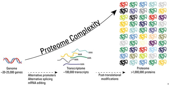
Multi-drug, Multi-arm, Biomarker-driven Clinical Trial for patients with Squamous Cell Carcinoma called the Lung Cancer Master Protocol, or Lung-MAP launched by NCI, Foundation Medicine, and Five Pharma Firms

US Personalized Cancer Genome Sequencing Market Outlook 2018 –

Comprehensive Genomic Characterization of Squamous Cell Lung Cancers

International Cancer Genome Consortium Website has 71 Committed Cancer Genome Projects Ongoing

Non-small Cell Lung Cancer drugs – where does the Future lie?

Lung cancer breathalyzer trialed in the UK

Diagnosing Lung Cancer in Exhaled Breath using Gold Nanoparticles

Multi-drug, Multi-arm, Biomarker-driven Clinical Trial for patients with Squamous Cell Carcinoma called the Lung Cancer Master Protocol, or Lung-MAP launched by NCI, Foundation Medicine, and Five Pharma Firms

Read Full Post »

Proteomics, Metabolomics, Signaling Pathways, and Cell Regulation: a Compilation of Articles in the Journal http://pharmaceuticalintelligence.com

Compilation of References by Leaders in Pharmaceutical Business Intelligence in the Journal http://pharmaceuticalintelligence.com about
Proteomics, Metabolomics, Signaling Pathways, and Cell Regulation

Curator: Larry H Bernstein, MD, FCAP

Proteomics

  1. The Human Proteome Map Completed

Reporter and Curator: Larry H. Bernstein, MD, FCAP

http://pharmaceuticalintelligence.com/2014/08/28/the-human-proteome-map-completed/

  1. Proteomics – The Pathway to Understanding and Decision-making in Medicine

Author and Curator, Larry H Bernstein, MD, FCAP

http://pharmaceuticalintelligence.com/2014/06/24/proteomics-the-pathway-to-
understanding-and-decision-making-in-medicine/

3. Advances in Separations Technology for the “OMICs” and Clarification of Therapeutic Targets

Author and Curator, Larry H Bernstein, MD, FCAP

http://pharmaceuticalintelligence.com/2012/10/22/advances-in-separations-technology-for-the-omics-and-clarification-         of-therapeutic-targets/

  1. Expanding the Genetic Alphabet and Linking the Genome to the Metabolome

Author and Curator, Larry H Bernstein, MD, FCAP

http://pharmaceuticalintelligence.com/2012/09/24/expanding-the-genetic-alphabet-and-linking-the-genome-to-the-                metabolome/

5. Genomics, Proteomics and standards

Larry H Bernstein, MD, FCAP, Author and Curator

http://pharmaceuticalintelligence.com/2014/07/06/genomics-proteomics-and-standards/

6. Proteins and cellular adaptation to stress

Larry H Bernstein, MD, FCAP, Author and Curator

http://pharmaceuticalintelligence.com/2014/07/08/proteins-and-cellular-adaptation-to-stress/


Metabolomics

  1. Extracellular evaluation of intracellular flux in yeast cells

Larry H. Bernstein, MD, FCAP, Reviewer and Curator

http://pharmaceuticalintelligence.com/2014/08/25/extracellular-evaluation-of-intracellular-flux-in-yeast-cells/

  1. Metabolomic analysis of two leukemia cell lines. I.

Larry H. Bernstein, MD, FCAP, Reviewer and Curator

http://pharmaceuticalintelligence.com/2014/08/23/metabolomic-analysis-of-two-leukemia-cell-lines-_i/

  1. Metabolomic analysis of two leukemia cell lines. II.

Larry H. Bernstein, MD, FCAP, Reviewer and Curator

http://pharmaceuticalintelligence.com/2014/08/24/metabolomic-analysis-of-two-leukemia-cell-lines-ii/

  1. Metabolomics, Metabonomics and Functional Nutrition: the next step in nutritional metabolism and biotherapeutics

Reviewer and Curator, Larry H. Bernstein, MD, FCAP

http://pharmaceuticalintelligence.com/2014/08/22/metabolomics-metabonomics-and-functional-nutrition-the-next-step-          in-nutritional-metabolism-and-biotherapeutics/

  1. Buffering of genetic modules involved in tricarboxylic acid cycle metabolism provides homeomeostatic regulation

Larry H. Bernstein, MD, FCAP, Reviewer and curator

http://pharmaceuticalintelligence.com/2014/08/27/buffering-of-genetic-modules-involved-in-tricarboxylic-acid-cycle-              metabolism-provides-homeomeostatic-regulation/

Metabolic Pathways

  1. Pentose Shunt, Electron Transfer, Galactose, more Lipids in brief

Reviewer and Curator: Larry H. Bernstein, MD, FCAP

http://pharmaceuticalintelligence.com/2014/08/21/pentose-shunt-electron-transfer-galactose-more-lipids-in-brief/

  1. Mitochondria: More than just the “powerhouse of the cell”

Ritu Saxena, PhD

http://pharmaceuticalintelligence.com/2012/07/09/mitochondria-more-than-just-the-powerhouse-of-the-cell/

  1. Mitochondrial fission and fusion: potential therapeutic targets?

Ritu saxena

http://pharmaceuticalintelligence.com/2012/10/31/mitochondrial-fission-and-fusion-potential-therapeutic-target/

4.  Mitochondrial mutation analysis might be “1-step” away

Ritu Saxena

http://pharmaceuticalintelligence.com/2012/08/14/mitochondrial-mutation-analysis-might-be-1-step-away/

  1. Selected References to Signaling and Metabolic Pathways in PharmaceuticalIntelligence.com

Curator: Larry H. Bernstein, MD, FCAP

http://pharmaceuticalintelligence.com/2014/08/14/selected-references-to-signaling-and-metabolic-pathways-in-                     leaders-in-pharmaceutical-intelligence/

  1. Metabolic drivers in aggressive brain tumors

Prabodh Kandal, PhD

http://pharmaceuticalintelligence.com/2012/11/11/metabolic-drivers-in-aggressive-brain-tumors/

  1. Metabolite Identification Combining Genetic and Metabolic Information: Genetic association links unknown metabolites to functionally related genes

Writer and Curator, Aviva Lev-Ari, PhD, RD

http://pharmaceuticalintelligence.com/2012/10/22/metabolite-identification-combining-genetic-and-metabolic-                        information-genetic-association-links-unknown-metabolites-to-functionally-related-genes/

  1. Mitochondria: Origin from oxygen free environment, role in aerobic glycolysis, metabolic adaptation

Larry H Bernstein, MD, FCAP, author and curator

http://pharmaceuticalintelligence.com/2012/09/26/mitochondria-origin-from-oxygen-free-environment-role-in-aerobic-            glycolysis-metabolic-adaptation/

  1. Therapeutic Targets for Diabetes and Related Metabolic Disorders

Reporter, Aviva Lev-Ari, PhD, RD

http://pharmaceuticalintelligence.com/2012/08/20/therapeutic-targets-for-diabetes-and-related-metabolic-disorders/

10.  Buffering of genetic modules involved in tricarboxylic acid cycle metabolism provides homeomeostatic regulation

Larry H. Bernstein, MD, FCAP, Reviewer and curator

http://pharmaceuticalintelligence.com/2014/08/27/buffering-of-genetic-modules-involved-in-tricarboxylic-acid-cycle-              metabolism-provides-homeomeostatic-regulation/

11. The multi-step transfer of phosphate bond and hydrogen exchange energy

Larry H. Bernstein, MD, FCAP, Curator:

http://pharmaceuticalintelligence.com/2014/08/19/the-multi-step-transfer-of-phosphate-bond-and-hydrogen-                          exchange-energy/

12. Studies of Respiration Lead to Acetyl CoA

http://pharmaceuticalintelligence.com/2014/08/18/studies-of-respiration-lead-to-acetyl-coa/

13. Lipid Metabolism

Author and Curator: Larry H. Bernstein, MD, FCAP

http://pharmaceuticalintelligence.com/2014/08/15/lipid-metabolism/

14. Carbohydrate Metabolism

Author and Curator: Larry H. Bernstein, MD, FCAP

http://pharmaceuticalintelligence.com/2014/08/13/carbohydrate-metabolism/

15. Update on mitochondrial function, respiration, and associated disorders

Larry H. Bernstein, MD, FCAP, Author and Curator

http://pharmaceuticalintelligence.com/2014/07/08/update-on-mitochondrial-function-respiration-and-associated-                   disorders/

16. Prologue to Cancer – e-book Volume One – Where are we in this journey?

Author and Curator: Larry H. Bernstein, MD, FCAP

http://pharmaceuticalintelligence.com/2014/04/13/prologue-to-cancer-ebook-4-where-are-we-in-this-journey/

17. Introduction – The Evolution of Cancer Therapy and Cancer Research: How We Got Here?

Author and Curator: Larry H. Bernstein, MD, FCAP

http://pharmaceuticalintelligence.com/2014/04/04/introduction-the-evolution-of-cancer-therapy-and-cancer-research-          how-we-got-here/

18. Inhibition of the Cardiomyocyte-Specific Kinase TNNI3K

Author and Curator: Larry H. Bernstein, MD, FCAP

http://pharmaceuticalintelligence.com/2013/11/01/inhibition-of-the-cardiomyocyte-specific-kinase-tnni3k/

19. The Binding of Oligonucleotides in DNA and 3-D Lattice Structures

Author and Curator: Larry H. Bernstein, MD, FCAP

http://pharmaceuticalintelligence.com/2013/05/15/the-binding-of-oligonucleotides-in-dna-and-3-d-lattice-structures/

20. Mitochondrial Metabolism and Cardiac Function

Author and Curator: Larry H. Bernstein, MD, FCAP

http://pharmaceuticalintelligence.com/2013/04/14/mitochondrial-metabolism-and-cardiac-function/

21. How Methionine Imbalance with Sulfur-Insufficiency Leads to Hyperhomocysteinemia

Curator: Larry H. Bernstein, MD, FCAP

http://pharmaceuticalintelligence.com/2013/04/04/sulfur-deficiency-leads_to_hyperhomocysteinemia/

22. AMPK Is a Negative Regulator of the Warburg Effect and Suppresses Tumor Growth In Vivo

Author and Curator: Stephen J. Williams, PhD

http://pharmaceuticalintelligence.com/2013/03/12/ampk-is-a-negative-regulator-of-the-warburg-effect-and-suppresses-         tumor-growth-in-vivo/

23. A Second Look at the Transthyretin Nutrition Inflammatory Conundrum

Author and Curator: Larry H. Bernstein, MD, FCAP

http://pharmaceuticalintelligence.com/2012/12/03/a-second-look-at-the-transthyretin-nutrition-inflammatory-                         conundrum/

24. Mitochondrial Damage and Repair under Oxidative Stress

Author and Curator: Larry H. Bernstein, MD, FCAP

http://pharmaceuticalintelligence.com/2012/10/28/mitochondrial-damage-and-repair-under-oxidative-stress/

25. Nitric Oxide and Immune Responses: Part 2

Author and Curator: Aviral Vatsa, PhD, MBBS

http://pharmaceuticalintelligence.com/2012/10/28/nitric-oxide-and-immune-responses-part-2/

26. Overview of Posttranslational Modification (PTM)

Writer and Curator: Larry H. Bernstein, MD, FCAP

http://pharmaceuticalintelligence.com/2014/07/29/overview-of-posttranslational-modification-ptm/

27. Malnutrition in India, high newborn death rate and stunting of children age under five years

Writer and Curator: Larry H. Bernstein, MD, FCAP

http://pharmaceuticalintelligence.com/2014/07/15/malnutrition-in-india-high-newborn-death-rate-and-stunting-of-                   children-age-under-five-years/

28. Update on mitochondrial function, respiration, and associated disorders

Writer and Curator: Larry H. Bernstein, MD, FCAP

http://pharmaceuticalintelligence.com/2014/07/08/update-on-mitochondrial-function-respiration-and-associated-                  disorders/

29. Omega-3 fatty acids, depleting the source, and protein insufficiency in renal disease

Larry H. Bernstein, MD, FCAP, Curator

http://pharmaceuticalintelligence.com/2014/07/06/omega-3-fatty-acids-depleting-the-source-and-protein-insufficiency-         in-renal-disease/

30. Introduction to e-Series A: Cardiovascular Diseases, Volume Four Part 2: Regenerative Medicine

Larry H. Bernstein, MD, FCAP, writer, and Aviva Lev- Ari, PhD, RN

http://pharmaceuticalintelligence.com/2014/04/27/larryhbernintroduction_to_cardiovascular_diseases-                                  translational_medicine-part_2/

31. Epilogue: Envisioning New Insights in Cancer Translational Biology
Series C: e-Books on Cancer & Oncology

Author & Curator: Larry H. Bernstein, MD, FCAP, Series C Content Consultant

http://pharmaceuticalintelligence.com/2014/03/29/epilogue-envisioning-new-insights/

32. Ca2+-Stimulated Exocytosis:  The Role of Calmodulin and Protein Kinase C in Ca2+ Regulation of Hormone                         and Neurotransmitter

Writer and Curator: Larry H Bernstein, MD, FCAP and
Curator and Content Editor: Aviva Lev-Ari, PhD, RN

http://pharmaceuticalintelligence.com/2013/12/23/calmodulin-and-protein-kinase-c-drive-the-ca2-regulation-of-                    hormone-and-neurotransmitter-release-that-triggers-ca2-stimulated-exocy

33. Cardiac Contractility & Myocardial Performance: Therapeutic Implications of Ryanopathy (Calcium Release-                           related Contractile Dysfunction) and Catecholamine Responses

Author, and Content Consultant to e-SERIES A: Cardiovascular Diseases: Justin Pearlman, MD, PhD, FACC
Author and Curator: Larry H Bernstein, MD, FCAP
and Article Curator: Aviva Lev-Ari, PhD, RN

http://pharmaceuticalintelligence.com/2013/08/28/cardiac-contractility-myocardium-performance-ventricular-arrhythmias-      and-non-ischemic-heart-failure-therapeutic-implications-for-cardiomyocyte-ryanopathy-calcium-release-related-                    contractile/

34. Role of Calcium, the Actin Skeleton, and Lipid Structures in Signaling and Cell Motility

Author and Curator: Larry H Bernstein, MD, FCAP Author: Stephen Williams, PhD, and Curator: Aviva Lev-Ari, PhD, RN

http://pharmaceuticalintelligence.com/2013/08/26/role-of-calcium-the-actin-skeleton-and-lipid-structures-in-signaling-and-cell-motility/

35. Identification of Biomarkers that are Related to the Actin Cytoskeleton

Larry H Bernstein, MD, FCAP, Author and Curator

http://pharmaceuticalintelligence.com/2012/12/10/identification-of-biomarkers-that-are-related-to-the-actin-                           cytoskeleton/

36. Advanced Topics in Sepsis and the Cardiovascular System at its End Stage

Author: Larry H Bernstein, MD, FCAP

http://pharmaceuticalintelligence.com/2013/08/18/advanced-topics-in-Sepsis-and-the-Cardiovascular-System-at-its-              End-Stage/

37. The Delicate Connection: IDO (Indolamine 2, 3 dehydrogenase) and Cancer Immunology

Demet Sag, PhD, Author and Curator

http://pharmaceuticalintelligence.com/2013/08/04/the-delicate-connection-ido-indolamine-2-3-dehydrogenase-and-               immunology/

38. IDO for Commitment of a Life Time: The Origins and Mechanisms of IDO, indolamine 2, 3-dioxygenase

Demet Sag, PhD, Author and Curator

http://pharmaceuticalintelligence.com/2013/08/04/ido-for-commitment-of-a-life-time-the-origins-and-mechanisms-of-             ido-indolamine-2-3-dioxygenase/

39. Confined Indolamine 2, 3 dioxygenase (IDO) Controls the Homeostasis of Immune Responses for Good and Bad

Curator: Demet Sag, PhD, CRA, GCP

http://pharmaceuticalintelligence.com/2013/07/31/confined-indolamine-2-3-dehydrogenase-controls-the-hemostasis-           of-immune-responses-for-good-and-bad/

40. Signaling Pathway that Makes Young Neurons Connect was discovered @ Scripps Research Institute

Reporter: Aviva Lev-Ari, PhD, RN

http://pharmaceuticalintelligence.com/2013/06/26/signaling-pathway-that-makes-young-neurons-connect-was-                     discovered-scripps-research-institute/

41. Naked Mole Rats Cancer-Free

Writer and Curator: Larry H. Bernstein, MD, FCAP

http://pharmaceuticalintelligence.com/2013/06/20/naked-mole-rats-cancer-free/

42. Late Onset of Alzheimer’s Disease and One-carbon Metabolism

Reporter and Curator: Dr. Sudipta Saha, Ph.D.

http://pharmaceuticalintelligence.com/2013/05/06/alzheimers-disease-and-one-carbon-metabolism/

43. Problems of vegetarianism

Reporter and Curator: Dr. Sudipta Saha, Ph.D.

http://pharmaceuticalintelligence.com/2013/04/22/problems-of-vegetarianism/

44.  Amyloidosis with Cardiomyopathy

Writer and Curator: Larry H. Bernstein, MD, FCAP

http://pharmaceuticalintelligence.com/2013/03/31/amyloidosis-with-cardiomyopathy/

45. Liver endoplasmic reticulum stress and hepatosteatosis

Larry H Bernstein, MD, FACP

http://pharmaceuticalintelligence.com/2013/03/10/liver-endoplasmic-reticulum-stress-and-hepatosteatosis/

46. The Molecular Biology of Renal Disorders: Nitric Oxide – Part III

Curator and Author: Larry H Bernstein, MD, FACP

http://pharmaceuticalintelligence.com/2012/11/26/the-molecular-biology-of-renal-disorders/

47. Nitric Oxide Function in Coagulation – Part II

Curator and Author: Larry H. Bernstein, MD, FCAP

http://pharmaceuticalintelligence.com/2012/11/26/nitric-oxide-function-in-coagulation/

48. Nitric Oxide, Platelets, Endothelium and Hemostasis

Curator and Author: Larry H Bernstein, MD, FACP

http://pharmaceuticalintelligence.com/2012/11/08/nitric-oxide-platelets-endothelium-and-hemostasis/

49. Interaction of Nitric Oxide and Prostacyclin in Vascular Endothelium

Curator and Author: Larry H Bernstein, MD, FACP

http://pharmaceuticalintelligence.com/2012/09/14/interaction-of-nitric-oxide-and-prostacyclin-in-vascular-endothelium/

50. Nitric Oxide and Immune Responses: Part 1

Curator and Author:  Aviral Vatsa PhD, MBBS

http://pharmaceuticalintelligence.com/2012/10/18/nitric-oxide-and-immune-responses-part-1/

51. Nitric Oxide and Immune Responses: Part 2

Curator and Author:  Aviral Vatsa PhD, MBBS

http://pharmaceuticalintelligence.com/2012/10/28/nitric-oxide-and-immune-responses-part-2/

52. Mitochondrial Damage and Repair under Oxidative Stress

Curator and Author: Larry H Bernstein, MD, FACP

http://pharmaceuticalintelligence.com/2012/10/28/mitochondrial-damage-and-repair-under-oxidative-stress/

53. Is the Warburg Effect the cause or the effect of cancer: A 21st Century View?

Curator and Author: Larry H Bernstein, MD, FACP

http://pharmaceuticalintelligence.com/2012/10/17/is-the-warburg-effect-the-cause-or-the-effect-of-cancer-a-21st-                 century-view/

54. Ubiquinin-Proteosome pathway, autophagy, the mitochondrion, proteolysis and cell apoptosis

Curator and Author: Larry H Bernstein, MD, FACP

http://pharmaceuticalintelligence.com/2012/10/30/ubiquinin-proteosome-pathway-autophagy-the-mitochondrion-                  proteolysis-and-cell-apoptosis/

55. Ubiquitin-Proteosome pathway, Autophagy, the Mitochondrion, Proteolysis and Cell Apoptosis: Part III

Curator and Author: Larry H Bernstein, MD, FACP

http://pharmaceuticalintelligence.com/2013/02/14/ubiquinin-proteosome-pathway-autophagy-the-mitochondrion-                   proteolysis-and-cell-apoptosis-reconsidered/

56. Nitric Oxide and iNOS have Key Roles in Kidney Diseases – Part II

Curator and Author: Larry H Bernstein, MD, FACP

http://pharmaceuticalintelligence.com/2012/11/26/nitric-oxide-and-inos-have-key-roles-in-kidney-diseases/

57. New Insights on Nitric Oxide donors – Part IV

Curator and Author: Larry H Bernstein, MD, FACP

http://pharmaceuticalintelligence.com/2012/11/26/new-insights-on-no-donors/

58. Crucial role of Nitric Oxide in Cancer

Curator and Author: Ritu Saxena, Ph.D.

http://pharmaceuticalintelligence.com/2012/10/16/crucial-role-of-nitric-oxide-in-cancer/

59. Nitric Oxide has a ubiquitous role in the regulation of glycolysis -with a concomitant influence on mitochondrial function

Curator and Author: Larry H Bernstein, MD, FACP

http://pharmaceuticalintelligence.com/2012/09/16/nitric-oxide-has-a-ubiquitous-role-in-the-regulation-of-glycolysis-with-         a-concomitant-influence-on-mitochondrial-function/

60. Targeting Mitochondrial-bound Hexokinase for Cancer Therapy

Curator and Author: Ziv Raviv, PhD, RN 04/06/2013

http://pharmaceuticalintelligence.com/2013/04/06/targeting-mitochondrial-bound-hexokinase-for-cancer-therapy/

61. Biochemistry of the Coagulation Cascade and Platelet Aggregation – Part I

Curator and Author: Larry H Bernstein, MD, FACP

http://pharmaceuticalintelligence.com/2012/11/26/biochemistry-of-the-coagulation-cascade-and-platelet-aggregation/

Genomics, Transcriptomics, and Epigenetics

  1. What is the meaning of so many RNAs?

Writer and Curator: Larry H. Bernstein, MD, FCAP

http://pharmaceuticalintelligence.com/2014/08/06/what-is-the-meaning-of-so-many-rnas/

  1. RNA and the transcription the genetic code

Larry H. Bernstein, MD, FCAP, Writer and Curator

http://pharmaceuticalintelligence.com/2014/08/02/rna-and-the-transcription-of-the-genetic-code/

  1. A Primer on DNA and DNA Replication

Writer and Curator: Larry H. Bernstein, MD, FCAP

http://pharmaceuticalintelligence.com/2014/07/29/a_primer_on_dna_and_dna_replication/

4. Synthesizing Synthetic Biology: PLOS Collections

Reporter: Aviva Lev-Ari

http://pharmaceuticalintelligence.com/2012/08/17/synthesizing-synthetic-biology-plos-collections/

5. Pathology Emergence in the 21st Century

Author and Curator: Larry Bernstein, MD, FCAP

http://pharmaceuticalintelligence.com/2014/08/03/pathology-emergence-in-the-21st-century/

6. RNA and the transcription the genetic code

Writer and Curator, Larry H. Bernstein, MD, FCAP

http://pharmaceuticalintelligence.com/2014/08/02/rna-and-the-transcription-of-the-genetic-code/

7. A Great University engaged in Drug Discovery: University of Pittsburgh

Larry H. Bernstein, MD, FCAP, Reporter and Curator

http://pharmaceuticalintelligence.com/2014/07/15/a-great-university-engaged-in-drug-discovery/

8. microRNA called miRNA-142 involved in the process by which the immature cells in the bone  marrow give                              rise to all the types of blood cells, including immune cells and the oxygen-bearing red blood cells

Aviva Lev-Ari, PhD, RN, Author and Curator

http://pharmaceuticalintelligence.com/2014/07/24/microrna-called-mir-142-involved-in-the-process-by-which-the-                   immature-cells-in-the-bone-marrow-give-rise-to-all-the-types-of-blood-cells-including-immune-cells-and-the-oxygen-             bearing-red-blood-cells/

9. Genes, proteomes, and their interaction

Larry H. Bernstein, MD, FCAP, Writer and Curator

http://pharmaceuticalintelligence.com/2014/07/28/genes-proteomes-and-their-interaction/

10. Regulation of somatic stem cell Function

Larry H. Bernstein, MD, FCAP, Writer and Curator    Aviva Lev-Ari, PhD, RN, Curator

http://pharmaceuticalintelligence.com/2014/07/29/regulation-of-somatic-stem-cell-function/

11. Scientists discover that pluripotency factor NANOG is also active in adult organisms

Larry H. Bernstein, MD, FCAP, Reporter

http://pharmaceuticalintelligence.com/2014/07/10/scientists-discover-that-pluripotency-factor-nanog-is-also-active-in-           adult-organisms/

12. Bzzz! Are fruitflies like us?

Larry H Bernstein, MD, FCAP, Author and Curator

http://pharmaceuticalintelligence.com/2014/07/07/bzzz-are-fruitflies-like-us/

13. Long Non-coding RNAs Can Encode Proteins After All

Larry H Bernstein, MD, FCAP, Reporter

http://pharmaceuticalintelligence.com/2014/06/29/long-non-coding-rnas-can-encode-proteins-after-all/

14. Michael Snyder @Stanford University sequenced the lymphoblastoid transcriptomes and developed an
allele-specific full-length transcriptome

Aviva Lev-Ari, PhD, RN, Author and Curator

http://pharmaceuticalintelligence.com/014/06/23/michael-snyder-stanford-university-sequenced-the-lymphoblastoid-            transcriptomes-and-developed-an-allele-specific-full-length-transcriptome/

15. Commentary on Biomarkers for Genetics and Genomics of Cardiovascular Disease: Views by Larry H                                     Bernstein, MD, FCAP

Author: Larry H Bernstein, MD, FCAP

http://pharmaceuticalintelligence.com/2014/07/16/commentary-on-biomarkers-for-genetics-and-genomics-of-                        cardiovascular-disease-views-by-larry-h-bernstein-md-fcap/

16. Observations on Finding the Genetic Links in Common Disease: Whole Genomic Sequencing Studies

Author an curator: Larry H Bernstein, MD, FCAP

http://pharmaceuticalintelligence.com/2013/05/18/observations-on-finding-the-genetic-links/

17. Silencing Cancers with Synthetic siRNAs

Larry H. Bernstein, MD, FCAP, Reviewer and Curator

http://pharmaceuticalintelligence.com/2013/12/09/silencing-cancers-with-synthetic-sirnas/

18. Cardiometabolic Syndrome and the Genetics of Hypertension: The Neuroendocrine Transcriptome Control Points

Reporter: Aviva Lev-Ari, PhD, RN

http://pharmaceuticalintelligence.com/2013/12/12/cardiometabolic-syndrome-and-the-genetics-of-hypertension-the-neuroendocrine-transcriptome-control-points/

19. Developments in the Genomics and Proteomics of Type 2 Diabetes Mellitus and Treatment Targets

Larry H. Bernstein, MD, FCAP, Reviewer and Curator

http://pharmaceuticalintelligence.com/2013/12/08/developments-in-the-genomics-and-proteomics-of-type-2-diabetes-           mellitus-and-treatment-targets/

20. Loss of normal growth regulation

Larry H Bernstein, MD, FCAP, Curator

http://pharmaceuticalintelligence.com/2014/07/06/loss-of-normal-growth-regulation/

21. CT Angiography & TrueVision™ Metabolomics (Genomic Phenotyping) for new Therapeutic Targets to Atherosclerosis

Reporter: Aviva Lev-Ari, PhD, RN

http://pharmaceuticalintelligence.com/2013/11/15/ct-angiography-truevision-metabolomics-genomic-phenotyping-for-           new-therapeutic-targets-to-atherosclerosis/

22.  CRACKING THE CODE OF HUMAN LIFE: The Birth of BioInformatics & Computational Genomics

Genomics Curator, Larry H Bernstein, MD, FCAP

http://pharmaceuticalintelligence.com/2014/08/30/cracking-the-code-of-human-life-the-birth-of-bioinformatics-                      computational-genomics/

23. Big Data in Genomic Medicine

Author and Curator, Larry H Bernstein, MD, FCAP

http://pharmaceuticalintelligence.com/2012/12/17/big-data-in-genomic-medicine/

24. From Genomics of Microorganisms to Translational Medicine

Author and Curator: Demet Sag, PhD

http://pharmaceuticalintelligence.com/2014/03/20/without-the-past-no-future-but-learn-and-move-genomics-of-                      microorganisms-to-translational-medicine/

25. Summary of Genomics and Medicine: Role in Cardiovascular Diseases

Author and Curator, Larry H Bernstein, MD, FCAP

http://pharmaceuticalintelligence.com/2014/01/06/summary-of-genomics-and-medicine-role-in-cardiovascular-diseases/

 26. Genomic Promise for Neurodegenerative Diseases, Dementias, Autism Spectrum, Schizophrenia, and Serious                      Depression

Author and Curator, Larry H Bernstein, MD, FCAP

http://pharmaceuticalintelligence.com/2013/02/19/genomic-promise-for-neurodegenerative-diseases-dementias-autism-        spectrum-schizophrenia-and-serious-depression/

 27.  BRCA1 a tumour suppressor in breast and ovarian cancer – functions in transcription, ubiquitination and DNA repair

Sudipta Saha, PhD

http://pharmaceuticalintelligence.com/2012/12/04/brca1-a-tumour-suppressor-in-breast-and-ovarian-cancer-functions-         in-transcription-ubiquitination-and-dna-repair/

28. Personalized medicine gearing up to tackle cancer

Ritu Saxena, PhD

http://pharmaceuticalintelligence.com/2013/01/07/personalized-medicine-gearing-up-to-tackle-cancer/

29. Differentiation Therapy – Epigenetics Tackles Solid Tumors

Stephen J Williams, PhD

      http://pharmaceuticalintelligence.com/2013/01/03/differentiation-therapy-epigenetics-tackles-solid-tumors/

30. Mechanism involved in Breast Cancer Cell Growth: Function in Early Detection & Treatment

     Aviva Lev-Ari, PhD, RN

http://pharmaceuticalintelligence.com/2013/01/17/mechanism-involved-in-breast-cancer-cell-growth-function-in-early-          detection-treatment/

31. The Molecular pathology of Breast Cancer Progression

Tilde Barliya, PhD

http://pharmaceuticalintelligence.com/2013/01/10/the-molecular-pathology-of-breast-cancer-progression

32. Gastric Cancer: Whole-genome reconstruction and mutational signatures

Aviva Lev-Ari, PhD, RN

http://pharmaceuticalintelligence.com/2012/12/24/gastric-cancer-whole-genome-reconstruction-and-mutational-                   signatures-2/

33. Paradigm Shift in Human Genomics – Predictive Biomarkers and Personalized Medicine –                                                       Part 1 (pharmaceuticalintelligence.com)

Aviva  Lev-Ari, PhD, RN

http://pharmaceuticalntelligence.com/2013/01/13/paradigm-shift-in-human-genomics-predictive-biomarkers-and-personalized-medicine-part-1/

34. LEADERS in Genome Sequencing of Genetic Mutations for Therapeutic Drug Selection in Cancer                                         Personalized Treatment: Part 2

A Lev-Ari, PhD, RN

http://pharmaceuticalintelligence.com/2013/01/13/leaders-in-genome-sequencing-of-genetic-mutations-for-therapeutic-       drug-selection-in-cancer-personalized-treatment-part-2/

35. Personalized Medicine: An Institute Profile – Coriell Institute for Medical Research: Part 3

Aviva Lev-Ari, PhD, RN

http://pharmaceuticalintelligence.com/2013/01/13/personalized-medicine-an-institute-profile-coriell-institute-for-medical-        research-part-3/

36. Harnessing Personalized Medicine for Cancer Management, Prospects of Prevention and Cure: Opinions of                           Cancer Scientific Leaders @http://pharmaceuticalintelligence.com

Aviva Lev-Ari, PhD, RN

http://pharmaceuticalintelligence.com/2013/01/13/7000/Harnessing_Personalized_Medicine_for_ Cancer_Management-      Prospects_of_Prevention_and_Cure/

37.  GSK for Personalized Medicine using Cancer Drugs needs Alacris systems biology model to determine the in silico
effect of the inhibitor in its “virtual clinical trial”

Aviva Lev-Ari, PhD, RN

http://pharmaceuticalintelligence.com/2012/11/14/gsk-for-personalized-medicine-using-cancer-drugs-needs-alacris-             systems-biology-model-to-determine-the-in-silico-effect-of-the-inhibitor-in-its-virtual-clinical-trial/

38. Personalized medicine-based cure for cancer might not be far away

Ritu Saxena, PhD

  http://pharmaceuticalintelligence.com/2012/11/20/personalized-medicine-based-cure-for-cancer-might-not-be-far-away/

39. Human Variome Project: encyclopedic catalog of sequence variants indexed to the human genome sequence

Aviva Lev-Ari, PhD, RN

http://pharmaceuticalintelligence.com/2012/11/24/human-variome-project-encyclopedic-catalog-of-sequence-variants-         indexed-to-the-human-genome-sequence/

40. Inspiration From Dr. Maureen Cronin’s Achievements in Applying Genomic Sequencing to Cancer Diagnostics

Aviva Lev-Ari, PhD, RN

http://pharmaceuticalintelligence.com/2013/01/10/inspiration-from-dr-maureen-cronins-achievements-in-applying-                genomic-sequencing-to-cancer-diagnostics/

41. The “Cancer establishments” examined by James Watson, co-discoverer of DNA w/Crick, 4/1953

Aviva Lev-Ari, PhD, RN

http://pharmaceuticalintelligence.com/2013/01/09/the-cancer-establishments-examined-by-james-watson-co-discover-         of-dna-wcrick-41953/

42. What can we expect of tumor therapeutic response?

Author and curator: Larry H Bernstein, MD, FACP

http://pharmaceuticalintelligence.com/2012/12/05/what-can-we-expect-of-tumor-therapeutic-response/

43. Directions for genomics in personalized medicine

Author and Curator: Larry H. Bernstein, MD, FCAP

http://pharmaceuticalintelligence.com/2013/01/27/directions-for-genomics-in-personalized-medicine/

44. How mobile elements in “Junk” DNA promote cancer. Part 1: Transposon-mediated tumorigenesis.

Stephen J Williams, PhD

http://pharmaceuticalintelligence.com/2012/10/31/how-mobile-elements-in-junk-dna-prote-cancer-part1-transposon-            mediated-tumorigenesis/

45. mRNA interference with cancer expression

Author and Curator, Larry H. Bernstein, MD, FCAP

 http://pharmaceuticalintelligence.com/2012/10/26/mrna-interference-with-cancer-expression/

46. Expanding the Genetic Alphabet and linking the genome to the metabolome

Aviva Lev-Ari, PhD, RD

http://pharmaceuticalintelligence.com/2012/09/24/expanding-the-genetic-alphabet-and-linking-the-genome-to-the-               metabolome/

47. Breast Cancer, drug resistance, and biopharmaceutical targets

Author and Curator: Larry H Bernstein, MD, FCAP

http://pharmaceuticalintelligence.com/2012/09/18/breast-cancer-drug-resistance-and-biopharmaceutical-targets/

48.  Breast Cancer: Genomic profiling to predict Survival: Combination of Histopathology and Gene Expression                            Analysis

Aviva Lev-Ari, PhD, RD

http://pharmaceuticalintelligence.com/2012/12/24/breast-cancer-genomic-profiling-to-predict-survival-combination-of-           histopathology-and-gene-expression-analysis

49. Gastric Cancer: Whole-genome reconstruction and mutational signatures

Aviva  Lev-Ari, PhD, RD

http://pharmaceuticalintelligence.com/2012/12/24/gastric-cancer-whole-genome-reconstruction-and-mutational-                   signatures-2/

50. Genomic Analysis: FLUIDIGM Technology in the Life Science and Agricultural Biotechnology

Aviva Lev-Ari, PhD, RD

http://pharmaceuticalintelligence.com/2012/08/22/genomic-analysis-fluidigm-technology-in-the-life-science-and-                   agricultural-biotechnology/

51. 2013 Genomics: The Era Beyond the Sequencing Human Genome: Francis Collins, Craig Venter, Eric Lander, et al.

Aviva Lev-Ari, PhD, RD

http://pharmaceuticalintelligence.com/2013_Genomics

52. Paradigm Shift in Human Genomics – Predictive Biomarkers and Personalized Medicine – Part 1

Aviva Lev-Ari, PhD, RD

http://pharmaceuticalintelligence.com/Paradigm Shift in Human Genomics_/

Signaling Pathways

  1. Proteins and cellular adaptation to stress

Larry H Bernstein, MD, FCAP, Curator

http://pharmaceuticalintelligence.com/2014/07/08/proteins-and-cellular-adaptation-to-stress/

  1. A Synthesis of the Beauty and Complexity of How We View Cancer:
    Cancer Volume One – Summary

Author and Curator: Larry H. Bernstein, MD, FCAP

http://pharmaceuticalintelligence.com/2014/03/26/a-synthesis-of-the-beauty-and-complexity-of-how-we-view-cancer/

  1. Recurrent somatic mutations in chromatin-remodeling and ubiquitin ligase complex genes in
    serous endometrial tumors

Sudipta Saha, PhD

http://pharmaceuticalintelligence.com/2012/11/19/recurrent-somatic-mutations-in-chromatin-remodeling-ad-ubiquitin-           ligase-complex-genes-in-serous-endometrial-tumors/

4.  Prostate Cancer Cells: Histone Deacetylase Inhibitors Induce Epithelial-to-Mesenchymal Transition

Stephen J Williams, PhD

http://pharmaceuticalintelligence.com/2012/11/30/histone-deacetylase-inhibitors-induce-epithelial-to-mesenchymal-              transition-in-prostate-cancer-cells/

5. Ubiquinin-Proteosome pathway, autophagy, the mitochondrion, proteolysis and cell apoptosis

Author and Curator: Larry H Bernstein, MD, FCAP

http://pharmaceuticalintelligence.com/2012/10/30/ubiquinin-proteosome-pathway-autophagy-the-mitochondrion-                   proteolysis-and-cell-apoptosis/

6. Signaling and Signaling Pathways

Larry H. Bernstein, MD, FCAP, Reporter and Curator

http://pharmaceuticalintelligence.com/2014/08/12/signaling-and-signaling-pathways/

7.  Leptin signaling in mediating the cardiac hypertrophy associated with obesity

Larry H. Bernstein, MD, FCAP, Reporter and Curator

http://pharmaceuticalintelligence.com/2013/11/03/leptin-signaling-in-mediating-the-cardiac-hypertrophy-associated-            with-obesity/

  1. Sensors and Signaling in Oxidative Stress

Larry H. Bernstein, MD, FCAP, Reporter and Curator

http://pharmaceuticalintelligence.com/2013/11/01/sensors-and-signaling-in-oxidative-stress/

  1. The Final Considerations of the Role of Platelets and Platelet Endothelial Reactions in Atherosclerosis and Novel
    Treatments

Larry H. Bernstein, MD, FCAP, Reporter and Curator

http://pharmaceuticalintelligence.com/2013/10/15/the-final-considerations-of-the-role-of-platelets-and-platelet-                      endothelial-reactions-in-atherosclerosis-and-novel-treatments

10.   Platelets in Translational Research – Part 1

Larry H. Bernstein, MD, FCAP, Reporter and Curator

http://pharmaceuticalintelligence.com/2013/10/07/platelets-in-translational-research-1/

11.  Disruption of Calcium Homeostasis: Cardiomyocytes and Vascular Smooth Muscle Cells: The Cardiac and
Cardiovascular Calcium Signaling Mechanism

Author and Curator: Larry H Bernstein, MD, FCAP, Author, and Content Consultant to e-SERIES A:
Cardiovascular Diseases: Justin Pearlman, MD, PhD, FACC and Curator: Aviva Lev-Ari, PhD, RN

http://pharmaceuticalintelligence.com/2013/09/12/disruption-of-calcium-homeostasis-cardiomyocytes-and-vascular-             smooth-muscle-cells-the-cardiac-and-cardiovascular-calcium-signaling-mechanism/

12. The Centrality of Ca(2+) Signaling and Cytoskeleton Involving Calmodulin Kinases and
Ryanodine Receptors in Cardiac Failure, Arterial Smooth Muscle, Post-ischemic Arrhythmia,
Similarities and Differences, and Pharmaceutical Targets

     Author and Curator: Larry H Bernstein, MD, FCAP, Author, and Content Consultant to
e-SERIES A: Cardiovascular Diseases: Justin Pearlman, MD, PhD, FACC and
Curator: Aviva Lev-Ari, PhD, RN

http://pharmaceuticalintelligence.com/2013/09/08/the-centrality-of-ca2-signaling-and-cytoskeleton-involving-calmodulin-       kinases-and-ryanodine-receptors-in-cardiac-failure-arterial-smooth-muscle-post-ischemic-arrhythmia-similarities-and-           differen/

13.  Nitric Oxide Signalling Pathways

Aviral Vatsa, PhD, MBBS

http://pharmaceuticalintelligence.com/2012/08/22/nitric-oxide-signalling-pathways/

14. Immune activation, immunity, antibacterial activity

Larry H. Bernstein, MD, FCAP, Curator

http://pharmaceuticalintelligence.com/2014/07/06/immune-activation-immunity-antibacterial-activity/

15.  Regulation of somatic stem cell Function

Larry H. Bernstein, MD, FCAP, Writer and Curator    Aviva Lev-Ari, PhD, RN, Curator

http://pharmaceuticalintelligence.com/2014/07/29/regulation-of-somatic-stem-cell-function/

16. Scientists discover that pluripotency factor NANOG is also active in adult organisms

Larry H. Bernstein, MD, FCAP, Reporter

http://pharmaceuticalintelligence.com/2014/07/10/scientists-discover-that-pluripotency-factor-nanog-is-also-active-in-adult-organisms/

Read Full Post »

Heroes in Medical Research: Green Fluorescent Protein and the Rough Road in Science

Curator: Stephen J. Williams, Ph.D.

Article ID #147: Heroes in Medical Research: Green Fluorescent Protein and the Rough Road in Science. Published on 7/27/2014

WordCloud Image Produced by Adam Tubman

In this series, “Heroes in Medical Research”, I like to discuss the people who made some important contributions to science and medicine which underlie the great transformative changes but don’t usually get the notoriety given to Nobel Laureates or who seem to fly under the radar of popular news. Their work may be the development of research tools which allowed a great discovery leading to a line of transformative research, a moment of serendipity leading to discovery of a cure, or just contributions to the development of a new field or the mentoring of a new generation of scientists and clinicians. One such discovery, which has probably been pivotal in many of our research, is the discovery of the green fluorescent protein (GFP), commonly used as an invaluable tool to monitor protein for cellular expression and localization studies. Although the development of research tools, whether imaging tools, vectors, animal models, cell lines, and such are not heralded, they always assist in the pivotal discoveries of our time. The following is a heartwarming story by Discover Magazine’s Yudhijit Bhattacharjee behind Dr. Douglas Prasher’s discovery of the green fluorescent protein, his successful efforts to sequence the gene and subsequent struggles in science and finally scientific recognition for his work. In addition the story describes Dr. Prather’s perseverance, a trait necessary for every scientist.

http://discovermagazine.com/2011/apr/30-how-bad-luck-networking-cost-prasher-nobel


The following is a wonderful entry into Wikipedia about Dr. Prasher at:

http://en.wikipedia.org/wiki/Douglas_Prasher

including a listing of his publications including the seminal Science and PNAS publications1,2.


prasher


(Photo: Dr. Prasher in the lab at UCSD. Photo credit UCSD and John Galstaldo)


In summary, Dr. Prather had been working at Wood’s Hole in Massachusetts trying to discover, isolate, then clone the protein which allowed a species of jellyfish living in the cold waters of the North Pacific, Aequorea victoria, to emit a green glow. Eventually he cloned the GFP gene, but gave up on work to express the gene in mammalian cells. Before leaving Wood’s Hole he gave the gene to Dr. Roger Tsien, who with Dr. Martin Chalfie and Osamu Shimomura showed the utility of GFP as an intracellular tracer to visualize, in real time, the expression and localization of GFP-tagged proteins (all three shared the 2008 Nobel Prize for this work). Dr. Tsien however realized the importance of Douglas’s cloning work as pivotal for their research, contacted Douglas (who now due to the bad economy was working at a Toyota dealership in Alabama) and invited him to the Nobel Prize Award Ceremony in Sweden as his guest. Although Dr. Prasher had “left academic science” he never really stopped his quest for a scientific career, using his spare time to review manuscripts.

Other researchers have invited their colleagues who made important contributions to the ultimate Nobel work. One such guest was one of my colleagues Dr. Leonard Cohen, who worked with Dr. Irwin Rose and Avram Hershko at the Institute for Cancer Research in Philadelphia a cell-free system from clams to discover the mechanism how cyclin B is degraded during the exit from the cell cycle (from A. Hershko’s Nobel speech). Dr. Hershko had acknowledged a slew of colleagues and highlighted their contributions to the ultimate work. It shows how even small discoveries can contribute to the sphere of scientific knowledge and breakthrough.

Luckily, in the end, perseverance has paid off as Dr. Prasher is now using his talents in Roger Tsien‘s group at the University of California in San Diego.

References:

1. Chalfie, M., Tu, Y., Euskirchen, G., Ward, W.W., Prasher, D.C., Green fluorescent protein as a marker for gene expression. Science, 263(5148), 802-805 (1994).


2. Heim, R., Prasher, D.C., Tsien, R.Y., Wavelength mutations and posttranslational autoxidation of green fluorescent protein. Proc. Natl. Acad. Sci. USA, 91(26), 12501-12504 (1994).

More posts on this site on Heroes in Medical Research series include:

Heroes in Medical Research: Developing Models for Cancer Research

Heroes in Medical Research: Dr. Carmine Paul Bianchi Pharmacologist, Leader, and Mentor

Heroes in Medical Research: Dr. Robert Ting, Ph.D. and Retrovirus in AIDS and Cancer

Heroes in Medical Research: Barnett Rosenberg and the Discovery of Cisplatin


Read Full Post »

Read Full Post »

USPTO Guidance On Patentable Subject Matter

USPTO Guidance On Patentable Subject Matter

Curator and Reporter: Larry H Bernstein, MD, FCAP

LH Bernstein

LH Bernstein


Revised 4 July, 2014

http://pharmaceuticalintelligence.com/2014/07/03/uspto-guidance-on-patentable-subject-matter


I came across a few recent articles on the subject of US Patent Office guidance on patentability as well as on Supreme Court ruling on claims. I filed several patents on clinical laboratory methods early in my career upon the recommendation of my brother-in-law, now deceased.  Years later, after both brother-in-law and patent attorney are no longer alive, I look back and ask what I have learned over $100,000 later, with many trips to the USPTO, opportunities not taken, and a one year provisional patent behind me.

My conclusion is

(1) that patents are for the protection of the innovator, who might realize legal protection, but the cost and the time investment can well exceed the cost of startup and building a small startup enterprize, that would be the next step.

(2) The other thing to consider is the capability of the lawyer or firm that represents you.  A patent that is well done can be expected to take 5-7 years to go through with due diligence.   I would not expect it to be done well by a university with many other competing demands. I might be wrong in this respect, as the climate has changed, and research universities have sprouted engines for change.  Experienced and productive faculty are encouraged or allowed to form their own such entities.

(3) The emergence of Big Data, computational biology, and very large data warehouses for data use and integration has changed the landscape. The resources required for an individual to pursue research along these lines is quite beyond an individuals sole capacity to successfully pursue without outside funding.  In addition, the changed designated requirement of first to publish has muddied the water.

Of course, one can propose without anything published in the public domain. That makes it possible for corporate entities to file thousands of patents, whether there is actual validation or not at the time of filing.  It would be a quite trying experience for anyone to pursue in the USPTO without some litigation over ownership of patent rights. At this stage of of technology development, I have come to realize that the organization of research, peer review, and archiving of data is still at a stage where some of the best systems avalailable for storing and accessing data still comes considerably short of what is needed for the most complex tasks, even though improvements have come at an exponential pace.

I shall not comment on the contested views held by physicists, chemists, biologists, and economists over the completeness of guiding theories strongly held.  Only history will tell.  Beliefs can hold a strong sway, and have many times held us back.

I am not an expert on legal matters, but it is incomprehensible to me that issues concerning technology innovation can be adjudicated in the Supreme Court, as has occurred in recent years. I have postgraduate degrees in  Medicine, Developmental Anatomy, and post-medical training in pathology and laboratory medicine, as well as experience in analytical and research biochemistry.  It is beyond the competencies expected for these type of cases to come before the Supreme Court, or even to the Federal District Courts, as we see with increasing frequency,  as this has occurred with respect to the development and application of the human genome.

I’m not sure that the developments can be resolved for the public good without a more full development of an open-access system of publishing. Now I present some recent publication about, or published by the USPTO.

DR ANTHONY MELVIN CRASTO

Dr. Melvin Castro - Organic Chemistry and New Drug Development

Dr. Melvin Castro – Organic Chemistry and New Drug Development


YOU ARE FOLLOWING THIS BLOG You are following this blog, along with 1,014 other amazing people (manage).

patentimages.storage.goog…

USPTO Guidance On Patentable Subject Matter: Impediment to Biotech Innovation

Joanna T. Brougher, David A. Fazzolare J Commercial Biotechnology 2014 20(3):Brougher

jcbiotech-patents

jcbiotech-patents


Abstract In June 2013, the U.S. Supreme Court issued a unanimous decision upending more than three decades worth of established patent practice when it ruled that isolated gene sequences are no longer patentable subject matter under 35 U.S.C. Section 101.While many practitioners in the field believed that the USPTO would interpret the decision narrowly, the USPTO actually expanded the scope of the decision when it issued its guidelines for determining whether an invention satisfies Section 101.

The guidelines were met with intense backlash with many arguing that they unnecessarily expanded the scope of the Supreme Court cases in a way that could unduly restrict the scope of patentable subject matter, weaken the U.S. patent system, and create a disincentive to innovation. By undermining patentable subject matter in this way, the guidelines may end up harming not only the companies that patent medical innovations, but also the patients who need medical care.  This article examines the guidelines and their impact on various technologies.

Keywords:   patent, patentable subject matter, Myriad, Mayo, USPTO guidelines

Full Text: PDF

References

35 U.S.C. Section 101 states “Whoever invents or discovers any new and useful process, machine, manufacture, or composition of matter, or any new and useful improvement thereof, may obtain a patent therefor, subject to the conditions and requirements of this title.

” Prometheus Laboratories, Inc. v. Mayo Collaborative Services, 566 U.S. ___ (2012)

Association for Molecular Pathology et al., v. Myriad Genetics, Inc., 569 U.S. ___ (2013).

Parke-Davis & Co. v. H.K. Mulford Co., 189 F. 95, 103 (C.C.S.D.N.Y. 1911)

USPTO. Guidance For Determining Subject Matter Eligibility Of Claims Reciting Or Involving Laws of Nature, Natural Phenomena, & Natural Products.

http://www.uspto.gov/patents/law/exam/myriad-mayo_guidance.pdf

Funk Brothers Seed Co. v. Kalo Inoculant Co., 333 U.S. 127, 131 (1948)

USPTO. Guidance For Determining Subject Matter Eligibility Of Claims Reciting Or Involving Laws of Nature, Natural Phenomena, & Natural Products.

http://www.uspto.gov/patents/law/exam/myriad-mayo_guidance.pdf

Courtney C. Brinckerhoff, “The New USPTO Patent Eligibility Rejections Under Section 101.” PharmaPatentsBlog, published May 6, 2014, accessed http://www.pharmapatentsblog.com/2014/05/06/the-new-patent-eligibility-rejections-section-101/

Courtney C. Brinckerhoff, “The New USPTO Patent Eligibility Rejections Under Section 101.” PharmaPatentsBlog, published May 6, 2014, accessed http://www.pharmapatentsblog.com/2014/05/06/the-new-patent-eligibility-rejections-section-101/

DOI: http://dx.doi.org/10.5912/jcb664


Science 4 July 2014; 345 (6192): pp. 14-15  DOI: http://dx.doi.org/10.1126/science.345.6192.14
  • IN DEPTH

INTELLECTUAL PROPERTY

Biotech feels a chill from changing U.S. patent rules

A 2013 Supreme Court decision that barred human gene patents is scrambling patenting policies.

PHOTO: MLADEN ANTONOV/AFP/GETTY IMAGES

A year after the U.S. Supreme Court issued a landmark ruling that human genes cannot be patented, the biotech industry is struggling to adapt to a landscape in which inventions derived from nature are increasingly hard to patent. It is also pushing back against follow-on policies proposed by the U.S. Patent and Trademark Office (USPTO) to guide examiners deciding whether an invention is too close to a natural product to deserve patent protection. Those policies reach far beyond what the high court intended, biotech representatives say.

“Everything we took for granted a few years ago is now changing, and it’s generating a bit of a scramble,” says patent attorney Damian Kotsis of Harness Dickey in Troy, Michigan, one of more than 15,000 people who gathered here last week for the Biotechnology Industry Organization’s (BIO’s) International Convention.

At the meeting, attorneys and executives fretted over the fate of patent applications for inventions involving naturally occurring products—including chemical compounds, antibodies, seeds, and vaccines—and traded stories of recent, unexpected rejections by USPTO. Industry leaders warned that the uncertainty could chill efforts to commercialize scientific discoveries made at universities and companies. Some plan to appeal the rejections in federal court.

USPTO officials, meanwhile, implored attendees to send them suggestions on how to clarify and improve its new policies on patenting natural products, and even announced that they were extending the deadline for public comment by a month. “Each and every one of you in this room has a moral duty … to provide written comments to the PTO,” patent lawyer and former USPTO Deputy Director Teresa Stanek Rea told one audience.

At the heart of the shake-up are two Supreme Court decisions: the ruling last year in Association for Molecular Pathology v. Myriad Genetics Inc. that human genes cannot be patented because they occur naturally (Science, 21 June 2013, p. 1387); and the 2012 Mayo v. Prometheus decision, which invalidated a patent on a method of measuring blood metabolites to determine drug doses because it relied on a “law of nature” (Science, 12 July 2013, p. 137).

Myriad and Mayo are already having a noticeable impact on patent decisions, according to a study released here. It examined about 1000 patent applications that included claims linked to natural products or laws of nature that USPTO reviewed between April 2011 and March 2014. Overall, examiners rejected about 40%; Myriad was the basis for rejecting about 23% of the applications, and Mayo about 35%, with some overlap, the authors concluded. That rejection rate would have been in the single digits just 5 years ago, asserted Hans Sauer, BIO’s intellectual property counsel, at a press conference. (There are no historical numbers for comparison.) The study was conducted by the news service Bloomberg BNA and the law firm Robins, Kaplan, Miller & Ciseri in Minneapolis, Minnesota.

USPTO is extending the decisions far beyond diagnostics and DNA?

The numbers suggest USPTO is extending the decisions far beyond diagnostics and DNA, attorneys say. Harness Dickey’s Kotsis, for example, says a client recently tried to patent a plant extract with therapeutic properties; it was different from anything in nature, Kotsis argued, because the inventor had altered the relative concentrations of key compounds to enhance its effect. Nope, decided USPTO, too close to nature.

In March, USPTO released draft guidance designed to help its examiners decide such questions, setting out 12 factors for them to weigh. For example, if an examiner deems a product “markedly different in structure” from anything in nature, that counts in its favor. But if it has a “high level of generality,” it gets dinged.

The draft has drawn extensive criticism. “I don’t think I’ve ever seen anything as complicated as this,” says Kevin Bastian, a patent attorney at Kilpatrick Townsend & Stockton in San Francisco, California. “I just can’t believe that this will be the standard.”

USPTO officials appear eager to fine-tune the draft guidance, but patent experts fear the Supreme Court decisions have made it hard to draw clear lines. “The Myriad decision is hopelessly contradictory and completely incoherent,” says Dan Burk, a law professor at the University of California, Irvine. “We know you can’t patent genetic sequences,” he adds, but “we don’t really know why.”

Get creative in using Draft Guidelines!

For now, Kostis says, applicants will have to get creative to reduce the chance of rejection. Rather than claim protection for a plant extract itself, for instance, an inventor could instead patent the steps for using it to treat patients. Other biotech attorneys may try to narrow their patent claims. But there’s a downside to that strategy, they note: Narrower patents can be harder to protect from infringement, making them less attractive to investors. Others plan to wait out the storm, predicting USPTO will ultimately rethink its guidance and ease the way for new patents.


Public comment period extended

USPTO has extended the deadline for public comment to 31 July, with no schedule for issuing final language. Regardless of the outcome, however, Stanek Rea warned a crowd of riled-up attorneys that, in the world of biopatents, “the easy days are gone.”


United States Patent and Trademark Office

Today we published and made electronically available a new edition of the Manual of Patent Examining Procedure (MPEP). Manual of Patent Examining Procedure uspto.gov http://www.uspto.gov/web/offices/pac/mpep/index.html Summary of Changes

PDF Title Page
PDF Foreword
PDF Introduction
PDF Table of Contents
PDF Chapter 600 –
PDF   Parts, Form, and Content of Application Chapter 700 –
PDF    Examination of Applications Chapter 800 –
PDF   Restriction in Applications Filed Under 35 U.S.C. 111; Double Patenting Chapter 900 –
PDF   Prior Art, Classification, and Search Chapter 1000 –
PDF  Matters Decided by Various U.S. Patent and Trademark Office Officials Chapter 1100 –
PDF   Statutory Invention Registration (SIR); Pre-Grant Publication (PGPub) and Preissuance Submissions Chapter 1200 –
PDF    Appeal Chapter 1300 –
PDF   Allowance and Issue Appendix L –
PDF   Patent Laws Appendix R –
PDF   Patent Rules Appendix P –
PDF   Paris Convention Subject Matter Index 
PDF Zipped version of the MPEP current revision in the PDF format.

Manual of Patent Examining Procedure (MPEP)Ninth Edition, March 2014

The USPTO continues to offer an online discussion tool for commenting on selected chapters of the Manual. To participate in the discussion and to contribute your ideas go to:
http://uspto-mpep.ideascale.com.

Manual of Patent Examining Procedure (MPEP) Ninth Edition, March 2014
The USPTO continues to offer an online discussion tool for commenting on selected chapters of the Manual. To participate in the discussion and to contribute your ideas go to: http://uspto-mpep.ideascale.com.

Note: For current fees, refer to the Current USPTO Fee Schedule.
Consolidated Laws – The patent laws in effect as of May 15, 2014. Consolidated Rules – The patent rules in effect as of May 15, 2014.  MPEP Archives (1948 – 2012)
Current MPEP: Searchable MPEP

The documents updated in the Ninth Edition of the MPEP, dated March 2014, include changes that became effective in November 2013 or earlier.
All of the documents have been updated for the Ninth Edition except Chapters 800, 900, 1000, 1300, 1700, 1800, 1900, 2000, 2300, 2400, 2500, and Appendix P.
More information about the changes and updates is available from the “Blue Page – Introduction” of the Searchable MPEP or from the “Summary of Changes” link to the HTML and PDF versions provided below. Discuss the Manual of Patent Examining Procedure (MPEP) Welcome to the MPEP discussion tool!

We have received many thoughtful ideas on Chapters 100-600 and 1800 of the MPEP as well as on how to improve the discussion site. Each and every idea submitted by you, the participants in this conversation, has been carefully reviewed by the Office, and many of these ideas have been implemented in the August 2012 revision of the MPEP and many will be implemented in future revisions of the MPEP. The August 2012 revision is the first version provided to the public in a web based searchable format. The new search tool is available at http://mpep.uspto.gov. We would like to thank everyone for participating in the discussion of the MPEP.

We have some great news! Chapters 1300, 1500, 1600 and 2400 of the MPEP are now available for discussion. Please submit any ideas and comments you may have on these chapters. Also, don’t forget to vote on ideas and comments submitted by other users. As before, our editorial staff will periodically be posting proposed new material for you to respond to, and in some cases will post responses to some of the submitted ideas and comments.Recently, we have received several comments concerning the Leahy-Smith America Invents Act (AIA). Please note that comments regarding the implementation of the AIA should be submitted to the USPTO via email t aia_implementation@uspto.gov or via postal mail, as indicated at the America Invents Act Web site. Additional information regarding the AIA is available at www.uspto.gov/americainventsact  We have also received several comments suggesting policy changes which have been routed to the appropriate offices for consideration. We really appreciate your thinking and recommendations!

FDA Guidance for Industry:Electronic Source Data in Clinical Investigations

Electronic Source Data

Electronic Source Data


The FDA published its new Guidance for Industry (GfI) – “Electronic Source Data in Clinical Investigations” in September 2013.
The Guidance defines the expectations of the FDA concerning electronic source data generated in the context of clinical trials. Find out more about this Guidance.
http://www.gmp-compliance.org/enews_4288_FDA%20Guidance%20for%20Industry%3A%20Electronic%20Source%20Data%20in%20Clinical%20Investigations
_8534,8457,8366,8308,Z-COVM_n.html

After more than 5 years and two draft versions, the final version of the Guidance for
Industry (GfI) – “Electronic Source Data in Clinical Investigations” was published in
September 2013. This new FDA Guidance defines the FDA’s expectations for sponsors,
CROs, investigators and other persons involved in the capture, review and retention of
electronic source data generated in the context of FDA-regulated clinical trials.In an
effort to encourage the modernization and increased efficiency of processes in clinical
trials, the FDA clearly supports the capture of electronic source data and emphasizes
the agency’s intention to support activities aimed at ensuring the reliability, quality,
integrity and traceability of this source data, from its electronic source to the electronic
submission of the data in the context of an authorization procedure. The Guidance
addresses aspects as data capture, data review and record retention. When the
computerized systems used in clinical trials are described, the FDA recommends
that the description not only focus on the intended use of the system, but also on
data protection measures and the flow of data across system components and
interfaces. In practice, the pharmaceutical industry needs to meet significant
requirements regarding organisation, planning, specification and verification of
computerized systems in the field of clinical trials. The FDA also mentions in the
Guidance that it does not intend to apply 21 CFR Part 11 to electronic health records
(EHR). Author: Oliver Herrmann Q-Infiity Source: http://www.fda.gov/downloads/Drugs/GuidanceComplianceRegulatoryInformation/
Guidances/UCM328691.pdf
Webinar: https://collaboration.fda.gov/p89r92dh8wc


Read Full Post »

Larry H Bernstein, MD, FCAP, Contributor

Article ID #143: The Discovery and Properties of Avemar – Fermented Wheat Germ Extract: Carcinogenesis Suppressor. Published on 6/7/2014

WordCloud Image Produced by Adam Tubman

http://pharmaceuticalintelligence.com/5-6-2014/larryhbern/ The Discovery_and_Properties_of_Avemar – Fermented_ Wheat_Germ_Extract:_Carcinogenesis_Suppressor

The following discussion will be a review of the current interest in Avemar, a nontoxic, fermentation product of wheat germ extract, garnering interest with respect to alternative and complementary medicinal use.

Extracts from an interview by Sandra Cascio with Mate Hidvegi

Mate’s Transylvania Professor Lajos David was the organizer of the Department of Pharmacy of the University of Szeged in the 1920’s. He was elected as the Dean of the Faculty of Medicine, the first and only pharmacist who reached this high position at the University since. Dr. Hidvegy’s grandfather was a devout Roman catholic, who publicly opposed Nazi persecution of Jews during the Holocaust. One of his colleagues and, perhaps his best friend, was Albert Szent­Gyorgyi, the Nobel laureate who discovered vitaminC. Szent­Gyorgyi moved to the United States after World War II, where he turned to studies of muscle biochemistry. In his later years he turned to cancer research. He  theorized that a revolutionary anticancer drug could be based upon vitamin C combined with methoxy­substituted benzoquinones, the precursors of which can be found in wheat germ. After completion of the PhD, Dr. Hidvegi spent two years with the Wheat Grain Trust in Winnipeg, Canada, before returning to Hungary in 1990.  He decided to followthepathwaythat Szent­Gyorgyi was now engaged intocompletehisgoals.He contacted anoldfriend,GaborFodor, a brilliantchemist, also a collaborator withSzent­Gyorgyiincancerresearch.

He wasinvited by Hermann Esterbauer, the head of the Institute of Biochemistry at the University of Graz, to work in his laboratory. Thanks to the generosity of Professor Esterbauer,  he accomplished much at Graz  together with his student, Dr. Rita Farkas.  It was soon after Szent­-Gyorgyi’s death when, with the help of Dr. Fodor, they prepared the chemicals to make the drug Szent­-Gyorgyi had intended to make, with encouragement from the great quantum­ biochemist, Janos Ladik.  They made wheat germ extracts with the highest free benzoquinone content.This required a  fermentation process to liberate the benzoquinone moieties from the chemical bonds which keep them in natural forms: in glycosides. He recalls the purple colored active molecules in the fermentation liquid. Living cells with their exo­ and endo­enzymes are used to split bonds and make new molecules. This is also true for the manufacturing process of Avemar. This extract contains new molecules which cannot be found elsewhere.

“WhenAvemar was voted by the majority of the more than 50,000 professionals for NutrAward, it became obvious that this product is of biological efficacy  plus safety, and it is based on good science.” It received the financial support needed. From this, he was able to complete the experiments and get the approval for the registration. The time arrived when he really had to give a name to the product which had only had a code name. One late night it just came: Avemar, from the Latin prayer: Ave Maria.

Avemar with widely used chemotherapeutic drugs completely inhibited the development of metastases. Exploring its whole activity profile might even take a lifetime of research. So far he has supervised Avemar research done in Hungary, Israel, the United States, Austria, Italy, Spain, Slovakia, the Czech Republic, Germany,the United Kingdom, Russia, Australia, Korea, Vietnam. It has been a good experience to see the scientific interest it has generated worldwide. In 2009, Dr. Hidvegy received an invitation from the Nobel laureate, James Watson, co­discoverer of DNA’s double helix. It was a great honor. Avemar, he hopes,will be a significant cancer drug.

Mate Hidvegi was born in Budapest, Hungary, in 1955. He studied, then  taughat what is now Budapest University of Technology  and Economics.  After finishing university, he worked in the cereal industry and was co­developer of patented feed advisory system based on near infrared ingredient      data. In Hungary, Hidvegi was one of the pioneers in the development of           technologies for large ­scale production of instantized extracts for  therapeutic use.


Carcinogenesis vol.22 no.10 pp.1649–1652, 2001

Wheat germ extract inhibits experimental colon carcino-genesis in F-344 rats

Attila Zalatnai, Karoly Lapis, Bela Szende, Erzsebet Raso, Andros Telekes, Akos Resetar, and Mate Hidvegi


It has been demonstrated for the first time that a wheat germ extract prevents colonic cancer in laboratory animals. Four-week-old inbred male F-344 rats were used in the study. Colon carcinogenesis was induced by azoxy-methane (AOM). Ten rats served as untreated controls (group 1). For the treatment of the animals in group 2, AOM was dissolved in physiologic saline and the animals were given three weekly subcutaneous injections at 15 mg/kg body weight (b/w). In two additional groups Avemar (MSC), a fermented wheat germ extract standardized to 2,6-dimethoxy-p-benzoquinone was administered as a tentative chemo-preventive agent. MSC was dissolved in water and was given by gavage at a dose of 3 g/kg b/w once a day. In group 3, animals started to receive MSC 2 weeks prior to the first injection of AOM daily and continuously thereafter until they were killed 32 weeks later. In group 4 only the basal diet and MSC were administered. At the end of the experiment all the rats were exsanguinated under a light ether anesthesia and necropsied. Percentage of animals developing colon tumors and number of tumors per animals: group 1 – 0 and 0; group 2– 83.0 and 2.3; group 3 – 44.8 (P ≤ 0.001) and 1.3 (P ≤ 0.004); group 4 – 0 and 0. All the tumors were histologically neoplastic. The numbers of the aberrant crypt foci (ACF) per area (cm2) in group 2 were 4.85 while in group 3 the ACF numbers were 2.03 only (P ≤ 0.0001).
Table I. Macroscopic findings in the large intestines of F-344 rats treated with MSC or MSC +  AOM
No. of animals     w/tumorw   Average
# tumors
Average
diameter

N

1 Untreated
controls (10)
0/10 0/10
2.  AOM (47) 39/47
(83.0%)
2.3 ­+ 0.21
(range 1–8)
2.35 +
0.25
3.   MSC +
AOM (29)
13/29
(44.8%)
1.3 + 0.17
(range 1–3)
2.21 +
0.12
4.  MSC (9) 0/9 0/9
Fig. 1. Experimental schedule. Colon carcinogenesis was induced by three consecutive s.c. doses of AOM 1 week apart in F-344 rats. Oral administration of MSC was started 2 weeks before the carcinogen treatments. All the animals were killed at the end of the experiment, e.g. on the 32nd week.  (not shown)


Summing up, although the chemoprevention of colon cancers (and their pre-neoplastic lesions) has well and long been established and could be achieved by totally different compounds, the mechanisms have still remained to be clarified. This is also true for MSC.

The exact mechanism by which the fermented wheat germ concentration can prevent colon cancer is still partly unknown. MSC did inhibit the AOM-induced ACF and colon neoplasm formation, the multiplicity of the tumors, apparently acting in the initiation phase. Regarding this, we can hypothesize that MSC acts as an immunomodulator.


Wheat Germ Extract Decreases Glucose Uptake and RNARibose Formation but Increases Fatty Acid Synthesis in MIAPancreatic Adenocarcinoma Cell

LG Boros, K Lapis, B Szende, R Tömösközi-Farkas, Ádám Balogh, …., and M Hidvégi

UCLA School of Medicine, Harbor-UCLA Research and Education Institute, Torrance, Ca.; First Institute of Pathology and Experimental Cancer Research, Semmelweis  Medical University, Budapest, Hungary; Central Food Research Institute, Budapest, Hungary; Department of Surgery, Albert Szent-Gyorgyi Medical and Pharmaceutical Center, School of General Medicine, University of Szeged, Szeged, Hungary; Department of Biochemistry and Molecular Biology, Institut d’Investigacions Biomediques August Pi i Sunyer, University of Barcelona, Barcelona, Spain; andDepartment of Biochemistryand Food Technology, Technical University of Budapest and Biromedicina Company, Budapest, Hungary

Pancreas 2001; 23 (2), pp. 141–147

Summary: The fermented wheat germ extract with standardized composition has potent tumor inhibitory properties. The fermented wheat germ extract controls tumor propagation. The authors show that this extract induces profound metabolic changes in cultured MIA pancreatic adenocarcinoma cells when the [1,2- 13C2] glucose isotope is used as the single tracer with biologic gas chromatography–mass spectrometry.

MIA cells treated with 0.1, 1, and 10 mg/mL wheat  germ extract showed a dose-dependent decrease in cell glucose consumption, consumption, uptake of isotope into ribosomal RNA (2.4%, 9.4%, and 8.0%), and release of 13CO2 . Conversely, direct glucose oxidation and ribose recycling in the pentose cycle showed a dose-dependent increase of 1.2%, 20.7%, and 93.4%. The newly synthesized fraction of cell palmitate and the 13C enrichment of acetyl units were also increased with all doses of wheat germ extract.

The fermented wheat germ extract controls tumor propagation primarily by regulating glucose carbon redistribution between cell proliferation–related and cell differentiation–related macromolecules. Wheat germ extract treatment is likely associated with the phosphor-ylation and transcriptional regulation of metabolic enzymes that are involved in glucose carbon redistribution between cell the direct oxidative degradation of glucose,proliferation–related structural and functional macromolecules(RNA, DNA) and the direct oxidative degradation and survival of pancreatic adenocarcinoma cells in culture.

Key Words: Pentose cycle—Ribose synthesis—Fermented wheat germ extract—Nonoxidative glucose metabolism—Cell proliferation—Avemar.


Fig 1 glu consumption of MIA pancreatic carcinoma cells in response to WGE

Fig 1 glu consumption of MIA pancreatic carcinoma cells in response to WGE


Figure 1. Glucose consumption of MIA pancreatic adenocarcinoma cells in response to increasing doses of fermented wheat germ extract (Avemar) treatment after 72 hours of culture. Glucose consumption (measured in milligrams) was estimated by the difference in media glucose content between Avemar-treated and control cultures. MIA cell glucose consumption was significantly inhibited in the presence of either 1 mg/mL (*p < 0.05) or 10 mg/mL (**p < 0.01) Avemar (x + SD;  n = 6).


fig-3-rna-syn-of-mia-pancreatic-carcinoma-cells-in-response-to-wge.jpg

fig-3-rna-syn-of-mia-pancreatic-carcinoma-cells-in-response-to-wge.jpg


Figure 3. Ribosomal RNA synthesis of MIA pancreatic adenocarcinoma cells in response to increasing doses of fermented wheat germ extract (Avemar) treatment after 72 hours of culture. Glucose carbon incorporation into ribose isolated from ribosomal RNA is expressed as molar enrichment. The dose-dependent decrease in of rRNA after Avemar treatment indicates that ribosomal RNA synthesis is the primary site significantly affected by all doses of Avemar treatment with a maximum decrease of 29% after 10 mg/mL treatment (x + SD; n = 9; *p < 0.05, **p < 0.01).

changes in metabolic activity indicate that Avemar treatment affects cell metabolism primarily by decreasing glucose uptake and nucleic acid ribose synthesis while increasing glucose oxidation through the oxidative reactions of the pentose cycle and fatty acid  synthesis from glucose carbon. The effect of Avemar treatment on lactate production and TCA cycle anapleurotic flux compared with glucose oxidation is less prominent


Fermented wheat germ extract induces apoptosis and downregulation of major histocompatibility complex class I proteins in tumor T and B cell lines

R FAJKA-BOJA, M HIDVÉGI, Y SHOENFELD, G  ION, D DEMYDENKO, R TÖMÖSKÖZI-FARKAS, et al.

INTL J ONCOLOGY 2002; 20: 563-570.

Lymphocyte Signal Transduction Laboratory, Institute of Genetics, and Cytokine Group, Institute of Biochemistry, Biological Research Center of the Hungarian Academy of Sciences, Szeged; Department of Biochemistry and Food Technology, Budapest University of Technology and Economics, Budapest, Hungary; Department of Medicine ‘B’, Center for Autoimmune Diseases, Sheba Medical Center, Tel-Hashomer, Israel; Central Food Research Institute; National Institute of Oncology; Biromedicina Co., Budapest, Hungary
Abstract. The fermented wheat germ extract (code name:  on cyto-fluorimeter using a monoclonal antibody to the  MSC, trade name: Avemar), with standardized benzoquinone non-polymorphic region of the human MHC class I. MSC  content has been shown to inhibit tumor propagation and stimulated tyrosine phosphorylation of intracellular proteins metastases formation in vivo. The aim of this study was to  understand the molecular and cellular mechanisms of the anti-tumor effect of MSC. Therefore, we have designed in vitro model experiments using T and B tumor lymphocytic cell lines. As a result of the MSC treatment, cell surface MHC class I proteins was downregulated by 70-85% compared to the non-stimulated control.

Prominent apoptosis of and the influx of extracellular Ca2+ resulted in elevation of the amount of the intracellular Ca2+ concentration. 20-40% was detected upon 24 h of MSC treatment of the cell lines. Apoptosis was measured with cytofluorimetry by staining the DNA with propidium iodide and detecting the ‘sub-G ’ cell population.

Tyrosine phosphorylation of intra-cellular proteins and elevation of the intracellular Ca2+ concentration were examined using immunoblotting with anti-phosphotyrosine antibody and cytofluorimetry by means of Ca2+ sensitive fluorescence dyes, Fluo-3AM and FuraRed-AM, respectively. MSC did not induce a similar degree of apoptosis in healthy peripheral blood mononuclear cells.

Inhibition of the cellular tyrosine phosphatase activity or Ca2+ influx resulted in the opposite effect – increasing or diminishing the Avemar induced apoptosis as well as the MHC class I downregulation. The level of the cell surface MHC class I molecules was analysed with indirect immunofluorescence. The benzoquinone component (2,6-dimethoxi-p-benzoquinone) in MSC induced similar apoptosis and downregulation of the MHC class I molecules in the tumor T and B cell lines to that of MSC. These results suggest that MSC acts on lymphoid tumor cells by reducing MHC class I expression and selectively promoting apoptosis of tumor cells on a tyrosine phosphorylation and Ca2+ influx dependent way.  One of the components in MSC, 2,6-dimethoxi-p-benzoquinone was shown to be an important factor in MSC mediated cell response.


Abbreviations:MHC, major histocompatibility complex;NK, natural killer;DMBQ, 2,6-dimethoxi-p-benzoquinone; FCS, fetal calf serum;PBMC, peripheral bloodmononuclear cells; TCR, T cell receptor;BCR, B cell receptor; mAb, monoclonal antibody;PMSF,phenylmethyl-sulfonylfluoride;pNPP, para-nitrophenyl-phosphate; PHA,phytohemagglutinineKey words: fermented wheat germ extract, Avemar, MSC, 2+ benzoquinone, tyrosine phosphorylation, intracellular Ca , CD45, tyrosine phosphatase, MHC class I downregulation, apoptosis


fig-4-apoptosis-of-t-cell-lines-induced-by-avamer.jpg

fig-4-apoptosis-of-t-cell-lines-induced-by-avamer.jpg


Figure 4. Apoptosis of tumor T cell lines and healthy lymphocytes upon MSC treatment. Jurkat cells were treated with 1 mg/ml MSC or .3 µg/ml DMBQ and PBMC were treated with 1 mg/ml
MSC for 24 h (A) or Jurkat cells were treated for 12 h (thick line in panel B). Control cells were left unstimulated (black bars in panel A or thin line on panel B). Apoptotic cells were enumerated
with the DNA analysis of the ‘sub-G ’ population (A) or with staining the cells with FITC1 labeled Annexin V
(B). Representative experiments are shown. The difference between the % of apoptosis in the case of treated and non-treated Jurkat cells was significant (MSC, p<0.001, n=14; DMBQ, p<0.05, n=3,
using  paired, two-tailed t-test). No difference was found for PBMC (n=2).

MSC treatment causes prominent apoptosis in lymphoid tumor cells but it does not induce apoptosis of healthy resting mononuclear cells. Moreover, although MSC blocks the proliferation of PBM cells stimulated with PHA, it does not induce apoptosis in PHA stimulated cells (data not shown).

Read Full Post »

Summary – Volume 4, Part 2: Translational Medicine in Cardiovascular Diseases

Summary – Volume 4, Part 2:  Translational Medicine in Cardiovascular Diseases

Author and Curator: Larry H Bernstein, MD, FCAP


We have covered a large amount of material that involves

  • the development,
  • application, and
  • validation of outcomes of medical and surgical procedures

that are based on translation of science from the laboratory to the bedside, improving the standards of medical practice at an accelerated pace in the last quarter century, and in the last decade.  Encouraging enabling developments have been:

1. The establishment of national and international outcomes databases for procedures by specialist medical societies

Stent Design and Thrombosis: Bifurcation Intervention, Drug Eluting Stents (DES) and Biodegrable Stents
Curator: Aviva Lev-Ari, PhD, RN
http://pharmaceuticalintelligence.com/2013/08/06/stent-design-and-thrombosis-bifurcation-intervention-drug-eluting-stents-des-and-biodegrable-stents/

On Devices and On Algorithms: Prediction of Arrhythmia after Cardiac Surgery and ECG Prediction of an Onset of Paroxysmal Atrial Fibrillation
Author, and Content Consultant to e-SERIES A: Cardiovascular Diseases: Justin Pearlman, MD, PhD, FACC
http://pharmaceuticalintelligence.com/2013/05/07/on-devices-and-on-algorithms-arrhythmia-after-cardiac-surgery-prediction-and-ecg-prediction-of-paroxysmal-atrial-fibrillation-onset/

Mitral Valve Repair: Who is a Patient Candidate for a Non-Ablative Fully Non-Invasive Procedure?
Author, and Content Consultant to e-SERIES A: Cardiovascular Diseases: Justin Pearlman, MD, PhD, FACC and Article Curator: Aviva Lev-Ari, PhD, RN
http://pharmaceuticalintelligence.com/2013/11/04/mitral-valve-repair-who-is-a-candidate-for-a-non-ablative-fully-non-invasive-procedure/

Cardiovascular Complications: Death from Reoperative Sternotomy after prior CABG, MVR, AVR, or Radiation; Complications of PCI; Sepsis from Cardiovascular Interventions
Author, Introduction and Summary: Justin D Pearlman, MD, PhD, FACC and Article Curator: Aviva Lev-Ari, PhD, RN
http://pharmaceuticalintelligence.com/2013/07/23/cardiovascular-complications-of-multiple-etiologies-repeat-sternotomy-post-cabg-or-avr-post-pci-pad-endoscopy-andor-resultant-of-systemic-sepsis/

Survivals Comparison of Coronary Artery Bypass Graft (CABG) and Percutaneous Coronary Intervention (PCI) /Coronary Angioplasty
Larry H. Bernstein, MD, Writer And Aviva Lev-Ari, PhD, RN, Curator
http://pharmaceuticalintelligence.com/2013/06/23/comparison-of-cardiothoracic-bypass-and-percutaneous-interventional-catheterization-survivals/

Revascularization: PCI, Prior History of PCI vs CABG
Curator: Aviva Lev-Ari, PhD, RN
http://pharmaceuticalintelligence.com/2013/04/25/revascularization-pci-prior-history-of-pci-vs-cabg/

Outcomes in High Cardiovascular Risk Patients: Prasugrel (Effient) vs. Clopidogrel (Plavix); Aliskiren (Tekturna) added to ACE or added to ARB
Reporter and Curator: Aviva Lev-Ari, PhD, RN
http://pharmaceuticalintelligence.com/2012/08/27/outcomes-in-high-cardiovascular-risk-patients-prasugrel-effient-vs-clopidogrel-plavix-aliskiren-tekturna-added-to-ace-or-added-to-arb/

Endovascular Lower-extremity Revascularization Effectiveness: Vascular Surgeons (VSs), Interventional Cardiologists (ICs) and Interventional Radiologists (IRs)
Curator: Aviva Lev-Ari, PhD, RN
http://pharmaceuticalintelligence.com/2012/08/13/coronary-artery-disease-medical-devices-solutions-from-first-in-man-stent-implantation-via-medical-ethical-dilemmas-to-drug-eluting-stents/

and more

2. The identification of problem areas, particularly in activation of the prothrombotic pathways, infection control to an extent, and targeting of pathways leading to progression or to arrythmogenic complications.

Cardiovascular Complications: Death from Reoperative Sternotomy after prior CABG, MVR, AVR, or Radiation; Complications of PCI; Sepsis from Cardiovascular Interventions Author, Introduction and Summary: Justin D Pearlman, MD, PhD, FACC and Article Curator: Aviva Lev-Ari, PhD, RN
http://pharmaceuticalintelligence.com/2013/07/23/cardiovascular-complications-of-multiple-etiologies-repeat-sternotomy-post-cabg-or-avr-post-pci-pad-endoscopy-andor-resultant-of-systemic-sepsis/

Anticoagulation genotype guided dosing
Larry H. Bernstein, MD, FCAP, Author and Curator
http://pharmaceuticalintelligence.com/2013/12/08/anticoagulation-genotype-guided-dosing/

Stent Design and Thrombosis: Bifurcation Intervention, Drug Eluting Stents (DES) and Biodegrable Stents
Curator: Aviva Lev-Ari, PhD, RN
http://pharmaceuticalintelligence.com/2013/08/06/stent-design-and-thrombosis-bifurcation-intervention-drug-eluting-stents-des-and-biodegrable-stents/

The Effects of Aprotinin on Endothelial Cell Coagulant Biology
Co-Author (Kamran Baig, MBBS, James Jaggers, MD, Jeffrey H. Lawson, MD, PhD) and Curator
http://pharmaceuticalintelligence.com/2013/07/20/the-effects-of-aprotinin-on-endothelial-cell-coagulant-biology/

Outcomes in High Cardiovascular Risk Patients: Prasugrel (Effient) vs. Clopidogrel (Plavix); Aliskiren (Tekturna) added to ACE or added to ARB
Reporter and Curator: Aviva Lev-Ari, PhD, RN
http://pharmaceuticalintelligence.com/2012/08/27/outcomes-in-high-cardiovascular-risk-patients-prasugrel-effient-vs-clopidogrel-plavix-aliskiren-tekturna-added-to-ace-or-added-to-arb/

Pharmacogenomics – A New Method for Druggability  Author and Curator: Demet Sag, PhD
http://pharmaceuticalintelligence.com/2014/04/28/pharmacogenomics-a-new-method-for-druggability/

Advanced Topics in Sepsis and the Cardiovascular System at its End Stage    Author: Larry H Bernstein, MD, FCAP
http://pharmaceuticalintelligence.com/2013/08/18/advanced-topics-in-Sepsis-and-the-Cardiovascular-System-at-its-End-Stage/

3. Development of procedures that use a safer materials in vascular management.

Stent Design and Thrombosis: Bifurcation Intervention, Drug Eluting Stents (DES) and Biodegrable Stents
Curator: Aviva Lev-Ari, PhD, RN
http://pharmaceuticalintelligence.com/2013/08/06/stent-design-and-thrombosis-bifurcation-intervention-drug-eluting-stents-des-and-biodegrable-stents/

Biomaterials Technology: Models of Tissue Engineering for Reperfusion and Implantable Devices for Revascularization
Author and Curator: Larry H Bernstein, MD, FACP and Curator: Aviva Lev-Ari, PhD, RN
http://pharmaceuticalintelligence.com/2013/05/05/bioengineering-of-vascular-and-tissue-models/

Vascular Repair: Stents and Biologically Active Implants
Author and Curator: Larry H Bernstein, MD, FACP and Curator: Aviva Lev-Ari, RN, PhD
http://pharmaceuticalintelligence.com/2013/05/04/stents-biologically-active-implants-and-vascular-repair/

Drug Eluting Stents: On MIT’s Edelman Lab’s Contributions to Vascular Biology and its Pioneering Research on DES
Author: Larry H Bernstein, MD, FACP and Curator: Aviva Lev-Ari, PhD, RN
http://PharmaceuticalIntelligence.com/2013/04/25/Contributions-to-vascular-biology/

MedTech & Medical Devices for Cardiovascular Repair – Curations by Aviva Lev-Ari, PhD, RN
http://pharmaceuticalintelligence.com/2014/04/17/medtech-medical-devices-for-cardiovascular-repair-curation-by-aviva-lev-ari-phd-rn/

4. Discrimination of cases presenting for treatment based on qualifications for medical versus surgical intervention.

Treatment Options for Left Ventricular Failure – Temporary Circulatory Support: Intra-aortic balloon pump (IABP) – Impella Recover LD/LP 5.0 and 2.5, Pump Catheters (Non-surgical) vs Bridge Therapy: Percutaneous Left Ventricular Assist Devices (pLVADs) and LVADs (Surgical)
Author: Larry H Bernstein, MD, FCAP And Curator: Justin D Pearlman, MD, PhD, FACC
http://pharmaceuticalintelligence.com/2013/07/17/treatment-options-for-left-ventricular-failure-temporary-circulatory-support-intra-aortic-balloon-pump-iabp-impella-recover-ldlp-5-0-and-2-5-pump-catheters-non-surgical-vs-bridge-therapy/

Coronary Reperfusion Therapies: CABG vs PCI – Mayo Clinic preprocedure Risk Score (MCRS) for Prediction of in-Hospital Mortality after CABG or PCI
Writer and Curator: Larry H. Bernstein, MD, FCAP and Curator: Aviva Lev-Ari, PhD, RN
http://pharmaceuticalintelligence.com/2013/06/30/mayo-risk-score-for-percutaneous-coronary-intervention/

ACC/AHA Guidelines for Coronary Artery Bypass Graft Surgery Reporter: Aviva Lev-Ari, PhD, RN
http://pharmaceuticalintelligence.com/2013/11/05/accaha-guidelines-for-coronary-artery-bypass-graft-surgery/

Mitral Valve Repair: Who is a Patient Candidate for a Non-Ablative Fully Non-Invasive Procedure?
Author, and Content Consultant to e-SERIES A: Cardiovascular Diseases: Justin Pearlman, MD, PhD, FACC and Article Curator: Aviva Lev-Ari, PhD, RN
http://pharmaceuticalintelligence.com/2013/11/04/mitral-valve-repair-who-is-a-candidate-for-a-non-ablative-fully-non-invasive-procedure/ 

5.  This has become possible because of the advances in our knowledge of key related pathogenetic mechanisms involving gene expression and cellular regulation of complex mechanisms.

What is the key method to harness Inflammation to close the doors for many complex diseases?
Author and Curator: Larry H Bernstein, MD, FCAP
http://pharmaceuticalintelligence.com/2014/03/21/what-is-the-key-method-to-harness-inflammation-to-close-the-doors-for-many-complex-diseases/

CVD Prevention and Evaluation of Cardiovascular Imaging Modalities: Coronary Calcium Score by CT Scan Screening to justify or not the Use of Statin
Curator: Aviva Lev-Ari, PhD, RN
http://pharmaceuticalintelligence.com/2014/03/03/cvd-prevention-and-evaluation-of-cardiovascular-imaging-modalities-coronary-calcium-score-by-ct-scan-screening-to-justify-or-not-the-use-of-statin/

Richard Lifton, MD, PhD of Yale University and Howard Hughes Medical Institute: Recipient of 2014 Breakthrough Prizes Awarded in Life Sciences for the Discovery of Genes and Biochemical Mechanisms that cause Hypertension
Curator: Aviva Lev-Ari, PhD, RN
http://pharmaceuticalintelligence.com/2014/03/03/richard-lifton-md-phd-of-yale-university-and-howard-hughes-medical-institute-recipient-of-2014-breakthrough-prizes-awarded-in-life-sciences-for-the-discovery-of-genes-and-biochemical-mechanisms-tha/

Pathophysiological Effects of Diabetes on Ischemic-Cardiovascular Disease and on Chronic Obstructive Pulmonary Disease (COPD)
Curator:  Larry H. Bernstein, MD, FCAP
http://pharmaceuticalintelligence.com/2014/01/15/pathophysiological-effects-of-diabetes-on-ischemic-cardiovascular-disease-and-on-chronic-obstructive-pulmonary-disease-copd/

Atherosclerosis Independence: Genetic Polymorphisms of Ion Channels Role in the Pathogenesis of Coronary Microvascular Dysfunction and Myocardial Ischemia (Coronary Artery Disease (CAD))
Reviewer and Co-Curator: Larry H Bernstein, MD, CAP and Curator: Aviva Lev-Ari, PhD, RN
http://pharmaceuticalintelligence.com/2013/12/21/genetic-polymorphisms-of-ion-channels-have-a-role-in-the-pathogenesis-of-coronary-microvascular-dysfunction-and-ischemic-heart-disease/

Notable Contributions to Regenerative Cardiology  Author and Curator: Larry H Bernstein, MD, FCAP and Article Commissioner: Aviva Lev-Ari, PhD, RD
http://pharmaceuticalintelligence.com/2013/10/20/notable-contributions-to-regenerative-cardiology/

As noted in the introduction, any of the material can be found and reviewed by content, and the eTOC is identified in attached:

http://wp.me/p2xfv8-1W


This completes what has been presented in Part 2, Vol 4 , and supporting references for the main points that are found in the Leaders in Pharmaceutical Intelligence Cardiovascular book.  Part 1 was concerned with Posttranslational Modification of Proteins, vital for understanding cellular regulation and dysregulation.  Part 2 was concerned with Translational Medical Therapeutics, the efficacy of medical and surgical decisions based on bringing the knowledge gained from the laboratory, and from clinical trials into the realm opf best practice.  The time for this to occur in practice in the past has been through roughly a generation of physicians.  That was in part related to the busy workload of physicians, and inability to easily access specialty literature as the volume and complexity increased.  This had an effect of making access of a family to a primary care provider through a lifetime less likely than the period post WWII into the 1980s.

However, the growth of knowledge has accelerated in the specialties since the 1980’s so that the use of physician referral in time became a concern about the cost of medical care.  This is not the place for or a matter for discussion here.  It is also true that the scientific advances and improvements in available technology have had a great impact on medical outcomes.  The only unrelated issue is that of healthcare delivery, which is not up to the standard set by serial advances in therapeutics, accompanied by high cost due to development costs, marketing costs, and development of drug resistance.

I shall identify continuing developments in cardiovascular diagnostics, therapeutics, and bioengineering that is and has been emerging.

1. Mechanisms of disease

REPORT: Mapping the Cellular Response to Small Molecules Using Chemogenomic Fitness Signatures 

Science 11 April 2014:
Vol. 344 no. 6180 pp. 208-211
http://dx.doi.org/10.1126/science.1250217

Abstract: Genome-wide characterization of the in vivo cellular response to perturbation is fundamental to understanding how cells survive stress. Identifying the proteins and pathways perturbed by small molecules affects biology and medicine by revealing the mechanisms of drug action. We used a yeast chemogenomics platform that quantifies the requirement for each gene for resistance to a compound in vivo to profile 3250 small molecules in a systematic and unbiased manner. We identified 317 compounds that specifically perturb the function of 121 genes and characterized the mechanism of specific compounds. Global analysis revealed that the cellular response to small molecules is limited and described by a network of 45 major chemogenomic signatures. Our results provide a resource for the discovery of functional interactions among genes, chemicals, and biological processes.

Yeasty HIPHOP

Laura Zahn
Sci. Signal. 15 April 2014; 7(321): ec103.   http://dx.doi.org/10.1126/scisignal.2005362

In order to identify how chemical compounds target genes and affect the physiology of the cell, tests of the perturbations that occur when treated with a range of pharmacological chemicals are required. By examining the haploinsufficiency profiling (HIP) and homozygous profiling (HOP) chemogenomic platforms, Lee et al.(p. 208) analyzed the response of yeast to thousands of different small molecules, with genetic, proteomic, and bioinformatic analyses. Over 300 compounds were identified that targeted 121 genes within 45 cellular response signature networks. These networks were used to extrapolate the likely effects of related chemicals, their impact upon genetic pathways, and to identify putative gene functions

Key Heart Failure Culprit Discovered

A team of cardiovascular researchers from the Cardiovascular Research Center at Icahn School of Medicine at Mount Sinai, Sanford-Burnham Medical Research Institute, and University of California, San Diego have identified a small, but powerful, new player in thIe onset and progression of heart failure. Their findings, published in the journal Nature  on March 12, also show how they successfully blocked the newly discovered culprit.
Investigators identified a tiny piece of RNA called miR-25 that blocks a gene known as SERCA2a, which regulates the flow of calcium within heart muscle cells. Decreased SERCA2a activity is one of the main causes of poor contraction of the heart and enlargement of heart muscle cells leading to heart failure.

Using a functional screening system developed by researchers at Sanford-Burnham, the research team discovered miR-25 acts pathologically in patients suffering from heart failure, delaying proper calcium uptake in heart muscle cells. According to co-lead study authors Christine Wahlquist and Dr. Agustin Rojas Muñoz, developers of the approach and researchers in Mercola’s lab at Sanford-Burnham, they used high-throughput robotics to sift through the entire genome for microRNAs involved in heart muscle dysfunction.

Subsequently, the researchers at the Cardiovascular Research Center at Icahn School of Medicine at Mount Sinai found that injecting a small piece of RNA to inhibit the effects of miR-25 dramatically halted heart failure progression in mice. In addition, it also improved their cardiac function and survival.

“In this study, we have not only identified one of the key cellular processes leading to heart failure, but have also demonstrated the therapeutic potential of blocking this process,” says co-lead study author Dr. Dongtak Jeong, a post-doctoral fellow at the Cardiovascular Research Center at Icahn School of  Medicine at Mount Sinai in the laboratory of the study’s co-senior author Dr. Roger J. Hajjar.

Publication: Inhibition of miR-25 improves cardiac contractility in the failing heart.Christine Wahlquist, Dongtak Jeong, Agustin Rojas-Muñoz, Changwon Kho, Ahyoung Lee, Shinichi Mitsuyama, Alain Van Mil, Woo Jin Park, Joost P. G. Sluijter, Pieter A. F. Doevendans, Roger J. :  Hajjar & Mark Mercola.     Nature (March 2014)    http://www.nature.com/nature/journal/vaop/ncurrent/full/nature13073.html


“Junk” DNA Tied to Heart Failure

Deep RNA Sequencing Reveals Dynamic Regulation of Myocardial Noncoding RNAs in Failing Human Heart and Remodeling With Mechanical Circulatory Support

Yang KC, Yamada KA, Patel AY, Topkara VK, George I, et al.
Circulation 2014;  129(9):1009-21.
http://dx.doi.org/10.1161/CIRCULATIONAHA.113.003863              http://circ.ahajournals.org/…/CIRCULATIONAHA.113.003863.full

The myocardial transcriptome is dynamically regulated in advanced heart failure and after LVAD support. The expression profiles of lncRNAs, but not mRNAs or miRNAs, can discriminate failing hearts of different pathologies and are markedly altered in response to LVAD support. These results suggest an important role for lncRNAs in the pathogenesis of heart failure and in reverse remodeling observed with mechanical support.

Junk DNA was long thought to have no important role in heredity or disease because it doesn’t code for proteins. But emerging research in recent years has revealed that many of these sections of the genome produce noncoding RNA molecules that still have important functions in the body. They come in a variety of forms, some more widely studied than others. Of these, about 90% are called long noncoding RNAs (lncRNAs), and exploration of their roles in health and disease is just beginning.

The Washington University group performed a comprehensive analysis of all RNA molecules expressed in the human heart. The researchers studied nonfailing hearts and failing hearts before and after patients received pump support from left ventricular assist devices (LVAD). The LVADs increased each heart’s pumping capacity while patients waited for heart transplants.

In their study, the researchers found that unlike other RNA molecules, expression patterns of long noncoding RNAs could distinguish between two major types of heart failure and between failing hearts before and after they received LVAD support.

“The myocardial transcriptome is dynamically regulated in advanced heart failure and after LVAD support. The expression profiles of lncRNAs, but not mRNAs or miRNAs, can discriminate failing hearts of different pathologies and are markedly altered in response to LVAD support,” wrote the researchers. “These results suggest an important role for lncRNAs in the pathogenesis of heart failure and in reverse remodeling observed with mechanical support.”

‘Junk’ Genome Regions Linked to Heart Failure

In a recent issue of the journal Circulation, Washington University investigators report results from the first comprehensive analysis of all RNA molecules expressed in the human heart. The researchers studied nonfailing hearts and failing hearts before and after patients received pump support from left ventricular assist devices (LVAD). The LVADs increased each heart’s pumping capacity while patients waited for heart transplants.

“We took an unbiased approach to investigating which types of RNA might be linked to heart failure,” said senior author Jeanne Nerbonne, the Alumni Endowed Professor of Molecular Biology and Pharmacology. “We were surprised to find that long noncoding RNAs stood out.

In the new study, the investigators found that unlike other RNA molecules, expression patterns of long noncoding RNAs could distinguish between two major types of heart failure and between failing hearts before and after they received LVAD support.

“We don’t know whether these changes in long noncoding RNAs are a cause or an effect of heart failure,” Nerbonne said. “But it seems likely they play some role in coordinating the regulation of multiple genes involved in heart function.”

Nerbonne pointed out that all types of RNA molecules they examined could make the obvious distinction: telling the difference between failing and nonfailing hearts. But only expression of the long noncoding RNAs was measurably different between heart failure associated with a heart attack (ischemic) and heart failure without the obvious trigger of blocked arteries (nonischemic). Similarly, only long noncoding RNAs significantly changed expression patterns after implantation of left ventricular assist devices.

Comment

Decoding the noncoding transcripts in human heart failure

Xiao XG, Touma M, Wang Y
Circulation. 2014; 129(9): 958960,  http://dx.doi.org/10.1161/CIRCULATIONAHA.114.007548 

Heart failure is a complex disease with a broad spectrum of pathological features. Despite significant advancement in clinical diagnosis through improved imaging modalities and hemodynamic approaches, reliable molecular signatures for better differential diagnosis and better monitoring of heart failure progression remain elusive. The few known clinical biomarkers for heart failure, such as plasma brain natriuretic peptide and troponin, have been shown to have limited use in defining the cause or prognosis of the disease.1,2 Consequently, current clinical identification and classification of heart failure remain descriptive, mostly based on functional and morphological parameters. Therefore, defining the pathogenic mechanisms for hypertrophic versus dilated or ischemic versus nonischemic cardiomyopathies in the failing heart remain a major challenge to both basic science and clinic researchers. In recent years, mechanical circulatory support using left ventricular assist devices (LVADs) has assumed a growing role in the care of patients with end-stage heart failure.3 During the earlier years of LVAD application as a bridge to transplant, it became evident that some patients exhibit substantial recovery of ventricular function, structure, and electric properties.4 This led to the recognition that reverse remodeling is potentially an achievable therapeutic goal using LVADs. However, the underlying mechanism for the reverse remodeling in the LVAD-treated hearts is unclear, and its discovery would likely hold great promise to halt or even reverse the progression of heart failure.


Efficacy and Safety of Dabigatran Compared With Warfarin in Relation to Baseline Renal Function in Patients With Atrial Fibrillation: A RE-LY (Randomized Evaluation of Long-term Anticoagulation Therapy) Trial Analysis

Circulation. 2014; 129: 951-952     http://dx.doi.org/10.1161/​CIR.0000000000000022

In patients with atrial fibrillation, impaired renal function is associated with a higher risk of thromboembolic events and major bleeding. Oral anticoagulation with vitamin K antagonists reduces thromboembolic events but raises the risk of bleeding. The new oral anticoagulant dabigatran has 80% renal elimination, and its efficacy and safety might, therefore, be related to renal function. In this prespecified analysis from the Randomized Evaluation of Long-Term Anticoagulant Therapy (RELY) trial, outcomes with dabigatran versus warfarin were evaluated in relation to 4 estimates of renal function, that is, equations based on creatinine levels (Cockcroft-Gault, Modification of Diet in Renal Disease (MDRD), Chronic Kidney Disease Epidemiology Collaboration [CKD-EPI]) and cystatin C. The rates of stroke or systemic embolism were lower with dabigatran 150 mg and similar with 110 mg twice daily irrespective of renal function. Rates of major bleeding were lower with dabigatran 110 mg and similar with 150 mg twice daily across the entire range of renal function. However, when the CKD-EPI or MDRD equations were used, there was a significantly greater relative reduction in major bleeding with both doses of dabigatran than with warfarin in patients with estimated glomerular filtration rate ≥80 mL/min. These findings show that dabigatran can be used with the same efficacy and adequate safety in patients with a wide range of renal function and that a more accurate estimate of renal function might be useful for improved tailoring of anticoagulant treatment in patients with atrial fibrillation and an increased risk of stroke.

Aldosterone Regulates MicroRNAs in the Cortical Collecting Duct to Alter Sodium Transport.

Robert S Edinger, Claudia Coronnello, Andrew J Bodnar, William A Laframboise, Panayiotis V Benos, Jacqueline Ho, John P Johnson, Michael B Butterworth

Journal of the American Society of Nephrology (Impact Factor: 8.99). 04/2014;     http://dx. DO.org/I:10.1681/ASN.2013090931

Source: PubMed

ABSTRACT A role for microRNAs (miRs) in the physiologic regulation of sodium transport in the kidney has not been established. In this study, we investigated the potential of aldosterone to alter miR expression in mouse cortical collecting duct (mCCD) epithelial cells. Microarray studies demonstrated the regulation of miR expression by aldosterone in both cultured mCCD and isolated primary distal nephron principal cells.

Aldosterone regulation of the most significantly downregulated miRs, mmu-miR-335-3p, mmu-miR-290-5p, and mmu-miR-1983 was confirmed by quantitative RT-PCR. Reducing the expression of these miRs separately or in combination increased epithelial sodium channel (ENaC)-mediated sodium transport in mCCD cells, without mineralocorticoid supplementation. Artificially increasing the expression of these miRs by transfection with plasmid precursors or miR mimic constructs blunted aldosterone stimulation of ENaC transport.

Using a newly developed computational approach, termed ComiR, we predicted potential gene targets for the aldosterone-regulated miRs and confirmed ankyrin 3 (Ank3) as a novel aldosterone and miR-regulated protein.

A dual-luciferase assay demonstrated direct binding of the miRs with the Ank3-3′ untranslated region. Overexpression of Ank3 increased and depletion of Ank3 decreased ENaC-mediated sodium transport in mCCD cells. These findings implicate miRs as intermediaries in aldosterone signaling in principal cells of the distal kidney nephron.


2. Diagnostic Biomarker Status

A prospective study of the impact of serial troponin measurements on the diagnosis of myocardial infarction and hospital and 6-month mortality in patients admitted to ICU with non-cardiac diagnoses.

Marlies Ostermann, Jessica Lo, Michael Toolan, Emma Tuddenham, Barnaby Sanderson, Katie Lei, John Smith, Anna Griffiths, Ian Webb, James Coutts, John hambers, Paul Collinson, Janet Peacock, David Bennett, David Treacher

Critical care (London, England) (Impact Factor: 4.72). 04/2014; 18(2):R62.   http://dx.doi.org/:10.1186/cc13818

Source: PubMed

ABSTRACT Troponin T (cTnT) elevation is common in patients in the Intensive Care Unit (ICU) and associated with morbidity and mortality. Our aim was to determine the epidemiology of raised cTnT levels and contemporaneous electrocardiogram (ECG) changes suggesting myocardial infarction (MI) in ICU patients admitted for non-cardiac reasons.
cTnT and ECGs were recorded daily during week 1 and on alternate days during week 2 until discharge from ICU or death. ECGs were interpreted independently for the presence of ischaemic changes. Patients were classified into 4 groups: (i) definite MI (cTnT >=15 ng/L and contemporaneous changes of MI on ECG), (ii) possible MI (cTnT >=15 ng/L and contemporaneous ischaemic changes on ECG), (iii) troponin rise alone (cTnT >=15 ng/L), or (iv) normal. Medical notes were screened independently by two ICU clinicians for evidence that the clinical teams had considered a cardiac event.
Data from 144 patients were analysed [42% female; mean age 61.9 (SD 16.9)]. 121 patients (84%) had at least one cTnT level >=15 ng/L. A total of 20 patients (14%) had a definite MI, 27% had a possible MI, 43% had a cTNT rise without contemporaneous ECG changes, and 16% had no cTNT rise. ICU, hospital and 180 day mortality were significantly higher in patients with a definite or possible MI.Only 20% of definite MIs were recognised by the clinical team. There was no significant difference in mortality between recognised and non-recognised events.At time of cTNT rise, 100 patients (70%) were septic and 58% were on vasopressors. Patients who were septic when cTNT was elevated had an ICU mortality of 28% compared to 9% in patients without sepsis. ICU mortality of patients who were on vasopressors at time of cTNT elevation was 37% compared to 1.7% in patients not on vasopressors.
The majority of critically ill patients (84%) had a cTnT rise and 41% met criteria for a possible or definite MI of whom only 20% were recognised clinically. Mortality up to 180 days was higher in patients with a cTnT rise.


Prognostic performance of high-sensitivity cardiac troponin T kinetic changes adjusted for elevated admission values and the GRACE score in an unselected emergency department population.

Moritz BienerMatthias MuellerMehrshad VafaieAllan S JaffeHugo A Katus,Evangelos Giannitsis

Clinica chimica acta; international journal of clinical chemistry (Impact Factor: 2.54). 04/2014;   http://dx.doi.org/10.1016/j.cca.2014.04.007

Source: PubMed

ABSTRACT To test the prognostic performance of rising and falling kinetic changes of high-sensitivity cardiac troponin T (hs-cTnT) and the GRACE score.
Rising and falling hs-cTnT changes in an unselected emergency department population were compared.
635 patients with a hs-cTnT >99th percentile admission value were enrolled. Of these, 572 patients qualified for evaluation with rising patterns (n=254, 44.4%), falling patterns (n=224, 39.2%), or falling patterns following an initial rise (n=94, 16.4%). During 407days of follow-up, we observed 74 deaths, 17 recurrent AMI, and 79 subjects with a composite of death/AMI. Admission values >14ng/L were associated with a higher rate of adverse outcomes (OR, 95%CI:death:12.6, 1.8-92.1, p=0.01, death/AMI:6.7, 1.6-27.9, p=0.01). Neither rising nor falling changes increased the AUC of baseline values (AUC: rising 0.562 vs 0.561, p=ns, falling: 0.533 vs 0.575, p=ns). A GRACE score ≥140 points indicated a higher risk of death (OR, 95%CI: 3.14, 1.84-5.36), AMI (OR,95%CI: 1.56, 0.59-4.17), or death/AMI (OR, 95%CI: 2.49, 1.51-4.11). Hs-cTnT changes did not improve prognostic performance of a GRACE score ≥140 points (AUC, 95%CI: death: 0.635, 0.570-0.701 vs. 0.560, 0.470-0.649 p=ns, AMI: 0.555, 0.418-0.693 vs. 0.603, 0.424-0.782, p=ns, death/AMI: 0.610, 0.545-0.676 vs. 0.538, 0.454-0.622, p=ns). Coronary angiography was performed earlier in patients with rising than with falling kinetics (median, IQR [hours]:13.7, 5.5-28.0 vs. 20.8, 6.3-59.0, p=0.01).
Neither rising nor falling hs-cTnT changes improve prognostic performance of elevated hs-cTnT admission values or the GRACE score. However, rising values are more likely associated with the decision for earlier invasive strategy.


Troponin assays for the diagnosis of myocardial infarction and acute coronary syndrome: where do we stand?

Arie Eisenman

ABSTRACT: Under normal circumstances, most intracellular troponin is part of the muscle contractile apparatus, and only a small percentage (< 2-8%) is free in the cytoplasm. The presence of a cardiac-specific troponin in the circulation at levels above normal is good evidence of damage to cardiac muscle cells, such as myocardial infarction, myocarditis, trauma, unstable angina, cardiac surgery or other cardiac procedures. Troponins are released as complexes leading to various cut-off values depending on the assay used. This makes them very sensitive and specific indicators of cardiac injury. As with other cardiac markers, observation of a rise and fall in troponin levels in the appropriate time-frame increases the diagnostic specificity for acute myocardial infarction. They start to rise approximately 4-6 h after the onset of acute myocardial infarction and peak at approximately 24 h, as is the case with creatine kinase-MB. They remain elevated for 7-10 days giving a longer diagnostic window than creatine kinase. Although the diagnosis of various types of acute coronary syndrome remains a clinical-based diagnosis, the use of troponin levels contributes to their classification. This Editorial elaborates on the nature of troponin, its classification, clinical use and importance, as well as comparing it with other currently available cardiac markers.

Expert Review of Cardiovascular Therapy 07/2006; 4(4):509-14.   http://dx.doi.org/:10.1586/14779072.4.4.509 


Impact of redefining acute myocardial infarction on incidence, management and reimbursement rate of acute coronary syndromes.

Carísi A Polanczyk, Samir Schneid, Betina V Imhof, Mariana Furtado, Carolina Pithan, Luis E Rohde, Jorge P Ribeiro

ABSTRACT: Although redefinition for acute myocardial infarction (AMI) has been proposed few years ago, to date it has not been universally adopted by many institutions. The purpose of this study is to evaluate the diagnostic, prognostic and economical impact of the new diagnostic criteria for AMI. Patients consecutively admitted to the emergency department with suspected acute coronary syndromes were enrolled in this study. Troponin T (cTnT) was measured in samples collected for routine CK-MB analyses and results were not available to physicians. Patients without AMI by traditional criteria and cTnT > or = 0.035 ng/mL were coded as redefined AMI. Clinical outcomes were hospital death, major cardiac events and revascularization procedures. In-hospital management and reimbursement rates were also analyzed. Among 363 patients, 59 (16%) patients had AMI by conventional criteria, whereas additional 75 (21%) had redefined AMI, an increase of 127% in the incidence. Patients with redefined AMI were significantly older, more frequently male, with atypical chest pain and more risk factors. In multivariate analysis, redefined AMI was associated with 3.1 fold higher hospital death (95% CI: 0.6-14) and a 5.6 fold more cardiac events (95% CI: 2.1-15) compared to those without AMI. From hospital perspective, based on DRGs payment system, adoption of AMI redefinition would increase 12% the reimbursement rate [3552 Int dollars per 100 patients evaluated]. The redefined criteria result in a substantial increase in AMI cases, and allow identification of high-risk patients. Efforts should be made to reinforce the adoption of AMI redefinition, which may result in more qualified and efficient management of ACS.

International Journal of Cardiology 03/2006; 107(2):180-7. · 5.51 Impact Factor   http://www.sciencedirect.com/science/article/pii/S0167527305005279


3. Biomedical Engineerin3g

Safety and Efficacy of an Injectable Extracellular Matrix Hydrogel for Treating Myocardial Infarction 

Sonya B. Seif-Naraghi, Jennifer M. Singelyn, Michael A. Salvatore,  et al.
Sci Transl Med 20 February 2013 5:173ra25  http://dx.doi.org/10.1126/scitranslmed.3005503

Acellular biomaterials can stimulate the local environment to repair tissues without the regulatory and scientific challenges of cell-based therapies. A greater understanding of the mechanisms of such endogenous tissue repair is furthering the design and application of these biomaterials. We discuss recent progress in acellular materials for tissue repair, using cartilage and cardiac tissues as examples of application with substantial intrinsic hurdles, but where human translation is now occurring.

 Acellular Biomaterials: An Evolving Alternative to Cell-Based Therapies

J. A. Burdick, R. L. Mauck, J. H. Gorman, R. C. Gorman,
Sci. Transl. Med. 2013; 5, (176): 176 ps4    http://stm.sciencemag.org/content/5/176/176ps4

Acellular biomaterials can stimulate the local environment to repair tissues without the regulatory and scientific challenges of cell-based therapies. A greater understanding of the mechanisms of such endogenous tissue repair is furthering the design and application of these biomaterials. We discuss recent progress in acellular materials for tissue repair, using cartilage and cardiac tissues as examples of applications with substantial intrinsic hurdles, but where human translation is now occurring.


Instructive Nanofiber Scaffolds with VEGF Create a Microenvironment for Arteriogenesis and Cardiac Repair

Yi-Dong Lin, Chwan-Yau Luo, Yu-Ning Hu, Ming-Long Yeh, Ying-Chang Hsueh, Min-Yao Chang, et al.
Sci Transl Med 8 August 2012; 4(146):ra109.   http://dx.doi.org/ 10.1126/scitranslmed.3003841

Angiogenic therapy is a promising approach for tissue repair and regeneration. However, recent clinical trials with protein delivery or gene therapy to promote angiogenesis have failed to provide therapeutic effects. A key factor for achieving effective revascularization is the durability of the microvasculature and the formation of new arterial vessels. Accordingly, we carried out experiments to test whether intramyocardial injection of self-assembling peptide nanofibers (NFs) combined with vascular endothelial growth factor (VEGF) could create an intramyocardial microenvironment with prolonged VEGF release to improve post-infarct neovascularization in rats. Our data showed that when injected with NF, VEGF delivery was sustained within the myocardium for up to 14 days, and the side effects of systemic edema and proteinuria were significantly reduced to the same level as that of control. NF/VEGF injection significantly improved angiogenesis, arteriogenesis, and cardiac performance 28 days after myocardial infarction. NF/VEGF injection not only allowed controlled local delivery but also transformed the injected site into a favorable microenvironment that recruited endogenous myofibroblasts and helped achieve effective revascularization. The engineered vascular niche further attracted a new population of cardiomyocyte-like cells to home to the injected sites, suggesting cardiomyocyte regeneration. Follow-up studies in pigs also revealed healing benefits consistent with observations in rats. In summary, this study demonstrates a new strategy for cardiovascular repair with potential for future clinical translation.

Manufacturing Challenges in Regenerative Medicine

I. Martin, P. J. Simmons, D. F. Williams.
Sci. Transl. Med. 2014; 6(232): fs16.   http://dx.doi.org/10.1126/scitranslmed.3008558

Along with scientific and regulatory issues, the translation of cell and tissue therapies in the routine clinical practice needs to address standardization and cost-effectiveness through the definition of suitable manufacturing paradigms.


Read Full Post »

Summary of Translational Medicine – e-Series A: Cardiovascular Diseases, Volume Four – Part 1

Summary of Translational Medicine – e-Series A: Cardiovascular Diseases, Volume Four – Part 1

Author and Curator: Larry H Bernstein, MD, FCAP

and

Curator: Aviva Lev-Ari, PhD, RN

Article ID #135: Summary of Translational Medicine – e-Series A: Cardiovascular Diseases, Volume Four – Part 1. Published on 4/28/2014

WordCloud Image Produced by Adam Tubman


Part 1 of Volume 4 in the e-series A: Cardiovascular Diseases and Translational Medicine, provides a foundation for grasping a rapidly developing surging scientific endeavor that is transcending laboratory hypothesis testing and providing guidelines to:

  • Target genomes and multiple nucleotide sequences involved in either coding or in regulation that might have an impact on complex diseases, not necessarily genetic in nature.
  • Target signaling pathways that are demonstrably maladjusted, activated or suppressed in many common and complex diseases, or in their progression.
  • Enable a reduction in failure due to toxicities in the later stages of clinical drug trials as a result of this science-based understanding.
  • Enable a reduction in complications from the improvement of machanical devices that have already had an impact on the practice of interventional procedures in cardiology, cardiac surgery, and radiological imaging, as well as improving laboratory diagnostics at the molecular level.
  • Enable the discovery of new drugs in the continuing emergence of drug resistance.
  • Enable the construction of critical pathways and better guidelines for patient management based on population outcomes data, that will be critically dependent on computational methods and large data-bases.

What has been presented can be essentially viewed in the following Table:


Summary Table for TM - Part 1

Summary Table for TM – Part 1


There are some developments that deserve additional development:

1. The importance of mitochondrial function in the activity state of the mitochondria in cellular work (combustion) is understood, and impairments of function are identified in diseases of muscle, cardiac contraction, nerve conduction, ion transport, water balance, and the cytoskeleton – beyond the disordered metabolism in cancer.  A more detailed explanation of the energetics that was elucidated based on the electron transport chain might also be in order.

2. The processes that are enabling a more full application of technology to a host of problems in the environment we live in and in disease modification is growing rapidly, and will change the face of medicine and its allied health sciences.


Electron Transport and Bioenergetics

Deferred for metabolomics topic

Synthetic Biology

Introduction to Synthetic Biology and Metabolic Engineering

Kristala L. J. Prather: Part-1    <iBiology > iBioSeminars > Biophysics & Chemical Biology >

http://www.ibiology.org Lecturers generously donate their time to prepare these lectures. The project is funded by NSF and NIGMS, and is supported by the ASCB and HHMI.
Dr. Prather explains that synthetic biology involves applying engineering principles to biological systems to build “biological machines”.

Dr. Prather has received numerous awards both for her innovative research and for excellence in teaching.  Learn more about how Kris became a scientist at
Prather 1: Synthetic Biology and Metabolic Engineering  2/6/14IntroductionLecture Overview In the first part of her lecture, Dr. Prather explains that synthetic biology involves applying engineering principles to biological systems to build “biological machines”. The key material in building these machines is synthetic DNA. Synthetic DNA can be added in different combinations to biological hosts, such as bacteria, turning them into chemical factories that can produce small molecules of choice. In Part 2, Prather describes how her lab used design principles to engineer E. coli that produce glucaric acid from glucose. Glucaric acid is not naturally produced in bacteria, so Prather and her colleagues “bioprospected” enzymes from other organisms and expressed them in E. coli to build the needed enzymatic pathway. Prather walks us through the many steps of optimizing the timing, localization and levels of enzyme expression to produce the greatest yield. Speaker Bio: Kristala Jones Prather received her S.B. degree from the Massachusetts Institute of Technology and her PhD at the University of California, Berkeley both in chemical engineering. Upon graduation, Prather joined the Merck Research Labs for 4 years before returning to academia. Prather is now an Associate Professor of Chemical Engineering at MIT and an investigator with the multi-university Synthetic Biology Engineering Reseach Center (SynBERC). Her lab designs and constructs novel synthetic pathways in microorganisms converting them into tiny factories for the production of small molecules. Dr. Prather has received numerous awards both for her innovative research and for excellence in teaching.

VIEW VIDEOS

https://www.youtube.com/watch?feature=player_embedded&v=ndThuqVumAk#t=0

https://www.youtube.com/watch?feature=player_embedded&v=ndThuqVumAk#t=12

https://www.youtube.com/watch?feature=player_embedded&v=ndThuqVumAk#t=74

https://www.youtube.com/watch?feature=player_embedded&v=ndThuqVumAk#t=129

https://www.youtube.com/watch?feature=player_embedded&v=ndThuqVumAk#t=168

https://www.youtube.com/watch?feature=player_embedded&v=ndThuqVumAk


II. Regulatory Effects of Mammalian microRNAs

Calcium Cycling in Synthetic and Contractile Phasic or Tonic Vascular Smooth Muscle Cells

in INTECH
Current Basic and Pathological Approaches to
the Function of Muscle Cells and Tissues – From Molecules to HumansLarissa Lipskaia, Isabelle Limon, Regis Bobe and Roger Hajjar
Additional information is available at the end of the chapter
http://dx.doi.org/10.5772/48240
1. Introduction
Calcium ions (Ca ) are present in low concentrations in the cytosol (~100 nM) and in high concentrations (in mM range) in both the extracellular medium and intracellular stores (mainly sarco/endo/plasmic reticulum, SR). This differential allows the calcium ion messenger that carries information
as diverse as contraction, metabolism, apoptosis, proliferation and/or hypertrophic growth. The mechanisms responsible for generating a Ca signal greatly differ from one cell type to another.
In the different types of vascular smooth muscle cells (VSMC), enormous variations do exist with regard to the mechanisms responsible for generating Ca signal. In each VSMC phenotype (synthetic/proliferating and contractile [1], tonic or phasic), the Ca signaling system is adapted to its particular function and is due to the specific patterns of expression and regulation of Ca.
For instance, in contractile VSMCs, the initiation of contractile events is driven by mem- brane depolarization; and the principal entry-point for extracellular Ca is the voltage-operated L-type calcium channel (LTCC). In contrast, in synthetic/proliferating VSMCs, the principal way-in for extracellular Ca is the store-operated calcium (SOC) channel.
Whatever the cell type, the calcium signal consists of  limited elevations of cytosolic free calcium ions in time and space. The calcium pump, sarco/endoplasmic reticulum Ca ATPase (SERCA), has a critical role in determining the frequency of SR Ca release by upload into the sarcoplasmic
sensitivity of  SR calcium channels, Ryanodin Receptor, RyR and Inositol tri-Phosphate Receptor, IP3R.
Synthetic VSMCs have a fibroblast appearance, proliferate readily, and synthesize increased levels of various extracellular matrix components, particularly fibronectin, collagen types I and III, and tropoelastin [1].
Contractile VSMCs have a muscle-like or spindle-shaped appearance and well-developed contractile apparatus resulting from the expression and intracellular accumulation of thick and thin muscle filaments [1].
Schematic representation of Calcium Cycling in Contractile and Proliferating VSMCs

Schematic representation of Calcium Cycling in Contractile and Proliferating VSMCs


Figure 1. Schematic representation of Calcium Cycling in Contractile and Proliferating VSMCs.

Left panel: schematic representation of calcium cycling in quiescent /contractile VSMCs. Contractile re-sponse is initiated by extracellular Ca influx due to activation of Receptor Operated Ca (through phosphoinositol-coupled receptor) or to activation of L-Type Calcium channels (through an increase in luminal pressure). Small increase of cytosolic due IP3 binding to IP3R (puff) or RyR activation by LTCC or ROC-dependent Ca influx leads to large SR Ca IP3R or RyR clusters (“Ca -induced Ca SR calcium pumps (both SERCA2a and SERCA2b are expressed in quiescent VSMCs), maintaining high concentration of cytosolic Ca and setting the sensitivity of RyR or IP3R for the next spike.
Contraction of VSMCs occurs during oscillatory Ca transient.
Middle panel: schematic representa tion of atherosclerotic vessel wall. Contractile VSMC are located in the media layer, synthetic VSMC are located in sub-endothelial intima.
Right panel: schematic representation of calcium cycling in quiescent /contractile VSMCs. Agonist binding to phosphoinositol-coupled receptor leads to the activation of IP3R resulting in large increase in cytosolic Ca calcium pumps (only SERCA2b, having low turnover and low affinity to Ca depletion leads to translocation of SR Ca sensor STIM1 towards PM, resulting in extracellular Ca influx though opening of Store Operated Channel (CRAC). Resulted steady state Ca transient is critical for activation of proliferation-related transcription factors ‘NFAT).
Abbreviations: PLC – phospholipase C; PM – plasma membrane; PP2B – Ca /calmodulin-activated protein phosphatase 2B (calcineurin); ROC- receptor activated channel; IP3 – inositol-1,4,5-trisphosphate, IP3R – inositol-1,4,5- trisphosphate receptor; RyR – ryanodine receptor; NFAT – nuclear factor of activated T-lymphocytes; VSMC – vascular smooth muscle cells; SERCA – sarco(endo)plasmic reticulum Ca sarcoplasmic reticulum.


Time for New DNA Synthesis and Sequencing Cost Curves

By Rob Carlson

I’ll start with the productivity plot, as this one isn’t new. For a discussion of the substantial performance increase in sequencing compared to Moore’s Law, as well as the difficulty of finding this data, please see this post. If nothing else, keep two features of the plot in mind: 1) the consistency of the pace of Moore’s Law and 2) the inconsistency and pace of sequencing productivity. Illumina appears to be the primary driver, and beneficiary, of improvements in productivity at the moment, especially if you are looking at share prices. It looks like the recently announced NextSeq and Hiseq instruments will provide substantially higher productivities (hand waving, I would say the next datum will come in another order of magnitude higher), but I think I need a bit more data before officially putting another point on the plot.


cost-of-oligo-and-gene-synthesis

cost-of-oligo-and-gene-synthesis

Illumina’s instruments are now responsible for such a high percentage of sequencing output that the company is effectively setting prices for the entire industry. Illumina is being pushed by competition to increase performance, but this does not necessarily translate into lower prices. It doesn’t behoove Illumina to drop prices at this point, and we won’t see any substantial decrease until a serious competitor shows up and starts threatening Illumina’s market share. The absence of real competition is the primary reason sequencing prices have flattened out over the last couple of data points.

Note that the oligo prices above are for column-based synthesis, and that oligos synthesized on arrays are much less expensive. However, array synthesis comes with the usual caveat that the quality is generally lower, unless you are getting your DNA from Agilent, which probably means you are getting your dsDNA from Gen9.

Note also that the distinction between the price of oligos and the price of double-stranded sDNA is becoming less useful. Whether you are ordering from Life/Thermo or from your local academic facility, the cost of producing oligos is now, in most cases, independent of their length. That’s because the cost of capital (including rent, insurance, labor, etc) is now more significant than the cost of goods. Consequently, the price reflects the cost of capital rather than the cost of goods. Moreover, the cost of the columns, reagents, and shipping tubes is certainly more than the cost of the atoms in the sDNA you are ostensibly paying for. Once you get into longer oligos (substantially larger than 50-mers) this relationship breaks down and the sDNA is more expensive. But, at this point in time, most people aren’t going to use longer oligos to assemble genes unless they have a tricky job that doesn’t work using short oligos.

Looking forward, I suspect oligos aren’t going to get much cheaper unless someone sorts out how to either 1) replace the requisite human labor and thereby reduce the cost of capital, or 2) finally replace the phosphoramidite chemistry that the industry relies upon.

IDT’s gBlocks come at prices that are constant across quite substantial ranges in length. Moreover, part of the decrease in price for these products is embedded in the fact that you are buying smaller chunks of DNA that you then must assemble and integrate into your organism of choice.

Someone who has purchased and assembled an absolutely enormous amount of sDNA over the last decade, suggested that if prices fell by another order of magnitude, he could switch completely to outsourced assembly. This is a potentially interesting “tipping point”. However, what this person really needs is sDNA integrated in a particular way into a particular genome operating in a particular host. The integration and testing of the new genome in the host organism is where most of the cost is. Given the wide variety of emerging applications, and the growing array of hosts/chassis, it isn’t clear that any given technology or firm will be able to provide arbitrary synthetic sequences incorporated into arbitrary hosts.

 TrackBack URL: http://www.synthesis.cc/cgi-bin/mt/mt-t.cgi/397


Startup to Strengthen Synthetic Biology and Regenerative Medicine Industries with Cutting Edge Cell Products

28 Nov 2013 | PR Web

Dr. Jon Rowley and Dr. Uplaksh Kumar, Co-Founders of RoosterBio, Inc., a newly formed biotech startup located in Frederick, are paving the way for even more innovation in the rapidly growing fields of Synthetic Biology and Regenerative Medicine. Synthetic Biology combines engineering principles with basic science to build biological products, including regenerative medicines and cellular therapies. Regenerative medicine is a broad definition for innovative medical therapies that will enable the body to repair, replace, restore and regenerate damaged or diseased cells, tissues and organs. Regenerative therapies that are in clinical trials today may enable repair of damaged heart muscle following heart attack, replacement of skin for burn victims, restoration of movement after spinal cord injury, regeneration of pancreatic tissue for insulin production in diabetics and provide new treatments for Parkinson’s and Alzheimer’s diseases, to name just a few applications.

While the potential of the field is promising, the pace of development has been slow. One main reason for this is that the living cells required for these therapies are cost-prohibitive and not supplied at volumes that support many research and product development efforts. RoosterBio will manufacture large quantities of standardized primary cells at high quality and low cost, which will quicken the pace of scientific discovery and translation to the clinic. “Our goal is to accelerate the development of products that incorporate living cells by providing abundant, affordable and high quality materials to researchers that are developing and commercializing these regenerative technologies” says Dr. Rowley


Life at the Speed of Light

http://kcpw.org/?powerpress_pinw=92027-podcast

NHMU Lecture featuring – J. Craig Venter, Ph.D.
Founder, Chairman, and CEO – J. Craig Venter Institute; Co-Founder and CEO, Synthetic Genomics Inc.

J. Craig Venter, Ph.D., is Founder, Chairman, and CEO of the J. Craig Venter Institute (JVCI), a not-for-profit, research organization dedicated to human, microbial, plant, synthetic and environmental research. He is also Co-Founder and CEO of Synthetic Genomics Inc. (SGI), a privately-held company dedicated to commercializing genomic-driven solutions to address global needs.

In 1998, Dr. Venter founded Celera Genomics to sequence the human genome using new tools and techniques he and his team developed.  This research culminated with the February 2001 publication of the human genome in the journal, Science. Dr. Venter and his team at JVCI continue to blaze new trails in genomics.  They have sequenced and a created a bacterial cell constructed with synthetic DNA,  putting humankind at the threshold of a new phase of biological research.  Whereas, we could  previously read the genetic code (sequencing genomes), we can now write the genetic code for designing new species.

The science of synthetic genomics will have a profound impact on society, including new methods for chemical and energy production, human health and medical advances, clean water, and new food and nutritional products. One of the most prolific scientists of the 21st century for his numerous pioneering advances in genomics,  he  guides us through this emerging field, detailing its origins, current challenges, and the potential positive advances.

His work on synthetic biology truly embodies the theme of “pushing the boundaries of life.”  Essentially, Venter is seeking to “write the software of life” to create microbes designed by humans rather than only through evolution. The potential benefits and risks of this new technology are enormous. It also requires us to examine, both scientifically and philosophically, the question of “What is life?”

J Craig Venter wants to digitize DNA and transmit the signal to teleport organisms

http://pharmaceuticalintelligence.com/2013/11/01/j-craig-venter-wants-to-digitize-dna-and-transmit-the-signal-to-teleport-organisms/

2013 Genomics: The Era Beyond the Sequencing of the Human Genome: Francis Collins, Craig Venter, Eric Lander, et al.

http://pharmaceuticalintelligence.com/2013/02/11/2013-genomics-the-era-beyond-the-sequencing-human-genome-francis-collins-craig-venter-eric-lander-et-al/

Human Longevity Inc (HLI) – $70M in Financing of Venter’s New Integrative Omics and Clinical Bioinformatics

http://pharmaceuticalintelligence.com/2014/03/05/human-longevity-inc-hli-70m-in-financing-of-venters-new-integrative-omics-and-clinical-bioinformatics/


Where Will the Century of Biology Lead Us?

By Randall Mayes

A technology trend analyst offers an overview of synthetic biology, its potential applications, obstacles to its development, and prospects for public approval.

  • In addition to boosting the economy, synthetic biology projects currently in development could have profound implications for the future of manufacturing, sustainability, and medicine.
  • Before society can fully reap the benefits of synthetic biology, however, the field requires development and faces a series of hurdles in the process. Do researchers have the scientific know-how and technical capabilities to develop the field?

Biology + Engineering = Synthetic Biology

Bioengineers aim to build synthetic biological systems using compatible standardized parts that behave predictably. Bioengineers synthesize DNA parts—oligonucleotides composed of 50–100 base pairs—which make specialized components that ultimately make a biological system. As biology becomes a true engineering discipline, bioengineers will create genomes using mass-produced modular units similar to the microelectronics and computer industries.

Currently, bioengineering projects cost millions of dollars and take years to develop products. For synthetic biology to become a Schumpeterian revolution, smaller companies will need to be able to afford to use bioengineering concepts for industrial applications. This will require standardized and automated processes.

A major challenge to developing synthetic biology is the complexity of biological systems. When bioengineers assemble synthetic parts, they must prevent cross talk between signals in other biological pathways. Until researchers better understand these undesired interactions that nature has already worked out, applications such as gene therapy will have unwanted side effects. Scientists do not fully understand the effects of environmental and developmental interaction on gene expression. Currently, bioengineers must repeatedly use trial and error to create predictable systems.

Similar to physics, synthetic biology requires the ability to model systems and quantify relationships between variables in biological systems at the molecular level.

The second major challenge to ensuring the success of synthetic biology is the development of enabling technologies. With genomes having billions of nucleotides, this requires fast, powerful, and cost-efficient computers. Moore’s law, named for Intel co-founder Gordon Moore, posits that computing power progresses at a predictable rate and that the number of components in integrated circuits doubles each year until its limits are reached. Since Moore’s prediction, computer power has increased at an exponential rate while pricing has declined.

DNA sequencers and synthesizers are necessary to identify genes and make synthetic DNA sequences. Bioengineer Robert Carlson calculated that the capabilities of DNA sequencers and synthesizers have followed a pattern similar to computing. This pattern, referred to as the Carlson Curve, projects that scientists are approaching the ability to sequence a human genome for $1,000, perhaps in 2020. Carlson calculated that the costs of reading and writing new genes and genomes are falling by a factor of two every 18–24 months. (see recent Carlson comment on requirement to read and write for a variety of limiting  conditions).

Startup to Strengthen Synthetic Biology and Regenerative Medicine Industries with Cutting Edge Cell Products

http://pharmaceuticalintelligence.com/2013/11/28/startup-to-strengthen-synthetic-biology-and-regenerative-medicine-industries-with-cutting-edge-cell-products/

Synthetic Biology: On Advanced Genome Interpretation for Gene Variants and Pathways: What is the Genetic Base of Atherosclerosis and Loss of Arterial Elasticity with Aging

http://pharmaceuticalintelligence.com/2013/05/17/synthetic-biology-on-advanced-genome-interpretation-for-gene-variants-and-pathways-what-is-the-genetic-base-of-atherosclerosis-and-loss-of-arterial-elasticity-with-aging/

Synthesizing Synthetic Biology: PLOS Collections

http://pharmaceuticalintelligence.com/2012/08/17/synthesizing-synthetic-biology-plos-collections/

Capturing ten-color ultrasharp images of synthetic DNA structures resembling numerals 0 to 9

http://pharmaceuticalintelligence.com/2014/02/05/capturing-ten-color-ultrasharp-images-of-synthetic-dna-structures-resembling-numerals-0-to-9/

Silencing Cancers with Synthetic siRNAs

http://pharmaceuticalintelligence.com/2013/12/09/silencing-cancers-with-synthetic-sirnas/

Genomics Now—and Beyond the Bubble

Futurists have touted the twenty-first century as the century of biology based primarily on the promise of genomics. Medical researchers aim to use variations within genes as biomarkers for diseases, personalized treatments, and drug responses. Currently, we are experiencing a genomics bubble, but with advances in understanding biological complexity and the development of enabling technologies, synthetic biology is reviving optimism in many fields, particularly medicine.

BY MICHAEL BROOKS    17 APR, 2014     http://www.newstatesman.com/

Michael Brooks holds a PhD in quantum physics. He writes a weekly science column for the New Statesman, and his most recent book is The Secret Anarchy of Science.

The basic idea is that we take an organism – a bacterium, say – and re-engineer its genome so that it does something different. You might, for instance, make it ingest carbon dioxide from the atmosphere, process it and excrete crude oil.

That project is still under construction, but others, such as using synthesised DNA for data storage, have already been achieved. As evolution has proved, DNA is an extraordinarily stable medium that can preserve information for millions of years. In 2012, the Harvard geneticist George Church proved its potential by taking a book he had written, encoding it in a synthesised strand of DNA, and then making DNA sequencing machines read it back to him.

When we first started achieving such things it was costly and time-consuming and demanded extraordinary resources, such as those available to the millionaire biologist Craig Venter. Venter’s team spent most of the past two decades and tens of millions of dollars creating the first artificial organism, nicknamed “Synthia”. Using computer programs and robots that process the necessary chemicals, the team rebuilt the genome of the bacterium Mycoplasma mycoides from scratch. They also inserted a few watermarks and puzzles into the DNA sequence, partly as an identifying measure for safety’s sake, but mostly as a publicity stunt.

What they didn’t do was redesign the genome to do anything interesting. When the synthetic genome was inserted into an eviscerated bacterial cell, the new organism behaved exactly the same as its natural counterpart. Nevertheless, that Synthia, as Venter put it at the press conference to announce the research in 2010, was “the first self-replicating species we’ve had on the planet whose parent is a computer” made it a standout achievement.

Today, however, we have entered another era in synthetic biology and Venter faces stiff competition. The Steve Jobs to Venter’s Bill Gates is Jef Boeke, who researches yeast genetics at New York University.

Boeke wanted to redesign the yeast genome so that he could strip out various parts to see what they did. Because it took a private company a year to complete just a small part of the task, at a cost of $50,000, he realised he should go open-source. By teaching an undergraduate course on how to build a genome and teaming up with institutions all over the world, he has assembled a skilled workforce that, tinkering together, has made a synthetic chromosome for baker’s yeast.


Stepping into DIYbio and Synthetic Biology at ScienceHack

Posted April 22, 2014 by Heather McGaw and Kyrie Vala-Webb

We got a crash course on genetics and protein pathways, and then set out to design and build our own pathways using both the “Genomikon: Violacein Factory” kit and Synbiota platform. With Synbiota’s software, we dragged and dropped the enzymes to create the sequence that we were then going to build out. After a process of sketching ideas, mocking up pathways, and writing hypotheses, we were ready to start building!

The night stretched long, and at midnight we were forced to vacate the school. Not quite finished, we loaded our delicate bacteria, incubator, and boxes of gloves onto the bus and headed back to complete our bacterial transformation in one of our hotel rooms. Jammed in between the beds and the mini-fridge, we heat-shocked our bacteria in the hotel ice bucket. It was a surreal moment.

While waiting for our bacteria, we held an “unconference” where we explored bioethics, security and risk related to synthetic biology, 3D printing on Mars, patterns in juggling (with live demonstration!), and even did a Google Hangout with Rob Carlson. Every few hours, we would excitedly check in on our bacteria, looking for bacterial colonies and the purple hue characteristic of violacein.

Most impressive was the wildly successful and seamless integration of a diverse set of people: in a matter of hours, we were transformed from individual experts and practitioners in assorted fields into cohesive and passionate teams of DIY biologists and science hackers. The ability of everyone to connect and learn was a powerful experience, and over the course of just one weekend we were able to challenge each other and grow.

Returning to work on Monday, we were hungry for more. We wanted to find a way to bring the excitement and energy from the weekend into the studio and into the projects we’re working on. It struck us that there are strong parallels between design and DIYbio, and we knew there was an opportunity to bring some of the scientific approaches and curiosity into our studio.


Read Full Post »

Post-Translational Modifications

Author and Curator: Larry H Bernstein, MD, FCAP 


Modification-specific proteomics: characterization of post-translational modifications by mass spectrometry.

Jensen ON.
Curr Opin Chem Biol. 2004 Feb; 8(1):33-41.   PMID: 15036154


Post-translational modifications generate tremendous

  • diversity,
  • complexity and
  • heterogeneity of gene products, and

their determination is one of the main challenges in proteomics research.

Recent developments in mass spectrometry based approaches for systematic, qualitative and quantitative determination of modified proteins promise to bring new insights on the

  • dynamics and
  • spatio-temporal control of protein activities
    • by post-translational modifications, and
    • reveal their roles in biological processes and pathogenic conditions.

Combinations of

  1. affinity-based enrichment and extraction methods,
  2. multidimensional separation technologies and
  3. mass spectrometry

are particularly attractive for systematic investigation of post-translationally modified proteins in proteomics.


PTM modifications

PTM modifications

Read Full Post »

Prologue to Cancer – e-book Volume One – Where are we in this journey?

Prologue to Cancer – e-book Volume One – Where are we in this journey?

Author and Curator: Larry H. Bernstein, MD, FCAP

Article ID #128: Prologue to Cancer – e-book Volume One – Where are we in this journey? Published on 4/13/2014

WordCloud Image Produced by Adam Tubman

Consulting Reviewer and Contributor:  Jose Eduardo de Salles Roselino, MD


LH Bernstein

LH Bernstein

Jose Eduardo de Salles Roselino

LES Roselino


This is a preface to the fourth in the ebook series of Leaders in Pharmaceutical Intelligence, a collaboration of experienced doctorate medical and pharmaceutical professionals.  The topic is of great current interest, and it entails a significant part of current medical expenditure by a group of neoplastic diseases that may develop at different periods in life, and have come to supercede infections or even eventuate in infectious disease as an end of life event.  The articles presented are a collection of the most up-to-date accounts of the state of a now rapidly emerging field of medical research that has benefitted enormously by progress in immunodiagnostics,  radiodiagnostics, imaging, predictive analytics, genomic and proteomic discovery subsequent to the completion of the Human Genome Project, advances in analytic methods in qPCR, gene sequencing, genome mapping, signaling pathways, exome identification, identification of therapeutic targets in inhibitors, activators, initiators in the progression of cell metabolism, carcinogenesis, cell movement, and metastatic potential.  This story is very complicated because we are engaged in trying to evoke from what we would like to be similar clinical events, dissimilar events in their expression and classification, whether they are within the same or different anatomic class.  Thus, we are faced with constructing an objective evidence-based understanding requiring integration of several disciplinary approaches to see a clear picture.  The failure to do so creates a high risk of failure in biopharmaceutical development.

The chapters that follow cover novel and important research and development in cancer related research, development, diagnostics and treatment, and in balance, present a substantial part of the tumor landscape, with some exceptions.  Will there ever be a unifying concept, as might be hoped for? I certainly can’t see any such prediction on the horizon.  Part of the problem is that disease classification is a human construct to guide us, and so are treatments that have existed and are reexamined for over 2,000 years.  In that time, we have changed, our afflictions have been modified, and our environment has changed with respect to the microorganisms within and around us, viruses, the soil, and radiation exposure, and the impacts of war and starvation, and access to food.  The outline has been given.  Organic and inorganic chemistry combined with physics has given us a new enterprise in biosynthetics that is and will change our world.  But let us keep in mind that this is a human construct, just as drug target development is such a construct, workable with limitations.

What Molecular Biology Gained from Physics

We need greater clarity and completeness in defining the carcinogenetic process.  It is the beginning, but not the end.  But we must first examine the evolution of the scientific structure that leads to our present understanding. This was preceded by the studies of anatomy, physiology, and embryology that had to occur as a first step, which was followed by the researches into bacteriology, fungi, sea urchins and the evolutionary creatures that could be studied having more primary development in scale.  They are still major objects of study, with the expectation that we can derive lessons about comparative mechanisms that have been passed on through the ages and have common features with man.  This became the serious intent of molecular biology, the discipline that turned to find an explanation for genetics, and to carry out controlled experiments modelled on the discipline that already had enormous success in physics, mathematics, and chemistry. In 1900, when Max Planck hypothesized that the frequency of light emitted by the black body depended on the frequency of the oscillator that emitted it, it had important ramifications for chemistry and biology (See Appendix II and Footnote 1, Planck equation, energy and oscillation).  The leading idea is to search below the large-scale observations of classical biology.

The central dogma of molecular biology where genetic material is transcribed into RNA and then translated into protein, provides a starting point, but the construct is undergoing revision in light of emerging novel roles for RNA and signaling pathways.   The term, coined by Warren Weaver (director of Natural Sciences for the Rockefeller Foundation), who observed an emergence of significant change given recent advances in fields such as X-ray crystallography. Molecular biology also plays important role in understanding formations, actions, regulations of various parts of cellswhich can be used efficiently for targeting new drugs, diagnosis of disease, physiology of the Cell. The Nobel Prize in Physiology or Medicine in 1969 was shared by Max Delbrück, Alfred D. Hershey, Salvador E. Luria, whose work with viral replication gave impetus to the field.  Delbruck was a physicist who trained in Copenhagen under Bohr, and specifically committed himself to a rigor in biology, as was in physics.

Dorothy Hodgkin protein crystallography

Dorothy Hodgkin protein crystallography

Rosalind Franlin crystallographer double helix

Rosalind Franlin
crystallographer
double helix

 Max Delbruck         molecular biology

Max Delbruck        
molecular biology

Max Planck

Max Planck Quantum Physics


We then stepped back from classical (descriptive) physiology, with the endless complexity, to molecular biology.  This led us to the genetic code, with a double helix model.  It has recently been found insufficiently explanatory, with the recent construction of triplex and quadruplex models. They have a potential to account for unaccounted for building blocks, such as inosine, and we don’t know whether more than one model holds validity under different conditions .  The other major field of development has been simply unaccounted for in the study of proteomics, especially in protein-protein interactions, and in the energetics of protein conformation, first called to our attention by the work of Jacob, Monod, and Changeux (See Footnote 2).  Proteins are not just rigid structures stamped out by the monotonously simple DNA to RNA to protein concept.  Nothing is ever quite so simple. Just as there are epigenetic events, there are posttranslational events, and yet more.

JPChangeux-150x170

JP Changeux


The Emergence of Molecular Biology

I now return the discussion to the topic of medicine, the emergence of molecular biology and the need for convergence with biochemistry in the mid-20th century. Jose Eduardo de Salles Roselino recalls “I was previously allowed to make of the conformational energy as made by R Marcus in his Nobel lecture revised (J. of Electroanalytical  Chemistry 438:(1997) p251-259. (See Footnote 1) His description of the energetic coordinates of a landscape of a chemical reaction is only a two-dimensional cut of what in fact is a volcano crater (in three dimensions) (each one varies but the sum of the two is constant. Solvational+vibrational=100% in ordinate) nuclear coordinates in abcissa. In case we could represent it by research methods that allow us to discriminate in one by one degree of different pairs of energy, we would most likely have 360 other similar representations of the same phenomenon. The real representation would take into account all those 360 representations together. In case our methodology was not that fine, for instance it discriminates only differences of minimal 10 degrees in 360 possible, will have 36 partial representations of something that to be perfectly represented will require all 36 being taken together. Can you reconcile it with ATGC?  Yet, when complete genome sequences were presented they were described as though we will know everything about this living being. The most important problems in biology will be viewed by limited vision always and the awareness of this limited is something we should acknowledge and teach it. Therefore, our knowledge is made up of partial representations. If we had the entire genome data for the most intricate biological problems, they are still not amenable to this level of reductionism. But going from general views of signals andsymptoms we could get to the most detailed molecular view and in this case genome provides an anchor.”

“Warburg Effect” describes the preference of glycolysis and lactic acid fermentation rather than oxidative phosphorylation for energy production in cancer cells. Mitochondrial metabolism is an important and necessary component in the functioning and maintenance of the cell, and accumulating evidence suggests that dysfunction of mitochondrial metabolism plays a role in cancer. Progress has demonstrated the mechanisms of the mitochondrial metabolism-to-glycolysis switch in cancer development and how to target this metabolic switch.


Glycolysis

glycolysis


Otto Heinrich Warburg (1883- )

Otto Warburg

435px-Louis_Pasteur,_foto_av_Félix_Nadar_Crisco_edit

Louis Pasteur


The expression “Pasteur effect” was coined by Warburg when inspired by Pasteur’s findings in yeast cells, when he investigated this metabolic observation (Pasteur effect) in cancer cells. In yeast cells, Pasteur had found that the velocity of sugar used was greatly reduced in presence of oxygen. Not to be confused, in the “Crabtree effect”, the velocity of sugar metabolism was greatly increased, a reversal, when yeast cells were transferred from the aerobic to an anaerobic condition. Thus, the velocity of sugar metabolism of yeast cells was shown to be under metabolic regulatory control in response to change in environmental oxygen conditions in growth. Warburg had to verify whether cancer cells and tissue related normal mammalian cells also have a similar control mechanism. He found that this control was also found in normal cells studied, but was absent in cancer cells. Strikingly, cancer cells continue to have higher anaerobic gycolysis despite the presence of oxygen in their culture media (See Footnote 3).

Taking this a step further, food is digested and supplied to cells In vertebrates mainly in the form of glucose, which is metabolized producing Adenosine Triphosphate (ATP) by two pathways. Glycolysis, occurs via anaerobic metabolism in the cytoplasm, and is of major significance for making ATP quickly, but in a minuscule amount (2 molecules).  In the presence of oxygen, the breakdown process continues in the mitochondria via the Krebs’s cycle coupled with oxidative phosphorylation, which is more efficient for ATP production (36 molecules). Cancer cells seem to depend on glycolysis. In the 1920s, Otto Warburg first proposed that cancer cells show increased levels of glucose consumption and lactate fermentation even in the presence of ample oxygen (known as “Warburg Effect”). Based on this theory, oxidative phosphorylation switches to glycolysis which promotes the proliferation of cancer cells. Many studies have demonstrated glycolysis as the main metabolic pathway in cancer cells.

Albert Szent Gyogy (Warburg’s student) and Otto Meyerhof both studied striated skeletal muscle metabolism invertebrates, and they found those changes observed in yeast by Pasteur. The description of the anaerobic pathway was largely credited to Emden and Meyerhof. Whenever there is increase in muscle work, energy need is above what can be provided by blood supply, the cell metabolism changes from aerobic (where  Acetyl CoA  provides the chemical energy for aerobic production of ATP) to anaerobic metabolism of glucose. In this condition, glucose is obtained directly from its muscle glycogen stores (not from hepatic glycogenolysis).  This is the sole source of chemical energy that is independent of oxygen supplied to the cell. It is a physiological change on muscle metabolism that favors autonomy. It does not depend upon the blood oxygen for aerobic metabolim or blood sources of carbon metabolites borne out from adipose tissue (free fatty acids) or muscle proteins (branched chain amino acids), or vascular delivery of glucose. On that condition, the muscle can perform contraction by its internal source of ATP and uses conversion of pyruvate to lactate in order to regenerate much-needed NAD (by hydride transfer from pyruvate) as a replacement for this mitochondrial function. This regulatory change, keeps glycolysis going at fast rate in order to meet ATP needs of the cell under low yield condition (only two or three ATP for each glucose converted into two lactate molecules). Therefore, it cannot last for long periods of time. This regulatory metabolic change is made in seconds, minutes and therefore happens with the proteins that are already presented in the cell. It does not requires the effect of transcription factors and/or changes in gene expression (See Footnote 1, 2).

In other types mammalian cells, like those from the lens of the eye (86% gycolysis + pentose shunt),  and red blood cells (RBC)[both lacking mitochondria], and also in the deep medullary layer of the kidneys, for lack of mitochondria in the first two cases and normally reduced blood perfusion in the third – A condition required for the counter current mechanism and our ability to concentrate urine also have, permanent higher anaerobic metabolism. In the case of RBC, it includes the ability to produce in a shunt of glycolytic pathway 2,3 diphospho- glycerate that is required to place the hemogloblin macromolecule in an unstable equilibrium between its two forms (R and T – Here presented as simplified accordingly to the model of Monod, Wyman and Changeux. The final model would be even much complex (see for instance, H-W and K review Nature 2007 vol 450: p 964-972 )

Any tissue under a condition of ischemia that is required for some medical procedures (open heart surgery, organ transplants, etc) displays this fast regulatory mechanism (See Footnote 1, 2). A display of these regulatory metabolic changes can be seen in: Cardioplegia: the protection of the myocardium during open heart surgery: a review. D. J. Hearse J. Physiol., Paris, 1980, 76, 751-756 (Fig 1).  The following points are made:

1-       It is a fast regulatory response. Therefore, no genetic mechanism can be taken into account.

2-       It moves from a reversible to an irreversible condition, while the cells are still alive. Death can be seen at the bottom end of the arrow. Therefore, it cannot be reconciled with some of the molecular biology assumptions:

A-       The gene and genes reside inside the heart muscle cells but, in order to preserve intact, the source of coded genetic information that the cell reads and transcribes, DNA must be kept to a minimal of chemical reactivity.

B-       In case sequence determines conformation, activity and function , elevated potassium blood levels could not cause cardiac arrest.

In comparison with those conditions here presented, cancer cells keep the two metabolic options for glucose metabolism at the same time. These cells can use glucose that our body provides to them or adopt temporarily, an independent metabolic form without the usual normal requirement of oxygen (one or another form for ATP generation).  ATP generation is here, an over-simplification of the metabolic status since the carbon flow for building blocks must also be considered and in this case oxidative metabolism of glucose in cancer cells may be viewed as a rich source of organic molecules or building blocks that dividing cells always need.

JES Roselino has conjectured that “most of the Krebs cycle reaction works as ideal reversible thermodynamic systems that can supply any organic molecule that by its absence could prevent cell duplication.” In the vision of Warburg, cancer cells have a defect in Pasteur-effect metabolic control. In case it was functioning normally, it will indicate which metabolic form of glucose metabolism is adequate for each condition. What more? Cancer cells lack differentiated cell function. Any role for transcription factors must be considered as the role of factors that led to the stable phenotypic change of cancer cells. The failure of Pasteur effect must be searched for among the fast regulatory mechanisms that aren’t dependent on gene expression (See Footnote 3).

Extending the thoughts of JES Roselino (Hepatology 1992;16: 1055-1060), reduced blood flow caused by increased hydrostatic pressure in extrahepatic cholestasis decreases mitochondrial function (quoted in Hepatology) and as part of Pasteur effect normal response, increased glycolysis in partial and/or functional anaerobiosis and therefore blocks the gluconeogenic activity of hepatocytes that requires inhibited glycolysis. In this case, a clear energetic link can be perceived between the reduced energetic supply and the ability to perform differentiated hepatic function (gluconeogenesis). In cancer cells, the action of transcription factors that can be viewed as different ensembles of kaleidoscopic pieces (with changing activities as cell conditions change) are clearly linked to the new stable phenotype. In relation to extrahepatic cholestasis mentioned above it must be reckoned that in case a persistent chronic condition is studied a secondary cirrhosis is installed as an example of persistent stable condition, difficult to be reversed and without the requirement for a genetic mutation. (See Footnote 4).

 The Rejection of Complexity

Most of our reasoning about genes was derived from scientific work in microorganisms. These works have provided great advances in biochemistry.

250px-DNA_labeled DNA diagram showing base pairing

double helix


hgp_hubris_220x288_72 genome cartoon

Dna triplex pic

Triple helix


formation of a triplex DNA structure

formation of triple helix


1-      The “Gelehrter idea”: No matter what you are doing you will always be better off, in case you have a gene (In chapter 7 Principles of Medical Genetics Gelehrter and Collins Williams & Wilkins 1990).

2-      The idea that everything could be found following one gene one enzyme relationship that works fine for our understanding of the metabolism, in all biological problems.

3-      The idea that everything that explains biochemistry in microorganisms explains also for every living being (J Nirenberg).

4-      The idea that biochemistry may not require that time should be also taken into account. Time must be considered only for genetic and biological evolution studies (S Luria. In Life- The unfinished experiment 1977 C Scribner´s sons NY).

5-      Finally, the idea that everything in biology, could be found in the genome. Since all information in biology goes from DNA through RNA to proteins. Alternatively, are in the DNA, in case the strict line that includes RNA is not included.

This last point can be accepted in case it is considered that ALL GENETIC information is in our DNA. Genetics as part of life and not as its total expression.

For example, when our body is informed that the ambient temperature is too low or alternatively is too high, our body is receiving an information that arrives from our environment. This external information will affect our proteins and eventually, in case of longer periods in a new condition will cause adaptive response that may include conformational changes in transcription factors (proteins) that will also, produce new readings on the DNA. However, it is an information that moves from outside, to proteins and not from DNA to proteins. The last pathway, when transcription factors change its conformation and change DNA reading will follow the dogmatic view as an adaptive response (See Footnotes 1-3).

However, in case, time is taken into account, the first reactions against cold or warmer temperatures will be the ones that happen through change in protein conformation, activities and function before any change in gene expression can be noticed at protein level. These fast changes, in seconds, minutes cannot be explained by changes in gene expression and are strongly linked to what is needed for the maintenance of life.

“It is possible”, says Roselino, “desirable, to explain all these fast biochemical responses to changes in a living being condition as the sound foundation of medical practices without a single mention to DNA. In case a failure in any mechanism necessary to life is found to be genetic in its origin, the genome in context with with this huge set of transcription factors must be taken into account. This is the biochemical line of reasoning that I have learned with Houssay and Leloir. It would be an honor to see it restored in modern terms.”

More on the Mechanism of Metabolic Control

It was important that genomics would play such a large role in medical research for the last 70 years. There is also good reason to rethink the objections of the Nobelists James Watson and Randy Schekman in the past year, whatever discomfort it brings.  Molecular biology has become a tautology, and as a result deranged scientific rigor inside biology.

Crick & Watson with their DNA model, 1953

Eatson and Crick

Randy-Schekman Berkeley

Randy-Schekman Berkeley


According to JES Roselino, “consider that glycolysis is oscillatory thanks to the kinetic behavior of Phosphofructokinase. Further, by its effect upon Pyruvate kinase through Fructose 1,6 diphosphate oscillatory levels, the inhibition of gluconeogenesis is also oscillatory. When the carbon flow through glycolysis is led to a maximal level gluconeogenesis will be almost completely blocked. The reversal of the Pyruvate kinase step in liver requires two enzymes (Pyruvate carboxylase (maintenance of oxaloacetic levels) + phosphoenolpyruvate carboxykinase (E.C. 4.1.1.32)) and energy requiring reactions that most likely could not as an ensemble, have a fast enough response against pyruvate kinase short period of inhibition during high frequency oscillatory periods of glycolytic flow. Only when glycolysis oscillates at low frequency the opposite reaction could enable gluconeogenic carbon flow.”

In case it can be shown in a rather convincing way, the same reasoning could be applied to understand how simple replicative signals inducing Go to G1 transition in cells, could easily overcome more complex signals required for cell differentiation and differentiated function.

Perhaps the problem of overextension of the equivalence of the DNA and what happens to the organism is also related to the initial reliance on a single cell model to relieve the complexity (which isn’t fully the case).

For instance, consider this fragment:
“Until only recently it was assumed that all proteins take on a clearly defined three-dimensional structure – i.e. they fold in order to be able to assume these functions.”
Cold Spring Harbour Symp. Quant. Biol. 1973  p 187-193 J.C Seidel and J Gergely – Investigation of conformational changes in Spin-Labeled Myosin Model for muscle contraction:
Huxley, A. F. 1971 Proc. Roy. Soc (London) (B) 178:1
Huxley, A.F and R. M. Simmons,1971. Nature 233:633
J.C Haselgrove X ray Evidence for a conformational Change in the Actin-containing filaments…Cold Spring Harbour Symp Quant Biol.1972 v 37: p 341-352

Only a very small sample indicating otherwise. Proteins were held as interacting macromolecules, changing their conformation in regulatory response to changes in the microenvironment (See Footnote 2). DNA was the opposite, non-interacting macromolecules to be as stable as a library must be.

The dogma held that the property of proteins could be read in DNA alone. Consequenly, the few examples quoted above, must be ignored and all people must believe that DNA alone, without environmental factors roles, controls protein amino acid sequence (OK), conformation (not true), activity (not true) and function (not true).

It appeared naively to be correct from the dogma to conclude from interpreting your genome: You have a 50% increased risk of developing the following disease (deterministic statement).  The correct form must be: You belong to a population that has a 50% increase in the risk of….followed by –  what you must do to avoid increase in your personal risk and the care you should take in case you want to have longer healthy life.  Thus, genetics and non-genetic diseases were treated as the same and medical foundations were reinforced by magical considerations (dogmas) in a very profitable way for those involved besides the patient.

 Footnotes:

  1. There is a link of electricity with ions in biology and the oscillatory behavior of some electrical discharges.  In addition, the oscillatory form of electrical discharged may have allowed Planck to relate high energy content with higher frequencies and conversely, low energy content in low frequency oscillatory events.  One may think of high density as an indication of great amount of matter inside a volume in space.  This helps the understanding of Planck’s idea as a high-density-energy in time for a high frequency phenomenon.
  1. Take into account a protein that may have its conformation restricted by an S-S bridge. This protein also, may move to another more flexible conformation in case it is in HS HS condition when the S-S bridge is broken. Consider also that, it takes some time for a protein to move from one conformation for instance, the restricted conformation (S-S) to other conformations. Also, it takes a few seconds or minutes to return to the S-S conformation (This is the Daniel Koshland´s concept of induced fit and relaxation time used by him in order to explain allosteric behavior of monomeric proteins- Monod, Wyman and Changeux requires tetramer or at least, dimer proteins).
  1. In case you have glycolysis oscillating in a frequency much higher than the relaxation time you could lead to the prevalence of high NADH effect leading to high HS /HS condition and at low glycolytic frequency, you could have predominance of S-S condition affecting protein conformation. In case you have predominance of NAD effect upon protein S-S you would get the opposite results.  The enormous effort to display the effect of citrate and over Phosphofructokinase conformation was made by others. Take into account that ATP action as an inhibitor in this case, is a rather unusual one. It is a substrate of the reaction, and together with its action as activator  F1,6 P (or its equivalent F2,6 P) is also unusual. However, it explains oscillatory behaviour of glycolysis. (Goldhammer , A.R, and Paradies: PFK structure and function, Curr. Top Cell Reg 1979; 15:109-141).
  1. The results presented in our Hepatology work must be viewed in the following way: In case the hepatic (oxygenated) blood flow is preserved, the bile secretory cells of liver receive well-oxygenated blood flow (the arterial branches bath secretory cells while the branches originated from portal vein irrigate the hepatocytes.  During extra hepatic cholestasis the low pressure, portal blood flow is reduced and the hepatocytes do not receive enough oxygen required to produce ATP that gluconeogenesis demands. Hepatic artery do not replace this flow since, its branches only join portal blood fluxes after the previous artery pressure  is reduced to a low pressure venous blood – at the point where the formation of hepatic vein is. Otherwise, the flow in the portal vein would be reversed or, from liver to the intestine. It is of no help to take into account possible valves for this reasoning since minimal arterial pressure is well above maximal venous pressure and this difference would keep this valve in permanent close condition. In low portal blood flow condition, the hepatocyte increases pyruvate kinase activity and with increased pyruvate kinase activity Gluconeogenesis is forbidden (See Walsh & Cooper revision quoted in the Hepatology as ref 23). For the hemodynamic considerations, role of artery and veins in hepatic portal system see references 44 and 45 Rappaport and Schneiderman and Rappapaport.


 Appendix I.

metabolic pathways

metabolic pathways

Signals Upstream and Targets Downstream of Lin28 in the Lin28 Pathway

Signals Upstream and Targets Downstream of Lin28 in the Lin28 Pathway


1.  Functional Proteomics Adds to Our Understanding

Ben Schuler’s research group from the Institute of Biochemistry of the University of Zurich has now established that an increase in temperature leads to folded proteins collapsing and becoming smaller. Other environmental factors can trigger the same effect. The crowded environments inside cells lead to the proteins shrinking. As these proteins interact with other molecules in the body and bring other proteins together, understanding of these processes is essential “as they play a major role in many processes in our body, for instance in the onset of cancer”, comments study coordinator Ben Schuler.

Measurements using the “molecular ruler”

“The fact that unfolded proteins shrink at higher temperatures is an indication that cell water does indeed play an important role as to the spatial organisation eventually adopted by the molecules”, comments Schuler with regard to the impact of temperature on protein structure. For their studies the biophysicists use what is known as single-molecule spectroscopy. Small colour probes in the protein enable the observation of changes with an accuracy of more than one millionth of a millimetre. With this “molecular yardstick” it is possible to measure how molecular forces impact protein structure.

With computer simulations the researchers have mimicked the behaviour of disordered proteins. They want to use them in future for more accurate predictions of their properties and functions.

Correcting test tube results

That’s why it’s important, according to Schuler, to monitor the proteins not only in the test tube but also in the organism. “This takes into account the fact that it is very crowded on the molecular level in our body as enormous numbers of biomolecules are crammed into a very small space in our cells”, says Schuler. The biochemists have mimicked this “molecular crowding” and observed that in this environment disordered proteins shrink, too.

Given these results many experiments may have to be revisited as the spatial organisation of the molecules in the organism could differ considerably from that in the test tube according to the biochemist from the University of Zurich. “We have, therefore, developed a theoretical analytical method to predict the effects of molecular crowding.” In a next step the researchers plan to apply these findings to measurements taken directly in living cells.

Explore further: Designer proteins provide new information about the body’s signal processesMore information: Andrea Soranno, Iwo Koenig, Madeleine B. Borgia, Hagen Hofmann, Franziska Zosel, Daniel Nettels, and Benjamin Schuler. Single-molecule spectroscopy reveals polymer effects of disordered proteins in crowded environments. PNAS, March 2014. DOI: 10.1073/pnas.1322611111


Effects of Hypoxia on Metabolic Flux

  1. Glucose-6-phosphate dehydrogenase regulation in the hepatopancreas of the anoxia-tolerantmarinemollusc, Littorina littorea

JL Lama , RAV Bell and KB Storey

Glucose-6-phosphate dehydrogenase (G6PDH) gates flux through the pentose phosphate pathway and is key to cellular antioxidant defense due to its role in producing NADPH. Good antioxidant defenses are crucial for anoxia-tolerant organisms that experience wide variations in oxygen availability. The marine mollusc, Littorina littorea, is an intertidal snail that experiences daily bouts of anoxia/hypoxia with the tide cycle and shows multiple metabolic and enzymatic adaptations that support anaerobiosis. This study investigated the kinetic, physical and regulatory properties of G6PDH from hepatopancreas of L. littorea to determine if the enzyme is differentially regulated in response to anoxia, thereby providing altered pentose phosphate pathway functionality under oxygen stress conditions.

Several kinetic properties of G6PDH differed significantly between aerobic and 24 h anoxic conditions; compared with the aerobic state, anoxic G6PDH (assayed at pH 8) showed a 38% decrease in K G6P and enhanced inhibition by urea, whereas in pH 6 assays Km NADP and maximal activity changed significantly.

All these data indicated that the aerobic and anoxic forms of G6PDH were the high and low phosphate forms, respectively, and that phosphorylation state was modulated in response to selected endogenous protein kinases (PKA or PKG) and protein phosphatases (PP1 or PP2C). Anoxia-induced changes in the phosphorylation state of G6PDH may facilitate sustained or increased production of NADPH to enhance antioxidant defense during long term anaerobiosis and/or during the transition back to aerobic conditions when the reintroduction of oxygen causes a rapid increase in oxidative stress.

Lama et al.  Peer J 2013.   http://dx.doi.org/10.7717/peerj.21


  1. Structural Basis for Isoform-Selective Inhibition in Nitric Oxide Synthase

    TL. Poulos and H Li

In the cardiovascular system, the important signaling molecule nitric oxide synthase (NOS) converts L-arginine into L-citrulline and releases nitric oxide (NO). NO produced by endothelial NOS (eNOS) relaxes smooth muscle which controls vascular tone and blood pressure. Neuronal NOS (nNOS) produces NO in the brain, where it influences a variety of neural functions such as neural transmitter release. NO can also support the immune system, serving as a cytotoxic agent during infections. Even with all of these important functions, NO is a free radical and, when overproduced, it can cause tissue damage. This mechanism can operate in many neurodegenerative diseases, and as a result the development of drugs targeting nNOS is a desirable therapeutic goal.

However, the active sites of all three human isoforms are very similar, and designing inhibitors specific for nNOS is a challenging problem. It is critically important, for example, not to inhibit eNOS owing to its central role in controlling blood pressure. In this Account, we summarize our efforts in collaboration with Rick Silverman at Northwestern University to develop drug candidates that specifically target NOS using crystallography, computational chemistry, and organic synthesis. As a result, we have developed aminopyridine compounds that are 3800-fold more selective for nNOS than eNOS, some of which show excellent neuroprotective effects in animal models. Our group has solved approximately 130 NOS-inhibitor crystal structures which have provided the structural basis for our design efforts. Initial crystal structures of nNOS and eNOS bound to selective dipeptide inhibitors showed that a single amino acid difference (Asp in nNOS and Asn in eNOS) results in much tighter binding to nNOS. The NOS active site is open and rigid, which produces few large structural changes when inhibitors bind. However, we have found that relatively small changes in the active site and inhibitor chirality can account for large differences in isoform-selectivity. For example, we expected that the aminopyridine group on our inhibitors would form a hydrogen bond with a conserved Glu inside the NOS active site. Instead, in one group of inhibitors, the aminopyridine group extends outside of the active site where it interacts with a heme propionate. For this orientation to occur, a conserved Tyr side chain must swing out of the way. This unanticipated observation taught us about the importance of inhibitor chirality and active site dynamics. We also successfully used computational methods to gain insights into the contribution of the state of protonation of the inhibitors to their selectivity. Employing the lessons learned from the aminopyridine inhibitors, the Silverman lab designed and synthesized symmetric double-headed inhibitors with an aminopyridine at each end, taking advantage of their ability to make contacts both inside and outside of the active site. Crystal structures provided yet another unexpected surprise. Two of the double-headed inhibitor molecules bound to each enzyme subunit, and one molecule participated in the generation of a novel Zn site that required some side chains to adopt alternate conformations. Therefore, in addition to achieving our specific goal, the development of nNOS selective compounds, we have learned how subtle differences in and structure can control proteinligand interactions and often in unexpected ways.


300px-Nitric_Oxide_Synthase

Nitric oxide synthase

arginine-NO-citulline cycle

arginine-NO-citulline cycle

active site of eNOS (PDB_1P6L) and nNOS (PDB_1P6H).

active site of eNOS (PDB_1P6L) and nNOS (PDB_1P6H).


NO - muscle, vasculature, mitochondria

NO – muscle, vasculature, mitochondria


Figure:  (A) Structure of one of the early dipeptide lead compounds, 1, that exhibits excellentisoform selectivity. (B, C) show the crystal structures of the dipeptide inhibitor 1 in the active site of eNOS (PDB: 1P6L) and nNOS (PDB: 1P6H). In nNOS, the inhibitor “curls” which enables the inhibitor R-amino group to interact with both Glu592 and Asp597. In eNOS, Asn368 is the homologue to nNOS Asp597.

Accounts in Chem Res 2013; 46(2): 390-98.

  1. Jamming a Protein Signal

Interfering with a single cancer-promoting protein and its receptor can open this resistance mechanism by initiating autophagy of the affected cells,  according to researchers at The University of Texas MD Anderson Cancer Center  in the journal Cell Reports.  According to Dr. Anil Sood and Yunfei Wen, lead and first authors, blocking  prolactin, a potent growth factor for ovarian cancer, sets off downstream events that result in cell by autophagy, the process  recycles damaged organelles and proteins for new use by the cell through the phagolysozome. This in turn, provides a clinical rationale for blocking prolactin and its receptor to initiate sustained autophagy as an alternative strategy for treating cancers.

Steep reductions in tumor weight

Prolactin (PRL) is a hormone previously implicated in ovarian, endometrial and other cancer development andprogression. When PRL binds to its cell membrane receptor, PRLR, activation of cancer-promoting cell signaling pathways follows.  A variant of normal prolactin called G129R blocks the reaction between prolactin and its receptor. Sood and colleagues treated mice that had two different lines of human ovarian cancer, both expressing the prolactin receptor, with G129R. Tumor weights fell by 50 percent for mice with either type of ovarian cancer after 28 days of treatment with G129R, and adding the taxane-based chemotherapy agent paclitaxel cut tumor weight by 90 percent. They surmise that higher doses of G129R may result in even greater therapeutic benefit.


3D experiments show death by autophagy


[video width=”1280″ height=”720″ mp4=”http://pharmaceuticalintelligence.com/wp-content/uploads/2014/04/1741-7007-11-65-s1-macromolecular-juggling-by-ubiquitylation-enzymes1.mp4″][/video]


Next the team used the prolactin-mimicking peptide to treat cultures of cancer spheroids which sharply reduced their numbers, and blocked the activation of JAK2 and STAT signaling pathways.

Protein analysis of the treated spheroids showed increased presence of autophagy factors and genomic analysis revealed increased expression of a number of genes involved in autophagy progression and cell death.  Then a series of experiments using fluorescence and electron microscopy showed that the cytosol of treated cells had large numbers of cavities caused by autophagy.

The team also connected the G129R-induced autophagy to the activity of PEA-15, a known cancer inhibitor. Analysis of tumor samples from 32 ovarian cancer patients showed that tumors express higher levels of the prolactin receptor and lower levels of phosphorylated PEA-15 than normal ovarian tissue. However, patients with low levels of the prolactin receptor and higher PEA-15 had longer overall survival than those with high PRLR and low PEA-15.

Source: MD Anderson Cancer Center


  1. Chemists’ Work with Small Peptide Chains of Enzymes

Korendovych and his team designed seven simple peptides, each containing seven amino acids. They then allowed the molecules of each peptide to self-assemble, or spontaneously clump together, to form amyloids. (Zinc, a metal with catalytic properties, was introduced to speed up the reaction.) What they found was that four of the seven peptides catalyzed the hydrolysis of molecules known as esters, compounds that react with water to produce water and acids—a feat not uncommon among certain enzymes.

“It was the first time that a peptide this small self-assembled to produce an enzyme-like catalyst,” says Korendovych. “Each enzyme has to be an exact fit for its respective substrate,” he says, referring to the molecule with which an enzyme reacts. “Even after millions of years, nature is still testing all the possible combinations of enzymes to determine which ones can catalyze metabolic reactions. Our results make an argument for the design of self-assembling nanostructured catalysts.”

Source: Syracuse University

Here are three articles emphasizing the value of combinatorial analysis, which can be formed from genomic, clinical, and proteomic data sets.


  1. Comparative analysis of differential network modularity in tissue specific normal and cancer protein interaction networks

    F Islam , M Hoque , RS Banik , S Roy , SS Sumi, et al.

As most biological networks show modular properties, the analysis of differential modularity between normal and cancer protein interaction networks can be a good way to understand cancer more significantly. Two aspects of biological network modularity e.g. detection of molecular complexes (potential modules or clusters) and identification of crucial nodes forming the overlapping modules have been considered in this regard.

The computational analysis of previously published protein interaction networks (PINs) has been conducted to identify the molecular complexes and crucial nodes of the networks. Protein molecules involved in ten major cancer signal transduction pathways were used to construct the networks based on expression data of five tissues e.g. bone, breast, colon, kidney and liver in both normal and cancer conditions.

Cancer PINs show higher level of clustering (formation of molecular complexes) than the normal ones. In contrast, lower level modular overlapping is found in cancer PINs than the normal ones. Thus a proposition can be made regarding the formation of some giant nodes in the cancer networks with very high degree and resulting in reduced overlapping among the network modules though the predicted molecular complex numbers are higher in cancer conditions.

Islam et al. Journal of Clinical Bioinformatics 2013, 3:19-32

  1. A new 12-gene diagnostic biomarker signature of melanoma revealed by integrated microarray analysis

    Wanting Liu , Yonghong Peng and Desmond J. Tobin
    PeerJ 1:e49;        http://dx.doi.org/10.7717/peerj.49

Here we present an integrated microarray analysis framework, based on a genome-wide relative significance (GWRS) and genome-wide global significance (GWGS) model. When applied to five microarray datasets on melanoma published between 2000 and 2011, this method revealed a new signature of 200 genes. When these were linked to so-called ‘melanoma driver’ genes involved in MAPK, Ca2+, and WNT signaling pathways we were able to produce a new 12-gene diagnostic biomarker signature for melanoma (i.e., EGFR, FGFR2, FGFR3, IL8, PTPRF, TNC, CXCL13, COL11A1, CHP2, SHC4, PPP2R2C, andWNT4).We have begun to experimentally validate a subset of these genes involved inMAPK signaling at the protein level, including CXCL13, COL11A1, PTPRF and SHC4 and found these to be overexpressed inmetastatic and primarymelanoma cells in vitro and in situ compared to melanocytes cultured from healthy skin epidermis and normal healthy human skin.


catalytic amyloid forming particle

catalytic amyloid forming particle


        8.    PanelomiX: A threshold-based algorithm to create panels of biomarkers

X Robin , N Turck , A Hainard , N Tiberti, et al.
               Translational Proteomics 2013.    http://dx.doi.org/10.1016/j.trprot.2013.04.003

The PanelomiX toolbox combines biomarkers and evaluates the performance of panels to classify patients better than singlemarkers or other classifiers. The ICBTalgorithm proved to be an efficient classifier, the results of which can easily be interpreted.

Here are two current examples of the immense role played by signaling pathways in carcinogenic mechanisms and in treatment targeting, which is also confounded by acquired resistance.


  1. Triple-Negative Breast Cancer

  1. epidermal growth factor receptor (EGFR or ErbB1) and
  2. high activity of the phosphatidylinositol 3-kinase (PI3K)–Akt pathway

are both targeted in triple-negative breast cancer (TNBC).

  • activation of another EGFR family member [human epidermal growth factor receptor 3 (HER3) (or ErbB3)] may limit the antitumor effects of these drugs.

This study found that TNBC cell lines cultured with the EGFR or HER3 ligand EGF or heregulin, respectively, and treated with either an Akt inhibitor (GDC-0068) or a PI3K inhibitor (GDC-0941) had increased abundance and phosphorylation of HER3.

The phosphorylation of HER3 and EGFR in response to these treatments

  1. was reduced by the addition of a dual EGFR and HER3 inhibitor (MEHD7945A).
  2. MEHD7945A also decreased the phosphorylation (and activation) of EGFR and HER3 and
  3. the phosphorylation of downstream targets that occurred in response to the combination of EGFR ligands and PI3K-Akt pathway inhibitors.

In culture, inhibition of the PI3K-Akt pathway combined with either MEHD7945A or knockdown of HER3

  1. decreased cell proliferation compared with inhibition of the PI3K-Akt pathway alone.
  2. Combining either GDC-0068 or GDC-0941 with MEHD7945A inhibited the growth of xenografts derived from TNBC cell lines or from TNBC patient tumors, and
  3. this combination treatment was also more effective than combining either GDC-0068 or GDC-0941 with cetuximab, an EGFR-targeted antibody.
  4. After therapy with EGFR-targeted antibodies, some patients had residual tumors with increased HER3 abundance and EGFR/HER3 dimerization (an activating interaction).

Thus, we propose that concomitant blockade of EGFR, HER3, and the PI3K-Akt pathway in TNBC should be investigated in the clinical setting.

Reference: Antagonism of EGFR and HER3 Enhances the Response to Inhibitors of the PI3K-Akt Pathway in Triple-Negative Breast Cancer. JJ Tao, P Castel, N Radosevic-Robin, M Elkabets, et al.  Sci. Signal., 25 March 2014;
7(318), p. ra29   http://dx.doi.org/10.1126/scisignal.2005125


                  10.   Metastasis in RAS Mutant or Inhibitor-Resistant Melanoma Cells

The protein kinase BRAF is mutated in about 40% of melanomas, and BRAF inhibitors improve progression-free and overall survival in these patients. However, after a relatively short period of disease control, most patients develop resistance because of reactivation of the RAF–ERK (extracellular signal–regulated kinase) pathway, mediated in many cases by mutations in RAS. We found that BRAF inhibition induces invasion and metastasis in RAS mutant melanoma cells through a mechanism mediated by the reactivation of the MEK (mitogen-activated protein kinase kinase)–ERK pathway.

Reference: BRAF Inhibitors Induce Metastasis in RAS Mutant or Inhibitor-Resistant Melanoma Cells by Reactivating MEK and ERK Signaling. B Sanchez-Laorden, A Viros, MR Girotti, M Pedersen, G Saturno, et al., Sci. Signal., 25 March 2014;  7(318), p. ra30  http://dx.doi.org/10.1126/scisignal.2004815

Appendix II.

The world of physics in the twentieth century saw the end of determinism established by Newton. This is characterized by discrete laws that describe natural observations. These are in gravity and in eletricity. In an early phase of investigation, an era of galvanic or voltaic electricity represented a revolutionary break from the historical focus on frictional electricity. Alessandro Voltadiscovered that chemical reactions could be used to create positively charged anodes and negatively charged cathodes.  In 1790, Prof. Luigi Alyisio Galvani of Bologna, while conducting experiments on “animal electricity“, noticed the twitching of a frog’s legs in the presence of an electric machine. He observed that a frog’s muscle, suspended on an iron balustrade by a copper hook passing through its dorsal column, underwent lively convulsions without any extraneous cause, the electric machine being at this time absent.  Volta communicated a description of his pile to the Royal Society of London and shortly thereafter Nicholson and Cavendish (1780) produced the decomposition of water by means of the electric current, using Volta’s pile as the source of electromotive force.

Siméon Denis Poisson attacked the difficult problem of induced magnetization, and his results provided  a first approximation. His innovation required the application of mathematics to physics.  His memoirs on the theory of electricity and magnetism created a new branch of mathematical physics.  The discovery of electromagnetic induction was made almost simultaneously and independently by Michael Faraday and Joseph Henry. Michael Faraday, the successor of Humphry Davy, began his epoch-making research relating to electric and electromagnetic induction in 1831. In his investigations of the peculiar manner in which iron filings arrange themselves on a cardboard or glass in proximity to the poles of a magnet, Faraday conceived the idea of magnetic “lines of force” extending from pole to pole of the magnet and along which the filings tend to place themselves. On the discovery being made that magnetic effects accompany the passage of an electric current in a wire, it was also assumed that similar magnetic lines of force whirled around the wire. He also posited that iron, nickel, cobalt, manganese, chromium, etc., are paramagnetic (attracted by magnetism), whilst other substances, such as bismuth, phosphorus, antimony, zinc, etc., are repelled by magnetism or are diamagnetic.

Around the mid-19th century, Fleeming Jenkin‘s work on ‘ Electricity and Magnetism ‘ and Clerk Maxwell’s ‘ Treatise on Electricity and Magnetism ‘ were published. About 1850 Kirchhoff published his laws relating to branched or divided circuits. He also showed mathematically that according to the then prevailing electrodynamic theory, electricity would be propagated along a perfectly conducting wire with the velocity of light. Herman Helmholtz investigated the effects of induction on the strength of a current and deduced mathematical equations, which experiment confirmed. In 1853 Sir William Thomson (later Lord Kelvin) predicted as a result of mathematical calculations the oscillatory nature of the electric discharge of a condenser circuit.  Joseph Henry, in 1842 discerned  the oscillatory nature of the Leyden jardischarge.

In 1864 James Clerk Maxwell announced his electromagnetic theory of light, which was perhaps the greatest single step in the world’s knowledge of electricity. Maxwell had studied and commented on the field of electricity and magnetism as early as 1855/6 when On Faraday’s lines of force was read to the Cambridge Philosophical Society. The paper presented a simplified model of Faraday’s work, and how the two phenomena were related. He reduced all of the current knowledge into a linked set of differential equations with 20 equations in 20 variables. This work was later published as On Physical Lines of Force in1861. In order to determine the force which is acting on any part of the machine we must find its momentum, and then calculate the rate at which this momentum is being changed. This rate of change will give us the force. The method of calculation which it is necessary to employ was first given by Lagrange, and afterwards developed, with some modifications, by Hamilton’s equations. Now Maxwell logically showed how these methods of calculation could be applied to the electro-magnetic field. The energy of a dynamical systemis partly kinetic, partly potential. Maxwell supposes that the magnetic energy of the field is kinetic energy, the electric energy potential.  Around 1862, while lecturing at King’s College, Maxwell calculated that the speed of propagation of an electromagnetic field is approximately that of the speed of light.   Maxwell’s electromagnetic theory of light obviously involved the existence of electric waves in free space, and his followers set themselves the task of experimentally demonstrating the truth of the theory. By 1871, he presented the Remarks on the mathematical classification of physical quantities.

A Wave-Particle Dilemma at the Century End

In 1896 J.J. Thomson performed experiments indicating that cathode rays really were particles, found an accurate value for their charge-to-mass ratio e/m, and found that e/m was independent of cathode material. He made good estimates of both the charge e and the mass m, finding that cathode ray particles, which he called “corpuscles”, had perhaps one thousandth of the mass of the least massive ion known (hydrogen). He further showed that the negatively charged particles produced by radioactive materials, by heated materials, and by illuminated materials, were universal.  In the late 19th century, the Michelson–Morley experiment was performed by Albert Michelson and Edward Morley at what is now Case Western Reserve University. It is generally considered to be the evidence against the theory of a luminiferous aether. The experiment has also been referred to as “the kicking-off point for the theoretical aspects of the Second Scientific Revolution.” Primarily for this work, Albert Michelson was awarded theNobel Prize in 1907.

Wave–particle duality is a theory that proposes that all matter exhibits the properties of not only particles, which have mass, but also waves, which transfer energy. A central concept of quantum mechanics, this duality addresses the inability of classical concepts like “particle” and “wave” to fully describe the behavior of quantum-scale objects. Standard interpretations of quantum mechanics explain this paradox as a fundamental property of the universe, while alternative interpretations explain the duality as an emergent, second-order consequence of various limitations of the observer. This treatment focuses on explaining the behavior from the perspective of the widely used Copenhagen interpretation, in which wave–particle duality serves as one aspect of the concept of complementarity, that one can view phenomena in one way or in another, but not both simultaneously.  Through the work of Max PlanckAlbert EinsteinLouis de BroglieArthur Compton, Niels Bohr, and many others, current scientific theory holds that all particles also have a wave nature (and vice versa).

Beginning in 1670 and progressing over three decades, Isaac Newton argued that the perfectly straight lines of reflection demonstrated light’s particle nature, but Newton’s contemporaries Robert Hooke and Christiaan Huygens—and later Augustin-Jean Fresnel—mathematically refined the wave viewpoint, showing that if light traveled at different speeds in different, refraction could be easily explained. The resulting Huygens–Fresnel principle was supported by Thomas Young‘s discovery of double-slit interference, the beginning of the end for the particle light camp.  The final blow against corpuscular theory came when James Clerk Maxwell discovered that he could combine four simple equations, along with a slight modification to describe self-propagating waves of oscillating electric and magnetic fields. When the propagation speed of these electromagnetic waves was calculated, the speed of light fell out. While the 19th century had seen the success of the wave theory at describing light, it had also witnessed the rise of the atomic theory at describing matter.

Matter and Light

In 1789, Antoine Lavoisier secured chemistry by introducing rigor and precision into his laboratory techniques. By discovering diatomic gases, Avogadro completed the basic atomic theory, allowing the correct molecular formulae of most known compounds—as well as the correct weights of atoms—to be deduced and categorized in a consistent manner. The final stroke in classical atomic theory came when Dimitri Mendeleev saw an order in recurring chemical properties, and created a table presenting the elements in unprecedented order and symmetry.   Chemistry was now an atomic science.

Black-body radiation, the emission of electromagnetic energy due to an object’s heat, could not be explained from classical arguments alone. The equipartition theorem of classical mechanics, the basis of all classical thermodynamic theories, stated that an object’s energy is partitioned equally among the object’s vibrational modes. This worked well when describing thermal objects, whose vibrational modes were defined as the speeds of their constituent atoms, and the speed distribution derived from egalitarian partitioning of these vibrational modes closely matched experimental results. Speeds much higher than the average speed were suppressed by the fact that kinetic energy is quadratic—doubling the speed requires four times the energy—thus the number of atoms occupying high energy modes (high speeds) quickly drops off. Since light was known to be waves of electromagnetism, physicists hoped to describe this emission via classical laws. This became known as the black body problem. The Rayleigh–Jeans law which, while correctly predicting the intensity of long wavelength emissions, predicted infinite total energy as the intensity diverges to infinity for short wavelengths.

The solution arrived in 1900 when Max Planck hypothesized that the frequency of light emitted by the black body depended on the frequency of the oscillator that emitted it, and the energy of these oscillators increased linearly with frequency (according to his constant h, where E = hν). By demanding that high-frequency light must be emitted by an oscillator of equal frequency, and further requiring that this oscillator occupy higher energy than one of a lesser frequency, Planck avoided any catastrophe; giving an equal partition to high-frequency oscillators produced successively fewer oscillators and less emitted light. And as in the Maxwell–Boltzmann distribution, the low-frequency, low-energy oscillators were suppressed by the onslaught of thermal jiggling from higher energy oscillators, which necessarily increased their energy and frequency. Planck had intentionally created an atomic theory of the black body, but had unintentionally generated an atomic theory of light, where the black body never generates quanta of light at a given frequency with energy less than .

In 1905 Albert Einstein took Planck’s black body model in itself and saw a wonderful solution to another outstanding problem of the day: the photoelectric effect, the phenomenon where electrons are emitted from atoms when they absorb energy from light.   Only by increasing the frequency of the light, and thus increasing the energy of the photons, can one eject electrons with higher energy. Thus, using Planck’s constant h to determine the energy of the photons based upon their frequency, the energy of ejected electrons should also increase linearly with frequency; the gradient of the line being Planck’s constant. These results were not confirmed until 1915, when Robert Andrews Millikan, produced experimental results in perfect accord with Einstein’s predictions. While  the energy of ejected electrons reflected Planck’s constant, the existence of photons was not explicitly proven until the discovery of the photon antibunching effect  When Einstein received his Nobel Prizein 1921, it was  for the photoelectric effect, the suggestion of quantized light. Einstein’s “light quanta” represented the quintessential example of wave–particle duality. Electromagnetic radiation propagates following  linear wave equations, but can only be emitted or absorbed as discrete elements, thus acting as a wave and a particle simultaneously.

Radioactivity Changes the Scientific Landscape

The turn of the century also features radioactivity, which later came to the forefront of the activities of World War II, the Manhattan Project, the discovery of the chain reaction, and later – Hiroshima and Nagasaki.

Marie Curie

Marie Curie


Marie Skłodowska-Curie was a Polish and naturalized-French physicist and chemist who conducted pioneering research on radioactivity. She was the first woman to win a Nobel Prize, the only woman to win in two fields, and the only person to win in multiple sciences. She was also the first woman to become a professor at the University of Paris, and in 1995 became the first woman to be entombed on her own merits in the Panthéon in Paris. She shared the 1903 Nobel Prize in Physics with her husband Pierre Curie and with physicist Henri Becquerel. She won the 1911 Nobel Prize in Chemistry.  Her achievements included a theory of radioactivity (a term that she coined, techniques for isolating radioactive isotopes, and the discovery of polonium and radium. She named the first chemical element that she discovered – polonium, which she first isolated in 1898 – after her native country. Under her direction, the world’s first studies were conducted into the treatment of neoplasms using radioactive isotopes. She founded the Curie Institutes in Paris and in Warsaw, which remain major centres of medical research today. During World War I, she established the first military field radiological centres.  Curie died in 1934 due to aplastic anemia brought on by exposure to radiation – mainly, it seems, during her World War I service in mobile X-ray units created by her.


Read Full Post »

« Newer Posts - Older Posts »