Feeds:
Posts
Comments

Posts Tagged ‘Stem cell’

Reporter and Curator: Dr. Sudipta Saha, Ph.D.

2.1.5.5

2.1.5.5   Promising research for a male birth control pill, Volume 2 (Volume Two: Latest in Genomics Methodologies for Therapeutics: Gene Editing, NGS and BioInformatics, Simulations and the Genome Ontology), Part 2: CRISPR for Gene Editing and DNA Repair

Scientists think excessive population growth is a cause of scarcity and environmental degradation. A male pill could reduce the number of unintended pregnancies, which accounts for 40 percent of all pregnancies worldwide.

But, big drug companies long ago dropped out of the search for a male contraceptive pill which is able to chemically intercept millions of sperm before they reach a woman’s egg. Right now the chemical burden for contraception relies solely on the female. There’s not much activity in the male contraception field because an effective solution is available on the female side.

Presently, male contraception means a condom or a vasectomy. But researchers from Center for Drug Discovery at Baylor College of Medicine, USA are renewing the search for a better option—an easy-to-take pill that’s safe, fast-acting, and reversible.

The scientists began with lists of genes active in the testes for sperm production and motility and then created knockout mice that lack those genes. Using the gene-editing technology called CRISPR, in collaboration with Japanese scientists, they have so far made more than 75 of these “knockout” mice.

They allowed these mice to mate with normal (wild type) female mice, and if their female partners don’t get pregnant after three to six months, it means the gene might be a target for a contraceptive. Out of 2300 genes that are particularly active in the testes of mice, the researchers have identified 30 genes whose deletion makes the male infertile. Next the scientists are planning a novel screening approach to test whether any of about two billion chemicals can disable these genes in a test tube. Promising chemicals could then be fed to male mice to see if they cause infertility.

Female birth control pills use hormones to inhibit a woman’s ovaries from releasing eggs. But hormones have side effects like weight gain, mood changes, and headaches. A trial of one male contraceptive hormone was stopped early in 2011 after one participant committed suicide and others reported depression. Moreover, some drug candidates have made animals permanently sterile which is not the goal of the research. The challenge is to prevent sperm being made without permanently sterilizing the individual.

As a better way to test drugs, Scientists at University of Georgia, USA are investigating yet another high-tech approach. They are turning human skin cells into stem cells that look and act like the spermatogonial cells in the testes. Testing drugs on such cells might provide more accurate leads than tests on mice.

The male pill would also have to start working quickly, a lot sooner than the female pill, which takes about a week to function. Scientists from University of Dundee, U.K. admitted that there are lots of challenges. Because, a women’s ovary usually release one mature egg each month, while a man makes millions of sperm every day. So, the male pill has to be made 100 percent effective and act instantaneously.

References:

https://www.technologyreview.com/s/603676/the-search-for-a-perfect-male-birth-control-pill/

https://futurism.com/videos/the-perfect-male-birth-control-pill-is-coming-soon/?utm_source=Digest&utm_campaign=c42fc7b9b6-EMAIL_CAMPAIGN_2017_03_20&utm_medium=email&utm_term=0_03cd0a26cd-c42fc7b9b6-246845533

http://www.telegraph.co.uk/women/sex/the-male-pill-is-coming—and-its-going-to-change-everything/

http://www.mensfitness.com/women/sex-tips/male-birth-control-pill-making

http://health.howstuffworks.com/sexual-health/contraception/male-bc-pill.htm

http://europe.newsweek.com/male-contraception-side-effects-study-pill-injection-518237?rm=eu

http://edition.cnn.com/2016/01/07/health/male-birth-control-pill/index.html

http://www.nhs.uk/Conditions/contraception-guide/Pages/male-pill.aspx

Read Full Post »

3D “Squeeze” Helps Adult Cells Become Stem Cells

Reporter: Irina Robu, PhD

Scientists based at Ecole Polytechnique Fédérale de Lausanne led by Matthias Lutolf have been engineering 3D extracellular matrices—gels. These scientists report that they have developed a gel that boosts the ability of normal cells to revert into stem cells by simply “squeezing” them.

The detail of the scientists’ work appeared in Nature Materials, January 11, 2015 in an article entitled, “Defined three-dimensional microenvironments boost induction of pluripotency.” According to the authors they find that the physical cell confinement imposed by the 3D microenvironment boosts reprogramming through an accelerated mesenchymal-to-epithelial transition and increased epigenetic remodeling. They concluded that 3D microenvironmental signals act synergistically with reprogramming transcription factors to increase somatic plasticity.

The researchers discovered that they could reprogram the cells faster and more efficiently  by simply adjusting the composition, hence the stiffness and density of the surrounding gel. As a result, the gel exerts different forces on the cells, “squeezing” them.

The scientists propose that the 3D environment is key to this process, generating mechanical signals that work together with genetic factors to make the cell easier to transform into a stem cell. The technique can be applied to a large number of cells to produce stem cells on an industrial scale.

Source

http://www.genengnews.com/gen-news-highlights/3d-squeeze-helps-adult-cells-become-stem-cells/81252223/


Read Full Post »

Muscular dystrophy has deficient stem cell dystrophin

Larry H. Bernstein, MD, FCAP, Curator

LPBI

Article ID #198: Muscular dystrophy has deficient stem cell dystrophin. Published on 11/21/2015

WordCloud Image Produced by Adam Tubman

Dystrophin Deficient Stem Cell Pathology

Muscular Dystrophy is a Stem Cell-Based Disease

Because DMD results from mutations in the dystrophin gene, the vast majority of muscular dystrophy research was based on a simple model in which the Dystrophin protein played a structural role in the structural integrity of muscle fibers. Abnormal versions of the Dystrophin protein caused the muscle fibers to become damaged and die as a result of contraction.  Dystrophin anchors the cytoskeleton of the muscle fibers, which are essential for muscle contraction, to the muscle cell membrane, and then to the extracellular matrix outside the cell that serves as a foundation upon which the muscle cells are built.

gb-2001-2-4-reviews3006-3

However in this current study, Rudnicki and his team discovered that muscle stem cells also express the dystrophin protein. This is a revelation because Dystrophin was thought to be protein that ONLY appeared in mature muscle. However, in this study, it became exceedingly clear that in the absence of Dystrophin, muscle stem cells generated ten-fold fewer muscle precursor cells, and, consequently, far fewer functional muscle fibers. Dystrophin is also a component of a signal transduction pathway that allows muscle stem cells to properly ascertain if they need to replace dead or dying muscle.  Muscle stem cells repair the muscle in response to injury or exercise by dividing to generate precursor cells that differentiate into muscle fibers.

Even though Rudnicki used mice as a model system in these experiments, the Dystrophin protein is highly conserved in most vertebrate animals. Therefore, it is highly likely that these results will also apply to human muscle stem cells.

Gene therapy experiments and trials are in progress and even show some promise, but Rudnicki’s work tells us that gene therapy approaches must target muscle stem cells as well as muscle fibers if they are to work properly.

“We’re already looking at approaches to correct this problem in muscle stem cells,” said Dr. Rudnicki.

This paper has received high praise from the likes of Ronald Worton, who was one of the co-discovers of the dystrophin gene with Louis Kunkel in 1987.

Early pathogenesis of Duchenne muscular dystrophy modelled in patient-derived human induced pluripotent stem cells

Emi Shoji, Hidetoshi Sakurai, Tokiko Nishino, Tatsutoshi Nakahata, Toshio Heike, Tomonari Awaya, Nobuharu Fujii, Yasuko Manabe, Masafumi Matsuo & Atsuko Sehara-Fujisawa

Scientific Reports 5, Article number: 12831 (2015)   http://dx.doi.org:/10.1038/srep12831

Duchenne muscular dystrophy (DMD) is a progressive and fatal muscle degenerating disease caused by a dystrophin deficiency. Effective suppression of the primary pathology observed in DMD is critical for treatment. Patient-derived human induced pluripotent stem cells (hiPSCs) are a promising tool for drug discovery. Here, we report an in vitro evaluation system for a DMD therapy using hiPSCs that recapitulate the primary pathology and can be used for DMD drug screening. Skeletal myotubes generated from hiPSCs are intact, which allows them to be used to model the initial pathology of DMD in vitro. Induced control and DMD myotubes were morphologically and physiologically comparable. However, electric stimulation of these myotubes for in vitro contraction caused pronounced calcium ion (Ca2+) influx only in DMD myocytes. Restoration of dystrophin by the exon-skipping technique suppressed this Ca2+ overflow and reduced the secretion of creatine kinase (CK) in DMD myotubes. These results suggest that the early pathogenesis of DMD can be effectively modelled in skeletal myotubes induced from patient-derived iPSCs, thereby enabling the development and evaluation of novel drugs.

Duchenne muscular dystrophy (DMD) is characterised by progressive muscle atrophy and weakness that eventually leads to ambulatory and respiratory deficiency from early childhood1. It is an X-linked recessive inherited disease with a relatively high frequency of 1 in 3500 males1,2.DMD, which is responsible for DMD, encodes 79 exons and produces dystrophin, which is one of the largest known cytoskeletal structural proteins3. Most DMD patients have various types of deletions or mutations in DMD that create premature terminations, resulting in a loss of protein expression4. Several promising approaches could be used to treat this devastating disease, such as mutation-specific drug exon-skipping5,6, cell therapy7, and gene therapy1,2.

Myoblasts from patients are the most common cell sources for assessing the disease phenotypes of DMD11,12. …Previous reports have shown that muscle cell differentiation from DMD patient myoblasts is delayed and that these cells have poor proliferation capacity compared to those of healthy individuals11,12. Our study revealed that control and DMD myoblasts obtained by activating tetracycline-dependent MyoD transfected into iPS cells (iPStet-MyoD cells) have comparable growth and differentiation potential and can produce a large number of intact and homogeneous myotubes repeatedly.

The pathogenesis of DMD is initiated and progresses with muscle contraction. The degree of muscle cell damage at the early stage of DMD can be evaluated by measuring the leakage of creatine kinase (CK) into the extracellular space15. Excess calcium ion (Ca2+) influx into skeletal muscle cells, together with increased susceptibility to plasma membrane injury, is regarded as the initial trigger of muscle damage in DMD19,20,21,22,23,24. Targeting these early pathogenic events is considered essential for developing therapeutics for DMD.

In this study, we established a novel evaluation system to analyse the cellular basis of early DMD pathogenesis by comparing DMD myotubes with the same clone but with truncated dystrophin-expressing DMD myotubes, using the exon-skipping technique. We demonstrated through in vitro contraction that excessive Ca2+ influx is one of the earliest events to occur in intact dystrophin-deficient muscle leading to extracellular leakage of CK in DMD myotubes.

Generation of tetracycline-inducible MyoD-transfected DMD patient-derived iPSCs (iPStet-MyoD cells)

Figure 1: Generation and characterization of control and DMD patient-derived Tet-MyoD-transfected hiPS cells.   Full size image

Morphologically and physiologically comparable intact myotubes differentiated from control and DMD-derived hiPSCs

Figure 2: Morphologically and physiologically comparable skeletal muscle cells differentiated from Control-iPStet-MyoD and DMD-iPStet-MyoD.   Full size image

Exon-skipping with AO88 restored expression of Dystrophin in DMD myotubes differentiated from DMD-iPStet-MyoD cells


Figure 3: Restoration of dystrophin protein expression by AO88.   Full size image

 
Restored dystrophin expression attenuates Ca2+ overflow in DMD-Myocytes


Figure 4: Restored expression of dystrophin diminishes Ca2+ influx in DMD muscle in response to electric stimulation.   Full size image


Ca2+ influx provokes skeletal muscle cellular damage in DMD muscle


Figure 5: Ca2+ influx induces prominent skeletal muscle cellular damage in DMD-Myocytes.   Full size image


Skeletal muscle differentiation in myoblasts from DMD patients is generally delayed compared to that in healthy individuals11,36,37.  Our differentiation system successfully induced the formation of myotubes from DMD patients, and the myotubes displayed analogous morphology and maturity compared with control myotubes (Fig. 2a–c).  Comparing myotubes generated from patient-derived iPS cells with those derived from the same DMD clones but expressing dystrophin by application of the exon-skipping technique enabled us to demonstrate the primary cellular phenotypes in skeletal muscle solely resulting from the loss of the dystrophin protein (Fig. 4b).  Our results demonstrate that truncated but functional dystrophin protein expression improved the cellular phenotype of DMD myotubes.

In DMD, the lack of dystrophin induces an excess influx of Ca2+ , leading to pathological dystrophic changes22. We consistently observed excess Ca2+ influx in DMD-Myocytes compared to Control-Myocytes (Supplementary Figure S3a and S3b) in response to electric stimulation. TRP channels, which are mechanical stimuli-activated Ca2+ channels40that are expressed in skeletal muscle cells41, can account for this pathogenic Ca2+ influx…

In conclusion, our study revealed that the absence of dystrophin protein induces skeletal muscle damage by allowing excess Ca2+ influx in DMD myotubes. Our experimental system recapitulated the early phase of DMD pathology as demonstrated by visualisation and quantification of Ca2+ influx using intact myotubes differentiated from hiPS cells.  This evaluation system significantly expands prospective applications with regard to assessing the effectiveness of exon-skipping drugs and also enables the discovery of drugs that regulate the initial events in DMD.

Duchenne muscular dystrophy affects stem cells, University of Ottawa study finds  

New treatments could one day be available for the most common form of muscular dystrophy after a study suggests the debilitating genetic disease affects the stem cells that produce healthy muscle fibres.

The findings are based on research from the University of Ottawa and The Ottawa Hospital, published Monday in the journal Nature Medicine.

For nearly two decades, doctors had thought the muscular weakness that is the hallmark of the disease was due to problems with human muscle fibers, said Dr. Michael Rudnicki, the study’s senior author.

The new research shows the specific protein characterized by its absence in Duchenne muscular dystrophy normally exists in stem cells.

Dystrophin protein found in stem cells

“The prevailing notion was that the protein that’s missing in Duchenne muscular dystrophy — a protein called dystrophin — was not involved at all in the function of the stem cells.”

http://soundcloud.com/cbcottawa1

When the genetic mutations caused by Duchenne muscular dystrophy inhibit the production of dystrophin in stem cells, those stem cells produce significantly fewer precursor cells — and thus fewer properly functioning muscle fibres.  Further, stem cells need dystrophin to sense their environment to figure out if they need to divide to produce more stem cells or perform muscle repair work.

Genetic repair might treat Duchenne muscular dystrophy

July 25, 2011|By Thomas H. Maugh II, Los Angeles Times
 

A genetic technique that allows the body to work around a crucial mutation that causes Duchenne muscular dystrophy increased the mass and function of muscles in a small group of patients with the devastating disease, paving the way for larger clinical trials of the drug. The study in a handful of boys age 5 to 15 showed that patients receiving the highest level of the drug, called AVI-4658 or eteplirsen, had a significant increase in production of a missing protein and increases in muscle fibers. The study demonstrated that the drug is safe in the short term. Results were reported Sunday in the journal Lancet.

Duchenne muscular dystrophy affects about one in every 3,500 males worldwide. It is caused by any one of several different mutations that affect production of a protein called dystrophin, which is important for the production and maintenance of muscle fibers. Affected patients become unable to walk and must use a wheelchair by age 8 to 12. Deterioration continues through their teens and 20s, and the condition typically proves fatal as muscle failure impairs their ability to breathe.

This study is designed to assess the efficacy, safety, tolerability, and pharmacokinetics (PK) of AVI-4658 (eteplirsen) in both 50.0 mg/kg and 30.0 mg/kg doses administered over 24 weeks in subjects diagnosed with Duchenne muscular dystrophy (DMD).


Condition Intervention Phase
Duchenne Muscular Dystrophy Drug: AVI-4658 (Eteplirsen)
Other: Placebo
Phase 2
Study Type:Interventional
Study Design:Allocation: Randomized
Endpoint Classification: Safety/Efficacy Study
Intervention Model: Parallel Assignment
Masking: Double Blind (Subject, Caregiver, Investigator, Outcomes Assessor)
Primary Purpose: Treatment
Official Title:A Randomized, Double-Blind, Placebo-Controlled, Multiple Dose Efficacy, Safety, Tolerability and Pharmacokinetics Study of AVI-4658(Eteplirsen),in the Treatment of Ambulant Subjects With Duchenne Muscular Dystrophy
 
 
Resource links provided by NLM:
 
 
Dystrophin expression in muscle stem cells regulates their polarity and asymmetric division

Nature Medicine(2015)   http://dx.doi.org:/10.1038/nm.3990

Dystrophin is expressed in differentiated myofibers, in which it is required for sarcolemmal integrity, and loss-of-function mutations in the gene that encodes it result in Duchenne muscular dystrophy (DMD), a disease characterized by progressive and severe skeletal muscle degeneration. Here we found that dystrophin is also highly expressed in activated muscle stem cells (also known as satellite cells), in which it associates with the serine-threonine kinase Mark2 (also known as Par1b), an important regulator of cell polarity. In the absence of dystrophin, expression of Mark2 protein is downregulated, resulting in the inability to localize the cell polarity regulator Pard3 to the opposite side of the cell. Consequently, the number of asymmetric divisions is strikingly reduced in dystrophin-deficient satellite cells, which also display a loss of polarity, abnormal division patterns (including centrosome amplification), impaired mitotic spindle orientation and prolonged cell divisions. Altogether, these intrinsic defects strongly reduce the generation of myogenic progenitors that are needed for proper muscle regeneration. Therefore, we conclude that dystrophin has an essential role in the regulation of satellite cell polarity and asymmetric division. Our findings indicate that muscle wasting in DMD not only is caused by myofiber fragility, but also is exacerbated by impaired regeneration owing to intrinsic satellite cell dysfunction.

Read Full Post »

Stem Cell derived kidneys

Larry H. Bernstein, MD, FCAP, Curator

LPBI


HUMAN STEM CELL-DERIVED KIDNEYS CONNECT TO BLOOD VESSELS WHEN TRANSPLANTED INTO MICE

http://health-innovations.org/2015/11/20/human-stem-cell-derived-kidneys-connect-to-blood-vessels-when-transplanted-into-mice/


ft Stem cell-derived kidneys connect to blood vessels when transplanted into mice - healthinnovations

https://michellepetersen76.files.wordpress.com/2015/11/ft-stem-cell-derived-kidneys-connect-to-blood-vessels-when-transplanted-into-mice-healthinnovations.png

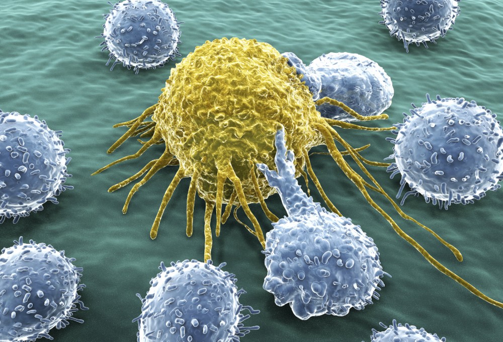
The kidney tissues derived from human iPS cells
A.The kidney tissue generated in vitro, which shows green fluorescence in each glomerulus.
B.Vascularized glomerulus formed upon transplantation into the mouse. Many red blood cells (arrowhead) are observed, and the substance exists in the lumen (*), suggesting the possible filtration.
C.Mouse vascular endothelial cells (green) are incorporated into the glomerulus that consists of podocytes (magenta).
D.The slit diaphragm (arrow) formed between the cellular processes of the podocytes. Credit: The Institute of Molecular Embryology and Genetics (IMEG).

In the field of iPS cell-based regenerative medicine, advanced research with clinical applications for many organs and tissues, such as the retina, has steadily progressed. However, growing a kidney from scratch has been extremely difficult.  Although the number of renal failure patients on dialysis is increasing, opportunities for renal transplant have been limited with great attention given to the growth of kidneys to stem the shortage.

Now, a study from researchers at Kumamoto University shows mouse kidney capillaries successfully connecting to kidney tissue derived from human iPS cells. The team state that this achievement shows that human kidney glomeruli made in vitro can connect to blood vessels after transplantation and grow to maturity, a big step forward in gain-of-function for a urine-producing kidney.  The opensource study is published in the Journal of the American Society of Nephrology.

Earlier studies from the lab led to the development of an in vitro three-dimensional kidney structure from human iPS cells.  However, it was unclear how similar the kidney tissue made in vitro was to that formed in a living body. Additionally, the original kidney tissue was not connected to any blood vessels, even though the primary function of the organ is to filter waste products and excess fluid from the blood.  In many kidney diseases, the pathology is with the glomeruli that filter urine from the blood. Filtration in the glomerulus is performed by cells called podocytes that are in direct contact with the blood vessels. Through the special filtration membrane of the podocytes, proteins don’t leak into the urine and allows moisture to pass through.  Therefore, the group focused on analyzing the podocyte of the glomeruli in detail.  They achieved this by genetically modifying the iPS cells and growing human kidney tissue in vitro with green fluorescence then visualizing how human glomeruli became established.

The current study continued this analysis by taking out only the podocytes of the human glomeruli using the green fluorescence, and revealed that glomerular podocytes made in vitro express the same genes important for normal biological function.  Data findings show that after transplanting the human iPS cell-based kidney tissue into a mouse body, glomeruli connecting to mouse kidney capillaries formed. Results show that human glomerular podocytes further matured around adjacent blood vessels as in a living body and formed a characteristic filtration membrane structure.  The group state that to their knowledge the successful connection of capillaries with the podocytes of iPS cell-manufactured human glomeruli resulting in a distinct filtration membrane is the first of its kind in the world.

The team surmise that their findings should advance research into the manufactured kidney’s function to produce and excrete urine.  They go on to add that by using iPS cells from patients, development of new drugs and clarification of the causes of kidney disease are also expected.  For the future, the researchers state that they are now working to develop a discharge path for the kidney and combine it with findings on glomerular cells.

Source: The Institute of Molecular Embryology and Genetics (IMEG)


Human Induced Pluripotent Stem Cell–Derived Podocytes Mature into Vascularized Glomeruli upon Experimental Transplantation

Sazia Sharmin*Atsuhiro Taguchi*Yusuke Kaku*Yasuhiro Yoshimura*Tomoko Ohmori*Tetsushi Sakuma, et al.

JASN Nov 19; 2015 ASN.2015010096      http://dx.doi.org:/10.1681/ASN.2015010096    http://jasn.asnjournals.org/content/early/2015/11/18/ASN.2015010096.full

Glomerular podocytes express proteins, such as nephrin, that constitute the slit diaphragm, thereby contributing to the filtration process in the kidney. Glomerular development has been analyzed mainly in mice, whereas analysis of human kidney development has been minimal because of limited access to embryonic kidneys. We previously reported the induction of three-dimensional primordial glomeruli from human induced pluripotent stem (iPS) cells. Here, using transcription activator–like effector nuclease-mediated homologous recombination, we generated human iPS cell lines that express green fluorescent protein (GFP) in the NPHS1 locus, which encodes nephrin, and we show that GFP expression facilitated accurate visualization of nephrin-positive podocyte formation in vitro. These induced human podocytes exhibited apicobasal polarity, with nephrin proteins accumulated close to the basal domain, and possessed primary processes that were connected with slit diaphragm–like structures. Microarray analysis of sorted iPS cell–derived podocytes identified well conserved marker gene expression previously shown in mouse and human podocytes in vivo. Furthermore, we developed a novel transplantation method using spacers that release the tension of host kidney capsules, thereby allowing the effective formation of glomeruli from human iPS cell–derived nephron progenitors. The human glomeruli were vascularized with the host mouse endothelial cells, and iPS cell–derived podocytes with numerous cell processes accumulated around the fenestrated endothelial cells. Therefore, the podocytes generated from iPS cells retain the podocyte-specific molecular and structural features, which will be useful for dissecting human glomerular development and diseases.


The glomerulus is the filtering apparatus of the kidney and contains three types of cells: podocytes, vascular endothelial cells, and mesangial cells. Podocytes cover the basal domains of the endothelial cells via the basement membrane and play a major role in the filtration process.1,2 Podocytes possess multiple cytoplasmic protrusions. The primary processes are complicated by the further stemming of smaller protrusions (secondary processes or foot processes), which interdigitate with those from neighboring podocytes. The gaps between these foot processes are connected with the slit diaphragm, which is detectable only by electron microscopy. The molecular nature of the slit diaphragm was initially revealed by identification of NPHS1 as the gene responsible for Finnish-type congenital nephrotic syndrome.3 The nephrin protein encoded by NPHS1intercalates with those from neighboring cells, thus forming a molecular mesh that hinders serum proteins of high molecular weight from leaking into the urine.4,5 To date, many slit diaphragm–associated proteins have been identified, including NPHS2 (encoding podocin) and NEPH1, mutations that cause proteinuria in humans and/or mice.6,7

Podocytes are derived from nephron progenitors that reside in the embryonic kidney and express transcription factor Six2.8 Upon Wnt stimulation, the nephron progenitors undergo mesenchymal-to-epithelial transition and form a tubule.9 This tubule changes its shape; one end forms the glomerulus with podocytes inside, which is surrounded by a Bowman’s capsule. Meanwhile, vascular endothelial cells and mesangial cells migrate into the developing glomeruli, thus connecting the glomeruli with circulation.2 In these processes, several transcription factors, including Wt1, regulate expression of nephrin in podocytes.10 Apical junctions are initially formed between the presumptive podocytes, but the apical domain loses its direct contact with that of the neighboring cells, thus forming the characteristic podocyte shape. Nephrin is eventually localized to the site close to the basal domain and contributes to the formation of the slit diaphragm.2 The molecular mechanisms underlying podocyte development have been extensively studied in mice. However, because of limited access to human embryos, relatively little is known regarding transcription profiles of podocytes and glomerulogenesis in humans.4,11,12

We have recently induced the nephron progenitors from mouse embryonic stem (ES) cells and human induced pluripotent stem (iPS) cells by redefining the in vivo origin of the nephron progenitors.13 The induced progenitor aggregates readily form three-dimensional primordial glomeruli and renal tubules upon Wnt stimulation in vitro. To analyze the detailed structures and transcription profiles of the induced podocytes, we have here inserted the GFP gene into the NPHS1 locus of human iPS cells via homologous recombination using transcription activator–like effector nucleases (TALENs)14 and generated glomeruli with green fluorescent protein (GFP)-tagged podocytes.


Fluorescent Visualization of Human Glomerular Podocytes Generated fromNPHS1-GFP iPS Cells

To visualize developing human podocytes in vitro, we inserted a gene encoding GFP into the NPHS1 locus of human iPS cells by homologous recombination (Figure 1A). We first constructed a pair of plasmids expressing TALENs targeted in close proximity to the NPHS1 start codon. When tested in HEK 293 cells, these plasmids efficiently deleted the NPHS1 gene (Supplemental Figure 1A). We then introduced these TALEN plasmids, along with a targeting vector containing the GFP gene and the homology arms, into human iPS cells. This resulted in efficient homologous recombination and isolation of heterozygous GFP knock-in (NPHS1-GFP) clones (Figure 1B, Supplemental Figure 1, B and C).

Figure 1.

Successful generation ofNPHS1-GFP iPS cells by homologous recombination. (A) Strategy for targeting the human NPHS1 locus. TheGFP cassette was inserted upstream of the NPHS1 start codon. The puromycin-resistance cassette (PURO) is flanked by loxP sites. Positions for primers and probes for screening are indicated. E, EcoRV; N, NheI. (B) Southern blot of control (+/+) and NPHS1-GFP (GFP/+) clones. Genomic DNA was digested and hybridized with the indicated probes.

We differentiated these NPHS1-GFP iPS clones toward the nephron progenitors and subsequently combined them with murine embryonic spinal cord, which is a potent inducer of tubulogenesis, as we previously reported.13 Four days after recombination, spotty GFP signals could be observed, and the number and intensity of GFP signals increased thereafter until day 9 (Figure 2A,Supplemental Figure 2A). We observed GFP signals in all the examined samples from seven independent experiments (a total of 50 samples). Some of the signals started in a crescent shape and gradually changed into round structures (Figure 2A, lower panels), which suggests that human glomerular formation in vitro may be visualized. Therefore, we examined glomerulogenesis using sections of the explants. At day 3, only tubular structures were observed and GFP-positive cells were undetectable (Figure 2B). At day 4, structures that resembled S-shaped bodies were observed, in which proximo-distal specification occurred toward the presumptive distal (E-cadherin+) and proximal (cadherin-6+) renal tubules and glomerular podocytes (WT1+) (Figure 2C). At day 6, various forms of primordial glomeruli were observed, and most of the GFP signals overlapped with those of WT1 (Figure 2B). We ordered these glomeruli according to GFP intensity, which is likely to reflect the chronologic order of development. Weakly GFP-positive (and WT1-positive) limbs appeared at one end of the tubules, which elongated to surround the renal tubules. GFP intensity increased when the podocyte layers were convoluted. At day 9, strongly GFP-positive round glomeruli were formed. These histologic changes are consistent with the previous observations of human glomeruli in aborted fetuses.15 Thus, we succeeded in visualizing human podocyte development and glomerulogenesis in vitro. Interestingly, some, but not all, of the Bowman’s capsule cells were positive for GFP and nephrin (Supplemental Figure 2B), suggesting that these cells are not completely specified yet. Indeed, transient nephrin expression in some capsule cells was reported in vivo.16

Figure 2.

Fluorescent visualization of human glomerular podocytes generated fromNPHS1-GFP iPS cells. (A) Morphologic changes of GFP-positive glomeruli during differentiation in vitro. The nephron progenitors induced fromNPHS1-GFP iPS cells were combined with murine embryonic spinal cord and cultured for the indicated time. Lower panels: higher magnification of the areas marked by rectangles in the upper panels. Note the shape changes of the glomerulus (arrowheads). Scale bars: 500 μm. (B) Histologic sections of glomeruli developing in vitro. Tissues at day 3, 6, and 9 after recombination with the spinal cord were analyzed. Top panels: Hematoxylin-eosin (HE) staining. Middle panels: GFP (green) staining. Bottom panels: dual staining with GFP and WT1. Nuclei were stained with 4′,6-diamidino-2-phenylindole (DAPI: blue). Scale bars: 20 μm. (C) Presumptive S-shaped bodies observed at day 4 (left two panels) and day 6 (right two panels). Serial sections were stained with E-cadherin (Ecad: magenta)/cadherin-6 (cad6: green) and E-cadherin (magenta)/WT1 (green). Arrowheads: WT1-positive presumptive glomerular regions. Scale bars: 20 μm.

Induced Podocytes Exhibit Apicobasal Polarity and Basally Localized Nephrin

We analyzed day 9 sections at higher resolution to examine the apicobasal polarity of the induced podocytes. GFP was detected in the nuclei and cytoplasm of most cells in the round glomeruli (Figure 3A) because we did not attach any localization signal to GFP when generating NPHS1-GFP iPS cells. Nephrin proteins were distributed in a linear fashion in the iPS cell–derived glomeruli and at one end of the WT1-positive podocyte layer (Figure 3, A and B). These expression patterns significantly overlapped with those of type IV collagen, which was accumulated on the basal side of the podocytes (Figure 3C). In contrast, podocalyxin, an apical marker, was expressed in a manner mutually exclusive of nephrin (Figure 3D). Therefore, the induced podocytes exhibited a well established apicobasal polarity and nephrin proteins were properly localized at the basal side, where the presumptive slit diaphragm should be formed. We also observed nephrin-positive dots on the lateral side of the podocytes (Figure 3A, arrowheads), as reported in human developing podocytes in vivo.15 We found that these dots actually represent the filamentous structures encompassing the basal to the lateral side of the podocytes (Figure 3, B and C, arrowheads). Although further investigation is required, this may reflect the transit state of nephrin proteins shifting from the apical to the basal domain of the induced podocytes.

Figure 3.

Induced podocytes exhibit apicobasal polarity and basally localized nephrin. (A) Nephrin (magenta) and GFP (green) staining of the induced glomerulus at day 9. (B) Nephrin (magenta) and WT1 (green) staining. (C) Nephrin (magenta) and type IV collagen (COL: green) staining. (D) Nephrin (magenta) and podocalyxin (PODXL: green) staining. The left columns are at lower magnification to show a whole glomerulus. The right two columns are singly stained, while the left two columns represent merged images. Arrows: nephrin proteins localized to the basal domain; arrowheads: nephrin-positive dot-like or filamentous structures. Scale bars: 10 μm.

Induced Podocytes Possess Primary Processes with the Slit Diaphragm–Like Structures

We further analyzed the morphology of the induced glomeruli by electron microscopy. Both scanning and transmission electron microscopy showed well organized glomeruli surrounded by Bowman’s capsules (Figure 4, A and B). Interestingly, numerous microvilli were detected in the apical domain of the induced podocytes (Figure 4, C and D). Similar microvilli were reported in developing in vivo podocytes in humans.17,18 The podocytes were attached to each other at sites close to the basal region (Figure 4D). Inspection of the basal side of the induced podocytes by scanning microscopy identified multiple protrusions (Figure 4E), which were confirmed by transmission microscopy (Figure 4F). Higher magnification clearly showed bridging structures between the protrusions, which may represent an immature form of the slit diaphragm (Figure 4, G and H, Supplemental Figure 3, A–C). Thus, this is the first in vitrogeneration of mammalian podocytes with slit diaphragm–like structures from pluripotent stem cells. However, because typical interdigitation of the protrusions is lacking, they are likely to represent primary processes but not secondary processes (foot processes).

Figure 4.

Induced podocytes possess primary processes with the immature slit diaphragm–like structures. (A and B) Induced glomerulus covered with a Bowman’s capsule shown by (A) scanning and (B) transmission electron microscopy. (C) Induced podocytes observed by scanning electron microscopy. Multiple microvilli are observed on the apical surface (arrowheads). (D) Aligned podocytes, which attach to each other at sites close to the basal region, shown by transmission electron microscopy. Multiple microvilli are observed on the apical surface (arrowheads). (E) Primary processes shown by scanning electron microscopy (asterisks). Podocytes from the basal side are shown. (F) Primary processes shown by transmission electron microscopy (asterisks). (G) Slit diaphragm–like structures between the primary processes (arrows), shown by scanning electron microscopy. (H) Primary processes with slit diaphragm–like structures (arrows), shown by transmission electron microscopy. Scale bars: A and B: 10μm; C–F: 2 μm; G and H: 0.2 μm.

Induction of Podocytes from Human NPHS1-GFP iPS Cells Enables Their Efficient Isolation

We next tried to purify the GFP-positive podocytes at day 9 by FACS. Of the induced cells, 7.45%±0.72% (mean±SEM from five independent induction experiments) were positive for GFP (Figure 5A, left panel). We also found that the monoclonal antibody against the extracellular domain of nephrin (48E11),19in combination with the anti-podocalyxin antibody, was useful for sorting developing podocytes. Of the GFP-positive cells, 94.0% were positive for both nephrin and podocalyxin (Figure 5A, middle panel), while most of the GFP-negative cells (97.5%) were negative for both markers (Figure 5A, right panel). Thus, GFP faithfully mimics nephrin expression and podocytes were enriched in the GFP-positive population. Quantitative RT-PCR analysis of sorted cells confirmed the differential expression of several podocyte markers, such asNPHS2 (encoding podocin) and synaptopodin (Figure 5B). When the sorted GFP-positive cells were cultured for 3 days, the cells expressed WT1 in nuclei and podocalyxin on the cell surface (Figure 5C). Nephrin and GFP were detected on the cell surface membrane and in the cytoplasm, respectively, at day 7 of culture, although expression levels were lower than before the start of the culture. These results indicate that induction from NPHS1-GFP iPS cells enables efficient isolation of developing human podocytes.

Figure 5.

Induction of podocytes from human NPHS1-GFP iPS cells enables their efficient isolation. (A) FACS analysis of induced tissues at day 9. Almost 8% of cells are positive for GFP in this representative experiment (left panel). Nephrin and podocalyxin (PODXL) expression in the GFP-positive or -negative fraction (middle and right panel, respectively). (B) Quantitative RT-PCR analysis of GFP-positive and -negative fractions. Average and SEM from three independent experiments are shown. β-ACT, β-actin; SYNPO, synaptopodin. (C) Immunostaining of podocytes cultured for the indicated times after sorting GFP-positive cells. Scale bars: 5 μm. (D) Venn diagram of the transcription profiles of podocytes. Microarray data of GFP-positive podocytes are compared with those of human adult glomeruli and murine podocytes.

GFP-Positive–Induced Podocytes Show Transcriptional Profiles That Overlap with Those of Mouse and Human Podocytes In Vivo

To obtain comprehensive transcription profiles of the iPS cell–derived podocytes, we performed microarray analysis at day 9. We detected 2985 probes that were enriched in GFP-positive podocytes compared with GFP-negative cells. Of these, the top 300 genes were used for unbiased cluster analysis against microarray data from a wide variety of human tissues (Supplemental Figure 4, A and C).20 Genes enriched in the GFP-positive podocytes had variable tissue specificity. For example, NPHS2 was selectively expressed in the kidney or fetal kidney tissues. However, synaptopodin andFOXC2 were sorted into the ubiquitously expressing cluster. Dendrin was assigned to a cluster enriched in the neuronal tissues. These results suggest a single molecule is not sufficient to confirm the identity of podocytes. Therefore, we compared our gene list of GFP-positive human podocytes with the published microarray analyses of adult human glomeruli and adult podocytes from Mafb-GFP transgenic mice.11,21 Overall, 190 probes were overlapping among the three gene sets (Figure 5D, Supplemental Table 1, Table 1). These included typical slit diaphragm–related genes, such as NPHS1, NPHS2,CD2AP,22 chloride intracellular channel protein 5 (CLIC5),23 and dendrin,24,25and basolateral adhesion molecules such as claudin 5 and integrinα3.26,27Phospholipase ε1 and nonmuscle myosin heavy chain 9 (Myh9), causative genes for hereditary kidney diseases,2830 were also included. Transcription factors that have important roles in podocyte development, including WT1, MAFB, FOXD1, and TCF21, as well as vascular attractants such as VEGFA and semaphorin, were also expressed.1,2,31 Interestingly, when these selected overlapping genes were used for the cluster analysis against the microarray data from various organs described above, kidney and fetal kidney were segregated as separate clusters, suggesting the kidney-biased features of the overlapping gene set (Supplemental Figure 4B).

Table 1.

Genes common to iPS cell–derived podocytes in vitro, human glomeruli, and mouse podocytes in vivo

We also identified genes common to GFP-positive podocytes and adult human glomeruli (Figure 5D, Supplemental Table 2), and genes common to GFP-positive podocytes and mouse adult podocytes (Figure 5D, Supplemental Table 3). The former includes BMP7,32 while the latter includes NEPH1 (KIRREL),FOXC2, ROBO2, and EPHRIN-B1.7,3336 These results indicated that the typical transcriptional profiles are well conserved among our podocytes generated in vitro as well as mouse and human podocytes in vivo. In addition, extracellular matrix components characteristic of glomeruli at the capillary loop stage,lamininα5/β2/γ1 isoforms (corresponding to laminin 521) and type IV collagenα4/α5,37 were detected, the latter of which is the causative gene for Alport syndrome. These data indicate that the transition to these mature forms from immature laminin 111 and collagen α1/α2 has already occurred in vitro.

Taken together, our podocytes induced in vitro possessed the typical features of those in vivo, not only in morphology but also in transcription profiles, further supporting the authenticity of our human iPS cell induction protocol. In addition, genes exclusively expressed in the GFP-positive podocytes are worthy of further investigation because they may include genes specific to developing human podocytes, a possibility that has not been addressed to date (Figure 5D,Supplemental Table 4).


Transplanted iPS Cell–Derived Nephron Progenitors Form Vascularized Glomeruli

We next examined whether glomeruli generated from iPS cells integrated with the vascular endothelial cells. The iPS cell–derived nephron progenitor spheres were induced by spinal cord for 1 day in vitro to initiate tubulogenesis and were then transplanted beneath the kidney capsule of immunodeficient mice. We also cotransplanted mixed aggregates of human umbilical vein endothelial cells (HUVECs) and mesenchymal stem cells (MSCs) because these cells are useful for the generation of vascularized organ buds in vitro.38,39 When these aggregates were transplanted using a conventional method that we used for the transplantation of mouse ES cell–derived nephron progenitors,13 minimal nephron differentiation was observed at 10 days after transplantation (n=4) (Figure 6A). Because human iPS cell–derived aggregates were larger (approximately 1000 µm in diameter) than those from mouse ES cells (approximately 600 µm) and were instantly flattened upon transplantation (Supplemental Figure 5A), we hypothesized that mechanical tension of the capsule may have hampered nephron differentiation. Therefore, we inserted two agarose rods of 1100 µm diameter in a V-shaped position to release tension and secure a space for the transplanted aggregates (Figure 6B). We also soaked the rods with VEGF to enhance vasculogenesis.31 As a result, we observed immature glomerular formation at day 10 in the transplants, accompanied by blood vessels integrating into these glomeruli (n=5) (Figure 6, C and D). The vessels were preferentially clustered in the glomeruli among the grafted tissue (Figure 6D), suggesting that the iPS cell–derived podocytes possess the potential to attract vasculature. This is also consistent with microarray data showing VEGFA expression in our induced podocytes.

Figure 6.

Transplanted iPS cell–derived nephron progenitors form vascularized glomeruli. (A) Hematoxylin-eosin sections of tissues at 10 days after transplantation using a conventional method. Right panel: magnified image of the square in the right panel. kid, kidney of the host mouse. (B) Method for transplantation using solid agarose rods. Right panel: macroscopic view of transplanted tissue under the kidney capsule. Ag, agarose rods. (C) Hematoxylin-eosin sections of the transplanted tissue at day 10 in the presence of the rods. Right panel: magnified images of the square. (D) Vascularized glomeruli at day 10. Staining of WT1 and CD31. Right panel: magnified image of the square in the left panel. (E) Hematoxylin-eosin section of the transplanted tissue at day 20. Middle and right panel: magnified images of the squares in the panels on their left, respectively. *Stromal cells. kid, kidney of the host mouse. (F) Vascularized glomeruli formed upon transplantation at day 20. Left panel: magnified images of panel E. Right panel: magnified image of the square in the left panel. Note the enlarged Bowman’s space. (G) The endothelial cells are of mouse origin. Staining of WT1 (magenta) and MECA-32, a marker for mouse-specific endothelial cells (green). (H) Hematoxylin-eosin staining showing red blood cells in the induced glomeruli. (I) Hematoxylin-eosin staining showing the eosin-positive precipitates in the Bowman’s space. (J) Staining of nephrin (magenta) and CD31 (green). Right panel shows the basal localization of nephrin. Scale bars: A, C–F, I: 100 μm; B: 1 mm; G, H, J: 10 μm.

At day 20 after transplantation, we observed enlarged transplanted tissues beneath the capsule (Supplemental Figure 5B). Histologic examination revealed excessive growth of stromal cells of human origin, which were presumably derived from nonrenal tissues that were coinduced with nephron progenitors from iPS cells (n=4) (Figure 6E, Supplemental Figure 5C). Nonetheless, glomeruli were formed and the blood vessels were well integrated into the glomeruli (Figure 6, F and G). Moreover, 90% (135 of 150) of the glomeruli contained red blood cells (Figure 6H). Indeed, some of the glomeruli showed an enlarged Bowman’s space and contained eosin-positive precipitation (Figure 6I), which might imply a small amount of urine production. Interestingly, endothelial cells in the induced glomeruli were of mouse origin (Figure 6G,Supplemental Figure 5D). HUVEC-derived endothelial cells were not integrated into the iPS cell–derived glomeruli but were located separately from the sites of nephron formation (Supplemental Figure 5E). Therefore, HUVEC may not be competent to interact with human podocytes.

The anti-human specific podocalyxin antibody stained the apical domains of the iPS cell–derived podocytes, but not those of the host mouse podocytes (Supplemental Figure 5F). Nephrin protein in induced podocytes was localized at the basal side that faced the vascular endothelial cells (Figure 6J), suggesting the emergence of filtering apparatus. Electron microscopic analyses of two additional samples at day 20 showed that iPS cell–derived podocytes accumulated around, and were closely associated with, endothelial cells (Figure 7A). The induced podocytes developed numerous complex cell processes, as well as a linear basement membrane, at interfaces with endothelial cells (Figure 7B). The distances between the cell processes of some podocytes were enlarged, and slit diaphragm–like structures were formed between the processes located above the basement membrane (Figure 7C). Each of these diaphragms appeared as an electron-dense line (approximately 35 nm wide, 10 nm thick) bridging adjacent cell processes of the iPS cell–derived podocytes (Figure 7D). This feature was also observed in vivo and differed from the immature ladder-like structure that was seen between adjacent podocytes cultured exclusively in vitro without transplantation (Figure 4). Endothelial cells also produced basement membrane, but it was not fused to that of the podocytes in most cases, thus forming double-layered structures (Figure 7E). Interestingly, endothelial cells were fenestrated with residual diaphragm, a characteristic feature of embryonic glomerular endothelial cells (Figure 7F).40Furthermore, an electron-dense substance was detected in the Bowman’s space (Figure 7C), as in Figure 6I, implying the possible presence of filtration. Taken together, glomeruli generated from human iPS cells were vascularized and had many morphologic features present in glomeruli in vivo.

Figure 7.

iPS cell–derived glomeruli in the transplants exhibited many morphologic features of those in vivo. (A) Induced podocytes surrounding the vascular endothelial cells and extending many cell processes, shown by transmission electron microcopy. (B) Complex cell processes of podocytes formed between the cells and above the basement membrane. (C and D) Formation of slit diaphragm–like structures (arrows) between the cell processes of induced podocytes. Note the electron-dense substance in the Bowman’s capsule (asterisk). (E) Formation of double-layered basement membranes, each derived from endothelial cells (white arrowheads) and induced podocytes. (F) Fenestrated endothelial cells with diaphragms (black arrowheads). bm, basement membrane derived from induced podocytes; en, endothelial cells. Scale bars: A: 1 μm; B, E: 0.5 μm; C, D, F: 0.2 μm.

Discussion

We have inserted GFP into the NPHS1 locus of human iPS cells and successfully differentiated them toward three-dimensional glomeruli. The GFP-positive–induced podocytes possessed apicobasal polarity and were equipped with primary processes and slit diaphragm–like structures. Furthermore, sorted podocytes exhibited typical transcription profiles that overlap with those in vivo. These findings underscore the authenticity of our induction protocol.NPHS1 promoter–driven GFP expression is a good indicator of glomerulus formation. Several groups have reported the induction of kidney tissues in vitro,13,4143 and our iPS cell lines will be useful for assessing the induction efficiency of glomeruli by each protocol. In addition, we successfully sorted human podocytes using a combination of anti-nephrin and anti-podocalyxin antibodies. These reagents will make genetic GFP integration unnecessary for the purification of podocytes from patient-derived iPS cells, and possibly from more complex in vivo tissues.

It is surprising that well organized glomeruli are formed without the other two components of glomeruli: mesangial and vascular endothelial cells. These two cell types are not derived from nephron progenitors, as shown by cell lineage analysis in mice,8,44,45 and indeed we did not detect these lineages in the induced glomeruli (Supplemental Figure 3D). Thus, glomeruli can self-organize their structures solely from the podocytes derived from nephron progenitors, without any inductive signals from mesangial cells or the vasculature. However, further maturation will be required to reproduce hereditary glomerular diseases. We developed a new transplantation technique using agarose rods to secure a space against the tension evoked by kidney capsules. This technical improvement led to the successful generation, for the first time, of vascularized glomeruli derived from human iPS cells. The induced podocytes exhibited complex cell processes with slit diaphragm–like structures, and linear basement membrane that ran along that of the endothelial cells was formed. Furthermore, endothelial cells were fenestrated, which is a characteristic feature of glomerular endothelial cells. Most experiments used agarose rods soaked with VEGF to potentially accelerate vasculogenesis; however, the absence of VEGF in the rods also caused the formation of vascularized glomeruli (Supplemental Figure 5G). Thus, we can at least conclude that the human iPS cell–derived podocytes expressed sufficient attractants, including VEGF, to recruit endothelial cells.

It is noteworthy that the integrated endothelial cells were of mouse origin from the host animals but were not derived from HUVECs, although both vascular sources were initially located in proximity to the iPS cell–derived transplants. Therefore, human podocytes recruited mouse endothelial cells despite species differences, while HUVECs may not be competent to interact with human podocytes. Even when we performed transplantation without HUVECs or MSCs, we observed vascularized glomeruli, suggesting that paracrine effects of these cells may also be minimal (Supplemental Figure 5H). The presence of double-layered basement membrane might be caused by the incomplete fusion between those derived from human podocytes and mouse endothelial cells, as observed when mouse embryonic kidney was transplanted onto a quail chorioallantoic membrane.46 Therefore, the identification of optimal sources for human endothelial cells is necessary.

While it is difficult to estimate the gestational age on the basis of the morphology of the individual glomeruli,47,48 waiting for a longer period after transplantation may help further maturation of induced podocytes. However, we observed an excessive growth of stromal, presumably nonrenal, cells in the transplants. Thus, it will be essential to develop methods to purify nephron progenitors for transplantation. At the same time, it is necessary to induce genuine stromal cells because both interstitial cells and mesangial cells are derived from renal stromal progenitors.45 At present, we have no evidence that proper mesangial cells exist in our vascularized glomeruli. Ideally, human endothelial and mesangial cells that correspond to those in the developing kidney should be combined. Although further induction studies, as well as imaging techniques to visualize the slit diaphragm with a higher resolution,49are needed to achieve this goal, our results will accelerate the understanding of human podocyte biology both in developmental and diseased states.


Read Full Post »

Medical Breakthrough: Israeli Researcher Predicts Where Cancer Will Spread

Reporter: Evelina Cohn Budu, PhD


An innovative technology developed in Israel may soon be able to predict the spread of cancer from one organ to another, potentially saving the lives of millions of people around the world.

The technology, developed at Israel’s Technion – Israel Institute of Technology, has been proven in preliminary laboratory trials, and is now entering into advanced testing using cells from patients undergoing surgery.

Assistant Professor Dr. Daphne Weihs has developed a unique biomechanical method for the early detection of metastatic cancer (a cancer that has already spread). At the metastatic stage, the original, primary tumor expands, invades and takes over more and more nearby tissue. A tumor that has become very aggressive “knows” how to send metastases to more distant tissues through the lymph and circulatory systems.

Metastases (secondary tumors) are usually more dangerous than the primary tumor because it is difficult to identify them at their inception. When they are detected at an advanced stage, treating them medically is more complicated and the medical prognosis is typically not good.

proteinattackscancercell

Photos: Courtesy of the Technion

cancer-cells-

Photos: Courtesy of the Technion

ABOUT THE RESEARCHER

Daphne Weihs , Assistant Professor

Affiliation: Faculty

Link to Lab Web Page:


SOURCE

http://nocamels.com/2015/10/israeli-researcher-predicts-spread-cancer/?utm_source=activetrail&utm_medium=email&utm_campaign=nc4/11/15


REFERENCES

New Israeli Cancer Vaccine Triggers Response In 90% Of Cancer Types

By Jonathan Neff, NoCamels.com January 01, 2015

http://nocamels.com/2015/01/new-vaccine-for-cancer/

How Elephants’ Genes Are Fighting Cancer In Humans

By Lauren Blanchard, NoCamels October 18, 2015

http://nocamels.com/2015/10/how-elephants-genes-are-helping-fight-cancer-in-humans/

Other related articles published in this Open Access Online Scientific Journal, include the following:


http://pharmaceuticalintelligence.com/?s=metastasis


Please place here FIVE articles from above link

Read Full Post »

Mass-producing stem cells to satisfy the demands of regenerative medicine

Reported by: Irina Robu, PhD

Instead of culturing cell on round, flat Petri dishes, Steve Oh from A*STAR Bioprocessing Technology Institute he grew them in a tiny polystyrene beads known as microcarriers floating in a nutritional brew. The standard Petri dish fits fewer than 100,000 cells, a minuscule amount when stacked against the 2 billion muscle cells  that make up the heart or 100 billion red blood cells needed to fill a bag of blood. 

The average Petri dish fits fewer than 100,000 cells, a miniscule amount when stacked against the 2 billion muscle cells that make up the heart or the 100 billion red blood cells needed to fill a bag of blood. The approach Reuveny suggested potentially could produce cells in much vaster numbers to make them more practical for therapy.
 
Dr. Oh first tried the approach on human embryonic stem cells, because they have the potential to mature into any type of cell in the body and struggled to develop a coating that would make the stem cells stick to the microcarriers and formulate the right mixture of nutrients for cell to grow. After a year, one line of human embryonic stem cells survived past the 20 week mark of stability and found out that these cells were two to four times times more densely packed than grown in petri dishes.

However after six years of refining the processes, they were able to achieve three times higher cell densities than petri dishes approach by modifying the feeding strategy.  Their success started with cardiomyocytes wich are known as the fastest cell type to differentiate. The researchers developed a strategy to grow pure batches of cariomyocytes without adding growth factors but instead use small molecules to first inhibit and then activate a key cell differentiation pathway known as Wnt signaling. Then they apply the small molecule approach to grow and differentiate cardiomyocytes from embryonic stem cells directly on microcarriers. And according to Dr. Oh their method had beat the Petri dish methods on purity, yield, cost of cells and simplicity of process.

The main  goal of the research is to grow enough cells inexpensively in order to patch up one square-centimeter of damaged heart muscle following a heart attack.

Source

http://phys.org/news/2015-06-mass-producing-stem-cells-demands-regenerative.html

Read Full Post »

Living Cellular Bio-ink Kits

Reporter: Irina Robu, PhD

CELLINK and RoosterBio developed cellular bio-ink kits. These kits  will enable printing of living cellular constructs by combining RoosterBio’s stem cell systems with the universal bioinks that have been created by CELLINK. This combination provides the materials needed for the creation of human tissue needed for research and testing. These kits represent the first commercially available, plug-and-play living cellular bioinks and are available for both commercial and academic use. Erik Gatenholm from CELLINK stated that “this revolutionary, cost effective, and elegantly simple-to-use Bioprinting Kit is exactly what innovators and early adopters worldwide need to propel their research in the tissue-engineering field. By offering a complete package of the right bioink together with the right cells we can finally establish the first standard in the bioprinting industry.”

These kits will minimize the time needed for optimizing a biomaterial-stem cell combination in preparation for research and development.

Source

http://3dprint.com/97382/first-living-cellular-bioink/

Read Full Post »

Tumor Associated Macrophages: The Double-Edged Sword Resolved?

Writer/Curator: Stephen J. Williams, Ph.D.

UPDATED 10/04/2021 TAMs Enhance Tumor Hypoxia and Aerobic Glycolysis

Cell-based immunity is vital for our defense against pathologic insult but recent evidence has shown the role of cell-based immunity, especially macrophages to play an important role in both the development and hindrance of tumor growth, including role in ovarian, hematologic cancers, melanoma, and breast cancer.  In the past half century, new immunological concepts of cancer initiation and progression have emerged, including the importance of the harnessing the immune system as a potential anti-cancer strategy. However, as our knowledge of the immune system and tumor biology has grown, the field has realized an immunological conundrum: how can an immune system act to both prevent tumor growth and promote the tumor’s growth?

As discussed in the lower section of this post, authors of a paper in the journal Science show how different populations of tumor-associated macrophages (TAMs) may exert both positive and negative effects on tumor cells, producing a sort of ying-yang war between the tumor and the immune system.

The Immune System: Brief Overview and Role in Cancer

celllineageimmunesystem

Figure. Cell lineage of the immune system. A description of the different cell types can be found here.

Histologic evaluation of multiple tumor types, especially solid tumors, reveal the infiltration of diverse immunological cell types, including myeloid and lymphoid cell lineages, such as macrophages and NK, T cell and B cells respectively.

The immunological conundrum

immuncecancerconundrum

Figure. Potential inflammatory signaling pathways in breast cancer stem cells.
Breast cancer stem cells may be regulated by chemokine- and/or cytokine-mediated inflammatory signaling in an autocrine or paracrine manner. (from University of Tokyo at http://www.ims.u-tokyo.ac.jp/system-seimei/en/research2_e.htm)

Role of Tumor Associated Macrophages

There are conflicting reports as to the functional consequence of these infiltrating tumor-associated macrophages (TAMs). TAMs have been shown to secrete mediators such as interleukins and cytokines in a paracrine manner such as CCL2, IL10 and TGFβ. In certain instances these cytokines and mediators actually promote the growth of the surrounding tumors.

J Leukoc Biol 2009 Nov 86(5) 1065-73, Figure 1

Figure.  TAMs can be divided into subpopulations with distinctive functions and secretogogues.

For Further Reference

Tumor-associated macrophages and the profile of inflammatory cytokines in oral squamous cell carcinoma. http://www.ncbi.nlm.nih.gov/pubmed/23089461 anti-inflamm IL10 and TGFB

Tumor-associated macrophage-derived IL-6 and IL-8 enhance invasive activity of LoVo cells induced by PRL-3 in a KCNN4 channel-dependent manner

TAMscytokines

Figure 2: TAM functions in tumor progression. Tumor cells and stromal cells, which produce a series of chemokines and growth factors, induce monocytes to differentiate into macrophages. In the tumor, most macrophages are M2-like, and they express some cytokines, chemokines, and proteases, which promote tumor angiogenesis, metastasis, and immunosuppression. From Macrophages in Tumor Microenvironments and the Progression of Tumors

ICB-14-NC-BRONTE-V2

Macrophages integrate metabolic and environmental signals to promote tumor growth. Area within dotted rectangle indicates proposed mechanisms of action. ARG, arginase; HIF, hypoxia-inducible factor; MCT, monocarboxylate transporter; NADH, nicotine adenine dinucleotide, reduced; PKM2, M2 isoform of pyruvate kinase; VEGF, vascular endothelial growth factor from Tumor cells hijack macrophages via lactic acid adapted from Colegio OR, Chu N-Q, Szabo AL, Chu T, Rhebergen AM, Jairam V et al. Functional polarization of tumour-associated macrophages by tumour-derived lactic acid. Nature (e-pub ahead of print 13 July 2014; doi:10.1038/nature13490). | Article |

Depletion of M2-Like Tumor-Associated Macrophages Delays Cutaneous T-Cell Lymphoma Development In Vivo

Targeting tumor-associated macrophages in an orthotopic murine model of diffuse malignant mesothelioma

Crosstalk between colon cancer cells and macrophages via inflammatory mediators and CD47 promotes tumour cell migration

Tumor-Associated Macrophages Regulate Murine Breast Cancer Stem Cells Through a Novel Paracrine EGFR/Stat3/Sox-2 Signaling Pathway

Science Paper: Different Populations of TAMS Have Different Tumor Effects

The cellular and molecular origin of tumor-associated macrophages Eric G. Pamer1 Ruth A. Franklin1,2, Will Liao3, Abira Sarkar1, Myoungjoo V. Kim1,2, Michael R. Bivona1, Kang Liu4, Ming O. Li1, Science 23 May 2014: Vol. 344 no. 6186 pp. 921-925

A recent Science paper from Cornel has investigated the origin, function, and characterization of TAMs on breast cancer growth. In summary, their efforts and research suggest different populations of TAMs with varied tumorigenic effects, a finding which may help explain the immunologic conundrum with respect to solid tumors.

The authors characterized the infiltrating immune cell types in a MMTV-PyMt model of breast cancer.

The MMTV-PyMt mouse breast cancer model:

is a transgenic model where mammary gland expression of the polyoma middle T antigen (PyMT) is driven by the Mouse Mammary Tumor Virus promoter (MMTV).

Microbiol. Mol. Biol. Rev. 2009 Sep 73(3) 542-63, FIG. 5

For a review of mouse models of breast cancer please see

Mouse models of breast cancer metastasis. Anna Fantozzi1 and Gerhard Christofori. Breast Cancer Res. 2006; 8(4): 212.

Results

1.     Macrophages constitute the predominate myeloid cell population in MMTV-PyMT mammary tumors

Tumor infiltrating immune cells included

  • Myeloid cells comprised 50% of CD45+ infiltrating leukocytes.
  • The CD45 antigen, also known as Protein tyrosine phosphatase, receptor type, C (PTPRC) is an enzyme that, in humans, is encoded by the PTPRC gene, and acts as a regulator of B and T-lymphocytes.
  • Authors noted three types of cells classified as Type I, II, and III based on
  1. Cell morphology
  2. Major histocompatibility complex
  • Infiltrating monocytes and neutrophils
  • Cells with dendritic and macrophage markers

2. TAMS differentiate from CCR2+ inflammatory monocytes

  • To determine whether Ly6C+CCR2+ inflammatory monocytes contributed to TAMs and MTMs, authors crossed PyMT mice to Ccr2−/− mice and found MTMs (mammary tumor macrophages) were significantly reduced in Ccr2−/− PyMT mice, implying that MTMs are constitutively repopulated by inflammatory monocytes
  • To determine whether inflammatory monocytes were required for TAM maintenance, we generated CCR2DTR PyMT mice expressing diphtheria toxin receptor (DTR) under control of the Ccr2 locus DT treatment resulted in 96% depletion of tumor-associated monocytes compared to 80% depletion in Ccr2−/− mice
  • To investigate whether monocytes could differentiate into TAMs in vivo, we transferred CCR2+ bone marrow cells isolated from CCR2GFP reporter mice into congenically marked CCR2DTR PyMT mice depleted of endogenous monocytes, we observed transferred cells in developing tumors demonstrate that tumor growth induces the differentiation of CCR2+ monocytes into TAMs.

3.     TAMs are phenotypically distinct from AAMs (M2 or alternatively activated macrophages)

  • Gene-expression profiling revealed the integrin CD11b (Itgam) was expressed at lower levels in TAMs than in MTMs while several other integrins and the integrin receptor Vcam1 were up-regulated in TAMs
  • AM population did not express AAM markers such as Ym1, Fizz1, and Mrc1; instead, MTMs more closely resembled AAMs. The authors detected Vcam1 up-regulation on TAMs as a late differentiation event

4.    RBPJ-dependent TAMs modulate the adaptive immune response

  • In DCs, canonical Notch signaling mediated by the key transcriptional regulator RBPJ controls lineage commitment and terminal differentiation. To explore whether Notch signaling played a role in TAM differentiation, authors used CD11ccre mice that efficiently deleted floxed DNA sequences to a greater extent in TAMs than MTMs, but not in monocytes or neutrophils (fig. S14). CD11ccreRbpjfl/fl PyMT mice exhibited a selective loss of MHCIIhiCD11blo TAMs ( 4A). However, a MHCIIhiCD11bhi population still remained
  • Transcriptional profiling comparing this population to WT TAMs confirmed a loss of the Notch-dependent program in RBPJ-deficient cells revealing that in the absence of RBPJ, inflammatory monocytes are unable to terminally differentiate into TAMs.

UPDATED 10/04/2021 TAMs Enhance Tumor Hypoxia and Aerobic Glycolysis

Tumor-Associated Macrophages Enhance Tumor Hypoxia and Aerobic Glycolysis

From:

Tumor-Associated Macrophages Enhance Tumor Hypoxia and Aerobic Glycolysis
Hoibin JeongSehui KimBeom-Ju HongChan-Ju LeeYoung-Eun KimSeoyeon BokJung-Min OhSeung-Hee GwakMin Young YooMin Sun LeeSeock-Jin ChungJoan DefrênePhilippe TessierMartin PelletierHyeongrin JeonTae-Young RohBumju KimKi Hean KimJi Hyeon JuSungjee KimYoon-Jin LeeDong-Wan KimIl Han KimHak Jae KimJong-Wan ParkYun-Sang LeeJae Sung LeeGi Jeong CheonIrving L. WeissmanDoo Hyun ChungYoon Kyung Jeon and G-One Ahn

Abstract

Tumor hypoxia and aerobic glycolysis are well-known resistance factors for anticancer therapies. Here, we demonstrate that tumor-associated macrophages (TAM) enhance tumor hypoxia and aerobic glycolysis in mice subcutaneous tumors and in patients with non–small cell lung cancer (NSCLC). We found a strong correlation between CD68 TAM immunostaining and PET 18fluoro-deoxyglucose (FDG) uptake in 98 matched tumors of patients with NSCLC. We also observed a significant correlation between CD68 and glycolytic gene signatures in 513 patients with NSCLC from The Cancer Genome Atlas database. TAM secreted TNFα to promote tumor cell glycolysis, whereas increased AMP-activated protein kinase and peroxisome proliferator-activated receptor gamma coactivator 1-alpha in TAM facilitated tumor hypoxia. Depletion of TAM by clodronate was sufficient to abrogate aerobic glycolysis and tumor hypoxia, thereby improving tumor response to anticancer therapies. TAM depletion led to a significant increase in programmed death-ligand 1 (PD-L1) expression in aerobic cancer cells as well as T-cell infiltration in tumors, resulting in antitumor efficacy by PD-L1 antibodies, which were otherwise completely ineffective. These data suggest that TAM can significantly alter tumor metabolism, further complicating tumor response to anticancer therapies, including immunotherapy.

Significance: These findings show that tumor-associated macrophages can significantly modulate tumor metabolism, hindering the efficacy of anticancer therapies, including anti-PD-L1 immunotherapy.

Introduction

Tumor hypoxia and glycolysis have long been recognized as major resistance factors contributing to failures of chemo- and radiotherapy (1, 2). Traditionally, tumor hypoxia is known to occur by two mechanisms: chronic or acute hypoxia (2). Chronic hypoxia occurs as a result of rapid proliferation of cancer cells and hence being constantly forced away from blood vessels beyond the oxygen diffusion distance of approximately 150 μm (2). Acute hypoxia on the other hand occurs by a temporary cessation of the blood flow due to highly disorganized tumor vasculature (2). Regardless of the mechanism, tumor hypoxia has been extensively documented for their contribution to resistance to all anticancer therapies including chemotherapy (2), surgery (3), radiotherapy (2), and recently immunotherapy (4).

Aerobic glycolysis, also known as Warburg effect, is a phenomenon whereby many types of tumors exhibit a preference of glucose over the oxygen for their energy substrate (5), and this has allowed us to track solid tumors in patients with PET using 18fluoro-deoxyglucose (FDG) radioactive tracer (6). Although several mechanisms for Warburg effect have been suggested including mitochondrial defects, adaptation to hypoxia [hence activation of hypoxia-inducible factor (HIF)], and oncogenic signals such as MYC and RAS (7), the exact mechanism is still controversial. Tumor glycolysis has also been reported to influence the therapy outcome (8). Preclinical studies have suggested that glycolysis can increase DNA repair enzyme expressions including Rad51 and Ku70, which can facilitate radiation-induced DNA double-strand break repair (9). Lactate, a major byproduct of glycolysis, has recently been shown to be utilized as a fuel source for oxidative phosphorylation in nearby cancer cells (10), which can promote the tumor recurrence following anticancer therapies.

Tumor-associated macrophages (TAM) are bone marrow–derived immune cells recruited to tumors and have been extensively reported for their protumoral role (11). Recruited to tumors by various tumor-secreting factors including stromal cell–derived factor-1 (SDF1; ref. 12), VEGF (13), semaphorin 3A (14), and colony-stimulating growth factor-1 (CSF1; ref. 15), TAMs have been shown to produce various growth factors and proteases necessary for tumor survival (11) or immunosuppressive cytokines inhibiting antitumor immune responses (16). Macrophages in general are known to be polarized to either classically activated M1 macrophages or alternatively activated M2 phenotype depending on the cytokine milieu in which they are exposed (17). Bacterial-derived products such as lipopolysaccharide have been shown to polarize macrophages toward M1 phenotype (17), while parasite-associated signals such as IL4 and IL13 can lead to M2-polarized macrophages with increased tissue repair abilities (17). It has been suggested that TAMs are M2-like, although various subpopulations of TAM have been also identified including TIE2-positive macrophages (18), programmed cell death protein-1 (PD-1)–expressing TAM (19), and C-C chemokine receptor type-2 (CCR2)–expressing TAM (20).

In this study, we demonstrate clinically and preclinically that TAMs are a novel contributor to tumor hypoxia and aerobic glycolysis by competing oxygen and glucose with cancer cells. We further observed that TAM can significantly interfere with T-cell infiltration thereby masking programmed death-ligand 1 (PD-L1) expression in the tumors. We believe that our results have an important clinical implication such that patients with high infiltration of TAM in their tumors may poorly respond to all anticancer therapies, including the latest immunotherapy.

Figure 1.

Strong correlations between TAM infiltration and glycolysis in patients with NSCLC. A, Representative PET/CT images for FDG uptake (top) and immunostaining of CD68 (bottom) from paired tumors of patients with NSCLC. Top, yellow circles, location of tumors. Bottom, red arrowheads, CD68-positive TAM. Scale bar, 100 μm. B, Correlation between glycolysis and CD68-positive TAM in 98 patients with NSCLC paired results as in A. Glycolysis was analyzed as FDG maximal standardized uptake value (FDG SUVmax; left) or 40% total lesion glycolysis (TLG; right). C, FDG SUVmax values for CD68low (n = 49) or CD68Hi (n = 49) NSCLC tumors. D, Subgroup analyses of FDG uptake in adenocarcinomas (n = 48; left) or squamous cell carcinomas (n = 50; right) of NSCLC. *, P < 0.05; ***, P < 0.001 in C and D as determined by the Student t test. Data are the mean ± SEM. E, TCGA analysis between CD68 and SLC2A1 (left) or HK2 (right) in 513 patients with adenocarcinoma NSCLC. P values are indicated in each plot.

TAMs make tumors more glycolytic

Figure 2.

TAMs make tumors more glycolytic. A, Left, PET/MRI images for FDG uptake in LLC tumors in mice before (D0, top) and after (D2, bottom) Veh or Clod treatment. Yellow circles, tumors. Right, T2-weighted MR images of LLC tumors treated with Veh or Clod pre (top)- or post (bottom)- contrast. Red arrowheads in Veh tumor, ferumoxytol-labeled TAM. B, FDG uptake SUVmax in A. **, P < 0.01, determined by two-way ANOVA. C, FACS plot indicating HoechstbrightKeratin+ (red boxes) population of cells sorted as aerobic cancer cells. D, Fold changes in gene expression in FACS-sorted aerobic cancer cells from LLC tumors treated with Clod or Veh. E, Glucose uptake (left) and lactate production (right) from the sorted aerobic cancer cells as in C. Data in D and E are the mean ± SEM from at least triplicate samples. *, P < 0.05; ***, P < 0.001 by the Student t test. F, Western blot of FACS-sorted aerobic cancer cells in C for GLUT1. β-Actin was used as the loading control. G, Oxygen consumption kinetics in FACS-sorted aerobic cancer cells as described in CH, LLC tumor growth in mice treated with Veh, Clod, Veh + metformin (Veh + Met), or Clod + metformin (Clod + Met). *, P < 0.05; **, P < 0.01; ***, P < 0.001, determined by two-way ANOVA. I, LLC tumor growth in mice treated with Veh, Clod, Veh + 2-DG, or Clod + 2-DG. Data in H and I are the mean ± SEM, with number of animals indicated in the graphs.

TAMs secrete TNFα to promote tumor glycolysis

Figure 3.

Macrophages secrete TNFα to facilitate glycolysis in cancer cells. A, Gene expression changes in LLC cocultured with (LLC+BMDM) or without (LLC) BMDM. Data are the mean ± SEM from at least triplicate determinations. B, Glucose uptake (left) and lactate production (right) in LLC cocultured with or without BMDM. Data are the mean ± SEM for triplicate samples per group. **, P < 0.01 by Student t test. C, Glucose uptake in LLC cultured alone, cocultured with BMDM, or cocultured with BMDM with glucose added back to the LLC compartment of the coculture system. Data are the mean ± SEM for n = 4 replicates per group. *, P < 0.05; **, P < 0.01 by one-way ANOVA. D, Antibody cytokine arrays in the supernatant obtained from BMDM culture with (BMDM+LLC) or without (BMDM) LLC. Red boxes indicate those cytokines whose expressions were increased in BMDM cocultured with LLC compared with BMDM alone. Blue box, CXCL1, a cytokine produced by LLC cancer cells themselves (Supplementary Fig. S2D). E, Luminex cytokine assays for TNFα in the supernatant from culture media, LLC alone, BMDM alone, or BMDM cocultured with LLC. Data are the mean ± SEM for n = 3 replicates per group. ***, P < 0.001 by one-way ANOVA. F, Glucose uptake in LLC alone (none), LLC cocultured with BMDM (+BMDM), or LLC treated with TNFα (+TNFα) or with IFNγ (+IFNγ). Data are the mean ± SEM from n = 3 samples per group. **, P < 0.01; ***, P < 0.001 by one-way ANOVA. G, Western blot for LLC cells treated with increasing concentrations of recombinant TNFα protein for GLUT1, HK2, or PGC-1α. β-Actin was used as the loading control. H, TNFα concentrations in the supernatant from LLC cultured with (+BMDM) or without (alone) BMDM, or in BMDM cultured with (+LLC) or without (alone) LLC, measured by ELISA. BD, below the detection limit. **, P < 0.01 by Student t test. I, Immunostaining of TNFα (red) and CD68 (green) in LLC tumors grown in mice. Nuclei are shown in blue with DAPI counterstaining. The inset shows magnified regions where indicated with the asterisk (*). White arrowheads, CD68-positive TAM-expressing TNFα. Scale bar, 100 μm. J, TNFα concentrations measured by ELISA in the supernatant from CD11b and F4/80 double-positive TAM sorted by FACS. Data are the mean ± SEM from triplicate determinations. ***, P < 0.001, determined by one-way ANOVA. K, TCGA analysis of clinical correlations between CD68 and TNF (left) or between TNF and HK2 (right) in 513 patients with adenocarcinoma NSCLC. P values are indicated in each plot.

TAMs exacerbate tumor hypoxia

Figure 4.

TAMs directly contribute to tumor hypoxia. A, Immunostaining of LLC tumors grown in mice for TAM by using S100A8 (red) and hypoxia by using pimonidazole (PIMO; green) antibodies. Nuclei are shown in blue with DAPI counterstaining. B, FACS analysis demonstrating that CD11b and F4/80 double-positive TAMs are pimonidazole-positive. C, Gene expression in CD11b and F4/80 double-positive TAM isolated from LLC tumors compared with those in cultured BMDM. Data are the mean ± SEM from triplicate determinations. D, Two-photon microscopy images of the dorsal window chamber whereby 5 × HRE-GFP–expressing LLC tumors had been implanted. Images were taken at 24 hours after a single intratumoral injection of PBS (+PBS) or PBS containing FACS-sorted TAM (+TAM). Scale bars in A and D, 100 μm. E, Representative FACS plots demonstrating HoechstbrightKeratin+ as aerobic tumor cells (red boxes) and HoechstdimKeratin+ as hypoxic tumor cells in LLC tumors grown in mice treated with Veh or Clod. F, Quantification of aerobic or hypoxic tumor cells in E. Data are the mean ± SEM for n = 6 mice per group. *, P < 0.05 by Student t test. G, TCGA analysis between CD68 and HIF1A in 513 patients with adenocarcinoma NSCLC. P value is indicated in the graph. H, Growth of LLC tumor treated with Veh or Clod immediately prior to a single dose of 20 Gy ionizing irradiation. *, P < 0.05 by two-way ANOVA.


Other posts on this site on Immunology and Cancer include

The Delicate Connection: IDO (Indolamine 2, 3 dehydrogenase) and Cancer Immunology

Innovations in Tumor Immunology

T cell-mediated immune responses & signaling pathways activated by TLRs

Vaccines, Small Peptides, aptamers and Immunotherapy [9]

Report on Cancer Immunotherapy Market & Clinical Pipeline Insight

Molecular Profiling in Cancer Immunotherapy: Debraj GuhaThakurta, PhD

Immunotherapy in Cancer: A Series of Twelve Articles in the Frontier of Oncology by Larry H Bernstein, MD, FCAP

Read Full Post »

Cancer Biology and Genomics for Disease Diagnosis (Vol. I) Now Available for Amazon Kindle

Cancer Biology and Genomics for Disease Diagnosis (Vol. I) Now Available for Amazon Kindle

Reporter: Stephen J Williams, PhD

Article ID #179: Cancer Biology and Genomics for Disease Diagnosis (Vol. I) Now Available for Amazon Kindle. Published on 8/14/2015

WordCloud Image Produced by Adam Tubman

Leaders in Pharmaceutical Business Intelligence would like to announce the First volume of their BioMedical E-Book Series C: e-Books on Cancer & Oncology

Volume One: Cancer Biology and Genomics for Disease Diagnosis

CancerandOncologyseriesCcoverwhich is now available on Amazon Kindle at                          http://www.amazon.com/dp/B013RVYR2K.

This e-Book is a comprehensive review of recent Original Research on Cancer & Genomics including related opportunities for Targeted Therapy written by Experts, Authors, Writers. This ebook highlights some of the recent trends and discoveries in cancer research and cancer treatment, with particular attention how new technological and informatics advancements have ushered in paradigm shifts in how we think about, diagnose, and treat cancer. The results of Original Research are gaining value added for the e-Reader by the Methodology of Curation. The e-Book’s articles have been published on the Open Access Online Scientific Journal, since April 2012.  All new articles on this subject, will continue to be incorporated, as published with periodical updates.

We invite e-Readers to write an Article Reviews on Amazon for this e-Book on Amazon. All forthcoming BioMed e-Book Titles can be viewed at:

http://pharmaceuticalintelligence.com/biomed-e-books/

Leaders in Pharmaceutical Business Intelligence, launched in April 2012 an Open Access Online Scientific Journal is a scientific, medical and business multi expert authoring environment in several domains of  life sciences, pharmaceutical, healthcare & medicine industries. The venture operates as an online scientific intellectual exchange at their website http://pharmaceuticalintelligence.com and for curation and reporting on frontiers in biomedical, biological sciences, healthcare economics, pharmacology, pharmaceuticals & medicine. In addition the venture publishes a Medical E-book Series available on Amazon’s Kindle platform.

Analyzing and sharing the vast and rapidly expanding volume of scientific knowledge has never been so crucial to innovation in the medical field. WE are addressing need of overcoming this scientific information overload by:

  • delivering curation and summary interpretations of latest findings and innovations
  • on an open-access, Web 2.0 platform with future goals of providing primarily concept-driven search in the near future
  • providing a social platform for scientists and clinicians to enter into discussion using social media
  • compiling recent discoveries and issues in yearly-updated Medical E-book Series on Amazon’s mobile Kindle platform

This curation offers better organization and visibility to the critical information useful for the next innovations in academic, clinical, and industrial research by providing these hybrid networks.

Table of Contents for Cancer Biology and Genomics for Disease Diagnosis

Preface

Introduction  The evolution of cancer therapy and cancer research: How we got here?

Part I. Historical Perspective of Cancer Demographics, Etiology, and Progress in Research

Chapter 1:  The Occurrence of Cancer in World Populations

Chapter 2.  Rapid Scientific Advances Changes Our View on How Cancer Forms

Chapter 3:  A Genetic Basis and Genetic Complexity of Cancer Emerge

Chapter 4: How Epigenetic and Metabolic Factors Affect Tumor Growth

Chapter 5: Advances in Breast and Gastrointestinal Cancer Research Supports Hope for Cure

Part II. Advent of Translational Medicine, “omics”, and Personalized Medicine Ushers in New Paradigms in Cancer Treatment and Advances in Drug Development

Chapter 6:  Treatment Strategies

Chapter 7:  Personalized Medicine and Targeted Therapy

Part III.Translational Medicine, Genomics, and New Technologies Converge to Improve Early Detection

Chapter 8:  Diagnosis                                     

Chapter 9:  Detection

Chapter 10:  Biomarkers

Chapter 11:  Imaging In Cancer

Chapter 12: Nanotechnology Imparts New Advances in Cancer Treatment, Detection, &  Imaging                                 

Epilogue by Larry H. Bernstein, MD, FACP: Envisioning New Insights in Cancer Translational Biology


Read Full Post »

Notes from Opening Plenary Session – The Genome and Beyond from the 2015 AACR Meeting in Philadelphia PA; Sunday April 19, 2015


Reporter: Stephen J. Williams, Ph.D.

The following contain notes from the Sunday April 19, 2015 AACR Meeting (Pennsylvania Convention Center, Philadelphia PA) 9:30 AM Opening Plenary Session

The Genome and Beyond

Session Chairperson: Lewis C. Cantley, Ph.D.

Speakers: Michael R. Stratton, Tyler Jacks, Stephen B. Baylin, Robert D. Schreiber, Williams R. Sellers

  1. A) Insights From Cancer Genomes: Michael R. Stratton, Ph.D.; Director of the Wellcome Trust Sanger Institute
  • How do we correlate mutations with causative factors of carcinogenesis and exposure?
  • Cancer was thought as a disease of somatic mutations
  • UV skin exposure – see C>T transversion in TP53 while tobacco exposure and lung cancer see more C>A transversion; Is it possible to determine EXPOSURE SIGNATURES?
  • Use a method called non negative matrix factorization (like face pattern recognition but a mutation pattern recognition)
  • Performed sequence analysis producing 12,000 mutation catalogs with 8,000 somatic mutation signatures
  • Found more mutations than expected; some mutation signatures found in all cancers, while some signatures in half of cancers, and some signatures not found in cancer
  • For example found 3 mutation signatures in ovarian cancer but 13 for breast cancers (80,000 mutations); his signatures are actually spectrums of mutations
  • kataegis: defined as localized hypermutation; an example is a signature he found related to AID/APOBEC family (involved in IgG variability); kataegis is more prone in hematologic cancers than solid cancers
  • recurrent tumors show a difference in mutation signatures than primary tumor before drug treatment


  1. B) Engineering Cancer Genomes: Tyler Jacks, Ph.D.; Director, Koch Institute for Integrative Cancer Research
  • Cancer GEM’s (genetically engineered mouse models of cancer) had moved from transgenics to defined oncogenes
  • Observation that p53 -/- mice develop spontaneous tumors (lymphomas)
  • then GEMs moved to Cre/Lox systems to generate mice with deletions however these tumor models require lots of animals, much time to create, expensive to keep;
  • figured can use CRSPR/Cas9 as rapid, inexpensive way to generate engineered mice and tumor models
  • he used CRSPR/Cas9 vectors targeting PTEN to introduce PTEN mutations in-vivo to hepatocytes; when they also introduced p53 mutations produced hemangiosarcomas; took ONLY THREE months to produce detectable tumors
  • also produced liver tumors by using CRSPR/Cas9 to introduce gain of function mutation in β-catenin


See an article describing this study by MIT News “A New Way To Model Cancer: New gene-editing technique allows scientists to more rapidly study the role of mutations in tumor development.”

The original research article can be found in the August 6, 2014 issue of Nature[1]

And see also on the Jacks Lab site under Research

  1. C) Above the Genome; The Epigenome and its Biology: Stephen B. Baylin
  • Baylin feels epigenetic therapy could be used for cancer cell reprogramming
  • Interplay between Histone (Movers) and epigenetic marks (Writers, Readers) important for developing epigenetic therapy
  • Difference between stem cells and cancer: cancer keeps multiple methylation marks whereas stem cells either keep one on or turn off marks in lineage
  • Corepressor drugs are a new exciting class in chemotherapeutic development
  • (Histone Demythylase {LSD1} inhibitors in clinical trials)
  • Bromodomain (Brd4) enhancers in clinical trials
  1. D) Using Genomes to Personalize Immunotherapy: Robert D. Schreiber, Ph.D.,
  • The three “E’s” of cancer immunoediting: Elimination, Equilibrium, and Escape
  • First evidence for immunoediting came from mice that were immunocompetent resistant to 3 methylcholanthrene (3mca)-induced tumorigenesis but RAG2 -/- form 3mca-induced tumors
  • RAG2-/- unedited (retain immunogenicity); tumors rejected by wild type mice
  • Edited tumors (aren’t immunogenic) led to tolerization of tumors
  • Can use genomic studies to identify mutant proteins which could be cancer specific immunoepitopes
  • MHC (major histocompatibility complex) tetramers: can develop vaccines against epitope and personalize therapy but only good as checkpoint block (anti-PD1 and anti CTLA4) but personalized vaccines can increase therapeutic window so don’t need to start PD1 therapy right away
  • For more details see references Schreiker 2011 Science and Shankaran 2001 in Nature
  1. E) Report on the Melanoma Keynote 006 Trial comparing pembrolizumab and ipilimumab (PD1 inhibitors)

Results of this trial were published the morning of the meeting in the New England Journal of Medicine and can be found here.

A few notes:

From the paper: The anti–PD-1 antibody pembrolizumab prolonged progression-free survival and overall survival and had less high-grade toxicity than did ipilimumab in patients with advanced melanoma. (Funded by Merck Sharp & Dohme; KEYNOTE-006 ClinicalTrials.gov number, NCT01866319.)

And from Twitter:

Robert Cade, PharmD @VTOncologyPharm

KEYNOTE-006 was presented at this week’s #AACR15 conference. Pembrolizumab blew away ipilimumab as 1st-line therapy for metastatic melanoma.

2:02 PM – 21 Apr 2015

Jeb Keiper @JebKeiper

KEYNOTE-006 at #AACR15 has pembro HR 0.63 in OS over ipi. Issue is ipi is dosed only 4 times over 2 years (per label) vs Q2W for pembro. Hmm

11:55 AM – 19 Apr 2015

OncLive.com @OncLive

Dr Antoni Ribas presenting data from KEYNOTE-006 at #AACR15 – Read more about the findings, at http://ow.ly/LMG6T 

11:25 AM – 19 Apr 2015

Joe @GantosJ

$MRK on 03/24 KEYNOTE-006 vs Yervoy Ph3 stopped early for meeting goals of PFS, OS & full data @ #AACR15 now back to weekend & family

9:05 AM – 19 Apr 2015

Kristen Slangerup @medwritekristen

Keytruda OS benefit over Yervoy in frontline #melanoma $MRK stops Ph3 early & data to come @ #AACR15 #immunotherapy http://yhoo.it/1EYwwq8 

2:40 PM – 26 Mar 2015

Yahoo Finance @YahooFinance

Merck’s Pivotal KEYNOTE-006 Study in First-Line Treatment for…

Merck , known as MSD outside the United States and Canada, today announced that the randomized, pivotal Phase 3 study investigating KEYTRUDA® compared to ipilimumab in the first-line treatment of…

View on web


Stephen J Williams @StephenJWillia2

Progression free survival better for pembrolzumab over ipilimumab by 2.5 months #AACR15 @Pharma_BI #Cancer #Immunotherapy

11:56 AM – 19 Apr 2015


Stephen J Williams @StephenJWillia2

Melanoma Keynote 006 trial PD1 inhibitor #Immunotherapy 80% responders after 1 year @Pharma_BI #AACR15


References

  1. Xue W, Chen S, Yin H, Tammela T, Papagiannakopoulos T, Joshi NS, Cai W, Yang G, Bronson R, Crowley DG et al: CRISPR-mediated direct mutation of cancer genes in the mouse liver. Nature 2014, 514(7522):380-384.


Other related articles on Cancer Genomics and Social Media Coverage were published in this Open Access Online Scientific Journal, include the following:

Cancer Biology and Genomics for Disease Diagnosis

Introduction – The Evolution of Cancer Therapy and Cancer Research: How We Got Here?

Methodology for Conference Coverage using Social Media: 2014 MassBio Annual Meeting 4/3 – 4/4 2014, Royal Sonesta Hotel, Cambridge, MA

List of Breakthroughs in Cancer Research and Oncology Drug Development by Awardees of The Israel Cancer Research Fund

2013 American Cancer Research Association Award for Outstanding Achievement in Chemistry in Cancer Research: Professor Alexander Levitzki

Genomics and Epigenetics: Genetic Errors and Methodologies – Cancer and Other Diseases

Cancer Genomics – Leading the Way by Cancer Genomics Program at UC Santa Cruz

Genomics and Metabolomics Advances in Cancer

Pancreatic Cancer: Genetics, Genomics and Immunotherapy

Multiple Lung Cancer Genomic Projects Suggest New Targets, Research Directions for Non-Small Cell Lung Cancer


Read Full Post »

Older Posts »

%d bloggers like this: