Healthcare analytics, AI solutions for biological big data, providing an AI platform for the biotech, life sciences, medical and pharmaceutical industries, as well as for related technological approaches, i.e., curation and text analysis with machine learning and other activities related to AI applications to these industries.
We, the Weizmann Institute of Science community, deeply mourn the passing of Prof. Zelig Eshhar of the Department of Immunology and Regenerative Biology. Prof. Eshhar was a trailblazing scientist in the field of cancer immunotherapy, a recipient of the Israel Prize in Life Science, and an acclaimed researcher who dedicated his life to life-saving research. May he rest in peace.
This Fourth of July weekend, a time when freedom and new beginnings are celebrated, we mourn the loss of one of science’s great liberators, Dr. Zelig Eshhar. His passing is deeply personal to me and profoundly impactful for the field of cancer immunotherapy.
Zelig was more than a scientist. He was a visionary who redefined what was possible in cancer treatment. As the “father” of CAR T therapy, he broke the bounds of conventional oncology and empowered the immune system to do what it was always meant to do: fight cancer. His pioneering work on chimeric antigen receptors, which began at the Weizmann Institute of Science in Israel and continued at the National Cancer Institute (NCI) at the The National Institutes of Health under another cancer legend, Dr. Steve Rosenberg, M.D., Ph.D., sparked a revolution that now brings hope to thousands of patients worldwide.
In December 2013, Kite Pharma licensed the groundbreaking CAR constructs Zelig had pioneered, forming the scientific backbone of our mission. His trust in our team was instrumental in building Kite, and he served on our Scientific Advisory Board with the humility and wisdom of a true giant. I will never forget when Zelig signed his agreement with Kite and inscribed a 50-shekel note in front of Ran Nussbaum, a fellow board member, and I, to mark “a new beginning” for CAR T therapy. Though small in size, that note carries monumental symbolic value – a belief in a better future.
One of my most cherished photographs is from 2013, standing with Dr. Zelig Eshhar and Dr. Rosenberg, two visionaries who helped launch a new chapter in medicine. That image captures more than a historic moment; it marks the start of a true paradigm shift. I knew I was among giants, but I didn’t yet grasp how life-changing that moment would be. It was Zelig who first showed us how to combine the precision of antibodies with the power of T cells, creating a therapeutic approach that would redefine what’s possible, not just in oncology, but across the spectrum of disease.
The Fourth of July celebrates independence. How fitting that we remember Zelig on this day, a man who gave medicine its own independence from the limitations of traditional cancer therapies. His legacy is not just in the patents he held or the publications he authored, but in every patient who now lives longer, stronger, and freer because of CAR T cell therapy.
To me, Zelig Eshhar will always be remembered not only as a pioneering scientist but also as a quiet hero, a generous mentor, and a dear friend. We honor him not just with words, but with action, by continuing to build, to innovate, and to carry forward the mission he began.
Zelig, your vision endures in every cell, every cure, and every life saved.
Prof. Selig Ashchar – one of the fathers of immunotherapy research in Israel – has passed away
Israel Prize laureate Prof. Zelig Ashchar, who was head of immunology research at Ichilov, has died at the age of 84. “My real prize is saving lives,” Ashchar said before receiving the Israel Prize 10 years ago. Ichilov Hospital paid tribute: “Beyond his unprecedented scientific achievements, Prof. Ashchar was a guide, mentor and an extraordinary human being – dedicated to his students, his colleagues and to science.”
Israel Prize laureate, Prof. Selig Ashchar of the Weizmann Institute of Science, who was head of immunological research at Ichilov Hospital and a pioneer in immunotherapy research for cancer treatment, passed away at the age of 84. He is survived by three children and grandchildren
Ichilov Hospital paid tribute to him: “It is with deep sadness that we at Ichilov Hospital say goodbye to the late Prof. Selig Ashchar – a groundbreaking scientist, Israel Prize laureate, and the one who served as the head of immunological research at Ichilov. Prof. Ashchar was one of the fathers of CAR-T therapy, a real revolution in the field of cancer research, which gave new hope and life to countless patients around the world. Thanks to him, Israel became a world leader in the field of immunotherapy, and patients who had no hope – were given a new chance.”
Prof. Zelig Ashchar upon receiving the Israel Prize in 2015
( Photo: Gil Yohanan )
Ichilov also said that “Beyond his unprecedented scientific achievements, Prof. Ashhar was a guide, mentor, and an extraordinary human being – dedicated to his students, his colleagues, and to science. His spirit and legacy will continue to inspire generations of researchers and therapists. We send our deepest condolences to his family, his loved ones, and all his partners in scientific and clinical endeavors. May his memory be blessed – and a light for the path of those who seek to change the world through science and medicine.”
Dr. Anat Gloverson Levin, principal investigator of the Laboratory for Immunology and Advanced Cellular Therapy using CAR-T at Ichilov, began her doctorate at the Weizmann Institute in 2006 under the supervision of Prof. Ashchar. In a post on the social network LinkedIn, she wrote: “I share with you my deep sorrow at the death of my legendary mentor, Prof. Selig Ashchar. Selig was not only a groundbreaking scientist whose invention saved many lives, but also an extraordinary, caring, generous, and endlessly inspiring human being.”
“I had the privilege of learning from him, witnessing his passion for discovery, and being guided by his wisdom and creativity. His ideas were always ahead of their time, and his dedication to science and his students was unparalleled. I have so many wonderful memories of our time together,” she added.
Prof. Zelig Ashhar was Professor Emeritus in the Department of Immunology at the Weizmann Institute of Science, and a recipient of the 2015 Israel Prize in Life Sciences. Ashhar was an expert in the genetic engineering of T cells, and was among those who laid the foundations for the clinical application of CAR-T technology that works against cancer cells. In 2021, he also won the Dan David Prize for his groundbreaking research that led to the development of dozens of medical treatments based on the revolution he led in editing T cells to attack cancerous tumors, and for laying the foundations, together with Dr. Steven Rosenberg, for the clinical application of this technology to fight cancer.
Economic Potential of a Drug Invention (Prof. Zelig Eshhar, Weitzman Institute, registered the patent) versus a Cancer Drug in Clinical Trials: CAR-T as a Case in Point, developed by Kite Pharma, under Arie Belldegrun, CEO, acquired by Gilead for $11.9 billion, 8/2017.
Biomolecular Condensates: A new approach to biology originated @MIT – Drug Discovery at DewPoint Therapeutics, Cambridge, MA gets new leaders, Ameet Nathwani, MD (ex-Sanofi, ex-Novartis) as Chief Executive Officer and Arie Belldegrun, PhD (ex-Kite Therapeutics) on R&D
Improving diagnostic yield in pediatric cancer precision medicine
Elaine R Mardis
Advent of genomics have revolutionized how we diagnose and treat lung cancer
We are currently needing to understand the driver mutations and variants where we can personalize therapy
PD-L1 and other checkpoint therapy have not really been used in pediatric cancers even though CAR-T have been successful
The incidence rates and mortality rates of pediatric cancers are rising
Large scale study of over 700 pediatric cancers show cancers driven by epigenetic drivers or fusion proteins. Need for transcriptomics. Also study demonstrated that we have underestimated germ line mutations and hereditary factors.
They put together a database to nominate patients on their IGM Cancer protocol. Involves genetic counseling and obtaining germ line samples to determine hereditary factors. RNA and protein are evaluated as well as exome sequencing. RNASeq and Archer Dx test to identify driver fusions
PECAN curated database from St. Jude used to determine driver mutations. They use multiple databases and overlap within these databases and knowledge base to determine or weed out false positives
They have used these studies to understand the immune infiltrate into recurrent cancers (CytoCure)
They found 40 germline cancer predisposition genes, 47 driver somatic fusion proteins, 81 potential actionable targets, 106 CNV, 196 meaningful somatic driver mutations
They are functioning well at NCI with respect to grant reviews, research, and general functions in spite of the COVID pandemic and the massive demonstrations on also focusing on the disparities which occur in cancer research field and cancer care
There are ongoing efforts at NCI to make a positive difference in racial injustice, diversity in the cancer workforce, and for patients as well
Need a diverse workforce across the cancer research and care spectrum
Data show that areas where the clinicians are successful in putting African Americans on clinical trials are areas (geographic and site specific) where health disparities are narrowing
Grants through NCI new SeroNet for COVID-19 serologic testing funded by two RFAs through NIAD (RFA-CA-30-038 and RFA-CA-20-039) and will close on July 22, 2020
Tuesday, June 23
12:45 PM – 1:46 PM EDT
Virtual Educational Session
Immunology, Tumor Biology, Experimental and Molecular Therapeutics, Molecular and Cellular Biology/Genetics
This educational session will update cancer researchers and clinicians about the latest developments in the detailed understanding of the types and roles of immune cells in tumors. It will summarize current knowledge about the types of T cells, natural killer cells, B cells, and myeloid cells in tumors and discuss current knowledge about the roles these cells play in the antitumor immune response. The session will feature some of the most promising up-and-coming cancer immunologists who will inform about their latest strategies to harness the immune system to promote more effective therapies.
Judith A Varner, Yuliya Pylayeva-Gupta
Introduction
Judith A Varner
New techniques reveal critical roles of myeloid cells in tumor development and progression
Different type of cells are becoming targets for immune checkpoint like myeloid cells
In T cell excluded or desert tumors T cells are held at periphery so myeloid cells can infiltrate though so macrophages might be effective in these immune t cell naïve tumors, macrophages are most abundant types of immune cells in tumors
CXCLs are potential targets
PI3K delta inhibitors,
Reduce the infiltrate of myeloid tumor suppressor cells like macrophages
When should we give myeloid or T cell therapy is the issue
Judith A Varner
Novel strategies to harness T-cell biology for cancer therapy
Positive and negative roles of B cells in cancer
Yuliya Pylayeva-Gupta
New approaches in cancer immunotherapy: Programming bacteria to induce systemic antitumor immunity
There are numerous examples of highly successful covalent drugs such as aspirin and penicillin that have been in use for a long period of time. Despite historical success, there was a period of reluctance among many to purse covalent drugs based on concerns about toxicity. With advances in understanding features of a well-designed covalent drug, new techniques to discover and characterize covalent inhibitors, and clinical success of new covalent cancer drugs in recent years, there is renewed interest in covalent compounds. This session will provide a broad look at covalent probe compounds and drug development, including a historical perspective, examination of warheads and electrophilic amino acids, the role of chemoproteomics, and case studies.
Benjamin F Cravatt, Richard A. Ward, Sara J Buhrlage
Discovering and optimizing covalent small-molecule ligands by chemical proteomics
Benjamin F Cravatt
Multiple approaches are being investigated to find new covalent inhibitors such as: 1) cysteine reactivity mapping, 2) mapping cysteine ligandability, 3) and functional screening in phenotypic assays for electrophilic compounds
Using fluorescent activity probes in proteomic screens; have broad useability in the proteome but can be specific
They screened quiescent versus stimulated T cells to determine reactive cysteines in a phenotypic screen and analyzed by MS proteomics (cysteine reactivity profiling); can quantitate 15000 to 20,000 reactive cysteines
Isocitrate dehydrogenase 1 and adapter protein LCP-1 are two examples of changes in reactive cysteines they have seen using this method
They use scout molecules to target ligands or proteins with reactive cysteines
For phenotypic screens they first use a cytotoxic assay to screen out toxic compounds which just kill cells without causing T cell activation (like IL10 secretion)
INTERESTINGLY coupling these MS reactive cysteine screens with phenotypic screens you can find NONCANONICAL mechanisms of many of these target proteins (many of the compounds found targets which were not predicted or known)
Electrophilic warheads and nucleophilic amino acids: A chemical and computational perspective on covalent modifier
The covalent targeting of cysteine residues in drug discovery and its application to the discovery of Osimertinib
Richard A. Ward
Cysteine activation: thiolate form of cysteine is a strong nucleophile
Thiolate form preferred in polar environment
Activation can be assisted by neighboring residues; pKA will have an effect on deprotonation
pKas of cysteine vary in EGFR
cysteine that are too reactive give toxicity while not reactive enough are ineffective
Accelerating drug discovery with lysine-targeted covalent probes
This Educational Session aims to guide discussion on the heterogeneous cells and metabolism in the tumor microenvironment. It is now clear that the diversity of cells in tumors each require distinct metabolic programs to survive and proliferate. Tumors, however, are genetically programmed for high rates of metabolism and can present a metabolically hostile environment in which nutrient competition and hypoxia can limit antitumor immunity.
Jeffrey C Rathmell, Lydia Lynch, Mara H Sherman, Greg M Delgoffe
T-cell metabolism and metabolic reprogramming antitumor immunity
Jeffrey C Rathmell
Introduction
Jeffrey C Rathmell
Metabolic functions of cancer-associated fibroblasts
Mara H Sherman
Tumor microenvironment metabolism and its effects on antitumor immunity and immunotherapeutic response
Greg M Delgoffe
Multiple metabolites, reactive oxygen species within the tumor microenvironment; is there heterogeneity within the TME metabolome which can predict their ability to be immunosensitive
Took melanoma cells and looked at metabolism using Seahorse (glycolysis): and there was vast heterogeneity in melanoma tumor cells; some just do oxphos and no glycolytic metabolism (inverse Warburg)
As they profiled whole tumors they could separate out the metabolism of each cell type within the tumor and could look at T cells versus stromal CAFs or tumor cells and characterized cells as indolent or metabolic
T cells from hyerglycolytic tumors were fine but from high glycolysis the T cells were more indolent
When knock down glucose transporter the cells become more glycolytic
If patient had high oxidative metabolism had low PDL1 sensitivity
Showed this result in head and neck cancer as well
Metformin a complex 1 inhibitor which is not as toxic as most mito oxphos inhibitors the T cells have less hypoxia and can remodel the TME and stimulate the immune response
Metformin now in clinical trials
T cells though seem metabolically restricted; T cells that infiltrate tumors are low mitochondrial phosph cells
T cells from tumors have defective mitochondria or little respiratory capacity
They have some preliminary findings that metabolic inhibitors may help with CAR-T therapy
Obesity, lipids and suppression of anti-tumor immunity
Lydia Lynch
Hypothesis: obesity causes issues with anti tumor immunity
Less NK cells in obese people; also produce less IFN gamma
RNASeq on NOD mice; granzymes and perforins at top of list of obese downregulated
Upregulated genes that were upregulated involved in lipid metabolism
All were PPAR target genes
NK cells from obese patients takes up palmitate and this reduces their glycolysis but OXPHOS also reduced; they think increased FFA basically overloads mitochondria
Long recognized for their role in cancer diagnosis and prognostication, pathologists are beginning to leverage a variety of digital imaging technologies and computational tools to improve both clinical practice and cancer research. Remarkably, the emergence of artificial intelligence (AI) and machine learning algorithms for analyzing pathology specimens is poised to not only augment the resolution and accuracy of clinical diagnosis, but also fundamentally transform the role of the pathologist in cancer science and precision oncology. This session will discuss what pathologists are currently able to achieve with these new technologies, present their challenges and barriers, and overview their future possibilities in cancer diagnosis and research. The session will also include discussions of what is practical and doable in the clinic for diagnostic and clinical oncology in comparison to technologies and approaches primarily utilized to accelerate cancer research.
Jorge S Reis-Filho, Thomas J Fuchs, David L Rimm, Jayanta Debnath
Using old methods and new methods; so cell counting you use to find the cells then phenotype; with quantification like with Aqua use densitometry of positive signal to determine a threshold to determine presence of a cell for counting
Hiplex versus multiplex imaging where you have ten channels to measure by cycling of flour on antibody (can get up to 20plex)
Hiplex can be coupled with Mass spectrometry (Imaging Mass spectrometry, based on heavy metal tags on mAbs)
However it will still take a trained pathologist to define regions of interest or field of desired view
Introduction
Jayanta Debnath
Challenges and barriers of implementing AI tools for cancer diagnostics
Jorge S Reis-Filho
Implementing robust digital pathology workflows into clinical practice and cancer research
Jayanta Debnath
Invited Speaker
Thomas J Fuchs
Founder of spinout of Memorial Sloan Kettering
Separates AI from computational algothimic
Dealing with not just machines but integrating human intelligence
Making decision for the patients must involve human decision making as well
How do we get experts to do these decisions faster
AI in pathology: what is difficult? =è sandbox scenarios where machines are great,; curated datasets; human decision support systems or maps; or try to predict nature
1) learn rules made by humans; human to human scenario 2)constrained nature 3)unconstrained nature like images and or behavior 4) predict nature response to nature response to itself
In sandbox scenario the rules are set in stone and machines are great like chess playing
In second scenario can train computer to predict what a human would predict
So third scenario is like driving cars
System on constrained nature or constrained dataset will take a long time for commuter to get to decision
Fourth category is long term data collection project
He is finding it is still finding it is still is difficult to predict nature so going from clinical finding to prognosis still does not have good predictability with AI alone; need for human involvement
End to end partnering (EPL) is a new way where humans can get more involved with the algorithm and assist with the problem of constrained data
An example of a workflow for pathology would be as follows from Campanella et al 2019 Nature Medicine: obtain digital images (they digitized a million slides), train a massive data set with highthroughput computing (needed a lot of time and big software developing effort), and then train it using input be the best expert pathologists (nature to human and unconstrained because no data curation done)
Led to first clinically grade machine learning system (Camelyon16 was the challenge for detecting metastatic cells in lymph tissue; tested on 12,000 patients from 45 countries)
The first big hurdle was moving from manually annotated slides (which was a big bottleneck) to automatically extracted data from path reports).
Now problem is in prediction: How can we bridge the gap from predicting humans to predicting nature?
With an AI system pathologist drastically improved the ability to detect very small lesions
Incidence rates of several cancers (e.g., colorectal, pancreatic, and breast cancers) are rising in younger populations, which contrasts with either declining or more slowly rising incidence in older populations. Early-onset cancers are also more aggressive and have different tumor characteristics than those in older populations. Evidence on risk factors and contributors to early-onset cancers is emerging. In this Educational Session, the trends and burden, potential causes, risk factors, and tumor characteristics of early-onset cancers will be covered. Presenters will focus on colorectal and breast cancer, which are among the most common causes of cancer deaths in younger people. Potential mechanisms of early-onset cancers and racial/ethnic differences will also be discussed.
Stacey A. Fedewa, Xavier Llor, Pepper Jo Schedin, Yin Cao
Cancers that are and are not increasing in younger populations
Stacey A. Fedewa
Early onset cancers, pediatric cancers and colon cancers are increasing in younger adults
Younger people are more likely to be uninsured and these are there most productive years so it is a horrible life event for a young adult to be diagnosed with cancer. They will have more financial hardship and most (70%) of the young adults with cancer have had financial difficulties. It is very hard for women as they are on their childbearing years so additional stress
Types of early onset cancer varies by age as well as geographic locations. For example in 20s thyroid cancer is more common but in 30s it is breast cancer. Colorectal and testicular most common in US.
SCC is decreasing by adenocarcinoma of the cervix is increasing in women’s 40s, potentially due to changing sexual behaviors
Breast cancer is increasing in younger women: maybe etiologic distinct like triple negative and larger racial disparities in younger African American women
Increased obesity among younger people is becoming a factor in this increasing incidence of early onset cancers
Other Articles on this Open Access Online Journal on Cancer Conferences and Conference Coverage in Real Time Include
New Mutant KRAS Inhibitors Are Showing Promise in Cancer Clinical Trials: Hope For the Once ‘Undruggable’ Target, Volume 2 (Volume Two: Latest in Genomics Methodologies for Therapeutics: Gene Editing, NGS and BioInformatics, Simulations and the Genome Ontology), Part 1: Next Generation Sequencing (NGS)
New Mutant KRAS Inhibitors Are Showing Promise in Cancer Clinical Trials: Hope For the Once ‘Undruggable’ Target
Curator: Stephen J. Williams, Ph.D.
UPDATED 10/2/2021
A Newly Identified Mutant RAS G12C GTPase Activating Protein (GAP) may lead to discovery of new class of RAS inhibitors
From the journal Science in Filling in the GAPs in understanding RAS: A newly identified regulator increases the efficacy of a new class of targeted anti-RAS drugs.
According to canonical thinking, mutationally activated RAS (constitutive activation) is insensitive to GTPase-activating proteins (GAPs). GAPs accelerated the conversion of GTP bound to RAS to GDP, a critical step in the inactivation of RAS signaling cycle. However a small molecule has just been FDA approved, sotorasib, in cancer cells by binding only the GDP bound KRAS G12C mutant. A few non small cell lung cancers (NSCLC) are resistant to another such inhibitor, adagrasib. In the same Science issue, another paper by Li et. al. explains the potential that a GAP, RGS3, may play in this conundrum and demonstrates that certain other inhibitors of the RAS cycle, mainly the GEF (guanine exchange factor) SOS1, in combination with MEK inhibitors may circumvent this resistance.
Inhibitors of mutant KRAS
The KRASG12C (Gly12→Cys) mutant is refractory to canonical GAPs, p120 RASGAP and neurofibromin, but not RGS3, which promotes GTP hydrolysis. The resulting GDP-bound KRASG12C is an anticancer drug target. Combination with inhibitors of SOS1 or with inhibitors of downstream signaling may further improve efficacy.
There is now considerable and increasing evidence for a causal role of aberrant activity of the Ras superfamily of small GTPases in human cancers. These GTPases act as GDP-GTP-regulated binary switches that control many fundamental cellular processes. A common mechanism of GTPase deregulation in cancer is the deregulated expression and/or activity of their regulatory proteins, guanine nucleotide exchange factors (GEFs) that promote formation of the active GTP-bound state and GTPase activating proteins (GAPs) that return the GTPase to its GDP-bound inactive state. We assess the association of GEFs and GAPs with cancer and their druggability for cancer therapeutics.
Box 1
Ras superfamily of small GTPases
The human Ras superfamily comprised of over 150 members which is divided into five major branches on the basis of sequence and functional similarities1, 2. In addition to the three Ras isoforms, other members of the Ras family with important roles in cancer include Rheb and Ral proteins. The ~20 kDa core G domain (corresponding to Ras residues 4–166) is conserved among all Ras superfamily proteins and is involved in GTP binding and hydrolysis148. This domain is comprised of five conserved guanine nucleotide consensus sequence elements (Ras residue numbering) involved in binding phosphate/Mg2+ (PM) or the guanine base (G). The switch I (Ras residues 30–38) and II (59–76) regions change in conformation during GDP-GTP cycling and contribute to preferential effector binding to the GTP-bound state and the core effector domain (E; Ras residues 32–40). Ras and Rho family proteins have additional C-terminal hypervariable (HV) sequences that commonly terminate with a CAAX motif that signals for farnesyl or geranylgeranyl isoprenoid addition to the cysteine residue, proteolytic removal of the AAX residues and carboxylmethylation of the prenylated cysteine. Some are modified additionally by a palmitate fatty acid to cysteine residues in the HV sequence that contributes to membrane association. Rab proteins also contain a C-terminal HV region that terminates with cysteine-containing motifs that are modified by addition of geranylgeranyl lipids, with some undergoing carboxylmethylation. Arf family proteins are characterized by an N-terminal extension involved in membrane interaction, with some cotranslationally modified by addition of a myristate fatty acid. Ran is not lipid modified but contains a C-terminal extension that is essential for function. Rho proteins are characterized by an up to 13 amino acid “Rho insert” sequence positioned between Ras residues 122 and 123 involved in effector regulation.
The GDP-GTP cycle
Ras superfamily proteins possess intrinsic guanine nucleotide exchange and GTP hydrolysis activities. However, these activities are too low to allow efficient and rapid cycling between their active GTP-bound and inactive GDP-bound states. GEFs and GAPs accelerate and regulate these intrinsic activities. Members of the different branches of the superfamily are regulated by GEFs and GAPs with structurally distinct catalytic domains3, 4, 149–152. Here we have utilized the Rho family as an example to illustrate the complexity of this process, where multiple GEFs and GAPs may regulate one specific GTPase. For the 20 human Rho GTPases there are 83 GEFs and 67 GAPs and a subset of Rho GTPases are not likely regulated by GEFs and GAPs (e.g., Rnd3/RhoE). Rho GTPases are activated by distinct RhoGEF families. Dbl family RhoGEFs (68) possesses a tandem Dbl homology (DH) catalytic and pleckstrin homology (PH) regulatory domain topology. DOCK family RhoGEFs (11) are characterized by two regions of high sequence conservation that are designated Dock-homology region regulatory DHR-1 and catalytic DHR-2 domains. Two other RhoGEFs have been described (SWAP70 and SLAT) contain a PH but no DH domain (2) and smgGDS (1) is an unusual GEF in that it functions as a GEF for some Rho as well as non-Rho family GTPases. At least 24 Dbl RhoGEFs have been reported to activate RhoA151. Rho (and Rab) GTPases are also controlled by a third class of regulatory proteins, Rho dissociation inhibitors (RhoGDI) (of which there are 3) whose main function involves regulation of Rho GTPase membrane association by masking the isoprenoid group.
So GEFs like SOS 1,2,3 are responsible for activation of the G Protien RAS upon Receptor Tyrosine Kinase (RTK) activation by a multitude of growth factors. The GAPs are responsible for termination of the activated RAS cycle and now RAS, in its normal unmuted version, can now be activated again. The G12C mutant keeps RAS in its activated state as GAPs cannot fascilitate the hydroylsis of GTP.
Li et al. (in this issue) theorized that there must be cellular factors that stimulate formation of GDP-bound KRAS G12C, enabling its vulnerability to KRAS G12C inhibitors, and discovered a regulator of G protein signaling 3 (RGS3), which had GAP activity previously for heterotrimeric G proteins GαI and Gαq with unexpected GAP activity for the KRAS G12C mutant. They also found the fully activated GTP bound KRAS G12C is dependent on RTK-mediated activation of SOS1. This suggested that SOS1 inhibitors with MEK inhibitors could be effective against mutant KRAS.
The paper by Li is summarized below:
The G protein signaling regulator RGS3 enhances the GTPase activity of KRAS
Recently reported to be effective in patients with lung cancer, KRASG12C inhibitors bind to the inactive, or guanosine diphosphate (GDP)–bound, state of the oncoprotein and require guanosine triphosphate (GTP) hydrolysis for inhibition. However, KRAS mutations prevent the catalytic arginine of GTPase-activating proteins (GAPs) from enhancing an otherwise slow hydrolysis rate. If KRAS mutants are indeed insensitive to GAPs, it is unclear how KRASG12C hydrolyzes sufficient GTP to allow inactive state–selective inhibition. Here, we show that RGS3, a GAP previously known for regulating G protein–coupled receptors, can also enhance the GTPase activity of mutant and wild-type KRAS proteins. Our study reveals an unexpected mechanism that inactivates KRAS and explains the vulnerability to emerging clinically effective therapeutics.
UPDATED 09/26/2021
The KRAS G12C Inhibitor MRTX849 Provides Insight toward Therapeutic Susceptibility of KRAS-Mutant Cancers in Mouse Models and Patients
Source: Hallin J, Engstrom LD, Hargis L, Calinisan A, Aranda R, Briere DM, Sudhakar N, Bowcut V, Baer BR, Ballard JA, Burkard MR, Fell JB, Fischer JP, Vigers GP, Xue Y, Gatto S, Fernandez-Banet J, Pavlicek A, Velastagui K, Chao RC, Barton J, Pierobon M, Baldelli E, Patricoin EF 3rd, Cassidy DP, Marx MA, Rybkin II, Johnson ML, Ou SI, Lito P, Papadopoulos KP, Jänne PA, Olson P, Christensen JG. The KRASG12C Inhibitor MRTX849 Provides Insight toward Therapeutic Susceptibility of KRAS-Mutant Cancers in Mouse Models and Patients. Cancer Discov. 2020 Jan;10(1):54-71. doi: 10.1158/2159-8290.CD-19-1167. Epub 2019 Oct 28. PMID: 31658955; PMCID: PMC6954325.
Abstract
Despite decades of research, efforts to directly target KRAS have been challenging. MRTX849 was identified as a potent, selective, and covalent KRASG12C inhibitor that exhibits favorable drug-like properties, selectively modifies mutant cysteine 12 in GDP-bound KRASG12C, and inhibits KRAS-dependent signaling. MRTX849 demonstrated pronounced tumor regression in 17 of 26 (65%) KRASG12C-positive cell line- and patient-derived xenograft models from multiple tumor types, and objective responses have been observed in patients with KRASG12C-positive lung and colon adenocarcinomas. Comprehensive pharmacodynamic and pharmacogenomic profiling in sensitive and partially resistant nonclinical models identified mechanisms implicated in limiting antitumor activity including KRAS nucleotide cycling and pathways that induce feedback reactivation and/or bypass KRAS dependence. These factors included activation of receptor tyrosine kinases (RTK), bypass of KRAS dependence, and genetic dysregulation of cell cycle. Combinations of MRTX849 with agents that target RTKs, mTOR, or cell cycle demonstrated enhanced response and marked tumor regression in several tumor models, including MRTX849-refractory models. SIGNIFICANCE: The discovery of MRTX849 provides a long-awaited opportunity to selectively target KRASG12C in patients. The in-depth characterization of MRTX849 activity, elucidation of response and resistance mechanisms, and identification of effective combinations provide new insight toward KRAS dependence and the rational development of this class of agents.
Introduction
KRAS is one of the most frequently mutated oncogenes in cancer; however, efforts to directly target KRAS have been largely unsuccessful due to its high affinity for GTP/GDP and the lack of a clear binding pocket (1–4). More recently, compounds were identified that covalently bind to KRASG12C at the cysteine 12 residue, lock the protein in its inactive GDP-bound conformation, inhibit KRAS-dependent signaling, and elicit antitumor responses in tumor models (5–7). Advances on early findings demonstrated that the binding pocket under the switch II region was exploitable for drug discovery, culminating in the identification of more potent KRASG12C inhibitors with improved physiochemical properties that are now entering clinical trials. The identification of KRASG12C inhibitors provides a renewed opportunity to develop a more comprehensive understanding of the role of KRAS as a driver oncogene and to explore the clinical utility of direct KRAS inhibition.
KRASG12C mutations are present in lung and colon adenocarcinomas as well as smaller fractions of other cancers. The genetic context of KRASG12C alteration can vary significantly among tumors and is predicted to affect response to KRAS inhibition. KRAS mutations are often enriched in tumors due to amplification of mutant or loss of wild-type allele (8, 9). In addition, KRAS mutations often co-occur with other key genetic alterations including TP53 and CDKN2A in multiple cancers, KEAP1 and/or STK11 in lung adenocarcinoma, or APC and PIK3CA in colon cancer (3, 8–12). Whether differences in KRAS-mutant allele fraction or co-occurrence with other mutations influence response to KRAS blockade is not yet well understood. In addition, due to the critical importance of the RAS pathway in normal cellular function, there is extensive pathway isoform redundancy and a comprehensive regulatory network in normal cells to ensure tight control of temporal pathway signaling. RAS pathway negative feedback signaling is mediated by ERK1/2 and receptor tyrosine kinases (RTK) as well as by ERK pathway target genes including dual-specificity phosphatases (DUSP) and Sprouty (SPRY) proteins (13–17). One important clinically relevant example is provided by the reactivation of ERK signaling observed following treatment of BRAFV600E-mutant cancers with selective BRAF inhibitors (18–20). The observed intertumoral heterogeneity and extensive feedback signaling network in KRAS-mutant cancers may necessitate strategies to more comprehensively block oncogenic signal transduction and deepen the antitumor response in concert with KRAS blockade (15, 21, 22).
Potential strategies to augment the response to KRASG12C inhibitor treatment are evident at multiple nodes of the signaling pathway regulatory machinery. RAS proteins are small GTPases that normally cycle between an active, GTP-bound state and an inactive, GDP-bound state. RAS proteins are loaded with GTP through guanine nucleotide exchange factors (e.g., SOS1) which are activated by upstream RTKs, triggering subsequent interaction with effector proteins that activate RAS-dependent signaling. RAS proteins hydrolyze GTP to GDP through their intrinsic GTPase activity, which is dramatically enhanced by GTPase-activating proteins (GAP). Mutations at codons 12 and 13 in RAS proteins impair GAP-stimulated GTP hydrolysis, leaving RAS predominantly in the GTP-bound, active state.
Potent covalent KRASG12C inhibitors described to date bind only GDP-bound KRAS (5–7). Although codon 12 and 13 mutations decrease the fraction of GDP-bound KRAS, recent biochemical analyses revealed that KRASG12C exhibits the highest intrinsic GTP hydrolysis rate and highest nucleotide exchange rate among KRAS mutants (23). Furthermore, the nucleotide-bound state of KRASG12C can be shifted toward the GDP-bound state by pharmacologically modulating upstream signaling with RTK inhibitors that increase the activity of KRASG12C inhibitors (7, 22, 24). Likewise, SHP2 is a phosphatase that positively transduces RTK signaling to KRAS. Accordingly, SHP2 inhibitors are active in cancers driven by KRAS mutations that are dependent on nucleotide cycling, including KRASG12C (25–27).
MRTX849 is among the first KRASG12C inhibitors to advance to clinical trials. The comprehensive and durable inhibition of KRASG12C by MRTX849 provides a unique opportunity to understand the extent to which KRAS functions as an oncogenic driver. In addition, the observation that the response to blockade of KRAS is markedly different in vitro and in vivo indicates that evaluation of the consequences of KRAS blockade in in vivo model systems is critical to understand the role of KRAS-driven tumor progression. The demonstration of partial responses in patients with lung and colon adenocarcinomas treated with MRTX849 in clinical trials indicates that results observed in tumor models extend to KRASG12C-positive human cancers. Our comprehensive molecular characterization of multiple tumor models at baseline and during response to KRAS inhibition has provided further insight toward the contextual role of KRAS mutation in the setting of genetic and tumoral heterogeneity. Finally, further interrogation of these genetic alterations and signaling pathways utilizing functional genomics strategies including CRISPR and combination approaches uncovered regulatory nodes that sensitize tumors to KRAS inhibition when cotargeted.
Results
MRTX849 Is a Potent and Selective Inhibitor of KRASG12C, KRAS-Dependent Signal Transduction, and Cell Viability In Vitro
A structure-based drug design approach, including optimization for favorable drug-like properties, led to the discovery of MRTX849 as a potent, covalent KRASG12C inhibitor (Fig. 1A; Supplementary Table S1). An LC/MS-based KRASG12C protein modification assay revealed that MRTX849 demonstrated much greater modification of KRASG12C when preloaded with GDP compared with GTP (Supplementary Table S2), supporting that MRTX849 binds to and stabilizes the inactive GDP-bound form of KRASG12C. Indeed, introducing a comutation that impairs the GTPase activity of KRASG12C (24) attenuated the inhibitory activity of MRTX1257, a close analogue of MRTX849 (Supplementary Fig. S1A). Secondary mutations that modulate the nucleotide exchange function of KRASG12C also affected inhibition by MRTX1257, supporting that the MRTX compound series is dependent on KRASG12C nucleotide cycling.
MRTX849 is a potent, covalent KRASG12C inhibitor in vitro. A, Structure of MRTX849. B, Immunoblot protein Western blot analyses of KRAS pathway targets in MIA PaCa-2 cells treated from 1 hours to 72 hours with MRTX849 at 100 nmol/L. C, Immunoblot protein Western blot analyses of KRAS pathway targets in MIA PaCa-2 cells treated for 24 hours with MRTX849 over a 13-point dose response. D, Left y-axis shows active RAS ELISA assay to determine the reduction in RAS-GTP abundance following MRTX849 treatment in MIA PaCa-2 cells for 24 hours. The vehicle value was normalized to 1 by dividing all average values by the vehicle value. Right y-axis shows quantitation of KRAS band shift by MRTX849 treatment in MIA PaCa-2 cells for 24 hours as assessed by Western blot and densitometry. E, In-cell Western blot assay to evaluate modulation of pERK in MIA PaCa-2 cells grown in standard tissue-culture conditions treated with MRTX849 over a time course. F, CellTiter-Glo assay to evaluate cell viability performed on seven KRASG12C-mutant cell lines and three non–KRASG12C-mutant cell lines grown in 2-D tissue-culture conditions in a 3-day assay (left plot) or 3-D conditions using 96-well, ULA plates in a 12-day assay (right plot).
We next determined the cellular activity of MRTX849 utilizing the KRASG12C-mutant H358 lung and MIA PaCa-2 pancreatic cancer cell lines. In both models, MRTX849 demonstrated an upward electrophoretic mobility shift of KRASG12C protein band migration by immunoblot, indicative of covalent modification of KRASG12C. A maximal mobility shift was observed by 1 hour, was maintained through 72 hours (Fig 1B; Supplementary Fig. S1B), and was evident at concentrations as low as 2 nmol/L with near-maximal modification observed at 15.6 nmol/L (Fig. 1C; Supplementary Fig. S1C). Comparable inhibition of active RAS was observed as determined by a RAF RAS-binding domain capture ELISA assay (Fig. 1D; 1D). MRTX849 also inhibited KRAS-dependent signaling targets including ERK1/2 phosphorylation (pERK; Thr202/Tyr204 ERK1), S6 phosphorylation (pS6; RSK-dependent Ser235/236), and expression of the ERK-regulated DUSP6, each with IC50 values in the single-digit nanomolar range in both cell lines (Fig. 1B and C; Supplementary Fig. S1B and S1C). The evaluation of pERK over a time course of 48 hours indicated maximal inhibition was observed at 24 hours (Fig. 1E; Supplementary Fig. S1E). Treatment with the des-acrylamide version of MRTX849, which is unable to covalently bind to KRASG12C, did not demonstrate significant inhibition of ERK phosphorylation (Supplementary Fig. S1F). The H358 cell line was selected for determination of MRTX849 cysteine selectivity utilizing an LC/MS-based proteomics approach able to detect approximately 6,000 cysteine-containing peptides. After treatment for 3 hours, decreased KRASG12C Cys12-free peptide was detected with treated-to-control ratios of 0.029 and 0.008 determined at 1 and 10 μmol/L, respectively, indicating near-complete engagement of the intended target (Supplementary Table S3). In contrast, the only other peptides identified were from lysine-tRNA ligase (KARS) at Cys209 near the detection limit, indicating a high degree of selectivity toward KRASCYS12.
To evaluate the breadth of MRTX849 activity, its effect on cell viability was determined across a panel of 17 KRASG12C-mutant and 3 non–KRASG12C-mutant cancer cell lines using 2-D (3-day, adherent cells) and 3-D (12-day, spheroids) cell-growth conditions. MRTX849 potently inhibited cell growth in the vast majority of KRASG12C-mutant cell lines with IC50 values ranging between 10 and 973 nmol/L in the 2-D format and between 0.2 and 1,042 nmol/L in the 3-D format (Supplementary Table S4; Fig. 1F). In agreement with prior KRASG12C inhibitor studies (5), MRTX849 demonstrated improved potency in the 3-D assay format, as all but one KRASG12C-mutant cell line exhibited an IC50 value below 100 nmol/L. Although MRTX849 was broadly effective in inhibiting viability of KRASG12C-mutant cell lines, IC50 values varied across the cell panel by 100-fold, suggesting a differential degree of sensitivity to treatment. All three non–KRASG12C-mutant cell lines tested demonstrated IC50 values greater than 1 μmol/L in 2-D conditions and greater than 3 μmol/L in 3-D conditions, suggesting the effect of MRTX849 on cell viability was dependent on the presence of KRASG12C.
To determine whether the difference in sensitivity across the cell panel correlated with the ability of MRTX849 to bind to KRAS or inhibit KRAS-dependent signal transduction, seven KRASG12C-mutant cancer cell lines were selected from the panel for further evaluation. In each cell line, MRTX849 demonstrated a very similar concentration-dependent electrophoretic mobility shift (IC50) for KRASG12C protein migration, suggesting that the ability to bind to and modify KRASG12C does not readily account for differences in response in viability studies (Fig. 1B and C; Supplementary Figs. S1B and S1C and S2A and S2B). The effect of MRTX849 on selected phosphoproteins implicated in mediating KRAS-dependent signaling was also evaluated across the cell panel by immunoblot and/or reverse-phase protein array (RPPA) following treatment for 6 or 24 hours. Notably, the concentration–response relationship and maximal effect of MRTX849 on inhibition of ERK and S6S235/236 phosphorylation varied across the cell panel (Supplementary Fig. S2A and S2C; Supplementary Table S7). MRTX849 demonstrated only partial inhibition of pERK in KYSE-410 and SW1573 cells and a minimal effect on pS6S235/236 in SW1573, H2030, and KYSE-410 cells (Supplementary Fig. S2A and S2C). Each of these cell lines were among those that exhibited a submaximal response to MRTX849 in both 2-D and 3-D viability assays (Fig. 1F). Although KRAS is implicated in mediating signal transduction through the PI3K and mTOR pathways, there was minimal evidence of a significant and/or durable effect of MRTX849 on AKT (S473, T308) or 4E-BP1 (T37/T46, S65, T70) phosphorylation at any time point in any cell lines evaluated (Supplementary Fig. S2D). However, MRTX849 demonstrated concentration-dependent partial inhibition of the mTOR-dependent signaling targets p70 S6 kinase (T412) and/or pS6 (S240/44) in the H358, MIA PaCa-2,H2122, and H1373 cell lines, each of which exhibited a maximal response to treatment. Together, these data suggest that maximizing inhibition of KRAS-dependent ERK and S6 signaling may be required to elicit a robust response in tumor-cell viability assays.
MRTX849 Treatment In Vivo Leads to Dose-Dependent KRASG12C Modification, KRAS Pathway Inhibition, and Antitumor Efficacy
Studies were conducted to evaluate MRTX849 antitumor activity along with its pharmacokinetic and pharmacodynamic properties in vivo, both to understand the clinical utility of this agent and to provide insight toward response to treatment. MRTX849 demonstrated moderate plasma clearance and prolonged half-life following oral administration (Supplementary Table S1; Supplementary Fig. S3). To evaluate the pharmacodynamic response to MRTX849 and to correlate drug exposure with target inhibition, MRTX849 was administered via oral gavage over a range of dose levels to H358 xenograft–bearing mice, and plasma and tumors were collected at defined time points. The fraction of covalently modified KRASG12C protein was proportional to the plasma concentration of MRTX849 (Fig. 2A). When evaluated over time after a single oral dose at 30 mg/kg, the modified fraction of KRASG12C was 74% at 6 hours after dose and gradually decreased to 47% by 72 hours (Fig. 2B). This extended pharmacodynamic effect, despite declining levels of MRTX849 in plasma, was consistent with the irreversible inhibition of KRASG12C by MRTX849 and the relatively long half-life for the KRASG12C protein (∼24–48 hours; Supplementary Table S5). The modification of KRASG12C was maximized after repeated daily dosing for 3 days at 30 mg/kg (Fig. 2B), and higher dose levels did not demonstrate additional KRASG12C modification in multiple tumor models (data not shown). The maximum level of modification of approximately 80%, despite increasing dose and plasma levels of MRTX849, suggests that accurate measurement of complete inhibition of KRASG12C utilizing LC/MS may not be attainable, potentially due to a pool of active, noncycling, or unfolded KRASG12C protein in tumors. Together, these studies demonstrated a dose-dependent increase in covalent modification of KRASG12C by MRTX849 and that the majority of targetable KRAS was covalently modified by MRTX849 over a repeated administration schedule at dose levels at or exceeding 30 mg/kg.
MRTX849 modifies KRASG12C and inhibits KRAS signaling and tumor growth in vivo. A, MRTX849 was administered orally as a single dose to mice bearing established H358 xenografts (average tumor volume ∼350 mm3) at 10, 30, and 100 mg/kg. KRAS modification and MRTX849 plasma concentration data from n = 3 mice are shown as mean ± SD. KRASG12C modification was statistically significant versus vehicle control using the two-tailed Student t test. **, P < 0.01. B, MRTX849 was administered orally as a single dose or daily (QD) for 3 days to mice bearing established H358 xenografts (average tumor volume ∼350 mm3) at 30 mg/kg. Plasma was collected at 0.5, 2, 6, 24, 48, and 72 hours after administration of the last dose, and tumors were collected at 6, 24, 48, and 72 hours after dose. KRASG12C modification and MRTX849 plasma concentration data are shown from n = 3 mice as mean ± SD. Induction of modified KRASG12C protein at all time points was determined to be statistically significant versus vehicle control using two-way ANOVA. In addition, induction of modified KRASG12C protein at 72 hours in day 1 samples and 48 and 72 hours in day 3 samples was statistically significant versus the 6-hour time point. Brackets indicate P < 0.05 as compared with left-most sample. C, MRTX849 was administered as in A. Tumors were collected 6 hours after dose, and total and phosphorylated ERK1/2 and total and phosphorylated S6 were analyzed by immunoblot and quantified by densitometric analysis. Relative fluorescence intensity of pERK1/2 and pS6 was normalized by dividing pERK1/2 and pS6 by total ERK1/2 and total S6, respectively. Vehicle-treated tumors were normalized to 1 by dividing all average values by the vehicle value. Average pERK1/2 and pS6 values were divided by the average value in vehicle-treated tumors. Data shown represent the average of 2 to 3 tumors per treatment group plus SD. Reduction of pS6 relative fluorescence intensity was determined to be statistically significant versus vehicle control using the two-tailed Student t test. Brackets indicate P < 0.05 compared with left-most sample. D, MRTX849 was administered as in B. Tumors were collected at 6, 24, 48, or 72 hours after administration of the last dose, and total and phosphorylated ERK1/2 and total and phosphorylated S6 were analyzed as in C. Data shown represent the average of 3 to 4 tumors per treatment group plus SD. Reduction of pS6 relative fluorescence intensity on day 3 was determined to be statistically significant versus vehicle control using two-way ANOVA. Brackets indicate P < 0.05 compared with left-most sample. E, MRTX849 was administered via daily oral gavage at the doses indicated to mice bearing established MIA PaCa-2 xenografts. Dosing was initiated when tumors were approximately 350 to 400 mm3.MRTX849 was administered to mice daily until day 16. Data are shown as mean tumor volume ± SEM. Tumor volumes at day 16 were determined to be statistically significant versus vehicle control via two-tailed Student t test. **, P < 0.01; *, P < 0.05.
To evaluate the effect of MRTX849 on KRAS-dependent signal transduction in vivo, a single dose of MRTX849 at 10, 30, or 100 mg/kg was administered to H358 tumor–bearing mice. Dose-dependent inhibition of ERK1/2 and pS6S235/36 phosphorylation was observed at 6 hours after dose based on immunoblot and densitometric analysis (Fig. 2C). MRTX849 also demonstrated marked inhibition of ERK1/2 and S6S235/36 phosphorylation after one or three daily doses at 6 or 24 hours, and levels gradually recovered by 72 hours after the final dose (Fig. 2D). pERK1/2 and pS6S235/36 were further evaluated in formalin-fixed, paraffin-embedded sections from vehicle-treated and MRTX849-treated xenografts in four tumor models utilizing IHC methods coupled with image analysis algorithms. These studies demonstrated increased pERK1/2 and pS6 in nontumor/stromal cells following MRTX849 administration, indicating that immunoblotting studies with bulk tumor lysate likely underrepresent the degree of pathway inhibition in tumor cells, whereas IHC-based evaluation may more accurately reflect both the degree and spatial impact of pathway inhibition. Maximal inhibition was observed for both ERK and S6S235/36 phosphorylation after a single dose at the 6-hour time point, with a rebound in signaling evident 24 hours after single dose in each model (Supplementary Fig. S4). Marked inhibition of ERK phosphorylation was observed at 6 hours after administration, with 89%, 94%, and 94% inhibition observed compared with vehicle controls in MIA PaCa-2, H1373, and H2122 tumors, respectively (H358 pERK not quantifiable). This indicates that dose levels at or exceeding 30 mg/kg dose maximized inhibition of ERK phosphorylation in multiple models (Supplementary Fig. S4A and S4B). Inhibition of S6 phosphorylation at 6 hours was more variable, with percent inhibition values of 76%, 50%, 86%, and 56% observed in MIA PaCa-2, H1373, H358, and H2122 tumors, respectively (Supplementary Fig. S4B). Together, these data indicate that consistent acute (6 hours) inhibition of KRAS-dependent ERK phosphorylation was maximized in all evaluated models, whereas inhibition of S6S235/36 was more variable, presumably due to varying degrees of KRAS-independent activation of this pathway in different tumor models.
MIA PaCa-2 and H358 were selected as MRTX849-responsive tumor models, thereby enabling a high-resolution understanding of dose–response relationships. Significant, dose-dependent, antitumor activity was observed at the 3, 10, 30, and 100 mg/kg dose levels in the MIA PaCa-2 model (Fig. 2E). Evidence of rapid tumor regression was observed at the earliest post-treatment tumor measurement, and animals in the 30 and 100 mg/kg cohorts exhibited evidence of a complete response at study day 15. Dosing was stopped at study day 16, and all 4 mice in the 100 mg/kg cohort and 2 of 7 mice in the 30 mg/kg cohort remained tumor-free through study day 70 (Supplementary Fig. S5A). In a second MIA PaCa-2 study, dose-dependent antitumor efficacy was observed at the 5, 10, and 20 mg/kg dose levels, and 2 of 5 mice at the 20 mg/kg dose level exhibited complete tumor regression (Supplementary Fig. S5B). Significant dose-dependent antitumor efficacy was also observed in the H358 model, including 61% and 79% tumor regression at the 30 and 100 mg/kg dose levels, respectively, at day 22 (Supplementary Fig. S5C). MRTX849 was well tolerated, and no effect on body weight was observed at all dose levels evaluated (Supplementary Fig. S5D). These studies indicated that MRTX849 demonstrated dose-dependent antitumor efficacy over a well-tolerated dose range and that the maximally efficacious dose of MRTX849 is between 30 and 100 mg/kg/day.
MRTX849 Demonstrates Broad-Spectrum Tumor Regression in KRASG12C Cell Line and Patient-Derived Xenograft Models
To evaluate the breadth of antitumor activity across genetically and histologically heterogeneous KRASG12C-mutant cancer models, MRTX849 was evaluated at a fixed dose of 100 mg/kg/day (a dose projected to demonstrate near-maximal target inhibition in most models) in a panel of human KRASG12C-mutant cell line–derived xenograft (CDX) and patient-derived xenograft (PDX) models. MRTX849 demonstrated tumor regression exceeding 30% volume reduction from baseline in 17 of 26 models (65%) at approximately 3 weeks of treatment (Fig. 3A; Supplementary Table S6). By comparison, MRTX849 did not exhibit significant antitumor activity at 100 mg/kg in three non–KRASG12C-mutant models (Fig. 3A; Supplementary Table S6). Together, these results indicate that KRASG12C-mutant tumors are broadly dependent upon mutant KRAS for tumor-cell growth and survival and that MRTX849 elicits antitumor activity through a KRASG12C-dependent mechanism.
Antitumor activity of MRTX849 in KRASG12C-mutant and non–KRASG12C-mutant human tumor xenograft models. A, MRTX849 was administered via oral gavage at 100 mg/kg every day to mice bearing the CDX or PDX model indicated. Dosing was initiated when tumors were, on average, approximately 250 to 400 mm3. MRTX849 was formulated as a free base and resuspended as a solution in 10% Captisol and 50 mmol/L citrate buffer, pH 5.0. The percent change from baseline control was calculated at days 19 to 22 for most models. Statistical significance was determined for each model and is shown in Supplementary Table S6. Status of mutations and alterations in key genes is shown below each model. MAF (%), percent KRASG12C-mutant allele fraction by RNA-seq; CNV, copy-number variation; * denotes very high CDK4 expression by RNA-seq and possible amplification. HER family status was determined by averaging EGFR, ERBB2, and ERBB3 RNA-seq expression for CDX (CCLE) or PDX (Crown huBase) models. Positive HER family calls denote greater than the median expression of the models tested. CDX and PDX model HER family calls were determined independently. B, Tumor-growth inhibition plots from representative xenograft models that were categorized as sensitive, partially sensitive, or treatment refractory.
Although MRTX849 exhibited marked antitumor responses in the majority of models tested, a response pattern ranging from delayed tumor growth to complete regression was observed across the xenograft panel. The response to treatment was categorized as sensitive, partially sensitive, or treatment refractory (Fig. 3B). Rank order and Pearson statistical analyses were performed to evaluate the correlation between in vitro potency (IC50 in 2-D or 3-D viability assays) and antitumor response in vivo (% regression or progression on day 22), and a significant correlation between response in cell lines compared with tumor models was not observed (Supplementary Fig. S6A and S6B). Thus, we focused on a comprehensive analysis of correlates with MRTX849 tumor response in vivo, including tumor histology, co-occurring genetic alterations, as well as baseline or drug-induced changes in expression of KRAS-related genes [RNA sequencing (RNA-seq)] and/or protein signaling networks (RPPA in 18 models, ref. 28; Supplementary Fig. S7). No individual genetic alteration, including but not limited to KRAS-mutant allele frequency, TP53, STK11, or CDKN2A, predicted the antitumor activity of MRTX849. Interestingly, baseline gene and/or protein expression of selected members of the HER family of RTKs and of regulators of early cell-cycle transition did exhibit a trend with the degree of antitumor response, suggesting these pathways may influence the response to KRAS inhibitors (Supplementary Fig. S7A). Together, these data indicate that there are no individual binary biomarkers that clearly predict therapeutic response and that the molecular complexity and heterogeneity present in distinct KRAS-mutated tumors may contribute to the response to target blockade.
MRTX849 Antitumor Activity Translates to RECIST Responses in Patients with Cancer
A 45-year-old female former smoker diagnosed with stage IV lung adenocarcinoma and refractory to multiple lines of therapy including carboplatin/pemetrexed/pembrolizumab, docetaxel, and investigational treatment with binimetinib and palbociclib was enrolled onto the MRTX849-001 phase Ib clinical trial with two bilateral lung lesions and mediastinal lymph node as target lesions. Targeted next-generation sequencing (NGS) demonstrated a KRASG12C mutation (c.34G>T). In addition, loss-of-function KEAP1 (K97M) and STK11 (E223*) mutations were detected and are predicted to be deleterious to their respective proteins. The patient was administered MRTX849 (600 mg twice a day) and had marked clinical improvement within 2 weeks, including complete resolution of baseline cough and oxygen dependency. A RECIST-defined partial response of 33% reduction of target lesions was observed at cycle 3 day 1 (45 days), and the patient continues on study (Fig. 4A).
Activity of MRTX849 in patients with lung and colon cancers. A, Pretreatment and 6-week scans of a heavily pretreated patient with a KRASG12C mutation–positive lung adenocarcinoma indicating 33% reduction of target lesions. Patient continues on study. The top plots show a coronal view, and bottom plots an axial view of CT chest images prior to MRTX849 treatment (left) and after two cycles of MRTX849 treatment (right). B, Baseline, 6-week (Cycle 2), and 12-week (Cycle 4) scans of a patient with a KRASG12C mutation–positive colon adenocarcinoma. Partial response (PR) was confirmed at Cycle 4, and patient continues on study. Four lesions (TL1–4) are shown with axial views of CT images prior to MRTX849 treatment (top), after two cycles of MRTX849 treatment (center), and after four cycles of MRTX849 treatment (bottom).
A 47-year-old female never-smoker with metastatic adenocarcinoma of the left colon who exhibited progressive disease after receiving multiple lines of systemic therapy, including FOLFOX plus bevacizumab, single-agent capecitabine, FOLFIRI plus bevacizumab, and an investigational antibody–drug conjugate, was enrolled into the MRTX849-001 phase Ib clinical trial. This patient had extensive metastases involving the liver, peritoneum, ovaries, and lymph nodes. Targeted NGS identified a KRASG12C mutation. The patient was administered MRTX849 (600 mg twice a day) and demonstrated marked clinical improvement within 3 weeks and a visible decrease in size of her umbilical Sister Mary Joseph nodule. Her carcinoembryonic antigen levels decreased from 77 ng/mLat baseline to 11 ng/mL at cycle 2 day 1 and 3 ng/mL by cycle 3 day 1 (normal range, 0–5 ng/mL). A RECIST-defined partial response with a 37% reduction of target lesions and complete response of a nontarget lesion was observed at cycle 3 day 1(day 42). Confirmatory CT scans were conducted at cycle 5, day 1 (day 84) and indicated a confirmed RECIST partial response with further reduction of target lesions at −47% from baseline (Fig. 4B). The patient remains on treatment through Cycle 6.
Temporal Effects of MRTX849 on KRAS-Dependent Signaling and Feedback Pathways and Relationship to Antitumor Activity Following Repeat Dosing in Xenograft Models
A comprehensive analysis was conducted to evaluate MRTX849-induced temporal molecular changes to further interrogate mechanisms of drug response across sensitive and partially sensitive models. To evaluate temporal changes in global gene expression, xenograft-bearing mice were administered vehicle or 100 mg/kg MRTX849, and RNA-seq was performed on tumors at 6 and 24 hours after treatment. Gene expression was evaluated at day 1 and day 5 for the sensitive models MIA PaCa-2 and H1373 to ensure sufficient tissue availability from regressing tumors, or at day 7 in the partially sensitive models H358, H2122, and H2030 to coordinate with tumor stasis plateau. The top differentially expressed gene set enrichment analysis (GSEA) hallmark gene sets, regardless of tumor response, in all five models were several KRAS-annotated gene sets confirming MRTX849 selectively inhibits multiple genes directly related to KRAS signaling. MYC, mTOR, cell cycle, and apoptosis/BCL2 pathway gene sets were also strongly differentially expressed, confirming MRTX849 broadly affected multiple well-established, KRAS-regulated pathways, several of which have proved difficult to directly inhibit with previous targeted therapies (Fig. 5A and B; Supplementary Fig. S8A—S8D). The marked impact of MRTX849 on a large number of genes that regulate cell cycle and apoptosis provides further insight into molecular mechanisms which mediate its antitumor activity.
MRTX849 treatment in vivo regulates KRAS-dependent oncogenic signaling and feedback-inhibitory pathways. A, Volcano plots displaying differentially expressed genes in xenograft tumors 24 hours after oral administration of vehicle or 100 mg/kg MRTX849 in a representative MRTX849-sensitive (H1373) and MRTX849-partially sensitive (H358) model. Significance denoted in the legend (Padj < 0.01). B, GSEA heat maps depicting hallmark signature pathways differentially regulated in at least one model 24 hours following oral administration of a single 100 mg/kg MRTX849 dose compared with vehicle. Normalized enrichment score shown in all models 6 or 24 hours after a single dose (QD × 1) or 5 (QD × 5) or 7 (QD × 7) days dosing. C, Genes that feedback-inhibit MAP kinase signaling are downregulated following MRTX849 treatment in all five cell line xenografts assessed by RNA-seq. TPM, Transcripts Per Kilobase Million.
Targeted RNA-seq analysis was performed on genes implicated in the temporal regulation of external signaling inputs and feedback pathways which collectively temper signaling flux through the RAS–RAF–MEK–ERK MAP kinase (MAPK) pathway including DUSP, SPRY, and PHLDA family genes (13, 18). These MAPK pathway–negative regulators were each ranked among the most strongly decreased genes following MRTX849 treatment, providing evidence that ERK-dependent transcriptional output is blocked and that pathways involved in reactivation of RTK- and ERK-dependent signaling were activated (Fig. 5C; Supplementary Fig. S4A).
On the basis of the observation of dynamic changes in transcriptional programs linked to KRAS pathway reactivation, IHC plus quantitative imaging of tumor cell–specific pERK and pS6 was evaluated over a range of time points. In the sensitive MIA PaCa-2 and H1373 tumor models, treatment with MRTX849 (100 mg/kg) demonstrated ≥90% inhibition of ERK phosphorylation at 6 and 24 hours on both days 1 and 5 (Supplementary Fig. S4). In contrast, in the partially sensitive H358 and H2122 models, robust inhibition of ERK phosphorylation was observed at 6 hours after a single dose; however, marked recovery of ERK phosphorylation was observed at 24 hours after single dose and at both 6 and 24 hours following 7 days of repeat-dose administration. Because DUSP, SPRY, and ETV family transcripts remain downregulated through 5 to 7 days in all models, it is evident that other independent factors contribute to temporal reactivation of ERK (Fig. 5C). Similar to what was observed with single-dose administration, the effect of MRTX849 on pS6 was variable over time and did not track with the antitumor activity of MRTX849. Together, these results suggest that the extent and duration of inhibition of pERK may track with the magnitude of antitumor efficacy of KRASG12C inhibitors and that further evaluation of the role of S6 is required to understand if it plays a role in drug sensitivity.
The effect of MRTX849 on cell proliferation and apoptosis was characterized by IHC analysis of Ki-67 or cleaved caspase-3 after a single dose or repeat administration. The fraction of Ki-67–positive cells was significantly reduced in tumors after repeat administration in all four models tested, further supporting a broadly operative antiproliferative mechanism, independent of the magnitude of MRTX849 antitumor response (Supplementary Fig. S4). Induction of apoptosis as determined by cleaved caspase-3 immunostaining was also evident on day 1 of treatment (6 and/or 24 hours after treatment) in the sensitive H358, MIA PaCa-2, and H1373 models (79%–100% maximal regression) but not in the partially sensitive H2122 model (Supplementary Fig. S4). An expanded RPPA-based pathway analysis of several models also indicated a correlation between antitumor activity of MRTX849 and decreased survivin (statistically significant at days 5/7 in 7 models evaluated; Supplementary Fig. S7B) and a trend toward increased cleaved caspase-3 induction (day 1, P = 0.08, 16 models), supporting the induction of apoptosis as a key mediator of a cytoreductive antitumor response (Supplementary Fig. S7C). Interestingly, the magnitude of reduction of MYC and cyclin B1 protein levels at days 5/7 also closely correlated with MRTX849 antitumor activity, consistent with their roles as critical regulators of KRAS-mediated cell growth and survival pathways (Supplementary Fig. S7B). Collectively, these data support that durable inhibition of ERK activity and maximal inhibition of ERK-regulated outputs including MYC and E2F-mediated transcription are associated with induction of apoptosis and maximal response to MRTX849 treatment.
CRISPR/Cas9 Screen Identifies Vulnerabilities and Modifiers of Response to MRTX849 in KRASG12C-Mutant Cancer Cell Lines In Vitro and In Vivo
The correlative analysis of genomic or proteomic markers with response to MRTX849 in the defined panel of models provided only limited insight toward mechanism of therapeutic response or resistance. Therefore, we directly interrogated the role of selected genes in mediating therapeutic response utilizing a focused CRISPR/Cas9 knockout screen targeting approximately 400 genes including many genes involved in KRAS signaling. This was conducted in H358 and H2122 cells in vitro and in H2122 xenografts in vivo in the presence and absence of MRTX849 treatment (Supplementary Fig. S9A–S9F). In MRTX849-anchored screens in vitro, single guide RNAs (sgRNA) that target RAS signaling pathway genes including MYC, SHP2 (H2122), mTOR pathway (MTOR and RPS6), and cell-cycle genes (CDK1, CDK2, CDK4/6, and RB1) were identified to affect cell fitness. sgRNAs that target KEAP1 and CBL were enriched in the H2122 model, demonstrating cell-specific genetic routes toward improved fitness through loss of classic tumor-suppressor genes, including in the context of MRTX849 treatment. KRAS sgRNA dropout was less pronounced in the MRTX849-treated cells compared with DMSO control–treated cells, as would be expected with redundant depletion of the drug target (Supplementary Fig. S9C and S9D). To evaluate whether a distinct KRAS dependence or modulation of MRTX849 therapeutic response was observed in vitro versus in vivo, xenograft-bearing mice bearing H2122 cells (∼250 mm3) transduced with the sgRNA library were orally administered vehicle or MRTX849 for 2 weeks (Supplementary Fig. S9A, S9E, and S9F). In MRTX849-treated xenografts, sgRNAs targeting cell cycle, SHP2, MYC, and mTOR pathway genes remained among the top depleted sgRNAs, demonstrating that inhibition of these targets in vivo, in the context of KRAS inhibition, leads to further tumor-growth inhibition over and above the effects of KRAS inhibition alone (Supplementary Fig. S9E and S9F). sgRNAs targeting the tumor suppressor KEAP1 were enriched in MRTX849-treated xenografts, suggesting loss of KEAP1 may represent a mechanism of intrinsic or acquired resistance. Interestingly, NRAS was one of the top enriched genes in the vehicle-treated xenografts, suggesting NRAS functions as a tumor suppressor in this context; however, enrichment was not as pronounced in the MRTX849-treated xenografts, suggesting NRAS may compensate for KRAS in the context of KRAS inhibition (Supplementary Fig. S9F). Collectively, these data demonstrate the importance of selected proteins that regulate RTK- and RAS-dependent signaling and cell-cycle transition in mediating the oncogenic effects of mutant KRAS, and also provide a catalog of potentially druggable vulnerabilities that complement KRAS blockade.
Cancer Therapeutic Combination Screen to Identify Rational and Clinically Tractable Strategies to Address Feedback and Resistance Pathways
To further interrogate pathways that mediate the antitumor response to MRTX849 and to identify combinations capable of enhancing response to MRTX849, a combination screen was conducted in vitro using a focused library of small-molecule inhibitors across a panel of cell lines (Supplementary Fig. S10A and S10B; Supplementary Table S8). Approximately 70 compounds targeting relevant pathways (RTKs, MAPK/ERK, PI3K, mTOR, cell cycle) were tested in a 3- or 7-day viability assay, and synergistic combinations were identified and ranked. Multiple hits from this screen were then identified for additional evaluation in combination studies with MRTX849, including the HER family inhibitor afatinib, the CDK4/6 inhibitor palbociclib, the SHP2 inhibitor RMC-4550, and mTOR pathway inhibitors.
Combination Strategies That Target Upstream Signaling Pathways Implicated in Extrinsic Regulation of KRAS Nucleotide Cycling and Feedback/Bypass Pathways
MRTX849 in combination with HER family inhibitors synergistically inhibited tumor-cell viability in the majority of cell lines evaluated and were the top hit in the combination screen in vitro (Supplementary Fig. S10). Cell lines with the highest (top 50th percentile) average composite baseline RNA expression values of selected HER family members exhibited the highest synergy scores to these combinations (Supplementary Fig. S11A). Afatinib was selected as a prototype HER family inhibitor based on its broad in vitro combination activity. Combination studies were conducted with MRTX849 and afatinib in five tumor models that were partially sensitive or treatment refractory to single-agent MRTX849. The MRTX849 and afatinib combination demonstrated significantly greater antitumor efficacy compared with either single agent in all five models evaluated, including multiple models exhibiting complete or near-complete responses to the combination (Fig. 6A; Supplementary Fig. S11B).
HER family and SHP2 inhibitor combinations further inhibit KRAS signaling and exhibit increased antitumor responses. A, MRTX849 at 100 mg/kg, afatinib at 12.5 mg/kg, or the combination was administered daily via oral gavage to mice bearing the H2122 or KYSE-410 cell line xenografts (n = 5). Combination treatment led to a statistically significant decrease in tumor growth compared with either single-agent treatment. *, Padj < 0.01. B, Quantification of KRAS mobility shift and pERK in H2122 cells treated for 24 hours with MRTX849 (0.1–73 nmol/L), afatinib (200 nmol/L), or the combination assessed by Western blot densitometry. C, MRTX849 at 100 mg/kg, afatinib at 12.5 mg/kg, or the combination was administered once or daily for 7 days via oral gavage to mice bearing H2122 cell line xenografts (n = 3/group). Tumors were harvested at 6 and 24 hours following the final dose. Tumor sections were stained for pERK and pS6 via IHC methods. Quantitation of images shown by H-score in tumor tissue. Reduction of pERK or pS6 staining intensity was determined to be statistically significant relative to vehicle or either single agent using one-way ANOVA. Brackets indicate P < 0.05 compared with left-most sample. D, Quantitation of KRAS band shift and pERK after 24-hour treatment with MRTX849 (0.1–73 nmol/L), RMC-4550 (1 μmol/L), or the combination in H358 cells assessed by Western blot densitometry. E, MRTX849 at 100 mg/kg, RMC-4550 at 30 mg/kg, or the combination was administered daily via oral gavage to mice bearing the KYSE-410 or H358 cell line xenografts (n = 5/group). Combination treatment led to a statistically significant reduction in tumor growth compared with either single agent on the last day of dosing. *, Padj < 0.05. F, MRTX849 at 100 mg/kg, RMC-4550 at 30 mg/kg, or the combination was administered via oral gavage to mice bearing KYSE-410 cell line xenografts (n = 3/group), and tumors were harvested at 6 and 24 hours post-dose. Tumor sections were stained with pERK or pS6 via IHC methods. Quantitation of images shown by H-score in tumor tissue. Reduction of pERK staining intensity was determined to be statistically significant relative to RMC-4550 alone using one-way ANOVA. Brackets indicate P < 0.05 compared with left-most sample.
To evaluate whether afatinib affected covalent modification of KRASG12C by MRTX849, partially sensitive H2122 cells were treated with increasing concentrations of MRTX849 alone or in the presence of afatinib (200 nmol/L, IC90), and the mobility shift in KRAS protein was densitometrically determined from immunoblots. A clear shift in the concentration response to MRTX849 was apparent in the presence of afatinib, indicating that the combination increased the fraction of modified KRASG12C consistent with the putative role of HER family receptors in extrinsic regulation of KRASG12C GTP loading (Fig. 6B). The concentration–response relationship for inhibition of ERK phosphorylation was also clearly shifted in the presence of afatinib. To further evaluate the effect of the combination on KRAS-dependent signaling, four cell lines (H2030, H2122, H358, and KYSE-410) were treated with a range of MRTX849 concentrations in the presence or absence of afatinib for 6 or 24 hours, and key signaling molecules were evaluated by RPPA. Afatinib demonstrated clear inhibition of EGFR (pY1068) and HER2 (pY1248) activity and partial inhibition of ERK, AKT (S473), and p70S6K phosphorylation at both time points (Supplementary Fig. S11C). The effect of afatinib on S6 (S235/236, S240/244) and p90 RSK (S380) phosphorylation was variable and exhibited only minimal inhibition in most of the cell lines evaluated. The combination of afatinib and MRTX849 demonstrated markedly enhanced concentration-dependent inhibition and/or a greater magnitude of effect on ERK, RSK, p70 S6K, and S6 (S235/236) phosphorylation compared with MRTX849 alone at both 6 and 24 hours. Of note, neither afatinib nor MRTX849 alone inhibited S6 phosphorylation at the S240/244 site regulated by mTOR/S6K, whereas the combination demonstrated marked inhibition at 24 hours.
In vivo, the combination also exhibited a trend toward increased pERK and pS6 (S235/236) inhibition in the partially sensitive H2122 model in combination groups as determined by quantitation of immunostaining after 1- or 7-day administration (Fig. 6C). Similar results were observed in the MRTX849-refractory KYSE-410 model, and the combination also increased the number of apoptotic cells in this model (Supplementary Fig. S12A–S12C). Collectively, these data indicate that upstream baseline HER family activation may limit the ability of MRTX849 to achieve robust inhibition of the ERK and mTOR–S6 signaling pathways. Accordingly, the combination of afatinib and MRTX849 can limit feedback reactivation of ERK and demonstrate complementary inhibition of AKT–mTOR–S6 signaling, resulting in significantly improved antitumor activity.
SHP2 inhibition has been shown to inhibit the growth of cells that harbor KRASG12C mutations, and this effect is likely mediated, in part, by decreasing KRAS GTP loading (25–27). To evaluate whether SHP2 inhibition enhanced covalent modification of KRASG12C by MRTX849, H358 and H2122 cells were incubated with increasing MRTX849 concentrations with or without the SHP2 inhibitor RMC-4550. In both cell lines, cotreatment with RMC-4550 (1 μmol/L, IC90) demonstrated a MRTX849 concentration-dependent increase in KRASG12C protein modification and a concomitant decrease in ERK phosphorylation compared with MRTX849 alone (Fig. 6D; Supplementary Fig. S13A). RPPA analysis of KRAS-dependent signaling was conducted at 6 or 24 hours after treatment in three cell lines (H358, H2030, H2122) over a range of MRTX849 concentrations in the presence or absence of RMC-4550. RMC-4550 demonstrated robust inhibition of ERK phosphorylation and partial inhibition of p90 RSK (S380) and p70 S6K (T412) at both time points (Supplementary Fig. S13B). The combination of RMC-4550 and MRTX849 demonstrated incrementally increased concentration-dependent inhibition of ERK and RSK phosphorylation in all cell lines at both 6 and 24 hours and markedly improved inhibition of S6 (S235/236) phosphorylation compared with MRTX849 alone in H2122 and H358 cells at 24 hours. In addition, the combination demonstrated near-complete inactivation of KRAS in MRTX849-refractory KYSE-410 xenografts as determined using an active RAS ELISA assay, and this was significant compared with single agents (Supplementary Fig. S13C). On the basis of these findings, combination studies were conducted with MRTX849 and RMC-4550 in six KRASG12C-mutated tumor models in vivo, and the combination demonstrated significantly greater antitumor efficacy compared with either single agent in 4 of 6 models evaluated (Fig. 6E; Supplementary Fig. S13D). Consistent with the in vitro data, the combination also demonstrated a significant decrease in ERK phosphorylation compared with either single agent in the KYSE-410 model as determined by quantitation of tumor-cell immunostaining on day 1 at 6 and 24 hours and day 7 at 6 hours after dose (Fig. 6F). Together, these data indicate that EGFR family and SHP2 blockade can augment the antitumor activity of KRASG12C inhibitors through enhancing covalent target modification and establishing a more comprehensive blockade of KRAS-dependent signaling.
Combinations That Inhibit Bypass Pathways Downstream of KRAS and Exhibit Increased Antitumor Activity in Xenograft Models
KRAS is implicated in regulation of the oncogenic S6 protein translation pathway through both ERK-dependent activation of RSK, which phosphorylates S6 at Ser235/236, and cross-talk with the PI3K and mTOR pathway that additionally phosphorylates S6 at Ser240/244 (29). However, the S6 pathway can also be activated independently of mutated KRAS in tumor cells through hyperactivated RTK signaling, PI3K activation, or STK11 mutations, each of which converge on mTOR-mediated activation of S6. In the in vitro combination screen, mTOR inhibitors demonstrated synergy in a subset of evaluated cell lines (Supplementary Fig. S14A). To further evaluate the effect of the combination on KRAS and mTOR pathway–dependent signaling, four cell lines were treated with MRTX849 in the presence or absence of the selective ATP-competitive mTOR inhibitor vistusertib (1 μmol/L), for 6 or 24 hours, and several signaling molecules were evaluated by RPPA. Vistusertib demonstrated clear and robust inhibition of several components of the PI3K–mTOR signaling pathway including AKT (S473), p70 S6K (T412), S6 (pS235/236, S240/244), and 4E-BP1 (S65, T70) phosphorylation in each cell line at both time points consistent with its mechanism of action (Supplementary Fig. S14B). MRTX849 alone did not affect 4E-BP1 or S6 (S240/244) activity, and it exhibited a variable and cell line–dependent effect on p70 S6K and S6 (pS235/236) phosphorylation in these cell lines. Vistusertib also demonstrated marked induction of ERK phosphorylation, often several-fold over vehicle control, at both time points in all four cell lines, consistent with prior reports (30). The combination of vistusertib and MRTX849 demonstrated a comparable level of inhibition of ERK phosphorylation compared with single-agent MRTX849, indicating that the activation of ERK signaling by vistusertib was impeded by the combination of the two agents. In addition, MRTX849 combined with vistusertib further inhibited p70 S6K and AKT S473 phosphorylation compared with either single agent. Near-complete inhibition of S6 (S235/236, S240/244) phosphorylation at limit of detection was observed for the combination in each cell line at evaluated time points.
Consequently, a cohort of tumor models was identified, and the combination of MRTX849 with the selective mTOR inhibitor vistusertib demonstrated marked tumor regression and significantly improved antitumor activity compared with either single agent in all six models evaluated (Fig. 7A; Supplementary Fig. S14C). MRTX849 in combination with a second, differentiated mTOR inhibitor, everolimus, which inhibits TORC1 but not TORC2, in the H2030 xenograft model also demonstrated a striking combination effect (Supplementary Fig. S14D). In the KRASG12C, STK11-mutant H2030 model, MRTX849 demonstrated marked inhibition of ERK phosphorylation through 24 hours, but exhibited only partial inhibition of pS6235/36 at 6 hours after dose, on days 1 and 7 (Fig. 7B and C). Vistusertib demonstrated marked inhibition of pS6235/36 at 6 hours after treatment with evidence of recovery by 24 hours. The combination of vistusertib and MRTX849 did not have a further effect on ERK phosphorylation but demonstrated a significant reduction in pS6235/36 on day 1 at 24 hours compared with vistusertib alone and a trend toward reduced pS6235/36 on both day 1 and day 7 at 6 hours compared with either single agent (Fig. 7B; Supplementary Fig. S14E). Together, these data indicate that MRTX849 and mTOR inhibitor combination demonstrates complementary inhibition of the ERK and mTOR–S6 signaling pathways, resulting in broad antitumor activity in KRASG12C-mutant tumor models.
CDK4/6 and mTOR combinations suppress independently hyperactivated downstream pathways and exhibit increased antitumor responses. A, MRTX849 at 100 mg/kg, vistusertib at 15 mg/kg, or the combination was administered daily via oral gavage to mice bearing the H2122 or H2030 cell line xenografts (n = 5/group). Combination treatment led to a statistically significant decrease in tumor growth compared with either single-agent treatment. *, Padj < 0.05. B, MRTX849 at 100 mg/kg, vistusertib at 15 mg/kg, or the combination was administered once or daily for 7 days via oral gavage to mice bearing H2030 cell line xenografts (n = 3/group). Tumors were harvested at 6 and 24 hours following the final dose. Tumor sections were stained with pERK and pS6 via IHC methods. Quantitation of images shown by H-score in tumor tissue. Reduction of pERK or pS6 staining intensity was determined to be statistically significant relative to vehicle or either single agent using one-way ANOVA. Brackets indicate P < 0.05 compared with left-most sample. C, Protein Western blot analysis of KRAS pathway targets in H2030 xenografts treated with MRTX849 (100 mg/kg), vistusertib (15 mg/kg), or the combination, 6 or 24 hours after a single dose. D, Protein Western blot analysis of KRAS pathway and cell-cycle targets in H2122 cells treated for 24 hours with MRTX849, palbociclib, or the combination. E, Normalized RNA-seq gene-expression data on E2F targets in H2122 xenografts treated with MRTX849, palbociclib, or the combination, 6 and 24 hours after a single daily dose or seven daily doses. F, MRTX849 at 100 mg/kg, palbociclib at 130 mg/kg, or the combination was administered daily via oral gavage to mice bearing the H2122 or SW1573 cell line xenografts (n = 5). Combination treatment led to a statistically significant decrease in tumor growth compared with either single-agent treatment. *, Padj < 0.05.
Signaling through KRAS is known to mediate cell proliferation, at least in part, through the regulation of the cyclin D family and triggering RB/E2F-dependent entry of cells into cell cycle. Loss-of-function mutations and homozygous deletions in the cell-cycle tumor suppressor CDKN2A (p16) are coincident in a subset of KRAS-mutant non–small cell lung cancer (NSCLC) and hyperactivate CDK4/6-dependent RB phosphorylation and cell-cycle transition. In the CDKN2A-null H2122 and SW1573 cell lines in vitro, MRTX849 demonstrated concentration-dependent partial inhibition of RB phosphorylation (pRB pS807/811) and concurrent increase in p27 in H2122 cells, but not SW1573 cells, at 24 hours (Fig. 7D; Supplementary Fig. S15A). MRTX849 in combination with the CDK4/6 inhibitor palbociclib (1 μmol/L) demonstrated near-complete inhibition of pRB in both H2122 and SW1573 cells and further induced p27 in H2122 cells. Interestingly, pS6 (S235/236) was also much more effectively suppressed by the combination in both H2122 and SW1573 cells, which is consistent with a recent report (31). RNA expression of target genes and RPPA analysis of target protein signaling events were also used as a readout of cell-cycle inhibition in the H2122 tumor model in vivo, and the combination of MRTX849 and palbociclib significantly inhibited E2F1 and selected E2F family target genes, induced p27 protein expression to a greater degree compared with either single agent, and further reduced the number of Ki-67–positive cells after 7 days of administration (Fig. 7E; Supplementary Fig. S15B and S15C). In addition, the combination demonstrated a significant decrease in pRB (S780) compared with either single agent after 7 days of administration in SW1573 tumors in vivo (Supplementary Fig. S15D). This combination also induced tumor regression in five tumor xenograft models that was significant compared with either single-agent control (Fig. 7F; Supplementary Fig. S15E). Although not significant, a trend was noted in which models with CDKN2A homozygous deletion exhibited an increased antitumor response to the combination of MRTX849 and CDK4/6 inhibition compared with models lacking evidence of genetic dysregulation of key cell-cycle genes (Supplementary Fig. S15F and S15G).
Discussion
The identification of MRTX849 as a highly selective KRASG12C inhibitor capable of near-complete inhibition of KRAS in vivo provides a renewed opportunity to better understand the role of this mutation as an oncogenic driver in various cancers and to guide rational clinical trial design. The lack of a significant correlation between sensitivity to MRTX849 antitumor activity in in vitro versus in vivo model systems made it necessary to further study KRAS oncogene dependence in tumor models in vivo, a more clinically relevant setting. The demonstration that MRTX849 exhibited significant antitumor efficacy in all evaluated KRASG12C-mutated cancer models and demonstrated marked regression in the majority (65%) confirms that this mutation is a broadly operative oncogenic driver and that MRTX849 represents a compelling therapeutic opportunity. This evidence of activity extended to patients, as demonstrated by RECIST partial responses in 2 patients enrolled in a phase I clinical trial of MRTX849. Collectively however, these data also illustrate that the degree of dependence of cancer cells on the presence of a KRASG12C mutation for growth and survival can vary across tumors and that co-occurring genetic alterations observed in KRAS-mutated cancers may influence response to direct targeted therapy. The further observation that KRAS mutations occur across different cancers and that no single co-occurring genetic alteration predicted response to treatment illustrates the genetic heterogeneity of KRAS-driven cancers. Findings in the present studies are consistent with other functional genomics or therapeutic strategies to block KRAS function across panels of cell lines or models which demonstrated a highly significant response of KRAS-mutant cells to target knockdown, a heterogeneous magnitude of response, and no clear co-occurring aberrations that predict resistance to target blockade (5, 32, 33). Interestingly, despite the implication that certain mutations that co-occur with KRAS including TP53, STK11, and KEAP1 may limit therapeutic response in KRASG12C-positive lung cancers, none of these mutations correlated with response or resistance in the cell-line panel. In addition, the partial response we reported in the patient with lung adenocarcinoma was observed in a patient harboring deleterious comutations in both STK11 and KEAP1. Together, these data further illustrate the heterogeneity and complexity of KRAS-mutated cancers and suggest that no binary co-occurring genetic event may be predictive of therapeutic response.
Temporal and dose–response analysis indicated maximal modification of KRASG12C and durable inhibition of KRAS-dependent signaling was important in maximizing therapeutic response. The recovery of ERK signaling and the inability to inhibit mTOR–S6 signaling despite continued treatment were each associated with transient or submaximal response to MRTX849. ERK1/2 is implicated in direct phosphorylation and negative feedback regulation of EGFR (T669), FGFR1 (S777), and SOS1, and each of these targets may facilitate KRASG12C-independent resetting of ERK signaling flux (34–36). The rapid and remarkable suppression of ERK pathway–regulated transcripts such as DUSP and SPRY/SPRED family members by MRTX849 in all models evaluated is consistent with that observed for RAF inhibitors and is implicated in reactivation of ERK and RTK signaling (18, 19). The dual-specificity phosphatases DUSP4 and 6 were strongly suppressed by MRTX849 and are implicated in dephosphorylating and inactivating ERK1/2 (14, 18, 37), whereas SPRY family members are implicated in the negative regulation of RTKs and adaptor proteins (e.g., GRB2), and may participate in modifying RAS family nucleotide exchange and effector binding (e.g., RAF1; ref. 38). Although suppression of DUSP and SPRY/SPRED was broadly observed in all models, the magnitude of signaling reactivation and response to MRTX849 varied across models. This suggests some tumor models harbor additional factors that bypass KRAS dependence or affect RAS pathway signaling flux, such as expression or activation of selected RTKs (e.g., ERBB2 amplification in the KYSE-410 model) or STK11 loss-of-function mutations, and may be primed for feedback reactivation of RAS-dependent signaling and/or limit the degree of signaling inhibition by MRTX849. This phenomenon was observed for BRAFV600E-mutant colon cancer (but not melanoma) which exhibits high baseline EGFR expression, is primed for rapid feedback activation of this RTK, and is resistant to single-agent inhibition but highly responsive to cotargeting BRAF (and/or MEK) and EGFR (20). In addition, blockade of BRAF or MEK1/2 resulted in feedback-mediated activation of the PI3K–mTOR signaling pathway in concert with the coactivation of upstream RTKs (e.g., EGFR), resulting in bypass of ERK pathway dependence and therapeutic resistance (17, 20, 39). The observations that baseline expression of HER family RTKs trended with MRTX849 antitumor activity and that CRISPR-based drug-anchored screens implicated EGFR, SHP2, and mTOR–S6 pathways as cotargetable vulnerabilities both support the hypothesis that these targets act as conditional response modifiers.
Activation of RTK signaling in the context of KRASG12C-mutant cancer was predicted to limit MRTX849 therapeutic response both by enhancing extrinsic regulation of GTPase activity and initiating KRAS-independent ERK and mTOR–S6 pathway activation. Therefore, HER family and SHP2 inhibition were employed as strategies to either block the critical RTK family in KRAS-mutant cells or block collective RTK signaling downstream, respectively. As MRTX849 binds only GDP-KRASG12C, both HER family and SHP2 inhibition each enhanced KRASG12C modification by MRTX849 and significantly improved antitumor activity. This observation is consistent with the putative role of activated RTKs in the engagement of SHP2 to mediate SOS1-dependent RAS GTP loading and to diminish RAS GAP activity, each of which converge on enhanced RAS activation state (40). The afatinib combination demonstrated a clear and marked inhibition of both the ERK–RSK and AKT–mTOR–S6 signaling pathways, whereas the SHP2 inhibitor combination demonstrated a clear impact on ERK–RSK signaling and a relatively less prominent impact on mTOR–S6 signaling. Although afatinib may more effectively address mTOR–S6 bypass signaling, SHP2 inhibition should be an effective combinatorial strategy to combat other RTKs outside of the HER family, such as FGFRs or MET, that could affect KRAS dependence. To further address bypass signaling mediated by RTK activation or STK11 mutations, each of which activate the mTOR–S6 signaling pathway independently of KRAS, mTOR inhibition in combination with MRTX849 was also evaluated. MRTX849 in combination with vistusertib, in fact, demonstrated significantly improved antitumor activity in vivo compared with either single agent in all six tumor models evaluated, regardless of STK11 mutational status. Consistent with the mechanism of action of vistusertib, comprehensive inhibition of AKT–mTOR–S6 signaling was observed for vistusertib alone and near-complete inhibition of pS6S235–36 and pS6240–44 was observed in combination. In addition, the marked feedback reactivation of ERK by vistusertib was relieved by the combination. The induction of ERK activity has been observed in tumor cells following mTORC1 inhibition by rapalogs or ATP-competitive inhibitors and has been implicated in limiting antitumor activity of this class of agents (30, 41, 42), supporting the suppression of ERK signaling by MRTX849 as a key mechanism of response to the combination. Notably, all three combination strategies converge on more comprehensive inhibition of KRAS-dependent signaling, converging on ERK and S6 activity. In addition, although the inhibition of the AKT–mTOR–S6 pathway did not correlate with model response to MRTX849 (potentially due to tumor heterogeneity), the observations that both MTOR and RPS6 drop out in drug-anchored CRISPR screens and that effective combination strategies more comprehensively block this pathway illustrate its likely importance in maximizing therapeutic response in KRAS-mutated cancers.
Cell-cycle dysregulation due to genetic alterations in cell-cycle regulators identified additional factors that could modify the therapeutic response to MRTX849. In addition, CDKN2A, RB1, CDK4, and CDK6 were all identified as gene targets that affected cell fitness in CRISPR screens. Genetic alterations including homozygous deletion of CDKN2A or amplification of CDK4 or CCND1 comprise up to 20% of KRAS-mutated NSCLC (43). Combination studies with MRTX849 and palbociclib in vivo demonstrated more comprehensive inhibition of RB and E2F family target genes and increased antitumor activity compared with either single agent in NSCLC models. In addition, these studies indicated that the combination resulted in more effective inhibition of S6 (S235/236) phosphorylation, establishing a previously unappreciated connection between cell-cycle blockade and protein translation pathways. Notably, this combination was especially effective in CDKN2A-deleted models, suggesting that this combination strategy may be primarily beneficial in a molecularly defined subset of patients characterized by decoupling of cell-cycle regulation from KRAS.
Collectively, models exhibiting a cytoreductive response to single-agent MRTX849 demonstrated a more comprehensive and durable inhibition of KRAS-dependent signaling and induction of an apoptotic response. These data suggest that maintaining durable inhibition of KRAS-dependent signaling below a defined threshold is required to elicit tumor regression. The elucidation of mechanisms that limit the therapeutic response to single-agent KRAS inhibition has provided insight toward strategies to enhance therapeutic activity in KRAS-mutant tumors. Of the 35% of models (9/26) that did not exhibit durable regression with single-agent MRTX849 treatment, five models (KYSE410, SW1573, H2122, H2030, and LU6405) were selected for rational combination studies, and at least one combination demonstrated significant improvement in antitumor efficacy and elicited a >50% tumor regression in all five models evaluated. These results suggest that essentially all KRASG12C-mutated cancers can derive clinical benefit from direct KRAS inhibitor–directed therapy either alone or in combination. Furthermore, rational pathway-centric combination regimens directed at hallmark signaling nodes may be directed to genetically defined patient subsets. For example, KRAS-mutated NSCLC exhibits mutually exclusive, co-occurring genetic alterations in STK11 and CDKN2A (43). The present data suggest that KRASG12C/STK11-mutated NSCLC could be readily addressed by combining a KRASG12C inhibitor with an RTK or mTOR inhibitor, whereas KRASG12C/CDKN2A-mutated NSCLC could be more effectively addressed by combination with a CDK4/6 inhibitor. Collectively, the present studies support the broad utility of covalent KRASG12C inhibitors in treating KRASG12C-mutated cancers and provide defining strategies to identify patients likely to benefit from single-agent therapy or rationally directed combinations.
UPDATED 02/07/2021
The November 1st issue of Science highlights a series of findings which give cancer researchers some hope in finally winning a thirty year war with the discovery of drugs that target KRAS, one of the most commonly mutated oncogenes (25% of cancers), and thought to be a major driver of tumorigenesis. Once considered an undruggable target, mainly because of the smooth surface with no obvious pockets to fit a drug in, as well as the plethora of failed attempts to develop such an inhibitor, new findings with recently developed candidates, highlighted in this article and other curated within, are finally giving hope to researchers and oncologists who have been hoping for a clinically successful inhibitor of this once considered elusive target.
Figure 1Mechanism of Action of ARS853 showing that the inhibitors may not need bind to the active conformation of KRAS for efficacy
Abstract: Two recent studies evaluated a small molecule that specifically binds to and inactivates the KRAS G12C mutant. The new findings argue that the perception that mutant KRAS is persistently frozen in its active GTP-bound form may not be accurate.
Although the development of the KRASG12C-specific inhibitor, compound 12 (Ostrem et al., 2013), was groundbreaking, subsequent studies found that the potency of compound 12 in cellular assays was limited (Lito et al., 2016, Patricelli et al., 2016). A search for more-effective analogs led to the development of ARS853 (Patricelli et al., 2016), which exhibited a 600-fold increase of its reaction rate in vitro over compound 12 and cellular activities in the low micromolar range.
A Summary and more in-depth curation of the Science article is given below:
Cancer researchers are making progress toward a goal that has eluded them for more than 30 years: shrinking tumors by shutting off a protein called KRAS that drives growth in many cancer types. A new type of drug aimed at KRAS made tumors disappear in mice and shrank tumors in lung cancer patients, two companies report in papers published this week. It’s not yet clear whether the drugs will extend patients’ lives, but the results are generating a wave of excitement. And one company, Amgen, reports an unexpected bonus: Its drug also appears to stimulate the immune system to attack tumors, suggesting it could be even more powerful if paired with widely available immunotherapy treatments.
In 2013, Dr. Kevan Shokat’s lab at UCSF discovered a small molecule that could fit in the groove of the KRAS mutant G12C. The G12C as well as the G12D is a common mutation found in KRAS in cancers. KRAS p.G12C mutations predominate in NSCLC comprising 11%–16% of lung adenocarcinomas (45%–50% of mutant KRAS is p.G12C) (Campbell et al., 2016; Jordan et al., 2017), as well as 1%–4% of pancreatic and colorectal adenocarcinomas, respectively (Bailey et al., 2016; Giannakis et al., 2016). This inhibitor was effective in shrinking, in mouse studies conducted by Wellspring Biosciences, implanted tumors containing this mutant KRAS.
Amgen press release on AMG510 Clinical Trial at ASCO 2019
THOUSAND OAKS, Calif., June 3, 2019 /PRNewswire/ — Amgen (NASDAQ: AMGN) today announced the first clinical results from a Phase 1 study evaluating investigational AMG 510, the first KRASG12C inhibitor to reach the clinical stage. In the trial, there were no dose-limiting toxicities at tested dose levels. AMG 510 showed anti-tumor activity when administered as a monotherapy in patients with locally-advanced or metastatic KRASG12C mutant solid tumors. These data are being presented during an oral session at the 55th Annual Meeting of the American Society of Clinical Oncology (ASCO) in Chicago.
“KRAS has been a target of active exploration in cancer research since it was identified as one of the first oncogenes more than 30 years ago, but it remained undruggable due to a lack of traditional small molecule binding pockets on the protein. AMG 510 seeks to crack the KRAS code by exploiting a previously hidden groove on the protein surface,” said David M. Reese, M.D., executive vice president of Research and Development at Amgen. “By irreversibly binding to cysteine 12 on the mutated KRAS protein, AMG 510 is designed to lock it into an inactive state. With high selectivity for KRASG12C, we believe investigational AMG 510 has high potential as both a monotherapy and in combination with other targeted and immune therapies.”
The Phase 1, first-in-human, open-label multicenter study enrolled 35 patients with various tumor types (14 non-small cell lung cancer [NSCLC], 19 colorectal cancer [CRC] and two other). Eligible patients were heavily pretreated with at least two or more prior lines of treatment, consistent with their tumor type and stage of disease.
Canon, J., Rex, K., Saiki, A.Y. et al. The clinical KRAS(G12C) inhibitor AMG 510 drives anti-tumour immunity. Nature 575, 217–223 (2019) doi:10.1038/s41586-019-1694-1
Besides blocking tumor growth, AMG510 appears to stimulate T cells to attack the tumor, thus potentially supplying a two pronged attack to the tumor, inhibiting oncogenic RAS and stimulating anti-tumor immunity.
Nearly four decades have passed since researchers first identified the RAS gene family, which includes HRAS, NRAS and KRAS. RAS is the most frequently mutated family of oncogenes – or potentially cancerous genes – in human cancers.1,2 While research efforts have been able to identify and develop treatments for other driver gene mutations that contribute to cancer growth, success with treating KRAS, the most frequently mutated variant of the RAS family, has remained elusive.2 But now there is hope.
Amgen, a leading biotechnology company, has taken on one of the toughest challenges of the last 40 years in cancer research.3 Chemical biologist Kevan Shokat’s lab at the University of California, San Francisco, identified a small molecule that could slip into a groove on a KRAS mutation called G12C in 2013.4 Building on their own research strategies and this new insight, scientists at Amgen used structural biology and medicinal chemistry to identify an adjacent groove, and by November 2017, made the initial decision to advance the molecule that would become investigational sotorasib.5
KRAS G12C is the most common KRAS mutation in NSCLC.6,7 In the U.S., about 13% of patients with NSCLC harbor the KRAS G12C mutation.8 There is a high unmet need and poor outcomes in the second-line treatment of KRAS G12C-driven non-small cell lung cancer (NSCLC) and, currently, there are no KRASG12C targeted therapies approved.2
According to Amgen’s head of research and development David Reese, “the company’s scientists had an idea some time ago that the future of oncology would be led by the marriage of immuno-oncology and precision therapy. We wanted to go after high value targets, and RAS proteins are one of them.”
At the same time that scientists discovered investigational sotorasib, the team was undertaking a project to map out every step it takes to progress a potential new treatment from an idea in a lab to being made available for patients. The goal was to shrink timelines and eliminate gaps to develop drugs more rapidly in order to reach patients with serious illnesses like NSCLC as quickly as possible.
Because of this effort to rapidly accelerate the speed of innovation, sotorasib entered clinical trials in humans less than 12 months.5 Sotorasib was the first investigational KRASG12C inhibitor to enter the clinic, and is now being studied in the broadest clinical program exploring 10 combinations with global investigational sites spanning five continents.9 In a little more than two years, the sotorasib clinical program has established a clinical data set of more than 700 patients studied across 13 tumor types.9
The investigational treatment was recently submitted to the FDA for review and was granted Breakthrough Therapy designation, a distinction designed to expedite the development and review of drugs.5 It was also accepted into the FDA’s Real-Time Oncology Review pilot program, which aims to explore a more efficient review process.5
To learn more about Amgen and how the speed of innovation is bringing new oncology treatments to patients with high unmet needs, visit Amgen.com/KnowKRAS.
______________________
1 Ryan MB, et al. Nat Rev Clin Oncol. 2018;15:709-720.
2 Cox AD, et al. Nat Rev Drug Discov. 2014;13:828-851.
3 Kim D, et al. Cell. 2020. doi:10.1016/j.cell.2020.09.044.
Campbell, J.D., Alexandrov, A., Kim, J., Wala, J., Berger, A.H., Pedamallu, C.S., Shukla, S.A., Guo, G., Brooks, A.N., Murray, B.A., et al.; Cancer Genome Atlas Research Network (2016). Distinct patterns of somatic genome alterations in lung adenocarcinomas and squamous cell carcinomas. Nat. Genet.48, 607–616
Jordan, E.J., Kim, H.R., Arcila, M.E., Barron, D., Chakravarty, D., Gao, J., Chang, M.T., Ni, A., Kundra, R., Jonsson, P., et al. (2017). Prospective comprehensive molecular characterization of lung adenocarcinomas for efficient patient matching to approved and emerging therapies. Cancer Discov. 7, 596–609.
Bailey, P., Chang, D.K., Nones, K., Johns, A.L., Patch, A.M., Gingras, M.C., Miller, D.K., Christ, A.N., Bruxner, T.J., Quinn, M.C., et al.; Australian Pancreatic Cancer Genome Initiative (2016). Genomic analyses identify molecular subtypes of pancreatic cancer. Nature 531, 47–52.
Giannakis, M., Mu, X.J., Shukla, S.A., Qian, Z.R., Cohen, O., Nishihara, R., Bahl, S., Cao, Y., Amin-Mansour, A., Yamauchi, M., et al. (2016). Genomic correlates of immune-cell infiltrates in colorectal carcinoma. Cell Rep. 15, 857–865.
NEW YORK (GenomeWeb) – Researchers from the US and the Netherlands reported in the New England Journal of Medicine that they have identified mutations in immune system-related genes in individuals who initially responded to anti-PD-1 treatment for metastatic melanoma treatment, but relapsed after six months or more.
A team led by investigators at the University of California at Los Angeles, the Jonsson Comprehensive Cancer Center, and the Netherlands Cancer Institute did exome sequencing on tumor samples from four individuals with metastatic melanoma prior to treatment with pembrolizumab (marketed as Keytruda by Merck). The researchers also assessed protein-coding sequences in tumor samples taken after late relapse, comparing the baseline and relapse tumors to search for mutations related to checkpoint blockade therapy resistance.
They uncovered suspicious mutations in three of the four individuals. In one individual, for example, they saw a truncating mutation affecting the beta-2-microglobulin (B2M) gene, which contributes to expression of class I major histocompatibility complex molecules recognized by the immune system’s CD8 T cells. Two more relapse tumors contained loss-of-function mutations to JAK1 or JAK2 — genes coding for interferon-related kinase enzymes.
“The mutations make the tumor resistant to the way the immune system tries to kill it,” first author Jesse Zaretsky, an MD/PhD student in senior author Antoni Ribas’ University of California at Los Angeles lab, told GenomeWeb. For example, he explained, the JAK mutations “are associated with the interferon receptor and make the tumors insensitive to the signals the immune system sends to slow [tumor] growth and kill the cancer.”
While roughly three-quarters of individuals treated with anti-PD-1 therapies show durable treatment responses, acquired resistance can occur, even long after immunotherapy-mediated tumor regression.
“With the approval of PD-1 checkpoint blockade agents for the treatment of patients with melanoma, lung cancer, and other cancers, it is anticipated that cases of late relapse after initial response will increase,” the study’s authors wrote. “Understanding the molecular mechanisms of acquired resistance … may open options for the rational design of salvage combination therapies or preventative interventions and may guide mechanistic biomarker studies for the selection of patients, before the initiation of treatment, who are unlikely to have a response.”
The team started with 78 metastatic melanoma patients who were treated with pembrolizumab at UCLA. Of the 42 individuals who showed an objective response to the checkpoint blockade therapy, 15 eventually experienced disease progression.
From that group of 15 patients, the researchers focused on four patients with late-acquired resistance — six months or more after response to pembrolizumab as a single agent — for whom there was sufficient biopsy material and clinical information available. Each of the patients had been receiving continuous doses of the drug until relapse, which occurred after a mean of nearly 21 months.
When the investigators scrutinized biopsies from the relapse tumors, they saw enhanced PD-L1 expression at the edges of tumors, along with CD8 T cells attempting to infiltrate the tumors. After capturing protein-coding portions of the genome in baseline and relapse tumor samples with Nimblegen exome kits, the team sequenced the exomes to nearly 150-fold average coverage using the Illumina HiSeq 2000.
“We wanted to capture all of the mutations down to low allele frequencies to get a picture of everything that was going on in the tumors, both before they went on the treatment and after [the tumors] came back,” Zaretsky said.
In the two cases marked by new JAK1 or JAK2 mutations at relapse, the team found that 93 percent to nearly 96 percent of baseline tumor mutations persisted in the relapse tumors.
The researchers suspect resistance mutations arose from clonal populations in the metastatic tumors that expanded after anti-PD-1 treatment. From allele frequency patterns in the relapsed tumors with JAK1/2 mutations, for example, they concluded that “tumors resistant to anti-PD-1 are a relatively homogeneous population derived directly from the baseline tumor and that acquisition of the JAK mutations was an early founder event.”
Even so, they didn’t detect burgeoning resistance mutations in the pre-pembrolizumab-treatment tumors, Zaretsky said, perhaps because such alterations were present in very few cells in the baseline tumors.
In cell lines established from the individual with JAK2 loss-of-function mutations at relapse, the team’s NanoString Technologies’ nCounter expression experiments pointed to loss of JAK2 protein expression after treatment progression, along with a dip in interferon gamma activity and diminished production of proteins involved in antigen presentation and T cell activity.
Other articles related to ImmunoOncology in this Open Access Journal include:
NKT cells are a subpopulation of T lymphocytes with phenotypic properties of both T and NK cells and a wide range of immune effector properties. In particular, one subset of these cells, known as invariant NKT cells (iNKT cells), has attracted substantial attention because of their ability to be specifically activated by glycolipid antigens presented by a cell surface protein called CD1d. The development of synthetic α-galactosylceramides as a family of powerful glycolipid agonists for iNKT cells has led to approaches for augmenting a wide variety of immune responses, including those involved in vaccination against infections and cancers. Here, we review basic, preclinical and clinical observations supporting approaches to improving immune responses through the use of iNKT cell-activating glycolipids. Results from preclinical animal studies and preliminary clinical studies in humans identify many promising applications for this approach in the development of vaccines and novel immunotherapies.
Cancer Res. 2013 Jul 1;73(13):3842-51. doi: 10.1158/0008-5472.CAN-12-1974. Epub 2013 May 23.
Avirulent Toxoplasma gondii generates therapeutic antitumor immunity by reversing immunosuppression in the ovarian cancer microenvironment.
Reversing tumor-associated immunosuppression seems necessary to stimulate effective therapeutic immunity against lethal epithelial tumors. Here, we show this goal can be addressed using cps, an avirulent, nonreplicating uracil auxotroph strain of the parasite Toxoplasma gondii (T. gondii), which preferentially invades immunosuppressive CD11c(+) antigen-presenting cells in the ovarian carcinoma microenvironment. Tumor-associated CD11c(+) cells invaded by cps were converted to immunostimulatory phenotypes, which expressed increased levels of the T-cell receptor costimulatory molecules CD80 and CD86. In response to cps treatment of the immunosuppressive ovarian tumor environment, CD11c(+) cellsregained the ability to efficiently cross-present antigen and prime CD8(+) T-cell responses. Correspondingly, cps treatment markedly increased tumor antigen-specific responses by CD8(+) T cells. Adoptive transfer experiments showed that these antitumor T-cell responses were effective in suppressing solid tumor development. Indeed, intraperitoneal cps treatment triggered rejection of established ID8-VegfA tumors, an aggressive xenograft model of ovarian carcinoma, also conferring a survival benefit in a related aggressive model (ID8-Defb29/Vegf-A). The therapeutic benefit of cps treatment relied on expression of IL-12, but it was unexpectedly independent of MyD88 signaling as well as immune experience with T. gondii. Taken together, our results establish that cps preferentially invades tumor-associated antigen-presenting cells and restores their ability to trigger potent antitumor CD8(+) T-cell responses. Immunochemotherapeutic applications of cps might be broadly useful to reawaken natural immunity in the highly immunosuppressive microenvironment of most solid tumors.
Toll-like receptor 3 (TLR3) agonists have been extensively used as adjuvants for anticancer vaccines. However, their immunostimulatory effects and precise mechanisms of action in the presence of antineoplastic monoclonal antibodies (mAbs) have not yet been evaluated. We investigated the effect of TLR3 agonists on cetuximab-mediated antibody-dependent cellular cytotoxicity (ADCC) against head and neck cancer (HNC) cells, as well as on dendritic cell (DC) maturation and cross-priming of epidermal growth factor receptor (EGFR)-specific CD8+ T cells. The cytotoxic activity of peripheral blood mononuclear cells (PBMCs) or isolated natural killer (NK) cells expressing polymorphic variants (at codon 158) of the Fcγ receptor IIIa (FcγIIIa) was determined in 51Cr release assays upon incubation with the TLR3 agonist poly-ICLC. NK cell stimulation was measured based on activation and degranulation markers, while DC maturation in the presence of poly-ICLC was assessed using flow cytometry. The DC-mediated cross priming of EGFR-specific CD8+ T cells was monitored upon in vitro stimulation with tetramer-based flow cytometry. TLR3-stimulated, unfractionated PBMCs from HNC patients mediated robust cetuximab-dependent ADCC, which was abrogated by NK-cell depletion. The cytolytic activity of TLR3-stimulated NK cells differed among cells expressing different polymorphic variants of FcγRIIIa, and NK cells exposed to both poly-ICLC and cetuximab expressed higher levels of CD107a and granzyme B than their counterparts exposed to either stimulus alone. Poly-ICLC plus cetuximab also induced a robust upregulation of CD80, CD83 and CD86 on the surface of DCs, a process that was partially NK-cell dependent. Furthermore, DCs matured in these conditions exhibited improved cross-priming abilities, resulting in higher numbers of EGFR-specific CD8+ T cells. These findings suggest that TLR3 agonists may provide a convenient means to improve the efficacy of mAb-based anticancer regimens.
Evidence has been accumulating since the middle of the last century, first from animal models and later from studies in cancer patients, that the immune system can recognise and reject tumours. The goal of tumour immunology has been to understand the components of the immune system that are important for tumour immunosurveillance and tumour rejection to understand how, when, and why they fail in cases of clinical disease. Immunotherapy, which involves strengthening the cancer patient’s immune system by improving its ability to recognise the tumour or providing a missing immune effector function, is one treatment approach that holds promise of a life-long cure [4].
Studies of cancer–immune system interactions have revealed that every known innate and adaptive immune effector mechanism participates in tumour recognition and control [5]. The first few transformed cells are detected by NK cells through their encounter with specific ligands on tumour cells. This leads to the destruction of some transformed cells and the uptake and processing of their fragments by macrophages and dendritic cells. In turn, these macrophages and dendritic cells are activated to secrete many inflammatory cytokines and present tumour cell-derived molecules to T- and B cells. Activation of T- and B cells leads to the production of additional cytokines that further promote activation of innate immunity and support the expansion and production of tumour-specific T cells and antibodies, respectively. The full power of the adaptive immune system leads to the elimination of remaining tumour cells and, importantly, to the generation of immune memory to specific tumour components that will serve to prevent tumour recurrence.
Effectors of adaptive immunity, such as CD4+ helper T cells, CD8+ cytotoxic T cells, and antibodies, specifically target tumour antigens; i.e. molecules expressed in tumour cells, but not in normal cells. Tumour antigens are normal cellular proteins that are abnormally expressed as a result of genetic mutations, quantitative differences in expression, or differences in posttranslational modifications [5]. In tumour types that have a well-documented viral origin, such as cervical cancer, caused by the human papillomavirus [5], or hepatocellular carcinoma caused by the hepatitis B virus [6], viral proteins can also serve as tumour antigens and targets for antitumour immune response [7].
The first indication that tumours carried molecules distinct from those on the normal cell of origin was derived from immunising mice with human tumours and selecting antibodies that recognised human tumour cells but not their normal counterparts. The major question was whether some, or all, of these molecules would also be recognised by the human immune system. 2011 was an important anniversary for human tumour immunology, marking 20 years since the publication by van der Bruggen et al. [8] that described the cloning of MAGE-1, a gene that encodes a human melanoma antigen recognised by patient’s antitumour T cells. This was not a mutant protein; its recognition by the immune system was due to the fact that it was only expressed by transformed, malignant cells and, with the exception of testicular germ cells, was not expressed in normal adult tissue. Many similar discoveries followed, with each new molecule providing a better understanding of what might be good targets for different forms of cancer immunotherapy. Tumour antigens have been tested as vaccines, as targets for monoclonal antibodies, and as targets for adoptively transferred cytotoxic T cells. There is a wealth of publications from preclinical studies targeting these antigens and results from phase I/II clinical trials. Recently, these studies were critically reviewed and a list of tumour antigens with the largest body of available data compiled [9]. The goal was to encourage faster progress in the design, testing, and approval of immunotherapeutic reagents that incorporate or target the most promising antigens.
As highlighted in the article two scenarios which present problems emerged:
In the past, immunotherapy was referred to as ‘passive’ (e.g. the infusion of preformed immune effectors, such as antibodies, cytokines, or activated T cells, NK cells, or lymphokine-activated killer cells), presumably acting directly on the tumour and independent of the immune system or ‘active’ (e.g. vaccines), designed to activate and therefore be dependent on the patient’s immune system. it has since become clear that both passive and active immunotherapies depend on the patient’s immune system for long-term tumour control or complete tumour elimination. anticancer monoclonal antibodies are a well-established class of immunotherapeutic agent. HOWEVER, The potential of these antibodies is drastically undermined by their administration relatively late in the disease course, when the patient’s immune system is largely compromised. Under more optimal conditions, antibody treatment might result not only in the direct cytostatic or cytotoxic effect on the tumour cell, but also in the loading of antibody-bound tumour antigens onto antigen presenting cells (APC) in the tumour microenvironment. The resultant cross-presentation to antitumour T- and B cells could result in additional antibodies to these antigens being produced, and propagation of the immune response at the tumour site would maintain tumour elimination long after the infused monoclonal antibody is gone.
The same scenario could be predicted for adoptively transferred T cells. Unlike antibodies, transferred T cells persist longer and may provide a memory response [14]; however, as long as the memory response is restricted to one clone, or a limited number of clones, then antigen-negative tumours will be able to escape. In addition, cancer vaccines encounter large numbers of immunosuppressive Tregand MDSC in circulation, as well as immunosuppressive cell-derived soluble products that flood the lymph nodes, preventing maturation of APCs and activation of T cells. Even when vaccines are delivered in the context ofex vivomatured and activated dendritic cells, their ability to activate T cells is compromised by the high-level expression of various molecules on T cells that block this process.
The scenarios proposed above present a rather bleak picture of the potential of immunotherapy to achieve the cure for cancer that has eluded standard therapy [15]. Interestingly, failures of some standard therapies are beginning to be ascribed to their inability to activate the patient’s immune system [16]. However, rather than seeing the picture as a deterrent, it should be considered as a road map, providing at least two major directions for new developments in immunotherapy.
The first direction is to continue using the old classes of immunotherapy that target the cancer directly, but to use them in combination with therapies that target the immune system in the tumour microenvironment, such as cytokines, suppressors of Treg or MDSC activity, or antibodies that modulate T-cell activity. The recently approved antibody, ipilimumab, which acts to sustain cytotoxic T-cell activity by augmenting T-cell activation and proliferation, is one example of such an immunomodulatory agent [17].
The other direction is to use immunotherapies, both old and new, for preventing cancer in individuals at high risk [18]. Studies of the tumour microenvironment are providing information about immunosurveillance of tumours from early premalignant lesions to more advanced dysplastic lesions to cancer. At each step, tumour-derived and immune system-derived components have a unique composition that will have distinct effects on immunotherapy. Because these premalignant microenvironments are less developed and immunosuppression is less entrenched, it should be easier to modulate towards the elimination of abnormal cells.
The combination of viral vaccination with intratumoral (IT) administration of CpG ODNs is yet to be investigated as an immunotherapeutic treatment for solid tumors. Here, we show that such a treatment regime can benefit survival of tumor-challenged mice. C57BL/6 mice bearing ovalbumin (OVA)-expressing EG.7 thymoma tumors were therapeutically vaccinated with adenovirus type 5 encoding OVA (Ad5-OVA), and the tumors subsequently injected with the immunostimulatory TLR9 agonist, CpG-B ODN 1826 (CpG), 4, 7, 10, and 13 days later. This therapeutic combination resulted in enhanced mean survival times that were more than 3.5× longer than naïve mice, and greater than 40% of mice were cured and capable of resisting subsequent tumor challenge. This suggests that an adaptive immune response was generated. Both Ad5-OVA and Ad5-OVA + CpG IT treatments led to significantly increased levels of H-2 K(b)-OVA-specific CD8+ lymphocytes in the peripheral blood and intratumorally. Lymphocyte depletion studies performed in vivo implicated both NK cells and CD8+ lymphocytes as co-contributors to the therapeutic effect. Analysis of tumor infiltrating lymphocytes (TILs) on day 12 post-tumor challenge revealed that mice treated with Ad5-OVA + CpG IT possessed a significantly reduced percentage of regulatory T lymphocytes (Tregs) within the CD4+ lymphocyte population, compared with TILs isolated from mice treated with Ad5-OVA only. In addition, the proportion of CD8+ TILs that were OVA-specific was reproducibly higher in the mice treated with Ad5-OVA + CpG IT compared with other treatment groups. These findings highlight the therapeutic potential of combining intratumoral CpG and vaccination with virus encoding tumor antigen.
Adv Drug Deliv Rev. 2009 Mar 28;61(3):268-74. doi: 10.1016/j.addr.2008.12.005. Epub 2009 Jan 7.
The use of an adenovirus transduced to express a prostate cancer antigen (PSA) as a vaccine for the treatment of prostate cancer has been shown to be active in the destruction of antigen-expressing prostate tumor cells in a pre-clinical model, using Balb/C or PSA transgenic mice. The destruction of PSA-secreting mouse prostate tumors was observed in Ad/PSA immunized mice in a prophylaxis study with 70% of the mice surviving long term tumor free. This successful immunotherapy was not observed in therapeutic studies in which tumors were established before vaccination and the development of anti-PSA immune response was not as easily generated in PSA transgenic mice. Immunization of conventional and transgenic animals was enhanced by incorporating a collagen matrix into the immunizing injection. Therefore the need to strengthen anti-PSA and anti-prostate cancer immunity was an obvious next step in developing a successful prostate cancer immunotherapy. Because the use ofimmunostimulatory CpG motifs was shown to enhance immune responses to a wide variety of antigens, our studies incorporated CpG into the Ad/PSA vaccine experimental plans. The results of the subsequent studies demonstrated a dichotomy where Ad/PSA plus CpG enhanced the in vivo destruction of PSA-secreting tumors and the survival of experimental animals, but revealed that the number and in vitro activities of antigen specific CD8+ T cells was decreased as compared to the values observed when the vaccine alone was used for immunization. The dichotomous observations were confirmed using another antigen system, OVA also incorporated into a replication defective adenovirus. Despite the reduction in antigen-specific CD8+ cells after vaccine plus CpG immunization the enhanced destruction of sc and systemic tumors was shown to be mediated entirely by CD8+ T cells. Finally, the reduction of the CD8+ T cells was the result of an observed decrease in the proliferation of the antigen specific cell population.
DNA vaccines can induce impressive specific cellular immune response (IR) when taking advantage of their recognition as pathogen-associated molecular patterns (PAMP) through Toll-like receptors (TLR) expressed on/in cells of the innate immune system. Among the many types of PAMP,immunostimulatory DNA, so-called CpG motifs, was shown to interact specifically with TLR9, which is expressed in plasmacytoid dendritic cells(pDC), a key regulatory cell for the activation of innate and adaptive IR. We now report that CpG motifs, when introduced into the backbone, are a useful adjuvant for plasmid-based DNA (pDNA) vaccines to induce melanoma antigen-specific protective T cell responses in the Cloudman M3/DBA/2 model. The CpG-enriched pDNA vaccine induced protection against subsequent challenge with melanoma cells at significantly higher levels than its parental unmodified vector. Preferential induction of an antigen-specific, protective T cell response could be demonstrated by (i) induction of antigen-dependent tumor cell protection, (ii) complete loss of protection by in vivo CD4+/CD8+T cell- but not NK cell-depletion, and (iii) the detection of antigen-specific T cell responses but not of relevant NK cell activity in vitro. These results demonstrate that employing PAMP in pDNA vaccines improves the induction of protective, antigen-specific, T cell-mediated IR.
J Biomed Sci. 2016 Jan 25;23(1):16. doi: 10.1186/s12929-016-0238-3.
DNA vaccines have emerged as an attractive approach for the generation of cytotoxic T lymphocytes (CTL). In our previous study, we found That Toll like receptor (TLR) ligands are promising candidates for the development of novel adjuvants for DNA vaccine. To improve the efficacy of DNA vaccine directed against human papillomavirus (HPV) tumors, we evaluated whether co-administration of a TLR4 ligand, monophosphoryl lipid A (MPL), and Natural Killer T Cell Ligand α-Galactosylceramide(α-GalCer) adjuvants with DNA vaccine would influence the anti-tumor efficacy of DNA vaccinations.
METHODS:
We investigated the effectiveness of α-GalCer and MPL combination as an adjuvant with an HPV-16 E7 DNA vaccine to enhance antitumor immune responses.
RESULTS:
By using adjuvant combination for a DNA vaccine, we found that the levels of lymphocyte proliferation, CTL activity, IFN- γ, IL-4 and IL-12 responses, and tumor protection against TC-1 cells were significantly increased compared to the DNA vaccine with individual adjuvants. In addition, inhibition of IL-18 signaling during vaccination decreased IFN-γ responses and tumor protection, and that this inhibition suggested stimulatory role of IL-18 in adjuvant effects of α-GalCer and MPL combination.
CONCLUSION:
The strong adjuvanticity associated with α-GalCer/MPL combination may to be an important tool in the development of novel and strong cancer immunotherapy.
Cancer Sci. 2015 Dec;106(12):1659-68. doi: 10.1111/cas.12824. Epub 2015 Nov 18.
Immune-enhancing adjuvants usually targets antigen (Ag)-presenting cells to tune up cellular and humoral immunity. CD141(+) dendritic cells (DC) represent the professional Ag-presenting cells in humans. In response to microbial pattern molecules, these DCs upgrade the maturation stage sufficient to improve cross-presentation of exogenous Ag, and upregulation of MHC and costimulators, allowing CD4/CD8 T cells to proliferate and liberating cytokines/chemokines that support lymphocyte attraction and survival. These DCs also facilitate natural killer-mediated cell damage. Toll-like receptors (TLRs) and their signaling pathways in DCs play a pivotal role in DC maturation. Therefore, providing adjuvants in addition to Ag is indispensable for successful vaccine immunotherapy for cancer, which has been approved in comparison with antimicrobial vaccines. Mouse CD8α(+) DCs express TLR7 and TLR9 in addition to the TLR2 family (TLR1, 2, and 6) and TLR3, whereas human CD141(+) DCs exclusively express the TLR2 family and TLR3. Although human and mouse plasmacytoid DCs commonly express TLR7/9 to respond to their agonists, the results on mouse adjuvant studies using TLR7/9 agonists cannot be simply extrapolated to human adjuvant immunotherapy. In contrast, TLR2 and TLR3 are similarly expressed in both human and mouse Ag-presenting DCs. Bacillus Calmette-Guerin peptidoglycan and polyinosinic-polycytidylic acid are representative agonists for TLR2 and TLR3, respectively, although they additionally stimulate cytoplasmic sensors: their functional specificities may not be limited to the relevant TLRs. These adjuvants have been posted up to a certain achievement in immunotherapy in some cancers. We herein summarize the history and perspectives of TLR2 and TLR3 agonists in vaccine-adjuvant immunotherapy for cancer.
Immunological memory is a hallmark of the adaptive immune system. Although natural killer (NK) cells have traditionally been classified as a component of the innate immune system, they have recently been shown in mice and humans to exhibit certain features of immunological memory, including an ability to undergo a clonal-like expansion during virus infection, generate long-lived progeny (i.e. memory cells), and mediate recall responses against previously encountered pathogens–all characteristics previously ascribed only to adaptive immune responses by B and T cells in mammals. To date, the molecular events that govern the generation of NK cell memory are not completely understood. Using a mouse model of cytomegalovirus infection, we demonstrate that individual pro-inflammatory IL-12, IL-18, and type I-IFN signaling pathways are indispensible and play non-redundant roles in the generation of virus-specific NK cell memory. Furthermore, we discovered that antigen-specific proliferation and protection by NK cells is mediated by the transcription factor Zbtb32, which is induced by pro-inflammatory cytokines and promotes a cell cycle program in activated NK cells. A greater understanding of the molecular mechanisms controllingNKcell responses will provide novel strategies for tailoring vaccinesto target infectious disease.
In 2013, two independent teams of scientists, one in Maryland and one in France, made a surprising observation: both germ-free mice and mice treated with a heavy dose of antibiotics responded poorly to a variety of cancer therapies typically effective in rodents. The Maryland team, led by Romina Goldszmidand Giorgio Trinchieri of the National Cancer Institute, showed that both an investigational immunotherapy and an approved platinum chemotherapy shrank a variety of implanted tumor types and improved survival to a far greater extent in mice with intact microbiomes.1 The French group, led by INSERM’s Laurence Zitvogel, got similar results when testing the long-standing chemotherapeutic agent cyclophosphamide in cancer-implanted mice, as well as in mice genetically engineered to develop tumors of the lung.2
The findings incited a flurry of research and speculation about how gut microbes contribute to cancer cell death, even in tumors far from the gastrointestinal tract. The most logical link between the microbiome and cancer is the immune system. Resident microbes can either dial up inflammation or tamp it down, and can modulate immune cells’ vigilance for invaders. Not only does the immune system appear to be at the root of how the microbiome interacts with cancer therapies, it also appears to mediate how our bacteria, fungi, and viruses influence cancer development in the first place.
“We clearly see shifts in the [microbial] community that precede development of tumors,” says microbiologist and immunologist Patrick Schloss, who studies the influence of the microbiome on colon cancer at the University of Michigan.
But the relationship between the microbiome and cancer is complex: while some microbes promote cell proliferation, others appear to protect us against cancerous growth. And in some cases, the conditions that spur one cancer may have the opposite effect in another. “It’s become pretty obvious that the commensal microbiota affect inflammation and, through that or through other mechanisms, affect carcinogenesis,” says Trinchieri. “What we really need is to have a much better understanding of which species, which type of bug, is doing what and try to change the balance.”
Gut feeling
In the late 1970s, pathologist J. Robin Warren of Royal Perth Hospital in Western Australia began to notice that curved bacteria often appeared in stomach tissue biopsies taken from patients with chronic gastritis, an inflammation of the stomach lining that often precedes the development of stomach cancer. He and Barry J. Marshall, a trainee in internal medicine at the hospital, speculated that the bacterium, now called Helicobacter pylori, was somehow causing the gastritis.3 So committed was Marshall to demonstrating the microbe’s causal relationship to the inflammatory condition that he had his own stomach biopsied to show that it contained no H. pylori, then infected himself with the bacterium and documented his subsequent experience of gastritis.4 Scientists now accept that H. pylori, a common gut microbe that is present in about 50 percent of the world’s population, is responsible for many cases of gastritis and most stomach ulcers, and is a strong risk factor for stomach cancer.5 Marshall and Warren earned the 2005 Nobel Prize in Physiology or Medicine for their work.
H. pylori may be the most clear-cut example of a gut bacterium that influences cancer development, but it is likely not the only one. Researchers who study cancer in mice have long had anecdotal evidence that shifts in the microbiome influence the development of diverse tumor types. “You have a mouse model of carcinogenesis. It works beautifully,” says Trinchieri. “You move to another institution. It works completely differently,” likely because the animals’ microbiomes vary with environment.
Around the turn of the 21st century, cancer researchers began to systematically experiment with the rodent microbiome, and soon had several lines of evidence linking certain gut microbes with a mouse’s risk of colon cancer. In 2001, for example, Shoichi Kado of the Yakult Central Institute for Microbiological Research in Japan and colleagues found that a strain of immunocompromised mice rapidly developed colon tumors, but that germ-free versions of these mice did not.6 That same year, an MIT-based group led by the late David Schauer demonstrated that infecting mice with the bacterium Citrobacter rodentium spurred colon tumor development.7 And in 2003, MIT’s Susan Erdman and her colleagues found that they could induce colon cancer in immunocompromised mice by infecting them with Helicobacter hepaticus, a relative of? H. pylori that commonly exists within the murine gut microbiome.8
More recent work has documented a similar link between colon cancer and the gut microbiome in humans. In 2014, a team led by Schloss sequenced 16S rRNA genes isolated from the stool of 90 people, some with colon cancer, some with precancerous adenomas, and still others with no disease.9 The researchers found that the feces of people with cancer tended to have an altered composition of bacteria, with an excess of the common mouth microbes Fusobacterium or Porphyromonas. A few months later, Peer Bork of the European Molecular Biology Laboratory performed metagenomic sequencing of stool samples from 156 people with or without colorectal cancer. Bork and his colleagues found they could predict the presence or absence of cancer using the relative abundance of 22 bacterial species, including Porphyromonas andFusobacterium.10 They could also use the method to predict colorectal cancer with about the same accuracy as a blood test, correctly identifying about 50 percent of cancers while yielding false positives less than 10 percent of the time. When the two tests were combined, they caught more than 70 percent of cancers.
Whether changes in the microbiota in colon cancer patients are harbingers of the disease or a consequence of tumor development remained unclear. “What comes first, the change in the microbiome or tumor development?” asks Schloss. To investigate this question, he and his colleagues treated mice with microbiome-altering antibiotics before administering a carcinogen and an inflammatory agent, then compared the outcomes in those animals and in mice that had received only the carcinogenic and inflammatory treatments, no antibiotics. The antibiotic-treated animals had significantly fewer and smaller colon tumors than the animals with an undisturbed microbiome, suggesting that resident bacteria were in some way promoting cancer development. And when the researchers transferred microbiota from healthy mice to antibiotic-treated or germ-free mice, the animals developed more tumors following carcinogen exposure. Sterile mice that received microbiota from mice already bearing malignancies developed the most tumors of all.11
Most recently, Schloss and his colleagues showed that treating mice with seven unique combinations of antibiotics prior to exposing them to carcinogens yielded variable but predictable levels of tumor formation. The researchers determined that the number of tumors corresponded to the unique ways that each antibiotic cocktail modulated the microbiome.12
“We’ve kind of proven to ourselves, at least, that the microbiome is involved in colon cancer,” says Schloss, who hypothesizes that gut bacteria–driven inflammation is to blame for creating an environment that is hospitable to tumor development and growth. Gain or loss of certain components of the resident bacterial community could lead to the release of reactive oxygen species, damaging cells and their genetic material. Inflammation also involves increased release of growth factors and blood vessel proliferation, potentially supporting the growth of tumors. (See illustration above.)
Recent research has also yielded evidence that the gut microbiota impact the development of cancer in sites far removed from the intestinal tract, likely through similar immune-modulating mechanisms.
Systemic effects
In the mid-2000s, MIT’s Erdman began infecting a strain of mice predisposed to intestinal tumors withH. hepaticus and observing the subsequent development of colon cancer in some of the animals. To her surprise, one of the mice developed a mammary tumor. Then, more of the mice went on to develop mammary tumors. “This told us that something really interesting was going on,” Erdman recalls. Sure enough, she and her colleagues found that mice infected with H. hepaticus were more likely to develop mammary tumors than mice not exposed to the bacterium.13The researchers showed that systemic immune activation and inflammation could contribute to mammary tumors in other, less cancer-prone mouse models, as well as to the development of prostate cancer.
At the University of Chicago, Thomas Gajewski and his colleagues have taken a slightly different approach to studying the role of the microbiome in cancer development. By comparing Black 6 mice coming from different vendors—Taconic Biosciences (formerly Taconic Farms) and the Jackson Laboratory—Gajewski takes advantage of the fact that the animals’ different origins result in different gut microbiomes. “We deliberately stayed away from antibiotics, because we had a desire to model how intersubject heterogeneity [in cancer development] might be impacted by the commensals they happen to be colonized with,” says Gajewski in an email to The Scientist.
Last year, the researchers published the results of a study comparing the progression of melanoma tumors implanted under the mice’s skin, finding that tumors in the Taconic mice grew more aggressively than those in the Jackson mice. When the researchers housed the different types of mice together before their tumors were implanted, however, these differences disappeared. And transferring fecal material from the Jackson mice into the Taconic mice altered the latter’s tumor progression.14
Instead of promoting cancer, in these experiments the gut microbiome appeared to slow tumor growth. Specifically, the reduced tumor growth in the Jackson mice correlated with the presence of Bifidobacterium, which led to the greater buildup of T?cells in the Jackson mice’s tumors. Bifidobacteriaactivate dendritic cells, which present antigens from bacteria or cancer cells to T?cells, training them to hunt down and kill these invaders. Feeding Taconic mice bifidobacteria improved their response to the implanted melanoma cells.
“One hypothesis going into the experiments was that we might identify immune-suppressive bacteria, or commensals that shift the immune response towards a character that was unfavorable for tumor control,” says Gajewski. “But in fact, we found that even a single type of bacteria could boost the antitumor immune response.”
Ideally, the immune system should recognize cancer as invasive and nip tumor growth in the bud. But cancer cells display “self” molecules that can inhibit immune attack. A new type of immunotherapy, dubbed checkpoint inhibition or blockade, spurs the immune system to attack cancer by blocking either the tumor cells’ surface molecules or the receptors on T?cells that bind to them.
CANCER THERAPY AND THE MICROBIOME
In addition to influencing the development and progression of cancer by regulating inflammation and other immune pathways, resident gut bacteria appear to influence the effectiveness of many cancer therapies that are intended to work in concert with host immunity to eliminate tumors.
Some cancer drugs, such as oxaliplatin chemotherapy and CpG-oligonucleotide immunotherapy, work by boosting inflammation. If the microbiome is altered in such a way that inflammation is reduced, these therapeutic agents are less effective.
Cancer-cell surface proteins bind to receptors on T cells to prevent them from killing cancer cells. Checkpoint inhibitors that block this binding of activated T cells to cancer cells are influenced by members of the microbiota that mediate these same cell interactions.
Cyclophosphamide chemotherapy disrupts the gut epithelial barrier, causing the gut to leak certain bacteria. Bacteria gather in lymphoid tissue just outside the gut and spur generation of T helper 1 and T helper 17 cells that migrate to the tumor and kill it.
As part of their comparison of Jackson and Taconic mice, Gajewski and his colleagues decided to test a type of investigational checkpoint inhibitor that targets PD-L1, a ligand found in high quantities on the surface of multiple types of cancer cells. Monoclonal antibodies that bind to PD-L1 block the PD-1 receptors on T?cells from doing so, allowing an immune response to proceed against the tumor cells. While treating Taconic mice with PD-L1–targeting antibodies did improve their tumor responses, they did even better when that treatment was combined with fecal transfers from Jackson mice, indicating that the microbiome and the immunotherapy can work together to take down cancer. And when the researchers combined the anti-PD-L1 therapy with a bifidobacteria-enriched diet, the mice’s tumors virtually disappeared.14
Gajewski’s group is now surveying the gut microbiota in humans undergoing therapy with checkpoint inhibitors to better understand which bacterial species are linked to positive outcomes. The researchers are also devising a clinical trial in which they will give Bifidobacterium supplements to cancer patients being treated with the approved anti-PD-1 therapy pembrolizumab (Keytruda), which targets the immune receptor PD-1 on T?cells, instead of the cancer-cell ligand PD-L1.
Meanwhile, Zitvogel’s group at INSERM is investigating interactions between the microbiome and another class of checkpoint inhibitors called CTLA-4 inhibitors, which includes the breakthrough melanoma treatment ipilimumab (Yervoy). The researchers found that tumors in antibiotic-treated and germ-free mice had poorer responses to a CTLA-4–targeting antibody compared with mice harboring unaltered microbiomes.15 Particular Bacteroides species were associated with T-cell infiltration of tumors, and feedingBacteroides fragilis to antibiotic-treated or germ-free mice improved the animals’ responses to the immunotherapy. As an added bonus, treatment with these “immunogenic” Bacteroides species decreased signs of colitis, an intestinal inflammatory condition that is a dangerous side effect in patients using checkpoint inhibitors. Moreover, Zitvogel and her colleagues showed that human metastatic melanoma patients treated with ipilimumab tended to have elevated levels of B. fragilis in their microbiomes. Mice transplanted with feces from patients who showed particularly strong B. fragilis gains did better on anti-CTLA-4 treatment than did mice transplanted with feces from patients with normal levels of B. fragilis.
“There are bugs that allow the therapy to work, and at the same time, they protect against colitis,” says Trinchieri. “That is very exciting, because not only [can] we do something to improve the therapy, but we can also, at the same time, try to reduce the side effect.”
And these checkpoint inhibitors aren’t the only cancer therapies whose effects are modulated by the microbiome. Trinchieri has also found that an immunotherapy that combines antibodies against interleukin-10 receptors with CpG oligonucleotides is more effective in mice with unaltered microbiomes.1He and his NCI colleague Goldszmid further found that the platinum chemotherapy oxaliplatin (Eloxatin) was more effective in mice with intact microbiomes, and Zitvogel’s group has shown that the chemotherapeutic agent cyclophosphamide is dependent on the microbiota for its proper function.
Although the mechanisms by which the microbiome influences the effectiveness of such therapies remains incompletely understood, researchers once again speculate that the immune system is the key link. Cyclophosphamide, for example, spurs the body to generate two types of T?helper cells, T?helper 1 cells and a subtype of T?helper 17 cells referred to as “pathogenic,” both of which destroy tumor cells. Zitvogel and her colleagues found that, in mice with unaltered microbiomes, treatment with cyclophosphamide works by disrupting the intestinal mucosa, allowing bacteria to escape into the lymphoid tissues just outside the gut. There, the bacteria spur the body to generate T?helper 1 and T?helper 17 cells, which translocate to the tumor. When the researchers transferred the “pathogenic” T?helper 17 cells into antibiotic-treated mice, the mice’s response to chemotherapy was partly restored.
Microbiome modification
As the link between the microbiome and cancer becomes clearer, researchers are thinking about how they can manipulate a patient’s resident microbial communities to improve their prognosis and treatment outcomes. “Once you figure out exactly what is happening at the molecular level, if there is something promising there, I would be shocked if people don’t then go in and try to modulate the microbiome, either by using pharmaceuticals or using probiotics,” says Michael Burns, a postdoc in the lab of University of Minnesota genomicist Ran Blekhman.
Even if researchers succeed in identifying specific, beneficial alterations to the microbiome, however, molding the microbiome is not simple. “It’s a messy, complicated system that we don’t understand,” says Schloss.
So far, studies of the gut microbiome and colon cancer have turned up few consistent differences between cancer patients and healthy controls. And the few bacterial groups that have repeatedly shown up are not present in every cancer patient. “We should move away from saying, ‘This is a causal species of bacteria,’” says Blekhman. “It’s more the function of a community instead of just a single bacterium.”
But the study of the microbiome in cancer is young. If simply adding one type of microbe into a person’s gut is not enough, researchers may learn how to dose people with patient-specific combinations of microbes or antibiotics. In February 2016, a team based in Finland and China showed that a probiotic mixture dubbed Prohep could reduce liver tumor size by 40 percent in mice, likely by promoting an anti-inflammatory environment in the gut.16
“If it is true that, in humans, we can alter the course of the disease by modulating the composition of the microbiota,” says José Conejo-Garcia of the Wistar Institute in Philadelphia, “that’s going to be very impactful.”
Kate Yandell has been a freelance writer living Philadelphia, Pennsylvania. In February she became an associate editor at Cancer Today.
GENETIC CONNECTION
The microbiome doesn’t act in isolation; a patient’s genetic background can also greatly influence response to therapy. Last year, for example, the Wistar Institute’s José Garcia-Conejo and Melanie Rutkowski, now an assistant professor at the University of Virginia, showed that a dominant polymorphism of the gene for the innate immune protein toll-like receptor 5 (TLR5) influences clinical outcomes in cancer patients by changing how the patients’ immune cells interact with their gut microbes (Cancer Cell, 27:27-40, 2015).
More than 7 percent of people carry a specific mutation in TLR5 that prevents them from mounting a full immune response when exposed to bacterial flagellin. Analyzing both genetic and survival data from the Cancer Genome Atlas, Conejo-Garcia, Rutkowski, and their colleagues found that estrogen receptor–positive breast cancer patients who carry the TLR5 mutation, called the R392X polymorphism, have worse outcomes than patients without the mutation. Among patients with ovarian cancer, on the other hand, those with the TLR5 mutation were more likely to live at least six years after diagnosis than patients who don’t carry the mutation.
Investigating the mutation’s contradictory effects, the researchers found that mice with normal TLR5produce higher levels of the cytokine interleukin 6 (IL-6) than those carrying the mutant version, which have higher levels of a different cytokine called interleukin 17 (IL-17). But when the researchers knocked out the animals’ microbiomes, these differences in cytokine production disappeared, as did the differences in cancer progression between mutant and wild-type animals.
“The effectiveness of depleting specific populations or modulating the composition of the microbiome is going to affect very differently people who are TLR5-positive or TLR5-negative,” says Conejo-Garcia. And Rutkowski speculates that many more polymorphisms linked to cancer prognosis may act via microbiome–immune system interactions. “I think that our paper is just the tip of the iceberg.”
N. Iida et al., “Commensal bacteria control cancer response to therapy by modulating the tumor microenvironment,” Science, 342:967-70, 2013.
S. Viaud et al., “The intestinal microbiota modulates the anticancer immune effects of cyclophosphamide,” Science, 342:971-76, 2013.
J.R. Warren, B. Marshall, “Unidentified curved bacilli on gastric epithelium in active chronic gastritis,”Lancet, 321:1273-75, 1983.
B.J. Marshall et al., “Attempt to fulfil Koch’s postulates for pyloric Campylobacter,” Med J Aust, 142:436-39, 1985.
J. Parsonnet et al., “Helicobacter pylori infection and the risk of gastric carcinoma,” N Engl J Med, 325:1127-31, 1991.
S. Kado et al., “Intestinal microflora are necessary for development of spontaneous adenocarcinoma of the large intestine in T-cell receptor β chain and p53 double-knockout mice,” Cancer Res, 61:2395-98, 2001.
J.V. Newman et al., “Bacterial infection promotes colon tumorigenesis in ApcMin/+ mice,” J Infect Dis, 184:227-30, 2001.
S.E. Erdman et al., “CD4+ CD25+ regulatory T lymphocytes inhibit microbially induced colon cancer in Rag2-deficient mice,” Am J Pathol, 162:691-702, 2003.
J.P. Zackular et al., “The human gut microbiome as a screening tool for colorectal cancer,” Cancer Prev Res, 7:1112-21, 2014.
G. Zeller et al., “Potential of fecal microbiota for early-stage detection of colorectal cancer,” Mol Syst Biol, 10:766, 2014.
J.P. Zackular et al., “The gut microbiome modulates colon tumorigenesis,” mBio, 4:e00692-13, 2013.
J.P. Zackular et al., “Manipulation of the gut microbiota reveals role in colon tumorigenesis,”mSphere, doi:10.1128/mSphere.00001-15, 2015.
V.P. Rao et al., “Innate immune inflammatory response against enteric bacteria Helicobacter hepaticus induces mammary adenocarcinoma in mice,” Cancer Res, 66:7395, 2006.
A. Sivan et al., “Commensal Bifidobacterium promotes antitumor immunity and facilitates anti-PD-L1 efficacy,” Science, 350:1084-89, 2015.
M. Vétizou et al., “Anticancer immunotherapy by CTLA-4 blockade relies on the gut microbiota,”Science, 350:1079-84, 2015.
ABSTRACT: Intestinal Th17 cells are induced and accumulate in response to colonization with a subgroup of intestinal microbes such as segmented filamentous bacteria (SFB) and certain extracellular pathogens. Here, we show that adhesion of microbes to intestinal epithelial cells (ECs) is a critical cue for Th17 induction. Upon monocolonization of germ-free mice or rats with SFB indigenous to mice (M-SFB) or rats (R-SFB), M-SFB and R-SFB showed host-specific adhesion to small intestinal ECs, accompanied by host-specific induction of Th17 cells. Citrobacter rodentium and Escherichia coli O157 triggered similar Th17 responses, whereas adhesion-defective mutants of these microbes failed to do so. Moreover, a mixture of 20 bacterial strains, which were selected and isolated from fecal samples of a patient with ulcerative colitis on the basis of their ability to cause a robust induction of Th17 cells in the mouse colon, also exhibited EC-adhesive characteristics….. · Sep 2015 · Cell 163(2) https://www.researchgate.net/profile/Shoichi_Kadohttp://dx.doi.org:/10.1016/j.cell.2015.08.058
MIT Faculty Newsletter Vol. XXII No. 2 Nov / Dec 2009 Memorial Resolution for David B. Schauer
We regretfully acknowledge the sudden death of our friend and colleague, Dr. David Schauer, on June 7, 2009 – one day after his 48th birthday. David Schauer was known not only for his keen scientific mind, but also as a friend whose empathy and compassion touched countless individuals.
David’s research was supported on a continuous basis by the National Institutes of Health throughout his career at MIT. His studies on microbial pathogenesis of gastrointestinal pathogenic bacteria, particularly Citrobacter rodentium, a murine model of enteropathogenic Escherichia coli, and enterohepatic helicobacter are widely known and respected.
David’s research provided important insights into the molecular mechanisms evoked by enteric pathogens and how the infections caused by these bacteria perturb the gastrointestinal barrier, elicit inflammation, and produce clinically relevant disease.
Susan Erdman is a Principal Research Scientist and Assistant Director in the Division of Comparative Medicine at MIT.
Invited Speaker. National Cancer Institute (NCI) Mucosal, Immunosurveillance, Inflammation and Cancer Workshop. April, 2005.
Invited reviewer. National Cancer Institute (NCI) RFA-CA-06-014 Tumor Microenvironment Network. August, 2006.
Invited speaker. Keystone Symposium on Mechanisms Linking Inflammation and Cancer, February, 2007
Alumni Fellow, College of Veterinary Medicine, Mississippi State University, 2007
Invited Speaker. National Cancer Institute (NCI) Changing Concepts in Cancer Etiology: The Role of the Human Microbiome. November, 2009.
Invited speaker. Keystone Symposium on Role of Inflammation in Oncogenesis, February, 2010
Invited reviewer. National Cancer Institute (NCI) Tumor Microenvironment Network. June, 2011
Invited Speaker, National Institute of Health (NIH) Human Microbiome Science Conference: Vision for the Future. Wound healing to longevity: Microbe-induced immune proficiency for human health. July, 2013.
Invited speaker, Wellcome Trust, Human Host-Microbiome Interactions Conference, April 2014.
Invited speaker, Van Andel Institute, Origins of Cancer Conference, July 2014
Peer Bork
Current position: senior group leader (bioinformatics) at the EMBL (Heidelberg);
joint coordinator of the EMBL Structural and Computational Biology unit;
visiting group leader at the MDC (Berlin-Buch)
Scientific interests: prediction of protein function at different spatial scales,
protein and genome evolution (iTOL), protein domain analysis,
comparative genome and proteome analysis, bridging genotype and phenotype
Some Projects: genome annotation,
comparative analysis of protein modules (SMART),
biochemical pathways and networks (STRING),
networks of chemicals and proteins (STITCH)
bioinformatics applications in molecular medicine
comparative metagenomics
•TLR5-dependent IL-6 mobilizes MDSCs that drive galectin-1 production by γδ T cells
•IL-17 drives malignant progression in IL-6-unresponsive tumors
•TLR5-dependent differences in tumor growth are abrogated upon microbiota depletion
•A common dominant TLR5 polymorphism influences the outcome of human cancers
The dominant TLR5R392X polymorphism abrogates flagellin responses in >7% of humans. We report that TLR5-dependent commensal bacteria drive malignant progression at extramucosal locations by increasing systemic IL-6, which drives mobilization of myeloid-derived suppressor cells (MDSCs). Mechanistically, expanded granulocytic MDSCs cause γδ lymphocytes in TLR5-responsive tumors to secrete galectin-1, dampening antitumor immunity and accelerating malignant progression. In contrast, IL-17 is consistently upregulated in TLR5-unresponsive tumor-bearing mice but only accelerates malignant progression in IL-6-unresponsive tumors. Importantly, depletion of commensal bacteria abrogates TLR5-dependent differences in tumor growth. Contrasting differences in inflammatory cytokines and malignant evolution are recapitulated in TLR5-responsive/unresponsive ovarian and breast cancer patients. Therefore, inflammation, antitumor immunity, and the clinical outcome of cancer patients are influenced by a common TLR5 polymorphism.
see also… Immune Influence
In recent years, research has demonstrated that microbes living in and on the mammalian body can affect cancer risk, as well as responses to cancer treatment.
Although the details of this microbe-cancer link remain unclear, investigators suspect that the microbiome’s ability to modulate inflammation and train immune cells to react to tumors is to blame. Here are some of the hypotheses that have come out of recent research in rodents for how gut bacteria shape immunity and influence cancer.
HOW THE MICROBIOME PROMOTES CANCER
Gut bacteria can dial up inflammation locally in the colon, as well as in other parts of the body, leading to the release of reactive oxygen species, which damage cells and DNA, and of growth factors that spur tumor growth and blood vessel formation.
Helicobacter pylori can cause inflammation and high cell turnover in the stomach wall, which may lead to cancerous growth.
HOW THE MICROBIOME STEMS CANCER
Gut bacteria can also produce factors that lower inflammation and slow tumor growth.
Some gut bacteria (e.g., Bifidobacterium)
appear to activate dendritic cells,
which present cancer-cell antigens to T cells that in turn kill the cancer cells.
Recent advances in an immune-cell cancer treatment — a type of immunotherapy* using engineered immune cells to target specific molecules on cancer cells — are producing dramatic results for people with cancer, according to Stanley Riddell, MD, an immunotherapy researcher and oncologist at Seattle’s Fred Hutchinson Cancer Research Center.**
Riddell and his colleagues have refined new methods of engineering a patient’s own immune cells to better target and kill cancer cells while decreasing side effects. In laboratory and clinical trials, the researchers are seeing “dramatic responses” in patients with tumors that are resistant to conventional high-dose chemotherapy, “providing new hope for patients with many different kinds of malignancies,” Riddell said.
Twenty-seven out of 29 patients with an advanced blood cancer who received experimental, “living” immunotherapy as part of a clinical trial experienced sustained remissions, in preliminary results of an ongoing study at Fred Hutchinson Cancer Research Center.
Boosting natural immune response
Adoptive T-cell transfer aims to boost a patient’s immune cells’ ability to recognize and attack cancer cells. (1) T cells are extracted from the patient’s blood, (2) genetically engineered to produce a molecule that recognizes cancer cells and grown in the laboratory, and (3) infused back into the patient to (4) improve immune response. (credit: LUNGevity Foundation)
The immune system produces two major types of immune reaction to protect the body: one uses antibodies secreted by B cells; the other uses T cells.
Riddell’s team takes T cells from the patient’s body, re-engineers them, and infuses them back into the patient to create an army of cancer-fighting immune cells. (credit: Fred Hutchinson Cancer Research Center)
T cells are white blood cells that detect foreign or abnormal cells — including cancerous or infected cells — and initiate a process that targets those cells for attack. But the natural immune response to a tumor is often neither potent nor persistent enough, so Riddell and associates pioneered a new way to boost this immune response using a method known as “adoptive T-cell transfer.”
With adoptive T-cell transfer, immune cells are engineered to recognize and attack the patient’s cancer cells. Researchers extract T cells from a patient’s blood and then introduce genes into those T cells so they synthesize highly potent receptors (called chimeric antigen receptors, or CARs) that can recognize and target the cancer cell.
A single treatment of a relatively small number of the re-engineered T cells only takes about 30 minutes, and within weeks, the patient goes into a complete remission. (credit: Fred Hutchinson Cancer Research Center)
They grow the T cells in a laboratory for about two weeks and then infuse the engineered cells back into the patient, where they can home in on the tumor site and destroy the cancer cells.
Sustained remission of B cell cancers
Riddell’s team has recently developed a refined version of this process that increases the effectiveness of the immune response while reducing negative side effects, such as neurological symptoms, fevers, and large decreases in blood pressure.
In a study published in the journal Nature Biotechnology, Riddell and his team describe tagging the potent T-cell receptor (with amino acid sequences called Strep-tag), and the resulting effect on human cancer cells in the laboratory and on a mouse model of lymphoma.
Those results, using the latest version of this experimental immunotherapy, suggest sustained remission in cases of B cell cancers that previously relapsed and had become resistant to treatment.***
“The results are simply astounding,” Riddell said. We are treating patients with advanced leukemia and lymphoma that have failed every conventional therapy and radiation therapy, including transplants … in a single treatment. Within weeks, the patient goes into remission.”
“In my years as a oncologist and as a research scientist, I have never seen a treatment that has that spectacular response rate in its initial testing in patients,” Riddell said. His team is initiating trials in lung, breast, sarcoma, melanoma, and soon in pancreatic cancer. The opportunities for this technology are “incredible” and the approach has the potential to also treat common cancers such as kidney and colon cancer, he said.
“We are at the precipice of a revolution in cancer treatment based on using immunotherapy.”
Funding for Riddell’s research was provided by Juno Therapeutics.
* For approximately 100 years, the main tools to treat cancer were surgery, chemotherapy, and radiation therapy. But since around 2000, doctors have had access to a type of immunotherapy based on engineered antibodies that can target specific molecules on cancer cells. For example, trastuzumab (Herceptin) can be used for some types of breast cancer and stomach cancer. The new treatment approach used by Riddell’s team is based on a new type of immunotherapy using engineered immune cells to kill cancer, rather than antibodies.
*** Such as acute lymphoblastic leukemia, Non-Hodgkin lymphoma, and chronic lymphocytic leukemia.
Abstract of Acquisition of a CD19 negative myeloid phenotype allows immune escape of MLL-rearranged B-ALL from CD19 CAR-T cell therapy
Administration of lymphodepletion chemotherapy followed by CD19-specific chimeric antigen receptor (CAR)-modified T cells is a remarkably effective approach to treat patients with relapsed and refractory CD19+ B cell malignancies. We treated 7 patients with B-cell acute lymphoblastic leukemia (B-ALL) harboring rearrangement of the mixed lineage leukemia (MLL) gene with CD19 CAR-T cells. All patients achieved complete remission in the bone marrow by flow cytometry after CD19 CAR-T cell therapy; however, within one month of CAR-T cell infusion two of the patients developed acute myeloid leukemia that was clonally related to their B-ALL, a novel mechanism of CD19-negative immune escape. These reports have implications for the management of patients with relapsed and refractory MLL-B-ALL who receive CD19 CAR-T cell therapy.
Abstract of Inclusion of Strep-tag II in design of antigen receptors for T-cell immunotherapy
Adoptive immunotherapy with genetically engineered T cells has the potential to treat cancer and other diseases. The introduction of Strep-tag II sequences into specific sites in synthetic chimeric antigen receptors or natural T-cell receptors of diverse specificities provides engineered T cells with a marker for identification and rapid purification, a method for tailoring spacer length of chimeric receptors for optimal function, and a functional element for selective antibody-coated, microbead-driven, large-scale expansion. These receptor designs facilitate cGMP manufacturing of pure populations of engineered T cells for adoptive T-cell therapies and enable in vivo tracking and retrieval of transferred cells for downstream research applications.
It is great that immunotherapy is being highlighted! However the approach they are using is misguided. Cancer occurs from constant chemical attack by free radicals and other types of chemical or forms of damage like radiation. The objective is prevention and secret is in the diet. If you already have it you have to eliminate all the bad stuff and start consuming nutrients that will enhance your immune system so it takes care of the cancer with the T cells. Watch this video and go to minute 38 where the Doc starts explaining this.https://www.youtube.com/watch?v=Pj1PK0LHJwg
Having survived terminal cancer with a dietary approach, what you say is too simplistic.
Cancer is anything that interferes with any of the many growth inhibition pathways the prevent individual cells within the cooperative society of cells that is an animal body from growing in a fashion that puts the whole cooperative system at risk.
Certainly diet, largely via its effect on our immune system, and certainly in some degrees by other mechanisms also, can play a huge role in that. The particular regime I am on is strictly vegan, largely raw, and high dose vitamin c and supplementation of other vitamin/mineral complexes in very low doses.
The work in this article looks very promising, and in most people it would be unnecessary if they changed their diet and bought the contribution from animal products (meat, dairy, fish and foul etc) to below 10% of total calories. Going to zero seems to slightly reduce the risk even further, but not hugely. Along with that one needs to reduce stress (which seems to be not directly about external factors, but more accurately how we contextualise and respond to them).
Immunotherapy historically has involved all arms of the immune system in experimental treatments. That includes not only trained white blood cells, but B-cell antibodies and T-cell antibodies. In some experiments they attached poisons such as ricin to kill the cancer cells.Indeed most anti-cancer drugs can theoretically be attached to antibodies to kill of cancer cells specifically.Most approaches have had miraculous cures and remissions of hopelessly ill cancer patients who were dying.They are not offered to people who have no other hope except as small treatment studies.Why? Oncol;ogy is a big medical business, to cure it outright would put Oncologists out of work.The giant pharmaceutical companies that sell super expensive drugs would lose great gobs of money.They have some of the biggest lobbies in congress to maintain their business.
Often Immunotherapy of whatever form will have dangerous side effects.Some people do die from the treatments.It is unetihcal to refuse to give people who have a few weeks or minths to live a shot at these miracle treatments. In the case of enhanced T-cell therapy such as this one it can be difficult to control how extreme the body attacks. Today they have the means to put in genetic switches which will simply turn off the T-cells or any other cell line, by turning off the genes responsible for the action.One such switch is being produced by the company Intrexon using the insect molting hormone ecdysone to stop and start the genes of any organism.There almost certainly could be analogous techniques to biochemically create similar results if we understand how this one works.— I will be dead and gone a thousand years before any of this is cheaply available to the general population.
Despite the fact that immunotherapy has attracted considerable interest in recent years because of major progress in the identification of human tumor antigens (TA) suitable for clinical use, considerable obstacles to the development of clinically effective immunotherapy still exists including inability to:
induce expansion of large pools of antigen specific CD8+ T cells
maintain durable anti-tumor immunity > 5 years
overcome inherent tolerogenic mechanisms, such as CD4+CD25+ regulatory T cells (Tregs)
Unfortunately understanding the effectiveness of this new protocol with respect to resolving these obstacles takes time and future studies with larger cohorts.
Cambridge Healthtech Institute’s inaugural Cancer Immunotherapy and Combinations meeting will convene immuno-oncology researchers, cancer immunotherapy developers, and technology providers to discuss next-generation approaches and combinations, including small molecule development, to enhance the efficacy of checkpoint inhibitors.
BISPECIFIC ANTIBODIES – DUAL TARGETING
FEATURED PRESENTATION: ANTI-PD1 OR CD137 ENHANCES NK-CELL CYTOTOXICITY TOWARDS CD30+ HODGKIN LYMPHOMA INDUCED BY CD30/CD16A TANDAB AFM13 Martin Treder, Ph.D., CSO, R&D, Affimed
In vivo Efficacy of Bispecific Antibodies Targeting Two Immune-Modulatory Receptors Jacqueline Doody, Ph.D., Vice President, Immunology, F-star Biotechnology, Ltd
Dual-Targeting Bispecific Antibodies for Selective Neutralization of CD47 on Cancer Cells Krzysztof Masternak, Ph.D., Head, Biology, Therapeutic Antibody Discovery, Novimmune
Update on MCLA-134: A Biclonics® Binding Two Immunomodulatory Targets Expressed by T Cells Mark Throsby, Ph.D., CSO, Merus
The ImmTAC Technology: A Cutting-Edge Immunotherapy for Cancer Treatment Samir Hassan, Ph.D., Director, Translational Research & Development, Immunocore Ltd.
RADIOTHERAPY AND CHEMOTHERAPY – PD-1 COMBINATIONS
Rational Development of Combinations of Antiangiogenic Therapy with Immune Checkpoint Blockers Using Mouse Models of HCC and Cirrhosis Dan Duda, D.M.D., Ph.D., Associate Professor, Steele Laboratories for Tumor Biology, Department of Radiation Oncology, Massachusetts General Hospital and Harvard Medical School
Harnessing the Immune Microenvironment of Gastrointestinal Cancers Using Combined Modalities Osama Rahma, M.D., Assistant Professor, Internal Medicine/Oncology, University of Virginia
AGONIST – PD-1 AND CTLA-4 COMBINATIONS
The Role of the Target in the Disposition and Immunogenicity of an Anti-GITR Agonist Antibody Enrique Escandón, Ph.D., Senior Principal Scientist, DMPK and Disposition, Merck
Combination of 4-1BB Agonist and PD-1 Antagonist Promotes Antitumor Effector/Memory CD8 T Cells Changyu Wang, Ph.D., Director, Cancer Immunology, Pfizer
Combination Immunotherapy with Checkpoint Blockade, Agonist Anti-OX40 mAb, and Vaccination Rescues Anergic CD8 T Cells William Redmond, Ph.D., Associate Member, Laboratory of Cancer Immunotherapy, Earle A. Chiles Research Institute, Providence Portland Medical Center
Interactive Breakout Discussion Groups with Continental Breakfast
This session features various discussion groups that are led by a moderator/s who ensures focused conversations around the key issues listed. Attendees choose to join a specific group and the small, informal setting facilitates sharing of ideas and active networking. Continental breakfast is available for all participants.
Topic: Small Molecule Targeting of IDO1 and TDO for Cancer Immunotherapy
Moderator: Rogier Buijsman, Ph.D., Head, Chemistry, Netherlands Translational Research Center B.V. (NTRC)
Overcoming challenges of current IDO1 inhibitors lacking selectivity over TDO and having suboptimal drug-like properties
Advances in IDO1 and TDO inhibitor screening
Is selective IDO1 or TDO inhibitors is required, or a dual IDO1/TDO inhibitor is preferred to obtain optimal efficacy and safety in the clinic?
Topic: Strategies for Developing Bispecific Antibodies for Cancer Immunotherapy
Moderator: Krzysztof Masternak, Ph.D., Head, Biology, Therapeutic Antibody Discovery, Novimmune
Considerations for efficacy in vitro and in vivo: selectivity for tumor cells, ADCP, ADCC, in vivo efficacy (xenograft models)
Insights into mechanisms of action
Safety considerations: binding selectivity, PK and tox studies
Topic: Combining Standard Antiangiogenic Therapy with Immune Checkpoint Inhibitors
Moderator: Dan Duda, D.M.D., Ph.D., Associate Professor, Steele Laboratories for Tumor Biology, Department of Radiation Oncology, Massachusetts General Hospital and Harvard Medical School
Will checkpoint combination with chemotherapy or other targeted agents prove to have too many toxicity issues?
How do we minimize overlapping toxic effects of radiation and immunotherapy?
How to optimize the sequencing of these two treatment modalities?
SMALL MOLECULE INHIBITORS AS SINGLE AND CHECKPOINT COMBINATION AGENTS
Selective Small Molecule Inhibitors of IDO1 and TDO for Cancer Immunotherapy Rogier Buijsman, Ph.D., Head, Chemistry, Netherlands Translational Research Center B.V. (NTRC)
Potent and Selective Small Molecule USP7 Inhibitors for Cancer Immunotherapy Suresh Kumar, Ph.D., Director, R&D, Progenra, Inc.
Epigenetic Agents for Combination with Cancer Immunotherapy Svetlana Hamm, Ph.D., Head, Biology, Translational Pharmacology, 4SC Group
VACCINES AND CHECKPOINT BLOCKADE IMMUNOTHERAPY
Immunotherapy for Mesothelioma with an in vivo DC Vaccine and PD-1/PD-L1 Blockade Huabiao Chen, M.D., Ph.D., Principal Investigator, Vaccine and Immunotherapy Center, Massachusetts General Hospital
Bringing Together Checkpoint Inhibition with Vaccines Using Customizing Capsids Willie Quinn, Ph.D., President & CEO, Bullet Bio
Exhibit booth space has sold out! The few remaining spaces are being sold via sponsorship only. To customize yoursponsorship package, please contact: Joseph Vacca, M.Sc., Associate Director, Business Development, 781-972-5431, jvacca@healthtech.com
Cambridge Healthtech Institute, 250 First Avenue, Suite 300, Needham, MA 02494 healthtech.com
Tel: 781-972-5400 | Fax: 781-972-5425
This email is being sent to sjwilliamspa@comcast.net. This email communication is for marketing purposes. If it is not of interest to you, please disregard and we apologize for any inconvenience this may have caused.
Visit www.chicorporate.com/corporate_unsubscribe.aspx to update usage.
Will President Obama’ s Cancer Immunotherapy Colloquium (dubbed Moonshot) mean Government is Fully Behind the War on Cancer or have we heard this before?
UPDATED on 12/13/2016
Greg Simon, White House Cancer Moonshot Task Force: Interview Q&A
The following is an interview recently conducted by PMWC with Greg Simon, Executive Director at the White House Cancer Moonshot Task Force. The discussion focused on the future of the Cancer Moonshot with the upcoming change of administration.
A status update on the Cancer Moonshot will be presented at the upcoming Precision Medicine World Conference (PMWC) 2017 Silicon Valley. To register, click here.
Subject: PMWC News – Late Breaking Interview – The White House Cancer Moonshot in Limbo
Reporter: Stephen J. Williams, Ph.D
President Obama announces a “Moonshot” Program to create collaborations aimed at developing immunotherapies to cure cancer by 2020 at his last State of the Union Address. Vice President Biden will lead the effort.
President Barack Obama announced a moonshot aimed at curing cancer, a project to be led by Vice President Joe Biden.
The United States can do “so much more,” Obama said in his seventh and final State of the Union address Jan. 12. “Last year, Vice President Biden said that with a new moonshot, America can cure cancer. Last month, he worked with this Congress to give scientists at the National Institutes of Health the strongest resources they’ve had over a decade.
“Tonight, I’m announcing a new national effort to get it done. And because he’s gone to the mat for all of us, on so many issues over the past 40 years, I’m putting Joe in charge of mission control. For the loved ones we’ve all lost, for the family we can still save—let’s make America the country that cures cancer once and for all.”
Did Patrick Soon-Shiong attempt to scoop President Barack Obama’s State of the Union address?
Several days before Obama announced the federal government’s moonshot to cure cancer, Soon-Shiong put out a draft press release, claiming that the White House, NIH, FDA and pharmaceutical companies have united in “Cancer MoonShot 2020,” an immunotherapy clinical trials program he devised.
Soon-Shiong, founder and CEO of NantWorks and the Chan Soon-Shiong Institute of Molecular Medicine, ultimately announced his moonshot on Jan. 11, a day before Obama announced his.
Government agencies said the biotechnology billionaire Patrick Soon-Shiong had overstated the extent of their involvement in “Cancer MoonShot 2020,” the immunotherapy clinical trials program he put together.
In an in-depth conversation with Matthew Bin Han Ong, a reporter with The Cancer Letter, Soon-Shiong said that while his program doesn’t seek federal funds, it has the support of NCI and FDA officials.
Soon-Shiong said he and Vice President Joe Biden met to discuss their interlocking missions and are now pursuing them.
PHILADELPHIA — The American Association for Cancer Research (AACR) applauds and commends President Obama and Vice President Biden for their dedication in the fight against cancer discussed during tonight’s State of the Union address.
The AACR looks forward to working with the administration and Congress to make faster progress against cancer so that we might achieve the goal that Vice President Biden outlined during his speech in the Rose Garden Oct. 21, 2015, specifically that now is the time to make an “absolute national commitment to end cancer as we know it today.”
“We have indeed reached an inflection point, where the number of discoveries that are being made at such an accelerated pace are saving lives and bringing enormous hope for cancer patients, even those with advanced disease,” said AACR President José Baselga, MD, PhD, physician-in-chief and chief medical officer at Memorial Sloan Kettering Cancer Center. “Now is the time for a major new initiative in cancer science that supports and builds upon our basic science foundation while translating these exciting scientific discoveries into improved treatments for cancer patients, such as in the areas of genomics, precision medicine, and immuno-oncology. Tonight’s State of the Union address underscores the importance of collaborations if we are to achieve the vision that President Obama has outlined.”
To that end, on Jan. 8, a group of 15 AACR members, led by Baselga and comprising a number of AACR Board Members, and other AACR leaders from nine states and 10 of the top cancer centers and medical institutions in the U.S., met with Vice President Biden’s senior staff to discuss the state of cancer research, as well as Vice President Biden’s commitment to leading in this important issue.
US Vice President Biden will meet with University of Pennsylvania researchers to discuss the new Moonshot program to eliminate cancer. Photo from http://www.philly.com
WASHINGTON – Vice President Biden will launch his effort to find a cure for cancer Friday in Philadelphia, with a visit to Penn’s Abramson Cancer Center at the school’s Perelman School of Medicine.
Biden announced the visit in an online post Tuesday night, when the call to cure the disease was one of the highlights of President Obama’s State of the Union speech.
“It’s personal for me. But it’s also personal for nearly every American, and millions of people around the world,” said Biden’s post on Medium. The vice president’s son Beau died of brain cancer at the age of 46 last year.
Biden compared the effort to President Kennedy’s call to go to the moon.
Vice President Biden is scheduled to spend part of Friday afternoon at the University of Pennsylvania’s Abramson Cancer Center, the first stop on his quest for the United States to cure cancer. President Obama announced the new “Moon Shot” mission during his State of the Union address Tuesday night, comparing it with John F. Kennedy’s 1961 declaration to Congress that the nation would land a man on the moon by the end of the decade.Biden’s 3 p.m. visit includes a tour of laboratories and a roundtable discussion with researchers at the Smilow Center for Translational Research and the Perelman Center for Advanced Medicine, both 3400 Civic Center Blvd. The events are not open to the public but are likely to cause some disruption.
In an internal e-mail Thursday afternoon, Garry Scheib, CEO of the Hospital of the University of Pennsylvania, told employees that parts of the building would be emptied for security reasons from 11 a.m. through evening. “In addition, the Secret Service will temporarily close roadways near our campus to allow for secure transport of the Vice President,” Scheib wrote.
A billionaire medical entrepreneur has pulled together several drugmakers and Philadelphia-based Independence Blue Cross to speed development of what researchers hope could be a powerful weapon against cancers – potent combinations of new drugs that harness the body’s immune system.
So-called immunotherapies help disease-fighting cells attack tumors. Yet researchers believe they may work best when two, three, or more of the drugs are used together – overwhelming a tumor’s cellular defenses with attacks from all sides.
The group – called the National Immunotherapy Coalition – brought together by Patrick Soon-Shiong calls itself Moon Shot 2020. The name spun out of conversations Soon-Shiong had last year with Vice President Biden, whose son Beau died of cancer in May. In his October announcement that he was not running for president, Biden suggested a project of moon-shot proportions would be needed to defeat cancer.
A controversial figure in oncology research circles because of his self-promotion, Soon-Shiong made his fortune by inventing the cancer drug Abraxane in the early 1990s. California-based Amgen and New Jersey-based Celgene have joined the effort. Early reports suggested Pfizer, Merck, and GlaxoSmithKline might participate, but other reports indicated they had not as of Monday.
Independence Blue Cross said in a statement Monday that it entered into an agreement with NantHealth, one of Soon-Shiong’s companies, to cover next-generation whole genome sequencing, which is a test designed to detect gene mutations that may serve as markers to help doctors choose cancer treatment.
Independence said its agreement with NantHealth involves a “very specific and complex lab study” related to certain types of cancer. The test will be covered for members with “specific conditions including rare cancers, tumors in children, metastatic cancer of unknown primary, primary brain cancer, triple negative breast cancer, and metastatic cancer where conventional therapies have been exhausted and patients remain candidates for further therapy. Coverage for the testing will be available to eligible members of Independence commercial plans in March 2016.”
As for the National Immunotherapy Coalition, Independence said members referred by their oncologist for participation in one of the approved Moon Shot 2020 clinical trials will be eligible for coverage for the routine patient care costs related to the trial. The coverage includes all routine services required for the patient – such as blood tests, supportive medications, and surgical interventions.
“Independence Blue Cross is committed to bringing state-of-the-art advances in oncology to our members and making care accessible and affordable,” Daniel J. Hilferty, president and CEO, Independence Blue Cross, said in the statement. “Decisions around cancer care are complex and personal. We’re focused on supporting Independence members and their oncologists by offering coverage for this innovative approach to treating cancer. Whole genome sequencing is one more option to help inform a personalized, effective treatment plan.”
Read more at http://www.philly.com/philly/business/20160112_Billionaire_pulls_together_drugmakers__IBX_for_cancer_collaboration.html#XuXeFCydClgRsX0W.99
This is a Great Announcement But What is the History of the Government and THE WAR on CANCER? (Have we heard this before?)
The War on Cancer (launced by US President Nixon in the early 1970’s) has been discussed on this site from a historical perspective
A summation of these efforts would say we have achieved great results in reducing the burden of cancer (through smoking cessation, early screening programs, better education, as well as therapeutic advances) however as the worldwide populace ages we are, and will see, a “rising tidal wave” of cancer incidence across the globe, and cancer researchers are feeling we are at an important precipice on this war, one which could be lost.
And the program which both President Obama and Vice President Biden are suggesting, the power would be a massive collaboration between government, academia, industry, and patient advocacy will certainly produce positive results.
However these efforts have been ongoing as with the University of Pennsylvania-Novartis deal to work together on CAR-T therapies for leukemias as well as other cancers
In 2003 then NCI president Dr. Andrew C. von Eschenbach announced, after discussions with leaders in the field, that
“I have proposed a challenge goal for the field of cancer research- to eliminate suffering and death due to cancer by 2015. I issued this challenge because I believe we are at a ‘strategic inflection’ in oncology…”
Later in early decade of 2010 another program began to help make a push to recoup some of the government research $ lost to budgetary constraints on the NIH
This program has met much success in raising money, awareness, and clinical trial enrollment (following shows current stats from the organization site)
Founded: May 28, 2008
Funds Pledged since inception: Over $370 Million
Number of scientists participating in SU2C-funded research: Over 1000
Clinical Trails funded by SU2C planned, initiated or completed: Over 160
Patients enrolled in SU2C supported Clinical Trials: Over 6,000 patients
Number of institutions joining in SU2C’s collaborative mission: 129
However, although it has grown the cancer research world encompasses a greater number than they can provide for.
In short, there has been no government effort much like Nixon’s War on Cancer, which took an obscure disease at the time and not only put it in the limelight but probably the most powerful result was the creation of the National Cancer Institute, thereby developing a framework to promote cancer research for the next century. President Obama should be applauded for this effort yet the real test for the Moonshot program will be to create, much like the NCI did, a self-perpetuating system by which continued further advancement can be made.
Kite and Alpine Immune Sciences Join Forces to Deliver Personalised Cancer Treatments
Curator: Rosalind Codrington, PhD
This curation was attributed to Stephen J. Williams, PhD as a result of 12/7/2022 e-mail:
From: Rosalind Codrington <rcods@hotmail.co.uk> Date: Wednesday, December 7, 2022 at 8:32 AM To: Aviva Lev-Ari <aviva.lev-ari@comcast.net> Subject: Website
Hello Aviva,
How are you? I hope that you remember me. I used to be a content writer (Rosalind Codrington) at LPBI. Would you be able to remove my profile from your website, please because I am not in science anymore.
Thank you, best regards
Rosalind
Kite Pharma is joining forces with Alpine Immune Sciences to target the immune synapse, the communications area between the antigen presenting cell and the T lymphocyte (FierceBiotech). Their approach is to specifically modify the T cells in the patient’s peripheral blood so that these T cells will target the patient’s tumour. Their engineered Autologous Cell Therapy (eACT) platform, allows them to modify in vitro the patient’s T cells so that they will express either chimeric antigen receptors (CAR) or T cell receptors (TCR).
They have devised single chain antibodies linked to intracellular T-cell activating domains and TCR to specifically target the tumour antigen in the patient. These modifications are introduced into the T-cells via a viral vector to express the CAR and TCR on these cells.
The CAR products are specifically engineered to target cell membrane antigens on the tumour cells, whilst the TCR products are able to target both the cell membrane and the intracellular antigens, giving these products a well rounded approach to targeting both solid tumours and haemtalogical malignancies.
Kite and Alpine Immune Science’s potential for delivering personalised tumour therapy is now being tested in clinical trials.


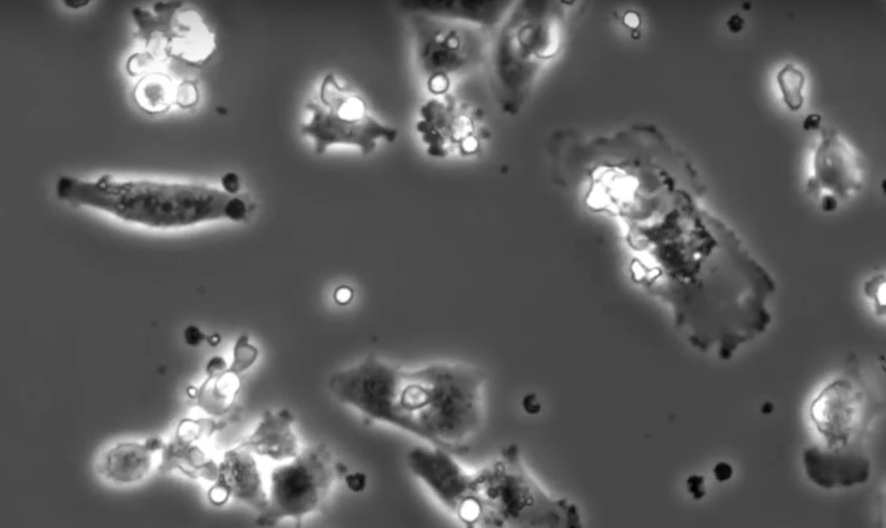
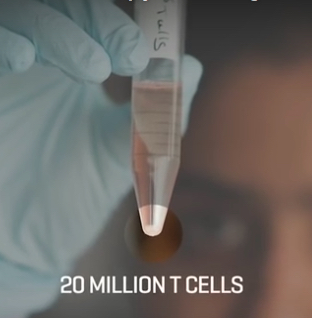
Having survived terminal cancer with a dietary approach, what you say is too simplistic.
Cancer is anything that interferes with any of the many growth inhibition pathways the prevent individual cells within the cooperative society of cells that is an animal body from growing in a fashion that puts the whole cooperative system at risk.
Certainly diet, largely via its effect on our immune system, and certainly in some degrees by other mechanisms also, can play a huge role in that. The particular regime I am on is strictly vegan, largely raw, and high dose vitamin c and supplementation of other vitamin/mineral complexes in very low doses.
The work in this article looks very promising, and in most people it would be unnecessary if they changed their diet and bought the contribution from animal products (meat, dairy, fish and foul etc) to below 10% of total calories. Going to zero seems to slightly reduce the risk even further, but not hugely. Along with that one needs to reduce stress (which seems to be not directly about external factors, but more accurately how we contextualise and respond to them).