Feeds:
Posts
Comments

Posts Tagged ‘Immunotherapy’

Inhibitory CD161 receptor recognized as a potential immunotherapy target in glioma-infiltrating T cells by single-cell analysis

Reporter: Dr. Premalata Pati, Ph.D., Postdoc


Brain tumors, especially the diffused Gliomas are of the most devastating forms of cancer and have so-far been resistant to immunotherapy. It is comprehended that T cells can penetrate the glioma cells, but it still remains unknown why infiltrating cells miscarry to mount a resistant reaction or stop the tumor development.

Gliomas are brain tumors that begin from neuroglial begetter cells. The conventional therapeutic methods including, surgery, chemotherapy, and radiotherapy, have accomplished restricted changes inside glioma patients. Immunotherapy, a compliance in cancer treatment, has introduced a promising strategy with the capacity to penetrate the blood-brain barrier. This has been recognized since the spearheading revelation of lymphatics within the central nervous system. Glioma is not generally carcinogenic. As observed in a number of cases, the tumor cells viably reproduce and assault the adjoining tissues, by and large, gliomas are malignant in nature and tend to metastasize. There are four grades in glioma, and each grade has distinctive cell features and different treatment strategies. Glioblastoma is a grade IV glioma, which is the crucial aggravated form. This infers that all glioblastomas are gliomas, however, not all gliomas are glioblastomas.

Decades of investigations on infiltrating gliomas still take off vital questions with respect to the etiology, cellular lineage, and function of various cell types inside glial malignancies. In spite of the available treatment options such as surgical resection, radiotherapy, and chemotherapy, the average survival rate for high-grade glioma patients remains 1–3 years (1).

A recent in vitro study performed by the researchers of Dana-Farber Cancer Institute, Massachusetts General Hospital, and the Broad Institute of MIT and Harvard, USA, has recognized that CD161 is identified as a potential new target for immunotherapy of malignant brain tumors. The scientific team depicted their work in the Cell Journal, in a paper entitled, “Inhibitory CD161 receptor recognized in glioma-infiltrating T cells by single-cell analysis.” on 15th February 2021.

To further expand their research and findings, Dr. Kai Wucherpfennig, MD, PhD, Chief of the Center for Cancer Immunotherapy, at Dana-Farber stated that their research is additionally important in a number of other major human cancer types such as 

  • melanoma,
  • lung,
  • colon, and
  • liver cancer.

Dr. Wucherpfennig has praised the other authors of the report Mario Suva, MD, PhD, of Massachusetts Common Clinic; Aviv Regev, PhD, of the Klarman Cell Observatory at Broad Institute of MIT and Harvard, and David Reardon, MD, clinical executive of the Center for Neuro-Oncology at Dana-Farber.

Hence, this new study elaborates the effectiveness of the potential effectors of anti-tumor immunity in subsets of T cells that co-express cytotoxic programs and several natural killer (NK) cell genes.

The Study-

IMAGE SOURCE: Experimental Strategy (Mathewson et al., 2021)


The group utilized single-cell RNA sequencing (RNA-seq) to mull over gene expression and the clonal picture of tumor-infiltrating T cells. It involved the participation of 31 patients suffering from diffused gliomas and glioblastoma. Their work illustrated that the ligand molecule CLEC2D activates CD161, which is an immune cell surface receptor that restrains the development of cancer combating activity of immune T cells and tumor cells in the brain. The study reveals that the activation of CD161 weakens the T cell response against tumor cells.

Based on the study, the facts suggest that the analysis of clonally expanded tumor-infiltrating T cells further identifies the NK gene KLRB1 that codes for CD161 as a candidate inhibitory receptor. This was followed by the use of 

  • CRISPR/Cas9 gene-editing technology to inactivate the KLRB1 gene in T cells and showed that CD161 inhibits the tumor cell-killing function of T cells. Accordingly,
  • genetic inactivation of KLRB1 or
  • antibody-mediated CD161 blockade

enhances T cell-mediated killing of glioma cells in vitro and their anti-tumor function in vivo. KLRB1 and its associated transcriptional program are also expressed by substantial T cell populations in other forms of human cancers. The work provides an atlas of T cells in gliomas and highlights CD161 and other NK cell receptors as immune checkpoint targets.

Further, it has been identified that many cancer patients are being treated with immunotherapy drugs that disable their “immune checkpoints” and their molecular brakes are exploited by the cancer cells to suppress the body’s defensive response induced by T cells against tumors. Disabling these checkpoints lead the immune system to attack the cancer cells. One of the most frequently targeted checkpoints is PD-1. However, recent trials of drugs that target PD-1 in glioblastomas have failed to benefit the patients.

In the current study, the researchers found that fewer T cells from gliomas contained PD-1 than CD161. As a result, they said, “CD161 may represent an attractive target, as it is a cell surface molecule expressed by both CD8 and CD4 T cell subsets [the two types of T cells engaged in response against tumor cells] and a larger fraction of T cells express CD161 than the PD-1 protein.”

However, potential side effects of antibody-mediated blockade of the CLEC2D-CD161 pathway remain unknown and will need to be examined in a non-human primate model. The group hopes to use this finding in their future work by

utilizing their outline by expression of KLRB1 gene in tumor-infiltrating T cells in diffuse gliomas to make a remarkable contribution in therapeutics related to immunosuppression in brain tumors along with four other common human cancers ( Viz. melanoma, non-small cell lung cancer (NSCLC), hepatocellular carcinoma, and colorectal cancer) and how this may be manipulated for prevalent survival of the patients.

References

(1) Anders I. Persson, QiWen Fan, Joanna J. Phillips, William A. Weiss, 39 – Glioma, Editor(s): Sid Gilman, Neurobiology of Disease, Academic Press, 2007, Pages 433-444, ISBN 9780120885923, https://doi.org/10.1016/B978-012088592-3/50041-4.

Main Source

Mathewson ND, Ashenberg O, Tirosh I, Gritsch S, Perez EM, Marx S, et al. 2021. Inhibitory CD161 receptor identified in glioma-infiltrating T cells by single-cell analysis. Cell.https://www.cell.com/cell/fulltext/S0092-8674(21)00065-9?elqTrackId=c3dd8ff1d51f4aea87edd0153b4f2dc7

Related Articles

VIDEOS on Cancer Biology, Cancer Genetics, Cancer Immunotherapy

19th Annual Koch Institute Summer Symposium on Cancer Immunotherapy, June 12, 2020 at MIT’s Kresge Auditorium


Other related articles published in this Open Access Online Scientific Journal include the following:


Single Cell Sequencing:

Part 4.1 in Genomics Volume 2

Latest in Genomics Methodologies for Therapeutics: Gene Editing, NGS & BioInformatics, Simulations and the Genome Ontology 

On Amazon.com since 12/28/2019

https://www.amazon.com/dp/B08385KF87


4.1.3   Single-cell Genomics: Directions in Computational and Systems Biology – Contributions of Prof. Aviv Regev @Broad Institute of MIT and Harvard, Cochair, the Human Cell Atlas Organizing Committee with Sarah Teichmann of the Wellcome Trust Sanger Institute

Curator: Aviva Lev-Ari, PhD, RN

https://pharmaceuticalintelligence.com/2018/09/03/single-cell-genomics-directions-in-computational-and-systems-biology-contributions-of-ms-aviv-regev-phd-broad-institute-of-mit-and-harvard-cochair-the-human-cell-atlas-organizing-committee-wit/


4.1.4   Cellular Genetics

https://www.sanger.ac.uk/science/programmes/cellular-genetics


4.1.5   Cellular Genomics

https://www.garvan.org.au/research/cellular-genomics


4.1.6   SINGLE CELL GENOMICS 2019 – sometimes the sum of the parts is greater than the whole, September 24-26, 2019, Djurönäset, Stockholm, Sweden http://www.weizmann.ac.il/conferences/SCG2019/single-cell-genomics-2019

Reporter: Aviva Lev-Ari, PhD, RN

https://pharmaceuticalintelligence.com/2019/05/29/single-cell-genomics-2019-september-24-26-2019-djuronaset-stockholm-sweden/


4.1.7   Norwich Single-Cell Symposium 2019, Earlham Institute, single-cell genomics technologies and their application in microbial, plant, animal and human health and disease, October 16-17, 2019, 10AM-5PM

Reporter: Aviva Lev-Ari, PhD, RN

https://pharmaceuticalintelligence.com/2019/06/04/norwich-single-cell-symposium-2019-earlham-institute-single-cell-genomics-technologies-and-their-application-in-microbial-plant-animal-and-human-health-and-disease-october-16-17-2019-10am-5pm/


4.1.8   Newly Found Functions of B Cell

Reporter and Curator: Dr. Sudipta Saha, Ph.D.

https://pharmaceuticalintelligence.com/2019/05/23/newly-found-functions-of-b-cell/


4.1.9 RESEARCH HIGHLIGHTS: HUMAN CELL ATLAS

https://www.broadinstitute.org/research-highlights-human-cell-atlas


CRISPR – 200 articles in the Journal


Chapter 21 in Genomics Volume 1

Genomics Orientations for Personalized Medicine. On Amazon.com since 11/23/2015

http://www.amazon.com/dp/B018DHBUO6


Glioblastoma – 150 articles in the Journal

Most recent


Immunotherapy may help in glioblastoma survival

Reporter and Curator: Dr. Sudipta Saha, Ph.D.

https://pharmaceuticalintelligence.com/2019/03/16/immunotherapy-may-help-in-glioblastoma-survival/


New Treatment in Development for Glioblastoma: Hopes for Sen. John McCain

Reporter: Aviva Lev-Ari, PhD, RN

https://pharmaceuticalintelligence.com/2017/07/25/new-treatment-in-development-for-glioblastoma-hopes-for-sen-john-mccain/


Funding Oncorus’s Immunotherapy Platform: Next-generation Oncolytic Herpes Simplex Virus (oHSV) for Brain Cancer, Glioblastoma Multiforme (GBM)

Reporter: Aviva Lev-Ari, PhD, RN

https://pharmaceuticalintelligence.com/2016/12/28/funding-oncoruss-immunotherapy-platform-next-generation-oncolytic-herpes-simplex-virus-ohsv-for-brain-cancer-glioblastoma-multiforme-gbm/


Glioma, Glioblastoma and Neurooncology

Curator: Larry H. Bernstein, MD, FCAP

https://pharmaceuticalintelligence.com/2015/10/19/glioma-glioblastoma-and-neurooncology/


Positron Emission Tomography (PET) and Near-Infrared Fluorescence Imaging:  Noninvasive Imaging of Cancer Stem Cells (CSCs)  monitoring of AC133+ glioblastoma in subcutaneous and intracerebral xenograft tumors

Reporter: Aviva Lev-Ari, PhD, RN

https://pharmaceuticalintelligence.com/2014/01/29/positron-emission-tomography-pet-and-near-infrared-fluorescence-imaging-noninvasive-imaging-of-cancer-stem-cells-cscs-monitoring-of-ac133-glioblastoma-in-subcutaneous-and-intracerebral-xenogra/


Gamma Linolenic Acid (GLA) as a Therapeutic tool in the Management of Glioblastoma

Eric Fine* (1), Mike Briggs* (1,2), Raphael Nir# (1,2,3)

https://pharmaceuticalintelligence.com/2013/07/15/gamma-linolenic-acid-gla-as-a-therapeutic-tool-in-the-management-of-glioblastoma/


Read Full Post »

Structure-guided Drug Discovery: (1) The Coronavirus 3CL hydrolase (Mpro) enzyme (main protease) essential for proteolytic maturation of the virus and (2) viral protease, the RNA polymerase, the viral spike protein, a viral RNA as promising two targets for discovery of cleavage inhibitors of the viral spike polyprotein preventing the Coronavirus Virion the spread of infection


Curators and Reporters: Stephen J. Williams, PhD and Aviva Lev-Ari, PhD, RN


Therapeutical options to coronavirus (2019-nCoV) include consideration of the following:

(a) Monoclonal and polyclonal antibodies

(b)  Vaccines

(c)  Small molecule treatments (e.g., chloroquinolone and derivatives), including compounds already approved for other indications 

(d)  Immuno-therapies derived from human or other sources


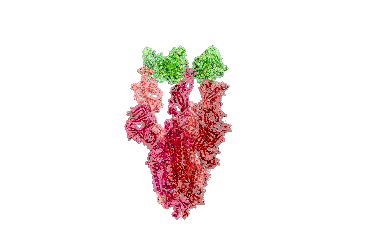
Structure of the nCoV trimeric spike

The World Health Organization has declared the outbreak of a novel coronavirus (2019-nCoV) to be a public health emergency of international concern. The virus binds to host cells through its trimeric spike glycoprotein, making this protein a key target for potential therapies and diagnostics. Wrapp et al. determined a 3.5-angstrom-resolution structure of the 2019-nCoV trimeric spike protein by cryo–electron microscopy. Using biophysical assays, the authors show that this protein binds at least 10 times more tightly than the corresponding spike protein of severe acute respiratory syndrome (SARS)–CoV to their common host cell receptor. They also tested three antibodies known to bind to the SARS-CoV spike protein but did not detect binding to the 2019-nCoV spike protein. These studies provide valuable information to guide the development of medical counter-measures for 2019-nCoV. [Bold Face Added by ALA]

Science, this issue p. 1260

Abstract

The outbreak of a novel coronavirus (2019-nCoV) represents a pandemic threat that has been declared a public health emergency of international concern. The CoV spike (S) glycoprotein is a key target for vaccines, therapeutic antibodies, and diagnostics. To facilitate medical countermeasure development, we determined a 3.5-angstrom-resolution cryo–electron microscopy structure of the 2019-nCoV S trimer in the prefusion conformation. The predominant state of the trimer has one of the three receptor-binding domains (RBDs) rotated up in a receptor-accessible conformation. We also provide biophysical and structural evidence that the 2019-nCoV S protein binds angiotensin-converting enzyme 2 (ACE2) with higher affinity than does severe acute respiratory syndrome (SARS)-CoV S. Additionally, we tested several published SARS-CoV RBD-specific monoclonal antibodies and found that they do not have appreciable binding to 2019-nCoV S, suggesting that antibody cross-reactivity may be limited between the two RBDs. The structure of 2019-nCoV S should enable the rapid development and evaluation of medical countermeasures to address the ongoing public health crisis.

SOURCE
Cryo-EM structure of the 2019-nCoV spike in the prefusion conformation
  1. Department of Molecular Biosciences, The University of Texas at Austin, Austin, TX 78712, USA.

  2. 2Vaccine Research Center, National Institute of Allergy and Infectious Diseases, National Institutes of Health, Bethesda, MD 20892, USA.
  1. Corresponding author. Email: jmclellan@austin.utexas.edu
  1. * These authors contributed equally to this work.

Science  13 Mar 2020:
Vol. 367, Issue 6483, pp. 1260-1263
DOI: 10.1126/science.abb2507


02/04/2020

New Coronavirus Protease Structure Available

PDB data provide a starting point for structure-guided drug discovery

A high-resolution crystal structure of COVID-19 (2019-nCoV) coronavirus 3CL hydrolase (Mpro) has been determined by Zihe Rao and Haitao Yang’s research team at ShanghaiTech University. Rapid public release of this structure of the main protease of the virus (PDB 6lu7) will enable research on this newly-recognized human pathogen.

Recent emergence of the COVID-19 coronavirus has resulted in a WHO-declared public health emergency of international concern. Research efforts around the world are working towards establishing a greater understanding of this particular virus and developing treatments and vaccines to prevent further spread.

While PDB entry 6lu7 is currently the only public-domain 3D structure from this specific coronavirus, the PDB contains structures of the corresponding enzyme from other coronaviruses. The 2003 outbreak of the closely-related Severe Acute Respiratory Syndrome-related coronavirus (SARS) led to the first 3D structures, and today there are more than 200 PDB structures of SARS proteins. Structural information from these related proteins could be vital in furthering our understanding of coronaviruses and in discovery and development of new treatments and vaccines to contain the current outbreak.

The coronavirus 3CL hydrolase (Mpro) enzyme, also known as the main protease, is essential for proteolytic maturation of the virus. It is thought to be a promising target for discovery of small-molecule drugs that would inhibit cleavage of the viral polyprotein and prevent spread of the infection.

Comparison of the protein sequence of the COVID-19 coronavirus 3CL hydrolase (Mpro) against the PDB archive identified 95 PDB proteins with at least 90% sequence identity. Furthermore, these related protein structures contain approximately 30 distinct small molecule inhibitors, which could guide discovery of new drugs. Of particular significance for drug discovery is the very high amino acid sequence identity (96%) between the COVID-19 coronavirus 3CL hydrolase (Mpro) and the SARS virus main protease (PDB 1q2w). Summary data about these closely-related PDB structures are available (CSV) to help researchers more easily find this information. In addition, the PDB houses 3D structure data for more than 20 unique SARS proteins represented in more than 200 PDB structures, including a second viral protease, the RNA polymerase, the viral spike protein, a viral RNA, and other proteins (CSV).

Public release of the COVID-19 coronavirus 3CL hydrolase (Mpro), at a time when this information can prove most vital and valuable, highlights the importance of open and timely availability of scientific data. The wwPDB strives to ensure that 3D biological structure data remain freely accessible for all, while maintaining as comprehensive and accurate an archive as possible. We hope that this new structure, and those from related viruses, will help researchers and clinicians address the COVID-19 coronavirus global public health emergency.

Update: Released COVID-19-related PDB structures include

  • PDB structure 6lu7 (X. Liu, B. Zhang, Z. Jin, H. Yang, Z. Rao Crystal structure of COVID-19 main protease in complex with an inhibitor N3 doi: 10.2210/pdb6lu7/pdb) Released 2020-02-05
  • PDB structure 6vsb (D. Wrapp, N. Wang, K.S. Corbett, J.A. Goldsmith, C.-L. Hsieh, O. Abiona, B.S. Graham, J.S. McLellan (2020) Cryo-EM structure of the 2019-nCoV spike in the prefusion conformation Science doi: 10.1126/science.abb2507) Released 2020-02-26
  • PDB structure 6lxt (Y. Zhu, F. Sun Structure of post fusion core of 2019-nCoV S2 subunit doi: 10.2210/pdb6lxt/pdb) Released 2020-02-26
  • PDB structure 6lvn (Y. Zhu, F. Sun Structure of the 2019-nCoV HR2 Domain doi: 10.2210/pdb6lvn/pdb) Released 2020-02-26
  • PDB structure 6vw1
    J. Shang, G. Ye, K. Shi, Y.S. Wan, H. Aihara, F. Li Structural basis for receptor recognition by the novel coronavirus from Wuhan doi: 10.2210/pdb6vw1/pdb
    Released 2020-03-04
  • PDB structure 6vww
    Y. Kim, R. Jedrzejczak, N. Maltseva, M. Endres, A. Godzik, K. Michalska, A. Joachimiak, Center for Structural Genomics of Infectious Diseases Crystal Structure of NSP15 Endoribonuclease from SARS CoV-2 doi: 10.2210/pdb6vww/pdb
    Released 2020-03-04
  • PDB structure 6y2e
    L. Zhang, X. Sun, R. Hilgenfeld Crystal structure of the free enzyme of the SARS-CoV-2 (2019-nCoV) main protease doi: 10.2210/pdb6y2e/pdb
    Released 2020-03-04
  • PDB structure 6y2f
    L. Zhang, X. Sun, R. Hilgenfeld Crystal structure (monoclinic form) of the complex resulting from the reaction between SARS-CoV-2 (2019-nCoV) main protease and tert-butyl (1-((S)-1-(((S)-4-(benzylamino)-3,4-dioxo-1-((S)-2-oxopyrrolidin-3-yl)butan-2-yl)amino)-3-cyclopropyl-1-oxopropan-2-yl)-2-oxo-1,2-dihydropyridin-3-yl)carbamate (alpha-ketoamide 13b) doi: 10.2210/pdb6y2f/pdb
    Released 2020-03-04
  • PDB structure 6y2g
    L. Zhang, X. Sun, R. Hilgenfeld Crystal structure (orthorhombic form) of the complex resulting from the reaction between SARS-CoV-2 (2019-nCoV) main protease and tert-butyl (1-((S)-1-(((S)-4-(benzylamino)-3,4-dioxo-1-((S)-2-oxopyrrolidin-3-yl)butan-2-yl)amino)-3-cyclopropyl-1-oxopropan-2-yl)-2-oxo-1,2-dihydropyridin-3-yl)carbamate (alpha-ketoamide 13b) doi: 10.2210/pdb6y2g/pdb
    Released 2020-03-04
First page image

Abstract

Coronavirus disease 2019 (COVID-19) is a global pandemic impacting nearly 170 countries/regions and more than 285,000 patients worldwide. COVID-19 is caused by the Severe Acute Respiratory Syndrome Coronavirus-2 (SARS-CoV-2), which invades cells through the angiotensin converting enzyme 2 (ACE2) receptor. Among those with COVID-19, there is a higher prevalence of cardiovascular disease and more than 7% of patients suffer myocardial injury from the infection (22% of the critically ill). Despite ACE2 serving as the portal for infection, the role of ACE inhibitors or angiotensin receptor blockers requires further investigation. COVID-19 poses a challenge for heart transplantation, impacting donor selection, immunosuppression, and post-transplant management. Thankfully there are a number of promising therapies under active investigation to both treat and prevent COVID-19. Key Words: COVID-19; myocardial injury; pandemic; heart transplant

SOURCE

https://www.ahajournals.org/doi/pdf/10.1161/CIRCULATIONAHA.120.046941

ACE2

  • Towler P, Staker B, Prasad SG, Menon S, Tang J, Parsons T, Ryan D, Fisher M, Williams D, Dales NA, Patane MA, Pantoliano MW (Apr 2004). “ACE2 X-ray structures reveal a large hinge-bending motion important for inhibitor binding and catalysis”The Journal of Biological Chemistry279 (17): 17996–8007. doi:10.1074/jbc.M311191200PMID 14754895.


  • Turner AJ, Tipnis SR, Guy JL, Rice G, Hooper NM (Apr 2002). “ACEH/ACE2 is a novel mammalian metallocarboxypeptidase and a homologue of angiotensin-converting enzyme insensitive to ACE inhibitors”Canadian Journal of Physiology and Pharmacology80 (4): 346–53. doi:10.1139/y02-021PMID 12025971.


  •  Zhang, Haibo; Penninger, Josef M.; Li, Yimin; Zhong, Nanshan; Slutsky, Arthur S. (3 March 2020). “Angiotensin-converting enzyme 2 (ACE2) as a SARS-CoV-2 receptor: molecular mechanisms and potential therapeutic target”Intensive Care Medicine. Springer Science and Business Media LLC. doi:10.1007/s00134-020-05985-9ISSN 0342-4642PMID 32125455.


  • ^ Gurwitz, David (2020). “Angiotensin receptor blockers as tentative SARS‐CoV‐2 therapeutics”Drug Development Researchdoi:10.1002/ddr.21656PMID 32129518.


Angiotensin converting enzyme 2 (ACE2)

is an exopeptidase that catalyses the conversion of angiotensin I to the nonapeptide angiotensin[1-9][5] or the conversion of angiotensin II to angiotensin 1-7.[6][7] ACE2 has direct effects on cardiac functiona and is expressed predominantly in vascular endothelial cells of the heart and the kidneys.[8] ACE2 is not sensitive to the ACE inhibitor drugs used to treat hypertension.[9]

ACE2 receptors have been shown to be the entry point into human cells for some coronaviruses, including the SARS virus.[10] A number of studies have identified that the entry point is the same for SARS-CoV-2,[11] the virus that causes COVID-19.[12][13][14][15]

Some have suggested that a decrease in ACE2 could be protective against Covid-19 disease[16], but others have suggested the opposite, that Angiotensin II receptor blocker drugs could be protective against Covid-19 disease via increasing ACE2, and that these hypotheses need to be tested by datamining of clinical patient records.[17]

REFERENCES

https://en.wikipedia.org/wiki/Angiotensin-converting_enzyme_2


FOLDING@HOME TAKES UP THE FIGHT AGAINST COVID-19 / 2019-NCOV

We need your help! Folding@home is joining researchers around the world working to better understand the 2019 Coronavirus (2019-nCoV) to accelerate the open science effort to develop new life-saving therapies. By downloading Folding@Home, you can donate your unused computational resources to the Folding@home Consortium, where researchers working to advance our understanding of the structures of potential drug targets for 2019-nCoV that could aid in the design of new therapies. The data you help us generate will be quickly and openly disseminated as part of an open science collaboration of multiple laboratories around the world, giving researchers new tools that may unlock new opportunities for developing lifesaving drugs.

2019-nCoV is a close cousin to SARS coronavirus (SARS-CoV), and acts in a similar way. For both coronaviruses, the first step of infection occurs in the lungs, when a protein on the surface  of the virus binds to a receptor protein on a lung cell. This viral protein is called the spike protein, depicted in red in the image below, and the receptor is known as ACE2. A therapeutic antibody is a type of protein that can block the viral protein from binding to its receptor, therefore preventing the virus from infecting the lung cell. A therapeutic antibody has already been developed for SARS-CoV, but to develop therapeutic antibodies or small molecules for 2019-nCoV, scientists need to better understand the structure of the viral spike protein and how it binds to the human ACE2 receptor required for viral entry into human cells.

Proteins are not stagnant—they wiggle and fold and unfold to take on numerous shapes.  We need to study not only one shape of the viral spike protein, but all the ways the protein wiggles and folds into alternative shapes in order to best understand how it interacts with the ACE2 receptor, so that an antibody can be designed. Low-resolution structures of the SARS-CoV spike protein exist and we know the mutations that differ between SARS-CoV and 2019-nCoV.  Given this information, we are uniquely positioned to help model the structure of the 2019-nCoV spike protein and identify sites that can be targeted by a therapeutic antibody. We can build computational models that accomplish this goal, but it takes a lot of computing power.

This is where you come in! With many computers working towards the same goal, we aim to help develop a therapeutic remedy as quickly as possible. By downloading Folding@home here [LINK] and selecting to contribute to “Any Disease”, you can help provide us with the computational power required to tackle this problem. One protein from 2019-nCoV, a protease encoded by the viral RNA, has already been crystallized. Although the 2019-nCoV spike protein of interest has not yet been resolved bound to ACE2, our objective is to use the homologous structure of the SARS-CoV spike protein to identify therapeutic antibody targets.

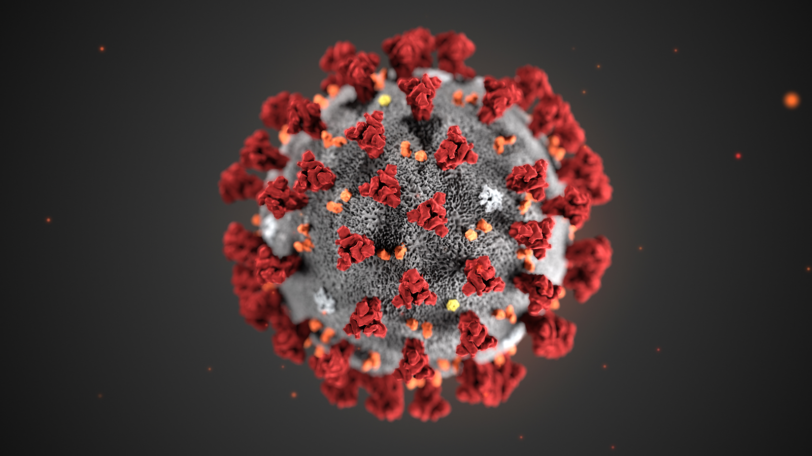
This illustration, created at the Centers for Disease Control and Prevention (CDC), reveals ultrastructural morphology exhibited by coronaviruses. Note the spikes that adorn the outer surface of the virus, which impart the look of a corona surrounding the virion, when viewed electron microscopically. A novel coronavirus virus was identified as the cause of an outbreak of respiratory illness first detected in Wuhan, China in 2019.

Image and Caption Credit: Alissa Eckert, MS; Dan Higgins, MAM available at https://phil.cdc.gov/Details.aspx?pid=23311

Structures of the closely related SARS-CoV spike protein bound by therapeutic antibodies may help rapidly design better therapies. The three monomers of the SARS-CoV spike protein are shown in different shades of red; the antibody is depicted in green. [PDB: 6NB7 https://www.rcsb.org/structure/6nb7]

(post authored by Ariana Brenner Clerkin)

References:

PDB 6lu7 structure summary ‹ Protein Data Bank in Europe (PDBe) ‹ EMBL-EBI https://www.ebi.ac.uk/pdbe/entry/pdb/6lu7 (accessed Feb 5, 2020).

Tian, X.; Li, C.; Huang, A.; Xia, S.; Lu, S.; Shi, Z.; Lu, L.; Jiang, S.; Yang, Z.; Wu, Y.; et al. Potent Binding of 2019 Novel Coronavirus Spike Protein by a SARS Coronavirus-Specific Human Monoclonal Antibody; preprint; Microbiology, 2020. https://doi.org/10.1101/2020.01.28.923011.

Walls, A. C.; Xiong, X.; Park, Y. J.; Tortorici, M. A.; Snijder, J.; Quispe, J.; Cameroni, E.; Gopal, R.; Dai, M.; Lanzavecchia, A.; et al. Unexpected Receptor Functional Mimicry Elucidates Activation of Coronavirus Fusion. Cell 2019176, 1026-1039.e15. https://doi.org/10.2210/pdb6nb7/pdb.

SOURCE

https://foldingathome.org/2020/02/27/foldinghome-takes-up-the-fight-against-covid-19-2019-ncov/

UPDATED 3/13/2020

I am reposting the following Science blog post from Derrick Lowe as is and ask people go browse through the comments on his Science blog In the Pipeline because, as Dr. Lowe states that in this current crisis it is important to disseminate good information as quickly as possible so wanted the readers here to have the ability to read his great posting on this matter of Covid-19.  Also i would like to direct readers to the journal Science opinion letter concerning how important it is to rebuild the trust in good science and the scientific process.  The full link for the following In the Pipeline post is: https://blogs.sciencemag.org/pipeline/archives/2020/03/06/covid-19-small-molecule-therapies-reviewed

A Summary of current potential repurposed therapeutics for COVID-19 Infection from In The Pipeline: A Science blog from Derick Lowe

Covid-19 Small Molecule Therapies Reviewed

Let’s take inventory on the therapies that are being developed for the coronavirus epidemic. Here is a very thorough list of at Biocentury, and I should note that (like Stat and several other organizations) they’re making all their Covid-19 content free to all readers during this crisis. I’d like to zoom in today on the potential small-molecule therapies, since some of these have the most immediate prospects for use in the real world.

The ones at the front of the line are repurposed drugs that are already approved for human use, for a lot of obvious reasons. The Biocentury list doesn’t cover these, but here’s an article at Nature Biotechnology that goes into detail. Clinical trials are a huge time sink – they sort of have to be, in most cases, if they’re going to be any good – and if you’ve already done all that stuff it’s a huge leg up, even if the drug itself is not exactly a perfect fit for the disease. So what do we have? The compound that is most advanced is probably remdesivir from Gilead, at right. This has been in development for a few years as an RNA virus therapy – it was originally developed for Ebola, and has been tried out against a whole list of single-strand RNA viruses. That includes the related coronaviruses SARS and MERS, so Covid-19 was an obvious fit.

The compound is a prodrug – that phosphoramide gets cleaved off completely, leaving the active 5-OH compound GS-44-1524. It mechanism of action is to get incorporated into viral RNA, since it’s taken up by RNA polymerase and it largely seems to evade proofreading. This causes RNA termination trouble later on, since that alpha-nitrile C-nucleoside is not exactly what the virus is expecting in its genome at that point, and thus viral replication is inhibited.

There are five clinical trials underway (here’s an overview at Biocentury). The NIH has an adaptive-design Phase II trial that has already started in Nebraska, with doses to be changed according to Bayesian readouts along the way. There are two Phase III trials underway at China-Japan Friendship Hospital in Hubei, double-blinded and placebo-controlled (since placebo is, as far as drug therapy goes, the current standard of care). And Gilead themselves are starting two open-label trials, one with no control arm and one with an (unblinded) standard-of-care comparison arm. Those might read out first, depending on when they get off the ground, but will be only rough readouts due to the fast-and-loose trial design. The two Hubei trials and the NIH one will add some rigor to the process, but I’m not sure when they’re going to report. My personal opinion is that I like the chances of this drug more than anything else on this list, but it’s still unlikely to be a game-changer.

There’s an RNA polymerase inhibitor (favipiravir) from Toyama, at right, that’s in a trial in China. It’s a thought – a broad-spectrum agent of this sort would be the sort of thing to try. But unfortunately, from what I can see, it has already turned up as ineffective in in vitro tests. The human trial that’s underway is honestly the sort of thing that would only happen under circumstances like the present: a developing epidemic with a new pathogen and no real standard of care. I hold out little hope for this one, but given that there’s nothing else at present, it probably should be tried. As you’ll see, this is far from the only situation like this.

One of the screens of known drugs in China that also flagged remdesivir noted that the old antimalarial drug chloroquine seemed to be effective in vitro. It had been reported some years back as a possible antiviral, working through more than one mechanism, probably both at viral entry and intracellularly thereafter. That part shouldn’t be surprising – chloroquine’s actual mode(s) of action against malaria parasites are still not completely worked out, either, and some of what people thought they knew about it has turned out to be wrong. There are several trials underway with it at Chinese facilities, some in combination with other agents like remdesivir. Chloroquine has of course been taken for many decades as an antimalarial, but it has a number of liabilities, including seizures, hearing damage, retinopathy and sudden effects on blood glucose. So it’s going to be important to establish just how effective it is and what doses will be needed. Just as with vaccine candidates, it’s possible to do more harm with a rushed treatment than the disease is doing itself

There are several other known antiviral drugs are being tried in China, but I don’t have too much hope for those, either. The neuraminidase inhibitors such as oseltamivir (better known as Tamiflu) were tried against SARS and were ineffective; there is no reason to expect anything versus Covid-19 although these drugs are a component of some drug cocktail trials. The HIV protease therapies such as darunavir and the combination therapy Kaletra are in trials, but that’s also a rather desperate long shot, since there’s no particular reason to think that they will have any such protease inhibition against what this new virus has to offer (and indeed, such agents weren’t much help against SARS in the end, either). The classic interferon/ribavirin combination seems to have had some activity against SARS and MERS, and is in two trials from what I can see. That’s not an awful idea by any means, but it’s not a great one, either: if your viral disease has interferon/ribavirin as a front line therapy, it generally means that there’s nothing really good available. No, unless we get really lucky none of these ideas are going to slow the disease down much.

There are a few other repurposed-protease-inhibitors ideas out there, such as this one. (Edit: I had seen this paper but couldn’t track it down, so thanks to those who sent it along). This paper suggests that the TMPRSS2 protease is important for viral entry on the human-cell-side of the process, a pathway that has been noted for other coronaviruses. And it points out that there is a an approved inhibitor (in Japan) for this enzyme (camostat), so that would definitely seem to be worth a trial, probably in combination with remdesivir.

That’s about it for the existing small molecules, from what I can see. What about new ones? Don’t hold your breath, is all I can say. A drug discovery program from scratch against a new pathogen is, as many readers here well know, not a trivial exercise. As this Bloomberg article details, many such efforts in the past (small molecules and vaccines alike) have come to grief because by the time they had anything to deliver the epidemic itself had passed. Indeed, Gilead’s remdesivir had already been dropped as a potential Ebola therapy.

You will either need to have a target in mind up front or go phenotypic. For the former, what you’d see are better characterizations of the viral protease and more extensive screens against it. Two other big target areas are viral entry (which involves the “spike” proteins on the virus surface and the ACE2 protein on human cells) and viral replication. To the former, it’s worth quickly noting that ACE2 is so much unlike the more familiar ACE protein that none of the cardiovascular ACE inhibitors do anything to it at all. And targeting the latter mechanisms is how remdesivir was developed as a possible Ebola agent, but as you can see, that took time, too. Phenotypic screens are perfectly reasonable against viral pathogens as well, but you’ll need to put time and effort into that assay up front, just as with any phenotypic effort, because as anyone who does that sort of work will tell you, a bad phenotypic screen is a complete waste of everyone’s time.

One of the key steps for either route is identifying an animal model. While animal models of infectious disease can be extremely well translated to human therapy, that doesn’t happen by accident: you need to choose the right animal. Viruses in general (and coronaviruses are no exception) vary widely in their effects in different species, and not just across the gaps of bird/reptile/human and the like. No, you’ll run into things where even the usual set of small mammals are acting differently from each other, with some of them not even getting sick at all. This current virus may well have gone through a couple of other mammalian species before landing on us, but you’ll note that dogs (to pick one) don’t seem to have any problem with it.

All this means that any new-target new-chemical-matter effort against Covid-19 (or any new pathogen) is going to take years, and there is just no way around that. Update: see here for just such an effort to start finding fragment hits for the viral protease. This puts small molecules in a very bimodal distribution: you have the existing drugs that might be repurposed, and are presumably available right now. Nothing else is! At the other end, for completely new therapies you have the usual prospects of drug discovery: years from now, lots of money, low success rate, good luck to all of us. The gap between these two could in theory be filled by vaccines and antibody therapies (if everything goes really, really well) but those are very much their own area and will be dealt with in a separate post.

Either way, the odds are that we (and I mean “we as a species” here) are going to be fighting this epidemic without any particularly amazing pharmacological weapons. Eventually we’ll have some, but I would advise people, pundits, and politicians not to get all excited about the prospects for some new therapies to come riding up over the hill to help us out. The odds of that happening in time to do anything about the current outbreak are very small. We will be going for months, years, with the therapeutic options we have right now. Look around you: what we have today is what we have to work with.

Other related articles published in this Open Access Online Scientific Journal include the following:


Group of Researchers @ University of California, Riverside, the University of Chicago, the U.S. Department of Energy’s Argonne National Laboratory, and Northwestern University solve COVID-19 Structure and Map Potential Therapeutics

Reporters: Stephen J Williams, PhD and Aviva Lev-Ari, PhD, RN

https://pharmaceuticalintelligence.com/2020/03/06/group-of-researchers-solve-covid-19-structure-and-map-potential-therapeutic/

Predicting the Protein Structure of Coronavirus: Inhibition of Nsp15 can slow viral replication and Cryo-EM – Spike protein structure (experimentally verified) vs AI-predicted protein structures (not experimentally verified) of DeepMind (Parent: Google) aka AlphaFold

Curators: Stephen J. Williams, PhD and Aviva Lev-Ari, PhD, RN

https://pharmaceuticalintelligence.com/2020/03/08/predicting-the-protein-structure-of-coronavirus-inhibition-of-nsp15-can-slow-viral-replication-and-cryo-em-spike-protein-structure-experimentally-verified-vs-ai-predicted-protein-structures-not/


Coronavirus facility opens at Rambam Hospital using new Israeli tech

https://www.jpost.com/Israel-News/Coronavirus-facility-opens-at-Rambam-Hospital-using-new-Israeli-tech-619681


Read Full Post »

Engineered Bacteria used as Trojan Horse for Cancer Immunotherapy

Reporter: Irina Robu, PhD

Researchers are using synthetic biology— design and construction of new biological entities such as enzymes, genetic circuits, and cells or the redesign of existing biological systems—is changing medicine leading to innovative solution in molecular-based therapeutics. To address the issue of designing therapies that can induce a potent, anti-tumor immune response researchers at Columbia Engineering and Columbia Irving Medical Center engineered a strain of non-pathogenic bacteria that can colonize tumors in mice. The non-pathogenic bacteria act as Trojan Horse that can lead to complete tumor regression in a mouse model of lymphoma. Their results are currently published in Nature Medicine.

The scientists led by Nicholas Arpaia, used their expertise in synthetic biology and immunology to engineer a strain of bacteria able to grow and multiply in the necrotic core of tumors. The non-pathogenic E. coli are programmed to self-destruct when the bacteria numbers reach a critical threshold, allowing for actual release of therapeutics and averting them from causing havoc somewhere else in the body. Afterward, a small portion of bacteria survive lysis and repopulate the population which allows repeated rounds of drug delivery inside treated tumors.

In the present study, the scientists release a nanobody that targets CD47 protein, which defends cancer cells from being eaten by distinctive immune cells. The mutual effects of bacteria, induced local inflammation within the tumor and the blockage of the CD47 leads to better ingestion and activation of T-cells within the treated tumors. The team deduced that the treatment with their engineered bacteria not only cleared the treated tumors but also reduced the incidence of tumor metastasis.

Before moving to clinical trials, the team is performing proof-of-concept tests, safety and toxicology studies of their immunotherapeutic bacteria in a rand of advanced solid tumor settings in mouse models. They have currently collaborated with Gary Schwartz, deputy director of the Herbert Irving Comprehensive Cancer and have underway a company to translate their promising technology to patients.

SOURCE

Sreyan Chowdhury, Samuel Castro, Courtney Coker, Taylor E. Hinchliffe, Nicholas Arpaia, Tal Danino. Programmable bacteria induce durable tumor regression and systemic antitumor immunity. Nature Medicine, 2019

Read Full Post »

Nanoparticles Could Boost Effectiveness of Allergy Shots

Reporter : Irina Robu, PhD

Immunotherapy is a preventive treatment for allergic reactions to substances such as grass pollens, house dust mites and bee venom. The only existing therapy that treats their causes is allergen-specific immunotherapy or allergy shots which can cause severe side effects. For many people, allergies are a seasonal annoyance. But for others, exposure to a particular allergen can cause antagonistic reactions such as itching, breathing problems or even death. Allergy shots can diminish sensitivity by gradually ramping up exposure to the offending substance. Each allergy shot contains a tiny amount of the specific substance or substances that trigger your allergic reactions.

Holger Frey and colleagues report in Biomacromolecules the development of a potentially better allergy shot that uses nanocarriers to address these unwanted issues. In order to develop a safer, cause-based therapy scientist have developed nanoparticles that enclose an allergen and deliver it to specific cells. However, these nanocarriers degrade slowly, hindering the efficiency of the treatment.

Nanocarriers offer the following potential advantages: site-specific delivery of drugs, peptides, and genes, improved in-vitro and in-vivo stability and reduced side effect profile. However, nanoparticles are usually first picked up by the phagocytic cells of the immune system which may promote inflammatory disorders. In order to overcome the limitations, the researchers designed a novel type of nanocarrier created on the biocompatible molecule poly (ethylene glycol) that releases its cargo only in targeted immune cells.

This approach could be used not only for allergies but also can be used for other immunotherapies such as cancer and AIDS.

Source

https://www.eurekalert.org/pub_releases/2015-09/acs-ncb092215.php

Read Full Post »

Immunotherapy may help in glioblastoma survival

Immunotherapy may help in glioblastoma survival, Volume 2 (Volume Two: Latest in Genomics Methodologies for Therapeutics: Gene Editing, NGS and BioInformatics, Simulations and the Genome Ontology), Part 1: Next Generation Sequencing (NGS)

Reporter and Curator: Dr. Sudipta Saha, Ph.D.


Glioblastoma is the most common primary malignant brain tumor in adults and is associated with poor survival. But, in a glimmer of hope, a recent study found that a drug designed to unleash the immune system helped some patients live longer. Glioblastoma powerfully suppresses the immune system, both at the site of the cancer and throughout the body, which has made it difficult to find effective treatments. Such tumors are complex and differ widely in their behavior and characteristics.


A small randomized, multi-institution clinical trial was conducted and led by researchers at the University of California at Los Angeles involved patients who had a recurrence of glioblastoma, the most common central nervous system cancer. The aim was to evaluate immune responses and survival following neoadjuvant and/or adjuvant therapy with pembrolizumab (checkpoint inhibitor) in 35 patients with recurrent, surgically resectable glioblastoma. Patients who were randomized to receive neoadjuvant pembrolizumab, with continued adjuvant therapy following surgery, had significantly extended overall survival compared to patients that were randomized to receive adjuvant, post-surgical programmed cell death protein 1 (PD-1) blockade alone.


Neoadjuvant PD-1 blockade was associated with upregulation of T cell– and interferon-γ-related gene expression, but downregulation of cell-cycle-related gene expression within the tumor, which was not seen in patients that received adjuvant therapy alone. Focal induction of programmed death-ligand 1 in the tumor microenvironment, enhanced clonal expansion of T cells, decreased PD-1 expression on peripheral blood T cells and a decreasing monocytic population was observed more frequently in the neoadjuvant group than in patients treated only in the adjuvant setting. These findings suggest that the neoadjuvant administration of PD-1 blockade enhanced both the local and systemic antitumor immune response and may represent a more efficacious approach to the treatment of this uniformly lethal brain tumor.


Immunotherapy has not proved to be effective against glioblastoma. This small clinical trial explored the effect of PD-1 blockade on recurrent glioblastoma in relation to the timing of administration. A total of 35 patients undergoing resection of recurrent disease were randomized to either neoadjuvant or adjuvant pembrolizumab, and surgical specimens were compared between the two groups. Interestingly, the tumoral gene expression signature varied between the two groups, such that those who received neoadjuvant pembrolizumab displayed an INF-γ gene signature suggestive of T-cell activation as well as suppression of cell-cycle signaling, possibly consistent with growth arrest. Although the study was not powered for efficacy, the group found an increase in overall survival in patients receiving neoadjuvant pembrolizumab compared with adjuvant pembrolizumab of 13.7 months versus 7.5 months, respectively.


In this small pilot study, neoadjuvant PD-1 blockade followed by surgical resection was associated with intratumoral T-cell activation and inhibition of tumor growth as well as longer survival. How the drug works in glioblastoma has not been totally established. The researchers speculated that giving the drug before surgery prompted T-cells within the tumor, which had been impaired, to attack the cancer and extend lives. The drug didn’t spur such anti-cancer activity after the surgery because those T-cells were removed along with the tumor. The results are very important and very promising but would need to be validated in much larger trials.


References:


https://www.washingtonpost.com/health/2019/02/11/immunotherapy-may-help-patients-with-kind-cancer-that-killed-john-mccain/?noredirect=on&utm_term=.e1b2e6fffccc


https://www.ncbi.nlm.nih.gov/pubmed/30742122


https://www.practiceupdate.com/content/neoadjuvant-anti-pd-1-immunotherapy-promotes-immune-responses-in-recurrent-gbm/79742/37/12/1


https://www.esmo.org/Oncology-News/Neoadjuvant-PD-1-Blockade-in-Glioblastoma


https://neurosciencenews.com/immunotherapy-glioblastoma-cancer-10722/


Read Full Post »

Cancer Drugs Shed Light on Rheumatism

Reporter: Irina Robu, PhD

The human body is often described as being ‘at war’. By this, it is meant that the body is constantly under attack from things that are trying to do it harm. These include toxins, bacteria, fungi, parasites and viruses. The human immune system is one of the most effective defense mechanisms known to nature and can sometimes can be overwhelmed by disease. Yet, on occasions our immune systems turn on our own tissue and attack it which can trigger conditions such as type I diabetes, rheumatoid arthritis and lupus.

In the case of rheumatoid arthritis, immune cells start to attack tissues in the joins which causes them to become painful, stiff and swollen. It is known that one third of those who develop rheumatoid arthritis, feel the horrible effects of the disease within two years of its onset. Immunologist Adrian Hayday, which is a researcher at Francis Crick Institute of London says that the current treatment for rheumatoid arthritis require patients to take the drugs for the rest of their lives. But, researchers such as Hayday found an unexpected ally in the battle against autoimmune disease, cancer.

However, there is a positive consequence to the discovery that cancer immunotherapies have the effect of triggering autoimmune diseases and for the first-time rheumatoid arthritis can be detected at the earliest stages. At present, people are not diagnosed with the condition until symptoms have already made their lives so unpleasant, they have gone to see their doctors. As a result, research backed by Cancer Research UK and Arthritis Research UK, has been launched with the aim of uncovering the roots of autoimmune disease from research on cancer patients.

The scientists mentioned stress that their work is only now start and warn that it will still take several years of research to get substantial results. Nevertheless, uncovering the first stages of an autoimmune disease emerging in a person’s body should give researchers a vital lead in ultimately developing treatments that will prevent or halt a range of conditions that currently cause a great deal of misery and require constant medication.

Our immune defenses consist of a range of cells and proteins that notice invading micro-organisms and attack them. The first line of defense, yet, consists of simple physical barriers similar to skin, which blocks invaders from entering your body. When this defense is penetrated, they are attacked by a number of agents. The key cells, leukocytes seek out and destroy disease-causing organisms. Neutrophils rush to the site of an infection and attack invading bacteria. Helper T-cells give instructions to other cells while killer T-cells punch holes in infected cells so that their contents ooze out. After these macrophages clean up the mess left behind.

Another significant agent is the B-cell, which produces antibodies that lock on to sites on the surface of bacteria or viruses and immobilize them until macrophages consume them. These cells can live a long time and can answer quickly following a second exposure to the same infections. In conclusion, suppressor T-cells act when an infection has been distributed with and the immune system needs to be reassured, the killer cells may keep on attacking, as they do in autoimmune diseases. By slowing down the immune system, regulatory T-cells prevent damage to “good” cells.

SOURCE

https://www.theguardian.com/science/2018/mar/03/immunotherapy-cancer-patients-rheumatoid-arthritis-robin-mckie


Read Full Post »

Cracking Tumor Defiance

Reporter: Irina Robu, PhD

Two research groups from Harvard Medical School based at Dana Faber Cancer Institute have discovered a genetic mechanism in a cancer cells that influence whether they respond or resist to immunotherapy drugs, otherwise called as checkpoint inhibitors. The results are published in Science as part of two articles. One article is focused on clinical trial patients with advanced kidney cancer treated with checkpoint inhibitors comes from Eliezer van Allen’s group at Dana Farber Cancer Institute and Toni Choueiri group at Lank Center for Genitourinary Oncology at Dana Farber. The second articles is focused on identifying the immunotherapy resistance mechanism in melanoma cells comes from Kai Wucherpfennig at Dana-Farber and Shirley Liu at Dana -Farber. The two groups joined on that the resistance to immune checkpoint blockade is critically controlled by changes in a group of proteins that regulate how DNA is packaged in cells. The assortment of proteins, called a chromatin remodeling complex, is known as SWI/SNF. Its components are encoded by different genes, among them ARID2PBRM1 and BRD7. SWI/SNF’s job is to open up stretches of tightly wound DNA so that its blueprints can be read by the cell to activate certain genes to make proteins.

Scientists led by Van Allen and Choueiri wanted a clarification for why some patients with a form of metastatic kidney cancer, clear cell renal carcinoma (ccRCC) gain clinical benefit from treatment with immune checkpoint inhibitors that block the PD-1 checkpoint while others patients don’t. The researchers use whole exome DNA sequencing to analyze tumor samples from 35 patients treated in a clinical trial with Opdivo, a checkpoint blocker nivolumab to search for other characteristics of ccRCC tumors that influence immunotherapy response and/or resistance. The scientist discovered that patients from the trial benefited from the immunotherapy treatment with longer survival and progression free survival were those whose tumors lacked a functioning PRBM1 gene. Loss of PRBM1 gene function caused cancer cells to have increased expression of other genes including those in the gene pathway known as IL6/JAK-STAT3, which is involved in immune system stimulation.

When the PBRM1 gene was knocked out in experiments, the melanoma cells became more sensitive to interferon gamma produced by T cells and, in response, produced signaling molecules that recruited more tumor-fighting T cells into the tumor. The two other genes in the PBAF complex—ARID2 and BRD7—are also found mutated in some cancers, according to the researchers, and those cancers, like the melanoma lacking ARID2 function, may also respond better to checkpoint blockade. According to the researchers, finding ways to alter those target molecules “will be important to extend the benefit of immunotherapy to larger patient populations, including cancers that thus far are refractory to immunotherapy.”

SOURCE

http://globalnewsconnect.com/cracking-tumor-defiance

Read Full Post »

Meeting report: Cambridge Healthtech Institute’s 4th Annual Immuno-Oncology SUMMIT: Oncolytic Virus Immunotherapy Stream – 2016

Reporter: David Orchard-Webb, PhD


Cambridge Healthtech Institute’s 4th Annual Immuno-Oncology SUMMIT took place August 29-September 2, 2016 at the Marriott Long Wharf Boston, MA. The following is a synthesis of the Oncolytic Virus Immunotherapy stream.


Biomarkers


Biomarkers for patient selection in clinical trials is an important consideration for developing cancer therapeutics and immunotherapeutics such as oncolytic viruses in particular. Howard L. Kaufman, M.D., discussed the development of biomarkers for oncolytic virus efficaciousness and patient selection focusing on Imlygic (HSV-1). An important consideration for any viral therapy is the presence or absence of the receptors that the virus uses to gain entry to the cell. For example HSV-1 utilises Nectin and HVEM cell surface receptors and their expression levels on a patient’s tumour will influence whether Imlygic can gain entry and replicate in tumours. In addition he reported that B-RAF mutation facilitates Imlygic infection and that MEK inhibitors sensitise melanoma cell lines to Imlygic. Stephen Russell also presented data on the mathematical modelling of Vesicular Stomatitis Virus (VSV) tumour spread and the development of a companion diagnostic based on gene expression profiling to predict patients whose tumours will be readily infected.


The immune reaction triggered by oncolytic viruses is important to monitor. Howard L. Kaufman discussed immunogenic cell death and stated that oncolytic viruses trigger immunity through the release of pathogen-associated molecular patterns (PAMPs) and damage-associated molecular patterns (DAMPs). He reported that immunosuppressive Tregs, PDL1 and IDO expression were associated with anti-cancer CD8+ T cell infiltration. Imlygic also promoted the tumour infiltration of monocytes which depending on the context may either be immunosuppressive or beneficial through recruiting natural killer (NK) cells. This highlights the importance of combining Imlygic with other immune modulating therapeutics that can modulate the immunosuppressive cells and messengers that are present in the tumour environment. He discussed the finding that high mutation burden is a marker for response to immune checkpoint inhibition (such as CTLA and PD1) and suggested that due to the fact that oncolytic viruses release tumour associated antigens (TAA) during cell lysis this may also be a predictive marker for oncolytic viral therapy immune response. Supporting this notion Stephen Russell reported that a patient that underwent complete remission of multiple myeloma plasmacytomas in response to a measles virus oncotherapy had a very high mutational burden.


Targeting the tumour stroma with adenoviral vectors


VCN Biosciences SL is a privately-owned company focused in the development of new therapeutic approaches for tumors that lack effective treatment”. Manel Cascalló presented data from an ongoing phase I, multi-center, open-label dose escalation study of intravenous administration of VCN-01 oncolytic adenovirus with or without intravenous gemcitabine and Abraxane® in advanced solid tumors. Patients were selected based on low anti-Ad levels. Manel highlighted the problems of the pancreatic cancer matrix which limit intratumoral virus spread and also reduces chemotherapy uptake and tumour lymphocyte infiltration. VCN-01 expresses hyaluronidase to degrade the extracellular matrix and is administered intravenously. Liver tropism is reduced by replacement of the heparan sulfate glycosaminoglycan putative-binding site KKTK of the fiber shaft with an integrin-binding motif RGDK. VCN-01 replicates only in Rb tumour suppressor pathway dysregulated cancers, achieved through genetic modification of the E1A protein. In previous mouse xenograft studies of pancreatic and melanoma tumours VCN-01 showed efficaciousness in intratumoral spread, degradation of hyaluronan, and evidence of sensitisation to chemotherapy. The mouse models suggested that strategies that further target other major components of the ECM such as collagen and stromal cells may increase VCN-01 efficaciousness further [1]. The phase I trial supported safety and demonstrated that when administered intravenously VCN-01 reached the pancreatic tumour and replicated. In combination with gemcitabine and Abraxane® neutropenia was observed earlier than with chemotherapy alone. This is suggestive of increased efficaciousness of the chemotherapeutics as would be expected if a greater effective concentration reached the tumour. Biopsies suggested that VCN-01 shifted the balance of immune cells towards CD8+ T cells and away from immunosuppressive Treg.


Adenovirus tumor-specific immunogene (T-SIGn) Therapy


PsiOxus Therapeutics Ltd develops novel therapeutics for serious diseases with a particular focus upon cancer”. Brian Champion discussed the application EnAd a chimeric Ad11p/Ad3 adenovirus which retains the Ad11 receptor usage (CD46 and DSG2). PsiOxus are developing Membrane-integrated T-cell Engagers (MiTe) proteins delivered via EnAd. These MiTe proteins are expressed at the cancer cell surface and engage with and activate T-cells. Their lead candidate NG-348 showed promising T-cell activation in vitro.


Vaccinia virus – overcoming the immunosuppressive cancer microenvironment


David Kirn provided a recent history of the oncolytic virus field and provided an overview of the validation of vaccinia virus over the period 2007-14 stating that it can produce cancer oncolysis, induce an immune response, and result in angiogenic ablation.


Western Oncolytics develops novel therapies for cancer”. Steve Thorne discussed strategies to mitigate the immunosupressive environment encountered by oncolytic viruses. He presented data from models of tumours resistant to vaccinia oncolytic virus that Treg, and myeloid-derived suppressor cell (MDSC) numbers were higher whereas CD8+ T-cell levels were lower than in a sensitive model. He elaborated on a strategy of targeting the PGE2 pathway in order to reduce MDSC numbers entering the tumour microenvironment. He demonstrated that vaccinia virus expressing HPGD has reduced levels of MDSC in target tumours.


Transgene (Euronext: TNG), part of Institut Mérieux, is a publicly traded French biopharmaceutical company focused on discovering and developing targeted immunotherapies for the treatment of cancer and infectious diseases”. Eric Quéméneur presented preclinical data on Transgene’s oncolytic vaccinia virus TG6002 which expresses a chimeric bifunctional enzyme which converts the nontoxic prodrug 5‐FC into the toxic metabolites 5‐FU and 5‐FUMP. This allows systemic delivery of the non-toxic prodrug chemotherapy with activation at tumours infected with the Vaccinia oncolytic virus. The virus plus prodrug combination was effective against all of the solid tumour cell lines tested. In addition the combination was effective against glioblastoma cancer stem-like cells. In pancreatic and colorectal cancer cell line models the vaccinia prodrug combination was synergistic or additive when combined with additional chemotherapeutics. In immunocompetent mouse models TG6002 increased the Tumour Teff/Treg ratio indicative of a shift from an immunosuppressive to an immunocompetent microenvironment. Furthermore in mouse models TG6002 induced an abscopal response.


Vesicular Stomatitis Virus (VSV) – A single shot cure for cancer?


Vyriad strives to develop potent, safe and cost-effective cancer therapies in areas of unmet need”. Stephen Russell presented his position that oncolytic viruses could be a single shot cure for cancer. He emphasised the point that in oncolytic viral therapy the initial dose will be the most effective due to the relatively low levels of neutralising antibodies present and therefore defining the optimal dose is critical. The trend is for increased initial dose. Two IND’s have been accepted by the FDA, one for measles virus and the other for VSV.


John Bell described using VSV to deliver Artificial microRNAs (amiRNAs) to tumours. It was demonstrate that a VSV delivering ARID1A amiRNA was synthetic lethal when combined with EZH2 (methyl transferase) inhibition. He postulated that oncolytic viruses can be used to create factories of therapeutic amiRNAs transmitted throughout the tumour by exosomes.


HSV-1 an update on immune checkpoint combinations


Amgen was the first company to launch an FDA approved (October 2015) oncolytic virus, trade name Imlygic, which was developed by the UK based company Biovex. Jennifer Gansert gave a background on Imlygic and presented new data on combination with the CTLA4 inhibitor Ipilimumab. In mouse models abscopal response in contralateral tumours was 100% when a single tumour was treated with Imlygic combined with systemic delivery of anti-CTLA4. A Phase 1b clinical trial to test the combination in unresectable melanoma patients was completed and published in 2016. Fifty percent of the patients had durable response for greater than 6 months and 20% of the patients had ongoing complete response after a year of follow-up. Overall 72% of patients has controlled disease (no progression). In addition Amgen is recruiting for a phase III trial of the anti-PD1 Pembrolizumab in combination with Imlygic for unresectable stage IIIB to IVM1c melanoma.


Virttu is a privately held biotechnology company, which has pioneered the development of oncolytic viruses for treating cancer”. Joe Connor discussed Seprehvir an oncolyic virus based on HSV-1 like Imlygic which is in clinical trials for which 100 patients have been treated to date. The trial data indicate that Seprehvir induces CD8+ T cell infiltration and activity as well as a novel anti-tumour immune response against select antigens such as Mage A8/9. Preclinical investigations focus on combination with checkpoint inhibitor antibodies, CAR-T targeted to GD2, and synergies with targeted therapies on the mTOR/VEGFR signalling axes.


Reovirus – an update


Oncolytics Biotech Inc. is a clinical-stage oncology company focused on the development of oncolytic viruses for use as cancer therapeutics in some of the most prevalent forms of the disease”. Brad Thompson provided an update on REOLYSIN®, Oncolytics Biotech’s proprietary T3D reovirus. Highlights included concluding the first checkpoint inhibitor and REOLYSIN® study in patients with pancreatic cancer and preparing for registration study in multiple myeloma.


Maraba virus – privileged antigen presentation in splenic B cell follicles


Turnstone Biologics is developing “a first-in-class oncolytic viral immunotherapy that combines a bioselected and engineered oncolytic virus to directly lyse tumors with a potent vaccine technology to drive tumor-antigen specific T-cell responses of unprecedented magnitude”. Caroline Breitbach described Maraba MG1 Oncolytic Virus which was isolated from Brazilian sand flies. Their lead candidate is an MG1 virus expressing the tumour antigen MAGE-A3. In mouse models a combination of adenovirus-MAGE-A3 and MG1-MAGE-A3 in a prime-boost regimen produced extremely robust CD8+ T cell responses. It is thought that a privileged antigen presentation in splenic B cell follicles maximizes the T cell responses. A phase I/II trial is enrolling patients to test the adenovirus-MAGE-A3 and MG1-MAGE-A3 prime-boost regimen in patients with MAGE‐A3 positive solid tumours for which there is no life prolonging standard therapy.


Oncolytic virus manufacturing


Anthony Davies of Dark Horse Consulting Inc. reviewed the manufacturing hurdles facing oncolytic viruses and pointed out that thus far adenovirus is the gold standard. He discussed isoelectric focusing for virus manufacturing, process flow and the procurement of key raw materials. He emphasized the importance of codifying analytical methods, and the statistical design of experiments (DOE) for optimal use of finite resources.


Mark Federspiel described the difficulties associated with measles virus manufacturing which include the large pleomorphic size (100-300nm) which cannot be filter sterilized efficiently due to shear stress. As a result aseptic conditions must be maintained throughout the manufacturing process. There are also issues with genomic contamination from infected cells. He described improved manufacturing bioprocesses to overcome these limitations using the HeLa S3 cell line. Using this cell line resulted in less residual genomic DNA than the standard however it was still relatively high compared to vaccine production. There is still much room for improvement.


REFERENCES
Rodríguez-García A, Giménez-Alejandre M, Rojas JJ, Moreno R, Bazan-Peregrino M, Cascalló M, Alemany R. Safety and efficacy of VCN-01, an oncolytic adenovirus combining fiber HSG-binding domain replacement with RGD and hyaluronidase expression. Clin Cancer Res. 2015 Mar 15;21(6):1406-18. Doi: 10.1158/1078-0432.CCR-14-2213. Epub 2014 Nov 12. PubMed PMID: 25391696.


Other Related Articles Published In This Open Access Online Journal Include The Following:

https://pharmaceuticalintelligence.com/2016/07/15/agenda-for-oncolytic-virus-immunotherapy-unlocking-oncolytic-virotherapies-from-science-to-commercialization-chis-4th-annual-immuno-oncology-summit-august-29-30-2016-marriott-lo/

Real Time Coverage and eProceedings of Presentations on August 29 and August 30, 2016 CHI’s 4th IMMUNO-ONCOLOGY SUMMIT – Oncolytic Virus Immunotherapy Track

https://pharmaceuticalintelligence.com/2016/09/01/real-time-coverage-and-eproceedings-of-presentations-on-august-29-and-august-30-2016-chis-4th-immuno-oncology-summit-oncolytic-virus-immunotherapy-track/

LIVE Tweets via @pharma_BI and by @AVIVA1950 for August 29 and August 30, 2016 of CHI’s 4th IMMUNO-ONCOLOGY SUMMIT – Oncolytic Virus Immunotherapy Track, Marriott Long Wharf Hotel – Boston

https://pharmaceuticalintelligence.com/2016/09/01/live-tweets-via-pharma_bi-and-by-aviva1950-for-august-29-and-august-30-2016-of-chis-4th-immuno-oncology-summit-oncolytic-virus-immunotherapy-track-marriott-long-wharf-hotel/

Read Full Post »

Vectorisation Of Immune Checkpoint Inhibitor Antibodies

Reporter: David Orchard-Webb, PhD


The FDA approved ipilimumab (anti-CTLA-4) and nivolumab (anti-PD-1) combination in October 2015 for the treatment of advanced melanoma. The antibodies have recently been approved in the UK for the same indication. Over half of patients respond to the combination [1]. These drugs belong to the class of monoclonal antibodies known as immune checkpoint inhibitors. The binding of anti-CTLA-4 antibodies to activated T cells prevents the surface CTLA-4 receptor from binding CD80 and/or CD86 on antigen presenting cells (APCs). Normally CTLA-4 binding to APCs deactivates the T-cell. Antibodies against programmed cell death protein 1 (PD-1) work by a similar mechanism to CTLA-4. These drugs are delivered by repeated intravenous injections (iv) [2].


Oncolytic viruses are an emerging class of immunotherapeutics that actively stimulate the immune system by releasing tumour antigens via lysis and by virtue of anti-viral immunity. The first FDA approved oncolytic virus (Imlygic), developed by Amgen/ BioVex, was given the green light in October 2015 for advanced melanoma patients delivered via direct tumour injection. The mechanism of action of oncolytic viruses is highly complementary with checkpoint inhibitor antibodies and multiple trials combining these two classes of agent are under way.


At the recent American Association for Cancer Research (AACR) annual meeting in New Orleans, Louisiana, the oldest biotechnology company in France – Transgene, presented preclinical data concerning oncolytic vaccinia viruses that express whole antibody (mAb), Fragment antigen-binding (Fab) or single-chain variable fragment (scFv) against mouse PD-1 [3]. These combinations proved superior over virus alone in mouse xenografts of melanoma and fibrosarcoma cell lines. Transgene claim that “these results pave the way for next generation of oncolytic vaccinia armed with immunomodulatory therapeutic proteins such as mAbs” (Figure 1) [3].


 698848905_d8bf7f415f_z
Figure 1: The convergence of therapeutics based on oncolytic viruses and monoclonal antibodies against immune checkpoint inhibotor proteins. Image Source: Eric Molina. No changes were made. Creative Commons Attribution 2.0 Generic (CC BY 2.0).


The combination of immune checkpoint inhibitors and oncolytic virus as a single molecular entity clearly has advantages in terms of manufacturing cost effectiveness. In addition viral vectors have the capacity for perfect specificity to tumours which has potential safety advantages.


REFERENCES


  1. http://www.bbc.com/news/health-365496740
  2. http://www.cancer.org/cancer/skincancer-melanoma/detailedguide/melanoma-skin-cancer-treating-immunotherapy
  3. http://www.transgene.fr/wp-content/uploads/2016/04/1604-Poster-AACR-format-122-244-v2.pdf


Other Related Articles Published In This Open Access Online Journal Include The Following:


https://pharmaceuticalintelligence.com/2016/04/12/oncolytic-virus-immunotherapy/

https://pharmaceuticalintelligence.com/2015/09/23/oncolytic-viruses-a-new-class-of-immunotherapy-drugs-against-cancer/

https://pharmaceuticalintelligence.com/2016/06/16/first-drug-in-checkpoint-inhibitor-class-of-cancer-immunotherapies-has-demonstrated-superiority-over-standard-of-care-in-the-treatment-of-first-line-lung-cancer-patients-mercks-keytryda/

https://pharmaceuticalintelligence.com/2016/05/07/durable-responses-with-checkpoint-inhibitor/

https://pharmaceuticalintelligence.com/2016/05/02/cancer-research-institute-nyc-623-6242016-will-combination-of-adoptive-t-cell-therapy-and-anti-checkpoint-inhibitor-therapies-be-the-next-wave/

https://pharmaceuticalintelligence.com/2016/02/14/checkpoint-inhibitors-for-gastrointestinal-cancers/

Read Full Post »

Bispecific and Trispecific Engagers: NK-T Cells and Cancer Therapy

Curator: Larry H. Bernstein, MD, FCAP


Successful adoptive transfer and in vivo expansion of human haploidentical NK cells in patients with cancer

Jeffrey S. Miller, Yvette Soignier, Angela Panoskaltsis-Mortari, …, Todd E. Defor, Linda J. Burns, Paul J. Orchard, Bruce R. Blazar, John E. Wagner, Arne Slungaard, Daniel J. Weisdorf, Ian J. Okazaki, and Philip B. McGlave
Blood. 2005;105:3051-3057   http://www.fortressbiotech.com/pdfs/Miller_NK%20adoptive%20immunotherapy.Blood.2005.pdf

We previously demonstrated that autologous natural killer (NK)–cell therapy after hematopoietic cell transplantation (HCT) is safe but does not provide an antitumor effect. We hypothesize that this is due to a lack of NK-cell inhibitory receptor mismatching with autologous tumor cells, which may be overcome by allogeneic NK-cell infusions. Here, we test haploidentical, related-donor NK-cell infusions in a nontransplantation setting to determine safety and in vivo NK-cell expansion. Two lower intensity outpatient immune suppressive regimens were tested: (1) lowdose cyclophosphamide and methylprednisolone and (2) fludarabine. A higher intensity inpatient regimen of high-dose cyclophosphamide and fludarabine (HiCy/Flu) was tested in patients with poorprognosis acute myeloid leukemia (AML). All patients received subcutaneous interleukin 2 (IL-2) after infusions. Patients who received lower intensity regimens showed transient persistence but no in vivo expansion of donor cells. In contrast, infusions after the more intense Hi-Cy/Flu resulted in a marked rise in endogenous IL-15, expansion of donor NK cells, and induction of complete hematologic remission in 5 of 19 poor-prognosis patients with AML. These findings suggest that haploidentical NK cells can persist and expand in vivo and may have a role in the treatment of selected malignancies used alone or as an adjunct to HCT.

Human natural killer (NK) cells are a subset of peripheral blood lymphocytes defined by the expression of CD56 or CD16 and the absence of the T-cell receptor (CD3).1 They recognize and kill transformed cell lines in a major histocompatibility complex (MHC)–unrestricted fashion and produce cytokines critical to the innate immune response. NK-cell function, distinct from the MHC-restricted cytolytic activity of T cells, may play a role in antitumor surveillance.2 The effects of NK-cell infusions have been studied in adoptive immunotherapy clinical trials. In these studies, autologous lymphokine-activated killer cells obtained from peripheral blood mononuclear cells (PBMCs) were administered to patients along with exogenous high-dose interleukin-2 (IL-2). Up to 20% of patients responded to these infusions of NK-cell– containing populations.3

In contrast to NK cells, T cells recognize targets through an antigen-specific T-cell receptor (TCR) and interact with targets only if human leukocyte antigen (HLA) MHC antigens are also recognized. Although NK-cell killing is MHC-unrestricted, NK cells display a number of activating and inhibitory receptors that ligate MHC molecules to modulate the immune response.4,5 NK-cell receptors that recognize antigens at the HLA-A, -B, or -C loci are members of the immunoglobulin superfamily and are termed killer immunoglobulin receptors (KIRs).6,7 Other receptor families (natural killer group 2 [NKG2]/CD94) that recognize antigens of the nonclassical HLA-E, -F, or -G loci and other ligand specificities have also been described.8-10 Engagement of these NK-cell receptors results in stimulation or inhibition of NK-cell effector function depending on intracellular signaling mediated through the cytoplasmic tail or adaptor molecules associated with each receptor.11-13 The NK-cell response to a target thus depends on the net effect of activating and inhibitory receptors.

Clinical trials have assessed the effects of low-dose IL-2 administration on activation of NK cells in patients with cancer. We have demonstrated the safety and feasibility of daily subcutaneous IL-2 injections following high-dose chemotherapy and autologous hematopoietic cell transplantation (HCT). Whereas IL-2 signifi- cantly expanded the number of circulating NK cells in vivo, these NK cells were not maximally cytotoxic as determined by in vitro assays.14 Subsequent studies tested infusion of IL-2–activated NK-cell–enriched populations or intravenous IL-2 infusions combined with subcutaneous IL-2. Although these approaches augmented in vivo NK-cell function, no consistent efficacy of autologous NK-cell therapy could be detected in cancer patients when compared with cohorts of matched controls.15

We then hypothesized that autologous NK cells may be suppressed by the physiologic response resulting from NK-cell recognition of “self” MHC molecules. This notion is supported by recent data from haploidentical T-cell–depleted transplantation studies. KIR mismatch with tumor MHC (ie, KIR ligand) may lead to greater tumor kill. In these studies, Ruggeri et al16 showed that stratifying patients by their KIR ligand mismatch would select for patients with alloreactive NK cells that protect against acute myeloid leukemia (AML) relapse. Although virtually untested in solid tumors, these clinical data strongly support a therapeutic role for allogeneic NK cells in myeloid leukemia.17 We present data on the biologic effects of haploidentical NK-cell infusions administered to cancer patients as cell-based immunotherapy with the goal of demonstrating a feasible and safe method that permits in vivo donor NK-cell expansion.

………


In this study, we demonstrate that adoptively transferred human NK cells derived from haploidentical related donors can be expanded in vivo. Of interest, in vivo NK-cell expansion occurs after preparation with a high dose (Hi-Cy/Flu) but not lower doses of immunosuppression (Lo-Cy/mPred or Flu). Successful lymphocyte adoptive transfer following intensive immunosuppresion is not surprising. Lymphopenia may change the competitive balance between transferred lymphocytes and endogenous lymphocytes. Alternatively, lymphopenia may induce survival factors or deplete cellular or soluble inhibitory factors.25,26 In murine studies, preparative regimens sufficient to induce lymphopenia allowed homeostatic T-cell expansion in vivo that potentiated effective antitumor immunity.27 This concept has been tested in human T-cell clinical trials by Rosenberg’s group.28 T-cell lymphopenia was induced by Hi-Cy/Flu, similar to what was used here. Successful adoptive transfer and expansion of NK cells may also require intense immunosuppression. Prlic et al20 showed that mature NK cells proliferated only in an NK-cell–deficient host where the endogenous NK-cell pool was absent.

We also demonstrate that NK-cell adoptive therapy is associated with a striking rise in endogenous IL-15 levels, reminiscent of the role IL-7 plays in CD4 T-cell homeostasis.29 IL-15 is required for the final steps of in vitro NK-cell differentiation from CD34 progenitors.22-24 Cooper et al21 was the first to show that IL-15 was absolutely required for in vivo expansion and survival of NK cells, in mice, in part through bcl-2 expression. Transfer of NK cells into IL-15/ hosts resulted in loss of NK cells by 4 days after transfer. IL-15 receptor alpha knockout mice generate IL-15 but do not have NK cells and are unable to undergo successful adoptive transfer. This implies that IL-15 responsiveness by cells other than NK cells may be important in driving this response. IL-15 transgenic mice markedly expand their NK cells and CD8 T cells, ultimately resulting in an NK/T-lymphocytic leukemia.30 The endogenous origin of IL-15 in our patients was unclear. Our data support the notion that IL-15 levels increased only after an intensive lymphocyte-depleting preparative regimen as demonstrated by the inverse correlation between IL-15 concentrations and the absolute lymphocyte count. This does not exclude the possibility that IL-15 may be produced following chemotherapy-induced damage to gastrointestinal mucosa or other cells of epithelial origin.31-34 The effects of exogenous IL-2 administration in these patients needs to be explored as it does add toxicity to the regimen. Further clinical testing may demonstrate that expansion will occur in the presence of IL-15 alone.

Donor NK-cell infusions were feasible and tolerated without unexpected toxicity except for the umbilical cord blood transplantation patient who developed EBV reactivation after treatment. The risk of posttransplantation lymphoproliferative disease approached 10% when HCT is performed using a T-cell–depleted and mismatched graft.35 Although a single event, this finding is important to understand the possible consequences of allogeneic NK-cell therapy in heavily pretreated immunosuppressed patients. It also emphasizes that the CD3- depleted final product, enriched for NK cells but containing B cells, may need further purification to lessen the possibility of this complication. Clinical ex vivo selection methods to address this issue using CD3 depletion followed by CD56 selection are now in place36 and will be tested. We have previously shown that monocytes serve as accessory cells for NK-cell expansion in vitro18 but the role of accessory cells in vivo, if any, is unknown. We need to verify that removal of monocytes and B cells does not change the in vivo expansion potential of NK cells seen here before recommending a purified NK-cell product in all future studies.

In summary, this is the first study to demonstrate that adoptively transferred human NK cells can be expanded in vivo. Expansion was dependent on the more intense Hi-Cy/Flu preparative regimen, which induced lymphopenia, and the more potent immunosuppression that was associated with high endogenous concentrations of IL-15, none of which was observed following Lo-Cy/mPred and Flu alone. It is intriguing that this same regimen is the basis for many transplantation regimens and may help explain the robust NK-cell reconstitution seen in that setting. In this study, NKenriched cells were obtained from related haploidentical donors by efficient depletion of CD3 from PBMCs, although contaminating B cells and monocytes remained in the final product. A maximum tolerated dose was not reached and the largest cell dose administered was that obtained during a single lymphapheresis collection. Although tumor response was not a primary goal of this study, 5 of 19 poor-prognosis patients with AML achieved complete remission after haploidentical NK-cell therapy, with a significantly higher complete remission rate when KIR ligand mismatched donors were used, a strategy that predicts NK-cell alloreactivity.16,37 The precise role of the cells versus the high-intensity chemotherapy regimen in responding patients cannot be definitively determined in this current study. However, the benefit of alloreactivity and the preferential expansion of functional NK cells in responding patients is consistent with at least a partial effect from the NK cells. Our data suggest that prospective selection of KIR ligand– mismatched donors is warranted when possible, which will be assessed in subsequent larger clinical trails.


The biology of natural killer cells in cancer, infection, and pregnancy.

OBJECTIVE: NK cells are important cells of the immune system. They are ultimately derived from pluripotent hematopoietic stem cells. NK cell cytotoxicity and other functions are tightly regulated by numerous activating and inhibitory receptors including newly discovered receptors that selectively recognize major histocompatibility complex class I alleles. Based on their defining function of spontaneous cytotoxicity without prior immunization, NK cells have been thought to play a critical role in immune surveillance and cancer therapy. However, new insights into NK cell biology have suggested major roles for NK cells in infection control and uterine function. The purpose of this review is to provide an update on NK cell function, ontogeny, and biology in order to better understand the role of NK cells in health and disease.
DATA SOURCES: In the Medline database, the major subject heading “Natural Killer Cells” was introduced in 1983, identifying 16,848 citations as of December 31, 2000. Since 1986, there have been approximately 1000 citations per year under this subject heading. In this database, 68% of manuscripts are limited to human NK cells; 40% of citations cross with the major sub-heading of cytotoxicity, 40% with cytokines, 36% with neoplasm, 5% with antibody-dependent cellular cytotoxicity, 2.8% with pregnancy, and 1.3% with infection. Of references from the year 2000-2001, 46 were selected to combine with contributions from earlier literature.
CONCLUSIONS: NK cells should no longer be thought of as direct cytotoxic killers alone as they clearly serve a critical role in cytokine production which may be important to control cancer, infection, and fetal implantation. Understanding mechanisms of NK cell functions may lead to novel therapeutic strategies for the treatment of human disease.

NK cell-based immunotherapy for malignant diseases

Min Cheng, Yongyan Chen, Weihua Xiao, Rui Sun and Zhigang Tian
Cellular & Molecular Immunology (2013) 10, 230–252;   published online 22 April 2013     http://dx. doi.org:/10.1038/cmi.2013.10

Natural killer (NK) cells play critical roles in host immunity against cancer. In response, cancers develop mechanisms to escape NK cell attack or induce defective NK cells. Current NK cell-based cancer immunotherapy aims to overcome NK cell paralysis using several approaches. One approach uses expanded allogeneic NK cells, which are not inhibited by self histocompatibility antigens like autologous NK cells, for adoptive cellular immunotherapy. Another adoptive transfer approach uses stable allogeneic NK cell lines, which is more practical for quality control and large-scale production. A third approach is genetic modification of fresh NK cells or NK cell lines to highly express cytokines, Fc receptors and/or chimeric tumor-antigen receptors. Therapeutic NK cells can be derived from various sources, including peripheral or cord blood cells, stem cells or even induced pluripotent stem cells (iPSCs), and a variety of stimulators can be used for large-scale production in laboratories or good manufacturing practice (GMP) facilities, including soluble growth factors, immobilized molecules or antibodies, and other cellular activators. A list of NK cell therapies to treat several types of cancer in clinical trials is reviewed here. Several different approaches to NK-based immunotherapy, such as tissue-specific NK cells, killer receptor-oriented NK cells and chemically treated NK cells, are discussed. A few new techniques or strategies to monitor NK cell therapy by non-invasive imaging, predetermine the efficiency of NK cell therapy byin vivo experiments and evaluate NK cell therapy approaches in clinical trials are also introduced.

Surgery, chemotherapeutic agents and ionizing radiation have been used for decades as primary strategies to eliminate the tumors in patients; however, the development of resistance to drugs or radiation led to a significant incidence of tumor relapse. Therefore, investigating effective strategies to eliminate these resistant tumor cells is urgently needed. The importance of immune system in malignant diseases has been demonstrated by recent major scientific advances.

Both innate and adaptive immune cells actively prevent neoplastic development in a process called ‘cancer immunosurveillance’. Innate immune cells, including monocytes, macrophages, dendritic cells (DCs) and natural killer (NK) cells, mediate immediate, short-lived responses by releasing cytokines that directly lyse tumor cells or capture debris from dead tumor cells. Adaptive immune cells, including T and B cells, mediate long-lived, antigen-specific responses and effective memory.1 Despite these immune responses, malignant cells can develop mechanisms to evade immunosurveillance. Some tumors protect themselves by establishing an immune-privileged environment. For example, they can produce immunosuppressive cytokines IL-10 and transforming growth factor-β (TGF-β) to suppress the adaptive antitumor immune response, or skew the immune response toward a Th2 response with significantly less antitumor capacity.2,3,4 Some tumors alter their expressions of IL-6, IL-10, vascular epithelial growth factor or granulocyte monocyte-colony stimulating factor (GM-CSF), impairing DC functions via inactivation or suppressing maturation.5 In some cases, induced regulatory T cells suppress tumor-specific CD4+ and CD8+ T-cell responses.6 Tumor cells also minimally express or shed tumor-associated antigens, shed the ligands of NK cell-activating receptor such as the NKG2D ligands UL16-binding protein 2, major histocompatibility complex (MHC) class I chain-related molecules A and B molecules (MICA/MICB) or alter MHC-I and costimulatory molecule expression to evade the immune responses.7,8,9 Malignant cells may also actively eliminate immune cells by activation-induced cell death or Fas ligand (FasL) expression.10,11 In addition, primary cancer treatments like chemotherapy and ionizing radiation can compromise antitumor immune responses by their immunosuppressive side effects.

Tumor cells can be eliminated when immune responses are adequate; when they are not, tumor growth and immunourveillance enter into a dynamic balance until tumor cells evade immunosurveillance, at which point neoplasms appear clinically as a consequence. Therapies designed to induce either a potent passive or active antitumor response against malignancies by harnessing the power of the immune system, known as tumor immunotherapy, is an appealing alternative strategy to control tumor growth. Until now, the cancer immunotherapy field has covered a vast array of therapeutic agents, including cytokines, monoclonal antibodies, vaccines, adoptive cell transfers (T, NK and NKT) and Toll-like receptor (TLR) agonists.1,12,13 Adoptive NK cell transfer in particular has held great promise for over three decades. With progress in the NK cell biology field and in understanding NK function, developing NK cells to be a powerful cancer immunotherapy tool has been achieved in recent years. In this article, we will review recent advances in NK cell-based cancer immunotherapy, focusing on potential approaches and large-scale NK cell expansion for clinical practice, as well as on the clinical trials and future perspectives to enhance the efficacy of NK cells.

NK cells were first identified in 1975 as a unique lymphocyte subset that are larger in size than T and B lymphocytes and contain distinctive cytoplasmic granules.14,15 After more than 30 years, our understanding of NK cell biology and function lends important insights into their role in immunosurveillance. It has been known that NK cells develop in bone marrow (BM) from common lymphoid progenitor cells;16 however, NK cell precursors have still not been clearly characterized in humans.17 After development, NK cells distribute widely throughout lymphoid and non-lymphoid tissues, including BM, lymph nodes (LN), spleen, peripheral blood, lung and liver.18

NK cells, defined as CD3CD56+ lymphocytes, are distinguished as CD56bright and CD56dim subsets. Approximately 90% of peripheral blood and spleen NK cells belong to the CD56dimCD16+ subset with marked cytotoxic function upon interacting with target cells.19,20In contrast, most NK cells in lymph nodes and tonsils belong to the CD56brightCD16 subset and exhibit predominantly immune regulation properties by producing cytokines such as interferon (IFN)-γ in response to IL-12, IL-15 and IL-18 stimulation.19,21

NK cells rapidly kill certain target cells without prior immunization or MHC restriction, whose activation is dependent on the balance between inhibitory and activating signals from invariant receptors.22,23,24 The activating receptors include the cytotoxicity receptors (NCRs) (NKp46, NKp30 and NKp44), C-type lectin receptors (CD94/NKG2C, NKG2D, NKG2E/H and NKG2F) and killer cell immunoglobulin-like receptors (KIRs) (KIR-2DS and KIR-3DS), while the inhibitory receptors include C-type lectin receptors (CD94/NKG2A/B) and KIRs (KIR-2DL and KIR-3DL). Since some structural families contain both activating and inhibitory receptors, trying to understand how NK cell activity is regulated is often complicated.25 At steady state, the inhibitory receptors (KIRs and CD94/NKG2A/B), which bind to various MHC-I molecules present on almost all cell types, inhibit NK cell activation and prevent NK cell-mediated killing. Under stress conditions, cells downregulate MHC-I expression, causing NK cells to lose inhibitory signaling and be activated in a process called ‘missing-self recognition’. Additionally, the non-MHC self molecules Clr-b (mouse), LLT-1 (human) and CD48 (mouse) recognized by the inhibitory receptors NKR-P1B, NKR-P1A and 2B4, respectively, also perform this function.26,27 In contrast to the self-expressed inhibitory receptor ligands, NK cell-activating receptors can recognize either pathogen-encoded molecules that are not expressed by the host, called ‘non-self recognition’, or self-expressed proteins that are upregulated by transformed or infected cells, called ‘stress-induced self recognition’. For example, mouse Ly49H recognizes cytomegalovirus-encoded m157, and NKG2D recognizes the self proteins human UL16-binding proteins and MICA/MICB.28,29 NK cells identify their targets by recognizing a set of receptors on target cells in an NK-target cell zipper formation; this results in the integration of multiple activating and inhibitory signals, the outcome of which depends on the nature of the interacting cells.26IFNs or DC/macrophage-derived cytokines, such as type I IFN, IL-12, IL-18 and IL-15, enhance the activation or promote the maturation of NK cells, which can also augment NK cell cytolytic activity against tumor cells.30,31,32 Cytotoxic activity of NK cells can increase approximately 20–200 fold after exposure to IFN-α/β or IL-12. Despite these known innate immune cell functions, accumulating evidence in both mice and humans demonstrates that NK cells are educated and selected during development, possess receptors with antigen specificity, undergo clonal expansion during infection and can generate long-lived memory cells.33,34

After over 30 years of researching NK cells, evidence supports that they play critical roles in the early control of viral infection, in hematopoietic stem cell (HSC) transplantation (improved grafting, graft-vs.-host disease and graft-vs.-tumor), in tumor immunosurveillance and in reproduction (uterine spiral artery remodeling). The roles of NK cells in controlling organ transplantation, parasitic and HIV infections, autoimmunity and asthma have also been suggested, but remain to be explored further.26 In particular, therapeutic strategies harnessing the power of NK cells to target multiple malignancies have been designed.

NK cells originally described as large granular lymphocytes, exhibited natural cytotoxicity against certain tumor cells in the absence of preimmunization or stimulation.35,36,37 CD56dim NK cells, which make up the majority of circulating cells, are the most potent cytotoxic NK cells against tumor cells. Evidence gathered from a mouse xenograft tumor model testing functionally deficient NK cells or antibody-mediated NK cell depletion supports that NK cells can eradicate tumor cells.38,39,40,41 An 11-year follow-up study in patients indicated that low NK-like cytotoxicity was associated with increased cancer risk.42 High levels of tumor infiltrating NK cells (TINKs) are associated with a favorable tumor outcome in patients with colorectal carcinoma, gastric carcinoma and squamous cell lung cancer, suggesting that NK-cell infiltration into tumor tissues represents a positive prognostic marker.43,44,45 As described above, NK-cell recognition of tumor cells by inhibitory and activating receptors is complex, and the three recognition models—‘missing-self’, ‘non-self’ and ‘stress-induced self’—might be used to sense missing- or altered-self cells. Activated NK cells are thus in a position to directly or indirectly exert their antitumor activity to control tumor growth and prevent the rapid dissemination of metastatic tumors by ‘immunosurveillance’ mechanisms (Figure 1).

Unfortunately we are unable to provide accessible alternative text for this. If you require assistance to access this image, please contact help@nature.com or the author

Figure 1.

NK cells in tumor immunosurveillance. The diagram shows the potential roles of NK cells in tumor immunosurveillance. NK cells initially recognize the tumor cells via stress or danger signals. Activated NK cells directly kill target tumor cells through at least four mechanisms: cytoplasmic granule release, death receptor-induced apoptosis, effector molecule production or ADCC. Additionally, NK cells act as regulatory cells when reciprocally interact with DCs to improve their antigen uptake and presentation, facilitating the generation of antigen-specific CTL responses. Also, by producing cytokines such as IFN-γ, activated NK cells induce CD8+ T cells to become CTLs. Activated NK cells can also promote differentiation of CD4+ T cells toward a Th1 response and promote CTL differentiation. Cytokines produced by NK cells might also regulate antitumor Ab production by B cells. Ab, antibody; ADCC, antibody-dependent cellular cytotoxicity; CTL, cytotoxic T lymphocyte; DC, dendritic cell; IFN, interferon; NK, natural killer.

Full figure and legend (96K)

Direct tumor clearance by NK-mediated cytotoxicity

Upon cellular transformation, surface MHC-I expression on tumor cells is often reduced or lost to evade recognition by antitumor T cells. In parallel, cellular stress and DNA damage lead to upregulated expression of ligands on tumor cells for NK cell-activating receptors. Human tumor cells that have lost self MHC-I expression or bear ‘altered-self’ stress-inducible proteins are ideal NK cell targets, as NK cells are activated by initially recognizing certain ‘stress’ or ‘danger’ signals.46 The ‘missing-self’ model of tumor cell recognition by NK cells was first demonstrated by observing that MHC-I-deficient syngeneic tumor cells were selectively rejected by NK cells; additionally, NK cell inhibitory receptors were shown to detect this absence of MHC-I expression.47,48,49 NK cells can also kill certain MHC-I-sufficient tumor cells by detecting stress-induced self ligands through their activating receptors. Broad MICA/B expression has been detected on epithelial tumors, melanoma, hepatic carcinoma and some hematopoetic malignancies, representing a counter-measure by the immune system to combat tumor development.31 NK cell-mediated cytotoxicity is also important against tumor initiation and metastasis in vivo.50,51,52

NK cells directly kill target tumor cells through several mechanisms: (i) by releasing cytoplasmic granules containing perforin and granzymes that leads to tumor-cell apoptosis by caspase-dependent and -independent pathways.53,54 Cytotoxic granules reorient towards the tumor cell soon after NK–tumor cell interaction and are released into the intercellular space in a calcium-dependent manner; granzymes are allowed entry into tumor cells by perforin-induced membrane perforations, leading to apoptosis; (ii) by death receptor-mediated apoptosis. Some NK cells express tumor-necrosis factor (TNF) family members, such as FasL or TNF-related apoptosis-inducing ligand (TRAIL), which can induce tumor-cell apoptosis by interacting with their respective receptors, Fas and TRAIL receptor (TRAILR), on tumor cells.55,56,57,58,59 TNF-α produced by activated NK cells can also induce tumor-cell apoptosis;60 (iii) by secreting various effector molecules, such as IFN-γ, that exert antitumor functions in various ways, including restricting tumor angiogenesis and stimulating adaptive immunity.61,62 Cytokine activation or exposure to tumor cells is also associated with nitric oxide (NO) production, where NK cells kill target tumor cells by NO signaling;63,64 (iv) through antibody-dependent cellular cytotoxicity (ADCC) by expressing CD16 to destroy tumor cells.40 The antitumor activity of NK cells can be further enhanced by cytokine stimulation, such as by IL-2, IL-12, IL-18, IL-15 or those that induce IFN production.40,65,66,67,68,69,70

Indirect NK-mediated antitumor immunity

NK cells act as regulatory cells when reciprocally interact with DCs, macrophages, T cells and endothelial cells by producing various cytokines (IFN-γ, TNF-α and IL-10), as well as chemokines and growth factors.26,71 By producing IFN-γ, activated NK cells induce CD8+ T cells to become cytotoxic T lymphocytes (CTLs), and also help to differentiate CD4+ T cells toward a Th1 response to promote CTL differentiation.72,73 NK cell-derived cytokines might also regulate antitumor antibody (Ab) production by B cells.40 In addition, cancer cells killed by NK cells could provide tumor antigens for DCs, inducing them to mature and present antigen.74By lysing surrounding DCs that have phagocytosed and processed foreign antigens, activated NK cells also could provide additional antigenic cellular debris for other DCs. Thus, activated NK cells promote antitumor immunity by regulating DC activation and maturation,75 as these DCs can facilitate the generation of antigen-specific CTL responses through their ability to cross-present tumor-specific antigens (derived from NK cell-mediated tumor lysis) to CD8+ T cells.76,77

During tumor progression, tumor cells develop several mechanisms to either escape from NK-cell recognition and attack or to induce defective NK cells. These include losing expression of adhesion molecules, costimulatory ligands or ligands for activating receptors, upregulating MHC class I, soluble MIC, FasL or NO expression, secreting immunosuppressive factors such as IL-10, TGF-β and indoleam ine 2,3-d ioxygense (IDO) and resisting Fas- or perforin-mediated apoptosis.31,78,79 In cancer patients, NK cell abnormalities have been observed, including decreased cytotoxicity, defective expression of activating receptors or intracellular signaling molecules, overexpression of inhibitory receptors, defective proliferation, decreased numbers in peripheral blood and in tumor infiltrate, and defective cytokine production.60Given that NK cells play critical roles in the first-line of defense against malignancies by direct and indirect mechanisms, the therapeutic use of NK cells in human cancer immunotherapy has been proposed and followed in a clinical context (Table 1).

….more

For NK cell immunotherapy, obtaining a sufficient number of functional NK cells is critical in clinical protocols. Therefore, the number, purity and state of NK cell proliferation and activation are considered as the key factors.151 In Table 2, the purification/expansion of clinical-grade NK cells developed in recent years is summarized. They can be produced from cord blood, bone marrow, peripheral blood and embryonic stem cells. Overall, the summarized methods suggest that long-term ex vivoexpansion of NK cells may present a clinical benefit, but not the short-term activation which is not sufficient for augmenting the functions of NK cells.152

Table 2 – Expansion of NK cells in vitro for clinical practice*.Full table

….more

Results from treating hematological malignancies demonstrated a critical role for NK cells in clinical immunotherapy, as alloreactive NK cells highlighted the graft-vs.-leukemia effect in AML patients.172 The graft-vs.-tumor effect of alloreactive NK cells was also strengthened by mismatched IL-2-activated lymphocytes in patients with solid tumors or hematological malignancies.173 As discussed above, autologous NK cells, allogeneic NK cells, NK cell lines and genetically modified NK cells were investigated for effectiveness as tumor immunotherapies. The clinical study designs evaluating the efficacy of these various NK cell-mediated tumor therapies are summarized in Table 3.

Table 3 – Clinical trials of tumor immunotherapy by using NK cells.Full table

…..more

NK cell-based immunotherapy holds great promise for cancer treatment. However, only modest clinical success has been achieved thus far using NK cell-based therapies in cancer patients. Progress in the field of understanding NK cell biology and function is therefore needed to assist in developing novel approaches to effectively manipulate NK cells for the ultimate benefit of treating cancer patients.


Present and Future of Allogeneic Natural Killer Cell Therapy

Front Immunol. 2015; 6: 286.  Published online 2015 Jun 3.    doi:  10.3389/fimmu.2015.00286

Natural killer (NK) cells are innate lymphocytes that are capable of eliminating tumor cells and are therefore used for cancer therapy. Although many early investigators used autologous NK cells, including lymphokine-activated killer cells, the clinical efficacies were not satisfactory. Meanwhile, human leukocyte antigen (HLA)-haploidentical hematopoietic stem cell transplantation revealed the antitumor effect of allogeneic NK cells, and HLA-haploidentical, killer cell immunoglobulin-like receptor ligand-mismatched allogeneic NK cells are currently used for many protocols requiring NK cells. Moreover, allogeneic NK cells from non-HLA-related healthy donors have been recently used in cancer therapy. The use of allogeneic NK cells from non-HLA-related healthy donors allows the selection of donor NK cells with higher flexibility and to prepare expanded, cryopreserved NK cells for instant administration without delay for ex vivo expansion. In cancer therapy with allogeneic NK cells, optimal matching of donors and recipients is important to maximize the efficacy of the therapy. In this review, we summarize the present state of allogeneic NK cell therapy and its future directions.

Cancer is a major threat for humans worldwide, with approximately 14 million new cases and 8.2 million cancer-related deaths in 2012 (1). Although most common cancer treatments include surgery, chemotherapy, and radiotherapy, unsatisfactory cure rates require new therapeutic approaches, especially for refractory cancers. For this purpose, cancer immunotherapies with various cytokines, antibodies, and immune cells have been clinically applied to patients to encourage their own immune system to help fight the cancer (2).

Adoptive cellular immunotherapies have employed several types of immune cells, including dendritic cells (DCs), cytotoxic T lymphocytes (CTLs), lymphokine-activated killer (LAK) cells, cytokine-induced killer (CIK) cells, and natural killer (NK) cells. Although there has been recent progress in DC therapy and CTL therapy, clinical applications are somewhat limited because cancer antigens must first be characterized and autologous cells must be used. By contrast, LAK cells, CIK cells, and NK cells have antigen-independent cytolytic activity against tumor cells. In particular, NK cells can be used from not only autologous sources but also allogeneic sources and, recently, allogeneic NK cells have been employed more often in cancer treatment. Whereas autologous NK cells from cancer patients may have functional defects (3), allogeneic NK cells from healthy donors have normal function and can be safely administered to cancer patients (4). Allogeneic NK cell therapy is particularly beneficial because it can enhance the anti-cancer efficacy of NK cells via donor–recipient incompatibility in terms of killer cell immunoglobulin-like receptors (KIRs) on donor NK cells and major histocompatibility complex (MHC) class I on recipient tissues.

Natural killer cells are innate lymphocytes that provide a first line of defense against viral infections and cancer (5). Human NK cells are recognized as CD3CD56+ lymphocytes. They can be further subdivided into two subsets based on the surface expression level of CD56. The CD56dim population with low-density expression of CD56 comprises approximately 90% of human blood NK cells and has a potent cytotoxic function, whereas the CD56bright population (approximately 10% of blood NK cells) with high-density expression of CD56 displays a potent cytokine producing capacity and has immunoregulatory functions (6). The CD56dim NK cell subset also expresses high levels of the Fc receptor for IgG (FcγRIII, CD16), which allows them to mediate antibody-dependent cellular cytotoxicity (ADCC) (7). NK cells comprise 5–15% of circulating lymphocytes and are also found in peripheral tissues, including the liver, peritoneal cavity, and placenta. Activated NK cells are capable of extravasation and infiltration into tissues that contain pathogens or malignant cells while resting NK cells circulate in the blood (8).

The NK cell activity is regulated by signals from activating and inhibitory receptors (9, 10). The activating signal is mediated by several NK receptors including NKG2D and natural cytotoxicity receptors (NCRs) (911). By contrast, NK cell activity is suppressed by inhibitory receptors, including KIRs, which bind to human leukocyte antigen (HLA) class I molecules on target cells (9, 10, 12). NKG2A is also an important inhibitory receptor binding to non-classical HLA molecule, HLA-E (13). If target cells lose or downregulate HLA expression (14), the NK inhibitory signal is abrogated, allowing NK cells to become activated and kill malignant targets. However, NK cell function is impaired in cancer patients by various mechanisms, particularly in tumor microenvironment (15).

Although NK cell activity is determined by the summation of signals from activating and inhibitory receptors, the inhibitory signal through KIRs is a main regulator of NK cell function particularly in allogeneic settings. Inhibitory KIRs have long cytoplasmic tails containing two immunoreceptor tyrosine-based inhibition motifs (ITIMs). Each KIR has its cognate ligand and consists of two (KIR2DL) or three (KIR3DL) extracellular Ig-domains. KIR2DL1 and KIR2DL2/3 recognize group 2 HLA-C (called C2, Lys80) and group 1 HLA-C (called C1, Asn80), respectively. KIR3DL1 recognizes HLA-Bw4 (16). The KIR repertoire on human NK cells is randomly determined and independent of the number and allotype of HLA class I ligands (17).

The antitumor activity of allogeneic NK cells has been demonstrated in the setting of hematopoietic stem cell transplantation (HSCT). Allogeneic HSCT is an established curative treatment for hematologic malignancies. In allogeneic HSCT, donor T cells contribute to graft-versus-host disease (GVHD) and graft-versus-tumor (GVT) effects (18). In T cell-depleted HSCT, however, donor NK cells are the major effector cells responsible for controlling residual cancer cells before T cell reconstitution (19, 20).

Natural killer cells are the first lymphoid population to recover after allogeneic HSCT. In the first month of transplantation, reconstituted NK cells represent the predominant lymphoid cells and play a crucial role in controlling the host immune system. Allogeneic NK cells prevent viral infections and restrain residual cancer cells in the early phase of transplantation (21). Of note, the GVT activity of donor NK cells is significantly improved when KIRs of donor and HLA class I of the recipient are incompatible, and consequently when inhibitory signals are absent, as observed in HLA-haploidentical HSCT (22). Therefore, increased GVT activity of NK cells with KIR-HLA incompatibility is the underlying rationale for the development of allogeneic NK cell therapy.

Following the discovery of inhibitory KIRs and the understanding that they play a role in preventing NK cell killing of self MHC class I-expressing tumor cells, investigators began to research the possibility of using allogeneic donor NK cells instead of autologous NK cells for cancer therapy. Several groups have infused activated, expanded donor NK cells to patients early after allogeneic HSCT to provide antitumor effects (23). In Table Table1,1, clinical trials with allogeneic NK cells as therapeutics are summarized.

Table 1   Selected clinical trials with expanded allogeneic NK cells
….. more
As summarized in Table Table2,2, two clinical trials are investigating the use of CAR-expressing allogeneic NK cells. The aim of both studies is to assess the safety, feasibility, and efficacy of expanded, activated, and CD19-redirected haploidentical NK cells in ALL patients who have persistent disease after intensive chemotherapy or HSCT (NCT00995137, NCT01974479). Further, other tumor antigens, such as CS1, CEA, CD138, and CD33, are targeted by CARs expressed by NK cells, although NK-92, YT, or NKL cell lines were used (4851).
Table 2  Genetically modified, expanded allogeneic NK cells.
…….

Therapeutic regimens

In allogeneic NK cell therapy, optimal therapeutic regimens for clinical applications should be considered because adoptively transferred NK cells not only target tumor cells but also interact with the immunological environment. To potentiate the therapeutic efficacy of allogeneic NK cells, proper strategies, including pre-conditioning or combination therapy, could be applied (34).

Upregulation of NKG2D ligands by spironolactone (63) or histone deacetylase inhibitors (64, 65) and upregulation of TRAIL-R2 by doxorubicin (66) result in enhanced antitumor efficacy of NK cells. Proteasome inhibitors also sensitize tumor cells to NK cell-mediated killing via TRAIL and FasL pathways. In addition, c-kit tyrosine kinase inhibitor (67) and JAK inhibitors (68) increase the susceptibility of tumor cells to NK cytotoxicity and enhance antitumor responses by increased IFN-γ production from NK cells. However, protein kinase inhibitors should be used cautiously because some protein kinase inhibitors, such as sorafenib, inhibit the effector function of NK cells (69).

Immunomodulatory drugs can augment NK cell function. Lenalidomide enhances rituximab-induced killing of non-Hodgkin’s lymphoma and B-cell chronic lymphocytic leukemia through NK cell and monocyte-mediated ADCC mechanisms (70). Combination therapy using IL-2 and anti-CD25 shows anti-leukemic effects by depletion of regulatory T cells in addition to activation and expansion of NK cells (71). Alloferon, an immunomodulatory peptide, enhances the expression of NK-activating receptor 2B4 and granule exocytosis from NK cells against cancer cells (72).

Therapeutic antibodies can be combined with allogeneic NK cell therapy (73). Antibodies against tumor antigens (e.g., CD20 and CS1) can induce ADCC of NK cells (74, 75). Antibodies to activating NK receptors (e.g., 4-1BB, GITR, NKG2D, DNAM-1, and NCRs) can enhance NK activation (74, 7679). In addition, inhibitory receptors (e.g., KIR2DL, PD-1, PD-L1, and NKG2A) can be blocked by antibodies (8085). Bispecific and trispecific killer cell engagers directly activate NK cells through CD16 signaling and thus, induce cytotoxicity and cytokine production against tumor targets (86, 87).

Conclusion

Antitumor activity of allogeneic NK cells was first observed in a setting of HLA-haploidentical HSCT. Allogeneic NK cell therapy was tried mostly using HLA-haploidentical NK cells with or without allogeneic HSCT and, recently, allogeneic NK cells from unrelated, random donors have been used in a non-HSCT setting. The efficacy of allogeneic NK cell therapy can be enhanced by optimal donor selection in terms of the KIR genotype of donors and donor KIR-recipient MHC incompatibility. Furthermore, efficacy can be increased by genetic modification of NK cells and optimized therapeutic regimens. In the future, allogeneic NK cell therapy can be an effective therapeutic modality for cancer.

δγ T cells for immune therapy of patients with lymphoid malignancies

http://dx.doi.org:/10.1182/blood-2002-12-3665                      Prepublished online  Blood March 6, 2003; 2003 102: 200-206
Martin Wilhelm, Volker Kunzmann, Susanne Eckstein, Peter Reimer, Florian Weissinger, Thomas Ruediger and Hans-Peter Tony

There is increasing evidence that gammadelta T cells have potent innate antitumor activity. We described previously that synthetic aminobisphosphonates are potent gammadelta T cell stimulatory compounds that induce cytokine secretion (ie, interferon gamma [IFN-gamma]) and cell-mediated cytotoxicity against lymphoma and myeloma cell lines in vitro. To evaluate the antitumor activity of gammadelta T cells in vivo, we initiated a pilot study of low-dose interleukin 2 (IL-2) in combination with pamidronate in 19 patients with relapsed/refractory low-grade non-Hodgkin lymphoma (NHL) or multiple myeloma (MM). The objectives of this trial were to determine toxicity, the most effective dose for in vivo activation/proliferation of gammadelta T cells, and antilymphoma efficacy of the combination of pamidronate and IL-2. The first 10 patients (cohort A) who entered the study received 90 mg pamidronate intravenously on day 1 followed by increasing dose levels of continuous 24-hour intravenous (IV) infusions of IL-2 (0.25 to 3 x 10⁶ IU/m2) from day 3 to day 8. Even at the highest IL-2 dose level in vivo, gammadelta T-cell activation/proliferation and response to treatment were disappointing with only 1 patient achieving stable disease. Therefore, the next 9 patients were selected by positive in vitro proliferation of gammadelta T cells in response to pamidronate/IL-2 and received a modified treatment schedule (6-hour bolus IV IL-2 infusions from day 1-6). In this patient group (cohort B), significant in vivo activation/proliferation of gammadelta T cells was observed in 5 patients (55%), and objective responses (PR) were achieved in 3 patients (33%). Only patients with significant in vivo proliferation of gammadelta T cells responded to treatment, indicating that gammadelta T cells might contribute to this antilymphoma effect. Overall, administration of pamidronate and low-dose IL-2 was well tolerated. In conclusion, this clinical trial demonstrates, for the first time, that gammadelta T-cell-mediated immunotherapy is feasible and can induce objective tumor responses.

Despite significant improvement in the treatment of low-grade non-Hodgkin lymphoma (NHL) and multiple myeloma (MM), most patients relapse or become resistant to conventional treatment strategies such as chemotherapy or radiation. Therefore, there is need for alternative tumor therapies. One possibility is manipulating the immune system to target and eliminate neoplastic cells. Most current immunotherapeutic approaches aim at inducing antitumor response via stimulation of the adaptive immune system, which is dependent on major histocompatibility complex (MHC)– restricted T cells. Despite major advances in our understanding of the adaptive immunity toward tumors and the introduction of vaccine-based strategies, durable responses are rare, and active immunotherapy is still not an established treatment modality. Adaptive immunotherapeutic approaches have several disadvantages: T cells need specific tumor-associated antigens (TAAs) and appropriate costimulatory molecules for activation. Failure or loss of TAAs, MHC molecules, and/or costimulatory molecules renders tumor cells resistant to T-cell–mediated cytotoxicity or induces anergy of specific T cells.1

Mice deficient in innate effector cells such as natural killer (NK) cells, NK T cells, or T cells show a significantly increased incidence of tumors and provide clear evidence for an immune surveillance function of the innate immune system.2-4 Recognition of transformed cells by the innate immune system seems to be dependent on expression of stress-induced ligands and/or loss of MHC class I molecules on tumor cells.5 Several studies have demonstrated a role for human T cells in recognition of transformed cells.6,7 T cells exhibit a potent MHC-unrestricted lytic activity against different tumor cells in vitro.8-10 In addition, T cells have been found with increased frequency in disease-free survivors of acute leukemia following allogeneic bone marrow transplantation.11 Adoptive transfer of ex vivo–expanded human T cells in a mouse tumor model further supports the in vivo antitumor effects of T cells.12 V9V2 T cells, which represent most of the human circulating T cells, recognize small nonpeptide compounds with an essential phosphate residue (ie, microbial metabolites) or alkylamines.13-17 As we have shown previously, also synthetic aminobisphosphonates such as pamidronate are potent T-cell– stimulatory compounds.18 In addition, we could demonstrate that pamidronate-activated T cells produce cytokines (ie, interferon [IFN-]), exhibit specific cytotoxicity against lymphoma or myeloma cell lines, and lead to reduced survival of autologous myeloma cells.8

The aim of this pilot study is to evaluate the feasibility of activation and/or expansion of T cells in vivo using the combination of pamidronate and interleukin 2 (IL-2) in patients with refractory/relapsed lymphoma or myeloma, to determine the most effective IL-2 dose, to assess the toxicity of this regimen, and to evaluate its ability to exert antitumor effects.   …..

There has been no study published so far on in vivo stimulation of T cells in humans, and the consequences of a selective activation of T cells in vivo were not known. Therefore, evaluation of toxicity was one major end point of this study. We started with a low IL-2 dose of 0.25 106 IU IL-2/m2 and subsequently increased the IL-2 dose to 3 106 IU IL-2/m2 in cohort A and to 2 106 IU IL-2/m2 in cohort B. Overall, the combination of pamidronate and IL-2 was well tolerated, and no dose-limiting toxicity was observed. Most of the patients developed self-limiting fever and thrombophlebitis at the infusion site. Local thrombophlebitis has been described as a rare side effect in
patients receiving pamidronate alone.20,21 The high frequency of local thrombophlebitis in patients receiving pamidronate in combination with IL-2 might reflect immune-mediated effects on endothelial cells. It has also been recently shown that aminobisphosphonates have dose-dependent effects on proliferation-inhibition and apoptosis-induction of human endothelial cells in vitro.22

Next we asked whether the combination of pamidronate and IL-2 induces activation and proliferation of T cells in vivo. None of the first 10 patients included in this pilot study (cohort A, Table 1) developed a measurable T-cell response in vivo. The inability to induce T-cell proliferative response in vivo correlated with the negative in vitro proliferation of T cells in response to pamidronate/IL-2 in 4 of 5 analyzable patients. Therefore, extensive prior in vitro testing was initiated for all further eligible patients. Using this strategy, we found that a much lower proportion of patients with hematologic malignancies showed positive in vitro proliferation of T cells in response to pamidronate/IL-2 compared with a control group of healthy donors (49% versus 88%). Although the exact mechanisms of this defect are currently under investigation, a severe immunodeficiency caused by extensive prior chemotherapy in these relapsed/ refractory patients and/or the underlying disease itself may account for this observation. Indeed, the type of underlying disease seems to influence the in vitro proliferative response to pamidronate/IL-2 (Table 2). The failure of patients with B-CLL to develop a measurable T-cell proliferative response may be a result of the very small number of T cells in peripheral blood, which were often below the detection limit in our series. However, a larger number of patients with distinct disease entities and at different disease stages (eg, untreated versus treated) need to be evaluated to support this observation and to identify additional clinical parameters influencing T-cell reactivity. Furthermore, extensive prior in vitro testing in eligible patients revealed that T-cell proliferation in response to pamidronate can be significantly enhanced by concomitant addition of IL-2 to PBMC cultures on day 1 instead of day 3 (as previously done).

Thus, for all further patients the treatment schedule was changed (concomitant administration of IL-2 on day 1), and only patients with significant in vitro proliferation of T cells in the presence of pamidronate and IL-2 were included (cohort B, Table 1). After these modifications, significant in vivo expansion of T cells could be observed in 5 of 9 patients (55%) (Table 1). In vivo proliferation of T cells was associated with a robust up-regulation of early (CD69) and late (HLA-DR) activation markers, whereas pamidronate and IL-2 failed to induce comparable effects on T cells and NK cells (Table 3). These data support in vitro findings that the action of pamidronate is highly specific and, except for V9V2 T cells, it does not activate other immune effector cells.8,23,24 However, at higher IL-2 doses unspecific stimulation effects of IL-2 became more evident because a proportion of patients showed a moderate up-regulation of activation markers on T cells and NK cells at the highest dose level of IL-2 tested in this study. On the basis of the analysis of activation marker expression and proliferation we conclude that 1 106 IU IL-2/m2 IL-2 per day seems to be the most effective dose with respect to specific and effective T-cell stimulation in vivo.

Another aim of our study was to assess the clinical response. None of the 9 analyzable patients of cohortA(Table 1) achieved an objective tumor response. After change of protocol and inclusion criteria (cohort B, Table 1) 3 of 9 patients (33%) achieved an objective tumor response (3 PR). Clinical response could be associated with T-cell proliferation in vivo, because all 4 patients from cohort B without T-cell proliferation in vivo did not experience an objective tumor response, and 4 of 5 patients with T-cell proliferation in vivo responded (3 PR, 1 stable disease [SD]). These results suggest that the observed tumor regression in our patients is dependent on T-cell activation and proliferation. The relevance of this correlation is underlined by the fact that pamidronate-stimulated T cells possess an increased capacity for killing tumor cells in vitro.8,10 It is still open which mechanisms may have been responsible for the clinical responses. Several other antitumor effects have been attributed to aminobisphosphonates. However, at pharmacologically achievable concentrations in vivo, only the specific stimulation of V9V2T cells can be observed.8 Alternatively, the occurrence of clinical remissions may be attributed to an IL-2–mediated effect on other immune effector cells. However, our immunologic monitoring indicates that the combination of pamidronate and low-dose IL-2 does not induce specific activation and expansion of T cells or NK cells compared with the effect on T cells. In addition, the concentrations of IL-2 used here are much lower than the doses required in other immunotherapeutic approaches for these malignancies.25-27

The important question of what precise mechanisms are involved in tumor recognition and eradication by T cells is out of the scope of this study and will require further in vitro and in vivo studies. However, tumor cell recognition by T cells seems to be modulated by a balance of positive and negative signals.28 Although killer inhibitory receptors (KIRs) are obviously involved in the mediation of negative signals, the positive signals are only incompletely understood. One example of such a positive signal is the NKG2D-DAP10 receptor complex, which is known to interact with stress-induced ligands on tumor cells such as MICA and Rae-1.29 The very slow response profiles of most of the patients in our series strongly argue for an indirect influence on lymphoma cells rather than a sole cytotoxic effect. One possible mechanism may be secretion of cytokines, which influence tumor cells or their microenvironment.30 We have already shown that IFN- is the major cytokine secreted by pamidronate-activated T cells.8,31 IFN- has multiple antitumor effects such as direct inhibition of tumor growth, blocking angiogenesis, or stimulation of macrophages.32 Recently, a significant negative correlation between angiogenetic factors (ie, VEGF) and IFN- serum levels was described in patients treated with pamidronate.33 Therefore, IFN- might be one of the key cytokines involved in the T-cell– mediated antitumor response.

In conclusion, this study indicates for the first time that in vivo T-cell stimulation by pamidronate and low-dose IL-2 is a safe and promising immunotherapy approach in the treatment of
patients with low-grade B-NHL and MM. Further studies are necessary to confirm the clinical efficacy of this novel strategy. Our immunologic and clinical monitoring data provide further insight into the capacity of T cells to induce an antitumor immune response. However, this study also reveals that the function of T cells can be impaired in some patients with lymphoid malignancies. Therefore, the results of this study provide principles relevant to the design of future trials, including appropriate prior in vitro testing.

EXPANSION OF HIGHLY CYTOTOXIC HUMAN NATURAL KILLER CELLS FOR CANCER CELL THERAPY

Cancer Res. 2009 May 1; 69(9): 4010–4017.       Published online 2009 Apr 21.    doi:  10.1158/0008-5472.CAN-08-3712

Infusions of natural killer (NK) cells are an emerging tool for cancer immunotherapy. The development of clinically applicable methods to produce large numbers of fully functional NK cells is a critical step to maximize the potential of this approach. We determined the capacity of the leukemia cell line K562 modified to express a membrane-bound form of interleukin-15 and 4-1BB ligand (K562-mb15-41BBL) to generate human NK cells with enhanced cytotoxicity. Seven-day coculture with irradiated K562-mb15-41BBL induced a median 21.6-fold expansion of CD56+CD3 NK cells from peripheral blood (range, 5.1-86.6-fold; n = 50), which was considerably superior to that produced by stimulation with interleukin (IL)-2, IL-12, IL-15 and/or IL-21 and caused no proliferation of CD3+ lymphocytes. Similar expansions could also be obtained from the peripheral blood of patients with acute leukemia undergoing therapy (n = 11). Comparisons of the gene expression profiles of the expanded NK cells and of their unstimulated or IL-2-stimulated counterparts demonstrated marked differences. The expanded NK cells were significantly more potent than unstimulated or IL-2-stimulated NK cells against acute myeloid leukemia (AML) cells in vitro. They could be detected for more than one month when injected into immunodeficient mice and could eradicate leukemia in murine models of AML. We therefore adapted the K562-mb15-41BBL stimulation method to large-scale clinical-grade conditions, generating large numbers of highly cytotoxic NK cells. The results that we report here provide rationale and practical platform for clinical testing of expanded and activated NK cells for cell therapy of cancer.

Natural killer (NK) cells can kill cancer cells in the absence of prior stimulation and hold considerable potential for cell-based therapies targeting human malignancies (14). This notion is corroborated by the observation that, among patients with leukemia undergoing hematopoietic stem cell transplantation, the antileukemic effect of the transplant was significantly greater when the donor NK cells exhibited a killer inhibitory receptor (KIR) profile that predicted a higher cytotoxicity against the leukemic cells of the recipient (3;57). Moreover, allogeneic NK cells might be beneficial when directly infused into patients, a procedure that was shown to induce clinical remission in patients with high-risk acute myeloid leukemia (AML) (8). Infusions of NK cells have also been proposed as a means to improve the treatment of other cancers (9).

Because NK cells represent a small fraction of peripheral blood mononuclear cells, generating them in numbers sufficient to meet clinical requirements, especially if multiple infusions are planned, is problematic. Hence, NK cell-based therapies would greatly benefit from reliable methods to produce large numbers of fully functional NK cells ex vivo. Unlike T and B lymphocytes, which readily respond to a variety of stimuli, NK cells typically do not undergo sustained proliferation. Indeed, their reported proliferative responses to cytokines with or without coculture with other cells have generally been modest and of short duration in most studies (1016).

We previously found that the K562 leukemia cell line genetically modified to express membrane-bound interleukin (IL)-15 and 41BB ligand specifically activates NK cells, drives them into the cell cycle and allows their genetic modification (17). In this study, we determined the capacity of NK cells stimulated by contact with K562-mb15-41BBL cells to exert anti-AML cytotoxicity.

…….

We found that K562-mb15-41BBL cells induce sustained and specific proliferation of human NK cells. NK cell expansion was observed in all donors tested, including patients with acute leukemia undergoing therapy, with no apparent proliferative advantage of any particular NK cell subset. Gene expression of NKAES-NK cells was markedly different than that of unstimulated and IL-2-stimulated cells, not only in regards to their expression of cell proliferation-associated genes but also in that of molecules that might regulate NK-cell function and their interaction with other cell types. NKAES-NK cells had powerful cytotoxicity against AML cell lines and AML cells from patients, and were more potent than unstimulated or IL-2-activated NK cells from the same donors. Based on these findings, and on the effectiveness of NKAES-NK cells in murine models of AML, we developed a Master Cell Bank of K562-mb15-41BBL cells under cGMP guidelines, and demonstrated that large-scale expansion and activation of human NK cells for clinical studies was feasible, producing expansions of CD56+CD3 cells that were even higher than those observed in the initial small-scale experiments while maintaining high anti-AML cytotoxicity.

IL-2 can induce proliferative responses in human NK cells but only a minor fraction sustains continued growth (10;26;27). Conceivably, some NK-cell subsets might be more responsive, as suggested by early reports of up to 50-fold expansion after culture with IL-2 for 2 weeks of an NK subset that adheres to plastic (2831). It is unclear, however, whether some CD3+ cells might have had, at least in part, contributed to the increased cell numbers (29;30). More recently, anti-CD3 and IL-2 reportedly induced 190-fold NK expansions after 21 days from the blood of healthy individuals (32) and, surprisingly, 1600-fold expansions after 20 days from that of patients with myeloma (25). However, these cells’ cytotoxicity against K562 cells was <10% at 1 : 1 E : T (25), a ratio at which NKAES-NK cells from healthy donors or leukemia patients had a median cytotoxicity of 69% cells. Our results with IL-2 alone or in combination with other cytokines are in line with those of earlier reports (10;26;27;33). Indeed, most investigators have indicated that sustained expansions of CD56+CD3 cells require additional signals (14;16), such as the presence of B-lymphoblastoid cells (26;34;35). B-lymphoblastoid cells, however, also induce vigorous expansions of T lymphocytes, whereas NKAES cultures do not stimulate T-cell proliferation. In the setting of allogeneic NK-cell therapy, this could be an important practical advantage as it would facilitate the complete removal of residual T cells at the end of the cultures (to avoid the risk of graft-versus-host disease). Because K562-mb15-41BBL cells are lethally-irradiated before culture and they are lysed by the expanding NK cells, the risk of infusing viable K562-mb15-41BBL is negligible. Nevertheless, we have incorporated safeguards in our clinical protocol. We prepare cultures of irradiated K562-mb15-41BBL cells, and monitor their growth and DNA-synthesis rate. We also test for the presence of viable K562-mb15-41BBL cells at the end of the culture by flow cytometry, using GFP as a marker. The clinical product is released only if there is no cell growth and no viable of K562-mb15-41BBL cell at the end of the cultures.

Most patients with AML respond to initial treatment and achieve remission, but occult resistant leukemia persists in approximately half of the patients, leading to overt (and usually fatal) relapse (36;37). NK cell infusions have shown to be clinically effective in patients with high-risk AML (8); they are being considered for the therapy of other hematological malignancies (9;38). Conceivably, NK-cell therapy will be most powerful when the number of NK cells infused is sufficiently high to produce a high E : T ratio. In our murine models of AML, multiple injections of NKAES-derived cells were required to eradicate leukemia and achieve long-term remissions. The number of NK cells that can be generated with the method that we describe should meet the requirement for a high E : T ratio, particularly in the setting of minimal residual disease, and allow multiple NK cell infusions. We found that administration of IL-2 significantly prolonged the survival of NKAES-NK cells in immunodeficient mice. It is possible that other cytokines not yet available for clinical studies, such as IL-15, might prove to be superior for this purpose. Of note, it was shown in clinical studies that lymphodepletion of the recipients, a procedure essential to ensure prolonged engraftment of the infused cells (39), resulted in high levels of serum IL-15 (8).

Although infusion of allogeneic unstimulated or IL-2-stimulated NK cells has proven to be safe, with no significant graft-versus-host disease detected, the safety of NKAES-NK cell infusions must be established. To this end, we have begun a Phase I dose-escalation clinical study of haploidentical NKAES-NK cells in patients with refractory leukemia. In addition to AML and other hematologic malignancies, some solid tumors should also be susceptible to NK cell cytotoxicity (9). Therefore, patients with these malignancies could also be eligible for clinical studies of NK cell therapy.

ADOPTIVE T CELL THERAPY: HARNESSING THE IMMUNE SYSTEM TO FIGHT CANCER

August 15, 2014 | by Hiu Chung So    http://www.cityofhope.org/blog/adoptive-t-cell-fight-cancer

Immunotherapy — using one’s immune system to treat a disease — has been long lauded as the “magic bullet” of cancer treatments, one that can be more effective than the conventional therapies of surgery, radiation or chemotherapy. One specific type of immunotherapy, called adoptive T cell therapy, is demonstrating promising results for blood cancers and may have potential against other types of cancers, too.

In adoptive T cell therapy, T cells (in blue, above) are extracted from the patient and re-engineered to recognize and attack cancer cells. They are then re-infused back into the patient, where it can then target and kill cancer cells throughout the body. (Photo credit: Lawrence Berkeley Laboratory)

In adoptive T cell therapy, T cells (in blue, above) are extracted from the patient and modified to recognize unique cancer markers and attack the cells carrying those markers. They are then reinfused back into the patient, where they can kill cancer cells throughout the body. (Photo credit: Lawrence Berkeley Laboratory)

What is adoptive T cell therapy and how does it work to treat cancer?

Every day, our immune system works to recognize and destroy abnormal, mutated cells. But the abnormal cells that eventually become cancer are the ones that slip past this defense system. The idea behind this therapy is to make immune cells (specifically, T lymphocytes) sensitive to cancer-specific abnormalities so that malignant cells can be targeted and attacked throughout the body.

Who would be good candidates for this type of therapy?

Currently, adoptive T cell therapy is mostly used to treat lymphoma and lymphoid leukemia, because these cancer cells have unique surface markers that we can reprogram T cells to recognize and attack. However, we also studying how to adapt this approach to treat other cancers as well, including myeloid leukemia, multiple myeloma and solid tumors.

What happens to the patient during this therapy?

First, we collect the patient’s own T cells from the bloodstream, which takes about four hours. The cells are then modified to recognize the patient’s cancer; a two- to three-week process in our laboratories. They are then frozen for later use as needed.

While the T cells are being modified, the patient undergoes an autologous stem cell transplant. Afterward, the re-engineered T cells are infused back into the patient so that they can kill any residual cancer cells that remained after the transplant. Depending on the type of cancer, its stage, the patient’s health and other factors, some patients may receive the modified T cell infusions shortly after their transplant; others may get their infusions later on, when tests showed that the cancer has relapsed.

….more


The Application of Natural Killer Cell Immunotherapy for the Treatment of Cancer

Katayoun Rezvani1* and Rayne H. Rouce2,3
THIS ARTICLE IS PART OF THE RESEARCH TOPIC   NK cell-based cancer immunotherapy
Front. Immunol., 17 November 2015 |
http://dx.doi.org/10.3389/fimmu.2015.00578


Natural killer (NK) cells are essential components of the innate immune system and play a critical role in host immunity against cancer. Recent progress in our understanding of NK cell immunobiology has paved the way for novel NK cell-based therapeutic strategies for the treatment of cancer. In this review, we will focus on recent advances in the field of NK cell immunotherapy, including augmentation of antibody-dependent cellular cytotoxicity, manipulation of receptor-mediated activation, and adoptive immunotherapy with ex vivo-expanded, chimeric antigen receptor (CAR)-engineered, or engager-modified NK cells. In contrast to T lymphocytes, donor NK cells do not attack non-hematopoietic tissues, suggesting that an NK-mediated antitumor effect can be achieved in the absence of graft-vs.-host disease. Despite reports of clinical efficacy, a number of factors limit the application of NK cell immunotherapy for the treatment of cancer, such as the failure of infused NK cells to expand and persist in vivo. Therefore, efforts to enhance the therapeutic benefit of NK cell-based immunotherapy by developing strategies to manipulate the NK cell product, host factors, and tumor targets are the subject of intense research. In the preclinical setting, genetic engineering of NK cells to express CARs to redirect their antitumor specificity has shown significant promise. Given the short lifespan and potent cytolytic function of mature NK cells, they are attractive candidate effector cells to express CARs for adoptive immunotherapies. Another innovative approach to redirect NK cytotoxicity towards tumor cells is to create either bispecific or trispecific antibodies, thus augmenting cytotoxicity against tumor-associated antigens. These are exciting times for the study of NK cells; with recent advances in the field of NK cell biology and translational research, it is likely that NK cell immunotherapy will move to the forefront of cancer immunotherapy over the next few years.

Natural killer (NK) cell-mediated cytotoxicity contributes to the innate immune response against various malignancies, including leukemia (1, 2). The antitumor effect of NK cells is a subject of intense investigation in the field of cancer immunotherapy. In this review, we will focus on recent advances in NK cell immunotherapy, including

  • augmentation of antibody-dependent cytotoxicity,
  • manipulation of receptor-mediated activation, and
  • adoptive immunotherapy with ex vivo-expanded,
  • chimeric antigen receptor (CAR)-engineered, or
  • engager-modified NK cells.


Biology of NK Cells Relevant to Adoptive Immunotherapy

Natural killer cells are characterized by the lack of CD3/TCR molecules and by the expression of CD16 and CD56 surface antigens. Around 90% of circulating NK cells are CD56dim, characterized by their distinct ability to mediate cytotoxicity in response to target cell stimulation (3, 4). This subset includes the alloreactive NK cells that play a central role in targeting leukemia cells in the setting of allogeneic hematopoietic stem cell transplant (HSCT) (5). The remaining NK cells, predominantly housed in lymphoid organs, are CD56bright, and although less mature (“unlicensed”) (3, 6, 7), they have a greater capability to secrete and respond to cytokines (8, 9). CD56bright and CD56dim NK cells are also distinguished by their differential expression of FcγRIII (CD16), an integral determinant of NK-mediated antibody-dependent cellular cytotoxicity (ADCC), with CD56dim NK cells expressing high levels of the receptor, while CD56bright NK cells are CD16 dim or negative (6). In contrast to T and B lymphocytes, NK cells do not express rearranged, antigen-specific receptors; rather, NK effector function is dictated by the integration of signals received through germ-line-encoded receptors that can recognize ligands on their cellular targets. Functionally, NK cell receptors are classified as activating or inhibitory. NK cell function, including cytotoxicity and cytokine release, is governed by a balance between signals received from inhibitory receptors, notably the killer Ig-like receptors (KIRs) and the heterodimeric C-type lectin receptor (NKG2A), and activating receptors, in particular the natural cytotoxicity receptors (NCRs) NKp46, NKp30, NKp44, and the C-type lectin-like activating immunoreceptor NKG2D (9).

The inhibitory KIRs (iKIRs) with known HLA ligands include KIR2DL2 and KIR2DL3, which recognize the HLA-C group 1-related alleles characterized by an asparagine residue at position 80 of the α-1 helix (HLA-CAsn80); KIR2DL1, which recognizes the HLA-C group 2-related alleles characterized by a lysine residue at position 80 (HLA-CLys80); and KIR3DL1, which recognizes the HLA-Bw4 alleles (9, 10). NK cells also express several activating receptors that are potentially specific for self-molecules. KIR2DS1 has been shown to interact with group 2 HLA-C molecules (HLA-C2), while KIR2DS2 was recently shown to recognize HLA-A*11 (10, 11). Hence, these receptors require mechanisms to prevent inadvertent activation against normal tissues, processes referred to as “tolerance to self.” Engagement of iKIR receptors by HLA class I leads to signals that block NK-cell triggering during effector responses. These receptors explain the “missing self” hypothesis, which postulates that NK cells survey tissues for normal levels of the ubiquitously expressed MHC class I molecules (12, 13). Upon cellular transformation or viral infection, surface MHC class I expression on the cell surface is often reduced or lost to evade recognition by antitumor T cells. When a mature NK cell encounters transformed cells lacking MHC class I, their inhibitory receptors are not engaged, and the unsuppressed activating signals, in turn, can trigger cytokine secretion and targeted attack of the virus-infected or transformed cell (13, 14). In parallel, cellular stress and DNA damage (occurring in cells during viral or malignant transformation) results in upregulation of “stress ligands” that can be recognized by activating NK receptors. Thus, human tumor cells that have lost self-MHC class I expression or bear “altered-self” stress-inducible proteins are ideal targets for NK recognition and killing (1416). NK cells directly kill tumor cells through several mechanisms, including release of cytoplasmic granules containing perforin and granzyme (1618), expression of tumor necrosis factor (TNF) family members, such as FasL or TNF-related apoptosis-inducing ligand (TRAIL), which induce tumor cell apoptosis by interacting with their respective receptors Fas and TRAIL receptor (TRAILR) (1619) as well as ADCC (9).


Interaction Between Natural Killer Cells and Other Immune Subsets

Increasing understanding of NK cell biology and their interaction with other cells of the immune system has led to several novel immunotherapeutic approaches as discussed in this review. NK cells produce cytokines that can exert regulatory control of downstream adaptive immune responses by influencing the magnitude of T cell responses, specifically T helper-1 (TH1) function (20). NK cell function, in turn, is regulated by cytokines, such as IL-2, IL-15, IL-12, and IL-18 (21), as well as by interactions with other cell types, such as dendritic cells, macrophages, and mesenchymal stromal cells (10, 22, 23). IL-15 has emerged as a pivotal cytokine required for NK cell development and maintenance. Whereas mice deficient in IL-2 (historically the cytokine of choice to expand and activate NK cells) have normal NK cells, IL-15-deficient mice lack NK cells (24).

Several cytokines are also known to inhibit NK cell activation and function, thus playing a crucial role in tumor escape from NK immune surveillance. Recently, considerable attention has been paid to the inhibitory effects of transforming growth factor-beta (TGF-β) and IL-10 on NK cell cytotoxicity (12, 25, 26). Several groups have shown that secretion of TGF-β by tumor cells results in downregulation of activating receptors, such as NKp30 and NKG2D, with resultant NK dysfunction (25,26). Similarly, IL-10 production by acute myeloid leukemia (AML) blasts induces upregulation of NKG2A with significant impairment in NK function (3).


Modulation of Antibody-Dependent Cellular Cytotoxicity

The CD56dim subset of NK cells expresses the Fcγ receptor CD16, through which NK cells mount ADCC, providing opportunities for its modulation to augment NK effector function (27, 28). In fact, a number of clinically approved therapeutic antibodies targeting tumor-associated antigens (such as rituximab or cetuximab) function at least partially through triggering NK cell-mediated ADCC. Several studies using mouse tumor models have established that efficient antibody–Fc receptor (FcR) interactions are essential for the efficacy of monoclonal antibody (mAb) therapy, a mainstay of cancer therapy (28, 29). Based on this premise, Romain et al. successfully engineered the Fc region of the IgG mAb, HuM195 targeting the AML leukemia antigen CD33, by introducing the triple mutation S293D/A330L/I332E (DLE). Using timelapse imaging microscopy in nanowell grids (TIMING, a method of analyzing kinetics of thousands of NK cells and mAb-coated targets), they demonstrated that the DLE-HuM195 antibody increased both the quality and quantity of NK cell-mediated ADCC by recruiting NK cells to participate in cytotoxicity via CD16-mediated signaling. NK cells encountering DLE-HuM195-coated targets induced rapid target cell apoptosis by promoting conjugation to multiple target cells (leading to increased “serial killing” of targets), thus inducing apoptosis in twice the number of targets as the wild-type mAb (27).

Additional approaches under investigation to enhance NK cell-mediated ADCC include antibody engineering and therapeutic combination of antibodies predicted to have synergistic activity. For example, mogamulizumab (an anti-CCR4 mAb recently approved in Japan) is defucosylated to increase binding by FcγRIIIA and thereby enhances ADCC. Mogamulizumab successfully induced ADCC activity against CCR4-positive cell lines and inhibited the growth of EBV-positive NK-cell lymphomas in a murine xenograft model (30). These findings suggest that mogamulizumab may be a therapeutic option against EBV-associated T and NK-lymphoproliferative diseases (30). Obinutuzumab (GA101) is a novel type II glycoengineered mAb against CD20 with increased FcγRIII binding and ADCC activity. In contrast to rituximab, GA101 induces activation of NK cells irrespective of their inhibitory KIR expression, and its activity is not negatively affected by KIR/HLA interactions (31). These data show that modification of the Fc fragment to enhance NK-mediated ADCC can be an effective strategy to augment the efficacy of therapeutic mAbs (31).

Although enhanced NK-mediated ADCC occurs in the presence of certain mAbs, in the case of non-engineered mAbs (such as rituximab), this NK-mediated cytotoxicity is typically still under the jurisdiction of KIR-mediated inhibition. However, ADCC responses can be potentiated in vitro in the presence of antibodies that block NK cell inhibitory receptor interaction with MHC class I ligands (32). These include the use of anti-KIR Abs to block the interaction of iKIRs with their cognate HLA class I ligands. To exploit this pathway pharmacologically, a fully humanized anti-KIR mAb 1-7F9 (IPH2101) (33) with the ability to block KIR2DL1/L2/L3 and KIR2DS1/S2 was generated. In vitro, anti-KIR mAbs can augment NK cell-mediated lysis of HLA-C-expressing tumor cells, including autologous AML blasts and autologous CD138+ multiple myeloma (MM) cells (34). Additionally, in a dose-escalation phase 1 clinical trial in elderly patients with AML, 1-7F9 mAb was reported to be safe and could block KIRs for prolonged periods (35). A recombinant version of this mAb with a stabilized hinge (lirilumab) was recently developed. Lirilumab is a fully humanized IgG4 anti-KIR2DL1, -L2, -L3, -S1, and -S2 mAb. The iKIRs targeted by lirilumab collectively recognize virtually all HLA-C alleles, and the blockade of the three KIR2DLs allows targeting of every patient without the need for prior HLA or KIR typing (33, 34). Furthermore, the combination of an anti-KIR mAb with the immunomodulatory drug lenalidomide was shown to potentiate ADCC and is being tested in a phase 1 clinical trial in patients with MM [NCT01217203 (35)]. A potential concern is related to how inhibitory KIR blockade may impact on the ability of NK cells to discriminate self, healthy cells from abnormal virally infected or cancerous cells. Preliminary in vitro data suggest that Ab blockade of iKIRs will preferentially augment the ADCC response, without increasing cytotoxicity against self healthy cells (32). It is reassuring that in the IPH2101 phase 1 studies, no alterations in the expression of major inhibitory or activating NK receptors or frequencies of circulating peripheral lymphocytes were reported, indicating that the Ab does not induce clinically significant targeting of normal cells by NK cells (35). Lin et al. recently reported on the application of an agonistic NK cell-targeted mAb to augment ADCC (36). Following FcR triggering during ADCC, expression of the activation marker CD137 is increased. Agonistic antibodies targeting CD137 have been reported to augment NK-cell function, including degranulation, secretion of IFN-γ, and antitumor cytotoxicity in in vitro and in vivo preclinical models of tumor (3639). The combination of the agonistic anti-CD137 antibody with rituximab is currently being evaluated in a phase 1 trial in patients with lymphoma [NCT01307267 (3537)].

Other factors, such as specific CD16 polymorphisms and NKG2D engagement, can also influence ADCC, with certain polymorphisms (such as FcγRIIIa-V158F polymorphism) resulting in a stronger IgG binding (40). These findings are clinically relevant, as supported by the observation that patients with non-Hodgkin lymphoma (NHL) with the FcγRIIIa-V158F polymorphism experienced improved clinical response to rituximab (41, 42). In summary, several antibody combinations designed to boost ADCC have shown promising results in preclinical and early clinical trials, thus warranting further study of this strategy to enhance NK cell activity against tumor cells.


Adoptive Transfer of Autologous NK Cells

The early studies of adoptive NK cell therapy focused on enhancing the antitumor activity of endogenous NK cells (43). Initial trials of adoptive NK therapy in the autologous setting involved using CD56 beads to select NK cells from a leukapheresis product and subsequently infusing the bead-selected autologous NK cells into patients (43, 44). Infusions were followed by administration of systemic cytokines (most commonly IL-2) to provide additional in vivo stimulation and support their expansion. This strategy met with limited success due to a combination of factors (44). Although cytokine stimulation promoted NK cell activation and resulted in greater cytotoxicity against malignant targets in vitro, only limited in vivoantitumor activity was observed (4345). Similar findings were observed when autologous NK cells and systemic IL-2 were given as consolidation treatment to patients with lymphoma who underwent autologous BMT (46). The poor clinical outcomes observed with adoptive transfer of ex vivo activated autologous NK cells followed by systemic IL-2 were attributed to three factors: (1) development of severe life-threatening side effects, such as vascular leak syndrome as a result of IL-2 therapy; (2) IL-2-induced expansion of regulatory T cells known to directly inhibit NK cell function and induce activation-induced cell death (4749); and (3) lack of antitumor effect related to the inhibition of autologous NK cells by self-HLA molecules. Strategies to overcome this autologous “checkpoint,” thus redirecting autologous NK cells to target and kill leukemic blasts are the subject of intense investigation (3335). These include the use of anti-KIR Abs (such as the aforementioned lirilumab) to block the interaction of inhibitory receptors on the surface of NK cells with their cognate HLA class I ligand.


Exploiting the Alloreactivity of Allogeneic NK Cells – Adoptive Immunotherapy and Beyond

An alternative strategy is to use allogeneic instead of autologous NK cells, thus taking advantage of the inherent alloreactivity afforded by the “missing self” concept (13). Over the past decade, adoptive transfer of ex vivo-activated or -expanded allogeneic NK cells has emerged as a promising immunotherapeutic strategy for cancer (24, 5052). Allogeneic NK cells are less likely to be subject to the inhibitory response resulting from NK cell recognition of self-MHC molecules as seen with autologous NK cells. A number of studies have shown that infusion of haploidentical NK cells to exploit KIR/HLA alloreactivity is safe and can mediate impressive clinical activity in some patients with AML (5052). In fact, algorithms have been developed to ensure selection of stem cell donors with the greatest potential for NK cell alloreactivity for allogeneic HSCT (50).

Promising results in the HSCT setting suggest that the application of this strategy in the non-transplant setting may be a plausible option. Miller et al. were among the first to show that adoptive transfer of ex vivo-expanded haploidentical NK cells after lymphodepleting chemotherapy is safe, and can result in expansion of NK cells in vivo without inducing graft-vs.-host disease (GVHD) (50). In a phase I dose-escalation trial, 43 patients with either hematologic malignancies (poor prognosis AML or Hodgkin lymphoma) or solid tumor (metastatic melanoma or renal cell carcinoma) received up to 2 × 10⁷cells/kg of haploidentical NK cells following either low intensity [low-dose cyclophosphamide (Cy) and methylprednisolone or fludarabine (Flu)] or high intensity regimens (Hi-Cy/Flu). All patients received subcutaneous IL-2 after NK cell infusion. Whereas adoptively infused NK cells persisted only transiently following low intensity regimens, AML patients who received the more intense Hi-Cy/Flu regimen had a marked rise in endogenous IL-15 associated with expansion of donor NK cells and induction of complete remission (CR) in five of 19 very high-risk patients. The superior NK expansion observed after high-dose compared to low-dose chemotherapy was attributed to a combination of factors including prevention of host T cell-mediated rejection and higher levels of cytokines, such as IL-15. These findings provided the first evidence that haploidentical NK cells are safe and can persist and expand in vivo, supporting the proof of concept that NK cells may be applied for the treatment of selected malignancies either alone or as an adjunct to HSCT (50).

Another pivotal pilot study, the NKAML trial (Pilot Study of Haploidentical NK Transplantation for AML), reported that infusion of KIR-HLA-mismatched donor NK cells can reduce the risk of relapse in childhood AML (51). Ten pediatric patients with favorable or intermediate risk AML in first CR were enrolled following completion of 4–5 cycles of chemotherapy. All patients received a low-dose conditioning regimen consisting of Cy/Flu prior to infusion of NK cells (median, 29 × 106/kg NK cells) from a haploidentical donor, followed by six doses of IL-2. NK infusions were well tolerated with limited non-hematologic toxicity. All patients had transient engraftment of NK cells for a median of 10 days (range 2–189 days) with significant expansion of KIR-mismatched NK cells. With a median follow-up of 964 days, all patients remained in remission, suggesting that donor-recipient HLA-mismatched NK cells may reduce the risk of relapse in childhood AML (51).

Other strategies currently under investigation include the infusion of KIR-ligand-mismatched haploidentical NK cells as part of the pre-HSCT conditioning regimen (NCT00402558), and NK cell infusion to prevent relapse or as therapy for minimal residual disease in patients after haploidentical HSCT (NCT01386619).

….more


Human NK Cell Lines as a Source of NK Immunotherapy

The adoptive transfer of NK cell lines has several theoretical advantages over the use of patient- or donor-derived NK cells. These are primarily related to the lack of expression of iKIRs, presumed lack of immunogenicity, ease of expansion and availability as an “off-the-shelf” product (85). Several human NK cell lines, such as NK-92 and KHYG-1, have been documented to exert antitumor activity in both preclinical and clinical settings (8688). NK-92, the most extensively characterized NK-cell line, was established in 1994 from the PB of a male Caucasian patient with NHL. NK-92 cells are IL-2-dependent, harbor a CD2+CD56+CD57+ phenotype and exert potent in vitro cytotoxicity (86). Infusion of up to 1010 cells/m2NK-92 cells into patients with advanced lung cancer and other advanced malignancies was well tolerated and the cells persisted for a minimum of 48 h with encouraging clinical responses (86, 8891). However, potential limitations of using NK cell lines, such as NK-92 cells, include the requirement for irradiation to reduce the risk of engrafting cells with potential in vivo tumorigenicity, and the need for pre-infusion conditioning to avoid host rejection. Furthermore, infusion of allogeneic NK cell lines may induce T and B cell alloimmune responses, limiting their in vivo persistence and precluding multiple infusions. A number of studies are testing NK-92 cells (Neukoplast®) in patients with solid tumors, such as Merkel cell cancer and renal cell carcinoma, as well as in hematological malignancies (85).

While results from clinical studies of NK cell adoptive therapy are encouraging (4852, 70), significant gaps remain in our understanding of the optimal conditions for NK cell infusion. Based on the pioneering work from Rosenberg et al. demonstrating the importance of lymphodepletion to support the expansion of tumor-infiltrating T cells (92) and given its emergence as a key determinant of efficacy with CAR therapy, several groups are actively investigating the ideal preparative regimen to promote the expansion and persistence of adoptively infused NK cells (53, 69, 70, 75). Available data support the use of high-dose Cy/Flu regimen as the frontrunner, considering it is reasonably well tolerated and shown to support the in vivo expansion of NK cells (51, 70). IL-15 is an ideal candidate cytokine for the expansion of NK cells in vivo, especially since it does not promote expansion of regulatory T cells (66), which have been shown to suppress NK cell effector function in IL-2-based trials (69, 70). In a recent phase 1 study in patients with metastatic melanoma or renal cell carcinoma, rhIL-15 was shown to activate NK cells, monocytes, γδ, and CD8 T cells (93). However, as an intravenous bolus dose, rhIL-15 proved too difficult to administer because of significant clinical toxicities (93). Based on these promising data, alternative dosing strategies are being investigated, including continuous intravenous infusions. To this effect, systemic IL-15 along with infusion of donor NK cells are currently being tested in a phase I clinical trial for AML (NCT01385423).

………..

Bispecific and Trispecific Engagers

An innovative immunoglobulin-based strategy to redirect NK cytotoxicity towards tumor cells is to create either bispecific or trispecific antibodies (BiKE, TriKE) (113). BiKEs are constructed by joining a single-chain Fv against CD16 and a single-chain Fv against a tumor-associated antigen (BiKE), or two tumor-associated antigens (TriKE). Gleason et al. showed that bispecific (bscFv) CD16/CD19 and trispecific (tscFv) CD16/CD19/CD22 engagers directly trigger NK cell activation through CD16, significantly increasing NK cell cytolytic activity and cytokine production against various CD19-expressing B cell lines. The same group also developed and tested a CD16 × 33 BiKE in refractory AML and demonstrated that the potent killing by NK cells could overcome the inhibitory effect of KIR signaling (113, 114).

Notably, activated NK cells lose CD16 (FcRγIII) and CD62L through a metalloprotease called ADAM17, which is expressed on NK cells, which may in turn impact on the efficacy of Fc-mediated cytotoxicity (115). Romee et al. recently showed that selective inhibition of ADAM17 enhances CD16-mediated NK cell function by preserving CD16 on the NK cell surface, thus enhancing ADCC (115). Additionally, Fc-induced production of cytokines by NK cells exposed to rituximab-coated B cell targets can be further enhanced by ADAM17 inhibition. These findings support a role for targeting ADAM17 to prevent CD16 shedding and to improve the efficacy of therapeutic mAbs. The same group subsequently discovered that ADAM17 inhibition enhances CD16 × 33 BiKE responses against primary AML targets (114).


NK Cells – What Does the Future Hold?

Recent advances in the understanding of NK cell immunobiology have paved the way for novel and innovative anti-cancer therapies. Here, we have discussed a representation of these novel immunotherapeutic strategies to potentiate NK cell function and enhance antitumor activity including ADCC-inducing mAbs, ex vivo activated or genetically modified NK cells and bi- or trispecific engagers (Figure 1).

…..


Read Full Post »

Older Posts »

%d bloggers like this: