Feeds:
Posts
Comments

Archive for the ‘BioInks’ Category

Philly Biotech Scene: Biobots and 3D Bioprinting (Now Called Allevi)

Reporter: Stephen J. Williams, Ph.D.

Biobots now known as Allevi, Inc..  Their new Biobots community has been renamed Allevi Academy.

The goal of BioBots has always been the same: Give laboratories the ability to create living things from scratch. Those things–such as pieces of tissue or bone–could then be studied with the hopes of finding cures and solving diseases.

That vision helped the company’s co-founders, Ricky Solorzano and Danny Cabrera, land on Inc.’s 30 Under 30 list in 2016. And while the original goal has remained, much has changed. In August, Cabrera, the company’s first CEO, left the Philadelphia-based startup. And in November, the company rebranded, changing its name to a more mature but far less memorable name, Allevi.

“People think running a startup is just a straight line, that you go in one direction,” Solorzano, who has since shifted from CTO to CEO, tells Inc. “You really go up, down, sideways, left, right, 45 degrees this way, 90 degrees that way.”

For Solorzano and Cabrera, the split represents the end of an era. The two Miami residents both attended the University of Pennsylvania, where they first discussed the idea of developing an affordable three-dimensional printer that could produce living tissue. They founded the company together in 2014.

The following is based on an interview back in 2016 I did with Biobots founders Danial Cabrera, Ricardo Solorzano, and Sohaib Hashimi.

A year ago (2014), we founded BioBots in a dorm room on top of a noisy college bar with the mission of conquering the largest mystery of our generation – life. Disillusioned with existing tools and technologies for engineering organisms, and inspired by the idea of biology as technology, we launched BioBots with a command: “Build with Life.”

It only took a few weeks for our first apostles to join us. Dr. Dan Huh and his student Yooni at Penn began working with a prototype that would become the first BioBot. With the help and unyielding support of our early clients and partners like Elliot Menschik at DreamIt Health, we began the journey of bringing biofabrication technology to people across the world.

Today hundreds of labs are turning to BioBots for tools that allow them to engineer biology. I am constantly inspired by our partners’ research projects, goals and progress; they consistently remind me that we are accelerating the pace not only of regenerative medicine, but of human evolution.

None of this would be possible without all of our BioBot employees, their families, our friends in the media, investors, and most importantly – our visionary clients, who continue to pour their passion, talents, energy and love into building this company. A year ago we were two guys in a bar. Today, hundreds of supporters have taken up the mantle of biofabricator.

Our vision at BioBots is to make tools that harness life as an engineering discipline and push the human race forward. We look forward to helping you do much more and test the boundary of what we can build with biology.  Thank you for being a part of our journey!

BioBots to Bring Revolutionary 3D Bioprinter to the Masses with $5,000 Beta Program & Eventually Print Whole Organs

“​Life is the oldest and most efficient manufacturing technology that we as people know of. It’s become clear over the past several decades as scientists have engineered life to work for us, that biology is the next frontier for manufacturing. However, there is one thing missing. ​Doing biology today is the equivalent of computer programming 50 years ago – it’s inefficient, it’s slow, and the technology is only available to scientists at well-funded institutions​, out of the hands of the ordinary people that could be leading this new revolution​.” ~ BioBots CEO Danny Cabrera to 3DPrint.com

BioBots is a company launched by Daniel Cabrera, a recent graduate of University of Pennsylvania’s Engineering School, as well as Ricardo Solorzano and Sohaib Hashmi, who are staff research specialists in the Perelman School of Medicine (UPenn). The three got together to create a 3D bioprinter capable of printing in multiple body tissues. While this certainly isn’t the first ever bioprinter created, Cabrera tells us that it is not the same as others on the market today.

“Employing the tool that transformed traditional avenues of manufacturing, we at BioBots are using 3D printers to engineer biology,” Cabrera told 3DPrint.com. “Our 3D bioprinters employ the use of a novel extrusion process that addresses the previous technical hurdles of 3D bioprinting, as well as a biomaterials cartridge system that makes this revolutionary technology accessible to untrained users. Just imagine ​the kind of products that people will build now that they can plug and print living tissues. At BioBots, we are building this future, today.”

The BioBot 3D printer works with both “Blue Light” and UV light. The cell solution, which contains living, growing cells as well as vasculature for nourishment, is extruded from the 3D printer in a similar fashion to how at-home fused filament fabrication (FFF) 3D printers work. However, different from your typical FFF 3D printer, once a biological material has been extruded, an ultraviolet light (or Blue Light) cures and hardens it. This occurs one layer at a time until the desired object is printed.  The objects printed can be living cell tissue or non-living scaffolds, and Cabrera tells us that over a dozen different cell types have been used with these printers so far. The unique cartridge system that BioBots’ bioprinter uses, enable users to easily switch between the printing of different biological materials, almost as easily as a normal desktop printer can switch between colors.

“We have won several innovation competitions and recently received funding from DreamIt Health, a start-up accelerator program based out of Philadelphia,” said Cabrera. “We are opening a Beta program with the goal of placing printers in the hands of the best experts and working with them to generate publishable data. The idea is to generate interest in this area and inform scientists about the tool we’re developing through published research. We currently have Beta tester relationships in place with Dr. Dan Huh’s lab at Penn, Dr. Kara Spiller’s lab in Drexel, and Dr. Kevin Costa’s lab in Mt. Sinai and are definitely looking to expand.”

The company is also open to accepting many new Beta testers into the program. That program costs a mere $5,000 and supplies the following benefits to the testers:

  • A 3D bioprinter (80um resolution) capable of extruding a variety of hydrogels (collagen, alginate, agarose, polyethylene glycol, hyaluronic acid, etc.)
  • 1 Year service agreement & active development for your bioprinter
  • BioBots software package
  • Access to an online community of collaborators who are working together to solve tough tissue engineering, regenerative medicine, and biomaterials problems
  • Having your work showcased at a number of conferences that BioBots has been invited to speak at

For those interested in joining the Beta program, they are asked to email the company for more details.

The team behind BioBots is equally as impressive as the machine itself. Cabrera has recently graduated from UPenn, where he studied computer science and biology, and won first place in the North America International Genetically Engineered Machines competition for his work on automating genetic engineering work flows and making life easier to engineer. The company’s CTO has been working in the field of regenerative medicine for about 4 years, and has authored several papers on building 3D blood vessels. He actually built the first BioBots prototype from his dorm room at UPenn.

While the Beta program is meant as a way in which the company can build up their user base, solidify a community of doctors, engineers, designers, educators and students, and test out their latest version of their BioBots bioprinter, others can pre-order the printer for $25,000. The team isn’t only targeting Ph.D researchers. They want these machines to be used by educators and researchers everywhere. “Our 3D bioprinters enable users to easily print high resolution biological structures – whether you’re a researcher on the frontier of regenerative medicine or a high school biology teacher,” said Cabrera.

While we are still far away from 3D printing working organs, the fact that BioBots offers a 3D printer capable of printing in a vast array of biological materials at a price starting as low as $5,000, means that this technology can reach the hands of virtually any researchers interested in studying the potential that it holds for the future. Other bioprinters from larger companies can cost upwards of $250,000, severely limiting access.  This is wear BioBots may become quite revolutionary.

Cabrera tells us that they are working on curriculum/lesson plans to go along with their printers, so that high school students can learn about bioprinting through the use of these relatively affordable machines.

When I asked Cabrera how long he thinks it will be, before we see fully printed working organs, he told me that it isn’t about the technology not being there, but rather its about researchers being able to come up with ways to use it. His guess is that within the next 10-15 years we may see the first 3D printed working organ.

What do you think? Will the BioBots 3D bioprinter lead the way in allowing researchers to fully investigate and innovate upon this technology? Discuss in theBioBots forum thread on 3DPB.com. Check out the videos below, including the first one, showing a demo of the BioBots printer using photocurable PEG.

 Source: https://www.biobots.io/news-article/biobots-to-bring-revolutionary-3d-bioprinter-to-the-masses-with-5000-beta-program-eventually-print-whole-organs/

Biobots offers, on their site at https://www.biobots.io/build-with-life/

  • Wikis: where one can browse through these pages to learn about established biotechnologies, tissue fabrication methods, foundational advances in biology and in our ability to design and engineer living things.
  • Protocols: where one can find information in a “Use the protocols section” to learn more about how to interact with your BioBot 1, different bioinks, and new emerging biofabrication techniques. This is the place to develop and share new methods.
  • BioReports: a collection of experimental logs with methodology used and results obtained from experiments using the BioBot systems

Advantages of the Biobots system

PRECISION

Our team of engineers has worked hard to ensure precision in every aspect of BioBot 1. We use linear rails over less expensive belt systems that slip and require adjustment, guaranteeing a consistent 10 micron precision on each axis.


Other Articles on this Open Access Journal on 3D Bioprinting Include:

A Revolution in Medicine: Medical 3D BioPrinting

Audio Podcasts – 3D Medical BioPrinting Technology

Global Technology Conferences on 3D BioPrinting 2015 – 2016

Volume Four: Medical 3D BioPrinting – The Revolution in Medicine


Read Full Post »

Use of 3D Bioprinting for Development of Toxicity Prediction Models

Curator: Stephen J. Williams, PhD

SOT FDA Colloquium on 3D Bioprinted Tissue Models: Tuesday, April 9, 2019

The Society of Toxicology (SOT) and the U.S. Food and Drug Administration (FDA) will hold a workshop on “Alternative Methods for Predictive Safety Testing: 3D Bioprinted Tissue Models” on Tuesday, April 9, at the FDA Center for Food Safety and Applied Nutrition in College Park, Maryland. This workshop is the latest in the series, “SOT FDA Colloquia on Emerging Toxicological Science: Challenges in Food and Ingredient Safety.”

Human 3D bioprinted tissues represent a valuable in vitro approach for chemical, personal care product, cosmetic, and preclinical toxicity/safety testing. Bioprinting of skin, liver, and kidney is already appearing in toxicity testing applications for chemical exposures and disease modeling. The use of 3D bioprinted tissues and organs may provide future alternative approaches for testing that may more closely resemble and simulate intact human tissues to more accurately predict human responses to chemical and drug exposures.

A synopsis of the schedule and related works from the speakers is given below:


8:40 AM–9:20 AM Overview and Challenges of Bioprinting
Sharon Presnell, Amnion Foundation, Winston-Salem, NC
9:20 AM–10:00 AM Putting 3D Bioprinting to the Use of Tissue Model Fabrication
Y. Shrike Zhang, Brigham and Women’s Hospital, Harvard Medical School and Harvard-MIT Division of Health Sciences and Technology, Boston, MA
10:00 AM–10:20 AM Break
10:20 AM–11:00 AM Uses of Bioprinted Liver Tissue in Drug Development
Jean-Louis Klein, GlaxoSmithKline, Collegeville, PA
11:00 AM–11:40 AM Biofabrication of 3D Tissue Models for Disease Modeling and Chemical Screening
Marc Ferrer, National Center for Advancing Translational Sciences, NIH, Rockville, MD

Sharon Presnell, Ph.D. President, Amnion Foundation

Dr. Sharon Presnell was most recently the Chief Scientific Officer at Organovo, Inc., and the President of their wholly-owned subsidiary, Samsara Sciences. She received a Ph.D. in Cell & Molecular Pathology from the Medical College of Virginia and completed her undergraduate degree in biology at NC State. In addition to her most recent roles, Presnell has served as the director of cell biology R&D at Becton Dickinson’s corporate research center in RTP, and as the SVP of R&D at Tengion. Her roles have always involved the commercial and clinical translation of basic research and early development in the cell biology space. She serves on the board of the Coulter Foundation at the University of Virginia and is a member of the College of Life Sciences Foundation Board at NC State. In January 2019, Dr. Presnell will begin a new role as President of the Amnion Foundation, a non-profit organization in Winston-Salem.

A few of her relevant publications:

Bioprinted liver provides early insight into the role of Kupffer cells in TGF-β1 and methotrexate-induced fibrogenesis

Integrating Kupffer cells into a 3D bioprinted model of human liver recapitulates fibrotic responses of certain toxicants in a time and context dependent manner.  This work establishes that the presence of Kupffer cells or macrophages are important mediators in fibrotic responses to certain hepatotoxins and both should be incorporated into bioprinted human liver models for toxicology testing.

Bioprinted 3D Primary Liver Tissues Allow Assessment of Organ-Level Response to Clinical Drug Induced Toxicity In Vitro

Abstract: Modeling clinically relevant tissue responses using cell models poses a significant challenge for drug development, in particular for drug induced liver injury (DILI). This is mainly because existing liver models lack longevity and tissue-level complexity which limits their utility in predictive toxicology. In this study, we established and characterized novel bioprinted human liver tissue mimetics comprised of patient-derived hepatocytes and non-parenchymal cells in a defined architecture. Scaffold-free assembly of different cell types in an in vivo-relevant architecture allowed for histologic analysis that revealed distinct intercellular hepatocyte junctions, CD31+ endothelial networks, and desmin positive, smooth muscle actin negative quiescent stellates. Unlike what was seen in 2D hepatocyte cultures, the tissues maintained levels of ATP, Albumin as well as expression and drug-induced enzyme activity of Cytochrome P450s over 4 weeks in culture. To assess the ability of the 3D liver cultures to model tissue-level DILI, dose responses of Trovafloxacin, a drug whose hepatotoxic potential could not be assessed by standard pre-clinical models, were compared to the structurally related non-toxic drug Levofloxacin. Trovafloxacin induced significant, dose-dependent toxicity at clinically relevant doses (≤ 4uM). Interestingly, Trovafloxacin toxicity was observed without lipopolysaccharide stimulation and in the absence of resident macrophages in contrast to earlier reports. Together, these results demonstrate that 3D bioprinted liver tissues can both effectively model DILI and distinguish between highly related compounds with differential profile. Thus, the combination of patient-derived primary cells with bioprinting technology here for the first time demonstrates superior performance in terms of mimicking human drug response in a known target organ at the tissue level.

A great interview with Dr. Presnell and the 3D Models 2017 Symposium is located here:

Please click here for Web based and PDF version of interview

Some highlights of the interview include

  • Exciting advances in field showing we can model complex tissue-level disease-state phenotypes that develop in response to chronic long term injury or exposure
  • Sees the field developing a means to converge both the biology and physiology of tissues, namely modeling the connectivity between tissues such as fluid flow
  • Future work will need to be dedicated to develop comprehensive analytics for 3D tissue analysis. As she states “we are very conditioned to get information in a simple way from biochemical readouts in two dimension, monocellular systems”  however how we address the complexity of various cellular responses in a 3D multicellular environment will be pertinent.
  • Additional challenges include the scalability of such systems and making such system accessible in a larger way
  1. Shrike Zhang, Brigham and Women’s Hospital, Harvard Medical School and Harvard-MIT Division of Health Sciences and Technology

Dr. Zhang currently holds an Assistant Professor position at Harvard Medical School and is an Associate Bioengineer at Brigham and Women’s Hospital. His research interests include organ-on-a-chip, 3D bioprinting, biomaterials, regenerative engineering, biomedical imaging, biosensing, nanomedicine, and developmental biology. His scientific contributions have been recognized by >40 international, national, and regional awards. He has been invited to deliver >70 lectures worldwide, and has served as reviewer for >400 manuscripts for >30 journals. He is serving as Editor-in-Chief for Microphysiological Systems, and Associate Editor for Bio-Design and Manufacturing. He is also on Editorial Board of BioprintingHeliyonBMC Materials, and Essays in Biochemistry, and on Advisory Panel of Nanotechnology.

Some relevant references from Dr. Zhang

Multi-tissue interactions in an integrated three-tissue organ-on-a-chip platform.

Skardal A, Murphy SV, Devarasetty M, Mead I, Kang HW, Seol YJ, Shrike Zhang Y, Shin SR, Zhao L, Aleman J, Hall AR, Shupe TD, Kleensang A, Dokmeci MR, Jin Lee S, Jackson JD, Yoo JJ, Hartung T, Khademhosseini A, Soker S, Bishop CE, Atala A.

Sci Rep. 2017 Aug 18;7(1):8837. doi: 10.1038/s41598-017-08879-x.


Reconstruction of Large-scale Defects with a Novel Hybrid Scaffold Made from Poly(L-lactic acid)/Nanohydroxyapatite/Alendronate-loaded Chitosan Microsphere: in vitro and in vivo Studies.

Wu H, Lei P, Liu G, Shrike Zhang Y, Yang J, Zhang L, Xie J, Niu W, Liu H, Ruan J, Hu Y, Zhang C.

Sci Rep. 2017 Mar 23;7(1):359. doi: 10.1038/s41598-017-00506-z.


A liver-on-a-chip platform with bioprinted hepatic spheroids.

Bhise NS, Manoharan V, Massa S, Tamayol A, Ghaderi M, Miscuglio M, Lang Q, Shrike Zhang Y, Shin SR, Calzone G, Annabi N, Shupe TD, Bishop CE, Atala A, Dokmeci MR, Khademhosseini A.

Biofabrication. 2016 Jan 12;8(1):014101. doi: 10.1088/1758-5090/8/1/014101.


Marc Ferrer, National Center for Advancing Translational Sciences, NIH

Marc Ferrer is a team leader in the NCATS Chemical Genomics Center, which was part of the National Human Genome Research Institute when Ferrer began working there in 2010. He has extensive experience in drug discovery, both in the pharmaceutical industry and academic research. Before joining NIH, he was director of assay development and screening at Merck Research Laboratories. For 10 years at Merck, Ferrer led the development of assays for high-throughput screening of small molecules and small interfering RNA (siRNA) to support programs for lead and target identification across all disease areas.

At NCATS, Ferrer leads the implementation of probe development programs, discovery of drug combinations and development of innovative assay paradigms for more effective drug discovery. He advises collaborators on strategies for discovering small molecule therapeutics, including assays for screening and lead identification and optimization. Ferrer has experience implementing high-throughput screens for a broad range of disease areas with a wide array of assay technologies. He has led and managed highly productive teams by setting clear research strategies and goals and by establishing effective collaborations between scientists from diverse disciplines within industry, academia and technology providers.

Ferrer has a Ph.D. in biological chemistry from the University of Minnesota, Twin Cities, and completed postdoctoral training at Harvard University’s Department of Molecular and Cellular Biology. He received a B.Sc. degree in organic chemistry from the University of Barcelona in Spain.


Some relevant references for Dr. Ferrer

Fully 3D Bioprinted Skin Equivalent Constructs with Validated Morphology and Barrier Function.

Derr K, Zou J, Luo K, Song MJ, Sittampalam GS, Zhou C, Michael S, Ferrer M, Derr P.

Tissue Eng Part C Methods. 2019 Apr 22. doi: 10.1089/ten.TEC.2018.0318. [Epub ahead of print]


Determination of the Elasticity Modulus of 3D-Printed Octet-Truss Structures for Use in Porous Prosthesis Implants.

Bagheri A, Buj-Corral I, Ferrer M, Pastor MM, Roure F.

Materials (Basel). 2018 Nov 29;11(12). pii: E2420. doi: 10.3390/ma11122420.


Mutation Profiles in Glioblastoma 3D Oncospheres Modulate Drug Efficacy.

Wilson KM, Mathews-Griner LA, Williamson T, Guha R, Chen L, Shinn P, McKnight C, Michael S, Klumpp-Thomas C, Binder ZA, Ferrer M, Gallia GL, Thomas CJ, Riggins GJ.

SLAS Technol. 2019 Feb;24(1):28-40. doi: 10.1177/2472630318803749. Epub 2018 Oct 5.


A high-throughput imaging and nuclear segmentation analysis protocol for cleared 3D culture models.

Boutin ME, Voss TC, Titus SA, Cruz-Gutierrez K, Michael S, Ferrer M.

Sci Rep. 2018 Jul 24;8(1):11135. doi: 10.1038/s41598-018-29169-0.

A High-Throughput Screening Model of the Tumor Microenvironment for Ovarian Cancer Cell Growth.

Lal-Nag M, McGee L, Guha R, Lengyel E, Kenny HA, Ferrer M.

SLAS Discov. 2017 Jun;22(5):494-506. doi: 10.1177/2472555216687082. Epub 2017 Jan 31.


Exploring Drug Dosing Regimens In Vitro Using Real-Time 3D Spheroid Tumor Growth Assays.

Lal-Nag M, McGee L, Titus SA, Brimacombe K, Michael S, Sittampalam G, Ferrer M.

SLAS Discov. 2017 Jun;22(5):537-546. doi: 10.1177/2472555217698818. Epub 2017 Mar 15.


RNAi High-Throughput Screening of Single- and Multi-Cell-Type Tumor Spheroids: A Comprehensive Analysis in Two and Three Dimensions.

Fu J, Fernandez D, Ferrer M, Titus SA, Buehler E, Lal-Nag MA.

SLAS Discov. 2017 Jun;22(5):525-536. doi: 10.1177/2472555217696796. Epub 2017 Mar 9.


Other Articles on 3D Bioprinting on this Open Access Journal include:

Global Technology Conferences on 3D BioPrinting 2015 – 2016

3D Medical BioPrinting Technology Reporting by Irina Robu, PhD – a forthcoming Article in “Medical 3D BioPrinting – The Revolution in Medicine, Technologies for Patient-centered Medicine: From R&D in Biologics to New Medical Devices”

Bio-Inks and 3D BioPrinting

New Scaffold-Free 3D Bioprinting Method Available to Researchers

Gene Editing for Gene Therapies with 3D BioPrinting


Read Full Post »

Intelligent Implant Biomaterials

Reporter: Irina Robu, PhD

Implants are gradually being used to treat various bone defects. A key factor for long term success of implants is the proper selection of the implant biomaterial. The biologic environment does not accept completely any material so to optimize biologic performance, implants should be selected to reduce the negative biologic response while maintaining adequate function. The implanted structure must if possible stimulate new bone formation, integrate with existing tissue and lastly be resorbed by the body to enable healthy bone growth.

The EU-funded MGNIM project which tailored biodegradable magnesium implant materials focused on producing aluminum- free Mg-based material suitable for bone applications. MAGNIM produced over 20 different Mg alloys and evaluated their mechanical and structural properties. In addition, they assessed their biological interaction, more specifically their corrosion-behavior. Out of these, two of the new alloys (Mg-2Ag and Mg-10Gd) were nominated for animal trials as pilot results indicated an anti-inflammatory function of degradation products.

The two new alloys, Mg-2Ag and Mg-10Gd as well as Mg alloy WE43 were tested in-vivo for biodegradability and functionality. Screws made of these materials were inserted into the femur of rats and their degradation was monitored. Imaging and histological data from explants revealed new bone formation in the screw implant site.

Even though the project has ended, additional testing in large animal models will be carried out prior to human clinical trials. MAGNIM partners propose to optimize implant material homogeneity and surface properties.

SOURCE
http://cordis.europa.eu/result/rcn/150629_en.html

Read Full Post »

Topical Solution for Combination Oncology Drug Therapy: Patch that delivers Drug, Gene, and Light-based Therapy to Tumor, Volume 2 (Volume Two: Latest in Genomics Methodologies for Therapeutics: Gene Editing, NGS and BioInformatics, Simulations and the Genome Ontology), Part 1: Next Generation Sequencing (NGS)

Topical Solution for Combination Oncology Drug Therapy: Patch that delivers Drug, Gene, and Light-based Therapy to Tumor

Reporter: Aviva Lev-Ari, PhD, RN


Self-assembled RNA-triple-helix hydrogel scaffold for microRNA modulation in the tumour microenvironment

Affiliations

  1. Massachusetts Institute of Technology, Institute for Medical Engineering and Science, Harvard-MIT Division for Health Sciences and Technology, Cambridge, Massachusetts 02139, USA
    • João Conde,
    • Nuria Oliva,
    • Mariana Atilano,
    • Hyun Seok Song &
    • Natalie Artzi
  2. School of Engineering and Materials Science, Queen Mary University of London, London E1 4NS, UK
    • João Conde
  3. Grup dEnginyeria de Materials, Institut Químic de Sarrià-Universitat Ramon Llull, Barcelona 08017, Spain
    • Mariana Atilano
  4. Division of Bioconvergence Analysis, Korea Basic Science Institute, Yuseong, Daejeon 169-148, Republic of Korea
    • Hyun Seok Song
  5. Broad Institute of MIT and Harvard, Cambridge, Massachusetts 02142, USA
    • Natalie Artzi
  6. Department of Medicine, Biomedical Engineering Division, Brigham and Womens Hospital, Harvard Medical School, Boston, Massachusetts 02115, USA
    • Natalie Artzi

Contributions

J.C. and N.A. conceived the project and designed the experiments. J.C., N.O., H.S.S. and M.A. performed the experiments, collected and analysed the data. J.C. and N.A. co-wrote the manuscript. All authors discussed the results and reviewed the manuscript.

Nature Materials
15,
353–363
(2016)
doi:10.1038/nmat4497
Received
22 April 2015
Accepted
26 October 2015
Published online
07 December 2015

The therapeutic potential of miRNA (miR) in cancer is limited by the lack of efficient delivery vehicles. Here, we show that a self-assembled dual-colour RNA-triple-helix structure comprising two miRNAs—a miR mimic (tumour suppressor miRNA) and an antagomiR (oncomiR inhibitor)—provides outstanding capability to synergistically abrogate tumours. Conjugation of RNA triple helices to dendrimers allows the formation of stable triplex nanoparticles, which form an RNA-triple-helix adhesive scaffold upon interaction with dextran aldehyde, the latter able to chemically interact and adhere to natural tissue amines in the tumour. We also show that the self-assembled RNA-triple-helix conjugates remain functional in vitro and in vivo, and that they lead to nearly 90% levels of tumour shrinkage two weeks post-gel implantation in a triple-negative breast cancer mouse model. Our findings suggest that the RNA-triple-helix hydrogels can be used as an efficient anticancer platform to locally modulate the expression of endogenous miRs in cancer.

SOURCE

http://www.nature.com/nmat/journal/v15/n3/abs/nmat4497.html#author-information


Patch that delivers drug, gene, and light-based therapy to tumor sites shows promising results

In mice, device destroyed colorectal tumors and prevented remission after surgery.

Helen Knight | MIT News Office
July 25, 2016

Approximately one in 20 people will develop colorectal cancer in their lifetime, making it the third-most prevalent form of the disease in the U.S. In Europe, it is the second-most common form of cancer.

The most widely used first line of treatment is surgery, but this can result in incomplete removal of the tumor. Cancer cells can be left behind, potentially leading to recurrence and increased risk of metastasis. Indeed, while many patients remain cancer-free for months or even years after surgery, tumors are known to recur in up to 50 percent of cases.

Conventional therapies used to prevent tumors recurring after surgery do not sufficiently differentiate between healthy and cancerous cells, leading to serious side effects.

In a paper published today in the journal Nature Materials, researchers at MIT describe an adhesive patch that can stick to the tumor site, either before or after surgery, to deliver a triple-combination of drug, gene, and photo (light-based) therapy.

Releasing this triple combination therapy locally, at the tumor site, may increase the efficacy of the treatment, according to Natalie Artzi, a principal research scientist at MIT’s Institute for Medical Engineering and Science (IMES) and an assistant professor of medicine at Brigham and Women’s Hospital, who led the research.

The general approach to cancer treatment today is the use of systemic, or whole-body, therapies such as chemotherapy drugs. But the lack of specificity of anticancer drugs means they produce undesired side effects when systemically administered.

What’s more, only a small portion of the drug reaches the tumor site itself, meaning the primary tumor is not treated as effectively as it should be.

Indeed, recent research in mice has found that only 0.7 percent of nanoparticles administered systemically actually found their way to the target tumor.

“This means that we are treating both the source of the cancer — the tumor — and the metastases resulting from that source, in a suboptimal manner,” Artzi says. “That is what prompted us to think a little bit differently, to look at how we can leverage advancements in materials science, and in particular nanotechnology, to treat the primary tumor in a local and sustained manner.”

The researchers have developed a triple-therapy hydrogel patch, which can be used to treat tumors locally. This is particularly effective as it can treat not only the tumor itself but any cells left at the site after surgery, preventing the cancer from recurring or metastasizing in the future.

Firstly, the patch contains gold nanorods, which heat up when near-infrared radiation is applied to the local area. This is used to thermally ablate, or destroy, the tumor.

These nanorods are also equipped with a chemotherapy drug, which is released when they are heated, to target the tumor and its surrounding cells.

Finally, gold nanospheres that do not heat up in response to the near-infrared radiation are used to deliver RNA, or gene therapy to the site, in order to silence an important oncogene in colorectal cancer. Oncogenes are genes that can cause healthy cells to transform into tumor cells.

The researchers envision that a clinician could remove the tumor, and then apply the patch to the inner surface of the colon, to ensure that no cells that are likely to cause cancer recurrence remain at the site. As the patch degrades, it will gradually release the various therapies.

The patch can also serve as a neoadjuvant, a therapy designed to shrink tumors prior to their resection, Artzi says.

When the researchers tested the treatment in mice, they found that in 40 percent of cases where the patch was not applied after tumor removal, the cancer returned.

But when the patch was applied after surgery, the treatment resulted in complete remission.

Indeed, even when the tumor was not removed, the triple-combination therapy alone was enough to destroy it.

The technology is an extraordinary and unprecedented synergy of three concurrent modalities of treatment, according to Mauro Ferrari, president and CEO of the Houston Methodist Research Institute, who was not involved in the research.

“What is particularly intriguing is that by delivering the treatment locally, multimodal therapy may be better than systemic therapy, at least in certain clinical situations,” Ferrari says.

Unlike existing colorectal cancer surgery, this treatment can also be applied in a minimally invasive manner. In the next phase of their work, the researchers hope to move to experiments in larger models, in order to use colonoscopy equipment not only for cancer diagnosis but also to inject the patch to the site of a tumor, when detected.

“This administration modality would enable, at least in early-stage cancer patients, the avoidance of open field surgery and colon resection,” Artzi says. “Local application of the triple therapy could thus improve patients’ quality of life and therapeutic outcome.”

Artzi is joined on the paper by João Conde, Nuria Oliva, and Yi Zhang, of IMES. Conde is also at Queen Mary University in London.

SOURCE

http://news.mit.edu/2016/patch-delivers-drug-gene-light-based-therapy-tumor-0725

Other related articles published in thie Open Access Online Scientific Journal include the following:

The Development of siRNA-Based Therapies for Cancer

Author: Ziv Raviv, PhD

https://pharmaceuticalintelligence.com/2013/05/09/the-development-of-sirna-based-therapies-for-cancer/


Targeted Liposome Based Delivery System to Present HLA Class I Antigens to Tumor Cells: Two papers

Reporter: Stephen J. Williams, Ph.D.

https://pharmaceuticalintelligence.com/2016/07/20/targeted-liposome-based-delivery-system-to-present-hla-class-i-antigens-to-tumor-cells-two-papers/


Blast Crisis in Myeloid Leukemia and the Activation of a microRNA-editing Enzyme called ADAR1

Curator: Larry H. Bernstein, MD, FCAP

https://pharmaceuticalintelligence.com/2016/06/10/blast-crisis-in-myeloid-leukemia-and-the-activation-of-a-microrna-editing-enzyme-called-adar1/


First challenge to make use of the new NCI Cloud Pilots – Somatic Mutation Challenge – RNA: Best algorithms for detecting all of the abnormal RNA molecules in a cancer cell

Reporter: Aviva Lev-Ari, PhD, RN

https://pharmaceuticalintelligence.com/2016/07/17/first-challenge-to-make-use-of-the-new-nci-cloud-pilots-somatic-mutation-challenge-rna-best-algorithms-for-detecting-all-of-the-abnormal-rna-molecules-in-a-cancer-cell/


miRNA Therapeutic Promise

Curator: Larry H. Bernstein, MD, FCAP

https://pharmaceuticalintelligence.com/2016/05/01/mirna-therapeutic-promise/

Read Full Post »

3-D Printed Organs

Curator: Larry H. Bernstein, MD, FCAP


The Future of 3-D Printing in Medicine

Today’s 3-D printed plastic models of hearts may one day translate into on-demand printed, functional replacement organs
http://www.dicardiology.com/article/future-3-d-printing-medicine?eid=333021707&bid=1408765#sthash.M7AYV16i.dpuf

http://www.dicardiology.com/sites/daic/files/styles/content_feed_large_new/public/field/image/3-D%20printed%20blood%20vessel%20like%20tube%20made%20of%20living%20cells.jpg

A 3-D printed vessel-like lumen made from living cells as part of the research at The South Carolina Project for Organ Biofabrication.

Science fiction offers a lot of ideas for creating new body parts on demand, and the advancement of 3-D printing (also called additive manufacturing) is slowly translating this idea into science fact. Today, the 3-D printed anatomic models created from patient computed tomography (CT), magnetic resonance imaging (MRI) or 3-D ultrasound imaging datasets are used for education and to plan and navigate complex procedures. These models are used to teach about complex or rare cardiac or congenital conditions that up until recently could only be seen using examples extracted from cadavers. Today, anatomical models of rare cardiac anatomy can be printed on demand from CT scans of surviving patients.  That concept can now be translated into 3-D printing of implantable devices customized to a specific patient using their imaging. Experts at several medical conferences are also saying printing functional biological replacement tissues is already in development.

Video interview with Dee Dee Wang, M.D., FACC, FASE, Henry Ford Hospital, explaining the use of 3-D printing to aid procedural planning and guidance in complex structural heart cases.

See video examples of 3-D printed hearts as part of the editor’s choice of the most innovative new teachnology at ACC.16. – See more at: http://www.dicardiology.com/article/future-3-d-printing-medicine?eid=333021707&bid=1408765#sthash.M7AYV16i.dpuf

Early Experience Printing Implantable Devices Printed 3-D models are currently used for surgical planning in complex cases, especially in pediatric congenital heart procedures, said Richard G. Ohye, M.D., professor of cardiac surgery, head, section of pediatric cardiovascular surgery, surgical director, pediatric cardiovascular transplant program, co-director, Michigan Congenital Heart Center, C.S. Mott Children’s Hospital, Ann Arbor, Mich. However, he explained 3-D printing will soon allow the creation of customized implantable medical devices, including actual tissue or vessel replacements.  In fact, 3-D printed devices are already being used on a small scale.

He presented a case of a three-month-old patient whose airway was underdeveloped and required a splint to hold it open. The patient underwent a CT scan and a 3-D reconstruction of the airway allowed doctors to create a virtual airway splint implant customized to fit into the small anatomy. The design included a “C”-shaped tube that had numerous holes to use as suture anchor points. The shape was designed to allow it to expand outward as the patient grew. They then 3-D printed the splint from bioresorbable plastic and implanted it in the patient. He said the material it was made from is expected to dissolve within three to four years.

The Finnish dental equipment maker Planmeca recently introduced a 3-D printer that allows dental laboratories and large clinics to create dental splints, models and surgical guides. In the near future, the Planmeca Creo printer will also support the creation of intricate, customized temporary fillings. The jump to printing full organs to transplant is much more complex, but the groundwork is being laid today. Ohye said engineered heart tissue created using cardiac stem cells has already been created, but it is limited to a size of about 200 microns. Anything larger requires blood vessels to keep the cells alive, he explained.

3-D Printing of Biological Tissue Implants Research is being conducted to enable 3-D printing of blood vessels, where cells are deposited by the robotically driven printer in patterns that build up layer-by-layer to create a lumen. That same concept is being tested at a few centers to create 3-D print heart valves. Ohye said the process currently being investigated used a printed matrix of biocompatible material, in which stem cells can then be deposited. If the process can be worked out to create engineered, printed organs, these might be used to create benchtop model organs for new drug testing in the next few years. Implantable 3-D printed living organs for transplant into human patients are also a very real possibility.

“Bioprinting is likely to be a huge field for the future of medicine,” said Roger Markwald, Ph.D., director, Cardiovascular Developmental Biology Center, Medical University of South Carolina. He is involved with The South Carolina Project for Organ Biofabrication, one of the groups at the forefront of 3-D bioprinting research. He explained there are too few organ donors to meet demand and there is an even greater need for soft tissues for reconstructive surgeries for things such as injuries, burns, infections, tumor resections and congenital malformations.  “There are too few organ donors to meet the needs,” Markwald said. “At least 21 people die each day because of the lack of implants.”  This organ shortage might be solved in the future by bioprinting organs on demand.

Biomaterials can be printed using current technology, but there is a fatal flaw. “The Achilles heel of tissue engineering today is the need to create vascularity in the structure, and that has been the focus of what we have been trying to do,” Markwald said.   The key to printing vascularizable micro-organs may involve chemical modifications of alginate hydrogels. Markwald’s lab created an oxidized alginate, which is biodegradable and provides stability for 3-D bioprinting. It also is bioactive, allowing cells to migrate and remodel. They created “plug and play” molds to prepare micro-organ constructs for surgical implantation. These are made with the biodegradable alginate, which contain small molecules to promote host vascular in-growth and suppress inflammatory responses.

Bioprinting is enabled using a “biopaper” made of bioresorbable hydrogels. These allow printing of the cells against gravity and allow the cells to grow, interact and function physiologically. Markwald said research is leading to the development of hydrogels specific to each type of organ tissue.  The “bioink” is made from 300 micron diameter spheroids that contain between 8,000-12,000 autologous adipose-derived stem cells. He said it takes about 7 million cells to make 840 spheroids, and it takes thousands of these spheroids to print a 1 mm cube.

Just as 3-D printing allows simultaneous printing of several different colors of materials to build a color 3-D model, bioprinting is being developed to allow use of several different cell types to create complex tissue units.  “Eventually we will be able to make functional hearts or livers,” Markwald said. “What we can print right now are cardiac patches and small- to medium-sized blood vessels, skin tissue, soft tissue (adipose, muscle) for reconstructive surgery, and vascularized micro-organs that can be grown in a bioreactor and used to supplement the function of a diseased organ like the liver.”

Creating 3-D Printable Files Creating files for 3-D printing from medical imaging datasets starts with good imaging, said Shuai Leng, Ph.D., associate professor of medical physics, Mayo Clinic, Rochester, Minn. “If you start with garbage in, you get garbage out, so you need good image quality,” he stressed.   To create a usable 3-D file, he suggests using 0.6 mm thin imaging slices. This allows for very smooth surfaces. By comparison, he said use of 6 mm slices will make the printed object very rough and textured, appearing pixelated, when it is printed in 3-D.  He said dual-energy CT is great for 3-D printing because it can easily exclude bone so only blood vessels or soft tissue remain in the image area.

Metal implants commonly cause problems when creating 3-D printing files, but dual-energy systems have metal artifact reduction software to separate the metal and artifacts from the anatomy to allow creation of better models.  When using 3-D models for procedural planning and navigation, you need to ensure the precision of the model by using U.S. Food and Drug Administration (FDA)-cleared 3-D printing software, such as programs offered by Stratasys or Materialise. The resulting printed models also should be compared to the original images to ensure quality control. Before printing, images should be checked in three planes and approved by a radiologist or the ordering physician.  The final imaging files are converted into STL/CAD files that can be read by the 3-D printers and translated into the final 3-D object.

Legal Considerations Regarding 3-D printing The field of 3-D printing comes with a new set of legal questions hospitals using the technology will need to consider, said Bruce Kline, a technology licensing manager who oversees patents for new technology developed at Mayo Clinic. For starters, he said the STL files printers use are a lot like MP3 music files, in that they can be protected under copyright and require licensing to use. Copyright violations can occur if a purchased STL anatomical model file for rare disease is illegally shared with another institution that did not purchase the file from the vendor that created the file. Under the law, if a device has a functional use it falls under patent law. If it is not functional, it falls under copyright law. Kline said most medical 3-D printing for educational models and complex anatomy evaluation currently falls under copyright. But, he said that will rapidly change in the coming years as customizable 3-D printable medical devices see wider use. Additive manufacturing allows the creation of patient-specific devices at the point of care. Kline said an interesting fact is that these devices are FDA 510(k)-exempt if produced by a hospital instead of a medical device vendor. He said this blurs the lines between traditional vendor relationships, since the hospital can now become the manufacturer. However, if a hospital makes a device, it also becomes liable for it.

He advised that it might be better for a commercial vendor to make the device for the hospital so the vendor assumes the liability of the device.   Custom-made medical devices are also exempt under FDA regulations, Kline said. So, if a physician creates or modifies a device to meet the clinical needs of a specific patient’s anatomy, he said it is acceptable to use under current FDA rules. This may leave the door wide open for use of 3-D printed devices that are customized for each patient using their own 3-D imaging datasets.  It is possible printable device files may become available in the next few years to customize and print on demand. However, Kline said it will be much more difficult to enforce patents on these types of devices. He explained if someone makes one or two devices, there is no economical way for the creator of those device files to go after the user/maker of unlicensed copies of the device to claim lost profits. Currently, Kline said surgical planning models created with 3-D printing are not reimbursable. No CPT code exists for their use, because he said CPT codes are based on clinical trial data showing clinical efficacy to justify reimbursement.

Proposed FDA Guidance for 3-D Printing   In May, the FDA released the draft guidance “Technical Considerations for Additive Manufactured Devices,” for public comment. It is a leapfrog guidance document to provide FDA’s initial thoughts on technical considerations specific to 3-D printed devices. Specifically, this draft guidance outlines technical considerations associated with additive manufacturing processes, and the testing and characterization for final finished devices fabricated using 3-D printing. It is intended to serve as a mechanism by which the agency can share initial thoughts regarding the content of premarket submissions for emerging technologies and new clinical applications that are likely to be of public health importance very early in product development. The draft document was created following a fall 2014 workshop where 3-D printing experts discussed all the facets of 3-D printing and attempted to anticipate the issues and questions that will be raised as 3-D printable devices begin to come before the FDA for review in the coming years. 

The FDA notes that in medical device applications, 3-D printing has the advantage of facilitating the creation of anatomically matched devices and surgical instrumentation by using a patient’s own medical imaging. The FDA said another advantage is the ease in fabricating complex geometric structures, allowing the creation of engineered open lattice structures, tortuous internal channels and internal support structures that would not be easily made or possible using traditional manufacturing approaches.  However, the FDA stated the unique aspects of the printing process, such as the layer-wise fabrication and the relative lack of history of medical devices manufactured using 3-D printing techniques, pose challenges in determining optimal characterization and assessment methods for the final finished device. There are also questions as to the optimal process validation and verification methods for these devices. The FDA is gathering public feedback on the draft document through August, 2016. The draft document can be found online at www.fda.gov/ucm/groups/fdagov-public/@fdagov-meddev-gen/documents/document/ucm499809.pdf

Partnerships Make 3-D More Accessible The setup and maintenance costs for 3-D printing are more involved than many hospitals want to get involved with. This is especially true at centers where there is very limited application. This has led to partnerships between advanced imaging vendors and 3-D printer vendors to create contract services for one-off printing projects.  Advanced visualization software company Vital Images announced a partnership with 3-D printer company Stratasys at the Radiological Society of North America (RSNA) 2015 annual meeting. They created the industry’s first print-on-demand service using Vital’s Vitrea advanced visualization software and Stratasys’ 3-D printing services. Vital Images’ software takes patient scans and converts them into STL files that can be sent directly to a 3-D printer, improving workflow efficiency and 3-D printing accessibility.

GE Healthcare is working with 3-D printer vendor Materialise to develop a software package that will allow the easy creation of 3-D printable files from GE 3-D ultrasound sound systems. GE hopes to have commercial product launch for this technology later in 2016.  Materialise already offers its Mimics Innovation Suite software to create 3-D printer files from medical imaging. Its latest version includes the ability to create images not only from MRI and CT datasets, but also from fluoroscopic imaging from C-arms. It also includes a virtual X-ray tool to allow engineers to create projects to find the optimal angle for 2-D/3-D registration. This allows for an evaluation of the 3-D position of bones and implants without a post-operative CT or MRI scan. It has an automated heart segmentation tool to easily separate the cardiovascular anatomy for advanced research and analyses. The vendor said on a good quality dataset, segmentation now requires only a few mouse clicks rather than several hours of tedious work.

Editor’s Choice of the Most Innovative Trends and Technologies ACC.16 – See more at: http://www.dicardiology.com/article/future-3-d-printing-medicine?eid=333021707&bid=1408765#sthash.M7AYV16i.dpuf

Stratasys to Present Power of 3-D Printing at HIMSS 2016 – See more at: http://www.dicardiology.com/article/future-3-d-printing-medicine?eid=333021707&bid=1408765#sthash.M7AYV16i.dpuf


Selecting the Right Material for 3D Printing

This industrial 3D printing white paper explores the properties of thermoplastic and metal materials available with direct metal laser sintering, selective laser sintering and stereolithography technologies. It also includes a quick-reference guide of material attributes that can steer you toward the proper grade.

http://whitepapers.ecnmag.com/20160517_proto_3d

Click to access 3D-Printing-Materials-WP-US-Final.pdf

Read Full Post »

3D revolution and tissue repair

Curator: Larry H. Bernstein, MD, FCAP


Berkeley Lab captures first high-res 3D images of DNA segments

DNA segments are targeted to be building blocks for molecular computer memory and electronic devices, nanoscale drug-delivery systems, and as markers for biological research and imaging disease-relevant proteins

In a Berkeley Lab-led study, flexible double-helix DNA segments (purple, with green DNA models) connected to gold nanoparticles (yellow) are revealed from the 3D density maps reconstructed from individual samples using a Berkeley Lab-developed technique called individual-particle electron tomography (IPET). Projections of the structures are shown in the green background grid. (credit: Berkeley Lab)

An international research team working at the Lawrence Berkeley National Laboratory (Berkeley Lab) has captured the first high-resolution 3D images of double-helix DNA segments attached at either end to gold nanoparticles — which could act as building blocks for molecular computer memory and electronic devices (see World’s smallest electronic diode made from single DNA molecule), nanoscale drug-delivery systems, and as markers for biological research and for imaging disease-relevant proteins.

The researchers connected coiled DNA strands between polygon-shaped gold nanoparticles and then reconstructed 3D images, using a cutting-edge electron microscope technique coupled with a protein-staining process and sophisticated software that provided structural details at the scale of about 2 nanometers.

“We had no idea about what the double-strand DNA would look like between the gold nanoparticles,” said Gang “Gary” Ren, a Berkeley Lab scientist who led the research. “This is the first time for directly visualizing an individual double-strand DNA segment in 3D,” he said.

The results were published in an open-access paper in the March 30 edition of Nature Communications.

The method developed by this team, called individual-particle electron tomography (IPET), had earlier captured the 3-D structure of a single protein that plays a key role in human cholesterol metabolism. By grabbing 2D images of an object from different angles, the technique allows researchers to assemble a 3D image of that object.

The team has also used the technique to uncover the fluctuation of another well-known flexible protein, human immunoglobulin 1, which plays a role in the human immune system.

https://youtu.be/lQrbmg9ry90
Berkeley Lab | 3-D Reconstructions of Double strand DNA and Gold Nanoparticle Structures

For this new study of DNA nanostructures, Ren used an electron-beam study technique called cryo-electron microscopy (cryo-EM) to examine frozen DNA-nanogold samples, and used IPET to reconstruct 3-D images from samples stained with heavy metal salts. The team also used molecular simulation tools to test the natural shape variations (“conformations”) in the samples, and compared these simulated shapes with observations.

First visualization of DNA strand dynamics without distorting x-ray crystallography

Ren explained that the naturally flexible dynamics of samples, like a man waving his arms, cannot be fully detailed by any method that uses an average of many observations.

A popular way to view the nanoscale structural details of delicate biological samples is to form them into crystals and zap them with X-rays, but that destroys their natural shape, especially fir the DNA-nanogold samples in this study, which the scientists say are incredibly challenging to crystallize. Other common research techniques may require a collection of thousands of near-identical objects, viewed with an electron microscope, to compile a single, averaged 3-D structure. But an averaged 3D image may not adequately show the natural shape fluctuations of a given object.

The samples in the latest experiment were formed from individual polygon gold nanostructures, measuring about 5 nanometers across, connected to single DNA-segment strands with 84 base pairs. Base pairs are basic chemical building blocks that give DNA its structure. Each individual DNA segment and gold nanoparticle naturally zipped together with a partner to form the double-stranded DNA segment with a gold particle at either end.

https://youtu.be/RDOpgj62PLU
Berkeley Lab | These views compare the various shape fluctuations obtained from different samples of the same type of double-helix DNA segment (DNA renderings in green, 3D reconstructions in purple) connected to gold nanoparticles (yellow).

The samples were flash-frozen to preserve their structure for study with cryo-EM imaging. The distance between the two gold nanoparticles in individual samples varied from 20 to 30 nanometers, based on different shapes observed in the DNA segments.

Researchers used a cryo-electron microscope at Berkeley Lab’s Molecular Foundry for this study. They collected a series of tilted images of the stained objects, and reconstructed 14 electron-density maps that detailed the structure of individual samples using the IPET technique.

Sub-nanometer images next

Ren said that the next step will be to work to improve the resolution to the sub-nanometer scale.

“Even in this current state we begin to see 3-D structures at 1- to 2-nanometer resolution,” he said. “Through better instrumentation and improved computational algorithms, it would be promising to push the resolution to that visualizing a single DNA helix within an individual protein.”

In future studies, researchers could attempt to improve the imaging resolution for complex structures that incorporate more DNA segments as a sort of “DNA origami,” Ren said. Researchers hope to build and better characterize nanoscale molecular devices using DNA segments that can, for example, store and deliver drugs to targeted areas in the body.

“DNA is easy to program, synthesize and replicate, so it can be used as a special material to quickly self-assemble into nanostructures and to guide the operation of molecular-scale devices,” he said. “Our current study is just a proof of concept for imaging these kinds of molecular devices’ structures.”

The team included researchers at UC Berkeley, the Kavli Energy NanoSciences Institute at Berkeley Lab and UC Berkeley, and Xi’an Jiaotong University in China. This work was supported by the National Science Foundation, DOE Office of Basic Energy Sciences, National Institutes of Health, the National Natural Science Foundation of China, Xi’an Jiaotong University in China, and the Ministry of Science and Technology in China. View more about Gary Ren’s research group here.


Abstract of Three-dimensional structural dynamics and fluctuations of DNA-nanogold conjugates by individual-particle electron tomography

DNA base pairing has been used for many years to direct the arrangement of inorganic nanocrystals into small groupings and arrays with tailored optical and electrical properties. The control of DNA-mediated assembly depends crucially on a better understanding of three-dimensional structure of DNA-nanocrystal-hybridized building blocks. Existing techniques do not allow for structural determination of these flexible and heterogeneous samples. Here we report cryo-electron microscopy and negative-staining electron tomography approaches to image, and three-dimensionally reconstruct a single DNA-nanogold conjugate, an 84-bp double-stranded DNA with two 5-nm nanogold particles for potential substrates in plasmon-coupling experiments. By individual-particle electron tomography reconstruction, we obtain 14 density maps at ~2-nm resolution. Using these maps as constraints, we derive 14 conformations of dsDNA by molecular dynamics simulations. The conformational variation is consistent with that from liquid solution, suggesting that individual-particle electron tomography could be an expected approach to study DNA-assembling and flexible protein structure and dynamics.


World’s smallest electronic diode made from single DNA molecule

Electronic components 1,000 times smaller than with silicon may be possible
http://www.kurzweilai.net/worlds-smallest-electronic-diode-made-from-single-dna-molecule
By inserting a small “coralyne” molecule into DNA, scientists were able to create a single-molecule diode (connected here by two gold electrodes), which can be used as an active element in future nanoscale circuits. The diode circuit symbol is shown on the left. (credit: University of Georgia and Ben-Gurion University)

Nanoscale electronic components can be made from single DNA molecules, as researchers at the University of Georgia and at Ben-Gurion University in Israel have demonstrated, using a single molecule of DNA to create the world’s smallest diode.

DNA double helix with base pairs (credit: National Human Genome Research Institute)

A diode is a component vital to electronic devices that allows current to flow in one direction but prevents its flow in the other direction. The development could help stimulate development of DNA components for molecular electronics.

As noted in an open-access Nature Chemistry paper published this week, the researchers designed a 11-base-pair (bp) DNA molecule and inserted a small molecule named coralyne into the DNA.*

They found, surprisingly, that this caused the current flowing through the DNA to be 15 times stronger for negative voltages than for positive voltages, a necessary feature of a diode.

Electronic elements 1,00o times smaller than current components

“Our discovery can lead to progress in the design and construction of nanoscale electronic elements that are at least 1,000 times smaller than current components,” says the study’s lead author, Bingqian Xu an associate professor in the UGA College of Engineering and an adjunct professor in chemistry and physics.

The research team plans to enhance the performance of the molecular diode and construct additional molecular devices, which may include a transistor (similar to a two-layer diode, but with one additional layer).

A theoretical model developed by Yanantan Dubi of Ben-Gurion University indicated the diode-like behavior of DNA originates from the bias voltage-induced breaking of spatial symmetry inside the DNA molecule after the coralyne is inserted.

The research is supported by the National Science Foundation.

*“We prepared the DNA–coralyne complex by specifically intercalating two coralyne molecules into a custom-designed 11-base-pair (bp) DNA molecule (5′-CGCGAAACGCG-3′) containing three mismatched A–A base pairs at the centre,” according to the authors.

UPDATE April 6, 2016 to clarify the coralyne intercalation (insertion) into the DNA molecule.


Abstract of Molecular rectifier composed of DNA with high rectification ratio enabled by intercalation

The predictability, diversity and programmability of DNA make it a leading candidate for the design of functional electronic devices that use single molecules, yet its electron transport properties have not been fully elucidated. This is primarily because of a poor understanding of how the structure of DNA determines its electron transport. Here, we demonstrate a DNA-based molecular rectifier constructed by site-specific intercalation of small molecules (coralyne) into a custom-designed 11-base-pair DNA duplex. Measured current–voltage curves of the DNA–coralyne molecular junction show unexpectedly large rectification with a rectification ratio of about 15 at 1.1 V, a counter-intuitive finding considering the seemingly symmetrical molecular structure of the junction. A non-equilibrium Green’s function-based model—parameterized by density functional theory calculations—revealed that the coralyne-induced spatial asymmetry in the electron state distribution caused the observed rectification. This inherent asymmetry leads to changes in the coupling of the molecular HOMO−1 level to the electrodes when an external voltage is applied, resulting in an asymmetric change in transmission.


A stem-cell repair system that can regenerate any kind of human tissue …including disease and aging; human trials next year
http://www.kurzweilai.net/a-stem-cell-repair-system-that-can-regenerate-any-kind-of-human-tissue

http://www.kurzweilai.net/images/spinal_disc_regeneration.jpg

UNSW researchers say the therapy has enormous potential for treating spinal disc injury and joint and muscle degeneration and could also speed up recovery following complex surgeries where bones and joints need to integrate with the body (credit: UNSW TV)

A stem cell therapy system capable of regenerating any human tissue damaged by injury, disease, or aging could be available within a few years, say University of New South Wales (UNSW Australia) researchers.

Their new repair system*, similar to the method used by salamanders to regenerate limbs, could be used to repair everything from spinal discs to bone fractures, and could transform current treatment approaches to regenerative medicine.

The UNSW-led research was published this week in the Proceedings of the National Academy of Sciences journal.

Reprogramming bone and fat cells

The system reprograms bone and fat cells into induced multipotent stem cells (iMS), which can regenerate multiple tissue types and has been successfully demonstrated in mice, according to study lead author, haematologist, and UNSW Associate Professor John Pimanda.

“This technique is a significant advance on many of the current unproven stem cell therapies, which have shown little or no objective evidence they contribute directly to new tissue formation,” Pimanda said. “We have taken bone and fat cells, switched off their memory and converted them into stem cells so they can repair different cell types once they are put back inside the body.”

“We are currently assessing whether adult human fat cells reprogrammed into iMS cells can safely repair damaged tissue in mice, with human trials expected to begin in late 2017.”

http://www.kurzweilai.net/images/UNSW-stem-cell-repair.jpg

Advantages over stem-cell types

There are different types of stem cells including embryonic stem (ES) cells, which during embryonic development generate every type of cell in the human body, and adult stem cells, which are tissue-specific, but don’t regenerate multiple tissue types. Embryonic stem cells cannot be used to treat damaged tissues because of their tumor forming capacity. The other problem when generating stem cells is the requirement to use viruses to transform cells into stem cells, which is clinically unacceptable, the researchers note.

Research shows that up to 20% of spinal implants either don’t heal or there is delayed healing. The rates are higher for smokers, older people and patients with diseases such diabetes or kidney disease.

Human trials are planned next year once the safety and effectiveness of the technique using human cells in mice has been demonstrated.

* The technique involves extracting adult human fat cells and treating them with the compound 5-Azacytidine (AZA), along with platelet-derived growth factor-AB (PDGF-AB) for about two days. The cells are then treated with the growth factor alone for a further two-three weeks.

AZA is known to induce cell plasticity, which is crucial for reprogramming cells. The AZA compound relaxes the hard-wiring of the cell, which is expanded by the growth factor, transforming the bone and fat cells into iMS cells. When the stem cells are inserted into the damaged tissue site, they multiply, promoting growth and healing.

The new technique is similar to salamander limb regeneration, which is also dependent on the plasticity of differentiated cells, which can repair multiple tissue types, depending on which body part needs replacing.

Along with confirming that human adult fat cells reprogrammed into iMS stem cells can safely repair damaged tissue in mice, the researchers said further work is required to establish whether iMS cells remain dormant at the sites of transplantation and retain their capacity to proliferate on demand.

https://youtu.be/zAMCBNujzzw

Abstract of PDGF-AB and 5-Azacytidine induce conversion of somatic cells into tissue-regenerative multipotent stem cells

Current approaches in tissue engineering are geared toward generating tissue-specific stem cells. Given the complexity and heterogeneity of tissues, this approach has its limitations. An alternate approach is to induce terminally differentiated cells to dedifferentiate into multipotent proliferative cells with the capacity to regenerate all components of a damaged tissue, a phenomenon used by salamanders to regenerate limbs. 5-Azacytidine (AZA) is a nucleoside analog that is used to treat preleukemic and leukemic blood disorders. AZA is also known to induce cell plasticity. We hypothesized that AZA-induced cell plasticity occurs via a transient multipotent cell state and that concomitant exposure to a receptive growth factor might result in the expansion of a plastic and proliferative population of cells. To this end, we treated lineage-committed cells with AZA and screened a number of different growth factors with known activity in mesenchyme-derived tissues. Here, we report that transient treatment with AZA in combination with platelet-derived growth factor–AB converts primary somatic cells into tissue-regenerative multipotent stem (iMS) cells. iMS cells possess a distinct transcriptome, are immunosuppressive, and demonstrate long-term self-renewal, serial clonogenicity, and multigerm layer differentiation potential. Importantly, unlike mesenchymal stem cells, iMS cells contribute directly to in vivo tissue regeneration in a context-dependent manner and, unlike embryonic or pluripotent stem cells, do not form teratomas. Taken together, this vector-free method of generating iMS cells from primary terminally differentiated cells has significant scope for application in tissue regeneration.


First transistors made entirely of nanocrystal ‘inks’ in simplified process

Transistors and other electronic components to be built into flexible or wearable applications; 3D printing planned
http://www.kurzweilai.net/first-transistors-made-entirely-of-nanocrystal-inks
Because this process works at relatively low temperatures, many transistors can be made on a flexible backing at once. (credit: University of Pennsylvania)

University of Pennsylvania engineers have developed a simplified new approach for making transistors by sequentially depositing their components in the form of liquid nanocrystal “inks.” The new process open the door for transistors and other electronic components to be built into flexible or wearable applications. It also avoids the highly complex current process for creating transistors, which requires high-temperature, high-vacuum equipment. Also, the new lower-temperature process is compatible with a wide array of materials and can be applied to larger areas.

Transistors patterned on plastic backing

The researchers’ nanocrystal-based field effect transistors were patterned onto flexible plastic backings using spin coating, but could eventually be constructed by additive manufacturing systems, like 3D printers.

Published in the journal Science,  the study was lead by Cherie Kagan, the Stephen J. Angello Professor in the School of Engineering and Applied Science, and Ji-Hyuk Choi, then a member of her lab, now a senior researcher at the Korea Institute of Geoscience and Mineral Resources. Researchers at Korea University Korea’s Yonsei University were also involved.

[+]

Kagan’s group developed four nanocrystal inks that comprise the transistor, then deposited them on a flexible backing. (credit: University of Pennsylvania)

The researchers began by dispersing a specific type of nanocrystals in a liquid, creating nanocrystal inks. They developed a library of four of these inks: a conductor (silver), an insulator (aluminum oxide), a semiconductor (cadmium selenide), and a conductor combined with a dopant (a mixture of silver and indium). (“Doping” the semiconductor layer of a transistor with impurities controls whether the device creates a positive or negative charge.)

“These materials are colloids just like the ink in your inkjet printer,” Kagan said, “but you can get all the characteristics that you want and expect from the analogous bulk materials, such as whether they’re conductors, semiconductors or insulators.” Although the electrical properties of several of these nanocrystal inks had been independently verified, they had never been combined into full devices. “Our question was whether you could lay them down on a surface in such a way that they work together to form functional transistors.”

Laying down patterns in layers

Such a process entails layering or mixing them in precise patterns.

First, the conductive silver nanocrystal ink was deposited from liquid on a flexible plastic surface that was treated with a photolithographic mask, then rapidly spun to draw it out in an even layer. The mask was then removed to leave the silver ink in the shape of the transistor’s gate electrode.

The researchers followed that layer by spin-coating a layer of the aluminum oxide nanocrystal-based insulator, then a layer of the cadmium selenide nanocrystal-based semiconductor and finally another masked layer for the indium/silver mixture, which forms the transistor’s source and drain electrodes. Upon heating at relatively low temperatures, the indium dopant diffused from those electrodes into the semiconductor component.

“The trick with working with solution-based materials is making sure that, when you add the second layer, it doesn’t wash off the first, and so on,” Kagan said. “We had to treat the surfaces of the nanocrystals, both when they’re first in solution and after they’re deposited, to make sure they have the right electrical properties and that they stick together in the configuration we want.”

Because this entirely ink-based fabrication process works at lower temperatures than existing vacuum-based methods, the researchers were able to make several transistors on the same flexible plastic backing at the same time.

[+]

The inks’ specialized surface chemistry allowed them to stay in configuration without losing their electrical properties. (credit: University of Pennsylvania)

“Making transistors over larger areas and at lower temperatures have been goals for an emerging class of technologies, when people think of the Internet of things, large area flexible electronics and wearable devices,” Kagan said. “We haven’t developed all of the necessary aspects so they could be printed yet, but because these materials are all solution-based, it demonstrates the promise of this materials class and sets the stage for additive manufacturing.”

Because this entirely ink-based fabrication process works at lower temperatures than existing vacuum-based methods, the researchers were able to make several transistors on the same flexible plastic backing at the same time.

3D-printing transistors for wearables

“This is the first work,” Choi said, “showing that all the components, the metallic, insulating, and semiconducting layers of the transistors, and even the doping of the semiconductor, could be made from nanocrystals.”

“Making transistors over larger areas and at lower temperatures have been goals for an emerging class of technologies, when people think of the Internet of things, large area flexible electronics and wearable devices,” Kagan said. “We haven’t developed all of the necessary aspects so they could be printed yet, but because these materials are all solution-based, it demonstrates the promise of this materials class and sets the stage for additive manufacturing.”

The research was supported by the National Science Foundation, the U.S. Department of Energy, the Office of Naval Research, and the Korea Institute of Geoscience and Mineral Resources funded by the Ministry of Science, ICT, and Future Planning of Korea.


Abstract of Exploiting the colloidal nanocrystal library to construct electronic devices

Synthetic methods produce libraries of colloidal nanocrystals with tunable physical properties by tailoring the nanocrystal size, shape, and composition. Here, we exploit colloidal nanocrystal diversity and design the materials, interfaces, and processes to construct all-nanocrystal electronic devices using solution-based processes. Metallic silver and semiconducting cadmium selenide nanocrystals are deposited to form high-conductivity and high-mobility thin-film electrodes and channel layers of field-effect transistors. Insulating aluminum oxide nanocrystals are assembled layer by layer with polyelectrolytes to form high–dielectric constant gate insulator layers for low-voltage device operation. Metallic indium nanocrystals are codispersed with silver nanocrystals to integrate an indium supply in the deposited electrodes that serves to passivate and dope the cadmium selenide nanocrystal channel layer. We fabricate all-nanocrystal field-effect transistors on flexible plastics with electron mobilities of 21.7 square centimeters per volt-second.

Best textile manufacturing methods for creating human tissues with stem cells
Bioengineers determine three best processes for engineering tissues needed for organ and tissue repair
http://www.kurzweilai.net/best-textile-manufacturing-methods-for-creating-human-tissues-with-stem-cells
All four textile manufacturing processes and corresponding scaffold (structure) types studied exhibited the presence of lipid vacuoles (small red spheres, right column, indicating stem cells undergoing random differentiation), compared to control (left). Electrospun scaffolds (row a) exhibited only a monolayer of lipid vacuoles in a single focal plane, while meltblown, spunbond, and carded scaffolds (rows b, c, d) exhibited vacuoles in multiple planes throughout the fabric thickness. Scale bars: 100 μm (credit: S. A. Tuin et al./Biomedical Materials)

Elizabeth Loboa, dean of the Missouri University College of Engineering, and her team have tested new tissue- engineering methods (based on textile manufacturing) to find ones that are most cost-effective and can be produced in larger quantities.

Tissue engineering is a process that uses novel biomaterials seeded with stem cells to grow and replace missing tissues. When certain types of materials are used, the “scaffolds” that are created to hold stem cells eventually degrade, leaving natural tissue in its place. The new tissues could help patients suffering from wounds caused by diabetes and circulation disorders, patients in need of cartilage or bone repair, and women who have had mastectomies by replacing their breast tissue. The challenge is creating enough of the material on a scale that clinicians need to treat patients.

Comparing textile manufacturing techniques

http://www.kurzweilai.net/images/electrospinning.png

Electrospinning experiment: nanofibers are collected into an ethanol bath and removed at predefined time intervals (credit: J. M. Coburn et al./The Johns Hopkins University/PNAS)

In typical tissue engineering approaches that use fibers as scaffolds, non-woven materials are often bonded together using an electrostatic field. This process, called electrospinning (see Nanoscale scaffolds and stem cells show promise in cartilage repair and Improved artificial blood vessels), creates the scaffolds needed to attach to stem cells.

However, large-scale production with electrospinning is not cost-effective. “Electrospinning produces weak fibers, scaffolds that are not consistent, and pores that are too small,” Loboa said. “The goal of ‘scaling up’ is to produce hundreds of meters of material that look the same, have the same properties, and can be used in clinical settings. So we investigated the processes that create textiles, such as clothing and window furnishings like drapery, to scale up the manufacturing process.”

The group published two papers using three industry-standard, high-throughput manufacturing techniques — meltblowing, spunbonding, and carding — to determine if they would create the materials needed to mimic native tissue.

Meltblowing is a technique during which nonwoven materials are created using a molten polymer to create continuous fibers. Spunbond materials are made much the same way but the fibers are drawn into a web while in a solid state instead of a molten one. Carding involves the separation of fibers through the use of rollers, forming the web needed to hold stem cells in place.

http://www.kurzweilai.net/images/carded-scaffold-fabrication.jpg

Schematic of gilled fiber multifilament spinning and carded scaffold fabrication (credit: Stephen A. Tuin et al./Acta Biomaterialia)

Cost-effective methods

Loboa and her colleagues tested these techniques to create polylactic acid (PLA) scaffolds (a Food and Drug Administration-approved material used as collagen fillers), seeded with human stem cells. They then spent three weeks studying whether the stem cells remained healthy and if they began to differentiate into fat and bone pathways, which is the goal of using stem cells in a clinical setting when new bone and/or new fat tissue is needed at a defect site. Results showed that the three textile manufacturing methods proved as viable if not more so than electrospinning.

“These alternative methods are more cost-effective than electrospinning,” Loboa said. “A small sample of electrospun material could cost between $2 to $5. The cost for the three manufacturing methods is between $.30 to $3.00; these methods proved to be effective and efficient. Next steps include testing how the different scaffolds created in the three methods perform once implanted in animals.”

Researchers at North Carolina State University and the University of North Carolina at Chapel Hill were also involved in the two studies, which were published in Biomedical Materials (open access) and Acta Biomaterialia. The National Science Foundation, the National Institutes of Health, and the Nonwovens Institute provided funding for the studies.


Abstract of Creating tissues from textiles: scalable nonwoven manufacturing techniques for fabrication of tissue engineering scaffolds

Electrospun nonwovens have been used extensively for tissue engineering applications due to their inherent similarities with respect to fibre size and morphology to that of native extracellular matrix (ECM). However, fabrication of large scaffold constructs is time consuming, may require harsh organic solvents, and often results in mechanical properties inferior to the tissue being treated. In order to translate nonwoven based tissue engineering scaffold strategies to clinical use, a high throughput, repeatable, scalable, and economic manufacturing process is needed. We suggest that nonwoven industry standard high throughput manufacturing techniques (meltblowing, spunbond, and carding) can meet this need. In this study, meltblown, spunbond and carded poly(lactic acid) (PLA) nonwovens were evaluated as tissue engineering scaffolds using human adipose derived stem cells (hASC) and compared to electrospun nonwovens. Scaffolds were seeded with hASC and viability, proliferation, and differentiation were evaluated over the course of 3 weeks. We found that nonwovens manufactured via these industry standard, commercially relevant manufacturing techniques were capable of supporting hASC attachment, proliferation, and both adipogenic and osteogenic differentiation of hASC, making them promising candidates for commercialization and translation of nonwoven scaffold based tissue engineering strategies.


Abstract of Fabrication of novel high surface area mushroom gilled fibers and their effects on human adipose derived stem cells under pulsatile fluid flow for tissue engineering applications

The fabrication and characterization of novel high surface area hollow gilled fiber tissue engineering scaffolds via industrially relevant, scalable, repeatable, high speed, and economical nonwoven carding technology is described. Scaffolds were validated as tissue engineering scaffolds using human adipose derived stem cells (hASC) exposed to pulsatile fluid flow (PFF). The effects of fiber morphology on the proliferation and viability of hASC, as well as effects of varied magnitudes of shear stress applied via PFF on the expression of the early osteogenic gene marker runt related transcription factor 2 (RUNX2) were evaluated. Gilled fiber scaffolds led to a significant increase in proliferation of hASC after seven days in static culture, and exhibited fewer dead cells compared to pure PLA round fiber controls. Further, hASC-seeded scaffolds exposed to 3 and 6 dyn/cm2 resulted in significantly increased mRNA expression of RUNX2 after one hour of PFF in the absence of soluble osteogenic induction factors. This is the first study to describe a method for the fabrication of high surface area gilled fibers and scaffolds. The scalable manufacturing process and potential fabrication across multiple nonwoven and woven platforms makes them promising candidates for a variety of applications that require high surface area fibrous materials.

Statement of Significance

We report here for the first time the successful fabrication of novel high surface area gilled fiber scaffolds for tissue engineering applications. Gilled fibers led to a significant increase in proliferation of human adipose derived stem cells after one week in culture, and a greater number of viable cells compared to round fiber controls. Further, in the absence of osteogenic induction factors, gilled fibers led to significantly increased mRNA expression of an early marker for osteogenesis after exposure to pulsatile fluid flow. This is the first study to describe gilled fiber fabrication and their potential for tissue engineering applications. The repeatable, industrially scalable, and versatile fabrication process makes them promising candidates for a variety of scaffold-based tissue engineering applications.

Read Full Post »

Update on FDA Policy Regarding 3D Bioprinted Material

Curator: Stephen J. Williams, Ph.D.

Last year (2015) in late October the FDA met to finalize a year long process of drafting guidances for bioprinting human tissue and/or medical devices such as orthopedic devices.  This importance of the development of these draft guidances was highlighted in a series of articles below, namely that

  • there were no standards as a manufacturing process
  • use of human tissues and materials could have certain unforseen adverse events associated with the bioprinting process

In the last section of this post a recent presentation by the FDA is given as well as an excellent  pdf here BioprintingGwinnfinal written by a student at University of Kentucky James Gwinn on regulatory concerns of bioprinting.

Bio-Printing Could Be Banned Or Regulated In Two Years

3D Printing News January 30, 2014 No Comments 3dprinterplans

organovaliver


Cross-section of multi-cellular bioprinted human liver tissue Credit: organovo.com

Bio-printing has been touted as the pinnacle of additive manufacturing and medical science, but what if it might be shut down before it splashes onto the medical scene. Research firm, Gartner Inc believes that the rapid development of bio-printing will spark calls to ban the technology for human and non-human tissue within two years.

A report released by Gartner predicts that the time is drawing near when 3D-bioprinted human organs will be readily available, causing widespread debate. They use an example of 3D printed liver tissue by a San Diego-based company named Organovo.

“At one university, they’re actually using cells from human and non-human organs,” said Pete Basiliere, a Gartner Research Director. “In this example, there was human amniotic fluid, canine smooth muscle cells, and bovine cells all being used. Some may feel those constructs are of concern.”

Bio-printing 

Bio-printing uses extruder needles or inkjet-like printers to lay down rows of living cells. Major challenges still face the technology, such as creating vascular structures to support tissue with oxygen and nutrients. Additionally, creating the connective tissue or scaffolding-like structures to support functional tissue is still a barrier that bio-printing will have to overcome.

Organovo has worked around a number of issues and they hope to print a fully functioning liver for pharmaceutical industry by the end of this year.  “We have achieved thicknesses of greater than 500 microns, and have maintained liver tissue in a fully functional state with native phenotypic behavior for at least 40 days,” said Mike Renard, Organovo’s executive vice president of commercial operations.

clinical trails and testing of organs could take over a decade in the U.S. This is because of the strict rules the U.S. Food and Drug Administration (FDA) places on any new technology. Bio-printing research could outplace regulatory agencies ability to keep up.

“What’s going to happen, in some respects, is the research going on worldwide is outpacing regulatory agencies ability to keep up,” Basiliere said. “3D bio-printing facilities with the ability to print human organs and tissue will advance far faster than general understanding and acceptance of the ramifications of this technology.”

Other companies have been successful with bio-printing as well. Munich-based EnvisionTEC is already selling a printer called a Bioplotter that sells for $188,000 and can print 3D pieces of human tissue. China’s Hangzhou Dianzi University has developed a printer called Regenovo, which printed a small working kidney that lasted four months.

“These initiatives are well-intentioned, but raise a number of questions that remain unanswered. What happens when complex enhanced organs involving nonhuman cells are made? Who will control the ability to produce them? Who will ensure the quality of the resulting organs?” Basiliere said.

Gartner believes demand for bio-printing will explode in 2015, due to a burgeoning population and insufficient levels of healthcare in emerging markets. “The overall success rates of 3D printing use cases in emerging regions will escalate for three main reasons: the increasing ease of access and commoditization of the technology; ROI; and because it simplifies supply chain issues with getting medical devices to these regions,” Basiliere said. “Other primary drivers are a large population base with inadequate access to healthcare in regions often marred by internal conflicts, wars or terrorism.”

It’s interesting to hear Gartner’s bold predictions for bio-printing. Some of the experts we have talked to seem to think bio-printing is further off than many expect, possibly even 20 or 30 years away for fully functioning organs used in transplants on humans. However, less complicated bio-printing procedures and tissue is only a few years away.


FDA examining regulations for 3‑D printed medical devices

Renee Eaton Monday, October 27, 2014

fdalogo

The official purpose of a recent FDA-sponsored workshop was “to provide a forum for FDA, medical device manufacturers, additive manufacturing companies and academia to discuss technical challenges and solutions of 3-D printing.” The FDA wants “input to help it determine technical assessments that should be considered for additively manufactured devices to provide a transparent evaluation process for future submissions.”

Simply put, the FDA is trying to stay current with advanced manufacturing technologies that are revolutionizing patient care and, in some cases, democratizing its availability. When a next-door neighbor can print a medical device in his or her basement, it clearly has many positive and negative implications that need to be considered.

Ignoring the regulatory implications for a moment, the presentations at the workshop were fascinating.

STERIS representative Dr. Bill Brodbeck cautioned that the complex designs and materials now being created with additive manufacturing make sterilization practices challenging. For example, how will the manufacturer know if the implant is sterile or if the agent has been adequately removed? Also, some materials and designs cannot tolerate acids, heat or pressure, making sterilization more difficult.

Dr. Thomas Boland from the University of Texas at El Paso shared his team’s work on 3-D-printed tissues. Using inkjet technology, the researchers are evaluating the variables involved in successfully printing skin. Another bio-printing project being undertaken at Wake Forest by Dr. James Yoo involves constructing bladder-shaped prints using bladder cell biopsies and scaffolding.

Dr. Peter Liacouras at Walter Reed discussed his institution’s practice of using 3-D printing to create surgical guides and custom implants. In another biomedical project, work done at Children’s National Hospital by Drs. Axel Krieger and Laura Olivieri involves the physicians using printed cardiac models to “inform clinical decisions,” i.e. evaluate conditions, plan surgeries and reduce operating time.

As interesting as the presentations were, the subsequent discussions were arguably more important. In an attempt to identify and address all significant impacts of additive manufacturing on medical device production, the subject was organized into preprinting (input), printing (process) and post-printing (output) considerations. Panelists and other stakeholders shared their concerns and viewpoints on each topic in an attempt to inform and persuade FDA decision-makers.

An interesting (but expected) outcome was the relative positions of the various stakeholders. Well-established and large manufacturers proposed validation procedures: material testing, process operating guidelines, quality control, traceability programs, etc. Independent makers argued that this approach would impede, if not eliminate, their ability to provide low-cost prosthetic devices.

Comparing practices to the highly regulated food industry, one can understand and accept the need to adopt similar measures for some additively manufactured medical devices. An implant is going into someone’s body, so the manufacturer needs to evaluate and assure the quality of raw materials, processing procedures and finished product.

But, as in the food industry, this means the producer needs to know the composition of materials. Suppliers cannot hide behind proprietary formulations. If manufacturers are expected to certify that a device is safe, they need to know what ingredients are in the materials they are using.

Many in the industry are also lobbying the FDA to agree that manufacturers should be expected to certify the components and not the additive manufacturing process itself. They argue that what matters is whether the device is safe, not what process was used to make it.

Another distinction should be the product’s risk level. Devices should continue to be classified as I, II or III and that classification, not the process used, should determine its level of regulation.


Will the FDA Regulate Bioprinting?

Published by Sandra Helsel, May 21, 2014 10:20 am

(3DPrintingChannel) The FDA currently assesses 3D printed medical devices and conventionally made products under the same guidelines, despite the different manufacturing methods involved. To receive device approval, manufacturers must prove that the device is equivalent to a product already on the market for the same use, or the device must undergo the process of attaining pre-market approval. However, the approval process for 3D printed devices could become complicated because the devices are manufactured differently and can be customizable. Two teams at the agency are now trying to determine how approval process should be tweaked to account for the changes.

3D Printing and 3D Bioprinting – Will the FDA Regulate Bioprinting?

This entry was posted by Bill Decker on May 20, 2014 at 8:52 am

3dprintedskin


VIEW VIDEO

https://www.youtube.com/watch?v=5KY-JZCXKXQ#action=share


The 3d printing revolution came to medicine and is making people happy while scaring them at the same time!

3-D printing—the process of making a solid object of any shape from a digital model—has grown increasingly common in recent years, allowing doctors to craft customized devices like hearing aids, dental implants, and surgical instruments. For example, University of Michigan researchers last year used a 3-D laser printer to create an airway splint out of plastic particles. In another case, a patient had 75% of his skull replaced with a 3-D printed implant customized to fit his head. The 3d printing revolution came to medicine and is making people happy while scaring them at the same time!

Printed hearts? Doctors are getting there
FDA currently treats assesses 3-D printed medical devices and conventionally made products under the same guidelines, despite the different manufacturing methods involved. To receive device approval, manufacturers must prove that the device is equivalent to a product already on the market for the same use, or the device must undergo the process of attaining pre-market approval.

“We evaluate all devices, including any that utilize 3-D printing technology, for safety and effectiveness, and appropriate benefit and risk determination, regardless of the manufacturing technologies used,” FDA spokesperson Susan Laine said.
However, the approval process for 3-D printed devices could become complicated because the devices are manufactured differently and can be customizable. Two teams at the agency now are trying to determine how approval process should be tweaked to account for the changes:

http://product-liability.weil.com/news/the-stuff-of-innovation-3d-bioprinting-and-fdas-possible-reorganization/

Print This Post

The Stuff of Innovation – 3D Bioprinting and FDA’s Possible Reorganization

Weil Product Liability Monitor on September 10, 2013 ·

Posted in News

Contributing Author: Meghan A. McCaffrey

With 3D printers, what used to exist only in the realm of science fiction — who doesn’t remember the Star Trek food replicator that could materialize a drink or meal with the mere press of a button — is now becoming more widely available with  food on demand, prosthetic devices, tracheal splintsskull implants, and even liver tissue all having recently been printed, used, implanted or consumed.  3D printing, while exciting, also presents a unique hybrid of technology and biology, making it a potentially unique and difficult area to regulate and oversee.  With all of the recent technological advances surround 3D printer technology, the FDA recently announced in a blog post that it too was going 3D, using it to “expand our research efforts and expand our capabilities to review innovative medical products.”  In addition, the agency will be investigating how 3D printing technology impacts medical devices and manufacturing processes.  This will, in turn, raise the additional question of how such technology — one of the goals of which, at least in the medical world,  is to create unique and custom printed devices, tissue and other living organs for use in medical procedures — can be properly evaluated, regulated and monitored.
In medicine, 3D printing is known as “bioprinting,” where so-called bioprinters print cells in liquid or gel format in an attempt to engineer cartilage, bone, skin, blood vessels, and even small pieces of liver and other human tissues [see a recent New York Times article here].  Not to overstate the obvious, but this is truly cutting edge science that could have significant health and safety ramifications for end users.  And more importantly for regulatory purposes, such bioprinting does not fit within the traditional category of a “device” or a “biologic.”  As was noted in Forbes, “more of the products that FDA is tasked with regulating don’t fit into the traditional categories in which FDA has historically divided its work.  Many new medical products transcend boundaries between drugs, devices, and biologics…In such a world, the boundaries between FDA’s different centers may no longer make as much sense.”  To that end, Forbes reported that FDA Commissioner Peggy Hamburg announced Friday the formation of a “Program Alignment Group” at the FDA whose goal is to identify and develop plans “to best adapt to the ongoing rapid changes in the regulatory environment, driven by scientific innovation, globalization, the increasing complexity of regulated products, new legal authorities and additional user fee programs.”

It will be interesting to see if the FDA can retool the agency to make it a more flexible, responsive, and function-specific organization.  In the short term, the FDA has tasked two laboratories in the Office of Science and Engineering Laboratories with investigating how the new 3D technology can impact the safety and efficacy of devices and materials manufactured using the technology.  The Functional Performance and Device Use Laboratory is evaluating “the effect of design changes on the safety and performance of devices when used in different patient populations” while the Laboratory for Solid Mechanics is assessing “how different printing techniques and processes affect the strength and durability of the materials used in medical devices.”  Presumably, all of this information will help the FDA evaluate at some point in the future whether a 3D printed heart is safe and effective for use in the patient population.

In any case, this type of hybrid technology can present a risk for companies and manufacturers creating and using such devices.  It remains to be seen what sort of regulations will be put in place to determine, for example, what types of clinical trials and information will have to be provided before a 3D printer capable of printing a human heart is approved for use by the FDA.  Or even on a different scale, what regulatory hurdles (and on-going monitoring, reporting, and studies) will be required before bioprinted cartilage can be implanted in a patient’s knee.  Are food replicators and holodecks far behind?

http://www.raps.org/regulatory-focus/news/2014/05/19000/FDA-3D-Printing-Guidance-and-Meeting/

Home / Regulatory Focus / News

FDA Plans Meeting to Explore Regulation, Medical Uses of 3D Printing Technology

Posted 16 May 2014 By Alexander Gaffney, RAC

The US Food and Drug Administration (FDA) plans to soon hold a meeting to discuss the future of regulating medical products made using 3D printing techniques, it has announced.

fdaplanstomeetbioprinting

Background

3D printing is a manufacturing process which layers printed materials on top of one another, creating three-dimensional parts (as opposed to injection molding or routing materials).

The manufacturing method has recently come into vogue with hobbyists, who have been driven by several factors only likely to accelerate in the near future:

  • The cost of 3D printers has come down considerably.
  • Electronic files which automate the printing process are shareable over the Internet, allowing anyone with the sufficient raw materials to build a part.
  • The technology behind 3D printing is becoming more advanced, allowing for the manufacture of increasingly durable parts.

While the technology has some alarming components—the manufacture of untraceable weapons, for example—it’s increasingly being looked at as the future source of medical product innovation, and in particular for medical devices like prosthetics.

Promise and Problems

But while 3D printing holds promise for patients, it poses immense challenges for regulators, who must assess how to—or whether to—regulate the burgeoning sector.

In a recent FDA Voice blog posting, FDA regulators noted that 3D-printed medical devices have already been used in FDA-cleared clinical interventions, and that it expects more devices to emerge in the future.

Already, FDA’s Office of Science and Engineering laboratories are working to investigate how the technology will affect the future of device manufacturing, and CDRH’s Functional Performance and Device Use Laboratory is developing and adapting computer modeling methods to help determine how small design changes could affect the safety of a device. And at the Laboratory for Solid Mechanics, FDA said it is investigating the materials used in the printing process and how those might affect durability and strength of building materials.

And as Focus noted in August 2013, there are myriad regulatory challenges to confront as well. For example: If a 3D printer makes a medical device, will that device be considered adulterated since it was not manufactured under Quality System Regulation-compliant conditions? Would each device be required to be registered with FDA? And would FDA treat shared design files as unauthorized promotion if they failed to make proper note of the device’s benefits and risks? What happens if a device was never cleared or approved by FDA?

The difficulties for FDA are seemingly endless.

Plans for a Guidance Document

But there have been indications that FDA has been thinking about this issue extensively.

In September 2013, Focus first reported that CDRH Director Jeffery Shuren was planning to release a guidance on 3D printing in “less than two years.”

Responding to Focus, Shuren said the guidance would be primarily focused on the “manufacturing side,” and probably on how 3D printing occurs and the materials used rather than some of the loftier questions posed above.

“What you’re making, and how you’re making it, may have implications for how safe and effective that device is,” he said, explaining how various methods of building materials can lead to various weaknesses or problems.

“Those are the kinds of things we’re working through. ‘What are the considerations to take into account?'”

“We’re not looking to get in the way of 3D printing,” Shuren continued, noting the parallel between 3D printing and personalized medicine. “We’d love to see that.”

Guidance Coming ‘Soon’

In recent weeks there have been indications that the guidance could soon see a public release. Plastics News reported that CDRH’s Benita Dair, deputy director of the Division of Chemistry and Materials Science, said the 3D printing guidance would be announced “soon.”

“In terms of 3-D printing, I think we will soon put out a communication to the public about FDA’s thoughts,” Dair said, according to Plastics News. “We hope to help the market bring new devices to patients and bring them to the United States first. And we hope to play an integral part in that.”

Public Meeting

But FDA has now announced that it may be awaiting public input before it puts out that guidance document. In a 16 May 2014 Federal Register announcement, the agency said it will hold a meeting in October 2014 on the “technical considerations of 3D printing.”

“The purpose of this workshop is to provide a forum for FDA, medical device manufacturers, additive manufacturing companies, and academia to discuss technical challenges and solutions of 3-D printing. The Agency would like input regarding technical assessments that should be considered for additively manufactured devices to provide a transparent evaluation process for future submissions.”

That language—”transparent evaluation process for future submissions”—indicates that at least one level, FDA plans to treat 3D printing no differently than any other medical device, subjecting the products to the same rigorous premarket assessments that many devices now undergo.

FDA’s notice seems to focus on industrial applications for the technology—not individual ones. The agency notes that it has already “begun to receive submissions using additive manufacturing for both traditional and patient-matched devices,” and says it sees “many more on the horizon.”

Among FDA’s chief concerns, it said, are process verification and validation, which are both key parts of the medical device quality manufacturing regulations.

But the notice also indicates that existing guidance documents, such as those specific to medical device types, will still be in effect regardless of the 3D printing guidance.

Discussion Points

FDA’s proposed list of discussion topics include:

  • Preprinting considerations, including but not limited to:
    • material chemistry
    • physical properties
    • recyclability
    • part reproducibility
    • process validation
  • Printing considerations, including but not limited to:
    • printing process characterization
    • software used in the process
    • post-processing steps (hot isostatic pressing, curing)
    • additional machining
  • Post-printing considerations, including but not limited to:
    • cleaning/excess material removal
    • effect of complexity on sterilization and biocompatibility
    • final device mechanics
    • design envelope
    • verification

– See more at: http://www.raps.org/regulatory-focus/news/2014/05/19000/FDA-3D-Printing-Guidance-and-Meeting/#sthash.cDg4Utln.dpuf


FDA examining regulations for 3‑D printed medical devices


Renee Eaton Monday, October 27, 2014

Share this article

The official purpose of a recent FDA-sponsored workshop was “to provide a forum for FDA, medical device manufacturers, additive manufacturing companies and academia to discuss technical challenges and solutions of 3-D printing.” The FDA wants “input to help it determine technical assessments that should be considered for additively manufactured devices to provide a transparent evaluation process for future submissions.”

Simply put, the FDA is trying to stay current with advanced manufacturing technologies that are revolutionizing patient care and, in some cases, democratizing its availability. When a next-door neighbor can print a medical device in his or her basement, it clearly has many positive and negative implications that need to be considered.

Ignoring the regulatory implications for a moment, the presentations at the workshop were fascinating.

STERIS representative Dr. Bill Brodbeck cautioned that the complex designs and materials now being created with additive manufacturing make sterilization practices challenging. For example, how will the manufacturer know if the implant is sterile or if the agent has been adequately removed? Also, some materials and designs cannot tolerate acids, heat or pressure, making sterilization more difficult.

Dr. Thomas Boland from the University of Texas at El Paso shared his team’s work on 3-D-printed tissues. Using inkjet technology, the researchers are evaluating the variables involved in successfully printing skin. Another bio-printing project being undertaken at Wake Forest by Dr. James Yoo involves constructing bladder-shaped prints using bladder cell biopsies and scaffolding.

Dr. Peter Liacouras at Walter Reed discussed his institution’s practice of using 3-D printing to create surgical guides and custom implants. In another biomedical project, work done at Children’s National Hospital by Drs. Axel Krieger and Laura Olivieri involves the physicians using printed cardiac models to “inform clinical decisions,” i.e. evaluate conditions, plan surgeries and reduce operating time.

As interesting as the presentations were, the subsequent discussions were arguably more important. In an attempt to identify and address all significant impacts of additive manufacturing on medical device production, the subject was organized into preprinting (input), printing (process) and post-printing (output) considerations. Panelists and other stakeholders shared their concerns and viewpoints on each topic in an attempt to inform and persuade FDA decision-makers.

An interesting (but expected) outcome was the relative positions of the various stakeholders. Well-established and large manufacturers proposed validation procedures: material testing, process operating guidelines, quality control, traceability programs, etc. Independent makers argued that this approach would impede, if not eliminate, their ability to provide low-cost prosthetic devices.

Comparing practices to the highly regulated food industry, one can understand and accept the need to adopt similar measures for some additively manufactured medical devices. An implant is going into someone’s body, so the manufacturer needs to evaluate and assure the quality of raw materials, processing procedures and finished product.

But, as in the food industry, this means the producer needs to know the composition of materials. Suppliers cannot hide behind proprietary formulations. If manufacturers are expected to certify that a device is safe, they need to know what ingredients are in the materials they are using.

Many in the industry are also lobbying the FDA to agree that manufacturers should be expected to certify the components and not the additive manufacturing process itself. They argue that what matters is whether the device is safe, not what process was used to make it.

Another distinction should be the product’s risk level. Devices should continue to be classified as I, II or III and that classification, not the process used, should determine its level of regulation.

If you are interested in submitting comments to the FDA on this topic, post them by Nov. 10.

FDA Guidance Summary on 3D BioPrinting

fdaregulationguidelinesfor3dbioprinting_1 fdaregulationguidelinesfor3dbioprinting_2 fdaregulationguidelinesfor3dbioprinting_3 fdaregulationguidelinesfor3dbioprinting_4 fdaregulationguidelinesfor3dbioprinting_5 fdaregulationguidelinesfor3dbioprinting_6 fdaregulationguidelinesfor3dbioprinting_7 fdaregulationguidelinesfor3dbioprinting_8 fdaregulationguidelinesfor3dbioprinting_9 fdaregulationguidelinesfor3dbioprinting_10 fdaregulationguidelinesfor3dbioprinting_11


Read Full Post »

Bio-inks and 3D BioPrinting

Curator: Stephen J. Williams, Ph.D.


Bio-ink is a material made from living cells that behaves much like a liquid, allowing people to “print” it in order to create a desired shape. This material was developed by researchers at the University of Missouri, Columbia, with the goal of someday being able to do things like print replacements for failing organs. This technology is only in the very early stages of testing and development, but it shows promise.

To make bio-ink, scientists create a slurry of cells that can be loaded into a cartridge and inserted into a specially designed printer, along with another cartridge containing a gel known as bio-paper. After inputting the standards for the thing they want to print, the researchers trigger the printer, and the cartridges alternate layers to build a three dimensional structure, with the bio-paper creating a supportive matrix that the ink can thrive on.

Through a process that is not yet totally understood, the individual droplets fuse together, eventually latticing upwards through the bio-paper to create a solid structure. Understanding this process and the point at which cells differentiate to accomplish different tasks is an important part of creating a usable material; perhaps someday hospitals will be able to use it to generate tissue and organs for use by their patients.


The most obvious potential use for bio-ink is in skin grafting. With this technology, labs could quickly create sheets of skin for burn victims and other people who might be in need of grafts. By creating grafts derived from the patient’s own cells, it could reduce the risk of rejection and scarring. Bio-ink could also be used to make replacements for vascular material removed during surgeries, allowing people to receive new veins and arteries.

Eventually, entire organs could be constructed from this material. Since organs are in short supply around the world, bio-ink could potentially save untold numbers of lives, as patients would no longer have to wait on the transplant list for new organs. The use of such organs could also allay fears about contaminated organ supplies or unscrupulous organ acquisition methods.


RegenHu

Universal Matrix for 3D Tissue Printing

BioInkTM is a chemically-defined hydrogel to support growth of different cell types. It allows cell adhesion, mimics the natural extracellular matrix and is biodegradable.

BioInkTM is provided as a ready-to-use chemically-defined hydrogel to print 3D tissue models. Exclusively designed for regenHU’s BioFactory® and 3DDiscovery® tissue and bio-printers.

A versatile, chemically-defined hydrogel, supporting cell attachment, growth, differentiation and migration. The BioInkTM is suitable for long-term tissue cultivation (in vitro human dermis for up to 7 weeks).


A versatile bioink for three-dimensional printing of cellular scaffolds based on thermally and photo-triggered tandem gelation

  • a Cartilage Engineering + Regeneration Laboratory, ETH Zürich, Otto-Stern-Weg 7, 8093 Zürich, Switzerland
  • b Biomaterials Department, INNOVENT e.V. Jena, Prüssingstrasse 27 B, 07745 Jena, Germany
  • c AO Research Institute Davos, Clavadelerstrasse 8, 7270 Davos Platz, Switzerland


Layer-by-layer bioprinting is a logical choice for the fabrication of stratified tissues like articular cartilage. Printing of viable organ replacements, however, is dependent on bioinks with appropriate rheological and cytocompatible properties. In cartilage engineering, photocrosslinkable glycosaminoglycan-based hydrogels are chondrogenic, but alone have generally poor printing properties. By blending the thermoresponsive polymer poly(N-isopropylacrylamide) grafted hyaluronan (HA-pNIPAAM) with methacrylated hyaluronan (HAMA), high-resolution scaffolds with good viability were printed. HA-pNIPAAM provided fast gelation and immediate post-printing structural fidelity, while HAMA ensured long-term mechanical stability upon photocrosslinking. The bioink was evaluated for rheological properties, swelling behavior, printability and biocompatibility of encapsulated bovine chondrocytes. Elution of HA-pNIPAAM from the scaffold was necessary to obtain good viability. HA-pNIPAAM can therefore be used to support extrusion of a range of biopolymers which undergo tandem gelation, thereby facilitating the printing of cell-laden, stratified cartilage constructs with zonally varying composition and stiffness.

bioink presentation_1 bioink presentation_2 bioink presentation_3 bioink presentation_4 bioink presentation_5 bioink presentation_6 bioink presentation_7 bioink presentation_8 bioink presentation_9 bioink presentation_10 bioink presentation_11 bioink presentation_12 bioink presentation_13 bioink presentation_14 bioink presentation_15


https://www.youtube.com/watch?v=9D749wZSlb0

For more information see:

http://www.slideshare.net/StephenJWilliamsPhD/clipboards/my-clips


And for more information on biopaper and methodology please see this pdf file courtesy of The First Symposium on BioPrinting in Tissue Engineering (see file) biopaper presentation


Read Full Post »

Real Time 3 D Holograms

Larry H. Bernstein, MD, FCAP, Curator

LPBI


Next-Gen Holographic Microscope Offers Real-Time 3D Imaging

http://www.rdmag.com/news/2016/03/next-gen-holographic-microscope-offers-real-time-3d-imaging       KAIST

3-D Images of representative biological cells taken with the HT-1

3-D Images of representative biological cells taken with the HT-1

Researchers have developed a powerful method for 3D imaging of live cells without staining.

Professor YongKeun Park of the Physics Department at the Korea Advanced Institute of Science and Technology (KAIST) is a leading researcher in the field of biophotonics and has dedicated much of his research career to working on digital holographic microscopy technology. Park and his research team collaborated with the R&D team of a start-up that Park co-founded to develop a state-of-the-art, 2D/3D/4D holographic microscope that would allow a real-time label-free visualization of biological cells and tissues.

The HT is an optical analogy of X-ray computed tomography (CT). Both X-ray CT and HT share the same physical principle—the inverse of wave scattering. The difference is that HT uses laser illumination, whereas X-ray CT uses X-ray beams. From the measurement of multiple 2D holograms of a cell, coupled with various angles of laser illuminations, the 3-D refractive index (RI) distribution of the cell can be reconstructed. The reconstructed 3D RI map provides structural and chemical information of the cell including mass, morphology, protein concentration and dynamics of the cellular membrane.

The HT enables users to quantitatively and non-invasively investigate the intrinsic properties of biological cells, for example, dry mass and protein concentration. Some of the research team’s breakthroughs that have leveraged HT’s unique and special capabilities can be found in several recent publications, including a lead article on the simultaneous 3-D visualization and position tracking of optically trapped particles which was published in Optica on April 20, 2015.

Current fluorescence confocal microscopy techniques require the use of exogenous labeling agents to render high-contrast molecular information. Therefore, drawbacks include possible

  • photo-bleaching
  • photo-toxicity
  • interference with normal molecular activities

Immune or stem cells that need to be reinjected into the body are considered particularly difficult to employ with fluorescence microscopy.

“As one of the two currently available, high-resolution tomographic microscopes in the world, I believe that the HT-1 is the best-in-class regarding specifications and functionality. Users can see 3D/4D live images of cells, without fixing, coating or staining cells. Sample preparation times are reduced from a few days or hours to just a few minutes,” said Park.

“Our technology has set a new paradigm for cell observation under a microscope. I expect that this tomographic microscopy will be more widely used in future in various areas of pharmaceuticals, neuroscience, immunology, hematology and cell biology,” Park added.

The researchers announced the launch of their new microscopic tool, the holotomography (HT)-1, to the global marketplace through the Korean start-up TomoCube. Two Korean hospitals, Seoul National University Hospital in Bundang and Boramae Hospital in Seoul, are currently using this microscope. The research team has also introduced the HT-1 at the Photonics West Exhibition 2016 that took place on February 16-18, 2016, in San Francisco, CA.


Chip-Based Atomic Physics Makes Second Quantum Revolution a Reality

http://www.rdmag.com/news/2016/03/chip-based-atomic-physics-makes-second-quantum-revolution-reality

A quartz surface above the electrodes used to trap atoms. The color map on the surface shows the electric field amplitude.

A quartz surface above the electrodes used to trap atoms. The color map on the surface shows the electric field amplitude.

A University of Oklahoma-led team of physicists believes chip-based atomic physics holds promise to make the second quantum revolution—the engineering of quantum matter with arbitrary precision—a reality. With recent technological advances in fabrication and trapping, hybrid quantum systems are emerging as ideal platforms for a diverse range of studies in quantum control, quantum simulation and computing.

James P. Shaffer, professor in the Homer L. Dodge Department of Physics and Astronomy, OU College of Arts and Sciences; Jon Sedlacek, OU graduate student; and a team from the University of Nevada, Western Washington University, The United States Naval Academy, Sandia National Laboratories and Harvard-Smithsonian Center for Astrophysics, have published research important for integrating Rydberg atoms into hybrid quantum systems and the fundamental study of atom-surface interactions, as well as applications for electrons bound to a 2-D surface.

“A convenient surface for application in hybrid quantum systems is quartz because of its extensive use in the semiconductor and optics industries,” Sedlacek said. “The surface has been the subject of recent interest as a result of it stability and low surface energy. Mitigating electric fields near ‘trapping’ surfaces is the holy grail for realizing hybrid quantum systems,” added Hossein Sadeghpour, director of the Institute for Theoretical Atomic Molecular and Optical Physics, Harvard-Smithsonian Center for Astrophysics.

In this work, Shaffer finds ionized electrons from Rydberg atoms excited near the quartz surface form a 2-D layer of electrons above the surface, canceling the electric field produced by rubidium surface adsorbates. The system is similar to electron trapping in a 2-D gas on superfluid liquid helium. The binding of electrons to the surface substantially reduces the electric field above the surface.

“Our results show that binding is due to the image potential of the electron inside the quartz,” said Shaffer. “The electron can’t diffuse into the quartz because the rubidium adsorbates make the surface have a negative electron affinity. The approach is a promising pathway for coupling Rydberg atoms to surfaces, as well as for using surfaces close to atomic and ionic samples.”

A paper on this research was published in the American Physics Society’s Physical Review Letters. The OU part of this work was supported by the Defense Advanced Research Projects Agency Quasar program by a grant through the Army Research Office, the Air Force Office of Scientific Research and the National Science Foundation.


New Spin on Biomolecular Tags Lets MRI Catch Metabolic Wobbles

http://www.genengnews.com/gen-news-highlights/new-spin-on-biomolecular-tags-lets-mri-catch-metabolic-wobbles/81252531/

Duke scientists have discovered a new class of inexpensive and long-lived molecular tags that enhance MRI signals by 10,000-fold. To activate the tags, the researchers mix them with a newly developed catalyst (center) and a special form of hydrogen (gray), converting them into long-lived magnetic resonance “lightbulbs” that might be used to track disease metabolism in real time. [Thomas Theis, Duke University]

In principle, magnetic resonance imaging (MRI) could be used to track disease-related biomolecular processes. In practice, magnetic resonance signals die out too quickly. Also, these signals are detectable only with incredibly expensive equipment. The necessary devices, called hyperpolarizers, are commercially available, but they cost as much as $3 million each.

Yet magnetic resonance can be more practical, report scientists from Duke University. These scientists say that they have discovered a new class of molecular tags that enhance magnetic resonance signals by 10,000-fold and generate detectable signals that last over an hour, and not just a few seconds, as is the case with currently available tags. Moreover, the tags are biocompatible and inexpensive to produce, paving the way for widespread use of MRI to monitor metabolic process of conditions such as cancer and heart disease in real time.

According to the Duke team, which was led by physicist Warren S. Warren, Ph.D., and chemist Thomas Theis, Ph.D., the hyperpolarization window to in vitro and in vivo biochemistry can be opened by combining two advances: (1) the use of 15N2-diazirines as storage vessels for hyperpolarization, and (2) a relatively simple and inexpensive approach to hyperpolarization called SABRE-SHEATH.

The details appeared March 25 in the journal Science Advances, in an article entitled, “Direct and Cost-Efficient Hyperpolarization of Long-Lived Nuclear Spin States on Universal 15N2-Diazirine Molecular Tags.” The article explains that the promise of magnetic resonance in tracking chemical transformations has not been realized because of the limitations of existing techniques, such as dissolution dynamic nuclear polarization (d-DNP). Such techniques have lacked adequate sensitivity and are unable to detect small number of molecules without using unattainably massive magnetic fields.

MRI takes advantage of a property called spin, which makes the nuclei in hydrogen atoms act like tiny magnets. Applying a strong magnetic field, followed by a series of radio waves, induces these hydrogen magnets to broadcast their locations. Most of the hydrogen atoms in the body are bound up in water; therefore, the technique is used in clinical settings to create detailed images of soft tissues like organs, blood vessels, and tumors inside the body.

With greater sensitivity, however, magnetic resonance techniques could be used to track chemical transformations in real time. This degree of sensitivity, say the Duke scientists, could be within reach.

“We use a recently developed method, SABRE-SHEATH, to directly hyperpolarize 15N2 magnetization and long-lived 15N2 singlet spin order, with signal decay time constants of 5.8 and 23 minutes, respectively,” wrote the authors of the Science Advances article. “We find >10,000-fold enhancements generating detectable nuclear MR signals that last for over an hour.” The authors added that 15N2-diazirines represent a class of particularly promising and versatile molecular tags and can be incorporated into a wide range of biomolecules without significantly altering molecular function.

“This represents a completely new class of molecules that doesn’t look anything at all like what people thought could be made into MRI tags,” said Dr. Warren “We envision it could provide a whole new way to use MRI to learn about the biochemistry of disease.”

Qiu Wang, Ph.D., an assistant professor of chemistry at Duke and co-author on the paper, said the structure of 15N2-diazirine is a particularly exciting target for hyperpolarization because it has already been demonstrated as a tag for other types of biomedical imaging.

“It can be tagged on small molecules, macromolecules, amino acids, without changing the intrinsic properties of the original compound,” said Dr. Wang. “We are really interested to see if it would be possible to use it as a general imaging tag.” Magnetic resonance, added Dr. Theis, is uniquely sensitive to chemical transformations: “With magnetic resonance, you can see and track chemical transformations in real time.”

The scientists believe their SABRE-SHEATH catalyst could be used to hyperpolarize a wide variety of chemical structures at a fraction of the cost of other methods. “You could envision, in five or ten years, you’ve got the container with the catalyst, you’ve got the bulb with the hydrogen gas,” explained Dr. Warren. “In a minute, you’ve made the hyperpolarized agent, and on the fly you could actually take an image. That is something that is simply inconceivable by any other method.”


Direct and cost-efficient hyperpolarization of long-lived nuclear spin states on universal 15N2-diazirine molecular tags
Conventional magnetic resonance (MR) faces serious sensitivity limitations which can be overcome by hyperpolarization methods, but the most common method (dynamic nuclear polarization) is complex and expensive, and applications are limited by short spin lifetimes (typically seconds) of biologically relevant molecules. We use a recently developed method, SABRE-SHEATH, to directly hyperpolarize 15N2 magnetization and long-lived 15N2 singlet spin order, with signal decay time constants of 5.8 and 23 minutes, respectively. We find >10,000-fold enhancements generating detectable nuclear MR signals that last for over an hour. 15N2-diazirines represent a class of particularly promising and versatile molecular tags, and can be incorporated into a wide range of biomolecules without significantly altering molecular function.

Hyperpolarization enables real-time monitoring of in vitro and in vivo biochemistry

Conventional magnetic resonance (MR) is an unmatched tool for determining molecular structures and monitoring structural transformations. However, even very large magnetic fields only slightly magnetize samples at room temperature and sensitivity remains a fundamental challenge; for example, virtually all MR images are of water because it is the molecule at the highest concentration in vivo. Nuclear spin hyperpolarization significantly alters this perspective by boosting nuclear MR (NMR) sensitivity by four to nine orders of magnitude (13), giving access to detailed chemical information at low concentrations. These advances are beginning to transform biomedical in vivo applications (49) and structural in vitro studies (1016).

Current hyperpolarization technology is expensive and associated with short signal lifetimes

Still, two important challenges remain. First, hyperpolarized MR is associated with high cost for the most widespread hyperpolarization technology [dissolution dynamic nuclear polarization (d-DNP), $2 million to $3 million for commercial hyperpolarizers]. Second, hyperpolarized markers typically have short signal lifetimes: typically, hyperpolarized signals may only be tracked for 1 to 2 min in the most favorable cases (6), greatly limiting this method as a probe for slower biological processes.

The presented approach is inexpensive and produces long-lived signals

Here, we demonstrate that both of these challenges can be overcome simultaneously, setting the stage for hour-long tracking of molecular markers with inexpensive equipment. Specifically, we illustrate the potential of 15N2-diazirines as uniquely powerful storage vessels for hyperpolarization. We show that diazirine can be hyperpolarized efficiently and rapidly (literally orders of magnitude cheaper and quicker than d-DNP), and that this hyperpolarization can be induced in states that maintain hyperpolarization for more than an hour.

Our approach uses parahydrogen (p-H2) to directly polarize long-lived nuclear spin states. The first demonstration of parahydrogen-induced polarization (PHIP) was performed in the late 1980s (1719). Then, PHIP was used to rely on the addition of p-H2 to a carbon double or triple bond, incorporating highly polarized hydrogen atoms into molecules. This approach generally requires specific catalyst-substrate pairs; in addition, hydrogen atoms usually have short relaxation times (T1) that cause signal decay within a few seconds. A more recent variant, SABRE (signal amplification by reversible exchange) (20, 21), uses p-H2 to polarize 1H atoms on a substrate without hydrogenation. In SABRE, both p-H2 and substrate reversibly bind to an iridium catalyst and the hyperpolarization is transferred from p-H2 to the substrate through J-couplings established on the catalytic intermediate. Recently, we extended this method to SABRE-SHEATH (SABRE in SHield Enables Alignment Transfer to Heteronuclei) for direct hyperpolarization of 15N molecular sites (2224). This method has several notable features. Low-γ nuclei (13C, 15N) tend to have long relaxation times, particularly if a proton is not attached. In addition, conventional SABRE relies on small differences between four-bond proton-proton J-couplings (detailed in the Supplementary Materials), whereas SABRE-SHEATH uses larger two-bond heteronuclear J-couplings. It is extremely simple: SABRE-SHEATH requires nothing but p-H2, the catalyst, and a shield to reduce Earth’s field by about 99%. After 1 to 5 min of bubbling p-H2into the sample in the shield, we commonly achieve 10% nitrogen polarization, many thousands of times stronger than thermal signals (22). In contrast, d-DNP typically produces such polarization levels in an hour, at much higher cost.

Diazirines are small and versatile molecular tags

A general strategy for many types of molecular imaging is the creation of molecular tags, which ideally do not alter biochemical pathways but provide background-free signatures for localization. This strategy has not been very successful in MR because of sensitivity issues. Here, we demonstrate that SABRE-SHEATH enables a MR molecular beacon strategy using diazirines Embedded Image 
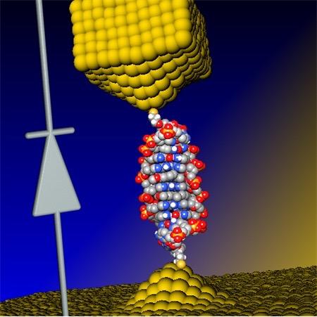
(three-membered rings containing a nitrogen-nitrogen double bond). They are highly attractive as molecular tags, primarily because of their small size. Diazirines have already been established as biocompatible molecular tags for photoaffinity labeling (25). They can be incorporated into many small molecules, metabolites, and biomolecules without drastically altering biological function. Diazirines share similarities with methylene (CH2) groups in terms of electronic and steric properties such that they can replace methylene groups without drastically distorting biochemical behavior. Furthermore, diazirines are stable at room temperature, are resistant to nucleophiles, and do not degrade under either acidic or alkaline conditions (25). With these attractive properties, diazirines have been used for the study of many signaling pathways. For example, they have been incorporated into hormones (26), epileptic drugs (27), antibiotics (28), hyperthermic drugs (29), anticancer agents (30), anesthetics (31), nucleic acids (32), amino acids (33), and lipids (34). They also have been introduced into specific molecular reporters to probe enzyme function and their binding sites such as in kinases (35), aspartic proteases (36), or metalloproteinases (37), to name a few. The nitrogen-nitrogen moiety is also intrinsically interesting, because the two atoms are usually very close in chemical shift and strongly coupled, thus suited to support a long-lived singlet state as described below.


Fig. 1The hyperpolarization mechanism.

(A) The precatalyst, 15N2-diazirine substrate, and p-H2 are mixed, resulting in the activated species depicted in (B). (B) Both p-H2 and the free 15N2-diazirine [2-cyano-3-(D3 methyl-15N2-diazirine)-propanoic acid] are in reversible exchange with the catalytically active iridium complex. The catalyst axial position is occupied by IMes [1,3-bis(2,4,6-trimethylphenyl)-imidazolium] and Py (pyridine) as nonexchanging ligands. The structure shown is a local energy minimum of the potential energy surface based on all-electron DFT calculations and the dispersion-corrected PBE density functional. In the complex, hyperpolarization is transferred from the parahydrogen (p-H2)–derived hydrides to the 15N nuclei (white, hydrogen; gray, carbon; blue, nitrogen; red, oxygen).

Density functional theory calculations shed light on polarization transfer catalyst

The Ir complex conformation shown in Fig. 1B was determined by all-electron density functional theory (DFT) calculations [semilocal Perdew-Burke-Ernzerhof (PBE) functional (38), corrected for long-range many-body dispersion interactions (39), in the FHI-aims software package (40, 41); see the Supplementary Materials for details]. The calculations indicate a η1 single-sided N attachment rather than η2-N=N attachment of the diazirines. In the Ir complex, hyperpolarization is transferred from p-H2 gas (~92% para-state, 7.5 atm) to the 15N2-diazirine. Both p-H2 and substrate are in reversible exchange with the central complex, which results in continuous pumping of hyperpolarization: p-H2 is continually refreshed and hyperpolarization accumulated on the diazirine substrate.

An alternate polarization transfer catalyst is introduced

As opposed to the traditional [Ir(COD)(IMes)(Cl)] catalyst (18), the synthesized [Ir(COD)(IMes)(Py)][PF6] results in a pyridine ligand trans to IMes, improving our hyperpolarization levels by a factor of ~3 (see the Supplementary Materials). We have found that this new approach, which avoids competition from added pyridine, makes it possible to directly hyperpolarize a wide variety of different types of15N-containing molecules (and even 13C). However, diazirines represent a particularly general and interesting class of ligands for molecular tags and are the focus here.

As depicted in Fig. 2A, the hyperpolarization proceeds outside the high-field NMR magnet at low magnetic fields, enabling SABRE-SHEATH directly targeting 15N nuclei (22). To establish the hyperpolarization, we bubble p-H2 for ~5 min at the adequate field. Then, the sample is transferred into the NMR magnet within ~10 s, and 15N2 signal detection is performed with a simple 90° pulse followed by data acquisition.


Fig. 2Experimental and spectral distinction between magnetization and singlet spin order.

(A) Experimental procedure. The sample is hyperpolarized by bubbling parahydrogen (p-H2) through the solution in the NMR tube for 5 min and subsequently transferred into the high-field magnet for detection. If the hyperpolarization/bubbling is performed in a magnetic shield at ~6 mG, z-magnetization is created (black). If the hyperpolarization/bubbling is performed in the laboratory field anywhere between ~0.1 and ~1 kG, singlet order is created (blue). (B) Z-magnetization and singlet spin order can easily be distinguished based on their spectral appearance. a.u., arbitrary units. Z-magnetization produces an in-phase quartet (black). Singlet order gives an anti-phase quartet (blue). The spin system parameters for the 15N2 two-spin system are JNN = 17.3 Hz and Δδ = 0.58 parts per million.

Two types of hyperpolarized states can be created: Magnetization and singlet order

We create two different types of hyperpolarization on the 15N2-diazirine. We can hyperpolarize traditional z-magnetization, which corresponds to nuclear spins aligned with the applied magnetic field and is associated with pure in-phase signal as illustrated with the black trace in Fig. 2B. Alternatively, we can hyperpolarize singlet order on the 15N2-diazirine, which corresponds to an anti-aligned spin state, with both spins pointing in opposite directions, entangled in a quantum mechanically “hidden” state. This hidden singlet order is converted into a detectable state when transferred to a high magnetic field and associated with the anti-phase signal illustrated by the blue trace in Fig. 2B (see the Supplementary Materials for details). The difference in symmetry of z-magnetization and singlet order leads to differences in signal decay rates; z-magnetization is directly exposed to all NMR relaxation mechanisms and is often associated with shorter signal lifetimes, which may impede molecular tracking on biologically relevant time scales. Singlet order, on the other hand, is protected from many relaxation mechanisms because it has no angular momentum (4252) and can therefore exhibit much longer lifetimes, enabling hour-long molecular tracking.

The type of hyperpolarized state is selected by the magnetic field

We can control which type of hyperpolarization we create by choosing the appropriate magnetic fields for the bubbling process. Z-magnetization is created in the SABRE-SHEATH mode at low magnetic fields inside a magnetic shield (2224, 53, 54). This behavior is explained by resonance conditions for hyperpolarizing magnetization versus singlet order that we derive in the Supplementary Materials. The condition for creating magnetization, νH − νN = |JHH ± JNN|, is field-dependent in the NMR frequencies, νH and νN, and field-independent in the J-couplings. Accordingly, hyperpolarized magnetization is created at a magnetic field where the frequency difference matches the J-couplings. This magnetic field is ~6 mG, which is obtained by using JHH = −10 Hz, JNN = −17.3 Hz, γ1H = 4.2576 kHz/G, and γ15N = −0.4316 kHz/G (see the Supplementary Materials). The theoretical prediction of 6 mG matches the experimental maximum for hyperpolarized z-magnetization illustrated by the blue data in Fig. 3A.

………


The demonstrated hyperpolarization lifetimes, combined with the ease of hyperpolarization in these broadly applicable biomolecular tags, may establish a paradigm shift for biomolecular sensing and reporting in optically opaque tissue. The demonstrated lifetimes even exceed lifetimes of some common radioactive tracers used in positron emission tomography (PET) (for example, 11C, 20.3 min). However, unlike PET, MR is exquisitely sensitive to chemical transformations and does not use ionizing radiation (such that, for example, daily progression monitoring of disease is easily possible). The presented work may allow direct access to biochemical mechanisms and kinetics in optically opaque media. We therefore envision tracking subtle biochemical processes in vitro with unprecedented NMR sensitivities as well as real-time in vivo biomolecular imaging with hyperpolarized diazirines.

Read Full Post »

Medical MEMS, BioMEMS and Sensor Applications

Curator and Reporter: Aviva Lev-Ari, PhD, RN


Contents for Chapter 11

Medical MEMS, BioMEMS and Sensors Applications

Curators: Justin D. Pearlman, MD, PhD, FACC, LPBI Group, Danut Dragoi, PhD, LPBI Group and William H. Zurn, Alpha IP

FOR

Series E: Patient-centered Medicine

Volume 4:  Medical 3D BioPrinting – The Revolution in Medicine

Editors: Larry H Bernstein, MD FCAP and Aviva Lev-Ari, PhD, RN

http://pharmaceuticalintelligence.com/biomed-e-books/series-e-titles-in-the-strategic-plan-for-2014-1015/volume-four-medical-3d-bioprinting-the-revolution-in-medicine/

Work-in-Progress

ContactLens

Image Source

http://www.memsjournal.com/2010/05/medical-applications-herald-third-wave-of-mems.html

Image is courtesy of Google Images


WirelessPressure

Image Source

Stanford Engineering Team Invents Pressure Sensor That Uses Radio Waves | CytoFluidix

Image is courtesy of Google Images


Introduction by Dr. Pearlman


Chapter 1: Blood Glucose Sensors

1.1       MINIATURIZED GLUCOSE SENSOR – Google

  • Tiny wireless chip and miniaturized glucose sensor
  • Embedded between two layers of soft contact lens material
  • Accurate glucose monitoring for diabetics
  • Using bodily fluids, i.e. tears
  • Prototypes can generate one reading per second
  • Experimenting with LEDs
  • Early warning for the wearer


Chapter 2: Blood Chemistry Tests – up to 100 Samples

2.1       NON-INVASIVE BLOOD MONITOR- UCSD

  • Digital tattoo monitors blood below the skin
  • Tattoos are needle-less
    • Sensor-laden transdermal patch
  • Painless for the user Tiny sensors “ink”
  • Can read blood levels of:
    • Sodium, glucose, kidney function
  • Prototypes contain probes
  • Wireless, battery-powered chip
  • Continually test up to a hundred different samples


2.3       CELLPHONE-BASED RAPID-DIAGNOSTIC-TEST (RDT) READER – UCLA

  • Lateral flow immuno-chromatographic assays
  • Sense the presence of a target analyte in a sample
  • Device connects to the camera on a cell phone
  • Weighs only 65 grams


2.4       IMPLANTABLE BLOOD ANALYZER CHIP – EPFL

  • Implantable device for instantaneous blood analysis
  • Wireless data transmission to a doctor
  • Applications include monitoring general health
  • Tailor drug delivery to a patient’s unique needs
  • Includes five sensors and a radio transmitter
  • Powered via inductive coupling from a battery patch
  • Worn outside the body


Chapter 3: Motion Sensors for Head-Impact

3.1       HEAD-IMPACT MONITORING PATCH – STMicro & X2Biosystems

  • Wearable electronic contains MEMS motion sensors
  • Microcontroller, low-power radio transmitter, and power management circuitry
  • Cloud-based system combines athlete concussion history
  • Pre-season neurocognitive function, balance, and coordinate-performance data
  • Creates a baseline for comparison after a suspected injury event


Chapter 4: Drug Delivery & Drug Compliance Monitoring Systems

4.1       Smart Pill delivers Therapeutic Agent Load to target – ELECTRONIC PILL – Phillips

  • Electronic pill to treat gastrointestinal cancer
  • An ingestible pill is swallowed by the patient, finds its way to the tumor, dispenses the drugs and passes harmlessly from the body
  • Smart pill contains reservoir for drug supply, fluid pump for drug delivery, pH sensor (for navigation), thermometer, microprocessor, communication


4.2       Drug Compliance Monitoring Systems

4.2.1    INGESTIBLE BIOMEDICAL SENSOR – Proteus Digital Health

  • Biomedical sensor that monitors medication adherence
  • Embedded into a pill, the sensor is activated by stomach fluid
  • Transmits a signal through the body to a skin patch
  • Indicates whether a patient has ingested material


4.2.2    MICROPUMP DEVICES – Purdue University

  • Device based on skin contact actuation for drug delivery
  • Actuation mechanism only requires body heat
  • Induced actuation can result to a gradient of 100 Pa/oC
  • Sufficient to drive liquid drug through micro-needle arrays
  • Prototypes exhibit low fabrication costs, employment of biocompatible materials and battery-less operation Suitable for single- or multiple-use transdermal drug dispensers


4.2.3    IMPLANTABLE MEMS DRUG DELIVERY SYSTEM – MIT

  • Device can deliver a vasoconstrictor agent
  • On demand to injured soldiers to prevent hemorrhagic shock
  • Other applications include medical implants
  • For cancer detection and monitoring
  • Implant can provide physicians and patients
  • Real-time information on the efficacy of treatment


Chapter 5: Remove Monitoring of Food-related Diseases

5.1       LASER-DRIVEN, HANDHELD SPECTROMETER

  • For analyzing food scanned
  • Information to a cloud-based application
  • Examines the results Data is accumulated from many users
  • Used to develop warning algorithms
  • For Allergies, Bacteria


Chapter 6: Skin Protection and Photo-Sensitivity Management

6.1       WEARABLE-UVEXPOSURESENSOR – Gizmag

  • Wristband for monitoring UV exposure
  • Allows user to maximize vitamin D production
  • Reducing the risk of sun
  • Over-exposure and skin cancer
  • LED indicators light up as UV exposure accumulates
  • Flashes once the safe UV limit has been reached


6.2       WEARABLE SKIN SENSOR KTH – Chemistry 2011

  • Bio-patch for measuring and collecting vital information through the skin
  • Inexpensive, versatile and comfortable to wear
  • User Data being gathered depends on where it is placed on the body


Chapter 7: Ophthalmic Applications

7.1       INTRAOCULAR PRESSURE SENSOR – Sensimed & ST Microelectronics

  • Smart contact lens called Triggerfish
  • Contact lens can measure, monitor, and control
  • Intra-ocular pressure levels for patients
  • Catch early cases of glaucoma
  • MEMS strain gage pressure sensor
  • Mounted on a flexible substrate MEMS


7.2       MICRO-MIRRORS ENABLING HANDHELD OPHTHALMIC – OCT News

  • Swept source OCT model for retinal 3D imaging
  • Replaces bulky galvanometer scanners in a handheld OCT probe for primary care physicians
  • Ultrahigh-speed two-axis optical beam steering gimbal-less MEMS mirrors
  • MEMS Actuator with a 2.4 mm bonded mirror and an angular reach of +6°
  • Low power consumption of <100mW including the MEMS actuator driver Retinal 3D Imaging


Chapter 8: Hearing Assist Technologies

8.1       MEMS TECHNOLOGY FOR HEARING RESTORATION – University of Utah

  • Eliminates electronics outside the ear
  • Associated with reliability issues and social stigma
  • Accelerometer-based microphone
  • Successfully tested in cadaver ear canals
  • Prototype measures 2.5 x 6.2mm, weighs 25mg


Chapter 9: Lab-on-a-Chip

9.1       ORGAN-ON-A-CHIP – Johns Hopkins University

  • Silicon substrate for living human cells
  • Controlled environment
  • Emulate how cells function inside a living human body
  • Replace controversial and costly animal testing
  • Lab-on-a-chip: a cost effective end to animal testing


Chapter 10: Intra-Cranial Studies: Pressure Measurement, Monitoring and Adaptation

10.1:   CEREBRAL PRESSURE SENSOR – Fraunhofer Institute

  • Sensor to monitor cerebral pressure that can lead to dementia
  • Pressure changes in the brain can be measured and transmitted
  • Reading device outside the patient’s body
  • Operating at very low power, the sensor module
  • Powered wirelessly by the reading device


10.2    WIRELESS, IMPLANTABLE BRAIN SENSOR – National Institute of Biomedical Imaging and Bioengineering

  • Fully implantable within the brain
  • Allow natural studies of brain activity
  • Cord-free control of advanced prosthetics

Wireless charging Prototypes transmitted brain activity data


Chapter 11: Cardiac and Cardiovascular Monitoring System

11.1    IMPLANTABLE MICRO DEVICE FOR MONITORING AND TREATING ANEURISMS – Electronic Design

  • RF-addressed wireless pressure sensor are powered by inductive coupling
  • Do not need batteries MEMS pressure sensor
  • Wireless antenna are inserted near the heart
  • With a catheter, Blood-pressure readings
  • Are sent to a wireless scanner for monitoring Pressure changes
  • Deflect the transducer’s diaphragm
  • Change the LC circuit’s resonant


11.2    CUSTOM- FITTED, IMPLANTABLE DEVICE FOR TREATMENT AND PREDICTION OF CARDIAC DISORDERS – Washington University

  • Working prototypes were developed on inexpensive 3D printers
  • The 3D elastic membrane is made of a soft, flexible, silicon material
  • Precisely shaped to match the outer layer of the heart


Chapter 12: microfluidic chips

12.1    MICROFLUIDIC MEMS FOR DIABETES TREATMENT – Micronews

  • Watertight pump mounted on a disposable skin patch
  • Provides continuous insulin infusion
  • Controlled by a dedicated smart phone device
  • Incorporating a BGM (blood- glucose meter)


12.2    ACOUSTIC RECEIVER ANTENNA/SENSOR PDMS MEMBRANE – Purdue

POLY-DI-METHYL-SILOXANE (PDMS)

Polydimethylsiloxane called PDMS or dimethicone is a polymer widely used for the fabrication and prototyping of microfluidic chips.

It is a mineral-organic polymer (a structure containing carbon and silicon) of the siloxane family (word derived from silicon, oxygen and alkane). Apart from microfluidics, it is used as a food additive (E900), in shampoos, and as an anti-foaming agent in beverages or in lubricating oils.

For the fabrication of microfluidic devices, PDMS (liquid) mixed with a cross-linking agent is poured into a microstructured mold and heated to obtain a elastomeric replica of the mold (PDMS cross-linked).


Why Use PDMS for Microfluidic Device Fabrication?


PDMS was chosen to fabricate microfluidic chips primarily for those reasons:

Human alveolar epithelial and pulmonary microvascular endothelial cells cultured in a PDMS chip to mimick lung functions

  • It is transparent at optical frequencies (240 nM – 1100 nM), which facilitates the observation of contents in micro-channels visually or through a microscope.
  • It has a low autofluorescence [2]
  • It is considered as bio-compatible (with some restrictions).

The PDMS bonds tightly to glass or another PDMS layer with asimple plasma treatment. This allows the production of multilayers PDMS devices and enables to take advantage of technological possibilities offered by glass substrates, such as the use of metal deposition, oxide deposition or surface functionalisation.

PDMS, during cross-linking, can be coated with a controlled thickness on a substrate using a simple spincoat. This allows the fabrication of multilayer devices and the integration of micro valves.

It is deformable, which allows the integration of microfluidic valves using the deformation of PDMS micro-channels, the easy connection of leak-proof fluidic connections and its use to detect very low forces like biomechanics interactions from cells.

SOURCE

http://www.elveflow.com/microfluidic-tutorials/microfluidic-reviews-and-tutorials/the-poly-di-methyl-siloxane-pdms-and-microfluidics/


  • Ferrite RF radiation Acoustic wave Rectifier
  • Buried in PDMS Implantable miniature pressure sensor
  • Powered by an acoustically actuated cantilever
  • No battery required
  • Acoustic waves in the 200-500 hertz range
  • Cause cantilever to vibrate
  • Scavenging energy to power pressure sensor


Chapter 13: Peropheral Neuropathy Management

13.1    WIRELESS SHOE INSERT – Mobile Health News

  • WIRELESS SHOE INSERT – Mobile Health News
  • Help diabetics manage peripheral nerve damage
  • Insole collects data of where wearers
  • Putting pressure on their feet
  • Transmits wirelessly to a wristwatch-type display
  • Prevent amputations that often stem from diabetic foot ulcers


Chapter 14: Endoscopic Diagnostics Tools

14.1    ENDOSCOPE USING MEMS SCANNING MIRROR

  • For gastrointestinal and urological imaging
  • Alternative to biopsies in cancer detection
  • A laser beam pointed at the mirror is precisely deflected
  • Steered by the scanning mirror to reach a target


Chapter 15: MEMS guided Surgical Tools

15.1    MICROMACHINED SURGICAL TOOLS; SILICON MEMS TWEEZERS – ElectrolQ Used for minimally invasive surgical (MIS)

  • Procedures where diagnosis, monitoring, or treatment of diseases are performed
  • Performing with very small incisions MEMS
  • Based microsurgical tools is a key enabling technology for angioplasty, catheterization, endoscopy, laparoscopy, and neurosurgery


Summary by Dr. Pearlman

  • Multiple projects by Academia & Industry
  • Multiple MEMS devices for measuring body activities.
  • Many patch type devices attached to the skin
  • Devices attached to the eye
  • Smaller is better, lower footprint, lower power


Read Full Post »

Older Posts »

%d bloggers like this: