Feeds:
Posts
Comments

Posts Tagged ‘3D bioprinting’

First-ever living 3D printed aneurysm


Reporter : Irina Robu, PhD

A brain aneurysm is a bulge that forms in the blood vessel of your brain that could lead to severe health issues and possibly death. Brain aneurysm affect about one in 50 Americans and can lead to serious medical emergencies including stroke and brain damage.  Current treatments for brain aneurysm are limited and very invasive and can vary from person to person.

Researchers at Lawrence Livermore National Laboratory and their collaborators were able to replicate an aneurysm in vitro by 3D printing blood vessels with human cerebral cells. One of the leading engineers, William Hynes  performed an endovascular repair procedure on the printed aneurysm by inserting a catheter into blood vessel and tightly packed platinum coils inside the aneurysm sac. Afterward, the scientists introduced blood plasma into the aneurysm and identified the formation of blood clot where the coils were located and they were able to observe the post-op healing process of the endothelial cells within the vessels.

One thing that was obvious to the LLNL scientists is that computer modeling is an important step to developing patient-specific care for aneurysms based on patient’s blood vessel geometry, blood pressure and other factors. They also determined that it takes time for the new surgical technology to move from laboratory to the clinic.

The idea is if they can replicate the aneurysms as much as needed using  animal models or 3D printing, they can help find better options for aneurysms with uncontrollable geometries.  Since, the most common treatment for aneurysms is  the endovascular metal coiling approach, researchers believe  that by taking out the guesswork out of aneurysms treatment researchers can design more predictive 3D models that takes patient geometry into account.

Hynes teamed with former LLNL scientist Duncan Maitland and Amanda Randles, a former Lab computational scientist  to verify if Randles’s flow dynamic model compares with the real world. At low flow rates, scientist saw little movement of blood into the aneurysm, while an increased flow rate, resulted in a circular flow of blood throughout the aneurysm, as would be predictable in a true brain aneurysm.  

Using the data obtained from the flow dynamic model in combination with the 3D printing platform, researchers developed a potential tool for surgeons to pre-select the best coil types desirable to fully pack an aneurysm to obtain the best treatment outcome, and perform “test runs” of procedures before attempting them on the human patient.

Unlike animal models, LLNL’s platform allows scientists to directly measure the fluid dynamics inside the vessels and aneurysm while maintaining biological relevance.

In addition to patient-specific care and serving as a testbed for surgical training, researchers mentioned that the platform can improve the understanding of basic biology and the post-surgery healing response. Even though the results are promising, researchers mentioned that there is long way before their platform is applicable in a clinical environment setting.

SOURCE :

https://www.universityofcalifornia.edu/news/lab-team-develops-first-ever-living-3d-printed-aneurysm-improve-surgical-procedures-personalize?utm_source=fiat-lux

Read Full Post »

Expanding 3D Printing in Cardiology

Reporter: Irina Robu, PhD

3D printing is a fabrication technique used to transform digital objects into physical models, which builds structures of arbitrary geometry by depositing material in successive layers on the basis of specific digital design. Even though, the use of 3D bioprinting in cardiovascular medicine is relatively new development, advancement within this discipline is occurring at such a rapid rate. Most cardiologists believed the costs would be too high for routine use such that the price tag was better for academic applications.

Now as the prices are starting to lower, the idea of using 3D printed models of organs vessels and tissue manufactured based on CT, MRI and echocardiography might be beneficial according to Dr. Fadi Matar, professor at University of South Florida. He and his cardiology colleagues use 3D printed models to allow them to view patient’s complex anatomies before deciding what treatments to pursue. The models allow them to calculate the size and exact placement of devices which has led to shorter procedure time and better outcome.

In a study published in Academic Radiology, David Ballard, professor at University School of Medicine appraised the costs of setting up a 3D printing lab including the commercial printer plus software, lab space, materials and staffing. According to Ballard’s team, the commercial printers start at $12,000 but can be as high as high as $500,000.

According to American Medical Association-approved Category III Current Procedural Terminology (CPT) codes allows cardiology relief from setting up a new 3D printing lab such as Codes 0559T and 0560T, for individually prepared 3D-printed anatomical models with one or more components (including arteries and veins) and Codes 0561T and 0562T, which are for the production of personalized 3D-printed cutting or drilling tools that use patient imaging data and often are used to guide or facilitate surgery.

These codes have been met with enthusiasm by teams eyeing 3D printing, but there are noteworthy limitations to Category III codes—which are temporary codes describing emerging technologies, services and procedures that are used for tracking effectiveness data. It is important to note that Category III codes are not reimbursed but often are a step toward reimbursement.

New and improved materials also might lead to a sharper focus on 3D printing in cardiology. Dr. Fadi Matar says companies are working on materials that better mimic elements of the heart. Such “mimicry” ought to enhance the value of 3D-printed models since they will give cardiologists more realistic insights into how specific devices will interact with an individual patient’s heart. Even with the complex modalities of using 3D bioprinting, in time there would be less obstacles to being able to set up a 3D bioprinter lab.

SOURCE

https://www.cardiovascularbusiness.com/topics/cardiovascular-imaging/seeing-future-3d-new-cpt-codes-set-stage-expanding-3d-printing

Read Full Post »

3D-Printed Brain Clear the Way to Find Cancer Treatments

Reported by: Irina Robu, PhD

Glioblastomas are aggressive and malignant grade IV brain tumors and can located wherever in the brain and do not regularly spread outside of the brain. Common symptoms patients with glioblastoma experience include headaches, seizures, confusion, memory loss, muscle weakness, visual changes, language deficit, and cognitive changes. Glioblastomas tend to affect older individuals (age 45 to 70) with rare occurrences in children. Treatment methods typically include a combination of surgery, chemotherapy, radiation therapy, and alternating electric fields therapy.

Scientists at Northwestern University developed a technique to study their fast spreading cancer using a 3D structure made of agglomeration of human brain cells and biomaterials, which can help doctors better understand how the tumor grows and speed up the potential discovery of novel drugs to fight it. A water-based substance serves as a matrix to hold the cells into place. However, inside the living brain, scientists can’t observe how the tumor cells grow or respond the treatment and they have to use mice/rats to understand tumor development. Animal studies are expensive and time consuming, but the 3D printed live tissue allows researchers to study glioblastoma to be studied more directly.

To understand what happens inside the 3D model, the researchers used a laser to scan the sample and create a snapshot of the cellular structure. This combination allows them to assess the effectiveness of a commonly used chemotherapy drug, temozolomide. The drug, temozolomide kills glioblastoma cells in two-dimensional models, but when put into a three-dimensional one, the tumor rebounded which implies that the drug did not work in the long term.

This 3D model may be able to speed up that process to weed out ineffective drugs first, confirming that only the most promising ones move to animal, and eventually human, trials.

SOURCE

A 3D-printed brain could make it easier to find cancer treatments

Read Full Post »

 A Revolution in Medicine: Medical 3D BioPrinting

Curated by : Irina Robu, PhD

Imagine a scenario, where years from now, one of your organs stop working properly. What would you do?  The current option is to wait in line for a transplant, hoping that the donor is a match. But what if you can get an organ ready for you with no chance of rejection? Even though it may sound like science fiction at the current moment, organ 3D bioprinting can revolutionize medicine and health care.

I have always found the field of tissue engineering and 3D bioprinting fascinating. What interests me about 3D bioprinting is that it has the capacity to be a game changer, because it would make organs widely available to those who need them and it would eliminate the need for a living or deceased donor.  Moreover, it would be beneficial for pediatric patients who suffer specific problems that the current bio-prosthetic options might not address. It would minimize the risk of rejection as well as the components would be customized to size.

There have been advancements in the field of 3D bioprinting and one such advancement is using a 3D printed cranium by neurosurgeons at the University Medical Centre Utrecht. The patient was a young woman who suffered from a chronic bone disorder. The 3D reconstruction of her skull would minimize the brain damage that might have occurred if doctors used a durable plastic cranium.

So, what exactly is bioprinting? 3D bioprinting is an additive manufacturing procedure where biomaterials, such as cells and growth factors, are combined to generate tissue-like structures that duplicate natural tissues. At its core, bioprinting works in a similar way to conventional 3D printing. A digital model becomes a physical 3D object layer-by-layer.  However, in the case of bioprinting, a living cell suspension is used instead of a thermoplastic.

The procedure mostly involves preparation, printing, maturation and application and can be summarized in three steps:

  1. Pre-bioprinting step which includes creating a digital model obtained by using computed tomography (CT) and magnetic resonance imaging (MRI) scans which are then fed to the printer.
  2. Bioprinting step where the actual printing process takes place, where the bioink is placed in a printer cartridge and deposition occurs based on the digital model.
  3. Post-bioprinting step is the mechanical and chemical stimulation of printed parts in order to create stable biostructures which can ultimately be implanted.

3D bioprinting allows suitable microarchitectures that provide mechanical stability and promote cell ingrowth to be produced while preventing any homogeneity issues that occur after conventional cell seeding, such as cell placement. Immediate vascularization of implanted scaffolds is critical, because it provides an influx of nutrients and outflow of by-products preventing necrosis. The benefits of homogeneous seeded scaffolds are that it allows them to integrate faster into the host tissue, uniform cell growth in vivo and lower risk of rejection.

However, in order to address the limitations of the commercially available technology for producing tissue implants, researchers are working to develop a 3D bioprinter that can fit into a laminar flow hood, ultra-low cost and customizable for different organs. Bioprinting can be applied in a clinical setting where the ultimate goal is to implant 3D bioprinted structures into the patients, it is necessary to maintain sterile printing solutions and ensure accuracy in complex tissues, needed for cell-to-cell distances and correct output.

The final aim of bioprinting is to promote an alternative to autologous and allogeneic tissue implants, which will replace animal testing for the study of disease and development of treatments.  We know that for now a short-term goal for 3D bioprinting is to create alternatives to animal testing and to increase the speed of drug testing. The long-term goal is to change the status quo, to develop a personalized organ made from patient’s own cells. However, some ethical challenges still exist regarding the ownership of the organ.

A powerful starting point is the creation of tissue components for heart, liver, pancreas, and other vital organs.  Moreover, each small progress in 3D bioprinting will allow 3D bioprinting to make organs widely available to those who need them, instead of waiting years for a transplant to become available.

I invite you to read a biomedical e-book that I had the pleasure to author along with several other scientists, called Medical 3D BioPrinting – The Revolution in Medicine Technologies for Patient-centered Medicine: From R&D in Biologics to New Medical Devices (Series E: Patient-Centered Medicine Book 4).


Read Full Post »

Use of 3D Bioprinting for Development of Toxicity Prediction Models

Curator: Stephen J. Williams, PhD

SOT FDA Colloquium on 3D Bioprinted Tissue Models: Tuesday, April 9, 2019

The Society of Toxicology (SOT) and the U.S. Food and Drug Administration (FDA) will hold a workshop on “Alternative Methods for Predictive Safety Testing: 3D Bioprinted Tissue Models” on Tuesday, April 9, at the FDA Center for Food Safety and Applied Nutrition in College Park, Maryland. This workshop is the latest in the series, “SOT FDA Colloquia on Emerging Toxicological Science: Challenges in Food and Ingredient Safety.”

Human 3D bioprinted tissues represent a valuable in vitro approach for chemical, personal care product, cosmetic, and preclinical toxicity/safety testing. Bioprinting of skin, liver, and kidney is already appearing in toxicity testing applications for chemical exposures and disease modeling. The use of 3D bioprinted tissues and organs may provide future alternative approaches for testing that may more closely resemble and simulate intact human tissues to more accurately predict human responses to chemical and drug exposures.

A synopsis of the schedule and related works from the speakers is given below:


8:40 AM–9:20 AM Overview and Challenges of Bioprinting
Sharon Presnell, Amnion Foundation, Winston-Salem, NC
9:20 AM–10:00 AM Putting 3D Bioprinting to the Use of Tissue Model Fabrication
Y. Shrike Zhang, Brigham and Women’s Hospital, Harvard Medical School and Harvard-MIT Division of Health Sciences and Technology, Boston, MA
10:00 AM–10:20 AM Break
10:20 AM–11:00 AM Uses of Bioprinted Liver Tissue in Drug Development
Jean-Louis Klein, GlaxoSmithKline, Collegeville, PA
11:00 AM–11:40 AM Biofabrication of 3D Tissue Models for Disease Modeling and Chemical Screening
Marc Ferrer, National Center for Advancing Translational Sciences, NIH, Rockville, MD

Sharon Presnell, Ph.D. President, Amnion Foundation

Dr. Sharon Presnell was most recently the Chief Scientific Officer at Organovo, Inc., and the President of their wholly-owned subsidiary, Samsara Sciences. She received a Ph.D. in Cell & Molecular Pathology from the Medical College of Virginia and completed her undergraduate degree in biology at NC State. In addition to her most recent roles, Presnell has served as the director of cell biology R&D at Becton Dickinson’s corporate research center in RTP, and as the SVP of R&D at Tengion. Her roles have always involved the commercial and clinical translation of basic research and early development in the cell biology space. She serves on the board of the Coulter Foundation at the University of Virginia and is a member of the College of Life Sciences Foundation Board at NC State. In January 2019, Dr. Presnell will begin a new role as President of the Amnion Foundation, a non-profit organization in Winston-Salem.

A few of her relevant publications:

Bioprinted liver provides early insight into the role of Kupffer cells in TGF-β1 and methotrexate-induced fibrogenesis

Integrating Kupffer cells into a 3D bioprinted model of human liver recapitulates fibrotic responses of certain toxicants in a time and context dependent manner.  This work establishes that the presence of Kupffer cells or macrophages are important mediators in fibrotic responses to certain hepatotoxins and both should be incorporated into bioprinted human liver models for toxicology testing.

Bioprinted 3D Primary Liver Tissues Allow Assessment of Organ-Level Response to Clinical Drug Induced Toxicity In Vitro

Abstract: Modeling clinically relevant tissue responses using cell models poses a significant challenge for drug development, in particular for drug induced liver injury (DILI). This is mainly because existing liver models lack longevity and tissue-level complexity which limits their utility in predictive toxicology. In this study, we established and characterized novel bioprinted human liver tissue mimetics comprised of patient-derived hepatocytes and non-parenchymal cells in a defined architecture. Scaffold-free assembly of different cell types in an in vivo-relevant architecture allowed for histologic analysis that revealed distinct intercellular hepatocyte junctions, CD31+ endothelial networks, and desmin positive, smooth muscle actin negative quiescent stellates. Unlike what was seen in 2D hepatocyte cultures, the tissues maintained levels of ATP, Albumin as well as expression and drug-induced enzyme activity of Cytochrome P450s over 4 weeks in culture. To assess the ability of the 3D liver cultures to model tissue-level DILI, dose responses of Trovafloxacin, a drug whose hepatotoxic potential could not be assessed by standard pre-clinical models, were compared to the structurally related non-toxic drug Levofloxacin. Trovafloxacin induced significant, dose-dependent toxicity at clinically relevant doses (≤ 4uM). Interestingly, Trovafloxacin toxicity was observed without lipopolysaccharide stimulation and in the absence of resident macrophages in contrast to earlier reports. Together, these results demonstrate that 3D bioprinted liver tissues can both effectively model DILI and distinguish between highly related compounds with differential profile. Thus, the combination of patient-derived primary cells with bioprinting technology here for the first time demonstrates superior performance in terms of mimicking human drug response in a known target organ at the tissue level.

A great interview with Dr. Presnell and the 3D Models 2017 Symposium is located here:

Please click here for Web based and PDF version of interview

Some highlights of the interview include

  • Exciting advances in field showing we can model complex tissue-level disease-state phenotypes that develop in response to chronic long term injury or exposure
  • Sees the field developing a means to converge both the biology and physiology of tissues, namely modeling the connectivity between tissues such as fluid flow
  • Future work will need to be dedicated to develop comprehensive analytics for 3D tissue analysis. As she states “we are very conditioned to get information in a simple way from biochemical readouts in two dimension, monocellular systems”  however how we address the complexity of various cellular responses in a 3D multicellular environment will be pertinent.
  • Additional challenges include the scalability of such systems and making such system accessible in a larger way
  1. Shrike Zhang, Brigham and Women’s Hospital, Harvard Medical School and Harvard-MIT Division of Health Sciences and Technology

Dr. Zhang currently holds an Assistant Professor position at Harvard Medical School and is an Associate Bioengineer at Brigham and Women’s Hospital. His research interests include organ-on-a-chip, 3D bioprinting, biomaterials, regenerative engineering, biomedical imaging, biosensing, nanomedicine, and developmental biology. His scientific contributions have been recognized by >40 international, national, and regional awards. He has been invited to deliver >70 lectures worldwide, and has served as reviewer for >400 manuscripts for >30 journals. He is serving as Editor-in-Chief for Microphysiological Systems, and Associate Editor for Bio-Design and Manufacturing. He is also on Editorial Board of BioprintingHeliyonBMC Materials, and Essays in Biochemistry, and on Advisory Panel of Nanotechnology.

Some relevant references from Dr. Zhang

Multi-tissue interactions in an integrated three-tissue organ-on-a-chip platform.

Skardal A, Murphy SV, Devarasetty M, Mead I, Kang HW, Seol YJ, Shrike Zhang Y, Shin SR, Zhao L, Aleman J, Hall AR, Shupe TD, Kleensang A, Dokmeci MR, Jin Lee S, Jackson JD, Yoo JJ, Hartung T, Khademhosseini A, Soker S, Bishop CE, Atala A.

Sci Rep. 2017 Aug 18;7(1):8837. doi: 10.1038/s41598-017-08879-x.


Reconstruction of Large-scale Defects with a Novel Hybrid Scaffold Made from Poly(L-lactic acid)/Nanohydroxyapatite/Alendronate-loaded Chitosan Microsphere: in vitro and in vivo Studies.

Wu H, Lei P, Liu G, Shrike Zhang Y, Yang J, Zhang L, Xie J, Niu W, Liu H, Ruan J, Hu Y, Zhang C.

Sci Rep. 2017 Mar 23;7(1):359. doi: 10.1038/s41598-017-00506-z.


A liver-on-a-chip platform with bioprinted hepatic spheroids.

Bhise NS, Manoharan V, Massa S, Tamayol A, Ghaderi M, Miscuglio M, Lang Q, Shrike Zhang Y, Shin SR, Calzone G, Annabi N, Shupe TD, Bishop CE, Atala A, Dokmeci MR, Khademhosseini A.

Biofabrication. 2016 Jan 12;8(1):014101. doi: 10.1088/1758-5090/8/1/014101.


Marc Ferrer, National Center for Advancing Translational Sciences, NIH

Marc Ferrer is a team leader in the NCATS Chemical Genomics Center, which was part of the National Human Genome Research Institute when Ferrer began working there in 2010. He has extensive experience in drug discovery, both in the pharmaceutical industry and academic research. Before joining NIH, he was director of assay development and screening at Merck Research Laboratories. For 10 years at Merck, Ferrer led the development of assays for high-throughput screening of small molecules and small interfering RNA (siRNA) to support programs for lead and target identification across all disease areas.

At NCATS, Ferrer leads the implementation of probe development programs, discovery of drug combinations and development of innovative assay paradigms for more effective drug discovery. He advises collaborators on strategies for discovering small molecule therapeutics, including assays for screening and lead identification and optimization. Ferrer has experience implementing high-throughput screens for a broad range of disease areas with a wide array of assay technologies. He has led and managed highly productive teams by setting clear research strategies and goals and by establishing effective collaborations between scientists from diverse disciplines within industry, academia and technology providers.

Ferrer has a Ph.D. in biological chemistry from the University of Minnesota, Twin Cities, and completed postdoctoral training at Harvard University’s Department of Molecular and Cellular Biology. He received a B.Sc. degree in organic chemistry from the University of Barcelona in Spain.


Some relevant references for Dr. Ferrer

Fully 3D Bioprinted Skin Equivalent Constructs with Validated Morphology and Barrier Function.

Derr K, Zou J, Luo K, Song MJ, Sittampalam GS, Zhou C, Michael S, Ferrer M, Derr P.

Tissue Eng Part C Methods. 2019 Apr 22. doi: 10.1089/ten.TEC.2018.0318. [Epub ahead of print]


Determination of the Elasticity Modulus of 3D-Printed Octet-Truss Structures for Use in Porous Prosthesis Implants.

Bagheri A, Buj-Corral I, Ferrer M, Pastor MM, Roure F.

Materials (Basel). 2018 Nov 29;11(12). pii: E2420. doi: 10.3390/ma11122420.


Mutation Profiles in Glioblastoma 3D Oncospheres Modulate Drug Efficacy.

Wilson KM, Mathews-Griner LA, Williamson T, Guha R, Chen L, Shinn P, McKnight C, Michael S, Klumpp-Thomas C, Binder ZA, Ferrer M, Gallia GL, Thomas CJ, Riggins GJ.

SLAS Technol. 2019 Feb;24(1):28-40. doi: 10.1177/2472630318803749. Epub 2018 Oct 5.


A high-throughput imaging and nuclear segmentation analysis protocol for cleared 3D culture models.

Boutin ME, Voss TC, Titus SA, Cruz-Gutierrez K, Michael S, Ferrer M.

Sci Rep. 2018 Jul 24;8(1):11135. doi: 10.1038/s41598-018-29169-0.

A High-Throughput Screening Model of the Tumor Microenvironment for Ovarian Cancer Cell Growth.

Lal-Nag M, McGee L, Guha R, Lengyel E, Kenny HA, Ferrer M.

SLAS Discov. 2017 Jun;22(5):494-506. doi: 10.1177/2472555216687082. Epub 2017 Jan 31.


Exploring Drug Dosing Regimens In Vitro Using Real-Time 3D Spheroid Tumor Growth Assays.

Lal-Nag M, McGee L, Titus SA, Brimacombe K, Michael S, Sittampalam G, Ferrer M.

SLAS Discov. 2017 Jun;22(5):537-546. doi: 10.1177/2472555217698818. Epub 2017 Mar 15.


RNAi High-Throughput Screening of Single- and Multi-Cell-Type Tumor Spheroids: A Comprehensive Analysis in Two and Three Dimensions.

Fu J, Fernandez D, Ferrer M, Titus SA, Buehler E, Lal-Nag MA.

SLAS Discov. 2017 Jun;22(5):525-536. doi: 10.1177/2472555217696796. Epub 2017 Mar 9.


Other Articles on 3D Bioprinting on this Open Access Journal include:

Global Technology Conferences on 3D BioPrinting 2015 – 2016

3D Medical BioPrinting Technology Reporting by Irina Robu, PhD – a forthcoming Article in “Medical 3D BioPrinting – The Revolution in Medicine, Technologies for Patient-centered Medicine: From R&D in Biologics to New Medical Devices”

Bio-Inks and 3D BioPrinting

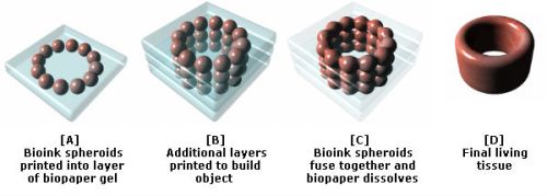
New Scaffold-Free 3D Bioprinting Method Available to Researchers

Gene Editing for Gene Therapies with 3D BioPrinting


Read Full Post »

New 3D-printed Device could Help Treat Spinal Cord Injuries

Reporter: Irina Robu, PhD

Every ten minutes, a person is added to the national transplant waiting list in the US alone, where on average 20 people die each day while waiting for a transplant. The shortage of organ donors is not just confined to the US and scientists are turning to technology for help against this worldwide issue.

Bioprinting sounds innovative, but it has a potential to be the next big thing in healthcare and the hope is that printing and transplanting an organ will take a few hours without any risk of rejection from the body. These printed organs are created from the very cells of the body they will re-enter, matching the exact size, specifications and requirements of each individual patient. The artificial creation of human skin, tissue and internal organs sounds like something from the distant future, nevertheless much of it is happening right now in research facilities around the globe and providing new options for treatment.

Medical researchers and engineers at University of Minnesota created a groundbreaking 3-D printed device that could help patients with long term spinal injuries regain some function. A 3-D printed silicone guide, serves as a platform for specialized cells that are then 3-D printed on top of it. The guide would be surgically implanted into the injured area of the spinal cord where it would serve as a “bridge” between living nerve cells above and below the area of injury.

According to Dr. Ann Parr “This is a very exciting first step in developing a treatment to help people with spinal cord injuries.” The expectation is that this would help patients alleviate pain as well as regain some functions like control of muscles, bowel and bladder. In the current experiments developed at University of Minnesota, years, researchers start with any kind of cell from an adult, such as a skin cell or blood cell which then use to reprogram the cells into neuronal stem cells. The engineers print these cells onto a silicone guide using an exclusive 3-D-printing technology in which the same 3-D printer is used to print both the guide and the cells. The guide keeps the cells alive and allows them to change into neurons. The team developed a prototype guide that would be surgically implanted into the damaged part of the spinal cord and help connect living cells on each side of the injury.

Despite all of these complexities, the hardest part of the entire procedure is being able to keep about 75% of cells during the 3-D printing process. But even with the latest technology, developing the prototype guides wasn’t easy. But although the research is very exciting, we need to be careful to offset expectations against reality. While the research still needs more work, there is no doubt that the future of healthcare and medicine will be very different thanks to this research.

SOURCE

https://www.sciencedaily.com/releases/2018/08/180809093429.htm


Read Full Post »

First 3D Printed Tibia Replacement

Reporter: Irina Robu, PhD

Current advances have allowed 3D printing of biocompatible materials, cells and supporting components into complex 3D functional living tissues. 3D bioprinting has already been used for the generation and transplantation of several tissues, including multilayered skin, bone, vascular grafts, tracheal splints, heart tissue and cartilaginous structures. Thanks to 3D printing, an Australian man got to keep his leg. The man, Reuben Lichter nearly lost his leg above the knee due to a bacterial infection. Doctors told him that he had osteomyelitis which infected his entire bone. Lichter’s bacterial disease of osteomyelitis affects 2 in every 10,000 people in the United States. He had two choices: an experimental procedure using the 3D printed bone or lose his leg. For Lichter, the choice was easy.

Michael Wagels who served as the lead surgeon performed the world’s first-ever transplant surgery using a 3D printed bone. The scaffold was initially modeled at Queensland University of Technology. Biomedical engineers designed the scaffold to promote bone growth around it and then slowly dissolve over time. To have the body successfully grow around the scaffold, the team introduced tissue and blood vessels from both of Lichter’s legs to the scaffold. The surgery itself happened over five operations at Brisbane’s Princess Alexandra Hospital.

However, the next major challenge for biomedical engineers is how to successfully 3D print organs.

SOURCE

https://interestingengineering.com/australian-man-gets-worlds-first-3d-printed-tibia-replacement

Read Full Post »

3-D Printed Ovaries Produce Healthy Offspring

Reporter: Irina Robu, PhD

Each year about 120,000 organs are transplanted from one human being to another and most of the time is a living volunteer. But lack of suitable donors, predominantly means the supply of such organs is inadequate. Countless people consequently die waiting for a transplant which has led researchers to study the question of how to build organs from scratch.

One promising approach is to print them, but “bioprinting” remains largely experimental. Nevertheless, bioprinted tissue is before now being sold for drug testing, and the first transplantable tissues are anticipated to be ready for use in a few years’ time. The first 3D printed organ includes bioprosthetic ovaries which are constructed of 3D printed scaffolds that have immature eggs and have been successful in boosting hormone production and restoring fertility was developed by Teresa K. Woodruff, a reproductive scientist and director of the Women’s Health Research Institute at Feinberg School of Medicine, at Northwestern University, in Illinois.

What sets apart these bioprosthetic ovaries is the architecture of the scaffold. The material is made of gelatin made from broken-down collagen that is safe to humans which is self-supporting and can lead to building multiple layers.

The 3-D printed “scaffold” or “skeleton” is implanted into a female and its pores can be used to optimize how follicles, or immature eggs, get wedged within the scaffold. The scaffold supports the survival of the mouse’s immature egg cells and the cells that produce hormones to boost production. The open construction permits room for the egg cells to mature and ovulate, blood vessels to form within the implant enabling the hormones to circulate and trigger lactation after giving birth. The purpose of this scaffold is to recapitulate how an ovary would function.
The scientists’ only objective for developing the bioprosthetic ovaries was to help reestablish fertility and hormone production in women who have suffered adult cancer treatments and now have bigger risks of infertility and hormone-based developmental issues.


SOURCES

Printed human body parts could soon be available for transplant
https://www.economist.com/news/science-and-technology/21715638-how-build-organs-scratch


3D printed ovaries produce healthy offspring giving hope to infertile women

http://www.telegraph.co.uk/science/2017/05/16/3d-printed-ovaries-produce-healthy-offspring-giving-hope-infertile/


Brave new world: 3D-printed ovaries produce healthy offspring

http://www.naturalnews.com/2017-05-27-brave-new-world-3-d-printed-ovaries-produce-healthy-offspring.html


3-D-printed scaffolds restore ovary function in infertile mice

http://www.medicalnewstoday.com/articles/317485.php


Our Grandkids May Be Born From 3D-Printed Ovaries

http://gizmodo.com/these-mice-gave-birth-using-3d-printed-ovaries-1795237820


Read Full Post »

BioPrinting Basics

Curator: Larry H. Bernstein, MD, FCAP


The ABCs of 3D Bioprinting of Living Tissues, Organs   5/06/2016 

(Credit: Ozbolat Lab/Penn State University)

Although first originated in 2003, the world of bioprinting is still very new and ambiguous. Nevertheless, as the need for organ donation continues to increase worldwide, and organ and tissue shortages prevail, a handful of scientists have started utilizing this cutting-edge science and technology for various areas of regenerative medicine to possibly fill that organ-shortage void.

Among these scientists is Ibrahim Tarik Ozbolat, an associate professor of Engineering Science and Mechanics Department and the Huck Institutes of the Life Sciences at Penn State University, who’s been studying bioprinting and tissue engineering for years.

While Ozbolat is not the first to originate 3D bioprinting research, he’s the first one at Penn State University to spearhead the studies at Ozbolat Lab, Leading Bioprinting Research.

“Tissue engineering is a big need. Regenerative medicine, biofabrication of tissues and organs that can replace the damage or diseases is important,” Ozbolat told R&D Magazine after his seminar presentation at Interphex last week in New York City, titled 3D Bioprinting of Living Tissues & Organs.”

3D bioprinting is the process of creating cell patterns in a confined space using 3D-printing technologies, where cell function and viability are preserved within the printed construct.

Recent progress has allowed 3D printing of biocompatible materials, cells and supporting components into complex 3D functional living tissues. The technology is being applied to regenerative medicine to address the need for tissues and organs suitable for transplantation. Compared with non-biological printing, 3D bioprinting involves additional complexities, such as the choice of materials, cell types, growth and differentiation factors, and technical challenges related to the sensitivities of living cells and the construction of tissues. Addressing these complexities requires the integration of technologies from the fields of engineering, biomaterials science, cell biology, physics and medicine, according to nature.com.

“If we’re able to make organs on demand, that will be highly beneficial to society,” said Ozbolat. “We have the capability to pattern cells, locate them and then make the same thing that exists in the body.”

3D bioprinting of tissues and organs

Sean V Murphy & Anthony Atala
Nature Biotechnology 32,773–785(2014)       doi:10.1038/nbt.2958


Additive manufacturing, otherwise known as three-dimensional (3D) printing, is driving major innovations in many areas, such as engineering, manufacturing, art, education and medicine. Recent advances have enabled 3D printing of biocompatible materials, cells and supporting components into complex 3D functional living tissues. 3D bioprinting is being applied to regenerative medicine to address the need for tissues and organs suitable for transplantation. Compared with non-biological printing, 3D bioprinting involves additional complexities, such as the choice of materials, cell types, growth and differentiation factors, and technical challenges related to the sensitivities of living cells and the construction of tissues. Addressing these complexities requires the integration of technologies from the fields of engineering, biomaterials science, cell biology, physics and medicine. 3D bioprinting has already been used for the generation and transplantation of several tissues, including multilayered skin, bone, vascular grafts, tracheal splints, heart tissue and cartilaginous structures. Other applications include developing high-throughput 3D-bioprinted tissue models for research, drug discovery and toxicology.


Future Technologies : Bioprinting
Bioprinting

3D printing is increasingly permitting the direct digital manufacture (DDM) of a wide variety of plastic and metal items. While this in itself may trigger a manufacturing revolution, far more startling is the recent development of bioprinters. These artificially construct living tissue by outputting layer-upon-layer of living cells. Currently all bioprinters are experimental. However, in the future, bioprinters could revolutionize medical practice as yet another element of the New Industrial Convergence.

Bioprinters may be constructed in various configurations. However, all bioprinters output cells from a bioprint head that moves left and right, back and forth, and up and down, in order to place the cells exactly where required. Over a period of several hours, this permits an organic object to be built up in a great many very thin layers.

In addition to outputting cells, most bioprinters also output a dissolvable gel to support and protect cells during printing. A possible design for a future bioprinter appears below and in the sidebar, here shown in the final stages of printing out a replacement human heart. Note that you can access larger bioprinter images on the Future Visions page. You may also like to watch my bioprinting video.

bioprinter


Bioprinting Pioneers

Several experimental bioprinters have already been built. For example, in 2002 Professor Makoto Nakamura realized that the droplets of ink in a standard inkjet printer are about the same size as human cells. He therefore decided to adapt the technology, and by 2008 had created a working bioprinter that can print out biotubing similar to a blood vessel. In time, Professor Nakamura hopes to be able to print entire replacement human organs ready for transplant. You can learn more about this groundbreaking work here or read this message from Professor Nakamura. The movie below shows in real-time the biofabrication of a section of biotubing using his modified inkjet technology.


Another bioprinting pioneer is Organovo. This company was set up by a research group lead by Professor Gabor Forgacs from the University of Missouri, and in March 2008 managed to bioprint functional blood vessels and cardiac tissue using cells obtained from a chicken. Their work relied on a prototype bioprinter with three print heads. The first two of these output cardiac and endothelial cells, while the third dispensed a collagen scaffold — now termed ‘bio-paper’ — to support the cells during printing.

Since 2008, Organovo has worked with a company called Invetech to create a commercial bioprinter called the NovoGen MMX. This is loaded with bioink spheroids that each contain an aggregate of tens of thousands of cells. To create its output, the NovoGen first lays down a single layer of a water-based bio-paper made from collagen, gelatin or other hydrogels. Bioink spheroids are then injected into this water-based material. As illustrated below, more layers are subsequently added to build up the final object. Amazingly, Nature then takes over and the bioink spheroids slowly fuse together. As this occurs, the biopaper dissolves away or is otherwise removed, thereby leaving a final bioprinted body part or tissue.


bioprinting stages

As Organovo have demonstrated, using their bioink printing process it is not necessary to print all of the details of an organ with a bioprinter, as once the relevant cells are placed in roughly the right place Nature completes the job. This point is powerfully illustrated by the fact that the cells contained in a bioink spheroid are capable of rearranging themselves after printing. For example, experimental blood vessels have been bioprinted using bioink spheroids comprised of an aggregate mix of endothelial, smooth muscle and fibroblast cells. Once placed in position by the bioprint head, and with no technological intervention, the endothelial cells migrate to the inside of the bioprinted blood vessel, the smooth muscle cells move to the middle, and the fibroblasts migrate to the outside.

In more complex bioprinted materials, intricate capillaries and other internal structures also naturally form after printing has taken place. The process may sound almost magical. However, as Professor Forgacs explains, it is no different to the cells in an embryo knowing how to configure into complicated organs. Nature has been evolving this amazing capability for millions of years. Once in the right places, appropriate cell types somehow just know what to do.

In December 2010, Organovo create the first blood vessels to be bioprinted using cells cultured from a single person. The company has also successfully implanted bioprinted nerve grafts into rats, and anticipates human trials of bioprinted tissues by 2015. However, it also expects that the first commercial application of its bioprinters will be to produce simple human tissue structures for toxicology tests. These will enable medical researchers to test drugs on bioprinted models of the liver and other organs, thereby reducing the need for animal tests.

In time, and once human trials are complete, Organovo hopes that its bioprinters will be used to produce blood vessel grafts for use in heart bypass surgery. The intention is then to develop a wider range of tissue-on-demand and organs-on-demand technologies. To this end, researchers are now working on tiny mechanical devices that can artificially exercise and hence strengthen bioprinted muscle tissue before it is implanted into a patient.

Organovo anticipates that its first artificial human organ will be a kidney. This is because, in functional terms, kidneys are one of the more straight-forward parts of the body. The first bioprinted kidney may in fact not even need to look just like its natural counterpart or duplicate all of its features. Rather, it will simply have to be capable of cleaning waste products from the blood. You can read more about the work of Organovoand Professor Forgac’s in this article from Nature.

Regenerative Scaffolds and Bones

A further research team with the long-term goal of producing human organs-on-demand has created the Envisiontec Bioplotter. Like Organovo’s NovoGen MMX, this outputs bio-ink ’tissue spheroids’ and supportive scaffold materials including fibrin and collagen hydrogels. But in addition, the Envisontech can also print a wider range of biomaterials. These include biodegradable polymers and ceramics that may be used to support and help form artificial organs, and which may even be used as bioprinting substitutes for bone.

Talking of bone, a team lead by Jeremy Mao at the Tissue Engineering and Regenerative Medicine Lab at Columbia University is working on the application of bioprinting in dental and bone repairs. Already, a bioprinted, mesh-like 3D scaffold in the shape of an incisor has been implanted into the jaw bone of a rat. This featured tiny, interconnecting microchannels that contained ‘stem cell-recruiting substances’. In just nine weeks after implantation, these triggered the growth of fresh periodontal ligaments and newly formed alveolar bone. In time, this research may enable people to be fitted with living, bioprinted teeth, or else scaffolds that will cause the body to grow new teeth all by itself. You can read more about this development in this article from The Engineer.

In another experient, Mao’s team implanted bioprinted scaffolds in the place of the hip bones of several rabbits. Again these were infused with growth factors. As reported inThe Lancet, over a four month period the rabbits all grew new and fully-functional joints around the mesh. Some even began to walk and otherwise place weight on their new joints only a few weeks after surgery. Sometime next decade, human patients may therefore be fitted with bioprinted scaffolds that will trigger the grown of replacement hip and other bones. In a similar development, a team from Washington State University have also recently reported on four years of work using 3D printers to create a bone-like material that may in the future be used to repair injuries to human bones.

In Situ Bioprinting

The aforementioned research progress will in time permit organs to be bioprinted in a lab from a culture of a patient’s own cells. Such developments could therefore spark a medical revolution. Nevertheless, others are already trying to go further by developing techniques that will enable cells to be printed directly onto or into the human body in situ. Sometime next decade, doctors may therefore be able to scan wounds and spray on layers of cells to very rapidly heal them.

Already a team of bioprinting researchers lead by Anthony Alata at the Wake Forrest School of Medicine have developed a skin printer. In initial experiments they have taken 3D scans of test injuries inflicted on some mice and have used the data to control a bioprint head that has sprayed skin cells, a coagulant and collagen onto the wounds. The results are also very promising, with the wounds healing in just two or three weeks compared to about five or six weeks in a control group. Funding for the skin-printing project is coming in part from the US military who are keen to develop in situ bioprinting to help heal wounds on the battlefield. At present the work is still in a pre-clinical phase with Alata progressing his research usig pigs. However, trials of with human burn victims could be a little as five years away.

The potential to use bioprinters to repair our bodies in situ is pretty mind blowing. In perhaps no more than a few decades it may be possible for robotic surgical arms tipped with bioprint heads to enter the body, repair damage at the cellular level, and then also repair their point of entry on their way out. Patients would still need to rest and recuperate for a few days as bioprinted materials fully fused into mature living tissue. However, most patients could potentially recover from very major surgery in less than a week.

Cosmetic Applications …

Bioprinting Implications …

More information on bioprinting can be found in my books 3D Printing: Second Editionand The Next Big Thing. There is also a bioprinting section in my 3D Printing Directory. Oh, and there is also a great infographic about bioprinting here. Enjoy!


How to print out a blood vessel

New work moves closer to the age of organs on demand.

http://www.nature.com/news/2008/080320/images/news.2008.675.jpg

Blood vessels can now be ‘printed out’ by machine. Could bigger structures be in the future?SUSUMU NISHINAGA / SCIENCE PHOTO LIBRARY

Read Full Post »

Mid Atlantic LRIG 22nd Annual Technology Showcase: Agenda on 3D Bioprinting on Wednesday, May 11, 2016 at Holiday Inn, 195 Davidson Avenue, Somerset, NJ

Reporter: Stephen J. Williams, Ph.D.


Symposium Speakers and Topics:

Human Organoids
Hatem E. Sabaawy-Director, Production GMP Facility for Cell and Gene Therapy, RBHS-Robert Wood Johnson Medical School, Rutgers Cancer Institute of New Jersey

Intestinal Organoids for Drug Discovery
Richard Visconti-Associate Principal Scientist, Cellular Pharmacology, Merck Research Laboratories, Kenilworth,  New Jersey

3D Bioprinting
Elizabeth Wu-President, WuZenTech, Edison, New Jersey

Building  Your Brand  Through LinkedIn
Stan Robinson, Jr., LinkedIn Consultant, Helping Professionals with Social Selling, Personal Branding

Register at EventBrite here: https://www.eventbrite.com/e/mid-atlantic-22nd-annual-technology-and-exhibition-tickets-21359945171 

To sign up to be an LRIG member or update your profile, please visit us at http://lrig.org
Hoping to see you on May 11th.
Reserve your spot today!


Read Full Post »

Older Posts »

%d bloggers like this: