Healthcare analytics, AI solutions for biological big data, providing an AI platform for the biotech, life sciences, medical and pharmaceutical industries, as well as for related technological approaches, i.e., curation and text analysis with machine learning and other activities related to AI applications to these industries.
Eight Subcellular Pathologies driving Chronic Metabolic Diseases – Methods for Mapping Bioelectronic Adjustable Measurements as potential new Therapeutics: Impact on Pharmaceuticals in Use
In this curation we wish to present two breaking through goals:
Goal 1:
Exposition of a new direction of research leading to a more comprehensive understanding of Metabolic Dysfunctional Diseases that are implicated in effecting the emergence of the two leading causes of human mortality in the World in 2023: (a) Cardiovascular Diseases, and (b) Cancer
Goal 2:
Development of Methods for Mapping Bioelectronic Adjustable Measurements as potential new Therapeutics for these eight subcellular causes of chronic metabolic diseases. It is anticipated that it will have a potential impact on the future of Pharmaceuticals to be used, a change from the present time current treatment protocols for Metabolic Dysfunctional Diseases.
According to Dr. Robert Lustig, M.D, an American pediatric endocrinologist. He is Professor emeritus of Pediatrics in the Division of Endocrinology at the University of California, San Francisco, where he specialized in neuroendocrinology and childhood obesity, there are eight subcellular pathologies that drive chronic metabolic diseases.
These eight subcellular pathologies can’t be measured at present time.
In this curation we will attempt to explore methods of measurement for each of these eight pathologies by harnessing the promise of the emerging field known as Bioelectronics.
Unmeasurable eight subcellular pathologies that drive chronic metabolic diseases
Glycation
Oxidative Stress
Mitochondrial dysfunction [beta-oxidation Ac CoA malonyl fatty acid]
Insulin resistance/sensitive [more important than BMI], known as a driver to cancer development
Membrane instability
Inflammation in the gut [mucin layer and tight junctions]
Epigenetics/Methylation
Autophagy [AMPKbeta1 improvement in health span]
Diseases that are not Diseases: no drugs for them, only diet modification will help
Image source
Robert Lustig, M.D. on the Subcellular Processes That Belie Chronic Disease
These eight Subcellular Pathologies driving Chronic Metabolic Diseases are becoming our focus for exploration of the promise of Bioelectronics for two pursuits:
Will Bioelectronics be deemed helpful in measurement of each of the eight pathological processes that underlie and that drive the chronic metabolic syndrome(s) and disease(s)?
IF we will be able to suggest new measurements to currently unmeasurable health harming processes THEN we will attempt to conceptualize new therapeutic targets and new modalities for therapeutics delivery – WE ARE HOPEFUL
In the Bioelecronics domain we are inspired by the work of the following three research sources:
Michael Levin is an American developmental and synthetic biologist at Tufts University, where he is the Vannevar Bush Distinguished Professor. Levin is a director of the Allen Discovery Center at Tufts University and Tufts Center for Regenerative and Developmental Biology. Wikipedia
THE VOICE of Dr. Justin D. Pearlman, MD, PhD, FACC
PENDING
THE VOICE of Stephen J. Williams, PhD
Ten TakeAway Points of Dr. Lustig’s talk on role of diet on the incidence of Type II Diabetes
25% of US children have fatty liver
Type II diabetes can be manifested from fatty live with 151 million people worldwide affected moving up to 568 million in 7 years
A common myth is diabetes due to overweight condition driving the metabolic disease
There is a trend of ‘lean’ diabetes or diabetes in lean people, therefore body mass index not a reliable biomarker for risk for diabetes
Thirty percent of ‘obese’ people just have high subcutaneous fat. the visceral fat is more problematic
there are people who are ‘fat’ but insulin sensitive while have growth hormone receptor defects. Points to other issues related to metabolic state other than insulin and potentially the insulin like growth factors
At any BMI some patients are insulin sensitive while some resistant
Visceral fat accumulation may be more due to chronic stress condition
Fructose can decrease liver mitochondrial function
A methionine and choline deficient diet can lead to rapid NASH development
3D printing is a technique that has gained immense popularity for its ability to create 3D structures in art, jewelry, engineering, medicine. In this case, radiologists use 3D printing to transform a 2D scan into 3D visualization of a patient’s anatomy. Radiologists use their unique skills to visualize the anatomy of the organs of interest which give them a large advantage in communicating with patients as well as surgical teams.
The 3D printed anatomical models have proved valuable in providing a better understanding of complex anatomies and being used as a tangible aid for pre-surgical planning. It gives the patient a clear understanding of what is happening and it provides a great value when it comes to patient specific care. However, 3D modelling is essential at the beginning but it can also be a useful tool for surgeons. The list of medical 3D printing benefits is infinite. Just recently, a scientific team at University of Minnesota constructed their own patient specific 3D organ model based on MRI scans and prostate tissue samples of patients. The organs allow surgeons to plan and rehearse surgery.
In addition to researchers at University of Minnesota, Siemens Health engineers also created a platform to make medical apps that can be accessible throughout hospitals. In addition, Siemens Health partnered up with Materialise to make 3D printing software an integral part of the radiology workflow.
Hence, using 3D bioprinting is a desirable path to follow for radiologist. Not only they get to interpret anatomy, but now they can use 3D bioprinting as a state of the art tool that empower them to provide immense value to an audience that stretches from patient to practitioner.
Healing traumatic brain injuries with self-assembling peptide hydrogels
Reporter : Irina Robu, PhD
In 2014, TBIs resulted in about 2.53 million emergency department visits in the U.S., according to the Centers for Disease Control and Prevention. A traumatic brain injury (TBI) can range from a mild concussion to a severe head injury. It is caused by a blow to the head or body, a wound that breaks through the skull or another injury that jars or shakes the brain. Individuals with traumatic brain injuries can develop secondary disorders after the initial blow. Researchers, Biplab Sarkar and Vivek Kumar from New Jersey Institute of Technology are hoping to prevent secondary disorders by injecting a self-assembling peptide hydrogel into the brains of rats with traumatic brain injury and see what happens. They observed that the hydrogel helped blood vessels regrow in addition to neuronal survival.
The researchers explained that after traumatic brain injury, the brain can amass glutamate which kills some neurons which is marked by overactive oxygen-containing molecules (oxidative stress), inflammation and disruption of the blood-brain barrier. Furthermore, TBI survivors can experience impaired motor control and depression. Within the experiment, the researchers showed that a week after injecting the gel in rats, the neurons have twice as many neurons at the injury site than the control animals did.
The NJIT researchers distinguished that they needed to inject the hydrogel directly in a rat’s brain just seconds after a TBI, which is not ideal, because it would be impossible to give a patient the treatment within that short period of time. The next step in showing that the self-assembling peptide hydrogel works is to combine their previous blood vessel-growing peptide and the new version to see whether it could enhance recovery. And the researchers plan to inspect whether the hydrogels work for more diffuse brain injuries such as concussions.
More than a million Americans have heart attacks each year. Researchers at Northwestern University and University of California, San Diego have designed a minimally invasive platform to deliver nanomaterial that turns body’s inflammatory response into a signal rather than means of scarring following a heart attack. The researchers from Northwestern-UC San Diego established a novel way to deliver a bioactivated, biodegradable, regenerative substance through a noninvasive catheter without clogging in-vivo in a rat model.
When a person has a heart attack, the extracellular matrix is stripped away and scar tissue forms in its place, decreasing the heart’s functionality. The team injects a self-assembling peptide that seeks out a target, the heart’s damaged extracellular matrix and the solution is then activated by the inflammatory environment itself and gels.
The team’s preclinical research was led in rats and segmented into two proof-of-concept tests. The first test recognized that the material could be fed through a catheter without clogging and without interacting with human blood. The second determined whether the self-assembling peptides could find their way to the damaged tissue, bypassing healthy heart tissue. The scientists attached a fluorescent tag to the self-assembling peptides and imaged the heart to see where the peptides eventually settled.
Researchers now know that when they remove the fluorescent tag and replace it with a therapeutic, the self-assembling peptides will locate to the affected area of the heart. One hurdle is that catheter delivery in a rodent model is far more complicated than the same procedure in a human.
A major innovation occurred when sterically constrained cyclic peptides, which flow freely during delivery and rapidly assemble into hydrogels when they come in contact with disease associated enzymes. The process creates conditions for the peptides to better self-assemble on top one another and form the scaffold that resembles the native extracellular matrix.
A self-cleaning spacesuit was developed by engineers using carbon nanotube technology to purge itself from hyper-abrasive space dust. The sharp and sticky particles can cause noteworthy wear and tear on protective gear as well as causing them to overheat.Kavya Manyapu, a flight crew operations and test engineer for Starliner Spacecraft at Boeing, has now created a way to magnetize flexible carbon nanotube fibers which make the fabric immune to the problematic dust particles. A magnetic field induces a process known as electrophoresis, which carries and moves charged particles away from an area to stop it building up in certain areas.
However, particles on our moon and other planets are sharper and abrasive because of the atmosphere which erodes bulging edges here on Earth. It is also often electrically charged due to the relentless and unfiltered UV rays from space which experts say make the dust particles ‘sticky’. Static electricity aids the dust cling to a spacesuit and then wears out the fabric – often in crevices and folds such as elbows and knees.
Carbon nano-tubes are already in use to stop dust settling on solar panels and other sensors in space but they are brittle and ill-suited for use in clothing. However, scientists have found a way to make technology flexible with the use of small magnetic field created a fabric that can repel the dust.
Boeing engineers created a fully functioning knee joint section to prove their concept was operative. The segment was fully pressurized, as it would be on future lunar and Martian missions. It can even be adapted to improved suit the circumstances and requirements of other planets.
Engineers and scientists at California Institute of Technology (Caltech) and ETH Zurich developed an artificial skin capable of detecting temperature changes using a mechanism similar to the biological mechanism that allow snakes to sense prey through heat. In those organs, ion channels in the cell membrane of sensory nerve fibers expand as temperature increases. This dilation allows calcium ions to flow, triggering electrical impulses.
The material used is a long chain molecule found in plant cells which gives the skin its temperature sensing capabilities. The team chose pectin because the pectin molecules in the film have a weakly bonded double-strand structure that contains calcium ions. As temperature increases, these bonds break down and the double strands “unzip,” releasing the positively charged calcium ions.
This would make pectin sensors useful for industrial applications, such as thermal sensors in consumer electronics or robotic skins to augment human-robot interactions. However, they need to change the fabrication process as that the current process leads to the presence of water which tends to bubble or evaporate at high temperatures.
Implants are gradually being used to treat various bone defects. A key factor for long term success of implants is the proper selection of the implant biomaterial. The biologic environment does not accept completely any material so to optimize biologic performance, implants should be selected to reduce the negative biologic response while maintaining adequate function. The implanted structure must if possible stimulate new bone formation, integrate with existing tissue and lastly be resorbed by the body to enable healthy bone growth.
The EU-funded MGNIM project which tailored biodegradable magnesium implant materials focused on producing aluminum- free Mg-based material suitable for bone applications. MAGNIM produced over 20 different Mg alloys and evaluated their mechanical and structural properties. In addition, they assessed their biological interaction, more specifically their corrosion-behavior. Out of these, two of the new alloys (Mg-2Ag and Mg-10Gd) were nominated for animal trials as pilot results indicated an anti-inflammatory function of degradation products.
The two new alloys, Mg-2Ag and Mg-10Gd as well as Mg alloy WE43 were tested in-vivo for biodegradability and functionality. Screws made of these materials were inserted into the femur of rats and their degradation was monitored. Imaging and histological data from explants revealed new bone formation in the screw implant site.
Even though the project has ended, additional testing in large animal models will be carried out prior to human clinical trials. MAGNIM partners propose to optimize implant material homogeneity and surface properties.
New research from California Institute of Technology headed by Anupama Thubagere and Lulu Qian built robots from DNA and programmed them to sort and deliver molecules to a specified location. These robots can potentially transform the drug delivery field to how body fights infections to how microscopic measurements are made. The dominant premise of DNA robots is that rather than creating molecular devices from scratch, we can use the power of molecular machinery by building microscopic-size robots and send them to places that are then impossible to reach, such as a cell or a hard-to-reach cancerous tumor. These robots demonstrated the ability to perform simple tasks, however this latest effort ramped up a path by programming DNA robots to perform a cargo‐sorting task and possibly many other tasks.
Each robot was built from a single-stranded DNA molecule of just 53 nucleotides equipped with “legs” for walking and “arms” for picking up objects. The robot are 20 nanometers tall and their walking strides measures six nanometers long, where one nanometer is a billionth of a meter. For the cargo, the researchers used two types of molecules, each being a distinct single-stranded piece of DNA. For the tests, the researchers placed the cargo onto a random location along the surface of a two-dimensional origami DNA test platform. The walking DNA robots moved in parallel along this surface, hunting for their cargo.
To see if a robot successfully picked up and dropped off the right cargo at the right location, the researchers used two fluorescent dyes to differentiate the molecules.
The researchers guess that each DNA robot took around 300 steps to complete its task, or roughly ten times more than in previous efforts. Though, more work is needed to figure out how these DNA robots perform under different environmental conditions. This new study suggests a worthwhile methodology for scientists to continue pursuing.
3-D Printing in Water using Novel Hybrid Nanoparticles
Reporter: Irina Robu, PhD
3D printing has become an essential tool for fabricating different organic based materials, but printing structures in water has been thought-provoking due to lack of water soluble molecules known as photo initiators. The photo initiator can induce chemical reactions needed to form solid printed material by light. However, researchers at the Hebrew University of Jerusalem’s Center for Nanoscience and Nanotechnology have developed a new type of photo initiator for three-dimensional printing in water. This innovative nanoparticle allows the creating of bio-friendly 3D structures.
By 3D printing in water, it also opens up the digital light processing method to medical applications, leading toward a competitive response for patient specific implants and tissues because the photo initiators cause rapid solidification of a liquid material that can create faster reactions when exposed to light. 3D printing in water opens up innovative ways for tailored fabrication of medical devices and for printing hydrogels or bio-scaffolds that are typical used in tissue engineering.
The challenge of 3D printing in water is finding an initiator that is not consumed by irradiation. However, unlike regular photo initiators, the novel hybrid nanoparticles developed by Prof. Magdassi present tunable properties, wide excitation window in the UV and visible range, high light sensitivity, and their ability to split water, and absorb oxygen molecules that typically inhibit the performance of the process. The particles added as photo initiator are semi conductive hybrid nanoparticles and are used to create high resolution 3D objects at sub-microscopic scale.
Therefore, 3-D printing in water could allow personalized fabrication of joint replacements, heart valves, artificial tendons and ligaments etc.
Amol Ashok Pawar et al. Rapid Three-Dimensional Printing in Water Using Semiconductor–Metal Hybrid Nanoparticles as Photoinitiators, Nano Letters (2017)
Although first originated in 2003, the world of bioprinting is still very new and ambiguous. Nevertheless, as the need for organ donation continues to increase worldwide, and organ and tissue shortages prevail, a handful of scientists have started utilizing this cutting-edge science and technology for various areas of regenerative medicine to possibly fill that organ-shortage void.
Among these scientists is Ibrahim Tarik Ozbolat, an associate professor of Engineering Science and Mechanics Department and the Huck Institutes of the Life Sciences at Penn State University, who’s been studying bioprinting and tissue engineering for years.
While Ozbolat is not the first to originate 3D bioprinting research, he’s the first one at Penn State University to spearhead the studies at Ozbolat Lab, Leading Bioprinting Research.
“Tissue engineering is a big need. Regenerative medicine, biofabrication of tissues and organs that can replace the damage or diseases is important,” Ozbolat told R&D Magazine after his seminar presentation at Interphex last week in New York City, titled 3D Bioprinting of Living Tissues & Organs.”
3D bioprinting is the process of creating cell patterns in a confined space using 3D-printing technologies, where cell function and viability are preserved within the printed construct.
Recent progress has allowed 3D printing of biocompatible materials, cells and supporting components into complex 3D functional living tissues. The technology is being applied to regenerative medicine to address the need for tissues and organs suitable for transplantation. Compared with non-biological printing, 3D bioprinting involves additional complexities, such as the choice of materials, cell types, growth and differentiation factors, and technical challenges related to the sensitivities of living cells and the construction of tissues. Addressing these complexities requires the integration of technologies from the fields of engineering, biomaterials science, cell biology, physics and medicine, according to nature.com.
“If we’re able to make organs on demand, that will be highly beneficial to society,” said Ozbolat. “We have the capability to pattern cells, locate them and then make the same thing that exists in the body.”
Additive manufacturing, otherwise known as three-dimensional (3D) printing, is driving major innovations in many areas, such as engineering, manufacturing, art, education and medicine. Recent advances have enabled 3D printing of biocompatible materials, cells and supporting components into complex 3D functional living tissues. 3D bioprinting is being applied to regenerative medicine to address the need for tissues and organs suitable for transplantation. Compared with non-biological printing, 3D bioprinting involves additional complexities, such as the choice of materials, cell types, growth and differentiation factors, and technical challenges related to the sensitivities of living cells and the construction of tissues. Addressing these complexities requires the integration of technologies from the fields of engineering, biomaterials science, cell biology, physics and medicine. 3D bioprinting has already been used for the generation and transplantation of several tissues, including multilayered skin, bone, vascular grafts, tracheal splints, heart tissue and cartilaginous structures. Other applications include developing high-throughput 3D-bioprinted tissue models for research, drug discovery and toxicology.
3D printing is increasingly permitting the direct digital manufacture (DDM) of a wide variety of plastic and metal items. While this in itself may trigger a manufacturing revolution, far more startling is the recent development of bioprinters. These artificially construct living tissue by outputting layer-upon-layer of living cells. Currently all bioprinters are experimental. However, in the future, bioprinters could revolutionize medical practice as yet another element of the New Industrial Convergence.
Bioprinters may be constructed in various configurations. However, all bioprinters output cells from a bioprint head that moves left and right, back and forth, and up and down, in order to place the cells exactly where required. Over a period of several hours, this permits an organic object to be built up in a great many very thin layers.
In addition to outputting cells, most bioprinters also output a dissolvable gel to support and protect cells during printing. A possible design for a future bioprinter appears below and in the sidebar, here shown in the final stages of printing out a replacement human heart. Note that you can access larger bioprinter images on the Future Visions page. You may also like to watch my bioprinting video.
Bioprinting Pioneers
Several experimental bioprinters have already been built. For example, in 2002 Professor Makoto Nakamura realized that the droplets of ink in a standard inkjet printer are about the same size as human cells. He therefore decided to adapt the technology, and by 2008 had created a working bioprinter that can print out biotubing similar to a blood vessel. In time, Professor Nakamura hopes to be able to print entire replacement human organs ready for transplant. You can learn more about this groundbreaking work here or read this message from Professor Nakamura. The movie below shows in real-time the biofabrication of a section of biotubing using his modified inkjet technology.
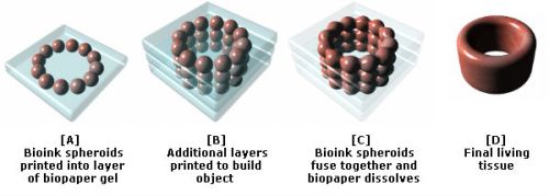
Another bioprinting pioneer is Organovo. This company was set up by a research group lead by Professor Gabor Forgacs from the University of Missouri, and in March 2008 managed to bioprint functional blood vessels and cardiac tissue using cells obtained from a chicken. Their work relied on a prototype bioprinter with three print heads. The first two of these output cardiac and endothelial cells, while the third dispensed a collagen scaffold — now termed ‘bio-paper’ — to support the cells during printing.
Since 2008, Organovo has worked with a company called Invetech to create a commercial bioprinter called the NovoGen MMX. This is loaded with bioink spheroids that each contain an aggregate of tens of thousands of cells. To create its output, the NovoGen first lays down a single layer of a water-based bio-paper made from collagen, gelatin or other hydrogels. Bioink spheroids are then injected into this water-based material. As illustrated below, more layers are subsequently added to build up the final object. Amazingly, Nature then takes over and the bioink spheroids slowly fuse together. As this occurs, the biopaper dissolves away or is otherwise removed, thereby leaving a final bioprinted body part or tissue.
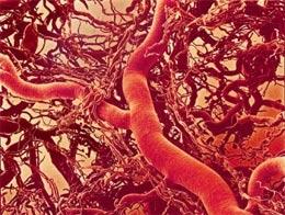
As Organovo have demonstrated, using their bioink printing process it is not necessary to print all of the details of an organ with a bioprinter, as once the relevant cells are placed in roughly the right place Nature completes the job. This point is powerfully illustrated by the fact that the cells contained in a bioink spheroid are capable of rearranging themselves after printing. For example, experimental blood vessels have been bioprinted using bioink spheroids comprised of an aggregate mix of endothelial, smooth muscle and fibroblast cells. Once placed in position by the bioprint head, and with no technological intervention, the endothelial cells migrate to the inside of the bioprinted blood vessel, the smooth muscle cells move to the middle, and the fibroblasts migrate to the outside.
In more complex bioprinted materials, intricate capillaries and other internal structures also naturally form after printing has taken place. The process may sound almost magical. However, as Professor Forgacs explains, it is no different to the cells in an embryo knowing how to configure into complicated organs. Nature has been evolving this amazing capability for millions of years. Once in the right places, appropriate cell types somehow just know what to do.
In December 2010, Organovo create the first blood vessels to be bioprinted using cells cultured from a single person. The company has also successfully implanted bioprinted nerve grafts into rats, and anticipates human trials of bioprinted tissues by 2015. However, it also expects that the first commercial application of its bioprinters will be to produce simple human tissue structures for toxicology tests. These will enable medical researchers to test drugs on bioprinted models of the liver and other organs, thereby reducing the need for animal tests.
In time, and once human trials are complete, Organovo hopes that its bioprinters will be used to produce blood vessel grafts for use in heart bypass surgery. The intention is then to develop a wider range of tissue-on-demand and organs-on-demand technologies. To this end, researchers are now working on tiny mechanical devices that can artificially exercise and hence strengthen bioprinted muscle tissue before it is implanted into a patient.
Organovo anticipates that its first artificial human organ will be a kidney. This is because, in functional terms, kidneys are one of the more straight-forward parts of the body. The first bioprinted kidney may in fact not even need to look just like its natural counterpart or duplicate all of its features. Rather, it will simply have to be capable of cleaning waste products from the blood. You can read more about the work of Organovoand Professor Forgac’s in this article from Nature.
Regenerative Scaffolds and Bones
A further research team with the long-term goal of producing human organs-on-demand has created the Envisiontec Bioplotter. Like Organovo’s NovoGen MMX, this outputs bio-ink ’tissue spheroids’ and supportive scaffold materials including fibrin and collagen hydrogels. But in addition, the Envisontech can also print a wider range of biomaterials. These include biodegradable polymers and ceramics that may be used to support and help form artificial organs, and which may even be used as bioprinting substitutes for bone.
Talking of bone, a team lead by Jeremy Mao at the Tissue Engineering and Regenerative Medicine Lab at Columbia University is working on the application of bioprinting in dental and bone repairs. Already, a bioprinted, mesh-like 3D scaffold in the shape of an incisor has been implanted into the jaw bone of a rat. This featured tiny, interconnecting microchannels that contained ‘stem cell-recruiting substances’. In just nine weeks after implantation, these triggered the growth of fresh periodontal ligaments and newly formed alveolar bone. In time, this research may enable people to be fitted with living, bioprinted teeth, or else scaffolds that will cause the body to grow new teeth all by itself. You can read more about this development in this article from The Engineer.
In another experient, Mao’s team implanted bioprinted scaffolds in the place of the hip bones of several rabbits. Again these were infused with growth factors. As reported inThe Lancet, over a four month period the rabbits all grew new and fully-functional joints around the mesh. Some even began to walk and otherwise place weight on their new joints only a few weeks after surgery. Sometime next decade, human patients may therefore be fitted with bioprinted scaffolds that will trigger the grown of replacement hip and other bones. In a similar development, a team from Washington State University have also recently reported on four years of work using 3D printers to create a bone-like material that may in the future be used to repair injuries to human bones.
In Situ Bioprinting
The aforementioned research progress will in time permit organs to be bioprinted in a lab from a culture of a patient’s own cells. Such developments could therefore spark a medical revolution. Nevertheless, others are already trying to go further by developing techniques that will enable cells to be printed directly onto or into the human body in situ. Sometime next decade, doctors may therefore be able to scan wounds and spray on layers of cells to very rapidly heal them.
Already a team of bioprinting researchers lead by Anthony Alata at the Wake Forrest School of Medicine have developed a skin printer. In initial experiments they have taken 3D scans of test injuries inflicted on some mice and have used the data to control a bioprint head that has sprayed skin cells, a coagulant and collagen onto the wounds. The results are also very promising, with the wounds healing in just two or three weeks compared to about five or six weeks in a control group. Funding for the skin-printing project is coming in part from the US military who are keen to develop in situ bioprinting to help heal wounds on the battlefield. At present the work is still in a pre-clinical phase with Alata progressing his research usig pigs. However, trials of with human burn victims could be a little as five years away.
The potential to use bioprinters to repair our bodies in situ is pretty mind blowing. In perhaps no more than a few decades it may be possible for robotic surgical arms tipped with bioprint heads to enter the body, repair damage at the cellular level, and then also repair their point of entry on their way out. Patients would still need to rest and recuperate for a few days as bioprinted materials fully fused into mature living tissue. However, most patients could potentially recover from very major surgery in less than a week.


