Healthcare analytics, AI solutions for biological big data, providing an AI platform for the biotech, life sciences, medical and pharmaceutical industries, as well as for related technological approaches, i.e., curation and text analysis with machine learning and other activities related to AI applications to these industries.
A self-cleaning spacesuit was developed by engineers using carbon nanotube technology to purge itself from hyper-abrasive space dust. The sharp and sticky particles can cause noteworthy wear and tear on protective gear as well as causing them to overheat.Kavya Manyapu, a flight crew operations and test engineer for Starliner Spacecraft at Boeing, has now created a way to magnetize flexible carbon nanotube fibers which make the fabric immune to the problematic dust particles. A magnetic field induces a process known as electrophoresis, which carries and moves charged particles away from an area to stop it building up in certain areas.
However, particles on our moon and other planets are sharper and abrasive because of the atmosphere which erodes bulging edges here on Earth. It is also often electrically charged due to the relentless and unfiltered UV rays from space which experts say make the dust particles ‘sticky’. Static electricity aids the dust cling to a spacesuit and then wears out the fabric – often in crevices and folds such as elbows and knees.
Carbon nano-tubes are already in use to stop dust settling on solar panels and other sensors in space but they are brittle and ill-suited for use in clothing. However, scientists have found a way to make technology flexible with the use of small magnetic field created a fabric that can repel the dust.
Boeing engineers created a fully functioning knee joint section to prove their concept was operative. The segment was fully pressurized, as it would be on future lunar and Martian missions. It can even be adapted to improved suit the circumstances and requirements of other planets.
One blood sample can be tested for a comprehensive array of cancer cell biomarkers: R&D at WPI
Curator: Marzan Khan, B.Sc
A team of mechanical engineers at Worcester Polytechnic Institute (WPI) have developed a fascinating technology – a liquid biopsy chip that captures and detects metastatic cancer cells, just from a small blood sample of cancer patients(1). This device is a recent development in the scientific field and holds tremendous potential that will allow doctors to spot signs of metastasis for a variety of cancers at an early stage and initiate an appropriate course of treatment(1).
Metastasis occurs when cancer cells break away from their site of origin and spread to other parts of the body via the lymph or the bloodstream, where they give rise to secondary tumors(2). By this time, the cancer is at an advanced stage and it becomes increasingly difficult to fight the disease. The cells that are shed by primary and metastatic cancers are called circulating tumor cells (CTCs) and their numbers lie in the range of 1–77,200/m(3). The basis of the liquid biopsy chip test is to capture these circulating tumor cells in the patient’s blood and identify the cell type through specific interaction with antibodies(4).
The chip is comprised of individual test units or small elements, about 3 millimeters wide(4). Each small element contains a network of carbon nanotube sensors in a well which are functionalized with antibodies(4). These antibodies will bind cell-surface antigens or protein markers unique for each type of cancer cell. Specific interaction between a cell surface protein and its corresponding antibody is a thermodynamic event that causes a change in free energy which is transduced into electricity(3). This electrical signature is picked up by the semi-conducting carbon nanotubes and can be seen as electrical spikes(4). Specific interactions create an increase in electrical signal, whereas non-specific interactions cause a decrease in signal or no change at all(4). Capture efficiency of cancer cells with the chip has been reported to range between 62-100%(4).
The liquid biopsy chip is also more advanced than microfluidics for several reasons. Firstly, the nanotube-chip arrays can capture as well as detect cancer cells, while microfluidics can only capture(4). Samples do not need to be processed for labeling or fixation, so the cell structures are preserved(4). Unlike microfluidics, these nanotubes will also capture tiny structures called exosomes spanning the nanometer range that are produced from cancer cells and carry the same biomarkers(4).
Pancreatic cancer is the fourth leading cause of cancer-associated deaths in the United states, with a survival window of 5 years in only 6% of the cases with treatment(5). In most patients, the disease has already metastasized at the time of diagnosis due to the lack of early-diagnostic markers, affecting some of the major organs such as liver, lungs and the peritoneum(5,6). Despite surgical resection of the primary tumor, the recurrence of local and metastatic tumors is rampant(5). Metastasis is the major cause of mortality in cancers(5). The liquid biopsy chip, that identifies CTCs can thus become an effective diagnostic tool in early detection of cancer as well as provide information into the efficacy of treatment(3). At present, ongoing experiments with this device involve testing for breast cancers but Dr. Balaji Panchapakesan and his team of engineers at WPI are optimistic about incorporating pancreatic and lung cancers into their research.
REFERENCES
1.Nanophenotype. Researchers build liquid biopsy chip that detects metastatic cancer cells in blood: One blood sample can be tested for a comprehensive array of cancer cell biomarkers. 27 Dec 2016. Genesis Nanotechnology,Inc
2.Martin TA, Ye L, Sanders AJ, et al. Cancer Invasion and Metastasis: Molecular and Cellular Perspective. In: Madame Curie Bioscience Database [Internet]. Austin (TX): Landes Bioscience; 2000-2013.
3.F Khosravi, B King, S Rai, G Kloecker, E Wickstrom, B Panchapakesan. Nanotube devices for digital profiling of cancer biomarkers and circulating tumor cells. 23 Dec 2013. IEEE Nanotechnology Magazine 7 (4), 20-26
4.Farhad Khosravi, Patrick J Trainor, Christopher Lambert, Goetz Kloecker, Eric Wickstrom, Shesh N Rai and Balaji Panchapakesan. Static micro-array isolation, dynamic time series classification, capture and enumeration of spiked breast cancer cells in blood: the nanotube–CTC chip. 29 Sept 2016. Nanotechnology. Vol 27, No.44. IOP Publishing Ltd
6.Deeb, A., Haque, S.-U., & Olowoure, O. (2015). Pulmonary metastases in pancreatic cancer, is there a survival influence? Journal of Gastrointestinal Oncology, 6(3), E48–E51. http://doi.org/10.3978/j.issn.2078-6891.2014.114
Research could lead to nanosensors that recognize fibrinogen, insulin, or other biomarkers
Using carbon nanotubes, MIT chemical engineers have devised a new method for detecting proteins, including fibrinogen, one of the coagulation factors critical to the blood-clotting cascade.
This approach, if developed into an implantable sensor, could be useful for monitoring patients who are taking blood thinners, allowing doctors to make sure the drugs aren’t interfering too much with blood clotting.
The new method is the first to create synthetic recognition sites (similar to natural antibodies) for proteins and to couple them directly to a powerful nanosensor such as a carbon nanotube. The researchers have also made significant progress on a similar recognition site for insulin, which could enable better monitoring of patients with diabetes. It may also be possible to use this approach to detect proteins associated with cancer or heart disease, says Michael Strano, the Carbon P. Dubbs Professor in Chemical Engineering at MIT.
A targeted search
The new sensor is the latest example of a method developed in Strano’s lab, known asCorona Phase Molecular Recognition (CoPhMoRe).
This technique takes advantage of the interactions between a given polymer and a nanoparticle surface such as that of a fluorescent single-walled carbon nanotube, when the polymer is wrapped around the nanotube.
Certain regions of the polymers latch onto the nanoparticle surface like anchors, while other regions extend outwards into their environment. This outward-facing region, also known as the adsorbed phase or corona, has a 3-D structure that depends on the composition of the polymer.
CoPhMoRe works when a specific polymer adsorbs to the nanoparticle surface and creates a corona that recognizes the target molecule. These interactions are very specific, just like the binding between an antibody and its target. Binding of the target alters the carbon nanotubes’ natural fluorescence, allowing the researchers to measure how much of the target molecule is present.
Strano’s lab has previously used this approach to find recognition sites and develop nansensors for estradiol and riboflavin, among other molecules. The new paper represents their first attempt to identify corona phases that can detect proteins, which are larger, more complex, and more fragile than the molecules identified by their previous sensors.
For this study, Bisker began by screening carbon nanotubes wrapped in 20 different polymers including DNA, RNA, and polyethylene glycol (PEG), a polymer often added to drugs to increase their longevity in the bloodstream.
On their own, none of the polymers had any affinity for the 14 proteins tested, all taken from human blood. However, when the researchers tested polymer-wrapped nanotubes against the same proteins, they turned up a match between one of the modified nanotubes and fibrinogen.
“A chemist or a biologist would not be able to predict ahead of time that there should be any kind of affinity between fibrinogen and this corona phase,” Strano says. “It really is a new kind of molecular recognition.”
Fibrinogen, one of the most abundant proteins in human blood, is part of the blood-clotting cascade. When a blood vessel is damaged, an enzyme called thrombin converts fibrinogen into fibrin, a stringy protein that forms clots to seal the wound.
A sensor for fibrinogen could help doctors determine if patients who are taking blood thinners still have enough clotting capability to protect them from injury, and could allow doctors to calculate more finely tuned dosages. It could also be used to test patients’ blood clotting before they go into surgery, or to monitor wound healing, Bisker says.
Synthetic antibodies
The researchers believe their synthetic molecular recognition agents are an improvement over existing natural systems based on antibodies or DNA sequences known as aptamers, which are more fragile and tend to degrade over time.
“One of the advantages of this is that it’s a completely synthetic system that can have a much longer lifetime within the body,” Bisker says.
In 2013, researchers in Strano’s lab demonstrated that carbon nanotube sensors can remain active in mice for more than a year after being embedded in a polymer gel and surgically implanted under the skin.
In addition to insulin, the researchers are also interested in detecting troponin, a protein that is released by dying heart cells, or detecting proteins associated with cancer, which would be useful for monitoring the success of chemotherapy. These and other protein sensors could become critical components of devices that deliver drugs in response to a sign of illness.
“By measuring therapeutic markers in the human body in real time, we can enable drug delivery systems that are much smarter, and release drugs in precise quantities,” Strano says. “However, measurement of those biomarkers is the first step.”
Nanotube “forest” in a microfluidic channel may help detect rare proteins and viruses.
Engineers at MIT have devised a new technique for trapping hard-to-detect molecules, using forests of carbon nanotubes.
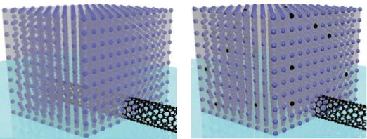
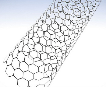
The team modified a simple microfluidic channel with an array of vertically aligned carbon nanotubes — rolled lattices of carbon atoms that resemble tiny tubes of chicken wire. The researchers had previously devised a method for standing carbon nanotubes on their ends, like trees in a forest. With this method, they created a three-dimensional array of permeable carbon nanotubes within a microfluidic device, through which fluid can flow.
Now the researchers have given the nanotube array the ability to trap certain particles. To do this, the team coated the array, layer by layer, with polymers of alternating electric charge.
“You can think of each nanotube in the forest as being concentrically coated with different layers of polymer,” says Brian Wardle, professor of aeronautics and astronautics at MIT. “If you drew it in cross-section, it would be like rings on a tree.”
Depending on the number of layers deposited, the researchers can create thicker or thinner nanotubes and thereby tailor the porosity of the forest to capture larger or smaller particles of interest.
The nanotubes’ polymer coating may also be chemically manipulated to bind specific bioparticles flowing through the forest. To test this idea, the researchers applied an established technique to treat the surface of the nanotubes with antibodies that bind to prostate specific antigen (PSA), a common experimental target. The polymer-coated arrays captured 40 percent more antigens, compared with arrays lacking the polymer coating.
Wardle says the combination of carbon nanotubes and multilayer coatings may help finely tune microfluidic devices to capture extremely small and rare particles, such as certain viruses and proteins.
“There are smaller bioparticles that contain very rich amounts of information that we don’t currently have the ability to access in point-of-care [medical testing] devices like microfluidic chips,” says Wardle, who is a co-author on the paper. “Carbon nanotube arrays could actually be a platform that could target that size of bioparticle.”
The paper’s lead author is Allison Yost, a former graduate student who is currently an engineer at Accion Systems. Others on the paper include graduate student Setareh Shahsavari; postdoc Roberta Polak; School of Engineering Professor of Teaching Innovation Gareth McKinley; professor of materials science and engineering Michael Rubner, and Raymond A. And Helen E. St. Laurent Professor of Chemical Engineering Robert Cohen.
A porous forest
Carbon nanotubes have been a subject of intense scientific study, as they possess exceptional electrical, mechanical, and optical properties. While their use in microfluidics has not been well explored, Wardle says carbon nanotubes are an ideal platform because their properties may be manipulated to attract certain nanometer-sized molecules. Additionally, carbon nanotubes are 99 percent porous, meaning a nanotube is about 1 percent carbon and 99 percent air.
“Which is what you need,” Wardle says. “You need to flow quantities of fluid through this material to shed all the millions of particles you don’t want to find and grab the one you do want to find.”
What’s more, Wardle says, a three-dimensional forest of carbon nanotubes would provide much more surface area on which target molecules may interact, compared with the two-dimensional surfaces in conventional microfluidics.
“The capture efficiency would scale with surface area,” Wardle notes.
A versatile array
The team integrated a three-dimensional array of carbon nanotubes into a microfluidic device by using chemical vapor deposition and photolithography to grow and pattern carbon nanotubes onto silicon wafers. They then grouped the nanotubes into a cylinder-shaped forest, measuring about 50 micrometers tall and 1 millimeter wide, and centered the array within a 3 millimeter-wide, 7-millimeter long microfluidic channel.
The researchers coated the nanotubes in successive layers of alternately charged polymer solutions in order to create distinct, binding layers around each nanotube. To do so, they flowed each solution through the channel and found they were able to create a more uniform coating with a gap between the top of the nanotube forest and the roof of the channel. Such a gap allowed solutions to flow over, then down into the forest, coating each individual nanotube. In the absence of a gap, solutions simply flowed around the forest, coating only the outer nanotubes.
After coating the nanotube array in layers of polymer solution, the researchers demonstrated that the array could be primed to detect a given molecule, by treating it with antibodies that typically bind to prostate specific antigen (PSA). They pumped in a solution containing small amounts of PSA and found that the array captured the antigen effectively, throughout the forest, rather than just on the outer surface of a typical microfluidic element.
Wardle says that the nanotube array is extremely versatile, as the carbon nanotubes may be manipulated mechanically, electrically, and optically, while the polymer coatings may be chemically altered to capture a wide range of particles. He says an immediate target may be biomarkers called exosomes, which are less than 100 nanometers wide and can be important signals of a disease’s progression.
“Science is really picking up on how much information these particles contain, and they’re sort of everywhere, but really hard to find, even with large-scale equipment,” Wardle says. “This type of device actually has all the characteristics and functionality that would allow you to go after bioparticles like exosomes and things that really truly are nanometer scale.”
This research was funded, in part, by the National Science Foundation.
A Natural Light Switch
MIT scientists identify and map the protein behind a light-sensing mechanism.
MIT scientists, working with colleagues in Spain, have discovered and mapped a light-sensing protein that uses vitamin B12 to perform key functions, including gene regulation.
The result, derived from studying proteins from the bacterium Thermus thermophilus, involves at least two findings of broad interest. First, it expands our knowledge of the biological role of vitamin B12, which was already understood to help convert fat into energy, and to be involved in brain formation, but has now been identified as a key part of photoreceptor proteins — the structures that allow organisms to sense and respond to light.
Second, the research describes a new mode of gene regulation, in which the light-sensing proteins play a key role. In so doing, the scientists observe, the bacteria have repurposed existing protein structures that use vitamin B12, and put them to work in new ways.
“Nature borrowed not just the vitamin, but really the whole enzyme unit, and modified it … and made it a light sensor,” says Catherine Drennan, a professor of chemistry and biology at MIT
The paper describes the photoreceptors in three different states: in the dark, bound to DNA, and after being exposed to light.
“It’s wonderful that we’ve been able to get all the series of structures, to understand how it works at each stage,” Drennan says.
The paper has nine co-authors, including Drennan; graduate students Percival Yang-Ting Chen, Marco Jost, and Gyunghoon Kang of MIT; Jesus Fernandez-Zapata and S. Padmanabhan of the Institute of Physical Chemistry Rocasolano, in Madrid; and Monserrat Elias-Arnanz, Juan Manuel Ortiz-Guerreo, and Maria Carmen Polanco, of the University of Murcia, in Murcia, Spain.
The researchers used a combination of X-ray crystallography techniques and in-vitro analysis to study the bacteria. Drennan, who has studied enzymes that employ vitamin B12 since she was a graduate student, emphasizes that key elements of the research were performed by all the co-authors.
Jost performed crystallography to establish the shapes of the structures, while the Spanish researchers, Drennan notes, “did all of the control experiments to show that we were really thinking about this right,” among other things.
By studying the structures of the photoreceptor proteins in their three states, the scientists developed a more thorough understanding of the structures, and their functions, than they would have by viewing the proteins in just one state.
Microbes, like many other organisms, benefit from knowing whether they are in light or darkness. The photoreceptors bind to the DNA in the dark, and prevent activity pertaining to the genes of Thermus thermophilus. When light hits the microbes, the photoreceptor structures cleave and “fall apart,” as Drennan puts it, and the bacteria start producing carotenoids, which protect the organisms from negative effects of sunlight, such as DNA damage.
The research also shows that the exact manner in which the photoreceptors bind to the DNA is novel. The structures contain tetramers, four subunits of the protein, of which exactly three are bound to the genetic material — something Drennan says surprised her.
“That’s the best part about science,” Drennan says. “You see something novel, then you think it’s not really going to be that novel, but you do the experiments [and it is].”
Other scientists say the findings are significant. “It’s a very exciting development,” says Rowena Matthews, a professor emerita of biological chemistry at the University of Michigan, who has read the paper. Of the newly discovered use of vitamin B12 and a derivative of it, adenosylcobalamin, Matthews adds, “There was very limited knowledge of its versatility.”
Drennan adds that in the long run, the finding could have practical applications, such as the engineering of light-directed control of DNA transcription, or the development of controlled interactions between proteins.
“I would be very interested in … thinking about whether there could be practical applications of this,” Drennan says.
HIV Protein Manipulates Hundreds of Human Genes
Findings search for new or improved treatments for patients with AIDS.
UT Southwestern Medical Center researchers have deciphered how a small protein made by the human immunodeficiency virus (HIV) that causes AIDS manipulates human genes to further its deadly agenda.
The findings, published in the online journal eLife, could aid in the search for new or improved treatments for patients with AIDS, or to the development of preventive strategies.
“We have identified the molecular mechanisms by which the Tat protein made by HIV interacts with the host cell to activate or repress several hundred human genes,” said Dr. Iván D’Orso, Assistant Professor of Microbiology at UT Southwestern and senior author of the study. “The findings clearly suggest that blocking Tat activity may be of therapeutic value to HIV patients.”
It has long been known that HIV causes AIDS by hijacking the body’s immune cells, transforming them into HIV factories and killing other immune cells that normally fight disease. HIV also hides in cells and continues to undermine the host’s immune system despite antiretroviral therapy that has improved the outlook of those with AIDS.
The latest data from the Centers for Disease Control and Prevention (CDC), in 2012, estimated 1.2 million Americans were living with HIV, including 156,300 whose infections had not been diagnosed. About 50,000 people in the U.S. are newly infected with HIV annually, the CDC projects. In 2013, the CDC estimated that over 26,000 Americans had the advanced form of HIV infection, AIDS.
Like all retroviruses, HIV has very few genes of its own and must take over the host’s cellular machinery in order to propagate and spread throughout the body. Although the broad aspects of that cellular hijacking were known, the nuances remain to be explored, Dr. D’Orso said.
“We observed that HIV methodically and precisely manipulates the host’s genes and cellular machinery. We also observed that HIV rewires cellular defensive pathways to benefit survival of the virus,” he added.
The study provides insights into HIV’s ability to survive despite antiretroviral therapy, findings that could lead to new therapeutic targets or ways to make current therapies more effective, he said.
“Our study indicates that this small viral protein, Tat, directly binds to about 400 human genes to generate an environment in which HIV can thrive. Then, this protein precisely turns off the body’s immune defense. It is striking that such a small viral protein has such a large impact,” Dr. D’Orso said. “The human genes and pathways that Tat manipulates correlate well with symptoms observed in these patients, such as immune system hyperactivation, then weakening, and accelerated aging,” Dr. D’Orso said, describing the situation in which HIV infection leads to AIDS.
Italy’s National Institute of Health in Rome recently completed a phase II clinical trial of an experimental vaccine that targets the Tat protein. That trial, which followed 87 HIV-positive patients for up to three years, reported that the vaccine was well-tolerated without significant side effects. However, it will take several years to determine if the vaccine works, Dr. D’Orso said.
Although someone can have HIV for years without showing symptoms, AIDS occurs when HIV blocks the body’s ability to fight off illness. The person then becomes overrun by the opportunistic infections and specific cancers that are hallmarks of AIDS.
New Light Shed on Genetic Regulation
A team of scientists has uncovered greater intricacy in protein signaling than was previously understood, shedding new light on the nature of genetic production.
Christine Vogel, an assistant professor in New York University’s Department of Biology and one of the study’s senior authors, explains that “to make a protein, we need to make a messenger RNA molecule from the gene encoded in the DNA, and then, in a second process, make proteins from these RNA molecules. Both processes are highly regulated and coupled.”
This coupling is similar to the coupling between a moving escalator and a person walking on it at the same time.
The research takes a closer look at how the two coupled processes change in the cell responding to an outside stimulus.
“Until recently, it has been very difficult to study these systems and researchers have thought that the movement of the escalator is most important during the cellular response,” Vogel explains. “We now show that is not necessarily the case, and under some circumstances, the person’s walking determines the overall outcome.”
In biology, this means that both of the processes—to make RNAs and proteins—play important roles, but with different patterns.
In their study, the scientists, who also included researchers from National University Singapore and Berlin’s Max Delbruck Center, took a closer look at how the two processes exactly respond over time.
Their results showed notable distinctions between DNA and mRNA in the nature of their signaling. Notably, the process of making RNA from DNA was pulse-like—a brief messaging over the studied period that returned to the normal levels by the end of the measurements. By contrast, the process of making a protein from RNA was akin to an on/off switch: once started, levels remained constant for consistent periods before reverting back to long stretches of dormancy.
While the reasons for these differences in cell behavior remain unknown, the researchers believe the answer may lie in the nature of the two tasks.
“It is very costly for the cell to make proteins, but making RNA messages from DNA is a relatively low-energy and simple process, so it makes sense that we see frequent, or pulsating, signaling at this stage,” observes Vogel. “By contrast, creating proteins is an intricate undertaking, requiring a great deal of time and energy. This may be why, once you decided to stop production of proteins, you do not turn it back on that easily—and the other way around.”
Where Cancer Cells May Begin
Scientists use fruit fly genetics to understand how things could go wrong in cancer.
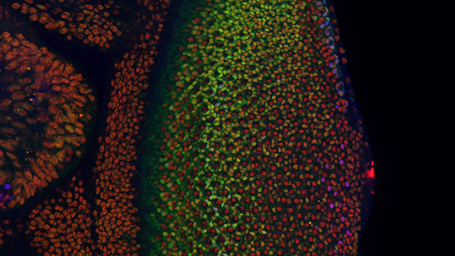
Cancer cells are normal cells that go awry by making bad developmental decisions during their lives. In a study involving the fruit fly equivalent of an oncogene implicated in many human leukemias, Northwestern University researchers have gained insight into how developing cells normally switch to a restricted, or specialized, state and how that process might go wrong in cancer.
The fruit fly’s eye is an intricate pattern of many different specialized cells, such as light-sensing neurons and cone cells. Because flies share with humans many of the same cancer-causing genes, scientists use the precisely made compound eye of Drosophila melanogaster (the common fruit fly) as a workhorse to study what goes wrong in human cancer.
A multidisciplinary team co-led by biologist Richard W. Carthew and engineer Luís A.N. Amaral studied normal cell behavior in the developing eye. The researchers were surprised to discover that the levels of an important protein called Yan start fluctuating wildly when the cell is switching from a more primitive, stem-like state to a more specialized state. If the levels don’t or can’t fluctuate, the cell doesn’t switch and move forward.
“This mad fluctuation, or noise, happens at the time of cell transition,” said Carthew, professor of molecular biosciences in Northwestern’s Weinberg College of Arts and Sciences. “For the first time, we see there is a brief time period as the developing cell goes from point A to point B. The noise is a state of ‘in between’ and is important for cells to switch to a more specialized state. This limbo might be where normal cells take a cancerous path.”
The researchers also found that a molecular signal received by a cell receptor called EGFR is important for turning the noise off. If that signal is not received, the cell remains in an uncontrolled state.
By pinpointing this noise and its “off” switch as important points in the normal process of cell differentiation, the Northwestern researchers provide targets for scientists studying how cells can go out of control and transform into cancer cells.
The “noisy” protein the Northwestern researchers studied is called Yan in the fly and Tel-1 in humans. (The protein is a transcription factor.) The Tel-1 protein instructs cells to turn into white blood cells; the gene that produces the protein, oncogene Tel-1, is frequently mutated in leukemia.
The EGFR protein that turns off the noise in flies is called Her-2 in humans. Her-2 is an oncogene that plays an important role in human breast cancer.
“On the surface, flies and humans are very different, but we share a remarkable amount of infrastructure,” said Carthew, a member of the Robert H. Lurie Comprehensive Cancer Center of Northwestern University. “We can use fruit fly genetics to understand how humans work and how things go wrong in cancer and other diseases.”
Fruit fly cells are small and closely packed together, making study of them challenging. Carthew and Amaral’s team of biologists, chemical and biological engineers, computer scientists and chemists together figured out how to identify and analyze thousands and thousands of individual cells in the flies’ eyes.
“In the past, people have built models of regulatory networks that control cell differentiation mostly by genetically perturbing one or two components of the network at a time and then compiling those results into models,” said Amaral, professor of chemical and biological engineering at the McCormick School of Engineering. “We instead measured the retina as it developed and found the unexpected behavior of the key regulatory factors Yan and EGFR.”
Nicolás Peláez, first author of the study and a Ph.D. candidate in interdisciplinary biological sciences working with Amaral and Carthew, built new tools to study this strange feature of noise in developing flies. His methods enabled the researchers to easily measure both the concentration of the Yan protein and its fluctuation (noise).
It takes 15 to 20 hours for a fruit fly cell to go from being an unrestricted cell to a restricted cell, Carthew said. Peláez determined the Yan protein is noisy, or fluctuating, for six to eight of those hours.
“Studying the dynamics of molecules regulating fly-eye patterning can inform us about human disease,” Peláez said. “Using model organisms such as fruit flies will help us understand quantitatively the basic biological principles governing differentiation in complex animals.”
Mechanism of Tumor Suppressing Gene Uncovered
The most commonly mutated gene in cancer,p53, works to prevent tumor formation by keeping mobile elements in check that otherwise lead to genomic instability, UT Southwestern Medical Center researchers have found.
The p53 gene long has been known to suppress tumor formation, but the mechanisms behind this function – and why disabling the gene allows tumors to form – were not fully understood.
Findings from the study answer some of these questions and could one day lead to new ways of diagnosing and treating cancer, said the study’s senior author, Dr. John Abrams, Professor of Cell Biology at UT Southwestern.
The investigators found that normal p53 gene action restrains transposons, mobile genetic elements called retroelements that can make copies of themselves and move to different positions on chromosomes. But, they discovered, when p53 is disabled by mutation, dramatic eruptions of these mobile elements occur. The study revealed that in mice with cancer and in human samples of two types of cancer (Wilms’ tumors and colon tumors) disabled for p53, transposons became very active.
In a healthy state, certain mechanisms work to keep these retroelements quiet and inactive, explained Dr. Abrams. One of those mechanisms is p53 action. Conversely, when p53 is mutated, retroelements can erupt.
“If you take the gene away, transposons can wreak havoc throughout the genome by causing it to become highly dysregulated, which can lead to disease,” Dr. Abrams said. “Our findings help explain why cancer genomes are so much more fluid and destabilized than normal genomes. They also provide a novel framework for understanding how normal cells become tumors.”
Although much more research is needed, Dr. Abrams said, the potential clinical implications of the team’s findings are significant.
“Understanding how p53 prevents tumors raises the prospect of therapeutic interventions to correct cases in which p53 is disabled,” Dr. Abrams said. “If retroelements are at the heart of certain p53-driven cancers, finding ways to suppress them could potentially allow us to prevent those cancers or intervene to keep them from progressing.”
This understanding also could lead to advances in diagnosing some cancers through biomarkers related to p53 and transposon activity.
“One possibility is that perhaps blood or urine tests could detect dysregulated retroelements that could be indicative of certain types of cancer,” Dr. Abrams said.
N3xt generation carbon nanotubes, Volume 2 (Volume Two: Latest in Genomics Methodologies for Therapeutics: Gene Editing, NGS and BioInformatics, Simulations and the Genome Ontology), Part 1: Next Generation Sequencing (NGS)
N3xt generation carbon nanotubes
Larry H. Bernstein, MD, FCAP, Curator
LPBI
Skyscraper-style carbon-nanotube chip design ‘boosts electronic performance by factor of a thousand’
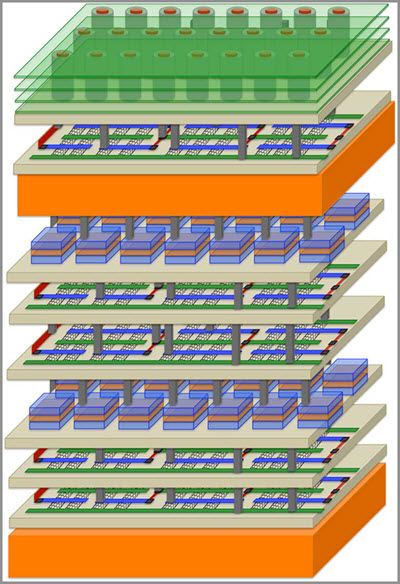
A new revolutionary high-rise architecture for computing (credit: Stanford University)
Researchers at Stanford and three other universities are creating a revolutionary new skyscraper-like high-rise architecture for computing based on carbon nanotube materials instead of silicon.
In Rebooting Computing, a special issue (in press) of the IEEE Computer journal, the team describes its new approach as “Nano-Engineered Computing Systems Technology,” or N3XT.
Suburban-style chip layouts create long commutes and regular traffic jams in electronic circuits, wasting time and energy, they note.
N3XT will break data bottlenecks by integrating processors and memory-like floors in a skyscraper and by connecting these components with millions of “vias,” which play the role of tiny electronic elevators.
The N3XT high-rise approach will move more data, much faster, using far less energy, than would be possible using low-rise circuits, according to the researchers.
Stanford researchers including Associate Professor Subhasish Mitra and Professor H.-S. Philip Wong have “assembled a group of top thinkers and advanced technologies to create a platform that can meet the computing demands of the future,” Mitra says.
“When you combine higher speed with lower energy use, N3XT systems outperform conventional approaches by a factor of a thousand,” Wong claims.
Carbon nanotube transistors
Engineers have previously tried to stack silicon chips but with limited success, the researchers suggest. Fabricating a silicon chip requires temperatures close to 1,800 degrees Fahrenheit, making it extremely challenging to build a silicon chip atop another without damaging the first layer. The current approach to what are called 3-D, or stacked, chips is to construct two silicon chips separately, then stack them and connect them with a few thousand wires.
But conventional 3-D silicon chips are still prone to traffic jams and it takes a lot of energy to push data through what are a relatively few connecting wires.
The N3XT team is taking a radically different approach: building layers of processors and memory directly atop one another, connected by millions of vias that can move more data over shorter distances that traditional wire, using less energy, and immersing computation and memory storage into an electronic super-device.
The key is the use of non-silicon materials that can be fabricated at much lower temperatures than silicon, so that processors can be built on top of memory without the new layer damaging the layer below. As in IBM’s recent chip breakthrough (see “Method to replace silicon with carbon nanotubes developed by IBM Research“), N3XT chips are based on carbon nanotube transistors.
Transistors are fundamental units of a computer processor, the tiny on-off switches that create digital zeroes and ones. CNTs are faster and more energy-efficient than silicon processors, and much thinner. Moreover, in the N3XT architecture, they can be fabricated and placed over and below other layers of memory.
Team members also envision using data storage technologies that rely on materials other than silicon. This would allow for the new materials to be manufactured on top of CNTs, using low-temperature fabrication processes.
One such data storage technology is called resistive random-access memory, or RRAM (see “‘Memristors’ based on transparent electronics offer technology of the future“). Resistance slows down electrons, creating a zero, while conductivity allows electrons to flow, creating a one. Tiny jolts of electricity switch RRAM memory cells between these two digital states. N3XT team members are also experimenting with a variety of nanoscale magnetic storage materials.
Just as skyscrapers have ventilation systems, N3XT high-rise chip designs incorporate thermal cooling layers. This work, led by Stanford mechanical engineers Kenneth Goodson and Mehdi Asheghi, ensures that the heat rising from the stacked layers of electronics does not degrade overall system performance.
Mitra and Wong have already demonstrated a working prototype of a high-rise chip. At the International Electron Devices Meeting in December 2014 they unveiled a four-layered chip made up of two layers of RRAM memory sandwiched between two layers of CNTs (see “Stanford engineers invent radical ‘high-rise’ 3D chips“).
In their N3XT paper they ran simulations showing how their high-rise approach was a thousand times more efficient in carrying out many important and highly demanding industrial software applications.
references:
M. Aly, M. Gao, G. Hills, C-S Lee, G. Pitner, M. Shulaker, T. Wu, M. Asheghi, J. Bokor, F. Franchetti, K. Goodson, C. Kozyrakis, I. Markov, K. Olukotun, L. Pileggi, E. Pop, J. Rabaey, C. Ré, H.-S.P. Wong and S. Mitra, Energy-Efficient Abundant-Data Computing: The N3XT 1,000X. IEEE Computer, Special Issue on Rebooting Computing, Dec. 2015 (in press)
This is a little off-thread, but it does involve carbon, so this is the only place I can squeeze this prediction in…
The big news tonight is that the lithium batteries in electric skate boards called “hoverboards” are catching on fire.
This is going to take a lot of them off of the market. But…they will rise again when carbon batteries with graphene and CNTs and boron nitride and I don’t know what else make batteries run as cool as these high-rise CNT chips.
In fact, these high-rise CNT chips will most likely be part of the power control and distribution in the N3XT generation of hoverboards.
I am really curious what will be the next computing paradigm. Personally, I see more than one type of computers being used at the same time : neuromorphic, optical, electronic von Neumann nanotube transistors 3D chips, quantum for some purposes and also magnetic storage (memristors).
What is even more interesting, Mr Kurzweil wrote and said that nanotube high-rise computers like described in the article here will be dominant type of computers in the 20s. It may or may not be true.
Oh for sure, Kynareth, the first von Neumann machines will do their work with these processors. But memristors should also be a part of them.
I suspect that high-rise cnt chips will indeed be dominant as soon as they come to market. This will give you the N3XT “Field of Dreams.”
If you build it they will come. As soon as it is produced in mass quantities, everybody will come and beat down the doors to get them.
This is fantastic. I want the next, or the third next gen of this to be a molecularly built up block of circuitry. This article shows advances using the nano-tube connector that has been experimented before, and also the replacement overall in the circuitry. I only suggest that further generation because it resembles the “molybloc circuits”-bricks and tiles of densely packed circuitry, molybloc standing for molecular block circuitry, found in the Honor Harringoton Sci Fi series (David Weber).
Stanford engineers invent radical ‘high-rise’ 3D chips
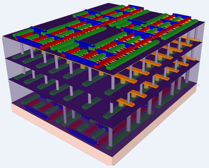
A four-layer prototype high-rise chip built by Stanford engineers. The bottom and top layers are logic transistors. Sandwiched between them are two layers of memory. The vertical tubes are nanoscale electronic “elevators” that connect logic and memory, allowing them to work together efficiently. (Credit: Max Shulaker, Stanford)
Stanford engineers have build 3D “high-rise” chips that could leapfrog the performance of the single-story logic and memory chips on today’s circuit cards, which are subject to frequent traffic jams between logic and memory.
The Stanford approach would attempt to end these jams by building layers of logic atop layers of memory to create a tightly interconnected high-rise chip. Many thousands of nanoscale electronic “elevators” would move data between the layers much faster, using less electricity, than the bottleneck-prone wires connecting single-story logic and memory chips today.
The work is led by Subhasish Mitra, a Stanford associate professor of electrical engineering and of computer science, and H.-S. Philip Wong, the Williard R. and Inez Kerr Bell Professor in Stanford’s School of Engineering. They describe their new high-rise chip architecture in a paper being presented at the IEEE International Electron Devices Meeting Dec. 15–17.
The researchers’ innovation leverages three breakthroughs: a new technology for creating transistors using nanotubes, a new type of computer memory that lends itself to multi-story fabrication, and a technique to build these new logic and memory technologies into high-rise structures in a radically different way than previous efforts to stack chips.
“This research is at an early stage, but our design and fabrication techniques are scalable,” Mitra said. “With further development this architecture could lead to computing performance that is much, much greater than anything available today.” Wong said the prototype chip unveiled at IEDM shows how to put logic and memory together into three-dimensional structures that can be mass-produced.
“Paradigm shift is an overused concept, but here it is appropriate,” Wong said. “With this new architecture, electronics manufacturers could put the power of a supercomputer in your hand.”
Overcoming silicon heat
Researchers have been trying to solve a major problem with chip-generated heat by creating carbon nanotubes (CNT) transistors. Mitra and Wong are presenting a second paper at the conference showing how their team made some of the highest performance CNT transistors ever built.
Image of a CNT-based field-effect transistor (FET) using a new high-density process (credit: Mitra/Wong Lab, Stanford)
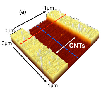
Until now the standard process used to grow CNTs did not create sufficient density. The Stanford engineers solved this problem an ingenious technique. They started by growing CNTs the standard way, on round quartz wafers. Then they created a metal film that acts like a tape. Using this adhesive process, they lifted an entire crop of CNTs off the quartz growth medium and placed it onto a silicon wafer that would become the foundation of their high-rise chip.
They repeated this process 13 times, achieving some of the highest density, highest performance CNTs ever made. Moreover, the Stanford team showed that they could perform this technique on more than one layer of logic as they created their high-rise chip.
RRAM memory
Left: Today’s single-story electronic circuit cards, where logic and memory chips exist as separate structures connected by wires, can get jammed with digital traffic between logic and memory. Right: layers of logic and memory create skyscraper chips where data would move up and down on nanoscale “elevators” to avoid traffic jams. (Credit: Mitra/Wong Lab, Stanford)
Wong is a world leader in a new memory technology called “resistive random access memory” (RRAM) which he unveiled at last year’s IEDM conference.
Unlike today’s memory chips, this new storage technology is not based on silicon, but titanium nitride, hafnium oxide and platinum. This formed a metal/oxide/metal sandwich. Applying electricity to this three-metal sandwich one way causes it to resist the flow of electricity. Reversing the polarity causes the structure to conduct electricity again.
The change from resistive to conductive states is how this new memory technology creates digital zeroes and ones.
RRAM uses less energy than current memory, leading to prolonged battery life in mobile devices. Inventing this new memory technology was also the key to creating the high-rise chip because RRAM can be made at much lower temperatures than silicon memory.
Interconnected layers
Max Shulaker and Tony Wu, Stanford graduate students in electrical engineering, created the techniques behind the four-story high-rise chip unveiled at the conference.
The low-heat process for making RRAM and CNTs enabled them to fabricate each layer of memory directly atop each layer of CNT logic. While making each memory layer, they were able to drill thousands of interconnections into the logic layer below. This multiplicity of connections is what enables the high-rise chip to avoid the traffic jams on conventional circuit cards.
There is no way to tightly interconnect layers using today’s conventional silicon-based logic and memory. That’s because it takes so much heat to build a layer of silicon memory — about 1,000 degrees Celsius — that any attempt to do so would melt the logic below.
Previous efforts to stack silicon chips could save space but not avoid the digital traffic jams. That’s because each layer would have to be built separately and connected by wires — which would still be prone to traffic jams, unlike the nanoscale elevators in the Stanford design.
‘Memristors’ based on transparent electronics offer technology of the future
Memristors are faster, smaller, and use less power than non-volatile flash memory
Transparent electronics (pioneered at Oregon State University) may find one of their newest applications as a next-generation replacement for some uses of non-volatile flash memory, a multi-billion dollar technology nearing its limit of small size and information storage capacity.
Researchers at OSU have confirmed that zinc tin oxide, an inexpensive and environmentally benign compound, could provide a new, transparent technology where computer memory is based on resistance, instead of an electron charge.
This resistive random access memory, or RRAM, is referred to by some researchers as a “memristor.” Products using this approach could become even smaller, faster and cheaper than the silicon transistors that have revolutionized modern electronics — and transparent as well.
Transparent electronics offer potential for innovative products that don’t yet exist, like information displayed on an automobile windshield, or surfing the web on the glass top of a coffee table.
“Flash memory has taken us a long way with its very small size and low price,” said John Conley, a professor in the OSU School of Electrical Engineering and Computer Science. “But it’s nearing the end of its potential, and memristors are a leading candidate to continue performance improvements.”
Memristors: faster than flash
Memristors have a simple structure, are able to program and erase information rapidly, and consume little power. They accomplish a function similar to transistor-based flash memory, but with a different approach. Whereas traditional flash memory stores information with an electrical charge, RRAM accomplishes this with electrical resistance. Like flash, it can store information as long as it’s needed.
Flash memory computer chips are ubiquitous in almost all modern electronic products, ranging from cell phones and computers to video games and flat panel televisions.
Thin-film transistors that control liquid crystal displays
Some of the best opportunities for these new amorphous oxide semiconductors are not so much for memory chips, but with thin-film, flat panel displays, researchers say. Private industry has already shown considerable interest in using them for the thin-film transistors that control liquid crystal displays, and one compound approaching commercialization is indium gallium zinc oxide.
But indium and gallium are getting increasingly expensive, and zinc tin oxide — also a transparent compound — appears to offer good performance with lower cost materials. The new research also shows that zinc tin oxide can be used not only for thin-film transistors, but also for memristive memory, Conley said, an important factor in its commercial application.
More work is needed to understand the basic physics and electrical properties of the new compounds, researchers said.
This research was supported by the U.S. Office of Naval Research, the National Science Foundation and the Oregon Nanoscience and Microtechnologies Institute.
Bipolar resistive switching is demonstrated in the amorphous oxide semiconductor zinc–tin-oxide (ZTO). A gradual forming process produces improved switching uniformity. Al/ZTO/Pt crossbar devices show switching ratios greater than 103, long retention times, and good endurance. The resistive switching in these devices is consistent with a combined filamentary/interfacial mechanism. Overall, ZTO shows great potential as a low cost material for embedding memristive memory with thin film transistor logic for large area electronics.
Highlights
► We present the first report of resistive switching in zinc–tin-oxide (ZTO). ► ZTO is the leading alternative material to IGZO for TFTs for LCDs. ► ZTO has an advantage over IGZO of lower cost due to the absence of In and Ga. ► Al/ZTO/Pt crossbar RRAM devices show switching ratios greater than 103. ► ZTO shows promise for embedding RRAM with TFT logic for large area electronics.
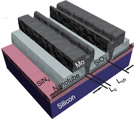
Schematic of a set of molybdenum (M0) end-contacted nanotube transistors (credit: Qing Cao et al./Science)
IBM Research has announced a “major engineering breakthrough” that could lead to carbon nanotubes replacing silicon transistors in future computing technologies.
As transistors shrink in size, electrical resistance increases within the contacts, which impedes performance. So IBM researchers invented a metallurgical process similar to microscopic welding that chemically binds the contact’s metal (molybdenum) atoms to the carbon atoms at the ends of nanotubes.
The new method promises to shrink transistor contacts without reducing performance of carbon-nanotube devices, opening a pathway to dramatically faster, smaller, and more powerful computer chips beyond the capabilities of traditional silicon semiconductors.
“This is the kind of breakthrough that we’re committed to making at IBM Research via our $3 billion investment over 5 years in research and development programs aimed a pushing the limits of chip technology,” said Dario Gil, VP, Science & Technology, IBM Research. “Our aim is to help IBM produce high-performance systems capable of handling the extreme demands of new data analytics and cognitive computing applications.”
The development was reported today in the October 2 issue of the journal Science.
Schematic of carbon nanotube transistor contacts. Left: High-resistance side-bonded contact, where the single-wall nanotube (SWNT) (black tube) is partially covered by the metal molybdenum (Mo) (purple dots). Right: low-resistance end-bonded contact, where the SWNT is attached to the molybdenum electrode through carbide bonds, while the carbon atoms (black dots) from the originally covered portion of the SWNT uniformly diffuse out into the Mo electrode (credit: Qing Cao et al./Science)
The new “end-bonded contact scheme” allows carbon-nanotube contacts to be shrunken down to below 10 nanometers without deteriorating performance. IBM says the scheme could overcome contact resistance challenges all the way to the 1.8 nanometer node and replace silicon with carbon nanotubes.
Silicon transistors have been made smaller year after year, but they are approaching a point of physical limitation. With Moore’s Law running out of steam, shrinking the size of the transistor — including the channels and contacts — without compromising performance has been a challenge for researchers for decades.
IBM has previously shown that carbon nanotube transistors can operate as excellent switches at channel dimensions of less than ten nanometers, which is less than half the size of today’s leading silicon technology. Electrons in carbon transistors can move more easily than in silicon-based devices and use less power.
Carbon nanotubes are also flexible and transparent, making them useful for flexible and stretchable electronics or sensors embedded in wearables.
IBM acknowledges that several major manufacturing challenges still stand in the way of commercial devices based on nanotube transistors.
Moving beyond the limits of silicon transistors requires both a high-performance channel and high-quality electrical contacts. Carbon nanotubes provide high-performance channels below 10 nanometers, but as with silicon, the increase in contact resistance with decreasing size becomes a major performance roadblock. We report a single-walled carbon nanotube (SWNT) transistor technology with an end-bonded contact scheme that leads to size-independent contact resistance to overcome the scaling limits of conventional side-bonded or planar contact schemes. A high-performance SWNT transistor was fabricated with a sub–10-nanometer contact length, showing a device resistance below 36 kilohms and on-current above 15 microampere per tube. The p-type end-bonded contact, formed through the reaction of molybdenum with the SWNT to form carbide, also exhibited no Schottky barrier. This strategy promises high-performance SWNT transistors, enabling future ultimately scaled device technologies.
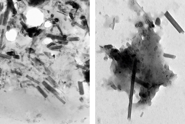
Carbon nanotubes (rods) and nanoparticles (black clumps) found inside a lung cell vacuole (left) are similar to those found in vehicle exhaust in tailpipes of cars in Paris (right) (credit: Fathi Moussa/Paris-Saclay University)
Carbon nanotubes (CNTs) have been found in cells extracted from the airways of Parisian children under routine treatment for asthma, according to a report in the journal EBioMedicine (open access) by scientists in France and atRice University.
The cells were taken from 69 randomly selected asthma patients aged 2 to 17 who underwent routine fiber-optic bronchoscopies as part of their treatment. The researchers analyzed particulate matter found in the alveolar macrophage cells (also known as dust cells), which help stop foreign materials like particles and bacteria from entering the lungs.
The study partially answers the question of what makes up the black material inside alveolar macrophages, the original focus of the study. The researchers found single-walled and multiwalled carbon nanotubes and amorphous carbon among the cells.
The nanotube aggregates in the cells ranged in size from 10 to 60 nanometers in diameter and up to several hundred nanometers in length, small enough that optical microscopes would not have been able to identify them in samples from former patients. The new study used more sophisticated tools, including high-resolution transmission electron microscopy, X-ray spectroscopy, Raman spectroscopy, and near-infrared fluorescence microscopy to definitively identify them in the cells and in the environmental samples.
“The concentrations of nanotubes are so low in these samples that it’s hard to believe they would cause asthma, but you never know,” said Rice chemist Lon Wilson, a corresponding author of the paper. “What surprised me the most was that carbon nanotubes were the major component of the carbonaceous pollution we found in the samples.”
The study notes but does not make definitive conclusions about the controversial proposition that carbon nanotube fibers may act like asbestos, a proven carcinogen. But the authors did note that “long carbon nanotubes and large aggregates of short ones can induce a granulomatous (inflammation) reaction.”
The researchers also suggested previous studies that link the carbon content of airway macrophages and the decline of lung function should be reconsidered in light of the new findings. The researchers also suggested that the large surface areas of nanotubes and their ability to adhere to substances may make them effective carriers for other pollutants.
Carbon nanotubes from forest fires and cars?
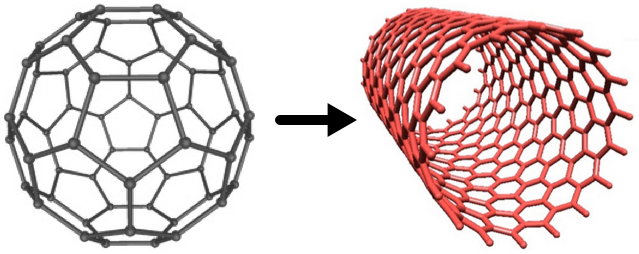
Fullerenes (left) can be converted to carbon nanotubes (right) with a catalytic process, according to Rice chemists (credits: Soroush83/CC and Matías Soto/Rice University)
However, similar nanotubes have been found in samples from the exhaust pipes of Paris vehicles, in dust gathered from various places around the city, in spider webs in India, and even in ice cores, the paper notes.
“We know that carbon nanoparticles are found in nature,” Wilson said, noting that round fullerene (C60) molecules are commonly produced by volcanoes, forest fires, and other combustion of carbon materials. “All you need is a little catalysis to make carbon nanotubes instead of fullerenes.”
A car’s catalytic converter, which turns toxic carbon monoxide into safer emissions, bears at least a passing resemblance to the Rice-invented high-pressure carbon monoxide, or HiPco, process to make carbon nanotubes, he said. “So it is not a big surprise, when you think about it,” Wilson said.
“Based on our discovery of CNTs in tailpipes, we propose that the catalytic converters of the automobiles are manufacturing carbon nanotubes, Wilson told KurzweilAI. “However, we have not actually proven that.”
We are all carbon-nanotube bearers now
For ethical reasons, no cells from healthy patients were analyzed, but because nanotubes were found in all of the samples, the study led the researchers to conclude that carbon nanotubes are likely to be found in everybody.
“It’s kind of ironic. In our laboratory, working with carbon nanotubes, we wear facemasks to prevent exactly what we’re seeing in these samples, yet everyone walking around out there in the world probably has at least a small concentration of carbon nanotubes in their lungs,” he said.
The study followed one released by Rice and Baylor College of Medicine earlier this month with the similar goal ofanalyzing the black substance found in the lungs of smokers who died of emphysema. That study found carbon black nanoparticles that were the product of the incomplete combustion of such organic material as tobacco.
Co-authors are from Paris-Saclay University, the Paediatric Pulmonology and Allergy Center and the Department of Anatomo-Pathology of the Groupe hospitalier La Roche-Guyon, and Paris Diderot University. The Welch Foundation partially supported the research.
Abstract of Anthropogenic Carbon Nanotubes Found in the Airways of Parisian Children
Compelling evidence shows that fine particulate matters (PM) from air pollution penetrate lower airways and are associated with adverse health effects even within concentrations below those recommended by the WHO. A paper reported a dose-dependent link between carbon content in alveolar macrophages (assessed only by optical microscopy) and the decline in lung function. However, to the best of our knowledge, PM had never been accurately characterized inside human lung cells and the most responsible components of the particulate mix are still unknown. On another hand carbon nanotubes (CNTs) from natural and anthropogenic sources might be an important component of PM in both indoor and outdoor air.
We used high-resolution transmission electron microscopy and energy dispersive X-ray spectroscopy to characterize PM present in broncho-alveolar lavage-fluids (n = 64) and inside lung cells (n = 5 patients) of asthmatic children. We show that inhaled PM mostly consist of CNTs. These CNTs are present in all examined samples and they are similar to those we found in dusts and vehicle exhausts collected in Paris, as well as to those previously characterized in ambient air in the USA, in spider webs in India, and in ice core. These results strongly suggest that humans are routinely exposed to CNTs.
Mesothelin: An early detection biomarker for cancer (By Jack Andraka)
Author/ Curator: Tilda Barliya PhD
Article 10.1. Mesothelin: An early detection biomarker for cancer (By Jack Andraka)
I was recently amazed to read about a young teen who scooped the headlines with his story: Jack Andraka created an early detection test for pancreatic cancer (PC) (1). While we extensively discussed pancreatic cancer in previous posts (1b), this one deserve it’s on attention.
Andraka tells the audience about his journey from learning about a the family member diagnosed with PC, to a flash insight while learning about carbon nanotubes during a biology class, through the screening and finding one protein out of thousands and all the way up his final discovery. His journey wasn’t easy to say the least, he story though deserve all the applause.
Starting with his journey, Andraka began by “looking for a protein in the bloodstream that would be a biomarker for pancreatic cancer, one that would be found in all cases, even in the earliest stages”. He finally narrowed it down to the one that could work – Mesothelin.
So what is mesothelin?
Gubbels JA, et al. Mol. Cancer (2006). Model for peritoneal metastasis of ovarian tumors.
Mesothelin is a 4o kDa secreted protein expressed in normal mesothelial cells and over-expressed in several human tumors including mesothelioma, ovarian and pancreatic adenocarcinoma (2,3). Although the full mechanism by which mesothelin work is still unsolved, it is postulated thought, that mesothelin growth and apoptosis of pancreatic cancer cells by a p53 -dependent and independent pathways (7).
Andraka’s method:
human mesothelin-specific antibodies were mixed with single walled carbon nanotubes and used to coat strips of ordinary filter paper. This made the paper conductive. The optimal layering was determined using a scanning electron microscope. Cell media spiked with varying amounts of mesothelin was then tested against the paper biosensor and any change in the electrical potential of the sensor strip (due to the changing conductivity of the nanotubes) was measured, before and after each application.
The antibodies would bind to the mesothelin and enlarge. These beefed-up molecules would spread the nanotubes farther apart, changing the electrical properties of the network: The more mesothelin present, the more antibodies would bind and grow big, and the weaker the electrical signal would become.
A dose-response curve was constructed with an R2 value of .9992. Tests on human blood serum obtained from both healthy people and patients with chronic pancreatities, pancreatic intraepithelial neoplasia (a precursor to pancreatic carcinoma), or pancreatic cancer showed a similar response. The sensor’s limit of detection sensitivity was found to be 0.156 ng/mL; 10 ng/mL is considered the level of overexpression of mesothelin consistent with pancreatic cancer. Andraka’s sensor costs $0.03 (to compare to a $800 cost of a standard test) and 10 tests can be performed per strip, taking 5 minutes each. The method is 168 times faster, 26,667 times less expensive, and 400 times more sensitive than ELISA, and 25% to 50% more accurate than the CA19-9 test (5).
More so, Wang K and colleagues showed that inhibition of mesothelin may be used as novel strategy for targeting cancer cells (6). The authors showed that silencing the MSLN gene, encoding for mesothelin, inhibits cell proliferation and invasion. While this work is very impressive, the authors haven’t evaluated the potential use these siRNA in animal studies.
In summary:
It is very exiting to know that we may now have a simple and cheap blood test that has the huge potential to save many lives. All we need to do now is to conduct a multinational large scale screening for potential patients.
Andraka on his part is very hopeful, he believes “it could potentially be used to test for ovarian and lung cancer too. And by switching out the protein the test reacts to, it could — down the road — be used for diseases as varied as heart disease and HIV/AIDS”.
4. Argani P, Iacobuzio-Donahue C, Ryu B, Rosty C, Goggins M, Wilentz RE, Murugesan SR, Leach SD, Jaffee E, Yeo CJ, Cameron JL, Kern SE and Hruban RH. Mesothelin is overexpressed in the vast majority of ductal adenocarcinomas of the pancreas: identification of a new pancreatic cancer marker by serial analysis of gene expression (SAGE). Clin Cancer Res. 2001 Dec;7(12):3862-3868. http://clincancerres.aacrjournals.org/content/7/12/3862.long
6. Wang K, Bodempudi V, Liu Z, Borrego-Diaz E, Yamoutpoor F, et al. (2012) Inhibition of Mesothelin as a Novel Strategy for Targeting Cancer Cells. PLoS ONE 7(4): e33214. doi:10.1371/journal.pone.0033214. http://www.plosone.org/article/info:doi/10.1371/journal.pone.0033214
7. Zheng C, Jia W, Tang Y, Zhao HL, Jiang Y and Sun S. Mesothelin regulates growth and apoptosis in pancreatic cancer cells through p53-dependent and -independent signal pathway. Journal of Experimental & Clinical Cancer Research 2012, 31:84. http://www.jeccr.com/content/pdf/1756-9966-31-84.pdf
Other related articles on this open Access Online Scientific Journal, include the following:
I. Pancreatic cancer genomes: Axon guidance pathway genes – aberrations revealed.


