Feeds:
Posts
Comments

Archive for the ‘Pulmonary pathology’ Category

Responses to the #COVID-19 outbreak from Oncologists, Cancer Societies and the NCI: Important information for cancer patients

Curator: Stephen J. Williams, Ph.D.

UPDATED 3/20/2020

Among the people who are identified at risk of coronovirus 2019 infection and complications of the virus include cancer patients undergoing chemotherapy, who in general, can be immunosuppressed, especially while patients are undergoing their treatment.  This has created anxiety among many cancer patients as well as their care givers and prompted many oncologist professional groups, cancer societies, and cancer centers to formulate some sort of guidelines for both the cancer patients and the oncology professional with respect to limiting the risk of infection to coronavirus (COVID19). 


This information will be periodically updated and we are working to get a Live Twitter Feed to bring oncologist and cancer patient advocacy groups together so up to date information can be communicated rapidly.  Please see this page regularly for updates as new information is curated.

IN ADDITION, I will curate a listing of drugs with adverse events of immunosuppression for people who might wonder if the medications they are taking are raising their risk of infections.

Please also see @pharma_BI for updates as well.

Please also see our Coronavirus Portal at https://pharmaceuticalintelligence.com/coronavirus-portal/

For ease of reading information for patients are BOLDED and in RED

ASCO’s Response to COVID-19

From the Cancer Letter: The following is a guest editorial by American Society of Clinical Oncology (ASCO) Executive Vice President and Chief Medical Officer Richard L. Schilsky MD, FACP, FSCT, FASCO. This story is part of The Cancer Letter’s ongoing coverage of COVID-19’s impact on oncology. A full list of our coverage, as well as the latest meeting cancellations, is available here.


The worldwide spread of the coronavirus (COVID-19) presents unprecedented challenges to the cancer care delivery system.

Our patients are already dealing with a life-threatening illness and are particularly vulnerable to this viral infection, which can be even more deadly for them. Further, as restrictions in daily movement and social distancing take hold, vulnerable patients may be disconnected from friends, family or other support they need as they manage their cancer.

As providers, we rely on evidence and experience when treating patients but now we face uncertainty. There are limited data to guide us in the specific management of cancer patients confronting COVID-19 and, at present, we have no population-level guidance regarding acceptable or appropriate adjustments of treatment and practice operations that both ensure the best outcome for our patients and protect the safety of our colleagues and staff.

As normal life is dramatically changed, we are all feeling anxious about the extreme economic challenges we face, but these issues are perhaps even more difficult for our patients, many of whom are now facing interruption

As we confront this extraordinary situation, the health and safety of members, staff, and individuals with cancer—in fact, the entire cancer community—is ASCO’s highest priority.

ASCO has been actively monitoring and responding to the pandemic to ensure that accurate information is readily available to clinicians and their patients. Recognizing that this is a rapidly evolving situation and that limited oncology-specific, evidence-based information is available, we are committed to sharing what is known and acknowledging what is unknown so that the most informed decisions can be made.

To help guide oncology professionals as they deal with the impact of coronavirus on both their patients and staff, ASCO has collated questions from its members, posted responses at asco.org and assembled a compendium of additional resources we hope will be helpful as the virus spreads and the disease unfolds. We continue to receive additional questions regarding clinical care and we are updating our FAQs on a regular basis.

We hope this information is helpful even when it merely confirms that there are no certain answers to many questions. Our answers are based on the best available information we identify in the literature, guidance from public health authorities, and input received from oncology and infectious disease experts.

For patients, we have posted a blog by Dr. Merry Jennifer Markham, chair of ASCO’s Cancer Communications Committee. This can be found on Cancer.Net, ASCO’s patient information website, and it provides practical guidance to help patients reduce their risk of exposure, better understand COVID-19 symptoms, and locate additional information.

This blog is available both in English and Spanish. Additional blog posts addressing patient questions will be posted as new questions are received and new information becomes available.

Find below a Tweet from Dr.Markham which includes links to her article on COVID-19 for cancer patients

https://twitter.com/DrMarkham/status/1237797251038220289?s=20

NCCN’s Response to COVID-19 and COVID-19 Resources

JNCCN: How to Manage Cancer Care during COVID-19 Pandemic

Experts from the Seattle Cancer Care Alliance (SCCA)—a Member Institution of the National Comprehensive Cancer Network® (NCCN®)—are sharing insights and advice on how to continue providing optimal cancer care during the novel coronavirus (COVID-19) pandemic. SCCA includes the Fred Hutchinson Cancer Research Center and the University of Washington, which are located in the epicenter of the COVID-19 outbreak in the United States. The peer-reviewed article sharing best practices is available for free online-ahead-of-print via open access at JNCCN.org.

Coronavirus disease 2019 (COVID-19) Resources for the Cancer Care Community

NCCN recognizes the rapidly changing medical information relating to COVID-19 in the oncology ecosystem, but understands that a forum for sharing best practices and specific institutional responses may be helpful to others.  Therefore, we are expeditiously providing documents and recommendations developed by NCCN Member Institutions or Guideline Panels as resources for oncology care providers. These resources have not been developed or reviewed by the standard NCCN processes, and are provided for information purposes only. We will post more resources as they become available so check back for additional updates.

Documents

Links

National Cancer Institute Response to COVID-19

More information at https://www.cancer.gov/contact/emergency-preparedness/coronavirus

What people with cancer should know: https://www.cancer.gov/coronavirus

Get the latest public health information from CDC: https://www.coronavirus.gov

Get the latest research information from NIH: https://www.nih.gov/coronavirus


Coronavirus: What People with Cancer Should Know

ON THIS PAGE

Both the resources at cancer.gov (NCI) as well as the resources from ASCO are updated as new information is evaluated and more guidelines are formulated by members of the oncologist and cancer care community and are excellent resources for those living with cancer, and also those who either care for cancer patients or their family and relatives.

Related Resources for Patients (please click on links)


Some resources and information for cancer patients from Twitter

Twitter feeds which may be useful sources of discussion and for cancer patients include:


@OncLive OncLive.com includes healthcare information for patients and includes videos and newsletters


@DrMarkham Dr. Markham is Chief of Heme-Onc & gyn med onc @UF | AD Med Affairs @UFHealthCancer and has collected very good information for patients concerning #Covid19 


@DrMaurieMarkman Dr. Maurie Markman is President of Medicine and Science (Cancer Centers of America, Philadelphia) @CancerCenter #TreatThePerson #Oncology #Genomics #PrecisionMedicine and hosts a great online live Tweet feed discussing current topics in cancer treatment and care for patients called #TreatThePerson Chat

UPDATED 3/20/2020 INFORMATION FROM NCI DESIGNATED CANCER CENTERS FOR PATIENTS/PROVIDERS

The following is a listing with links of NCI Designated Comprehensive Cancer Centers and some select designated Cancer Centers* which have information on infectious risk guidance for cancer patients as well as their physicians and caregivers.   There are 51 NCI Comprehensive Cancer Centers and as more cancer centers formulate guidance this list will be updated. 


Cancer Center State Link to COVID19 guidance
City of Hope CA Advice for cancer patients, survivors and caregivers
Jonsson Cancer Center at UCLA CA Cancer and COVID19
UCSF Hellen Diller Family Comprehensive Cancer CA COVID-19 Links for Patients and Providers
Lee Moffit FL Protecting against Coronavirus 19
University of Kansas Cancer Center* KS COVID19 Info for patients
Barbara & Karmanos Cancer Institute (Wayne State) MI COVID19 Resources
Rogel Cancer Center (Univ of Michigan) MI COVID19 Patient Specific Guidelines
Alvin J. Siteman Cancer Center (MO) Coronavirus
Fred & Pamela Buffet CC* NE Resources for Patients and Providers
Rutgers Cancer Institute of NJ NJ What patients should know about COVID19
Memorial Sloan Kettering NY What COVID19 means for cancer patients
Herbert Irving CC (Columbia University) NY Coronavirus Resource Center
MD Anderson Cancer  TX Planning for Patients, Providers
Hunstman Cancer Center UT COVID19 What you need to know
Fred Hutchinson WA COVID19 What patients need to know


Please also see related information on Coronavirus 2019 and Cancer and Immunotherapy at the following links on the Open Access Online Journal:

Volume Two: Cancer Therapies: Metabolic, Genomics, Interventional, Immunotherapy and Nanotechnology in Therapy Delivery 

at

https://pharmaceuticalintelligence.com/biomed-e-books/series-c-e-books-on-cancer-oncology/volume-two-immunotherapy-in-cancer-radiation-oncology/

AND

Coronavirus Portal


Read Full Post »

Molecular Pathogenesis of Progressive Lung Diseases

Author: Larry H. Bernstein, MD, FCAP


Abstract

The lung and its airways are constantly exposed to the air we breath, its contaminants, microparticulates (asbestose), and incidental microorganisms, such as viruses. These are sources of acute and chronic pulmonary diseases. Just as the lung remodels in normal growth and development, the lung remodels following acute injuries, but in the case of chronic conditions, the remodeling capacity is stressed. The lung is potentially stressed even without exposure to external contaminants or viruses. This stress is related to its normal function of gas exchange between oxygen and carbon dioxide across the alveolar wall. This involves a mechanism for tissue repair initiated by signaling pathways that are triggered in response to oxidative stress that result in a process called the unfolded protein response (UPR). The UPR does not necessarily lead to tissue damage. Damage only occurs when there is sustained stress that exceeds the ability of the tissue to repair the cellular framework. Here, we shall visit the underlying repair process that may be undermined in different lung diseases, all of which involve the inflammatory response, but not necessarily under the same course and conditions.


Introduction

The lung develops as an outpouching of the foregut and consists of the trachea and bronchi, and the alveoli. Air exchange occurs in the alveoli. In utero, the lungs are filled with fluid, and breathing occurs at the time of birth. When birth is premature, the surfactant produced by the alveolar lining cells that is necessary for passage of air into and expand the lungs may be insufficient, leading to alveolar collapse. Another problem may be neonatal hypertension. The discussion that follows will only deal with a common metabolic condition that underlies the conditions that underlie the development of chronic pulmonary diseases in the neonate and the adult.

The main feature of the alveoli is that they consist of a single layer of epithelium lining the airspaces beneath which lies a capillary, ideally suited for the exchange of O2 and CO2. There is a basement membrane between the epithelial cells and the capillaries. Two types of alveolar epithelial cells cover 90% of the airway surface. The alveolar type I epithelial cells (ATI), whose main function is gas interchange, are the larger flattened phenotype. Alveolar type II epithelial cells (ATII) are the most abundant epithelial cell type functioning to maintain the alveolar space by secretion of several types of surfactant proteins and other ECM components. There is also a basement membrane beneath the epithelium to be considered. Secretory Clara and goblet cells, ciliated, basal and neuroendocrine cells are also found in the tracheo-bronchial pseudostratified epithelium. Ciliated and secretory cells are involved in clearing the airway passages from microorganisms, air pollutants and other inhaled pathogens. Mucous and goblet cells secrete mucous into the apical surface of the epithelium, which traps foreign particles. These are then cleared out by the action of ciliated cells.

In cellular senescence there are secretory phenotypes that produce pro-inflammatory and pro-fibrotic factors. In the case of subepithelial fibrosis immune cells, like macrophages and neutrophils as well as activated myofibroblasts populate the subcellular matrix and release of pro-fibrotic transforming growth factor beta and continuous deposition of ECM stiffens the basement membrane. This is accompanied by interstitial fibrosis (1).

The remainder of this review will consider how the lung reacts to stresses that may be functionally inherent, genetic mediated, environmental, or virus. This requires an understanding of the UPR, a common mechanism for cellular repair in response to oxidative and nitrosative stress, which is the common mechanism for protecting the alveolar cell, but becomes pathogenic when the stress exceeds the clearance mechanism.


The unfolded protein response (UPR)

The mitochondria (mi) and the endoplasmic reticulum (ER) play key roles in the response to stress, and the mitochondria are also involved by way of signaling mechanisms. We shall begin by considering the ER role (ERUPR). The ER are tubular structures that have smooth and rough portions. The rough ER are essential for translation of the genetic code into an amino acid sequence. The smooth ER is involved in lipid synthesis, and other processes. Just as tRNAs are important building blocks for protein, microRNAs come into the picture as well. The microRNAs have a regulatory role in that they are noncoding, but they repress gene expression and thereby, protein homeostasis (protostasis) under the influence of ERUPR signaling. They have their expression under the influence of UPR signaling when there is oxidative/nitrosative stress (2).

The ER-induced ERUPR is mediated by three major ER-resident transmembrane sensors named PKR-like endoplasmic reticulum kinase (PERK), activating transcription factor 6 (ATF6a and isoforms), and inositol requiring enzyme 1 (IRE1a and isoforms)(2-4). BiP is an abundant ER chaperone that dissociates from these three sensors, leading to their activation of the ER stress response.

The first step is activation of IRE1a, which dimerizes, forms oligomers, and autophosphorylates. This results in a conformational change that activates RNAse. IRE1a RNAse excises a 26-nucleotide intron of the mRNA encoding the transcription factor X-box binding protein-1 (XBP1). This in turn is ligated resulting in a coding reading phase frame shift in the mRNA and leads to the expression of a more stable and active transcription factor, termed XBP1 spliced (XBP1s). XBP1s trans-activates target genes, which depends on the context of tissue and the stress stimuli. The targets of XBP1s are genes involved in protein folding, endoplasmic reticulum-associated degradation (ERAD), protein translocation to the ER, and protein secretion. IRE1a also signals through the assembly of many adapter proteins and regulators, referred to as the UPRosome, as well as control gene expression through ER stress-dependent XBP1 mRNA splicing (2-5)).

The ER is the site where protein is synthesis and maturation occurs. It is also where the transportation and release of correctly folded proteins together with the Golgi apparatus. ER dysfunction has been viewed in the context of adaptation to protein processing and folding in the ER lumen (3).

Activation of the UPR results in accumulation of reactive oxygen species (ROS) in cells devoid of PERK. ATF4 and PERK knockout cells require amino acid and cysteine supplementation. This is thought to be to replenish amino acids lost during secretion and to increase glutathione levels. ATF4 is essential for regulating amino acid metabolism and oxidative stress response. (In addition, PERK knockout cells cannot activate eIF2a dependent translational up-regulation of ATF4, and ATF4-/- cells lack ATF4 protein. ATF4 induces the transcription of genes involved in amino acid import, glutathione biosynthesis and resistance to oxidative stress (3).

NFD-kB is released from its inhibitor IkB as a result of PERK-mediated attenuation of translation. A variety of different genes involved in inflammatory pathways are expressed, such as those encoding the cytokines IL-1 and TNF-a, as NF-kB moves to the nucleus and switches on. Activated IRE1a recruits tumor necrosis factor-a (TNF-a)-receptor-associated factor 2 (TRAF2) in the second branch of the UPR. TRAF2 then activates JNK and IkB kinase (IKK). These are inflammatory kinases that phosphorylate and activate downstream mediators of inflammation. The third branch of the UPR, the ATF6 pathway, also activates NF-kB. The crosstalk between the three branches is evidenced by the spliced X-box binding protein 1 (XBP1s) and ATF4 both inducing production of the cytokines IL-8, IL-6, and monocyte chemoattractant protein 1 (MCP1) by endothelial cells. XBP1s and IFN-b are both initiated in IFN-b production when ER stress is combined with activation of Toll-like receptor (TLR) signaling and in IFN-a production by dendritic cells. ER calcium stores are mediated in calcium-dependent inflammatory responses that produce IL-8. The XBP1s expand the capacity of the ER for protein folding and results in the assembly of the metainflammasome. This protein complex integrates pathogen and nutrient sensing with ER stress, inflammatory kinases, insulin action, and metabolic homeostasis. The eIF2a kinase PKR (double-stranded RNA-activated protein kinase) is a core component of the metaflammasome which interacts directly with several inflammatory kinases such as IKK and JNK, insulin receptor signaling components such as IRS1, and the translational machinery via eIF2a (4).

The conformational alteration of IRE1 via phosphorylation which exposes the RNAse that removes an intron from XBP1 mRNA generates of a protein that is a transcriptional regulator of genes involved in protein folding and degradation, both necessary mechanisms needed to restore ER homeostasis. GRP78 dissociation activates PERK, which in turn phosphorylates eIF2a, an inhibitor of new protein translation and activator of the transcription factor ATF4. The phosphorylation of PERK observed in primary aveolar epithelial cells comes with significant increase in the expression of ATF4 (5).

ER stress-dependent activation of UPR-mediated ER Ca2+ store expansion (via XBP-1 mRNA splicing) is induced by inflammation. This response is coupled to amplification of Ca2+-dependent inflammation and may be beneficial or adverse for the airways. This depends on whether airways are competent to clear or are obstructed. In normal airways (competent to clear), the airway epithelial ER Ca2+ store expansion provides a beneficial response, and reverses the expanded ER Ca2+ stores back to normal levels. However, the airway epithelial ER Ca2+ store expansion-mediated amplification of airway inflammation may be maladaptive for CF and COPD airways. This results in persistent airway epithelial ER Ca2+ store expansion that leads to chronic airway inflammation (3).

XBP1s launches a transcriptional program to produce chaperones (such as Grp78) and proteins involved in ER biogenesis, phospholipid synthesis, ER-associated protein degradation (ERAD), and secretion alone or in conjunction with ATF6a, which are key regulators of the transcriptional response programs.

There are five proteins that have sequence similarity with ATF6a and are anchored to the ER and in response to activation by specific stimuli. They undergo regulated intramembrane proteolysis in the Golgi and subsequent translocation to the nucleus. They all have been implicated in the ER stress response due to their ability to respond to traditional ER stressors. They activate known UPR targets, or show activity at UPR response elements.

Activation of the third arm of the UPR through PERK results in phosphorylation of eukaryotic translational initiation factor 2a (eIF2a) converting eIF2a to a competitor of eIF2b, which then results in reduced global protein synthesis. PERK is one of four protein kinases that can mediate eIF2a phosphorylation; the other three kinases are double stranded RNA-activated protein kinase (PKR), GCN2 general control non-derepressible kinase 2 (GCN2), and heme-regulated inhibitor kinase (HRI)(4).


Oxidative stress

Several oxidants give rise to reactive oxygen species (ROS) by inflammatory and epithelial cells within the lung as part of an inflammatory-immune response. These interfere with protein folding in the ER and the compensatory response is the ‘‘unfolded protein response’’ (UPR). Superoxide radicals (O2 •−) can either react with nitric oxide (NO) to form highly reactive peroxynitrite molecules (ONOO) or are rapidly converted into hydrogen peroxide (H2O2) under the influence of superoxide dismutase (SOD) by activation of NADPH oxidase 2 (Nox2) on macrophages, neutrophils and epithelium. The non-enzymatic production of damaging hydroxyl radical (OH) from H2O2 occurs in the presence of Fe2+. Glutathione peroxidases (Gpxs) and catalase catalyze H2O2 to formH2O and O2. The ROS O2•−, ONOO, H2O2 andOH trigger extensive inflammation, DNA damage, protein denaturation and lipid peroxidation. Lipid peroxidation products are 8-isoprostane, 4-hydroxy-2-nonenal (4-OH-2-nonenal) and malondialdehyde (MDA)], LTB4, carbon monoxide and myeloperoxidase (MPO)(6).


Mitochondrial phase of UPR (mtUPR)

Mitochondria have a role in regulating alveolar epithelial cell (AEC) programmed cell death (apoptosis), and they are impaired by the generation of ROS, previously discussed. mtDNA encodes for 13 proteins, and that includes several essential for oxidative phosphorylation. The role of hemoglobin in O2/CO2 exchange given the redox state of iron, and the significant concentration of mitochondria in the AEC provides a suitable environment for the generation of ROS, which trigger an AEC mtDNA damage response and apoptosis (7). AEC mtDNA damage repair depends on 8-oxoguanine DNA glycosylase (OGG1) and mitochondrial aconitase (ACO-2), as they actively maintain mtDNA integrity. Reactive oxygen species (ROS)-driven mitochondrial metabolism is modulated by SIRTs. Indeed, SIRT3 is a mitochondrial deactylase linked to mitochondrial metabolism and mtDNA integrity. Moreover, it is known that there is crosstalk between mitochondrial ROS production, mtDNA damage, p53 activation, OGG1, and ACO-2 acting as a mitochondrial redox-sensor involved in mtDNA maintenance (7). Oxidative stress-induced mtROS induces mtDNA damage. It decreases the concentration of SIRT3, ACo-2 and mtOGG1 in AEC, and thereby causes a defective electron transport (ETC) that results in mitochondrial dysfunction, AEC apoptosis, and pulmonary fibrosis.

MtDNA encodes only 3% of mitochondrial proteins, and the rest are nuclear DNA proteins that are transported into the mitochondrion by transfer from the cytosol into the inner membrane. However, OGG1, ACO-2, mitochondrial transcription factor A (Tfam) are among those proteins encoded by nDNA essential for maintaining mtDNA integrity, as are those proteins involved in mtDNA repair. Nevertheless, mtDNA is ~50-fold more sensitive to oxidative damage because of proximity to the ETC, and are without histone protection, and repair mechanisms are limited. Consequently, stress-induced mtDNA damage has a mutation rate that is 10-fold greater than nDNA mtDNA damage. mtDNA mutations can then lead to mitochondrial dysfunction, including the collapse in the mitochondrial membrane potential (ΔΨm) and release of pro-apoptogenic agents (7).

The mitochondrial ETC generates hydroxyl radicals (HO), superoxide anions (O2•−), and hydrogen peroxide (H2O2) generated from redox-active ferrous (Fe2+) iron or contact with asbestos fibers that impair ETC function by decreasing SIRT3, ACo-2 and mtOGG1 in AEC, causing mtDNA damage creating energy imbalance leading to apoptosis. (Not shown. from Seok-Jo Kim, P Cheresh, RP Jablonski, DB Williams and DW Kamp. Int. J. Mol. Sci. 2015; 16: 21486-21519. http://dx.doi.org:/10.3390/ijms160921486)


AEC apoptosis and pulmonary fibrosis

AEC apoptosis is followed by pulmonary fibrosis (PF) because of mutation –related damage to AEC Type 2 (AT2) cells (i.e., surfactant C and A2 genes, MUC5b). Oxidative stress occurs in the majority of AT2 cells, many of them having shortened telomeres, and PF occurs in the underlying matrix (7). This is evidenced with activation by various fibrotic stimuli that stimulate pro-apoptotic Bcl-2 family members action (i.e., ROS, DNA damage, asbestos, etc.). The intrinsic apoptotic death pathway acting in mitochondria results in increased permeability of the outer mitochondrial membrane, reduced ΔΨm. This is accompanied by the release of apoptotic proteins, such as cytochrome c, that activate pro-apoptotic caspase-9 and caspase-3.

Pulmonary fibrosis is driven by PINK1 expression and AEC apoptosis. Pro-apoptotic Bim activation is associated with mitochondria-regulated apoptosis and fibrosis. In addition, mitochondrial quality control pathway disruptions lead to accumulation of mtDNA mutations. These mtDNA mutations may compromise ETC function, and they also drive AEC to aerobic glycolysis, associated with the lung cancer phenotype (7).

AEC mtDNA damage is modulated by p53 in the pro-fibrotic lung response (8). In this process, plasminogen activator inhibitor (PAI-1) promotes AEC apoptosis, and at the same time reduces fibroblast proliferation and collagen production. At the same time there is crosstalk between the p53-uPA fibrinolytic system in AT2 cells. A change in phenotype in lung fibroblasts and tissue injury includes lung fibrosis. This is brought on by mtDNA damage and a DNA damage-associated molecular pattern (DAMP) that activates innate immun responses, especially toll like receptor (TLR)-9 signaling (8).
Concurrently, ACO-2 can be relocated from the TCA cycle to the nucleosome to stabilize the mtDNA with subsequent removal of oxidized Aco-2 by Lon protease (9).

Consider the role that UPR plays a role in lung diseases caused by the expression of genetically mutated, misfolded proteins. In cystic fibrosis, The UPR in airway epithelial cells is activated by mutant cystic fibrosis transmembrane conductance regulator (CFTR) delta F508, which interferes with CFTR expression and activates the innate immune response. The UPR in AT2 induces AT2 apoptosis concomitant with epithelial–mesenchymal transformation and extracellular matrix production in mutant surfactant protein C–induced interstitial pulmonary fibrosis (IPF)(10). It also is assumed to play a role in the pathogenesis of COPD. Potential mechanisms that activate the UPR in AT2 cells include direct oxidation of client proteins or chaperones, impaired function of the proteasome or autophagosomes, and decreased expression of miRNAs.


Disease specific UPR involvement in pulmonary fibrosis

  • Idiopathic Pulmonary Fibrosis (IPF)

Idiopathic pulmonary fibrosis (IPF) is characterized by repeated injury to the alveolar epithelium with loss of lung epithelial cells and abnormal tissue repair, which results in accumulation of fibroblasts and myofibroblasts, deposit of extracellular matrix components and distorted lung architecture (11). The expression of heme oxygenase-1, a critical defender against oxidative stress, is decreased in macrophages of idiopathic pulmonary fibrosis patients, suggesting an oxidant–antioxidant imbalance in the pathogenesis of idiopathic pulmonary fibrosis (12).

Epithelial apoptosis leads to the release of growth factors and chemokines, which recruit fibroblasts to the site of injury (fibroblastic foci). Thus, myofibroblasts proliferate and extracellular matrix is deposited continues unabated in IPF. The transformation of epithelial cells into mesenchymal cells is a process known as epithelial mesenchymal transition. It allows direct communication between cells, and may explain the buildup of myofibroblasts in interstitial pulmonary fibrosis (IPF). When the distal epithelium in the lung becomes injured the basement membrane loses its integrity. It has to re-epithelialize the surface. Growth factors locally produced can potentially recruit fibroblasts or myofibroblasts (11). TGFb
-/- mice are devoid of avb6 integrin. Hence, they are unable to activate latent TGF-b1 and are protected from bleomycin-induced pulmonary fibrosis. Primary AECs were found to produce ET-1 at physiologically active levels and increased synthesis of TGF-b1 and the induction of EMT in AECs (11). The fibrosis of IPF occurs only in the lung, is the major source of surfactant proteins (SPs), such as SP-C. This protein appears vulnerable to mutations that disrupt folding and secretion. Recent studies found that IPF patients carry increased number of apoptotic cells in alveolar and bronchial epithelia. The bleomycin mouse model supports an hypothesis that inhibition of epithelial cell apoptosis prevents the development of the fibrosis (1).

  • Interstitial pulmonary fibrosis (IPF)

IPF is the most common variety of lung fibrosis and carries a sobering mortality approaching 50% at 3–4 years (7). Increased oxidative DNA damage is seen in IPF, silicosis, and asbestosis patients, as well in experimental animal models. Ras-related C3 botulinum toxin substrate 1 (Rac1), is a protein encoded by the RAC1 gene found in human cells, which has a variety of alternatively spliced versions of the Rac1 protein (13). The UPR is activated in AT2 cells and induces epithelial–mesenchymal transformation, extracellular matrix production, and type II cell apoptosis In mutant surfactant protein C–induced interstitial pulmonary fibrosis (IPF) (10). The fibrotic phenotype of activated myofibroblasts show inhibition of the ER stress-induced IRE1a signaling pathway by using the inhibitor 4l8C that blocks TGFb-induced activation of myofibroblasts in vitro (13). IRE1a cleaves miR-150 releasing the suppressive effect that miR-150 exerts on aSMA expression through c-Myb. It also blocks ER expansion through an XBP-1-dependent pathway. In addition, prominent expression of UPR markers in AECs has been shown in the lungs of patients with surfactant protein C (SFTPC) mutation-associated fibrosis (14). Patients without SFTPC mutations with familial interstitial pneumonia and patients with sporadic IPF had selective UPR activation of AECs lining areas where there was fibrotic remodeling.
Activation of the UPR pathways may result from altered surfactant protein processing or chronic herpesvirus infection.

Fibroblasts in fibroblastic foci of IPF showed immunoreactivity for GRP78. In addition, TGF-b1 increased expression of GRP78, XBP-1, and ATF6a, which was accompanied by increases in a-SMA and collagen type I expression in mouse and human fibroblasts (15). TGF-b1–induced UPR and a-SMA and collagen type I induction were suppressed by the 4-PBA chaperone. Therefore, UPR is involved in myofibroblastic differentiation during fibrosis.

Initial observations linking ER stress and IPF were made in cases of familial interstitial pneumonia (FIP), the familial form of IPF, in a family with a mutation in surfactant protein C (SFTPC). ER stress markers are highly expressed in the alveolar epithelium in IPF and FIP (15). ER stress is induced in the alveolar epithelium predisposed to enhanced lung fibrosis after treatment with bleomycin, which is mediated at least in part by increased alveolar epithelial cell (AEC) apoptosis. In another study, aged mice developed greater ER stress in the AEC population linked to MHV68 infection as a result of increased BiP expression and increased XBP1 splicing, as well as increased AEC apoptosis, compared with young mice (16).


Chronic Obstructive Lung Disease (COPD)

Inflammatory and infectious factors are present in diseased airways that interact with G-protein coupled receptors (GPCRs), such as purinergic receptors and bradykinin (BK) receptors, to stimulate phospholipase C [PLC]. This is followed by the activation of inositol 1,4,5-trisphosphate (IP3)-dependent activation of IP3 channel receptors in the ER, which results in channel opening and release of stored Ca2+ into the cytoplasm. When ER Ca2+ stores are depleted a pathway for Ca2+ influx across the plasma membrane is activated. This has been referred to as “capacitative Ca2+ entry”, and “store-operated calcium entry” (3). In the next step PLC mediated Ca2+ i is mobilized as a result of GPCR activation by inflammatory mediators, which triggers cytokine production by Ca2+ i-dependent activation of the transcription factor nuclear factor kB (NF-kB) in airway epithelia. Ca2+ binding proteins including calmodulin, protein kinases C (PKCs) and the phosphatidylinositol 3-kinase (PI3K) can link Ca2+ i mobilization to NF-kB activation. Ca2+ i from ER Ca2+ release and/or a Ca2+ influx through the plasma membrane can be sensed by Ca2+ binding proteins (3). Chronically infected/inflamed native human bronchial epithelia exhibit UPR activation-dependent XBP-1 mRNA splicing and ER Ca2+ store expansion.

Protein secretion can constitute an irreversible loss of amino acids into the extracellular environment and produce net loss of equivalents from the cell. The greater the secretory burden, the greater the loss of amino acids and reducing equivalents from the cell. Activation of the UPR results in accumulation of ROS in PERK knockout cells. ATF4(-/-) and PERK (-/-) knockout cells require amino acid and cysteine supplementation to replenish amino acids lost during secretion. ATF4 would be required to induce the transcription of genes involved in amino acid import, glutathione biosynthesis and resistance to oxidative stress (3). Oxidative stress is a hallmark of CF airways disease and ATF4-induced amino acid transport is necessary for a protective role in inflamed CF airway epithelia.

Airway epithelial infection/inflammation induces ER stress-dependent activation of UPR-mediated ER Ca2+ store expansion (via XBP-1 mRNA splicing). The airway epithelial ER Ca2+ store is beneficial to the clearing of infection in normal airways. Epithelial ER Ca2+ store expansion-mediated amplification of airway inflammation may not be adequate for cystic fibrosis (CF) and COPD airways (3).

Changes in phosphor-eIF2a and CHOP expression correlate directly with the severity of airflow obstruction in COPD (10). An increase in CHOP in COPD was associated with increases in caspase 3 and 7, suggesting that the PERK pathway was contributing to heightened apoptosis in COPD. Mucous hypersecretion contributes to symptomatology and morbidity in COPD. IRE1b expression in airway epithelial cells promotes mucus cell development and mucin production.


Cigarette Smoke and COPD

Cigarette smoking is the major cause of COPD and accounts for more than 95% of cases in industrialized countries. It is the third largest cause of death in the world. It is now well established that cardiovascular -related comorbidities such as stroke contribute to morbidity and mortality in COPD (18). COPD involves chronic obstructive bronchiolitis with fibrosis, obstruction of small airways, emphysema with enlargement of airspaces, destruction of lung parenchyma, and loss of lung elasticity and closure of small airways. Chronic obstructive pulmonary disease (COPD) is characterized by progressive airflow limitation and loss of lung function (18). Chronic obstructive bronchiolitis, emphysema and mucus plugging are all characteristic features.

Proteomes of lung samples were taken from chronic cigarette smokers. There were 26 differentially expressed proteins (20 were up-regulated, 5 were down-regulated, and 1 was detected only in the smoking group) compared with nonsmokers. Several UPR proteins were up-regulated in smokers compared with nonsmokers and ex-smokers, including the chaperones, glucose-regulated protein 78 (GRP78) and calreticulin; a foldase, protein disulfide isomerase (PDI), and enzymes involved in antioxidant defense (18). Indeed, a UPR response in the human lung occurs in cigarette smoking that is rapid in onset, concentration dependent, and may be partially reversible with smoking cessation.
Of the proteins reported in chronic smokers, four are involved in translation and ribosome formation (60S acidic ribosomal protein P2, heat shock protein 27, and elongation factors-1b and -1d). Heat shock protein 27 inhibits formation of the large and small ribosomal complex, and 60S acidic ribosomal protein P2 associates with elongation factor-2 to form the large and small ribosomal complex. Glyceraldehyde-3-phosphate dehydrogenase, malate dehydrogenase, and ATP synthase subunit beta were up-regulated, and the inflammatory protein S100-A9/calgranulin C, an EF hand calcium-binding protein, was down-regulated (19).

Conclusion

The current status of a consolidated view of chronic pulmonary fibrotic diseases could not have been envisioned in a 19th century scientific framework. There was no scientific guideline for constructing such a perspective. I have written this perspective on lung diseases keeping in memory the contributions of my mentor, Averill A. Liebow. I have not included pulmonary carcinoma in this discussion, although it too has a place. It was in 1927 that Otto Warburg conducted his historic work with rediscovery of the observation of Louis Pasteur more than a half century earlier in his observation of aerobic glycolysis in cancer cells. The mitochondrion was not known then, which he referred to as “grana”. There was no clear mechanism for such a phenomenon. This discussion based on a growing body of work brings greater clarity to the relationship between lung development, the aging of pulmonary tissue, and the process of tissue remodeling, with a more unified view of pulmonary degeneration that even applies to pulmonary hypertension.


References

  1. The epithelium in idiopathic pulmonary fibrosis: breaking the barrier. A Camelo, R Dunmore, MA Sleeman and DL Clarke. Front in Pharm Jan2014; 4(173). doi: 10.3389/fphar.2013.00173


  1. Endoplasmic reticulum stress signaling: the microRNA connection. M Maurel and E Chevet. Am J Physiol Cell Physiol 2013; 304: C1117–C1126. doi:10.1152/ajpcell.00061.2013


  1. Endoplasmic Reticulum Stress in Chronic Obstructive Lung Diseases. CMP Ribeiro and WK O’Neal. Current Molec Med 2012; 12(7).


  1. Endoplasmic Reticulum Stress and the Inflammatory Basis of Metabolic Disease

GS Hotamisligil. Cell 2010 Mar; 140: 900–917. DOI 10.1016/j.cell.2010.02.034


  1. Induction of the unfolded protein response by cigarette smoke is primarily an activating transcription factor 4-C/EBP homologous protein mediated process. P Geraghty, A Wallace, JM D’Armiento. Int J COPD 2011; 6: 309–319. DOI: 10.2147/COPD.S19599


  1. COPD and stroke: are systemic inflammation and oxidative stress the missing links?

V Austin, PJ Crack, S Bozinovski, AA Miller and R Vlahos. Clinical Science 2016; 130: 1039–1050.

doi: 10.1042/CS20160043.


  1. The Role of Mitochondrial DNA in Mediating Alveolar Epithelial Cell Apoptosis and Pulmonary Fibrosis. Seok-Jo Kim, P Cheresh, RP Jablonski, DB Williams and DW Kamp. Int. J. Mol. Sci. 2015; 16: 21486-21519. http://dx.doi.org:/10.3390/ijms160921486


  1. ROS-generating mitochondrial DNA mutations can regulate tumor cell metastasis. K Ishikawa, K Takenaga, …, H Imanishi, K Nakada, Y Honma, J Hayashi. Science 2008, 320, 661–664.


  1. Mitochondria in lung biology and pathology: More than just a powerhouse. PT Schumacker, MN Gillespie, K Nakahira, AM Choi, ED Crouser, CA Piantadosi, & J Bhattacharya. Am. J. Physiol. Lung. Cell Mol. Physiol. 2014, 306, L962–L974.


  1. The Unfolded Protein Response in Chronic Obstructive Pulmonary Disease. SG Kelsen. Ann Am Thorac Soc Apr 2016; 13(S2): S138–S145. DOI: 10.1513/AnnalsATS.201506-320KV


  1. Epithelial fibroblast triggering and interactions in pulmonary fibrosis. PW Noble. Eur Respir Rev 2008; 17: 109: 123–129. DOI: 10.1183/09059180.00010904.


  1. Decreased expression of haem oxygenase-1 by alveolar macrophages in idiopathic pulmonary fibrosis. Q Ye, Y Dalavanga, N Poulakis, SU Sixt, J Guzman and U Costabel. Eur Respir J 2008; 31: 1030–1036. DOI: 10.1183/09031936.00125407


  1. https://en.wikipedia.org/wiki/RAC1


  1. Endoplasmic reticulum stress enhances fibrosis through IRE1a-mediated degradation of miR-150 and XBP-1 splicing. F Heindryckx, F Binet, M Ponticos, K Rombouts, J Lau, J Kreuger & P Gerwins. EMBO Mol Med 2016; 8(7): 729–744. DOI 10.15252/emmm.201505925.


  1. Endoplasmic reticulum stress in alveolar epithelial cells is prominent in IPF: association with altered surfactant protein processing and herpesvirus infection. WE Lawson, PF Crossno, VV Polosukhin, J Roldan, Dong-Sheng Cheng, et al. Am J Physiol Lung Cell Mol Physiol 294: L1119–L1126, 2008. doi:10.1152/ajplung.00382.2007


  1. Involvement of Endoplasmic Reticulum Stress in Myofibroblastic Differentiation of Lung Fibroblasts. HA Baek, DS Kim, HS Park, KY Jang, MJ Kang, et al. Am J Respir Cell Mol Biol 2012 Jun; 46:731–739. DOI: 10.1165/rcmb.2011-0121OC.


  1. Emerging evidence for endoplasmic reticulum stress in the pathogenesis of idiopathic pulmonary fibrosis. H Tanjore, TS Blackwell and WE Lawson. Am J Physiol Lung Cell Mol Physiol 302: L721–L729, 2012. doi:10.1152/ajplung.00410.2011.


  1. COPD and stroke: are systemic inflammation and oxidative stress the missing links? V Austin, PJ Crack, S Bozinovski, AA Miller and R Vlahos. Clinical Science 2016; 130: 1039–1050. doi: 10.1042/CS20160043


  1. Cigarette Smoke Induces an Unfolded Protein Response in the Human Lung – A Proteomic Approach. SG Kelsen, X Duan, R Ji, O Perez, C Liu, and S Merali. Am J Respir Cell Mol Biol 2008; 38: 541–550. DOI: 10.1165/rcmb.2007-0221OC


Read Full Post »

Kidney Disease and Pulmonary Hypertension: A Deadly Duo – Consult QD

Reporter: Aviva Lev-Ari, PhD, RN


Cleveland Clinic research finds that chronic kidney disease is widely prevalent in patients with pulmonary hypertension, and that lower levels of kidney function are associated with an increased risk of death.

Sourced through Scoop.it from: consultqd.clevelandclinic.org

See on Scoop.itCardiovascular Disease: PHARMACO-THERAPY

Read Full Post »

Pathophysiology of Pulmonary Arterial Hypertension

Reporter: Aviva Lev-Ari, PhD, RN


Watch Video

https://www.youtube.com/v/SFbCh2wYSxQ?fs=1&hl=fr_FR

In this video from the ATRIUM Cardiology Collaborative, Brent Reed provides an overview of the pathophysiology of pulmonary arterial hypertension.

Sourced through Scoop.it from: www.youtube.com

See on Scoop.itCardiovascular Disease: PHARMACO-THERAPY

Read Full Post »

U.S. FDA advisers recommend approval of Cinqair (Reslizumab,Teva) asthma drug for adults

Reporter: Aviva Lev-Ari, PhD, RN


An advisory committee to the U.S. Food and Drug Administration recommended on Wednesday that the agency approve Teva Pharmaceutical Industries Ltd’s <TEVA.TA) experimental drug reslizumab for severe asthma in patients aged 18 and older.

The panel voted unanimously that the drug should not be approved for children aged 12 to 17.


Reslizumab is an antibody drug designed to be given intravenously once every four weeks.

The FDA, according to a Teva spokeswoman, is slated to decide by March whether to approve the drug, which would be sold under the brand name Cinqair.


The agency last month approved a similar asthma drug, Nucala, or mepolizumab, which is produced by GlaxoSmithKline Plc, for patients age 12 and older.

Sourced through Scoop.it from: www.reuters.com

See on Scoop.itCardiovascular Disease: PHARMACO-THERAPY

Read Full Post »

Lung Cancer Update

Larry H. Bernstein, MD, FCAP, Curator

LPBI


Investigational Agent May Benefit Non-Small Cell Lung Cancer Patients With Leptomeningeal Disease

Korean researchers are now reporting they may have an important new weapon that can cross the blood-brain barrier and combat leptomeningeal disease. If verified in future studies, this could provide a whole new approach to treating patients with non-small cell lung cancer (NSCLC) with leptomeningeal disease.

http://www.oncotherapynetwork.com/lung-cancer-targets/investigational-agent-may-benefit-non-small-cell-lung-cancer-patients-leptomeningeal-disease
The epidermal growth factor receptor-tyrosine kinase inhibitor (EGFR-TKI) AZD9291 has been found to cross the blood-brain barrier. This experimental agent also showed clinical activity in heavily pretreated NSCLC patients with leptomeningeal disease, according to data from a phase I BLOOM clinical trial presented at the AACR-NCI-EORTC International Conference on Molecular Targets and Cancer Therapeutics.

Study results were also published April 30, 2015, in The New England Journal of Medicine.

Leptomeningeal disease, a disease in which lung cancer cells spread to the membranes surrounding the brain and spinal cord, is rare at initial diagnosis of NSCLC. “However, as their lung cancer progresses, up to 15% of patients will develop this devastating complication. Additionally, an increased risk of central nervous system (CNS) involvement has been reported among patients with EGFR-mutant NSCLC, in particular those treated with a first-generation EGFR-TKI,” said Dae Ho Lee, MD, PhD,  who is an associate professor in the Department of Oncology in the University of Ulsan College of Medicine and Asan Medical Center, Seoul, Korea.

Dr. Lee said patients with EGFR-mutated NSCLC and leptomeningeal disease have an average survival of 7 to 11 months, and currently, there are no established effective treatments for this condition.

Of the 13 heavily pretreated EGFR-mutant NSCLC patients that Dr. Lee and colleagues enrolled in the phase I trial, 10 had received other EGFR-TKIs as prior therapies and seven had received radiotherapy to the brain. Among these patients, four had T790M-positive disease detected in their plasma and two had DNA with the T790M mutation detected in their cerebrospinal fluid (CSF). All patients received 160 mg of AZD9291 once daily until disease progression. Treatment beyond progression was allowed at investigator discretion.

“There is no standardized way to measure response of leptomeningeal disease to therapy, but a combination of clearing cancer cells from the fluid surrounding the brain (CSF cytology), changes on brain MRI imaging, and improvement in neurologic symptoms is likely to be the best composite endpoint to assess clinical benefit,” said Dr. Lee.

Read More »

Subset of Lung Cancer Patients Have Improved Outcomes with Tivantinib, Erlotinib Combination

Advanced lung cancer patients who have tumors with mutations in the epidermal growth factor receptor (EGFR) gene may benefit from the combination treatment of erlotinib standard therapy plus tivantinib.

http://www.oncotherapynetwork.com/lung-cancer-targets/subset-lung-cancer-patients-have-improved-outcomes-tivantinib-erlotinib-combination

The results of a preplanned subset analysis of a large phase III previously reported clinical trial were presented (abstract B194) at the International Conference on Molecular Targets and Cancer Therapeutics conference, held November 5-9, 2015, in Boston. This conference was organized by the American Association for Cancer Research and the National Cancer Institute.

The addition of tivantinib, an experimental, oral anticancer drug that has selective anti-c-Met activity, to the standard of care EGFR inhibitor ,erlotinib, improved progression-free survival (PFS), response rate, and also overall survival in some cases, compared to erlotinib alone in patients with previously treated, advanced, non-squamous, EGFR and MET inhibitor naive non-small cell lung cancer (NSCLC).

Previously published in the Journal of Clinical Oncology, results of the randomized phase III MARQUEE patient trial (1,048 enrolled) demonstrated that adding tivantinib to erlotinib treatment improved PFS, but did not improve overall survival compared to erlotinib alone in the overall study population. Patients in the erlotinib plus placebo arm had a median overall survival of 7.8 months compared to 8.5 months in the combination arm (P = .81).

Of the 109 patients on trial who had tumors positive for an EGFR mutation (56 assigned to the combination and 53 to the control arm), those treated with trivantinib had a median PFS of 13.0 months compared to 7.5 months in the control group (P = .0016).

Although not statistically significant, the overall response rate and median overall survival were also increased, from 43% to 61% and from 20.0 months to 25.5 months, respectively. According to Wallace Akerley, MD, director of thoracic oncology at the Huntsman Cancer Institute at the University of Utah in Salt Lake City, greater numbers of patients give a greater chance to show small differences, so this subset analysis was capable of showing only major differences in outcome. Overall survival was also improved from 20.0 months to 25.5 months in the experimental group, although the difference was not statistically significant (P = 0.1).

– See more at: http://www.oncotherapynetwork.com/lung-cancer-targets/subset-lung-cancer-patients-have-improved-outcomes-tivantinib-erlotinib-combination#sthash.38XyIlNg.dpuf

Read More »


Oncogenomics  

Investigators at Moffitt Cancer Center have found that STK11 gene mutations are associated with changes in immune surveillance genes, while TP53 mutations are associated with changes in proliferation genes.

There are four gene mutations (KRAS, TP53, STK11, and EGFR) that most commonly occur in lung cancer.  However, there are limited effective therapies to target these mutations. The researchers hypothesized that the presence of commonly co-occurring mutations in STK11 and TP53 tumor suppressors may represent a significant source of heterogeneity in KRAS-mutant tumors.

They analyzed gene expression patterns in 442 lung adenocarcinomas and screened the tumors for gene mutations known to contribute to lung cancer development. They used this data to assess associations between genetic alterations, gene expression patterns, and clinical outcomes.

They found that 34.8% of lung tumors had KRAS mutations, 10.6% had mutations in EGFR, 15.3% in STK11, and 25.1% in TP53. Lung cancer patients who had KRAS mutations had a shorter survival than patients without KRAS mutations. They found that lung cancer patients who had EGFR mutations had a better overall survival (OS) than patients without EGFR mutations.

The study also revealed that tumors with either TP53 or STK11 mutations had different gene expression patterns. Lung tumors with TP53 mutations had higher levels of genes that are associated with proliferation and growth, while lung tumors with STK11 mutations had lower levels of genes that are associated with immune surveillance. They confirmed these results by showing that tumors with STK11 mutations had reduced levels of T cells.

– See more at: http://www.oncotherapynetwork.com/lung-cancer-targets/researchers-identify-genetic-mutations-associated-poor-outcomes-lung-cancer-patients

Differential association of STK11 and TP53 with KRAS mutation-associated gene expression, proliferation and immune surveillance in lung adenocarcinoma
M B Schabath, E A Welsh, W J Fulp, L Chen, J K Teer, Z J Thompson, B E Engel, M Xie, et al.   Oncogene , (19 October 2015) | http://dx.doi.org:/10.1038/onc.2015.375

While mutations in the KRAS oncogene are among the most prevalent in human cancer, there are few successful treatments to target these tumors. It is also likely that heterogeneity in KRAS-mutant tumor biology significantly contributes to the response to therapy. We hypothesized that the presence of commonly co-occurring mutations in STK11 and TP53 tumor suppressors may represent a significant source of heterogeneity in KRAS-mutant tumors. To address this, we utilized a large cohort of resected tumors from 442 lung adenocarcinoma patients with data including annotation of prevalent driver mutations (KRAS and EGFR) and tumor suppressor mutations (STK11 and TP53), microarray-based gene expression and clinical covariates, including overall survival (OS). Specifically, we determined impact of STK11 and TP53 mutations on a new KRAS mutation-associated gene expression signature as well as previously defined signatures of tumor cell proliferation and immune surveillance responses. Interestingly, STK11, but not TP53 mutations, were associated with highly elevated expression of KRAS mutation-associated genes. Mutations in TP53 and STK11 also impacted tumor biology regardless of KRAS status, with TP53 strongly associated with enhanced proliferation and STK11 with suppression of immune surveillance. These findings illustrate the remarkably distinct ways through which tumor suppressor mutations may contribute to heterogeneity in KRAS-mutant tumor biology. In addition, these studies point to novel associations between gene mutations and immune surveillance that could impact the response to immunotherapy.


AZD9291 in EGFR Inhibitor–Resistant Non–Small-Cell Lung Cancer

Pasi A. Jänne, James Chih-Hsin Yang, Dong-Wan Kim, David Planchard, et al.

N Engl J Med 2015; 372:1689-1699 April 30, 2015    DOI: http://dx.doi.org:/10.1056/NEJMoa1411817

Somatic mutations in the gene encoding epidermal growth factor receptor (EGFR) are detected in approximately 30 to 40% of non–small-cell lung cancers (NSCLCs) from Asian patients and in 10% of NSCLCs from white patients.1-3 EGFR mutations lead to constitutive activation of EGFR signaling and oncogenic transformation both in vitro and in vivo.4,5 Cancers with EGFR mutations (EGFR-mutated cancers) depend on EGFR signaling for growth and survival and are often sensitive to treatment with EGFR tyrosine kinase inhibitors.6 Among patients with advanced EGFR-mutated NSCLC, treatment with EGFR tyrosine kinase inhibitors (e.g., gefitinib, erlotinib, and afatinib) is associated with response rates of 56 to 74% and a median progression-free survival of 10 to 14 months; both outcomes are superior to those with platinum-based chemotherapy.7-10

Despite initial responses to EGFR tyrosine kinase inhibitors, the majority of patients will have disease progression within 1 to 2 years after treatment initiation (acquired resistance).7-10 In approximately 60% of patients, the mechanism of acquired resistance is the development of an additional EGFR mutation, EGFR T790M.11 This mutation leads to an enhanced affinity for ATP, thus reducing the ability of ATP-competitive reversible EGFR tyrosine kinase inhibitors, including gefitinib and erlotinib, to bind to the tyrosine kinase domain of EGFR.12 One strategy to overcome this mechanism of resistance is through the use of irreversible EGFR inhibitors.13 Although the irreversible EGFR inhibitors afatinib and dacomitinib have been shown to be effective in preclinical models, they are associated with response rates of less than 10% and a progression-free survival of less than 4 months in patients with NSCLC who have received previous treatment with gefitinib or erlotinib, probably owing to an inability of afatinib or dacomitinib to inhibit EGFR T790M at clinically achievable doses.14-17 In addition, the potent inhibition of wild-type EGFR by these agents is associated with skin and gastrointestinal toxic effects.18,19 Treatment options after the failure of an EGFR tyrosine kinase inhibitor are thus limited and include cytotoxic chemotherapy or supportive care.20

AZD9291 (AstraZeneca) is an oral, potent, irreversible EGFR tyrosine kinase inhibitor that is selective for EGFR tyrosine kinase inhibitor–sensitizing mutations and the T790M resistance mutation (Fig. S1 in the Supplementary Appendix, available with the full text of this article at NEJM.org).21 As compared with previous EGFR inhibitors, AZD9291 shows significantly less in vitro activity against wild-type EGFR.21 In studies involving genetically engineered mouse models of EGFR-mutated NSCLC, AZD9291 had antitumor activity in EGFR L858R tumors that was similar to that of afatinib, but AZD9291 was significantly more effective than afatinib in EGFR L858R tumors that had a concurrent T790M mutation.21 This suggests that AZD9291 may be effective in patients with EGFR-mutated NSCLC in whom T790M-mediated resistance to EGFR inhibitors had developed. We conducted a phase 1 study to determine the safety and efficacy of AZD9291 in patients with advanced EGFR-mutated NSCLC in whom resistance to treatment with EGFR tyrosine kinase inhibitors had developed.


FIGURE 3

Progression-free Survival According to Status with Respect to EGFR T790M.
http://www.nejm.org/na101/home/literatum/publisher/mms/journals/content/nejm/2015/nejm_2015.372.issue-18/nejmoa1411817/20150424/images/small/nejmoa1411817_f3.gif


The development of drug resistance is a major barrier to the successful long-term treatment of patients with EGFR-mutated NSCLC. Strategies to treat patients with EGFR T790M, the most common cause of acquired drug resistance, have been hampered by both lack of efficacy and dose-limiting toxic effects.16,17 Among the most effective strategies to date, the combination of afatinib and cetuximab is associated with a response rate of 29% (32% among patients with EGFR T790M and 25% among patients without it) but is associated with substantial skin toxic effects (20% of grade 3 or higher) and gastrointestinal toxic effects (6% of grade 3 or higher).23 In contrast, we found that AZD9291 as monotherapy was associated with a response rate of 61%, with limited skin and gastrointestinal adverse effects, among patients withEGFR T790M. This suggests that a structurally distinct EGFR inhibitor, one that is selective for the mutated form of EGFR, can be clinically effective and has a side-effect profile that is not dose-limiting in the majority of patients in whom T790M-mediated drug resistance had developed. It has long been recognized that EGFR T790M is a drug-resistance mechanism, but our study provides clinical evidence that the presence of T790M causes resistance to EGFR tyrosine kinase inhibitors.

The primary objective of this study was to assess the safety and efficacy of AZD9291. The 20-mg starting dose was selected on the basis of preclinical toxicology data and xenograft models that predicted that this dose would be sufficient to inhibit EGFR T790M, whereas doses equivalent to 80 mg or more were expected to lead to more profound inhibition of tumor growth.21 AZD9291 treatment led to similar response rates among patients with detectable EGFR T790M across all dose levels. As suggested by the preclinical studies, AZD9291 treatment was associated with limited skin and gastrointestinal adverse effects. At the 160-mg and 240-mg dose levels, there was an increase in the incidence and severity of adverse events associated with inhibition of nonmutant EGFR, including rash, dry skin, pruritus, and diarrhea. This suggests that at these dose levels, AZD9291 is starting to inhibit wild-type EGFR more significantly in patients. The dose of 80 mg once daily is being evaluated further as a single agent in patients with detectable EGFR T790M (ClinicalTrials.gov numbers, NCT02094261 and NCT0215198). ….

Acquired resistance to EGFR tyrosine kinase inhibitors is mediated by non-T790M mechanisms in approximately 40% of cancers.11 Although the mechanisms are not fully understood, known mechanisms include activation of non-EGFR bypass signaling pathways and histologic transformation (epithelial-to-mesenchymal transformation or transformation to small-cell lung cancer); in some instances, these mechanisms may be due to tumor heterogeneity. AZD9291 was associated with a response rate of 21% among patients without detectable EGFR T790M and a lower rate (11%) among patients who were T790M-negative and had received an EGFR tyrosine kinase inhibitor as the last treatment regimen before study entry. Thus, one reason for the activity of AZD9291 in patients without detectable EGFR T790M may be a retreatment effect after a “holiday” from treatment with an EGFR tyrosine kinase inhibitor, as reported previously in some studies of gefitinib.24

Current approaches to address cancers that are resistant to EGFR tyrosine kinase inhibitors with non–T790M-dependent resistance mechanisms include investigation of the combination of an EGFR inhibitor and a MET inhibitor; this combination, however, has been limited by both toxic effects and a lack of efficacy.25,26 The activity of AZD9291 coupled with its safety profile may provide the opportunity to evaluate combination treatment strategies, including with MET inhibitors, to further improve clinical outcomes in patients with resistance to EGFR tyrosine kinase inhibitors.

In summary, AZD9291 was associated with tumor responses in the majority of patients with advanced NSCLC in whom T790M-mediated drug resistance had developed.


3 MARQUEE study

Giorgio V. Scagliotti1, Sergey Orlov2, Joachim von Pawel3, Frances A. Shepherd4, Wallace Akerley5, et al

Background: Erlotinib is highly effective for EGFR mutant lung cancer, but invariably, all tumors develop resistance. Tivantinib was evaluated in combination with erlotinib for non-squamous NSCLC in the biomarker-unselected Phase 3 MARQUEE study. The objective of this exploratory analysis was to evaluate the safety and efficacy of tivantinib when combined with erlotinib for treatment of EGFR mutant NSCLC.
Methods: Patients with advanced non-squamous, EGFR and MET inhibitor naive NSCLC previously treated with 1-2 lines of systemic therapy, including a platinum-doublet, were stratified by number of prior therapies, sex, smoking history, and EGFR and KRAS mutation status, then randomized to oral tivantinib (360 mg twice daily) + erlotinib (150 mg once daily) (T+E) or placebo + erlotinib (P+E) and treated until disease progression. For this analysis, the EGFR mutant subpopulation continued to be managed consistent with the original protocol after the study in the rest of the population was completed. A data-cut was defined in advance to occur at ~2.5 years after the last patient was randomized. Testing for EGFR genotype in pre-treatment tumor tissue was performed at a central laboratory; existing EGFR results were acceptable if they met validation criteria. Key efficacy measures included PFS, OS, and objective response rate (ORR). Safety measures included the incidence of common and Grade 3/4 adverse events.
Results: In the overall study, 1048 patients were randomized. EGFR genotype was known for 99.8%; 109 (10.4%) patients (56 T+E, 53 P+E) had EGFR mutant disease, and all are included in this analysis. Patient characteristics within the EGFR mutant subgroup were generally balanced between T+E and P+E arms, respectively; median age: 60 vs 65 y, female: 57% vs 53%, Caucasian: 82% vs 83%, Asian: 3.6% vs 3.8%, never smoker: 48% vs 60%, one prior therapy: 70% vs 76%, and ECOG performance status of 0: 38% vs 32%. Addition of tivantinib to erlotinib substantially increased PFS in this population; median PFS was 13.0 and 7.5 months for T+E and P+E, respectively (hazard ratio [HR] 0.49; 95% CI, 0.31-0.77; p=0.0016). At the data cut-off, 6 patients remained on study treatment, all in the T+E arm (10.4%). Deaths had occurred in 73 (67%) subjects, and OS tended to be longer with tivantinib. Median OS was 25.5 and 20.0 months, respectively (HR=0.68; 95% CI, 0.43-1.08; p=0.10). ORR was higher at 61% (95% CI, 48-72%) for T+E compared with 43% (95% CI, 31-57%) for P+E. The most common reason for treatment discontinuation was progressive disease for both treatment arms. Common adverse events included diarrhea (39.3% vs 43.4%), rash (35.7% vs 39.6%), and asthenia/fatigue (30.4% vs 25.4%), which occurred at similar rates between treatments. Neutropenia (Grade 3/4: 16.1% vs 5.7%) and febrile neutropenia (3.6% vs 0%) were more common with T+E as expected.
Conclusion: Tivantinib combined with erlotinib was well tolerated and increased the efficacy of erlotinib for EGFR mutant NSCLC in this exploratory analysis.


Read Full Post »

developments in medical spectroscopy

Larry H. Bernstein, MD, FCAP, Curator

LPBI


Using QCLs for MIR-Based Spectral Imaging — Applications in Tissue Pathology
A quantum cascade laser (QCL) microscope allows for fast data acquisition, real-time chemical imaging and the ability to collect only spectral frequencies of interest. Due to their high-quality, highly tunable illumination characteristics and excellent signal-to-noise performance, QCLs are paving the way for the next generation of mid-infrared (MIR) imaging methodologies.

Using QCLs for MIR-Based Spectral Imaging — Applications in Tissue Pathology

http://www.photonics.com/images/Web/Articles/2015/9/8/Imaging_Prostate.png


Efficient Spectroscopic Imaging Demonstrated In Vivo
Although optical spectroscopy is routinely used study molecules in cell samples, it is currently not practical to perform in vivo. Now, a converted Raman spectroscopy system has been used to reveal the chemical composition of living tissues in seconds.

Efficient Spectroscopic Imaging Demonstrated In Vivo

http://www.photonics.com/images2/EmailBlasts%5CSpectroscopy/2015/11/Efficient_Spectroscopic_Imaging_Demonstrated_In_Vivo.jpg


Broadband Laser Aimed at Cancer Detection
Covering a wide swath of the mid-infrared region, a new laser system offers greater spectral sensitivity.

Broadband Laser Aimed at Cancer Detection

http://www.photonics.com/images/Web/Articles/2015/9/25/REAS_molecular_1.jpg


Using QCLs for MIR-Based Spectral Imaging — Applications in Tissue Pathology

A quantum cascade laser (QCL) microscope allows for fast data acquisition, real-time chemical imaging and the ability to collect only spectral frequencies of interest. Due to their high-quality, highly tunable illumination characteristics and excellent signal-to-noise performance, QCLs are paving the way for the next generation of mid-infrared (MIR) imaging methodologies.

MICHAEL WALSH, UNIVERSITY OF ILLINOIS AT CHICAGO; MATTHEW BARRE & BENJAMIN BIRD, DAYLIGHT SOLUTIONS

H. Sreedhar*1, V. Varma*2, A. Graham3, Z. Richards1, F. Gambacorata4, A. Bhatt1,
P. Nguyen1, K. Meinke1, L. Nonn1, G. Guzman1, E. Fotheringham5, M. Weida5,
D. Arnone5, B. Mohar5, J. Rowlette5
1 Department of Bioengineering, University of Illinois at Chicago
2 Department of Pathology, University of Illinois at Chicago
3 Department of Bioengineering, University of Illinois at Urbana-Champaign
4 Department of Chemical Engineering, University of Illinois at Chicago
5 Daylight Solutions, San Diego
*Contributed Equally

Real-time, MIR chemical imaging microscopes could soon become powerful frontline screening tools for practicing pathologists. The ability to see differences in the biochemical makeup across a tissue sample greatly enhances a practioner’s ability to detect early stages of disease or disease variants. Today, this is accomplished much as it was 100 years ago — through the use of specially formulated stains and dyes in combination with white light microscopy. A new MIR, QCL-based microscope from Daylight Solutions enables real-time, nondestructive biochemical imaging of tissues without the need to perturb the sample with chemical or heat treatments, thus preserving the sample for follow-on fluorescence tagging, histochemical staining or other “omics” testing within the workflow.
MIR chemical imaging is a well-established absorbance spectroscopy technique; it senses the relative amount of light that molecules absorb due to their unique vibrational resonances falling within the MIR portion of the electromagnetic spectrum (i.e., wavelengths from approximately 2 to 15 µm). This absorption can be detected with a variety of MIR detector types and can provide detailed information about the sample’s chemical composition.

The most common instrument for this type of measurement is known as a Fourier transform infrared (FTIR) spectrometer. FTIR systems use a broadband MIR light source, known as a globar, to illuminate a sample; the absorption spectrum is generated by the use of interferometry. Throughout the past decade, FTIR systems have incorporated linear arrays and 2D focal plane arrays (FPAs) in a microscope configuration to enable a technique known as chemical imaging.

With this approach, the illumination beam is expanded across a sample area, and the data produced is transformed into a hyperspectral data cube — a 2D image of the sample with an absorption profile associated with every pixel. This is a very versatile technique that allows the detailed spatial distribution of chemical content to be analyzed across a sample. Recently, this technique has proved to be very useful within the biomedical imaging sector for label-free, biochemical analyses of cells, tissue and biofluids.

While FTIR microscopy now is established as a powerful technique for a wide variety of applications, the instruments used for this methodology are fundamentally limited by the brightness of the globar source. Users looking to maximize the signal-to-noise ratios, and the associated resolutions of the images produced are forced to use synchrotron facilities, which replace the globar light source with a MIR beam generated by a particle accelerator. This approach can yield excellent results but clearly is not practical for benchtop applications; it is particularly unfit for biomedical imaging applications within clinical settings.

The recent advent of QCLs has provided an ideal light source for next-generation MIR microscopy. They are compact, semiconductor-based lasers that produce high-brightness light in the MIR region. The devices can be manufactured in an external cavity configuration to provide broadly tunable output with a narrow spectral bandwidth at each frequency. In this configuration, a QCL can be tuned across the MIR spectrum to sequentially capture an absorption profile for chemical identification.

Daylight Solutions’ IR microscope incorporates a broadly tunable and high-brightness QCL light source (it is an order of magnitude brighter than a synchrotron), a set of high numerical aperture (NA) diffraction-limited objectives, and an uncooled microbolometer FPA into a compact, benchtop instrument, as shown in Figure 1. The instrument provides rapid, high-resolution chemical images across very large fields of view and also provides a real-time chemical imaging mode. By overcoming the physical size, camera cooling and data collection time requirements of FTIR-based instruments, the microscope is positioned to bring MIR microscopy beyond research settings and into clinical use.

Schematic of a quantum cascade laser (QCL) microscope.


Figure 1. Schematic of a quantum cascade laser (QCL) microscope. Courtesy of Daylight Solutions.


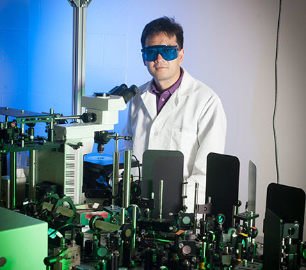
Dr. Michael Walsh of the University of Illinois at Chicago (UIC) conducts research within the pathology department’s Spectral Pathology Lab, which has been using the IR microscope for the past several months. Walsh has been focused on developing chemical imaging techniques, with the ultimate goal of improving diagnoses within the field of tissue pathology.

Currently, the state-of-the-art method-ology used for the diagnosis of most solid-organ diseases is to extract a tissue sample via a biopsy. Tissue inherently has very little contrast and needs to be stained with dyes or probes to visualize and identify cell types and tissue structures. The field of pathology is based on examining the stained tissues, typically using white light, to determine if the tissue morphology deviates from a normal pattern. If the tissue looks abnormal, the disease state may be further subclassified by grade or by predicted outcome. However, the field of pathology is limited by the information that can be derived from the stained tissues and the subjective interpretation of the tissue by a highly trained pathologist.

Spero microscope.


Spero microscope. Courtesy of Daylight Solutions.


UIC’s Spectral Pathology Lab is focused on identifying areas in pathology where current techniques fail, or where there is a need for additional diagnostic or prognostic information that can help improve patient care. Potentially, MIR imaging is a very valuable adjunct to the current practice of pathology. Rather than using only stains, MIR imaging can interrogate the entire biochemistry of the tissue and render a diagnosis in an objective fashion. Traditionally, MIR imaging with an FTIR system has been limited by slow data acquisition speeds and the need to collect the entire spectral data cube. QCL imaging with the Spero microscope has the potential to speed up the data acquisition of images obtained from a tissue sample and to collect only the spectral frequencies of interest. The device also provides real-time imaging of samples at 30 fps, which could allow pathologists to very rapidly identify areas of interest on a tissue biopsy in a manner that is similar to their current clinical workflows. Some examples of the comparison of FTIR-derived and QCL-derived images from multiple organ tissues of interest are presented.

H&E-stained image of a mouse brain section on IR reflective slide, with selected regions labeled: hypothalamus, thalamus, and dentate gyrus.
Figure 2.
(a) H&E-stained image of a mouse brain section on IR reflective slide, with selected regions labeled: hypothalamus, thalamus, and dentate gyrus. (b) Transflectance QCL IR image of same region, prior to staining, at 1652 cm−1, in which the thalamus is clearly distinguished from surrounding regions. (c) Same region at 1548 cm−1. (d) Same region at 1500 cm−1. Courtesy of University of Illinois at Chicago (UIC)/Spectral Pathology Lab.


A tissue section from a mouse brain was scanned using the Spero microscope’s high-magnification objective (12.5×; 0.7 NA; 1.4 × 1.4-µm pixels) at various MIR frequencies in transflection mode, as shown in Figure 2. The tissue then was stained using hematoxylin and eosin (H&E), the most common stain in histopathology, and is displayed in Figure 2a. Using the H&E stain, regions were identified in the brain (thalamus, dentate gyrus and hypothalamus) that correlated with structures in the IR image. By illuminating the tissue at various wavelengths, discrete tissue features exhibit contrast due to the difference in absorption, as highlighted in the IR images taken at 1652, 1548 and 1500 cm−1 in Figure 2b-d, respectively. The microscope also makes it possible to visualize tissue at these individual wavelengths in real time. The identification of cell types and their biochemical changes is of particular interest in neuropathology.

Transmission FTIR image of a 4-µm thick section from a human liver tissue microarray on barium fluoride at 1650 cm-1.
Figure 3.
(a) Transmission FTIR image of a 4-µm thick section from a human liver tissue microarray on barium fluoride at 1650 cm-1. The image was taken with 64 coadditions of successive scans. (b) Transmission image from the Spero microscope of the same tissue at 1652 cm-1, both baseline corrected between 1796 cm-1 and 904 cm-1. In both images, the bright white stripe dividing the tissue core roughly in half is a region of fibrosis (red arrow), while the rest of the tissue on either side is composed primarily of hepatocytes (blue arrow). Courtesy of UIC/Spectral Pathology Lab.


A single biopsy core obtained from human liver tissue was scanned in transmission mode on a barium fluoride substrate by an Agilent Cary 600 Series FTIR microscope (Figure 3a). The FTIR image was acquired using a 36× Cassegrain collecting objective and a 15× Cassegrain condenser for a pixel size of 2.2 × 2.2 µm. Figure 3b shows the same liver core acquired using the Spero microscope with the high-magnification collecting objective (12.5×, 0.7 NA) and condenser objective for a pixel size of 1.4 × 1.4 µm. High-definition IR imaging enables clear contrast and identification of the band of fibrosis in the center of the core and the surrounding regions of liver cells, known as hepatocytes, and is indicated within Figure 3a-b. Acquisition of IR imaging data at the diffraction limit enables chemical information to be recorded from tissue structures at the single-cell level, allowing accurate characterization of individual tissue components, different cell types, varied disease states or other aspects of a tissue section.

Averaged spectra for regions of interest corresponding to the hepatocytes and the fibrotic area on the FTIR image in Figure 3a.
Figure 4.
(a) Averaged spectra for regions of interest corresponding to the hepatocytes and the fibrotic area on the FTIR image in Figure 3a. Spectra have been truncated from 1800 to 900 cm-1, normalized to 1650 cm-1, and baseline corrected between 1796 and 904 cm-1. (b) Averaged spectra for regions of interest corresponding to the hepatocytes and the fibrotic area on the Spero microscope image in Figure 3b. Spectra have been normalized to 1652 cm-1 and baseline corrected between 1796 and 904 cm-1.


Figure 4 displays average spectra calculated from homogenous tissue regions that describe hepatocytes and fibrosis within the liver tissue core shown in Figure 3. The spectra acquired from both FTIR and QCL systems are very similar. Walsh is focused on developing spectral classifiers that can aid pathologists in making very difficult diagnoses in the precancerous stages of liver cancer.

H&E-stained section of human colon tissue, and FTIR (with 16 coadditions) and Spero microscope transmission images of a 4-µm thick serial section of the same sample on barium fluoride. FTIR image shown at 1650 cm-1, Spero microscope image shown at 1652 cm-1.
Figure 5.
H&E-stained section of human colon tissue, and FTIR (with 16 coadditions) and Spero microscope transmission images of a 4-µm thick serial section of the same sample on barium fluoride. FTIR image shown at 1650 cm-1, Spero microscope image shown at 1652 cm-1. The red circle indicates mucin, the green circle indicates malignant colon carcinoma epithelium, and the blue circle indicates fibroblastic stroma. The raw spectra (taken from single pixels in approximately the same location for each of the three tissue features) are shown below their respective IR images. The FTIR spectra were truncated to match the Spero microscope’s spectral range of 1800 to 900 cm-1. Courtesy of UIC/Spectral Pathology Lab.


Point spectra from individual pixels were obtained and compared from a human colon sample on barium fluoride scanned in transmission on the same FTIR and QCL systems, which is shown in Figure 5. A serial section was obtained and stained with H&E to identify the different tissue structures. Using the H&E image as a reference, spectra from mucin (red), malignant colon carcinoma epithelium (green) and fibroblastic stroma (blue) were collected from a single pixel at approximately the same location. The unprocessed QCL and FTIR spectra are shown directly beneath their respective images. The FTIR system has an FPA size of 128 × 128 detector elements, while the Spero system has a microbolometer of 480 × 480 detector elements. Therefore, the FTIR image was collected as a mosaic and then stitched together.

FTIR and Spero microscope spectra from a single pixel of mucin, from the tissue shown in Figure 5.
Figure 6.
(a) FTIR and Spero microscope spectra from a single pixel of mucin, from the tissue shown in Figure 5. (b) FTIR and Spero microscope spectra from a single pixel of malignant colon carcinoma epithelium, from the same tissue. (c) FTIR and Spero microscope spectra from a single pixel of fibroblastic stroma. All spectra have been normalized (FTIR to 1650 cm-1, Spero to 1652 cm-1) and baseline corrected between 1796 and 904 cm-1, with the FTIR spectra truncated to match the Spero microscope’s spectral range of 1800 to 900 cm-1. Note that pixels for each tissue feature were located in approximately the same region, and that the two images have different pixel sizes (2.2 × 2.2 µm for FTIR, 1.4 × 1.4 µm for Spero microscope). Courtesy of UIC/Spectral Pathology Lab.


The spectra obtained from the regions of interest depicted in Figure 5 were preprocessed, as shown in Figure 6. The data was peak height normalized to the Amide I band. The FTIR data and QCL data were processed using a simple, two-point linear baseline correction between 1796 and 904 cm−1. Figure 6a-c shows the processed data from single pixels looking at the biochemistry of mucin, malignant colon carcinoma epithelium and fibroblastic stroma, respectively. The spectra from the QCL and FTIR systems are very similar on an individual-pixel level.

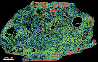
Finally, Figure 7 shows the scan of a frozen prostate tissue section captured with the microscope. Once thawed, the system can quickly image these sections at a single frequency of interest. The real-time capabilities of the system combined with the capacity for scanning frozen samples could someday allow for the analysis of samples in a time-critical intraoperative setting.

Transflectance scan of a 5-µm frozen human prostate tissue section on Kevley low-emissivity substrate captured with the Spero microscope.


Figure 7. Transflectance scan of a 5-µm frozen human prostate tissue section on Kevley low-emissivity substrate captured with the Spero microscope. Visualized with a false color map at 1640 cm-1. Data was baseline corrected between 1796 and 904 cm-1. Courtesy of UIC/Spectral Pathology Lab. University of Illinois at Chicago — Spectral Pathology Lab members, from left to right: David Martinez, Francesca Gambacorta, Vishal Varma, Andrew Graham and Michael Walsh. Courtesy of Daylight Solutions.


While there has been significant interest in MIR imaging for pathology applications for a number of years1-5, the technology has lacked the maturity to be ready for clinical implementation due to slow scanning speeds, low spatial resolutions and by a lack of computational power to fully handle large multispectral datasets. The Spero microscope, coupled with modern computing power, overcomes these limitations. The information detailed above demonstrates that the quality of the images and spectra obtained from the instrument are similar to those offered by FTIR imaging methods but with the additional benefits associated with the use of a QCL-based system. Recent advances in large multielement FPAs6-8) and high-resolution imaging approaches9-11 for tissue pathology have made this a much more attractive approach for fast and detailed image acquisition. QCLs represent the next step toward clinical implementation — they have demonstrated fast data acquisition, live-imaging capabilities and the ability to collect only spectral frequencies of diagnostic value.

Meet the authors

Michael Walsh holds a PhD in biological sciences and is an assistant professor at the University of Illinois at Chicago in Chicago; email: walshm@uic.edu. Matthew Barre is the business development manager at Daylight Solutions in San Diego; email: mbarre@daylightsolutions.com. Benjamin Bird is an applications scientist at Daylight Solutions in San Diego; email: bbird@daylightsolutions.com.

References

1. D.C. Fernandez et al. (2005). Infrared spectroscopic imaging for histopathologic recognition. Nat Biotechnol, Vol. 23, Issue 4, pp. 469-474.

2. C. Matthaus et al. (2008). Chapter 10: Infrared and Raman microscopy in cell biology. Methods Cell Biol, Vol. 89, pp. 275-308.

3. C. Kendall et al. (2009). Vibrational spectroscopy: a clinical tool for cancer diagnostics. Analyst, Vol. 134, Issue 6, pp. 1029-1045.

4. C. Krafft et al. (2009). Disease recognition by infrared and Raman spectroscopy. J Biophotonics, Vol. 2, Issue 1-2, pp. 13-28.

5. F.L. Martin et al. (2010). Distinguishing cell types or populations based on the computational analysis of their infrared spectra. Nat Protoc, Vol. 5, Issue 11, pp. 1748-1760.


Broadband Laser Aimed at Cancer Detection

Covering a wide swath of the mid-infrared, a new system offers greater spectral sensitivity

BY JAMES F. LOWE, WEB MANAGING EDITOR, JAMES.LOWE@PHOTONICS.COM

MUNICH, Sept. 25, 2015 — Mid-infrared (MIR) light is rich with molecular “fingerprint” information that can be used to detect substances from atmospheric pollutants to cancer cells.

While some lasers already operate in this region, enabling a variety of spectroscopy applications, their linewidth is relatively narrow, which limits the types of substances they can detect at any given moment.

Now a team of researchers from Germany and Spain has developed a laser system with phase-coherent emission from 6.8 to 16.4 μm and output power of 0.1 W. That is broad and powerful enough, they said, to detect subtle signs of cancer early in its development.

Molecules absorb portions of the MIR spectrum in ways that are unique to their atomic structures, and their absorption patterns provide a means of identifying the molecules with great specificity, even in low concentrations.


Emission spectrum


The emission spectrum of the laser and corresponding molecular fingerprint regions. Courtesy of the Institute of Photonic Sciences (ICFO).


“Cancer causes subtle modification in protein structure and content within a cell,” said professor Dr. Jens Biegert, a group leader at the Institute of Photonic Sciences (ICFO) in Barcelona. “Looking at only a few nanometer range, the probability of detection is extremely low. But comparing many of such intervals, one can have an extremely high confidence level.”

The new laser system generates MIR pulses via difference-frequency generation driven by the nonlinearly compressed pulses of a Kerr-lens mode-locked Yb:YAG thin-disc oscillator. It features a repetition rate of 100 MHz and pulse durations of 66 fs — so short that the electric field oscillates only twice per pulse.

Lab


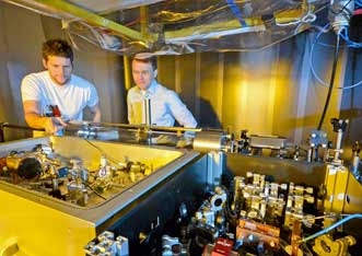
Staff scientist Dr. Ioachim Pupeza (left) and postdoctoral researcher Oleg Pronin helped develop a laser system that emits ultrashort pulses of mid-infrared light. These pulses can be used to detect trace molecules in gaseous and liquid media. Courtesy of Thorsten Naeser/Ludwig Maximilian University.


“Since we now possess a compact source of high-intensity and coherent infrared light, we have a tool that can serve as an extremely sensitive sensor for the detection of molecules, and is suitable for serial production,” said project leader Dr. Ioachim Pupeza, a staff scientist at Ludwig Maximilian University of Munich (LMU).

The LMU and ICFO researchers aim to use their MIR laser to identify and quantify disease markers in exhaled air. Many diseases, including some types of cancer, are thought to produce specific molecules that end up in the air expelled from the lungs.

“We assume that exhaled breath contains well over 1000 different molecular species,” said Dr. Alexander Apolonskiy, an LMU group leader.

However, the amount of molecular biomarkers present in exhaled breath is extraordinarily low, meaning a diagnostic tool would need to be capable of detecting concentrations of at least one part per billion. The next step will be to couple the new laser system with a novel amplifier that would increase its brightness and boost sensitivity one part per trillion.

Detecting MIR signatures

The laser’s output spans more than one octave. Until now, the researchers said, such broadband emission has only been available from large-scale synchrotron sources.

Other more compact MIR sources, such as quantum cascade lasers (QCLs), have narrower linewidths. Tuning them to different sensing bands is time consuming, and combining multiple QCLs emitting in different parts of the MIR would be cost-prohibitive, Biegert said.

Meanwhile, the laser system’s 100-MHz pulse train is hundreds to thousands of times more powerful than state-of-the-art frequency combs that emit in the same range, the researchers said.

Detecting broadband MIR signals presents its own problems, however. Detectors for this region have poor signal-to-noise ratios unless cooled with liquid nitrogen, the researchers said.

In this case, electro-optical sampling proved to be a better option. Well-established for the terahertz range, the technique is less common in the fingerprint region.

“In the MIR range, there are not many groups who have implemented this already, because you need a broadband, phase-stable MIR pulse and an ultrashort sample pulse at the same time, which is quite challenging,” Pupeza said.

Having solved that problem with their broadband laser, the team now could use electro-optical sampling to extract the data they wanted.

In a nutshell, the process works like this: The electric field of an MIR pulse alters the birefringence of a crystal. This change can be measured by observing how the polarization of slightly shorter near-infrared (NIR) pulse is changed while propagating through the same crystal at the same time. In the end, only the NIR pulse is measured directly.

“Therefore, one big advantage is low-noise detection in the NIR, even though one obtains information on spectral components in the MIR,” said Ioachim Pupeza. “You only need to perform a Fourier transform numerically to get the spectrum of the pulse once you have its electric field.”

http://www.photonics.com/Article.aspx?AID=57757
The research was published in Nature Photonics (doi: 10.1038/nphoton.2015.179).


Read Full Post »

Asthma sourced carbon nanotubes

Larry H. Bernstein, MD, FCAP, Curator

LPBI

Carbon nanotubes found in cells from airways of asthmatic children in Paris

Carbon nanotubes, possibly from cars, are ubiquitous, found even in ice cores — we may all have them in our lungs, say Rice scientists
October 19, 2015

http://www.kurzweilai.net/carbon-nanotubes-found-in-cells-from-airways-of-asthmatic-children-in-paris?utm_source=KurzweilAI+Weekly+Newsletter_147a5a48c1-9a20162408-282099089

Carbon nanotubes (rods) and nanoparticles (black clumps) found inside a lung cell vacuole (left) are similar to those found in vehicle exhaust in tailpipes of cars in Paris (right) (credit: Fathi Moussa/Paris-Saclay University)

http://www.kurzweilai.net/images/CNTs-in-lungs-cars.jpg

Carbon nanotubes (CNTs) have been found in cells extracted from the airways of Parisian children under routine treatment for asthma, according to a report in the journal EBioMedicine (open access) by scientists in France and atRice University.

The cells were taken from 69 randomly selected asthma patients aged 2 to 17 who underwent routine fiber-optic bronchoscopies as part of their treatment. The researchers analyzed particulate matter found in the alveolar macrophage cells (also known as dust cells), which help stop foreign materials like particles and bacteria from entering the lungs.

The study partially answers the question of what makes up the black material inside alveolar macrophages, the original focus of the study. The researchers found single-walled and multiwalled carbon nanotubes and amorphous carbon among the cells.

The nanotube aggregates in the cells ranged in size from 10 to 60 nanometers in diameter and up to several hundred nanometers in length, small enough that optical microscopes would not have been able to identify them in samples from former patients. The new study used more sophisticated tools, including high-resolution transmission electron microscopy, X-ray spectroscopy, Raman spectroscopy, and near-infrared fluorescence microscopy to definitively identify them in the cells and in the environmental samples.

“The concentrations of nanotubes are so low in these samples that it’s hard to believe they would cause asthma, but you never know,” said Rice chemist Lon Wilson, a corresponding author of the paper. “What surprised me the most was that carbon nanotubes were the major component of the carbonaceous pollution we found in the samples.”

The study notes but does not make definitive conclusions about the controversial proposition that carbon nanotube fibers may act like asbestos, a proven carcinogen. But the authors did note that “long carbon nanotubes and large aggregates of short ones can induce a granulomatous (inflammation) reaction.”

The researchers also suggested previous studies that link the carbon content of airway macrophages and the decline of lung function should be reconsidered in light of the new findings. The researchers also suggested that the large surface areas of nanotubes and their ability to adhere to substances may make them effective carriers for other pollutants.

Carbon nanotubes from forest fires and cars?

Fullerenes (left) can be converted to carbon nanotubes (right) with a catalytic process, according to Rice chemists (credits: Soroush83/CC and Matías Soto/Rice University)

http://www.kurzweilai.net/images/fullerene-to-nanotube.jpg

However, similar nanotubes have been found in samples from the exhaust pipes of Paris vehicles, in dust gathered from various places around the city, in spider webs in India, and even in ice cores, the paper notes.

“We know that carbon nanoparticles are found in nature,” Wilson said, noting that round fullerene (C60) molecules are commonly produced by volcanoes, forest fires, and other combustion of carbon materials. “All you need is a little catalysis to make carbon nanotubes instead of fullerenes.”

A car’s catalytic converter, which turns toxic carbon monoxide into safer emissions, bears at least a passing resemblance to the Rice-invented high-pressure carbon monoxide, or HiPco, process to make carbon nanotubes, he said. “So it is not a big surprise, when you think about it,” Wilson said.

“Based on our discovery of CNTs in tailpipes, we propose that the catalytic converters of the automobiles are manufacturing carbon nanotubes, Wilson told KurzweilAI. “However, we have not actually proven that.”

We are all carbon-nanotube bearers now

For ethical reasons, no cells from healthy patients were analyzed, but because nanotubes were found in all of the samples, the study led the researchers to conclude that carbon nanotubes are likely to be found in everybody.

“It’s kind of ironic. In our laboratory, working with carbon nanotubes, we wear facemasks to prevent exactly what we’re seeing in these samples, yet everyone walking around out there in the world probably has at least a small concentration of carbon nanotubes in their lungs,” he said.

The study followed one released by Rice and Baylor College of Medicine earlier this month with the similar goal ofanalyzing the black substance found in the lungs of smokers who died of emphysema. That study found carbon black nanoparticles that were the product of the incomplete combustion of such organic material as tobacco.

Co-authors are from Paris-Saclay University, the Paediatric Pulmonology and Allergy Center and the Department of Anatomo-Pathology of the Groupe hospitalier La Roche-Guyon, and Paris Diderot University. The Welch Foundation partially supported the research.


Abstract of Anthropogenic Carbon Nanotubes Found in the Airways of Parisian Children

Compelling evidence shows that fine particulate matters (PM) from air pollution penetrate lower airways and are associated with adverse health effects even within concentrations below those recommended by the WHO. A paper reported a dose-dependent link between carbon content in alveolar macrophages (assessed only by optical microscopy) and the decline in lung function. However, to the best of our knowledge, PM had never been accurately characterized inside human lung cells and the most responsible components of the particulate mix are still unknown. On another hand carbon nanotubes (CNTs) from natural and anthropogenic sources might be an important component of PM in both indoor and outdoor air.

We used high-resolution transmission electron microscopy and energy dispersive X-ray spectroscopy to characterize PM present in broncho-alveolar lavage-fluids (n = 64) and inside lung cells (n = 5 patients) of asthmatic children. We show that inhaled PM mostly consist of CNTs. These CNTs are present in all examined samples and they are similar to those we found in dusts and vehicle exhausts collected in Paris, as well as to those previously characterized in ambient air in the USA, in spider webs in India, and in ice core. These results strongly suggest that humans are routinely exposed to CNTs.

Read Full Post »

%d