Funding, Deals & Partnerships: BIOLOGICS & MEDICAL DEVICES; BioMed e-Series; Medicine and Life Sciences Scientific Journal – http://PharmaceuticalIntelligence.com
New studies link cell cycle proteins to immunosurveillance of premalignant cells
Curator: Stephen J. Williams, Ph.D.
The following is from a Perspectives article in the journal Science by Virinder Reen and Jesus Gil called “Clearing Stressed Cells: Cell cycle arrest produces a p21-dependent secretome that initaites immunosurveillance of premalignant cells”. This is a synopsis of the Sturmlechener et al. research article in the same issue (2).
Complex organisms repair stress-induced damage to limit the replication of faulty cells that could drive cancer. When repair is not possible, tissue homeostasis is maintained by the activation of stress response programs such as apoptosis, which eliminates the cells, or senescence, which arrests them (1). Cellular senescence causes the arrest of damaged cells through the induction of cyclin-dependent kinase inhibitors (CDKIs) such as p16 and p21 (2). Senescent cells also produce a bioactive secretome (the senescence-associated secretory phenotype, SASP) that places cells under immunosurveillance, which is key to avoiding the detrimental inflammatory effects caused by lingering senescent cells on surrounding tissues. On page 577 of this issue, Sturmlechner et al. (3) report that induction of p21 not only contributes to the arrest of senescent cells, but is also an early signal that primes stressed cells for immunosurveillance.Senescence is a complex program that is tightly regulated at the epigenetic and transcriptional levels. For example, exit from the cell cycle is controlled by the induction of p16 and p21, which inhibit phosphorylation of the retinoblastoma protein (RB), a transcriptional regulator and tumor suppressor. Hypophosphorylated RB represses transcription of E2F target genes, which are necessary for cell cycle progression. Conversely, production of the SASP is regulated by a complex program that involves super-enhancer (SE) remodeling and activation of transcriptional regulators such as nuclear factor κB (NF-κB) or CCAAT enhancer binding protein–β (C/EBPβ) (4).
Senescence is a complex program that is tightly regulated at the epigenetic and transcriptional levels. For example, exit from the cell cycle is controlled by the induction of p16 and p21, which inhibit phosphorylation of the retinoblastoma protein (RB), a transcriptional regulator and tumor suppressor. Hypophosphorylated RB represses transcription of E2F target genes, which are necessary for cell cycle progression. Conversely, production of the SASP is regulated by a complex program that involves super-enhancer (SE) remodeling and activation of transcriptional regulators such as nuclear factor κB (NF-κB) or CCAAT enhancer binding protein–β (C/EBPβ) (4).
Sturmlechner et al. found that activation of p21 following stress rapidly halted cell cycle progression and triggered an internal biological timer (of ∼4 days in hepatocytes), allowing time to repair and resolve damage (see the figure). In parallel, C-X-C motif chemokine 14 (CXCL14), a component of the PASP, attracted macrophages to surround and closely surveil these damaged cells. Stressed cells that recovered and normalized p21 expression suspended PASP production and circumvented immunosurveillance. However, if the p21-induced stress was unmanageable, the repair timer expired, and the immune cells transitioned from surveillance to clearance mode. Adjacent macrophages mounted a cytotoxic T lymphocyte response that destroyed damaged cells. Notably, the overexpression of p21 alone was sufficient to orchestrate immune killing of stressed cells, without the need of a senescence phenotype. Overexpression of other CDKIs, such as p16 and p27, did not trigger immunosurveillance, likely because they do not induce CXCL14 expression.In the context of cancer, senescent cell clearance was first observed following reactivation of the tumor suppressor p53 in liver cancer cells. Restoring p53 signaling induced senescence and triggered the elimination of senescent cells by the innate immune system, prompting tumor regression (5). Subsequent work has revealed that the SASP alerts the immune system to target preneoplastic senescent cells. Hepatocytes expressing the oncogenic mutant NRASG12V (Gly12→Val) become senescent and secrete chemokines and cytokines that trigger CD4+ T cell–mediated clearance (6). Despite the relevance for tumor suppression, relatively little is known about how immunosurveillance of oncogene-induced senescent cells is initiated and controlled.
Source of image: Reen, V. and Gil, J. Clearing Stressed Cells. Science Perspectives 2021;Vol 374(6567) p 534-535.
References
2. Sturmlechner I, Zhang C, Sine CC, van Deursen EJ, Jeganathan KB, Hamada N, Grasic J, Friedman D, Stutchman JT, Can I, Hamada M, Lim DY, Lee JH, Ordog T, Laberge RM, Shapiro V, Baker DJ, Li H, van Deursen JM. p21 produces a bioactive secretome that places stressed cells under immunosurveillance. Science. 2021 Oct 29;374(6567):eabb3420. doi: 10.1126/science.abb3420. Epub 2021 Oct 29. PMID: 34709885.
More Articles on Cancer, Senescence and the Immune System in this Open Access Online Scientific Journal Include
The CRISPR-Cas9 system has proven to be a powerful tool for genome editing allowing for the precise modification of specific DNA sequences within a cell. Many efforts are currently underway to use the CRISPR-Cas9 system for the therapeutic correction of human genetic diseases. CRISPR/Cas9 has revolutionized our ability to engineer genomes and conduct genome-wide screens in human cells.
CRISPR–Cas9 induces a p53-mediated DNA damage response and cell cycle arrest in immortalized human retinal pigment epithelial cells, leading to a selection against cells with a functional p53 pathway. Inhibition of p53 prevents the damage response and increases the rate of homologous recombination from a donor template. These results suggest that p53 inhibition may improve the efficiency of genome editing of untransformed cells and that p53 function should be monitored when developing cell-based therapies utilizing CRISPR–Cas9.
Whereas some cell types are amenable to genome engineering, genomes of human pluripotent stem cells (hPSCs) have been difficult to engineer, with reduced efficiencies relative to tumour cell lines or mouse embryonic stem cells. Using hPSC lines with stable integration of Cas9 or transient delivery of Cas9-ribonucleoproteins (RNPs), an average insertion or deletion (indel) efficiency greater than 80% was achieved. This high efficiency of insertion or deletion generation revealed that double-strand breaks (DSBs) induced by Cas9 are toxic and kill most hPSCs.
The toxic response to DSBs was P53/TP53-dependent, such that the efficiency of precise genome engineering in hPSCs with a wild-type P53 gene was severely reduced. These results indicate that Cas9 toxicity creates an obstacle to the high-throughput use of CRISPR/Cas9 for genome engineering and screening in hPSCs. As hPSCs can acquire P53 mutations, cell replacement therapies using CRISPR/Cas9-enginereed hPSCs should proceed with caution, and such engineered hPSCs should be monitored for P53 function.
CRISPR-based editing of T cells to treat cancer, as scientists at the University of Pennsylvania are studying in a clinical trial, should also not have a p53 problem. Nor should any therapy developed with CRISPR base editing, which does not make the double-stranded breaks that trigger p53. But, there are pre-existing humoral and cell-mediated adaptive immune responses to Cas9 in humans, a factor which must be taken into account as the CRISPR-Cas9 system moves forward into clinical trials.
The “Guardian Of The Genome” p53 In Pancreatic Cancer
Curator: David Orchard-Webb, PhD
A recent next generation sequencing (NGS) study of PDAC samples found that the “guardian of the genome” p53 (TP53) had a total mutation rate of 50% (the literature suggests up to 75%) [1]. TP53 deletions occurred in 6% of samples. Missense mutation of p53 (44% of samples) can convert it from a tumour suppressor to a neo-oncogene. This curation collates recent articles of note with reference to p53 including function, drug development, and prognostics in pancreatic ductal adenocarcinoma (PDAC).
Vaccines
Katchman, Benjamin A., Rodrigo Barderas, Rizwan Alam, Diego Chowell, Matthew S. Field, Laura J. Esserman, Garrick Wallstrom, et al. ‘Proteomic Mapping of p53 Immunogenicity in Pancreatic, Ovarian, and Breast Cancers’. Proteomics. Clinical Applications, 28 April 2016. doi:10.1002/prca.201500096.
Terashima, Takeshi, Eishiro Mizukoshi, Kuniaki Arai, Tatsuya Yamashita, Mariko Yoshida, Hajime Ota, Ichiro Onishi, et al. ‘P53, hTERT, WT-1, and VEGFR2 Are the Most Suitable Targets for Cancer Vaccine Therapy in HLA-A24 Positive Pancreatic Adenocarcinoma’. Cancer Immunology, Immunotherapy: CII 63, no. 5 (May 2014): 479–89. doi:10.1007/s00262-014-1529-8.
Drug targeting p53
Xu, Jing, Amit Singh, and Mansoor M. Amiji. ‘Redox-Responsive Targeted Gelatin Nanoparticles for Delivery of Combination Wt-p53 Expressing Plasmid DNA and Gemcitabine in the Treatment of Pancreatic Cancer’. BMC Cancer 14, no. 1 (2014): 75. http://www.biomedcentral.com/1471-2407/14/75/.
Camp, E R, C Wang, E C Little, P M Watson, K F Pirollo, A Rait, D J Cole, E H Chang, and D K Watson. ‘Transferrin Receptor Targeting Nanomedicine Delivering Wild-Type p53 Gene Sensitizes Pancreatic Cancer to Gemcitabine Therapy’. Cancer Gene Therapy 20, no. 4 (April 2013): 222–28. doi:10.1038/cgt.2013.9.
Izetti, Patricia, Agnes Hautefeuille, Ana Lucia Abujamra, Caroline Brunetto de Farias, Juliana Giacomazzi, Bárbara Alemar, Guido Lenz, et al. ‘PRIMA-1, a Mutant p53 Reactivator, Induces Apoptosis and Enhances Chemotherapeutic Cytotoxicity in Pancreatic Cancer Cell Lines’. Investigational New Drugs 32, no. 5 (October 2014): 783–94. doi:10.1007/s10637-014-0090-9.
Takei, Y., S. Okamoto, K. Kawamura, Y. Jiang, T. Morinaga, M. Shingyoji, I. Sekine, et al. ‘Expression of p53 Synergistically Augments Caspases-Mediated Apoptosis Induced by Replication-Competent Adenoviruses in Pancreatic Carcinoma Cells’. Cancer Gene Therapy 22, no. 9 (September 2015): 445–53. doi:10.1038/cgt.2015.33.
Li, Jinluan, Jianji Pan, Xianggao Zhu, Ying Su, Lingling Bao, Sufang Qiu, Changyan Zou, Yong Cai, Junxin Wu, and Ivan WK Tham. ‘Recombinant Adenovirus-p53 (Gendicine) Sensitizes a Pancreatic Carcinoma Cell Line to Radiation’. Chinese Journal of Cancer Research 25, no. 6 (2013): 715. http://www.cjcrcn.org/article/html_3076.html.
Hastie, Eric, Marcela Cataldi, Nury Steuerwald, and Valery Z. Grdzelishvili. ‘An Unexpected Inhibition of Antiviral Signaling by Virus-Encoded Tumor Suppressor p53 in Pancreatic Cancer Cells’. Virology 483 (September 2015): 126–40. doi:10.1016/j.virol.2015.04.017.
The Impact of p53 Status on Therapy
Kurahara, Hiroshi, Kosei Maemura, Yuko Mataki, Masahiko Sakoda, Hiroyuki Shinchi, and Shoji Natsugoe. ‘Impact of p53 and PDGFR-β Expression on Metastasis and Prognosis of Patients with Pancreatic Cancer’. World Journal of Surgery, 3 March 2016. doi:10.1007/s00268-016-3477-2.
Xiang, Jin-Feng, Wen-Quan Wang, Liang Liu, Hua-Xiang Xu, Chun-Tao Wu, Jing-Xuan Yang, Zi-Hao Qi, et al. ‘Mutant p53 Determines Pancreatic Cancer Poor Prognosis to Pancreatectomy through Upregulation of Cavin-1 in Patients with Preoperative Serum CA19-9 ≥ 1,000 U/mL’. Scientific Reports 6 (12 January 2016): 19222. doi:10.1038/srep19222.
Rajeshkumar, N. V., Prasanta Dutta, Shinichi Yabuuchi, Roeland F. de Wilde, Gary V. Martinez, Anne Le, Jurre J. Kamphorst, et al. ‘Therapeutic Targeting of the Warburg Effect in Pancreatic Cancer Relies on an Absence of p53 Function’. Cancer Research 75, no. 16 (15 August 2015): 3355–64. doi:10.1158/0008-5472.CAN-15-0108.
Fiorini, Claudia, Marco Cordani, Chiara Padroni, Giovanni Blandino, Silvia Di Agostino, and Massimo Donadelli. ‘Mutant p53 Stimulates Chemoresistance of Pancreatic Adenocarcinoma Cells to Gemcitabine’. Biochimica et Biophysica Acta (BBA) – Molecular Cell Research 1853, no. 1 (January 2015): 89–100. doi:10.1016/j.bbamcr.2014.10.003.
TP53 Function in Pancreatic Cancer
Bailey, J M, A M Hendley, K J Lafaro, M A Pruski, N C Jones, J Alsina, M Younes, et al. ‘p53 Mutations Cooperate with Oncogenic Kras to Promote Adenocarcinoma from Pancreatic Ductal Cells’. Oncogene, 23 November 2015. doi:10.1038/onc.2015.441.
Sheng, Weiwei, Ming Dong, Jianping Zhou, Xin Li, Qingfeng Liu, Qi Dong, and Feng Li. ‘Cooperation among Numb, MDM2 and p53 in the Development and Progression of Pancreatic Cancer’. Cell and Tissue Research 354, no. 2 (November 2013): 521–32. doi:10.1007/s00441-013-1679-6.
Weissmueller, Susann, Eusebio Manchado, Michael Saborowski, John P. Morris, Elvin Wagenblast, Carrie A. Davis, Sung-Hwan Moon, et al. ‘Mutant p53 Drives Pancreatic Cancer Metastasis through Cell-Autonomous PDGF Receptor β Signaling’. Cell 157, no. 2 (April 2014): 382–94. doi:10.1016/j.cell.2014.01.066.
Morton, J. P., P. Timpson, S. A. Karim, R. A. Ridgway, D. Athineos, B. Doyle, N. B. Jamieson, et al. ‘Mutant p53 Drives Metastasis and Overcomes Growth Arrest/senescence in Pancreatic Cancer’. Proceedings of the National Academy of Sciences 107, no. 1 (5 January 2010): 246–51. doi:10.1073/pnas.0908428107.
Hamilton, Garth, Aswin G. Abraham, Jennifer Morton, Oliver Sampson, Dafni E. Pefani, Svetlana Khoronenkova, Anna Grawenda, et al. ‘AKT Regulates NPM Dependent ARF Localization and p53mut Stability in Tumors’. Oncotarget 5, no. 15 (2014): 6142–6167. http://eprints.gla.ac.uk/102649.
Sadagopan, S, M V Veettil, S Chakraborty, N Sharma-Walia, N Paudel, V Bottero, and B Chandran. ‘Angiogenin Functionally Interacts with p53 and Regulates p53-Mediated Apoptosis and Cell Survival’. Oncogene 31, no. 46 (15 November 2012): 4835–47. doi:10.1038/onc.2011.648.
TP53 and Epithelial Mesenchymal Transition (EMT)
Wörmann, Sonja M., Liang Song, Jiaoyu Ai, Kalliope N. Diakopoulos, Kivanc Görgülü, Dietrich Ruess, Andrew Campbell, et al. ‘Loss of P53 Function Activates JAK2-STAT3 Signaling to Promote Pancreatic Tumor Growth, Stroma Modification, and Gemcitabine Resistance in Mice and Is Associated With Patient Survival’. Gastroenterology, 18 March 2016. doi:10.1053/j.gastro.2016.03.010.
Lee, Sun-Hye, Su-Jin Lee, Yeon Sang Jung, Yongbin Xu, Ho Sung Kang, Nam-Chul Ha, and Bum-Joon Park. ‘Blocking of p53-Snail Binding, Promoted by Oncogenic K-Ras, Recovers p53 Expression and Function’. Neoplasia 11, no. 1 (January 2009): 22–IN6. doi:10.1593/neo.81006.
Pancreatic Cancer Risk Factor
Sonoyama, Takayuki. ‘TP53 Codon 72 Polymorphism Is Associated with Pancreatic Cancer Risk in Males, Smokers and Drinkers’. Molecular Medicine Reports, 8 March 2011. doi:10.3892/mmr.2011.449.
3D PDAC in vitro Culture Models
Huang, Ling, Audrey Holtzinger, Ishaan Jagan, Michael BeGora, Ines Lohse, Nicholas Ngai, Cristina Nostro, et al. ‘Ductal Pancreatic Cancer Modeling and Drug Screening Using Human Pluripotent Stem Cell– and Patient-Derived Tumor Organoids’. Nature Medicine 21, no. 11 (26 October 2015): 1364–71. doi:10.1038/nm.3973.
REFERENCES
Witkiewicz, Agnieszka K., Elizabeth A. McMillan, Uthra Balaji, GuemHee Baek, Wan-Chi Lin, John Mansour, Mehri Mollaee, et al. ‘Whole-Exome Sequencing of Pancreatic Cancer Defines Genetic Diversity and Therapeutic Targets’. Nature Communications 6 (9 April 2015): 6744. doi:10.1038/ncomms7744.
Other Related Articles Published In This Open Access Online Journal Include The Following:
Protection against cellular stress from various sources, such as nutritional, physical, pathogenic, or oncogenic, results in the induction of both intrinsic and extrinsic cellular protection mechanisms that collectively limit the damage these insults inflict on the host. The major extrinsic protection mechanism against cellular stress is the immune system. Indeed, it has been well described that cells that are stressed due to association with viral infection or early malignant transformation can be directly sensed by the immune system, particularly natural killer (NK) cells. Although the ability of NK cells to directly recognize and respond to stressed cells is well appreciated, the mechanisms and the breadth of cell-intrinsic responses that are intimately linked with their activation are only beginning to be uncovered. This review will provide a brief introduction to NK cells and the relevant receptors and ligands involved in direct responses to cellular stress. This will be followed by an in-depth discussion surrounding the various intrinsic responses to stress that can naturally engage NK cells, and how therapeutic agents may induce specific activation of NK cells and other innate immune cells by activating cellular responses to stress.
Stress induces specific intrinsic and extrinsic physiological mechanisms within cells that lead to their identification as functionally abnormal
Sources of cellular stress can be nutritional, physical, pathogenic, or oncogenic
Intrinsic responses to cellular stress include activation of the DNA-damage response, tumor-suppressor genes, and senescence
The extrinsic response to cellular stress is activation of the immune system, such as natural killer cells
Intrinsic responses to cellular stress can directly upregulate factors that can activate the immune system, and the immune system been shown to be indispensable for the efficacy of some chemotherapy
Further critical determinants of intrinsic responses to stress and cell death that can activate the immune system must be identified
Identification of the different cellular pathways and molecular determinants controlling the immunogenicity of different cancer therapies is required
How can we harness the ability of therapeutic agents to activate both the intrinsic and extrinsic responses to cellular stress to achieve more specific and safer approaches to cancer treatment?
Any insult to a cell that leads to its abnormal behavior or premature death can be defined as a source of stress. As the turnover and maintenance of cells in all multi-cellular organisms is tightly regulated, it is essential that stressed cells be rapidly identified to avoid widespread tissue damage and to maintain tissue homeostasis. Various intrinsic cellular mechanisms exist within cells that become activated when they are exposed to stress. These include activation of DNA-damage response proteins, senescence programs, and tumor-suppressor genes.1 Extrinsic mechanisms also exist that combat cellular stress, through the upregulation of mediators that can activate different components of the immune system.2 Although frequently discussed separately, much recent evidence has indicated that intrinsic and extrinsic responses to cellular stress are intimately linked.3
As the link between cell intrinsic and extrinsic responses to stress have been uncovered, these observations are now being harnessed therapeutically, particularly in the context of cancer.4 Indeed, various chemotherapeutic agents and radiotherapy are critically dependent on the immune system to elicit their full therapeutic benefit.5, 6 The mechanisms by which this occurs may be twofold: (i) the induction of intrinsic cellular stress mechanisms activates innate immunity and (ii) the release and presentation of tumor-specific antigens engages an inflammatory adaptive immune response.
NK cells are the major effector lymphocyte of innate immunity found in all the primary and secondary immune compartments as well as various mucosal tissues.7 Through their ability to induce direct cytotoxicity of target cells and produce pro-inflammatory cytokines such as interferon-gamma, NK cells are critically involved in the immune surveillance of tumors8, 9, 10 and microbial infections.11, 12 The major mechanism that regulates NK cell contact-dependent functions (such as cytotoxicity and recognition of targets) is the relative contribution of inhibitory and activating receptors that bind to cognate ligands.
Under normal physiological conditions, NK cell activity is inhibited through the interaction of their inhibitory receptors with major histocompatibility complex (MHC) class I.13, 14 However, upon instances of cellular stress that are frequently associated with viral infection and malignant transformation, ligands for activating receptors are often upregulated and MHC class I expression may be downregulated. The upregulation of these activating ligands and downregulation of MHC class I thus provides a signal for NK cells to become activated and display effector functions. Activating receptors are able to provide NK cells with a strong stimulus in the absence of co-stimulation due to the presence of adaptor molecules such as DAP10, DAP12, FcRγ, and CD3ζ that contain immunoreceptor tyrosine-based activating motifs (ITAMs).15, 16,17 By contrast, inhibitory receptors contain inhibitory motifs (ITIMs) within their cytoplasmic tails that can activate downstream targets such as SHP-1 and SHP-2 and directly antagonize those signaling pathways activated through ITAMs.18, 19, 20 The specific details of individual classes of inhibitory and activating receptors and their ligands are summarized in Figure 1 and have been extensively reviewed elsewhere.14, 21 Instead, this review will more focus on the relevant activating receptors that are primarily involved in the direct regulation of NK cell-mediated recognition of cellular stress: natural killer group 2D (NKG2D) and DNAX accessory molecule-1 (DNAM-1).
NK cell receptors and their cognate ligands. Major inhibitory and activating receptors on NK cells and their cognate ligands on targets are depicted. BAT3, human leukocyte antigen (HLA)-B-associated transcript 3; CRTAM, class I-restricted T-cell-associated molecule; HA, hemagglutinin; HLA-E, HLA class I histocompatibility antigen, alpha chain E; IgG, immunoglobulin G; LFA-1, leukocyte function-associated antigen-1; LLT1, lectin-like transcript 1; TIGIT, T cell immunoglobulin and ITIM domain
NK Cell-Mediated Recognition of Cellular Stress by NKG2D and DNAM-1
NKG2D is a lectin-like type 2 transmembrane receptor expressed as a homodimer in both mice and humans by virtually all NK cells.22, 23 Upon interaction with its ligands, NKG2D can trigger NK cell-mediated cytotoxicity against their targets. The ligands for NKG2D are self proteins related to MHC class I molecules.24 In humans, these ligands consist of the MHC class I chain-related protein (MIC) family (e.g., MICA and MICB) and the UL16-binding protein (ULBP1-6) family.25, 26 In mice, ligands for NKG2D include the retinoic acid early inducible (Rae) gene family, the H60 family, and mouse ULBP-like transcript-1 (MULT-1).27, 28, 29 NKG2D ligands are generally absent on the cell surface of healthy cells but are frequently upregulated upon cellular stress associated with viral infection and malignant transformation.3, 30 Indeed, NKG2D ligand expression has been found on many transformed cell lines, and NKG2D-dependent elimination of tumor cells expressing NKG2D ligands has been well documented in vitro and in tumor transplant experiments.25, 30, 31, 32, 33 In humans, NKG2D ligands have been described on different primary tumors34, 35 and specific NKG2D gene polymorphisms are associated with susceptibility to cancer.36 Finally, blocking NKG2D through gene inactivation or monoclonal antibodies leads to an increased susceptibility to tumor development in mouse models,37, 38demonstrating the key role played by NKG2D in immune surveillance of tumors. NKG2D can also contribute to shape tumor immunogenicity, a process called immunoediting, as demonstrated by the frequent ability of tumor cells to avoid NKG2D-mediated recognition through NKG2D ligand shedding, as discussed later in this review.38, 39, 40
DNAM-1 is a transmembrane adhesion molecule constitutively expressed on T cells, NK cells, macrophages, and a small subset of B cells in mice and humans.41, 42, 43 DNAM-1 contains an extracellular region with two IgV-like domains, a transmembrane region and a cytoplasmic region containing tyrosine- and serine-phosphorylated sites that is able to initiate downstream activation cascades.41, 44 There is accumulating evidence showing that DNAM-1 not only promotes adhesion of NK cells and CTLs but also greatly enhances their cytotoxicity toward ligand-expressing targets.41, 45, 46, 47, 48, 49, 50 The ligands for DNAM-1 are the nectin/nectin-like family members CD155 (PVR, necl-5) and CD112 (PVRL2, nectin-2).45, 46 Like NKG2D ligands, DNAM-1 ligands are frequently expressed on virus-infected and transformed cells.51, 52DNAM-1 ligands, especially CD155, are overexpressed by many types of solid and hematological malignancies and blocking DNAM-1 interactions with its ligands reduces the ability of NK cells to kill tumor cells in vitro.41, 49, 53, 54, 55, 56, 57 Further evidence of the role of DNAM-1 in tumor immune surveillance is provided by studies using experimental and spontaneous models of cancer in vivo showing enhanced tumor spread in the absence of DNAM-1.47, 48, 49, 50, 58
As NKG2D and DNAM-1 ligands are frequently expressed on stressed cells, many studies have sought to determine the mechanisms that underpin these observations. The guiding hypothesis for these studies is that cell-intrinsic responses to stress are directly linked to cell-extrinsic responses that can trigger rapid NK cell surveillance and elimination of stressed cells. Indeed, major cell-intrinsic responses to cellular stress can directly lead to NK cell-activating ligand upregulation and are outlined in the following sections.
The DNA-Damage Response
Cellular stress caused by the activation of the DNA-damage response leads to downstream apoptosis or cell-cycle arrest. The activation of DNA-damage checkpoints occurs when there are excessive DNA strand breaks and replication errors, thereby representing an important tumorigenesis barrier that can slow or inhibit the progression of malignant transformation.59, 60 Two major transducers of the DNA-damage response are the PI3-kinase-related protein kinases ATM (ataxia telangiectasia mutated) and ATR (ATM and Rad3-related). ATM and ATR can modulate numerous signaling pathways such as checkpoint kinases (Chk1 and Chk2, which inhibit cell-cycle progression and promote DNA repair) and p53 (which mediates cell-cycle arrest and apoptosis).61
In addition to the induction of cell-cycle arrest and apoptosis, activation of the DNA-damage response has been shown to promote the expression of several activating ligands that are specific for NK cell receptors, primarily those of the NKG2D receptor. These findings have shown a critical direct link between cellular transformation, apoptosis, and surveillance by the immune system.62 The first evidence of this link between DNA damage and immune cell activation was provided by Raulet and colleagues who showed that NKG2D ligands were upregulated by genotoxic stress and stalled DNA replication conditions known to activate either ATM or ATR.63 These observations have now been extended by several other studies that have defined further DNA-damaging conditions (e.g., genotoxic drugs/chemotherapy, deregulated proliferation, or oxidative stress) that can promote NKG2D ligand upregulation.64, 65, 66, 67
The role of the DNA-damage response in controlling NKG2D ligand expression and subsequent NK cell activation has also been demonstrated in the context of anti-viral immunity, specifically in Abelson murine leukemia virus infection.68 This pathogen was shown to induce activation-induced cytidine deaminase (AID) expression outside the germinal center, resulting in generalized hypermutation, DNA-damage checkpoint activation, and Chk1 phosphorylation. The genotoxic activity of virally induced AID not only restricted the proliferation of infected cells but also induced the expression of NKG2D ligands. More recently, another member of APOBEC-AID family of cytidine deaminases, A3G, has been shown to promote the recognition of HIV-infected cells by NK cells after DNA-damage response activation.69 In this study, viral protein Vpr-mediated repair processes, which generate nicks, gaps, and breaks of DNA, activate an ATM/ATR DNA-damage response that leads to NKG2D ligand expression.
The DNA-damage sensors ATM and ATR have also been shown to regulate other key NK cell-activating ligands such as the DNAM-1 ligand, CD155.58, 65, 70 For example, in the Eμ-myc spontaneous B-cell lymphoma model, activation of the DNA-damage response leads to the upregulation of CD155 in the early-stage transformed B cells, subsequently activating spontaneous tumor regression in an NK cell- and T-cell-dependent manner.58 The DNA-damage response can also regulate the expression of the death receptor DR5.71 The engagement of DR5 by the effector molecule TRAIL, which is expressed by NK cells and T cells, can induce apoptosis of target cells and has been shown to have a key role in immune surveillance against tumors.72 Collectively, these results suggest that the detection of DNA damage, primarily through ATM and ATR, may represent a conserved protection mechanism governing the immunogenicity of infected or transformed cells, leading to direct recognition by NK cells (Figure 2).
Overview of the molecular pathways leading to NK cell recognition of intrinsic cellular stress. Oncogenic transformation and viral infection can activate intrinsic cellular responses to stress. These responses include activation of the DNA-damage response, senescence, tumor suppressors, and the presentation and/or release of HSPs that, in turn, can activate NK cells through various receptor–ligand interactions. Senescent cells can also release pro-inflammatory cytokines that can recruit NK cells and other innate immunity, such as macrophages. CCL2, C-C motif chemokine ligand 2; CXCL11, C-X-C motif chemokine ligand 11; DR, death receptor 5; IFN, interferon; IL, interleukin; LFA-1, leukocyte function-associated antigen-1; TRAIL, tumor necrosis factor-related apoptosis-inducing ligand
As a result of these studies, many therapeutic agents known to induce DNA damage have been evaluated for their ability to increase the immunogenicity of cancer cells for a more targeted therapeutic approach using NK cells.64, 65 For example, treatment of multiple myeloma cells with doxorubicin, melphalan, or bortezomib can lead to DNAM-1 and NKG2D ligand upregulation.65Indeed, many chemotherapeutic agents commonly used, especially in hematological malignancies, can trigger the DNA-damage pathway. Therefore, it is reasonable to speculate that there is a general role of ATM and ATR in the induction of NK cell activation as a therapeutic effect of these agents.
Senescence
Cellular senescence is generally defined as a growth-arrest program in mammalian cells that limits their lifespan.73 The major type of cellular senescence is replicative senescence that occurs due to telomere shortening. However, it is now generally accepted that premature senescence can also occur due to oncogene activation (oncogene-induced senescence) and/or the loss/gain of tumor-suppressor gene function, in the absence of telomere shortening.74 Thus, premature senescence is an important barrier against malignant transformation.59 Upon engagement of the senescence program, although cells are in growth arrest, they remain metabolically active and can produce many pro-inflammatory cytokines, as well as upregulate adhesion molecules and activating ligands to alert the immune system.75, 76, 77Activation of the immune system, in particular innate immunity, has a critical role in the clearance of senescent cells.78, 79, 80, 81More specifically, in a model of hepatocellular carcinoma, it has been shown that reactivation of p53 can induce a senescence program, resulting in tumor regression through the activation of NK cells, macrophages, and neutrophils. Of note, intercellular adhesion molecule (ICAM)-1, which can trigger both adhesion and cytotoxicity of NK cells,82 and interleukin-15, a cytokine that can promote NK cell effector function,83 were both upregulated in senescent tumors. More recently, the potential contribution of NK cells was also shown in the clearance of senescent hepatic stellate cells, a mechanism important in limiting liver fibrosis in response to a fibrogenic agent.80 ICAM-1, NKG2D ligands (MICA and ULPB2), and DNAM-1 ligands (CD155) were all upregulated on senescent hepatic stellate cells.
The specific mechanisms linking the senescence program to immune activation are not yet fully understood. However, the intracellular molecular mechanisms that govern induction of senescence may provide possible indications. Both replicative senescence and premature senescence (e.g., oncogene-induced senescence) have been shown to have common molecular determinants, such as the activation of the DNA-damage response pathway (e.g., ATM and ATR) and downstream activation of p53 and p16INK4A.1, 59, 84, 85, 86 Activation of the DNA-damage response would presumably initiate the upregulation of NK cell-activating ligands as previously discussed. However, how senescence may be linked to the induction of pro-inflammatory cytokine release is a more compelling question and requires further investigation (Figure 2). Nevertheless, induction of pro-inflammatory cytokines is an important protective mechanism in order to recruit immune cells that can rapidly recognize and remove senescent cells. Interestingly, activation of NK cells by senescent cells has been observed in a clinical context when multiple myeloma cells were treated with chemotherapy and genotoxic agents.65 In this setting, NKG2D and DNAM-1 ligands were both upregulated through a mechanism that required activation of the DNA-damage pathway initiated by ATM and ATR.65
Tumor Suppressors: p53
p53 is a potent tumor suppressor and central regulator of apoptosis, DNA repair, and cell proliferation, that is activated in response to DNA damage, oncogene activation, and other cellular stress.87 The number of identified cellular functions that p53 regulates has greatly increased over the past few years, and there is now a vast array of evidence that shows that p53 can be induced by viral infection88 to limit pathogen spread by inducing apoptosis.89, 90 Furthermore, p53 not only acts as an intrinsic barrier against tumorigenesis or pathogenic spread but can also lead to increased cellular immunogenicity. For example, p53 reactivation in a hepatocellular carcinoma can promote tumor regression mediated by innate immunity.78 A direct link between p53 expression and immune cell recognition was recently provided by Textor et al.91 where expression of p53 in lung cancer cell lines strongly upregulated the NKG2D ligands ULBP1 and 2, resulting in NK cell activation. Subsequently, p53-responsive elements were found to directly regulate ULBP1 and 2 expression, the deletion of which abolished the capacity of p53 to mediate ULBP1 and 2 upregulation. Another recent report that used a pharmacological activator of p53 confirmed the ability of p53 to directly induce ULBP2 expression that was independent of ATM/ATR.92 However, it has also been shown that miR34a and miR34C microRNAs (miRNAs) induced by p53 can target ULBP2 mRNA and reduce its cell-surface expression, suggesting that p53 may have a dual role in regulating ULBP2 expression.93 Finally, early work showed that NKG2D ligands can be upregulated by ATR/ATM in the total absence of p53 in tumor cell lines,62, 63 suggesting the existence of ATM/ATR-dependent and p53-independent pathways that regulate NKG2D ligand expression in response to cellular stress.
In addition to regulating NK cell ligand expression, genetic reactivation of p53 in tumors can also induce a wide array of pro-inflammatory mediators ranging from adhesion receptor (ICAM-1) expression to the production of various chemokines (CXCL11 and monocyte chemoattractant protein-1) and cytokines (interleukin-15).78 Furthermore, recent studies in anti-viral immunity indicate that several interferon-inducible genes and Toll-like receptor-3 expression are direct transcriptional targets of p53 and that p53 contributes to production of type I interferon by virally infected cells.94, 95, 96 All together, these studies suggest that p53 accumulation could represent a key determinant of the immunogenicity of stressed cells that are infected or undergoing malignant transformation through its ability to regulate innate immune activation.
Oncogenes
Malignant transformation is a complex process that frequently involves the activation of one or more oncogenes in addition to the inactivation or mutation of tumor-suppressor genes (e.g., p53). Oncogene activation is a powerful inducer of cellular stress that is able to activate intrinsic cellular programs that lead to cell apoptosis or senescence (e.g., activation of the DNA-damage response and p53).1 In addition, many recent reports have also shown that major oncogenes can activate extrinsic responses to cellular stress through inducing the upregulation of NK cell-activating ligands.63, 97, 98 This suggests that oncogene activation can represent a key cellular event in alerting the immune system to ongoing cellular transformation (Figure 3).
Molecular mechanisms that regulate the cell surface expression of NKG2D ligands. The major group of NK cell-activating ligands that are upregulated by intrinsic cellular responses to stress are those that bind the NKG2D receptor. Activation of the DNA-damage response, senescence, oncogenes, tumor suppressors, or sensing of deregulated proliferation can induce NKG2D ligand gene transcription and increase mRNA translation, leading to extracellular protein expression. MMP, matrix metalloproteases
The enhanced expression of the proto-oncogene Myc has been described as a critical event leading to cellular transformation and is a frequently found genetic alteration in cancer.99 In a recent study, again using the Eμ-myc model, Medzhitov and colleagues demonstrated the ability of c-Myc to alert NK cells to early oncogenic transformation through the upregulation of Rae-1.97 In this study, the induction of Rae-1 was dependent on the direct regulation of Rae-1 transcription by Myc through its interaction with the Raet1 epsilon gene. Collectively, these results provide a possible direct molecular mechanism to explain the increased susceptibility of NKG2D gene-targeted mice to lymphoma development in the Eμ-myc model.38
Recent evidence suggests that several oncogenic mutations of Ras (H-Ras, N-Ras, and K-Ras) can also regulate NKG2D ligand expression in both mice and humans.98 Interestingly, in this case, NKG2D ligands were regulated through MAPK/MEK and PI3K pathways downstream of oncogenic H-RasV12. The activation of PI3K pathways, and more particularly the p110α subunits by virus-encoded proteins, has also been shown to induce the Rae-1 family of ligands.100 As many viruses can manipulate the PI3K pathway101 and tumors often bear Ras and p110α oncogene mutations,102 collectively, this data suggests that there is the existence of a common molecular mechanism by which NK cells sense cellular stress mediated by PI3K-dependent regulation of NKG2D ligands.
Interestingly, whereas Myc was involved in the transcriptional regulation of NKG2D ligands, PI3K can increase NKG2D ligand expression by increasing the translation of Rae-1 mRNA.98 This involved the induction of eIF4E, a protein that enhances the translation of mRNA.103 As number of tumors and viruses can upregulate host translation initiation machinery through the overexpression of eIF4E,104, 105 this may represent an important means by which NK cells can discriminate tumor- and virus-infected cells from normal cells.
Heat-Shock Proteins (HSPs)
HSPs are highly conserved intracellular chaperone molecules that are present in most prokaryotic and eukaryotic cells that mediate protection against cellular damage under conditions of stress. HSPs are distributed in most intracellular compartments of cells where they support the correct folding of nascent polypeptides, prevent protein aggregation, and assist in protein transport across membranes.106 Many tumors display overexpression of HSPs as a response to cellular stress induced by oncogenic transformation.107, 108 HSPs can also be mobilized to the plasma membrane, or even released from cells, under conditions of stress.109
Although intracellular HSPs can promote cell survival by interfering with different apoptosis components, many studies have reported that membrane-bound or soluble HSPs can directly stimulate innate immunity.110 A major immunostimulatory function of HSPs is to promote the presentation of tumor-specific antigens by MHC class I to CD8 T cells.111, 112, 113 Soluble and membrane-bound HSPs can also induce antigen-presenting cell maturation and the resultant secretion of pro-inflammatory cytokines.114, 115, 116Finally, HSPs may directly activate NK cells as HSP70, when overexpressed on tumor cells, can induce a selective dose-dependent increase in NK cell-mediated cytotoxicity in vitro.117 NK cells may directly recognize HSP70 through a 14-amino-acid oligomer (TKD) that is localized in the C-terminal domain of the protein through CD94.118, 119 Tumor-specific HSP70 that is either presented at the cell surface or secreted on exosomes can also enhance NK cell activity against diverse types of cancer in vivo.120, 121 Most importantly, hepatocellular carcinoma cells that are treated with various chemotherapeutic agents can become more susceptible to NK cell-mediated cytotoxicity through their release of HSP-containing exosomes, giving the aforementioned findings a therapeutic context.122 Collectively, these results suggest that HSP translocation to the plasma membrane or secretion during cellular stress may represent a potent danger signal that can stimulate NK cell activity, particularly in the context of cancer.
Emerging Links Between Cellular Stress and NK Cell Activation
Deregulated proliferation
Deregulated proliferation is a critical determinant of cellular transformation and pathogenic infection.123, 124 Interestingly, recent results indicate that the Rae-1 family of ligands is a transcriptional target for E2F transcription factors, which control cell-cycle progression.125 Cellular stress induced by deregulated proliferation may therefore activate NKG2D-bearing lymphocytes, including NK cells, as an early barrier to tumorigenesis. Future studies will have to address whether E2F transcription factors or indeed other cell-cycle regulators can also modulate other NK cell-activating ligands and whether these mechanisms can promote NK cell-tumor immunosurveillance in vivo.
MiRNAs
MiRNAs are an abundant class of small non-coding RNAs that are approximately 22 nucleotides in length. The functions of miRNAs are to directly regulate protein-encoding mRNAs to suppress gene expression.126 Owing to their ability to regulate gene expression, miRNAs have been implicated in many biological and pathological processes.127 Recent studies demonstrate that several miRNAs can regulate the cell-surface expression of NKG2D ligands (MICA, MICB, and ULBP2), therefore implicating miRNAs as a link between intrinsic and extrinsic responses to oncogenic transformation.93,128, 129 Indeed, in the context of cancer, many miRNAs (designated oncomiRs) are activated and are involved in the initiation and progression of disease.130 As such, several oncomiRs have been implicated in the suppression of NKG2D ligand upregulation in various types of cancer, leading to their ability to facilitate tumor escape from NK cell-mediated immunosurveillance.93, 129
Regulation of cell-surface expression
In addition to transcriptional and post-transcriptional regulation of NK cell receptor ligand expression by intrinsic cellular pathways, recent evidence suggest that stress-induced mechanisms can affect cell-surface expression of such ligands. For example, the expression of MULT-1 NKG2D ligand is repressed at the protein level on normal cells by polyubiquitination of its cytoplasmic tail leading to its degradation. By contrast, upon cellular stress induced by UV irradiation or heat shock, MULT-1 is less ubiquitinated resulting in an increase in cell-surface expression.131
NKG2D ligand shedding, secretion, or excretion in vesicles such as exosomes also seems to regulate their level of expression at the cell surface. For example, cleavage of the extracellular domain of NKG2D ligands by matrix metalloproteases results in their shedding from the cell surface, a common escape mechanism used by tumor cells to avoid NK cell recognition.39, 40, 132, 133 Indeed, soluble NKG2D ligands have been described in many cancer types in humans and usually correlates with a negative prognosis.40, 133,134, 135 Therefore, a better understanding of the intrinsic and extrinsic molecular pathways implicated in NKG2D ligand shedding could have direct therapeutic implications.
Cellular hyperploidy
Cellular hyperploidy occurs when there is an acquisition of more than two sets of chromosomes. For example, tetraploidy (or tetraploidization), where there is the presence of four sets of chromosomes, has been associated with early tumorigenesis due the acquisition of extra genetic material inherently making cells more susceptible to genetic abnormalities. As a barrier to potential oncogenic transformation from cellular hyperploidy, this event can be intrinsically sensed, leading to the inhibition of cell-cycle progression.136 In a recent report, it has also been shown that cellular hyperploidy can be extrinsically sensed by the immune system.137 Hyperploid cells were shown to become immunogenic as a result of a constitutive endoplasmic reticulum stress response that leads to the cell-surface exposure of calreticulin, a potent phagocytic signal for antigen-presenting cells, such as macrophages.138 Interestingly, in this study, it was also shown that ex vivo tumors from Eμ-myc mice crossed to a DNAM-1−/−background exhibited a larger nuclear diameter and P-eIF2αphosphorylation (as surrogate markers for cellular hyperploidy) than tumors from a wild-type background. This suggests that DNAM-1 on NK cells (and T cells) may participate in the immunoselection of hyperploid cells.
Immunogenic cell death
There is now clear evidence that demonstrates that the full clinical efficacy of some conventional chemo- and radiotherapy requires the activation of several immune components.139, 140 A common determinant of this requirement is the ability of these therapies to induce immunogenic cancer cell death. More specifically, it has been shown that cell-surface exposure of calreticulin and the release of high mobility group box-1 and adenosine triphosphate can activate dendritic cells leading to engulfment of dying cells, tumor-specific antigen presentation, and production of pro-inflammatory cytokines such as interleukin-1β6, 141, 142 (Figure 4). Interestingly, although CD8 T cells were the main effectors responsible for the therapeutic effects of chemotherapy in these settings, the efficient priming of these cells required the presence of upstream innate immunity, such as γδ T cells.143 As the phenotype and function of NK cells are modulated in humans during chemotherapy144, 145 and many cellular stress pathways activated by chemotherapy can increase tumor cell sensitivity to NK cell-mediated cytotoxicity and recognition (as discussed in the previous sections), it will be important to determine whether NK cells can also contribute to the efficacy of chemotherapy.
The role of NK cells during immunogenic cell death induced by cancer therapy. (1) Immunogenic cell death induced by various cancer therapies such as chemotherapy and radiotherapy involves the cell surface exposure of calreticulin (CRT), and the release of high mobility group box (HMGB)-1 and adenosine triphosphate (ATP) that triggers dendritic cell engulfment of dying cells, antigen presentation, and production of interleukin (IL)-1β, ultimately leading to activation of CD8 T cells. The efficient priming of tumor-specific CD8 T cells may require the presence of innate immunity such as γδ T cells that produce IL-17 in response to IL-1β. (2) Various cancer therapies can also activate intrinsic cellular responses to stress in tumor cells that can result in the upregulation of NKG2D ligands and the DNAM-1 ligand, CD155. This subsequently leads to the recognition and destruction of tumors by NK cells and CD8 T cells. HDAC, histone deacetylases; IFN, interferon
NK cells provide an important primary defense mechanism against stressed cells. This is due to their ability to respond rapidly to the upregulation of multiple activating ligands. Although frequently documented on stressed cells, the mechanisms that govern the regulation of NK cell-activating ligand expression have only recently become apparent, particularly in the context of cancer. These mechanisms include multiple intrinsic cellular responses, such as
the activation of the DNA-damage response, senescence,
the activation of tumor suppressors and
oncogenes, and
the sensing of deregulated cell proliferation.
Indeed, many forms of chemo- and radiotherapy that are currently used in the clinic that aim to engage these intrinsic cellular responses have been shown to require the immune system, including NK cells for their full efficacy. Thus, formulating specific therapies that can concurrently engage intrinsic and extrinsic responses to cellular stress will hopefully lead to more targeted, effective, and safer approaches for the treatment of cancer.
AstraZeneca’s WEE1 protein inhibitor AZD1775 Shows Success Against Tumors with a SETD2 mutation
Stephen J. Williams, Ph.D., Curator
There have been multiple trials investigating the utility of cyclin inhibitors as anti-tumoral agents (see post) with the idea of blocking mitotic entry however another potential antitumoral mechanism has been to drive the cell into mitosis in the presence of DNA damage or a defective DNA damage repair capacity. A recent trial investigating an inhibitor or the cell cycle checkpoint inhibitor Wee1 showed positive results in select cohorts of patients with mutations in DNA repair, indicating the therapeutic advantage of hijacking the cell’s own DNA damage response, much like how PARP inhibitor Olaparib works in BRCA1 mutation positive ovarian cancer patients.
Investigators at Oxford University say that one of AstraZeneca’s ($AZN) pipeline drugs proved particularly effective in killing cancer cells with a particular genetic mutation.
The ex-Merck ($MRK) drug is AstraZeneca’s WEE1 protein inhibitor AZD1775, which proved particularly lethal to genes with a SETD2 mutation, which the researchers see as a potential ‘Achilles heel’ often found in kidney cancer and childhood brain tumors.
“When WEE1 was inhibited in cells with a SETD2 mutation, the levels of deoxynucleotides, the components that make DNA, dropped below the critical level needed for replication,” noted Oxford’s Andy Ryan. “Starved of these building blocks, the cells die. Importantly, normal cells in the body do not have SETD2 mutations, so these effects of WEE1 inhibition are potentially very selective to cancer cells.”
AstraZeneca landed rights to the drug back in 2013, when incoming Merck R&D chief Roger Perlmutter opted to spin it out while focusing an immense effort around the development of its PD-1 checkpoint inhibitor KEYTRUDA® (pembrolizumab). Since then, AstraZeneca has made it available to academic investigators through their open innovation program.
Wee1, DNA damage checkpoint and cell cycle regulation
In fission yeast, Wee1 delays entry into mitosis by inhibiting the activity of Cdk1, the cyclin-dependent kinase that promotes entry into mitosis (Cdk1 is encoded by the cdc2+ gene in fission yeast and the CDC28 gene in budding yeast) (Russell and Nurse, 1987a). Wee1 inhibits Cdk1 by phosphorylating a highly conserved tyrosine residue at the N-terminus (Featherstone and Russell, 1991; Gould and Nurse, 1989; Lundgren et al., 1991; Parker et al., 1992; Parker and Piwnica-Worms, 1992). The phosphatase Cdc25 promotes entry into mitosis by removing the inhibitory phosphorylation (Dunphy and Kumagai, 1991; Gautier et al., 1991; Kumagai and Dunphy, 1991; Millar et al., 1991; Russell and Nurse, 1986; Strausfeld et al., 1991). Loss of Wee1 activity causes cells to enter mitosis before sufficient growth has occurred and cytokinesis therefore produces two abnormally small daughter cells (Fig. 1A) (Nurse, 1975). Conversely, increasing the gene dosage of wee1 causes delayed entry into mitosis and an increase in cell size, indicating that the levels of Wee1 activity determine the timing of entry into mitosis and can have strong effects on cell size (Russell and Nurse, 1987a). Similarly, cdc25– mutants undergo delayed entry into mitosis, producing abnormally large cells, and an increase in the gene dosage of cdc25 causes premature entry into mitosis and decreased cell size (Russell and Nurse, 1986). Despite these difficulties, early work in fission yeast suggested that the Wee1 kinase plays an important role in a checkpoint that coordinates cell growth and cell division at the G2/M transition (Fantes and Nurse, 1978; Nurse, 1975; Thuriaux et al., 1978). WEE1 is an evolutionarily conserved nuclear tyrosine kinase (Table 2) that is markedly active during the S/G2 phase of the cell cycle [24, 25]. It was first discovered 25 years ago as a cell division cycle (cdc) mutant-wee1– in the fission yeast, Schizosaccharomyces pombe [26]. Fission yeast lacking WEE1 are characterized by a smaller cell size, and this phenotype has been attributed to the ability of WEE1 to negatively regulate the activity of cyclin dependent kinase, Cdc2 (Cdc28 in budding yeast and CDK1 in human), in the Cdc2/CyclinB complex [27]. Recently, WEE1 was shown to directly phosphorylate the mammalian core histone H2B at tyrosine 37 in a cell cycle dependent manner. Inhibition of WEE1 kinase activity either by a specific inhibitor (MK-1775) or suppression of its expression by RNA interference abrogated H2B Y37-phosphorylation with a concurrent increase in histone transcription [17].
As shown in the Below figure Wee1 is a CDK cyclin kinase which results in an inactivating phosphorylation event on CDK/Cyclin complexes
Figure 1. Schematic representation of the effects of Chk1 and Wee1 inhibition on CDK-CYCLIN complex regulation, that gets more activated being unphosphorylated from Cell cycle, checkpoints and cancer by Laura Carrassa.
Figure 2. Schematic representation of the role of Chk1 and Wee1 in regulation of the CDK-cyclin complexes involved in S phase and M phase entry from Cell cycle, checkpoints and cancer by Laura Carrassa.
The following articles discuss how Wee1 can be a target and synergize with current chemotherapy
p53 mutation Frequency in Ovarian Cancer and contribution to chemo-resistance
The following is from the curated database TCGA and cBioPortal TCGA Data Viewer for mutations found in ovarian cancer sequencing studies in the literature
Confirmed that mutations in gene TP53 are present in more than 96 percent of ovarian cases (>57% mutation frequency) while SETD2 mutations are present in only 1% of cases (1.1% mutation frequency).
In general, ovarian cancers with TP53 are considered to have increased resistance to commonly used cytotoxic agents used for this neoplasm, for example cisplatin and taxol, as TP53 is a major tumor suppressor/transcription factor involved in cell cycle, DNA damage response, and other chemosensitivity mechanisms. One subtype of TP53 mutations, widely termed gain-of-function (GOF) mutations, surprisingly converts this protein from a tumor suppressor to an oncogene. We term the resulting change an oncomorphism. In this review, we discuss particular TP53 mutations, including known oncomorphic properties of the resulting mutant p53 proteins. For example, several different oncomorphic mutations have been reported, but each mutation acts in a distinct manner and has a different effect on tumor progression and chemoresistance.
Figure 1. The spectrum of protection against cancer provided by WT p53. As copies of WT p53 (TP53+/+) are lost, cancer protection decreases. When oncomorphic mutations are acquired, cancer susceptibility is increased.
Oncomorphic p53 proteins were first identified over two decades ago, when different TP53 mutants were introduced into cells devoid of endogenous p53 [38,39]. Among all cancers, the most common oncomorphic mutations are at positions R248, R273, and R175, and in ovarian cancers the most common oncomorphic TP53 mutations are at positions R273, R248, R175, and Y220 at frequencies of 8.13%, 6.02%, 5.53%, and 3.74%, respectively [33,34]. In in vitro studies, cells with oncomorphic p53 demonstrate increased invasion, migration, angiogenesis, survival, and proliferation as well as resistance to chemotherapy [35,37,40,41].
Figure 2. Hotspots for TP53 mutations. Mutations that occur at a frequency greater than 3% are highlighted. Certain p53 mutants have oncomorphic activity (denoted by *), functioning through novel protein interactions as well as novel transcriptional targets to promote cell survival and potentially chemoresistance. Codons in the “other” category include those that produce non-functional p53 or have not been characterized to date.
Osman AA, Monroe MM, Ortega Alves MV, Patel AA, Katsonis P, Fitzgerald AL, Neskey DM, Frederick MJ, Woo SH, Caulin C, Hsu TK, McDonald TO, Kimmel M, Meyn RE, Lichtarge O, Myers JN.
Mol Cancer Ther. 2015 Feb;14(2):608-19. doi: 10.1158/1535-7163.MCT-14-0735-T. Epub 2014 Dec 10.
Mol Cancer Ther. 2015 Jan;14(1):90-100. doi: 10.1158/1535-7163.MCT-14-0496. Epub 2014 Nov 5.
Mol Cancer Ther. 2013 Aug;12(8):1442-52. doi: 10.1158/1535-7163.MCT-13-0025. Epub 2013 May 22.
1Laboratory of Cell Biology, Center for Cancer Research, National Cancer Institute, NIH, Bethesda, Maryland.
2Laboratory of Cancer Biology and Genetics, Center for Cancer Research, National Cancer Institute, NIH, Bethesda, Maryland.
3Surgical Neurology Branch, National Institute of Neurological Disorders and Stroke, NIH, Bethesda, Maryland.
4Laboratory of Cell Biology, Center for Cancer Research, National Cancer Institute, NIH, Bethesda, Maryland. mgottesman@nih.gov.
Abstract
Despite early positive response to platinum-based chemotherapy, the majority of ovarian carcinomas develop resistance and progress to fatal disease. Protein phosphatase 2A (PP2A) is a ubiquitous phosphatase involved in the regulation of DNA-damage response (DDR) and cell-cycle checkpoint pathways. Recent studies have shown that LB100, a small-molecule inhibitor of PP2A, sensitizes cancer cells to radiation-mediated DNA damage. We hypothesized that LB100 could sensitize ovarian cancer cells to cisplatin treatment. We performed in vitro studies in SKOV-3, OVCAR-8, and PEO1, -4, and -6 ovarian cancer lines to assess cytotoxicity potentiation, cell-death mechanism(s), cell-cycle regulation, and DDR signaling. In vivo studies were conducted in an intraperitoneal metastatic mouse model using SKOV-3/f-Luc cells. LB100 sensitized ovarian carcinoma lines to cisplatin-mediated cell death. Sensitization via LB100 was mediated by abrogation of cell-cycle arrest induced by cisplatin. Loss of the cisplatin-induced checkpoint correlated with decreased Wee1 expression, increased cdc2 activation, and increased mitotic entry (p-histone H3). LB100 also induced constitutive hyperphosphorylation of DDR proteins (BRCA1, Chk2, and γH2AX), altered the chronology and persistence of JNK activation, and modulated the expression of 14-3-3 binding sites. In vivo, cisplatin sensitization via LB100 significantly enhanced tumor growth inhibition and prevented disease progression after treatment cessation. Our results suggest that LB100 sensitizes ovarian cancer cells to cisplatin in vitro and in vivo by modulation of the DDR pathway and cell-cycle checkpoint abrogation.
So Why SETD2 Mutations?
SETD2 is a histone methyltransferase that is specific for lysine-36 of histone H3, and methylation of this residue is associated with active chromatin and chromatin remodeling.
Kanu N, Grönroos E, Martinez P, Burrell RA, Yi Goh X, Bartkova J, Maya-Mendoza A, Mistrík M, Rowan AJ, Patel H, Rabinowitz A, East P, Wilson G, Santos CR, McGranahan N, Gulati S, Gerlinger M, Birkbak NJ, Joshi T, Alexandrov LB, Stratton MR, Powles T, Matthews N, Bates PA, Stewart A, Szallasi Z, Larkin J, Bartek J, Swanton C.
Oncogene. 2015 Mar 2. doi: 10.1038/onc.2015.24. [Epub ahead of print]
Ahn JW, Kim HS, Yoon JK, Jang H, Han SM, Eun S, Shim HS, Kim HJ, Kim DJ, Lee JG, Lee CY, Bae MK, Chung KY, Jung JY, Kim EY, Kim SK, Chang J, Kim HR, Kim JH, Lee MG, Cho BC, Lee JH, Bang D.
Genome Med. 2014 Feb 27;6(2):18. doi: 10.1186/gm535. eCollection 2014.
#2. Gemcitabine Hydrochloride With or Without WEE1 Inhibitor MK-1775 in Treating Patients With Recurrent Ovarian, Primary Peritoneal, or Fallopian Tube Cancer
This randomized phase II clinical trial studies how well gemcitabine hydrochloride and WEE1 inhibitor MK-1775 work compared to gemcitabine hydrochloride alone in treating patients with ovarian, primary peritoneal, or fallopian tube cancer that has come back after a period of time. Gemcitabine hydrochloride may prevent tumor cells from multiplying by damaging their deoxyribonucleic acid (DNA, molecules that contain instructions for the proper development and functioning of cells), which in turn stops the tumor from growing. The protein WEE1 may help to repair the damaged tumor cells, so the tumor continues to grow. WEE1 inhibitor MK-1775 may block the WEE1 protein activity and may increase the effectiveness of gemcitabine hydrochloride by preventing the WEE1 protein from repairing damaged tumor cells without causing harm to normal cells. It is not yet known whether gemcitabine hydrochloride with or without WEE1 inhibitor MK-1775 may be an effective treatment for recurrent ovarian, primary peritoneal, or fallopian tube cancer.
Primary Outcome Measures:
PFS evaluated using RECIST version 1.1 [ Time Frame: Time from start of treatment to time to progression or death, whichever occurs first, assessed up to 1 year ] [ Designated as safety issue: No ]
Secondary Outcome Measures:
GCIG CA125 response rate [ Time Frame: Up to 1 year ] [ Designated as safety issue: No ]
Incidence of grade 3 or 4 serious adverse events, graded according to the National Cancer Institute CTCAE version 4.0 [ Time Frame: Up to 1 year ] [ Designated as safety issue: Yes ]
Objective response by RECIST version 1.1 [ Time Frame: Up to 1 year ] [ Designated as safety issue: No ]
Overall survival [ Time Frame: Up to 1 year ] [ Designated as safety issue: No ]
Survival estimates will be computed using the Kaplan-Meier method.
p53 protein expression in archival tumor tissue by immunohistochemistry (IHC) [ Time Frame: Baseline ] [ Designated as safety issue: No ]
TP53 mutations (presence and type of mutation) by Sanger sequencing [ Time Frame: Baseline ] [ Designated as safety issue: No ]
These Trials Are Not Investigating TP53 Status of Patient Cohorts
To establish the safety and tolerability of single-agent MK-1775 in patients with refractory solid tumors
To determine the pharmacokinetics of MK-1775 in patients with refractory solid tumors
SECONDARY OBJECTIVES:
To determine the effect of MK-1775 on markers of DNA damage and apoptosis in tumor tissue and circulating tumor cells
To evaluate the antitumor activity of MK-1775 in patients with refractory solid tumors
Note: A further expansion cohort of 6 additional patients with documented tumors harboring BRCA-1 or -2 mutations will lso be enrolled at the MTD to further explore the safety of the agent and obtain preliminary evidence of activity in this patient population
To estimate the maximum tolerated dose (MTD) and/or recommended Phase 2 dose of MK-1775 (WEE1 inhibitor MK-1775) administered on days 1 through 5 every 21 days, in combination with oral irinotecan (irinotecan hydrochloride), to children with recurrent or refractory solid tumors.
To define and describe the toxicities of MK-1775 in combination with oral irinotecan administered on this schedule.
III. To characterize the pharmacokinetics of MK-1775 in children with refractory cancer.
SECONDARY OBJECTIVES:
To preliminarily define the antitumor activity of MK-1775 and irinotecan within the confines of a Phase 1 study.
To obtain initial Phase 2 efficacy data on the anti-tumor activity of MK-1775 in combination with irinotecan administered to children with relapsed or refractory neuroblastoma and in children with relapsed or refractory medulloblastoma/CNS PNET (central nervous system primitive neuroectodermal tumor).
III. To investigate checkpoint over-ride by MK-1775 via the mechanism-based pharmacodynamic (PD) biomarker of decreased cyclin-dependent kinase 1 (CDK1) phosphorylation in correlative and exploratory studies.
To evaluate potential predictive biomarkers of MK-1775 sensitivity, including v-myc avian myelocytomatosis viral oncogene homolog (MYC), v-myc avian myelocytomatosis viral oncogene neuroblastoma derived homolog (MYCN), phosphorylated-WEE1 G2 checkpoint kinase (p-Wee1), enhancer of zeste homolog 2 (Drosophila) (EZH2) and gamma-H2A histone family, member X (H2AX) in tumor tissues in correlative and exploratory studies.
Research could lead to nanosensors that recognize fibrinogen, insulin, or other biomarkers
Using carbon nanotubes, MIT chemical engineers have devised a new method for detecting proteins, including fibrinogen, one of the coagulation factors critical to the blood-clotting cascade.
This approach, if developed into an implantable sensor, could be useful for monitoring patients who are taking blood thinners, allowing doctors to make sure the drugs aren’t interfering too much with blood clotting.
The new method is the first to create synthetic recognition sites (similar to natural antibodies) for proteins and to couple them directly to a powerful nanosensor such as a carbon nanotube. The researchers have also made significant progress on a similar recognition site for insulin, which could enable better monitoring of patients with diabetes. It may also be possible to use this approach to detect proteins associated with cancer or heart disease, says Michael Strano, the Carbon P. Dubbs Professor in Chemical Engineering at MIT.
A targeted search
The new sensor is the latest example of a method developed in Strano’s lab, known asCorona Phase Molecular Recognition (CoPhMoRe).
This technique takes advantage of the interactions between a given polymer and a nanoparticle surface such as that of a fluorescent single-walled carbon nanotube, when the polymer is wrapped around the nanotube.
Certain regions of the polymers latch onto the nanoparticle surface like anchors, while other regions extend outwards into their environment. This outward-facing region, also known as the adsorbed phase or corona, has a 3-D structure that depends on the composition of the polymer.
CoPhMoRe works when a specific polymer adsorbs to the nanoparticle surface and creates a corona that recognizes the target molecule. These interactions are very specific, just like the binding between an antibody and its target. Binding of the target alters the carbon nanotubes’ natural fluorescence, allowing the researchers to measure how much of the target molecule is present.
Strano’s lab has previously used this approach to find recognition sites and develop nansensors for estradiol and riboflavin, among other molecules. The new paper represents their first attempt to identify corona phases that can detect proteins, which are larger, more complex, and more fragile than the molecules identified by their previous sensors.
For this study, Bisker began by screening carbon nanotubes wrapped in 20 different polymers including DNA, RNA, and polyethylene glycol (PEG), a polymer often added to drugs to increase their longevity in the bloodstream.
On their own, none of the polymers had any affinity for the 14 proteins tested, all taken from human blood. However, when the researchers tested polymer-wrapped nanotubes against the same proteins, they turned up a match between one of the modified nanotubes and fibrinogen.
“A chemist or a biologist would not be able to predict ahead of time that there should be any kind of affinity between fibrinogen and this corona phase,” Strano says. “It really is a new kind of molecular recognition.”
Fibrinogen, one of the most abundant proteins in human blood, is part of the blood-clotting cascade. When a blood vessel is damaged, an enzyme called thrombin converts fibrinogen into fibrin, a stringy protein that forms clots to seal the wound.
A sensor for fibrinogen could help doctors determine if patients who are taking blood thinners still have enough clotting capability to protect them from injury, and could allow doctors to calculate more finely tuned dosages. It could also be used to test patients’ blood clotting before they go into surgery, or to monitor wound healing, Bisker says.
Synthetic antibodies
The researchers believe their synthetic molecular recognition agents are an improvement over existing natural systems based on antibodies or DNA sequences known as aptamers, which are more fragile and tend to degrade over time.
“One of the advantages of this is that it’s a completely synthetic system that can have a much longer lifetime within the body,” Bisker says.
In 2013, researchers in Strano’s lab demonstrated that carbon nanotube sensors can remain active in mice for more than a year after being embedded in a polymer gel and surgically implanted under the skin.
In addition to insulin, the researchers are also interested in detecting troponin, a protein that is released by dying heart cells, or detecting proteins associated with cancer, which would be useful for monitoring the success of chemotherapy. These and other protein sensors could become critical components of devices that deliver drugs in response to a sign of illness.
“By measuring therapeutic markers in the human body in real time, we can enable drug delivery systems that are much smarter, and release drugs in precise quantities,” Strano says. “However, measurement of those biomarkers is the first step.”
Nanotube “forest” in a microfluidic channel may help detect rare proteins and viruses.
Engineers at MIT have devised a new technique for trapping hard-to-detect molecules, using forests of carbon nanotubes.
The team modified a simple microfluidic channel with an array of vertically aligned carbon nanotubes — rolled lattices of carbon atoms that resemble tiny tubes of chicken wire. The researchers had previously devised a method for standing carbon nanotubes on their ends, like trees in a forest. With this method, they created a three-dimensional array of permeable carbon nanotubes within a microfluidic device, through which fluid can flow.
Now the researchers have given the nanotube array the ability to trap certain particles. To do this, the team coated the array, layer by layer, with polymers of alternating electric charge.
“You can think of each nanotube in the forest as being concentrically coated with different layers of polymer,” says Brian Wardle, professor of aeronautics and astronautics at MIT. “If you drew it in cross-section, it would be like rings on a tree.”
Depending on the number of layers deposited, the researchers can create thicker or thinner nanotubes and thereby tailor the porosity of the forest to capture larger or smaller particles of interest.
The nanotubes’ polymer coating may also be chemically manipulated to bind specific bioparticles flowing through the forest. To test this idea, the researchers applied an established technique to treat the surface of the nanotubes with antibodies that bind to prostate specific antigen (PSA), a common experimental target. The polymer-coated arrays captured 40 percent more antigens, compared with arrays lacking the polymer coating.
Wardle says the combination of carbon nanotubes and multilayer coatings may help finely tune microfluidic devices to capture extremely small and rare particles, such as certain viruses and proteins.
“There are smaller bioparticles that contain very rich amounts of information that we don’t currently have the ability to access in point-of-care [medical testing] devices like microfluidic chips,” says Wardle, who is a co-author on the paper. “Carbon nanotube arrays could actually be a platform that could target that size of bioparticle.”
The paper’s lead author is Allison Yost, a former graduate student who is currently an engineer at Accion Systems. Others on the paper include graduate student Setareh Shahsavari; postdoc Roberta Polak; School of Engineering Professor of Teaching Innovation Gareth McKinley; professor of materials science and engineering Michael Rubner, and Raymond A. And Helen E. St. Laurent Professor of Chemical Engineering Robert Cohen.
A porous forest
Carbon nanotubes have been a subject of intense scientific study, as they possess exceptional electrical, mechanical, and optical properties. While their use in microfluidics has not been well explored, Wardle says carbon nanotubes are an ideal platform because their properties may be manipulated to attract certain nanometer-sized molecules. Additionally, carbon nanotubes are 99 percent porous, meaning a nanotube is about 1 percent carbon and 99 percent air.
“Which is what you need,” Wardle says. “You need to flow quantities of fluid through this material to shed all the millions of particles you don’t want to find and grab the one you do want to find.”
What’s more, Wardle says, a three-dimensional forest of carbon nanotubes would provide much more surface area on which target molecules may interact, compared with the two-dimensional surfaces in conventional microfluidics.
“The capture efficiency would scale with surface area,” Wardle notes.
A versatile array
The team integrated a three-dimensional array of carbon nanotubes into a microfluidic device by using chemical vapor deposition and photolithography to grow and pattern carbon nanotubes onto silicon wafers. They then grouped the nanotubes into a cylinder-shaped forest, measuring about 50 micrometers tall and 1 millimeter wide, and centered the array within a 3 millimeter-wide, 7-millimeter long microfluidic channel.
The researchers coated the nanotubes in successive layers of alternately charged polymer solutions in order to create distinct, binding layers around each nanotube. To do so, they flowed each solution through the channel and found they were able to create a more uniform coating with a gap between the top of the nanotube forest and the roof of the channel. Such a gap allowed solutions to flow over, then down into the forest, coating each individual nanotube. In the absence of a gap, solutions simply flowed around the forest, coating only the outer nanotubes.
After coating the nanotube array in layers of polymer solution, the researchers demonstrated that the array could be primed to detect a given molecule, by treating it with antibodies that typically bind to prostate specific antigen (PSA). They pumped in a solution containing small amounts of PSA and found that the array captured the antigen effectively, throughout the forest, rather than just on the outer surface of a typical microfluidic element.
Wardle says that the nanotube array is extremely versatile, as the carbon nanotubes may be manipulated mechanically, electrically, and optically, while the polymer coatings may be chemically altered to capture a wide range of particles. He says an immediate target may be biomarkers called exosomes, which are less than 100 nanometers wide and can be important signals of a disease’s progression.
“Science is really picking up on how much information these particles contain, and they’re sort of everywhere, but really hard to find, even with large-scale equipment,” Wardle says. “This type of device actually has all the characteristics and functionality that would allow you to go after bioparticles like exosomes and things that really truly are nanometer scale.”
This research was funded, in part, by the National Science Foundation.
A Natural Light Switch
MIT scientists identify and map the protein behind a light-sensing mechanism.
MIT scientists, working with colleagues in Spain, have discovered and mapped a light-sensing protein that uses vitamin B12 to perform key functions, including gene regulation.
The result, derived from studying proteins from the bacterium Thermus thermophilus, involves at least two findings of broad interest. First, it expands our knowledge of the biological role of vitamin B12, which was already understood to help convert fat into energy, and to be involved in brain formation, but has now been identified as a key part of photoreceptor proteins — the structures that allow organisms to sense and respond to light.
Second, the research describes a new mode of gene regulation, in which the light-sensing proteins play a key role. In so doing, the scientists observe, the bacteria have repurposed existing protein structures that use vitamin B12, and put them to work in new ways.
“Nature borrowed not just the vitamin, but really the whole enzyme unit, and modified it … and made it a light sensor,” says Catherine Drennan, a professor of chemistry and biology at MIT
The paper describes the photoreceptors in three different states: in the dark, bound to DNA, and after being exposed to light.
“It’s wonderful that we’ve been able to get all the series of structures, to understand how it works at each stage,” Drennan says.
The paper has nine co-authors, including Drennan; graduate students Percival Yang-Ting Chen, Marco Jost, and Gyunghoon Kang of MIT; Jesus Fernandez-Zapata and S. Padmanabhan of the Institute of Physical Chemistry Rocasolano, in Madrid; and Monserrat Elias-Arnanz, Juan Manuel Ortiz-Guerreo, and Maria Carmen Polanco, of the University of Murcia, in Murcia, Spain.
The researchers used a combination of X-ray crystallography techniques and in-vitro analysis to study the bacteria. Drennan, who has studied enzymes that employ vitamin B12 since she was a graduate student, emphasizes that key elements of the research were performed by all the co-authors.
Jost performed crystallography to establish the shapes of the structures, while the Spanish researchers, Drennan notes, “did all of the control experiments to show that we were really thinking about this right,” among other things.
By studying the structures of the photoreceptor proteins in their three states, the scientists developed a more thorough understanding of the structures, and their functions, than they would have by viewing the proteins in just one state.
Microbes, like many other organisms, benefit from knowing whether they are in light or darkness. The photoreceptors bind to the DNA in the dark, and prevent activity pertaining to the genes of Thermus thermophilus. When light hits the microbes, the photoreceptor structures cleave and “fall apart,” as Drennan puts it, and the bacteria start producing carotenoids, which protect the organisms from negative effects of sunlight, such as DNA damage.
The research also shows that the exact manner in which the photoreceptors bind to the DNA is novel. The structures contain tetramers, four subunits of the protein, of which exactly three are bound to the genetic material — something Drennan says surprised her.
“That’s the best part about science,” Drennan says. “You see something novel, then you think it’s not really going to be that novel, but you do the experiments [and it is].”
Other scientists say the findings are significant. “It’s a very exciting development,” says Rowena Matthews, a professor emerita of biological chemistry at the University of Michigan, who has read the paper. Of the newly discovered use of vitamin B12 and a derivative of it, adenosylcobalamin, Matthews adds, “There was very limited knowledge of its versatility.”
Drennan adds that in the long run, the finding could have practical applications, such as the engineering of light-directed control of DNA transcription, or the development of controlled interactions between proteins.
“I would be very interested in … thinking about whether there could be practical applications of this,” Drennan says.
HIV Protein Manipulates Hundreds of Human Genes
Findings search for new or improved treatments for patients with AIDS.
UT Southwestern Medical Center researchers have deciphered how a small protein made by the human immunodeficiency virus (HIV) that causes AIDS manipulates human genes to further its deadly agenda.
The findings, published in the online journal eLife, could aid in the search for new or improved treatments for patients with AIDS, or to the development of preventive strategies.
“We have identified the molecular mechanisms by which the Tat protein made by HIV interacts with the host cell to activate or repress several hundred human genes,” said Dr. Iván D’Orso, Assistant Professor of Microbiology at UT Southwestern and senior author of the study. “The findings clearly suggest that blocking Tat activity may be of therapeutic value to HIV patients.”
It has long been known that HIV causes AIDS by hijacking the body’s immune cells, transforming them into HIV factories and killing other immune cells that normally fight disease. HIV also hides in cells and continues to undermine the host’s immune system despite antiretroviral therapy that has improved the outlook of those with AIDS.
The latest data from the Centers for Disease Control and Prevention (CDC), in 2012, estimated 1.2 million Americans were living with HIV, including 156,300 whose infections had not been diagnosed. About 50,000 people in the U.S. are newly infected with HIV annually, the CDC projects. In 2013, the CDC estimated that over 26,000 Americans had the advanced form of HIV infection, AIDS.
Like all retroviruses, HIV has very few genes of its own and must take over the host’s cellular machinery in order to propagate and spread throughout the body. Although the broad aspects of that cellular hijacking were known, the nuances remain to be explored, Dr. D’Orso said.
“We observed that HIV methodically and precisely manipulates the host’s genes and cellular machinery. We also observed that HIV rewires cellular defensive pathways to benefit survival of the virus,” he added.
The study provides insights into HIV’s ability to survive despite antiretroviral therapy, findings that could lead to new therapeutic targets or ways to make current therapies more effective, he said.
“Our study indicates that this small viral protein, Tat, directly binds to about 400 human genes to generate an environment in which HIV can thrive. Then, this protein precisely turns off the body’s immune defense. It is striking that such a small viral protein has such a large impact,” Dr. D’Orso said. “The human genes and pathways that Tat manipulates correlate well with symptoms observed in these patients, such as immune system hyperactivation, then weakening, and accelerated aging,” Dr. D’Orso said, describing the situation in which HIV infection leads to AIDS.
Italy’s National Institute of Health in Rome recently completed a phase II clinical trial of an experimental vaccine that targets the Tat protein. That trial, which followed 87 HIV-positive patients for up to three years, reported that the vaccine was well-tolerated without significant side effects. However, it will take several years to determine if the vaccine works, Dr. D’Orso said.
Although someone can have HIV for years without showing symptoms, AIDS occurs when HIV blocks the body’s ability to fight off illness. The person then becomes overrun by the opportunistic infections and specific cancers that are hallmarks of AIDS.
New Light Shed on Genetic Regulation
A team of scientists has uncovered greater intricacy in protein signaling than was previously understood, shedding new light on the nature of genetic production.
Christine Vogel, an assistant professor in New York University’s Department of Biology and one of the study’s senior authors, explains that “to make a protein, we need to make a messenger RNA molecule from the gene encoded in the DNA, and then, in a second process, make proteins from these RNA molecules. Both processes are highly regulated and coupled.”
This coupling is similar to the coupling between a moving escalator and a person walking on it at the same time.
The research takes a closer look at how the two coupled processes change in the cell responding to an outside stimulus.
“Until recently, it has been very difficult to study these systems and researchers have thought that the movement of the escalator is most important during the cellular response,” Vogel explains. “We now show that is not necessarily the case, and under some circumstances, the person’s walking determines the overall outcome.”
In biology, this means that both of the processes—to make RNAs and proteins—play important roles, but with different patterns.
In their study, the scientists, who also included researchers from National University Singapore and Berlin’s Max Delbruck Center, took a closer look at how the two processes exactly respond over time.
Their results showed notable distinctions between DNA and mRNA in the nature of their signaling. Notably, the process of making RNA from DNA was pulse-like—a brief messaging over the studied period that returned to the normal levels by the end of the measurements. By contrast, the process of making a protein from RNA was akin to an on/off switch: once started, levels remained constant for consistent periods before reverting back to long stretches of dormancy.
While the reasons for these differences in cell behavior remain unknown, the researchers believe the answer may lie in the nature of the two tasks.
“It is very costly for the cell to make proteins, but making RNA messages from DNA is a relatively low-energy and simple process, so it makes sense that we see frequent, or pulsating, signaling at this stage,” observes Vogel. “By contrast, creating proteins is an intricate undertaking, requiring a great deal of time and energy. This may be why, once you decided to stop production of proteins, you do not turn it back on that easily—and the other way around.”
Where Cancer Cells May Begin
Scientists use fruit fly genetics to understand how things could go wrong in cancer.
Cancer cells are normal cells that go awry by making bad developmental decisions during their lives. In a study involving the fruit fly equivalent of an oncogene implicated in many human leukemias, Northwestern University researchers have gained insight into how developing cells normally switch to a restricted, or specialized, state and how that process might go wrong in cancer.
The fruit fly’s eye is an intricate pattern of many different specialized cells, such as light-sensing neurons and cone cells. Because flies share with humans many of the same cancer-causing genes, scientists use the precisely made compound eye of Drosophila melanogaster (the common fruit fly) as a workhorse to study what goes wrong in human cancer.
A multidisciplinary team co-led by biologist Richard W. Carthew and engineer Luís A.N. Amaral studied normal cell behavior in the developing eye. The researchers were surprised to discover that the levels of an important protein called Yan start fluctuating wildly when the cell is switching from a more primitive, stem-like state to a more specialized state. If the levels don’t or can’t fluctuate, the cell doesn’t switch and move forward.
“This mad fluctuation, or noise, happens at the time of cell transition,” said Carthew, professor of molecular biosciences in Northwestern’s Weinberg College of Arts and Sciences. “For the first time, we see there is a brief time period as the developing cell goes from point A to point B. The noise is a state of ‘in between’ and is important for cells to switch to a more specialized state. This limbo might be where normal cells take a cancerous path.”
The researchers also found that a molecular signal received by a cell receptor called EGFR is important for turning the noise off. If that signal is not received, the cell remains in an uncontrolled state.
By pinpointing this noise and its “off” switch as important points in the normal process of cell differentiation, the Northwestern researchers provide targets for scientists studying how cells can go out of control and transform into cancer cells.
The “noisy” protein the Northwestern researchers studied is called Yan in the fly and Tel-1 in humans. (The protein is a transcription factor.) The Tel-1 protein instructs cells to turn into white blood cells; the gene that produces the protein, oncogene Tel-1, is frequently mutated in leukemia.
The EGFR protein that turns off the noise in flies is called Her-2 in humans. Her-2 is an oncogene that plays an important role in human breast cancer.
“On the surface, flies and humans are very different, but we share a remarkable amount of infrastructure,” said Carthew, a member of the Robert H. Lurie Comprehensive Cancer Center of Northwestern University. “We can use fruit fly genetics to understand how humans work and how things go wrong in cancer and other diseases.”
Fruit fly cells are small and closely packed together, making study of them challenging. Carthew and Amaral’s team of biologists, chemical and biological engineers, computer scientists and chemists together figured out how to identify and analyze thousands and thousands of individual cells in the flies’ eyes.
“In the past, people have built models of regulatory networks that control cell differentiation mostly by genetically perturbing one or two components of the network at a time and then compiling those results into models,” said Amaral, professor of chemical and biological engineering at the McCormick School of Engineering. “We instead measured the retina as it developed and found the unexpected behavior of the key regulatory factors Yan and EGFR.”
Nicolás Peláez, first author of the study and a Ph.D. candidate in interdisciplinary biological sciences working with Amaral and Carthew, built new tools to study this strange feature of noise in developing flies. His methods enabled the researchers to easily measure both the concentration of the Yan protein and its fluctuation (noise).
It takes 15 to 20 hours for a fruit fly cell to go from being an unrestricted cell to a restricted cell, Carthew said. Peláez determined the Yan protein is noisy, or fluctuating, for six to eight of those hours.
“Studying the dynamics of molecules regulating fly-eye patterning can inform us about human disease,” Peláez said. “Using model organisms such as fruit flies will help us understand quantitatively the basic biological principles governing differentiation in complex animals.”
Mechanism of Tumor Suppressing Gene Uncovered
The most commonly mutated gene in cancer,p53, works to prevent tumor formation by keeping mobile elements in check that otherwise lead to genomic instability, UT Southwestern Medical Center researchers have found.
The p53 gene long has been known to suppress tumor formation, but the mechanisms behind this function – and why disabling the gene allows tumors to form – were not fully understood.
Findings from the study answer some of these questions and could one day lead to new ways of diagnosing and treating cancer, said the study’s senior author, Dr. John Abrams, Professor of Cell Biology at UT Southwestern.
The investigators found that normal p53 gene action restrains transposons, mobile genetic elements called retroelements that can make copies of themselves and move to different positions on chromosomes. But, they discovered, when p53 is disabled by mutation, dramatic eruptions of these mobile elements occur. The study revealed that in mice with cancer and in human samples of two types of cancer (Wilms’ tumors and colon tumors) disabled for p53, transposons became very active.
In a healthy state, certain mechanisms work to keep these retroelements quiet and inactive, explained Dr. Abrams. One of those mechanisms is p53 action. Conversely, when p53 is mutated, retroelements can erupt.
“If you take the gene away, transposons can wreak havoc throughout the genome by causing it to become highly dysregulated, which can lead to disease,” Dr. Abrams said. “Our findings help explain why cancer genomes are so much more fluid and destabilized than normal genomes. They also provide a novel framework for understanding how normal cells become tumors.”
Although much more research is needed, Dr. Abrams said, the potential clinical implications of the team’s findings are significant.
“Understanding how p53 prevents tumors raises the prospect of therapeutic interventions to correct cases in which p53 is disabled,” Dr. Abrams said. “If retroelements are at the heart of certain p53-driven cancers, finding ways to suppress them could potentially allow us to prevent those cancers or intervene to keep them from progressing.”
This understanding also could lead to advances in diagnosing some cancers through biomarkers related to p53 and transposon activity.
“One possibility is that perhaps blood or urine tests could detect dysregulated retroelements that could be indicative of certain types of cancer,” Dr. Abrams said.
For the first time scientists are able to visualize the origin of cancer cells and track their progression through tumorigenesis. [Dr. Leonard Zon, Boston Children’s Hospital].
Conundrums exist in every facet of our daily lives and are often dismissed as either general annoyances or fleeting curiosities. Yet for scientists, solving common scientific enigmas can often lead to a watershed moment within their respective discipline. For instance, researchers have been perplexed for decades as to why the vast number of cells that have cancer genes never morph into cancerous tumors.
Researchers at Boston Children’s Hospital have published results from a new study, which visualized the origins of cancer from the first affected cell and watched its spread within a live animal. The research team is hopeful that their findings could change the way scientists understand melanoma and other cancers, as well as open doors to new therapeutic interventions.
“An important mystery has been why some cells in the body already have mutations seen in cancer, but do not yet fully behave like the cancer,” stated lead author Charles Kaufman, M.D., Ph.D., a postdoctoral fellow in the laboratory of Howard Hughes Medical Institute investigator Leonard Zon, M.D. at Boston Children’s Hospital. “We found that the beginning of cancer occurs after activation of an oncogene or loss of a tumor suppressor, and involves a change that takes a single cell back to a stem cell state.”
The findings from this study were published online today in Science through an article entitled “A zebrafish melanoma model reveals emergence of neural crest identity during melanoma initiation.”
The Boston researchers tracked the development of melanoma in live zebrafish that had been genetically engineered to have the human cancer mutation BRAFV600E—found in most benign moles—and the p53 tumor suppressor gene removed. Moreover, the fish contained a GFP-crestin gene that would light up if it was turned on, a beacon indicating activation of a genetic program characteristic of stem cells. Typically this program is turned off after embryonic development. Occasionally, however, crestin and other genes in the program turn back on in individual cells.
“Every so often we would see a green spot on a fish,” noted Dr. Zon, who is the director of the Stem Cell Research Program at Boston Children’s, member of the Harvard Stem Cell Institute, and senior author on the current study. “When we followed them, they became tumors 100 percent of the time.”
When Dr. Zon and his colleagues looked to see what was different in these early cancer cells, they found that crestin and the other activated genes are the same ones turned on during zebrafish embryonic development, specifically in the stem cells that give rise to the pigment cells known as melanocytes within a structure called the neural crest.
“What’s cool about this group of genes is that they also get turned on in human melanoma,” Dr. Zon said. “It’s a change in cell fate, back to neural crest status.”
While trying to track these glowing cells in live fish was exceedingly difficult, Dr. Kaufman managed to isolate and characterize 30 fish with a small green glowing cluster of cells. Interestingly, in all 30 cases, the clusters grew into melanomas. In two fish, Dr. Kaufman was able to see a single green-glowing cell and watch it divide to ultimately become a tumor mass.
“It’s estimated that only one in tens or hundreds of millions of cells in a mole eventually become a melanoma,” explained Dr. Kaufman. “Because we can also efficiently breed many fish, we can look for these very rare events. The rarity is very similar in both humans and fish, which suggests that the underlying process of melanoma formation is probably much the same in humans.”
The investigators are optimistic that the results from their study could lead to new genetic tests for suspicious moles to see whether the cells are behaving like neural crest cells, indicating that the stem-cell program has been turned on. Additionally, the scientists are investigating the regulatory elements that turn on the genetic program, often called super-enhancers. These control elements have analogous epigenetic functions in zebrafish and human melanoma, potentially providing druggable targets to stop a mole from becoming cancerous.
Stem Cell Program and Division of Hematology/Oncology, Children’s Hospital Boston, Howard Hughes Medical Institute, Boston, MA 02115, USA. 2Harvard Stem Cell Institute, Boston, MA 02115, USA.3Department of Medical Oncology, Dana-Farber Cancer Institute, Boston, MA 02215, USA.4Harvard Medical School, Boston, MA 02115, USA
Visualizing the beginnings of melanoma
In cancer biology, a tumor begins from a single cell within a group of precancerous cells that share genetic mutations. Kaufman et al. used a zebrafish melanoma model to visualize cancer initiation (see the Perspective by Boumahdi and Blanpain). They used a fluorescent reporter that specifically lit up neural crest progenitors that are only present during embryogenesis or during adult melanoma tumor formation. The appearance of this tumor correlated with a set of gene regulatory elements, called super-enhancers, whose identification and manipulation may prove beneficial in detecting and preventing melanoma initiation.
The “cancerized field” concept posits that cells in a given tissue sharing an oncogenic mutation are cancer-prone, yet only discreet clones within the field initiate tumors. Studying the process of cancer initiation has remained challenging because of (i) the rarity of these events, (ii) the difficulty of visiualizing initiating clones in living organisms, and (iii) the transient nature of a newly transformed clone emerging before it expands to form an early tumor. A more complete understanding of the molecular processes that regulate cancer initiation could provide important prognostic information about which precancerous lesions are most prone to becoming cancer and also implicate druggable molecular pathways that, when inhibited, may prevent the cancer from ever starting.
RATIONALE
The majority of benign nevi carry oncogenic BRAFV600E mutations and can be considered a cancerized field of melanocytes, but they only rarely convert to melanoma. In an effort to define events that initiate cancer, we used a melanoma model in the zebrafish in which the humanBRAFV600E oncogene is driven by the melanocyte-specific mitfa promoter. When bred into a p53mutant background, these fish develop melanoma tumors over the course of many months. The zebrafish crestin gene is expressed embryonically in neural crest progenitors (NCPs) and is specifically reexpressed only in melanoma tumors, making it an ideal candidate for tracking melanoma from initiation onward.
RESULTS
We developed a crestin:EGFP reporter that recapitulates the embryonic neural crest expression pattern of crestin and its expression in melanoma tumors. We show through live imaging of transgenic zebrafish crestin reporters that within a cancerized field (BRAFV600E–mutant; p53-deficient), a single melanocyte reactivates the NCP state, and this establishes that a fate change occurs at melanoma initiation in this model. Early crestin+ patches of cells expand and are transplantable in a manner consistent with their possessing tumorigenic activity, and they exhibit a gene expression pattern consistent with the NCP identity readout by the crestin reporter. Thecrestin element is regulated by NCP transcription factors, including sox10. Forced sox10overexpression in melanocytes accelerated melanoma formation, whereas CRISPR/Cas9 targeting of sox10 delayed melanoma onset. We show activation of super-enhancers at NCP genes in both zebrafish and human melanomas, identifying an epigenetic mechanism for control of this NCP signature leading to melanoma.
CONCLUSION
This work using our zebrafish melanoma model and in vivo reporter of NCP identity allows us to see cancer from its birth as a single cell and shows the importance of NCP-state reemergence as a key event in melanoma initiation from a field of cancer-prone melanocytes. Thus, in addition to the typical fixed genetic alterations in oncogenes and tumor supressors that are required for cancer development, the reemergence of progenitor identity may be an additional rate-limiting step in the formation of melanoma. Preventing NCP reemergence in a field of cancer-prone melanocytes may thus prove therapeutically useful, and the association of NCP genes with super-enhancer regulatory elements implicates the associated druggable epigenetic machinery in this process.
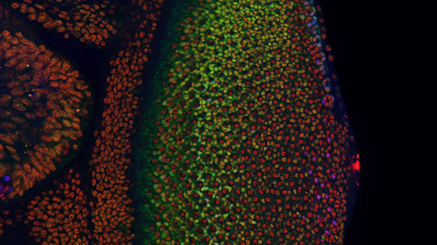
The crestin:EGFP transgene is specifically expressed in melanoma inBRAFV600E/p53 mutant melanoma-prone zebrafish. (Top) A single cell expressingcrestin:EGFP expands into a small patch of cells over the course of 2 weeks, capturing the initiation of melanoma formation (bracket). (Bottom) A fully formed melanoma specifically expresses crestin:EGFP, whereas the rest of the fish remains EGFP-negative.
Neural crest reporter expression in melanoma.
The crestin:EGFP transgene is specifically expressed in melanoma inBRAFV600E/p53 mutant melanoma-prone zebrafish. (Top) A single cell expands into a small patch of cells over the course of 2 weeks, capturing the initiation of melanoma formation (bracket). (Bottom) A fully formed melanoma specifically expresses crestin:EGFP, whereas the rest of the fish remains EGFP-negative.
The “cancerized field” concept posits that cancer-prone cells in a given tissue share an oncogenic mutation, but only discreet clones within the field initiate tumors. Most benign nevi carry oncogenic BRAFV600E mutations but rarely become melanoma. The zebrafish crestin gene is expressed embryonically in neural crest progenitors (NCPs) and specifically reexpressed in melanoma. Live imaging of transgenic zebrafish crestin reporters shows that within a cancerized field (BRAFV600E–mutant; p53-deficient), a single melanocyte reactivates the NCP state, revealing a fate change at melanoma initiation in this model. NCP transcription factors, including sox10, regulate crestin expression. Forced sox10 overexpression in melanocytes accelerated melanoma formation, which is consistent with activation of NCP genes and super-enhancers leading to melanoma. Our work highlights NCP state reemergence as a key event in melanoma initiation.
Most of the time skin moles are harmless, but they occasionally turn into melanoma, a life-threatening skin cancer. Leonard Zon and colleagues found that this happens when a single cell regresses back to a stem cell state and starts to divide and invade the surrounding tissue. (Image: Courtesy of Boston Children’s Hospital)
Researchers at Harvard-affiliated Boston Children’s Hospital have, for the first time, visualized the origins of cancer from the first affected cell and watched its spread in a live animal. Their work, published in the Jan. 29 issue of Science, could change the way scientists understand melanoma and other cancers and lead to new, early treatments before the cancer has taken hold.
“An important mystery has been why some cells in the body already have mutations seen in cancer, but do not yet fully behave like the cancer,” said the paper’s first author, Charles Kaufman, a postdoctoral fellow in the Zon Laboratory at Boston Children’s Hospital. “We found that the beginning of cancer occurs after activation of an oncogene or loss of a tumor suppressor, and involves a change that takes a single cell back to a stem cell state.”
That change, Kaufman and colleagues found, involves a set of genes that could be targeted to stop cancer from ever starting.
The study imaged live zebrafish over time to track the development of melanoma. All the fish had the human cancer mutation BRAFV600E — found in most benign moles — and had also lost the tumor suppressor gene p53.
Kaufman and colleagues engineered the fish to light up in fluorescent green if a gene called crestin was turned on — a “beacon” indicating activation of a genetic program characteristic of stem cells. This program normally shuts off after embryonic development, but occasionally, in certain cells and for reasons not yet known, crestin and other genes in the program turn back on.
“Every so often we would see a green spot on a fish,” said Leonard Zon, director of the Stem Cell Research Program at Boston Children’s and senior investigator on the study. “When we followed them, they became tumors 100 percent of the time.”
The cell that caused melanoma
When Kaufman, Zon, and colleagues looked to see what was different about these early cancer cells, they found that crestin and the other activated genes were the same ones turned on during zebrafish embryonic development — specifically, in the stem cells that give rise to the pigment cells known as melanocytes, within a structure called the neural crest.
“What’s cool about this group of genes is that they also get turned on in human melanoma,” said Zon, who is also a member of the Harvard Stem Cell Institute and a Howard Hughes Medical Institute investigator. “It’s a change in cell fate, back to neural crest status.”
Finding these cancer-originating cells was tedious. Wearing goggles and using a microscope with a fluorescent filter, Kaufman examined the fish as they swam around, shooting video with his iPhone. Scanning 50 fish could take two to three hours. In 30 fish, Kaufman spotted a small cluster of green-glowing cells about the size of the head of a Sharpie marker — and in all 30 cases, these spots grew into melanomas. In two cases, he was able to see on a single green-glowing cell and watch it divide and ultimately become a tumor mass.
“It’s estimated that only one in tens or hundreds of millions of cells in a mole eventually becomes a melanoma,” said Kaufman, who is also an instructor at the Harvard-affiliated Dana-Farber Cancer Institute. “Because we can also efficiently breed many fish, we can look for these very rare events. The rarity is very similar in both humans and fish, which suggests that the underlying process of melanoma formation is probably much the same in humans.”
Zon, the Grousbeck Professor of Pediatric Medicine at Harvard Medical School, and Kaufman believe that their findings could lead to a new genetic test for suspicious moles to see whether the cells are behaving like neural crest cells, indicating that the stem-cell program has been turned on. They are also investigating the regulatory elements that turn on the genetic program (known as super-enhancers). These DNA elements have epigenetic functions that are similar in zebrafish and human melanoma, and could potentially be targeted with drugs to stop a mole from becoming cancerous.
A paradigm shift for cancer?
Zon and Kaufman posit a new model for cancer formation, going back to a decades-old concept of “field cancerization.” They propose that normal tissue becomes primed for cancer when oncogenes are activated and tumor suppressor genes are silenced or lost, but that cancer develops only when a cell in the tissue reverts to a more primitive, embryonic state and starts dividing. They believe this model may apply to most if not all cancers, not just melanoma.
The study was supported by the National Institutes of Health, the National Institute of Arthritis and Musculoskeletal and Skin Diseases, the Ellison Foundation, the Melanoma Research Alliance, the V Foundation, and the Howard Hughes Medical Institute. Zon is a founder and stockholder of Fate, Inc., and Scholar Rock.
His collection isn’t very diverse, composed as it is entirely of zebrafish—minnow-like, freshwater, tropical fish that grow about an inch-and-a-half long. But Zon isn’t interested in winning best in show. Instead, he is leading a scientific movement to show that his striped fish could be humanity’s new animal of choice for drug discovery and studying disease.
Not only does the zebrafish require fewer facility resources than science’s go-to organism, the mouse, but fish are quicker to reproduce, easier to experimentally manipulate, and at least 70 percent of human protein-coding genes have analogs in the zebrafish, including those related to skin cancer, muscular dystrophy, and T cell leukemia.
“The zebrafish is now emerging as another powerful organism for the modeling and study of human diseases, and it is conceivable that zebrafish models will complement mouse models in the future,” Zon wrote in the December issue of Trends in Cell Biology, pointing out the accelerating increase in studies on zebrafish, from about 150 in 1995 to over 2,000 in 2013.
As a pediatric oncologist, Zon’s main purpose in using zebrafish is to help cancer patients. He is the first scientist to successfully apply basic research from the zebrafish to develop an FDA-approved treatment for human melanoma, and is now applying similar methods to find therapies for muscle and blood cancers.
One cancer Zon is pursuing is rhabdomyosarcoma, a rare muscle cancer diagnosed primarily in early childhood. Zebrafish, under certain conditions, develop tumors very similar to the human disease. He is currently using zebrafish embryos to identify which genes transform a normal muscle stem cell into a malignant tumor, as well as searching for factors that might suppress the cancer.
“One of the things that’s really interesting in zebrafish is that the embryos are completely transparent and you can watch the tumors invade the normal tissues,”
he said. “That’s a process that you can’t study in any other organism.”
Postdocs in Zon’s lab can expose the zebrafish embryos to multiple chemicals and literally watch changes in the fish’s development. In 2007, this technique led to the discovery of a type of prostaglandin that expands blood stem cells about 300 to 400 percent. Last fall, the prostaglandin found in Zon’s lab passed Phase Ib clinical trials as a therapy that increases the success of cord blood transplants.
A similar screen led to a major paper last November, in which Zon and fellow HSCI Executive Committee member Amy Wagers, PhD, showed that the same chemicals that stimulate muscle development in zebrafish can also be used to differentiate human stem cells into muscle cells in the laboratory, an historically challenging task that, now overcome, makes muscle cell therapy a more realistic possibility.
“This research demonstrates that over 300 million years of evolution, the pathways used in the fish are conserved through vertebrates all the way up to the human,” Zon said.
His passion for and success with zebrafish has helped make the animal a staple in HSCI faculty member laboratories across Boston, and an unexpected symbol for stem cell research.
Human muscle stem cell therapy gets help from zebrafish
November 7, 2013
Harvard Stem Cell Scientists have discovered that the same chemicals that stimulate muscle development in zebrafish can also be used to differentiate human stem cells into muscle cells in the laboratory, an historically challenging task that, now overcome, makes muscle cell therapy a more realistic clinical possibility.
The work, published this week in the journal Cell, began with a discovery by Boston Children’s Hospital researchers, led by Leonard Zon, MD, and graduate student Cong (Tony) Xu, who tested 2,400 different chemicals in cultures of zebrafish embryo cells to determine if any could increase the numbers of muscle cells formed. Using fluorescent reporter fish in which muscle cells were visible during their creation, the researchers found six chemicals that were very effective at promoting muscle formation.
Zon shared his results with Harvard Department of Stem Cell and Regenerative Biologyprofessor Amy Wagers, PhD, and Mohammadsharif Tabebordbar, a graduate student in her laboratory, who tested the six chemicals in mice. One of the six, called forskolin, was found to increase the numbers of muscle stem cells from mice that could be obtained when these cells were grown in laboratory dishes. Moreover, the cultured cells successfully integrated into muscle when transplanted back into mice.
Inspired by the successful application of these chemicals in mice, Salvatore Iovino, PhD, a joint postdoctoral fellow in the Wagers lab and the lab of C. Ronald Kahn, MD, at the Joslin Diabetes Center, investigated whether the chemicals would also affect human cells and found that a combination of three chemicals, including forskolin, could induce differentiation of human induced pluripotent stem (iPS) cells, made by reprogramming skin cells. Exposure of iPS cells to these chemicals converted them into skeletal muscle, an outcome the Wagers and Kahn labs had been striving to achieve for years using conventional methods. When transplanted into a mouse, the human iPS-derived muscle cells also contributed to muscle repair, offering early promise that this protocol could provide a route to muscle stem cell therapy in humans.
The interdisciplinary, cross-laboratory collaboration between Zon, Wagers, and Kahn highlights the advantage of open exchange between researchers. “If we had done this screen directly on human iPS cells, it would have taken at least 10 times as long and cost 100 times as much,” said Wagers. “The zebrafish gave us a big advantage here because it has a fast generation time, rapid development, and can be easily and relatively cheaply screened in a culture dish.”
“This research demonstrates that over 300 million years of evolution, the pathways used in the fish are conserved through vertebrates all the way up to the human,” said Wagers’ fellow HSCRB professor Leonard Zon, chair of the Harvard Stem Cell InstituteExecutive Committee and director of the stem cell program at Boston Children’s Hospital. “We can now make enough human muscle progenitors in a dish to allow us to model diseases of the muscle lineage, like Duchenne muscular dystrophy, conduct drug screens to find chemicals that correct those disease, and in the long term, efficiently transplant muscle stem cells into a patient.”
In a similar biomedical application, Kahn, who is chief academic officer at the Joslin, plans to apply the new ability to quickly produce muscle stem cells for diabetes research. His lab will generate iPS-derived muscle cells from people who are at risk for diabetes and people who have diabetes to identify alterations that lead to insulin resistance in the muscle.
Going forward, Zon plans to apply this platform of cross-species discovery to other stem cell lines, including those involved in blood and eye development. “We have a new system to use to study tissue development, and it’s not just muscle that can be studied, every single organ can be studied in the zebrafish system,” he said.
Photo:Newly created muscle progenitor cells (green) and muscle fibers (red) in a zebrafish embryo. (credit: Zon Lab)
Dynamics and heterogeneity of a fate determinant during transition towards cell differentiation
NicolásPeláez
Department of Molecular Biosciences, Northwestern University, Evanston, United States; Department of Chemical and Biological Engineering, Howard Hughes Medical Institute, University Northwestern, Evanston, United States
Contribution: Conception and design; Acquisition of data; Analysis and interpretation of data; Drafting or revising the article
No competing interests declared
” data-author-inst=”NorthwesternUniversityUnitedStates|HowardHughesMedicalInstituteUniversityNorthwesternUnitedStates”>NicolásPeláez, et al. eLife 2015;4:e08924 http://dx.doi.org/10.7554/eLife.08924.001
Yan is an ETS-domain transcription factor responsible for maintaining Drosophila eye cells in a multipotent state. Yan is at the core of a regulatory network that determines the time and place in which cells transit from multipotency to one of several differentiated lineages. Using a fluorescent reporter for Yan expression, we observed a biphasic distribution of Yan in multipotent cells, with a rapid inductive phase and slow decay phase. Transitions to various differentiated states occurred over the course of this dynamic process, suggesting that Yan expression level does not strongly determine cell potential. Consistent with this conclusion, perturbing Yan expression by varying gene dosage had no effect on cell fate transitions. However, we observed that as cells transited to differentiation, Yan expression became highly heterogeneous and this heterogeneity was transient. Signals received via the EGF Receptor were necessary for the transience in Yan noise since genetic loss caused sustained noise. Since these signals are essential for eye cells to differentiate, we suggest that dynamic heterogeneity of Yan is a necessary element of the transition process, and cell states are stabilized through noise reduction.
As animal cells develop, they pass through different states to mature into specific cell types. For some cells, this development depends on the cell’s ability to switch between two stable states, a property called bistability.
Many bistable systems operate during development and often feature proteins called transcription factors that regulate a few cell states in specific tissues. These proteins activate or repress a range of target genes, and cells can adjust the levels of the transcription factors to bring about transitions between states. In fruit flies, two transcription factors, called Yan and Pnt, regulate numerous processes throughout development.
In the developing embryo of a fruit fly, Yan and Pnt are regulated by signals induced by a receptor called EGFR. When EGFR is activated, Pnt is produced and Yan is degraded. When this receptor is not activated, Yan is produced and represses Pnt. Mathematical modelling of these interactions has concluded that this is a bistable system: that is, cells should either have high levels of Yan and low levels Pnt, or low Yan levels and high Pnt levels. However, larval eye cells first activate, and then turn off, both proteins together. This argues against bistability and raises questions about how these proteins regulate cell fate transitions in the eye, and perhaps in other organs.
To investigate this question, Peláez et al. tagged Yan with a fluorescent marker to track its activity in the eyes of fruit fly larvae as they develop. A combination of fluorescence-based microscopy and an automated imaging analysis system were then used to score fluorescence in thousands of individual eye cells and assess the changes in the levels of Yan over time. This approach revealed some unexpected results.
Yan levels were seen to vary in both immature and maturing cells. Thus eye cells transition between states as Yan levels increase rapidly, suggesting the need for a mechanism that is distinct from bistability. Peláez et al. suggest that larger changes in the seemingly random fluctuations in the levels of the transcription factor (also known as “expression noise”) might play a role in this mechanism. In particular, Yan expression noise briefly peaks as cells transition to a more mature state, and the transient nature of this ‘noisy’ response requires the activation of EGFR.
One possible explanation for these observations is that Yan’s effect on cell states depends on this variability in its levels, which might prime cells to change states when they receive another signal. These findings also raise many questions for future studies to explore, including how this increase in the noise level of Yan is triggered as cells begin their transitions towards specific cell types.
Introduction
Cells within complex organisms exist in different states that confer specific functionalities to each cell. Cellular states can be organized into cyclic cascades such as the G1-S-G2-M cell cycle, or into linear cascades as observed in cell differentiation. A common feature of cells that undergo state transitions is the apparent irreversibility of the transitions even when such transitions are triggered by transient stimuli. Modeling of these transitions often assumes system bistability, in which cells can be resting in one of two stable states.
Animal cells frequently utilize transcription factors to enforce a given state, and transitions to another state are driven by increasing or decreasing the levels of these transcription factors (Yao et al., 2008;Kueh et al., 2013; Laslo et al., 2006; Park et al., 2012). The Rb-E2F pathway generates bistability in E2F expression, which dictates the transition from G1 to S phase (Yao et al., 2008). Expression of the transcription factor PU.1 determines lymphoid versus myeloid hematopoietic cell lineages (Kueh et al., 2013; Laslo et al., 2006). Adipocyte differentiation is controlled by differential expression of C/EBP and PPARγ proteins (Park et al., 2012). Regulation by positive feedback is a hallmark of bistable systems, and in all of the above cases, the transcription factors act in one or more positive feedback circuits. In some systems, positive feedback is generated by two transcription factors that mutually repress each other’s expression. In these scenarios, cells in one state continually express a transcription factor that represses a second transcription factor, and when these cells transit to another state, they continually express the second transcription factor, which represses its antagonist. Fate restriction in hematopoietic, neural, pancreatic, and muscle cell lineages is regulated by such double-negative feedback circuits (Briscoe et al., 2000; Laslo et al., 2006; Olguin et al., 2007; Schaffer et al., 2010).
Most examples of this type of bistable mechanism have transcription factors that act specifically within a handful of cell states limited to a single tissue or organ system. One remarkable exception to this rule is found in Drosophila. There, two ETS-domain transcription factors act in a wide assortment of cell types across the body and across the life cycle. Yan and Pointed (Pnt) act downstream of signals mediated by receptor tyrosine kinases (RTKs) that regulate cell differentiation, migration, and division in tissues ranging from ovarian follicular cells, dorsal and ventral neuroectoderm, embryonic mesoderm, the embryonic trachea, and the post-embryonic compound eye (Dumstrei et al., 1998;Gabay et al., 1996; Halfon et al., 2000; Jurgens et al., 1984; Morimoto et al., 1996; O’Neill et al., 1994; Ohshiro et al., 2002; Yao et al., 2008) It is thought that Yan and Pnt control such diverse cell states by acting in concert with tissue-specific transcription factors to regulate transcription of appropriate target genes. For instance, transcription of even-skipped occurs in mesoderm only if Yan/Pnt act in combination with Tinman and Twist proteins (Halfon et al., 2000), whereas transcription of prospero (pros) in the eye only occurs if Yan/Pnt act in combination with Lozenge, Sine Oculis, and Glass proteins (Hayashi et al., 2008; Xu et al., 2000).
Several tissues show mutually exclusive expression of Yan and Pnt, suggestive of cross-repression (Boisclair Lachance et al., 2014). The best characterized is the embryonic ventral ectoderm, where ventral-most cells express Pnt and more lateral cells express Yan (Gabay et al., 1996). This pattern is established by secretion of a ligand for the EGF Receptor (EGFR) from the ventral midline (Golembo et al., 1996). EGFR activation in nearby ventral cells induces the Ras-MAPK pathway to express Pnt and degrade Yan (Gabay et al., 1996; Melen et al., 2005). Cells with insufficient EGFR activation express Yan, which represses Pnt. Mathematical modeling has described this as a bistable system in which cells are either in a High Yan/Low Pnt state or a Low Yan/High Pnt state (Graham et al., 2010; Melen et al., 2005). Transition from one state to the other is ultrasensitive to the strength of EGFR activation (Melen et al., 2005).
Paradoxically, other Drosophila tissues show co-expression of Yan and Pnt (Boisclair Lachance et al., 2014). The larval eye is one such tissue. Retinal progenitor cells initiate expression of both proteins, and when they transit to differentiated photoreceptor fates, these cells reduce expression of both proteins. In contrast, when retinal progenitor cells transit to differentiated cone cell fates, they maintain their expression of both proteins. These observations are at odds with long-standing genetic studies that support a standard bistable mechanism acting in the eye (Lai and Rubin, 1992;O’Neill et al., 1994; Rebay and Rubin, 1995). Thus, new approaches to studying these transitions in the eye are needed.
Here, we have adopted a systems-level approach to study Yan dynamics in the larval eye. A yellow fluorescent protein (YFP) — based isoform of Yan was developed as a reporter for Yan protein levels. Fluorescence-based microscopic imaging of cells was coupled with automated high-throughput image analysis to score fluorescence in each cell and annotate the data in a quantitative and unbiased fashion. Yan exhibits monostability, both in progenitor and differentiating cells, with Yan levels varying in cells in either state. Cell state transitions occur independent of absolute Yan concentrations, suggesting that some other mechanism allows Yan to regulate transitions. One such mechanism might be the noise in Yan levels, which undergoes a transient spike as cells begin to transition to differentiated states. Loss of EGFR signaling, which prevents cells from differentiating, causes these cells to have prolonged noisy Yan expression, and suggests that Yan noise is key for cell state transitions in the eye.
Results
The compound eye epithelium is established during embryogenesis as an internal disc of cells called the eye imaginal disc (Wolff and Ready, 1993). During the larval phase of the life cycle, the disc grows in size by asynchronous cell division. During the final 50 hr of the larval phase, a morphogenetic furrow (MF) moves across the eye disc from posterior to anterior (Figure 1A,B). All cells arrest in G1 phase within five cell diameters anterior to the furrow, and then as the furrow passes through them, periodic clusters of cells express the proneural gene atonal (Jarman et al., 1994). Atonal expression is subsequently restricted to one cell per cluster, which becomes the R8 photoreceptor. Each R8 cell then secretes an EGFR ligand that activates the receptor in neighboring cells and causes them to transit from multipotent progenitor to differentiated states (Figure 1C) (Freeman, 1996). Transitions occur in a sequence of symmetric pairs of multipotent progenitor cells that differentiate into R2/R5, R3/R4, and R1/R6 photoreceptors (Figure 1C) (Wolff and Ready, 1993). Thereafter, a single progenitor transits to a R7 photoreceptor fate followed by two pairs of cells, C1/C2 and C3/C4, that differentiate into cone cells. These cone cells are non-neuronal and form the simple lens that overlies each cluster of eight photoreceptors. The furrow induces the nearly simultaneous differentiation of a column of R8 cells, with repeated column inductions producing approximately 800 units or ommatidia as the furrow moves across the eye.
Figure 1.Development and patterning of the compound eye.
(A) Differentiation is initiated in the developing eye by the MF, which moves across the eye epithelium. On the furrow’s posterior side, G1-arrested progenitor cells undergo differentiation (light blue). On the anterior side, progenitor cells are still proliferating (dark blue). The large grey rectangle outlines the region that was analyzed for Yan levels; the small rectangle corresponds to the developmental sequence outlined in panel C (B) A maximal projection of Yan-YFP fluorescence in an eye. Bar = 100 μm. (C) Top, an apical view of the sequential differentiation of eight photoreceptor (R1-R8) and four cone cell types (C1-C4) from multipotent progenitor cells (grey) in an ommatidium. Arrows denote inductive signal transmitted from the R8 to activate EGFR on nearby cells. Bottom, a cross-section view through an eye disc adapted after Wolff and Ready (1993). Note the progenitor cell nuclei are basally positioned, and as they transition into a differentiated state, their nuclei migrate apically. C1/C2 cells are positioned anterior and posterior in the ommatidium while C3/C4 cells are positioned equatorial and polar in the ommatidium. (D) Top, an optical slice of H2Av-mRFP fluorescence in an eye disc at a plane that bisects progenitor cell nuclei. Bottom, the same optical slice imaged for Yan-YFP fluorescence. Bars = 8 μm
A central tenet of the bistable model of cell differentiation in the eye posits that differentiation is marked by a transition from high Yan protein levels in multipotent progenitor cells to low Yan levels in differentiating cells (Graham et al., 2010). Formulation of this model stemmed from studies of R7 cell differentiation, the final photoreceptor recruited to each ommatidium. Reduced Yan causes inappropriate expression of the R7 determinant pros and ectopic R7 cells in yan hypomorphic mutants (Kauffmann et al., 1996; Lai and Rubin, 1992). Conversely, a Yan isoform that is resistant to MAPK-dependent degradation, blocks R7 differentiation and pros expression (Kauffmann et al., 1996; Rebay and Rubin, 1995).
Quantifying Yan dynamics
To quantitatively test the bistable model, we used BAC recombineering to insert fast-fold yellow fluorescent protein (YFP) in-frame at the carboxy-terminus of the yan coding sequence (Webber et al., 2013). The Yan-YFP transgene fully complemented null mutations in the endogenous yan gene and completely restored normal eye development (Figure 1—figure supplement 1), demonstrating that the YFP tag does not compromise Yan function and that all essential genomic regulatory sequences are included. The pattern of Yan-YFP protein expression was qualitatively similar to that of endogenous Yan (Figure 1—figure supplement 1).
We used histone His2Av-mRFP fluorescence in fixed specimens to mark all eye cell nuclei for automated segmentation (Figure 1D—figure supplement 2). Nuclei were manually classified into the different cell types of the eye, which is possible because every cell undergoing differentiation can be unambiguously identified by its position and nuclear morphology without the need of cell-specific markers (Ready et al., 1976; Tomlinson, 1985; Tomlinson and Ready, 1987; Wolff and Ready, 1993). To validate our identification of all cell types using this method, we cross-checked with specific cell-specific markers, and found that our classification was accurate over 98% of the time (Figure 1—figure supplement 3). Cells were scored for nuclear Yan-YFP concentration and their exact position within 3D coordinate space of each fixed eye sample (Figure 1—figure supplement 2). Yan-YFP protein levels were normalized to His2Av-mRFP, which provided some control over measurement variation (Figure 1—figure supplement 4). We then mapped each cell’s spatial position in the x-y plane of the eye disc onto time. Two reasons made this possible. First, the furrow moves at an approximately constant velocity forming one column of R8 cells every two hours (Basler and Hafen, 1989; Campos-Ortega and Hofbauer, 1977). Second, each column of R8 cells induces the other cell fates at a constant rate (Lebovitz and Ready, 1986). Thus even in a fixed specimen, the temporal dynamics of cell state transitions are visible in the repetitive physical organization of ommatidia relative to the furrow (Figure 1C). Together these features allow the estimation of time based on a cell’s position relative to the furrow (Figure 2—figure supplement 1). This can be applied repeatedly to hundreds of cells in a single sample, creating a composite picture of the dynamics (Figure 2B–F). Although the developmental progression of an individual cell cannot be measured by this approach, it nevertheless provides a dynamic view of hundreds of cells across a developing epithelium as they respond to signaling processes. From this information, individual cell behaviors can be reconstructed and modeled.
(A) Average time at which initiation of differentiation is first detected by apical migration of committing cell nuclei. Time zero is set to when R8 differentiation initiates. Differentiation proceeds over a time course after commitment is initiated (horizontal arrows) (B) Yan-YFP fluorescence in multipotent progenitor cells. We fit a Hill function (blue curve) to the inductive phase and an exponential decay (black curve) to the decay phase. (C-F) Scatter plots of Yan-YFP levels in individual cells for R2/R5 (C), R3/R4 (D), R7 (E), and C3/C4 (F) cells. These are overlaid with scatter plots of Yan-YFP in multipotent cells at times preceding and coincident with the appearance of the relevant differentiated cells. Note the similar Yan-YFP levels between multipotent cells and differentiating cells at their first appearance. (G) Moving averages of Yan-YFP levels for multipotent and photoreceptor cells. Gaps between the multipotent and photoreceptor curves are due to the window size for line averaging. (H) Moving averages of Yan-YFP in multipotent and cone cells.
Yan-YFP expression in multipotent progentior cells was biphasic (Figure 2B). Cells anterior to the furrow expressed a basal level of Yan-YFP, but this level dramatically increased in cells immediately anterior to the furrow, starting eight hours before the first R8 cells were identifiable. Approximately eight hours after R8 definition, Yan-YFP levels peaked, and thereafter gradually decayed until Yan-YFP approached its basal level again. The results are surprising in two ways. First, Yan-YFP is not maintained at a stable steady state within progenitor cells, which would have been predicted by the bistable model. Rather, its dynamics are reminiscent of monostable responses to transient stimuli, with a single basal steady state. Second, at the peak of Yan-YFP expression, there is remarkably large heterogeneity in Yan-YFP levels across cells.
We also followed Yan-YFP dynamics in cells as they transited into a differentiated state and thereafter. Again, the results did not follow the expectations predicted by the bistable model. First, progenitors transited to a differentiated state at levels of Yan-YFP that varied, depending upon the type of differentiated state being adopted (Figure 2C–H). Cells entering the R3/R4 and R1/R6 states began with Yan-YFP levels that were approximately two-fold greater than cells entering the R2/R5 states. Cells entering the R7 state were intermediate between these two extremes. Despite these differences at the transition point, Yan-YFP levels decayed to a similar basal steady state irrespective of the photoreceptor type, and this basal level was at least three-fold lower than that which the progenitor cells achieved (Figure 2G). Thus, rather than the single high Yan progenitor state previously modeled, our results suggest a dynamic range of high Yan states from which different cell fates are specified according to the spatio-temporal sequence of differentiation.
We noted that for most photoreceptors, it took approximately 20 hr for Yan-YFP to decay to the basal steady state (Figure 2G), significantly longer than had been previously modeled (Graham et al., 2010). Since expression of several neural-specific genes is detected 2–8 hr after the transition (Tomlinson and Ready, 1987; Van Vactor et al., 1988), the slower than anticipated Yan decay indicates that early differentiation does not require cells to have assumed a basal steady-state level of Yan-YFP.
The last group of progenitors to differentiate form the non-neuronal cone cells. We also measured Yan-YFP in those cells. Yan-YFP dynamics in cone cells were more similar to progenitor cells over the same time period (Figure 2F,H). This behavior was in contrast to photoreceptors, which exhibited different decay dynamics from progenitor cells. Thus, accelerated degradation of Yan-YFP is not essential for cells to transition to all retinal cell states.
EGFR-ras signaling regulates Yan-YFP dynamics
The bistable model posits that the switch from one state to another is triggered by a signal that progenitor cells receive though the EGFR protein. Given the unanticipated Yan-YFP dynamics, we wanted to ask whether and how they were influenced by EGFR signaling. EGFR null mutants are inviable; however, a temperature sensitive (ts) allele of EGFR enables controlled inactivation of the RTK’s activity (Kumar et al., 1998). We grew EGFRts mutant larvae at a restrictive temperature for 18 hr before analyzing effects on Yan-YFP. Surprisingly, progenitor cells exhibited biphasic expression of Yan-YFP over time, but the amplitude of the pulse in expression was significantly reduced (Figure 3A). This suggests that EGFR signaling contributes to the stimuli that induce the Yan-YFP peak. To further test this hypothesis, we misexpressed a constitutively active form of Ras protein in eye cells. The peak of Yan-YFP in progenitors was now prolonged (Figure 3B). Together, these results suggest that EGFR-Ras signaling stimulates the transient appearance of Yan-YFP in progenitor cells, and that the decline in Yan-YFP within older progenitor cells is linked to a loss of signal reception by these cells over time.
Figure 3.EGFR/Ras and Pnt regulate Yan-YFP levels.
(A–D) Moving averages of Yan-YFP in different cell types. Shown with shading is the standard error of the mean for each moving average. (A,C) Wildtype and EGFRts mutants incubated at the non-permissive temperature and analyzed for progenitors (A) and R2/R5 cells (C). (B,D) Wildtype and sev>Rasv12 mutants were analyzed for progenitors (B) and R2/R5 cells (D). (E) Optical slice through progenitor cell nuclei in a disc that contains clones ofpnt2 mutant cells. Left, fluorescence of RFP, which positively marks wildtype cells and not pnt2 mutant cells. Middle, Yan-YFP fluorescence, showing reduced levels in pnt2mutant clones. Arrows highlight apoptotic nuclei. Right, merged image with Yan-YFP in green and RFP in purple. Clone borders are outlined. (F,G) Moving averages of Yan-YFP in R3/R4 cells that ectopically express PntP1 (F) or PntP2-VP16 (G) due to LongGMR-Gal4 driving the UAS transgenes. Since PntP2 requires MAPK phosphorylation to become transcriptionally active, we misexpressed a VP16 fusion of PntP2. PntP1 is constitutively active (Brunner et al., 1994; Gabay et al., 1996).
We next examined the effects of EGFR and Ras on Yan-YFP dynamics in cells as they differentiate. The bistable model predicts that EGFR is required for the loss of Yan-YFP in photoreceptors. Indeed,EGFRts mutant R2/R5 cells delayed their initial decline in Yan-YFP levels (Figure 3C). Conversely, misexpression of constitutively active Ras caused a premature decline in Yan-YFP (Figure 3D). These results are consistent with EGFR-Ras stimulating the degradation of Yan-YFP as cells switch their states. However, Yan-YFP dropped to below-normal levels in EGFRtsmutant R2/R5 cells (Figure 3C). These complex effects suggest a dual role for EGFR signaling in photoreceptors. In the first few hours as cells transit to a photoreceptor state, EGFR stimulates the accelerated decay of Yan-YFP. Thereafter, EGFR inhibits the decay of Yan-YFP in a manner that might be related to that role that EGFR plays in boosting Yan-YFP levels in progenitor cells.
The source of EGFR ligand originates from the R8 cell (Freeman, 1996). If this diffusive ligand is responsible for controlling Yan-YFP levels in other photoreceptors as they are recruited to an ommatidium, we would predict a correlation between Yan-YFP levels in differentiating cells and their distances from adjacent R8 cells. Indeed, at the times when R2/R5 cells differentiate (~0–15 hr), their Yan-YFP levels are positively correlated (p<0.01) with their physical distance to the nearest R8 cells (Figure 3—figure supplement 1). These correlations are absent in EGFRts mutants, providing evidence that R8 cells act through EGFR to control Yan-YFP dynamics in differentiating cells.
Pnt regulates Yan dynamics
Pnt proteins have been hypothesized to cross-repress Yan expression, and this double negative feedback loop is thought to be necessary for bistability (Graham et al., 2010; Shilo, 2014). At odds with this view, Pnt and Yan proteins are co-expressed in progenitor and differentiating cells of the eye (Boisclair Lachance et al., 2014). Since Pnt proteins act downstream of many RTKs including EGFR, we wondered if Pnt mediated the positive effects of EGFR-Ras signaling on Yan-YFP in progenitor cells. Null mutants of the pnt gene are embryonic inviable; therefore we generated clones of eye cells that were null mutant for pnt in an otherwise wildtype animal. Mutant progenitor cells showed a reduction in Yan-YFP levels as they progressed through their biphasic expression pattern (Figure 3E). Thus, Pnt possibly mediates the positive effect of EGFR signaling on Yan-YFP expression in progenitors. We also wished to know if Pnt mediates the complex effects of EGFR in differentiating photoreceptors. Pnt proteins are rapidly cleared in photoreceptors (Boisclair Lachance et al., 2014) and so loss-of-function mutant analysis would be uninformative. Therefore, we overexpressed PntP1 or constitutively-active PntP2 in cells as they transited into a photoreceptor state and beyond. The early phase of Yan-YFP decay was accelerated while the later phase of Yan-YFP decay was inhibited (Figure 3F,G). These complex effects are precisely the opposite to those caused by loss of EGFR signaling, as would be expected if Pnt mediated EGFR’s complex effects on Yan-YFP dynamics in photoreceptor cells.
Cells are indifferent to absolute levels of Yan
The bistable model predicts that different cell states depend upon discrete absolute concentration of Yan present inside cells. To test this idea, we varied the number of Yan-YFP gene copies. In general, protein output is proportional to gene copy number in Drosophila (Lucchesi and Rawls, 1973). We increased Yan-YFP copy number from its normal diploid number to tetraploid, and monitored Yan-YFP in progenitors and differentiating cells. As expected, four copies caused a higher steady-state level of Yan-YFP in progenitor cells, though this increase was less than two-fold (Figure 4A). The amplitude of the Yan-YFP pulse was also increased as progenitor cells aged. Strikingly, as four-copy progenitor cells transited to a differentiated state, the onset of Yan-YFP decay occurred at the same time as it occurred for two-copy cells (Figure 4A–C). Yan-YFP levels were much greater in four-copy cells compared to two-copy cells making their transit into the identical cell states. To confirm that absolute Yan-YFP concentration had little effect on cell state transitions, we examined expression of a direct target of Yan in R7 cells: the pros gene (Kauffmann et al., 1996; Xu et al., 2000). Expression was monitored with an antibody specific for Pros protein. We observed at most a one hour delay in the onset of Pros expression in R7 cells containing four copies of Yan-YFP (Figure 4D), far less than the ten-hour delay predicted if absolute concentration of Yan-YFP dictated when Pros expression begins (Figure 4D).
Figure 4.Cell state transitions are unaffected by Yan-YFP gene copy number.
(A–C) Moving averages of Yan-YFP in eye discs containing two versus four copies of the Yan-YFP transgene. (A) R2/R5 and progenitor cells. (B) R3/R4 and progenitor cells. (C) R7 and progenitor cells. (D) Moving averages of Yan-YFP and Pros proteins in R7 cells containing either two vs. four copies of the Yan-YFP transgene.
Possibly, absolute concentration of Yan is unimportant when a cell transits to a different state, but the switch to a constant basal Yan level is robustly maintained regardless of starting concentration. An examination of Yan-YFP decay in photoreceptor cells makes that possibility less likely; Yan-YFP decays to different basal levels in two- versus four-copy differentiated cells (Figure 4A–C). To further test this notion, we fit the data to several plausible functional forms. We found that exponentially decaying functions systematically best-fit to the data (Figure 4—figure supplement 1). Thus, for each cell state we fit an exponential decay function to its Yan-YFP temporal profile (Figure 4—figure supplement 2). From these fits, we derived the average decay rate constants and half-lives of Yan-YFP for cells carrying two, four, or six copies of yan. As expected, we found that Yan-YFP half-life was different between progenitors and differentiating photoreceptors (Figure 5—figure supplement 1). The half-life in photoreceptors was two-fold lower than in progenitors, accounting for the more rapid loss of Yan-YFP in the former cells. Strikingly, Yan-YFP half-life was not significantly affected byyan copy number within either progenitor or photoreceptor cells (Figure 5). Thus, Yan-YFP concentration only affected its decay rate as a first-order function, implying that there is no higher order mechanism to accelerate Yan-YFP decay when cells contain greater concentrations of Yan-YFP.
Figure 5.Yan protein half-life is largely unaffected by yan gene copy number.
Exponential decay of Yan-YFP levels. Note the robustness of Yan-YFP half-lives across replicates and yan gene copy number. Note also how half-lives are nearly twice as long for progenitor cells versus photoreceptor neurons.
As a final test of the effects of Yan-YFP levels on cell states, we allowed 4X and 6X yan animals to complete eye development and then examined the external patterning of the fully differentiated compound eye. The compound eyes were completely normal in appearance (Figure 5—figure supplement 2), implying that differentiation was unaffected by the absolute concentrations of Yan inside eye cells.
Yan expression noise spikes during cell state transitions
Our results indicate that Yan’s effects on retinal cell states are not dictated by uniform and stable concentrations of Yan protein. One potential explanation is that Yan’s effect on cell states actually depends on variability in Yan protein levels. A growing body of evidence is pointing to the importance of transient fluctuations in expression of factors to control cell states (Cahan and Daley, 2013;Torres-Padilla and Chambers, 2014). Key regulators of stem cells fluctuate in abundance over time, and this is correlated with stem cells existing in multiple connected microstates, with the overall structure of the population remaining in a stable pluripotent macrostate (MacArthur and Lemischka, 2013). Heterogeneity among cells is not simply due to independent noise in expression of individual genes but is due to fluctuating networks of regulatory genes (Chang et al., 2008; Kumar et al., 2014). Such fluctuations appear to be stochastic in nature, priming cells to transit into differentiated states with a certain probability that is guided by extrinsic signals.
Our data revealed considerable heterogeneity in the level of Yan-YFP among cells at similar developmental times (Figure 2B). To quantify the noise, we used developmental time to bin cells of similar age, and measured detrended fluctuations of Yan-YFP by calculating residuals to the fitted function and normalizing binned residuals to the function (Goldberger et al., 2002). Progenitor cells showed a spike in Yan-YFP noise as they began to induce Yan-YFP expression (Figure 6A). The noise spike was short-lived (approximately 17 hr), and noise thereafter returned to a basal level with secondary minor spikes. The major peak in noise magnitude coincided with the time at which R8 cells are formed.
Figure 6. Noise in Yan-YFP expression is highly dynamic.
Moving averages of Yan-YFP levels and noise (detrended fluctuations) for (A) progenitors, (B) R2/R5, (C) R3/R4, (D) R1/R6, and (E) R7 cells. (F) Comparative noise dynamics for all cells analyzed in (A–E). (G) Moving averages of Yan-YFP noise (coefficient of variation) in R2/R5 cells sampled from wildtype and EGFRts mutant eyes at the non-permissive temperature. Shown with shading is the standard error of the mean for each moving average. (H) Moving averages of Yan-YFP noise (coefficient of variation) in R2/R5 cells sampled from wildtype and sev>Rasv12 mutant eyes. Shown with shading is the standard error of the mean for each moving average.
Photoreceptor cells showed a large spike in Yan-YFP noise as they began to differentiate (Figure 6B–E). The magnitude of each noise peak varied with the photoreceptor cell state; R3 and R4 cells exhibited the greatest amplitude in noise (Figure 6F). These noise spikes showed a distinct temporal relationship, with spikes coinciding with the times at which individual cell states were switched (Figure 6F). Thus, the noise spikes are not a simple consequence of a global stimulus synchronously affecting noise in all cells. Thereafter, all cells reduced Yan-YFP noise to a basal level that was comparable to basal noise in the progenitor cells. However, each cell type exhibited a secondary minor spike at 30–35 hr, which might reflect a synchronous stimulus.
Detrended fluctuation is one method to measure expression variability, but it can suffer from distortion if the model fitting is not adequately weighted. Therefore, we also calculated the coefficient of variation, that is, the standard deviation of Yan-YFP intensity within a sliding window divided by its mean. This method yielded noise profiles with transient spikes for each cell type that was consistent with calculations using detrended fluctuation (Figure 6—figure supplements 1 and 2). However, while the coefficient of variation yielded results that varied strongly with bin width, the detrended fluctuations yielded profiles that were generally robust against changes in bin width (Figure 6—figure supplement 1).
To rule out the possibility that these unexpectedly dynamic features of Yan-YFP were caused by its transgenic origins or fusion with YFP, we compared Yan-YFP dynamics to those of endogenous Yan protein that was bound with an anti-Yan antibody. The profiles of Yan-YFP protein levels and noise were highly similar to endogenous Yan protein levels and noise, in both multipotent and differentiating cells (Figure 6—figure supplement 3). Thus, transient spikes of expression heterogeneity are a fundamental feature of Yan protein.
Since Yan regulates Pros expression in R7 cells, it was possible that Pros showed a transient noise spike as a consequence. Therefore, we measured Pros protein heterogeneity and found that its dynamics did not resemble that of Yan (Figure 6—figure supplement 4). Instead, Pros noise was high starting at the onset of expression, and thereafter gradually declined as Pros protein levels increased in R7 cells. We conclude that noise spikes are not a general feature of gene expression in the developing eye but might reflect unique roles of Yan in mediating cell state transitions.
We wondered what might cause these spikes in Yan-YFP noise during cell state transitions. Because EGFR signaling is important for regulating Yan-YFP concentration during these transitions (Figure 3), we analyzed Yan-YFP noise when EGFR signaling was inhibited in EGFRts mutant animals raised at a non-permissive temperature. The noise spike in progenitor cells was not significantly affected by loss of EGFR signaling (data not shown). We also examined the effects of EGFRts on noise in differentiating photoreceptor cells. Interestingly, noise increased at the normal time of transition but the elevated noise did not quickly drop to basal levels (Figure 6G). Rather, high noise was extended for an additional 10 to 15 hr. Conversely, misexpressing constitutively active Ras within differentiating cells caused a premature dropdown in Yan-YFP noise (Figure 6H). These results indicate that EGFR/Ras signaling is required for the rapid drop in Yan-YFP noise after it has peaked, creating a transient spike.
Discussion
This study relied upon a set of methods that enable systems-level analysis of Yan expression dynamics in a developing animal tissue. Transgene recombineering was used to insert YFP into a genomic rescue fragment of the yan gene, which fully replaced endogenous yan. Yan-YFP protein was quantified in thousands of individual cells by fluorescence confocal microscopy and automated cell segmentation analysis. Based on the unique features of Drosophila eye development, every analyzed cell was assigned an age, and composites of cells across a time-spectrum of ages were derived. This allowed us to reconstruct the temporal dynamics of Yan protein expression in cells as they transited from one state to another or were maintained in a given state. The fact that both Yan concentration and noise were easily measured using our approach indicates that it provides a powerful method for studying how other molecular determinants regulate cell states.
Contrary to what is currently believed, the expression of Yan in progenitor cells has many hallmarks of monostability. A stable basal state exists in cells anterior to the furrow, and when the furrow passes, Yan rises and falls to form a biphasic profile (Figure 7A). If cells transit towards differentiation, then the fall in Yan is accelerated but the fundamental biphasic profile is preserved. This monostable-like behavior is not like a behavior where progenitor cells exist in a high Yan stable-state and switch to a low Yan stable-state when they transit towards differentiation (Figure 7A). Other lines of evidence also point away from a bistable switch mechanism. Yan reaches its basal steady state many hours after cells have adopted their differentiated photoreceptor state and are executing specialized gene expression programs. Thus, Yan levels are variable at the time when cells actually become differentiated.
(A) Top: a hypothetical bistable behavior would be where Yan is in a stable high state within progenitor cells and in a stable low state within differentiated cells. Bottom: the observed behavior of Yan appears monostable, with both progenitors and differentiated cells in unstable Yan states. (B) Heterogeneity in Yan expression is maximal when progenitor cells enter a transition state that resolves to a more homogeneous differentiated state, dependent upon EGFR signaling. This heterogeneous transition state may be a primary mechanism for Yan’s effect on cells, independent of the absolute Yan concentration within progenitors.
…..-> end
Illuminating a Cancer’s Origins
Researchers have developed a technique to visualize the origin of melanoma in zebrafish, throwing light on a genetic switch for cancer.
By Catherine Offord | February 1, 2016
Many cells containing cancer-associated genes never become tumors. Using a fluorescent reporter to highlight cells activating stem cell-like patterns of gene expression, researchers at Boston Children’s Hospital have now developed a technique to visualize the origins of tumors in zebrafish. The team’s findings were published last week (January 29) in Science.
“An important mystery has been why some cells in the body already have mutations seen in cancer, but do not yet fully behave like the cancer,” Charles Kaufman of Boston Children’s Hospital said in a statement. “We found that the beginning of cancer occurs after activation of an oncogene or loss of a tumor suppressor, and involves a change that takes a single cell back to a stem cell state.”
The researchers focused on a gene present in vertebrates called crestin. This gene is expressed normally only in neural crest progenitor cells, which give rise to a number of other cell types during embryogenesis. However the gene is also expressed in melanoma tumor cells, making it a clear marker of the onset of cancer.
Injecting a fluorescent crestin reporter into transparent zebrafish carrying mutations in melanoma genes, the researchers found they were able to visualize the moment that precancerous cells activated stem cell-like patterns of gene expression.
“Every so often we would see a green spot on a fish,” said study coauthor Leonard Zon of Boston Children’s Hospital in the statement. “When we followed them, they became tumors 100 percent of the time.”
“It’s a significant advance in the field,” Ze’ev Ronai, the scientific director of the Sanford Burnham Prebys Medical Discovery Institute in La Jolla, California, told The New York Times. “They provide a pretty strong demonstration that that pathway is correct.”
The team will now focus on the pathways regulating the switch in gene expression, according to the statement—research that Kornelia Polyak of the Dana-Farber Cancer Institute told the Times would be essential to develop drugs targeting melanoma.
“What is the stimulus that allows a single cell out of millions to become cancerous?” she asked. “What is the trigger and why does it happen so rarely? Why, why is this happening?”
Fluorescence, nucleic acid strands help show computing operations inside a living cell
Using strands of nucleic acid and a fluorescence reporter, scientists at the Georgia Institute of Technology (Georgia Tech; Atlanta, GA) and colleagues have demonstrated basic computing operations inside a living mammalian cell. The research could lead to an artificial sensing system that could control a cell’s behavior in response to such stimuli as the presence of toxins or the development of cancer.
The research uses DNA strand displacement, a technology that has been widely used outside of cells for the design of molecular circuits, motors, and sensors. Researchers modified the process to provide both “AND” and “OR” logic gates able to operate inside the living cells and interact with native messenger RNA (mRNA).
The tools they developed could provide a foundation for biocomputers able to sense, analyze, and modulate molecular information at the cellular level. Philip Santangelo, an associate professor in the Wallace H. Coulter Department of Biomedical Engineering at Georgia Tech and Emory University, says the devices could sense an aberrant RNA, for instance, and then shut down cellular translation or induce cell death.
Strand displacement reactions are the biological equivalent of the switches or gates that form the foundation for silicon-based computing. They can be programmed to turn on or off in response to an external stimuli such as a molecule. An “AND” gate, for example, would switch when both conditions were met, while an “OR” gate would switch when either condition was met.
In the switches the researchers used, a fluorescence reporter molecule and its complementary quenching molecule were placed side-by-side to create an “off” mode. Binding of RNA in one of the strands then displaced a portion of nucleic acid, separating the molecules and allowing generation of a signal that created an “on” mode. Two “on” modes on adjacent nucleic acid strands created an “AND” gate.
Image shows activation of “AND” gates in cells as observed by fluorescence microscopy. (Credit: Chiara Zurla, Georgia Tech)
Georg Seelig, assistant professor of computer science and engineering and electrical engineering at the University of Washington, says that in the longer term, the research team wants to expand this technology to create circuits with many inputs, such as those they have constructed in cell-free settings.
The researchers used ligands designed to bind to specific portions of the nucleic acid strands, which can be created as desired and produced by commercial suppliers.
Challenges in the research team’s work were getting the devices into the cells without triggering the switches, providing operation rapid enough to be useful and not killing the human cell lines that the researchers used in the lab.
The nucleic acid computers ultimately operated as desired, and the next step is to use their switching to trigger the production of signaling chemicals that would prompt the desired reaction from the cells. Cellular activity is normally controlled by the production of proteins, so the nucleic acid switches will have to be given the ability to produce enough signaling molecules to induce a change.
Cells, of course, already know how to sense toxic molecules and the development malignant tendencies, and to then take action. But those safeguards can be turned off by viruses or cancer cells that know how to circumvent natural cellular processes.
DNA strand displacement has been widely used for the design of molecular circuits, motors, and sensors in cell-free settings. Recently, it has been shown that this technology can also operate in biological environments, but capabilities remain limited. Here, we look to adapt strand displacement and exchange reactions to mammalian cells and report DNA circuitry that can directly interact with a native mRNA. We began by optimizing the cellular performance of fluorescent reporters based on four-way strand exchange reactions and identified robust design principles by systematically varying the molecular structure, chemistry and delivery method. Next, we developed and tested AND and OR logic gates based on four-way strand exchange, demonstrating the feasibility of multi-input logic. Finally, we established that functional siRNA could be activated through strand exchange, and used native mRNA as programmable scaffolds for co-localizing gates and visualizing their operation with subcellular resolution.
Decisions made at the design level, such as the choice of gate architecture, nucleic acid modifications and delivery method have a strong impact on reaction kinetics, stability against nuclease degradation and subcellular localization..
Single Cell Shines Light on Cell Malignant Transformation Larry H. Bernstein, MD, FCAP, Curator LPBI Single Cell …
The lack of oxygen inhibits the transport of protons and thereby causes a decrease in membrane potential. Cell survival under conditions of mild hypoxia is mediated by phosphoinositide-3 kinase (PIK3) using severe hypoxia or anoxia, and then cells initiate a
cascade of events that lead to apoptosis. After DNA damage, a very important regulator of apoptosis is the p53 protein. This tumor suppressor gene has mutations in over 60% of human tumors and acts as a suppressor of cell division. The growth-suppressive effects of p53 are considered to be mediated through the transcriptional trans-activation activity of the protein. In addition to the maturational state of the clonal tumor, the prognosis of patients with CLL is dependent of genetic changes within the neoplastic cell population.
The genetic changes can be identified by fluorescent probes to chromosomal using a technique referred to as fluorescent in situ hybridization (FISH). Chromosomal evaluation using FISH can identify certain chromosomal abnormalities of CLL that have prognostic significance. Deletion of part of the short arm of chromosome 17 (del 17 q), with target the cell cycle regulating protein p53, is particularly deleterious. This abnormality is found in 10% of patients with CCL and has a pour
prognosis. Deletion of long arm of chromosome 13(del 13q) is the most common geneticabnormality in CLL with roughly 50% of patients exhibiting this effect. These patients have the best prognosis and most will live many years without the need for therapy. Agents damaging DNA may increase the expression of p53 and its trans-activation activity, suggesting that p53 acts to protect cells against the accumulation of mutants and their subsequent conversion to a malignant
status.
Protein p53, in its normal form, acts in stopping the cell division whenever damage to a cell’s DNA is detected, thus giving the cells the possibility of repairing DNA before the errors would duplicate and be passed on to the daughter cells. Antibodies to human p53 have been detected in patients with cancer. These antibodies are highly specific for malignant diseases and are rarely detected in healthy donors or patients having benign diseases. This immune response is correlated with the presence of a p53 gene mutation, leading to the accumulation of an ineffectivep53 protein in tumor cells[ 8] with either tridimensional structure;Over-expression of normal p53 protein can result either in G1 arrest, mediated by p21 protein [ or in the induction of apoptosis [ 9 ]. Also hypoxia itself ca also prevent apoptosis by inducing the expression of the anti apoptotic protein IAP-2. A typical response to the hypoxic environment, by hypoxia inducible factor 1, [ 6] for example, is expression of insulin-independent GLUT [5] triggered by HIF 1α [6] insuring maximum glucose uptake for glycolytic ATP generation.
Mechanism of Tumor Suppressing Gene Uncovered
Published: Tuesday, January 26, 2016
Last Updated: Tuesday, January 26, 2016
The most commonly mutated gene in cancer,p53, works to prevent tumor formation by keeping mobile elements in check that otherwise lead to genomic instability, UT Southwestern Medical Center researchers have found.
The p53 gene long has been known to suppress tumor formation, but the mechanisms behind this function – and why disabling the gene allows tumors to form – were not fully understood.
Findings from the study answer some of these questions and could one day lead to new ways of diagnosing and treating cancer, said the study’s senior author, Dr. John Abrams, Professor of Cell Biology at UT Southwestern.
The investigators found that normal p53 gene action restrains transposons, mobile genetic elements called retroelements that can make copies of themselves and move to different positions on chromosomes. But, they discovered, when p53 is disabled by mutation, dramatic eruptions of these mobile elements occur. The study revealed that in mice with cancer and in human samples of two types of cancer (Wilms’ tumors and colon tumors) disabled for p53, transposons became very active.
In a healthy state, certain mechanisms work to keep these retroelements quiet and inactive, explained Dr. Abrams. One of those mechanisms is p53 action. Conversely, when p53 is mutated, retroelements can erupt.
“If you take the gene away, transposons can wreak havoc throughout the genome by causing it to become highly dysregulated, which can lead to disease,” Dr. Abrams said. “Our findings help explain why cancer genomes are so much more fluid and destabilized than normal genomes. They also provide a novel framework for understanding how normal cells become tumors.”
Although much more research is needed, Dr. Abrams said, the potential clinical implications of the team’s findings are significant.
“Understanding how p53 prevents tumors raises the prospect of therapeutic interventions to correct cases in which p53 is disabled,” Dr. Abrams said. “If retroelements are at the heart of certain p53-driven cancers, finding ways to suppress them could potentially allow us to prevent those cancers or intervene to keep them from progressing.”
This understanding also could lead to advances in diagnosing some cancers through biomarkers related to p53 and transposon activity.
“One possibility is that perhaps blood or urine tests could detect dysregulated retroelements that could be indicative of certain types of cancer,” Dr. Abrams said.
P53, which helps healthy cells prevent genetic mutations, is missing from about half of all tumors. Researchers have found that a backup system takes over when p53 is disabled and encourages cancer cells to continue dividing. In the background of this illustration are crystal structures of p53 DNA-binding domains.
Image: Jose-Luis Olivares/MIT (p53 illustration by Richard Wheeler/Wikimedia Commons)
About half of all tumors are missing a gene called p53, which helps healthy cells prevent genetic mutations. Many of these tumors develop resistance to chemotherapy drugs that kill cells by damaging their DNA.
MIT cancer biologists have now discovered how this happens: A backup system that takes over when p53 is disabled encourages cancer cells to continue dividing even when they have suffered extensive DNA damage. The researchers also discovered that an RNA-binding protein called hnRNPA0 is a key player in this pathway.
“I would argue that this particular RNA-binding protein is really what makes tumor cells resistant to being killed by chemotherapy when p53 is not around,” says Michael Yaffe, the David H. Koch Professor in Science, a member of the Koch Institute for Integrative Cancer Research, and the senior author of the study, which appears in the Oct. 22 issue of Cancer Cell.
The findings suggest that shutting off this backup system could make p53-deficient tumors much more susceptible to chemotherapy. It may also be possible to predict which patients are most likely to benefit from chemotherapy and which will not, by measuring how active this system is in patients’ tumors.
Rewired for resistance
In healthy cells, p53 oversees the cell division process, halting division if necessary to repair damaged DNA. If the damage is too great, p53 induces the cell to undergo programmed cell death.
In many cancer cells, if p53 is lost, cells undergo a rewiring process in which a backup system, known as the MK2 pathway, takes over part of p53’s function. The MK2 pathway allows cells to repair DNA damage and continue dividing, but does not force cells to undergo cell suicide if the damage is too great. This allows cancer cells to continue growing unchecked after chemotherapy treatment.
“It only rescues the bad parts of p53’s function, but it doesn’t rescue the part of p53’s function that you would want, which is killing the tumor cells,” says Yaffe, who first discovered this backup system in 2013.
In the new study, the researchers delved further into the pathway and found that the MK2 protein exerts control by activating the hnRNPA0 RNA-binding protein.
RNA-binding proteins are proteins that bind to RNA and help control many aspects of gene expression. For example, some RNA-binding proteins bind to messenger RNA (mRNA), which carries genetic information copied from DNA. This binding stabilizes the mRNA and helps it stick around longer so the protein it codes for will be produced in larger quantities.
“RNA-binding proteins, as a class, are becoming more appreciated as something that’s important for response to cancer therapy. But the mechanistic details of how those function at the molecular level are not known at all, apart from this one,” says Ian Cannell, a research scientist at the Koch Institute and the lead author of the Cancer Cell paper.
In this paper, Cannell found that hnRNPA0 takes charge at two different checkpoints in the cell division process. In healthy cells, these checkpoints allow the cell to pause to repair genetic abnormalities that may have been introduced during the copying of chromosomes.
One of these checkpoints, known as G2/M, is controlled by a protein called Gadd45, which is normally activated by p53. In lung cancer cells without p53, hnRNPA0 stabilizes mRNA coding for Gadd45. At another checkpoint called G1/S, p53 normally turns on a protein called p21. When p53 is missing, hnRNPA0 stabilizes mRNA for a protein called p27, a backup to p21. Together, Gadd45 and p27 help cancer cells to pause the cell cycle and repair DNA so they can continue dividing.
Personalized medicine
The researchers also found that measuring the levels of mRNA for Gadd45 and p27 could help predict patients’ response to chemotherapy. In a clinical trial of patients with stage 2 lung tumors, they found that patients who responded best had low levels of both of those mRNAs. Those with high levels did not benefit from chemotherapy.
“You could measure the RNAs that this pathway controls, in patient samples, and use that as a surrogate for the presence or absence of this pathway,” Yaffe says. “In this trial, it was very good at predicting which patients responded to chemotherapy and which patients didn’t.”
“The most exciting thing about this study is that it not only fills in gaps in our understanding of how p53-deficient lung cancer cells become resistant to chemotherapy, it also identifies actionable events to target and could help us to identify which patients will respond best to cisplatin, which is a very toxic and harsh drug,” says Daniel Durocher, a senior investigator at the Samuel Lunenfeld Research Institute of Mount Sinai Hospital in Toronto, who was not part of the research team.
The MK2 pathway could also be a good target for new drugs that could make tumors more susceptible to DNA-damaging chemotherapy drugs. Yaffe’s lab is now testing potential drugs in mice, including nanoparticle-based sponges that would soak up all of the RNA binding protein so it could no longer promote cell survival.
This work was supported in part by the Charles and Marjorie Holloway Foundation.
The enzyme ephrin receptor A2 (EphA2) normally blocks KRAS mutation-driven lung adenocarcinoma tumorigenesis, but a new study shows that EphA2 deletion mutation allows aggressive tumor growth—providing “important therapeutic targets” for this deadly form of lung cancer.
Using shRNA-mediated screening of 4,700 candidate human genes with tumor suppression potential, the team subsequently identified 16 genes in animal models that inhibit or slow KRAS- or TP53-driven tumorigenesis. The loss of EphA2 enhanced KRASG12D-driven lung adenocarcinoma carcinogenesis, they found.1
“We identified several tumor suppressors, including EphA2, loss of which promotes adenocarcinoma in the context of KRASG12D mutation,” the coauthors reported.1 “EphA2 loss promotes cell proliferation by activating ERK MAP kinase signaling and hedgehog signaling pathways, leading to tumorigenesis.”
A KRAS mutation is associated with tumorigenesis in 300 days, in animal models, noted senior author Inder Verma, PhD, Professor of Genetics and the Salk Institute’s Irwin and Joan Jacobs Chair in Exemplary Life Science.
“But without EphA2, the KRAS mutation leads to tumors in half the time, 120 to 150 days,” Dr. Verma noted. “This molecule EphA2 is having a huge effect on restraining cancer growth when KRAS is mutated.”
Up to 20% of all cancers harbor KRAS mutations, and these aberrations are particularly common in lung and colon cancers. EphA2 gene mutations were found in 54 of 230 patients whose lung adenocarcinoma tumor genomes were sequenced in the Cancer Genome Atlas Project, the coauthors noted.
“Oddly, among human lung cancer patients with EPhA2 mutations, around 8% of patients actually have high EphA2 expression,” cautioned coauthor Yifeng Xia, PhD, also at the Salk Institute. “So, in some instances, EphA2 is not suppressing tumors and may be context-dependent. Therefore, we need to carefully evaluate the molecule’s function when designing new therapeutics.”
EphA2 activation suppresses both cell signaling and cell proliferation, the team noted. “We believe that the enzyme might serve as a potential drug target in KRAS-dependent lung adenocarcinoma,” explained lead study author Narayana Yeddula, PhD, a Salk research associate.
By testing over 4,000 genes in human tumors, a Salk team uncovered an enzyme responsible for suppressing a common and deadly lung cancer
November 02, 2015
LA JOLLA–Scientists at the Salk Institute have uncovered a molecule whose mutation leads to the aggressive growth of a common and deadly type of lung cancer in humans.
This enzyme, called EphA2, normally polices a gene responsible for tissue growth. But when EphA2 is mutated, the Salk team discovered, cellular systems can run amok and quickly develop tumors. The new work, published the week of November 2, 2015 in PNAS, suggests that EphA2 could be a new target for a subset of lung cancer, which affects nonsmokers as well as smokers, and is the leading cause of cancer-related deaths worldwide.
Salk scientists discover a molecule in humans called EphA2 that acts as a tumor suppressor in certain types of lung cancer. This image shows normal mouse lung tissue (left) and lungs with tumors resulting from a KRAS mutation (center). When EphA2 is knocked out, the KRAS mutation results in many more and larger tumors during the same time frame (right). Image: Courtesy of the Salk Institute for Biological Studies
“Sometimes there are hundreds of mutations in the genes of a patient’s tumors, but you don’t know whether they are drivers of the disease or byproducts,” says senior author Inder Verma, professor of genetics and holder of Salk’s Irwin and Joan Jacobs Chair in Exemplary Life Science. “We found a new way by which to identify cancer suppressor genes and understand how they could be targeted for therapies.”
Two gene mutations in particular are known to spur the growth of human tumors: KRAS and p53. Though both genes have been heavily studied, they are difficult to therapeutically target, so the Salk team decided to look at genes that might police KRAS and p53 instead.
The researchers narrowed in on the 4,700 genes in the human genome related to cellular signaling–specifically, genes that have the ability to tamp down cell growth and proliferation. Then the team adapted a genetic screening technique to quickly and efficiently test the effect of these thousands of genes on tumor development. In animal models, the Salk team found that 16 of these cell-signaling genes produced molecules that had a significant effect on KRAS- and p53-related tumors.
Of these 16 molecules, one especially stood out: the EphA2 enzyme, originally discovered in the lab of another Salk scientist, Tony Hunter. Previously, EphA2’s significance in lung cancer was unclear, but the team discovered that its absence let KRAS-associated tumors grow much more aggressively.
“With a mutation in KRAS, a tumor forms in 300 days. But without EphA2, the KRAS mutation leads to tumors in half the time, 120 to 150 days,” says Verma, who is also an American Cancer Society Professor of Molecular Biology. “This molecule EphA2 is having a huge effect on restraining cancer growth when KRAS is mutated.” Mutated KRAS is a common culprit in approximately 10 to 20 percent of all cancers, particularly colon cancer and human lung cancer.
From left: Salk researchers Yifeng Xia, Eugene Ke, Narayana Yeddula and Inder Verma Image: Courtesy of the Salk Institute for Biological Studies
“Since activating EphA2 led to the suppression of both cell signaling and cell proliferation, we believe that the enzyme might serve as a potential drug target in KRAS-dependent lung adenocarcinoma,” says Narayana Yeddula, a Salk research associate and first author of the paper.
A 10-year national project called the Cancer Genome Atlas mapped the genomes of hundreds of patients for over 20 different cancers and uncovered a number of related genetic mutations, though the role of these mutations has not been well understood in lung cancer (especially adenocarcinoma, which makes up almost a quarter of all lung cancers). From the Cancer Genome Atlas data, the Salk team found that genetic alterations of EphA2 were detected in 54 out of 230 patients with adenocarcinoma. The team also found, surprisingly, that the loss of EphA2 activated a pathway commonly associated with cancer (dubbed Hedgehog) that promotes tumor growth.
“Oddly, among human lung cancer patients with EphA2 mutations, around 8 percent of patients actually have high EphA2 expression. So, in some instances, EphA2 is not suppressing tumors and may be context-dependent. Therefore, we need to carefully evaluate the molecule’s function when designing new therapeutics,” adds Yifeng Xia, a Salk staff researcher involved in the work.
Other authors on the paper were Eugene Ke of the Salk Institute and Joep Beumer of the Salk Institute and the Hubrecht Institute in the Netherlands.
SMO Gene Amplification and Activation of the Hedgehog Pathway as Novel Mechanisms of Resistance to Anti-Epidermal Growth Factor Receptor Drugs in Human Lung Cancer
Purpose: Resistance to tyrosine kinase inhibitors (TKI) of EGF receptor (EGFR) is often related to activation of other signaling pathways and evolution through a mesenchymal phenotype.
Experimental Design: Because the Hedgehog (Hh) pathway has emerged as an important mediator of epithelial-to-mesenchymal transition (EMT), we studied the activation of Hh signaling in models of EGFR-TKIs intrinsic or acquired resistance from both EGFR-mutated and wild-type (WT) non–small cell lung cancer (NSCLC) cell lines.
Results: Activation of the Hh pathway was found in both models of EGFR-mutated and EGFR-WT NSCLC cell line resistant to EGFR-TKIs. In EGFR-mutated HCC827-GR cells, we found SMO (the Hh receptor) gene amplification, MET activation, and the functional interaction of these two signaling pathways. In HCC827-GR cells, inhibition of SMO or downregulation of GLI1 (the most important Hh-induced transcription factor) expression in combination with MET inhibition exerted significant antitumor activity.
In EGFR-WT NSCLC cell lines resistant to EGFR inhibitors, the combined inhibition of SMO and EGFR exerted a strong antiproliferative activity with a complete inhibition of PI3K/Akt and MAPK phosphorylation. In addition, the inhibition of SMO by the use of LDE225 sensitizes EGFR-WT NSCLC cells to standard chemotherapy.
This article is featured in Highlights of This Issue, p. 4497
Translational Relevance
The amplification of SMO in non–small cell lung cancer (NSCLC) resistant to EGFR-TKIs opens new possibilities of treatment for those patients who failed first-line EGFR-targeted therapies. The synergistic interaction of the Hedgehog (Hh) and MET pathways further support the rationale for a combined therapy with specific inhibitors. In addition, Hh pathway activation is essential for the acquisition of mesenchymal properties and, as such, for the aggressiveness of the disease. Also, in EGFR wild-type NSCLC models, inhibition of Hh, along with inhibition of EGF receptor (EGFR), can revert the resistance to anti-EGFR targeted drugs. In addition, inhibition of the Hh pathway sensitizes EGFR wild-type NSCLC to standard chemotherapy. These data encourage further evaluation of Hh inhibitors as novel therapeutic agents to overcome tyrosine kinase inhibitor (TKI) resistance and to revert epithelial-to-mesenchymal transition (EMT) in NSCLC.
Tyrosine kinase inhibitors (TKI) against the EGF receptor (EGFR) represent the first example of molecularly targeted agents developed in the treatment of non–small cell lung cancer (NSCLC) and are, currently, useful treatments after failure of first-line chemotherapy and, more importantly, for the first-line treatment of patients whose tumors have EGFR-activating gene mutations (1). However, after an initial response, all patients experience disease progression as a result of resistance occurrence. Recognized mechanisms of acquired resistance to anti-EGFR-TKIs in EGFR-mutated NSCLC are METgene amplification or the acquisition of secondary mutations such as the substitution of a threonine with a methionine (T790M) in exon 20 of the EGFR gene itself (2). However, these molecular changes are able to identify only a portion of patients with cancer defined as “non-responders” to EGFR-targeted agents. A number of molecular abnormalities in cancer cells may partly contribute to resistance to anti-EGFR agents (2, 3). Our group and others have shown that epithelial-to-mesenchymal transition (EMT) is a critical event in the metastatic switch and is generally associated with resistance to molecularly targeted agents in NSCLC models (4, 5). EMT is a process characterized by loss of polarity and dramatic remodeling of cell cytoskeleton through loss of epithelial cell junction proteins, such as E-cadherin, and gain of mesenchymal markers, such as vimentin (6). The clinical relevance of EMT and drug insensitivity comes from studies showing an association between epithelial markers and sensitivity to erlotinib in NSCLC cell lines, suggesting that EMT-type cells are resistant to erlotinib (7). In particular, recent data suggest that cancer cells with EMT phenotype demonstrate stem cell–like features and strategies reverting EMT could enhance the therapeutic efficacy of EGFR inhibitors (4, 5).
The Hedgehog (Hh) signaling cascade has emerged as an important mediator of cancer development and metastatic progression. The Hh signaling pathway is composed of the ligands sonic, Indian, and desert hedgehog (Shh, Ihh, Dhh, respectively) and the cell surface molecules Patched (PTCH) and Smoothened (SMO). In the absence of Hh ligands, PTCH causes suppression of SMO; however, upon ligand binding to PTCH, SMO protein leads to activation of the transcription factor GLI1, which in turn translocates into the nucleus, leading to the expression of Hh induced genes (8). The Hh signaling pathway is normally active in human embryogenesis and in tissue repair, as well as in cancer stem cell renewal and survival. This pathway is critical for lung development and its aberrant reactivation has been implicated in cellular response to injury and cancer growth (9–11). Indeed, increased Hh signaling has been demonstrated in bronchial epithelial cells exposed to cigarette smoke extraction. In particular, the activation of this pathway happens at an early stage of carcinogenesis when cells acquire the ability to growth in soft agar and as tumors when xenografted in immunocompromised mice. Treatment with Hh inhibitors at this stage can cause complete regression of tumors (12). Overexpression of Hh signaling molecules has been demonstrated in NSCLC compared with adjacent normal lung parenchyma, suggesting an involvement in the pathogenesis of this tumor (13, 14).
Reactivation of the Hh pathway with induction of EMT has been implicated in the carcinogenesis of several cancer types (15). Inhibition of the Hh pathway can reverse EMT and is associated with enhanced tumor sensitivity to cytotoxic agents (16). Recently, upregulation of the Hh pathway has been demonstrated in the NSCLC cell line A549, concomitantly with the acquisition of a TGFβ1-induced EMT phenotype with increased cell motility and invasion (17).
The aim of the present work was to study the role of the Hh signaling pathway as mechanism of resistance to EGFR-TKIs in different models of NSCLC.
Activation of Hh signaling pathway in NSCLC cell lines with resistance to EGFR-TKIs
We established an in vitro model of acquired resistance to the EGFR-TKI gefitinib using the EGFR exon 19 deletion mutant (delE746-A750) HCC827 human NSCLC cell line by continuous culturing these cells in the presence of increasing doses of gefitinib. HCC827 cells, which were initially sensitive to gefitinib treatment (in vitro IC50 ∼ 80 nmol/L), became resistant (HCC827-GR cells) after 12 months of continuous treatment with IC50 > 20 μmol/L. This cell line was also cross-resistant to erlotinib and to the irreversible EGFR kinase inhibitor BIBW2992 (afatinib; data not shown). Sequencing of the EGFR gene in gefitinib-resistant HCC827-GR cells showed the absence of EGFRT790M mutation (data not shown). After the establishment of HCC827-GR cells, we characterized their resistant phenotype by protein expression analysis. While the activation of EGFR resulted efficiently inhibited by gefitinib treatment both in HCC827 and in HCC827-GR cells, phosphorylation of AKT and MAPK proteins persisted in HCC827-GR cells despite the inhibition of the upstream EGFR (Fig. 1A).
Activation of Hh signaling pathway in NSCLC cell lines resistant to EGFR-TKIs. A, Western blot analysis of EGFR and of downstream signaling pathways in parental EGFR-mutated human lung adenocarcinoma HCC827 cells and in their gefitinib-resistant derivative (HCC827-GR). β-Actin was included as a loading control. B, Western blot analysis of Hh pathway, MET, and selected epithelial- and mesenchymal-related proteins in a panel of EGFR-TKI–sensitive (HCC827, H322, and Calu-3) and -resistant (HCC827-GR, H1299, Calu-3 ER, H460) NSCLC cell lines. β -Actin was included as a loading control. C, FISH analysis of gain in MET andSMO gene copy number in HCC827 and HCC827-GR. D, top, GLI-driven luciferase expression in HCC827 and HCC827-GR cells before and after depletion of GLI1 in both cell lines; bottom, evidence of GLI1 mRNA downregulation by siRNA. β-Actin was included as a loading control. E, MTT cell proliferation assays in HCC827-GR and PC9 cancer cell transfected with an empty vector or SMO expression plasmid with the indicated concentrations of gefitinib for 3 days. Bottom, Western blotting for evaluation of SMO after transfection.
HCC827-GR cells exhibited a mesenchymal phenotype with increased ability to invade, to migrate, and to grow in an anchorage-independent manner (Fig. 2A–C). Therefore, we next examined whether HCC827-GR cell line exhibits molecular changes known to occur during the EMT. Indeed, we found expression of vimentin and SLUG proteins and loss of E-cadherin protein expression in gefitinib-resistant cells as compared with gefitinib-sensitive cells (Fig. 1B). Although activation of the AXL kinase and NF-κB (20–22) have been described as known mechanisms of EGFR-TKI resistance, the analysis of their activation status resulted not significantly different among our cell lines. However, further studies are needed to explore a potential cooperation of AXL and NF-κB with Hh signaling.
Activation of Hh signaling pathway mediates resistance to EGFR-TKIs in EGFR-dependent NSCLC cell lines. A, invasion assay. B, migration assay, C, anchorage-independent colony formation in soft agar. D, cell proliferation measured with the MTT assay in parental human lung adenocarcinoma HCC827 cells and in HCC827-GR derivative. The results are the average ± SD of 3 independent experiments, each done in triplicate.
Recently, expression of Shh and activation of the Hh pathway have been correlated to the TGFβ-induced EMT in A549 lung cancer cells (17). To investigate the expression profile of Hh signaling components in this in vitro model of acquired resistance to anti-EGFR–TKIs, we performed Western blot analysis for Shh, GLI1, 2, 3, SMO, and PTCH in HCC827-GR cells. While Shh levels did not differ between HCC827 and HCC827-GR cells, a significantly increased expression of SMO and GLI1 was found in HCC827-GR cells as compared with parental cells (Fig. 1B). No differences in the levels of GLI2 and 3 were observed (data not shown). Of interest, also PTCH protein levels resulted increased in HCC827-GR cells. This is of relevance, as PTCH is a target gene of GLI1 transcriptional activity and increased PTCH levels indicate activation of Hh signaling. We further analyzed expression and activation of MET, as a known mechanism of acquired resistance to anti-EGFR drugs in NSCLC. Indeed, MET phosphorylation resulted strongly activated in HCC827-GR cells (Fig. 1B). Analysis of the MET ligand levels, HGF, by ELISA assay, did not evidence any significant difference in conditioned media of our cells (data not shown). As previous studies have demonstrated MET gene amplification in NSCLC cell lines with acquired resistance to gefitinib (23), we evaluated MET gene copy number by FISH analysis and D-PCR in HCC827 and in HCC827-GR cell lines. The mean MET gene copy number was similar between gefitinib-sensitive and gefitinib-resistant HCC827 cell line (Fig. 1C).
Of interest, while we were working to these experiments, data on SMO gene amplification in EGFR-mutated NSCLC patients with acquired resistance to anti-EGFR targeted drugs were reported on rebiopsies performed at progression, revealing SMO amplification in 2 of 16 patients (12.5%; ref. 24). For this reason, we evaluated by FISH SMO gene copy number in HCC827-GR cells, in which the mean SMO gene copy number was 4-fold higher than that of parental HCC827 cells, indicating SMO gene amplification (Fig. 1C).
We further analyzed the expression and the activation of these molecules on a larger panel of EGFR-WT NSCLC cell lines, including NSCLC cells sensitive to EGFRTKIs, such as H322 and Calu-3 cells, NSCLC cell lines with intrinsic resistance to EGFR-TKIs, such as H1299 and H460 cells and Calu-3 ER (erlotinib-resistant) cells, which represents an in vitromodel of acquired resistance to erlotinib obtained from Calu-3 cells (refs. 4, 18; Supplementary Table S1). As shown in Fig. 1B, similarly to HCC827-GR cells, the Hh signaling pathway resulted in activation of these NSCLC models of intrinsic or acquired resistance to EGFR-TKI.
To further investigate the presence of specific mutations in the Hh pathway components, we sequenced DNA from our panel of NSCLC cell lines by Ion Torrent NGS; results indicated the absence of specific mutations in Hh-related genes (data not shown).
Because GLI1 is a transcription factor, we tested the functional significance of increased expression of this gene in the EGFR-sensitive and -resistant cell lines, using a GLI1-responsive promoter within a luciferase reporter expression vector (Fig. 1D). Analysis of luciferase activity of HCC827-GR cells revealed a 6- to 7-fold increase in GLI-responsive promoter activity as compared with HCC827 cells (P < 0.001), suggesting that transcriptional activity of GLI1 is significantly higher in gefitinib-resistant HCC827-GR cells. Furthermore, depletion of GLI1 protein expression by transfection with a GLI1-specific siRNA expression vector led to approximately 65% decrease in GLI1-driven promoter activity in HCC827-GR (P < 0.01; Fig. 1D). To determine whether SMO expression may promote resistance to gefitinib, 2 cell lines harboring the mutated EGFR gene, HCC827 and PC9 cells, and the sensitive EGFR-WT cell line Calu-3, were transiently transfected with an SMO expression plasmid. When treated with gefitinib, transfected cells exhibited a partial loss of sensitivity to the EGFR inhibition (Fig. 1E).
Activation of Hh signaling pathway mediates resistance to EGFR-TKIs in EGFR-dependent NSCLC cell lines
As previously mentioned, HCC827-GR cells acquired expression of vimentin and SLUG and loss of E-cadherin when compared with gefitinib-sensitive HCC827 cancer cells along with an increased ability to invade, migrate, and form colonies in semisolid medium (Fig. 2A–C). We next evaluated whether the Hh pathway activation was necessary for gefitinib acquired resistance by genetically or by pharmacologically inhibiting Hh components in the HCC827-GR cell line. Knockdown of GLI1 by a GLI1siRNA approach had a very little effect on HCC827-GR cells. However, when gefitinib treatment (1 μmol/L) was performed in HCC827-GR cells after GLI1 blockade, invasion, migration, and colony-forming capabilities were significantly inhibited (Fig. 2A–C). Next, we evaluated the effects of 2 small-molecule inhibitors of SMO, such as LDE225 and vismodegib. Treatment with LDE225 (1 μmol/L;Fig. 2A–D) or with vismodegib (1 μmol/L; data not shown) alone did not significantly affect the viability and the invasion and migration abilities of HCC827-GR cells. Combined treatment with gefitinib and LDE225 (1 μmol/L) or vismodegib (1 μmol/L) caused inhibition of these parameters in HCC827-GR cells (Fig. 2A–C).
Taken together, these data show that Hh activation is required for acquisition of gefitinib resistance in HCC827-GR cells.
As overexpression and activation of MET was found in HCC827-GR cells, we evaluated whether inhibition of MET phosphorylation by PHA-665752 could restore gefitinib sensitivity in this model. Although abrogation of MET signaling in combination with the inhibition of EGFR signaling marginally affected gefitinib sensitivity of HCC827-GR cells, surprisingly, inhibition of MET synergistically enhanced the effects of Hh inhibition in HCC827-GR cells (Fig. 2A–D) in terms of invasion, migration, colony-forming, and proliferation abilities, indicating a significant synergism between these 2 signaling pathways. The triple inhibition of EGFR, SMO, and MET did not result in any additional antiproliferative effects (data not shown).
Cooperation between Hh and MET signaling pathways in mediating resistance to EGFR-TKI in EGFR-dependent NSCLC cell lines
To study the role of Hh pathway in the regulation of key signaling mediators downstream of the EGFR and to explore the interaction between Hh and MET pathways, we further characterized the effects of Hh inhibition alone and in combination with EGFR or MET inhibitor on the intracellular signaling by Western blotting. As illustrated in Fig. 3A, treatment of HCC827-GR cells with the SMO inhibitor LDE225, gefitinib or with the MET inhibitor PHA-665772, for 72 hours, did not affect total MAPK and AKT protein levels and activation. A marked decrease of the activated form of both proteins was observed only when LDE225 was combined with PHA-665772, at greater level than inhibition of EGFR and MET, suggesting that the Hh pathway cooperates with MET to the activation of both MAPK and AKT signaling pathways. In addition, vimentin expression, induced during the acquisition of gefitinib resistance, was significantly decreased after Hh inhibition, suggesting that the Hh pathway represents a key mediator of EMT in this model. The combination of MET and Hh inhibitors strongly induced cleavage of the 113-kDa PARP to the 89-kDa fragment, indicating an enhanced programmed cell death.
Cooperation between Hh and MET signaling pathways in mediating resistance to EGFR-TKIs in HCC827-GR cells. A, Western blot analysis of Hh, MET, and EGFR activation and their downstream pathways activation following treatment with the indicated concentration LDE225 and PHA-556752 on HCC827-GR NSCLC cell line. β-Actin was included as a loading control. B, co-immunoprecipitation for the interaction between MET and SMO. Whole-cell extracts from HCC827 and HCC827-GR cells untreated or treated with LDE225 or/and PHA556752 were immunoprecipitated (IP) with anti- SMO (top) or anti-MET (bottom). The immunoprecipitates were subjected to Western blot analysis (WB) with indicated antibodies. Control immunoprecipitation was done using control mouse preimmune serum (PS). C, GLI-driven luciferase expression in HCC827-GR cells during treatment with gefitinib, LDE225, PHA-556752, or their combinations. D, co-immunoprecipitation for the interaction between SUFU and GLI1. Whole-cell extracts from HCC827 and HCC827-GR cells untreated or treated with LDE225 or/and PHA556752 were immunoprecipitated (IP) with anti-GLI1 (top) or anti-SUFU (bottom) antibodies. The immunoprecipitates were subjected to Western blot analysis with indicated antibodies. Control immunoprecipitation was done using control mouse PS.
Of interest, the inhibition of SMO by LDE225 also reduced the activated, phosphorylated form of MET (Fig. 3A), revealing an interaction between SMO and MET receptors. To address this issue, we hypothesized a direct interplay between both receptors. SMO immunoprecipitates from HCC827-GR cells showed greater MET binding than that from the parental HCC827 cells (Fig. 3B). As MET has been demonstrated to interact with HER3 to mediate resistance to EGFR inhibitors (25), we explored the expression of HER3 in SMO immunoprecipitates. Protein expression analysis did not show any association with HER3; similar results were obtained with EGFR protein expression analysis in the immunoprecipitates (data not shown).
The increased SMO/MET heterodimerization observed in HCC827-GR cells was partially reduced by the inhibition of SMO or MET with LDE225 or PHA-665752, respectively, and to a greater extent with the combined treatment (Fig. 3B). These results support the hypothesis that Hh and MET pathways interplay at level of their receptors.
To study whether the cooperation between these 2 pathways appears also at a downstream level, and considering that, as shown in Fig. 3A, MET inhibition partially reduces the levels of GLI1 and PTCH proteins, we analyzed luciferase expression of GLI1 reporter vector in HCC827-GR cells after treatment with LDE225, PHA-665752, or both. As shown in Fig. 3C, transcriptional activity of GLI1 resulted strongly decreased by the combined treatment. In particular, treatment with single-agent LDE225 did not abrogate the transcriptional activity of GLI1 suggesting a GLI1 noncanonical activation. In addition, single-agent PHA-665752 reduced GLI1-dependent signal, suggesting a role for MET in GLI1 regulation. To better investigate these findings, we hypothesized that MET can regulate GLI1 activity through its nuclear translocation. We, therefore, analyzed the binding ability of SUFU, a known cytoplasmic negative regulator of GLI1, following treatment of HCC827-GR cells with LDE225 and/or PHA-665752. Indeed, interaction between SUFU and GLI1 was markedly decreased in HCC827-GR cells as compared with HCC827 cells (Fig. 3D), which further confirmed the role of the activation of Hh pathway in this gefitinib-resistant NSCLC model. Furthermore, while combined treatment with LDE225 and PHA-665752 strongly increased the binding between GLI1 and SUFU, suggesting an inhibitory effect on GLI1 activity, also treatment with the MET inhibitor PHA-665752 alone favored the interaction of GLI1 with SUFU (Fig. 3D), indicating a role of MET on the activation of GLI1. This phenomenon could be a consequence of the decreased interplay between SMO and MET receptors or the effect of a direct regulation of GLI1 by MET.
Effects of the combined treatment with LDE225 and gefitinib or PHA-665752 on HCC827-GR tumor xenografts
We finally investigated the in vivo antitumor activity of Hh inhibition by LDE225, alone and in combination with gefitinib or with the MET inhibitor in nude mice bearing HCC827-GR cells. Treatment with gefitinib, as single agent, did not cause any change in tumor size as compared with control untreated mice, confirming that the in vitro model of gefitinib resistance is valid also in vivo. Treatment with LDE225 or with PHA-665752 as single agents caused a decrease in tumor size even stronger than that observed in vitro, suggesting a major role of these drugs on tumor microenvironment. However, combined treatments, such as LDE225 plus gefitinib or LDE225 plus PHA-665752, significantly suppressed HCC827-GR tumor growth with a major activity of LDE225 plus PHA-665752 combination. Indeed at 21 days from the starting of treatment, the mean tumor volumes in mice bearing HCC827-GR tumor xenografts and treated with LDE225 plus gefitinib or with LDE225 plus PHA-665752 were 24% and 2%, respectively, as compared with control untreated mice (Fig. 4A). Figure 4B shows changes in tumor size from baseline in the 6 groups of treatment. A total of eight mice for each treatment group were considered. Combined treatment of LDE225 plus gefitinib caused objective responses in 5 of 8 mice (62.5%). Of interest, the most active treatment combination was LDE225 plus PHA-665752 with complete responses in 8 of 8 mice (100%).
Effects of the combined treatment with LDE225 and gefitinib or PHA-665752 on HCC827-GR tumor xenografts. A, athymic nude mice were injected subcutaneously into the dorsal flank with 107 HCC827-GR cancer cells. After 7 to 10 days (average tumor size, 75 mm3), mice were treated as indicated in Materials and Methods for 3 weeks. HCC827-GR xenografted mice received only vehicle (control group), gefitinib (100 mg/kg daily orally by gavage), LDE225 (20 mg/kg intraperitoneally three times a week), PHA-665752 (25 mg/kg intraperitoneally twice a week), or their combination. Data represent the average (±SD). The Student t test was used to compare tumor sizes among different treatment groups at day 21 following the start of treatment. B, waterfall plot representing the change in tumor size from baseline in the 6 groups of treatment. A total of 8 mice for each treatment group were evaluated. C, effects of combined LDE225 and PHA-665752 on expression of MET, PTCH, and vimentin. Tissues were stained with hematoxylin and eosin (H&E). Representative section from each condition.
We then studied the effects of gefitinib, LDE225, PHA-665752, and their combinations on the expression of PTCH, MET, and vimentin in tumor xenografts biopsies from mice of each group of treatment (Fig. 4C and Supplementary Table S2). We measured PTCH expression, as it represents a direct marker of Hh activation. While vimentin staining was particularly intense in control and gefitinib-treated tumors, treatment with LDE225 alone and in combination with PHA-665752 significantly reduced the intensity of the staining further confirming the role of Hh inhibition on the reversal of mesenchymal phenotype. Of interest, MET immunostaining resulted in a consistent nuclear positivity: this particular localization has been described as a marker of poor outcome and tendency to a mesenchymal phenotype (26). Although the combination of LDE225 and gefitinib resulted in a significant reduction of tumor growth with a concomitant reduction in staining intensity of vimentin, the combination of LDE225 and PHA-665752 was the most effective treatment, with 8 of 8 (100%) mice having a complete response in their tumors. In fact, histologic evaluations of these tumors found only fibrosis and no viable cancer cells. According to Western blot analysis of protein extracts harvested from the HCC827-GR xenograft tumors, the levels of phospho-EGFR, phospho-MET, and GLI1 resulted in a decrease after treatment with the respective inhibitor. Interestingly, the combined treatment with LDE225 and PHA-665752 resulted in a stronger inhibition of phospho-MAPK and phospho-AKT (Supplementary Fig. S1).
Role of the Hh pathway in mediating resistance to EGFR inhibitors in EGFR-WT NSCLC
As shown in Fig. 1B, although H1299, H460, and Calu-3 ER lacked SMO amplification (data not shown), these cells displayed Hh pathway activation. We further conducted luciferase expression analysis that showed a 8- to 9-fold increase in GLI1-dependent promoter activity in these lines as compared with EGFR inhibitor–sensitive H322 and Calu-3 cells, suggesting that transcriptional activity of GLI1 is higher in EGFR-TKI–resistant EGFR-WT NSCLC lines (Supplementary Fig. S2A). Similar to HCC827-GR cells, these cells showed also activation of MET. However, as reported in previous studies (4), MET inhibition alone or in combination with EGFR inhibition or with SMO inhibition resulted ineffective in inhibiting cancer cell proliferation and survival (data not shown).
We therefore tested the effects of Hh inhibition, by silencing GLI1 or by using LDE225, alone and/or in combination with erlotinib. Although knockdown of GLI1 or treatment with LDE225 (1 μmol/L) did not significantly affect NSCLC cell viability, combined treatment with erlotinib restored sensitivity to erlotinib (Supplementary Fig. S2B).
In addition, H1299, Calu-3 ER, and H460 cells exhibited significantly higher invasive and migratory abilities than H322 and Calu-3 cells and inhibition of Hh pathway significantly reduced these abilities. Collectively, these results suggest that Hh pathway activation mediates the acquisition of mesenchymal properties in EGFR-WT lung adenocarcinoma cells with erlotinib resistance (Supplementary Fig. S2B–S2D).
We next evaluated the effects of LDE225 alone and/or in combination with erlotinib on the activation of downstream pathways. Erlotinib treatment result was unable to decrease the phosphorylation levels of AKT and MAPK in H1299 and Calu-3 ER cells (Fig. 5A). However, when LDE225 was combined with erlotinib, a strong inhibition of AKT and MAPK activation was observed in these EGFR inhibitor–resistant cells (Fig. 5A). Furthermore, flow cytometric analysis revealed that combined treatment with both erlotinib and LDE225 significantly enhanced the apoptotic cell percentage to 65% and 70% (P < 0.001) in H1299 and Calu-3 ER cells, respectively (Fig. 5B), confirmed by the induction of PARP cleavage after the combined treatment (Fig. 5A). These findings suggest that Hh pathway drives proliferation and survival signals in NSCLC cells in which EGFR is blocked by erlotinib, and only the inhibition of both pathways can induce strong antiproliferative and proapoptotic effects. The in vitro synergism between EGFR and SMO was confirmed alsoin vivo. Combination of erlotinib and LDE225 significantly suppressed growth of Calu-3 ER xenografted tumors in nude mice (Supplementary Fig. S1F).
Activation of Hh signaling pathway mediates resistance to EGFR-TKI in EGFR-WT NSCLC cell lines. A, Western blot analysis of EGFR and its downstream pathways activation, including PARP cleaved form, following treatment with the indicated concentration LDE225 and erlotinib on Calu-3, Calu-3 ER, and H1299 NSCLC cell line. β-Actin was included as a loading control. B, apoptosis was evaluated as described in Supplementary Materials and Methods with annexin V staining in Calu-3, Calu-3-GR, and H1299 cancer cells, which were treated with the indicated concentration LDE225 and erlotinib. Columns, mean of 3 identical wells of a single representative experiment; bars, top 95% confidence interval; ***, P < 0.001 for comparisons between cells treated with drug combination and cells treated with single agent.
Hh pathway inhibition sensitizes EGFR-WT NSCLC cell lines to standard chemotherapy
To extend our preclinical observations, we further investigated the effects of Hh pathway inhibition on sensitivity of EGFR-WT NSCLC cells to standard chemotherapy used in this setting and mostly represented by cisplatin.
To investigate the role of the Hh pathway in mediating resistance also to chemotherapy, we evaluated the efficacy of cisplatin and Hh inhibition treatment alone or in combination on the colony-forming ability in semisolid medium of H1299 and H460 cell lines (Fig. 6). Treatment with cisplatin alone resulted in a dose-dependent inhibition of colony formation with an IC50 value of 13 and 11 μmol/L for H1299 and H460 cells, respectively. However, when combined with LDE225, the treatment resulted in a significant synergistic antiproliferative effect in both NSCLC cell lines (Fig. 6). Together, these results indicate that treatment of EGFR-WT NSCLC cells with Hh inhibitors could improve sensitivity of NSCLCs to standard chemotherapy.
Hh pathway inhibition sensitizes EGFR-WT NSCLC cell lines to standard chemotherapy. Anchorage-independent colony formation in soft agar in human lung adenocarcinoma H1299 and H460. The results are the average ± SD of 3 independent experiments, each done in triplicate. For defining the effect of the combined drug treatments, any potentiation was estimated by multiplying the percentage of cells remaining by each individual agent. The synergistic index was calculated as previously described (19). In the following equations, A and B are the effects of each individual agent and AB is the effect of the combination. Subadditivity was defined as %AB/(%A%B) < 0.9; additivity was defined as %AB/(%A%B) = 0.9–1.0; and supra-additivity was defined as %AB/(%A%B) > 1.0.
Discussion
Resistance to currently available anticancer drugs represents a major clinical challenge for the treatment of patients with advanced NSCLC. Our previous works (4, 18) reported that whereas EGFR-TKI–sensitive NSCLC cell lines express the well-established epithelial markers, cancer cell lines with intrinsic or acquired resistance to anti-EGFR drugs express mesenchymal characteristics, including the expression of vimentin and a fibroblastic scattered morphology. This transition plays a critical role in tumor invasion, metastatic dissemination, and the acquisition of resistance to therapies such as EGFR inhibitors. Among the various molecular pathways, the Hh signaling cascade has emerged as an important mediator of cancer development and progression (8). The Hh signaling pathway is active in human embryogenesis and tissue repair in cancer stem cell renewal and survival and is critical for lung development. Its aberrant reactivation has been implicated in cellular response to injury and cancer growth (9–11). Indeed, increased Hh signaling has been demonstrated by cigarette smoke extraction exposure in bronchial epithelial cells (12). In particular, the activation of this pathway correlated with the ability to growth in soft agar and in mice as xenograft and treatment with Hh inhibitors showed regression of tumors at this stage (12). Overexpression of Hh signaling molecules has been demonstrated in NSCLC compared with adjacent normal lung parenchyma, suggesting an involvement in the pathogenesis of this tumor (13, 14).
Recently, alterations of the SMO gene (mutation, amplification, mRNA overexpression) were found in 12.2% of tumors of The Cancer Genome Atlas (TCGA) lung adenocarcinomas by whole-exome sequencing (27). The incidence of SMO mutations was 2.6% and SMO gene amplifications were found in 5% of cases. SMO mutations and amplification strongly correlated with SHH gene dysregulation (P < 0.0001). In a small case report series, 3 patients with NSCLC with Hh pathway activation had been treated with the SMO inhibitor LDE225 with a significant reduction in tumor burden, suggesting that Hh pathway alterations occur in NSCLC and could be an actionable and valuable therapeutic target (27). Recently, upregulation of Shh, both at the mRNA and at the protein levels, was demonstrated in the A549 NSCLC cell line, concomitantly with the acquisition of a TGFβ1-induced EMT phenotype (17, 28, 29) and mediated increased cell motility, invasion, and tumor cell aggressiveness (30, 31).
In the present study, SMO gene amplification has been identified for the first time as a novel mechanism of acquired resistance to EGFR-TKI in EGFR-mutant HCC827-GR NSCLC cells. These data are in agreement with the results of a cohort of patients with EGFR-mutant NSCLC that were treated with EGFR-TKIs (24). Giannikopoulus and colleagues have demonstrated the presence of SMO gene amplification in tumor biopsies taken at occurrence of resistance to EGFR-TKIs in 2 of 16 patients (24). In both cases, theMET gene was also amplified. In this respect, although the MET gene was not amplified in HCC827-GR cells, we found a significant functional and structural interaction between MET and Hh pathways in these cells. In fact, the combined inhibition of both SMO and MET exerted a significant antiproliferative and proapoptotic effect in this model, demonstrated by tumor regressions with complete response in 100% of HCC827-GR tumors xenografted in nude mice.
Several MET inhibitors have been evaluated in phase II/III clinical studies in patients with NSCLC, with controversial results. Most probably, blocking MET receptor alone is not enough to revert the resistant phenotype, as it is implicated in several intracellular interactions, and the best way to overcome resistance to anti-EGFR-TKIs is a combined approach, with Hh pathway inhibitors.
In the context of EMT, Zhang and colleagues demonstrated that AXL activation drives resistance in erlotinib-resistant subclones derived from HCC827, independently from MET activation in the same subclone, and that its inhibition is sufficient to restore erlotinib sensitivity by inhibiting downstream signal MAPK, AKT, and NF-κB (21). In addition, Bivona and colleagues described in 3 HCC827 erlotinib-resistant subclones increased RELA phosphorylation, a marker of NF-κB activation, in the absence of MET upregulation, and demonstrated that NF-κB inhibition enhanced erlotinib sensitivity, independently from AKT or MAPK inhibition (22). Differently, we detected Hh and MET hyperactivation in our resistance model HCC827-GR without a clear increase in AXL and NF-κB activation.
Although the level of activation of AXL and NF-κB did not result in contribution to resistance in our model, further studies are needed to explore a potential cooperation of AXL and NF-κB with Hh signaling.
In a preclinical model, the evolution of resistance can depend strictly from the selective activation of specific pathways, whereas different mechanisms can occur simultaneously in patients with NSCLC, due to tumor heterogeneity. Thus, all data regarding EFGFR-TKIs resistance have to be considered equally valid.
We further extended the evaluation of the Hh pathway to NSCLC cell lines harboring the wild-type EGFR gene and demonstrated that Hh is selectively activated in NSCLC cells with intrinsic or acquired resistance to EGFR inhibition and occurred in the context of EMT.
To further validate these data, we blocked SMO or downregulated GLI1 RNA expression in NSCLC cells that had undergone EMT, and this resulted in resensitization of NSCLC cells to erlotinib and loss of vimentin expression, indicating an mesenchymal-to-epithelial transition promoted by the combined inhibition of EGFR and Hh. Inhibition of the Hh pathway alone was not sufficient to reverse drug resistance but required concomitant EGFR inhibition to block AKT and MAPK activation and to restore apoptosis, indicating that the prosurvival PI3K/AKT pathway and the mitogenic RAS/RAF/MEK/MAPK pathways likely represent the level of interaction of EGFR and Hh signals.
In EGFR-WT NSCLC models, the role of MET amplification/activation is less clear, and in our experience, its inhibition did not increase the antitumor activity of SMO inhibitors.
In addition, Hh inhibition contributed to increase the response to cisplatin treatment which is the standard chemotherapeutic option used in EGFR-WT NSCLC patients and in EGFR-mutated patients after progression on first-line EGFR-TKI, thus representing a valid contribution to achieve a better disease control in those patients without oncogenic activation or after progression on molecularly targeted agents.
Collectively, the results of the present study provide experimental evidence that activation of the Hh pathway, through SMO amplification, is a potential novel mechanism of acquired resistance in EGFR-mutated NSCLC patients that occurs concomitantly with MET activation, and the combined inhibition of these 2 pathways exerts a significant antitumor activity. In light of these results, screening of SMO alteration is strongly recommended in EGFR-mutated NSCLC patients with acquired resistance to EGFR-TKIs at first progression.
Early-stage lung cancer patients considered to be high risk for surgery can achieve good clinical outcomes with surgical resection, according to a new study.
Pembrolizumab, a PD-1 inhibitor, demonstrated better overall survival and progression-free survival vs docetaxel in non–small-cell lung cancer patients.
A study looking at trends from 1985 to 2005 found that overall survival has increased in Medicare patients with small-cell lung cancer, and that treatment with chemotherapy is associated with improved survival.
Patients with ALK-rearranged non–small-cell lung cancer and brain metastases survive longer when treated with radiotherapy and tyrosine kinase inhibitors.
The maker of pembrolizumab, a programmed death 1 (PD-1) inhibitor (Keytruda; Merck), announced phase II/III trial results showing that the drug resulted in better overall survival (OS) and progression-free survival (PFS) compared with docetaxel in patients with non–small-cell lung cancer (NSCLC). The study included only patients who had failed prior systemic therapy and whose tumors expressed programmed death ligand 1 (PD-L1). – See more at: http://www.cancernetwork.com/lung-cancer/pembrolizumab-offers-survival-benefit-nsclc#sthash.NUIqYKmi.dpuf
“The results from this trial provide part of a growing body of evidence supporting the potential of Keytruda in the treatment of NSCLC,” said Merck’s president, Roger M. Perlmutter, MD, PhD, in a press release.
The KEYNOTE-010 trial results have not yet been presented or published. The study compared two doses of pembrolizumab (2 mg/kg and 10 mg/kg) with docetaxel in 1,034 patients. All had progressed following treatment with platinum-containing systemic therapy, and all had tumors expressing PD-L1.
According to Merck’s release, pembrolizumab was associated with longer OS, in both the 2-mg/kg and 10-mg/kg dose groups, compared with docetaxel. This survival benefit was seen both in a subgroup of patients with PD-L1 expression tumor proportion scores of 50% or higher, as well as in all enrolled patients (all had a score of 1% or higher).
Both doses also resulted in longer PFS vs docetaxel in the 50% or higher group; this was not statistically significant in the full cohort of patients.
Pembrolizumab received an accelerated approval from the US Food and Drug Administration (FDA) in early October (at the 2-mg/kg dose level). The FDA noted in a press release that the most common side effects in a safety cohort of 550 patients included fatigue, dyspnea, and decreased appetite.
Earlier this year, results of a phase I study of pembrolizumab yielded promising survival outcomes. The median OS in that cohort of 495 patients was 12.0 months, and there was an overall response rate to the drug of 19.4%; this was higher in patients who had not received any previous treatment.
Pembrolizumab is not the first immunotherapy agent approved for the treatment of NSCLC. The FDA granted approval to nivolumab (Opdivo; Bristol-Myers Squibb) for the treatment of metastatic squamous NSCLC in March, and the indication was expanded to advanced non-squamous NSCLC in October.
P53, which helps healthy cells prevent genetic mutations, is missing from about half of all tumors. Researchers have found that a backup system takes over when p53 is disabled and encourages cancer cells to continue dividing. In the background of this illustration are crystal structures of p53 DNA-binding domains.
About half of all tumors are missing a gene called p53, which helps healthy cells prevent genetic mutations. Many of these tumors develop resistance to chemotherapy drugs that kill cells by damaging their DNA.
MIT cancer biologists have now discovered how this happens: A backup system that takes over when p53 is disabled encourages cancer cells to continue dividing even when they have suffered extensive DNA damage. The researchers also discovered that an RNA-binding protein called hnRNPA0 is a key player in this pathway.
“I would argue that this particular RNA-binding protein is really what makes tumor cells resistant to being killed by chemotherapy when p53 is not around,” says Michael Yaffe, the David H. Koch Professor in Science, a member of the Koch Institute for Integrative Cancer Research, and the senior author of the study, which appears in the Oct. 22 issue of Cancer Cell.
The findings suggest that shutting off this backup system could make p53-deficient tumors much more susceptible to chemotherapy. It may also be possible to predict which patients are most likely to benefit from chemotherapy and which will not, by measuring how active this system is in patients’ tumors.
Rewired for resistance
In healthy cells, p53 oversees the cell division process, halting division if necessary to repair damaged DNA. If the damage is too great, p53 induces the cell to undergo programmed cell death.
In many cancer cells, if p53 is lost, cells undergo a rewiring process in which a backup system, known as the MK2 pathway, takes over part of p53’s function. The MK2 pathway allows cells to repair DNA damage and continue dividing, but does not force cells to undergo cell suicide if the damage is too great. This allows cancer cells to continue growing unchecked after chemotherapy treatment.
“It only rescues the bad parts of p53’s function, but it doesn’t rescue the part of p53’s function that you would want, which is killing the tumor cells,” says Yaffe, who first discovered this backup system in 2013.
In the new study, the researchers delved further into the pathway and found that the MK2 protein exerts control by activating the hnRNPA0 RNA-binding protein.
RNA-binding proteins are proteins that bind to RNA and help control many aspects of gene expression. For example, some RNA-binding proteins bind to messenger RNA (mRNA), which carries genetic information copied from DNA. This binding stabilizes the mRNA and helps it stick around longer so the protein it codes for will be produced in larger quantities.
“RNA-binding proteins, as a class, are becoming more appreciated as something that’s important for response to cancer therapy. But the mechanistic details of how those function at the molecular level are not known at all, apart from this one,” says Ian Cannell, a research scientist at the Koch Institute and the lead author of the Cancer Cell paper.
In this paper, Cannell found that hnRNPA0 takes charge at two different checkpoints in the cell division process. In healthy cells, these checkpoints allow the cell to pause to repair genetic abnormalities that may have been introduced during the copying of chromosomes.
One of these checkpoints, known as G2/M, is controlled by a protein called Gadd45, which is normally activated by p53. In lung cancer cells without p53, hnRNPA0 stabilizes mRNA coding for Gadd45. At another checkpoint called G1/S, p53 normally turns on a protein called p21. When p53 is missing, hnRNPA0 stabilizes mRNA for a protein called p27, a backup to p21. Together, Gadd45 and p27 help cancer cells to pause the cell cycle and repair DNA so they can continue dividing.
Personalized medicine
The researchers also found that measuring the levels of mRNA for Gadd45 and p27 could help predict patients’ response to chemotherapy. In a clinical trial of patients with stage 2 lung tumors, they found that patients who responded best had low levels of both of those mRNAs. Those with high levels did not benefit from chemotherapy.
“You could measure the RNAs that this pathway controls, in patient samples, and use that as a surrogate for the presence or absence of this pathway,” Yaffe says. “In this trial, it was very good at predicting which patients responded to chemotherapy and which patients didn’t.”
“The most exciting thing about this study is that it not only fills in gaps in our understanding of how p53-deficient lung cancer cells become resistant to chemotherapy, it also identifies actionable events to target and could help us to identify which patients will respond best to cisplatin, which is a very toxic and harsh drug,” says Daniel Durocher, a senior investigator at the Samuel Lunenfeld Research Institute of Mount Sinai Hospital in Toronto, who was not part of the research team.
The MK2 pathway could also be a good target for new drugs that could make tumors more susceptible to DNA-damaging chemotherapy drugs. Yaffe’s lab is now testing potential drugs in mice, including nanoparticle-based sponges that would soak up all of the RNA binding protein so it could no longer promote cell survival.
A mutation causes the p53 gene to lose any of its functions anfd this will inevitably lead to carcinogenesis by letting the cell grow indefinitely, without any regulation. The p53 gene has been mapped in chromosome 17. In the cell, p53 protein binds DNA, stimulating another gene to produce the protein p21 that interact with cycle cell in division, stimulating a protein of stop division (cdk2).
When p21 forms a complex with cdk2, the cell cannot pass through to the next stage of cell division, and remains arrested in G1.
The p53 protein product of a TP53 mutant gene cannot bind damaged DNA in an effective way, and as a consequence, the p21 protein is not made available to act as the stop signal for the cell cycle/cell division. Therefore, cells divide uncontrollably and form tumors.
Not surprisingly, there is an increased frequency in the amplification of the ubiquitin ligases protein (MDM2) involved in the mechanism for the down regulation of p53 activity through ubiquitin-dependent proteosomal degradation of p53.
P53 has been shown to promote hematopietic stem cells (HSCs) quiescence and self-renewal with recent studies showing that deficiency of p53 likely promotes acute myeloid leukemia (AML) by eliminating its ability to limit aberrant self-renewal in hematopoietic progenitors.
Was demonstrate that the human p53 promoter is trans-activated by high c-Myc expression and repressed by high max-expression. In examining the relative levels of c-Myc and p53 in human Burkitt’s lymphomas and otherB-lymphoid lines, were found that there is a correlation between the levels of c-Myc protein and p53 mRNA expression.
In particular, the cells that express very low levels of c-Myc protein also express low levels of p53mRNA, while the cells that express high levels of c-Myc tend to express high levels of p53 mRNA .
[] Roy B, Beamon J, Balint E, Reisman D. Transactivation of the Human p53 Tumor Suppressor Gene by c-Myc/Max Contributes to Elevated Mutant p53 Expression in Some Tumors. Molecular, 2010 ].
In experimental models, disrupting the MDM2–p53 interaction restored p53 function and sensitized tumors to chemotherapy or radiotherapy.
This strategy could be particularly beneficial in treating cancers that do not harbor TP53 mutations. For example in hematologic malignancies, such as multiple myeloma, chronic lymphocytic leukemia (CLL), acute lymphoblastic leukemia (ALL), acute myeloid leukemia (AML), and Hodgkin’s disease, the induction of p53 – using a small MDM2-inhibitor molecule, nutlin-3 – can induce the apoptosis of malignant cells.
Nutlins are a group of cis-imidazoline analogs, first identified by Vassilev et al [2004], which have a high binding potency and selectivity for MDM2. Crystallization data have shown that nutlin-3 mimics the three residues of the helical region of the trans-activation domain of p53 (Phe19, Trp23 and Leu26), which are conserved across species and critical for binding to MDM2.
Nutlin-3 displaces p53 by competing for MDM2 binding. It has also been found that nutlin-3 potently induces apoptosis in cell lines derived from hematologic malignancies, including AML, myeloma, ALL, and
B-cell CLL. [Secchiero P, Voltan R, Iasio GM, Melloni . The oncogene DEK promotes leukemic cell survival and is down regulated by both Nutlin-3 and chlorambucil in B-chronic lymphocytic leukemic cells. Clin Cancer Res 2010; 16: 1824–1833].
A large cohort study of primary CLL, done on over 100 patients, examined the samples from the patients for a response to MDM2 inhibition. The study found direct correlation between wild-type TP53 status and MDM2 inhibitor-induced (nutlin-3 and MI-219) cytotoxicity across various CLL subtypes.
However, a large number of patients with cancer did produce p53-reactive T cells [Van der Burg SH, Cock K, Menon AG, Franken KL. Long lasting p53-specific T cell memory responses in the absence of anti-p53 antibodies in patients with respected primary colorectal cancer. Eur. J. Immunol 2001; 31: 146–155].
The results from these studies served as a good reason to attempt the vaccination of patients using p53-derived peptides, and a several clinical trials are currently in progress. The most advanced work used a long synthetic peptide mixture derived from p53 (p53-SLP; ISA Pharmaceuticals, Bilthoven, the Netherlands].
The vaccine is delivered in the adjuvant setting and induces T helper type cells. However, the response may not be potent enough to result in clinical benefit as a mono-therapy: This indicated that these p53-specific T-helper responses are not polarized. Therefore, approaches are being investigated to promote a stronger and more correctly polarized response using new clinicla trials.