Feeds:
Posts
Comments

Posts Tagged ‘neurons’

Healing traumatic brain injuries with self-assembling peptide hydrogels

Reporter : Irina Robu, PhD

In 2014, TBIs resulted in about 2.53 million emergency department visits in the U.S., according to the Centers for Disease Control and Prevention. A traumatic brain injury (TBI) can range from a mild concussion to a severe head injury. It is caused by a blow to the head or body, a wound that breaks through the skull or another injury that jars or shakes the brain. Individuals with traumatic brain injuries can develop secondary disorders after the initial blow. Researchers, Biplab Sarkar and Vivek Kumar from New Jersey Institute of Technology are hoping to prevent secondary disorders by injecting a self-assembling peptide hydrogel into the brains of rats with traumatic brain injury and see what happens. They observed that the hydrogel helped blood vessels regrow in addition to neuronal survival.

The researchers explained that after traumatic brain injury, the brain can amass glutamate which kills some neurons which is marked by overactive oxygen-containing molecules (oxidative stress), inflammation and disruption of the blood-brain barrier. Furthermore, TBI survivors can experience impaired motor control and depression. Within the experiment, the researchers showed that a week after injecting the gel in rats, the neurons have twice as many neurons at the injury site than the control animals did.

The NJIT researchers distinguished that they needed to inject the hydrogel directly in a rat’s brain just seconds after a TBI, which is not ideal, because it would be impossible to give a patient the treatment within that short period of time. The next step in showing that the self-assembling peptide hydrogel works is to combine their previous blood vessel-growing peptide and the new version to see whether it could enhance recovery. And the researchers plan to inspect whether the hydrogels work for more diffuse brain injuries such as concussions.

SOURCE

https://www.fiercebiotech.com/research/healing-traumatic-brain-injuries-self-assembling-peptide-hydrogels

Read Full Post »

Biologists Wondered—How Old are Cells in an Organism?

Reporter: Irina Robu, PhD

Scientists form Salk Institute discovered that the mouse brain, live and pancreas contain populations of cells and proteins with extremely long lifespans with some as old as neurons. The research was published in Cell Metabolism on June 6, 2019. The general idea is that most neurons in the brain do not divide during adulthood and experience a long lifespan and age-related decline. Yet, due to limitations the lifespan of cells outside of the brain was difficult to determine.

However, the researchers knew very well that neurons are not replaced during the lifespan, they used them as control to compare other non-dividing cells. The team used an electron isotope labeling with hybrid imaging method to visualize and quantify cell and protein age and turnover in the brain, pancreas and liver in the young and old rodent models.

To confirm that their method is correct, the scientist determined first the age of the neurons and then realized that the cells that line blood vessels, endothelial cells were as old as neurons. According to this research, it means that some non-neuronal cells do not replicate themselves throughout the lifespan. The pancreas, the organ responsible for maintaining blood sugar levels and secreting digestive enzymes showed cells of all ages. Still, some beta cells, replicate during the lifetime and are relatively young, while others do not divide and were long lived. Yet, delta cells found in stomach do not divide at all.
Unlike other type of cells, the liver cells have the capacity to regenerate during adulthood. The researchers expected to observe young liver cells, however the majority of liver cells were found to be as old as the animal, while the cells that line blood vessels and stellate like cells, another liver type cell were short lived.

But on the molecular level, a selection of long-lived cells contains protein complexes displaying age mosaicism. Due to the modern visualizing technologies, scientists were able to pinpoint the age of the cells and their supra-molecular complexes precisely. The ultimate goal to determining the age of the cells and sub-cellular structures is to provide insights into cell maintenance and repair mechanism and utilize these mechanisms to prevent or delay old age-linked decline of organs with limited cell regeneration.

SOURCE

https://www.salk.edu/news-release/how-old-are-your-organs-to-scientists-surprise-organs-are-a-mix-of-young-and-old-cells/

Read Full Post »

Functioning Human Neural Networks Grown in 3-D from Stem Cells

Reporter: Irina Robu, PhD


Researchers at Tuffs University developed three-dimensional human tissue model that mimics structural and functional features of the brain and were able to demonstrate sustained neural activity over several months. The 3D brain tissue models were the result of a collaborative effort between researchers from Tufts University School of Engineering, Tufts University School of Medicine, the Sackler School of Graduate Biomedical Sciences at Tufts, and the Jackson Laboratory.


These tissue models have the ability to populate a 3D matrix of silk protein and collagen with cells from patients with Parkinson’s disease, Alzheimer’s disease and the ability to

  • explore cell interactions,
  • disease progression and
  • response to treatment.

The 3D brain tissue models overcome a crucial challenge of previous models which is the availability of human source neurons due to the fact that neurological tissues are rarely removed from

  • healthy patients, and are usually available
  • post-mortem from diseased patients.

The 3D tissue models are populated with human induced pluripotent stem cells (iPSCs) that can be derived from several sources, including patient skin. The iPSCs are generated by turning back the clock on cell development to their embryonic-like precursors. They can then be dialed forward again to any cell type, including neurons. The porous structure of the 3D tissue cultures labeled in the research delivers sufficient oxygenation, access for nutrients and measurement of cellular properties. A clear window in the center of each 3D matrix allows researchers to visualize the

  • growth,
  • organization and
  • behavior of individual cells.

According to David L. Kaplan, “the silk-collagen scaffolds provide the right environment to produce cells with the genetic signatures and electrical signaling found in native neuronal tissues”. Compared to growing and culturing cells in two dimensions, the three-dimensional matrix yields a knowingly extra complete mix of cells found in neural tissue, with the appropriate morphology and expression of receptors and neurotransmitters. Other researchers have used iPSCs to create brain-like organoids, but can still make it difficult figuring out what individual cells are doing in real time. Likewise, cells in the center of the organoids may not obtain enough oxygen or nutrients to function in a native state.

However, the researchers can see a great advantage of the 3D tissue models with advanced imaging techniques, and the addition of cell types such as microglia and endothelial cells,to create a more complete model of the brain environment and the complex interactions that are involved in

  • signaling,
  • learning and plasticity, and
  • degeneration.


SOURCE

https://www.rdmag.com/news/2018/10/scientists-grow-functioning-human-neural-networks-3d-stem-cells

Read Full Post »

Neuron clearing with age

Larry H. Bernstein, MD, FCAP, Curator

LPBI


Brain Guardians Remove Dying Neurons

Salk scientists show how immune receptors clear dead and dysfunctional brain cells and how they might be targets for treating neurodegenerative diseases

By Salk Institute for Biological Studies

By adolescence, your brain already contains most of the neurons that you’ll have for the rest of your life. But a few regions continue to grow new nerve cells—and require the services of cellular sentinels, specialized immune cells that keep the brain safe by getting rid of dead or dysfunctional cells.

Now, Salk scientists have uncovered the surprising extent to which both dying and dead neurons are cleared away, and have identified specific cellular switches that are key to this process. The work was detailed in Nature on April 6, 2016.

https://youtu.be/bevh2BSuI0U

Video courtesy of the Salk Institute

“We discovered that receptors on immune cells in the brain are vital for both healthy and injured states,” says Greg Lemke, senior author of the work, a Salk professor of molecular neurobiology and the holder of the Françoise Gilot-Salk Chair. “These receptors could be potential therapeutic targets for neurodegenerative conditions or inflammation-related disorders, such as Parkinson’s disease.”

death in the brain

http://www.labmanager.com/media/Industry%20News%20Pics/April-2016/apr7-2016-salk-1-Death-in-the-brain.jpg

An accumulation of dead cells (green spots) is seen in the subventricular zone (SVZ)—a neurogenic region—of the brain in a mouse lacking the receptors Mer and Axl. (Blue staining marks all cells.) No green spots are seen in the SVZ from a normal mouse. IMAGE CREDIT: SALK INSTITUTE

Two decades ago, the Lemke lab discovered that immune cells express critical molecules called TAM receptors, which have since become a focus for autoimmune and cancer research in many laboratories. Two of the TAM receptors, dubbed Mer and Axl, help immune cells called macrophages act as garbage collectors, identifying and consuming the over 100 billion dead cells that are generated in a human body every day.

For the current study, the team asked if Mer and Axl did the same job in the brain. Specialized central nervous system macrophages called microglia make up about 10 percent of cells in the brain, where they detect, respond to and destroy pathogens. The researchers removed Axl and Mer in the microglia of otherwise healthy mice. To their surprise, they found that the absence of the two receptors resulted in a large pile-up of dead cells, but not everywhere in the brain. Cellular corpses were seen only in the small regions where the production of new neurons—neurogenesis—is observed.

Many cells die normally during adult neurogenesis, but they are immediately eaten by microglia. “It is very hard to detect even a single dead cell in a normal brain, because they are so efficiently recognized and cleared by microglia,” says Paqui G. Través, a co-first author on the paper and former Salk research associate. “But in the neurogenic regions of mice lacking Mer and Axl, we detected many such cells.”

When the researchers more closely examined this process by tagging the newly growing neurons in mice’s microglia missing Mer and Axl, they noticed something else interesting. New neurons that migrate to the olfactory bulb, or smell center, increased dramatically without Axl and Mer around. Mice lacking the TAM receptors had a 70 percent increase in newly generated cells in the olfactory bulb than normal mice.

https://youtu.be/jLAnUtCBUtU

Video courtesy of the Salk Institute

How—and to what extent—this unchecked new neural growth affects a mouse’s sense of smell is not yet known, according to Lemke, though it is an area the lab will explore. But the fact that so many more living nerve cells were able to migrate into the olfactory bulb in the absence of the receptors suggests that Mer and Axl have another role aside from clearing dead cells—they may actually also target living, but functionally compromised, cells.

“It appears as though a significant fraction of cell death in neurogenic regions is not due to intrinsic death of the cells but rather is a result of the microglia themselves, which are killing a fraction of the cells by engulfment,” says Lemke. “In other words, some of these newborn neuron progenitors are actually being eaten alive.”

This isn’t necessarily a bad thing in the healthy brain, Lemke adds. The brain produces more neurons than it can use and then prunes back the cells that aren’t needed. However, in an inflamed or diseased brain, the destruction of living cells may backfire.

Greg Lemke and Lawrence Fourgeaud

Greg Lemke and Lawrence Fourgeaud PHOTO CREDIT: SALK INSTITUTE    http://www.labmanager.com/media/Industry%20News%20Pics/April-2016/apr7-2016-salk-2-Greg-Lemke_Lawrence-Fourgeaud.jpg

The Lemke lab did one more series of experiments to understand the role of TAM receptors in disease: they looked at the activity of Axl and Mer in a mouse model of Parkinson’s disease. This model produces a human protein present in an inherited form of the disease that results in a slow degeneration of the brain. The team saw that Axl was far more active in this setting, consistent with other studies showing that increased Axl is a reliable indicator of inflammation in tissues.

the area of a brain lacking Mer and Axl

http://www.labmanager.com/media/Industry%20News%20Pics/April-2016/apr7-2016-salk-3-Trail-of-death-covercropped.jpg

In the area of a brain lacking Mer and Axl a ‘trail of death’ is apparent from the migratory pathway from the neurogenic region to the olfactory bulb (smell center of the brain). Blue staining marks all cells, and green spots are dead cells. No green spots are seen in the same section from a normal mouse. IMAGE CREDIT: SALK INSTITUTE

“It seems that we can modify the course of the disease in an animal model by manipulating Axl and Mer,” says Lawrence Fourgeaud, a co-first author on the paper and former Salk research associate. The team cautions that more research needs to be done to determine if modulating the TAM receptors could be a viable therapy for neurodegenerative disease involving microglia.

Other researchers on the paper were Yusuf Tufail, Humberto Leal-Bailey, Erin D. Lew, Patrick G. Burrola, Perri Callaway, Anna Zagórska and Axel Nimmerjahn of the Salk Institute; and Carla V. Rothlin of the Yale University School of Medicine.

The work was supported by the National Institutes of Health, the Leona M. and Harry B. Helmsley Charitable Trust, the Howard Hughes Medical Institute, and the NomisH.N. and Frances C. Berger, Fritz B. Burns, HKT, WaittRita Allen, and Hearst foundations.

Related Article: How Neurons Lose Their Connections

Related Article: Beer Compound Could Help Fend Off Alzheimer’s and Parkinson’s Diseases


TAM receptors regulate multiple features of microglial physiology

Lawrence FourgeaudPaqui G. TravésYusuf TufailHumberto Leal-Bailey, …., Axel Nimmerjahn Greg Lemke
Nature 532:240–244 (14 April 2016).     http://dx.doi.org:/10.1038/nature17630

Microglia are damage sensors for the central nervous system (CNS), and the phagocytes responsible for routine non-inflammatory clearance of dead brain cells1. Here we show that the TAM receptor tyrosine kinases Mer and Axl2 regulate these microglial functions. We find that adult mice deficient in microglial Mer and Axl exhibit a marked accumulation of apoptotic cells specifically in neurogenic regions of the CNS, and that microglial phagocytosis of the apoptotic cells generated during adult neurogenesis3, 4 is normally driven by both TAM receptor ligands Gas6 and protein S5. Using live two-photon imaging, we demonstrate that the microglial response to brain damage is also TAM-regulated, as TAM-deficient microglia display reduced process motility and delayed convergence to sites of injury. Finally, we show that microglial expression of Axl is prominently upregulated in the inflammatory environment that develops in a mouse model of Parkinson’s disease6. Together, these results establish TAM receptors as both controllers of microglial physiology and potential targets for therapeutic intervention in CNS disease.

http://www.nature.com/nature/journal/v532/n7598/carousel/nature17630-f1.jpg

http://www.nature.com/nature/journal/v532/n7598/carousel/nature17630-f2.jpg

http://www.nature.com/nature/journal/v532/n7598/carousel/nature17630-f3.jpg

http://www.nature.com/nature/journal/v532/n7598/carousel/nature17630-sf3.jpg


Read Full Post »

Brain Matters from iBiology

Larry H. Bernstein, MD, FCAP, Curator

LPBI


ADAM COHEN: VISUALIZING ACTIVITY IN THE BRAIN

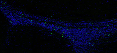
The pattern of electrical signals propagated through neuronal networks determines brain function. Adam Cohen examines the possibility of visualizing these signals inside an intact brain using fluorescent transmembrane proteins that are sensitive to voltage. Cohen discusses the barriers to this approach, something he predicts scientists from many disciplines will eventually overcome.

https://youtu.be/Zw8lWmGuXLU      Download: High ResLow Res     Recorded: 2014

Adam Cohen is Professor in the Departments of Chemistry and Physics at Harvard University and Investigator of the Howard Hughes Medical Institute. He develops biological tools and analytical approaches to investigate the behaviors of molecules and cells in vitro and in vivo. His lab merges protein engineering, optics, and physics, among other disciplines, on a variety of projects. For example, they have developed a fluorescent transmembrane protein that detects membrane voltage, which is useful in visualizing electrical activity in cells, such as cultured neurons.

Related —


The Evolution of Neural Circuits and Behaviors​
Melina Hale (University of Chicago)

Evolution can be defined as a change in heritable characteristics. In her fist talk, Hale does a excellent job of explaining how these changes occur. She uses examples, such as the variable color of the pepper moth, to explain selection of characteristics and she describes how geographic isolation can lead to the evolution of new species. In her second lecture, Hale describes work from her lab on the startle response, a highly conserved behavior found in fish and other vertebrates. Comparisons of the neurons which control the startle response, across many species of fish, have allowed Hale and her colleagues to determine how this neuronal circuit, and this behavior, have evolved over hundreds of millions of years.

Part 1 is an outstanding video for high school or undergraduate educators looking for material to teach evolution.

Watch Melina Hale’s iBioSeminar:

Part 1: Introduction to Evolution

Part 2: Neural Circuits and How They Evolve: A Startling Example!


Discovery of a ‘Neuronal Big Bang’

University of Geneva   http://www.biosciencetechnology.com/news/2016/03/discovery-neuronal-big-bang


This is an expression of all the genes of a neuron during the first hours after its birth. Each circle represents a development stage (6h, 12h, 24h), and the colored points within each circle represent the level of gene expression. (Credit: Jabaudon Lab/ UNIGE)

This is an expression of all the genes of a neuron during the first hours after its birth. Each circle represents a development stage (6h, 12h, 24h), and the colored points within each circle represent the level of gene expression. (Credit: Jabaudon Lab/ UNIGE)


Our brain is home to different types of neurons, each with their own genetic signature that defines their function. These neurons are derived from progenitor cells, which are specialized stem cells that have the ability to divide to give rise to neurons. Neuroscientists from the Faculty of Medicine at the University of Geneva (UNIGE) shed light on the mechanisms that allow progenitors to generate neurons. By developing a novel technology called FlashTag that enables them to isolate and visualize neurons at the very moment they are born, they have deciphered the basic genetic code allowing the construction of a neuron. This discovery, which is published in Science, allows not only to understand how our brain develops, but also how to use this code to reconstruct neurons from stem cells. Researchers will now be able to better understand the mechanisms underlying neurological diseases such as autism and schizophrenia.

Directed by Denis Jabaudon, a neuroscientist and neuroscientist at the Department of Basic Neurosciences at UNIGE Faculty of Medicine and neurologist at the University Hospitals Geneva (HUG), the researchers developed a technology termed FlashTag, which visualizes neurons as they are being born. Using this approach, at the very moment where a progenitor divides, it is tagged with a fluorescent marker that persists in its progeny. Scientists can then visualize and isolate newborn neurons in order to dynamically observe which genes are expressed in the first few hours of their existence. Over time, they can then study their evolution and changes in gene expression. “Previously, we only had a few photos to reconstruct the history of neurons, which left a lot of room for speculation. Thanks to FlashTag, there is now a full genetic movie unfolding before our eyes. Every instant becomes visible from the very beginning, which allows us to understand the developmental scenario at play, identify the main characters, their interactions and their incentives”, notes Jabaudon. Working in the cerebral cortex of the mouse, the scientists have thus identified the key genesto neuronal development, and demonstrated that their expression dynamics is essential for the brain to develop normally.

A very precise primordial choreography

This discovery, by giving access to the primordial code of the formation of neurons, helps us to understand how neurons function in the adult brain. And it appears that several of these original genes are also involved in neurodevelopmental and neurodegenerative diseases, which can occur many years later. This suggests that a predisposition may be present from the very first moments in the existence of neurons, and that environmental factors can then impact on how diseases may develop later on. By understanding the genetic choreography of neurons, the researchers can therefore observe how these genes behave from the start, and identify potential anomalies predicting diseases.

After successfully reading this genetic code, the scientists we able to rewrite it in newborn neurons. By altering the expression of certain genes, they were able to accelerate neuronal growth, thus altering the developmental script. With FlashTag, it is now possible to isolate newborn neurons and recreate cerebral circuits in vitro, which enables scientists to test their function as well as to develop new treatments.

A website open to all

The UNIGE team posted a website where it is possible to enter the name of a gene and observe how it is expressed, and how it interacts with other genes. “Each research team can only focus on a handful of genes at a time, while our genome is made up of close to 20,000 genes. We therefore made our tool available for other researchers to use it, in a fully open way,” highlights Jabaudon.

Chronic Stress Causes Brain Inflammation, Memory Loss

A new study suggests that long-term stress can hurt short-term memory, in part due to inflammation brought on by an immune response.

Bevin Fletcher, Associate Editor    http://www.biosciencetechnology.com/news/2016/03/chronic-stress-causes-brain-inflammation-memory-loss

A new study suggests that long-term stress can hurt short-term memory, in part due to inflammation brought on by an immune response.

Researchers from Ohio State University performed experiments where mice were exposed to repeated social defeat by exposure to an aggressive, larger, alpha mouse.  The mice that were under chronic stress, had difficulty remembering where the escape hole was in a maze they had previously mastered before the stressful period.

The findings were published in The Journal of Neuroscience.

“The stressed mice didn’t recall it. The mice that weren’t stressed really remembered it,” lead researcher Johnathan Godbout, associate professor of neuroscience at Ohio State, said in statement.

The researchers noted that this kind of stress isn’t the once-in-a-while, acute stress someone might feel before a big meeting or presentation, but prolonged, continued stress.

The mice also displayed depressive-like behavior through social avoidance that continued after four weeks of observation.

Brain changes were also observed in the stressed mice, including inflammation associated with the presence of immune cells, known as macrophages, in the brain.  The researchers also recorded shortfalls in the development of new neurons at 10 days and 28 days after the chronic stress ended.

John Sheridan, associate director of Ohio State’s Institute for Behavioral Medicine said in a statement that there might be ways to interrupt the inflammation that occurs in the brain.

When the mice were given a chemical that inhibited inflammation, both memory loss and the inflammatory macrophages disappeared, leading researchers to conclude that post-stress memory deficits is directly tied to inflammation and the immune system. The depressive symptoms and the brain-cell problem did not go away.

“Stress releases immune cells from the bone marrow and those cells can traffic to brain areas associated with neuronal activation in response to stress,” Sheridan said. “They’re being called to the brain, to the center of memory.”

The team aims to understand the underpinnings of stress and responses that could one day lead to treatments for people that suffer from anxiety, depression, or post-traumatic stress disorder.

New information from this study could lead to immune-based treatments, Godbout said.


 A retinoic acid-enhanced, multicellular human blood-brain barrier model derived from stem cell sources Ethan S. Lippmann, Abraham Al-Ahmad, Samira M. Azarin, Sean P. Palecek &Eric V. Shusta

Scientific Reports 4, Article number: 4160 (2014)   http://dx.doi.org:/10.1038/srep04160

Blood-brain barrier (BBB) models are often used to investigate BBB function and screen brain-penetrating therapeutics, but it has been difficult to construct a human model that possesses an optimal BBB phenotype and is readily scalable. To address this challenge, we developed a human in vitro BBB model comprising brain microvascular endothelial cells (BMECs), pericytes, astrocytes and neurons derived from renewable cell sources. First, retinoic acid (RA) was used to substantially enhance BBB phenotypes in human pluripotent stem cell (hPSC)-derived BMECs, particularly through adherens junction, tight junction, and multidrug resistance protein regulation. RA-treated hPSC-derived BMECs were subsequently co-cultured with primary human brain pericytes and human astrocytes and neurons derived from human neural progenitor cells (NPCs) to yield a fully human BBB model that possessed significant tightness as measured by transendothelial electrical resistance (~5,000 Ωxcm2). Overall, this scalable human BBB model may enable a wide range of neuroscience studies.

The blood-brain barrier (BBB) is composed of brain microvascular endothelial cells (BMECs) which line brain capillaries and control molecular and cellular trafficking between the bloodstream and neural tissue. These properties are tightly regulated by the surrounding neurovascular microenvironment throughout BBB development and into adulthood. While this barrier is essential for preserving healthy brain activity, its dysfunction has been implicated in a number of neurological diseases1. Moreover, an intact BBB serves as a major bottleneck for brain drug delivery2. Studies regarding BBB development and regulation can be difficult and time-consuming to conduct in vivo and testing brain penetration of therapeutics in vivo is a low throughput endeavor. As such, in vitro BBB models have been widely implemented to study interactions between BMECs and other cells of the neurovascular unit and to conduct screens for prospective BBB-permeant drugs.

In vitro BBB models are typically constructed using primary BMECs isolated from animal brain tissue, including bovine, porcine, rat, and mouse (reviewed extensively in ref. 3). These BMECs are then co-cultured with combinations of other cells of the neurovascular unit, such as neurons, pericytes, and astrocytes, to upregulate BBB properties4,5,6,7. Models derived from animal tissue have proven extremely useful in studying various aspects of the BBB, such as developmental and regulatory mechanisms8,9,10,11,12 and assaying drug permeability, but it is generally well-accepted that owing to species differences, a robust human BBB model is vital to achieve a detailed understanding of human developmental pathways and to conduct relevant drug discovery and design studies13. Human BMEC sources for BBB models have previously consisted of either primary biopsied brain tissue14,15 or immortalized cell lines16. Primary human BMECs typically possess moderate barrier properties but are of limited scale14,15, and immortalized BMECs are clonal and readily scalable but often suffer from suboptimal barrier properties16,17. From a co-culture perspective, human neurons, astrocytes, and pericytes can also be difficult to obtain from primary tissue sources in sufficient quantities for modeling purposes. These collective issues have hindered the development of in vitro human BBB models that are both high fidelity and scalable3.

We have recently demonstrated that stem cells may be attractive candidates to replace primary cells in human BBB models. Human pluripotent stem cells (hPSCs), including both human embryonic stem cells (hESCs) and induced pluripotent stem cells (iPSCs), can be differentiated into cells possessing both endothelial and BBB properties (coined hPSC-derived BMECs) via co-differentiation of neural and endothelial progenitors, followed by selection and subsequent culture of the endothelial cells18. The iPSC-derived BMECs co-cultured with rat astrocytes possessed reasonable barrier tightness as measured by TEER (860 ± 260 Ωxcm2)18, but the TEER remained below some primary bovine19 and porcine20,21 models (800–2,000 Ωxcm2) and substantially lower than in vivo TEER (measured up to 5,900 Ωxcm2)22. In searching for candidates to improve the BBB phenotype, we identified all-trans retinoic acid (RA). BMECs in vivo have been shown to express retinol-binding protein and its membrane receptor STRA623,STRA6 expression has been detected in brain endothelium but not peripheral endothelium in adult mice24, and STRA6 expression was increased during the differentiation of hPSC-derived BMECs in our previous work18. Moreover, RA has been shown to upregulate certain BBB properties in immortalized rodent25,26 and human27 BMEC lines. In this manuscript, we demonstrate maturation of hPSC-derived BMEC phenotypes following retinoic acid (RA) addition during the differentiation process, including enhanced adherens junction protein expression, barrier function, and multidrug resistance protein (MRP) efflux activity. We also demonstrated in previous work that primary human neural progenitor cells (NPCs) could be differentiated to a defined mixture of neurons and astrocytes capable of inducing BBB properties in rat BMECs in co-culture7. In this manuscript, it is shown that under optimized culture conditions, RA-treated hPSC-derived BMECs sequentially co-cultured with primary human brain pericytes and NPC-derived astrocytes and neurons can achieve physiologic TEER values, forming a scalable, fully human BBB model.

.…….

The purpose of this work was to construct a renewable, robust human BBB multicellular co-culture model employing hPSCs, NPCs, and pericytes. Using previous studies as guides25,26,31, RA was identified as a significant modulator of BMEC properties during hPSC differentiation that greatly enhanced physical barrier characteristics as demonstrated by elevated TEER in BMECs cultured alone or with neurovascular cell co-culture. In recent work, RA treatment on the hCMEC/D3 human brain endothelial cell line served to increase occludin and VE-cadherin expression, and the authors suggested that RA secreted by radial glia may be involved in BBB development27. In our study, when RA was added during the endothelial progenitor expansion phase of hPSC-derived BMEC differentiation, similar results were observed including an earlier onset of VE-cadherin expression and increased occludin expression. Moreover, the BMEC yield was increased 2-fold and the tightness of the hPSC-derived BMEC monolayers as measured by elevated TEER was significantly enhanced for three different hPSC lines. Somewhat unexpectedly, RA treatment resulted in decreased claudin-5 expression. However, the Western blotting analysis was conducted using whole-cell lysates and does not take into account the substantially improved intercellular claudin-5 junctional continuity upon RA treatment (Fig. 2C). We and others have previously observed a strong correlation between such junctional continuity and resultant barrier phenotype6,29,32. In addition, previous work has demonstrated claudin-5 expression is relatively constant across peripheral and BBB endothelium while occludin expression is increased at the BBB relative to other vascular beds31. Thus, a combination of claudin-5 localization and elevated occludin expression may be the key phenotypic indicators of increased barrier function31,33. RA treatment of hPSC-derived BMECs also selectively increased MRP efflux activity, which agrees with reports demonstrating that signaling via nuclear receptors can regulate efflux transporter expression and activity at the blood-brain barrier in vitro and in vivo34,35,36,37. RA influences many aspects of brain development, such as anterior/posterior axis patterning in the hindbrain and anterior spinal cord38,39,40 and regulation of neurogenesis41,42,43. During BMEC differentiation, RA could trigger several modes of action. RA may act directly on the developing endothelial cells to upregulate BBB properties, it could induce changes in the neural cells to indirectly promote BBB differentiation, or it could act by a combination of these mechanisms. Future work will be necessary to deconvolute the RA signaling mechanisms affecting the hPSC-derived BMEC differentiation scheme.


In Your Dreams

Understanding the sleeping brain may be the key to unlocking the secrets of the human mind.

By David Gelernter | March 1, 2016

http://www.the-scientist.com/?articles.view/articleNo/45357/title/In-Your-Dreams

Many scientists who study the mind live in fantasyland. They ought to move back to reality: neuroscientists, psychologists, computer scientists pursuing artificial intelligence, and the philosophers of mind who are, in many cases, the sharpest thinkers in the room.

The mind makes us rational. That mind is the one we choose to study. When we study sleep or dreaming, we isolate them first—as the specialized topics they are. But, as I argue in my new book The Tides of Mind, we will never reach a deep understanding of mind unless we start with an integrated view, stretching from rational, methodical thought to nightmares.

Integrating dreaming with the rest of mind is something like being asked to assemble a car from a large pile of metal, plastic, rubber, glass, and an ocelot. Dreaming is hallucination, centering on a radically different self from our waking selves, within unreal settings and stories. Dreams can please or scare us far more vividly than our ordinary thoughts. And they are so slippery, so hard to grasp, that we start losing them the moment we wake up.

But dreaming fits easily into the big picture of mind; and we will make no basic progress on understanding the mind until we see how. Dreaming is the endpoint of the spectrum of consciousness, the smooth progression from one type of consciousness to the next, that we each experience daily.

The simplest approach to the spectrum centers on mental focus. The quality of our attention goes from concentrated to diffuse over the course of a normal day; from a state in which we can concentrate—we can think and remember in a relatively disciplined way—to one in which, with our minds wandering and memory growing increasingly vibrant and distracting, we approach sleep. Then our thinking becomes hallucinatory (as we pass through “sleep-onset thought”); and finally, we are asleep and dreaming. Usually, we oscillate down and up more than once during the day. We move partway down, come partway back, then finally slide slowly to the bottom, when we sleep and dream.

We can also describe the spectrum as a steady shift from a mind dominated by action to one dominated by passive mental experience; from mental doing to mental being. In the upper spectrum, we tend to ignore emotion as we pursue some mental object by means of reasoning or analysis. But the daydreams and fantasies that occupy us as we move down-spectrum are often emotional. And in dreaming we encounter the most saturated emotions, good and bad, that the mind can generate.

The spectrum clarifies important aspects of the mind. “Intentionality,” the quality of aboutness (“I believe that bird is a sparrow” is about “that bird”), is sometimes called “the mark of the mental”—the distinguishing attribute of mental states. But intentionality belongs strictly to the upper spectrum, and disappears gradually as we descend. At the bottom, our minds are dominated by experience, pure being. Happiness or pain or “the experience of seeing purple” are states that have causes but are about nothing.

Software simulations of the upper spectrum, of thinking-about, have grown steadily stronger over the years. That trend will continue. Being, however, is not computable. Software can no more reproduce “being happy” than it can reproduce “being rusty.” Such states depend on physical properties of particular objects. A digital computer resembles only the upper-spectrum mind. Software will never come close to reproducing the mind as a whole.
Leaving sleep outside our investigation is a good way not to see any of this. Arbitrarily hacking off one end of any natural spectrum is an invitation to conceptual chaos. There has been plenty of that in the science of mind. We must start by understanding sleep and dreaming, and go from there.

David Gelernter is a professor of computer science at Yale University. Read an excerpt from his latest book, The Tides of Mind: Uncovering the Spectrum of Consciousness at the-scientist.com.

Out in the Cold

Serotonin’s long-debated role in sleep promotion is temperature-dependent.

By Karen Zusi | March 1, 2016     http://www.the-scientist.com/?articles.view/articleNo/45346/title/Out-in-the-Cold

N.M. Murray et al., “Insomnia caused by serotonin depletion is due to hypothermia,” Sleep, 38:1985-93, 2015.

Sleepless nights
Early research into serotonin’s functions suggested that the neurotransmitter promotes sleep: lab animals deprived of the chemical often developed insomnia. More recent evidence indicated that serotonin plays a part in wakefulness instead, a theory that has gained significant traction. But explanations of the initial experimental data were scarce—so Nick Murray, then a research fellow at the University of Iowa Carver College of Medicine, started digging.

Faulty furnace?
“Over the past 5 or 10 years, we’ve found that serotonin is a key neurotransmitter for generating body heat,” says Murray. To investigate whether this role was related to serotonin’s impact on sleep, he and his colleagues injected para-chlorophenylalanine into mice to inhibit serotonin synthesis.

On ice
When kept at room temperature (20 °C), the mice with depleted serotonin slept less and developed a lower body temperature compared with their control counterparts. However, when housed at 33 °C—a thermoneutral temperature for mice—the sleep and body temperature of the treated mice stayed normal. “Serotonin isn’t a sleep-promoting neurotransmitter,” concludes Murray, now a resident at California Pacific Medical Center. He suggests that mice lacking serotonin had a tough time sleeping under early experimental conditions simply because the animals were cold, and that at higher temperatures other neurotransmitter systems in the brain would function to allow them a normal sleep-wake cycle.

Case closed
The study “solves a long-standing mystery” in the field, says Clifford Saper of Harvard University. “Not very many labs measure sleep and body temperature at the same time,” he adds. “It just basically escaped everybody’s notice for all these years.”


Read Full Post »

Brain location for loneliness

Larry H. Bernstein, MD, FCAP, Curator

LPBI


Pinpointing Loneliness in the Brain

Anne Trafton, MIT News Office   http://www.biosciencetechnology.com/news/2016/02/pinpointing-loneliness-brain


In this image of the dorsal raphe nucleus, dopamine neurons are labeled in green, red, or both (appearing yellow).  (Image: Gillian Matthews)

In this image of the dorsal raphe nucleus, dopamine neurons are labeled in green, red, or both (appearing yellow). (Image: Gillian Matthews)


Humans, like all social animals, have a fundamental need for contact with others. This deeply ingrained instinct helps us to survive; it’s much easier to find food, shelter, and other necessities with a group than alone. Deprived of human contact, most people become lonely and emotionally distressed.

In a study appearing in the Feb. 11 issue of Cell, MIT neuroscientists have identified a brain region that represents these feelings of loneliness. This cluster of cells, located near the back of the brain in an area called the dorsal raphe nucleus (DRN), is necessary for generating the increased sociability that normally occurs after a period of social isolation, the researchers found in a study of mice.

“To our knowledge, this is the first time anyone has pinned down a loneliness-like state to a cellular substrate. Now we have a starting point for really starting to study this,” said Kay Tye, the Whitehead Career Development Assistant Professor of Brain and Cognitive Sciences, a member of MIT’s Picower Institute for Learning and Memory, and one of the senior authors of the study.

While much research has been done on how the brain seeks out and responds to rewarding social interactions, very little is known about how isolation and loneliness also motivate social behavior.

“There are many studies from human psychology describing how we have this need for social connection, which is particularly strong in people who feel lonely. But our understanding of the neural mechanisms underlying that state is pretty slim at the moment. It certainly seems like a useful, adaptive response, but we don’t really know how that’s brought about,” said Gillian Matthews, a postdoc at the Picower Institute and the paper’s lead author.

Only the lonely

Matthews first identified the loneliness neurons somewhat serendipitously, while studying a completely different topic. As a Ph.D. student at Imperial College London, she was investigating how drugs affect the brain, particularly dopamine neurons. She originally planned to study how drug abuse influences the DRN, a brain region that had not been studied very much.

As part of the experiment, each mouse was isolated for 24 hours, and Matthews noticed that in the control mice, which had not received any drugs, there was a strengthening of connections in the DRN following the isolation period.

Further studies, both at Imperial College London and then in Tye’s lab at MIT, revealed that these neurons were responding to the state of isolation. When animals are housed together, DRN neurons are not very active. However, during a period of isolation, these neurons become sensitized to social contact and when the animals are reunited with other mice, DRN activity surges. At the same time, the mice become much more sociable than animals that had not been isolated.

When the researchers suppressed DRN neurons using optogenetics, a technique that allows them to control brain activity with light, they found that isolated mice did not show the same rebound in sociability when they were re-introduced to other mice.

“That suggested these neurons are important for the isolation-induced rebound in sociability,” Tye said. “When people are isolated for a long time and then they’re reunited with other people, they’re very excited, there’s a surge of social interaction. We think that this adaptive and evolutionarily conserved trait is what we are modeling in mice, and these neurons could play a role in that increased motivation to socialize.”

Social dominance

The researchers also found that animals with a higher rank in the social hierarchy were more responsive to changes in DRN activity, suggesting that they may be more susceptible to feelings of loneliness following isolation.

“The social experience of every animal is not the same in a group,” Tye said. “If you’re the dominant mouse, maybe you love your social environment. And if you’re the subordinate mouse, and you’re being beat up every day, maybe it’s not so fun. Maybe you feel socially excluded already.”

The findings represent “an amazing cornerstone for future studies of loneliness,” said Alcino Silva, a professor of neurobiology, psychiatry, and psychology at the David Geffen School of Medicine at UCLA who was not involved in the research.

“There is something poetic and fascinating about the idea that modern neuroscience tools have allowed us to reach to the very depths of the human soul, and that in this search we have discovered that even the most human of emotions, loneliness, is shared in some recognizable form with even one of our distant mammalian relatives — the mouse,” Silva said.

The researchers are now studying whether these neurons actually detect loneliness or are responsible for driving the response to loneliness, and whether they might be part of a larger brain network that responds to social isolation. Another area to be explored is whether differences in these neurons can explain why some people prefer more social contact than others, and whether those differences are innate or formed by experience.

“There’s probably some part that could very well be determined by innate brain features, but I think probably an equal, if not greater, contribution would be from the environment in which individuals have developed,” Tye said. “These are completely open questions. We can only speculate about it at this point.”

Mark Ungless, a senior lecturer at Imperial College London, is also a senior author of the study. MIT graduate students Edward Nieh and Caitlin Vander Weele are also lead authors.

Read Full Post »

Advanced Nanospectroscopy

Larry H. Bernstein, MD, FCAP, Curator

LPBI


Graphene Enables Nanoelectromechanical Systems Integration

http://www.photonics.com/Article.aspx?AID=58224&refer=Spectroscopy&utm_source=Spectroscopy_2016_02_03

BARCELONA, Spain, Jan. 21, 2016 — Combining nanoelectromechanical (NEMS) systems with on-chip optics holds promise as a method to actively control light at the nanoscale, and now a hybrid system has overcome the challenges of integrating such nanoscale devices with optical fields thanks to the material graphene.

Researchers from the Institute of Photonic Sciences (ICFO) have demonstrated an on-chip graphene NEMS suspended a few tens of nanometers above nitrogen-vacancy centres (NVCs), which are stable single-photon emitters embedded in nanodiamonds. The work confirms that graphene is an ideal platform for both nanophotonics and nanomechanics, the researchers said.

Due to its electromechanical properties, graphene NEMS can be actuated and deflected electrostatically over a few tens of nanometers with modest voltages applied to a gate electrode, the researchers found. The graphene motion can thus be used to modulate the light emission by the NVC, while the emitted field can be used as a universal probe of the graphene position. The optomechanical coupling between the graphene displacement and the NVC emission is based on near-field, dipole-dipole interaction.

64

http://www.photonics.com/images/Web/Articles/2016/1/21/PIC_ICFO.jpg

False color scanning electronic micrograph of a hybrid graphene-nitrogen-vacancy nearfield nano-optomechanical system. Courtesy of ICFO.

The researchers observed that the coupling strength increased strongly for shorter distances and was enhanced because of graphene’s 2D character and linear dispersion. These achievements hold promise for selective control of emitter arrays on-chip, optical spectroscopy of individual nano-objects, and integrated optomechanical information processing. The ICFO team also said the hybrid device could advance quantum optomechanics.

The research was published in Nature Communications (doi: 10.1038/ncomms10218).

Electromechanical control of nitrogen-vacancy defect emission using graphene NEMS

Antoine Reserbat-PlanteyKevin G. SchädlerLouis GaudreauGabriele NavickaiteJohannes Güttinger, et al.

Nature Communications 2016; 7(10218)      http://dx.doi.org:/10.1038/ncomms10218

Despite recent progress in nano-optomechanics, active control of optical fields at the nanoscale has not been achieved with an on-chip nano-electromechanical system (NEMS) thus far. Here we present a new type of hybrid system, consisting of an on-chip graphene NEMS suspended a few tens of nanometres above nitrogen-vacancy centres (NVCs), which are stable single-photon emitters embedded in nanodiamonds. Electromechanical control of the photons emitted by the NVC is provided by electrostatic tuning of the graphene NEMS position, which is transduced to a modulation of NVC emission intensity. The optomechanical coupling between the graphene displacement and the NVC emission is based on near-field dipole–dipole interaction. This class of optomechanical coupling increases strongly for smaller distances, making it suitable for nanoscale devices. These achievements hold promise for selective control of emitter arrays on-chip, optical spectroscopy of individual nano-objects, integrated optomechanical information processing and open new avenues towards quantum optomechanics.


Graphene is ideal substrate for brain electrodes, researchers find

February 1, 2016  http://www.kurzweilai.net/graphene-is-ideal-substrate-for-brain-electrodes-researchers-find

This illustration portrays neurons interfaced with a sheet of graphene molecules in the background (credit: Graphene Flagship)

An international study headed by the European Graphene Flagship research consortium has found that graphene is a promising material for use in electrodes that interface with neurons, based on its excellent conductivity, flexibility for molding into complex shapes, biocompatibility, and stability within the body.

The graphene-based substrates they studied* promise to overcome problems with “glial scar” tissue formation (caused by electrode-based brain trauma and long-term inflammation). To avoid that, current electrodes based on tungsten or silicon use a protective coating on electrodes, which reduces charge transfer. Current electrodes are also rigid (resulting in tissue detachment and preventing neurons from moving) and generate electrical noise, with partial or complete loss of signal over time, the researchers note in a paper published recently in the journal ACS Nano.

Electrodes are used as neural biosensors and for prosthetic applications — such as deep-brain intracranial electrodes used to control motor disorders (mainly epilepsy or Parkinson’s) and for brain-computer interfaces (BCIs), used to recover sensory functions or control robotic arms for paralyzed patients. These applications require an interface with long-term, minimal interference.

Interfacing graphene to neurons directly

Scanning electron microscope image of rat hippocampal neurons grown in the lab on a graphene-based substrate, showing normal morphology characterized by well-defined round neural soma, extended neurite arborization (branching), and cell density similar to control substrates (credit: A. Fabbro et al./ACS Nano)

“For the first time, we interfaced graphene to neurons directly, without any peptide-coating,” explained lead neuroscientist Prof. Laura Ballerini of the International School for Advanced Studies (SISSA/ISAS) and the University of Trieste.

Using electron microscopy and immunofluorescence, the researchers found that the neurons remained healthy, transmitting normal electric impulses and, importantly, no adverse glial reaction, which leads to damaging scar tissue, was seen.

As a next step, Ballerini says the team plans to investigate how different forms of graphene, from multiple layers to monolayers, are able to affect neurons,  and “whether tuning the graphene material properties might alter the synapses and neuronal excitability in new and unique ways.”

Prof. Andrea C. Ferrari, Director of the Cambridge Graphene Centre and Chair of the Graphene Flagship Executive Board, said the Flagship will “support biomedical research and development based on graphene technology with a new work package and a significant cash investment from 2016.”

The interdisciplinary collaboration also included the University Castilla-La Mancha and the Cambridge Graphene Centre.

* The study used two methods of creating graphene-based substrates (GBSs).  Liquid phase exfoliation (LPE) — peeling off graphene from graphite — can be performed without the potentially hazardous chemical treatments involved in graphene oxide production, is scalable, and operates at room temperature, with high yield. LPE dispersions can also be easily deposited on target substrates by drop-casting, filtration, or printing. Ball milling (BM), with the help of melamine (which forms large hydrogen-bond domains, unlike LPE), can be performed in a solid environment. “Our data indicate that both GBSs are promising for next-generation bioelectronic systems, to be used as brain interfaces,” the paper concludes.


Read Full Post »

Larry H. Benstein, MD, FCAP, Gurator and writer

http://pharmaceuticalintelligence.com/7/8/2014/Update on mitochondrial function, respiration, and associated disorders

This is a condensed account of very recent published work on respiration and disturbed mitochondrail function.  We know that their is an equilibrium between respiration and autophagy in eukaryotic cells.  The Krebs Cycle produces 32 ATPs in oxidative phosphorylation, which is far more efficient than glycolysis.  There is also a different contribution of mitochondrial metabolism, in the balance, between tissues that are synthetic and those that are catabolic.  This is a subject long understood, essential for cellular energetics, and not adequately explored.


Gain-of-Function Mutant p53 Promotes Cell Growth and Cancer Cell Metabolism via Inhibition of AMPK Activation.

Zhou G1Wang J2Zhao M2Xie TX2Tanaka N2, et al.
Mol Cell. 
2014 Jun 19;54(6):960-974.   doi: 10.1016/j.molcel.2014.04.024. 

Many mutant p53 proteins (mutp53s) exert oncogenic gain-of-function (GOF) properties, but the mechanisms mediating these functions remain poorly defined.

We show here that GOF mutp53s inhibit AMP-activated protein kinase (AMPK) signaling in head and neck cancer cells.

Conversely, downregulation of GOF mutp53s enhances AMPK activation under energy stress, decreasing the activity of the anabolic factors acetyl-CoA carboxylase and ribosomal protein S6 and inhibiting aerobic glycolytic potential and invasive cell growth.

Under conditions of energy stress, GOF mutp53s, but not wild-type p53, preferentially bind to the AMPKα subunit and inhibit AMPK activation.

Given the importance of AMPK as an energy sensor and tumor suppressor that inhibits anabolic metabolism, our findings reveal that direct inhibition of AMPK activation is an important mechanism through which mutp53s can gain oncogenic function. PMID:24857548

Investigating and Targeting Chronic Lymphocytic Leukemia Metabolism with the HIV Protease Inhibitor Ritonavir and Metformin.

Adekola KUAydemir SDMa SZhou ZRosen STShanmugam M.
Leuk Lymphoma. 2014 May 14:1-23.

Chronic Lymphocytic Leukemia (CLL) remains fatal due to the development of resistance to existing therapies. Targeting abnormal glucose metabolism sensitizes various cancer cells to chemotherapy and/or elicits toxicity.

Examination of glucose dependency in CLL demonstrated variable sensitivity to glucose deprivation. Further evaluation of metabolic dependencies of CLL cells resistant to glucose deprivation revealed increased engagement of fatty acid oxidation upon glucose withdrawal.

Investigation of glucose transporter expression in CLL reveals up-regulation of glucose transporter GLUT4. Treatment of CLL cells with HIV protease inhibitor ritonavir, that inhibits GLUT4, elicits toxicity similar to that elicited upon glucose-deprivation.

CLL cells resistant to ritonavir are sensitized by co-treatment with metformin, potentially targeting compensatory mitochondrial complex 1 activity. Ritonavir and metformin have been administered in humans for treatment of diabetes in HIV patients, demonstrating the tolerance of this combination in humans. Our studies strongly substantiate further investigation of FDA approved ritonavir and metformin for CLL.

KEYWORDS:  Basic Biology; Chemotherapeutic approaches; Lymphoid Leukemia; Signal transduction             PMID: 24828872

Utilizing hydrogen sulfide as a novel anti-cancer agent by targeting cancer glycolysis and pH imbalance.

Lee ZW1Teo XYTay EYTan CHHagen TMoore PKDeng LW.
Br J Pharmacol. 2014 May 15.    doi: 10.1111/bph.12773

Many disparate studies have reported the ambiguous role of hydrogen sulfide (H2 S) in cell survival. The present study investigated the effect of H2 S on viability of cancer and non-cancer cells.

Cancer and non-cancer cells were exposed to H2 S (using sodium hydrosulfide, NaHS and GYY4137) and cell viability was examined by crystal violet assay. We then examined cancer cellular glycolysis process by in vitro enzymatic assays and pH regulator activity. Lastly, intracellular pH (pHi) was determined by ratiometric pHi measurement using BCECF staining.

Continuous, but not single, exposure to H2 S decreased cell survival more effectively in cancer cells, as compared to non-cancer cells. Slow H2 S-releasing donor, GYY4137, significantly increased glycolysis leading to overproduction of lactate. H2 S also decreased anion exchanger and sodium/proton exchanger activity. The combination of increased metabolic acid production and defective pH regulation resulted in an uncontrolled intracellular acidification leading to cancer cell death. In contrast, no significant intracellular acidification or cell death was observed in non-cancer cells.

Low and continuous exposure to H2 S targets metabolic processes and pH homeostasis in cancer cells, potentially serving as a novel and selective anti-cancer strategy.

KEYWORDS:  cancer cell death; cancer glucose metabolism; hydrogen sulfide; pH homeostasis          PMID: 24827113


Agonism of the 5-Hydroxytryptamine 1F Receptor Promotes Mitochondrial Biogenesis and Recovery from Acute Kidney Injury

Garrett SMWhitaker RMBeeson CC, and Schnellmann RG

Center for Cell Death, Injury, and Regeneration, Department of Drug Discovery and Biomedical Sciences, Medical University of South Carolina, Charleston, South Carolina (S.M.G., R.M.W., C.C.B., R.G.S.); and Ralph H. Johnson Veterans Affairs Medical Center, Charleston, South Carolina (R.G.S.)
Address correspondence to: Dr. Rick G. Schnellmann, Department of Drug Discovery and Biomedical Sciences, MUSC, Charleston, SC 29425.
E-mail: schnell@musc.edu

Many acute and chronic conditions, such as acute kidney injury, chronic kidney disease, heart failure, and liver disease, involve mitochondrial dysfunction. Although we have provided evidence that drug-induced stimulation of mitochondrial biogenesis (MB) accelerates mitochondrial and cellular repair, leading to recovery of organ function, only a limited number of chemicals have been identified that induce MB.

The goal of this study was to assess the role of the 5-hydroxytryptamine 1F (5-HT1F) receptor in MB. Immunoblot and quantitative polymerase chain reaction analyses revealed 5-HT1F receptor expression in renal proximal tubule cells (RPTC). A MB screening assay demonstrated that two selective 5-HT1F receptor agonists,

  1. LY334370 (4-fluoro-N-[3-(1-methyl-4-piperidinyl)-1H-indol-5-yl]benzamide) and
  2. LY344864 (N-[(3R)-3-(dimethylamino)-2,3,4,9-tetrahydro-1H-carbazol-6-yl]-4-fluorobenzamide; 1–100 nM)

increased carbonylcyanide-p-trifluoromethoxyphenylhydrazone–uncoupled oxygen consumption in RPTC, and

  • validation studies confirmed both agonists increased mitochondrial proteins  in vitro.
    [e.g., ATP synthase β, cytochrome c oxidase 1 (Cox1), and NADH dehydrogenase (ubiquinone) 1β subcomplex subunit 8 (NDUFB8)]

Small interfering RNA knockdown of the 5-HT1F receptor

  • blocked agonist-induced MB.

Furthermore, LY344864 increased

  • peroxisome proliferator–activated receptor (PPAR) coactivator 1-α, Cox1, and
  • NDUFB8 transcript levels and
  • mitochondrial DNA (mtDNA) copy number

in murine renal cortex, heart, and liver.

Finally, LY344864 accelerated recovery of renal function, as indicated by

  • decreased blood urea nitrogen and kidney injury molecule 1 and
  • increased mtDNA copy number

following ischemia/reperfusion-induced acute kidney injury (AKI).

In summary, these studies reveal that

  • the 5-HT1F receptor is linked to MB, 5-HT1F receptor agonism promotes MB in vitro and in vivo, and

5-HT1F receptor agonism promotes recovery from AKI injury.

Induction of MB through 5-HT1F receptor agonism represents a new target and approach to treat mitochondrial organ dysfunction.

Footnotes

  • Portions of this work have been presented previously: Garrett SM, Wills LP, and Schnellmann RG (2012) Serotonin (5-HT) 1F receptor agonism as a potential treatment for acceleration of recovery from acute kidney injury.American Society of Nephrology Annual Meeting; 2012 Nov 1–4; San Diego, CA.
  • dx.doi.org/10.1124/jpet.114.214700.

Ca2+ regulation of mitochondrial function in neurons.

Rueda CB1Llorente-Folch I1Amigo I1Contreras L1González-Sánchez P1Martínez-Valero P1Juaristi I1Pardo B1Del Arco A2Satrústegui J3

Biochim Biophys Acta. 2014 May 10. pii: S0005-2728(14)00126-1.
doi: 10.1016/j.bbabio.2014.04.010.

Calcium is thought to regulate respiration but it is unclear whether this is dependent on the increase in ATP demand caused by any Ca2+ signal or to Ca2+ itself.

[Na+]i, [Ca2+]i and [ATP]i dynamics in intact neurons exposed to different workloads in the absence and presence of Ca2+ clearly showed that

  • Ca2+-stimulation of coupled respiration is required to maintain [ATP]i levels.

Ca2+ may regulate respiration by

  1. activating metabolite transport in mitochondria from outer face of the inner mitochondrial membrane, or
  2. after Ca2+ entry in mitochondria through the calcium uniporter (MCU).

Two Ca2+-regulated mitochondrial metabolite transporters are expressed in neurons,

  1. the aspartate-glutamate exchanger ARALAR/AGC1/Slc25a12, a component of the malate-aspartate shuttle, with a Kd for Ca2+ activation of 300nM, and
  2. the ATP-Mg/Pi exchanger SCaMC-3/Slc25a23, with S0.5 for Ca2+ of 300nM and 3.4μM, respectively.

The lack of SCaMC-3 results in a smaller Ca2+-dependent stimulation of respiration only at high workloads, as caused by veratridine, whereas

  • the lack of ARALAR reduced by 46% basal OCR in intact neurons using glucose as energy source and the Ca2+-dependent responses to all workloads (veratridine, K+-depolarization, carbachol).

The lack of ARALAR caused a reduction of about 65-70% in the response to the high workload imposed by veratridine, and

  • completely suppressed the OCR responses to moderate (K+-depolarization) and small (carbachol) workloads,
  • effects reverted by pyruvate supply.

For K+-depolarization, this occurs in spite of the presence of large [Ca2+]mit signals and increased reduction of mitochondrial NAD(P)H.

These results show that ARALAR-MAS is a major contributor of Ca2+-stimulated respiration in neurons

  • by providing increased pyruvate supply to mitochondria.

In its absence and under moderate workloads, matrix Ca2+ is unable to stimulate pyruvate metabolism and entry in mitochondria suggesting a limited role of MCU in these conditions.

This article was invited for a Special Issue entitled: 18th European Bioenergetic Conference.    Copyright © 2014. Published by Elsevier B.V.

KEYWORDS:  ATP-Mg/Pi transporter; Aspartate–glutamate transporter; Calcium; Calcium-regulated transport; Mitochondrion; Neuronal respiration PMID: 24820519


Sestrin2 inhibits uncoupling protein 1 expression through suppressing reactive oxygen species.

Ro SH1Nam M2Jang I1Park HW1Park H1Semple IA1Kim M1et al.
Proc Natl Acad Sci U S A. 2014 May 27;111(21):7849-54.
doi: 10.1073/pnas.1401787111.

Uncoupling protein 1 (Ucp1), which is localized in the mitochondrial inner membrane of mammalian brown adipose tissue (BAT), generates heat by uncoupling oxidative phosphorylation. Upon cold exposure or nutritional abundance, sympathetic neurons stimulate BAT to express Ucp1 to induce energy dissipation and thermogenesis. Accordingly, increased Ucp1 expression reduces obesity in mice and is correlated with leanness in humans.

Despite this significance, there is currently a limited understanding of how Ucp1 expression is physiologically regulated at the molecular level. Here, we describe the involvement of Sestrin2 and reactive oxygen species (ROS) in regulation of Ucp1 expression. Transgenic overexpression of Sestrin2 in adipose tissues inhibited both basal and cold-induced Ucp1 expression in interscapular BAT, culminating in decreased thermogenesis and increased fat accumulation.

Endogenous Sestrin2 is also important for suppressing Ucp1 expression because BAT from Sestrin2(-/-) mice exhibited a highly elevated level of Ucp1 expression. The redox-inactive mutant of Sestrin2 was incapable of regulating Ucp1 expression, suggesting that Sestrin2 inhibits Ucp1 expression primarily through reducing ROS accumulation.

Consistently, ROS-suppressing antioxidant chemicals, such as butylated hydroxyanisole and N-acetylcysteine, inhibited cold- or cAMP-induced Ucp1 expression as well. p38 MAPK, a signaling mediator required for cAMP-induced Ucp1 expression, was inhibited by either Sestrin2 overexpression or antioxidant treatments.

Taken together, these results suggest that Sestrin2 and antioxidants inhibit Ucp1 expression through suppressing ROS-mediated p38 MAPK activation, implying a critical role of ROS in proper BAT metabolism.

KEYWORDS: aging; homeostasis; mouse; β-adrenergic signaling      PMID: 24825887     PMCID:  PMC4040599

Mitochondrial EF4 links respiratory dysfunction and cytoplasmic translation in Caenorhabditis elegans.

Yang F1Gao Y1Li Z2Chen L3Xia Z4Xu T5Qin Y6
Biochim Biophys Acta. 2014 May 15. pii: S0005-2728(14)00499-X.
doi: 10.1016/j.bbabio.2014.05.353.

How animals coordinate cellular bioenergetics in response to stress conditions is an essential question related to aging, obesity and cancer. Elongation factor 4 (EF4/LEPA) is a highly conserved protein that promotes protein synthesis under stress conditions, whereas its function in metazoans remains unknown.

Here, we show that, in Caenorhabditis elegans, the mitochondria-localized CeEF4 (referred to as mtEF4) affects mitochondrial functions, especially at low temperature (15°C).

At worms’ optimum growing temperature (20°C), mtef4 deletion leads to self-brood size reduction, growth delay and mitochondrial dysfunction.

Transcriptomic analyses show that mtef4 deletion induces retrograde pathways, including mitochondrial biogenesis and cytoplasmic translation reorganization.

At low temperature (15°C), mtef4 deletion reduces mitochondrial translation and disrupts the assembly of respiratory chain supercomplexes containing complex IV.

These observations are indicative of the important roles of mtEF4 in mitochondrial functions and adaptation to stressful conditions.

Copyright © 2014. Published by Elsevier B.V.

KEYWORDSC. elegans; EF4(LepA/GUF1); Mitochondrial dysfunction; Retrograde pathways; Translation    PMID:  24837196

The metabolite α-ketoglutarate extends lifespan by inhibiting ATP synthase and TOR.

Chin RM1Fu X2Pai MY3Vergnes L4Hwang H5Deng G6Diep S2, et al.
Nature  2014 Jun 19;509(7505):397-401. doi: 10.1038/nature13264. 

Metabolism and ageing are intimately linked. Compared with ad libitum feeding, dietary restriction consistently extends lifespan and delays age-related diseases in evolutionarily diverse organisms. Similar conditions of nutrient limitation and genetic or pharmacological perturbations of nutrient or energy metabolism also have longevity benefits.

Recently, several metabolites have been identified that modulate ageing; however, the molecular mechanisms underlying this are largely undefined. Here we show that α-ketoglutarate (α-KG), a tricarboxylic acid cycle intermediate, extends the lifespan of adult Caenorhabditis elegans.

ATP synthase subunit β is identified as a novel binding protein of α-KG using a small-molecule target identification strategy termed drug affinity responsive target stability (DARTS). The ATP synthase, also known as complex V of the mitochondrial electron transport chain, is the main cellular energy-generating machinery and is highly conserved throughout evolution.

Although complete loss of mitochondrial function is detrimental, partial suppression of the electron transport chain has been shown to extend C. elegans lifespan.

We show that α-KG inhibits ATP synthase and, similar to ATP synthase knockdown, inhibition by α-KG leads to reduced ATP content, decreased oxygen consumption, and increased autophagy in both C. elegans and mammalian cells.

We provide evidence that the lifespan increase by α-KG requires ATP synthase subunit β and is dependent on target of rapamycin (TOR) downstream.

Endogenous α-KG levels are increased on starvation and α-KG does not extend the lifespan of dietary-restricted animals, indicating that α-KG is a key metabolite that mediates longevity by dietary restriction.

Our analyses uncover new molecular links between a common metabolite, a universal cellular energy generator and dietary restriction in the regulation of organismal lifespan, thus suggesting new strategies for the prevention and treatment of ageing and age-related diseases.

PMID: 24828042


Read Full Post »

Reporter: Ritu Saxena, Ph.D.

An article published yesterday in Science Translational Medicine describes how mitochondrial deficits were rescued in the stem cells derived from Parkinson’s disease using pharmacological intervention.

The research published in the article has been discussed in the following news brief: http://www.worldpharmanews.com/research/2132-patient-derived-stem-cells-could-improve-drug-research-for-parkinsons

Research article: http://stm.sciencemag.org/content/4/141/141ra90.abstract

Read Full Post »

%d bloggers like this: