Healthcare analytics, AI solutions for biological big data, providing an AI platform for the biotech, life sciences, medical and pharmaceutical industries, as well as for related technological approaches, i.e., curation and text analysis with machine learning and other activities related to AI applications to these industries.
Eight Subcellular Pathologies driving Chronic Metabolic Diseases – Methods for Mapping Bioelectronic Adjustable Measurements as potential new Therapeutics: Impact on Pharmaceuticals in Use
In this curation we wish to present two breaking through goals:
Goal 1:
Exposition of a new direction of research leading to a more comprehensive understanding of Metabolic Dysfunctional Diseases that are implicated in effecting the emergence of the two leading causes of human mortality in the World in 2023: (a) Cardiovascular Diseases, and (b) Cancer
Goal 2:
Development of Methods for Mapping Bioelectronic Adjustable Measurements as potential new Therapeutics for these eight subcellular causes of chronic metabolic diseases. It is anticipated that it will have a potential impact on the future of Pharmaceuticals to be used, a change from the present time current treatment protocols for Metabolic Dysfunctional Diseases.
According to Dr. Robert Lustig, M.D, an American pediatric endocrinologist. He is Professor emeritus of Pediatrics in the Division of Endocrinology at the University of California, San Francisco, where he specialized in neuroendocrinology and childhood obesity, there are eight subcellular pathologies that drive chronic metabolic diseases.
These eight subcellular pathologies can’t be measured at present time.
In this curation we will attempt to explore methods of measurement for each of these eight pathologies by harnessing the promise of the emerging field known as Bioelectronics.
Unmeasurable eight subcellular pathologies that drive chronic metabolic diseases
Glycation
Oxidative Stress
Mitochondrial dysfunction [beta-oxidation Ac CoA malonyl fatty acid]
Insulin resistance/sensitive [more important than BMI], known as a driver to cancer development
Membrane instability
Inflammation in the gut [mucin layer and tight junctions]
Epigenetics/Methylation
Autophagy [AMPKbeta1 improvement in health span]
Diseases that are not Diseases: no drugs for them, only diet modification will help
Image source
Robert Lustig, M.D. on the Subcellular Processes That Belie Chronic Disease
These eight Subcellular Pathologies driving Chronic Metabolic Diseases are becoming our focus for exploration of the promise of Bioelectronics for two pursuits:
Will Bioelectronics be deemed helpful in measurement of each of the eight pathological processes that underlie and that drive the chronic metabolic syndrome(s) and disease(s)?
IF we will be able to suggest new measurements to currently unmeasurable health harming processes THEN we will attempt to conceptualize new therapeutic targets and new modalities for therapeutics delivery – WE ARE HOPEFUL
In the Bioelecronics domain we are inspired by the work of the following three research sources:
Michael Levin is an American developmental and synthetic biologist at Tufts University, where he is the Vannevar Bush Distinguished Professor. Levin is a director of the Allen Discovery Center at Tufts University and Tufts Center for Regenerative and Developmental Biology. Wikipedia
THE VOICE of Dr. Justin D. Pearlman, MD, PhD, FACC
PENDING
THE VOICE of Stephen J. Williams, PhD
Ten TakeAway Points of Dr. Lustig’s talk on role of diet on the incidence of Type II Diabetes
25% of US children have fatty liver
Type II diabetes can be manifested from fatty live with 151 million people worldwide affected moving up to 568 million in 7 years
A common myth is diabetes due to overweight condition driving the metabolic disease
There is a trend of ‘lean’ diabetes or diabetes in lean people, therefore body mass index not a reliable biomarker for risk for diabetes
Thirty percent of ‘obese’ people just have high subcutaneous fat. the visceral fat is more problematic
there are people who are ‘fat’ but insulin sensitive while have growth hormone receptor defects. Points to other issues related to metabolic state other than insulin and potentially the insulin like growth factors
At any BMI some patients are insulin sensitive while some resistant
Visceral fat accumulation may be more due to chronic stress condition
Fructose can decrease liver mitochondrial function
A methionine and choline deficient diet can lead to rapid NASH development
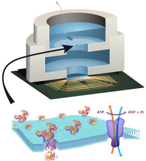
Illustration depicting a biocell attached to a CMOS integrated circuit with a membrane containing sodium-potassium pumps in pores. Energy is stored chemically in ATP molecules. When the energy is released as charged ions (which are then converted to electrons to power the chip at the bottom of the experimental device), the ATP is converted to ADP + inorganic phosphate. (credit: Trevor Finney and Jared Roseman/Columbia Engineering)
Columbia Engineering researchers have combined biological and solid-state components for the first time, opening the door to creating entirely new artificial biosystems.
In this experiment, they used a biological cell to power a conventional solid-state complementary metal-oxide-semiconductor (CMOS) integrated circuit. An artificial lipid bilayer membrane containing adenosine triphosphate (ATP)-powered ion pumps (which provide energy for cells) was used as a source of ions (which were converted to electrons to power the chip).
The study, led by Ken Shepard, Lau Family Professor of Electrical Engineering and professor of biomedical engineering at Columbia Engineering, was published online today (Dec. 7, 2015) in an open-access paper in Nature Communications.
How to build a hybrid biochip
Living systems achieve this functionality with their own version of electronics based on lipid membranes and ion channels and pumps, which act as a kind of “biological transistor.” Charge in the form of ions carry energy and information, and ion channels control the flow of ions across cell membranes.
Solid-state systems, such as those in computers and communication devices, use electrons; their electronic signaling and power are controlled by field-effect transistors.
To build a prototype of their hybrid system, Shepard’s team packaged a CMOS integrated circuit (IC) with an ATP-harvesting “biocell.” In the presence of ATP, the system pumped ions across the membrane, producing an electrical potential (voltage)* that was harvested by the integrated circuit.
“We made a macroscale version of this system, at the scale of several millimeters, to see if it worked,” Shepard notes. “Our results provide new insight into a generalized circuit model, enabling us to determine the conditions to maximize the efficiency of harnessing chemical energy through the action of these ion pumps. We will now be looking at how to scale the system down.”
While other groups have harvested energy from living systems, Shepard and his team are exploring how to do this at the molecular level, isolating just the desired function and interfacing this with electronics. “We don’t need the whole cell,” he explains. “We just grab the component of the cell that’s doing what we want. For this project, we isolated the ATPases because they were the proteins that allowed us to extract energy from ATP.”
The capability of a bomb-sniffing dog, no Alpo required
Next, the researchers plan to go much further, such as recognizing specific molecules and giving chips the potential to taste and smell.
The ability to build a system that combines the power of solid-state electronics with the capabilities of biological components has great promise, they believe. “You need a bomb-sniffing dog now, but if you can take just the part of the dog that is useful — the molecules that are doing the sensing — we wouldn’t need the whole animal,” says Shepard.
The technology could also provide a power source for implanted electronic devices in ATP-rich environments such as inside living cells, the researchers suggest.
* “In general, integrated circuits, even when operated at the point of minimum energy in subthreshold, consume on the order of 10−2 W mm−2 (or assuming a typical silicon chip thickness of 250 μm, 4 × 10⁻² W mm−3). Typical cells, in contrast, consume on the order of 4 × 10⁻⁶ W mm−3. In the experiment, a typical active power dissipation for the IC circuit was 92.3 nW, and the active average harvesting power was 71.4 fW for the biocell (the discrepancy is managed through duty-cycled operation of the IC).” — Jared M. Roseman et al./Nature Communications
There is enormous potential in combining the capabilities of the biological and the solid state to create hybrid engineered systems. While there have been recent efforts to harness power from naturally occurring potentials in living systems in plants and animals to power complementary metal-oxide-semiconductor integrated circuits, here we report the first successful effort to isolate the energetics of an electrogenic ion pump in an engineered in vitro environment to power such an artificial system. An integrated circuit is powered by adenosine triphosphate through the action of Na+/K+ adenosine triphosphatases in an integrated in vitro lipid bilayer membrane. The ion pumps (active in the membrane at numbers exceeding 2 × 106mm−2) are able to sustain a short-circuit current of 32.6pAmm−2 and an open-circuit voltage of 78mV, providing for a maximum power transfer of 1.27pWmm−2 from a single bilayer. Two series-stacked bilayers provide a voltage sufficient to operate an integrated circuit with a conversion efficiency of chemical to electrical energy of 14.9%.
(a) Illustration depicting biocell attached to CMOS integrated circuit. (b) Illustration of membrane in pore containing sodium–potassium pumps. (c) Circuit model of equivalent stacked membranes, =2.1pA, =98.6GΩ, =575GΩ and =75pF, Ag/AgCl electrode equivalent resistance RWE+RCE<20kΩ, energy-harvesting capacitor CSTOR=100nF combined with switch as an impedance transformation network (only one switch necessary due to small duty cycle), and CMOS IC voltage doubler and resistor representing digital switching load. RL represents the four independent ring oscillator loads. (d) Equivalent circuit detail of stacked biocell. (e) Switched-capacitor voltage doubler circuit schematic.
The energetics of living systems are based on electrochemical membrane potentials that are present in cell plasma membranes, the inner membrane of mitochondria, or the thylakoid membrane of chloroplasts1. In the latter two cases, the specific membrane potential is known as the proton-motive force and is used by proton adenosine triphosphate (ATP) synthases to produce ATP. In the former case, Na+/K+-ATPases hydrolyse ATP to maintain the resting potential in most cells.
While there have been recent efforts to harness power from some naturally occurring potentials in living systems that are the result of ion pump action both in plants2 and animals3, 4 to power complementary metal-oxide semiconductor (CMOS) integrated circuits (ICs), this work is the first successful effort to isolate the energetics of an electrogenic ion pump in an engineered in vitroenvironment to power such an artificial system. Prior efforts to harness power from in vitromembrane systems incorporating ion-pumping ATPases5, 6, 7, 8, 9 and light-activated bacteriorhodopsin9, 10, 11 have been limited by difficulty in incorporating these proteins in sufficient quantity to attain measurable current and in achieving sufficiently large membrane resistances to harness these currents. Both problems are solved in this effort to power an IC from ATP in an in vitro environment. The resulting measurements provide new insight into a generalized circuit model, which allows us to determine the conditions to maximize the efficiency of harnessing chemical energy through the action of electrogenic ion pumps.
ATP-powered IC
Figure 1a shows the complete hybrid integrated system, consisting of a CMOS IC packaged with an ATP-harvesting ‘biocell’. The biocell consists of two series-stacked ATPase bearing suspended lipid bilayers with a fluid chamber directly on top of the IC. Series stacking of two membranes is necessary to provide the required start-up voltage for IC and eliminates the need for an external energy source, which is typically required to start circuits from low-voltage supplies2, 3. As shown inFig. 1c, a matching network in the form of a switched capacitor allows the load resistance of the IC to be matched to that presented by the biocell. In principle, the switch S can be implicit. The biocell charges CSTOR until the self start-up voltage, Vstart, is reached. The chip then operates until the biocell voltage drops below the minimum supply voltage for operation, Vmin. Active current draw from the IC stops at this point, allowing the charge to build up again on CSTOR. In our case, however, the IC leakage current exceeds 13.5nA at Vstart, more than can be provided by the biocell. As a result, an explicit transistor switch and comparator (outside of the IC) are used for this function in the experimental results presented here, which are not powered by the biocell and not included in energy efficiency calculations (see Supplementary Discussion for additional details). The energy from the biocell is used to operate a voltage converter (voltage doubler) and some simple inverter-based ring oscillators in the IC, which receive power from no other sources.
…….. Prior to the addition of ATP, the membrane produces no electrical power and has an Rm of 280GΩ. A 1.7-pA short-circuit (SC) current (Fig. 2b) through the membrane is observed upon the addition of ATP (final concentration 3mM) to the cis chamber where functional, properly oriented enzymes generate a net electrogenic pump current. To perform these measurements, currents through each membrane of the biocell are measured using a voltage-clamp amplifier (inset of Fig. 2b) with a gain of 500GΩ with special efforts taken to compensate amplifier leakage currents. Each ATPase transports three Na+ ions from the cis chamber to the trans chamber and two K+ ions from thetrans chamber to the cis chamber (a net charge movement of one cation) for every molecule of ATP hydrolysed. At a rate of 100 hydrolysis events per second under zero electrical (SC) bias13, this results in an electrogenic current of ~16aA. The observed SC current corresponds to about 105 active ATPases in the membrane or a concentration of about 2 × 106mm−2, about 5% of the density of channels occurring naturally in mammalian nerve fibres14. It is expected that half of the channels inserted are inactive because they are oriented incorrectly.
(a)…Pre-ATP data linear fit (black line) slope yield Rm=280GΩ. Post ATP data fit to a Boltzmann curve, slope=0.02V (blue line). Post-ATP linear fit (red line) yields Ip=−1.8pA and Rp=61.6GΩ, which corresponds to a per-ATP source resistance of 6.16 × 10¹⁵. The current due to membrane leakage through R_{m} is subtracted in the post-ATP curve…. (b)…
Current–voltage characteristics of the ATPases
Figure 2a shows the complete measured current–voltage (I–V) characteristic of a single ATPase-bearing membrane in the presence of ATP. The current due to membrane leakage through Rm is subtracted in the post-ATP curve. The I–V characteristic fits a Boltzmann sigmoid curve, consistent with sodium–potassium pump currents measured on membrane patches at similar buffer conditions13, 15, 16. This nonlinear behaviour reflects the fact that the full ATPase transport cycle (three Na+ ions from cis to trans and two K+ ions from trans to cis) time increases (the turn-over rate, kATP, decreases) as the membrane potential increases16. No effect on pump current is expected from any ion concentration gradients produced by the action of the ATPases (seeSupplementary Discussion). Using this Boltzmann fit, we can model the biocell as a nonlinear voltage-controlled current source IATPase (inset Fig. 2a), in which the current produced by this source varies as a function of Vm. In the fourth quadrant, where the cell is producing electrical power, this model can be linearized as a Norton equivalent circuit, consisting of a DC current source (Ip) in parallel with a current-limiting resistor (Rp), which acts to limit the current delivered to the load at increasing bias (IATPase~Ip−Vm/Rp). Figure 2c shows the measured and simulated charging of Cm for a single membrane (open-circuited voltage). A custom amplifier with input resistance Rin>10TΩ was required for this measurement (see Electrical Measurement Methods).
Reconciling operating voltage differences
The electrical characteristics of biological systems and solid-state systems are mismatched in their operating voltages. The minimum operating voltage of solid-state systems is determined by the need for transistors to modulate a Maxwell–Boltzmann (MB) distribution of carriers by several orders of magnitude through the application of a potential that is several multiples of kT/q (where kis Boltzmann’s constant, T is the temperature in degrees Kelvin and q is the elementary charge). Biological systems, while operating under the same MB statistics, have no such constraints for operating ion channels since they are controlled by mechanical (or other conformational) processes rather than through modulation of a potential barrier. To bridge this operating voltage mismatch, the circuit includes a switched-capacitor voltage doubler (Fig. 1d) that is capable of self-startup from voltages as low Vstart=145mV (~5.5kT/q) and can be operated continuously from input voltages from as low as Vmin=110mV (see Supplementary Discussion)…..
Maximizing the efficiency of harvesting energy from ATP
Solid-state systems and biological systems are also mismatched in their operating impedances. In our case, the biocell presents a source impedance, =84.2GΩ, while the load impedance presented by the complete integrated circuit (including both the voltage converter and ring oscillator loads) is approximately RIC=200kΩ. (The load impedance, RL, of the ring oscillators alone is 305kΩ.) This mismatch in source and load impedance is manifest in large differences in power densities. In general, integrated circuits, even when operated at the point of minimum energy in subthreshold, consume on the order of 10−2Wmm−2 (or assuming a typical silicon chip thickness of 250μm, 4 × 10⁻²Wmm−3) (ref. 17). Typical cells, in contrast, consume on the order of 4 × 10⁻⁶Wmm−3 (ref. 18). In our case, a typical active power dissipation for our circuit is 92.3nW, and the active average harvesting power is 71.4fW for the biocell. This discrepancy is managed through duty-cycled operation of the IC in which the circuit is largely disabled for long periods of time (Tcharge), integrating up the power onto a storage capacitor (CSTOR), which is then expended in a very brief period of activity (Trun), as shown in Fig. 3a.
The overall efficiency of the system in converting chemical energy to the energy consumed in the load ring oscillator (η) is given by the product of the conversion efficiency of the voltage doubler (ηconverter) and the conversion efficiency of chemical energy to electrical energy in the biocell (ηbiocell), η=ηconverter × ηbiocell. ηconverter is relatively constant over the range of input voltages at ~59%, as determined by various loading test circuits included in the chip design (Supplementary Figs 1–6). ηbiocell, however, varies with transmembrane potential Vm. η is the efficiency in transferring power to the power ring oscillator loads from the ATP harvested by biocell.
…….
To first order, the energy made available to the Na+/K+-ATPase by the hydrolysis of ATP is independent of the chemical or electric potential of the membrane and is given by |ΔGATP|/(qNA), where ΔGATP is the Gibbs free energy change due to the ATP hydrolysis reaction per mole of ATP at given buffer conditions and NA is Avogadro’s number. Since every charge that passes through IATPase corresponds to a single hydrolysis event, we can use two voltage sources in series with IATPase to independently account for the energy expended by the pumps both in moving charge across the electric potential difference and in moving ions across the chemical potential difference. The dependent voltage source Vloss in this branch fixes the voltage across IATPase, and the total power produced by the pump current source is (|ΔGATP|/NA)(NkATP), which is the product of the energy released per molecule of ATP, the number of active ATPases and the ATP turnover rate. The power dissipated in voltage source Vchem models the work performed by the ATPases in transporting ions against a concentration gradient. In the case of the Na+/K+ ATPase,Vchem is given by . The power dissipated in this source is introduced back into the circuit in the power generated by the Nernst independent voltage sources, and . The power dissipated in the dependent voltage source Vloss models any additional power not used to perform chemical or electrical work. ……
Integration of ATP-harvesting ion pumps could provide a means to power future CMOS microsystems scaled to the level of individual cells22. In molecular diagnostics, the integration of pore-forming proteins such as alpha haemolysin23 or MspA porin24 with CMOS electronics is already finding application in DNA sequencing25. Exploiting the large diversity of function available in transmembrane proteins in these hybrid systems could, for example, lead to highly specific sensing platforms for airborne odorants or soluble molecular entities26, 27. Heavily multiplexed platforms could become high-throughput in vitro drug-screening platforms against this diversity of function. In addition, integration of transmembrane proteins with CMOS may become a convenient alternative to fluorescence for coupling to synthetic biological systems28.
Himes, C., Carlson, E., Ricchiuti, R. J., Otis, B. P. & Parviz, B. A.Ultralow voltage nanoelectronics powered directly, and solely, from a tree. IEEE Trans. Nanotechnol.9, 2–5(2010).
Mercier, P. P., Lysaght, A. C., Bandyopadhyay, S., Chandrakasan, A. P. & Stankovic, K. M.Energy extraction from the biologic battery in the inner ear. Nat. Biotechnol.30, 1240–1243(2012).
Using computational modeling, researchers at Carnegie Mellon University, the Colorado School of Mines and the University of California, Davis have come up with a design for a better liposome. Their findings, while theoretical, could provide the basis for efficiently constructing new vehicles for nanodrug delivery.
Liposomes are small containers with shells made of lipids, the same material that makes up the cell membrane. In recent years, liposomes have been used for targeted drug delivery. In this process, the membrane of a drug-containing liposome is engineered to contain proteins that will recognize and interact with complementary proteins on the membrane of a diseased or dysfunctional cell. After the drug-containing liposomes are administered, they travel through the body, ideally connecting with targeted cells where they release the drug.
This packaging technique is often used with highly toxic nanodrugs, like chemotherapy drugs, in an attempt to prevent the free drug from damaging non-cancerous cells. However, studies of this model of delivery have shown that in many cases less than 10 percent of the drugs transported by liposomes end up in tumor cells. Often, the liposome breaks open before it reaches a tumor cell and the drug is absorbed into the body’s organs, including the liver and spleen, resulting in toxic side effects.
“Even with current forms of targeted drug delivery, treatments like chemotherapy are still very brutal. We wanted to see how we could make targeted drug delivery better,” said Markus Deserno, professor of physics at Carnegie Mellon and a member of the university’s Center for Membrane Biology and Biophysics.
Deserno and colleagues propose that targeted drug delivery can be improved by making more stable liposomes. Using three different types of computer modeling, they have shown that liposomes can be made sturdier by incorporating a nanoparticle core made of a material like gold or iron and connecting that core to the liposome’s membrane using polymer tethers. The core and tethers act as a hub-and-spoke-like scaffold and shock-absorber system that help the liposome to weather the stresses and strains it encounters as it travels through the body to its target.
Francesca Stanzione and Amadeu K. Sum of the Colorado School of Mines conducted a fine-grained simulation that looked at how the polymer tethers anchor the liposome’s membrane at an atomistic level. Roland Faller of UC Davis did a meso-scale simulation that looked how a number of tethers held on to a small patch of membrane. Each of these simulations allowed researchers to look at smaller components of the liposome, nanoparticle core and tethers, but not the entire structure.
To see the entire structure, Carnegie Mellon’s Deserno and Mingyang Hu developed a coarse-grained model that represents groupings of components rather than individual atoms. For example, one lipid in the cell membrane might have 100 atoms. In a fine-grain simulation, each atom would be represented. In Deserno’s coarse grain simulation, those atoms might be represented by only three pieces instead of 100.
“Its unfeasible to look at the complete construct at an atomistic level. There are too many atoms to consider, and the timescale is too long. Even with the most advanced supercomputer, we wouldn’t have the power to run an atom-level simulation,” Deserno said. “But the physics that matters isn’t locally specific. It’s more like soft matter physics, which can be described at a much coarser resolution.”
Deserno’s simulation allowed the researchers to see how the entire reinforced liposome construct responded to stress and strain. They proposed that if a liposome was given the right-sized hub and tethers, its membrane would be much more resilient, bending to absorb impact and pressure.
Additionally, they were able to simulate how to best assemble the liposome, hub and tether system. They found that if the hub and tether are attached and placed in a solution of lipids, and solvent conditions are suitably chosen, a correctly sized liposome would self-assemble around the hub and tethers.
The researchers hope that chemists and drug developers will one day be able to use their simulations to determine what size core and polymer tethers they would need to effectively secure a liposome designed to deliver a specific drug or other nanoparticle. Using such simulations could narrow down the design parameters, speed up the development process and reduce costs.
NIHS to use the Lipotype Shotgun Lipidomics Technology for lipid analysis.
Lipotype GmbH and the Nestlé Institute of Health Sciences (NIHS) have collaborated to employ the innovative Lipotype Shotgun Lipidomics Technology to analyze lipids in blood for nutritional research. Recently, Lipotype and NIHS have jointly published results of the robustness of the Lipotype Technology. Lipotype envisions a future use of its technology in clinical diagnostics screens for establishing reliable lipid diagnostic biomarkers.
Innovative Lipotype Technology for lipid analysis
The purpose of this collaboration is to enable NIHS to use the Lipotype Shotgun Lipidomics Technology for lipid analysis. The mass spectrometry-based Lipotype technology covers a broad spectrum of lipid molecules and delivers quantitative results in high-throughput. The Nestlé Institute of Health Sciences uses this technology platform for nutritional research. NIHS is a specialized biomedical research institute and is part of Nestlé’s global Research & Development network.
Joint research project reveals robustness of Lipotype Technology
During the collaboration, Lipotype and NIHS conducted a joint research project and demonstrated that the Lipotype technology was robust enough to deliver data with high precision and negligible technical variation between different sites. In addition, important features are the high coverage and throughput, which were confirmed when applying the Lipotype technology.
Lipotype envisions these as important features, required for future use in clinical diagnostics screens, in order to establish and validate reliable lipid diagnostic biomarkers. The results have been published in October 2015, in the European Journal of Lipid Science and Technology (Surma et al. “An Automated Shotgun Lipidomics Platform for High Throughput, Comprehensive, and Quantitative Analysis of Blood Plasma Intact Lipids.”).
Lipids play an important role for health and disease
Lipotype is a spin-off company of the Max-Planck-Institute of Molecular Cell Biology and Genetics in Dresden, Germany. Prof. Kai Simons, CEO of Lipotype explains: “We developed a novel Shotgun-Lipidomics technology to analyze lipids in blood and other biological samples. Our analysis is quick and covers hundreds of lipid molecules at the same time. Our technology can be used to identify disease related lipid signatures.”
Researchers at the University of Liverpool, working with a global healthcare company, have helped develop a new treatment for obesity.
The treatment, which is a once-daily injectable derivative of a metabolic hormone called GLP-1 conventionally used in the treatment of type 2 diabetes, has proved successful in helping non-diabetic obese patients lose weight.
Professor John Wilding, who leads Obesity and Endocrinology research in the Institute of Ageing and Chronic Disease, investigates the pathophysiology and treatment of both obesity and type 2 diabetes and is applying his expertise in this area to work with, and often act as a consultant for, a number of large pharmaceutical companies looking to develop new treatments for obesity and diabetes.
Exciting development
Professor Wilding, said: “The biology of GLP-1 has been a focus of my research for 20 years; in particular when I was working at Hammersmith Hospital in London, I was part of the team that demonstrated that it was involved in appetite regulation; work on GLP-1 has continued during my time in Liverpool. Being involved in the development of a treatment, from the basic research right through to clinical trials in patients is very exciting”.
“It is likely that the treatment will be used initially in very specific situations, such as helping patients who are severely obese. It differs from current treatments used for diabetes, as it has stronger appetite regulating effects but no greater effect on glucose control.”
In 2014 more than 1.9 billion adults worldwide were classed as obese by the World Health Organisation; in the UK numbers have more than tripled since 1980. This Obesity can lead to other serious health-related illnesses including type 2 diabetes, hypertension and obstructive sleep apnoea as well as increasing the risk for many common cancers.
The drug has been approved in the European Union, but has not yet launched in the UK.
Professor Wilding added: “Consultancy like this can help relationship and reputation building and informs my research keeping it at the forefront of developments. It also brings many other benefits such as publications and income generation, which can help support other research, for example by such as funding for pilot projects that can lead to grant applications and investigator-initiated trials funded by the company”.
Researchers in the West Midlands have made a breakthrough in explaining how an incurable type of blood cancer develops from an often symptomless prior blood disorder.
The findings could lead to more effective treatments and ways to identify those most at risk of developing the cancer.
All patients diagnosed with myeloma, a cancer of the blood-producing bone marrow, first develop a relatively benign condition called ‘monoclonal gammopathy of undetermined significance’ or ‘MGUS’.
MGUS is fairly common in the older population and only progresses to cancer in approximately one in 100 cases. However, currently there is no way of accurately predicting which patients with MGUS are likely to go on to get myeloma.
Myeloma is diagnosed in around 4,000 people each year in the UK. It specifically affects antibody-producing white blood cells found in the bone marrow, called plasma cells. The researcher team from the University of Birmingham, New Cross and Heartlands Hospitals compared the cellular chemistry of bone marrow and blood samples taken from patients with myeloma, patients with MGUS and healthy volunteers.
Surprisingly, the researchers found that the metabolic activity of the bone marrow of patients with MGUS was significantly different to plasma from healthy volunteers, but there were very few differences at all between the MGUS and myeloma samples. The research was funded by the blood cancer charity Bloodwise, which changed its name from Leukaemia & Lymphoma in September.
The findings suggest that the biggest metabolic changes occur with the development of the symptomless condition MGUS and not with the later progression to myeloma.
Dr Daniel Tennant, who led the research at the University of Birmingham, said, “Our findings show that very few changes are required for a MGUS patient to progress to myeloma as we now know virtually all patients with myeloma evolve from MGUS. A drug that interferes with these specific initial metabolic changes could make a very effective treatment for myeloma, so this is a very exciting discovery.”
The research team found over 200 products of metabolism differed between the healthy volunteers and patients with MGUS or myeloma, compared to just 26 differences between MGUS patients and myeloma patients. The researchers believe that these small changes could drive the key shifts in the bone marrow required to support myeloma growth.


 . The power dissipated in this source is introduced back into the circuit in the power generated by the Nernst independent voltage sources,
  and
and  . The power dissipated in the dependent voltage source Vloss models any additional power not used to perform chemical or electrical work. ……