Feeds:
Posts
Comments

Archive for the ‘Ionic Transporters Na+’ Category

Hybrid lipid bioelectronic membranes

Larry H. Bernstein, MD, FCAP, Curator

LPBI


Hybrid solid-state chips and biological cells integrated at molecular level

Biological ion channels combine with solid-state transistors to create a new kind of hybrid bioelectronics. Imagine chips with dog-like capability to taste and smell, or even recognize specific molecules.
http://www.kurzweilai.net/hybrid-solid-state-chips-and-biological-cells-integrated-at-molecular-level
Illustration depicting a biocell attached to a CMOS integrated circuit with a membrane containing sodium-potassium pumps in pores. Energy is stored chemically in ATP molecules. When the energy is released as charged ions (which are then converted to electrons to power the chip at the bottom of the experimental device), the ATP is converted to ADP + inorganic phosphate. (credit: Trevor Finney and Jared Roseman/Columbia Engineering)

Columbia Engineering researchers have combined biological and solid-state components for the first time, opening the door to creating entirely new artificial biosystems.

In this experiment, they used a biological cell to power a conventional solid-state complementary metal-oxide-semiconductor (CMOS) integrated circuit. An artificial lipid bilayer membrane containing adenosine triphosphate (ATP)-powered ion pumps (which provide energy for cells) was used as a source of ions (which were converted to electrons to power the chip).

The study, led by Ken Shepard, Lau Family Professor of Electrical Engineering and professor of biomedical engineering at Columbia Engineering, was published online today (Dec. 7, 2015) in an open-access paper in Nature Communications.

How to build a hybrid biochip

Living systems achieve this functionality with their own version of electronics based on lipid membranes and ion channels and pumps, which act as a kind of “biological transistor.” Charge in the form of ions carry energy and information, and ion channels control the flow of ions across cell membranes.

Solid-state systems, such as those in computers and communication devices, use electrons; their electronic signaling and power are controlled by field-effect transistors.

To build a prototype of their hybrid system, Shepard’s team packaged a CMOS integrated circuit (IC) with an ATP-harvesting “biocell.” In the presence of ATP, the system pumped ions across the membrane, producing an electrical potential (voltage)* that was harvested by the integrated circuit.

“We made a macroscale version of this system, at the scale of several millimeters, to see if it worked,” Shepard notes. “Our results provide new insight into a generalized circuit model, enabling us to determine the conditions to maximize the efficiency of harnessing chemical energy through the action of these ion pumps. We will now be looking at how to scale the system down.”

While other groups have harvested energy from living systems, Shepard and his team are exploring how to do this at the molecular level, isolating just the desired function and interfacing this with electronics. “We don’t need the whole cell,” he explains. “We just grab the component of the cell that’s doing what we want. For this project, we isolated the ATPases because they were the proteins that allowed us to extract energy from ATP.”

The capability of a bomb-sniffing dog, no Alpo required

Next, the researchers plan to go much further, such as recognizing specific molecules and giving chips the potential to taste and smell.

The ability to build a system that combines the power of solid-state electronics with the capabilities of biological components has great promise, they believe. “You need a bomb-sniffing dog now, but if you can take just the part of the dog that is useful — the molecules that are doing the sensing — we wouldn’t need the whole animal,” says Shepard.

The technology could also provide a power source for implanted electronic devices in ATP-rich environments such as inside living cells, the researchers suggest.

*  “In general, integrated circuits, even when operated at the point of minimum energy in subthreshold, consume on the order of 10−2 W mm−2 (or assuming a typical silicon chip thickness of 250 μm, 4 × 10⁻² W mm−3). Typical cells, in contrast, consume on the order of 4 × 10⁻⁶ W mm−3. In the experiment, a typical active power dissipation for the IC circuit was 92.3 nW, and the active average harvesting power was 71.4 fW for the biocell (the discrepancy is managed through duty-cycled operation of the IC).” — Jared M. Roseman et al./Nature Communications


Hybrid integrated biological–solid-state system powered with adenosine triphosphate

Jared M. RosemanJianxun LinSiddharth RamakrishnanJacob K. Rosenstein & Kenneth L. Shepard
Nature Communications 7 Dec 2015; 6(10070)
     http://dx.doi.org:/10.1038/ncomms10070

There is enormous potential in combining the capabilities of the biological and the solid state to create hybrid engineered systems. While there have been recent efforts to harness power from naturally occurring potentials in living systems in plants and animals to power complementary metal-oxide-semiconductor integrated circuits, here we report the first successful effort to isolate the energetics of an electrogenic ion pump in an engineered in vitro environment to power such an artificial system. An integrated circuit is powered by adenosine triphosphate through the action of Na+/K+ adenosine triphosphatases in an integrated in vitro lipid bilayer membrane. The ion pumps (active in the membrane at numbers exceeding 2 × 106mm−2) are able to sustain a short-circuit current of 32.6pAmm−2 and an open-circuit voltage of 78mV, providing for a maximum power transfer of 1.27pWmm−2 from a single bilayer. Two series-stacked bilayers provide a voltage sufficient to operate an integrated circuit with a conversion efficiency of chemical to electrical energy of 14.9%.


Figure 1: Fully hybrid biological–solid-state system.


Fully hybrid biological-solid-state system.

http://www.nature.com/ncomms/2015/151207/ncomms10070/images/ncomms10070-f1.jpg

(a) Illustration depicting biocell attached to CMOS integrated circuit. (b) Illustration of membrane in pore containing sodium–potassium pumps. (c) Circuit model of equivalent stacked membranes, =2.1pA, =98.6G, =575G and =75pF, Ag/AgCl electrode equivalent resistance RWE+RCE<20k, energy-harvesting capacitor CSTOR=100nF combined with switch as an impedance transformation network (only one switch necessary due to small duty cycle), and CMOS IC voltage doubler and resistor representing digital switching load. RL represents the four independent ring oscillator loads. (d) Equivalent circuit detail of stacked biocell. (e) Switched-capacitor voltage doubler circuit schematic.


The energetics of living systems are based on electrochemical membrane potentials that are present in cell plasma membranes, the inner membrane of mitochondria, or the thylakoid membrane of chloroplasts1. In the latter two cases, the specific membrane potential is known as the proton-motive force and is used by proton adenosine triphosphate (ATP) synthases to produce ATP. In the former case, Na+/K+-ATPases hydrolyse ATP to maintain the resting potential in most cells.

While there have been recent efforts to harness power from some naturally occurring potentials in living systems that are the result of ion pump action both in plants2 and animals3, 4 to power complementary metal-oxide semiconductor (CMOS) integrated circuits (ICs), this work is the first successful effort to isolate the energetics of an electrogenic ion pump in an engineered in vitroenvironment to power such an artificial system. Prior efforts to harness power from in vitromembrane systems incorporating ion-pumping ATPases5, 6, 7, 8, 9 and light-activated bacteriorhodopsin9, 10, 11 have been limited by difficulty in incorporating these proteins in sufficient quantity to attain measurable current and in achieving sufficiently large membrane resistances to harness these currents. Both problems are solved in this effort to power an IC from ATP in an in vitro environment. The resulting measurements provide new insight into a generalized circuit model, which allows us to determine the conditions to maximize the efficiency of harnessing chemical energy through the action of electrogenic ion pumps.


ATP-powered IC

Figure 1a shows the complete hybrid integrated system, consisting of a CMOS IC packaged with an ATP-harvesting ‘biocell’. The biocell consists of two series-stacked ATPase bearing suspended lipid bilayers with a fluid chamber directly on top of the IC. Series stacking of two membranes is necessary to provide the required start-up voltage for IC and eliminates the need for an external energy source, which is typically required to start circuits from low-voltage supplies2, 3. As shown inFig. 1c, a matching network in the form of a switched capacitor allows the load resistance of the IC to be matched to that presented by the biocell. In principle, the switch S can be implicit. The biocell charges CSTOR until the self start-up voltage, Vstart, is reached. The chip then operates until the biocell voltage drops below the minimum supply voltage for operation, Vmin. Active current draw from the IC stops at this point, allowing the charge to build up again on CSTOR. In our case, however, the IC leakage current exceeds 13.5nA at Vstart, more than can be provided by the biocell. As a result, an explicit transistor switch and comparator (outside of the IC) are used for this function in the experimental results presented here, which are not powered by the biocell and not included in energy efficiency calculations (see Supplementary Discussion for additional details). The energy from the biocell is used to operate a voltage converter (voltage doubler) and some simple inverter-based ring oscillators in the IC, which receive power from no other sources.

Figure 1: Fully hybrid biological–solid-state system.

http://www.nature.com/ncomms/2015/151207/ncomms10070/images/ncomms10070-f1.jpg


……..   Prior to the addition of ATP, the membrane produces no electrical power and has an Rm of 280G. A 1.7-pA short-circuit (SC) current (Fig. 2b) through the membrane is observed upon the addition of ATP (final concentration 3mM) to the cis chamber where functional, properly oriented enzymes generate a net electrogenic pump current. To perform these measurements, currents through each membrane of the biocell are measured using a voltage-clamp amplifier (inset of Fig. 2b) with a gain of 500G with special efforts taken to compensate amplifier leakage currents. Each ATPase transports three Na+ ions from the cis chamber to the trans chamber and two K+ ions from thetrans chamber to the cis chamber (a net charge movement of one cation) for every molecule of ATP hydrolysed. At a rate of 100 hydrolysis events per second under zero electrical (SC) bias13, this results in an electrogenic current of ~16aA. The observed SC current corresponds to about 105 active ATPases in the membrane or a concentration of about 2 × 106mm−2, about 5% of the density of channels occurring naturally in mammalian nerve fibres14. It is expected that half of the channels inserted are inactive because they are oriented incorrectly.

Figure 2: Single-cell biocell characterization.

http://www.nature.com/ncomms/2015/151207/ncomms10070/images_article/ncomms10070-f2.jpg

(a)…Pre-ATP data linear fit (black line) slope yield Rm=280G. Post ATP data fit to a Boltzmann curve, slope=0.02V (blue line). Post-ATP linear fit (red line) yields Ip=−1.8pA and Rp=61.6G, which corresponds to a per-ATP source resistance of 6.16 × 10¹⁵. The current due to membrane leakage through R_{m} is subtracted in the post-ATP curve…. (b)…


Current–voltage characteristics of the ATPases

Figure 2a shows the complete measured current–voltage (IV) characteristic of a single ATPase-bearing membrane in the presence of ATP. The current due to membrane leakage through Rm is subtracted in the post-ATP curve. The IV characteristic fits a Boltzmann sigmoid curve, consistent with sodium–potassium pump currents measured on membrane patches at similar buffer conditions13, 15, 16. This nonlinear behaviour reflects the fact that the full ATPase transport cycle (three Na+ ions from cis to trans and two K+ ions from trans to cis) time increases (the turn-over rate, kATP, decreases) as the membrane potential increases16. No effect on pump current is expected from any ion concentration gradients produced by the action of the ATPases (seeSupplementary Discussion). Using this Boltzmann fit, we can model the biocell as a nonlinear voltage-controlled current source IATPase (inset Fig. 2a), in which the current produced by this source varies as a function of Vm. In the fourth quadrant, where the cell is producing electrical power, this model can be linearized as a Norton equivalent circuit, consisting of a DC current source (Ip) in parallel with a current-limiting resistor (Rp), which acts to limit the current delivered to the load at increasing bias (IATPase~IpVm/Rp). Figure 2c shows the measured and simulated charging of Cm for a single membrane (open-circuited voltage). A custom amplifier with input resistance Rin>10T was required for this measurement (see Electrical Measurement Methods).


Reconciling operating voltage differences

The electrical characteristics of biological systems and solid-state systems are mismatched in their operating voltages. The minimum operating voltage of solid-state systems is determined by the need for transistors to modulate a Maxwell–Boltzmann (MB) distribution of carriers by several orders of magnitude through the application of a potential that is several multiples of kT/q (where kis Boltzmann’s constant, T is the temperature in degrees Kelvin and q is the elementary charge). Biological systems, while operating under the same MB statistics, have no such constraints for operating ion channels since they are controlled by mechanical (or other conformational) processes rather than through modulation of a potential barrier. To bridge this operating voltage mismatch, the circuit includes a switched-capacitor voltage doubler (Fig. 1d) that is capable of self-startup from voltages as low Vstart=145mV (~5.5kT/q) and can be operated continuously from input voltages from as low as Vmin=110mV (see Supplementary Discussion)…..


Maximizing the efficiency of harvesting energy from ATP

Solid-state systems and biological systems are also mismatched in their operating impedances. In our case, the biocell presents a source impedance, =84.2G, while the load impedance presented by the complete integrated circuit (including both the voltage converter and ring oscillator loads) is approximately RIC=200k. (The load impedance, RL, of the ring oscillators alone is 305k.) This mismatch in source and load impedance is manifest in large differences in power densities. In general, integrated circuits, even when operated at the point of minimum energy in subthreshold, consume on the order of 10−2Wmm−2 (or assuming a typical silicon chip thickness of 250μm, 4 × 10⁻²Wmm−3) (ref. 17). Typical cells, in contrast, consume on the order of 4 × 10⁻⁶Wmm−3 (ref. 18). In our case, a typical active power dissipation for our circuit is 92.3nW, and the active average harvesting power is 71.4fW for the biocell. This discrepancy is managed through duty-cycled operation of the IC in which the circuit is largely disabled for long periods of time (Tcharge), integrating up the power onto a storage capacitor (CSTOR), which is then expended in a very brief period of activity (Trun), as shown in Fig. 3a.

The overall efficiency of the system in converting chemical energy to the energy consumed in the load ring oscillator (η) is given by the product of the conversion efficiency of the voltage doubler (ηconverter) and the conversion efficiency of chemical energy to electrical energy in the biocell (ηbiocell), η=ηconverter × ηbiocell. ηconverter is relatively constant over the range of input voltages at ~59%, as determined by various loading test circuits included in the chip design (Supplementary Figs 1–6). ηbiocell, however, varies with transmembrane potential Vm. η is the efficiency in transferring power to the power ring oscillator loads from the ATP harvested by biocell.

…….

To first order, the energy made available to the Na+/K+-ATPase by the hydrolysis of ATP is independent of the chemical or electric potential of the membrane and is given by |ΔGATP|/(qNA), where ΔGATP is the Gibbs free energy change due to the ATP hydrolysis reaction per mole of ATP at given buffer conditions and NA is Avogadro’s number. Since every charge that passes through IATPase corresponds to a single hydrolysis event, we can use two voltage sources in series with IATPase to independently account for the energy expended by the pumps both in moving charge across the electric potential difference and in moving ions across the chemical potential difference. The dependent voltage source Vloss in this branch fixes the voltage across IATPase, and the total power produced by the pump current source is (|ΔGATP|/NA)(NkATP), which is the product of the energy released per molecule of ATP, the number of active ATPases and the ATP turnover rate. The power dissipated in voltage source Vchem models the work performed by the ATPases in transporting ions against a concentration gradient. In the case of the Na+/K+ ATPase,Vchem is given by . The power dissipated in this source is introduced back into the circuit in the power generated by the Nernst independent voltage sources, and . The power dissipated in the dependent voltage source Vloss models any additional power not used to perform chemical or electrical work. ……


Integration of ATP-harvesting ion pumps could provide a means to power future CMOS microsystems scaled to the level of individual cells22. In molecular diagnostics, the integration of pore-forming proteins such as alpha haemolysin23 or MspA porin24 with CMOS electronics is already finding application in DNA sequencing25. Exploiting the large diversity of function available in transmembrane proteins in these hybrid systems could, for example, lead to highly specific sensing platforms for airborne odorants or soluble molecular entities26, 27. Heavily multiplexed platforms could become high-throughput in vitro drug-screening platforms against this diversity of function. In addition, integration of transmembrane proteins with CMOS may become a convenient alternative to fluorescence for coupling to synthetic biological systems28.


Roseman, J. M. et al. Hybrid integrated biological–solid-state system powered with adenosine triphosphate. Nat. Commun. 6:10070      http://dx.doi.org:/10.1038/ncomms10070 (2015).


  • Rottenberg, H. The measurement of membrane potential and deltapH in cells, organelles, and vesicles. Methods Enzymol. 55, 547569 (1979).
  • Himes, C., Carlson, E., Ricchiuti, R. J., Otis, B. P. & Parviz, B. A. Ultralow voltage nanoelectronics powered directly, and solely, from a tree. IEEE Trans. Nanotechnol. 9, 25(2010).
  • Mercier, P. P., Lysaght, A. C., Bandyopadhyay, S., Chandrakasan, A. P. & Stankovic, K. M.Energy extraction from the biologic battery in the inner ear. Nat. Biotechnol. 30, 12401243(2012).
  • Halámková, L. et al. Implanted Biofuel Cell Operating in a Living Snail. J. Am. Chem. Soc.134, 50405043 (2012).


Read Full Post »

Irreconciliable Dissonance in Physical Space and Cellular Metabolic Conception

Irreconciliable Dissonance in Physical Space and Cellular Metabolic Conception

Curator: Larry H. Bernstein, MD, FCAP

Pasteur Effect – Warburg Effect – What its history can teach us today. 

José Eduardo de Salles Roselino

The Warburg effect, in reality the “Pasteur-effect” was the first example of metabolic regulation described. A decrease in the carbon flux originated at the sugar molecule towards the end of the catabolic pathway, with ethanol and carbon dioxide observed when yeast cells were transferred from an anaerobic environmental condition to an aerobic one. In Pasteur´s studies, sugar metabolism was measured mainly by the decrease of sugar concentration in the yeast growth media observed after a measured period of time. The decrease of the sugar concentration in the media occurs at great speed in yeast grown in anaerobiosis (oxygen deficient) and its speed was greatly reduced by the transfer of the yeast culture to an aerobic condition. This finding was very important for the wine industry of France in Pasteur’s time, since most of the undesirable outcomes in the industrial use of yeast were perceived when yeasts cells took a very long time to create, a rather selective anaerobic condition. This selective culture media was characterized by the higher carbon dioxide levels produced by fast growing yeast cells and by a higher alcohol content in the yeast culture media.

However, in biochemical terms, this finding was required to understand Lavoisier’s results indicating that chemical and biological oxidation of sugars produced the same calorimetric (heat generation) results. This observation requires a control mechanism (metabolic regulation) to avoid burning living cells by fast heat released by the sugar biological oxidative processes (metabolism). In addition, Lavoisier´s results were the first indications that both processes happened inside similar thermodynamics limits. In much resumed form, these observations indicate the major reasons that led Warburg to test failure in control mechanisms in cancer cells in comparison with the ones observed in normal cells.

[It might be added that the availability of O2 and CO2 and climatic conditions over 750 million years that included volcanic activity, tectonic movements of the earth crust, and glaciation, and more recently the use of carbon fuels and the extensive deforestation of our land masses have had a large role in determining the biological speciation over time, in sea and on land. O2 is generated by plants utilizing energy from the sun and conversion of CO2. Remove the plants and we tip the balance. A large source of CO2 is from beneath the earth’s surface.]

Biology inside classical thermodynamics places some challenges to scientists. For instance, all classical thermodynamics must be measured in reversible thermodynamic conditions. In an isolated system, increase in P (pressure) leads to increase in V (volume), all this occurring in a condition in which infinitesimal changes in one affects in the same way the other, a continuum response. Not even a quantic amount of energy will stand beyond those parameters.

In a reversible system, a decrease in V, under same condition, will led to an increase in P. In biochemistry, reversible usually indicates a reaction that easily goes either from A to B or B to A. For instance, when it was required to search for an anti-ischemic effect of Chlorpromazine in an extra hepatic obstructed liver, it was necessary to use an adequate system of increased biliary system pressure in a reversible manner to exclude a direct effect of this drug over the biological system pressure inducer (bile secretion) in Braz. J. Med. Biol. Res 1989; 22: 889-893. Frequently, these details are jumped over by those who read biology in ATGC letters.

Very important observations can be made in this regard, when neutral mutations are taken into consideration since, after several mutations (not affecting previous activity and function), a last mutant may provide a new transcript RNA for a protein and elicit a new function. For an example, consider a Prion C from lamb getting similar to bovine Prion C while preserving  its normal role in the lamb when its ability to change Human Prion C is considered (Stanley Prusiner).

This observation is good enough, to confirm one of the most important contributions of Erwin Schrodinger in his What is Life:

“This little book arose from a course of public lectures, delivered by a theoretical physicist to an audience of about four hundred which did not substantially dwindle, though warned at the outset that the subject matter was a difficult one and that the lectures could not be termed popular, even though the physicist’s most dreaded weapon, mathematical deduction, would hardly be utilized. The reason for this was not that the subject was simple enough to be explained without mathematics, but rather that it was much too involved to be fully accessible to mathematics.”

After Hans Krebs, description of the cyclic nature of the citrate metabolism and after its followers described its requirement for aerobic catabolism two major lines of research started the search for the understanding of the mechanism of energy transfer that explains how ADP is converted into ATP. One followed the organic chemistry line of reasoning and therefore, searched for a mechanism that could explain how the breakdown of carbon-carbon link could have its energy transferred to ATP synthesis. One of the major leaders of this research line was Britton Chance. He took into account that relatively earlier in the series of Krebs cycle reactions, two carbon atoms of acetyl were released as carbon dioxide ( In fact, not the real acetyl carbons but those on the opposite side of citrate molecule). In stoichiometric terms, it was not important whether the released carbons were or were not exactly those originated from glucose carbons. His research aimed at to find out an intermediate proteinaceous intermediary that could act as an energy reservoir. The intermediary could store in a phosphorylated amino acid the energy of carbon-carbon bond breakdown. This activated amino acid could transfer its phosphate group to ADP producing ATP. A key intermediate involved in the transfer was identified by Kaplan and Lipmann at John Hopkins as acetyl coenzyme A, for which Fritz Lipmann received a Nobel Prize.

Alternatively, under possible influence of the excellent results of Hodgkin and Huxley a second line of research appears. The work of Hodgkin & Huxley indicated that the storage of electrical potential energy in transmembrane ionic asymmetries and presented the explanation for the change from resting to action potential in excitable cells. This second line of research, under the leadership of Peter Mitchell postulated a mechanism for the transfer of oxide/reductive power of organic molecules oxidation through electron transfer as the key for the energetic transfer mechanism required for ATP synthesis.
This diverted the attention from high energy (~P) phosphate bond to the transfer of electrons. During most of the time the harsh period of the two confronting points of view, Paul Boyer and followers attempted to act as a conciliatory third party, without getting good results, according to personal accounts (in L. A. or Latin America) heard from those few of our scientists who were able to follow the major scientific events held in USA, and who could present to us later. Paul  Boyer could present how the energy was transduced by a molecular machine that changes in conformation in a series of 3 steps while rotating in one direction in order to produce ATP and in opposite direction in order to produce ADP plus Pi from ATP (reversibility).

However, earlier, a victorious Peter Mitchell obtained the result in the conceptual dispute, over the Britton Chance point of view, after he used E. Coli mutants to show H+ gradients in the cell membrane and its use as energy source, for which he received a Nobel Prize. Somehow, this outcome represents such a blow to Chance’s previous work that somehow it seems to have cast a shadow over very important findings obtained during his earlier career that should not be affected by one or another form of energy transfer mechanism.  For instance, Britton Chance got the simple and rapid polarographic assay method of oxidative phosphorylation and the idea of control of energy metabolism that brings us back to Pasteur.

This metabolic alternative result seems to have been neglected in the recent years of obesity epidemics, which led to a search for a single molecular mechanism required for the understanding of the accumulation of chemical (adipose tissue) reserve in our body. It does not mean that here the role of central nervous system is neglected. In short, in respiring mitochondria the rate of electron transport linked to the rate of ATP production is determined primarily by the relative concentrations of ADP, ATP and phosphate in the external media (cytosol) and not by the concentration of respiratory substrate as pyruvate. Therefore, when the yield of ATP is high as it is in aerobiosis and the cellular use of ATP is not changed, the oxidation of pyruvate and therefore of glycolysis is quickly (without change in gene expression), throttled down to the resting state. The dependence of respiratory rate on ADP concentration is also seen in intact cells. A muscle at rest and using no ATP has a very low respiratory rate.   [When skeletal muscle is stressed by high exertion, lactic acid produced is released into the circulation and is metabolized aerobically by the heart at the end of the activity].

This respiratory control of metabolism will lead to preservation of body carbon reserves and in case of high caloric intake in a diet, also shows increase in fat reserves essential for our biological ancestors survival (Today for our obesity epidemics). No matter how important this observation is, it is only one focal point of metabolic control. We cannot reduce the problem of obesity to the existence of metabolic control. There are numerous other factors but on the other hand, we cannot neglect or remove this vital process in order to correct obesity. However, we cannot explain obesity ignoring this metabolic control. This topic is so neglected in modern times that we cannot follow major research lines of the past that were interrupted by the emerging molecular biology techniques and the vain belief that a dogmatic vision of biology could replace all previous knowledge by a new one based upon ATGC readings. For instance, in order to display bad consequences derived from the ignorance of these old scientific facts, we can take into account, for instance, how ion movements across membranes affects membrane protein conformation and therefore contradicts the wrong central dogma of molecular biology. This change in protein conformation (with unchanged amino acid sequence) and/or the lack of change in protein conformation is linked to the factors that affect vital processes as the heart beats. This modern ignorance could also explain some major pitfalls seen in new drugs clinical trials and in a small scale on bad medical practices.

The work of Britton Chance and of Peter Mitchell have deep and sound scientific roots that were made with excellent scientific techniques, supported by excellent scientific reasoning and that were produced in a large series of very important intermediary scientific results. Their sole difference was to aim at very different scientific explanations as their goals (They have different Teleology in their minds made by their previous experiences). When, with the use of mutants obtained in microorganisms P Mitchell´s goal was found to survive and B Chance to succumb to the experimental evidence, all those excellent findings of B Chance and followers were directed to the dustbin of scientific history as an example of lack of scientific consideration.  [On the one hand, the Mitchell model used a unicellular organism; on the other, Chance’s work was with eukaryotic cells, quite relevant to the discussion.]

We can resume the challenge faced by these two great scientists in the following form: The first conceptual unification in bioenergetics, achieved in the 1940s, is inextricably bound up with the name of Fritz Lipmann. Its central feature was the recognition that adenosine triphosphate, ATP, serves as a universal energy  “currency” much as money serves as economic currency. In a nutshell, the purpose of metabolism is to support the synthesis of ATP. In microorganisms, this is perfect! In humans or mammals, or vertebrates, by the same reason that we cannot consider that gene expression is equivalent to protein function (an acceptable error in the case of microorganisms) this oversimplifies the metabolic requirement with a huge error. However, in case our concern is ATP chemistry only, the metabolism produces ATP and the hydrolysis of ATP pays for the performance of almost, all kinds of works. It is possible to presume that to find out how the flow of metabolism (carbon flow) led to ATP production must be considered a major focal point of research of the two contenders. Consequently, what could be a minor fall of one of the contenders, in case we take into account all that was found during their entire life of research, the real failure in B Chance’s final goal was amplified far beyond what may be considered by reason!

Another aspect that must be taken into account: Both contenders have in the scientific past a very sound root. Metabolism may produce two forms of energy currency (I personally don´t like this expression*) and I use it here because it was used by both groups in order to express their findings. Together with simplistic thermodynamics, this expression conveys wrong ideas): The second kind of energy currency is the current of ions passing from one side of a membrane to the other. The P. Mitchell scientific root undoubtedly have the work of Hodgkin & Huxley, Huxley &  Huxley, Huxley & Simmons

*ATP is produced under the guidance of cell needs and not by its yield. When glucose yields only 2 ATPs per molecule it is oxidized at very high speed (anaerobiosis) as is required to match cellular needs. On the other hand, when it may yield (thermodynamic terms) 38 ATP the same molecule is oxidized at low speed. It would be similar to an investor choice its least money yield form for its investment (1940s to 1972) as a solid support. B. Chance had the enzymologists involved in clarifying how ATP could be produced directly from NADH + H+ oxidative reductive metabolic reactions or from the hydrolysis of an enolpyruvate intermediary. Both competitors had their work supported by different but, sound scientific roots and have produced very important scientific results while trying to present their hypothetical point of view.

Before the winning results of P. Mitchell were displayed, one line of defense used by B. Chance followers was to create a conflict between what would be expected by a restrictive role of proteins through its specificity ionic interactions and the general ability of ionic asymmetries that could be associated with mitochondrial ATP production. Chemical catalyzed protein activities do not have perfect specificity but an outstanding degree of selective interaction was presented by the lock and key model of enzyme interaction. A large group of outstanding “mitochondriologists” were able to show ATP synthesis associated with Na+, K+, Ca2+… asymmetries on mitochondrial membranes and any time they did this, P. Mitchell have to display the existence of antiporters that exchange X for hydrogen as the final common source of chemiosmotic energy used by mitochondria for ATP synthesis.

This conceptual battle has generated an enormous knowledge that was laid to rest, somehow discontinued in the form of scientific research, when the final E. Coli mutant studies presented the convincing final evidence in favor of P. Mitchell point of view.

Not surprisingly, a “wise anonymous” later, pointed out: “No matter what you are doing, you will always be better off in case you have a mutant”

(Principles of Medical Genetics T D Gelehrter & F.S. Collins chapter 7, 1990).

However, let’s take the example of a mechanical wristwatch. It clearly indicates when the watch is working in an acceptable way, that its normal functioning condition is not the result of one of its isolated components – or something that can be shown by a reductionist molecular view.  Usually it will be considered that it is working in an acceptable way, in case it is found that its accuracy falls inside a normal functional range, for instance, one or two standard deviations bellow or above the mean value for normal function, what depends upon the rigor wisely adopted. While, only when it has a faulty component (a genetic inborn error) we can indicate a single isolated piece as the cause of its failure (a reductionist molecular view).

We need to teach in medicine, first the major reasons why the watch works fine (not saying it is “automatic”). The functions may cross the reversible to irreversible regulatory limit change, faster than what we can imagine. Latter, when these ideas about normal are held very clear in the mind set of medical doctors (not medical technicians) we may address the inborn errors and what we may have learn from it. A modern medical technician may cause admiration when he uses an “innocent” virus to correct for a faulty gene (a rather impressive technological advance). However, in case the virus, later shows signals that indicate that it was not so innocent, a real medical doctor will be called upon to put things in correct place again.

Among the missing parts of normal evolution in biochemistry a lot about ion fluxes can be found. Even those oscillatory changes in Ca2+ that were shown to affect gene expression (C. De Duve) were laid to rest since, they clearly indicate a source of biological information that despite the fact that it does not change nucleotides order in the DNA, it shows an opposing flux of biological information against the dogma (DNA to RNA to proteins). Another, line has shown a hierarchy, on the use of mitochondrial membrane potential: First the potential is used for Ca2+ uptake and only afterwards, the potential is used for ADP conversion into ATP (A. L. Lehninger). In fact, the real idea of A. L. Lehninger was by far, more complex since according to him, mitochondria works like a buffer for intracellular calcium releasing it to outside in case of a deep decrease in cytosol levels or capturing it from cytosol when facing transient increase in Ca2+ load. As some of Krebs cycle dehydrogenases were activated by Ca2+, this finding was used to propose a new control factor in addition to the one of ADP (B. Chance). All this was discontinued with the wrong use of calculus (today we could indicate bioinformatics in a similar role) in biochemistry that has established less importance to a mitochondrial role after comparative kinetics that today are seen as faulty.

It is important to combat dogmatic reasoning and restore sound scientific foundations in basic medical courses that must urgently reverse the faulty trend that tries to impose a view that goes from the detail towards generalization instead of the correct form that goes from the general finding well understood towards its molecular details. The view that led to curious subjects as bioinformatics in medical courses as training in sequence finding activities can only be explained by its commercial value. The usual form of scientific thinking respects the limits of our ability to grasp new knowledge and relies on reproducibility of scientific results as a form to surpass lack of mathematical equation that defines relationship of variables and the determination of its functional domains. It also uses old scientific roots, as its sound support never replaces existing knowledge by dogmatic and/or wishful thinking. When the sequence of DNA was found as a technical advance to find amino acid sequence in proteins it was just a technical advance. This technical advance by no means could be considered a scientific result presented as an indication that DNA sequences alone have replaced the need to study protein chemistry, its responses to microenvironmental changes in order to understand its multiple conformations, changes in activities and function. As E. Schrodinger correctly describes the chemical structure responsible for the coded form stored of genetic information must have minimal interaction with its microenvironment in order to endure hundreds and hundreds years as seen in Hapsburg’s lips. Only magical reasoning assumes that it is possible to find out in non-reactive chemical structures the properties of the reactive ones.

For instance, knowledge of the reactions of the Krebs cycle clearly indicate a role for solvent that no longer could be considered to be an inert bath for catalytic activity of the enzymes when the transfer of energy include a role for hydrogen transport. The great increase in understanding this change on chemical reaction arrived from conformational energy.

Again, even a rather simplistic view of this atomic property (Conformational energy) is enough to confirm once more, one of the most important contribution of E. Schrodinger in his What is Life:

“This little book arose from a course of public lectures, delivered by a theoretical physicist to an audience of about four hundred which did not substantially dwindle, though warned at the outset that the subject matter was a difficult one and that the lectures could not be termed popular, even though the physicist’s most dreaded weapon, mathematical deduction, would hardly be utilized. The reason for this was not that the subject was simple enough to be explained without mathematics, but rather that it was much too involved to be fully accessible to mathematics.”

In a very simplistic view, while energy manifests itself by the ability to perform work conformational energy as a property derived from our atomic structure can be neutral, positive or negative (no effect, increased or decreased reactivity upon any chemistry reactivity measured as work)

Also:

“I mean the fact that we, whose total being is entirely based on a marvelous interplay of this very kind, yet if all possess the power of acquiring considerable knowledge about it. I think it possible that this knowledge may advance to little just a short of a complete understanding -of the first marvel. The second may well be beyond human understanding.”

In fact, scientific knowledge allows us to understand how biological evolution may have occurred or have not occurred and yet does not present a proof about how it would have being occurred. It will be always be an indication of possible against highly unlike and never a scientific proven fact about the real form of its occurrence.

As was the case of B. Chance in its bioenergetics findings, we may get very important findings that indicates wrong directions in the future as was his case, or directed toward our past.

The Skeleton of Physical Time – Quantum Energies in Relative Space of S-labs

By Radoslav S. Bozov  Independent Researcher

WSEAS, Biology and BioSystems of Biomedicine

Space does not equate to distance, displacement of an object by classically defined forces – electromagnetic, gravity or inertia. In perceiving quantum open systems, a quanta, a package of energy, displaces properties of wave interference and statistical outcomes of sums of paths of particles detected by a design of S-labs.

The notion of S-labs, space labs, deals with inherent problems of operational module, R(i+1), where an imagination number ‘struggles’ to work under roots of a negative sign, a reflection of an observable set of sums reaching out of the limits of the human being organ, an eye or other foundational signal processing system.

While heavenly bodies, planets, star systems, and other exotic forms of light reflecting and/or emitting objects, observable via naked eye have been deduced to operate under numerical systems that calculate a periodic displacement of one relative to another, atomic clocks of nanospace open our eyes to ever expanding energy spaces, where matrices of interactive variables point to the problem of infinity of variations in scalar spaces, however, defining properties of minute universes as a mirror image of an astronomical system. The first and furthermost problem is essentially the same as those mathematical methodologies deduced by Isaac Newton and Albert Einstein for processing a surface. I will introduce you to a surface interference method by describing undetermined objective space in terms of determined subjective time.

Therefore, the moment will be an outcome of statistical sums of a numerical system extending from near zero to near one. Three strings hold down a dual system entangled via interference of two waves, where a single wave is a product of three particles (today named accordingly to either weak or strong interactions) momentum.

The above described system emerges from duality into trinity the objective space value of physical realities. The triangle of physical observables – charge, gravity and electromagnetism, is an outcome of interference of particles, strings and waves, where particles are not particles, or are strings strings, or  are waves waves of an infinite character in an open system which we attempt to define to predict outcomes of tomorrow’s parameters, either dependent or independent as well as both subjective to time simulations.

We now know that aging of a biological organism cannot be defined within singularity. Thereafter, clocks are subjective to apparatuses measuring oscillation of defined parameters which enable us to calculate both amplitude and a period, which we know to be dependent on phase transitions.

The problem of phase was solved by the applicability of carbon relative systems. A piece of diamond does not get wet, yet it holds water’s light entangled property. Water is the dark force of light. To formulate such statement, we have been searching truth by examining cooling objects where the Maxwell demon is translated into information, a data complex system.

Modern perspectives in computing quantum based matrices, 0+1 =1 and/or 0+0=1, and/or 1+1 =0, will be reduced by applying a conceptual frame of Aladdin’s flying anti-gravity carpet, unwrapping both past and future by sending a photon to both, placing present always near zero. Thus, each parallel quantum computation of a natural system approaching the limit of a vibration of a string defining 0 does not equal 0, and 1 does not equal 1. In any case, if our method 1+1 = 1, yet, 1 is not 1 at time i+1. This will set the fundamentals of an operational module, called labris operator or in simplicity S-labs. Note, that 1 as a result is an event predictable to future, while interacting parameters of addition 1+1 may be both, 1 as an observable past, and 1 as an imaginary system, or 1+1 displaced interactive parameters of past observable events. This is the foundation of Future Quantum Relative Systems Interference (QRSI), taking analytical technologies of future as a result of data matrices compressing principle relative to carbon as a reference matter rational to water based properties.

Goedel’s concept of loops exist therefore only upon discrete relative space uniting to parallel absolute continuity of time ‘lags’. ( Goedel, Escher and Bach: An Eternal Golden Braid. A Metaphorical Fugue on Minds and Machines in the Spirit of Lewis Carroll. D Hofstadter.  Chapter XX: Strange Loops, Or Tangled Hierarchies. A grand windup of many of the ideas about hierarchical systems and self-reference. It is concerned with the snarls which arise when systems turn back on themselves-for example, science probing science, government investigating governmental wrongdoing, art violating the rules of art, and finally, humans thinking about their own brains and minds. Does Gödel’s Theorem have anything to say about this last “snarl”? Are free will and the sensation of consciousness connected to Gödel’s Theorem? The Chapter ends by tying Gödel, Escher, and Bach together once again.)  The fight struggle in-between time creates dark spaces within which strings manage to obey light properties – entangled bozons of information carrying future outcomes of a systems processing consciousness. Therefore, Albert Einstein was correct in his quantum time realities by rejecting a resolving cube of sugar within a cup of tea (Henri Bergson 19th century philosopher. Bergson’s concept of multiplicity attempts to unify in a consistent way two contradictory features: heterogeneity and continuity. Many philosophers today think that this concept of multiplicity, despite its difficulty, is revolutionary.) However, the unity of time and space could not be achieved by deducing time to charge, gravity and electromagnetic properties of energy and mass.

Charge is further deduced to interference of particles/strings/waves, contrary to the Hawking idea of irreducibility of chemical energy carrying ‘units’, and gravity is accounted for by intrinsic properties of   anti-gravity carbon systems processing light, an electromagnetic force, that I have deduced towards ever expanding discrete energy space-energies rational to compressing mass/time. The role of loops seems to operate to control formalities where boundaries of space fluctuate as a result of what we called above – dark time-spaces.

Indeed, the concept of horizon is a constant due to ever expanding observables. Thus, it fails to acquire a rational approach towards space-time issues.

Richard Feynman has touched on issues of touching of space, sums of paths of particle traveling through time. In a way he has resolved an important paradigm, storing information and possibly studying it by opening a black box. Schroedinger’s cat is alive again, but incapable of climbing a tree when chased by a dog. Every time a cat climbs a garden tree, a fruit falls on hedgehogs carried away parallel to living wormholes whose purpose of generating information lies upon carbon units resolving light.

In order to deal with such a paradigm, we will introduce i+1 under square root in relativity, therefore taking negative one ( -1 = sqrt (i+1), an operational module R dealing with Wheelers foam squeezed by light, releasing water – dark spaces. Thousand words down!

What is a number? Is that a name or some kind of language or both? Is the issue of number theory possibly accountable to the value of the concept of entropic timing? Light penetrating a pyramid holding bean seeds on a piece of paper and a piece of slice of bread, a triple set, where a church mouse has taken a drop of tear, but a blood drop. What an amazing physics! The magic of biology lies above egoism, above pride, and below Saints.

We will set up the twelve parameters seen through 3+1 in classic realities:

–              discrete absolute energies/forces – no contradiction for now between Newtonian and Albert Einstein mechanics

–              mass absolute continuity – conservational law of physics in accordance to weak and strong forces

–              quantum relative spaces – issuing a paradox of Albert Einstein’s space-time resolved by the uncertainty principle

–              parallel continuity of multiple time/universes – resolving uncertainty of united space and energy through evolving statistical concepts of scalar relative space expansion and vector quantum energies by compressing relative continuity of matter in it, ever compressing flat surfaces – finding the inverse link between deterministic mechanics of displacement and imaginary space, where spheres fit within surface of triangles as time unwraps past by pulling strings from future.

To us, common human beings, with an extra curiosity overloaded by real dreams, value happens to play in the intricate foundation of life – the garden of love, its carbon management in mind, collecting pieces of squeezed cooling time.

The infinite interference of each operational module to another composing ever emerging time constrains unified by the Solar system, objective to humanity, perhaps answers that a drop of blood and a drop of tear is united by a droplet of a substance separating negative entropy to time courses of a physical realities as defined by an open algorithm where chasing power subdue to space becomes an issue of time.

Jose Eduardo de Salles Roselino

Some small errors: For intance an increase i P leads to a decrease in V ( not an increase in V)..


Radoslav S. Bozov  Independent Researcher

If we were to use a preventative measures of medical science, instruments of medical science must predict future outcomes based on observable parameters of history….. There are several key issues arising: 1. Despite pinning a difference on genomic scale , say pieces of information, we do not know how to have changed that – that is shift methylome occupying genome surfaces , in a precise manner.. 2. Living systems operational quo DO NOT work as by vector gravity physics of ‘building blocks. That is projecting a delusional concept of a masonry trick, who has not worked by corner stones and ever shifting momenta … Assuming genomic assembling worked, that is dealing with inferences through data mining and annotation, we are not in a position to read future in real time, and we will never be, because of the rtPCR technology self restriction into data -time processing .. We know of existing post translational modalities… 3. We don’t know what we don’t know, and that foundational to future medicine – that is dealing with biological clocks, behavior, and various daily life inputs ranging from radiation to water systems, food quality, drugs…

Read Full Post »

Proteomics

Writer and Curator: Larry H. Bernstein, MD, FCAP


The previous discussion concerned genomics, metabolomics, and cancer. The discussion that follows is concerned with the expanding filed of proteomics, which has implication for disease discovery, pharmaceutical targeting, and diagnostics.

The human proteome – a scientific opportunity for transforming diagnostics, therapeutics, and healthcare

Marc Vidal, Daniel W Chan, Mark Gerstein, Matthias Mann, Gilbert S Omenn, et al.
Clinical Proteomics 2012, 9:6  http://www.clinicalproteomicsjournal.com/content/9/1/6

A National Institutes of Health (NIH) workshop was convened in Bethesda, MD on September 26–27, 2011, with representative scientific leaders in the field of proteomics and its applications to clinical settings. The main purpose of this workshop was to articulate ways in which the biomedical research community can capitalize on recent technology advances and synergize with ongoing efforts to advance the field of human proteomics. This executive summary and the following full report describe the main discussions and outcomes of the workshop.

Proteomics Pioneer Award 2013: Professor Amos Bairoch, University of Geneva, Switzerland

Eupa Open Proteomics 2 (2014) 34  http://dx.doi.org/10.1016/j.euprot.2013.12.002

Amos Bairoch has always been fascinated by computer science, genetics and biochemistry. His fi rst project, as a PhD student, was the development of PC/Gene, a MS-DOS based software package for the analysis of protein and nucleotide sequences. While working on this project, he realized that there was no single resource for protein sequences, and started to develop the first annotated protein sequence database, which became Swiss-Prot and was first released in July 1986. In 1988, he created PROSITE, a database of protein families and domains, and a little later ENZYME, an enzyme nomenclature database.

Amos Bairoch led the Swiss-Prot group from its creation in 1988 until 2009. During this period, Swiss-Prot became the primary protein sequence resource in the world and has been a key research instrument for both bioinformaticians and laboratory-based scientists, particularly in the field of proteomics.

Since 2009, Amos Bairoch’s group is developing neXtProt, a knowledgebase
specifically dedicated to human proteins.neXtProt has been chosen as the reference protein database for the HUPO Human Proteome Projects.

For his major contributions in the field of proteomic databases, Amos Bairoch received the Friedrich Miescher Award from the Swiss Society of Biochemistry in 1993, the Helmut Horten Foundation Incentive Award in 1995, the Pehr Edman award and the European Latsis Prize in 2004, the Otto Naegeli prize in 2010, and the HUPO Distinguished Achievement Award in Proteomic Sciences in 2011.

National Heart, Lung, and Blood Institute Clinical Proteomics Working Group Report

CB Granger, JE Van Eyk, SC Mockrin and N. Leigh Anderson
Circulation. 2004;109:1697-1703
http://dx.doi.org:/10.1161/01.CIR.0000121563.47232.2A

The National Heart, Lung, and Blood Institute (NHLBI) Clinical Proteomics Working Group was charged with identifying opportunities and challenges in clinical proteomics and using these as a basis for recommendations aimed at directly improving patient care. The group included representatives of clinical and translational research, proteomic technologies, laboratory medicine, bioinformatics, and 2 of the NHLBI Proteomics Centers, which form part of a program focused on innovative technology development. This report represents the results from a one-and-a-half-day meeting on May 8 and 9, 2003. For the purposes of this report, clinical proteomics is defined as the systematic, comprehensive, large-scale identification of protein patterns (“fingerprints”) of disease and the application of this knowledge to improve patient care and public health through better assessment of disease susceptibility, prevention of disease, selection of therapy for the individual, and monitoring of treatment response.

The -omics era: Proteomics and lipidomics in vascular research

Athanasios Didangelos, Christin Stegemann, Manuel Mayr
Atherosclerosis 221 (2012) 12– 17
http://dx.doi.org:/10.1016/j.atherosclerosis.2011.09.043

The retention of proatherogenic low-density lipoprotein (LDL) particles on the subendothelial extracellular matrix (ECM) is a hallmark of atherosclerosis. Apolipoprotein B (apoB)-containing lipoprotein particles are trapped in the arterial intima by proteoglycans in atherosclerosis-prone areas and eventually become modified, commonly by aggregation and oxidation. The initial accumulation of proatherogenic lipoproteins initiates an inflammatory response, which results in the release of proteolytic enzymes and induces the dedifferentiation of vascular smooth muscle cells (SMCs) resulting in alterations of their matrix producing properties. The precise mechanisms responsible for the accumulation of certain matrix components and subsequent lipoprotein retention on the vessel wall are not fully elucidated. Undoubtedly, ECM remodeling contributes to the formation of atherosclerotic lesions and the lipid composition of apolipoproteins influences their binding properties to the matrix. An unbiased discovery approach, which is not limited to known molecules of presumed importance, will be invaluable for the identification of novel, previously unknown mediators of disease. Although descriptive, the detailed examination of atherosclerotic plaques using advanced proteomics and lipidomics techniques can generate novel insights and form the basis for further mechanistic investigations.

The Revolution in Proteomics Ionization –
CaptiveSpray nanoBooster™
Bruker, LC-MS Source

Bruker CaptiveSpray principle:

Stable and robust nanoflow LC/MS is still a challenge in proteomics analysis. The Bruker CaptiveSpray source is a revolutionary ion source with a patented design that provides provides easy operation just as simple normal flow electrospray.

CaptiveSpray delivers nanospray sensitivity, resists plugging, and provides reproducible uninterrupted flow for even the most complex proteomics samples.

CaptiveSpray nanoBooster brings your MS to the next performance level and provides even higher flexibility.

  • Boost nanoflow sensitivity
    • Push up ID rates
    • Enabling Glycoanalysis
    • Supercharging capability

CaptiveSpray provides a vortex gas that sweeps around the emitter spray tip to desolvate and to focus the Taylor cone into the MS inlet capillary. The vacuum seal to the MS ion guide draws all of the sample ions into the MS increasing the efficiency of sample transfer from the spray tip into the mass spectrometer. The direct connection to the inlet capillary eliminates the need for any source adjustment making the CaptiveSpray source truly Plug-and-Play.

CaptiveSpray Illustration

CaptiveSpray Illustration

CaptiveSpray Illustration

Structure elucidation

Structure elucidation

Structure elucidation

Tissue Proteomics for the Next Decade? Towards a Molecular Dimension in Histology

R Longuespee, M Fleron, C Pottier, F Quesada-Calvo, Marie-Alice Meuwis, et al.
OMICS A Journal of Integrative Biology 2014; 18(9)
http://dx.doi.org:/10.1089/omi.2014.0033

Currently, sampling methods, biochemical procedures, and MS instrumentations allow scientists to perform ‘‘in depth’’ analysis of the protein content of any type of tissue of interest. This article reviews the salient issues in proteomics analysis of tissues. We first outline technical and analytical considerations for sampling and biochemical processing of tissues and subsequently the instrumental possibilities for proteomics analysis such as shotgun proteomics in an anatomical context. Specific attention concerns formalin fixed and paraffin embedded (FFPE) tissues that are potential ‘‘gold mines’’ for histopathological investigations. In all, the matrix assisted laser desorption/ionization (MALDI) MS imaging, which allows for differential mapping of hundreds of compounds on a tissue section, is currently the most striking evidence of linkage and transition between ‘‘classical’’ and ‘‘molecular’’ histology. Tissue proteomics represents a veritable field of research and investment activity for modern biomarker discovery and development for the next decade.

A transcriptome-proteome integrated network identifies ERp57 as a hub that mediates bone metastasis

N Santana-Codina, R Carretero, R Sanz-Pamplona1, T Cabrera, et al.
The American Society for Biochemistry and Molecular Biology
MCP  Apr 26, 2013; Manuscript M112.022772
E-mail: asierra@idibell.cat

Bone metastasis is the most common distant relapse in breast cancer. The identification of key proteins involved in the osteotropic phenotype would represent a major step toward the development of new prognostic markers and therapeutic improvements. The aim of this study was to characterize functional phenotypes that favor bone metastasis in human breast cancer.
We used the human breast cancer cell line MDA-MB-231 and its osteotropic BO2 subclone to identify crucial proteins in bone metastatic growth. We identified 31 proteins, 15 underexpressed and 16 overexpressed, in BO2 cells compared to parental cells. We employed a network-modeling approach in which these 31 candidate proteins were prioritized with respect to their potential in metastasis formation, based on the topology of the protein–protein interaction network and differential expression. The protein–protein interaction network provided a framework to study the functional relationships between biological molecules by attributing functions to genes whose functions had not been characterized.
The combination of expression profiles and protein interactions revealed an endoplasmic reticulum-thiol oxidoreductase, ERp57, functioning as a hub which retained 4 downregulated nodes involved in antigen presentation associated with the human major histocompatibility complex class I molecules, including HLA-A, HLA-B, HLA-E and HLA-F. Further analysis of the interaction network revealed an inverse correlation between ERp57 and vimentin, which influences cytoskeleton reorganization. Moreover, knockdown of ERp57 in BO2 cells confirmed its bone organ-specific prometastatic role. Altogether, ERp57 appears as a multifunctional chaperone that can regulate diverse biological processes to maintain the homeostasis of breast cancer cells and promote the development of bone metastasis.

Tandem-repeat protein domains across the tree of life

Kristin K. Jernigan and Seth R. Bordenstein
PeerJ 3:e732; 2015 http://dx.doi.org:/10.7717/peerj.732

Tandem-repeat protein domains, composed of repeated units of conserved stretches of 20–40 amino acids, are required for a wide array of biological functions. Despite their diverse and fundamental functions, there has been no comprehensive assessment of their taxonomic distribution, incidence, and associations with organismal lifestyle and phylogeny.
In this study, we assess for the first time the abundance of armadillo (ARM) and tetratricopeptide (TPR) repeat domains across all three domains in the tree of life and compare the results to our previous analysis on ankyrin (ANK) repeat domains in this journal. All eukaryotes and a majority of the bacterial and archaeal genomes analyzed have a minimum of one TPR and ARM repeat. In eukaryotes, the fraction of ARM-containing proteins is approximately double that of TPR and ANK-containing proteins, whereas bacteria and archaea are enriched in TPR-containing proteins relative to ARM- and ANK-containing proteins.
We show in bacteria that phylogenetic history, rather than lifestyle or pathogenicity, is a predictor of TPR repeat domain abundance, while neither phylogenetic history nor lifestyle predicts ARM repeat domain abundance. Surprisingly, pathogenic bacteria were not enriched in TPR-containing proteins, which have been associated within virulence factors in certain species. Taken together, this comparative analysis provides a newly appreciated view of the prevalence and diversity of multiple types of tandem-repeat protein domains across the tree of life.
A central finding of this analysis is that tandem repeat domain-containing proteins are prevalent not just in eukaryotes, but also in bacterial and archaeal species.

Detection of colorectal adenoma and cancer based on transthyretin and C3a-desArg serum levels

Anne-Kristin Fentz, Monika Sporl, Jorg Spangenberg, Heinz Joachim List, et al.
Proteomics Clin. Appl. 2007, 1, 536–544
http://dx.doi.org:/10.1002/prca.200600664

Colorectal cancer is the second leading cause of cancer death, and it develops from benign colorectal adenomas in over 95% of patients. Early detection of these cancer precursors by screening tests and their removal can potentially eradicate more than 95% of colorectal cancers before they develop.
To discover sensitive and specific biomarkers for improvement of pre-clinical diagnosis of colorectal adenoma and cancer, we analysed in two independent studies (n = 87 and n = 83 patients) serum samples from colorectal cancer (stage III), colorectal adenoma and control patients using SELDI-TOF-MS. Extensive statistical analysis was performed to establish homogeneous patient groups based on their clinical data.
Two biomarkers that were each able to distinguish control patients from either colorectal adenoma or colorectal cancer patients (p,0.001) were identified as transthyretin (pre-albumin) and C3adesArg by MS/MS and were further validated by antibody-based assays (radial immunodiffusion, ELISA). A combination of both proteins clearly indicated the presence of colorectal adenoma or carcinoma. Using a cut-off of  >0.225 g/L for transthyretin and >1974 ng/mL for C3a-desArg, we found a sensitivity and specificity for colorectal adenoma of 96% and 70%, respectively.

The essential biology of the endoplasmic reticulum stress response for structural and computational biologists

Sadao Wakabayashi, Hiderou Yoshida
CSBJ Mar 2013; 6(7), e201303010   http://dx.doi.org/10.5936/csbj.201303010

The endoplasmic reticulum (ER) stress response is a cytoprotective mechanism that maintains homeostasis of the ER by upregulating the capacity of the ER in accordance with cellular demands. If the ER stress response cannot function correctly, because of reasons such as aging, genetic mutation or environmental stress, unfolded proteins accumulate in the ER and cause ER stress-induced apoptosis, resulting in the onset of folding diseases, including Alzheimer’s disease and diabetes mellitus. Although the mechanism of the ER stress response has been analyzed extensively by biochemists, cell biologists and molecular biologists, many aspects remain to be elucidated. For example, it is unclear how sensor molecules detect ER stress, or how cells choose the two opposite cell fates (survival or apoptosis) during the ER stress response. To resolve these critical issues, structural and computational approaches will be indispensable, although the mechanism of the ER stress response is complicated and difficult to understand holistically at a glance. Here, we provide a concise introduction to the mammalian ER stress response for structural and computational biologists.

Sequence co-evolution gives 3D contacts and structures of protein complexes

Thomas A Hopf, Charlotta P I Schärfe, João P G L M Rodrigues, et al.
eLife 2014;3:e03430   http://dx.doi.org:/10.7554/eLife.03430

Protein–protein interactions are fundamental to many biological processes. Experimental screens have identified tens of thousands of interactions, and structural biology has provided detailed functional insight for select 3D protein complexes. An alternative rich source of information about protein interactions is the evolutionary sequence record. Building on earlier work, we show that analysis of correlated evolutionary sequence changes across proteins identifies residues that are close in space with sufficient accuracy to determine the three-dimensional structure of the protein complexes. We evaluate prediction performance in blinded tests on 76 complexes of known 3D structure, predict protein–protein contacts in 32 complexes of unknown structure, and demonstrate how evolutionary couplings can be used to distinguish between interacting and non-interacting protein pairs in a large complex. With the current growth of sequences, we expect that the method can be generalized to genome-wide elucidation of protein–protein interaction networks and used for interaction predictions at residue resolution.
S-Glutathionylation of Cryptic Cysteines Enhances Titin Elasticity by Blocking Protein Folding

Jorge Alegre-Cebollada, P Kosuri, D Giganti, E Eckels, JA Rivas-Pardo, et al.
Cell, Mar 13, 2014; 156: 1235–1246. http://dx.doi.org/10.1016/j.cell.2014.01.056

The giant elastic protein titin is a determinant factor in how much blood fills the left ventricle during diastole and thus in the etiology of heart disease. Titin has been identified as a target of S-glutathionylation, an end product of the nitric-oxide-signaling cascade that increases cardiac muscle elasticity. However, it is unknown how S-glutathionylation may regulate the elasticity of titin and cardiac tissue.
Here, we show that mechanical unfolding of titin immunoglobulin (Ig) domains exposes buried cysteine residues, which then can be S-glutathionylated. S-glutathionylation of cryptic cysteines greatly decreases the mechanical stability of the parent Ig domain as well as its ability to fold. Both effects favor a more extensible state of titin. Furthermore, we demonstrate that S-glutathionylation of cryptic cysteines in titin mediates mechanochemical modulation of the elasticity of human cardiomyocytes.
We propose that posttranslational modification of cryptic residues is a general mechanism to regulate tissue elasticity.
Encounter complexes and dimensionality reduction in protein–protein association

Dima Kozakov, Keyong Li, David R Hall, Dmitri Beglov, Jiefu Zheng, et al.
eLife 2014;3:e01370 http://dx.doi.org:/10.7554/eLife.01370.001

An outstanding challenge has been to understand the mechanism whereby proteins associate. We report here the results of exhaustively sampling the conformational space in protein–protein association using a physics-based energy function. The agreement between experimental intermolecular paramagnetic relaxation enhancement (PRE) data and the PRE profiles calculated from the docked structures shows that the method captures both specific and non-specific encounter complexes. To explore the energy landscape in the vicinity of the native structure, the nonlinear manifold describing the relative orientation of two solid bodies is projected onto a Euclidean space in which the shape of low energy regions is studied by principal component analysis. Results show that the energy surface is canyon-like, with a smooth funnel within a two dimensional subspace capturing over 75% of the total motion. Thus, proteins tend to associate along preferred pathways, similar to sliding of a protein along DNA in the process of protein-DNA recognition.

Cardiovascular Proteomics: Evolution and Potential

  1. Kent Arrell, Irina Neverova and Jennifer E. Van Eyk
    Circ Res. 2001;88:763-773 http://dx.doi.org:/doi:/10.1161/hh0801.090193

The development of proteomics is a timely one for cardiovascular research. Analyses at the organ, subcellular, and molecular levels have revealed dynamic, complex, and subtle intracellular processes associated with heart and vascular disease. The power and flexibility of proteomic analyses, which facilitate protein separation, identification, and characterization, should hasten our understanding of these processes at the protein level. Properly applied, proteomics provides researchers with cellular protein “inventories” at specific moments in time, making it ideal for documenting protein modification due to a particular disease, condition, or treatment. This is accomplished through the establishment of species- and tissue-specific protein databases, providing a foundation for subsequent proteomic studies. Evolution of proteomic techniques has permitted more thorough investigation into molecular mechanisms underlying cardiovascular disease, facilitating identification not only of modified proteins but also of the nature of their modification. Continued development should lead to functional proteomic studies, in which identification of protein modification, in conjunction with functional data from established biochemical and physiological methods, has the ability to further our understanding of the interplay between proteome change and cardiovascular disease.

Advances in Proteomic Technologies and Its Contribution to the Field of Cancer

Mehdi Mesri

Advances in Medicine  2014, Article ID 238045, 25 pages http://dx.doi.org/10.1155/2014/238045

Systematic studies of the cancer genome have generated a wealth of knowledge in recent years. These studies have uncovered a number of new cancer genes not previously known to be causal targets in cancer. Genetic markers can be used to determine predisposition to tumor development, but molecularly targeted treatment strategies are not widely available for most cancers. Precision care plans still must be developed by understanding and implementing basic science research into clinical treatment. Proteomics is continuing to make major strides in the discovery of fundamental biological processes as well as more recent transition into an assay platform capable of measuring hundreds of proteins in any biological system. As such, proteomics can translate basic science discoveries into the clinical practice of precision medicine. The proteomic field has progressed at a fast rate over the past five years in technology, breadth and depth of applications in all areas of the bioscience. Some of the previously experimental technical approaches are considered the gold standard today, and the community is now trying to come to terms with the volume and complexity of the data generated. Here I describe contribution of proteomics in general and biological mass spectrometry in particular to cancer research, as well as related major technical and conceptual developments in the field.

Chemoproteomics reveals Toll-like receptor fatty acylation

Nicholas M Chesarino, Jocelyn C Hach, James L Chen, Balyn W Zaro, et al.
BMC Biology 2014, 12:91 http://www.biomedcentral.com/1741-7007/12/91

Background: Palmitoylation is a 16-carbon lipid post-translational modification that increases protein hydrophobicity. This form of protein fatty acylation is emerging as a critical regulatory modification for multiple aspects of cellular interactions and signaling. Despite recent advances in the development of chemical tools for the rapid identification and visualization of palmitoylated proteins, the palmitoyl proteome has not been fully defined. Here we sought to identify and compare the palmitoylated proteins in murine fibroblasts and dendritic cells.
Results: A total of 563 putative palmitoylation substrates were identified, more than 200 of which have not been previously suggested to be palmitoylated in past proteomic studies. Here we validate the palmitoylation of several new proteins including Toll-like receptors (TLRs) 2, 5 and 10, CD80, CD86, and NEDD4. Palmitoylation of TLR2, which was uniquely identified in dendritic cells, was mapped to a transmembrane domain-proximal cysteine. Inhibition of TLR2 S-palmitoylation pharmacologically or by cysteine mutagenesis led to decreased cell surface expression and a decreased inflammatory response to microbial ligands. Conclusions: This work identifies many fatty acylated proteins involved in fundamental cellular processes as well as cell type-specific functions, highlighting the value of examining the palmitoyl proteomes of multiple cell types. Spalmitoylation of TLR2 is a previously unknown immunoregulatory mechanism that represents an entirely novel avenue for modulation of TLR2 inflammatory activity.

Comparative Proteomics and Network Analysis Identify PKC Epsilon Underlying Long-Chain Fatty Acid Signaling

T Yonezawa, R Kurata, A Tajima, X Cui, H Maruta, H Nakaoka, K Nakajima and H Inokio
J Proteomics Bioinform 2014: 7:11 http://dx.doi.org/10.4172/jpb.1000337

Long-chain fatty acid possesses myriad roles in the biological function of the cells, not only as an energy substrate but also as substrates for cell membrane synthesis and as precursors for intracellular signaling molecules. However, little is known about the biological pathways that are stimulated by long-chain fatty acid. In order to identify the pathway of long-chain fatty acid, we performed 2-dimensional gel electrophoresis in the cells treated with or without oleate, and then analyzed 648 protein spots using PDQuest software and narrowed down 22 significant changing spots by statistical criterion. We also tried to determine these spots by MALDI-QIT-TOF-MS and SWISSPROT database query. We identified 11 proteins and predicted the biological network using available data sets from protein-protein interaction database. This prediction indicated that several protein kinase Cs (PKCs) underlie long chain fatty acid signaling. Indeed, oleate stimulated predicted PKC pathways. In expression array, oleate significantly up-regulated only PKC epsilon, but not other PKCs, in transcriptional levels. Collectively, our proteomics and network analysis implicates that PKC epsilon pathway plays an important role in long-chain fatty acid signaling.
Editorial: The art of proteomics translation

Translational Proteomics 2013; 1: 1–2 http://dx.doi.org/10.1016/j.trprot.2013.03.001

Over the years, the difficulties of transferring fundamental proteomics discoveries to clinical applications have caused a lot of frustration to proteomics researchers and clinicians alike, in both academia and industry. One of the reasons for this barrier is the lack of understanding between basic scientists and physicians: they have been trained using opposing concepts. Whilst the former want to control and understand all variables, the latter need rapid actions on patients, rather than absolute certainties. Both disciplines are difficult to con-dense into a single scientist and therefore interdisciplinary associations need to be fostered. Translational research has often been viewed as a two-way street: bedside to bench, and back to bedside. We should perhaps look at it as a roundabout, with the patient and his disease in the center, surrounded by a constant, iterative inter-play between basic, translational and clinical scientists, from both the public and private sectors. Proteomics research needs more than just a translation road bridge from discoveries to cures. Rather, it requires networks of road junctions to fill all the gaps and to allow cross-fertilization and synergies. Translational research and translational proteomics are more than just interesting concepts and hot keywords, they are supposed to improve the quality of people’s lives. With the launch of Translational Proteomics, we want to help the scientific and medical communities overcome the challenges on the long path from discovery to patient care. By focusing on connecting basic proteomics research to its ultimate clinical applications, the Journal will provide a space for publications detailing proteomics experiments, from early discovery to validation and the bedside.

Structural Basis of Diverse Membrane Target Recognitions by Ankyrins

C Wang, Z Wei, K Chen, F Ye, C Yu, V Bennett, and M Zhang
eLife 2014;  http:dx.doi.org:/10.7554/eLife.04353

Ankyrin adaptors together with their spectrin partners coordinate diverse ion channels and cell adhesion molecules within plasma membrane domains and  thereby promote physiological activities including fast signaling in the heart and  nervous system. Ankyrins specifically bind to numerous membrane targets through  their 24 ankyrin repeats (ANK repeats), although the mechanism for the facile and  independent evolution of these interactions has not been resolved. Here we report the structures of ANK repeats in complex with an inhibitory segment from the C-terminal regulatory domain and with a sodium channel Nav1.2 peptide, respectively, showing that the extended, extremely conserved inner groove spanning the entire ANK repeat solenoid contains multiple target binding sites capable of accommodating target protein with very diverse sequences via combinatorial usage of these sites. These structures establish a framework for understanding the evolution of ankyrins’ membrane targets, with implications for other proteins containing extended ANK repeat domains.

Fusion of Protein Aggregates Facilitates Asymmetric Damage Segregation

Miguel Coelho, Steven J. Lade, Simon Alberti, Thilo Gross, Iva M. Tolic
PLOS Biology June 2014; 12(6):e1001886
http://dx.doi.org:/10.1371/journal.pbio.1001886

Asymmetric segregation of damaged proteins at cell division generates a cell that retains damage and a clean cell that supports population survival. In cells that divide asymmetrically, such as Saccharomyces cerevisiae, segregation of damaged proteins is achieved by retention and active transport. We have previously shown that in the symmetrically dividing Schizosaccharomyces pombe there is a transition between symmetric and asymmetric segregation of damaged proteins. Yet how this transition and generation of damage-free cells are achieved remained unknown. Here, by combining in vivo imaging of Hsp104-associated aggregates, a form of damage, with mathematical modeling, we find that fusion of protein aggregates facilitates asymmetric segregation. Our model predicts that, after stress, the increased number of aggregates fuse into a single large unit, which is inherited asymmetrically by one daughter cell, whereas the other one is born clean. We experimentally confirmed that fusion increases segregation asymmetry, for a range of stresses, and identified Hsp16 as a fusion factor. Our work shows that fusion of protein aggregates promotes the formation of damage-free cells. Fusion of cellular factors may represent a general mechanism for their asymmetric segregation at division.

Symmetric exchange of multi-protein building blocks between stationary focal adhesions and the cytosol

Jan-Erik Hoffmann, Y Fermin, R LO Stricker, K Ickstadt, E Zamir
eLife 2014;3:e02257. http://dx.doi.org:/10.7554/eLife.02257.001

How can the integrin adhesome get self-assembled locally, rapidly, and correctly as diverse cell-matrix adhesion sites? Here, we investigate this question by exploring the cytosolic state of integrin-adhesome components and their dynamic exchange between adhesion sites and cytosol. Using fluorescence cross-correlation spectroscopy (FCCS) and fluorescence recovery after photo-bleaching (FRAP) we found that the integrin adhesome is extensively pre-assembled already in the cytosol as multi-protein building blocks for adhesion sites. Stationary focal adhesions release symmetrically the same types of protein complexes that they recruit, thereby keeping the cytosolic pool of building blocks spatiotemporally uniform. We conclude a model in which multi-protein building blocks enable rapid and modular self-assembly of adhesion sites and symmetric exchange of these building blocks preserves their specifications and thus the assembly logic of the system.

Redox signaling via the molecular chaperone BiP protects cells against endoplasmic reticulum-derived oxidative stress

Jie Wang, Kristeen A Pareja, Chris A Kaiser, Carolyn S Sevier
eLife 2014;3:e03496. http://dx.doi.org:/10.7554/eLife.03496

Oxidative protein folding in the endoplasmic reticulum (ER) has emerged as a potentially significant source of cellular reactive oxygen species (ROS). Recent studies suggest that levels of ROS generated as a byproduct of oxidative folding rival those produced by mitochondrial respiration. Mechanisms that protect cells against oxidant accumulation within the ER have begun to be elucidated yet many questions still remain regarding how cells prevent oxidant-induced damage from ER folding events. Here we report a new role for a central well-characterized player in ER homeostasis as a direct sensor of ER redox imbalance. Specifically we show that a conserved cysteine in the lumenal chaperone BiP is susceptible to oxidation by peroxide, and we demonstrate that oxidation of this conserved cysteine disrupts BiP’s ATPase cycle. We propose that alteration of BiP activity upon oxidation helps cells cope with disruption to oxidative folding within the ER during oxidative stress.

Current perspectives on cadherin-cytoskeleton interactions and dynamics

Xuan Liang, Guillermo A Gomez, Alpha S Yap
Cell Health and Cytoskeleton 2015:7 11–24
http://dx.doi.org/10.2147/CHC.S76107

Cells are linked together dynamically by adhesion molecules, such as the classical cadherins. E-cadherin, which mediates epithelial cell–cell interactions, plays fundamental roles in tissue organization and is often perturbed in diseases such as cancer. It has long been recognized that the biology of E-cadherin arises from cooperation between adhesion and the actin cytoskeleton. A major feature is the generation of contractile forces at junctions, yielding patterns of tension that contribute to tissue integrity and patterning. Here we discuss recent developments in understanding how cadherin junctions integrate signaling and cytoskeletal dynamics to sense and generate force.

N-glycosylation status of E-cadherin controls cytoskeletal dynamics through the organization of distinct β-catenin- and γ-catenin-containing AJs

Basem T Jamal, M Nita-Lazar, Z Gao, B Amin, J Walker, MA Kukuruzinska
Cell Health and Cytoskeleton 2009:1 67–80

N-glycosylation of E-cadherin has been shown to inhibit cell–cell adhesion. Specifically, our recent studies have provided evidence that the reduction of E-cadherin N-glycosylation promoted the recruitment of stabilizing components, vinculin and serine/threonine protein phosphatase 2A (PP2A), to adherens junctions (AJs) and enhanced the association of AJs with the actin cytoskeleton. Here, we examined the details of how N-glycosylation of E-cadherin affected the molecular organization of AJs and their cytoskeletal interactions. Using the hypoglycosylated E-cadherin variant, V13, we show that V13/β-catenin complexes preferentially interacted with PP2A and with the microtubule motor protein dynein. This correlated with dephosphorylation of the microtubule-associated protein tau, suggesting that increased association of PP2A with V13-containing AJs promoted their tethering to microtubules. On the other hand, V13/γ-catenin complexes associated more with vinculin, suggesting that they mediated the interaction of AJs with the actin cytoskeleton. N-glycosylation driven changes in the molecular organization of AJs were physiologically significant because transfection of V13 into A253 cancer cells, lacking both mature AJs and tight junctions (TJs), promoted the formation of stable AJs and enhanced the function of TJs to a greater extent than wild-type E-cadherin. These studies provide the first mechanistic insights into how N-glycosylation of E-cadherin drives changes in AJ composition through the assembly of distinct β-catenin- and γ-catenin-containing scaffolds that impact the interaction with different cytoskeletal components.

Mapping the dynamics of force transduction at cell-cell 4 junctions of epithelial clusters

Mei Rosa Ng, Achim Besser, Joan S. Brugge, Gaudenz Danuser
eLife 2014;10.7554/eLife.03282
http://dx.doi.org/10.7554/eLife.03282

Force transduction at cell-cell adhesions regulates tissue development, maintenance and adaptation. We developed computational and experimental approaches to quantify, with both subcellular and multi-cellular resolution, the dynamics of force transmission in cell clusters. Applying this technology to spontaneously-forming adherent epithelial cell clusters, we found that basal force fluctuations were coupled to E-cadherin localization at the level of individual cell-cell junctions. At the multi-cellular scale, cell-cell force exchange depended on the cell position within a cluster, and was adaptive to reconfigurations due to cell divisions or positional rearrangements. Importantly, force transmission through a cell required coordinated modulation of cell-matrix adhesion and actomyosin contractility in the cell and its neighbors. These data provide insights into  mechanisms that could control mechanical stress homeostasis in dynamic epithelial tissues, and highlight our methods as a resource for the study of mechanotransduction in cell-cell adhesions.

G-protein-coupled receptor signaling and polarized actin dynamics drive cell-in-cell invasion

Vladimir Purvanov, Manuel Holst, Jameel Khan, Christian Baarlink, Robert Grosse
eLife 2014;3:e02786.  http://dx.doi.org:/10.7554/eLife.02786

Homotypic or entotic cell-in-cell invasion is an integrin-independent process observed in carcinoma cells exposed during conditions of low adhesion such as in exudates of malignant disease. Although active cell-in-cell invasion depends on RhoA and actin, the precise mechanism as well as the underlying actin structures and assembly factors driving the process are unknown. Furthermore, whether specific cell surface receptors trigger entotic invasion in a signal-dependent fashion has not been investigated. In this study, we identify the G-protein-coupled LPA receptor 2 (LPAR2) as a signal transducer specifically required for the actively invading cell during entosis. We find that G12/13 and PDZ-RhoGEF are required for entotic invasion, which is driven by blebbing and a uropod-like actin structure at the rear of the invading cell. Finally, we provide evidence for an involvement of the RhoA-regulated formin Dia1 for entosis downstream of LPAR2. Thus, we delineate a signaling process that regulates actin dynamics during cell-in-cell invasion.

Cytoskeletal Basis of Ion Channel Function in Cardiac Muscle

Matteo Vatta, and Georgine Faulkner
Future Cardiol. 2006 Jul 1; 2(4): 467–476. http://dx.doi.org:/10.2217/14796678.2.4.467

The heart is a force-generating organ that responds to self-generated electrical stimuli from specialized cardiomyocytes. This function is modulated by sympathetic and parasympathetic activity.

In order to contract and accommodate the repetitive morphological changes induced by the cardiac cycle, cardiomyocytes depend on their highly evolved and specialized cytoskeletal apparatus. Defects in components of the cytoskeleton, in the long term, affect the ability of the cell to compensate at both functional and structural levels. In addition to the structural remodeling, the myocardium becomes increasingly susceptible to altered electrical activity leading to arrhythmogenesis. The development of arrhythmias secondary to structural remodeling defects has been noted, although the detailed molecular mechanisms are still elusive. Here I will review the current knowledge of the molecular and functional relationships between the cytoskeleton and ion channels and, I will discuss the future impact of new data on molecular cardiology research and clinical practice.

Structure and transport mechanism of the sodium/proton 2 antiporter MjNhaP1

Cristina Paulino, D Wöhlert , E Kapotova, Ö Yildiz & W Kühlbrandt
eLife 2014;  http://dx.doi.org/10.7554/eLife.03583

Sodium/proton antiporters are essential for sodium and pH homeostasis and play a major role in human health and disease. We determined the structures of the archaeal sodium/proton antiporter MjNhaP1 in two complementary states. The inward-open state was obtained by x-ray crystallography in the presence of sodium at pH8, where the transporter is highly active. The outward-open state was obtained by electron crystallography without sodium at pH4, where MjNhaP1 is inactive. Comparison of both structures reveals a 7° tilt of the 6-helix bundle. Na+  uptake measurements indicate non-cooperative transport with an activity maximum at pH7.5. We conclude that binding of a Na+ ion from the outside induces helix movements that close the extracellular cavity, open the cytoplasmic funnel, and result in a ~5 Å vertical relocation of the ion binding site to release the substrate ion into the cytoplasm.

Integrated control of transporter endocytosis and recycling by the arrestin-related protein Rod1 and the ubiquitin ligase Rsp5

Michel Becuwe, Sébastien Léon
eLife 2014; http://dx.doi.org/10.7554/eLife.03307

After endocytosis, membrane proteins can recycle to the cell membrane or be degraded in lysosomes. Cargo ubiquitylation favors their lysosomal targeting and can be regulated by external signals, but the mechanism is ill-defined. Here, we studied the post-endocytic trafficking of Jen1, a yeast monocarboxylate transporter, using microfluidics-assisted live cell imaging. We show that the ubiquitin ligase Rsp5 and the glucose-regulated arrestin related (ART) protein Rod1, involved in the glucose-induced internalization of Jen1, are  also required for the post-endocytic sorting of Jen1 to the yeast lysosome. This new step takes place at the trans-Golgi network (TGN), where Rod1 localizes dynamically upon triggering endocytosis. Indeed, transporter trafficking to the TGN after internalization is required for their degradation. Glucose removal promotes Rod1 relocalization to the cytosol and Jen1 deubiquitylation, allowing transporter recycling when the signal is only transient. Therefore, nutrient availability regulates transporter fate through the localization of the ART/Rsp5 ubiquitylation complex at the TGN.

  1. McKenney, W Huynh, ME. Tanenbaum, G Bhabha, and RD. Vale
    Science Express 19 June 2014 /10.1126/science.1254198
    http://www.sciencemag.org/content/early/recent/10.1126/science.1254198

Cytoplasmic dynein is a molecular motor that transports a large variety of cargoes (e.g., organelles, mRNAs, and viruses) along microtubules over long intracellular distances. The dynactin protein complex is important for dynein activity in vivo, but its precise role has been unclear. Here, we found that purified mammalian dynein did not move processively on microtubules in vitro. However, when dynein formed a complex with dynactin and one of four different cargo-specific adapter proteins, the motor became ultra-processive, moving for distances similar to those of native cargoes in living cells. Thus, we propose that dynein is largely inactive in the cytoplasm and that a variety of adapter proteins activate processive motility by linking dynactin to dynein only when the motor is bound to its proper cargo.

Removal of surface charge–charge interactions from ubiquitin leaves the protein folded and very stable

Vakhtang V. Loladze And George I. Makhatadze
Protein Science (2002), 11:174–177
http://www.proteinscience.org/cgi/doi/10.1101/ps.29902.

The contribution of solvent-exposed charged residues to protein stability was evaluated using ubiquitin as a model protein. We combined site-directed mutagenesis and specific chemical modifications to first replace all Arg residues with Lys, followed by carbomylation of Lys- amino groups. Under the conditions in which all carboxylic groups are protonated (at pH 2), the chemically modified protein is folded and very stable (dG= 18 kJ/mol). These results indicate that surface charge–charge interactions are not an essential fundamental force for protein folding and stability.

Phase Transitions of Multivalent Proteins Can Promote Clustering of Membrane Receptors

Sudeep Banjade and Michael K. Rosen
eLife 2014; http://dx.doi.org/10.7554/eLife.04123

Clustering of proteins into micrometer-sized structures at membranes is observed in many signaling pathways. Most models of clustering are specific to particular systems, and relationships between physical properties of the clusters and their molecular components are not well understood. We report biochemical reconstitution on supported lipid bilayers of protein clusters containing the adhesion receptor Nephrin, and its cytoplasmic partners, Nck and N-WASP. With Nephrin attached to the bilayer, multivalent interactions enable these proteins to polymerize on the membrane surface and undergo two-dimensional phase separation, producing micrometer-sized clusters. Dynamics and thermodynamics of the clusters are modulated by the valencies and affinities of the interacting species. In the presence of the Arp2/3 complex, the clusters assemble actin filaments, suggesting that clustering of regulatory factors could promote local actin assembly at membranes. Interactions between multivalent proteins could be a  general mechanism for cytoplasmic adaptor proteins to organize membrane receptors into micrometer-scale signaling zones.

The quantitative architecture of centromeric chromatin

Dani L Bodor, João F Mata, Mikhail Sergeev, Ana Filipa David, et al.
eLife 2014;3:e02137. http://dx.doi.org:/10.7554/eLife.02137

The centromere, responsible for chromosome segregation during mitosis, is epigenetically defined by CENP-A containing chromatin. The amount of centromeric CENP-A has direct implications for both the architecture and epigenetic inheritance of centromeres. Using complementary strategies, we determined that typical human centromeres contain ∼400 molecules of CENP-A, which is controlled by a mass-action mechanism. This number, despite representing only ∼4% of all centromeric nucleosomes, forms a ∼50-fold enrichment to the overall genome. In addition, although pre-assembled CENP-A is randomly segregated during cell division, this amount of CENP-A is sufficient to prevent stochastic loss of centromere function and identity. Finally, we produced a statistical map of CENP-A occupancy at a human neocentromere and identified nucleosome positions that feature CENP-A in a majority of cells. In summary, we present a quantitative view of the centromere that provides a mechanistic framework for both robust epigenetic inheritance of centromeres and the paucity of neocentromere formation.

Synaptic proteins promote calcium-triggered fast transition from point contact to full fusion

Jiajie Diao, Patricia Grob, Daniel J Cipriano, Minjoung Kyoung
eLife 2012;1:e00109. http://dx.doi.org:/10.7554/eLife.00109

The molecular underpinnings of synaptic vesicle fusion for fast neurotransmitter release are still unclear. Here, we used a single vesicle–vesicle system with reconstituted SNARE and synaptotagmin-1 proteoliposomes to decipher the temporal sequence of membrane states upon Ca2+-injection at 250–500 μM on a 100-ms timescale. Furthermore, detailed membrane morphologies were imaged with cryo-electron microscopy before and after Ca2+-injection. We discovered a heterogeneous network of immediate and delayed fusion pathways. Remarkably, all instances of Ca2+-triggered immediate fusion started from a membrane–membrane point-contact and proceeded to complete fusion without discernible hemifusion intermediates. In contrast, pathways that involved a stable hemifusion diaphragm only resulted in fusion after many seconds, if at all. When complexin was included, the Ca2+-triggered fusion network shifted towards the immediate pathway, effectively synchronizing fusion, especially at lower Ca2+-concentration. Synaptic proteins may have evolved to select this immediate pathway out of a heterogeneous network of possible membrane fusion pathways.

Cytoskeleton, cytoskeletal interactions, and vascular endothelial function

Jingli Wang, Michael E Widlansky
Cell Health and Cytoskeleton 2012:4 119–127
http://dx.doi.org/10.2147/CHC.S21823

Far from being inert, the vascular endothelium is a critical regulator of vascular function. While the endothelium participates in autocrine, paracrine, and endocrine signaling, it also transduces mechanical signals from the cell surface involving key cell structural elements. In this review, we discuss the structure of the vascular endothelium and its relationship to traditional cardiovascular risk factors and clinical cardiovascular events. Further, we review the emerging evidence that cell structural elements, including the glycocalyx, intercellular junctions, and cytoskeleton elements, help the endothelium to communicate with its environment to regulate vascular function, including vessel permeability and signal transduction via nitric oxide bioavailability. Further work is necessary to better delineate the regulatory relationships between known key regulators of vascular function and endothelial cell structural elements.

Cellular prion protein is required for neuritogenesis: fine-tuning of multiple signaling pathways involved in focal adhesions and actin cytoskeleton dynamics

Aurélie Alleaume-Butaux, C Dakowski, M Pietri, S Mouillet-Richard, et al.
Cell Health and Cytoskeleton 2013:5 1–12
http://dx.doi.org/10.2147/CHC.S28081

Neuritogenesis is a dynamic phenomenon associated with neuronal differentiation that allows a rather spherical neuronal stem cell to develop dendrites and axon, a prerequisite for the integration and transmission of signals. The acquisition of neuronal polarity occurs in three steps: (1) neurite sprouting, which consists of the formation of buds emerging from the postmitotic neuronal soma; (2) neurite outgrowth, which represents the conversion of buds into neurites, their elongation and evolution into axon or dendrites; and (3) the stability and plasticity of neuronal polarity. In neuronal stem cells, remodeling and activation of focal adhesions (FAs) associated with deep modifications of the actin cytoskeleton is a prerequisite for neurite sprouting and subsequent neurite outgrowth. A multiple set of growth factors and interactors located in the extracellular matrix and the plasma membrane orchestrate neuritogenesis by acting on intracellular signaling effectors, notably small G proteins such as RhoA, Rac, and Cdc42, which are involved in actin turnover and the dynamics of FAs. The cellular prion protein (PrPC), a glycosylphosphatidylinositol (GPI)-anchored membrane protein mainly known for its role in a group of fatal neurodegenerative diseases, has emerged as a central player in neuritogenesis. Here, we review the contribution of PrPC to neuronal polarization and detail the current knowledge on the signaling pathways fine-tuned by PrPC to promote neurite sprouting, outgrowth, and maintenance. We emphasize that PrPC-dependent neurite sprouting is a process in which PrPC governs the dynamics of FAs and the actin cytoskeleton via β1 integrin signaling. The presence of PrPC is necessary to render neuronal stem cells competent to respond to neuronal inducers and to develop neurites. In differentiating neurons, PrPC exerts a facilitator role towards neurite elongation. This function relies on the interaction of PrPC with a set of diverse partners such as elements of the extracellular matrix, plasma membrane receptors, adhesion molecules, and soluble factors that control actin cytoskeleton turnover through Rho-GTPase signaling. Once neurons have reached their terminal stage of differentiation and acquired their polarized morphology, PrPC also takes part in the maintenance of neurites. By acting on tissue nonspecific alkaline phosphatase, or matrix metalloproteinase type 9, PrPC stabilizes interactions between neurites and the extracellular matrix.

Broader implications: biological and clinical significance of microtubule acetylation

Sharon M Rymut, Thomas J Kelley
Cell Health and Cytoskeleton 2015:7 71–82
http://dx.doi.org/10.2147/CHC.S77040

Microtubule acetylation is a key posttranslational modification that enhances organelle transport, drives cell signaling, and regulates cell cycle regulation. The optimal level of microtubule acetylation is regulated by the acetyltransferase alpha-tubulin-N-acetyltransferase 1and two deacetylases, histone deacetylase 6 and sirtuin-2. Alterations in microtubule acetylation levels have been associated with the pathophysiology of a number of diseases, including various forms of neurodegenerative conditions, cancer, and even cystic fibrosis. In this review, we will highlight the biological and clinical significance of microtubule acetylation and the potential of targeting this pathway for therapeutics.

Inositol-1,4,5-trisphosphate 1 (IP3)-mediated STIM1 oligomerization requires  intact mitochondrial Ca2+ uptake

  1. Deak, S. Blass, M. J. Khan, L. N. Groschner, M. Waldeck-Weiermair, et al.
    Journal of Cell Science 2014 advanced print

Mitochondria contribute to cell signaling by controlling store-operated Ca2+ entry (SOCE).  SOCE is activated by Ca2+ release from the endoplasmic reticulum (ER), whereupon the stromal  interacting molecule 1 (STIM1) forms oligomers, redistributes to ER-plasma membrane  junctions, and opens plasma membrane Ca2+ channels. Mechanisms by which mitochondria interfere with the complex process of SOCE are insufficiently clarified. In this study we used a shRNA approach to investigate the direct involvement of mitochondrial Ca2+ buffering in SOCE. We demonstrate that knock-down of two proteins that are essential for mitochondrial Ca2+ uptake, either the mitochondrial calcium uniporter (MCU) or uncoupling protein 2 (UCP2), results in decelerated STIM1 oligomerization and impaired SOCE following cell stimulation with an inositol-1,4,5-trisphosphate (IP3)-generating agonist. Upon artificially augmented cytosolic Ca2+-buffering or ER Ca2+ depletion by sarco/endoplasmic reticulum Ca2+-ATPase (SERCA) inhibitors, STIM1 oligomerization did not rely on intact mitochondrial Ca2+ uptake.  However, MCU-dependent mitochondrial sequestration of Ca2+ entering through the SOCE  pathway was essential to prevent slow deactivation of SOCE. Our findings show a stimulus specific contribution of mitochondrial Ca2+ uptake to the SOCE machinery likely by shaping cytosolic Ca2+ micro-domains.

Role of forkhead box protein A3 in age-associated metabolic decline

Xinran Ma, Lingyan Xu, Oksana Gavrilov, and Elisabetta Mueller
PNAS | September 30, 2014 | vol. 111 | no. 39 | 14289–14294
www.pnas.org/cgi/doi/10.1073/pnas.1407640111

Aging is associated with increased adiposity and diminished thermogenesis, but the critical transcription factors influencing these metabolic changes late in life are poorly understood. We recently demonstrated that the winged helix factor forkhead box protein A3 (Foxa3) regulates the expansion of visceral adipose tissue in high-fat diet regimens; however, whether Foxa3 also contributes to the increase in adiposity and the decrease in brown fat activity observed during the normal aging process is currently unknown.
Here we report that during aging, levels of Foxa3 are significantlyand selectively up-regulated in brown and inguinal white fat depots, and that midage Foxa3-null mice have increased white fat browning and thermogenic capacity, decreased adipose tissue expansion, improved insulin sensitivity, and increased longevity. Foxa3 gain-of-function and loss-of-function studies in inguinal adipose depots demonstrated a cell-autonomous function for Foxa3 in white fat tissue browning. Furthermore, our analysis revealed that the mechanisms of Foxa3 modulation of brown fat gene programs involve the suppression of peroxisome proliferator activated receptor γ coactivtor 1 α (PGC1α) levels through interference with cAMP responsive element binding protein 1-mediated transcriptional regulation of the PGC1α promoter. Overall, our data demonstrate a role for Foxa3 in energy expenditure and in age-associated metabolic disorders.

Prediction of enzyme function by combining sequence similarity and protein interactions

Jordi Espadaler, Narayanan Eswa, Enrique Querol, Francesc X Avilés, et al.
BMC Bioinformatics 2008, 9:249 http://dx.doi.org:/10.1186/1471-2105-9-249

Background: A number of studies have used protein interaction data alone for protein function prediction. Here, we introduce a computational approach for annotation of enzymes, based on the observation that similar protein sequences are more likely to perform the same function if they share similar interacting partners.
Results: The method has been tested against the PSI-BLAST program using a set of 3,890 protein sequences from which interaction data was available. For protein sequences that align with at least 40% sequence identity to a known enzyme, the specificity of our method in predicting the first three EC digits increased from 80% to 90% at 80% coverage when compared to PSI-BLAST.
Conclusion: Our method can also be used in proteins for which homologous sequences with known interacting partners can be detected. Thus, our method could increase 10% the specificity of genome-wide enzyme predictions based on sequence matching by PSI-BLAST alone.

Plasma Transthyretin Indicates the Direction of both Nitrogen Balance and Retinoid Status in Health and Disease

Ingenbleek Yves and Bienvenu Jacques
The Open Clinical Chemistry Journal, 2008, 1, 1-12

Whatever the nutritional status and the disease condition, the actual transthyretin (TTR) plasma level is determined by opposing influences between anabolic and catabolic alterations. Rising TTR values indicate that synthetic processes prevail over tissue breakdown with a nitrogen balance (NB) turning positive as a result of efficient nutritional support and / or anti-inflammatory therapy. Declining TTR values point to the failure of sustaining NB as an effect of maladjusted dietetic management and / or further worsening of the morbid condition. Serial measurement of TTR thus appears as a dynamic index defining the direction of NB in acute and chronic disorders, serving as a guide to alert the physician on the validity of his therapeutic strategy. The level of TTR production by the liver also works as a limiting factor for the cellular bioavailability of retinol and retinoid derivatives which play major roles in the brain ageing process. Optimal protein nutritional status, as assessed by TTR values within the normal range, prevents the occurrence of vascular and cerebral damages while maintaining the retinoid-mediated memory, cognitive and behavioral activities of elderly persons.

Prof. Dr. Volker Haucke
Institut für Chemie-Biochemie
Takustrasse 6
http://userpage.chemie.fu-berlin.de/biochemie/aghaucke/teaching.html

Eukaryotic cells contain three major types of cytoskeletal filaments

Eukaryotic cells contain three major types of cytoskeletal filaments

major types of cytoskeletal filaments

major types of cytoskeletal filaments

Intermediate Filaments support the nuclear membrane and connect cells at cell junctions

Intermediate Filaments support the nuclear membrane and connect cells at cell junctions

microtubules (MTs; green) radiate from MTOCs (yellow) towards the cell periphery

microtubules (MTs; green) radiate from MTOCs (yellow) towards the cell periphery

Actin polymerization in vitro reveals a critical dependence of filament assembly on G-actin concentration via a 3-step nucleation mechanism

Actin polymerization in vitro reveals a critical dependence of filament assembly on G-actin concentration via a 3-step nucleation mechanism

Binding-proteins and receptors

Motor, visual and emotional deficits in mice after closed-head mild traumatic brain injury are alleviated by the novel CB2 inverse agonist SMM-189
Reiner, A., Heldt, S.A., Presley, C.S., (…), Gurley, S.N., Moore, B.M.
2015  International Journal of Molecular Sciences 16 (1), pp. 758-787

We have developed a focal blast model of closed-head mild traumatic brain injury (TBI) in mice. As true for individuals that have experienced mild TBI, mice subjected to 50-60 psi blast show motor, visual and emotional deficits, diffuse axonal injury and microglial activation, but no overt neuron
loss. Because microglial activation can worsen brain damage after a concussive event and because microglia can be
modulated by their cannabinoid type 2 receptors (CB2), we evaluated the effectiveness of the novel CB2 receptor inverse agonist SMM-189 in altering microglial activation and mitigating deficits after mild TBI. In vitro analysis indicated that SMM-189 converted human microglia from the pro-inflammatory M1 phenotype to the pro-healing M2 phenotype. Studies in mice showed that daily administration of SMM-189 for two weeks beginning shortly after blast greatly reduced the motor, visual, and emotional deficits otherwise evident after 50-60 psi blasts, and prevented brain injury that may contribute to these deficits. Our results suggest that treatment with the CB2 inverse agonist SMM-189 after a mild TBI event can reduce its adverse consequences by beneficially modulating microglial activation. These
findings recommend further evaluation of CB2 inverse agonists as a novel therapeutic approach for treating mild TBI.

The novel small leucine-rich protein chondroadherin-like (CHADL) is expressed in cartilage and modulates chondrocyte differentiation
Tillgren, V., Ho, J.C.S., Önnerfjord, P., Kalamajski, S.
2015  Journal of Biological Chemistry 290 (2), pp. 918-925

The constitution and biophysical properties of extracellular matrices can dramatically influence cellular phenotype during development, homeostasis, or pathogenesis. These effects can be signaled through a differentially regulated assembly of collagen fibrils, orchestrated by a family of collagen-associated small leucine-rich proteins (SLRPs). In this report, we describe the tissue-specific expression and function of a previously uncharacterized SLRP, chondroadherin-like (CHADL). We developed antibodies against CHADL and, by immunohistochemistry, detected CHADL expression mainly in skeletal tissues, particularly in fetal cartilage and in the pericellular space of adult chondrocytes. In situ hybridizations and immunoblots on tissue lysates confirmed this tissue-specific expression pattern. Recombinant CHADL bound collagen in cell culture and inhibited in vitro collagen fibrillogenesis. After Chadl shRNA knockdown, chondrogenic ATDC5 cells increased their differentiation, indicated by increased transcript levels of Sox9, Ihh, Col2a1, and Col10a1. The knockdown increased collagen II and aggrecan deposition in the cell layers.

Microarray analysis of the knockdown samples suggested collagen receptor-related changes, although other upstream effects could not be excluded. Together, our data indicate that the novel SLRP CHADL is expressed in cartilaginous tissues, influences collagen fibrillogenesis, and modulates chondrocyte differentiation. CHADL appears to have a negative regulatory role, possibly ensuring the formation of a stable extracellular matrix.

P53 protein-mediated Up-regulation of MAP kinase phosphatase 3 (MKP-3) contributes to the establishment of the cellular senescent phenotype through dephosphorylation of extracellular signal-regulated kinase 1/2 (ERK1/2)
Zhang, H., Chi, Y., Gao, K., Zhang, X., Yao, J.
2015  Source of the DocumentJournal of Biological Chemistry 290 (2), pp. 1129-1140

Growth arrest is one of the essential features of cellular senescence. At present, the precise mechanisms responsible for the establishment of the senescence-associated arrested phenotype are still incompletely understood. Given that ERK1/2 is one of the major kinases controlling cell growth and proliferation, we examined the possible implication of ERK1/2. Exposure of normal rat epithelial cells to etoposide caused cellular senescence, as manifested by enlarged cell size, a flattened cell body, reduced cell proliferation, enhanced ?-galactosidase activity, and elevated p53 and p21. Senescent cells displayed a blunted response to growth factor-induced cell proliferation, which was preceded by impaired ERK1/2 activation. Further analysis revealed that senescent cells expressed a significantly higher level of mitogenactivated protein phosphatase 3 (MKP-3, a cytosolic ERK1/2-targeted phosphatase), which was suppressed by blocking the transcriptional activity of the tumor suppressor p53 with pifithrin-?. Inhibition of MKP-3 activity with a specific inhibitor or siRNA enhanced basal ERK1/2 phosphorylation and promoted cell proliferation. Apart from its role in growth arrest, impairment of ERK1/2 also contributed to the resistance of senescent cells to oxidant-elicited cell injury. These results therefore indicate that p53-mediated up-regulation of MKP-3 contributes to the establishment of the senescent cellular phenotype through dephosphorylating ERK1/2. Impairment of ERK1/2 activation could be an important mechanism by which p53 controls cellular senescence.

Dynamics and interaction of Interleukin-4 receptor subunits in living cells
Gandhi, H., Worch, R., Kurgonaite, K., (…), Bökel, C., Weidemann, T.
2015  Biophysical Journal 107 (11), pp. 2515-2527

It has long been established that dimerization of Interleukin-4 receptor (IL-4R) subunits is a pivotal step for JAK/STAT signal transduction. However, ligand-induced complex formation at the surface of living cells has been challenging to observe. Here we report an experimental assay employing trisNTA dyes for orthogonal, external labeling of eGFP-tagged receptor constructs that allows the quantification of receptor heterodimerization by dual-color fluorescence cross-correlation spectroscopy. Fluorescence cross-correlation spectroscopy analysis at the plasma membrane shows that IL-4R subunit dimerization is indeed a strictly ligand-induced process.

Under conditions of saturating cytokine occupancy, we determined intramembrane dissociation constants (Kd,2D) of 180 and 480 receptors per ?m2 for the type-2 complexes IL-4:IL-4R?/IL-13R?1 and IL-13:IL-13R?1/IL-4R?, respectively. For the lower affinity type-1 complex IL-4:IL-4R?/IL-2R?, we estimated a Kd,2D of ?1000 receptors per ?m2. The receptor densities required for effective dimerization thus exceed the typical, average expression levels by several orders of magnitude. In addition, we find that all three receptor subunits accumulate rapidly within a subpopulation of early sorting and recycling endosomes stably anchored just beneath the plasma membrane (cortical endosomes, CEs). The receptors, as well as labeled IL-4 and trisNTA ligands are specifically trafficked into CEs by a constitutive internalization mechanism. This may compensate for the inherent weak affinities that govern ligand-induced receptor dimerization at the plasma membrane. Consistently, activated receptors are also concentrated at the CEs. Our observations thus suggest that receptor trafficking may play an important role for the regulation of IL-4R-mediated JAK/STAT signaling.

Role of mitochondria in nonalcoholic fatty liver disease
Nassir, F., Ibdah, J.A.
2015  International Journal of Molecular Sciences 15 (5), pp. 8713-8742

Nonalcoholic fatty liver disease (NAFLD) affects about 30% of the general population in the United States and includes a spectrum of disease that includes simple steatosis, non-alcoholic steatohepatitis (NASH), fibrosis and cirrhosis. Significant insight has been gained into our understanding of the pathogenesis of NALFD; however the key metabolic aberrations underlying lipid accumulation in hepatocytes and the progression of NAFLD remain to be elucidated. Accumulating and emerging evidence indicate that hepatic mitochondria play a critical role in the development and pathogenesis of steatosis and NAFLD. Here, we review studies that document a link between the pathogenesis of NAFLD and hepatic mitochondrial dysfunction with particular focus on new insights into the role of impaired fatty acid oxidation, the transcription factor peroxisome proliferator-activated receptor-? coactivator-1? (PGC-1?), and sirtuins in development and progression of NAFLD.

Read Full Post »

Summary to Metabolomics

Summary to Metabolomics

Author and Curator: Larry H. Bernstein, MD, FCAP 

This concludes a long step-by-step journey into rediscovering biological processes from the genome as a framework to the remodeled and reconstituted cell through a number of posttranscription and posttranslation processes that modify the proteome and determine the metabolome.  The remodeling process continues over a lifetime. The process requires a balance between nutrient intake, energy utilization for work in the lean body mass, energy reserves, endocrine, paracrine and autocrine mechanisms, and autophagy.  It is true when we look at this in its full scope – What a creature is man?

http://masspec.scripps.edu/metabo_science/recommended_readings.php
 Recommended Readings and Historical Perspectives

Metabolomics is the scientific study of chemical processes involving metabolites. Specifically, metabolomics is the “systematic study of the unique chemical fingerprints that specific cellular processes leave behind”, the study of their small-molecule metabolite profiles.[1] The metabolome represents the collection of all metabolites in a biological cell, tissue, organ or organism, which are the end products of cellular processes.[2] mRNA gene expression data and proteomic analyses reveal the set of gene products being produced in the cell, data that represents one aspect of cellular function. Conversely, metabolic profiling can give an instantaneous snapshot of the physiology of that cell. One of the challenges of systems biology and functional genomics is to integrate proteomic, transcriptomic, and metabolomic information to provide a better understanding of cellular biology.

The term “metabolic profile” was introduced by Horning, et al. in 1971 after they demonstrated that gas chromatography-mass spectrometry (GC-MS) could be used to measure compounds present in human urine and tissue extracts. The Horning group, along with that of Linus Pauling and Arthur B. Robinson led the development of GC-MS methods to monitor the metabolites present in urine through the 1970s.

Concurrently, NMR spectroscopy, which was discovered in the 1940s, was also undergoing rapid advances. In 1974, Seeley et al. demonstrated the utility of using NMR to detect metabolites in unmodified biological samples.This first study on muscle highlighted the value of NMR in that it was determined that 90% of cellular ATP is complexed with magnesium. As sensitivity has improved with the evolution of higher magnetic field strengths and magic angle spinning, NMR continues to be a leading analytical tool to investigate metabolism. Efforts to utilize NMR for metabolomics have been influenced by the laboratory of Dr. Jeremy Nicholson at Birkbeck College, University of London and later at Imperial College London. In 1984, Nicholson showed 1H NMR spectroscopy could potentially be used to diagnose diabetes mellitus, and later pioneered the application of pattern recognition methods to NMR spectroscopic data.

In 2005, the first metabolomics web database, METLIN, for characterizing human metabolites was developed in the Siuzdak laboratory at The Scripps Research Institute and contained over 10,000 metabolites and tandem mass spectral data. As of September 2012, METLIN contains over 60,000 metabolites as well as the largest repository of tandem mass spectrometry data in metabolomics.

On 23 January 2007, the Human Metabolome Project, led by Dr. David Wishart of the University of Alberta, Canada, completed the first draft of the human metabolome, consisting of a database of approximately 2500 metabolites, 1200 drugs and 3500 food components. Similar projects have been underway in several plant species, most notably Medicago truncatula and Arabidopsis thaliana for several years.

As late as mid-2010, metabolomics was still considered an “emerging field”. Further, it was noted that further progress in the field depended in large part, through addressing otherwise “irresolvable technical challenges”, by technical evolution of mass spectrometry instrumentation.

Metabolome refers to the complete set of small-molecule metabolites (such as metabolic intermediates, hormones and other signaling molecules, and secondary metabolites) to be found within a biological sample, such as a single organism. The word was coined in analogy with transcriptomics and proteomics; like the transcriptome and the proteome, the metabolome is dynamic, changing from second to second. Although the metabolome can be defined readily enough, it is not currently possible to analyse the entire range of metabolites by a single analytical method. The first metabolite database(called METLIN) for searching m/z values from mass spectrometry data was developed by scientists at The Scripps Research Institute in 2005. In January 2007, scientists at the University of Alberta and the University of Calgary completed the first draft of the human metabolome. They catalogued approximately 2500 metabolites, 1200 drugs and 3500 food components that can be found in the human body, as reported in the literature. This information, available at the Human Metabolome Database (www.hmdb.ca) and based on analysis of information available in the current scientific literature, is far from complete.

Each type of cell and tissue has a unique metabolic ‘fingerprint’ that can elucidate organ or tissue-specific information, while the study of biofluids can give more generalized though less specialized information. Commonly used biofluids are urine and plasma, as they can be obtained non-invasively or relatively non-invasively, respectively. The ease of collection facilitates high temporal resolution, and because they are always at dynamic equilibrium with the body, they can describe the host as a whole.

Metabolites are the intermediates and products of metabolism. Within the context of metabolomics, a metabolite is usually defined as any molecule less than 1 kDa in size.
A primary metabolite is directly involved in the normal growth, development, and reproduction. A secondary metabolite is not directly involved in those processes.  By contrast, in human-based metabolomics, it is more common to describe metabolites as being either endogenous (produced by the host organism) or exogenous. Metabolites of foreign substances such as drugs are termed xenometabolites. The metabolome forms a large network of metabolic reactions, where outputs from one enzymatic chemical reaction are inputs to other chemical reactions.

Metabonomics is defined as “the quantitative measurement of the dynamic multiparametric metabolic response of living systems to pathophysiological stimuli or genetic modification”. The word origin is from the Greek μεταβολή meaning change and nomos meaning a rule set or set of laws. This approach was pioneered by Jeremy Nicholson at Imperial College London and has been used in toxicology, disease diagnosis and a number of other fields. Historically, the metabonomics approach was one of the first methods to apply the scope of systems biology to studies of metabolism.

There is a growing consensus that ‘metabolomics’ places a greater emphasis on metabolic profiling at a cellular or organ level and is primarily concerned with normal endogenous metabolism. ‘Metabonomics’ extends metabolic profiling to include information about perturbations of metabolism caused by environmental factors (including diet and toxins), disease processes, and the involvement of extragenomic influences, such as gut microflora. This is not a trivial difference; metabolomic studies should, by definition, exclude metabolic contributions from extragenomic sources, because these are external to the system being studied.

Toxicity assessment/toxicology. Metabolic profiling (especially of urine or blood plasma samples) detects the physiological changes caused by toxic insult of a chemical (or mixture of chemicals).

Functional genomics. Metabolomics can be an excellent tool for determining the phenotype caused by a genetic manipulation, such as gene deletion or insertion. Sometimes this can be a sufficient goal in itself—for instance, to detect any phenotypic changes in a genetically-modified plant intended for human or animal consumption. More exciting is the prospect of predicting the function of unknown genes by comparison with the metabolic perturbations caused by deletion/insertion of known genes.

Nutrigenomics is a generalised term which links genomics, transcriptomics, proteomics and metabolomics to human nutrition. In general a metabolome in a given body fluid is influenced by endogenous factors such as age, sex, body composition and genetics as well as underlying pathologies. The large bowel microflora are also a very significant potential confounder of metabolic profiles and could be classified as either an endogenous or exogenous factor. The main exogenous factors are diet and drugs. Diet can then be broken down to nutrients and non- nutrients.

http://en.wikipedia.org/wiki/Metabolomics

Jose Eduardo des Salles Roselino

The problem with genomics was it was set as explanation for everything. In fact, when something is genetic in nature the genomic reasoning works fine. However, this means whenever an inborn error is found and only in this case the genomic knowledge afterwards may indicate what is wrong and not the completely way to put biology upside down by reading everything in the DNA genetic as well as non-genetic problems.

Coordination of the transcriptome and metabolome by the circadian clock PNAS 2012

Coordination of the transcriptome and metabolome by the circadian clock PNAS 2012

analysis of metabolomic data and differential metabolic regulation for fetal lungs, and maternal blood plasma

conformational changes leading to substrate efflux.img

conformational changes leading to substrate efflux.img

The cellular response is defined by a network of chemogenomic response signatures.

The cellular response is defined by a network of chemogenomic response signatures.

Dynamic Construct of the –Omics

Dynamic Construct of the –Omics

 genome cartoon

genome cartoon

central dogma phenotype

central dogma phenotype

Read Full Post »

Summary of Proteomics

Author and Curator: Larry H. Bernstein, MD, FCAP 


We have completed a series of discussions on proteomics, a scientific endeavor that is essentially 15 years old.   It is quite remarkable what has been accomplished in that time.  The interest is abetted by the understanding of the limitations of the genomic venture that has preceded it.  The thorough, yet incomplete knowledge of the genome, has led to the clarification of its limits.  It is the coding for all that lives, but all that lives has evolved to meet a demanding and changing environment with respect to

  1. availability of nutrients
  2. salinity
  3. temperature
  4. radiation exposure
  5. toxicities in the air, water, and food
  6. stresses – both internal and external

We have seen how both transcription and translation of the code results in a protein, lipoprotein, or other complex than the initial transcript that was modeled from tRNA. What you see in the DNA is not what you get in the functioning cell, organ, or organism.  There are comparabilities as well as significant differences between plants, prokaryotes, and eukaryotes.  There is extensive variation.  The variation goes beyond genomic expression, and includes the functioning cell, organ type, and species.

Here, I return to the introductory discussion.  Proteomics is a goal directed, sophisticated science that uses a combination of methods to find the answers to biological questions. Graves PR and Haystead TAJ.  Molecular Biologist’s Guide to Proteomics.
Microbiol Mol Biol Rev. Mar 2002; 66(1): 39–63.  http://www.ncbi.nlm.nih.gov/pmc/articles/PMC120780/

Peptide mass tag searching

Peptide mass tag searching

Peptide mass tag searching. Shown is a schematic of how information from an unknown peptide (top) is matched to a peptide sequence in a database (bottom) for protein identification. The partial amino acid sequence or “tag” obtained by MS/MS is combined with the peptide mass (parent mass), the mass of the peptide at the start of the sequence (mass tag 1), and the mass of the peptide at the end of the sequence (mass tag 2). The specificity of the protease used (trypsin is shown) can also be included in the search.

ICAT method for measuring differential protein expression

ICAT method for measuring differential protein expression

The ICAT method for measuring differential protein expression. (A) Structure of the ICAT reagent. ICAT consists of a biotin affinity group, a linker region that can incorporate heavy (deuterium) or light (hydrogen) atoms, and a thiol-reactive end group for linkage to cysteines. (B) ICAT strategy. Proteins are harvested from two different cell states and labeled on cysteine residues with either the light or heavy form of the ICAT reagent. Following labeling, the two protein samples are mixed and digested with a protease such as trypsin. Peptides labeled with the ICAT reagent can be purified by virtue of the biotin tag by using avidin chromatography. Following purification, ICAT-labeled peptides can be analyzed by MS to quantitate the peak ratios and proteins can be identified by sequencing the peptides with MS/MS.

Strategies for determination of phosphorylation sites in proteins

Strategies for determination of phosphorylation sites in proteins

Strategies for determination of phosphorylation sites in proteins. Proteins phosphorylated in vitro or in vivo can be isolated by protein electrophoresis and analyzed by MS. (A) Identification of phosphopeptides by peptide mass fingerprinting. In this method, phosphopeptides are identified by comparing the mass spectrum of an untreated sample to that of a sample treated with phosphatase. In the phosphatase-treated sample, potential phosphopeptides are identified by a decrease in mass due to loss of a phosphate group (80 Da). (B) Phosphorylation sites can be identified by peptide sequencing using MS/MS. (C) Edman degradation can be used to monitor the release of inorganic 32P to provide information about phosphorylation sites in peptides.

protein mining strategy

protein mining strategy

Proteome-mining strategy. Proteins are isolated on affinity column arrays from a cell line, organ, or animal source and purified to remove nonspecific adherents. Then, compound libraries are passed over the array and the proteins eluted are analyzed by protein electrophoresis. Protein information obtained by MS or Edman degradation is then used to search DNA and protein databases. If a relevant target is identified, a sublibrary of compounds can be evaluated to refine the lead. From this method a protein target and a drug lead can be simultaneously identified.

Although the technology for the analysis of proteins is rapidly progressing, it is still not feasible to study proteins on a scale equivalent to that of the nucleic acids. Most of proteomics relies on methods, such as protein purification or PAGE, that are not high-throughput methods. Even performing MS can require considerable time in either data acquisition or analysis. Although hundreds of proteins can be analyzed quickly and in an automated fashion by a MALDI-TOF mass spectrometer, the quality of data is sacrificed and many proteins cannot be identified. Much higher quality data can be obtained for protein identification by MS/MS, but this method requires considerable time in data interpretation. In our opinion, new computer algorithms are needed to allow more accurate interpretation of mass spectra without operator intervention. In addition, to access unannotated DNA databases across species, these algorithms should be error tolerant to allow for sequencing errors, polymorphisms, and conservative substitutions. New technologies will have to emerge before protein analysis on a large-scale (such as mapping the human proteome) becomes a reality.

Another major challenge for proteomics is the study of low-abundance proteins. In some eukaryotic cells, the amounts of the most abundant proteins can be 106-fold greater than those of the low-abundance proteins. Many important classes of proteins (that may be important drug targets) such as transcription factors, protein kinases, and regulatory proteins are low-copy proteins. These low-copy proteins will not be observed in the analysis of crude cell lysates without some purification. Therefore, new methods must be devised for subproteome isolation.

Tissue Proteomics for the Next Decade?  Towards a Molecular Dimension in Histology

R Longuespe´e, M Fle´ron, C Pottier, F Quesada-Calvo, Marie-Alice Meuwis, et al.
OMICS A Journal of Integrative Biology 2014; 18: 9.    http://dx.doi.org:/10.1089/omi.2014.0033

The concept of tissues appeared more than 200 years ago, since textures and attendant differences were described within the whole organism components. Instrumental developments in optics and biochemistry subsequently paved the way to transition from classical to molecular histology in order to decipher the molecular contexts associated with physiological or pathological development or function of a tissue. In 1941, Coons and colleagues performed the first systematic integrated examination of classical histology and biochemistry when his team localized pneumonia antigens in infected tissue sections. Most recently, in the early 21st century, mass spectrometry (MS) has progressively become one of the most valuable tools to analyze biomolecular compounds. Currently, sampling methods, biochemical procedures, and MS instrumentations
allow scientists to perform ‘‘in depth’’ analysis of the protein content of any type of tissue of interest. This article reviews the salient issues in proteomics analysis of tissues. We first outline technical and analytical considerations for sampling and biochemical processing of tissues and subsequently the instrumental possibilities for proteomics analysis such as shotgun proteomics in an anatomical context. Specific attention concerns formalin fixed and paraffin embedded (FFPE) tissues that are potential ‘‘gold mines’’ for histopathological investigations. In all, the matrix assisted laser desorption/ionization (MALDI) MS imaging, which allows for differential mapping of hundreds of compounds on a tissue section, is currently the most striking evidence of linkage and transition between ‘‘classical’’ and ‘‘molecular’’ histology. Tissue proteomics represents a veritable field of research and investment activity for modern biomarker discovery and development for the next decade.

Progressively, tissue analyses evolved towards the description of the whole molecular content of a given sample. Currently, mass spectrometry (MS) is the most versatile
analytical tool for protein identification and has proven its great potential for biological and clinical applications. ‘‘Omics’’ fields, and especially proteomics, are of particular
interest since they allow the analysis of a biomolecular picture associated with a given physiological or pathological state. Biochemical techniques were then adapted for an optimal extraction of several biocompounds classes from tissues of different natures.

Laser capture microdissection (LCM) is used to select and isolate tissue areas of interest for further analysis. The developments of MS instrumentations have then definitively transformed the scientific scene, pushing back more and more detection and identification limits. Since a few decades, new approaches of analyses appeared, involving the use of tissue sections dropped on glass slides as starting material. Two types of analyses can then be applied on tissue sections: shotgun proteomics and the very promising MS imaging (MSI) using Matrix Assisted Laser Desorption/Ionization (MALDI) sources. Also known as ‘‘molecular histology,’’ MSI is the most striking hyphen between histology and molecular analysis. In practice, this method allows visualization of the spatial distribution of proteins, peptides, drugs, or others analytes directly on tissue sections. This technique paved new ways of research, especially in the field of histopathology, since this approach appeared to be complementary to conventional histology.

Tissue processing workflows for molecular analyses

Tissue processing workflows for molecular analyses

Tissue processing workflows for molecular analyses. Tissues can either be processed in solution or directly on tissue sections. In solution, processing involves protein
extraction from tissue pieces in order to perform 2D gel separation and identification of proteins, shotgun proteomics, or MALDI analyses. Extracts can also be obtained from
tissues area selection and protein extraction after laser micro dissection or on-tissue processing. Imaging techniques are dedicated to the morphological characterization or molecular mapping of tissue sections. Histology can either be conducted by hematoxylin/eosin staining or by molecular mapping using antibodies with IHC. Finally, mass spectrometry imaging allows the cartography of numerous compounds in a single analysis. This approach is a modern form of ‘‘molecular histology’’ as it grafts, with the use of mathematical calculations, a molecular dimension to classical histology. (AR, antigen retrieval; FFPE, formalin fixed and paraffin embedded; fr/fr, fresh frozen; IHC, immunohistochemistry; LCM, laser capture microdissection; MALDI, matrix assisted laser desorption/ionization; MSI, mass spectrometry imaging; PTM, post translational modification.)

Analysis of tissue proteomes has greatly evolved with separation methods and mass spectrometry instrumentation. The choice of the workflow strongly depends on whether a bottom-up or a top-down analysis has to be performed downstream. In-gel or off-gel proteomics principally differentiates proteomic workflows. The almost simultaneous discoveries of the MS ionization sources (Nobel Prize awarded) MALDI (Hillenkamp and Karas, 1990; Tanaka et al., 1988) and electrospray ionization (ESI) (Fenn et al., 1989) have paved the way for analysis of intact proteins and peptides. Separation methods such as two-dimension electrophoresis (2DE) (Fey and Larsen, 2001) and nanoscale reverse phase liquid chromatography (nanoRP-LC) (Deterding et al., 1991) lead to efficient preparation of proteins for respectively topdown and bottom-up strategies. A huge panel of developments was then achieved mostly for LC-MS based proteomics in order to improve ion fragmentation approaches and peptide
identification throughput relying on database interrogation. Moreover, approaches were developed to analyze post translational modifications (PTM) such as phosphorylations (Ficarro et al., 2002; Oda et al., 2001; Zhou et al., 2001) or glycosylations (Zhang et al., 2003), proposing as well different quantification procedures. Regarding instrumentation, the most cutting edge improvements are the gain of mass accuracy for an optimal detection of the eluted peptides during LC-MS runs (Mann and Kelleher, 2008; Michalski et al., 2011) and the increase in scanning speed, for example with the use of Orbitrap analyzers (Hardman and Makarov, 2003; Makarov et al., 2006; Makarov et al., 2009; Olsen et al., 2009). Ion transfer efficiency was also drastically improved with the conception of ion funnels that homogenize the ion transmission
capacities through m/z ranges (Kelly et al., 2010; Kim et al., 2000; Page et al., 2006; Shaffer et al., 1998) or by performing electrospray ionization within low vacuum (Marginean et al., 2010; Page et al., 2008; Tang et al., 2011). Beside collision induced dissociation (CID) that is proposed for many applications (Li et al., 2009; Wells and McLuckey, 2005), new fragmentation methods were investigated, such as higher-energy collisional dissociation (HCD) especially for phosphoproteomic
applications (Nagaraj et al., 2010), and electron transfer dissociation (ETD) and electron capture dissociation (ECD) that are suited for phospho- and glycoproteomics (An
et al., 2009; Boersema et al., 2009; Wiesner et al., 2008). Methods for data-independent MS2 analysis based on peptide fragmentation in given m/z windows without precursor selection neither information knowledge, also improves identification throughput (Panchaud et al., 2009; Venable et al., 2004), especially with the use of MS instruments with high resolution and high mass accuracy specifications (Panchaud et al., 2011). Gas fractionation methods such as ion mobility (IM) can also be used as a supplementary separation dimension which enable more efficient peptide identifications (Masselon et al., 2000; Shvartsburg et al., 2013; Shvartsburg et al., 2011).

Microdissection relies on a laser ablation principle. The tissue section is dropped on a plastic membrane covering a glass slide. The preparation is then placed into a microscope
equipped with a laser. A highly focused beam will then be guided by the user at the external limit of the area of interest. This area composed by the plastic membrane, and the tissue section will then be ejected from the glass slide and collected into a tube cap for further processing. This mode of microdissection is the most widely used due to its ease of handling and the large panels of devices proposed by constructors. Indeed, Leica microsystem proposed the Leica LMD system (Kolble, 2000), Molecular Machine and Industries, the MMI laser microdissection system Microcut, which was used in combination with IHC (Buckanovich et al., 2006), Applied Biosystems developed the Arcturus
microdissection System, and Carl Zeiss patented P.A.L.M. MicroBeam technology (Braakman et al., 2011; Espina et al., 2006a; Espina et al., 2006b; Liu et al., 2012; Micke
et al., 2005). LCM represents a very adequate link between classical histology and sampling methods for molecular analyses as it is a simple customized microscope. Indeed,
optical lenses of different magnification can be used and the method is compatible with classical IHC (Buckanovich et al., 2006). Only the laser and the tube holder need to be
added to the instrumentation.

After microdissection, the tissue pieces can be processed for analyses using different available MS devices and strategies. The simplest one consists in the direct analysis of the
protein profiles by MALDI-TOF-MS (MALDI-time of flight-MS). The microdissected tissues are dropped on a MALDI target and directly covered by the MALDI matrix (Palmer-Toy et al., 2000; Xu et al., 2002). This approach was already used in order to classify breast cancer tumor types (Sanders et al., 2008), identify intestinal neoplasia protein biomarkers (Xu et al., 2009), and to determine differential profiles in glomerulosclerosis (Xu et al., 2005).

Currently the most common proteomic approach for LCM tissue analysis is LC-MS/MS. Label free LC-MS approaches have been used to study several cancers like head and neck squamous cell carcinomas (Baker et al., 2005), esophageal cancer (Hatakeyama et al., 2006), dysplasic cervical cells (Gu et al., 2007), breast carcinoma tumors (Hill et al., 2011; Johann et al., 2009), tamoxifen-resistant breast cancer cells (Umar et al., 2009), ER + / – breast cancer cells (Rezaul et al., 2010), Barretts esophagus (Stingl et al., 2011), and ovarian endometrioid cancer (Alkhas et al., 2011). Different isotope labeling methods have been used in order to compare proteins expression. ICAT was first used to investigate proteomes of hepatocellular carcinoma (Li et al., 2004; 2008). The O16/O18 isotopic labeling was then used for proteomic analysis of ductal carcinoma of the breast (Zang et al., 2004).

Currently, the lowest amount of collected cells for a relevant single analysis using fr/fr breast cancer tissues was 3000–4000 (Braakman et al., 2012; Liu et al., 2012; Umar et al., 2007). With a Q-Exactive (Thermo, Waltham) mass spectrometer coupled to LC, Braakman was able to identify up to 1800 proteins from 4000 cells. Processing
of FFPE microdissected tissues of limited sizes still remains an issue which is being addressed by our team.

Among direct tissue analyses modes, two categories of investigations can be done. MALDI profiling consists in the study of molecular localization of compounds and can be
combined with parallel shotgun proteomic methods. Imaging methods give less detailed molecular information, but is more focused on the accurate mapping of the detected compounds through tissue area. In 2007, a concept of direct tissue proteomics (DTP) was proposed for high-throughput examination of tissue microarray samples. However, contrary to the classical workflow, tissue section chemical treatment involved a first step of scrapping each FFPE tissue spot with a razor blade from the glass slide. The tissues were then transferred into a tube and processed with RIPA buffer and finally submitted to boiling as an AR step (Hwang et al., 2007). Afterward, several teams proved that it was possible to perform the AR directly on tissue sections. These applications were mainly dedicated to MALDI imaging analyses (Bonnel et al., 2011; Casadonte and Caprioli, 2011; Gustafsson et al., 2010). However, more recently, Longuespe´e used citric acid antigen retrieval (CAAR) before shotgun proteomics associated to global profiling proteomics (Longuespee et al., 2013).

MALDI imaging workflow

MALDI imaging workflow

MALDI imaging workflow. For MALDI imaging experiments, tissue sections are dropped on conductive glass slides. Sample preparations are then adapted depending on the nature of the tissue sample (FFPE or fr/fr). Then, matrix is uniformly deposited on the tissue section using dedicated devices. A laser beam subsequently irradiates the preparation following a given step length and a MALDI spectrum is acquired for each position. Using adapted software, the different detected ions are then mapped through the tissue section, in function of their differential intensities. The ‘‘molecular maps’’ are called images. (FFPE, formalin fixed and paraffin embedded; fr/fr, fresh frozen; MALDI, matrix assisted laser desorption ionization.)

Proteomics instrumentations, specific biochemical preparations, and sampling methods such as LCM altogether allow for the deep exploration and comparison of different proteomes between regions of interest in tissues with up to 104 detected proteins. MALDI MS imaging that allows for differential mapping of hundreds of compounds on a tissue section is currently the most striking illustration of association between ‘‘classical’’ and ‘‘molecular’’ histology.

Novel serum protein biomarker panel revealed by mass spectrometry and its prognostic value in breast cancer

L Chung, K Moore, L Phillips, FM Boyle, DJ Marsh and RC Baxter*  Breast Cancer Research 2014, 16:R63
http://breast-cancer-research.com/content/16/3/R63

Introduction: Serum profiling using proteomic techniques has great potential to detect biomarkers that might improve diagnosis and predict outcome for breast cancer patients (BC). This study used surface-enhanced laser desorption/ionization time-of-flight (SELDI-TOF) mass spectrometry (MS) to identify differentially expressed proteins in sera from BC and healthy volunteers (HV), with the goal of developing a new prognostic biomarker panel.
Methods: Training set serum samples from 99 BC and 51 HV subjects were applied to four adsorptive chip surfaces (anion-exchange, cation-exchange, hydrophobic, and metal affinity) and analyzed by time-of-flight MS. For validation, 100 independent BC serum samples and 70 HV samples were analyzed similarly. Cluster analysis of protein spectra was performed to identify protein patterns related to BC and HV groups. Univariate and multivariate statistical analyses were used to develop a protein panel to distinguish breast cancer sera from healthy sera, and its prognostic potential was evaluated.
Results: From 51 protein peaks that were significantly up- or downregulated in BC patients by univariate analysis, binary logistic regression yielded five protein peaks that together classified BC and HV with a receiver operating characteristic (ROC) area-under-the-curve value of 0.961. Validation on an independent patient cohort confirmed
the five-protein parameter (ROC value 0.939). The five-protein parameter showed positive association with large tumor size (P = 0.018) and lymph node involvement (P = 0.016). By matrix-assisted laser desorption/ionization time-of-flight (MALDI-TOF) MS, immunoprecipitation and western blotting the proteins were identified as a fragment
of apolipoprotein H (ApoH), ApoCI, complement C3a, transthyretin, and ApoAI. Kaplan-Meier analysis on 181 subjects after median follow-up of >5 years demonstrated that the panel significantly predicted disease-free survival (P = 0.005), its efficacy apparently greater in women with estrogen receptor (ER)-negative tumors (n = 50, P = 0.003) compared to ER-positive (n = 131, P = 0.161), although the influence of ER status needs to be confirmed after longer follow-up.
Conclusions: Protein mass profiling by MS has revealed five serum proteins which, in combination, can distinguish between serum from women with breast cancer and healthy control subjects with high sensitivity and specificity. The five-protein panel significantly predicts recurrence-free survival in women with ER-negative tumors and may have value in the management of these patients.

Cellular prion protein is required for neuritogenesis: fine-tuning of multiple signaling pathways involved in focal adhesions and actin cytoskeleton dynamics

Aurélie Alleaume-Butaux, et al.   Cell Health and Cytoskeleton 2013:5 1–12

Neuritogenesis is a dynamic phenomenon associated with neuronal differentiation that allows a rather spherical neuronal stem cell to develop dendrites and axon, a prerequisite for the integration and transmission of signals. The acquisition of neuronal polarity occurs in three steps:

(1) neurite sprouting, which consists of the formation of buds emerging from the postmitotic neuronal soma;

(2) neurite outgrowth, which represents the conversion of buds into neurites, their elongation and evolution into axon or dendrites; and

(3) the stability and plasticity of neuronal polarity.

In neuronal stem cells, remodeling and activation of focal adhesions (FAs)

  • associated with deep modifications of the actin cytoskeleton is
  • a prerequisite for neurite sprouting and subsequent neurite outgrowth.

A multiple set of growth factors and interactors located in

  • the extracellular matrix and the plasma membrane orchestrate neuritogenesis
  • by acting on intracellular signaling effectors, notably small G proteins such as RhoA, Rac, and Cdc42,
  • which are involved in actin turnover and the dynamics of FAs.

The cellular prion protein (PrPC), a glycosylphosphatidylinositol (GPI)-anchored membrane protein

  • mainly known for its role in a group of fatal neurodegenerative diseases,
  • has emerged as a central player in neuritogenesis.

Here, we review the contribution of PrPC to neuronal polarization and

  • detail the current knowledge on the signaling pathways fine-tuned
  • by PrPC to promote neurite sprouting, outgrowth, and maintenance.

We emphasize that PrPC-dependent neurite sprouting is a process in which

  • PrPC governs the dynamics of FAs and the actin cytoskeleton via β1 integrin signaling.

The presence of PrPC is necessary to render neuronal stem cells

  • competent to respond to neuronal inducers and to develop neurites.

In differentiating neurons, PrPC exerts a facilitator role towards neurite elongation.

This function relies on the interaction of PrPC with a set of diverse partners such as

  1. elements of the extracellular matrix,
  2. plasma membrane receptors,
  3. adhesion molecules, and
  4. soluble factors that control actin cytoskeleton turnover
  • through Rho-GTPase signaling.

Once neurons have reached their terminal stage of differentiation and

  • acquired their polarized morphology,
  • PrPC also takes part in the maintenance of neurites.

By acting on tissue nonspecific alkaline phosphatase, or matrix metalloproteinase type 9,

  • PrPC stabilizes interactions between neurites and the extracellular matrix.

Fusion-pore expansion during syncytium formation is restricted by an actin network

Andrew Chen et al., Journal of Cell Science 121, 3619-3628. http://dx.doi.org:/10.1242/jcs.032169

Cell-cell fusion in animal development and in pathophysiology

  • involves expansion of nascent fusion pores formed by protein fusogens
  • to yield an open lumen of cell-size diameter.

Here we explored the enlargement of micron-scale pores in syncytium formation,

  • which was initiated by a well-characterized fusogen baculovirus gp64.

Radial expansion of a single or, more often, of multiple fusion pores

  • proceeds without loss of membrane material in the tight contact zone.

Pore growth requires cell metabolism and is

  • accompanied by a local disassembly of the actin cortex under the pores.

Effects of actin-modifying agents indicate that

  • the actin cortex slows down pore expansion.

We propose that the growth of the strongly bent fusion-pore rim

  1. is restricted by a dynamic resistance of the actin network and
  2. driven by membrane-bending proteins that are involved in
  3. the generation of highly curved intracellular membrane compartments.

Pak1 Is Required to Maintain Ventricular Ca2+ Homeostasis and Electrophysiological Stability Through SERCA2a Regulation in Mice

Yanwen Wang, et al.  Circ Arrhythm Electrophysiol. 2014;7:00-00.

Impaired sarcoplasmic reticular Ca2+ uptake resulting from

  • decreased sarcoplasmic reticulum Ca2+-ATPase type 2a (SERCA2a) expression or activity
  • is a characteristic of heart failure with its associated ventricular arrhythmias.

Recent attempts at gene therapy of these conditions explored strategies

  • enhancing SERCA2a expression and the activity as novel approaches to heart failure management.

We here explore the role of Pak1 in maintaining ventricular Ca2+ homeostasis and electrophysiological stability

  • under both normal physiological and acute and chronic β-adrenergic stress conditions.

Methods and Results—Mice with a cardiomyocyte-specific Pak1 deletion (Pak1cko), but not controls (Pak1f/f), showed

  • high incidences of ventricular arrhythmias and electrophysiological instability
  • during either acute β-adrenergic or chronic β-adrenergic stress leading to hypertrophy,
  • induced by isoproterenol.

Isolated Pak1cko ventricular myocytes correspondingly showed

  • aberrant cellular Ca2+ homeostasis.

Pak1cko hearts showed an associated impairment of SERCA2a function and

  • downregulation of SERCA2a mRNA and protein expression.

Further explorations of the mechanisms underlying the altered transcriptional regulation

  • demonstrated that exposure to control Ad-shC2 virus infection
  • increased SERCA2a protein and mRNA levels after
  • phenylephrine stress in cultured neonatal rat cardiomyocytes.

This was abolished by the

  • Pak1-knockdown in Ad-shPak1–infected neonatal rat cardiomyocytes and
  • increased by constitutive overexpression of active Pak1 (Ad-CAPak1).

We then implicated activation of serum response factor, a transcriptional factor well known for

  • its vital role in the regulation of cardiogenesis genes in the Pak1-dependent regulation of SERCA2a.

Conclusions—These findings indicate that

Pak1 is required to maintain ventricular Ca2+ homeostasis and electrophysiological stability

  • and implicate Pak1 as a novel regulator of cardiac SERCA2a through
  • a transcriptional mechanism

fusion in animal development and in pathophysiology involves expansion of nascent fusion pores

  • formed by protein fusogens to yield an open lumen of cell-size diameter.

Here we explored the enlargement of micron-scale pores in syncytium formation,

  • which was initiated by a well-characterized fusogen baculovirus gp64.

Radial expansion of a single or, more often, of multiple fusion pores proceeds

  • without loss of membrane material in the tight contact zone.

Pore growth requires cell metabolism and is accompanied by

  • a local disassembly of the actin cortex under the pores.

Effects of actin-modifying agents indicate that the actin cortex slows down pore expansion.

We propose that the growth of the strongly bent fusion-pore rim is restricted

  • by a dynamic resistance of the actin network and driven by
  • membrane-bending proteins that are involved in the generation of
  • highly curved intracellular membrane compartments.

Role of forkhead box protein A3 in age-associated metabolic decline

Xinran Maa,1, Lingyan Xua,1, Oksana Gavrilovab, and Elisabetta Muellera,2
PNAS Sep 30, 2014 | 111 | 39 | 14289–14294  http://pnas.org/cgi/doi/10.1073/pnas.1407640111

Significance
This paper reports that the transcription factor forkhead box protein A3 (Foxa3) is

  • directly involved in the development of age-associated obesity and insulin resistance.

Mice that lack the Foxa3 gene

  1. remodel their fat tissues,
  2. store less fat, and
  3. burn more energy as they age.

These mice also live significantly longer.

We show that Foxa3 suppresses a key metabolic cofactor, PGC1α,

  • which is involved in the gene programs that turn on energy expenditure in adipose tissues.

Overall, these findings suggest that Foxa3 contributes to the increased adiposity observed during aging,

  • and that it can be a possible target for the treatment of metabolic disorders.

Aging is associated with increased adiposity and diminished thermogenesis, but

  • the critical transcription factors influencing these metabolic changes late in life are poorly understood.

We recently demonstrated that the winged helix factor forkhead box protein A3 (Foxa3)

  • regulates the expansion of visceral adipose tissue in high-fat diet regimens; however,
  • whether Foxa3 also contributes to the increase in adiposity and the decrease in brown fat activity
  • observed during the normal aging process is currently unknown.

Here we report that during aging, levels of Foxa3 are significantly and selectively

  • up-regulated in brown and inguinal white fat depots, and that
  • midage Foxa3-null mice have increased white fat browning and thermogenic capacity,
  1. decreased adipose tissue expansion,
  2. improved insulin sensitivity, and
  3. increased longevity.

Foxa3 gain-of-function and loss-of-function studies in inguinal adipose depots demonstrated

  • a cell-autonomous function for Foxa3 in white fat tissue browning.

The mechanisms of Foxa3 modulation of brown fat gene programs involve

  • the suppression of peroxisome proliferator activated receptor γ coactivtor 1 α (PGC1α) levels
  • through interference with cAMP responsive element binding protein 1-mediated
  • transcriptional regulation of the PGC1α promoter.

Our data demonstrate a role for Foxa3 in energy expenditure and in age-associated metabolic disorders.

Control of Mitochondrial pH by Uncoupling Protein 4 in Astrocytes Promotes Neuronal Survival

HP Lambert, M Zenger, G Azarias, Jean-Yves Chatton, PJ. Magistretti,§, S Lengacher
JBC (in press) M114.570879  http://www.jbc.org/cgi/doi/10.1074/jbc.M114.570879

Background: Role of uncoupling proteins (UCP) in the brain is unclear.
Results: UCP, present in astrocytes, mediate the intra-mitochondrial acidification leading to a decrease in mitochondrial ATP production.
Conclusion: Astrocyte pH regulation promotes ATP synthesis by glycolysis whose final product, lactate, increases neuronal survival.
Significance: We describe a new role for a brain uncoupling protein.

Brain activity is energetically costly and requires a steady and

  • highly regulated flow of energy equivalents between neural cells.

It is believed that a substantial share of cerebral glucose, the major source of energy of the brain,

  • will preferentially be metabolized in astrocytes via aerobic glycolysis.

The aim of this study was to evaluate whether uncoupling proteins (UCPs),

  • located in the inner membrane of mitochondria,
  • play a role in setting up the metabolic response pattern of astrocytes.

UCPs are believed to mediate the transmembrane transfer of protons

  • resulting in the uncoupling of oxidative phosphorylation from ATP production.

UCPs are therefore potentially important regulators of energy fluxes. The main UCP isoforms

  • expressed in the brain are UCP2, UCP4, and UCP5.

We examined in particular the role of UCP4 in neuron-astrocyte metabolic coupling

  • and measured a range of functional metabolic parameters
  • including mitochondrial electrical potential and pH,
  1. reactive oxygen species production,
  2. NAD/NADH ratio,
  3. ATP/ADP ratio,
  4. CO2 and lactate production, and
  5. oxygen consumption rate (OCR).

In brief, we found that UCP4 regulates the intra-mitochondrial pH of astrocytes

  • which acidifies as a consequence of glutamate uptake,
  • with the main consequence of reducing efficiency of mitochondrial ATP production.
  • the diminished ATP production is effectively compensated by enhancement of glycolysis.
  • this non-oxidative production of energy is not associated with deleterious H2O2 production.

We show that astrocytes expressing more UCP4 produced more lactate,

  • used as energy source by neurons, and had the ability to enhance neuronal survival.

Jose Eduardo des Salles Roselino

The problem with genomics was it was set as explanation for everything. In fact, when something is genetic in nature the genomic reasoning works fine. However, this means whenever an inborn error is found and only in this case the genomic knowledge afterwards may indicate what is wrong and not the completely way to put biology upside down by reading everything in the DNA genetic as well as non-genetic problems.

Read Full Post »

Functional Correlates of Signaling Pathways

Author and Curator: Larry H. Bernstein, MD, FCAP


We here move on to a number of specific, key published work on signaling, and look at the possible therapeutic applications to disease states.

Scripps Research Professor Wolfram Ruf and colleagues have identified a key connection between

  • the signaling pathways and the immune system spiraling out of control involving
  • the coagulation system and vascular endothelium that,
  • if disrupted may be a target for sepsis. (Science Daily, Feb 29, 2008).

It may be caused by a bacterial infection that enters the bloodstream, but

  • we now recognize the same cascade not triggered by bacterial invasion.

The acute respiratory distress syndrome (ARDS) has been defined as

  • a severe form of acute lung injury featuring
  • pulmonary inflammation and increased capillary leak.

ARDS is associated with a high mortality rate and accounts for 100,000 deaths annually in the United States. ARDS may arise in a number of clinical situations, especially in patients with sepsis. A well-described pathophysiological model of ARDS is one form of

  • the acute lung inflammation mediated by
  1. neutrophils,
  2. cytokines, and
  3. oxidant stress.

Neutrophils are major effect cells at the frontier of

  • innate immune responses, and they play
  • a critical role in host defense against invading microorganisms.

The tissue injury appears to be related to

  • proteases and toxic reactive oxygen radicals
  • released from activated neutrophils.

In addition, neutrophils can produce cytokines and chemokines that

enhance the acute inflammatory response.

Neutrophil accumulation in the lung plays a pivotal role in the pathogenesis of acute lung injury during sepsis. Directed movement of neutrophils is

  • mediated by a group of chemoattractants,
  • especially CXC chemokines.

Local lung production of CXC chemokines is intensified during experimental sepsis induced by cecal ligation and puncture (CLP).

Sepsis, Multi-organ Dysfunction Syndrome, and Septic Shock: A Conundrum of Signaling Pathways Cascading Out of Control

http://pharmaceuticalintelligence.com/2012/10/13/sepsis-multi-organ-dysfunction-syndrome-and-septic-shock-a-conundrum-of-signaling-pathways-cascading-out-of-control/

Integrins and extracellular matrix in mechanotransduction

ligand binding of integrins

ligand binding of integrins

Integrins are a family of cell surface receptors which

mediate cell–matrix and cell–cell adhesions.

Among other functions they provide an important

mechanical link between the cells external and intracellular environments while

the adhesions that they form also have critical roles in cellular signal-transduction.

Cell–matrix contacts occur at zones in the cell surface where

adhesion receptors cluster and when activated

the receptors bind to ligands in the extracellular matrix.

The extracellular matrix surrounds the cells of tissues and forms the

structural support of tissue which is particularly important in connective tissues.

Cells attach to the extracellular matrix through

specific cell-surface receptors and molecules

including integrins and transmembrane proteoglycans.

The integrin family of αβ heterodimeric receptors act as

cell adhesion molecules

connecting the ECM to the actin cytoskeleton.

The actin cytoskeleton is involved in the regulation of

1.cell motility,

2.cell polarity,

3.cell growth, and

4.cell survival.

The combination of αβ subunits determines

binding specificity and

signaling properties.

Both α and β integrin subunits contain two separate tails, which

penetrate the plasma membrane and possess small cytoplasmic domains which facilitate

the signaling functions of the receptor.

There is some evidence that the β subunit is the principal site for

binding of cytoskeletal and signaling molecules,

whereas the α subunit has a regulatory role. The integrin tails

link the ECM to the actin cytoskeleton within the cell and with cytoplasmic proteins,

such as talin, tensin, and filamin. The extracellular domains of integrin receptors bind the ECM ligands.

binding of integrins depends on ECM divalent cations ch19

binding of integrins depends on ECM divalent cations ch19

integrin coupled to F-actin via linker

integrin coupled to F-actin via linker

http://www.nature.com/nrm/journal/vaop/ncurrent/images/nrm3896-f4.jpg

Schematic of the ‘focal adhesion clutch’ on stiff (a) versus soft (b) extracellular matrix (ECM). In all cases, integrins are coupled to F-actin via linker proteins (for example, talin and vinculin). The linker proteins move backwards (as indicated by the small arrows) as F-actin also moves backwards, under pushing forces from actin polymerization and/or pulling forces from myosin II activity. This mechanism transfers force from actin to integrins, which pull on the ECM. A stiff ECM (a) resists this force so that the bound integrins remain immobile. A compliant matrix (b) deforms under this force (as indicated by the compressed ECM labelled as deformed matrix) so that the bound integrins can also move backwards. Their movement reduces the net loading rate on all the force-bearing elements, which results in altered cellular responses

The ECM is a complex mixture of matrix molecules, including –

  • glycoproteins, collagens, laminins, glycosaminoglycans, proteoglycans,
  • and nonmatrix proteins, – including growth factors

The integrin receptor formed from the binding of α and β subunits is

  • shaped like a globular head supported by two rod-like legs (Figure 1).

Most of the contact between the two subunits occurs in the head region, with

  • the intracellular tails of the subunits forming the legs of the receptor.

Integrin recognition of ligands is not constitutive but

  • is regulated by alteration of integrin affinity for ligand binding.

For integrin binding to ligands to occur

  • the integrin must be primed and activated, both of which involve
  • conformational changes to the receptor.

Linking integrin conformation to function

Figure  Integrin binding to extracellular matrix (ECM). Conformational changes to integrin structure and clustering of subunits which allow enhanced function of the receptor.

Integrins work alongside other proteins such as

cadherins,

immunoglobulin superfamily

cell adhesion molecules,

selectins, and

syndecans

to mediate

cell–cell and

cell–matrix interactions and communication.

Activation of adhesion receptors triggers the formation of matrix contacts in which

bound matrix components,

adhesion receptors,

and associated intracellular cytoskeletal and signaling molecules

form large functional, localized multiprotein complexes.

Cell–matrix contacts are important in a variety of different cell and

tissue properties including

1.embryonic development,

2.inflammatory responses,

3.wound healing,

4.and adult tissue homeostasis.

Integrin extracellular binding activity is regulated from inside the cell and binding to the ECM induces signals that are transmitted into the cell. This bidirectional signaling requires

dynamic,

spatially, and

temporally regulated formation and

disassembly of multiprotein complexes that

form around the short cytoplasmic tails of integrins.

Ligand binding to integrin family members leads to clustering of integrin molecules in the plasma membrane and recruitment of actin filaments and intracellular signaling molecules to the cytoplasmic domain of the integrins. This forms focal adhesion complexes which are able to maintain

not only adhesion to the ECM

but are involved in complex signaling pathways

which include establishing

1.cell polarity,

2.directed cell migration, and

3.maintaining cell growth and survival.

Initial activation through integrin adhesion to matrix recruits up to around 50 diverse signaling molecules

to assemble the focal adhesion complex

which is capable of responding to environmental stimuli efficiently.

Mapping of the integrin

adhesome binding and signaling interactions

a network of 156 components linked together which can be modified by 690 interactions.

Genetic programming occurs with the binding of integrins to the ECM

Signal transduction pathway activation arising from integrin-ECM binding results in

  • changes in gene expression of cells and
  • leads to alterations in cell and tissue function.

Various different effects can arise depending on the

1.cell type,

2.matrix composition, and

3.integrins activated

It has been suggested that integrin-type I collagen interaction is necessary for

  • the phosphorylation and activation of osteoblast-specific transcription factors
  • present in committed osteoprogenitor cells.

During mechanical loading/stimulation of chondrocytes there is an

  1. influx of ions across the cell membrane resulting from
  2. activation of mechanosensitive ion channels
  3. which can be inhibited by subunit-specific anti-integrin blocking antibodies or RGD peptides.

Using these strategies it was identified that

  • α5β1 integrin is a major mechanoreceptor in articular chondrocyte
  • responses to mechanical loading/stimulation.

Osteoarthritic chondrocytes show a depolarization response to 0.33 Hz stimulation

  • in contrast to the hyperpolarization response of normal chondrocytes.

The mechanotransduction pathway in chondrocytes derived from normal and osteoarthritic cartilage

  • both involve recognition of the mechanical stimulus
  • by integrin receptors resulting in
  • the activation of integrin signaling pathways
  • leading to the generation of a cytokine loop.

Normal and osteoarthritic chondrocytes show differences

  • at multiple stages of the mechanotransduction cascade.
Signaling pathways activated in chondrocytes

Signaling pathways activated in chondrocytes

http://dx.doi.org/10.1016/j.matbio.2014.08.007

Chondrocyte integrins are important mediators of cell–matrix interactions in cartilage

  • by regulating the response of the cells to signals from the ECM that
  1. control cell proliferation,
  2. survival,
  3. differentiation,
  4. matrix remodeling.

Integrins participate in development and maintenance of the tissue but also

  • in pathological processes related to matrix destruction, where
  • they likely play a role in the progression of OA.

Cellular adaptation to mechanical stress: role of integrins, Rho, cytoskeletal tension and mechanosensitive ion channels

Cells exhibited four types of mechanical responses:

(1) an immediate viscoelastic response;

(2) early adaptive behavior characterized by pulse-to-pulse attenuation in response to oscillatory forces;

(3) later adaptive cell stiffening with sustained (>15 second) static stresses; and

(4) a large-scale repositioning response with prolonged (>1 minute) stress.

Importantly, these adaptation responses differed biochemically.

The immediate and early responses were affected by

chemically dissipating cytoskeletal prestress (isometric tension), whereas

the later adaptive response was not.

The repositioning response was prevented by

inhibiting tension through interference with Rho signaling,

similar to the case of the immediate and early responses, but it was also prevented by

blocking mechanosensitive ion channels or

by inhibiting Src tyrosine kinases.

All adaptive responses were suppressed by cooling cells to 4°C to slow biochemical remodeling. Thus, cells use multiple mechanisms to sense and respond to static and dynamic changes in the level of mechanical stress applied to integrins.

Microtubule-Stimulated ADP Release, ATP Binding, and Force Generation In Transport Kinesins

All three classes of molecular motor proteins are now known to be

  • large protein families with diverse cellular functions.

Both the kinesin family and the myosin family have been defined and their proteins grouped into subfamilies. Finally, the elusive cytoplasmic version of dynein was identified and a multigene family of flagellar and cytoplasmic dyneins defined. Members of a given motor protein family share

  • significant homology in their motor domains with the defining member,
  • kinesin, dynein or myosin; but they also contain
  • unique protein domains that are specialized for interaction with different cargoes.

This large number of motor proteins may reflect

  • the number of cellular functions that require force generation or movement,
  • ranging from mitosis to morphogenesis to transport of vesicles.

Kinesins are a large family of microtubule (MT)-based motors that play important roles in many cellular activities including

mitosis,

motility, and

intracellular transport

Their involvement in a range of pathological processes

  • also highlights their significance as therapeutic targets and
  • the importance of understanding the molecular basis of their function

They are defined by their motor domains that contain both

  • the microtubule (MT) and
  • ATP binding sites.

Three ATP binding motifs—

  1. the P-loop,
  2. switch I,
  3. switch II–

are highly conserved among

  1. kinesins,
  2. myosin motors, and
  • small GTPases.

They share a conserved mode of MT binding such that

  • MT binding,
  • ATP binding, and
  • hydrolysis

are functionally coupled for efficient MT-based work.

The interior of a cell is a hive of activity, filled with

  • proteins and other items moving from one location to another.

A network of filaments called microtubules forms tracks

  • along which so-called motor proteins carry these items.

Kinesins are one group of motor proteins, and a typical kinesin protein has

  • one end (called the ‘motor domain’) that can attach itself to the microtubules.

The other end links to the cargo being carried, and a ‘neck’ connects the two. When two of these proteins work together,

  • flexible regions of the neck allow the two motor domains to move past one another,
  • which enable the kinesin to essentially walk along a microtubule in a stepwise manner.

Although the two kinesins have been thought to move along the microtubule tracks in different ways, Atherton et al. find that the core mechanism used by their motor domains is the same.

When a motor domain binds to the microtubule, its shape changes,

  • first stimulating release of the breakdown products of ATP from the previous cycle.

This release makes room for a new ATP molecule to bind. The structural changes caused by ATP binding

  • produce larger changes in the flexible neck region that
  • enable individual motor domains within a kinesin pair to
  • co-ordinate their movement and move in a consistent direction.

The major and largely invariant point of contact between kinesin motor domains and the MT is helix-α4,

  • which lies at the tubulin intradimer interface.

The conformational changes in functionally important regions of each motor domain are described,

  • starting with the nucleotide-binding site,
  • from which all other conformational changes emanate.

The nucleotide-binding site (Figure 2) has three major elements:

(1) the P-loop (brown) is visible in all our reconstructions;

(2) loop9 (yellow, contains switch I) undergoes major conformational changes through the ATPase cycle; and

(3) loop11 (red, contains switch II) that connects strand-β7 to helix-α4, the conformation and flexibility of which is

  • determined by MT binding and motor nucleotide state.

Movement and extension of helix-α6 controls neck linker docking

the N-terminus of helix-α6 is closely associated with elements of the nucleotide binding site suggesting that

  • its conformation alters in response to different nucleotide states.

Further,

  • because the orientation of helix-α6 with respect to helix-α4 controls neck linker docking and
  • because helix-α4 is held against the MT during the ATPase cycle,
    • conformational changes in helix-α6 control movement of the neck linker.

Mechanical amplification and force generation involves conformational changes across the motor domain

A key conformational change in the motor domain following Mg-ATP binding is

  • peeling of the central β-sheet from the C-terminus of helix-α4 increasing their separation;
  • this is required to accommodate rotation of helix-α6 and consequent neck linker docking

ATP binding draws loop11 and loop9 closer together; causing

(1) tilting of most of the motor domain not contacting the MT towards the nucleotide-binding site,

(2) rotation, translation, and extension of helix-α6 which we propose contributes to force generation, and

(3) allows neck linker docking and biases movement of the 2nd head towards the MT plus end.

In both motors, microtubule binding promotes

ordered conformations of conserved loops that

stimulate ADP release,

enhance microtubule affinity and

prime the catalytic site for ATP binding.

ATP binding causes only small shifts of these nucleotide-coordinating loops but induces

large conformational changes elsewhere that

allow force generation and

neck linker docking towards the microtubule plus end.

The study presents evidence provide evidence for a conserved ATP-driven

mechanism for kinesins and

reveals the critical mechanistic contribution of the microtubule interface.

Phosphorylation at endothelial cell–cell junctions: Implications for VE-cadherin function

This review summarizes the role of VE-cadherin phosphorylation in the regulation of endothelial cell–cell junctions and highlights how this affects vascular permeability and leukocyte extravasation.

The vascular endothelium is the inner lining of blood vessels and

forms a physical barrier between the vessel lumen and surrounding tissue;

controlling the extravasation of fluids,

plasma proteins and leukocytes.

Changes in the permeability of the endothelium are tightly regulated. Under basal physiological conditions, there is a continuous transfer of substances across the capillary beds. In addition the endothelium can mediate inducible,

transient hyperpermeability

in response to stimulation with inflammatory mediators,

which takes place primarily in post-capillary venules

However, when severe, inflammation may result in dysfunction of the endothelial barrier

  • in various parts of the vascular tree, including large veins, arterioles and capillaries.

Dysregulated permeability is observed in various pathological conditions, such as

  • tumor-induced angiogenesis,
  • cerebrovascular accident and
  • atherosclerosis.

Two fundamentally different pathways regulate endothelial permeability,

  1. the transcellular and
  2. paracellular pathways.

Solutes and cells can pass through the body of endothelial cells via the transcellular pathway, which includes

  • vesicular transport systems,
  • fenestrae, and
  • biochemical transporters.

The paracellular route is controlled by

  • the coordinated opening and closing of endothelial junctions and
  • thereby regulates traffic across the intercellular spaces between endothelial cells.

Endothelial cells are connected by

tight, gap and

adherens junctions,

of which the latter, and particularly the adherens junction component,

vascular endothelial (VE)-cadherin,

are of central importance for the initiation and stabilization of cell–cell contacts.

Although multiple adhesion molecules are localized at endothelial junctions,

  • blocking the adhesive function of VE-cadherin using antibodies
  • is sufficient to disrupt endothelial junctions and
  • to increase endothelial monolayer permeability both in vitro and in vivo.

Like other cadherins, VE-cadherin mediates adhesion via

  • homophilic, calcium-dependent interactions.

This cell–cell adhesion

is strengthened by binding of cytoplasmic proteins, the catenins,

to the C-terminus of VE-cadherin.

VE-cadherin can directly bind

  • β-catenin and plakoglobin, which
  • both associate with the actin binding protein α-catenin.

Initially, α-catenin was thought to directly anchor cadherins to the actin cytoskeleton, but recently it became clear that

  • α-catenin cannot bind to both β-catenin and actin simultaneously.

Numerous lines of evidence indicate that p120-catenin

  • promotes VE-cadherin surface expression and stability at the plasma membrane.

Different models are proposed that describe how

  • p120-catenin regulates cadherin membrane dynamics, including the hypothesis
  • that p120-catenin functions as a ‘cap’ that prevents the interaction of VE-cadherin
  • with the endocytic membrane trafficking machinery.

In addition, p120-catenin might regulate VE-cadherin internalization

  • through interactions with small GTPases.

Cytoplasmic p120-catenin, which is not bound to VE-cadherin, has been shown to

decrease RhoA activity,

elevate active Rac1 and Cdc42, and thereby is thought

to regulate actin cytoskeleton organization and membrane trafficking.

The intact cadherin-catenin complex is required for proper functioning of the adherens junction.

Several mechanisms may be involved in the

  • regulation of the organization and function of the cadherin–catenin complex, including
  1. endocytosis of the complex,
  2. VE-cadherin cleavage and
  3. actin cytoskeleton reorganization.

The remainder of this review primarily focuses on the

role of tyrosine phosphorylation in the control of VE-cadherin-mediated cell–cell adhesion.

Regulation of the adhesive function of VE-cadherin by tyrosine phosphorylation

It is a widely accepted concept that tyrosine phosphorylation of

  • components of the VE–cadherin-catenin complex
  • Correlates with the weakening of cell–cell adhesion.

A general idea has emerged that

tyrosine phosphorylation of the VE-cadherin complex

leads to the uncoupling of VE-cadherin from the actin cytoskeleton

through dissociation of catenins from the cadherin.

However, tyrosine phosphorylation of VE-cadherin

  • is required for efficient transmigration of leukocytes.

This suggests that VE-cadherin-mediated cell–cell contacts

1.are not just pushed open by the migrating leukocytes, but play

2.a more active role in the transmigration process.

A schematic overview of leukocyte adhesion-induced signals leading to VE-cadherin phosphorylation

Regulation of the integrity of endothelial cell–cell contacts by phosphorylation of VE-cadherin.

Regulation of the integrity of endothelial cell–cell contacts by phosphorylation of VE-cadherin

Regulation of the integrity of endothelial cell–cell contacts by phosphorylation of VE-cadherin

N-glycosylation status of E-cadherin controls cytoskeletal dynamics through the organization of distinct β-catenin- and γ-catenin-containing AJs

N-glycosylation of E-cadherin has been shown to inhibit cell–cell adhesion.

Specifically, our recent studies have provided evidence that

  • the reduction of E-cadherin N-glycosylation
  • promoted the recruitment of stabilizing components,
  • vinculin and serine/ threonine protein phosphatase 2A (PP2A), to adherens junctions (AJs)
  • and enhanced the association of AJs with the actin cytoskeleton.

Here, we examined the details of how

N-glycosylation of E-cadherin affected the molecular organization of AJs and their cytoskeletal interactions.

Using the hypoglycosylated E-cadherin variant, V13, we show that

V13/β-catenin complexes preferentially interacted with PP2A and with the microtubule motor protein dynein.

This correlated with dephosphorylation of the microtubule-associated protein tau, suggesting that

increased association of PP2A with V13-containing AJs promoted their tethering to microtubules.

These studies provide the first mechanistic insights into how N-glycosylation of E-cadherin drives changes in AJ composition through

  • the assembly of distinct β-catenin- and γ-catenin-containing scaffolds that impact the interaction with different cytoskeletal components

Cytoskeletal Basis of Ion Channel Function in Cardiac Muscle

MacKinnon. Fig 1  Ion channels exhibit three basic properties

MacKinnon. Fig 1 Ion channels exhibit three basic properties

In order to contract and accommodate the repetitive morphological changes induced by the cardiac cycle, cardiomyocytes

depend on their highly evolved and specialized cytoskeletal apparatus.

Defects in components of the cytoskeleton, in the long term,

affect the ability of the cell to compensate at both functional and structural levels.

In addition to the structural remodeling,

the myocardium becomes increasingly susceptible to altered electrical activity leading to arrhythmogenesis.

The development of arrhythmias secondary to structural remodeling defects has been noted, although the detailed molecular mechanisms are still elusive.

subjects with severe left ventricular chamber dilation such as in DCM can have left bundle branch block (LBBB), while right bundle branch block (RBBB) is more characteristic of right ventricular failure.  LBBB and RBBB have both been repeatedly associated with AV block in heart failure.

The impact of volume overload on structural and electro-cardiographic alterations has been noted in cardiomyopathy patients treated with left ventricular assist device (LVAD) therapy, which puts the heart at mechanical rest.

In LVAD-treated subjects,

QRS- and both QT- and QTc duration decreased,

suggesting that QRS- and QT-duration are significantly influenced by mechanical load and

that the shortening of the action potential duration contributes to the improved contractile performance after LVAD support.

An early postoperative period study after cardiac unloading therapy in 17 HF patients showed that in the first two weeks after LVAD implantation,

HF was associated with a relatively high incidence of ventricular arrhythmias associated with QTc interval prolongation.

In addition, a recent retrospective study of 100 adult patients with advanced HF, treated with an axial-flow HeartMate LVAD suggested that

  • the rate of new-onset monomorphic ventricular tachycardia (MVT) was increased in LVAD treated patients compared to patients given only medical treatment,

The myocardium is exposed to severe and continuous biomechanical stress during each contraction-relaxation cycle. When fiber tension remains uncompensated or simply unbalanced,

it may represent a trigger for arrhythmogenesis caused by cytoskeletal stretching,

which ultimately leads to altered ion channel localization, and subsequent action potential and conduction alterations.

Cytoskeletal proteins not only provide the backbone of the cellular structure, but they also

maintain the shape and flexibility of the different sub-cellular compartments, including the

1.plasma membrane,

2.the double lipid layer, which defines the boundaries of the cell and where

ion channels are mainly localized.

The interaction between the sarcomere, which is the basic for the passive force during diastole and for the restoring force during systole.

Sarcomeric Proteins and Ion Channels

besides fiber stretch associated with mechanical and hemodynamic impairment, cytoskeletal alterations due to primary genetic defects or indirectly to alterations in response to cellular injury can potentially

1.affect ion channel anchoring, and trafficking, as well as

2.functional regulation by second messenger pathways,

3.causing an imbalance in cardiac ionic homeostasis that will trigger arrhythmogenesis.

Intense investigation of

the sarcomeric actin network,

the Z-line structure, and

chaperone molecules docking in the plasma membrane,

has shed new light on the molecular basis of

  • cytoskeletal interactions in regulating ion channels

Actin disruption using cytochalasin D, an agent that interferes with actin polymerization, increased Na+ channel activity in 90% of excised patches tested within 2 min, which indicated that

the integrity of the filamentous actin (F-actin) network was essential for the maintenance of normal Na+ channel function

These data were the first to support a role for the cytoskeleton in cardiac arrhythmias.

Molecular interactions between the cytoskeleton and ion channels

The figure illustrates the interactions between the ion channels on the sarcolemma, and the sarcomere in cardiac myocytes. Note that the Z-line is connected to the cardiac T-tubules. The diagram illustrates the complex protein-protein interactions that occur between structural components of the cytoskeleton and ion channels. The cytoskeleton is involved in regulating the metabolism of ion channels, modifying their expression, localization, and electrical properties.

sarcomere structure

sarcomere structure

It is important to be aware of the enormous variety of clinical presentations that derive from distinct variants in the same pool of genetic factors. Knowledge of these variants could facilitate tailoring the therapy of choice for each patient. In particular,

the recent findings of structural and functional links between

the cytoskeleton and ion channels

could expand the therapeutic interventions in

arrhythmia management in structurally abnormal myocardium, where aberrant binding

between cytoskeletal proteins can directly or indirectly alter ion channel function.

Read Full Post »

Integrins, Cadherins, Signaling and the Cytoskeleton

Curator: Larry H. Bernstein, MD, FCAP 


We have reviewed the cytoskeleton, cytoskeleton pores and ionic translocation under lipids. We shall now look at this again, with specific attention to proteins, transporters and signaling.

Integrins and extracellular matrix in mechanotransduction

Lindsay Ramage
Queen’s Medical Research Institute, University of Edinburgh,

Edinburgh, UK
Cell Health and Cytoskeleton 2012; 4: 1–9

https://s3.amazonaws.com/academia.edu.documents/37116869/CHC-21829-integrins-and-extracellular-matrix-in-mechanotransduction_122311.pdf?response-content-disposition=inline%3B%20filename%3DCell_Health_and_Cytoskeleton_Integrins_a.pdf&X-Amz-Algorithm=AWS4-HMAC-SHA256&X-Amz-Credential=AKIAIWOWYYGZ2Y53UL3A%2F20191231%2Fus-east-1%2Fs3%2Faws4_request&X-Amz-Date=20191231T021009Z&X-Amz-Expires=3600&X-Amz-SignedHeaders=host&X-Amz-Signature=b376084e0e1c31c399ee7fe96eb81b1b65d3346d647192e9ebeff96f577e117d

Integrins are a family of cell surface receptors which

  • mediate cell–matrix and cell–cell adhesions.

Among other functions they provide an important

  • mechanical link between the cells external and intracellular environments while
  • the adhesions that they form also have critical roles in cellular signal-transduction.

Cell–matrix contacts occur at zones in the cell surface where

  • adhesion receptors cluster and when activated
  • the receptors bind to ligands in the extracellular matrix.

The extracellular matrix surrounds the cells of tissues and forms the

  • structural support of tissue which is particularly important in connective tissues.

Cells attach to the extracellular matrix through

  • specific cell-surface receptors and molecules
  • including integrins and transmembrane proteoglycans.

Integrins work alongside other proteins such as

  • cadherins,
  • immunoglobulin superfamily
  • cell adhesion molecules,
  • selectins, and
  • syndecans

to mediate

  • cell–cell and
  • cell–matrix interactions and communication.

Activation of adhesion receptors triggers the formation of matrix contacts in which

  • bound matrix components,
  • adhesion receptors,
  • and associated intracellular cytoskeletal and signaling molecules

form large functional, localized multiprotein complexes.

Cell–matrix contacts are important in a variety of different cell and

tissue properties including

  1. embryonic development,
  2. inflammatory responses,
  3. wound healing,
  4. and adult tissue homeostasis.

This review summarizes the roles and functions of integrins and extracellular matrix proteins in mechanotransduction.

Integrins are a family of αβ heterodimeric receptors which act as

  • cell adhesion molecules
  • connecting the ECM to the actin cytoskeleton.

The actin cytoskeleton is involved in the regulation of

  1. cell motility,
  2. cell polarity,
  3. cell growth, and
  4. cell survival.

The integrin family consists of around 25 members which are composed of differing

  • combinations of α and β subunits.

The combination of αβ subunits determines

  • binding specificity and
  • signaling properties.

In mammals around 19 α and eight β subunits have been characterized.

Both α and β integrin subunits contain two separate tails, which

  • penetrate the plasma membrane and possess small cytoplasmic domains which facilitate
  • the signaling functions of the receptor.

There is some evidence that the β subunit is the principal

site for

  • binding of cytoskeletal and signaling molecules,

whereas the α subunit has a regulatory role. The integrin

tails

  • link the ECM to the actin cytoskeleton within the cell and with cytoplasmic proteins,

such as talin, tensin, and filamin. The extracellular domains of integrin receptors bind the ECM ligands.

The ECM is a complex mixture of matrix molecules, including -glycoproteins, collagens, laminins, glycosaminoglycans, proteoglycans,
and nonmatrix proteins, – including growth factors.
These can be categorized as insoluble molecules within the ECM, soluble molecules, and/or matrix-associated biochemicals, such as systemic hormones or growth factors and cytokines that act locally.

The integrin receptor formed from the binding of α and β subunits is shaped like a globular head supported by two rod-like legs (Figure 1). Most of the contact between the two subunits occurs in the head region, with the intracellular tails of the subunits forming the legs of the receptor.6 Integrin recognition of ligands is not constitutive but is regulated by alteration of integrin affinity for ligand binding. For integrin binding to ligands to occur the integrin must be primed and activated, both of which involve conformational changes to the receptor.

The integrins are composed of well-defined domains used for protein–protein interactions. The α-I domains of α integrin subunits comprise the ligand binding sites. X-ray crystallography has identified an α-I domain within the β subunit and a β propeller domain within the α subunit which complex to form the ligand-binding head of the integrin.

The use of activating and conformation-specific antibodies also suggests that the β chain is extended in the active integrin. It has since been identified that the hybrid domain in the β chain is critical for integrin activation, and a swing-out movement of this leg activates integrins.

http://www.ks.uiuc.edu/Publications/Stories/tcbg_ytt/pdfs/dbp6.pdf

DBP6: Integrin

Integrin

Integrin

Integrin.large

Integrin.large

Linking integrin conformation to function

Figure  Integrin binding to extracellular matrix (ECM). Conformational changes to integrin structure and clustering of subunits which allow enhanced function of the receptor.

integrin coupled to F-actin via linker

integrin coupled to F-actin via linker

http://dx.dio.org:/integrin-coupled-to-f-actin-via-linker-nrm3896-f4.jpg

Integrin extracellular binding activity is regulated from inside the cell and binding to the ECM induces signals that are transmitted into the cell.15 This bidirectional signaling requires

  • dynamic,
  • spatially, and
  • temporally regulated formation and
  • disassembly of multiprotein complexes that
    form around the short cytoplasmic tails of integrins.

Ligand binding to integrin family members leads to clustering of integrin molecules in the plasma membrane and recruitment of actin filaments and intracellular signaling molecules to the cytoplasmic domain of the integrins. This forms focal adhesion complexes which are able to maintain

  • not only adhesion to the ECM
  • but are involved in complex signaling pathways

which include establishing

  1. cell polarity,
  2. directed cell migration, and
  3. maintaining cell growth and survival.

Initial activation through integrin adhesion to matrix recruits up to around 50 diverse signaling molecules

  • to assemble the focal adhesion complex
  • which is capable of responding to environmental stimuli efficiently.

Mapping of the integrin

  • adhesome binding and signaling interactions

identified a network of 156 components linked together which can be modified by 690 interactions.

The binding of the adaptor protein talin to the β subunit cytoplasmic tail is known to have a key role in integrin activation. This is thought to occur through the disruption of

  • inhibitory interactions between α and β subunit cytoplasmic tails.

Talin also binds

  • to actin and to cytoskeletal and signaling proteins.

This allows talin to directly link activated integrins

to signaling events and the cytoskeleton.


Genetic programming occurs with the binding of integrins to the ECM

Signal transduction pathway activation arising from integrin-

ECM binding results in changes in gene expression of cells

and leads to alterations in cell and tissue function. Various

different effects can arise depending on the

  1. cell type,
  2. matrix composition, and
  3. integrins activated.

One way in which integrin expression is important in genetic programming is in the fate and differentiation of stem cells.
Osteoblast differentiation occurs through ECM interactions

with specific integrins

  • to initiate intracellular signaling pathways leading to osteoblast-specific gene expression
  • disruption of interactions between integrins and collagen;
  • fibronectin blocks osteoblast differentiation and

Disruption of α2 integrin prevents osteoblast differentiation, and activation of the transcription factor

  • osteoblast-specific factor 2/core-binding factor α1.

It was found that the ECM-integrin interaction induces osteoblast-specific factor 2/core-binding factor α1 to

  • increase its activity as a transcriptional enhancer
  • rather than increasing protein levels.

It was also found that modification of α2 integrin alters

  • induction of the osteocalcin promoter;
  • inhibition of α2 prevents activation of the osteocalcin promoter,
  • overexpression enhanced osteocalcin promoter activity.

It has been suggested that integrin-type I collagen interaction is necessary for the phosphorylation and activation of osteoblast-specific transcription factors present in committed osteoprogenitor cells.

A variety of growth factors and cytokines have been shown to be important in the regulation of integrin expression and function in chondrocytes. Mechanotransduction in chondrocytes occurs through several different receptors and ion channels including integrins. During osteoarthritis the expression of integrins by chondrocytes is altered, resulting in different cellular transduction pathways which contribute to tissue pathology.

In normal adult cartilage, chondrocytes express α1β1, α10β1 (collagen receptors), α5β1, and αvβ5 (fibronectin) receptors. During mechanical loading/stimulation of chondrocytes there is an influx of ions across the cell membrane resulting from activation of mechanosensitive ion channels which can be inhibited by subunit-specific anti-integrin blocking antibodies or RGD peptides. Using these strategies it was identified that α5β1 integrin is a major mechanoreceptor in articular chondrocyte responses to mechanical loading/stimulation.

Osteoarthritic chondrocytes show a depolarization response to 0.33 Hz stimulation in contrast to the hyperpolarization response of normal chondrocytes. The mechanotransduction pathway in chondrocytes derived from normal and osteoarthritic cartilage both involve recognition of the mechanical stimulus by integrin receptors resulting in the activation of integrin signaling pathways leading to the generation of a cytokine loop. Normal and osteoarthritic chondrocytes show differences at multiple stages of the mechanotransduction cascade (Figure 3). Early events are similar; α5β1 integrin and stretch activated ion channels are activated and result in rapid tyrosine phosphorylation events. The actin cytoskeleton is required for the integrin-dependent Mechanotransduction leading to changes in membrane potential in normal but not osteoarthritic chondrocytes.

Cell–matrix interactions are essential for maintaining the integrity of tissues. An intact matrix is essential for cell survival and proliferation and to allow efficient mechanotransduction and tissue homeostasis. Cell–matrix interactions have been extensively studied in many tissues and this knowledge is being used to develop strategies to treat pathology. This is particularly important in tissues subject to abnormal mechanical loading, such as musculoskeletal tissues. Integrin-ECM interactions are being used to enhance tissue repair mechanisms in these tissues through differentiation of progenitor cells for in vitro and in vivo use. Knowledge of how signaling cascades are differentially regulated in response to physiological and pathological external stimuli (including ECM availability and mechanical loading/stimulation) will enable future strategies to be developed to prevent and treat the progression of pathology associated with integrin-ECM interactions.

Cellular adaptation to mechanical stress: role of integrins, Rho, cytoskeletal tension and mechanosensitive ion channels

  1. Matthews, DR. Overby, R Mannix and DE. Ingber
    1Vascular Biology Program, Departments of Pathology and Surgery, Children’s Hospital, and 2Department of Pediatrics, Massachusetts General Hospital, Harvard Medical School, Boston, MA J Cell Sci 2006; 119: 508-518. http://dx.doi.org:/10.1242/jcs.02760

To understand how cells sense and adapt to mechanical stress, we applied tensional forces to magnetic microbeads bound to cell-surface integrin receptors and measured changes in bead isplacement with sub-micrometer resolution using optical microscopy. Cells exhibited four types of mechanical responses: (1) an immediate viscoelastic response;

(2) early adaptive behavior characterized by pulse-to-pulse attenuation in response to oscillatory forces;

(3) later adaptive cell stiffening with sustained (>15 second) static stresses; and

(4) a large-scale repositioning response with prolonged (>1 minute) stress.

Importantly, these adaptation responses differed biochemically. The immediate and early responses were affected by

  • chemically dissipating cytoskeletal prestress (isometric tension), whereas
  • the later adaptive response was not.

The repositioning response was prevented by

  • inhibiting tension through interference with Rho signaling,

similar to the case of the immediate and early responses, but it was also prevented by

  • blocking mechanosensitive ion channels or
  • by inhibiting Src tyrosine kinases.

All adaptive responses were suppressed by cooling cells to 4°C to slow biochemical remodeling. Thus, cells use multiple mechanisms to sense and respond to static and dynamic changes in the level of mechanical stress applied to integrins.

Microtubule-Stimulated ADP Release, ATP Binding, and Force Generation In Transport Kinesins

J Atherton, I Farabella, I-Mei Yu, SS Rosenfeld, A Houdusse, M Topf, CA Moores

1Institute of Structural and Molecular Biology, Department of Biological Sciences, Birkbeck College, University of London, London, United Kingdom; 2Structural Motility, Institut Curie, Centre National de la Recherche Scientifique, Paris, France; 3Department of Cancer Biology, Lerner Research Institute, Cleveland Clinic, Cleveland, United States
eLife 2014;3:e03680. http://dx.doi.org:/10.7554/eLife.03680

Kinesins are a large family of microtubule (MT)-based motors that play important roles in many cellular activities including

  • mitosis,
  • motility, and
  • intracellular transport

Their involvement in a range of pathological processes also highlights their significance as therapeutic targets and the importance of understanding the molecular basis of their function They are defined by their motor domains that contain both the microtubule (MT) and ATP binding sites. Three ATP binding motifs—the P-loop, switch I, switch II–are highly conserved among kinesins, myosin motors, and small GTPases. They share a conserved mode of MT binding such that MT binding, ATP binding, and hydrolysis are functionally coupled for efficient MT-based work.

The interior of a cell is a hive of activity, filled with proteins and other items moving from one location to another. A network of filaments called microtubules forms tracks along which so-called motor proteins carry these items. Kinesins are one group of motor proteins, and a typical kinesin protein has one end (called the ‘motor domain’) that can attach itself to the microtubules.

The other end links to the cargo being carried, and a ‘neck’ connects the two. When two of these proteins work together, flexible regions of the neck allow the two motor domains to move past one another, which enable the kinesin to essentially walk along a microtubule in a stepwise manner.

Atherton et al. use a technique called cryo-electron microscopy to study—in more detail than previously seen—the structure of the motor domains of two types of kinesin called kinesin-1 and kinesin-3. Images were taken at different stages of the cycle used by the motor domains to extract the energy from ATP molecules. Although the two kinesins have been thought to move along the microtubule tracks in different ways, Atherton et al. find that the core mechanism used by their motor domains is the same.

When a motor domain binds to the microtubule, its shape changes, first stimulating release of the breakdown products of ATP from the previous cycle. This release makes room for a new ATP molecule to bind. The structural changes caused by ATP binding are relatively small but produce larger changes in the flexible neck region that enable individual motor domains within a kinesin pair to co-ordinate their movement and move in a consistent direction. This mechanism involves tight coupling between track binding and fuel usage and makes kinesins highly efficient motors.

A number of kinesins drive long distance transport of cellular cargo with dimerisation allowing them to take multiple 8 nm ATP-driven steps toward MT plus ends. Their processivity depends on communication between the two motor domains, which is achieved via the neck linker that connects each motor domain to the dimer-forming coiled-coil

Kinesins are a superfamily of microtubule-based

  • ATP-powered motors, important for multiple, essential cellular functions.

How microtubule binding stimulates their ATPase and controls force generation is not understood. To address this fundamental question, we visualized microtubule-bound kinesin-1 and kinesin-3 motor domains at multiple steps in their ATPase cycles—including their nucleotide-free states—at ∼7 Å resolution using cryo-electron microscopy.

All our reconstructions have, as their asymmetric unit, a triangle-shaped motor domain bound to an αβ-tubulin dimer within the MT lattice (Figure 1). The structural comparisons below are made with respect to the MT surface, which, at the resolution of our structures (∼7 Å, Table 1), is the same (CCC > 0.98 for all). As is well established across the superfamily, the major and largely invariant point of contact between kinesin motor domains and the MT is helix-α4, which lies at the tubulin intradimer interface (Figure 1C, Kikkawa et al., 2001).

However, multiple conformational changes are seen throughout the rest of each domain in response to bound nucleotide (Figure 1D). Below, we describe the conformational changes in functionally important regions of each motor domain starting with the nucleotide-binding site, from which all other conformational changes emanate.

The nucleotide-binding site (Figure 2) has three major elements: (1) the P-loop (brown) is visible in all our reconstructions;

(2) loop9 (yellow, contains switch I) undergoes major conformational changes through the ATPase cycle; and

(3) loop11 (red, contains switch II) that connects strand-β7 to helix-α4,

the conformation and flexibility of which is determined by MT binding and motor nucleotide state.

Movement and extension of helix-α6 controls neck linker docking

the N-terminus of helix-α6 is closely associated with elements of the nucleotide binding site suggesting that its conformation alters in response to different nucleotide states. In addition, because the orientation of helix-α6 with respect to helix-α4 controls neck linker docking and because helix-α4 is held against the MT during the ATPase cycle,

  • conformational changes in helix-α6 control movement of the neck linker.

Mechanical amplification and force generation involves conformational changes across the motor domain

A key conformational change in the motor domain following Mg-ATP binding is peeling of the central β-sheet from the C-terminus of helix-α4 increasing their separation (Figure 3—figure supplement 2); this is required to accommodate rotation of helix-α6 and consequent neck linker docking (Figure 3B–E).

Peeling of the central β-sheet has previously been proposed to arise from tilting of the entire motor domain relative to static MT contacts, pivoting around helix-α4 (the so-called ‘seesaw’ model; Sindelar, 2011). Specifically, this model predicts that the major difference in the motor before and after Mg-ATP binding would be the orientation of the motor domain with respect to helix-α4.

Kinesin mechanochemistry and the extent of mechanistic conservation within the motor superfamily are open questions, critical to explain how MT binding, and ATP binding and hydrolysis drive motor activity. Our structural characterisation of two transport motors now allows us to propose a model that describes the roles of mechanochemical elements that together drive conserved MT-based motor function.

Model of conserved MT-bound kinesin mechanochemistry. Loop11/N-terminus of helix-α4 is flexible in ADP-bound kinesin in solution, the neck linker is also flexible while loop9 chelates ADP. MT binding is sensed by loop11/helix-α4 N-terminus, biasing them towards more ordered conformations.

We propose that this favours crosstalk between loop11 and loop9, stimulating ADP release. In the NN conformation, both loop11 and loop9 are well ordered and primed to favour ATP binding, while helix-α6—which is required for mechanical amplification–is closely associated with the MT on the other side of the motor domain. ATP binding draws loop11 and loop9 closer together; causing

(1) tilting of most of the motor domain not contacting the MT towards the nucleotide-binding site,

(2) rotation, translation, and extension of helix-α6 which we propose contributes to force generation, and

(3) allows neck linker docking and biases movement of the 2nd head towards the MT plus end.

In both motors, microtubule binding promotes

  • ordered conformations of conserved loops that
  • stimulate ADP release,
  • enhance microtubule affinity and
  • prime the catalytic site for ATP binding.

ATP binding causes only small shifts of these nucleotide-coordinating loops but induces

  • large conformational changes elsewhere that
  • allow force generation and
  • neck linker docking towards the microtubule plus end.

Family-specific differences across the kinesin–microtubule interface account for the

  • distinctive properties of each motor.

Our data thus provide evidence for a

conserved ATP-driven

  • mechanism for kinesins and
  • reveal the critical mechanistic contribution of the microtubule interface.

Phosphorylation at endothelial cell–cell junctions: Implications for VE-cadherin function

I Timmerman, PL Hordijk, JD van Buul

Cell Health and Cytoskeleton 2010; 2: 23–31
Endothelial cell–cell junctions are strictly regulated in order to

  • control the barrier function of endothelium.

Vascular endothelial (VE)-cadherin is one of the proteins that is crucial in this process. It has been reported that

  • phosphorylation events control the function of VE-cadherin.

This review summarizes the role of VE-cadherin phosphorylation in the regulation of endothelial cell–cell junctions and highlights how this affects vascular permeability and leukocyte extravasation.

The vascular endothelium is the inner lining of blood vessels and

  • forms a physical barrier between the vessel lumen and surrounding tissue;
  • controlling the extravasation of fluids,
  • plasma proteins and leukocytes.

Changes in the permeability of the endothelium are tightly regulated. Under basal physiological conditions, there is a continuous transfer of substances across the capillary beds. In addition the endothelium can mediate inducible,

  • transient hyperpermeability
  • in response to stimulation with inflammatory mediators,
  • which takes place primarily in postcapillary venules.

However, when severe, inflammation may result in dysfunction of the endothelial barrier in various parts of the vascular tree, including large veins, arterioles and capillaries. Dysregulated permeability is observed in various pathological conditions, such as tumor-induced angiogenesis, cerebrovascular accident and atherosclerosis.

Two fundamentally different pathways regulate endothelial permeability,

  • the transcellular and paracellular pathways.

Solutes and cells can pass through the body of endothelial cells via the transcellular pathway, which includes

  • vesicular transport systems, fenestrae, and biochemical transporters.

The paracellular route is controlled by

  • the coordinated opening and closing of endothelial junctions and
  • thereby regulates traffic across the intercellular spaces between endothelial cells.

Endothelial cells are connected by

  • tight, gap and
  • adherens junctions,

of which the latter, and particularly the adherens junction component,

  • vascular endothelial (VE)-cadherin,
  • are of central importance for the initiation and stabilization of cell–cell contacts.

Although multiple adhesion molecules are localized at endothelial junctions, blocking the adhesive function of VE-cadherin using antibodies is sufficient to disrupt endothelial junctions and to increase endothelial monolayer permeability both in vitro and in vivo. Like other cadherins, VE-cadherin mediates adhesion via homophilic, calcium-dependent interactions.

This cell–cell adhesion

  • is strengthened by binding of cytoplasmic proteins, the catenins,
  • to the C-terminus of VE-cadherin.

VE-cadherin can directly bind β-catenin and plakoglobin, which

  • both associate with the actin binding protein α-catenin.

Initially, α-catenin was thought to directly anchor cadherins to the actin cytoskeleton, but recently it became clear that

  • α-catenin cannot bind to both β-catenin and actin simultaneously.

Data using purified proteins show that

  • monomeric α-catenin binds strongly to cadherin-bound β-catenin;
  • in contrast to the dimer which has a higher affinity for actin filaments,
  • indicating that α-catenin might function as a molecular switch regulating cadherin-mediated cell–cell adhesion and actin assembly.

Thus, interactions between the cadherin complex and the actin cytoskeleton are more complex than previously thought. Recently, Takeichi and colleagues reported that

  • the actin binding protein EPLIN (epithelial protein lost in neoplasm)
  • can associate with α-catenin and thereby
  • link the E-cadherin–catenin complex to the actin cytoskeleton.

Although this study was performed in epithelial cells,

  • an EPLIN-like molecule might serve as
  • a bridge between the cadherin–catenin complex and
  • the actin cytoskeleton in endothelial cells.

Next to β-catenin and plakoglobin, p120-catenin also binds directly to the intracellular tail of VE-cadherin.

Numerous lines of evidence indicate that

  • p120-catenin promotes VE-cadherin surface expression and stability at the plasma membrane.

Different models are proposed that describe how p120-catenin regulates cadherin membrane dynamics, including the hypothesis

  • that p120-catenin functions as a ‘cap’ that prevents the interaction of VE-cadherin
  • with the endocytic membrane trafficking machinery.

In addition, p120-catenin might regulate VE-cadherin internalization through interactions with small GTPases. Cytoplasmic p120-catenin, which is not bound to VE-cadherin, has been shown to

  • decrease RhoA activity,
  • elevate active Rac1 and Cdc42, and thereby is thought
  • to regulate actin cytoskeleton organization and membrane trafficking.

The intact cadherin-catenin complex is required for proper functioning of the adherens junction. Mutant forms of VE-cadherin which

  • lack either the β-catenin, plakoglobin or p120 binding regions reduce the strength of cell–cell adhesion.

Moreover, our own results showed that

  • interfering with the interaction between α-catenin and β-catenin,
  • using a cell-permeable peptide which encodes the binding site in α-catenin for β-catenin,
  • resulted in an increased permeability of the endothelial monolayer.

Several mechanisms may be involved in the regulation of the organization and function of the cadherin–catenin complex, including endocytosis of the complex, VE-cadherin cleavage and actin cytoskeleton reorganization. The remainder of this review primarily focuses on the

  • role of tyrosine phosphorylation in the control of VE-cadherin-mediated cell–cell adhesion.

Regulation of the adhesive function of VE-cadherin by tyrosine phosphorylation

It is a widely accepted concept that tyrosine phosphorylation of components of the VE–cadherin-catenin complex

  • Correlates with the weakening of cell–cell adhesion.

One of the first reports that supported this idea showed that the level of phosphorylation of VE-cadherin was

  • high in loosely confluent endothelial cells, but
  • low in tightly confluent monolayers,

when intercellular junctions are stabilized.

In addition, several conditions that induce tyrosine phosphorylation

of adherens junction components, like

  • v-Src transformation
  • and inhibition of phosphatase activity by pervanadate,

have been shown to shift cell–cell adhesion from a strong to a weak state. More physiologically relevant;

permeability-increasing agents such as

  • histamine,
  • tumor necrosis factor-α (TNF-α),
  • thrombin,
  • platelet-activating factor (PAF) and
  • vascular endothelial growth factor (VEGF)

increase tyrosine phosphorylation of various components of the cadherin–catenin complex.

A general idea has emerged that

  • tyrosine phosphorylation of the VE-cadherin complex
  • leads to the uncoupling of VE-cadherin from the actin cytoskeleton
  • through dissociation of catenins from the cadherin.

However, tyrosine phosphorylation of VE-cadherin is required for efficient transmigration of leukocytes.

This suggests that VE-cadherin-mediated cell–cell contacts

  1. are not just pushed open by the migrating leukocytes, but play
  2. a more active role in the transmigration process.

A schematic overview of leukocyte adhesion-induced signals leading to VE-cadherin phosphorylation

Regulation of the integrity of endothelial cell–cell contacts by phosphorylation of VE-cadherin

Regulation of the integrity of endothelial cell–cell contacts by phosphorylation of VE-cadherin

Regulation of the integrity of endothelial cell–cell contacts by phosphorylation of VE-cadherin.

Notes: A) Permeability-inducing agents such as thrombin, histamine and VEGF, induce tyrosine phosphorylation (pY) of VE-cadherin and the associated catenins. Although the specific consequences of catenin tyrosine phosphorylation in endothelial cells are still unknown, VE-cadherin tyrosine phosphorylation results in opening of the cell–cell junctions (indicated by arrows) and enhanced vascular permeability. How tyrosine phosphorylation affects VE-cadherin adhesiveness is not yet well understood; disrupted binding of catenins, which link the cadherin to the actin cytoskeleton, may be involved. VEGF induces phosphorylation of VE-cadherin at specific residues, Y658 and Y731, which have been reported to regulate p120-catenin and β-catenin binding, respectively. Moreover, VEGF stimulation results in serine phosphorylation (pSer) of VE-cadherin, specifically at residue S665, which leads to its endocytosis. B) Adhesion of leukocytes to endothelial cells via ICAM-1 increases endothelial permeability by inducing phosphorylation of VE-cadherin on tyrosine residues. Essential mediators, such as the kinases Pyk2 and Src, and signaling routes involving reactive oxygen species (ROS) and Rho, have been shown to act downstream of ICAM-1. Different tyrosine residues within the cytoplasmic domain of VE-cadherin are involved in the extravasation of neutrophils and lymphocytes, including Y658 and Y731. (β: β-catenin, α: α-catenin, γ: γ-catenin/plakoglobin).

N-glycosylation status of E-cadherin controls cytoskeletal dynamics through the organization of distinct β-catenin- and γ-catenin-containing AJs

BT Jamal, MN Nita-Lazar, Z Gao, B Amin, J Walker, MA Kukuruzinska
Cell Health and Cytoskeleton 2009; 1: 67–80

N-glycosylation of E-cadherin has been shown to inhibit cell–cell adhesion. Specifically, our recent studies have provided evidence that the reduction of E-cadherin N-glycosylation promoted the recruitment of stabilizing components, vinculin and serine/ threonine protein phosphatase 2A (PP2A), to adherens junctions (AJs) and enhanced the association of AJs with the actin cytoskeleton. Here, we examined the details of how

  • N-glycosylation of E-cadherin affected the molecular organization of AJs and their cytoskeletal interactions.

Using the hypoglycosylated E-cadherin variant, V13, we show that

  • V13/β-catenin complexes preferentially interacted with PP2A and with the microtubule motor protein dynein.

This correlated with dephosphorylation of the microtubule-associated protein tau, suggesting that

  • increased association of PP2A with V13-containing AJs promoted their tethering to microtubules.

On the other hand, V13/γ-catenin complexes associated more with vinculin, suggesting that they

  • mediated the interaction of AJs with the actin cytoskeleton.
  • N-glycosylation driven changes in the molecular organization of AJs were physiologically significant because transfection of V13 into A253 cancer cells, lacking both mature AJs and tight junctions (TJs), promoted the formation of stable AJs and enhanced the function of TJs to a greater extent than wild-type E-cadherin.

These studies provide the first mechanistic insights into how N-glycosylation of E-cadherin drives changes in AJ composition through

  • the assembly of distinct β-catenin- and γ-catenin-containing scaffolds that impact the interaction with different cytoskeletal components.

Cytoskeletal Basis of Ion Channel Function in Cardiac Muscle

Matteo Vatta, and Georgine Faulkner,

1 Departments of Pediatrics (Cardiology), Baylor College of Medicine, Houston, TX 2 Department of Reproductive and Developmental Sciences, University of Trieste, Trieste, Italy
3 Muscular Molecular Biology Unit, International Centre for Genetic Engineering and Biotechnology, Padriciano, Trieste, Italy

Future Cardiol. 2006 July 1; 2(4): 467–476. http://dx.doi.org:/10.2217/14796678.2.4.467

The heart is a force-generating organ that responds to

  • self-generated electrical stimuli from specialized cardiomyocytes.

This function is modulated

  • by sympathetic and parasympathetic activity.

In order to contract and accommodate the repetitive morphological changes induced by the cardiac cycle, cardiomyocytes

  • depend on their highly evolved and specialized cytoskeletal apparatus.

Defects in components of the cytoskeleton, in the long term,

  • affect the ability of the cell to compensate at both functional and structural levels.

In addition to the structural remodeling,

  • the myocardium becomes increasingly susceptible to altered electrical activity leading to arrhythmogenesis.

The development of arrhythmias secondary to structural remodeling defects has been noted, although the detailed molecular mechanisms are still elusive. Here I will review

  • the current knowledge of the molecular and functional relationships between the cytoskeleton and ion channels

and, I will discuss the future impact of new data on molecular cardiology research and clinical practice.

Myocardial dysfunction in the end-stage failing heart is very often associated with increasing

  • susceptibility to ventricular tachycardia (VT) and ventricular fibrillation (VF),

both of which are common causes of sudden cardiac death (SCD).

Among the various forms of HF,

myocardial remodeling due to ischemic cardiomyopathy (ICM) or dilated cardiomyopathy (DCM)

  • is characterized by alterations in baseline ECG,

which includes the

  • prolongation of the QT interval,
  • as well as QT dispersion,
  • ST-segment elevation, and
  • T-wave abnormalities,

especially during exercise. In particular, subjects with

severe left ventricular chamber dilation such as in DCM can have left bundle branch block (LBBB), while right bundle branch block (RBBB) is more characteristic of right ventricular failure.  LBBB and RBBB have both been repeatedly associated with AV block in heart failure.

The impact of volume overload on structural and electro-cardiographic alterations has been noted in cardiomyopathy patients treated with left ventricular assist device (LVAD) therapy, which puts the heart at mechanical rest. In LVAD-treated subjects,

  • QRS- and both QT- and QTc duration decreased,
  • suggesting that QRS- and QT-duration are significantly influenced by mechanical load and
  • that the shortening of the action potential duration contributes to the improved contractile performance after LVAD support.

Despite the increasing use of LVAD supporting either continuous or pulsatile blood flow in patients with severe HF, the benefit of this treatment in dealing with the risk of arrhythmias is still controversial.

Large epidemiological studies, such as the REMATCH study, demonstrated that the

  • employment of LVAD significantly improved survival rate and the quality of life, in comparison to optimal medical management.

An early postoperative period study after cardiac unloading therapy in 17 HF patients showed that in the first two weeks after LVAD implantation,

  • HF was associated with a relatively high incidence of ventricular arrhythmias associated with QTc interval prolongation.

In addition, a recent retrospective study of 100 adult patients with advanced HF, treated with an axial-flow HeartMate LVAD suggested that

  • the rate of new-onset monomorphic ventricular tachycardia (MVT) was increased in LVAD treated patients compared to patients given only medical treatment,

while no effect was observed on the development of polymorphic ventricular tachycardia (PVT)/ventricular fibrillation (VF).

The sarcomere

The myocardium is exposed to severe and continuous biomechanical stress during each contraction-relaxation cycle. When fiber tension remains uncompensated or simply unbalanced,

  • it may represent a trigger for arrhythmogenesis caused by cytoskeletal stretching,
  • which ultimately leads to altered ion channel localization, and subsequent action potential and conduction alterations.

Cytoskeletal proteins not only provide the backbone of the cellular structure, but they also

  • maintain the shape and flexibility of the different sub-cellular compartments, including the
  1. plasma membrane,
  2. the double lipid layer, which defines the boundaries of the cell and where
  • ion channels are mainly localized.

The interaction between the sarcomere, which is the basic for the passive force during diastole and for the restoring force during systole. Titin connects

  • the Z-line to the M-line of the sarcomeric structure
    (Figure 1).

In addition to the strategic

  • localization and mechanical spring function,
  • titin is a length-dependent sensor during
  • stretch and promotes actin-myosin interaction

Titin is stabilized by the cross-linking protein

  • telethonin (T-Cap), which localizes at the Z-line and is also part of titin sensor machinery (Figure 1).

The complex protein interactions in the sarcomere entwine telethonin to other

  • Z-line components through the family of the telethonin-binding proteins of the Z-disc, FATZ, also known as calsarcin and myozenin.

FATZ binds to

  1. calcineurin,
  2. γ-filamin as well as the
  3. spectrin-like repeats (R3–R4) of α-actinin-2,

the major component of the Z-line and a pivotal

  • F-actin cross-linker (Figure 1).contractile unit of striated muscles, and
  • the sarcolemma,

the plasma membrane surrendering the muscle fibers in skeletal muscle and the muscle cell of the cardiomyocyte,

  • determines the mechanical plasticity of the cell, enabling it to complete and re-initiate each contraction-relaxation cycle.

At the level of the sarcomere,

  • actin (thin) and myosin (thick) filaments generate the contractile force,

while other components such as titin, the largest protein known to date, are responsible for

  • the passive force during diastole and for the restoring force during systole, and (titin).
  • the Z-line to the M-line of the sarcomeric structure
    (Figure 1).

In addition to the strategic

  • localization and mechanical spring function,
  • it acts as a length-dependent sensor during stretch and
  • promotes actin-myosin interaction.

Stabilized by the cross-linking protein telethonin (T-Cap),

  • titin localizes at the Z-line and is
  • part of titin sensor machinery

Another cross-linker of α-actinin-2 in the complex Z-line scaffold is

  • the Z-band alternatively spliced PDZ motif protein (ZASP),
  • which has an important role in maintaining Z-disc stability

in skeletal and cardiac muscle (Figure 1).

ZASP contains a PDZ motif at its N-terminus,

  • which interacts with C-terminus of α-actinin-2,
  • and a conserved sequence called the ZASP like motif (ZM)
  • found in the alternatively spliced exons 4 and 6.

It has also been reported

  • to bind to the FATZ (calsarcin) family of Z-disc proteins (Figure 1).

The complex protein interactions in the sarcomere entwine telethonin to other Z-line components through the family of the telethonin-binding proteins of the

  1. Z-disc,
  2. FATZ, also known as calsarcin and
  3. myozenin

FATZ binds to calcineurin,

  1. γ-filamin as well as the
  2. spectrin-like repeats (R3–R4) of α-actinin-2, the major component of the Z-line and a pivotal F-actin cross-linker (Figure 1).
sarcomere structure

sarcomere structure

Figure 1. Sarcomere structure

The diagram illustrates the sarcomeric structure. The Z-line determines the boundaries of the contractile unit, while Titin connects the Z-line to the M-line and acts as a functional spring during contraction/relaxation cycles.

Sarcomeric Proteins and Ion Channels

In addition to systolic dysfunction characteristic of dilated cardiomyopathy (DCM) and diastolic dysfunction featuring hypertrophic cardiomyopathy (HCM), the clinical phenotype of patients with severe cardiomyopathy is very often associated with a high incidence of cardiac arrhythmias. Therefore, besides fiber stretch associated with mechanical and hemodynamic impairment, cytoskeletal alterations due to primary genetic defects or indirectly to alterations in response to cellular injury can potentially

  1. affect ion channel anchoring, and trafficking, as well as
  2. functional regulation by second messenger pathways,
  3. causing an imbalance in cardiac ionic homeostasis that will trigger arrhythmogenesis.

Intense investigation of

  • the sarcomeric actin network,
  • the Z-line structure, and
  • chaperone molecules docking in the plasma membrane,

has shed new light on the molecular basis of

  • cytoskeletal interactions in regulating ion channels.

In 1991, Cantiello et al., demonstrated that

  • although the epithelial sodium channel and F-actin are in close proximity,
  • they do not co-localize.

Actin disruption using cytochalasin D, an agent that interferes with actin polymerization, increased Na+ channel activity in 90% of excised patches tested within 2 min, which indicated that

  • the integrity of the filamentous actin (F-actin) network was essential
  • for the maintenance of normal Na+ channel function.

Later, the group of Dr. Jonathan Makielski demonstrated that

  • actin disruption induced a dramatic reduction in Na+ peak current and
  • slowed current decay without affecting steady-state voltage-dependent availability or recovery from inactivation.

These data were the first to support a role for the cytoskeleton in cardiac arrhythmias.

F-actin is intertwined in a multi-protein complex that includes

  • the composite Z-line structure.

Further, there is a direct binding between

  • the major protein of the Z-line, α-actinin-2 and
  • the voltage-gated K+ channel 1.5 (Kv1.5), (Figure 2).

The latter is expressed in human cardiomyocytes and localizes to

  • the intercalated disk of the cardiomyocyte
  • in association with connexin and N-cadherin.

Maruoka et al. treated HEK293 cells stably expressing Kv1.5 with cytochalasin D, which led to

  • a massive increase in ionic and gating IK+ currents.

This was prevented by pre-incubation with phalloidin, an F-actin stabilizing agent. In addition, the Z-line protein telethonin binds to the cytoplasmic domain of minK, the beta subunit of the potassium channel KCNQ1 (Figure 2).

Molecular interactions between the cytoskeleton and ion channels

Molecular interactions between the cytoskeleton and ion channels

Figure 2. Molecular interactions between the cytoskeleton and ion channels

The figure illustrates the interactions between the ion channels on the sarcolemma, and the sarcomere in cardiac myocytes. Note that the Z-line is connected to the cardiac T-tubules. The diagram illustrates the complex protein-protein interactions that occur between structural components of the cytoskeleton and ion channels. The cytoskeleton is involved in regulating the metabolism of ion channels, modifying their expression, localization, and electrical properties. The cardiac sodium channel Nav1.5 associates with the DGC, while potassium channels such as Kv1.5, associate with the Z-line.

Ion Channel Subunits and Trafficking

Correct localization is essential for ion channel function and this is dependent upon the ability of auxiliary proteins to

  • shuttle ion channels from the cytoplasm to their final destination such as
  • the plasma membrane or other sub-cellular compartments.

In this regard, Kvβ-subunits are

  • cytoplasmic components known to assemble with the α-subunits of voltage-dependent K+ (Kv) channels
  • at their N-terminus to form stable Kvα/β hetero-oligomeric channels.

When Kvβ is co-expressed with Kv1.4 or Kv1.5, it enhances Kv1.x channel trafficking to the cell membrane without changing the overall protein channel content. The regulatory Kvβ subunits, which are also expressed in cardiomyocytes, directly decrease K+ current by

  • accelerating Kv1.x channel inactivation.

Therefore, altered expression or mutations in Kvβ subunits could cause abnormal ion channel transport to the cell surface, thereby increasing the risk of cardiac arrhythmias.

Ion Channel Protein Motifs and Trafficking

Cell membrane trafficking in the Kv1.x family may occur in a Kvβ subunit-independent manner through specific motifs in their C-terminus. Mutagenesis of the final asparagine (N) in the Kv1.2 motif restores the leucine (L) of the Kv1.4 motif

  • re-establishing high expression levels at the plasma membrane in a Kvβ-independent manner

Cytoskeletal Proteins and Ion Channel Trafficking

Until recently, primary arrhythmias such as LQTS have been almost exclusively regarded as ion channelopathies. Other mutations have been identified with regard to channelopathies. However, the conviction that primary mutations in ion channels were solely responsible for

  • the electrical defects associated with arrhythmias

has been shaken by the identification of mutations in the

  • ANK2 gene encoding the cytoskeletal protein ankyrin-B

that is associated with LQTS in animal models and humans.

Ankyrin-B acts as a chaperone protein, which shuttles the cardiac sodium channel from the cytoplasm to the membrane. Immunohistochemical analysis has localized ankyrin-B to the Zlines/T-tubules on the plasma membrane in the myocardium. Mutations in ankyrin-B associated with LQTS

  • alter sodium channel trafficking due to loss of ankyrin-B localization at the Z-line/transverse (T)-tubules.

Reduced levels of ankyrin-B at cardiac Z-lines/T-tubules were associated with the deficiency of ankyrin-B-associated proteins such as Na/K-ATPase, Na/Ca exchanger (NCX) and inositol-1, 4, 5-trisphosphate receptors (InsP3R).

Dystrophin component of the Dystrophin Glycoprotien Complex (DGC)

Synchronized contraction is essential for cardiomyocytes, which are connected to each other via the extracellular matrix (ECM) through the DGC. The N-terminus domain of dystrophin

  • binds F-actin, and connects it to the sarcomere, while
  • the cysteine-rich (CR) C-terminus domain ensures its connection to the sarcolemma (Figure 2).

The central portion of dystrophin, the rod domain, is composed of

  • rigid spectrin-like repeats and four hinge portions (H1–H4) that determine the flexibility of the protein.

Dystrophin possesses another F-actin binding domain in the Rod domain region, between the basic repeats 11- 17 (DysN-R17).

Dystrophin, originally identified as the gene responsible for Duchenne and Becker muscular dystrophies (DMD/BMD), and later for the X-linked form of dilated cardiomyopathy (XLCM), exerts a major function in physical force transmission in striated muscle. In addition to its structural significance, dystrophin and other DGC proteins such as syntrophins are required for the

  • correct localization,
  • clustering and
  • regulation of ion channel function.

Syntrophins have been implicated in ion channel regulation.  Syntrophins contain two pleckstrin homology (PH) domains, a PDZ domain, and a syntrophin-unique (SU) C-terminal region. The interaction between syntrophins and dystrophin occurs at the PH domain distal to the syntrophin N-terminus and through the highly conserved SU domain. Conversely, the PH domain proximal to the N-terminal portion of the protein and the PDZ domain interact with other membrane components such as

  1. phosphatidyl inositol-4, 5-bisphosphate,
  2. neuronal NOS (nNOS),
  3. aquaporin-4,
  4. stress-activated protein kinase-3, and
  5. 5,

thereby linking all these molecules to the dystrophin complex (Figure 2).

Among the five known isoforms of syntrophin, the 59 KDa α1-syntrophin isoform is the most highly represented in human heart, whereas in skeletal muscle it is only present on the

  • sarcolemma of fast type II fibers.

In addition, the skeletal muscle γ2-syntrophin was found at high levels only at the

  • postsynaptic membrane of the neuromuscular junctions.

In addition to syntrophin, other scaffolding proteins such as caveolin-3 (CAV3), which is present in the caveolae, flask-shaped plasma membrane microdomains, are involved

  • in signal transduction and vesicle trafficking in myocytes,
  • modulating cardiac remodeling during heart failure.

CAV3 and α1-syntrophin, localizes at the T-tubule and are part of the DGC. In addition, α1-syntrophin binds Nav1.5, while

  • caveolin-3 binds the Na+/Ca2+ exchanger, Nav1.5 and the L-type Ca2+ channel as well as nNOS and the DGC (Figure 2).

Although ankyrin-B is the only protein found mutated in patients with primary arrhythmias, other proteins such as caveolin-3 and the syntrophins if mutated may alter ion channel function.

Conclusions

It is important to be aware of the enormous variety of clinical presentations that derive from distinct variants in the same pool of genetic factors. Knowledge of these variants could facilitate tailoring the therapy of choice for each patient. In particular, the recent findings of structural and functional links between

  • the cytoskeleton and ion channels

could expand the therapeutic interventions in

  • arrhythmia management in structurally abnormal myocardium, where aberrant binding
  • between cytoskeletal proteins can directly or indirectly alter ion channel function.

Executive Summary

Arrhythmogenesis and myocardial structure

  • Rhythm alterations can develop as a secondary consequence of myocardial structural abnormalities or as a result of a primary defect in the cardiac electric machinery.
  • Until recently, no molecular mechanism has been able to fully explain the occurrence of arrhythmogenesis in heart failure, however genetic defects that are found almost exclusively in ion channel genes account for the majority of primary arrhythmias such as long QT syndromes and Brugada syndrome. The contractile apparatus is linked to ion channels
  • The sarcomere, which represents the contractile unit of the myocardium not only generates the mechanical force necessary to exert the pump function, but also provides localization and anchorage to ion channels.
  • Alpha-actinin-2, and telethonin, two members of the Z-line scaffolding protein complex in the striated muscle associate with the potassium voltage-gated channel alpha subunit Kv1.5 and the beta subunit KCNE1 respectively.
  • Mutations in KCNE1 have previously been associated with the development of arrhythmias in LQTS subjects.
  • Mutations in both alpha-actinin-2, and telethonin were identified in individuals with cardiomyopathy. The primary defect is structural leading to ventricular dysfunction, but the secondary consequence is arrhythmia.

Ion channel trafficking and sub-cellular compartments

  • Ion channel trafficking from the endoplasmic reticulum (ER) to the Golgi complex is an important check-point for regulating the functional channel molecules on the plasma membrane. Several molecules acting as chaperones bind to and shuttle the channel proteins to their final localization on the cell surface
  • Ion channel subunits such as Kvβ enhance Kv1.x ion channel presentation on the sarcolemma. The α subunits of the Kv1.x potassium channels can be shuttled in a Kvβ-independent manner through specific sequence motif at Kv1.x protein level.
  • In addition, cytoskeletal proteins such as ankyrin-G bind Nav1.5 and are involved in the sodium channel trafficking. Another member of the ankyrin family, ankyrin-B was found mutated in patients with LQTS but the pathological mechanism of ankyrin-B mutations is still obscure, although the sodium current intensity is dramatically reduced.

The sarcolemma and ion channels

  • The sarcolemma contains a wide range of ion channels, which are responsible for the electrical propagating force in the myocardium.
  • The DGC is a protein complex, which forms a scaffold for cytoskeletal components and ion channels.
  • Dystrophin is the major component of the DGC and mutations in dystrophin and DGC cause muscular dystrophies and X-linked cardiomyopathies (XLCM) in humans. Cardiomyopathies are associated with arrhythmias
  • Caveolin-3 and syntrophins associate with Nav1.5, and are part of the DGC. Syntrophins can directly modulate Nav1.5 channel function.

Conclusions

  • The role of the cytoskeleton in ion channel function has been hypothesized in the past, but only recently the mechanism underlying the development of arrhythmias in structurally impaired myocardium has become clearer.
  • The recently acknowledged role of the cytoskeleton in ion channel function suggests that genes encoding cytoskeletal proteins should be regarded as potential candidates for variants involved in the susceptibility to arrhythmias, as well as the primary target of genetic mutations in patients with arrhythmogenic syndromes such as LQTS and Brugada syndrome.
  • Studies of genotype-phenotype correlation and and patient risk stratification for mutations in cytoskeletal proteins will help to tailor the therapy and management of patients with arrhythmias.

Read Full Post »

Summary of Lipid Metabolism


Author: Larry H. Bernstein, MD, FCAP


Lipid Classification System

The LIPID MAPS Lipid Classification System is comprised of eight lipid categories, each with its own sublassification hierarchy.

http://www.lipidmaps.org/resources/tutorials/lipid_cns.html

Each LMSD record contains an image of the

  • molecular structure,
  • common and systematic names,
  • links to external databases,
  • Wikipedia pages (where available),
  • other annotations and links to structure viewing tools.

All lipids in the LIPID MAPS Structure Database (LMSD) have been classified using this system and have been assigned LIPID MAPS ID’s (LM_ID) which reflects their position in the classification hierarchy.

The LIPID MAPS Structure Database (LMSD) is a relational database encompassing structures and annotations of biologically relevant lipids. As of May 3, 2013, LMSD contains over 37,500 unique lipid structures, making it the largest public lipid-only database in the world. Structures of lipids in the database come from several sources:

  • LIPID MAPS Consortium’s core laboratories and partners;
  • lipids identified by LIPID MAPS experiments;
  • biologically relevant lipids manually curated from LIPID BANK, LIPIDAT, Lipid Library, Cyberlipids, ChEBI and other public sources;
  • novel lipids submitted to peer-reviewed journals;
  • computationally generated structures for appropriate classes.

All the lipid structures in LMSD adhere to the structure drawing rules proposed by the LIPID MAPS consortium. A number of structure viewing options are offered: gif image (default), Chemdraw (requires Chemdraw ActiveX/Plugin), MarvinView (Java applet) and JMol (Java applet).

(as of 10/8/14)

Number of lipids per category

Fatty acyls          5869

Glycerolipids       7541

Glycerophospholipids       8002

Sphingolipids      4338

Sterol lipids         2715

Prenol lipids        1259

Sacccharolipids  1293

Polyketides         6742

TOTAL  37,759 structures

References

Sud M, Fahy E, Cotter D, Brown A, Dennis EA, Glass CK, Merrill AH Jr, Murphy RC, Raetz CR, Russell DW, Subramaniam S. LMSD: LIPID MAPS structure database Nucleic Acids Research 35: p. D527-32. PMID:17098933 [doi:10.1093/nar/gkl838] PMID: 17098933

Fahy E, Sud M, Cotter D & Subramaniam S. LIPID MAPS online tools for lipid research Nucleic Acids Research (2007) 35: p. W606-12.PMID:17584797 [doi:10.1093/nar/gkm324] PMID: 17584797 

The Recognition of Essential Fatty Acids

Dietary fat has long been recognized as an important source of energy for mammals, but in the late 1920s, researchers demonstrated the dietary requirement for particular fatty acids, which came to be called essential fatty acids. It was not until the advent of intravenous feeding, however, that the importance of essential fatty acids was widely accepted: Clinical signs of essential fatty acid deficiency are generally observed only in patients on total parenteral nutrition who received mixtures devoid of essential fatty acids or in those with malabsorption syndromes.

These signs include dermatitis and changes in visual and neural function. Over the past 40 years, an increasing number of physiological functions, such as immunomodulation, have been attributed to the essential fatty acids and their metabolites, and this area of research remains quite active.1, 2

Fatty Acid Nomenclature

The fat found in foods consists largely of a heterogeneous mixture of triacylglycerols (triglycerides)–glycerol molecules that are each combined with three fatty acids. The fatty acids can be divided into two categories, based on chemical properties: saturated fatty acids, which are usually solid at room temperature, and unsaturated fatty acids, which are liquid at room temperature. The term “saturation” refers to a chemical structure in which each carbon atom in the fatty acyl chain is bound to (saturated with) four other atoms, these carbons are linked by single bonds, and no other atoms or molecules can attach; unsaturated fatty acids contain at least one pair of carbon atoms linked by a double bond, which allows the attachment of additional atoms to those carbons (resulting in saturation). Despite their differences in structure, all fats contain approximately the same amount of energy (37 kilojoules/gram, or 9 kilocalories/gram).

The class of unsaturated fatty acids can be further divided into monounsaturated and polyunsaturated fatty acids. Monounsaturated fatty acids (the primary constituents of olive and canola oils) contain only one double bond. Polyunsaturated fatty acids (PUFAs) (the primary constituents of corn, sunflower, flax seed and many other vegetable oils) contain more than one double bond. Fatty acids are often referred to using the number of carbon atoms in the acyl chain, followed by a colon, followed by the number of double bonds in the chain (e.g., 18:1 refers to the 18-carbon monounsaturated fatty acid, oleic acid; 18:3 refers to any 18-carbon PUFA with three double bonds).

PUFAs are further categorized on the basis of the location of their double bonds. An omega or n notation indicates the number of carbon atoms from the methyl end of the acyl chain to the first double bond. Thus, for example, in the omega-3 (n-3) family of PUFAs, the first double bond is 3 carbons from the methyl end of the molecule.  Finally, PUFAs can be categorized according to their chain length. The 18-carbon n-3 and n-6 short-chain PUFAs are precursors to the longer 20- and 22-carbon PUFAs, called long-chain PUFAs (LCPUFAs).

Fatty Acid Metabolism

Mammalian cells can introduce double bonds into all positions on the fatty acid chain except the n-3 and n-6 position. Thus, the short-chain alpha- linolenic acid (ALA, chemical abbreviation: 18:3n-3) and linoleic acid (LA, chemical abbreviation: 18:2n-6) are essential fatty acids.

No other fatty acids found in food are considered ‘essential’ for humans, because they can all be synthesized from the short chain fatty acids.

Following ingestion, ALA and LA can be converted in the liver to the long chain, more unsaturated n-3 and n-6 LCPUFAs by a complex set of synthetic pathways that share several enzymes (Figure 1). LC PUFAs retain the original sites of desaturation (including n-3 or n-6). The omega-6 fatty acid LA is converted to gamma-linolenic acid (GLA, 18:3n-6), an omega- 6 fatty acid that is a positional isomer of ALA. GLA, in turn, can be converted to the longerchain omega-6 fatty acid, arachidonic acid (AA, 20:4n-6). AA is the precursor for certain classes of an important family of hormone- like substances called the eicosanoids (see below).

The omega-3 fatty acid ALA (18:3n-3) can be converted to the long-chain omega-3 fatty acid, eicosapentaenoic acid (EPA; 20:5n-3). EPA can be elongated to docosapentaenoic acid (DPA 22:5n-3), which is further desaturated to docosahexaenoic acid (DHA; 22:6n-3). EPA and DHA are also precursors of several classes of eicosanoids and are known to play several other critical roles, some of which are discussed further below.

The conversion from parent fatty acids into the LC PUFAs – EPA, DHA, and AA – appears to occur slowly in humans. In addition, the regulation of conversion is not well understood, although it is known that ALA and LA compete for entry into the metabolic pathways.

Physiological Functions of EPA and AA

As stated earlier, fatty acids play a variety of physiological roles. The specific biological functions of a fatty acid are determined by the number and position of double bonds and the length of the acyl chain.

Both EPA (20:5n-3) and AA (20:4n-6) are precursors for the formation of a family of hormone- like agents called eicosanoids. Eicosanoids are rudimentary hormones or regulating – molecules that appear to occur in most forms of life. However, unlike endocrine hormones, which travel in the blood stream to exert their effects at distant sites, the eicosanoids are autocrine or paracrine factors, which exert their effects locally – in the cells that synthesize them or adjacent cells. Processes affected include the movement of calcium and other substances into and out of cells, relaxation and contraction of muscles, inhibition and promotion of clotting, regulation of secretions including digestive juices and hormones, and control of fertility, cell division, and growth.3

The eicosanoid family includes subgroups of substances known as prostaglandins, leukotrienes, and thromboxanes, among others. As shown in Figure 1.1, the long-chain omega-6 fatty acid, AA (20:4n-6), is the precursor of a group of eicosanoids that include series-2 prostaglandins and series-4 leukotrienes. The omega-3 fatty acid, EPA (20:5n-3), is the precursor to a group of eicosanoids that includes series-3 prostaglandins and series-5 leukotrienes. The AA-derived series-2 prostaglandins and series-4 leukotrienes are often synthesized in response to some emergency such as injury or stress, whereas the EPA-derived series-3 prostaglandins and series-5 leukotrienes appear to modulate the effects of the series-2 prostaglandins and series-4 leukotrienes (usually on the same target cells). More specifically, the series-3 prostaglandins are formed at a slower rate and work to attenuate the effects of excessive levels of series-2 prostaglandins. Thus, adequate production of the series-3 prostaglandins seems to protect against heart attack and stroke as well as certain inflammatory diseases like arthritis, lupus, and asthma.3.

EPA (22:6 n-3) also affects lipoprotein metabolism and decreases the production of substances – including cytokines, interleukin 1ß (IL-1ß), and tumor necrosis factor a (TNF-a) – that have pro-inflammatory effects (such as stimulation of collagenase synthesis and the expression of adhesion molecules necessary for leukocyte extravasation [movement from the circulatory system into tissues]).2 The mechanism responsible for the suppression of cytokine production by omega-3 LC PUFAs remains unknown, although suppression of omega-6-derived eicosanoid production by omega-3 fatty acids may be involved, because the omega-3 and omega-6 fatty acids compete for a common enzyme in the eicosanoid synthetic pathway, delta-6 desaturase.

DPA (22:5n-3) (the elongation product of EPA) and its metabolite DHA (22:6n-3) are frequently referred to as very long chain n-3 fatty acids (VLCFA). Along with AA, DHA is the major PUFA found in the brain and is thought to be important for brain development and function. Recent research has focused on this role and the effect of supplementing infant formula with DHA (since DHA is naturally present in breast milk but not in formula).

Overview of Lipid Catabolism:

http://www.elmhurst.edu/~chm/vchembook/622overview.html

The major aspects of lipid metabolism are involved with

  • Fatty Acid Oxidation to produce energy or
  • the synthesis of lipids which is called Lipogenesis.

The metabolism of lipids and carbohydrates are related by the conversion of lipids from carbohydrates. This can be seen in the diagram. Notice the link through actyl-CoA, the seminal discovery of Fritz Lipmann. The metabolism of both is upset by diabetes mellitus, which results in the release of ketones (2/3 betahydroxybutyric acid) into the circulation.


fatty acid metabolism

fatty acid metabolism


http://www.elmhurst.edu/~chm/vchembook/images/590metabolism.gif

The first step in lipid metabolism is the hydrolysis of the lipid in the cytoplasm to produce glycerol and fatty acids.

Since glycerol is a three carbon alcohol, it is metabolized quite readily into an intermediate in glycolysis, dihydroxyacetone phosphate. The last reaction is readily reversible if glycerol is needed for the synthesis of a lipid.

The hydroxyacetone, obtained from glycerol is metabolized into one of two possible compounds. Dihydroxyacetone may be converted into pyruvic acid, a 3-C intermediate at the last step of glycolysis to make energy.

In addition, the dihydroxyacetone may also be used in gluconeogenesis (usually dependent on conversion of gluconeogenic amino acids) to make glucose-6-phosphate for glucose to the blood or glycogen depending upon what is required at that time.

Fatty acids are oxidized to acetyl CoA in the mitochondria using the fatty acid spiral. The acetyl CoA is then ultimately converted into ATP, CO2, and H2O using the citric acid cycle and the electron transport chain.

There are two major types of fatty acids – ω-3 and ω-6.  There are also saturated and unsaturated with respect to the existence of double bonds, and monounsaturated and polyunsatured.  Polyunsaturated fatty acids (PUFAs) are important in long term health, and it will be seen that high cardiovascular risk is most associated with a low ratio of ω-3/ω-6, the denominator being from animal fat. Ω-3 fatty acids are readily available from fish, seaweed, and flax seed. More can be said of this later.

Fatty acids are synthesized from carbohydrates and occasionally from proteins. Actually, the carbohydrates and proteins have first been catabolized into acetyl CoA. Depending upon the energy requirements, the acetyl CoA enters the citric acid cycle or is used to synthesize fatty acids in a process known as LIPOGENESIS.

The relationships between lipid and carbohydrate metabolism are
summarized in Figure 2.

fatty acid spiral


Energy Production Fatty Acid Oxidation:

Visible” ATP:

In the fatty acid spiral, there is only one reaction which directly uses ATP and that is in the initiating step. So this is a loss of ATP and must be subtracted later.

A large amount of energy is released and restored as ATP during the oxidation of fatty acids. The ATP is formed from both the fatty acid spiral and the citric acid cycle. 

Connections to Electron Transport and ATP:

One turn of the fatty acid spiral produces ATP from the interaction of the coenzymes FAD (step 1) and NAD+ (step 3) with the electron transport chain. Total ATP per turn of the fatty acid spiral is:

Electron Transport Diagram – (e.t.c.)

Step 1 – FAD into e.t.c. = 2 ATP
Step 3 – NAD+ into e.t.c. = 3 ATP
Total ATP per turn of spiral = 5 ATP

In order to calculate total ATP from the fatty acid spiral, you must calculate the number of turns that the spiral makes. Remember that the number of turns is found by subtracting one from the number of acetyl CoA produced. See the graphic on the left bottom.

Example with Palmitic Acid = 16 carbons = 8 acetyl groups

Number of turns of fatty acid spiral = 8-1 = 7 turns

ATP from fatty acid spiral = 7 turns and 5 per turn = 35 ATP.

This would be a good time to remember that single ATP that was needed to get the fatty acid spiral started. Therefore subtract it now.

NET ATP from Fatty Acid Spiral = 35 – 1 = 34 ATP

SREBPs: activators of the complete program of cholesterol and fatty acid synthesis in the liver

Jay D. Horton1,2, Joseph L. Goldstein1 and Michael S. Brown1

1Department of Molecular Genetics, and
2Department of Internal Medicine, University of Texas Southwestern Medical Center, Dallas, Texas, USA

J Clin Invest. 2002;109(9):1125–1131.
http://dx.doi.org:/10.1172/JCI15593
Lipid homeostasis in vertebrate cells is regulated by a family of membrane-bound transcription factors designated sterol regulatory element–binding proteins (SREBPs). SREBPs directly activate the expression of more than 30 genes dedicated to the synthesis and uptake of cholesterol, fatty acids, triglycerides, and phospholipids, as well as the NADPH cofactor required to synthesize these molecules (14). In the liver, three SREBPs regulate the production of lipids for export into the plasma as lipoproteins and into the bile as micelles. The complex, interdigitated roles of these three SREBPs have been dissected through the study of ten different lines of gene-manipulated mice. These studies form the subject of this review.

SREBPs: activation through proteolytic processing

SREBPs belong to the basic helix-loop-helix–leucine zipper (bHLH-Zip) family of transcription factors, but they differ from other bHLH-Zip proteins in that they are synthesized as inactive precursors bound to the endoplasmic reticulum (ER) (1, 5). Each SREBP precursor of about 1150 amino acids is organized into three domains: (a) an NH2-terminal domain of about 480 amino acids that contains the bHLH-Zip region for binding DNA; (b) two hydrophobic transmembrane–spanning segments interrupted by a short loop of about 30 amino acids that projects into the lumen of the ER; and (c) a COOH-terminal domain of about 590 amino acids that performs the essential regulatory function described below.

In order to reach the nucleus and act as a transcription factor, the NH2-terminal domain of each SREBP must be released from the membrane proteolytically (Figure1). Three proteins required for SREBP processing have been delineated in cultured cells, using the tools of somatic cell genetics (see ref. 5for review). One is an escort protein designated SREBP cleavage–activating protein (SCAP). The other two are proteases, designated Site-1 protease (S1P) and Site-2 protease (S2P). Newly synthesized SREBP is inserted into the membranes of the ER, where its COOH-terminal regulatory domain binds to the COOH-terminal domain of SCAP (Figure 1).

Figure 1

Model for the sterol-mediated proteolytic release of SREBPs from membranes JCI0215593.f1

Model for the sterol-mediated proteolytic release of SREBPs from membranes. SCAP is a sensor of sterols and an escort of SREBPs. When cells are depleted of sterols, SCAP transports SREBPs from the ER to the Golgi apparatus, where two proteases, Site-1 protease (S1P) and Site-2 protease (S2P), act sequentially to release the NH2-terminal bHLH-Zip domain from the membrane. The bHLH-Zip domain enters the nucleus and binds to a sterol response element (SRE) in the enhancer/promoter region of target genes, activating their transcription.

SCAP is both an escort for SREBPs and a sensor of sterols. When cells become depleted in cholesterol, SCAP escorts the SREBP from the ER to the Golgi apparatus, where the two proteases reside. In the Golgi apparatus, S1P, a membrane-bound serine protease, cleaves the SREBP in the luminal loop between its two membrane-spanning segments, dividing the SREBP molecule in half. (Fig 1)  The NH2-terminal bHLH-Zip domain is then released from the membrane via a second cleavage mediated by S2P, a membrane-bound zinc metalloproteinase. The NH2-terminal domain, designated nuclear SREBP (nSREBP), translocates to the nucleus, where it activates transcription by binding to nonpalindromic sterol response elements (SREs) in the promoter/enhancer regions of multiple target genes.

SREBPs: two genes, three proteins

The mammalian genome encodes three SREBP isoforms, designated SREBP-1a, SREBP-1c, and SREBP-2.

SREBP-1a is a potent activator of all SREBP-responsive genes, including those that mediate the synthesis of cholesterol, fatty acids, and triglycerides. High-level transcriptional activation is dependent on exon 1a, which encodes a longer acidic transactivation segment than does the first exon of SREBP-1c. The roles of SREBP-1c and SREBP-2 are more restricted than that of SREBP-1a. SREBP-1c preferentially enhances transcription of genes required for fatty acid synthesis but not cholesterol synthesis.

SREBP-1c and SREBP-2 activate three genes required to generate NADPH, which is consumed at multiple stages in these lipid biosynthetic pathways (8) (Figure 2).

major metabolic intermediates in the pathways for synthesis of cholesterol, fatty acids, and triglycerides

major metabolic intermediates in the pathways for synthesis of cholesterol, fatty acids, and triglycerides

Steroids

A major class of lipids, steroids, have a ring structure of three cyclohexanes and one
cyclopentane in a fused ring system as shown below. There are a variety of functional
groups that may be attached. The main feature, as in all lipids, is the large number of
carbon-hydrogens which make steroids non-polar.

Steroids include such well known compounds as cholesterol, sex hormones, birth
control pills, cortisone, and anabolic steroids.


sex hormones

sex hormones

cortisone

cortisone

Adrenocorticoid Hormones

The adrenocorticoid hormones are products of the adrenal glands.

The most important mineralcorticoid is aldosterone, which regulates the
reabsorption of sodium and chloride ions in the kidney tubules and increases
the loss of potassium ions.Aldosterone is secreted when blood sodium ion
levels are too low to cause the kidney to retain sodium ions. If sodium
levels are elevated, aldosterone is not secreted, so that some sodium
will be lost in the urine. Aldosterone also controls swelling in the tissues.

Cortisol, the most important glucocortinoid, has the function of increasing
glucose and glycogen concentrations in the body. These reactions are
completed in the liver by taking fatty acids from lipid storage cells and
amino acids from body proteins to make glucose and glycogen.

In addition, cortisol is elevated in the circulation with cytokine mediated
(IL1, IL1, TNFα) inflammatory reaction, called the systemic inflammatory
response syndrome. Its ketone derivative, cortisone, has the ability
to relieve inflammatory effects. Cortisone or similar synthetic derivatives
such as prednisolone are used to treat inflammatory diseases, rheumatoid
arthritis, and bronchial asthma. There are many side effects with the use
of cortisone drugs, such as bone resorption, so there use must be
monitored carefully.

Hormone Receptors

Steroid hormone receptors are found on the plasma membrane, in the cytosol and also in the nucleus of target cells. They are generally intracellular receptors (typically cytoplasmic) and initiate signal transduction for steroid hormones which lead to changes in gene expression over a time period of hours to days. The best studied steroid hormone receptors are members of the nuclear receptor subfamily 3 (NR3) that include receptors for estrogen (group NR3A)[1] and 3-ketosteroids (group NR3C).[2] In addition to nuclear receptors, several G protein-coupled receptors and ion channels act as cell surface receptors for certain steroid hormones.


Steroid Hormone Receptors and their Response Elements

Steroid hormone receptors are proteins that have a binding site for a particular steroid molecule. Their response elements are DNA sequences that are bound by the complex of the steroid bound to its Steroid receptor.

The response element is part of the promoter of a gene. Binding by the receptor activates or represses, as the case may be, the gene controlled by that promoter.

It is through this mechanism that steroid hormones turn genes on (or off).

steroid hormone receptor

steroid hormone receptor

http://users.rcn.com/jkimball.ma.ultranet/BiologyPages/S/Sigler.jpg

The glucocorticoid receptor, like all steroid hormone receptors, is a zinc-finger transcription factor; the zinc atoms are the four yellow spheres. Each is attached to four cysteines.

For a steroid hormone to regulate (turn on or off) gene transcription, its receptor must:

  1. bind to the hormone (cortisol in the case of the glucocorticoid receptor)
  2. bind to a second copy of itself to form a homodimer
  3. be in the nucleus, moving from the cytosol if necessary
  4. bind to its response element
  5. bind to other protein cofactors

Each of these functions depend upon a particular region of the protein (e.g., the zinc fingers for binding DNA).

Each of these functions depend upon a particular region of the protein (e.g., the zinc fingers for binding DNA). Mutations in any one region may upset the function of that region without necessarily interfering with other functions of the receptor.

Positive and Negative Response Elements

Some of the hundreds of glucocorticoid response elements in the human genome activate gene transcription when bound by the hormone/receptor complex. Others inhibit gene transcription when bound by the hormone/receptor complex.

Example: When the stress hormone cortisol — bound to its receptor — enters the nucleus of a liver cell, the complex binds to the positive response elements of the many genes needed for gluconeogenesis — the conversion of protein and fat into glucose resulting in a rise in the level of blood sugar.

the negative response element of the insulin receptor gene thus diminishing the ability of the cells to remove glucose from the blood. (This hyperglycemic effect is enhanced by the binding of the cortisol/receptor complex to a negative response element in the beta cells of the pancreas thus reducing the production of insulin.)

Note that every type of cell in the body contains the same response elements in its genome. What determines if a given cell responds to the arrival of a hormone depends on the presence of the hormone’s receptor in the cell.

The Nuclear Receptor Superfamily

Retinoids

Retinoids

http://users.rcn.com/jkimball.ma.ultranet/BiologyPages/R/Retinoids.png

 The zinc-finger proteins that serve as receptors for glucocorticoids and progesterone are members of a large family of similar proteins that serve as receptors for a variety of small, hydrophobic molecules. These include:

  1. other steroid hormones like
  2. the mineralocorticoid aldosterone
  3. estrogens
  4. the thyroid hormone, T3
  5. calcitriol, the active form of vitamin D
  6. retinoids: vitamin A (retinol) and its relatives
    1. retinal
    2. retinoic acid (tretinoin — also available as the drug Retin-A®); and its isomer
  7. isotretinoin (sold as Accutane® for the treatment of acne).
  8. bile acids
  9. fatty acids.

These bind members of the superfamily called peroxisome-proliferator-activated receptors (PPARs). They got their name from their initial discovery as the receptors for

  • drugs that increase the number and size of peroxisomes in cells.

In every case, the receptors consist of at least

  • three functional modules or domains.

From N-terminal to C-terminal, these are:

  1. a domain needed
  2. the zinc-finger domain needed for DNA binding (to the response element)
  3. the domain responsible for binding the particular hormone as well as the second unit of the dimer.
  4. for the receptor to activate the promoters of the genes being controlled

Schematic diagram of type II zinc finger proteins characteristic of the DNA-binding domain structure of members of the steroid hormone receptor superfamily. Zinc fingers are common features of many transcription factors, allowing proteins to bind to DNA. Each circle represents one amino acid. The CI zinc finger interacts specifically with five base pairs of DNA and determines the DNA sequence recognized by the particular steroid receptor. The three shaded amino acids indicated by the arrows in the knuckle of the CI zinc finger are in the “P box” that allows HRE sequence discrimination between the GR and ERα. The vertically striped aa within the knuckle of the CII zinc finger constitutes the “D box” that is important for dimerization and contacts with the DNA phosphate backbone. Adapted from Tsai M-J, O’Malley BW. Molecular mechanisms of action of steroid/thyroid receptor superfamily members. Annu Rev Biochem 1994;63:451-483; Gronemeyer H. Transcription activation by estrogen and progesterone receptors. Annu Rev Genet 1991;25:89-123.

type II zinc finger proteins

type II zinc finger proteins

Cytoskeleton and Cell Membrane Physiology

http://pharmaceuticalinnovation.com/10/28/2014/larryhbern/Cytoskeleton_
and_Cell_Membrane_Physiology

Definition and Function

The cytoskeleton is a series of intercellular proteins that help a cell with

  1. shape,
  2. support, and
  3. movement.

Cytoskeleton has three main structural components:

  1. microfilaments,
  2. intermediate filaments, and
  3. movement

The cytoskeleton mediates movement by

  • helping the cell move in its environment and
  • mediating the movement of the cell’s components.

Thereby it provides an important structural framework for the cell –

  • the framework for the movement of organelles, contiguous with the cell membrane, around the cytoplasm. By the activity of
  • the network of protein microfilaments, intermediate filaments, and microtubules.

The structural framework supports cell function as follows:

Cell shape. For cells without cell walls, the cytoskeleton determines the shape of the cell. This is one of the functions of the intermediate filaments.

Cell movement. The dynamic collection of microfilaments and microtubles can be continually in the process of assembly and disassembly, resulting in forces that move the cell. There can also be sliding motions of these structures. Audesirk and Audesirk give examples of white blood cells “crawling” and the migration and shape changes of cells during the development of multicellular organisms.

Organelle movement. Microtubules and microfilaments can help move organelles from place to place in the cell. In endocytosis a vesicle formed engulfs a particle abutting the cell. Microfilaments then attach to the vesicle and pull it into the cell. Much of the complex synthesis and distribution function of the endoplasmic reticulum and the Golgi complex makes use of transport vescicles,  associated with the cytoskeleton.

Cell division. During cell division, microtubules accomplish the movement of the chromosones to the daughter nucleus. Also, a ring of microfilaments helps divide two developing cells by constricting the central region between the cells (fission).

References:
Hickman, et al. Ch 4 Hickman, Cleveland P., Roberts, Larry S., and Larson, Allan, Integrated Principles of Zoology, 9th. Ed., Wm C. Brown, 1995.
Audesirk & Audesirk Ch 6 Audesirk, Teresa and Audesirk, Gerald, Biology, Life on Earth, 5th Ed., Prentice-Hall, 1999.
http://hyperphysics.phy-astr.gsu.edu/hbase/biology/bioref.html#c1
http://hyperphysics.phy-astr.gsu.edu/hbase/biology/cytoskel.html


Read Full Post »

Cytoskeleton and Cell Membrane Physiology


Curator: Larry H Bernstein, MD, FCAP


cell-membrane

cell-membrane

early evolution of lipid membranes and the three domains of life

early evolution of lipid membranes and the three domains of life

Definition and Function

The cytoskeleton is a series of intercellular proteins that help a cell with

  1. shape,
  2. support, and
  3. movement.

Cytoskeleton has three main structural components:

  1. microfilaments,
  2. intermediate filaments, and
  3. movement

The cytoskeleton mediates movement by

  • helping the cell move in its environment and
  • mediating the movement of the cell’s components.

Thereby it provides an important structural framework for the cell –

  • the framework for the movement of organelles, contiguous with the cell membrane, around the cytoplasm. By the activity of
  • the network of protein microfilaments, intermediate filaments, and microtubules.

The structural framework supports cell function as follows:

Cell shape. For cells without cell walls, the cytoskeleton determines the shape of the cell. This is one of the functions of the intermediate filaments.

Cell movement. The dynamic collection of microfilaments and microtubles can be continually in the process of assembly and disassembly, resulting in forces that move the cell. There can also be sliding motions of these structures. Audesirk and Audesirk give examples of white blood cells “crawling” and the migration and shape changes of cells during the development of multicellular organisms.

Organelle movement. Microtubules and microfilaments can help move organelles from place to place in the cell. In endocytosis a vesicle formed engulfs a particle abutting the cell. Microfilaments then attach to the vesicle and pull it into the cell. Much of the complex synthesis and distribution function of the endoplasmic reticulum and the Golgi complex makes use of transport vescicles,  associated with the cytoskeleton.

Cell division. During cell division, microtubules accomplish the movement of the chromosones to the daughter nucleus. Also, a ring of microfilaments helps divide two developing cells by constricting the central region between the cells (fission).

References:
Hickman, et al. Ch 4 Hickman, Cleveland P., Roberts, Larry S., and Larson, Allan, Integrated Principles of Zoology, 9th. Ed., Wm C. Brown, 1995.
Audesirk & Audesirk Ch 6 Audesirk, Teresa and Audesirk, Gerald, Biology, Life on Earth, 5th Ed., Prentice-Hall, 1999.
http://hyperphysics.phy-astr.gsu.edu/hbase/biology/bioref.html#c1
http://hyperphysics.phy-astr.gsu.edu/hbase/biology/cytoskel.html

Intermediate filaments are 8-12 nanometers in diameter and are twisted together in a cord shape. They are composed of keratin and keratin-like proteins.  These filaments are tough and resist tension.

Microtubules are composed of alpha and beta tubulin that form long, hollow cylinders.  These are fairly strong proteins and are the largest component of cytoskeleton at 25 nanometers. Tubular monomers can be lengthened or shortened from the positive end.

Microtubules have three different functions.

They make up the cell’s

  1. centriole
  2. the flagella and cilia of a cell, and
  3. they serve as “tracks” for transport vesicles to move along.

http://biology.kenyon.edu/HHMI/Biol113/cytoskeleton.htm

Key Points 

Microtubules

  1. help the cell resist compression,
  2. provide a track along which vesicles can move throughout the cell, and
  3. are the components of cilia and flagella.

Cilia and flagella are hair-like structures that

  1. assist with locomotion in some cells, as well as
  2. line various structures to trap particles.

The structures of cilia and flagella are a “9+2 array,” meaning that

  • a ring of nine microtubules is surrounded by two more microtubules.

Microtubules attach to replicated chromosomes

  • during cell division and
  • pull them apart to opposite ends of the pole,
  • allowing the cell to divide with a complete set of chromosomes in each daughter cell.

Microtubules are the largest element of the cytoskeleton.

The walls of the microtubule are made of

  • polymerized dimers of α-tubulin and β-tubulin, two globular proteins.

https://figures.boundless.com/18608/full/figure-04-05-04ab.jpe

With a diameter of about 25 nm, microtubules are the widest components of the cytoskeleton.

https://figures.boundless.com/18608/full/figure-04-05-04ab.jpe

They help the cell

  • resist compression,
  • provide a track along which vesicles move through the cell, and
  • pull replicated chromosomes to opposite ends of a dividing cell.

Like microfilaments, microtubules can dissolve and reform quickly.

Microtubules are also the structural elements of flagella, cilia, and centrioles (the latter are the two perpendicular bodies of the centrosome). In animal cells, the centrosome is the microtubule-organizing center. In eukaryotic cells, flagella and cilia are quite different structurally from their counterparts in prokaryotes.

Intermediate Filaments

Intermediate filaments (IFs) are cytoskeletal components found in animal cells. They are composed of a family of related proteins sharing common structural and sequence features.

epithelial cells

epithelial cells

https://figures.boundless.com/22035/full/epithelial-cells.jpe

flagella and cilia share a common structural arrangement of microtubules called a “9 + 2 array.” This is an appropriate name because a single flagellum or cilium is made of a ring of nine microtubule doublets surrounding a single microtubule doublet in the center.

9+2 array

9+2 array

https://figures.boundless.com/18609/full/figure-04-05-05.jpe

https://www.boundless.com/physiology/textbooks/boundless-anatomy-and-physiology-textbook/cellular-structure-and-function-3/the-cytoskeleton-46/the-composition-and-function-of-the-cytoskeleton-348-11460/

http://jcs.biologists.org/content/115/22/4215/F4.large.jpg

The `Spectraplakins’: cytoskeletal giants with characteristics of both spectrin and plakin families

Katja Röper, Stephen L. Gregory and Nicholas H. Brown
J Cell Sci Nov 15, 2002; 115: 4215-4225
http://dx.doi.org:/10.1242/​jcs.00157

cytoskel

cytoskel

http://plantphys.info/plant_physiology/images/cytoskelfcns.gif

cytoskeleton

cytoskeleton

http://img.sparknotes.com/figures/D/d479f5da672c08a54f986ae699069d7a/cytoskeleton.gif

The sequential endosymbiotic origins of eukaryotes: Compared to bacteria and archaea, the typical eukaryotic cell is much more structurally complex.

While the prokaryotes have a rigid cell wall, the ancestral eukaryote appears to have been wall-less (the walls of plant cells appear to represent a adaptation, and are not homologous to prokaryotic cell walls).

In addition to a nucleus (wherein the cell’s DNA is located, and which we will return to in the next section), there are cytoskeletal structures, including distinctive flagella (quite different from those found in prokaryotes), an active (motile) plasma membrane, capable of engulfing other cells, and multiple internal membrane systems. (A more complete description of cell structure is beyond this version of Biofundamentals).

In aerobic bacteria and cyanobacteria, the electron transport chains associated with ATP synthesis (through either photosynthesis or aerobic respiration) located within the plasma membrane (and in the case of cyanobacteria, internal membrane systems as well).

The same processes (aerobic respiration and photosynthesis) occur within eukaryotic cells. Animals have aerobic respiration, while plants have both).

However, these processes do not occur on the plasma membrane, but rather within distinct cytoplasmic organelles: mitochondria for aerobic respiration and chloroplasts for photosynthesis. All eukaryotic cells have mitochondria, plants (which are eukaryotic) have both mitochondria and chloroplasts.

An intriguing evolutionary question was, are these processes related, that is, are the processes of aerobic respiration and photosynthesis found in eukaryotes homologous to the processes found in bacteria and cyanobacteria, or did they originate independently.

The path to understanding that homologous nature of these processes began with studies of cell structure.

http://virtuallaboratory.colorado.edu/Biofundamentals/lectureNotes-Revision/Topic2I_Symbiosis.htm

spectrin protein superfamily.large

spectrin protein superfamily.large

http://mmbr.asm.org/content/70/3/605/F4.large.jpg

The role of secreted factors and extracellular matrix

The role of secreted factors and extracellular matrix

Focal Adhesions: Transmembrane Junctions Between the Extracellular Matrix and the Cytoskeleton

K Burridge, K Fath, T Kelly, G Nuckolls, and C Turner
Ann Rev Cell Biol Nov 1988; 4: 487-525

http://dx.doi.org:/10.1146/annurev.cb.04.110188.002415

the extracellular matrix (ECM) is a collection of extracellular molecules secreted by cells that

  • provides structural and biochemical support to the surrounding cells.[1]

Because multicellularity evolved independently in different multicellular lineages, the composition of ECM varies between multicellular structures; however,

  • cell adhesion,
  • cell-to-cell communication and
  • differentiation

are common functions of the ECM.[2]

The animal extracellular matrix includes

  • the interstitial matrix and
  • the basement membrane.[3]

Interstitial matrix is present between various animal cells (i.e., in the intercellular spaces).

Gels of polysaccharides and fibrous proteins

  • fill the interstitial space and act as
  • a compression buffer against the stress placed on the ECM.[4]

Basement membranes are sheet-like depositions of ECM on which various epithelial cells rest.

The Extracellular Matrix (ECM)
http://userpage.chemie.fu-berlin.de/biochemie/aghaucke/lehre/cytoskelet-ECM.pdf

Mechanical support to tissues

http://www.nature.com/scitable/content/ne0000/ne0000/ne0000/ne0000/14707425/U4CP5-1_FibronectinIntegri_ksm.jpg

http://www.nature.com/scitable/content/integrin-connects-the-extracellular-matrix-with-the-14707425

Organization of cells into tissues

  1. Activation of signaling pathways (cell growth, proliferation; development); examples:
  2. TGF-β, integrins
  3. specialized roles (tendon, bone; cartilage; cell movement during development; basal lamina in epithelia)

Components

  1. proteoglycans
  2. collagen fibers (mechanical strength)
  3. multiadhesive matrix proteins (linking cell surface receptors to the (ECM)

Integrin connects the extracellular matrix with the actin cytoskeleton inside the cell

Fibronectin Integrin

Fibronectin Integrin

http://www.nature.com/scitable/content/ne0000/ne0000/ne0000/ne0000/14707425/U4CP5-1_FibronectinIntegri_ksm.jpg

http://www.nature.com/scitable/content/integrin-connects-the-extracellular-matrix-with-the-14707425

Continuous membrane-cytoskeleton adhesion requires continuous accommodation to lipid and cytoskeleton dynamics.

Sheetz MP, Sable JE, Döbereiner HG.
Annu Rev Biophys Struct Biomol. 2006;35:417-34.

The plasma membrane of most animal cells conforms to the cytoskeleton and only occasionally separates to form blebs. Previous studies indicated that

  • many weak interactions between cytoskeleton and the lipid bilayer
  • kept the surfaces together to counteract the normal outward pressure of cytoplasm.

Either the loss of adhesion strength or the formation of gaps in the cytoskeleton enables the pressure to form blebs. Membrane-associated cytoskeleton proteins, such as spectrin and filamin, can

  • control the movement and aggregation of membrane proteins and lipids,
    e.g., phosphoinositol phospholipids (PIPs), as well as blebbing.

At the same time, lipids (particularly PIPs) and membrane proteins affect

  • cytoskeleton and signaling dynamics.

We consider here the roles of the major phosphatidylinositol-4,5-diphosphate (PIP2) binding protein, MARCKS, and PIP2 levels in controlling cytoskeleton dynamics. Further understanding of dynamics will provide important clues about how membrane-cytoskeleton adhesion rapidly adjusts to cytoskeleton and membrane dynamics. http://www.ncbi.nlm.nih.gov/pubmed/16689643

Interaction of membrane/lipid rafts with the cytoskeleton: impact on signaling and function: membrane/lipid rafts, mediators of cytoskeletal arrangement and cell signaling.

Head BP, Patel HH, Insel PA   Epub 2013 Jul 27.
Biochim Biophys Acta. 2014 Feb;1838(2):532-45.
http://dx.doi.org:/10.1016/j.bbamem.2013.07.018

The plasma membrane in eukaryotic cells contains microdomains that are

  • enriched in certain glycosphingolipids, gangliosides, and sterols (such as cholesterol) to form membrane/lipid rafts (MLR).

These regions exist as caveolae, morphologically observable flask-like invaginations, or as a less easily detectable planar form. MLR are scaffolds for many molecular entities, including

  • signaling receptors and ion channels that
  • communicate extracellular stimuli to the intracellular milieu.

Much evidence indicates that this organization and/or the clustering of MLR into more active signaling platforms

  • depends upon interactions with and dynamic rearrangement of the cytoskeleton.

Several cytoskeletal components and binding partners, as well as enzymes that regulate the cytoskeleton, localize to MLR and help

  • regulate lateral diffusion of membrane proteins and lipids in response to extracellular events
    (e.g., receptor activation, shear stress, electrical conductance, and nutrient demand).

MLR regulate

  • cellular polarity,
  • adherence to the extracellular matrix,
  • signaling events (including ones that affect growth and migration), and
  • are sites of cellular entry of certain pathogens, toxins and nanoparticles.

The dynamic interaction between MLR and the underlying cytoskeleton thus regulates many facets of the function of eukaryotic cells and their adaptation to changing environments. Here, we review general features of MLR and caveolae and their role in several aspects of cellular function, including

  • polarity of endothelial and epithelial cells,
  • cell migration,
  • mechanotransduction,
  • lymphocyte activation,
  • neuronal growth and signaling, and
  • a variety of disease settings.

This article is part of a Special Issue entitled: Reciprocal influences between cell cytoskeleton and membrane channels, receptors and transporters. Guest Editor: Jean Claude Hervé.

Cell control by membrane–cytoskeleton adhesion

Michael P. Sheetz
Nature Reviews Molecular Cell Biology 2, 392-396 (May 2001) | http://dx.doi.doi:/10.1038/35073095

The rates of mechanochemical processes, such as endocytosis, membrane extension and membrane resealing after cell wounding, are known to be controlled biochemically, through interaction with regulatory proteins. Here, I propose that these rates are also controlled physically, through an apparently continuous adhesion between plasma membrane lipids and cytoskeletal proteins.

Lipid Rafts, Signalling and the Cytoskeleton
http://www.bms.ed.ac.uk/research/others/smaciver/Cyto-Topics/lipid_rafts_and_the_cytoskeleton.htm

Lipid rafts are specialised membrane domains enriched in certain lipids cholesterol and proteins. The existence of lipid rafts was first hypothesised in 1988 (Simons & van Meer, 1988; Simon & Ikonen, 1997), but what we know as “caveolae” were first observed  much earlier (Palade, 1953; Yamada, 1955).  Caveolae are flask shaped invaginations on the cell surface that are a type of membrane raft, these were named “caveolae intracellulare” (Yamada, 1955).  After a long argument (Jacobson & Dietrich, 1999), most now consider that these rafts actually exist, however, there is some confusion surrounding the classification of these rafts. It presently seems that there could be three types; caveolae, glycosphingolipid enriched membranes (GEM), and polyphospho inositol rich rafts. It may also be that there are inside rafts (PIP2 rich and caveolae) and outside rafts (GEM).

The fatty-acid chains of lipids within the rafts tend to be extended and so more tightly packed, creating domains with higher order. It is therefore thought that  rafts exist in a separate ordered phase that floats in a sea of poorly ordered lipids.  Glycosphingolipids, and other lipids with long, straight acyl chains are preferentially incorporated into the rafts.

Caveolae are similar in composition to GEMs that lack caveolae and in fact cells that lack caveolin-1 do not have morphologically identifiable caveolae but instead have extra GEM.  These cells can then be transfected with caveolin-1 cDNA and the caveolae then appear.  This suggests that GEM are merely caveolae without caveolin-1.  Caveolin-1 is a 21kDa integral membrane protein that binds cholesterol (Maruta et al, 1995). In cells lacking caveolin-1, caveolin-2 is synthesised but remains in the Golgi.  Caveolin 1 and 2 colocalise when expressed in the same cells and they may form hetero-dimers (Scherer et al, 1997). Caveolin-3 is expressed in muscle where it forms muscle-type caveolae.  Caveolin-3 is involved in certain types of muscular dystrophy (Galbiati et al, ). A slightly confusing finding is that caveolae are the reported site of integrin signalling ().  It is difficult to imagine integrins being available in the depths of membrane invaginations for binding extra-cellular ligands.

The function of rafts

Many functions have been attributed to rafts, from cholesterol transport, endocytosis and signal transduction.  The later is almost certainly the case. It has been suggested that the primary function of caveolae was in constitutive endocytic trafficking but recent data show that this is not the case, instead caveolae are very stable regions of membranes that are not involved in  endocytosis (Thompsen et al, 2002).

lipid raft

lipid raft

Rafts and the Cytoskeleton

Many actin binding proteins are known to bind to polyphosphoinositides and to be regulated by them (see PI and ABPs), by a series of protein domains such as PH, PX and ENTH (see Domains).  It is consequently scarcely surprising that some ABPs are suggested to link the actin cytoskeleton and PIP2-enriched rafts. One of these is gelsolin, a Ca2+, pH and polyphosphoinositide regulated actin capping and severing protein (see Gelsolin Family), that partitions into rafts isolated biochemically from brain (Fanatsu et al, 2000).

GEMs too are suggested to link to the actin cytoskeleton through ABPs particularly ERM proteins through EBP50, a protein that binds members of the ERM proteins through the ERM C-terminus (Brdickova et al, 2001).

References:

Brdickova, N., Brdicka, T., Andrea, L., Spicka, J., Angelisova, P., Milgram, S. L. & Horejsi, V. (2001) Interaction between two adaptor proteins, PAG and EBP50: a possible link between membrane rafts and actin cytoskeleton.  FEBS letters. 507, 133-136.

Cary, L. A. & Cooper, J. A. (2000) Molecular switches in lipid rafts.  Nature. 404, 945-947.

Czarny, M., Fiucci, G., Lavie, Y., Banno, Y., Nozawa, Y. & Liscovitch, M. (2000) Phospholipase D2: functional interaction with caveolin in low-density membrane microdomains.,  FEBS letters.

Foger, N., Funatsu, N., Kumanogoh, H., Sokawa, Y. & Maekawa, S. (2000) Identification of gelsolin as an actin regulatory component in a Triton insoluble low density fraction (raft) of newborn bovine brain.  Neuroscience Research. 36, 311-317.

Galbiati, F., Engelman, J. A., Volonte, D., Zhang, X. L., Minetti, C., Li, M., Hou jr, H., Kneitz, B., Edelman, W. & Lisanti, M. P. (2001) Caveolin-3 null mice show a loss of caveolae, changes in the microdomain distribution of the dystrophin-glycoprotein complex, and T-tubule abnormalities.  J. Biol.Chem. 276, 21425-21433.

…  (more)

centralpore-small  Gating and Ion Conductivity

centralpore-small Gating and Ion Conductivity

Interaction of epithelial ion channels with the actin-based cytoskeleton.

Mazzochi C, Benos DJ, Smith PR.
Am J Physiol Renal Physiol. 2006 Dec;291(6):F1113-22. Epub 2006 Aug 22

The interaction of ion channels with the actin-based cytoskeleton in epithelial cells

  • not only maintains the polarized expression of ion channels within specific membrane domains,
  • it also functions in the intracellular trafficking and regulation of channel activity.

Initial evidence supporting an interaction between

  • epithelial ion channels and the
  • actin-based cytoskeleton

came from patch-clamp studies of the effects of cytochalasins on channel activity. Cytochalasins were shown to

  • either activate or inactivate epithelial ion channels.

An interaction between

  • the actin-based cytoskeleton and epithelial ion channels

was further supported by the fact that the addition of monomeric or filamentous actin to excised patches had an effect on channel activity comparable to that of cytochalasins. Through the recent application of molecular and proteomic approaches, we now know that

  • the interactions between epithelial ion channels and actin can either be direct or indirect,
  • the latter being mediated through scaffolding or actin-binding proteins
  • that serve as links between the channels and the actin-based cytoskeleton.

This review discusses recent advances in our understanding of the interactions between epithelial ion channels and the actin-based cytoskeleton, and the roles these interactions play in regulating the cell surface expression, activity, and intracellular trafficking of epithelial ion channels.

epithelial ion channels

epithelial ion channels

Actin cytoskeleton regulates ion channel activity in retinal neurons.

Maguire G, Connaughton V, Prat AG, Jackson GR Jr, Cantiello HF.
Neuroreport. 1998 Mar 9;9(4):665-70

The actin cytoskeleton is an important contributor to the integrity of cellular shape and responses in neurons. However, the molecular mechanisms associated with functional interactions between the actin cytoskeleton and neuronal ion channels are largely unknown. Whole-cell and single channel recording techniques were thus applied to identified retinal bipolar neurons of the tiger salamander (Ambystoma tigrinum) to assess the role of acute changes in actin-based cytoskeleton dynamics in the regulation of voltage-gated ion channels. Disruption of endogenous actin filaments after brief treatment (20-30 min) with cytochalasin D (CD) activated voltage-gated K+ currents in bipolar cells, which were largely prevented by intracellular perfusion with the actin filament-stabilizer agent, phalloidin. Either CD treatment under cell-attached conditions or direct addition of actin to excised, inside-out patches of bipolar cells activated and/or increased single K+ channels. Thus, acute changes in actin-based cytoskeleton dynamics regulate voltage-gated ion channel activity in bipolar cells.

Cytoskeletal Basis of Ion Channel Function in Cardiac Muscle

Matteo Vatta, Ph.D1,2 and Georgine Faulkner, Ph.D3

The publisher’s final edited version of this article is available at Future Cardiol

The heart is a force-generating organ that responds to self-generated electrical stimuli from specialized cardiomyocytes. This function is modulated by sympathetic and parasympathetic activity.

In order to contract and accommodate the repetitive morphological changes induced by the cardiac cycle,

  • cardiomyocytes depend on their highly evolved and specialized cytoskeletal apparatus.

Defects in components of the cytoskeleton, in the long term, affect

  • the ability of the cell to compensate at
  • both functional and structural levels.

In addition to the structural remodeling, the myocardium becomes

  • increasingly susceptible to altered electrical activity leading to arrhythmogenesis.

The development of arrhythmias secondary to structural remodeling defects has been noted, although the detailed molecular mechanisms are still elusive. Here I will review the current knowledge of the molecular and functional relationships between the cytoskeleton and ion channels and, I will discuss the future impact of new data on molecular cardiology research and clinical practice. 

Stretch-activated ion channel

Stretch-activated or stretch-gated ion channels are

  • ion channels which open their pores in response to
  • mechanical deformation of a neuron’s plasma membrane.

[Also see mechanosensitive ion channels and mechanosensitive channels, with which they may be synonymous]. Opening of the ion channels

  • depolarizes the afferent neuron producing an action potential with sufficient depolarization.[1]

Channels open in response to two different mechanisms: the prokaryotic model and the mammalian hair cell model.[2][3] Stretch-activated ion channels have been shown to detect vibration, pressure, stretch, touch, sounds, tastes, smell, heat, volume, and vision.[4][5][6] Stretch-activated ion channels have been categorized into

three distinct “superfamilies”:

  1. the ENaC/DEG family,
  2. the TRP family, and
  3. the K1 selective family.

These channels are involved with bodily functions such as blood pressure regulation.[7] They are shown to be associated with many cardiovascular diseases.[3] Stretch-activated channels were first observed in chick skeletal muscles by Falguni Guharay and Frederick Sachs in 1983 and the results were published in 1984.[8] Since then stretch-activated channels have been found in cells from bacteria to humans as well as plants.

Mechanosensitivity of cell membranes. Ion channels, lipid matrix and cytoskeleton.

Petrov AG, Usherwood PN.
Eur Biophys J. 1994;23(1):1-19

Physical and biophysical mechanisms of mechano-sensitivity of cell membranes are reviewed. The possible roles of

  • the lipid matrix and of
  • the cytoskeleton in membrane mechanoreception

are discussed. Techniques for generation of static strains and dynamic curvatures of membrane patches are considered. A unified model for

  • stress-activated and stress-inactivated ion channels

under static strains is described. A review of work on

  • stress-sensitive pores in lipid-peptide model membranes

is presented. The possible role of flexoelectricity in mechano-electric transduction, e.g. in auditory receptors is discussed. Studies of

  • flexoelectricity in model lipid membranes, lipid-peptide membranes and natural membranes containing ion channels

are reviewed. Finally, possible applications in molecular electronics of mechanosensors employing some of the recognized principles of mechano-electric transduction in natural membranes are discussed.Marhaba, R. & Zoller, M. (2001) Involvement of CD44 in cytoskeleton rearrangement and raft reorganization in T cells.  J.Cell Sci. 114, 1169-1178.

FIGURE 2 | The transient pore model.

peroxisomal matrix protein

peroxisomal matrix protein

FROM THE FOLLOWING ARTICLE:
Peroxisomal matrix protein import: the transient pore model

Ralf Erdmann & Wolfgang Schliebs
Nature Reviews Molecular Cell Biology 6, 738-742 (September 2005)
http://dx.doi.org:/10.1038/nrm1710

Peroxisomal matrix protein import: the transient pore model
The transient pore model

The peroxisomal import receptor peroxin-5 (Pex5) recognizes peroxisomal targeting signal-1 (PTS1)-containing cargo proteins in the cytosol. It then moves to the peroxisome where it inserts into the peroxisomal membrane to become an integral part of the protein-import apparatus. Pex14 and/or Pex13, which are associated with Pex17, are proposed to be involved in tethering the receptor to the membrane and in the assembly, stabilization and rearrangement of the translocon. Cargo release into the peroxisomal matrix is thought to be initiated by intraperoxisomal factors — for example, the competitive binding of the intraperoxisomal Pex8, which also has a PTS1. The disassembly and recycling of Pex5 is triggered by a cascade of protein–protein interactions at the peroxisomal membrane that results in the Pex1-, Pex6-driven, ATP-dependent dislocation of Pex5 from the peroxisomal membrane to the cytosol. Pex1 and Pex6 are AAA+ (ATPases associated with a variety of cellular activities) peroxins that are associated with the peroxisome membrane through Pex15 in yeast or its orthologue PEX26 in mammals. Pex4, which is membrane-anchored through Pex22, is a member of the E2 family of ubiquitin-conjugating enzymes, and Pex2, Pex10 and Pex12 contain the RING-finger motif that is a characteristic element of E3 ubiquitin ligases. Mono- or di-ubiquitylation are reversible steps that seem to be required for the efficient recycling of import receptors, whereas polyubiquitylation might signal the proteasome-dependent degradation of receptors when the physiological dislocation of receptors is blocked. Ub, ubiquitin.

Nature Reviews Molecular Cell Biology 6, 738-742 (September 2005) |
http://dx.doi.org:/10.1038/nrm1710

FROM THE FOLLOWING ARTICLE:

peroxisomal protein pore model

peroxisomal protein pore model


Peroxisomal matrix protein import: the transient pore model

Ralf Erdmann & Wolfgang Schliebs
Nature Reviews Molecular Cell Biology 6, 738-742 (September 2005)
http://dx.doi.org:/10.1038/nrm1710

Peroxisomal matrix protein import: the transient pore model

Peroxin-13 (Pex13), Pex14 and Pex17 are constituents of the docking complex for cycling peroxisomal import receptors. Another protein assembly in the peroxisomal membrane comprises the RING-finger-motif-containing peroxins Pex2, Pex10 and Pex12. This motif is a characteristic element of E3 ubiquitin ligases, and this subcomplex is linked to the docking complex by Pex8, which is peripherally attached to the lumenal side of the peroxisomal membrane. Pex4 is a member of the E2 family of ubiquitin-conjugating enzymes and is anchored to the peroxisomal membrane through the cytosolic domain of Pex22. Pex1 and Pex6 are interacting AAA+ proteins (ATPases associated with a variety of cellular activities), which are attached to the membrane through binding to Pex15 in yeast or to its mammalian counterpart PEX26.

Peroxisomal matrix protein import: the transient pore model

Ralf Erdmann & Wolfgang Schliebs

Peroxisomes import folded, even oligomeric, proteins, which distinguishes the peroxisomal translocation machinery from the well-characterized translocons of other organelles. How proteins are transported across the peroxisomal membrane is unclear. Here, we propose a mechanistic model that conceptually divides the import process into three consecutive steps: the formation of a

  • translocation pore by the import receptor,
  • the ubiquitylation of the import receptors, and
  • pore disassembly/receptor recycling.

Phytosphingosine

Masoud Naderi Maralani

Identification of the phytosphingosine metabolic pathway leading to odd-numbered fatty acids

The long-chain base ​phytosphingosine is a component of sphingolipids and exists in yeast, plants and some mammalian tissues. ​Phytosphingosine is unique in that it possesses an additional hydroxyl group compared with other long-chain bases. However, its metabolism is unknown. Here we show that ​phytosphingosine is metabolized to odd-numbered fatty acids and is incorporated into glycerophospholipids both in yeast and mammalian cells. Disruption of the yeast gene encoding long-chain base 1-phosphate lyase, which catalyzes the committed step in the metabolism of ​phytosphingosine to glycerophospholipids, causes an ~40% reduction in the level of phosphatidylcholines that contain a C15 fatty acid. We also find that ​2-hydroxypalmitic acid is an intermediate of the phytosphingosine metabolic pathway. Furthermore, we show that the yeast ​MPO1 gene, whose product belongs to a large, conserved protein family of unknown function, is involved in ​phytosphingosine metabolism. Our findings provide insights into fatty acid diversity and identify a pathway by which hydroxyl group-containing lipids are metabolized.  nature.com nature.com

About GPCRs

G-protein-coupled receptors (GPCRs) are a class of membrane proteins that allow the transmission of a wide variety of signals over the cell membrane, between different cells and over long distances inside the body. The molecular mechanisms of action of GPCRs were worked in great detail by Brian Kobilka and Robert Lefkowitz for which they were jointly awarded the Nobel Prize in Chemistry for 2012. Read More

Read Full Post »

Mechanisms of Drug Resistance

Curator: Larry H. Bernstein, MD, FCAP

Leaders in Pharmaceutical Intelligence, CSO


Mechanisms of Drug Resistance

This discussion is a continuing discussion of matters of metabolomics and the
essential role of genomic or epigenetic mechanisms to guide the development of
proteomic driven effectors of resistance to drug therapy.
We start with the elucidation of efflux pumps in bacteria, and we conclude with
consideration of cancer cells.

Part 1. Antimicrobial Resistance

Antimicrobial resistance is the ability of microbes, such as bacteria, viruses,
parasites, or
fungi, to grow in the presence of a chemical (drug) that would normally kill it
or limit its growth.

difference between non-resistant bacteria and drug resistant bacteria

difference between non-resistant bacteria and drug resistant bacteria

http://www.niaid.nih.gov/SiteCollectionImages/topics/antimicrobialresistance/1whatIs
DrugResistance.gif

Non-resistant bacteria multiply, and upon drug treatment, the bacteria die. Drug
resistant bacteria multiply as well, but upon drug treatment, the bacteria continue
to spread.

Many infectious diseases are increasingly difficult to treat because of antimicrobial-resistant organisms, including HIV infection, staphylococcal infection, tuberculosis,
influenza, gonorrhea, candida infection, and malaria.

Between 5 and 10 percent of all hospital patients develop an infection. About 90,000
of these patients die each year as a result of their infection, up from 13,300 patient
deaths in 1992.

According to the Centers for Disease Control and Prevention (April 2011), antibiotic
resistance in the United States costs an estimated $20 billion a year in excess health
care costs. In addition, a cost of $35 million in other societal costs and more than 8
million additional days that people spend in the hospital. This is because people
infected with antimicrobial-resistant organisms are more likely to have longer hospital stays and may require more complicated treatment.

Diagnostic tests designed to determine which microbe is causing infection and to
which antimicrobials the microbe might be resistant take a few days or weeks to give
results because of a requirement for the microbe to grow for it to be identified.

Part 2. Antibiotic Tolerance   
Reported By Jef Akst | June 25, 2014

Optimization of lag time underlies antibiotic tolerance in evolved bacterial
populations

O. Fridma et al.    Nature, 2014 
http://dx.doi.org://10.1038/nature13469

Populations of Escherichia coli grown in the lab develop tolerance when exposed to
repeated treatments with the antibiotic ampicillin. The bacteria evolved to stay in a
dormant “lag” phase for just longer than three-, five-, or eight-hour-long treatment
courses. Antibiotic tolerance, which allows bacteria to survive even high levels of
antibiotics by remaining dormant. Tolerance may lead to an inaccurate assumption
that an unsuccessful antibiotic treatment failed as a result of resistance, in which
the microbe has evolved to grow in the presence of the drug. Resistance is very well
known; but the issue of tolerance is much less known,” according to Tom Coenye of
the Laboratory of Pharmaceutical Microbiology (LPM) at Gent University in Belgium,
who was not involved in the research.  This is a new phenomenon, extended lag,
where mutants have a longer lag time, and that extended lag allows them to survive
an attack by antibiotics.

To gain a better understanding of how bacterial populations might evolve to tolerate
antibiotic exposure, Nathalie Q. Balaban, a microbiologist and physicist at The Hebrew
University of Jerusalem in Israel and her colleagues exposed cultures of E. coli to high
concentrations of ampicillin for three, five, or eight hours, then washed the drug away
and suspended the bacteria in fresh media to be grown overnight. The next day, the
team repeated these treatments. In 10 cycles we could see that tolerance had evolved,
” Balaban said. Indeed, while the ampicillin treatments killed more than 99.9 percent of
the E. coli, by day 10, bacterial survival had increased 100-fold.

Moreover, the bacteria were also tolerant to norfloxacin, an antibiotic with a different mechanism of action than ampicillin but also ineffective during the dormant stage,
further supporting the idea that the E. coli populations had evolved to tolerate certain
durations of antibiotic exposure. “This is characteristic of tolerance,” said Balaban.
“The bacteria that have evolved tolerance under ampicillin are also more tolerant to
this completely different class of antibiotics.” Resistance, on the other hand, is usually
class-specific, she noted.

The researchers identified three genes that seemed to play a functional role in antibiotic
tolerance. While the exact mechanism of how mutations in these genes may have
lengthened the bacteria’s lag time is not yet known, two of the genes are part of pathways
that were previously implicated in bacterial persistence, including an antitoxin in a
common toxin-antitoxin module
 that may help regulate that bacteria’s growth.

Part 3. Multidrug Resistance Perspective

Mechanisms of antibiotic resistance in salmonella: efflux pumps, genetics,
quorum sensing and biofilm formation.

Perspectives in Drug Discovery and Design 02/2011; 8:114-123.
Martins M, McCusker, Amaral, Fanning S

Multidrug resistance (MDR) to antibiotics presents a serious therapeutic problem
in the treatment of bacterial infections. The importance of this mechanism of resistance
in clinical settings is reflected in the increasing number of reports of multidrug resistant
isolates. In Salmonella enterica, the most common etiological agent of food borne
salmonellosis worldwide, MDR is becoming a major concern.

In Salmonella the main mechanisms of antibiotic resistance are mutations in target
genes (such as DNA gyrase and topoisomerase IV) and the over-expression of efflux pumps. However, other mechanisms such as

  1. changes in the cell envelope;
  2. down regulation of membrane porins;
  3. increased lipopolysaccharide (LPS) component of the outer cell membrane;
  4. quorum sensing and
  5. biofilm formation

can also contribute to the resistance seen in this microorganism. To overcome
this problem new therapeutic approaches are urgently needed.

In the case of efflux-mediated multidrug resistant isolates, one of the treatment
options could be

  • the use of efflux pump inhibitors (EPIs)
  • in combination with the antibiotics to which the bacteria is resistant.

By blocking the efflux pumps

  • resistance is partly or wholly reversed,
  • allowing antibiotics showing no activity against the MDR strains
  • to be used to treat these infections.

Compounds that show potential as an EPI are therefore of interest, as well as new
strategies to target the efflux systems. Quorum sensing (QS) and biofilm formation
are systems also known to be involved in antibiotic resistance. Consequently,
compounds that

  • can disrupt or inhibit these bacterial “communication systems” will be of use in
    the treatment of these infections.

Part 5. Effux pumps and S. Aureus

Multidrug Efflux Pumps in Staphylococcus aureus: an Update

SS Costa, M Viveiros, L Amaral and I Couto
1Grupo de Micobactérias, Unidade de Microbiologia Médica, Instituto de Higiene e
Medicina Tropical, Universidade Nova de Lisboa (IHMT, UNL), 2Centro de Recursos
Microbiológicos (CREM), UNL, Portugal,3COST ACTION BM0701 (ATENS), Brussels,
Belgium
The Open Microbiology Journal 2013;(Suppl 1-M5): 59-71

The emergence of infections caused by multi- or pan-resistant bacteria in the hospital
or in the community settings is an increasing health concern. Albeit there is no single
resistance mechanism behind multi-resistance, multidrug efflux pumps,

  • proteins that cells use to detoxify from noxious compounds,

seem to play a key role in the emergence of these multidrug resistant (MDR) bacteria.
During the last decades, experimental data has established their contribution to low
level resistance to antimicrobials in bacteria and their

  • potential role in the appearance of MDR phenotypes, by the extrusion of multiple,
    unrelated compounds.

Recent studies suggest that

  • efflux pumps may be used by the cell as a first-line defense mechanism,

avoiding the drug to reach lethal concentrations, until a stable, more efficient alteration
occurs, that allows survival in the presence of that agent.

In this paper we review the current knowledge on

  • MDR efflux pumps and their
  • intricate regulatory network in Staphylococcus aureus,

a major pathogen, responsible from mild to life-threatening infections. Particular emphasis will be given to the potential role that

  • aureus MDR efflux pumps,
  • either chromosomal or plasmid-encoded, have
  • on resistance towards different antimicrobial agents and
  • on the selection of drug – resistant strains.

We will also discuss the many questions that still remain on the role of each specific
efflux pump and the need to establish appropriate methodological approaches to
address all these questions.

        Table 1. Multidrug Efflux Pumps Described for Staphylococcus aureus

Efflux Pump  Family Regulator(s) Substrate Specificity  References 
Chromosomally-encoded Efflux Systems 
NorA MFS MgrA,
NorG(?)
Hydrophilic fluoroquinolones (ciprofloxacin,
norfloxacin) QACs (tetraphenylphosphonium,
benzalkonium chloride) Dyes (e.g. ethidium
bromide, rhodamine)
[16,18,19]
NorB MFS MgrA,
NorG
Fluoroquinolones (e.g. hydrophilic: ciprofloxacin,
norfloxacin and hydrophobic: moxifloxacin,
sparfloxacin) Tetracycline QACs (e.g.
tetraphenylphosphonium, cetrimide) Dyes (e.g. ethidium bromide)
[31]
NorC MFS MgrA(?),
NorG
Fluoroquinolones (e.g. hydrophilic: ciprofloxacin
and hydrophobic: moxifloxacin) Dyes
(e.g. rhodamine)
[35,36]
MepA MATE MepR Fluoroquinolones (e.g. hydrophilic: ciprofloxacin,
norfloxacin and hydrophobic: moxifloxacin,
sparfloxacin) Glycylcyclines (e.g. tigecycline) QACs (e.g. tetraphenylphosphonium, cetrimide, benzalkonium chloride) Dyes
(e.g. ethidium bromide)
[37,38]
MdeA MFS n.i. Hydrophilic fluoroquinolones (e.g. ciprofloxacin,
norfloxacin) Virginiamycin, novobiocin, mupirocin,
fusidic acid QACs (e.g. tetraphenylphosphonium,
benzalkonium chloride, dequalinium) Dyes (e.g. ethidium bromide)
[39,40]
SepA n.d. n.i. QACs (e.g. benzalkonium chloride) Biguanidines
(e.g. chlorhexidine) Dyes (e.g. acriflavine)
[41]
SdrM MFS n.i. Hydrophilic fluoroquinolones (e.g. norfloxacin) Dyes (e.g. ethidium bromide, acriflavine) [42]
LmrS MFS n.i. Oxazolidinone (linezolid) Phenicols
(e.g. choramphenicol, florfenicol) Trimethoprim, erythromycin, kanamycin,
fusidic acid QACs (e.g. tetrapheny-
lphosphonium) Detergents (e.g. sodium
docecyl sulphate) Dyes (e.g. ethidium
bromide)
[43]
Plasmid-encoded Efflux Systems

QacA MFS QacR QACs (e.g. tetraphenylphosphonium,
benzalkonium chloride, dequalinium)
Biguanidines (e.g. chlorhexidine)
Diamidines (e.g. pentamidine) Dyes
(e.g. ethidium bromide,
rhodamine, acriflavine)
[45,49]
QacB MFS QacR QACs (e.g. tetraphenylphosphonium,
benzalkonium chloride)Dyes (e.g. ethidium bromide, rhodamine,
acriflavine)
[53]
Smr SMR n.i. QACs (e.g. benzalkonium chloride,
cetrimide) Dyes (e.g. ethidium bromide)
[58,61]
QacG SMR n.i. QACs (e.g. benzalkonium chloride,
cetyltrymethylammonium) Dyes
(e.g. ethidium bromide)
[67]
QacH SMR n.i. QACs (e.g. benzalkonium chloride,
cetyltrymethylammonium) Dyes
(e.g. ethidium bromide)
[68]
QacJ SMR n.i. QACs (e.g. benzalkonium chloride,
cetyltrymethylammonium) Dyes
(e.g. ethidium bromide)
[69]

a n.d.: The family of transporters to which SepA belongs is not elucidated to date.
b n.i.: The transporter has no regulator identified to date.
QACs: quaternary ammonium compounds

Identification of the plasmid-encoded qacA efflux pump gene
in meticillin-resistant Staphylococcus aureus (MRSA)
strain HPV107, a representative of the MRSA Iberian clone

S.S. Costaa,b, E. Ntokouc, A. Martinsa,d, M. Viveirosa,e, S. Pournarasc,
I. Coutoa,b, L. Amarala,d,e,∗
a Unidade de Micobactérias, Instituto de Higiene e Medicina Tropical,
Universidade Nova de Lisboa (IHMT, UNL), b Centro de Recursos Microbiológicos,
Universidade Nova de Lisboa (CREM, UNL), d Unidade de Parasitologia e
Microbiologia Médica (UPMM), Instituto de Higiene e Medicina Tropical, Universidade
Nova de Lisboa (IHMT, UNL), Lisbon, Portugal; e COST ACTION BM0701 (ATENS)
c Department of Microbiology, Medical School, University of Thessaly, Larissa, Greece;
Int J Antimicrobial Agents  2010; 36: 557–561
http://www.elsevier.com/locate/ijantimicag

Methicillin-resistant Staphylococcus aureus (MRSA) is a major nosocomial
bacterium for which prevention and control measures consist mainly of

  • the application of biocides with antiseptic and disinfectant activity.

In this study, we demonstrated the presence of

  • the plasmid-located efflux pump gene qacA in MRSA strain HPV107,

a clinical isolate representative of the MRSA Iberian clone. The existence
of efflux activity in strain HPV107 due to the QacA pump was found and

  • this QacA efflux activity was linked with a phenotype of
  • reduced susceptibility towards several biocide compounds.

No association could be made with antibiotic resistance. This work
emphasises the potential of QacA pump activity in

  • the maintenance and dissemination of important MRSA strains in
    the hospital setting and, increasingly, in the community.

Efflux-mediated response of Staphylococcus aureus exposed to
ethidium bromide

I Couto1,2, S S Costa1, M Viveiros1, M Martins1,3 and L Amaral1,3*
1Unidade de Micobacterias, Instituto de Higiene e Medicina Tropical,
Universidade Nova de Lisboa (UNL), 2Centro de Recursos Microbiolo´gicos (CREM), Faculdade de Cieˆncias e Tecnologia, UNL,3UPMM,
Instituto de Higiene e Medicina Tropical, UNL, Portugal
J Antimicrob Chemother  (2008) 62, 504–513
http://dx.doi.org:/10.1093/jac/dkn217

By adapting an antibiotic-susceptible Staphylococcus aureus strain to
increasing concentrations of ethidium bromide, a known substrate
of efflux pumps (EPs), and

  • by phenotypically and genotypically analysing the resulting progeny,
  • we characterized the molecular mechanisms of S. aureus
    adaptation to ethidium bromide.

ATCC 25923 was grown in increasing concentrations of ethidium bromide.
The MICs of representatives of eight classes of antibiotics, eight biocides
and two dyes against ATCC 25923 and its ethidium bromide-resistant progeny
ATCC 25923EtBr were determined

  • with or without six efflux pump inhibitors (EPIs).

Efflux activity in the presence/absence of EPIs was evaluated by realtime
fluorometry. The presence and expression of eight EP genes were assayed
by PCR and quantitative RT–PCR (qRT–PCR), respectively. Mutations in
grlA, gyrA and norA promoter regions were screened by DNA sequencing.

Compared with its parental strain, ATCC 25923EtBr was

  • 32-fold more resistant to ethidium bromide and
  • also more resistant to biocides and hydrophilic fluoroquinolones.
  • Resistance to these could be reduced by the EPIs chlorpromazine,
    thioridazine and reserpine.

Increased efflux of ethidium bromide by ATCC 25923EtBr could be
inhibited by the same EPIs. qRT–PCR showed that

  • norA was 35-fold over-expressed in ATCC 25923EtBr,

whereas the remaining EP genes showed no significant increase in their

expression. Sequencing of the norA promoter region revealed

  • a 70 bp deletion in ATCC 25923EtBr.

Exposure of S. aureus to quaternary compounds such as ethidium bromide
results in decreased susceptibility of the organism to a wide variety of
compounds, including quinolones and biocides

  • through an efflux-mediated response, which
  • for strain ATCC 25923 is mainly NorA-mediated.

This altered expression may result from alterations in the norA
promoter region
.

Ethnic consumption of plant leaf extracts and appraisal of
their nutraceutical efficacy against multidrug resistant
staphylococcus aureus

Kaushik S1, 2*, Tomar Rs1, Shrivastav V1, Shrivastav A2 And Jain Sk3
Amity Institute of Biotechnology, Amity University Madhya Pradesh,
Gwalior (M.P.);  2: College of Life Sciences, Cancer Hospital and
Research Institute, Gwalior (M.P.); 3: Department of Microbiology,
Vikram University, Ujjain (M.P.), INDIA
IJBPAS, Feb, 2014, 3(2): 204-209

Nutraceuticals are natural bioactive chemical compounds that have
health promoting, disease preventing or medicinal properties.
Emergence of Multi Drug Resistant Staphylococci is increasing at
alarming rates and diseases caused by these strains leave patients
against multiple resistant Staphylococcus aureus.

The test bacteria were isolated and characterized by standard and
NCCLS recommended microbiological techniques. A total of eighteen
plant extracts were analysed for their antimicrobial activity. The
selection of medicinal plants was based on their traditional uses in
India. However most of these plants were not previously screened.
Antibacterial activity of these components was performed by standard
Kirby Bauer Disk Diffusion method approved by NCCLS and the
inhibitory effect was analysed by calculating Zone of inhibition.

Among the eighteen plant extracts analysed we found highest
activity in the effect of chemotherapy and as promising bio control agents

  • Guava,
  • Mango,
  • Jamun and
  • Pomengrate plant extracts,

while most of the other plants were either showing very moderate/
least activity against test bacteria. Our recent experiment indicated
that phytochemicals extracted with methanol can be utilized as
nutraceutical to lower the side.

Part 6. Efflux pumps and gram-negative organisms

Efflux Pumps that Bestow Multi-Drug Resistance of Pathogenic Gram-
negative 
Bacteria 

Amaral L1,2*, Spengler G2, Martins A2,3 and Molnar J2
1Travel Medicine of the Centre for Malaria and Other Tropical Diseases (CMDT),
Institute of Hygiene and Tropical Medicine, Lisbon, Portugal 2Department of
Medical Microbiology and Immunobiology, Faculty of Medicine, University of
Szeged, Szeged, Hungary 3Unit of Parasitology and Medical Microbiology
(UPMM), Institute of Hygiene and Tropical Medicine, Lisbon, Portugal
Amaral et al., Biochem Pharmacol 2013; 2:3
http://dx.doi.org/10.4172/2167-0501.1000119
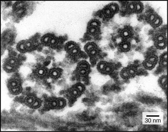
The efflux pump

The efflux pump

Efflux pumps are integral plasma membrane protein systems that recognize and bind
noxious compounds present in the cytoplasm (toxic products produced by metabolism;
compounds that have penetrated the cell), or periplasm of the bacterial cell and extrude
it into the environment in which the bacterium resides [1].

The efflux pump machinery gives the cell additional protection to the one provided by

  • the constituents of its cell wall (example: lipopolysaccharides), and
  • provides an initial protection to noxious agents present in its
    natural environment that have penetrated into the cell (example: bile
    salts in the colon) [1].

The efflux pump machinery is divided into five superfamily classes;

  • the major facilitator (MF),
  • the ATP-binding cassette (ABC),
  • the resistance-nodulation-division (RND),
  • the small multi-drug resistance (SMR) and
  • the multi-drug and toxic compound extrusion (MATE).

With respect to Gram-negative bacteria, although they all play
important roles in the protection of the bacterium from noxious
agents present in the environment, the

  • main efflux pump of the Gram negative bacterium is a
    member of the RND superfamily, and
  • because multi-drug resistance of clinical isolates have
    been associated with the over-expression of this pump,

it has received a great deal of attention [2].

The first in vitro response of bacteria to a given noxious agent,
such as an antibiotic, is to over-express its main efflux pump [2].
If the bacterium is serially exposed in vitro to increasing
concentrations of that compound, it responds by increasing
the effective number of its main efflux pump, as well as others
that provide redundant protection [2].

However, if that “adapted” bacterium is now maintained at a
constant level of a noxious agent, the level of efflux pump
activity increases up to a maximum, followed by a gradual
return of efflux pump activity to its basal level. Concomitant
to this process, an accumulation of mutations of essential
proteins located in the plasma membrane (example penicillin
binding proteins), mutations 30 S component of the ribosome
and gyrase take place [3]. These events suggest that when
the organism is faced with an environment that contains a
constant toxic level of a compound, and the cost for
maintaining an energy consuming system, such as that
needed for the energy dependent efflux pump, is too
great a price to pay.

Therefore, in order to survive in this unchanging environment,
other mechanisms are activated. For example, activation of a
mutator master gene is thought to be an important step at this
level, which results in the mutation of genes that code for
essential proteins, reversing the over-expression of efflux-
pumps, but still conferring the bacterial resistant to the
environmental pressure via other mechanism(s), yet
to be understood [4,5].

During therapy, the level of resistance increases many fold
higher than that of the initial infecting strain. Hence, clinical
isolates from treated patients often show much higher levels
of antibiotic resistance than that of their wild type counterpart
(sometimes it can even present a 1000 fold increase) [6].
At this stage, resistance is usually related to the presence
of mutations, which reduces the survival of the resistant
bacteria,

  • once it is transferred to a noxious agent-free environment

that contains the competing wild type counterpart [3,4].

Depending upon when during therapy a clinical strain is isolated,
its resistance to two or more antibiotic classes (multi-drug
resistance (MDR)), may be due entirely to over-expressed
efflux pumps; to a mixture of over-expressed efflux pumps
and increasing accumulation of mutations; and only to mutations [3,4].

The degree of resistance can readily be determined with
methods that employ compounds known for their modulation
of efflux pump activity, such as

  • phenothiazines [7] or phenyl-arginine-betanaphthylamide
    (PAβN),
  • the latter which competes with the antibiotic as
    substrate of the efflux pump [8].

If in presence of such compounds,

  • the MDR bacterium is rendered fully susceptible
    to the antibiotic(s) to which it was initially resistant,
  • resistance is most likely due to its overexpressed
    efflux pump systems.
  • Contributions made by accumulated mutations
    render the organism less and less affected by the EPI.

This type of information is of great value to clinicians faced
with long-term therapy of a bacterial infection that
progresses to an MDR phenotype. It should be understood
that although the Gram-negative bacterium has essentially
one main efflux pump, such as

  • the AcrAB (Escherichia coli) or
  • the MexAB (Pseudomonas aeruginosa),

the deletion of the main efflux pump results in the over-
expression of one or more other RND efflux pumps,
such as is the case for deletion of the AcrAB, followed by

  • the over-expression of the AcrEF pump [2].

Redundancy of as many as nine RND efflux pumps [2],
provides additional protection to the organism.

The pumps belonging to the RND family form

  • a tripartite complex together with
  • the periplasmic proteins belonging to the
    membrane fusion-protein (MFP) family and
  • the outer membrane channels.

RND transporters consist of

  • a transporter protein that recognises and
    binds the noxious agent
    in the cytoplasm or periplasm and
  • transports it to the contiguous channel (TolC),
  • ending at the surface of the outer membrane.

The transporter is attached to the plasma membrane
by two or three fusion proteins, which are believed to assist the

  • extrusion of the substrate by peristaltic actions [9].

Although the actual structure of RND efflux pumps
in the cell envelop is not completely understood,

  • the structure of the transporter, TolC and fusion
    proteins are well established for major Gram-negative
    bacteria [10].

The PMF energy dependent efflux pump most likely needs the
passage of hydronium ions through its internal cavity,

  • for the release of the substrate that is
  • in turn ejected into the TolC channel via the
  • peristaltic action of the fusion proteins [11].

A low pH,

  • the concentration of hydronium ions at the surface of the cell
  • results in a pH difference of 2 or 3 pH units compared
    to that of the milieu,

the surface concentration of hydronium ions

  • provides the force for the mobility of hydronium ions
  • through porins leading to the acidification of the periplasm,
  • providing the low pH needed by the transporter
  • for the release of the substrate.

At high pH, these hydronium ions come from

  • hydrolysis of ATP by ATP synthase, and
  • are passed into the transporter, thereby
  • reducing its internal pH, so that
  • the release of the substrates can take place [11,12].

EPIs, such as the phenothiazines chlorpromazine or thioridazine,

  • exert their inhibition at pH above 6, and
  • are thought to affect hydrolysis of ATP
  • denying the efflux pump transporter hydronium ions needed

for release of the bound substrate [11,12].

The search for EPIs that are clinically useful continues, although

with respect to thioridazine, this old neuroleptic has been shown

  • to inhibit efflux pumps of pathogenic mycobacteria [13], and
  • has been successfully used to treat extensively drug resistant
    tuberculosis infections [14].

The regulation of the main efflux pump of Escherichia coli may
take place via   distinct pathways. The induced synthesis of the
transporter component of the AcrAB efflux pump, when the
organism is exposed in vitro to a noxious agent,

  1. involves the activation of the stress gene soxS,
  2. followed by the activation of the local regulator marA,
  3. then by the activation of the transporter gene acrB [8].

In the case of Salmonella spp. two component resistance
mechanisms, such as the PmrA/PmrB system, directly
activate the master efflux pump regulator ram A gene [15].
The activation of the PmrA/PmrB system takes place
readily when Salmonella spp. is phagocytosed due to
the acidic nature of the phagolysosome [15], as follows:

  1. PmrB is a sensor that self-phosphorylates, and
  2. then transfers the phosphate to PmrA.
  3. PmrA activates a nine gene operon, which
  4. codes for Lipid A introduced into the nascent
    lipopolysaccharide layer of the outer membrane.
  5. The increased presence of Lipid A renders the
    phagocytosed bacterium practically immune to
    everything, including the hydrolases of the
    phagolysososome [15].

Although some EPIs are in clinical trials, none have yet to
reach the marketplace,    mainly due to their common
toxicity against healthy mammalian cells, affecting
intrinsic mammalian efflux pumps, as for example
those of the blood brain barrier. Lastly, it should be
noted that compounds that inhibit the efflux pump
of bacteria also have the capacity to promote the
removal of plasmids that carry antibiotic resistant
genes [16,17].

  1. Nikaido H, Pages JM (2012) Broad-specificity efflux
    pumps and their role in multidrug resistance of Gram-
    negative bacteria. FEMSMicrobiol Rev 36: 340-363.
  2. Viveiros M, Jesus A, Brito M, Leandro C, Martins M,
    et al. (2005) Inducement and reversal of tetracycline
    resistance in Escherichia coli K-12 and expression of
    proton gradient-dependent multidrug efflux pump
    genes. Antimicrob Agents Chemother 49: 3578-3582.
  3. Martins A, Couto I, Aagaard L, Martins M, Viveiros M
    (2007) Prolonged exposure of methicillin-resistant
    Staphylococcus aureus (MRSA) COL strain to
    increasing concentrations of oxacillin results in a
    multidrug-resistant phenotype. Int J Antimicrob
    Agent 29: 302-305.
  4. Martins A, Spengler G, Molnar J, Amaral L (2012)
    Sequential responses of bacteria to noxious agents
    (antibiotics) leading to accumulation of mutations
    and permanent resistance. Biochem Pharmacol J
    Open Access 1: 7.

Inhibitors of efflux pumps of Gram-negative
bacteria inhibit Quorum Sensing

Leonard Amaral, Joseph Molnar
1 Grupo de Micobacterias, Unidade de Microbacterilogia,
Centro de Malaria e Doenças Tropicais (CMDT), Instituto de
Higiene e Medicina Tropical, Universidade Nova de Lisboa,
Lisbon, Portugal; 2 Cost Action BM0701 (ATENS) of the
European Commission/European Science Foundation;
3 Department of Medical Microbiology and Immunobiology,
University of Szeged, Szeged, Hungary
Open Journal of Pharmacology, 2012, 2-2

Quorum Sensing (QS) systems of bacteria consist of

  • a producer of the QS signal and the responder.

The generation of a QS signal provides the means by which
a population can behave in a concerted manner such as

  • swarming, swimming and secretion of biofilm, etc.

Because concerted bahaviour bestows protection to the bacterial
species, and hence factors involved in the severity of an infection
such as virulence are products of QS systems, compounds that
inhibit the QS system have significant clinical relevance. Recent
evidence suggests that

  • the secretion of QS signals takes place via
  • the efflux pump system of the producer of the signal.

Interestingly, compounds such as phenothiazines and
trifluoromethyl ketones (TFs)

  • that inhibit proton motive force (PMF) activities such
    as swarming and swimming also
  • inhibit the PMF dependent efflux pump systems of
    bacteria and their QS   systems.

This review discusses the relationship between the efflux
pump, the QS system and the compounds that affect both.
Lastly, suggestions are made regarding classes of compounds
that have been shown

  • to inhibit PMF dependent efflux pumps and the need
  • to evaluate them for QS inhibitory properties.

Keywords: Quorum Sensing, QS signal, acylated hydroxyl
lactone (AHL), efflux pumps, Proton Motive Force (PMF),
inhibitors of efflux pumps, inhibitors of QS systems,
phenothiazines, Trifluormethyl Ketones (TFs), plants
sources for QS inhibitors

Efflux pumps of bacteria provide protection from noxious
agents that are present in the environment in which they
exist. Noxious agents may be naturally occurring compounds
present in environments outside and within the human.

Because over-expressed efflux pumps render antibiotic
therapy problematic, an intense search for agents that
inhibit specific efflux pumps of specific bacteria has
been conducted during the past decade [9].

Communication between bacteria of the same strain
or species and between species contributes to their
survival [11-13]. Communication involves the secretion
of signals that invoke a specific response from the responder
[11-13]. This  communication process is termed Quorum
sensing (QS). When it takes place between strains of the
same species,

  • communication is directed towards the reduction
    of population growth and
  • reducing the possibility of exceeding the nutritional
    support of the environment

Other signals may involve a population response that involves

  • the secretion of bioactive molecules that inhibit the
    replication of a competing population species [14-16]
    or even kill [biocidins) [17-21] or
  • promote a swarming effect that recruits members
    of the same species to migrate  to a specific location [22-24]
    similar to swarming by insects subsequent to signals
    indicating site of food [example bees).
  • biofilm, encase the bacteria at distances from each other
    [25-29] and within the matrix of this biofilm are
    channels used for further communication [30].

Biofilms are produced in the wild, at sites such as surfaces
of rocks which maintain the bacterial population in situ [31]
and are also produced at sites of the human colonized by
infecting bacteria [32, 33].

Agents that inhibit the QS response of the infecting bacterium
are obviously important and hence, the search for such agents
that inhibit the QS system and biofilm formation has been in
effect for the past two decades [11-13].

There is a relationship between efflux pumps (EP), QS and
biofilm (BF) secretion which has come to the forefront only
recently [13]. Control of this relationship is critical for
successful therapy of MDR bacterial infections which have
become rather commonplace. It is the intent of this review
to identify agents which may serve to interfere with the
complex system of EP-QS-BF interaction.

Proton motive force (PMF) dependent transporters obtain
their energy for function from the proton motive force. The
proton motive force is the result of cellular metabolism which
yields protons that are not used for coupling with molecular
oxygen and which are exported to the surface of the cell [43-45]
where they are distributed and bound to components of
the protective lipopolysaccharide layer that covers the cell
and constitutes a part of the outer cell wall of Gram-negative
[46] and the cell wall of Gram positive bacteria [47].

The larger the concentration of protons (hydronium ions)
on the surface of the cell with respect to their lower
concentration on the medial side of the cytoplasmic
membrane creates an electrochemical gradient that
is termed the proton motive force (PMF) [48].

Because hydronium ions cannot penetrate the cell wall
or the membrane, they may re-enter the cell only
through channels such as porins in general [49, 50].
The movement of these hydromium ions from the
surface of the cell to the periplasm or cytoplasm is
predicated upon systems that use the PMF as source
of energy-namely the resistance nodulation division
(RND) family of transporters.

E. coli has a multiplicity of efflux pumps that may
exceed 30 in number [51]. However, the main
efflux pump of this organism is the AcrAB-TolC
efflux pump [52, 53] which when deleted, its
function is replaced by the AcrEF-TolC efflux
pump [51]. Both efflux pumps are members
of the resistance nodulation division family of
transporters [51] and consist of three proteins:

  1. The transporter AcrB coded by the gene acrB and
    is intimately attached to the  plasma membrane;
  2. Two fusion proteins AcrA coded by the gene acrA
    that flank the AcrB transporter and are thought
    to assist the movement of a substrate through
    the AcrB transporter [35]; and,
  3. TolC which is also part of other tri-unit efflux pumps
    of the organism [35], is contiguous with the AcrB
    transporter and provides a conduit for the extrusion
    of the substrate [38].

Although the means for the recognition of the substrate to
be extruded appears to involve a pocket within the transporter,
it appears to be

  • defined by a phenyalanine residue [54].

Nevertheless, studies employing fluorochromes recognised by
the AcrB transporter indicate that the binding and release of
the substrate are pH dependent [55].

  • At low pH the dissociation of the substrate is high and
  • at high pH it is very slow.

In a physiological environment of ca. pH 7, if the dissociation
of the substrate is slow or not at all, then the effectiveness of
the pump to extrude a noxious agent would be nullified.
However, since the pump functions at this pH, conditions that
result in the dissociation of the substrate needed for continuous
pump action must involve a

  • decrease of the pH of the internal cavity of the pump
    to which the substrate is bound.

It has been postulated that the lowering of the pH takes place
by the generation of hydronium ions from metabolism [6] which

  • pass from the cytoplasmic side of the plasma membrane
    through the transporter.

At lower pH, there is no need for the generation of metabolically
derived  hydronium ions since these ions can be

  • diverted by the PMF from the surface of the cell
    to the periplasm via porins.

Whether hydronium ions are to be generated from the
hydrolysis of ATP at high pH or used for the synthesis
of ATP at low pH is a special

  • function of ATP synthase [56-58].

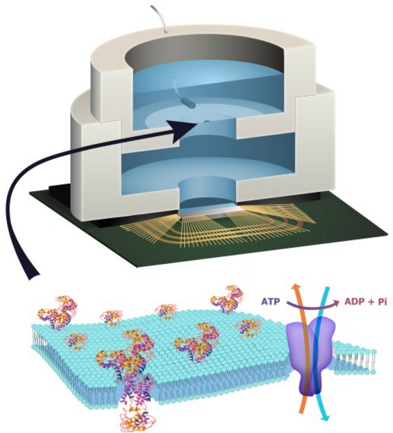
Model of the AcrAB-TolC efflux pump of a Gram-
negative bacterium

AcrAB-TolC efflux pump of a Gram-negative bacterium

AcrAB-TolC efflux pump of a Gram-negative bacterium

Hypothesis. At near neutral pH, Hydronium ions from hydrolysis of ATP
by ATP synthase pass through the AcrB

transporter, reduce the pH to a point that causes the release of the
substrate. When the hydronium ions reach the surface of the cell they
are distributed over that surface and bind to lipopolysaccharides
and basic amino acids. When there is a need for hydronium ions for
activity of the efflux pump and the pH is lower than neutral, and
the hydrolysis of ATP is not favoured, hydronium ions from the
surface of cell via the PMF mobilize through the Aqua porins
and reach the transporter where they are pushed through
the transporter by the peristaltic action caused by the fusion
proteins. Substrates bound to the transporter dissociate
when the pH is reduced by the flow of hydronium ions and
are carried out by the flow of water.

Inhibitors of bacterial efflux pumps
Inhibitors of the QS of bacteria

Because phenothiazines inhibit many energy dependent systems
of bacteria such as motility [89, 90, 95], and these phenothiazines
also inhibit efflux pumps of bacteria [6, 7, 9, 41, 51, 73, 74, 76-83],
there seems to be a correlation between an active efflux pump
system and a functional QS system. That this assumption is correct,
recent evidence has been provided showing that the efflux pumps of
the AHL responding environmental Chromobacterium violaceum
(CV026) bacterium and that of E. coli are inhibited by the phenothiazine
thioridazine (TZ) [12]. Because TZ is known to inhibit genes that
regulate and code for efflux pumps of bacteria [41, 119, 120], it is
possible that the inhibition of the responding CV0126 bacterium to
AHLs [12] involves the inhibition of genes that code and regulate
the efflux pump of the responder which is assumed to recognise the
AHL signal as an noxious agent and hence would extrude it to the
environment [12]. The inhibition of an efflux pump should manifest
itself as an inhibitor of the QS component responsible for biofilm
formation.

Since the discovery of berberine a powerful inhibitor of bacterial
efflux pumps [159], plants have become sources of inhibitors of
efflux pumps [160-164]. Given that efflux pumps and the  QS of
bacteria have an intimate relationship as described in this review,
attention has been focused on plants for potential sources of inhibitors
of efflux pumps and QS systems. Essential oils from Columbian
plants have yielded a large number of compounds that inhibit the
QS system of responding bacteria such as

  1. limonene-carvone , the
  2. citral (geranial-neral) (isolated from Lippia alba),
  3. α-pinene (from Ocotea sp.),
  4. β-pinene (from Swinglea glutinosa),
  5. cineol (from Elettaria cardamomun),
  6. α-zingiberene (from Zingiber officinale) and
  7.  pulegone (from Minthostachys mollis) [165].

Several other essential oils, in particular were shown to present
promising inhibitory properties for the short chain AHL quorum
sensing (QS) system in Escherichia coli containing the biosensor

  •  plasmid pJBA132, in  particular Lippia alba.

Citral was the only  essential oil that presented some activity for
the long chain AHL QS system in Pseudomonas putida containing

  •  the plasmid pRK-C12 [165].

The essence of this review is to correlate the relationship of the
efflux pump system to the QS system of bacteria via the use of
compounds that inhibit both systems. Simply put, inhibitors of
the efflux pump system also, when studied, inhibit the QS system
as well. Because the PMF dependent efflux pump system of Gram-
negatives that is overexpressed is responsible for the multi-drug
phenotype of the bacterium, compounds that affect the PMF of
the bacterium are candidates that will inhibit the activity of the
pump. Consequently, this inhibition will inhibit the secretion of
biofilm, and because biofilm is a deterrent to the action of antibiotics,
compounds that affect the efflux pump system are promising
candidates for clinical evaluation.

Limiting and controlling carbapenem-resistant
Klebsiella pneumonia

L Saidel-Odes, A Borer.
1Infection Control and Hospital Epidemiology Unit, 2Infectious
Diseases Institute, Soroka University Medical Center and the
Faculty of Health Sciences, Ben-Gurion University of the Negev,
Beer-Sheva, Israel
Infection and Drug Resistance 2014:7 9–14

Carbapenem-resistant Klebsiella pneumoniae (CRKP)

  • is resistant to almost all antimicrobial agents,
  • is associated with substantial morbidity and mortality, and
  • poses a serious threat to public health.

The ongoing worldwide spread of this pathogen emphasizes the
need for immediate intervention. This article reviews the global
spread and risk factors for CRKP colonization/infection, and
provides an overview of the strategy to combat CRKP dissemination

Outbreaks of CRKP that have occurred around the world have
been associated with the plasmid-encoded carbapenemase
K. pneumoniae carbapenemase (KPC),

  • a carbapenem-hydrolyzing β-lactamase.19

CRKP isolates are resistant to almost all available antimicrobials
and are susceptible

  • only to polymyxins and tigecycline;
  • a minority to the few remaining aminoglycosides,
    though resistance to these agents is increasingly reported.20,21

Several investigators have evaluated predictors for CRKP colonization.
The following summarizes various studies.

  1. In a multivariate analysis, prior use of macrolides and
    any antibiotic exposure $14 days remained the only
    independent factors associated with CRKP bacteremia
  2. Nosocomial isolation of CRKP was strongly favored by the
    selection pressure of carbapenem. In this study, prior
    treatment with fluoroquinolones was associated with
    decreased risk for the emergence of CRKP.
  3. Previous use of carbapenem and cephalosporin
  4. Nursing home residency before hospital admission, bedridden
    status, and previous antibiotic therapy
  5. exposure to fluoroquinolones
  6. the recipient of antibiotics
  7. intensive care unit (ICU) stay, and
  8. Poor functional status,
  9. Independent predictors of subsequent carbapenem-
    resistant Enterobacteriaceae (CRE) infection were
  • admission to the ICU,
  • having a central venous  catheter,
  • receipt of antibiotics, and
  • diabetes mellitus

Schwaber et al and the Israeli CRE Working Group enforced the
Israel Ministry of Health guidelines mandating physical separation
of hospitalized carriers of CRE and dedicated staffing and appointed
a professional task force charged with containment.19 The monthly
incidence of nosocomial CRE was reduced from 55.5 to 11.7 cases
per 100,000 patient days within 15 months.

Part 7.  Tuberculosis

The Mechanism by which the Phenothiazine Thioridazine
Contributes to Cure Problematic Drug-Resistant Forms
of Pulmonary Tuberculosis: Recent Patents for “New Use”

L Amaral1*, A Martins2,3, G Spengler2, A Hunyadi4 and J Molnar2
Recent Patents on Anti-Infective Drug Discovery 2013; 8(3):000-000

At this moment, over half million patients suffer from multi-drug
resistant tuberculosis (MDR-TB) according to the data from the WHO.
A large majority is terminally ill with essentially incurable pulmonary
tuberculosis. This herein mini-review provides the experimental and
observational evidence that a specific phenothiazine,

  • thioridazine,

will contribute to cure any form of drug-resistant tuberculosis. This
antipsychotic agent is no longer under patent  protection for its
initial use. The reader is informed on the recent developments

  • in patenting this compound for “new use” with a special
  • emphasis on the aspects of drug-resistance.

Given that economic motivation can stimulate the use of this drug
as an antitubercular agent, future prospects are also discussed.

Thioridazine is not the only phenothiazine that has been recommended
for therapy of pulmonary tuberculosis. In general, many phenothiazines
have been implicated for antitubercular activity [62, 80-86]. Among
these are

  • trifluoperazine [87-94],
  • methdilazine [95, 96],
  • promazine [97, 98],
  • promethazine [97, 98],
  • fluphenazin [99],
  • propiomazine [100], and
  • the methylene blue related toluidine blue [101].

There are phenothiazine compounds derived from the parental
methylene blue for therapy of pathologies unrelated to tuberculosis
that also possess

  •  antitubercular [44, 48] and/or antimalarial properties [44].

Moreover, derivatives made from any of the phenothiazines that
have in vitro activity against Mycobacterium tuberculosis are also
active [61, 67, 102, 103], suggesting ample opportunities for
patenting of new analogs developed from known, active phenothiazines
with even less side effects than those of TZ, as recently suggested by
Musuka and co-authors [104]. It is important to mention, that the
commercially available phenothiazines such as for example

  •  trifluoperazine, methdilazine, promazine, promethazine,
    fluphenazin and propiomazine

are beyond patent protection as initially intended. Nevertheless,
these compounds have been patented as adjuvants for the treatment
of MDR cancer (patent expired in 2011 [105]; and, right afterwards,
a new patent has been filed with a priority date of 28th March, 2012,
claiming combination therapy of cancer with a chemotherapeutic
agent and a dopamine receptor antagonist against Cancer stem cells (CSC).

Taking into account that intrinsic MDR is considered as one of the key
properties of CSCs [107], the subject to be covered is indeed related.
According to the MDR, XDR and TDR Mycobacterium tuberculosis,
subjects of this herein paper, the initial step for actually reaching those
in need has been made: a patent has been published in December, 2007,
for the use of TZ and its derivatives for reversing anti-microbial drug
resistance [108]. We must note, however, that, despite the six years
passed since, we were unable to find any related clinical trials, which
would certainly be of outmost importance and urgency in order to
proceed towards an effective therapy of highly resistant mycobacterial
infections.

Mechanism Of Action Of Tz: Why It Cures Multi-Drug,
Extensively Drug Resistant And Probably Totally Drug
Resistant Tuberculosis

Over-expressed efflux pumps of Mycobacterium tuberculosis render
the organism multi-drug resistant [13]. Special attention has been
given to those coded by the

  • mmpL7, p55, efpA, mmr, Rv1258c and Rv2459 genes [109].

The activity of these efflux pumps can be suppressed by

  • concentrations of TZ that have no effect on the viability of
    Mycobacterium tuberculosis
  • rendering the organism susceptible to the antibiotic to
    which it was initially resistant
  • as a consequence of the over-expression of its
    efflux pumps [109].

TZ has also been shown to inhibit the activity of the main

  • efflux pumps of bacteria belonging to other species.

TZ has strong inhibitory activity against the genes that code for
essential proteins of M. tuberculosis [122-124].  Consequently, we
may conclude that the in vitro activity of TZ involves

  • the inhibition of the efflux pumps of M. tuberculosis and that
  • the in vitro exposure of this organism to TZ renders the organism
  • susceptible to antibiotics to which it was initially resistant
  • as a consequence of over-expressed efflux pumps [21].

Phenothiazines such as CPZ, TZ, trifluoperazine, etc., also inhibit

  • the binding of calcium to calcium binding proteins such as

calmodulin in eukaryotes [125], and

  • interfere with other proteins involved in
  • the regulation of cellular activity [126].

They inhibit the transport of calcium and potassium systems

  • in eukaryotic cells [127-129] as well as in
  • mycobacteria [89, 130] and
  • E. coli [113].

In fact, in the latter case, calcium was shown essential to

  • the continuous activity of the thioridiazine sensitive
    efflux system [113].

The killing activity of the human macrophage as well as that
of the neutrophil

  • is dependent upon the retention of calcium and potassium
  • within the phagolysosome of the cell [131].

Considering this, several alternative choices are available for
patenting under “new use”, which would allow a “fresh start”
for the compound to be developed. However, the needed
experimental proof that these phenothiazine agents have
activity at the pulmonary macrophage of the alveolar unit
(the site where the causative organism of pulmonary tuberculosis
resides) is still absent.

Targeting the Human Macrophage with Combinations
of Drugs and Inhibitors of Ca2+ and K+ Transport to
Enhance the Killing of Intracellular Multi-Drug Resistant
M. tuberculosis (MDR-TB) – a Novel, Patentable Approach
to Limit the Emergence of XDR-TB

Marta Martins
UCD Centre for Food Safety, School of Agriculture, Food Science and
Veterinary Medicine, University College Dublin, Belfield, Dublin 4, Ireland
& Unit of Mycobacteriology and UPMM; Instituto de Higiene e Medicina
Tropical, Universidade Nova de Lisboa (IHMT/UNL),  Lisbon, Portugal
Recent Patents on Anti-Infective Drug Discovery, 2011, 6, 000-000

The emergence of resistance in Tuberculosis has become a serious
problem for the control of this disease. For that reason, new therapeutic
strategies that can be implemented in the clinical setting are urgently
needed. The design of new compounds active against mycobacteria
must take into account that Tuberculosis is mainly an intracellular
infection of the alveolar macrophage and therefore must maintain
activity within the host cells.

An alternative therapeutic approach will be described in this review,
focusing on the activation of the phagocytic cell and the subsequent
killing of the internalized bacteria. This approach explores the combined
use of antibiotics and phenothiazines, or Ca2+ and K+ flux inhibitors,
in the infected macrophage.

Targeting the infected macrophage and not the internalized bacteria
could overcome the problem of bacterial multi-drug resistance. This
will potentially eliminate the appearance of new multi-drug resistant
tuberculosis (MDR-TB) cases and subsequently prevent the emergence
of extensively-drug resistant tuberculosis (XDR-TB).

Patents resulting from this novel and innovative approach could be
extremely valuable if they can be implemented in the clinical setting.
Other patents will also be discussed such as the treatment of TB
using immunomodulator compounds (for example: betaglycans).

Role of Phenothiazines and Structurally Similar
Compounds of Plant Origin in the Fight against
Infections by Drug Resistant Bacteria


SG. Dastidar 1, JE. Kristiansen 2, J Molnar 3 and L Amaral
Antibiotics 2013; 2: 58-71;
http://dx.doi.org:/10.3390/antibiotics2010058

Phenothiazines have their primary effects on the plasma membranes
of prokaryotes and eukaryotes. Among the components of the
prokaryotic plasma membrane affected are

  • efflux pumps,
  • their energy sources
  • and energy providing enzymes, such as ATPase,
  • and genes that regulate and code for the permeability
    aspect of a bacterium.

The response of multidrug and extensively drug resistant
tuberculosis to phenothiazines shows an alternative therapy for the
treatment of these dreaded diseases, which are claiming more and
more lives every year throughout the world.

Many phenothiazines have shown

  • synergistic activity with several antibiotics thereby
  • lowering the doses of antibiotics administered to patients
    suffering from specific bacterial infections.

Trimeprazine is synergistic with trimethoprim. Flupenthixol (Fp)
has been found to be synergistic with penicillin and chlorpromazine
(CPZ); in addition, some antibiotics are also synergistic. Along with
the antibacterial action described in this review,

  • many phenothiazines possess plasmid curing activities, which
  • render the bacterial carrier of the plasmid sensitive to antibiotics.

Thus, simultaneous applications of a phenothiazine like TZ would not
only act as an additional antibacterial agent but also would help

  • to eliminate drug resistant plasmid from
    the infectious bacterial cells.

Part 8.  Cancer Cytotherapy

Synthesis and Structure-Activity Relationships of Novel
Dioxolanes as MDR Modulators in Cancer

A Martins 1,2,†,*, J Csábi 3,†, A Balázs 4, DKitka 1, L Amaral 5,
J Molnár 1, A Simon 6, G Tóth 6 and A Hunyadi 3,
Molecules 2013, 18, 15255-15275;
http://dx.doi.org:/10.3390/molecules181215255

Ecdysteroids, molting hormones of insects, can exert several mild,
non-hormonal bioactivities in mammals, including humans. In a
previous study, we have found a significant effect of certain derivatives

  • on the ABCB1 transporter mediated multi-drug resistance of a
  • transfected murine leukemia cell line.

In this paper, we present a structure-activity relationship study
focused on

  • the apolar dioxolane derivatives of 20-hydroxyecdysone.

Semi-synthesis and bioactivity of a total of 32 ecdysteroids, including
20 new compounds, is presented, supplemented with their

  • complete 1H- and 13C-NMR signal assignment

As published before [9], the 20,22-diol moiety of 20E is more reactive
than the 2,3-diol, probably due to the free rotation of the 20,22-bond
of 20E that allows the 20,22-dioxolane ring to form with less strain.

This allowed us to selectively obtain the 20,22-mono-dioxolane
derivatives 2–14, or, depending on the amount of reagent and the
reaction time, the 2,3;20,22-bis-homo-dioxolanes 17 and 21–25.

By utilizing the 20,22-monodioxolane ecdysteroids, another aldehyde
or ketone could be coupled to position 2,3, resulting in several bis-hetero-
dioxolane derivatives 26–33. For this, however, gradually decreasing
reactivity with the increase of the size of the reagent was a limiting factor:

  • larger aldehydes or ketones (mainly those containing a
    substituted aromatic ring) could not be coupled at the 2,3-position.
  • The 2,3-monodioxolane derivatives also appeared to be present as
    minor side-products of the reactions, and as a consequence of their
    low amount, only one such compound (compound 15) was isolated and studied.

To selectively obtain this kind of a compound (16) in a more reasonable
yield, another, three-step approach was successfully applied:

  • after protecting the 20,22-diol with phenylboronic acid, the
    2,3-acetonide could be prepared, and
  • removal of the 20,22 protecting group afforded the desired
    2,3-monoacetonide in a one-pot procedure.

In the case of the reactions with aldehydes or asymmetric ketones,
the new C-28 and C-29 central atoms of the dioxolane rings are
stereogenic centers and thus two possible diastereomers can be
formed at both diols. Their configuration was elucidated by two-
dimensional ROESY or selective one-dimensional ROESY experiments,
e.g., in the doubly substituted

  • dioxolane derivative 22 (R1 = R4 = n-Bu, R2 = R3 = H)
  • the unambiguous differentiation of the 1H and 13C signals of
    the two n-butyl groups was achieved in the following way
    (see Figure 2).

Assignment of the H-C(28) atoms (δ = 4.93/105.9 ppm) was supported by

  • the H-2/C-28 and H-3/C-28 HMBC correlations, and
  • that of H-C(29) (δ = 4.91/105.6 ppm) by the H-22/C-29
    cross peak, respectively.

The selective ROESY experiment irradiating at 4.93 ppm showed

  • contacts with the Hα-2 and Hα-3 atoms proving the
    α position of the R2 = H atom.

The ROESY response obtained irradiating H = R3 signal (δ = 4.91)
on H-22 (δ = 3.64 ppm) revealed their

  • cis arrangement and the R configuration around C-29.

The unambiguous assignments of the signals

  • of the two n-butyl groups R1 and R4 were achieved by
  • selective TOCSY experiments (irradiation at
  • δ = 4.93 and 4.91, respectively).

Figure 2

Stereostructure of 22. Red-ROESY proximitries. Blue- 1H. Black-1 001

Stereostructure of 22. Red-ROESY proximitries. Blue- 1H. Black-1 001

Stereostructure of 22. Red arrows indicate the detected ROESY
steric proximities, the blue numbers give the characteristic 1H,
and the black numbers the 13C chemical shifts.


Related Material

Identification of Efflux Pump-mediated Multidrug-resistant
Bacteria by the Ethidium Bromide-agar Cartwheel Method

M Martins, M Viveiros, I Couto, SS. Costa, T Pacheco, S Fanning,
Jean-Marie Pagès, and L Amaral
in vivo 2011; 25: 171-178  

Index for efflux activity of the MDR strains. The capacity to efflux EtBr
of each bacterial strain was ranked relative to the reference strain
according to the following formula:


Index for efflux activity of the MDR strains

Index for efflux activity of the MDR strains

A Simple Method for Assessment of MDR Bacteria for
Over-Expressed Efflux Pumps

M Martinsa,b*, MP. McCuskera,b, M Viveirosa,c, I Coutoc,d,
S Fanninga,b, Jean-Marie Pagès b,e, L Amaral,b,
The Open Microbiol J 2013; 7: 1-5  1874-2858/13 Bentham

Flowchart followed to test bacterial strains using the EtBr-agar
Cartwheel method.

Flowchart followed to test bacterial strains using the EtBr-agar Cartwheel method.

Flowchart followed to test bacterial strains using the EtBr-agar Cartwheel method.

EtBr-agar cartwheel method applied to different bacterial species

EtBr-agar cartwheel method applied to different bacterial species

EtBr-agar cartwheel method applied to different bacterial species

The effect of selected EPIs on the resistance of the induced and
MDR Gram-positive bacteria.

TET
Enterococcus EFC
ATCC29212
HSEFM-D
1.5
>2.5
w/o
EPI
+
TZ
+
CPZ
+
RES
4
16
4
4
4
4
4
8
(4×) (4×) (2×)
                                MCEtBr NOR  (mg/l) MIC NOR (mg/l)
HSEFM-E >2.5 0.125 0.125 0.125 0.125

EPI: Efflux pump inhibitor; w/o: without; TZ: thioridazine; CPZ:
chlorpromazine; PAN: phenyl arginine β-naphthylamide. Values
in bold-type correspond to a decrease of 4-fold or higher on
the MIC values in comparison to those in the absence of inhibitor.
Values in parenthesis indicate the MIC decrease relative to that
of the original culture. The concentration of each EPI used is
defined in the Materials and Methods section.

Macrocyclic diterpenes resensitizing multidrug
resistant phenotypes 

MA. Reis a, A Paterna a, RJ. Ferreira a, H Lage b,
Maria-José U. Ferreira a,⇑
a Instituto de Investigação do Medicamento (iMed.ULisboa), Faculdade
de Farmácia, Universidade de Lisboa, Lisboa, Portugal
b Charité Campus Mitte, Institute of Pathology, Berlin, Germany
Bioorganic & Medicinal Chemistry xxx (2014) xxx–xxx

Herein, collateral sensitivity effect was exploited as a strategy to
select effective compounds to overcome multidrug resistance in
cancer. Thus, eleven macrocyclic diterpenes, namely jolkinol D (1),
isolated from Euphorbia piscatoria, and its derivatives (2–11) were
evaluated for their activity on three different Human cancer entities:

  • gastric (EPG85-257), pancreatic (EPP85-181) and colon (HT-29)

each with a variant selected for resistance to mitoxantrone

  1. EPG85-257RN;
  2. EPP85-181RN;
  3. HT-29RN and
  • one to daunorubicin (EPG85-257RD; EPP85-181RD; HT-29RD).

Jolkinol D (1) and most of its derivatives (2–11) exhibited significant
collateral sensitivity effect towards the cell lines

  • EPG85-257RN (associated with P-glycoprotein overexpression) and
  • HT-29RD (altered topoisomerase II expression).

The benzoyl derivative, jolkinoate L (8) demonstrated ability to

  • target different cellular contexts with
  • concomitant high antiproliferative activity.

These compounds were previously assessed as
P-glycoprotein modulators,

  • at non-cytotoxic doses, on MDR1-mouse lymphoma cells.

A regression analysis between

  1. the antiproliferative activity presented herein and
  2. the previously assessed P-glycoprotein modulatory effect

showed a strong relation between the compounds that presented

  • both high P-glycoprotein modulation and cytotoxicity.

Molecular Docking Characterizes Substrate-Binding Sites
and Efflux Modulation Mechanisms within P
Glycoprotein.

Ferreira,† Maria-José U. Ferreira,† and DJVA dos Santos*,†,‡
†Research Institute for Medicines and Pharmaceutical Sciences
(iMed.UL), Faculty of Pharmacy, University of Lisbon, Lisbon, Portugal
‡REQUIMTE, Department of Chemistry & Biochemistry, Faculty of
Sciences, University of Porto, Porto, Portugal
J. Chem. Inf. Model. XXXX, XXX, XXX−XXX
http://dx.doi.org:/10.1021/ci400195v

P-Glycoprotein (Pgp) is one of the best characterized ABC
transporters
, often involved

  • in the multidrug-resistance phenotype
  • overexpressed by several cancer cell lines.

Experimental studies contributed to important knowledge concerning
substrate polyspecificity, efflux mechanism, and drug binding sites.
This information is, however, scattered through different perspectives,
not existing a unifying model for the knowledge available for this transporter.
Using a previously refined structure of murine Pgp,

  • three putative drug-binding sites were hereby characterized
  • by means of molecular docking.

The modulator site (M-site) is characterized by

  • cross interactions between both Pgp halves

herein defined for the first time, having an important role in

  • impairing conformational changes leading to substrate efflux.

Two other binding sites, located next to the inner leaflet of the lipid bilayer,

  • were identified as the substrate binding H and R sites
  • by matching docking and experimental results.

A new classification model

  • with the ability to discriminate substrates from modulators

is also proposed, integrating a vast number of theoretical and experimental data.

conformational changes leading to substrate efflux

conformational changes leading to substrate efflux

conformational changes leading to substrate efflux

http://pubs.acs.org/appl/literatum/publisher/achs/journals/content/jcisd8/
2013/jcisd8.2013.53.issue-7/ci400195v/production/pdfimages_v02/normal.img-000.jpg


Read Full Post »

Older Posts »