Feeds:
Posts
Comments

Posts Tagged ‘Cytoskeleton’

Lesson 6 of Cell Signaling & Motility – Cytoskeleton II: #TUBiol3373

Author: Stephen J. Williams, Ph.D.

In this lesson we will go over the biochemical makeup and formation of various actin containing cellular structures involved in cellular motility, structure, as well as the dynamics of muscular contraction.  The lesson had been put on your Canvas and I am emailing you the Google Docs version.  If you are having problems downloading you can download here (I believe maybe the Canvas version had problems with embedding videos properly so that is why I am sending you also by email)

Download Below

cell signaling 6 lesson 2020

After opening the powerpoint (or Google Doc) please review with the following notes which highlight some concepts as well as some reviews and reminders of past lectures.  It may be handy to also have lecture 5 handy if you need to refer to it.  In between some sections there will be polls (really multiple choice quizzes DON’T WORRY you will not be graded on them but they are for your benefit.  There will also be a section under Comments all the way at the end and at the last quiz where you can also ask questions.

Remember you can always email me or Tweet me any questions @StephenJWillia2 using the hashtag #TUBiol3373.

In addition you can also leave comments at the very bottom which can be answered.

Slide 2 of lesson 6 is a refresher of the end of our last lecture, talking about Actin Binding Regulatory Proteins.


The picture above shows a brief review of some of the structures and actin binding proteins involved in helping to form these actin filament structures (like filamin in cross linked structures, profilin which binds the actin monomers [G-actin] and helps with addition of these monomers to the leading plus end.

*** Remember G-actin (Globular Actin) is the monomer and F-actin (filamentious actin) is the polymerized actin strand [filament]

Also remember from the last lecture that G-Actin as monomer has affinity for ATP {Adenosine triphosphate} and these G-Actin-ATP will be able to polymerize to form the F-Actin form.  Also F-actin can then hydrolyze the ATP to ADP and inorganic phosphate.  At this point the actin-ADP unit looses affinity for the remaining F-Actin chain and depolymerization can occur


An event referred to as TREADMILLING or when the G actin units are removed from minus end and added to the plus (or growing barbed) end

Also remember that there is a critical concentration of G-Actin-ATP needed for bypassing the lag phase of nucleation before the elongation phase and the rate of addition to the plus end is faster than addition to minus end and greater than the rate of depolymerization at the minus end

Cell Structures That Involve Actin (see links for more information)

  1. filopodia
  2. parallel actin bundles
  3. actin cortex
  4. lamellipodia
  5. stress fibers
  6. microvilli
  7. contractile ring in cytokinesis


Nucleating proteins Arp (actin related protein and Formins

Arp ====> formation of lamellipodia

Formins ====> formation of stress fibers

Process involving formins starts with a signaling event by activation of a G-protein, the GTP binding protein Rho

Rho is a subfamily member of the Ras superfamily.  The Rho family consists of cdc42, rac1, and RhoA (we will discuss at a later date).  Rho acts like G proteins, as a molecular switch.

Note that just like the Ras member of G-proteins and the Ras GTP/GDP cycle, the Rho activation, deactivation cycle also depends on GEFs [Guanine nucleotide exchange factors] and GAPs [GTPase activating proteins] and also GDIs [guanine nucleotide dissociation inhibitors which we will discuss later but involved in preventing Rho diffusion in the cell, acting as a tether].

Myosin and Motor (muscle) Function; Neuromuscular junctions, the sarcoplasmic reticulum and Ohhh the plethora of signaling events

In this section, from slides 29 to 54, we talk about myosin and the interactions between myosin and actin in formation of the contractile unit of the muscle (skeletal).

We also talk about some familiar signaling events, in particular the neuromuscular junction.

At this junction is a special type of acetylcholine receptor

Remember we talked about two types of acetylcholine receptors:

  1. muscarinic receptors – typical GPCRs that tranduce the signal via Gi or Gq depending on the muscarinic subtype
  2. nicotinic receptors – these are ligand {receptor} operated channels and when activated opens a Na+ channel which leads to depolarization


Now the depolarization activates another set of channels, the voltage operated calcium channels so we have two types of ion channels: Receptor {ligand} operated channels and Voltage operated channels.  These are sometimes abbreviated as ROCs and VOCs.

The unit of the myofibril on the contactile unit of the skeletal muscle is the sarcomere and upon the calcium transient, the sarcomere shortens with the two z-disks moving closer to each other as shown in the video in the lecture.

Also briefly review the introduction part on microtubules. We will finish that next week. Note that the microtubule is comprised of the protein tubulin, which is another GTP binding protein.

For other articles and more information please see

Lesson 5 Cell Signaling And Motility: Cytoskeleton & Actin: Curations and Articles of reference as supplemental information: #TUBiol3373

Role of Calcium, the Actin Skeleton, and Lipid Structures in Signaling and Cell Motility

Identification of Biomarkers that are Related to the Actin Cytoskeleton


Read Full Post »

Lesson 5 Cell Signaling And Motility: Cytoskeleton & Actin: Curations and Articles of reference as supplemental information: #TUBiol3373

Curator: Stephen J. Williams, Ph.D.

Cell motility or migration is an essential cellular process for a variety of biological events. In embryonic development, cells migrate to appropriate locations for the morphogenesis of tissues and organs. Cells need to migrate to heal the wound in repairing damaged tissue. Vascular endothelial cells (ECs) migrate to form new capillaries during angiogenesis. White blood cells migrate to the sites of inflammation to kill bacteria. Cancer cell metastasis involves their migration through the blood vessel wall to invade surrounding tissues.

Please Click on the Following Powerpoint Presentation for Lesson 4 on the Cytoskeleton, Actin, and Filaments

CLICK ON LINK BELOW

cell signaling 5 lesson

This post will be updated with further information when we get into Lesson 6 and complete our discussion on the Cytoskeleton

Please see the following articles on Actin and the Cytoskeleton in Cellular Signaling

Role of Calcium, the Actin Skeleton, and Lipid Structures in Signaling and Cell Motility

This article, constitutes a broad, but not complete review of the emerging discoveries of the critical role of calcium signaling on cell motility and, by extension, embryonic development, cancer metastasis, changes in vascular compliance at the junction between the endothelium and the underlying interstitial layer.  The effect of calcium signaling on the heart in arrhtmogenesis and heart failure will be a third in this series, while the binding of calcium to troponin C in the synchronous contraction of the myocardium had been discussed by Dr. Lev-Ari in Part I.

Universal MOTIFs essential to skeletal muscle, smooth muscle, cardiac syncytial muscle, endothelium, neovascularization, atherosclerosis and hypertension, cell division, embryogenesis, and cancer metastasis. The discussion will be presented in several parts:
1.  Biochemical and signaling cascades in cell motility
2.  Extracellular matrix and cell-ECM adhesions
3.  Actin dynamics in cell-cell adhesion
4.  Effect of intracellular Ca++ action on cell motility
5.  Regulation of the cytoskeleton
6.  Role of thymosin in actin-sequestration
7.  T-lymphocyte signaling and the actin cytoskeleton


Identification of Biomarkers that are Related to the Actin Cytoskeleton

In this article the Dr. Larry Bernstein covers two types of biomarker on the function of actin in cytoskeleton mobility in situ.

  • First, is an application in developing the actin or other component, for a biotarget and then, to be able to follow it as

(a) a biomarker either for diagnosis, or

(b) for the potential treatment prediction of disease free survival.

  • Second, is mostly in the context of MI, for which there is an abundance of work to reference, and a substantial body of knowledge about

(a) treatment and long term effects of diet, exercise, and

(b) underlying effects of therapeutic drugs.

Microtubule-Associated Protein Assembled on Polymerized Microtubules

(This article has a great 3D visualization of a microtuble structure as well as description of genetic diseases which result from mutations in tubulin and effects on intracellular trafficking of proteins.

A latticework of tiny tubes called microtubules gives your cells their shape and also acts like a railroad track that essential proteins travel on. But if there is a glitch in the connection between train and track, diseases can occur. In the November 24, 2015 issue of PNAS, Tatyana Polenova, Ph.D., Professor of Chemistry and Biochemistry, and her team at the University of Delaware (UD), together with John C. Williams, Ph.D., Associate Professor at the Beckman Research Institute of City of Hope in Duarte, California, reveal for the first time — atom by atom — the structure of a protein bound to a microtubule. The protein of focus, CAP-Gly, short for “cytoskeleton-associated protein-glycine-rich domains,” is a component of dynactin, which binds with the motor protein dynein to move cargoes of essential proteins along the microtubule tracks. Mutations in CAP-Gly have been linked to such neurological diseases and disorders as Perry syndrome and distal spinal bulbar muscular dystrophy.


Read Full Post »

Proteomics

Writer and Curator: Larry H. Bernstein, MD, FCAP


The previous discussion concerned genomics, metabolomics, and cancer. The discussion that follows is concerned with the expanding filed of proteomics, which has implication for disease discovery, pharmaceutical targeting, and diagnostics.

The human proteome – a scientific opportunity for transforming diagnostics, therapeutics, and healthcare

Marc Vidal, Daniel W Chan, Mark Gerstein, Matthias Mann, Gilbert S Omenn, et al.
Clinical Proteomics 2012, 9:6  http://www.clinicalproteomicsjournal.com/content/9/1/6

A National Institutes of Health (NIH) workshop was convened in Bethesda, MD on September 26–27, 2011, with representative scientific leaders in the field of proteomics and its applications to clinical settings. The main purpose of this workshop was to articulate ways in which the biomedical research community can capitalize on recent technology advances and synergize with ongoing efforts to advance the field of human proteomics. This executive summary and the following full report describe the main discussions and outcomes of the workshop.

Proteomics Pioneer Award 2013: Professor Amos Bairoch, University of Geneva, Switzerland

Eupa Open Proteomics 2 (2014) 34  http://dx.doi.org/10.1016/j.euprot.2013.12.002

Amos Bairoch has always been fascinated by computer science, genetics and biochemistry. His fi rst project, as a PhD student, was the development of PC/Gene, a MS-DOS based software package for the analysis of protein and nucleotide sequences. While working on this project, he realized that there was no single resource for protein sequences, and started to develop the first annotated protein sequence database, which became Swiss-Prot and was first released in July 1986. In 1988, he created PROSITE, a database of protein families and domains, and a little later ENZYME, an enzyme nomenclature database.

Amos Bairoch led the Swiss-Prot group from its creation in 1988 until 2009. During this period, Swiss-Prot became the primary protein sequence resource in the world and has been a key research instrument for both bioinformaticians and laboratory-based scientists, particularly in the field of proteomics.

Since 2009, Amos Bairoch’s group is developing neXtProt, a knowledgebase
specifically dedicated to human proteins.neXtProt has been chosen as the reference protein database for the HUPO Human Proteome Projects.

For his major contributions in the field of proteomic databases, Amos Bairoch received the Friedrich Miescher Award from the Swiss Society of Biochemistry in 1993, the Helmut Horten Foundation Incentive Award in 1995, the Pehr Edman award and the European Latsis Prize in 2004, the Otto Naegeli prize in 2010, and the HUPO Distinguished Achievement Award in Proteomic Sciences in 2011.

National Heart, Lung, and Blood Institute Clinical Proteomics Working Group Report

CB Granger, JE Van Eyk, SC Mockrin and N. Leigh Anderson
Circulation. 2004;109:1697-1703
http://dx.doi.org:/10.1161/01.CIR.0000121563.47232.2A

The National Heart, Lung, and Blood Institute (NHLBI) Clinical Proteomics Working Group was charged with identifying opportunities and challenges in clinical proteomics and using these as a basis for recommendations aimed at directly improving patient care. The group included representatives of clinical and translational research, proteomic technologies, laboratory medicine, bioinformatics, and 2 of the NHLBI Proteomics Centers, which form part of a program focused on innovative technology development. This report represents the results from a one-and-a-half-day meeting on May 8 and 9, 2003. For the purposes of this report, clinical proteomics is defined as the systematic, comprehensive, large-scale identification of protein patterns (“fingerprints”) of disease and the application of this knowledge to improve patient care and public health through better assessment of disease susceptibility, prevention of disease, selection of therapy for the individual, and monitoring of treatment response.

The -omics era: Proteomics and lipidomics in vascular research

Athanasios Didangelos, Christin Stegemann, Manuel Mayr
Atherosclerosis 221 (2012) 12– 17
http://dx.doi.org:/10.1016/j.atherosclerosis.2011.09.043

The retention of proatherogenic low-density lipoprotein (LDL) particles on the subendothelial extracellular matrix (ECM) is a hallmark of atherosclerosis. Apolipoprotein B (apoB)-containing lipoprotein particles are trapped in the arterial intima by proteoglycans in atherosclerosis-prone areas and eventually become modified, commonly by aggregation and oxidation. The initial accumulation of proatherogenic lipoproteins initiates an inflammatory response, which results in the release of proteolytic enzymes and induces the dedifferentiation of vascular smooth muscle cells (SMCs) resulting in alterations of their matrix producing properties. The precise mechanisms responsible for the accumulation of certain matrix components and subsequent lipoprotein retention on the vessel wall are not fully elucidated. Undoubtedly, ECM remodeling contributes to the formation of atherosclerotic lesions and the lipid composition of apolipoproteins influences their binding properties to the matrix. An unbiased discovery approach, which is not limited to known molecules of presumed importance, will be invaluable for the identification of novel, previously unknown mediators of disease. Although descriptive, the detailed examination of atherosclerotic plaques using advanced proteomics and lipidomics techniques can generate novel insights and form the basis for further mechanistic investigations.

The Revolution in Proteomics Ionization –
CaptiveSpray nanoBooster™
Bruker, LC-MS Source

Bruker CaptiveSpray principle:

Stable and robust nanoflow LC/MS is still a challenge in proteomics analysis. The Bruker CaptiveSpray source is a revolutionary ion source with a patented design that provides provides easy operation just as simple normal flow electrospray.

CaptiveSpray delivers nanospray sensitivity, resists plugging, and provides reproducible uninterrupted flow for even the most complex proteomics samples.

CaptiveSpray nanoBooster brings your MS to the next performance level and provides even higher flexibility.

  • Boost nanoflow sensitivity
    • Push up ID rates
    • Enabling Glycoanalysis
    • Supercharging capability

CaptiveSpray provides a vortex gas that sweeps around the emitter spray tip to desolvate and to focus the Taylor cone into the MS inlet capillary. The vacuum seal to the MS ion guide draws all of the sample ions into the MS increasing the efficiency of sample transfer from the spray tip into the mass spectrometer. The direct connection to the inlet capillary eliminates the need for any source adjustment making the CaptiveSpray source truly Plug-and-Play.

CaptiveSpray Illustration

CaptiveSpray Illustration

CaptiveSpray Illustration

Structure elucidation

Structure elucidation

Structure elucidation

Tissue Proteomics for the Next Decade? Towards a Molecular Dimension in Histology

R Longuespee, M Fleron, C Pottier, F Quesada-Calvo, Marie-Alice Meuwis, et al.
OMICS A Journal of Integrative Biology 2014; 18(9)
http://dx.doi.org:/10.1089/omi.2014.0033

Currently, sampling methods, biochemical procedures, and MS instrumentations allow scientists to perform ‘‘in depth’’ analysis of the protein content of any type of tissue of interest. This article reviews the salient issues in proteomics analysis of tissues. We first outline technical and analytical considerations for sampling and biochemical processing of tissues and subsequently the instrumental possibilities for proteomics analysis such as shotgun proteomics in an anatomical context. Specific attention concerns formalin fixed and paraffin embedded (FFPE) tissues that are potential ‘‘gold mines’’ for histopathological investigations. In all, the matrix assisted laser desorption/ionization (MALDI) MS imaging, which allows for differential mapping of hundreds of compounds on a tissue section, is currently the most striking evidence of linkage and transition between ‘‘classical’’ and ‘‘molecular’’ histology. Tissue proteomics represents a veritable field of research and investment activity for modern biomarker discovery and development for the next decade.

A transcriptome-proteome integrated network identifies ERp57 as a hub that mediates bone metastasis

N Santana-Codina, R Carretero, R Sanz-Pamplona1, T Cabrera, et al.
The American Society for Biochemistry and Molecular Biology
MCP  Apr 26, 2013; Manuscript M112.022772
E-mail: asierra@idibell.cat

Bone metastasis is the most common distant relapse in breast cancer. The identification of key proteins involved in the osteotropic phenotype would represent a major step toward the development of new prognostic markers and therapeutic improvements. The aim of this study was to characterize functional phenotypes that favor bone metastasis in human breast cancer.
We used the human breast cancer cell line MDA-MB-231 and its osteotropic BO2 subclone to identify crucial proteins in bone metastatic growth. We identified 31 proteins, 15 underexpressed and 16 overexpressed, in BO2 cells compared to parental cells. We employed a network-modeling approach in which these 31 candidate proteins were prioritized with respect to their potential in metastasis formation, based on the topology of the protein–protein interaction network and differential expression. The protein–protein interaction network provided a framework to study the functional relationships between biological molecules by attributing functions to genes whose functions had not been characterized.
The combination of expression profiles and protein interactions revealed an endoplasmic reticulum-thiol oxidoreductase, ERp57, functioning as a hub which retained 4 downregulated nodes involved in antigen presentation associated with the human major histocompatibility complex class I molecules, including HLA-A, HLA-B, HLA-E and HLA-F. Further analysis of the interaction network revealed an inverse correlation between ERp57 and vimentin, which influences cytoskeleton reorganization. Moreover, knockdown of ERp57 in BO2 cells confirmed its bone organ-specific prometastatic role. Altogether, ERp57 appears as a multifunctional chaperone that can regulate diverse biological processes to maintain the homeostasis of breast cancer cells and promote the development of bone metastasis.

Tandem-repeat protein domains across the tree of life

Kristin K. Jernigan and Seth R. Bordenstein
PeerJ 3:e732; 2015 http://dx.doi.org:/10.7717/peerj.732

Tandem-repeat protein domains, composed of repeated units of conserved stretches of 20–40 amino acids, are required for a wide array of biological functions. Despite their diverse and fundamental functions, there has been no comprehensive assessment of their taxonomic distribution, incidence, and associations with organismal lifestyle and phylogeny.
In this study, we assess for the first time the abundance of armadillo (ARM) and tetratricopeptide (TPR) repeat domains across all three domains in the tree of life and compare the results to our previous analysis on ankyrin (ANK) repeat domains in this journal. All eukaryotes and a majority of the bacterial and archaeal genomes analyzed have a minimum of one TPR and ARM repeat. In eukaryotes, the fraction of ARM-containing proteins is approximately double that of TPR and ANK-containing proteins, whereas bacteria and archaea are enriched in TPR-containing proteins relative to ARM- and ANK-containing proteins.
We show in bacteria that phylogenetic history, rather than lifestyle or pathogenicity, is a predictor of TPR repeat domain abundance, while neither phylogenetic history nor lifestyle predicts ARM repeat domain abundance. Surprisingly, pathogenic bacteria were not enriched in TPR-containing proteins, which have been associated within virulence factors in certain species. Taken together, this comparative analysis provides a newly appreciated view of the prevalence and diversity of multiple types of tandem-repeat protein domains across the tree of life.
A central finding of this analysis is that tandem repeat domain-containing proteins are prevalent not just in eukaryotes, but also in bacterial and archaeal species.

Detection of colorectal adenoma and cancer based on transthyretin and C3a-desArg serum levels

Anne-Kristin Fentz, Monika Sporl, Jorg Spangenberg, Heinz Joachim List, et al.
Proteomics Clin. Appl. 2007, 1, 536–544
http://dx.doi.org:/10.1002/prca.200600664

Colorectal cancer is the second leading cause of cancer death, and it develops from benign colorectal adenomas in over 95% of patients. Early detection of these cancer precursors by screening tests and their removal can potentially eradicate more than 95% of colorectal cancers before they develop.
To discover sensitive and specific biomarkers for improvement of pre-clinical diagnosis of colorectal adenoma and cancer, we analysed in two independent studies (n = 87 and n = 83 patients) serum samples from colorectal cancer (stage III), colorectal adenoma and control patients using SELDI-TOF-MS. Extensive statistical analysis was performed to establish homogeneous patient groups based on their clinical data.
Two biomarkers that were each able to distinguish control patients from either colorectal adenoma or colorectal cancer patients (p,0.001) were identified as transthyretin (pre-albumin) and C3adesArg by MS/MS and were further validated by antibody-based assays (radial immunodiffusion, ELISA). A combination of both proteins clearly indicated the presence of colorectal adenoma or carcinoma. Using a cut-off of  >0.225 g/L for transthyretin and >1974 ng/mL for C3a-desArg, we found a sensitivity and specificity for colorectal adenoma of 96% and 70%, respectively.

The essential biology of the endoplasmic reticulum stress response for structural and computational biologists

Sadao Wakabayashi, Hiderou Yoshida
CSBJ Mar 2013; 6(7), e201303010   http://dx.doi.org/10.5936/csbj.201303010

The endoplasmic reticulum (ER) stress response is a cytoprotective mechanism that maintains homeostasis of the ER by upregulating the capacity of the ER in accordance with cellular demands. If the ER stress response cannot function correctly, because of reasons such as aging, genetic mutation or environmental stress, unfolded proteins accumulate in the ER and cause ER stress-induced apoptosis, resulting in the onset of folding diseases, including Alzheimer’s disease and diabetes mellitus. Although the mechanism of the ER stress response has been analyzed extensively by biochemists, cell biologists and molecular biologists, many aspects remain to be elucidated. For example, it is unclear how sensor molecules detect ER stress, or how cells choose the two opposite cell fates (survival or apoptosis) during the ER stress response. To resolve these critical issues, structural and computational approaches will be indispensable, although the mechanism of the ER stress response is complicated and difficult to understand holistically at a glance. Here, we provide a concise introduction to the mammalian ER stress response for structural and computational biologists.

Sequence co-evolution gives 3D contacts and structures of protein complexes

Thomas A Hopf, Charlotta P I Schärfe, João P G L M Rodrigues, et al.
eLife 2014;3:e03430   http://dx.doi.org:/10.7554/eLife.03430

Protein–protein interactions are fundamental to many biological processes. Experimental screens have identified tens of thousands of interactions, and structural biology has provided detailed functional insight for select 3D protein complexes. An alternative rich source of information about protein interactions is the evolutionary sequence record. Building on earlier work, we show that analysis of correlated evolutionary sequence changes across proteins identifies residues that are close in space with sufficient accuracy to determine the three-dimensional structure of the protein complexes. We evaluate prediction performance in blinded tests on 76 complexes of known 3D structure, predict protein–protein contacts in 32 complexes of unknown structure, and demonstrate how evolutionary couplings can be used to distinguish between interacting and non-interacting protein pairs in a large complex. With the current growth of sequences, we expect that the method can be generalized to genome-wide elucidation of protein–protein interaction networks and used for interaction predictions at residue resolution.
S-Glutathionylation of Cryptic Cysteines Enhances Titin Elasticity by Blocking Protein Folding

Jorge Alegre-Cebollada, P Kosuri, D Giganti, E Eckels, JA Rivas-Pardo, et al.
Cell, Mar 13, 2014; 156: 1235–1246. http://dx.doi.org/10.1016/j.cell.2014.01.056

The giant elastic protein titin is a determinant factor in how much blood fills the left ventricle during diastole and thus in the etiology of heart disease. Titin has been identified as a target of S-glutathionylation, an end product of the nitric-oxide-signaling cascade that increases cardiac muscle elasticity. However, it is unknown how S-glutathionylation may regulate the elasticity of titin and cardiac tissue.
Here, we show that mechanical unfolding of titin immunoglobulin (Ig) domains exposes buried cysteine residues, which then can be S-glutathionylated. S-glutathionylation of cryptic cysteines greatly decreases the mechanical stability of the parent Ig domain as well as its ability to fold. Both effects favor a more extensible state of titin. Furthermore, we demonstrate that S-glutathionylation of cryptic cysteines in titin mediates mechanochemical modulation of the elasticity of human cardiomyocytes.
We propose that posttranslational modification of cryptic residues is a general mechanism to regulate tissue elasticity.
Encounter complexes and dimensionality reduction in protein–protein association

Dima Kozakov, Keyong Li, David R Hall, Dmitri Beglov, Jiefu Zheng, et al.
eLife 2014;3:e01370 http://dx.doi.org:/10.7554/eLife.01370.001

An outstanding challenge has been to understand the mechanism whereby proteins associate. We report here the results of exhaustively sampling the conformational space in protein–protein association using a physics-based energy function. The agreement between experimental intermolecular paramagnetic relaxation enhancement (PRE) data and the PRE profiles calculated from the docked structures shows that the method captures both specific and non-specific encounter complexes. To explore the energy landscape in the vicinity of the native structure, the nonlinear manifold describing the relative orientation of two solid bodies is projected onto a Euclidean space in which the shape of low energy regions is studied by principal component analysis. Results show that the energy surface is canyon-like, with a smooth funnel within a two dimensional subspace capturing over 75% of the total motion. Thus, proteins tend to associate along preferred pathways, similar to sliding of a protein along DNA in the process of protein-DNA recognition.

Cardiovascular Proteomics: Evolution and Potential

  1. Kent Arrell, Irina Neverova and Jennifer E. Van Eyk
    Circ Res. 2001;88:763-773 http://dx.doi.org:/doi:/10.1161/hh0801.090193

The development of proteomics is a timely one for cardiovascular research. Analyses at the organ, subcellular, and molecular levels have revealed dynamic, complex, and subtle intracellular processes associated with heart and vascular disease. The power and flexibility of proteomic analyses, which facilitate protein separation, identification, and characterization, should hasten our understanding of these processes at the protein level. Properly applied, proteomics provides researchers with cellular protein “inventories” at specific moments in time, making it ideal for documenting protein modification due to a particular disease, condition, or treatment. This is accomplished through the establishment of species- and tissue-specific protein databases, providing a foundation for subsequent proteomic studies. Evolution of proteomic techniques has permitted more thorough investigation into molecular mechanisms underlying cardiovascular disease, facilitating identification not only of modified proteins but also of the nature of their modification. Continued development should lead to functional proteomic studies, in which identification of protein modification, in conjunction with functional data from established biochemical and physiological methods, has the ability to further our understanding of the interplay between proteome change and cardiovascular disease.

Advances in Proteomic Technologies and Its Contribution to the Field of Cancer

Mehdi Mesri

Advances in Medicine  2014, Article ID 238045, 25 pages http://dx.doi.org/10.1155/2014/238045

Systematic studies of the cancer genome have generated a wealth of knowledge in recent years. These studies have uncovered a number of new cancer genes not previously known to be causal targets in cancer. Genetic markers can be used to determine predisposition to tumor development, but molecularly targeted treatment strategies are not widely available for most cancers. Precision care plans still must be developed by understanding and implementing basic science research into clinical treatment. Proteomics is continuing to make major strides in the discovery of fundamental biological processes as well as more recent transition into an assay platform capable of measuring hundreds of proteins in any biological system. As such, proteomics can translate basic science discoveries into the clinical practice of precision medicine. The proteomic field has progressed at a fast rate over the past five years in technology, breadth and depth of applications in all areas of the bioscience. Some of the previously experimental technical approaches are considered the gold standard today, and the community is now trying to come to terms with the volume and complexity of the data generated. Here I describe contribution of proteomics in general and biological mass spectrometry in particular to cancer research, as well as related major technical and conceptual developments in the field.

Chemoproteomics reveals Toll-like receptor fatty acylation

Nicholas M Chesarino, Jocelyn C Hach, James L Chen, Balyn W Zaro, et al.
BMC Biology 2014, 12:91 http://www.biomedcentral.com/1741-7007/12/91

Background: Palmitoylation is a 16-carbon lipid post-translational modification that increases protein hydrophobicity. This form of protein fatty acylation is emerging as a critical regulatory modification for multiple aspects of cellular interactions and signaling. Despite recent advances in the development of chemical tools for the rapid identification and visualization of palmitoylated proteins, the palmitoyl proteome has not been fully defined. Here we sought to identify and compare the palmitoylated proteins in murine fibroblasts and dendritic cells.
Results: A total of 563 putative palmitoylation substrates were identified, more than 200 of which have not been previously suggested to be palmitoylated in past proteomic studies. Here we validate the palmitoylation of several new proteins including Toll-like receptors (TLRs) 2, 5 and 10, CD80, CD86, and NEDD4. Palmitoylation of TLR2, which was uniquely identified in dendritic cells, was mapped to a transmembrane domain-proximal cysteine. Inhibition of TLR2 S-palmitoylation pharmacologically or by cysteine mutagenesis led to decreased cell surface expression and a decreased inflammatory response to microbial ligands. Conclusions: This work identifies many fatty acylated proteins involved in fundamental cellular processes as well as cell type-specific functions, highlighting the value of examining the palmitoyl proteomes of multiple cell types. Spalmitoylation of TLR2 is a previously unknown immunoregulatory mechanism that represents an entirely novel avenue for modulation of TLR2 inflammatory activity.

Comparative Proteomics and Network Analysis Identify PKC Epsilon Underlying Long-Chain Fatty Acid Signaling

T Yonezawa, R Kurata, A Tajima, X Cui, H Maruta, H Nakaoka, K Nakajima and H Inokio
J Proteomics Bioinform 2014: 7:11 http://dx.doi.org/10.4172/jpb.1000337

Long-chain fatty acid possesses myriad roles in the biological function of the cells, not only as an energy substrate but also as substrates for cell membrane synthesis and as precursors for intracellular signaling molecules. However, little is known about the biological pathways that are stimulated by long-chain fatty acid. In order to identify the pathway of long-chain fatty acid, we performed 2-dimensional gel electrophoresis in the cells treated with or without oleate, and then analyzed 648 protein spots using PDQuest software and narrowed down 22 significant changing spots by statistical criterion. We also tried to determine these spots by MALDI-QIT-TOF-MS and SWISSPROT database query. We identified 11 proteins and predicted the biological network using available data sets from protein-protein interaction database. This prediction indicated that several protein kinase Cs (PKCs) underlie long chain fatty acid signaling. Indeed, oleate stimulated predicted PKC pathways. In expression array, oleate significantly up-regulated only PKC epsilon, but not other PKCs, in transcriptional levels. Collectively, our proteomics and network analysis implicates that PKC epsilon pathway plays an important role in long-chain fatty acid signaling.
Editorial: The art of proteomics translation

Translational Proteomics 2013; 1: 1–2 http://dx.doi.org/10.1016/j.trprot.2013.03.001

Over the years, the difficulties of transferring fundamental proteomics discoveries to clinical applications have caused a lot of frustration to proteomics researchers and clinicians alike, in both academia and industry. One of the reasons for this barrier is the lack of understanding between basic scientists and physicians: they have been trained using opposing concepts. Whilst the former want to control and understand all variables, the latter need rapid actions on patients, rather than absolute certainties. Both disciplines are difficult to con-dense into a single scientist and therefore interdisciplinary associations need to be fostered. Translational research has often been viewed as a two-way street: bedside to bench, and back to bedside. We should perhaps look at it as a roundabout, with the patient and his disease in the center, surrounded by a constant, iterative inter-play between basic, translational and clinical scientists, from both the public and private sectors. Proteomics research needs more than just a translation road bridge from discoveries to cures. Rather, it requires networks of road junctions to fill all the gaps and to allow cross-fertilization and synergies. Translational research and translational proteomics are more than just interesting concepts and hot keywords, they are supposed to improve the quality of people’s lives. With the launch of Translational Proteomics, we want to help the scientific and medical communities overcome the challenges on the long path from discovery to patient care. By focusing on connecting basic proteomics research to its ultimate clinical applications, the Journal will provide a space for publications detailing proteomics experiments, from early discovery to validation and the bedside.

Structural Basis of Diverse Membrane Target Recognitions by Ankyrins

C Wang, Z Wei, K Chen, F Ye, C Yu, V Bennett, and M Zhang
eLife 2014;  http:dx.doi.org:/10.7554/eLife.04353

Ankyrin adaptors together with their spectrin partners coordinate diverse ion channels and cell adhesion molecules within plasma membrane domains and  thereby promote physiological activities including fast signaling in the heart and  nervous system. Ankyrins specifically bind to numerous membrane targets through  their 24 ankyrin repeats (ANK repeats), although the mechanism for the facile and  independent evolution of these interactions has not been resolved. Here we report the structures of ANK repeats in complex with an inhibitory segment from the C-terminal regulatory domain and with a sodium channel Nav1.2 peptide, respectively, showing that the extended, extremely conserved inner groove spanning the entire ANK repeat solenoid contains multiple target binding sites capable of accommodating target protein with very diverse sequences via combinatorial usage of these sites. These structures establish a framework for understanding the evolution of ankyrins’ membrane targets, with implications for other proteins containing extended ANK repeat domains.

Fusion of Protein Aggregates Facilitates Asymmetric Damage Segregation

Miguel Coelho, Steven J. Lade, Simon Alberti, Thilo Gross, Iva M. Tolic
PLOS Biology June 2014; 12(6):e1001886
http://dx.doi.org:/10.1371/journal.pbio.1001886

Asymmetric segregation of damaged proteins at cell division generates a cell that retains damage and a clean cell that supports population survival. In cells that divide asymmetrically, such as Saccharomyces cerevisiae, segregation of damaged proteins is achieved by retention and active transport. We have previously shown that in the symmetrically dividing Schizosaccharomyces pombe there is a transition between symmetric and asymmetric segregation of damaged proteins. Yet how this transition and generation of damage-free cells are achieved remained unknown. Here, by combining in vivo imaging of Hsp104-associated aggregates, a form of damage, with mathematical modeling, we find that fusion of protein aggregates facilitates asymmetric segregation. Our model predicts that, after stress, the increased number of aggregates fuse into a single large unit, which is inherited asymmetrically by one daughter cell, whereas the other one is born clean. We experimentally confirmed that fusion increases segregation asymmetry, for a range of stresses, and identified Hsp16 as a fusion factor. Our work shows that fusion of protein aggregates promotes the formation of damage-free cells. Fusion of cellular factors may represent a general mechanism for their asymmetric segregation at division.

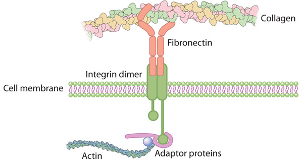
Symmetric exchange of multi-protein building blocks between stationary focal adhesions and the cytosol

Jan-Erik Hoffmann, Y Fermin, R LO Stricker, K Ickstadt, E Zamir
eLife 2014;3:e02257. http://dx.doi.org:/10.7554/eLife.02257.001

How can the integrin adhesome get self-assembled locally, rapidly, and correctly as diverse cell-matrix adhesion sites? Here, we investigate this question by exploring the cytosolic state of integrin-adhesome components and their dynamic exchange between adhesion sites and cytosol. Using fluorescence cross-correlation spectroscopy (FCCS) and fluorescence recovery after photo-bleaching (FRAP) we found that the integrin adhesome is extensively pre-assembled already in the cytosol as multi-protein building blocks for adhesion sites. Stationary focal adhesions release symmetrically the same types of protein complexes that they recruit, thereby keeping the cytosolic pool of building blocks spatiotemporally uniform. We conclude a model in which multi-protein building blocks enable rapid and modular self-assembly of adhesion sites and symmetric exchange of these building blocks preserves their specifications and thus the assembly logic of the system.

Redox signaling via the molecular chaperone BiP protects cells against endoplasmic reticulum-derived oxidative stress

Jie Wang, Kristeen A Pareja, Chris A Kaiser, Carolyn S Sevier
eLife 2014;3:e03496. http://dx.doi.org:/10.7554/eLife.03496

Oxidative protein folding in the endoplasmic reticulum (ER) has emerged as a potentially significant source of cellular reactive oxygen species (ROS). Recent studies suggest that levels of ROS generated as a byproduct of oxidative folding rival those produced by mitochondrial respiration. Mechanisms that protect cells against oxidant accumulation within the ER have begun to be elucidated yet many questions still remain regarding how cells prevent oxidant-induced damage from ER folding events. Here we report a new role for a central well-characterized player in ER homeostasis as a direct sensor of ER redox imbalance. Specifically we show that a conserved cysteine in the lumenal chaperone BiP is susceptible to oxidation by peroxide, and we demonstrate that oxidation of this conserved cysteine disrupts BiP’s ATPase cycle. We propose that alteration of BiP activity upon oxidation helps cells cope with disruption to oxidative folding within the ER during oxidative stress.

Current perspectives on cadherin-cytoskeleton interactions and dynamics

Xuan Liang, Guillermo A Gomez, Alpha S Yap
Cell Health and Cytoskeleton 2015:7 11–24
http://dx.doi.org/10.2147/CHC.S76107

Cells are linked together dynamically by adhesion molecules, such as the classical cadherins. E-cadherin, which mediates epithelial cell–cell interactions, plays fundamental roles in tissue organization and is often perturbed in diseases such as cancer. It has long been recognized that the biology of E-cadherin arises from cooperation between adhesion and the actin cytoskeleton. A major feature is the generation of contractile forces at junctions, yielding patterns of tension that contribute to tissue integrity and patterning. Here we discuss recent developments in understanding how cadherin junctions integrate signaling and cytoskeletal dynamics to sense and generate force.

N-glycosylation status of E-cadherin controls cytoskeletal dynamics through the organization of distinct β-catenin- and γ-catenin-containing AJs

Basem T Jamal, M Nita-Lazar, Z Gao, B Amin, J Walker, MA Kukuruzinska
Cell Health and Cytoskeleton 2009:1 67–80

N-glycosylation of E-cadherin has been shown to inhibit cell–cell adhesion. Specifically, our recent studies have provided evidence that the reduction of E-cadherin N-glycosylation promoted the recruitment of stabilizing components, vinculin and serine/threonine protein phosphatase 2A (PP2A), to adherens junctions (AJs) and enhanced the association of AJs with the actin cytoskeleton. Here, we examined the details of how N-glycosylation of E-cadherin affected the molecular organization of AJs and their cytoskeletal interactions. Using the hypoglycosylated E-cadherin variant, V13, we show that V13/β-catenin complexes preferentially interacted with PP2A and with the microtubule motor protein dynein. This correlated with dephosphorylation of the microtubule-associated protein tau, suggesting that increased association of PP2A with V13-containing AJs promoted their tethering to microtubules. On the other hand, V13/γ-catenin complexes associated more with vinculin, suggesting that they mediated the interaction of AJs with the actin cytoskeleton. N-glycosylation driven changes in the molecular organization of AJs were physiologically significant because transfection of V13 into A253 cancer cells, lacking both mature AJs and tight junctions (TJs), promoted the formation of stable AJs and enhanced the function of TJs to a greater extent than wild-type E-cadherin. These studies provide the first mechanistic insights into how N-glycosylation of E-cadherin drives changes in AJ composition through the assembly of distinct β-catenin- and γ-catenin-containing scaffolds that impact the interaction with different cytoskeletal components.

Mapping the dynamics of force transduction at cell-cell 4 junctions of epithelial clusters

Mei Rosa Ng, Achim Besser, Joan S. Brugge, Gaudenz Danuser
eLife 2014;10.7554/eLife.03282
http://dx.doi.org/10.7554/eLife.03282

Force transduction at cell-cell adhesions regulates tissue development, maintenance and adaptation. We developed computational and experimental approaches to quantify, with both subcellular and multi-cellular resolution, the dynamics of force transmission in cell clusters. Applying this technology to spontaneously-forming adherent epithelial cell clusters, we found that basal force fluctuations were coupled to E-cadherin localization at the level of individual cell-cell junctions. At the multi-cellular scale, cell-cell force exchange depended on the cell position within a cluster, and was adaptive to reconfigurations due to cell divisions or positional rearrangements. Importantly, force transmission through a cell required coordinated modulation of cell-matrix adhesion and actomyosin contractility in the cell and its neighbors. These data provide insights into  mechanisms that could control mechanical stress homeostasis in dynamic epithelial tissues, and highlight our methods as a resource for the study of mechanotransduction in cell-cell adhesions.

G-protein-coupled receptor signaling and polarized actin dynamics drive cell-in-cell invasion

Vladimir Purvanov, Manuel Holst, Jameel Khan, Christian Baarlink, Robert Grosse
eLife 2014;3:e02786.  http://dx.doi.org:/10.7554/eLife.02786

Homotypic or entotic cell-in-cell invasion is an integrin-independent process observed in carcinoma cells exposed during conditions of low adhesion such as in exudates of malignant disease. Although active cell-in-cell invasion depends on RhoA and actin, the precise mechanism as well as the underlying actin structures and assembly factors driving the process are unknown. Furthermore, whether specific cell surface receptors trigger entotic invasion in a signal-dependent fashion has not been investigated. In this study, we identify the G-protein-coupled LPA receptor 2 (LPAR2) as a signal transducer specifically required for the actively invading cell during entosis. We find that G12/13 and PDZ-RhoGEF are required for entotic invasion, which is driven by blebbing and a uropod-like actin structure at the rear of the invading cell. Finally, we provide evidence for an involvement of the RhoA-regulated formin Dia1 for entosis downstream of LPAR2. Thus, we delineate a signaling process that regulates actin dynamics during cell-in-cell invasion.

Cytoskeletal Basis of Ion Channel Function in Cardiac Muscle

Matteo Vatta, and Georgine Faulkner
Future Cardiol. 2006 Jul 1; 2(4): 467–476. http://dx.doi.org:/10.2217/14796678.2.4.467

The heart is a force-generating organ that responds to self-generated electrical stimuli from specialized cardiomyocytes. This function is modulated by sympathetic and parasympathetic activity.

In order to contract and accommodate the repetitive morphological changes induced by the cardiac cycle, cardiomyocytes depend on their highly evolved and specialized cytoskeletal apparatus. Defects in components of the cytoskeleton, in the long term, affect the ability of the cell to compensate at both functional and structural levels. In addition to the structural remodeling, the myocardium becomes increasingly susceptible to altered electrical activity leading to arrhythmogenesis. The development of arrhythmias secondary to structural remodeling defects has been noted, although the detailed molecular mechanisms are still elusive. Here I will review the current knowledge of the molecular and functional relationships between the cytoskeleton and ion channels and, I will discuss the future impact of new data on molecular cardiology research and clinical practice.

Structure and transport mechanism of the sodium/proton 2 antiporter MjNhaP1

Cristina Paulino, D Wöhlert , E Kapotova, Ö Yildiz & W Kühlbrandt
eLife 2014;  http://dx.doi.org/10.7554/eLife.03583

Sodium/proton antiporters are essential for sodium and pH homeostasis and play a major role in human health and disease. We determined the structures of the archaeal sodium/proton antiporter MjNhaP1 in two complementary states. The inward-open state was obtained by x-ray crystallography in the presence of sodium at pH8, where the transporter is highly active. The outward-open state was obtained by electron crystallography without sodium at pH4, where MjNhaP1 is inactive. Comparison of both structures reveals a 7° tilt of the 6-helix bundle. Na+  uptake measurements indicate non-cooperative transport with an activity maximum at pH7.5. We conclude that binding of a Na+ ion from the outside induces helix movements that close the extracellular cavity, open the cytoplasmic funnel, and result in a ~5 Å vertical relocation of the ion binding site to release the substrate ion into the cytoplasm.

Integrated control of transporter endocytosis and recycling by the arrestin-related protein Rod1 and the ubiquitin ligase Rsp5

Michel Becuwe, Sébastien Léon
eLife 2014; http://dx.doi.org/10.7554/eLife.03307

After endocytosis, membrane proteins can recycle to the cell membrane or be degraded in lysosomes. Cargo ubiquitylation favors their lysosomal targeting and can be regulated by external signals, but the mechanism is ill-defined. Here, we studied the post-endocytic trafficking of Jen1, a yeast monocarboxylate transporter, using microfluidics-assisted live cell imaging. We show that the ubiquitin ligase Rsp5 and the glucose-regulated arrestin related (ART) protein Rod1, involved in the glucose-induced internalization of Jen1, are  also required for the post-endocytic sorting of Jen1 to the yeast lysosome. This new step takes place at the trans-Golgi network (TGN), where Rod1 localizes dynamically upon triggering endocytosis. Indeed, transporter trafficking to the TGN after internalization is required for their degradation. Glucose removal promotes Rod1 relocalization to the cytosol and Jen1 deubiquitylation, allowing transporter recycling when the signal is only transient. Therefore, nutrient availability regulates transporter fate through the localization of the ART/Rsp5 ubiquitylation complex at the TGN.

  1. McKenney, W Huynh, ME. Tanenbaum, G Bhabha, and RD. Vale
    Science Express 19 June 2014 /10.1126/science.1254198
    http://www.sciencemag.org/content/early/recent/10.1126/science.1254198

Cytoplasmic dynein is a molecular motor that transports a large variety of cargoes (e.g., organelles, mRNAs, and viruses) along microtubules over long intracellular distances. The dynactin protein complex is important for dynein activity in vivo, but its precise role has been unclear. Here, we found that purified mammalian dynein did not move processively on microtubules in vitro. However, when dynein formed a complex with dynactin and one of four different cargo-specific adapter proteins, the motor became ultra-processive, moving for distances similar to those of native cargoes in living cells. Thus, we propose that dynein is largely inactive in the cytoplasm and that a variety of adapter proteins activate processive motility by linking dynactin to dynein only when the motor is bound to its proper cargo.

Removal of surface charge–charge interactions from ubiquitin leaves the protein folded and very stable

Vakhtang V. Loladze And George I. Makhatadze
Protein Science (2002), 11:174–177
http://www.proteinscience.org/cgi/doi/10.1101/ps.29902.

The contribution of solvent-exposed charged residues to protein stability was evaluated using ubiquitin as a model protein. We combined site-directed mutagenesis and specific chemical modifications to first replace all Arg residues with Lys, followed by carbomylation of Lys- amino groups. Under the conditions in which all carboxylic groups are protonated (at pH 2), the chemically modified protein is folded and very stable (dG= 18 kJ/mol). These results indicate that surface charge–charge interactions are not an essential fundamental force for protein folding and stability.

Phase Transitions of Multivalent Proteins Can Promote Clustering of Membrane Receptors

Sudeep Banjade and Michael K. Rosen
eLife 2014; http://dx.doi.org/10.7554/eLife.04123

Clustering of proteins into micrometer-sized structures at membranes is observed in many signaling pathways. Most models of clustering are specific to particular systems, and relationships between physical properties of the clusters and their molecular components are not well understood. We report biochemical reconstitution on supported lipid bilayers of protein clusters containing the adhesion receptor Nephrin, and its cytoplasmic partners, Nck and N-WASP. With Nephrin attached to the bilayer, multivalent interactions enable these proteins to polymerize on the membrane surface and undergo two-dimensional phase separation, producing micrometer-sized clusters. Dynamics and thermodynamics of the clusters are modulated by the valencies and affinities of the interacting species. In the presence of the Arp2/3 complex, the clusters assemble actin filaments, suggesting that clustering of regulatory factors could promote local actin assembly at membranes. Interactions between multivalent proteins could be a  general mechanism for cytoplasmic adaptor proteins to organize membrane receptors into micrometer-scale signaling zones.

The quantitative architecture of centromeric chromatin

Dani L Bodor, João F Mata, Mikhail Sergeev, Ana Filipa David, et al.
eLife 2014;3:e02137. http://dx.doi.org:/10.7554/eLife.02137

The centromere, responsible for chromosome segregation during mitosis, is epigenetically defined by CENP-A containing chromatin. The amount of centromeric CENP-A has direct implications for both the architecture and epigenetic inheritance of centromeres. Using complementary strategies, we determined that typical human centromeres contain ∼400 molecules of CENP-A, which is controlled by a mass-action mechanism. This number, despite representing only ∼4% of all centromeric nucleosomes, forms a ∼50-fold enrichment to the overall genome. In addition, although pre-assembled CENP-A is randomly segregated during cell division, this amount of CENP-A is sufficient to prevent stochastic loss of centromere function and identity. Finally, we produced a statistical map of CENP-A occupancy at a human neocentromere and identified nucleosome positions that feature CENP-A in a majority of cells. In summary, we present a quantitative view of the centromere that provides a mechanistic framework for both robust epigenetic inheritance of centromeres and the paucity of neocentromere formation.

Synaptic proteins promote calcium-triggered fast transition from point contact to full fusion

Jiajie Diao, Patricia Grob, Daniel J Cipriano, Minjoung Kyoung
eLife 2012;1:e00109. http://dx.doi.org:/10.7554/eLife.00109

The molecular underpinnings of synaptic vesicle fusion for fast neurotransmitter release are still unclear. Here, we used a single vesicle–vesicle system with reconstituted SNARE and synaptotagmin-1 proteoliposomes to decipher the temporal sequence of membrane states upon Ca2+-injection at 250–500 μM on a 100-ms timescale. Furthermore, detailed membrane morphologies were imaged with cryo-electron microscopy before and after Ca2+-injection. We discovered a heterogeneous network of immediate and delayed fusion pathways. Remarkably, all instances of Ca2+-triggered immediate fusion started from a membrane–membrane point-contact and proceeded to complete fusion without discernible hemifusion intermediates. In contrast, pathways that involved a stable hemifusion diaphragm only resulted in fusion after many seconds, if at all. When complexin was included, the Ca2+-triggered fusion network shifted towards the immediate pathway, effectively synchronizing fusion, especially at lower Ca2+-concentration. Synaptic proteins may have evolved to select this immediate pathway out of a heterogeneous network of possible membrane fusion pathways.

Cytoskeleton, cytoskeletal interactions, and vascular endothelial function

Jingli Wang, Michael E Widlansky
Cell Health and Cytoskeleton 2012:4 119–127
http://dx.doi.org/10.2147/CHC.S21823

Far from being inert, the vascular endothelium is a critical regulator of vascular function. While the endothelium participates in autocrine, paracrine, and endocrine signaling, it also transduces mechanical signals from the cell surface involving key cell structural elements. In this review, we discuss the structure of the vascular endothelium and its relationship to traditional cardiovascular risk factors and clinical cardiovascular events. Further, we review the emerging evidence that cell structural elements, including the glycocalyx, intercellular junctions, and cytoskeleton elements, help the endothelium to communicate with its environment to regulate vascular function, including vessel permeability and signal transduction via nitric oxide bioavailability. Further work is necessary to better delineate the regulatory relationships between known key regulators of vascular function and endothelial cell structural elements.

Cellular prion protein is required for neuritogenesis: fine-tuning of multiple signaling pathways involved in focal adhesions and actin cytoskeleton dynamics

Aurélie Alleaume-Butaux, C Dakowski, M Pietri, S Mouillet-Richard, et al.
Cell Health and Cytoskeleton 2013:5 1–12
http://dx.doi.org/10.2147/CHC.S28081

Neuritogenesis is a dynamic phenomenon associated with neuronal differentiation that allows a rather spherical neuronal stem cell to develop dendrites and axon, a prerequisite for the integration and transmission of signals. The acquisition of neuronal polarity occurs in three steps: (1) neurite sprouting, which consists of the formation of buds emerging from the postmitotic neuronal soma; (2) neurite outgrowth, which represents the conversion of buds into neurites, their elongation and evolution into axon or dendrites; and (3) the stability and plasticity of neuronal polarity. In neuronal stem cells, remodeling and activation of focal adhesions (FAs) associated with deep modifications of the actin cytoskeleton is a prerequisite for neurite sprouting and subsequent neurite outgrowth. A multiple set of growth factors and interactors located in the extracellular matrix and the plasma membrane orchestrate neuritogenesis by acting on intracellular signaling effectors, notably small G proteins such as RhoA, Rac, and Cdc42, which are involved in actin turnover and the dynamics of FAs. The cellular prion protein (PrPC), a glycosylphosphatidylinositol (GPI)-anchored membrane protein mainly known for its role in a group of fatal neurodegenerative diseases, has emerged as a central player in neuritogenesis. Here, we review the contribution of PrPC to neuronal polarization and detail the current knowledge on the signaling pathways fine-tuned by PrPC to promote neurite sprouting, outgrowth, and maintenance. We emphasize that PrPC-dependent neurite sprouting is a process in which PrPC governs the dynamics of FAs and the actin cytoskeleton via β1 integrin signaling. The presence of PrPC is necessary to render neuronal stem cells competent to respond to neuronal inducers and to develop neurites. In differentiating neurons, PrPC exerts a facilitator role towards neurite elongation. This function relies on the interaction of PrPC with a set of diverse partners such as elements of the extracellular matrix, plasma membrane receptors, adhesion molecules, and soluble factors that control actin cytoskeleton turnover through Rho-GTPase signaling. Once neurons have reached their terminal stage of differentiation and acquired their polarized morphology, PrPC also takes part in the maintenance of neurites. By acting on tissue nonspecific alkaline phosphatase, or matrix metalloproteinase type 9, PrPC stabilizes interactions between neurites and the extracellular matrix.

Broader implications: biological and clinical significance of microtubule acetylation

Sharon M Rymut, Thomas J Kelley
Cell Health and Cytoskeleton 2015:7 71–82
http://dx.doi.org/10.2147/CHC.S77040

Microtubule acetylation is a key posttranslational modification that enhances organelle transport, drives cell signaling, and regulates cell cycle regulation. The optimal level of microtubule acetylation is regulated by the acetyltransferase alpha-tubulin-N-acetyltransferase 1and two deacetylases, histone deacetylase 6 and sirtuin-2. Alterations in microtubule acetylation levels have been associated with the pathophysiology of a number of diseases, including various forms of neurodegenerative conditions, cancer, and even cystic fibrosis. In this review, we will highlight the biological and clinical significance of microtubule acetylation and the potential of targeting this pathway for therapeutics.

Inositol-1,4,5-trisphosphate 1 (IP3)-mediated STIM1 oligomerization requires  intact mitochondrial Ca2+ uptake

  1. Deak, S. Blass, M. J. Khan, L. N. Groschner, M. Waldeck-Weiermair, et al.
    Journal of Cell Science 2014 advanced print

Mitochondria contribute to cell signaling by controlling store-operated Ca2+ entry (SOCE).  SOCE is activated by Ca2+ release from the endoplasmic reticulum (ER), whereupon the stromal  interacting molecule 1 (STIM1) forms oligomers, redistributes to ER-plasma membrane  junctions, and opens plasma membrane Ca2+ channels. Mechanisms by which mitochondria interfere with the complex process of SOCE are insufficiently clarified. In this study we used a shRNA approach to investigate the direct involvement of mitochondrial Ca2+ buffering in SOCE. We demonstrate that knock-down of two proteins that are essential for mitochondrial Ca2+ uptake, either the mitochondrial calcium uniporter (MCU) or uncoupling protein 2 (UCP2), results in decelerated STIM1 oligomerization and impaired SOCE following cell stimulation with an inositol-1,4,5-trisphosphate (IP3)-generating agonist. Upon artificially augmented cytosolic Ca2+-buffering or ER Ca2+ depletion by sarco/endoplasmic reticulum Ca2+-ATPase (SERCA) inhibitors, STIM1 oligomerization did not rely on intact mitochondrial Ca2+ uptake.  However, MCU-dependent mitochondrial sequestration of Ca2+ entering through the SOCE  pathway was essential to prevent slow deactivation of SOCE. Our findings show a stimulus specific contribution of mitochondrial Ca2+ uptake to the SOCE machinery likely by shaping cytosolic Ca2+ micro-domains.

Role of forkhead box protein A3 in age-associated metabolic decline

Xinran Ma, Lingyan Xu, Oksana Gavrilov, and Elisabetta Mueller
PNAS | September 30, 2014 | vol. 111 | no. 39 | 14289–14294
www.pnas.org/cgi/doi/10.1073/pnas.1407640111

Aging is associated with increased adiposity and diminished thermogenesis, but the critical transcription factors influencing these metabolic changes late in life are poorly understood. We recently demonstrated that the winged helix factor forkhead box protein A3 (Foxa3) regulates the expansion of visceral adipose tissue in high-fat diet regimens; however, whether Foxa3 also contributes to the increase in adiposity and the decrease in brown fat activity observed during the normal aging process is currently unknown.
Here we report that during aging, levels of Foxa3 are significantlyand selectively up-regulated in brown and inguinal white fat depots, and that midage Foxa3-null mice have increased white fat browning and thermogenic capacity, decreased adipose tissue expansion, improved insulin sensitivity, and increased longevity. Foxa3 gain-of-function and loss-of-function studies in inguinal adipose depots demonstrated a cell-autonomous function for Foxa3 in white fat tissue browning. Furthermore, our analysis revealed that the mechanisms of Foxa3 modulation of brown fat gene programs involve the suppression of peroxisome proliferator activated receptor γ coactivtor 1 α (PGC1α) levels through interference with cAMP responsive element binding protein 1-mediated transcriptional regulation of the PGC1α promoter. Overall, our data demonstrate a role for Foxa3 in energy expenditure and in age-associated metabolic disorders.

Prediction of enzyme function by combining sequence similarity and protein interactions

Jordi Espadaler, Narayanan Eswa, Enrique Querol, Francesc X Avilés, et al.
BMC Bioinformatics 2008, 9:249 http://dx.doi.org:/10.1186/1471-2105-9-249

Background: A number of studies have used protein interaction data alone for protein function prediction. Here, we introduce a computational approach for annotation of enzymes, based on the observation that similar protein sequences are more likely to perform the same function if they share similar interacting partners.
Results: The method has been tested against the PSI-BLAST program using a set of 3,890 protein sequences from which interaction data was available. For protein sequences that align with at least 40% sequence identity to a known enzyme, the specificity of our method in predicting the first three EC digits increased from 80% to 90% at 80% coverage when compared to PSI-BLAST.
Conclusion: Our method can also be used in proteins for which homologous sequences with known interacting partners can be detected. Thus, our method could increase 10% the specificity of genome-wide enzyme predictions based on sequence matching by PSI-BLAST alone.

Plasma Transthyretin Indicates the Direction of both Nitrogen Balance and Retinoid Status in Health and Disease

Ingenbleek Yves and Bienvenu Jacques
The Open Clinical Chemistry Journal, 2008, 1, 1-12

Whatever the nutritional status and the disease condition, the actual transthyretin (TTR) plasma level is determined by opposing influences between anabolic and catabolic alterations. Rising TTR values indicate that synthetic processes prevail over tissue breakdown with a nitrogen balance (NB) turning positive as a result of efficient nutritional support and / or anti-inflammatory therapy. Declining TTR values point to the failure of sustaining NB as an effect of maladjusted dietetic management and / or further worsening of the morbid condition. Serial measurement of TTR thus appears as a dynamic index defining the direction of NB in acute and chronic disorders, serving as a guide to alert the physician on the validity of his therapeutic strategy. The level of TTR production by the liver also works as a limiting factor for the cellular bioavailability of retinol and retinoid derivatives which play major roles in the brain ageing process. Optimal protein nutritional status, as assessed by TTR values within the normal range, prevents the occurrence of vascular and cerebral damages while maintaining the retinoid-mediated memory, cognitive and behavioral activities of elderly persons.

Prof. Dr. Volker Haucke
Institut für Chemie-Biochemie
Takustrasse 6
http://userpage.chemie.fu-berlin.de/biochemie/aghaucke/teaching.html

Eukaryotic cells contain three major types of cytoskeletal filaments

Eukaryotic cells contain three major types of cytoskeletal filaments

major types of cytoskeletal filaments

major types of cytoskeletal filaments

Intermediate Filaments support the nuclear membrane and connect cells at cell junctions

Intermediate Filaments support the nuclear membrane and connect cells at cell junctions

microtubules (MTs; green) radiate from MTOCs (yellow) towards the cell periphery

microtubules (MTs; green) radiate from MTOCs (yellow) towards the cell periphery

Actin polymerization in vitro reveals a critical dependence of filament assembly on G-actin concentration via a 3-step nucleation mechanism

Actin polymerization in vitro reveals a critical dependence of filament assembly on G-actin concentration via a 3-step nucleation mechanism

Binding-proteins and receptors

Motor, visual and emotional deficits in mice after closed-head mild traumatic brain injury are alleviated by the novel CB2 inverse agonist SMM-189
Reiner, A., Heldt, S.A., Presley, C.S., (…), Gurley, S.N., Moore, B.M.
2015  International Journal of Molecular Sciences 16 (1), pp. 758-787

We have developed a focal blast model of closed-head mild traumatic brain injury (TBI) in mice. As true for individuals that have experienced mild TBI, mice subjected to 50-60 psi blast show motor, visual and emotional deficits, diffuse axonal injury and microglial activation, but no overt neuron
loss. Because microglial activation can worsen brain damage after a concussive event and because microglia can be
modulated by their cannabinoid type 2 receptors (CB2), we evaluated the effectiveness of the novel CB2 receptor inverse agonist SMM-189 in altering microglial activation and mitigating deficits after mild TBI. In vitro analysis indicated that SMM-189 converted human microglia from the pro-inflammatory M1 phenotype to the pro-healing M2 phenotype. Studies in mice showed that daily administration of SMM-189 for two weeks beginning shortly after blast greatly reduced the motor, visual, and emotional deficits otherwise evident after 50-60 psi blasts, and prevented brain injury that may contribute to these deficits. Our results suggest that treatment with the CB2 inverse agonist SMM-189 after a mild TBI event can reduce its adverse consequences by beneficially modulating microglial activation. These
findings recommend further evaluation of CB2 inverse agonists as a novel therapeutic approach for treating mild TBI.

The novel small leucine-rich protein chondroadherin-like (CHADL) is expressed in cartilage and modulates chondrocyte differentiation
Tillgren, V., Ho, J.C.S., Önnerfjord, P., Kalamajski, S.
2015  Journal of Biological Chemistry 290 (2), pp. 918-925

The constitution and biophysical properties of extracellular matrices can dramatically influence cellular phenotype during development, homeostasis, or pathogenesis. These effects can be signaled through a differentially regulated assembly of collagen fibrils, orchestrated by a family of collagen-associated small leucine-rich proteins (SLRPs). In this report, we describe the tissue-specific expression and function of a previously uncharacterized SLRP, chondroadherin-like (CHADL). We developed antibodies against CHADL and, by immunohistochemistry, detected CHADL expression mainly in skeletal tissues, particularly in fetal cartilage and in the pericellular space of adult chondrocytes. In situ hybridizations and immunoblots on tissue lysates confirmed this tissue-specific expression pattern. Recombinant CHADL bound collagen in cell culture and inhibited in vitro collagen fibrillogenesis. After Chadl shRNA knockdown, chondrogenic ATDC5 cells increased their differentiation, indicated by increased transcript levels of Sox9, Ihh, Col2a1, and Col10a1. The knockdown increased collagen II and aggrecan deposition in the cell layers.

Microarray analysis of the knockdown samples suggested collagen receptor-related changes, although other upstream effects could not be excluded. Together, our data indicate that the novel SLRP CHADL is expressed in cartilaginous tissues, influences collagen fibrillogenesis, and modulates chondrocyte differentiation. CHADL appears to have a negative regulatory role, possibly ensuring the formation of a stable extracellular matrix.

P53 protein-mediated Up-regulation of MAP kinase phosphatase 3 (MKP-3) contributes to the establishment of the cellular senescent phenotype through dephosphorylation of extracellular signal-regulated kinase 1/2 (ERK1/2)
Zhang, H., Chi, Y., Gao, K., Zhang, X., Yao, J.
2015  Source of the DocumentJournal of Biological Chemistry 290 (2), pp. 1129-1140

Growth arrest is one of the essential features of cellular senescence. At present, the precise mechanisms responsible for the establishment of the senescence-associated arrested phenotype are still incompletely understood. Given that ERK1/2 is one of the major kinases controlling cell growth and proliferation, we examined the possible implication of ERK1/2. Exposure of normal rat epithelial cells to etoposide caused cellular senescence, as manifested by enlarged cell size, a flattened cell body, reduced cell proliferation, enhanced ?-galactosidase activity, and elevated p53 and p21. Senescent cells displayed a blunted response to growth factor-induced cell proliferation, which was preceded by impaired ERK1/2 activation. Further analysis revealed that senescent cells expressed a significantly higher level of mitogenactivated protein phosphatase 3 (MKP-3, a cytosolic ERK1/2-targeted phosphatase), which was suppressed by blocking the transcriptional activity of the tumor suppressor p53 with pifithrin-?. Inhibition of MKP-3 activity with a specific inhibitor or siRNA enhanced basal ERK1/2 phosphorylation and promoted cell proliferation. Apart from its role in growth arrest, impairment of ERK1/2 also contributed to the resistance of senescent cells to oxidant-elicited cell injury. These results therefore indicate that p53-mediated up-regulation of MKP-3 contributes to the establishment of the senescent cellular phenotype through dephosphorylating ERK1/2. Impairment of ERK1/2 activation could be an important mechanism by which p53 controls cellular senescence.

Dynamics and interaction of Interleukin-4 receptor subunits in living cells
Gandhi, H., Worch, R., Kurgonaite, K., (…), Bökel, C., Weidemann, T.
2015  Biophysical Journal 107 (11), pp. 2515-2527

It has long been established that dimerization of Interleukin-4 receptor (IL-4R) subunits is a pivotal step for JAK/STAT signal transduction. However, ligand-induced complex formation at the surface of living cells has been challenging to observe. Here we report an experimental assay employing trisNTA dyes for orthogonal, external labeling of eGFP-tagged receptor constructs that allows the quantification of receptor heterodimerization by dual-color fluorescence cross-correlation spectroscopy. Fluorescence cross-correlation spectroscopy analysis at the plasma membrane shows that IL-4R subunit dimerization is indeed a strictly ligand-induced process.

Under conditions of saturating cytokine occupancy, we determined intramembrane dissociation constants (Kd,2D) of 180 and 480 receptors per ?m2 for the type-2 complexes IL-4:IL-4R?/IL-13R?1 and IL-13:IL-13R?1/IL-4R?, respectively. For the lower affinity type-1 complex IL-4:IL-4R?/IL-2R?, we estimated a Kd,2D of ?1000 receptors per ?m2. The receptor densities required for effective dimerization thus exceed the typical, average expression levels by several orders of magnitude. In addition, we find that all three receptor subunits accumulate rapidly within a subpopulation of early sorting and recycling endosomes stably anchored just beneath the plasma membrane (cortical endosomes, CEs). The receptors, as well as labeled IL-4 and trisNTA ligands are specifically trafficked into CEs by a constitutive internalization mechanism. This may compensate for the inherent weak affinities that govern ligand-induced receptor dimerization at the plasma membrane. Consistently, activated receptors are also concentrated at the CEs. Our observations thus suggest that receptor trafficking may play an important role for the regulation of IL-4R-mediated JAK/STAT signaling.

Role of mitochondria in nonalcoholic fatty liver disease
Nassir, F., Ibdah, J.A.
2015  International Journal of Molecular Sciences 15 (5), pp. 8713-8742

Nonalcoholic fatty liver disease (NAFLD) affects about 30% of the general population in the United States and includes a spectrum of disease that includes simple steatosis, non-alcoholic steatohepatitis (NASH), fibrosis and cirrhosis. Significant insight has been gained into our understanding of the pathogenesis of NALFD; however the key metabolic aberrations underlying lipid accumulation in hepatocytes and the progression of NAFLD remain to be elucidated. Accumulating and emerging evidence indicate that hepatic mitochondria play a critical role in the development and pathogenesis of steatosis and NAFLD. Here, we review studies that document a link between the pathogenesis of NAFLD and hepatic mitochondrial dysfunction with particular focus on new insights into the role of impaired fatty acid oxidation, the transcription factor peroxisome proliferator-activated receptor-? coactivator-1? (PGC-1?), and sirtuins in development and progression of NAFLD.

Read Full Post »

Larry H. Bernstein, MD, FCAP, Interviewer, Curator

Leaders in Pharmaceutical Intelligence

Biochemical Insights of Dr. Jose Eduardo de Salles Roselino

http://pharmaceuticalintelligence.com/12/24/2014/larryhbern/Biochemical_
Insights_of_Dr._Jose_Eduardo_de_Salles_Roselino/

Biochemical Insights of Dr. Jose Eduardo de Salles Roselino

How is it that developments late in the 20th century diverted the attention of
biological processes from a dynamic construct involving interacting chemical
reactions under rapidly changing external conditions effecting tissues and cell
function to a rigid construct that is determined unilaterally by the genome
construct, diverting attention from mechanisms essential for seeing the complete
cellular construct?

Larry, I assume that in case you read the article titled Neo – Darwinism, The
Modern Synthesis and Selfish Genes that bares no relationship with Physiology
with Molecular Biology J. Physiol 2011; 589(5): 1007-11 by Denis Noble, you might
find that it was the key factor required in order to understand the dislodgment
of physiology as a foundation of medical reasoning. In the near unilateral emphasis
of genomic activity as a determinant of cellular activity all of the required general
support for the understanding of my reasoning. The DNA to protein link goes
from triplet sequence to amino acid sequence. That is the realm of genetics.
Further, protein conformation, activity and function requires that environmental
and micro-environmental factors should be considered (Biochemistry). If that
were not the case, we have no way to bridge the gap between the genetic
code and the evolution of cells, tissues, organs, and organisms.

  • Consider this example of hormonal function. I would like to stress in
    the cAMP dependent hormonal response, the transfer of information
    that 
    occurs through conformation changes after protein interactions.
    This mechanism therefore, requires that proteins must not have their
    conformation determined by sequence alone.
    Regulatory protein conformation is determined by its sequence plus
    the interaction it has in its micro-environment. For instance, if your
    scheme takes into account what happens inside the membrane and
    that occurs before cAMP, then production is increased by hormone
    action. A dynamic scheme  will show an effect initially, over hormone
    receptor (hormone binding causing change in its conformation) followed
    by GTPase change in conformation caused by receptor interaction and
    finally, Adenylate cyclase change in conformation and in activity after
    GTPase protein binding in a complex system that is dependent on self-
    assembly and also, on changes in their conformation in response to
    hormonal signals (see R. A Kahn and A. G Gilman 1984 J. Biol. Chem.
    v. 259,n 10 pp6235-6240. In this case, trimeric or dimeric G does not
    matter). Furthermore, after the step of cAMP increased production we
    also can see changes in protein conformation.  The effect of increased
    cAMP levels over (inhibitor protein and protein kinase protein complex)
    also is an effect upon protein conformation. Increased cAMP levels led
    to the separation of inhibitor protein (R ) from cAMP dependent protein
    kinase (C ) causing removal of the inhibitor R and the increase in C activity.
    R stands for regulatory subunit and C for catalytic subunit of the protein
    complex.
  • This cAMP effect over the quaternary structure of the enzyme complex
    (C protein kinase + R the inhibitor) may be better understood as an
    environmental information producing an effect in opposition to
    what may be considered as a tendency  towards a conformation
    “determined” by the genetic code. This “ideal” conformation
    “determined” by the genome  would be only seen in crystalline
    protein.
     In carbohydrate metabolism in the liver the hormonal signal
    causes a biochemical regulatory response that preserves homeostatic
    levels of glucose (one function) and in the muscle, it is a biochemical
    regulatory response that preserves intracellular levels of ATP (another
    function).
  • Therefore, sequence alone does not explain conformation, activity
    and function of regulatory proteins
    .  If this important regulatory
    mechanism was  not ignored, the work of  S. Prusiner (Prion diseases
    and the BSE crisis Stanley B. Prusiner 1997 Science; 278: 245 – 251,
    10  October) would be easily understood.  We would be accustomed
    to reason about changes in protein conformation caused by protein
    interaction with other proteins, lipids, small molecules and even ions.
  • In case this wrong biochemical reasoning is used in microorganisms.
    Still it is wrong but, it will cause a minor error most of the time, since
    we may reduce almost all activity of microorganism´s proteins to a
    single function – The production of another microorganism. However,
    even microorganisms respond differently to their micro-environment
    despite a single genome (See M. Rouxii dimorphic fungus works,
    later). The reason for the reasoning error is, proteins are proteins
    and DNA are DNA quite different in chemical terms. Proteins must
    change their conformation to allow for fast regulatory responses and
    DNA must preserve its sequence to allow for genetic inheritance.

Read Full Post »

Summary of Cell Structure, Anatomic Correlates of Metabolic Function

Author and Curator: Larry H. Bernstein, MD, FCAP  


This chapter has been concerned with the subcellular ultrastructure of organelles, and importantly, their function.  There is no waste in the cell structure. The nucleus has the instructions necessary to carry out the cell’s functions.  In the Eukaryotic cell there is significant differentiation so that the cells are regulated for the needs that they uniquely carry out.  When there is disregulation, it leads to remodeling or to cell death.

Here I shall note some highlights of this chapter.

  1. In every aspect of cell function, proteins are involved embedded in the structure, for most efficient functioning.
  2. Metabolic regulation is dependent on pathways that are also linkages of proteins.
  3. Energy utilization is dependent on enzymatic reactions, often involving essential metal ions of high valence numbers, which facilitates covalent and anion binding, and has an essential role in allostericity.

Mitochondria

Mitochondria,_mammalian_lung

Mitochondria,_mammalian_lung

http://en.wikipedia.org/wiki/File:Mitochondria,_mammalian_lung_-_TEM.jpg

Mitochondria range from 0.5 to 1.0 micrometer (μm) in diameter. These structures are sometimes described as “cellular power plants” because they generate most of the cell’s supply of adenosine triphosphate (ATP), used as a source of chemical energy. In addition to supplying cellular energy, mitochondria are involved in other tasks such as signaling, cellular differentiation, cell death, as well as the control of the cell cycle and cell growth. Mitochondria have been implicated in several human diseases, including mitochondrial disorders and cardiac dysfunction.

The number of mitochondria in a cell can vary widely by organism, tissue, and cell type. For instance, red blood cells have no mitochondria, whereas liver cells can have more than 2000. The organelle is composed of compartments that carry out specialized functions. These compartments or regions include the outer membrane, the intermembrane space, the inner membrane, and the cristae and matrix. Mitochondrial proteins vary depending on the tissue and the species. The mitochondrial proteome is thought to be dynamically regulated. Although most of a cell’s DNA is contained in the cell nucleus, the mitochondrion has its own independent genome. Further, its DNA shows substantial similarity to bacterial genomes.

In 1913 particles from extracts of guinea-pig liver were linked to respiration by Otto Heinrich Warburg, which he called “grana”. Warburg and Heinrich Otto Wieland, who had also postulated a similar particle mechanism, disagreed on the chemical nature of the respiration. It was not until 1925 when David Keilin discovered cytochromes that the respiratory chain was described.  In 1939, experiments using minced muscle cells demonstrated that one oxygen atom can form two adenosine triphosphate molecules, and, in 1941, the concept of phosphate bonds being a form of energy in cellular metabolism was developed by Fritz Albert Lipmann. In the following years, the mechanism behind cellular respiration was further elaborated, although its link to the mitochondria was not known. The introduction of tissue fractionation by Albert Claude allowed mitochondria to be isolated from other cell fractions and biochemical analysis to be conducted on them alone. In 1946, he concluded that cytochrome oxidase and other enzymes responsible for the respiratory chain were isolated to the mitchondria.

The first high-resolution micrographs appeared in 1952, replacing the Janus Green stains as the preferred way of visualising the mitochondria. This led to a more detailed analysis of the structure of the mitochondria, including confirmation that they were surrounded by a membrane. It also showed a second membrane inside the mitochondria that folded up in ridges dividing up the inner chamber and that the size and shape of the mitochondria varied from cell to cell.  In 1967, it was discovered that mitochondria contained ribosomes. In 1968, methods were developed for mapping the mitochondrial genes, with the genetic and physical map of yeast mitochondria being completed in 1976.

A mitochondrion contains outer and inner membranes composed of phospholipid bilayers and proteins. The two membranes have different properties. Because of this double-membraned organization, there are five distinct parts to a mitochondrion. They are:

  1. the outer mitochondrial membrane,
  2. the intermembrane space (the space between the outer and inner membranes),
  3. the inner mitochondrial membrane,
  4. the cristae space (formed by infoldings of the inner membrane), and
  5. the matrix (space within the inner membrane).

Mitochondria stripped of their outer membrane are called mitoplasts.

Mitochondrion_structure_drawing

Mitochondrion_structure_drawing

http://upload.wikimedia.org/wikipedia/commons/thumb/9/9e/Mitochondrion_structure_drawing.svg/500px-Mitochondrion_structure_drawing.svg.png

Mitochondrion ultrastructure (interactive diagram) A mitochondrion has a double membrane; the inner one contains its chemiosmotic apparatus and has deep grooves which increase its surface area. While commonly depicted as an “orange sausage with a blob inside of it” (like it is here), mitochondria can take many shapes and their intermembrane space is quite thin.

The intermembrane space is the space between the outer membrane and the inner membrane. It is also known as perimitochondrial space. Because the outer membrane is freely permeable to small molecules, the concentrations of small molecules such as ions and sugars in the intermembrane space is the same as the cytosol. However, large proteins must have a specific signaling sequence to be transported across the outer membrane, so the protein composition of this space is different from the protein composition of the cytosol. One protein that is localized to the intermembrane space in this way is cytochrome c.

The inner mitochondrial membrane contains proteins with five types of functions:

  1. Those that perform the redox reactions of oxidative phosphorylation
  2. ATP synthase, which generates ATP in the matrix
  3. Specific transport proteins that regulate metabolite passage into and out of the matrix
  4. Protein import machinery.
  5. Mitochondria fusion and fission protein.

It contains more than 151 different polypeptides, and has a very high protein-to-phospholipid ratio (more than 3:1 by weight, which is about 1 protein for 15 phospholipids). The inner membrane is home to around 1/5 of the total protein in a mitochondrion. In addition, the inner membrane is rich in an unusual phospholipid, cardiolipin. This phospholipid was originally discovered in cow hearts in 1942, and is usually characteristic of mitochondrial and bacterial plasma membranes. Cardiolipin contains four fatty acids rather than two, and may help to make the inner membrane impermeable. Unlike the outer membrane, the inner membrane doesn’t contain porins, and is highly impermeable to all molecules. Almost all ions and molecules require special membrane transporters to enter or exit the matrix. Proteins are ferried into the matrix via the translocase of the inner membrane (TIM) complex or via Oxa1. In addition, there is a membrane potential across the inner membrane, formed by the action of the enzymes of the electron transport chain.

The inner mitochondrial membrane is compartmentalized into numerous cristae, which expand the surface area of the inner mitochondrial membrane, enhancing its ability to produce ATP. For typical liver mitochondria, the area of the inner membrane is about five times as large as the outer membrane. This ratio is variable and mitochondria from cells that have a greater demand for ATP, such as muscle cells, contain even more cristae. These folds are studded with small round bodies known as F1 particles or oxysomes. These are not simple random folds but rather invaginations of the inner membrane, which can affect overall chemiosmotic function. One recent mathematical modeling study has suggested that the optical properties of the cristae in filamentous mitochondria may affect the generation and propagation of light within the tissue.

Mitochondrion

Mitochondrion

http://upload.wikimedia.org/wikipedia/commons/thumb/d/d8/MitochondrionCAM.jpg/250px-MitochondrionCAM.jpg

The matrix is the space enclosed by the inner membrane. It contains about 2/3 of the total protein in a mitochondrion. The matrix is important in thThe MAM is enriched in enzymes involved in lipid biosynthesis, such as phosphatidylserine synthase on the ER face and phosphatidylserine decarboxylase on the mitochondrial face.[28][29] Because mitochondria are dynamic organelles constantly undergoing fission and fusion events, they require a constant and well-regulated supply of phospholipids for membrane integrity.[30][31] But mitochondria are not only a destination for the phospholipids they finish synthesis of; rather, this organelle also plays a role in inter-organelle trafficking of the intermediates and products of phospholipid biosynthetic pathways, ceramide and cholesterol metabolism, and glycosphingolipid anabolisme production of ATP with the aid of the ATP synthase contained in the inner membrane. The matrix contains a highly concentrated mixture of hundreds of enzymes, special mitochondrial ribosomes, tRNA, and several copies of the mitochondrial DNA genome. Of the enzymes, the major functions include oxidation of pyruvate and fatty acids, and the citric acid cycle.

Purified MAM from subcellular fractionation has shown to be enriched in enzymes involved in phospholipid exchange, in addition to channels associated with Ca2+ signaling. The mitochondria-associated ER membrane (MAM) is another structural element that is increasingly recognized for its critical role in cellular physiology and homeostasis. Once considered a technical snag in cell fractionation techniques, the alleged ER vesicle contaminants that invariably appeared in the mitochondrial fraction have been re-identified as membranous structures derived from the MAM—the interface between mitochondria and the ER. Physical coupling between these two organelles had previously been observed in electron micrographs and has more recently been probed with fluorescence microscopy. Such studies estimate that at the MAM, which may comprise up to 20% of the mitochondrial outer membrane, the ER and mitochondria are separated by a mere 10–25 nm and held together by protein tethering complexes.

Such trafficking capacity depends on the MAM, which has been shown to facilitate transfer of lipid intermediates between organelles. In contrast to the standard vesicular mechanism of lipid transfer, evidence indicates that the physical proximity of the ER and mitochondrial membranes at the MAM allows for lipid flipping between opposed bilayers. Despite this unusual and seemingly energetically unfavorable mechanism, such transport does not require ATP. Instead, in yeast, it has been shown to be dependent on a multiprotein tethering structure termed the ER-mitochondria encounter structure, or ERMES, although it remains unclear whether this structure directly mediates lipid transfer or is required to keep the membranes in sufficiently close proximity to lower the energy barrier for lipid flipping.

A critical role for the ER in calcium signaling was acknowledged before such a role for the mitochondria was widely accepted, in part because the low affinity of Ca2+ channels localized to the outer mitochondrial membrane seemed to fly in the face of this organelle’s purported responsiveness to changes in intracellular Ca2+ flux. But the presence of the MAM resolves this apparent contradiction: the close physical association between the two organelles results in Ca2+ microdomains at contact points that facilitate efficient Ca2+ transmission from the ER to the mitochondria. Transmission occurs in response to so-called “Ca2+ puffs” generated by spontaneous clustering and activation of IP3R, a canonical ER membrane Ca2+ channel.

The properties of the Ca2+ pump SERCA and the channel IP3R present on the ER membrane facilitate feedback regulation coordinated by MAM function. In particular, clearance of Ca2+ by the MAM allows for spatio-temporal patterning of Ca2+ signaling because Ca2+ alters IP3R activity in a biphasic manner. SERCA is likewise affected by mitochondrial feedback: uptake of Ca2+ by the MAM stimulates ATP production, thus providing energy that enables SERCA to reload the ER with Ca2+ for continued Ca2+ efflux at the MAM. Thus, the MAM is not a passive buffer for Ca2+ puffs; rather it helps modulate further Ca2+ signaling through feedback loops that affect ER dynamics.

Regulating ER release of Ca2+ at the MAM is especially critical because only a certain window of Ca2+ uptake sustains the mitochondria, and consequently the cell, at homeostasis. Sufficient intraorganelle Ca2+ signaling is required to stimulate metabolism by activating dehydrogenase enzymes critical to flux through the citric acid cycle. However, once Ca2+ signaling in the mitochondria passes a certain threshold, it stimulates the intrinsic pathway of apoptosis in part by collapsing the mitochondrial membrane potential required for metabolism.  Studies examining the role of pro- and anti-apoptotic factors support this model; for example, the anti-apoptotic factor Bcl-2 has been shown to interact with IP3Rs to reduce Ca2+ filling of the ER, leading to reduced efflux at the MAM and preventing collapse of the mitochondrial membrane potential post-apoptotic stimuli. Given the need for such fine regulation of Ca2+ signaling, it is perhaps unsurprising that dysregulated mitochondrial Ca2+ has been implicated in several neurodegenerative diseases, while the catalogue of tumor suppressors includes a few that are enriched at the MAM.

…more

http://en.wikipedia.org/wiki/Mitochondrion

Lysosome and Apoptosis

Role of autophagy in cancer

R Mathew, V Karantza-Wadsworth & E White

Nature Reviews Cancer 7, 961-967 (Dec 2007) |  http://dx.doi.org:/10.1038/nrc2254

Autophagy is a cellular degradation pathway for the clearance of damaged or superfluous proteins and organelles. The recycling of these intracellular constituents also serves as an alternative energy source during periods of metabolic stress to maintain homeostasis and viability. In tumour cells with defects in apoptosis, autophagy allows prolonged survival. Paradoxically, autophagy defects are associated with increased tumorigenesis, but the mechanism behind this has not been determined. Recent evidence suggests that autophagy provides a protective function to limit tumour necrosis and inflammation, and to mitigate genome damage in tumour cells in response to metabolic stress.

Sustained Activation of mTORC1 in Skeletal Muscle Inhibits Constitutive and Starvation-Induced Autophagy and Causes a Severe, Late-Onset Myopathy

P Castets, S Lin, N Rion, S Di Fulvio, et al.
cell-metabolism 7 May, 2013; 17(5): p731–744   http://dx.doi.org/10.1016/j.cmet.2013.03.015

  • mTORC1 inhibition is required for constitutive and starvation-induced autophagy
  • Sustained activation of mTORC1 causes a severe myopathy due to autophagy impairment
  • TSC1 depletion is sufficient to activate mTORC1 irrespective of other stimuli
  • mTORC1 inactivation is sufficient to trigger LC3 lipidation

Autophagy is a catabolic process that ensures homeostatic cell clearance and is deregulated in a growing number of myopathological conditions. Although FoxO3 was shown to promote the expression of autophagy-related genes in skeletal muscle, the mechanisms triggering autophagy are unclear. We show that TSC1-deficient mice (TSCmKO), characterized by sustained activation of mTORC1, develop a late-onset myopathy related to impaired autophagy. In young TSCmKO mice,

  • constitutive and starvation-induced autophagy is blocked at the induction steps via
  • mTORC1-mediated inhibition of Ulk1, despite FoxO3 activation.

Rapamycin is sufficient to restore autophagy in TSCmKO mice and

  • improves the muscle phenotype of old mutant mice.

Inversely, abrogation of mTORC1 signaling by

  • depletion of raptor induces autophagy regardless of FoxO inhibition.

Thus, mTORC1 is the dominant regulator of autophagy induction in skeletal muscle and

  • ensures a tight coordination of metabolic pathways.

These findings may open interesting avenues for therapeutic strategies directed toward autophagy-related muscle diseases.

Histone deacetylases 1 and 2 regulate autophagy flux and skeletal muscle homeostasis in mice

Viviana Moresi, et al.   PNAS Jan 31, 2012; 109(5): 1649-1654
http://dx.doi.org:/10.1073/pnas.1121159109
http://www.pnas.org/content/109/5/1649/F6.medium.gif

HDAC1 activates FoxO and is both sufficient and required for skeletal muscle atrophy

Beharry, PB. Sandesara, BM. Roberts, et al.
J. Cell Sci. Apr 2014 127 (7) 1441-1453   http://dx.doi.org:/10.1242/​jcs.136390

The Forkhead box O (FoxO) transcription factors are activated, and necessary for the muscle atrophy, in several pathophysiological conditions, including muscle disuse and cancer cachexia. However, the mechanisms that lead to FoxO activation are not well defined. Recent data from our laboratory and others indicate that

  • the activity of FoxO is repressed under basal conditions via reversible lysine acetylation,
  • which becomes compromised during catabolic conditions.

Therefore, we aimed to determine how histone deacetylase (HDAC) proteins contribute to

  • activation of FoxO and induction of the muscle atrophy program.

Through the use of various pharmacological inhibitors to block HDAC activity, we demonstrate that

  • class I HDACs are key regulators of FoxO and the muscle-atrophy program
  • during both nutrient deprivation and skeletal muscle disuse.

Furthermore, we demonstrate, through the use of wild-type and dominant-negative HDAC1 expression plasmids,

  • that HDAC1 is sufficient to activate FoxO and induce muscle fiber atrophy in vivo and
  • is necessary for the atrophy of muscle fibers that is associated with muscle disuse.

The ability of HDAC1 to cause muscle atrophy required its deacetylase activity and

  • was linked to the induction of several atrophy genes by HDAC1,
  • including atrogin-1, which required deacetylation of FoxO3a.

Moreover, pharmacological inhibition of class I HDACs during muscle disuse, using MS-275,

  • significantly attenuated both disuse muscle fiber atrophy and contractile dysfunction.

Together, these data solidify the importance of class I HDACs in the muscle atrophy program and

  • indicate that class I HDAC inhibitors are feasible countermeasures to impede muscle atrophy and weakness.

Autophagy and thyroid carcinogenesis: genetic and epigenetic links
F Morani, R Titone, L Pagano, et al.  Endocr Relat Cancer Feb 1, 2014 21 R13-R29
http://dx.doi.org:/10.1530/ERC-13-0271

Autophagy is a vesicular process for the lysosomal degradation of protein aggregates and

  • of damaged or redundant organelles.

Autophagy plays an important role in cell homeostasis, and there is evidence that

  • this process is dysregulated in cancer cells.

Recent in vitro preclinical studies have indicated that autophagy is

  • involved in the cytotoxic response to chemotherapeutics in thyroid cancer cells.

Indeed, several oncogenes and oncosuppressor genes implicated in thyroid carcinogenesis

  • also play a role in the regulation of autophagy.

In addition, some epigenetic modulators involved in thyroid carcinogenesis also influence autophagy. In this review, we highlight the genetic and epigenetic factors that

  • mechanistically link thyroid carcinogenesis and autophagy, thus substantiating the rationale for
  • an autophagy-targeted therapy of aggressive and radio-chemo-resistant thyroid cancers.

Read Full Post »

Introduction to Subcellular Structure

Author and Curator: Larry H. Bernstein, MD, FCAP  


The following chapter of the metabolism/transcriptomics/proteomics/metabolomics series deals with the subcellular structure of the cell.  This would have to include the cytoskeleton, which has a key role in substrate and ion efflux and influx, and in cell movement mediated by tubulins.  It has been extensively covered already.  Much of the contributions here are concerned with the mitochondrion, which is also covered in metabolic pathways.  The ribosome is the organelle that we have discussed with respect to the transcription and translation of the genetic code through mRNA and tRNA, and the therapeutic implications of SiRNA as well as the chromatin regulation of lncRNA.

We have also encountered the mitochondrion and the lysosome in the discussion of apoptosis and autophagy, maintaining the balance between cell regeneration and cell death.

I here list the organelles:

  1. Nucleus
  2. Centrosome
  3. Nuclear Membrane
  4. Ribososome
  5. Endoplasmic Reticulum
  6. Mitochondria
  7. Lysosome
  8. Cytoskeleton
  9. Golgi apparatus
  10. Cytoplasm
cell_organelle_quiz

cell_organelle_quiz

http://www.youtube.com/watch?feature=player_embedded&v=JufLDxmCwB0

http://www.youtube.com/watch?feature=player_embedded&v=FFrKN7hJm64

Golgi Apparatus

Found within the cytoplasm of both plant and animal cells, the Golgi is composed of stacks of membrane-bound structures known as cisternae (singular: cisterna). An individual stack is sometimes called a dictyosome (from Greek dictyon: net + soma: body), especially in plant cells. A mammalian cell typically contains 40 to 100 stacks. Between four and eight cisternae are usually present in a stack; however, in some protists as many as sixty have been observed. Each cisterna comprises a flat, membrane-enclosed disc that includes special Golgi enzymes which modify or help to modify cargo proteins that travel through it.

The cisternae stack has four functional regions: the cis-Golgi network, medial-Golgi, endo-Golgi, and trans-Golgi network. Vesicles from the endoplasmic reticulum (via the vesicular-tubular clusters) fuse with the network and subsequently progress through the stack to the trans-Golgi network, where they are packaged and sent to their destination.

The Golgi apparatus is integral in modifying, sorting, and packaging these macromolecules for cell secretion (exocytosis) or use within the cell. It primarily modifies proteins delivered from the rough endoplasmic reticulum, but is also involved in the transport of lipids around the cell, and the creation of lysosomes.  Enzymes within the cisternae are able to modify the proteins by addition of carbohydrates (glycosylation) and phosphates (phosphorylation). In order to do so, the Golgi imports substances such as nucleotide sugars from the cytosol. These modifications may also form a signal sequence which determines the final destination of the protein. For example, the Golgi apparatus adds a mannose-6-phosphate label to proteins destined for lysosomes.

The Golgi plays an important role in the synthesis of proteoglycans, which are molecules present in the extracellular matrix of animals. It is also a major site of carbohydrate synthesis. This includes the production of glycosaminoglycans (GAGs), long unbranched polysaccharides which the Golgi then attaches to a protein synthesised in the endoplasmic reticulum to form proteoglycans. Enzymes in the Golgi polymerize several of these GAGs via a xylose link onto the core protein. Another task of the Golgi involves the sulfation of certain molecules passing through its lumen via sulfotranferases that gain their sulfur molecule from a donor called PAPS. This process occurs on the GAGs of proteoglycans as well as on the core protein. Sulfation is generally performed in the trans-Golgi network. The level of sulfation is very important to the proteoglycans’ signalling abilities, as well as giving the proteoglycan its overall negative charge.

The phosphorylation of molecules requires that ATP is imported into the lumen of the Golgi and utilised by resident kinases such as casein kinase 1 and casein kinase 2. One molecule that is phosphorylated in the Golgi is apolipoprotein, which forms a molecule known as VLDL that is found in plasma. It is thought that the phosphorylation of these molecules labels them for secretion into the blood.

The Golgi has a putative role in apoptosis, with several Bcl-2 family members localised there, as well as to the mitochondria. A newly characterized protein, GAAP (Golgi anti-apoptotic protein), almost exclusively resides in the Golgi and protects cells from apoptosis by an as-yet undefined mechanism.

The vesicles that leave the rough endoplasmic reticulum are transported to the cis face of the Golgi apparatus, where they fuse with the Golgi membrane and empty their contents into the lumen. Once inside the lumen, the molecules are modified, then sorted for transport to their next destinations. The Golgi apparatus tends to be larger and more numerous in cells that synthesize and secrete large amounts of substances; for example, the plasma B cells and the antibody-secreting cells of the immune system have prominent Golgi complexes.

Those proteins destined for areas of the cell other than either the endoplasmic reticulum or Golgi apparatus are moved towards the trans face, to a complex network of membranes and associated vesicles known as the trans-Golgi network (TGN). This area of the Golgi is the point at which proteins are sorted and shipped to their intended destinations by their placement into one of at least three different types of vesicles, depending upon the molecular marker they carry.

Nucleus_ER_golgi

Nucleus_ER_golgi

Diagram of secretory process from endoplasmic reticulum (orange) to Golgi apparatus (pink). 1. Nuclear membrane; 2. Nuclear pore; 3. Rough endoplasmic reticulum (RER); 4. Smooth endoplasmic reticulum (SER); 5. Ribosome attached to RER; 6. Macromolecules; 7. Transport vesicles; 8. Golgi apparatus; 9. Cis face of Golgi apparatus; 10. Trans face of Golgi apparatus; 11. Cisternae of the Golgi Apparatus

Exocytotic vesicles

After packaging, the vesicles bud off and immediately move towards the plasma membrane, where they fuse and release the contents into the extracellular space in a process known as constitutive secretion. (Antibody release by activated plasma B cells)

Secretory vesicles

After packaging, the vesicles bud off and are stored in the cell until a signal is given for their release. When the appropriate signal is received they move towards the membrane and fuse to release their contents. This process is known as regulated secretion. (Neurotransmitter release from neurons)

Lysosomal vesicles

Vesicle contains proteins and ribosomes destined for the lysosome, an organelle of degradation containing many acid hydrolases, or to lysosome-like storage organelles. These proteins include both digestive enzymes and membrane proteins. The vesicle first fuses with the late endosome, and the contents are then transferred to the lysosome via unknown mechanisms.

http://en.wikipedia.org/wiki/Golgi_apparatus

Lysosome (derived from the Greek words lysis, meaning “to loosen”, and soma, “body”) is a membrane-bound cell organelle found in animal cells (they are absent in red blood cells). They are structurally and chemically spherical vesicles containing hydrolytic enzymes, which are capable of breaking down virtually all kinds of biomolecules, including proteins, nucleic acids, carbohydrates, lipids, and cellular debris.  Lysosomes are responsible for cellular homeostasis for their involvements in secretion, plasma membrane repair, cell signalling and energy metabolism, which are related to health and diseases. Depending on their functional activity their sizes can be very different, as the biggest ones can be more than 10 times bigger than the smallest ones. They were discovered and named by Belgian biologist Christian de Duve, who eventually received the Nobel Prize in Physiology or Medicine in 1974.

Enzymes of the lysosomes are synthesised in the rough endoplasmic reticulum. The enzymes are released from Golgi apparatus in small vesicles which ultimately fuse with acidic vesicles called endosomes, thus becoming full lysosomes. In the process the enzymes are specifically tagged with mannose 6-phosphate to differentiate them from other enzymes. Lysosomes are interlinked with three intracellular processes namely phagocytosis, endocytosis and autophagy. Extracellular materials such as microorganisms taken up by phagocytosis, macromolecules by endocytosis, and unwanted cell organelles are fused with lysosomes in which they are broken down to their basic molecules. Thus lysosomes are the recycling units of a cell.

http://en.wikipedia.org/wiki/Lysosome

The endoplasmic reticulum (ER) is a type of organelle in the cells of eukaryotic organisms that forms an interconnected network of flattened, membrane-enclosed sacs or tubes known as cisternae. The membranes of the ER are continuous with the outer membrane of the nuclear envelope. Endoplasmic reticulum occurs in most types of eukaryotic cells, including the most primitive Giardia, but is absent from red blood cells and spermatozoa. There are two types of endoplasmic reticulum, rough endoplasmic reticulum (RER) and smooth endoplasmic reticulum (SER). The outer (cytosolic) face of the rough endoplasmic reticulum is studded with ribosomes that are the sites of protein synthesis. The rough endoplasmic reticulum is especially prominent in cells such as hepatocytes where active smooth endoplasmic reticulum lacks ribosomes and functions in lipid metabolism, carbohydrate metabolism, and detoxification and is especially abundant in mammalian liver and gonad cells. The lacey membranes of the endoplasmic reticulum were first seen in 1945 by Keith R. Porter, Albert Claude, Brody Meskers and Ernest F. Fullam, using electron microscopy.

http://en.wikipedia.org/wiki/Endoplasmic_reticulum

endoplasmic_reticulum

endoplasmic_reticulum

https://2cslacardano.wikispaces.com/file/view/Cell7.png/338811858/408×313/Cell7.png

Cytoskeleton

The Effects of Actomyosin Tension on Nuclear Pore Transport
Rachel Sammons
Undergraduate Honors Thesis
Spring 2011

The cytoskeleton maintains cellular structure and tension through a force balance with the nucleus, where actomyosin is anchored to the nuclear envelope by nesprin integral proteins. It is hypothesized that the presence or absence of this tension alters the transport of molecules through the nuclear pore complex. We tested the effects of cytoskeletal tension on nuclear transport in human umbilical vein endothelial cells (HUVECs) by performing fluorescence recovery after photo-bleaching (FRAP) experiments on the nuclei to monitor the passive transport of the molecules through nuclear pores.

Using myosin inhibitors, as well as siRNA transfections to reduce the expression of nesprin-1, we altered the nucleo-cytoskeletal force balance and monitored the effect of each on the nuclear pore. FRAP data was fit to a diffusion model by assuming pseudo-steady state inside the nuclear pore, perfect mixing within both the cytoplasm and the nucleus, and no intracellular binding of the fluorescent probes. From these results and a model from the current literature relating diffusion rate constants to nuclear pore radii, we were able to determine that changing cytoskeletal tension alters nuclear pore size and passive transport.

nuclear pores in nuclear envelope

nuclear pores in nuclear envelope

image of nuclear pores on the external surface of the nuclear envelope

nuclear envelope and FG filaments

nuclear envelope and FG filaments

nuclear envelope and FG filaments

Figure 1: The structure and location of the nuclear pore, shown by (a) AFM image of nuclear pores on the external surface of the nuclear envelope[5] and (b) computer model cross-section. The nuclear envelope is shown in cyan, and FG filaments in blue can be seen throughout the channel. The nuclear basket extends into the nucleoplasm.

Fusion-pore expansion during syncytium formation is restricted by an actin network

A Chen, E Leikina, K Melikov, B Podbilewicz, MM. Kozlov and LV. Chernomordik,*
J Cell Sci 1 Nov 2008;121: 3619-3628. http://dx.doi.org:/10.1242/​jcs.032169

Effects of actin-modifying agents indicate that the actin cortex slows down pore expansion. We propose that the growth of the strongly bent fusion-pore rim is restricted by a dynamic resistance of the actin network and driven by membrane-bending proteins that are involved in the generation of highly curved intracellular membrane compartments.

Read Full Post »

Summary of Signaling and Signaling Pathways

Summary of Signaling and Signaling Pathways

Author and Curator: Larry H Bernstein, MD, FCAP

In the imtroduction to this series of discussions I pointed out JEDS Rosalino’s observation about the construction of a complex molecule of acetyl coenzyme A, and the amount of genetic coding that had to go into it.  Furthermore, he observes –  Millions of years later, or as soon as, the information of interaction leading to activity and regulation could be found in RNA, proteins like reverse transcriptase move this information to a more stable form (DNA). In this way it is easier to understand the use of CoA to make two carbon molecules more reactive.

acetylCoA

acetylCoA

In the tutorial that follows we find support for the view that mechanisms and examples from the current literature, which give insight into the developments in cell metabolism, are achieving a separation from inconsistent views introduced by the classical model of molecular biology and genomics, toward a more functional cellular dynamics that is not dependent on the classic view.  The classical view fits a rigid framework that is to genomics and metabolomics as Mendelian genetics if to multidimentional, multifactorial genetics.  The inherent difficulty lies in two places:

  1. Interactions between differently weighted determinants
  2. A large part of the genome is concerned with regulatory function, not expression of the code

The goal of the tutorial was to achieve an understanding of how cell signaling occurs in a cell.  Completion of the tutorial would provide

  1. a basic understanding signal transduction and
  2. the role of phosphorylation in signal transduction.
Regulation of the integrity of endothelial cell–cell contacts by phosphorylation of VE-cadherin

Regulation of the integrity of endothelial cell–cell contacts by phosphorylation of VE-cadherin

In addition – detailed knowledge of –

  1. the role of Tyrosine kinases and
  2. G protein-coupled receptors in cell signaling.
serine

serine

threonine

threonine

protein kinase

protein kinase

We are constantly receiving and interpreting signals from our environment, which can come

  • in the form of light, heat, odors, touch or sound.

The cells of our bodies are also

  • constantly receiving signals from other cells.

These signals are important to

  • keep cells alive and functioning as well as
  • to stimulate important events such as
  • cell division and differentiation.

Signals are most often chemicals that can be found

  • in the extracellular fluid around cells.

These chemicals can come

  • from distant locations in the body (endocrine signaling by hormones), from
  • nearby cells (paracrine signaling) or can even
  • be secreted by the same cell (autocrine signaling).

Notch-mediated juxtacrine signal between adjacent cells. 220px-Notchccr

Signaling molecules may trigger any number of cellular responses, including

  • changing the metabolism of the cell receiving the signal or
  • result in a change in gene expression (transcription) within the nucleus of the cell or both.
controlling the output of ribosomes.

controlling the output of ribosomes.

To which I would now add..

  • result in either an inhibitory or a stimulatory effect

The three stages of cell signaling are:

Cell signaling can be divided into 3 stages:

Reception: A cell detects a signaling molecule from the outside of the cell.

Transduction: When the signaling molecule binds the receptor it changes the receptor protein in some way. This change initiates the process of transduction. Signal transduction is usually a pathway of several steps. Each relay molecule in the signal transduction pathway changes the next molecule in the pathway.

Response: Finally, the signal triggers a specific cellular response.

signal transduction

signal transduction

http://www.hartnell.edu/tutorials/biology/images/signaltransduction_simple.jpg

The initiation is depicted as follows:

Signal Transduction – ligand binds to surface receptor

Membrane receptors function by binding the signal molecule (ligand) and causing the production of a second signal (also known as a second messenger) that then causes a cellular response. These types of receptors transmit information from the extracellular environment to the inside of the cell.

  • by changing shape or
  • by joining with another protein
  • once a specific ligand binds to it.

Examples of membrane receptors include

  • G Protein-Coupled Receptors and
Understanding these receptors and identifying their ligands and the resulting signal transduction pathways represent a major conceptual advance.

Understanding these receptors and identifying their ligands and the resulting signal transduction pathways represent a major conceptual advance.

  • Receptor Tyrosine Kinases.
intracellular signaling

intracellular signaling

http://www.hartnell.edu/tutorials/biology/images/membrane_receptor_tk.jpg

Intracellular receptors are found inside the cell, either in the cytopolasm or in the nucleus of the target cell (the cell receiving the signal).

Note that though change in gene expression is stated, the change in gene expression does not here imply a change in the genetic information – such as – mutation.  That does not have to be the case in the normal homeostatic case.

This point is the differentiating case between what JEDS Roselino has referred as

  1. a fast, adaptive reaction, that is the feature of protein molecules, and distinguishes this interaction from
  2. a one-to-one transcription of the genetic code.

The rate of transcription can be controlled, or it can be blocked.  This is in large part in response to the metabolites in the immediate interstitium.

This might only be

  • a change in the rate of a transcription or a suppression of expression through RNA.
  • Or through a conformational change in an enzyme
 Swinging domains in HECT E3 enzymes

Swinging domains in HECT E3 enzymes

Since signaling systems need to be

  • responsive to small concentrations of chemical signals and act quickly,
  • cells often use a multi-step pathway that transmits the signal quickly,
  • while amplifying the signal to numerous molecules at each step.

Signal transduction pathways are shown (simplified):

Signal Transduction

Signal Transduction

Signal transduction occurs when an

  1. extracellular signaling molecule activates a specific receptor located on the cell surface or inside the cell.
  2. In turn, this receptor triggers a biochemical chain of events inside the cell, creating a response.
  3. Depending on the cell, the response alters the cell’s metabolism, shape, gene expression, or ability to divide.
  4. The signal can be amplified at any step. Thus, one signaling molecule can cause many responses.

In 1970, Martin Rodbell examined the effects of glucagon on a rat’s liver cell membrane receptor. He noted that guanosine triphosphate disassociated glucagon from this receptor and stimulated the G-protein, which strongly influenced the cell’s metabolism. Thus, he deduced that the G-protein is a transducer that accepts glucagon molecules and affects the cell. For this, he shared the 1994 Nobel Prize in Physiology or Medicine with Alfred G. Gilman.

Guanosine monophosphate structure

Guanosine monophosphate structure

In 2007, a total of 48,377 scientific papers—including 11,211 e-review papers—were published on the subject. The term first appeared in a paper’s title in 1979. Widespread use of the term has been traced to a 1980 review article by Rodbell: Research papers focusing on signal transduction first appeared in large numbers in the late 1980s and early 1990s.

Signal transduction involves the binding of extracellular signaling molecules and ligands to cell-surface receptors that trigger events inside the cell. The combination of messenger with receptor causes a change in the conformation of the receptor, known as receptor activation.

This activation is always the initial step (the cause) leading to the cell’s ultimate responses (effect) to the messenger. Despite the myriad of these ultimate responses, they are all directly due to changes in particular cell proteins. Intracellular signaling cascades can be started through cell-substratum interactions; examples are the integrin that binds ligands in the extracellular matrix and steroids.

Integrin

Integrin

Most steroid hormones have receptors within the cytoplasm and act by stimulating the binding of their receptors to the promoter region of steroid-responsive genes.

steroid hormone receptor

steroid hormone receptor

Various environmental stimuli exist that initiate signal transmission processes in multicellular organisms; examples include photons hitting cells in the retina of the eye, and odorants binding to odorant receptors in the nasal epithelium. Certain microbial molecules, such as viral nucleotides and protein antigens, can elicit an immune system response against invading pathogens mediated by signal transduction processes. This may occur independent of signal transduction stimulation by other molecules, as is the case for the toll-like receptor. It may occur with help from stimulatory molecules located at the cell surface of other cells, as with T-cell receptor signaling. Receptors can be roughly divided into two major classes: intracellular receptors and extracellular receptors.

Signal transduction cascades amplify the signal output

Signal transduction cascades amplify the signal output

Signal transduction cascades amplify the signal output

G protein-coupled receptors (GPCRs) are a family of integral transmembrane proteins that possess seven transmembrane domains and are linked to a heterotrimeric G protein. Many receptors are in this family, including adrenergic receptors and chemokine receptors.

Arrestin binding to active GPCR kinase (GRK)-phosphorylated GPCRs blocks G protein coupling

signal transduction pathways

signal transduction pathways

Arrestin binding to active GPCR kinase (GRK)-phosphorylated GPCRs blocks G protein coupling

Arrestin binding to active GPCR kinase (GRK)-phosphorylated GPCRs blocks G protein coupling

Signal transduction by a GPCR begins with an inactive G protein coupled to the receptor; it exists as a heterotrimer consisting of Gα, Gβ, and Gγ. Once the GPCR recognizes a ligand, the conformation of the receptor changes to activate the G protein, causing Gα to bind a molecule of GTP and dissociate from the other two G-protein subunits.

The dissociation exposes sites on the subunits that can interact with other molecules. The activated G protein subunits detach from the receptor and initiate signaling from many downstream effector proteins such as phospholipases and ion channels, the latter permitting the release of second messenger molecules.

Receptor tyrosine kinases (RTKs) are transmembrane proteins with an intracellular kinase domain and an extracellular domain that binds ligands; examples include growth factor receptors such as the insulin receptor.

 insulin receptor and and insulin receptor signaling pathway (IRS)

insulin receptor and and insulin receptor signaling pathway (IRS)

To perform signal transduction, RTKs need to form dimers in the plasma membrane; the dimer is stabilized by ligands binding to the receptor.

RTKs

RTKs

The interaction between the cytoplasmic domains stimulates the autophosphorylation of tyrosines within the domains of the RTKs, causing conformational changes.

Allosteric_Regulation.svg

Subsequent to this, the receptors’ kinase domains are activated, initiating phosphorylation signaling cascades of downstream cytoplasmic molecules that facilitate various cellular processes such as cell differentiation and metabolism.

Signal-Transduction-Pathway

Signal-Transduction-Pathway

As is the case with GPCRs, proteins that bind GTP play a major role in signal transduction from the activated RTK into the cell. In this case, the G proteins are

  • members of the Ras, Rho, and Raf families, referred to collectively as small G proteins.

They act as molecular switches usually

  • tethered to membranes by isoprenyl groups linked to their carboxyl ends.

Upon activation, they assign proteins to specific membrane subdomains where they participate in signaling. Activated RTKs in turn activate

  • small G proteins that activate guanine nucleotide exchange factors such as SOS1.

Once activated, these exchange factors can activate more small G proteins, thus

  • amplifying the receptor’s initial signal.

The mutation of certain RTK genes, as with that of GPCRs, can result in the expression of receptors that exist in a constitutively activate state; such mutated genes may act as oncogenes.

Integrin


Integrin

Integrin

Integrin-mediated signal transduction

An overview of integrin-mediated signal transduction, adapted from Hehlgens et al. (2007).

Integrins are produced by a wide variety of cells; they play a role in

  • cell attachment to other cells and the extracellular matrix and
  • in the transduction of signals from extracellular matrix components such as fibronectin and collagen.

Ligand binding to the extracellular domain of integrins

  • changes the protein’s conformation,
  • clustering it at the cell membrane to
  • initiate signal transduction.

Integrins lack kinase activity; hence, integrin-mediated signal transduction is achieved through a variety of intracellular protein kinases and adaptor molecules, the main coordinator being integrin-linked kinase.

As shown in the picture, cooperative integrin-RTK signaling determines the

  1. timing of cellular survival,
  2. apoptosis,
  3. proliferation, and
  4. differentiation.
integrin-mediated signal transduction

integrin-mediated signal transduction

Integrin signaling

Integrin signaling

ion channel

A ligand-gated ion channel, upon binding with a ligand, changes conformation

  • to open a channel in the cell membrane
  • through which ions relaying signals can pass.

An example of this mechanism is found in the receiving cell of a neural synapse. The influx of ions that occurs in response to the opening of these channels

  1. induces action potentials, such as those that travel along nerves,
  2. by depolarizing the membrane of post-synaptic cells,
  3. resulting in the opening of voltage-gated ion channels.
RyR and Ca+ release from SR

RyR and Ca+ release from SR

An example of an ion allowed into the cell during a ligand-gated ion channel opening is Ca2+;

  • it acts as a second messenger
  • initiating signal transduction cascades and
  • altering the physiology of the responding cell.

This results in amplification of the synapse response between synaptic cells

  • by remodelling the dendritic spines involved in the synapse.

In eukaryotic cells, most intracellular proteins activated by a ligand/receptor interaction possess an enzymatic activity; examples include tyrosine kinase and phosphatases. Some of them create second messengers such as cyclic AMP and IP3,

cAMP

cAMP

Inositol_1,4,5-trisphosphate.svg

Inositol_1,4,5-trisphosphate.svg

  • the latter controlling the release of intracellular calcium stores into the cytoplasm.

Many adaptor proteins and enzymes activated as part of signal transduction possess specialized protein domains that bind to specific secondary messenger molecules. For example,

  • calcium ions bind to the EF hand domains of calmodulin,
  • allowing it to bind and activate calmodulin-dependent kinase.
calcium movement and RyR2 receptor

calcium movement and RyR2 receptor

PIP3 and other phosphoinositides do the same thing to the Pleckstrin homology domains of proteins such as the kinase protein AKT.

Signals can be generated within organelles, such as chloroplasts and mitochondria, modulating the nuclear
gene expression in a process called retrograde signaling.

Recently, integrative genomics approaches, in which correlation analysis has been applied on transcript and metabolite profiling data of Arabidopsis thaliana, revealed the identification of metabolites which are putatively acting as mediators of nuclear gene expression.

http://fpls.com/unraveling_retrograde_signaling_pathways:_finding_candidate_signaling_molecules_via_metabolomics_and_systems_biology_driven_approaches

Related articles

  1. Systems Biology Approach Reveals Genome to Phenome Correlation in Type 2 Diabetes (plosone.org)
  2. Gene Expression and Thiopurine Metabolite Profiling in Inflammatory Bowel Disease – Novel Clues to Drug Targets and Disease Mechanisms? (plosone.org)
  3. Activation of the Jasmonic Acid Plant Defence Pathway Alters the Composition of Rhizosphere

Nutrients 2014, 6, 3245-3258; http://dx.doi.org:/10.3390/nu6083245

Omega-3 (ω-3) fatty acids are one of the two main families of long chain polyunsaturated fatty acids (PUFA). The main omega-3 fatty acids in the mammalian body are

  • α-linolenic acid (ALA), docosahexaenoic acid (DHA) and eicosapentaenoic acid (EPA).

Central nervous tissues of vertebrates are characterized by a high concentration of omega-3 fatty acids. Moreover, in the human brain,

  • DHA is considered as the main structural omega-3 fatty acid, which comprises about 40% of the PUFAs in total.

DHA deficiency may be the cause of many disorders such as depression, inability to concentrate, excessive mood swings, anxiety, cardiovascular disease, type 2 diabetes, dry skin and so on.

On the other hand,

  • zinc is the most abundant trace metal in the human brain.

There are many scientific studies linking zinc, especially

  • excess amounts of free zinc, to cellular death.

Neurodegenerative diseases, such as Alzheimer’s disease, are characterized by altered zinc metabolism. Both animal model studies and human cell culture studies have shown a possible link between

  • omega-3 fatty acids, zinc transporter levels and
  • free zinc availability at cellular levels.

Many other studies have also suggested a possible

  • omega-3 and zinc effect on neurodegeneration and cellular death.

Therefore, in this review, we will examine

  • the effect of omega-3 fatty acids on zinc transporters and
  • the importance of free zinc for human neuronal cells.

Moreover, we will evaluate the collective understanding of

  • mechanism(s) for the interaction of these elements in neuronal research and their
  • significance for the diagnosis and treatment of neurodegeneration.

Epidemiological studies have linked high intake of fish and shellfish as part of the daily diet to

  • reduction of the incidence and/or severity of Alzheimer’s disease (AD) and senile mental decline in

Omega-3 fatty acids are one of the two main families of a broader group of fatty acids referred to as polyunsaturated fatty acids (PUFAs). The other main family of PUFAs encompasses the omega-6 fatty acids. In general, PUFAs are essential in many biochemical events, especially in early post-natal development processes such as

  • cellular differentiation,
  • photoreceptor membrane biogenesis and
  • active synaptogenesis.

Despite the significance of these

two families, mammals cannot synthesize PUFA de novo, so they must be ingested from dietary sources. Though belonging to the same family, both

  • omega-3 and omega-6 fatty acids are metabolically and functionally distinct and have
  • opposing physiological effects. In the human body,
  • high concentrations of omega-6 fatty acids are known to increase the formation of prostaglandins and
  • thereby increase inflammatory processes [10].

the reverse process can be seen with increased omega-3 fatty acids in the body.

Many other factors, such as

  1. thromboxane A2 (TXA2),
  2. leukotriene
  3. B4 (LTB4),
  4. IL-1,
  5. IL-6,
  6. tumor necrosis factor (TNF) and
  7. C-reactive protein,

which are implicated in various health conditions, have been shown to be increased with high omega-6 fatty acids but decreased with omega-3 fatty acids in the human body.

Dietary fatty acids have been identified as protective factors in coronary heart disease, and PUFA levels are known to play a critical role in

  • immune responses,
  • gene expression and
  • intercellular communications.

omega-3 fatty acids are known to be vital in

  • the prevention of fatal ventricular arrhythmias, and
  • are also known to reduce thrombus formation propensity by decreasing platelet aggregation, blood viscosity and fibrinogen levels

.Since omega-3 fatty acids are prevalent in the nervous system, it seems logical that a deficiency may result in neuronal problems, and this is indeed what has been identified and reported.

The main

In another study conducted with individuals of 65 years of age or older (n = 6158), it was found that

  • only high fish consumption, but
  • not dietary omega-3 acid intake,
  • had a protective effect on cognitive decline

In 2005, based on a meta-analysis of the available epidemiology and preclinical studies, clinical trials were conducted to assess the effects of omega-3 fatty acids on cognitive protection. Four of the trials completed have shown

a protective effect of omega-3 fatty acids only among those with mild cognitive impairment conditions.

A  trial of subjects with mild memory complaints demonstrated

  • an improvement with 900 mg of DHA.

We review key findings on

  • the effect of the omega-3 fatty acid DHA on zinc transporters and the
  • importance of free zinc to human neuronal cells.

DHA is the most abundant fatty acid in neural membranes, imparting appropriate

  • fluidity and other properties,

and is thus considered as the most important fatty acid in neuronal studies. DHA is well conserved throughout the mammalian species despite their dietary differences. It is mainly concentrated

  • in membrane phospholipids at synapses and
  • in retinal photoreceptors and
  • also in the testis and sperm.

In adult rats’ brain, DHA comprises approximately

  • 17% of the total fatty acid weight, and
  • in the retina it is as high as 33%.

DHA is believed to have played a major role in the evolution of the modern human –

  • in particular the well-developed brain.

Premature babies fed on DHA-rich formula show improvements in vocabulary and motor performance.

Analysis of human cadaver brains have shown that

  • people with AD have less DHA in their frontal lobe
  • and hippocampus compared with unaffected individuals

Furthermore, studies in mice have increased support for the

  • protective role of omega-3 fatty acids.

Mice administrated with a dietary intake of DHA showed

  • an increase in DHA levels in the hippocampus.

Errors in memory were decreased in these mice and they demonstrated

  • reduced peroxide and free radical levels,
  • suggesting a role in antioxidant defense.

Another study conducted with a Tg2576 mouse model of AD demonstrated that dietary

  • DHA supplementation had a protective effect against reduction in
  • drebrin (actin associated protein), elevated oxidation, and to some extent, apoptosis via
  • decreased caspase activity.


Zinc

Zinc is a trace element, which is indispensable for life, and it is the second most abundant trace element in the body. It is known to be related to

  • growth,
  • development,
  • differentiation,
  • immune response,
  • receptor activity,
  • DNA synthesis,
  • gene expression,
  • neuro-transmission,
  • enzymatic catalysis,
  • hormonal storage and release,
  • tissue repair,
  • memory,
  • the visual process

and many other cellular functions. Moreover, the indispensability of zinc to the body can be discussed in many other aspects,  as

  • a component of over 300 different enzymes
  • an integral component of a metallothioneins
  • a gene regulatory protein.

Approximately 3% of all proteins contain

  • zinc binding motifs .

The broad biological functionality of zinc is thought to be due to its stable chemical and physical properties. Zinc is considered to have three different functions in enzymes;

  1. catalytic,
  2. coactive and

Indeed, it is the only metal found in all six different subclasses

of enzymes. The essential nature of zinc to the human body can be clearly displayed by studying the wide range of pathological effects of zinc deficiency. Anorexia, embryonic and post-natal growth retardation, alopecia, skin lesions, difficulties in wound healing, increased hemorrhage tendency and severe reproductive abnormalities, emotional instability, irritability and depression are just some of the detrimental effects of zinc deficiency.

Proper development and function of the central nervous system (CNS) is highly dependent on zinc levels. In the mammalian organs, zinc is mainly concentrated in the brain at around 150 μm. However, free zinc in the mammalian brain is calculated to be around 10 to 20 nm and the rest exists in either protein-, enzyme- or nucleotide bound form. The brain and zinc relationship is thought to be mediated

  • through glutamate receptors, and
  • it inhibits excitatory and inhibitory receptors.

Vesicular localization of zinc in pre-synaptic terminals is a characteristic feature of brain-localized zinc, and

  • its release is dependent on neural activity.

Retardation of the growth and development of CNS tissues have been linked to low zinc levels. Peripheral neuropathy, spina bifida, hydrocephalus, anencephalus, epilepsy and Pick’s disease have been linked to zinc deficiency. However, the body cannot tolerate excessive amounts of zinc.

The relationship between zinc and neurodegeneration, specifically AD, has been interpreted in several ways. One study has proposed that β-amyloid has a greater propensity to

  • form insoluble amyloid in the presence of
  • high physiological levels of zinc.

Insoluble amyloid is thought to

  • aggregate to form plaques,

which is a main pathological feature of AD. Further studies have shown that

  • chelation of zinc ions can deform and disaggregate plaques.

In AD, the most prominent injuries are found in

  • hippocampal pyramidal neurons, acetylcholine-containing neurons in the basal forebrain, and in
  • somatostatin-containing neurons in the forebrain.

All of these neurons are known to favor

  • rapid and direct entry of zinc in high concentration
  • leaving neurons frequently exposed to high dosages of zinc.

This is thought to promote neuronal cell damage through oxidative stress and mitochondrial dysfunction. Excessive levels of zinc are also capable of

  • inhibiting Ca2+ and Na+ voltage gated channels
  • and up-regulating the cellular levels of reactive oxygen species (ROS).

High levels of zinc are found in Alzheimer’s brains indicating a possible zinc related neurodegeneration. A study conducted with mouse neuronal cells has shown that even a 24-h exposure to high levels of zinc (40 μm) is sufficient to degenerate cells.

If the human diet is deficient in zinc, the body

  • efficiently conserves zinc at the tissue level by compensating other cellular mechanisms

to delay the dietary deficiency effects of zinc. These include reduction of cellular growth rate and zinc excretion levels, and

  • redistribution of available zinc to more zinc dependent cells or organs.

A novel method of measuring metallothionein (MT) levels was introduced as a biomarker for the

  • assessment of the zinc status of individuals and populations.

In humans, erythrocyte metallothionein (E-MT) levels may be considered as an indicator of zinc depletion and repletion, as E-MT levels are sensitive to dietary zinc intake. It should be noted here that MT plays an important role in zinc homeostasis by acting

  • as a target for zinc ion binding and thus
  • assisting in the trafficking of zinc ions through the cell,
  • which may be similar to that of zinc transporters

Zinc Transporters

Deficient or excess amounts of zinc in the body can be catastrophic to the integrity of cellular biochemical and biological systems. The gastrointestinal system controls the absorption, excretion and the distribution of zinc, although the hydrophilic and high-charge molecular characteristics of zinc are not favorable for passive diffusion across the cell membranes. Zinc movement is known to occur

  • via intermembrane proteins and zinc transporter (ZnT) proteins

These transporters are mainly categorized under two metal transporter families; Zip (ZRT, IRT like proteins) and CDF/ZnT (Cation Diffusion Facilitator), also known as SLC (Solute Linked Carrier) gene families: Zip (SLC-39) and ZnT (SLC-30). More than 20 zinc transporters have been identified and characterized over the last two decades (14 Zips and 8 ZnTs).

Members of the SLC39 family have been identified as the putative facilitators of zinc influx into the cytosol, either from the extracellular environment or from intracellular compartments (Figure 1).

The identification of this transporter family was a result of gene sequencing of known Zip1 protein transporters in plants, yeast and human cells. In contrast to the SLC39 family, the SLC30 family facilitates the opposite process, namely zinc efflux from the cytosol to the extracellular environment or into luminal compartments such as secretory granules, endosomes and synaptic vesicles; thus decreasing intracellular zinc availability (Figure 1). ZnT3 is the most important in the brain where

  • it is responsible for the transport of zinc into the synaptic vesicles of
  • glutamatergic neurons in the hippocampus and neocortex,

Figure 1: Subcellular localization and direction of transport of the zinc transporter families, ZnT and ZIP. Arrows show the direction of zinc mobilization for the ZnT (green) and ZIP (red) proteins. A net gain in cytosolic zinc is achieved by the transportation of zinc from the extracellular region and organelles such as the endoplasmic reticulum (ER) and Golgi apparatus by the ZIP transporters. Cytosolic zinc is mobilized into early secretory compartments such as the ER and Golgi apparatus by the ZnT transporters. Figures were produced using Servier Medical Art, http://www.servier.com/.   http://www.hindawi.com/journals/jnme/2012/173712.fig.001.jpg

Figure 2: Early zinc signaling (EZS) and late zinc signaling (LZS). EZS involves transcription-independent mechanisms where an extracellular stimulus directly induces an increase in zinc levels within several minutes by releasing zinc from intracellular stores (e.g., endoplasmic reticulum). LSZ is induced several hours after an external stimulus and is dependent on transcriptional changes in zinc transporter expression. Components of this figure were produced using Servier Medical Art, http://www.servier.com/ and adapted from Fukada et al. [30].

omega-3 fatty acids in the mammalian body are

  1. α-linolenic acid (ALA),
  2. docosahexenoic acid (DHA) and
  3. eicosapentaenoic acid (EPA).

In general, seafood is rich in omega-3 fatty acids, more specifically DHA and EPA (Table 1). Thus far, there are nine separate epidemiological studies that suggest a possible link between

  • increased fish consumption and reduced risk of AD
  • and eight out of ten studies have reported a link between higher blood omega-3 levels

DHA and Zinc Homeostasis

Many studies have identified possible associations between DHA levels, zinc homeostasis, neuroprotection and neurodegeneration. Dietary DHA deficiency resulted in

  • increased zinc levels in the hippocampus and
  • elevated expression of the putative zinc transporter, ZnT3, in the rat brain.

Altered zinc metabolism in neuronal cells has been linked to neurodegenerative conditions such as AD. A study conducted with transgenic mice has shown a significant link between ZnT3 transporter levels and cerebral amyloid plaque pathology. When the ZnT3 transporter was silenced in transgenic mice expressing cerebral amyloid plaque pathology,

  • a significant reduction in plaque load
  • and the presence of insoluble amyloid were observed.

In addition to the decrease in plaque load, ZnT3 silenced mice also exhibited a significant

  • reduction in free zinc availability in the hippocampus
  • and cerebral cortex.

Collectively, the findings from this study are very interesting and indicate a clear connection between

  • zinc availability and amyloid plaque formation,

thus indicating a possible link to AD.

DHA supplementation has also been reported to limit the following:

  1. amyloid presence,
  2. synaptic marker loss,
  3. hyper-phosphorylation of Tau,
  4. oxidative damage and
  5. cognitive deficits in transgenic mouse model of AD.

In addition, studies by Stoltenberg, Flinn and colleagues report on the modulation of zinc and the effect in transgenic mouse models of AD. Given that all of these are classic pathological features of AD, and considering the limiting nature of DHA in these processes, it can be argued that DHA is a key candidate in preventing or even curing this debilitating disease.

In order to better understand the possible links and pathways of zinc and DHA with neurodegeneration, we designed a study that incorporates all three of these aspects, to study their effects at the cellular level. In this study, we were able to demonstrate a possible link between omega-3 fatty acid (DHA) concentration, zinc availability and zinc transporter expression levels in cultured human neuronal cells.

When treated with DHA over 48 h, ZnT3 levels were markedly reduced in the human neuroblastoma M17 cell line. Moreover, in the same study, we were able to propose a possible

  • neuroprotective mechanism of DHA,

which we believe is exerted through

  • a reduction in cellular zinc levels (through altering zinc transporter expression levels)
  • that in turn inhibits apoptosis.

DHA supplemented M17 cells also showed a marked depletion of zinc uptake (up to 30%), and

  • free zinc levels in the cytosol were significantly low compared to the control

This reduction in free zinc availability was specific to DHA; cells treated with EPA had no significant change in free zinc levels (unpublished data). Moreover, DHA-repleted cells had

  • low levels of active caspase-3 and
  • high Bcl-2 levels compared to the control treatment.

These findings are consistent with previous published data and further strengthen the possible

  • correlation between zinc, DHA and neurodegeneration.

On the other hand, recent studies using ZnT3 knockout (ZnT3KO) mice have shown the importance of

  • ZnT3 in memory and AD pathology.

For example, Sindreu and colleagues have used ZnT3KO mice to establish the important role of

  • ZnT3 in zinc homeostasis that modulates presynaptic MAPK signaling
  • required for hippocampus-dependent memory

Results from these studies indicate a possible zinc-transporter-expression-level-dependent mechanism for DHA neuroprotection.

Read Full Post »

Cytoskeleton and Cell Membrane Physiology


Curator: Larry H Bernstein, MD, FCAP


cell-membrane

cell-membrane

early evolution of lipid membranes and the three domains of life

early evolution of lipid membranes and the three domains of life

Definition and Function

The cytoskeleton is a series of intercellular proteins that help a cell with

  1. shape,
  2. support, and
  3. movement.

Cytoskeleton has three main structural components:

  1. microfilaments,
  2. intermediate filaments, and
  3. movement

The cytoskeleton mediates movement by

  • helping the cell move in its environment and
  • mediating the movement of the cell’s components.

Thereby it provides an important structural framework for the cell –

  • the framework for the movement of organelles, contiguous with the cell membrane, around the cytoplasm. By the activity of
  • the network of protein microfilaments, intermediate filaments, and microtubules.

The structural framework supports cell function as follows:

Cell shape. For cells without cell walls, the cytoskeleton determines the shape of the cell. This is one of the functions of the intermediate filaments.

Cell movement. The dynamic collection of microfilaments and microtubles can be continually in the process of assembly and disassembly, resulting in forces that move the cell. There can also be sliding motions of these structures. Audesirk and Audesirk give examples of white blood cells “crawling” and the migration and shape changes of cells during the development of multicellular organisms.

Organelle movement. Microtubules and microfilaments can help move organelles from place to place in the cell. In endocytosis a vesicle formed engulfs a particle abutting the cell. Microfilaments then attach to the vesicle and pull it into the cell. Much of the complex synthesis and distribution function of the endoplasmic reticulum and the Golgi complex makes use of transport vescicles,  associated with the cytoskeleton.

Cell division. During cell division, microtubules accomplish the movement of the chromosones to the daughter nucleus. Also, a ring of microfilaments helps divide two developing cells by constricting the central region between the cells (fission).

References:
Hickman, et al. Ch 4 Hickman, Cleveland P., Roberts, Larry S., and Larson, Allan, Integrated Principles of Zoology, 9th. Ed., Wm C. Brown, 1995.
Audesirk & Audesirk Ch 6 Audesirk, Teresa and Audesirk, Gerald, Biology, Life on Earth, 5th Ed., Prentice-Hall, 1999.
http://hyperphysics.phy-astr.gsu.edu/hbase/biology/bioref.html#c1
http://hyperphysics.phy-astr.gsu.edu/hbase/biology/cytoskel.html

Intermediate filaments are 8-12 nanometers in diameter and are twisted together in a cord shape. They are composed of keratin and keratin-like proteins.  These filaments are tough and resist tension.

Microtubules are composed of alpha and beta tubulin that form long, hollow cylinders.  These are fairly strong proteins and are the largest component of cytoskeleton at 25 nanometers. Tubular monomers can be lengthened or shortened from the positive end.

Microtubules have three different functions.

They make up the cell’s

  1. centriole
  2. the flagella and cilia of a cell, and
  3. they serve as “tracks” for transport vesicles to move along.

http://biology.kenyon.edu/HHMI/Biol113/cytoskeleton.htm

Key Points 

Microtubules

  1. help the cell resist compression,
  2. provide a track along which vesicles can move throughout the cell, and
  3. are the components of cilia and flagella.

Cilia and flagella are hair-like structures that

  1. assist with locomotion in some cells, as well as
  2. line various structures to trap particles.

The structures of cilia and flagella are a “9+2 array,” meaning that

  • a ring of nine microtubules is surrounded by two more microtubules.

Microtubules attach to replicated chromosomes

  • during cell division and
  • pull them apart to opposite ends of the pole,
  • allowing the cell to divide with a complete set of chromosomes in each daughter cell.

Microtubules are the largest element of the cytoskeleton.

The walls of the microtubule are made of

  • polymerized dimers of α-tubulin and β-tubulin, two globular proteins.

https://figures.boundless.com/18608/full/figure-04-05-04ab.jpe

With a diameter of about 25 nm, microtubules are the widest components of the cytoskeleton.

https://figures.boundless.com/18608/full/figure-04-05-04ab.jpe

They help the cell

  • resist compression,
  • provide a track along which vesicles move through the cell, and
  • pull replicated chromosomes to opposite ends of a dividing cell.

Like microfilaments, microtubules can dissolve and reform quickly.

Microtubules are also the structural elements of flagella, cilia, and centrioles (the latter are the two perpendicular bodies of the centrosome). In animal cells, the centrosome is the microtubule-organizing center. In eukaryotic cells, flagella and cilia are quite different structurally from their counterparts in prokaryotes.

Intermediate Filaments

Intermediate filaments (IFs) are cytoskeletal components found in animal cells. They are composed of a family of related proteins sharing common structural and sequence features.

epithelial cells

epithelial cells

https://figures.boundless.com/22035/full/epithelial-cells.jpe

flagella and cilia share a common structural arrangement of microtubules called a “9 + 2 array.” This is an appropriate name because a single flagellum or cilium is made of a ring of nine microtubule doublets surrounding a single microtubule doublet in the center.

9+2 array

9+2 array

https://figures.boundless.com/18609/full/figure-04-05-05.jpe

https://www.boundless.com/physiology/textbooks/boundless-anatomy-and-physiology-textbook/cellular-structure-and-function-3/the-cytoskeleton-46/the-composition-and-function-of-the-cytoskeleton-348-11460/

http://jcs.biologists.org/content/115/22/4215/F4.large.jpg

The `Spectraplakins’: cytoskeletal giants with characteristics of both spectrin and plakin families

Katja Röper, Stephen L. Gregory and Nicholas H. Brown
J Cell Sci Nov 15, 2002; 115: 4215-4225
http://dx.doi.org:/10.1242/​jcs.00157

cytoskel

cytoskel

http://plantphys.info/plant_physiology/images/cytoskelfcns.gif

cytoskeleton

cytoskeleton

http://img.sparknotes.com/figures/D/d479f5da672c08a54f986ae699069d7a/cytoskeleton.gif

The sequential endosymbiotic origins of eukaryotes: Compared to bacteria and archaea, the typical eukaryotic cell is much more structurally complex.

While the prokaryotes have a rigid cell wall, the ancestral eukaryote appears to have been wall-less (the walls of plant cells appear to represent a adaptation, and are not homologous to prokaryotic cell walls).

In addition to a nucleus (wherein the cell’s DNA is located, and which we will return to in the next section), there are cytoskeletal structures, including distinctive flagella (quite different from those found in prokaryotes), an active (motile) plasma membrane, capable of engulfing other cells, and multiple internal membrane systems. (A more complete description of cell structure is beyond this version of Biofundamentals).

In aerobic bacteria and cyanobacteria, the electron transport chains associated with ATP synthesis (through either photosynthesis or aerobic respiration) located within the plasma membrane (and in the case of cyanobacteria, internal membrane systems as well).

The same processes (aerobic respiration and photosynthesis) occur within eukaryotic cells. Animals have aerobic respiration, while plants have both).

However, these processes do not occur on the plasma membrane, but rather within distinct cytoplasmic organelles: mitochondria for aerobic respiration and chloroplasts for photosynthesis. All eukaryotic cells have mitochondria, plants (which are eukaryotic) have both mitochondria and chloroplasts.

An intriguing evolutionary question was, are these processes related, that is, are the processes of aerobic respiration and photosynthesis found in eukaryotes homologous to the processes found in bacteria and cyanobacteria, or did they originate independently.

The path to understanding that homologous nature of these processes began with studies of cell structure.

http://virtuallaboratory.colorado.edu/Biofundamentals/lectureNotes-Revision/Topic2I_Symbiosis.htm

spectrin protein superfamily.large

spectrin protein superfamily.large

http://mmbr.asm.org/content/70/3/605/F4.large.jpg

The role of secreted factors and extracellular matrix

The role of secreted factors and extracellular matrix

Focal Adhesions: Transmembrane Junctions Between the Extracellular Matrix and the Cytoskeleton

K Burridge, K Fath, T Kelly, G Nuckolls, and C Turner
Ann Rev Cell Biol Nov 1988; 4: 487-525

http://dx.doi.org:/10.1146/annurev.cb.04.110188.002415

the extracellular matrix (ECM) is a collection of extracellular molecules secreted by cells that

  • provides structural and biochemical support to the surrounding cells.[1]

Because multicellularity evolved independently in different multicellular lineages, the composition of ECM varies between multicellular structures; however,

  • cell adhesion,
  • cell-to-cell communication and
  • differentiation

are common functions of the ECM.[2]

The animal extracellular matrix includes

  • the interstitial matrix and
  • the basement membrane.[3]

Interstitial matrix is present between various animal cells (i.e., in the intercellular spaces).

Gels of polysaccharides and fibrous proteins

  • fill the interstitial space and act as
  • a compression buffer against the stress placed on the ECM.[4]

Basement membranes are sheet-like depositions of ECM on which various epithelial cells rest.

The Extracellular Matrix (ECM)
http://userpage.chemie.fu-berlin.de/biochemie/aghaucke/lehre/cytoskelet-ECM.pdf

Mechanical support to tissues

http://www.nature.com/scitable/content/ne0000/ne0000/ne0000/ne0000/14707425/U4CP5-1_FibronectinIntegri_ksm.jpg

http://www.nature.com/scitable/content/integrin-connects-the-extracellular-matrix-with-the-14707425

Organization of cells into tissues

  1. Activation of signaling pathways (cell growth, proliferation; development); examples:
  2. TGF-β, integrins
  3. specialized roles (tendon, bone; cartilage; cell movement during development; basal lamina in epithelia)

Components

  1. proteoglycans
  2. collagen fibers (mechanical strength)
  3. multiadhesive matrix proteins (linking cell surface receptors to the (ECM)

Integrin connects the extracellular matrix with the actin cytoskeleton inside the cell

Fibronectin Integrin

Fibronectin Integrin

http://www.nature.com/scitable/content/ne0000/ne0000/ne0000/ne0000/14707425/U4CP5-1_FibronectinIntegri_ksm.jpg

http://www.nature.com/scitable/content/integrin-connects-the-extracellular-matrix-with-the-14707425

Continuous membrane-cytoskeleton adhesion requires continuous accommodation to lipid and cytoskeleton dynamics.

Sheetz MP, Sable JE, Döbereiner HG.
Annu Rev Biophys Struct Biomol. 2006;35:417-34.

The plasma membrane of most animal cells conforms to the cytoskeleton and only occasionally separates to form blebs. Previous studies indicated that

  • many weak interactions between cytoskeleton and the lipid bilayer
  • kept the surfaces together to counteract the normal outward pressure of cytoplasm.

Either the loss of adhesion strength or the formation of gaps in the cytoskeleton enables the pressure to form blebs. Membrane-associated cytoskeleton proteins, such as spectrin and filamin, can

  • control the movement and aggregation of membrane proteins and lipids,
    e.g., phosphoinositol phospholipids (PIPs), as well as blebbing.

At the same time, lipids (particularly PIPs) and membrane proteins affect

  • cytoskeleton and signaling dynamics.

We consider here the roles of the major phosphatidylinositol-4,5-diphosphate (PIP2) binding protein, MARCKS, and PIP2 levels in controlling cytoskeleton dynamics. Further understanding of dynamics will provide important clues about how membrane-cytoskeleton adhesion rapidly adjusts to cytoskeleton and membrane dynamics. http://www.ncbi.nlm.nih.gov/pubmed/16689643

Interaction of membrane/lipid rafts with the cytoskeleton: impact on signaling and function: membrane/lipid rafts, mediators of cytoskeletal arrangement and cell signaling.

Head BP, Patel HH, Insel PA   Epub 2013 Jul 27.
Biochim Biophys Acta. 2014 Feb;1838(2):532-45.
http://dx.doi.org:/10.1016/j.bbamem.2013.07.018

The plasma membrane in eukaryotic cells contains microdomains that are

  • enriched in certain glycosphingolipids, gangliosides, and sterols (such as cholesterol) to form membrane/lipid rafts (MLR).

These regions exist as caveolae, morphologically observable flask-like invaginations, or as a less easily detectable planar form. MLR are scaffolds for many molecular entities, including

  • signaling receptors and ion channels that
  • communicate extracellular stimuli to the intracellular milieu.

Much evidence indicates that this organization and/or the clustering of MLR into more active signaling platforms

  • depends upon interactions with and dynamic rearrangement of the cytoskeleton.

Several cytoskeletal components and binding partners, as well as enzymes that regulate the cytoskeleton, localize to MLR and help

  • regulate lateral diffusion of membrane proteins and lipids in response to extracellular events
    (e.g., receptor activation, shear stress, electrical conductance, and nutrient demand).

MLR regulate

  • cellular polarity,
  • adherence to the extracellular matrix,
  • signaling events (including ones that affect growth and migration), and
  • are sites of cellular entry of certain pathogens, toxins and nanoparticles.

The dynamic interaction between MLR and the underlying cytoskeleton thus regulates many facets of the function of eukaryotic cells and their adaptation to changing environments. Here, we review general features of MLR and caveolae and their role in several aspects of cellular function, including

  • polarity of endothelial and epithelial cells,
  • cell migration,
  • mechanotransduction,
  • lymphocyte activation,
  • neuronal growth and signaling, and
  • a variety of disease settings.

This article is part of a Special Issue entitled: Reciprocal influences between cell cytoskeleton and membrane channels, receptors and transporters. Guest Editor: Jean Claude Hervé.

Cell control by membrane–cytoskeleton adhesion

Michael P. Sheetz
Nature Reviews Molecular Cell Biology 2, 392-396 (May 2001) | http://dx.doi.doi:/10.1038/35073095

The rates of mechanochemical processes, such as endocytosis, membrane extension and membrane resealing after cell wounding, are known to be controlled biochemically, through interaction with regulatory proteins. Here, I propose that these rates are also controlled physically, through an apparently continuous adhesion between plasma membrane lipids and cytoskeletal proteins.

Lipid Rafts, Signalling and the Cytoskeleton
http://www.bms.ed.ac.uk/research/others/smaciver/Cyto-Topics/lipid_rafts_and_the_cytoskeleton.htm

Lipid rafts are specialised membrane domains enriched in certain lipids cholesterol and proteins. The existence of lipid rafts was first hypothesised in 1988 (Simons & van Meer, 1988; Simon & Ikonen, 1997), but what we know as “caveolae” were first observed  much earlier (Palade, 1953; Yamada, 1955).  Caveolae are flask shaped invaginations on the cell surface that are a type of membrane raft, these were named “caveolae intracellulare” (Yamada, 1955).  After a long argument (Jacobson & Dietrich, 1999), most now consider that these rafts actually exist, however, there is some confusion surrounding the classification of these rafts. It presently seems that there could be three types; caveolae, glycosphingolipid enriched membranes (GEM), and polyphospho inositol rich rafts. It may also be that there are inside rafts (PIP2 rich and caveolae) and outside rafts (GEM).

The fatty-acid chains of lipids within the rafts tend to be extended and so more tightly packed, creating domains with higher order. It is therefore thought that  rafts exist in a separate ordered phase that floats in a sea of poorly ordered lipids.  Glycosphingolipids, and other lipids with long, straight acyl chains are preferentially incorporated into the rafts.

Caveolae are similar in composition to GEMs that lack caveolae and in fact cells that lack caveolin-1 do not have morphologically identifiable caveolae but instead have extra GEM.  These cells can then be transfected with caveolin-1 cDNA and the caveolae then appear.  This suggests that GEM are merely caveolae without caveolin-1.  Caveolin-1 is a 21kDa integral membrane protein that binds cholesterol (Maruta et al, 1995). In cells lacking caveolin-1, caveolin-2 is synthesised but remains in the Golgi.  Caveolin 1 and 2 colocalise when expressed in the same cells and they may form hetero-dimers (Scherer et al, 1997). Caveolin-3 is expressed in muscle where it forms muscle-type caveolae.  Caveolin-3 is involved in certain types of muscular dystrophy (Galbiati et al, ). A slightly confusing finding is that caveolae are the reported site of integrin signalling ().  It is difficult to imagine integrins being available in the depths of membrane invaginations for binding extra-cellular ligands.

The function of rafts

Many functions have been attributed to rafts, from cholesterol transport, endocytosis and signal transduction.  The later is almost certainly the case. It has been suggested that the primary function of caveolae was in constitutive endocytic trafficking but recent data show that this is not the case, instead caveolae are very stable regions of membranes that are not involved in  endocytosis (Thompsen et al, 2002).

lipid raft

lipid raft

Rafts and the Cytoskeleton

Many actin binding proteins are known to bind to polyphosphoinositides and to be regulated by them (see PI and ABPs), by a series of protein domains such as PH, PX and ENTH (see Domains).  It is consequently scarcely surprising that some ABPs are suggested to link the actin cytoskeleton and PIP2-enriched rafts. One of these is gelsolin, a Ca2+, pH and polyphosphoinositide regulated actin capping and severing protein (see Gelsolin Family), that partitions into rafts isolated biochemically from brain (Fanatsu et al, 2000).

GEMs too are suggested to link to the actin cytoskeleton through ABPs particularly ERM proteins through EBP50, a protein that binds members of the ERM proteins through the ERM C-terminus (Brdickova et al, 2001).

References:

Brdickova, N., Brdicka, T., Andrea, L., Spicka, J., Angelisova, P., Milgram, S. L. & Horejsi, V. (2001) Interaction between two adaptor proteins, PAG and EBP50: a possible link between membrane rafts and actin cytoskeleton.  FEBS letters. 507, 133-136.

Cary, L. A. & Cooper, J. A. (2000) Molecular switches in lipid rafts.  Nature. 404, 945-947.

Czarny, M., Fiucci, G., Lavie, Y., Banno, Y., Nozawa, Y. & Liscovitch, M. (2000) Phospholipase D2: functional interaction with caveolin in low-density membrane microdomains.,  FEBS letters.

Foger, N., Funatsu, N., Kumanogoh, H., Sokawa, Y. & Maekawa, S. (2000) Identification of gelsolin as an actin regulatory component in a Triton insoluble low density fraction (raft) of newborn bovine brain.  Neuroscience Research. 36, 311-317.

Galbiati, F., Engelman, J. A., Volonte, D., Zhang, X. L., Minetti, C., Li, M., Hou jr, H., Kneitz, B., Edelman, W. & Lisanti, M. P. (2001) Caveolin-3 null mice show a loss of caveolae, changes in the microdomain distribution of the dystrophin-glycoprotein complex, and T-tubule abnormalities.  J. Biol.Chem. 276, 21425-21433.

…  (more)

centralpore-small  Gating and Ion Conductivity

centralpore-small Gating and Ion Conductivity

Interaction of epithelial ion channels with the actin-based cytoskeleton.

Mazzochi C, Benos DJ, Smith PR.
Am J Physiol Renal Physiol. 2006 Dec;291(6):F1113-22. Epub 2006 Aug 22

The interaction of ion channels with the actin-based cytoskeleton in epithelial cells

  • not only maintains the polarized expression of ion channels within specific membrane domains,
  • it also functions in the intracellular trafficking and regulation of channel activity.

Initial evidence supporting an interaction between

  • epithelial ion channels and the
  • actin-based cytoskeleton

came from patch-clamp studies of the effects of cytochalasins on channel activity. Cytochalasins were shown to

  • either activate or inactivate epithelial ion channels.

An interaction between

  • the actin-based cytoskeleton and epithelial ion channels

was further supported by the fact that the addition of monomeric or filamentous actin to excised patches had an effect on channel activity comparable to that of cytochalasins. Through the recent application of molecular and proteomic approaches, we now know that

  • the interactions between epithelial ion channels and actin can either be direct or indirect,
  • the latter being mediated through scaffolding or actin-binding proteins
  • that serve as links between the channels and the actin-based cytoskeleton.

This review discusses recent advances in our understanding of the interactions between epithelial ion channels and the actin-based cytoskeleton, and the roles these interactions play in regulating the cell surface expression, activity, and intracellular trafficking of epithelial ion channels.

epithelial ion channels

epithelial ion channels

Actin cytoskeleton regulates ion channel activity in retinal neurons.

Maguire G, Connaughton V, Prat AG, Jackson GR Jr, Cantiello HF.
Neuroreport. 1998 Mar 9;9(4):665-70

The actin cytoskeleton is an important contributor to the integrity of cellular shape and responses in neurons. However, the molecular mechanisms associated with functional interactions between the actin cytoskeleton and neuronal ion channels are largely unknown. Whole-cell and single channel recording techniques were thus applied to identified retinal bipolar neurons of the tiger salamander (Ambystoma tigrinum) to assess the role of acute changes in actin-based cytoskeleton dynamics in the regulation of voltage-gated ion channels. Disruption of endogenous actin filaments after brief treatment (20-30 min) with cytochalasin D (CD) activated voltage-gated K+ currents in bipolar cells, which were largely prevented by intracellular perfusion with the actin filament-stabilizer agent, phalloidin. Either CD treatment under cell-attached conditions or direct addition of actin to excised, inside-out patches of bipolar cells activated and/or increased single K+ channels. Thus, acute changes in actin-based cytoskeleton dynamics regulate voltage-gated ion channel activity in bipolar cells.

Cytoskeletal Basis of Ion Channel Function in Cardiac Muscle

Matteo Vatta, Ph.D1,2 and Georgine Faulkner, Ph.D3

The publisher’s final edited version of this article is available at Future Cardiol

The heart is a force-generating organ that responds to self-generated electrical stimuli from specialized cardiomyocytes. This function is modulated by sympathetic and parasympathetic activity.

In order to contract and accommodate the repetitive morphological changes induced by the cardiac cycle,

  • cardiomyocytes depend on their highly evolved and specialized cytoskeletal apparatus.

Defects in components of the cytoskeleton, in the long term, affect

  • the ability of the cell to compensate at
  • both functional and structural levels.

In addition to the structural remodeling, the myocardium becomes

  • increasingly susceptible to altered electrical activity leading to arrhythmogenesis.

The development of arrhythmias secondary to structural remodeling defects has been noted, although the detailed molecular mechanisms are still elusive. Here I will review the current knowledge of the molecular and functional relationships between the cytoskeleton and ion channels and, I will discuss the future impact of new data on molecular cardiology research and clinical practice. 

Stretch-activated ion channel

Stretch-activated or stretch-gated ion channels are

  • ion channels which open their pores in response to
  • mechanical deformation of a neuron’s plasma membrane.

[Also see mechanosensitive ion channels and mechanosensitive channels, with which they may be synonymous]. Opening of the ion channels

  • depolarizes the afferent neuron producing an action potential with sufficient depolarization.[1]

Channels open in response to two different mechanisms: the prokaryotic model and the mammalian hair cell model.[2][3] Stretch-activated ion channels have been shown to detect vibration, pressure, stretch, touch, sounds, tastes, smell, heat, volume, and vision.[4][5][6] Stretch-activated ion channels have been categorized into

three distinct “superfamilies”:

  1. the ENaC/DEG family,
  2. the TRP family, and
  3. the K1 selective family.

These channels are involved with bodily functions such as blood pressure regulation.[7] They are shown to be associated with many cardiovascular diseases.[3] Stretch-activated channels were first observed in chick skeletal muscles by Falguni Guharay and Frederick Sachs in 1983 and the results were published in 1984.[8] Since then stretch-activated channels have been found in cells from bacteria to humans as well as plants.

Mechanosensitivity of cell membranes. Ion channels, lipid matrix and cytoskeleton.

Petrov AG, Usherwood PN.
Eur Biophys J. 1994;23(1):1-19

Physical and biophysical mechanisms of mechano-sensitivity of cell membranes are reviewed. The possible roles of

  • the lipid matrix and of
  • the cytoskeleton in membrane mechanoreception

are discussed. Techniques for generation of static strains and dynamic curvatures of membrane patches are considered. A unified model for

  • stress-activated and stress-inactivated ion channels

under static strains is described. A review of work on

  • stress-sensitive pores in lipid-peptide model membranes

is presented. The possible role of flexoelectricity in mechano-electric transduction, e.g. in auditory receptors is discussed. Studies of

  • flexoelectricity in model lipid membranes, lipid-peptide membranes and natural membranes containing ion channels

are reviewed. Finally, possible applications in molecular electronics of mechanosensors employing some of the recognized principles of mechano-electric transduction in natural membranes are discussed.Marhaba, R. & Zoller, M. (2001) Involvement of CD44 in cytoskeleton rearrangement and raft reorganization in T cells.  J.Cell Sci. 114, 1169-1178.

FIGURE 2 | The transient pore model.

peroxisomal matrix protein

peroxisomal matrix protein

FROM THE FOLLOWING ARTICLE:
Peroxisomal matrix protein import: the transient pore model

Ralf Erdmann & Wolfgang Schliebs
Nature Reviews Molecular Cell Biology 6, 738-742 (September 2005)
http://dx.doi.org:/10.1038/nrm1710

Peroxisomal matrix protein import: the transient pore model
The transient pore model

The peroxisomal import receptor peroxin-5 (Pex5) recognizes peroxisomal targeting signal-1 (PTS1)-containing cargo proteins in the cytosol. It then moves to the peroxisome where it inserts into the peroxisomal membrane to become an integral part of the protein-import apparatus. Pex14 and/or Pex13, which are associated with Pex17, are proposed to be involved in tethering the receptor to the membrane and in the assembly, stabilization and rearrangement of the translocon. Cargo release into the peroxisomal matrix is thought to be initiated by intraperoxisomal factors — for example, the competitive binding of the intraperoxisomal Pex8, which also has a PTS1. The disassembly and recycling of Pex5 is triggered by a cascade of protein–protein interactions at the peroxisomal membrane that results in the Pex1-, Pex6-driven, ATP-dependent dislocation of Pex5 from the peroxisomal membrane to the cytosol. Pex1 and Pex6 are AAA+ (ATPases associated with a variety of cellular activities) peroxins that are associated with the peroxisome membrane through Pex15 in yeast or its orthologue PEX26 in mammals. Pex4, which is membrane-anchored through Pex22, is a member of the E2 family of ubiquitin-conjugating enzymes, and Pex2, Pex10 and Pex12 contain the RING-finger motif that is a characteristic element of E3 ubiquitin ligases. Mono- or di-ubiquitylation are reversible steps that seem to be required for the efficient recycling of import receptors, whereas polyubiquitylation might signal the proteasome-dependent degradation of receptors when the physiological dislocation of receptors is blocked. Ub, ubiquitin.

Nature Reviews Molecular Cell Biology 6, 738-742 (September 2005) |
http://dx.doi.org:/10.1038/nrm1710

FROM THE FOLLOWING ARTICLE:

peroxisomal protein pore model

peroxisomal protein pore model


Peroxisomal matrix protein import: the transient pore model

Ralf Erdmann & Wolfgang Schliebs
Nature Reviews Molecular Cell Biology 6, 738-742 (September 2005)
http://dx.doi.org:/10.1038/nrm1710

Peroxisomal matrix protein import: the transient pore model

Peroxin-13 (Pex13), Pex14 and Pex17 are constituents of the docking complex for cycling peroxisomal import receptors. Another protein assembly in the peroxisomal membrane comprises the RING-finger-motif-containing peroxins Pex2, Pex10 and Pex12. This motif is a characteristic element of E3 ubiquitin ligases, and this subcomplex is linked to the docking complex by Pex8, which is peripherally attached to the lumenal side of the peroxisomal membrane. Pex4 is a member of the E2 family of ubiquitin-conjugating enzymes and is anchored to the peroxisomal membrane through the cytosolic domain of Pex22. Pex1 and Pex6 are interacting AAA+ proteins (ATPases associated with a variety of cellular activities), which are attached to the membrane through binding to Pex15 in yeast or to its mammalian counterpart PEX26.

Peroxisomal matrix protein import: the transient pore model

Ralf Erdmann & Wolfgang Schliebs

Peroxisomes import folded, even oligomeric, proteins, which distinguishes the peroxisomal translocation machinery from the well-characterized translocons of other organelles. How proteins are transported across the peroxisomal membrane is unclear. Here, we propose a mechanistic model that conceptually divides the import process into three consecutive steps: the formation of a

  • translocation pore by the import receptor,
  • the ubiquitylation of the import receptors, and
  • pore disassembly/receptor recycling.

Phytosphingosine

Masoud Naderi Maralani

Identification of the phytosphingosine metabolic pathway leading to odd-numbered fatty acids

The long-chain base ​phytosphingosine is a component of sphingolipids and exists in yeast, plants and some mammalian tissues. ​Phytosphingosine is unique in that it possesses an additional hydroxyl group compared with other long-chain bases. However, its metabolism is unknown. Here we show that ​phytosphingosine is metabolized to odd-numbered fatty acids and is incorporated into glycerophospholipids both in yeast and mammalian cells. Disruption of the yeast gene encoding long-chain base 1-phosphate lyase, which catalyzes the committed step in the metabolism of ​phytosphingosine to glycerophospholipids, causes an ~40% reduction in the level of phosphatidylcholines that contain a C15 fatty acid. We also find that ​2-hydroxypalmitic acid is an intermediate of the phytosphingosine metabolic pathway. Furthermore, we show that the yeast ​MPO1 gene, whose product belongs to a large, conserved protein family of unknown function, is involved in ​phytosphingosine metabolism. Our findings provide insights into fatty acid diversity and identify a pathway by which hydroxyl group-containing lipids are metabolized.  nature.com nature.com

About GPCRs

G-protein-coupled receptors (GPCRs) are a class of membrane proteins that allow the transmission of a wide variety of signals over the cell membrane, between different cells and over long distances inside the body. The molecular mechanisms of action of GPCRs were worked in great detail by Brian Kobilka and Robert Lefkowitz for which they were jointly awarded the Nobel Prize in Chemistry for 2012. Read More

Read Full Post »

Introduction to Lipid Metabolism

Author, Curator: Larry H. Bernstein, MD, FCAP 

Introduction to Lipid Metabolism

This series of articles is concerned with lipid metabolism. These discussions lay
the groundwork to proceed to discussions that will take on a somewhat different
approach, but they are critical to developing a more complete point of view of life
processes.  I have indicated that there are protein-protein interactions or protein-membrane interactions and associated regulatory features, but the focus of the
discussion or points made were different, and will be returned to.  The role of
lipids in circulating plasma proteins as biomarkers for coronary vascular disease
can be traced to the early work of Frederickson and the classification of lipid disorders.  The very critical role of lipids in membrane structure in health and
disease has had much less attention, despite the enormous importance,
especially in the nervous system.

This portion of the discussions of metabolism will have several topics on lipid
metabolism.  The first is concerned with the basic types of lipids -which are defined structurally and have different carbon chain length, and have
two basic types of indispensible fatty acid derivations – along pro-inflammatory
and anti-inflammatory pathways:

  1. Alpha-linolenic acid (ALA) and LA, n-3 polyunsaturated fatty acids LCPUFAs (EPA, DHA, and AA), eicosanoids,
    delta-3-desaturase, prostaglandins, and leukotrienes.
  2. the role of the mitochondrial electron transport chain in hydrogen transfers
    and oxidative phosphorylation with respect to the oxidation of fatty acids
    and fatty acid synthesis.
  3. The membrane structures of the cell, including
  • the cytoskeleton, essential organelles, and the intercellular matrix, which
    is a critical consideration for
  • cell motility, membrane conductivity, flexibility, and  signaling.
  • The membrane structure involves aggregation of lipids with proteins,
  • and is associated with hydrophobicity.
  1. The pathophysiology of systemic circulating lipid disorders.
  2. The fifth is the pathophysiology of cell structures under oxidative
    stress.
  3. Lipid disposal and storage diseases.

Read Full Post »

Compilation of References in Leaders in Pharmaceutical Intelligence about proteomics, metabolomics, signaling pathways, and cell regulation

Compilation of References in Leaders in Pharmaceutical Intelligence about
proteomics, metabolomics, signaling pathways, and cell regulation

Curator: Larry H. Bernstein, MD, FCAP


Proteomics

  1. The Human Proteome Map Completed
    Reporter and Curator: Larry H. Bernstein, MD, FCAP
    http://pharmaceuticalintelligence.com/2014/08/28/the-human-proteome-map-completed/
  1. Proteomics – The Pathway to Understanding and Decision-making in Medicine
    Author and Curator, Larry H Bernstein, MD, FCAP
    http://pharmaceuticalintelligence.com/2014/06/24/proteomics-the-pathway-to-understanding-and-decision-making-in-medicine/
  1. Advances in Separations Technology for the “OMICs” and Clarification of Therapeutic Targets
    Author and Curator, Larry H Bernstein, MD, FCAP
    http://pharmaceuticalintelligence.com/2012/10/22/advances-in-separations-technology-for-the-omics-and-clarification-of-therapeutic-targets/
  1. Expanding the Genetic Alphabet and Linking the Genome to the Metabolome
    Author and Curator, Larry H Bernstein, MD, FCAP
    http://pharmaceuticalintelligence.com/2012/09/24/expanding-the-genetic-alphabet-and-linking-the-genome-to-the-metabolome/
  1. Synthesizing Synthetic Biology: PLOS Collections
    Reporter: Aviva Lev-Ari
    http://pharmaceuticalintelligence.com/2012/08/17/synthesizing-synthetic-biology-plos-collections/


Metabolomics

  1. Extracellular evaluation of intracellular flux in yeast cells
    Larry H. Bernstein, MD, FCAP, Reviewer and Curator
    http://pharmaceuticalintelligence.com/2014/08/25/extracellular-evaluation-of-intracellular-flux-in-yeast-cells/ 
  2. Metabolomic analysis of two leukemia cell lines. I.
    Larry H. Bernstein, MD, FCAP, Reviewer and Curator
    http://pharmaceuticalintelligence.com/2014/08/23/metabolomic-analysis-of-two-leukemia-cell-lines-_i/ 
  3. Metabolomic analysis of two leukemia cell lines. II.
    Larry H. Bernstein, MD, FCAP, Reviewer and Curator
    http://pharmaceuticalintelligence.com/2014/08/24/metabolomic-analysis-of-two-leukemia-cell-lines-ii/ 
  4. Metabolomics, Metabonomics and Functional Nutrition: the next step in nutritional metabolism and biotherapeutics
    Reviewer and Curator, Larry H. Bernstein, MD, FCAP
    http://pharmaceuticalintelligence.com/2014/08/22/metabolomics-metabonomics-and-functional-nutrition-the-next-step-in-nutritional-metabolism-and-biotherapeutics/ 
  5. Buffering of genetic modules involved in tricarboxylic acid cycle metabolism provides homeomeostatic regulation
    Larry H. Bernstein, MD, FCAP, Reviewer and curator
    http://pharmaceuticalintelligence.com/2014/08/27/buffering-of-genetic-modules-involved-in-tricarboxylic-acid-cycle-metabolism-provides-homeomeostatic-regulation/


Metabolic Pathways

  1. Pentose Shunt, Electron Transfer, Galactose, more Lipids in brief
    Reviewer and Curator: Larry H. Bernstein, MD, FCAP
    http://pharmaceuticalintelligence.com/2014/08/21/pentose-shunt-electron-transfer-galactose-more-lipids-in-brief/
  2. Mitochondria: More than just the “powerhouse of the cell”
    Reviewer and Curator: Ritu Saxena
    http://pharmaceuticalintelligence.com/2012/07/09/mitochondria-more-than-just-the-powerhouse-of-the-cell/
  3. Mitochondrial fission and fusion: potential therapeutic targets?
    Reviewer and Curator: Ritu saxena
    http://pharmaceuticalintelligence.com/2012/10/31/mitochondrial-fission-and-fusion-potential-therapeutic-target/ 
  4. Mitochondrial mutation analysis might be “1-step” away
    Reviewer and Curator: Ritu Saxena
    http://pharmaceuticalintelligence.com/2012/08/14/mitochondrial-mutation-analysis-might-be-1-step-away/
  5. Selected References to Signaling and Metabolic Pathways in PharmaceuticalIntelligence.com
    Curator: Larry H. Bernstein, MD, FCAP
    http://pharmaceuticalintelligence.com/2014/08/14/selected-references-to-signaling-and-metabolic-pathways-in-leaders-in-pharmaceutical-intelligence/
  6. Metabolic drivers in aggressive brain tumors
    Prabodh Kandal, PhD
    http://pharmaceuticalintelligence.com/2012/11/11/metabolic-drivers-in-aggressive-brain-tumors/ 
  7. Metabolite Identification Combining Genetic and Metabolic Information: Genetic association links unknown metabolites to functionally related genes
    Author and Curator: Aviva Lev-Ari, PhD, RD
    http://pharmaceuticalintelligence.com/2012/10/22/metabolite-identification-combining-genetic-and-metabolic-information-genetic-association-links-unknown-metabolites-to-functionally-related-genes/
  8. Mitochondria: Origin from oxygen free environment, role in aerobic glycolysis, metabolic adaptation
    Author and curator:Larry H Bernstein, MD, FCAP
    http://pharmaceuticalintelligence.com/2012/09/26/mitochondria-origin-from-oxygen-free-environment-role-in-aerobic-glycolysis-metabolic-adaptation/
  9. Therapeutic Targets for Diabetes and Related Metabolic Disorders
    Reporter, Aviva Lev-Ari, PhD, RD
    http://pharmaceuticalintelligence.com/2012/08/20/therapeutic-targets-for-diabetes-and-related-metabolic-disorders/
  10. Buffering of genetic modules involved in tricarboxylic acid cycle metabolism provides homeomeostatic regulation
    Larry H. Bernstein, MD, FCAP, Reviewer and curator
    http://pharmaceuticalintelligence.com/2014/08/27/buffering-of-genetic-modules-involved-in-tricarboxylic-acid-cycle-metabolism-provides-homeomeostatic-regulation/
  11. The multi-step transfer of phosphate bond and hydrogen exchange energy
    Curator:Larry H. Bernstein, MD, FCAP,
    http://pharmaceuticalintelligence.com/2014/08/19/the-multi-step-transfer-of-phosphate-bond-and-hydrogen-exchange-energy/
  12. Studies of Respiration Lead to Acetyl CoA
    Author and Curator: Larry H. Bernstein, MD, FCAP
    http://pharmaceuticalintelligence.com/2014/08/18/studies-of-respiration-lead-to-acetyl-coa/
  13. Lipid Metabolism
    Author and Curator: Larry H. Bernstein, MD, FCAP
    http://pharmaceuticalintelligence.com/2014/08/15/lipid-metabolism/
  14. Carbohydrate Metabolism
    Author and Curator: Larry H. Bernstein, MD, FCAP
    http://pharmaceuticalintelligence.com/2014/08/13/carbohydrate-metabolism/
  15. Prologue to Cancer – e-book Volume One – Where are we in this journey?
    Author and Curator: Larry H. Bernstein, MD, FCAP
    http://pharmaceuticalintelligence.com/2014/04/13/prologue-to-cancer-ebook-4-where-are-we-in-this-journey/
  16. Introduction – The Evolution of Cancer Therapy and Cancer Research: How We Got Here?
    Author and Curator: Larry H. Bernstein, MD, FCAP
    http://pharmaceuticalintelligence.com/2014/04/04/introduction-the-evolution-of-cancer-therapy-and-cancer-research-how-we-got-here/
  17. Inhibition of the Cardiomyocyte-Specific Kinase TNNI3K
    Author and Curator: Larry H. Bernstein, MD, FCAP
    http://pharmaceuticalintelligence.com/2013/11/01/inhibition-of-the-cardiomyocyte-specific-kinase-tnni3k/
  18. The Binding of Oligonucleotides in DNA and 3-D Lattice Structures
    Author and Curator: Larry H. Bernstein, MD, FCAP
    http://pharmaceuticalintelligence.com/2013/05/15/the-binding-of-oligonucleotides-in-dna-and-3-d-lattice-structures/
  19. Mitochondrial Metabolism and Cardiac Function
    Author and Curator: Larry H. Bernstein, MD, FCAP
    http://pharmaceuticalintelligence.com/2013/04/14/mitochondrial-metabolism-and-cardiac-function/
  20. How Methionine Imbalance with Sulfur-Insufficiency Leads to Hyperhomocysteinemia
    Curator: Larry H. Bernstein, MD, FCAP
    http://pharmaceuticalintelligence.com/2013/04/04/sulfur-deficiency-leads_to_hyperhomocysteinemia/
  21. AMPK Is a Negative Regulator of the Warburg Effect and Suppresses Tumor Growth In Vivo
    Author and Curator: SJ. Williams
    http://pharmaceuticalintelligence.com/2013/03/12/ampk-is-a-negative-regulator-of-the-warburg-effect-and-suppresses-tumor-growth-in-vivo/
  22. A Second Look at the Transthyretin Nutrition Inflammatory Conundrum
    Author and Curator: Larry H. Bernstein, MD, FCAP
    http://pharmaceuticalintelligence.com/2012/12/03/a-second-look-at-the-transthyretin-nutrition-inflammatory-conundrum/
  23. Overview of Posttranslational Modification (PTM)
    Writer and Curator: Larry H. Bernstein, MD, FCAP
    http://pharmaceuticalintelligence.com/2014/07/29/overview-of-posttranslational-modification-ptm/
  24. Malnutrition in India, high newborn death rate and stunting of children age under five years
    Writer and Curator: Larry H. Bernstein, MD, FCAP
    http://pharmaceuticalintelligence.com/2014/07/15/malnutrition-in-india-high-newborn-death-rate-and-stunting-of-children-age-under-five-years/
  25. Update on mitochondrial function, respiration, and associated disorders
    Writer and Curator: Larry H. Bernstein, MD, FCAP
    http://pharmaceuticalintelligence.com/2014/07/08/update-on-mitochondrial-function-respiration-and-associated-disorders/
  26. Omega-3 fatty acids, depleting the source, and protein insufficiency in renal disease
    Larry H. Bernstein, MD, FCAP, Curator
    http://pharmaceuticalintelligence.com/2014/07/06/omega-3-fatty-acids-depleting-the-source-and-protein-insufficiency-in-renal-disease/ 
  27. Late Onset of Alzheimer’s Disease and One-carbon Metabolism
    Reporter and Curator: Dr. Sudipta Saha, Ph.D.
    http://pharmaceuticalintelligence.com/2013/05/06/alzheimers-disease-and-one-carbon-metabolism/
  28. Problems of vegetarianism
    Reporter and Curator: Dr. Sudipta Saha, Ph.D.
    http://pharmaceuticalintelligence.com/2013/04/22/problems-of-vegetarianism/


Signaling Pathways

  1. Introduction to e-Series A: Cardiovascular Diseases, Volume Four Part 2: Regenerative Medicine
    Larry H. Bernstein, MD, FCAP, writer, and Aviva Lev- Ari, PhD, RN  http://pharmaceuticalintelligence.com/2014/04/27/larryhbernintroduction_to_cardiovascular_diseases-translational_medicine-part_2/
  2. Epilogue: Envisioning New Insights in Cancer Translational Biology
    Series C: e-Books on Cancer & Oncology
    Author & Curator: Larry H. Bernstein, MD, FCAP, Series C Content Consultant
    http://pharmaceuticalintelligence.com/2014/03/29/epilogue-envisioning-new-insights/
  3. Ca2+-Stimulated Exocytosis:  The Role of Calmodulin and Protein Kinase C in Ca2+ Regulation of Hormone and Neurotransmitter  Writer and Curator: Larry H Bernstein, MD, FCAP and Curator and Content Editor: Aviva Lev-Ari, PhD, RN
    http://pharmaceuticalintelligence.com/2013/12/23/calmodulin-and-protein-kinase-c-drive-the-ca2-regulation-of-hormone-and-neurotransmitter-release-that-triggers-ca2-stimulated-exocy
  4. Cardiac Contractility & Myocardial Performance: Therapeutic Implications of Ryanopathy (Calcium Release-related Contractile Dysfunction) and Catecholamine Responses
    Author, and Content Consultant to e-SERIES A: Cardiovascular Diseases: Justin Pearlman, MD, PhD, FACC
    Author and Curator: Larry H Bernstein, MD, FCAP and Article Curator: Aviva Lev-Ari, PhD, RN
    http://pharmaceuticalintelligence.com/2013/08/28/cardiac-contractility-myocardium-performance-ventricular-arrhythmias-and-non-ischemic-heart-failure-therapeutic-implications-for-cardiomyocyte-ryanopathy-calcium-release-related-contractile/
  5. Role of Calcium, the Actin Skeleton, and Lipid Structures in Signaling and Cell Motility
    Author and Curator: Larry H Bernstein, MD, FCAP Author: Stephen Williams, PhD, and Curator: Aviva Lev-Ari, PhD, RN
    http://pharmaceuticalintelligence.com/2013/08/26/role-of-calcium-the-actin-skeleton-and-lipid-structures-in-signaling-and-cell-motility/
  6. Identification of Biomarkers that are Related to the Actin Cytoskeleton
    Larry H Bernstein, MD, FCAP, Author and Curator
    http://pharmaceuticalintelligence.com/2012/12/10/identification-of-biomarkers-that-are-related-to-the-actin-cytoskeleton/
  7. Advanced Topics in Sepsis and the Cardiovascular System at its End Stage
    Author and Curator: Larry H Bernstein, MD, FCAP
    http://pharmaceuticalintelligence.com/2013/08/18/advanced-topics-in-Sepsis-and-the-Cardiovascular-System-at-its-End-Stage/
  8. The Delicate Connection: IDO (Indolamine 2, 3 dehydrogenase) and Cancer Immunology
    Demet Sag, PhD, Author and Curator
    http://pharmaceuticalintelligence.com/2013/08/04/the-delicate-connection-ido-indolamine-2-3-dehydrogenase-and-immunology/
  9. IDO for Commitment of a Life Time: The Origins and Mechanisms of IDO, indolamine 2, 3-dioxygenase
    Demet Sag, PhD, Author and Curator
    http://pharmaceuticalintelligence.com/2013/08/04/ido-for-commitment-of-a-life-time-the-origins-and-mechanisms-of-ido-indolamine-2-3-dioxygenase/
  10. Confined Indolamine 2, 3 dioxygenase (IDO) Controls the Homeostasis of Immune Responses for Good and Bad
    Author and Curator: Demet Sag, PhD, CRA, GCP
    http://pharmaceuticalintelligence.com/2013/07/31/confined-indolamine-2-3-dehydrogenase-controls-the-hemostasis-of-immune-responses-for-good-and-bad/
  11. Signaling Pathway that Makes Young Neurons Connect was discovered @ Scripps Research Institute
    Reporter: Aviva Lev-Ari, PhD, RN
    http://pharmaceuticalintelligence.com/2013/06/26/signaling-pathway-that-makes-young-neurons-connect-was-discovered-scripps-research-institute/
  12. Naked Mole Rats Cancer-Free
    Writer and Curator: Larry H. Bernstein, MD, FCAP
    http://pharmaceuticalintelligence.com/2013/06/20/naked-mole-rats-cancer-free/
  13. Amyloidosis with Cardiomyopathy
    Writer and Curator: Larry H. Bernstein, MD, FCAP
    http://pharmaceuticalintelligence.com/2013/03/31/amyloidosis-with-cardiomyopathy/
  14. Liver endoplasmic reticulum stress and hepatosteatosis
    Larry H Bernstein, MD, FACP
    http://pharmaceuticalintelligence.com/2013/03/10/liver-endoplasmic-reticulum-stress-and-hepatosteatosis/
  15. The Molecular Biology of Renal Disorders: Nitric Oxide – Part III
    Curator and Author: Larry H Bernstein, MD, FACP
    http://pharmaceuticalintelligence.com/2012/11/26/the-molecular-biology-of-renal-disorders/
  16. Nitric Oxide Function in Coagulation – Part II
    Curator and Author: Larry H. Bernstein, MD, FCAP
    http://pharmaceuticalintelligence.com/2012/11/26/nitric-oxide-function-in-coagulation/
  17. Nitric Oxide, Platelets, Endothelium and Hemostasis
    Curator and Author: Larry H Bernstein, MD, FACP
    http://pharmaceuticalintelligence.com/2012/11/08/nitric-oxide-platelets-endothelium-and-hemostasis/
  18. Interaction of Nitric Oxide and Prostacyclin in Vascular Endothelium
    Curator and Author: Larry H Bernstein, MD, FACP
    http://pharmaceuticalintelligence.com/2012/09/14/interaction-of-nitric-oxide-and-prostacyclin-in-vascular-endothelium/
  19. Nitric Oxide and Immune Responses: Part 1
    Curator and Author:  Aviral Vatsa PhD, MBBS
    http://pharmaceuticalintelligence.com/2012/10/18/nitric-oxide-and-immune-responses-part-1/
  20. Nitric Oxide and Immune Responses: Part 2
    Curator and Author:  Aviral Vatsa PhD, MBBS
    http://pharmaceuticalintelligence.com/2012/10/28/nitric-oxide-and-immune-responses-part-2/
  21. Nitric Oxide and iNOS have Key Roles in Kidney Diseases – Part II
    Curator and Author: Larry H Bernstein, MD, FACP
    http://pharmaceuticalintelligence.com/2012/11/26/nitric-oxide-and-inos-have-key-roles-in-kidney-diseases/
  22. New Insights on Nitric Oxide donors – Part IV
    Curator and Author: Larry H Bernstein, MD, FACP
    http://pharmaceuticalintelligence.com/2012/11/26/new-insights-on-no-donors/
  23. Crucial role of Nitric Oxide in Cancer
    Curator and Author: Ritu Saxena, Ph.D.
    http://pharmaceuticalintelligence.com/2012/10/16/crucial-role-of-nitric-oxide-in-cancer/
  24. Nitric Oxide has a ubiquitous role in the regulation of glycolysis -with a concomitant influence on mitochondrial function
    Curator and Author: Larry H Bernstein, MD, FACP
    http://pharmaceuticalintelligence.com/2012/09/16/nitric-oxide-has-a-ubiquitous-role-in-the-regulation-of-glycolysis-with-a-concomitant-influence-on-mitochondrial-function/
  25. Nitric Oxide and Immune Responses: Part 2
    Author and Curator: Aviral Vatsa, PhD, MBBS
    http://pharmaceuticalintelligence.com/2012/10/28/nitric-oxide-and-immune-responses-part-2/
  26. Mitochondrial Damage and Repair under Oxidative Stress
    Author and Curator: Larry H. Bernstein, MD, FCAP
    http://pharmaceuticalintelligence.com/2012/10/28/mitochondrial-damage-and-repair-under-oxidative-stress/
  27. Is the Warburg Effect the cause or the effect of cancer: A 21st Century View?
    Curator and Author: Larry H Bernstein, MD, FACP
    http://pharmaceuticalintelligence.com/2012/10/17/is-the-warburg-effect-the-cause-or-the-effect-of-cancer-a-21st-century-view/
  28. Targeting Mitochondrial-bound Hexokinase for Cancer Therapy
    Curator and Author: Ziv Raviv, PhD, RN 04/06/2013
    http://pharmaceuticalintelligence.com/2013/04/06/targeting-mitochondrial-bound-hexokinase-for-cancer-therapy/
  29. Ubiquinin-Proteosome pathway, autophagy, the mitochondrion, proteolysis and cell apoptosis
    Curator and Author: Larry H Bernstein, MD, FACP
    http://pharmaceuticalintelligence.com/2012/10/30/ubiquinin-proteosome-pathway-autophagy-the-mitochondrion-proteolysis-and-cell-apoptosis/
  30. Ubiquitin-Proteosome pathway, Autophagy, the Mitochondrion, Proteolysis and Cell Apoptosis: Part III
    Curator and Author: Larry H Bernstein, MD, FACP
    http://pharmaceuticalintelligence.com/2013/02/14/ubiquinin-proteosome-pathway-autophagy-the-mitochondrion-proteolysis-and-cell-apoptosis-reconsidered/
  31. Biochemistry of the Coagulation Cascade and Platelet Aggregation – Part I
    Curator and Author: Larry H Bernstein, MD, FACP
    http://pharmaceuticalintelligence.com/2012/11/26/biochemistry-of-the-coagulation-cascade-and-platelet-aggregation/


Genomics, Transcriptomics, and Epigenetics

  1. What is the meaning of so many RNAs?
    Writer and Curator: Larry H. Bernstein, MD, FCAP
    http://pharmaceuticalintelligence.com/2014/08/06/what-is-the-meaning-of-so-many-rnas/
  2. RNA and the transcription the genetic code
    Larry H. Bernstein, MD, FCAP, Writer and Curator
    http://pharmaceuticalintelligence.com/2014/08/02/rna-and-the-transcription-of-the-genetic-code/
  3. A Primer on DNA and DNA Replication
    Writer and Curator: Larry H. Bernstein, MD, FCAP
    http://pharmaceuticalintelligence.com/2014/07/29/a_primer_on_dna_and_dna_replication/
  4. Pathology Emergence in the 21st Century
    Author and Curator: Larry Bernstein, MD, FCAP
    http://pharmaceuticalintelligence.com/2014/08/03/pathology-emergence-in-the-21st-century/
  5. RNA and the transcription the genetic code
    Writer and Curator, Larry H. Bernstein, MD, FCAP
    http://pharmaceuticalintelligence.com/2014/08/02/rna-and-the-transcription-of-the-genetic-code/
  6. Commentary on Biomarkers for Genetics and Genomics of Cardiovascular Disease: Views by Larry H Bernstein, MD, FCAP
    Author: Larry H Bernstein, MD, FCAP
    http://pharmaceuticalintelligence.com/2014/07/16/commentary-on-biomarkers-for-genetics-and-genomics-of-cardiovascular-disease-views-by-larry-h-bernstein-md-fcap/
  7. Observations on Finding the Genetic Links in Common Disease: Whole Genomic Sequencing Studies
    Author an Curator: Larry H Bernstein, MD, FCAP
    http://pharmaceuticalintelligence.com/2013/05/18/observations-on-finding-the-genetic-links/
  8. Silencing Cancers with Synthetic siRNAs
    Larry H. Bernstein, MD, FCAP, Reviewer and Curator
    http://pharmaceuticalintelligence.com/2013/12/09/silencing-cancers-with-synthetic-sirnas/
  9. Cardiometabolic Syndrome and the Genetics of Hypertension: The Neuroendocrine Transcriptome Control Points
    Reporter: Aviva Lev-Ari, PhD, RN
    http://pharmaceuticalintelligence.com/2013/12/12/cardiometabolic-syndrome-and-the-genetics-of-hypertension-the-neuroendocrine-transcriptome-control-points/
  10. Developments in the Genomics and Proteomics of Type 2 Diabetes Mellitus and Treatment Targets
    Larry H. Bernstein, MD, FCAP, Reviewer and Curator
    http://pharmaceuticalintelligence.com/2013/12/08/developments-in-the-genomics-and-proteomics-of-type-2-diabetes-mellitus-and-treatment-targets/
  11. CT Angiography & TrueVision™ Metabolomics (Genomic Phenotyping) for new Therapeutic Targets to Atherosclerosis
    Reporter: Aviva Lev-Ari, PhD, RN
    http://pharmaceuticalintelligence.com/2013/11/15/ct-angiography-truevision-metabolomics-genomic-phenotyping-for-new-therapeutic-targets-to-atherosclerosis/
  12. CRACKING THE CODE OF HUMAN LIFE: The Birth of BioInformatics & Computational Genomics
    Genomics Curator, Larry H Bernstein, MD, FCAP
    http://pharmaceuticalintelligence.com/2014/08/30/cracking-the-code-of-human-life-the-birth-of-bioinformatics-computational-genomics/
  13. Big Data in Genomic Medicine
    Author and Curator, Larry H Bernstein, MD, FCAP
    http://pharmaceuticalintelligence.com/2012/12/17/big-data-in-genomic-medicine/
  14.  From Genomics of Microorganisms to Translational Medicine
    Author and Curator: Demet Sag, PhD
    http://pharmaceuticalintelligence.com/2014/03/20/without-the-past-no-future-but-learn-and-move-genomics-of-microorganisms-to-translational-medicine/
  15.  Summary of Genomics and Medicine: Role in Cardiovascular Diseases
    Author and Curator, Larry H Bernstein, MD, FCAP
    http://pharmaceuticalintelligence.com/2014/01/06/summary-of-genomics-and-medicine-role-in-cardiovascular-diseases/

Read Full Post »

Older Posts »

%d bloggers like this: