Healthcare analytics, AI solutions for biological big data, providing an AI platform for the biotech, life sciences, medical and pharmaceutical industries, as well as for related technological approaches, i.e., curation and text analysis with machine learning and other activities related to AI applications to these industries.
Connecting the Immune Response to Amyloid-β Aggregation in Alzheimer’s Disease via IFITM3
Reporter : Irina Robu, PhD
Alzheimer’s disease is a complex condition and it begins with slow aggregation of amyloid-β deposits over the course of years. This produces a mild cognitive impairment and a state of chronic inflammation enough to trigger harmful aggregation of the altered tau protein. Clearing amyloid-β from the brain hasn’t produced telling benefits to patients suggesting that it is not the key process in the development of the condition.
Recent research indicates that beta-amyloid has antiviral and antimicrobial properties, indicating a possible link between the immune response against infections and development of Alzheimer’s disease. Scientists have discovered evidence that protein interferon-induced transmembrane protein 3 (IFITM3) is involved in immune response to pathogens and play a key role in the accumulation of beta amyloid in plaques. IFITM3 is able to alter the activity of gamma-secretase enzyme, which breaks down the precursor proteins into fragments of beta-amyloid that make up plaques.
Yet it was determined that the production of IFITM3 starts in reply to activation of the immune system by invading viruses and bacteria. Indeed, researchers found that the level of IFITM3 in human brain samples correlated with levels of certain viral infections as well as with gamma-secretase activity and beta-amyloid production. Age is the number one risk factor for Alzheimer’s and the levels of both inflammatory markers and IFITM3 increased with advancing age in mice.
Innate immunity is also correlated with Alzheimer’s disease1, but the influence of immune activation on the production of amyloid beta is unknown. They were able to identify IFITM3 as γ-secretase modulatory protein, and establish a mechanism by which inflammation affects the generation of amyloid-β.
According to the current research, inflammatory cytokines induce the expression of IFITM3 in neurons and astrocytes, which binds to γ-secretase and upregulates its activity, thereby increasing the production of amyloid-β. The expression of IFITM3 is increased with ageing and in mouse models that express Alzheimer’s disease genes. IFITM3 protein is upregulated in tissue samples from a subset of patients with late-onset Alzheimer’s disease that exhibit higher γ-secretase activity. The amount of IFITM3 in the γ-secretase complex has a strong and positive correlation with γ-secretase activity in samples from patients with late-onset Alzheimer’s disease. These conclusions disclose a mechanism in which γ-secretase is controlled by neuroinflammation via IFITM3 and the risk of Alzheimer’s disease is thus amplified
Lesson 5 Cell Signaling And Motility: Cytoskeleton & Actin: Curations and Articles of reference as supplemental information: #TUBiol3373
Curator: Stephen J. Williams, Ph.D.
Cell motility or migration is an essential cellular process for a variety of biological events. In embryonic development, cells migrate to appropriate locations for the morphogenesis of tissues and organs. Cells need to migrate to heal the wound in repairing damaged tissue. Vascular endothelial cells (ECs) migrate to form new capillaries during angiogenesis. White blood cells migrate to the sites of inflammation to kill bacteria. Cancer cell metastasis involves their migration through the blood vessel wall to invade surrounding tissues.
Please Click on the Following Powerpoint Presentation for Lesson 4 on the Cytoskeleton, Actin, and Filaments
This article, constitutes a broad, but not complete review of the emerging discoveries of the critical role of calcium signaling on cell motility and, by extension, embryonic development, cancer metastasis, changes in vascular compliance at the junction between the endothelium and the underlying interstitial layer. The effect of calcium signaling on the heart in arrhtmogenesis and heart failure will be a third in this series, while the binding of calcium to troponin C in the synchronous contraction of the myocardium had been discussed by Dr. Lev-Ari in Part I.
Universal MOTIFs essential to skeletal muscle, smooth muscle, cardiac syncytial muscle, endothelium, neovascularization, atherosclerosis and hypertension, cell division, embryogenesis, and cancer metastasis. The discussion will be presented in several parts:
1. Biochemical and signaling cascades in cell motility
2. Extracellular matrix and cell-ECM adhesions
3. Actin dynamics in cell-cell adhesion
4. Effect of intracellular Ca++ action on cell motility
5. Regulation of the cytoskeleton
6. Role of thymosin in actin-sequestration
7. T-lymphocyte signaling and the actin cytoskeleton
(This article has a great 3D visualization of a microtuble structure as well as description of genetic diseases which result from mutations in tubulin and effects on intracellular trafficking of proteins.
A latticework of tiny tubes called microtubules gives your cells their shape and also acts like a railroad track that essential proteins travel on. But if there is a glitch in the connection between train and track, diseases can occur. In the November 24, 2015 issue of PNAS, Tatyana Polenova, Ph.D., Professor of Chemistry and Biochemistry, and her team at the University of Delaware (UD), together with John C. Williams, Ph.D., Associate Professor at the Beckman Research Institute of City of Hope in Duarte, California, reveal for the first time — atom by atom — the structure of a protein bound to a microtubule. The protein of focus, CAP-Gly, short for “cytoskeleton-associated protein-glycine-rich domains,” is a component of dynactin, which binds with the motor protein dynein to move cargoes of essential proteins along the microtubule tracks. Mutations in CAP-Gly have been linked to such neurological diseases and disorders as Perry syndrome and distal spinal bulbar muscular dystrophy.
Curation of selected topics and articles on Role of G-Protein Coupled Receptors in Chronic Disease as supplemental information for #TUBiol3373
Curator: Stephen J. Williams, PhD
Below is a series of posts and articles related to the role of G protein coupled receptors (GPCR) in various chronic diseases. This is only a cursory collection and by no means represents the complete extensive literature on pathogenesis related to G protein function or alteration thereof. However it is important to note that, although we think of G protein signaling as rather short lived, quick, their chronic activation may lead to progression of various disease. As to whether disease onset, via GPCR, is a result of sustained signal, loss of desensitization mechanisms, or alterations of transduction systems is an area to be investigated.
Inflammatory and infectious factors are present in diseased airways that interact with G-protein coupled receptors (GPCRs), such as purinergic receptors and bradykinin (BK) receptors, to stimulate phospholipase C [PLC]. This is followed by the activation of inositol 1,4,5-trisphosphate (IP3)-dependent activation of IP3 channel receptors in the ER, which results in channel opening and release of stored Ca2+ into the cytoplasm. When ER Ca2+ stores are depleted a pathway for Ca2+ influx across the plasma membrane is activated. This has been referred to as “capacitative Ca2+ entry”, and “store-operated calcium entry” (3). In the next step PLC mediated Ca2+ i is mobilized as a result of GPCR activation by inflammatory mediators, which triggers cytokine production by Ca2+ i-dependent activation of the transcription factor nuclear factor kB (NF-kB) in airway epithelia.
Larry H. Bernstein, MD, FCAP, Curator discusses findings from a research team at University of California at San Diego (UCSD) which the neuropeptide hormone corticotropin-releasing factor (CRF) as having an important role in the etiology of Alzheimer’s Disease (AD). CRF activates the CRF receptor (a G stimulatory receptor). It was found inhibition of the CRF receptor prevented cognitive impairment in a mouse model of AD. Furthermore researchers at the Flanders Interuniversity Institute for Biotechnology found the loss of a protein called G protein-coupled receptor 3 (GPR3) may lower the amyloid plaque aggregation, resulting in improved cognitive function. Additionally inhibition of several G-protein coupled receptors alter amyloid precursor processing, providing a further mechanism of the role of GPCR in AD (see references in The role of G protein-coupled receptors in the pathology of Alzheimer’s disease by Amantha Thathiah and Bart De Strooper Nature Reviews Feb 2011; 12: 73-87 and read post).
Further curations and references of G proteins and chronic disease can be found at the Open Access journal https://pharmaceuticalintelligence.com using the search terms “GCPR” and “disease” in the Search box in the upper right of the home page.
New Studies toward Understanding Alzheimer Disease
Curators: Larry H. Bernstein, MD, FCAP and Aviva Lev-Ari, PhD, RN
There is no unifying concept of Alzheimer Disease beyond the Tau and beta amyloid roles. Recently, Ingenbleek and Bernstein (journal AD) made the connection between the age related decline of liver synthesis of plasma transthyretin and the more dramatic decline of transthyretin at the blood brain barrier, and the relationship to inability to transfer vitamin A via retinol binding protein to the brain. Related metabolic events are reported by several groups.
They show that Aβ oligomerization, a behavior traditionally viewed as intrinsically pathological, may be necessary for the antimicrobial activities of the peptide. Collectively, our data are consistent with a model in which soluble Aβ oligomers first bind to microbial cell wall carbohydrates via a heparin-binding domain. Developing protofibrils inhibited pathogen adhesion to host cells. Propagating β-amyloid fibrils mediate agglutination and eventual entrapment of unatttached microbes….Salmonella Typhimurium bacterial infection of the brains of transgenic 5XFAD mice resulted in rapid seeding and accelerated β-amyloid deposition, which closely colocalized with the invading bacteria.
This is quite interesting in that infection drives the production of acute phase reactants resulting in decreased production of transthyretin. Whether this also has ties to chronic disease in the elderly and risk of AD is not known.
Through whole-genome sequencing of 1345 individuals from 410 families with late-onset AD (LOAD), they identified three highly penetrant variants in PRKCA, the gene that encodes protein kinase Cα (PKCα), in five of the families. All three variants linked with LOAD displayed increased catalytic activity relative to wild-type PKCα as assessed in live-cell imaging experiments using a genetically encoded PKC activity reporter. Deleting PRKCA in mice or adding PKC antagonists to mouse hippocampal slices infected with a virus expressing the Aβ precursor CT100 revealed that PKCα was required for the reduced synaptic activity caused by Aβ. In PRKCA(-/-) neurons expressing CT100, introduction of PKCα, but not PKCα lacking a PDZ interaction moiety, rescued synaptic depression, suggesting that a scaffolding interaction bringing PKCα to the synapse is required for its mediation of the effects of Aβ. Thus, enhanced PKCα activity may contribute to AD, possibly by mediating the actions of Aβ on synapses.
Kim BM, You MH, Chen CH, Suh J, Tanzi RE, Ho Lee T.
Hum Mol Genet. 2016 Apr 19. pii: ddw114.
Extracellular deposition of amyloid-beta (Aβ) peptide, a metabolite of sequential cleavage of amyloid precursor protein (APP), is a critical step in the pathogenesis of Alzheimer’s disease (AD). While death-associated protein kinase 1 (DAPK1) is highly expressed in AD brains and its genetic variants are linked to AD risk, little is known about the impact of DAPK1 on APP metabolism and Aβ generation. This study demonstrated a novel effect of DAPK1 in the regulation of APP processing using cell culture and mouse models. DAPK1, but not its kinase deficient mutant (K42A), significantly increased human Aβ secretion in neuronal cell culture models. Moreover, knockdown of DAPK1 expression or inhibition of DAPK1 catalytic activity significantly decreased Aβ secretion. Furthermore, DAPK1, but not K42A, triggered Thr668 phosphorylation of APP, which may initiate and facilitate amyloidogenic APP processing leading to the generation of Aβ. In Tg2576 APPswe-overexpressing mice, knockout of DAPK1 shifted APP processing toward non-amyloidogenic pathway and decreased Aβ generation. Finally, in AD brains, elevated DAPK1 levels showed co-relation with the increase of APP phosphorylation. Combined together, these results suggest that DAPK1 promotes the phosphorylation and amyloidogenic processing of APP, and that may serve a potential therapeutic target for AD.
The “amyloid β hypothesis” of Alzheimer’s disease (AD) has been the reigning hypothesis explaining pathogenic mechanisms of AD over the last two decades. However, this hypothesis has not been fully validated in animal models, and several major unresolved issues remain. Our 3D human neural cell culture model system provides a premise for a new generation of cellular AD models that can serve as a novel platform for studying pathogenic mechanisms and for high-throughput drug screening in a human brain-like environment.
The two key pathological hallmarks of AD are senile plaques (amyloid plaques) and neurofibrillary tangles (NFTs), which develop in brain regions responsible for memory and cognitive functions (i.e. cerebral cortex and limbic system) 3. Senile plaques are extracellular deposits of amyloid-β (Aβ) peptides, while NFTs are intracellular, filamentous aggregates of hyperphosphorylated tau protein 4.
The identification of Aβ as the main component of senile plaques by Drs. Glenner and Wong in 1984 5 resulted in the original formation of the “amyloid hypothesis.” According to this hypothesis, which was later renamed the “amyloid-β cascade hypothesis” by Drs. Hardy and Higgins 6, the accumulation of Aβ is the initial pathological trigger in the disease, subsequently leading to hyperphosphorylation of tau, causing NFTs, and ultimately, neuronal death and dementia 4,7–10. Although the details have been modified to reflect new findings, the core elements of this hypothesis remain unchanged: excess accumulation of the pathogenic forms of Aβ, by altered Aβ production and/or clearance, triggers the vicious pathogenic cascades that eventually lead to NFTs and neuronal death.
Over the last two decades, the Aβ hypothesis of AD has reigned, providing the foundation for numerous basic studies and clinical trials 4,7,10,11. According to this hypothesis, the accumulation of Aβ, either by altered Aβ production and/or clearance, is the initial pathological trigger in the disease. The excess accumulation of Aβ then elicits a pathogenic cascade including synaptic deficits, altered neuronal activity, inflammation, oxidative stress, neuronal injury, hyperphosphorylation of tau causing NFTs and ultimately, neuronal death and dementia 4,7–10.
One of the major unresolved issues of the Aβ hypothesis is to show a direct causal link between Aβ and NFTs 12–14. Studies have demonstrated that treatments with various forms of soluble Aβ oligomers induced synaptic deficits and neuronal injury, as well as hyperphosphorylation of tau proteins, in mouse and rat neurons, which could lead to NFTs and neurodegeneration in vivo18–21. However, transgenic AD mouse models carrying single or multiple human familial AD (FAD) mutations in amyloid precursor protein (APP) and/or presenilin 1 (PS1) do not develop NFTs or robust neurodegeneration as observed in human patients, despite robust Aβ deposition 13,22,23. Double and triple transgenic mouse models, harboring both FAD and tau mutations linked with frontotemporal dementia (FTD), are the only rodent models to date displaying both amyloid plaques and NFTs. However, the NFT pathology in these models stems mainly from the overexpression of human tau as a result of the FTD, rather than the FAD mutations24,25.
Human neurons carrying FAD mutations are an optimal model to test whether elevated levels of pathogenic Aβ trigger pathogenic cascades including NFTs, since those cells truly share the same genetic background that induces FAD in humans. Indeed, Israel et al., observed elevated tau phosphorylation in neurons with an APP duplication FAD mutation 33. Blocking Aβ generation by β-secretase inhibitors significantly decreased tau phosphorylation in the same model, but γ-secretase inhibitor, another Aβ blocker, did not affect tau phosphorylation 33. Neurons with the APP V717I FAD mutation also showed an increase in levels of phospho tau and total tau levels 28. More importantly, Muratore and colleagues showed that treatments with Aβ-neutralizing antibodies in those cells significantly reduced the elevated total and phospho tau levels at the early stages of differentiation, suggesting that blocking pathogenic Aβ can reverse the abnormal tau accumulation in APP V717I neurons 28.
Recently, Moore et al. also reported that neurons harboring the APP V717I or the APP duplication FAD mutation showed increases in both total and phospho tau levels 27. Interestingly, altered tau levels were not detected in human neurons carrying PS1 FAD mutations, which significantly increased pathogenic Aβ42 species in the same cells 27. These data suggest that elevated tau levels in these models were not due to extracellular Aβ accumulation but may possibly represent a very early stage of tauopathy. It may also be due to developmental alterations induced by the APP FAD mutations.
As summarized, most human FAD neurons showed significant increases in pathogenic Aβ species, while only APP FAD neurons showed altered tau metabolism that may represent very early stages of tauopathy. However, all of these human FAD neurons failed to recapitulate robust extracellular amyloid plaques, NFTs, or any signs of neuronal death, as predicted in the amyloid hypothesis.
In our recent study, we moved one step closer to proving the amyloid hypothesis. By generating human neural stem cell lines carrying multiple mutations in APP together with PS1, we achieved high levels of pathogenic Aβ42 comparable to those in brains of AD patients 44–46.
Platform for AD drug screening in human neural progenitor cells with FAD mutations in a 3D culture system, which successfully reproduce human AD pathogenesis (amyloid plaques-driven tauopathy).
In addition to the impact on toxic Aβ species, our 3D culture model can test if these antibodies can block tau pathologies in 3D human neural cell culture systems 44–46. Human cellular AD models can also be used to determine optimal doses of candidate AD drugs to block Aβ and/or tau pathology without affecting neuronal survival (Fig. 1).
While much progress has been made, many challenges still lie on the path to creating human neural cell culture models that comprehensively recapitulate pathogenic cascades of AD. A major difficulty lies in reconstituting the brain regions most affected in AD: the hippocampus and specific cortical layers. Recent progress in 3D culture technology, such as “cerebral organoids,” may also be helpful in rebuilding the brain structures that are affected by AD in a dish 52,53. These “cerebral organoids” were able to model various discrete brain regions including human cortical areas 52, which enabled them to reproduce microcephaly, a brain developmental disorder. Similarly, pathogenic cascades of AD may be recapitulated in cortex-like structures using this model. Adding neuroinflammatory components, such as microglial cells, which are critical in AD pathogenesis, will illuminate the validity of the amyloid β hypothesis. Reconstitution of robust neuronal death stemming from Aβ and tau pathologies will be the next major step in comprehensively recapitulating AD in a cellular model.
Natunen T, Takalo M, Kemppainen S, Leskelä S, Marttinen M, Kurkinen KM, Pursiheimo JP, Sarajärvi T, Viswanathan J, Gabbouj S, Solje E, Tahvanainen E, Pirttimäki T, Kurki M, Paananen J, Rauramaa T, Miettinen P, Mäkinen P, Leinonen V, Soininen H, Airenne K, Tanzi RE, Tanila H, Haapasalo A, Hiltunen M.
Accumulation of β-amyloid (Aβ) and phosphorylated tau in the brain are central events underlying Alzheimer’s disease (AD) pathogenesis. Aβ is generated from amyloid precursor protein (APP) by β-site APP-cleaving enzyme 1 (BACE1) and γ-secretase-mediated cleavages. Ubiquilin-1, a ubiquitin-like protein, genetically associates with AD and affects APP trafficking, processing and degradation. Here, we have investigated ubiquilin-1 expression in human brain in relation to AD-related neurofibrillary pathology and the effects of ubiquilin-1 overexpression on BACE1, tau, neuroinflammation, and neuronal viability in vitro in co-cultures of mouse embryonic primary cortical neurons and microglial cells under acute neuroinflammation as well as neuronal cell lines, and in vivo in the brain of APdE9 transgenic mice at the early phase of the development of Aβ pathology. Ubiquilin-1 expression was decreased in human temporal cortex in relation to the early stages of AD-related neurofibrillary pathology (Braak stages 0-II vs. III-IV). There was a trend towards a positive correlation between ubiquilin-1 and BACE1 protein levels. Consistent with this, ubiquilin-1 overexpression in the neuron-microglia co-cultures with or without the induction of neuroinflammation resulted in a significant increase in endogenously expressed BACE1 levels. Sustained ubiquilin-1 overexpression in the brain of APdE9 mice resulted in a moderate, but insignificant increase in endogenous BACE1 levels and activity, coinciding with increased levels of soluble Aβ40 and Aβ42. BACE1 levels were also significantly increased in neuronal cells co-overexpressing ubiquilin-1 and BACE1. Ubiquilin-1 overexpression led to the stabilization of BACE1 protein levels, potentially through a mechanism involving decreased degradation in the lysosomal compartment. Ubiquilin-1 overexpression did not significantly affect the neuroinflammation response, but decreased neuronal viability in the neuron-microglia co-cultures under neuroinflammation. Taken together, these results suggest that ubiquilin-1 may mechanistically participate in AD molecular pathogenesis by affecting BACE1 and thereby APP processing and Aβ accumulation.
The retina is responsible for capturing images from the visual field. Retinitis pigmentosa, which refers to a group of inherited diseases that cause retinal degeneration, causes a gradual decline in vision because retinal photoreceptor cells (rods and cones) die. Images on the left are courtesy of the National Eye Institute, NIH; image on the right is courtesy of Robert Fariss, Ph.D., and Ann Milam, Ph.D., National Eye Institute, NIH.
Metabolomics, the comprehensive evaluation of the products of cellular processes, can provide new findings and insight in a vast array of diseases and dysfunctions. Though promising, metabolomics lacks the standing of genomics or proteomics. It is, in a manner of speaking, the new kid on the “omics” block.
Even though metabolomics is still an emerging discipline, at least some quarters are giving it a warm welcome. For example, metabolomics is being advanced by the Common Fund, an initiate of the National Institutes of Health (NIH). The Common Fund has established six national metabolomics cores. In addition, individual agencies within NIH, such as the National Institute of Environmental Health Sciences (NIEHS), are releasing solicitations focused on growing more detailed metabolomics programs.
Whether metabolomic studies are undertaken with or without public support, they share certain characteristics and challenges. Untargeted or broad-spectrum studies are used for hypotheses generation, whereas targeted studies probe specific compounds or pathways. Reproducibility is a major challenge in the field; many studies cannot be reproduced in larger cohorts. Carefully defined guidance and standard operating procedures for sample collection and processing are needed.
While these challenges are being addressed, researchers are patiently amassing metabolomic insights in several areas, such as retinal diseases, neurodegenerative diseases, and autoimmune diseases. In addition, metabolomic sleuths are availing themselves of a growing selection of investigative tools.
A Metabolomic Eye on Retinal Degeneration
The retina has one of the highest metabolic activities of any tissue in the body and is composed of multiple cell types. This fact suggests that metabolomics might be helpful in understanding retinal degeneration. At least, that’s what occurred to Ellen Weiss, Ph.D., a professor of cell biology and physiology at the University of North Carolina School of Medicine at Chapel Hill. To explore this possibility, Dr. Weiss began collaborating with Susan Sumner, Ph.D., director of systems and translational sciences at RTI International.
Retinal degeneration is often studied through the use of genetic-mouse models that mimic the disease in humans. In the model used by Dr. Weiss, cells with a disease-causing mutation are the major light-sensing cells that degenerate during the disease. Individuals with the same or a similar genetic mutation will initially lose dim-light vision then, ultimately, bright-light vision and color vision.
Wild-type and mutant phenotypes, as well as dark- and light-raised animals, were compared, since retinal degeneration is exacerbated by light in this genetic model. Retinas were collected as early as day 18, prior to symptomatic disease, and analyzed. Although data analysis is ongoing, distinct differences have emerged between the phenotypes as well as between dark- and light-raised animals.
“There is a clear increase in oxidative stress in both light-raised groups but to a larger extent in the mutant phenotype,” reports Dr. Weiss. “There are global changes in metabolites that suggest mitochondrial dysfunction, and dramatic changes in lipid profiles. Now we need to understand how these metabolites are involved in this eye disease and the relevance of these perturbations.”
For example, the glial cells in the retina that upregulate a number of proteins in response to stress to attempt to save the retina are as likely as the light-receptive neurons to undergo metabolic changes.
“One of the challenges in metabolomics studies is assigning the signals that represent the metabolites or compounds in the samples,” notes Dr. Sumner. “Signals may be ‘unknown unknowns,’ compounds that have never been identified before, or ‘known unknowns,’ compounds that are known but that have not yet been assigned in the biological matrix.”
Internal and external libraries, such as the Human Metabolome Dictionary, are used to match signals. Whether or not a match exists, fragmentation patterns are used to characterize the metabolite, and when possible a standard is obtained to confirm identity. To assist with this process, the NIH Common Fund supports Metabolite Standard Synthesis Cores (MSSCs). RTI International holds an MSSC contract in addition to being a NIH-designated metabolomics core.
Mitochondrial Dysfunction in Alzheimer’s Disease
Alzheimer’s disease (AD) is difficult to diagnose early due to its asymptomatic phase; accurate diagnosis occurs only in postmortem brain tissue. To evaluate familial AD, a rare inherited form of the disease, the laboratory of Eugenia Trushina, Ph.D., associate professor of neurology and associate professor of pharmacology at the Mayo Clinic, uses mouse models to study the disease’s early molecular mechanisms.
Synaptic loss underlies cognitive dysfunction. The length of neurons dictates that mitochondria move within the cell to provide energy at the site of the synapses. An initial finding was that very early on mitochondrial trafficking was affected reducing energy supply to synapses and distant parts of the cell.
During energy production, the major mitochondrial metabolite is ATP, but the organelle also produces many other metabolites, molecules that are implicated in many pathways. One can assume that changes in energy utilization, production, and delivery are associated with some disturbance.
“Our goal,” explains Dr. Trushina, “was to get a proof of concept that we could detect in the blood of AD patients early changes of mitochondria dysfunction or other changes that could be informative of the disease over time.”
A Mayo Clinic aging study involves a cohort of patients, from healthy to those with mild cognitive impairment (MCI) through AD. Patients undergo an annual battery of tests including cognitive function along with blood and cerebrospinal fluid sampling. Metabolic signatures in plasma and cerebrospinal fluid of normal versus various disease stages were compared, and affected mitochondrial and lipid pathways identified in MCI patients that progressed to AD.
“Last year we published on a new compound that goes through the blood/brain barrier, gets into mitochondria, and very specifically, partially inhibits mitochondrial complex I activity, making the cell resistant to oxidative damage,” details Dr. Trushina. “The compound was able to either prevent or slow the disease in the animal familial models.
“Treatment not only reduced levels of amyloid plaques and phosphorylated tau, it also restored mitochondrial transport in neurons. Now we have additional compounds undergoing investigation for safety in humans, and target selectivity and engagement.”
“Mitochondria play a huge role in every aspect of our lives,” Dr. Trushina continues. “The discovery seems counterintuitive, but if mitochondria function is at the heart of AD, it may provide insight into the major sporadic form of the disease.”
Distinguishing Types of Asthma
In children, asthma generally manifests as allergy-induced asthma, or allergic asthma. And allergic asthma has commonalities with allergic dermatitis/eczema, food allergies, and allergic rhinitis. In adults, asthma is more heterogeneous, and distinct and varied subpopulations emerge. Some have nonallergic asthma; some have adult-onset asthma; and some have obesity-, occupational-, or exercise-induced asthma.
Adult asthmatics may have markers of TH2 high verus TH2 low asthma (T helper 2 cell cytokines) and they may respond to various triggers—environmental antigens, occupational antigens, irritants such as perfumes and chlorine, and seasonal allergens. Exercise, too, can trigger asthma.
One measure that can phenotype asthmatics is nitric oxide, an exhaled breath biomarker. Nitric oxide is a smooth muscle relaxant, vasodilator, and bronchodilator that can have anti-inflammatory properties. There is a wide range of values in asthmatics, and a number of values are needed to understand the trend in a particular patient. L-arginine is the amino acid that produces nitric oxide when converted to L-citrulline, a nonessential amino acid.
According to Nicholas Kenyon, M.D., a pulmonary and critical care specialist who is co-director of the University of California, Davis Asthma Network (UCAN), some metabolomic studies suggest that there is a state of L-arginine depletion during asthma attacks or in severe asthma suggesting a lack of substrate to produce nitric oxide. Dr. Kenyon is conducting clinical work on L-arginine supplementation in a double-blind cross-over intervention trial of L-arginine versus placebo. The 50-subject study in severe asthmatics should be concluded in early 2017.
Many new biologic therapies are coming to market to treat asthma; it will be challenging to determine which advanced therapy to provide to which patient. Therapeutics mostly target severe asthma populations and are for patients with evidence of higher numbers of eosinophils in the blood and lung, which include anti-IL-5 and (soon) anti-IL-13, among others.
Tools Development
Waters is developing metabolomics applications that use multivariate statistical methods to highlight compounds of interest. Typically these applications combine separation procedures, accomplished by means of liquid chromatography or gas chromatography (LC or GC), with detection methods that rely on mass spectrometry (MS). To support the identification, quantification, and analysis of LC-MS data, the company provides bioinformatics software. For example, Progenesis QI software can interrogate publicly available databases and process information about isotopic patterns, retention times, and collision cross-sections.
Mass spectrometry (MS) is the gold standard in metabolomics and lipidomics. But there is a limit to what accurate mass and resolution can achieve. For example, neither isobaric nor isomeric species are resolvable solely by MS. New orthogonal analytical tools will allow more confident identifications.
To improve metabolomics separations before MS detection, a post-ionization separation tool, like ion mobility, which is currently used to support traditional UPLC-MS and MS imaging metabolomics protocols, becomes useful. The collision-cross section (CCS), which measures the shape of molecules, can be derived, and it can be used as an additional identification coordinate.
Other new chromatographic tools are under development, such as microflow devices and UltraPerformance Convergence Chromatography (UPC2), which uses liquid CO2 as its mobile phase, to enable new ways of separating chiral metabolites. Both UPC2 and microflow technologies have decreased solvent consumption and waste disposal while maintaining UPLC-quality performance in terms of chromatographic resolution, robustness, and reproducibility.
Informatics tools are also improving. In the latest versions of Waters’ Progenesis software, typical metabolomics identification problems are resolved by allowing interrogation of publicly available databases and scoring according to accurate mass, isotopic pattern, retention time, CCS, and either theoretical or experimental fragments.
MS imaging techniques, such as MALDI and DESI, provide spatial information about the metabolite composition in tissues. These approaches can be used to support and confirm traditional analyses without sample extraction, and they allow image generation without the use of antibodies, similar to immunohistochemistry.
“Ion-mobility tools will soon be implemented for routine use, and the use of extended CCS databases will help with metabolite identification,” comments Giuseppe Astarita Ph.D., principal scientist, Waters. “More applications of ambient ionization MS will emerge, and they will allow direct-sampling analyses at atmospheric pressure with little or no sample preparation, generating real-time molecular fingerprints that can be used to discriminate among phenotypes.”
Microflow Technology
Microflow technology offers sensitivity and robustness. For example, at the Proteomics and Metabolomics Facility, Colorado State University, peptide analysis was typically performed using nanoflow chromatography; however, nanoflow chromatography is slow and technically challenging. Moving to microflow offered significant improvements in robustness and ease-of-use and resulted in improved chromatography without sacrificing sensitivity.
Conversely, small molecule applications were typically performed with analytical-scale chromatography. While this flow regime is extremely robust and fast, it can sometimes be limited in sensitivity. Moving to microflow offered significant improvements in sensitivity, 5- to 10-fold depending on the compound, without sacrificing robustness.
But broad-scale microflow adoption is hampered by a lack of available column chemistries and legacy HPLC or UPLC infrastructure that is not conducive to low-flow operation.
“We utilize microflow technology on all of our tandem quadrupole instruments for targeted quantitative assays,” says Jessica Prenni, Ph.D., director, Proteomics and Metabolomics Facility, Colorado State University. “All of our peptide quantitation is exclusively performed with microflow technology, and many of our small molecule assays. Application examples include endocannabinoids, bile acids and plant phytohormone panels.”
Compound annotation and comparability and transparency in data processing and reporting is a challenge in metabolomics research. Multiple groups are actively working on developing new tools and strategies; common best practices need to be adopted.
The continued growth of open-source spectral databases and new tools for spectral prediction from compound databases will dramatically impact the ability for metabolomics to result in novel discoveries. The move to a systems-level understanding through the combination of various omics data also will have a huge influence and be enabled by the continued development of open-source and user-friendly pathway-analysis tools.
Where Trackless Terrain Once Challenged Biomarker Development, Clearer Paths Are Emerging
Fusion detection can be carried out with traditional opposing primer-based library preparation methods, which require target- and fusion-specific primers that define the region to be sequenced. With these methods, primers are needed that flank the target region and the fusion partner, so only known fusions can be detected. An alternative method, ArcherDX’ Anchored Multiplex PCR (AMP), can be used to detect the target of interest, plus any known and unknown fusion partners. This is because AMP uses target-specific unidirectional primers, along with reverse primers, that hybridize to the sequencing adapter that is ligated to each fragment prior to amplification.
In time, the narrow, tortuous paths followed by pioneers become wider and straighter, whether the pioneers are looking to settle new land or bring new biomarkers to the clinic.
In the case of biomarkers, we’re still at the stage where pioneers need to consult guides and outfitters or, in modern parlance, consultants and technology providers. These hardy souls tend to congregate at events like the Biomarker Conference, which was held recently in San Diego.
At this event, biomarker experts discussed ways to avoid unfortunate detours on the trail from discovery and development to clinical application and regulatory approval. Of particular interest were topics such as the identification of accurate biomarkers, the explication of disease mechanisms, the stratification of patient groups, and the development of standard protocols and assay platforms. In each of these areas, presenters reported progress.
Another crucial subject is the integration of techniques such as next-generation sequencing (NGS). This particular technique has been instrumental in advancing clinical cancer genomics and continues to be the most feasible way of simultaneously interrogating multiple genes for driver mutations.
Enriching nucleic acid libraries for target genes of interest prior to NGS greatly enhances the sensitivity of detecting mutations, as the enriched regions are sequenced multiple times. This is particularly useful when analyzing clinical samples, which generate low amounts of poor-quality nucleic acids.
Most target-enrichment strategies require prior knowledge of both ends of the target region to be sequenced. Therefore, only gene fusions with known partners can be amplified for downstream NGS assays.
Archer’s Anchored Multiplex PCR (AMP™) technology overcomes this limitation, as it can enrich for novel fusions, while only requiring knowledge of one end of the fusion pair. At the heart of the AMP chemistry are unique Molecular Barcode (MBC) adapters, ligated to the 5′ ends of DNA fragments prior to amplification. The MBCs contain universal primer binding sites for PCR and a molecular barcode for identifying unique molecules. When combined with 3′ gene-specific primers, MBCs enable amplification of target regions with unknown 5′ ends.
“Tagging each molecule of input nucleic acid with a unique molecular barcode allows for de-duplication, error correction, and quantitative analysis, resulting in high sequencing consensus. With its low error rate and low limits of detection, AMP is revolutionizing the field of cancer genomics.”
In a proof-of-concept study, a single-tube 23-plex panel was designed to amplify the kinase domains of ALK, RET, ROS1, and MUSK genes by AMP. This enrichment strategy enabled identification of gene fusions with multiple partners and alternative splicing events in lung cancer, thyroid cancer, and glioblastoma specimens by NGS.
Over the last decade, the Biomarker/Translational Research Laboratory has focused on developing clinical genotyping and fluorescent in situ hybridization (FISH) assays for rapid personalized genomic testing.
“Initially, we analyzed the most prevalent hotspot mutations, about 160 in 25 cancer genes,” continued Dr. Borger. “However, this approach revealed mutations in only half of our patients. With the advent of NGS, we are able to sequence 190 exons in 39 cancer genes and obtain significantly richer genetic fingerprints, finding genetic aberrations in 92% of our cancer patients.”
Using multiplexed approaches, Dr. Borger’s team within the larger Center for Integrated Diagnostics (CID) program at MGH has established high-throughput genotyping service as an important component of routine care. While only a few susceptible molecular alterations may currently have a corresponding drug, the NGS-driven analysis may supply new information for inclusion of patients into ongoing clinical trials, or bank the result for future research and development.
“A significant impediment to discovery of clinically relevant genomic signatures is our current inability to interconnect the data,” explained Dr. Borger. “On the local level, we are striving to compile the data from clinical observations, including responses to therapy and genotyping. Globally, it is imperative that comprehensive public databases become available to the research community.”
This image, from the Massachusetts General Hospital Cancer Center, shows multicolor fluorescence in situ hybridization (FISH) analysis of cells from a patient with esophagogastric cancer. Remarkably, the FISH analysis revealed that co-amplification of the MET gene (red signal) and the EGFR gene (green signal) existed simultaneously in the same tumor cells. A chromosome 7 control probe is shown in blue.
Tumor profiling at MGH have already yielded significant discoveries. Dr. Borger’s lab, in collaboration with oncologists at the MGH Cancer Center, found significant correlations between mutations in the genes encoding the metabolic enzymes isocitrate dehydrogenase (IDH1 and IDH2) and certain types of cancers, such as cholangiocarcinoma and acute myelogenous leukemia (AML).
Historically, cancer signatures largely focus on signaling proteins. Discovery of a correlative metabolic enzyme offered a promise of diagnostics based on metabolic byproducts that may be easily identified in blood. Indeed, the metabolite 2-hydroxyglutarate accumulates to high levels in the tissues of patients carrying IDH1 and IDH2 mutations. They have reported that circulating 2-hydroxyglutarate as measured in the blood correlates with tumor burden, and could serve as an important surrogate marker of treatment response. …..
Researchers Uncover How ‘Silent’ Genetic Changes Drive Cancer
“Traditionally, it has been hard to use standard methods to quantify the amount of tRNA in the cell,” says Tavazoie. The lead authors of the article, Hani Goodarzi, formerly a postdoc in the lab and now a new assistant professor at UCSF, and research assistant Hoang Nguyen, devised and applied a new method that utilizes state-of-the-art genomic sequencing technology to measure the amount of tRNAs in different cell types.
The team chose to compare breast tissue from healthy individuals with tumor samples taken from breast cancer patients–including both primary tumors that had not spread from the breast to other body sites, and highly aggressive, metastatic tumors.
They found that the levels of two specific tRNAs were significantly higher in metastatic cells and metastatic tumors than in primary tumors that did not metastasize or healthy samples. “There are four different ways to encode for the protein building block arginine,” explains Tavazoie. “Yet only one of those–the tRNA that recognizes the codon CGG–was associated with increased metastasis.”
The tRNA that recognizes the codon GAA and encodes for a building block known as glutamic acid was also elevated in metastatic samples.
The team hypothesized that the elevated levels of these tRNAs may in fact drive metastasis. Working in mouse models of primary, non-metastatic tumors, the researchers increased the production of the tRNAs, and found that these cells became much more invasive and metastatic.
They also did the inverse experiment, with the anticipated results: reducing the levels of these tRNAs in metastatic cells decreased the incidence of metastases in the animals.
How do two tRNAs drive metastasis? The researchers teamed up with members of the Rockefeller University proteomics facility to see how protein expression changes in cells with elevated levels of these two tRNAs.
“We found global increases in many dozens of genes,” says Tavazoie, “so we analyzed their sequences and found that the majority of them had significantly increased numbers of these two specific codons.”
According to the researchers, two genes stood out among the list. Known as EXOSC2 and GRIPAP1, these genes were strongly and directly induced by elevated levels of the specific glutamic acid tRNA.
“When we mutated the GAA codons to GAG– a “silent” mutation because they both spell out the protein building block glutamic acid–we found that increasing the amount of tRNA no longer increased protein levels,” explains Tavazoie. These proteins were found to drive breast cancer metastasis.
The work challenges previous assumptions about how tRNAs function and suggests that tRNAs can modulate gene expression, according to the researchers. Tavazoie points out that “it is remarkable that within a single cell type, synonymous changes in genetic sequence can dramatically affect the levels of specific proteins, their transcripts, and the way a cell behaves.”
Testing Blood Metabolites Could Help Tailor Cancer Treatment
Scientists have found that measuring how cancer treatment affects the levels of metabolites – the building blocks of fats and proteins – can be used to assess whether the drug is hitting its intended target.
This new way of monitoring cancer therapy could speed up the development of new targeted drugs – which exploit specific genetic weaknesses in cancer cells – and help in tailoring treatment for patients.
Scientists at The Institute of Cancer Research, London, measured the levels of 180 blood markers in 41 patients with advanced cancers in a phase I clinical trial conducted with The Royal Marsden NHS Foundation Trust.
They found that investigating the mix of metabolic markers could accurately assess how cancers were responding to the targeted drug pictilisib.
Their study was funded by the Wellcome Trust, Cancer Research UK and the pharmaceutical company Roche, and is published in the journal Molecular Cancer Therapeutics.
Pictilisib is designed to specifically target a molecular pathway in cancer cells, called PI3 kinase, which has key a role in cell metabolism and is defective in a range of cancer types.
As cancers with PI3K defects grow, they can cause a decrease in the levels of metabolites in the bloodstream.
The new study is the first to show that blood metabolites are testable indicators of whether or not a new cancer treatment is hitting the correct target, both in preclinical mouse models and also in a trial of patients.
Using a sensitive technique called mass spectrometry, scientists at The Institute of Cancer Research (ICR) initially analysed the metabolite levels in the blood of mice with cancers that had defects in the PI3K pathway.
They found that the blood levels of 26 different metabolites, which were low prior to therapy, had risen considerably following treatment with pictilisib. Their findings indicated that the drug was hitting its target, and reversing the effects of the cancer on mouse metabolites.
Similarly, in humans the ICR researchers found that almost all of the metabolites – 22 out of the initial 26 – once again rose in response to pictilisib treatment, as seen in the mice.
Blood levels of the metabolites began to increase after a single dose of pictilisib, and were seen to drop again when treatment was stopped, suggesting that the effect was directly related to the drug treatment.
Metabolites vary naturally depending on the time of day or how much food a patient has eaten. But the researchers were able to provide the first strong evidence that despite this variation metabolites can be used to test if a drug is working, and could help guide decisions about treatment.
New Metabolic Pathway Reveals Aspirin-Like Compound’s Anti-Cancer Properties
Researchers at the Gladstone Institutes say they have found a new pathway by which salicylic acid, a key compound in the nonsteroidal anti-inflammatory drugs aspirin and diflunisal, stops inflammation and cancer.
In a study (“Salicylate, Diflunisal and Their Metabolites Inhibit CBP/p300 and Exhibit Anticancer Activity”) published in eLife, the investigators discovered that both salicylic acid and diflunisal suppress two key proteins that help control gene expression throughout the body. These sister proteins, p300 and CREB-binding protein (CBP), are epigenetic regulators that control the levels of proteins that cause inflammation or are involved in cell growth.
By inhibiting p300 and CBP, salicylic acid and diflunisal block the activation of these proteins and prevent cellular damage caused by inflammation. This study provides the first concrete demonstration that both p300 and CBP can be targeted by drugs and may have important clinical implications, according to Eric Verdin, M.D., associate director of the Gladstone Institute of Virology and Immunology .
“Salicylic acid is one of the oldest drugs on the planet, dating back to the Egyptians and the Greeks, but we’re still discovering new things about it,” he said. “Uncovering this pathway of inflammation that salicylic acid acts upon opens up a host of new clinical possibilities for these drugs.”
Earlier research conducted in the laboratory of co-author Stephen D. Nimer, M.D., director of Sylvester Comprehensive Cancer Center at the University of Miami Miller School of Medicine, and a collaborator of Verdin’s, established a link between p300 and the leukemia-promoting protein AML1-ETO. In the current study, scientists at Gladstone and Sylvester worked together to test whether suppressing p300 with diflunisal would suppress leukemia growth in mice. As predicted, diflunisal stopped cancer progression and shrunk the tumors in the mouse model of leukemia. ……
Novel Protein Agent Targets Cancer and Host of Other Diseases
Researchers at Georgia State University have designed a new protein compound that can effectively target the cell surface receptor integrin v3, mutations in which have been linked to a number of diseases. Initial results using this new molecule show its potential as a therapeutic treatment for an array of illnesses, including cancer.
The novel protein molecule targets integrin v3 at a novel site that has not been targeted by other scientists. The researchers found that the molecule induces apoptosis, or programmed cell death, of cells that express integrin v3. This integrin has been a focus for drug development because abnormal expression of v3 is linked to the development and progression of various diseases.
“This integrin pair, v3, is not expressed in high levels in normal tissue,” explained senior study author Zhi-Ren Liu, Ph.D., professor in the department of biology at Georgia State. “In most cases, it’s associated with a number of different pathological conditions. Therefore, it constitutes a very good target for multiple disease treatment.”
“Here we use a rational design approach to develop a therapeutic protein, which we call ProAgio, which binds to integrin αvβ3 outside the classical ligand-binding site,” the authors wrote. “We show ProAgio induces apoptosis of integrin αvβ3-expressing cells by recruiting and activating caspase 8 to the cytoplasmic domain of integrin αvβ3.”
The findings from this study were published recently in Nature Communications in an article entitled “Rational Design of a Protein That Binds Integrin αvβ3 Outside the Ligand Binding Site.” …..
“We took a unique angle,” Dr. Lui noted. “We designed a protein that binds to a different site. Once the protein binds to the site, it directly triggers cell death. When we’re able to kill pathological cells, then we’re able to kill the disease.”
The investigators performed extensive cell and molecular testing that confirmed ProAgio interacts and binds well with integrin v3. Interestingly, they found that ProAgio induces apoptosis by recruiting caspase 8—an enzyme that plays an essential role in programmed cell death—to the cytoplasmic area of integrin v3. ProAgio was much more effective in inducing cell death than other agents tested.
Noncoding RNAs Not So Noncoding
Bits of the transcriptome once believed to function as RNA molecules are in fact translated into small proteins.
In 2002, a group of plant researchers studying legumes at the Max Planck Institute for Plant Breeding Research in Cologne, Germany, discovered that a 679-nucleotide RNA believed to function in a noncoding capacity was in fact a protein-coding messenger RNA (mRNA).1 It had been classified as a long (or large) noncoding RNA (lncRNA) by virtue of being more than 200 nucleotides in length. The RNA, transcribed from a gene called early nodulin 40 (ENOD40), contained short open reading frames (ORFs)—putative protein-coding sequences bookended by start and stop codons—but the ORFs were so short that they had previously been overlooked. When the Cologne collaborators examined the RNA more closely, however, they found that two of the ORFs did indeed encode tiny peptides: one of 12 and one of 24 amino acids. Sampling the legumes confirmed that these micropeptides were made in the plant, where they interacted with a sucrose-synthesizing enzyme.
Five years later, another ORF-containing mRNA that had been posing as a lncRNA was discovered inDrosophila.2,3 After performing a screen of fly embryos to find lncRNAs, Yuji Kageyama, then of the National Institute for Basic Biology in Okazaki, Japan, suppressed each transcript’s expression. “Only one showed a clear phenotype,” says Kageyama, now at Kobe University. Because embryos missing this particular RNA lacked certain cuticle features, giving them the appearance of smooth rice grains, the researchers named the RNA “polished rice” (pri).
Turning his attention to how the RNA functioned, Kageyama thought he should first rule out the possibility that it encoded proteins. But he couldn’t. “We actually found it was a protein-coding gene,” he says. “It was an accident—we are RNA people!” The pri gene turned out to encode four tiny peptides—three of 11 amino acids and one of 32—that Kageyama and colleagues showed are important for activating a key developmental transcription factor.4
Since then, a handful of other lncRNAs have switched to the mRNA ranks after being found to harbor micropeptide-encoding short ORFs (sORFs)—those less than 300 nucleotides in length. And given the vast number of documented lncRNAs—most of which have no known function—the chance of finding others that contain micropeptide codes seems high.
Overlooked ORFs
From the late 1990s into the 21st century, as species after species had their genomes sequenced and deposited in databases, the search for novel genes and their associated mRNAs duly followed. With millions or even billions of nucleotides to sift through, researchers devised computational shortcuts to hunt for canonical gene and mRNA features, such as promoter regions, exon/intron splice sites, and, of course, ORFs.
ORFs can exist in practically any stretch of RNA sequence by chance, but many do not encode actual proteins. Because the chance that an ORF encodes a protein increases with its length, most ORF-finding algorithms had a size cut-off of 300 nucleotides—translating to 100 amino acids. This allowed researchers to “filter out garbage—that is, meaningless ORFs that exist randomly in RNAs,” says Eric Olsonof the University of Texas Southwestern Medical Center in Dallas.
Of course, by excluding all ORFs less than 300 nucleotides in length, such algorithms inevitably missed those encoding genuine small peptides. “I’m sure that the people who came up with [the cut-off] understood that this rule would have to miss anything that was shorter than 100 amino acids,” saysNicholas Ingolia of the University of California, Berkeley. “As people applied this rule more and more, they sort of lost track of that caveat.” Essentially, sORFs were thrown out with the computational trash and forgotten.
Aside from statistical practicality and human oversight, there were also technical reasons that contributed to sORFs and their encoded micropeptides being missed. Because of their small size, sORFs in model organisms such as mice, flies, and fish are less likely to be hit in random mutagenesis screens than larger ORFs, meaning their functions are less likely to be revealed. Also, many important proteins are identified based on their conservation across species, says Andrea Pauli of the Research Institute of Molecular Pathology in Vienna, but “the shorter [the ORF], the harder it gets to find and align this region to other genomes and to know that this is actually conserved.”
As for the proteins themselves, the standard practice of using electrophoresis to separate peptides by size often meant micropeptides would be lost, notes Doug Anderson, a postdoc in Olson’s lab. “A lot of times we run the smaller things off the bottom of our gels,” he says. Standard protein mass spectrometry was also problematic for identifying small peptides, says Gerben Menschaert of Ghent University in Belgium, because “there is a washout step in the protocol so that only larger proteins are retained.”
But as researchers take a deeper dive into the function of the thousands of lncRNAs believed to exist in genomes, they continue to uncover surprise micropeptides. In February 2014, for example, Pauli, then a postdoc in Alex Schier’s lab at Harvard University, discovered a hidden code in a zebrafish lncRNA. She had been hunting for lncRNAs involved in zebrafish development because “we hadn’t really anticipated that there would be any coding regions out there that had not been discovered—at least not something that is essential,” she says. But one lncRNA she identified actually encoded a 58-amino-acid micropeptide, which she called Toddler, that functioned as a signaling protein necessary for cell movements that shape the early embryo.5
Then, last year, Anderson and his colleagues reported another. Since joining Olson’s lab in 2010, Anderson had been searching for lncRNAs expressed in the heart and skeletal muscles of mouse embryos. He discovered a number of candidates, but one stood out for its high level of sequence conservation—suggesting to Anderson that it might have an important function. He was right, the RNA was important, but for a reason that neither Anderson nor Olson had considered: it was in fact an mRNA encoding a 46-amino-acid-long micropeptide.6
“When we zeroed in on the conserved region [of the gene], Doug found that it began with an ATG [start] codon and it terminated with a stop codon,” Olson says. “That’s when he looked at whether it might encode a peptide and found that indeed it did.” The researchers dubbed the peptide myoregulin, and found that it functioned as a critical calcium pump regulator for muscle relaxation.
With more and more overlooked peptides now being revealed, the big question is how many are left to be discovered. “Were there going to be dozens of [micropeptides]? Were there going to be hundreds, like there are hundreds of microRNAs?” says Ingolia. “We just didn’t know.”
Little things mean a lot. To any biologist, this time-worn maxim is old news. But it’s worth revisiting. As several articles in this issue of The Scientist illustrate, how researchers define and examine the “little things” does mean a lot.
Consider this month’s cover story, “Noncoding RNAs Not So Noncoding,” by TS correspondent Ruth Williams. Combing the human genome for open reading frames (ORFs), sequences bracketed by start and stop codons, yielded a protein-coding count somewhere in the neighborhood of 24,000. That left a lot of the genome relegated to the category of junk—or, later, to the tens of thousands of mostly mysterious long noncoding RNAs (lncRNAs). But because they had only been looking for ORFs that were 300 nucleotides or longer (i.e., coding for proteins at least 100 amino acids long), genome probers missed so-called short ORFs (sORFs), which encode small peptides. “Their diminutive size may have caused these peptides to be overlooked, their sORFs to be buried in statistical noise, and their RNAs to be miscategorized, but it does not prevent them from serving important, often essential functions, as the micropeptides characterized to date demonstrate,” writes Williams.
How little things work definitely informs another field of life science research: synthetic biology. As the functions of genes and gene networks are sussed out, bioengineers are using the information to design small, synthetic gene circuits that enable them to better understand natural networks. In “Synthetic Biology Comes into Its Own,” Richard Muscat summarizes the strides made by synthetic biologists over the last 15 years and offers an optimistic view of how such networks may be put to use in the future. And to prove him right, just as we go to press, a collaborative group led by one of syn bio’s founding fathers, MIT’s James Collins, has devised a paper-based test for Zika virus exposure that relies on a freeze-dried synthetic gene circuit that changes color upon detection of RNAs in the viral genome. The results are ready in a matter of hours, not the days or weeks current testing takes, and the test can distinguish Zika from dengue virus. “What’s really exciting here is you can leverage all this expertise that synthetic biologists are gaining in constructing genetic networks and use it in a real-world application that is important and can potentially transform how we do diagnostics,” commented one researcher about the test.
Moving around little things is the name of the game when it comes to delivering a package of drugs to a specific target or to operating on minuscule individual cells. Mini-scale delivery of biocompatible drug payloads often needs some kind of boost to overcome fluid forces or size restrictions that interfere with fine-scale manipulation. To that end, ingenious solutions that motorize delivery by harnessing osmotic changes, magnets, ultrasound, and even bacterial flagella are reviewed in “Making Micromotors Biocompatible.”
Cilengitide, a cyclic RGD pentapeptide, is currently in clinical phase III for treatment of glioblastomas and in phase II for several other tumors. This drug is the first anti-angiogenic small molecule targeting the integrins αvβ3, αvβ5 and α5β1. It was developed by us in the early 90s by a novel procedure, the spatial screening. This strategy resulted in c(RGDfV), the first superactive αvβ3 inhibitor (100 to 1000 times increased activity over the linear reference peptides), which in addition exhibited high selectivity against the platelet receptor αIIbβ3. This cyclic peptide was later modified by N-methylation of one peptide bond to yield an even greater antagonistic activity in c(RGDf(NMe)V). This peptide was then dubbed Cilengitide and is currently developed as drug by the company Merck-Serono (Germany).
This article describes the chemical development of Cilengitide, the biochemical background of its activity and a short review about the present clinical trials. The positive anti-angiogenic effects in cancer treatment can be further increased by combination with “classical” anti-cancer therapies. Several clinical trials in this direction are under investigation.
Integrins are heterodimeric receptors that are important for cell-cell and cell-extracellular matrix (ECM) interactions and are composed of one α and one β-subunit [1, 2]. These cell adhesion molecules act as transmembrane linkers between their extracellular ligands and the cytoskeleton, and modulate various signaling pathways essential in the biological functions of most cells. Integrins play a crucial role in processes such as cell migration, differentiation, and survival during embryogenesis, angiogenesis, wound healing, immune and non-immune defense mechanisms, hemostasis and oncogenic transformation [1]. The fact that many integrins are also linked with pathological conditions has converted them into very promising therapeutic targets [3]. In particular, integrins αvβ3, αvβ5 and α5β1 are involved in angiogenesis and metastasis of solid tumors, being excellent candidates for cancer therapy [4–7].
There are a number of different integrin subtypes which recognize and bind to the tripeptide sequence RGD (arginine, glycine, aspartic acid), which represents the most prominent recognition motif involved in cell adhesion. For example, the pro-angiogenic αvβ3 integrin binds various RGD-containing proteins, including fibronectin (Fn), fibrinogen (Fg), vitronectin (Vn) and osteopontin [8]. It is therefore not surprising that this integrin has been targeted for cancer therapy and that RGD-containing peptides and peptidomimetics have been designed and synthesized aiming to selectively inhibit this receptor [9, 10].
One classical strategy used in drug design is based on the knowledge about the structure of the receptor-binding pocket, preferably in complex with the natural ligand. However, this strategy, the so-called “rational structure-based design”, could not be applied in the field of integrin ligands since the first structures of integrin’s extracellular head groups were not described until 2001 for αvβ3 [11] (one year later, in 2002 the structure of this integrin in complex with Cilengitide was also reported [12]) and 2004 for αIIbβ3 [13]. Therefore, initial efforts in this field focused on a “ligand-oriented design”, which concentrated on optimizing RGD peptides by means of different chemical approaches in order to establish structure-activity relationships and identify suitable ligands.
We focused our interest in finding ligands for αvβ3 and based our approach on three chemical strategies pioneered in our group: 1) Reduction of the conformational space by cyclization; 2) Spatial screening of cyclic peptides; and 3)N-Methyl scan.
The combination of these strategies lead to the discovery of the cyclic peptidec(RGDf(NMe)V) in 1995. This peptide showed subnanomolar antagonistic activity for the αvβ3 receptor, nanomolar affinities for the closely related integrins αvβ5 and α5β1, and high selectivity towards the platelet receptor αIIbβ3. The peptide was patented together with Merck in 1997 (patent application submitted in 15.9.1995, opened in 20.3.1997) [14] and first presented with Merck’s agreement at the European Peptide Symposium in Edinburgh (September 1996) [15]. The synthesis and activity of this molecule was finally published in 1999 [16]. This peptide is now developed by Merck-Serono, (Darmstadt, Germany) under the name “Cilengitide” and has recently entered Phase III clinical trials for treating glioblastoma [17]. …..
The discovery 30 years ago of the RGD motif in Fn was a major breakthrough in science. This tripeptide sequence was also identified in other ECM proteins and was soon described as the most prominent recognition motif involved in cell adhesion. Extensive research in this direction allowed the description of a number of bidirectional proteins, the integrins, which were able to recognize and bind to the RGD sequence. Integrins are key players in the biological function of most cells and therefore the inhibition of RGD-mediated integrin-ECM interactions became an attractive target for the scientific community.
However, the lack of selectivity of linear RGD peptides represented a major pitfall which precluded any clinical application of RGD-based inhibitors. The control of the molecule’s conformation by cyclization and further spatial screening overcame these limitations, showing that it is possible to obtain privileged bioactive structures, which enhance the biological activity of linear peptides and significantly improve their receptor selectivity. Steric control imposed in RGD peptides together with their biological evaluation and extensive structural studies yielded the cyclic peptide c(RGDfV), the first small selective anti-angiogenic molecule described. N-Methylation of this cyclic peptide yielded the much potentc(RGDf(NMe)V), nowadays known as Cilengitide.
The fact that brain tumors, which are highly angiogenic, are more susceptible to the treatment with integrin antagonists, and the positive synergy observed for Cilengitide in combination with radio-chemotherapy in preclinical studies, encouraged subsequent clinical trials. Cilengitide is currently in phase III for GBM patients and in phase II for other types of cancers, with to date a promising therapeutic outcome. In addition, the absence of significant toxicity and excellent tolerance of this drug allows its combination with classical therapies such as RT or cytotoxic agents. The controlled phase III study CENTRIC was launched in 2008, with primary outcome measures due on September 2012. The results of this and other clinical studies are expected with great hope and interest.
Integrins are heterodimeric, transmembrane receptors that function as mechanosensors, adhesion molecules and signal transduction platforms in a multitude of biological processes. As such, integrins are central to the etiology and pathology of many disease states. Therefore, pharmacological inhibition of integrins is of great interest for the treatment and prevention of disease. In the last two decades several integrin-targeted drugs have made their way into clinical use, many others are in clinical trials and still more are showing promise as they advance through preclinical development. Herein, this review examines and evaluates the various drugs and compounds targeting integrins and the disease states in which they are implicated.
Integrins are heterodimeric cell surface receptors found in nearly all metazoan cell types, composed of non-covalently linked α and β subunits. In mammals, eighteen α-subunits and eight β-subunits have been identified to date 1. From this pool, 24 distinct heterodimer combinations have been observed in vivo that confer cell-to-cell and cell-to-ligand specificity relevant to the host cell and the environment in which it functions 2. Integrin-mediated interactions with the extracellular matrix (ECM) are required for the attachment, cytoskeletal organization, mechanosensing, migration, proliferation, differentiation and survival of cells in the context of a multitude of biological processes including fertilization, implantation and embryonic development, immune response, bone resorption and platelet aggregation. Integrins also function in pathological processes such as inflammation, wound healing, angiogenesis, and tumor metastasis. In addition, integrin binding has been identified as a means of viral entry into cells 3. ….
Combination of cilengitide and radiation therapy and temozolomide. The addition of cilengitide to radiotherapy and temozolomide based treatment regimens has shown promising preliminary results in ongoing Phase II trials in both newly diagnosed and progressive glioblastoma multiforme 139–140. In addition to the Phase II objectives sought, these trials are significant in that they represent progress that has made in determining tumor drug uptake and in identifying a subset of patients that may benefit from treatment. In a Phase II trial enrolling 52 patients with newly diagnosed glioblastoma multiforme receiving 500 mg cilengitide twice weekly during radiotherapy and in combination with temozolomide for 6 monthly cycles following radiotherapy, 69% achieved 6 months progression free survival compared to 54 % of patients receiving radiotherapy followed by temozolomide alone. The one-year overall survival was 67 and 62 % of patients for the cilengitide combination group and the radiotherapy and temozolomide group, respectively. Non-hematological grade 3-4 toxcities were limited, and included symptoms of fatigue, asthenia, anorexia, elevated liver function tests, deep vein thrombosis and pulmonary embolism in across a total of 5.7% of the patients. Grade 3-4 hematological malignancies were more common and included lymphopenia (53.8%), thrombocytopenia (13.4%) and neutropenia (9.6%). This trial is significant in the fact that is has provided the first evidence correlating a molecular biomarker with response to treatment. Decreased methylguanine methyltransferase (MGMT) expression was associated with favorable outcome. Patients harboring increased MGMT promoter methylation appeared to benefit more from combined treatment with cilengitide than did patients lacking promoter methylation. The significance of the MGMT promoter methylation in predicting response is likely due to inclusion of temozolomide in the treatment combination.
A similar Phase II study evaluating safety and differences in overall survival among newly diagnosed glioblastoma multiforme patients receiving radiation therapy combined with temozolomide and varying doses of cilengitide is nearing completion. Preliminary reports specify that initial safety run-in studies in 18 patients receiving doses 500, 1000 and 2000 mg cilengitide found no dose limiting toxicities. Subsequently 94 patients were randomized to receive standard therapy plus 500 or 2000 mg cilengitide. Median survival time in both cohorts was 18.9 months. At 12 months the overall survival was 79.5 % (89/112 patients).
In the last two decades great progress has been made in the discovery and development of integrin targeted therapeutics. Years of intense research into integrin function has provided an understanding of the potential applications for the treatment of disease. Advances in structural characterization of integrin-ligand interactions has proved beneficial in the design and development of potent, selective inhibitors for a number of integrins involved in platelet aggregation, inflammatory responses, angiongenesis, neovascularization and tumor growth.
The αIIbβ3 integrin antagonists were the first inhibitors to make their way into clinical use and have proven to be effective and safe drugs, contributing to the reduction of mortality and morbidity associated with acute coronary syndromes. Interestingly, the prolonged administration of small molecules targeting this integrin for long-term prevention of thrombosis related complications have not been successful, for reasons that are not yet fully understood. This suggests that modulating the intensity, duration and temporal aspects of integrin function may be more effective than simply shutting off integrin signaling in some instances. Further research into the dynamics of platelet activation and thrombosis formation may elucidate the mechanisms by which integrin activation is modulated.
The introduction of α4 targeted therapies held great promise for the treatment of inflammatory diseases. The development of Natalizumab greatly improved the quality of life for multiple sclerosis patients and those suffering with Crohn’s Disease compared to previous treatments, but the role in asthma related inflammation could not be validated. Unfortunately for MS and Crohn’s patients, immune surveillance in the central nervous system was also compromised as a direct effect α4β7 antagonism, with potentially lethal effects. Thus Natalizumab and related α4β7 targeting drugs are now limited to patients refractory to standard therapies. The design and development of α4β1 antagonists for the treatment of Crohn’s Disease may offer benefit with decreased risks. The involvement of these integrins in fetal development also raises concerns for widespread clinical use.
Integrin antagonists that target angiogenesis are progressing through clinical trials. Cilengitide has shown promising results for the treatment of glioblastomas and recurrent gliomas, cancers with notoriously low survival and cure rates. The greatest challenge facing the development of anti-angiogenic integrin targeted therapies is the overall lack of biomarkers by which to measure treatment efficacy.
Mapping the ligand-binding pocket of integrin α5β1 using a gain-of-function approach
Integrin α5β1 is a key receptor for the extracellular matrix protein fibronectin. Antagonists of human α5β1 have therapeutic potential as anti-angiogenic agents in cancer and diseases of the eye. However, the structure of the integrin is unsolved and the atomic basis of fibronectin and antagonist binding by α5β1 is poorly understood. Here we demonstrate that zebrafish α5β1 integrins do not interact with human fibronectin or the human α5β1 antagonists JSM6427 and cyclic peptide CRRETAWAC. Zebrafish α5β1 integrins do bind zebrafish fibronectin-1, and mutagenesis of residues on the upper surface and side of the zebrafish α5 subunit β-propeller domain shows that these residues are important for the recognition of RGD and synergy sites in fibronectin. Using a gain-of-function analysis involving swapping regions of the zebrafish α5 subunit with the corresponding regions of human α5 we show that blades 1-4 of the β-propeller are required for human fibronectin recognition, suggesting that fibronectin binding involves a broad interface on the side and upper face of the β-propeller domain. We find that the loop connecting blades 2 and 3 of the β-propeller (D3-A3 loop) contains residues critical for antagonist recognition, with a minor role played by residues in neighbouring loops. A new homology model of human α5β1 supports an important function for D3-A3 loop residues Trp-157 and Ala-158 in the binding of antagonists. These results will aid the development of reagents that block α5β1 functions in vivo.
Structural Basis of Integrin Regulation and Signaling
Integrins are cell adhesion molecules that mediate cell-cell, cell-extracellular matrix, and cellpathogen interactions. They play critical roles for the immune system in leukocyte trafficking and migration, immunological synapse formation, costimulation, and phagocytosis. Integrin adhesiveness can be dynamically regulated through a process termed inside-out signaling. In addition, ligand binding transduces signals from the extracellular domain to the cytoplasm in the classical outside-in direction. Recent structural, biochemical, and biophysical studies have greatly advanced our understanding of the mechanisms of integrin bidirectional signaling across the plasma membrane. Large-scale reorientations of the ectodomain of up to 200 Å couple to conformational change in ligand-binding sites and are linked to changes in α and β subunit transmembrane domain association. In this review, we focus on integrin structure as it relates to affinity modulation, ligand binding, outside-in signaling, and cell surface distribution dynamics.
The immune system relies heavily on integrins for (a) adhesion during leukocyte trafficking from the bloodstream, migration within tissues, immune synapse formation, and phagocytosis; and (b) signaling during costimulation and cell polarization. Integrins are so named because they integrate the extracellular and intracellular environments by binding to ligands outside the cell and cytoskeletal components and signaling molecules inside the cell. Integrins are noncovalently associated heterodimeric cell surface adhesion molecules. In vertebrates, 18 α subunits and 8 β subunits form 24 known αβ pairs (Figure 1). This diversity in subunit composition contributes to diversity in ligand recognition, binding to cytoskeletal components and coupling to downstream signaling pathways. Immune cells express at least 10 members of the integrin family belonging to the β2, β7, and β1 subfamilies (Table 1). The β2 and β7 integrins are exclusively expressed on leukocytes, whereas the β1 integrins are expressed on a wide variety of cells throughout the body. Distribution and ligand-binding properties of the integrins on leukocytes are summarized in Table 1. For reviews, see References 1 and 2. Mutations that block expression of the β2 integrin subfamily lead to leukocyte adhesion deficiency, a disease associated with severe immunodeficiency (3).
As adhesion molecules, integrins are unique in that their adhesiveness can be dynamically regulated through a process termed inside-out signaling or priming. Thus, stimuli received by cell surface receptors for chemokines, cytokines, and foreign antigens initiate intracellular signals that impinge on integrin cytoplasmic domains and alter adhesiveness for extracellular ligands. In addition, ligand binding transduces signals from the extracellular domain to the cytoplasm in the classical outside-in direction (outside-in signaling). These dynamic properties of integrins are central to their proper function in the immune system. Indeed, mutations or small molecules that stabilize either the inactive state or the active adhesive state—and thereby block the adhesive dynamics of leukocyte integrins—inhibit leukocyte migration and normal immune responses.
Recent progress in neurodegenerative diseases and gliomas
Curator: Larry H. Bernstein, MD, FCAP
LPBI
Alzheimer’s Protein Not All Bad, Says MassGen Study
A controversial idea—that amyloid-beta (Aβ) protein fights bacterial infections in the brain—has gained additional support from a new study. Previously, the idea seemed worthy of investigation, if a bit of a stretch, on the basis of cell culture results. Now, thanks to the efforts of a scientific team lead by researchers based at Massachusetts General Hospital, it has been reinforced by observations of how the Aβ protein functions in animals’ brains.
Details of the new study appeared May 25 in the journal Science Translational Medicine, in an article entitled, “Amyloid-β Peptide Protects against Microbial Infection in Mouse and Worm Models of Alzheimer’s Disease.” The article suggests that the tendency of Aβ protein to form insoluble aggregates is not, as has been widely assumed, intrinsically abnormal, even though the aggregates are recognized as a hallmark of Alzheimer’s disease. Rather, Aβ protein appears to be a natural antibiotic that can trap and imprison bacterial pathogens that manage to pass the blood–brain barrier, which becomes increasingly “leaky” with age.
“We present in vivo data showing that Aβ expression protects against fungal and bacterial infections in mouse, nematode, and cell culture models of AD,” wrote the article’s authors. “We show that Aβ oligomerization, a behavior traditionally viewed as intrinsically pathological, may be necessary for the antimicrobial activities of the peptide.”
The MassGen scientists and their colleagues found that transgenic mice expressing human Aβ survived significantly longer after the induction of Salmonella infection in their brains than did mice with no genetic alteration. Mice lacking the amyloid precursor protein died even more rapidly. Transgenic Aβ expression also appeared to protect C. elegans roundworms from either Candida orSalmonella infection. Similarly, human Aβ expression protected cultured neuronal cells from Candida. In fact, human Aβ expressed by living cells appears to be 1000 times more potent against infection than does the synthetic Aβ used in previous studies.
That superiority appears to relate to properties of Aβ that have been considered part of Alzheimer’s disease pathology—the propensity of small molecules to form oligomers and then aggregate into Aβ plaques. This propensity, suggests the MassGen-led team, may indicate that Aβ acts like an antimicrobial peptide (AMP).
While AMPs fight infection through several mechanisms, a fundamental process involves forming oligomers that bind to microbial surfaces and then clump together into aggregates that both prevent the pathogens from attaching to host cells and allow the AMPs to kill microbes by disrupting their cellular membranes. The synthetic Aβ preparations used in earlier studies did not include oligomers. In the current study, however, oligomeric human Aβ not only showed an even stronger antimicrobial activity, its aggregation into the sorts of fibrils that form Aβ plaques was also seen to entrap microbes in both mouse and roundworm models.
“Our findings raise the intriguing possibility that β-amyloid may play a protective role in innate immunity and infectious or sterile inflammatory stimuli may drive amyloidosis,” the study’s authors concluded. “These data suggest a dual protective/damaging role for Aβ, as has been described for other antimicrobial peptides.”
One of the study’s co-corresponding authors, Rudolph Tanzi, Ph.D., director of the Genetics and Aging Research Unit in the MassGeneral Institute for Neurodegenerative Disease (MGH-MIND), pointed out that AMPs are known to play a role in the pathologies of a broad range of major and minor inflammatory disease. “For example, LL-37, which has been our model for Aβ’s antimicrobial activities, has been implicated in several late-life diseases, including rheumatoid arthritis, lupus, and atherosclerosis,” he elaborated. “The sort of dysregulation of AMP activity that can cause sustained inflammation in those conditions could contribute to the neurodegenerative actions of Aβ in Alzheimer’s disease.”
The study’s other co-corresponding author, Robert Moir, M.D., also of the MGH-MIND Genetics and Aging unit, noted that the study’s findings may lead to potential new therapeutic strategies. He also indicated that therapies designed to eliminate amyloid plaques from patient’s brains may have their limitations.
“It does appear likely that the inflammatory pathways of the innate immune system could be potential treatment targets, Dr. Moir explained. “If validated, our data also warrant the need for caution with therapies aimed at totally removing Aβ plaques. Amyloid-based therapies aimed at dialing down but not wiping out Aβ in the brain might be a better strategy.”
It remains to be determined, however, whether Aβ typically fights real infections or is apt to behave errantly, forming aggregates as though microbes are present, even if they are, in fact, not. “Our findings raise the intriguing possibility that Alzheimer’s pathology may arise when the brain perceives itself to be under attack from invading pathogens,” said Dr. Moir. “Further study will be required to determine whether or not a bona fide infection is involved.”Amyloid-β peptide protects against microbial infection in mouse and worm models of Alzheimer’s disease
A protein called Aβ is thought to cause neuronal death in Alzheimer’s disease (AD). Aβ forms insoluble aggregates in the brains of patients with AD, which are a hallmark of the disease. Aβ and its propensity for aggregation are widely viewed as intrinsically abnormal. However, in new work, Kumar et al. show that Aβ is a natural antibiotic that protects the brain from infection. Most surprisingly, Aβ aggregates trap and imprison bacterial pathogens. It remains unclear whether Aβ is fighting a real or falsely perceived infection in AD. However, in any case, these findings identify inflammatory pathways as potential new drug targets for treating AD.
Abstract
The amyloid-β peptide (Aβ) is a key protein in Alzheimer’s disease (AD) pathology. We previously reported in vitro evidence suggesting that Aβ is an antimicrobial peptide. We present in vivo data showing that Aβ expression protects against fungal and bacterial infections in mouse, nematode, and cell culture models of AD. We show that Aβ oligomerization, a behavior traditionally viewed as intrinsically pathological, may be necessary for the antimicrobial activities of the peptide. Collectively, our data are consistent with a model in which soluble Aβ oligomers first bind to microbial cell wall carbohydrates via a heparin-binding domain. Developing protofibrils inhibited pathogen adhesion to host cells. Propagating β-amyloid fibrils mediate agglutination and eventual entrapment of unatttached microbes. Consistent with our model, Salmonella Typhimurium bacterial infection of the brains of transgenic 5XFAD mice resulted in rapid seeding and accelerated β-amyloid deposition, which closely colocalized with the invading bacteria. Our findings raise the intriguing possibility that β-amyloid may play a protective role in innate immunity and infectious or sterile inflammatory stimuli may drive amyloidosis. These data suggest a dual protective/damaging role for Aβ, as has been described for other antimicrobial peptides.
CRISPR Crossing New Barriers
Researchers Are Developing Ways to Edit Some of the Most Difficult-to-Edit DNA-Neuronal DNA
Confocal microscopic image of the hippocampus showing immunoreactivities for mEGFP (magenta) and the HA tag (green) fused to ß-Actin.
Ryohei Yasuda, Ph.D., scientific director, and his team at the Max Planck Florida Institute of Neuroscience (MPFI) are working to understand the way individual cells in our brains change as we learn and form memories. One of their main goals is to understand how different proteins behave and impact the structure and function of an individual cell, but, much like the field of genetics was once limited by the inability to visualize the structure of DNA, their research has been limited by their ability to locate and visualize the many different types of proteins within a single cell. Current imaging methods do not provide contrast and specificity high enough to see distinct proteins. Plus, the best methods are time-consuming and expensive; it can take a year or more to develop engineered models.
Over the past few years, the development of CRISPR technology has helped scientists overcome countless genetic engineering challenges, and allowed them to edit genes with unmatched precision and speed, massively increasing clarity and cutting the cost of research requiring genetic engineering. The technique has been used in myriad ways to increase understanding and treatment of diseases and disorders, but some cells are more difficult to edit than others. Brain cells have proven especially difficult to manipulate using CRISPR.
Recently, MPFI researchers Takayasu Mikuni, Ph.D., M.D., and Jun Nishiyama, Ph.D., M.D., and Dr. Yasuda were able to harness the power of the CRISPR/Cas9 system in order to create a quick, scalable, and high-resolution technique to edit neuronal DNA, which they called “SLENDR,” (single-cell labeling of endogenous proteins by CRISPR/Cas9-mediated homology-directed repair.) Using the technique, the researchers labeled several distinct proteins with fluorescence, and were able to observe protein localization in the brain that was previously invisible. That’s just the start of what researchers may be able to accomplish using this reliable, new technique for inserting genes into neurons.
CRISPR/Cas9 and Neurons
CRISPR is a tool built into bacterial DNA that the organisms use to fight infections. When a virus invades and attempts to insert its infectious DNA into that of a bacterial cell, a special section of the bacterial DNA, called CRISPR, cuts the viral DNA and renders it unable to wreak havoc on the bacteria. The organism then inserts a copy of the viral DNA into its own DNA to work as a type of adaptive immune system, to better recognize and defeat the invader in the future. As scientists have begun to understand how this system works, they have manipulated it to target and damage specific, functional genes in a variety of organisms, and in some cases, insert a new gene in its place.
Once the section of DNA is damaged, the technique relies on the cell to naturally repair its own DNA. There are two methods that the cell might use to accomplish this. One is homology-directed repair (HDR), the other is non-homologous end joining (NHEJ). HDR rebuilds or replaces the damaged locus of the genome, whereas NHEJ reattaches the damaged ends. When the reattachment occurs following the degradation of the ends, it often leads to the deletion of function of the gene (“knock-out” the gene). If a cell uses HDR to repair itself, scientists can include a desired gene in the CRISPR system that will be inserted into the DNA to replace the damaged gene.
Despite the impressive power of CRISPR system, its use in brain cells has been limited because by the time the brain has developed, its cells are no longer dividing. Most mature brain cells will repair themselves using NHEJ. The researcher can’t give the cell a gene to insert if it’s not going to insert one to begin with. While scientists can use CRISPR relatively easily to damage and knock out certain genes through NHEJ in the brain, the lack of cell division has made it very difficult for them to knock indesired sequences to genes, through HDR, with reliable precision. That’s where the SLENDR technique comes in.
SLENDR
SLENDR combines the power of the CRISPR/Cas9 system with the specificity and timing of in utero electroporation. Electroporation is a well-known technique used for introducing new material into cells and creating genetic knock-outs and knock-ins. Using in utero electroporation allows researchers to insert the CRISPR/CAS9 system into prenatal models, where brain cells are still developing and dividing. Thus, the broken DNA is still being repaired via HDR, giving researchers the opportunity to precisely modify a gene. This is a big deal. “I believe that SLENDR will be a standard tool for molecular and cellular neurobiology,” said Dr. Yasuda. “SLENDR provides a valuable means to determine subcellular localization of proteins, and will help researchers to determine the function of the proteins.”
In the recent study, the researchers at MPFI inserted a gene that made proteins of interest fluoresce under the microscope. They were even able to reliably label two different proteins with distinct colors at the same time in the same cell. The researchers were able to use the technique to visualize the proteins both in vivo and in vitro. And they were able to do it in a matter of days rather than years.
With existing knowledge of how brains develop, researchers can adjust the timing and position of the electroporation in utero to accurately target cells that will go on to populate particular cortical layers of the brain, even if they haven’t differentiated and moved to that layer yet.
The recent study used the technique primarily to tag certain proteins within brain cells and observe their behavior. But, with continued optimization, the method has the potential to elucidate immeasurable brain activities in both normal and diseased brains, and lead to a deeper understanding of brain function. “The most important part is that precise genome editing is possible in the brain. That’s what’s important,” said Dr. Nishiyama, post-doctoral researcher who worked on the study. “That’s the biggest thing.” Neuroscientists would be remiss to ignore its worth and not explore its potential.
Emma Yasinski is a scientific writer at Max Planck Florida Institute for Neuroscience. Correspondence should be directed to Ryohei Yasuda, Ph.D. (ryohei.yasuda@mpfi.org), scientific director, Max Planck Florida Institute for Neuroscience.
Altered Metabolism of Four Compounds Drives Glioblastoma Growth
Findings suggest new ways to treat the malignancy, slow its progression and reveal its extent more precisely.
The altered metabolism of two essential amino acids helps drive the development of the most common and lethal form of brain cancer, according to a new study led by researchers at The Ohio State University Comprehensive Cancer Center – Arthur G. James Cancer Hospital and Richard J. Solove Research Institute (OSUCCC – James).
The study shows that in glioblastoma (GBM), the essential amino acids methionine and tryptophan are abnormally metabolized due to the loss of key enzymes in GBM cells.
The altered methionine metabolism leads to activation of oncogenes, while the changes in tryptophan metabolism shield GBM cells from detection by immune cells. Together, the changes promote tumor progress and cancer-cell survival.
“Our findings suggest that restricting dietary intake of methionine and tryptophan might help slow tumor progression and improve treatment outcomes,” says first author and OSUCCC – James researcher Kamalakannan Palanichamy, PhD, research assistant professor in Radiation Oncology.
“While we need to better understand how these abnormally regulated metabolites activate oncogenic proteins, our intriguing discovery suggests novel therapeutic targets for this disease,” says principal investigator and study leader Arnab Chakravarti, MD, chair and professor of Radiation Oncology and co-director of the Brain Tumor Program.
“For example, restoring the lost enzymes in the two metabolic pathways might slow tumor progression and reduce aggressiveness by inactivating oncogenic kinases and activating immune responses,” says Chakravarti, who holds the Max Morehouse Chair in Cancer Research.
Chakravarti further notes that because GBM cells take up methionine much faster than normal glioma cells, positron emission tomography that uses methionine as a tracer (MET-PET) might help map GBM tumors more accurately, allowing more precise surgical removal and radiation therapy planning. (MET-PET is currently an experimental imaging method.)
More than 11,880 new cases of GBM were estimated to occur in 2015, with overall survival averaging 12 to 15 months, so there is an urgent need for more effective therapies.
Amino acids are the building blocks of proteins. Tryptophan and methionine are essential amino acids – the diet must provide them because cells cannot make them. Normally, the lack of an essential amino acid in the diet can lead to serious diseases and even death. Foods rich in tryptophan and methionine include cheese, lamb, beef, pork, chicken, turkey, fish, eggs, nuts and soybeans.
Palanichamy, Chakravarti and their colleagues conducted this study using 13 primary GBM cell lines derived from patient tumors, four commercially available GBM cell lines and normal human astrocyte cells. Metabolite analyses were done using liquid chromatography coupled with mass spectrometry.
An investigation carried out at The Ohio State University Comprehensive Cancer Center (OH, USA) has uncovered abnormal metabolism of the essential amino acids methionine and tryptophan in glioblastoma.
The study suggests that this abnormal amino acid metabolism aids in the development of the disease. Furthermore, the findings, published recently in Clinical Cancer Research, hint at novel methods to potentially treat the malignancy, slow its progression and reveal its extent more precisely.
According to the study, it is the loss of key enzymes within glioblastoma cells that results in this abnormal metabolism. Modified methionine metabolism is described as promoting the activation of oncogenes, and the changes in tryptophan aid in masking the malignant cells from the immune system.
“While we need to better understand how these abnormally regulated metabolites activate oncogenic proteins, our intriguing discovery suggests novel therapeutic targets for this disease,” commented principal investigator and study leader Arnab Chakravarti (The Ohio State University Comprehensive Cancer Center).
Rapid eye movement sleep (dreaming) shown necessary for memory formationA study published in the journal Science by researchers at the Douglas Mental Health University Institute at McGill University and the University of Bern provides the first evidence that rapid eye movement (REM) sleep — the phase where dreams appear — is directly involved in memory formation (at least in mice). “We already knew that … more…
May 16, 2016
Inhibition of media septum GABA neurons during rapid eye movement (REM) sleep reduces theta rhythm (a characteristic of REM sleep). Schematic of the in vivo recording configuration: an optic fiber delivered orange laser light to the media septum part of the brain, allowing for optogenetic inhibition of media septum GABA neurons while recording the local field potential signal from electrodes implanted in hippocampus area CA1. (credit: Richard Boyce et al./Science)
A study published in the journal Science by researchers at the Douglas Mental Health University Institute at McGill University and the University of Bern provides the first evidence that rapid eye movement (REM) sleep — the phase where dreams appear — is directly involved in memory formation (at least in mice).
“We already knew that newly acquired information is stored into different types of memories, spatial or emotional, before being consolidated or integrated,” says Sylvain Williams, a researcher and professor of psychiatry at McGill*. “How the brain performs this process has remained unclear until now. We were able to prove for the first time that REM sleep (dreaming) is indeed critical for normal spatial memory formation in mice,” said Williams.
Dream quest
Hundreds of previous studies have tried unsuccessfully to isolate neural activity during REM sleep using traditional experimental methods. In this new study, the researchers instead used optogenetics, which enables scientists to precisely target a population of neurons and control its activity by light.
“We chose to target [GABA neurons in the media septum] that regulate the activity of the hippocampus, a structure that is critical for memory formation during wakefulness and is known as the ‘GPS system’ of the brain,” Williams says.
To test the long-term spatial memory of mice, the scientists trained the rodents to spot a new object placed in a controlled environment where two objects of similar shape and volume stand. Spontaneously, mice spend more time exploring a novel object than a familiar one, showing their use of learning and recall.
Shining orange laser light on media septum (MS) GABA neurons during REM sleep reduces frequency and power (purple section) of neuron signals in dorsal CA1 area of hippocampus (credit: Richard Boyce et al./Science)
When these mice were in REM sleep, however, the researchers used light pulses to turn off their memory-associated neurons to determine if it affects their memory consolidation. The next day, the same rodents did not succeed the spatial memory task learned on the previous day. Compared to the control group, their memory seemed erased, or at least impaired.
“Silencing the same neurons for similar durations outside of REM episodes had no effect on memory. This indicates that neuronal activity specifically during REM sleep is required for normal memory consolidation,” says the study’s lead author, Richard Boyce, a PhD student.
Implications for brain disease
REM sleep is understood to be a critical component of sleep in all mammals, including humans. Poor sleep quality is increasingly associated with the onset of various brain disorders such as Alzheimer’s and Parkinson’s disease.
In particular, REM sleep is often significantly perturbed in Alzheimer’s diseases (AD), and results from this study suggest that disruption of REM sleep may contribute directly to memory impairments observed in AD, the researchers say.
This work was partly funded by the Canadian Institutes of Health Research (CIHR), the Natural Science and Engineering Research Council of Canada (NSERC), a postdoctoral fellowship from Fonds de la recherche en Santé du Québec (FRSQ) and an Alexander Graham Bell Canada Graduate scholarship (NSERC).
* Williams’ team is also part of the CIUSSS de l’Ouest-de-l’Île-de-Montréal research network. Williams co-authored the study with Antoine Adamantidis, a researcher at the University of Bern’s Department of Clinical Research and at the Sleep Wake Epilepsy Center of the Bern University Hospital.
Abstract of Causal evidence for the role of REM sleep theta rhythm in contextual memory consolidation
Rapid eye movement sleep (REMS) has been linked with spatial and emotional memory consolidation. However, establishing direct causality between neural activity during REMS and memory consolidation has proven difficult because of the transient nature of REMS and significant caveats associated with REMS deprivation techniques. In mice, we optogenetically silenced medial septum γ-aminobutyric acid–releasing (MSGABA) neurons, allowing for temporally precise attenuation of the memory-associated theta rhythm during REMS without disturbing sleeping behavior. REMS-specific optogenetic silencing of MSGABA neurons selectively during a REMS critical window after learning erased subsequent novel object place recognition and impaired fear-conditioned contextual memory. Silencing MSGABA neurons for similar durations outside REMS episodes had no effect on memory. These results demonstrate that MSGABA neuronal activity specifically during REMS is required for normal memory consolidation.
Islet amyloidosis by IAPP contributes to pancreatic β-cell death in diabetes, but the nature of toxic IAPP species remains elusive. Using concurrent time-resolved biophysical and biological measurements, we define the toxic species produced during IAPP amyloid formation and link their properties to induction of rat INS-1 β-cell and murine islet toxicity. These globally flexible, low order oligomers upregulate pro-inflammatory markers and induce reactive oxygen species. They do not bind 1-anilnonaphthalene-8-sulphonic acid and lack extensive β-sheet structure. Aromatic interactions modulate, but are not required for toxicity. Not all IAPP oligomers are toxic; toxicity depends on their partially structured conformational states. Some anti-amyloid agents paradoxically prolong cytotoxicity by prolonging the lifetime of the toxic species. The data highlight the distinguishing properties of toxic IAPP oligomers and the common features that they share with toxic species reported for other amyloidogenic polypeptides, providing information for rational drug design to treat IAPP induced β-cell death.
NIH study visualizes proteins involved in cancer cell metabolism
Cryo-EM methods can determine structures of small proteins bound to potential drug candidates.
Scientists using a technology called cryo-EM (cryo-electron microscopy) have broken through a technological barrier in visualizing proteins with an approach that may have an impact on drug discovery and development. They were able to capture images of glutamate dehydrogenase, an enzyme found in cells, at a resolution of 1.8 angstroms, a level of detail at which the structure of the central parts of the enzyme could be visualized in atomic detail. The scientists from the National Cancer Institute (NCI), part of the National Institutes of Health, and their colleagues also reported achieving another major milestone, by showing that the shapes of cancer target proteins too small to be considered within the reach of current cryo-EM capabilities can now be determined at high resolution.
The research team was led by NCI’s Sriram Subramaniam, Ph.D., with contributions from scientists at the National Center for Advancing Translational Sciences (NCATS), also part of NIH. The findings appeared online May 26, 2016, in Cell.
“These advances demonstrate a real-life scenario in which drug developers now could potentially use cryo-EM to tweak drugs by actually observing the effects of varying drug structure — much like an explorer mapping the shoreline to find the best place to dock a boat — and alter its activity for a therapeutic effect,” said Doug Lowy, M.D., acting director, NCI.
Both discoveries have the potential to have an impact on drug discovery and development. Cryo-EM imaging enables analysis of structures of target proteins bound to drug candidates without first needing a step to coax the proteins to form ordered arrays. These arrays were needed for the traditional method of structure determination using X-ray crystallography, a powerful technique that has served researchers well for more than a half century. However, not all proteins can be crystallized easily, and those that do crystallize may not display the same shape that is present in their natural environment, either since the protein shape can be modified by crystallization additives or by the contacts that form between neighboring proteins within the crystal lattice.
“It is exciting to be able to use cryo-EM to visualize structures of complexes of potential drug candidates at such a high level of detail.”
—Sriram Subramaniam, Ph.D.,National Caner Institute
“It is exciting to be able to use cryo-EM to visualize structures of complexes of potential drug candidates at such a high level of detail,” said Subramaniam. “The fact that we can obtain structures of small cancer target proteins bound to drug candidates without needing to form 3D crystals could revolutionize and accelerate the drug discovery process.”
Two of the small proteins the researchers imaged in this new study, isocitrate dehydrogenase (IDH1) and lactate dehydrogenase (LDH), are active targets for cancer drug development. Mutations in the genes that code for these proteins are common in several types of cancer. Thus, imaging the surfaces of these proteins in detail can help scientists identify molecules that will bind to them and aid in turning the protein activity off.
In publications in the journal Science last year and this year, Subramaniam and his team reported resolutions of 2.2 angstroms and 2.3 angstroms in cryo-EM with larger proteins, including a complex of a cancer target protein with a small molecule inhibitor. Of note, the journal Nature Methods deemed cryo-EM as the “Method of the Year” in January 2016. “Our earlier work showed what was technically possible,” Subramaniam said. “This latest advance is a delivery of that promise for small cancer target proteins.” For more information on cryo-EM, go to http://electron.nci.nih.gov.
Islet amyloidosis by IAPP contributes to pancreatic β-cell death in diabetes, but the nature of toxic IAPP species remains elusive. Using concurrent time-resolved biophysical and biological measurements, we define the toxic species produced during IAPP amyloid formation and link their properties to induction of rat INS-1 β-cell and murine islet toxicity. These globally flexible, low order oligomers upregulate pro-inflammatory markers and induce reactive oxygen species. They do not bind 1-anilnonaphthalene-8-sulphonic acid and lack extensive β-sheet structure. Aromatic interactions modulate, but are not required for toxicity. Not all IAPP oligomers are toxic; toxicity depends on their partially structured conformational states. Some anti-amyloid agents paradoxically prolong cytotoxicity by prolonging the lifetime of the toxic species. The data highlight the distinguishing properties of toxic IAPP oligomers and the common features that they share with toxic species reported for other amyloidogenic polypeptides, providing information for rational drug design to treat IAPP induced β-cell death.
Single domain antibodies (sdAbs) aid in x-ray crystallography of mammalian serotonin 5-HT3 receptor
Serotonin 5-HT3 is part of the cys-loop receptor family, the mechanism of this family is not well understood due to difficulties in obtaining high resolution crystal structures. Serotonin 5-HT3 receptor is an important druggable target in alleviating nausea and vomiting induced by chemotherapy or anesthesia, as well as psychiatric disorders. It’s structure is critical in discovering new drugs to modulate its activity.
Previously, electron microscopy imaging of non-mammalian homologs of Cys-loop receptors provided basic understanding of extracellular ligand binding sites and pore forming domains. Little was known about intracellular domains and the way they interact with cellular scaffolding proteins, as they are absent in non-mammalian homologs. A recent publication in Nature extends our understanding behind the mechanism of serotonin 5-HT3 receptors, by resolving a 3.5A crystal structure.
Mouse 5-HT3 exists as a homopentamer and is difficult to express, purify and crystallize. To overcome this challenge, researchers split the receptor by proteolyzing each subunit into two fragments. In addition, an sdAb chaperone, which acts as an inhibitor locking the channel into a non-conducting conformation, was used to stabilized the pentameric structure, enabling resolution of a 3.5A crystal structure. Most importantly the split receptor displays an intracellular domain that is tightly coupled to the membrane domain, which provides important structural information that will lead to further understanding of the physiological conformation of 5-HT3 and Cys-loop receptors.
Using a miniature microscope that opens a window into the brain, UCLA neuroscientists have identified in mice how the brain links different memories over time–and this may help develop new drugs in the future for memory-robbing diseases such as Alzheimer’s.
FDA approves new antibody drug for treating pediatric neuroblastoma
Pediatric neuroblastoma is a rare and difficult to treat cancer that forms from immature nerve cells. This form of cancer occurs in 1 in 100,000 children, with 650 new cases each year in the United States. Current therapies, which are non-specific, only provide 40-50% long term survival rate to patients suffering from high-risk neuroblastoma, making this form of cancer an area of high medical unmet need.
A new drug, called dinutuxumab was granted priority review and orphan drug designation by the FDA. It is the first drug of its kind to be approved that specifically treats pediatric neuroblastoma. In addition to the approval, the FDA also issued a rare pediatric review priority voucher to the makers of the drug, for future groundbreaking therapies in pediatric neuroblastoma.
Dinutuxumab (formerly called ch14.18) is a disialoganglioside (GD2) binding chimeric monoclonal antibody that works in combination with granulocyte-macrophage colony-stimulating factor (GM-CSF), interleukin-2 (IL-2), and 13-cis-retinoic acid (RA) for treating high-risk pediatric neuroblastoma.
Antibody therapeutics are highly efficacious and specific towards rare and difficult-to-treat cancers and discovery of new antibody therapeutics will help address critical needs. Antibody drug discovery may be challenging, but working with an experienced partner can help.
Electronic Biosensor Detects Molecules Linked to Cancer, Alzheimer’s, and Parkinson’s
5/20/2016 by Fundação de Amparo À Pesquisa Do Estado de São Paulo
A biosensor developed by researchers at the National Nanotechnology Laboratory (LNNano) in Campinas, São Paulo State, Brazil, has been proven capable of detecting molecules associated with neurodegenerative diseases and some types of cancer.
The device is basically a single-layer organic nanometer-scale transistor on a glass slide. It contains the reduced form of the peptide glutathione (GSH), which reacts in a specific way when it comes into contact with the enzyme glutathione S-transferase (GST), linked to Parkinson’s, Alzheimer’s and breast cancer, among other diseases. The GSH-GST reaction is detected by the transistor, which can be used for diagnostic purposes.
An inexpensive portable biosensor has been developed by researchers at Brazil’s National Nanotechnology Laboratory with FAPESP’s support. (Credit: LNNano)
The project focuses on the development of point-of-care devices by researchers in a range of knowledge areas, using functional materials to produce simple sensors and microfluidic systems for rapid diagnosis.
“Platforms like this one can be deployed to diagnose complex diseases quickly, safely and relatively cheaply, using nanometer-scale systems to identify molecules of interest in the material analyzed,” explained Carlos Cesar Bof Bufon, Head of LNNano’s Functional Devices & Systems Lab (DSF) and a member of the research team for the project, whose principal investigator is Lauro Kubota, a professor at the University of Campinas’s Chemistry Institute (IQ-UNICAMP).
In addition to portability and low cost, the advantages of the nanometric biosensor include its sensitivity in detecting molecules, according to Bufon.
“This is the first time organic transistor technology has been used in detecting the pair GSH-GST, which is important in diagnosing degenerative diseases, for example,” he explained. “The device can detect such molecules even when they’re present at very low levels in the examined material, thanks to its nanometric sensitivity.” A nanometer (nm) is one billionth of a meter (10-9 meter), or one millionth of a millimeter.
The system can be adapted to detect other substances, such as molecules linked to different diseases and elements present in contaminated material, among other applications. This requires replacing the molecules in the sensor with others that react with the chemicals targeted by the test, which are known as analytes.
The team is working on paper-based biosensors to lower the cost even further and to improve portability and facilitate fabrication as well as disposal.
The challenge is that paper is an insulator in its usual form. Bufon has developed a technique to make paper conductive and capable of transporting sensing data by impregnating cellulose fibers with polymers that have conductive properties.
The technique is based on in situ synthesis of conductive polymers. For the polymers not to remain trapped on the surface of the paper, they have to be synthesized inside and between the pores of the cellulose fibers. This is done by gas-phase chemical polymerization: a liquid oxidant is infiltrated into the paper, which is then exposed to monomers in the gas phase. A monomer is a molecule of low molecular weight capable of reacting with identical or different molecules of low molecular weight to form a polymer.
The monomers evaporate under the paper and penetrate the pores of the fibers at the submicrometer scale. Inside the pores, they blend with the oxidant and begin the polymerization process right there, impregnating the entire material.
The polymerized paper acquires the conductive properties of the polymers. This conductivity can be adjusted by manipulating the element embedded in the cellulose fibers, depending on the application for which the paper is designed. Thus, the device can be electrically conductive, allowing current to flow without significant losses, or semiconductive, interacting with specific molecules and functioning as a physical, chemical or electrochemical sensor.
Protein Oxidation in Aging: Not All Proteins Are Created Equal
Cancer, Alzheimer’s disease and other age-related diseases develop over the course of aging, and certain proteins are shown to play critical roles this process. Those proteins are subject to destabilization as a result of oxidation, which further leads to features of aging cells. It is estimated that almost 50% of proteins are damaged due to oxidation for people at their 80s. The oxidative damage mediated by free radicals occurs when converting food to energy in the presence of oxygen. Cellular structures, such as proteins, DNA, and lipids, are prone to these oxidation damages, which further contribute to the development of age-related diseases.
Using computational models with physics principles incorporated, de Graff el al. from Stony Brook University unfolded the molecular mechanism that how natural chemical process affects the aging of proteins. First, the authors revealed the major factor to explain stability loss in aging cells and organisms is likely to be random modification of the protein sidechains. Furthermore, through the evaluation and analysis on the protein electrostatics, the authors suggested that highly charged proteins are in particular subject to the oxidation induced destabilization. Even one single oxidation could lead to unfold the whole structure for these highly charged proteins. Old cells are enriched in those highly charged proteins, thus the destabilization effects are elevated in the aging cells. In addition, 20 proteins associated with aging are further identified to be at high risk of oxidation. The list includes telomerase proteins and histones, both of which play critical roles in the aging of cells and cancer development. The team is currently working on analyzing more proteins, with the hope to provide key information to aid targeted treatments against age-related diseases.
Further Reading: Emerging Opportunity for Treating Alzheimer Disease by Immunotherapy
Single domain antibodies shown to cross blood brain barrier and offers enhanced delivery of therapeutics to CNS targets
A major challenge in developing both small molecule and antibody therapeutics for CNS disorders including brain cancer and neurodegenerative diseases, is penetrating the blood brain barrier (BBB). A study published in FASEB demonstrated that monomeric variable heavy-chain domain of camel homodimeric antibodies (mVHH), can cross the BBB in-vivo, and recognize its intracellular target: glial fibrillary acidic protein (GFAP). The ability of mVHH to cross the BBB of normal animals and those undergoing pathological stress makes it a promising modality for treating CNS diseases as well as for brain imaging.
The investigators of this study expressed a recombinant fusion protein, VHH-GFP, which was able to cross the BBB in-vivo and specifically label astrocytes. GenScript is fully engaged in single-domain antibody lead generation and optimization. With our one-stop services, we are determined to be your best partner in antibody drug discovery from gene synthesis to in-vivo characterization of candidate antibodies. All you need to provide is the Genbank accession number of the antigen protein!
New insight behind the success of fighting cancer by targeting immune checkpoint proteins
Immune checkpoint blockade has proven to be highly successful in the clinic at treating aggressive and difficult-to-treat forms of cancer. The mechanism of the blockade, targeting CTLA-4 and PD-1 receptors which act as on/off switches in T cell-mediated tumor rejection, is well understood. However, little is known about the tumor antigen recognition profile of these affected T-cells, once the checkpoint blockade is initiated.
In a recent published study, the authors used genomics and bioinformatics approaches to identify critical epitopes on 3-methylcholanthrene induced sarcoma cell lines, d42m1-T3 and F244. CD8+ T cells in anti-PD-1 treated tumor bearing mice were isolated and fluorescently labeled with tetramers loaded with predicted mutant epitopes. Out of 66 predicted mutants, mLama4 and mAlg8 were among the highest in tetramer-positive infiltrating T-cells. To determine whether targeting these epitopes alone would yield similar results as anti-PD-1 treatment, vaccines against these two epitopes were developed and tested in mice. Prophylactic administration of the combined vaccine against mLama4 and mAlg8 yielded an 88% survival in tumor bearing mice, thus demonstrating that these two epitopes are the major antigenic targets from checkpoint-blockade and therapies against these two targets are similarly efficacious.
In addition to understanding the mechanism, identification of these tumor-specific mutant antigens is the first step in discovering the next wave of cancer immunotherapies via vaccines or antibody therapeutics. Choosing the right antibody platform can speed the discovery of a new therapeutics against these new targets. Single domain antibodies have the advantage of expedited optimization, flexibility of incorporating multiple specificity and functions, superior stability, and low COG over standard antibody approaches.
Anti-PD-1 is poised to be a blockbuster, which other immune-checkpoint targeting drugs are on the horizon?
Clinical studies of anti-immune-checkpoint protein therapeutics have shown not only an improved overall survival, but also a long-term durable response, compared to chemotherapy and genomically-targeted therapy. To expand the success of immune-checkpoint therapeutics into more tumor types and improving efficacy in difficult-to-treat tumors, additional targets involved in checkpoint-blockade need to be explored, as well as testing the synergy between combining approaches.
Currently, CTLA-4 and PD-1/PD-L1 are furthest along in development, and have shown very promising results in metastatic melanoma patients. This is just a fraction of targets involved in the checkpoint-blockade pathway. Several notable targets include:
LAG-3 – Furthest along in clinical development with both a fusion protein and antibody approach, antibody apporach being tested in combination with anti-PD-1
TIM-3 – Also in clinical development. Pre-clinical studies indicate that it co-expresses with PD-1 on tumor-infiltrating lymphocytes. Combination with anti-PD-improves anti-tumor response
VISTA – Antibody targeting VISTA was shown to improve anti-tumor immune response in mice
In addition, there are also co-stimulatory factors that are also being explored as viable therapeutic targets
OX40 – Both OX40 and 4-1BB are part of the TNF-receptor superfamily. Phase I data shows acceptable safety profile, and evidence of anti-tumor response in some patients
4-1BB – Phase I/II data on an antibody therapeutic targeting OX40 shows promising clinical response for melanoma, renal cell carcinoma and ovarian cancer.
Inducible co-stimulator (ICOS) – Member of the CD28/B7 family. Its expression was found to increase upon T-cell activation. Anti-CTLA-4 therapy increases ICOS-positive effector T-cells, indicating that it may work in synergy with anti-CTLA-4. Clinical trials of anti-ICOS antibody are planned for 2015.
CTLA-4 found in dendritic cells suggests New cancer treatment possibilities
Both dendritic cells and T cells are important in triggering the immune response, whereas antigen presenting dendritic cells act as the “general” leading T cells “soldiers” to chase and eliminate enemies in the battle against cancer. The well-known immune checkpoint break, CTLA-4, is believed to be present only in T cells (and cells of the same lineage). However, a new study published in Stem Cells and Development suggests that CTLA-4 also presents in dendritic cells. It further explores the mechanism on how turning off the dendritic cells in the immune response against tumors.
With a wide range of animal models to choose from, what are the crucial factors to consider?
A recent perspective published in Nature Medicine addresses these gaps by comparing the strengths and limitations of different tumor models, as well as best models to use for answering different biological questions and best practices for preclinical modeling.
Below is a summary of the authors’ key considerations:
It is important to choose a model based on the biology of the target. Several diverse tumor models may be required to address complex biology
If the biology of the target includes signaling between the tumor and the stroma, then it is crucial to understand drug efficacy in the presence of an appropriate tumor microenvironment with orthotopic models
Avoid overuse of models that are highly sensitive to the drug, unless there is clinically relevant biomarker data to support the findings
For studying agents that reduce pre-existing tumors, make sure that the tumors are established in the model prior to treatment
Understanding the pharmacokinetics of a drug in the model prior to studies is important to ensure that the dosing is within range, and that off-target and toxic side effects are not skewing anti-tumor activity.
Brain research is the most expanding interdisciplinary research that is using the state of the art techniques to overcome limitations in order to conduct more accurate and effective experiments. Drug delivery to the target site in the central nervous system (CNS) is one of the most difficult steps in neuroscience researches and therapies. Taking advantage of the nanoscale structure of neural cells (both neurons and glia); nanodrug delivery (second generation of biotechnological products) has a potential revolutionary impact into the basic understanding, visualization and therapeutic applications of neuroscience. Current review article firstly provides an overview of preparation and characterization, purification and separation, loading and delivering of nanodrugs. Different types of nanoparticle bioproducts and a number of methods for their fabrication and delivery systems including (carbon) nanotubes are explained. In the second part, neuroscience and nervous system drugs are deeply investigated. Different mechanisms in which nanoparticles enhance the uptake and clearance of molecules form cerebrospinal fluid (CSF) are discussed. The focus is on nanodrugs that are being used or have potential to improve neural researches, diagnosis and therapy of neurodegenerative disorders.
Keywords: Nanodrug, Nanofabrication and purification, Neuroscience, Nervous system, Nano-nervous drugs.
1. INTRODUCTION
The delivery of drugs to the nervous system is mainly limited by the presence of two anatomical and biochemical dynamic barriers: the blood–brain barrier (BBB) and blood–cerebrospinal fluid barrier (BCSFB) separating the blood from the cerebral parenchyma [1]. These barriers tightly seal the central nervous system (CNS) from the changeable milieu of blood. With the advancement of electron microscopy it is found that the ultrastructural localization of the blood–brain barrier is correlated with the capillary endothelial cells within the brain [2]. The BBB inhibits the free paracellular diffusion of water-soluble molecules by an elaborate network of complex tight junctions (TJs) that interconnects the endothelial cells. Similar to the endothelial barrier, the morphological correlate of the BCSFB is found at the level of unique apical tight junctions between the choroid plexus epithelial cells inhibiting paracellular diffusion of water-soluble molecules across this barrier [1, 3]. Beside its barrier function, it allows the directed transport of ions and nutrients into the cerebrospinal fluid (CSF) and removal of toxic agents out of the CSF using numerous transport systems.
One of the most challenging steps in neuroscience researches and therapy is the availability of techniques to penetrate these permeability barriers and delivering drugs to the CNS. Several strategies have been used to circumvent the barriers inhibiting CNS penetration. These strategies generally fall into one or more of the following three categories: manipulating drugs, disrupting the BBB (BBBD) and finding alternative routes for drug delivery. Drug manipulation methods include: Lipophilic Analogs, prodrugs, chemical drug delivery systems (CDDS), Carrier-mediated transport (CMT) and Receptor-mediated drug delivery. The drug manipulating strategy has been frequently employed, but the results have often been disappointing [4–6]. All of these methods have major limitations: they are invasive procedures, have toxic side effects and low efficiency, and are not sufficiently safe [7]. Two methods for disrupting the BBB have been reported: osmotic blood-brain barrier disruption and biochemical blood-brain barrier disruption. However, these procedures also break down the self-defense mechanism of the brain and make it vulnerable to damage or infection from all circulating chemicals or toxins. Since the above techniques aim to enhance the penetration of drugs to the CNS via circulatory system, they will increase the penetration of drugs throughout the entire body. This will frequently result in unwanted systemic side effects. In the other hand, systemically administered agents must penetrate the BBB to enter the CNS, which is a difficult task. Despite advances in rational CNS drug design and BBBD, many potentially efficacious drug molecules still cannot penetrate into the brain parenchyma at therapeutic concentrations. The alternative strategy to enhance CNS penetration of drug molecules is based on methodology that does not rely on the cardiovascular system. These strategies circumvent the BBB altogether and do not need drug manipulation to enhance BBB permeability and/or BBBD. Using alternative routes to deliver drugs to the CNS, e.g. intraventricular/intrathecal route and olfactory pathway, is one of these strategies.
One strategy for bypassing the BBB that has been studied extensively both in laboratory and in clinical trials is the intralumbar injection or intreventricular infusion of drugs directly into the CSF. Compared to vascular drug delivery, intra-CSF drug administration theoretically has several advantages. Intra-CSF administration bypasses the BCB and results in immediate high CSF drug concentrations. Due to the fact that the drug is somewhat contained within the CNS, a smaller dose can be used, potentially minimizing systemic toxicity. Furthermore, drugs in the CSF encounter minimize protein binding and decrease enzymatic activity relative to drugs in plasma, leading to longer drug half-life in the CSF. Finally, since the CSF freely exchanges molecules with the extracellular fluid of the brain parenchyma, delivering drugs into the CSF could theoretically result in therapeutic CNS drug concentrations [7, 8]. However, for several reasons this delivery was not as successful as predicted. These include a slow rate of drug distribution within the CSF and increase in intracranial pressure associated with fluid injection or infusion into small ventricular volumes.
Another CNS drug delivery route is the intranasal route. In this method drugs are transported intranasally along olfactory sensory neurons to yield significant concentrations in the CSF and olfactory bulb. An obvious advantage of this method is that it is noninvasive relative to other strategies. This method has received relatively little attention, since there are difficulties that have to be overcome. Among these obstacles is an enzymatically active, low pH nasal epithelium, the possibility of mucosal irritation or the possibility of large variability caused by nasal pathology, such as common cold.
Based on the advantages and disadvantages of aforementioned strategies, researchers are still looking for novel and better methods of CNS drug deliveries. The most direct way of circumventing the BBB is to deliver drugs directly to the brain interstitium which mainly includes the use of small colloidal particles like liposomes and nanoparticles [8]. By directing agents uniquely to an intracranial target, interstitial drug delivery can theoretically yield high CNS drug concentrations with minimal systemic exposure and toxicity. Furthermore, with this strategy, intracranial drug concentrations can be sustained, which is crucial in treatment with many chemotherapeutic agents. The basic reason of common acceptance of these carriers is due to their controlled profile or drug release nature as well as due to their selected targeting mechanism. Targeting action maybe due to the steric hindrance created by nano-vectors for achieving targeting ability. These carriers are usually administered through parenteral route and due to their steric phenomenon they conceal themselves from opsonisation event induced by tissue macrophages. By this way they achieve targeting ability to brain and other reticuloendothelial system (RES) organs like liver, spleen, etc.
Several approaches have been developed for delivering drugs directly to the brain interstitium like injections, catheters, and pumps. One such methodology is the Ommaya reservoir or implantable pump which achieves truly continuous drug delivery. Though interstitial drug delivery to the CNS has had only modest clinical impact, its therapeutic potential may soon be realized using new advances in polymer technologies to modify the aforementioned techniques. Polymeric or lipidbased devices that can deliver drug molecules at defined rates for specific periods of time are now making a tremendous impact in clinical medicine.
Among the strategies of direct drug delivery to the CNS, nanoparticles have attracted considerable interest from the last few decades. It has been shown that nano delivery systems have great potential to facilitate the movement of drugs across barriers (e.g., BBB). Nanosystems employed for the development of nano drug delivery systems in the treatment of CNS disorders include polymeric nanoparticles, nanospheres, nanosuspensions, nanoemulsions, nanogels, nano-micelles and nano-liposomes, carbon nanotubes, nanofibers and nanorobots, solid lipid nanoparticles (SLN), nanostructured lipid carriers (NLC) and lipid drug conjugates (LDC). Although the exact mechanism of barrier opening by nanoparticles is not known, the novel properties such as tiny size, tailored surface, better solubility, and multi-functionality of nanoparticles present the capability to interact with composite cellular functions in new ways. In fact, nanotechnology has now emerged as an area of research for invention of newer approaches for the CNS drug delivery and a revolutionary method to improve diagnosis and therapy of neurodegenerative disorders.
In this line, an overview of preparation and characterization, purification and separation, loading and delivering of nanodrugs is the first subject of this review. Different types of nanoparticle bioproducts including carbon nanotubes as a drug delivery system and also as a novel tool in neuroscience research are explored. For instance, nanodrug delivery systems like human serum albumin (HSA) nanoparticles, bovine serum albumin (BSA)-Gum Arabic (Acacia) nanoparticles and α-lactalbumin nanoparticles are explained.
The impact of nanotechnology on neuroscience and drug delivery to the central nervous system (CNS) is the subject of the second part of this review. Different mechanisms in which nanoparticles enhance the uptake of molecules both hydrophilic and hydrophobic across the BBB and the impact of various physiochemical parameters of nanoparticles on its uptake and clearance form CSF are discussed. Also nanodrugs that are being used or have potential to improve neural researches, diagnosis and therapy of neurodegenerative disorders are investigated.
2. FROM NANOTECHNOLOGY TO NEUROPHARMACOLOGY
Nanotechnology started by the suggestion of a famous physicist, Richard Feynman, that it should be possible, in principle, to make nanoscale machines that “arrange the atoms the way we want”, and do chemical synthesis by mechanical manipulation [9, 10]. Nanotechnologies exploit materials and devices with a functional organization that has been engineered at the nanometer scale. In a broad sense, they can be defined as the science and engineering involved in the design, syntheses, characterization, and application of materials and devices whose smallest functional organization in at least one dimension is on the nanometer scale, ranging from a few to several hundred nanometers. A nanometer is roughly the size of a molecule itself (e.g., a DNA molecule is about 2.5 nm long while a sodium atom is about 0.2 nm) [10]. Nanotechnology is not in itself a single emerging scientific discipline but rather a meeting of traditional sciences such as chemistry, physics, materials science, and biology to bring together the required collective expertise needed to develop these novel technologies.
The application of nanotechnology in cell biology and physiology enables targeted interactions at a fundamental molecular level. Nanotechnology, in the context of nanomedicine, can be defined as the technologies for making nanocarriers of therapeutics and imaging agents, nanoelectronic biosensors, nanodevices, and microdevices with nanostructures. It also covers possible future applications of molecular nanotechnology (MNT) and nanovaccinology. Unlike the definition in core nanotechnology field, which restricts the “nano” to at least 1–100 nm in one dimension, nanocarriers in the biomedical field are often referred to as particles with a dimension a few nanometers to 1000 nm [8, 11]. Although, the initial properties of nanomaterials studied were for its physical, mechanical, electrical, magnetic, chemical and biological applications, recently, attention has been geared towards its pharmaceutical application, especially in the area of drug delivery [8]. There are a few challenges in use of large size materials in drug deliveries. Some of these challenges are poor bioavailability, in vivo stability, solubility, intestinal absorption, sustained and targeted delivery to site of action, therapeutic effectiveness, generalized side effects, and plasma fluctuations of drugs (see Table 11).
The most important innovations are taking place in nanopharmocology and drug delivery which involves developing nanoscale particles or molecules to improve bioavailability. These pharmacological applications of nanotechnology include: the formation of novel nanoscopic entities [11, 27], exploring and matching specific compounds to particular patients for maximum effectiveness; and advanced pharmaceutical delivery systems and discovery of new pharmacological molecular entities; selection of pharmaceuticals for specific individuals to maximize effectiveness and minimize side effects, and delivery of pharmaceuticals to targeted locations or tissues within the body. Examples of nanomaterials include nanotubes and nanofibers, liposomes, nanoparticles, polymeric micelles, block ionomer complexes, nanogels, and dendrimers.
Nanotubes [28, 29] and nanofibers mimic tubular structures that appear in nature, such as rod shaped bacteria or viruses, microtubules, ion channels, as well as axons and dendrites. They are low-dimensional nanostructures, having a very large axial ratio. Properties of a molecule in a nanotube or nanofiber structure can be different from those in the bulk or in other nanomaterials, such as spherical nanoparticles. These materials have a large surface–volume ratio, which results in a high exposure of the material components to the surrounding environment [30]. This makes nanotubes and nanofibers promising structures for biosensing and molecular recognition [31]. However, it provides a way to control drug release through the nanotubes wall, while the large hollow area inside nanotubes provides an excellent storage for drugs and other agents [32]. Furthermore, nanotubes can be synthesized to be open-ended, which can be exploited for certain biological applications.
Carbon nanotubes (CNTs) was discovered by Iijima [33] which are composed of carbon atoms arranged in hexagonal ring structures similar to graphite, with some five-membered or seven-membered rings providing the structure curvature [29, 34,35]. CNTs are compatible with biological tissues for scaffolding purposes and the charge carried by the nanotubes can be manipulated to control neurite outgrowth [36, 37]. It has also been suggested that CNTs functionalized with growth factors, such as nerve growth factor or brain-derived neurotrophic factor, can stimulate growth of neurons on the nanotube scaffold [38–40]. In such application the toxicity of CNTs remains an issue that must be overcome [41, 42]. It has been reported that conductive polymer coatings for living neural cells has been generated using poly (3,4-ethylenedioxythiophene) PEDOT nanotubes [43]. The electric conductivity of PEDOT was used to enhance the electrical activity of the tissue with a long range aim of treating CNS disorders, which show sensory and motor impairments. These observations suggested that nanotube and nanofiber scaffolds have potential for neuroregeneration as well as treatment of CNS trauma [27, 44]. Nanomaterials suggest a promising strategy for neuroprotection [45]. Neuroprotection is an effect that may result in salvage, recovery, or regeneration of the nervous system.
The role of nanotechnology in targeted drug delivery and imaging was discussed in many reviews and papers [46, 47]. As a step towards a realistic system, a brief overview of preparation, characterization, delivery, loading, purification and separation of nanoparticles and nanodrugs are presented herein. In next two sections the fabrication methods of nanoparticle bioproducts and also the delivery systems of nanodrugs are explained. Subsequently we go back to the CNS nanodrugs for research and therapy and the delivery systems of nanodrugs for nervous system.
……
3. NANODRUG DELIVERY SYSTEMS
The major goals in designing nanoparticles as a delivery system are to control particle size, surface properties [85] and release of pharmacologically active agents in order to achieve the site-specific action of the drug at the therapeutically optimal rate and dose regimen [86]. If nanoparticles are considered to be used as drug delivery vehicles, it depends on many factors including: (a) size of nanoparticles required; (b) inherent properties of the drug, e.g., aqueous solubility; (c) surface characteristics such as charge and permeability; (d) degree of biodegradability, biocompatibility and toxicity; (e) drug release profile desired; and (f) antigenicity of the final product. The advantages of using nanoparticles as a drug delivery system might be summarized as follow [87]:
Particle size and surface characteristics of nanoparticles can be easily manipulated to achieve both passive and active drug targeting after parenteral administration.
They control and sustain release of the drug during the transportation and at the site of localization, altering organ distribution of the drug and subsequent clearance of the drug so as to achieve increase in drug therapeutic efficacy and reduction in side effects.
Controlled release and particle degradation characteristics can be readily modulated by the choice of matrix constituents. Drug loading is relatively high and drugs can be incorporated into the systems without any chemical reaction; this is an important factor for preserving the drug activity.
Site-specific targeting can be achieved by attaching targeting ligands to surface of particles or use of magnetic guidance.
The system can be used for various routes of administration including oral, nasal, parenteral, intraocular etc.
NANODRUG DELIVERY SYSTEMS
The major goals in designing nanoparticles as a delivery system are to control particle size, surface properties [85] and release of pharmacologically active agents in order to achieve the site-specific action of the drug at the therapeutically optimal rate and dose regimen [86]. If nanoparticles are considered to be used as drug delivery vehicles, it depends on many factors including: (a) size of nanoparticles required; (b) inherent properties of the drug, e.g., aqueous solubility; (c) surface characteristics such as charge and permeability; (d) degree of biodegradability, biocompatibility and toxicity; (e) drug release profile desired; and (f) antigenicity of the final product. The advantages of using nanoparticles as a drug delivery system might be summarized as follow [87]:
Particle size and surface characteristics of nanoparticles can be easily manipulated to achieve both passive and active drug targeting after parenteral administration.
They control and sustain release of the drug during the transportation and at the site of localization, altering organ distribution of the drug and subsequent clearance of the drug so as to achieve increase in drug therapeutic efficacy and reduction in side effects.
Controlled release and particle degradation characteristics can be readily modulated by the choice of matrix constituents. Drug loading is relatively high and drugs can be incorporated into the systems without any chemical reaction; this is an important factor for preserving the drug activity.
Site-specific targeting can be achieved by attaching targeting ligands to surface of particles or use of magnetic guidance.
The system can be used for various routes of administration including oral, nasal, parenteral, intraocular etc.
NERVOUS SYSTEM NANODRUGS
Nanomaterials and nanoparticles can interact with biological systems at fundamental and molecular levels [100, 101]. In neuroscience, the application of nanotechnologies entails specific interactions with neurons and glial cells. Nanodevices can target the cells and glia with a high degree of specificity. This unique molecular specificity enables the nanodrugs to stimulate and interact with tissues and neurons in controlled ways, while minimizing undesirable effects. There are two main types of nervous system drugs (neurodrugs): behavioural and molecular. Behavioural neurodrugs are for the study of how different drugs affect human behaviour and human brain. These drugs are usually used for diagnosis and therapy of neurodegeneration disorders [47, 102]. Molecular neurodrugs are used for the study of neurons and their neurochemical interactions. Since for the most part, neurons in the human brain communicate with one another by releasing chemical messengers called neurotransmitters, these drugs have to target specific transmitters and receptors to have beneficial effect on neurological functions. The preparation of these two types of drugs is closely connected. Researchers are studying the interactions of different neurotransmitters [103], neurohormones [104], neuromodulators [105], enzymes [106], second messengers [107], co-transporters [108, 109], ion channels [110], and receptor proteins [111] in the central and peripheral nervous systems to develop drugs to treat many different neurological disorders, including pain [112], neurodegenerative diseases such as Parkinson’s disease [113] and Alzheimer’s disease [114], psychological disorders [115], addiction [116], and many others.
The blood–brain barrier significantly hinders the passage of systemically delivered therapeutics and the brain extracellular matrix limits the distribution and longevity of locally delivered agents. Nanoparticles represent a promising solution to these problems. They can cross blood-brain barrier and enter the CNS. Although the applications of nanotechnology in basic and clinical neuroscience are only in the early stages of development, partly because of the complexities associated with interacting with neural cells and the mammalian nervous system, however the early results show an impressive potential of nanotechnologies to contribute to neuroscience research [117]. One area in which nanotechnology may have a significant clinical impact in neuroscience is the selective transport and delivery of drugs and other small molecules across the blood brain barrier that cannot cross otherwise.
Examples of current research include technologies that are designed to better interact with neural cells, advanced molecular imaging technologies [118, 119], materials and hybrid molecules used in neural regeneration [120], neuroprotection [121], and targeted delivery of drugs and small molecules across the blood–brain barrier [122, 123]. Among all these modern methods of drug delivery to the central nervous system (CNS), the design and application of bionanotechnologies aimed at the CNS provide revolutionary new approaches for studying cell and molecular biology and physiology. The successful and meaningful development of bionanotechnologies designed to interact with the CNS as research or clinical tools require an understanding of the relevant neurophysiology and neuropathology, an appreciation of the inherent ‘nanoscale’ structure of the CNS, and an understanding of the relevant chemistry and materials science and engineering. At nanoscale, consideration of individual molecules and interacting groups of molecules in relation to the bulk macroscopic properties of the material or device becomes important, since it is control over the fundamental molecular structure that allows control over the macroscopic chemical and physical properties [124]. Applications to neuroscience and physiology imply materials and devices designed to interact with the body at subcellular (i.e., molecular) scales with a high degree of specificity. This can potentially translate into targeted cellular and tissue-specific clinical applications designed to achieve maximal therapeutic affects with minimal side effects.
It started with controlled release strategy and the development of miniaturized delivery systems [125] and continued by the application of albumin nanoparticles for the first time in the Johns Hopkins Medical Institution in Baltimore [126]. Other nanoconstructs such as drug-polymer conjugates were first proposed in the 1970s [127] and developed pre-clinically in the 1980s [128]. Prof. Kreuter [129] proposed a definition of polymeric nanoparticles for pharmaceutical purposes for the first time that later was adopted by the Encyclopaedia of Pharmaceutical Technology [130] and the Encyclopedia of nanotechnology [131]. Today, more than 25 nanomedicines have already been approved for human use [102]. Usually the application of nanodrugs to neuroscience is divided into two parts: application in basic neuroscience [124], and in clinical neuroscience [27].
The development of nanotechnology products may play an important role in adding a new group of therapeutics to the products of pharmaceutical companies [132]. Nanotechnology enhances (1) delivery of poorly water-soluble drugs; (2) delivery of large macromolecule drugs to intracellular sites of action; (3) targeted delivery of drugs in a cell- or tissue-specific manner; (4) transcytosis of drugs across tight epithelial and endothelial barriers; (5) co-delivery of two or more drugs or therapeutic modality for combination therapy; (6) visualization of sites of drug delivery by combining therapeutic agents with imaging modalities; and (7) real-time read on the in vivo efficacy of a therapeutic agent [133]. Additionally, the manufacturing complexity of nanotechnology therapeutics may also create a significant hurdle for generic drug companies to develop equivalent therapeutics readily [132].
…….
Safe, site-specific, and efficient delivery of compounds to CNS disease sites remains a singular goal in achieving optimal therapeutic outcomes to combat neurodegenerative diseases. Treatment of CNS disorders by systemic administration or local delivery of drugs is currently inefficient in many cases. Furthermore, clinical neuroscience faces great challenges due to the extremely heterogeneous cellular and molecular environment and the complexities of the brain’s anatomical and functional “wiring” and associated information processing [224]. However, the emergence of nanotechnology provides hope that it will revolutionize diagnosis and treatment of CNS disorders. Neurodegenerative diseases are usually linked to a loss of brain and spinal cord cells. For example, the neuronal damage in AD and PD is associated with abnormal protein processing and accumulation and results in gradual cognitive and motor deterioration [225].
Disease related changes in proteomics, protein folding, protein-protein interaction, Volume 2 (Volume Two: Latest in Genomics Methodologies for Therapeutics: Gene Editing, NGS and BioInformatics, Simulations and the Genome Ontology), Part 1: Next Generation Sequencing (NGS)
Disease related changes in proteomics, protein folding, protein-protein interaction
Curator: Larry H. Bernstein, MD, FCAP
LPBI
Frankenstein Proteins Stitched Together by Scientists
The Frankenstein monster, stitched together from disparate body parts, proved to be an abomination, but stitched together proteins may fare better. They may, for example, serve specific purposes in medicine, research, and industry. At least, that’s the ambition of scientists based at the University of North Carolina. They have developed a computational protocol called SEWING that builds new proteins from connected or disconnected pieces of existing structures. [Wikipedia]
Unlike Victor Frankenstein, who betrayed Promethean ambition when he sewed together his infamous creature, today’s biochemists are relatively modest. Rather than defy nature, they emulate it. For example, at the University of North Carolina (UNC), researchers have taken inspiration from natural evolutionary mechanisms to develop a technique called SEWING—Structure Extension With Native-substructure Graphs. SEWING is a computational protocol that describes how to stitch together new proteins from connected or disconnected pieces of existing structures.
“We can now begin to think about engineering proteins to do things that nothing else is capable of doing,” said UNC’s Brian Kuhlman, Ph.D. “The structure of a protein determines its function, so if we are going to learn how to design new functions, we have to learn how to design new structures. Our study is a critical step in that direction and provides tools for creating proteins that haven’t been seen before in nature.”
Traditionally, researchers have used computational protein design to recreate in the laboratory what already exists in the natural world. In recent years, their focus has shifted toward inventing novel proteins with new functionality. These design projects all start with a specific structural “blueprint” in mind, and as a result are limited. Dr. Kuhlman and his colleagues, however, believe that by removing the limitations of a predetermined blueprint and taking cues from evolution they can more easily create functional proteins.
Dr. Kuhlman’s UNC team developed a protein design approach that emulates natural mechanisms for shuffling tertiary structures such as pleats, coils, and furrows. Putting the approach into action, the UNC team mapped 50,000 stitched together proteins on the computer, and then it produced 21 promising structures in the laboratory. Details of this work appeared May 6 in the journal Science, in an article entitled, “Design of Structurally Distinct Proteins Using Strategies Inspired by Evolution.”
“Helical proteins designed with SEWING contain structural features absent from other de novo designed proteins and, in some cases, remain folded at more than 100°C,” wrote the authors. “High-resolution structures of the designed proteins CA01 and DA05R1 were solved by x-ray crystallography (2.2 angstrom resolution) and nuclear magnetic resonance, respectively, and there was excellent agreement with the design models.”
Essentially, the UNC scientists confirmed that the proteins they had synthesized contained the unique structural varieties that had been designed on the computer. The UNC scientists also determined that the structures they had created had new surface and pocket features. Such features, they noted, provide potential binding sites for ligands or macromolecules.
“We were excited that some had clefts or grooves on the surface, regions that naturally occurring proteins use for binding other proteins,” said the Science article’s first author, Tim M. Jacobs, Ph.D., a former graduate student in Dr. Kuhlman’s laboratory. “That’s important because if we wanted to create a protein that can act as a biosensor to detect a certain metabolite in the body, either for diagnostic or research purposes, it would need to have these grooves. Likewise, if we wanted to develop novel therapeutics, they would also need to attach to specific proteins.”
Currently, the UNC researchers are using SEWING to create proteins that can bind to several other proteins at a time. Many of the most important proteins are such multitaskers, including the blood protein hemoglobin.
Histone Mutation Deranges DNA Methylation to Cause Cancer
In some cancers, including chondroblastoma and a rare form of childhood sarcoma, a mutation in histone H3 reduces global levels of methylation (dark areas) in tumor cells but not in normal cells (arrowhead). The mutation locks the cells in a proliferative state to promote tumor development. [Laboratory of Chromatin Biology and Epigenetics at The Rockefeller University]
They have been called oncohistones, the mutated histones that are known to accompany certain pediatric cancers. Despite their suggestive moniker, oncohistones have kept their oncogenic secrets. For example, it has been unclear whether oncohistones are able to cause cancer on their own, or whether they need to act in concert with additional DNA mutations, that is, mutations other than those affecting histone structures.
While oncohistone mechanisms remain poorly understood, this particular question—the oncogenicity of lone oncohistones—has been resolved, at least in part. According to researchers based at The Rockefeller University, a change to the structure of a histone can trigger a tumor on its own.
This finding appeared May 13 in the journal Science, in an article entitled, “Histone H3K36 Mutations Promote Sarcomagenesis Through Altered Histone Methylation Landscape.” The article describes the Rockefeller team’s study of a histone protein called H3, which has been found in about 95% of samples of chondoblastoma, a benign tumor that arises in cartilage, typically during adolescence.
The Rockefeller scientists found that the H3 lysine 36–to–methionine (H3K36M) mutation impairs the differentiation of mesenchymal progenitor cells and generates undifferentiated sarcoma in vivo.
After the scientists inserted the H3 histone mutation into mouse mesenchymal progenitor cells (MPCs)—which generate cartilage, bone, and fat—they watched these cells lose the ability to differentiate in the lab. Next, the scientists injected the mutant cells into living mice, and the animals developed the tumors rich in MPCs, known as an undifferentiated sarcoma. Finally, the researchers tried to understand how the mutation causes the tumors to develop.
The scientists determined that H3K36M mutant nucleosomes inhibit the enzymatic activities of several H3K36 methyltransferases.
“Depleting H3K36 methyltransferases, or expressing an H3K36I mutant that similarly inhibits H3K36 methylation, is sufficient to phenocopy the H3K36M mutation,” the authors of the Science study wrote. “After the loss of H3K36 methylation, a genome-wide gain in H3K27 methylation leads to a redistribution of polycomb repressive complex 1 and de-repression of its target genes known to block mesenchymal differentiation.”
Essentially, when the H3K36M mutation occurs, the cell becomes locked in a proliferative state—meaning it divides constantly, leading to tumors. Specifically, the mutation inhibits enzymes that normally tag the histone with chemical groups known as methyls, allowing genes to be expressed normally.
In response to this lack of modification, another part of the histone becomes overmodified, or tagged with too many methyl groups. “This leads to an overall resetting of the landscape of chromatin, the complex of DNA and its associated factors, including histones,” explained co-author Peter Lewis, Ph.D., a professor at the University of Wisconsin-Madison and a former postdoctoral fellow in laboratory of C. David Allis, Ph.D., a professor at Rockefeller.
The finding—that a “resetting” of the chromatin landscape can lock the cell into a proliferative state—suggests that researchers should be on the hunt for more mutations in histones that might be driving tumors. For their part, the Rockefeller researchers are trying to learn more about how this specific mutation in histone H3 causes tumors to develop.
“We want to know which pathways cause the mesenchymal progenitor cells that carry the mutation to continue to divide, and not differentiate into the bone, fat, and cartilage cells they are destined to become,” said co-author Chao Lu, Ph.D., a postdoctoral fellow in the Allis lab.
Once researchers understand more about these pathways, added Dr. Lewis, they can consider ways of blocking them with drugs, particularly in tumors such as MPC-rich sarcomas—which, unlike chondroblastoma, can be deadly. In fact, drugs that block these pathways may already exist and may even be in use for other types of cancers.
“One long-term goal of our collaborative team is to better understand fundamental mechanisms that drive these processes, with the hope of providing new therapeutic approaches,” concluded Dr. Allis.
Histone H3K36 mutations promote sarcomagenesis through altered histone methylation landscape
Missense mutations (that change one amino acid for another) in histone H3 can produce a so-called oncohistone and are found in a number of pediatric cancers. For example, the lysine-36–to-methionine (K36M) mutation is seen in almost all chondroblastomas. Lu et al. show that K36M mutant histones are oncogenic, and they inhibit the normal methylation of this same residue in wild-type H3 histones. The mutant histones also interfere with the normal development of bone-related cells and the deposition of inhibitory chromatin marks.
Several types of pediatric cancers reportedly contain high-frequency missense mutations in histone H3, yet the underlying oncogenic mechanism remains poorly characterized. Here we report that the H3 lysine 36–to–methionine (H3K36M) mutation impairs the differentiation of mesenchymal progenitor cells and generates undifferentiated sarcoma in vivo. H3K36M mutant nucleosomes inhibit the enzymatic activities of several H3K36 methyltransferases. Depleting H3K36 methyltransferases, or expressing an H3K36I mutant that similarly inhibits H3K36 methylation, is sufficient to phenocopy the H3K36M mutation. After the loss of H3K36 methylation, a genome-wide gain in H3K27 methylation leads to a redistribution of polycomb repressive complex 1 and de-repression of its target genes known to block mesenchymal differentiation. Our findings are mirrored in human undifferentiated sarcomas in which novel K36M/I mutations in H3.1 are identified.
Mitochondria? We Don’t Need No Stinking Mitochondria!
Diagram comparing typical eukaryotic cell to the newly discovered mitochondria-free organism. [Karnkowska et al., 2016, Current Biology 26, 1–11]
The organelle that produces a significant portion of energy for eukaryotic cells would seemingly be indispensable, yet over the years, a number of organisms have been discovered that challenge that biological pretense. However, these so-called amitochondrial species may lack a defined organelle, but they still retain some residual functions of their mitochondria-containing brethren. Even the intestinal eukaryotic parasite Giardia intestinalis, which was for many years considered to be mitochondria-free, was proven recently to contain a considerably shriveled version of the organelle.
Now, an international group of scientists has released results from a new study that challenges the notion that mitochondria are essential for eukaryotes—discovering an organism that resides in the gut of chinchillas that contains absolutely no trace of mitochondria at all.
“In low-oxygen environments, eukaryotes often possess a reduced form of the mitochondrion, but it was believed that some of the mitochondrial functions are so essential that these organelles are indispensable for their life,” explained lead study author Anna Karnkowska, Ph.D., visiting scientist at the University of British Columbia in Vancouver. “We have characterized a eukaryotic microbe which indeed possesses no mitochondrion at all.”
Mysterious Eukaryote Missing Mitochondria
Researchers uncover the first example of a eukaryotic organism that lacks the organelles.
Monocercomonoides sp. PA203VLADIMIR HAMPL, CHARLES UNIVERSITY, PRAGUE, CZECH REPUBLIC
Scientists have long thought that mitochondria—organelles responsible for energy generation—are an essential and defining feature of a eukaryotic cell. Now, researchers from Charles University in Prague and their colleagues are challenging this notion with their discovery of a eukaryotic organism,Monocercomonoides species PA203, which lacks mitochondria. The team’s phylogenetic analysis, published today (May 12) in Current Biology,suggests that Monocercomonoides—which belong to the Oxymonadida group of protozoa and live in low-oxygen environments—did have mitochondria at one point, but eventually lost the organelles.
“This is quite a groundbreaking discovery,” said Thijs Ettema, who studies microbial genome evolution at Uppsala University in Sweden and was not involved in the work.
“This study shows that mitochondria are not so central for all lineages of living eukaryotes,” Toni Gabaldonof the Center for Genomic Regulation in Barcelona, Spain, who also was not involved in the work, wrote in an email to The Scientist. “Yet, this mitochondrial-devoid, single-cell eukaryote is as complex as other eukaryotic cells in almost any other aspect of cellular complexity.”
Charles University’s Vladimir Hampl studies the evolution of protists. Along with Anna Karnkowska and colleagues, Hampl decided to sequence the genome of Monocercomonoides, a little-studied protist that lives in the digestive tracts of vertebrates. The 75-megabase genome—the first of an oxymonad—did not contain any conserved genes found on mitochondrial genomes of other eukaryotes, the researchers found. It also did not contain any nuclear genes associated with mitochondrial functions.
“It was surprising and for a long time, we didn’t believe that the [mitochondria-associated genes were really not there]. We thought we were missing something,” Hampl told The Scientist. “But when the data kept accumulating, we switched to the hypothesis that this organism really didn’t have mitochondria.”
Because researchers have previously not found examples of eukaryotes without some form of mitochondria, the current theory of the origin of eukaryotes poses that the appearance of mitochondria was crucial to the identity of these organisms.
“We now view these mitochondria-like organelles as a continuum from full mitochondria to very small . Some anaerobic protists, for example, have only pared down versions of mitochondria, such as hydrogenosomes and mitosomes, which lack a mitochondrial genome. But these mitochondrion-like organelles perform essential functions of the iron-sulfur cluster assembly pathway, which is known to be conserved in virtually all eukaryotic organisms studied to date.
Yet, in their analysis, the researchers found no evidence of the presence of any components of this mitochondrial pathway.
Like the scaling down of mitochondria into mitosomes in some organisms, the ancestors of modernMonocercomonoides once had mitochondria. “Because this organism is phylogenetically nested among relatives that had conventional mitochondria, this is most likely a secondary adaptation,” said Michael Gray, a biochemist who studies mitochondria at Dalhousie University in Nova Scotia and was not involved in the study. According to Gray, the finding of a mitochondria-deficient eukaryote does not mean that the organelles did not play a major role in the evolution of eukaryotic cells.
To be sure they were not missing mitochondrial proteins, Hampl’s team also searched for potential mitochondrial protein homologs of other anaerobic species, and for signature sequences of a range of known mitochondrial proteins. While similar searches with other species uncovered a few mitochondrial proteins, the team’s analysis of Monocercomonoides came up empty.
“The data is very complete,” said Ettema. “It is difficult to prove the absence of something but [these authors] do a convincing job.”
To form the essential iron-sulfur clusters, the team discovered that Monocercomonoides use a sulfur mobilization system found in the cytosol, and that an ancestor of the organism acquired this system by lateral gene transfer from bacteria. This cytosolic, compensating system allowed Monocercomonoides to lose the otherwise essential iron-sulfur cluster-forming pathway in the mitochondrion, the team proposed.
“This work shows the great evolutionary plasticity of the eukaryotic cell,” said Karnkowska, who participated in the study while she was a postdoc at Charles University. Karnkowska, who is now a visiting researcher at the University of British Columbia in Canada, added: “This is a striking example of how far the evolution of a eukaryotic cell can go that was beyond our expectations.”
“The results highlight how many surprises may await us in the poorly studied eukaryotic phyla that live in under-explored environments,” Gabaldon said.
Ettema agreed. “Now that we’ve found one, we need to look at the bigger picture and see if there are other examples of eukaryotes that have lost their mitochondria, to understand how adaptable eukaryotes are.”
Karnkowska et al., “A eukaryote without a mitochondrial organelle,” Current Biology,doi:10.1016/j.cub.2016.03.053, 2016.
•Monocercomonoides sp. is a eukaryotic microorganism with no mitochondria
•The complete absence of mitochondria is a secondary loss, not an ancestral feature
•The essential mitochondrial ISC pathway was replaced by a bacterial SUF system
The presence of mitochondria and related organelles in every studied eukaryote supports the view that mitochondria are essential cellular components. Here, we report the genome sequence of a microbial eukaryote, the oxymonad Monocercomonoides sp., which revealed that this organism lacks all hallmark mitochondrial proteins. Crucially, the mitochondrial iron-sulfur cluster assembly pathway, thought to be conserved in virtually all eukaryotic cells, has been replaced by a cytosolic sulfur mobilization system (SUF) acquired by lateral gene transfer from bacteria. In the context of eukaryotic phylogeny, our data suggest that Monocercomonoides is not primitively amitochondrial but has lost the mitochondrion secondarily. This is the first example of a eukaryote lacking any form of a mitochondrion, demonstrating that this organelle is not absolutely essential for the viability of a eukaryotic cell.
This method catches a bait protein together with its associated protein partners in virus-like particles that are budded from human cells. Like this, cell lysis is not needed and protein complexes are preserved during purification.
With his feet in both a proteomics lab and an interactomics lab, VIB/UGent professor Sven Eyckerman is well aware of the shortcomings of conventional approaches to analyze protein complexes. The lysis conditions required in mass spectrometry–based strategies to break open cell membranes often affect protein-protein interactions. “The first step in a classical study on protein complexes essentially turns the highly organized cellular structure into a big messy soup”, Eyckerman explains.
Inspired by virus biology, Eyckerman came up with a creative solution. “We used the natural process of HIV particle formation to our benefit by hacking a completely safe form of the virus to abduct intact protein machines from the cell.” It is well known that the HIV virus captures a number of host proteins during its particle formation. By fusing a bait protein to the HIV-1 GAG protein, interaction partners become trapped within virus-like particles that bud from mammalian cells. Standard proteomic approaches are used next to reveal the content of these particles. Fittingly, the team named the method ‘Virotrap’.
The Virotrap approach is exceptional as protein networks can be characterized under natural conditions. By trapping protein complexes in the protective environment of a virus-like shell, the intact complexes are preserved during the purification process. The researchers showed the method was suitable for detection of known binary interactions as well as mass spectrometry-based identification of novel protein partners.
Virotrap is a textbook example of bringing research teams with complementary expertise together. Cross-pollination with the labs of Jan Tavernier (VIB/UGent) and Kris Gevaert (VIB/UGent) enabled the development of this platform.
Jan Tavernier: “Virotrap represents a new concept in co-complex analysis wherein complex stability is physically guaranteed by a protective, physical structure. It is complementary to the arsenal of existing interactomics methods, but also holds potential for other fields, like drug target characterization. We also developed a small molecule-variant of Virotrap that could successfully trap protein partners for small molecule baits.”
Kris Gevaert: “Virotrap can also impact our understanding of disease pathways. We were actually surprised to see that this virus-based system could be used to study antiviral pathways, like Toll-like receptor signaling. Understanding these protein machines in their natural environment is essential if we want to modulate their activity in pathology.“
Trapping mammalian protein complexes in viral particles
Cell lysis is an inevitable step in classical mass spectrometry–based strategies to analyse protein complexes. Complementary lysis conditions, in situ cross-linking strategies and proximal labelling techniques are currently used to reduce lysis effects on the protein complex. We have developed Virotrap, a viral particle sorting approach that obviates the need for cell homogenization and preserves the protein complexes during purification. By fusing a bait protein to the HIV-1 GAG protein, we show that interaction partners become trapped within virus-like particles (VLPs) that bud from mammalian cells. Using an efficient VLP enrichment protocol, Virotrap allows the detection of known binary interactions and MS-based identification of novel protein partners as well. In addition, we show the identification of stimulus-dependent interactions and demonstrate trapping of protein partners for small molecules. Virotrap constitutes an elegant complementary approach to the arsenal of methods to study protein complexes.
Proteins mostly exert their function within supramolecular complexes. Strategies for detecting protein–protein interactions (PPIs) can be roughly divided into genetic systems1 and co-purification strategies combined with mass spectrometry (MS) analysis (for example, AP–MS)2. The latter approaches typically require cell or tissue homogenization using detergents, followed by capture of the protein complex using affinity tags3 or specific antibodies4. The protein complexes extracted from this ‘soup’ of constituents are then subjected to several washing steps before actual analysis by trypsin digestion and liquid chromatography–MS/MS analysis. Such lysis and purification protocols are typically empirical and have mostly been optimized using model interactions in single labs. In fact, lysis conditions can profoundly affect the number of both specific and nonspecific proteins that are identified in a typical AP–MS set-up. Indeed, recent studies using the nuclear pore complex as a model protein complex describe optimization of purifications for the different proteins in the complex by examining 96 different conditions5. Nevertheless, for new purifications, it remains hard to correctly estimate the loss of factors in a standard AP–MS experiment due to washing and dilution effects during treatments (that is, false negatives). These considerations have pushed the concept of stabilizing PPIs before the actual homogenization step. A classical approach involves cross-linking with simple reagents (for example, formaldehyde) or with more advanced isotope-labelled cross-linkers (reviewed in ref. 2). However, experimental challenges such as cell permeability and reactivity still preclude the widespread use of cross-linking agents. Moreover, MS-generated spectra of cross-linked peptides are notoriously difficult to identify correctly. A recent lysis-independent solution involves the expression of a bait protein fused to a promiscuous biotin ligase, which results in labelling of proteins proximal to the activity of the enzyme-tagged bait protein6. When compared with AP–MS, this BioID approach delivers a complementary set of candidate proteins, including novel interaction partners7, 8. Such particular studies clearly underscore the need for complementary approaches in the co-complex strategies.
The evolutionary stress on viruses promoted highly condensed coding of information and maximal functionality for small genomes. Accordingly, for HIV-1 it is sufficient to express a single protein, the p55 GAG protein, for efficient production of virus-like particles (VLPs) from cells9, 10. This protein is highly mobile before its accumulation in cholesterol-rich regions of the membrane, where multimerization initiates the budding process11. A total of 4,000–5,000 GAG molecules is required to form a single particle of about 145 nm (ref. 12). Both VLPs and mature viruses contain a number of host proteins that are recruited by binding to viral proteins. These proteins can either contribute to the infectivity (for example, Cyclophilin/FKBPA13) or act as antiviral proteins preventing the spreading of the virus (for example, APOBEC proteins14).
We here describe the development and application of Virotrap, an elegant co-purification strategy based on the trapping of a bait protein together with its associated protein partners in VLPs that are budded from the cell. After enrichment, these particles can be analysed by targeted (for example, western blotting) or unbiased approaches (MS-based proteomics). Virotrap allows detection of known binary PPIs, analysis of protein complexes and their dynamics, and readily detects protein binders for small molecules.
Concept of the Virotrap system
Classical AP–MS approaches rely on cell homogenization to access protein complexes, a step that can vary significantly with the lysis conditions (detergents, salt concentrations, pH conditions and so on)5. To eliminate the homogenization step in AP–MS, we reasoned that incorporation of a protein complex inside a secreted VLP traps the interaction partners under native conditions and protects them during further purification. We thus explored the possibility of protein complex packaging by the expression of GAG-bait protein chimeras (Fig. 1) as expression of GAG results in the release of VLPs from the cells9, 10. As a first PPI pair to evaluate this concept, we selected the HRAS protein as a bait combined with the RAF1 prey protein. We were able to specifically detect the HRAS–RAF1 interaction following enrichment of VLPs via ultracentrifugation (Supplementary Fig. 1a). To prevent tedious ultracentrifugation steps, we designed a novel single-step protocol wherein we co-express the vesicular stomatitis virus glycoprotein (VSV-G) together with a tagged version of this glycoprotein in addition to the GAG bait and prey. Both tagged and untagged VSV-G proteins are probably presented as trimers on the surface of the VLPs, allowing efficient antibody-based recovery from large volumes. The HRAS–RAF1 interaction was confirmed using this single-step protocol (Supplementary Fig. 1b). No associations with unrelated bait or prey proteins were observed for both protocols.
Figure 1: Schematic representation of the Virotrap strategy.
Expression of a GAG-bait fusion protein (1) results in submembrane multimerization (2) and subsequent budding of VLPs from cells (3). Interaction partners of the bait protein are also trapped within these VLPs and can be identified after purification by western blotting or MS analysis (4).
Virotrap for the detection of binary interactions
We next explored the reciprocal detection of a set of PPI pairs, which were selected based on published evidence and cytosolic localization15. After single-step purification and western blot analysis, we could readily detect reciprocal interactions between CDK2 and CKS1B, LCP2 and GRAP2, and S100A1 and S100B (Fig. 2a). Only for the LCP2 prey we observed nonspecific association with an irrelevant bait construct. However, the particle levels of the GRAP2 bait were substantially lower as compared with those of the GAG control construct (GAG protein levels in VLPs; Fig. 2a, second panel of the LCP2 prey). After quantification of the intensities of bait and prey proteins and normalization of prey levels using bait levels, we observed a strong enrichment for the GAG-GRAP2 bait (Supplementary Fig. 2).
…..
Virotrap for unbiased discovery of novel interactions
For the detection of novel interaction partners, we scaled up VLP production and purification protocols (Supplementary Fig. 5 and Supplementary Note 1 for an overview of the protocol) and investigated protein partners trapped using the following bait proteins: Fas-associated via death domain (FADD), A20 (TNFAIP3), nuclear factor-κB (NF-κB) essential modifier (IKBKG), TRAF family member-associated NF-κB activator (TANK), MYD88 and ring finger protein 41 (RNF41). To obtain specific interactors from the lists of identified proteins, we challenged the data with a combined protein list of 19 unrelated Virotrap experiments (Supplementary Table 1 for an overview). Figure 3 shows the design and the list of candidate interactors obtained after removal of all proteins that were found in the 19 control samples (including removal of proteins from the control list identified with a single peptide). The remaining list of confident protein identifications (identified with at least two peptides in at least two biological repeats) reveals both known and novel candidate interaction partners. All candidate interactors including single peptide protein identifications are given in Supplementary Data 2 and also include recurrent protein identifications of known interactors based on a single peptide; for example, CASP8 for FADD and TANK for NEMO. Using alternative methods, we confirmed the interaction between A20 and FADD, and the associations with transmembrane proteins (insulin receptor and insulin-like growth factor receptor 1) that were captured using RNF41 as a bait (Supplementary Fig. 6). To address the use of Virotrap for the detection of dynamic interactions, we activated the NF-κB pathway via the tumour necrosis factor (TNF) receptor (TNFRSF1A) using TNFα (TNF) and performed Virotrap analysis using A20 as bait (Fig. 3). This resulted in the additional enrichment of receptor-interacting kinase (RIPK1), TNFR1-associated via death domain (TRADD), TNFRSF1A and TNF itself, confirming the expected activated complex20.
Figure 3: Use of Virotrap for unbiased interactome analysis
Lysis conditions used in AP–MS strategies are critical for the preservation of protein complexes. A multitude of lysis conditions have been described, culminating in a recent report where protein complex stability was assessed under 96 lysis/purification protocols5. Moreover, the authors suggest to optimize the conditions for every complex, implying an important workload for researchers embarking on protein complex analysis using classical AP–MS. As lysis results in a profound change of the subcellular context and significantly alters the concentration of proteins, loss of complex integrity during a classical AP–MS protocol can be expected. A clear evolution towards ‘lysis-independent’ approaches in the co-complex analysis field is evident with the introduction of BioID6 and APEX25 where proximal proteins, including proteins residing in the complex, are labelled with biotin by an enzymatic activity fused to a bait protein. A side-by-side comparison between classical AP–MS and BioID showed overlapping and unique candidate binding proteins for both approaches7, 8, supporting the notion that complementary methods are needed to provide a comprehensive view on protein complexes. This has also been clearly demonstrated for binary approaches15 and is a logical consequence of the heterogenic nature underlying PPIs (binding mechanism, requirement for posttranslational modifications, location, affinity and so on).
In this report, we explore an alternative, yet complementary method to isolate protein complexes without interfering with cellular integrity. By trapping protein complexes in the protective environment of a virus-like shell, the intact complexes are preserved during the purification process. This constitutes a new concept in co-complex analysis wherein complex stability is physically guaranteed by a protective, physical structure. A comparison of our Virotrap approach with AP–MS shows complementary data, with specific false positives and false negatives for both methods (Supplementary Fig. 7).
The current implementation of the Virotrap platform implies the use of a GAG-bait construct resulting in considerable expression of the bait protein. Different strategies are currently pursued to reduce bait expression including co-expression of a native GAG protein together with the GAG-bait protein, not only reducing bait expression but also creating more ‘space’ in the particles potentially accommodating larger bait protein complexes. Nevertheless, the presence of the bait on the forming GAG scaffold creates an intracellular affinity matrix (comparable to the early in vitro affinity columns for purification of interaction partners from lysates26) that has the potential to compete with endogenous complexes by avidity effects. This avidity effect is a powerful mechanism that aids in the recruitment of cyclophilin to GAG27, a well-known weak interaction (Kd=16 μM (ref. 28)) detectable as a background association in the Virotrap system. Although background binding may be increased by elevated bait expression, weaker associations are readily detectable (for example, MAL—MYD88-binding study; Fig. 2c).
The size of Virotrap particles (around 145 nm) suggests limitations in the size of the protein complex that can be accommodated in the particles. Further experimentation is required to define the maximum size of proteins or the number of protein complexes that can be trapped inside the particles.
….
In conclusion, Virotrap captures significant parts of known interactomes and reveals new interactions. This cell lysis-free approach purifies protein complexes under native conditions and thus provides a powerful method to complement AP–MS or other PPI data. Future improvements of the system include strategies to reduce bait expression to more physiological levels and application of advanced data analysis options to filter out background. These developments can further aid in the deployment of Virotrap as a powerful extension of the current co-complex technology arsenal.
New Autism Blood Biomarker Identified
Researchers at UT Southwestern Medical Center have identified a blood biomarker that may aid in earlier diagnosis of children with autism spectrum disorder, or ASD
In a recent edition of Scientific Reports, UT Southwestern researchers reported on the identification of a blood biomarker that could distinguish the majority of ASD study participants versus a control group of similar age range. In addition, the biomarker was significantly correlated with the level of communication impairment, suggesting that the blood test may give insight into ASD severity.
“Numerous investigators have long sought a biomarker for ASD,” said Dr. Dwight German, study senior author and Professor of Psychiatry at UT Southwestern. “The blood biomarker reported here along with others we are testing can represent a useful test with over 80 percent accuracy in identifying ASD.”
ASD1 – was 66 percent accurate in diagnosing ASD. When combined with thyroid stimulating hormone level measurements, the ASD1-binding biomarker was 73 percent accurate at diagnosis
A Search for Blood Biomarkers for Autism: Peptoids
Autism spectrum disorder (ASD) is a neurodevelopmental disorder characterized by impairments in social interaction and communication, and restricted, repetitive patterns of behavior. In order to identify individuals with ASD and initiate interventions at the earliest possible age, biomarkers for the disorder are desirable. Research findings have identified widespread changes in the immune system in children with autism, at both systemic and cellular levels. In an attempt to find candidate antibody biomarkers for ASD, highly complex libraries of peptoids (oligo-N-substituted glycines) were screened for compounds that preferentially bind IgG from boys with ASD over typically developing (TD) boys. Unexpectedly, many peptoids were identified that preferentially bound IgG from TD boys. One of these peptoids was studied further and found to bind significantly higher levels (>2-fold) of the IgG1 subtype in serum from TD boys (n = 60) compared to ASD boys (n = 74), as well as compared to older adult males (n = 53). Together these data suggest that ASD boys have reduced levels (>50%) of an IgG1 antibody, which resembles the level found normally with advanced age. In this discovery study, the ASD1 peptoid was 66% accurate in predicting ASD.
….
Peptoid libraries have been used previously to search for autoantibodies for neurodegenerative diseases19 and for systemic lupus erythematosus (SLE)21. In the case of SLE, peptoids were identified that could identify subjects with the disease and related syndromes with moderate sensitivity (70%) and excellent specificity (97.5%). Peptoids were used to measure IgG levels from both healthy subjects and SLE patients. Binding to the SLE-peptoid was significantly higher in SLE patients vs. healthy controls. The IgG bound to the SLE-peptoid was found to react with several autoantigens, suggesting that the peptoids are capable of interacting with multiple, structurally similar molecules. These data indicate that IgG binding to peptoids can identify subjects with high levels of pathogenic autoantibodies vs. a single antibody.
In the present study, the ASD1 peptoid binds significantly lower levels of IgG1 in ASD males vs. TD males. This finding suggests that the ASD1 peptoid recognizes antibody(-ies) of an IgG1 subtype that is (are) significantly lower in abundance in the ASD males vs. TD males. Although a previous study14 has demonstrated lower levels of plasma IgG in ASD vs. TD children, here, we additionally quantified serum IgG levels in our individuals and found no difference in IgG between the two groups (data not shown). Furthermore, our IgG levels did not correlate with ASD1 binding levels, indicating that ASD1 does not bind IgG generically, and that the peptoid’s ability to differentiate between ASD and TD males is related to a specific antibody(-ies).
ASD subjects underwent a diagnostic evaluation using the ADOS and ADI-R, and application of the DSM-IV criteria prior to study inclusion. Only those subjects with a diagnosis of Autistic Disorder were included in the study. The ADOS is a semi-structured observation of a child’s behavior that allows examiners to observe the three core domains of ASD symptoms: reciprocal social interaction, communication, and restricted and repetitive behaviors1. When ADOS subdomain scores were compared with peptoid binding, the only significant relationship was with Social Interaction. However, the positive correlation would suggest that lower peptoid binding is associated with better social interaction, not poorer social interaction as anticipated.
The ADI-R is a structured parental interview that measures the core features of ASD symptoms in the areas of reciprocal social interaction, communication and language, and patterns of behavior. Of the three ADI-R subdomains, only the Communication domain was related to ASD1 peptoid binding, and this correlation was negative suggesting that low peptoid binding is associated with greater communication problems. These latter data are similar to the findings of Heuer et al.14 who found that children with autism with low levels of plasma IgG have high scores on the Aberrant Behavior Checklist (p < 0.0001). Thus, peptoid binding to IgG1 may be useful as a severity marker for ASD allowing for further characterization of individuals, but further research is needed.
It is interesting that in serum samples from older men, the ASD1 binding is similar to that in the ASD boys. This is consistent with the observation that with aging there is a reduction in the strength of the immune system, and the changes are gender-specific25. Recent studies using parabiosis26, in which blood from young mice reverse age-related impairments in cognitive function and synaptic plasticity in old mice, reveal that blood constituents from young subjects may contain important substances for maintaining neuronal functions. Work is in progress to identify the antibody/antibodies that are differentially binding to the ASD1 peptoid, which appear as a single band on the electrophoresis gel (Fig. 4).
……..
The ADI-R is a structured parental interview that measures the core features of ASD symptoms in the areas of reciprocal social interaction, communication and language, and patterns of behavior. Of the three ADI-R subdomains, only the Communication domain was related to ASD1 peptoid binding, and this correlation was negative suggesting that low peptoid binding is associated with greater communication problems. These latter data are similar to the findings of Heuer et al.14 who found that children with autism with low levels of plasma IgG have high scores on the Aberrant Behavior Checklist (p < 0.0001). Thus, peptoid binding to IgG1 may be useful as a severity marker for ASD allowing for further characterization of individuals, but further research is needed.
Titration of IgG binding to ASD1 using serum pooled from 10 TD males and 10 ASD males demonstrates ASD1’s ability to differentiate between the two groups. (B)Detecting IgG1 subclass instead of total IgG amplifies this differentiation. (C) IgG1 binding of individual ASD (n=74) and TD (n=60) male serum samples (1:100 dilution) to ASD1 significantly differs with TD>ASD. In addition, IgG1 binding of older adult male (AM) serum samples (n=53) to ASD1 is significantly lower than TD males, and not different from ASD males. The three groups were compared with a Kruskal-Wallis ANOVA, H = 10.1781, p<0.006. **p<0.005. Error bars show SEM. (D) Receiver-operating characteristic curve for ASD1’s ability to discriminate between ASD and TD males.
Association between peptoid binding and ADOS and ADI-R subdomains
Higher scores in any domain on the ADOS and ADI-R are indicative of more abnormal behaviors and/or symptoms. Among ADOS subdomains, there was no significant relationship between Communication and peptoid binding (z = 0.04, p = 0.966), Communication + Social interaction (z = 1.53, p = 0.127), or Stereotyped Behaviors and Restrictive Interests (SBRI) (z = 0.46, p = 0.647). Higher scores on the Social Interaction domain were significantly associated with higher peptoid binding (z = 2.04, p = 0.041).
Among ADI-R subdomains, higher scores on the Communication domain were associated with lower levels of peptoid binding (z = −2.28, p = 0.023). There was not a significant relationship between Social Interaction (z = 0.07, p = 0.941) or Restrictive/Repetitive Stereotyped Behaviors (z = −1.40, p = 0.162) and peptoid binding.
Computational Model Finds New Protein-Protein Interactions
Researchers at University of Pittsburgh have discovered 500 new protein-protein interactions (PPIs) associated with genes linked to schizophrenia.
Using a computational model they developed, researchers at the University of Pittsburgh School of Medicine have discovered more than 500 new protein-protein interactions (PPIs) associated with genes linked to schizophrenia. The findings, published online in npj Schizophrenia, a Nature Publishing Group journal, could lead to greater understanding of the biological underpinnings of this mental illness, as well as point the way to treatments.
There have been many genome-wide association studies (GWAS) that have identified gene variants associated with an increased risk for schizophrenia, but in most cases there is little known about the proteins that these genes make, what they do and how they interact, said senior investigator Madhavi Ganapathiraju, Ph.D., assistant professor of biomedical informatics, Pitt School of Medicine.
“GWAS studies and other research efforts have shown us what genes might be relevant in schizophrenia,” she said. “What we have done is the next step. We are trying to understand how these genes relate to each other, which could show us the biological pathways that are important in the disease.”
Each gene makes proteins and proteins typically interact with each other in a biological process. Information about interacting partners can shed light on the role of a gene that has not been studied, revealing pathways and biological processes associated with the disease and also its relation to other complex diseases.
Dr. Ganapathiraju’s team developed a computational model called High-Precision Protein Interaction Prediction (HiPPIP) and applied it to discover PPIs of schizophrenia-linked genes identified through GWAS, as well as historically known risk genes. They found 504 never-before known PPIs, and noted also that while schizophrenia-linked genes identified historically and through GWAS had little overlap, the model showed they shared more than 100 common interactors.
“We can infer what the protein might do by checking out the company it keeps,” Dr. Ganapathiraju explained. “For example, if I know you have many friends who play hockey, it could mean that you are involved in hockey, too. Similarly, if we see that an unknown protein interacts with multiple proteins involved in neural signaling, for example, there is a high likelihood that the unknown entity also is involved in the same.”
Dr. Ganapathiraju and colleagues have drawn such inferences on protein function based on the PPIs of proteins, and made their findings available on a website Schizo-Pi. This information can be used by biologists to explore the schizophrenia interactome with the aim of understanding more about the disease or developing new treatment drugs.
Schizophrenia interactome with 504 novel protein–protein interactions
(GWAS) have revealed the role of rare and common genetic variants, but the functional effects of the risk variants remain to be understood. Protein interactome-based studies can facilitate the study of molecular mechanisms by which the risk genes relate to schizophrenia (SZ) genesis, but protein–protein interactions (PPIs) are unknown for many of the liability genes. We developed a computational model to discover PPIs, which is found to be highly accurate according to computational evaluations and experimental validations of selected PPIs. We present here, 365 novel PPIs of liability genes identified by the SZ Working Group of the Psychiatric Genomics Consortium (PGC). Seventeen genes that had no previously known interactions have 57 novel interactions by our method. Among the new interactors are 19 drug targets that are targeted by 130 drugs. In addition, we computed 147 novel PPIs of 25 candidate genes investigated in the pre-GWAS era. While there is little overlap between the GWAS genes and the pre-GWAS genes, the interactomes reveal that they largely belong to the same pathways, thus reconciling the apparent disparities between the GWAS and prior gene association studies. The interactome including 504 novel PPIs overall, could motivate other systems biology studies and trials with repurposed drugs. The PPIs are made available on a webserver, called Schizo-Pi at http://severus.dbmi.pitt.edu/schizo-pi with advanced search capabilities.
Schizophrenia (SZ) is a common, potentially severe psychiatric disorder that afflicts all populations.1 Gene mapping studies suggest that SZ is a complex disorder, with a cumulative impact of variable genetic effects coupled with environmental factors.2 As many as 38 genome-wide association studies (GWAS) have been reported on SZ out of a total of 1,750 GWAS publications on 1,087 traits or diseases reported in the GWAS catalog maintained by the National Human Genome Research Institute of USA3 (as of April 2015), revealing the common variants associated with SZ.4 The SZ Working Group of the Psychiatric Genomics Consortium (PGC) identified 108 genetic loci that likely confer risk for SZ.5 While the role of genetics has been clearly validated by this study, the functional impact of the risk variants is not well-understood.6,7 Several of the genes implicated by the GWAS have unknown functions and could participate in possibly hitherto unknown pathways.8 Further, there is little or no overlap between the genes identified through GWAS and ‘candidate genes’ proposed in the pre-GWAS era.9
Interactome-based studies can be useful in discovering the functional associations of genes. For example,disrupted in schizophrenia 1 (DISC1), an SZ related candidate gene originally had no known homolog in humans. Although it had well-characterized protein domains such as coiled-coil domains and leucine-zipper domains, its function was unknown.10,11 Once its protein–protein interactions (PPIs) were determined using yeast 2-hybrid technology,12 investigators successfully linked DISC1 to cAMP signaling, axon elongation, and neuronal migration, and accelerated the research pertaining to SZ in general, and DISC1 in particular.13 Typically such studies are carried out on known protein–protein interaction (PPI) networks, or as in the case of DISC1, when there is a specific gene of interest, its PPIs are determined by methods such as yeast 2-hybrid technology.
Knowledge of human PPI networks is thus valuable for accelerating discovery of protein function, and indeed, biomedical research in general. However, of the hundreds of thousands of biophysical PPIs thought to exist in the human interactome,14,15 <100,000 are known today (Human Protein Reference Database, HPRD16 and BioGRID17 databases). Gold standard experimental methods for the determination of all the PPIs in human interactome are time-consuming, expensive and may not even be feasible, as about 250 million pairs of proteins would need to be tested overall; high-throughput methods such as yeast 2-hybrid have important limitations for whole interactome determination as they have a low recall of 23% (i.e., remaining 77% of true interactions need to be determined by other means), and a low precision (i.e., the screens have to be repeated multiple times to achieve high selectivity).18,19Computational methods are therefore necessary to complete the interactome expeditiously. Algorithms have begun emerging to predict PPIs using statistical machine learning on the characteristics of the proteins, but these algorithms are employed predominantly to study yeast. Two significant computational predictions have been reported for human interactome; although they have had high false positive rates, these methods have laid the foundation for computational prediction of human PPIs.20,21
We have created a new PPI prediction model called High-Confidence Protein–Protein Interaction Prediction (HiPPIP) model. Novel interactions predicted with this model are making translational impact. For example, we discovered a PPI between OASL and DDX58, which on validation showed that an increased expression of OASL could boost innate immunity to combat influenza by activating the RIG-I pathway.22 Also, the interactome of the genes associated with congenital heart disease showed that the disease morphogenesis has a close connection with the structure and function of cilia.23Here, we describe the HiPPIP model and its application to SZ genes to construct the SZ interactome. After computational evaluations and experimental validations of selected novel PPIs, we present here 504 highly confident novel PPIs in the SZ interactome, shedding new light onto several uncharacterized genes that are associated with SZ.
We developed a computational model called HiPPIP to predict PPIs (see Methods and Supplementary File 1). The model has been evaluated by computational methods and experimental validations and is found to be highly accurate. Evaluations on a held-out test data showed a precision of 97.5% and a recall of 5%. 5% recall out of 150,000 to 600,000 estimated number of interactions in the human interactome corresponds to 7,500–30,000 novel PPIs in the whole interactome. Note that, it is likely that the real precision would be higher than 97.5% because in this test data, randomly paired proteins are treated as non-interacting protein pairs, whereas some of them may actually be interacting pairs with a small probability; thus, some of the pairs that are treated as false positives in test set are likely to be true but hitherto unknown interactions. In Figure 1a, we show the precision versus recall of our method on ‘hub proteins’ where we considered all pairs that received a score >0.5 by HiPPIP to be novel interactions. In Figure 1b, we show the number of true positives versus false positives observed in hub proteins. Both these figures also show our method to be superior in comparison to the prediction of membrane-receptor interactome by Qi et al’s.24 True positives versus false positives are also shown for individual hub proteins by our method in Figure 1cand by Qi et al’s.23 in Figure 1d. These evaluations showed that our predictions contain mostly true positives. Unlike in other domains where ranked lists are commonly used such as information retrieval, in PPI prediction the ‘false positives’ may actually be unlabeled instances that are indeed true interactions that are not yet discovered. In fact, such unlabeled pairs predicted as interactors of the hub gene HMGB1 (namely, the pairs HMGB1-KL and HMGB1-FLT1) were validated by experimental methods and found to be true PPIs (See the Figures e–g inSupplementary File 3). Thus, we concluded that the protein pairs that received a score of ⩾0.5 are highly confident to be true interactions. The pairs that receive a score less than but close to 0.5 (i.e., in the range of 0.4–0.5) may also contain several true PPIs; however, we cannot confidently say that all in this range are true PPIs. Only the PPIs predicted with a score >0.5 are included in the interactome.
Computational evaluation of predicted protein–protein interactions on hub proteins: (a) precision recall curve. (b) True positive versus false positives in ranked lists of hub type membrane receptors for our method and that by Qi et al. True positives versus false positives are shown for individual membrane receptors by our method in (c) and by Qi et al. in (d). Thick line is the average, which is also the same as shown in (b). Note:x-axis is recall in (a), whereas it is number of false positives in (b–d). The range of y-axis is observed by varying the threshold from 1.0–0 in (a), and to 0.5 in (b–d).
SZ interactome
By applying HiPPIP to the GWAS genes and Historic (pre-GWAS) genes, we predicted over 500 high confidence new PPIs adding to about 1400 previously known PPIs.
Schizophrenia interactome: network view of the schizophrenia interactome is shown as a graph, where genes are shown as nodes and PPIs as edges connecting the nodes. Schizophrenia-associated genes are shown as dark blue nodes, novel interactors as red color nodes and known interactors as blue color nodes. The source of the schizophrenia genes is indicated by its label font, where Historic genes are shown italicized, GWAS genes are shown in bold, and the one gene that is common to both is shown in italicized and bold. For clarity, the source is also indicated by the shape of the node (triangular for GWAS and square for Historic and hexagonal for both). Symbols are shown only for the schizophrenia-associated genes; actual interactions may be accessed on the web. Red edges are the novel interactions, whereas blue edges are known interactions. GWAS, genome-wide association studies of schizophrenia; PPI, protein–protein interaction.
We have made the known and novel interactions of all SZ-associated genes available on a webserver called Schizo-Pi, at the addresshttp://severus.dbmi.pitt.edu/schizo-pi. This webserver is similar to Wiki-Pi33 which presents comprehensive annotations of both participating proteins of a PPI side-by-side. The difference between Wiki-Pi which we developed earlier, and Schizo-Pi, is the inclusion of novel predicted interactions of the SZ genes into the latter.
Despite the many advances in biomedical research, identifying the molecular mechanisms underlying the disease is still challenging. Studies based on protein interactions were proven to be valuable in identifying novel gene associations that could shed new light on disease pathology.35 The interactome including more than 500 novel PPIs will help to identify pathways and biological processes associated with the disease and also its relation to other complex diseases. It also helps identify potential drugs that could be repurposed to use for SZ treatment.
Functional and pathway enrichment in SZ interactome
When a gene of interest has little known information, functions of its interacting partners serve as a starting point to hypothesize its own function. We computed statistically significant enrichment of GO biological process terms among the interacting partners of each of the genes using BinGO36 (see online at http://severus.dbmi.pitt.edu/schizo-pi).
Protein aggregation and aggregate toxicity: new insights into protein folding, misfolding diseases and biological evolution
Massimo Stefani · Christopher M. Dobson
Abstract The deposition of proteins in the form of amyloid fibrils and plaques is the characteristic feature of more than 20 degenerative conditions affecting either the central nervous system or a variety of peripheral tissues. As these conditions include Alzheimer’s, Parkinson’s and the prion diseases, several forms of fatal systemic amyloidosis, and at least one condition associated with medical intervention (haemodialysis), they are of enormous importance in the context of present-day human health and welfare. Much remains to be learned about the mechanism by which the proteins associated with these diseases aggregate and form amyloid structures, and how the latter affect the functions of the organs with which they are associated. A great deal of information concerning these diseases has emerged, however, during the past 5 years, much of it causing a number of fundamental assumptions about the amyloid diseases to be reexamined. For example, it is now apparent that the ability to form amyloid structures is not an unusual feature of the small number of proteins associated with these diseases but is instead a general property of polypeptide chains. It has also been found recently that aggregates of proteins not associated with amyloid diseases can impair the ability of cells to function to a similar extent as aggregates of proteins linked with specific neurodegenerative conditions. Moreover, the mature amyloid fibrils or plaques appear to be substantially less toxic than the prefibrillar aggregates that are their precursors. The toxicity of these early aggregates appears to result from an intrinsic ability to impair fundamental cellular processes by interacting with cellular membranes, causing oxidative stress and increases in free Ca2+ that eventually lead to apoptotic or necrotic cell death. The ‘new view’ of these diseases also suggests that other degenerative conditions could have similar underlying origins to those of the amyloidoses. In addition, cellular protection mechanisms, such as molecular chaperones and the protein degradation machinery, appear to be crucial in the prevention of disease in normally functioning living organisms. It also suggests some intriguing new factors that could be of great significance in the evolution of biological molecules and the mechanisms that regulate their behaviour.
The genetic information within a cell encodes not only the specific structures and functions of proteins but also the way these structures are attained through the process known as protein folding. In recent years many of the underlying features of the fundamental mechanism of this complex process and the manner in which it is regulated in living systems have emerged from a combination of experimental and theoretical studies [1]. The knowledge gained from these studies has also raised a host of interesting issues. It has become apparent, for example, that the folding and unfolding of proteins is associated with a whole range of cellular processes from the trafficking of molecules to specific organelles to the regulation of the cell cycle and the immune response. Such observations led to the inevitable conclusion that the failure to fold correctly, or to remain correctly folded, gives rise to many different types of biological malfunctions and hence to many different forms of disease [2]. In addition, it has been recognised recently that a large number of eukaryotic genes code for proteins that appear to be ‘natively unfolded’, and that proteins can adopt, under certain circumstances, highly organised multi-molecular assemblies whose structures are not specifically encoded in the amino acid sequence. Both these observations have raised challenging questions about one of the most fundamental principles of biology: the close relationship between the sequence, structure and function of proteins, as we discuss below [3].
It is well established that proteins that are ‘misfolded’, i.e. that are not in their functionally relevant conformation, are devoid of normal biological activity. In addition, they often aggregate and/or interact inappropriately with other cellular components leading to impairment of cell viability and eventually to cell death. Many diseases, often known as misfolding or conformational diseases, ultimately result from the presence in a living system of protein molecules with structures that are ‘incorrect’, i.e. that differ from those in normally functioning organisms [4]. Such diseases include conditions in which a specific protein, or protein complex, fails to fold correctly (e.g. cystic fibrosis, Marfan syndrome, amyotonic lateral sclerosis) or is not sufficiently stable to perform its normal function (e.g. many forms of cancer). They also include conditions in which aberrant folding behaviour results in the failure of a protein to be correctly trafficked (e.g. familial hypercholesterolaemia, α1-antitrypsin deficiency, and some forms of retinitis pigmentosa) [4]. The tendency of proteins to aggregate, often to give species extremely intractable to dissolution and refolding, is of course also well known in other circumstances. Examples include the formation of inclusion bodies during overexpression of heterologous proteins in bacteria and the precipitation of proteins during laboratory purification procedures. Indeed, protein aggregation is well established as one of the major difficulties associated with the production and handling of proteins in the biotechnology and pharmaceutical industries [5].
Considerable attention is presently focused on a group of protein folding diseases known as amyloidoses. In these diseases specific peptides or proteins fail to fold or to remain correctly folded and then aggregate (often with other components) so as to give rise to ‘amyloid’ deposits in tissue. Amyloid structures can be recognised because they possess a series of specific tinctorial and biophysical characteristics that reflect a common core structure based on the presence of highly organised βsheets [6]. The deposits in strictly defined amyloidoses are extracellular and can often be observed as thread-like fibrillar structures, sometimes assembled further into larger aggregates or plaques. These diseases include a range of sporadic, familial or transmissible degenerative diseases, some of which affect the brain and the central nervous system (e.g. Alzheimer’s and Creutzfeldt-Jakob diseases), while others involve peripheral tissues and organs such as the liver, heart and spleen (e.g. systemic amyloidoses and type II diabetes) [7, 8]. In other forms of amyloidosis, such as primary or secondary systemic amyloidoses, proteinaceous deposits are found in skeletal tissue and joints (e.g. haemodialysis-related amyloidosis) as well as in several organs (e.g. heart and kidney). Yet other components such as collagen, glycosaminoglycans and proteins (e.g. serum amyloid protein) are often present in the deposits protecting them against degradation [9, 10, 11]. Similar deposits to those in the amyloidoses are, however, found intracellularly in other diseases; these can be localised either in the cytoplasm, in the form of specialised aggregates known as aggresomes or as Lewy or Russell bodies or in the nucleus (see below).
The presence in tissue of proteinaceous deposits is a hallmark of all these diseases, suggesting a causative link between aggregate formation and pathological symptoms (often known as the amyloid hypothesis) [7, 8, 12]. At the present time the link between amyloid formation and disease is widely accepted on the basis of a large number of biochemical and genetic studies. The specific nature of the pathogenic species, and the molecular basis of their ability to damage cells, are however, the subject of intense debate [13, 14, 15, 16, 17, 18, 19, 20]. In neurodegenerative disorders it is very likely that the impairment of cellular function follows directly from the interactions of the aggregated proteins with cellular components [21, 22]. In the systemic non-neurological diseases, however, it is widely believed that the accumulation in vital organs of large amounts of amyloid deposits can by itself cause at least some of the clinical symptoms [23]. It is quite possible, however, that there are other more specific effects of aggregates on biochemical processes even in these diseases. The presence of extracellular or intracellular aggregates of a specific polypeptide molecule is a characteristic of all the 20 or so recognised amyloid diseases. The polypeptides involved include full length proteins (e.g. lysozyme or immunoglobulin light chains), biological peptides (amylin, atrial natriuretic factor) and fragments of larger proteins produced as a result of specific processing (e.g. the Alzheimer βpeptide) or of more general degradation [e.g. poly(Q) stretches cleaved from proteins with poly(Q) extensions such as huntingtin, ataxins and the androgen receptor]. The peptides and proteins associated with known amyloid diseases are listed in Table 1. In some cases the proteins involved have wild type sequences, as in sporadic forms of the diseases, but in other cases these are variants resulting from genetic mutations associated with familial forms of the diseases. In some cases both sporadic and familial diseases are associated with a given protein; in this case the mutational variants are usually associated with early-onset forms of the disease. In the case of the neurodegenerative diseases associated with the prion protein some forms of the diseases are transmissible. The existence of familial forms of a number of amyloid diseases has provided significant clues to the origins of the pathologies. For example, there are increasingly strong links between the age at onset of familial forms of disease and the effects of the mutations involved on the propensity of the affected proteins to aggregate in vitro. Such findings also support the link between the process of aggregation and the clinical manifestations of disease [24, 25].
The presence in cells of misfolded or aggregated proteins triggers a complex biological response. In the cytosol, this is referred to as the ‘heat shock response’ and in the endoplasmic reticulum (ER) it is known as the ‘unfolded protein response’. These responses lead to the expression, among others, of the genes for heat shock proteins (Hsp, or molecular chaperone proteins) and proteins involved in the ubiquitin-proteasome pathway [26]. The evolution of such complex biochemical machinery testifies to the fact that it is necessary for cells to isolate and clear rapidly and efficiently any unfolded or incorrectly folded protein as soon as it appears. In itself this fact suggests that these species could have a generally adverse effect on cellular components and cell viability. Indeed, it was a major step forward in understanding many aspects of cell biology when it was recognised that proteins previously associated only with stress, such as heat shock, are in fact crucial in the normal functioning of living systems. This advance, for example, led to the discovery of the role of molecular chaperones in protein folding and in the normal ‘housekeeping’ processes that are inherent in healthy cells [27, 28]. More recently a number of degenerative diseases, both neurological and systemic, have been linked to, or shown to be affected by, impairment of the ubiquitin-proteasome pathway (Table 2). The diseases are primarily associated with a reduction in either the expression or the biological activity of Hsps, ubiquitin, ubiquitinating or deubiquitinating enzymes and the proteasome itself, as we show below [29, 30, 31, 32], or even to the failure of the quality control mechanisms that ensure proper maturation of proteins in the ER. The latter normally leads to degradation of a significant proportion of polypeptide chains before they have attained their native conformations through retrograde translocation to the cytosol [33, 34].
….
It is now well established that the molecular basis of protein aggregation into amyloid structures involves the existence of ‘misfolded’ forms of proteins, i.e. proteins that are not in the structures in which they normally function in vivo or of fragments of proteins resulting from degradation processes that are inherently unable to fold [4, 7, 8, 36]. Aggregation is one of the common consequences of a polypeptide chain failing to reach or maintain its functional three-dimensional structure. Such events can be associated with specific mutations, misprocessing phenomena, aberrant interactions with metal ions, changes in environmental conditions, such as pH or temperature, or chemical modification (oxidation, proteolysis). Perturbations in the conformational properties of the polypeptide chain resulting from such phenomena may affect equilibrium 1 in Fig. 1 increasing the population of partially unfolded, or misfolded, species that are much more aggregation-prone than the native state.
Fig. 1 Overview of the possible fates of a newly synthesised polypeptide chain. The equilibrium ① between the partially folded molecules and the natively folded ones is usually strongly in favour of the latter except as a result of specific mutations, chemical modifications or partially destabilising solution conditions. The increased equilibrium populations of molecules in the partially or completely unfolded ensemble of structures are usually degraded by the proteasome; when this clearance mechanism is impaired, such species often form disordered aggregates or shift equilibrium ② towards the nucleation of pre-fibrillar assemblies that eventually grow into mature fibrils (equilibrium ③). DANGER! indicates that pre-fibrillar aggregates in most cases display much higher toxicity than mature fibrils. Heat shock proteins (Hsp) can suppress the appearance of pre-fibrillar assemblies by minimising the population of the partially folded molecules by assisting in the correct folding of the nascent chain and the unfolded protein response target incorrectly folded proteins for degradation.
……
Little is known at present about the detailed arrangement of the polypeptide chains themselves within amyloid fibrils, either those parts involved in the core βstrands or in regions that connect the various β-strands. Recent data suggest that the sheets are relatively untwisted and may in some cases at least exist in quite specific supersecondary structure motifs such as β-helices [6, 40] or the recently proposed µ-helix [41]. It seems possible that there may be significant differences in the way the strands are assembled depending on characteristics of the polypeptide chain involved [6, 42]. Factors including length, sequence (and in some cases the presence of disulphide bonds or post-translational modifications such as glycosylation) may be important in determining details of the structures. Several recent papers report structural models for amyloid fibrils containing different polypeptide chains, including the Aβ40 peptide, insulin and fragments of the prion protein, based on data from such techniques as cryo-electron microscopy and solid-state magnetic resonance spectroscopy [43, 44]. These models have much in common and do indeed appear to reflect the fact that the structures of different fibrils are likely to be variations on a common theme [40]. It is also emerging that there may be some common and highly organised assemblies of amyloid protofilaments that are not simply extended threads or ribbons. It is clear, for example, that in some cases large closed loops can be formed [45, 46, 47], and there may be specific types of relatively small spherical or ‘doughnut’ shaped structures that can result in at least some circumstances (see below).
…..
The similarity of some early amyloid aggregates with the pores resulting from oligomerisation of bacterial toxins and pore-forming eukaryotic proteins (see below) also suggest that the basic mechanism of protein aggregation into amyloid structures may not only be associated with diseases but in some cases could result in species with functional significance. Recent evidence indicates that a variety of micro-organisms may exploit the controlled aggregation of specific proteins (or their precursors) to generate functional structures. Examples include bacterial curli [52] and proteins of the interior fibre cells of mammalian ocular lenses, whose β-sheet arrays seem to be organised in an amyloid-like supramolecular order [53]. In this case the inherent stability of amyloid-like protein structure may contribute to the long-term structural integrity and transparency of the lens. Recently it has been hypothesised that amyloid-like aggregates of serum amyloid A found in secondary amyloidoses following chronic inflammatory diseases protect the host against bacterial infections by inducing lysis of bacterial cells [54]. One particularly interesting example is a ‘misfolded’ form of the milk protein α-lactalbumin that is formed at low pH and trapped by the presence of specific lipid molecules [55]. This form of the protein has been reported to trigger apoptosis selectively in tumour cells providing evidence for its importance in protecting infants from certain types of cancer [55]. ….
Amyloid formation is a generic property of polypeptide chains ….
It is clear that the presence of different side chains can influence the details of amyloid structures, particularly the assembly of protofibrils, and that they give rise to the variations on the common structural theme discussed above. More fundamentally, the composition and sequence of a peptide or protein affects profoundly its propensity to form amyloid structures under given conditions (see below).
Because the formation of stable protein aggregates of amyloid type does not normally occur in vivo under physiological conditions, it is likely that the proteins encoded in the genomes of living organisms are endowed with structural adaptations that mitigate against aggregation under these conditions. A recent survey involving a large number of structures of β-proteins highlights several strategies through which natural proteins avoid intermolecular association of β-strands in their native states [65]. Other surveys of protein databases indicate that nature disfavours sequences of alternating polar and nonpolar residues, as well as clusters of several consecutive hydrophobic residues, both of which enhance the tendency of a protein to aggregate prior to becoming completely folded [66, 67].
……
Precursors of amyloid fibrils can be toxic to cells
It was generally assumed until recently that the proteinaceous aggregates most toxic to cells are likely to be mature amyloid fibrils, the form of aggregates that have been commonly detected in pathological deposits. It therefore appeared probable that the pathogenic features underlying amyloid diseases are a consequence of the interaction with cells of extracellular deposits of aggregated material. As well as forming the basis for understanding the fundamental causes of these diseases, this scenario stimulated the exploration of therapeutic approaches to amyloidoses that focused mainly on the search for molecules able to impair the growth and deposition of fibrillar forms of aggregated proteins. ….
Structural basis and molecular features of amyloid toxicity
The presence of toxic aggregates inside or outside cells can impair a number of cell functions that ultimately lead to cell death by an apoptotic mechanism [95, 96]. Recent research suggests, however, that in most cases initial perturbations to fundamental cellular processes underlie the impairment of cell function induced by aggregates of disease-associated polypeptides. Many pieces of data point to a central role of modifications to the intracellular redox status and free Ca2+ levels in cells exposed to toxic aggregates [45, 89, 97, 98, 99, 100, 101]. A modification of the intracellular redox status in such cells is associated with a sharp increase in the quantity of reactive oxygen species (ROS) that is reminiscent of the oxidative burst by which leukocytes destroy invading foreign cells after phagocytosis. In addition, changes have been observed in reactive nitrogen species, lipid peroxidation, deregulation of NO metabolism [97], protein nitrosylation [102] and upregulation of heme oxygenase-1, a specific marker of oxidative stress [103]. ….
Results have recently been reported concerning the toxicity towards cultured cells of aggregates of poly(Q) peptides which argues against a disease mechanism based on specific toxic features of the aggregates. These results indicate that there is a close relationship between the toxicity of proteins with poly(Q) extensions and their nuclear localisation. In addition they support the hypotheses that the toxicity of poly(Q) aggregates can be a consequence of altered interactions with nuclear coactivator or corepressor molecules including p53, CBP, Sp1 and TAF130 or of the interaction with transcription factors and nuclear coactivators, such as CBP, endowed with short poly(Q) stretches ([95] and references therein)…..
Concluding remarks
The data reported in the past few years strongly suggest that the conversion of normally soluble proteins into amyloid fibrils and the toxicity of small aggregates appearing during the early stages of the formation of the latter are common or generic features of polypeptide chains. Moreover, the molecular basis of this toxicity also appears to display common features between the different systems that have so far been studied. The ability of many, perhaps all, natural polypeptides to ‘misfold’ and convert into toxic aggregates under suitable conditions suggests that one of the most important driving forces in the evolution of proteins must have been the negative selection against sequence changes that increase the tendency of a polypeptide chain to aggregate. Nevertheless, as protein folding is a stochastic process, and no such process can be completely infallible, misfolded proteins or protein folding intermediates in equilibrium with the natively folded molecules must continuously form within cells. Thus mechanisms to deal with such species must have co-evolved with proteins. Indeed, it is clear that misfolding, and the associated tendency to aggregate, is kept under control by molecular chaperones, which render the resulting species harmless assisting in their refolding, or triggering their degradation by the cellular clearance machinery [166, 167, 168, 169, 170, 171, 172, 173, 175, 177, 178].
Misfolded and aggregated species are likely to owe their toxicity to the exposure on their surfaces of regions of proteins that are buried in the interior of the structures of the correctly folded native states. The exposure of large patches of hydrophobic groups is likely to be particularly significant as such patches favour the interaction of the misfolded species with cell membranes [44, 83, 89, 90, 91, 93]. Interactions of this type are likely to lead to the impairment of the function and integrity of the membranes involved, giving rise to a loss of regulation of the intracellular ion balance and redox status and eventually to cell death. In addition, misfolded proteins undoubtedly interact inappropriately with other cellular components, potentially giving rise to the impairment of a range of other biological processes. Under some conditions the intracellular content of aggregated species may increase directly, due to an enhanced propensity of incompletely folded or misfolded species to aggregate within the cell itself. This could occur as the result of the expression of mutational variants of proteins with decreased stability or cooperativity or with an intrinsically higher propensity to aggregate. It could also occur as a result of the overproduction of some types of protein, for example, because of other genetic factors or other disease conditions, or because of perturbations to the cellular environment that generate conditions favouring aggregation, such as heat shock or oxidative stress. Finally, the accumulation of misfolded or aggregated proteins could arise from the chaperone and clearance mechanisms becoming overwhelmed as a result of specific mutant phenotypes or of the general effects of ageing [173, 174].
The topics discussed in this review not only provide a great deal of evidence for the ‘new view’ that proteins have an intrinsic capability of misfolding and forming structures such as amyloid fibrils but also suggest that the role of molecular chaperones is even more important than was thought in the past. The role of these ubiquitous proteins in enhancing the efficiency of protein folding is well established [185]. It could well be that they are at least as important in controlling the harmful effects of misfolded or aggregated proteins as in enhancing the yield of functional molecules.
Nutritional Status is Associated with Faster Cognitive Decline and Worse Functional Impairment in the Progression of Dementia: The Cache County Dementia Progression Study1
Nutritional status may be a modifiable factor in the progression of dementia. We examined the association of nutritional status and rate of cognitive and functional decline in a U.S. population-based sample. Study design was an observational longitudinal study with annual follow-ups up to 6 years of 292 persons with dementia (72% Alzheimer’s disease, 56% female) in Cache County, UT using the Mini-Mental State Exam (MMSE), Clinical Dementia Rating Sum of Boxes (CDR-sb), and modified Mini Nutritional Assessment (mMNA). mMNA scores declined by approximately 0.50 points/year, suggesting increasing risk for malnutrition. Lower mMNA score predicted faster rate of decline on the MMSE at earlier follow-up times, but slower decline at later follow-up times, whereas higher mMNA scores had the opposite pattern (mMNA by time β= 0.22, p = 0.017; mMNA by time2 β= –0.04, p = 0.04). Lower mMNA score was associated with greater impairment on the CDR-sb over the course of dementia (β= 0.35, p < 0.001). Assessment of malnutrition may be useful in predicting rates of progression in dementia and may provide a target for clinical intervention.
Shared Genetic Risk Factors for Late-Life Depression and Alzheimer’s Disease
Background: Considerable evidence has been reported for the comorbidity between late-life depression (LLD) and Alzheimer’s disease (AD), both of which are very common in the general elderly population and represent a large burden on the health of the elderly. The pathophysiological mechanisms underlying the link between LLD and AD are poorly understood. Because both LLD and AD can be heritable and are influenced by multiple risk genes, shared genetic risk factors between LLD and AD may exist. Objective: The objective is to review the existing evidence for genetic risk factors that are common to LLD and AD and to outline the biological substrates proposed to mediate this association. Methods: A literature review was performed. Results: Genetic polymorphisms of brain-derived neurotrophic factor, apolipoprotein E, interleukin 1-beta, and methylenetetrahydrofolate reductase have been demonstrated to confer increased risk to both LLD and AD by studies examining either LLD or AD patients. These results contribute to the understanding of pathophysiological mechanisms that are common to both of these disorders, including deficits in nerve growth factors, inflammatory changes, and dysregulation mechanisms involving lipoprotein and folate. Other conflicting results have also been reviewed, and few studies have investigated the effects of the described polymorphisms on both LLD and AD. Conclusion: The findings suggest that common genetic pathways may underlie LLD and AD comorbidity. Studies to evaluate the genetic relationship between LLD and AD may provide insights into the molecular mechanisms that trigger disease progression as the population ages.
Association of Vitamin B12, Folate, and Sulfur Amino Acids With Brain Magnetic Resonance Imaging Measures in Older Adults: A Longitudinal Population-Based Study
Importance Vitamin B12, folate, and sulfur amino acids may be modifiable risk factors for structural brain changes that precede clinical dementia.
Objective To investigate the association of circulating levels of vitamin B12, red blood cell folate, and sulfur amino acids with the rate of total brain volume loss and the change in white matter hyperintensity volume as measured by fluid-attenuated inversion recovery in older adults.
Design, Setting, and Participants The magnetic resonance imaging subsample of the Swedish National Study on Aging and Care in Kungsholmen, a population-based longitudinal study in Stockholm, Sweden, was conducted in 501 participants aged 60 years or older who were free of dementia at baseline. A total of 299 participants underwent repeated structural brain magnetic resonance imaging scans from September 17, 2001, to December 17, 2009.
Main Outcomes and Measures The rate of brain tissue volume loss and the progression of total white matter hyperintensity volume.
Results In the multi-adjusted linear mixed models, among 501 participants (300 women [59.9%]; mean [SD] age, 70.9 [9.1] years), higher baseline vitamin B12 and holotranscobalamin levels were associated with a decreased rate of total brain volume loss during the study period: for each increase of 1 SD, β (SE) was 0.048 (0.013) for vitamin B12 (P < .001) and 0.040 (0.013) for holotranscobalamin (P = .002). Increased total homocysteine levels were associated with faster rates of total brain volume loss in the whole sample (β [SE] per 1-SD increase, –0.035 [0.015]; P = .02) and with the progression of white matter hyperintensity among participants with systolic blood pressure greater than 140 mm Hg (β [SE] per 1-SD increase, 0.000019 [0.00001]; P = .047). No longitudinal associations were found for red blood cell folate and other sulfur amino acids.
Conclusions and Relevance This study suggests that both vitamin B12 and total homocysteine concentrations may be related to accelerated aging of the brain. Randomized clinical trials are needed to determine the importance of vitamin B12supplementation on slowing brain aging in older adults.
Notes from Kurzweill
This vitamin stops the aging process in organs, say Swiss researchers
A potential breakthrough for regenerative medicine, pending further studies
Improved muscle stem cell numbers and muscle function in NR-treated aged mice: Newly regenerated muscle fibers 7 days after muscle damage in aged mice (left: control group; right: fed NR). (Scale bar = 50 μm). (credit: Hongbo Zhang et al./Science) http://www.kurzweilai.net/images/improved-muscle-fibers.png
EPFL researchers have restored the ability of mice organs to regenerate and extend life by simply administering nicotinamide riboside (NR) to them.
NR has been shown in previous studies to be effective in boosting metabolism and treating a number of degenerative diseases. Now, an article by PhD student Hongbo Zhang published in Science also describes the restorative effects of NR on the functioning of stem cells for regenerating organs.
As in all mammals, as mice age, the regenerative capacity of certain organs (such as the liver and kidneys) and muscles (including the heart) diminishes. Their ability to repair them following an injury is also affected. This leads to many of the disorders typical of aging.
Mitochondria —> stem cells —> organs
To understand how the regeneration process deteriorates with age, Zhang teamed up with colleagues from ETH Zurich, the University of Zurich, and universities in Canada and Brazil. By using several biomarkers, they were able to identify the molecular chain that regulates how mitochondria — the “powerhouse” of the cell — function and how they change with age. “We were able to show for the first time that their ability to function properly was important for stem cells,” said Auwerx.
Under normal conditions, these stem cells, reacting to signals sent by the body, regenerate damaged organs by producing new specific cells. At least in young bodies. “We demonstrated that fatigue in stem cells was one of the main causes of poor regeneration or even degeneration in certain tissues or organs,” said Zhang.
How to revitalize stem cells
Which is why the researchers wanted to “revitalize” stem cells in the muscles of elderly mice. And they did so by precisely targeting the molecules that help the mitochondria to function properly. “We gave nicotinamide riboside to 2-year-old mice, which is an advanced age for them,” said Zhang.
“This substance, which is close to vitamin B3, is a precursor of NAD+, a molecule that plays a key role in mitochondrial activity. And our results are extremely promising: muscular regeneration is much better in mice that received NR, and they lived longer than the mice that didn’t get it.”
Parallel studies have revealed a comparable effect on stem cells of the brain and skin. “This work could have very important implications in the field of regenerative medicine,” said Auwerx. This work on the aging process also has potential for treating diseases that can affect — and be fatal — in young people, like muscular dystrophy (myopathy).
So far, no negative side effects have been observed following the use of NR, even at high doses. But while it appears to boost the functioning of all cells, it could include pathological ones, so further in-depth studies are required.
Abstract of NAD+ repletion improves mitochondrial and stem cell function and enhances life span in mice
Adult stem cells (SCs) are essential for tissue maintenance and regeneration yet are susceptible to senescence during aging. We demonstrate the importance of the amount of the oxidized form of cellular nicotinamide adenine dinucleotide (NAD+) and its impact on mitochondrial activity as a pivotal switch to modulate muscle SC (MuSC) senescence. Treatment with the NAD+ precursor nicotinamide riboside (NR) induced the mitochondrial unfolded protein response (UPRmt) and synthesis of prohibitin proteins, and this rejuvenated MuSCs in aged mice. NR also prevented MuSC senescence in the Mdx mouse model of muscular dystrophy. We furthermore demonstrate that NR delays senescence of neural SCs (NSCs) and melanocyte SCs (McSCs), and increased mouse lifespan. Strategies that conserve cellular NAD+ may reprogram dysfunctional SCs and improve lifespan in mammals.
Discriminating the gene target of a distal regulatory element from other nearby transcribed genes is a challenging problem with the potential to illuminate the causal underpinnings of complex diseases. We present TargetFinder, a computational method that reconstructs regulatory landscapes from diverse features along the genome. The resulting models accurately predict individual enhancer–promoter interactions across multiple cell lines with a false discovery rate up to 15 times smaller than that obtained using the closest gene. By evaluating the genomic features driving this accuracy, we uncover interactions between structural proteins, transcription factors, epigenetic modifications, and transcription that together distinguish interacting from non-interacting enhancer–promoter pairs. Most of this signature is not proximal to the enhancers and promoters but instead decorates the looping DNA. We conclude that complex but consistent combinations of marks on the one-dimensional genome encode the three-dimensional structure of fine-scale regulatory interactions.
Scientists from the University of Leicester and the University of Maryland have reversed Alzheimer’s and Parkinson’s symptoms by inhibiting an enzyme in fruit fly models, highlighting a new avenue to treat neurodegenerative diseases.
Maryland’s Robert Schwarcz and Leicester’s Flaviano Giorgini studied the amino acid tryptophan, which degrades in the body into several metabolites that have different effects on the nervous system. These include 3-hydroxykynurenine (3-HK), which can damage the nervous system, and kynurenic acid (KYNA), which can prevent nerve degeneration. The relative abundance of these two compounds in the brain could be critical in Parkinson’s, Alzheimer’s and Huntington’s disease, the University of Maryland said in a statement.
3-HK and KYNA exist in a balance between “good” and “bad” metabolites in the body. In neurodegenerative disease, the balance shifts toward the “bad,” Giorgini said in a statement. The researchers shifted the balance back toward “good” by giving genetically modified fruit flies a chemical that selectively inhibits the enzyme TDO, which controls the relationship between 3-HK and KYNA. This increased levels of the “protective” KYNA, and improved movement and lengthened life span in fruit flies genetically modified to model neurodegenerative disease.
Giorgini’s team at Leicester has previously used genetic approaches to inhibit TDO and another enzyme, KMO. The treatment lowered the levels of toxic tryptophan metabolites and reduced neuron loss in fruit fly models of Huntington’s disease.
It is estimated that 5 million Americans have Alzheimer’s disease and as many as 1 million have Parkinson’s. Current treatments may help to control symptoms but do not halt or delay disease progression. “Our hope is that by improving our knowledge of how these nerve cells become sick and die in the brain, we can help devise ways to interfere with these processes, and thereby either delay disease onset or prevent disease altogether,” Giorgini said in the Leicester statement. Giorgini’s next step will be to validate the work in mammalian models.
Meanwhile, a UC San Diego team recently spotlighted the dendritic spines of neurons as a possible target in Alzheimer’s. And The Wall Street Journalreported this week that seniors are clamoring to participate in a clinical trial to see if the diabetes drug metformin can stave off the diseases that come with aging, including cognitive decline.
– here’s a statement from the University of Maryland
– and here’s the University of Leicester’s statement
– read the study abstract
Tryptophan-2,3-dioxygenase (TDO) inhibition ameliorates neurodegeneration by modulation of kynurenine pathway metabolites
Neurodegenerative diseases such as Alzheimer’s (AD), Parkinson’s (PD), and Huntington’s (HD) present a significant and increasing burden on society. Perturbations in the kynurenine pathway (KP) of tryptophan degradation have been linked to the pathogenesis of these disorders, and thus manipulation of this pathway may have therapeutic relevance. Here we show that genetic inhibition of two KP enzymes—kynurenine-3-monooxygenase and tryptophan-2,3-dioxygenase (TDO)—improved neurodegeneration and other disease symptoms in fruit fly models of AD, PD, and HD, and that alterations in levels of neuroactive KP metabolites likely underlie the beneficial effects. Furthermore, we find that inhibition of TDO using a drug-like compound reverses several disease phenotypes, underscoring the therapeutic promise of targeting this pathway in neurodegenerative disease.
Abstract
Metabolites of the kynurenine pathway (KP) of tryptophan (TRP) degradation have been closely linked to the pathogenesis of several neurodegenerative disorders. Recent work has highlighted the therapeutic potential of inhibiting two critical regulatory enzymes in this pathway—kynurenine-3-monooxygenase (KMO) and tryptophan-2,3-dioxygenase (TDO). Much evidence indicates that the efficacy of KMO inhibition arises from normalizing an imbalance between neurotoxic [3-hydroxykynurenine (3-HK); quinolinic acid (QUIN)] and neuroprotective [kynurenic acid (KYNA)] KP metabolites. However, it is not clear if TDO inhibition is protective via a similar mechanism or if this is instead due to increased levels of TRP—the substrate of TDO. Here, we find that increased levels of KYNA relative to 3-HK are likely central to the protection conferred by TDO inhibition in a fruit fly model of Huntington’s disease and that TRP treatment strongly reduces neurodegeneration by shifting KP flux toward KYNA synthesis. In fly models of Alzheimer’s and Parkinson’s disease, we provide genetic evidence that inhibition of TDO or KMO improves locomotor performance and ameliorates shortened life span, as well as reducing neurodegeneration in Alzheimer’s model flies. Critically, we find that treatment with a chemical TDO inhibitor is robustly protective in these models. Consequently, our work strongly supports targeting of the KP as a potential treatment strategy for several major neurodegenerative disorders and suggests that alterations in the levels of neuroactive KP metabolites could underlie several therapeutic benefits.
The kynurenine pathway (KP), the major catabolic route of tryptophan (TRP) metabolism in mammals (Fig. 1), has been closely linked to the pathogenesis of several brain disorders (1). This pathway contains several neuroactive metabolites, including 3-hydroxykynurenine (3-HK), quinolinic acid (QUIN) and kynurenic acid (KYNA) (2). QUIN is a well-characterized endogenous neurotoxin that specifically activates N-methyl-D-aspartate (NMDA) receptors, thereby inducing excitotoxicity (3, 4). The metabolites 3-HK and QUIN are also neurotoxic via the generation of free radicals and oxidative stress (5, 6). Conversely, KYNA—synthesized by kynurenine aminotransferases (KATs)—is neuroprotective through its antioxidant properties and antagonism of both the α7 nicotinic acetylcholine receptor and the glycine coagonist site of the NMDA receptor (7⇓⇓⇓⇓⇓–13). Levels of these metabolites are regulated at two critical points in the KP: (i) the initial, rate-limiting conversion of TRP into N-formylkynurenine by either tryptophan-2,3-dioxygenase (TDO) or indoleamine-2,3-dioxygenase 1 and 2 (IDO1 and IDO2); and (ii) synthesis of 3-HK from kynurenine by the flavoprotein kynurenine-3-monoxygenase (KMO) (1).
Consequences of KP manipulation. KP metabolites and enzymatic steps are indicated in black, whereas the key KP enzymes TDO, KMO, and KATs are indicated in purple. The metabolites 3-HK and QUIN are neurotoxic (as indicated by red arrows), whereas KYNA and TRP are neuroprotective (as indicated by green arrows). Inhibition of TDO results in increased TRP levels, and either TDO or KMO inhibition leads to a reduction in the 3-HK/KYNA ratio (highlighted in blue). The enzyme 3-hydroxyanthranilic acid dioxygenase is not present in flies, and thus QUIN is not synthesized.
Alterations in levels of the KP metabolites have been observed in a broad range of brain disorders, including both neurodegenerative and psychiatric conditions (14). In neurodegenerative diseases such as Huntington’s (HD), Parkinson’s (PD), and Alzheimer’s (AD), a shift toward increased synthesis of the neurotoxic metabolites QUIN and 3-HK relative to KYNA may contribute to disease (1). Indeed, in patients with HD and HD model mice, 3-HK and QUIN levels are increased in the neostriatum and cortex (15, 16). Moreover, KYNA levels are reduced in the striatum of patients with HD (17). Several studies have also found perturbation in KP metabolites in the blood and cerebrospinal fluid of patients with AD, with decreased levels of KYNA correlating with reduced cognitive performance (18, 19). Similarly, in the basal ganglia of patients with PD, a reduction in KYNA levels combined with increased 3-HK has been observed (20, 21).
Drosophila melanogaster has provided a useful model for interrogation of the KP in both normal physiology and in neurodegenerative disease (22, 23). In fruit flies, TDO and KMO are encoded by vermillion (v) andcinnabar (cn), respectively, and both are implicated in Drosophila eye color pigmentation and brain plasticity (24, 25). In flies, TDO is the sole enzyme that catalyzes the initial step of the KP, as IDO1 and IDO2 are not present (Fig. 1), and so provides a distinctive model for examining the role of this critical step in the pathway. Moreover, we have previously found that downregulating cn and v gene expression significantly reduces neurodegeneration in flies expressing a mutant huntingtin (HTT) fragment—the central causative insult underlying HD (22). We also observed that pharmacological manipulations that reduced the 3-HK/KYNA ratio were always associated with neuroprotection. Notably, reintroduction of physiological levels of 3-HK in HD flies that lacked this metabolite due to KMO inhibition was sufficient to abolish neuroprotection (22). Furthermore, in a Caenorhabditis elegans model of PD, genetic down-regulation of TDO ameliorates α-synuclein (aSyn) toxicity (26). This effect appeared to be independent of changes in the levels of serotonin or KP metabolites but was correlated with increased TRP levels. Supplementing worms with TRP also suppressed aSyn-dependent phenotypes (26). The present study was designed to further define the mechanism(s) that underlies the neuroprotection conferred by TRP treatment and TDO inhibition and to extend our analyses of the neuroprotective potential of the KP to fruit fly models of AD and PD.
Discussion
Impairments in KP metabolism have been linked to several neurodegenerative disorders, and in particular to the pathogenesis of HD (37). Notably, increased levels of 3-HK and QUIN have been measured in the neostriatum and cortex of patients with early stage HD (15), and these changes are associated with an up-regulation of IDO1 transcription (38) and a reduction in the activity of KAT, which is critical for KYNA synthesis (17). These data in patients with HD are supported by observations in HD mice, which show increased cerebral KMO activity (39). We previously found that either genetic or pharmacological inhibition of KMO is protective in HD flies and leads to a corresponding increase in KYNA levels relative to 3-HK (22). Furthermore, we reported that KYNA treatment reduced neurodegeneration in these flies. Here, we have extended this work by generating transgenic flies that overexpress hKAT and thereby synthesize ∼20-fold more KYNA than control flies. This increased formation of KYNA reduced neurodegeneration and eclosion defects in HD model flies. Furthermore, KMO inhibition by RNAi revealed beneficial effects in several behavioral and disease-relevant outcome measures, including larval crawling, longevity, climbing, and rhabdomere degeneration, in AD and PD model flies. These results strongly support the notion that KMO inhibition has relevance as a treatment strategy in a broad range of neurodegenerative diseases. In addition, these data also suggest that the design of small molecules capable of increasing KAT activity could have therapeutic relevance for neurodegenerative disorders.
The present results, demonstrating that both genetic and pharmacological inhibition of TDO provides robust neuroprotection in fly models of AD and PD, also confirmed and extended the results of our previous study, which had identified TDO as a candidate drug target in HD flies (22). These protective effects are associated with a decrease in the 3-HK/KYNA ratio, i.e., a shift toward increased KYNA synthesis. Work inC. elegans has revealed that TDO inhibition is also protective in models of proteotoxicity, although amelioration of the phenotypes occurred independently of changes in the levels of KP metabolites and was instead associated with elevated TRP levels (26). Although the underlying mechanism remained unclear, the favorable effects of high TRP levels in the nematode were substantiated by the fact that TRP treatment conferred robust protection from disease-related phenotypes (Fig. 1). In the present study, too, TRP supplementation of the diet was effective, ameliorating rhabdomere degeneration and eclosion defects in HD flies. However, TRP feeding was also associated with a reduction in the 3-HK/KYNA ratio, suggesting that the protective effects of the amino acid may be linked to an increase in the production of the neuroprotective metabolite KYNA (Fig. 1). Indeed, partial inhibition of KYNA synthesis in TDO-deficient flies proved sufficient to completely reverse neuroprotection. In addition, restoration of physiological 3-HK levels in TDO-deficient HD flies did not reverse neuroprotection, in contrast to KMO-deficient flies (22). In primary neurons, 3-HK toxicity is dependent upon its uptake via neutral amino acid transporters, and coapplication of TRP can block this toxicity by competing for the same transporters (6). Thus, it is possible that the vast excess of TRP observed in the heads of HTT93Q v−/− flies (approximately eightfold versus controls) competes with 3-HK for rhabdomere uptake, thereby requiring hyperphysiological levels of 3-HK to reverse TDO-dependent neuroprotection. A similar mechanism may also contribute to the neuroprotection observed with TRP treatment in general. Herein, we have also found that RNAi knockdown of either cn or v does not increase TRP levels, and thus the neuroprotection observed in the AD and PD flies strongly correlates with a decrease in the 3-HK/KYNA ratio. The mechanism causing TRP treatment to favor KYNA synthesis over the formation of 3-HK in Drosophila, as well as the unexpected qualitative differences in the effects of TDO inhibition and TRP administration on KP metabolism between fruit flies and nematodes, clearly requires further investigation.
Interestingly, we found that QUIN—which is not normally synthesized in fruit flies (30)—potentiated neurodegeneration in HD flies, and reversed the protective effects of KMO inhibition. As the same QUIN treatment did not cause neuron loss in wild-type flies, mutant HTT may potentiate vulnerability by enhancing NMDA receptor function (40, 41) and/or by increasing susceptibility to toxic free radicals (42), i.e., by augmenting the two major mechanisms known to be involved in QUIN-induced neurotoxicity (43). If verified in mammals, a reduction in brain QUIN levels—along with a decrease in 3-HK levels—relative to KYNA could therefore be especially promising in the treatment of HD (44). Our observation of increased levels of QUIN in HTT93Q versus WT flies is enigmatic, but may be due to altered feeding behavior, increased permeability of the blood–brain barrier (45, 46), or differences in KP metabolism, and would be interesting to explore in future studies.
In conclusion, the present set of experiments further validates the hypothesis that KP metabolism is causally linked to neuronal viability and that modulation of the KP constitutes a promising therapeutic strategy for a variety of major neurodegenerative disorders. Notably, we provide the first genetic evidence to our knowledge that KMO inhibition is protective in animal models of PD and AD and that pharmacological targeting of TDO is also neuroprotective. We have clarified the mechanism underlying the protective effects of TDO inhibition, which will stimulate efforts to target this step of the KP in neurodegenerative disease. These results, together with supportive studies in flies (47) and rodents (48), raise the possibility that inhibition of TDO and KMO—or combinatorial treatment—may offer therapeutic advantages. The availability of new TDO inhibitors (49, 50), and access to the crystal structures of both TDO (51) and KMO (52), should allow further testing of these hypotheses in the near future.
Numerous studies have documented a strong association between diabetes and Alzheimer’s disease (AD). The nature of the relationship, however, has remained a puzzle, in part because of seemingly incongruent findings. For example, some studies have concluded that insulin deficiency is primarily at fault, suggesting that intranasal insulin or inhibiting the insulin-degrading enzyme (IDE) could be beneficial. Other research has concluded that hyperinsulinemia is to blame, which implies that intranasal insulin or the inhibition of IDE would exacerbate the disease. Such antithetical conclusions pose a serious obstacle to making progress on treatments. However, careful integration of multiple strands of research, with attention to the methods used in different studies, makes it possible to disentangle the research on AD. This integration suggests that there is an important relationship between insulin, IDE, and AD that yields multiple pathways to AD depending on the where deficiency or excess in the cycle occurs. I review evidence for each of these pathways here. The results suggest that avoiding excess insulin, and supporting robust IDE levels, could be important ways of preventing and lessening the impact of AD. I also describe what further tests need to be conducted to verify the arguments made in the paper, and their implications for treating AD.
Alzheimer’s disease (AD) is a devastating, fatal disease that affects an estimated 5.2 million Americans, and 44 million people worldwide. It is a disease that is often harder on the families and caregivers of the patient than on the patient themselves. The direct costs to the US of AD in 2014 were estimated to total $214 billion [1]. Adding in the informal costs (the costs of family members and friends providing unpaid care to those with dementia) doubles those figures. The bulk of the financial burden of AD is not due to the cost of drugs or doctor’s visits; instead, the vast majority (75–84%) of the expenses go to nursing home care, plus formal and informal home care [2]. The drugs currently available for AD (primarily cholinesterase inhibitors and memantine) offer only incremental improvements in symptoms— they do not stop the progression of the disease [3–6]. Analysts estimate that AD will cost $1.5 trillion per year US by 2050 if a more effective treatment is not developed [7].
AD has been a difficult puzzle to solve, in part because of an abundance of seemingly incongruent findings. For example, though many studies have identified a significant association between type 2 diabetes mellitus (T2DM) and AD (see the review provided inTable 1), these studies have often come to different conclusions about the causal mechanism and its implications for treatment. Some studies, for example, have concluded that insulin deficiency is primarily at fault, suggesting that intranasal insulin or inhibiting the insulin-degrading enzyme (IDE) could be beneficial [8–11]. Other research has concluded that hyperinsulinemia is to blame, which implies that intranasal insulin or the inhibition of IDE could exacerbate the disease [12–16]. Similarly, recent studies have found a strong association between AD and amylin, a peptide hormone co-secreted with insulin [17–20]. Jackson et al. and Srodulski et al. found that amylin, like the amyloid-β (Aβ) protein thought to be central to the pathology of AD, forms amyloid plaques in the brain, leading them to conclude that amylin might be another amyloid with a causal role in dementia. Oskarsson et al. similarly found that amylin and Aβ were colocalized in plaques in the brain, leading them to draw similar conclusions. However, two other recent studies came to an opposite conclusion: they obtained results suggesting that amylin provides a neuroprotective effect that reduces the symptoms of AD [21, 22].
Despite these seemingly incongruent results, careful integration of multiple strands of research (with attention to the methods used in different studies) makes it possible to disentangle the research on AD. Such an integration points to the likelihood of a relationship between insulin, IDE, and AD that yields multiple pathways to the disease depending on the where deficiency or excess in the cycle occurs.
I posit that the weight of the evidence suggests that under normal circumstances, insulin upregulates the expression of IDE, which subsequently breaks insulin down [23, 24]. IDE, however, also plays important roles in breaking down Aβ and other amyloidogenic peptides. This system can malfunction and lead to AD in at least four different ways as outlined below (see Fig. 1) Evidence for each of these will be reviewed in turn in the sections that follow:
1) An individual with severe insulin deficiency (e.g., undertreated type 1 diabetes mellitus, T1DM) may have inadequate IDE activity, resulting in the accumulation of Aβ (and other protein aggregates) in the brain [23, 25, 26].
2) An individual with diminished IDE (or similar protease) production may end up with accumulation of Aβ and amylin plaques in the brain [27–32].
3) If an individual has exceptionally high levels of insulin (and amylin) in the body (e.g., due to early stages of T2DM), the insulin and amylin may competitively inhibit the breakdown of Aβ, resulting in AD [30, 33–35].
4) An individual may produce more than a typical level of an amyloidogenic peptide, or experience synergistic interactions between amyloidogenic peptides, leading to deposition that outpaces clearing by IDE and similar proteases [17–19, 36–42].
INSULIN, INSULIN DEGRADING ENZYME, AND ALZHEIMER’S DISEASE
The potential roles of insulin and insulin degrading enzyme in AD and other dementias came under scrutiny because of the strong association between T2DM and AD. I thus begin here by briefly reviewing the research on the relationship between T2DM and AD, and I will then review the evidence (summaries are provided in Table 1) for the pathway proposed inFig. 1 and its possible malfunctions.
Type 2 diabetes and Alzheimer’s disease
There is an abundance of studies suggesting that T2DM has a positive association with AD. Some of the most well known research in this area was based on a large population study in Rotterdam [43, 44]. The longitudinal results from this study suggested that diabetes increased the risk of dementia by 1.9 fold, and that diabetic patients treated with insulin were at even greater risk (4.3 fold). Another longitudinal cohort study from the same time frame found that T2DM was associated with an increased risk for all dementias (1.66), and AD in particular (2.27 fold for men, 1.37 fold for women) [45]. Several other longitudinal cohort studies followed, with nearly all concluding that T2DM increased the risk of AD between 1.3 fold and 1.8 fold [46–49].
When cognitive testing is used to assess AD, there is a risk that other types of dementias (notably vascular dementia) will be labeled AD. A diagnosis of AD may often be the result of multiple pathologies [50]. Using autopsies, Schneider et al., for example, found that of 179 probable AD cases, almost half exhibited mixed pathologies— most commonly vascular dementia as a result of cerebral infarctions or neocortical Lewy bodies [51]. Notably, many studies have also implicated T2DM in an increased risk of vascular dementia [46, 52, 53], and, as I will explain later, we have reason to suspect that T2DM could also increase the risk of Lewy bodies.
As noted previously, there is considerable disagreement about the mechanism by which T2DM increases the risk of AD. One line of research posits that it is the excess of insulin or glucose from T2DM that leads to AD. Several studies have found that people with AD have significantly higher levels of insulin and glucose than healthy controls [13, 15, 54]. In a large sample longitudinal study of elderly subjects, Luchsinger et al. conclude that 39% of AD in their sample was attributable to hyperinsulinemia or diabetes [55]. This is also consistent with earlier findings of Ott et al. that diabetics that were treated with insulin had far greater risk of AD [43, 44].
Another line of research, however, suggests that it is insulin deficiency (either by the relative deficiency that results from insulin resistance in early stages of T2DM, or absolute deficiency that occurs when beta cell dysfunction occurs in full-blown T2DM) that causes AD by impairing insulin’s ability to perform its roles in the brain [8, 56–61]. Insulin and its signaling pathways have been found to affect learning and memory [62, 63]. For example, some research has found that administering insulin growth factors in rats and mice enhances memory retention [63, 64], and depleting it impairs learning [65]. However, the mechanisms of insulin’s roles in the brain remain poorly understood [66]. The potential roles of insulin deficiency in AD has led some researchers to conclude that administration of intranasal insulin, or insulin sensitizers, would attenuate neurological deficits. For example, in several studies, Reger et al. found that moderate doses of intranasal insulin improved some memory symptoms in older adults with AD or mild cognitive impairment [57–59]. However, it is important to note first that patients with diabetes were excluded from the study, and second that the results were nonmonotonic in the dosages of insulin. That is, at higher levels, insulin administration impaired performance.
If insulin deficiency is one of the main culprits in AD, then we would expect a very strong relationship between undertreated T1DM (whereby an individual does not produce insulin or amylin at all) and AD. However, T1DM is much rarer than T2DM, and in the past it often resulted in much earlier mortality. Furthermore, survival of T1DM requires individuals to be treated with insulin, confounding our ability to analyze its relationship with AD. In a few studies, T1DM was induced in mice via administration of streptozotocin, and these mice exhibited AD (or AD-like) pathology [11, 24, 67, 68]1. For example, in the study by Wang et al., inducing T1DM in mice via administration of streptozotocin resulted in increased Aβ generation, neuritic plaque formation, and spatial memory deficits in mice engineered to exhibit AD [11]. Similarly, a study by Clodfelder-Miller et al. found that within three days of insulin depletion by streptozotocin, mice exhibited a five-fold increase in tau phosphorylation [68]. There is also some evidence to suggest that having a severe insulin deficiency, as in T1DM, could result in inadequate upregulation of proteases that break down Aβ [71]. This leads, then, to the first malfunction pathway proposed above.
MALFUNCTION 1. SEVERE INSULIN DEFICIENCY MAY LEAD TO INADEQUATE PROTEASE PRODUCTION OR ACTIVITY
There is a paucity of information about what stimulates IDE and other amyloid proteases, however, the studies that do exist suggest that insulin is a primary regulator of IDE [23, 24, 26]. For example, in one study, Zhao et al. conclude that IDE upregulation requires insulin-mediated Akt (also known as protein kinase B) activation [23], and mice induced to have T1DM (and thus do not produce insulin) expressed significantly less IDE [24]. Rezende et al. also found that when insulin secretion levels were dramatically reduced in mice through protein secretion, there was a concomitant dramatic decrease in IDE expression in the liver [72]. This suggests that a severe insulin deficiency could possibly be accompanied by an IDE deficiency that could lead to or exacerbate AD. However, because severe insulin deficiency is typically fatal if untreated, this malfunction pathway is probably the least likely to be observed in situ. Furthermore, there are other proteases known to act upon Aβ (e.g., cathepsin D, endothelin-converting enzyme, neprilysin [73–77]) that may not be as reliant upon insulin, and we know relatively little about how these proteases are stimulated, which ones act on Aβ in vivo, and how they may compensate for each other. This area thus requires much further research.
MALFUNCTION 2. DIMINISHED PROTEASE PRODUCTION OR ACTIVITY RESULTS IN AMYLOID ACCUMULATION
IDE is one of the most widely studied proteases in studies of the relationship between insulin and Aβ. It is a ubiquitously expressed zinc metalloprotease that breaks down or irreversibly binds insulin [27, 33, 36, 78, 79], amylin [36], Aβ [27–31, 33, 80–86], and other amyloidogenic peptides [34, 36], including alpha synuclein [87, 88], which is implicated in Parkinson’s disease and Lewy body dementia. An individual with diminished IDE production (for example, due to the excess of an IDE inhibitor such as nitric oxide [89], or an inadequacy of zinc) might thus end up with accumulation of Aβ, amylin plaques, and alpha synuclein in the brain.
There are numerous studies showing that diminished IDE can result in accumulation of Aβ and memory deficits [27, 90, 91]. For example, Farris et al. found that IDE knockout mice had a greater than 50% decrease in Aβ degradation in both brain membrane fractions and primary neuronal cultures. [27] The mice exhibited an increase in cerebral accumulation of endogenous Aβ (consistent with AD), hyperinsulinemia, and glucose intolerance. Madani et al. found that neprilysin knockout mice had increased Aβ deposition [91], and Miller et al. found that IDE knockout mice had significantly increased levels of Aβ in the brain [92]. Vingtdeux et al. found that CALHM1 potentiates IDE activity, and CALHM1 knockout mice exhibited roughly a 50% increase in endogenous Aβ concentrations in the whole brain and primary neurons [85].
There is also research suggesting that deficiency in IDE might lead to an accumulation of insulin in the brain (hyperinsulinemia), leading to insulin resistance and glucose intolerance [27, 32]. Though the possibility of IDE regulating insulin in the brain remains to be directly tested, this possibility suggests that one of the reasons that AD and T2DM are closely associated might be because they can be caused by the same mechanism.
Diminished IDE production may also account for at least part of the association between the ApoE4 allele and AD. ApoE4 is a well-established risk factor for AD; most estimates suggest that roughly 20–25% of the population has the ApoE4 allele, yet roughly 50% of the AD population has the ApoE4 allele [21, 93, 94]. Furthermore, in their longitudinal study of 2,574 Japanese-American men (and 216 autopsies), Peila et al. found that the combination of diabetes and having one or more copies of the ApoE4 allele resulted in a dramatically higher risk of AD— 5.5 fold— than either risk alone [46]. Having a single copy of the allele is thought to triple the risk for late-onset AD; having two copies of the allele is thought to increase the risk of AD 12 fold [94]. Importantly, Cook et al. found that AD patients with the ApoE4 allele had a 50% reduction in expression of IDE compared to AD patients without the ApoE4 allele, suggesting that the ApoE4 allele may play a role in IDE deficiency leading to AD [95]. The mechanisms that underlie ApoE4’s relationship to AD remain to be more fully explored, however.
Qiu et al.’s study, and a review by Qiu and Folstein, suggest that IDE is likely to be an important protease involved in Aβ degradation [30, 35]. Furthermore, since the IDE gene plays a significant role also in diabetes, this would help to explain the strong relationship between the two pathologies. However, it is important to note that are other proteases that appear to play roles in breaking down or binding Aβ such as neprilysin, endothelin-converting enzyme, cathepsin B, or cathepsin D [73, 83, 96]. The role of these proteases in Aβ degradation and AD pathology have thus far received far less attention and warrant further study.
MALFUNCTION 3. EXCESS INSULIN OR AMYLIN COMPETITIVELY INHIBITS BREAKDOWN OF Aβ
Though IDE can break down a range of amyloidogenic peptides [34], it has a preferential affinity for insulin [33, 36]. This suggests the possibility that, consistent with Qiu et al.’s hypothesis, one of the causal mechanisms leading to sporadic AD may be the competitive inhibition of Aβ by insulin or amylin [30]. Numerous studies have shown that insulin completely competitively inhibits the breakdown of both Aβ [30, 33, 35] and amylin [36]. Furthermore, though insulin upregulates the expression of IDE, it has been shown that its ability to increase IDE diminishes at high levels [23]. This suggests that in the hyperinsulinemia of early T2DM (or obesity) [97, 98], insulin levels could outstrip IDE’s ability to break it down. If IDE preferentially targets insulin and insulin levels are outpacing IDE production, IDE’s ability to breakdown other amyloidogenic proteins will be markedly reduced. As eloquently stated in Bennett et al., “Normally, a balance exists between deposition and degradation of the amyloidogenic peptide. When the levels of the peptide exceed the capacity to degrade them, either by increased expression of the peptide, or decreased expression of the enzymatic activity of IDE, the balance is shifted from degradation to deposition.” [36] The chronic hyperinsulinemia of early T2DM and its potential to competitively inhibit the breakdown of Aβ thus likely explains a large portion of the association between T2DM and AD.
This competitive inhibition of degradation among different IDE substrates brings us to the recent amylin studies. In 2013, Jackson et al. found patients with AD had large amylin plaque deposits and mixed amylin and Aβ deposits in their brains [17]. They concluded that amylin may play a causal role in AD. Then in 2014, Srodulski et al. found that rats that were genetically modified to express human amylin accumulated amylin oligomers in their brains, and exhibited neurological deficits [18]. Oskarsson et al. also found that amylin and Aβ were colocalized in the brains of human AD patients [19]. If amylin, which is co-secreted with insulin, competes with Aβ for degradation, promotes the deposition of Aβ, or accumulates in plaques on the brain itself, this is another strong mechanism by which T2DM and dementia may be linked.
Remarkably, another set of recent studies appeared to directly counter this conclusion by finding that amylin exhibited a neuroprotective effect [21, 99, 100]. These studies found that injection of pramlintide (an amylin analog), or rat/mouse amylin, appeared to improve memory symptoms in mice engineered to exhibit AD or accelerated senescence. Furthermore, administration of pramlintide to humans appeared to improve some learning and memory measures. However, two important features of these recent studies must be noted. First, the studies used rat/mouse amylin, or pramlintide (synthetic amylin), both of which have proline substitutions at positions 25, 28, and 29 that limit amylin’s self-aggregating properties, thus rendering it non-amyloidogenic. Second, in all the studies except for Qiu et al.’s study of homebound elderly [22], the subjects were diabetes free, and thus likely had no deficiency in their IDE system or excess of competing insulin. In the Qiu et al. study of homebound elderly, the researchers found some differences in memory scores across quartiles of amylin levels across all subjects (including those with diabetes), however these differences were not monotonically increasing. Furthermore, the significant differences they report in amylin levels across AD patients, mild cognitive impairment patients, and healthy persons were only for patients without diabetes.
The preceding suggests that pramlintide might have beneficial effects for people without diabetes. This could be due to its role in preventing post-prandial glucose spikes or by inhibiting glucagon, both of which would collectively lessen insulin demand. These mechanisms of potential benefit, however, are speculative; the mechanism of pramlintide’s potential neuroprotective effect is unclear. For people with diabetes, however, pramlintide seems far less likely to be beneficial in AD. In people with diabetes, insulin and amylin levels are already high, and may have already exhausted their capacity for stimulating IDE production and activity.
MALFUNCTION 4. EXCESS PRODUCTION OF AN AMYLOIDOGENIC PROTEIN, OR POSITIVE INTERACTIONS BETWEEN AMYLOIDOGENIC PROTEINS, RESULT IN AMYLOID ACCUMULATION
Excess production of an amyloidogenic protein can result in amyloid deposition outpacing amyloid degradation, resulting in amyloid accumulation [36]. For example, mutations in the amyloid precursor protein gene (the gene associated with early-onset AD) can increase the relative production of Aβ42, leading to amyloid deposition [37, 38]. Similarly, excess production of amylin (as in T2DM) may directly cause a build-up of amylin plaques on the brain [17]. There is also growing evidence that some amyloidogenic proteins can foster the formation of amyloids of other proteins, in a process known as “cross-seeding” [39–41]. For example, alpha synuclein can initiate tau amyloid formation, and then the two peptides can increase the polymerization of each other [42]. Similarly, amylin may interact with Aβ to increase its formation of plaques [17–19].
IMPLICATIONS
The implications of the preceding are very important, because the mechanism by which AD arises strongly determines what treatments are likely to be effective. If insulin deficiency is the main culprit, for example, then intranasal insulin might be of significant benefit. If, however, hyperinsulinemia and/or competitive inhibition of degradation of Aβ by a degrading protease is the main cause, then administering intranasal insulin could exacerbate the disease. Furthermore, there is evidence that hyperinsulinemia precedes the hyperglycemia of T2DM by many years and it is likely the chronic overstimulation of insulin receptors that leads to insulin resistance [35, 97, 101]. This suggests that extreme caution should be exercised in giving a diabetic or pre-diabetic individual extra insulin— it may just add fuel to the fire.
The evidence overwhelmingly suggests that testing for glucose tolerance should occur early and often. There is no downside to glucose tolerance testing other than minor cost, and the upside could be the earlier identification and treatment of pre-diabetes and diabetes. Given the evidence of a very strong association between hyperinsulinemia and AD (and other forms of dementia), every patient with AD or mild cognitive impairment should be regularly tested for glucose intolerance. Furthermore, obesity is also strongly associated with hyperinsulinemia [97] and diabetes [102], and thus, not surprisingly, increases the risk of AD [103]. Given the widespread prevalence of obesity, pre-diabetes, and diabetes, glucose intolerance testing should become a standard part of regular health screening in the broader population. According to the American Diabetes Association, of the 29.1 million Americans with diabetes in 2012, 8.1 million (28%) were undiagnosed. Furthermore, 86 million Americans are estimated to be pre-diabetic (i.e., have impaired glucose tolerance) and most will have no symptoms that are recognizable without medical testing [104]. Early identification of, and intervention for, diabetes and prediabetes could significantly lessen the impact of both diabetes and AD.
The review and arguments here suggest a few necessary future studies:
1. Insulin Deficiency versus Hyperinsulinemia
First, to test the competing hypotheses that it is insulin deficiency versus hyperinsulinemia that create or exacerbate AD (and thus the appropriateness of administering intranasal insulin) mice (including wild type, mice altered to develop AD, and mice altered to develop both AD and hyperinsulinemia) should be given chronic intranasal insulin and then subjected to both cognitive testing and quantification of amyloid burden and tauopathy. If the pathways to AD posed in this paper are correct, intranasal insulin should make AD in hyperinsulinemic mice worse. Its effects on the other mice should be very informative: if it yields moderate neurological improvements in the AD mice, this would support its use in non-hyperinsulinemic AD patients. It might, however, induce hyperinsulinemia and insulin resistance leading to T2DM in the wild type mice, and both T2DM and worsening of AD in the AD mice by competitively inhibiting the breakdown of Aβ. Ideally a range of doses of insulin will be used to assess potential curvilinear effects.
2. Neuroprotective versus Competitive Inhibition Effects of Amylin and Pramlintide
Next, to assess the neuroprotective versus competitive inhibition effects of amylin and pramlintide, in vitro studies should first be conducted to assess the degree to which pramlintide (rather than human amylin) competitively inhibits the breakdown of Aβ. If IDE responds primarily to the amyloidogenic motif rather than particular amino acid sequences, as suggested by Kurochkin [34] and others, perhaps pramlintide competes less for breakdown of IDE compared to human amylin. Next different groups of mice (wild type, mice altered to develop AD and hyperinsulinemia, mice altered to develop AD but without hyperinsulinemia, and IDE knockout mice altered to develop AD) should be chronically treated with different amylins (pramlintide, human amylin), at different dosage levels to observe the potential differential effects of amylin and pramlintide depending on the state of the insulin-IDE system of the subject. IDE inhibitors that can decrease (rather than eliminate) IDE could also be used to more precisely explore the relationships between intranasal insulin, pramlintide, IDE, and AD [32, 105]. This should be followed by cognitive testing and quantification of amyloid burden and tauopathy. If the arguments about competitive inhibition presented earlier are correct, the administration of human amylin should exacerbate AD in all but the wild-type mice. I was unable to find any studies of whether pramlintide competitively inhibits the breakdown of Aβ, so it is impossible to predict the outcome of the tests with pramlintide. If, however, it turns out that pramlintide does competitively inhibit the breakdown of Aβ, then its administration should also exacerbate AD in all but the wild-type mice.
If the results of the first set of studies above find that hyperinsulinemia rather than insulin deficiency exacerbates AD, then this implies that we should be able to significantly lower rates of AD through educating people about how to better manage their blood sugar and insulin. A very large portion of the population has little understanding of the glycemic index of carbohydrates. Furthermore, very few people know that some artificial sweeteners produce an insulin response. This also yields a potential policy implication that perhaps food should be labeled with a measure of its effect on insulin. The results of the second study should help us to identify any AD risks of administering pramlintide (which is already given to many diabetic patients in addition to insulin sensitizers) and help us to assess the overall reliability, efficacy, and safety of administering these drugs.
If the results of future studies support the arguments made in this paper, then this suggests that much more research should be done to identify the different proteases that degrade Aβin vivo, to better understand their promoters and inhibitors, and to evaluate the safety and efficacy of increasing the availability of such proteases in both AD and diabetes. Relatedly, it would be valuable to develop easily administered diagnostic tests to identify protease deficiencies.
Finally, the work here hints at some very fruitful opportunities for extending the findings in AD to better understand other neurodegenerative diseases that involve protein aggregation in the brain. For example, many of the studies cited here with IDE knockout or IDE upregulated mice should be adapted for studies of Parkinson’s disease, Lewy body dementia, and Huntington’s disease. These latter diseases have longer peptide sequences than IDE is thought to degrade (IDE has been shown to only cleave peptides of 50 amino acids or shorter [106]), but recent studies indicate that IDE nonproteolytically inhibits alpha synuclein fibril formation by binding to alpha synuclein oligomers and preventing them from forming amyloids [88], suggesting there could be great promise in this research pathway.
It should be noted that there is disagreement about the validity of a Type 1 diabetes model based on the administration of streptozotocin. Some studies have suggested that streptozotocin is neurotoxic per se [69] while others have found that streptozotocin is not directly neurotoxic [70].
Hurd MD , Martorell P , Delavande A , Mullen KJ , Langa KM ((2013) ) Monetary costs of dementia in the United States. N Engl J Med 368: , 1326–1334.
[3]
Kavirajan H , Schneider LS ((2007) ) Efficacy and adverse effects of cholinesterase inhibitors and memantine in vascular dementia: A meta-analysis of randomised controlled trials. Lancet Neurol 6: , 782–792.
[4]
Korczyn AD ((2012) ) Why have we failed to cure Alzheimer’s disease?. J Alzheimers Dis 29: , 275–282.
[5]
Trinh NH , Hoblyn J , Mohanty SU , Yaffe K ((2003) ) Efficacy of cholinesterase inhibitors in the treatment of neuropsychiatric symptoms and functional impairment in Alzheimer disease – A meta-analysis. JAMA 289: , 210–216.
[6]
Lanctot KL , Herrmann N , Yau KK , Khan LR , Liu BA , Loulou MM , Einarson TR ((2003) ) Efficacy and safety of cholinesterase inhibitors in Alzheimer’s disease: A meta-analysis. Can Med Assoc J 169: , 557–564.
[7]
Zissimopoulos J , Crimmins E , Clair P St. ((2014) ) The value of delaying Alzheimer disease onset. Conference: Forum for Health Economics and Policy
[8]
de la Monte SM ((2012) ) Brain insulin resistance and deficiency as therapeutic targets in Alzheimer’s disease. Curr Alzheimer Res 9: , 35–66.
[9]
de la Monte SM ((2012) ) Contributions of brain insulin resistance and deficiency in amyloid-related neurodegeneration in Alzheimer’s disease. Drugs 72: , 49–66.
[10]
Devi L , Alldred MJ , Ginsberg SD , Ohno M ((2012) ) Mechanisms underlying insulin deficiency-induced acceleration of beta-amyloidosis in a mouse model of Alzheimer’s Disease.e. PLoS One 7: , e32792.
We evaluated the effect of cognitive stimulation (CS) on platelet total phospholipases A2activity (tPLA2A) in patients with mild cognitive impairment (MCI_P). At baseline, tPLA2A negatively correlated with Mini-Mental State Examination score (MMSE_s): patients with MMSE_s <26 (Subgroup 1) had significantly higher activity than those with MMSE_s ≥26 (Subgroup 2), who had values similar to the healthy elderly. Regarding CS effect, Subgroup 1 had a significant tPLA2A reduction, whereas Subgroup 2 did not significantly changes after training. Our results showed for the first time that tPLA2A correlates with the cognitive conditions of MCI_P, and that CS acts selectively on subjects with a dysregulated tPLA2A.
Phospholipases A2 (PLA2) form a superfamily of enzymes that catalyze production of lyso-phospholipids and free fatty acids by the hydrolysis of phospholipids sn-2 ester bond. They play a pivotal role in many physiological processes, including membrane remodelingand cell signaling [1, 2], and are involved in neurodegenerative disorders [3, 4].
PLA2 modulation is a potential therapeutic target [5, 6]; in this context, cognitive stimulation (CS) is particularly promising, not only because in animal models it has effective regulating properties [7], but also because it is non-invasive, has no side effects, and presents no contraindications.
To date, only one study has been performed in humans: in a little cohort of healthy elderly subjects, a memory training intervention was proved to modulate platelet PLA2 activity [8]. The use of platelet PLA2 as peripheral biomarker of the neuronal enzyme is convincing in light of the recent finding that total PLA2 (tPLA2) activity in thrombocytes may mirror thetotal activity in the brain [9]. Moreover, platelets are widely considered “circulating neurons” because of the similarities existing between the two cells in terms of enzymes, receptors, and metabolic products [10, 11].
On these grounds, we evaluated the effects of CS on platelet tPLA2 activity in a cohort of subjects with mild cognitive impairment (MCI).
The present study showed that in subjects with MCI, platelet tPLA2 activity correlates with patients’ cognitive conditions, and that CS acts selectively on the enzyme, i.e., it modulates the parameter only in individuals with deregulated values in comparison to the healthy elderly.
Based on the MMSE score, it was possible to subdivide at baseline the MCI cohort into two subgroups: patients with more evident cognitive impairment (MMSE score <26) and significantly higher tPLA2 activity, and individuals cognitively more preserved (MMSE score ≥26), who had tPLA2 activity similar to the healthy elderly. The finding that the increase of tPLA2 activity and the severity of the global cognitive status impairment are significantly linked suggests a possible role of tPLA2 in MCI progression. PLA2 activity alterations may lead to the synthesis of excessive proinflammatory mediators and peroxidative products [19], and inflammation and oxidative stress may contribute to the pathogenesis of Alzheimer’s disease (AD) [20, 21], of which MCI could be a prodromal condition. It is therefore conceivable that the more deregulated tPLA2 is, the more harmful molecules might be released, and the more severe the pathological consequences might become. The finding that in patients affected by AD tPLA2 activity is significantly higher than in healthy controls [22, 23] as well as in MCI subjects [23] is in line with this hypothesis.
As far as the therapeutic potentialities of CS are concerned, the protocol not only exerted positive effects on several cognitive outcomes, but also counteracted the peripheral enzymatic deregulation. Indeed, CS improved parameters linked to memory, attention, and verbal, confirming the results of others [24]. It is worth noting that CS acts on tPLA2 activity in a “dysfunction-dependent” mode: in subjects with an initial enzymatic activity higher than in the healthy elderly (Subgroup 1), CS reduced the value; in subjects with an initial enzymatic activity similar to the healthy elderly (Subgroup 2) CS did not induce any significant change. Thus, CS seems to have homeostatic properties on tPLA2 activity. This result may seem in contradictionwith the observation that in the healthy elderly CS induces platelet tPLA2 increase [8]. Actually, it is conceivable that, in absence of pathology, increased activity produced by the training improves cell functioning while in MCI, where the increased values might be linked to inflammation and oxidative stress, the protocol acts in the opposite way. Indeed, recent evidence supports the use of specific PLA2 inhibitors as preventive/therapeutic agents for inflammatory disorders [25], and several studies showed that environmental enrichment exert anti-inflammatory and neuromodulatory effects [26]. Thus, in MCI and AD, where the involvement of neuroinflammation is well established [27, 28], CS may produce a down regulation effect in the central nervous system, which might influence also circulation blood components.
In conclusion, this study suggests that platelet tPLA2 activity may be useful as peripheral biomarker to differentiate MCI patients at different pathological stages, and sustains the use of CS as non-pharmacological therapeutic strategy.
JNK: A Putative Link Between Insulin Signaling and VGLUT1 in Alzheimer’s Disease
In the present work, the involvement of JNK in insulin signaling alterations and its role in glutamatergic deficits in Alzheimer’s disease (AD) has been studied. In postmortem cortical tissues, pJNK levels were increased, while insulin signaling and the expression of VGLUT1 were decreased. A significant correlation was found between reduced expression of insulin receptor and VGLUT1. The administration of a JNK inhibitor reversed the decrease in VGLUT1 expression found in a mice model of insulin resistance. It is suggested that activation of JNK in AD inhibits insulin signaling which could lead to a decreased expression of VGLUT1, therefore contributing to the glutamatergic deficit in AD.
Normal Amplitude of Electroretinography and Visual Evoked Potential Responses in AβPP/PS1 Mice
Alzheimer’s disease has been shown to affect vision in human patients and animal models. This may pose the risk of bias in behavior studies and therefore requires comprehensive investigation. We recorded electroretinography (ERG) under isoflurane anesthesia and visual evoked potentials (VEP) in awake amyloid expressing AβPPswe/PS1dE9 (AβPP/PS1) and wild-type littermate mice at a symptomatic age. The VEPs in response to patterned stimuli were normal in AβPP/PS1 mice. They also showed normal ERG amplitude but slightly shortened ERG latency in dark-adapted conditions. Our results indicate subtle changes in visual processing in aged male AβPP/PS1 mice specifically at a retinal level.
Brain Metabolism Correlates of The Free and Cued Selective Reminding Test in Mild Cognitive Impairment
Free and Cued Selective Reminding Test (FCSRT) measures immediate and delayed episodic memory and cueing sensitivity and is suitable to detect prodromal Alzheimer’s disease (AD). The present study aimed at investigating the segregation effect of FCSRT scores on brain metabolism of memory-related structures, usually affected by AD pathology, in the Mild Cognitive Impairment (MCI) stage. A cohort of forty-eight MCI patients underwent FCSRT and 18F-FDG-PET. Multiple regression analysis showed that Immediate Free Recall correlated with brain metabolism in the bilateral anterior cingulate and delayed free recall with the left anterior cingulate and medial frontal gyrus, whereas semantic cueing sensitivity with the left posterior cingulate. FCSRT in MCI is associated with neuro-functional activity of specific regions of memory-related structures connected to hippocampal formation, such as the cingulate cortex, usually damaged in AD.
The Presence of Select Tau Species in Human Peripheral Tissues and Their Relation to Alzheimer’s Disease
Tau becomes excessively phosphorylated in Alzheimer’s disease (AD) and is widely studied within the brain. Further examination of the extent and types of tau present in peripheral tissues and their relation to AD is warranted given recent publications on pathologic spreading. Cases were selected based on the presence of pathological tau spinal cord deposits (n = 18). Tissue samples from sigmoid colon, scalp, abdominal skin, liver, and submandibular gland were analyzed by western blot and enzyme-linked immunosorbent assays (ELISAs) for certain tau species; frontal cortex gray matter was used for comparison. ELISAs revealed brain to have the highest total tau levels, followed by submandibular gland, sigmoid colon, liver, scalp, and abdominal skin. Western blots with antibodies recognizing tau phosphorylated at threonine 231(pT231), serine 396 and 404 (PHF-1), and an unmodified total human tau between residues 159 and 163 (HT7) revealed multiple banding patterns, some of which predominated in peripheral tissues. As submandibular gland had the highest levels of peripheral tau, a second set of submandibular gland samples were analyzed (n = 36; 19 AD, 17 non-demented controls). ELISAs revealed significantly lower levels of pS396 (p = 0.009) and pT231 (p = 0.005) in AD cases but not total tau (p = 0.18). Furthermore, pT231 levels in submandibular gland inversely correlated with Braak neurofibrillary tangle stage (p = 0.04), after adjusting for age at death, gender, and postmortem interval. These results provide evidence that certain tau species are present in peripheral tissues. Of potential importance, submandibular gland pT231 is progressively less abundant with increasing Braak neurofibrillary tangle stage.
Non-Verbal Episodic Memory Deficits in Primary Progressive Aphasias are Highly Predictive of Underlying Amyloid Pathology
Diagnostic distinction of primary progressive aphasias (PPA) remains challenging, in particular for the logopenic (lvPPA) and nonfluent/agrammatic (naPPA) variants. Recent findings highlight that episodic memory deficits appear to discriminate these PPA variants from each other, as only lvPPA perform poorly on these tasks while having underlying amyloid pathology similar to that seen in amnestic dementias like Alzheimer’s disease (AD). Most memory tests are, however, language based and thus potentially confounded by the prevalent language deficits in PPA. The current study investigated this issue across PPA variants by contrasting verbal and non-verbal episodic memory measures while controlling for their performance on a language subtest of a general cognitive screen. A total of 203 participants were included (25 lvPPA; 29 naPPA; 59 AD; 90 controls) and underwent extensive verbal and non-verbal episodic memory testing, with a subset of patients (n = 45) with confirmed amyloid profiles as assessed by Pittsburgh Compound B and PET. The most powerful discriminator between naPPA and lvPPA patients was a non-verbal recall measure (Rey Complex Figure delayed recall), with 81% of PPA patients classified correctly at presentation. Importantly, AD and lvPPA patients performed comparably on this measure, further highlighting the importance of underlying amyloid pathology in episodic memory profiles. The findings demonstrate that non-verbal recall emerges as the best discriminator of lvPPA and naPPA when controlling for language deficits in high load amyloid PPA cases.
Prion and other amyloid-forming diseases represent a group of neurodegenerative disorders that affect both animals and humans. The role of metal ions, especially copper and zinc is studied intensively in connection with these diseases. Their involvement in protein misfolding and aggregation and their role in creation of reactive oxygen species have been shown. Recent data also show that metal ions not only bind the proteins with high affinity, but also modify their biochemical properties, making them important players in prion-related diseases. In particular, the level of zinc ions is tightly regulated by several mechanisms, including transporter proteins and the low molecular mass thiol-rich metallothioneins. From four metallothionein isoforms, metallothionein-3, a unique brain-specific metalloprotein, plays a crucial role only in this regulation. This review critically evaluates the involvement of metallothioneins in prion- and amyloid-related diseases in connection with the relationship between metallothionein isoforms and metal ion regulation of their homeostasis.
Do Microglia Default on Network Maintenance in Alzheimer’s Disease?
Although the cause of Alzheimer’s disease (AD) remains unknown, a number of new findings suggest that the immune system may play a critical role in the early stages of the disease. Genome-wide association studies have identified a wide array of risk-associated genes for AD, many of which are associated with abnormal functioning of immune cells. Microglia are the brain’s immune cells. They play an important role in maintaining the brain’s extracellular environment, including clearance of aggregated proteins such as amyloid-β (Aβ). Recent studies suggest that microglia play a more active role in the brain than initially considered. Specifically, microglia provide trophic support to neurons and also regulate synapses. Microglial regulation of neuronal activity may have important consequences for AD. In this article we review the function of microglia in AD and examine the possible relationship between microglial dysfunction and network abnormalities, which occur very early in disease pathogenesis.
Alzheimer’s disease (AD) is a progressive, neurodegenerative disease that primarily affects the regions of the brain that are associated with high functioning. AD is characterized by progressive dementia that begins with mood changes, memory loss, and reduced cognition [1]. The primary pathogenic process in AD is the accumulation of amyloid-β protein (Aβ) [1–3]. Aβ aggregates into extracellular amyloid plaques that are a hallmark pathological feature of the disease. Aβ is cleaved from the larger amyloid-β protein precursor (AβPP) [4–6]. However, it remains unclear why Aβ, a protein fragment normally only present in small amounts within the brain, is able to accumulate in the AD brain and cause toxicity. In a small percentage (5%) of AD sufferers, the cause of the disease is genetic. Inherited mutations within the AβPP gene itself appear to predispose the protein to Aβ production [7]. Mutations within the presenilin 1 and 2 genes, encoding proteins that form part of the secretase complex that cleaves the Aβ peptide from AβPP, also result in inherited AD due to accumulations of Aβ [8–11].
The cause of AD is largely unknown for the remaining 95% of cases of sporadic AD, which typically develops a decade or two later than familial AD [12]. However, the degenerative processes are nearly identical between the two forms of the disease. Therefore, it is reasonable to assume that the underlying disease process is the same between the two forms of the disease. Genetic studies have identified a number of genetic risk factors for AD. An early discovery was that allelic variants of apolipoprotein E (ApoE) carry inherently different risks of AD [13–15]. In particular, the ɛ4 allele carries a high risk of AD, with risk of disease occurring in a dose-dependent manner based onzygosity. The ApoE ɛ4 allele is associated with increased Aβ aggregation, reduced lipid transport, and reduced receptor-mediated Aβ clearance [16]. Interestingly, ApoE is predominantly expressed by non-neuronal cells— astrocytes and microglia, rather than by neurons [17]. These findings suggested that although clinical AD manifests from neuronal degeneration, other cells of the central nervous system (CNS) may be intimately involved in pathogenesis or disease progression.
More recently, genome-wide association studies (GWAS) have been used to identify a large number of risk genes for AD. In 2013, a mutation in the triggering receptor expressed on myeloid cells 2 (TREM2) was identified [18, 19]. TREM2 is almost exclusively expressed by immune cells within the brain, and mutations to TREM2 are associated with decreased phagocytosis and an increased pro-inflammatory reactive phenotype. Individuals heterozygous for TREM2 mutations have a high risk of developing AD, however the mutation is rare [18, 19]. Additional AD risk factor genes that have been identified include genes associated with lipid processing, endocytosis, and the immune response, which have recently been covered in excellent reviews [20, 21]. The common unifying feature of these immune-associated mutations is that they are proposed to interfere with microglial function, in particular, the efficiency of phagocytosis [20]. Specifically, mutations to complement receptor 1 (CR1) and cluster of differentiation 33 (CD33) can result in reduced activity of the complement system and reduced phagocytosis [22, 23]. Phosphatidylinositol binding clathrin assembly protein (PICALM) and bridging integrator 1 (BIN1) mutations affect clathrin-mediated endocytosis [24, 25] and SORL1 mutations reduce intracellular trafficking of AβPP [26]. The function of some of these proteins in relation to phagocytosis is discussed later in this review. The identification of such a wide array of risk genes associated with reduced immune cell function now leads us to believe that abnormal functioning of immune cells may play a more important role in the early stages of disease than previously considered.
MICROGLIA
Microglia are the immune cells of the CNS and account for approximately 10% of the CNS cellpopulation, with regional variation in density [27, 28]. During embryonic development, microglia originate from yolk sac progenitor cells that migrate into the developing CNS during early embryogenesis [29,30].Following construction of the blood-brain barrier (BBB), microglia are renewed by local turnover [31]. In the healthy brain, microglia actively support neurons through the release of insulin-like growth factor 1, nerve growth factor, ciliary neurotrophic factor, and brain-derived neurotrophic factor (BDNF) [32–34]. Microglia also provide indirect support to neurons by clearance of debris to maintain the extracellular environment, and phagocytosis of apoptotic cells to facilitate neurogenesis [35, 36]. In the adult brain, microglia coordinate much of their activity with astrocytes and activate in response to similar stimuli [37, 38]. Dysfunctional signaling between microglia and astrocytes often results in chronic inflammation, a characteristic of many neurodegenerative diseases [39, 40].
Historically, it has been thought that microglia ‘rest’ when not responding to inflammatory stimuli or damage [41, 42]. However, this notion is being increasingly recognized as inaccurate [43]. When not involved in active inflammatory signaling, microglia constantly patrol the neuropil by extension and retraction of their finely branched processes [44]. Microglial activation is often broadly classified into two states; pro-inflammatory (M1) or anti-inflammatory (M2) [36, 45], based on similar phenotypes in peripheral macrophages [46]. M1 activated microglia are characterized by increased expression of pro-inflammatory mediators and cytokines, including inducible nitric oxide synthase, tumor necrosis factor-α, and interleukin-1β, often under the control of the transcription factor nuclear factor-κB [45]. Pro-inflammatory microglia rapidly retract their processes and adopt an amoeboid morphology and often migrate closer to the site of injury [47]. Anti-inflammatory M2 activation of microglia, often referred to as alternative activation, represents the other side of microglial behavior. Anti-inflammatory activation is characterized by increased expression of cytokines including arginase 1 and interleukin-10, and is associated with increased ramification of processes [45]. The polarization of microglia into M1 or M2 throughout the brain is well characterized, especially in neurodegenerative diseases [48]. In the AD brain, microglia expressing markers of M1 activation are typically localized to brain regions such as the hippocampus that are most heavily affected in the disease [49]. However, it is important to note that M1 and M2 classifications of microglia may over-simplify microglial phenotypes and may only represent the extremes of microglial activation [50]. It has been more recently proposed that microglia likely occupy a continuum between these phenotypes [39, 51].
Do microglia have multiple roles in AD?
Classical pro-inflammatory activation of microglia has long been associated with AD [39, 49]. Samples taken from late-stage AD brains contain characteristic signs of inflammation, including amoeboid morphology of microglia, high levels of pro-inflammatory cytokines in the cerebrospinal fluid, and evidence of neuronal damage due to chronic exposure to pro-inflammatory cytokines and oxidative stress [52, 53]. The cause of this inflammation may be in response to direct toxicity of Aβ to neurons resulting in activation of nearby microglia and astrocytes [53, 54]. However, Aβ may also induce inflammatory activation of microglia and astrocytes. Activated immune cells are typically present surrounding amyloid plaques [55–57], with such peri-plaque cells exhibiting strong evidence of pro-inflammatory activation [56, 58–60]. The presence of undigested Aβ particles within these activated microglia may suggest that the Aβ peptide itself is a pro-inflammatory signal for microglia [61–64]. In vitro experiments provide supporting evidence for the in vivo studies, with Aβ promoting pro-inflammatory microglial activation [65, 66], and also acting as a potent chemotactic signal [67].
However, it is important to note that although widespread inflammation is characteristic of late-stage AD, it remains unclear what role inflammation could play in early stages of the disease. Some evidence suggests that reducing inflammation through the long-term use of some non-steroidal anti-inflammatory drugs (NSAIDs) can reduce the risk of AD [68]. However, these findings have not yet been verified in clinical trials [69, 70]. Little is understood about how NSAIDs and related compounds affect the delicate balance of pro- versus anti-inflammatory microglial activity within the brain. Although there is considerable evidence to suggest that chronic inflammation may contribute to pathology in the later stages of AD, it is important to note that inflammation normally only represents a small aspect of microglial function. The non-inflammatory functions of microglia may play a more important role in early disease; specifically, microglial functions relating to maintenance of the CNS.
Phagocytosis: A vital role of microglia that may be lost in AD
SYNAPTIC PRUNING: MICROGLIA CAN REGULATE NETWORK ACTIVITY
Recently, a new function has been proposed for microglia. A number of studies have provided evidence that microglia prune synapses throughout life. Microglia are known to remove extraneous synapses during development to ensure that only meaningful connections remain [43]. It was, however, thought that differentiated astrocytes performed the majority of synaptic pruning in the adult brain [91]. The discovery that microglial processes are constantly active within the brain and are often positioned near synapses raised the question of whether microglial synaptic pruning continued throughout life [44, 47, 92–94]. This question was answered in 2014 in a study that demonstrated that microglia do prune synapses into adulthood, and that this activity is important for normal brain function [95]. These findings supported those found a year earlier in a study reporting that ablation of microglia from brain slices increases synapse density and results in abnormal firing of hippocampalneurons [96].
Altered microglial behavior may underlie altered neuronal firing in AD
Altered neuronal activity is an early phenomenon in AD
The cause of DMN hypoactivity in AD is not yet clear; however studies performed in cohorts that are genetically predisposed to AD suggest that DMN hypoactivity is preceded by a period of hyperactivity and increased functional connectivity [123, 136], often manifesting as an absence of normal DMN deactivation during external tasks [137–140]. DMN hyperactivity may interfere with hippocampal memory encoding, leading to the memory deficits that are present in mild cognitive impairment [141, 142]. It has been proposed that hippocampal hyperexcitability in AD may develop as a protective mechanism against increased input from the DMN [142–144]. As AD progresses, the initial hyperexcitability of the DMN and hippocampus may result in hypoactivity due to exhaustion of compensatory mechanisms [123, 136]. Evidence from both transgenic AD mice and longitudinal human studies supports an exhaustion model of hyperactivation leading to later hypoactivation [143, 145–147]. Interestingly, a number of studies report a lower incidence of AD among those who regularly practice meditation which specifically ‘calms’ the DMN [148].
Our understanding of AD as a disease is changing. Historically considered to be primarily a disease of neuronal degeneration, this neurocentric view has widened to encompass non-neuronal cells such as astrocytes into our understanding of the disease process and pathogenesis. A proposed model for microglia in AD is shown in Fig. 2. Microglia perform a wide range of functions in the CNS and although this includes induction of an inflammatory reaction in response to damage, they also have critical roles for maintaining normal function in the brain. Recent evidence shows that microglia regulate neuronal activity through synaptic pruning throughout life as an extension on their normal phagocytosis behavior. The discovery of a large number of AD risk genes associated with reduced immune cell function suggests that perturbed microglial phagocytosis could lead to AD. In our model, altered microglial phagocytosis of synapses results in network dysfunction and onset of AD, occurring downstream of Aβ.
The immune system and microglia represent a novel target for intervention in AD. Importantly, a large number of anti-inflammatory drugs are already in use for other conditions. What is important to know at this stage is exactly how to best target immune cell function. The studies outlined here provide evidence that an indiscriminate dampening down of all microglial activity may result in a worse outcome for individuals by suppressing normal microglial regulatory functions. We currently do not know whether future microglial-based therapies should focus on reducing chronic inflammation or conversely, whether they should be aimed at boosting microglial phagocytosis. It is also likely that future treatment strategies may use a combination of approaches to target Aβ, immune cell phagocytosis and network activity. An increasing view in the AD field is that any drug or therapy needs to be provided very early in the disease process to maximize its beneficial effects. Although we are currently unable to effectively target those at risk of AD at such an early stage, advances in neuroimaging for subtle changes in network activity, or in assays for immune cell function, may provide new avenues for identification of early damage and risk of disease.
REFERENCES
[1]
Selkoe DJ ((2011) ) Alzheimer’s disease. Cold Spring Harb Perspect Biol 3: , pii: a004457.
[2]
Masters CL , Simms G , Weinman NA , Multhaup G , McDonald BL , Beyreuther K ((1985) ) Amyloid plaque core protein in Alzheimer disease and Down syndrome. Proc Natl Acad Sci U S A 82: , 4245–4249.
[3]
Glenner GG , Wong CW ((1984) ) Alzheimer’s disease: Initial report of the purification and characterization of a novel cerebrovascular amyloid protein. Biochem Biophys Res Commun 120: , 885–890.
[4]
Goldgaber D , Lerman MI , McBride OW , Saffiotti U , Gajdusek DC ((1987) ) Characterization and chromosomal localization of a cDNA encoding brain amyloid of Alzheimer’s disease. Science 235: , 877–880.
[5]
Kang J , Lemaire HG , Unterbeck A , Salbaum JM , Masters CL , Grzeschik KH , Multhaup G , Beyreuther K , Muller-Hill B ((1987) ) The precursor of Alzheimer’s disease amyloid A4 protein resembles a cell-surface receptor. Nature 325: , 733–736.
[6]
Robakis NK , Ramakrishna N , Wolfe G , Wisniewski HM ((1987) ) Molecular cloning and characterization of a cDNA encoding the cerebrovascular and the neuritic plaque amyloid peptides. Proc Natl Acad Sci U S A 84: , 4190–4194.
[7]
Levy E , Carman MD , Fernandez-Madrid IJ , Power MD , Lieberburg I , van Duinen SG , Bots GT , Luyendijk W , Frangione B ((1990) ) Mutation of the Alzheimer’s disease amyloid gene in hereditary cerebral hemorrhage, Dutch type. Science 248: , 1124–1126.
[8]
Levy-Lahad E , Wasco W , Poorkaj P , Romano DM , Oshima J , Pettingell WH , Yu CE , Jondro PD , Schmidt SD , Wang K , et al ((1995) ) Candidate gene for the chromosome 1 familial Alzheimer’s disease locus. Science 269: , 973–977.
[9]
Rogaev EI , Sherrington R , Rogaeva EA , Levesque G , Ikeda M , Liang Y , Chi H , Lin C , Holman K , Tsuda T , et al ((1995) ) Familial Alzheimer’s disease in kindreds with missense mutations in a gene on chromosome 1 related to the Alzheimer’s disease type 3 gene. Nature 376: , 775–778.
[10]
Sherrington R , Rogaev EI , Liang Y , Rogaeva EA , Levesque G , Ikeda M , Chi H , Lin C , Li G , Holman K , Tsuda T , Mar L , Foncin JF , Bruni AC , Montesi MP , Sorbi S , Rainero I , Pinessi L , Nee L , Chumakov I , Pollen D , Brookes A , Sanseau P , Polinsky RJ , Wasco W , Da Silva HA , Haines JL , Perkicak-Vance MA , Tanzi RE , Roses AD , Fraser PE , Rommens JM , St George-Hyslop PH ((1995) ) Cloning of a gene bearing missense mutations in early-onset familial Alzheimer’s disease. Nature 375: , 754–760.
Late-Onset Metachromatic Leukodystrophy with Early Onset Dementia Associated with a Novel Missense Mutation in the Arylsulfatase A Gene
A 48-year-old male patient presented with personality changes and progressive memory loss over 2 years with initially suspected Hashimoto’s encephalopathy. Strategy of diagnostic workup of early onset dementia included dementia from neurodegenerative, neuroinflammatory, metabolic/toxic, and psychiatric origin. The patient’s neurological exam was normal. MRI revealed a leukencephalopathy, predominantly in the frontal periventricular white matter, without notable changes over 2 years. On neurophysiological examination, prolonged central conduction times and a sensorimotor polyneuropathy were noted. Neuropsychological impairment included disorientation in place and a reduced short time memory. Behavioral alterations were predominated by sudden mood changes and disinhibition. Cerebrospinal fluid was normal. Despite presence of thyroid autoantibodies, glucocorticosteroid treatment did not improve the dementia. A metachromatic leukodystrophy was diagnosed by decreased arylsulfatase-A activity in leucocytes/fibroblasts and identification of a compound heterozygous mutation in the ARSA gene: c.542T>G (exon 3) and the novel mutation c.1013T>C (exon 6). Pathogenic function was suggested by bioinformatic mutation search. In a patient with early onset dementia, strategic diagnostic workup including genetic assessment revealed an adult-onset metachromatic leukodystrophy with a novel mutation in the arylsulfatase A gene.
Most-Read JAD Articles in March 2016
Microbes and Alzheimer’s Disease – Openly Available Itzhaki, Ruth F. | Lathe, Richard | Balin, Brian J. | Ball, Melvyn J. | Bearer, Elaine L. | Braak, Heiko | Bullido, Maria J. | Carter, Chris | Clerici, Mario | Cosby, S. Louise | Del Tredici, Kelly | Field, Hugh | Fulop, Tamas | Grassi, Claudio | Griffin, W. Sue T. | Haas, Jürgen | Hudson, Alan P. | Kamer, Angela R. | Kell, Douglas B. | Licastro, Federico | Letenneur, Luc | Lövheim, Hugo | Mancuso, Roberta | Miklossy, Judith | Otth, Carola | Palamara, Anna Teresa | Perry, George | Preston, Christopher | Pretorius, Etheresia | Strandberg, Timo | Tabet, Naji | Taylor-Robinson, Simon D. | Whittum-Hudson, Judith A.


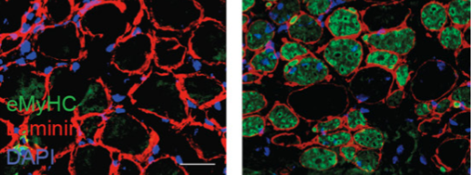