Feeds:
Posts
Comments

Posts Tagged ‘Biotechnology’


The Vibrant Philly Biotech Scene: Recent Happenings & Deals

Curator: Stephen J. Williams, Ph.D.


As the office and retail commercial real estate market has been drying up since the COVID pandemic, commercial real estate developers in the Philadelphia area have been turning to the health science industry to suit their lab space needs.  This includes refurbishing old office space as well as new construction.

Gattuso secures $290M construction loan for life sciences building on Drexel campus

Source: https://www.bizjournals.com/philadelphia/news/2022/12/19/construction-loan-gattuso-drexel-life-sciences.html?utm_source=st&utm_medium=en&utm_campaign=BN&utm_content=pl&ana=e_pl_BN&j=30034971&senddate=2022-12-20


By Ryan Mulligan  –  Reporter, Philadelphia Business Journal

Dec 19, 2022

Gattuso Development Partners and Vigilant Holdings of New York have secured a $290 million construction loan for a major life sciences building set to be developed on Drexel University’s campus.

The funding comes from Houston-based Corebridge Financial, with an additional equity commitment from Boston-based Baupost Group, which is also a partner on the project. JLL’s Capital Markets group arranged the loan.

Plans for the University City project at 3201 Cuthbert St. carry a price tag of $400 million. The 11-story building will total some 520,000 square feet, making it the largest life sciences research and lab space in the city when it comes online.

The building at 3201 Cuthbert will rise on what had served as a recreation field used by Drexel and is located next to the Armory. Gattuso Development, which will lease the parcel from Drexel, expects to to complete the project by fall 2024. Robert A.M. Stern Architects designed the building.


A rendering of a $400 million lab and research facility Drexel University and Gattuso Development Partners plan to build at 3201 Cuthbert St. in Philadelphia.

Enlarge

A rendering of a $400 million lab and research facility Drexel University and Gattuso Development Partners plan to build at 3201 Cuthbert St. in Philadelphia.

The building is 45% leased by Drexel and SmartLabs, an operator of life sciences labs. Drexel plans to occupy about 60,000 square feet, while SmartLabs will lease two floors totaling 117,000 square feet.

“We believe the project validates Philadelphia’s emergence as a global hub for life sciences research, and we are excited to begin construction,” said John Gattuso, the co-founder and president of Philadelphia-based Gattuso Development.

Ryan Ade, Brett Segal and Christopher Peck of JLL arranged the financing.

The project is another play in what amounts to an arms race for life sciences space and tenants in University City. Spark Therapeutics plans to build a $575 million, 500,000-square-foot gene therapy manufacturing plant on Drexel’s campus. One uCity Square, a $280 million, 400,000-square-foot life sciences building, was recently completed at 38th and Market streets. At 3151 Market St., a $307 million, 417,000-square-foot life sciences building is proposed as part of the Schuylkill Yards development.

Tmunity CEO Usman Azam departing to lead ‘stealth’ NYC biotech firm


By John George  –  Senior Reporter, Philadelphia Business Journal

Feb 7, 2022

The CEO of one of Philadelphia’s oldest cell therapy companies is departing to take a new job in the New York City area.

Usman “Oz” Azam, who has been CEO of Tmunity Therapeutics since 2016, will lead an unnamed biotechnology company currently operating in stealth mode.

In a posting on his LinkedIn page, Azam said, “After a decade immersed in cell therapies and immuno-oncology, I am now turning my attention to a new opportunity, and will be going back to where I started my life sciences career in neurosciences.”

Tmunity, a University of Pennsylvania spinout, is looking to apply CAR T-cell therapy, which has proved to be successful in treating liquid cancers, for the treatment of solid tumors.

Last summer, Tmunity suspended clinical testing of its lead cell therapy candidate targeting prostate cancer after two patients in the study died. Azam, in an interview with the Business Journal in June, said the company, which had grown to about 50 employees since its launch in 2015, laid off an undisclosed number of employees as a result of the setback.

Azam said on LinkedIn he is still a big believer in CAR T-cell therapy, noting Tmunity co-founder Dr. Carl June and his colleagues at Penn just published in Nature the 10-year landmark clinical outcomes study with the first CD19 CAR-T patients and programs.

“It’s just the beginning,” he stated. “I’m excited about the prospect of so many new cell- and gene-based therapies emerging in the next five to 10 years to tackle many solid and liquid tumors, and I hope we all continue to see the remarkable impact this makes on patients and families around the world.”

Azam could not be reached for comment Monday. Tmunity has engaged a search firm to identify his successor.

Tmunity, which is based in Philadelphia, has its own manufacturing operations in East Norriton. Tmunity’s founders include June and fellow Penn cell therapy pioneer Bruce Levine, who led the development of a CAR T-cell therapy now marketed by Novartis as Kymriah, a treatment for certain types of blood cancers.

In therapy using CAR-T cells, a patient’s T cells — part of their immune system — are removed and genetically modified in the laboratory. After they are re-injected into a patient, the T cells are better able to attack and destroy tumors. CAR is an acronym for chimeric antigen receptor. Chimeric antigen receptors are receptor proteins that have been engineered to give T cells their improved ability to target tumors.

Source: https://www.bizjournals.com/philadelphia/news/2022/02/07/tmunity-therapeutics-philadelphia-cell-azam-oz.html?utm_source=st&utm_medium=en&utm_campaign=BN&utm_content=pl&ana=e_pl_BN&j=30034971&senddate=2022-12-20


PIDC names U.S. Department of Treasury veteran, Philadelphia native as next president

 
By   –  Reporter, Philadelphia Business Journal


The Philadelphia Industrial Development Corp. has tapped U.S. Department of Treasury veteran Jodie Harris to be its next president.

Harris succeeds Anne Bovaird Nevins, who spent 15 years in the organization and took over as president in January 2020 before stepping down at the end of last year. Executive Vice President Sam Rhoads has been interim president.

Harris, a Philadelphia native who currently serves as director of the Community Development Financial Institutions Fund for the Department of Treasury, was picked after a regional and national search and will begin her tenure as president on June 1. She becomes the 12th head of PIDC and the first African-American woman to lead the organization.

PIDC is a public-private economic development corporation founded by the city and the Chamber of Commerce for Greater Philadelphia in 1958. It mainly uses industrial and commercial real estate projects to attract jobs, foster business opportunities and spur overall community growth. The organization has spurred over $18.5 billion in financing across its 65 years.

PIDC has its hand in development projects spanning the city, including master planning roles in expansive campuses like the Philadelphia Navy Yard and the Lower Schuylkill Biotech Campus in Southwest Philadelphia.

In a statement, Harris said that it is “a critical time for Philadelphia’s economy.”

“I’m especially excited for the opportunity to lead such an important and impactful organization in my hometown of Philadelphia,” Harris said. “As head of the CDFI Fund, I know first-hand what it takes to drive meaningful, sustainable, and equitable economic growth, especially in historically underserved communities.”

Harris is a graduate of the University of Maryland and received an MBA and master of public administration from New York University. In the Treasury Department, Harris’ most recent work aligns with PIDC’s economic development mission. At the Community Development Financial Institutions Fund, she oversaw a $331 million budget, mainly comprised of grant and administrative funding for various economic programs. Under Harris’ watch, the fund distributed over $3 billion in pandemic recovery funding, its highest level of appropriated grants ever.

Harris has been a part of the Treasury Department for 15 years, including as director of community and economic development policy.

In addition to government work, Harris has previously spent time in the private, academia and nonprofit sectors. In the beginning of her career, Harris worked at Meridian Bank and Accenture before turning to become a social and education policy researcher at New York University. She also spent two years as president of the Urban Business Assistance Corporation in New York.

Mayor Jim Kenney said that Philadelphia is “poised for long-term growth” and Harris will help drive it.

Source: https://www.bizjournals.com/philadelphia/news/2023/02/23/pidc-names-next-president-treasury.html 

$250M life sciences conversion planned for Philadelphia’s historic Quartermaster site
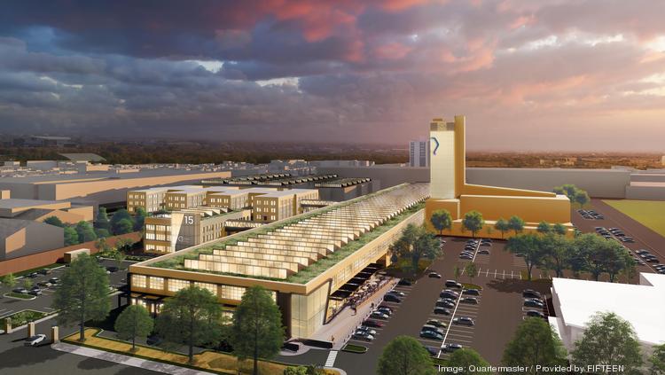
 
By   –  Reporter, Philadelphia Business Journal

Listen to this article     3 min

Real estate company SkyREM plans to spend $250 million converting the historic Quartermaster site in South Philadelphia to a life sciences campus with restaurants and a hotel.

The redevelopment would feature wet and dry lab space for research, development and bio-manufacturing.

The renamed Quartermaster Science + Technology Park is near the southwest corner of Oregon Avenue and South 20th Street in the city’s Girard Estates neighborhood. It’s east of the Quartermaster Plaza retail center, which sold last year for $100 million.

The 24-acre campus is planned to have six acres of green space, an Aldi grocery store opening by March and already is the headquarters for Indego, the bicycle share program in Philadelphia.

Six buildings totaling 1 million square feet of space would be used for research and development labs. There’s 500,000 square feet of vacant space available for life sciences and high technology companies with availabilities as small as 1,000 square feet up to 250,000 square feet contiguous. There’s also 150,000 square feet of retail space available.

The office park has 200,000 square feet already occupied by tenants. The Philadelphia Job Corps Center and Delaware Valley Intelligence Center are tenants at the site.
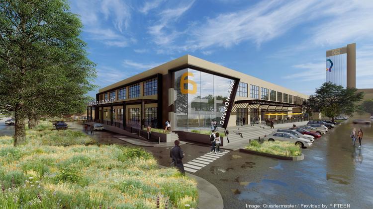
The campus was previously used by the military as a place to produce clothing, footwear and personal equipment during World War I and II. The clothing factory closed in 1994. The Philadelphia Quartermaster Depot was listed on the National Register of Historic Places in 2010.

“We had a vision to preserve the legacy of this built-to-last historic Philadelphia landmark and transform it to create a vibrant space where the best and brightest want to innovate, collaborate, and work,” SkyREM CEO and Founder Alex Dembitzer said in a statement.

SkyREM, a real estate investor and developer, has corporate offices in New York and Philadelphia. The company acquired the site in 2001.

Vered Nohi, SkyREM’s regional executive director of new business development, called the redevelopment “transformational” for Philadelphia.


Quartermaster would join a wave of new life sciences projects being developed in the surrounding area and across the region.

The site is near both interstates 76 and 95 and is about 2 miles north of the Philadelphia Navy Yard, which has undergone a similar transformation from a military hub to a major life sciences and mixed-use redevelopment project. The Philadelphia Industrial Development Corp. is also in the process of selecting a developer to create a massive cell and gene therapy manufacturing complex across two sites totaling about 40 acres on Southwest Philadelphia’s Lower Schuylkill riverfront.

At 34th Street and Grays Ferry Avenue, the University of Pennsylvania is teaming with Longfellow Real Estate Partners on proposed a $365 million, 455,000-square-foot life sciences and biomanufacturing building at Pennovation Works.
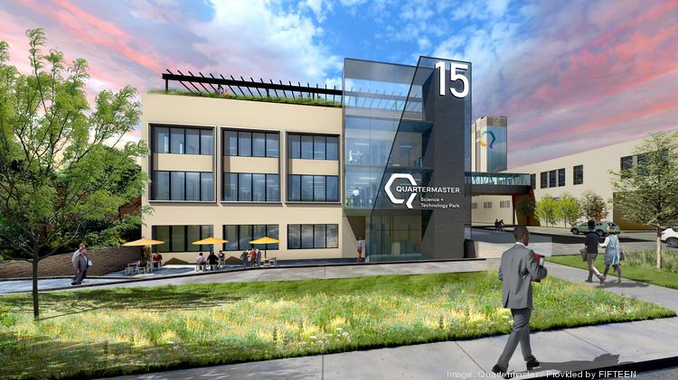
 

SkyREM is working with Maryland real estate firm Scheer Partners to lease the science and technology space. Philadelphia’s MPN Realty will handle leasing of the retail space. Architecture firm Fifteen is working on the project’s design.

Scheer Partners Senior Vice President Tim Conrey said the Quartermaster conversion will help companies solve for “speed to market” as demand for life science space in the region has been strong.
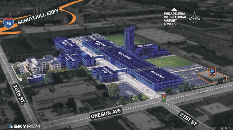
Brandywine pauses new spec office development, continues to bet big on life sciences
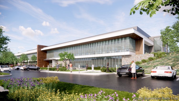
By   –  Reporter, Philadelphia Business Journal


Brandywine Realty Trust originally planned to redevelop a Radnor medical office into lab and office space, split 50-50 between the two uses.

After changes in demand for lab and office space, Brandywine (NYSE: BDN) recently completed the 168,000-square-foot, four-story building at 250 King of Prussia Road in Radnor fully for life sciences.

“The pipeline is now 100% life sciences, which, while requiring more capital, is also generating longer term leases at a higher return on cost,” Brandywine CEO Jerry Sweeney of the project said during the company’s fourth-quarter earnings call on Thursday.

At the same time, Brandywine is holding off on developing new office buildings unless it has a tenant lined up in advance.

The shift reflects how Philadelphia-based Brandywine continues to lean into — and bet big — on life sciences.

Brandywine is the city’s largest owner of trophy office buildings and has several major development projects in the works. The company is planning to eventually develop 3 million square feet of life sciences space. For now, 800,000 square feet of life sciences space is under development, including a 12-story, 417,000-square-foot life sciences building at 3151 Market St. and a 29-story building with 200,000 square feet of life sciences space at 3025 John F. Kennedy Blvd. Both are part of the multi-phase Schuylkill Yards project underway near 30th Street Station in University City.

Once its existing projects are completed, Brandywine would have 800,000 square feet of life sciences space, making up 8% of its portfolio.Sweeney said the company wants to grow that figure to 21%.

Brandywine is developing a 145,000-square-foot, build-to-suit office building at 155 King of Prussia Road in Radnor for Arkema, a France-based global supplier of specialty materials. The building will be Arkema’s North American headquarters. Construction began in January and is scheduled to be completed in late 2024.

Brandywine reported that since November it raised over $705 million through fourth-quarter asset sales, an unsecured bond transaction and a secured loan. The company has “complete availability” on its $600 million unsecured line of credit, Sweeney said.

Brandywine sold a 95% leased, 86,000-square-foot office building at 200 Barr Harbor Drive in West Conshohocken for $30.5 million. The company also sold its 50% ownership interest in the 1919 Market joint venture for $83.2 million to an undisclosed buyer. 1919 Market St. is a 29-story building with apartments, office and commercial space. Brandywine co-developed the property with LCOR and the California State Teacher’s Retirement System.

Brandywine declined to comment and LCOR could not be reached.

Brandywine’s core portfolio is 91% leased.

The project at 250 King of Prussia Road cost $103.7 million and was recently completed. The renovation included 12-foot high floor-to-ceiling glass on the second floor, a new roof, lobby, elevator core, common area with a skylight and an added structured parking deck.

Located in the Radnor Life Science Center, a new campus with nearly 1 million square feet of lab, research and office space, Sweeney said it’s a “magnet” for biotech companies. Avantor, a global manufacturer and distributor of life sciences products, is headquartered in the complex.


Sweeney said Brandywine is “very confident” demand will stay strong for life sciences in Radnor. The building at 250 King of Prussia Road is projected to be fully leased by early 2024.

“Larger users we’re talking to, they just tend to take a little bit more time than we would like as they go through technical requirements and space planning requirements,” Sweeney said.

While Brandywine is aiming to increase its life sciences footprint, the company is being selective about what it builds next. The company may steer away from developments other than life sciences. The Schuylkill Yards project, for example, features a significant life sciences portion in University City.

“Other than fully leased build-to-suit opportunities, our future development starts are on hold,” Sweeney said, “pending more leasing on the existing joint venture pipeline and more clarity on the cost of debt capital and cap rates.”


Brandywine said about 70% to 75%of suburban tenants have returned to offices while that number has been around 50% in Philadelphia. At this point, though, it hasn’t yet affected demand when leasing space. Some tenants, for example, have moved out of the city while others have moved in.

In the fourth quarter, Brandywine had $55.7 million funds from operations, or 32 cents per share. That’s down from $60.4 million, or 35 cents per share, in the fourth quarter of 2021. Brandywine generated $129 million in revenue in the fourth quarter, up slightly from $125.5 in the year-ago period.

Brandywine stock is up 6.4% since the start of the year to $6.70 per share on Monday afternoon.

Many of Brandywine’s properties are in desirable locations, which have seen demand remain strong despite challenges facing offices, on par with industry trends.

Brandywine’s 12-story, 417,000-square-foot building at 3151 Market St. is on budget for $308 million and on schedule to be completed in the second quarter of 2024. Sweeney said Brandywine anticipates entering a construction loan in the second half of 2023, which would help complete the project. The building, being developed along with a global institutional investor,would be used for life sciences, innovation and office space as part of the larger Schuylkill Yards development in University City.

The company’s 29-story building at 3025 John F. Kennedy Blvd. with 200,000 square feet of life sciences space and 326 luxury apartments, is also on budget, costing $287.3 million, and on time, eyeing completion in the third quarter of this year.

Source: https://www.bizjournals.com/philadelphia/news/2023/02/06/brandywine-realty-life-sciences-development.html

Read Full Post »

Live Notes, Real Time Conference Coverage 2020 AACR Virtual Meeting April 28, 2020 Symposium: New Drugs on the Horizon Part 3 12:30-1:25 PM

Reporter: Stephen J. Williams, PhD

New Drugs on the Horizon: Part 3
Introduction

Andrew J. Phillips, C4 Therapeutics

  • symposium brought by AACR CICR and had about 30 proposals for talks and chose three talks
  • unfortunately the networking event is not possible but hope to see you soon in good health

ABBV-184: A novel survivin specific T cell receptor/CD3 bispecific therapeutic that targets both solid tumor and hematological malignancies

Edward B Reilly
AbbVie Inc. @abbvie

  • T-cell receptors (TCR) can recognize the intracellular targets whereas antibodies only recognize the 25% of potential extracellular targets
  • survivin is expressed in multiple cancers and correlates with poor survival and prognosis
  • CD3 bispecific TCR to survivn (Ab to CD3 on T- cells and TCR to survivin on cancer cells presented in MHC Class A3)
  • ABBV184  effective in vivo in lung cancer models as single agent;
  • in humanized mouse tumor models CD3/survivin bispecific can recruit T cells into solid tumors; multiple immune cells CD4 and CD8 positive T cells were found to infiltrate into tumor
  • therapeutic window as measured by cytokine release assays in tumor vs. normal cells very wide (>25 fold)
  • ABBV184 does not bind platelets and has good in vivo safety profile
  • First- in human dose determination trial: used in vitro cancer cell assays to determine 1st human dose
  • looking at AML and lung cancer indications
  • phase 1 trial is underway for safety and efficacy and determine phase 2 dose
  • survivin has very few mutations so they are not worried about a changing epitope of their target TCR peptide of choice

The discovery of TNO155: A first in class SHP2 inhibitor

Matthew J. LaMarche
Novartis @Novartis

  • SHP2 is an intracellular phosphatase that is upstream of MEK ERK pathway; has an SH2 domain and PTP domain
  • knockdown of SHP2 inhibits tumor growth and colony formation in soft agar
  • 55 TKIs there are very little phosphatase inhibitors; difficult to target the active catalytic site; inhibitors can be oxidized at the active site; so they tried to target the two domains and developed an allosteric inhibitor at binding site where three domains come together and stabilize it
  • they produced a number of chemical scaffolds that would bind and stabilize this allosteric site
  • block the redox reaction by blocking the cysteine in the binding site
  • lead compound had phototoxicity; used SAR analysis to improve affinity and reduce phototox effects
  • was very difficult to balance efficacy, binding properties, and tox by adjusting stuctures
  • TNO155 is their lead into trials
  • SHP2 expressed in T cells and they find good combo with I/O with uptick of CD8 cells
  • TNO155 is very selective no SHP1 inhibition; SHP2 can autoinhibit itself when three domains come together and stabilize; no cross reactivity with other phosphatases
  • they screened 1.5 million compounds and got low hit rate so that is why they needed to chemically engineer and improve on the classes they found as near hits

Closing Remarks


Xiaojing Wang
Genentech, Inc. @genentech

Follow on Twitter at:

@pharma_BI

@AACR

@CureCancerNow

@pharmanews

@BiotechWorld

@HopkinsMedicine

#AACR20

Read Full Post »

Reporter: Gail S. Thornton


From The Wall Street Journal (www.wsj.com)

Published January 9, 2019

Health-Care CEOs Outline Strategies at J.P. Morgan Conference

Chiefs at Johnson & Johnson, CVS discuss what’s next on a range of industry issues

One of the biggest health conferences of the year for investors, the J.P. Morgan Health-Care Conference, is taking place this week in San Francisco. Here are some of the hot topics covered at the four-day event, which wraps up Thursday.

BioMarin Mulls Payment Plans

BioMarin Pharmaceutical Inc. CEO Jean-Jacques Bienaimé said he would consider pursuing installment payment arrangements for the biotech’s experimental gene therapy for hemophilia. At the conference, Mr. Bienaimé told the Wall Street Journal that the one-time infusion, Valrox, is likely to cost in the millions because studies have shown it can eliminate bleeding episodes in patients, and current hemophilia treatments taken chronically can cost millions over several years. “We’re not trying to charge more than existing therapies,” he said. “We want to offer a better treatment at the same or lower cost.”

Johnson & Johnson Warns on Pricing

As politicians hammer drug prices, Johnson & Johnson CEO Alex Gorsky suggested companies need to police themselves. At the conference, Mr. Gorsky told investors that drug companies should price drugs reasonably and be transparent. “If we don’t do this as an industry, I think there will be other alternatives that will be more onerous for us,” Mr. Gorsky says. Some drugmakers pulled back from price increases in mid-2018 amid heightened political scrutiny, but prices went up for many drugs at the start of 2019.

Marijuana-Derived Drugs Show Promise


CVS Discusses New Stores

CVS Health Corp. Chief Executive Larry Merlo began showing initial concepts the company will be testing as it begins piloting new models of its drugstores that incorporate its Aetna combination. The first new test store will open next month in Houston, he told investors, and it will include expanded health-care services including a new concierge who will help patients with questions. 

Aetna Savings On the Way

Mr. Merlo also spelled out when the company will achieve the initial $750 million in synergies it has promised from the CVS-Aetna deal. In the first quarter, he said the company will see benefits from consolidating corporate functions. Savings from procurement and aligning lists of covered drugs should be seen in the first half, he says. Medical-cost savings will start affecting results toward the end of the year, he noted. 

Lilly Cuts Price

Drugmaker Eli Lilly & Co. expects average net US pricing for its drugs–after rebates and discounts–to decline in the low- to mid-single digits on a percentage basis this year, Chief Financial Officer Josh Smiley told the Journal. Lilly’s net prices had risen during the first half of 2018, but dropped in the third quarter as the company took a “restrained approach,” Mr. Smiley said. Lilly, which hasn’t yet reported fourth-quarter results, took some list price increases for cancer drugs in late December but hasn’t raised prices in the new year, he said.

Peter Loftus at peter.loftus@wsj.com and Anna Wilde Mathews at anna.mathews@wsj.com

Read Full Post »

NIH SBIR Funding Early Ventures: September 26, 2018 sponsored by Pennovation

Stephen J. Williams PhD, Reporter

Penn Center for Innovation (Pennovation) sponsored a “Meet with NCI SBIR” program directors at University of Pennsylvania Medicine Smilow Center for Translational Research with a presentation on advice on preparing a successful SBIR/STTR application to the NCI as well as discussion of NCI SBIR current funding opportunities.   Time was allotted in the afternoon for one-on-one discussions with NCI SBIR program directors.

To find similar presentations and one-on-one discussions with NCI/SBIR program directors in an area nearest to you please go to their page at:

https://sbir.cancer.gov/newsevents/events

For more complete information on the NCI SBIR and STTR programs please go to their web page at: https://sbir.cancer.gov/about

A few notes from the meeting are given below:

  • In 2016 the SBIR/STTR 2016 funded $2.5 billion (US) of early stage companies; this is compared to the $6.6 billion invested in early  stage ventures by venture capital firms so the NCI program is very competitive with alternate sources of funding
  • It was stressed that the SBIR programs are flexible as far as ownership of a company; SBIR allows now that >50% of the sponsoring company can be owned by other ventures;  In addition they are looking more favorably on using outside contractors and giving leeway on budgetary constraints so AS THEY SUGGEST ALWAYS talk to the program director about any questions you may have well before (at least 1 month) you submit. More on eligibility criteria is found at: https://sbir.cancer.gov/about/eligibilitycriteria
  • STTR should have strong preliminary data since more competitive; if don’t have enough go for  an R21 emerging technologies grant which usually does not require preliminary data
  • For entities outside the US need a STRONG reason for needing to do work outside the US

Budget levels were discussed as well as  the waiver program, which allows for additional funds to be requested based on criteria set by NCI (usually for work that is deemed high priority or of a specialized nature which could not be covered sufficiently under the standard funding limits) as below:

Phase I: 150K standard but you can get waivers for certain work up to 300K

Phase II: 1M with waiver up to 2M

Phase IIB waiver up to 4M

You don’t need to apply for the waiver but grant offices may suggest citing a statement requesting a waiver as review panels will ask for this information

Fast Track was not discussed in the presentation but for more information of the Fast Track program please visit the website  

NCI is working hard to cut review times to 7 months between initial review to funding however at beginning of the year they set pay lines and hope to fund 50% of the well scored grants

NCI SBIR is a Centralized system with center director and then program director with specific areas of expertise: Reach out to them

IMAT Program and Low-Resource Setting new programs more suitable for initial studies and also can have non US entities

Phase IIB Bridge funding to cross “valley of death” providing up to 4M for 2-3 years: most were for drug/biological but good amount for device and diagnostics


Also they have announced administrative supplements for promoting diversity within a project: can add to the budget

FY18 Contracts Areas

3 on biotherapies

2 imaging related

2 on health IT

4 on radiation therapy related: NOTE They spent alot of time discussing the contracts centered on radiation therapy and seems to be an area of emphasis of the NCI SBIR program this year

4 other varied topics


Breakdown of funding

>70% of NCI SBIR budget went to grants (for instance Omnibus grants); about 20-30% for contracts; 16% for phase I and 34 % for phase II ;

ALSO the success rate considerably higher for companies that talk to the program director BEFORE applying than not talking to them; also contracts more successful than Omnibus applications

Take Advantage of these useful Assistance Programs through the NIH SBIR Program (Available to all SBIR grantees)

NICHE ASSESSMENT Program

From the NCI SBIR website:

The Niche Assessment Program is designed to help small businesses “jump start” their commercialization efforts. All active HHS (NIH, CDC, FDA) SBIR/STTR Phase I awardees and Phase I Fast-Track awardees (by grant or contract) are eligible to apply. Registration is on a first-come, first-serve basis!

The Niche Assessment Program provides market insight and data that can be used to help small businesses strategically position their technology in the marketplace. The results of this program can help small businesses develop their commercialization plans for their Phase II application, and be exposed to potential partners. Services are provided by Foresight Science & Technology of Providence, RI.

Technology Niche Analyses® (TNA®) are provided by Foresight, for one hundred and seventy-five (175), HHS SBIR/STTR Phase I awardees. These analyses assess potential applications for a technology and then for one viable application, it provides an assessment of the:

  1. Needs and concerns of end-users;
  2. Competing technologies and competing products;
  3. Competitive advantage of the SBIR/STTR-developed technology;
  4. Market size and potential market share (may include national and/or global markets);
  5. Barriers to market entry (may include but is not limited to pricing, competition, government regulations, manufacturing challenges, capital requirements, etc.);
  6. Market drivers;
  7. Status of market and industry trends;
  8. Potential customers, licensees, investors, or other commercialization partners; and,
  9. The price customers are likely to pay.

Commercialization Acceleration Program  (CAP)

From the NIH SBIR website:

NIH CAP is a 9-month program that is well-regarded for its combination of deep domain expertise and access to industry connections, which have resulted in measurable gains and accomplishments by participating companies. Offered since 2004 to address the commercialization objectives of companies across the spectrum of experience and stage, 1000+ companies have participated in the CAP. It is open only to HHS/NIH SBIR/STTR Phase II awardees, and 80 slots are available each year. The program enables participants to establish market and customer relevance, build commercial relationships, and focus on revenue opportunities available to them.

I-Corps Program

The I-Corps program provides funding, mentoring, and networking opportunities to help commercialize your promising biomedical technology. During this 8-week, hands-on program, you’ll learn how to focus your business plan and get the tools to bring your treatment to the patients who need it most.

Program benefits include:

  • Funding up to $50,000 to cover direct program costs
  • Training from biotech sector experts
  • Expanding your professional network
  • Building the confidence and skills to create a comprehensive business model
  • Gaining years of entrepreneurial skills in only weeks.


ICORPS is an Entrepreneurial Program (8 week course) to go out talk to customers, get assistance with business models, useful resource which can guide the new company where they should focus on for the commercialization aspect

THE NCI Applicant Assistance Program (AAP)

The SBIR/STTR Applicant Assistance Program (AAP) is aimed at helping eligible small R&D businesses and individuals successfully apply for Phase I SBIR/STTR funding from the National Cancer Institute (NCI), National Institute for Neurological Disorders and Stroke (NINDS), National Heart, Lung and Blood Institute (NHLBI). Participation in the AAP will be funded by the NCI, NINDS, and NHLBI with NO COST TO PARTICIPANTS. The program will include the following services:

  • Needs Assessment/Small Business Mentoring
  • Phase I Application Preparation Support
  • Application Review
  • Team/Facilities Development
  • Market Research
  • Intellectual Property Consultation

For more details about the program, please refer to NIH Notice NOT-CA-18-072.


These programs are free for first time grant applicants and must not have been awarded previous SBIR

Peer Learning Webinar Series goal to improve peer learning .Also they are starting to provide Regulatory Assistance (see below)

NIH also provides Mentoring programs for CEOS and C level

Application tips

  1. Start early: and obtain letters of collaboration
  2. Build a great team: PI multi PI, consider other partners to fill gaps (academic, consultants, seasoned entrepreneurs (don’t need to be paid)
  3. They will pre review 1 month before due date, use NIH Project Reporter to view previous funded grants
  4. Specify study section in SF to specify areas of expertise for review
  5. Specific aims are very important; some of the 20 reviewers focus on this page (describes goals and milestones as well; spend as much time on this page as the rest of the application
  6. Letters of support from KOLs are important to have; necessary from consultants and collaborators; helpful from clinicians
  7. Have a phase II commercialization plan
  8. Note for non US clinical trials:  They will not fund nonUS clinical trials; the company must have a FWA
  9. SBIR budgets defined by direct costs; can request a 7% fee as an indirect cost; and they have a 5,000 $ technical assistance program like regulatory consultants but if requested can’t participate in NIH technical assistance programs so most people don’t apply for TAP


  • They are trying to change the definition of innovation as also using innovative methods (previously reviewers liked tried and true methodology)

10.  before you submit solicit independent readers

NCI SBIR can be found on Twitter @NCIsbir ‏

Discussion with Monique Pond, Ph.D. on Establishment of a Regulatory Assistance Program for NCI SBIR

I was able to sit down with Dr. Monique Pond,  AAAS Science & Technology Policy Fellow, Health Scientist within the NCI SBIR Development Center to discuss the new assistance program in regulatory affairs she is developing for the NCI SBIR program.  Dr Pond had received her PhD in chemistry from the Pennsylvania State University, completed a postdoctoral fellow at NIST and then spent many years as a regulatory writer and consultant in the private sector.  She applied through the AAAS for this fellowship and will bring her experience and expertise in regulatory affairs from the private sector to the SBIR program. Dr. Pond discussed the difficulties that new ventures have in formulating regulatory procedures for their companies, the difficulties in getting face time with FDA regulators and helping young companies start thinking about regulatory issues such as pharmacovigilence, oversight, compliance, and navigating the complex regulatory landscape.

In addition Dr. Pond discussed the AAAS fellowship program and alternative career paths for PhD scientists.


A formal interview will follow on this same post.


Other articles on this OPEN ACCESS JOURNAL on Funding for Startups and Early Ventures are given below:


Mapping Medical Device Startups Across The Globe per Funding Criteria

Funding Oncorus’s Immunotherapy Platform: Next-generation Oncolytic Herpes Simplex Virus (oHSV) for Brain Cancer, Glioblastoma Multiforme (GBM)


Funding Opportunities for Cancer Research


Team Profile: DrugDiscovery @LPBI Group – A BioTech Start Up submitted for Funding Competition to MassChallenge Boston 2016 Accelerator


A Message from Faculty Director Lee Fleming on Latest Issue of Crowdfunding; From the Fung Institute at Berkeley


PROTOCOL for Drug Screening of 3rd Party Intellectual Property Presented for Funding Representation


Foundations as a Funding Source


The Bioscience Crowdfunding Environment: The Bigger Better VC?


Read Full Post »

Recent Research On SMAD4 In Pancreatic Cancer

Curator: David Orchard-Webb, PhD


Deleted in Pancreatic Cancer, locus 4 (DPC4) officially known as SMAD4 is a component of the Transforming Growth Factor Beta (TGFß) pathway with tumour suppressive properties. As its name suggests it is frequently lost in pancreatic cancer, although through a variety of mechanisms in addition to gene deletion. The loss of SMAD4 is important in the progression of pancreatic intraepithelial neoplasia (PanIN) towards pancreatic ductal adenocarcinoma (PDAC). The expression of SMAD4 can suppress metastasis, angiogenesis, and cancer stem-like cell generation. SMAD4 can promote cancer cell apoptosis through a recently described mechanism involving a lethal epithelial to mesenchymal transition (EMT). SMAD4 status has a predictive role in pancreatic cancer personalised medicine. This curation categorises recent publications of note regarding SMAD4.


Role of SMAD4 in neoplastic progression towards PDAC


Garcia-Carracedo, Dario, Chih-Chieh Yu, Nathan Akhavan, Stuart A. Fine, Frank Schönleben, Naoki Maehara, Dillon C. Karg, et al. ‘Smad4 Loss Synergizes with TGFα Overexpression in Promoting Pancreatic Metaplasia, PanIN Development, and Fibrosis’. Edited by Ilse Rooman. PLOS ONE 10, no. 3 (24 March 2015): e0120851. doi:10.1371/journal.pone.0120851.


Norris, A M, A Gore, A Balboni, A Young, D S Longnecker, and M Korc. ‘AGR2 Is a SMAD4-Suppressible Gene That Modulates MUC1 Levels and Promotes the Initiation and Progression of Pancreatic Intraepithelial Neoplasia’. Oncogene 32, no. 33 (15 August 2013): 3867–76. doi:10.1038/onc.2012.394.


Leung, Lisa, Nikolina Radulovich, Chang-Qi Zhu, Dennis Wang, Christine To, Emin Ibrahimov, and Ming-Sound Tsao. ‘Loss of Canonical Smad4 Signaling Promotes KRAS Driven Malignant Transformation of Human Pancreatic Duct Epithelial Cells and Metastasis’. Edited by Hidayatullah G Munshi. PLoS ONE 8, no. 12 (27 December 2013): e84366. doi:10.1371/journal.pone.0084366.


Mechanism of SMAD4 deactivation


Xia, Xiang, Kundong Zhang, Gang Cen, Tao Jiang, Jun Cao, Kejian Huang, Chen Huang, Qian Zhao, and Zhengjun Qiu. ‘MicroRNA-301a-3p Promotes Pancreatic Cancer Progression via Negative Regulation of SMAD4’. Oncotarget 6, no. 25 (28 August 2015): 21046–63. doi:10.18632/oncotarget.4124.


Murphy, Stephen J., Steven N. Hart, Geoffrey C. Halling, Sarah H. Johnson, James B. Smadbeck, Travis Drucker, Joema Felipe Lima, et al. ‘Integrated Genomic Analysis of Pancreatic Ductal Adenocarcinomas Reveals Genomic Rearrangement Events as Significant Drivers of Disease’. Cancer Research 76, no. 3 (1 February 2016): 749–61. doi:10.1158/0008-5472.CAN-15-2198.


Sawai, Yugo, Yuzo Kodama, Takahiro Shimizu, Yuji Ota, Takahisa Maruno, Yuji Eso, Akira Kurita, et al. ‘Activation-Induced Cytidine Deaminase Contributes to Pancreatic Tumorigenesis by Inducing Tumor-Related Gene Mutations’. Cancer Research 75, no. 16 (15 August 2015): 3292–3301. doi:10.1158/0008-5472.CAN-14-3028.


Demagny, Hadrien, and Edward M De Robertis. ‘Point Mutations in the Tumor Suppressor Smad4/DPC4 Enhance Its Phosphorylation by GSK3 and Reversibly Inactivate TGF-β Signaling’. Molecular & Cellular Oncology 3, no. 1 (2 January 2016): e1025181. doi:10.1080/23723556.2015.1025181.


Foster, David. ‘BxPC3 Pancreatic Cancer Cells Express a Truncated Smad4 Protein upon PI3K and mTOR Inhibition’. Oncology Letters, 28 January 2014. doi:10.3892/ol.2014.1833.


Hao, Jun, Shuyu Zhang, Yingqi Zhou, Cong Liu, Xiangui Hu, and Chenghao Shao. ‘MicroRNA 421 Suppresses DPC4/Smad4 in Pancreatic Cancer’. Biochemical and Biophysical Research Communications 406, no. 4 (25 March 2011): 552–57. doi:10.1016/j.bbrc.2011.02.086.


SMAD4 effects on cell motility


Zhang, Xueying, Junxia Cao, Yujun Pei, Jiyan Zhang, and Qingyang Wang. ‘Smad4 Inhibits Cell Migration via Suppression of JNK Activity in Human Pancreatic Carcinoma PANC‑1 Cells’. Oncology Letters, 7 April 2016. doi:10.3892/ol.2016.4427.


Kang, Ya ’an, Jianhua Ling, Rei Suzuki, David Roife, Xavier Chopin-Laly, Mark J. Truty, Deyali Chatterjee, et al. ‘SMAD4 Regulates Cell Motility through Transcription of N-Cadherin in Human Pancreatic Ductal Epithelium’. Edited by Neil A. Hotchin. PLoS ONE 9, no. 9 (29 September 2014): e107948. doi:10.1371/journal.pone.0107948.


Chen, Yu-Wen, Pi-Jung Hsiao, Ching-Chieh Weng, Kung-Kai Kuo, Tzu-Lei Kuo, Deng-Chyang Wu, Wen-Chun Hung, and Kuang-Hung Cheng. ‘SMAD4 Loss Triggers the Phenotypic Changes of Pancreatic Ductal Adenocarcinoma Cells’. BMC Cancer 14, no. 1 (2014): 1. https://bmccancer.biomedcentral.com/articles/10.1186/1471-2407-14-181.


SMAD4 effects on angiogenesis


Zhou, Zhichao, Juming Lu, Jingtao Dou, Zhaohui Lv, Xi Qin, and Jing Lin. ‘FHL1 and Smad4 Synergistically Inhibit Vascular Endothelial Growth Factor Expression’. Molecular Medicine Reports 7, no. 2 (February 2013): 649–53. doi:10.3892/mmr.2012.1202.


SMAD4 mediated repression of cancer stem-like cells


Hoshino, Yukari, Jun Nishida, Yoko Katsuno, Daizo Koinuma, Taku Aoki, Norihiro Kokudo, Kohei Miyazono, and Shogo Ehata. ‘Smad4 Decreases the Population of Pancreatic Cancer–Initiating Cells through Transcriptional Repression of ALDH1A1’. The American Journal of Pathology 185, no. 5 (2015): 1457–1470. http://www.sciencedirect.com/science/article/pii/S0002944015000802.


SMAD4 mediated growth inhibition/ apoptosis induction


David, Charles J., Yun-Han Huang, Mo Chen, Jie Su, Yilong Zou, Nabeel Bardeesy, Christine A. Iacobuzio-Donahue, and Joan Massagué. ‘TGF-β Tumor Suppression through a Lethal EMT’. Cell 164, no. 5 (February 2016): 1015–30. doi:10.1016/j.cell.2016.01.009.


Wang, Qi, Juanjuan Li, Wei Wu, Ruizhe Shen, He Jiang, Yuting Qian, Yanping Tang, et al. ‘Smad4-Dependent Suppressor Pituitary Homeobox 2 Promotes PPP2R2A-Mediated Inhibition of Akt Pathway in Pancreatic Cancer’. Oncotarget 7, no. 10 (8 March 2016): 11208–22. doi:10.18632/oncotarget.7158.


Poorly characterised targets of SMAD4


Fullerton, Paul T., Chad J. Creighton, and Martin M. Matzuk. ‘Insights Into SMAD4 Loss in Pancreatic Cancer From Inducible Restoration of TGF-β Signaling’. Molecular Endocrinology (Baltimore, Md.) 29, no. 10 (October 2015): 1440–53. doi:10.1210/me.2015-1102.


Li, Lei, Zhaoshen Li, Xiangyu Kong, Dacheng Xie, Zhiliang Jia, Weihua Jiang, Jiujie Cui, et al. ‘Down-Regulation of MicroRNA-494 via Loss of SMAD4 Increases FOXM1 and β-Catenin Signaling in Pancreatic Ductal Adenocarcinoma Cells’. Gastroenterology 147, no. 2 (August 2014): 485–497.e18. doi:10.1053/j.gastro.2014.04.048.


Drugs that restore SMAD4


Lin, Sheng-Zhang, Jin-Bo Xu, Xu Ji, Hui Chen, Hong-Tao Xu, Ping Hu, Liang Chen, et al. ‘Emodin Inhibits Angiogenesis in Pancreatic Cancer by Regulating the Transforming Growth Factor-Β/drosophila Mothers against Decapentaplegic Pathway and Angiogenesis-Associated microRNAs’. Molecular Medicine Reports 12, no. 4 (October 2015): 5865–71. doi:10.3892/mmr.2015.4158.


Predictive value of SMAD4 status in personalised medicine


Whittle, Martin C., Kamel Izeradjene, P. Geetha Rani, Libing Feng, Markus A. Carlson, Kathleen E. DelGiorno, Laura D. Wood, et al. ‘RUNX3 Controls a Metastatic Switch in Pancreatic Ductal Adenocarcinoma’. Cell 161, no. 6 (June 2015): 1345–60. doi:10.1016/j.cell.2015.04.048.


Boone, Brian A., Shirin Sabbaghian, Mazen Zenati, J. Wallis Marsh, A. James Moser, Amer H. Zureikat, Aatur D. Singhi, Herbert J. Zeh, and Alyssa M. Krasinskas. ‘Loss of SMAD4 Staining in Pre-Operative Cell Blocks Is Associated with Distant Metastases Following Pancreaticoduodenectomy with Venous Resection for Pancreatic Cancer’. Journal of Surgical Oncology 110, no. 2 (August 2014): 171–75. doi:10.1002/jso.23606.


Herman, Joseph M., Katherine Y. Fan, Aaron T. Wild, Laura D. Wood, Amanda L. Blackford, Ross C. Donehower, Manuel Hidalgo, et al. ‘Correlation of Smad4 Status With Outcomes in Patients Receiving Erlotinib Combined With Adjuvant Chemoradiation and Chemotherapy After Resection for Pancreatic Adenocarcinoma’. International Journal of Radiation Oncology*Biology*Physics 87, no. 3 (November 2013): 458–59. doi:10.1016/j.ijrobp.2013.06.2039.


Other Related Articles Published In This Open Access Online Journal Include The Following:


Pancreatic Cancer Modeling using Retrograde Viral Vector Delivery and IN-Vivo CRISPR/Cas9-mediated Somatic Genome Editing

Wnt/β-catenin Signaling [7.10]


Read Full Post »

3-D visualization of cancer cells, Volume 2 (Volume Two: Latest in Genomics Methodologies for Therapeutics: Gene Editing, NGS and BioInformatics, Simulations and the Genome Ontology), Part 1: Next Generation Sequencing (NGS)

3-D visualization of cancer cells

Larry H. Bernstein, MD, FCAP, Curator

LPBI


Cancer cells in 3D

What researchers miss on glass slides
February 22, 2016   http://www.kurzweilai.net/cancer-in-3-d


A spheroid of many lung cancer cells illustrates a diversity of behaviors. (credit: Welf and Driscoll et al./Developmental Cell)


Cancer cells don’t live on glass slides. Yet the vast majority of images related to cancer biology come from the cells being photographed on flat, two-dimensional surfaces — images sometimes used to draw conclusions about the behavior of cells that normally reside in a more complex environment.

Now a new high-resolution microscope, presented (open access) February 22 in Developmental Cell, makes it possible to visualize cancer cells in 3D and record how they are signaling to other parts of their environment — revealing previously unappreciated biology of how cancer cells survive and disperse within living things. Based on ”microenvironmental selective plane illumination microscopy” (meSPIM),  the new microscope is designed to image cells in microenvironments free of hard surfaces near the sample.

“There is clear evidence that the environment strongly affects cellular behavior — thus, the value of cell culture experiments on glass must at least be questioned,” says senior author Reto Fiolka, an optical scientist at theUniversity of Texas Southwestern Medical Center. “Our microscope is one tool that may bring us a deeper understanding of the molecular mechanisms that drive cancer cell behavior, since it enables high-resolution imaging in more realistic tumor environments.”

[+]

This image shows the extracted surfaces of two cancer cells. (Left) A lung cancer cell colored by actin intensity near the cell surface. Actin is a structural molecule that is integral to cell movement. (Right) A melanoma cell colored by PI3-kinase activity near the cell surface. PI3K is a signaling molecule that is key to many cell processes. (credit: Welf and Driscoll et al./Developmental Cell)

Hidden protrusions from cancer cells

In their study, Fiolka and colleagues, including co-senior author Gaudenz Danuser, and co-first authors Meghan Driscoll and Erik Welf, also of UT Southwestern, used their microscope to image different kinds of skin cancer cells from patients. They found that in a 3D environment (where cells normally reside), unlike a glass slide, multiple melanoma cell lines and primary melanoma cells (from patients with varied genetic mutations) form many small protrusions called blebs.

One hypothesis is that this blebbing may help the cancer cells survive or move around and could thus play a role in skin cancer cell invasiveness or drug resistance in patients.

[+]

This is a melanoma cell (red) embedded in a 3-D collagen matrix (white). A 100 x 100 x 100 μm cube is shown, with one corner cut away to show the interaction of the cell with the collagen. (credit: Welf and Driscoll et al./Developmental Cell)

The researchers say that this is a first step toward understanding 3D biology in tumor microenvironments. But since these kinds of images may be too complicated to interpret by the naked eye alone, the next step will be to develop powerful computer platforms to extract and process the information.

The microscope control software and image analytical code are freely available to the scientific community.

The authors were supported by the Cancer Prevention Research Institute of Texas and the National Institutes of Health.


Abstract of Quantitative Multiscale Cell Imaging in Controlled 3D Microenvironments

The microenvironment determines cell behavior, but the underlying molecular mechanisms are poorly understood because quantitative studies of cell signaling and behavior have been challenging due to insufficient spatial and/or temporal resolution and limitations on microenvironmental control. Here we introduce microenvironmental selective plane illumination microscopy (meSPIM) for imaging and quantification of intracellular signaling and submicrometer cellular structures as well as large-scale cell morphological and environmental features. We demonstrate the utility of this approach by showing that the mechanical properties of the microenvironment regulate the transition of melanoma cells from actin-driven protrusion to blebbing, and we present tools to quantify how cells manipulate individual collagen fibers. We leverage the nearly isotropic resolution of meSPIM to quantify the local concentration of actin and phosphatidylinositol 3-kinase signaling on the surfaces of cells deep within 3D collagen matrices and track the many small membrane protrusions that appear in these more physiologically relevant environments.

Read Full Post »

Hybrid lipid bioelectronic membranes

Larry H. Bernstein, MD, FCAP, Curator

LPBI


Hybrid solid-state chips and biological cells integrated at molecular level

Biological ion channels combine with solid-state transistors to create a new kind of hybrid bioelectronics. Imagine chips with dog-like capability to taste and smell, or even recognize specific molecules.
http://www.kurzweilai.net/hybrid-solid-state-chips-and-biological-cells-integrated-at-molecular-level
Illustration depicting a biocell attached to a CMOS integrated circuit with a membrane containing sodium-potassium pumps in pores. Energy is stored chemically in ATP molecules. When the energy is released as charged ions (which are then converted to electrons to power the chip at the bottom of the experimental device), the ATP is converted to ADP + inorganic phosphate. (credit: Trevor Finney and Jared Roseman/Columbia Engineering)

Columbia Engineering researchers have combined biological and solid-state components for the first time, opening the door to creating entirely new artificial biosystems.

In this experiment, they used a biological cell to power a conventional solid-state complementary metal-oxide-semiconductor (CMOS) integrated circuit. An artificial lipid bilayer membrane containing adenosine triphosphate (ATP)-powered ion pumps (which provide energy for cells) was used as a source of ions (which were converted to electrons to power the chip).

The study, led by Ken Shepard, Lau Family Professor of Electrical Engineering and professor of biomedical engineering at Columbia Engineering, was published online today (Dec. 7, 2015) in an open-access paper in Nature Communications.

How to build a hybrid biochip

Living systems achieve this functionality with their own version of electronics based on lipid membranes and ion channels and pumps, which act as a kind of “biological transistor.” Charge in the form of ions carry energy and information, and ion channels control the flow of ions across cell membranes.

Solid-state systems, such as those in computers and communication devices, use electrons; their electronic signaling and power are controlled by field-effect transistors.

To build a prototype of their hybrid system, Shepard’s team packaged a CMOS integrated circuit (IC) with an ATP-harvesting “biocell.” In the presence of ATP, the system pumped ions across the membrane, producing an electrical potential (voltage)* that was harvested by the integrated circuit.

“We made a macroscale version of this system, at the scale of several millimeters, to see if it worked,” Shepard notes. “Our results provide new insight into a generalized circuit model, enabling us to determine the conditions to maximize the efficiency of harnessing chemical energy through the action of these ion pumps. We will now be looking at how to scale the system down.”

While other groups have harvested energy from living systems, Shepard and his team are exploring how to do this at the molecular level, isolating just the desired function and interfacing this with electronics. “We don’t need the whole cell,” he explains. “We just grab the component of the cell that’s doing what we want. For this project, we isolated the ATPases because they were the proteins that allowed us to extract energy from ATP.”

The capability of a bomb-sniffing dog, no Alpo required

Next, the researchers plan to go much further, such as recognizing specific molecules and giving chips the potential to taste and smell.

The ability to build a system that combines the power of solid-state electronics with the capabilities of biological components has great promise, they believe. “You need a bomb-sniffing dog now, but if you can take just the part of the dog that is useful — the molecules that are doing the sensing — we wouldn’t need the whole animal,” says Shepard.

The technology could also provide a power source for implanted electronic devices in ATP-rich environments such as inside living cells, the researchers suggest.

*  “In general, integrated circuits, even when operated at the point of minimum energy in subthreshold, consume on the order of 10−2 W mm−2 (or assuming a typical silicon chip thickness of 250 μm, 4 × 10⁻² W mm−3). Typical cells, in contrast, consume on the order of 4 × 10⁻⁶ W mm−3. In the experiment, a typical active power dissipation for the IC circuit was 92.3 nW, and the active average harvesting power was 71.4 fW for the biocell (the discrepancy is managed through duty-cycled operation of the IC).” — Jared M. Roseman et al./Nature Communications


Hybrid integrated biological–solid-state system powered with adenosine triphosphate

Jared M. RosemanJianxun LinSiddharth RamakrishnanJacob K. Rosenstein & Kenneth L. Shepard
Nature Communications 7 Dec 2015; 6(10070)
     http://dx.doi.org:/10.1038/ncomms10070

There is enormous potential in combining the capabilities of the biological and the solid state to create hybrid engineered systems. While there have been recent efforts to harness power from naturally occurring potentials in living systems in plants and animals to power complementary metal-oxide-semiconductor integrated circuits, here we report the first successful effort to isolate the energetics of an electrogenic ion pump in an engineered in vitro environment to power such an artificial system. An integrated circuit is powered by adenosine triphosphate through the action of Na+/K+ adenosine triphosphatases in an integrated in vitro lipid bilayer membrane. The ion pumps (active in the membrane at numbers exceeding 2 × 106mm−2) are able to sustain a short-circuit current of 32.6pAmm−2 and an open-circuit voltage of 78mV, providing for a maximum power transfer of 1.27pWmm−2 from a single bilayer. Two series-stacked bilayers provide a voltage sufficient to operate an integrated circuit with a conversion efficiency of chemical to electrical energy of 14.9%.


Figure 1: Fully hybrid biological–solid-state system.


Fully hybrid biological-solid-state system.

http://www.nature.com/ncomms/2015/151207/ncomms10070/images/ncomms10070-f1.jpg

(a) Illustration depicting biocell attached to CMOS integrated circuit. (b) Illustration of membrane in pore containing sodium–potassium pumps. (c) Circuit model of equivalent stacked membranes, =2.1pA, =98.6G, =575G and =75pF, Ag/AgCl electrode equivalent resistance RWE+RCE<20k, energy-harvesting capacitor CSTOR=100nF combined with switch as an impedance transformation network (only one switch necessary due to small duty cycle), and CMOS IC voltage doubler and resistor representing digital switching load. RL represents the four independent ring oscillator loads. (d) Equivalent circuit detail of stacked biocell. (e) Switched-capacitor voltage doubler circuit schematic.


The energetics of living systems are based on electrochemical membrane potentials that are present in cell plasma membranes, the inner membrane of mitochondria, or the thylakoid membrane of chloroplasts1. In the latter two cases, the specific membrane potential is known as the proton-motive force and is used by proton adenosine triphosphate (ATP) synthases to produce ATP. In the former case, Na+/K+-ATPases hydrolyse ATP to maintain the resting potential in most cells.

While there have been recent efforts to harness power from some naturally occurring potentials in living systems that are the result of ion pump action both in plants2 and animals3, 4 to power complementary metal-oxide semiconductor (CMOS) integrated circuits (ICs), this work is the first successful effort to isolate the energetics of an electrogenic ion pump in an engineered in vitroenvironment to power such an artificial system. Prior efforts to harness power from in vitromembrane systems incorporating ion-pumping ATPases5, 6, 7, 8, 9 and light-activated bacteriorhodopsin9, 10, 11 have been limited by difficulty in incorporating these proteins in sufficient quantity to attain measurable current and in achieving sufficiently large membrane resistances to harness these currents. Both problems are solved in this effort to power an IC from ATP in an in vitro environment. The resulting measurements provide new insight into a generalized circuit model, which allows us to determine the conditions to maximize the efficiency of harnessing chemical energy through the action of electrogenic ion pumps.


ATP-powered IC

Figure 1a shows the complete hybrid integrated system, consisting of a CMOS IC packaged with an ATP-harvesting ‘biocell’. The biocell consists of two series-stacked ATPase bearing suspended lipid bilayers with a fluid chamber directly on top of the IC. Series stacking of two membranes is necessary to provide the required start-up voltage for IC and eliminates the need for an external energy source, which is typically required to start circuits from low-voltage supplies2, 3. As shown inFig. 1c, a matching network in the form of a switched capacitor allows the load resistance of the IC to be matched to that presented by the biocell. In principle, the switch S can be implicit. The biocell charges CSTOR until the self start-up voltage, Vstart, is reached. The chip then operates until the biocell voltage drops below the minimum supply voltage for operation, Vmin. Active current draw from the IC stops at this point, allowing the charge to build up again on CSTOR. In our case, however, the IC leakage current exceeds 13.5nA at Vstart, more than can be provided by the biocell. As a result, an explicit transistor switch and comparator (outside of the IC) are used for this function in the experimental results presented here, which are not powered by the biocell and not included in energy efficiency calculations (see Supplementary Discussion for additional details). The energy from the biocell is used to operate a voltage converter (voltage doubler) and some simple inverter-based ring oscillators in the IC, which receive power from no other sources.

Figure 1: Fully hybrid biological–solid-state system.

http://www.nature.com/ncomms/2015/151207/ncomms10070/images/ncomms10070-f1.jpg


……..   Prior to the addition of ATP, the membrane produces no electrical power and has an Rm of 280G. A 1.7-pA short-circuit (SC) current (Fig. 2b) through the membrane is observed upon the addition of ATP (final concentration 3mM) to the cis chamber where functional, properly oriented enzymes generate a net electrogenic pump current. To perform these measurements, currents through each membrane of the biocell are measured using a voltage-clamp amplifier (inset of Fig. 2b) with a gain of 500G with special efforts taken to compensate amplifier leakage currents. Each ATPase transports three Na+ ions from the cis chamber to the trans chamber and two K+ ions from thetrans chamber to the cis chamber (a net charge movement of one cation) for every molecule of ATP hydrolysed. At a rate of 100 hydrolysis events per second under zero electrical (SC) bias13, this results in an electrogenic current of ~16aA. The observed SC current corresponds to about 105 active ATPases in the membrane or a concentration of about 2 × 106mm−2, about 5% of the density of channels occurring naturally in mammalian nerve fibres14. It is expected that half of the channels inserted are inactive because they are oriented incorrectly.

Figure 2: Single-cell biocell characterization.

http://www.nature.com/ncomms/2015/151207/ncomms10070/images_article/ncomms10070-f2.jpg

(a)…Pre-ATP data linear fit (black line) slope yield Rm=280G. Post ATP data fit to a Boltzmann curve, slope=0.02V (blue line). Post-ATP linear fit (red line) yields Ip=−1.8pA and Rp=61.6G, which corresponds to a per-ATP source resistance of 6.16 × 10¹⁵. The current due to membrane leakage through R_{m} is subtracted in the post-ATP curve…. (b)…


Current–voltage characteristics of the ATPases

Figure 2a shows the complete measured current–voltage (IV) characteristic of a single ATPase-bearing membrane in the presence of ATP. The current due to membrane leakage through Rm is subtracted in the post-ATP curve. The IV characteristic fits a Boltzmann sigmoid curve, consistent with sodium–potassium pump currents measured on membrane patches at similar buffer conditions13, 15, 16. This nonlinear behaviour reflects the fact that the full ATPase transport cycle (three Na+ ions from cis to trans and two K+ ions from trans to cis) time increases (the turn-over rate, kATP, decreases) as the membrane potential increases16. No effect on pump current is expected from any ion concentration gradients produced by the action of the ATPases (seeSupplementary Discussion). Using this Boltzmann fit, we can model the biocell as a nonlinear voltage-controlled current source IATPase (inset Fig. 2a), in which the current produced by this source varies as a function of Vm. In the fourth quadrant, where the cell is producing electrical power, this model can be linearized as a Norton equivalent circuit, consisting of a DC current source (Ip) in parallel with a current-limiting resistor (Rp), which acts to limit the current delivered to the load at increasing bias (IATPase~IpVm/Rp). Figure 2c shows the measured and simulated charging of Cm for a single membrane (open-circuited voltage). A custom amplifier with input resistance Rin>10T was required for this measurement (see Electrical Measurement Methods).


Reconciling operating voltage differences

The electrical characteristics of biological systems and solid-state systems are mismatched in their operating voltages. The minimum operating voltage of solid-state systems is determined by the need for transistors to modulate a Maxwell–Boltzmann (MB) distribution of carriers by several orders of magnitude through the application of a potential that is several multiples of kT/q (where kis Boltzmann’s constant, T is the temperature in degrees Kelvin and q is the elementary charge). Biological systems, while operating under the same MB statistics, have no such constraints for operating ion channels since they are controlled by mechanical (or other conformational) processes rather than through modulation of a potential barrier. To bridge this operating voltage mismatch, the circuit includes a switched-capacitor voltage doubler (Fig. 1d) that is capable of self-startup from voltages as low Vstart=145mV (~5.5kT/q) and can be operated continuously from input voltages from as low as Vmin=110mV (see Supplementary Discussion)…..


Maximizing the efficiency of harvesting energy from ATP

Solid-state systems and biological systems are also mismatched in their operating impedances. In our case, the biocell presents a source impedance, =84.2G, while the load impedance presented by the complete integrated circuit (including both the voltage converter and ring oscillator loads) is approximately RIC=200k. (The load impedance, RL, of the ring oscillators alone is 305k.) This mismatch in source and load impedance is manifest in large differences in power densities. In general, integrated circuits, even when operated at the point of minimum energy in subthreshold, consume on the order of 10−2Wmm−2 (or assuming a typical silicon chip thickness of 250μm, 4 × 10⁻²Wmm−3) (ref. 17). Typical cells, in contrast, consume on the order of 4 × 10⁻⁶Wmm−3 (ref. 18). In our case, a typical active power dissipation for our circuit is 92.3nW, and the active average harvesting power is 71.4fW for the biocell. This discrepancy is managed through duty-cycled operation of the IC in which the circuit is largely disabled for long periods of time (Tcharge), integrating up the power onto a storage capacitor (CSTOR), which is then expended in a very brief period of activity (Trun), as shown in Fig. 3a.

The overall efficiency of the system in converting chemical energy to the energy consumed in the load ring oscillator (η) is given by the product of the conversion efficiency of the voltage doubler (ηconverter) and the conversion efficiency of chemical energy to electrical energy in the biocell (ηbiocell), η=ηconverter × ηbiocell. ηconverter is relatively constant over the range of input voltages at ~59%, as determined by various loading test circuits included in the chip design (Supplementary Figs 1–6). ηbiocell, however, varies with transmembrane potential Vm. η is the efficiency in transferring power to the power ring oscillator loads from the ATP harvested by biocell.

…….

To first order, the energy made available to the Na+/K+-ATPase by the hydrolysis of ATP is independent of the chemical or electric potential of the membrane and is given by |ΔGATP|/(qNA), where ΔGATP is the Gibbs free energy change due to the ATP hydrolysis reaction per mole of ATP at given buffer conditions and NA is Avogadro’s number. Since every charge that passes through IATPase corresponds to a single hydrolysis event, we can use two voltage sources in series with IATPase to independently account for the energy expended by the pumps both in moving charge across the electric potential difference and in moving ions across the chemical potential difference. The dependent voltage source Vloss in this branch fixes the voltage across IATPase, and the total power produced by the pump current source is (|ΔGATP|/NA)(NkATP), which is the product of the energy released per molecule of ATP, the number of active ATPases and the ATP turnover rate. The power dissipated in voltage source Vchem models the work performed by the ATPases in transporting ions against a concentration gradient. In the case of the Na+/K+ ATPase,Vchem is given by 
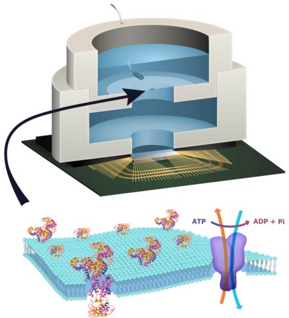
. The power dissipated in this source is introduced back into the circuit in the power generated by the Nernst independent voltage sources, and . The power dissipated in the dependent voltage source Vloss models any additional power not used to perform chemical or electrical work. ……


Integration of ATP-harvesting ion pumps could provide a means to power future CMOS microsystems scaled to the level of individual cells22. In molecular diagnostics, the integration of pore-forming proteins such as alpha haemolysin23 or MspA porin24 with CMOS electronics is already finding application in DNA sequencing25. Exploiting the large diversity of function available in transmembrane proteins in these hybrid systems could, for example, lead to highly specific sensing platforms for airborne odorants or soluble molecular entities26, 27. Heavily multiplexed platforms could become high-throughput in vitro drug-screening platforms against this diversity of function. In addition, integration of transmembrane proteins with CMOS may become a convenient alternative to fluorescence for coupling to synthetic biological systems28.


Roseman, J. M. et al. Hybrid integrated biological–solid-state system powered with adenosine triphosphate. Nat. Commun. 6:10070      http://dx.doi.org:/10.1038/ncomms10070 (2015).


  • Rottenberg, H. The measurement of membrane potential and deltapH in cells, organelles, and vesicles. Methods Enzymol. 55, 547569 (1979).
  • Himes, C., Carlson, E., Ricchiuti, R. J., Otis, B. P. & Parviz, B. A. Ultralow voltage nanoelectronics powered directly, and solely, from a tree. IEEE Trans. Nanotechnol. 9, 25(2010).
  • Mercier, P. P., Lysaght, A. C., Bandyopadhyay, S., Chandrakasan, A. P. & Stankovic, K. M.Energy extraction from the biologic battery in the inner ear. Nat. Biotechnol. 30, 12401243(2012).
  • Halámková, L. et al. Implanted Biofuel Cell Operating in a Living Snail. J. Am. Chem. Soc.134, 50405043 (2012).


Read Full Post »

Personalized Medicine – The California Initiative

Curator: Demet Sag, PhD, CRA, GCP

Are we there yet?  Life is a journey so the science.

Governor Brown announced Precision Medicine initiative for California on April 14, 2015.  UC San Francisco is hosting the two-year initiative, through UC Health, which includes UC’s five medical centers, with $3 million in startup funds from the state. The public-private initiative aims to leverage these funds with contributions from other academic and industry partners.

With so many campuses spread throughout the state and so much scientific, clinical and computational expertise, the UC system has the potential to bring it all together, said Atul Butte, MD, PhD, who is leading the initiative.

At the beginning of 2015 President Obama signed this initiative and assigned people to work on this project.

Previously NCI Director Harold Varmus, MD said that “Precision medicine is really about re-engineering the diagnostic categories for cancer to be consistent with its genomic underpinnings, so we can make better choices about therapy,” and “In that sense, many of the things we’re proposing to do are already under way.”

The proposed initiative has two main components:

  • a near-term focus on cancers and
  • a longer-term aim to generate knowledge applicable to the whole range of health and disease.

Both components are now within our reach because of advances in basic research, including molecular biology, genomics, and bioinformatics. Furthermore, the initiative taps into converging trends of increased connectivity, through social media and mobile devices, and Americans’ growing desire to be active partners in medical research.

Since the human genome is sequenced it became clear that actually there are few genes than expected and shared among organisms to accomplish same or similar core biological functions.  As a result, knowledge of the biological role of such shared proteins in one organism can be transferred to another organism.

It was necessary to generate a dynamic yet controlled standardized collection of information with ever changing and accumulating data. It was called Gene Ontology Consortium. Three independent ontologies can be reached at  (http://www.geneontology.org) developed based on :

  1. biological process,
  2. molecular function and
  3. cellular component.

We need a common language for annotation for a functional conservation. Genesis of the grand biological unification made it possible to complete the genomic sequences of not only human but also the main model organisms and more:

·         the budding yeast, Saccharomyces cerevisiae, completed in 1996

·         the nematode worm Caenorhabditis elegans, completed in 1998

·         the fruitfly Drosophila melanogaster,

·         the flowering plant Arabidopsis thaliana

·         fission yeast Schizosaccharomyces pombe

·         the mouse , Mus musculus

On the other hand, as we know there are allelic variations that underlie common diseases and complete genome sequencing for many individuals with and without disease is required.  However, there are advantages and disadvantages as we can carry out partial surveys of the genome by genotyping large numbers of common SNPs in genome-wide association studies but there are problems such as computing the data efficiently and sharing the information without tempering privacy. Therefore we should be mindful about few main conditions including:

  1. models of the allelic architecture of commondiseases,
  2. sample size,
  3. map density and
  4. sample-collection biases.

This will lead into the cost control and efficiency while identifying genuine disease-susceptibility loci. The genome-wide association studies (GWAS) have progressed from assaying fewer than 100,000 SNPs to more than one million, and sample sizes have increased dramatically as the search for variants that explain more of the disease/trait heritability has intensified.

In addition, we must translate this sequence information from genomics locus of the genes to function with related polymorphism of these genes so that possible patterns of the gene expression and disease traits can be matched. Then, we may develop precision technologies for:

  1. Diagnostics
  2. Targeted Drugs and Treatments
  3. Biomarkers to modulate cells for correct functions

With the knowledge of:

  1. gene expression variations
  2. insight in the genetic contribution to clinical endpoints ofcomplex disease and
  3. their biological risk factors,
  4. share etiologic pathways

therefore, requires an understanding of both:

  • the structure and
  • the biology of the genome.

These studies demonstrated hundreds of associations of common genetic variants with over 80 diseases and traits collected under a controlled online resource.  However, identifying published GWAS can be challenging as a simple PubMed search using the words “genome wide association studies”  may be easily populated with un-relevant  GWAS.

National Human Genome Research Institute (NHGRI) Catalog of Published Genome-Wide Association Studies (http://www.genome.gov/gwastudies), an online, regularly updated database of SNP-trait associations extracted from published GWAS was developed.

Therefore, sequencing of a human genome is a quite undertake and requires tools to make it possible:

  • to explore the genetic component incomplex diseases and
  • to fully understand the genetic pathways contributing tocomplex disease

The rapid increase in the number of GWAS provides an unprecedented opportunity to examine the potential impact of common genetic variants on complex diseases by systematically cataloging and summarizing key characteristics of the observed associations and the trait/disease associated SNPs (TASs) underlying them.

With this in mind, many forms can be established:

  1. to describe the features of this resource and the methods we have used to produce it,
  2. to provide and examine key descriptive characteristics of reported TASs such as estimated risk allele frequencies and odds ratios,
  3. to examine the underlying functionality of reported risk loci by mapping them to genomic annotation sets and assessing overrepresentation via Monte Carlo simulations and
  4. to investigate the relationship between recent human evolution and human disease phenotypes.

This procedure has no clear path so there are several obstacles in the actual functional variant that is often unknown. This may be due to:

  1. trait/disease associated SNPs (TASs),
  2. a well known SNP+ strong linkage disequilibrium (LD) with the TAS,
  3. an unknown common SNP tagged by a haplotype
  4. rare single nucleotide variant tagged by a haplotype on which the TAS occurs, or
  5. Copy Number variation (CNV), a linked copy number variant.

There can be other factors such as

  • Evolution,
  • Natural Selection
  • Environment
  • Pedigree
  • Epigenetics

Even though heritage is another big factor, the concept of heritability and its definition as an estimable, dimensionless population parameter as introduced by Sewall Wright and Ronald Fisher almost a century ago.

As a result, heritability gain interest since it allows us to compare of the relative importance of genes and environment to the variation of traits within and across populations. The heritability is an ongoing mechanism and  remains as a key:

  • to selection in evolutionary biology and agriculture, and
  • to the prediction of disease risk in medicine.

Table 1.

Reported TASs associated with two or more distinct traits

Chromosomal region Rs number(s) Attributed genes Associated traits reported in catalog
1p13.2 rs2476601, rs6679677 PTPN22 Crohn’s disease, type 1 diabetes, rheumatoid arthritis
1q23.2 rs2251746, rs2494250 FCER1A Serum IgE levels, select biomarker traits (MCP1)
2p15 rs1186868, rs1427407 BCL11A Fetal hemoglobin, F-cell distribution
2p23.3 rs780094 GCKR CRP, lipids, waist circumference
6p21.33 rs3131379, rs3117582 HLA / MHC region Systemic lupus erythematosus, lung cancer, psoriasis, inflammatory bowel disease, ulcerative colitis, celiac disease, rheumatoid arthritis, juvenile idiopathic arthritis, multiple sclerosis, type 1 diabetes
6p22.3 rs6908425, rs7756992, rs7754840, rs10946398, rs6931514 CDKAL1 Crohn’s disease, type 2 diabetes
6p25.3 rs1540771, rs12203592, rs872071 IRF4 Freckles, hair color, chronic lymphocytic leukemia
6q23.3 rs5029939, rs10499194 TNFAIP3 Systemic lupus erythematosus, rheumatoid arthritis
7p15.1 rs1635852, rs864745 JAZF1 Height, type 2 diabetes*
8q24.21 rs6983267 Intergenic Prostate or colorectal cancer, breast cancer
9p21.3 rs10811661, rs1333040, rs10811661, rs10757278, rs1333049 CDKN2A, CDKN2B Type 2 diabetes, intracranial aneurysm, myocardial infarction
9q34.2 rs505922, rs507666, rs657152 ABO Protein quantitative trait loci (TNF-α), soluble ICAM-1, plasma levels of liver enzymes (alkaline phosphatase)
12q24 rs1169313, rs7310409, rs1169310, rs2650000 HNF1A Plasma levels of liver enzyme (GGT), C-reactive protein, LDL cholesterol
16q12.2 rs8050136, rs9930506, rs6499640, rs9939609, rs1121980 FTO Type 2 diabetes, body mass index or weight
17q12 rs7216389, rs2872507 ORMDL3 Asthma, Crohn’s disease
17q12 rs4430796 TCF2 Prostate cancer, type 2 diabetes
18p11.21 rs2542151 PTPN2 Type 1 diabetes, Crohn’s disease
19q13.32 rs4420638 APOE, APOC1, APOC4 Alzheimer’s disease, lipids

* The well known association of JAZF1 with prostate cancer was reported with a p value of 2 × 10⁻⁶ (18), which did not meet the threshold of 5 × 10⁻⁸ for this analysis.

PMC full text: Proc Natl Acad Sci U S A. 2009 Jun 9; 106(23): 9362–9367.

Published online 2009 May 27. doi:  10.1073/pnas.0903103106

.

Table 2

Allele-Frequency Data for Nine Reproducible Associations

frequency
gene diseasea SNP associated alleleb Europeand Africane δf FST reference(s)c
CTLA4 T1DM Thr17Ala Ala .38 (1,670) .209 (402) .171 .06 Osei-Hyiaman et al. 2001; Lohmueller et al. 2003
DRD3 Schizophrenia Ser9Gly Ser/Ser .67 (202) .116 (112) .554 .458 Crocq et al. 1996; Lohmueller et al.2003
AGT Hypertension Thr235Met Thr .42 (3,034) .91 (658) .49 .358 Rotimi et al. 1996; Nakajima et al.2002
PRNP CJD Met129Val Met .72 (138) .556 (72) .164 .049 Hirschhorn et al. 2002; Soldevila et al. 2003
F5 DVT Arg506Gln Gln .044 (1,236) .00 (251) .044 .03 Rees et al. 1995; Hirschhorn et al.2002
HFE HFE Cys382Tyr Tyr .038 (2,900) .00 (806) .038 .024 Feder et al. 1996; Merryweather-Clarke et al. 1997
MTHFR DVT C677T T .3 (188) .066 (468) .234 .205 Schneider et al. 1998; Ray et al.2002
PPARG T2DM Pro12Ala Pro .925 (120) 1.0 (120) .075 .067 Altshuler et al. 2000HapMap Project
KCNJ11 T2DM Asp23Lys Lys .36 (96) .09 (98) .27 .182 Florez et al. 2004

aCJD = Creutzfeldt-Jacob disease; DVT = deep venous thrombosis; HFE = hemochromatosis; T1DM = type I diabetes; T2DM = type II diabetes.

bThe associated allele is the SNP associated with disease, regardless of whether it is the derived or the ancestral allele. The frequencies for this allele are given.

cThe reference that claims this to be a reproducible association, as well as the reference from which the allele frequencies were taken. For allele frequencies obtained from a meta-analysis, only the reference claiming reproducible association is given.

dAllele frequency obtained from the literature involving a European population. Either the general population frequency or the frequency in control groups in an association study was used. To reduce bias, when a control frequency was used for Europeans, a control frequency was also used for Africans. The total number of chromosomes surveyed is given in parentheses after each frequency.

eAllele frequency obtained from the literature involving a West African population. The total number of chromosomes surveyed is given in parentheses after each frequency.

fδ = The difference in the allele frequency between Europeans and Africans.

Table 3

PMC full text:

Am J Hum Genet. 2006 Jan; 78(1): 130–136.

Published online 2005 Nov 16. doi:  10.1086/499287

Copyright/License ►Request permission to reuse

Allele-Frequency Data for 39 Reported Associations

frequency
gene disease/phenotypea SNP associated alleleb Europeand Africane δf FST referencec
ADRB1 MI Arg389Gly Arg .717 (46) .467 (30) .251 .1 Iwai et al. 2003
ALOX5AP MI, stroke rs10507391 T .682 (44) .159 (44) .523 .425 Helgadottir et al. 2004
CAT Hypertension −844 (C/T) Tg .714 (42) .659 (44) .055 0 Jiang et al. 2001
CCR2 AIDS susceptibility Ile64Val Val .87 (46) .813 (48) .057 0 Smith et al. 1997
CD36 Malaria Y to stop Stop 0 (46) .083 (48) .083 .062 Aitman et al. 2000
F13 MI Val34Leu Val .762 (42) .795 (44) .033 0 Kohler et al. 1999
FGA Pulmonary embolism Thr312Ala Ala .2 (40) .5 (42) .3 .159 Carter et al. 2000
GP1BA CAD Thr145Met Met .022 (46) .167 (48) .145 .095 Gonzalez-Conejero et al.1998
ICAM1 MS Lys469Glu Lys .643 (42) .875 (48) .232 .12 Nejentsev et al. 2003
ICAM1 Malaria Lys29Met Met 0 (46) .354 (48) .354 .335 Fernandez-Reyes et al.1997
IFNGR1 Hp infection −56 (C/T) T .455 (44) .604 (48) .15 .023 Thye et al. 2003
IL13 Asthma −1055 (C/T) T .196 (46) .25 (44) .054 0 van der Pouw Kraan et al. 1999
IL13 Bronchial asthma Arg110Gln Gln .273 (44) .119 (42) .154 .05 Heinzmann et al. 2003
IL1A AD −889 (C/T) T .295 (44) .391 (46) .096 0 Nicoll et al. 2000
IL1B Gastric cancer −31 (C/T) T .826 (46) .375 (48) .451 .335 El-Omar et al. 2000
IL3 RA −16 (C/T) C .739 (46) .875 (48) .136 .037 Yamada et al. 2001
IL4 Asthma −590 (T/C) T .174 (46) .708 (48) .534 .436 Noguchi et al. 1998
IL4R Asthma Gln576Arg Arg .295 (44) .565 (46) .27 .118 Hershey et al. 1997
IL6 Juvenile arthritis −174 (C/G) G .5 (44) 1 (46) .5 .494 Fishman et al. 1998
IL8 RSV bronchiolitis −251 (T/A) Th .659 (44) .229 (48) .43 .301 Hull et al. 2000
ITGA2 MI 807 (C/T) T .316 (38) .25 (48) .066 0 Moshfegh et al. 1999
LTA MI Thr26Asn Asn .357 (42) .5 (44) .143 .018 Ozaki et al. 2002
MC1R Fair skin Val92Met Met .068 (44) 0 (44) .068 .047 Valverde et al. 1995
NOS3 MI Glu298Asp Asp .5 (44) .136 (44) .364 .247 Shimasaki et al. 1998
PLAU AD Pro141Leu Pro .659 (44) .979 (48) .32 .287 Finckh et al. 2003
PON1 CAD Arg192Gln Arg .174 (46) .727 (44) .553 .461 Serrato and Marian 1995
PON2 CAD Cys311Ser Ser .826 (46) .762 (42) .064 0 Sanghera et al. 1998
PTGS2 Colon cancer −765 (G/C) C .238 (42) .292 (48) .054 0 Koh et al. 2004
PTPN22i RA Arg620Trp Trp .084 (1,120) .024 (818) .059 .03 Begovich et al. 2004
SELE CAD Ser128Arg Arg .091 (44) .021 (48) .07 .025 Wenzel et al. 1994
SELL IgA nephropathy Pro238Ser Ser .065 (46) .333 (48) .268 .183 Takei et al. 2002
SELP MI Thr715Pro Thr .864 (44) .977 (44) .114 .063 Herrmann et al. 1998
SFTPB ARDS Ile131Thr Thr .5 (44) .348 (46) .152 .025 Lin et al. 2000
SPD RSV infection Met11Thr Met .568 (44) .478 (46) .09 0 Lahti et al. 2002
TF AD Pro570Ser Pro .957 (46) .935 (46) .022 0 Zhang et al. 2003
THBD MI Ala455Val Ala .87 (46) .848 (46) .022 0 Norlund et al. 1997
THBS4 MI Ala387Pro Pro .341 (44) .083 (48) .258 .166 Topol et al. 2001
TNFA Infectious disease −308 (A/G) A .182 (44) .205 (44) .023 0 Bayley et al. 2004
VCAM1 Stroke in SCD Gly413Ala Gly 1 (46) .938 (48) .063 .041 Taylor et al. 2002

aAD = Alzheimer disease; AIDS = acquired immunodeficiency syndrome; ARDS = acute respiratory distress syndrome; CAD = coronary artery disease; Hp = Helicobacter pylori; MI = myocardial infarction; MS = multiple sclerosis; RA = rheumatoid arthritis; RSV = respiratory syncytial virus; SCD = sickle cell disease.

bThe associated allele is the SNP associated with disease, regardless of whether it is the derived or the ancestral allele. The frequencies for this allele are given.

cThe reference that reported association with the listed disease/phenotype.

dFrequency obtained from the Seattle SNPs database for the European sample. The total number of chromosomes surveyed is given in parentheses after each frequency.

eFrequency obtained from the Seattle SNPs database for the African American sample. The total number of chromosomes surveyed is given in parentheses after each frequency.

fδ = The difference in the allele frequency between African Americans and Europeans.

gAssociated allele in database is A.

hAssociated allele in reference is A.

iThis SNP was not from the Seattle SNPs database; instead, allele frequencies from Begovich et al. (2004) were used.

They reported that “The SNPs associated with common disease that we investigated do not show much higher levels of differentiation than those of random SNPs. Thus, in these cases, ethnicity is a poor predictor of an individual’s genotype, which is also the pattern for random variants in the genome. This lends support to the hypothesis that many population differences in disease risk are environmental, rather than genetic, in origin. However, some exceptional SNPs associated with common disease are highly differentiated in frequency across populations, because of either a history of random drift or natural selection. The exceptional SNPs  are located in AGT, DRD3, ALOX5AP, ICAM1, IL1B, IL4, IL6, IL8, and PON1. Of note, evidence of selection has been observed for AGT (Nakajima et al. 2004), IL4(Rockman et al. 2003), IL8 (Hull et al. 2001), and PON1 (Allebrandt et al. 2002). Yet, for the vast majority of the common-disease–associated polymorphisms we examined, ethnicity is likely to be a poor predictor of an individual’s genotype.”

In 2002The International HapMap Project was launched:

  • to provide a public resource
  • to accelerate medical genetic research.

Two Hapmap projects were completed. In phase I the objective was to genotype at least one common SNP every 5 kilobases (kb) across the euchromatic portion of the genome in 270 individuals from four geographically diverse population. In Phase II of the HapMap Project, a further 2.1 million SNPs were successfully genotyped on the same individuals.

The re-mapping of SNPs from Phase I of the project identified 21,177 SNPs that had an ambiguous position or some other feature indicative of low reliability; these are not included in the filtered Phase II data release. All genotype data are available from the HapMap Data Coordination Center (http://www.hapmap.org) and dbSNP (http://www.ncbi.nlm.nih.gov/SNP).

In the Phase II HapMap we identified 32,996 recombination hotspots3,6,36 (an increase of over 50% from Phase I) of which 68% localized to a region of≤5 kb. The median map distance induced by a hotspot is 0.043 cM (or one crossover per 2,300 meioses) and the hottest identified, on chromosome 20, is 1.2 cM (one crossover per 80 meioses). Hotspots account for approximately 60% of recombination in the human genome and about 6% of sequence (Supplementary Fig. 6).

In addition to many previously identified regions in HapMap Phase I including LARGESYT1 andSULT1C2 (previously called SULT1C1), about  200 regions identified from the Phase II HapMap that include many established cases of selection, such as the genes HBB andLCT, the HLA region, and an inversion on chromosome 17. Finally, in the future, whole-genome sequencing will provide a natural convergence of technologies to type both SNP and structural variation. Nevertheless, until that point, and even after, the HapMap Project data will provide an invaluable resource for understanding the structure of human genetic variation and its link to phenotype.


FUNCTIONAL GENOMICS AND DATA FOR MEDICINE:  BIOINFORMATICS/COMPUTER BIOLOGY

HMM libraries, such as PANTHER, Pfam, and SMART, are used primarily to recognize and annotate conserved motifs in protein sequences.

In the genomic era, one of the fundamental goals is to characterize the function of proteins on a large scale.

PANTHER, for relating protein sequence relationships to function relationships in a robust and accurate way under two main parts:

  • the PANTHER library (PANTHER/LIB)- collection of “books,” each representing a protein family as a multiple sequence alignment, a Hidden Markov Model (HMM), and a family tree.
  • the PANTHER index (PANTHER/X)- ontology for summarizing and navigating molecular functions and biological processes associated with the families and subfamilies.

PANTHER can be applied on three areas of active research:

  • to report the size and sequence diversity of the families and subfamilies, characterizing the relationship between sequence divergence and functional divergence across a wide range of protein families.
  • use the PANTHER/X ontology to give a high-level representation of gene function across the human and mouse genomes.
  • to rank missense single nucleotide polymorphisms (SNPs), on a database-wide scale, according to their likelihood of affecting protein function.

PRINTS is a compendium of protein motif ‘fingerprints’. A fingerprint is defined as a group of motifs excised from conserved regions of a sequence alignment, whose diagnostic power or potency is refined by iterative databasescanning (in this case the OWL composite sequence database).

The information contained within PRINTS is distinct from, but complementary to the consensus expressions stored in the widely-used PROSITE dictionary of patterns.

However, the position-specific amino acid probabilities in an HMM can also be used to annotate individual positions in a protein as being conserved (or conserving a property such as hydrophobicity) and therefore likely to be required for molecular function. For example, a mutation (or variant) at a conserved position is more likely to impact the function of that protein.

In addition, HMMs from different subfamilies of the same family can be compared with each other, to provide hypotheses about which residues may mediate the differences in function or specificity between the subfamilies.

Several computational algorithms and databases for comparing protein sequences developed and matured:

  1. particularly Hidden Markov Models (HMM;Krogh et al. 1994Eddy 1996) and
  2. PSI-BLAST (Altschul et al. 1997),

The profile has a different amino acid substitution vector at each position in the profile, based on the pattern of amino acids observed in a multiple alignment of related sequences.

Profile methods combine algorithms with databases: A group of related sequences is used to build a statistical representation of corresponding positions in the related proteins. The power of these methods therefore increases as new sequences are added to the database of known proteins.

Multiple sequence alignments (Dayhoff et al. 1974) and profiles have allowed a systematic study of related sequences. One of the key observations is that some positions are “conserved,” that is, the amino acid is invariant or restricted to a particular property (such as hydrophobicity), across an entire group of related sequences.

The dependence of profile and pattern-matching approaches (Jongeneel et al. 1989) on sequence databases led to the development of databases of profiles

  1. BLOCKS,Henikoff and Henikoff 1991;
  2. PRINTS,Attwood et al. 1994) and
  3. patterns (Prosite,Bairoch 1991) that could be searched in much the same way as sequence databases.

Among the most widely used protein family databases are

  1. Pfam (Sonnhammer et al. 1997;Bateman et al. 2002) and
  2. SMART (Schultz et al. 1998;Letunic et al. 2002), which combine expert analysis with the well-developed HMM formalism for statistical modeling of protein families (mostly families of related protein domains).

Either knowing its family membership to predict its function, or subfamily within that family is enough (Hannenhalli and Russell 2000).

  • Phylogenetic trees (representing the evolutionary relationships between sequences) and
  • dendrograms (tree structures representing the similarity between sequences) (e.g.,Chiu et al. 1985Rollins et al. 1991).

The PANTHER/LIB HMMs can be viewed as a statistical method for scoring the “functional likelihood” of different amino acid substitutions on a wide variety of proteins. Because it uses evolutionarily related sequences to estimate the probability of a given amino acid at a particular position in a protein, the method can be referred to as generating position-specific evolutionary conservation” (PSEC) scores.

Schematic illustration of the process for building PANTHER families.

  1. Family clustering.
  2. Multiple sequence alignment (MSA), family HMM, and family tree building.
  3. Family/subfamily definition and naming.
  4. Subfamily HMM building.
  5. Molecular function and biological process association.

Of these, steps 1, 2, and 4 are computational, and steps 3 and 5 are human-curated (with the extensive aid of software tools).


Further Reading

Human Phenome Project: Freimer N., Sabatti C. The human phenome project. Nat. Genet. 2003;34:15–21.

Jones R., Pembrey M., Golding J., Herrick D. The search for genenotype/phenotype associations and the phenome scan. Paediatr. Perinat. Epidemiol. 2005;19:264–275.

Stearns F.W. One hundred years of pleiotropy: A retrospective. Genetics.2010;186:767–773.

Welch J.J., Waxman D. Modularity and the cost of complexity. Evolution.2003;57:1723–1734.

Albert A.Y., Sawaya S., Vines T.H., Knecht A.K., Miller C.T., Summers B.R., Balabhadra S., Kingsley D.M., Schluter D. The genetics of adaptive shape shift in stickleback: Pleiotropy and effect size. Evolution. 2008;62:76–85.

Brem R.B., Yvert G., Clinton R., Kruglyak L. Genetic dissection of transcriptional regulation in budding yeast. Science. 2002;296:752–755.

Morley M., Molony C.M., Weber T.M., Devlin J.L., Ewens K.G., Spielman R.S., Cheung V.G. Genetic analysis of genome-wide variation in human gene expression. Nature. 2004;430:743–747. [PMC free article] [PubMed]

Wagner G.P., Zhang J. The pleiotropic structure of the genotype-phenotype map: The evolvability of complex organisms. Nat. Rev. Genet. 2011;12:204–213.

Cooper Z.N., Nelson R.M., Ross L.F. Informed consent for genetic research involving pleiotropic genes: An empirical study of ApoE research. IRB. 2006;28:1–11.


Model Organisms:

Worm Sequencing Consortium. The C. elegans Sequencing Consortium Genome sequence of the nematode C. elegans: a platform for investigating biology. Science.1998;282:2012–2018.

Adams MD, et al. The genome sequence of Drosophila melanogasterScience.2000;287:2185–2195.

Meinke DW, et al. Arabidopsis thaliana: a model plant for genome analysis. Science. 1998;282:662–682. [PubMed]

Chervitz SA, et al. Using the Saccharomyces Genome Database (SGD) for analysis of protein similarities and structure. Nucleic Acids Res. 1999;27:74–78.

The FlyBase Consortium The FlyBase database of the Drosophila Genome Projects and community literature. Nucleic Acids Res. 1999;27:85–88.

Blake JA, et al. The Mouse Genome Database (MGD): expanding genetic and genomic resources for the laboratory mouse. Nucleic Acids Res. 2000;28:108–111.

Ball CA, et al. Integrating functional genomic information into the Saccharomyces Genome Database. Nucleic Acids Res. 2000;28:77–80.

Venter, J.C., Adams, M.D., Myers, E.W., Li, P.W., Mural, R.J., Sutton, G.G., Smith, H.O., Yandell, M., Evans, C.A., Holt, R.A., et al. 2001. The sequence of the human genome. Science 291: 1304–1351.

Lander, E.S., Linton, L.M., Birren, B., Nusbaum, C., Zody, M.C., Baldwin, J., Devon, K., Dewar, K., Doyle, M., FitzHugh, W., et al. 2001. Initial sequencing and analysis of the human genome. Nature 409: 860–921.

Mi, H., Vandergriff, J., Campbell, M., Narechania, A., Lewis, S., Thomas, P.D., and Ashburner, M. 2003. Assessment of genome-wide protein function classification for Drosophila melanogaster. Genome Res.

Ashburner, M., Ball, C.A., Blake, J.A., Botstein, D., Butler, H., Cherry, J.M., Davis, A.P., Dolinski, K., Dwight, S.S., Eppig, J.T., et al. The Gene Ontology Consortium. 2000. Gene ontology: Tool for the unification of biology. Nat. Genet. 25: 25–29.


Computational Biology

Attwood TK, Beck ME, Bleasby AJ, Parry-Smith DJ. PRINTS–a database of protein motif fingerprints. Nucleic Acids Res. 1994 Sep;22(17):3590-6.

Obenauer JC, Yaffe MB. Computational prediction of protein-protein interactions.

Methods Mol Biol. 2004;261:445-68. Review.

Aitken A. Protein consensus sequence motifs. Mol Biotechnol. 1999 Oct;12(3):241-53. Review.

Bork P, Koonin EV. Protein sequence motifs. Curr Opin Struct Biol. 1996 Jun;6(3):366-76. Review.

Hodgman TC. The elucidation of protein function by sequence motif analysis.  Comput Appl Biosci. 1989 Feb;5(1):1-13. Review.

Altschul, S.F., Madden, T.L., Schäffer, A.A., Zhang, J., Zhang, Z., Miller, W., and Lipman, D.J. 1997. Gapped BLAST and PSI-BLAST: A new generation of protein database search programs. Nucleic Acids Res. 25: 3389–3402.

Spencer CC, et al. The influence of recombination on human genetic diversity.PLoS Genet. 2006;2:e148.

Petes TD. Meiotic recombination hot spots and cold spots. Nature Rev. Genet.2001;2:360–369.

Griffiths RC, Tavaré S. The age of a mutation in a general coalescent tree. Stoch Models. 1998;14:273–295. doi: 10.1080/15326349808807471.

Gauderman WJ. Sample size requirements for matched case-control studies of gene-environment interaction. Stat Med. 2002;21(1):35–50. doi: 10.1002/sim.973.

Attwood, T.K., Beck, M.E., Bleasby, A.J., and Parry-Smith, D.J. 1994. PRINTS—A database of protein motif fingerprints. Nucleic Acids Res. 22: 3590–3596.

Bairoch, A. 1991. PROSITE: A dictionary of sites and patterns in proteins. Nucleic Acids Res. 19 Suppl: 2241–2245.

Bairoch, A. and Apweiler, R. 2000. The SWISS-PROT protein sequence database and its supplement TrEMBL in 2000. Nucleic Acids Res. 28: 45–48.

Bateman, A., Birney, E., Cerruti, L., Durbin, R., Etwiller, L., Eddy, S.R., Griffiths-Jones, S., Howe, K.L., Marshall, M., and Sonnhammer, E.L. 2002. The Pfam protein families database. Nucleic Acids Res. 30: 276–280.

Sonnhammer, E.L., Eddy, S.R., and Durbin, R. 1997. Pfam: A comprehensive database of protein domain families based on seed alignments. Proteins 28:405–420.

Swets, J.A. 1988. Measuring the accuracy of diagnostic systems. Science 240:1285–1293. [PubMed]

Thomas, P.D., Kejariwal, A., Campbell, M.J., Mi, H., Diemer, K., Guo, N., Ladunga, I., Ulitsky-Lazareva, B., Muruganujan, A., Rabkin, S., et al. 2003. PANTHER: A browsable database of gene products organized by biological function, using curated protein family and subfamily classification. Nucleic Acids Res. 31: 334–341.

HUGO Gene Nomenclature Committee (2011). HGNC Database.http://www.genenames.org/.


Population Genomics, GWAS, Inheritance, Heritability, Migration, Selection  an Evolution:

Dayhoff, M.O., Barker, W.C., and McLaughlin, P.J. 1974. Inferences from protein and nucleic acid sequences: Early molecular evolution, divergence of kingdoms and rates of change. Orig. Life 5: 311–330.

Joseph Lachance Disease-associated alleles in genome-wide association studies are enriched for derived low frequency alleles relative to HapMap and neutral expectations BMC Med Genomics. 2010; 3: 57.

Joseph Lachance, Sarah A. Tishkoff  Biased Gene Conversion Skews Allele Frequencies in Human Populations, Increasing the Disease Burden of Recessive Alleles  Am J Hum Genet. 2014 October 2; 95(4): 408-420.

Hemalatha Kuppusamy, Helga M. Ogmundsdottir, Eva Baigorri, Amanda Warkentin, Hlif Steingrimsdottir, Vilhelmina Haraldsdottir, Michael J. Mant, John Mackey, James B. Johnston, Sophia Adamia, Andrew R. Belch, Linda M. Pilarski Inherited Polymorphisms in Hyaluronan Synthase 1 Predict Risk of Systemic B-Cell Malignancies but Not of Breast Cancer  PLoS One. 2014; 9(6): e100691.

Joseph Lachance, Sarah A. Tishkoff  Population Genomics of Human Adaptation

Annu Rev Ecol Evol Syst. Author manuscript; available in PMC 2014 November 5.

Published in final edited form as: Annu Rev Ecol Evol Syst. 2013 November; 44: 123–143

Joseph Lachance, Sarah A. Tishkoff SNP ascertainment bias in population genetic analyses: Why it is important, and how to correct it  Bioessays.

Erik Corona, Rong Chen, Martin Sikora, Alexander A. Morgan, Chirag J. Patel, Aditya Ramesh, Carlos D. Bustamante, Atul J. Butte Analysis of the Genetic Basis of Disease in the Context of Worldwide Human Relationships and Migration PLoS Genet. 2013 May; 9(5): e1003447.

Olga Y. Gorlova, Jun Ying, Christopher I. Amos, Margaret R. Spitz, Bo Peng, Ivan P. Gorlov J Derived SNP Alleles Are Used More Frequently Than Ancestral Alleles As Risk-Associated Variants In Common Human Diseases Bioinform Comput Biol.

Ani Manichaikul, Wei-Min Chen, Kayleen Williams, Quenna Wong, Michèle M. Sale, James S. Pankow, Michael Y. Tsai, Jerome I. Rotter, Stephen S. Rich, Josyf C. Mychaleckyj  Analysis of Family- and Population-Based Samples in Cohort Genome-Wide Association Studies Hum Genet.

Altshuler D, Daly MJ, Lander ES. Genetic mapping in human disease. Science. 2008; 322(5903):881–888. doi: 10.1126/science.1156409.

Wellcome Trust Case Control Consortium. Genome-wide association study of 14,000 cases of seven common diseases and 3,000 shared controls. Nature. 2007;447(7145):661–678. doi: 10.1038/nature05911.

Kotowski IK, Pertsemlidis A, Luke A, Cooper RS, Vega GL, Cohen JC, Hobbs HH. A spectrum of PCSK9 Alleles contributes to plasma levels of low-density lipoprotein cholesterol. American Journal of Human Genetics.2006;78(3):410–422. doi: 10.1086/500615.

Tomlinson I, Webb E, Carvajal-Carmona L, Broderick P, Kemp Z, Spain S, Penegar S, Chandler I, Gorman M, Wood W. et al. A genome-wide association scan of tag SNPs identifies a susceptibility variant for colorectal cancer at 8q24.21. Nature Genetics. 2007;39(8):984–988. doi: 10.1038/ng2085.

Todd JA, Walker NM, Cooper JD, Smyth DJ, Downes K, Plagnol V, Bailey R, Nejentsev S, Field SF, Payne F. et al. Robust associations of four new chromosome regions from genome-wide analyses of type 1 diabetes. Nature Genetics. 2007;39(7):857–864. doi: 10.1038/ng2068.

Manolio TA, Collins FS, Cox NJ, Goldstein DB, Hindorff LA, Hunter DJ, McCarthy MI, Ramos EM, Cardon LR, Chakravarti A. et al. Finding the missing heritability of complex diseases. Nature. 2009;461(7265):747–753. doi: 10.1038/nature08494.

Maher B. Personal genomes: The case of the missing heritability. Nature.2008;456(7218):18–21. doi: 10.1038/456018a.

Clark AG, Boerwinkle E, Hixson J, Sing CF. Determinants of the success of whole-genome association testing. Genome Res. 2005;15(11):1463–1467. doi: 10.1101/gr.4244005.

Clarke AJ, Cooper DN. GWAS: heritability missing in action? European Journal of Human Genetics. 2010;18:859–861. doi: 10.1038/ejhg.2010.35.

Moore JH, Williams SM. Epistasis and its implications for personal genetics. Am J Hum Genet. 2009;85(3):309–320. doi: 10.1016/j.ajhg.2009.08.006.

Goldstein DB. Common genetic variation and human traits. N Engl J Med.2009;360(17):1696–1698. doi: 10.1056/NEJMp0806284.

Hirschhorn JN. Genomewide association studies–illuminating biologic pathways. N Engl J Med. 2009;360(17):1699–1701. doi: 10.1056/NEJMp0808934.

Iles MM. What can genome-wide association studies tell us about the genetics of common disease? PLoS Genet. 2008;4(2):e33. doi: 10.1371/journal.pgen.0040033.

Myles S, Davison D, Barrett J, Stoneking M, Timpson N. Worldwide population differentiation at disease-associated SNPs. BMC Med Genomics.2008;1:22. doi: 10.1186/1755-8794-1-22.

Lohmueller KE, Mauney MM, Reich D, Braverman JM. Variants associated with common disease are not unusually differentiated in frequency across populations. Am J Hum Genet. 2006;78(1):130–136. doi: 10.1086/499287.

Hindorff LA, Sethupathy P, Junkins HA, Ramos EM, Mehta JP, Collins FS, Manolio TA. Potential etiologic and functional implications of genome-wide association loci for human diseases and traits. Proc Natl Acad Sci USA.2009;106(23):9362–9367. doi: 10.1073/pnas.0903103106.

Wang WYS, Barratt BJ, Clayton DG, Todd JA. Genome-wide association studies: Theoretical and practical concerns. Nature Reviews Genetics.2005;6(2):109–118. doi: 10.1038/nrg1522.

Hacia JG, Fan JB, Ryder O, Jin L, Edgemon K, Ghandour G, Mayer RA, Sun B, Hsie L, Robbins C. et al. Determination of ancestral alleles for human single-nucleotide polymorphisms using high-density oligonucleotide arrays. Nat Genet. 1999;22(2):164–167. doi: 10.1038/9674.

Lohmueller KE, Pearce CL, Pike M, Lander ES, Hirschhorn JN. Meta-analysis of genetic association studies supports a contribution of common variants to susceptibility to common disease. Nat Genet. 2003;33(2):177–182. doi: 10.1038/ng1071.

Wang WY, Pike N. The allelic spectra of common diseases may resemble the allelic spectrum of the full genome. Med Hypotheses. 2004;63(4):748–751. doi: 10.1016/j.mehy.2003.12.057.

HapMart. http://hapmart.hapmap.org/BioMart/martview/

Frazer KA, Ballinger DG, Cox DR, Hinds DA, Stuve LL, Gibbs RA, Belmont JW, Boudreau A, Hardenbol P, Leal S. et al. A second generation human haplotype map of over 3.1 million SNPs. Nature.2007;449(7164):851–861. doi: 10.1038/nature06258.

Rotimi CN, Jorde LB. Ancestry and disease in the age of genomic medicine. N Engl J Med. 2010;363(16):1551–1558. doi: 10.1056/NEJMra0911564.

Ganapathy G, Uyenoyama MK. Site frequency spectra from genomic SNP surveys. Theor Popul Biol. 2009;75(4):346–354. doi: 10.1016/j.tpb.2009.04.003.

Nielsen R, Hubisz MJ, Clark AG. Reconstituting the frequency spectrum of ascertained single-nucleotide polymorphism data. Genetics.2004;168(4):2373–2382. doi: 10.1534/genetics.104.031039.

Watterson GA, Guess HA. Is the most frequent allele the oldest? Theor Popul Biol. 1977;11(2):141–160. doi: 10.1016/0040-5809(77)90023-5.

Sherry ST, Ward MH, Kholodov M, Baker J, Phan L, Smigielski EM, Sirotkin K. dbSNP: the NCBI database of genetic variation. Nucleic Acids Res. 2001;29(1):308–311. doi: 10.1093/nar/29.1.308.

Spencer CC, Deloukas P, Hunt S, Mullikin J, Myers S, Silverman B, Donnelly P, Bentley D, McVean G. The influence of recombination on human genetic diversity. PLoS Genet. 2006;2(9):e148. doi: 10.1371/journal.pgen.0020148.

Kimura M. The number of heterozygous nucleotide sites maintained in a finite population due to steady flux of mutations. Genetics. 1969;61(4):893–903.

Johnson AD, O’Donnell CJ. An open access database of genome-wide association results. BMC Med Genet. 2009;10:6. doi: 10.1186/1471-2350-10-6.

Kimura M, Ohta T. The age of a neutral mutant persisting in a finite population. Genetics. 1973;75(1):199–212.

McVean GA, et al. The fine-scale structure of recombination rate variation in the human genome. Science. 2004;304:581–584.

Slatkin M, Rannala B. Estimating the age of alleles by use of intraallelic variability. Am J Hum Genet. 1997;60(2):447–458.

Green R, Krause J, Briggs A, Maricic T, Stenzel U, Kircher M, Patterson N, Li H, Zhai W, Fritz M. et al. A Draft Sequence of the Neanderthal Genome. Science. 2010;328:710–722. doi: 10.1126/science.1188021.

Bamshad M, Wooding SP. Signatures of natural selection in the human genome. Nat Rev Genet. 2003;4(2):99–111. doi: 10.1038/nrg999.

Hernandez RD, Williamson SH, Bustamante CD. Context dependence, ancestral misidentification, and spurious signatures of natural selection. Mol Biol Evol. 2007;24(8):1792–1800. doi: 10.1093/molbev/msm108.

Bustamante CD, et al. Natural selection on protein-coding genes in the human genome. Nature. 2005;437:1153–1157.


Previously published articles:


Personalized Medicine in Cancer [3] larryhbern
Advances in Gene Editing Technology: New Gene Therapy Options in Personalized Medicine 2012pharmaceutical
Big Data for Personalized Medicine and Biomarker Discovery, May 5-6, 2015 | Philadelphia, PA 2012pharmaceutical
Tweets by @pharma_BI and by @AVIVA1950 for @PMWCIntl, #PMWC15, #PMWC2015 LIVE @Silicon Valley 2015 Personalized Medicine World Conference 2012pharmaceutical
Presentations Content for Track One @Silicon Valley 2015 Personalized Medicine World Conference, Mountain View, CA, January 26 to January 28, 2015 2012pharmaceutical
Views of Content Presentations – Track One @Silicon Valley 2015 Personalized Medicine World Conference, Mountain View, CA, January 26 to January 28, 2015 2012pharmaceutical
Word Associations of Twitter Discussions for 10th Annual Personalized Medicine Conference at the Harvard Medical School, November 12-13, 2014 2012pharmaceutical
8:30AM–12:00PM, January 28, 2015 – Morality, Ethics & Public Law in PM, LIVE @Silicon Valley 2015 Personalized Medicine World Conference, Mountain View, CA 2012pharmaceutical
2:00PM–5:00PM, January 27, 2015 – Personalizing Evidence in the Learning Healthcare System & Biomarker Discovery Technologies, LIVE @Silicon Valley 2015 Personalized Medicine World Conference, Mountain View, CA 2012pharmaceutical
9:15AM–2:00PM, January 27, 2015 – Regulatory & Reimbursement Frameworks for Molecular Testing, LIVE @Silicon Valley 2015 Personalized Medicine World Conference, Mountain View, CA 2012pharmaceutical
7:45AM–9:15AM, January 27, 2015 – Risk, Reward & Innovation, LIVE @Silicon Valley 2015 Personalized Medicine World Conference, Mountain View, CA 2012pharmaceutical
3:30PM –5:15PM, January 26, 2015 – NGS Applications: Impact of Genomics on Cancer Care @Silicon Valley 2015 Personalized Medicine World Conference, Mountain View, CA 2012pharmaceutical
2:15PM – 3:00PM, January 26, 2015 – Impact of Genomics on Cancer Care @Silicon Valley 2015 Personalized Medicine World Conference, Mountain View, CA 2012pharmaceutical
1:00PM – 1:15PM, January 26, 2015 – Clinical Methodologies of NGS – LIVE @Silicon Valley 2015 Personalized Medicine World Conference, Mountain View, CA 2012pharmaceutical
10:30AM-12PM, January 26, 2015 – NGS Applications: Impact of Genomics on Cancer Care – LIVE @Silicon Valley 2015 Personalized Medicine World Conference, Mountain View, CA 2012pharmaceutical
9AM-10AM, January 26, 2015 – Newborn & Prenatal Diagnosis – LIVE @Silicon Valley 2015 Personalized Medicine World Conference, Mountain View, CA 2012pharmaceutical
7:55AM – 9AM, January 26, 2015 – Introduction and Overview – LIVE @Silicon Valley 2015 Personalized Medicine World Conference, Mountain View, CA 2012pharmaceutical
Hamburg, Snyderman to Address Timely Issues in Personalized Medicine at 2015 Personalized Medicine World Conference in Silicon Valley 2012pharmaceutical
The Personalized Medicine Coalition welcomes the Administration’s focus on Personalized Medicine 2012pharmaceutical
Silicon Valley 2015 Personalized Medicine World Conference, Mountain View, CA, January 26, 2015, 8:00AM to January 28, 2015, 3:30PM PST 2012pharmaceutical
TOTAL Views of Presentation Content per Presentation: 10th Annual Personalized Medicine Conference at the Harvard Medical School, November 12-13, 2014 2012pharmaceutical
Silicon Valley 2015 Personalized Medicine World Conference, Mountain View, CA, January 26, 2015, 8:00AM to January 28, 2015, 3:30PM PST 2012pharmaceutical
FDA Commissioner, Dr. Margaret A. Hamburg on HealthCare for 310Million Americans and the Role of Personalized Medicine 2012pharmaceutical
Tweeting on the 10th Annual Personalized Medicine Conference at the Harvard Medical School, November 12-13, 2014 2012pharmaceutical
Content of the Presentations at the 10th Annual Personalized Medicine Conference at the Harvard Medical School, November 12-13, 2014 2012pharmaceutical
2:15PM 11/13/2014 – Panel Discussion Reimbursement/Regulation @10th Annual Personalized Medicine Conference at the Harvard Medical School, Boston 2012pharmaceutical
1:00PM 11/13/2014 – Panel Discussion Genomics in Prenatal and Childhood Disorders @10th Annual Personalized Medicine Conference at the Harvard Medical School, Boston 2012pharmaceutical
11:30AM 11/13/2014 – Role of Genetics and Genomics in Pharmaceutical Development @10th Annual Personalized Medicine Conference at the Harvard Medical School, Boston 2012pharmaceutical
10:15AM 11/13/2014 – Panel Discussion — IT/Big Data @10th Annual Personalized Medicine Conference at the Harvard Medical School, Boston 2012pharmaceutical
8:30AM 11/13/2014 – Harvard Business School Case Study: 23andMe @10th Annual Personalized Medicine Conference at the Harvard Medical School, Boston 2012pharmaceutical
8:00AM 11/13/2014 – Welcome from Gary Gottlieb, M.D., Partners HealthCare @10th Annual Personalized Medicine Conference at the Harvard Medical School, Boston 2012pharmaceutical
4:00PM 11/12/2014 – Panel Discussion Novel Approaches to Personalized Medicine @10th Annual Personalized Medicine Conference at the Harvard Medical School, Boston 2012pharmaceutical
3:15PM 11/12/2014 – Discussion Complex Disorders @10th Annual Personalized Medicine Conference at the Harvard Medical School, Boston 2012pharmaceutical
1:45PM 11/12/2014 – Panel Discussion – Oncology @10th Annual Personalized Medicine Conference at the Harvard Medical School, Boston 2012pharmaceutical
1:15PM 11/12/2014 – Keynote Speaker – International Genetics Health and Disease @10th Annual Personalized Medicine Conference at the Harvard Medical School, Boston 2012pharmaceutical
11:30AM 11/12/2014 – Personalized Medicine Coalition Award & Award Recipient Speech @10th Annual Personalized Medicine Conference at the Harvard Medical School, Boston 2012pharmaceutical
11:00AM 11/12/2014 – Keynote Speaker – Past, Present and Future of Personalized Medicine @10th Annual Personalized Medicine Conference at the Harvard Medical School, Boston 2012pharmaceutical
9:20AM 11/12/2014 – Panel Discussion – Genomic Technologies @10th Annual Personalized Medicine Conference at the Harvard Medical School, Boston 2012pharmaceutical
8:50AM 11/12/2014 – Keynote Speaker – CEO, American Medical Association @10th Annual Personalized Medicine Conference at the Harvard Medical School, Boston 2012pharmaceutical
8:20AM 11/12/2014 – Special Guest Keynote Speaker – The Future of Personalized Medicine @10th Annual Personalized Medicine Conference at the Harvard Medical School, Boston 2012pharmaceutical
8:00AM 11/12/2014 – Welcome & Opening Remarks @10th Annual Personalized Medicine Conference at the Harvard Medical School, Boston 2012pharmaceutical
Hashtags and Twitter Handles for 10th Annual Personalized Medicine at Harvard Medical School, 11/12 – 11/13/2014 2012pharmaceutical
Personalized Medicine Coalition (PMC) – Upcoming Events 2012pharmaceutical
10th Annual Personalized Medicine Conference at the Harvard Medical School, November 12-13, 2014, The Joseph B. Martin Conference Center at Harvard Medical School, 77 Avenue Louis Pasteur, Boston, MA 2012pharmaceutical
Personalized Medicine Coalition Recognizes Mark Levin with Award for Leadership 2012pharmaceutical
Research and Markets: Global Personalized Medicine Report 2014 – Scientific … – Rock Hill Herald (press release) 2012pharmaceutical
The Role of Medical Imaging in Personalized Medicine Dror Nir
CardioPredict™ Personalized Medicine Molecular Diagnostic Test 2012pharmaceutical
Life Sciences Circle Event: Next omics – Personalized Medicine beyond Genomics, December 11, 2013 5:30-8:30PM, The Broad Institute, Cambridge 2012pharmaceutical
Issues in Personalized Medicine: Discussions of Intratumor Heterogeneity from the Oncology Pharma forum on LinkedIn sjwilliamspa
Personalized medicine-based diagnostic test for NSCLC ritusaxena
Personalized Medicine and Colon Cancer tildabarliya
Systems Diagnostics – Real Personalized Medicine: David de Graaf, PhD, CEO, Selventa Inc. 2012pharmaceutical
Helping Physicians identify Gene-Drug Interactions for Treatment Decisions: New ‘CLIPMERGE’ program – Personalized Medicine @ The Mount Sinai Medical Center 2012pharmaceutical
Issues in Personalized Medicine in Cancer: Intratumor Heterogeneity and Branched Evolution Revealed by Multiregion Sequencing sjwilliamspa
Ethical Concerns in Personalized Medicine: BRCA1/2 Testing in Minors and Communication of Breast Cancer Risk sjwilliamspa
Personalized Medicine: Clinical Aspiration of Microarrays sjwilliamspa
The Promise of Personalized Medicine larryhbern
Personalized Medicine in NSCLC larryhbern
Attitudes of Patients about Personalized Medicine larryhbern
Understanding the Role of Personalized Medicine larryhbern
Directions for Genomics in Personalized Medicine larryhbern
Personalized Medicine: An Institute Profile – Coriell Institute for Medical Research: Part 3 2012pharmaceutical
Paradigm Shift in Human Genomics – Predictive Biomarkers and Personalized Medicine – Part 1 2012pharmaceutical
Harnessing Personalized Medicine for Cancer Management, Prospects of Prevention and Cure: Opinions of Cancer Scientific Leaders @ http://pharmaceuticalintelligence.com 2012pharmaceutical
Nanotechnology, personalized medicine and DNA sequencing tildabarliya
Personalized medicine gearing up to tackle cancer ritusaxena
Personalized Medicine Company Genection launched ritusaxena
Personalized Medicine: Cancer Cell Biology and Minimally Invasive Surgery (MIS) 2012pharmaceutical
The Way With Personalized Medicine: Reporters’ Voice at the 8th Annual Personalized Medicine Conference,11/28-29, 2012, Harvard Medical School, Boston, MA 2012pharmaceutical
Personalized Medicine Coalition: Upcoming Events 2012pharmaceutical
Highlights from 8th Annual Personalized Medicine Conference, November 28-29, 2012, Harvard Medical School, Boston, MA 2012pharmaceutical
Personalized medicine-based cure for cancer might not be far away ritusaxena
GSK for Personalized Medicine using Cancer Drugs needs Alacris systems biology model to determine the in silico effect of the inhibitor in its “virtual clinical trial” 2012pharmaceutical
Congestive Heart Failure & Personalized Medicine: Two-gene Test predicts response to Beta Blocker Bucindolol 2012pharmaceutical
Personalized Medicine as Key Area for Future Pharmaceutical Growth 2012pharmaceutical
Clinical Genetics, Personalized Medicine, Molecular Diagnostics, Consumer-targeted DNA – Consumer Genetics Conference (CGC) – October 3-5, 2012, Seaport Hotel, Boston, MA 2012pharmaceutical
AGENDA – Personalized Diagnostics, February 16-18, 2015 | Moscone North Convention Center | San Francisco, CA Part of the 22nd Annual Molecular Medicine Tri-Conference 2012pharmaceutical
Arrowhead’s 6th Annual Personalized & Precision Medicine Conference is coming to San Francisco, October 29-30, 2014 2012pharmaceutical
Personalized Cardiovascular Genetic Medicine at Partners HealthCare and Harvard Medical School 2012pharmaceutical
Precision Medicine for Future of Genomics Medicine is The New Era Demet Sag, Ph.D., CRA, GCP
Precision Medicine Initiative: Now is a State Initiative in California 2012pharmaceutical
1:30 pm – 2:20 pm 3/26/2015, LIVE Precision Medicine: Who’s Paying? @ MassBio Annual Meeting 2015, Cambridge, MA, Sonesta Hotel, 3/26 – 3/27, 2015 2012pharmaceutical
We Celebrate >600,000 Views for our 2,830 Scientific Articles in Life Sciences and Medicine 2012pharmaceutical
attn #3: Investors in HealthCare — Platforms in the Ecosystem of Regulatory & Reimbursement – Integrated Informational Platforms in Orthopedic Medical Devices, and Global Peer-Reviewed Scientific Curations: Bone Disease and Orthopedic Medicine – Draft 2012pharmaceutical
Foundation Medicine: Roche has Taken Over at $1.2B and 52.4 percent to 56.3 percent of Foundation Medicine on a fully diluted basis 2012pharmaceutical
Bridging the Gap in Precision Medicine @UCSF 2012pharmaceutical
Germline Genes and Drug Targets: Medicine more Proactive and Disease Prevention more Effective. 2012pharmaceutical
Proteomics – The Pathway to Understanding and Decision-making in Medicine larryhbern
Multi-drug, Multi-arm, Biomarker-driven Clinical Trial for patients with Squamous Cell Carcinoma called the Lung Cancer Master Protocol, or Lung-MAP launched by NCI, Foundation Medicine, and Five Pharma Firms 2012pharmaceutical
Preventive Care: Anticipated Changes caused by Genomics in the Clinic and Personalised Medicine 2012pharmaceutical
Cancer Labs at School of Medicine @ Technion: Janet and David Polak Cancer and Vascular Biology Research Center 2012pharmaceutical
Reprogramming Adult Patient Cells into Stem Cells: the Promise of Personalized Genetic Therapy 2012pharmaceutical
US Personalized Cancer Genome Sequencing Market Outlook 2018 – 2012pharmaceutical
Summary of Translational Medicine – e-Series A: Cardiovascular Diseases, Volume Four – Part 1 larryhbern
Introduction to Translational Medicine (TM) – Part 1: Translational Medicine larryhbern
Cancer Diagnosis at the Crossroads: Precision Medicine Driving Change, 9/14 – 9/17/2014, Sheraton Seattle Hotel, Seattle WA 2012pharmaceutical
Genomic Medicine and the Bioeconomy: Innovation for a Better World May 12–16, 2014 • Boston, MA 2012pharmaceutical
Institute of Medicine (IOM) Report on Genome-based Therapeutics and Companion Diagnostics 2012pharmaceutical
“Medicine Meets Virtual Reality” – NextMed-MMVR21 Conference 2/19 – 2/22/2014, Manhattan Beach Marriott, Manhattan Beach, CA

View

2012pharmaceutical

Read Full Post »

Leaders in Pharmaceutical Intelligence Presentation at The Life Sciences Collaborative

Curator: Stephen J. Williams, Ph.D. Website Analytics: Adam Sonnenberg, BSc Leaders in Pharmaceutical Intelligence presented their ongoing efforts to develop an open-access scientific and medical publishing and curation platform to The Life Science Collaborative, an executive pharmaceutical and biopharma networking group in the Philadelphia/New Jersey area.

Our Team

Slide1

For more information on the Vision, Funding Deals and Partnerships please see our site at http://pharmaceuticalintelligence.com/vision/

Slide2

For more information about our Team please see our site at http://pharmaceuticalintelligence.com/contributors-biographies/

Slide5

For more information of LPBI Deals and Partnerships please see our site at http://pharmaceuticalintelligence.com/joint-ventures/

Slide4

For more information about our BioMed E-Series please see our site at http://pharmaceuticalintelligence.com/biomed-e-books/

E-Book Titles by LPBI

LPBI book titles slide Slide8Slide3

Slide6

For more information on Real-Time Conference Coverage including a full list of Conferences Covered by LPBI please go to http://pharmaceuticalintelligence.com/press-coverage/

For more information on Real-Time Conference Coverage and a full listing of Conferences Covered by LPBI please go to:

http://pharmaceuticalintelligence.com/press-coverage/ Slide7

Slide10

The Pennsylvania (PA) and New Jersey (NJ) Biotech environment had been hit hard by the recession and loss of anchor big pharma companies however as highlighted by our interviews in “The Vibrant Philly Biotech Scene” and other news outlets, additional issues are preventing the PA/NJ area from achieving its full potential (discussions also with LSC)

Slide9Download the PowerPoint slides here: Presentationlsc

Read Full Post »

The Vibrant Philly Biotech Scene: Focus on Computer-Aided Drug Design and Gfree Bio, LLC

Curator: Stephen J. Williams, Ph.D.

Article ID #166: The Vibrant Philly Biotech Scene: Focus on Computer-Aided Drug Design and Gfree Bio, LLC. Published on 2/10/2015

WordCloud Image Produced by Adam Tubman


philly philly2night


This post is the second in a series of posts highlighting interviews with Philadelphia area biotech startup CEO’s and show how a vibrant biotech startup scene is evolving in the city as well as the Delaware Valley area. Philadelphia has been home to some of the nation’s oldest biotechs including Cephalon, Centocor, hundreds of spinouts from a multitude of universities as well as home of the first cloned animal (a frog), the first transgenic mouse, and Nobel laureates in the field of molecular biology and genetics. Although some recent disheartening news about the fall in rankings of Philadelphia as a biotech hub and recent remarks by CEO’s of former area companies has dominated the news, biotech incubators like the University City Science Center and Bucks County Biotechnology Center as well as a reinvigorated investment community (like PCCI and MABA) are bringing Philadelphia back. And although much work is needed to bring the Philadelphia area back to its former glory days (including political will at the state level) there are many bright spots such as the innovative young companies as outlined in these posts.

efavirenz_med-2In today’s post, I had the opportunity to talk with molecular modeler Charles H. Reynolds, Ph.D., founder and CEO of Gfree Bio LLC, a computational structure-based design and modeling company based in the Pennsylvania Biotech Center of Bucks County. Gfree is actually one of a few molecular modeling companies at the Bucks County Biotech Center (I highlighted another company RabD Biotech which structural computational methods to design antibody therapeutics).

Below is the interview with Dr. Reynolds of Gfree Bio LLC and Leaders in Pharmaceutical Business Intelligence (LPBI):

LPBI: Could you briefly explain, for non-molecular modelers, your business and the advantages you offer over other molecular modeling programs (either academic programs or other biotech companies)? As big pharma outsources more are you finding that your company is filling a needed niche market?

GfreeBio: Gfree develops and deploys innovative computational solutions to accelerate drug discovery. We can offer academic labs a proven partner for developing SBIR/STTR proposals that include a computational or structure-based design component. This can be very helpful in developing a successful proposal. We also provide the same modeling and structure-based design input for small biotechs that do not have these capabilities internally. Working with Gfree is much more cost-effective than trying to develop these capabilities internally. We have helped several small biotechs in the Philadelphia region assess their modeling needs and apply computational tools to advance their discovery programs. (see publication and collaboration list here).

LPBI: Could you offer more information on the nature of your 2014 STTR award?

GfreeBio: Gfree has been involved in three successful SBIR/STTR awards in 2014.   I am the PI for an STTR with Professor Burgess of Texas A&M that is focused on new computational and synthetic approaches to designing inhibitors for protein-protein interactions. Gfree is also collaborating with the Wistar Institute and Phelix Therapeutics on two other Phase II proposals in the areas of oncology and infectious disease.

LPBI: Why did you choose the Bucks County Pennsylvania Biotechnology Center?

GfreeBio: I chose to locate my company at the Biotech Center because it is a regional hub for small biotech companies and it provides a range of shared resources that are very useful to the company. Many of my most valuable collaborations have resulted from contacts at the center.

LPBI: The Blumberg Institute and Natural Products Discovery Institute has acquired a massive phytochemical library. How does this resource benefit the present and future plans for GfreeBio?

GfreeBio: To date Gfree Bio has not been an active collaborator with the Natural Products Insititute, but I have a good relationship with the Director and that could change at any time.

LPBI: Was the state of Pennsylvania and local industry groups support GfreeBio’s move into the Doylestown incubator? Has the partnership with Ben Franklin Partners and the Center provided you with investment and partnership opportunities?

GfreeBio: Gfree Bio has not been actively seeking outside investors, at least to date. We have been focused on growing the company through collaborations and consulting relationships. However, we have benefitted from being part of the Keystone Innovation Zone, a state program that provides incentives for small technology-based businesses in Pennsylvania.

LPBI: You will be speaking at a conference in the UK on reinventing the drug discovery process through tighter collaborations between biotech, academia, and non-profit organizations.  How do you feel the Philadelphia area can increase this type of collaboration to enhance not only the goals and missions of nonprofits, invigorate the Pennsylvania biotech industry, but add much needed funding to the local academic organizations?

GfreeBio: I think this type of collaboration across sectors appears to be one of the most important emerging models for drug discovery.   The Philadelphia region has been in many ways hard hit by the shift of drug discovery from large vertically integrated pharmaceutical companies to smaller biotechs, since this area was at the very center of “Big Pharma.” But I think the region is bouncing back as it shifts more to being a center for biotech. The three ingredients for success in the new pharma model are great universities, a sizeable talent pool, and access to capital. The last item may be the biggest challenge locally. The KIZ program (Keystone Innovation Zone) is a good start, but the region and state could do more to help promote innovation and company creation. Some other states are being much more aggressive.

LPBI: In addition, the Pennsylvania Biotechnology Center in Bucks County appears to have this ecosystem: nonprofit organizations, biotechs, and academic researchers. Does this diversity of researchers/companies under one roof foster the type of collaboration needed, as will be discussed at the UK conference? Do you feel collaborations which are in close physical proximity are more effective and productive than a “virtual-style” (online) collaboration model? Could you comment on some of the collaborations GfreeBio is doing with other area biotechs and academics?

GfreeBio: I do think the “ecosystem” at the Pennsylvania Biotechnology Center is important in fostering new innovative companies. It promotes collaborations that might not happen otherwise, and I think close proximity is always a big plus. As I mentioned before, many of the current efforts of Gfree have come from contacts at the center.   This includes SBIR/STTR collaborations and contract work for local small biotech companies.

LPBI: Thompson Reuters just reported that China’s IQ (Innovation Quotient) has risen dramatically with the greatest patents for pharmaceuticals and compounds from natural products. Have you or your colleagues noticed more competition or business from Chinese pharmaceutical companies?

GfreeBio: The rise of Asia, particularly China, has been one of the most significant recent trends in the pharmaceutical industry. Initially, this was almost exclusively in the CRO space, but now China is aggressively building a fully integrated domestic pharmaceutical industry.

LPBI: How can the Philadelphia ecosystem work closer together to support greater innovation?

GfreeBio: A lot has happened in recent years to promote innovation and company creation in the region. There could always be more opportunities for networking and collaboration within the Philadelphia community. Of course the biggest obstacle in this business is often financing. Philadelphia needs more public and private sources for investment in startups.

LPBI: Thank you Dr. Reynolds.

Please look for future posts in this series on the Philly Biotech Scene on this site

Also, if you would like your Philadelphia biotech startup to be highlighted in this series please contact me: sjwilliamspa@comcast.net or @StephenJWillia2.
Our site is read by ~ 570,000 readers, among them thousand international readers daily and followed by thousands of Twitter followers.


Other posts on this site in this VIBRANT PHILLY BIOTECH SCENE SERIES OR referring to PHILADELPHIA BIOTECH include:

RAbD Biotech Presents at 1st Pitch Life Sciences-Philadelphia

The Vibrant Philly Biotech Scene: Focus on Vaccines and Philimmune, LLC

What VCs Think about Your Pitch? Panel Summary of 1st Pitch Life Science Philly

1st Pitch Life Science- Philadelphia- What VCs Really Think of your Pitch

LytPhage Presents at 1st Pitch Life Sciences-Philadelphia

Hastke Inc. Presents at 1st Pitch Life Sciences-Philadelphia

PCCI’s 7th Annual Roundtable “Crowdfunding for Life Sciences: A Bridge Over Troubled Waters?” May 12 2014 Embassy Suites Hotel, Chesterbrook PA 6:00-9:30 PM

Pfizer Cambridge Collaborative Innovation Events: ‘The Role of Innovation Districts in Metropolitan Areas to Drive the Global an | Basecamp Business

Mapping the Universe of Pharmaceutical Business Intelligence: The Model developed by LPBI and the Model of Best Practices LLC

Read Full Post »

Older Posts »