BioPrinting Basics
Curator: Larry H. Bernstein, MD, FCAP
The ABCs of 3D Bioprinting of Living Tissues, Organs 5/06/2016

Although first originated in 2003, the world of bioprinting is still very new and ambiguous. Nevertheless, as the need for organ donation continues to increase worldwide, and organ and tissue shortages prevail, a handful of scientists have started utilizing this cutting-edge science and technology for various areas of regenerative medicine to possibly fill that organ-shortage void.
Among these scientists is Ibrahim Tarik Ozbolat, an associate professor of Engineering Science and Mechanics Department and the Huck Institutes of the Life Sciences at Penn State University, who’s been studying bioprinting and tissue engineering for years.
While Ozbolat is not the first to originate 3D bioprinting research, he’s the first one at Penn State University to spearhead the studies at Ozbolat Lab, Leading Bioprinting Research.
“Tissue engineering is a big need. Regenerative medicine, biofabrication of tissues and organs that can replace the damage or diseases is important,” Ozbolat told R&D Magazine after his seminar presentation at Interphex last week in New York City, titled 3D Bioprinting of Living Tissues & Organs.”
3D bioprinting is the process of creating cell patterns in a confined space using 3D-printing technologies, where cell function and viability are preserved within the printed construct.
Recent progress has allowed 3D printing of biocompatible materials, cells and supporting components into complex 3D functional living tissues. The technology is being applied to regenerative medicine to address the need for tissues and organs suitable for transplantation. Compared with non-biological printing, 3D bioprinting involves additional complexities, such as the choice of materials, cell types, growth and differentiation factors, and technical challenges related to the sensitivities of living cells and the construction of tissues. Addressing these complexities requires the integration of technologies from the fields of engineering, biomaterials science, cell biology, physics and medicine, according to nature.com.
“If we’re able to make organs on demand, that will be highly beneficial to society,” said Ozbolat. “We have the capability to pattern cells, locate them and then make the same thing that exists in the body.”
Sean V Murphy & Anthony Atala
Nature Biotechnology 32,773–785(2014) doi:10.1038/nbt.2958
Additive manufacturing, otherwise known as three-dimensional (3D) printing, is driving major innovations in many areas, such as engineering, manufacturing, art, education and medicine. Recent advances have enabled 3D printing of biocompatible materials, cells and supporting components into complex 3D functional living tissues. 3D bioprinting is being applied to regenerative medicine to address the need for tissues and organs suitable for transplantation. Compared with non-biological printing, 3D bioprinting involves additional complexities, such as the choice of materials, cell types, growth and differentiation factors, and technical challenges related to the sensitivities of living cells and the construction of tissues. Addressing these complexities requires the integration of technologies from the fields of engineering, biomaterials science, cell biology, physics and medicine. 3D bioprinting has already been used for the generation and transplantation of several tissues, including multilayered skin, bone, vascular grafts, tracheal splints, heart tissue and cartilaginous structures. Other applications include developing high-throughput 3D-bioprinted tissue models for research, drug discovery and toxicology.
3D printing is increasingly permitting the direct digital manufacture (DDM) of a wide variety of plastic and metal items. While this in itself may trigger a manufacturing revolution, far more startling is the recent development of bioprinters. These artificially construct living tissue by outputting layer-upon-layer of living cells. Currently all bioprinters are experimental. However, in the future, bioprinters could revolutionize medical practice as yet another element of the New Industrial Convergence.
Bioprinters may be constructed in various configurations. However, all bioprinters output cells from a bioprint head that moves left and right, back and forth, and up and down, in order to place the cells exactly where required. Over a period of several hours, this permits an organic object to be built up in a great many very thin layers.
In addition to outputting cells, most bioprinters also output a dissolvable gel to support and protect cells during printing. A possible design for a future bioprinter appears below and in the sidebar, here shown in the final stages of printing out a replacement human heart. Note that you can access larger bioprinter images on the Future Visions page. You may also like to watch my bioprinting video.

Bioprinting Pioneers
Several experimental bioprinters have already been built. For example, in 2002 Professor Makoto Nakamura realized that the droplets of ink in a standard inkjet printer are about the same size as human cells. He therefore decided to adapt the technology, and by 2008 had created a working bioprinter that can print out biotubing similar to a blood vessel. In time, Professor Nakamura hopes to be able to print entire replacement human organs ready for transplant. You can learn more about this groundbreaking work here or read this message from Professor Nakamura. The movie below shows in real-time the biofabrication of a section of biotubing using his modified inkjet technology.
Another bioprinting pioneer is Organovo. This company was set up by a research group lead by Professor Gabor Forgacs from the University of Missouri, and in March 2008 managed to bioprint functional blood vessels and cardiac tissue using cells obtained from a chicken. Their work relied on a prototype bioprinter with three print heads. The first two of these output cardiac and endothelial cells, while the third dispensed a collagen scaffold — now termed ‘bio-paper’ — to support the cells during printing.
Since 2008, Organovo has worked with a company called Invetech to create a commercial bioprinter called the NovoGen MMX. This is loaded with bioink spheroids that each contain an aggregate of tens of thousands of cells. To create its output, the NovoGen first lays down a single layer of a water-based bio-paper made from collagen, gelatin or other hydrogels. Bioink spheroids are then injected into this water-based material. As illustrated below, more layers are subsequently added to build up the final object. Amazingly, Nature then takes over and the bioink spheroids slowly fuse together. As this occurs, the biopaper dissolves away or is otherwise removed, thereby leaving a final bioprinted body part or tissue.
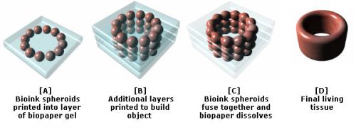
As Organovo have demonstrated, using their bioink printing process it is not necessary to print all of the details of an organ with a bioprinter, as once the relevant cells are placed in roughly the right place Nature completes the job. This point is powerfully illustrated by the fact that the cells contained in a bioink spheroid are capable of rearranging themselves after printing. For example, experimental blood vessels have been bioprinted using bioink spheroids comprised of an aggregate mix of endothelial, smooth muscle and fibroblast cells. Once placed in position by the bioprint head, and with no technological intervention, the endothelial cells migrate to the inside of the bioprinted blood vessel, the smooth muscle cells move to the middle, and the fibroblasts migrate to the outside.
In more complex bioprinted materials, intricate capillaries and other internal structures also naturally form after printing has taken place. The process may sound almost magical. However, as Professor Forgacs explains, it is no different to the cells in an embryo knowing how to configure into complicated organs. Nature has been evolving this amazing capability for millions of years. Once in the right places, appropriate cell types somehow just know what to do.
In December 2010, Organovo create the first blood vessels to be bioprinted using cells cultured from a single person. The company has also successfully implanted bioprinted nerve grafts into rats, and anticipates human trials of bioprinted tissues by 2015. However, it also expects that the first commercial application of its bioprinters will be to produce simple human tissue structures for toxicology tests. These will enable medical researchers to test drugs on bioprinted models of the liver and other organs, thereby reducing the need for animal tests.
In time, and once human trials are complete, Organovo hopes that its bioprinters will be used to produce blood vessel grafts for use in heart bypass surgery. The intention is then to develop a wider range of tissue-on-demand and organs-on-demand technologies. To this end, researchers are now working on tiny mechanical devices that can artificially exercise and hence strengthen bioprinted muscle tissue before it is implanted into a patient.
Organovo anticipates that its first artificial human organ will be a kidney. This is because, in functional terms, kidneys are one of the more straight-forward parts of the body. The first bioprinted kidney may in fact not even need to look just like its natural counterpart or duplicate all of its features. Rather, it will simply have to be capable of cleaning waste products from the blood. You can read more about the work of Organovoand Professor Forgac’s in this article from Nature.
Regenerative Scaffolds and Bones
A further research team with the long-term goal of producing human organs-on-demand has created the Envisiontec Bioplotter. Like Organovo’s NovoGen MMX, this outputs bio-ink ’tissue spheroids’ and supportive scaffold materials including fibrin and collagen hydrogels. But in addition, the Envisontech can also print a wider range of biomaterials. These include biodegradable polymers and ceramics that may be used to support and help form artificial organs, and which may even be used as bioprinting substitutes for bone.
Talking of bone, a team lead by Jeremy Mao at the Tissue Engineering and Regenerative Medicine Lab at Columbia University is working on the application of bioprinting in dental and bone repairs. Already, a bioprinted, mesh-like 3D scaffold in the shape of an incisor has been implanted into the jaw bone of a rat. This featured tiny, interconnecting microchannels that contained ‘stem cell-recruiting substances’. In just nine weeks after implantation, these triggered the growth of fresh periodontal ligaments and newly formed alveolar bone. In time, this research may enable people to be fitted with living, bioprinted teeth, or else scaffolds that will cause the body to grow new teeth all by itself. You can read more about this development in this article from The Engineer.
In another experient, Mao’s team implanted bioprinted scaffolds in the place of the hip bones of several rabbits. Again these were infused with growth factors. As reported inThe Lancet, over a four month period the rabbits all grew new and fully-functional joints around the mesh. Some even began to walk and otherwise place weight on their new joints only a few weeks after surgery. Sometime next decade, human patients may therefore be fitted with bioprinted scaffolds that will trigger the grown of replacement hip and other bones. In a similar development, a team from Washington State University have also recently reported on four years of work using 3D printers to create a bone-like material that may in the future be used to repair injuries to human bones.
In Situ Bioprinting
The aforementioned research progress will in time permit organs to be bioprinted in a lab from a culture of a patient’s own cells. Such developments could therefore spark a medical revolution. Nevertheless, others are already trying to go further by developing techniques that will enable cells to be printed directly onto or into the human body in situ. Sometime next decade, doctors may therefore be able to scan wounds and spray on layers of cells to very rapidly heal them.
Already a team of bioprinting researchers lead by Anthony Alata at the Wake Forrest School of Medicine have developed a skin printer. In initial experiments they have taken 3D scans of test injuries inflicted on some mice and have used the data to control a bioprint head that has sprayed skin cells, a coagulant and collagen onto the wounds. The results are also very promising, with the wounds healing in just two or three weeks compared to about five or six weeks in a control group. Funding for the skin-printing project is coming in part from the US military who are keen to develop in situ bioprinting to help heal wounds on the battlefield. At present the work is still in a pre-clinical phase with Alata progressing his research usig pigs. However, trials of with human burn victims could be a little as five years away.
The potential to use bioprinters to repair our bodies in situ is pretty mind blowing. In perhaps no more than a few decades it may be possible for robotic surgical arms tipped with bioprint heads to enter the body, repair damage at the cellular level, and then also repair their point of entry on their way out. Patients would still need to rest and recuperate for a few days as bioprinted materials fully fused into mature living tissue. However, most patients could potentially recover from very major surgery in less than a week.
Cosmetic Applications …
Bioprinting Implications …
More information on bioprinting can be found in my books 3D Printing: Second Editionand The Next Big Thing. There is also a bioprinting section in my 3D Printing Directory. Oh, and there is also a great infographic about bioprinting here. Enjoy!
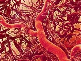
How to print out a blood vessel
New work moves closer to the age of organs on demand.

http://www.nature.com/news/2008/080320/images/news.2008.675.jpg
Blood vessels can now be ‘printed out’ by machine. Could bigger structures be in the future?SUSUMU NISHINAGA / SCIENCE PHOTO LIBRARY
Leave a Reply