Feeds:
Posts
Comments

Posts Tagged ‘Protein folding’

Effect of mitochondrial stress on epigenetic modifiers

Larry H. Bernstein, MD, FCAP, Curator

LPBI


Early Mitochondrial Stress Alters Epigenetics, Secures Lifelong Health Benefits

GEN 5/3/2016  http://www.genengnews.com/gen-news-highlights/early-mitochondrial-stress-alters-epigenetics-secures-lifelong-health-benefits/81252685/

A little adversity builds character, or so the saying goes. True or not, the saying does seem an apt description of a developmental phenomenon that shapes gene expression. While it knows nothing of character, the gene expression apparatus appears to respond well to short-term mitochondrial stress that occurs early in development. In fact, transient stress seems to result in lasting benefits. These benefits, which include improved metabolic function and increased longevity, have been observed in both worms and mice, and may even occur—or be made to occur—in humans.

Gene expression is known to be subject to reprogramming by epigenetic modifiers, but such modifiers generally affect metabolism or lifespan, not both. A new set of epigenetic modifiers, however, has been found to trigger changes that do just that—both improve metabolism and extend lifespan.

Scientists based at the University of California, Berkeley, and the École Polytechnique Fédérale de Lausanne (EPFL) have discovered enzymes that are ramped up after mild stress during early development and continue to affect the expression of genes throughout the animal’s life. When the scientists looked at strains of inbred mice that have radically different lifespans, those with the longest lifespans had significantly higher expression of these enzymes than did the short-lived mice.

“Two of the enzymes we discovered are highly, highly correlated with lifespan; it is the biggest genetic correlation that has ever been found for lifespan in mice, and they’re both naturally occurring variants,” said Andrew Dillin, a UC Berkeley professor of molecular and cell biology. “Based on what we see in worms, boosting these enzymes could reprogram your metabolism to create better health, with a possible side effect of altering lifespan.”

Details of the work, which appeared online April 29 in the journal Cell, are presented in a pair of papers. One paper (“Two Conserved Histone Demethylases Regulate Mitochondrial Stress-Induced Longevity”) resulted from an effort led by Dillin and the EPFL’s Johan Auwerx. The other paper (“Mitochondrial Stress Induces Chromatin Reorganization to Promote Longevity and UPRmt”) resulted from an effort led by Dillin and his UC Berkeley colleague Barbara Meyer.

According to these papers, mitochondrial stress activates enzymes in the brain that affect DNA folding, exposing a segment of DNA that contains the 1500 genes involved in the work of the mitochondria. A second set of enzymes then tags these genes, affecting their activation for much or all of the lifetime of the animal and causing permanent changes in how the mitochondria generates energy.

The first set of enzymes—methylases, in particular LIN-65—add methyl groups to the DNA, which can silence promoters and thus suppress gene expression. By also opening up the mitochondrial genes, these methylases set the stage for the second set of enzymes—demethylases, in this case jmjd-1.2 and jmjd-3.1—to ramp up transcription of the mitochondrial genes. When the researchers artificially increased production of the demethylases in worms, all the worms lived longer, a result identical to what is observed after mitochondrial stress.

“By changing the epigenetic state, these enzymes are able to switch genes on and off,” Dillin noted. This happens only in the brain of the worm, however, in areas that sense hunger or satiety. “These genes are expressed in neurons that are sensing the nutritional status of the animal, and these signals emanate out to the periphery to change peripheral metabolism,” he continued.

When the scientists profiled enzymes in short- and long-lived mice, they found upregulation of these genes in the brains of long-lived mice, but not in other tissues or in the brains of short-lived mice. “These genes are expressed in the hypothalamus, exactly where, when you eat, the signals are generated that tell you that you are full. And when you are hungry, signals in that region tell you to go and eat,” Dillin explained said. “These genes are all involved in peripheral feedback.”

Among the mitochondrial genes activated by these enzymes are those involved in the body’s response to proteins that unfold, which is a sign of stress. Increased activity of the proteins that refold other proteins is another hallmark of longer life.

These observations suggest that the reversal of aging by epigenetic enzymes could also take place in humans.

“It seems that, while extreme metabolic stress can lead to problems later in life, mild stress early in development says to the body, ‘Whoa, things are a little bit off-kilter here, let’s try to repair this and make it better.’ These epigenetic switches keep this up for the rest of the animal’s life,” Dillin stated.


Two Conserved Histone Demethylases Regulate Mitochondrial Stress-Induced Longevity

Carsten Merkwirth6, Virginija Jovaisaite6, Jenni Durieux,…., Reuben J. Shaw, Johan Auwerx, Andrew Dillin

Highlights
  • H3K27 demethylases jmjd-1.2 and jmjd-3.1 are required for ETC-mediated longevity
  • jmjd-1.2 and jmjd-3.1 extend lifespan and are sufficient for UPRmt activation
  • UPRmt is required for increased lifespan due to jmjd-1.2 or jmjd-3.1 overexpression
  • JMJD expression is correlated with UPRmt and murine lifespan in inbred BXD lines

Across eukaryotic species, mild mitochondrial stress can have beneficial effects on the lifespan of organisms. Mitochondrial dysfunction activates an unfolded protein response (UPRmt), a stress signaling mechanism designed to ensure mitochondrial homeostasis. Perturbation of mitochondria during larval development in C. elegans not only delays aging but also maintains UPRmt signaling, suggesting an epigenetic mechanism that modulates both longevity and mitochondrial proteostasis throughout life. We identify the conserved histone lysine demethylases jmjd-1.2/PHF8 and jmjd-3.1/JMJD3 as positive regulators of lifespan in response to mitochondrial dysfunction across species. Reduction of function of the demethylases potently suppresses longevity and UPRmt induction, while gain of function is sufficient to extend lifespan in a UPRmt-dependent manner. A systems genetics approach in the BXD mouse reference population further indicates conserved roles of the mammalian orthologs in longevity and UPRmt signaling. These findings illustrate an evolutionary conserved epigenetic mechanism that determines the rate of aging downstream of mitochondrial perturbations.

Figure thumbnail fx1


Mitochondrial Stress Induces Chromatin Reorganization to Promote Longevity and UPRmt
Ye Tian, Gilberto Garcia, Qian Bian, Kristan K. Steffen, Larry Joe, Suzanne Wolff, Barbara J. Meyer, Andrew Dillincorrespondence
http://dx.doi.org/10.1016/j.cell.2016.04.011             Publication stage: In Press Corrected Proof
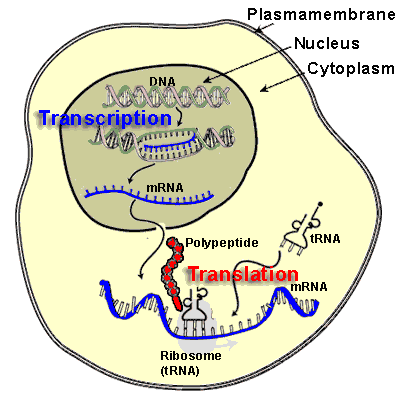
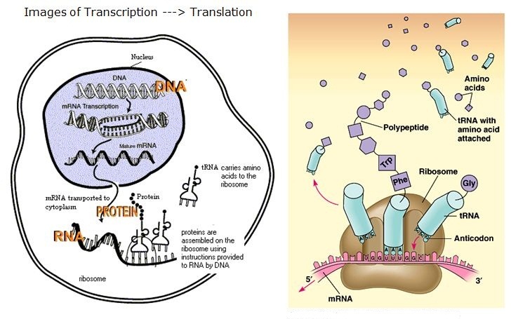
Highlights
  • LIN-65 accumulates in the nucleus in response to mitochondrial stress
  • Mitochondrial stress-induced chromatin changes depend on MET-2 and LIN-65
  • LIN-65 and DVE-1 exhibit interdependence in nuclear accumulation
  • met-2 and atfs-1 act in parallel to affect mitochondrial stress-induced longevity

Organisms respond to mitochondrial stress through the upregulation of an array of protective genes, often perpetuating an early response to metabolic dysfunction across a lifetime. We find that mitochondrial stress causes widespread changes in chromatin structure through histone H3K9 di-methylation marks traditionally associated with gene silencing. Mitochondrial stress response activation requires the di-methylation of histone H3K9 through the activity of the histone methyltransferase met-2 and the nuclear co-factor lin-65. While globally the chromatin becomes silenced by these marks, remaining portions of the chromatin open up, at which point the binding of canonical stress responsive factors such as DVE-1 occurs. Thus, a metabolic stress response is established and propagated into adulthood of animals through specific epigenetic modifications that allow for selective gene expression and lifespan extension

 Siddharta Mukherjee’s Writing Career Just Got Dealt a Sucker Punch
Author: Theral Timpson

Siddharha Mukherjee won the 2011 Pulitzer Prize in non-fiction for his book, The Emperor of All Maladies.  The book has received widespread acclaim among lay audience, physicians, and scientists alike.  Last year the book was turned into a special PBS series.  But, according to a slew of scientists, we should all be skeptical of his next book scheduled to hit book shelves this month, The Gene, An Intimate History.

Publishing an article on epigenetics in the New Yorker this week–perhaps a selection from his new book–Mukherjee has waltzed into one of the most active scientific debates in all of biology: that of gene regulation, or epigenetics.

Jerry Coyne, the evolutionary biologist known for keeping journalists honest, has published a two part critique of Mukherjee’s New Yorker piece.  The first part–wildly tweeted yesterday–is a list of quotes from Coyne’s colleagues and those who have written in to the New Yorker, including two Nobel prize winners, Wally Gilbert and Sidney Altman, offering some very unfriendly sentences.

Wally Gilbert: “The New Yorker article is so wildly wrong that it defies rational analysis.”

Sidney Altman:  “I am not aware that there is such a thing as an epigenetic code.  It is unfortunate to inflict this article, without proper scientific review, on the audience of the New Yorker.”

The second part is a thorough scientific rebuttal of the Mukherjee piece.  It all serves as a great drama about one of the most contested ideas in biology and also as a cautionary tale to journalists, even experienced writers such as Mukherjee, about the dangers of wading into scientific arguments.  Readers may remember that a few years ago, science writer, David Dobbs, similarly skated into the same topic with his piece, Die, Selfish Gene, Die, and which raised a similar shitstorm, much of it from Coyne.

Mukherjee’s mistake is in giving credence to only one side of a very fierce debate–that the environment causes changes in the genome which can be passed on; another kind of evolution–as though it were settled science.   Either Mukherjee, a physicisan coming off from a successful book and PBS miniseries on cancer, is setting himself up as a scientist, or he has been a truly naive science reporter.   If he got this chapter so wrong, what does it mean about an entire book on the gene?

Coyne quotes one of his colleagues who raised some questions about the New Yorker’s science reporting, one particular question we’ve been asking here at Mendelspod.  How do we know what we know?  Does science now have an edge on any other discipline for being able to create knowledge?

Coyne’s colleague is troubled by science coverage in the New Yorker, and goes so far as to write that the New Yorker has been waging a “war on behalf of cultural critics and literary intellectuals against scientists and technologists.”

From my experience, it’s not quite that tidy.  First of all, the New Yorker is the best writing I read each week.  Period.  Second, I haven’t found their science writing to have the slant claimed in the quote above.  For example, most other mainstream outlets–including the New York Times with the Amy Harmon pieces–have given the anti-GMO crowd an equal say in the mistaken search for a “balance” on whether GMOs are harmful.  (Remember John Stewart’s criticism of Fox News?  That they give a false equivalent between two sides even when there is no equivalent on the other side?)

But the New Yorker has not fallen into this trap on GMOs and most of their pieces on the topic–mainly by Michael Specter–have been decidedly pro science and therefore decided pro GMO.

So what led Mukherjee to play scientist as well as journalist?  There’s no question about whether I enjoy his prose.  His writing beautifully whisks me away so that I don’t feel that I’m really working to understand.  There is a poetic complexity that constantly brings different threads effortlessly together, weaving them into the same light.  At one point he uses the metaphor of a web for the genome, with the epigenome being the stuff that sticks to the web.  He borrows the metaphor from the Hindu notion of “being”, or jaal.

“Genes form the threads of the web; the detritus that adheres to it transforms every web into a singular being.”

There have been a few writers on Twitter defending Mukherjee’s piece.  Tech Review’s Antonio Regalado called Coyne and his colleagues “tedious literalists” who have an “issue with epigenetic poetry.”

At his best, Mukherjee can take us down the sweet alleys of his metaphors and family stories with a new curiosity for the scientific truth.  He can hold a mirror up to scientists, or put the spotlight on their work.   At their worst, Coyne and his scientific colleagues can reek of a fear of language and therefore metaphor.  The always outspoken scientist and author, Richard Dawkins, who made his name by personifying the gene, was quick to personify epigentics in a tweet:   “It’s high time the 15 minutes of underserved fame for “epigenetics” came to an overdue end.”  Dawkins is that rare scientist who has consistently been as comfortable with rhetoric and language as he is with data.

Hats off to Coyne who reminds us that a metaphor–however lovely–does not some science make. If Mukherjee wants to play scientist, let him create and gather data. If it’s the role of science journalist he wants, let him collect all the science he can before he begins to pour it into his poetry.


Same but Different  

How epigenetics can blur the line between nature and nurture.

Annals of Science MAY 2, 2016 ISSUE     BY

http://www.newyorker.com/wp-content/uploads/2016/05/160502_r28072-1200.jpg

The author’s mother (right) and her twin are a study in difference and identity. CREDIT: PHOTOGRAPH BY DAYANITA SINGH FOR THE NEW YORKER

October 6, 1942, my mother was born twice in Delhi. Bulu, her identical twin, came first, placid and beautiful. My mother, Tulu, emerged several minutes later, squirming and squalling. The midwife must have known enough about infants to recognize that the beautiful are often the damned: the quiet twin, on the edge of listlessness, was severely undernourished and had to be swaddled in blankets and revived.

The first few days of my aunt’s life were the most tenuous. She could not suckle at the breast, the story runs, and there were no infant bottles to be found in Delhi in the forties, so she was fed through a cotton wick dipped in milk, and then from a cowrie shell shaped like a spoon. When the breast milk began to run dry, at seven months, my mother was quickly weaned so that her sister could have the last remnants.
Tulu and Bulu grew up looking strikingly similar: they had the same freckled skin, almond-shaped face, and high cheekbones, unusual among Bengalis, and a slight downward tilt of the outer edge of the eye, something that Italian painters used to make Madonnas exude a mysterious empathy. They shared an inner language, as so often happens with twins; they had jokes that only the other twin understood. They even smelled the same: when I was four or five and Bulu came to visit us, my mother, in a bait-and-switch trick that amused her endlessly, would send her sister to put me to bed; eventually, searching in the half-light for identity and difference—for the precise map of freckles on her face—I would realize that I had been fooled.

But the differences were striking, too. My mother was boisterous. She had a mercurial temper that rose fast and died suddenly, like a gust of wind in a tunnel. Bulu was physically timid yet intellectually more adventurous. Her mind was more agile, her tongue sharper, her wit more lancing. Tulu was gregarious. She made friends easily. She was impervious to insults. Bulu was reserved, quieter, and more brittle. Tulu liked theatre and dancing. Bulu was a poet, a writer, a dreamer.

….. more

Why are identical twins alike? In the late nineteen-seventies, a team of scientists in Minnesota set out to determine how much these similarities arose from genes, rather than environments—from “nature,” rather than “nurture.” Scouring thousands of adoption records and news clips, the researchers gleaned a rare cohort of fifty-six identical twins who had been separated at birth. Reared in different families and different cities, often in vastly dissimilar circumstances, these twins shared only their genomes. Yet on tests designed to measure personality, attitudes, temperaments, and anxieties, they converged astonishingly. Social and political attitudes were powerfully correlated: liberals clustered with liberals, and orthodoxy was twinned with orthodoxy. The same went for religiosity (or its absence), even for the ability to be transported by an aesthetic experience. Two brothers, separated by geographic and economic continents, might be brought to tears by the same Chopin nocturne, as if responding to some subtle, common chord struck by their genomes.

One pair of twins both suffered crippling migraines, owned dogs that they had named Toy, married women named Linda, and had sons named James Allan (although one spelled the middle name with a single “l”). Another pair—one brought up Jewish, in Trinidad, and the other Catholic, in Nazi Germany, where he joined the Hitler Youth—wore blue shirts with epaulets and four pockets, and shared peculiar obsessive behaviors, such as flushing the toilet before using it. Both had invented fake sneezes to diffuse tense moments. Two sisters—separated long before the development of language—had invented the same word to describe the way they scrunched up their noses: “squidging.” Another pair confessed that they had been haunted by nightmares of being suffocated by various metallic objects—doorknobs, fishhooks, and the like.

The Minnesota twin study raised questions about the depth and pervasiveness of qualities specified by genes: Where in the genome, exactly, might one find the locus of recurrent nightmares or of fake sneezes? Yet it provoked an equally puzzling converse question: Why are identical twins different? Because, you might answer, fate impinges differently on their bodies. One twin falls down the crumbling stairs of her Calcutta house and breaks her ankle; the other scalds her thigh on a tipped cup of coffee in a European station. Each acquires the wounds, calluses, and memories of chance and fate. But how are these changes recorded, so that they persist over the years? We know that the genome can manufacture identity; the trickier question is how it gives rise to difference.

….. more

But what turns those genes on and off, and keeps them turned on or off? Why doesn’t a liver cell wake up one morning and find itself transformed into a neuron? Allis unpacked the problem further: suppose he could find an organism with two distinct sets of genes—an active set and an inactive set—between which it regularly toggled. If he could identify the molecular switches that maintain one state, or toggle between the two states, he might be able to identify the mechanism responsible for cellular memory. “What I really needed, then, was a cell with these properties,” he recalled when we spoke at his office a few weeks ago. “Two sets of genes, turned ‘on’ or ‘off’ by some signal.”

more…

“Histones had been known as part of the inner scaffold for DNA for decades,” Allis went on. “But most biologists thought of these proteins merely as packaging, or stuffing, for genes.” When Allis gave scientific seminars in the early nineties, he recalled, skeptics asked him why he was so obsessed with the packing material, the stuff in between the DNA.  …. A skein of silk tangled into a ball has very different properties from that same skein extended; might the coiling or uncoiling of DNA change the activity of genes?

In 1996, Allis and his research group deepened this theory with a seminal discovery. “We became interested in the process of histone modification,” he said. “What is the signal that changes the structure of the histone so that DNA can be packed into such radically different states? We finally found a protein that makes a specific chemical change in the histone, possibly forcing the DNA coil to open. And when we studied the properties of this protein it became quite clear that it was also changing the activity of genes.” The coils of DNA seemed to open and close in response to histone modifications—inhaling, exhaling, inhaling, like life.

Allis walked me to his lab, a fluorescent-lit space overlooking the East River, divided by wide, polished-stone benches. A mechanical stirrer, whirring in a corner, clinked on the edge of a glass beaker. “Two features of histone modifications are notable,” Allis said. “First, changing histones can change the activity of a gene without affecting the sequence of the DNA.” It is, in short, formally epi-genetic, just as Waddington had imagined. “And, second, the histone modifications are passed from a parent cell to its daughter cells when cells divide. A cell can thus record ‘memory,’ and not just for itself but for all its daughter cells.”

…..


The New Yorker screws up big time with science: researchers criticize the Mukherjee piece on epigenetics

Jerry Coyne
https://whyevolutionistrue.wordpress.com/2016/05/05/the-new-yorker-screws-up-big-time-with-science-researchers-criticize-the-mukherjee-piece-on-epigenetics/

Abstract: This is a two part-post about a science piece on gene regulation that just appeared in the New Yorker. Today I give quotes from scientists criticizing that piece; tomorrow I’ll present a semi-formal critique of the piece by two experts in the field.

esterday I gave readers an assignment: read the new New Yorkerpiece by Siddhartha Mukherjee about epigenetics. The piece, called “Same but different” (subtitle: “How epigenetics can blur the line between nature and nurture”) was brought to my attention by two readers, both of whom praised it.  Mukherjee, a physician, is well known for writing the Pulitzer-Prize-winning book (2011) The Emperor of All Maladies: A Biography of Cancer. (I haven’t read it yet, but it’s on my list.)  Mukherjee has a new book that will be published in May: The Gene: An Intimate History. As I haven’t seen it, the New Yorker piece may be an excerpt from this book.

Everyone I know who has read The Emperor of All Maladies gives it high praise. I wish I could say the same for Mukherjee’s New Yorker piece. When I read it at the behest of the two readers, I found his analysis of gene regulation incomplete and superficial. Although I’m not an expert in that area, I knew that there was a lot of evidence that regulatory proteins called “transcription factors”, and not “epigenetic markers” (see discussion of this term tomorrow) or modified histones—the factors emphasized by Mukherjee—played hugely important roles in gene regulation. The speculations at the end of the piece about “Lamarckian evolution” via environmentally induced epigenetic changes in the genome were also unfounded, for we have no evidence for that kind of adaptive evolution. Mukherjee does, however, mention that lack of evidence, though I wish he’d done so more strongly given that environmental modification of DNA bases is constantly touted as an important and neglected factor in evolution.

Unbeknownst to me, there was a bit of a kerfuffle going on in the community of scientists who study gene regulation, with many of them finding serious mistakes and omissions in Mukherjee’s piece.  There appears to have been some back-and-forth emailing among them, and several wrote letters to the New Yorker, urging them to correct the misconceptions, omissions, and scientific errors in “Same but different.” As I understand it, both Mukherjee and the New Yorker simply batted these criticisms away, and, as far as I know, will not publish any corrections.  So today and tomorrow I’ll present the criticisms here, just so they’ll be on the record.

Because Mukherjee writes very well, and because even educated laypeople won’t know the story of gene regulation revealed over the last few decades,  they may not see the big lacunae in his piece. It is, then,  important to set matters straight, for at least we should know what science has told us about how genes are turned on and off. The criticism of Mukherjee’s piece, coming from scientists who really are experts in gene regulation, shows a lack of care on the part of Mukherjee and theNew Yorker: both a superficial and misleading treatment of the state of the science, and a failure of the magazine to properly vet this piece (I have no idea whether they had it “refereed” not just by editors but by scientists not mentioned in the piece).

Let me add one thing about science and the New Yorker. I believe I’ve said this before, but the way the New Yorker treats science is symptomatic of the “two cultures” problem. This is summarized in an email sent me a while back by a colleague, which I quote with permission:

The New Yorker is fine with science that either serves a literary purpose (doctors’ portraits of interesting patients) or a political purpose (environmental writing with its implicit critique of modern technology and capitalism). But the subtext of most of its coverage (there are exceptions) is that scientists are just a self-interested tribe with their own narrative and no claim to finding the truth, and that science must concede the supremacy of literary culture when it comes to anything human, and never try to submit human affairs to quantification or consilience with biology. Because the magazine is undoubtedly sophisticated in its writing and editing they don’t flaunt their postmodernism or their literary-intellectual proprietariness, but once you notice it you can make sense of a lot of their material.

. . . Obviously there are exceptions – Atul Gawande is consistently superb – but as soon as you notice it, their guild war on behalf of cultural critics and literary intellectuals against scientists, technologists, and analytic scholars becomes apparent.

…. more

Researchers criticize the Mukherjee piece on epigenetics: Part 2

Trigger warning: Long science post!

Yesterday I provided a bunch of scientists’ reactions—and these were big names in the field of gene regulation—to Siddhartha Mukherjee’s ill-informed piece in The New Yorker, “Same but different” (subtitle: “How epigenetics can blur the line between nature and nurture”). Today, in part 2, I provide a sentence-by-sentence analysis and reaction by two renowned researchers in that area. We’ll start with a set of definitions (provided by the authors) that we need to understand the debate, and then proceed to the critique.

Let me add one thing to avoid confusion: everything below the line, including the definition (except for my one comment at the end) was written by Ptashne and Greally.

by Mark Ptashne and John Greally

Introduction

Ptashne is The Ludwig Professor of Molecular Biology at the Memorial Sloan Kettering Cancer Center in New York. He wrote A Genetic Switch, now in its third edition, which describes the principles of gene regulation and the workings of a ‘switch’; and, with Alex Gann, Genes and Signals, which extends these principles and ideas to higher organisms and to other cellular processes as well.  John Greally is the Director of the Center for Epigenomics at the Albert Einstein College of Medicine in New York.


The New Yorker  (May 2, 2016) published an article entitled “Same But Different” written by Siddhartha Mukherjee.  As readers will have gathered from the letters posted yesterday, there is a concern that the article is misleading, especially for a non-scientific audience. The issue concerns our current understanding of “gene regulation” and how that understanding has been arrived at.

First some definitions/concepts:

Gene regulation refers to the “turning on and off of genes”.  The primary event in turning a gene “on” is to transcribe (copy) it into messenger RNA (mRNA). That mRNA is then decoded, usually, into a specific protein.  Genes are transcribed by the enzyme called RNA polymerase.

Development:  the process in which a fertilized egg (e.g., a human egg) divides many times and eventually forms an organism.  During this process, many of the roughly 23,000 genes of a human are turned “on” or “off” in different combinations, at different times and places in the developing organism. The process produces many different cell types in different organs (e.g. liver and brain), but all retain the original set of genes.

Transcription factors: proteins that bind to specific DNA sequences near specific genes and turn transcription of those genes on and off. A transcriptional ‘activator’, for example, bears two surfaces: one binds a specific sequence in DNA, and the other binds to, and thereby recruits to the gene, protein complexes that include RNA polymerase. It is widely acknowledged that the identity of a cell in the body depends on the array of transcription factors present in the cell, and the cell’s history.  RNA molecules can also recognize specific genomic sequences, and they too sometimes work as regulators.  Neither transcription factors nor these kinds of RNA molecules – the fundamental regulators of gene expression and development – are mentioned in the New Yorker article.

Signals:  these come in many forms (small molecules like estrogen, larger molecules (often proteins such as cytokines) that determine the ability of transcription factors to work.  For example, estrogen binds directly to a transcription factor (the estrogen receptor) and, by changing its shape, permits it to bind DNA and activate transcription.

Memory”:  a dividing cell can (often does) produce daughters that are identical, and that express identical genes as does the mother cell.  This occurs because the transcription factors present in the mother cell are passively transmitted to the daughters as the cell divides, and they go to work in their new contexts as before.  To make two different daughters, the cell must distribute its transcription factors asymmetrically.

Positive Feedback: An activator can maintain its own expression by  positive feedback.  This requires, simply, that a copy of the DNA sequence to which the activator binds is  present  near its own gene. Expression of the activator  then becomes self-perpetuating.  The activator (of which there now are many copies in the cell) activates  other target genes as it maintains its own expression. This kind of ‘memory circuit’, first described  in  bacteria, is found in higher organisms as well.  Positive feedback can explain how a fully differentiated cell (that is, a cell that has reached its developmental endpoint) maintains its identity.

Nucleosomes:  DNA in higher organisms (eukaryotes) is wrapped, like beads on a string, around certain proteins (called histones), to form nucleosomes.  The histones are subject to enzymatic modifications: e.g., acetyl, methyl, phosphate, etc. groups can be added to these structures. In bacteria there are no nucleosomes, and the DNA is more or less ‘naked’.

“Epigenetic modifications: please don’t worry about the word ”epigenetic”; it is misused in any case. What Mukherjee refers to by this term are the histone modifications mentioned above, and a modification to DNA itself: the addition of methyl groups. Keep in mind that the organisms that have taught us the most about development – flies (Drosophila) and worms (C. elegans)—do not have the enzymes required for DNA methylation. That does not mean that DNA methylation cannot do interesting things in humans, for example, but it is obviously not at the heart of gene regulation.

Specificity Development requires the highly specific sequential turning on and off of sets of genes.  Transcription factors and RNA supply this specificity, but   enzymes that impart modifications to histones  cannot: every nucleosome (and hence every gene) appears the same to the enzyme.  Thus such enzymes cannot pick out particular nucleosomes associated with particular genes to modify.  Histone modifications might be imagined to convey ‘memory’ as cells divide – but there are no convincing indications that this happens, nor are there molecular models that might explain why they would have the imputed effects.

Analysis and critique of Mukherjee’s article

The picture we have just sketched has taken the combined efforts of many scientists over 50 years to develop.  So what, then, is the problem with the New Yorker article?

There are two: first, the picture we have just sketched, emphasizing the primary role of transcription factors and RNA, is absent.  Second, that picture is replaced by highly dubious speculations, some of which don’t make sense, and none of which has been shown to work as imagined in the article.

(Quotes from the Mukherjee article are indented and in plain text; they are followed by comments, flush left and in bold, by Ptashne and Greally.)

In 1978, having obtained a Ph.D. in biology at Indiana University, Allis began to tackle a problem that had long troubled geneticists and cell biologists: if all the cells in the body have the same genome, how does one become a nerve cell, say, and another a blood cell, which looks and functions very differently?

The problems referred to were recognized long before 1978.  In fact, these were exactly the problems that the great French scientists François Jacob and Jacques Monod took on in the 1950s-60s.  In a series of brilliant experiments, Jacob and Monod showed that in bacteria, certain genes encode products that regulate (turn on and off) specific other genes.  Those regulatory molecules turned out to be proteins, some of which respond to signals from the environment.  Much of the story of modern biology has been figuring out how these proteins – in bacteria and in higher organisms  – bind to and regulate specific genes.  Of note is that in higher organisms, the regulatory proteins look and act like those in bacteria, despite the fact that eukaryotic DNA is wrapped in nucleosomes  whereas bacterial DNA is not.   We have also learned that certain RNA molecules can play a regulatory role, a phenomenon made possible by the fact that RNA molecules, like regulatory proteins, can recognize specific genomic sequences.

In the nineteen-forties, Conrad Waddington, an English embryologist, had proposed an ingenious answer: cells acquired their identities just as humans do—by letting nurture (environmental signals) modify nature (genes). For that to happen, Waddington concluded, an additional layer of information must exist within a cell—a layer that hovered, ghostlike, above the genome. This layer would carry the “memory” of the cell, recording its past and establishing its future, marking its identity and its destiny but permitting that identity to be changed, if needed. He termed the phenomenon “epigenetics”—“above genetics.”

This description greatly misrepresents the original concept.  Waddington argued that development proceeds not by the loss (or gain) of genes, which would be a “genetic” process, but rather that some genes would be selectively expressed in specific and complex cellular patterns as development proceeds.  He referred to this intersection of embryology (then called “epigenesis”) and genetics as “epigenetic”.We now understand that regulatory proteins work in combinations to turn on and off genes, including their own genes, and that sometimes the regulatory proteins respond to signals sent by other cells.  It should be emphasized that Waddington never proposed any “ghost-like” layer of additional information hovering above the gene.  This is a later misinterpretation of a literal translation of the term epigenetics, with “epi-“ meaning “above/upon” the genetic information encoded in DNA sequence.  Unfortunately, this new and pervasive definition encompasses all of transcriptional regulation and is of no practical value.

…..more

By 2000, Allis and his colleagues around the world had identified a gamut of proteins that could modify histones, and so modulate the activity of genes. Other systems, too, that could scratch different kinds of code on the genome were identified (some of these discoveries predating the identification of histone modifications). One involved the addition of a chemical side chain, called a methyl group, to DNA. The methyl groups hang off the DNA string like Christmas ornaments, and specific proteins add and remove the ornaments, in effect “decorating” the genome. The most heavily methylated parts of the genome tend to be dampened in their activity.

It is true that enzymes that modify histones have been found—lots of them.  A striking problem is that, after all this time, it is not at all clear what the vast majority of these modifications do.  When these enzymatic activities are eliminated by mutation of their active sites (a task substantially easier to accomplish in yeast than in higher organisms) they mostly have little or no effect on transcription.  It is not even clear that histones are the biologically relevant substrates of most of these enzymes.  

 In the ensuing decade, Allis wrote enormous, magisterial papers in which a rich cast of histone-modifying proteins appear and reappear through various roles, mapping out a hatchwork of complexity. . . These protein systems, overlaying information on the genome, interacted with one another, reinforcing or attenuating their signals. Together, they generated the bewildering intricacy necessary for a cell to build a constellation of other cells out of the same genes, and for the cells to add “memories” to their genomes and transmit these memories to their progeny. “There’s an epigenetic code, just like there’s a genetic code,” Allis said. “There are codes to make parts of the genome more active, and codes to make them inactive.”

By ‘epigenetic code’ the author seems to mean specific arrays of nucleosome modifications, imparted over time and cell divisions, marking genes for expression.  This idea has been tested in many experiments and has been found not to hold.

….. and more


Larry H. Bernstein, MD, FCAP

I hope that this piece brings greater clarity to the discussion.  I have heard the use of the term “epigenetics” for over a decade.  The term was never so clear.  I think that the New Yorker article was a reasonable article for the intended audience.  It was not intended to clarify debates about a mechanism for epigenetic based changes in evolutionary science.  I think it actually punctures the “classic model” of the cell depending only on double stranded DNA and transcription, which deflates our concept of the living cell.  The concept of epigenetics was never really formulated as far as I have seen, and I have done serious work in enzymology and proteins at a time that we did not have the technology that exists today.  I have considered with the critics that protein folding, protein misfolding, protein interactions with proximity of polar and nonpolar groups, and the regulatory role of microRNAs that are not involved in translation, and the evolving concept of what is “dark (noncoding) DNA” lend credence to the complexity of this discussion.  Even more interesting is the fact that enzymes (and isoforms of enzymes) have a huge role in cellular metabolic differences and in the function of metabolic pathways.  What is less understood is the extremely fast reactions involved in these cellular reactions.  These reactions are in my view critical drivers.  This is brought out by Erwin Schroedinger in the book What is Life? which infers that there can be no mathematical expression of life processes.


Read Full Post »

Prions and protein misfolding disorders

Larry H. Bernstein, MD, FCAP, Curator

LPBI


Prions and protein-folding diseases
E. Norrby
J Intern Med 2011; 270: 1–14. http://dx.doi.org:/10.1111/j.1365-2796.2011.02387.x

Prions represent a group of proteins with a unique capacity to fold into different conformations. One isoform is rich in beta-pleated sheets and can aggregate into amyloid that may be pathogenic. This abnormal form propagates itself by imposing its confirmation on the homologous normal host cell protein. Pathogenic prions have been shown to cause lethal neurodegenerative diseases in humans and animals. These diseases are sometimes infectious and hence referred to as transmissible spongiform encephalopathies. In the present review, the remarkable evolution of the heterodox prion concept is summarized. The origin of this phenomenon is based on information transfer between homologous proteins, without the involvement of nucleic acid-encoded mechanisms. Historically, kuru and Creutzfeldt-Jakob disease (CJD) were the first infectious prion diseases to be identified in man. It was their relationship to scrapie in sheep and experimental rodents that allowed an unravelling of the particular molecular mechanism that underlie the disease process. Transmission between humans has been documented to have occurred in particular contexts, including ritual cannibalism, iatrogenic transmission because of pituitary gland-derived growth hormone or the use in neurosurgical procedures of dura mater from cadavers, and the temporary use of a prion-contaminated protein-rich feed for cows. The latter caused a major outbreak of bovine spongiform encephalopathy, which spread to man by human consumption of contaminated meat, causing approximately 200 cases of variant CJD. All these epidemics now appear to be over because of measures taken to curtail further spread of prions. Recent studies have shown that the mechanism of protein aggregation may apply to a wider range of diseases in and possibly also outside the brain, some of which are relatively common such as Alzheimer’s and Parkinson’s diseases. Furthermore, it has become apparent that the phenomenon of prion aggregation may have a wider physiological importance, but a full understanding of this remains to be defined. It may involve maintaining neuronal functions and possibly contributing to the establishment of long-term memory.

The history of the identification of the infectious nature of prion diseases and the discovery of the chemical nature of this type of infectious agent is remarkable. The great advances in this field of research have been recognized by two Nobel Prizes in Physiology or Medicine: one in 1976 to D. Carleton Gajdusek (a prize shared with Baruch S. Blumberg, for the discovery of hepatitis B virus) and the other in 1997 to Stanley B. Prusiner. Infectious prion diseases represent relatively rare phenomena mostly observed in the context of the spread of the agent by human intervention. However, the principal molecular mechanisms that lead to disease may have applications for a number of much more common noncontagious diseases. Two fundamental observations are relevant to the understanding of the molecular mechanisms involved. The first is that the same polypeptide chain, depending on the environmental conditions, including the possible presence of homologous proteins with a predetermined folding pattern, may take on dramatically altered folding. In certain cases, major aggregates of homologous proteins may be formed and such aggregates in turn may display cytopathogenic effects. A degenerative disease may ensue. The second relevant observation is that much still remains to be learnt about protein-folding phenomena and the role of information transfer systems engaging only proteins. Many proteins have sequences of amino acids that make them potentially prionogenic. Such sequences that under certain circumstances may be the cause of disease in mammals can, in another context, play a central role in physiological functions, for example, as the source of epigenetic mechanisms of protein signalling of importance for the survival of yeast cells.

In this review, our emerging understanding of the mechanisms of prion diseases will first be discussed, in particular their unique mechanisms of spread will be considered. The original belief in relatively firm barriers preventing the spread of prions between species was questioned when it became clear that bovine spongiform encephalopathy (BSE), also known as ‘mad cow disease’, could be transmitted to man. It was then shown, surprisingly, that the disease contracted from infected cattle could spread from man to man by blood transfusion. Next, the possibility of a much wider application of the pathogenic mechanism of cell destruction by protein aggregation, including degenerative processes causing for example Alzheimer’s and Parkinson’s diseases, will be discussed. The rapidly growing appreciation of the significance of signaling between proteins outside the canonical steps of the central dogma ofmolecular biology will finally be considered.  ….

Unique samples of brain collected by Gajdusek allowed identification of the histopathology of kuru. It showed many similarities to the changes in brains from patients with sporadic sCJD, but there were also similarities to the changes in brains from sheep with scrapie as noted by Hadlow [2]. His observation encouraged Gajdusek to attempt to transmit these non-inflammatory diseases to chimpanzees. He and his collaborators were successful in transmitting first kuru [3] and then CJD [4] by intracerebral inoculation of chimpanzees. The term transmissible spongiform encephalitis (TSE) was commonly used to refer to this kind of infection.

Next, after determining that kuru was infectious, the route of transmission was found to be the ritual cannibalism practiced by the Fore people [5]. The body of the deceased relative was prepared for the funeral meal, and the central nervous system containing the largest concentration of the infectious agent was consumed mainly by the women and children; thus, they were primarily affected by the disease (Fig. 1). However, no child born after 1960, the year in which the practice of ritual cannibalism ceased, has contracted kuru. The incubation time of the disease can be long, potentially longer than the life span of the individual, and cases of kuru have occurred in the present century [6]. The infectious nature of sCJD had been demonstrated, but it was initially unclear how it might spread. In fact, under normal conditions, CJD is not infectious. To date, careful studies of transfusion of blood from people incubating or even displaying early signs of CJD have not revealed any spread of the agent between individuals by this route [7]. Only under conditions of medical interventions involving brain material could an iatrogenic spread of the spontaneous form of the disease be demonstrated.


Throughout the 1970s, Gajdusek continued to refer to the infectious agents that cause kuru⁄CJD as slow viruses, and this situation did not change until Prusiner began to gain insights into the chemical nature of the agents using hamster scrapie as amodel to isolate and characterize them. He continued to purify the infectious material from hamster brains until he produced a relatively pure protein preparation [8]. Because of the lack of any evidence of participating nucleic acid in this preparation, he named it prion (proteinaceous and infectious particle) [9]. The protein preparation – referred to as PrP 27–30 (prion protein with a molecular weight 27–30 kD) – was pure enough to determine a short section of its amino terminal amino acid sequence. This information made it possible to determine that the gene responsible for the synthesis of the prion protein was not foreign, but a normal host cell gene [10]. Thus, it became possible to explain the absence of any inflammatory response in the tissues in the presence of this infectious disease; under healthy conditions, we do not develop immunological reactions to our own tissues.

Following the identification of the PrP gene, it was possible to produce mice deficient in PrP using ‘knockout’ technology [11]. At this time, it was known that the PrP protein appears in the brain early during embryonic development. It was found, unexpectedly, that development and life span appeared to be normal in animals without PrP protein. The PrP-knockout mice could be used for two types of critical follow-up experiments.

First, after infection with different doses of infectious prion material, the knockout mice were found to be completely refractory to development of disease [12, 13]. Furthermore, they could mount an immune response to the PrP protein, as this no longer represented an endogenous product. For the first time, antibodies could be generated against different parts of the protein. The protein that Prusiner et al. had identified was indeed critical to the pathogenic process in prion diseases.

The second type of experiment was to investigate the effects of reintroducing into the PrP-knockout mice either a modified homologous PrP gene (carrying a substitution or deletion of a single or a stretch of amino acids) or a PrP gene from a different species. The former experiment enabled attempts to dissect the role of different parts of the PrP protein in the pathogenic process (as discussed below). The latter enabled the evaluation of the species specificity of PrP proteins. Mice are normally infected only by prions from othermice or rodents, such as hamsters, but not from more distant species. However, by generation of transgenic mice carrying for example a human or a bovine PrP gene, this species barrier can be overcome providing important opportunities for studies of different kinds of pathogenic PrP proteins of relevance for human disease. Inappropriate folding and aggregation of proteins can cause disease

Diseases caused by protein aggregation have been known for a long time. They are collectively referred to as amyloid diseases, a term reflecting the original misconception that the observed stainable deposits contained starch (amylum in Latin). Later, it was demonstrated that they were in fact composed of various kinds of proteins. The amyloid deposits have characteristic staining properties and show birefringence under polarized light. Protein aggregates showing these characteristics were demonstrated in the brains of patients with CJD. However, amyloid formation is not always a feature of prion disease [14]. It is in fact found in only about 10% of the brains of patients with sCJD, but at a much higher rate in patients with other forms of the disease.

It then became possible to determine the structure of purified PrP using nuclear magnetic resonance analysis [15, 16]. It was shown that PrP can appear in two completely different forms, albeit with an identical amino acid sequence. The ‘healthy’ protein, referred to as PrP-C (control), has a structure involving predominantly two large alpha-helix structures, whereas there is a predominance of beta-pleated sheets in the pathological PrP-TSE protein (Fig. 2). It is the PrP-TSE protein that may form amyloid protein aggregates. The reason for the two completely different configurations of the same protein is not known, but a critical observation is that if a small amount of PrP-TSE is added to a larger amount of PrP-C, the ‘healthy’ protein is converted to the TSE form by an as yet undefined contagious ‘snowball’ effect. Two theoretical models, nucleation-polymerization and template assistance, have been proposed to explain this ([17], Fig. 3). However, as discussed in the next section, only certain kinds of proteins are capable of forming amyloid and are truly infectious, and the term prion is reserved for them; the term prionogenic has been introduced to include noninfectious amyloid-generating proteins.

The awareness that CJD was a disease that could be transmitted to experimental animals immediately raised the question of to what extent itmight be transmissible between humans. There was no epidemiological evidence of connections between cases of CJD, except for an increased frequency of occurrence in certain families and ethnic groups. In the light of understanding the seminal importance of PrP in the disease, it could be deduced that familial cases must be because of inherited mutations in different sites of the PrP gene, whereas sporadic cases were likely to be caused by mutation(s) accumulated in somatic (possibly brain) cells or alternatively a spontaneous emergence of amisfolded PrP protein (the nucleation– polymerization model) during the lifetime of the individual. As already mentioned, to date it has not been possible to find evidence for transmission of disease from individuals with sCJD by blood transmission [7], but relatively recent data provide evidence for a possible transmission of scrapie in sheep by experimental blood transfusion [18].

Gajdusek and collaborators at an early stage began to search for evidence of the spread of CJD through medical intervention. They found that in CJD, as in scrapie, the majority of infectious prions were located in the brain; indeed, brain tissue is about 100 000 times more infectious than peripheral tissues, such as blood [19]. The first case of iatrogenic spread of CJD between two individuals was found in connection with a corneal transplant [20], and later the similar spread to two relatively young individuals was demonstrated as a result of using electrodes for intracerebral recording [21].

Three epidemics of strikingly different origins have been documented, and all comprise slightly in excess of two hundred cases with the maximum number of cases at the end of the 1990s (Fig. 4). The first of the three epidemics was caused by the use of human growth hormone prepared from pools of many hundreds of pituitary glands from cadavers [see 19 for references]. Because these preparations were used in growing individuals, many of the victims of the disease were relatively young. When this iatrogenic spread of CJD prions was discovered, the product was rapidly withdrawn, and it was progressively replaced by growth hormone prepared by recombinant DNA technology. The average incubation time of this parenterally injected material was estimated to be 15 years (range 4–36 years). The total number of cases to date is 206, and the epidemic seems to have just reached an end. Most cases occurred in France, 109 of 1700 treated individuals. The corresponding figures are 56 of 1848 treated individuals in the UK and 28 of 7700, in the USA. In the USA, an additional step of purification was introduced in 1977, which may have reduced the risk of transmission of infectious prions.

The second epidemic has a distinctly different iatrogenic background. It relates to the previous use of heterologous cadaveric dura mater material to improve the healing process after neurosurgical interventions [see 19 for references]. The total number of cases registered to date is 196 (only 142 shown in Fig. 4), the majority (63%) of which have been in Japan. The estimated average incubation time was 11 years (range 16 months – 23 years). The use of this type of graft was banned in the UK in 1989 and in Japan in 1997. In 1987 a disinfection step with NaOH was introduced, but eventually this was not considered safe. The alternatives used today are synthetic dura mater material, connective tissue (fascia lata orfascia temporalis) from the patient or material of animal origin (bovine pericardium)’. Overall, it seems that the threat of iatrogenic spread of CJD is now minimal [19]. With the present awareness of the situation, any potential occupational risk of disease, for example, for surgeons and nurses involved in brain surgical procedures, can in practical terms be eliminated.

The main focus of interest during the last 15 years has been the third epidemic, the unexpected spread of prions from cattle to man. …..

Fig. 2 Fundamentally different structures of normal and inappropriately folded PrP protein. The latter has a predominance of beta-pleated sheets, which gives it a propensity to aggregate with other homologous proteins potentially causing destruction of tissues. Figure kindly provided by Paul Brown.

Fig. 3 Schematic model of conversion of PrP-C to PrP-TSE. In the nucleation-polymerization model, there is a rapid conversion of PrP protein between the PrP-C (circles) and PrP-TSE forms (squares), but the former is more stable. In the presence of an aggregate large enough to act as a stable nucleus, illustrated by the collection of PrP-TSE squares, a change from PrP-C to PrP-TSE is favored. In the template-assistance model, the conversion of PrP-C or a modified conformation, PrP-INT (intermediate), to Prp-TSE is extremely slow in the absence of PrP-TSE, but the process of conversion is essentially irreversible. PrP-TSE is able to propagate itself by catalysing the conversion of other PrP-INT molecules to the PrP-TSE confirmation. The final product of the two models is amyloid,which is potentially responsible for the disease process. Modified from ref. 17

In 1989 mandatory changes in slaughtering techniques were introduced. These changes ensured that the brain and spinal cord, the main sources of prions, were excluded from products used for human consumption. The precaution was taken even though at the time it was not anticipated that prions could spread from cattle directly to man, as there had never been any evidence that the scrapie agent could spread from sheep to man. In principle, the same species barrier that had prevented such a spread for hundreds of years was expected to exist also between cows andman. However, in 1994 the first case of CJD of bovine origin was identified inman [22]. For several reasons, it was concluded that this case was caused by transmission of prions from cows. ….

Once it became clear that the presence of the PrP gene was absolutely essential to the development of PrPTSE-derived diseases and that animals without PrP, unexpectedly, seemed to develop normally, it was important to determine the physiological role of the PrP protein. However, despite many studies, its fundamental function(s) still remains to be definitively identified. Different studies have highlighted a wide range of different functions [37–42]. The chromosomal gene denoted Prnp encodes PrP. It is a member of the Prn gene family, which also genes encoding two other proteins. The PrP open reading frame is encoded within a single exon directing the synthesis of a protein with 254 amino acids. This protein is post-translationally modified by removal of a 22-amino acid, amino terminal signal peptide and a 23-amino acid carboxy terminal. The latter directs the addition of a glycosylphosphatidyl inositol membrane anchor. Under normal conditions, PrP is a membrane-bound protein, but it can also show biological activity and cause infectious amyloid disease in a nonmembrane-bound form [43]. The protein has two glycosylation sites and an internal disulphide bond. All these properties are shared between molecules exerting their normal physiological function(s) and proteins causing prion diseases. The majority, but not all, PrP proteins are relatively resistant to protease digestion. This property was used in the early attempts to purify the protein. Proteinase digestion cleaves about 67 amino acids from the amino terminal of the 209-amino acid final protein product. This produces PrP 27–30, a truncated protein, which can still form amyloid. This was the protein used by Prusiner et al. to identify the nature of PrP. Alignment of PrP sequences of different mammalian origin shows a striking degree of conservation, highlighting a crucial biological function preserved through evolution [42]. However, there are also differences, which explain the species barrier to disease transmission mentioned earlier.

A number of different functions have been proposed for the normal protein: modulation of signal pathways of importance for the survival of cells, protection against oxidative stress and binding of copper. It was recently reported that membrane-bound PrP represents the major cellular receptor for the oligomeric beta-amyloid involved in Alzheimer’s disease [44]. Whether there is any significance to this possible connection between mechanisms of development of these two neurodegenerative diseases that both depend on transmission of inappropriately folded proteins remains to be seen. A number of recent studies points towards the particular importance of the PrP protein for long-term maintenance of neuronal functions. One study involving four independently targeted mouse strains depleted of PrP-C demonstrated a role of the gene product for peripheral myelin maintenance [45]. Ablation of the protein triggered chronic demyelinating neuropathy. Other recent results suggest that the seminal role of normal PrP is to maintain brain cells in good condition and protect them from overexcitement.   ….

The early studies of transmissible prion brain disease inmice and hamsters revealed that agents of different origin and⁄ or passage history could cause disease after different incubation times and with different histopathologies [51]. Originally, this was interpreted to mean that nucleic acids must play a role in prion inheritance because it was believed at the time that only this type ofmolecule could be the source of stably inherited properties. However, the more the system was analysed, the more it became clear that proteins alone could be a source of diversity, the expression of which to a considerable extent was dependent on the environmental conditions. To date, studies of hereditable human prion diseases have demonstrated correlations with more than 40 different mutations in the PrP gene [see ref 42]. These genetic variants include single-nucleotide base changes, deletions and occurrence of a varying number of segmental repeats. The effects of many of these types of genetic changes have now been mimicked by the use of transgenic mice. It has been shown that prions exist as conformationally diverse populations and that amongst these there are different strains that can replicate with independent reproducibility. Prion transformation may occur by competition and selection [52]. Other studies have focused on the effect of deletion of the part of the PrP-TSE protein that is responsible for anchoring to the cytoplasmic membrane [43]. The soluble form of PrP-TSE can still cause disease, but there is a major change in the incubation time and in histopathological changes in the infected brain [53, 54].

The propensity of certain proteins to form potentially pathogenic aggregates can be examined currently by four different approaches.

  1. Synthetic peptides exploring amino acid-dependent conformational differences that determine the emergence of polymorphic amyloid fibrils structurallymimicking prion strains.
  2. Performance of bioinformatic proteome-wide surveys for prionogenic proteins in certain species.
  3. Examination of the product(s) of replication of prions of different molecular characteristics in transgenic mice with a PrP gene construct of a preselected, potentially different species origin (possibly a chimera), with different molecular characteristics and displaying varying levels of expression.
  4. Treatment of prion inocula prior to inoculation by different procedures to attempt to increase the infectiousness of the preparation. This is referred to as in vitro generation of prions, but it should be kept in mind that the read-out of prion ‘replication’ is always anin vivo system. ….

Prion replication in mammalian systems requires the presence of both PrP-C and PrP-TSE. The latter serves as a seeding nucleus or a template onto which the physiological form of the protein is refolded into the infectious conformation (see Fig. 3). To undergo conversion, it is likely that PrP-C must develop an intermediate state.

Experimentally induced increase in the infectiousness of a prion-containing material can be achieved in vivo or in vitro. Many different experiments have demonstrated that it is the characteristics of the seeding nucleus or template that decides the nature of the final product.  ….

In further experiments, including denaturation by guanidine hydrochloride at varying concentrations, it was demonstrated that the conformational stability of the prions (either native or synthetic) correlated with the incubation period of disease [65–68]. Even protease-sensitive forms of PrP have been found to be capable of inducing disease [69].

In vitro replication of infectious PrP using a mixture in which all reagents are defined and employing a cell culture read out system as not yet been demonstrated. Nevertheless, there is general agreement that the successful generation of new infectious material that has been achieved both in vivo and in vitro rules out the possibility that prion replication is dependent on information stored in nucleic acids. ….

It has become increasingly realized that there is an extensive flow of information, or cross-talk, between proteins. Many proteins do not have a firm three-dimensional form in their native state, but represent a random coil; on coming into contact with a specific part of another protein or another chemical structure that they take on a fixed three-dimensional structure. Others can, under certain conditions, spontaneously move from secondary to tertiary and even quaternary structures. Still, protein folding as a general phenomenon has only been incompletely explained, and it is known that in many cases assisting proteins, like the chaperones, need to be present. It has been clearly demonstrated that the same polypeptide chain may take on very different conformations and that this occurs under various environmental conditions, in particular in the presence of homologous proteins already folded into one form or another. Epigenetics, i.e. the transfer of resilient genetic information not stored in nucleic acid sequences, is a rapidly expanding field and there is room for still more surprises from the study of prions.

Infectious prion diseases are rare, but the mechanism of tissue destruction by aggregation of proteins via their beta-pleated sheets seems to also apply to many other diseases, some of which are common [71, 72]. Several examples are given in Table 1. One interesting case is the beta-amyloid protein, which plays a central role in Alzheimer’s disease. …

Table 1 Prions and potential prionoids

Brain extracts from transgenic mice expressing mutant forms of tau protein have been injected into brains of other transgenic mice expressing human wild-type tau, leading to development of aggregates of the human tau [75]. Thus ‘tauopathies’ may be the result of a prion-like process in which hyperphosphorylation of the protein leads to polymerization and subsequently produces filamentous protein aggregates. There is also evidence for prion-like transmission of polyglutamine protein aggregates, characteristic of Huntington’s disease [76]. Studies have shown that amyloid protein A, the critical protein in secondary amyloidosis, injected into mouse brain can lead to degenerative disease [77]. Additional studies of material from patients with Parkinson’s disease have revealed that the occurrence of inappropriate protein folding can be transmitted from the cells of the host to transplanted cells (see [78]). Also, diseases outside the central nervous system can involve cells subjected to degenerative processes induced by inappropriately folded proteins; one example of this is diabetes type 2 [79]. Although these different diseases appear to have their origin in self-sustained aggregation of prionoid proteins, it should be noted that there is no evidence that they may be transmitted by an infectious process.

To date, the focus in studies of mammalian prions and prionogenic proteins has been on their potential for development of disease. Whether this category of proteins may also, as in the case of fungi, carry important physiological functions remains to be determined. It was recently demonstrated that the cytoplasmic polyadenylation element binding protein can form prion-like multimers in sensory neurons in the nervous system of the giant marine snail Aplysia [80]. This modification has been proposed to serve a function in long-term memory. Thus, for readers who have followed this review to the end, recollection of the salient facts and speculations presented – if stored for the future – may be due to aggregation of prionogenic proteins in the brain, provided of course that the fundamental long-term memory mechanisms of the human brain are similar to those of Aplysia.


Protein aggregation and aggregate toxicity: new insights into protein folding, misfolding diseases and biological evolution
Massimo Stefani · Christopher M. Dobson
J Mol Med 2003; 81:678–699      http://dx.doi.org:/10.1007/s00109-003-0464-5

The deposition of proteins in the form of amyloid fibrils and plaques is the characteristic feature of more than 20 degenerative conditions affecting either the central nervous system or a variety of peripheral tissues. As these conditions include Alzheimer’s, Parkinson’s and the prion diseases, several forms of fatal systemic amyloidosis, and at least one condition associated with medical intervention (haemodialysis), they are of enormous importance in the context of present-day human health and welfare. Much remains to be learned about the mechanism by which the proteins associated with these diseases aggregate and form amyloid structures, and how the latter affect the functions of the organs with which they are associated. A great deal of information concerning these diseases has emerged, however, during the past 5 years, much of it causing a number of fundamental assumptions about the amyloid diseases to be reexamined. For example, it is now apparent that the ability to form amyloid structures is not an unusual feature of the small number of proteins associated with these diseases but is instead a general property of polypeptide chains. It has also been found recently that aggregates of proteins not associated with amyloid diseases can impair the ability of cells to function to a similar extent as aggregates of proteins linked with specific neurodegenerative conditions. Moreover, the mature amyloid fibrils or plaques appear to be substantially less toxic than the prefibrillar aggregates that are their precursors. The toxicity of these early aggregates appears to result from an intrinsic ability to impair fundamental cellular processes by interacting with cellular membranes, causing oxidative stress and increases in free Ca2+ that eventually lead to apoptotic or necrotic cell death. The ‘new view’ of these diseases also suggests that other degenerative conditions could have similar underlying origins to those of the amyloidoses. In addition, cellular protection mechanisms, such as molecular chaperones and the protein degradation machinery, appear to be crucial in the prevention of disease in normally functioning living organisms. It also suggests some intriguing new factors that could be of great significance in the evolution of biological molecules and the mechanisms that regulate their behavior.

The genetic information within a cell encodes not only the specific structures and functions of proteins but also the way these structures are attained through the process known as protein folding. In recent years many of the underlying features of the fundamental mechanism of this complex process and the manner in which it is regulated in living systems have emerged from a combination of experimental and theoretical studies [1]. The knowledge gained from these studies has also raised a host of interesting issues. It has become apparent, for example, that the folding and unfolding of proteins is associated with a whole range of cellular processes from the trafficking of molecules to specific organelles to the regulation of the cell cycle and the immune response. Such observations led to the inevitable conclusion that the failure to fold correctly, or to remain correctly folded, gives rise to many different types of biological malfunctions and hence to many different forms of disease [2]. In addition, it has been recognised recently that a large number of eukaryotic genes code for proteins that appear to be ‘natively unfolded’, and that proteins can adopt, under certain circumstances, highly organised multi-molecular assemblies whose structures are not specifically encoded in the amino acid sequence. Both these observations have raised challenging questions about one of the most fundamental principles of biology: the close relationship between the sequence, structure and function of proteins, as we discuss below [3]. It is well established that proteins that are ‘misfolded’, i.e. that are not in their functionally relevant conformation, are devoid of normal biological activity. In addition, they often aggregate and/or interact inappropriately with other cellular components leading to impairment of cell viability and eventually to cell death. Many diseases, often known as misfolding or conformational diseases, ultimately result from the presence in a living system of protein molecules with structures that are ‘incorrect’, i.e. that differ from those in normally functioning organisms [4]. Such diseases include conditions in which a specific protein, or protein complex, fails to fold correctly (e.g. cystic fibrosis, Marfan syndrome, amyotonic lateral sclerosis) or is not sufficiently stable to perform its normal function (e.g. many forms of cancer). They also include conditions in which aberrant folding behaviour results in the failure of a protein to be correctly trafficked (e.g. familial hypercholesterolaemia, α1-antitrypsin deficiency, and some forms of retinitis pigmentosa) [4]. The tendency of proteins to aggregate, often to give species extremely intractable to dissolution and refolding, is of course also well known in other circumstances. Examples include the formation of inclusion bodies during overexpression of heterologous proteins in bacteria and the precipitation of proteins during laboratory purification procedures. Indeed, protein aggregation is well established as one of the major difficulties associated with the production and handling of proteins in the biotechnology and pharmaceutical industries [5].

Considerable attention is presently focused on a group of protein folding diseases known as amyloidoses. In these diseases specific peptides or proteins fail to fold or to remain correctly folded and then aggregate (often with other components) so as to give rise to ‘amyloid’ deposits in tissue. Amyloid structures can be recognised because they possess a series of specific tinctorial and biophysical characteristics that reflect a common core structure based on the presence of highly organised β- sheets [6]. The deposits in strictly defined amyloidoses are extracellular and can often be observed as thread-like fibrillar structures, sometimes assembled further into larger aggregates or plaques. These diseases include a range of sporadic, familial or transmissible degenerative diseases, some of which affect the brain and the central nervous system (e.g. Alzheimer’s and Creutzfeldt-Jakob diseases), while others involve peripheral tissues and organs such as the liver, heart and spleen (e.g. systemic amyloidoses and type II diabetes) [7, 8]. In other forms of amyloidosis, such as primary or secondary systemic amyloidoses, proteinaceous deposits are found in skeletal tissue and joints (e.g. haemodialysis-related amyloidosis) as well as in several organs (e.g. heart and kidney). Yet other components such as collagen, glycosaminoglycans and proteins (e.g. serum amyloid protein) are often present in the deposits protecting them against degradation [9, 10, 11]. Similar deposits to those in the amyloidoses are, however, found intracellularly in other diseases; these can be localised either in the cytoplasm, in the form of specialised aggregates known as aggresomes or as Lewy or Russell bodies or in the nucleus (see below).

The presence in tissue of proteinaceous deposits is a hallmark of all these diseases, suggesting a causative link between aggregate formation and pathological symptoms (often known as the amyloid hypothesis) [7, 8, 12]. At the present time the link between amyloid formation and disease is widely accepted on the basis of a large number of biochemical and genetic studies. The specific nature of the pathogenic species, and the molecular basis of their ability to damage cells, are however, the subject of intense debate [13, 14, 15, 16, 17, 18, 19, 20]. In neurodegenerative disorders it is very likely that the impairment of cellular function follows directly from the interactions of the aggregated proteins with cellular components [21, 22]. In the systemic non-neurological diseases, however, it is widely believed that the accumulation in vital organs of large amounts of amyloid deposits can by itself cause at least some of the clinical symptoms [23]. It is quite possible, however, that there are other more specific effects of aggregates on biochemical processes even in these diseases. The presence of extracellular or intracellular aggregates of a specific polypep tide molecule is a characteristic of all the 20 or so recognised amyloid diseases. The polypeptides involved include full length proteins (e.g. lysozyme or immunoglobulin light chains), biological peptides (amylin, atrial natriuretic factor) and fragments of larger proteins produced as a result of specific processing (e.g. the Alzheimer β- peptide) or of more general degradation [e.g. poly(Q) stretches cleaved from proteins with poly(Q) extensions such as huntingtin, ataxins and the androgen receptor]. The peptides and proteins associated with known amyloid diseases are listed in Table 1. In some cases the proteins involved have wild type sequences, as in sporadic forms of the diseases, but in other cases these are variants resulting from genetic mutations associated with familial forms of the diseases. In some cases both sporadic and familial diseases are associated with a given protein; in this case the mutational variants are usually associated with early-onset forms of the disease. In the case of the neurodegenerative diseases associated with the prion protein some forms of the diseases are transmissible. The existence of familial forms of a number of amyloid diseases has provided significant clues to the origins of the pathologies. For example, there are increasingly strong links between the age at onset of familial forms of disease and the effects of the mutations involved on the propensity of the affected proteins to aggregate in vitro. Such findings also support the link between the process of aggregation and the clinical manifestations of disease [24, 25].

The presence in cells of misfolded or aggregated proteins triggers a complex biological response. In the cytosol, this is referred to as the ‘heat shock response’ and in the endoplasmic reticulum (ER) it is known as the ‘unfolded protein response’. These responses lead to the expression, among others, of the genes for heat shock proteins (Hsp, or molecular chaperone proteins) and proteins involved in the ubiquitin-proteasome pathway [26]. The evolution of such complex biochemical machinery testifies to the fact that it is necessary for cells to isolate and clear rapidly and efficiently any unfolded or incorrectly folded protein as soon as it appears. In itself this fact suggests that these species could have a generally adverse effect on cellular components and cell viability. Indeed, it was a major step forward in understanding many aspects of cell biology when it was recognised that proteins previously associated only with stress, such as heat shock, are in fact crucial in the normal functioning of living systems. This advance, for example, led to the discovery of the role of molecular chaperones in protein folding and in the normal ‘housekeeping’ processes that are inherent in healthy cells [27, 28]. More recently a number of degenerative diseases, both neurological and systemic, have been linked to, or shown to be affected by, impairment of the ubiquitin-proteasome pathway (Table 2). The diseases are primarily associated with a reduction in either the expression or the biological activity of Hsps, ubiquitin, ubiquitinating or deubiquitinating enzymes and the proteasome itself, as we show below [29, 30, 31, 32], or even to the failure of the quality control mechanisms that ensure proper maturation of proteins in the ER. The latter normally leads to degradation of a significant proportion of polypeptide chains before they have attained their native conformations through retrograde translocation to the cytosol [33, 34]. For example, the most common mutation of the CFTR chloride channel associated with cystic fibrosis interferes with the cor rect folding of the polypeptide chain; as a consequence, much of the mutated protein is not secreted but is retained in the ER and rapidly degraded even though, when properly folded, it could still function as ion channel at the cell surface ([35] and references therein).

Table 1 A summary of the main amyloidoses and the proteins or peptides involved

Table 2 Neurodegenerative diseases with inclusion bodies shown to be linked to deficits of the ubiquitin-proteasome pathway (modified from [26])

It is now well established that the molecular basis of protein aggregation into amyloid structures involves the existence of ‘misfolded’ forms of proteins, i.e. proteins that are not in the structures in which they normally function in vivo or of fragments of proteins resulting from degradation processes that are inherently unable to fold [4, 7, 8, 36]. Aggregation is one of the common consequences of a polypeptide chain failing to reach or maintain its functional three-dimensional structure. Such events can be associated with specific mutations, misprocessing phenomena, aberrant interactions with metal ions, changes in environmental conditions, such as pH or temperature, or chemical modification (oxidation, proteolysis). Perturbations in the conformational properties of the polypeptide chain resulting from such phenomena may affect equilibrium 1 in Fig. 1 increasing the population of partially unfolded, or misfolded, species that are much more aggregation-prone than the native state. …

Fig. 1 Overview of the possible fates of a newly synthesised polypeptide chain. The equilibrium ① between the partially folded molecules and the natively folded ones is usually strongly in favour of the latter except as a result of specific mutations, chemical modifications or partially destabilising solution conditions. The increased equilibrium populations of molecules in the partially or completely unfolded ensemble of structures are usually degraded by the proteasome; when this clearance mechanism is impaired, such species often form disordered aggregates or shift equilibrium ② towards the nucleation of pre-fibrillar assemblies that eventually grow into mature fibrils (equilibrium ③). DANGER! indicates that pre-fibrillar aggregates in most cases display much higher toxicity than mature fibrils. Heat shock proteins (Hsp) can suppress the appearance of pre-fibrillar assemblies by minimising the population of the partially folded molecules by assisting in the correct folding of the nascent chain and the unfolded protein response target incorrectly folded proteins for degradation.

The various peptides and proteins associated with amyloid diseases have no obvious similarities in size, amino acid composition, sequence or structure. Nevertheless, the amyloid fibrils into which they convert have marked similarities both in their external morphology (Fig. 2) and in their internal structure (Fig. 3). Circular dichroism and Fourier transform infra-red spectroscopy both indicate a high content of β-structure, even when the monomeric peptide or protein is substantially disordered or rich in α-helical structure. Although it has not yet proved possible to obtain a detailed definition of the molecular structure of any amyloid fibril, investigations by electron and atomic force microscopy show that they are typically long, straight and unbranched. The fibrils are typically 6–12 nm in diameter and usually consist of two to six ‘protofilaments’, each of diameter about 2 nm, that are often twisted around each other to form supercoiled rope-like structures [38, 39]. Each protofilament in such structures appears to have a highly ordered inner core that X-ray fibre diffraction data suggest consists of some or all of the polypeptide chain arranged in a characteristic cross-β structure. In this structural organisation, the β-strands run perpendicular to the protofilament axis, resulting in a series of β-sheets that propagate along the direction of the fibril (Fig. 3).

Little is known at present about the detailed arrangement of the polypeptide chains themselves within amyloid fibrils, either those parts involved in the core β- strands or in regions that connect the various β-strands. Recent data suggest that the sheets are relatively untwisted and may in some cases at least exist in quite specific supersecondary structure motifs such as β-helices [6, 40] or the recently proposed µ-helix [41]. It seems possible that there may be significant differences in the way the strands are assembled depending on characteristics of the polypeptide chain involved [6, 42]. Factors including length, sequence (and in some cases the presence of disulphide bonds or post-translational modifications such as glycosylation) may be important in determining details of the structures. Several recent papers report structural models for amyloid fibrils containing different polypeptide chains, including the Aβ40 peptide, insulin and fragments of the prion protein, based on data from such techniques as cryo-electron microscopy and solid-state magnetic resonance spectroscopy [43, 44]. These models have much in common and do indeed appear to reflect the fact that the structures of different fibrils are likely to be variations on a common theme [40]. It is also emerging that there may be some common and highly organised assemblies of amyloid protofilaments that are not simply extended threads or ribbons. It is clear, for example, that in some cases large closed loops can be formed [45, 46, 47], and there may be specific types of relatively small spherical or ‘doughnut’ shaped structures that can result in at least some circumstances.

Fig. 4 Some amyloid-related peptides/proteins form early aggregates of globular appearance that further organise into beaded chains, globular annular ‘doughnut’ shaped assemblies eventually giving mature protofilaments and fibrils. Pre-fibrilar aggregates may interact with reconstituted phospholipid membranes and with cell membranes where they form aspecific channels (pores) disrupting cellular homeostasis. The latter possible mechanism of toxicity is similar to that displayed by antimicrobial peptides, pore-forming eukaryotic proteins and bacterial toxins and newly synthesised cyclic peptide antibiotics (see text). The electron micrographs of the globular and beaded chains of Aβ peptides are taken from Harper et al. [200]. The electron micrographs of the rings of the α-synuclein A53T (upper row) and A30P (middle row) mutants and of the Alzheimer precursor protein artic mutant (lower row) are from [201].

The similarity of some early amyloid aggregates with the pores resulting from oligomerisation of bacterial toxins and pore-forming eukaryotic proteins (see below) also suggest that the basic mechanism of protein aggregation into amyloid structures may not only be associated with diseases but in some cases could result in species with functional significance. Recent evidence indicates that a variety of micro-organisms may exploit the controlled aggregation of specific proteins (or their precursors) to generate functional structures. Examples include bacterial curli [52] and proteins of the interior fibre cells of mammalian ocular lenses, whose β-sheet arrays seem to be organised in an amyloid-like supramolecular order [53]. In this case the inherent stability of amyloid-like protein structure may contribute to the long-term structural integrity and transparency of the lens. Recently it has been hypothesised that amyloid-like aggregates of serum amyloid A found in secondary amyloidoses following chronic inflammatory diseases protect the host against bacterial infections by inducing lysis of bacterial cells [54]. One particularly interesting example is a ‘misfolded’ form of the milk protein α-lactalbumin that is formed at low pH and trapped by the presence of specific lipid molecules [55]. This form of the protein has been reported to trigger apoptosis selectively in tumour cells providing evidence for its importance in protecting infants from certain types of cancer [55]. ….

Until about 30 years ago proteolysis was considered to be the primary factor triggering the formation of amyloid aggregates in vivo, following the demonstration that lysosomal enzymes, at acidic pH values, are able to convert amyloidogenic proteins into amyloid fibrils [56]. This idea was challenged around 10 years ago when it was shown that transthyretin can be converted in vitro into amyloid fibrils following an acid-induced conformational change [57]. This finding demonstrated that a modification of the three-dimensional structure was sufficient to enable the production of an aggregation-prone species. This suggestion was not immediately accepted, at least in part as a consequence of the well established fact that the peptide found in the plaques characteristic of Alzheimer’s disease resulted from proteolysis of the Alzheimer’s precursor protein. Following these initial observations a large number of proteins known to aggregate in vivo were found to form fibrillar aggregates in vitro as a result of induced conformational changes; these data, however, reinforced the idea that the molecular basis of protein aggregation was an unusual feature of the few peptides and proteins found to be associated with the amyloid diseases, resulting from a specific conformational change related to the specific amino acid sequences. In 1998 two papers were published, each reporting the observation that a protein unrelated to any amyloid disease aggregated in vitro to form structures indistinguishable from the amyloid fibrils that could be produced from the disease-associated peptides and proteins [58, 59]. These observations were made by chance, but it was soon shown that a similar conversion could be achieved deliberately for other proteins by a rational choice of solution conditions [60, 61]. Since then a substantial number of similar studies have been reported ([61] and references therein; Table 3). In each case aggregation of a full-length protein to form amyloid fibrils was found to require solution conditions (such as low pH, lack of specific ligands, high temperature, moderate concentrations of salts or co-solvents such as trifluoroethanol) such that the native structure was partially or completely disrupted but under which interactions such as hydrogen-bonding were not completely inhibited. …..

It was generally assumed until recently that the proteinaceous aggregates most toxic to cells are likely to be mature amyloid fibrils, the form of aggregates that have been commonly detected in pathological deposits. It therefore appeared probable that the pathogenic features underlying amyloid diseases are a consequence of the interaction with cells of extracellular deposits of aggregated material. As well as forming the basis for understanding the fundamental causes of these diseases, this scenario stimulated the exploration of therapeutic approaches to amyloidoses that focused mainly on the search for molecules able to impair the growth and deposition of fibrillar forms of aggregated proteins. An increasing quantity of recent experimental data suggests, however, that in many cases at least the species that are most highly toxic to cells are the pre-fibrillar aggregates (sometimes referred to as amorphous aggregates, protein micelles or protofibrils) rather than the mature fibrils into which they often develop. In particular, a number of reports concerning Aβ peptides, α synuclein and transthyretin indicate that these early aggregates are the most toxic species [18, 76, 77, 78, 79, 80]; in addition, the presence of such species has also been reported for huntingtin [44], and possibly the androgen receptor [81] in diseased transgenic mice. The hypothesis that toxicity is exhibited primarily by early aggregates also provides an explanation for the lack of existence of a direct correlation between the density of fibrillar plaques in the brains of victims of Alzheimer’s disease and the severity of the clinical symptoms [82]. ….

The presence of toxic aggregates inside or outside cells can impair a number of cell functions that ultimately lead to cell death by an apoptotic mechanism [95, 96]. Recent research suggests, however, that in most cases initial perturbations to fundamental cellular processes underlie the impairment of cell function induced by aggregates of disease-associated polypeptides. Many pieces of data point to a central role of modifications to the intracellular redox status and free Ca2+ levels in cells exposed to toxic aggregates [45, 89, 97, 98, 99, 100, 101]. A modification of the intracellular redox status in such cells is associated with a sharp increase in the quantity of reactive oxygen species (ROS) that is reminiscent of the oxidative burst by which leukocytes destroy invading foreign cells after phagocytosis. In addition, changes have been observed in reactive nitrogen species, lipid peroxidation, deregulation of NO metabolism [97], protein nitrosylation [102] and upregulation of heme oxygenase-1, a specific marker of oxidative stress [103]. …..

It is not clear why protein aggregation is followed, even in vitro, by production of ROS. In the case of Aβ42, Met35, Gly29 and Gly33 have been suggested to be involved [109]; a role has also been proposed for metal ions such as Fe, Cu and Zn, for example, through the generation of hydroxide radicals from hydrogen peroxide [110, 111]. An upregulation of the activity of hydrogen peroxide-producing membrane enzymes, such as plasma membrane NADPH oxidase and ER cytochrome P450 reductase, has also been reported in Aβ-induced neurotoxicity in microglia and in cortical neurons [112, 113]. More generally, intracellular oxidative stress could be related to some form of destabilisation of cell membranes by toxic species leading to a failure to regulate appropriately plasma membrane proteins such as receptors and ion pumps [114] and/or to impairment of mitochondrial function. Mitochondria play a well recognised role in oxidative stress and apoptosis; in this regard, a key factor in Aβ peptide neurotoxicity could be the opening of mitochondrial permeability transition pores by Ca2+ entry in neuronal mitochondria [115] followed by release of cytochrome c, a strong inducer of apoptosis. …

Since 1993, a ‘channel hypothesis’ of the molecular basis of the cytotoxicity of amyloid aggregates has been put forward [131] by similarity with the proposed mechanism of toxicity of pore-forming peptides and proteins [90, 91]. As is pointed out above, this idea stems from a number of pieces of evidence leading to the proposal that unchaperoned, positively charged and misfolded proteins, or early aggregates of such species, can interact with lipid membranes ([90, 91] and references therein). Evidence for this proposal comes from the study of both artificial model systems, such as phospholipid bilayers, and cell membranes; in the latter the function of specific membrane proteins has been found to be impaired [78, 91]. In most cases interaction of a misfolded species with a membrane would occur via a two-step mechanism involving electrostatic interaction of the positively charged residues with negatively charged or polar lipid head groups followed by the insertion of hydrophobic regions into the membrane hydrophobic interior [91].

According to this hypothesis, misfolding of proteins, such as at least some of those involved in neurodegenerative diseases, would then induce cytotoxicity. Such cytotoxicity would be a direct consequence of the exposure of hydrophobic regions, favouring the interaction of the misfolded species with the plasma membrane and other cell membranes and leading to membrane damage via the formation of non-specific ion channels. These channels, or pores, have been described for a number of peptide and proteins associated with amyloid disease including Aβ peptides [19, 45, 78, 89] and their fragments [132], α-synuclein [133], islet-amyloid polypeptide [86], the 106–126 fragment of the prion protein [41], poly(Q) stretches [134, 135], the C-type natriuretic peptide [84], β2-microglobulin [48], transthyretin [136], murine serum amyloid A [137] and the N-terminal peptide of an acutephase isoform variant of human serum amyloid A1.1 (SAAp) [54]. The channels have been investigated primarily by recording ion currents across biological or reconstituted membranes, but ‘doughnuts’ of channel-like assemblies of pre-fibrillar aggregates of Aβ1–42, α-synuclein, transthyretin and serum amyloid A have also been observed by electron and atomic force microscopy [45, 46, 49, 137]. ….

In general, heterogeneity of amyloid intermediates, including globules, chains, doughnuts, protofilaments and fibrils, could result in increased potency of toxicity since the different types of intermediates may act in differing ways on membranes such as the suite of peptides in venoms. In the case of α-synuclein the ‘pores’ coexist with fibrils under conditions of molecular crowding [138], raising the possibility that the former are more stable under cytoplasmic conditions and leading to the proposal that they are the pathogenic species in Parkinson’s disease [46]. The size-dependent permeabilisation of artificial vesicles by protofibrillar α-synuclein suggests that permeabilisation occurs mainly as a result of a specific membrane perturbation via the formation of pores at least 2.5 nm in diameter [46]. If α-synuclein annular protofibrils are the pathogenic species in Parkinson’s disease and other amyloidoses, inhibition of their production should represent a suitable therapeutic strategy. However, it is difficult to imagine a drug molecule able to distinguish specifically among chain protofibrils, annular protofibrils and mature fibrils, when one considers that protofibril elongation into fibrils and protofibril annulation are likely to involve the same interactions leading to β-sheet extension [88]. ….

Fig. 6 Flow-chart of the main molecular steps leading misfolded polypeptide chains to induce cell death. In the panel, aggregation of proteins into fully formed, mature amyloid fibrils could be considered to be potentially beneficial in the light of recent findings indicating that, at least in most cases, the true toxic species are the early pre-fibrillar aggregates, whereas mature fibrils appear less toxic or devoid of toxicity. Degradation of misfolded proteins is carried out by the ubiquitin-proteasome machinery. The path leading to cell death occurs when the chaperone and clearing cellular machineries are overwhelmed by the presence of an excess of unfolded/malfolded proteins; the latter is followed by the appearance of unstable amyloid nuclei and pre-fibrillar assemblies further growing into mature fibrils; such assemblies may also interact with cell membranes destabilising them and modifying ion balance possibly by formation of aspecific membrane pores. The rise of the intracellular free Ca2+ and ROS is one of the earliest modification in the path of cell death following cell exposure to early amyloid aggregates of most peptides and proteins. (Modified from [91]).

The potential cytotoxicity of many aggregated proteins suggests that, in addition to providing cells with mechanisms to clear unfolded and misfolded proteins and to minimise their ability to induce toxicity, evolution must also have operated to eliminate protein sequences with a high intrinsic propensity to aggregate [8]. Thus mutations that are neutral with respect to protein function could be selected against because they enhance the tendency of proteins to aggregate under physiological conditions. It is interesting in this regard that most of the polypeptide chains associated with aggregation diseases are either intact, or fragments of, proteins that are secreted or membrane bound. It could be that such proteins are more easily able to escape the cellular mechanisms that protect against misfolding and aggregation. Moreover, it is possible that processing in the ER prior to secretion through the Golgi, or indeed the events involved in the retrograde translocation into the cytosol of polypeptide chains that have failed the quality-control tests in the ER [35], represent additional steps associated with folding in which errors could occur or accumulate. The recent studies of the ways in which structural adaptations of proteins can minimise their tendency to misfold and aggregate mentioned above show, however, that polypeptides are far from optimised in their ability to resist aggregation. One reason for this fact is that sequences must encode many features of proteins, such as their need to fold and to bind to other species. Another is that sequences selected by evolution are in general optimised only to an extent that allows a particular organism to function efficiently during its normal life span [148]. …

The data reported in the past few years strongly suggest that the conversion of normally soluble proteins into amyloid fibrils and the toxicity of small aggregates appearing during the early stages of the formation of the latter are common or generic features of polypeptide chains. Moreover, the molecular basis of this toxicity also appears to display common features between the different systems that have so far been studied. The ability of many, perhaps all, natural polypeptides to ‘misfold’ and convert into toxic aggregates under suitable conditions suggests that one of the most important driving forces in the evolution of proteins must have been the negative selection against sequence changes that increase the tendency of a polypeptide chain to aggregate. Nevertheless, as protein folding is a stochastic process, and no such process can be completely infallible, misfolded proteins or protein folding intermediates in equilibrium with the natively folded molecules must continuously form within cells. Thus mechanisms to deal with such species must have co-evolved with proteins. Indeed, it is clear that misfolding, and the associated tendency to aggregate, is kept under control by molecular chaperones, which render the resulting species harmless assisting in their refolding, or triggering their degradation by the cellular clearance machinery [166, 167, 168, 169, 170, 171, 172, 173, 175, 177, 178]. Misfolded and aggregated species are likely to owe their toxicity to the exposure on their surfaces of regions of proteins that are buried in the interior of the structures of the correctly folded native states. The exposure of large patches of hydrophobic groups is likely to be particularly significant as such patches favour the interaction of the misfolded species with cell membranes [44, 83, 89, 90, 91, 93]. Interactions of this type are likely to lead to the impairment of the function and integrity of the membranes involved, giving rise to a loss of regulation of the intracellular ion balance and redox status and eventually to cell death. In addition, misfolded proteins undoubtedly interact inappropriately with other cellular components, potentially giving rise to the impairment of a range of other biological processes. Under some conditions the intracellular content of aggregated species may increase directly, due to an enhanced propensity of incompletely folded or misfolded species to aggregate within the cell itself. This could occur as the result of the expression of mutational variants of proteins with decreased stability or cooperativity or with an intrinsically higher propensity to aggregate. It could also occur as a result of the overproduction of some types of protein, for example, because of other genetic factors or other disease conditions, or because of perturbations to the cellular environment that generate conditions favouring aggregation, such as heat shock or oxidative stress. Finally, the accumulation of misfolded or aggregated proteins could arise from the chaperone and clearance mechanisms becoming overwhelmed as a result of specific mutant phenotypes or of the general effects of ageing [173, 174].

The topics discussed in this review not only provide a great deal of evidence for the ‘new view’ that proteins have an intrinsic capability of misfolding and forming structures such as amyloid fibrils but also suggest that the role of molecular chaperones is even more important than was thought in the past. The role of these ubiquitous proteins in enhancing the efficiency of protein folding is well established [185]. It could well be that they are at least as important in controlling the harmful effects of misfolded or aggregated proteins as in enhancing the yield of functional molecules.
https://www.researchgate.net/profile/Massimo_Stefani/publication/10595052_Protein_Aggregation_and_Aggregate_Toxicity_New_Insights_into_Protein_Folding_Misfolding_Diseases_and_Biological_Evolution/links/0046352171a742f19a000000.pdf


Protein Misfolding, Evolution and Disease
Dobson C. M.
Trends in biochemical sciences 1999; 24(9): 329-332   0968-0004
http://www.ncbi.nlm.nih.gov/pubmed/10470028
http://dx.doi.org/10.1016/S0968-0004(99)01445-0


Read Full Post »

CRISPR/Cas9, Familial Amyloid Polyneuropathy ( FAP) and Neurodegenerative Disease

CRISPR/Cas9, Familial Amyloid Polyneuropathy (FAP) and Neurodegenerative Disease, Volume 2 (Volume Two: Latest in Genomics Methodologies for Therapeutics: Gene Editing, NGS and BioInformatics, Simulations and the Genome Ontology), Part 2: CRISPR for Gene Editing and DNA Repair

CRISPR/Cas9, Familial Amyloid Polyneuropathy ( FAP) and Neurodegenerative Disease

Curator: Larry H. Bernstein, MD, FCAP


CRISPR/Cas9 and Targeted Genome Editing: A New Era in Molecular Biology

https://www.neb.com/tools-and-resources/feature-articles/crispr-cas9-and-targeted-genome-editing-a-new-era-in-molecular-biology

The development of efficient and reliable ways to make precise, targeted changes to the genome of living cells is a long-standing goal for biomedical researchers. Recently, a new tool based on a bacterial CRISPR-associated protein-9 nuclease (Cas9) from Streptococcus pyogenes has generated considerable excitement (1). This follows several attempts over the years to manipulate gene function, including homologous recombination (2) and RNA interference (RNAi) (3). RNAi, in particular, became a laboratory staple enabling inexpensive and high-throughput interrogation of gene function (4, 5), but it is hampered by providing only temporary inhibition of gene function and unpredictable off-target effects (6). Other recent approaches to targeted genome modification – zinc-finger nucleases [ZFNs, (7)] and transcription-activator like effector nucleases [TALENs (8)]– enable researchers to generate permanent mutations by introducing doublestranded breaks to activate repair pathways. These approaches are costly and time-consuming to engineer, limiting their widespread use, particularly for large scale, high-throughput studies.

The Biology of Cas9

The functions of CRISPR (Clustered Regularly Interspaced Short Palindromic Repeats) and CRISPR-associated (Cas) genes are essential in adaptive immunity in select bacteria and archaea, enabling the organisms to respond to and eliminate invading genetic material. These repeats were initially discovered in the 1980s in E. coli (9), but their function wasn’t confirmed until 2007 by Barrangou and colleagues, who demonstrated that S. thermophilus can acquire resistance against a bacteriophage by integrating a genome fragment of an infectious virus into its CRISPR locus (10).

Three types of CRISPR mechanisms have been identified, of which type II is the most studied. In this case, invading DNA from viruses or plasmids is cut into small fragments and incorporated into a CRISPR locus amidst a series of short repeats (around 20 bps). The loci are transcribed, and transcripts are then processed to generate small RNAs (crRNA – CRISPR RNA), which are used to guide effector endonucleases that target invading DNA based on sequence complementarity (Figure 1) (11).

Figure 1. Cas9 in vivo: Bacterial Adaptive Immunity

https://www.neb.com/~/media/NebUs/Files/Feature%20Articles/Images/FA_Cas9_Fig1_Cas9InVivo.png

In the acquisition phase, foreign DNA is incorporated into the bacterial genome at the CRISPR loci. CRISPR loci is then transcribed and processed into crRNA during crRNA biogenesis. During interference, Cas9 endonuclease complexed with a crRNA and separate tracrRNA cleaves foreign DNA containing a 20-nucleotide crRNA complementary sequence adjacent to the PAM sequence. (Figure not drawn to scale.)

https://www.neb.com/~/media/NebUs/Files/Feature%20Articles/Images/FA_Cas9_GenomeEditingGlossary.png

One Cas protein, Cas9 (also known as Csn1), has been shown, through knockdown and rescue experiments to be a key player in certain CRISPR mechanisms (specifically type II CRISPR systems). The type II CRISPR mechanism is unique compared to other CRISPR systems, as only one Cas protein (Cas9) is required for gene silencing (12). In type II systems, Cas9 participates in the processing of crRNAs (12), and is responsible for the destruction of the target DNA (11). Cas9’s function in both of these steps relies on the presence of two nuclease domains, a RuvC-like nuclease domain located at the amino terminus and a HNH-like nuclease domain that resides in the mid-region of the protein (13).

To achieve site-specific DNA recognition and cleavage, Cas9 must be complexed with both a crRNA and a separate trans-activating crRNA (tracrRNA or trRNA), that is partially complementary to the crRNA (11). The tracrRNA is required for crRNA maturation from a primary transcript encoding multiple pre-crRNAs. This occurs in the presence of RNase III and Cas9 (12).

During the destruction of target DNA, the HNH and RuvC-like nuclease domains cut both DNA strands, generating double-stranded breaks (DSBs) at sites defined by a 20-nucleotide target sequence within an associated crRNA transcript (11, 14). The HNH domain cleaves the complementary strand, while the RuvC domain cleaves the noncomplementary strand.

The double-stranded endonuclease activity of Cas9 also requires that a short conserved sequence, (2–5 nts) known as protospacer-associated motif (PAM), follows immediately 3´- of the crRNA complementary sequence (15). In fact, even fully complementary sequences are ignored by Cas9-RNA in the absence of a PAM sequence (16).

Cas9 and CRISPR as a New Tool in Molecular Biology

The simplicity of the type II CRISPR nuclease, with only three required components (Cas9 along with the crRNA and trRNA) makes this system amenable to adaptation for genome editing. This potential was realized in 2012 by the Doudna and Charpentier labs (11). Based on the type II CRISPR system described previously, the authors developed a simplified two-component system by combining trRNA and crRNA into a single synthetic single guide RNA (sgRNA). sgRNAprogrammed Cas9 was shown to be as effective as Cas9 programmed with separate trRNA and crRNA in guiding targeted gene alterations (Figure 2A).

To date, three different variants of the Cas9 nuclease have been adopted in genome-editing protocols. The first is wild-type Cas9, which can site-specifically cleave double-stranded DNA, resulting in the activation of the doublestrand break (DSB) repair machinery. DSBs can be repaired by the cellular Non-Homologous End Joining (NHEJ) pathway (17), resulting in insertions and/or deletions (indels) which disrupt the targeted locus. Alternatively, if a donor template with homology to the targeted locus is supplied, the DSB may be repaired by the homology-directed repair (HDR) pathway allowing for precise replacement mutations to be made (Figure 2A) (17, 18).

Cong and colleagues (1) took the Cas9 system a step further towards increased precision by developing a mutant form, known as Cas9D10A, with only nickase activity. This means it cleaves only one DNA strand, and does not activate NHEJ. Instead, when provided with a homologous repair template, DNA repairs are conducted via the high-fidelity HDR pathway only, resulting in reduced indel mutations (1, 11, 19). Cas9D10A is even more appealing in terms of target specificity when loci are targeted by paired Cas9 complexes designed to generate adjacent DNA nicks (20) (see further details about “paired nickases” in Figure 2B).

The third variant is a nuclease-deficient Cas9 (dCas9, Figure 2C) (21). Mutations H840A in the HNH domain and D10A in the RuvC domain inactivate cleavage activity, but do not prevent DNA binding (11, 22). Therefore, this variant can be used to sequence-specifically target any region of the genome without cleavage. Instead, by fusing with various effector domains, dCas9 can be used either as a gene silencing or activation tool (21, 23–26). Furthermore, it can be used as a visualization tool. For instance, Chen and colleagues used dCas9 fused to Enhanced Green Fluorescent Protein (EGFP) to visualize repetitive DNA sequences with a single sgRNA or nonrepetitive loci using multiple sgRNAs (27).

Figure 2. CRISPR/Cas9 System Applications

https://www.neb.com/~/media/NebUs/Files/Feature%20Articles/Images/FA_Cas9_Fig2_Cas9forGenomeEditing.png?device=modal

  1. Wild-type Cas9 nuclease site specifically cleaves double-stranded DNA activating double-strand break repair machinery. In the absence of a homologous repair template non-homologous end joining can result in indels disrupting the target sequence. Alternatively, precise mutations and knock-ins can be made by providing a homologous repair template and exploiting the homology directed repair pathway.
    B. Mutated Cas9 makes a site specific single-strand nick. Two sgRNA can be used to introduce a staggered double-stranded break which can then undergo homology directed repair.
    C. Nuclease-deficient Cas9 can be fused with various effector domains allowing specific localization. For example, transcriptional activators, repressors, and fluorescent proteins.

Targeting Efficiency and Off-target Mutations

Targeting efficiency, or the percentage of desired mutation achieved, is one of the most important parameters by which to assess a genome-editing tool. The targeting efficiency of Cas9 compares favorably with more established methods, such as TALENs or ZFNs (8). For example, in human cells, custom-designed ZFNs and TALENs could only achieve efficiencies ranging from 1% to 50% (29–31). In contrast, the Cas9 system has been reported to have efficiencies up to >70% in zebrafish (32) and plants (33), and ranging from 2–5% in induced pluripotent stem cells (34). In addition, Zhou and colleagues were able to improve genome targeting up to 78% in one-cell mouse embryos, and achieved effective germline transmission through the use of dual sgRNAs to simultaneously target an individual gene (35).

A widely used method to identify mutations is the T7 Endonuclease I mutation detection assay (36, 37) (Figure 3). This assay detects heteroduplex DNA that results from the annealing of a DNA strand, including desired mutations, with a wildtype DNA strand (37).

Figure 3. T7 Endonuclease I Targeting Efficiency Assay

https://www.neb.com/~/media/NebUs/Files/Feature%20Articles/Images/FA_Cas9_Fig3_T7Assay_TargetEfficiency.png

Genomic DNA is amplified with primers bracketing the modified locus. PCR products are then denatured and re-annealed yielding 3 possible structures. Duplexes containing a mismatch are digested by T7 Endonuclease I. The DNA is then electrophoretically separated and fragment analysis is used to calculate targeting efficiency.

Another important parameter is the incidence of off-target mutations. Such mutations are likely to appear in sites that have differences of only a few nucleotides compared to the original sequence, as long as they are adjacent to a PAM sequence. This occurs as Cas9 can tolerate up to 5 base mismatches within the protospacer region (36) or a single base difference in the PAM sequence (38). Off-target mutations are generally more difficult to detect, requiring whole-genome sequencing to rule them out completely.

Recent improvements to the CRISPR system for reducing off-target mutations have been made through the use of truncated gRNA (truncated within the crRNA-derived sequence) or by adding two extra guanine (G) nucleotides to the 5´ end (28, 37). Another way researchers have attempted to minimize off-target effects is with the use of “paired nickases” (20). This strategy uses D10A Cas9 and two sgRNAs complementary to the adjacent area on opposite strands of the target site (Figure 2B). While this induces DSBs in the target DNA, it is expected to create only single nicks in off-target locations and, therefore, result in minimal off-target mutations.

By leveraging computation to reduce off-target mutations, several groups have developed webbased tools to facilitate the identification of potential CRISPR target sites and assess their potential for off-target cleavage. Examples include the CRISPR Design Tool (38) and the ZiFiT Targeter, Version 4.2 (39, 40).

Applications as a Genome-editing and Genome Targeting Tool

Following its initial demonstration in 2012 (9), the CRISPR/Cas9 system has been widely adopted. This has already been successfully used to target important genes in many cell lines and organisms, including human (34), bacteria (41), zebrafish (32), C. elegans (42), plants (34), Xenopus tropicalis (43), yeast (44), Drosophila (45), monkeys (46), rabbits (47), pigs (42), rats (48) and mice (49). Several groups have now taken advantage of this method to introduce single point mutations (deletions or insertions) in a particular target gene, via a single gRNA (14, 21, 29). Using a pair of gRNA-directed Cas9 nucleases instead, it is also possible to induce large deletions or genomic rearrangements, such as inversions or translocations (50). A recent exciting development is the use of the dCas9 version of the CRISPR/Cas9 system to target protein domains for transcriptional regulation (26, 51, 52), epigenetic modification (25), and microscopic visualization of specific genome loci (27).

The CRISPR/Cas9 system requires only the redesign of the crRNA to change target specificity. This contrasts with other genome editing tools, including zinc finger and TALENs, where redesign of the protein-DNA interface is required. Furthermore, CRISPR/Cas9 enables rapid genome-wide interrogation of gene function by generating large gRNA libraries (51, 53) for genomic screening.

The Future of CRISPR/Cas9

The rapid progress in developing Cas9 into a set of tools for cell and molecular biology research has been remarkable, likely due to the simplicity, high efficiency and versatility of the system. Of the designer nuclease systems currently available for precision genome engineering, the CRISPR/Cas system is by far the most user friendly. It is now also clear that Cas9’s potential reaches beyond DNA cleavage, and its usefulness for genome locus-specific recruitment of proteins will likely only be limited by our imagination.


Scientists urge caution in using new CRISPR technology to treat human genetic disease

By Robert Sanders, Media relations | MARCH 19, 2015
http://news.berkeley.edu/2015/03/19/scientists-urge-caution-in-using-new-crispr-technology-to-treat-human-genetic-disease/

http://news.berkeley.edu/wp-content/uploads/2015/03/crispr350.jpg

The bacterial enzyme Cas9 is the engine of RNA-programmed genome engineering in human cells. (Graphic by Jennifer Doudna/UC Berkeley)

A group of 18 scientists and ethicists today warned that a revolutionary new tool to cut and splice DNA should be used cautiously when attempting to fix human genetic disease, and strongly discouraged any attempts at making changes to the human genome that could be passed on to offspring.

Among the authors of this warning is Jennifer Doudna, the co-inventor of the technology, called CRISPR-Cas9, which is driving a new interest in gene therapy, or “genome engineering.” She and colleagues co-authored a perspective piece that appears in the March 20 issue of Science, based on discussions at a meeting that took place in Napa on Jan. 24. The same issue of Science features a collection of recent research papers, commentary and news articles on CRISPR and its implications.    …..

A prudent path forward for genomic engineering and germline gene modification

David Baltimore1,  Paul Berg2, …., Jennifer A. Doudna4,10,*, et al.
http://science.sciencemag.org/content/early/2015/03/18/science.aab1028.full
Science  19 Mar 2015.  http://dx.doi.org:/10.1126/science.aab1028


Correcting genetic defects

Scientists today are changing DNA sequences to correct genetic defects in animals as well as cultured tissues generated from stem cells, strategies that could eventually be used to treat human disease. The technology can also be used to engineer animals with genetic diseases mimicking human disease, which could lead to new insights into previously enigmatic disorders.

The CRISPR-Cas9 tool is still being refined to ensure that genetic changes are precisely targeted, Doudna said. Nevertheless, the authors met “… to initiate an informed discussion of the uses of genome engineering technology, and to identify proactively those areas where current action is essential to prepare for future developments. We recommend taking immediate steps toward ensuring that the application of genome engineering technology is performed safely and ethically.”


Amyloid CRISPR Plasmids and si/shRNA Gene Silencers

http://www.scbt.com/crispr/table-amyloid.html

Santa Cruz Biotechnology, Inc. offers a broad range of gene silencers in the form of siRNAs, shRNA Plasmids and shRNA Lentiviral Particles as well as CRISPR/Cas9 Knockout and CRISPR Double Nickase plasmids. Amyloid gene silencers are available as Amyloid siRNA, Amyloid shRNA Plasmid, Amyloid shRNA Lentiviral Particles and Amyloid CRISPR/Cas9 Knockout plasmids. Amyloid CRISPR/dCas9 Activation Plasmids and CRISPR Lenti Activation Systems for gene activation are also available. Gene silencers and activators are useful for gene studies in combination with antibodies used for protein detection.    Amyloid CRISPR Knockout, HDR and Nickase Knockout Plasmids


CRISPR-Cas9-Based Knockout of the Prion Protein and Its Effect on the Proteome


Mehrabian M, Brethour D, MacIsaac S, Kim JK, Gunawardana C.G, Wang H, et al.
PLoS ONE 2014; 9(12): e114594. http://dx.doi.org/10.1371/journal.pone.0114594

The molecular function of the cellular prion protein (PrPC) and the mechanism by which it may contribute to neurotoxicity in prion diseases and Alzheimer’s disease are only partially understood. Mouse neuroblastoma Neuro2a cells and, more recently, C2C12 myocytes and myotubes have emerged as popular models for investigating the cellular biology of PrP. Mouse epithelial NMuMG cells might become attractive models for studying the possible involvement of PrP in a morphogenetic program underlying epithelial-to-mesenchymal transitions. Here we describe the generation of PrP knockout clones from these cell lines using CRISPR-Cas9 knockout technology. More specifically, knockout clones were generated with two separate guide RNAs targeting recognition sites on opposite strands within the first hundred nucleotides of the Prnp coding sequence. Several PrP knockout clones were isolated and genomic insertions and deletions near the CRISPR-target sites were characterized. Subsequently, deep quantitative global proteome analyses that recorded the relative abundance of>3000 proteins (data deposited to ProteomeXchange Consortium) were undertaken to begin to characterize the molecular consequences of PrP deficiency. The levels of ∼120 proteins were shown to reproducibly correlate with the presence or absence of PrP, with most of these proteins belonging to extracellular components, cell junctions or the cytoskeleton.

http://journals.plos.org/plosone/article/figure/image?size=inline&id=info:doi/10.1371/journal.pone.0114594.g001

http://journals.plos.org/plosone/article/figure/image?size=inline&id=info:doi/10.1371/journal.pone.0114594.g003


Development and Applications of CRISPR-Cas9 for Genome Engineering

Patrick D. Hsu,1,2,3 Eric S. Lander,1 and Feng Zhang1,2,*
Cell. 2014 Jun 5; 157(6): 1262–1278.   doi:  10.1016/j.cell.2014.05.010

Recent advances in genome engineering technologies based on the CRISPR-associated RNA-guided endonuclease Cas9 are enabling the systematic interrogation of mammalian genome function. Analogous to the search function in modern word processors, Cas9 can be guided to specific locations within complex genomes by a short RNA search string. Using this system, DNA sequences within the endogenous genome and their functional outputs are now easily edited or modulated in virtually any organism of choice. Cas9-mediated genetic perturbation is simple and scalable, empowering researchers to elucidate the functional organization of the genome at the systems level and establish causal linkages between genetic variations and biological phenotypes. In this Review, we describe the development and applications of Cas9 for a variety of research or translational applications while highlighting challenges as well as future directions. Derived from a remarkable microbial defense system, Cas9 is driving innovative applications from basic biology to biotechnology and medicine.

The development of recombinant DNA technology in the 1970s marked the beginning of a new era for biology. For the first time, molecular biologists gained the ability to manipulate DNA molecules, making it possible to study genes and harness them to develop novel medicine and biotechnology. Recent advances in genome engineering technologies are sparking a new revolution in biological research. Rather than studying DNA taken out of the context of the genome, researchers can now directly edit or modulate the function of DNA sequences in their endogenous context in virtually any organism of choice, enabling them to elucidate the functional organization of the genome at the systems level, as well as identify causal genetic variations.

Broadly speaking, genome engineering refers to the process of making targeted modifications to the genome, its contexts (e.g., epigenetic marks), or its outputs (e.g., transcripts). The ability to do so easily and efficiently in eukaryotic and especially mammalian cells holds immense promise to transform basic science, biotechnology, and medicine (Figure 1).

http://www.ncbi.nlm.nih.gov/pmc/articles/PMC4343198/bin/nihms659174f1.jpg

For life sciences research, technologies that can delete, insert, and modify the DNA sequences of cells or organisms enable dissecting the function of specific genes and regulatory elements. Multiplexed editing could further allow the interrogation of gene or protein networks at a larger scale. Similarly, manipulating transcriptional regulation or chromatin states at particular loci can reveal how genetic material is organized and utilized within a cell, illuminating relationships between the architecture of the genome and its functions. In biotechnology, precise manipulation of genetic building blocks and regulatory machinery also facilitates the reverse engineering or reconstruction of useful biological systems, for example, by enhancing biofuel production pathways in industrially relevant organisms or by creating infection-resistant crops. Additionally, genome engineering is stimulating a new generation of drug development processes and medical therapeutics. Perturbation of multiple genes simultaneously could model the additive effects that underlie complex polygenic disorders, leading to new drug targets, while genome editing could directly correct harmful mutations in the context of human gene therapy (Tebas et al., 2014).

Eukaryotic genomes contain billions of DNA bases and are difficult to manipulate. One of the breakthroughs in genome manipulation has been the development of gene targeting by homologous recombination (HR), which integrates exogenous repair templates that contain sequence homology to the donor site (Figure 2A) (Capecchi, 1989). HR-mediated targeting has facilitated the generation of knockin and knockout animal models via manipulation of germline competent stem cells, dramatically advancing many areas of biological research. However, although HR-mediated gene targeting produces highly precise alterations, the desired recombination events occur extremely infrequently (1 in 106–109 cells) (Capecchi, 1989), presenting enormous challenges for large-scale applications of gene-targeting experiments.

Genome Editing Technologies Exploit Endogenous DNA Repair Machinery

http://www.ncbi.nlm.nih.gov/pmc/articles/PMC4343198/bin/nihms659174f2.gif

To overcome these challenges, a series of programmable nuclease-based genome editing technologies have been developed in recent years, enabling targeted and efficient modification of a variety of eukaryotic and particularly mammalian species. Of the current generation of genome editing technologies, the most rapidly developing is the class of RNA-guided endonucleases known as Cas9 from the microbial adaptive immune system CRISPR (clustered regularly interspaced short palindromic repeats), which can be easily targeted to virtually any genomic location of choice by a short RNA guide. Here, we review the development and applications of the CRISPR-associated endonuclease Cas9 as a platform technology for achieving targeted perturbation of endogenous genomic elements and also discuss challenges and future avenues for innovation.   ……

Figure 4   Natural Mechanisms of Microbial CRISPR Systems in Adaptive Immunity

http://www.ncbi.nlm.nih.gov/pmc/articles/PMC4343198/bin/nihms659174f4.gif

……  A key turning point came in 2005, when systematic analysis of the spacer sequences separating the individual direct repeats suggested their extrachromosomal and phage-associated origins (Mojica et al., 2005Pourcel et al., 2005Bolotin et al., 2005). This insight was tremendously exciting, especially given previous studies showing that CRISPR loci are transcribed (Tang et al., 2002) and that viruses are unable to infect archaeal cells carrying spacers corresponding to their own genomes (Mojica et al., 2005). Together, these findings led to the speculation that CRISPR arrays serve as an immune memory and defense mechanism, and individual spacers facilitate defense against bacteriophage infection by exploiting Watson-Crick base-pairing between nucleic acids (Mojica et al., 2005Pourcel et al., 2005). Despite these compelling realizations that CRISPR loci might be involved in microbial immunity, the specific mechanism of how the spacers act to mediate viral defense remained a challenging puzzle. Several hypotheses were raised, including thoughts that CRISPR spacers act as small RNA guides to degrade viral transcripts in a RNAi-like mechanism (Makarova et al., 2006) or that CRISPR spacers direct Cas enzymes to cleave viral DNA at spacer-matching regions (Bolotin et al., 2005).   …..

As the pace of CRISPR research accelerated, researchers quickly unraveled many details of each type of CRISPR system (Figure 4). Building on an earlier speculation that protospacer adjacent motifs (PAMs) may direct the type II Cas9 nuclease to cleave DNA (Bolotin et al., 2005), Moineau and colleagues highlighted the importance of PAM sequences by demonstrating that PAM mutations in phage genomes circumvented CRISPR interference (Deveau et al., 2008). Additionally, for types I and II, the lack of PAM within the direct repeat sequence within the CRISPR array prevents self-targeting by the CRISPR system. In type III systems, however, mismatches between the 5′ end of the crRNA and the DNA target are required for plasmid interference (Marraffini and Sontheimer, 2010).  …..

In 2013, a pair of studies simultaneously showed how to successfully engineer type II CRISPR systems from Streptococcus thermophilus (Cong et al., 2013) andStreptococcus pyogenes (Cong et al., 2013Mali et al., 2013a) to accomplish genome editing in mammalian cells. Heterologous expression of mature crRNA-tracrRNA hybrids (Cong et al., 2013) as well as sgRNAs (Cong et al., 2013Mali et al., 2013a) directs Cas9 cleavage within the mammalian cellular genome to stimulate NHEJ or HDR-mediated genome editing. Multiple guide RNAs can also be used to target several genes at once. Since these initial studies, Cas9 has been used by thousands of laboratories for genome editing applications in a variety of experimental model systems (Sander and Joung, 2014). ……

The majority of CRISPR-based technology development has focused on the signature Cas9 nuclease from type II CRISPR systems. However, there remains a wide diversity of CRISPR types and functions. Cas RAMP module (Cmr) proteins identified in Pyrococcus furiosus and Sulfolobus solfataricus (Hale et al., 2012) constitute an RNA-targeting CRISPR immune system, forming a complex guided by small CRISPR RNAs that target and cleave complementary RNA instead of DNA. Cmr protein homologs can be found throughout bacteria and archaea, typically relying on a 5 site tag sequence on the target-matching crRNA for Cmr-directed cleavage.

Unlike RNAi, which is targeted largely by a 6 nt seed region and to a lesser extent 13 other bases, Cmr crRNAs contain 30–40 nt of target complementarity. Cmr-CRISPR technologies for RNA targeting are thus a promising target for orthogonal engineering and minimal off-target modification. Although the modularity of Cmr systems for RNA-targeting in mammalian cells remains to be investigated, Cmr complexes native to P. furiosus have already been engineered to target novel RNA substrates (Hale et al., 20092012).   ……

Although Cas9 has already been widely used as a research tool, a particularly exciting future direction is the development of Cas9 as a therapeutic technology for treating genetic disorders. For a monogenic recessive disorder due to loss-of-function mutations (such as cystic fibrosis, sickle-cell anemia, or Duchenne muscular dystrophy), Cas9 may be used to correct the causative mutation. This has many advantages over traditional methods of gene augmentation that deliver functional genetic copies via viral vector-mediated overexpression—particularly that the newly functional gene is expressed in its natural context. For dominant-negative disorders in which the affected gene is haplosufficient (such as transthyretin-related hereditary amyloidosis or dominant forms of retinitis pigmentosum), it may also be possible to use NHEJ to inactivate the mutated allele to achieve therapeutic benefit. For allele-specific targeting, one could design guide RNAs capable of distinguishing between single-nucleotide polymorphism (SNP) variations in the target gene, such as when the SNP falls within the PAM sequence.


CRISPR/Cas9: a powerful genetic engineering tool for establishing large animal models of neurodegenerative diseases

Zhuchi Tu, Weili Yang, Sen Yan, Xiangyu Guo and Xiao-Jiang Li

Molecular Neurodegeneration 2015; 10:35  http://dx.doi.org:/10.1186/s13024-015-0031-x

Animal models are extremely valuable to help us understand the pathogenesis of neurodegenerative disorders and to find treatments for them. Since large animals are more like humans than rodents, they make good models to identify the important pathological events that may be seen in humans but not in small animals; large animals are also very important for validating effective treatments or confirming therapeutic targets. Due to the lack of embryonic stem cell lines from large animals, it has been difficult to use traditional gene targeting technology to establish large animal models of neurodegenerative diseases. Recently, CRISPR/Cas9 was used successfully to genetically modify genomes in various species. Here we discuss the use of CRISPR/Cas9 technology to establish large animal models that can more faithfully mimic human neurodegenerative diseases.

Neurodegenerative diseases — Alzheimer’s disease(AD),Parkinson’s disease(PD), amyotrophic lateral sclerosis (ALS), Huntington’s disease (HD), and frontotemporal dementia (FTD) — are characterized by age-dependent and selective neurodegeneration. As the life expectancy of humans lengthens, there is a greater prevalence of these neurodegenerative diseases; however, the pathogenesis of most of these neurodegenerative diseases remain unclear, and we lack effective treatments for these important brain disorders.

CRISPR/Cas9,  Non-human primates,  Neurodegenerative diseases,  Animal model

There are a number of excellent reviews covering different types of neurodegenerative diseases and their genetic mouse models [812]. Investigations of different mouse models of neurodegenerative diseases have revealed a common pathology shared by these diseases. First, the development of neuropathology and neurological symptoms in genetic mouse models of neurodegenerative diseases is age dependent and progressive. Second, all the mouse models show an accumulation of misfolded or aggregated proteins resulting from the expression of mutant genes. Third, despite the widespread expression of mutant proteins throughout the body and brain, neuronal function appears to be selectively or preferentially affected. All these facts indicate that mouse models of neurodegenerative diseases recapitulate important pathologic features also seen in patients with neurodegenerative diseases.

However, it seems that mouse models can not recapitulate the full range of neuropathology seen in patients with neurodegenerative diseases. Overt neurodegeneration, which is the most important pathological feature in patient brains, is absent in genetic rodent models of AD, PD, and HD. Many rodent models that express transgenic mutant proteins under the control of different promoters do not replicate overt neurodegeneration, which is likely due to their short life spans and the different aging processes of small animals. Also important are the remarkable differences in brain development between rodents and primates. For example, the mouse brain takes 21 days to fully develop, whereas the formation of primate brains requires more than 150 days [13]. The rapid development of the brain in rodents may render neuronal cells resistant to misfolded protein-mediated neurodegeneration. Another difficulty in using rodent models is how to analyze cognitive and emotional abnormalities, which are the early symptoms of most neurodegenerative diseases in humans. Differences in neuronal circuitry, anatomy, and physiology between rodent and primate brains may also account for the behavioral differences between rodent and primate models.


Mitochondrial dynamics–fusion, fission, movement, and mitophagy–in neurodegenerative diseases

Hsiuchen Chen and David C. Chan
Human Molec Gen 2009; 18, Review Issue 2 R169–R176
http://dx.doi.org:/10.1093/hmg/ddp326

Neurons are metabolically active cells with high energy demands at locations distant from the cell body. As a result, these cells are particularly dependent on mitochondrial function, as reflected by the observation that diseases of mitochondrial dysfunction often have a neurodegenerative component. Recent discoveries have highlighted that neurons are reliant particularly on the dynamic properties of mitochondria. Mitochondria are dynamic organelles by several criteria. They engage in repeated cycles of fusion and fission, which serve to intermix the lipids and contents of a population of mitochondria. In addition, mitochondria are actively recruited to subcellular sites, such as the axonal and dendritic processes of neurons. Finally, the quality of a mitochondrial population is maintained through mitophagy, a form of autophagy in which defective mitochondria are selectively degraded. We review the general features of mitochondrial dynamics, incorporating recent findings on mitochondrial fusion, fission, transport and mitophagy. Defects in these key features are associated with neurodegenerative disease. Charcot-Marie-Tooth type 2A, a peripheral neuropathy, and dominant optic atrophy, an inherited optic neuropathy, result from a primary deficiency of mitochondrial fusion. Moreover, several major neurodegenerative diseases—including Parkinson’s, Alzheimer’s and Huntington’s disease—involve disruption of mitochondrial dynamics. Remarkably, in several disease models, the manipulation of mitochondrial fusion or fission can partially rescue disease phenotypes. We review how mitochondrial dynamics is altered in these neurodegenerative diseases and discuss the reciprocal interactions between mitochondrial fusion, fission, transport and mitophagy.


Applications of CRISPR–Cas systems in Neuroscience

Matthias Heidenreich  & Feng Zhang
Nature Rev Neurosci 2016; 17:36–44   http://dx.doi.org:/10.1038/nrn.2015.2

Genome-editing tools, and in particular those based on CRISPR–Cas (clustered regularly interspaced short palindromic repeat (CRISPR)–CRISPR-associated protein) systems, are accelerating the pace of biological research and enabling targeted genetic interrogation in almost any organism and cell type. These tools have opened the door to the development of new model systems for studying the complexity of the nervous system, including animal models and stem cell-derived in vitro models. Precise and efficient gene editing using CRISPR–Cas systems has the potential to advance both basic and translational neuroscience research.
Cellular neuroscience
, DNA recombination, Genetic engineering, Molecular neuroscience

Figure 3: In vitro applications of Cas9 in human iPSCs.close

http://www.nature.com/nrn/journal/v17/n1/carousel/nrn.2015.2-f3.jpg

a | Evaluation of disease candidate genes from large-population genome-wide association studies (GWASs). Human primary cells, such as neurons, are not easily available and are difficult to expand in culture. By contrast, induced pluripo…

  1. Genome-editing Technologies for Gene and Cell Therapy

Molecular Therapy 12 Jan 2016

  1. Systematic quantification of HDR and NHEJ reveals effects of locus, nuclease, and cell type on genome-editing

Scientific Reports 31 Mar 2016

  1. Controlled delivery of β-globin-targeting TALENs and CRISPR/Cas9 into mammalian cells for genome editing using microinjection

Scientific Reports 12 Nov 2015


Alzheimer’s Disease: Medicine’s Greatest Challenge in the 21st Century

https://www.physicsforums.com/insights/can-gene-editing-eliminate-alzheimers-disease/

The development of the CRISPR/Cas9 system has made gene editing a relatively simple task.  While CRISPR and other gene editing technologies stand to revolutionize biomedical research and offers many promising therapeutic avenues (such as in the treatment of HIV), a great deal of debate exists over whether CRISPR should be used to modify human embryos. As I discussed in my previous Insight article, we lack enough fundamental biological knowledge to enhance many traits like height or intelligence, so we are not near a future with genetically-enhanced super babies. However, scientists have identified a few rare genetic variants that protect against disease.  One such protective variant is a mutation in the APP gene that protects against Alzheimer’s disease and cognitive decline in old age. If we can perfect gene editing technologies, is this mutation one that we should be regularly introducing into embryos? In this article, I explore the potential for using gene editing as a way to prevent Alzheimer’s disease in future generations. Alzheimer’s Disease: Medicine’s Greatest Challenge in the 21st Century Can gene editing be the missing piece in the battle against Alzheimer’s? (Source: bostonbiotech.org) I chose to assess the benefit of germline gene editing in the context of Alzheimer’s disease because this disease is one of the biggest challenges medicine faces in the 21st century. Alzheimer’s disease is a chronic neurodegenerative disease responsible for the majority of the cases of dementia in the elderly. The disease symptoms begins with short term memory loss and causes more severe symptoms – problems with language, disorientation, mood swings, behavioral issues – as it progresses, eventually leading to the loss of bodily functions and death. Because of the dementia the disease causes, Alzheimer’s patients require a great deal of care, and the world spends ~1% of its total GDP on caring for those with Alzheimer’s and related disorders. Because the prevalence of the disease increases with age, the situation will worsen as life expectancies around the globe increase: worldwide cases of Alzheimer’s are expected to grow from 35 million today to over 115 million by 2050.

Despite much research, the exact causes of Alzheimer’s disease remains poorly understood. The disease seems to be related to the accumulation of plaques made of amyloid-β peptides that form on the outside of neurons, as well as the formation of tangles of the protein tau inside of neurons. Although many efforts have been made to target amyloid-β or the enzymes involved in its formation, we have so far been unsuccessful at finding any treatment that stops the disease or reverses its progress. Some researchers believe that most attempts at treating Alzheimer’s have failed because, by the time a patient shows symptoms, the disease has already progressed past the point of no return.

While research towards a cure continues, researchers have sought effective ways to prevent Alzheimer’s disease. Although some studies show that mental and physical exercise may lower ones risk of Alzheimer’s disease, approximately 60-80% of the risk for Alzheimer’s disease appears to be genetic. Thus, if we’re serious about prevention, we may have to act at the genetic level. And because the brain is difficult to access surgically for gene therapy in adults, this means using gene editing on embryos.

Reference https://www.physicsforums.com/insights/can-gene-editing-eliminate-alzheimers-disease/


Utilising CRISPR to Generate Predictive Disease Models: a Case Study in Neurodegenerative Disorders


Dr. Bhuvaneish.T. Selvaraj  – Scottish Centre for Regenerative Medicine

http://www.crisprsummit.com/utilising-crispr-to-generate-predictive-disease-models-a-case-study-in-neurodegenerative-disorders

  • Introducing the latest developments in predictive model generation
  • Discover how CRISPR is being used to develop disease models to study and treat neurodegenerative disorders
  • In depth Q&A session to answer your most pressing questions


Turning On Genes, Systematically, with CRISPR/Cas9

http://www.genengnews.com/gen-news-highlights/turning-on-genes-systematically-with-crispr-cas9/81250697/


Scientists based at MIT assert that they can reliably turn on any gene of their choosing in living cells. [Feng Zhang and Steve Dixon]  http://www.genengnews.com/media/images/GENHighlight/Dec12_2014_CRISPRCas9GeneActivationSystem7838101231.jpg

With the latest CRISPR/Cas9 advance, the exhortation “turn on, tune in, drop out” comes to mind. The CRISPR/Cas9 gene-editing system was already a well-known means of “tuning in” (inserting new genes) and “dropping out” (knocking out genes). But when it came to “turning on” genes, CRISPR/Cas9 had little potency. That is, it had demonstrated only limited success as a way to activate specific genes.

A new CRISPR/Cas9 approach, however, appears capable of activating genes more effectively than older approaches. The new approach may allow scientists to more easily determine the function of individual genes, according to Feng Zhang, Ph.D., a researcher at MIT and the Broad Institute. Dr. Zhang and colleagues report that the new approach permits multiplexed gene activation and rapid, large-scale studies of gene function.

The new technique was introduced in the December 10 online edition of Nature, in an article entitled, “Genome-scale transcriptional activation by an engineered CRISPR-Cas9 complex.” The article describes how Dr. Zhang, along with the University of Tokyo’s Osamu Nureki, Ph.D., and Hiroshi Nishimasu, Ph.D., overhauled the CRISPR/Cas9 system. The research team based their work on their analysis (published earlier this year) of the structure formed when Cas9 binds to the guide RNA and its target DNA. Specifically, the team used the structure’s 3D shape to rationally improve the system.

In previous efforts to revamp CRISPR/Cas9 for gene activation purposes, scientists had tried to attach the activation domains to either end of the Cas9 protein, with limited success. From their structural studies, the MIT team realized that two small loops of the RNA guide poke out from the Cas9 complex and could be better points of attachment because they allow the activation domains to have more flexibility in recruiting transcription machinery.

Using their revamped system, the researchers activated about a dozen genes that had proven difficult or impossible to turn on using the previous generation of Cas9 activators. Each gene showed at least a twofold boost in transcription, and for many genes, the researchers found multiple orders of magnitude increase in activation.

After investigating single-guide RNA targeting rules for effective transcriptional activation, demonstrating multiplexed activation of 10 genes simultaneously, and upregulating long intergenic noncoding RNA transcripts, the research team decided to undertake a large-scale screen. This screen was designed to identify genes that confer resistance to a melanoma drug called PLX-4720.

“We … synthesized a library consisting of 70,290 guides targeting all human RefSeq coding isoforms to screen for genes that, upon activation, confer resistance to a BRAF inhibitor,” wrote the authors of the Nature paper. “The top hits included genes previously shown to be able to confer resistance, and novel candidates were validated using individual [single-guide RNA] and complementary DNA overexpression.”

A gene signature based on the top screening hits, the authors added, correlated with a gene expression signature of BRAF inhibitor resistance in cell lines and patient-derived samples. It was also suggested that large-scale screens such as the one demonstrated in the current study could help researchers discover new cancer drugs that prevent tumors from becoming resistant.

More at –  http://www.genengnews.com/gen-news-highlights/turning-on-genes-systematically-with-crispr-cas9/81250697/


Susceptibility and modifier genes in Portuguese transthyretin V30M amyloid polyneuropathy: complexity in a single-gene disease
Miguel L. Soares1,2, Teresa Coelho3,6, Alda Sousa4,5, …, Maria Joa˜o Saraiva2,5 and Joel N. Buxbaum1
Human Molec Gen 2005; 14(4): 543–553   http://dx.doi.org:/10.1093/hmg/ddi051
https://www.researchgate.net/profile/Isabel_Conceicao/publication/8081351_Susceptibility_and_modifier_genes_in_Portuguese_transthyretin_V30M_amyloid_polyneuropathy_complexity_in_a_single-gene_disease/links/53e123d70cf2235f352733b3.pdf

Familial amyloid polyneuropathy type I is an autosomal dominant disorder caused by mutations in the transthyretin (TTR ) gene; however, carriers of the same mutation exhibit variability in penetrance and clinical expression. We analyzed alleles of candidate genes encoding non-fibrillar components of TTR amyloid deposits and a molecule metabolically interacting with TTR [retinol-binding protein (RBP)], for possible associations with age of disease onset and/or susceptibility in a Portuguese population sample with the TTR V30M mutation and unrelated controls. We show that the V30M carriers represent a distinct subset of the Portuguese population. Estimates of genetic distance indicated that the controls and the classical onset group were furthest apart, whereas the late-onset group appeared to differ from both. Importantly, the data also indicate that genetic interactions among the multiple loci evaluated, rather than single-locus effects, are more likely to determine differences in the age of disease onset. Multifactor dimensionality reduction indicated that the best genetic model for classical onset group versus controls involved the APCS gene, whereas for late-onset cases, one APCS variant (APCSv1) and two RBP variants (RBPv1 and RBPv2) are involved. Thus, although the TTR V30M mutation is required for the disease in Portuguese patients, different genetic factors may govern the age of onset, as well as the occurrence of anticipation.

Autosomal dominant disorders may vary in expression even within a given kindred. The basis of this variability is uncertain and can be attributed to epigenetic factors, environment or epistasis. We have studied familial amyloid polyneuropathy (FAP), an autosomal dominant disorder characterized by peripheral sensorimotor and autonomic neuropathy. It exhibits variation in cardiac, renal, gastrointestinal and ocular involvement, as well as age of onset. Over 80 missense mutations in the transthyretin gene (TTR ) result in autosomal dominant disease http://www.ibmc.up.pt/~mjsaraiv/ttrmut.html). The presence of deposits consisting entirely of wild-type TTR molecules in the hearts of 10– 25% of individuals over age 80 reveals its inherent in vivo amyloidogenic potential (1).

FAP was initially described in Portuguese (2) where, until recently, the TTR V30M has been the only pathogenic mutation associated with the disease (3,4). Later reports identified the same mutation in Swedish and Japanese families (5,6). The disorder has since been recognized in other European countries and in North American kindreds in association with V30M, as well as other mutations (7).

TTR V30M produces disease in only 5–10% of Swedish carriers of the allele (8), a much lower degree of penetrance than that seen in Portuguese (80%) (9) or in Japanese with the same mutation. The actual penetrance in Japanese carriers has not been formally established, but appears to resemble that seen in Portuguese. Portuguese and Japanese carriers show considerable variation in the age of clinical onset (10,11). In both populations, the first symptoms had originally been described as typically occurring before age 40 (so-called ‘classical’ or early-onset); however, in recent years, more individuals developing symptoms late in life have been identified (11,12). Hence, present data indicate that the distribution of the age of onset in Portuguese is continuous, but asymmetric with a mean around age 35 and a long tail into the older age group (Fig. 1) (9,13). Further, DNA testing in Portugal has identified asymptomatic carriers over age 70 belonging to a subset of very late-onset kindreds in whose descendants genetic anticipation is frequent. The molecular basis of anticipation in FAP, which is not mediated by trinucleotide repeat expansions in the TTR or any other gene (14), remains elusive.

Variation in penetrance, age of onset and clinical features are hallmarks of many autosomal dominant disorders including the human TTR amyloidoses (7). Some of these clearly reflect specific biological effects of a particular mutation or a class of mutants. However, when such phenotypic variability is seen with a single mutation in the gene encoding the same protein, it suggests an effect of modifying genetic loci and/or environmental factors contributing differentially to the course of disease. We have chosen to examine age of onset as an example of a discrete phenotypic variation in the presence of the particular autosomal dominant disease-associated mutation TTR V30M. Although the role of environmental factors cannot be excluded, the existence of modifier genes involved in TTR amyloidogenesis is an attractive hypothesis to explain the phenotypic variability in FAP. ….

ATTR (TTR amyloid), like all amyloid deposits, contains several molecular components, in addition to the quantitatively dominant fibril-forming amyloid protein, including heparan sulfate proteoglycan 2 (HSPG2 or perlecan), SAP, a plasma glycoprotein of the pentraxin family (encoded by the APCS gene) that undergoes specific calcium-dependent binding to all types of amyloid fibrils, and apolipoprotein E (ApoE), also found in all amyloid deposits (15). The ApoE4 isoform is associated with an increased frequency and earlier onset of Alzheimer’s disease (Ab), the most common form of brain amyloid, whereas the ApoE2 isoform appears to be protective (16). ApoE variants could exert a similar modulatory effect in the onset of FAP, although early studies on a limited number of patients suggested this was not the case (17).

In at least one instance of senile systemic amyloidosis, small amounts of AA-related material were found in TTR deposits (18). These could reflect either a passive co-aggregation or a contributory involvement of protein AA, encoded by the serum amyloid A (SAA ) genes and the main component of secondary (reactive) amyloid fibrils, in the formation of ATTR.

Retinol-binding protein (RBP), the serum carrier of vitamin A, circulates in plasma bound to TTR. Vitamin A-loaded RBP and L-thyroxine, the two natural ligands of TTR, can act alone or synergistically to inhibit the rate and extent of TTR fibrillogenesis in vitro, suggesting that RBP may influence the course of FAP pathology in vivo (19). We have analyzed coding and non-coding sequence polymorphisms in the RBP4 (serum RBP, 10q24), HSPG2 (1p36.1), APCS (1q22), APOE (19q13.2), SAA1 and SAA2 (11p15.1) genes with the goal of identifying chromosomes carrying common and functionally significant variants. At the time these studies were performed, the full human genome sequence was not completed and systematic singlenucleotide polymorphism (SNP) analyses were not available for any of the suspected candidate genes. We identified new SNPs in APCS and RBP4 and utilized polymorphisms in SAA, HSPG2 and APOE that had already been characterized and shown to have potential pathophysiologic significance in other disorders (16,20–22). The genotyping data were analyzed for association with the presence of the V30M amyloidogenic allele (FAP patients versus controls) and with the age of onset (classical- versus late-onset patients). Multilocus analyses were also performed to examine the effects of simultaneous contributions of the six loci for determining the onset of the first symptoms.  …..

The potential for different underlying models for classical and late onset is supported by the MDR analysis, which produces two distinct models when comparing each class with the controls. One could view the two onset classes as unique diseases. If this is the case, then the failure to detect a single predictive genetic model is consistent with two related, but different, diseases. This is exactly what would be expected in such a case of genetic heterogeneity (28). Using this approach, a major gene effect can be viewed as a necessary, but not sufficient, condition to explain the course of the disease. Analyzing the cases but omitting from the analysis of phenotype the necessary allele, in this case TTR V30M, can then reveal a variety of important modifiers that are distinct between the phenotypes.

The significant comparisons obtained in our study cohort indicate that the combined effects mainly result from two and three-locus interactions involving all loci except SAA1 and SAA2 for susceptibility to disease. A considerable number of four-site combinations modulate the age of onset with SAA1 appearing in a majority of significant combinations in late-onset disease, perhaps indicating a greater role of the SAA variants in the age of onset of FAP.

The correlation between genotype and phenotype in socalled simple Mendelian disorders is often incomplete, as only a subset of all mutations can reliably predict specific phenotypes (34). This is because non-allelic genetic variations and/or environmental influences underlie these disorders whose phenotypes behave as complex traits. A few examples include the identification of the role of homozygozity for the SAA1.1 allele in conferring the genetic susceptibility to renal amyloidosis in FMF (20) and the association of an insertion/deletion polymorphism in the ACE gene with disease severity in familial hypertrophic cardiomyopathy (35). In these disorders, the phenotypes arise from mutations in MEFV and b-MHC, but are modulated by independently inherited genetic variation. In this report, we show that interactions among multiple genes, whose products are confirmed or putative constituents of ATTR deposits, or metabolically interact with TTR, modulate the onset of the first symptoms and predispose individuals to disease in the presence of the V30M mutation in TTR. The exact nature of the effects identified here requires further study with potential application in the development of genetic screening with prognostic value pertaining to the onset of disease in the TTR V30M carriers.

If the effects of additional single or interacting genes dictate the heterogeneity of phenotype, as reflected in variability of onset and clinical expression (with the same TTR mutation), the products encoded by alleles at such loci could contribute to the process of wild-type TTR deposition in elderly individuals without a mutation (senile systemic amyloidosis), a phenomenon not readily recognized as having a genetic basis because of the insensitivity of family history in the elderly.


Safety and Efficacy of RNAi Therapy for Transthyretin Amyloidosis

Coelho T, Adams D, Silva A, et al.
N Engl J Med 2013;369:819-29.    http://dx.doi.org:/10.1056/NEJMoa1208760

Transthyretin amyloidosis is caused by the deposition of hepatocyte-derived transthyretin amyloid in peripheral nerves and the heart. A therapeutic approach mediated by RNA interference (RNAi) could reduce the production of transthyretin.

Methods We identified a potent antitransthyretin small interfering RNA, which was encapsulated in two distinct first- and second-generation formulations of lipid nanoparticles, generating ALN-TTR01 and ALN-TTR02, respectively. Each formulation was studied in a single-dose, placebo-controlled phase 1 trial to assess safety and effect on transthyretin levels. We first evaluated ALN-TTR01 (at doses of 0.01 to 1.0 mg per kilogram of body weight) in 32 patients with transthyretin amyloidosis and then evaluated ALN-TTR02 (at doses of 0.01 to 0.5 mg per kilogram) in 17 healthy volunteers.

Results Rapid, dose-dependent, and durable lowering of transthyretin levels was observed in the two trials. At a dose of 1.0 mg per kilogram, ALN-TTR01 suppressed transthyretin, with a mean reduction at day 7 of 38%, as compared with placebo (P=0.01); levels of mutant and nonmutant forms of transthyretin were lowered to a similar extent. For ALN-TTR02, the mean reductions in transthyretin levels at doses of 0.15 to 0.3 mg per kilogram ranged from 82.3 to 86.8%, with reductions of 56.6 to 67.1% at 28 days (P<0.001 for all comparisons). These reductions were shown to be RNAi mediated. Mild-to-moderate infusion-related reactions occurred in 20.8% and 7.7% of participants receiving ALN-TTR01 and ALN-TTR02, respectively.

ALN-TTR01 and ALN-TTR02 suppressed the production of both mutant and nonmutant forms of transthyretin, establishing proof of concept for RNAi therapy targeting messenger RNA transcribed from a disease-causing gene.


Alnylam May Seek Approval for TTR Amyloidosis Rx in 2017 as Other Programs Advance


https://www.genomeweb.com/rnai/alnylam-may-seek-approval-ttr-amyloidosis-rx-2017-other-programs-advance

Officials from Alnylam Pharmaceuticals last week provided updates on the two drug candidates from the company’s flagship transthyretin-mediated amyloidosis program, stating that the intravenously delivered agent patisiran is proceeding toward a possible market approval in three years, while a subcutaneously administered version called ALN-TTRsc is poised to enter Phase III testing before the end of the year.

Meanwhile, Alnylam is set to advance a handful of preclinical therapies into human studies in short order, including ones for complement-mediated diseases, hypercholesterolemia, and porphyria.

The officials made their comments during a conference call held to discuss Alnylam’s second-quarter financial results.

ATTR is caused by a mutation in the TTR gene, which normally produces a protein that acts as a carrier for retinol binding protein and is characterized by the accumulation of amyloid deposits in various tissues. Alnylam’s drugs are designed to silence both the mutant and wild-type forms of TTR.

Patisiran, which is delivered using lipid nanoparticles developed by Tekmira Pharmaceuticals, is currently in a Phase III study in patients with a form of ATTR called familial amyloid polyneuropathy (FAP) affecting the peripheral nervous system. Running at over 20 sites in nine countries, that study is set to enroll up to 200 patients and compare treatment to placebo based on improvements in neuropathy symptoms.

According to Alnylam Chief Medical Officer Akshay Vaishnaw, Alnylam expects to have final data from the study in two to three years, which would put patisiran on track for a new drug application filing in 2017.

Meanwhile, ALN-TTRsc, which is under development for a version of ATTR that affects cardiac tissue called familial amyloidotic cardiomyopathy (FAC) and uses Alnylam’s proprietary GalNAc conjugate delivery technology, is set to enter Phase III by year-end as Alnylam holds “active discussions” with US and European regulators on the design of that study, CEO John Maraganore noted during the call.

In the interim, Alnylam continues to enroll patients in a pilot Phase II study of ALN-TTRsc, which is designed to test the drug’s efficacy for FAC or senile systemic amyloidosis (SSA), a condition caused by the idiopathic accumulation of wild-type TTR protein in the heart.

Based on “encouraging” data thus far, Vaishnaw said that Alnylam has upped the expected enrollment in this study to 25 patients from 15. Available data from the trial is slated for release in November, he noted, stressing that “any clinical endpoint result needs to be considered exploratory given the small sample size and the very limited duration of treatment of only six weeks” in the trial.

Vaishnaw added that an open-label extension (OLE) study for patients in the ALN-TTRsc study will kick off in the coming weeks, allowing the company to gather long-term dosing tolerability and clinical activity data on the drug.

Enrollment in an OLE study of patisiran has been completed with 27 patients, he said, and, “as of today, with up to nine months of therapy … there have been no study drug discontinuations.” Clinical endpoint data from approximately 20 patients in this study will be presented at the American Neurological Association meeting in October.

As part of its ATTR efforts, Alnylam has also been conducting natural history of disease studies in both FAP and FAC patients. Data from the 283-patient FAP study was presented earlier this year and showed a rapid progression in neuropathy impairment scores and a high correlation of this measurement with disease severity.

During last week’s conference call, Vaishnaw said that clinical endpoint and biomarker data on about 400 patients with either FAC or SSA have already been collected in a nature history study on cardiac ATTR. Maraganore said that these findings would likely be released sometime next year.

Alnylam Presents New Phase II, Preclinical Data from TTR Amyloidosis Programs
https://www.genomeweb.com/rnai/alnylam-presents-new-phase-ii-preclinical-data-ttr-amyloidosis-programs


Amyloid disease drug approved

Nature Biotechnology 2012; (3http://dx.doi.org:/10.1038/nbt0212-121b

The first medication for a rare and often fatal protein misfolding disorder has been approved in Europe. On November 16, the E gave a green light to Pfizer’s Vyndaqel (tafamidis) for treating transthyretin amyloidosis in adult patients with stage 1 polyneuropathy symptoms. [Jeffery Kelly, La Jolla]


Safety and Efficacy of RNAi Therapy for Transthyretin …

http://www.nejm.org/…/NEJMoa1208760?&#8230;

The New England Journal of Medicine

Aug 29, 2013 – Transthyretin amyloidosis is caused by the deposition of hepatocyte-derived transthyretin amyloid in peripheral nerves and the heart.


Alnylam’s RNAi therapy targets amyloid disease

Ken Garber
Nature Biotechnology 2015; 33(577)    http://dx.doi.org:/10.1038/nbt0615-577a

RNA interference’s silencing of target genes could result in potent therapeutics.

http://www.nature.com/nbt/journal/v33/n6/images/nbt0615-577a-I1.jpg

The most clinically advanced RNA interference (RNAi) therapeutic achieved a milestone in April when Alnylam Pharmaceuticals in Cambridge, Massachusetts, reported positive results for patisiran, a small interfering RNA (siRNA) oligonucleotide targeting transthyretin for treating familial amyloidotic polyneuropathy (FAP).  …

  1. Analysis of 589,306 genomes identifies individuals resilient to severe Mendelian childhood diseases

Nature Biotechnology 11 April 2016

  1. CRISPR-Cas systems for editing, regulating and targeting genomes

Nature Biotechnology 02 March 2014

  1. Near-optimal probabilistic RNA-seq quantification

Nature Biotechnology 04 April 2016


Translational Neuroscience: Toward New Therapies

https://books.google.com/books?isbn=0262029863

Karoly Nikolich, ‎Steven E. Hyman – 2015 – ‎Medical

Tafamidis for Transthyretin Familial Amyloid Polyneuropathy: A Randomized, Controlled Trial. … Multiplex Genome Engineering Using CRISPR/Cas Systems.


Is CRISPR a Solution to Familial Amyloid Polyneuropathy?

Author and Curator: Larry H. Bernstein, MD, FCAP

Originally published as

https://pharmaceuticalintelligence.com/2016/04/13/is-crispr-a-solution-to-familial-amyloid-polyneuropathy/


http://scholar.aci.info/view/1492518a054469f0388/15411079e5a00014c3d

FAP is characterized by the systemic deposition of amyloidogenic variants of the transthyretin protein, especially in the peripheral nervous system, causing a progressive sensory and motor polyneuropathy.

FAP is caused by a mutation of the TTR gene, located on human chromosome 18q12.1-11.2.[5] A replacement of valine by methionine at position 30 (TTR V30M) is the mutation most commonly found in FAP.[1] The variant TTR is mostly produced by the liver.[citation needed] The transthyretin protein is a tetramer.    ….


Read Full Post »

Biology, Physiology and Pathophysiology of Heat Shock Proteins

Curation: Larry H. Bernstein, MD, FCAP


Heat Shock Proteins (HSP)

  1. Exploring the association of molecular chaperones, heat shock proteins, and the heat shock response in physiological/pathological processes

Hsp70 chaperones: Cellular functions and molecular mechanism

M. P. MayerB. Bukau
Cell and Molec Life Sci  Mar 2005; 62:670  http://dx.doi.org:/10.1007/s00018-004-4464-6

Hsp70 proteins are central components of the cellular network of molecular chaperones and folding catalysts. They assist a large variety of protein folding processes in the cell by transient association of their substrate binding domain with short hydrophobic peptide segments within their substrate proteins. The substrate binding and release cycle is driven by the switching of Hsp70 between the low-affinity ATP bound state and the high-affinity ADP bound state. Thus, ATP binding and hydrolysis are essential in vitro and in vivo for the chaperone activity of Hsp70 proteins. This ATPase cycle is controlled by co-chaperones of the family of J-domain proteins, which target Hsp70s to their substrates, and by nucleotide exchange factors, which determine the lifetime of the Hsp70-substrate complex. Additional co-chaperones fine-tune this chaperone cycle. For specific tasks the Hsp70 cycle is coupled to the action of other chaperones, such as Hsp90 and Hsp100.

70-kDa heat shock proteins (Hsp70s) assist a wide range of folding processes, including the folding and assembly of newly synthesized proteins, refolding of misfolded and aggregated proteins, membrane translocation of organellar and secretory proteins, and control of the activity of regulatory proteins [17]. Hsp70s have thus housekeeping functions in the cell in which they are built-in components of folding and signal transduction pathways, and quality control functions in which they proofread the structure of proteins and repair misfolded conformers. All of these activities appear to be based on the property of Hsp70 to interact with hydrophobic peptide segments of proteins in an ATP-controlled fashion. The broad spectrum of cellular functions of Hsp70 proteins is achieved through

  • the amplification and diversification of hsp70genes in evolution, which has generated specialized Hsp70 chaperones,
  • co-chaperones which are selectively recruited by Hsp70 chaperones to fulfill specific cellular functions and
  • cooperation of Hsp70s with other chaperone systems to broaden their activity spectrum. Hsp70 proteins with their co-chaperones and cooperating chaperones thus constitute a complex network of folding machines.

Protein folding processes assisted by Hsp70

The role of Hsp70s in the folding of non-native proteins can be divided into three related activities: prevention of aggregation, promotion of folding to the native state, and solubilization and refolding of aggregated proteins. In the cellular milieu, Hsp70s exert these activities in the quality control of misfolded proteins and the co- and posttranslational folding of newly synthesized proteins. Mechanistically related but less understood is the role of Hsp70s in the disassembly of protein complexes such as clathrin coats, viral capsids and the nucleoprotein complex, which initiates the replication of bacteriophage λ DNA. A more complex folding situation exists for the Hsp70-dependent control of regulatory proteins since several steps in the folding and activation process of these substrates are assisted by multiple chaperones.

Hsp70 proteins together with their co-chaperones of the J-domain protein (JDP) family prevent the aggregation of non-native proteins through association with hydrophobic patches of substrate molecules, which shields them from intermolecular interactions (‘holder’ activity). Some JDPs such as Escherichia coli DnaJ and Saccharomyces cerevisiae Ydj1 can prevent aggregation by themselves through ATP-independent transient and rapid association with the substrates. Only members of the Hsp70 family with general chaperone functions have such general holder activity.

Hsp70 chaperone systems assist non-native folding intermediates to fold to the native state (‘folder’ activity). The mechanism by which Hsp70-chaperones assist the folding of non-native substrates is still unclear. Hsp70-dependent protein folding in vitro occurs typically on the time scale of minutes or longer. Substrates cycle between chaperone-bound and free states until the ensemble of molecules has reached the native state. There are at least two alternative modes of action. In the first mechanism Hsp70s play a rather passive role. Through repetitive substrate binding and release cycles they keep the free concentration of the substrate sufficiently low to prevent aggregation, while allowing free molecules to fold to the native state (‘kinetic partitioning’). In the second mechanism, the binding and release cycles induce local unfolding in the substrate, e.g. the untangling of a misfolded β-sheet, which helps to overcome kinetic barriers for folding to the native state (‘local unfolding’) [8–11]. The energy of ATP may be used to induce such conformational changes or alternatively to drive the ATPase cycle in the right direction.

Hsp70 in cellular physiology and pathophysiology

Two Hsp70 functions are especially interesting, de novo folding of nascent polypeptides and interaction with signal transduction proteins, and therefore some aspects of these functions shall be discussed below in more detail. Hsp70 chaperones were estimated to assist the de novo folding of 10–20% of all bacterial proteins whereby the dependence on Hsp70 for efficient folding correlated with the size of the protein [12]. Since the average protein size in eukaryotic cells is increased (52 kDa in humans) as compared to bacteria (35 kDa in E. coli) [25], it is to be expected that an even larger percentage of eukaryotic proteins will be in need of Hsp70 during de novo folding. This reliance on Hsp70 chaperones increases even more under stress conditions. Interestingly, mutated proteins [for example mutant p53, cystis fibrosis transmembrane regulator (CFTR) variant ΔF508, mutant superoxid dismutase (SOD) 1] seem to require more attention by the Hsp70 chaperones than the corresponding wild-type protein [2629]. As a consequence of this interaction the function of the mutant protein can be preserved. Thereby Hsp70 functions as a capacitor, buffering destabilizing mutations [30], a function demonstrated earlier for Hsp90 [3132]. Such mutations are only uncovered when the overall need for Hsp70 action exceeds the chaperone capacity of the Hsp70 proteins, for example during stress conditions [30], at certain stages in development or during aging, when the magnitude of stress-induced increase in Hsp70 levels declines [3334]. Alternatively, the mutant protein can be targeted by Hsp70 and its co-chaperones to degradation as shown e.g. for CFTRΔF508 and some of the SOD1 mutant proteins [35,36]. Deleterious mutant proteins may then only accumulate when Hsp70 proteins are overwhelmed by other, stress-denatured proteins. Both mechanisms may contribute to pathological processes such as oncogenesis (mutant p53) and neurodegenerative diseases, including amyotrophic, lateral sclerosis (SOD1 mutations), Parkinsonism (α-synuclein mutations), Huntington’s chorea (huntingtin with polyglutamin expansions) and spinocerebellar ataxias (proteins with polyglutamin expansions).

De novo folding is not necessarily accelerated by Hsp70 chaperones. In some cases folding is delayed for different reasons. First, folding of certain proteins can only proceed productively after synthesis of the polypeptide is completed as shown, e.g. for the reovirus lollipop-shaped protein sigma 1 [37]. Second, proteins destined for posttranslational insertion into organellar membranes are prevented from aggregation and transported to the translocation pore [38]. Third, in the case of the caspase-activated DNase (CAD), the active protein is dangerous for the cell and therefore can only complete folding in the presence of its specific inhibitor (ICAD). Hsp70 binds CAD cotranslationally and mediates folding only to an intermediate state. Folding is completed after addition of ICAD, which is assembled into a complex with CAD in an Hsp70-dependent manner [39]. Similar folding pathways may exist also for other potentially dangerous proteins.

As mentioned above Hsp70 interacts with key regulators of many signal transduction pathways controlling cell homeostasis, proliferation, differentiation and cell death. The interaction of Hsp70 with these regulatory proteins continues in activation cycles that also involve Hsp90 and a number of co-chaperones. The regulatory proteins, called clients, are thereby kept in an inactive state from which they are rapidly activated by the appropriate signals. Hsp70 and Hsp90 thus repress regulators in the absence of the upstream signal and guarantee full activation after the signal transduction pathway is switched on [6]. Hsp70 can be titrated away from these clients by other misfolded proteins that may arise from internal or external stresses. Consequently, through Hsp70 disturbances of the cellular system induced by environmental, developmental or pathological processes act on these signal transduction pathways.

In this way stress response and apoptosis are linked to each other. Hsp70 inhibits apoptosis acting on the caspase-dependent pathway at several steps both upstream and downstream of caspase activation and on the caspase-independent pathway. Overproduction of Hsp70 leads to increased resistance against apoptosis-inducing agents such as tumor necrosis factor-α(TNFα), staurosporin and doxorubicin, while downregulation of Hsp70 levels by antisense technology leads to increased sensitivity towards these agents [1840]. This observation relates to many pathological processes, such as oncogenesis, neurodegeneration and senescence. In many tumor cells increased Hsp70 levels are observed and correlate with increased malignancy and resistance to therapy. Downregulation of the Hsp70 levels in cancer cells induce differentiation and cell death [41]. Neurodegenerative diseases such as Alzheimer’s disease, Parkinson’s disease, Huntington’s corea and spinocerebellar ataxias are characterized by excessive apoptosis. In several different model systems overexpression of Hsp70 or one of its co-chaperones could overcome the neurodegenerative symptoms induced by expression of a disease-related gene (huntingtin, α-synuclein or ataxin) [20,42]. Senescence in cell culture as well as aging in vivo is correlated with a continuous decline in the ability to mount a stress response [3443]. Age-related symptoms and diseases reflect this decreased ability to cope with cellular stresses. Interestingly, centenarians seem to be an exception to the rule, as they show a significant induction of Hsp70 production after heat shock challenge [44].

ATPase domain and ATPase cycle

Substrate binding

The coupling mechanism: nucleotide-controlled opening and closing of the substrate binding cavity

The targeting activity of co-chaperones

J-domain proteins

Bag proteins

Hip, Hop and CHIP

Perspectives

The Hsp70 protein family and their co-chaperones constitute a complex network of folding machines which is utilized by cells in many ways. Despite considerable progress in the elucidation of the mechanistic basis of these folding machines, important aspects remain to be solved. With respect to the Hsp70 proteins it is still unclear whether their activity to assist protein folding relies on the ability to induce conformational changes in the bound substrates, how the coupling mechanism allows ATP to control substrate binding and to what extent sequence variations within the family translate into variations of the mechanism. With respect to the action of co-chaperones we lack a molecular understanding of the coupling function of JDPs and of how co-chaperones target their Hsp70 partner proteins to substrates. Furthermore, it can be expected that more cellular processes will be discovered that depend on the chaperone activity of Hsp70 chaperones.


  1. The biochemistry and ultrastructure of molecular chaperones

Structure and Mechanism of the Hsp90 Molecular Chaperone Machinery

Laurence H. Pearl and Chrisostomos Prodromou
Ann Rev of Biochem July 2006;75:271-294
http://dx.doi.org:/10.1146/annurev.biochem.75.103004.142738

Heat shock protein 90 (Hsp90) is a molecular chaperone essential for activating many signaling proteins in the eukaryotic cell. Biochemical and structural analysis of Hsp90 has revealed a complex mechanism of ATPase-coupled conformational changes and interactions with cochaperone proteins, which facilitate activation of Hsp90’s diverse “clientele.” Despite recent progress, key aspects of the ATPase-coupled mechanism of Hsp90 remain controversial, and the nature of the changes, engendered by Hsp90 in client proteins, is largely unknown. Here, we discuss present knowledge of Hsp90 structure and function gleaned from crystallographic studies of individual domains and recent progress in obtaining a structure for the ATP-bound conformation of the intact dimeric chaperone. Additionally, we describe the roles of the plethora of cochaperones with which Hsp90 cooperates and growing insights into their biochemical mechanisms, which come from crystal structures of Hsp90 cochaperone complexes.


  1. Properties of heat shock proteins (HSPs) and heat shock factor (HSF)

Heat shock factors: integrators of cell stress, development and lifespan

Malin Åkerfelt,*‡ Richard I. Morimoto,§ and Lea Sistonen*‡
Nat Rev Mol Cell Biol. 2010 Aug; 11(8): 545–555.  doi:  10.1038/nrm2938

Heat shock factors (HSFs) are essential for all organisms to survive exposures to acute stress. They are best known as inducible transcriptional regulators of genes encoding molecular chaperones and other stress proteins. Four members of the HSF family are also important for normal development and lifespan-enhancing pathways, and the repertoire of HSF targets has thus expanded well beyond the heat shock genes. These unexpected observations have uncovered complex layers of post-translational regulation of HSFs that integrate the metabolic state of the cell with stress biology, and in doing so control fundamental aspects of the health of the proteome and ageing.

In the early 1960s, Ritossa made the seminal discovery of temperature-induced puffs in polytene chromosomes of Drosophila melanogaster larvae salivary glands1. A decade later, it was shown that the puffing pattern corresponded to a robust activation of genes encoding the heat shock proteins (HSPs), which function as molecular chaperones2. The heat shock response is a highly conserved mechanism in all organisms from yeast to humans that is induced by extreme proteotoxic insults such as heat, oxidative stress, heavy metals, toxins and bacterial infections. The conservation among different eukaryotes suggests that the heat shock response is essential for survival in a stressful environment.

The heat shock response is mediated at the transcriptional level by cis-acting sequences called heat shock elements (HSEs; BOX 1) that are present in multiple copies upstream of the HSP genes3. The first evidence for a specific transcriptional regulator, the heat shock factor (HSF) that can bind to the HSEs and induce HSP gene expression, was obtained through DNA–protein interaction studies on nuclei isolated from D. melanogaster cells4,5. Subsequent studies showed that, in contrast to a single HSF in invertebrates, multiple HSFs are expressed in plants and vertebrates68. The mammalian HSF family consists of four members: HSF1,HSF2, HSF3 and HSF4. Distinct HSFs possess unique and overlapping functions (FIG. 1), exhibit tissue-specific patterns of expression and have multiple post-translational modifications (PTMs) and interacting protein partners7,9,10. Functional crosstalk between HSF family members and PTMs facilitates the fine-tuning of HSF-mediated gene regulation. The identification of many targets has further extended the impact of HSFs beyond the heat shock response. Here, we present the recent discoveries of novel target genes and physiological functions of HSFs, which have changed the view that HSFs act solely in the heat shock response. Based on the current knowledge of small-molecule activators and inhibitors of HSFs, we also highlight the potential for pharmacologic modulation of HSF-mediated gene regulation.

Box 1

The heat shock element

http://www.ncbi.nlm.nih.gov/pmc/articles/PMC3402356/bin/nihms281610u1.jpg

Heat shock factors (HSFs) act through a regulatory upstream promoter element, called the heat shock element (HSE). In the DNA-bound form of a HSF, each DNA-binding domain (DBD) recognizes the HSE in the major groove of the double helix6. The HSE was originally identified using S1 mapping of transcripts of the Drosophila melanogaster heat shock protein (HSP) genes3 (see the figure; part a). Residues –47 to –66 are necessary for heat inducibility. HSEs in HSP gene promoters are highly conserved and consist of inverted repeats of the pentameric sequence nGAAn132. The type of HSEs that can be found in the proximal promoter regions of HSP genes is composed of at least three contiguous inverted repeats: nTTCnnGAAnnTTCn132134. The promoters of HSF target genes can also contain more than one HSE, thereby allowing the simultaneous binding of multiple HSFs. The binding of an HSF to an HSE occurs in a cooperative manner, whereby binding of an HSF trimer facilitates binding of the next one135. More recently, Trinklein and colleagues used chromatin immunoprecipitation to enrich sequences bound by HSF1 in heat-shocked human cells to define the HSE consensus sequence. They confirmed the original finding of Xiao and Lis, who identified guanines as the most conserved nucleotides in HSEs87,133 (see the figure; part b). Moreover, in a pair of inverted repeats, a TTC triplet 5′ of a GAA triplet is separated by a pyrimidine–purine dinucleotide, whereas the two nucleotides separating a GAA triplet 5′ from a TTC triplet is unconstrained87. The discovery of novel HSF target genes that are not involved in the heat shock response has rendered it possible that there may be HSEs in many genes other than the HSP genes. Although there are variations in these HSEs, the spacing and position of the guanines are invariable7. Therefore, both the nucleotides and the exact spacing of the repeated units are considered as key determinants for recognition by HSFs and transcriptional activation. Part b of the figure is modified, with permission, from REF. 87 © (2004) The American Society for Cell Biology.

Figure 1     http://www.ncbi.nlm.nih.gov/pmc/articles/PMC3402356/bin/nihms281610f1.gif

The mammalian HSF machinery

HSFs as stress integrators

A hallmark of stressed cells and organisms is the increased synthesis of HSPs, which function as molecular chaperones to prevent protein misfolding and aggregation to maintain protein homeostasis, also called proteostasis11. The transcriptional activation of HSP genes is mediated by HSFs (FIG. 2a), of which HSF1 is the master regulator in vertebrates. Hsf1-knockout mouse and cell models have revealed that HSF1 is a prerequisite for the transactivation of HSP genes, maintenance of cellular integrity during stress and development of thermotolerance1215. HSF1 is constitutively expressed in most tissues and cell types16, where it is kept inactive in the absence of stress stimuli. Thus, the DNA-binding and transactivation capacity of HSF1 are coordinately regulated through multiple PTMs, protein–protein interactions and subcellular localization. HSF1 also has an intrinsic stress-sensing capacity, as both D. melanogaster and mammalian HSF1 can be converted from a monomer to a homotrimer in vitro in response to thermal or oxidative stress1719.

Figure 2    http://www.ncbi.nlm.nih.gov/pmc/articles/PMC3402356/bin/nihms281610f2.gif

Members of the mammalian HSF family

Functional domains

HSFs, like other transcription factors, are composed of functional domains. These have been most thoroughly characterized for HSF1 and are schematically presented in FIG. 2b. The DNA-binding domain (DBD) is the best preserved domain in evolution and belongs to the family of winged helix-turn-helix DBDs2022. The DBD forms a compact globular structure, except for a flexible wing or loop that is located between β-strands 3 and 4 (REF. 6). This loop generates a protein– protein interface between adjacent subunits of the HSF trimer that enhances high-affinity binding to DNA by cooperativity between different HSFs23. The DBD can also mediate interactions with other factors to modulate the transactivating capacity of HSFs24. Consequently, the DBD is considered as the signature domain of HSFs for target-gene recognition.

The trimerization of HSFs is mediated by arrays of hydrophobic heptad repeats (HR-A and HR-B) that form a coiled coil, which is characteristic for many Leu zippers6,25 (FIG. 2b). The trimeric assembly is unusual, as Leu zippers typically facilitate the formation of homodimers or heterodimers. Suppression of spontaneous HSF trimerization is mediated by yet another hydrophobic repeat, HR-C2628. Human HSF4 lacks the HR-C, which could explain its constitutive trimerization and DNA-binding activity29. Positioned at the extreme carboxyl terminus of HSFs is the transactivation domain, which is shared among all HSFs6except for yeast Hsf, which has transactivation domains in both the amino and C termini, and HSF4A, which completely lacks a transactivation domain2931. In HSF1, the transactivation domain is composed of two modules — AD1 and AD2, which are rich in hydrophobic and acidic residues (FIG. 3a) — that together ensures a rapid and prolonged response to stress32,33. The transactivation domain was originally proposed to provide stress inducibility to HSF1 (REFS 34,35), but it soon became evident that an intact regulatory domain, located between the HR-A and HR-B and the transactivation domain, is essential for the responsiveness to stress stimuli32,33,36,37. Because several amino acids that are known targets for different PTMs reside in the regulatory domain33,3842, the structure and function of this domain are under intensive investigation.

Figure 3    http://www.ncbi.nlm.nih.gov/pmc/articles/PMC3402356/bin/nihms281610f3.gif

HSF1 undergoes multiple PTMs on activation

Regulation of the HSF1 activation–attenuation cycle

The conversion of the inactive monomeric HSF1 to high-affinity DNA-binding trimers is the initial step in the multistep activation process and is a common feature of all eukaryotic HSFs43,44 (FIG. 3b). There is compelling evidence for HSF1 interacting with multiple HSPs at different phases of its activation cycle. For example, monomeric HSF1 interacts weakly with HSP90 and, on stress, HSF1 dissociates from the complex, allowing HSF1 trimerization45,46 (FIG. 3b). Trimeric HSF1 can be kept inactive when its regulatory domain is bound by a multi-chaperone complex of HSP90, co-chaperone p23 (also known as PTGES3) and immunophilin FK506-binding protein 5 (FKBP52; also known as FKBP4)4651. Elevated levels of both HSP90 and HSP70 negatively regulate HSF1 and prevent trimer formation on heat shock52. Activated HSF1 trimers also interact with HSP70 and the co-chaperone HSP40 (also known as DNAJB1), but instead of suppressing the DNA-binding activity of HSF1, this interaction inhibits its transactivation capacity5254. Although the inhibitory mechanism is still unknown, the negative feedback from the end products of HSF1-dependent transcription (the HSPs) provides an important control step in adjusting the duration and intensity of HSF1 activation according to the levels of chaperones and presumably the levels of nascent and misfolded peptides.

A ribonucleoprotein complex containing eukaryotic elongation factor 1A (eEF1A) and a non-coding RNA, heat shock RNA-1 (HSR-1), has been reported to possess a thermosensing capacity. According to the proposed model, HSR-1 undergoes a conformational change in response to heat stress and together with eEF1A facilitates trimerization of HSF1 (REF. 55). How this activation mode relates to the other regulatory mechanisms associated with HSFs remains to be elucidated.

Throughout the activation–attenuation cycle, HSF1 undergoes extensive PTMs, including acetylation, phosphorylation and sumoylation (FIG. 3). HSF1 is also a phosphoprotein under non-stress conditions, and the results from mass spectrometry (MS) analyses combined with phosphopeptide mapping experiments indicate that at least 12 Ser residues are phosphorylated41,5659. Among these sites, stress-inducible phosphorylation of Ser230 and Ser326 in the regulatory domain contributes to the transactivation function of HSF1 (REFS 38,41). Phosphorylation-mediated sumoylation on a single Lys residue in the regulatory domain occurs rapidly and transiently on exposure to heat shock; Ser303 needs to be phosphorylated before a small ubiquitin-related modifier (SUMO) can be conjugated to Lys298 (REF. 39). The extended consensus sequence ΨKxExxSP has been named the phosphorylation-dependent sumoylation motif (PDSM; FIG. 3)40. The PDSM was initially discovered in HSF1 and subsequently found in many other proteins, especially transcriptional regulators such as HSF4, GATA1, myocyte-specific enhancer factor 2A (MEF2A) and SP3, which are substrates for both SUMO conjugation and Pro-directed kinases40,6062.

Recently, Mohideen and colleagues showed that a conserved basic patch on the surface of the SUMO-conjugating enzyme ubiquitin carrier protein 9 (UBC9; also known as UBE2I) discriminates between the phosphorylated and non-phosphorylated PDSM of HSF1 (REF. 63). Future studies will be directed at elucidating the molecular mechanisms for dynamic phosphorylation and UBC9-dependent SUMO conjugation in response to stress stimuli and establishing the roles of kinases, phosphatases and desumoylating enzymes in the heat shock response. The kinetics of phosphorylation-dependent sumoylation of HSF1 correlates inversely with the severity of heat stress, and, as the transactivation capacity of HSF1 is impaired by sumoylation and this PTM is removed when maximal HSF1 activity is required40, sumoylation could modulate HSF1 activity under moderate stress conditions. The mechanisms by which SUMO modification represses the transactivating capacity of HSF1, and the functional relationship of this PTM with other modifications that HSF1 is subjected to, will be investigated with endogenous substrate proteins.

Phosphorylation and sumoylation of HSF1 occur rapidly on heat shock, whereas the kinetics of acetylation are delayed and coincide with the attenuation phase of the HSF1 activation cycle. Stress-inducible acetylation of HSF1 is regulated by the balance of acetylation by p300–CBP (CREB-binding protein) and deacetylation by the NAD+-dependent sirtuin, SIRT1. Increased expression and activity of SIRT1 enhances and prolongs the DNA-binding activity of HSF1 at the human HSP70.1promoter, whereas downregulation of SIRT1 enhances the acetylation of HSF1 and the attenuation of DNA-binding without affecting the formation of HSF1 trimers42. This finding led to the discovery of a novel regulatory mechanism of HSF1 activity, whereby SIRT1 maintains HSF1 in a state that is competent for DNA binding by counteracting acetylation (FIG. 3). In the light of current knowledge, the attenuation phase of the HSF1 cycle is regulated by a dual mechanism: a dependency on the levels of HSPs that feed back directly by weak interactions with HSF1, and a parallel step that involves the SIRT1-dependent control of the DNA-binding activity of HSF1. Because SIRT1 has been implicated in caloric restriction and ageing, the age-dependent loss of SIRT1 and impaired HSF1 activity correlate with an impairment of the heat shock response and proteostasis in senescent cells, connecting the heat shock response to nutrition and ageing (see below).

HSF dynamics on the HSP70 promoter

For decades, the binding of HSF to the HSP70.1 gene has served as a model system for inducible transcription in eukaryotes. In D. melanogaster, HSF is constitutively nuclear and low levels of HSF are associated with the HSP70promoter before heat shock6466. The uninduced HSP70 promoter is primed for transcription by a transcriptionally engaged paused RNA polymerase II (RNAP II)67,68. RNAP II pausing is greatly enhanced by nucleosome formation in vitro, implying that chromatin remodelling is crucial for the release of paused RNAP II69. It has been proposed that distinct hydrophobic residues in the transactivation domain of human HSF1 can stimulate RNAP II release and directly interact withBRG1, the ATPase subunit of the chromatin remodelling complex SWI/SNF70,71. Upon heat shock, RNAP II is released from its paused state, leading to the synthesis of a full-length transcript. Rapid disruption of nucleosomes occurs across the entire HSP70 gene, at a rate that is faster than RNAP II-mediated transcription72. The nucleosome displacement occurs simultaneously with HSF recruitment to the promoter in D. melanogaster. Downregulation of HSF abrogates the loss of nucleosomes, indicating that HSF provides a signal for chromatin rearrangement, which is required for HSP70 nucleosome displacement. Within seconds of heat shock, the amount of HSF at the promoter increases drastically and HSF translocates from the nucleoplasm to several native loci, including HSP genes. Interestingly, the levels of HSF occupying the HSP70 promoter reach saturation soon after just one minute65,73.

HSF recruits the co-activating mediator complex to the heat shock loci, which acts as a bridge to transmit activating signals from transcription factors to the basal transcription machinery. The mediator complex is recruited by a direct interaction with HSF: the transactivation domain of D. melanogaster HSF binds to TRAP80(also known as MED17), a subunit of the mediator complex74. HSF probably has other macromolecular contacts with the preinitiation complex as it binds to TATA-binding protein (TBP) and the general transcription factor TFIIB in vitro75,76. In contrast to the rapid recruitment and elongation of RNAP II on heat shock, activated HSF exchanges very slowly at the HSP70 promoter. HSF stays stably bound to DNA in vivo and no turnover or disassembly of transcription activator is required for successive rounds of HSP70 transcription65,68.

Functional interplay between HSFs

Although HSF1 is the principal regulator of the heat shock response, HSF2 also binds to the promoters of HSP genes. In light of our current knowledge, HSF2 strictly depends on HSF1 for its stress-related functions as it is recruited to HSP gene promoters only in the presence of HSF1 and this cooperation requires an intact HSF1 DBD77. Nevertheless, HSF2 modulates, both positively and negatively, the HSF1-mediated inducible expression of HSP genes, indicating that HSF2 can actively participate in the transcriptional regulation of the heat shock response. Coincident with the stress-induced transcription of HSP genes, HSF1 and HSF2 colocalize and accumulate rapidly on stress into nuclear stress bodies (NSBs; BOX 2), where they bind to a subclass of satellite III repeats, predominantly in the human chromosome 9q12 (REFS 7880). Consequently, large and stable non-coding satellite III transcripts are synthesized in an HSF1-dependent manner in NSBs81,82. The function of these transcripts and their relationship with other HSF1 targets, and the heat shock response in general, remain to be elucidated.


Box 2

Nuclear stress bodies  

http://www.ncbi.nlm.nih.gov/pmc/articles/PMC3402356/bin/nihms281610u2.jpg

The cell nucleus is highly compartmentalized and dynamic. Many nuclear factors are diffusely distributed throughout the nucleoplasm, but they can also accumulate in distinct subnuclear compartments, such as nucleoli, speckles, Cajal bodies and promyelocytic leukaemia (PML) bodies136. Nuclear stress bodies (NSBs) are different from any other known nuclear bodies137,138. Although NSBs were initially thought to contain aggregates of denatured proteins and be markers of heat-shocked cells, their formation can be elicited by various stresses, such as heavy metals and proteasome inhibitors137. NSBs are large structures, 0.3–3 μm in diameter, and are usually located close to the nucleoli or nuclear envelope137,138. NSBs consist of two populations: small, brightly stained bodies and large, clustered and ring-like structures137.

NSBs appear transiently and are the main site of heat shock factor 1 (HSF1) and HSF2 accumulation in stressed human cells80. HSF1 and HSF2 form a physically interacting complex and colocalize into small and barely detectable NSBs after only five minutes of heat shock, but the intensity and size of NSBs increase after hours of continuous heat shock. HSF1 and HSF2 colocalize in HeLa cells that have been exposed to heat shock for one hour at 42°C (see the figure; confocal microscopy image with HSF1–green fluorescent protein in green and endogenous HSF2 in red). NSBs form on specific chromosomal loci, mainly on q12 of human chromosome 9, where HSFs bind to a subclass of satellite III repeats78,79,83. Stress-inducible and HSF1-dependent transcription of satellite III repeats has been shown to produce non-coding RNA molecules, called satellite III transcripts81,82. The 9q12 locus consists of pericentromeric heterochromatin, and the satellite III repeats provide scaffolds for docking components, such as splicing factors and other RNA-processing proteins139143.

HSF2 also modulates the heat shock response through the formation of heterotrimers with HSF1 in the NSBs when bound to the satellite III repeats83 (FIG. 4). Studies on the functional significance of heterotrimerization indicate that HSF1 depletion prevents localization of HSF2 to NSBs and abolishes the stress-induced synthesis of satellite III transcripts. By contrast, increased expression of HSF2 leads to its own activation and the localization of both HSF1 and HSF2 to NSBs, where transcription is spontaneously induced in the absence of stress stimuli. These results suggest that HSF2 can incorporate HSF1 into a transcriptionally competent heterotrimer83. It is possible that the amounts of HSF2 available for heterotrimerization with HSF1 influence stress-inducible transcription, and that HSF1–HSF2 heterotrimers regulate transcription in a temporal manner. During the acute phase of heat shock, HSF1 is activated and HSF1–HSF2 heterotrimers are formed, whereas upon prolonged exposures to heat stress the levels of HSF2 are diminished, thereby limiting heterotrimerization83. Intriguingly, in specific developmental processes such as corticogenesis and spermatogenesis, the expression of HSF2 increases spatiotemporarily, leading to its spontaneous activation. Therefore, it has been proposed that HSF-mediated transactivation can be modulated by the levels of HSF2 to provide a switch that integrates the responses to stress and developmental stimuli83 (FIG. 4). Functional relationships between different HSFs are emerging, and the synergy of DNA-binding activities among HSF family members offers an efficient way to control gene expression in a cell- and stimulus-specific manner to orchestrate the differential upstream signalling and target-gene networks.

Figure 4   http://www.ncbi.nlm.nih.gov/pmc/articles/PMC3402356/bin/nihms281610f4.gif


Interactions between different HSFs provide distinct functional modes in transcriptional regulation

A new member of the mammalian HSF family, mouse HSF3, was recently identified10. Avian HSF3 was shown to be activated at higher temperatures and with different kinetics than HSF1 (REF. 84), whereas in mice, heat shock induces the nuclear translocation of HSF3 and activation of stress-responsive genes other than HSP genes10. Future experiments will determine whether HSF3 is capable of interacting with other HSFs, potentially through heterocomplex formation. HSF4 has not been implicated in the heat shock response, but it competes with HSF1 for common target genes in mouse lens epithelial cells85, which will be discussed below. It is important to elucidate whether the formation of homotrimers or hetero trimers between different family members is a common theme in HSF-mediated transcriptional regulation.


HSFs as developmental regulators

Evidence is accumulating that HSFs are highly versatile transcription factors that, in addition to protecting cells against proteotoxic stress, are vital for many physioogical functions, especially during development. The initial observations using deletion experiments of the D. melanogaster Hsf gene revealed defective oogenesis and larvae development86. These effects were not caused by obvious changes in HSP gene expression patterns, which is consistent with the subsequent studies showing that basal expression of HSP genes during mouse embryogenesis is not affected by the lack of HSF1 (REF. 13). These results are further supported by genome-wide gene expression studies revealing that numerous genes, not classified as HSP genes or molecular chaperones, are under HSF1-dependent control87,88.

Although mice lacking HSF1 can survive to adulthood, they exhibit multiple defects, such as increased prenatal lethality, growth retardation and female infertility13. Fertilized oocytes do not develop past the zygotic stage when HSF1-deficient female mice are mated with wild-type male mice, indicating that HSF1 is a maternal factor that is essential for early post-fertilization development89. Recently, it was shown that HSF1 is abundantly expressed in maturing oocytes, where it regulates specifically Hsp90α transcription90. The HSF1-deficient oocytes are devoid of HSP90α and exhibit a blockage of meiotic maturation, including delayed G2–M transition or germinal vesicle breakdown and defective asymmetrical division90. Moreover, intra-ovarian HSF1-depleted oocytes contain dysfunctional mitochondria and are sensitive to oxidative stress, leading to reduced survival91. The complex phenotype of Hsf1-knockout mice also demonstrates the involvement of HSF1 in placenta formation, placode development and the immune system15,85,92,93, further strengthening the evidence for a protective function of HSF1 in development and survival.

Both HSF1 and HSF2 are key regulators in the developing brain and in maintaining proteostasis in the central nervous system. Disruption of Hsf1 results in enlarged ventricles, accompanied by astrogliosis, neurodegeneration, progressive myelin loss and accumulation of ubiquitylated proteins in specific regions of the postnatal brain under non-stressed conditions94,95. The expression of HSP25 (also known as HSPB1) and α-crystallin B chain (CRYAB), which are known to protect cells against stress-induced protein damage and cell death, is dramatically decreased in brains lacking HSF1 (REF. 13). In contrast to HSF1, HSF2 is already at peak levels during early brain development in mice and is predominantly expressed in the proliferative neuronal progenitors of the ventricular zone and post-mitotic neurons of the cortical plate9699. HSF2-deficient mice have enlarged ventricles and defects in cortical lamination owing to abnormal neuronal migration9799. Incorrect positioning of superficial neurons during cortex formation in HSF2-deficient embryos is caused by decreased expression of the cyclin-dependent kinase 5 (CDK5) activator p35, which is a crucial regulator of the cortical migration signalling pathway100,101. The p35 gene was identified as the first direct target of HSF2 in cortex development99. As correct cortical migration requires the coordination of multiple signalling molecules, it is likely that HSF2, either directly or indirectly, also regulates other components of the same pathway.


Cooperativity of HSFs in development

In adult mice, HSF2 is most abundantly expressed in certain cell types of testes, specifically pachytene spermatocytes and round spermatids102. The cell-specific expression of HSF2 in testes is regulated by a microRNA, miR-18, that directly binds to the 3′ untranslated region (UTR) of HSF2 (J.K. Björk, A. Sandqvist, A.N. Elsing, N. Kotaja and L.S., unpublished observations). Targeting of HSF2 in spermatogenesis reveals the first physiological role for miR-18, which belongs to the oncomir-1 cluster associated mainly with tumour progression103. In accordance with the expression pattern during the maturation of male germ cells, HSF2-null male mice display several abnormal features in spermatogenesis, ranging from smaller testis size and increased apoptosis at the pachytene stage to a reduced amount of sperm and abnormal sperm head shape97,98,104. A genome-wide search for HSF2 target promoters in mouse testis revealed the occupancy of HSF2 on the sex chromosomal multi-copy genes spermiogenesis specific transcript on the Y 2 (Ssty2), Sycp3-like Y-linked (Sly) and Sycp3-like X-linked (Slx), which are important for sperm quality104. Compared with the Hsf2-knockout phenotype, disruption of both Hsf1 and Hsf2 results in a more pronounced phenotype, including larger vacuolar structures, more widely spread apoptosis and a complete lack of mature spermatozoa and male sterility105. The hypo thesis that the activities of HSF1 and HSF2 are intertwined and essential for spermatogenesis is further supported by our results that HSF1 and HSF2 synergistically regulate the sex chromosomal multi-copy genes in post-meiotic round spermatids (M.Å., A. Vihervaara, E.S. Christians, E. Henriksson and L.S., unpublished observations). Given that the sex chromatin mostly remains silent after meiosis, HSF1 and HSF2 are currently the only known transcriptional regulators during post-meiotic repression. These results, together with the earlier findings that HSF2 can also form heterotrimers with HSF1 in testes83, strongly suggest that HSF1 and HSF2 act in a heterocomplex and fine-tune transcription of their common target genes during the maturation of male germ cells.

HSF1 and HSF4 are required for the maintenance of sensory organs, especially when the organs are exposed to environmental stimuli for the first time after birth85,88. During the early postnatal period, Hsf1-knockout mice display severe atrophy of the olfactory epithelium, increased accumulation of mucus and death of olfactory sensory neurons88. Although lens development in HSF4-deficient mouse embryos is normal, severe abnormalities, including inclusion-like structures in lens fibre cells, appear soon after birth and the mice develop cataracts85,106,107. Intriguingly, inherited severe cataracts occurring in Chinese and Danish families have been associated with a mutation in the DBD of HSF4 (REF. 108). In addition to the established target genes, Hsp25Hsp70 and Hsp90, several new targets for HSF1 and HSF4, such as crystallin γF (Crygf), fibroblast growth factor 7 (Fgf7) and leukaemia inhibitory factor (Lif) have been found to be crucial for sensory organs85,88. Furthermore, binding of either HSF1 or HSF4 to the Fgf7 promoter shows opposite effects on gene expression, suggesting competitive functions between the two family members85. In addition to the proximal promoters, HSF1, HSF2 and HSF4 bind to other genomic regions (that is, introns and distal parts of protein-coding genes in mouse lens), and there is also evidence for either synergistic interplay or competition between distinct HSFs occupying the target-gene promoters109. It is possible that the different HSFs are able to compensate for each other to some extent. Thus, the identification of novel functions and target genes for HSFs has been a considerable step forward in understanding their regulatory mechanisms in development.


HSFs and lifespan

The lifespan of an organism is directly linked to the health of its tissues, which is a consequence of the stability of the proteome and functionality of its molecular machineries. During its lifetime, an organism constantly encounters environmental and physiological stress and requires an efficient surveillance of protein quality to prevent the accumulation of protein damage and the disruption of proteostasis. Proteotoxic insults contribute to cellular ageing, and numerous pathophysiological conditions, associated with impaired protein quality control, increase prominently with age11. From studies on the molecular basis of ageing, in which a wide range of different model systems and experimental strategies have been used, the insulin and insulin-like growth factor 1 receptor (IGF1R) signalling pathway, which involves the phosphoinositide 3-kinase (PI3K) and AKT kinases and the Forkhead box protein O (FOXO) transcription factors (such as DAF-16 in Caenorhabditis elegans), has emerged as a key process. The downregulation of HSF reduces the lifespan and accelerates the formation of protein aggregates in C. elegans carrying mutations in different components of the IGF1R-mediated pathway. Conversely, inhibition of IGF1R signalling results in HSF activation and promotes longevity by maintaining proteostasis110,111. These results have prompted many laboratories that use other model organisms to investigate the functional relationship between HSFs and the IGF1R signalling pathway.

The impact of HSFs on the lifespan of whole organisms is further emphasized by a recent study, in which proteome stability was examined during C. elegansageing112. The age-dependent misfolding and downregulation of distinct metastable proteins, which display temperature-sensitive missense mutations, was examined in different tissues. Widespread failure in proteostasis occurred rapidly at an early stage of adulthood, coinciding with the severely impaired heat shock response and unfolded protein response112. The age-dependent collapse of proteostasis could be restored by overexpression of HSF and DAF-16, strengthening the evidence for the unique roles of these stress-responsive transcription factors to prevent global instability of the proteome.

Limited food intake or caloric restriction is another process that is associated with an enhancement of lifespan. In addition to promoting longevity, caloric restriction slows down the progression of age-related diseases such as cancer, cardiovascular diseases and metabolic disorders, stimulates metabolic and motor activities, and increases resistance to environmental stress stimuli113. To this end, the dynamic regulation of HSF1 by the NAD+-dependent protein deacetylase SIRT1, a mammalian orthologue of the yeast transcriptional regulator Sir2, which is activated by caloric restriction and stress, is of particular interest. Indeed, SIRT1 directly deacetylates HSF1 and keeps it in a state that is competent for DNA binding. During ageing, the DNA-binding activity of HSF1 and the amount of SIRT1 are reduced. Consequently, a decrease in SIRT1 levels was shown to inhibit HSF1 DNA-binding activity in a cell-based model of ageing and senescence42. Furthermore, an age-related decrease in the HSF1 DNA-binding activity is reversed in cells exposed to caloric restriction114. These results indicate that HSF1 and SIRT1 function together to protect cells from stress insults, thereby promoting survival and extending lifespan. Impaired proteostasis during ageing may at least partly reflect the compromised HSF1 activity due to lowered SIRT1 expression.


Impact of HSFs in disease

The heat shock response is thought to be initiated by the presence of misfolded and damaged proteins, and is thus a cell-autonomous response. When exposed to heat, cells in culture, unicellular organisms, and cells in a multicellular organism can all trigger a heat shock response autonomously115117. However, it has been proposed that multicellular organisms sense stress differently to isolated cells. For example, the stress response is not properly induced even if damaged proteins are accumulated in neurodegenerative diseases like Huntington’s disease and Parkinson’s disease, suggesting that there is an additional control of the heat shock response at the organismal level118. Uncoordinated activation of the heat shock response in cells in a multicellular organism could cause severe disturbances of interactions between cells and tissues. In C. elegans, a pair of thermosensory neurons called AFDs, which sense and respond to temperature, regulate the heat shock response in somatic tissues by controlling HSF activity119,120. Moreover, the heat shock response in C. elegans is influenced by the metabolic state of the organism and is reduced under conditions that are unfavourable for growth and reproduction121. Neuronal control may therefore allow organisms to coordinate the stress response of individual cells with the varying metabolic requirements in different tissues and developmental stages. These observations are probably relevant to diseases of protein misfolding that are highly tissue-specific despite the often ubiquitous expression of damaged proteins and the heat shock response.

Elevated levels of HSF1 have been detected in several types of human cancer, such as breast cancer and prostate cancer122,123. Mice deficient in HSF1 exhibit a lower incidence of tumours and increased survival than their wild-type counterparts in a classical chemical skin carcinogenesis model and in a genetic model expressing an oncogenic mutation of p53. Similar results have been obtained in human cancer cells lines, in which HSF1 was depleted using an RNA interference strategy124. HSF1 expression is likely to be crucial for non-oncogene addiction and the stress phenotype of cancer cells, which are attributes given to many cancer cells owing to their high intrinsic level of proteotoxic and oxidative stress, frequent spontaneous DNA damage and aneuploidy125. Each of these features may disrupt proteostasis, raising the need for efficient chaperone and proteasome activities. Accordingly, HSF1 would be essential for the survival of cancer cells that experience constant stress and develop non-oncogene addiction.


HSFs as therapeutic targets

Given the unique role of HSF1 in stress biology and proteostasis, enhanced activity of this principal regulator during development and early adulthood is important for the stability of the proteome and the health of the cell. However, HSF1 is a potent modifier of tumorigenesis and, therefore, a potential target for cancer therapeutics125. In addition to modulating the expression of HSF1, the various PTMs of HSF1 that regulate its activity should be considered from a clinical perspective. As many human, age-related pathologies are associated with stress and misfolded proteins, several HSF-based therapeutic strategies have been proposed126. In many academic and industrial laboratories, small molecule regulators of HSF1 are actively being searched for (see Supplementary information S1 (table)). For example, celastrol, which has antioxidant properties and is a natural compound derived from the Celastreace family of plants, activates HSF1 and induces HSP expression with similar kinetics to heat shock, and could therefore be a potential candidate molecule for treating neurodegenerative diseases127,128. In a yeast-based screen, a small-molecule activator of human HSF1 was found and named HSF1A129. HSF1A, which is structurally distinct from the other known activators, activates HSF1 and enhances chaperone expression, thereby counteracting protein misfolding and cell death in polyQ-expressing neuronal precursor cells129. Triptolide, also from the Celastreace family of plants, is a potent inhibitor of the transactivating capacity of HSF1 and has been shown to have beneficial effects in treatments of pancreatic cancer xenografts130,131. These examples of small-molecule regulators of HSF1 are promising candidates for drug discovery and development. However, the existence of multiple mammalian HSFs and their functional interplay should also be taken into consideration when planning future HSF-targeted therapies.


Concluding remarks and future perspectives

HSFs were originally identified as specific heat shock-inducible transcriptional regulators of HSP genes, but now there is unambiguous evidence for a wide variety of HSF target genes that extends beyond the molecular chaperones. The known functions governed by HSFs span from the heat shock response to development, metabolism, lifespan and disease, thereby integrating pathways that were earlier strictly divided into either cellular stress responses or normal physiology.

Although the extensive efforts from many laboratories focusing on HSF biology have provided a richness of understanding of the complex regulatory mechanisms of the HSF family of transcription factors, several key questions remain. For example, what are the initial molecular events (that is, what is the ‘thermometer’) leading to the multistep activation of HSFs? The chromatin-based interaction between HSFs and the basic transcription machinery needs further investigation before the exact interaction partners at the chromatin level can be established. The activation and attenuation mechanisms of HSFs require additional mechanistic insights, and the roles of the multiple signal transduction pathways involved in post-translational regulation of HSFs are only now being discovered and are clearly more complex than anticipated. Although still lacking sufficient evidence, the PTMs probably serve as rheostats to allow distinct forms of HSF-mediated regulation in different tissues during development. Further emphasis should therefore be placed on understanding the PTMs of HSFs during development, ageing and different protein folding diseases. Likewise, the subcellular distribution of HSF molecules, including the mechanism by which HSFs shuttle between the cytoplasm and the nucleus, remains enigmatic, as do the movements of HSF molecules in different nuclear compartments such as NSBs.

Most studies on the impact of HSFs in lifespan and disease have been conducted with model organisms such as D. melanogaster and C. elegans, which express a single HSF. The existence of multiple members of the HSF family in mammals warrants further investigation of their specific and overlapping functions, including their extended repertoire of target genes. The existence of multiple HSFs in higher eukaryotes with different expression patterns suggests that they may have functions that are triggered by distinct stimuli, leading to activation of specific target genes. The impact of the HSF family in the adaptation to diverse biological environments is still poorly understood, and future studies are likely to broaden the prevailing view of HSFs being solely stress-inducible factors. To this end, the crosstalk between distinct HSFs that has only recently been uncovered raises obvious questions about the stoichiometry between the components in different complexes residing in different cellular compartments, and the mechanisms by which the factors interact with each other. Interaction between distinct HSF family members could generate new opportunities in designing therapeutics for protein-folding diseases, metabolic disorders and cancer.


  1. Role in the etiology of cancer

Expression of heat shock proteins and heat shock protein messenger ribonucleic acid in human prostate carcinoma in vitro and in tumors in vivo

Dan Tang,1 Md Abdul Khaleque,2 Ellen L. Jones,1 Jimmy R. Theriault,2 Cheng Li,3 Wing Hung Wong,3 Mary Ann Stevenson,2 and Stuart K. Calderwood1,2,4
Cell Stress Chaperones. 2005 Mar; 10(1): 46–58. doi:  10.1379/CSC-44R.1

Heat shock proteins (HSPs) are thought to play a role in the development of cancer and to modulate tumor response to cytotoxic therapy. In this study, we have examined the expression of hsf and HSP genes in normal human prostate epithelial cells and a range of prostate carcinoma cell lines derived from human tumors. We have observed elevated expressions of HSF1, HSP60, and HSP70 in the aggressively malignant cell lines PC-3, DU-145, and CA-HPV-10. Elevated HSP expression in cancer cell lines appeared to be regulated at the post–messenger ribonucleic acid (mRNA) levels, as indicated by gene chip microarray studies, which indicated little difference in heat shock factor (HSF) or HSP mRNA expression between the normal and malignant prostate cell lines. When we compared the expression patterns of constitutive HSP genes between PC-3 prostate carcinoma cells growing as monolayers in vitro and as tumor xenografts growing in nude mice in vivo, we found a marked reduction in expression of a wide spectrum of the HSPs in PC-3 tumors. This decreased HSP expression pattern in tumors may underlie the increased sensitivity to heat shock of PC-3 tumors. However, the induction by heat shock of HSP genes was not markedly altered by growth in the tumor microenvironment, and HSP40, HSP70, and HSP110 were expressed abundantly after stress in each growth condition. Our experiments indicate therefore that HSF and HSP levels are elevated in the more highly malignant prostate carcinoma cells and also show the dominant nature of the heat shock–induced gene expression, leading to abundant HSP induction in vitro or in vivo.

Heat shock proteins (HSPs) were first discovered as a cohort of proteins that is induced en masse by heat shock and other chemical and physical stresses in a wide range of species (Lindquist and Craig 1988Georgopolis and Welch 1993). The HSPs (Table 1) have been subsequently characterized as molecular chaperones, proteins that have in common the property of modifying the structures and interactions of other proteins (Lindquist and Craig 1988Beckmann et al 1990;Gething and Sambrook 1992Georgopolis and Welch 1993Netzer and Hartl 1998). Molecular chaperone function dictates that the HSP often interact in a stoichiometric, one-on-one manner with their substrates, necessitating high intracellular concentrations of the proteins (Lindquist and Craig 1988Georgopolis and Welch 1993). As molecules that shift the balance from denatured, aggregated protein conformation toward ordered, functional conformation, HSPs are particularly in demand when the protein structure is disrupted by heat shock, oxidative stress, or other protein-damaging events (Lindquist and Craig 1988;Gething and Sambrook 1992Georgopolis and Welch 1993). The HSP27, HSP40,HSP70, and HSP110 genes have therefore evolved a highly efficient mechanism for mass synthesis during stress, with powerful transcriptional activation, efficient messenger ribonucleic acid (mRNA) stabilization, and selective mRNA translation (Voellmy 1994). HSP27, HSP70, HSP90, and HSP110 increase to become the dominantly expressed proteins after stress (Hickey and Weber 1982Landry et al 1982Li and Werb 1982Subjeck et al 1982Henics et al 1999) (Zhao et al 2002). Heat shock factor (HSF) proteins have been shown to interact with the promoters of many HSP genes and ensure prompt transcriptional activation in stress and equally precipitous switch off after recovery (Sorger and Pelham 1988Wu 1995). The hsf gene family includes HSF1 (hsf1), the molecular coordinator of the heat shock response, as well as 2 less well-characterized genes, hsf2 and hsf4(Rabindran et al 1991Schuetz et al 1991) (Nakai et al 1997). In addition to the class of HSPs induced by heat, cells also contain a large number of constitutively expressed HSP homologs, which are also listed in Table 1. The constitutive HSPs are found in a variety of multiprotein complexes containing both HSPs and cofactors (Buchner 1999). These include HSP10-HSP60 complexes that mediate protein folding and HSP70- and HSP90-containing complexes that are involved in both generic protein-folding pathways and in specific association with regulatory proteins within the cell (Netzer and Hartl 1998). HSP90 plays a particularly versatile role in cell regulation, forming complexes with a large number of cellular kinases, transcription factors, and other molecules (Buchner 1999Grammatikakis et al 2002).


Table 1     http://www.ncbi.nlm.nih.gov/pmc/articles/PMC1074571/bin/i1466-1268-10-1-46-t01.jpg


Heat shock protein family genes studied by microchip array analysis

Many tumor types contain high concentrations of HSP of the HSP28, HSP70, and HSP90 families compared with adjacent normal tissues (Ciocca et al 1993Yano et al 1999Cornford et al 2000Strik et al 2000Ricaniadis et al 2001Ciocca and Vargas-Roig 2002). We have concentrated here on HSP gene expression in prostate carcinoma. The progression of prostatic epithelial cells to the fully malignant, metastatic phenotype is a complex process and involves the expression of oncogenes as well as escape from androgen-dependent growth and survival (Cornford et al 2000). There is a molecular link between HSP expression and tumor progression in prostate cancer in that HSP56, HSP70, and HSP90 regulate the function of the androgen receptor (AR) (Froesch et al 1998Grossmann et al 2001). Escape from AR dependence during tumorigenesis may involve altered HSP-AR interactions (Grossmann et al 2001). The role of HSPs in tumor development may also be related to their function in the development of tolerance to stress (Li and Hahn 1981). Thermotolerance is induced in cells preconditioned by mild stress coordinately with the expression of high HSP levels (Landry et al 1982Li and Werb 1982Subjeck et al 1982). Elevated HSP expression appears to be a factor in tumor pathogenesis, and, among other mechanisms, this may involve the ability of individual HSPs to block the pathways of apoptosis and permit malignant cells to arise despite the triggering of apoptotic signals during transformation (Volloch and Sherman 1999). De novo HSP expression may also afford protection of cancer cells from treatments such as chemotherapy and hyperthermia by thwarting the proapoptotic influence of these modalities (Gabai et al 1998Hansen et al 1999Blagosklonny 2001Asea et al 2001Van Molle et al 2002). The mechanisms underlying HSP induction in tumor cells are not known but may reflect the genetic alterations accompanying malignancy or the disordered state of the tumor microenvironment, which would be expected to lead to cellular stress.

Here, we have examined expression of hsf and HSP genes in immortalized normal human prostate epithelial cells and a range of prostate carcinoma cells obtained from human tumors at the mRNA and protein levels. Our aim was to determine whether hsf-HSP expression profiles are conserved in cells that express varying degrees of malignancy, under resting conditions and after heat and ionizing radiation. In addition, we have compared HSP expression profiles of a metastatic human prostate carcinoma cell line growing either in monolayer culture or as a tumor xenograft in nude mice. These studies were prompted by findings in our laboratory that prostate carcinoma cells are considerably more sensitive to heat-induced apoptosis in vivo growing as tumors compared with similar cells growing in tissue culture in vitro. Our studies show that, although the hsf-HSP expression profiles are similar in normal and malignant prostate-derived cells at the mRNA level, expression at the protein level was very different. HSF1 and HSP protein expression was highest in the 3 aggressively metastatic prostate cancer cell lines (PC-3, DU-145, and CA-HPV-10). Although the gene expression patterns of constitutive HSP differ enormously in PC-3 cells in vitro and in xenografts in vivo, stress induction of HSP genes is not markedly altered by exposure to the tumor microenvironment, indicating the hierarchical rank of the stress response that permits it to override other forms of regulation. ……

The experiments described here are largely supportive of the notion that HSP gene expression and HSF activity and expression are increased in more advanced stages of cancer (Fig 4). The most striking finding in the study was the elevation of HSF1 and HSP levels in aggressively malignant prostate carcinoma cell lines (Fig 4). It is significant that these changes in HSF and HSP levels would not have been predicted from microarray studies of HSF (Fig 3) and HSP (Fig 1) mRNA levels. The increased HSF levels observed in the metastatic prostate carcinoma cell lines in particular appear to be due to altered regulation of either mRNA translation or protein turnover (or both) (Figs 3 and ​and4).4). Although we do not at this stage know the mechanisms involved, 1 candidate could be differential activity of the proteosome in the metastatic cell lines: both HSF1 and HSF2 are targets for proteosomal degradation (Mathew et al 1998). Despite these differences in HSP expression between cells of varying degrees of malignancy under growth conditions, stress caused a major shift in HSP gene expression and activation of HSP40-1, HSP70-1A, HSP70-1B, HSP70-6 (HSP70B), DNA-J2–like, and HSP105 in all cells (Fig 2). Even in LnCap cells with minimal HSF1 and HSF2 expression, heat-inducible HSP70 protein expression was observed (Fig 4). Interestingly, we observed minimal induction of the HSP70B gene in LnCap cells: because the HSP70B promoter is known to be almost exclusively induced by stress through the HSE in its promoter, the findings may suggest that a mechanism for HSP70 induction alternative to HSF1 activation may be operative in LnCap cells (Schiller et al 1988). Increased HSP expression in cancer patients has been shown to signal a poor response to treatment by a number of modalities, suggesting that HSP expression is involved with development of resistance to treatment in addition to being involved in the mechanisms of malignant progression (Ciocca et al 1993Cornford et al 2000Yamamoto et al 2001Ciocca and Vargas-Roig 2002;Mese et al 2002). In addition, subpopulations of LnCap-derived cells, selected for enhanced capacity to metastasize, have been shown to express elevated levels of HSF1, HSP70, and HSP27 compared with nonselected controls (Hoang et al 2000). This may be highly significant because our studies indicate minimal levels of HSF1 and HSP in the poorly metastatic parent LnCap cells (Figs 1 and ​and4).4). Previous studies have also indicated that elevated HSP70 expression occurs at an early stage in cellular immortalization from embryonic stem cells (Ravagnan et al 2001). We had to use immortalized prostatic epithelial cells for our normal controls and may have missed a very early change in HSP expression during the immortalization process.

As indicated by the kinetic studies (Figs 5–7), HSPs are activated at a number of regulatory levels by stress in addition to transcriptional activation, and these may include stress-induced mRNA stabilization, differential translation, and protein stabilization (Hickey and Weber 1982Zhao et al 2002). HSF1 activity and HSP expression appear to be subject to differential regulation by a number of pathways at normal temperatures but are largely independent of such regulation when exposed to heat shock, which overrides constitutive regulation and permits prompt induction of this emergency response.

Growth of PC-3 cells in vivo as tumor xenografts was accompanied by a marked decrease in constitutive HSP expression (Figs 8 and ​and11).11). Decreased HSP expression was part of a global switch in gene expression that accompanies the switch of PC-3 cells from growth as monolayers in tissue culture to growth as tumors in vivo (D. Tang and S.K. Calderwood, in preparation). Many reports indicate changes in a wide range of cellular properties as cells grow as tumors, and these properties may reflect the remodeling of gene expression patterns. These changes may reflect adaptation to the chemical nature of the tumor microenvironment and the alterations in cell-cell interaction in growth as a tumor in vivo. Our studies also indicate the remarkable sturdiness of the heat shock response that remains intact in the PC-3 cells growing in vivo despite the global rearrangements in other gene expressions mentioned above (Figs 10 and ​and1111).

The elevation in HSF1 and HSP levels in cancer shown in our studies and in those of others and its association with a poor prognosis and inferior response to therapy suggests the strategy of targeting HSP in cancer therapy. Treatment with HSP70 antisense oligonucleotides, for instance, can cause tumor cell apoptosis on its own and can synergize with heat shock in cell killing (Jones et al 2004). Indeed, it has been shown that antagonizing heat-inducible HSP expression with quercitin, a bioflavonoid drug that inhibits HSF1 activation, or by using antisense oligonucleotides directed against HSP70 mRNA further sensitizes PC-3 cells to heat-induced apoptosis in vitro and leads to tumor regression in vivo (Asea et al 2001Lepchammer et al 2002Jones et al 2004) (A. Asea et al, personal communication). The strategy of targeting HSP expression or function in cancer cells may thus be indicated. Such a strategy might prove particularly effective because constitutive HSP expression is reduced in tumors, and this might be related to increased killing of PC-3 tumor cells by heat (Fig 12).


  1. Molecular chaperones in aging

Aging and molecular chaperones

Csaba So˝ti*, Pe´ter Csermely
Exper Geront 2003; 38:1037–1040  http://195.111.72.71/docs/pcs/03exger.pdf

Chaperone function plays a key role in sequestering damaged proteins and in repairing proteotoxic damage. Chaperones are induced by environmental stress and are called as stress or heat shock proteins. Here, we summarize the current knowledge about protein damage in aged organisms, about changes in proteolytic degradation, chaperone expression and function in the aging process, as well as the involvement of chaperones in longevity and cellular senescence. The role of chaperones in aging diseases, such as in Alzheimer’s disease, Parkinson’s disease, Huntington’s disease and in other neurodegenerative diseases as well as in atherosclerosis and in cancer is discussed. We also describe how the balance between chaperone requirement and availability becomes disturbed in aged organisms, or in other words, how chaperone overload develops. The consequences of chaperone overload are also outlined together with several new research strategies to assess the functional status of chaperones in the aging process.

Molecular chaperones Chaperones are ubiquitous, highly conserved proteins (Hartl, 1996), either assisting in the folding of newly synthesized or damaged proteins in an ATP-dependent active process or working in an ATP-independent passive mode sequestering damaged proteins for future refolding or digestion. Environmental stress leads to proteotoxic damage. Damaged, misfolded proteins bind to chaperones, and liberate the heat shock factor (HSF) from its chaperone complexes. HSF is activated and transcription of chaperone genes takes place (Morimoto, 2002). Most chaperones, therefore, are also called stress or (after the archetype of experimental stress) heat shock proteins (Hsp-s).

Aging proteins—proteins of aging organisms During the life-span of a stable protein, various posttranslational modifications occur including backbone and side chain oxidation, glycation, etc. In aging organisms, the disturbed cellular homeostasis leads to an increased rate of protein modification: in an 80-year old human, half of all proteins may become oxidized (Stadtman and Berlett, 1998). Susceptibility to various proteotoxic damages is mainly increased due to dysfunction of mitochondrial oxidation of starving yeast cells (Aguilaniu et al., 2001). In prokaryotes, translational errors result in folding defects and subsequent protein oxidation (Dukan et al., 2000), which predominantly takes place in growth arrested cells (Ballesteros et al., 2001). Additionally, damaged signalling networks loose their original stringency, and irregular protein phosphorylation occurs (e.g.: the Parkinson disease-related a-synuclein also becomes phosphorylated, leading to misfolding and aggregation; Neumann et al., 2002).

Aging protein degradation Irreversibly damaged proteins are recognized by chaperones, and targeted for degradation. Proteasome level and function decreases with aging, and some oxidized, aggregated proteins exert a direct inhibition on proteasome activity. Chaperones also aid in lysosomal degradation. The proteolytic changes are comprehensively reviewed by Szweda et al. (2002). Due to the degradation defects, damaged proteins accumulate in the cells of aged organisms, and by aggregation may cause a variety of protein folding diseases (reviewed by So˝ti and Csermely, 2002a).

Aging chaperones I: defects in chaperone induction Damaged proteins compete with the HSF in binding to the Hsp90-based cytosolic chaperone complex, which may contribute to the generally observed constitutively elevated chaperone levels in aged organisms (Zou et al., 1998; So˝ti and Csermely, 2002b). On the contrary, the majority of the reports showed that stress-induced synthesis of chaperones is impaired in aged animals. While HSF activation does not change, DNA binding activity may be reduced during aging (Heydari et al., 2000). A number of signaling events use an overlapping network of chaperones not only to establish the activation-competent state of different transcription factors (e.g. steroid receptors), but also as important elements in the attenuation of respective responses. HSF transcriptional activity is also negatively influenced by higher levels of chaperones (Morimoto, 2002). Differential changes of these proteins in various organisms and tissues may lead to different extents of (dys)regulation. More importantly, the cross-talk between different signalling pathways through a shared pool of chaperones may have severe consequences during aging when the cellular conformational homeostasis is deranged (see below).

Aging chaperones II: defects in chaperone function   Direct studies on chaperone function in aged organisms are largely restricted to a-crystallin having a decreased activity in aged human lenses (Cherian and Abraham, 1995; Cherian-Shaw et al., 1999). In a recent study, an initial test of passive chaperone function of whole cytosols was assessed showing a decreased chaperone capacity in aged rats compared to those of young counterparts (Nardai et al., 2002). What can be the mechanism behind these deleterious changes in chaperone function? Chaperones may also be prone to oxidative damage, as GroEL is preferentially oxidized in growth-arrested E. coli (Dukan and Nystro¨m, 1999). Macario and Conway de Macario (2002) raised the idea of ‘sick chaperones’ in aged organisms in a recent review. Indeed, chaperones are interacting with a plethora of other proteins (Csermely, 2001a), which requires rather extensive binding surfaces. These exposed areas may make chaperones a preferential target for proteotoxic damage: chaperones may behave as ‘suicide proteins’ during aging, sacrificing themselves instead of ‘normal’ proteins. The high abundance of chaperones (which may constitute more than 5% of cellular proteins), and their increased constitutive expression in aged organisms makes them a good candidate for this ‘altruistic courtesy.’ It may be especially true for mitochondrial Hsp60, the role of which would deserve extensive studies.

Aging chaperones III: defects in capacity, the chaperone overload Another possible reason of decreased chaperone function is chaperone overload (Csermely, 2001b). In aging organisms, the balance between misfolded proteins and available free chaperones is grossly disturbed: increased protein damage, protein degradation defects increase the amount of misfolded proteins, while chaperone damage, inadequate synthesis of molecular chaperones and irreparable folding defects (due to posttranslational changes) decrease the amount of available free chaperones. Chaperone overload occurs, where the need for chaperones may greatly exceed the available chaperone capacity (Fig. 1). Under these conditions, the competition for available chaperones becomes fierce and the abundance of damaged proteins may disrupt the folding assistance to other chaperone targets, such as: (1) newly synthesized proteins; (2) ‘constantly damaged’ (mutant) proteins; and (3) constituents of the cytoarchitecture (Csermely, 2001a). This may cause defects in signal transduction, protein transport, immune recognition, cellular organization as well as the appearance of previously buffered, hidden mutations in the phenotype of the cell (Csermely, 2001b). Chaperone overload may significantly decrease the robustness of cellular networks, as well as shift their function towards a more stochastic behavior. As a result of this, aging cells become more disorganized, their adaptation is impaired.

Fig. 1. Chaperone overload: a shift in the balance between misfolded proteins and available free chaperones in aging organisms. The accumulation of chaperone substrates along with an impaired chaperone function may exhaust the folding assistance to specific chaperone targets and leads to deterioration in vital processes. Chaperone overload may significantly decrease the robustness of cellular networks, and compromise the adaptative responses. See text for details.

Senescent cells and chaperones The involvement of chaperones in aging at the cellular level is recently reviewed (So˝ti et al., 2003). Non-dividingsenescent-peripheral cells tend to have increased chaperone levels (Verbeke et al., 2001), and cannot preserve the induction of several chaperones (Liu et al., 1989), similarly to cells from aged animals. Activation and binding of HSF to the heat shock element is decreased in aged cells (Choi et al., 1990). Interestingly, cellular senescence seems to unmask a proteasomal activity leading to the degradation of HSF (Bonelli et al., 2001). Chaperone induction per se seems to counteract senescence. Repeated mild heat shock (a kind of hormesis) has been reported to delay fibroblast aging (Verbeke et al., 2001), though it does not seem to extend replicative lifespan. A major chaperone, Hsp90 is required for the correct function of telomerase, an important enzyme to extend the life-span of cells (Holt et al., 1999). Mortalin (mtHsp70/Grp75), a member of the Hsp70 family, produces opposing phenotypic effects related to its localization. In normal cells, it is pancytoplasmically distributed, and its expression causes senescence. Its upregulation and perinuclear distribution, however, is connected to transformation, probably via p53 inactivation. Mortalin also induces life-span extension in human fibroblasts or in C. elegans harboring extra copies of the orthologous gene (Kaul et al., 2002).

Aging organisms and chaperones: age-related diseases Unbalanced chaperone requirement and chaperone capacity in aged organisms helps the accumulation of aggregated proteins, which often cause folding diseases, mostly of the nervous system, due to the very limited proliferation potential of neurons. Over expression of chaperones often delays the onset or diminishes the symptoms of the disease (So˝ti and Csermely, 2002b). Other aging diseases, such as atherosclerosis and cancer are also related to chaperone action. Here space limitation precludes a detailed description of these rapidly developing fields, however, numerous recent reviews were published on these subjects, where the interested readers may find a good summary and several hints for further readings (Ferreira and Carlos, 2002; Neckers, 2002; Sarto et al., 2000; Wick and Xu, 1999).


Chaperones and Longevity

Increased chaperone induction leads to increased longevity (Tatar et al., 1997). Moreover, a close correlation exists between stress resistance and longevity in several long-lived C. elegans and Drosophila mutants (Lithgow and Kirkwood, 1996). As the other side of the same coin, damaged HSF has been found as an important gene to cause accelerated aging in C. elegans (Garigan et al., 2002). Caloric restriction, the only effective experimental manipulation known to retard aging in rodents and primates (Ramsey et al., 2000), restores age-impaired chaperone induction, while reversing the age-induced changes in constitutive Hsp levels (see So˝ti and Csermely, 2002a,b). These examples confirm the hypothesis that a better adaptation capacity to various stresses greatly increases the chances to reach longevity. 10. Conclusions and perspectives Aging can be defined as a multicausal process leading to a gradual decay of self-defensive mechanisms, and an exponential accumulation of damage at the molecular, cellular and organismal level. The protein oxidation, damage, misfolding and aggregation together with the simultaneously impaired function and induction of chaperones in aged organisms disturb the balance between chaperone requirement and availability. There are several important aspects for future investigation of this field: † the measurement of active chaperone function (i.e. chaperone-assisted refolding of damaged proteins) in cellular extracts does not have a well-established method yet; † we have no methods to measure free chaperone levels; † among the consequences of chaperone overload, changes in signal transduction, protein transport, immune recognition and cellular organization have not been systematically measured and/or related to the protein folding homeostasis of aging organisms and cells.


  1. Extracellular HSPs in inflammation and immunity

Cutting Edge: Heat Shock Protein (HSP) 60 Activates the Innate Immune Response: CD14 Is an Essential Receptor for HSP60 Activation of Mononuclear Cells1

Amir Kol,* Andrew H. Lichtman,† Robert W. Finberg,‡ Peter Libby,*† and Evelyn A. Kurt-Jones2‡
J  Immunol 2000; 164: 13–17.  https://www.researchgate.net/profile/Robert_Finberg/publication/12696457_Cutting_Edge_Heat_Shock_Protein_(HSP)_60_Activates_the_Innate_Immune_Response_CD14_Is_an_Essential_Receptor_for_HSP60_Activation_of_Mononuclear_Cells/links/53ee00460cf23733e80b21c0.pdf

Heat shock proteins (HSP), highly conserved across species, are generally viewed as intracellular proteins thought to serve protective functions against infection and cellular stress. Recently, we have reported the surprising finding that human and chlamydial HSP60, both present in human atheroma, can activate vascular cells and macrophages. However, the transmembrane signaling pathways by which extracellular HSP60 may activate cells remains unclear. CD14, the monocyte receptor for LPS, binds numerous microbial products and can mediate activation of monocytes/macrophages and endothelial cells, thus promoting the innate immune response. We show here that human HSP60 activates human PBMC and monocyte-derived macrophages through CD14 signaling and p38 mitogen-activated protein kinase, sharing this pathway with bacterial LPS. These findings provide further insight into the molecular mechanisms by which extracellular HSP may participate in atherosclerosis and other inflammatory disorders by activating the innate immune system.

There is increasing interest in the role of nontraditional mediators of inflammation in atherosclerosis (1). Recent studies from our laboratory have shown that chlamydial and human heat shock protein 60 (HSP60)3 colocalize in human atheroma (2), and either HSP60 induces adhesion molecule and cytokine production by human vascular cells and macrophages, in a pattern similar to that induced by Escherichia coli LPS (3, 4). These results suggested that HSP60 and LPS might share similar signaling mechanisms. CD14 is the major high-affinity receptor for bacterial LPS on the cell membrane of mononuclear cells and macrophages (5, 6). In addition to LPS, CD14 functions as a signaling receptor for other microbial products, including peptidoglycan from Gram-positive bacteria and mycobacterial lipoarabinomann (7, 8). CD14 is considered a pattern recognition receptor for microbial Ags and, with Toll-like receptor (TLR) proteins, an important mediator of innate immune responses to infection (9–14). We have examined the role of CD14 in the response of human monocytes and macrophages to HSP60.  …..

HSP may play a central role in the innate immune response to microbial infections. Because both microbes and stressed or injured host cells produce abundant HSP (36), and dying cells likely release these proteins, it is conceivable that HSP furnish signals that inform the innate immune system of the presence of infection and cell damage. The findings reported here, that human HSP60 induces IL-6 production by mononuclear cells and macrophages via the CD14, supports this hypothesis, suggesting that human HSP60 may act together with LPS or other microbial products to provoke innate immune responses.

Inflammation and immunity can contribute to the pathogenesis and complications of atherosclerosis (37). Moreover, the search for novel risk factors for atherosclerosis has revived the concept that microbial products might substantially contribute to the inflammatory reaction in the atheromatous vessel wall (38, 39). We have shown that chlamydial HSP60 colocalizes with human HSP60 in the macrophages of human atheroma (2). Therefore, bacterial and human HSP60, released from dying or injured cells during atherogenesis (40) or myocardial injury (41), may further promote local inflammation and possibly activate the innate immune system. Previous reports that immunization with mycobacterial HSP65 enhances atheroma formation in rabbits (42), have suggested an important role for HSPs in atherogenesis, particularly because the high degree of homology between HSPs of the same m.w. among different species might stimulate autoimmunity (43).

In conclusion, our findings, that CD14 mediates cellular activation induced by human HSP60 provide further insight into the molecular mechanisms by which HSP may activate the innate immune system and participate in atherogenesis and other inflammatory disorders.

DAMPs, PAMPs and alarmins: all we need to know about danger

Marco E. Bianchi1
J. Leukoc. Biol. 81: 1–5; 2007.   http://aerozon.ru/documents/publications/37_Bianche.pdf

Multicellular animals detect pathogens via a set of receptors that recognize pathogen associated molecular patterns (PAMPs). However, pathogens are not the only causative agents of tissue and cell damage: trauma is another one. Evidence is accumulating that trauma and its associated tissue damage are recognized at the cell level via receptor-mediated detection of intracellular proteins released by the dead cells. The term “alarmin” is proposed to categorize such endogenous molecules that signal tissue and cell damage. Intriguingly, effector cells of innate and adaptive immunity can secrete alarmins via nonclassical pathways and often do so when they are activated by PAMPs or other alarmins. Endogenous alarmins and exogenous PAMPs therefore convey a similar message and elicit similar responses; they can be considered subgroups of a larger set, the damage associated molecular patterns (DAMPs).

Multicellular animals must distinguish whether their cells are alive or dead and detect when microorganisms intrude, and have evolved surveillance/defense/repair mechanisms to this end. How these mechanisms are activated and orchestrated is still incompletely understood, and I will argue that that these themes define a unitary field of investigation, of both basic and medical interest.

A complete system for the detection, containment, and repair of damage caused to cells in the organism requires warning signals, cells to respond to them via receptors and signaling pathways, and outputs in the form of physiological responses. Classically, a subset of this system has been recognized and studied in a coherent form: pathogen-associated molecular patterns (PAMPs) are a diverse set of microbial molecules which share a number of different recognizable biochemical features (entire molecules or, more often, part of molecules or polymeric assemblages) that alert the organism to intruding pathogens [1]. Such exogenous PAMPs are recognized by cells of the innate and acquired immunity system, primarily through toll-like receptors (TLRs), which activate several signaling pathways, among which NF-kB is the most distinctive. As a result, some cells are activated to destroy the pathogen and/or pathogen-infected cells, and an immunological response is triggered in order to produce and select specific T cell receptors and antibodies that are best suited to recognize the pathogen on a future occasion. Most of the responses triggered by PAMPs fall into the general categories of inflammation and immunity.

However, pathogens are not the only causative agents of tissue and cell damage: trauma is another one. Tissues can be ripped, squashed, or wounded by mechanical forces, like falling rocks or simply the impact of one’s own body hitting the ground. Animals can be wounded by predators. In addition, tissues can be damaged by excessive heat (burns), cold, chemical insults (strong acids or bases, or a number of different cytotoxic poisons), radiation, or the withdrawal of oxygen and/or nutrients. Finally, humans can also be damaged by specially designed drugs, such as chemotherapeutics, that are meant to kill their tumor cells with preference over their healthy cells. Very likely, we would not be here to discuss these issues if evolution had not incorporated in our genetic program ways to deal with these damages, which are not caused by pathogens but are nonetheless real and common enough. Tellingly, inflammation is also activated by these types of insults. A frequently quoted reason for the similarity of the responses evoked by pathogens and trauma is that pathogens can easily breach wounds, and infection often follows trauma; thus, it is generally effective to respond to trauma as if pathogens were present. In my opinion, an additional reason is that pathogens and trauma both cause tissue and cell damage and thus trigger similar responses.

None of these considerations is new; however, a new awareness of the close relationship between trauma- and pathogenevoked responses emerged from the EMBO Workshop on Innate Danger Signals and HMGB1, which was held in February 2006 in Milano (Italy); many of the findings presented at the meeting are published in this issue of the Journal of Leukocyte Biology. At the end of the meeting, Joost Oppenheim proposed the term “alarmin” to differentiate the endogenous molecules that signal tissue and cell damage. Together, alarmins and PAMPs therefore constitute the larger family of damage-associated molecular patterns, or DAMPs.

Extranuclear expression of HMGB1 has been involved in a number of pathogenic conditions: sepsis [44], arthritis [45, 46], atherosclerosis [10], systemic lupus erythematosus (SLE) [47], cancer [48] and hepatitis [49, this issue]. Uric acid has been known to be the aethiologic agent for gout since the 19th century. S100s may be involved in arthritis [31, this issue] and psoriasis [50]. However, although it is clear that excessive alarmin expression might lead to acute and chronic diseases, the molecular mechanisms underlying these effects are still largely unexplored.

The short list of alarmins presented above is certainly both provisional and incomplete and serves only as an introduction to the alarmin concept and to the papers published in this issue of JLB. Other molecules may be added to the list, including cathelicidins, defensins and eosinophil-derived neurotoxin (EDN) [51], galectins [52], thymosins [53], nucleolin [54], and annexins [55; and 56, this issue]; more will emerge with time. Eventually, the concept will have to be revised and adjusted to the growing information. Indeed, I have previously argued that any misplaced protein in the cell can signal damage [57], and Polly Matzinger has proposed that any hydrophobic surface (“Hyppo”, or Hydrophobic protein part) might act as a DAMP [58]. As most concepts in biology, the alarmin category serves for our understanding and does not correspond to a blueprint or a plan in the construction of organisms. Biology proceeds via evolution, and evolution is a tinkerer or bricoleur, finding new functions for old molecules. In this, the reuse of cellular components as signals for alerting cells to respond to damage and danger, is a prime example.


  1. Role of heat shock and the heat shock response in immunity and cancer


Heat Shock Proteins: Conditional Mediators of Inflammation in Tumor Immunity

Stuart K. Calderwood,1,* Ayesha Murshid,1 and Jianlin Gong1
Front Immunol. 2012; 3: 75.  doi:  10.3389/fimmu.2012.00075

Heat shock protein (HSP)-based anticancer vaccines have undergone successful preclinical testing and are now entering clinical trial. Questions still remain, however regarding the immunological properties of HSPs. It is now accepted that many of the HSPs participate in tumor immunity, at least in part by chaperoning tumor antigenic peptides, introducing them into antigen presenting cells such as dendritic cells (DC) that display the antigens on MHC class I molecules on the cell surface and stimulate cytotoxic lymphocytes (CTL). However, in order for activated CD8+ T cells to function as effective CTL and kill tumor cells, additional signals must be induced to obtain a sturdy CTL response. These include the expression of co-stimulatory molecules on the DC surface and inflammatory events that can induce immunogenic cytokine cascades. That such events occur is indicated by the ability of Hsp70 vaccines to induce antitumor immunity and overcome tolerance to tumor antigens such as mucin1. Secondary activation of CTL can be induced by inflammatory signaling through Toll-like receptors and/or by interaction of antigen-activated T helper cells with the APC. We will discuss the role of the inflammatory properties of HSPs in tumor immunity and the potential role of HSPs in activating T helper cells and DC licensing.

Heat shock protein, vaccine, inflammation, antigen presentation

Heat shock proteins (HSP) were first discovered as a group of polypeptides whose level of expression increases to dominate the cellular proteome after stress (Lindquist and Craig, 1988). These increases in HSPs synthesis correlate with a marked resistance to potentially toxic stresses such as heat shock (Li and Werb,1982). The finding that such proteins have extracellular immune functions suggested that, as highly abundant intracellular proteins they could be prime candidates as danger signals to the immune response (Srivastava and Amato,2001). There are several human HSP gene families with known immune significance and their classification is reviewed in Kampinga et al. (2009). These include the HSPA (Hsp70) family, which includes the HPA1A and HSPA1B genes encoding the two major stress-inducible Hsp70s, that together are often referred to as Hsp72. When referring to Hsp70 in this chapter, we generally refer to the products of these two genes. The Hsp70 family also includes two other members with immune function – HSPA8 and HSPA5 genes, whose protein products are known as Hsc70 the major constitutive Hsp70 family member and Grp78, a key ER-resident protein. In addition two more Hsp70 related genes have immune significance and these include HSPH2 (Hsp110) and HSPH4 the ER-resident class H protein Grp170. The Hsp90 family also has major functions in tumor immunity and these include HSPC2 and HSPC3, which encode the major cytoplasmic proteins Hsp90a and Hsp90b, and HSPC4 that encodes ER chaperone Grp94. In addition, the product of the HSPD1 gene, the mitochondrial chaperone Hsp60 has some immunological functions. Mice have been shown to encode orthologs of each of these genes (Kampinga et al., 2009).

It has been suggested that many of the HSPs have the property of damage associated molecular patterns (DAMPs), inducers of sterile inflammation and innate immunity (Kono and Rock, 2008). The additional discovery that intracellular HSPs function as molecular chaperones and can bind to a wide spectrum of intracellular polypeptides further indicated that they could play a broad role in the immune response and might mediate both innate immunity due to their status as DAMPs and adaptive immunity by chaperoning antigens.

Heat shock proteins are currently employed as vaccines in cancer immunotherapy (Tamura et al., 1997; Murshid et al., 2011a). The rationale behind the approach is that if HSPs can be extracted from tumor tissue bound to the polypeptides which they chaperone during normal metabolism, they may retain antigenic peptides specific to the tumor (Noessner et al., 2002; Srivastava, 2002; Wang et al., 2003; Enomoto et al., 2006; Gong et al., 2010). Indeed, vaccines based on Hsp70, Hsp90, Grp94, Hsp110, and Grp170 polypeptide complexes have been used successfully to immunize mice to a range of tumor types and Hsp70 and Grp94 vaccines have undergone recent clinical trials (rev: Murshid et al., 2011a). These effects of the HSP vaccines on tumor immunity appear to be mediated largely to the associated, co-isolated tumor polypeptides, although in the case of Grp94 this question is still controversial and tumor regression was observed in mice treated with the chaperone devoid of its peptide binding domain (Udono and Srivastava, 1993; Srivastava, 2002; Nicchitta, 2003; Chandawarkar et al., 2004; Nicchitta et al.,2004). Use of such HSP vaccines is potentially a powerful approach to tumor immunotherapy as the majority of the antigenic repertoire of most individual tumor cells is unknown (Srivastava and Old, 1988; Srivastava, 1996). Individual cancer cells are likely to take a lone path in accumulating a spectrum of random mutations. Although some mutations are functional, permitting cells to become transformed and to progress into a highly malignant state, many such changes are likely to be passenger mutations not required to drive tumor growth (Srivastava and Old, 1988; Srivastava, 1996). Some of these individual mutant sequences will be novel antigenic epitopes and together with the few known shared tumor antigens comprise an “antigenic fingerprint” for each individual tumor (Srivastava,1996). Accumulation of mutations in cancer appears to be related to, and may drive the increases in HSPs observed in many tumors (Kamal et al., 2003; Whitesell and Lindquist, 2005; Trepel et al., 2010). As the mutant conformations of tumor proteins are “locked in” due to the covalent nature of the alterations, cancer cells appear to be under permanent proteotoxic stress and rich in HSP expression (Ciocca and Calderwood, 2005). For tumor immunology these conditions may offer a therapeutic opportunity as individual HSPs, whose expression is expanded in cancer will chaperone a cross-section of the “antigenic fingerprint” of the individual tumors (Murshid et al., 2011a). This approach was first utilized by Srivastava (20002006) and led to the development of immunotherapy using HSP–peptide complexes.

In addition to using HSP–peptide complexes extracted from tumors, in cases where tumor antigens are known, these can be directly loaded onto purified or recombinant HSPs and the complex used as a vaccine. This procedure has been carried out successfully in the case of the “large HSPs,” Hsp110 and Grp170 (Manjili et al., 20022003). A variant of this approach employs the molecular engineering of tumor antigens in order to produce molecular chaperone-fusion genes which encode products in which the HSP is fused covalently to the antigen. The fusion proteins are then employed as vaccines. This approach was pioneered by Young et al. who showed that a fusion between mycobacterial Hsp70 and ovalbumin could induced cytotoxic lymphocytes (CTL) in mice with the capacity to kill Ova-expressing cancer cells (Suzue et al., 1997). The vaccines could be used effectively without adjuvant and adjuvant properties were ascribed to the molecular chaperone component of the fusion protein. Subsequent studies have confirmed the utility of the approach in targeting common tumor antigens such as the melanoma antigen Mage3 (Wang et al., 2009).

HSPs and Immunosurveillance in Cancer

The question next arises as to the role of endogenous HSPs, with or without bound antigens in immunosurveillance of cancer cells. Although the immune system can recognize tumor antigens and generate a CTL response, most cancers evade immune cell killing by a range of strategies (van der Bruggen et al., 1991; Pardoll,2003). These include the down-regulation of surface MHC class I molecules by individual tumor cells and release of immunosuppressive IL-10 by tumors (Moller and Hammerling, 1992; Chouaib et al., 2002). Tumors in vivo also appear to attract a range of hematopoietic cells with immunosuppressive action including regulatory CD4+CD25+FoxP3+ T cells (Treg), M2 macrophages, myeloid-derived suppressor cells (MDSC) and some classes of natural killer cells (Pekarek et al.,1995; Terabe et al., 2005; Mantovani et al., 2008; Marigo et al., 2008). The tumor milieu also contain a small fraction of cells of mesenchymal origin identified by surface fibroblast activation protein-a (FAP cells) that suppress antitumor immune responses (Kraman et al., 2010). Endogenous tumor HSPs may also participate in immune suppression. Although the majority of the HSPs function as intracellular molecular chaperones, a fraction of these proteins can be released from cells even under unstressed conditions and may participate in immune functions (rev: Murshid and Calderwood, 2012). Intracellular Hsp70 can be actively secreted from tumor cells in either free form or packaged into lipid-bounded structures called exosomes (Mambula and Calderwood, 2006b; Chalmin et al., 2010). In addition Hsp70 and Hsp90 can also be found associated with the surfaces of tumor cells where they can function as molecular chaperones or as recognition structures for immune cells (Sidera et al., 2008; Qin et al., 2010; Multhoff and Hightower, 2011). As Hsp70 was shown in a number of earlier studies to be pro-inflammatory due to its interaction with pattern recognition receptors such as Toll-like receptors 2 and 4 (TLR2 and TLR4), these findings might suggest, as mentioned above, that Hsp70 released by tumors could be pro-inflammatory and possess the properties of DAMPs (Asea et al., 20002002; Vabulas et al., 2002). However, subsequent studies indicated that a portion of the TLR4 activation detected in the earlier reports, involving exposure of monocytes, macrophages, or dendritic cells (DC) to HSPs in vitro may be due to trace contamination with bacterial pathogen associated molecular patterns (PAMPs), potent TLR activators (Tsan and Gao,2004). In spite of these drawbacks, an overwhelming amount of evidence now seems to indicate the interaction of Hsp70 and other HSPs with TLRs (particularly TLR4) in vivo – in a wide range of physiological and pathological conditions, leading to acute inflammation in many conditions (Chase et al., 2007; Wheeler et al., 2009; see Appendix for a full list of references). Thus both TLR2 and TLR4 appear to be important components of inflammatory responses to Hsp70 under many pathophysiological conditions. In cancer therapy it has been shown that autoimmunity can be triggered in mice through necrotic killing of melanocytes engineered to overexpress Hsp70; such treatment led to the concomitant immune destruction of B16 melanoma tumors that share patterns of antigen expression with the killed melanocytes (Sanchez-Perez et al., 2006). Hsp70 appears to play an adjuvant role in this form of therapy through its interaction with TLR4 and induction of the cytokine TNF-a (Sanchez-Perez et al., 2006). However, despite these findings it has also been shown that depletion of Hsp70 in cancer cells can, in the absence of other treatments lead to tumor regression by inducing antitumor immunity (Rerole et al., 2011). This effect appears to be due to the secretion by cancer cells of immunosuppressive exosomes containing Hsp70 that activate MDSC and lead to local immunosuppression (Chalmin et al., 2010). Under normal circumstances therefore, release of endogenous Hsp70 into the extracellular microenvironment may be a component of the tumor defenses against immunosurveillance. Extracellular Hsp60 has also been shown be immunomodulatory and can increase levels of FoxP3 Treg in vitro and suppress T cell-mediated immunity (de Kleer et al., 2010; Aalberse et al., 2011).

The pro-inflammatory properties of extracellular HSPs may be more evident underin vivo situations particularly in the context of tissue damage (Sanchez-Perez et al.,2006). For instance when elevated temperatures were used to boost Hsp70 release from Lewis Lung carcinoma cells in vivo, antitumor immunity was activated along with release of chemokines CCL2, CCL5, and CCL10, in a TLR4-dependent manner, leading to attraction of DC and T cells into the tumor (Chen et al., 2009). Thus under resting conditions, the tumor milieu appears to be a specialized immunosuppressive environment, rich in inhibitory cells such as Treg, MDSC, and M2 macrophages and inaccessible to “exhausted” CD8+ T cells that often fail to penetrate the tumor microcirculation. However, under inflammatory conditions involving necrotic cell killing of tumor cells, extracellular HSPs may be able to amplify the anticancer immune response, intracellular HSPs may be released to further increase such a response and CTL may triggered to penetrate the tumor milieu, inducing antigen-specific cancer cell killing (Evans et al., 2001; Mambula and Calderwood, 2006a; Sanchez-Perez et al., 2006; Chen et al., 2009).


HSP-Based Anticancer Vaccines

It is apparent that a number of HSP types, conjugated to peptide complexes (HSP.PC) from cancer cells form effective bases for immunotherapy approaches with unique properties, as mentioned above (Calderwood et al., 2008; Murshid et al., 2011a). The immunogenicity of most HSP.PC appears to involve the ability of the HSPs to sample the tumor “antigenic fingerprint,” deliver the antigens to antigen presenting cells (APC) such as DC and stimulate activation of CTL (Tamura et al., 1997; Singh-Jasuja et al., 2000b; Wang et al., 2003; Murshid et al.,2010). A number of studies show that HSPs can chaperone tumor antigens and deliver them to the appropriate destination – MHC class I molecules on the DC surface (Singh-Jasuja et al., 2000a,b; Srivastava and Amato, 2001; Delneste et al.,2002; Enomoto et al., 2006; Gong et al., 2009). In addition, Hsp70 has been shown to chaperone viral antigenic peptides and increase cross priming of human CTL under ex vivo conditions (Tischer et al., 2011). However, it is still far from clear how the process of HSP-mediated cross priming unfolds. For instance, the CD8+ expressing DC subpopulation in lymph nodes is regarded as the primary cross-presenting APC (Heath and Carbone, 2009). It is not however currently known whether the CD8+ DC subset or other peripheral or lymph-node resident, DC interact with HSP vaccines to induce cross presentation. HSPs appear to be able to enter APC, such as mouse bone marrow derived DC (BMDC) and human DC in a receptor-mediated manner (Basu et al., 2001; Delneste et al., 2002; Gong et al.,2009; Murshid et al., 2010). However, no unique endocytosing HSP receptor has emerged and HSP–antigen complexes appear instead to be taken up by proteins with “scavenger” function such as LOX-1, SRECI, and CD91 that can each take up a wide range of extracellular ligands (Basu et al., 2001; Delneste et al., 2002; Theriault et al., 2006; Murshid et al., 2010). A pathway for Hsp90–peptide (Hsp90.PC) uptake has been characterized in mouse BMDC by scavenger receptor SRECI (Murshid et al., 2010). SRECI is able to mediate the whole process of Hsp90.PC endocytosis, trafficking through the cytoplasm to the sites of antigen processing and presentation of antigens to CD8+ T lymphocytes on MHC class I molecules (Murshid et al., 2010). This process is known as antigen cross presentation (Kurts et al., 2010). It is not currently clear what the relative contribution to antigen cross presentation of the various HSP receptors might be under in vivo conditions. It may be that each receptor class contributes to an individual aspect of CTL activation by HSP peptide complexes although a definitive understanding may await studies in mice deficient in each receptor class.


HSPs and CTL Programming

It is evident that that HSPs can mediate antigen cross presentation and activate CD8+ T lymphocytes. However, presentation of tumor antigens by DC is not sufficient for CTL programming and, in the absence of co-stimulatory molecules and innate immunity, the “helpless” CD8+ cells will cease to proliferate abundantly and will most likely undergo apoptosis (Schurich et al., 2009; Kurts et al., 2010). One mechanism for enhancing CTL programming involves activation of the TLR pathways that lead to synthesis of co-stimulatory molecules (Rudd et al.,2009; Yamamoto and Takeda, 2010). The co-stimulatory molecules, including CD80 and CD86 then become expressed on the DC cell surface and amplify the signals induced by binding of the T cell receptor on CD8+ T cells to MHC class I peptide complexes on the presenting DC (Parra et al., 1995; Rudd et al., 2009). This process is important in pathogen infection in which microbially derived antigens are encountered in the presence of inflammatory PAMPs that can activate innate immune transcriptional networks. Originally it had been thought that HSPs could provide analogous stimulation through their suspected activity as DAMPs and their inbuilt ability to trigger innate immunity through TLR2 and TLR4 on DC (Asea et al., 20002002; Vabulas et al., 2002). (The potential role of HSPs as DAMPs has been the subject of a recent review: van Eden et al., 2012). Subsequent studies on the capacity of HSPs to bind TLRs do not indicate avid binding of Hsp70 to either TLR2 or TLR4 when expressed in cells deficient in HSP receptors in vitro (Theriault et al., 2006). In vivo however, TLR signaling is essential for Hsp70 vaccine-induced tumor cell killing. Studies of tumor-bearing mice treated with an Hsp70 vaccine in vivo indicated that vaccine function is depleted by knockout of the TLR signaling intermediate Myd88 and completely abrogated by double knockout of TLR2 and TLR4 (Gong et al., 2009). These findings were somewhat complicated by the fact that TLR4 is involved in upstream regulation of the expression of Hsp70 receptor SRECI, but do strongly implicate a role for these receptors in amplifying immune signaling by Hsp70 vaccines and Hsp70-based immunotherapy (Sanchez-Perez et al., 2006; Gong et al., 2009). It is still not clear to what degree HSPs are capable of providing a sturdy DC maturing signal through TLR2/TLR4. The potency of HSP anticancer vaccines could potentially be improved by addition of PAMPs such as CpG DNA shown to activate TLR9, or double stranded RNA that can activate TLR3 (Murshid et al., 2011a). As mentioned, one contradictory factor in the earlier studies was that, although TLR2 and TLR4 are required for a sturdy Hsp70 vaccine-mediated immune response, direct binding of Hsp70 to these receptors was not observed (Theriault et al., 2006; Gong et al., 2009; Murshid et al., 2012). A rationale for these findings might be that HSPs can activate TLR signaling indirectly through primary binding to established HSP receptors such as LOX-1 and SRECI which secondarily recruit and activate the TLRs (Murshid et al., 2011b). Both of these scavenger receptors bind to TLR2 upon stimulation and activate TLR2-based signaling (Jeannin et al., 2005; A. Murshid and SK Calderwood, in preparation). In addition, we have found that Hsp90–SRECI complexes move to the lipid raft compartment of the cell, an environment highly enriched in TLR2 and TLR4 (Triantafilou et al., 2002; Murshid et al., 2010).

http://www.ncbi.nlm.nih.gov/pmc/articles/PMC3342006/bin/fimmu-03-00075-g001.jpg

Heat shock protein–peptide complexes extracted from tumor cells interact with endocytosing receptors (HSP-R) such as SRECI or signaling receptors (TLR) such as TLR4 on DC. SREC1 mediates uptake and intracellular processing of antigens and the presentation of resulting peptides on surface MHC class I and MHC class II proteins. MHC class II receptor–peptide complexes then bind to T cell receptors on CD4+ cells. One consequence of binding is interaction of CD40 ligand on the MHC class II cell with CD40 on the DC leading to the licensing interaction that results in enhanced expression of co-stimulatory proteins on the DC cell surface. The licensed DC may then interact with CD8+ T cells through T cell interaction with MHC class I peptide complexes. This effect will be enhanced by simultaneous interaction of CD80 or Cd86 co-stimulatory complexes on the DC with CD28 on the CD8+ cells, leading to effective CD8+ CTL that can lyse tumor cells. T cell programming can also be amplified by signals emanating from activated TLR that can boost levels of CD80 and CD86 as well as inflammatory cytokines (not shown).


Hsp70, Cell Damage, and Inflammation

The question of whether Hsp70 acts as DAMP and could by itself induce an inflammatory response in cancer patients in vivo is still open. However, some recent studies by Vile et al. using a gene therapy approach may shed some light on the inflammatory role of Hsp70 in tumor therapy. In this approach, as mentioned above, normal murine tissues were engineered to express high Hsp70 levels then subjected to treatments that lead to necrotic killing. The aim was to stimulate an autoimmune response that could lead to bystander immune killing of tumor cells that share the antigenic repertoire as the killed normal cells (Sanchez-Perez et al.,2006). In the initial studies, normal melanocytes were preloaded with Hsp70 plasmids and then necrotic cell death was triggered (Daniels et al., 2004). This treatment led to T cell-mediated immune killing of syngeneic B16 melanoma cells transplanted at a distant site in the mouse, presumably in response to antigens shared by the killed normal melanocytes and melanoma cell (Daniels et al., 2004). This effect only occurred when melanocytes were induced to undergo necrosis and Hsp70 levels were elevated, indicating a role for high levels of Hsp70 in the tumor specific immune response. Interestingly, these conditions did not lead to a prolonged autoimmune response, an effect mediated by the induction of a delayed Treg response (Srivastava, 2003; Daniels et al., 2004). It is notable that some early studies of chaperone-based tumor vaccines in animal models demonstrated a primary CTL response to tumors in response to treatment followed by delayed activation of a Treg reaction, and that chaperone levels must be carefully titrated for effective induction of tumor immunity (Udono and Srivastava, 1993; Liu et al.,2009). The role of Hsp70 in autoimmune rejection of tumors was also investigated in prostate cancer (Kottke et al., 2007). Ablation of normal prostate cells by necrotic killing with fusogenic viruses in the absence of Hsp70 elevation led to the induction of the cytokines IL-10 and TGF-b in the mouse prostate and a Treg response. However, when Hsp70 levels were elevated in these cells, IL-10, TGF-b, and IL-6 were induced simultaneously, the IL-6 component leading to further induction of IL-17, a profound Th17 response and tumor rejection (Kottke et al.,2007). Thus elevated levels of Hsp70, presumably released from cells undergoing necrosis can influence the local cytokine patterns and lead to an inflammatory statein vivo. Interestingly, these results seem to be tissue specific as inflammatory killing of pancreatic cells even in the presence of elevated Hsp70 did not provoke IL-6 release, a Th17 response or tumor rejection and the Treg response dominated under these conditions (Kottke et al., 2009). Thus the role of Hsp70 in tissue inflammation and tumor rejection seems to require elevated concentrations of extracellular chaperones, significant levels of necrotic cell killing, and tissue specific cytokine release.

Conclusion

  • Earlier studies investigating HSP vaccines considered such structures to be the “Swiss penknives” of immunology able to deliver antigens directly to APC and confer a maturing signal that could render DC able to effectively program CTL (Srivastava and Amato, 2001; Noessner et al., 2002). It is well established now that Hsp70, Hsp90, Hsp110, and GRP170 can chaperone tumor antigens and activate antigen cross presentation (Murshid et al., 2011a). In addition, HSPs were thought to be DAMPs with ability to strongly activate TLR signaling and innate immunity (Asea et al., 2000). However, although there is compelling evidence to indicate that Hsp70, for instance can interact with TLR4 under a number of pathological situations (see Appendix, Sanchez-Perez et al., 2006), it remains unclear whether free Hsp70 binds directly to the Toll-like receptor and induces innate immunity in the absence of other treatments in vitro(Tsan and Gao, 2004).
  • Elevated levels of extracellular HSPs appear to have the capacity to amplify the effects of inflammatory signals emanating from necrotic cells in vivoin a TLR4-dependent manner (Daniels et al., 2004; Sanchez-Perez et al., 2006; Kottke et al., 2007). In the presence of cell injury and death, elevated levels of Hsp70 appear to increase the production of inflammatory signals that involve cytokines such as IL-6 and IL-17 and lead to a specific T cell-mediated immune response to tumor cells sharing antigens with the dying cells (Kottke et al., 2007). The mechanisms involved in these processes are not clear although one possibility is that HSPs can induce the engulfment of necrotic cells. Hsp70 has been shown to increase bystander engulfment of a variety of structures (Wang et al., 2006a,b). In addition, tumor cells treated with elevated temperatures release inflammatory chemokines in an Hsp70 and TLR4-dependent mechanisms and this effect may be significant in CTL programming and tumor cell killing (Chen et al., 2009). Our studies indicate that CTL induction by Hsp70 vaccines in vivo has an absolute requirement for TLR2 and TLR4 suggesting that at least in vivo HSPs can trigger innate immunity through TLR signaling (Gong et al., 2009).
  • HSPs appear also to be able to direct antigen presentation through the class II pathway in DC and may stimulate T helper cells (Gong et al., 2009). It may thus be possible that HSPs participate in DC licensing and reinforce CTL programming during exposure to HSP vaccines. Future studies will address these questions.
  • A further interesting consideration is whether HSPs released from untreated tumor cells enhance or depress tumor immunity. One initial study shows that Hsp70 released from tumor cells in exosomes can strongly decrease tumor immunity through effects on MDSC (Chalmin et al., 2010). Further studies will be required to make a definitive statement on these questions.


  1. Protein aggregation disorders and HSP expression

Chaperone suppression of aggregation and altered subcellular proteasome localization imply protein misfolding in SCA1

Christopher J. Cummings1,5, Michael A. Mancini3, Barbara Antalffy4, Donald B. DeFranco7, Harry T. Orr8 & Huda Y. Zoghbi1,2,6
Nature Genetics 19, 148 – 154 (1998) http://dx.doi.org:/10.1038/502

Spinocerebellar ataxia type 1 (SCA1) is an autosomal dominant neurodegenerative disorder caused by expansion of a polyglutamine tract in ataxin-1. In affected neurons of SCA1 patients and transgenic mice, mutant ataxin-1 accumulates in a single, ubiquitin-positive nuclear inclusion. In this study, we show that these inclusions stain positively for the 20S proteasome and the molecular chaperone HDJ-2/HSDJ. Similarly, HeLa cells transfected with mutant ataxin-1 develop nuclear aggregates which colocalize with the 20S proteasome and endogenous HDJ-2/HSDJ. Overexpression of wild-type HDJ-2/HSDJ in HeLa cells decreases the frequency of ataxin-1 aggregation. These data suggest that protein misfolding is responsible for the nuclear aggregates seen in SCA1, and that overexpression of a DnaJ chaperone promotes the recognition of a misfolded polyglutamine repeat protein, allowing its refolding and/or ubiquitin-dependent degradation.

Effects of heat shock, heat shock protein 40 (HDJ-2), and proteasome inhibition on protein aggregation in cellular models of Huntington’s disease

Andreas Wyttenbach, Jenny Carmichael, Jina Swartz, Robert A. Furlong, Yolanda Narain, Julia Rankin, and David C. Rubinsztein*
https://www.researchgate.net/profile/David_Rubinsztein/publication/24447892_Effects_of_heat_shock_heat_shock_protein_40_(HDJ2)_and_proteasome_inhibition_on_protein_aggregation_in_cellular_models_of_Huntington’s_disease/links/00b7d528b80aab69bb000000.pdf

Huntington’s disease (HD), spinocerebellar ataxias types 1 and 3 (SCA1, SCA3), and spinobulbar muscular atrophy (SBMA) are caused by CAGypolyglutamine expansion mutations. A feature of these diseases is ubiquitinated intraneuronal inclusions derived from the mutant proteins, which colocalize with heat shock proteins (HSPs) in SCA1 and SBMA and proteasomal components in SCA1, SCA3, and SBMA. Previous studies suggested that HSPs might protect against inclusion formation, because overexpression of HDJ-2yHSDJ (a human HSP40 homologue) reduced ataxin-1 (SCA1) and androgen receptor (SBMA) aggregate formation in HeLa cells. We investigated these phenomena by transiently transfecting part of huntingtin exon 1 in COS-7, PC12, and SH-SY5Y cells. Inclusion formation was not seen with constructs expressing 23 glutamines but was repeat length and time dependent for mutant constructs with 43–74 repeats. HSP70, HSP40, the 20S proteasome and ubiquitin colocalized with inclusions. Treatment with heat shock and lactacystin, a proteasome inhibitor, increased the proportion of mutant huntingtin exon 1-expressing cells with inclusions. Thus, inclusion formation may be enhanced in polyglutamine diseases, if the pathological process results in proteasome inhibition or a heat-shock response. Overexpression of HDJ-2yHSDJ did not modify inclusion formation in PC12 and SH-SY5Y cells but increased inclusion formation in COS-7 cells. To our knowledge, this is the first report of an HSP increasing aggregation of an abnormally folded protein in mammalian cells and expands the current understanding of the roles of HDJ-2yHSDJ in protein folding.


  1. Hsp70 in blood cell differentiation.


Apoptosis Versus Cell Differentiation -Role of Heat Shock Proteins HSP90, HSP70 and HSP27

David Lanneau, Aurelie de Thonel, Sebastien Maurel, Celine Didelot, and Carmen Garrido
Prion. 2007 Jan-Mar; 1(1): 53–60.  http://www.ncbi.nlm.nih.gov/pmc/articles/PMC2633709/

Heat shock proteins HSP27, HSP70 and HSP90 are molecular chaperones whose expression is increased after many different types of stress. They have a protective function helping the cell to cope with lethal conditions. The cytoprotective function of HSPs is largely explained by their anti-apoptotic function. HSPs have been shown to interact with different key apoptotic proteins. As a result, HSPs can block essentially all apoptotic pathways, most of them involving the activation of cystein proteases called caspases. Apoptosis and differentiation are physiological processes that share many common features, for instance, chromatin condensation and the activation of caspases are frequently observed. It is, therefore, not surprising that many recent reports imply HSPs in the differentiation process. This review will comment on the role of HSP90, HSP70 and HSP27 in apoptosis and cell differentiation. HSPs may determine de fate of the cells by orchestrating the decision of apoptosis versus differentiation.

Key Words: apoptosis, differentiation, heat shock proteins, chaperones, cancer cells, anticancer drugs

Go to:

Introduction

Stress or heat shock proteins (HSPs) were first discovered in 19621 as a set of highly conserved proteins whose expression was induced by different kinds of stress. It has subsequently been shown that most HSPs have strong cytoprotective effects and behave as molecular chaperones for other cellular proteins. HSPs are also induced at specific stages of development, differentiation and during oncogenesis.2 Mammalian HSPs have been classified into five families according to their molecular size: HSP100, HSP90, HSP70, HSP60 and the small HSPs. Each family of HSPs is composed of members expressed either constitutively or regulated inducibly, and/or targeted to different sub-cellular compartments. The most studied HSPs are HSP90, the inducible HSP70 (also called HSP72) and the small heat shock protein HSP27.

HSP90 is a constitutively abundant chaperone that makes up 1–2% of cytosolic proteins. It is an ATP-dependent chaperone that accounts for the maturation and functional stability of a plethora of proteins termed HSP90 client proteins. In mammals, HSP90 comprises 2 homologue proteins (HSP90α and HSP90β) encoded by separated but highly conserved genes that arose through duplication during evolution.3 Most studies do not differentiate between the two isoforms because for a long time they have been considered as having the same function in the cells. However, recent data and notably out-of-function experiments indicate that at least some functions of the beta isoform are not overlapped by HSP90α’s functions.4 HSP70, like HSP90, binds ATP and undergoes a conformational change upon ATP binding, needed to facilitate the refolding of denatured proteins. The chaperone function of HSP70 is to assist the folding of newly synthesized polypeptides or misfolded proteins, the assembly of multi-protein complexes and the transport of proteins across cellular membranes.5,6 HSP90 and HSP70 chaperone activity is regulated by co-chaperones like Hip, CHIP or Bag-1 that increase or decrease their affinity for substrates through the stabilization of the ADP or ATP bound state. In contrast to HSP90 and HSP70, HSP27 is an ATP-independent chaperone, its main chaperone function being protection against protein aggregation.7 HSP27 can form oligomers of more than 1000 Kda. The chaperone role of HSP27 seems modulated by its state of oligomerization, the multimer being the chaperone competent state.8 This oligomerization is a very dynamic process modulated by the phosphorylation of the protein that favors the formation of small oligomers. Cell-cell contact and methylglyoxal can also modulate the oligomerization of the protein.9

It is now well accepted that HSPs are important modulators of the apoptotic pathway. Apoptosis, or programmed cell death, is a type of death essential during embryogenesis and, latter on in the organism, to assure cell homeostasis. Apoptosis is also a very frequent type of cell death observed after treatment with cytotoxic drugs.10 Mainly, two pathways of apoptosis can be distinguished, although cross-talk between the two signal transducing cascades exists (Fig. 1). The extrinsic pathway is triggered through plasma membrane proteins of the tumor necrosis factor (TNF) receptor family known as death receptors, and leads to the direct activation of the proteases called caspases, starting with the receptor-proximal caspase-8. The intrinsic pathway involves intracellular stress signals that provoke the permeabilization of the outer mitochondrial membrane, resulting in the release of pro-apoptotic molecules normally confined to the inter-membrane space. Such proteins translocate from mitochondria to the cytosol in a reaction that is controlled by Bcl-2 and Bcl-2-related proteins.11 One of them is the cytochrome c, which interacts with cytosolic apoptosis protease-activating factor-1 (Apaf-1) and pro-caspase-9 to form the apoptosome, the caspase-3 activation complex.12Apoptosis inducing factor (AIF) and the Dnase, EndoG, are other mitochondria intermembrane proteins released upon an apoptotic stimulus. They translocate to the nucleus and trigger caspase-independent nuclear changes.13,14 Two additional released mitochondrial proteins, Smac/Diablo and Htra2/Omi, activate apoptosis by neutralizing the inhibitory activity of the inhibitory apoptotic proteins (IAPs) that associate with and inhibit caspases15 (Fig. 1).

Figure 1     http://www.ncbi.nlm.nih.gov/pmc/articles/PMC2633709/figure/F1/

Modulation of apoptosis and differentiation by HSP90, HSP70 and HSP27. In apoptosis (upper part), HSP90 can inhibit caspase (casp.) activation by its interaction with Apaf1. HSP90 stabilizes proteins from the survival signaling including RIP, Akt and 

Apoptosis and differentiation are two physiological processes that share different features like chromatin condensation or the need of caspase activity.16 It has been demonstrated in many differentiation models that the activation of caspases is preceded by a mitochondrial membrane depolarization and release of mitochondria apoptogenic molecules.17,18 This suggests that the mitochondrial-caspase dependent apoptotic pathway is a common intermediate for conveying apoptosis and differentiation. Timing, intensity and cellular compartmentalization might determine whether a cell is to die or differentiate. HSPs might be essential to orchestrate this decision. This review will describe the role of HSP90, HSP70 and HSP27 in apoptosis and cell differentiation.


HSP27, HSP70 and HSP90 are Anti-Apoptotic Proteins

Overexpression of HSP27, HSP70 or HSP90 prevents apoptosis triggered by various stimuli, including hyperthermia, oxidative stress, staurosporine, ligation of the Fas/Apo-1/CD95 death receptor or anticancer drugs.2,1921 Downregulation or inhibition of HSP27, HSP70 or HSP90 have been shown to be enough to sensitize a cell to apoptosis, proving that endogenous levels of those chaperones seem to be sufficiently high to control apoptosis.2224 It is now known that these chaperones can interact with key proteins of the apoptotic signaling pathways (Fig. 1).


HSP90: A survival protein through its client proteins.

HSP90 client proteins include a number of signaling proteins like ligand-dependent transcription factors and signal transducing kinases that play a role in the apoptotic process. Upon binding and hydrolysis of ATP, the conformation of HSP90 changes and the client protein, which is no longer chaperoned, is ubiquitinated and degraded by the proteasome.25

A function for HSP90 in the serine/threonine protein kinase Akt pathway was first suggested by studies using an HSP90 inhibitor that promoted apoptosis in HEK293T and resulted in suppressed Akt activity.26 A direct interaction between Akt and HSP90 was reported later.27 Binding of HSP90 protects Akt from protein phosphatase 2A (PP2A)-mediated dephosphorylation.26 Phosphorylated Akt can then phosphorylate the Bcl-2 family protein Bad and caspase-9 leading to their inactivation and to cell survival.28,29 But Akt has been also shown to phosphorylate IkB kinase, which results in promotion of NFkB-mediated inhibition of apoptosis.30 When the interaction HSP90/Akt was prevented by HSP90 inhibitors, Akt was dephosphorylated and destabilized and the likelihood of apoptosis increased.27 Additional studies showed that another chaperone participates in the Akt-HSP90 complex, namely Cdc37.26 Together this complex protects Akt from proteasome degradation. In human endothelial cells during high glucose exposure, apoptosis can be prevented by HSP90 through augmentation of the protein interaction between eNOS and HSP90 and recruitment of the activated Akt.31 HSP90 has also been shown to interact with and stabilize the receptor interacting protein (RIP). Upon ligation of TNFR-1, RIP-1 is recruited to the receptor and promotes the activation of NFκB and JNK. Degradation of RIP-1 in the absence of HSP90 precludes activation of NFκB mediated by TNFα and sensitizes cells to apoptosis.32 Another route by which HSP90 can affect NFκB survival activity is via the IKK complex.33 The HSP90 inhibitor geldanamycin prevents TNF-induced activation of IKK, highlighting the role of HSP90 in NFκB activation. Some other HSP90 client proteins through which this chaperone could participate in cell survival are p5334 and the transcription factors Her2 and Hif1α.35,36

But the anti-apoptotic role of HSP90 can also be explained by its effect and interaction with proteins not defined as HSP90 client proteins (i.e., whose stability is not regulated by HSP90). HSP90 overexpression in human leukemic U937 cells can prevent the activation of caspases in cytosolic extracts treated with cytochrome c probably because HSP90 can bind to Apaf-1 and inhibit its oligomerization and further recruitment of procaspase-9.37

Unfortunately, most studies do not differentiate between HSP90α and HSP90β. It has recently been demonstrated in multiple myeloma, in which an over expression of HSP90 is necessary for cell survival, that depletion of HSP90β by siRNA is sufficient to induce apoptosis. This effect is strongly increased when also HSP90α is also depleted,23 suggesting different and cooperating anti-apoptotic properties for HSP90α and HSP90β. Confirming this assumption, in mast cells, HSP90β has been shown to associate with the anti-apoptotic protein Bcl-2. Depletion of HSP90β with a siRNA or inhibion of HSP90 with geldanamycin inhibits HSP90β interaction with Bcl-2 and results in cytochrome c release, caspase activation and apoptosis.38

In conclusion, HSP90 anti-apoptotic functions can largely be explained by its chaperone role assuring the stability of different proteins. Recent studies suggest that the two homologue proteins, HSP90α and HSP90β, might have different survival properties. It would be interesting to determine whether HSP90α and HSP90β bind to different client proteins or bind with different affinity.


HSP70: A quintessential inhibitor of apoptosis.

HSP70 loss-of-function studies demonstrated the important role of HSP70 in apoptosis. Cells lacking hsp70.1 and hsp70.3, the two genes that code for inductive HSP70, are very sensitive to apoptosis induced by a wide range of lethal stimuli.39Further, the testis specific isoform of HSP70 (hsp70.2) when ablated, results in germ cell apoptosis.40 In cancer cells, depletion of HSP70 results in spontaneous apoptosis.41

HSP70 has been shown to inhibit the apoptotic pathways at different levels (Fig. 1). At the pre-mitochondrial level, HSP70 binds to and blocks c-Jun N-terminal Kinase (JNK1) activity.42,43 Confirming this result, HSP70 deficiency induces JNK activation and caspase-3 activation44 in apoptosis induced by hyperosmolarity. HSP70 also has been shown to bind to non-phosphorylated protein kinase C (PKC) and Akt, stabilizing both proteins.45

At the mitochondrial level, HSP70 inhibits Bax translocation and insertion into the outer mitochondrial membrane. As a consequence, HSP70 prevents mitochondrial membrane permeabilization and release of cytochrome c and AIF.46

At the post-mitochondrial level HSP70 has been demonstrated to bind directly to Apaf-1, thereby preventing the recruitment of procaspase-9 to the apoptosome.47However, these results have been contradicted by a study in which the authors demonstrated that HSP70 do not have any direct effect on caspase activation. They explain these contradictory results by showing that it is a high salt concentration and not HSP70 that inhibits caspase activation.48

HSP70 also prevents cell death in conditions in which caspase activation does not occur.49 Indeed, HSP70 binds to AIF, inhibits AIF nuclear translocation and chromatin condensation.39,50,51 The interaction involves a domain of AIF between aminoacids 150 and 228.52 AIF sequestration by HSP70 has been shown to reduce neonatal hypoxic/ischemic brain injury.53 HSP70 has also been shown to associate with EndoG and to prevent DNA fragmentation54 but since EndoG can form complexes with AIF, its association with HSP70 could involve AIF as a molecular bridge.

HSP70 can also rescue cells from a later phase of apoptosis than any known survival protein, downstream caspase-3 activation.55 During the final phases of apoptosis, chromosomal DNA is digested by the DNase CAD (caspase activated DNase), following activation by caspase-3. The enzymatic activity and proper folding of CAD has been reported to be regulated by HSP70.56

At the death receptors level, HSP70 binds to DR4 and DR5, thereby inhibiting TRAIL-induced assembly and activity of death inducing signaling complex (DISC).57 Finally, HSP70 has been shown to inhibit lysosomal membrane permeabilization thereby preventing cathepsines release, proteases also implicated in apoptosis.58,59

In conclusion, HSP70 is a quintessential regulator of apoptosis that can interfere with all main apoptotic pathways. Interestingly, the ATP binding domain of HSP70 is not always required. For instance, while the ATPase function is needed for the Apaf-150 and AIF binding,51 it is dispensable for JNK60 or GATA-161binding/protection. In this way, in erythroblasts, in which HSP70 blocks apoptosis by protecting GATA-1 from caspase-3 cleavage, a HSP70 mutant that lacks the ATP binding domain of HSP70 is as efficient as wild type HSP70 in assuring the protection of erythroblasts.61


HSP27: An inhibitor of caspase activation.

HSP27 depletion reports demonstrate that HSP27 essentially blocks caspase-dependent apoptotic pathways. Small interefence targeting HSP27 induces apoptosis through caspase-3 activation.62,63 This may be consequence of the association of HSP27 with cytochrome c in the cytosol, thereby inhibiting the formation of the caspase-3 activation complex as demonstrated in leukemia and colon cancer cells treated with different apoptotic stimuli.6466 This interaction involves amino-acids 51 and 141 of HSP27 and do not need the phosphorylation of the protein.65 In multiple myeloma cells treated with dexamethasone, HSP27 has also been shown to interact with Smac.67

HSP27 can also interfere with caspase activation upstream of the mitochondria.66This effect seems related to the ability of HSP27 to interact and regulate actin microfilaments dynamics. In L929 murine fibrosarcoma cells exposed to cytochalasin D or staurosporine, overexpressed HSP27 binds to F-actin68preventing the cytoskeletal disruption, Bid intracellular redistribution and cytochrome c release66 (Fig. 1). HSP27 has also important anti-oxidant properties. This is related to its ability to uphold glutathione in its reduced form,69 to decrease reactive oxygen species cell content,19 and to neutralize the toxic effects of oxidized proteins.70 These anti-oxidant properties of HSP27 seem particularly relevant in HSP27 protective effect in neuronal cells.71

HSP27 has been shown to bind to the kinase Akt, an interaction that is necessary for Akt activation in stressed cells. In turn, Akt could phosphorylate HSP27, thus leading to the disruption of HSP27-Akt complexes.72 HSP27 also affects one downstream event elicited by Fas/CD95. The phosphorylated form of HSP27 directly interacts with Daxx.73 In LNCaP tumor cells, HSP27 has been shown to induce cell protection through its interaction with the activators of transcription 3 (Stat3).74 Finally, HSP27 protective effect can also be consequence of its effect favouring the proteasomal degradation of certain proteins under stress conditions. Two of the proteins that HSP27 targets for their ubiquitination/proteasomal degradation are the transcription factor nuclear factor κB (NFκB) inhibitor IκBα and p27kip1. The pronounced degradation of IkBα induced by HSP27 overexpression increases NFκB dependent cell survival75 while that of p27kip1facilitates the passage of cells to the proliferate phases of the cellular cycle. As a consequence HSP27 allows the cells to rapidly resume proliferation after a stress.76

Therefore, HSP27 is able to block apoptosis at different stages because of its interaction with different partners. The capacity of HSP27 to interact with one or another partner seems to be determined by the oligomerization/phosphorylation status of the protein, which, at its turn, might depend on the cellular model/experimental conditions. We have demonstrated in vitro and in vivo that for HSP27 caspase-dependent anti-apoptotic effect, large non-phosphorylated oligomers of HSP27 were the active form of the protein.77 Confirming these results, it has recently been demonstrated that methylglyoxal modification of HSP27 induces large oligomers formation and increases the anti-apoptotic caspase-inhibitory properties of HSP27.78 In contrast, for HSP27 interaction with the F-actin and with Daxx, phosphorylated and small oligomers of HSP27 were necessary73,79 and it is its phosphorylated form that protects against neurotoxicity.80


HSP27, HSP70 and HSP90 and Cell Differentiation

Under the prescribed context of HSPs as powerful inhibitors of apoptosis, it is reasonable to assume that an increase or decrease in their expression might modulate the differentiation program. The first evidence of the role of HSPs in cell differentiation comes from their tightly regulated expression at different stages of development and cell differentiation. For instance during the process of endochondrial bone formation, they are differentially expressed in a stage-specific manner.81 In addition, during post-natal development, time at which extensive differentiation takes place, HSPs expression is regulated in neuronal and non-neuronal tissues.82 In hemin-induced differentiation of human K562 erythroleukemic cells, genes coding for HSPs are induced.83

In leukemic cells HSP27 has been described as a pre-differentiation marker84because its induction occurs early during differentiation.8588 HSP27 expression has also been suggested as a differentiation marker for skin keratinocytes89 and for C2C12 muscle cells.90 This role for HSP27 in cell differentiation might be related to the fact that HSP27 expression increases as cells reach the non proliferative/quiescent phases of the cellular cycle (G0/G1).19,76

Subcellular localization is another mechanism whereby HSPs can determine whether a cell is to die or to differentiate. We, and others, have recently demonstrated the essential function of nuclear HSP70 for erythroid differentiation. During red blood cells’ formation, HSP70 and activated caspase-3 accumulate in the nucleus of the erythroblast.91 HSP70 directly associates with GATA-1 protecting this transcription factor required for erythropoiesis from caspase-3 cleavage. As a result, erythroblats continue their differentiation process instead of dying by apoptosis.61 HSP70, during erythropoiesis in TF-1 cells, have been shown to bind to AIF and thereby to block AIF-induced apoptosis, thus allowing the differentiation of erythroblasts to proceed.18

HSP90 has been required for erythroid differentiation of leukemia K562 cells induced by sodium butyrate92 and for DMSO-differentiated HL-60 cells. Regulation of HSP90 isoforms may be a critical event in the differentiation of human embryonic carcinoma cells and may be involved in differentiation into specific cell lineages.93 This effect of HSP90 in cell differentiation is probably because multiple transduction proteins essential for differentiation are client proteins of HSP90 such as Akt,94 RIP32 or Rb.95 Loss of function studies confirm that HSP90 plays a role in cell differentiation and development. In Drosophila melanogaster, point mutations of HSP83 (the drosophila HSP90 gene) are lethal as homozygotes. Heteterozygous mutant combinations produce viable adults with the same developmental defect: sterility.96 In Caenorhabditis elegans, DAF-21, the homologue of HSP90, is necessary for oocyte development.97 In zebrafish, HSP90 is expressed during normal differentiation of triated muscle fibres. Disruption of the activity of the proteins or the genes give rise to failure in proper somatic muscle development.98 In mice, loss-of-function studies demonstrate that while HSP90α loss-of-function phenotype appears to be normal, HSP90β is lethal. HSP90β is essential for trophoblasts differentiation and thereby for placenta development and this function can not be performed by HSP90α.4

HSP90 inhibitors have also been used to study the role of HSP90 in cell differentiation. These inhibitors such as the benzoquinone ansamycin geldanamycin or its derivative the 17-allylamino-17-demethoxygeldanamycin (17-AAG), bind to the ATP-binding “pocket” of HSP90 with higher affinity than natural nucleotides and thereby HSP90 chaperone activity is impaired and its client proteins are degraded. As could be expected by the reported role of HSP90 in cell differentiation, inhibitors of HSP90 block C2C12 myoblasts differentiation.99 In cancer cells and human leukemic blasts, 17-AAG induces a retinoblastoma-dependent G1 block. These G1 arrested cells do not differentiate but instead die by apoptosis.100

However, some reports describe that inhibitors of HSP90 can induce the differentiation process. In acute myeloid leukemia cells, 17-AAG induced apoptosis or differentiation depending on the dose and time of the treatment.101Opposite effects on cell differentiation and apoptosis are also obtained with the HSP90 inhibitor geldanamycin: in PC12 cells it induced apoptosis while in murin neuroblastoma N2A cells it induced differentiation.102 In breast cancer cells, 17-AAG-induced G1 block is accompanied by differentiation followed by apoptosis.103 The HSP90 inhibitor PU3, a synthetic purine that like 17-AAG binds with high affinity to the ATP “pocket” of HSP90, caused breast cancer cells arrest in G1 phase and differentiation.104

These contradictory reports concerning the inhibitors of HSP90 and cell differentiation could be explained if we consider that these drugs, depending on the experimental conditions, can have some side effects more or less independent of HSP90. Another possibility is that these studies do not differentiate between the amount of HSP90α and HSP90β inhibited. It is presently unknown whether HSP90 inhibitors equally block both isoforms, HSP90α and HSP90β. It not known neither whether post-translational modifications of HSP90 (acetylation, phosphorylation.) can affect their affinity for the inhibitors. HSP90α has been reported to be induced by lethal stimuli while the HSP90β can be induced by growth factors or cell differentiating signals.105 Mouse embryos out-of-function studies clearly show the role of HSP90β in the differentiation process and, at least for HSP90β role in embryo cell differentiation, there is not an overlap with HSP90α functions. Therefore, we can hypothesized that it can be the degree of inhibition of HSP90β by the HSP90 inhibitors that would determine whether or not there is a blockade of the differentiation process. This degree of inhibition of the different HSP90 isoforms might be conditioned by their cellular localization and their post-translational modifications. It should be noted, however, that the relative relevance of HSP90β in the differentiation process might depend on the differentiation model studied.

To summarize, we can hypothesize that the role in the differentiation process of a chaperone will be determined by its transient expression, subcellular redistribution and/or post-translational modifications induced at a given stage by a differ- entiation factor. How can HSPs affect the differentiation process? First by their anti-apoptotic role interfering with caspase activity, we and other authors have shown that caspase activity was generally required for cell differentiation.16,17Therefore, HSPs by interfering with caspase activity at a given moment, in a specific cellular compartment, may orchestrate the decision differentiation versus apoptosis. In this way, we have recently shown that HSP70 was a key protein to orchestrate this decision in erythroblasts.61 Second, HSPs may affect the differentiation process by regulating the nuclear/cytosolic shuttling of proteins that take place during differentiation. For instance, c-IAP1 is translocated from the nucleus to the cytosol during differentiation of hematopoietic and epithelial cells, and we have demonstrated that HSP90 is needed for this c-IAP1 nuclear export.106It has also been shown that, during erythroblast differentiation, HSP70 is needed to inhibit AIF nuclear translocation.18 Third, in the case of HSP90, the role in the differentiation process could be through certain of its client proteins, like RIP or Akt, whose stability is assured by the chaperone.


Repercussions and Concluding Remarks

The ability of HSPs to modulate the fate of the cells might have important repercussions in pathological situations such as cancer. Apoptosis, differentiation and oncogenesis are very related processes. Defaults in differentiation and/or apoptosis are involved in many cancer cells’ aetiology. HSPs are abnormally constitutively high in most cancer cells and, in clinical tumors, they are associated with poor prognosis. In experimental models, HSP27 and HSP70 have been shown to increase cancer cells’ tumorigenicty and their depletion can induce a spontaneous regression of the tumors.24,107 Several components of tumor cell-associated growth and survival pathways are HSP90 client proteins. These qualities have made HSPs targets for anticancer drug development. Today, although many research groups and pharmaceutical companies look for soluble specific inhibitors of HSP70 and HSP27, only specific soluble inhibitors of HSP90 are available for clinical trials. For some of them (17-AAG) phase II clinical trials are almost finished.108 However, considering the new role of HSP90β in cell differentiation, it seems essential to re-evaluate the functional consequences of HSP90 blockade.

Differential expression of heat shock protein 70 (hsp70) in …

by D Lang – ‎2000 Journal of Leukocyte Biology  www.jleukbio.org/content/68/5/729.long


Related articles

Differential expression of heat shock protein 70 (hsp70) in human monocytes … Induction of hsp70 in different cell lines also increases the resistance to … (NO), oxidative stress, chemotherapeutic agents, ceramide, or radiation []. ….. and type-2 cytokines in the regulation of human monocyte apoptosis  Blood 90,1618-1625.

Cell Death and Disease – Do not stress, just differentiate …

Nature Jan 29, 2015 by C Boudesco – ‎2015 – ‎Related articles

– The concept that cell differentiation needs a specific pattern of HSPs was first … shock, suggesting a specific role for HSPs in red blood cell formation. … Conversely, HSP70, the well-described role of which is to assist the … Trinklein ND et al Cell Stress Chaperones 2004; 9: 21–28.

Cell Death and Differentiation – Pharmacological induction …

Nature by ZN Demidenko – ‎2006 – ‎ ‎Related articles

Nov 25, 2005 – Pharmacological induction of Hsp70 protects apoptosis-prone cells from …. GA did not cause cleavage of caspase-9 and PARP in HL60 cells …

HSP90 and HSP70: Implication in Inflammation Processes …

by M Sevin – ‎2015  – ‎Related articles  www.hindawi.com/journals/mi/2015/970242/

Sep 27, 2015 – In Bcr-abl leukaemia cells, the expression of the protein HSP70 is also elevated ….. GATA-1 protein level during erythroid cell differentiation,” Blood, vol. …. Cdc37 and Hsp90,” Molec Cell 2002; 9(2): 401–410


HSF-1 activates the ubiquitin proteasome system to promote non-apoptotic developmental cell death inC. elegans

A new pathway for non-apoptotic cell death

The results presented here allow us to construct a model for the initiation and execution of LCD in C. elegans (Figure 7). The logic of the LCD pathway may be similar to that of developmental apoptotic pathways. In C. elegans and Drosophila, where the control of specific cell deaths has been primarily examined, cell lineage or fate determinants control the expression of specific transcription factors that then impinge on proteins regulating caspase activation (Fuchs and Steller, 2011). Likewise, LCD is initiated by redundant determinants that require a transcription factor to activate protein degradation genes.

Figure 7.

https://elife-publishing-cdn.s3.amazonaws.com/12821/elife-12821-fig7-v3-480w.jpg

Figure 7. Model for linker cell death.

Green, upstream regulators. Orange, HSF-1. Purple, proteolytic components.    DOI: http://dx.doi.org/10.7554/eLife.12821.016


Our data suggest that three partially redundant signals control LCD initiation. The antagonistic Wnt pathways we describe may provide positional information to the linker cell, as the relevant ligands are expressed only near the region where the linker cell dies. The LIN-29 pathway, which controls timing decisions during the L4-adult molt, may ensure that LCD takes place only at the right time. Finally, while the TIR-1/SEK-1 pathway could act constitutively in the linker cell, it may also respond to specific cues from neighboring cells. Indeed, MAPK pathways are often induced by extracellular ligands. We propose that these three pathways, together, trigger activation of HSF-1. Our data support a model in which HSF-1 is present in two forms, HSF-1LC, promoting LCD, and HSF-1HS, protecting cells from stresses, including heat shock. We postulate that the redundant LCD initiation pathways tip the balance in favor of HSF-1LC, allowing this activity to bind to promoters and induce transcription of key LCD effectors, including LET-70/UBE2D2 and other components of the ubiquitin proteasome system (UPS), functioning through E3 ligase complexes consisting of CUL-3, RBX-1, BTBD-2, and SIAH-1.

Importantly, the molecular identification of LCD components and their interactions opens the door to testing the impact of this cell death pathway on vertebrate development. For example, monitoring UBE2D2 expression during development could reveal upregulation in dying cells. Likewise, genetic lesions in pathway components we identified may lead to a block in cell death. Double mutants in apoptotic and LCD genes would allow testing of the combined contributions of these processes.

The proteasome and LCD

As is the case with caspase proteases that mediate apoptosis (Pop and Salvesen, 2009), how the UPS induces LCD is not clear, and remains an exciting area of future work. That loss of BTBD-2, a specific E3 ligase component, causes extensive linker cell survival suggests that a limited set of targets may be required for LCD. Previous work demonstrated that BTBD2, the vertebrate homolog of BTBD-2, interacts with topoisomerase I (Khurana et al., 2010; Xu et al., 2002), raising the possibility that this enzyme may be a relevant target, although other targets may exist.

The UPS has been implicated in a number of cell death processes in which it appears to play a general role in cell dismantling, most notably, perhaps, in intersegmental muscle remodeling during metamorphosis in moths (Haas et al., 1995). However, other studies suggest that the UPS can have specific regulatory functions, as with caspase inhibition by IAP E3 ligases (Ditzel et al., 2008).

During Drosophila sperm development, caspase activity is induced by the UPS to promote sperm individualization, a process that resembles cytoplasm-specific activation of apoptosis (Arama et al., 2007). While C. elegans caspases are dispensible for LCD, it remains possible that they participate in linker cell dismantling or serve as a backup in case the LCD program fails.

Finally, the proteasome contains catalytic domains with target cleavage specificity reminiscent of caspases; however, inactivation of the caspase-like sites does not, alone, result in overt cellular defects (Britton et al., 2009), suggesting that this activity may be needed to degrade only specific substrates. Although the proteasome generally promotes proteolysis to short peptides, site-specific cleavage of proteins by the proteasome has been described (Chen et al., 1999). It is intriguing to speculate, therefore, that caspases and the proteasome may have common, and specific, targets in apoptosis and LCD.

A pro-death developmental function for HSF-1

Our discovery that C. elegans heat-shock factor, HSF-1, promotes cell death is surprising. Heat-shock factors are thought to be protective proteins, orchestrating the response to protein misfolding induced by a variety of stressors, including elevated temperature. Although a role for HSF1 has been proposed in promoting apoptosis of mouse spermatocytes following elevated temperatures (Nakai et al., 2000), it is not clear whether this function is physiological. In this context, HSF1 induces expression of the gene Tdag51 (Hayashida et al., 2006). Both pro- and anti-apoptotic activities have been attributed to Tdag51 (Toyoshima et al., 2004), and which is activated in sperm is not clear. Recently, pathological roles for HSF1 in cancer have been detailed (e.g. Mendillo et al., 2012), but in these capacities HSF1 still supports cell survival.

Developmental functions for HSF1 have been suggested in which HSF1 appears to act through transcriptional targets different from those of the heat-shock response (Jedlicka et al., 1997), although target identity remains obscure. Here, we have shown that HSF-1 has at least partially non-overlapping sets of stress-induced and developmental targets. Indeed, typical stress targets of HSF-1, such as the small heat-shock gene hsp-16.49 as well as genes encoding larger chaperones, likehsp-1, are not expressed during LCD, whereas let-70, a direct transcriptional target for LCD, is not induced by heat shock. Interestingly, the yeast let-70 homologs ubc4 and ubc5 are induced by heat shock (Seufert and Jentsch, 1990), supporting a conserved connection between HSF and UBE2D2-family proteins. However, the distinction between developmental and stress functions is clearly absent in this single-celled organism, raising the possibility that this separation of function may be a metazoan innovation.

What distinguishes the stress-related and developmental forms of HSF-1? One possibility is that whereas the stress response appears to be mediated by HSF-1 trimerization, HSF-1 monomers or dimers might promote LCD roles. Although this model would nicely account for the differential activities in stress responses and LCD of the HSF-1(R145A) transgenic protein, which would be predicted to favor inactivation of a larger proportion of higher order HSF-1 complexes, the identification of conserved tripartite HSEs in the let-70 and rpn-3 regulatory regions argues against this possibility. Alternatively, selective post-translational modification of HSF-1 could account for these differences. In mammals, HSF1 undergoes a variety of modifications including phosphorylation, acetylation, ubiquitination, and sumoylation (Xu et al., 2012), which, depending on the site and modification, stimulate or repress HSF1 activity. In this context, it is of note that p38/MAPK-mediated phosphorylation of HSF1 represses its stress-related activity (Chu et al., 1996), and the LCD regulator SEK-1 encodes a MAPKK. However, no single MAPK has been identified that promotes LCD (E.S.B., M.J.K. unpublished results), suggesting that other mechanisms may be at play.

Our finding that POP-1/TCF does not play a significant role in LCD raises the possibility that Wnt signaling exerts direct control over HSF-1 through interactions with β-catenin. However, we have not been able to demonstrate physical interactions between these proteins to date (M.J.K, unpublished results).

Finally, a recent paper (Labbadia and Morimoto, 2015) demonstrated that in young adult C. elegans, around the time of LCD, global binding of HSF-1 to its stress-induced targets is reduced through changes in chromatin modification. Remarkably, we showed that chromatin regulators play a key role in let-70 induction and LCD (J.A.M., M.J.K and S.S., manuscript in preparation), suggesting, perhaps, that differences in HSF-1 access to different loci may play a role in distinguishing its two functions.

LCD and neurodegeneration

Previous studies from our lab raised the possibility that LCD may be related to degenerative processes that promote vertebrate neuronal death. Nuclear crenellation is evident in dying linker cells and in degenerating cells in polyQ disease (Abraham et al., 2007) and the TIR-1/Sarm adapter protein promotes LCD in C. elegans as well as degeneration of distal axonal segments following axotomy in Drosophila and vertebrates (Osterloh et al., 2012). The studies we present here, implicating the UPS and heat-shock factor in LCD, also support a connection with neurodegeneration. Indeed, protein aggregates found in cells of patients with polyQ diseases are heavily ubiquitylated (Kalchman et al., 1996). Chaperones also colocalize with protein aggregates in brain slices from SCA patients, and HSF1 has been shown to alleviate polyQ aggregation and cellular demise in both polyQ-overexpressing flies and in neuronal precursor cells (Neef et al., 2010). While the failure of proteostatic mechanisms in neurodegenerative diseases is generally thought to be a secondary event in their pathogenesis, it is possible that this failure reflects the involvement of a LCD-like process, in which attempts to engage protective measures instead result in activation of a specific cell death program.

Read Full Post »

Transformer Proteins against Cancer

Larry H. Bernstein, MD, FCAP, Curator

LPBI


Revision  1/13/20165

GEN News Highlights:  “Transformer” Proteins with Role in Cancer

http://www.genengnews.com/gen-news-highlights/transformer-proteins-with-role-in-cancer/81252133/

Investigators at the Scripps Research Institute (TSRI) and St. Jude Children’s Research Hospital say they have found how a protein involved in cancer twists and morphs into different structures.

“We’re studying basic biophysics, but we believe the complexity and rules we uncover for the physics of protein disorder and folding could one day also be used for better designs of therapeutics,” said Ashok Deniz, Ph.D., associate professor at TSRI.

The study (“Asymmetric Modulation of Protein Order-Disorder Transitions by Phosphorylation and Partner Binding”), published in Angewandte Chemie, focuses  on the nucleophosmin (NPM1) protein, which has many functions and, when mutated, has been shown to interfere with cells’ normal tumor suppressing ability. NPM1 has been implicated in cancers such as non-Hodgkin lymphoma and acute myelogenous leukemia.

Previous research led by study collaborators Richard Kriwacki, Ph.D., and Diana Mitrea, Ph.D., at St. Jude had demonstrated that a section of NPM1, called the N-terminal domain (Npm-N), doesn’t have a defined, folded structure. Instead, the protein morphs between two forms: a one-subunit disordered monomer and a five-subunit folded pentamer.


Until now, the mechanism behind this transformation was unknown, but scientists believed this monomer-pentamer equilibrium could be important for the protein’s location and functioning in the cell. To shed light on how this transformation occurred, Dr. Deniz and his colleagues used a combination of three techniques—single-molecule biophysics, fluorescence resonance energy transfer (FRET), and circular dichroism, which enabled them to study individual molecules and collections of molecules. Single-molecule methods are especially useful for such studies because they can uncover important information that remains hidden in conventional studies.

The researchers found that the transformation can proceed through more than one pathway. In one pathway, the transformation begins when the cell sends signals to attach phosphoryl groups to NPM1. Such phosphorylation prompts the ordered pentamer to become disordered and likely causes NPM1 to shuttle outside the cell’s nucleus. A meeting with a binding partner can mediate the reverse transformation to a pentamer.

When NPM1 does become a pentamer again under these conditions, which likely causes it to move back to the nucleolus, it takes a different path instead of just retracing its earlier steps.


Priya Banerjee, Ph.D., an American Heart Association-supported postdoctoral research associate at TSRI and the first author of the study, compared these complicated transitions to the morphing of a “Transformers” toy, where a robot can become a car and then a jet. “Phosphorylation and partner-binding are like different cellular switches driving these changes,” said Dr. Banerjee.

According to Dr. Banerjee, the new study also reveals many intermediate states between monomer and pentamer structures and that these states can be manipulated or “tuned” by changing conditions such as salt levels, phosphorylation, and partner binding, which may explain how cells regulate the protein’s multiple functions. The researchers said future studies could shed more light on the biological functions of these different structures and how they might be used in future cancer therapies.

The team added that combining the three techniques used in this study, plus a novel protein-labeling technique for single-molecule fluorescence, could be a useful strategy for studying other unstructured, “intrinsically disordered proteins” (IDPs), which are involved in a host of cellular functions, as well as neurodegenerative disease, heart disease, infectious disease, type 2 diabetes and other conditions.


Single ‘Transformer’ Proteins

http://www.technologynetworks.com/Proteomics/news.aspx?ID=186996


“We’re studying basic biophysics, but we believe the complexity and rules we uncover for the physics of protein disorder and folding could one day also be used for better designs of therapeutics,” said TSRI Associate Professor Ashok Deniz, senior author of the new study along with Richard Kriwacki, faculty member at St. Jude.

The study  focuses on a protein called nucleophosmin (NPM1). This protein has many functions and, when mutated, has been shown to interfere with cells’ normal tumor-suppressing ability. NPM1 has been implicated in cancers such as non-Hodgkin lymphoma and acute myelogenous leukemia.

Previous research led by study collaborators Kriwacki and Diana Mitrea at St. Jude had shown that a section of NPM1, called the N-terminal domain (Npm-N), doesn’t have a defined, folded structure. Instead, the protein morphs between two forms: a one-subunit disordered monomer and a five-subunit folded pentamer.

Until now, the mechanism behind this transformation was unknown, but scientists believed this monomer-pentamer equilibrium could be important for the protein’s location and functioning in the cell.

To shed light on how this transformation occurred, Deniz and his colleagues used an innovative combination of three techniques—single-molecule biophysics, fluorescence resonance energy transfer (FRET) and circular dichroism—which enabled them to study individual molecules and collections of molecules. Single-molecule methods are especially useful for such studies because they can uncover important information that remains hidden in conventional studies.

Remarkably, the researchers found that the transformation can proceed through more than one pathway. In one pathway, the transformation begins when the cell sends signals to attach phosphoryl groups to NPM1. This modification, called phosphorylation, prompts the ordered pentamer to become disordered and likely causes NPM1 to shuttle outside the cell’s nucleus. A meeting with a binding partner can mediate the reverse transformation to a pentamer.

Interestingly, when NPM1 does become a pentamer again under these conditions, which likely causes it to move back to the nucleolus, it takes a different path instead of just retracing its earlier steps.

Priya Banerjee, an American Heart Association-supported postdoctoral research associate at TSRI and the first author of the study, compared these complicated transitions to the morphing of a “Transformers” toy, where a robot can become a car and then a jet. “Phosphorylation and partner-binding are like different cellular switches driving these changes,” said Banerjee.

Banerjee said the new study also reveals many intermediate states between monomer and pentamer structures—and that these states can be manipulated or “tuned” by changing conditions such as salt levels, phosphorylation and partner binding, which may explain how cells regulate the protein’s multiple functions. The researchers said future studies could shed more light on the biological functions of these different structures and how they might be used in future cancer therapies.

The researchers added that combining the three techniques used in this study, plus a novel protein-labeling technique for single-molecule fluorescence, could be a useful strategy for studying other unstructured, “intrinsically disordered proteins” (IDPS). IDPS are involved in a host of cellular functions, as well as neurodegenerative disease, heart disease, infectious disease, type 2 diabetes and other conditions.

TSRI and St. Jude Scientists Study Physics of Single ‘Transformer’ Proteins with Role in Cancer

http://www.scripps.edu/news/press/2015/20151221deniz.html

December 21, 2015 – A new study led by scientists at The Scripps Research Institute (TSRI) and St. Jude Children’s Research Hospital shows how a protein involved in cancer twists and morphs into different structures.

“We’re studying basic biophysics, but we believe the complexity and rules we uncover for the physics of protein disorder and folding could one day also be used for better designs of therapeutics,” said TSRI Associate Professor Ashok Deniz, senior author of the new study along with Richard Kriwacki, faculty member at St. Jude.

The study, published recently in the journal Angewandte Chemie, focuses on a protein called nucleophosmin (NPM1). This protein has many functions and, when mutated, has been shown to interfere with cells’ normal tumor suppressing ability. NPM1 has been implicated in cancers such as non-Hodgkin lymphoma and acute myelogenous leukemia.

Previous research led by study collaborators Kriwacki and Diana Mitrea at St. Jude had shown that a section of NPM1, called the N-terminal domain (Npm-N), doesn’t have a defined, folded structure. Instead, the protein morphs between two forms: a one-subunit disordered monomer and a five-subunit folded pentamer.

Until now, the mechanism behind this transformation was unknown, but scientists believed this monomer-pentamer equilibrium could be important for the protein’s location and functioning in the cell.

To shed light on how this transformation occurred, Deniz and his colleagues used an innovative combination of three techniques—single-molecule biophysics, fluorescence resonance energy transfer (FRET) and circular dichroism—which enabled them to study individual molecules and collections of molecules. Single-molecule methods are especially useful for such studies because they can uncover important information that remains hidden in conventional studies.

Remarkably, the researchers found that the transformation can proceed through more than one pathway. In one pathway, the transformation begins when the cell sends signals to attach phosphoryl groups to NPM1. This modification, called phosphorylation, prompts the ordered pentamer to become disordered and likely causes NPM1 to shuttle outside the cell’s nucleus. A meeting with a binding partner can mediate the reverse transformation to a pentamer.

Interestingly, when NPM1 does become a pentamer again under these conditions, which likely causes it to move back to the nucleolus, it takes a different path instead of just retracing its earlier steps.


Asymmetric Modulation of Protein Order-Disorder Transitions by Phosphorylation and Partner Binding  

Priya R. Banerjee, Diana M. Mitrea, Richard W. Kriwacki, Ashok, A. Deniz

     http://dx.doi.org:/10.1002/anie.201507728

As for many intrinsically disordered proteins, order–disorder transitions in the N-terminal oligomerization domain of the multifunctional nucleolar protein nucleophosmin (Npm-N) are central to its function, with phosphorylation and partner binding acting as regulatory switches. However, the mechanism of this transition and its regulation remain poorly understood. In this study, single-molecule and ensemble experiments revealed pathways with alternative sequences of folding and assembly steps for Npm-N. Pathways could be switched by altering the ionic strength. Phosphorylation resulted in pathway-specific effects, and decoupled folding and assembly steps to facilitate disorder. Conversely, binding to a physiological partner locked Npm-N in ordered pentamers and counteracted the effects of phosphorylation. The mechanistic plasticity found in the Npm-N order–disorder transition enabled a complex interplay of phosphorylation and partner-binding steps to modulate its folding landscape.


Conditionally and Transiently Disordered Proteins: Awakening Cryptic Disorder To Regulate Protein Function

Related articles:

Discovery of Small Molecules that Inhibit the Disordered Protein, p27(Kip1).

Iconaru LI, Ban D, Bharatham K, Ramanathan A, Zhang W, Shelat AA, Zuo J, Kriwacki RW.

Sci Rep. 2015 Oct 28;5:15686. doi: 10.1038/srep15686.

Select item 264270603.

Design, Synthesis and Evaluation of 2,5-Diketopiperazines as Inhibitors of the MDM2-p53 Interaction.

Pettersson M, Quant M, Min J, Iconaru L, Kriwacki RW, Waddell MB, Guy RK, Luthman K, Grøtli M.

PLoS One. 2015 Oct 1;10(10):e0137867. doi: 10.1371/journal.pone.0137867. eCollection 2015.

Select item 264113004.

Discoveries and controversies in BCL-2 protein-mediated apoptosis.

Zheng JH, Viacava Follis A, Kriwacki RW, Moldoveanu T.

FEBS J. 2015 Sep 28. doi: 10.1111/febs.13527. [Epub ahead of print] Review.

PMID:
26411300
Select item 262360135.

Pin1-Induced Proline Isomerization in Cytosolic p53 Mediates BAX Activation and Apoptosis.

Follis AV, Llambi F, Merritt P, Chipuk JE, Green DR, Kriwacki RW.

Mol Cell. 2015 Aug 20;59(4):677-84. doi: 10.1016/j.molcel.2015.06.029. Epub 2015 Jul 30.

PMID:
26236013


A phosphorylation switch controls the spatiotemporal activation of Rho GTPases in directional cell migration

Xuan CaoTomonori KanekoJenny S. LiAn-Dong Liu, et al.

Nature Communications 2015; 6(7721)     http://dx.doi.org:/10.1038/ncomms8721

Although cell migration plays a central role in development and disease, the underlying molecular mechanism is not fully understood. Here we report that a phosphorylation-mediated molecular switch comprising deleted in liver cancer 1 (DLC1), tensin-3 (TNS3), phosphatase and tensin homologue (PTEN) and phosphoinositide-3-kinase (PI3K) controls the spatiotemporal activation of the small GTPases, Rac1 and RhoA, thereby initiating directional cell migration induced by growth factors. On epidermal growth factor (EGF) or platelet-derived growth factor (PDGF) stimulation, TNS3 and PTEN are phosphorylated at specific Thr residues, which trigger the rearrangement of the TNS3–DLC1 and PTEN–PI3K complexes into the TNS3–PI3K and PTEN–DLC1 complexes. Subsequently, the TNS3–PI3K complex translocates to the leading edge of a migrating cell to promote Rac1 activation, whereas PTEN–DLC1 translocates to the posterior for localized RhoA activation. Our work identifies a core signalling mechanism by which an external motility stimulus is coupled to the spatiotemporal activation of Rac1 and RhoA to drive directional cell migration.


Protein Phosphorylation is an Important Tool…  Chapter 13.  in Protein Phosphorylation in Human Health 

Roberta Fraschini, Erica Raspelli and Corinne Cassani 

http://cdn.intechopen.com/pdfs-wm/38809.pdf

Protein phosphorylation is a reversible posttranslational modification that can modulate protein role in several physiological processes in almost every possible way. These include modification of its intrinsic biological activity, subcellular location, half-life and binding with other proteins. Protein phosphorylation is particularly important for the regulation of key proteins involved in the control of cell cycle progression.

Protein phosphorylation is the covalent binding of a phosphate group to some critical residues of the polypeptide. The phosphorylation state of a protein is given by a balance between the activity of protein kinases and protein phosphatases. Eukaryotic protein kinases transfer phosphate groups (PO43-) from ATP to an hydroxyl group of the lateral chain of specific serine, threonine or tyrosine residues on peptide substrates. In simple eukaryotic cells, like yeasts, Ser/Thr kinases are more common, while more complex eukaryotic cells, like human cells, have many Tyr kinases. Protein kinases recognize their substrates specifically and their active site consists of an activation loop and a catalytic loop between which substrates bind. Protein kinases differ from each other in the structure of their catalitic domain. A second class of enzymes, protein phosphatases, is responsible for the reverse reaction in which phosphate groups are removed from a protein. Phosphatases gain specificity by binding protein cofactors which facilitate binding to specific phosphoproteins. The active phosphatase often consists of a complex of the phosphatase catalytic subunit and a regulatory subunit.

The phosphorylation of specific residues induces structural changes that regulate protein functions by modulating protein folding, substrate affinity, stability and activity. For example, phosphorylation can cause switch-like changes in protein function, which can also lead to major modifications in the catalytic function of enzymes, including kinases and phosphatases. In addition, protein phosphorylation often leads to rearrangement in the structure of the protein that can induce changes in interacting partners or subcellular localization.

Phosphorylation acts as a molecular switch for many regulatory events in signalling pathways that drive cell division, proliferation, differentiation and apoptosis. In order to ensure an appropriate balance of protein phosphorylation, the cell can compartmentalize both protein kinases and phosphatases. Another kind of regulation can be achieved by the spatial distribution of kinases and phosphatases, that creates a gradient of phosphorylated substrates across different subcellular compartments. This spatial separation can also control the activity of other proteins or enzymes and the occurrence of other posttranslational modifications.

Molecular Biology of the Cell. 4th edition.   Protein Function.   http://www.ncbi.nlm.nih.gov/books/NBK26911/

We have seen that each type of protein consists of a precise sequence of amino acids that allows it to fold up into a particular three-dimensional shape, or conformation. But proteins are not rigid lumps of material. They can have precisely engineered moving parts whose mechanical actions are coupled to chemical events. It is this coupling of chemistry and movement that gives proteins the extraordinary capabilities that underlie the dynamic processes in living cells.

In this section, we explain how proteins bind to other selected molecules and how their activity depends on such binding. We show that the ability to bind to other molecules enables proteins to act as catalysts, signal receptors, switches, motors, or tiny pumps. The examples we discuss in this chapter by no means exhaust the vast functional repertoire of proteins. However, the specialized functions of many of the proteins you will encounter elsewhere in this book are based on similar principles.

All Proteins Bind to Other Molecules

The biological properties of a protein molecule depend on its physical interaction with other molecules. Thus, antibodies attach to viruses or bacteria to mark them for destruction, the enzyme hexokinase binds glucose and ATP so as to catalyze a reaction between them, actin molecules bind to each other to assemble into actin filaments, and so on. Indeed, all proteins stick, or bind, to other molecules. In some cases, this binding is very tight; in others, it is weak and short-lived. But the binding always shows great specificity, in the sense that each protein molecule can usually bind just one or a few molecules out of the many thousands of different types it encounters. The substance that is bound by the protein—no matter whether it is an ion, a small molecule, or a macromolecule— is referred to as a ligand for that protein (from the Latin word ligare, meaning “to bind”).

The ability of a protein to bind selectively and with high affinity to a ligand depends on the formation of a set of weak, noncovalent bonds—hydrogen bonds, ionic bonds, and van der Waals attractions—plus favorable hydrophobic interactions (see Panel 2-3, pp. 114–115). Because each individual bond is weak, an effective binding interaction requires that many weak bonds be formed simultaneously. This is possible only if the surface contours of the ligandmolecule fit very closely to the protein, matching it like a hand in a glove (Figure 3-37).

Figure 3-37. The selective binding of a protein to another molecule.

Figure 3-37

The selective binding of a protein to another molecule. Many weak bonds are needed to enable a protein to bind tightly to a second molecule, which is called aligand for the protein. A ligand must therefore fit precisely into a protein’s binding (more…)

The region of a protein that associates with a ligand, known as the ligand’s binding site, usually consists of a cavity in the protein surface formed by a particular arrangement of amino acids. These amino acids can belong to different portions of the polypeptide chain that are brought together when the protein folds (Figure 3-38). Separate regions of the protein surface generally provide binding sites for different ligands, allowing the protein’s activity to be regulated, as we shall see later. And other parts of the protein can serve as a handle to place the protein in a particular location in the cell—an example is the SH2 domain discussed previously, which is often used to move a protein containing it to sites in theplasma membrane in response to particular signals.

Figure 3-38. The binding site of a protein.

Figure 3-38

The binding site of a protein. (A) The folding of the polypeptide chain typically creates a crevice or cavity on the protein surface. This crevice contains a set of amino acid side chains disposed in such a way that they can make noncovalent bonds only (more…)

Although the atoms buried in the interior of the protein have no direct contact with the ligand, they provide an essential scaffold that gives the surface its contours and chemical properties. Even small changes to the amino acids in the interior of a protein molecule can change its three-dimensional shape enough to destroy a binding site on the surface.

The Details of a Protein’s Conformation Determine Its Chemistry

Proteins have impressive chemical capabilities because the neighboring chemical groups on their surface often interact in ways that enhance the chemical reactivity of amino acid side chains. These interactions fall into two main categories.

First, neighboring parts of the polypeptide chain may interact in a way that restricts the access of water molecules to aligand binding site. Because water molecules tend to form hydrogen bonds, they can compete with ligands for sites on the protein surface. The tightness of hydrogen bonds (and ionic interactions) between proteins and their ligands is therefore greatly increased if water molecules are excluded. Initially, it is hard to imagine a mechanism that would exclude a molecule as small as water from a protein surface without affecting the access of the ligand itself. Because of the strong tendency of water molecules to form water–water hydrogen bonds, however, water molecules exist in a large hydrogen-bonded network (see Panel 2-2, pp. 112–113). In effect, a ligand binding site can be kept dry because it is energetically unfavorable for individual water molecules to break away from this network, as they must do to reach into a crevice on a protein’s surface.

Second, the clustering of neighboring polar amino acid side chains can alter their reactivity. If a number of negatively charged side chains are forced together against their mutual repulsion by the way the protein folds, for example, the affinity of the site for a positively charged ion is greatly increased. In addition, when amino acid side chains interact with one another through hydrogen bonds, normally unreactive side groups (such as the –CH2OH on the serine shown inFigure 3-39) can become reactive, enabling them to enter into reactions that make or break selected covalent bonds.

Figure 3-39. An unusually reactive amino acid at the active site of an enzyme.

Figure 3-39

An unusually reactive amino acid at the active site of an enzyme. This example is the “catalytic triad” found in chymotrypsin, elastase, and other serine proteases (see Figure 3-14). The aspartic acid side chain (Asp 102) induces the histidine (more…)

The surface of each protein molecule therefore has a unique chemical reactivity that depends not only on which amino acid side chains are exposed, but also on their exact orientation relative to one another. For this reason, even two slightly different conformations of the same protein molecule may differ greatly in their chemistry.

Sequence Comparisons Between Protein Family Members Highlight Crucial Ligand Binding Sites

As we have described previously, many of the domains in proteins can be grouped into families that show clear evidence of their evolution from a common ancestor, and genome sequences reveal large numbers of proteins that contain one or more common domains. The three-dimensional structures of the members of the same domain family are remarkably similar. For example, even when the amino acid sequence identity falls to 25%, the backbone atoms in a domain have been found to follow a common protein fold within 0.2 nanometers (2 Å).

These facts allow a method called “evolutionary tracing” to be used to identify those sites in a protein domain that are the most crucial to the domain’s function. For this purpose, those amino acids that are unchanged, or nearly unchanged, in all of the known protein family members are mapped onto a structural model of the three-dimensional structure of one family member. When this is done, the most invariant positions often form one or more clusters on the protein surface, as illustrated in Figure 3-40A for the SH2 domain described previously (see Panel 3-2, pp. 138–139). These clusters generally correspond to ligand binding sites.

Figure 3-40. The evolutionary trace method applied to the SH2 domain.

Figure 3-40

The evolutionary trace method applied to the SH2 domain. (A) Front and back views of a space-filling model of the SH2 domain, with evolutionarily conserved amino acids on the protein surface colored yellow, and those more toward the protein interior colored (more…)

The SH2 domain is a module that functions in protein–protein interactions. It binds the protein containing it to a second protein that contains a phosphorylated tyrosine side chain in a specific amino acid sequence context, as shown in Figure 3-40B. The amino acids located at the binding site for the phosphorylated polypeptide have been the slowest to change during the long evolutionary process that produced the large SH2 family of peptide recognition domains. Becausemutation is a random process, this result is attributed to the preferential elimination during evolution of all organisms whose SH2 domains became altered in a way that inactivated the SH2-binding site, thereby destroying the function of the SH2 domain.

In this era of extensive genome sequencing, many new protein families have been discovered whose functions are unknown. By identifying the critical binding sites on a three-dimensional structure determined for one family member, the above method of evolutionary tracing is being used to help determine the functions of such proteins.

Proteins Bind to Other Proteins Through Several Types of Interfaces

Proteins can bind to other proteins in at least three ways. In many cases, a portion of the surface of one protein contacts an extended loop of polypeptide chain (a “string”) on a second protein (Figure 3-41A). Such a surface–string interaction, for example, allows the SH2 domain to recognize a phosphorylated polypeptide as a loop on a second protein, as just described, and it also enables a protein kinase to recognize the proteins that it will phosphorylate (see below).

Figure 3-41. Three ways in which two proteins can bind to each other.

Figure 3-41

Three ways in which two proteins can bind to each other. Only the interacting parts of the two proteins are shown. (A) A rigid surface on one protein can bind to an extended loop of polypeptide chain (a “string”) on a second protein. (B)(more…)

A second type of protein–protein interface is formed when two α helices, one from each protein, pair together to form acoiled-coil (Figure 3-41B). This type of protein interface is found in several families of gene regulatory proteins, as discussed in Chapter 7.

The most common way for proteins to interact, however, is by the precise matching of one rigid surface with that of another (Figure 3-41C). Such interactions can be very tight, since a large number of weak bonds can form between two surfaces that match well. For the same reason, such surface–surface interactions can be extremely specific, enabling aprotein to select just one partner from the many thousands of different proteins found in a cell.


Molecular Motors

Perhaps the most fascinating proteins that associate with the cytoskeleton are the molecular motors called motor proteins. These remarkable proteins bind to a polarized cytoskeletal filament and use the energy derived from repeated cycles of ATP hydrolysis to move steadily along it. Dozens of different motor proteins coexist in every eucaryotic cell. They differ in the type of filament they bind to (either actin or microtubules), the direction in which they move along the filament, and the “cargo” they carry. Many motor proteins carry membrane-enclosed organelles—such as mitochondria, Golgi stacks, or secretory vesicles—to their appropriate locations in the cell. Other motor proteins cause cytoskeletal filaments to slide against each other, generating the force that drives such phenomena as muscle contraction, ciliary beating, and cell division.

Cytoskeletal motor proteins that move unidirectionally along an oriented polymer track are reminiscent of some other proteins and protein complexes discussed elsewhere in this book, such as DNA and RNA polymerases, helicases, and ribosomes. All of these have the ability to use chemical energy to propel themselves along a linear track, with the direction of sliding dependent on the structural polarity of the track. All of them generate motion by coupling nucleosidetriphosphate hydrolysis to a large-scale conformational change in a protein, as explained in Chapter 3.

The cytoskeletal motor proteins associate with their filament tracks through a “head” region, or motor domain, that binds and hydrolyzes ATP. Coordinated with their cycle of nucleotide hydrolysis and conformational change, the proteins cycle between states in which they are bound strongly to their filament tracks and states in which they are unbound. Through a mechanochemical cycle of filament binding, conformational change, filament release, conformational relaxation, and filament rebinding, the motor protein and its associated cargo move one step at a time along the filament (typically a distance of a few nanometers). The identity of the track and the direction of movement along it are determined by the motor domain (head), while the identity of the cargo (and therefore the biological function of the individual motor protein) is determined by the tail of the motor protein.

In this section, we begin by describing the three groups of cytoskeletal motor proteins. We then describe how they work to transport membrane-enclosed organelles or to change the shape of structures built from cytoskeletal filaments. We end by describing their action in muscle contraction and in powering the whiplike motion of structures formed from microtubules.


Actin-based Motor Proteins Are Members of the Myosin Superfamily

The first motor protein identified was skeletal muscle myosin, which is responsible for generating the force for muscle contraction. This myosin, called myosin II (see below) is an elongated protein that is formed from two heavy chains and two copies of each of two light chains. Each of the heavy chains has a globular head domain at its N-terminus that contains the force-generating machinery, followed by a very long amino acid sequence that forms an extended coiled-coil that mediates heavy chain dimerization (Figure 16-51). The two light chains bind close to the N-terminal head domain, while the long coiled-coil tail bundles itself with the tails of other myosin molecules. These tail-tail interactions result in the formation of large bipolar “thick filaments” that have several hundred myosin heads, oriented in opposite directions at the two ends of the thick filament (Figure 16-52).

Figure 16-51. Myosin II.

Figure 16-51

Myosin II. (A) A myosin II molecule is composed of two heavy chains (each about 2000 amino acids long (green) and four light chains (blue). The light chains are of two distinct types, and one copy of each type is present on each myosin head. Dimerization (more…)

Figure 16-52. The myosin II bipolar thick filament.

Figure 16-52

The myosin II bipolar thick filament. (A) Electron micrograph of a myosin II thick filament isolated from frog muscle. Note the central bare zone, which is free of head domains. (B) Schematic diagram, not drawn to scale. The myosin II molecules aggregate (more…)

Each myosin head binds and hydrolyses ATP, using the energy of ATP hydrolysis to walk toward the plus end of anactin filament. The opposing orientation of the heads in the thick filament makes the filament efficient at sliding pairs of oppositely oriented actin filaments past each other. In skeletal muscle, in which carefully arranged actin filaments are aligned in “thin filament” arrays surrounding the myosin thick filaments, the ATP-driven sliding of actin filaments results in muscle contraction (discussed later). Cardiac and smooth muscle contain myosins that are similarly arranged, although they are encoded by different genes.

When a muscle myosin is digested by chymotrypsin and papain, the head domain is released as an intact fragment (called S1). The S1 fragment alone can generate filament sliding in vitro, proving that the motor activity is contained completely within the head (Figure 16-53).

Figure 16-53. Direct evidence for the motor activity of the myosin head.

Figure 16-53

Direct evidence for the motor activity of the myosin head. In this experiment, purified S1 myosin heads were attached to a glass slide, and then actin filaments labeled with fluorescent phalloidin were added and allowed to bind to the myosin heads. (A) (more…)

It was initially thought that myosin was present only in muscle, but in the 1970’s, researchers found that a similar two-headed myosin protein was also present in nonmuscle cells, including protozoan cells. At about the same time, other researchers found a myosin in the freshwater amoeba Acanthamoeba castellanii that was unconventional in having a motor domain similar to the head of muscle myosin but a completely different tail. This molecule seemed to function as a monomer and was named myosin I (for one-headed); the conventional myosin was renamed myosin II (for two-headed).

Subsequently, many other myosin types were discovered. The heavy chains generally start with a recognizable myosin motor domain at the N-terminus, and then diverge widely with a variety of C-terminal tail domains (Figure 16-54). The new types of myosins include a number of one-headed and two-headed varieties that are approximately equally related to myosin I and myosin II, and the nomenclature now reflects their approximate order of discovery (myosin III through at least myosin XVIII). The myosin tails (and the tails of motor proteins generally) have apparently diversified during evolution to permit the proteins to dimerize with other subunits and to interact with different cargoes.

Figure 16-54. Myosin superfamily tree.

Figure 16-54

Myosin superfamily tree. (A) A family tree for a few of the many known members of the myosin superfamily. The length of the lines separating individual family members indicates the amount of difference in the amino acid sequence of the motor domain. Groups (more…)

Some myosins (such as VIII and XI) have been found only in plants, and some have been found only in vertebrates (IX). Most, however, are found in all eucaryotes, suggesting that myosins arose early in eucaryotic evolution. The yeastSaccharomyces cerevisiae contains five myosins: two myosin Is, one myosin II, and two myosin Vs. One can speculate that these three types of myosins are necessary for a eucaryotic cell to survive and that other myosins perform more specialized functions in multicellular organisms. The nematode C. elegans, for example, has at least 15 myosin genes, representing at least seven structural classes; the human genome includes about 40 myosin genes.

All of the myosins except one move toward the plus end of an actin filament, although they do so at different speeds. The exception is myosin VI, which moves toward the minus end.

The exact functions for most of the myosins remain to be determined. Myosin II is always associated with contractile activity in muscle and nonmuscle cells. It is also generally required for cytokinesis, the pinching apart of a dividing cell into two daughters (discussed in Chapter 18), as well as for the forward translocation of the body of a cell during cell migration. The myosin I proteins contain a second actinbinding site or a membrane-binding site in their tails, and they are generally involved in intracellular organization and the protrusion of actin-rich structures at the cell surface. Myosin V is involved in vesicle and organelle transport. Myosin VII is found in the inner ear in vertebrates, and certain mutations in the gene coding for myosin VII cause deafness in mice and humans.


There Are Two Types of Microtubule Motor Proteins: Kinesins and Dyneins

Kinesin is a motor protein that moves along microtubules. It was first identified in the giant axon of the squid, where it carries membrane-enclosed organelles away from the neuronal cell body toward the axon terminal by walking toward the plus end of microtubules. Kinesin is similar structurally to myosin II in having two heavy chains and two light chains per active motor, two globular head motor domains, and an elongated coiled-coil responsible for heavy chain dimerization. Like myosin, kinesin is a member of a large protein superfamily, for which the motor domain is the only common element (Figure 16-55). The yeastSaccharomyces cerevisiae has six distinct kinesins. The nematode C. elegans has 16 kinesins, and humans have about 40.

Figure 16-55. Kinesin and kinesin-related proteins.

Figure 16-55

Kinesin and kinesin-related proteins. (A) Structures of four kinesin superfamily members. As in the myosin superfamily, only the motor domains are conserved. Conventional kinesin has the motor domain at the N-terminus of the heavy chain. The middle domain (more…)

There are at least ten families of kinesin-related proteins, or KRPs, in the kinesin superfamily. Most of them have the motor domain at the N-terminus of the heavy chain and walk toward the plus end of the microtubule. A particularly interesting family has the motor domain at the C-terminus and walks in the opposite direction, toward the minus end of the microtubule. Some KRP heavy chains lack a coiled-coil sequence and seem to function as monomers, analogous to myosin I. Some others are homodimers, and yet others are heterodimers. At least one KRP (BimC) can self-associate through the tail domain, forming a bipolar motor that slides oppositely oriented microtubules past one another, much as a myosin II thick filament does for actin filaments. Most kinesins carry a binding site in the tail for either a membrane-enclosed organelle or another microtubule. Many of the kinesin superfamily members have specific roles in mitotic and meiotic spindle formation and chromosome separation during cell division.

The dyneins are a family of minus-end-directed microtubule motors, but they are unrelated to the kinesin superfamily. They are composed of two or three heavy chains (that include the motor domain) and a large and variable number of associated light chains. The dynein family has two major branches (Figure 16-56). The most ancient branch contains thecytoplasmic dyneins, which are typically heavy-chain homodimers, with two large motor domains as heads. Cytoplasmic dyneins are probably found in all eucaryotic cells, and they are important for vesicle trafficking, as well as for localization of the Golgi apparatus near the center of the cell. Axonemal dyneins, the other large branch, include heterodimers and heterotrimers, with two or three motor-domain heads, respectively. They are highly specialized for the rapid and efficient sliding movements of microtubules that drive the beating of cilia and flagella (discussed later). A third, minor, branch shares greater sequence similarity with cytoplasmic than with axonemal dyneins but seems to be involved in the beating of cilia.

Figure 16-56. Dyneins.

Figure 16-56

Dyneins. Freeze-etch electron micrographs of a molecule of cytoplasmic dynein and a molecule of ciliary (axonemal) dynein. Like myosin II and kinesin, cytoplasmic dynein is a two-headed molecule. The ciliary dynein shown has three heads. Note that the (more…)

Dyneins are the largest of the known molecular motors, and they are also among the fastest: axonemal dyneins can move microtubules in a test tube at the remarkable rate of 14 μm/sec. In comparison, the fastest kinesins can move their microtubules at about 2–3 μm/sec.


The Structural Similarity of Myosin and Kinesin Indicates a Common Evolutionary Origin

The motor domain of myosins is substantially larger than that of kinesins, about 850 amino acids compared with about 350. The two classes of motor proteins track along different filaments and have different kinetic properties, and they have no identifiable amino acid sequence similarities. However, determination of the three-dimensional structure of the motor domains of myosin and kinesin has revealed that these two motor domains are built around nearly identical cores (Figure 16-57). The central force-generating element that the two types of motor proteins have in common includes the site of ATP binding and the machinery necessary to translate ATP hydrolysis into an allosteric conformational change. The differences in domain size and in the choice of track can be attributed to large loops extending outward from this central core. These loops include the actin-binding and microtubule-binding sites, respectively.

Figure 16-57. X-ray crystal structures of myosin and kinesin heads.

Figure 16-57

X-ray crystal structures of myosin and kinesin heads. The central nucleotide-binding domains of myosin and kinesin (shaded in yellow) are structurally very similar. The very different sizes and functions of the two motors are due to major differences (more…)

An important clue to how the central core is involved in force generation has come from the observation that the motor core also bears some structural resemblance to the nucleotidebinding site of the small GTPases of the Ras superfamily. As discussed in Chapter 3 (see Figure 3-74), these proteins exhibit distinct conformations in their GTP-bound (active) and GDP-bound (inactive) forms: mobile “switch” loops in the nucleotide-binding site are in close contact with the γ-phosphate in the GTP-bound state, but these loops swing out when the hydrolyzed γ-phosphate is released. Although the details of the movement are different for the two motor proteins, and ATP rather than GTP is hydrolyzed, the relatively small structural change in the active site—the presence or absence of a terminal phosphate—is similarly amplified to cause a rotation of a different part of the protein. In kinesin and myosin, a switch loop interacts extensively with those regions of the protein involved in microtubule and actin binding, respectively, allowing the structural transitions caused by the ATP hydrolysis cycle to be relayed to the polymer-binding interface. The relay of structural changes between the polymer-binding site and the nucleotide hydrolysis site seems to work in both directions, since theATPase activity of motor proteins is strongly activated by binding to their filament tracks.

Motor Proteins Generate Force by Coupling ATP Hydrolysis to Conformational Changes

Although the cytoskeletal motor proteins and GTP-binding proteins both use structural changes in their nucleoside-triphosphate-binding sites to produce cyclic interactions with a partner protein, the motor proteins have a further requirement: each cycle of binding and release must propel them forward in a single direction along a filament to a newbinding site on the filament. For such unidirectional motion, a motor protein must use the energy derived from ATP binding and hydrolysis to force a large movement in part of the protein molecule. For myosin, each step of the movement along actin is generated by the swinging of an 8.5-nm-long α helix, or lever arm (see Figure 16-57), which is structurally stabilized by the binding of light chains. At the base of this lever arm next to the head, there is a piston-like helix that connects movements at the ATP-binding cleft in the head to small rotations of the so-called converter domain. A small change at this point can swing the helix like a long lever, causing the far end of the helix to move by about 5.0 nm. These changes in the conformation of the myosin are coupled to changes in its binding affinity for actin, allowing the myosin head to release its grip on the actin filament at one point and snatch hold of it again at another. The full mechanochemical cycle of nucleotide binding, nucleotide hydrolysis, and phosphate release (which causes the “power stroke”) produces a single step of movement (Figure 16-58). In the myosin VI subfamily of myosins, which move backward (toward the minus end of the actin filament), the converter domain probably lies in a different orientation, so that the same piston-like movement of the small helix causes the lever arm to rotate in the opposite direction.

Figure 16-58. The cycle of structural changes used by myosin to walk along an actin filament.

Figure 16-58

The cycle of structural changes used by myosin to walk along an actin filament. (Based on I. Rayment et al., Science 261:50–58, 1993. © AAAS.)

In kinesin, instead of the rocking of a lever arm, the small movements of switch loops at the nucleotidebinding siteregulate the docking and undocking of the motor head domain to a long linker region that connects this motor head at one end to the coiled-coil dimerization domain at the other end. When the front (leading) kinesin head is bound to amicrotubule before the power stroke, its linker region is relatively unstructured. On the binding of ATP to this bound head, its linker region docks along the side of the head, which throws the second head forward to a position where it will be able to bind a new attachment site on the protofilament, 8 nm closer to the microtubule plus end than the binding site for the first head. The nucleotide hydrolysis cycles in the two heads are closely coordinated, so that this cycle of linker docking and undocking can allow the two-headed motor to move in a hand-over-hand (or head-over-head) stepwise manner (Figure 16-59A).

Figure 16-59. Comparison of the mechanochemical cycles of kinesin and myosin II.

Figure 16-59

Comparison of the mechanochemical cycles of kinesin and myosin II. The shading in the two circles representing the hydrolysis cycle indicates the proportion of the cycle spent in attached and detached states for each motor protein. (A) Summary of the (more…)

The coiled-coildomain seems both to coordinate the mechanochemical cycles of the two heads (motor domains) of thekinesin dimer and to determine its directionality of movement. Recall that whereas most members of the kinesin superfamily, with their motor domains at the N-terminus, move toward the plus end of the microtubule, a few superfamily members have their motor domains at the C-terminus and move toward the minus end. Since the motor domains of these two types of kinesins are essentially identical, how can they move in opposite directions? The answer seems to lie in the way in which the heads are connected. In high-resolution images of forward-walking and backward-walking members of the kinesin superfamily bound to microtubules, the heads that are attached to the microtubule are essentially indistinguishable, but the second, unattached heads are oriented very differently. This difference in tilt apparently biases the next binding site for the second head, and thereby determines the directionality of motor movement (Figure 16-60).

Figure 16-60. Orientation of forward- and backward-walking kinesin superfamily proteins bound to microtubules.

Figure 16-60

Orientation of forward- and backward-walking kinesin superfamily proteins bound to microtubules. These images were generated by fitting the structures of the free motor-protein dimers (determined by x-ray crystallography) onto a lower resolution image (more…)

Although both myosin and kinesin undergo analogous mechanochemical cycles, the exact nature of the coupling between the mechanical and chemical cycles is different in the two cases (see Figure 16-60). For example, myosin without any nucleotide is tightly bound to its actin track, in a so-called “rigor” state, and it is released from this track by the association of ATP. In contrast, kinesin forms a rigor-like tight association with a microtubule when ATP is bound to the kinesin, and it is hydrolysis of ATP that promotes release of the motor from its track.

Thus, cytoskeletal motor proteins work in a manner highly analogous to GTP-binding proteins, except that in motor proteins the small protein conformational changes (a few tenths of a nanometer) associated with nucleotide hydrolysis are amplified by special protein domains—the lever arm in the case of myosin and the linker in the case of kinesin—to generate large-scale (several nanometers) conformational changes that move the motor proteins stepwise along their filament tracks. The analogy between the GTPases and the cytoskeletal motor proteins has recently been extended by the observation that one of the GTP-binding proteins—the bacterial elongation factorG—translates the chemical energy of GTP hydrolysis into directional movement of the mRNAmolecule on the ribosome.

Motor Protein Kinetics Are Adapted to Cell Functions

The motor proteins in the myosin and kinesin superfamilies exhibit a remarkable diversity of motile properties, well beyond their choice of different polymer tracks. Most strikingly, a single dimer of conventional kinesin moves in a highly processive fashion, traveling for hundreds of ATPase cycles along a microtubule without dissociating. Skeletal muscle myosin II, in contrast, cannot move processively and makes just one or a few steps along an actin filament before letting go. These differences are critical for the motors’ various biological roles. A small number of kinesin molecules must be able to transport a mitochondrion all the way down a nerve cellaxon, and therefore require a high level of processivity. Skeletal muscle myosin, in contrast, never operates as a single molecule but rather as part of a huge array of myosin II molecules. Here processivity would actually inhibit biological function, since efficient muscle contraction requires that each myosin head perform its power stroke and then quickly get out of the way, to avoid interfering with the actions of the other heads attached to the same actin filament.

There are two reasons for the high degree of processivity of kinesin movement. The first is that the mechanochemical cycles of the two motor heads in a kinesin dimer are coordinated with each other, so that one kinesin head does not let go until the other is poised to bind. This coordination allows the motor protein to operate in a hand-over-hand fashion, never allowing the organelle cargo to diffuse away from the microtubule track. There is no apparent coordination between the myosin heads in a myosin II dimer. The second reason for the high processivity of kinesin movement is that kinesin spends a relatively large fraction of its ATPase cycle tightly bound to the microtubule. For both kinesin and myosin, the conformational change that produces the force-generating working stroke must occur while the motor protein is tightly bound to its polymer, and the recovery stroke in preparation for the next step must occur while the motor is unbound. But as we have seen in Figure 16-59, myosin spends only about 5% of its ATPase cycle in the tightly bound state and is unbound the rest of the time.

What myosin loses in processivity it gains in speed; in an array in which many motor heads are interacting with the sameactin filament, a set of linked myosins can move their filament a total distance equivalent to 20 steps during a single cycle time, while kinesins can move only two. Thus, myosins can typically drive filament sliding much more rapidly than kinesins, even though they hydrolyze ATP at comparable rates and take molecular steps of comparable length.

Within each motor protein class, movement speeds vary widely, from about 0.2 to 60 μm/sec for myosins, and from about 0.02 to 2 μm/sec for kinesins. These differences arise from a fine-tuning of the mechanochemical cycle. The number of steps that an individual motor molecule can take in a given time, and thereby the velocity, can be increased by either increasing the motor protein’s intrinsic ATPase rate or decreasing the proportion of cycle time spent bound to the filament track. Moreover, the size of each step can be changed by either changing the length of the lever arm (for example, the lever arm of myosin V is about three times longer than the lever arm of myosin II) or the angle through which the helix swings (Figure 16-61). Each of these parameters varies slightly among different members of the myosin and kinesin families, corresponding to slightly different protein sequences and structures. It is assumed that the behavior of each motor protein, whose function is determined by the identity of the cargo attached through its tail-domain, has been fine-tuned during evolution for speed and processivity according to the specific needs of the cell.

Figure 16-61. The effect of lever arm length on the step size for a motor protein.

Figure 16-61

The effect of lever arm length on the step size for a motor protein. The lever arm of myosin II is much shorter than the lever arm of myosin V. The power stroke in the head swings their lever arms through the same angle, so myosin V is able to take a (more…)

Motor Proteins Mediate the Intracellular Transport of Membrane-enclosed Organelles

A major function of cytoskeletal motors in interphase cells is the transport and positioning of membrane-enclosed organelles. Kinesin was originally identified as the protein responsible for fast axonal transport, the rapid movement of mitochondria, secretory vesicle precursors, and various synapse components down the microtubule highways of theaxon to the distant nerve terminals. Although organelles in most cells need not cover such long distances, their polarized transport is equally necessary. A typical microtubule array in an interphase cell is oriented with the minus ends near the center of the cell at the centrosome, and the plus ends extending to the cell periphery. Thus, centripetal movements of organelles toward the cell center require the action of minus-end-directed motor proteins such as cytoplasmic dynein, whereas centrifugal movements toward the periphery require plus-end-directed motors such as kinesins.

The role of microtubules and microtubule motors in the behavior of intracellular membranes is best exemplified by the part they play in organizing the endoplasmic reticulum (ER) and the Golgi apparatus. The network of ER membranetubules aligns with microtubules and extends almost to the edge of the cell, whereas the Golgi apparatus is located near the centrosome. When cells are treated with a drug that depolymerizes microtubules, such as colchicine or nocodazole, the ER collapses to the center of the cell, while the Golgi apparatus fragments and disperses throughout the cytoplasm(Figure 16-62). In vitro, kinesins can tether ER-derived membranes to preformed microtubule tracks, and walk toward the microtubule plus ends, dragging the ER membranes out into tubular protrusions and forming a membranous web very much like the ER in cells. Likewise, the outward movement of ER tubules toward the cell periphery is associated with microtubule growth in living cells. Conversely, dyneins are required for positioning the Golgi apparatus near the cell center, moving Golgi vesicles along microtubule tracks toward minus ends at the centrosome.

Figure 16-62. Effect of depolymerizing microtubules on the Golgi apparatus.

Figure 16-62

Effect of depolymerizing microtubules on the Golgi apparatus. (A) In this endothelial cell, the microtubules are labeled in red, and the Golgi apparatus is labeled in green (using an antibody against a Golgi protein). As long as the system of microtubules (more…)

The different tails and their associated light chains on specific motor proteins allow the motors to attach to their appropriate organelle cargo. For example, there is evidence for membrane-associated motor receptors, sorted to specific membrane-enclosed compartments, that interact directly or indirectly with the tails of the appropriate kinesin family members. One of these receptors seems to be the amyloid precursor protein, APP, which binds directly to a light chainon the tail of kinesin-I and is proposed to be a transmembrane motor proteinreceptormolecule in nerve-cell axons. It is the abnormal processing of this protein that gives rise to Alzheimer’s disease, as discussed in Chapter 15.

For dynein, attachment to membranes is known to be mediated by a large macromolecular assembly. Cytoplasmic dynein is itself a huge proteincomplex, and it requires association with a second large protein complex called dynactinto translocate organelles effectively. The dynactin complex includes a short actinlike filament that is made of the actin-related protein Arp1 (distinct from Arp2 and Arp3, the components of the ARP complex involved in the nucleation of conventional actin filaments). Membranes of the Golgi apparatus are coated with the proteins ankyrin and spectrin, which have been proposed to associate with the Arp1 filament in the dynactin complex to form a planar cytoskeletal array reminiscent of the erythrocyte membranecytoskeleton (see Figure 10-31). The spectrin array probably gives structural stability to the Golgi membrane, and—via the Arp1 filament—it may mediate the regulatable attachment of dynein to the organelle (Figure 16-63).

Figure 16-63. A model for the attachment of dynein to a membrane-enclosed organelle.

Figure 16-63

A model for the attachment of dynein to a membrane-enclosed organelle. Dynein requires the presence of a large number of accessory proteins to associate with membrane-enclosed organelles. Dynactin is a large complex (red)that includes components that (more…)

Motor proteins also have a significant role in organelle transport along actin filaments. The first myosin shown to mediate organelle motility was myosin V, a two-headed myosin with a large step size (see Figure 16-61). In mice, mutations in the myosin V gene result in a “dilute” phenotype, in which fur color looks faded. In mice (and humans),membrane-enclosed pigment granules, called melanosomes, are synthesized in cells called melanocytes beneath the skin surface. These melanosomes move out to the ends of dendritic processes in the melanocytes, from where they are delivered to the overlying keratinocytes that form the skin and fur. Myosin V is associated with the surface of melanosomes, and it is able to mediate their actin-based movement in a test tube (Figure 16-64). In dilute mutant mice, the melanosomes are not delivered to the keratinocytes efficiently, and pigmentation is defective. Other myosins, including myosin I, are associated with endosomes and a variety of other organelles.

Figure 16-64. Myosin V on melanosomes.

Figure 16-64

Myosin V on melanosomes. (A) Phase-contrast image of a portion of a melanocyte isolated from a mouse. The black spots are melanosomes, which are membrane-enclosed organelles filled with the skin pigment melanin. (B) The same cell labeled with a fluorescent (more…)

Motor Protein Function Can Be Regulated

The cell can regulate the activity of motor proteins, allowing it to change either the positioning of its membrane-enclosed organelles or its whole-cell movements. One of the most dramatic examples is provided by fish melanocytes. These giant cells, which are responsible for rapid changes in skin coloration in several species of fish, contain large pigment granules that can alter their location in response to neuronal or hormonal stimulation (Figure 16-65). These pigment granules aggregate or disperse by moving along an extensive network of microtubules. The minus ends of these microtubules are nucleated by the centrosome and are located in the center of the cell, while the plus ends are distributed around the cell periphery. The tracking of individual pigment granules reveals that the inward movement is rapid and smooth, while the outward movement is jerky, with frequent backward steps (Figure 16-66). Both dynein and kinesinare associated with the pigment granules. The jerky outward movements apparently result from a tug-of-war between the two motor proteins, with the stronger kinesin winning out overall. When the kinesin light chains become phosphorylated after a hormonal stimulation that signals skin color change, kinesin is inactivated, leaving dynein free to drag the pigment granules rapidly toward the cell center, changing the fish’s color. In a similar way, the movement of other membrane organelles coated with particular motor proteins is controlled by a complex balance of competing signals that regulate both motor protein attachment and activity.

Figure 16-65. Regulated melanosome movements in fish pigment cells.

Figure 16-65

Regulated melanosome movements in fish pigment cells. These giant cells, which are responsible for changes in skin coloration in several species of fish, contain large pigment granules, or melanosomes (brown). The melanosomes can change their location (more…)

Figure 16-66. Bidirectional movement of a melanosome on a microtubule.

Figure 16-66

Bidirectional movement of a melanosome on a microtubule. An isolated melanosome (yellow) moves along a microtubule on a glass slide, from the plus end toward the minus end. Halfway through the video sequence, it abruptly switches direction and moves from (more…)

Myosin activity can also be regulated by phosphorylation. In nonmuscle cells, myosin II can be phosphorylated on a variety of sites on both heavy and light chains, affecting both motor activity and thick filament assembly. The myosin II can exist in two different conformational states in such cells, an extended state that is capable of forming bipolar filaments, and a bent state in which the tail domain apparently interacts with the motor head. Phosphorylation of the regulatory light chain by the calcium-dependent myosin light-chain kinase (MLCK) causes the myosin II to preferentially assume the extended state, which promotes its assembly into a bipolar filament and leads to cell contraction (Figure 16-67). Myosin light-chain phosphorylation is an indirect target of activated Rho, the small GTPasediscussed previously whose activation causes a reorganization of the actincytoskeleton into contractile stress fibers. The MLCK is also activated during mitosis, causing myosin II to assemble into the contractile ring that is responsible for dividing the mitotic cell into two. Regulation of other members of the myosin superfamily is not as well understood, but control of these myosins is also likely to involve site-specific phosphorylations.

Figure 16-67. Light-chain phosphorylation and the regulation of the assembly of myosin II into thick filaments.

Figure 16-67

Light-chain phosphorylation and the regulation of the assembly of myosin II into thick filaments. (A) The controlled phosphorylation by the enzyme myosin light-chain kinase (MLCK) of one of the two light chains (the so-called regulatory light chain, shown (more…)

Summary

Motor proteins use the energy of ATP hydrolysis to move along microtubules or actin filaments. They mediate the sliding of filaments relative to one another and the transport of membrane-enclosed organelles along filament tracks. All known motor proteins that move on actin filaments are members of the myosin superfamily. The motor proteins that move on microtubules are members of either the kinesin superfamily or the dynein family. The myosin and kinesin superfamilies are diverse, with about 40 genes encoding each type of protein in humans. The only structural element shared among all members of each superfamily is the motor “head” domain. These heads can be attached to a wide variety of “tails,” which attach to different types of cargo and enable the various family members to perform different functions in the cell. Although myosin and kinesin walk along different tracks and use different mechanisms to produce force and movement by ATP hydrolysis, they share a common structural core, suggesting that they are derived from a common ancestor.

Two types of specialized motility structures in eucaryotic cells consist of highly ordered arrays of motor proteins that move on stabilized filament tracks. The myosin-actin system of the sarcomere powers the contraction of various types of muscle, including skeletal, smooth, and cardiac muscle. The dyneinmicrotubule system of the axoneme powers the beating of cilia and the undulations of flagella.

By agreement with the publisher, this book is accessible by the search feature, but cannot be browsed.

Copyright © 2002, Bruce Alberts, Alexander Johnson, Julian Lewis, Martin Raff, Keith Roberts, and Peter Walter; Copyright © 1983, 1989, 1994, Bruce Alberts, Dennis Bray, Julian Lewis, Martin Raff, Keith Roberts, and James D. Watson .
Bookshelf ID: NBK26888

Read Full Post »

Protein folding

Larry H. Bernstein, MD, FCAP, Curator

Leaders in Pharmaceutical Innovation

Series E. 2; 4.8

Protein folding is the process by which a protein structure assumes its functional shape or conformation. It is the physical process by which a polypeptide folds into its characteristic and functional three-dimensional structure from random coil.

The importance of protein folding

Joachim Pietzsch

Proteins are the biological workhorses that carry out vital functions in every cell. To carry out their task, proteins must fold into a complex three-dimensional structure, but what tells a protein which shape it should be and how does it achieve this?

Of all the molecules found in living organisms, proteins are the most important. They are used to support the skeleton, control senses, move muscles, digest food, defend against infections and process emotions. Proteins come in all shapes and sizes (Fig. 1)

  • they can be round (like haemoglobin), long (like collagen), strong (like spectrin c which protects erythrocytes (the cells that carry oxygen from the lungs to our tissues) from the powerful shearing forces they’re exposed to), or elastic (like titin, which controls muscle stretching and contraction). The name protein is derived from the Greek word prôtos, meaning – primary, or first rank of importance, and with good reason.

These are the most abundant component within a cell, more than half the dry weight of a cell is made up of proteins, and they have a range of indispensable roles; for example, enzymes, the biocatalysts that carry out crucial biochemical reactions in every cell that would otherwise be too slow to sustain life.

What is remarkable is that the more than 100,000 proteins in our bodies are produced from a set of only 20 building blocks, known as amino acids. All amino acids have the same basic structure, an amino group, a carboxyl group and a hydrogen atom, but differ due to the presence of a side-chain (known as R; (Fig. 2)). This side-chain varies dramatically between amino acids, from a simple hydrogen atom in the amino acid glycine to a complex structure found in tryptophan. Depending on the nature of the side-chain, an amino acid can be hydrophilic (water-attracting) or hydrophobic (water-repelling), acidic or basic; and it is this diversity in side-chain properties that gives each protein its specific character.

Creating a functional protein

The sequence of amino acids in a protein defines its primary structure. The blueprint for each amino acid is laid down by sets of three letters known as base triplets that are found in the coding regions of genes. These base triplets are recognized by ribosomes, the protein building sites of the cell, which create and successively join the amino acids together. This is a remarkably quick process: a protein of 300 amino acids will be made in little more than a minute.

The result is a linear chain of amino acids, but this only becomes a functional protein when it folds into its three-dimensional (tertiary structure) form. This occurs through an intermediate form, known as secondary structure, the most common of which are the rod-like a-helix and the plate-like b-pleated sheet (Fig. 3). These secondary structures are formed by a small number of amino acids that are close together, which then, in turn, interact, fold and coil to produce the tertiary structure that contains its functional regions (called domains).

Although it is possible to deduce the primary structure of a protein from a gene’s sequence, its tertiary structure cannot be determined (although it should become possible to make predictions when more tertiary sequences are submitted to databases). It can only be determined by complex experimental analyses and, at present, this information is only known for about 10% of proteins. It is therefore not yet known how an amino-acid chain folds into its tertiary structure in the short time scale (fractions of a second) that occurs in the cell. So, there is a huge gap in our knowledge of how we move from protein sequence to function in living organisms: the line of sight from the genetic blueprint for a protein to its biological function is blocked by the impenetrable jungle of protein folding, and some researchers believe that clearing this jungle is the most important task in biochemistry at present.

The quest to understand protein folding

One of the most important results in understanding the process of protein folding was a thought-provoking experiment that was carried out by Christian Anfinsen and colleagues in the early 1960s. They investigated a protein called ribonuclease, which they isolated from the pancreatic tissue of cattle. This enzyme, made up of 124 amino acids, cleaves any ribonucleic acid (RNA) that could be harmful to the cell, such as truncated RNA that would not make a fully operational protein. To do this, although this was not known in Anfinsen’s time, it briefly binds RNA in a binding site and requires several sulphur-containing amino-acid cysteine residues in the protein, which form bonds with each other (called disulphide bridges) and hold the protein structure together.

Ribonuclease can be denatured by adding certain chemicals or by heat. The disulphide bridges break and other forces of attraction between amino acids disappear, which makes the enzyme collapse into a tangled, useless ball. In various studies, Anfinsen showed that this denaturation process could be completely reversed by removing these denaturing chemicals or by lowering the temperature. The ribonuclease then folds back to its natural functional state on its own. So, Anfinsen concluded that the amino-acid sequence determines the shape of a protein, a finding for which Anfinsen received the Nobel Prize in Chemistry in 1972.

But, if this is true, how do proteins find the right conformation out of the simply endless number of potential three-dimensional forms that it could randomly fold into? After all, the folding of a protein is not a chemical reaction, with a bond breaking here and a new one forming there. It is more like the weaving of an intertwined molecular pattern, the stability of which is defined by innumerable forces between atoms. Indeed, Cyrus Levinthal calculated in 1969 that finding the strongest attraction by simple trial and error would be impossible. He said that even if a protein only consisted of 100 amino acids and each of these flexible residues could only take on two different spatial orientations, the protein could theoretically adopt as many as 1030 possible conformations. Assuming a protein could try out 100 billion different conformations per second, it would still take 100 billion years to try all possibilities. So, Levinthal suggested that nature must have devised more effective methods to achieve this and postulated the existence of defined sets of folding pathways by which protein folding can take place rapidly.

However, we now know that such fixed protein folding pathways do not seem to exist. Various protein folding pathways that have been investigated experimentally and theoretically in recent years have thrown up interesting hypotheses, but have remained hard to prove in working models. In the case of proteins of less than 100 amino acids, only two levels of folding can be observed, the unfolded protein (which occurs in numerous forms) and the finished, folded, functional protein. For larger proteins, three steps can be observed. The intermediate is either a so-called molten globule, which is formed by a process called hydrophobic collapse (in which all hydrophobic side-chains suddenly slide inside the protein or clump together) or a structure in which the secondary structures of the protein are already fully formed. However, there is disagreement about whether these intermediates are formed en route to the correct folding pattern, or whether they represent structural cul-de-sacs.

Finding the energy to fold

As with all processes in nature, protein folding also needs energy, the process has to obey the laws of thermodynamics. A protein always folds so that it achieves the lowest possible energy, just as we always try to adopt the most comfortable position, in which we need to move about least, when going to sleep. It is thought that this is achieved by using an energy gradient or funnel along the path from the random tangle to the folded protein. Alan Fersht of Cambridge University used the following analogy to illustrate this model: if you blindfold a golfer and let him hit the ball in any direction he likes, the probability that he will hole the ball is almost infinitesimal. The same is true of a protein finding the right form by chance. However, if all parts of the golf course slope toward the hole, which is at the lowest point in the area, even a blindfolded golfer has a good chance of finding the hole. So, fixed reaction pathways are not necessary, as each protein seeks out its natural shape through a funnel of declining energy; it can take many folding routes and still reach its target of the completed tertiary structure.

A helping hand

This understanding of protein folding was obtained from computer models (in silico) or from experiments in the laboratory (in vitro) in which an individual protein was denatured to observe it folding back into its original form. But, the situation is considerably more complex in the living cell (in vivo). Although the fundamental energy rules also apply here, folding at least of large proteins rarely takes place spontaneously, as the ribosomes do not synthesize only one protein at a time. Instead, cells contain a vast number of proteins and other biomolecules at the extraordinarily high concentration of 340 grams per litre. Ordered protein folding in this cramped chaos is only possible under the supervision of specialized molecules, called chaperones, which accompany proteins and make sure that those that are being formed at the ribosomes do not clump together prematurely (Fig. 4). Chaperones do not merely oversee the folding of the protein, they also protect its tertiary structure in situations in which the cell is under stress; for example, elevated body temperature, so these chaperones have also been classified as heat-shock proteins (HSPs).

The HSP70s, so called because they have a molecular weight of 70 kilodaltons, are the most important class of chaperones. They bind to the developing protein chain and protect those parts of the newly formed protein that are particularly sensitive to premature reaction with the environment and therefore to malformation. When they let go of the new protein chain it is ready to fold. It is now taken over by a chaperonin, a molecule shaped like a double ring, which fits round the protein chain like a cylinder so that it can fold undisturbed inside (Fig. 4). Although the cylindrical folding cage opens every ~10 seconds, the protein only leaves the chaperonin when it has achieved its required form.

We now understand better than ever how protein folding, both in vitro and in vivo, takes place. And this, in turn, has given us a better understanding of the origin and course of diseases that are associated with defective protein folding. But why the normal folding of every protein always runs towards a predetermined goal and what this goal looks like, that is, what the instructions in the primary structure are that determine the correct tertiary structure is still a great mystery, even though the number of proposed models has dramatically increased. Computer simulations cannot yet solve the folding code that is hidden in the primary structure by simply calculating the molecular dynamics atom by atom, as to work through just 50 milliseconds of folding would take even the fastest computer around 30,000 years. Any realistic hope of cracking the folding code, such as to produce special designer proteins that evolution had not planned, is probably a very long way off. However, our improved understanding of the route that a protein must take from its synthesis to the correct folded form already enables us to contemplate better treatments or even cures for diseases in which proteins have departed from the correct folding route (see Treating protein folding diseases).

The Anfinsen Experiment in Protein Folding

http://sandwalk.blogspot.com/2007/02/anfinsen-experiment-in-protein-folding.html

Chaperones, which help proteins find their native, active conformation

Kazutoshi Mori and Peter Walter

PNAS 2014; 111(50): 17696–17697 http://dx.doi.org:/10.1073/pnas.1419343111

Kazutoshi Mori and Peter Walter

Albert Lasker
Basic Medical Research Award

For discoveries concerning the unfolded protein response — an intracellular quality control system that detects harmful misfolded proteins in the endoplasmic reticulum and signals the nucleus to carry out corrective measures.

http://www.laskerfoundation.org/awards/2014_b_interview_mori.htm

Proteins that are secreted from the cell or inserted into the plasma membrane, transit through the endoplasmic reticulum where they are properly folded and assembled and may undergo post-translational modification. Walter tells the story of the exciting discovery made in his lab of the “unfolded protein response”, a feedback pathway that ensures that the cell makes enough ER to properly modify all the secreted proteins in the cell.

https://youtu.be/cvE6Kia0T9E

Kazutoshi Mori and Peter Walter
For discoveries concerning the unfolded protein response — an intracellular quality control system that detects harmful misfolded proteins in the endoplasmic reticulum and signals the nucleus to carry out corrective measures.

The 2014 Albert Lasker Basic Medical Research Award honors two scientists for their discoveries concerning the unfolded protein response, an intracellular quality-control system that detects harmful misfolded proteins in the endoplasmic reticulum and signals the nucleus to carry out corrective measures. Kazutoshi Mori (Kyoto University) and Peter Walter (University of California, San Francisco) identified core components of this process and unveiled unexpected aspects of its mechanism.

Approximately one third of cellular proteins pass through the endoplasmic reticulum (ER), a netlike labyrinth of membrane-bound tubes and flattened sacs inside the cell. Work in the 1960s revealed that the ER sorts and transports proteins that are destined for export or the cell’s surface, and we now know that the ER allows cargo to pass only after applying stringent standards. In particular, proteins must assume correct three-dimensional shapes to perform their jobs, and the ER fosters this outcome. Furthermore, when unfolded proteins accumulate in this compartment, the cell bolsters the ER’s folding capacity. This phenomenon forms the linchpin of the unfolded protein response (UPR).

The first clues about this system’s existence emerged in the late 1970s, when researchers discovered that glucose starvation drives cells to boost production of particular proteins. Amy Lee (University of Southern California) reported in 1983 that the rise stems from an increase in the quantity of messenger RNA (mRNA) templates for these glucose-regulated proteins, or GRPs.

Three years later, Hugh Pelham (Medical Research Council, Cambridge) established that one of the GRPs, GRP78, resides in the ER and resembles a protein that prevents heat-damaged proteins from clumping. When glucose supplies drop, sugars that normally decorate some proteins are no longer available. Pelham proposed that the resulting sugar-deficient proteins stick together, perhaps because they misfold, and that GRP78, like its molecular relative, thwarts protein aggregation. Pelham also found that GRP78 was identical to another protein, BiP, that associates with partially assembled antibody molecules in the ER. In parallel, Mary-Jane Gething and Joseph Sambrook (University of Texas Southwestern Medical Center) showed that BiP attaches to misfolded forms of a different protein in the ER.

These findings hinted that BiP helps proteins fold; if true, manufacture of BiP in response to unfolded proteins would serve a clear benefit. The connection between glucose starvation and folding remained murky, however, and the model relied on that link. In 1988, Gething and Sambrook established that misfolded protein rather than sugar-adornment defects sends the alert to ramp up BiP output.

In 1989, yeast BiP surfaced. Its quantities also climb in response to unfolded ER proteins. Mori joined Gething and Sambrook’s lab as a postdoctoral fellow, and the group identified a short series of DNA letters that abuts the BiP gene. This sequence spurs molecular machinery to copy, or transcribe, the BiP DNA into mRNA when unfolded proteins accumulate in the ER; the sequence, when placed next to a different gene, similarly turns on its transcription.

Together, these observations suggested that cells must somehow monitor the abundance of unfolded proteins in the ER and transmit that information to the nucleus, which houses the genes. These events spark production of BiP and other proteins that promote folding, which reverse the problem. But no one knew how the nuclear equipment senses the ER environment.

The complexity of Ire1
Independently, Mori, in Texas, and Walter, in San Francisco, placed the DNA stretch that Mori had uncovered next to a gene whose product makes a blue substance. When unfolded proteins accumulate in the ER and the engineered yeast cells send the usual signal to the nucleus, it stimulates not only typical UPR targets, but also the gene that turns the yeast blue. Yeast with defects in the UPR system would not change color, the researchers reasoned.

In 1993, the investigators used this scheme to isolate white yeast strains and thus tracked down the faulty genes whose normal versions presumably contribute to the UPR. One encodes a protein called Ire1.

Sequence analysis of Ire1 suggested that it is a kinase—an enzyme that adds phosphates to itself and/or other proteins. Additional work by Walter and Mori confirmed and extended this prediction. They found that Ire1 lies in the ER membrane with its kinase portion in the cytoplasm. In this orientation, the ER region could detect an unfolded protein signal and the other end could convey the message to cytoplasmic partners.

Mammalian kinases were well known to monitor environmental cues and, by adding phosphates to themselves or other molecules, trigger adaptive physiological changes. Perhaps, Mori and Walter reasoned, Ire1 would behave similarly.

To figure out how Ire1 delivers the unfolded-protein message, Walter and Mori (by then an independent investigator in Japan) set out to identify the presumptive courier that picks up the signal and carries it to the nucleus. They sought a protein that binds to the DNA sequences adjacent to UPR target genes and provokes transcription. The investigators captured the component they sought, a protein that previously had been named Hac1.

Their results, reported in 1996, contradicted expectation. In the simplest scenario, the theoretical protein to which Ire1 affixes a phosphate would be ready for action upon stimulation. Hac1, however, is not ready for anything; rather, it is manufactured only after the UPR alarm rings.

A crucial clue to explain this result came from the observation that the Hac1-encoding mRNA shrinks when unfolded proteins accumulate. Instead of adding a phosphate to another protein, Ire1 prompts removal of a chunk of Hac1’s mRNA. Additional work by Walter, which was confirmed and extended by Mori, established thatHAC1 precursor mRNA contains an internal stretch of 252 genetic letters that is eliminated to supply the blueprint for active Hac1.

A canonical molecular machine splices sequences from precursor mRNAs and operates in the nucleus. The plot thickened when Walter showed that this apparatus does not act on HAC1 mRNA. Instead, he found, the severed HAC1 mRNA is stitched together by a cytoplasmic enzyme—tRNA ligase—that normally joins the two components of a different type of RNA, transfer RNA.

The search was now on for an enzyme that excises the middle piece of the HAC1 precursor mRNA. Inspired by a related protein’s behavior, Walter showed that the cytoplasmic segment of Ire1, which contains the kinase and an additional stretch of protein, could cut HAC1 precursor mRNA at the expected sites. Then he demonstrated that the splicing reaction could occur in the test tube with only two enzymes: Ire1 cleaves the HAC1 precursor mRNA at both splice junctions, and the transfer RNA ligase sews them together.

Mammalian systems unfold
As these details of the yeast UPR were materializing, researchers were struggling to gain traction in the mammalian system. In 1998, Mori unearthed a sequence that was common only to genes that fire up in response to unfolded ER proteins. This element rouses several UPR target genes, he found. Furthermore, a human protein called ATF6 binds to this DNA motif and activates adjacent genes.

Mori noticed that an overabundance of unfolded proteins incites conversion of full-length ATF6 to a smaller version; the large form dwells in the ER, whereas the trimmed one resides in the nucleus. This and other work suggested that excess unfolded proteins trigger release of a portion of ER membrane-bound ATF6. The liberated fragment travels to the nucleus and activates transcription of UPR target genes.

While Mori was discovering and elucidating ATF6’s role in the UPR, David Ron (New York University School of Medicine) and Randal Kaufman (University of Michigan Medical Center) found mammalian versions of Ire1, which share fundamental functional features with their yeast cousin. Three years later, Mori and Ron identified the human and worm versions of yeast Hac1, a protein known as XBP1.

In the meantime, near the beginning of 1999, David Ron and Ron Wek (Indiana University School of Medicine) had independently uncovered a third arm of the UPR, which depends on a protein called PERK. Like Ire1 and ATF6, PERK also lies across the ER membrane. Furthermore, its ER domain resembles that of Ire1. On the cytoplasmic side, a protein kinase segment of PERK adds phosphates to a particular protein, which then impedes translation of mRNAs. As a result, fewer proteins enter the ER, thus lightening the folding load.

Strength in numbers
In the last ten years, Walter, with UCSF colleague Robert Stroud, has peered more closely at Ire1 activation with X-ray crystallography. Previous work by Mori, Walter, and others had suggested that UPR induction causes Ire1 molecules to snuggle up in the membrane. By studying yeast Ire1, Walter and Stroud provided an atomic-level rationale for those results and illuminated details of the reaction.

In addition to providing assistance during protein folding, BiP attaches to Ire1 on the side that lies within the ER; when BiP falls off, naked Ire1 molecules pair up and create grooves that bind the unfolded proteins, Walter and Stroud suggest. Multiple Ire1 duos then congregate to form higher order structures; such association rearranges their cytoplasmic segments, positioning them so they can grab and then snip the HAC1/XBP1 mRNA, according to the model.

Researchers are still uncovering layers in the UPR. For example, Ire1 governs ER membrane synthesis and a system that shuttles recalcitrant unfolded proteins from the ER to a cellular incinerator. Even with these additional components, the unfolded protein burden sometimes surpasses the cell’s management capacity. That situation can trigger cell suicide, which obliterates unhealthy cells that might otherwise wreak havoc. Investigators are deciphering how the Ire1, ATF6, and PERK branches of the pathway help cells make life-and-death decisions.

Many scientists are now pursuing ways to harness the UPR for medical advantage. Certain forms of some inherited diseases that cause elevated cholesterol levels, cystic fibrosis, and retinitis pigmentosa produce abnormal proteins that do not fold properly and overwhelm the UPR.

Walter and Mori have unraveled a process with numerous unusual features. Their work has unlocked a multi-layered, highly choreographed system that lies at the heart of normal cellular function.

by Evelyn Strauss

http://www.laskerfoundation.org/awards/2014_b_description.htm

http://www.jci.org/articles/view/78419

Peter Walter: Winner of the 2015 Vilcek Prize in Biomedical Science

https://youtu.be/3lSsNdP_Mmc

Kazutoshi Mori of Kyoto University in Japan and Peter Walter of the University of California, San Francisco, have won the 2014 Lasker Award for basic medical research. Mori and Walter are being honored by the Albert and Mary Lasker Foundation for their work related to the unfolded protein response—a cellular stress response that has been implicated in several protein-folding diseases.

In its announcement, the foundation said that “Mori and Walter’s work has led to a better understanding of inherited diseases such as cystic fibrosis, retinitis pigmentosa, and certain elevated cholesterol conditions in which unfolded proteins overwhelm the unfolded protein response.”

Three years ago, the Lasker Foundation honored Franz-Ulrich Hartl and Arthur Horwich for their protein-folding work with its 2011 basic research award.

Meanwhile, Alim Louis Benabid of Joseph Fourier University in Grenoble, France, and Mahlon DeLong of the Emory University School of Medicine in Atlanta, Georgia, have won the this year’s Lasker-DeBakey Clinical Medical Research Award for their deep-brain stimulation work that has been used to help restore and motor function in patients with advanced Parkinson’s disease.

And the University of Washington’s Mary-Claire King has won the 2014 Lasker-Koshland Special Achievement Award in Medical Science for “bold, imaginative, and diverse contributions to medical science and human rights” related to her work to reunite missing persons or their remains with their families, as well as her discovery of the cancer-related BRCA1 gene locus. In a commentary published in JAMA today (September 8), King and her colleagues advocated for population-based screening for cancer-related genetic variants. “Population-wide screening will require significant efforts to educate the public and to develop new counseling strategies, but this investment will both save women’s lives and provide a model for other public health programs in genomic medicine,” they wrote.

This year’s recipients will receive a $250,000 honorarium per category. The awards will be presented on Friday, September 19, in New York City.

http://www.the-scientist.com/?articles.view/articleNo/40946/title/Lasker-Winners-Announced/

2014Life Science and MedicineProfessor Kazutoshi Mori and Professor Peter Walter

http://www.shawprize.org/en/shaw.php?tmp=4&twoid=97&threeid=238&fourid=428

https://youtu.be/NgW6WvMgj2s

World’s Largest Protein Interaction Map Created

GEN News  Sep 8, 2015
http://www.genengnews.com/gen-news-highlights/world-s-largest-protein-interaction-map-created/81251700/

A veritable tree-of-life investigation shows how proteins fit together and interact. [iStock/xrender]

http://www.genengnews.com/Media/images/GENHighlight/thumb_Sep8_2015_iStock_26507385_protein5412011712.jpg

An international research team reports that it has studied cells from numerous organisms, from amebae to worms to mice to humans, to show how proteins fit together to build different tissues and bodies. They say they uncovered tens of thousands of new protein interactions, accounting for about a quarter of all estimated protein contacts in a cell.

When even a single one of these interactions is lost it can lead to disease, and the map is already helping scientists spot individual proteins that could be at the root of complex human disorders, the team points out in an article (“Panorama of ancient metazoan macromolecular complexes”) in Nature. They add that their data will be available through open access databases.

Proteins work in teams by sticking to each other to carry out their jobs. Many proteins come together to form so called molecular machines that play key roles, such a building new proteins or recycling those no longer needed by literally grinding them into reusable parts. But for the vast majority of proteins, and there are tens of thousands of them in human cells, we still don’t know what they do.

This is where the map created by researchers led by Andrew Emili, Ph.D., from the University of Toronto’s Donnelly Centre and Edward Marcotte, Ph.D., from the University of Texas at Austin map comes in. Using a technique developed by the groups, the scientists were able to fish thousands of protein machineries out of cells and count individual proteins they are made of. They then built a network that, similar to social networks, offers clues into protein function based on which other proteins they hang out with. For example, a new and unstudied protein, whose role we don’t yet know, is likely to be involved in fixing damage in a cell if it sticks to cell’s known “handymen” proteins.

The study gathered information on protein machineries from nine species that represent the tree of life: baker’s yeast, amebae, sea anemones, flies, worms, sea urchins, frogs, mice, and humans. The new map expands the number of known protein associations over 10-fold, and gives insights into how they evolved over time.

“For me the highlight of the study is its sheer scale. We have tripled the number of protein interactions for every species. So across all the animals, we can now predict, with high confidence, more than 1 million protein interactions [which is] a fundamentally ‘big step’ moving the goal posts forward in terms of protein interactions networks,” says Dr. Emili, who is also Ontario Research Chair in Biomarkers in Disease Management and a professor in the department of molecular genetics.

The researchers discovered that tens of thousands of protein associations remained unchanged since the first ancestral cell appeared, one billion years ago, preceding all of animal life on Earth.

“Protein assemblies in humans were often identical to those in other species. This not only reinforces what we already know about our common evolutionary ancestry, it also has practical implications, providing the ability to study the genetic basis for a wide variety of diseases and how they present in different species,” says Dr. Marcotte, who notes that the map is already proving useful in pinpointing possible causes of human disease.

One example is a newly discovered molecular machine, dubbed Commander, which consists of about a dozen individual proteins. Genes that encode some of Commander’s components had previously been found to be mutated in people with intellectual disabilities but it was not clear how these proteins worked.

Because Commander is present in all animal cells, the scientists went on to disrupt its components in tadpoles, revealing abnormalities in the way brain cells are positioned during embryo development and providing a possible origin for a complex human condition.

“With tens of thousands of other new protein interactions, our map promises to open many more lines of research into links between proteins and disease, which we are keen to explore in depth over the coming years,” says Dr. Emili.

Hormone Injections May Have Spread ‘Seeds’ of Alzheimer’s

by Seth Augenstein

http://www.biosciencetechnology.com/news/2015/09/hormone-injections-may-have-spread-seeds-alzheimers-study-says?et_cid=4808735&et_rid=535648082&type=cta

The “seeds” of Alzheimer’s and related brain diseases may be transmitted by direct tissue transplantation, according to a new study.

The brains of eight people showed development of Creutzfeldt-Jakob disease, caused by treatments with contaminated growth hormones from human cadavers decades before, said the scientists, who published their findings in this week’sNature. Six of the eight also had the amyloid buildup that is a tell-tale sign of Alzheimer’s.

“This is the first evidence of real-world transmission of amyloid pathology,” John Hardy, a University College London molecular neuroscientist, reportedly told the journal.

The hormone treatments were administered to people who were short in height. The samples were taken from cadavers’ pituitary glands that contaminated with prions. The treatments started in 1958, and were halted in 1985 due to reports of Creutzfeldt-Jakob. By the year 2000, 38 of the patients had developed the disease. By 2012, the number had grown to 450 cases among the recipients of the cadaver-derived HGH and some other medical procedures, like transplants and brain surgery.

The study last week showed that the newly-understood Multiple System Atrophy could be spread by direct contact with re-used surgical instruments.

“You can’t kill a protein, and it can stick tightly to stainless steel, even when the surgical instrument is cleaned,” said Kurt Giles, a UCSF researcher and one of the authors of that MSA study.

Brain-eating cannibals from Papua New Guinea were the subject of a June study analyzing their genetic adaptations allowing them to survive a prion disease called kuru. The authors of the newest paper on the hormone spread of Creutzfeldt-Jakob were also cautious in pointing out particular dangers. 

Researchers Uncover More About The Elusive Pericyte-Tumour Interaction.

Sept 11, 2015     by Healthinnovations       Reported by Aviva Lev-Avi, PhD, RN

Tumours are known to evade the immune system by a variety of mechanisms, one of which is the recruitment of ‘myeloid-derived suppressor cells’ (MDSC). MDSCs suppress the ability of the immune system’s killer T-cells to destroy cancer cells. It is known that the more MDSCs present, the worse the prognosis or therapy response of the patient. Tumours secrete signal molecules such as interleukin-6 (IL-6) that help in recruiting MDSCs, however; the mechanisms behind IL-6 tumour secretion are quite unknown.

Now, a study from researchers at Karolinska Institutet is the first to suggest that cells in the tumour blood vessels contribute to a local environment that protects the cancer cells from these tumour-killing immune cells. The team state that their study can contribute to the development of better immune-based cancer therapies.  The study is published in the Journal of the National Cancer Institute.

Chemists solve major piece of cellular mystery

08/28/2015 Kimm Fesenmaier, California Institute of Technology

http://www.rdmag.com/videos/2015/08/chemists-solve-major-piece-cellular-mystery?et_cid=4775862&et_rid=535648082&location=top

Not just anything is allowed to enter the nucleus, the heart of eukaryotic cells where, among other things, genetic information is stored. A double membrane, called the nuclear envelope, serves as a wall, protecting the contents of the nucleus.

The NPC is targeted by a number of diseases, including some aggressive forms of leukemia and nervous system disorders such as a hereditary form of Lou Gehrig’s disease. Now a team led by André Hoelz, assistant professor of biochemistry at Caltech, has solved a crucial piece of the puzzle.

Hoelz and his colleagues published a paper describing the atomic structure of the NPC’s coat nucleoporin complex, a subcomplex that forms what they now call the outer rings. The team has now solved the architecture of the pore’s inner ring, a subcomplex that is central to the NPC’s ability to serve as a barrier and transport facilitator. They built up the complex and then systematically dissected it. Then they validated how it works in vivo, in a species of fungus.

“Using an interdisciplinary approach, we solved the architecture of this subcomplex and showed that it cannot change shape significantly,” says Hoelz. “It is a relatively rigid scaffold that is incorporated into the pore and basically just sits as a decoration, like pom-poms on a bicycle. It cannot dilate.”


Folding Funnels Reveal Protein’s Inner Life

Tue, 03/22/2016 – 12:17pm
Rick Kubetz, University of Illinois at Urbana-Champaign
http://www.rdmag.com/news/2016/03/folding-funnels-reveal-proteins-inner-life
The molecular folding funnel contains all of the stable molecular states and folding pathways between them, providing important information about structure and mechanisms that can reveal how a polymer or protein folds, or aid in the design of drug molecule or ligands with a particular shape.

Proteins are the molecules of life. They are chemically programmed by their amino acid sequence to fold into highly organized conformations that underpin all of biological structure (e.g., hair, scales) and function (e.g., enzymes, antibodies). Understanding the sequence-structure-function relationship—the “protein folding problem”—is one of the great, unsolved problems in physical chemistry, and is of inestimable scientific value in exposing the inner workings of life and the rational design of molecular machines.

“This work lays the foundations to recover the protein folding landscapes directly from experimental data, providing a route to new understanding and rational design of proteins,” explained Andrew Ferguson, an assistant professor of materials science and engineering at the University of Illinois at Urbana-Champaign. “While we remain far from this goal, our understanding of protein folding was revolutionized by the ‘new view’ that envisages molecular folding as a conformational search over a funneled free energy surface.”

According to Ferguson, the single-molecule free energy surface encodes all of the thermodynamics and pathways of folding, dictating protein structure and dynamics. Each point on the landscape corresponds to an ensemble of similar protein conformations, and the height of the landscape prescribes their stability. It is a key goal of physical chemistry to determine molecular folding landscapes.

“Molecular folding landscapes can be inferred from long computer simulations in which the positions of all atoms in the molecule are known,” said Jiang Wang, a graduate research assistant and first author of the paper, “Nonlinear reconstruction of single-molecule free energy surfaces from univariate time series,” published in the March 21, 2016 issue of Physical Review E.

“Experimental techniques such as single molecule Förster resonance energy transfer (FRET) can measure distances between covalently-grafted fluorescent dye molecules to track the size of the molecule as a function of time, but it has so far not been possible to reconstruct folding funnels from experimental measurements of single coarse-grained observables,” Ferguson explained. “In this work, we have integrated nonlinear machine learning and statistical thermodynamics with Takens’ Theorem from dynamical systems theory to demonstrate in computer simulations of a hydrophobic polymer chain that it is possible to determine molecular folding landscapes from time series of a single experimentally-accessible observable.”

“The information loss associated with its reconstruction from a single observable means that the topography of the reconstructed funnel may be perturbed—the heights and depths of the free energy peaks and valleys may be altered—but it faithfully preserves the topology of the true funnel—the locality, continuity, and connectivity of molecular configurations,” Wang noted. “This means that the folding funnel determined from measurements of, in this case, the head-to-tail distance of the chain is geometrically and topologically identical and contains precisely the same molecular states and transition pathways as that computed from knowledge of all the atomic positions,” Ferguson added.

“We are very excited by this idealized proof of principle for computer simulations of a polymer chain, and are currently working to extend our analyses to simulations of biologically realistic peptides and proteins, and partner with single molecule biophysicists to apply our technique to experimental measurements of real proteins,” Ferguson said.


Nonlinear reconstruction of single-molecule free-energy surfaces from univariate time series

Jiang Wang and Andrew L. Ferguson
Phys. Rev. E 93, 032412 – Published 21 March 2016     http://dx.doi.org/10.1103/PhysRevE.93.032412
The stable conformations and dynamical fluctuations of polymers and macromolecules are governed by the underlying single-molecule free energy surface. By integrating ideas from dynamical systems theory with nonlinear manifold learning, we have recovered single-molecule free energy surfaces from univariate time series in a single coarse-grained system observable. Using Takens’ Delay Embedding Theorem, we expand the univariate time series into a high dimensional space in which the dynamics are equivalent to those of the molecular motions in real space. We then apply the diffusion map nonlinear manifold learning algorithm to extract a low-dimensional representation of the free energy surface that is diffeomorphic to that computed from a complete knowledge of all system degrees of freedom. We validate our approach in molecular dynamics simulations of a C24H50 n-alkane chain to demonstrate that the two-dimensional free energy surface extracted from the atomistic simulation trajectory is – subject to spatial and temporal symmetries – geometrically and topologically equivalent to that recovered from a knowledge of only the head-to-tail distance of the chain. Our approach lays the foundations to extract empirical single-molecule free energy surfaces directly from experimental measurements.

Read Full Post »

Protein-binding, Protein-Protein interactions & Therapeutic Implications

Writer and Curator: Larry H. Bernstein, MD, FCAP 

7.3  Protein-binding, Protein-Protein interactions & Therapeutic Implications

7.3.1 Action at a Distance. Allostery_Delabarre_allostery review

7.3.2 Chemical proteomics approaches to examine novel histone modifications

7.3.3 Misfolded Proteins – from Little Villains to Little Helpers… Against Cancer

7.3.4 Endoplasmic reticulum protein 29 (ERp29) in epithelial cancer

7.3.5 Putting together structures of epidermal growth factor receptors

7.3.6 Complex Relationship between Ligand Binding and Dimerization in the Epidermal Growth Factor Receptor

7.3.7 IGFBP-2.PTEN- A critical interaction for tumors and for general physiology

7.3.8 Emerging-roles-for-the-Ph-sensing-G-protein-coupled-receptor

7.3.9 Protein amino-terminal modifications and proteomic approaches for N-terminal profiling

7.3.10 Protein homeostasis networks in physiology and disease

7.3.11 Proteome sequencing goes deep

7.3.1 Action at a Distance. Allostery_Delabarre_allostery review

DeLaBarre B1Hurov J1Cianchetta G1Murray S1Dang L2.
Chem Biol. 2014 Sep 18; 21(9):1143-61
http://dx.doi.org:/10.1016/j.chembiol.2014.08.007

Cancer cells must carefully regulate their metabolism to maintain growth and division under varying nutrient and oxygen levels. Compelling data support the investigation of numerous enzymes as therapeutic targets to exploit metabolic vulnerabilities common to several cancer types. We discuss the rationale for developing such drugs and review three targets with central roles in metabolic pathways crucial for cancer cell growth: pyruvate kinase muscle isozyme splice variant 2 (PKM2) in glycolysis, glutaminase in glutaminolysis, and mutations in isocitrate dehydrogenase 1 and 2 isozymes (IDH1/2) in the tricarboxylic acid cycle. These targets exemplify the drugging approach to cancer metabolism, with allosteric modulation being the common theme. The first glutaminase and mutant IDH1/2 inhibitors have entered clinical testing, and early data are promising. Cancer metabolism provides a wealth of novel targets, and targeting allosteric sites promises to yield selective drugs with the potential to transform clinical outcomes across many cancer types.

Based on knowledge acquired to date, there is no doubt that cancer metabolism provides a wealth of novel therapeutic targets and multiple innovative ways in which to exploit metabolic vulnerabilities for therapeutic benefit. More comprehensive reviews cover the breadth of metabolic targets that are currently under investigation (Stine and Dang, 2013; Vander Heiden, 2011). The following sections of this review focus on PKM2, glutaminase, and mutated IDH1/2 as exemplary metabolism targets under investigation for development of cancer therapies.
Drugging Glycolysis: Targeting Pyruvate Kinase Muscle Isozyme Alternative Splice Variant 2 PK catalyzes the last step of glycolysis, converting phosphoenolpyruvate (PEP) to pyruvate, while producing one molecule of ATP. The reaction encompasses two chemical steps: the first involves a phosphoryl transfer from PEP to ADP, forming an enolate intermediate and ATP, and the second involves protonation of the enolate intermediate, forming pyruvate (Robinson and Rose, 1972). PKM2 is one of four PK isoforms in humans. PKM1 and PKM2 result from the alternative splicing of exons 9 and 10 of the PKM gene, which encode a stretch of amino acids that differ at 23 positions between PKM1 and PKM2. PKM1 is constitutively active in skeletal muscle and brain tissue, but is not allosterically regulated. PKM2 is expressed in fetal and proliferating tissues, has low basal activity compared with PKM1, and is allosterically regulated. R-type pyruvate kinase (PKR) and L-type pyruvate kinase (PKL) are transcribed via different promoters from the PKLR gene. PKR is expressed in erythrocytes and PKL in the liver. PKR, PKL, and PKM1 exist as stable tetramers,whereas PKM2 forms tetramers (high activity form), dimers (low activity form), and monomers (Mazurek, 2011).

Figure 1. Central Metabolic Pathways Utilized by Cancer Cells *denotes mutated isoenzyme.

Pyruvate Kinase Muscle Isozyme Alternative Splice Variant 2 in Cancer Cell Metabolism Cancer cells predominantly express PKM2, which can be downregulated by tyrosine kinase growth factor signaling pathways, allowing metabolic flexibility. Phosphotyrosine peptides have been shown to suppress PKM2 activity by binding tightly to PKM2, thereby catalyzing the release of fructose 1,6-bisphosphate (FBP), resulting in a switch to the low activity dimer state (Christofk et al., 2008b; Hitosugi et al., 2009). This downregulation is thought to support tumor growth and proliferation by allowing for the shunting of glycolytic intermediates toward other biosynthetic pathways (i.e., pentose phosphate and serine pathways). In keeping with this model, the activation of PKM2 in cancer cells using small molecule agonists resulted in serine auxotrophy (Kung et al., 2012). Consistent with the hypothesis that PKM2 is a critical metabolic switch, there is growing evidence that, depending on the cellular stress environment, PKM2activity canberegulated byposttranslational modification such as acetylation (Lv et al., 2011), phosphorylation (Hitosugi et al., 2009), cysteine oxidation (Anastasiou et al., 2011), and proline hydroxylation (Luo et al., 2011). The utility of PKM2 activators in the clinic has yet to be determined, but recent work with tumor xenografts with a PKM2 activator suggests that this may be a viable approach (Parnell et al., 2013). As PKM2 tetramers show greater than 50-fold higher activity than PKM2 monomers (Anastasiou et al., 2012), one consideration when designing drugs to activate PKM2 for therapeutic means would be the need for small-molecule ligands capable of driving the enzyme toward its optimally active tetrameric form, thus forcing cancer cells into a less flexible metabolic state.

Structure of Pyruvate Kinase Muscle Isozyme Alternative Splice Variant 2 The structure of the PKM2 tetramer is summarized in Figure 2A. PKM2 is allosterically activated in a ‘‘feedforward’’ manner by the upstream glycolytic metabolite, FBP, which induces a shift to the active tetrameric conformation (Christofk et al., 2008b; Dombrauckas et al., 2005). PKM2 can be independently allosterically activated by serine (Chaneton et al., 2012), which binds in a distinct pocket that can also accommodate the inhibitor phenylalanine (Protein Data Bank [PDB] ID: 4FXJ). The binding of phenylalanine results in a tetrameric form distinct from the active conformer (Morgan et al., 2013). It is not clear how the change from serine to phenylalanine elicits such a dramatic change in protein behavior, or whether there is any biological interaction between serine activation and phenylalanine inhibition of PKM2 in cancer cells. Of note, PKM1 and PKL/R are not activated by serine, despite the conservation of the serine binding site in all PK isoforms.
Figure 2. Three Different Metabolic Enzymes and Their Allosteric Inhibitors Protomers are depicted as cartoon ribbons in blue, green, yellow, and cyan. Synthetic allostery is depicted in stick format with red highlight. (A) Structure of tetrameric PKM2:AGI-980 (4:2 complex) from PDB 4G1N. AGI-980 is shown in stick rendering near the center of tetramer. Each PK monomer consists of four domains, usually designated A, B, C, and N (Dombrauckas et al., 2005). The tetramer is a dimer-of-dimers with approximate D2 symmetry. The dimer is formed between the A domains of each monomer, while the tetramer is formed via dimerization along the C subunit interfaces of each dimer. The active site of PKM2 lies within a cleft between the A and B domain, illustrated by a PEP analog (red spheres). The FBP binding pocket is located entirely within the C domain (pink spheres). The natural allosteric site of serine is also shown (black spheres). (B)Tetrameric GAC:BPTES (4:2 complex) from PDB 3UO9. Glutamate molecules are shown as spheres. (C) Dimeric IDH2R140Q:AGI-6780 (2:1 complex) from PDB 4JA8 (Wang et al., 2013). NADP molecules are shown as spheres.
Discovery of Allosteric Activators of Pyruvate Kinase Muscle Isozyme Alternative Splice Variant 2 A number of small molecules that potently activate PKM2 have been discovered by various groups (Table 1). Interestingly, all seven X-rayco-complexescurrentlyavailableshowcompoundsbound at a novel binding pocket distinct from the FBP and serine binding sites, which would otherwise allow cells to overcome negative regulation by phosphotyrosines (Kung et al., 2012). The compounds found in structures 3GQY, 3GR4 (Boxer et al., 2010), 3H6O (Jiang et al., 2010), 3ME3, and 3U2Z (Anastasiou et al., 2012) were identified by screening the NIH Small Molecule Repository, and can be classified into two distinct chemical series, both of which establish very similar interactions with PKM2 (Table 1). Analogues in these two classes selectively activated PKM2 allosterically with good selectivity against PKM1, PKL, and PKR (Anastasiou et al., 2012; Boxer et al., 2010; Jiang et al., 2010). The molecule found in the structure 4JPG (Guo et al., 2013) is similar to the two series described above, where the pyrimidone ring is found between the two Phe26 residues (Table 1). Interestingly, the activator found in the 4G1N structure (Kung et al., 2012) sits in the same pocket as the NIH compounds. However, the interactions are quite different, with the side chains of the two Phe26 that line the pocket assuming distinct conformations. This activator wraps around the two aromatic residues, which pushes it closer to the walls of the pocket, allowing for a richer series of interactions with PKM2 (Table 1). There are two additional series of PKM2 activators that have been reported for which no structural information is available (Table 1)(Parnell et al., 2013; Xu et al., 2014; Yacovan et al., 2012). Members of this series were shown to have an activation level comparable to that of FBP, with selectivity for PKM2 over PKL, PKR, and PKM1. PKM2 offers a very interesting example of an allosterically regulated enzyme. Different allosteric sites have so far been identified for three different types of activator (FBP, serine, and small-molecule ligands) and all activate PKM2 by stabilizing the tetrameric form. It is remarkable that molecules as small as serine can dramatically alter this protein’s conformational landscape and aggregation state and lead to an active enzyme. This unusual allosteric site revealed by the small-molecule ligands is of particular curiosity, largely because neither its function nor its native ligands are known. All of the drug-like activators described above bind at the dimer–dimer interface and seem to act by displacing water from the mainly apolar pocket, thus contributing to the stabilization of the tetramer. While these PKM2 activators show promising preclinical data, none have yet entered clinical development.

Table 1. Biochemical Properties of Small Molecule PKM2 Inhibitors Series PDB ID Ligand Reference Binding Characteristics

Substituted N,N’diarylsulfonamide 3GQY (Boxer et al., 2010)

  •  All completely buried within A-A’ interface, 35A ˚ from FBP pocket
  •  Binding pocket lined with residues equivalent to those of PKM2 molecules forming A-A’ interface
  •  All sandwiched between phenyl rings of the two Phe26 from different monomers
  •  All additionally interact with side chain of Phe26 through slightly distorted T-shaped p-p interactions (two such interactions for substituted N,N0diarylsulfonamides and one for thieno[3,2-b]pyrrole[3,2-] pyridazinones)
  1. 3GR4 (Boxer et al., 2010) 3ME3 (Anastasiou et al., 2012)
  2. Thieno[3,2-b]pyrrole [3,2-d]pyridazinone 3H6O (Jiang et al., 2010)
  3. 3U2Z (Anastasiou et al., 2012)
  4. 2-((1H-benzo[d]imidazol1-yl)methyl)-4H-pyrido [1,2-a]pyrimidin-4-ones
  5. 4JPG (Guo et al., 2013)
  • Pyrimidone ring found between the two Phe26 residues forming p-p interactions with the aromatic rings
  • Carbonyl interacts with a bridging water molecule
  • Benzimidazole reaches a region of the activator pocket that is not occupied in any of the published crystal structures
  • One of the imidazole nitrogens forms an H-bond with Lys311, which is normally part of a salt bridge to Asp354

Quinolone sulfonamides 4G1N (Kung et al., 2012)

  •  Quinoline moiety sits on a flat, mainly apolar surface defined by Phe26, Leu27 and Met30 from chain A and Phe26, Tyr390 and Leu394 from chain A’
  •  One of the two oxygen atoms of the sulfonamide accepts an H bond from the backbone oxygen of Tyr390, the other interacts with a water molecule
  •  The oxygen of the amide moiety forms an H-bond with side-chain nitrogen of Lys311
  •  Terminal aromatic ring sits in the other copy of the quinoline pocket d Aromatic rings of the side chains of the two Phe26 lining the pocket almost perpendicular (not parallel); activator wrapped around the two aromatic residues

3-(trifluoromethyl)-1Hpyrazole-5-carboxamide (Parnell et al., 2013; Xu et al., 2014)

  • Cocrystal structure of one compound bound to tetrameric PKM2 obtained but file not available for download from PDB: described as bound to the allosteric site at the dimer–dimer interface

5-((2,3-dihydrobenzo[b] [1,4]dioxin-6-yl)sulfonyl)-2methyl-1-(methylsulfonyl) indoline scaffold (Yacovan et al., 2012)

  • Cocrystal structure of one compound bound to PKM2 obtained but not available for download from the PDB: described as bound to dimer interface
  • Interactions very similar to those established by thieno [3,2-b]pyrrole[3,2-d]pyridazinone series above

Drugging Glutaminolysis: Targeting the Glutaminase C Variant Glutaminase catalyzes the conversion of glutamine to glutamate and ammonia. Glutamate can be oxidized to a-ketoglutarate (aKG), which then anaplerotically feeds into the TCA cycle as a means of providing proliferating cells with biosynthetic intermediates and ATP (Figure 1); glutamate is also used as a substrate for the generation of glutathione, which provides protection from redox stress (Hensley et al., 2013; Shanware et al., 2011). The ammonia produced during the reaction can be used in certain tissues like the kidney to provide pH homeostasis, and nitrogen derived from glutamine is utilized in nucleotide biosynthetic and glycosylation pathways.

Table 2. Characteristics of Small Molecule Glutaminase Inhibitors

BPTES N-(5–[1,3,4]thiadiazol-2yl)-2-phenylacetamide 6 (Shukla et al., 2012)

  • Similar potency but better water solubility vs. BPTES d Attenuated growth of P493 human lymphoma B cells in vitro d Diminished tumor growth in P493 tumor xenograft SCID mice with no apparent toxicity

CB-839 (Calithera) (Gross et al., 2014)

  • Orally bioavailable d Binds at allosteric sites of GLS1 KGA and GAC d Potent, selective, time-dependent reversible inhibition with slow recovery time
  • Anti-proliferative activity (double-digit nM potency) in cellular proliferation assays in wide range of tumors
  • Currently in Phase I trials of locally-advanced/metastatic refractory solid tumors (triple negative breast cancer, NSCLC, RCC, mesothelioma) and hematological cancers [Clinicaltrials.gov: NCT02071927, NCT02071862, NCT02071888]

Dibenzophenanthridines Compound 968 (Katt et al., 2012; Wang et al., 2010)

  • Modest potency in the low mM concentrations d Loses all inhibitory activity against glutaminase activated by preincubation with inorganic phosphate (phosphate does not affect BPTES potency)
  • Anti-proliferative activity in breast cancer cell line at 10 mmol/L concentration

There are three isoforms of IDH. IDH1 is located in both the peroxisome and the cytosol, whereas IDH2 and IDH3 are located in mitochondria. It is unclear what the relative contributions of the IDH2 and IDH3 isoforms are to overall mitochondrial TCA function. IDH1 and IDH2 are both obligatory homodimeric proteins and use NADP+ as a cofactor, whereas IDH3 uses NAD+ as a cofactor and is a heterotrimeric protein comprising alpha, beta, and gamma subunits. All three isozymes require either Mg2+ or Mn2+ asdivalent metal cofactors for catalysis.The dimeric structure of IDH2 is shown in Figure 2C.

Mutant Isocitrate Dehydrogenase in Cancer Cell Metabolism The role of IDH mutations in cancer metabolism was recognized following the observation of frequent and recurrent mutations of IDH1 and IDH2 in patients with glioma and AML, initially identified by genomic deep sequencing and subsequent comparative genetic analyses (Parsons et al., 2008; Yan et al., 2009). These mutations were originally characterized as loss of function (Mardis etal.,2009; Parsonsetal.,2008; Yanet al.,2009), suggesting that mutated IDH acts as a tumor suppressor due to the loss of catalytic conversion of isocitrate to aKG (Zhaoetal., 2009). However, with the exception of cases of haploinsufficiency, the heterozygous mutation pattern of IDH is more consistent with an oncogene role. Subsequently, IDH mutations were shown to possess the neomorphic activity to generate the oncometabolite, 2-hydroxyglutarate (2HG) (Dang et al., 2009; Gross et al., 2010; Ward et al., 2010). With a single codon substitution, the kinetic properties of the mutant IDH isozyme are significantly altered, resulting in an obligatory sequential ordered reaction in the reverse direction (Rendina et al., 2013). Indeed, the critical kinetic observation of mutant IDH was not only the loss of affinity for isocitrate, but also a dramatic increase in NADPH affinity by three orders of magnitude (Dang et al.,2009), suggesting a substantial change in protein dynamics imparted by the mutation. The only known homeostatic 2HG clearance mechanism is the relatively inefficient reconversion of 2HG back to aKG by D-2hydroxyglutarate dehydrogenase. Therefore, 2HG accumulates when over-produced by mutant IDH. 2HG itself has been shown to be sufficient to drive the malignant phenotype (Rakheja et al., 2013). Abnormally high 2HG levels impair aKG-dependent dioxygenases through competitive inhibition, including those that modify DNA and histones (i.e., Jumonji domain-containing histone demethylases and the ten-eleven translocation (TET) family of 50-methylcytosine hydroxylases) (Chowdhury et al., 2011; Figueroa et al., 2010), as well as EglN prolyl hydroxylase in regulating hypoxia-inducible factor (Losman et al., 2013). This results in altered epigenetic status that blocks cell differentiation. These findings, combined with the inhibitory effects of fumarate and succinate on the same families of aKG-dependent enzymes, highlight a critical and fascinatingnetwork that ties together central metabolic pathways and epigenetic control. Remarkably, mutations in TET2 are mutually exclusive with IDH mutations in AML, strongly suggesting that, in this context, the tumorigenic effects of 2HG are at least in part driven by inhibition of TET2. The precise targets of IDH mutations with associated 2HG production (and TET2 mutations) that promote tumorigenesis are currentlyunknown;however,itisclearthatIDH1/2andTET2mutations lead to a block in hematopoietic cell differentiation (Figueroa et al., 2010; Lu et al., 2012; Moran-Crusio et al., 2011; Wang et al., 2013). To date, no IDH3 mutation associated with cancer has been reported (Krell et al., 2011; Reitman and Yan, 2010), suggesting that the role of IDH1/2 has a greater impact on tumorigenesis. Targeting mutated isoforms of IDH1/2 therefore presents a logical approach to cancer therapy. A consideration in designing suchdrugsistheheterozygoussomaticnatureoftheIDH1/2mutation, which likely yields a mixture of homo- and heterodimers; statistically, heterodimers should be the major species in vivo. Mutant homodimers and wild-type-mutant heterodimers both efficiently catalyze the production of 2HG from aKG (Dang et al., 2009; Rendina et al., 2013). However, the heterodimer is potentially more oncogenic, as it is more efficient at producing 2HG than homodimeric mutants (Pietrak et al., 2011) due to an increased local concentration of substrate while conserving NADPH. The heterodimer as a molecular target therefore becomes an important consideration in this scenario.

Structure of Isocitrate Dehydrogenase Structurally, both IDH1 and IDH2 comprise three main domains: the large domain, the small domain, and the clasp region (Yang et al., 2010). A simplified description of protein motion is provided in Figure 3 (Rendina et al., 2013; Xu et al., 2004). The dynamic of motion may differ slightly between IDH1 and IDH2 mutants. IDH1 mutants appear to open wider than IDH2 mutants to the point of unwinding a helix termed ‘‘seg2’’ (Yang et al., 2010). In contrast, the open form of IDH2 does not involve the melting of any secondary structure, and as a consequence has a much narrower range of motion (Taylor et al., 2008; Wang et al., 2013). This differential in protein dynamics could possibly explain the differential responses of IDH1 and IDH2 to inhibitors. X-ray structures of IDH3 have not yet been reported, but it appears to be distinct from IDH1 and IDH2 in terms of primary sequence and predicted quaternary organization (Kim et al., 1995; Ramachandran and Colman, 1980). There are three arginine residues in the enzyme active site that are predicted to play a central role in electrostatic stabilization and proper geometric orientation of isocitrate via its acidic moieties as the substrate binds in the active site. With the exception of the novel G97D or G97N codon mutation (Ward et al., 2012), virtually all confirmed IDH mutations that generate high levels of 2HG occur in one of these arginines (i.e., IDH1-R132 and IDH2-R172/R140) (Losman and Kaelin, 2013) and have in common a substitution of one of the diffuse positive charges of the respective arginine’s guanidinium moiety.
Discovery of Inhibitors against Mutated Isocitrate Dehydrogenase Several inhibitors of mutant IDH isoforms that block 2HG production in vitro and in vivo have been recently described. The first potent and specific IDH1 inhibitors reported were the phenylglycine series, specifically AGI-5198 (Popovici-Muller et al., 2012; Rohle et al., 2013) and subsequently ML309 (Davis et al., 2014)(Table 3), which were shown to be rapid-equilibrium inhibitors specific for IDH1-R132-codon mutations. These compounds inhibited IDH1-R132H competitively with respect to aKG and uncompetitively with respect to NADPH, suggesting that they preferably bind to the enzyme-NADPH ternary complex. Notably, they do not appreciably cross-react against the IDH2-R140Q mutant isozyme, suggesting a unique binding mode in IDH1-R132 that does not favorably exist in IDH2R140. Because no X-ray co-complex has been reported for this series, the exact mode of binding cannot be ascertained at this time. Preclinical data indicated 2HG inhibition and antitumor effects in vitro and in vivo (Table 3). These phenylglycine compounds appear to be excellent chemical tools for tumor biology investigation, but optimization of their properties is likely required for further therapeutic development. Co-complexes of IDH1-R132H with two different 1-hydroxypyridin-2-one inhibitors have been reported (Zheng et al., 2013), but the quality of the crystal structure data supporting the mechanism of inhibition is poor. AG-120, a selective, potent inhibitor of mutated IDH1, is currently in clinical development for the treatment of cancers with IDH1 mutations (Table 3), but there is currently no published information on this inhibitor. Another inhibitor of mutated IDH1 has been reported recently (Table 3) (Deng et al., 2014). Co-complex X-ray studies revealed that Compound1 binds mutated IDH1 allosterically at the dimer interface resulting in an asymmetric open conformation. Distinctively, Compound 1 displaces the conserved catalytic Tyr139 and further disrupts the Mg2+ binding network, consistent with kinetic results of competitive inhibition with respect to Mg2+, but not with aKG substrate. Others have reported modeling of inhibitors into the active site of IDH1, but experimental evidence is lacking (Chaturvedi et al., 2013; Davis et al., 2014). The first reported potent and selective IDH2 inhibitor was the urea-sulfonamide series, AGI-6780 (Wang et al., 2013), a timedependent slow-tight binder to IDH2-R140Q exhibiting noncompetitive inhibition with respect to substrate and uncompetitive inhibition with respect to NADPH, and nanomolar potency for 2HG inhibition (Table 3). This compound showed good inhibitory selectivity for IDH2-R140Q, with no effect on the closely related IDH1 and IDH1-R132H isozymes. At doses that effectively blocked 2HG to basal levels, AGI-6780 induced differentiation of TF-1 erythroleukemia and primary human AML cells in vitro, suggesting potential to reverse leukemic phenotype in AML tumors harboring the IDH2 mutation. Unlike the case of IDH1 above, the published structure of AGI-6780 co-complexed with IDH2-R140Q allows for detailed analysis of its inhibitory mechanism (Wang et al., 2013). In the X-ray structure, a single molecule
of AGI-6780 binds at the interface of two protomers (Figure 2C). The allosteric inhibition appears to arise from the ability of AGI6780 to keep the IDH2-R140Q mutant enzyme in an open orientation, thereby preventing the NADPH cofactor and substrate aKG from coming close to the catalytic Mg2+ binding site (see Figure 3). The highly symmetric AGI-6780 binding pocket extends deep into the protein interface and is closed over by loops composed of residues 152–167, which also fold over the binding pocket, providing anexplanation for the time-dependent inhibition kinetics. AGI-6780 makes several direct H-bond interactions from its urea group and amide nitrogen to Gln316, but a significant amount of binding energy arises from van der Waals contacts between the protein and hydrophobic surfaces of AGI-6780. The in vivo potential for this compound is not known, since its pharmacokinetic properties were not reported. Nevertheless, this effective mode of inhibition serves as an important molecular model for the design of bioisosteric compounds. OtherIDH2inhibitorsareunderdevelopment,notablyAG-221, a first-in-class, orally available inhibitor (Table 3) which demonstrated a survival advantage in a preclinical study of a primary human IDH2 mutant AML xenograft mouse model (Yen et al., 2013). Early phase I clinical trial data for AG-221 show promise, with meaningful clinical responses in evaluable AML patients harboring IDH2 mutations (Stein et al., 2014). To date, there is no published example of a molecule that inhibits both IDH1 and IDH2 mutant isoforms with equipotency.

Table 3.Characteristics of Small Molecule Inhibitors of Mutant IDH

PhenylglycineAGI-5198 (Popovici-Mulleretal., 2012; Rohleetal.,2013)
N-cyclohexyl-2-(N-(3-fluorophenyl)-2(2-methyl-1H-imidazol-1-yl)acetamido)2-(o-tolyl)acetamide IDH1-R132H

  • Good potency against enzyme and in U87cell line overexpressing R132H mutation (IC50= 70nM)
  • Good oral exposure in rodents at high doses (>300mg/kg), which were likely at levels saturating hepatic clearance mechanisms
  • Plasma 2HG inhibition > 90% (BID dosing) in xenograft model of U87-R132H tumors
  • Promoted differentiation of glioma cells via induced demethylation of histone H3K9me3 and expression of genes associated with gliogenic differentiation at near-complete 2HG inhibition
  • inhibited plasma 2HG and delayed growth of IDH1-mutant but not wild-type glioma xenografts in mice

ML309 (Davis et al.,2014)
2-(2-(1H-benzo[d]imidazol-1-yl)-N-(3fluorophenyl)acetamido)-N-cyclopentyl2-o-tolylacetamide IDH1-R132H IDH1-R132C dIC50=68nM(R132H)

  • Inhibited 2HG production in glioblastoma cell line (IC50 = 250 nM) with minimal cytotoxicity
  • 1-hydroxypyridin2-one Compounds2and3 (Zhengetal.,2013)
    6-substituted1-hydroxypyridin-2-oneIDH1-R132H IDH1-R132C
  • K i= 190 and 280 nM (forR132H)
  • Inhibited production of 2HG in IDH1 mutated cells

Undisclosed
AG-120 (Agios)
Undisclosed
IDH1

  • Orally available, selective, potent inhibitor
  • PhaseI studies ongoing in advanced solid tumors (NCT02073994; NCT02074839)

Allostery as an Approach to Drugging Metabolic Enzymes Is Important in Cancer All enzymes discussed in this article are allosterically targeted by small molecule modulators. With the exception of the enzymes of lipid metabolism, it is striking that there are very few examples of the regulation of metabolic enzymes by drug-like molecules at the catalytic site. We believe that this observation will hold true for the wider set of metabolic enzymes. Metabolic pathways are typically regulated by upstream and downstream metabolites through feedforward and feedback mechanisms. This regulation occurs typically through binding at allosteric sites, which have distinctly different properties relative to active sites. Therefore regulation can come from effectors that may have very different properties to the substrate. This review describes the potential therapeutic impact of specific allosteric regulators of PKM2, glutaminase, and IDH. Additionally, preclinical studies of tool compounds demonstrated that allosteric regulators of other enzymes involved in cancer cell metabolism could provide more therapeutic opportunities (Table 4). Substrates and products of metabolic enzymes tend to be small and very polar, and often include crucial metal ions and their ligands, so it is likely that targeting their catalytic pockets will yield molecules with similar properties. From a drug-discovery point of view, targeting allosteric sites is appealing as hydrophilic substrate-binding sites are generally not hospitable to strong interactions with small molecule drugs, which gain potency to a large extent through hydrophobic interactions. In addition, as activity of most metabolic enzymes is regulated by multimerization, the formation of multimers provides opportunity for binding sites to form at protein–protein interfaces.

Table 4. Examples of Allostery in Cancer Cell Metabolism

TH           Tyrosine hydroxylase         Haloperidol                                           Activator             Catecholamine metabolism               (Casu and Gale, 1981)
PDK1      Pyruvate dehydrogenase
kinase isozyme1                  3,5-diphenylpent-2-enoicacids                         Activator             TCAcycle                                                (Stroba et al., 2009)
BCKDK  Branched chain keto acid
dehydrogenase kinase   (S)-a-chloro-phenylpropionicacid[(S)-CPP]     Inhibitor              Branch-chain amino acid                   (Tso et al., 2013)
ACACA   Acetyl-CoA carboxylase
alpha                                 5-tetradecyloxy-2-furoicacid (TOFA)                  Inhibitor              Fatty acid  synthesis                            (Wang et al.,2009)

FBP1     Fructose-1,6
bisphosphatase1               Benzoxazole benzene sulfonamide1                    Inhibitor              Glycolysis                                        (von Geldern et al., 2006)
ALADA minolevulinate
dehydratase                     wALAD in1 benzimidazoles                                     Inhibitor              Haem synthesis                                    (Lentz et al., 2014)
TYR       Tyrosinase         2,3-dithiopropanol                                                   Inhibitor              Melanin metabolism                    (Wood and Schallreuter, 1991)
DBHD  opamine beta
hydroxylase-2H-phthalazinehydrazone (hydralazine;HYD)
2-1H-pyridinonehydrazone (2-hydrazinopyridine;HP)
2-quinoline-carboxylicacid (QCA)
1H-imidazole-4-aceticacid (imidazole-4-aceticacid;IAA)                             Inhibitor         Neurotransmitter synthesis                    (Townes et al.,1990)
DCTD   dCMP
deaminase        5-iodo-2’-deoxyuridine5’-triphosphate                                 Inhibitor          Nucleotide metabolism                      (Prusoff and Chang, 1968)
TYMP  Thymidine
phosphorylase     5’-O-tritylinosine (KIN59)                                                    Inhibitor          Nucleotide metabolism                         (Casanova et al.,2006)
TYMS Thymidylate
synthase         1,3-propanediphosphonicacid (PDPA)                                     Inhibitor          Nucleotide   metabolism                        (Lovelace et al.,2007)

Figure 3. Simplified Description of IDH Protein Motion The large domain (residues 1–103 and 286–414) forms nearly all of the NADPH cofactor binding residues and roughly half of the substrate binding residues.The small domain(residues 104–136 and 186–285) contains the remaining substrate binding residues and the metal binding residues. The interface between the two protomers is formed by both the small domain and the clasp region (residues 137–185). The large domain moves away from the small domain to facilitate NADPH cofactor exchange and substrate binding. The large domain then closes up against the small domain, thereby completing the substrate binding pocket and bringing the cofactor, substrate, and metal into close contact with each other and with the key catalytic residues to facilitate hydride transfer between substrate and cofactor and enzyme-assisted carboxylation/decarboxylation. Subsequent opening of the large domain from the small domain would enable product release and cofactor exchange to complete the catalytic cycle (Rendina et al., 2013; Xu et al., 2004).

7.3.2 Chemical proteomics approaches to examine novel histone modifications

Xin LiXiang David Li
Current Opinion in Chemical Biology Feb 2015; 24:80–90
http://dx.doi.org/10.1016/j.cbpa.2014.10.015

Highlights

  • A variety of novel histone PTMs have been identified by MS-based methods.
  • Regulatory mechanisms and cellular functions of most novel histone PTMs remain unknown, due to lack of knowledge about their readers, erasers and writers.
  • Chemical proteomics approaches provide valuable tools to characterize novel histone PTMs.
  • The application of photoaffinity probes helps the profiling of histone PTMs’ readers, erasers and writers.

Histone posttranslational modifications (PTMs) play key roles in the regulation of many fundamental cellular processes, such as gene transcription, DNA damage repair and chromosome segregation. Significant progress has been made on the detection of a large variety of PTMs on histones. However, the identification of these PTMs’ regulating enzymes (i.e. ‘writers’ and ‘erasers’) and functional binding partners (i.e. ‘readers’) have been a relatively slow-paced process. As a result, cellular functions and regulatory mechanisms of many histone PTMs, particularly the newly identified ones, remain poorly understood. This review focuses on the recent progress in developing chemical proteomics approaches to profile readers, erasers and writers of histone PTMs. One of such efforts involves the development of the Cross-Linking-Assisted and SILAC-based Protein Identification (CLASPI) approach to examine PTM-mediated protein–protein interactions.

Table 1    Novel histone PTMs                      functions
1             Lysine formylation             Arising from oxidative damage of DNA modification sites overlap with lysine acetylation and methylation, potentially interfere with normal regulation of these PTMs

2      Lysine propionylation  p300,c CREB-binding protein,c Sirt1,c Sirt2,c Sirt3c
Structurally similar with lysine acetylation, regulated by same set of enzymes, H3K23pr may be regulatory for cell metabolism
3    Lysine butyrylation       p300,c CREB-binding protein,c Sirt1,c Sirt2,c Sirt3c
Structurally similar with lysine acetylation, regulated by same set of enzymes
4    Lysine malonylation    Sirt5c
Changing the positively charged lysine to negatively charged residue, likely to affect the chromatin structure
5   Lysine succinylation    Sirt5c
A  mutation to mimic crotonyl lysine that changes lysine to glutamic acid of histone H4K31, reduces cell viability
6  Lysine crotonylation   Sirt1,c Sirt2,c Sirt3
Enriched at active gene promoters potential enhancers in mammalian genomes, male germ cell differentiation
7 Lysine 2-hydroxyiso
butyrylation                     HDAC1-3c
Associated with gene transcription
8  Lysine 4-oxononoylation    Modified by 4-oxo-2-nonenal, generated under oxidative stress, prevents nucleosome assembly in vitro
9 Lysine 5-hydroxylation   JMJD6
suppress lysine acetylation and methylation
10 Glutamine methylation   Nop1  (yeast), fibrillarin (huma)
human histone H2AQ105
11 Serine and
threonine GlcNAcylation  O-GlcNAc transferase
H2BS112 GlcNAcylation promotes K120 monoubiquitination, H3S10 GlcNAcylation suppresses phosphorylation of site
12 Serine and threonine acetylation
13 Serine palmitoylation   Lpcat1
catalyzed H4S47 palmitoylation, Ca2+-dependent, regulates global RNA synthesis
14  Cysteine glutathionylation
H3.2 and H3.3
conserved cysteine, but not H3.1, destabilize the nucleosomal structure
15 Cysteine fatty-acylation
H3.2 C110
16 Tyrosine hydroxylation

Fig. 1. Schematic description of a MS-based method for the identification of novel histone PTMs.

http://ars.els-cdn.com/content/image/1-s2.0-S1367593114001562-gr1.sml

Fig. 2. Chemical proteomics approaches to profile readers and erasers of histone PTMs.
(a) Photo-cross-linking strategy to capture proteins recognizing histone PTMs.
(b) Chemical structure of photoaffinity peptide probes.
Modifications of interest were labeled in green; photo-cross-linkers were labeled in red; chemical handles (alkyne) were labeled in blue; the sequence of probe C and probes 1–5 were derived from the
histone H3 1–15 amino acids residues, the sequence of probe 6 was derived from the histone H4 1–19 amino acids residues.
(c) Schematic for the CLASPI strategy to profile proteins that bind certain histone mark in whole-cell proteomes

http://ars.els-cdn.com/content/image/1-s2.0-S1367593114001562-gr2.sml

Consistent with our findings, Tate and coworkers [57] recently reported the development of a photoaffinity probe based on a succinylated glutamate dehydrogenase (GDH) peptide for capturing Sirt5
as the corresponding desuccinylase. In addition to the application of photo-cross-linking strategy for examining the histone PTMs with known erasers, we recently used CLASPI with a photoaffinity
probe (probe 5, Figure 2b) to profile proteins that recognize a novel histone mark, crotonylation at histone H3K4 (H3K4cr, Table 1, Entry 6) [25], whose erasers were unknown. This study revealed,
for the first time, that Sirt3 can recognize the H3K4cr mark and efficiently catalyze the removal of histone crotonylation marks. More importantly, Sirt3 was found to regulate histone Kcr level in
cells and may potentially modulate gene transcription through its decrotonylase activity [58]. By converting bisubstrate inhibitors of HATs (histone peptides with certain lysine residues covalently
attached to Ac-CoA) to clickable photoaffinity probes (for example, probe 6, Figure 2b), they carried out the first systematic profiling of HATs in whole-cell proteomes [59].  We  anticipate  that  similar methods can be used to search for writers of novel histone PTMs such as Kmal, Ksucc, Kcr and Khib (Table 1) since the corresponding acyl-CoAs are presumed to be the acyl donors.

We have shown, in this review, the applications and recent advances of chemical tools, in combination with MS-based proteomics approaches, for the detection and characterization of histone
PTMs and their readers, erasers and writers.

This article belongs to a special issue

Omics Edited By Benjamin F Cravatt and Thomas Kodadek

Editorial overview: Omics: Methods to monitor and manipulate biological systems: recent advances in ‘omics’

Benjamin F Cravatt, Thomas Kodadek
Current Opinion in Chemical Biology Feb 2015; 24:v–vii
http://dx.doi.org/10.1016/j.cbpa.2014.12.023

7.3.3 Misfolded Proteins – from Little Villains to Little Helpers… Against Cancer

Ansgar Brüning1,* and Julia Jückstock
Front Oncol. 2015; 5: 47
http://dx.doi.org/10.3389.2Ffonc.2015.00047

The application of cytostatic drugs targeting the high proliferation rates of cancer cells is currently the most commonly used treatment option in cancer chemotherapy. However, severe side effects and resistance mechanisms may occur as a result of such treatment, possibly limiting the therapeutic efficacy of these agents. In recent years, several therapeutic strategies have been developed that aim at targeting not the genomic integrity and replication machinery of cancer cells but instead their protein homeostasis. During malignant transformation, the cancer cell proteome develops vast aberrations in the expression of mutated proteins, oncoproteins, drug- and apoptosis-resistance proteins, etc. A complex network of protein quality-control mechanisms, including chaperoning by heat shock proteins (HSPs), not only is essential for maintaining the extravagant proteomic lifestyle of cancer cells but also represents an ideal cancer-specific target to be tackled. Furthermore, the high rate of protein synthesis and turnover in certain types of cancer cells can be specifically directed by interfering with the proteasomal and autophagosomal protein recycling and degradation machinery, as evidenced by the clinical application of proteasome inhibitors. Since proteins with loss of their native conformation are prone to unspecific aggregations and have proved to be detrimental to normal cellular function, specific induction of misfolded proteins by HSP inhibitors, proteasome inhibitors, hyperthermia, or inducers of endoplasmic reticulum stress represents a new method of cancer cell killing exploitable for therapeutic purposes. This review describes drugs – approved, repurposed, or under investigation – that can be used to accumulate misfolded proteins in cancer cells, and particularly focuses on the molecular aspects that lead to the cytotoxicity of misfolded proteins in cancer cells.

Introduction:

How Do Proteins Fold and What Makes Misfolded Proteins Dangerous?

For an understanding of misfolded proteins, it is necessary to understand how cellular proteins attain and then further maintain their native conformation and how mature proteins and unfolded proteins are generated and converted into each other.

The principles and mechanisms of protein folding were one of the major research topics and achievements of biochemical research in the last century. For decades, Anfinsen’s model, which explained protein structure by thermodynamic principles applying to the polypeptide’s inherent amino acid sequence (1), was to be found in the introductory sections of all textbooks in protein biochemistry. According to Anfinsen’s thermodynamic hypothesis, the structure with the lowest conformational Gibbs free energy was finally taken by each single polypeptide due to a thermodynamic and stereochemical selection for side chain relations that form most stable and effective enzymes or structural proteins (1). Beyond this individual selection for the energetically most optimized conformation, evolution also selected for amino acid sequences that energetically allowed the smoothest and most “frustration-free” folding processes via a thermodynamic “folding funnel” (1–3).

Whereas Anfinsen’s model preferred the side chain elements as preferential organizing structures, recent hypotheses have inversely proposed the backbone hydrogen bonds as the driving force behind protein folding (4). According to the former theory, the finally folded protein was assumed to attain a single defined structure and shape (1, 4), and the unfolded conditions were described as being represented by a structureless statistical coil with nearly indefinite conformations – a so-called “featureless energy landscape” (4). The latter model assumes that a protein selects during its folding process from a limited repertoire of stable scaffolds of backbone hydrogen bond-satisfied α-helices and β-strands (4). This also implies that unfolded proteins are not structureless, shoelace-like linear amino acid alignments as often depicted in cartoons for graphical reasons, but actually, at least in part, retain discrete and stable scaffolds.

Once the protein has attained its final conformation, the problem of stabilizing this structure arises. Hydrophobic interactions that press non-polar side chains into the center of the protein are assumed to be a major force in protein stabilization (5, 6). At the protein surface, polar interactions, mainly by hydrogen bonds of polar side chains and backbone structure, are assumed to be of similar importance (6). Salt bridges and covalent disulfide bonds were identified as further forces supporting the stability of proteins (6). Accordingly, all conditions that interfere with these stabilizing forces, including extreme temperature, salt concentrations, and redox conditions, may lead to protein misfolding.

Another aspect that must be taken into account when studying protein folding relates to the very different conditions found in viable cells when compared to test tube conditions. Considering the life-cycle of a protein, each protein begins as a growing polypeptide chain protruding from the ribosomal exit tunnel and with several of its future interacting amino acid binding partners not even yet attached to the growing chain of the nascent polymer. In these ribosomal exit tunnels, first molecular interactions and helical structures are formed, and evidence exists to support the notion that the speed of translation is regulated by slow translating codon sequences just to optimize these first folding processes (7). After leaving the ribosomal tunnel, nascent polypeptides are also directly welcomed by chaperoning protein complexes, which facilitate and further guide the folding process of newly synthesized proteins (8). It is believed that a high percentage of nascent proteins are subject to immediate degradation due to early folding errors (9). Since many nascent proteins are synthesized in parallel at polysomes, the temporal and spatial proximity of unfolded peptides brings the additional risk of protein aggregation (10). Moreover, as mentioned above, even incomplete folding intermediates and partially folded states may form energetically but not physiologically active metastable structures (11, 12). An immediate, perinatal guidance and chaperoning of newborn proteins is therefore essential to creating functional, integrative proteins and to avoiding misfolded, function-less polypeptides with potentially cytotoxic features.

Since protein structure and function are coupled, misfolded proteins are, at first, loss-of-function proteins that might reduce cell viability, in particular when generated in larger quantities. A more dangerous feature of misfolded proteins, however, lies in their strong tendency toward abnormal protein–protein interactions or aggregations, which is reflected by the involvement of misfolded proteins and their aggregates in several amyloidotic diseases, including neurodegenerative syndromes such as Alzheimer’s disease and Parkinson’s disease (13, 14). The fact that several of these intracellular and extracellular protein aggregates contain β-sheet-like structures and form filamentous structures also supports the notion that misfolded proteins are not necessarily structureless protein coils or unspecific aggregates, at least when they are formed by homogenous proteins as in the case of several neurodegenerative diseases (13). Paradoxically, these larger aggregates appear to reflect a cell protective mechanism so as to sequester or segregate smaller, but highly reactive, nucleation cores of condensing protein aggregates (13).

Unspecific hydrophobic interactions, in particular, have been held responsible for protein aggregations that form when terminally folded proteins lose their native conformation and expose buried hydrophobic side chains on their surface (15, 16). These hydrophobic interactions are also believed to be the most problematic issues with newly synthesized polypeptides on single ribosomes or polysomes (12). Once exposed to the surface, the hydrophobic structures will quickly find possible interaction partners. The intracellular milieu can be regarded as a “crowded environment” (17), fully packed with proteins in close contact and near to their solubility limit (8, 12). Thus, misfolded proteins not only aggregate among each other but may also attach to normal native proteins and inhibit their function and activity. Since such misfolding effects and interactions can also include nuclear DNA replication and repair enzymes (18), misfolded proteins may not only exert proteotoxic but also genotoxic effects, thereby endangering the entire cellular “interactome” (19) by interfering both with the integrity of the proteome (proteostasis) and the genome. Therefore, a misfolded protein is not simply a loss-of-function protein but also a promiscuous little villain that might act like a free radical, exerting uncontrolled danger to the cell.

The way in which cells deal with misfolded proteins strongly depends on the nature, strength, length, and location of the damage induced by the various insults. Management of misfolded proteins can be achieved by heat shock protein (HSP)-mediated protein renaturation (repair); proteasomal, lysosomal, or autophagosomal degradation (recycling); intracellular disposal (aggregation); or – in its last consequence if overwhelmed – by programed cell death (despair). In the following paragraphs, the cellular management of misfolded proteins is described and therapeutic options to induce misfolded proteins in cancer cells are presented.

Hsp90 and Hsp90 Inhibitors

The best-known and evolutionarily most-conserved mechanism to protect against protein misfolding is the binding and refolding process mediated by so-called heat shock proteins (HSPs). HSPs recognize unfolded or misfolded proteins and facilitate their restructuring in either an ATP-dependent (large HSPs) or energy-independent manner (low weight HSPs). HSP of 90 kDa (hsp90) is a constitutively expressed HSP and is regarded as the most common and abundantly expressed HSP in eukaryotic cells (20, 21). Although commonly referred to as hsp90, it consists of a variety of isoforms that are encoding for cytosolic (hsp90α1, α2, β), mitochondrial (TRAP1), or endoplasmic reticulum (ER)-resident (GRP94) forms. Its primary function is less that of a stress response protein and more to bind to a certain group of client proteins unable to maintain a stable configuration without being assisted by hsp90 (20, 22, 23). Steroid hormone receptors (estrogen receptor, glucocorticoid receptor), cell cycle regulatory proteins (CDK4, cyclin D, polo-like kinase), and growth factor receptors and their downstream targets (epidermal growth factor receptor 1, HER2, AKT) are among the best-studied client proteins of hsp90 (20–22). Also, several cancer-specific mutations generating otherwise instable oncoproteins, such as mutant p53 or bcr-abl, rely on hsp90 chaperoning to keep them in a soluble form, thereby facilitating the extravagant but vulnerable “malignant lifestyle” of hsp90-addicted cancer cells (21, 24). Accordingly, hsp90 has been assumed to be a prominent target, in particular for hormone-responsive and growth factor receptor amplification-dependent cancer types.

The microbial antibiotics geldanamycin and radicicol are the prototypes of hsp90 inhibitors. Based on intolerable toxicity, these molecules had to be chemically modified for application in humans, and most of the ongoing clinical studies with hsp90 inhibitors are aimed at identifying semi-synthetic derivatives of these lead compounds with an acceptable risk profile. Unfortunately, most recent studies using geldanamycin derivatives have provided disappointing results because of toxicities and insufficient efficacy (22, 25–27). Studies with radicicol (resorcinol) derivatives, in particular with ganetespib, appear to be more promising because of fewer adverse effects (22, 25–27). Liver and ocular (retinal) toxicities have been described as main adverse effects of hsp90 inhibition, and appeared to be experienced less with ganetespib than with most of the first generation hsp90 inhibitors (28).

Since both geldanamycin and radicicol target the highly conserved and unique ATP-binding domain of hsp90, new synthetic inhibitors have also been generated by rational drug design (22, 25–27). However, none of the various natural or synthetic hsp90 inhibitors under investigation have yet provided convincing clinical data, and future studies will show whether hsp90 can eventually be added to the list of effective cancer targets.

Hsp70, Hsp40, Hsp27, and HSF1

Hsp90 is assisted by several other HSPs and non-chaperoning co-factors, finally forming a large protein complex that recruits and releases client proteins in an energy-dependent manner (21, 22, 29). Client proteins for hsp90 are first bound to hsp70, which transfers the prospective client to hsp90 through the mediating help of an hsp70–hsp90 organizing protein (HOP). Binding of potential hsp90 client proteins to hsp70 is facilitated by its co-chaperone hsp40 (23, 30). Exposed hydrophobic amino acids, the typical feature of misfolded proteins, have been described as the main recognition signal for hsp70 proteins (15, 16, 31). Hsp70 proteins are not only supporter proteins for hsp90 but also represent a large chaperone family capable of acting independently of hsp90 and that can be found in all cellular compartments, including cytosol and nucleus (hsp70, hsp72, hsc70), mitochondria (GRP75 = mortalin), and the ER (GRP78 = BiP). Hsp70 chaperones may act on misfolded or nascent proteins either as “holders” or “folders” (31), which means that they prevent protein aggregation either by sheltering these aggregation-prone protein intermediates or by allowing these proteins to fold/refold into their native form in an assisted mechanism within a protected environment (31). Hsc70 (HSPA8) is a constitutively expressed major hsp70 isoform that is an essential factor for normal protein homeostasis even in unstressed cells (16). Misfolded proteins can also be destined by hsp70 proteins for their ultimate degradation. Proteins that expose KFERQ amino acid motifs on their surface during their unfolding process are preferentially bound by hsc70 and can be directed to lysosomes in a process called chaperone-mediated autophagy (CMA) (32, 33). In another mechanism of targeted protein degradation, interaction of hsc70 with the E3 ubiquitin ligase CHIP (carboxyl terminus of Hsc70-interacting protein) leads to ubiquitination of misfolded proteins and thus their destination of the ubiquitin-proteasome protein degradation pathway (34, 35). Since hsc70 is essential for normal protein homeostasis and its knock-out is lethal in mice (16, 36), hsc70 inhibition might not be an optimal target for cancer-specific induction of misfolded proteins. This contrasts with the inducible forms of hsp70 such as hsp72 (HSPA1), which are upregulated in a cell stress-specific manner and are often found to be constitutively overexpressed in cancer tissues (16, 36). Transcriptional activation of these inducible HSPs is mediated by the heat shock factor 1 (HSF1), which also regulates expression of hsp40 and the small HSP hsp27 by sharing a common promoter consensus sequence (heat shock response element) for HSF1 binding (37). HSF1 was also found to be constitutively activated in cancer tissues, modulating several cell cycle- and apoptosis-related pathways via its target genes (38–40). HSF1 itself is kept inactive in the cytosol by binding to hsp90, and the recruitment of hsp90 to misfolded proteins is considered a main activation mechanism to release monomeric HSF1 for its subsequent trimerization, post-translational activation, and nuclear translocation (24, 41). Also, since hsp90 inhibition causes hsp70 induction by HSF1 activation as a compensatory feed-back mechanism (24), combined inhibition of hsp90 and hsp70, or of hsp90 and HSF1 might be a more effective therapeutic approach for cancer treatment than single HSP targeting alone.

Indeed, several small-molecule inhibitors and aptamers for hsp70, hsp40, and hsp27 have been designed (16, 42–44), but most of them remain in pre-clinical development, or are either not applicable in humans or associated with intolerable side effects (16, 42–44). Notably, the natural bioflavonoid quercetin was shown to inhibit phosphorylation and transcriptional activity of the heat shock transcription factor HSF1, thus reducing HSP expression at its most basal level (45–48). This HSP and HSF1 inhibition may also contribute to the observed cancer-preventing effects of a flavonoid-rich diet, which includes fruits and vegetables. However, due to their low bioavailability, the concentrations of flavonoids needed to induce direct cytotoxic effects in cancer cells for (chemo-)therapeutic reasons are obviously not achievable in humans, even when applied as nutritional supplements (49). More effective and clinically more easily applicable inhibitors of HSF1 are therefore urgently sought. Promising HSF1 targeting strategies are currently under development, although are apparently not yet suited for clinical applications (24, 50, 51).

SP Williams Comment:

There is a new hsp90- inhibitor, ganetespib, which is active against ovarian cancer in vitro and in vivo. Clinical trials are looking at this in cisplatin refractory cases. This was identified by a network analysis from a previous siRNA screen on ovarian cancer cells for pathways related to growth inhibition in an effort to find possible targets against CP resistance. The reference ishttp://www.researchgate.net/publication/253647952_Network_analysis_identifies_an_HSP90-central_hub_susceptible_in_ovarian_cancer

Protein Ubiquitination and Proteasomal Degradation

Ubiquitin is a 76 amino acid polypeptide that can covalently be attached via its carboxy-terminus to free (lysyl) amino groups of proteins. Ubiquitination of proteins generates a cellular recognition motif that is involved in various functions ranging from transcription factor and protein kinase activation to DNA repair and protein degradation – depending on the extent and exact location of this post-translational modification (52, 53). Monoubiquitination of peptides of more than 20 amino acids was found to be a minimal requirement for protein degradation, but the canonical fourfold (poly-)ubiquitination with three further lysine (K48) side chain-linked ubiquitins appears to be most apt for an effective and rapid substrate recognition by the proteasome (54). This canonical polyubiquitin structure, as well as several other mixed polyubiquitin structures, can be recognized by the external 19S subunits of the 26S proteasome complex (54, 55). Prior to degradation of ubiquitinated proteins by the proteasomal 20S core subunit, the attached ubiquitin chains are released by the external 19S subunits for recycling, although they can also be co-degraded by the proteasome (56). After first passing the 19S subunit, the proteasomal target proteins are then unfolded in an energy-dependent manner and introduced into the narrow enzymatic cavity of proteasome for degradation. The barrel-shaped 20S proteasomal core complex contains three different proteolytic activities in duplicate (β1: caspase-like-, β2: tryptic-, and β5: chymotryptic activity), which initiate an efficient cleavage of the proteasomal target proteins into smaller peptides (57).

It is important to note that specific ubiquitination and ensuing proteasomal degradation is not an exclusive degradation mechanism of misfolded proteins but is also used to regulate the expression level of several native cell cycle regulatory proteins [cyclins, proliferating cell nuclear antigen (PCNA), p53], signaling pathway molecules (β-catenin, IκB), and survival factors (mcl-1) during the course of normal protein homeostasis and cell cycle progression (53, 55, 57, 58). Moreover, proteasomes are involved in protein maturation, including the processing and maturation of the NF-κB transcription factor subunit p50 and the drug-resistant protein MDR1 (57). Therefore, targeting proteasomal activity has not only been of interest for the generation of misfolded, cytotoxic proteins but also for interfering with the expression of proteins involved in several hallmarks of cancer, including cell cycle progression, signal transduction, and apoptosis.

Proteasome Inhibitors

Bortezomib (PS-341, Velcade ™) has long been known as a paragon of a clinically applicable proteasome inhibitor. Bortezomib has been approved for the treatment of multiple myeloma and mantle cell lymphoma (55, 59, 60). The great expectations of transferring the success of bortezomib to non-hematological solid cancer types have unfortunately not yet been fulfilled. It has been suggested that the high antibody-producing capacity of myeloma cells and thus the need for an efficient proteasomal degradation system to cope with the recycling process of misfolded ER-generated antibodies [ER-associated degradation process (ERAD); see below] might contribute to the high sensitivity of myeloma cells to bortezomib (9, 60, 61). Originally, bortezomib was developed to inhibit the proteasomal degradation of the NF-κB inhibitor IκB, thus targeting the pro-inflammatory, but also cancer-promoting, effect of the NF-κB transcription factor (55, 60, 62). Recent insights indicate that the anti-tumoral effect of bortezomib is not only mediated by its NF-κB inhibitory activity but also by its ability to induce accumulation of misfolded proteins in the cytosol and the ER (60, 62–65). However, the use of bortezomib, even for highly sensitive multiple myeloma, is limited by its strong tendency to induce a proteasome inhibition-independent peripheral neuropathy by acting on neuronal mitochondria (61). Since neurodegenerative diseases are associated with protein misfolding and aggregation, the neuropathological effects of bortezomib might also be assumed to be mediated by the possible proteotoxic effects of bortezomib in neuronal cells. However, although proteasome inhibitor-induced neurodegeneration and inclusion body formation have been described in animal models, similarities between proteasome inhibitor-induced neurodegeneration and Parkinson’s disease-like histopathological features could not be established (66).

Table 1 Drugs described in this review and their mechanism of action (MOA), status of approval, and main adverse effects.

Aggresome Formation and Re-Solubilization: Role of HDAC6

As depicted above, proteasome and HSP inhibition will eventually lead to the accumulation of misfolded and polyubiquitinated proteins. Based on their inherent cohesive properties mediated by their exposed hydrophobic surfaces, both ubiquitinated and non-ubiquitinated misfolded proteins tend to adhere as small aggregates (Figure ​(Figure1).1). Individual ubiquitinated proteins and small ubiquitinated aggregates can be recognized by specific ubiquitin-binding proteins such as HDAC6 via its zinc finger ubiquitin-binding domain. HDAC6 is an unusual histone deacetylase located in the cytosol that regulates microtubule acetylation and is also able to bind ubiquitinated proteins. Based on HDAC6’s additional ability to bind to microtubule motor protein dynein, these aggregates are actively transported along the microtubular system into perinuclear aggregates around the microtubule organizing center (MTOC) (108384). Recognition of small, scattered ubiquitinated aggregates by HDAC6 has been described as being mediated by unanchored ubiquitin chains, which are generated by aggregate-attached ubiquitin ligase ataxin-3 (85). Whereas proteasomal target proteins are primarily tagged by K-48 (lysine-48) linked ubiquitins; K-63 linked ubiquitin chains appear to be a preferential modification for aggresomal targeting by HDAC6 and were assumed to mediate a redirection from proteasomal degradation to aggresome formation in the case of proteasomal inhibition or overload (86). Accordingly, aggresome formation is not an unspecific protein aggregation but a specific, ubiquitin-controlled sorting process. Furthermore, these aggresomes consist not only of misfolded and deposited proteins but have also been shown to contain a large amount of associated HSPs and ubiquitin-binding proteins, including HDAC6 [Figure ​[Figure1;1; (108384)]. Aggresomes contain, and are also surrounded by, large numbers of proteasomes (108384), which help to resolubilize these aggregates not only through their intrinsic proteasomal digestion but also by generating unanchored K63-branched polyubiquitin chains, which then stimulate HDAC6-mediated autophagy, another cellular disposal mechanism in involving HDAC6 (87). Notably, HDAC6 has also been shown to control further maturation of autophagic vesicles by stimulating autophagosome–lysosome fusion (Figure ​(Figure1)1) in a manner different from the normal autophagosome–lysosome fusion process (88).

Figure 1

Drugs that inhibit folding or disposal of misfolded proteins. Native mature proteins, nascent proteins, or misfolded proteins can be prevented from folding or refolding by small and large heat shock protein inhibitors, of which the hsp90 inhibitors based 

The HDAC6 multitalent also exerts its deacetylase activity on hsp90 and modifies hsp90 client binding by facilitating its chaperoning of steroid hormone receptors and HSF1 (8991). Recruitment of HDAC6 to ubiquitinated proteins leads to the dissociation of the repressive HDAC6/hsp90/HSF1 complex (91) and allows the release of transcriptionally active HSF1 to the nucleus. The engagement of HDAC6 at the aggresome–autophagy pathway hence also indirectly facilitates HSF1 activity. p97/VCP (valosin-containing protein), another binding partner of HDAC6 and itself a multi-interactive, ATP-dependent chaperone (9294), is assumed to be involved not only in the specific separation of hsp90 and HSF1 by its “segregase” activity but also in the binding and remodeling of polyubiquitinated proteins before their delivery to the proteasome (9395). Additionally, p97/VCP dissociates polyubiquitinated proteins bound to HDAC6 (91). Accumulation of polyubiquitinated proteins thus leads to HDAC6-dependent HSF1 activation and HSP induction, p97/VCP-dependent recruitment and “preparation” of polyubiquitinated proteins to proteasomes, and, in the case of pharmacological proteasome inhibition or physiological overload, to an HDAC6-dependent detoxification of polyubiquitinated proteins by the aggresome/autophagy pathway.

Pharmacological Inhibition of Aggresome Formation: HDAC6 Inhibitors

The central involvement of HDAC6 in aggresome formation and clearance makes HDAC6 one of the most interesting druggable targets for the induction of proteotoxicity in cancer cells. Also, HDAC6 has been found to be overexpressed in various cancer tissues, associated with advanced cancer stages and increased neoplastic transformation (96). Several pan-histone deacetylase inhibitors have been developed and tested in clinical studies for a variety of diseases, including different types of cancer (9798). Although hematological malignancies responded best to most of the already clinically tested pan-histone deacetylase inhibitors, the efficacy on solid cancer types was disappointingly poor and also associated with intolerable side effects (98). The unforeseeable pleiotropic epigenetic mechanism caused by non-specific (nuclear) histone deacetylase inhibitors may also limit their application for use in cancer treatment or HDAC6 inhibition, and has led to the search for selective HDAC6 inhibitors with no inhibitory effects on transcription modifying histone deacetylases. Through screening of small molecules under the rationale of selecting for tubulin deacetylase inhibitors with no cross-reactive histone deacetylase activity, the HDAC6 inhibitor tubacin was identified, and suggested for use in the treatment of neurodegenerative diseases or to reduce cancer cell migration and angiogenesis (99). Hideshima et al. then proved the hypothesis that the combined use of bortezomib with tubacin leads to an accumulation of non-disposed cytotoxic proteins and aggregates in cancer cells (100). Indeed, a synergistic effect of these two drugs against multiple myeloma cells could be observed with no detectable toxic effect on peripheral blood mononuclear cells (100). This and follow-up studies also revealed the efficacy of tubacin as a single agent against leukemia cells (100101) and a chemo-sensitizing effect on cytotoxic drugs in breast- and prostate-cancer cells (102).

Endoplasmic Reticulum Stress

Besides the cytosol, the ER is a major site for protein synthesis, in particular for those proteins destined for extracellular secretion, the cell membrane, or their retention within the endomembrane system. At the rough ER, nascent proteins are co-translationally transported across the ER membrane into the ER lumen (107), where they immediately encounter ER-resident chaperones, most prominently represented by hsp70 family member BiP/GRP78 and hsp90 family member GRP94 to help proper protein folding (15108). Most of these proteins also undergo post-translational modifications, including N- or O-linked glycosylation or protein disulfide bridge-building (109110), thereby adding further mechanisms of protein stabilization but also challenges for proper protein folding.

Similar to the situation in cytosolic protein biosynthesis, a large proportion of nascent proteins in the ER are assumed to misfold and to go “off-pathway” even under normal physiological conditions. Furthermore, the ER lumen, narrowly sandwiched between two phospholipid membranes, has been described as an even more densely crowded environment than the cytosol, additionally facilitating unspecific protein attachments and aggregations (15). Since, with the exception of bulk reticulophagy, the lumen of the ER contains no endogenous protein degradation system, and the anterograde transport of ER proteins to the Golgi, lysosomes, endosomes, or the extracellular environment requires properly folded proteins, a retrograde transport of ER proteins into the cytosol remains the only possible mechanism of preventing misfolded protein accumulation within the ER. In this ERAD, misfolded proteins are re-exported across the ER membrane by a specific multi protein complex, ubiquitinated by ER membrane-integrated ubiquitin ligases, and finally become degraded by cytosolic proteasomes (111112). Notably, association of the cytosolic p97/VCP protein, an important interacting partner with HDAC6, has also been described as being an essential factor for driving the luminal proteins through the ER membrane pore complex into the cytosol (92,112).

Accordingly, all agents and conditions that interfere with these folding, maturation, and retranslocation processes can lead to protein misfolding and aggregation within this sensitive organelle. Chemicals that act as glycosylation inhibitors (tunicamycin), calcium ionophore inhibitors (A23187, thapsigargin), heavy metal ions (cadmium, lead), reducing agents (dithiothreitol), as well as conditions like hypoxia or oxidative stress, all lead to a phenomenon called ER stress (113116). In the ER-stress response, a triad of ER membrane-resident signaling receptors and transducers, IRE1, ATF6, and PERK1, become activated and lead to the transcriptional activation of cytosolic and ER-resident chaperones to cope with the increasing number of misfolded proteins. Induction of autophagy (reticulophagy; ER-phagy) may also occur and supports the removal of damaged regions of the ER (117). Under very intensive or even unmanageable ER-stress conditions, a variety of pro-apoptotic pathways ensue, including CHOP induction, c-JUN-kinase activation, and caspase cleavage (118120), which eventually prevails over the cytoprotective arm of the ER-stress response and may lead to apoptosis. Targeting of protein folding within the ER is therefore a very promising strategy to induce apoptosis in cancer cells, in particular in those cancer cells characterized by an unphysiologically high protein secretion rate, such as, for example, multiple myeloma cells. Whereas the above-mentioned drugs such as tunicamycin or thapsigargin are valuable tools for cell biology studies, they display unacceptable toxicities in humans and are not suited for therapeutic applications. Interestingly, several already established drugs used for non-cancerous diseases have been described as inducing ER stress at pharmacologically relevant concentrations in humans as an off-target effect (113116). The non-steroidal anti-inflammatory COX-2 inhibitor celecoxib is an approved drug to treat various forms of arthritis and pain, but has also been described as exerting ER stress by functioning as a SERCA (sarco/ER Ca2+ ATPase) inhibitor (113116). However, although well tolerated in humans, the ER-stress-inducing ability of celecoxib seems to be weaker than that of direct SERCA inhibitors such as thapsigargin, and the usefulness of celecoxib against advanced cancer has been questioned (116). Various HIV protease inhibitors have been described as inducing ER stress in human tissue cells as a side effect (121123). In particular the HIV drugs lopinavir, saquinavir, and nelfinavir appear to be potent inducers of the ER-stress reaction, leading to a focused interest in these drugs for the induction of ER stress and apoptosis in cancer cells (116124128). In fact, with currently over 27 clinical studies in cancer patients2, nelfinavir, either used as a single agent or in combination therapy, is on the list of the most promising prospective candidates to induce selective proteotoxicity in cancer cells at pharmacologically relevant concentrations. Although the exact mechanism by which nelfinavir induces ER stress is not yet clear, it was shown that nelfinavir causes the upregulation of cytosolic and ER-resident HSPs, and induces apoptosis in cancer cells associated with caspase activation and induction of the pro-apoptotic transcription factor CHOP (125126). Nelfinavir was also shown to be combinable with bortezomib to enhance its activity on cancer cells (129). Since the retrograde transport of misfolded ER proteins is inhibited by the p97/VCP inhibitor eeyarestatin (130131), we recently tested the combination of eeyarestatin with nelfinavir but found no synergistic effect between these two agents in cervical cancer cells (132). In contrast, eeyarestatin markedly sensitized cervical cancer cells to bortezomib treatment (132), which was also observed in preceding studies in which eeyarestatin was used to augment the ER-stress-inducing ability of bortezomib in leukemia cells (131).

Induction of proteotoxicity through the accumulation of misfolded proteins has evolved as a new treatment modality in the fight against cancer. Clinically approved drugs such as bortezomib and carfilzomib provide evidence of the functionality of this approach. Newly developed agents like the HDAC6 inhibitor ACY-1215 or repurposed drugs like nelfinavir or disulfiram are currently being tested in clinical trials with cancer patients and will hopefully further broaden our arsenal of anti-cancer drugs. Notably, most proteotoxic agents that have been approved or are in clinical trials target the ubiquitin-proteasome-system (UPS) and are mainly effective in multiple myeloma cells, which rely on a functional ER/ERAD/UPS for excessive and proper antibody production. Similarly, it can be assumed that other cancer cell types with a marked secretory phenotype may also be affected by ER/ERAD/UPS inhibitors. In accordance with this notion, a recent dose-escalating Phase Ia study with nelfinavir as a single agent, that covered a large variety of solid cancer entities, revealed response rates primarily in patients with neuroendocrine tumors (140). In most other solid cancer types, however, the chemo-sensitizing or combination effects of proteotoxic drugs may prevail, and have become the focus of an increasing number of very promising clinical and pre-clinical studies.

7.3.4 Endoplasmic reticulum protein 29 (ERp29) in epithelial cancer

Friend or Foe: Endoplasmic reticulum protein 29 (ERp29) in epithelial cancer

Chen S1Zhang D2

FEBS Open Bio. 2015 Jan 30; 5:91-8
http://dx.doi.org:/10.1016/j.fob.2015.01.004

The endoplasmic reticulum (ER) protein 29 (ERp29) is a molecular chaperone that plays a critical role in protein secretion from the ER in eukaryotic cells. Recent studies have also shown that ERp29 plays a role in cancer. It has been demonstrated that ERp29 is inversely associated with primary tumor development and functions as a tumor suppressor by inducing cell growth arrest in breast cancer. However, ERp29 has also been reported to promote epithelial cell morphogenesis, cell survival against genotoxic stress and distant metastasis. In this review, we summarize the current understanding on the biological and pathological functions of ERp29 in cancer and discuss the pivotal aspects of ERp29 as “friend or foe” in epithelial cancer.

The endoplasmic reticulum (ER) is found in all eukaryotic cells and is complex membrane system constituting of an extensively interlinked network of membranous tubules, sacs and cisternae. It is the main subcellular organelle that transports different molecules to their subcellular destinations or to the cell surface [10,85].

The ER contains a number of molecular chaperones involved in protein synthesis and maturation. Of the ER chaperones, protein disulfide isomerase (PDI)-like proteins are characterized by the presence of a thioredoxin domain and function as oxido-reductases, isomerases and chaperones [33]. ERp29 lacks the active-site double-cysteine (CxxC) motif and does not belong to the redox-active PDIs [5,47]. ERp29 is recognized as a characterized resident of the cellular ER, and it is expressed ubiquitously and abundantly in mammalian tissues [50]. Protein structural analysis showed that ERp29 consists of N-terminal and C-terminal domains [5]: N-terminal domain involves dimerization whereas the C-terminal domain is essential for substrate binding and secretion [78]. The biological function of ERp29 in protein secretion has been well established in cells [8,63,67].

ERp29 is proposed to be involved in the unfolded protein response (UPR) as a factor facilitating transport of synthesized secretory proteins from the ER to Golgi [83]. The expression of ERp29 was demonstrated to be increased in cells exposed to radiation [108], sperm cells undergoing maturation [42,107], and in certain cell types both under the pharmacologically induced UPR and under the physiological conditions (e.g., lactation, differentiation of thyroid cells) [66,82]. Under ER stress, ERp29 translocates the precursor protein p90ATF6 from the ER to Golgi where it is cleaved to be a mature and active form p50ATF by protease (S1P and S2P) [48]. In most cases, ERp29 interacts with BiP/GRP78 to exert its function under ER stress [65].

ERp29 is considered to be a key player in both viral unfolding and secretion [63,67,77,78] Recent studies have also demonstrated that ERp29 is involved in intercellular communication by stabilizing the monomeric gap junction protein connexin43 [27] and trafficking of cystic fibrosis transmembrane conductance regulator to the plasma membrane in cystic fibrosis and non-cystic fibrosis epithelial cells [90]. It was recently reported that ERp29 directs epithelial Na(+) channel (ENaC) toward the Golgi, where it undergoes cleavage during its biogenesis and trafficking to the apical membrane [40]. ERp29 expression protects axotomized neurons from apoptosis and promotes neuronal regeneration [111]. These studies indicate a broad biological function of ERp29 in cells.

Recent studies demonstrated a tumor suppressive function of ERp29 in cancer. It was found that ERp29 expression inhibited tumor formation in mice [4,87] and the level of ERp29 in primary tumors is inversely associated with tumor development in breast, lung and gallbladder cancer [4,29].

However, its expression is also responsible for cancer cell survival against genotoxic stress induced by doxorubicin and radiation [34,76,109]. The most recent studies demonstrate other important roles of ERp29 in cancer cells such as the induction of mesenchymal–epithelial transition (MET) and epithelial morphogenesis [3,4]. MET is considered as an important process of transdifferentiation and restoration of epithelial phenotype during distant metastasis [23,52]. These findings implicate ERp29 in promoting the survival of cancer cells and also metastasis. Hence, the current review focuses on the novel functions of ERp29 and discusses its pathological importance as a “friend or foe” in epithelial cancer.

2. ERp29 regulates mesenchymal–epithelial transition

2.1. Epithelial–mesenchymal transition (EMT) and MET

The EMT is an essential process during embryogenesis [6] and tumor development [43,96]. The pathological conditions such as inflammation, organ fibrosis and cancer progression facilitate EMT [16]. The epithelial cells after undergoing EMT show typical features characterized as: (1) loss of adherens junctions (AJs) and tight junctions (TJs) and apical–basal polarity; (2) cytoskeletal reorganization and distribution; and (3) gain of aggressive phenotype of migration and invasion [98]. Therefore, EMT has been considered to be an important process in cancer progression and its pathological activation during tumor development induces primary tumor cells to metastasize [95]. However, recent studies showed that the EMT status was not unanimously correlated with poorer survival in cancer patients examined [92].

In addition to EMT in epithelial cells, mesenchymal-like cells have capability to regain a fully differentiated epithelial phenotype via the MET [6,35]. The key feature of MET is defined as a process of transdifferentiation of mesenchymal-like cells to polarized epithelial-like cells [23,52] and mediates the establishment of distant metastatic tumors at secondary sites [22]. Recent studies demonstrated that distant metastases in breast cancer expressed an equal or stronger E-cadherin signal than the respective primary tumors and the re-expression of E-cadherin was independent of the E-cadherin status of the primary tumors [58]. Similarly, it was found that E-cadherin is re-expressed in bone metastasis or distant metastatic tumors arising from E-cadherin-negative poorly differentiated primary breast carcinoma [81], or from E-cadherin-low primary tumors [25]. In prostate and bladder cancer cells, the nonmetastatic mesenchymal-like cells were interacted with metastatic epithelial-like cells to accelerate their metastatic colonization [20]. It is, therefore, suggested that the EMT/MET work co-operatively in driving metastasis.

2.2. Molecular regulation of EMT/MET

E-cadherin is considered to be a key molecule that provides the physical structure for both cell–cell attachment and recruitment of signaling complexes [75]. Loss of E-cadherin is a hallmark of EMT [53]. Therefore, characterizing transcriptional regulators of E-cadherin expression during EMT/MET has provided important insights into the molecular mechanisms underlying the loss of cell–cell adhesion and the acquisition of migratory properties during carcinoma progression [73].

Several known signaling pathways, such as those involving transforming growth factor-β (TGF-β), Notch, fibroblast growth factor and Wnt signaling pathways, have been shown to trigger epithelial dedifferentiation and EMT [28,97,110]. These signals repress transcription of epithelial genes, such as those encoding E-cadherin and cytokeratins, or activate transcription programs that facilitate fibroblast-like motility and invasion [73,97].

The involvement of microRNAs (miRNAs) in controlling EMT has been emphasized [11,12,18]. MiRNAs are small non-coding RNAs (∼23 nt) that silence gene expression by pairing to the 3′UTR of target mRNAs to cause their posttranscriptional repression [7]. MiRNAs can be characterized as “mesenchymal miRNA” and “epithelial miRNA” [68]. The “mesenchymal miRNA” plays an oncogenic role by promoting EMT in cancer cells. For instance, the well-known miR-21, miR-103/107 are EMT inducer by repressing Dicer and PTEN [44].

The miR-200 family has been shown to be major “epithelial miRNA” that regulate MET through silencing the EMT-transcriptional inducers ZEB1 and ZEB2 [13,17]. MiRNAs from this family are considered to be predisposing factors for cancer cell metastasis. For instance, the elevated levels of the epithelial miR-200 family in primary breast tumors associate with poorer outcomes and metastasis [57]. These findings support a potential role of “epithelial miRNAs” in MET to promote metastatic colonization [15].

2.3. ERp29 promotes MET in breast cancer

The role of ERp29 in regulating MET has been established in basal-like MDA-MB-231 breast cancer cells. It is known that myosin light chain (MLC) phosphorylation initiates to myosin-driven contraction, leading to reorganization of the actin cytoskeleton and formation of stress fibers [55,56]. ERp29 expression in this type of cells markedly reduced the level of phosphorylated MLC [3]. These results indicate that ERp29 regulates cortical actin formation through a mechanism involved in MLC phosphorylation (Fig. 1). In addition to the phenotypic change, ERp29 expression leads to: expression and membranous localization of epithelial cell marker E-cadherin; expression of epithelial differentiation marker cytokeratin 19; and loss of the mesenchymal cell marker vimentin and fibronectin [3] (Fig. 1). In contrast, knockdown of ERp29 in epithelial MCF-7 cells promotes acquisition of EMT traits including fibroblast-like phenotype, enhanced cell spreading, decreased expression of E-cadherin and increased expression of vimentin [3,4]. These findings further substantiate a role of ERp29 in modulating MET in breast cancer cells.

Fig. 1  ERp29 triggers mesenchymal–epithelial transition. Exogenous expression of ERp29 in mesenchymal MDA-MB-231 breast cancer cells inhibits stress fiber formation by suppressing MLC phosphorylation. In addition, the overexpressed ERp29 decreases the 

http://www.ncbi.nlm.nih.gov/pmc/articles/PMC4329646/bin/gr1.gif

2.4. ERp29 targets E-cadherin transcription repressors

The transcription repressors such as Snai1, Slug, ZEB1/2 and Twist have been considered to be the main regulators for E-cadherin expression [19,26,32]. Mechanistic studies revealed that ERp29 expression significantly down-regulated transcription of these repressors, leading to their reduced nuclear expression in MDA-MB-231 cells [3,4] (Fig. 2). Consistent with this, the extracellular signal-regulated kinase (ERK) pathway which is an important up-stream regulator of Slug and Ets1 was highly inhibited [4]. Apparently, ERp29 up-regulates the expressions of E-cadherin transcription repressors through repressing ERK pathway. Interestingly, ERp29 over-expression in basal-like BT549 cells resulted in incomplete MET and did not significantly affect the mRNA or protein expression of Snai1, ZEB2 and Twist, but increased the protein expression of Slug [3]. The differential regulation of these transcriptional repressors of E-cadherin by ERp29 in these two cell-types may occur in a cell-context-dependent manner.

Fig. 2  ERp29 decreases the expression of EMT inducers to promote MET. Exogenous expression of ERp29 in mesenchymal MDA-MB-231 breast cancer cells suppresses transcription and protein expression of E-cadherin transcription repressors (e.g., ZEB2, SNAI1 and Twist), ..

http://www.ncbi.nlm.nih.gov/pmc/articles/PMC4329646/bin/gr2.gif

2.5. ERp29 antagonizes Wnt/ β-catenin signaling

Wnt proteins are a family of highly conserved secreted cysteine-rich glycoproteins. The Wnt pathway is activated via a binding of a family member to a frizzled receptor (Fzd) and the LDL-Receptor-related protein co-receptor (LRP5/6). There are three different cascades that are activated by Wnt proteins: namely canonical/β-catenin-dependent pathway and two non-canonical/β-catenin-independent pathways that include Wnt/Ca2+ and planar cell polarity [84]. Of note, the Wnt/β-catenin pathway has been extensively studied, due to its important role in cancer initiation and progression [79]. The presence of Wnt promotes formation of a Wnt–Fzd–LRP complex, recruitment of the cytoplasmic protein Disheveled (Dvl) to Fzd and the LRP phosphorylation-dependent recruitment of Axin to the membrane, thereby leading to release of β-catenin from membrane and accumulation in cytoplasm and nuclei. Nuclear β-catenin replaces TLE/Groucho co-repressors and recruits co-activators to activate expression of Wnt target genes. The most important genes regulated are those related to proliferation, such as Cyclin D1 and c-Myc [46,94], which are over-expressed in most β-catenin-dependent tumors. When β-catenin is absent in nucleus, the transcription factors T-cell factor/lymphoid enhancer factors (TCF/LEF) recruits co-repressors of the TLE/Groucho family and function as transcriptional repressors.

β-catenin is highly expressed in the nucleus of mesenchymal MDA-MB-231 cells. ERp29 over-expression in this type of cells led to translocation of nuclear β-catenin to membrane where it forms complex with E-cadherin [3] (Fig. 3). This causes a disruption of β-catenin/TCF/LEF complex and abolishes its transcription activity. Indeed, ERp29 significantly decreased the expression of cyclin D1/D2 [36], one of the downstream targets of activated Wnt/β-catenin signaling [94], indicating an inhibitory effect of ERp29 on this pathway. Meanwhile, expression of ERp29 in this cell type increased the nuclear expression of TCF3, a transcription factor regulating cancer cell differentiation while inhibiting self-renewal of cancer stem cells [102,106]. Hence, ERp29 may play dual functions in mesenchymal MDA-MB-231 breast cancer cells by: (1) suppressing activated Wnt/β-catenin signaling via β-catenin translocation; and (2) promoting cell differentiation via activating TCF3 (Fig. 3). Because β-catenin serves as a signaling hub for the Wnt pathway, it is particularly important to focus on β-catenin as the target of choice in Wnt-driven cancers. Though the mechanism by which ERp29 expression promotes the disassociation of β-catenin/TCF/LEF complex in MDA-MB-231 cells remains elusive, activating ERp29 expression may exert an inhibitory effect on the poorly differentiated, Wnt-driven tumors.

Fig. 3  ERp29 over-expression “turns-off” activated Wnt/β-catenin signaling. In mesenchymal MDA-MB-231 cells, high expression of nuclear β-catenin activates its downstream signaling involved in cell cycles and cancer stem cell 

http://www.ncbi.nlm.nih.gov/pmc/articles/PMC4329646/bin/gr3.gif

3. ERp29 regulates epithelial cell integrity

3.1. Cell adherens and tight junctions

Adherens junctions (AJs) and tight junctions (TJs) are composed of transmembrane proteins that adhere to similar proteins in the adjacent cell [69]. The transmembrane region of the TJs is composed mainly of claudins, tetraspan proteins with two extracellular loops [1]. AJs are mediated by Ca2+-dependent homophilic interactions of cadherins [71] which interact with cytoplasmic catenins that link the cadherin/catenin complex to the actin cytoskeleton [74].

The cytoplasmic domain of claudins in TJs interacts with occludin and several zona occludens proteins (ZO1-3) to form the plaque that associates with the cytoskeleton [99]. The AJs form and maintain intercellular adhesion, whereas the TJs serve as a diffusion barrier for solutes and define the boundary between apical and basolateral membrane domains [21]. The AJs and TJs are required for integrity of the epithelial phenotype, as well as for epithelial cells to function as a tissue [75].

The TJs are closely linked to the proper polarization of cells for the establishment of epithelial architecture[86]. During cancer development, epithelial cells lose the capability to form TJs and correct apico–basal polarity [59]. This subsequently causes the loss of contact inhibition of cell growth [91]. In addition, reduction of ZO-1 and occludin were found to be correlated with poorly defined differentiation, higher metastatic frequency and lower survival rates [49,64]. Hence, TJs proteins have a tumor suppressive function in cancer formation and progression.

3.2. Apical–basal cell polarity

The apical–basal polarity of epithelial cells in an epithelium is characterized by the presence of two specialized plasma membrane domains: namely, the apical surface and basolateral surface [30]. In general, the epithelial cell polarity is determined by three core complexes. These protein complexes include: (1) the partitioning-defective (PAR) complex; (2) the Crumbs (CRB) complex; and (3) the Scribble complex[2,30,45,51]. PAR complex is composed of two scaffold proteins (PAR6 and PAR3) and an atypical protein kinase C (aPKC) and is localized to the apical junction domain for the assembly of TJs [31,39]. The Crumbs complex is formed by the transmembrane protein Crumbs and the cytoplasmic scaffolding proteins such as the homologue of Drosophila Stardust (Pals1) and Pals-associated tight junction protein (Patj) and localizes to the apical [38]. The Scribble complex is comprised of three proteins, Scribble, Disc large (Dlg) and Lethal giant larvae (Lgl) and is localized in the basolateral domain of epithelial cells [100].

Fig. 4  ERp29 regulates epithelial cell morphogenesis. Over-expression of ERp29 in breast cancer cells induces the transition from a mesenchymal-like to epithelial-like phenotype and the restoration of tight junctions and cell polarity. Up-regulation and membrane 

http://www.ncbi.nlm.nih.gov/pmc/articles/PMC4329646/bin/gr4.gif

The current data from breast cancer cells supports the idea that ERp29 can function as a tumor suppressive protein, in terms of suppression of cell growth and primary tumor formation and inhibition of signaling pathways that facilitate EMT. Nevertheless, the significant role of ERp29 in cell survival against drugs, induction of cell differentiation and potential promotion of MET-related metastasis may lead us to re-assess its function in cancer progression, particularly in distant metastasis. Hence, it is important to explore in detail the ERp29’s role in cancer as a “friend or foe” and to elucidate its clinical significance in breast cancer and other epithelial cancers. Targeting ERp29 and/or its downstream molecules might be an alternative molecular therapeutic approach for chemo/radio-resistant metastatic cancer treatment

7.3.5 Putting together structures of epidermal growth factor receptors

Bessman NJ, Freed DM, Lemmon MA
Curr Opin Struct Biol. 2014 Dec; 29:95-101
http://dx.doi.org:/10.1016/j.sbi.2014.10.002

Highlights

  • Several studies suggest flexible linkage between extracellular and intracellular regions. • Others imply more rigid connections, required for allosteric regulation of dimers. • Interactions with membrane lipids play important roles in EGFR regulation. • Cellular studies suggest half-of-the-sites negative cooperativity for human EGFR.

Numerous crystal structures have been reported for the isolated extracellular region and tyrosine kinase domain of the epidermal growth factor receptor (EGFR) and its relatives, in different states of activation and bound to a variety of inhibitors used in cancer therapy. The next challenge is to put these structures together accurately in functional models of the intact receptor in its membrane environment. The intact EGFR has been studied using electron microscopy, chemical biology methods, biochemically, and computationally. The distinct approaches yield different impressions about the structural modes of communication between extracellular and intracellular regions. They highlight possible differences between ligands, and also underline the need to understand how the receptor interacts with the membrane itself.

http://ars.els-cdn.com/content/image/1-s2.0-S0959440X14001304-gr1.sml

http://ars.els-cdn.com/content/image/1-s2.0-S0959440X14001304-gr2.sml

Growth factor receptor tyrosine kinases (RTKs) such as the epidermal growth factor receptor (EGFR) have been the subjects of intense study for many years [1,2]. There are 58 RTKs in the deduced human
proteome, and all play key roles in regulating cellular processes such as proliferation, differentiation, cell survival and metabolism, cell migration, and cell cycle control [3].  Importantly, aberrant activation
of RTK signaling by mutation, gene amplification, gene translocation or other mechanisms has been causally linked to cancers, diabetes, inflammation, and other diseases. These observations have prompted
the development of many targeted therapies that inhibit RTKs such as EGFR [4], Kit, VEGFR, or their ligands — typically employing therapeutic antibodies [5] or small molecule tyrosine kinase inhibitors [6].
Following the initial discoveries for EGFR [7] and the platelet-derived growth factor receptor (PDGFR) [8] that ligand-stabilized dimers are essential for RTK signaling, structural studies over the past decade
or so have guided development of quite sophisticated mechanistic views[1]. Each RTK has a ligand-binding extracellular region (ECR) that is linked by a single transmembrane a-helix to an intracellular
tyrosine kinase domain (TKD). Structures of the isolated ECRs and TKDs from several RTKs point to surprising mechanistic diversity across the larger family [1]. Unliganded RTKs exist as an equilibrium
mixture of inactive monomers, inactive dimers and active dimers (Figure 1), except for the extreme case of the insulin receptor (IR), which is covalently dimerized [9]. Extracellular ligand can bind to monomers,
to inactive dimers, or to active dimers — in each case pushing the equilibria shown in Figure 1 towards the central ligand-bound active dimer. Thus, ligand binding can drive receptor dimerization (Figure 1,
upper), or can promote inactive-to-active conformational transitions in dimers (Figure 1, lower). Regardless of pathway, the intracellular TKD of the ligand-stabilized dimer becomes activated either through
trans-autophosphorylation or through induced allosteric changes [1,10]. Roles for other parts of the receptor in RTK activation, including the juxtamembrane (JM) and transmembrane (TM) segments, have
also become clearer. The key current challenge for the field is to assemble data from many studies of isolated RTK parts into coherent views of how the intact receptors are regulated in their native membranes.
We will focus here on recent efforts to do this for the EGFR (or ErbB receptor) family. The missing links in intact RTKs: flexible or rigid? A central goal in extrapolating to the intact RTKs from studies of
isolated soluble domains is to understand how the individual parts of the receptor communicate with one another. The methods that have been used to produce and study the isolated domains inevitably
yield the impression that inter-domain linkers are flexible and disordered. For example, extracellular juxtamembrane regions have typically only been observed as C-terminal extensions of  the soluble ECR.
Similarly, intracellular juxtamembrane regions have been encountered predominantly as N-terminal extensions of TKD constructs, or as short peptides. In each of these contexts, the JM regions are incomplete,
and may appear disordered and flexible simply because key structural restraints have been removed. Nonetheless, this possible artifact has strongly influenced thinking about linkages between the extracellular
and intracellular regions [11], and in turn about mechanisms of RTK signaling. Highly flexible linkages between extracellular and intracellular regions of RTKs are fully consistent with simpler ligand-induced
dimerization models for transmembrane signaling by RTKs. It is more difficult, however, to understand how subtle allosteric communication across the membrane could be achieved if the linkages are truly
flexible. For example, since flexible linkage implies structural independence of the extracellular and intracellular regions, it is difficult to envision how a transition from inactive to active dimer in Figure 1
could be controlled precisely by ligand without more rigid (or restricted) connections.

Recent experimental studies with intact — or nearly intact — EGFR differ in the impressions they provide about how flexibly or rigidly the extracellular and intracellular regions are linked. Springer’s laboratory used cysteine crosslinking and mutagenesis approaches to investigate this issue for EGFR expressed in Ba/F3 cells [12]. They were unable to identify any specific JM or TM region interfaces
that were required for EGFR signaling, leading them to argue that the linkage across the membrane is too flexible to transmit a specific orientation between the extracellular and intracellular regions.
Consistent with this, negative-stain electron microscopy studies of (nearly) full-length EGFR in dodecylmaltoside micelles showed that a given extracellular dimer can be linked to several different
arrangements of the intracellular kinase domain [13,14]. Similarly, dimers driven by inhibitor binding to the intracellular TKD could couple to multiple different ECR conformations [13]. Biochemical
studies are also consistent with such structural independence of the extracellular and intracellular  regions [15,16]. Contrasting with these observations, however, Schepartz and colleagues have
reported that different precise conformations within the EGFR intracellular region can be induced by distinct activating ligands [17]. They used a method called bipartite tetracysteine display that
reports on formation of a chemically detectable tetracysteine motif when two cysteine pairs come together at  the dimer  interface. EGF activation of the receptor led to formation of a  tetracysteine
motif that requires the intracellular JM helix  [18] shown in Figure 2a to form antiparallel coiled-coil dimers  (Figure 2b/c) as proposed by Kuriyan and colleagues [19,20]. Surprisingly, transforming
growth factor-a (TGFa),which also activates EGFR, did not bring these two cysteine pairs together in the same way — arguing that TGFa does not induce formation of the same intracellular antiparallel
coiled-coil. Instead, activation of EGFR with TGFa (but not EGF) stabilized an alternative tetracysteine motif, consistent with a different intracellular JM structure. Evidence for ‘inside-out’ signaling
in EGFR has also been reported, where alterations in the intracellular JM region directly influence allosteric EGF binding to the ECR of the intact receptor analyzed in CHO cells [21–23]. The contradictory
views of flexibility versus rigidity  in linkages between the domains leave the path to understanding the intact receptor unclear, although it seems  reasonable doubt that  the inactive dimers known to
form in the absence of ligand [24–26] could be regulated by extracellular ligand if all linkages are always highly flexible.
Does the membrane hold the key?
All of the studies that support direct conformational communication between the extracellular and intracellular regions of EGFR were performed in cells [17,21,22]. By contrast, most of those that
explicitly suggest otherwise were performed in detergent micelles [13,14,15] — where the potentially important influences of specific membrane lipids (or membrane geometry) are absent. Studies of intact  EGFR in liposomes with defined lipid compositions [27] have shown that the ganglioside GM3 inhibits ligand-independent activation (and dimerization) of the receptor, apparently through interactions with a  site in its extracellular JM region. McLaughlin and colleagues [28,29] also proposed a model in which interaction of the intracellular JM region (and TKD) with anionic phospholipids in the inner leaflet of  the plasma membrane (notably PtdIns(4,5)P2) exerts an inhibitory effect that must be overcome in order for EGFR to signal. Association of the JM and TM regions with specific membrane lipids is likely to  define specific structures in the linkages between the EGFR extracellular and intracellular regions that are more well-defined (and potentially rigid) than is typically appreciated. Recent studies have begun to  shed some structural light on how membrane interactions with the intracellular JM region of EGFR might influence the signaling mechanism. Endres et al. [20] found that simply tethering the complete  intracellular region of EGFR to the inner leaflet of the plasma membrane maintains the TKD in a largely monomeric state and inhibits its kinase activity. Parallel computational studies [30] suggest that this  results from the previously proposed [29] inhibitory interaction of the JM and TKD regions of EGFR with the negatively charged membrane surface. The data of Endres et al. [20] further indicated that TM-mediated dimerization reverses this inhibitory effect. Moreover, NMR studies of a 60-residue peptide containing the TM and part of  the JM region solubilized in lipid bicelles led them to conclude that specific  TM dimerization through an N terminal GxxxG motif stabilizes formation of an antiparallel coiled-coil between the two JM fragments in the dimer — the same JM coiled-coil shown in Figure 2b/c that was  investigated in the bipartite tetracysteine display studies of  intact EGF-bound EGFR described above [17,19]. Independent solid-state NMR studies of a similar TM-JM peptide from the EGFR relative
ErbB2 in vesicles containing acidic phospholipids [31] further suggested that an activating mutation in the TM domain leads to release of  the JM region from the anionic membrane surface. Collectively,
these data suggest that ligand-induced dimerization of the receptor (or reorientation of receptors within a dimer) may engage the TM domain in a specific dimer that promotes both the formation of activating
interactions in the JM region and the disruption of inhibitory interactions between the JM region (and possibly TKD) and the membrane surface.

Negative cooperativity 
A key characteristic of ligand binding at the cell surface to EGFR [36], IR [37], and other receptors [38] is negative cooperativity — which is lost when soluble forms of the ECR from human EGFR [39]
or IR [40] are studied in isolation. Several studies have shown that intracellular and/or transmembrane regions are required for this negative cooperativity to be manifest [21,22,40,41], implying that
these parts of the receptor contribute to breaking the symmetry of the dimer — as required for the two sites to have distinct binding properties [42]. Such propagation of dimer asymmetry across the
membrane would surely require defined structures in the regions that connect extracellular and intracellular regions, and is difficult to reconcile with highly flexible JM linkers.
In brief, binding of one ligand stabilizes a singly-liganded asymmetric dimer in which the unoccupied ligand-binding site is compromised [43]. The binding affinity of the second ligand is thus reduced,
constituting a half-of-the-sites mode of negative cooperativity [44]. Leahy’s group has provided important evidence consistent with a similar mechanism in the cases of human EGFR and ErbB4 [16].
By comparing human ErbB receptor ECR dimer crystal structures with different bound ligands, Leahy and colleagues went on to identify two types of dimer interface [16], a ‘flush’ interface that resembles
the asymmetric (singly-liganded) dimer seen for the Drosophila EGFR [43] and a ‘staggered’ interface seen in the ECRs from EGFR (with bound EGF [12]) and ErbB4 (with bound neuregulin1b[16]).
These observations suggest that the ‘flush’ interface drives the most  stable dimers, which are singly liganded (Figure 2b). Binding of the second ligand is weaker, and also forces the dimer interface
into the less stable ‘staggered’ conformation (Figure 2c). Taken together, these findings suggest both a structural basis for negative cooperativity and a possible structural distinction between singly-liganded
and doubly-liganded ErbB receptor dimers.

A model for EGFR activation
The model shown in Figure 2 summarizes key proposed steps in the activation of human EGFR. In the absence of ligand, the ECR exists in a tethered conformation with the domain II ‘dimerization
arm’ engaged in an intramolecular interaction with domain IV that occludes the dimer interface [49]. The TKDs and the N-terminal portions of each intracellular JM region are thought to be engaged
in autoinhibitory interactions with the membrane surface [20,28,29,30].

Figure 2. More detailed view of EGF-induced activation of EGFR, as described in the text.
In the absence of ligand (a), the ECR adopts a tethered conformation, with an autoinhibitory tether interaction between domains II and IV. The TKD and JM regions lie against the membrane, making what
are believed to be additional autoinhibitory interactions. Domains I and III of the ECR are colored red, and domains II and IV are green. The JM helix is shown as a short cylinder and labeled in magenta.
The N-lobes and C-lobes of the kinase are also labeled, and both helix aC (blue) and the short helix in the activation loop (green) that interacts with aC to inhibit the TKD [50] are shown. The C-tail is
also depicted as a curve bearing 5 tyrosines. As described in the text, binding of a single ligand (b) induces formation of a singly-liganded dimer with a ‘flush’ (presumed asymmetric) ECR dimer interface.
The JM region forms an anti-parallel helix, as labeled in magenta, and the TKDs form an asymmetric dimer in which the activator (grey) allosterically activates the receiver (shown with an amber N-lobe).
It is not clear how the extracellular and intracellular asymmetry is structurally related, if at all. Finally, a second ligand binds to yield a more symmetric dimer with the ‘staggered’ ECR interface (c) described
in the text.

Conclusions Our mechanistic understanding of EGFR and its relatives has advanced dramatically in recent years, and the past year or two has seen substantial progress in putting the results of studies
with isolated domains together into initial views of how the intact receptor works. New insights into the origin of allosteric regulation of EGFR have been gained through a combination of innovative
structural, biochemical, cellular, and computational studies. A self-consistent picture is beginning to emerge. Two key issues remain unclear, however, and represent the current frontiers in studies of EGFR.
The first — for which we describe progress in this review — centers on the influence of specific interactions of the receptor with membrane lipids, which seem likely to define the structural ‘connections’
between extracellular and intracellular regions of the receptor. The second centers on the role of the carboxy-terminal 230 amino acids, which is believed to play a regulatory role for which little detail has
so far been defined [55].
(10PRE4140108).
DMF
is
supported
by

7.3.6 Complex Relationship between Ligand Binding and Dimerization in the Epidermal Growth Factor Receptor

Bessman NJ1Bagchi A2Ferguson KM2Lemmon MA3.
Cell Rep. 2014 Nov 20; 9(4):1306-17.
http://dx.doi.org/10.1016/j.celrep.2014.10.010

Highlights

  • Preformed extracellular dimers of human EGFR are structurally heterogeneous • EGFR dimerization does not stabilize ligand binding
    • Extracellular mutations found in glioblastoma do not stabilize EGFR dimerization • Glioblastoma mutations in EGFR increase ligand-binding affinity

The epidermal growth factor receptor (EGFR) plays pivotal roles in development and is mutated or overexpressed in several cancers. Despite recent advances, the complex allosteric regulation of EGFR remains incompletely understood. Through efforts to understand why the negative cooperativity observed for intact EGFR is lost in studies of its isolated extracellular region (ECR), we uncovered unexpected relationships between ligand binding and receptor dimerization. The two processes appear to compete. Surprisingly, dimerization does not enhance ligand binding (although ligand binding promotes dimerization). We further show that simply forcing EGFR ECRs into preformed dimers without ligand yields ill-defined, heterogeneous structures. Finally, we demonstrate that extracellular EGFR-activating mutations in glioblastoma enhance ligand-binding affinity without directly promoting EGFR dimerization, suggesting that these oncogenic mutations alter the allosteric linkage between dimerization and ligand binding. Our findings have important implications for understanding how EGFR and its relatives are activated by specific ligands and pathological mutations.

http://www.cell.com/cms/attachment/2020816777/2040986303/fx1.jpg

X-ray crystal structures from 2002 and 2003 (Burgess et al., 2003) yielded the scheme for ligand-induced epidermal growth factor receptor (EGFR) dimerization shown in Figure 1. Binding of a single ligand to domains I and III within the same extracellular region (ECR) stabilizes an “extended” conformation and exposes a dimerization interface in domain II, promoting self-association with a KD in the micromolar range (Burgess et al., 2003, Dawson et al., 2005, Dawson et al., 2007). Although this model satisfyingly explains ligand-induced EGFR dimerization, it fails to capture the complex ligand-binding characteristics seen for cell-surface EGFR, with concave-up Scatchard plots indicating either negative cooperativity (De Meyts, 2008, Macdonald and Pike, 2008) or distinct affinity classes of EGF-binding site with high-affinity sites responsible for EGFR signaling (Defize et al., 1989). This cooperativity or heterogeneity is lost when the ECR from EGFR is studied in isolation, as also described for the insulin receptor (De Meyts, 2008).

Figure 1

Structural View of Ligand-Induced Dimerization of the hEGFR ECR

(A) Surface representation of tethered, unliganded, sEGFR from Protein Data Bank entry 1NQL (Ferguson et al., 2003). Ligand-binding domains I and III are green and cysteine-rich domains II and IV are cyan. The intramolecular domain II/IV tether is circled in red.

(B) Hypothetical model for an extended EGF-bound sEGFR monomer based on SAXS studies of an EGF-bound dimerization-defective sEGFR variant (Dawson et al., 2007) from PDB entry 3NJP (Lu et al., 2012). EGF is blue, and the red boundary represents the primary dimerization interface.

(C) 2:2 (EGF/sEGFR) dimer, from PDB entry 3NJP (Lu et al., 2012), colored as in (B). Dimerization arm contacts are circled in red.

http://www.cell.com/cms/attachment/2020816777/2040986313/gr1.sml

Here, we describe studies of an artificially dimerized ECR from hEGFR that yield useful insight into the heterogeneous nature of preformed ECR dimers and into the origins of negative cooperativity. Our data also argue that extracellular structures induced by ligand binding are not “optimized” for dimerization and conversely that dimerization does not optimize the ligand-binding sites. We also analyzed the effects of oncogenic mutations found in glioblastoma patients (Lee et al., 2006), revealing that they affect allosteric linkage between ligand binding and dimerization rather than simply promoting EGFR dimerization. These studies have important implications for understanding extracellular activating mutations found in EGFR/ErbB family receptors in glioblastoma and other cancers and also for understanding specificity of ligand-induced ErbB receptor heterodimerization

Predimerizing the EGFR ECR Has Modest Effects on EGF Binding

To access preformed dimers of the hEGFR ECR (sEGFR) experimentally, we C-terminally fused (to residue 621 of the mature protein) either a dimerizing Fc domain (creating sEGFR-Fc) or the dimeric leucine zipper from S. cerevisiae GCN4 (creating sEGFR-Zip). Size exclusion chromatography (SEC) and/or sedimentation equilibrium analytical ultracentrifugation (AUC) confirmed that the resulting purified sEGFR fusion proteins are dimeric (Figure S1). To measure KD values for ligand binding to sEGFR-Fc and sEGFR-Zip, we labeled EGF with Alexa-488 and monitored binding in fluorescence anisotropy (FA) assays. As shown in Figure 2A, EGF binds approximately 10-fold more tightly to the dimeric sEGFR-Fc or sEGFR-Zip proteins than to monomeric sEGFR (Table 1). The curves obtained for EGF binding to sEGFR-Fc and sEGFR-Zip showed no signs of negative cooperativity, with sEGFR-Zip actually requiring a Hill coefficient (nH) greater than 1 for a good fit (nH = 1 for both sEGFRWT and sEGFR-Fc). Thus, our initial studies argued that simply dimerizing human sEGFR fails to restore the negatively cooperative ligand binding seen for the intact receptor in cells.

One surprise from these data was that forced sEGFR dimerization has only a modest (≤10-fold) effect on EGF-binding affinity. Under the conditions of the FA experiments, isolated sEGFR (without zipper or Fc fusion) remains monomeric; the FA assay contains just 60 nM EGF, so the maximum concentration of EGF-bound sEGFR is also limited to 60 nM, which is over 20-fold lower than the KD for dimerization of the EGF/sEGFR complex (Dawson et al., 2005, Lemmon et al., 1997). This ≤10-fold difference in affinity for dimeric and monomeric sEGFR seems small in light of the strict dependence of sEGFR dimerization on ligand binding (Dawson et al., 2005,Lax et al., 1991, Lemmon et al., 1997). Unliganded sEGFR does not dimerize detectably even at millimolar concentrations, whereas liganded sEGFR dimerizes with KD ∼1 μM, suggesting that ligand enhances dimerization by at least 104– to 106-fold. Straightforward linkage of dimerization and binding equilibria should stabilize EGF binding to dimeric sEGFR similarly (by 5.5–8.0 kcal/mol). The modest difference in EGF-binding affinity for dimeric and monomeric sEGFR is also significantly smaller than the 40- to 100-fold difference typically reported between high-affinity and low-affinity EGF binding on the cell surface when data are fit to two affinity classes of binding site (Burgess et al., 2003, Magun et al., 1980).

Mutations that Prevent sEGFR Dimerization Do Not Significantly Reduce Ligand-Binding Affinity

The fact that predimerizing sEGFR only modestly increased ligand-binding affinity led us to question the extent to which domain II-mediated sEGFR dimerization is linked to ligand binding. It is typically assumed that the domain II conformation stabilized upon forming the sEGFR dimer in Figure 1C optimizes the domain I and III positions for EGF binding. To test this hypothesis, we introduced a well-characterized pair of domain II mutations into sEGFRs that block dimerization: one at the tip of the dimerization arm (Y251A) and one at its “docking site” on the adjacent molecule in a dimer (R285S). The resulting (Y251A/R285S) mutation abolishes sEGFR dimerization and EGFR signaling (Dawson et al., 2005, Ogiso et al., 2002). Importantly, we chose isothermal titration calorimetry (ITC) for these studies, where all interacting components are free in solution. Previous surface plasmon resonance (SPR) studies have indicated that dimerization-defective sEGFR variants bind immobilized EGF with reduced affinity (Dawson et al., 2005), and we were concerned that this reflects avidity artifacts, where dimeric sEGFR binds more avidly than monomeric sEGFR to sensor chip-immobilized EGF.

Surprisingly, our ITC studies showed that the Y251A/R285S mutation has no significant effect on ligand-binding affinity for sEGFR in solution (Table 1). These experiments employed sEGFR (with no Fc fusion) at 10 μM—ten times higher than KD for dimerization of ligand-saturated WT sEGFR (sEGFRWT) (KD ∼1 μM). Dimerization of sEGFRWT should therefore be complete under these conditions, whereas the Y251A/R285S-mutated variant (sEGFRY251A/R285S) does not dimerize at all (Dawson et al., 2005). The KD value for EGF binding to dimeric sEGFRWT was essentially the same (within 2-fold) as that for sEGFRY251A/R285S (Figures 2B and 2C; Table 1), arguing that the favorable Gibbs free energy (ΔG) of liganded sEGFR dimerization (−5.5 to −8 kcal/mol) does not contribute significantly (<0.4 kcal/mol) to enhanced ligand binding. …

Thermodynamics of EGF Binding to sEGFR-Fc

If there is no discernible positive linkage between sEGFR dimerization and EGF binding, why do sEGFR-Fc and sEGFR-Zip bind EGF ∼10-fold more strongly than wild-type sEGFR? To investigate this, we used ITC to compare EGF binding to sEGFR-Fc and sEGFR-Zip (Figures 3A and 3B ) with binding to isolated (nonfusion) sEGFRWT. As shown in Table 1, the positive (unfavorable) ΔH for EGF binding is further elevated in predimerized sEGFR compared with sEGFRWT, suggesting that enforced dimerization may actually impair ligand/receptor interactions such as hydrogen bonds and salt bridges. The increased ΔH is more than compensated for, however, by a favorable increase in TΔS. This favorable entropic effect may reflect an “ordering” imposed on unliganded sEGFR when it is predimerized, such that it exhibits fewer degrees of freedom compared with monomeric sEGFR. In particular, since EGF binding does induce sEGFR dimerization, it is clear that predimerization will reduce the entropic cost of bringing two sEGFR molecules into a dimer upon ligand binding, possibly underlying this effect.

Possible Heterogeneity of Binding Sites in sEGFR-Fc

Close inspection of EGF/sEGFR-Fc titrations such as that in Figure 3A suggested some heterogeneity of sites, as evidenced by the slope in the early part of the experiment. To investigate this possibility further, we repeated titrations over a range of temperatures. We reasoned that if there are two different types of EGF-binding sites in an sEGFR-Fc dimer, they might have different values for heat capacity change (ΔCp), with differences that might become more evident at higher (or lower) temperatures. Indeed, ΔCp values correlate with the nonpolar surface area buried upon binding (Livingstone et al., 1991), and we know that this differs for the two Spitz-binding sites in the asymmetric Drosophila EGFR dimer (Alvarado et al., 2010). As shown in Figure 3C, the heterogeneity was indeed clearer at higher temperatures for sEGFR-Fc—especially at 25°C and 30°C—suggesting the possible presence of distinct classes of binding sites in the sEGFR-Fc dimer. We were not able to fit the two KD values (or ΔH values) uniquely with any precision because the experiment has insufficient information for unique fitting to a model with four variables. Whereas binding to sEGFRWT could be fit confidently with a single-site binding model throughout the temperature range, enforced sEGFR dimerization (by Fc fusion) creates apparent heterogeneity in binding sites, which may reflect negative cooperativity of the sort seen with dEGFR. …

Ligand Binding Is Required for Well-Defined Dimerization of the EGFR ECR

To investigate the structural nature of the preformed sEGFR-Fc dimer, we used negative stain electron microscopy (EM). We hypothesized that enforced dimerization might cause the unliganded ECR to form the same type of loose domain II-mediated dimer seen in crystals of unliganded Drosophila sEGFR (Alvarado et al., 2009). When bound to ligand (Figure 4A), the Fc-fused ECR clearly formed the characteristic heart-shape dimer seen by crystallography and EM (Lu et al., 2010, Mi et al., 2011). Figure 4B presents a structural model of an Fc-fused liganded sEGFR dimer, and Figure 4C shows a calculated 12 Å resolution projection of this model. The class averages for sEGFR-Fc plus EGF (Figure 4A) closely resemble this model, yielding clear densities for all four receptor domains, arranged as expected for the EGF-induced domain II-mediated back-to-back extracellular dimer shown in Figure 1 (Garrett et al., 2002, Lu et al., 2010). In a subset of classes, the Fc domain also appeared well resolved, indicating that these particular arrangements of the Fc domain relative to the ECR represent highly populated states, with the Fc domains occupying similar positions to those of the kinase domain in detergent-solubilized intact receptors (Mi et al., 2011). …

Our results and those of Lu et al. (2012)) argue that preformed extracellular dimers of hEGFR do not contain a well-defined domain II-mediated interface. Rather, the ECRs in these dimers likely sample a broad range of positions (and possibly conformations). This conclusion argues against recent suggestions that stable unliganded extracellular dimers “disfavor activation in preformed dimers by assuming conformations inconsistent with” productive dimerization of the rest of the receptor (Arkhipov et al., 2013). The ligand-free inactive dimeric ECR species modeled by Arkhipov et al. (2013) in their computational studies of the intact receptor do not appear to be stable. The isolated ECR from EGFR has a very low propensity for self-association without ligand, with KD in the millimolar range (or higher). Moreover, sEGFR does not form a defined structure even when forced to dimerize by Fc fusion. It is therefore difficult to envision how it might assume any particular autoinhibitory dimeric conformation in preformed dimers. …

Extracellular Oncogenic Mutations Observed in Glioblastoma May Alter Linkage between Ligand Binding and sEGFR Dimerization

Missense mutations in the hEGFR ECR were discovered in several human glioblastoma multiforme samples or cell lines and occur in 10%–15% of glioblastoma cases (Brennan et al., 2013, Lee et al., 2006). Several elevate basal receptor phosphorylation and cause EGFR to transform NIH 3T3 cells in the absence of EGF (Lee et al., 2006). Thus, these are constitutively activating oncogenic mutations, although the mutated receptors can be activated further by ligand (Lee et al., 2006, Vivanco et al., 2012). Two of the most commonly mutated sites in glioblastoma, R84 and A265 (R108 and A289 in pro-EGFR), are in domains I and II of the ECR, respectively, and contribute directly in inactive sEGFR to intramolecular interactions between these domains that are thought to be autoinhibitory (Figure 5). Domains I and II become separated from one another in this region upon ligand binding to EGFR (Alvarado et al., 2009), as illustrated in the lower part of Figure 5. Interestingly, analogous mutations in the EGFR relative ErbB3 were also found in colon and gastric cancers (Jaiswal et al., 2013).

We hypothesized that domain I/II interface mutations might activate EGFR by disrupting autoinhibitory interactions between these two domains, possibly promoting a domain II conformation that drives dimerization even in the absence of ligand. In contrast, however, sedimentation equilibrium AUC showed that sEGFR variants harboring R84K, A265D, or A265V mutations all remained completely monomeric in the absence of ligand (Figure 6A) at a concentration of 10 μM, which is similar to that experienced at the cell surface (Lemmon et al., 1997). As with WT sEGFR, however, addition of ligand promoted dimerization of each mutated sEGFR variant, with KD values that were indistinguishable from those of WT. Thus, extracellular EGFR mutations seen in glioblastoma do not simply promote ligand-independent ECR dimerization, consistent with our finding that even dimerized sEGFR-Fc requires ligand binding in order to form the characteristic heart-shaped dimer. …

We suggest that domain I is normally restrained by domain I/II interactions so that its orientation with respect to the ligand is compromised. When the domain I/II interface is weakened with mutations, this effect is mitigated. If this results simply in increased ligand-binding affinity of the monomeric receptor, the biological consequence might be to sensitize cells to lower concentrations of EGF or TGF-α (or other agonists). However, cellular studies of EGFR with glioblastoma-derived mutations (Lee et al., 2006, Vivanco et al., 2012) clearly show ligand-independent activation, arguing that this is not the key mechanism. The domain I/II interface mutations may also reduce restraints on domain II so as to permit dimerization of a small proportion of intact receptor, driven by the documented interactions that promote self-association of the transmembrane, juxtamembrane, and intracellular regions of EGFR (Endres et al., 2013, Lemmon et al., 2014, Red Brewer et al., 2009).

Setting out to test the hypothesis that simply dimerizing the EGFR ECR is sufficient to recover the negative cooperativity lost when it is removed from the intact receptor, we were led to revisit several central assumptions about this receptor. Our findings suggest three main conclusions. First, we find that enforcing dimerization of the hEGFR ECR does not drive formation of a well-defined domain II-mediated dimer that resembles ligand-bound ECRs or the unliganded ECR from Drosophila EGFR. Our EM and SAXS data show that ligand binding is necessary for formation of well-defined heart-shaped domain II-mediated dimers. This result argues that the unliganded extracellular dimers modeled by Arkhipov et al. (2013)) are not stable and that it is improbable that stable conformations of preformed extracellular dimers disfavor receptor activation by assuming conformations that counter activating dimerization of the rest of the receptor. Recent work from the Springer laboratory employing kinase inhibitors to drive dimerization of hEGFR (Lu et al., 2012) also showed that EGF binding is required to form heart-shaped ECR dimers. These findings leave open the question of the nature of the ECR in preformed EGFR dimers but certainly argue that it is unlikely to resemble the crystallographic dimer seen for unligandedDrosophila EGFR (Alvarado et al., 2009) or that suggested by computational studies (Arkhipov et al., 2013).

This result argues that ligand binding is required to permit dimerization but that domain II-mediated dimerization may compromise, rather than enhance, ligand binding. Assuming flexibility in domain II, we suggest that this domain serves to link dimerization and ligand binding allosterically. Optimal ligand binding may stabilize one conformation of domain II in the scheme shown in Figure 1 that is then distorted upon dimerization of the ECR, in turn reducing the strength of interactions with the ligand. Such a mechanism would give the appearance of a lack of positive linkage between ligand binding and ECR dimerization, and a good test of this model would be to determine the high-resolution structure of a liganded sEGFR monomer (which we expect to differ from a half dimer). This model also suggests a mechanism for selective heterodimerization over homodimerization of certain ErbB receptors. If a ligand-bound EGFR monomer has a domain II conformation that heterodimerizes with ErbB2 in preference to forming EGFR homodimers, this could explain several important observations. It could explain reports that ErbB2 is a preferred heterodimerization partner of EGFR (Graus-Porta et al., 1997) and might also explain why EGF binds more tightly to EGFR in cells where it can form heterodimers with ErbB2 than in cells lacking ErbB2, where only EGFR homodimers can form (Li et al., 2012).

7.3.7 IGFBP-2/PTEN: A critical interaction for tumours and for general physiology?

IGFBP-2
The insulin-like growth factor family of proteins, together with insulin, form an evolutionarily conserved system that helps to coordinate the metabolic status and activity of organisms with their nutritional environment. When food is abundant, the IGF/insulin signalling pathway is switched on and cell proliferation and other activities are enhanced; while when food is limited, such activities are suppressed to conserve energy and resources [1,2]. The IGF axis consists of two ligands IGF-I and -II, a series of heterotetrameric tyrosine kinase receptors and six high affinity binding proteins IGFBP-1 to-6. These IGFBPs not only regulate the reservoir, availability and functions of IGFs but also have direct actions upon cell behaviour that are independent of IGF-binding [3]. The six IGFBPs are conserved in all placental mammals having evolved from serial duplication of genes that were present throughout vertebrate evolution [4]. Each of the six IGFBPs has evolved unique functions that presumably have conferred some evolutionary advantage and hence have been conserved across mammalian evolution. After IGFBP-3, IGFBP-2 is the second most abundant binding protein in the circulation throughout adult life in humans. While circulating IGFBP-3 levels peak during puberty and decrease thereafter, IGFBP-2 levels are highest in infancy and old age. Together with the other five IGFBPs, IGFBP-2 regulates IGF availability and actions and has pleiotropic effects on normal and neoplastic tissues [3]. One of the clear distinctive structural features of IGFBP-2 is that it contains an Arg-Gly-Asp (RGD) sequence that enables functional interactions with integrin receptors [4]. This structural element is only present in one of the other IGFBPs, IGFBP-1. Although the RGD sequence was only acquired in IGFBP-1 during mammalian evolution it was present within IGFBP-2 from early vertebrate evolution indicating that it has been a long retained functional characteristic of IGFBP-2 [4]. The integrin receptors are critical for the anchorage of cells to the extracellular matrix (ECM) within tissues and hence for maintaining tissue architecture [5,6]. In solid tissue an important safeguard is imposed by linking normal cell functions and proliferation to appropriate cues from the ECM that are mediated by signals from attachment receptors such as the integrin receptors. Anchoragedependent growth is a common feature of normal cells and loss of attachment results in a form of apoptosis called anoikis. The integrin receptors interact with growth factor receptors in an ancillary and permissive manner to ensure that the signals for growth and survival occur in the appropriate setting and not inappropriately in detached cells. It has also become clear that integrin receptors serve many other roles in regulating cell functions and integrating cues from the surrounding ECM [5,6]. Over the last few decades, as the role of IGFBPs as extracellular modulators of IGF-availability and actions has emerged, there has also been a gradual characterization of the intracellular counter-regulatory components that modulate the signals initiated by IGF-receptor activation. There has been considerable progress in charting the signalling cascades initiated from these receptors but it is evident that the reason needs to be mechanisms for inactivating the pathways in intervening periods in preparation for subsequent activation. Throughout the canonical kinase cascades, activated by receptor ligation, at each node there is a corresponding phosphatase that returns the pathway to the inactive state and modulates the signal. The extracellular regulators of these phosphatases have however received much less attention than the activating kinases. That the extracellular counter-regulators may act in synchrony and be linked to the intracellular counter-regulators has only recently started to be revealed. Transgenic over-expression of IGFBP-2 at supra-physiological levels in mice results in reduced somatic growth [7] and this growth deficit is more pronounced when these mice were crossed with mice with raised growth hormone/IGF-I [8] implying that the growth inhibitory effect was due to sequestration of IGF-I. As with most of the IGFBP-family [3], there are also however multiple lines of evidence that IGFBP-2 has cellular actions that are independent of its ability to bind IGFs. There is evidence that IGFBP-2 initiates intrinsic cellular signalling through specific binding of its RGD-motif to integrin receptors, particularly the α5β1 integrin.In addition IGFBP-2 appears to modulate IGF and epidermal growth factor signalling through interactions with α5β3 integrins [9]. A heparin binding domain also exists in IGFBP-2 and it has been shown to bind to glycosaminoglycans [10], heparin [11], and other proteoglycans such as the receptor protein tyrosine phosphatase-β (RPTPβ) [12,13]. In addition,IGFBP-2has been reported to be localized on the cell surface, in the cytoplasm and on the nuclear membrane[14]. Several groups have now reported nuclear localization of IGFBP-2 [15–17]. A functional nuclear localization sequence in the central domain of IGFBP-2 has been reported that appears to interact with importin-α [18]. In the nucleus IGFBP-2 has been reported to regulate the expression of vascular endothelial growth factor [19].
IGFBP-2 and metabolic regulation
Epidemiological studies of human populations have indicated that IGFBP-2 levels are reduced in obesity, metabolic syndrome and type 2 diabetes and are inversely correlated with insulin sensitivity [20]. That these associations were due to a metabolic role for IGFBP-2, rather thanitjustbeingamarkerofdisturbance,hasbeenconfirmedinanumber of animal models. Using a transgenic IGFBP-2 over-expressing mouse model, Wheatcroft and coworkers found that IGFBP-2 was able to protect mice from high-fat/high-energy induced obesity and insulin resistance, and also protected the mice from the age-related development of glucose intolerance and hypertension [21]. Over-expression of IGFBP-2 induced by Leptin in wild type or obese mice similarly resulted in reduced plasma glucose and insulin levels [22]. All these data indicate a metabolic role for IGFBP-2 in glucose homeostasis.
IGFBP-2 and cancer
As indicated above, the early reports had implied that IGFBP-2 was generally a negative regulator of IGF-activity; the systemic growth restriction observed in transgenic mice over-expressing IGFBP-2 was followed by observations that chemically induced colorectal cancers were inhibited in this model [23]. Despite this there has been an accumulation of evidence indicating that IGFBP-2 is positively associated with the malignant progression of a wide range of cancers, as has been reviewed previously [24]. Raised serum levels of IGFBP-2 have been reported and positive associations between tumor IGFBP-2 expression and malignancy or metastasis have been observed for a variety of cancers, including glioma [25], breast [26], prostate [27], lung [28], colon [29] and lymphoid tumor [30]. Subsequent work has generally been consistent with this association between IGFBP-2 and cancer progression. In view of the majority of evidence, indicating that IGFBP-2 interacting with IGFs generally inhibited cell growth, it was suggested thatIGF-independentactionswereprobablyresponsibleforpositiveassociations between IGFBP-2 and tumourgrowth and progression [24]. The explanation for the increased expression of IGFBP-2 that has beenreportedformanydifferentcancersappearstocomefromthefactorsthat have been shown to regulate IGFBP-2 expression.The tumor suppressor gene p53, which is the most mutated gene in many human cancers, has been reported to transcriptionally regulate IGFBP-2 [31].

There also appears to again be reciprocal feedback as p53 mRNA in the breast cancer cell line Hs578T increased significantly after treatment with human recombinant IGFBP-2, suggesting a close interaction between IGFBP-2 and p53 [14]. A number of hormonal regulators of IGFBP-2 expression have been described including hCG, FSH, TGF-β, IL1, estradiol, glucocorticoids, EGF, IGF-I, IGF-II and insulin [24]. The stimulation of IGFBP-2 expression by EGF, IGF-I, IGF-II and insulin has been shown to be via the PI3K/AKT/mTOR pathway in breast cancer cells [32] and in adipocytes [33]. This is one of the most well characterisedsignallingpathwaysactivatedbyinsulinandIGFs.Inaddition the critical counter-regulatory phosphatase that deactivates this pathway the phosphatase and tensin homologue PTEN has been shown to downregulate the expression of IGFBP-2 [34]. This suggests another autoregulatory loop in which activation of the PI3K/AKT/mTOR pathway by IGFs induces the expression of IGFBP-2 that then sequesters the IGFs and modulates the signal. As activating mutations in the PI3K pathway or loss of PTEN are very common across a variety of human cancers, this plus the effect of p53, probably accounts for the common dysregulation of IGFBP-2 observed across many cancers. Using prostate cancer cell lines it has also been shown that local IGFBP-2 expression is metabolically regulated; IGFBP-2 expression was increased in hyperglycemic conditions through acetylation of histones H3 and H4 associated with the IGFBP-2 promoter, furthermore this up-regulation of IGFBP-2 mediated hyperglycemia-induced chemo-resistance [35].

PI3K
The signaling kinase PI3K plays a fundamental role that has been maintained throughout most of evolution. The ability to control growth and development according to the availability of nutrients provides a survival advantage and therefore has been selectively retained throughout evolution. Evidence has accumulated to indicate that the PI3K pathway provides this control in all species from yeast to mammals.Various forms of the PI3K enzyme exist that are classified into three groups (classes I, II, and III). Only one of these forms is present in yeast and is equivalent to mammalian class III PI3K: this acts as a nutrient sensor and is directly activated by the availability of amino acids and then itself activates mTOR/S6K1 to regulate cell growth and development [36]. In mammals class 1API3K has evolved: this form is not directly activated by nutrients but consists of heterodimers comprising a catalytic p110 subunit and a regulatory p85 subunit that enables the enzyme to be controlled by receptor tyrosine kinases, classically the insulin and insulin-like growth factor receptors (IR and IGF-IR) [37]. This enables the regulation of PI3K by social nutritionally dependent signals rather than by nutrients directly. It is not by chance that the insulin/IGF/PI3K pathway plays a fundamental role in regulating both metabolism and growth as it clearly is an advantage to synchronize the set processes and this synchronized control has been maintained throughout evolution.

Phosphatase and tensin homolog (PTEN)
Of all the intracellular counter-regulators of the IGF-pathway the one that has received the most attention in relation to cancer is PTEN. PTEN is a lipid tyrosine phosphatase that negatively regulates the Akt/ PKB signaling pathway by specifically dephosphorylating phosphatidylinositol (3,4,5)-trisphosphate and thereby reduces AKT activation to reduce signals for cell metabolism, proliferation and survival [37]. PTEN is the second most often mutated tumor suppressor in human cancers, after p53[38]. Aberrant PTEN activity occurs due to mutation, homozygous deletion, loss of heterozygosity, or epigenetic silencing. Lost or reduced activity of PTEN has been observed in a great variety of cancers, including breast [39], prostate [40,41], colorectal [42], lung[43], glioblastoma [44], endometrial [45], etc. It has been demonstrated that deregulation of PTEN is involved in tumorigenesis, tumor progression and also the predisposition of many cancers [46]. AsPI3K/Akt signaling is critical for the metabolic effects of insulin. It is clear that PTEN will also play a role in modulating the metabolic actions of insulin. Consistent with this mice genetically modified to have haploinsufficiency of PTEN were observed to be hypersensitive to insulin [47]. Similarly humans with haplo-insufficiency due to mutations in PTEN were found to have enhanced insulin sensitivity [48]. Recently an increase in insulin sensitivity due to suppression of PTEN has been described in grizzly bears in preparation for hibernation, indicating that this is a mechanism for physiological adaptation [49]. Although the genetic defects resulting in PTEN loss in cancers and the intrinsic mechanisms for regulation of PTEN have been well characterised; there have been relatively few reports of external cell regulators. Of interest one of the few extrinsic regulators that has been described is IGF-II [50]. IGF-II is the most abundant growth factor present in most human tissues and activates the PI3K/AKT/mTOR pathway. Just as the induction of IGFBP-2 by activation of the PI3K pathway suggests an autoregulatory feedback loop extrinsic to the cell;the induction of PTEN by IGF-II via PI3K suggests an additional feedback loop that is intrinsic within the cell (Fig. 1). Activation of the pathway by IGF-II induces expression of PTEN that then attenuates the signal; conversely when the pathway is not activated then PTEN expression is reduced making the cell more responsive for when an activation signal is next received.One of the mechanisms that has emerged,to explain this feedback loop, indicates that the signaling output of the PI3K/PTEN pathway is balanced by asynchronous regulation of the activity of both PI3K and PTEN. The p85α regulatory subunit of PI3K that binds to and represses the activity of the p110 catalytic subunit also binds directly to PTEN at a regulatory site within PTEN where serine/threonine phosphorylation occurs to inactivatePTEN.The p85α subunit binds to unphosphorylated PTEN at this site and enhances its lipid phosphatase activity 3-fold [51]. The nature of this feedback loop has been further extended by reports that PTEN can suppress the expression of IGF-II [52,53]. As IGF-II induces PTEN, the ability of PTEN to subsequently reduce IGF-II expression may enable the cell to protect itself from over-stimulation. In contrast loss of PTEN may increase the expression of IGF-II resulting inactivation of the PI3K/AKT/mTOR pathway that is then unopposed.

PTEN/IGFBP-2 interactions
In view of the recognized importance of loss of PTEN for a variety of cancers there has been considerable interest in identifying biomarkers that could be used clinically to indicate loss of PTEN within tumors. An unbiased screen of human prostate cancer xenografts and human glioblastoma samples using microarray-based expression profiling found that the most significant gene was IGFBP-2 and it was confirmed in experimental models that IGFBP-2 was inversely regulated by PTEN [54]. This was consistent with all of the subsequent studies indicating that the expression of IGFBP-2 was regulated by the PI3K/AKT/mTOR pathway. An increase in tumor IGFBP-2 has also been associated with loss of PTEN in human breast cancer samples[55]. In the same year that a screen revealed IGFBP-2 as the best marker for loss of PTEN; the nature of the interaction between these two proteins was extended by the demonstration that at the cellular level IGFBP-2 can inversely regulate PTEN. With human breast cancer cells it was confirmed that IGF-II stimulated PTEN expression and that this was modulated by the binding of IGF-II to IGFBP-2, but when IGFBP-2 was not bound to IGF-II it was able to suppress PTEN via an interaction with cell surface integrin receptors (Fig. 1) [56]. Subsequent work with human prostate cancer cells indicated that the interaction of IGFBP-2 with integrin receptors could also result in PTEN inactivation via increasing its phosphorylation [57].

Fig.1. A proposed autoregulatory feedback loop of IGFBP-2/PTEN interaction. Binding of IGF-II to the IGF-IR activates the PI3K pathway. Induction of PI3K activates Akt and mTOR signaling and leads to cell proliferation and cell survival. The regulatory subunit of PI3K,p85, also binds and activates PTEN through dephosphorylation. This increased PTEN subsequently blocks IGFII production to avoid overstimulation. On the other hand, activated PI3K pathway induces IGFBP-2 expression, which when unbound to IGF-II, suppresses PTEN via an interaction with integrin receptors and/or the receptor protein tyrosine phosphatase β(RPTPβ). Thus the negative control of PTEN on PI3K signaling is counteracted. These feedback loops enable the extrinsic balance between IGF-II and IGFBP-2 to be tightly integrated to the intrinsic balance between PI3K and PTEN.

The ability of IGFBP-2 to regulate PTEN, originally observed in human cancer cell lines has subsequently been confirmed in a variety of normal cell types from different tissues. In IGFBP-2 knock-out mice a decrease in hematopoietic stem cell survival and cycling has been associated with an increase in PTEN and this appeared to be mediated by the heparin binding domain (HBD) within IGFBP-2 as the administration of a peptide analogue could restore the deficit [58]. Similarly a decrease in bone mass in the IGFBP-2 knock-out mice has been attributed to an increase in PTEN and again the use of a peptide analogue appeared to implicate the IGFBP-2HBD [59]. It was subsequently reported that the IGFBP-2HBD mediated an interaction with the RPTPβ resulting in dimerization and consequent inactivation of RPTPβ and that this reduction in phosphatase activity cooperated with IGF-I activation of the IGF-IR to enhance the phosphorylation and inactivation of PTEN [12]. Recently IGFBP-2 has been reported to also suppress PTEN in human skeletal muscle cells [60] and human visceral adipocytes [61] by interacting with integrin receptors. A similar association between IGFBP-2 and PTEN has been implicated as playing a role in murine skeletal muscle cell differentiation, although the functional regulation was not directly investigated in that study [62].

Summary
Evidence from a variety of different sources have indicated a close regulatory feedback loop between IGFBP-2 and PTEN. Work using a variety of different cell types from different tissues and different species has indicated that IGFBP-2 inversely regulates PTEN. There are reports that this is mediated via the IGFBP-2 RGD domain interacting with integrin receptors and by the IGFBP-2 HBD interacting with proteoglycans; the relative involvement of each of these domains and their functional interactions will require further work to elucidate. These studies however suggest a general mechanism that plays a role in a variety of normal physiological processes in addition to having important implications for the progression of many different cancers. The phosphatase PTEN has an important role in determining insulin sensitivity and the extent that IGFBP-2 exerts a metabolic role in regulating PTEN to determine insulin-sensitivity is yet to be examined. The extracellular balance between IGF-II and IGFBP-2 seems tightly linked with the intracellular balance between PI3K and PTEN (Fig. 1). When driving, in order to move forward there is a synchronous application of the accelerator and a removal of the brake. It appears that the cell also synchronizes activation of an essential regulatory pathway with the removal of the tightly linked inactivation pathway.

References
[1] B.C. Melnik, S.M. John, G. Schmitz, Over-stimulation of insulin/IGF-1 signaling by western diet may promote diseases of civilization: lessons learnt from Laron syndrome, Nutr. Metab. (Lond.) 8 (2011) 41. [2] J.M. Holly, C.M. Perks, Insulin-like growth factor physiology: what we have learned from human studies, Endocrinol. Metab. Clin. North. Am. 41 (2012) 249–263.
[3] J.Holly,C.Perks, The role ofinsulin-like growth factor binding proteins, Neuroendocrinology 83 (3–4) (2006) 154–160.
[4] D.O.Daza, etal.,Evolution of the insulin-like growth factor binding protein (IGFBP) family, Endocrinology 152 (6) (2011) 2278–2289.
[5] A.R. Ferreira, J.Felgueiras, M. Fardilha, Signaling pathways inanchoringjunctionsof epithelial cells: cell-to-cell and cell-to-extracellular matrix interactions, J. Recept. Signal Transduct. Res. (2014) 1–9.
[6] S.H. Kim, J. Turnbull, S. Guimond, Extracellular matrix and cell signalling: the dynamic cooperation of integrin, proteoglycan and growth factor receptor, J. Endocrinol. 209 (2) (2011) 139–151.
[7] A.Hoeflich,etal.,Overexpression ofinsulin-like growth factor-bindingprotein-2 in transgenic mice reduces postnatal body weight gain, Endocrinology 140 (12) (1999) 5488–5496.
[8] A. Hoeflich, et al., Growth inhibition in giant growth hormone transgenic mice by overexpression of insulin-like growth factor-binding protein-2, Endocrinology 142 (5) (2001) 1889–1898.
[9] G.K.Wang,etal., Aninteraction betweeninsulin-likegrowthfactor-bindingprotein 2 (IGFBP2) and integrin alpha5 is essential for IGFBP2-induced cell mobility, J. Biol. Chem. 281 (20) (2006) 14085–14091. [10] T.Arai,W.BusbyJr.,D.R.Clemmons,Bindingofinsulin-likegrowthfactor(IGF)IorII to IGF-binding protein-2 enables it to bind to heparin and extracellular matrix, Endocrinology 137 (11) (1996) 4571–4575. [11] J. Lund, et al., Heparin-binding mechanism of the IGF2/IGF-binding protein 2 complex, J. Mol. Endocrinol. 52 (3) (2014) 345–355.
[12] X. Shen, et al., Insulin-like growth factor (IGF) binding protein 2 functions coordinately with receptor protein tyrosinephosphatase βandtheIGF-Ireceptorto regulate IGF-I-stimulated signaling, Mol. Cell. Biol. 32 (20) (2012) 4116–4130.
[13] V.C.Russo, etal.,Insulin-like growth factor binding protein-2 bindingto extracellularmatrixplaysacriticalroleinneuroblastomacellproliferation,migration,andinvasion, Endocrinology 146 (10) (2005) 4445–4455.
[14] K.W. Frommer, etal., IGF-independent effects of IGFBP-2 on the human breast cancer cell line Hs578T, J. Mol. Endocrinol. 37 (1) (2006) 13–23.
[15] K. Miyako, et al., PAPA-1 Is a nuclear binding partner of IGFBP-2 and modulates its growth-promoting actions, Mol. Endocrinol. 23 (2) (2009) 169–175.
[16] X.Terrien,etal.,IntracellularcolocalizationandinteractionofIGF-bindingprotein-2 with the cyclin-dependent kinase inhibitor p21CIP1/WAF1 during growth inhibition, Biochem. J. 392 (Pt 3) (2005) 457–465.
[17] R.M. Villani, et al., Patched1 inhibits epidermal progenitor cell expansion and basal cell carcinoma formation by limiting Igfbp2 activity, Cancer Prev. Res. (Phila.) 3 (10) (2010) 1222–1234.
[18] W.J. Azar, et al., IGFBP-2 nuclear translocation is mediated by a functional NLS sequence and is essential for its pro-tumorigenic actions in cancer cells, Oncogene 33 (5) (2014) 578–588.
[19] W.J.Azar,etal.,IGFBP-2enhancesVEGFgenepromoteractivityandconsequentpromotion of angiogenesis by neuroblastoma cells, Endocrinology 152 (9) (2011) 3332–3342.
[20] S.B. Wheatcroft, M.T. Kearney, IGF-dependent and IGF-independent actions of IGFbinding protein-1 and -2: implications for metabolic homeostasis, Trends Endocrinol. Metab. 20 (4) (2009) 153–162. [21] S.B. Wheatcroft, et al., IGF-binding protein-2 protects against the development of obesity and insulin resistance, Diabetes 56 (2) (2007) 285–294.

7.3.8 Emerging roles for the pH-sensing G protein-coupled receptors in response to acidotic stress

Edward J Sanderlin, Calvin R Justus, Elizabeth A Krewson, Li V Yang
Cell Health & Cytoskel Mar 2015; 2015(7): 99—109
http://www.dovepress.com/emerging-roles-for-the-ph-sensing-g-protein-coupled-receptors-in-respo-peer-reviewed-article-CHC#

Protons (hydrogen ions) are the simplest form of ions universally produced by cellular metabolism including aerobic respiration and glycolysis. Export of protons out of cells by a number of acid transporters is essential to maintain a stable intracellular pH that is critical for normal cell function. Acid products in the tissue interstitium are removed by blood perfusion and excreted from the body through the respiratory and renal systems. However, the pH homeostasis in tissues is frequently disrupted in many pathophysiologic conditions such as in ischemic tissues and tumors where protons are overproduced and blood perfusion is compromised. Consequently, accumulation of protons causes acidosis in the affected tissue. Although acidosis has profound effects on cell function and disease progression, little is known about the molecular mechanisms by which cells sense and respond to acidotic stress. Recently a family of pH-sensing G protein-coupled receptors (GPCRs), including GPR4, GPR65 (TDAG8), and GPR68 (OGR1), has been identified and characterized. These GPCRs can be activated by extracellular acidic pH through the protonation of histidine residues of the receptors. Upon activation by acidosis the pH-sensing GPCRs can transduce several downstream G protein pathways such as the Gs, Gq/11, and G12/13 pathways to regulate cell behavior. Studies have revealed the biological roles of the pH-sensing GPCRs in the immune, cardiovascular, respiratory, renal, skeletal, endocrine, and nervous systems, as well as the involvement of these receptors in a variety of pathological conditions such as cancer, inflammation, pain, and cardiovascular disease. As GPCRs are important drug targets, small molecule modulators of the pH-sensing GPCRs are being developed and evaluated for potential therapeutic applications in disease treatment.

Cellular metabolism produces acid as a byproduct. Metabolism of each glucose molecule by glycolysis generates two pyruvate molecules. Under anaerobic conditions the metabolism of pyruvate results in the production of the glycolytic end product lactic acid, which has a pKa of 3.9. Lactic acid is deprotonated at the carboxyl group and results in one lactate ion and one proton at the physiological pH. Under aerobic conditions pyruvate is converted into acetyl-CoA and CO2 in the mitochondria. CO2in water forms a chemical equilibrium of carbonic acid and bicarbonate, an important physiological pH buffering system. The body must maintain suitable pH for proper physiological functions. Some regulatory mechanisms to control systemic pH are respiration, renal excretion, bone buffering, and metabolism.14 The respiratory system can buffer the blood by excreting carbonic acid as CO2 while the kidney responds to decreased circulatory pH by excreting protons and electrolytes to stabilize the physiological pH. Bone buffering helps maintain systemic pH by Ca2+ reabsorption and mineral dissolution. Collectively, it is clear that several biological systems require tight regulation to maintain pH for normal physiological functions. Cells utilize vast varieties of acid-base transporters for proper pH homeostasis within each biological context.58 Some such transporters are H+-ATPase, Na+/H+exchanger, Na+-dependent HCO3/C1 exchanger, Na+-independent anion exchanger, and monocarboxylate transporters. Cells can also maintain short-term pH homeostasis of the intracellular pH by rapid H+ consuming mechanisms. Some such mechanisms utilize metabolic conversions that move acids from the cytosol into organelles. Despite these cellular mechanisms that tightly maintain proper pH homeostasis, there are many diseases whereby pH homeostasis is disrupted. These pathological conditions are characterized by either local or systemic acidosis. Systemic acidosis can occur from respiratory, renal, and metabolic diseases and septic shock.14,9 Additionally, local acidosis is characterized in ischemic tissues, tumors, and chronically inflamed conditions such as in asthma and arthritis caused by deregulated metabolism and hypoxia.1015

Acidosis is a stress for the cell. The ability of the cell to sense and modulate activity for adaptation to the stressful environment is critical. There are several mechanisms whereby cells sense acidosis and modulate cellular functions to facilitate adaptation. Cells can detect extracellular pH changes by acid sensing ion channels (ASICs) and transient receptor potential (TRP) channels.16 Apart from ASIC and TRP channels, extracellular acidic pH was shown to stimulate inositol polyphosphate formation and calcium efflux.17,18 This suggested the presence of an unknown cell surface receptor that may be activated by a certain functional group, namely the imidazole of a histidine residue. The identity of the acid-activated receptor was later unmasked by Ludwig et al as a family of proton-sensing G protein-coupled receptors (GPCRs). This group identified human ovarian cancer GPCR 1 (OGR1) which upon activation will produce inositol phosphate and calcium efflux through the Gq pathway.19 These pH-sensing GPCR family members, including GPR4, GPR65 (TDAG8), and GPR68 (OGR1), will be discussed in this review (Figure 1). The proton-sensing GPCRs sense extracellular pH by protonation of several histidine residues on their extracellular domain. The activation of these proton-sensing GPCRs facilitates the downstream signaling through the Gq/11, Gs, and G12/13 pathways. Their expression varies in different cell types and play critical roles in sensing extracellular acidity and modulating cellular functions in several biological systems.

Figure 1 Biological roles and G protein coupling of the pH-sensing GPCRs.
Abbreviation: GPCRs, G protein-coupled receptors.

Role for the pH-sensing GPCRs in the immune system and inflammation

Acidic pH is a main characteristic of the inflammatory loci.14,20,21 The acidic microenvironment in inflamed tissue is predominately due to the increased metabolic demand from infiltrating immune cells, such as the neutrophil. These immune cells increase oxygen consumption and glucose uptake for glycolysis and oxidative phosphorylation. When oxygen availability is limited, cells often undergo anaerobic glycolysis. This process generates increasing amounts of lactic acid, thereby creating a local acidic microenvironment within the inflammatory loci.22 This presents a role for the pH-sensing GPCR GPR65 (TDAG8) in inflammation and immune cell function.23 TDAG8 was originally identified by cloning as an orphan GPCR which was observed to be upregulated during thymocyte apoptosis.24,25GPR65 (TDAG8) is predominately expressed in lymphoid tissues such as the spleen, lymph nodes, thymus, and leukocytes.2426 It was demonstrated that GPR65 inhibited pro-inflammatory cytokine secretion, which includes IL-6 and TNF-α, in mouse peritoneal macrophages upon activation by extracellular acidification. This cytokine inhibition was shown to occur through the Gs-cAMP-protein kinase A (PKA) signaling pathway.23,27 Treatment with dexamethasone, a potent glucocorticoid, increased GPR65 expression in peritoneal macrophages. Following dexamethasone treatment, there was an inhibition of TNF-α secretion in a manner dependent on increased expression of GPR65.28Another report provides an anti-inflammatory role for GPR65 in arthritis.29 Type II collagen-induced arthritis was increased in GPR65-null mice in comparison to wild-type mice. These studies taken together suggest GPR65 serves as a negative regulator in inflammation.30 However, one study provided a function for GPR65 as a positive modulator in inflammation.31 GPR65 was reported to increase eosinophil viability in the acidic microenvironment by reducing apoptosis through the cAMP pathway. As eosinophils are central in asthmatic inflammation and allergic airway disease, GPR65 may play a role in increasing asthmatic inflammation.31 On the other hand, GPR65 has shown little involvement in immune cell development. One report indicates that GPR65 knockout mice had normal immune development and function.26 Modulation of inflammation by GPR65 is complex and must be examined within each specific pathology.23

In addition to GPR65, GPR4 is also involved in the inflammatory response. Endothelial cells compose blood vessels that often penetrate acidic tissue microenvironments such as the inflammatory loci. Among the pH-sensing GPCR family, GPR4 has the highest expression in endothelial cells. Response to inflammation by vascular endothelial cells facilitates the induction of inflammatory cytokines that are involved in the recruitment of leukocytes for adherence and transmigration into inflamed tissues. Activation of GPR4 by acidosis in human umbilical vein endothelial cells, among other endothelial cell types, increased the expression of a broad range of pro-inflammatory genes including chemokines, cytokines, PTGS2, NF-κB pathway genes, and adhesion molecules.32 Moreover, human umbilical vein endothelial cells, when treated with acidic pH, increased GPR4-mediated endothelial adhesion to leukocytes.32,33 Altogether, GPR65 and GPR4 provide differential regulation of the inflammatory response through their acid sensing capabilities. GPR65 predominately demonstrates function in the inhibition of the inflammatory response whereas GPR4 activation exacerbates inflammation.

Role for the pH-sensing GPCRs in the cardiovascular system

Taken together, both GPR4 and GPR68 play roles in regulating the function of the cardiovascular system. GPR4 regulates blood vessel stability and endothelial cell function and GPR68 increases cardiomyogenic and pro-survival gene expression while also mediating aortic smooth muscle cell gene expression.

Role for the pH-sensing GPCRs in the renal system

GPR4 is expressed in the kidney cortex, isolated kidney collecting ducts, inner and outer medulla, and in cultured inner and outer medullary collecting duct cells.59 In mice deficient for GPR4, renal acid excretion and the ability to respond to metabolic acidosis was reduced.59 In response to acidosis, inner and outer medullary collecting duct cells produced cAMP, a second messenger for the Gs G-protein pathway, through the GPR4 receptor.59 In renal HEK293 epithelial cells GPR4 overexpression was found to increase the activity of PKA.60 In addition, the protein expression of H+-K+-ATPase α-subunit (HKα2) was increased following GPR4 overexpression dependent on increased PKA activity.60

GPR68 has also been reported to alter proton export of HEK293 cells by stimulating the Na+/H+exchanger and H+-ATPase.58 The activation of GPR68 by acidosis was found to stimulate this effect through a cluster of extracellular histidine residues and the Gq/PKC signaling pathway.58 In GPR68-null mice the expression of the pH-sensitive kinase Pyk2 in the kidney proximal tubules was upregulated which might compensate for GPR68 deficiency.58 Taken together, GPR4 and GPR68 may both be necessary for successful systemic pH buffering by controlling renal acid excretion.

Role for the pH-sensing GPCRs in the respiratory system

Aoki et al demonstrated that GPR68-deficient mice were resistant to asthma along with inhibiting Th2 cytokine and immunoglobulin E production.68 This study concludes that GPR68 in dendritic cells is crucial for the onset of asthmatic responses.68 Moreover, GPR65 has been implicated as having a role in respiratory disorders as it is highly expressed in eosinophils, hallmark cells for asthmatic inflammation.69 Kottyan et al showed that GPR65 increased the viability of eosinophils within an acidic environment through the cAMP pathway in murine asthma models.31 In summary, GPR68 and GPR65 play important roles in the respiratory system and asthma. GPR68 regulates gene expression in airway epithelial, smooth muscle and immune cells while GPR65 enhances the survival of airway eosinophils in response to acidosis.

Role for the pH-sensing GPCRs in the skeletal system

GPR65 has also been reported as a pH sensor in bone. GPR65 is expressed in osteoclasts and its activity may inhibit Ca2+ resorption.81 Disruption of GPR65 gene exacerbated osteoclastic bone resorption in ovariectomized mice.81 The relative bone density of GPR65-null mice was less than control mice.81 In cultured osteoclast cells from mice deficient for GPR65, the normal inhibition of osteoclast formation in response to acidosis was abrogated.81 Taken together, this data suggest that the activation of GPR65 may enhance bone density, thus the GPR65 signaling may be important for disease processes such as osteoporosis and other bone density disorders.

Role for the pH-sensing GPCRs in the endocrine system

GPR68 has also been found to modify insulin production and secretion. In GPR68 knockout mice insulin secretion in response to glucose administration was reduced when compared to wild-type mice although blood glucose was not significantly altered.84 GPR68 deficiency in this respect may reduce insulin secretion but at the same time increase insulin sensitivity. In addition, stimulation of GPR68 in islet cells by acidosis increased the secretion of insulin through the Gq/11 G-protein signaling.84

Role for the pH-sensing GPCRs in the nervous system and nociception

Acidosis causes pain by exciting nociceptors located in sensory neurons. Several types of ion channels and receptors, such as ASICs, TRPV1, and proton-sensing GPCRs, have been identified as nociceptors in response to acidosis. ASICs and TRPV act as proton-gated membrane-bound channels, which are activated by acidic pH and mediate multimodal sensory perception including nociception.8688  GPR65 activation sensitized the response of TRPV1 to capsaicin. The results suggest high accumulation of protons post inflammation may not only stimulate nociceptive ion channels such as TRPV1 to trigger pain, but also activate proton-sensing GPCRs to regulate heightened sensitivity to pain.89 Furthermore, Hang et al demonstrated GPR65 activation elicited cancer-related bone pain through the PKA and phosphorylated CREB (pCREB) signaling pathway in the rat model.90 Collectively, GPR4, GPR65, and GPR68 are all expressed in the dorsal root ganglia; GPR65 is a functional receptor involved in nociception and the nervous system by sensitizing inflammatory pain and the evocation of cancer-related bone pain.

Role for the pH-sensing GPCRs in tumor biology

The tumor microenvironment is highly heterogeneous. Hypoxia, acidosis, inflammation, defective vasculature, poor blood perfusion, and deregulated cancer cell metabolism are hallmarks of the tumor microenvironment.9193 The acidity in the tumor microenvironment is owing to the altered cancer cell metabolism termed the “Warburg Effect”. This metabolic phenotype allows the cancer cells to preferentially utilize glycolysis over oxidative phosphorylation as a primary means of energy production.94 This process occurs even in normoxic tissue environments where sufficient oxygen is available. Due to this phenomenon, the Warburg Effect is often termed “aerobic glycolysis”. This unique metabolic phenotype produces vast quantities of lactic acid, which serve as a proton source for acidification. Upon disassociation of lactic acid to one lactate molecule and one proton, the monocarboxylate transporter and proton transporters export lactate and protons into the extracellular tumor microenvironment.95 The proton-sensing GPCRs are activated by acidic pH and facilitate tumor cell modulation in response to extracellular acidification. GPR4, GPR65, and GPR68 play roles in tumor cell apoptosis, proliferation, metastasis, angiogenesis, and immune cell function.19,27,32,33,44,45,96,97

GPR4 has had conflicting reports in terms of tumor suppressing or promoting activities. One study demonstrated that GPR4 could act as a tumor metastasis suppressor, when overexpressed and activated by acidic pH in B16F10 melanoma cells, by impeding migration and invasion of tumor cells.45 GPR4 overexpression also significantly inhibited the lung metastasis of B16F10 melanoma cells in mice.45 Another study utilizing the B16F10 melanoma cell line which overexpressed GPR4 showed an increase in mitochondrial surface area and a significant reduction in membrane protrusions by quantification of 3D morphology.98 These data point to a decrease in cancer cell migration when GPR4 is overexpressed and provides another example of GPR4 as exhibiting tumor metastasis suppressor function.98 However, in another report GPR4 malignantly transformed immortalized NIH3T3 fibroblasts.99 This presents GPR4 with tumor-promoting capabilities. The conflicting reports seem to indicate the functional ability of GPR4 to act as a tumor promoter and a tumor suppressor depending on the context of certain cell types and biological systems.

Reports with GPR65 involvement in cancer cells provide evidence in favor for cancer cell survival; however, opposing evidences suggest GPR65 functions as a tumor suppressor. In the same report suggesting GPR4 is oncogenic due to GPR4 transforming immortalized NIH3T3 fibroblasts, GPR65 overexpression was able to transform the mouse NMuMG mammary epithelial cell line.99 Another group demonstrated in NCI-H460 human non-small cell lung cancer cells that GPR65 promotes cancer cell survival in an acidic microenvironment.100 Conversely, a recent study showed that GPR65 inhibited c-Myc oncogene expression in human lymphoma cells.101 Furthermore, GPR65 messenger ribonucleic acid expression was reduced by more than 50% in a variety of human lymphoma samples when compared to normal lymphoid tissues, therefore implying GPR65 has a tumor suppressor function in lymphoma.101 GPR65 has also been shown to increase glucocorticoid-induced apoptosis in murine lymphoma cells.102 These reports highlight cell type dependency and biological context for GPR65 activity as a tumor suppressor or promoter.

GPR68 also has roles in tumor biology as a potential tumor suppressor or a tumor promoter. Reports have shown that GPR68 can inhibit cancer metastasis, reduce cancer cell proliferation, and inhibit migration. One study showed that when GPR68 was overexpressed in prostate cancer cells, metastasis to the lungs, diaphragm, and spleen was inhibited.97 When GPR68 was overexpressed in ovarian cancer (HEY) cells, cellular proliferation and migration were significantly reduced, and cell adhesion to the extracellular matrix was increased.96 Another study reported GPR68 expression was critical for the tumor cell induced immunosuppression in myeloid-derived cells. This study proposed that GPR68 promotes M2 macrophage development and inhibits T-cell infiltration, and thereby facilitates tumor development.103 In summary, the biological roles of GPR4, GPR65, and GPR68 in tumor biology are complex and both tumor-suppressing and tumor-promoting functions have been reported, primarily dependent on cell type and biological milieu.

Development of small molecule modulators of the pH-sensing GPCRs

GPCRs are critical receptors for the regulation of many physiological operations. It is of little surprise that GPCRs have become a central focus of pharmaceutical development. In fact, 30%–50% of therapeutics focuses on modulating GPCR activity.104,105 In view of the diverse roles of the pH-sensing GPCRs in the context of multiple biological systems, targeting these receptors with small molecules and other modulators could serve as potential therapeutics for diseases associated with deregulated pH homeostasis. There have been recent developments in the characterization of GPR4 antagonists along with agonists for GPR65 and GPR68.29,32,50,106 The GPR4 antagonist demonstrated effectiveness in vitro to reduce the GPR4-mediated inflammatory response to acidosis in endothelial cells.32 The GPR65 agonist, BTB09089, showed in vitro effects in GPR65 activation of immune cells to inhibit inflammatory response; however, the activity of BTB09089 was not strong enough for the use in animal models in vivo.29 The GPR68 agonist, lsx, exhibited pro-neurogenic activity and induced hippocampal neurogenesis in young mice.107 It was also demonstrated that lsx suppressed the proliferation of malignant astrocytes.108 To date, however, much advancement needs to be done in development of efficacious agonists and antagonists of the pH-sensing GPCRs coupled with a capacity to target specific tissue dysfunction in the midst of systemic drug administration to optimize therapeutic effects and minimize potential adverse effects.

Concluding remarks

Cells encounter acidotic stress in many pathophysiologic conditions such as inflammation, cancer, and ischemia. Intricate molecular mechanisms, including a large array of acid/base transporters and acid sensors, have evolved for cells to sense and respond to acidotic stress. Emerging evidence has demonstrated that a family of the pH-sensing GPCRs can be activated by extracellular acidotic stress and regulate the function of multiple physiological systems (Table 1). The pH-sensing GPCRs also play important roles in various pathological disorders. Agonists, antagonists and other modulators of the pH-sensing GPCRs are being actively developed and evaluated as potential novel treatment for acidosis-related diseases.

Table 1 The main biological functions of the pH-sensing GPCRs

7.3.9 Protein amino-terminal modifications and proteomic approaches for N-terminal profiling

Lai ZW1, Petrera A2, Schilling O3.
Curr Opin Chem Biol. 2015 Feb; 24:71-9
http://dx.doi.org:/10.1016/j.cbpa.2014.10.026

Amino-/N-terminal processing is a crucial post-translational modification affecting almost all proteins. In addition to altering the chemical properties of the N-terminus, these modifications affect protein activation, conversion, and degradation, which subsequently lead to diversified biological functions. The study of N-terminal modifications is of increasing interest; especially since modifications such as proteolytic truncation or pyroglutamate formation have been linked to disease processes. During the past decade, mass spectrometry has played an important role in facilitating the investigation of N-terminal modifications. Continuous progress is being made in the development and application of robust methods for the dedicated analysis of native and modified protein N-termini in a proteome-wide manner. Here we highlight recent progress in our understanding of protein N-terminal biology as well as outlining present enrichment strategies for mass spectrometry-based studies of protein N-termini.

Highlights

    • N-terminal acetylation, pyroglutamate formation, N-degrons and proteolysis are reviewed.• N-terminomics provide comprehensive profiling of modification at protein N-termini in a proteome-wide manner.• We outline a number of established methodologies for the enrichment of protein N-termini through positive and negative selection strategies.• Peptidomics-based approach is beneficial for the study of post-translational processing of protein N-termini.

 Introduction The life of every protein begins at the amino-terminus, also known as the N-terminus. During the initiation of mRNA translation into proteins or polypeptides, newly synthesized amino
acid chains form the N-termini and are the first to exit the ribosomes into the cytosol or the endoplasmic reticulum. The N-termini of these proteins or protein precursors often contain a signaling peptide
sequence proximal to the N-terminus, which may function as a ‘zip-code’ to direct the delivery of a protein to a cellular compartment as well as orchestrating protein maturation via different post-translational
modifications (PTMs) such as acetylation or proteolysis. These modifications often determine protein activity or stability; thus being crucial for the tight regulation of cellular homeostasis (Figure 1).
Mass spectrometry (MS) based analyses of protein N-termini, termed N-terminomics, is a promising tool to tackle these problems. In the past decade, we have witnessed significant progress in the
area of mass spectrometric investigation of post-translational modifications such as phosphorylation or glycosylation [1].  Similarly, MS-based studies of protein N-termini are gaining momentum.
Recent progress in positional proteomics using advanced MS platforms combined with a number of effective enrichment strategies has reinforced significant interest in N-terminomics.
Here we outline some of the most current highlights on proteomics-based studies on N-terminal modifications, including N-acetylation, pyroglutamate formation, proteolysis, and N-terminal degrons
(Figure 2). We also present a number of recent N-terminomic methodologies for the study of protein N-termini.

Acetylation of protein N-termini represents an abundant post-translational modification in eukaryotes, affecting nearly all cytoplasmic proteins. This  modification is catalyzed by the N-terminal
acetyltransferase (Nat) enzyme complex, which transfers an acetyl group to the N-termini of newly synthesized proteins during translation (Figure 2). Initial findings highlighted that N-terminal
acetylation protects proteins from degradation [2–4]. Recent studies however yield a more diverse picture. N-terminal acetylation may also play a role in protein delivery and localization [5–7],
protein complex formation and generation of specific degradation signals in cellular proteins via the N-degron pathway [9,10]. Loss of N-terminal acetylation through inactive acetyltransferases leads to
smaller aggregates of prion proteins [11]. In addition, N-terminal acetyltransferases have been described to also function as N-terminal proprionyltransferases [12].  Genetic mutation in the Naa10 gene,
encoding the NatA catalytic subunit, is known to cause N-terminal acetyltransferase deficient phenotypes. This genetic mutation has also been linked to X-linked disorder of infancy, causing lethality in
male infants[13]. The multifunctional roles of N-acetyltransferases as well as the importance of  N-terminal acetylation have been previously reviewed in [14]. Few MS-based studies have emerged that
specifically investigate acetylated N-termini in a proteome wide manner. The structural and functional integrity of actomyosin fibers depends on active NatB. A novel methodology determines the
extent of N-terminal acetylation in vivo through chemical, stable-isotope coded acetylation of proteins before their mass spectrometric analysis [16].

Pyroglutamate conversion of N-terminal glutamate and glutamine Many proteins and biologically active peptides exhibit an N-terminal pyroglutamic acid (pGlu) residue. This post
translational modification originates from the conversion of N-terminal glutamate and glutamine into pyroglutamic acid by glutaminyl cyclase or isoglutaminyl cyclase (Figure 2). N-terminal
pGlu influences structural stability as well as biological activity of peptides and proteins [17]. pGlu protects proteins from degradation by aminopeptidases [18] as well as regulating the
biological activity of peptide hormones, neuropeptides or chemokines [19]. Examples include thyrotropin releasing hormone (TRH), gonadotropin-releasing hormone, and the human
chemokines MCP-1 and 2. The presence of N-terminal pGlu in some amyloidogenic peptides, such as amyloid-b peptides, increases their hydrophobicity, resulting in an accelerated
aggregation [20]. Modulating the extent of N-terminal pGlu formation through pharmaceutical inhibition of glutaminyl cyclase is considered a promising strategy, for example, to
increase the degradation of inflammatory and neurotoxic peptides. Inhibition of glutaminyl cyclase has alleviated liver inflammation by destabilizing the chemokine MCP1 (CCL2) [21].
Proteolytic degradation of this promigratory chemokine by inhibiting glutaminyl cyclase was also proposed as an attractive novel strategy in preventing thyroid cancer metastasis [22].
Given the functional relevance of N-terminal pGlu in pathological conditions, an MS-based approach to profile this modification may be particularly useful.

N-terminal degrons N-terminal residues have a strong impact on protein stability and half-life. Firstly described in 1986 by Varshavsky and colleagues [25], the N-end rule pathway
has been identified in a broad range of species, from mammals to bacteria, and from yeast to plants [26]. This control of protein degradation in eukaryotes and bacteria is governed
by the formation and recognition of specific sequences at protein N-termini, called N-degrons. The main determinant of an N-degron is an N-terminal destabilizing residue. In eukaryotes,
two N-end rule pathways are being distinguished: the Ac/N-end rule pathway targets proteins through their N-terminally acetylated residues while the Arg/N-rule pathway targets
unacetylated N-terminal residues and involves N-terminal arginylation [26]. Proteolytic processing leading to new protein N-termini is increasingly recognized to play an important
role in the formation of N-degrons. In eukaryotes, N-degron mediated protein degradation occurs through the  ubiquitin–proteasome system. N degrons are recognized by E3
ubiquitin ligases called N-recognins, which induce protein ubiquitylation. Recent studies showed that the N-end rule pathway can be regulated by various mechanisms [26].
Hemin, the ferric (Fe3+) counterpart of heme, and short peptides can bind to components of the N-end rule pathway and impede their functionality [26]. Although the N-end rule
pathway has been molecularly dissected in great detail, numbers of identified physiological substrates undergoing N-end rule degradation have remained limited. A recent study
has expanded the range of substrates targeted by the Arg/N-end rule. Kim and colleagues have shown that N terminal Met followed by a hydrophobic residue functions as an N-degron
[27]. N-terminal Met followed by a small residue is typically removed by aminopeptidases in a cotranslational manner (Figure 2). However, approximately 15% of the genes in mammals
or yeast encode for an N-terminal Met followed by a larger hydrophobic residue. This specific N-degron is targeted by the Ac/N-end rule pathway when the N-terminal Met is acetylated.
The Arg/N-end rule acts instead on the non-acetylated N-terminal Met. As previously mentioned, novel N-degrons can be generated by preceding proteolysis. Piatkov and colleagues
investigated this concept for proteolytic cleavage products that occur during apoptosis [28]. They find that numerous proapoptotic fragments are short lived substrates of Arg/N-end
rule pathway, attributing to this pathway an anti-apoptotic role. Notably, the corresponding N-degron sequences are evolutionary conserved.

Figure 1 Protein N-termini are susceptible to various post-translational modification.
For a more comprehensive overview of all possible N terminal modification, see [60].

Figure 2 Examples of N-terminal mofications: acetylation, pyroglutamate conversion, proteolysis and N-degron processing via deamidation and amino acid conjugation.

Proteolytic processing of N-termini Proteolysis has long been regarded a degradation process. It is now increasingly recognized as an important posttranslational modification
with an array of proteases mediating cellular signaling via the precise processing of bioactive proteins and peptides. The study of cleavage events using N-terminomics is particularly
useful for the identification of proteolytic substrates. Proteolytic cleavage of proteins and polypeptides results in the generation of cleavage fragments with new N-termini and
C-termini. Numerous recent proteomic studies highlighted differential regulation of proteases in different disease settings. MALDI-TOF in combination with enzymatic assays
established reduced levels of dipeptidyl-peptidase (DPP)4 in the serum of patients suffering from metastatic prostate cancer [31]. Another proteomic based study,  using isotope
coded affinity tag (ICAT) labeling showed bacterial leucine aminopeptidase from Plasmodium chabaudi to be significantly upregulated in periodontal disease [32]. Mass spectrometry
was also used for the functional characterization of proteases.

7.3.10 Protein homeostasis networks in physiology and disease

Although most text books of biochemistry describe the process of protein folding to a three dimensional native state as an intrinsic property of the primary sequence, it is becoming increasingly clear that this process can go wrong in an almost infinite number of ways. In fact, many different diseases are caused by the misfolding and aggregation of certain proteins without genetic mutations in the primary sequence. An integrative view of the mechanisms that maintain protein folding homeostasis is emerging, which could be thought as a balanced and dynamic network of interconnected processes tightly regulated by a series of quality control mechanisms. This protein homeostasis network involves families of folding catalysts, co-factors under specific environmental and metabolic conditions. Maintaining protein homeostasis is particularly challenging in specialized secretory cells where the high demand for protein synthesis generates a constant source of stress that could lead to proteotoxicity.

Protein folding is assisted and monitored by diverse interconnected processes that follow a sequential pattern over time. The calnexin/calreticulin cycle ensures the proper folding of glycosylated proteins through the secretory pathway, which establishes the final pattern of disulfide bond formation through interactions with the disulfide isomerase ERp57. Coupled to this cycle is the ER-associated degradation (ERAD) pathway, which translocates terminally misfolded proteins to the cytosol for degradation by proteasomes. In addition, macroautophagy is becoming a relevant mechanism for the clearance of damaged proteins and abnormal protein aggregates through lysosomal hydrolysis, a process also referred to as ERAD-II. The folding status at the ER is constantly monitored by the Unfolded Protein Response (UPR), a specialized signaling pathway initiated by the activation of three types of stress sensors. The process underlying the surveillance of protein folding stress by the UPR is not fully understood, but it may require coupling to key folding mediators such as BiP or the direct recognition of the misfolded peptides by stress sensors. The UPR regulates genes and processs related to almost every folding step in the secretory pathway to reduce the load of misfolded proteins, including protein translation into the ER, translocation, folding, quality control, ERAD, the redox status, and many other related functions. Protein folding stress is observed in many disease conditions such as cancer, diabetes, and neurodegeneration. For example, abnormal protein aggregation and the accumulation of protein inclusions is associated with Parkinson’s and Alzheimer’s Disease, and amyotrophic lateral sclerosis. In those diseases and many others, neuronal dysfunction and disease progression correlates with the presence of a strong ER stress response; however, the direct in vivo role of the UPR in the disease process has been experimentally defined in only a few cases. Therapeutic strategies are currently being developed to increase protein folding and clearance of misfolded proteins, with the goal of alleviating ER stress.

In this issue of Current Opinion in Cell Biology we present a series of focused reviews from recognized experts in the field, that provide an overview of mechanisms underlying protein folding and quality control, and how balance of protein homeostasis is maintained in physiology and deregulated in diseases. Daniela Roth and William Balch integrate the concept of protein homeostasis networks into an interesting model termed FoldFx, showing how the interconnection between different pathways in the context of the cellular proteome determines the energetic barrier required to generate a functional folded peptide. The authors have previously proposed the term Proteostasis to refer to the set of interacting activities that maintain the health of the proteome and the organism (protein homeostasis). The ER is a central subcellular compartment for protein synthesis and quality control in the secretory pathway. Yukio Kimata and Kenji Kohno give an overview of the signaling pathways that control adaptation to ER stress and maintenance of protein folding homeostasis. The authors summarize the models proposed so far for the activation of UPR stress sensors, and discuss how this directly or indirectly relates to the accumulation of unfolded proteins in the ER lumen. Chronic or irreversible ER stress triggers cell death by apoptosis. Gordon Shore, Feroz Papa, and Scott Oakes summarize the complex signaling pathways initiating apoptosis by ER stress, where cross talk between the ER and the mitochondria play a central role. The authors focus on addressing the role of the BCL-2 protein family on the activation of intrinsic mitochondrial apoptosis pathways, highlighting different cytosolic and transcriptional events that determine the transition between adaptive responses to apoptosis programmed by the UPR to eliminate irreversibly injured cells.

Although diverse families of chaperones, foldases and co-factors are expressed at the ER, only a few protein folding networks have been well defined. However, molecular explanations for specific substrate recognition and quality control mechanisms are poorly defined. Here we present a series of reviews covering different aspects of protein maturation. Amy Lee summarizes what is known about the biology of the key ER folding chaperone BiP/Grp78, and its emerging role in diverse pathological conditions including cancer. In two reviews, David B. Williams and Linda M. Hendershot describe the best characterized mechanism of protein quality control at the ER, the calnexin cycle. In addition, they give an overview of the function of a family of ER foldases, the protein disulfide isomerases (PDIs), in folding, quality control and degradation of abnormally folded proteins. PDIs are also becoming key factors in establishing the redox tone of the ER. Riccardo Bernasconi and Maurizio Molinari overview the ERAD process and how this pathway affects the efficiency of the protein folding process at the ER and its relation to pathological conditions.

Lysosomal-mediated degradation is becoming a fundamental process for the control of the haft-life of proteins and the degradation of misfolded, aggregate prone proteins. Ana Maria Cuervo reviews the relevance of Chaperone-mediated autophagy in the selective degradation of soluble cytosolic proteins in lysosomes, and also points out a key role for Chaperone-mediated autophagy in the cellular defense against proteotoxicity. David Rubinsztein and Guido Kroemer present two reviews highlighting the emerging relevance of macroautophagy in maintaining the homeostasis of the nervous system. They also discuss the actual impact of macroautophagy in the clearance of protein aggregates related to neurodegenerative diseases, including Parkinson’s disease, amyotrophic lateral sclerosis, Huntington’s disease among others. In addition, recent evidence suggesting an actual impairment of macroautophagy as a causative factor in aging-related disorders is also discussed.

Alterations in protein homeostasis underlie the etiology of many diseases affecting the nervous system, in addition to cancer and diabetes. Fumiko Urano summarizes the impact of ER stress in β cell dysfunction and death during the progression of type 1 and type 2 diabetes, as well as in genetic forms of diabetes such as Wolfram syndrome. The occurrence of basal ER stress is observed in specialized secretory cells and organs, including plasma B cells. Roberto Sitia covers several aspects of how proteotoxic stresses physiologically contribute to regulate the biogenesis, function and lifespan of B cells, and speculates about the possible impact of ER stress in the treatment of multiple myeloma. Claudio Soto describes the specific role of calcineurin, a key phosphatase in the brain, in the occurrence of synaptic dysfunction and neuronal death in prion-related disorders. We also present provide a review summarizing the emerging role of ER stress and the UPR in most neurodegenerative diseases related to protein misfolding. We also discuss the particular mechanisms currently proposed to be involved in the generation of protein folding stress at the ER in these pathologies, and speculate about possible therapeutic interventions to treat neurodegenerative diseases.

Strategies to increase the efficiency of quality control mechanisms, to reduce protein aggregation and to enhance folding are suggested to be beneficial in the setting of diseases associated with the disruption of protein homeostasis. Finally, Jeffery Kelly overviews recent chemical and biological therapeutic strategies to restore protein homeostasis, which could be achieved by enhancing the biological capacity of the proteostasis network or through small molecule to stabilize misfolding-prone proteins. In summary, this volume ofCurrent Opinion in Cell Biology compiles the most recent advances in understanding the impact of protein folding stress in physiology and disease, and integrates a variety of complex mechanisms that evolved to maintain protein homeostasis in a dynamic way in the context of a changing environment. The biomedical applications of developing strategies to cope with protein folding stress have profound implications for the treatment of the most prevalent diseases in the human population.

7.3.11 Proteome sequencing goes deep
Advances in mass spectrometry (MS) have transformed the scope and impact of protein characterization efforts. Identifying hundreds of proteins from rather simple biological matrices, such as yeast, was a daunting task just a few decades ago. Now, expression of more than half of the estimated ∼20,000 human protein coding genes can be confirmed in record time and from minute sample quantities. Access to proteomic information at such unprecedented depths has been fueled by strides in every stage of the shotgun proteomics workflow-from sample processing to data analysis-and promises to revolutionize our understanding of the causes and consequences of proteome variation.
Highlights
    • Recent MS advances have transformed the depth of coverage of the human proteome.• Expression of half the estimated human protein coding genes can be verified by MS.• MS sample preparation, instrumentation, and data analysis techniques are highlighted.

http://ars.els-cdn.com/content/image/1-s2.0-S1367593114001586-gr1.sml

Mammalian proteomes  are complex [3]. The human proteome contains ~20,300 protein-coding genes; however, non-synonymous single nucleotide polymorphisms (nsSNPs), alternative
splicing events, and post-translational modifications (PTMs) all occur and exponentially increase the number of distinct proteoforms [4–6]. Detection of 5000 proteins in a proteomic
experiment was a considerable achievement just a few years ago [7–9]. More recently, two groups identified over 10,000 protein groups in a single experiment. Through extensive protein
and peptide fractionation (72 fractions) and digestion with multiple enzymes, Nagaraj et al. identified 10,255 protein groups from HeLa cells over 288 hours of instrument analysis [10].
A comparison with paired RNA-Seq data revealed nearly complete overlap between the detected proteins and the expressed transcripts. In that same year, a similar strategy enabled
the identification of 10,006 proteins from the U2OS cell line [11]. Kim and co-workers analyzed 30 human tissues and primary cells over 2000 LC–MS/MS experiments, resulting
in the detection of 293,000 peptides with unique amino acid sequences and evidence for 17,294 gene products [16]. Wilhelm et al. amassed a total of 16,857 LC–MS/MS experiments
from human cell lines, tissues, and body fluids. These experiments produced 946,000 unique peptides, which map to 18,097 protein coding genes [17]. Together, these two studies
provide direct evidence for protein translation of over 90% of  human genes (Figure 2). New developments in mass spectrometer technology have increased the rate at which proteomes
can be analyzed. We describe developments in sample preparation, MS instrumentation, and bioinformatics that have been key to obtaining comprehensive proteomic coverage.
Further, we consider how access to such proteomic detail will impact genomic  research.

Aurelian Udristioiu

Aurelian

Aurelian Udristioiu

Lab Director at Emergency County Hospital Targu Jiu

Mg²+ is critical for maintaining the positional integrity of closely clustered phosphate groups. These clusters appear in numerous and distinct parts of the cell nucleus and cytoplasm. The Mg²+ ion maintains the integrity of nucleic acids, ribosomes and proteins. In addition, this ion acts as an oligo-element with role in energy catalysis. Biological cell membranes and cell walls exhibit poly-anionic charges on the surface. This finding has important implications for the transport of ions, particularly because different membranes preferentially bind different ions. Both Mg²+ and Ca²+ regularly stabilize membranes by cross-linking the carboxylated and phosphorylated head groups of lipids.

Notable document –

Theor Biol Med Model. 2010 Jun 9;7:19.
Native aggregation as a cause of origin of temporary cellular structures needed for all forms of cellular activity, signaling and transformations.
Matveev VV1.
Cell physiologist at Institute of Cytology, Russian Academy of Sciences

According to the hypothesis explored in this paper, native aggregation is genetically controlled (programmed) reversible aggregation that occurs when interacting proteins form new temporary structures through highly specific interactions. It is assumed that Anfinsen’s dogma may be extended to protein aggregation: composition and amino acid sequence determine not only the secondary and tertiary structure of single protein, but also the structure of protein aggregates (associates). Cell function is considered as a transition between two states (two states model), the resting state and state of activity (this applies to the cell as a whole and to its individual structures). In the resting state, the key proteins are found in the following inactive forms: natively unfolded and globular. When the cell is activated, secondary structures appear in natively unfolded proteins (including unfolded regions in other proteins), and globular proteins begin to melt and their secondary structures become available for interaction with the secondary structures of other proteins. These temporary secondary structures provide a means for highly specific interactions between proteins. As a result, native aggregation creates temporary structures necessary for cell activity.”One of the principal objects of theoretical research in any department of knowledge is to find the point of view from which the subject appears in its greatest simplicity.”Josiah Willard Gibbs (1839-1903).

http://www.ncbi.nlm.nih.gov/pmc/articles/instance/2901313/bin/1742-4682-7-19-1.gif

http://www.ncbi.nlm.nih.gov/pmc/articles/instance/2901313/bin/1742-4682-7-19-2.gif

To date, numerous mechanisms, signal pathways, and different factors have been found in the cell. Researchers are naturally eager to find commonalities in the mechanisms of cellular regulation. I would like to propose a substantial approach to problems of cell physiology – the structural ground that produces signals and underlies the diversity of cellular mechanisms.

The methodological basis for the proposed hypothesis results from studies by the scientific schools of Dmitrii Nasonov [1] and Gilbert Ling [26], which have gained new appreciation over the last 20-30 years owing to advances in protein physics [7] in the study of properties of globular proteins, their unfolding and folding, as well as the discovery of novel states of the protein molecule: the natively unfolded and the molten globule. The key statement for the rationale of the present paper is that the specificity of interactions of polypeptide chains with each other (at the intra- and inter-molecular levels) can be provided only by their secondary structures, primarily α-helices and β-sheets.

Nasonov’s school discovered and studied a fundamental phenomenon — the nonspecific reaction of the cell to external actions [1], while works by Ling [5] and his followers allow the mechanisms of this phenomenon to be understood.

The above-mentioned cell reaction has been called nonspecific because diverse physical and chemical factors produce the same complex of structural changes in the cell: an increase in the turbidity and macroscopic viscosity of the cytoplasm and in the adsorption of hydrophobic substances by cytoplasmic proteins. It is of primary importance that the same changes also occur in the cell during its transition into the active state: muscle contraction, action potential, enhancement of secretory activity (for details, see [8]). Hence, from the point of view of structural changes, there is no fundamental difference between the result of action on the cell of hydrostatic pressure and, for instance, muscle contraction. In both cases, proteins are aggregated.

Nasonov called the cause of these changes the stages of cell protein denaturation, as the changes of properties of isolated proteins during denaturation are very similar to the changes in the cytoplasm during the nonspecific reaction. As a result, the denaturational theory of cell excitation and damage was created [1]. The structural changes of protein denaturation were unclear in Nasonov’s time. Nowadays, it is assumed that the denaturation is the destruction of the tertiary and secondary structure of a protein. Below I give two definitions, for the denaturation of natively folded (globular) proteins and for natively unfolded proteins.

A key notion in physiology is the resting state of the cell. This is implicit in the concept of the threshold character of the action of stimuli on the cell, which has played a historical role in the development of physiological science. It is the threshold that is the boundary between two states — rest and activity. But in effect, all our knowledge about cells concerns active cells, not cells in the resting state. It is in the active cell that variable changes occur that can be recorded. Nothing happens in the resting cell, so there is nothing to be recorded in it. Nevertheless, it is obvious that the resting state is the initial cell state, the starting point for all changes occurring in the cell.

What characterizes the structural aspect of the cell in the state of rest? It is only in Ling’s work [5] that I have found a clear answer to this question. The answer can be interpreted as follows: if all resting cell proteins were arranged in one line, it would turn out that most of the peptide bonds in this superpolypeptide would be accessible to solvent (water), while only a few would be included in secondary structures. When the cell is activated, the ratio between the unfolded and folded areas is changed sharply to the opposite: the proportion of peptide bonds accessible to solvent decreases markedly, whereas the proportion included in secondary structures rises significantly. These two extreme states of cell proteins, suggested by Ling, provide a basis for further consideration.

If Ling’s approach is combined with Nasonov’s theory, we obtain several interesting consequences. First of all, it is clear that proteins with maximally unfolded structures form the structural basis of resting cells because they are inactive, i.e., do not interact with other proteins or other macromolecules. The situation changes when an action on the cell exceeds the threshold: completely or partially unfolded key proteins begin to fold when new secondary protein structures are formed. Owing to these new secondary structures, the proteins become capable of reacting, i.e., intramolecular aggregation (folding of individual polypeptides into globules) and intermolecular aggregation (interaction of some proteins with others) begin. A distinguishing feature of these aggregational processes is their absolutely specific character, which is ensured by the amino acid composition, shape, and size of the secondary structures. The structures appearing have physiological meaning, so such aggregation is native and the secondary structures causing it are centers of native aggregation. Another source of secondary structures necessary for native aggregation is the molten globule.

The ability of cells to return to the initial state, the state of rest, means that native aggregation is completely reversible, and the structures appearing in the course of native aggregation are temporary and are disassembled as soon as they cease to be necessary. Native aggregation can involve both the whole cell and individual organelles, compartments, and structures, and activation of proteins is of a threshold rather than a spontaneous character.

The meaning of the proposed hypothesis of native aggregation is that the primary cause of any functional changes in cell is the appearance, as a result of native aggregation, of temporary structures, continually appearing and disintegrating during the life of the cell. Since native aggregation is initiated by external stimuli or regulatory processes and the structures appearing have a temporary character, these structures can be called signal structures.

Signal structures can have different properties: (i) they can be centers of binding of ions, molecules (solutes), and proteins; (ii) they can have enzymatic activity; (iii) they can form channels and intercellular contacts; (iv) they can serve as matrices organizing the interactions of molecules in synthetic and transport processes; (iv) they can serve as receptors for signal molecules; (v) they can serve as the basis for constructing even more complex supramolecular structures. These structures “flash” in the cell space like signal lights, perform their role, and disappear, to appear in another place and at another time. The meaning of the existence of the structural “flashes” is that during transition into the active state the cell needs new resources, functions, mechanisms, regulators, and signals. As soon as the cell changes to the resting state, the need for these structures disappears, and they are disassembled. Extreme examples of native aggregation are muscle contraction, condensation of chromosomes, the appearance of the division spindle, and interactions of ligands with receptors.

Thus, the present paper will consider the meaning and significance of native aggregation as the universal structural basis of the active cell. The basis of pathological states is the inability of the cell to return to the resting state and errors in the formation of signal structures. The presentation of native aggregation is based on three pillars: (i) reversible protein aggregation is a structural basis of cell activity (Nasonov’s School); (ii) the operation of the living cell or its individual structures can be regarded as a repetitive sequence of transitions between two states (active and resting), a key role in which belongs to natively unfolded proteins (Ling’s approach); (iii) the specificity of interactions of separate parts of a single polypeptide chain with each other (folding) or the interaction of separate polypeptide chains among themselves (self-assembly, aggregation) can be provided only by protein secondary structures.

The goal of this paper is the enunciation of principles, rather than a review of facts corresponding to these principles.

Read Full Post »

Growth Factors, Suppressors and Receptors in Tumorigenesis

Writer and Curator: Larry H Bernstein, MD, FCAP

7.1 Growth Factors, Suppressors and Receptors in Tumorigenesis

7.1.1 Friend or Foe: Endoplasmic reticulum protein 29 (ERp29) in epithelial cancer

7.1.2 Putting together structures of epidermal growth factor receptors

7.1.3 Complex Relationship between Ligand Binding and Dimerization in the Epidermal Growth Factor Receptor

7.1.4 IGFBP-2.PTEN- A critical interaction for tumors and for general physiology

7.1.5 Emerging-roles-for-the-Ph-sensing-G-protein-coupled-receptor

7.1.6 Protein amino-terminal modifications and proteomic approaches for N-terminal profiling

7.1.7 Protein homeostasis networks in physiology and disease

7.1.8 Proteome sequencing goes deep

7.1.1 Friend or Foe: Endoplasmic reticulum protein 29 (ERp29) in epithelial cancer

Chen S1Zhang D2
FEBS Open Bio. 2015 Jan 30; 5:91-8
http://dx.doi.org:/10.1016/j.fob.2015.01.004

The endoplasmic reticulum (ER) protein 29 (ERp29) is a molecular chaperone that plays a critical role in protein secretion from the ER in eukaryotic cells. Recent studies have also shown that ERp29 plays a role in cancer. It has been demonstrated that ERp29 is inversely associated with primary tumor development and functions as a tumor suppressor by inducing cell growth arrest in breast cancer. However, ERp29 has also been reported to promote epithelial cell morphogenesis, cell survival against genotoxic stress and distant metastasis. In this review, we summarize the current understanding on the biological and pathological functions of ERp29 in cancer and discuss the pivotal aspects of ERp29 as “friend or foe” in epithelial cancer.

The endoplasmic reticulum (ER) is found in all eukaryotic cells and is complex membrane system constituting of an extensively interlinked network of membranous tubules, sacs and cisternae. It is the main subcellular organelle that transports different molecules to their subcellular destinations or to the cell surface [10,85].

The ER contains a number of molecular chaperones involved in protein synthesis and maturation. Of the ER chaperones, protein disulfide isomerase (PDI)-like proteins are characterized by the presence of a thioredoxin domain and function as oxido-reductases, isomerases and chaperones [33]. ERp29 lacks the active-site double-cysteine (CxxC) motif and does not belong to the redox-active PDIs [5,47]. ERp29 is recognized as a characterized resident of the cellular ER, and it is expressed ubiquitously and abundantly in mammalian tissues [50]. Protein structural analysis showed that ERp29 consists of N-terminal and C-terminal domains [5]: N-terminal domain involves dimerization whereas the C-terminal domain is essential for substrate binding and secretion [78]. The biological function of ERp29 in protein secretion has been well established in cells [8,63,67].

ERp9 is proposed to be involved in the unfolded protein response (UPR) as a factor facilitating transport of synthesized secretory proteins from the ER to Golgi [83]. The expression of ERp29 was demonstrated to be increased in cells exposed to radiation [108], sperm cells undergoing maturation [42,107], and in certain cell types both under the pharmacologically induced UPR and under the physiological conditions (e.g., lactation, differentiation of thyroid cells) [66,82]. Under ER stress, ERp29 translocates the precursor protein p90ATF6 from the ER to Golgi where it is cleaved to be a mature and active form p50ATF by protease (S1P and S2P) [48]. In most cases, ERp29 interacts with BiP/GRP78 to exert its function under ER stress [65].

ERp29 is considered to be a key player in both viral unfolding and secretion [63,67,77,78] Recent studies have also demonstrated that ERp29 is involved in intercellular communication by stabilizing the monomeric gap junction protein connexin43 [27] and trafficking of cystic fibrosis transmembrane conductance regulator to the plasma membrane in cystic fibrosis and non-cystic fibrosis epithelial cells [90]. It was recently reported that ERp29 directs epithelial Na(+) channel (ENaC) toward the Golgi, where it undergoes cleavage during its biogenesis and trafficking to the apical membrane [40]. ERp29 expression protects axotomized neurons from apoptosis and promotes neuronal regeneration [111]. These studies indicate a broad biological function of ERp29 in cells.

Recent studies demonstrated a tumor suppressive function of ERp29 in cancer. It was found that ERp29 expression inhibited tumor formation in mice [4,87] and the level of ERp29 in primary tumors is inversely associated with tumor development in breast, lung and gallbladder cancer [4,29].

However, its expression is also responsible for cancer cell survival against genotoxic stress induced by doxorubicin and radiation [34,76,109]. The most recent studies demonstrate other important roles of ERp29 in cancer cells such as the induction of mesenchymal–epithelial transition (MET) and epithelial morphogenesis [3,4]. MET is considered as an important process of transdifferentiation and restoration of epithelial phenotype during distant metastasis [23,52]. These findings implicate ERp29 in promoting the survival of cancer cells and also metastasis. Hence, the current review focuses on the novel functions of ERp29 and discusses its pathological importance as a “friend or foe” in epithelial cancer.

ERp29 regulates mesenchymal–epithelial transition

Epithelial–mesenchymal transition (EMT) and MET

The EMT is an essential process during embryogenesis [6] and tumor development [43,96]. The pathological conditions such as inflammation, organ fibrosis and cancer progression facilitate EMT [16]. The epithelial cells after undergoing EMT show typical features characterized as: (1) loss of adherens junctions (AJs) and tight junctions (TJs) and apical–basal polarity; (2) cytoskeletal reorganization and distribution; and (3) gain of aggressive phenotype of migration and invasion [98]. Therefore, EMT has been considered to be an important process in cancer progression and its pathological activation during tumor development induces primary tumor cells to metastasize [95]. However, recent studies showed that the EMT status was not unanimously correlated with poorer survival in cancer patients examined [92].

In addition to EMT in epithelial cells, mesenchymal-like cells have capability to regain a fully differentiated epithelial phenotype via the MET [6,35]. The key feature of MET is defined as a process of transdifferentiation of mesenchymal-like cells to polarized epithelial-like cells [23,52] and mediates the establishment of distant metastatic tumors at secondary sites [22]. Recent studies demonstrated that distant metastases in breast cancer expressed an equal or stronger E-cadherin signal than the respective primary tumors and the re-expression of E-cadherin was independent of the E-cadherin status of the primary tumors [58]. Similarly, it was found that E-cadherin is re-expressed in bone metastasis or distant metastatic tumors arising from E-cadherin-negative poorly differentiated primary breast carcinoma [81], or from E-cadherin-low primary tumors [25]. In prostate and bladder cancer cells, the nonmetastatic mesenchymal-like cells were interacted with metastatic epithelial-like cells to accelerate their metastatic colonization [20]. It is, therefore, suggested that the EMT/MET work co-operatively in driving metastasis.

Molecular regulation of EMT/MET

E-cadherin is considered to be a key molecule that provides the physical structure for both cell–cell attachment and recruitment of signaling complexes [75]. Loss of E-cadherin is a hallmark of EMT [53]. Therefore, characterizing transcriptional regulators of E-cadherin expression during EMT/MET has provided important insights into the molecular mechanisms underlying the loss of cell–cell adhesion and the acquisition of migratory properties during carcinoma progression [73].

Several known signaling pathways, such as those involving transforming growth factor-β (TGF-β), Notch, fibroblast growth factor and Wnt signaling pathways, have been shown to trigger epithelial dedifferentiation and EMT [28,97,110]. These signals repress transcription of epithelial genes, such as those encoding E-cadherin and cytokeratins, or activate transcription programs that facilitate fibroblast-like motility and invasion [73,97].

The involvement of microRNAs (miRNAs) in controlling EMT has been emphasized [11,12,18]. MiRNAs are small non-coding RNAs (∼23 nt) that silence gene expression by pairing to the 3′UTR of target mRNAs to cause their posttranscriptional repression [7]. MiRNAs can be characterized as “mesenchymal miRNA” and “epithelial miRNA” [68]. The “mesenchymal miRNA” plays an oncogenic role by promoting EMT in cancer cells. For instance, the well-known miR-21, miR-103/107 are EMT inducer by repressing Dicer and PTEN [44].

The miR-200 family has been shown to be major “epithelial miRNA” that regulate MET through silencing the EMT-transcriptional inducers ZEB1 and ZEB2 [13,17]. MiRNAs from this family are considered to be predisposing factors for cancer cell metastasis. For instance, the elevated levels of the epithelial miR-200 family in primary breast tumors associate with poorer outcomes and metastasis [57]. These findings support a potential role of “epithelial miRNAs” in MET to promote metastatic colonization [15].

ERp29 promotes MET in breast cancer

The role of ERp29 in regulating MET has been established in basal-like MDA-MB-231 breast cancer cells. It is known that myosin light chain (MLC) phosphorylation initiates to myosin-driven contraction, leading to reorganization of the actin cytoskeleton and formation of stress fibers [55,56]. ERp29 expression in this type of cells markedly reduced the level of phosphorylated MLC [3]. These results indicate that ERp29 regulates cortical actin formation through a mechanism involved in MLC phosphorylation (Fig. 1). In addition to the phenotypic change, ERp29 expression leads to: expression and membranous localization of epithelial cell marker E-cadherin; expression of epithelial differentiation marker cytokeratin 19; and loss of the mesenchymal cell marker vimentin and fibronectin [3] (Fig. 1). In contrast, knockdown of ERp29 in epithelial MCF-7 cells promotes acquisition of EMT traits including fibroblast-like phenotype, enhanced cell spreading, decreased expression of E-cadherin and increased expression of vimentin [3,4]. These findings further substantiate a role of ERp29 in modulating MET in breast cancer cells.

Fig. 1  ERp29 triggers mesenchymal–epithelial transition. Exogenous expression of ERp29 in mesenchymal MDA-MB-231 breast cancer cells inhibits stress fiber formation by suppressing MLC phosphorylation. In addition, the overexpressed ERp29 decreases the 

http://www.ncbi.nlm.nih.gov/pmc/articles/PMC4329646/bin/gr1.gif

ERp29 targets E-cadherin transcription repressors

The transcription repressors such as Snai1, Slug, ZEB1/2 and Twist have been considered to be the main regulators for E-cadherin expression [19,26,32]. Mechanistic studies revealed that ERp29 expression significantly down-regulated transcription of these repressors, leading to their reduced nuclear expression in MDA-MB-231 cells [3,4] (Fig. 2). Consistent with this, the extracellular signal-regulated kinase (ERK) pathway which is an important up-stream regulator of Slug and Ets1 was highly inhibited [4]. Apparently, ERp29 up-regulates the expressions of E-cadherin transcription repressors through repressing ERK pathway. Interestingly, ERp29 over-expression in basal-like BT549 cells resulted in incomplete MET and did not significantly affect the mRNA or protein expression of Snai1, ZEB2 and Twist, but increased the protein expression of Slug [3]. The differential regulation of these transcriptional repressors of E-cadherin by ERp29 in these two cell-types may occur in a cell-context-dependent manner.

Fig. 2  ERp29 decreases the expression of EMT inducers to promote MET. Exogenous expression of ERp29 in mesenchymal MDA-MB-231 breast cancer cells suppresses transcription and protein expression of E-cadherin transcription repressors (e.g., ZEB2, SNAI1 and Twist), ..

http://www.ncbi.nlm.nih.gov/pmc/articles/PMC4329646/bin/gr2.gif

ERp29 antagonizes Wnt/ β-catenin signaling

Wnt proteins are a family of highly conserved secreted cysteine-rich glycoproteins. The Wnt pathway is activated via a binding of a family member to a frizzled receptor (Fzd) and the LDL-Receptor-related protein co-receptor (LRP5/6). There are three different cascades that are activated by Wnt proteins: namely canonical/β-catenin-dependent pathway and two non-canonical/β-catenin-independent pathways that include Wnt/Ca2+ and planar cell polarity [84]. Of note, the Wnt/β-catenin pathway has been extensively studied, due to its important role in cancer initiation and progression [79]. The presence of Wnt promotes formation of a Wnt–Fzd–LRP complex, recruitment of the cytoplasmic protein Disheveled (Dvl) to Fzd and the LRP phosphorylation-dependent recruitment of Axin to the membrane, thereby leading to release of β-catenin from membrane and accumulation in cytoplasm and nuclei. Nuclear β-catenin replaces TLE/Groucho co-repressors and recruits co-activators to activate expression of Wnt target genes. The most important genes regulated are those related to proliferation, such as Cyclin D1 and c-Myc [46,94], which are over-expressed in most β-catenin-dependent tumors. When β-catenin is absent in nucleus, the transcription factors T-cell factor/lymphoid enhancer factors (TCF/LEF) recruits co-repressors of the TLE/Groucho family and function as transcriptional repressors.

β-catenin is highly expressed in the nucleus of mesenchymal MDA-MB-231 cells. ERp29 over-expression in this type of cells led to translocation of nuclear β-catenin to membrane where it forms complex with E-cadherin [3] (Fig. 3). This causes a disruption of β-catenin/TCF/LEF complex and abolishes its transcription activity. Indeed, ERp29 significantly decreased the expression of cyclin D1/D2 [36], one of the downstream targets of activated Wnt/β-catenin signaling [94], indicating an inhibitory effect of ERp29 on this pathway. Meanwhile, expression of ERp29 in this cell type increased the nuclear expression of TCF3, a transcription factor regulating cancer cell differentiation while inhibiting self-renewal of cancer stem cells [102,106]. Hence, ERp29 may play dual functions in mesenchymal MDA-MB-231 breast cancer cells by: (1) suppressing activated Wnt/β-catenin signaling via β-catenin translocation; and (2) promoting cell differentiation via activating TCF3 (Fig. 3). Because β-catenin serves as a signaling hub for the Wnt pathway, it is particularly important to focus on β-catenin as the target of choice in Wnt-driven cancers. Though the mechanism by which ERp29 expression promotes the disassociation of β-catenin/TCF/LEF complex in MDA-MB-231 cells remains elusive, activating ERp29 expression may exert an inhibitory effect on the poorly differentiated, Wnt-driven tumors.

Fig. 3  ERp29 over-expression “turns-off” activated Wnt/β-catenin signaling. In mesenchymal MDA-MB-231 cells, high expression of nuclear β-catenin activates its downstream signaling involved in cell cycles and cancer stem cell 

http://www.ncbi.nlm.nih.gov/pmc/articles/PMC4329646/bin/gr3.gif

ERp29 regulates epithelial cell integrity

Cell adherens and tight junctions

Adherens junctions (AJs) and tight junctions (TJs) are composed of transmembrane proteins that adhere to similar proteins in the adjacent cell [69]. The transmembrane region of the TJs is composed mainly of claudins, tetraspan proteins with two extracellular loops [1]. AJs are mediated by Ca2+-dependent homophilic interactions of cadherins [71] which interact with cytoplasmic catenins that link the cadherin/catenin complex to the actin cytoskeleton [74].

The cytoplasmic domain of claudins in TJs interacts with occludin and several zona occludens proteins (ZO1-3) to form the plaque that associates with the cytoskeleton [99]. The AJs form and maintain intercellular adhesion, whereas the TJs serve as a diffusion barrier for solutes and define the boundary between apical and basolateral membrane domains [21]. The AJs and TJs are required for integrity of the epithelial phenotype, as well as for epithelial cells to function as a tissue [75].

The TJs are closely linked to the proper polarization of cells for the establishment of epithelial architecture[86]. During cancer development, epithelial cells lose the capability to form TJs and correct apico–basal polarity [59]. This subsequently causes the loss of contact inhibition of cell growth [91]. In addition, reduction of ZO-1 and occludin were found to be correlated with poorly defined differentiation, higher metastatic frequency and lower survival rates [49,64]. Hence, TJs proteins have a tumor suppressive function in cancer formation and progression.

Apical–basal cell polarity

The apical–basal polarity of epithelial cells in an epithelium is characterized by the presence of two specialized plasma membrane domains: namely, the apical surface and basolateral surface [30]. In general, the epithelial cell polarity is determined by three core complexes. These protein complexes include: (1) the partitioning-defective (PAR) complex; (2) the Crumbs (CRB) complex; and (3) the Scribble complex[2,30,45,51]. PAR complex is composed of two scaffold proteins (PAR6 and PAR3) and an atypical protein kinase C (aPKC) and is localized to the apical junction domain for the assembly of TJs [31,39]. The Crumbs complex is formed by the transmembrane protein Crumbs and the cytoplasmic scaffolding proteins such as the homologue of Drosophila Stardust (Pals1) and Pals-associated tight junction protein (Patj) and localizes to the apical [38]. The Scribble complex is comprised of three proteins, Scribble, Disc large (Dlg) and Lethal giant larvae (Lgl) and is localized in the basolateral domain of epithelial cells [100].

Fig. 4  ERp29 regulates epithelial cell morphogenesis. Over-expression of ERp29 in breast cancer cells induces the transition from a mesenchymal-like to epithelial-like phenotype and the restoration of tight junctions and cell polarity. Up-regulation and membrane 

http://www.ncbi.nlm.nih.gov/pmc/articles/PMC4329646/bin/gr4.gif

The current data from breast cancer cells supports the idea that ERp29 can function as a tumor suppressive protein, in terms of suppression of cell growth and primary tumor formation and inhibition of signaling pathways that facilitate EMT. Nevertheless, the significant role of ERp29 in cell survival against drugs, induction of cell differentiation and potential promotion of MET-related metastasis may lead us to re-assess its function in cancer progression, particularly in distant metastasis. Hence, it is important to explore in detail the ERp29’s role in cancer as a “friend or foe” and to elucidate its clinical significance in breast cancer and other epithelial cancers. Targeting ERp29 and/or its downstream molecules might be an alternative molecular therapeutic approach for chemo/radio-resistant metastatic cancer treatment

7.1.2 Putting together structures of epidermal growth factor receptors

Bessman NJ1Freed DM2Lemmon MA3
Curr Opin Struct Biol. 2014 Dec; 29:95-101
http://dx.doi.org:/10.1016/j.sbi.2014.10.002

Highlights

  • Several studies suggest flexible linkage between extracellular and intracellular regions.
  • Others imply more rigid connections, required for allosteric regulation of dimers.
  • Interactions with membrane lipids play important roles in EGFR regulation.
  • Cellular studies suggest half-of-the-sites negative cooperativity for human EGFR.

Numerous crystal structures have been reported for the isolated extracellular region and tyrosine kinase domain of the epidermal growth factor receptor (EGFR) and its relatives, in different states of activation and bound to a variety of inhibitors used in cancer therapy. The next challenge is to put these structures together accurately in functional models of the intact receptor in its membrane environment. The intact EGFR has been studied using electron microscopy, chemical biology methods, biochemically, and computationally. The distinct approaches yield different impressions about the structural modes of communication between extracellular and intracellular regions. They highlight possible differences between ligands, and also underline the need to understand how the receptor interacts with the membrane itself.

http://ars.els-cdn.com/content/image/1-s2.0-S0959440X14001304-gr1.sml

http://ars.els-cdn.com/content/image/1-s2.0-S0959440X14001304-gr2.sml

7.1.3 Complex Relationship between Ligand Binding and Dimerization in the Epidermal Growth Factor Receptor

Bessman NJ1Bagchi A2Ferguson KM2Lemmon MA3.
Cell Rep. 2014 Nov 20; 9(4):1306-17.
http://dx.doi.org/10.1016/j.celrep.2014.10.010

Highlights

  • Preformed extracellular dimers of human EGFR are structurally heterogeneous
  • EGFR dimerization does not stabilize ligand binding
  • Extracellular mutations found in glioblastoma do not stabilize EGFR dimerization
  • Glioblastoma mutations in EGFR increase ligand-binding affinity

Summary

The epidermal growth factor receptor (EGFR) plays pivotal roles in development and is mutated or overexpressed in several cancers. Despite recent advances, the complex allosteric regulation of EGFR remains incompletely understood. Through efforts to understand why the negative cooperativity observed for intact EGFR is lost in studies of its isolated extracellular region (ECR), we uncovered unexpected relationships between ligand binding and receptor dimerization. The two processes appear to compete. Surprisingly, dimerization does not enhance ligand binding (although ligand binding promotes dimerization). We further show that simply forcing EGFR ECRs into preformed dimers without ligand yields ill-defined, heterogeneous structures. Finally, we demonstrate that extracellular EGFR-activating mutations in glioblastoma enhance ligand-binding affinity without directly promoting EGFR dimerization, suggesting that these oncogenic mutations alter the allosteric linkage between dimerization and ligand binding. Our findings have important implications for understanding how EGFR and its relatives are activated by specific ligands and pathological mutations.

http://www.cell.com/cms/attachment/2020816777/2040986303/fx1.jpg

X-ray crystal structures from 2002 and 2003 (Burgess et al., 2003) yielded the scheme for ligand-induced epidermal growth factor receptor (EGFR) dimerization shown in Figure 1. Binding of a single ligand to domains I and III within the same extracellular region (ECR) stabilizes an “extended” conformation and exposes a dimerization interface in domain II, promoting self-association with a KD in the micromolar range (Burgess et al., 2003, Dawson et al., 2005, Dawson et al., 2007). Although this model satisfyingly explains ligand-induced EGFR dimerization, it fails to capture the complex ligand-binding characteristics seen for cell-surface EGFR, with concave-up Scatchard plots indicating either negative cooperativity (De Meyts, 2008, Macdonald and Pike, 2008) or distinct affinity classes of EGF-binding site with high-affinity sites responsible for EGFR signaling (Defize et al., 1989). This cooperativity or heterogeneity is lost when the ECR from EGFR is studied in isolation, as also described for the insulin receptor (De Meyts, 2008).

ligand-induced-dimerization-of-the-hegfr-ecr

ligand-induced-dimerization-of-the-hegfr-ecr

Figure 1

Structural View of Ligand-Induced Dimerization of the hEGFR ECR

(A) Surface representation of tethered, unliganded, sEGFR from Protein Data Bank entry 1NQL (Ferguson et al., 2003). Ligand-binding domains I and III are green and cysteine-rich domains II and IV are cyan. The intramolecular domain II/IV tether is circled in red.

(B) Hypothetical model for an extended EGF-bound sEGFR monomer based on SAXS studies of an EGF-bound dimerization-defective sEGFR variant (Dawson et al., 2007) from PDB entry 3NJP (Lu et al., 2012). EGF is blue, and the red boundary represents the primary dimerization interface.

(C) 2:2 (EGF/sEGFR) dimer, from PDB entry 3NJP (Lu et al., 2012), colored as in (B). Dimerization arm contacts are circled in red.

http://www.cell.com/cms/attachment/2020816777/2040986313/gr1.sml

Here, we describe studies of an artificially dimerized ECR from hEGFR that yield useful insight into the heterogeneous nature of preformed ECR dimers and into the origins of negative cooperativity. Our data also argue that extracellular structures induced by ligand binding are not “optimized” for dimerization and conversely that dimerization does not optimize the ligand-binding sites. We also analyzed the effects of oncogenic mutations found in glioblastoma patients (Lee et al., 2006), revealing that they affect allosteric linkage between ligand binding and dimerization rather than simply promoting EGFR dimerization. These studies have important implications for understanding extracellular activating mutations found in EGFR/ErbB family receptors in glioblastoma and other cancers and also for understanding specificity of ligand-induced ErbB receptor heterodimerization

Predimerizing the EGFR ECR Has Modest Effects on EGF Binding

To access preformed dimers of the hEGFR ECR (sEGFR) experimentally, we C-terminally fused (to residue 621 of the mature protein) either a dimerizing Fc domain (creating sEGFR-Fc) or the dimeric leucine zipper from S. cerevisiae GCN4 (creating sEGFR-Zip). Size exclusion chromatography (SEC) and/or sedimentation equilibrium analytical ultracentrifugation (AUC) confirmed that the resulting purified sEGFR fusion proteins are dimeric (Figure S1). To measure KD values for ligand binding to sEGFR-Fc and sEGFR-Zip, we labeled EGF with Alexa-488 and monitored binding in fluorescence anisotropy (FA) assays. As shown in Figure 2A, EGF binds approximately 10-fold more tightly to the dimeric sEGFR-Fc or sEGFR-Zip proteins than to monomeric sEGFR (Table 1). The curves obtained for EGF binding to sEGFR-Fc and sEGFR-Zip showed no signs of negative cooperativity, with sEGFR-Zip actually requiring a Hill coefficient (nH) greater than 1 for a good fit (nH = 1 for both sEGFRWT and sEGFR-Fc). Thus, our initial studies argued that simply dimerizing human sEGFR fails to restore the negatively cooperative ligand binding seen for the intact receptor in cells.

One surprise from these data was that forced sEGFR dimerization has only a modest (≤10-fold) effect on EGF-binding affinity. Under the conditions of the FA experiments, isolated sEGFR (without zipper or Fc fusion) remains monomeric; the FA assay contains just 60 nM EGF, so the maximum concentration of EGF-bound sEGFR is also limited to 60 nM, which is over 20-fold lower than the KD for dimerization of the EGF/sEGFR complex (Dawson et al., 2005, Lemmon et al., 1997). This ≤10-fold difference in affinity for dimeric and monomeric sEGFR seems small in light of the strict dependence of sEGFR dimerization on ligand binding (Dawson et al., 2005,Lax et al., 1991, Lemmon et al., 1997). Unliganded sEGFR does not dimerize detectably even at millimolar concentrations, whereas liganded sEGFR dimerizes with KD ∼1 μM, suggesting that ligand enhances dimerization by at least 104– to 106-fold. Straightforward linkage of dimerization and binding equilibria should stabilize EGF binding to dimeric sEGFR similarly (by 5.5–8.0 kcal/mol). The modest difference in EGF-binding affinity for dimeric and monomeric sEGFR is also significantly smaller than the 40- to 100-fold difference typically reported between high-affinity and low-affinity EGF binding on the cell surface when data are fit to two affinity classes of binding site (Burgess et al., 2003, Magun et al., 1980).

Mutations that Prevent sEGFR Dimerization Do Not Significantly Reduce Ligand-Binding Affinity

The fact that predimerizing sEGFR only modestly increased ligand-binding affinity led us to question the extent to which domain II-mediated sEGFR dimerization is linked to ligand binding. It is typically assumed that the domain II conformation stabilized upon forming the sEGFR dimer in Figure 1C optimizes the domain I and III positions for EGF binding. To test this hypothesis, we introduced a well-characterized pair of domain II mutations into sEGFRs that block dimerization: one at the tip of the dimerization arm (Y251A) and one at its “docking site” on the adjacent molecule in a dimer (R285S). The resulting (Y251A/R285S) mutation abolishes sEGFR dimerization and EGFR signaling (Dawson et al., 2005, Ogiso et al., 2002). Importantly, we chose isothermal titration calorimetry (ITC) for these studies, where all interacting components are free in solution. Previous surface plasmon resonance (SPR) studies have indicated that dimerization-defective sEGFR variants bind immobilized EGF with reduced affinity (Dawson et al., 2005), and we were concerned that this reflects avidity artifacts, where dimeric sEGFR binds more avidly than monomeric sEGFR to sensor chip-immobilized EGF.

Surprisingly, our ITC studies showed that the Y251A/R285S mutation has no significant effect on ligand-binding affinity for sEGFR in solution (Table 1). These experiments employed sEGFR (with no Fc fusion) at 10 μM—ten times higher than KD for dimerization of ligand-saturated WT sEGFR (sEGFRWT) (KD ∼1 μM). Dimerization of sEGFRWT should therefore be complete under these conditions, whereas the Y251A/R285S-mutated variant (sEGFRY251A/R285S) does not dimerize at all (Dawson et al., 2005). The KD value for EGF binding to dimeric sEGFRWT was essentially the same (within 2-fold) as that for sEGFRY251A/R285S (Figures 2B and 2C; Table 1), arguing that the favorable Gibbs free energy (ΔG) of liganded sEGFR dimerization (−5.5 to −8 kcal/mol) does not contribute significantly (<0.4 kcal/mol) to enhanced ligand binding. …

Thermodynamics of EGF Binding to sEGFR-Fc

If there is no discernible positive linkage between sEGFR dimerization and EGF binding, why do sEGFR-Fc and sEGFR-Zip bind EGF ∼10-fold more strongly than wild-type sEGFR? To investigate this, we used ITC to compare EGF binding to sEGFR-Fc and sEGFR-Zip (Figures 3A and 3B ) with binding to isolated (nonfusion) sEGFRWT. As shown in Table 1, the positive (unfavorable) ΔH for EGF binding is further elevated in predimerized sEGFR compared with sEGFRWT, suggesting that enforced dimerization may actually impair ligand/receptor interactions such as hydrogen bonds and salt bridges. The increased ΔH is more than compensated for, however, by a favorable increase in TΔS. This favorable entropic effect may reflect an “ordering” imposed on unliganded sEGFR when it is predimerized, such that it exhibits fewer degrees of freedom compared with monomeric sEGFR. In particular, since EGF binding does induce sEGFR dimerization, it is clear that predimerization will reduce the entropic cost of bringing two sEGFR molecules into a dimer upon ligand binding, possibly underlying this effect.

Possible Heterogeneity of Binding Sites in sEGFR-Fc

Close inspection of EGF/sEGFR-Fc titrations such as that in Figure 3A suggested some heterogeneity of sites, as evidenced by the slope in the early part of the experiment. To investigate this possibility further, we repeated titrations over a range of temperatures. We reasoned that if there are two different types of EGF-binding sites in an sEGFR-Fc dimer, they might have different values for heat capacity change (ΔCp), with differences that might become more evident at higher (or lower) temperatures. Indeed, ΔCp values correlate with the nonpolar surface area buried upon binding (Livingstone et al., 1991), and we know that this differs for the two Spitz-binding sites in the asymmetric Drosophila EGFR dimer (Alvarado et al., 2010). As shown in Figure 3C, the heterogeneity was indeed clearer at higher temperatures for sEGFR-Fc—especially at 25°C and 30°C—suggesting the possible presence of distinct classes of binding sites in the sEGFR-Fc dimer. We were not able to fit the two KD values (or ΔH values) uniquely with any precision because the experiment has insufficient information for unique fitting to a model with four variables. Whereas binding to sEGFRWT could be fit confidently with a single-site binding model throughout the temperature range, enforced sEGFR dimerization (by Fc fusion) creates apparent heterogeneity in binding sites, which may reflect negative cooperativity of the sort seen with dEGFR. …

Ligand Binding Is Required for Well-Defined Dimerization of the EGFR ECR

To investigate the structural nature of the preformed sEGFR-Fc dimer, we used negative stain electron microscopy (EM). We hypothesized that enforced dimerization might cause the unliganded ECR to form the same type of loose domain II-mediated dimer seen in crystals of unliganded Drosophila sEGFR (Alvarado et al., 2009). When bound to ligand (Figure 4A), the Fc-fused ECR clearly formed the characteristic heart-shape dimer seen by crystallography and EM (Lu et al., 2010, Mi et al., 2011). Figure 4B presents a structural model of an Fc-fused liganded sEGFR dimer, and Figure 4C shows a calculated 12 Å resolution projection of this model. The class averages for sEGFR-Fc plus EGF (Figure 4A) closely resemble this model, yielding clear densities for all four receptor domains, arranged as expected for the EGF-induced domain II-mediated back-to-back extracellular dimer shown in Figure 1 (Garrett et al., 2002, Lu et al., 2010). In a subset of classes, the Fc domain also appeared well resolved, indicating that these particular arrangements of the Fc domain relative to the ECR represent highly populated states, with the Fc domains occupying similar positions to those of the kinase domain in detergent-solubilized intact receptors (Mi et al., 2011). …

Our results and those of Lu et al. (2012)) argue that preformed extracellular dimers of hEGFR do not contain a well-defined domain II-mediated interface. Rather, the ECRs in these dimers likely sample a broad range of positions (and possibly conformations). This conclusion argues against recent suggestions that stable unliganded extracellular dimers “disfavor activation in preformed dimers by assuming conformations inconsistent with” productive dimerization of the rest of the receptor (Arkhipov et al., 2013). The ligand-free inactive dimeric ECR species modeled by Arkhipov et al. (2013) in their computational studies of the intact receptor do not appear to be stable. The isolated ECR from EGFR has a very low propensity for self-association without ligand, with KD in the millimolar range (or higher). Moreover, sEGFR does not form a defined structure even when forced to dimerize by Fc fusion. It is therefore difficult to envision how it might assume any particular autoinhibitory dimeric conformation in preformed dimers. …

Extracellular Oncogenic Mutations Observed in Glioblastoma May Alter Linkage between Ligand Binding and sEGFR Dimerization

Missense mutations in the hEGFR ECR were discovered in several human glioblastoma multiforme samples or cell lines and occur in 10%–15% of glioblastoma cases (Brennan et al., 2013, Lee et al., 2006). Several elevate basal receptor phosphorylation and cause EGFR to transform NIH 3T3 cells in the absence of EGF (Lee et al., 2006). Thus, these are constitutively activating oncogenic mutations, although the mutated receptors can be activated further by ligand (Lee et al., 2006, Vivanco et al., 2012). Two of the most commonly mutated sites in glioblastoma, R84 and A265 (R108 and A289 in pro-EGFR), are in domains I and II of the ECR, respectively, and contribute directly in inactive sEGFR to intramolecular interactions between these domains that are thought to be autoinhibitory (Figure 5). Domains I and II become separated from one another in this region upon ligand binding to EGFR (Alvarado et al., 2009), as illustrated in the lower part of Figure 5. Interestingly, analogous mutations in the EGFR relative ErbB3 were also found in colon and gastric cancers (Jaiswal et al., 2013).

We hypothesized that domain I/II interface mutations might activate EGFR by disrupting autoinhibitory interactions between these two domains, possibly promoting a domain II conformation that drives dimerization even in the absence of ligand. In contrast, however, sedimentation equilibrium AUC showed that sEGFR variants harboring R84K, A265D, or A265V mutations all remained completely monomeric in the absence of ligand (Figure 6A) at a concentration of 10 μM, which is similar to that experienced at the cell surface (Lemmon et al., 1997). As with WT sEGFR, however, addition of ligand promoted dimerization of each mutated sEGFR variant, with KD values that were indistinguishable from those of WT. Thus, extracellular EGFR mutations seen in glioblastoma do not simply promote ligand-independent ECR dimerization, consistent with our finding that even dimerized sEGFR-Fc requires ligand binding in order to form the characteristic heart-shaped dimer. …

We suggest that domain I is normally restrained by domain I/II interactions so that its orientation with respect to the ligand is compromised. When the domain I/II interface is weakened with mutations, this effect is mitigated. If this results simply in increased ligand-binding affinity of the monomeric receptor, the biological consequence might be to sensitize cells to lower concentrations of EGF or TGF-α (or other agonists). However, cellular studies of EGFR with glioblastoma-derived mutations (Lee et al., 2006, Vivanco et al., 2012) clearly show ligand-independent activation, arguing that this is not the key mechanism. The domain I/II interface mutations may also reduce restraints on domain II so as to permit dimerization of a small proportion of intact receptor, driven by the documented interactions that promote self-association of the transmembrane, juxtamembrane, and intracellular regions of EGFR (Endres et al., 2013, Lemmon et al., 2014, Red Brewer et al., 2009).

Setting out to test the hypothesis that simply dimerizing the EGFR ECR is sufficient to recover the negative cooperativity lost when it is removed from the intact receptor, we were led to revisit several central assumptions about this receptor. Our findings suggest three main conclusions. First, we find that enforcing dimerization of the hEGFR ECR does not drive formation of a well-defined domain II-mediated dimer that resembles ligand-bound ECRs or the unliganded ECR from Drosophila EGFR. Our EM and SAXS data show that ligand binding is necessary for formation of well-defined heart-shaped domain II-mediated dimers. This result argues that the unliganded extracellular dimers modeled by Arkhipov et al. (2013)) are not stable and that it is improbable that stable conformations of preformed extracellular dimers disfavor receptor activation by assuming conformations that counter activating dimerization of the rest of the receptor. Recent work from the Springer laboratory employing kinase inhibitors to drive dimerization of hEGFR (Lu et al., 2012) also showed that EGF binding is required to form heart-shaped ECR dimers. These findings leave open the question of the nature of the ECR in preformed EGFR dimers but certainly argue that it is unlikely to resemble the crystallographic dimer seen for unligandedDrosophila EGFR (Alvarado et al., 2009) or that suggested by computational studies (Arkhipov et al., 2013).

This result argues that ligand binding is required to permit dimerization but that domain II-mediated dimerization may compromise, rather than enhance, ligand binding. Assuming flexibility in domain II, we suggest that this domain serves to link dimerization and ligand binding allosterically. Optimal ligand binding may stabilize one conformation of domain II in the scheme shown in Figure 1 that is then distorted upon dimerization of the ECR, in turn reducing the strength of interactions with the ligand. Such a mechanism would give the appearance of a lack of positive linkage between ligand binding and ECR dimerization, and a good test of this model would be to determine the high-resolution structure of a liganded sEGFR monomer (which we expect to differ from a half dimer). This model also suggests a mechanism for selective heterodimerization over homodimerization of certain ErbB receptors. If a ligand-bound EGFR monomer has a domain II conformation that heterodimerizes with ErbB2 in preference to forming EGFR homodimers, this could explain several important observations. It could explain reports that ErbB2 is a preferred heterodimerization partner of EGFR (Graus-Porta et al., 1997) and might also explain why EGF binds more tightly to EGFR in cells where it can form heterodimers with ErbB2 than in cells lacking ErbB2, where only EGFR homodimers can form (Li et al., 2012).

7.1.4 IGFBP-2.PTEN- A critical interaction for tumors and for general physiology

Li ZengClaire M. PerksJeff M.P. Holly
Growth Hormone & IGF Research online 7 February 2015
http://dx.doi.org/10.1016/j.ghir.2015.01.003

Highlights

  • IGFBP-2 is the second most abundant of the IGFBPs in the circulation.
  • IGFBP2 levels are increased in a variety of tumors and associated with progression and poor prognosis.
  • PTEN is a phosphatase that returns the PI3K/AKT/mTOR pathway to its inactivated state.
  • PTEN is the second most commonly mutated gene in a variety of comm