Funding, Deals & Partnerships: BIOLOGICS & MEDICAL DEVICES; BioMed e-Series; Medicine and Life Sciences Scientific Journal – http://PharmaceuticalIntelligence.com
Compilation of References by Leaders in Pharmaceutical Business Intelligence in the Journal http://pharmaceuticalintelligence.com about
Proteomics, Metabolomics, Signaling Pathways, and Cell Regulation
Curator: Larry H Bernstein, MD, FCAP
Proteomics
The Human Proteome Map Completed
Reporter and Curator: Larry H. Bernstein, MD, FCAP
33. Cardiac Contractility & Myocardial Performance: Therapeutic Implications of Ryanopathy (Calcium Release- related Contractile Dysfunction) and Catecholamine Responses
Author, and Content Consultant to e-SERIES A: Cardiovascular Diseases: Justin Pearlman, MD, PhD, FACC
Author and Curator: Larry H Bernstein, MD, FCAP
and Article Curator: Aviva Lev-Ari, PhD, RN
8. microRNA called miRNA-142 involved in the process by which the immature cells in the bone marrow give rise to all the types of blood cells, including immune cells and the oxygen-bearing red blood cells
36. Harnessing Personalized Medicine for Cancer Management, Prospects of Prevention and Cure: Opinions of Cancer Scientific Leaders @http://pharmaceuticalintelligence.com
37. GSK for Personalized Medicine using Cancer Drugs needs Alacris systems biology model to determine the in silico
effect of the inhibitor in its “virtual clinical trial”
11. Disruption of Calcium Homeostasis: Cardiomyocytes and Vascular Smooth Muscle Cells: The Cardiac and
Cardiovascular Calcium Signaling Mechanism
Author and Curator: Larry H Bernstein, MD, FCAP, Author, and Content Consultant to e-SERIES A:
Cardiovascular Diseases: Justin Pearlman, MD, PhD, FACC and Curator: Aviva Lev-Ari, PhD, RN
12. The Centrality of Ca(2+) Signaling and Cytoskeleton Involving Calmodulin Kinases and
Ryanodine Receptors in Cardiac Failure, Arterial Smooth Muscle, Post-ischemic Arrhythmia,
Similarities and Differences, and Pharmaceutical Targets
Author and Curator: Larry H Bernstein, MD, FCAP, Author, and Content Consultant to
e-SERIES A: Cardiovascular Diseases: Justin Pearlman, MD, PhD, FACC and
Curator: Aviva Lev-Ari, PhD, RN
Nitric Oxide and it’s impact on Cardiothoracic Surgery
Author, curator: Tilda Barliya PhD
In the past few weeks we’ve had extensive in-depth series about nitric oxide (NO) and it’s role in renal function and donors in renal disorders, coagulation, endothelium and hemostasis. This inspired this new post regarding the impact of NO on cardiothoratic surgery. You can read and follow up on these posts here: http://pharmaceuticalintelligence.com/category/nitric-oxide-in-health-and-disease/
Atherosclerosis in the form of peripheral arterial disease (PAD) affects approximately eight million Americans, which includes 12 to 20% of individuals over the age of 65. Approximately 20% of patients with PAD have typical symptoms of lower extremity claudication, rest pain, ulceration, or gangrene, and one-third have atypical exertional symptoms.Persons with PAD have impaired function and quality of life even if they do not report symptoms and experience a decline in lower extremity function over time. Cardiovascular disease is the major cause of death in patients with intermittent claudication; the annual rate of cardiovascular events (myocardial infarction, stroke, or death from cardiovascular causes) is 5 to 7%. Thus, PAD represents a significant source of morbidity and mortality. (1) (http://www.medscape.com/viewarticle/569812).
Several options exist for treating atherosclerotic lesions, including:
percutaneous transluminal angioplasty with and without stenting,
endarterectomy
bypass grafting
Unfortunately, patency rates for each of these procedures continue to be suboptimal secondary to the development of neointimal hyperplasia. A universal feature of all vascular surgical procedures is the removal of or damage to the endothelial cell monolayer that occurs whether the procedure performed is endovascular or open. This endothelial damage leads to a decreased or absent production of nitric oxide (NO) at the site of injury.
he relationship between NO and the cardiovascular system has proven to be a landmark discovery, and the scientists credited for its discovery were awarded the Nobel Prize in Medicine in 1998. Since its discovery, NO has proven to be one of the most important molecules in vascular homeostasis. In fact, the term endothelial dysfunction has now become synonymous with the reduced biologic activity of NO.
NO produced by endothelial cells has been shown to have many beneficial effects on the vasculature.
As described above,
NO stimulates vascular smooth muscle cells (VSMC) relaxation, which leads to vessel vasodilatation.
NO has opposite beneficial affects on endothelial cells compared with VSMCs.
Whereas NO stimulates endothelial cell proliferation and prevents endothelial cell apoptosis, it inhibits VSMC growth and migration and stimulates VSMC apoptosis.
NO also has many thromboresistant properties, such as inhibition of platelet aggregation, adhesion, and activation; inhibition of leukocyte adhesion and migration; and inhibition of matrix formation
As stated before, the endothelial cell monolayer is often removed or damaged during the time of vascular procedures, which leads to a local decrease in the production of NO. It is now understood that this loss of local NO synthesis by endothelial cells at the site of vascular injury is one of the inciting events that allows platelet aggregation, inflammatory cell infiltration, and VSMC proliferation and migration to occur in excess, which, taken together, leads to neointimal hyperplasia.
Reendothelialization of the injured artery can restore proper function to the artery and potentially halt the restenotic process. Many studies have attempted to improve the patency of bypass grafts and stents by coating them with endothelial cells in the hope that this would restore the thromboresistant nature of native blood vessels.
Unfortunately, although it has been possible to coat these devices with endothelial cells, these cells do not behave like normal endothelial cells and their NO production is often diminished or absent. Because the vasoprotective properties of endothelial cells are largely carried out by NO alone, investigators are engaged in research to improve the bioavailability of NO at the site of vascular injury in an attempt to reduce the risk of thrombosis and restenosis after successful revascularization. The overall goal of using a NO-based approach is to reproduce the same thromboresistive moiety observed with normal NO production.
Why of delivering NO to the injured site:
Systemic delivery
Local delivery
Systemic Delivery
One simple mechanism by which to deliver NO to the body is via inhalational therapy. Inhaled NO has been used clinically in the past to selectively reduce pulmonary vascular resistance in patients with pulmonary hypertension, as well as a potential therapy for patients with acute respiratory distress syndrome. Because the gas is delivered only to the pulmonary system and has a very short half-life, it was thought that there would be no systemic effects of the drug. Subsequently, studies in the mid- to late 1990s suggested that inhaled NO had beneficial antiplatelet and antileukocyte properties without adverse systemic side effects (2,3)
To test if inhaled NO had any beneficial systemic properties specifically on the vasculature, Lee and colleagues evaluated the effect of inhaled NO on neointimal hyperplasia in rats undergoing carotid balloon injury, Unfortunately, the treatment was required for the full 2 weeks to see any difference between the treatment and the control group, thereby limiting its clinical utility.
Despite some of the early animal studies, investigations with healthy human volunteers failed to reproduce these findings.I t was speculated that despite the obvious effects of inhaled NO on the pulmonary vasculature, systemic bioavailability could not be reliably achieved because of the immediate binding and depletion of NO by hemoglobin as soon as it entered the systemic circulation.
Hamon and colleagues tested the ability of orally supplementing l-arginine (2.25%), the precursor to NO, in the drinking water of rabbits to reduce the formation of neointimal hyperplasia after injuring the iliac arteries with a balloon. This amount of l-arginine is approximately sixfold higher than normal daily intake. When the arteries were studied 4 weeks after injury, the l-arginine-fed group exhibited less neointimal hyperplasia and greater acetylcholine-induced relaxation compared with the control animals. The authors speculated that the improved outcomes were due to increased bioavailability of NO secondary to the l-arginine-supplemented diets. To test the ability of this supplemented diet to reduce neointimal hyperplasia in a vein bypass graft model, Davies and colleagues fed rabbits l-arginine (2.25%) 7 days prior to and 28 days after common carotid vein bypass grafts. A 51% decrease in the formation of neointimal hyperplasia was demonstrated in the l-arginine-fed groups, and their vein grafts exhibited preserved NO-mediated relaxation.
Despite some of the positive findings in animals, similar studies in humans have failed to show any benefit with l-arginine supplementation. Shiraki and colleagues studied the effects of short-term high-dose l-arginine on restenosis after PTCA. Thirty-four patients undergoing cardiac catheterization and PTCA for angina pectoris received 500 mg of l-arginine administered through the cardiac catheter immediately prior to PTCA and 30 g per day of l-arginine administered via the peripheral vein for 5 days after PTCA. No significant statistical differences in restenosis were observed between the two groups (34% vs 44%). The authors speculated that the lack of effect was secondary to the fact that although the levels of l-arginine in the plasma increased significantly, NO and cyclic guanosine monophosphate (cGMP) did not. (4)
Table 1. Comparison of Different Nitric Oxide Donor Drugs Currently Used for Clinical or Research Purposes
Drug
Mechanism of NO Release
Unique Properties
Diazeniumdiolates
Spontaneous when in contact with physiologic fluidsNO release follows first-order kinetics
Stable as solidsVarious reliable half-lives depending on the structure of the nucleophile it is attached to
Nitrosamines can form as by-products
S-Nitrosothiols
Copper ion-mediated decomposition
Stable as a solid
Direct reaction with ascorbate
Must be protected from light
Homeolytic cleavage by light
Present in circulating blood
Potential for unlimited NO release
Sydnonimines
Requires enzymatic cleavage by liver esterases to form active metabolite
Stable as a solidMust be protected from light
Requires molecular oxygen as an electron acceptor
Requires alkaline pHReleases superoxide as a by-product, which may have negative effects
l-Arginine
Substrate for NOS genes
Stable as a solid
Ease of administration
Dependent on presence of NOS for NO production
Sodium nitroprusside
Requires a one-electron reduction to release NO
Stable as a solid
Must be protected from light
Light can induce NO release
Must be given intravenously
Releases cyanide as a by-product
Organic nitrates
Either by enzymatic cleavage or nonenzymatic bioactivation with sulfhydryl or thiol groups
Stable as a solid
Must be protected from light
Ease of administration
Development of tolerance limits efficacy
NO-releasing aspirin
Require enzymatic cleavage to break the covalent bond between the aspirin and the NO moiety
Stable as a solid
Ease of administration
Inherent benefits of aspirin also
Does not affect systemic blood pressure
Despite the ease of administration, the reliability of drug delivery, and the relative safety of these NO-donating drugs, there are limitations associated with systemic administration. One such limitation is that NO is rapidly inactivated by hemoglobin in the circulating blood, resulting in limited bioavailability. Furthermore, in attempts to increase the amount of drug delivered to obtain the desired clinical effect, unwanted systemic circulatory effects (eg, vasodilation) and unwanted hemostatic effects (eg, bleeding) often preclude administration of biologically effective doses of NO.
Because NO produces systemic side effects, lower doses of NO have been used in many of the human studies. One of the reasons for the differences observed between the animal studies and the human studies was the 10- to 50-fold lower doses of drugs used in the human studies compared with the animal studies. Thus, local delivery of NO may achieve improved results.
Local Delivery
The local delivery of drugs allows for the administration of the maximally effective dose of a drug without the unwanted systemic side effects. Because the target vessels are easily accessible during most vascular procedures, a local pharmacologic approach to administer a drug during the intervention can be easily performed.
Suzuki and colleagues performed a prospective, randomized, single-center clinical trial. (7)
The study population consisted of patients with symptomatic ischemic heart disease who were undergoing coronary artery stent placement. After stent deployment, l-arginine (600 mg/6 mL) or saline (6 mL) was locally delivered via a catheter over 15 minutes. The patients were followed with serial angiography and intravascular ultrasonography to assess for neointimal thickness for up to 6 months. The authors found that in the l-arginine-treated groups, there was slightly less neointimal volume, but this was not statistically significant.
Because it was not known if the addition of l-arginine actually translated to increased NO production, several studies have focused on the addition of NO donors directly to the site of injury.However, Critics of some of the highlighted animal studies point out that the evaluation of neointimal hyperplasia was performed radiographically, which could be subjectively biased. Furthermore, infusing the drug through a catheter for an extended period of time during the procedure to achieve an effect is not clinically feasible. Because of this, other studies have aimed to develop a clinically applicable approach to deliver NO locally to the site of injury.
Hydrogels
Vascular grafts
Gene therapy
represents another method by which to locally increase the level of NO at the site of vascular injury, tested in different multiple creative animal models. Thought, most of this studies shown great preliminary results, only the gene therapy moved forward into randomized clinical trial in humans using gene therapy to reduce neointimal hyperplasia.
In December 2000, the Recombinant DNA Advisory Committee at the National Institutes of Health voted unanimously to proceed with the first phase of clinical evaluation of iNOS lipoplex-mediated gene transfer, called REGENT-1: Restenosis Gene Therapy Trial. (8). The primary objective of this multicenter, prospective, single-blind, dose escalation study was to obtain safety and tolerability information of iNOS-lipoplex gene therapy for reducing restenosis following coronary angioplasty. As of 2002, 27 patients had been enrolled overseas and the process had been determined to be safe. To date, no results have been published as it appears that this trial lost its funding and closed. On April 5, 2002, a notification was issued that the trial had been closed without enrolling any individuals in the United States.
Unfortunately, despite the promising findings shown with NOS therapy, the field of gene therapy has been mottled by two widely known complications. One case occurred as the result of administering a large viral load that led to the death of a patient. In addition, in France, there were at least two cases of malignancy following retroviral gene therapy. (9)
Summary
Atherosclerosis in the form of coronary artery disease and peripheral vascular disease continues to be a major source of morbidity and mortality. Unfortunately, the procedures and materials that are currently used to alleviate these disease states are temporary at best because of the inevitable injury to the native endothelium and the subsequent impairment of NO release. Since the discovery of NO and its role in vascular biology, a main focus in vascular research has been to create novel mechanisms to use NO to combat neointimal hyperplasia. To date, numerous animal studies have restored NO production to the vasculature and have shown that this inhibits neointimal hyperplasia, improves patency rates, and is safe to the animal. Clinical studies using these novel NO-releasing compounds in humans are on the horizon.
Ref:
1. Daniel A. Popowich, Vinit Varu, Melina R. Kibbe. Nitric Oxide: What a Vascular Surgeon Needs to Know. Vascular. 2007;15(6):324-335. (http://www.medscape.com/viewarticle/569812).
2. Gries A, Bode C, Peter K, et al. Inhaled nitric oxide inhibits human platelet aggregation, P-selectin expression, and fibrinogen binding in vitro and in vivo Circulation 1998;97:1481-7.
3. Lee JS, Adrie C, Jacob HJ, et al. Chronic inhalation of nitric oxide inhibits neointimal formation after balloon-induced arterial injury Circ Res 1996;78:337-42.
4. Shiraki T, Takamura T, Kajiyama A, et al. Effect of short-term administration of high dose l-arginine on restenosis after percutaneous transluminal coronary angioplasty J Cardiol 2004;44:13-20.
5. David A. Fullerton, MD, Robert C. McIntyre, Jr, MD.Inhaled Nitric Oxide: Therapeutic Applications in Cardiothoracic Surgery. Ann Thorac Surg 1996;61:1856-1864. http://ats.ctsnetjournals.org/cgi/content/abstract/61/6/1856
6. Owen I.Miller,Swee Fong Tang, Anthony Keech,Nicholas B.Pigott, Elaine Beller and David S. Celemajer. Inhaled nitric oxide and prevention of pulmonary hypertension after congenital heart surgery: a randomised double-blind study. The Lancet,2000:356; 9240 Pages 1464 – 1469, http://www.thelancet.com/journals/lancet/article/PIIS0140-6736(00)02869-5/abstract
7. Suzuki T, Hayase M, Hibi K, et al. Effect of local delivery of l-arginine on in-stent restenosis in humans Am J Cardiol 2002;89:363-7.
8. von der Leyen HE, Chew N. Nitric oxide synthase gene transfer and treatment of restenosis: from bench to bedside Eur J Clin Pharmacol 2006;62:83-89
9. Barbato JE, Tzeng E. iNOS gene transfer for graft disease Trends Cardiovasc Med 2004;14:267-72.
10. E. Matevossian, A. Novotny, C. Knebel, T. Brill, M. Werner, I. Sinicina, M. Kriner, M. Stangl, S. Thorban, and N. Hüser. The Effect of Selective Inhibition of Inducible Nitric Oxide Synthase on Cytochrome P450 After Liver Transplantation in a Rat Model. Transplantation Proceedings 2008, 40, 983–985. http://211.144.68.84:9998/91keshi/Public/File/29/40-4/pdf/1-s2.0-S0041134508004181-main.pdf
DDAH Says NO to ADMA(1); The DDAH/ADMA/NOS Pathway(2)
Author-Writer-Reporter: Stephen J. Williams, PhD
Endothelium-derived nitric oxide (NO) has been shown to be vasoprotective. Nitric oxide enhances endothelial cell survival, inhibits excessive proliferation of vascular smooth muscle cells, regulates vascular smooth muscle tone, and prevents platelets from sticking to the endothelial wall. Together with evidence from preclinical and human studies, it is clear that impairment of the NOS pathway increases risk of cardiovascular disease (3-5).
This post contains two articles on the physiological regulation of nitric oxide (NO) by an endogenous NO synthase inhibitor asymmetrical dimethylarginine (ADMA) and ADMA metabolism by the enzyme DDAH(1,2). Previous posts on nitric oxide, referenced at the bottom of the page, provides excellent background and further insight for this posting. In summary plasma ADMA levels are elevated in patients with cardiovascular disease and several large studies have shown that plasma ADMA is an independent biomarker for cardiovascular-related morbidity and mortality(6-8).
Figure 1 A. Cardiac risks of ADMA B. Effects of ADMA (Photo credit: Wikipedia)
ADMA Production and Metabolism
Nuclear proteins such as histones can be methylated on arginine residues by protein-arginine methyltransferases, enzymes which use S-adenosylmethionine as methyl groups. This methylation event is thought to regulate protein function, much in the way of protein acetylation and phosphorylation (9). And much like phosphorylation, these modifications are reversible through methylesterases. The proteolysis of these arginine-methyl modifications lead to the liberation of free guanidine-methylated arginine residues such as L-NMMA, asymmetric dimethylarginine (ADMA) and symmetrical methylarginine (SDMA).
The first two, L-NMMA and ADMA, have been shown to inhibit the activity of the endothelial NOS. This protein turnover is substantial: for instance the authors note that each day 40% of constitutive protein in adult liver is newly synthesized protein. And in several diseases, such as muscular dystrophy, ischemic heart disease, and diabetes, it has been known since the 1970’s that protein catabolism rates are very high, with corresponding increased urinary excretion of ADMA(10-13). Methylarginines are excreted in the urine by cationic transport. However, the majority of ADMA and L-NMMA are degraded within the cell by dimethylaminohydrolase (DDAH), first cloned and purified in rat(14).
Figure 2. Endogenous inhibitors of NO synthase. Chemical structures generated from PubChem.
DDAH
DDAH specifically hydrolyzes ADMA and L-NMMA to yield citruline and demethylamine and usually shows co-localization with NOS. Pharmacologic inhibition of DDAH activity causes accumulation of ADMA and can reverse the NO-mediated bradykinin-induced relaxation of human saphenous vein.
Two isoforms have been found in human:
DDAH1 (found in brain and kidney and associated with nNOS) and
DDAH2 (highly expressed in heart, placenta, and kidney and associated with eNOS).
DDAH2 can be upregulated by all-trans retinoic acid (atRA can increase NO production). Increased reactive oxygen species and possibly homocysteine, a risk factor for cardiovascular disease, can decrease DDAH activity(15,16).
The importance of DDAH activity can also be seen in transgenic mice which overexpress DDAH, exhibiting increased NO production, increased insulin sensitivity, and reduced vascular resistance (17). Likewise,
Transgenic mice, null for the DDAH1, showed increase in blood pressure, decreased NO production, and significant increase in tissue and plasma ADMA and L-NMMA.
Figure 3. The DDAH/ADMA/NOS cycle. Figure adapted from Cooke and Ghebremarian (1).
As mentioned in the article by Cooke and Ghebremariam, the authors state: the weight of the evidence indicates that DDAH is a worthy therapeutic target. Agents that increase DDAH expression are known, and 1 of these, a farnesoid X receptor agonist, is in clinical trials
4. Miyazaki, H., Matsuoka, H., Cooke, J. P., Usui, M., Ueda, S., Okuda, S., and Imaizumi, T. : Endogenous nitric oxide synthase inhibitor: a novel marker of atherosclerosis.(1999) Circulation99, 1141-1146
5. Wilson, A. M., Shin, D. S., Weatherby, C., Harada, R. K., Ng, M. K., Nair, N., Kielstein, J., and Cooke, J. P. (2010): Asymmetric dimethylarginine correlates with measures of disease severity, major adverse cardiovascular events and all-cause mortality in patients with peripheral arterial disease. Vasc Med15, 267-274
6. Kielstein, J. T., Impraim, B., Simmel, S., Bode-Boger, S. M., Tsikas, D., Frolich, J. C., Hoeper, M. M., Haller, H., and Fliser, D. : Cardiovascular effects of systemic nitric oxide synthase inhibition with asymmetrical dimethylarginine in humans.(2004) Circulation109, 172-177
8. Mittermayer, F., Krzyzanowska, K., Exner, M., Mlekusch, W., Amighi, J., Sabeti, S., Minar, E., Muller, M., Wolzt, M., and Schillinger, M. : Asymmetric dimethylarginine predicts major adverse cardiovascular events in patients with advanced peripheral artery disease.(2006) Arteriosclerosis, thrombosis, and vascular biology26, 2536-2540
9. Kakimoto, Y., and Akazawa, S.: Isolation and identification of N-G,N-G- and N-G,N’-G-dimethyl-arginine, N-epsilon-mono-, di-, and trimethyllysine, and glucosylgalactosyl- and galactosyl-delta-hydroxylysine from human urine. (1970) The Journal of biological chemistry245, 5751-5758
10. Inoue, R., Miyake, M., Kanazawa, A., Sato, M., and Kakimoto, Y.: Decrease of 3-methylhistidine and increase of NG,NG-dimethylarginine in the urine of patients with muscular dystrophy. (1979) Metabolism: clinical and experimental28, 801-804
11. Millward, D. J.: Protein turnover in skeletal muscle. II. The effect of starvation and a protein-free diet on the synthesis and catabolism of skeletal muscle proteins in comparison to liver. (1970) Clinical science39, 591-603
15. Ito, A., Tsao, P. S., Adimoolam, S., Kimoto, M., Ogawa, T., and Cooke, J. P.: Novel mechanism for endothelial dysfunction: dysregulation of dimethylarginine dimethylaminohydrolase. (1999) Circulation99, 3092-3095
16. Stuhlinger, M. C., Tsao, P. S., Her, J. H., Kimoto, M., Balint, R. F., and Cooke, J. P. : Homocysteine impairs the nitric oxide synthase pathway: role of asymmetric dimethylarginine.(2001) Circulation104, 2569-2575
Writer, Author, ResponderClinical Pathologist, Biochemist, and Transfusion Physician _____________________________________________________________________________________________________________________________________________
Heterogeneity The heterogeneity is a problem that will take at least another decade to unravel because of the number of signaling pathways and the crosstalk that is specifically at issue. I must refer back to the work of Frank Dixon, Herschel Sidransky, and others, who did much to develop a concept of neoplasia occurring in several stages – minimal deviation and fast growing. These have differences in growth rates, anaplasia, and biochemical. This resembles the multiple “hit” theory that is described in “systemic inflammatory” disease leading to a final stage, as in sepsis and septic shock.
Tumor heterogeneity is problematic because of differences among the metabolic variety among types of gastrointestinal (GI) cancers, confounding treatment response and prognosis. A group of investigators from Sunnybrook Health Sciences Centre, University of Toronto, Ontario, Canada who evaluated the feasibility and safety of magnetic resonance (MR) imaging–controlled transurethral ultrasound therapy for prostate cancer in humans. Their study’s objective was to prove that using real-time MRI guidance of HIFU treatment is possible and it guarantees that the location of ablated tissue indeed corresponds to the locations planned for treatment. The real-time MRI guidance is an improvement in imaging technology.
The ability to allow resection with removal of the tumor, and adjacent tissue at risk is unproved, and is related to the length of remission.
See comment written for :
Knowing the tumor’s size and location, could we target treatment to THE ROI by applying…..
The Response vs. Recurrence Free Interval Conundrum
There is a difference between expected response to esophageal or gastric neoplasms both biologically and in expected response, even given variability within a class. The expected time to recurrence is usually longer in the latter case, but the confounders are –
There is a long latent period in abdominal cancers before discovery, unless a lesion is found incidentally in surgery for another reason. The undeniable reality is that it is not difficult to identify the main lesion, but it is difficult to identify adjacent epithelium that is at risk (transitional or pretransitional). Pathologists have a very good idea about precancerous cervical neoplasia.
The heterogeneity rests within each tumor and between the primary and metastatic sites, which is expected to be improved by targeted therapy directed by tumor-specific testing. Despite rapid advances in our understanding of targeted therapy for GI cancers, the impact on cancer survival has been marginal. Brücher BLDM, Bilchik A, Nissan A, Avital I & Stojadinovic A. Can tumor response to therapy be predicted, thereby improving the selection of patients for cancer treatment? Future Oncology 2012; 8(8): 903-906 , DOI 10.2217/fon.12.78 (doi:10.2217/fon.12.78) The heterogeneity is a problem that will take at least another decade to unravel because of the number of signaling pathways and the crosstalk that is specifically at issue.
Anaerobic Glycolysis and Respiratory Impairment In 1920, Otto Warburg received the Nobel Prize for his work on respiration. He postulated that cancer cells become anaerobic compared with their normal counterpart that uses aerobic respiration to meet most energy needs. He attributed this to “mitochondrial dysfunction. In fact, we now think that in response to oxidative stress, the mitochondrion relies on the Lynen Cycle to make more cells and the major source of energy becomes glycolytic, which is at the expense of the lean body mass (muscle), which produces gluconeogenic precursors from muscle proteolysis (cancer cachexia).
There is a loss of about 26 ATP ~Ps in the transition. The mitochondrial gene expression system includes the mitochondrial genome, mitochondrial ribosomes, and the transcription and translation machinery needed to regulate and conduct gene expression as well as mtDNA replication and repair. Machinery involved in energetics includes the enzymes of the Kreb’s citric acid or TCA (tricarboxylic acid) cycle, some of the enzymes involved in fatty acid catabolism (β-oxidation), and the proteins needed to help regulate these systems. The inner membrane is central to mitochondrial physiology and, as such, contains multiple protein systems of interest. These include the protein complexes involved in the electron transport component of oxidative phosphorylation and proteins involved in substrate and ion transport. ________________________________________________________________________________________________________________________________________________________________________________ Mitochondrial Roles in Cellular Homeostasis Mitochondrial roles in, and effects on, cellular homeostasis extend far beyond the production of ATP, but the transformation of energy is central to most mitochondrial functions. Reducing equivalents are also used for anabolic reactions. The energy produced by mitochondria is most commonly thought of to come from the pyruvate that results from glycolysis, but it is important to keep in mind that the chemical energy contained in both fats and amino acids can also be converted into NADH and FADH2 through mitochondrial pathways.
The major mechanism for harvesting energy from fats is β-oxidation; the major mechanism for harvesting energy from amino acids and pyruvate is the TCA cycle. Once the chemical energy has been transformed into NADH and FADH2 (also discovered by Warburg and the basis for a second Nobel nomination in 1934), these compounds are fed into the mitochondrial respiratory chain. The hydroxyl free radical is extremely reactive. It will react with most, if not all, compounds found in the living cell (including DNA, proteins, lipids and a host of small molecules).
The hydroxyl free radical is so aggressive that it will react within 5 (or so) molecular diameters from its site of production. The damage caused by it, therefore, is very site specific. The reactions of the hydroxyl free radical can be classified as hydrogen abstraction, electron transfer, and addition. The formation of the hydroxyl free radical can be disastrous for living organisms. Unlike superoxide and hydrogen peroxide, which are mainly controlled enzymatically, the hydroxyl free radical is far too reactive to be restricted in such a way – it will even attack antioxidant enzymes. Instead, biological defenses have evolved that reduce the chance that the hydroxyl free radical will be produced and, as nothing is perfect, to repair damage. ________________________________________________________________________________________________________________________________________________________________________________ Oxidative Stress and Mitochondrial Impairment Currently, some endogenous markers are being proposed as useful measures of total “oxidative stress” e.g., 8-hydroxy-2’deoxyguanosine in urine. The ideal scavenger must be non-toxic, have limited or no biological activity, readily reach the site of hydroxyl free radical production (i.e., pass through barriers such as the blood-brain barrier), react rapidly with the free radical, be specific for this radical, and neither the scavenger nor its product(s) should undergo further metabolism. Nitric oxide has a single unpaired electron in its π*2p antibonding orbital and is therefore paramagnetic. This unpaired electron also weakens the overall bonding seen in diatomic nitrogen molecules so that the nitrogen and oxygen atoms are joined by only 2.5 bonds. The structure of nitric oxide is a resonance hybrid of two forms. In living organisms nitric oxide is produced enzymatically. Microbes can generate nitric oxide by the reduction of nitrite or oxidation of ammonia.
In mammals nitric oxide is produced by stepwise oxidation of L-arginine catalyzed by nitric oxide synthase (NOS). Nitric oxide is formed from the guanidino nitrogen of the L-arginine in a reaction that consumes five electrons and requires flavin adenine dinucleotide (FAD), flavin mononucleotide (FMN) tetrahydrobiopterin (BH4), and iron protoporphyrin IX as cofactors. The primary product of NOS activity may be the nitroxyl anion that is then converted to nitric oxide by electron acceptors. The thiol-disulfide redox couple is very important to oxidative metabolism. GSH is a reducing cofactor for glutathione peroxidase, an antioxidant enzyme responsible for the destruction of hydrogen peroxide.
Thiols and disulfides can readily undergo exchange reactions, forming mixed disulfides. Thiol-disulfide exchange is biologically very important. For example, GSH can react with protein cystine groups and influence the correct folding of proteins, and it GSH may play a direct role in cellular signaling through thiol-disulfide exchange reactions with membrane bound receptor proteins (e.g., the insulin receptor complex), transcription factors (e.g., nuclear factor κB), and regulatory proteins in cells. Conditions that alter the redox status of the cell can have important consequences on cellular function. So the complexity of life is not yet unravelled.
Cells seem to be well-adjusted to glycolysis. While Otto Warburg first proposed that cancer cells show increased levels of glucose consumption and lactate fermentation even in the presence of ample oxygen (known as “Warburg Effect”), which requires oxidative phosphorylation to switch to glycolysis promoting the proliferation of cancer cells., many studies have demonstrated glycolysis as the main metabolic pathway in cancer cells. It is now accepted that glycolysis provides cancer cells with the most abundant extracellular nutrient, glucose, to make ample ATP metabolic intermediates, such as ribose sugars, glycerol and citrate, nonessential amino acids, and the oxidative pentose phosphate pathway, which serve as building blocks for cancer cells.
Dampened Mitochondrial Respiration
Since, cancer cells have increased rates of aerobic glycolysis, investigators argue over the function of mitochondria in cancer cells. Mitochondrion, a one of the smaller organelles, produces most of the energy in the form of ATP to supply the body. In Warburg’s theory, the function of cellular mitochondrial respiration is dampened and mitochondria are not fully functional. There are many studies backing this theory. A recent review on hypoxia nicely summarizes some current studies and speculates that the “Warburg Effect” provides a benefit to the tumor not by increasing glycolysis but by decreasing mitochondrial activity.
Glycolysis
Glycolysis is enhanced and beneficial to cancer cells. The mammalian target of rapamycin (mTOR) has been well discussed in its role to promote glycolysis; recent literature has revealed some new mechanisms of how glycolysis is promoted during skin cancer development.
On the other hand, Akt is not only involved in the regulation of mitochondrial metabolism in skin cancer but also of glycolysis. Activation of Akt has been found to phosphorylate FoxO3a, a downstream transcription factor of Akt, which promotes glycolysis by inhibiting apoptosis in melanoma. In addition, activated Akt is also associated with stabilized c-Myc and activation of mTOR, which both increase glycolysis for cancer cells.
Nevertheless, ras mutational activation prevails in skin cancer. Oncogenic ras induces glycolysis. In human squamous cell carcinoma, the c-Jun NH(2)-terminal Kinase (JNK) is activated as a mediator of ras signaling, and is essential for ras-induced glycolysis, since pharmacological inhibitors if JNK suppress glycolysis. CD147/basigin, a member of the immunoglobulin superfamily, is high expressed in melanoma and other cancers.
Glyoxalase I (GLO1) is a ubiquitous cellular defense enzyme involved in the detoxification of methylglyoxal, a cytotoxic byproduct of glycolysis. In human melanoma tissue, GLO1 is upregulated at both the mRNA and protein levels.
Knockdown of GLO1 sensitizes A375 and G361 human metastatic melanoma cells to apoptosis.
The transcription factor HIF-1 upregulates a number of genes in low oxygen conditions including glycolytic enzymes, which promotes ATP synthesis in an oxygen independent manner. Studies have demonstrated that hypoxia induces HIF-1 overexpression and its transcriptional activity increases in parallel with the progression of many tumor types. A recent study demonstrated that in malignant melanoma cells, HIF-1 is upregulated, leading to elevated expression of Pyruvate Dehydrogenase Kinase 1 (PDK1), and downregulated mitochondrial oxygen consumption.
The M2 isoform of Pyruvate Kinase (PKM2), which is required for catalyzing the final step of aerobic glycolysis, is highly expressed in cancer cells; whereas the M1 isoform (PKM1) is expressed in normal cells. Studies using the skin cell promotion model (JB6 cells) demonstrated that PKM2 is activated whereas PKM1 is inactivated upon tumor promoter treatment. Acute increases in ROS inhibited PKM2 through oxidation of Cys358 in human lung cancer cells. The levels of ROS and stage of tumor development may be pivotal for the role of PKM2.
Warburg effect is both, a cause and effect of cancer…Review article mentioned in link below explains how different factors can contribute to metabolic reprogramming and Warburg effect….The Supply-based model and Traditional model clearly explains how the cancer cells will progress during different availability of growth factors and nutrients…And recent studies including my project (under process of getting published) will also suggest that growth factors can drive cancer cells to undergo Warburg effect regardless of the presence of oxygen…
Otto Warburg proposed that “EVEN IN THE PRESENCE OF OXYGEN, cancer cells can reprogram their glucose metabolism, and thus their energy production, by limiting their energy metabolism largely to glycolysis” . http://www.ncbi.nlm.nih.gov/pubmed
The autophagic tumor stroma model of cancer metabolism.
Cancer cells induce oxidative stress in adjacent cancer-associated fibroblasts (CAFs). This activates reactive oxygen species (ROS) production and autophagy. ROS production in CAFs, via the bystander eff ect, serves to induce random mutagenesis in epithelial cancer cells, leading to double-strand DNA breaks and aneuploidy. Cancer cells mount an anti-oxidant defense and upregulate molecules that protect them against ROS and autophagy, preventing them from undergoing apoptosis. So, stromal fibroblasts conveniently feed and mutagenize cancer cells, while protecting them against death. See the text for more details. A+, autophagy positive; A-, autophagy negative; AR, autophagy resistant.
1. Recycled Nutrients
2. Random Mutagenesis
3. Protection Against Apoptosis
The reverse Warburg effect.
Via oxidative stress, cancer cells activate two major transcription factors in adjacent stromal fibroblasts (hypoxia-inducible factor (HIF)1α and NFκB).
This leads to the onset of both autophagy and mitophagy, as well as aerobic glycolysis, which then produces recycled nutrients (such as lactate, ketones, and glutamine).
These high-energy chemical building blocks can then be transferred and used as fuel in the tricarboxylic acid cycle (TCA) in adjacent cancer cells.
The outcome is high ATP production in cancer cells, and protection against cell death. ROS, reactive oxygen species.
The choline dependent methylation of PP2A is the brake, the “antidote”, which limits “the poison” resulting from an excess of insulin signaling. Moreover, it seems that choline deficiency is involved in the L to M2 transition of PK isoenzymes. The negative regulation of Ras/MAP kinase signals mediated by PP2A phosphatase seems to be complex.
The serine-threonine phosphatase does more than simply counteracting kinases; it binds to the intermediate Shc protein on the signaling cascade, which is inhibited. The targeting of PP2A towards proteins of the signaling pathway depends of the assembly of the different holoenzymes.
The relative decrease of methylated PP2A in the cytosol, not only cancels the brake over the signaling kinases, but also favors the inactivation of PK and PDH, which remain phosphorylated, contributing to the metabolic anomaly of tumor cells. In order to prevent tumors, one should then favor the methylation route rather than the phosphorylation route for choline metabolism.
the produced peroxide via free radicals over activate the cyclooxigenase and consequently the PI3K pathway, thereby activating the most important protein-kinase. This brakes the Warburg effect, and stops the PI3K activation.
Then all the cancer protein related with the generation of tumor (pAKT,pP70S6K, Cyclin D1, HIF1, VEGF, EGFrc, GSK, Myc, etc, etc, etc) get down regulated. That is what happens when one knocks down the new protein-kinase in pancreatic cancer cell lines. These pancreatic cancer cell lines divide very-very-very slowly.
I now transition from what is understood about the metabolic signatures of cancer that tend to behave more alike than the cell of origin, but not initially. This is perhaps a key to therapeutics. >>>
Can tumor response to therapy be predicted, thereby improving the selection of patients for cancer treatment? The goal is not just complete response. Histopathological response seems to be related post-treatment histopathological assessment but it is not free from the challenge of accurately determining treatment response, as this method cannot delineate whether or not there are residual cancer cells. Functional imaging to assess metabolic response by 18-fluorodeoxyglucose PET also has its limits, as the results are impacted significantly by several variables:
tumor type
sizing
doubling time
anaplasia?
extent of tumor necrosis
presence of tumor at the margin of biopsy
lymph node and/or distant metastasis
vascular involvement
type of antitumor therapy and the time when response was determined.
The new modality should be based on individualized histopathology as well as tumor molecular, genetic and functional characteristics, and individual patients’ characteristics, a greater challenge in an era of ‘minimally invasive treatment’. This has been pointed out by Brücher et al. if the International Consortium on Cancer with respect to the shortcoming of MIS as follows: Minimally Invasive Surgery (MIS) vs. conventional surgery dissection applied to cancer tissue with the known pathophysiology of recurrence and remission cycles has its short term advantages.
in many cases MIS is not the right surgical decision
predicting the uncertain future behavior of the tumor with respect to its response to therapeutics bears uncertain outcomes.
An increase in the desirable outcomes of MIS as a modality of treatment, will be assisted in the future, when anticipated progress is made in the field of
Cancer Research,
Translational Medicine and
Personalized Medicine,
when each of the cancer types, above, will already have a Genetic Marker allowing the Clinical Team to use the marker(s) for:
prediction of Patient’s reaction to Drug induction
design of Clinical Trials to validate drug efficacy on small subset of patients predicted to react favorable to drug regimen, increasing validity and reliability
Genetical identification of patients at no need to have a drug administered if non sensitivity to the drug has been predicted by the genetic marker.
See listing of cancers provided by Dr. Aviva Lev-Ari.
That is an optimistic order to effectively carry out in the face of the statistical/mathematical challenge imposed for any real success.
Brücher BLDM, Bilchik A, Nissan A, Avital I & Stojadinovic A. Can tumor response to therapy be predicted, thereby improving the selection of patients for cancer treatment? Future Oncology 2012; 8(8): 903-906 , DOI 10.2217/fon.12.78 (doi:10.2217/fon.12.78) _________________________________________________________________________________________________________________________________________________________________________________ A Model Based on Kullback Entropy and Identifying and Classifying Anomalies This listing suggests that for every cancer the following data has to be collected (except doubling time). If there are 8 variables, the classification based on these alone would calculate to be very sizable based on Eugene Rypka’s feature extraction and classification. But looking forward,
The critical question encountered by the pathologist is that key histological stains have been used for some time, such as Her2, and a number of others to establish tumor cell type, and differences with cell types. The number will grow as the genomic identifiers are explored and put to use. It doesn’t appear that the pathologist will be displaced any time soon. This is separate from older observations of nuclear polymorphism, anaplastic changes related to cell adhesion, etc. These do not displace the information gained from staging criteria. Clearly, there is much information that is used for individual decisions about therapeutic approach, which will undergo further refinement even before the end of this decade.
_________________________________________________________________________________________________________________________________________________________________________________ Melanoma Example A marker for increased glycolysis in melanoma is the elevated levels of Lactate Dehydrogenase (LDH) in the blood of patients with melanoma, which has proven to be an accurate predictor of prognosis and response to treatments. LDH converts pyruvate, the final product of glycolysis, to lactate when oxygen is absent. High concentrations of lactate, in turn, negatively regulate LDH. Therefore, targeting acid excretion may provide a feasible and effective therapeutic approach for melanoma. For instance, JugloSne, a main active component in walnut, has been used in traditional medicines.
Studies have shown that Juglone causes cell membrane damage and increased LDH levels in a concentration-dependent manner in cultured melanoma cells. As one of the rate-limiting enzyme of glycolysis, 6-phosphofructo-2-kinase/fructose-2,6-bisphosphatase isozyme 3 (PFKFB3) is activated in neoplastic cells. Studies have confirmed that an inhibitor of PFKFB3, 3-(3-pyridinyl)-1-(4-pyridinyl)-2-propen-1-one (3PO), suppresses glycolysis in neoplastic cells. In melanoma cell lines, the concentrations of Fru-2, 6-BP, lactate, ATP, NAD+, and NADH are diminished by 3PO. Therefore, targeting PFKFB3 using 3PO and other PFKFB3 specific inhibitors could be effective in melanoma chemotherapy.
This is only one example of the encouraging results from targeted therapy. An unexplored idea was provided to me that is interesting and be highly conditional, by loading with high concentrations of ketones to offset the glycolytic pathway redirected bypass of mitochondrial pathways. There is an inherent problem with muscle proteolysis raising the glucose level from gluconeogenesis. The effect is uncertain with respect to TCA cycle intermediates. It seems plausible that cure is not necessarily attainable due to inability to identify portions of proximate local tumor, modification and drug resistance. The reliable extension of disease free survival and maintaining a patient acceptable quality of life is improvable. __________________________________________________________________________________________________________________________________________________________________________________
Quiescent versus Proliferating Cells: Both Use Mitochondria, but to Different Ends
Altered Metabolism Is a Direct Response to Growth-Factor Signaling
PI3K/Akt/mTORC1 Activation: Driving Anabolic Metabolism and Tumorigenesis by Reprogramming Mitochondria
Bhowmick NA. Metastatic Ability: Adapting to a Tissue Site Unseen. Cancer Cell 2012; 22(5): 563-564. _____________________________________________________________________________________________________________________________________________________________________________ Therapeutic strategies that target glycolysis and biosynthetic pathways in cancer cells are currently the main focus of research in the field of cancer metabolism. In this issue of Cancer Cell, Hitosugi and colleagues show that targeting PGAM1 could be a way of “killing two birds with one stone”. Chaneton B, Gottlieb E. PGAMgnam Style: A Glycolytic Switch Controls Biosynthesis. Cancer Cell 2012; 22(5): 565-566. ______________________________________________________________________________________________________________________________________________________________________________ The Polycomb epigenetic silencing protein EZH2 is affected by gain-of-function somatic mutations in B cell lymphomas. Two recent reports describe the development of highly selective EZH2 inhibitors and reveal mutant EZH2 as playing an essential role in maintaining lymphoma proliferation. EZH2 inhibitors are thus a promising new targeted therapy for lymphoma. Melnick A. Epigenetic Therapy Leaps Ahead with Specific Targeting of EZH2. Cancer Cell 22(5): 569-570. _______________________________________________________________________________________________________________________________________________________________________________ The microenvironment of the primary as well as the metastatic tumor sites can determine the ability for a disseminated tumor to progress. In this issue of Cancer Cell, Calon and colleagues find that systemic TGF-β can facilitate colon cancer metastatic engraftment and expansion. Calon A, Espinet E, Palomo-Ponce S, Tauriello DVF, et al. Dependency of Colorectal Cancer on a TGF-β-Driven Program in Stromal Cells for Metastasis Initiation. Cancer Cell 2012;22(5): 571-584.
_______________________________________________________________________________________________________________________________________________________________________________ An analysis of what is possible, but who knows how far into the accelerating future? Tumor response criteria: are they appropriate? The International Consortium is centered at the Billroth Institute, in Munich. Interesting it is that Billroth was the father of abdominal surgery and performed the first esophagectomy and the firat gastrectomy. He also pioneered in keeping a record of treatments and outcomes in the 19th century, which Halsted studied. I need not repeat what has been stated in the post. The pathologist’s role is still important, as the editorial in Future Oncology gets at. This also requires necessary and sufficient features to extract differentiating classifiers. I don’t think we shall see pathologists the likes of many who were masters until the 1990′s. The surgical pathologist today cannot have complete command of the large knowledge base, but the tumor registry and the cancer committee has evolved to a better stage than in the 1960′s. Surgical grand rounds have been used for teaching and evaluating the practice since at least the 1960′s. What is asked is that we go beyond that.
See comment written for:
Knowing the tumor’s size and location, could we target treatment to THE ROI by applying…..
________________________________________________________________________________________________________________________________________________________________________________ Evidence-based medicine Evidence-based medicine is substantially flawed because of reliance on meta-analysis to arrive at conclusions from underpowered and inconsistent studies, discarding more than half of the studies examined that don’t meet the inclusion criteria.
– There can be no movement forward without the systematic collection of data into a functionally well designed repository.
– The current construct of the EMR probably has to be “remodeled” if not “remade”.
– The studies will have to use real data, not aggregates of studies with “missing information”.
– Bioinformatics is an emerging field that is only supported in the top two tiers of academic medical centers, which would include the well known cancer centers in Boston, Houston, and New York.
I don’t place much hope in “Watson” coming to the rescue, because you have to collect both a lot of information and “sufficient” information.
-”Sufficient” information has been precluded by years of cost-elimination without paying attention to the real impact of “technologies” on costs, and an inherent competition between labor and “capital” investment.
– Despite the progress in genomics, the heterogeneity of these solid tumors is a natural adaptation that occurs in carcinogenesis.
The heterogeneity traced over a time-span should have information about stage in carcinogenesis.
The pathologist can see and interpret histologic grades in the evolution that may have a better relationship to the evolutionary studies of genomics and signaling pathways than to stage of disease, but by combining the best available evidence, you move to a better classification. Without good classification, I don’t see how you can arrive at “science based” personalized medicine.
–there is still a Rubicon to cross in going from genomics to translational medicine, which extends to diet and lifestyle.
Subtitle: Nitric Oxide, Peroxinitrite, and NO donors in Renal Function Loss
Curator and Author: Larry H. Bernstein, MD, FCAP
This is the last of a series of articles on renal function, the nephron, the vasculature of the kidney, the importance of nitric oxide, the multiple and opposing roles of the NOS isoforms in kidney function, and the impairment of renal function in acute and chronic disease.
The Nitric Oxide and Renal is presented in FOUR parts:
The Essential Role of Nitric Oxide and Therapeutic NO Donor Targets in Renal Pharmacotherapy
Loop of Henle (Grey’s Anatomy book) (Photo credit: Wikipedia)
The first section notes the embryonic recapitulation of the evolution of the kidney in the emergence of oxygen dependent species from the deep oceans and swampland to a lung breathing and terrestrial existence. The excretory kidney that all mammals rely on for elimination of nitrogenous wastes, mainly composed of urea, is a metanephric kidney that develops in the fifth week of human embryonic life. The kidneys are paired organs capped by the adrenal glands that lie in the lower posterior abdominal wall supplied by the renal arteries that branch from the aorta.
The kidney has grossly and outer cortex that surrounds the medulla. The cortex is highly vascular containing all of the glomerular structures that receive circulation from an afferent arteriole and return circulation by way of an efferent arteriole. The efferent arteriole can become thickened by sustained high pressure. The glomerulus has a globular structure with a double membrane through which no cells pass, but the plasma is filtered, gaining entry into the tubular nephron. With aging there can be a loss of half of the original nephrons, but the measurement of significant loss is not necessarily measured as decreased glomerular filtration rate (GFR)(120 ml/min) until a loss of 1/3 to 1/2 of kidney mass. The metanephric kidney requires passage of large amounts of fluid volume. The cortex is high-energy and high mitochondrial content because of its concentrated circulation and the pressure need to drive filtration (passive). The next phase of urine formation begins in the cortex, in the proximal convoluted tubule, with the filtrate passing into the descending limb, in which there is active reabsorption of glucose by a transporter, exchange of Na/K by Na/K ATPase, excretion of urea and secretion of uric acid, and regulation of pH by carbonic anhydrase. This is not in itself sufficient to manage the formation of urine that is concentrated, contains nitrogenous waste, and returns key analytes to the circulation.
The medulla contains interstitium, the tubular nephron that consists of descending limb, the Loop of Henley, ascending limb, and the collecting ducts that empty into the medullary pyramids.
The interstitium develops a high pressure bacause of the concentration of Na that is pumped in proximally, and a volume of water that is removed from the adjacent distal end and the collecting ducts under the influence of antidiuretic hormone (ADH). When the circulation and the interstitial pressures are hypoxemic in the medulla, acute medullary necrosis ensues, largely the hallmark of acute tubular necrosis. When the kidney cortex becomes ischemic, partly as a result microthrombi in the arterioles and/or the glomerular capillaries, acute cortical necrosis ensues.
The second section is a description of the role of nitric oxide (NO) in renal function. The earliest concern was focused on the NO and eNOS in the cortex, with the rich vascular bed. The generation of NO by eNOS is essential for vasodilatation. Then, far more interest lie in the role of NO in tubular and interstitial function. The tubules are exposed to large changes in fluid flow pressures and to ionic stresses of the adjacent interstitial tissue. The interest was catalyzed by the wartime crush injuries that led to acute tubular necrosis with an initial 80% mortality, tht has been improved by fluid resuscitation, with caveats.
Nitric oxide (NO) and its metabolite, peroxynitrite (ONOO-), are involved in renal tubular cell injury. However, while eNOS generated NO is beneficial, inducible iNOS generated NO is a large player in ATM. We learn that both forms have an important counter-balancing effect. NO/ONOO- has an effect of reducing cell adhesion to the basement membrane. It is not the NO, but the converted ONOO- peroxynitrite that has the most damaging effect. The damage to tubular epithelium does signal repair, but the damaged cells feed into the tubule and are precursor of Tamm Horsfall protein (TFP), which obstructs flow. The exposure to NO donor SIN-1 caused a dose-dependent impairment in cell-matrix adhesion, that was obtained in different cell types and matrix proteins. In a seminal paper, the authors concluded that ONOO- generated in the tubular epithelium during ischemia/reperfusion has the potential to impair the adhesion properties of tubular cells, which then may contribute to the tubular obstruction in ARF.
In addition, inflammatory cytokines, released early in sepsis, cause Proximal Tubular Epithelial Cells (PTEC) cytoskeletal damage and alter integrin-dependent cell-matrix adhesion. After exposure of human PTEC to tumor necrosis factor-α, interleukin-1α, and interferon-γ, the actin cytoskeleton was disrupted , and the cytokines induced shedding of viable, apoptotic, and necrotic PTEC. The shedding was dependent on NO synthesized by inducible NO synthase (iNOS) produced as a result of cytokine actions on PTEC. The major ligand involved in cell anchorage was laminin, probably through interactions with the integrin α3β1, which was downregulated by the cytokines, but this was independent of NO, so hypotension and ischemia is not involved. Furthermore, the apoptotic effects of proinflammatory stimuli mainly are largely due to the expression of inducible NO synthase. Other expereiments indicated:
Exposure to SIN-1, which generated peroxynitrite (ONOO) produced a concentration- and time-dependent delayed cell death
a critical threshold concentration (>440 nM/min) was required for . NO to produce significant cell injury
N acetyl cysteine (NAC) given within the first 3 h posttreatment further delayed cell death and increased the intracellular thiol level in SIN-1 but not . NO-exposed cells but cell injury from . NO was independent of cGMP, caspases, and superoxide or peroxynitrite formation. Exposure of non-. NO-producing cells to . NO or peroxynitrite results in delayed cell death, which, although occurring by different mechanisms, is mediated by the loss of intracellular redox balance.
The third section attends to renal diseases. The best studied type is ARF. The mechanisms of ARF involve both vascular and tubular factors. An ischemic insult to the kidney will in general be the cause of the ARF. While a decrease in renal blood flow with diminished oxygen and substrate delivery to the tubule cells is an important ischemic factor, it must be remembered that a relative increase in oxygen demand by the tubule is also a factor in renal ischemia. In vitro studies using chemical anoxia have revealed abnormalities in the proximal tubule cytoskeleton that are associated with translocation of Na+/K+-ATPase from the basolateral to the apical membrane. A comparison of cadaveric transplanted kidneys with delayed versus prompt graft function has also provided important results regarding the role of Na+/K+-ATPase in ischemic renal injury. A translocation of Na+/K+-ATPase from the basolateral membrane to the cytoplasm may explain the decrease in tubular sodium reabsorption that occurs with ARF. Calpain-mediated breakdown products of the actin-binding protein spectrin have been shown to occur with renal ischemia. Calpain activity was also demonstrated to be increased during hypoxia in isolated proximal tubules. The existence of proteolytic pathways involving cysteine proteases, namely calpain and caspases, may explain the decrease in proximal tubule sodium reabsorption and increased FENa secondary to proteolytic uncoupling of Na+/K+-ATPase from its basolateral membrane anchoring proteins, but not the fall in GFR. An antisense oligonucleotide was shown to block the upregulation of iNOS and afford functional protection against acute renal ischemia. Thus, when isolated proximal tubules from iNOS, eNOS, and neuronal NO synthase (nNOS) knockout mice were exposed to hypoxia, only the tubules from the iNOS knockout mice were protected against hypoxia. This suggests that the protective role of vascular eNOS may be more important than the deleterious effect of iNOS at the tubule level during renal ischemia
The last section is the NO donors as therapeutic targets. IFNa produced dose-dependent and time-dependent decrease in transepithelial resistance (TER) ameliorated by tyrphostin, an inhibitor of phosphotyrosine kinase with increased expression of occludin and E-cadherin. Therefore, IFNa can directly affect barrier function in renal epithelial cells via overexpression or missorting of the junctional proteins occludin and E-cadherin.
There is agreement that oxygen-derived reactive species are important in renal ischemia-reperfusion (I-R) injury. Treatment with oxygen radical scavengers, antioxidants, and iron chelators such as superoxide dismutase, dimethylthiourea, allopurinol, and deferoxamine are protective. They all directly scavenge or inhibit the formation of peroxynitrite (ONOO−), a highly toxic species derived from nitric oxide (NO) and superoxide. Thus, the protective effects seen with these inhibitors may be due in part to their ability to inhibit ONOO− formation. However, induction of inducible nitric oxide synthase (iNOS) and production of high levels of nitric oxide (NO) also contribute to this injury. NO can combine with superoxide to form the potent oxidant peroxynitrite (ONOO−). L-NIL administered to animals subjected to I-R significantly decreased plasma creatinine levels to 1.2 ± 0.10 mg/dl and reduced tubular damage. 3-nitrotyrosine-protein adducts were detected in renal tubules after I-R injury. Selective inhibition of iNOS by L-NIL decreased injury, improved renal function, and decreased apparent ONOO− formation. Therefore, reactive nitrogen species should be considered potential therapeutic targets in the prevention and treatment of renal I-R injury.
NO functions to promote natriuresis and diuresis, contributing to adaptation to variations of dietary salt intake and maintenance of normal blood pressure. A pretreatment with nitric oxide donors or L-arginine may prevent the ischemic acute renal injury. In chronic kidney diseases, the systolic blood pressure is correlated with the plasma level of asymmetric dimethylarginine, an endogenous inhibitor of nitric oxide synthase. A reduced production and biological action of nitric oxide is associated with an elevation of arterial pressure, and conversely, an exaggerated activity may represent a compensatory mechanism to mitigate the hypertension.
Adequate medullary tissue oxygenation, in terms of balanced oxygen supply and demand, is dependent on the maintenance of medullary perfusion by adequate cortical perfusion and also on the high rate of O2 consumption required for active electrolyte transport. The sensitivity of the medulla to hypoxic conditions results from high O2 consumption. Renal sodium transport is the main O2-consuming function of the kidney and is closely linked to renal blood flow for sodium transport, particularly in the thick ascending limbs of the loop of Henle and the S3 segments of the proximal tubules. The medulla has been found to be the main site of production of NO in the kidney. In addition to the actions described above, NO appears to be a key regulator of renal tubule cell metabolism by inhibiting the activity of the Na+-K+-2Cl- cotransporter and reducing Na+/H+ exchange.
NO reversibly binds to the O2 binding site of cytochrome oxidase, and acts as a potent, rapid, and reversible inhibitor of cytochrome oxidase in competition with molecular O2. This inhibition could be dependent on the O2 level, since the IC50 (the concentration of NO that reduces the specified response by half) decreases with reduction in O2 concentration. The inhibition of electron flux at the cytochrome oxidase level switches the electron transport chain to a reduced state, and consequently leads to depolarization of the mitochondrial membrane potential. While the NO/O2 ratio can act as a regulator of cellular O2 consumption by matching decreases in O2 delivery to decreases in cellular O2 cellular, the inhibitory effect of NO on mitochondrial respiration under hypoxic conditions further impairs cellular aerobic metabolism.
HIFs are O2-sensitive transcription factors involved in O2-dependent gene regulation that mediate cellular adaptation to O2 deprivation and tissue protection under hypoxic conditions in the kidney. The induction of HO-1 can protect the kidney from ischemic damage by decreasing oxidative damage and NO generation. Finally, in addition to its anti-apoptotic properties, EPO may protect the kidney from ischemic damage by restoring the renal microcirculation by stimulating the mobilization and differentiation of progenitor cells toward an endothelial phenotype and by inducing NO release from eNOS.
The kidney is not only a major source of arginine and nitric oxide but NO plays an important role in the
water and electrolyte balance and
acid-base physiology and
many other homeostatic functions in the kidney.
We know that there is an unquestionable role of NO, and a competing balance to be achieved between eNOS, iNOS, an effect on tubular water and ion-cation reabsorption, a role of TNFa, and consequently an important role in essential/malignant hypertension, with the size of the effect related to the stage of disorder, the amount of interstitial fibrosis, the remaining nephron population, the hypertonicity of the medulla, the vasodilation of the medullary circulation, and the renin-angiotensin-aldosterone system.
(Photo credit: Wikipedia)
English: Nephron, Diagram of the urine formation. The number inside tubular urin concentration in mOsm/l – when ADH acts Polski: Nefron, Schemat tworzenia moczu. Cyfry wewnątrz kanalików oznaczają lokalne stężenie w mOsm/l – gdy działa ADH (dochodzi do zagęszczania moczu). (Photo credit: Wikipedia)
Computational models are very efficient tools to understand complex reactions like NO towards physiological conditions. Among them wall shear stress is one of the major factors which is reviewed in the article – “Differential Distribution of Nitric Oxide – A 3-D Mathematical Model”.
Sickle Cell disease patients, a hereditary disease, are also known to have decreased levels of NO which can become physiologically challenging. In USA alone, there are 90,000 people who are affected by Sickle cell disease.
Sickle cell disease is breakage of red blood cells (RBC) membrane and resulting release of the hemoglobin (Hb) into blood plasma. This process is also known as Hemolysis. Sickle cell disease is caused by single mutation of Hb which changes RBC from round shape to sickle or crescent shapes (Figure 1).
Figure 1 (A) shows normal red blood cells flowing freely through veins. The inset shows a cross section of a normal red blood cell with normal hemoglobin. Figure 1 (B) shows abnormal, sickled red blood cells The inset image shows a cross-section of a sickle cell with long polymerized HbS strands stretching and distorting the cell shape. Image Source: http://en.wikipedia.org/wiki/Sickle-cell_disease
Sickle Cell RBCs has much shorter life span of 10-20 days when compared with normal RBCs 100-120 days lifespan. Shorter life span of Sickle cell disease RBC’s are compensated by bone marrow generation of new RBCs. However, many times new blood generation cannot cope with the small life span of Sickle cell RBCs and causes pathological condition of Anemia.
RBCs generally breakdown and release Hbs in blood plasma after they reach their end of life span. Thus, in case of Sickle cell disease, there is more cell free Hb than normal. Furthermore, it is known that NO has a very high affinity towards Hbs, which is one of the ways free NO is regulated in blood. As a result presence of larger amounts of cell free Hb in Sickle cell disease lead to less availability of NO.
However, the question remained “what is the quantitative relationship between cell free Hb and depletion of NO”. Deonikar and Kavdia (J. Appl. Physiol., 2012) addressed this question by developing a 2 dimensional Mathematical Model of a single idealized arteriole, with different layers of blood vessels diffusing nutrients to tissue layers (Figure 2: Deonikar and Kavdia Figure 1).
cell free Hb in 2 dimensional representations of blood vessels.
The authors used steady state partial differential equation of circular geometry to represent diffusion of NO in blood and in tissues. They used first and second order biochemical reactions to represent the reactions between NO and RBC and NO autooxidation processes. Some of their reaction model parameters were obtained from literature, rest of them were fitted to experimental results from literature. The model and its parameters are explained in the previously published paper by same authors Deonikar and Kavdia, Annals of Biomed., 2010. The authors found that the reaction rate between NO and RBC is 0.2 x 10⁵, M-1 s-1 than 1.4 x 10⁵, M-1 s-1 as reported before by Butler et.al., Biochim. Biophys. Acta, 1998.
Their results show that even small increase in cell free Hb, 0.5uM, can decrease NO concentrations by 3-7 folds approximately (comparing Fig1(b) and 1(d) of Deonikar and Kavdia, 2012, as shown in Figure 2 of this article). Moreover, their mathematical analysis shows that the increase in diffusion resistance of NO from vascular lumen to cell free zone has no effect on NO distribution and concentration with available levels of cell free Hb.
Deonikar and Kavdia’s mathematical model is a simple representation of actual physiological scenario. However, their model results show that for Sickle cell disease patients, decrease in levels of bioavailable NO is an attribute to cell free Hb, which is in abundant for these patients. Their results show that small increase by 0.5 uM in cell free Hb can cause large decrease in NO concentrations.
These interesting insights from the model can help in further understanding in the context of physiological conditions, by replicating experiments in-vivo and then relating them to other known diseases of Sickle cell disease patients like Anemia, Pulmonary Hypertension. Further, drugs can be targeted towards decreasing free cell Hbs to keep balance in availability of NO, which in turn may help in other related disease like Pulmonary Hypertension of Sickle Cell disease patients.
Among many important roles of Nitric oxide (NO), one of the key actions is to act as a vasodilator and maintain cardiovascular health. Induction of NO is regulated by signals in tissue as well as endothelium.
The rate of production of NO has been established to be dependent on Wall Shear Stress (WSS) (Mashour and Broock, Brain Res., 1999) . Many mathematical models have been developed as 2D diffusion models to predict distribution of NO transport in single vessels, eg. arterioles (Please see Sources for references ).
Chen et. al. (Med. Biol. Eng. Comp., 2011) developed a 3-D model consisting of two branched arterioles and nine capillaries surrounding the vessels. Their model not only takes into account of the 3-D volume, but also branching effects on blood flow (Please see Fig 1 and Fig 2 from Chen et. al. 2011 ).
Fig. 1 Blood phase separation with vascular branching. RBC fractional flow in daughter branch alpha is not necessarily equal to that in branch beta
The mathematical model considers dynamic characteristics related to blood flow, blood vessel structures and transport mechanism in the wall. The authors have considered effects of branching and ratio of diameters between blood vessels of parent and children to determine the fractional blood flow which gets distributed in the network. These branching effects of the vessels will also affect the blood volume or RBC (Red Blood Cell), hence NO consumption in the blood. Parameters in the model are either obtained or fitted with experimental results from literature. Their model assumes a linear relationship of NO production with wall shear stress which in turn will be regulated by blood flow determined by branching characteristics of blood vessels. Moreover, the mathematical model includes transport of NO through the blood vessels in the tissue (in the defined volume of the model) as diffusion model,. The model was solved using Finite Elements method using the software COMSOL.
Their model results show that wall shear stress changes depending upon the distribution of RBC in the microcirculations of blood vessels, which leads to differential production of NO along the vascular network. Levels of NO at vascular walls can be less in branches which receive more blood flow, due to the balance between higher consumption of NO by RBC and production of NO due to high wall stress. Their 3-D simulations showed the importance of capillaries such that NO can be concentrated in tissues far away in distance from arterioles facilitating much controlled NO regulation.
Though, the 3-D model developed by Chen et. al., (2011) is an idealized mathematical model of blood flow with production and consumption of NO, depending upon WSS, yet it shows importance of structure of blood vessels in distributions of NO in vessels and tissues. Such a model with proper extension to larger network can give more insights into differential distributions of NO as a function of blood flow and wall shear stress. As nano-medicine become sophisticated in years to come, information of distribution of NO in tissues and blood vessels can help the medicine to be more targeted.
Clinical Trials Results for Endothelin System: Pathophysiological role in Chronic Heart Failure, Acute Coronary Syndromes and MI – Marker of Disease Severity or Genetic Determination?
Inhibition of ET-1, ETA and ETA-ETB, Induction of NO production, stimulation of eNOS and Treatment Regime with PPAR-gamma agonists (TZD): cEPCs Endogenous Augmentation for Cardiovascular Risk Reduction – A Bibliography
Coronary Artery Disease – Medical Devices Solutions: From First-In-Man Stent Implantation, via Medical Ethical Dilemmas to Drug Eluting Stents August 13, 2012
Vascular Medicine and Biology: CLASSIFICATION OF FAST ACTING THERAPY FOR PATIENTS AT HIGH RISK FOR MACROVASCULAR EVENTS Macrovascular Disease – Therapeutic Potential of cEPCs
Mitochondrial Damage and Repair under Oxidative Stress
Curator: Larry H Bernstein, MD, FCAP
Keywords: Mitochondria, mitochondrial dysfunction, electron transport chain, mtDNA, oxidative stress, oxidation-reduction, NO, DNA repair, lipid peroxidation, thiols, ROS, RNS, sulfur,base excision repair, ferredoxin. Summary: The mitochondrion is the energy source for aerobic activity of the cell, but it also has regulatory functions that will be discussed. The mitochondrion has been discussed in other posts at this site. It has origins from organisms that emerged from an anaerobic environment, such as the bogs and marshes, and may be related to the chloroplast. The aerobic cell was an advance in evolutionary development, but despite the energetic advantage of using oxygen, the associated toxicity of oxygen abundance required adaptive changes. Most bacteria that reduce nitrate (producing nitrite, nitrous oxide or nitrogen) are called facultative anaerobes use electron acceptors such as ferric ions, sulfate or carbon dioxide which become reduced to ferrous ions, hydrogen sulfide and methane, respectively, during the oxidation of NADH (reduced nicotinamide adenine dinucleotide is a major electron carrier in the oxidation of fuel molecules).
The underlying problem we are left with is oxidation-reduction reactions that are necessary for catabolic and synthetic reactions, and that cumulatively damage the organism associated with cancer, cardiovascular disease, neurodegerative disease, and inflammatory overload. Aerobic organisms tolerate have evolved mechanisms to repair or remove damaged molecules or to prevent or deactivate the formationof toxic species that lead to oxidative stress and disease. However, the normal balance between production of pro-oxidant species and destruction by the antioxidant defenses is upset in favor of overproduction of the toxic species, which leads to oxidative stress and disease. How this all comes together is the topic of choice.
The transformation of energy is central to mitochondrial function. The system of energetics includes:
the enzymes of the Kreb’s citric acid or TCA cycle,
some of the enzymes involved in fatty acid catabolism (β-oxidation), and
the proteins needed to help regulate these systems,
central to mitochondrial physiology through the production of reducing equivalents. Reducing equivalents are also used for anabolic reactions. Electron Transport Chain
It also houses the protein complexes involved in the electron transport component of oxidative phosphorylation and proteins involved in substrate and ion transport. The chemical energy contained in both fats and amino acids can also be converted into NADH and FADH2 through mitochondrial pathways. The major mechanism for harvesting energy from fats is β-oxidation; the major mechanism for harvesting energy from amino acids and pyruvate is the TCA cycle. Once the chemical energy has been transformed into NADH and FADH2, these compounds are fed into the mitochondrial respiratory chain.
Under physiological conditions, electrons generally enter either through complex I (NADH-mediated, examined in vitro using substrates such as glutamate/malate) or complex II (FADH2-mediated, examined in vitro using succinate).
Electrons are then sequentially passed through a series of electron carriers.
The progressive transfer of electrons (and resultant proton pumping) converts the chemical energy stored in carbohydrates, lipids, and amino acids into potential energy in the form of the proton gradient. The potential energy stored in this gradient is used to phosphorylate ADP forming ATP. Redox-Cycling
In redox cycling the reductant is continuously regenerated, thereby providing substrate for the “auto-oxidation” reaction.
When partially oxidized compounds are enzymatically reduced, the auto-oxidative generation of superoxide and other ROS to start again. Several enzymes
NADPH-cytochrome P450 reductase,
NADPH-cytochrome b5 reductase [EC 1.6.2.2]
NADPH-ubiquinone oxidoreductase [EC 1.6.5.3], and
xanthine oxidase [EC 1.2.3.2]),
can reduce quinones into semiquinones in a single electron process.
The semiquinone can then reduce dioxygen to superoxide during its oxidation to a quinone.
Redox cycling is thought to play a role in carcinogenesis. The naturally occurring estrogen metabolites (the catecholestrogens) have been implicated in hormone-induced cancer, possibly as a result of their redox cycling and production of ROS. It is thought that diethylstilbestrol causes the production of the mutagenic lesion 8-hydroxy-2’deoxyguanosine. It can also cause DNA strand breakage.
Another oxidative reaction that is associated with H2O2 is a significant problem for living organisms as a consequence of the reaction between hydrogen peroxide and oxidizable metals, the Fenton reaction [originally described in the oxidation of an α-hydroxy acid to an α-keto acid in the presence of hydrogen peroxide (or hypochlorite) and low levels of iron salts (Fenton (1876, 1894)). Chemical Reactions and Biological Significance
The hydroxyl free radical is so aggressive that it will react within 5 (or so) molecular diameters from its site of production. The damage caused by it, therefore, is very site specific. Biological defenses have evolved that reduce the chance that the hydroxyl free radical will be produced to repair damage. An antioxidant would have to occur at the site of hydroxyl free radical production and be at sufficient concentration to be effective.
Some endogenous markers have been proposed as a useful measures of total “oxidative stress” e.g., 8-hydroxy-2’deoxyguanosine in urine. The ideal scavenger
must be non-toxic,
have limited or no biological activity,
readily reach the site of hydroxyl free radical production,
react rapidly with the free radical, be specific for this radical, and
neither the scavenger nor its product(s) should undergo further metabolism.
Unlike oxygen, nitrogen does not possess unpaired electrons and is therefore considered diamagnetic. Nitrogen does not possess available d orbitals so it is limited to a valency of 3. In the presence of oxygen, nitrogen can produce Nitric oxide which occurs physiologically with the immune system which, when activated, can produce large quantities of nitric oxide.
Nitric oxide is produced by stepwise oxidation of L-arginine catalyzed by nitric oxide synthase (NOS). Nitric oxide is formed from the guanidino nitrogen of the L-arginine in a reaction that
consumes five electrons and
requires flavin adenine dinucleotide (FAD),
flavin mononucleotide (FMN) tetrahydrobiopterin (BH4), and
iron protoporphyrin IX as cofactors.
The primary product of NOS activity may be the nitroxyl anion that is then converted to nitric oxide by electron acceptors.
NOS cDNAs show homology with the cytochrome P450 reductase family. Based on molecular genetics there appears to be at least three distinct forms of NOS:
A Ca2+/calmodulin-requiring constitutive enzyme (c-NOS; ncNOS or type I)
A calcium-independent inducible enzyme (i-NOS; type II), which is primarily involved in the mediation of the cellular immune response; and
A second Ca2+/calmodulin-requiring constitutive enzyme found in aortic and umbilical endothelia (ec-NOS or type III)
This has been discussed extensively in this series of posts. Recently, a mitochondrial form of the enzyme, which appears to be similar to the endothelial form, has been found in brain and liver tissue. Although the exact role of nitric oxide in the mitochondrion remains elusive, it may play a role in the regulation of cytochrome oxidase. Nitric Oxide
Nitric oxide appears to regulate its own production through a negative feedback loop. The binding of nitric oxide to the heme prosthetic group of NOS inhibits this enzyme, and c-NOS and ec-NOS are much more sensitive to this regulation than i-NOS. It appears that in the brain, NO can regulate its own synthesis and therefore the neurotransmission process.
On the one hand, inhibition of ec-NOS will prevent the cytotoxicity associated with excessive nitric oxide production.
On the other, the insensitivity of i-NOS to nitric oxide will enable high levels of nitric oxide to be produced for cytotoxic effects.
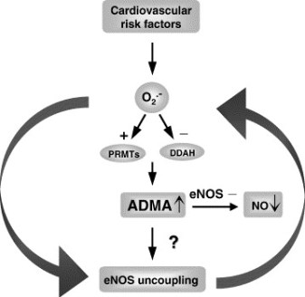
Endogenous inhibitors of NOS (guanidino-substituted derivatives of arginine) occur in vivo as a result of post-translational modification of protein contained arginine residues by S-adenosylmethionine. The dimethylarginines (NG,NG-dimethyl-L-arginine and NG,N’G-dimethyl-L-arginine) occurs in tissue proteins, plasma, and urine of humans and they are thought to act as both regulators of NOS activity and reservoirs of arginine for the synthesis of nitric oxide.
It has been calculated that even though membrane makes up about 3% of the total tissue volume, 90% of the reaction of nitric oxide with oxygen occurs within this compartment. Thus the membrane is an important site for nitric oxide chemistry.
There are two major aspects to nitric oxide chemistry.
It can undergo single electron oxidation and reduction reactions producing nitrosonium and nitroxyl
Having a single unpaired electron in its π*2p molecular orbital it will react readily with other molecules that also have unpaired electrons, such as free radicals and transition metals.
Examples of the reaction of nitric oxide with radical species include:
Nitric oxide will react with oxygen to form the peroxynitrite (nitrosyldioxyl) radical (ONO2)
and with superoxide to form the powerful oxidizing and nitrating agent, peroxynitrite anion (ONO2-). Peroxynitrite causes damage to many important biomolecules
Importance:
nitrosothiols that are important in the regulation of blood pressure terminates lipid peroxidation
3-nitrosotyrosine and/or 4-O-nitrosotyrosine can affect the activity of enzymes that utilize tyrosyl radicals
rapidly reacts with oxyhemoglobin, the primary route of its destruction in vivo
the reaction between nitric oxide and transition metal complexes
During the last reaction a “ligand” bond is formed (the unpaired electron of nitric oxide is partially transferred to the metal cation),
resulting in a nitrosated (nitrosylated) complex.
For example, such complexes can be formed with free iron ions,
iron bound to heme or iron located in iron-sulfur clusters.
Ligand formation allows nitric oxide to act as a signal, activating some enzymes while inhibiting others. Thus, the binding of nitric oxide to the Fe (II)-heme of guanylate (guanalyl) cyclase [GTP-pyrophosphate lyase: cyclizing] is the signal transduction mechanism. Guanylate cyclase exists as cytosolic and membrane-bound isozymes. Thiol-Didulfide Redox Couple
The thiol-disulfide redox couple is very important to oxidative metabolism. For example, GSH is a reducing cofactor for glutathione peroxidase, an antioxidant enzyme responsible for the destruction of hydrogen peroxide.
The importance of the antioxidant role of the thiol-disulfide redox couple:
Thiols and disulfides can readily undergo exchange reactions, forming mixed disulfides. Thiol-disulfide exchange is biologically very important. For example,
GSH can react with protein cystine groups and influence the correct folding of proteins.
GSH may also play a direct role in cellular signaling through thiol-disulfide exchange reactions with membrane bound receptor proteins
the insulin receptor complex)
transcription factors (e.g., nuclear factor κB)
and regulatory proteins in cells
Conditions that alter the redox status of the cell can have important consequences on cellular function.
The generation of ROS by redox cycling is only one possible explanation for the action of many drugs. Rifamycin not only owes its activity to ROS generation but also to its ability to block bacterial RNA synthesis as well. Quinones (and/or semiquinones) can also form adducts with nucleophiles, especially thiols. These adducts may act as toxins directly or indirectly through the inhibition of key enzymes (e.g., by reacting with essential cysteinyl residues) or the depletion of GSH. DNA Adduct Formation
By far the most intense research in this field has been directed towards the chemistry and biology of DNA adduct formation. Attack of the free bases and nucleosides by pro-oxidants can yield a wide variety of adducts and DNA-protein cross-links. Such attack usually occurs
at the C-4 and C-8 position of purines and
C-5 and C-6 of pyrimidines.
Hydroxyl free radical-induced damage to purine bases and nucleosides can proceed through a C-8-hydroxy N-7 radical intermediate, and then either undergo oxidation with the production of an 8-hydroxy purine, or reduction, probably by cellular thiols, followed by ring opening and the formation of FAPy (formamido-pyrimidine) metabolites (hydroxyl free radical-induced damage to guanosine). Although most research has focused on 8-hydroxy-purine adducts a growing number of publications are attempting to measure the FAPy derivative.
Nitrosation of the Amines of the Nucleic Acid Bases.
Primary aromatic amines produce deaminated products, while secondary amines form N-nitroso compounds. Formation of Peroxynitrite from Nitric Oxide.
Peroxynitrite shows complex reactivity
with DNA initiating DNA strand breakage, oxidation (e.g., formation of 8-hydroxyguanine, 8-OH2’dG, (5-hydroxymethyl)-uracil, and FAPyGua),
nitration (e.g., 8-nitroguanine), and
deamination of bases.
Peroxynitrite can also promote the production of lipid peroxidation related active carbonyls and cause the activation of NAD+ ADP-ribosyltransferase.
Modification of Guanine
Although all DNA bases can be oxidatively damaged, it is the modification of guanine that is the most frequent. 8OH2’dG is the most abundant DNA adduct. This can affect its hydrogen bonding between base-pairs. These base-pair substitutions are usually found clustered into areas called “hot spots”. Guanine normally binds to cytosine.
8OH2’dG, however, can form hydrogen bonds with adenine. The formation of 8OH2’dG in DNA can therefore result in a G→T transversion.
8-Hydroxyguanine was also shown to induce codon 12 activation of c-Ha-ras and K-ras in mammalian systems. G→T transversions are also the most frequent hot spot mutations found in the p53 supressor gene which is associated with human tumors.
Other mechanisms by which ROS/RNS can lead to mutations have been
proposed. Direct mechanisms include:
conformational changes in the DNA template that reduces the accuracy of replication by DNA polymerases
altered methylation of cytosine that affects gene control
Indirect mechanisms include:
Oxidative damage to proteins, including DNA polymerases and repair enzymes.
Damage to lipids causes the production of mutagenic carbonyl compounds
Misalignment mutagenesis (“slippery DNA”)
DNA Mismatch Repair 5 (Photo credit: Allen Gathman)
Repair of ROS/RNS-induced DNA Damage
The repair of damaged DNA is an ongoing and continuous process involving a
number of repair enzymes. Damaged DNA appears to be mended by two major mechanisms:
base excision repair (BER) and
nucleotide excision repair (NER)
Isolated DNA is found to contain low levels of damaged bases, so it appears that these repair processes are not completely effective. Base Excision Repair
BER is first started by DNA glycosylases which recognize specific base
modifications (e.g., 8OH2’dG). For example,
Formamido-pyrimidine-DNA glycosylase (Fpg protein) recognizes damaged purines such as 8-oxoguanine and FAPyGua.
Damaged pyrimidines are recognized by endonuclease III, which acts as both a glycosylase and AP endonuclease.
Glycosylases cleave the N-glycosylic bond between the damaged base and the sugar
Following the glycosylase step, AP endonucleases then remove the 3′-deoxyribose moiety by cleavage of the phosphodiester bonds thereby generating a 3’-hydroxyl group that can then be extended by DNA polymerase.
The final step in mending damaged DNA is the rejoining of the free ends of DNA by a DNA ligase. It also appears that the presence of 8-oxoguanine modified bases in DNA is not only a result of ROS attack on this macromolecule. Oxidized nucleosides and nucleotides from free cellular pools can also be incorporated into DNA by polymerases and cause AT to CG base substitution mutations.
Mitochondrial DNA Repair
The mitochondrion genome encodes the various complexes of the electron transport chain, but contains no genetic information for DNA repair enzymes. These enzymes must be obtained from the nucleus. As mitochondria are continuously producing DNA damaging pro-oxidant species, effective DNA repair mechanisms must exist within the mitochondrial matrix in order for these organelles to function. Mitochondria have a short existence, and excessively damaged mitochondria will be quickly removed. Mitochondria contain many BER enzymes and are proficient at repair, but they do not appear to repair damaged DNA by NER mechanisms.
Single Strand DNA Damage and PARP Activation
Single strand DNA breakage activates NAD+ ADP-ribosyltransferase (PARP). PARP is a protein-modifying, nucleotide-polymerizing enzyme and is found at high levels in the nucleus. Activated PARP
cleaves NAD+ into ADP-ribose and nicotinamide
then attaches the ADP-ribose units to a variety of nuclear proteins (including histones and its own automodification domain).
then polymerizes the initial ADP-ribose modification with other ADP-ribose units to form the nucleic acid-like polymer, poly (ADP) ribose.
PARP only appears to be involved with BER and not NER. In BER PARP does not appear to play a direct role but rather it probably helps by keeping the chromatin in a conformation that enables other repair enzymes to be effective. It may also provide temporary protection to DNA molecules while it is being repaired. Conflicting evidence suggests that PARP may not be an important DNA repair enzyme as cells from a PARP knockout mouse model have normal repair characteristics.
Activation of PARP can be dangerous to the cell. For each mole of ADP-ribose transferred, one mole of NAD+ is consumed, and through the regeneration of NAD+ four ATP molecules are wasted. Thus the activation of PARP can rapidly deplete a cell’s energy store and even lead to cell death. Some researchers suggest that this may be one mechanism whereby cells with excessive DNA damage are effectively removed. However, a variety of diseases may involve PARP overactivation including
circulatory shock,
CNS injury,
diabetes,
drug-induced cytotoxicity, and
inflammation.
The Indirect Pathway.
This (mutation) pathway does not involve oxidative damage to the protein per se. This process involves oxidative damage to the DNA molecule encoding the protein. Thus pro-oxidants can cause changes in the base sequence of the DNA molecule. If such base modification is in a coding region of DNA (exon) and not corrected, the DNA molecule may be transcribed incorrectly. Translation of the mutant mRNA can result in a mutant protein containing a wrong amino acid in its primary sequence. If this modified amino acid occurs in an essential part of the protein (e.g., the active site of an enzyme or a portion that alters folding), the function of that protein may be impaired. Fortunately, unlike modified DNA
that can pass from cell to cell during mitosis thereby continuing the production of mutant protein, damage to a protein is non-replicating and stops with its destruction.
The Direct Pathway
This (post-translational) pathway involves the action of a pro-oxidant on a protein resulting in
modification of amino acid residues,
the formation of carbonyl adducts,
cross-linking and
polypeptide chain fragmentation.
Such changes often result in altered protein conformation and/or activity. Proteins will produce a variety of carbonyl products when exposed to metal-based systems (metal/ascorbate and metal/hydrogen peroxide) in vitro. For example, histidine yields aspartate, asparagine and 2-oxoimidazoline, while proline produces glutamate, pyroglutamate, 4-hydroxyproline isomers, 2-pyrrolidone and γ-aminobutyric acid. Metal-based systems and other pro-oxidant conditions can oxidize methionine to its sulfoxide.
This portion of the presentation is endebted to THE HANDBOOK OF REDOX
BIOCHEMISTRY, Ian N. Acworth, August 2003, esa. (inacworth@esainc.com).
We shall now identify more recent work related to this presentation.
Oxygen and Oxidative Stress
The reduction of oxygen to water proceeds via one electron at a time. In the mitochondrial respiratory chain, Complex IV (cytochrome oxidase) retains all partially reduced intermediates until full reduction is achieved. Other redox centres in the electron transport chain, however, may leak electrons to oxygen, partially reducing this molecule to superoxide anion (O2_•). Even though O2_• is not a strong oxidant, it is a precursor of most other reactive oxygen species, and it also becomes involved in the propagation of oxidative chain reactions. Despite the presence of various antioxidant defences, the mitochondrion appears to be the main intracellular source of these oxidants. This review describes the main mitochondrial sources of reactive species and the antioxidant defences that evolved to prevent oxidative damage in all the mitochondrial compartments.
Reactive oxygen species (ROS) is a phrase used to describe a variety of molecules and free radicals (chemical species with one unpaired electron) derived from molecular oxygen. Molecular oxygen in the ground state is a bi-radical, containing two unpaired electrons in the outer shell (also known as a triplet state).
Since the two single electrons have the same spin, oxygen can only react with one electron at a time and therefore it is not very reactive with the two electrons in a chemical bond.
On the other hand, if one of the two unpaired electrons is excited and changes its spin, the resulting species (known as singlet oxygen) becomes a powerful oxidant as the two electrons with opposing spins can quickly react with other pairs of electrons, especially double bonds.
The formation of OH• is catalysed by reduced transition metals, which in turn may be re-reduced by O2 -•, propagating this process. In addition, O2-• may react with other radicals including nitric oxide (NO•) in a reaction controlled by the rate of diffusion of both radicals. The product, peroxynitrite, is also a very powerful oxidant. The oxidants derived from NO• have been recently called reactive nitrogen species (RNS).
‘Oxidative stress’ is an expression used to describe various deleterious processes resulting from an imbalance between the excessive formation of ROS and/or RNS and limited antioxidant defences.
Whilst small fluctuations in the steady-state concentration of these oxidants may actually play a role in intracellular signalling,
uncontrolled increases in the steady-state concentrations of these oxidants lead to free radical mediated chain reactions
which indiscriminately target
proteins,
lipids,
polysaccharides.
In vivo, O2-• is produced both enzymatically and nonenzymatically.
Enzymatic sources include
NADPH oxidases located on the cell membrane of
polymorphonuclear cells,
macrophages and
endothelial cells and
cytochrome P450-dependent oxygenases.
The proteolytic conversion of xanthine dehydrogenase to xanthine oxidase provides another enzymatic source of both O2 -• and H2O2 (and therefore constitutes a source of OH•) and has been proposed to mediate deleterious processes in vivo.
Given the highly reducing intramitochondrial environment, various respiratory components, including flavoproteins, iron–sulfur clusters and ubisemiquinone, are thermodynamically capable of transferring one electron to oxygen. Moreover, most steps in the respiratory chain involve single-electron reactions, further favouring the monovalent reduction of oxygen. On the other hand, the mitochondrion possesses various antioxidant defences designed to eliminate both O2- • and H2O2.
The rate of O2 -• formation by the respiratory chain is controlled primarily by mass action, increasing both when electron flow slows down (increasing the concentration of electron donors, R•) and when the concentration of oxygen increases (eqn (1); Turrens et al. 1982).
d[O2]/dt = k [O2] [R•].
The energy released as electrons flow through the respiratory chain is converted into a H+ gradient through the inner mitochondrial membrane (Mitchell, 1977). This gradient, in turn, dissipates through the ATP synthase complex (Complex V) and is responsible for the turning of a rotor-like protein complex required for ATP synthesis. In the absence of ADP,
the movement of H+ through ATP synthase ceases and
the H+ gradient builds up
causing electron flow to slow down and
the respiratory chain to become more reduced (State IV respiration).
Mitochondrial Antioxidant Defences
The deleterious effects resulting from the formation of ROS in the mitochondrion are, to a large extent, prevented by various antioxidant systems. Superoxide is enzymatically converted to H2O2 by a family of metalloenzymes called superoxide dismutases (SOD). Since O2-• may either reduce transition metals, which in turn can react with H2O2 producing OH• or spontaneously react with NO• to produce peroxynitrite, it is important to maintain the steady-state concentration of O2-• at the lowest possible level. Thus, although the dismutation of O2-• to H2O2 and O2 can also occur spontaneously, the role of SODs is to increase the rate of the reaction to that of a diffusion-controlled process.
The mitochondrial matrix contains a specific form of SOD, with manganese in the active site, which eliminates the O2 -• formed in the matrix or on the inner side of the inner membrane. The expression of MnSOD is further induced by agents that cause oxidative stress, including radiation and hyperoxia, in a process mediated by the oxidative activation of the nuclear transcription factor NFkB .
Reactive oxygen species (ROS) are products of normal metabolism and xenobiotic exposure, and depending on their concentration, ROS can be beneficial or harmful to cells and tissues.
At physiological low levels, ROS function as “redox messengers” in intracellular signaling and regulation, whereas
excess ROS induce oxidative modification of cellular macromolecules, inhibit protein function, and promote cell death.
Additionally, various redox systems, such as
the glutathione,
thioredoxin, and
pyridine nucleotide redox couples,
NADPH and antioxidant defense
NAD+ and the function of sirtuin proteins
participate in cell signaling and modulation of cell function, including apoptotic cell death. Cell apoptosis is initiated by extracellular and intracellular signals via two main pathways,
the death receptor and
the mitochondria-mediated pathways.
ROS and JNK-mediated apoptotic signaling
GSH redox status and apoptotic signaling
Various pathologies can result from oxidative stress-induced apoptotic signaling that is consequent to
ROS increases and/or antioxidant decreases,
disruption of intracellular redox homeostasis, and
irreversible oxidative modifications of lipid, protein, or DNA.
We focus on several key aspects of ROS and redox mechanisms in apoptotic signaling and highlight the gaps in knowledge and potential avenues for further investigation. A full understanding of the redox control of apoptotic initiation and execution could underpin the development of therapeutic interventions targeted at oxidative stress-associated disorders.
Iron-sulfur (FeyS) cluster-containing proteins catalyze a number of electron transfer and metabolic reactions. The components and molecular mechanisms involved in the assembly of the FeyS clusters have been identified only partially. In eukaryotes, mitochondria have been proposed to execute a crucial task in the generation of intramitochondrial and extramitochondrial FeyS proteins. Herein, we identify the essential ferredoxin Yah1p of Saccharomyces cerevisiae mitochondria as a central component of the FeyS protein biosynthesis machinery. Depletion of Yah1p by regulated gene expression resulted in a
30-fold accumulation of iron within mitochondria,
similar to what has been reported for other components involved in FeyS protein biogenesis. Yah1p was shown to be required for the assembly of FeyS proteins both inside mitochondria and in the cytosol. Apparently, at least one of the steps of FeyS cluster biogenesis within mitochondria requires reduction by ferredoxin. Our findings lend support to the idea of a primary function of mitochondria in the biosynthesis of FeyS proteins outside the organelle. To our knowledge, Yah1p is the first member of the ferredoxin family for which a function in FeyS cluster formation has been established. A similar role may be predicted for the bacterial homologs that are encoded within iron-sulfur cluster assembly (isc) operons of prokaryotes.
H Lange, A Kaut, G Kispal, and R Lill. A mitochondrial ferredoxin is essential for biogenesis of cellular iron-sulfur proteins. PNAS 2000; 97(3): 1050–1055.
DNA Charge Transport
Damaged bases in DNA are known to lead to errors in replication and transcription, compromising the integrity of the genome. The authors proposed a model where repair proteins containing redoxactive [4Fe-4S] clusters utilize DNA charge transport (CT) as a first step in finding lesions. In this model, the population of sites to search is reduced by a localization of protein in the vicinity of lesions. Here, we examine this model using single-molecule atomic force microscopy (AFM). XPD, a 5′-3′ helicase involved in nucleotide
excision repair, contains a [4Fe-4S] cluster and exhibits a DNA bound redox potential that is physiologically relevant.
In AFM studies, they observe the redistribution of XPD onto kilobase DNA strands containing a single base mismatch, which is not a specific substrate for XPD but, like a lesion, inhibits CT. They also provide evidence for DNA-mediated signaling between XPD and Endonuclease III (EndoIII), a base excision repair glycosylase that also contains a [4Fe-4S] cluster.
When XPD and EndoIII are mixed together, they coordinate in relocalizing onto the mismatched strand.
However, when a CT-deficient mutant of either repair protein is combined with the CT-proficient repair partner, no relocalization occurs.
The data presented here indicate that XPD, an archaeal protein from the NER pathway, may cooperate with other proteins that are proficient at DNA CT to localize in the vicinity of damage. XPD, a superfamily 2 DNA helicase with 5′-3′ polarity, is a component of TFIIH that is essential for repair of bulky lesions generated by exogenous sources such as UV light and chemical carcinogens. XPD contains a conserved [4Fe-4S] cluster suggested to be conformationally controlled by ATP binding and hydrolysis.
Mutations in the iron-sulfur domain of XPD can lead to diseases including TTD and XP, yet the function of the [4Fe-4S] cluster appears to be unknown.
Electrochemical studies have shown that when BER proteins MutY and EndoIII bind to DNA, their [4Fe-4S] clusters are activated toward one electron oxidation. XPD exhibits a DNA-bound midpoint potential similar to that of EndoIII and MutY when bound to DNA (approximately 80 mV vs. NHE), indicative of a possible role for the [4Fe-4S] cluster in DNA-mediated CT.
For EndoIII we have also already determined a direct correlation between the ability of proteins to redistribute in the vicinity of mismatches as measured by AFM, and the CT proficiency of the proteins measured electrochemically. Thus, we may utilize single-molecule AFM as a tool to probe the redistribution of proteins in the vicinity of base lesions and in so doing, the proficiency of the protein to carry out DNA CT.
Here we show that, like the BER protein EndoIII, XPD, involved both in transcription and NER, redistributes in the vicinity of a lesion. Importantly, this ability to relocalize is associated with the ability of XPD to carry out DNA CT. The mutant L325V is defective in its ability to carry out DNA CTand this XPD mutant also does not redistribute effectively onto the mismatched strand.
These data not only indicate a general link between the ability of a repair protein to carry out DNA CT and its ability to redistribute onto DNA strands near lesions but also provide evidence for coordinated DNA CT between different repair proteins in their search for damage in the genome. These data also provide evidence that two different repair proteins, each containing a [4Fe-4S] cluster at similar DNA bound potential, can communicate with one another through DNA-mediated CT.
The signaling function of mitochondria is considered with a special emphasis on their role in the regulation of redox status of the cell, possibly determining a number of pathologies including cancer and aging. The review summarizes the transport role of mitochondria in energy supply to all cellular compartments (mitochondria as an electric cable in the cell), the role of mitochondria in plastic metabolism of the cell including synthesis of
heme,
steroids,
iron-sulfur clusters, and
reactive oxygen and nitrogen species.
Mitochondria also play an important role in the Ca2+-signaling and the regulation of apoptotic cell death. Knowledge of mechanisms responsible for apoptotic cell death is important for the strategy for prevention of unwanted degradation of postmitotic cells such as cardiomyocytes and neurons.
In accordance with P. Mitchell’s chemiosmotic concept, vectorial transmembrane transfer of electrons and protons is accompanied by generation of electrochemical difference of proton electrochemical potential on the inner mitochondrial membrane; its utilization by ATP synthase induces conformational rearrangements resulting in ATP synthesis from ADP and inorganic phosphate. Details of the mechanism responsible for ATP synthesis are given elsewhere.
Membrane potential (DY) generated across the inner mitochondrial membrane is the component of the transmembrane electrochemical potential of H+ ions (DμH+), which provides ATP synthesis together with the concentration component (DpH). Maintenance of constant membrane potential is a vitally important precondition for functioning of mitochondria and the cell. Under conditions of limited supply of the cell with oxygen (hypoxia) and inability to carry out aerobic ATP synthesis, mitochondria become ATP consumers (rather than generators) and ATP is hydrolyzed by mitochondrial ATPase, and this is accompanied by generation of membrane potential.
Redox homeostasis, i.e. the sum of redox components (including proteins, low molecular weight redox components such as NAD/NADH, flavins, coenzymes Q, oxidized and reduced substrates, etc.) is one of important preconditions for normal cell functioning.
Single-strand and double-strand DNA damage (Photo credit: Wikipedia)
Mitochondria generate such potent regulators of redox potential as
superoxide anion,
hydrogen peroxide,
nitric oxide,
peroxynitrite, etc.
They are actively involved in regulation of cell redox potential and consequently
control proteolysis,
activation of transcription,
changes in mitochondrial DNA (mDNA),
cell metabolism, and
cell differentiation.
Zorov DB, Isaev NK, Plotnikov EY, Zorova LD, et al. The Mitochondrion as Janus Bifrons. Biochemistry (Moscow) 2007; 72(10): 1115-1126. ISSN 0006-2979.
DOI: 10.1134/S0006297907100094
Structure of the human mitochondrial genome. (Photo credit: Wikipedia)
Gene Expression Associated with Oxidoreduction and Mitochondria
The naked mole-rat (Heterocephalus glaber) is a long-lived, cancer resistant rodent and there is a great interest in identifying the adaptations responsible for these and other of its unique traits. We employed RNA sequencing to compare liver gene expression profiles between naked mole-rats and wild-derived mice. Our results indicate that genes associated with oxidoreduction and mitochondria were expressed at higher relative levels in naked mole-rats. The largest effect is nearly
300-fold higher expression of epithelial cell adhesion molecule (Epcam), a tumour-associated protein.
Also of interest are the
protease inhibitor, alpha2-macroglobulin (A2m), and the
mitochondrial complex II subunit Sdhc,
both ageing-related genes found strongly over-expressed in the naked mole-rat.
These results hint at possible candidates for specifying species differences in ageing and cancer, and in particular suggest complex alterations in mitochondrial and oxidation reduction pathways in the naked mole-rat. Our differential gene expression analysis obviated the need for a reference naked mole-rat genome by employing a combination of Illumina/Solexa and 454 platforms for transcriptome sequencing and assembling transcriptome contigs of the non-sequenced species. Overall, our work provides new research foci and methods for studying the naked mole-rat’s fascinating characteristics.
The complete set of viable deletion strains in Saccharomyces cerevisiae was screened for sensitivity of mutants to five oxidants to identify cell functions involved in resistance to oxidative stress. This screen identified a unique set of mainly constitutive functions providing the first line of defense against a particular oxidant; these functions are very dependent on the nature of the oxidant. Most of these functions are distinct from those involved in repair and recovery from damage, which are generally induced in response to stress, because there was little correlation between mutant sensitivity and the reported transcriptional response to oxidants of the relevant gene. The screen identified 456 mutants sensitive to at least one of five different types of oxidant, and these were ranked in order of sensitivity. Many genes identified were not previously known to have a role in resistance to reactive oxygen species. These encode functions including
protein sorting,
ergosterol metabolism,
autophagy, and
vacuolar acidification.
two mutants were sensitive to all oxidants examined, 12 were sensitive to at least four,
Different oxidants had very different spectra of deletants that were sensitive. These findings highlight the specificity of cellular responses to different oxidants:
No single oxidant is representative of general oxidative stress.
Mitochondrial respiratory functions were overrepresented in mutants sensitive to H2O2, and
vacuolar protein-sorting mutants were enriched in mutants sensitive to diamide.
Core functions required for a broad range of oxidative-stress resistance include
Isolated complex I deficiency is the most common enzymatic defect of the oxidative phosphorylation (OXPHOS) system, causing a wide range of clinical phenotypes. Th authers reported before that the rates at which reactive oxygen species (ROS)-sensitive dyes are converted into their fluorescent oxidation products are markedly increased in cultured skin fibroblasts of patients with nuclear-inherited isolated complex I deficiency.
Using videoimaging microscopy we show here that these cells also display a marked increase in NAD(P)H autofluorescence. Linear regression analysis revealed a negative correlation with the residual complex I activity and a positive correlation with the oxidation rates of the ROS sensitive dyes (5-(and-6)-chloromethyl-2′,7′-dichlorodihydrofluorescein and hydroethidine for a large cohort of 10 patient cell lines.
On the other hand, video-imaging microscopy of cells selectively expressing reduction-oxidation sensitive GFP1 in either the mitochondrial matrix or cytosol showed the absence of any detectable change in thiol redox state. In agreement with this result, neither the glutathione nor the glutathione disulfide content differed significantly between patient and healthy fibroblasts.
Finally, video-rate confocal microscopy of cells loaded with C11-BODIPY581/591 demonstrated that the extent of lipid peroxidation, which is regarded as a measure of oxidative damage, was not altered in patient fibroblasts. Our results indicate that fibroblasts of patients with isolated complex I deficiency maintain their thiol redox state despite marked increases in ROS production.
Mitochondrial research has recently been driven by the
identification of mitochondria-associated diseases and the role of mitochondria in apoptosis.
Moreover, mitochondria have been implicated in the process of carcinogenesis because of their vital role in
energy production,
nuclear-cytoplasmic signal integration and
control of metabolic pathways.
At some point during neoplastic transformation, there is an increase in reactive oxygen species (ROS), which damage the mitochondrial genome. This accelerates the somatic mutation rate of mitochondrial DNA.
Mitochondrial characteristics
There are several biological characteristics which cast mitochondria and, in particular, the mitochondrial genome, as a biological tool for early detection and monitoring of neoplasia and its potential progression. These vital characteristics are important in cancer research, as not all neoplasias become malignant. Mitochondria are archived in the cytoplasm of the ovum and as such do not recombine.
This genome has an accelerated mutation rate, by comparison with the nucleus, and accrues somatic mutations in tumour tissue. Moreover, mitochondrial DNA (mtDNA) has a high copy number in comparison with the nuclear archive of DNA. There are potentially thousands of mitochondrial genomes per cell, which enables detection of important biomarkers, even at low levels. In addition, mtDNA can be heteroplasmic, which means that disease-associated mutations occur in a subset of the genomes.
The presence of heteroplasmy is an indication of disease and is found in many human tumours. Identification of low levels of heteroplasmy may allow unprecedented early identification and monitoring of neoplastic progression to malignancy.
Coding for just 13 enzyme complex subunits, 22 transfer RNAs and two ribosomal RNAs, the mitochondrial genome is packaged in a compact 16,569 base pair (bp) circular molecule. These products participate in the critical electron transport process of ATP production. Collectively, mitochondria generate 80 per cent of the chemical fuel which fires cellular metabolism.
As a result, nuclear investment in the mitochondria is high — that is, several thousand nuclear genes control this organelle in order to accomplish the complex interactions required to maintain a network of pathways, which coordinate energy demand and supply.
It has been proposed that these mutations may serve as an early indication of potential cancer development and may represent a means for tracking tumour progression.
Does this provide a potential utility in that these mutations may be used for the identification and monitoring of neoplasia and malignant transformation where appropriate body fluids or non-invasive tissue access is available for mtDNA recovery? Specifically discussed are:
prostate,
breast,
colorectal,
skin and
lung cancers
There are many important questions yet to be addressed: such as
the relationship between mtDNA and the actual disease;
are mutations causative or merely a reflection of nuclear instability?
And, are these processes independent events?
Alterations in the non-coding D-loop suggest genome instability;
however, as studies focus more on the coding regions of the
mitochondrial genome,
Particularly in the case of nonsynonymous mutations in the genes
contributing products to the electron transport process, metabolic
implications are evident. Moreover, mutations in mitochondrial transfer RNAs indicate the possibility of a global mitochondrial translational shut down.
RL Parr, GD Dakubo, RE Thayer, K McKenney, MA Birch-Machin. Mitochondrial DNA as a potential tool for early cancer detection. HUMAN GENOMICS 2006; 2(4). 252–257.
Mitochondrial DNA (mtDNA) is particularly prone to oxidation due to the lack of histones and a deficient mismatch repair system. This explains an increased mutation rate of mtDNA that results in heteroplasmy, e.g., the coexistence of the mutant and wild-type mtDNA molecules within the same mitochondrion. Hyperglycemia is a key risk factor not only for diabetes-related disease, but also for cardiovascular and all-cause mortality. One can assume an increase in the risk of cardiovascular disease by 18% for each unit (%) glycated hemoglobin HbA1c. In the Glucose Tolerance in Acute Myocardial Infarction study of patients with acute coronary syndrome, abnormal glucose tolerance was the strongest independent predictor of subsequent cardiovascular complications and death. In the Asian Pacific Study, fasting plasma glucose was shown to be an independent predictor of cardiovascular events up to a level of 5.2 mmol/L.
Glucose level fluctuations and hyperglycemia are triggers for inflammatory responses via increased mitochondrial superoxide production and endoplasmic reticulum stress. Inflammation leads to insulin resistance and β-cell dysfunction, which further aggravates hyperglycemia. The molecular pathways that integrate hyperglycemia, oxidative stress, and diabetic vascular complications have been most clearly described in the pathogenesis of endothelial dysfunction, which is considered as the first step in atherogenesis according to the response to injury hypothesis.
In diabetes mellitus,
glycotoxicity,
advanced oxidative stress,
collagen cross-linking, and
accumulation of lipid peroxides
in foam macrophage cells and arterial wall cells may significantly
decrease the mutation threshold,
endothelial dysfunction,
promoting atherosclerosis.
Alterations in mitochondrial DNA (mtDNA), known as homoplasmic and heteroplasmic mutations, may influence mitochondrial OXPHOS capacity, and in turn contribute to the magnitude of oxidative stress in micro- and macrovascular networks in diabetic patients.
The authors critically consider the impact of mtDNA mutations on the pathogenesis of cardiovascular diabetic complications.
Mutation Threshhold
Although cells may harbor mutant mtDNA, the expression of disease is dependent on the percent of alleles bearing mutations. Modeling confirms that an upper threshold level might exist for mutations beyond which the mitochondrial population collapses, with a subsequent decrease in ATP. This decrease in ATP results in the phenotypic expression of disease. It is estimated that in many patients with clinical manifestations of mitochondrial disorders, the proportion of mutant DNA exceeds 50%.
For the MELAS (mitochondrial encephalopathy, lactic acidosis and stroke-like syndrome)-causing mutation m.3243 A>G in the mitochondrial gene encoding tRNALeu, which is also associated with diabetes plus deafness, a strong correlation between the level of mutational heteroplasmy and documented disease has been found. Increased percentages of mutant mtDNA in muscle cells (up to 71%) can lead to mitochondrial myopathy. Levels of heteroplasmy of over 80% may lead to recurrent stroke and mutation levels of 95% have been associated with MELAS.
Regardless of the type of mutation or the level of heteroplasmy in affected mitochondria, unrepaired damage leads to a decrease in ATP, which in turn causes the phenotypic manifestation of disease. The manifestation of disease not only depends on the ATP level but also on the tissue affected. Various tissues have differing levels of demand on OXPHOS capacity. To evaluate a tissue threshold, Leber’s hereditary optic neuropathy can be used as a model for mitochondrial neurodegenerative disease. For neural and skeletal muscle tissues, the tissue threshold should be as high as or higher than 90% of
damaged (mutated) mtDNA. To induce mitochondrial malfunctions, the tissue threshold of the cardiac muscle is estimated to be significantly lower (approximately 64%-67%). In chronic vascular disease such as atherosclerosis, a mutation threshold in the affected vessel wall (e.g., in the postmortem aortic atherosclerotic plaques) was observed to be significantly lower. For example, for mutations m.3256 C>T, m.12315 G>A, m.15059 G>A, and m.15315 G>A, the heteroplasmy range of 18%-66% in the atherosclerotic lesions was 2-3.5-fold that in normal vascular tissue.
Mitochondrial stress and insulin resistance
Mitochondrial damage precedes the development of atherosclerosis and tracks the extent of the lesion in apoE-null mice, and
mitochondrial dysfunction caused by heterozygous deficiency of a superoxide dismutase increases atherosclerosis and vascular mitochondrial damage in the same model.
Blood vessels destined to develop atherosclerosis may be characterized by inefficient ATP production due to the uncoupling of respiration and OXPHOS. Blood vessels have regions of hypoxia, which lower the ratio of state 3 (phosphorylating) to state 4 (nonphosphorylating) respiration. Human atherosclerotic lesions have been known for decades to be deficient in essential fatty acids, a condition that causes respiratory uncoupling and atherosclerosis.
The finding by Kokaze et al. helps to explain, at least in part, the anti-atherogenic effect of the allele m. 5178A due to its relation with the favorable lipid profile. The nucleotide change causes leucine-to-methionine substitution at codon 237 (Leu-237Met) of the NADH dehydrogenase subunit 2 located in the loop between 7th and 8th transmembrane domains of the mitochondrial protein. Given that this methionine residue is exposed at the surface of respiratory Complex I, this residue may be available as an efficient oxidant scavenger. Complex I
accepts electrons from NADH,
transfers them to ubiquinone, and
uses the energy released to pump protons across the mitochondrial inner membrane.
Thus, the Leu237Met replacement in the ND2 subunit might have a protective effect against oxidative damage to mitochondria.
Most fatty acid oxidation, which is promoted by peroxisome proliferator-activated receptor α (PPARα) activation, occurs in the mitochondria. Mitochondrial effects could explain why PPARα- deficient mice are protected from diet-induced insulin resistance and atherosclerosis as well as glucocorticoid induced insulin resistance and hypertension. Caloric restriction,
Nitric oxide (NO) is a lipophilic, highly diffusible and short-lived molecule that acts as a physiological messenger and has been known to regulate a variety of important physiological responses including vasodilation, respiration, cell migration, immune response and apoptosis. Jordi Muntané et al
NO is synthesized by the Nitric Oxide synthase (NOS) enzyme and the enzyme is encoded in three different forms in mammals: neuronal NOS (nNOS or NOS-1), inducible NOS (iNOS or NOS-2), and endothelial NOS (eNOS or NOS-3). The three isoforms, although similar in structure and catalytic function, differ in the way their activity and synthesis in controlled inside a cell. NOS-2, for example is induced in response to inflammatory stimuli, while NOS-1 and NOS-3 are constitutively expressed.
Regulation by Nitric oxide
NO is a versatile signaling molecule and the net effect of NO on gene regulation is variable and ranges from activation to inhibition of transcription.
The intracellular localization is relevant for the activity of NOS. Infact, NOSs are subject to specific targeting to subcellular compartments (plasma membrane, Golgi, cytosol, nucleus and mitochondria) and that this trafficking is crucial for NO production and specific post-translational modifications of target proteins.
Role of Nitric oxide in Cancer
One in four cases of cancer worldwide are a result of chronic inflammation. An inflammatory response causes high levels of activated macrophages. Macrophage activation, in turn, leads to the induction of iNOS gene that results in the generation of large amount of NO. The expression of iNOS induced by inflammatory stimuli coupled with the constitutive expression of nNOS and eNOS may contribute to increased cancer risk. NO can have varied roles in the tumor environment influencing DNA repair, cell cycle, and apoptosis. It can result in antagonistic actions including DNA damage and protection from cytotoxicity, inhibiting and stimulation cell proliferation, and being both anti-apoptotic and pro-apoptotic. Genotoxicity due to high levels of NO could be through direct modification of DNA (nitrosative deamination of nucleic acid bases, transition and/or transversion of nucleic acids, alkylation and DNA strand breakage) and inhibition of DNA repair enzymes (such as alkyltransferase and DNA ligase) through direct or indirect mechanisms. The Multiple actions of NO are probably the result of its chemical (post-translational modifications) and biological heterogeneity (cellular production, consumption and responses). Post-translational modifications of proteins by nitration, nitrosation, phosphorylation, acetylation or polyADP-ribosylation could lead to an increase in the cancer risk. This process can drive carcinogenesis by altering targets and pathways that are crucial for cancer progression much faster than would otherwise occur in healthy tissue.
NO can have several effects even within the tumor microenvironment where it could originate from several cell types including cancer cells, host cells, tumor endothelial cells. Tumor-derived NO could have several functional roles. It can affect cancer progression by augmenting cancer cell proliferation and invasiveness. Infact, it has been proposed that NO promotes tumor growth by regulating blood flow and maintaining the vasodilated tumor microenvironment.NO can stimulate angiogenesis and can also promote metastasis by increasing vascular permeability and upregulating matrix metalloproteinases (MMPs). MMPs have been associated with several functions including cell proliferation, migration, adhesion, differentiation, angiogenesis and so on. Recently, it was reported that metastatic tumor-released NO might impair the immune system, which enables them to escape the immunosurveillance mechanism of cells. Molecular regulation of tumour angiogenesis by nitric oxide.
S-nitrosylation and Cancer
The most prominent and recognized NO reaction with thiols groups of cysteineresidues is called S-nitrosylation or S-nitrosation, which leads to the formation of more stable nitrosothiols. High concentrations of intracellular NO can result in high concentrations of S-nitrosylated proteins and dysregulated S-nitrosylation has been implicated in cancer. Oxidative and nitrosative stress is sensed and closely associated with transcriptional regulation of multiple target genes.
Following are a few proteins that are modified via NO and modification of these proteins, in turn, has been known to play direct or indirect roles in cancer.
NO mediated aberrant proteins in Cancer
Bcl2
Bcl-2 is an important anti-apoptotic protein. It works by inhibiting mitochondrial Cytochrome C that is released in response to apoptotic stimuli. In a variety of tumors, Bcl-2 has been shown to be upregulated, and it has additionally been implicated with cancer chemo-resistance through dysregulation of apoptosis. NO exposure causes S-nitrosylation at the two cysteine residues – Cys158 and Cys229 that prevents ubiquitin-proteasomal pathway mediated degradation of the protein. Once prevented from degradation, the protein attenuates its anti-apoptotic effects in cancer progression. The S-nitrosylation based modification of Bcl-2 has been observed to be relevant in drug treatment studies (for eg. Cisplatin). Thus, the impairment of S-nitrosylated Bcl-2 proteins might serve as an effective therapeutic target to decrease cancer-drug resistance.
p53
p53 has been well documented as a tumor suppressor protein and acts as a major player in response to DNA damage and other genomic alterations within the cell. The activation of p53 can lead to cell cycle arrest and DNA repair, however, in case of irrepairable DNA damage, p53 can lead to apoptosis. Nuclear p53 accumulation has been related to NO-mediated anti-tumoral properties. High concentration of NO has been found to cause conformational changes in p53 resulting in biological dysfunction.. In RAW264.7, a murine macrophage cell line, NO donors induce p53 accumulation and apoptosis through JNK-1/2.
HIF-1a
Hypoxia-inducible factor 1 (HIF1) is a heterodimeric transcription factor that is predominantly active under hypoxic conditions because the HIF-1a subunit is rapidly degraded in normoxic conditions by proteasomal degradation. It regulates the transciption of several genes including those involved in angiogenesis, cell cycle, cell metabolism, and apoptosis. Hypoxic conditions within the tumor can lead to overexpression of HIF-1a. Similar to hypoxia-mediated stress, nitrosative stress can stabilize HIF-1a. NO derivatives have also been shown to participate in hypoxia signaling. Resistance to radiotherapy has been traced back to NO-mediated HIF-1a in solid tumors in some cases.
PTEN
Phosphatase and tensin homolog deleted on chromosome ten (PTEN), is again a tumor suppressor protein. It is a phosphatase and has been implicated in many human cancers. PTEN is a crucial negative regulator of PI3K/Akt signaling pathway. Over-activation of PI3K/Akt mediated signaling pathway is known to play a major role in tumorigenesis and angiogenesis. S-nitrosylation of PTEN, that could be a result of NO stress, inhibits PTEN. Inhibition of PTEN phosphatase activity, in turn, leads to promotion of angiogenesis.
C-Src
C-src belongs to the Src family of protein tyrosine kinases and has been implicated in the promotion of cancer cell invasion and metastasis. It was demonstrated that S-nitrosylation of c-Src at cysteine 498 enhanced its kinase activity, thus, resulting in the enhancement of cancer cell invasion and metastasis.
Jaiswal M, et al. Nitric oxide in gastrointestinal epithelial cell carcinogenesis: linking inflammation to oncogenesis. Am J Physiol Gastrointest Liver Physiol. 2001 Sep;281(3):G626-34. http://www.ncbi.nlm.nih.gov/pubmed/11518674
Author and Reporter: Meg Baker, Ph.D., Registered Patent Agent
The 1998 Noble Prize for medicine was for the discovery that nitric oxide (NO) was the chemical messenger responsible for relaxing vascular tissue and thereby increasing blood flow and reducing blood pressure. Alfred Noble himself had been prescribed nitro-glycerin for heart problems over 100 years before, a compound which is metabolized to NO.
NO, a gas at room temperature, has an exceedingly short half-life in the body. Normally, NO is produced from an amino acid, L-arginine (L-Arg), a normal component of the dietary protein, and molecular oxygen (O2) by the one of the several Nitric Oxide Synthases (EC 1.14.13.39): endothelial (eNOS, NOS III), inducible (iNOS, NOS II), and neural (nNOS, NOS I). In human studies, supplementation with l-arginine improved endothelium-dependent vasodilation.
The reaction of iNOS with L-Arg to produce NO leaves another amino acid, citrulline. Excess L-Arg can also be degraded by arginase (enzyme having two isoforms, I and II) which may be coinduced with iNOS in some cell types.
Citrulline formed as a by-product of the NOS reaction can be recycled to arginine by argininosuccinate synthetase (AS) and argininosuccinate lyase (AL).
Mori (2007) http:// www.ncbi.nlm.nih.gov/ pubmed/ 17513437 found that AS and sometimes AL are coinduced with inducible NOS (iNOS) in various cells. In these cells, NO was synthesized from citrulline (via arginine) as well as from arginine, indicating operation of the citrulline-NO cycle.
Whereas, low concentrations of NO protect cells from apoptosis, excessive NO causes apoptosis. NO causes endoplasmic reticulum (ER) stress, induces a transcription factor, CAAT/enhancer binding protein (C/EBP) homologous protein (CHOP), and leads to apoptosis.
The active site of NOS is formed by a heme-containing substrate-binding cavity, where L-arginine (Arg) and O2 are converted to L-citrulline and NO. The electrons required for reductive O2 activation are transferred from NADPH via the NOS-bound flavins (riboflavin, Vitamin B2) FMN and FAD. All NOS isoforms are only active as homodimers.
Generation of NO occurs in two discrete O2-requiring steps, with intermediate formation of N-hydroxy-L-arginine (NHA or NOHLA). NHA formation consumes one molecule of O2 and two electrons. Conversion of NHA to L-citrulline and NO requires another molecule of O2 and one more electron (http://en.wikipedia.org/wiki/Nitric_oxide_synthase). The overall stoichiometry, reflecting the three electrons derived from NADPH, that pass through the flavin co-factors and are transferred one by one via the heme iron, is then:
Another factor affecting NOS activity is the availability of essential co-factors such as tetrahydrobiopterin (BH4) (Boeger et al. Cardiovasc Res (2003) 59 (4): 824-833http://cardiovascres.oxfordjournals.org/content/59/4/824.full, Vasquez-Vivar J., et al . Superoxide generation by endothelial nitric oxide synthase: the influence of cofactors. Proc. Natl. Acad. Sci. USA 1998;95:9220-9225http://www.pnas.org/content/95/16/9220.full). H4-biopterin binds in the immediate vicinity of the heme at the dimer interface, interacting with residues from both subunits. When BH4 availability is limiting, electron transfer from NOS flavins becomes “uncoupled” from l-arginine oxidation and the ferrous-dioxygen complex formed as an intermediate in the reaction sequence, dissociates and superoxide(O2−·) is produced.
The conversion of Arg to NHA and of NHA to L-citrulline and NO both depend on the presence of H4-biopterin. In the absence of substrate or pterin, NADPH oxidation by NOS is accompanied by formation of O2− and peroxide (H2O2). Uncoupled eNOS is assumed to produce superoxide (O2−·) in addition to or instead of NO (·NO) which will react with itself, with NO, or with -hydroxyl, -sulfhydryl, or or side groups of proteins, lipids, or glycans. Reaction of ·NO produced by eNOS, with O2−· produced by eNOS or by other enzymes, such as NADPH and xanthine oxidases, decreases the amount of ·NO available to stimulate vascular relaxation. At the very low BH4 concentration of 100 nmol/L, recombinant human eNOS activity is fully developed. However, biopterin is formed from the pterin heterocycle also present in folic acid (Vitamin B9,
pteroyl-L-glutamate)
and which is synthesized from GTP. Human GTP cyclohydrolase I (GTPCH), is the rate-limiting enzyme in BH4 synthesis (Crabtree et al. JBC 2008, http://www.jbc.org/content/284/2/1136.full).
In addition to the NOS reaction, which generates a H3-biopterin radical cation, a neutral H3-biopterin radical is formed when H4-biopterin reacts with various radicals and which can be reduced back to H4-biopterin by ascorbate (Vitamin C). Folate species are also required to synthesize pyrimidines and purines (for DNA synthesis and repair and NADH and NADPH).
Enhancing NO Synthesis
The normal way to increase vascular nitric oxide is through vascular stress, such as exercise. As oxygen demand increases, cardiac output increases and the endothelial lining of the arteries releases nitric oxide into the blood, which, in turn, relaxes and widens the vessel wall, allowing for enhanced blood flow.
Enhancing the presence of L-Arg or the one or more of the NOS enzymes are obviously essential for NO production. However, NOS enzymes are co-valently bound to heme (heme, iron), and flavin co-factors (Vit B2), and require soluble co-factors NADPH (a dinucleotide phosphate, containing niacin, Vitamin B3), and BH4 (from Vit B9).
Foods high in Arginine and Citrulline include melons and cucumber, peanuts, salmon, and soy. Arginine is found in varying degrees (3-15% by weight) in all animal proteins. Blue-fin tuna has 1.8 g of arginine per 100 g so 2 oz. of tuna will provide about 1 g of arginine. Other sources of 1 g of L-Arg: 2.7 oz. of chicken thighs, about 4 oz. of chicken breast, 2 oz. of 75 percent lean hamburger or about 2.5 oz. of pork.
Foods rich in antioxidants and polyphenols will provide protection against free radical assault on proteins and, in particular, act to protect the NOS enzyme and cofactors. Almost all fruit and vegetables such as blueberries, cranberries, carrots, grapefruit, soybeans, apples, and spinach contain high levels of antioxidants. In addition, nuts, tea, seeds, dark chocolate, red wine, and seafood generally contain antioxidants such as resveratrol, ascorbate, and other phytochemicals. Other free radical scavengers, tocopherols (alpha-tocopherol, Vit E) work predominantly in the lipid environment such as in cell membranes, while the sulfur-containing soluble molecule, glutathione (GSH) protects the cytosolic milieu.
Supplements
Both L-Arg or L-citrulline can be purchased over the counter. Dietary L-arginine will be taken up by the intestine and transported directly to the liver by the hepatic artery as are most of the products of digestion. Much of this L-Arg will be used in metabolic steps related to the urea cycle which is co-ordinated with the kidney to rid the body of excess nitrogen and prevent ammonia concentration from building. A small amount will enter the blood stream and be used for NO synthesis.
Proargi-9 Plus® is one product being sold containing mutltigram doses of L-Arg plus L-Citrulline in combination with anti-oxidants and folate. Proargi-9 Plus® is a registered trademark and copyright of Nature’s Sunshine Products, Inc. L-arginine Plus™ is formulation with similar ingredients and stated amounts of L-Arg and L-Citrulline and is not affiliated with the makers of Proargi-9-Plus. Niteworks® is a registered trademark and copyright of Herbalife International, Inc. and is not affiliated with or a sponsor of L-arginine Plus™.
Dr. Joe Prendergast is an endocrinologist using L-Arg therapy who, over 19 years, never had to admit any of his 7200 diabetes patients to the hospital for peripheral artery disease, recommends supplemental L-Arg formulations to his patients. The combination of L-Arg with L-citrulline a longer acting NO forming product. http://www.livingwithoutdisease.com/?route=references/prendergast
Supplements of L-Arg and, in particular, in combination with L-citrulline other B-vitamins and antioxidents may be an effective way to boost vascular NO synthesis for anyone not exercising or eating a balanced diet, having a deficiency in any of the L-Arg recycling enzymes, NOS enzymes, co-factor recycling or synthetic enzymes, or other risk factor. Specific risk factors, such as inherently elevated levels of the natural NOS inhibitor ADMA (asymmetric-dimethyl-L-arginine) are beginning to be uncovered and will be the subject of another post.
Werner, et al. Tetrahydrobiopterin and Nitric Oxide: Mechanistic and Pharmacological Aspects Exp Biol Med December 2003 vol. 228 no. 11 1291-1302 Werner et al. Exp Biol Med 2003
Coronary Artery Disease – Medical Devices Solutions: From First-In-Man Stent Implantation, via Medical Ethical Dilemmas to Drug Eluting Stents August 13, 2012
Cardiovascular Disease (CVD) and the Role of agent alternatives in endothelial Nitric Oxide Synthase (eNOS) Activation and Nitric Oxide Production July 19, 2012
Curator and Research Study Originator: Aviva Lev-Ari, PhD, RN
Macrovascular Disease – Therapeutic Potential of cEPCs: Reduction Methods for CV Risk
July 2, 2012
An Investigation of the Potential of circulating Endothelial Progenitor Cells (cEPCs) as a Therapeutic Target for Pharmacological Therapy Design for Cardiovascular Risk Reduction: A New Multimarker Biomarker Discovery
Inhibition of ET-1, ETA and ETA-ETB, Induction of NO production, stimulation of eNOS and Treatment Regime with PPAR-gamma agonists (TZD): cEPCs Endogenous Augmentation for Cardiovascular Risk Reduction – A Bibliography