Healthcare analytics, AI solutions for biological big data, providing an AI platform for the biotech, life sciences, medical and pharmaceutical industries, as well as for related technological approaches, i.e., curation and text analysis with machine learning and other activities related to AI applications to these industries.
#TUBiol5227: Biomarkers & Biotargets: Genetic Testing and Bioethics
Curator: Stephen J. Williams, Ph.D.
The advent of direct to consumer (DTC) genetic testing and the resultant rapid increase in its popularity as well as companies offering such services has created some urgent and unique bioethical challenges surrounding this niche in the marketplace. At first, most DTC companies like 23andMe and Ancestry.com offered non-clinical or non-FDA approved genetic testing as a way for consumers to draw casual inferences from their DNA sequence and existence of known genes that are linked to disease risk, or to get a glimpse of their familial background. However, many issues arose, including legal, privacy, medical, and bioethical issues. Below are some articles which will explain and discuss many of these problems associated with the DTC genetic testing market as well as some alternatives which may exist.
As you can see,this market segment appears to want to expand into the nutritional consulting business as well as targeted biomarkers for specific diseases.
Rising incidence of genetic disorders across the globe will augment the market growth
Increasing prevalence of genetic disorders will propel the demand for direct-to-consumer genetic testing and will augment industry growth over the projected timeline. Increasing cases of genetic diseases such as breast cancer, achondroplasia, colorectal cancer and other diseases have elevated the need for cost-effective and efficient genetic testing avenues in the healthcare market.
For instance, according to the World Cancer Research Fund (WCRF), in 2018, over 2 million new cases of cancer were diagnosed across the globe. Also, breast cancer is stated as the second most commonly occurring cancer. Availability of superior quality and advanced direct-to-consumer genetic testing has drastically reduced the mortality rates in people suffering from cancer by providing vigilant surveillance data even before the onset of the disease. Hence, the aforementioned factors will propel the direct-to-consumer genetic testing market overt the forecast timeline.
Nutrigenomic Testing will provide robust market growth
The nutrigenomic testing segment was valued over USD 220 million market value in 2019 and its market will witness a tremendous growth over 2020-2028. The growth of the market segment is attributed to increasing research activities related to nutritional aspects. Moreover, obesity is another major factor that will boost the demand for direct-to-consumer genetic testing market.
Nutrigenomics testing enables professionals to recommend nutritional guidance and personalized diet to obese people and help them to keep their weight under control while maintaining a healthy lifestyle. Hence, above mentioned factors are anticipated to augment the demand and adoption rate of direct-to-consumer genetic testing through 2028.
Browse key industry insights spread across 161 pages with 126 market data tables & 10 figures & charts from the report, “Direct-To-Consumer Genetic Testing Market Size By Test Type (Carrier Testing, Predictive Testing, Ancestry & Relationship Testing, Nutrigenomics Testing), By Distribution Channel (Online Platforms, Over-the-Counter), By Technology (Targeted Analysis, Single Nucleotide Polymorphism (SNP) Chips, Whole Genome Sequencing (WGS)), Industry Analysis Report, Regional Outlook, Application Potential, Price Trends, Competitive Market Share & Forecast, 2020 – 2028” in detail along with the table of contents: https://www.gminsights.com/industry-analysis/direct-to-consumer-dtc-genetic-testing-market
Targeted analysis techniques will drive the market growth over the foreseeable future
Based on technology, the DTC genetic testing market is segmented into whole genome sequencing (WGS), targeted analysis, and single nucleotide polymorphism (SNP) chips. The targeted analysis market segment is projected to witness around 12% CAGR over the forecast period. The segmental growth is attributed to the recent advancements in genetic testing methods that has revolutionized the detection and characterization of genetic codes.
Targeted analysis is mainly utilized to determine any defects in genes that are responsible for a disorder or a disease. Also, growing demand for personalized medicine amongst the population suffering from genetic diseases will boost the demand for targeted analysis technology. As the technology is relatively cheaper, it is highly preferred method used in direct-to-consumer genetic testing procedures. These advantages of targeted analysis are expected to enhance the market growth over the foreseeable future.
Over-the-counter segment will experience a notable growth over the forecast period
The over-the-counter distribution channel is projected to witness around 11% CAGR through 2028. The segmental growth is attributed to the ease in purchasing a test kit for the consumers living in rural areas of developing countries. Consumers prefer over-the-counter distribution channel as they are directly examined by regulatory agencies making it safer to use, thereby driving the market growth over the forecast timeline.
Favorable regulations provide lucrative growth opportunities for direct-to-consumer genetic testing
Europe direct-to-consumer genetic testing market held around 26% share in 2019 and was valued at around USD 290 million. The regional growth is due to elevated government spending on healthcare to provide easy access to genetic testing avenues. Furthermore, European regulatory bodies are working on improving the regulations set on the direct-to-consumer genetic testing methods. Hence, the above-mentioned factors will play significant role in the market growth.
Focus of market players on introducing innovative direct-to-consumer genetic testing devices will offer several growth opportunities
Few of the eminent players operating in direct-to-consumer genetic testing market share include Ancestry, Color Genomics, Living DNA, Mapmygenome, Easy DNA, FamilytreeDNA (Gene By Gene), Full Genome Corporation, Helix OpCo LLC, Identigene, Karmagenes, MyHeritage, Pathway genomics, Genesis Healthcare, and 23andMe. These market players have undertaken various business strategies to enhance their financial stability and help them evolve as leading companies in the direct-to-consumer genetic testing industry.
For example, in November 2018, Helix launched a new genetic testing product, DNA discovery kit, that allows customer to delve into their ancestry. This development expanded the firm’s product portfolio, thereby propelling industry growth in the market.
The following posts discuss bioethical issues related to genetic testing and personalized medicine from a clinicians and scientisit’s perspective
Question:Each of these articles discusses certain bioethical issues although focuses on personalized medicine and treatment. Given your understanding of the robust process involved in validating clinical biomarkers and the current state of the DTC market, how could DTC testing results misinform patients and create mistrust in the physician-patient relationship?
Question: If you are developing a targeted treatment with a companion diagnostic, what bioethical concerns would you address during the drug development process to ensure fair, equitable and ethical treatment of all patients, in trials as well as post market?
Articles on Genetic Testing, Companion Diagnostics and Regulatory Mechanisms
Question: What type of regulatory concerns should one have during the drug development process in regards to use of biomarker testing?From the last article on Protecting Your IP how important is it, as a drug developer, to involve all payers during the drug development process?
Yet another Success Story: Machine Learning to predict immunotherapy response
Curator and Reporter: Dr. Premalata Pati, Ph.D., Postdoc
Immune-checkpoint blockers(ICBs) immunotherapy appears promising for various cancer types, offering a durable therapeutic advantage. Only a number of cases with cancer respond to this therapy. Biomarkers are required to adequately predict the responses of patients. This article evaluates this issue utilizing a system method to characterize the immune response of the anti-tumor based on the entire tumor environment. Researchers build mechanical biomarkers and cancer-specific response models using interpretable machine learning that predict the response of patients to ICB.
The lymphatic and immunological systems help the body defend itself by combating. The immune system functions as the body’s own personal police force, hunting down and eliminating pathogenic baddies.
According to Federica Eduati, Department of Biomedical Engineering at TU/e, “The immune system of the body is quite adept at detecting abnormally behaving cells. Cells that potentially grow into tumors or cancer in the future are included in this category. Once identified, the immune system attacks and destroys the cells.”
Immunotherapy and machine learning are combining to assist the immune system solve one of its most vexing problems: detecting hidden tumorous cells in the human body.
It is the fundamental responsibility of our immune system to identify and remove alien invaders like bacteria or viruses, but also to identify risks within the body, such as cancer. However, cancer cells have sophisticated ways of escaping death by shutting off immune cells. Immunotherapy can reverse the process, but not for all patients and types of cancer. To unravel the mystery, Eindhoven University of Technology researchers used machine learning. They developed a model to predict whether immunotherapy will be effective for a patient using a simple trick. Even better, the model outperforms conventional clinical approaches.
“Tumor also contains multiple types of immune and fibroblast cells which can play a role in favor of or anti-tumor, and communicates among themselves,” said Oscar Lapuente-Santana, a researcher doctoral student in the computational biology group. “We had to learn how complicated regulatory mechanisms in the micro-environment of the tumor affect the ICB response. We have used RNA sequencing datasets to depict numerous components of the Tumor Microenvironment (TME) in a high-level illustration.”
Using computational algorithms and datasets from previous clinical patient care, the researchers investigated the TME.
Eduati explained
While RNA-sequencing databases are publically available, information on which patients responded to ICB therapy is only available for a limited group of patients and cancer types. So, to tackle the data problem, we used a trick.
All 100 models learned in the randomized cross-validation were included in the EaSIeR tool. For each validation dataset, we used the corresponding cancer-type-specific model: SKCM for the melanoma Gide, Auslander, Riaz, and Liu cohorts; STAD for the gastric cancer Kim cohort; BLCA for the bladder cancer Mariathasan cohort; and GBM for the glioblastoma Cloughesy cohort. To make predictions for each job, the average of the 100 cancer-type-specific models was employed. The predictions of each dataset’s cancer-type-specific models were also compared to models generated for the remaining 17 cancer types.
From the same datasets, the researchers selected several surrogate immunological responses to be used as a measure of ICB effectiveness.
Lapuente-Santana stated
One of the most difficult aspects of our job was properly training the machine learning models. We were able to fix this by looking at alternative immune responses during the training process.
DREAM is an organization that carries out crowd-based tasks with biomedical algorithms. “We were the first to compete in one of the sub-challenges under the name cSysImmunoOnco team,” Eduati remarks.
The researchers noted,
We applied machine learning to seek for connections between the obtained system-based attributes and the immune response, estimated using 14 predictors (proxies) derived from previous publications. We treated these proxies as individual tasks to be predicted by our machine learning models, and we employed multi-task learning algorithms to jointly learn all tasks.
The researchers discovered that their machine learning model surpasses biomarkers that are already utilized in clinical settings to evaluate ICB therapies.
But why are Eduati, Lapuente-Santana, and their colleagues using mathematical models to tackle a medical treatment problem? Is this going to take the place of the doctor?
Eduati explains
Mathematical models can provide an overview of the interconnection between individual molecules and cells and at the same time predicting a particular patient’s tumor behavior. This implies that immunotherapy with ICB can be personalized in a patient’s clinical setting. The models can aid physicians with their decisions about optimum therapy, it is vital to note that they will not replace them.
Furthermore, the model aids in determining which biological mechanisms are relevant for the biological response.
The researchers noted
Another advantage of our concept is that it does not need a dataset with known patient responses to immunotherapy for model training.
Further testing is required before these findings may be implemented in clinical settings.
Main Source:
Lapuente-Santana, Ó., van Genderen, M., Hilbers, P. A., Finotello, F., & Eduati, F. (2021). Interpretable systems biomarkers predict response to immune-checkpoint inhibitors. Patterns, 100293. https://www.cell.com/patterns/pdfExtended/S2666-3899(21)00126-4
Other Related Articles published in this Open Access Online Scientific Journal include the following:
Inhibitory CD161 receptor recognized as a potential immunotherapy target in glioma-infiltrating T cells by single-cell analysis
Deep Learning for In-silico Drug Discovery and Drug Repurposing: Artificial Intelligence to search for molecules boosting response rates in Cancer Immunotherapy: Insilico Medicine @John Hopkins University
Efficiency of PARP inhibitors beyond BRCA mutations
Reporter
Irina Robu, PhD
PARP inhibitors are a group of pharmacological inhibitors of the enzyme poly ADP ribose polymerase, which are developed for multiple indications but most visible is the treatment of cancer. Several forms of cancer are extra dependent on PARP than regular cells, making PARP an striking target for cancer therapy. PARP inhibitors seem to improve progression-free survival in women with recurrent platinum-sensitive cancer. In addition to their use in cancer therapy, PARP inhibitors can be a potential treatment for acute life-threatening diseases, such as stroke and myocardial infarction and neurodegenerative diseases.
With this knowledge in hand, Lee Kraus, director of the Green Center for Reproductive Biology Sciences at UT Southwestern his team identified a potential biomarker, DDX21 protein, which is required for the production of ribosomes in nucleoli. Nonetheless, DDX21 in the nucleolus requires PARP-1, which is targeted by existing PARP inhibitors. The use of these drugs, blocks DDX21, hence inhibiting ribosome production which as result means that enhanced DDX21 levels in the nucleolus could regulate cancers that might be the most responsive to PARP inhibitors.
Their data published in the journal Molecular Cell explains why breast cancer patients can be responsive to PARP inhibitors, even though they do not carry BRCA mutation. It is well known that the PARP inhibitors currently on the market such as AstraZeneca’s Lynparza, Clovis’ Rubraca and GSK’s Zejula work by disturbing PARP proteins that help repair damaged DNA in cell, hence steering cancer cells onto a path of annihilation. Since cancer cells are addicted to ribosomes to grow and make proteins to support cell division, inhibiting PARP proteins can slow down the growth of the cell.
Kraus’s group is currently working to design clinical trials with UT Southwestern oncologists to see if their hypothesis works. At the same time, they founded Ribon Therapeutics which is the first industrial biotech program going after PARP7, a protein also similarly activated by stress and cellular response mechanisms.
Novel Discoveries in Molecular Biology and Biomedical Science, Volume 2 (Volume Two: Latest in Genomics Methodologies for Therapeutics: Gene Editing, NGS and BioInformatics, Simulations and the Genome Ontology), Part 1: Next Generation Sequencing (NGS)
Novel Discoveries in Molecular Biology and Biomedical Science
Curator: Larry H. Bernstein, MD, FCAP
UPDATED on 6/1/2016
The following is a collection of current articles on noncoding DNA, synthetic genome engineering, protein regulation of apoptosis, drug design, and geometrics.
No longer ‘junk DNA’ — shedding light on the ‘dark matter’ of the genome
A new tool called “LIGR-Seq” enables scientists to explore in depth what non-coding RNAs actually do in human cells May 23, 2016
he LIGR-seq method for global-scale mapping of RNA-RNA interactions in vivo to reveal unexpected functions for uncharacterized RNAs that act via base-pairing interactions (credit: University of Toronto)
What used to be dismissed by many as “junk DNA” has now become vitally important, as accelerating genomic data points to the importance of non-coding RNAs (ncRNAs) — a genome’s messages that do not specifically code for proteins — in development and disease.
But our progress in understanding these molecules has been slow because of the lack of technologies that allow for systematic mapping of their functions.
Now, professor Benjamin Blencowe’s team at the University of Toronto’s Donnelly Centre has developed a method called “LIGR-seq” that enables scientists to explore in depth what ncRNAs do in human cells.
The study, described in Molecular Cell, was published on May 19, along with two other papers, in Molecular Cell and Cell, respectively, from Yue Wan’s group at the Genome Institute of Singapore and Howard Chang’s group at Stanford University in California, who developed similar methods to study RNAs in different organisms.
Of the 3 billion letters in the human genome, only two per cent make up the protein-coding genes. The genes are copied, or transcribed, into messenger RNA (mRNA) molecules, which provide templates for building proteins that do most of the work in the cell. Much of the remaining 98 per cent of the genome was initially considered by some as lacking in functional importance. However, large swaths of the non-coding genome — between half and three quarters of it — are also copied into RNA.
So then what might the resulting ncRNAs do? That depends on whom you ask. Some researchers believe that most ncRNAs have no function, that they are just a by-product of the genome’s powerful transcription machinery that makes mRNA. However, it is emerging that many ncRNAs do have important roles in gene regulation — some ncRNAs act as carriages for shuttling the mRNAs around the cell, or provide a scaffold for other proteins and RNAs to attach to and do their jobs.
But the majority of available data has trickled in piecemeal or through serendipitous discovery. And with emerging evidence that ncRNAs could drive disease progression, such as cancer metastasis, there was a great need for a technology that would allow a systematic functional analysis of ncRNAs.
“Up until now, with existing methods, you had to know what you are looking for because they all require you to have some information about the RNA of interest. The power of our method is that you don’t need to preselect your candidates; you can see what’s occurring globally in cells, and use that information to look at interesting things we have not seen before and how they are affecting biology,” says Eesha Sharma, a PhD candidate in Blencowe’s group who, along with postdoctoral fellow Tim Sterne-Weiler, co-developed the method.
The human RNA-RNA interactome, showing interactions detected by LIGR-seq (credit: University of Toronto)
The new ‘‘LIGation of interacting RNA and high-throughput sequencing’’ (LIGR-seq) tool captures interactions between different RNA molecules. When two RNA molecules have matching sequences — strings of letters copied from the DNA blueprint — they will stick together like Velcro. With LIGR-seq, the paired RNA structures are removed from cells and analyzed by state-of-the-art sequencing methods to precisely identify the RNAs that are stuck together.
“Most researchers in the life sciences agree that there’s an urgent need to understand what ncRNAs do. This technology will open the door to developing a new understanding of ncRNA function,” says Blencowe, who is also a professor in the Department of Molecular Genetics.
Not having to rely on pre-existing knowledge will boost the discovery of RNA pairs that have never been seen before. Scientists can also, for the first time, look at RNA interactions as they occur in living cells, in all their complexity, unlike in the juices of mashed up cells that they had to rely on before. This is a bit like moving on to explore marine biology from collecting shells on the beach to scuba-diving among the coral reefs, where the scope for discovery is so much bigger.
Actually, ncRNAs come in multiple flavors: there’s rRNA, tRNA, snRNA, snoRNA, piRNA, miRNA, and lncRNA, to name a few, where prefixes reflect the RNA’s place in the cell or some aspect of its function. But the truth is that no one really knows the extent to which these ncRNAs control what goes on in the cell, or how they do this.
Discoveries
Nonetheless, the new technology developed by Blencowe’s group has been able to pick up new interactions involving all classes of RNAs and has already revealed some unexpected findings.
The team discovered new roles for small nucleolar RNAs (snoRNAs), which normally guide chemical modifications of other ncRNAs. It turns out that some snoRNAs can also regulate stability of a set of protein-coding mRNAs. In this way, snoRNAs can also directly influence which proteins are made, as well as their abundance, adding a new level of control in cell biology.
And this is only the tip of the iceberg; the researchers plan to further develop and apply their technology to investigate the ncRNAs in different settings.
“We would like to understand how ncRNAs function during development. We are particularly interested in their role in the formation of neurons. But we will also use our method to discover and map changes in RNA-RNA interactions in the context of human diseases,” says Blencowe.
Abstract of Global Mapping of Human RNA-RNA Interactions
The majority of the human genome is transcribed into non-coding (nc)RNAs that lack known biological functions or else are only partially characterized. Numerous characterized ncRNAs function via base pairing with target RNA sequences to direct their biological activities, which include critical roles in RNA processing, modification, turnover, and translation. To define roles for ncRNAs, we have developed a method enabling the global-scale mapping of RNA-RNA duplexes crosslinked in vivo, “LIGation of interacting RNA followed by high-throughput sequencing” (LIGR-seq). Applying this method in human cells reveals a remarkable landscape of RNA-RNA interactions involving all major classes of ncRNA and mRNA. LIGR-seq data reveal unexpected interactions between small nucleolar (sno)RNAs and mRNAs, including those involving the orphan C/D box snoRNA, SNORD83B, that control steady-state levels of its target mRNAs. LIGR-seq thus represents a powerful approach for illuminating the functions of the myriad of uncharacterized RNAs that act via base-pairing interactions.
Understanding the unknown functions of these genes may lead to the creation of new diagnostic tests for clinical laboratories and anatomic pathology groups
Once again, J. Craig Venter, PhD, is charting new ground in gene sequencing andgenomic science. This time his research team has built upon the first synthetic cell they created in 2010 to build a more sophisticated synthetic cell. Their findings from this work may give pathologists and medical laboratory scientists new tools to diagnose disease.
Recently the research team at the J. Craig Venter Institute (JCVI) and Synthetic Genomics, Inc. (SGI) published their latest findings. Among the things they learned is that science still does not understand the functions of about a third of the genes required for their synthetic cells to function.
JCVI-syn3.0 Could Radically Alter Understanding of Human Genome
Based in La Jolla, Calif., and Rockville, Md., JCVI is a not-for-profit research institute aiming to advance genomics. Building upon its first synthetic cell—Mycoplasma mycoides (M. mycoides) JCVI-syn1.0, which JCVI constructed in 2010—the same team of scientists created the first minimal synthetic bacterial cell, which they calledJCVI-syn3.0. This new artificial cell contains 531,560 base pairs and just 473 genes, which means it is the smallest genome of any organism that can be grown in laboratory media, according to a JCVI-SGI statement.
For pathologists and medical laboratory leaders, the creation of a synthetic life form is a milestone toward better understanding genome sequencing and how this new knowledge may help advance both diagnostics and therapeutics.
“What we’ve done is important because it is a step toward completely understanding how a living cell works,” Clyde Hutchison III, PhD, told New Scientist. “If we can really understand how the cell works, then we will be able to design cells efficiently for the production of pharmaceutical and other useful products.” Hutchison is Professor Emeritus of Microbiology and Immunology at the University of North Carolina at Chapel Hill, Distinguished Professor at the J. Craig Venter Institute, a member of the National Academy of Sciences, and a fellow of the American Academy of Arts and Sciences.
Clyde Hutchison, III, PhD (above), Professor Emeritus of Microbiology and Immunology at the University of North Carolina at Chapel Hill and Distinguished Professor at the J. Craig Venter Institute, stated that his team’s “goal is to have a cell for which the precise biological function of every gene is known.” (Photo credit: JCVI.)
Understanding a Gene’s True Purpose
According to the JCVI researchers, 149 genes have no known purpose. They are, however, necessary for life and health.
“We know about two-thirds of the essential biology, and we’re missing a third,” stated J. Craig Venter, PhD, Founder and CEO of JCVI, in a story published by MedPage Today.
This knowledge is based upon decades of research. JCVI seeks to create a minimal cell operating system to understand biology, while also providing what the JCVI statement called a “chassis for use in industrial applications.”
What Do these Genes Do Anyway?
The JCVI team found that among most genes’ biological functions:
“JCVI-syn3.0 is a working approximation of a minimal cellular genome—a compromise between a small genome size and a workable growth rate for an experimental organism. It retains almost all the genes that are involved in the synthesis and processing of macromolecules. Unexpectedly, it also contains 149 genes with unknown biological functions, suggesting the presence of undiscovered functions that are essential for life,” the researchers told the journal Science.
More research is needed, the scientists say, into the 149 genes that appear to lack specific biologic functions.
Unlocking Mystery of the 149 Genes Could Lead to Advances in Genomic Science
“Finding so many genes without a known function is unsettling, but it’s exciting because it’s left us with much still to learn. It’s like the ‘dark matter’ of biology,” said Alistair Elfick, PhD, Chair of Synthetic Biological Engineering, University of Edinburgh, UK, in the New Scientist article.
Studies such as JCVI’s research is key to broadening understanding and framing appropriate questions about scientific, ethical, and economic implications of synthetic biology.
The creation of a synthetic cell will have a profound and positive impact on understanding of biology and how life works, JCVI said.
Such research may inspire new whole genome synthesis tools and semi-automated processes that could dramatically affect clinical laboratory procedures. It also could lead to new techniques and tools for advanced vaccine and pharmaceuticals, JCVI pointed out.
No single technique has set the molecular biology field ablaze with excitement and potential like the CRISPR-Cas9 genome editing system has following its introduction only a few short years ago. The following articles represent the flexibility of this technique to potentially treat a host of genetic disorders and possibly even prevent the onset of disease.
Scientists recently convened at the CRISPR Precision Gene Editing Congress, held in Boston, to discuss the new technology. As with any new technique, scientists have discovered that CRISPR comes with its own set of challenges, and the Congress focused its discussion around improving specificity, efficiency, and delivery.
With a staggering number of papers published in the past several years involving the characterization and use of the CRISPR/Cas9 gene editing system, it is surprising that researchers are still finding new features of the versatile molecular scissor enzyme.
If a Cas9 nuclease variant could be engineered that was less grabby, it might loosen its grip on DNA sequences throughout the genome—except those sequences representing on-target sites. That’s the assumption that guided a new investigation by researchers at Massachusetts General Hospital.
The gene-editing technology known as CRISPR-Cas9 is starting to raise expectations in the therapeutic realm. In fact, CRISPR-Cas9 and other CRISPR systems are moving so close to therapeutic uses that the technology’s ethical implications are starting to attract notice.
Published: Tuesday, May 24, 2016 A comparison of synthetic gene-activating Cas9 proteins can help guide research and development of therapeutic approaches.
The CRISPR-Cas9 system has come to be known as the quintessential tool that allows researchers to edit the DNA sequences of many organisms and cell types. However, scientists are also increasingly recognizing that it can be used to activate the expression of genes. To that end, they have built a number of synthetic gene activating Cas9 proteins to study gene functions or to compensate for insufficient gene expression in potential therapeutic approaches.
“The possibility to selectively activate genes using various engineered variants of the CRISPR-Cas9 system left many researchers questioning which of the available synthetic activating Cas9 proteins to use for their purposes. The main challenge was that all had been uniquely designed and tested in different settings; there was no side-by-side comparison of their relative potentials,” said George Church, Ph.D., who is Core Faculty Member at the Wyss Institute for Biologically Inspired Engineering at Harvard University, leader of its Synthetic Biology Platform, and Professor of Genetics at Harvard Medical School. “We wanted to provide that side-by-side comparison to the biomedical research community.”
In a study published on 23 May in Nature Methods, the Wyss Institute team reports how it rigorously compared and ranked the most commonly used artificial Cas9 activators in different cell types from organisms including humans, mice and flies. The findings provide a valuable guide to researchers, allowing them to streamline their endeavors.
The team also included Wyss Core Faculty Member James Collins, Ph.D., who also is the Termeer Professor of Medical Engineering & Science and Professor of Biological Engineering at the Massachusetts Institute of Technology (MIT)’s Department of Biological Engineering and Norbert Perrimon, Ph.D., a Professor of Genetics at Harvard Medical School.
Gene activating Cas9 proteins are fused to variable domains borrowed from proteins with well-known gene activation potentials and engineered so that the DNA editing ability is destroyed. In some cases, the second component of the CRISPR-Cas9 system, the guide RNA that targets the complex to specific DNA sequences, also has been engineered to bind gene-activating factors.
“We first surveyed seven advanced Cas9 activators, comparing them to each other and the original Cas9 activator that served to provide proof-of-concept for the gene activation potential of CRISPR-Cas9. Three of them, provided much higher gene activation than the other candidates while maintaining high specificities toward their target genes,” said Marcelle Tuttle, Research Fellow at the Wyss and a co-lead author of the study.
The team went on to show that the three top candidates were comparable in driving the highest level of gene expression in cells from humans, mice and fruit flies, irrespective of their tissue and developmental origins. The researchers also pinpointed ways to further maximize gene activation employing the three leading candidates.
“In some cases, maximum possible activation of a target gene is necessary to achieve a cellular or therapeutic effect. We managed to cooperatively enhance expression of specific genes when we targeted them with three copies of a top performing activator using three different guide RNAs,” said Alejandro Chavez, Ph.D., a Postdoctoral Fellow and the study’s co-first author.
“The ease of use of CRISPR-Cas9 offers enormous potential for development of genome therapeutics. This study provides valuable new design criteria that will help enable synthetic biologists and bioengineers to develop more effective targeted genome engineering technologies in the future,” said Wyss Institute Founding Director Donald Ingber, M.D., Ph.D., who is the Judah Folkman Professor of Vascular Biology at Harvard Medical School and the Vascular Biology Program at Boston Children’s Hospital, and also Professor of Bioengineering at the Harvard John A. Paulson School of Engineering and Applied Sciences.
Engineering T Cells to Functionally Cure HIV-1 Infection
Despite the ability of antiretroviral therapy to minimize human immunodeficiency virus type 1 (HIV-1) replication and increase the duration and quality of patients’ lives, the health consequences and financial burden associated with the lifelong treatment regimen render a permanent cure highly attractive. Although T cells play an important role in controlling virus replication, they are themselves targets of HIV-mediated destruction. Direct genetic manipulation of T cells for adoptive cellular therapies could facilitate a functional cure by generating HIV-1–resistant cells, redirecting HIV-1–specific immune responses, or a combination of the two strategies. In contrast to a vaccine approach, which relies on the production and priming of HIV-1–specific lymphocytes within a patient’s own body, adoptive T-cell therapy provides an opportunity to customize the therapeutic T cells prior to administration. However, at present, it is unclear how to best engineer T cells so that sustained control over HIV-1 replication can be achieved in the absence of antiretrovirals. This review focuses on T-cell gene-engineering and gene-editing strategies that have been performed in efforts to inhibit HIV-1 replication and highlights the requirements for a successful gene therapy–mediated functional cure.
Automated top-down design technique simplifies creation of DNA origami nanostructures
Nanoparticles for drug delivery and cell targeting, nanoscale robots, custom-tailored optical devices, and DNA as a storage medium are among the possible applications
May 27, 2016
The boldfaced line, known as a spanning tree, follows the desired geometric shape of the target DNA origami design method, touching each vertex just once. A spanning tree algorithm is used to map out the proper routing path for the DNA strand. (credit: Public Domain)
MIT, Baylor College of Medicine, and Arizona State University Biodesign Institute researchers have developed a radical new top-down DNA origami* design method based on a computer algorithm that allows for creating designs for DNA nanostructures by simply inputting a target shape.
DNA origami (using DNA to design and build geometric structures) has already proven wildly successful in creating myriad forms in 2- and 3- dimensions, which conveniently self-assemble when the designed DNA sequences are mixed together. The tricky part is preparing the proper DNA sequence and routing design for scaffolding and staple strands to achieve the desired target structure. Typically, this is painstaking work that must be carried out manually.
The new algorithm, which is reported together with a novel synthesis approach in the journal Science, promises to eliminate all that and expands the range of possible applications of DNA origami in biomolecular science and nanotechnology. Think nanoparticles for drug delivery and cell targeting, nanoscale robots in medicine and industry, custom-tailored optical devices, and most interesting: DNA as a storage medium, offering retention times in the millions of years.**
Shape-shifting, top-down software
Unlike traditional DNA origami, in which the structure is built up manually by hand, the team’s radical top-down autonomous design method begins with an outline of the desired form and works backward in stages to define the required DNA sequence that will properly fold to form the finished product.
“The Science paper turns the problem around from one in which an expert designs the DNA needed to synthesize the object, to one in which the object itself is the starting point, with the DNA sequences that are needed automatically defined by the algorithm,” said Mark Bathe, an associate professor of biological engineering at MIT, who led the research. “Our hope is that this automation significantly broadens participation of others in the use of this powerful molecular design paradigm.”
The algorithm, which is known as DAEDALUS (DNA Origami Sequence Design Algorithm for User-defined Structures) after the Greek craftsman and artist who designed labyrinths that resemble origami’s complex scaffold structures, can build any type of 3-D shape, provided it has a closed surface. This can include shapes with one or more holes, such as a torus.
A simplified version of the top-down procedure used to design scaffolded DNA origami nanostructures. It starts with a polygon corresponding to the target shape. Software translates a wireframe version of this structure into a plan for routing DNA scaffold and staple strands. That enables a 3D DNA-based atomic-level structural model that is then validated using 3D cryo-EM reconstruction. (credit: adapted from Biodesign Institute images)
With the new technique, the target geometric structure is first described in terms of a wire mesh made up of polyhedra, with a network of nodes and edges. A DNA scaffold using strands of custom length and sequence is generated, using a “spanning tree” algorithm — basically a map that will automatically guide the routing of the DNA scaffold strand through the entire origami structure, touching each vertex in the geometric form once. Complementary staple strands are then assigned and the final DNA structural model or nanoparticle self-assembles, and is then validated using 3D cryo-EM reconstruction.
The software allows for fabricating a variety of geometric DNA objects, including 35 polyhedral forms (Platonic, Archimedean, Johnson and Catalan solids), six asymmetric structures, and four polyhedra with nonspherical topology, using inverse design principles — no manual base-pair designs needed.
To test the method, simpler forms known as Platonic solids were first fabricated, followed by increasingly complex structures. These included objects with nonspherical topologies and unusual internal details, which had never been experimentally realized before. Further experiments confirmed that the DNA structures produced were potentially suitable for biological applications since they displayed long-term stability in serum and low-salt conditions.
Biological research uses
The research also paves the way for designing nanoscale systems mimicking the properties of viruses, photosynthetic organisms, and other sophisticated products of natural evolution. One such application is a scaffold for viral peptides and proteins for use as vaccines. The surface of the nanoparticles could be designed with any combination of peptides and proteins, located at any desired location on the structure, in order to mimic the way in which a virus appears to the body’s immune system.
The researchers demonstrated that the DNA nanoparticles are stable for more than six hours in serum, and are now attempting to increase their stability further.
The nanoparticles could also be used to encapsulate the CRISPR-Cas9 gene editing tool. The CRISPR-Cas9 tool has enormous potential in therapeutics, thanks to its ability to edit targeted genes. However, there is a significant need to develop techniques to package the tool and deliver it to specific cells within the body, Bathe says.
This is currently done using viruses, but these are limited in the size of package they can carry, restricting their use. The DNA nanoparticles, in contrast, are capable of carrying much larger gene packages and can easily be equipped with molecules that help target the right cells or tissue.
The most exciting aspect of the work, however, is that it should significantly broaden participation in the application of this technology, Bathe says, much like 3-D printing has done for complex 3-D geometric models at the macroscopic scale.
* DNA origami brings the ancient Japanese method of paper folding down to the molecular scale. The basics are simple: Take a length of single-stranded DNA and guide it into a desired shape, fastening the structure together using shorter “staple strands,” which bind in strategic places along the longer length of DNA. The method relies on the fact that DNA’s four nucleotide letters—A, T, C, & G stick together in a consistent manner — As always pairing with Ts and Cs with Gs.
The DNA molecule in its characteristic double stranded form is fairly stiff, compared with single-stranded DNA, which is flexible. For this reason, single stranded DNA makes for an ideal lace-like scaffold material. Further, its pairing properties are predictable and consistent (unlike RNA).
** A single gram of DNA can store about 700 terabytes of information — an amount equivalent to 14,000 50-gigabyte Blu-ray disks — and could potentially be operated with a fraction of the energy required for other information storage options.
Essential role of miRNAs in orchestrating the biology of the tumor microenvironment
MicroRNAs (miRNAs) are emerging as central players in shaping the biology of the Tumor Microenvironment (TME). They do so both by modulating their expression levels within the different cells of the TME and by being shuttled among different cell populations within exosomes and other extracellular vesicles. This review focuses on the state-of-the-art knowledge of the role of miRNAs in the complexity of the TME and highlights limitations and challenges in the field. A better understanding of the mechanisms of action of these fascinating micro molecules will lead to the development of new therapeutic weapons and most importantly, to an improvement in the clinical outcome of cancer patients. Keywords: Exosomes, microRNAs, Tumor microenvironment, Cancer
While cancer treatment and survival have improved worldwide, the need for further understanding of the underlying tumor biology remains. In recent years, there has been a significant shift in scientific focus towards the role of the tumor microenvironment (TME) on the development, growth, and metastatic spread of malignancies. The TME is defined as the surrounding cellular environment enmeshed around the tumor cells including endothelial cells, lymphocytes, macrophages, NK cells, other cells of the immune system, fibroblasts, mesenchymal stem cells (MSCs), and the extracellular matrix (ECM). Each of these components interacts with and influences the tumor cells, continually shifting the balance between pro- and anti-tumor phenotype. One of the predominant methods of communication between these cells is through extracellular vesicles and their microRNA (miRNA) cargo. Extracellular vesicles (EVs) are between 30 nm to a few microns in diameter, are surrounded by a phospholipid bilayer membrane, and are released from a variety of cell types into the local environment. There are three well characterized groups of EVs: 1) exosomes, typically 30–100 nm, 2) microvesicles (or ectosomes), typically 100–1000 nm, and 3) large oncosomes, typically 1–10 μm. Each of these categories has a distinctly unique biogenesis and purpose in cellcell communication despite the fact that current laboratory methods do not always allow precise differentiation. EVs are found to be enriched with membrane-bound proteins, lipid raft-associated and cytosolic proteins, lipids, DNA, mRNAs, and miRNAs, all of which can be transferred to the recipient cell upon fusion to allow cell-cell communications [1]. Of these, miRNAs have been of particular interest in cancer research, both as modifiers of transcription and translation as well as direct inhibitors or enhancers of key regulatory proteins. These miRNAs are a large family of small non-coding RNAs (19–24 nucleotides) and are known to be aberrantly expressed, both in terms of content as well as number, in both the tumor cells and the cells of the TME. Synthesis of these mature miRNA is a complex process, starting with the transcription of long, capped, and polyadenylated pri-miRNA by RNA polymerase II. These are cropped into a 60–100 nucleotide hairpinstructure pre-miRNA by the microprocessor, a heterodimer of Drosha (a ribonuclease III enzyme) and DGCR8 (DiGeorge syndrome critical region gene 8). The premiRNA is then exported to the cytoplasm by exportin 5, cleaved by Dicer, and separated into single strands by helicases. The now mature miRNA are incorporated into the RNA-induced silencing complex (RISC), a cytoplasmic effector machine of the miRNA pathway. The primary mechanism of action of the mature miRNA-RISC complex is through their binding to the 3’ untranslated region, or less commonly the 5’ untranslated region, of target mRNA, leading to protein downregulation either via translational repression or mRNA degradation. More recently, it has been shown that miRNAs can also upregulate the expression of target genes [2]. MiRNA genes are mostly intergenic and are transcribed by independent promoters [3] but can also be encoded by introns, sharing the same promoter of their host gene [4]. MiRNAs undergo the same regulatory mechanisms of any other protein coding gene (promoter methylation, histone modifications, etc.…) [5, 6]. Interestingly, each miRNA may have contradictory effects both within varying tumor cell lines and within different cells of the TME. In this review, we provide a state-of-the-art description of the key role that miRNAs have in the communication between tumor cells and the TME and their subsequent effects on the malignant phenotype. Finally, this review has made every effort to clarify, whenever possible, whether the reference is to the −3p or the -5p miRNA. Whenever such clarification has not been provided, this indicates that it was not possible to infer such information from the cited bibliography.
Angiogenesis and miRNAs Cellular plasticity, critical in the development of malignancy, includes the many diverse mechanisms elicited by cancer cells to increase their malignant potential and develop increasing treatment resistance. One such mechanism, angiogenesis, is critical to the development of metastatic disease, affecting both the growth of malignant cells locally and their survival at distant sites. In the last ten years, miRNAs, often packaged in tumor cell-derived exosomes, have emerged as important contributors to the complicated regulation and balance of pro- and anti-angiogenic factors.
Most commonly, miRNAs derived from cancer cells have oncogenic activity, promoting angiogenesis and tumor growth and survival. The most-well characterized of the pro-angiogenic miRNAs, the miR-17-92 cluster encoding six miRNAs (miR-17, −18a, −19a, −19b, −20a, and −92a), is found on chromosome 13, and is highly conserved among vertebrates [7]. The complex and multifaceted functions of the miR-17-92 cluster are summarized in Fig. 1. Amplification, both at the genetic and RNA level, of miR-17-92 was initially found in several lymphoma cell lines and has subsequently been observed in multiple mouse tumor models [7].
Central role of the miR-17-92 cluster in the biology of the TME. The miR-17-92 cluster encoding miR-17, −18a, −19b, −20a, and -92a is upregulated in multiple tumor types and interacts with various components of the TME to finely “tune” the TME through a complex combination of pro- and anti-tumoral effects
Most commonly, miRNAs derived from cancer cells have oncogenic activity, promoting angiogenesis and tumor growth and survival. The most-well characterized of the pro-angiogenic miRNAs, the miR-17-92 cluster encoding six miRNAs (miR-17, −18a, −19a, −19b, −20a, and −92a), is found on chromosome 13, and is highly conserved among vertebrates [7]. The complex and multifaceted functions of the miR-17-92 cluster are summarized in Fig. 1. Amplification, both at the genetic and RNA level, of miR-17-92 was initially found in several lymphoma cell lines and has subsequently been observed in multiple mouse tumor models [7]. Up-regulation of this particular locus has further been confirmed in miRnome analysis across multiple different tumor types, including lung, breast, stomach, prostate, colon, and pancreatic cancer [8]. The miR-17-92 cluster is directly activated by Myc and modulates a variety of downstream transcription factors important in cell cycle regulation and apoptosis including activation of E2F family and Cyclin-dependent kinase inhibitor (CDKN1A) and downregulation of BCL2L11/BIM and p21 [7]. In addition to promoting cell cycle progression and inhibiting apoptosis, the miR-17-92 cluster also downregulates thrombospondin-1 (Tsp1) and connective tissue growth factor (CTGF), important antiangiogenic proteins [7]. Similarly, microvesicles from colorectal cancer cells contain miR-1246 and TGF-β which are transferred to endothelial cells to silence promyelocytic leukemia protein (PML) and activate Smad 1/5/8 signaling promoting proliferation and migration [9]. Likewise, lung cancer cell line derived microvesicles contain miR-494, in response to hypoxia, which targets PTEN in the endothelial cells promoting angiogenesis through the Akt/eNOS pathway [10]. Lastly, exosomal miR-135b from multiple myeloma cells suppresses the HIF-1/FIH-1 pathway in endothelial cells, increasing angiogenesis [11]. A summary of the studies showing the functions of exosomal miRNAs in shaping the biology of the TME is provided in Table 1.
Table 1
Actions of exosomal miRNAs exchanged between cells of the TME
The most common target of anti-angiogenic therapy is VEGF, and not unsurprisingly, multiple miRNAs (including miR-9, miR-20b, miR-130, miR-150, and miR-497) promote angiogenesis through the induction of the VEGF pathway. The most studied of these is the up-regulation of miR-9 which has been linked to a poor prognosis in multiple tumor types, including breast cancer, non-small cell lung cancer, and melanoma [12]. The two oncogenes MYC and MYCN activate miR-9 and cause E-cadherin downregulation resulting in the upregulated transcription of VEGF [13]. In addition, miR-9 has been shown to upregulate the JAK-STAT pathway, supporting endothelial cell migration and tumor angiogenesis [13]. Both amplification of miR-20b and miR-130 as well as miR-497 suppression regulate VEGF through hypoxia inducible factor 1α (HIF-1α) supporting increased angiogenesis [14, 15, 16, 17]. …..
The pivotal discovery in 2012 by Mitra et al. laid the ground-work for our current knowledge on the interactions between tumor-derived miRNAs and fibroblasts. In combination, the down-regulation of miR-214 and miR-31 and the up-regulation of miR-155 trigger the reprogramming of quiescent fibroblasts to CAFs [32]. As expected, the reverse regulation of these miRNAs reduced the migration and invasion of co-cultured ovarian cancer cells [32]. While the pathway of miR-155’s involvement in CAF biology is still being elucidated, the pathways of miR-214 and miR-31 have been established. In endometrial cancer, miR-31 was found to target the homeobox gene SATB2, leading to enhanced tumor cell migration and invasion [33]. MiR-214 similarly has an inverse correlation with its chemokine target, C-C motif Ligand 5 (CCL5) [32]. CCL5 secretion has been associated with enhanced motility, invasion, and metastatic potential through NF-κB-mediated MMP9 activation and through generation and differentiation of myeloid-derived suppressor cells (MDSCs) [34, 35, 36]. Furthermore, miR-210 and miR-133b overexpression and miR-149 suppression have been subsequently found to independently trigger the conversion to CAFs, possibly through paracrine stimulation, and to additionally promote EMT in prostate and gastric cancer, respectively [37, 38,39]. MiR-210 additionally enlists monocytes and encourages angiogenesis [37]. …
Another function of CAFs is the destruction of the ECM and its remodeling with a tumor-supportive composition and structure which includes modulation of specific integrins and metalloproteinases as some of the most studied miRNA targets. The 23 matrix metalloproteinases (MMPs) are critical in the ECM degradation, disruption of the growth signal balance, resistance to apoptosis, establishment of a favorable metastatic niche, and promotion of angiogenesis [54]. As expected, miRNAs have been found to regulate the actions of MMPs, together working to promote cancer cell growth, invasiveness, and metastasis. In HCC, MMP2 and 9 expression is up-regulated by miR-21 via PTEN pathway downregulation. Similarly, in cholangiocarcinoma it was observed that reduced levels of miR-138 induced up-regulation of RhoC, leading to increased levels of the same two MMPs [55, 56]. ….
As has been shown throughout this review, miRNAs have an important and varied effect on human carcinogenesis by shaping the biology of the TME towards a more permissive pro-tumoral phenotype. The complex events leading to such an outcome are currently quite universally defined as the “educational” process of cancer cells on the surrounding TME. While the initial focus was on the direction from the cancer cell to the surrounding TME, increasingly interest is centered on the implications of a more dynamic bidirectional exchange of genetic information. MiRNAs represent only part of the cargo of the extracellular vesicles, but an increasing scientific literature points towards their pivotal role in creating the micro-environmental conditions for cancer cell growth and dissemination. The nearby future will have to address several questions still unanswered. First, it is absolutely necessary to clarify which miRNAs and to what extent they are involved in this process. The contradictory results of some studies can be explained by the differences in tumor-types and by different concentrations of miRNAs used for functional studies. Understanding whether different concentrations of the same miRNA elicit different target effects and therefore changes the biology of the TME, will represent a significant consideration in the development of this field. It is certainly very attractive (especially in an attempt to develop new and desperately needed better cancer biomarkers) to think that concentrations of miRNAs within the TME are reflected systemically in the circulating levels of that same miRNA, however this has not yet been irrefutably demonstrated. Moreover, the study of the paracrine interactions among different cell populations of the TME and their reciprocal effects has been limited to two, maximum three cell populations. This is still way too far from describing the complexity of the TME and only the development of new tridimensional models of the TME will be able to cast a more conclusive light on such complexity. Finally, the pharmacokinetics of miRNA-containing vesicles is in its infancy at best, and needs to be further developed if the goal is development of new therapies based on the use of exosomic miRNAs. Therefore, the future of miRNA research, particularly in its role in the TME, holds still a lot of questions that need answering. However, for these exact same reasons, this is an incredibly exciting time for research in this field. We can envision a not too far future in which these concerns will be satisfactorily addressed and our understanding of the role of miRNAs within the TME will allow us to use them as new therapeutic weapons to successfully improve the clinical outcome of cancer patients.
Researchers at the Walter and Eliza Hall Institute in Australia have discovered a new way to trigger cell death that could lead to drugs to treat cancer and autoimmune disease.
Programmed cell death (a.k.a. apoptosis) is a natural process that removes unwanted cells from the body. Failure of apoptosis can allow cancer cells to grow unchecked or immune cells to inappropriately attack the body.
The protein known as Bak is central to apoptosis. In healthy cells, Bak sits in an inert state but when a cell receives a signal to die, Bak transforms into a killer protein that destroys the cell.
Triggering the cancer-apoptosis trigger
Institute researchers Sweta Iyer, PhD, Ruth Kluck, PhD, and colleagues unexpectedly discovered that an antibody they had produced to study Bak actually bound to the Bak protein and triggered its activation. They hope to use this discovery to develop drugs that promote cell death.
The researchers used information about Bak’s three-dimensional structure to find out precisely how the antibody activated Bak. “It is well known that Bak can be activated by a class of proteins called ‘BH3-only proteins’ that bind to a groove on Bak. We were surprised to find that despite our antibody binding to a completely different site on Bak, it could still trigger activation,” Kluck said. “The advantage of our antibody is that it can’t be ‘mopped up’ and neutralized by pro-survival proteins in the cell, potentially reducing the chance of drug resistance occurring.”
Drugs that target this new activation site could be useful in combination with other therapies that promote cell death by mimicking the BH3-only proteins. The researchers are now working with collaborators to develop their antibody into a drug that can access Bak inside cells.
Their findings have just been published in the open-access journal Nature Communications. The research was supported by the National Health and Medical Research Council, the Australian Research Council, the Victorian State Government Operational Infrastructure Support Scheme, and the Victorian Life Science Computation Initiative.
Abstract of Identification of an activation site in Bak and mitochondrial Bax triggered by antibodies
During apoptosis, Bak and Bax are activated by BH3-only proteins binding to the α2–α5 hydrophobic groove; Bax is also activated via a rear pocket. Here we report that antibodies can directly activate Bak and mitochondrial Bax by binding to the α1–α2 loop. A monoclonal antibody (clone 7D10) binds close to α1 in non-activated Bak to induce conformational change, oligomerization, and cytochrome c release. Anti-FLAG antibodies also activate Bak containing a FLAG epitope close to α1. An antibody (clone 3C10) to the Bax α1–α2 loop activates mitochondrial Bax, but blocks translocation of cytosolic Bax. Tethers within Bak show that 7D10 binding directly extricates α1; a structural model of the 7D10 Fab bound to Bak reveals the formation of a cavity under α1. Our identification of the α1–α2 loop as an activation site in Bak paves the way to develop intrabodies or small molecules that directly and selectively regulate these proteins.
“Cure” is a word that’s dominated the rhetoric in the war on cancer for decades. But it’s a word that medical professionals tend to avoid. While the American Cancer Society reports that cancer treatment has improved markedly over the decades and the five-year survival rate is impressively high for many cancers, oncologists still refrain from declaring their cancer-free patients cured. Why?
Patients are declared cancer-free (also called complete remission) when there are no more signs of detectable disease.
However, minuscule clusters of cancer cells below the detection level can remain in a patient’s body after treatment. Moreover, such small clusters of straggler cells may undergo metastasis, where they escape from the initial tumor into the bloodstream and ultimately settle in a distant site, often a vital organ such as the lungs, liver or brain.
When a colony of these metastatic cells reaches a detectable size, the patient is diagnosed with recurrent metastatic cancer. About one in three breast cancer patients diagnosed with early-stage cancer later develop metastatic disease, usually within five years of initial remission.
By the time metastatic cancer becomes evident, it is much more difficult to treat than when it was originally diagnosed.
What if these metastatic cells could be detected earlier, before they established a “foothold” in a vital organ? Better yet, could these metastatic cancer cells be intercepted, preventing them them from lodging in a vital organ in the first place?
The implant is a tiny porous polymer disc (basically a miniature sponge, no larger than a pencil eraser) that can be inserted just under a patient’s skin. Implantation triggers the immune system’s “foreign body response,” and the implant starts to soak up immune cells that travel to it. If the implant can catch mobile immune cells, then why not mobile metastatic cancer cells?
We gave implants to mice specially bred to model metastatic breast cancer. When the mice had palpable tumors but no evidence of metastatic disease, the implant was removed and analyzed.
Cancer cells were indeed present in the implant, while the other organs (potential destinations for metastatic cells) still appeared clean. This means that the implant can be used to spot previously undetectable metastatic cancer before it takes hold in an organ.
For patients with cancer in remission, an implant that can detect tumor cells as they move through the body would be a diagnostic breakthrough. But having to remove it to see if it has captured any cancer cells is not the most convenient or pleasant detection method for human patients.
Detecting cancer cells with noninvasive imaging
There could be a way around this, though: a special imaging method under development at Northwestern University called Inverse Spectroscopic Optical Coherence Tomography (ISOCT). ISOCT detects molecular-level differences in the way cells in the body scatter light. And when we scan our implant with ISOCT, the light scatter pattern looks different when it’s full of normal cells than when cancer cells are present. In fact, the difference is apparent when even as few as 15 out of the hundreds of thousands of cells in the implant are cancer cells.
There’s a catch – ISOCT cannot penetrate deep into tissue. That means it is not a suitable imaging technology for finding metastatic cells buried deep in internal organs. However, when the cancer cell detection implant is located just under the skin, it may be possible to detect cancer cells trapped in it using ISOCT. This could offer an early warning sign that metastatic cells are on the move.
This early warning could prompt doctors to monitor their patients more closely or perform additional tests. Conversely, if no cells are detected in the implant, a patient still in remission could be spared from unneeded tests.
The ISOCT results show that noninvasive imaging of the implant is feasible. But it’s a method still under development, and thus it’s not widely available. To make scanning easier and more accessible, we’re working to adapt more ubiquitous imaging technologies like ultrasound to detect tiny quantities of tumor cells in the implant.
Besides providing a way to detect tiny numbers of cancer cells before they can form new tumors in other parts of the body, our implant offers an even more intriguing possibility: diverting metastatic cells away from vital organs, and sequestering them where they cannot cause any damage.
In our mouse studies, we found that metastatic cells got caught in the implant before they were apparent in vital organs. When metastatic cells eventually made their way into the organs, the mice with implants still had significantly fewer tumor cells in their organs than implant-free controls. Thus, the implant appears to provide a therapeutic benefit, most likely by taking the metastatic cells it catches out of the circulation, preventing them from lodging anywhere vital.
Interestingly, we have not seen cancer cells leave the implant once trapped, or form a secondary tumor in the implant. Ongoing work aims to learn why this is. Whether the cells can stay safely immobilized in the implant or if it would need to be removed periodically will be important questions to answer before the implant could be used in human patients.
What the future may hold
For now, our work aims to make the implant more effective at drawing and detecting cancer cells. Since we tested the implant with metastatic breast cancer cells, we also want to see if it will work on other types of cancer. Additionally, we’re studying the cells the implant traps, and learning how the implant interacts with the body as a whole. This basic research should give us insight into the process of metastasis and how to treat it.
In the future (and it might still be far off), we envision a world where recovering cancer patients can receive a detector implant to stand guard for disease recurrence and prevent it from happening. Perhaps the patient could even scan their implant at home with a smartphone and get treatment early, when the disease burden is low and the available therapies may be more effective. Better yet, perhaps the implant could continually divert all the cancer cells away from vital organs on its own, like Iron Man’s electromagnet that deflects shrapnel from his heart.
This solution is still not a “cure.” But it would transform a formidable disease that one out of three cancer survivors would otherwise ultimately die from into a condition with which they could easily live.
New PSA Test Examines Protein Structures to Detect Prostate Cancers
5/16/2016 by Cleveland Clinic
A promising new test is detecting prostate cancer more precisely than current tests, by identifying molecular changes in the prostate specific antigen (PSA) protein, according to Cleveland Clinic research presented today at the American Urological Association annual meeting.
The study – part of an ongoing multicenter prospective clinical trial – found that the IsoPSATM test can also differentiate between high-risk and low-risk disease, as well as benign conditions.
Although widely used, the current PSA test relies on detection strategies that have poor specificity for cancer – just 25 percent of men who have a prostate biopsy due to an elevated PSA level actually have prostate cancer, according to the National Cancer Institute – and an inability to determine the aggressiveness of the disease.
The IsoPSA test, however, identifies prostate cancer in a new way. Developed by Cleveland Clinic, in collaboration with Cleveland Diagnostics, Inc., IsoPSA identifies the molecular structural changes in protein biomarkers. It is able to detect cancer by identifying these structural changes, as opposed to current tests that simply measure the protein’s concentration in a patient’s blood.
“While the PSA test has undoubtedly been one of the most successful biomarkers in history, its limitations are well known. Even currently available prostate cancer diagnostic tests rely on biomarkers that can be affected by physiological factors unrelated to cancer,” said Eric Klein, M.D., chair of Cleveland Clinic’s Glickman Urological & Kidney Institute. “These study results show that using structural changes in PSA protein to detect cancer is more effective and can help prevent unneeded biopsies in low-risk patients.”
The clinical trial involves six healthcare institutions and 132 patients, to date. It examined the ability of IsoPSA to distinguish patients with and without biopsy-confirmed evidence of cancer. It also evaluated the test’s precision in differentiating patients with high-grade (Gleason = 7) cancer from those with low-grade (Gleason = 6) disease and benign findings after standard ultrasound-guided biopsy of the prostate.
Substituting the IsoPSA structure-based composite index for the standard PSA resulted in improvement in diagnostic accuracy. Compared with serum PSA testing, IsoPSA performed better in both sensitivity and specificity.
“We took an ‘out of the box’ approach that has shown success in detecting prostate cancer but also has the potential to address other clinically important questions such as clinical surveillance of patients after treatment,” said Mark Stovsky, M.D., staff member, Cleveland Clinic Glickman Urological & Kidney Institute’s Department of Urology. Stovsky has a leadership position (Chief Medical Officer) and investment interest in Cleveland Diagnostics, Inc. “In general, the clinical utility of prostate cancer early detection and screening tests is often limited by the fact that biomarker concentrations may be affected by physiological processes unrelated to cancer, such as inflammation, as well as the relative lack of specificity of these biomarkers to the cancer phenotype. In contrast, clinical research data suggests that the IsoPSA assay can interrogate the entire PSA isoform distribution as a single stand-alone diagnostic tool which can reliably identify structural changes in the PSA protein that correlate with the presence or absence and aggressiveness of prostate cancer.”
Point of Care, Highly Accurate Cervical Cancer Screening
Fifty-five million times a year, American women go to their gynecologist for a Pap Smear. After waiting a few weeks for the results, more than 3.5 million of them are called back to the physician for a follow up visualization of the cervix. Beyond the stress related to possibly having cancer, the women are then subjected to a colposcopic exam, and all too often, a painful biopsy. Then more stressful waiting for a final diagnosis from the pathologist.
Cervical cancer develops slowly, allowing for successful treatment, when identified on time. Regions with high screening compliancy have low mortality rates from this cancer. In the US, for instance, where screening rates are close to 90%, only 4,200 women die from cervical cancer, annually, or 2.6 women per 100,000. However, the screening process in the developed world is long, complicated and not optimized.
In developing regions however, cervical cancer is a leading cause of women death. Over 85% of the total deaths from this cancer are in developing countries. Regions suffering from low screening rates include not only Africa, India and China, but many Eastern European countries as well. According to an OECD report from 2014, the cervical cancer screening rates in Romania and Hungary are as low as 14.6% and 36.7% respectively. The mortality rates in these countries are high, 16 in 100,000 women in Romania and 7.7 in 100,000 in Hungary.
The current screening process for cervical cancer detection is long, beginning with a Pap or HPV test. Cytology results take weeks to receive. A positive result requires follow-up testing by colposcopy and often biopsy. In countries where there is little access to medical care, or where screening compliancy is low, the chances of successful detection via this multi-step process are small. Developing regions and non-compliant countries require a point of care diagnostic method, which eliminates the need for return visits.
Additional limitations to cervical cancer screening are the low sensitivity and specificity rates of Pap tests and the high false positive rates of HPV test, leading to unnecessary colposcopies. Both cytology and colposcopy testing are highly dependent on operator proficiency for accurate diagnosis.
Biop has developed a new technology for the optimization of this process, into one, three minute, painless optical scan. The vaginal probe uses advanced optical, imaging and non-imaging technologies to identify and classify epithelium based cancers and pre-cancerous lesions. The probe is inserted into the vaginal canal, and scans the entire cervix. The resulting images and optical signatures created from the light, and captured by the sensors, are analyzed by the proprietary algorithm. The result is two pictures, on the physician’s screen; a high resolution photograph of the patient’s cervix, immediately next to a hot/cold map indicating a precise classification and location of any diseased lesions.
Deep learning applied to drug discovery and repurposing
Deep neural networks for drug discovery (credit: Insilico Medicine, Inc.)
Scientists from Insilico Medicine, Inc. have trained deep neural networks (DNNs) to predict the potential therapeutic uses of 678 drugs, using gene-expression data obtained from high-throughput experiments on human cell lines from Broad Institute’s LINCS databases and NIH MeSH databases.
The supervised deep-learning drug-discovery engine used the properties of small molecules, transcriptional data, and literature to predict efficacy, toxicity, tissue-specificity, and heterogeneity of response.
“We used LINCS data from Broad Institute to determine the effects on cell lines before and after incubation with compounds, co-author and research scientist Polina Mamoshina explained to KurzweilIAI.
“We used gene expression data of total mRNA from cell lines extracted and measured before incubation with compound X and after incubation with compound X to identify the response on a molecular level. The goal is to understand how gene expression (the transcriptome) will change after drug uptake. It is a differential value, so we need a reference (molecular state before incubation) to compare.”
The research is described in a paper in the upcoming issue of the journal Molecular Pharmaceutics.
Helping pharmas accelerate R&D
Alex Zhavoronkov, PhD, Insilico Medicine CEO, who coordinated the study, said the initial goal of their research was to help pharmaceutical companies significantly accelerate their R&D and increase the number of approved drugs. “In the process we came up with more than 800 strong hypotheses in oncology, cardiovascular, metabolic, and CNS spaces and started basic validation,” he said.
The team measured the “differential signaling pathway activation score for a large number of pathways to reduce the dimensionality of the data while retaining biological relevance.” They then used those scores to train the deep neural networks.*
“This study is a proof of concept that DNNs can be used to annotate drugs using transcriptional response signatures, but we took this concept to the next level,” said Alex Aliper, president of research, Insilico Medicine, Inc., lead author of the study.
Via Pharma.AI, a newly formed subsidiary of Insilico Medicine, “we developed a pipeline for in silico drug discovery — which has the potential to substantially accelerate the preclinical stage for almost any therapeutic — and came up with a broad list of predictions, with multiple in silico validation steps that, if validated in vitro and in vivo, can almost double the number of drugs in clinical practice.”
Despite the commercial orientation of the companies, the authors agreed not to file for intellectual property on these methods and to publish the proof of concept.
Deep-learning age biomarkers
According to Mamoshina, earlier this month, Insilico Medicine scientists published the first deep-learned biomarker of human age — aiming to predict the health status of the patient — in a paper titled “Deep biomarkers of human aging: Application of deep neural networks to biomarker development” by Putin et al, in Aging; and an overview of recent advances in deep learning in a paper titled “Applications of Deep Learning in Biomedicine” by Mamoshina et al., also in Molecular Pharmaceutics.
Insilico Medicine is located in the Emerging Technology Centers at Johns Hopkins University in Baltimore, Maryland, in collaboration with Datalytic Solutions and Mind Research Network.
* In this study, scientists used the perturbation samples of 678 drugs across A549, MCF-7 and PC-3 cell lines from the Library of Integrated Network-Based Cellular Signatures (LINCS) project developed by the National Institutes of Health (NIH) and linked those to 12 therapeutic use categories derived from MeSH (Medical Subject Headings) developed and maintained by the National Library of Medicine (NLM) of the NIH.
To train the DNN, scientists utilized both gene level transcriptomic data and transcriptomic data processed using a pathway activation scoring algorithm, for a pooled dataset of samples perturbed with different concentrations of the drug for 6 and 24 hours. Cross-validation experiments showed that DNNs achieve 54.6% accuracy in correctly predicting one out of 12 therapeutic classes for each drug.
One peculiar finding of this experiment was that a large number of drugs misclassified by the DNNs had dual use, suggesting possible application of DNN confusion matrices in drug repurposing. FutureTechnologies Media Group | Video presentation Insilico medicine
Abstract of Deep learning applications for predicting pharmacological properties of drugs and drug repurposing using transcriptomic data
Deep learning is rapidly advancing many areas of science and technology with multiple success stories in image, text, voice and video recognition, robotics and autonomous driving. In this paper we demonstrate how deep neural networks (DNN) trained on large transcriptional response data sets can classify various drugs to therapeutic categories solely based on their transcriptional profiles. We used the perturbation samples of 678 drugs across A549, MCF-7 and PC-3 cell lines from the LINCS project and linked those to 12 therapeutic use categories derived from MeSH. To train the DNN, we utilized both gene level transcriptomic data and transcriptomic data processed using a pathway activation scoring algorithm, for a pooled dataset of samples perturbed with different concentrations of the drug for 6 and 24 hours. When applied to normalized gene expression data for “landmark genes,” DNN showed cross-validation mean F1 scores of 0.397, 0.285 and 0.234 on 3-, 5- and 12-category classification problems, respectively. At the pathway level DNN performed best with cross-validation mean F1 scores of 0.701, 0.596 and 0.546 on the same tasks. In both gene and pathway level classification, DNN convincingly outperformed support vector machine (SVM) model on every multiclass classification problem. For the first time we demonstrate a deep learning neural net trained on transcriptomic data to recognize pharmacological properties of multiple drugs across different biological systems and conditions. We also propose using deep neural net confusion matrices for drug repositioning. This work is a proof of principle for applying deep learning to drug discovery and development.
A novel nanoscale organic transistor-based biosensor that can detect molecules associated with neurodegenerative diseases and some types of cancer has been developed by researchers at the National Nanotechnology Laboratory (LNNano) in Brazil.
The transistor, mounted on a glass slide, contains the reduced form of the peptide glutathione (GSH), which reacts in a specific way when it comes into contact with the enzyme glutathione S-transferase (GST), linked to Parkinson’s, Alzheimer’s and breast cancer, among other diseases.
Sensitive water-gated copper phthalocyanine (CuPc) thin-film transistor (credit: Rafael Furlan de Oliveira et al./Organic Electronics)
“The device can detect such molecules even when they’re present at very low levels in the examined material, thanks to its nanometric sensitivity,” explained Carlos Cesar Bof Bufon, Head of LNNano’s Functional Devices & Systems Lab (DSF).
Bufon said the system can be adapted to detect other substances by replacing the analytes (detection compounds). The team is working on paper-based biosensors to further lower the cost, improve portability, and facilitate fabrication and disposal.
The research is published in the journal Organic Electronics.
Abstract of Water-gated phthalocyanine transistors: Operation and transduction of the peptide–enzyme interaction
The use of aqueous solutions as the gate medium is an attractive strategy to obtain high charge carrier density (1012 cm−2) and low operational voltages (<1 V) in organic transistors. Additionally, it provides a simple and favorable architecture to couple both ionic and electronic domains in a single device, which is crucial for the development of novel technologies in bioelectronics. Here, we demonstrate the operation of transistors containing copper phthalocyanine (CuPc) thin-films gated with water and discuss the charge dynamics at the CuPc/water interface. Without the need for complex multilayer patterning, or the use of surface treatments, water-gated CuPc transistors exhibited low threshold (100 ± 20 mV) and working voltages (<1 V) compared to conventional CuPc transistors, along with similar charge carrier mobilities (1.2 ± 0.2) x 10−3 cm2 V−1 s−1. Several device characteristics such as moderate switching speeds and hysteresis, associated with high capacitances at low frequencies upon bias application (3.4–12 μF cm−2), indicate the occurrence of interfacial ion doping. Finally, water-gated CuPc OTFTs were employed in the transduction of the biospecific interaction between tripeptide reduced glutathione (GSH) and glutathione S-transferase (GST) enzyme, taking advantage of the device sensitivity and multiparametricity.
The study offers understanding of potential therapeutic targets.
Building on data from The Cancer Genome Atlas (TCGA) project, a multi-institutional team of scientists have completed the first large-scale “proteogenomic” study of breast cancer, linking DNA mutations to protein signaling and helping pinpoint the genes that drive cancer. Conducted by members of the National Cancer Institute’s Clinical Proteomic Tumor Analysis Consortium (CPTAC), including Baylor College of Medicine, Broad Institute of MIT and Harvard, Fred Hutchinson Cancer Research Center, New York University Langone Medical Center, and Washington University School of Medicine, the study takes aim at proteins, the workhorses of the cell, and their modifications to better understand cancer.
Appearing in the Advance Online Publication of Nature, the study illustrates the power of integrating genomic and proteomic data to yield a more complete picture of cancer biology than either analysis could do alone. The effort produced a broad overview of the landscape of the proteome (all the proteins found in a cell) and the phosphoproteome (the sites at which proteins are tagged by phosphorylation, a chemical modification that drives communication in the cell) across a set of 77 breast cancer tumors that had been genomically characterized in the TCGA project. Although the TCGA produced an extensive catalog of somatic mutations found in cancer, the effects of many of those mutations on cellular functions or patients’ outcomes are unknown.
In addition, not all mutated genes are true “drivers” of cancer — some are merely “passenger” mutations that have little functional consequence. And some mutations are found within very large DNA regions that are deleted or present in extra copies, so winnowing the list of candidate genes by studying the activity of their protein products can help identify therapeutic targets. “We don’t fully understand how complex cancer genomes translate into the driving biology that causes relapse and mortality,” said Matthew Ellis, director of the Lester and Sue Smith Breast Center at Baylor College of Medicine and a senior author of the paper.
“These findings show that proteogenomic integration could one day prove to be a powerful clinical tool, allowing us to traverse the large knowledge gap between cancer genomics and clinical action.” In this study, the researchers at the Broad Institute analyzed breast tumors using accurate mass, high-resolution mass spectrometry, a technology that extends the coverage of the proteome far beyond the coverage that can be achieved by traditional antibody-based methods. This allowed them to scale their efforts and quantify more than 12,000 proteins and 33,000 phosphosites, an extremely deep level of coverage.
Scripps scientists have designed a drug candidate that decreases growth of breast cancer cells.
In a development that could lead to a new generation of drugs to precisely treat a range of diseases, scientists from the Florida campus of The Scripps Research Institute (TSRI) have for the first time designed a drug candidate that decreases the growth of tumor cells in animal models in one of the hardest to treat cancers—triple negative breast cancer.
“This is the first example of taking a genetic sequence and designing a drug candidate that works effectively in an animal model against triple negative breast cancer,” said TSRI Professor Matthew Disney. “The study represents a clear breakthrough in precision medicine, as this molecule only kills the cancer cells that express the cancer-causing gene—not healthy cells. These studies may transform the way the lead drugs are identified—by using the genetic makeup of a disease.”
The study, published by the journal Proceedings of the National Academy of Sciences, demonstrates that the Disney lab’s compound, known as Targaprimir-96, triggers breast cancer cells to kill themselves via programmed cell death by precisely targeting a specific RNA that ignites the cancer.
Short-Cut to Drug Candidates
While the goal of precision medicine is to identify drugs that selectively affect disease-causing biomolecules, the process has typically involved time-consuming and expensive high-throughput screens to test millions of potential drug candidates to identify those few that affect the target of interest. Disney’s approach eliminates these screens.
The new study uses the lab’s computational approach called Inforna, which focuses on developing designer compounds that bind to RNA folds, particularly microRNAs.
MicroRNAs are short molecules that work within all animal and plant cells, typically functioning as a “dimmer switch” for one or more genes, binding to the transcripts of those genes and preventing protein production. Some microRNAs have been associated with diseases. For example, microRNA-96, which was the target of the new study, promotes cancer by discouraging programmed cell death, which can rid the body of cells that grow out of control.
In the new study, the drug candidate was tested in animal models over a 21-day course of treatment. Results showed decreased production of microRNA-96 and increased programmed cell death, significantly reducing tumor growth. Since targaprimir-96 was highly selective in its targeting, healthy cells were unaffected.
In contrast, Disney noted, a typical cancer therapeutic targets and kills cells indiscriminately, often leading to side effects that can make these drugs difficult for patients to tolerate.
Benjamin Zealley and Aubrey D.N.J. de Grey Commentary on Some Recent Theses Relevant to Combating Aging: June 2015
Cancer Autoantibody Biomarker Discovery and Validation Using Nucleic Acid Programmable Protein Array Jie Wang, PhD, Arizona State University
Currently in the United States, many patients with cancer do not benefit from population-based screening due to challenges associated with the existing cancer screening scheme. Blood-based diagnostic assays have the potential to detect diseases in a non-invasive way. Proteins released from small early tumors may only be present intermittently and are diluted to tiny concentrations in the blood, making them difficult to use as biomarkers. However, they can induce autoantibody (AAb) responses, which can amplify the signal and persist in the blood even if the antigen is gone. Circulating autoantibodies are a promising class of molecules that have the potential to serve as early detection biomarkers for cancers. This PhD thesis aims to screen for autoantibody biomarkers for the early detection of two deadly cancers, basal-like breast cancer and lung adenocarcinoma. First, a method was developed to display proteins in both native and denatured conformations on a protein array. This method adopted a novel protein tag technology, called a HaloTag, to immobilize proteins covalently on the surface of a glass slide. The covalent attachment allowed these proteins to endure harsh treatment without becoming dissociated from the slide surface, which enabled the profiling of antibody responses against both conformational and linear epitopes. Next, a plasma screening protocol was optimized to increase significantly the signal-to-noise ratio of protein array–based AAb detection. Following this, the AAb responses in basal-like breast cancer were explored using nucleic acid programmable protein arrays (NAPPA) containing 10,000 full-length human proteins in 45 cases and 45 controls. After verification in a large sample set (145 basal-like breast cancer cases, 145 controls, 70 non-basal breast cancer) by enzyme-linked immunosorbent assay (ELISA), a 13-AAb classifier was developed to differentiate patients from controls with a sensitivity of 33% at 98% specificity. A similar approach was also applied to the lung cancer study to identify AAbs that distinguished lung cancer patients from computed tomography–positive benign pulmonary nodules (137 lung cancer cases, 127 smoker controls, 170 benign controls). In this study, two panels of AAbs were discovered that showed promising sensitivity and specificity. Six out of eight AAb targets were also found to have elevated mRNA levels in lung adenocarcinoma patients using TCGA data. These projects as a whole provide novel insights into the association between AAbs and cancer, as well as general B cell antigenicity against self-proteins.
Comment: There are two widely supported models for cancer development and progression—the clonal evolution (CE) model and the cancer stem cell (CSC) model. Briefly, the former claims that most or all cells in a tumor contribute to its maintenance; as newer and more aggressive clones develop by random mutation, they become responsible for driving growth. The range of different mutational profiles generated is assumed to be large enough to account for disease recurrence after therapy (due to rare resistant clones) and metastasis (clones arising with the ability to travel to distant sites). The CSC model instead asserts that a small number of mutated stem cells are the origin of the primary cell mass, drive metastasis through the intermittent release of undifferentiated, highly mobile progeny, and account for recurrence due to a generally quiescent metabolic profile conferring potent resistance to chemotherapy. In either case, the immunological visibility of an early tumor may be highly sporadic. Clones arising early in CE differ little in proteomic terms from healthy host cells; those that do trigger a response are unlikely to have acquired robust resistance to immune attack, so are destroyed quickly in favor of their stealthier brethren. Likewise, CSCs share some of the immune privilege of normal stem cells and, due to their inherent ability to produce differentiated progeny with distinct proteomic signatures, are partially protected from attacks on their descendants. Consequently, such well-hidden cells may remain in the body for years to decades. The autoantibody panel developed in this study for basal-like breast cancer exhibits exceptional specificity despite a comparatively small training set. Given its ease of application, this suggests great promise for a more exhaustively trained classifier as a populationlevel screening tool.
Condition-Specific Differential Sub-Network Analysis for Biological Systems Deepali Jhamb, PhD, Indiana University
Biological systems behave differently under different conditions. Advances in sequencing technology over the last decade have led to the generation of enormous amounts of condition-specific data. However, these measurements often fail to identify low-abundance genes and proteins that can be biologically crucial. In this work, a novel textmining system was first developed to extract condition-specific proteins from the biomedical literature. The literaturederived data was then combined with proteomics data to construct condition-specific protein interaction networks. Furthermore, an innovative condition-specific differential analysis approach was designed to identify key differences, in the form of sub-networks, between any two given biological systems. The framework developed here was implemented to understand the differences between limb regenerationcompetent Ambystoma mexicanum and regeneration-deficient Xenopus laevis. This study provides an exhaustive systems-level analysis to compare regeneration competent and deficient sub-networks to show how different molecular entities inter-connect with each other and are rewired during the formation of an accumulation blastema in regenerating axolotl limbs. This study also demonstrates the importance of literature-derived knowledge, specific to limb regeneration, to augment the systems biology analysis. Our findings show that although the proteins might be common between the two given biological conditions, they can have a high dissimilarity based on their biological and topological properties in the sub-network. The knowledge gained from the distinguishing features of limb regeneration in amphibians can be used in future to induce regeneration chemically in mammalian systems. The approach developed in this dissertation is scalable and adaptable to understanding differential sub-networks between any two biological systems. This methodology will not only facilitate the understanding of biological processes and molecular functions that govern a given system, but will also provide novel intuitions about the pathophysiology of diseases/conditions.
Comment: We have long advocated a principle of directly comparing young and old bodies as a means to identify the classes of physical damage that accumulate in the body during aging. This approach circumvents our ignorance of the full etiology of each particular disease manifestation, a phenomenally difficult question given the ethical issues of experimenting on human subjects, the lengthy ‘‘incubation time’’ of aging-related diseases, and the complex interconnections between their risk factors—innate and environmental. Repairing such damage has the potential to prevent pathology before symptoms appear, an approach now becoming increasingly mainstream.11 However, a naı¨ve comparison faces a number of difficulties, even given a sufficiently large sample set to compensate for inter-individual variation. Most importantly, the causal significance of a given species cannot be reliably determined from its simple prevalence.12 The catalytic nature of cell biology means that those entities whose abundance changes the most profoundly in absolute terms are quite unlikely to be the drivers of that change and may even spontaneously revert to baseline levels in the absence of on-going stimulation. Meanwhile, functionality is often heavily influenced independently of abundance by post-translational modifications that may escape direct detection. Sub-network analysis uses computational means to identify groups of genes and/or proteins that vary in a synchronized way with some parameter, indicating functional connectivity. The application of methods such as those developed here to the comparison of a wide range of younger and older conditions will facilitate the identification of processes—not merely individual factors—that are impaired with age, and thus will help greatly in identifying the optimal points for intervention.
Development of a Light Actuated Drug Delivery-on-Demand System Chase Linsley, PhD, University of California, Los Angeles
The need for temporal–spatial control over the release of biologically active molecules has motivated efforts to engineer novel drug delivery-on-demand strategies actuated via light irradiation. Many systems, however, have been limited to in vitro proof-of-concept due to biocompatibility issues with the photo-responsive moieties or the light wavelength, intensity, and duration. To overcome these limitations, the objective of this dissertation was to design a light-actuated drug delivery-on-demand strategy that uses biocompatible chromophores and safe wavelengths of light, thereby advancing the clinical prospects of light-actuated drug delivery-on-demand systems. This was achieved by: (1) Characterizing the photothermal response of biocompatible visible light and near-infrared-responsive chromophores and demonstrating the feasibility and functionality of the light actuated on-demand drug delivery system in vitro; and (2) designing a modular drug delivery-on-demand system that could control the release of biologically active molecules over an extended period of time. Three biocompatible chromophores—Cardiogreen, Methylene Blue, and riboflavin—were identified and demonstrated significant photothermal response upon exposure to near-infrared and visible light, and the amount of temperature change was dependent upon light intensity, wavelength, as well as chromophore concentration. As a proof-of-concept, pulsatile release of a model protein from a thermally responsive delivery vehicle fabricated from poly(N-isopropylacrylamide) was achieved over 4 days by loading the delivery vehicle with Cardiogreen and irradiating with near-infrared light. To extend the useful lifetime of the light-actuated drug delivery-on-demand system, a modular, reservoir-valve system was designed. Using poly(ethylene glycol) as a reservoir for model small molecule drugs combined with a poly(N-isopropylacrylamide) valve spiked with chromophore-loaded liposomes, pulsatile release was achieved over 7 days upon light irradiation. Ultimately, this drug delivery strategy has potential for clinical applications that require explicit control over the presentation of biologically active molecules. Further research into the design and fabrication of novel biocompatible thermally responsive delivery vehicles will aid in the advancement of the light-actuated drug delivery-on-demand strategy described here. Comment: Our combined comments on this thesis and the next one appear after the next abstract.
Light-Inducible Gene Regulation in Mammalian Cells Lauren Toth, PhD, Duke University
The growing complexity of scientific research demands further development of advanced gene regulation systems. For instance, the ultimate goal of tissue engineering is to develop constructs that functionally and morphologically resemble the native tissue they are expected to replace. This requires patterning of gene expression and control of cellular phenotype within the tissue-engineered construct. In the field of synthetic biology, gene circuits are engineered to elucidate mechanisms of gene regulation and predict the behavior of more complex systems. Such systems require robust gene switches that can quickly turn gene expression on or off. Similarly, basic science requires precise genetic control to perturb genetic pathways or understand gene function. Additionally, gene therapy strives to replace or repair genes that are responsible for disease. The safety and efficacy of such therapies require control of when and where the delivered gene is expressed in vivo.
Unfortunately, these fields are limited by the lack of gene regulation systems that enable both robust and flexible cellular control. Most current gene regulation systems do not allow for the manipulation of gene expression that is spatially defined, temporally controlled, reversible, and repeatable. Rather, they provide incomplete control that forces the user to choose to control gene expression in either space or time, and whether the system will be reversible or irreversible. The recent emergence of the field of optogenetics—the ability to control gene expression using light—has made it possible to regulate gene expression with spatial, temporal, and dynamic control. Light-inducible systems provide the tools necessary to overcome the limitations of other gene regulation systems, which can be slow, imprecise, or cumbersome to work with. However, emerging light-inducible systems require further optimization to increase their efficiency, reliability, and ease of use.
Initially, we engineered a light-inducible gene regulation system that combines zinc finger protein technology and the light-inducible interaction between Arabidopsis thaliana plant proteins GIGANTEA (GI) and the light oxygen voltage (LOV) domain of FKF1. Zinc finger proteins (ZFPs) can be engineered to target almost any DNA sequence through tandem assembly of individual zinc finger domains that recognize a specific 3-bp DNA sequence. Fusion of three different ZFPs to GI (GI-ZFP) successfully targeted the fusion protein to the specific DNA target sequence of the ZFP. Due to the interaction between GI and LOV, co-expression of GI-ZFP with a fusion protein consisting of LOV fused to three copies of the VP16 transactivation domain (LOV-VP16) enabled blue-light dependent recruitment of LOV-VP16 to the ZFP target sequence. We showed that placement of three to nine copies of a ZFP target sequence upstream of a luciferase or enhanced green fluorescent protein (eGFP) transgene enabled expression of the transgene in response to blue light. Gene activation was both reversible and tunable on the basis of duration of light exposure, illumination intensity, and the number of ZFP binding sites upstream of the transgene. Gene expression could also be patterned spatially by illuminating the cell culture through photomasks containing various patterns.
Although this system was useful for controlling the expression of a transgene, for many applications it is useful to control the expression of a gene in its natural chromosomal position. Therefore, we capitalized on recent advances in programmed gene activation to engineer an optogenetic tool that could easily be targeted to new, endogenous DNA sequences without re-engineering the light inducible proteins. This approach took advantage of CRISPR/Cas9 technology, which uses a gene-specific guide RNA (gRNA) to facilitate Cas9 targeting and binding to a desired sequence, and the light-inducible heterodimerizers CRY2 and CIB1 from Arabidopsis thaliana to engineer a lightactivated CRISPR/Cas9 effector (LACE) system. We fused the full-length (FL) CRY2 to the transcriptional activator VP64 (CRY2FL-VP64) and the amino-terminal fragment of CIB1 to the amino, carboxyl, or amino and carboxyl terminus of a catalytically inactive Cas9. When CRY2-VP64 and one of the CIBN/dCas9 fusion proteins are expressed with a gRNA, the CIBN/dCas9 fusion protein localizes to the gRNA target. In the presence of blue light, CRY2FL binds to CIBN, which translocates CRY2FL-VP64 to the gene target and activates transcription. Unlike other optogenetic systems, the LACE system can be targeted to new endogenous loci by solely manipulating the specificity of the gRNA without having to re-engineer the light-inducible proteins. We achieved light-dependent activation of the IL1RN, HBG1/2, or ASCL1 genes by delivery of the LACE system and four gene-specific gRNAs per promoter region. For some gene targets, we achieved equivalent activation levels to cells that were transfected with the same gRNAs and the synthetic transcription factor dCas9-VP64. Gene activation was also shown to be reversible and repeatable through modulation of the duration of blue light exposure, and spatial patterning of gene expression was achieved using an eGFP reporter and a photomask.
Finally, we engineered a light-activated genetic ‘‘on’’ switch (LAGOS) that provides permanent gene expression in response to an initial dose of blue light illumination. LAGOS is a lentiviral vector that expresses a transgene only upon Cre recombinase–mediated DNA recombination. We showed that this vector, when used in conjunction with a light-inducible Cre recombinase system, could be used to express MyoD or the synthetic transcription factor VP64- MyoD in response to light in multiple mammalian cell lines, including primary mouse embryonic fibroblasts. We achieved light-mediated up-regulation of downstream myogenic markers myogenin, desmin, troponin T, and myosin heavy chains I and II as well as fusion of C3H10T1/2 cells into myotubes that resembled a skeletal muscle cell phenotype. We also demonstrated LAGOS functionality in vivo by engineering the vector to express human VEGF165 and human ANG1 in response to light. HEK 293T cells stably expressing the LAGOS vector and transiently expressing the light-inducible Cre recombinase proteins were implanted into mouse dorsal window chambers. Mice that were illuminated with blue light had increased micro-vessel density compared to mice that were not illuminated. Analysis of human vascular endothelial growth factor (VEGF) and human ANG1 levels by enzyme-linked immunosorbent assay (ELISA) revealed statistically higher levels of VEGF and ANG1 in illuminated mice compared to non-illuminated mice.
In summary, the objective of this work was to engineer robust light-inducible gene regulation systems that can control genes and cellular fate in a spatial and temporal manner. These studies combine the rapid advances in gene targeting and activation technology with natural light-inducible plant protein interactions. Collectively, this thesis presents several optogenetic systems that are expected to facilitate the development of multicellular cell and tissue constructs for use in tissue engineering, synthetic biology, gene therapy, and basic science both in vitro and in vivo.
Comment: Although it is easy to characterize technological progress as following in the wake of scientific discoveries, the reverse is almost equally true; advances in technique open the door to types of experiment previously intractable or impossible. Such is currently the case for the field of optically controlled biotechnology, which has exploded into prominence, particularly over the last half-decade. Light of an appropriate wavelength can penetrate mammalian tissues to a depth of up to a couple of centimeters, rendering much of the living body accessible to optical study and control—still more if the detector/source is integrated into an endoscopic or fiber optic probe. Techniques borrowed from the semiconductor industry allow patterns of illumination to be controlled down to the nanometer scale, ideal for addressing individual cells. The highly controlled time course of such experiments, as compared to traditional means of gene activation, such as the addition of a chemical agent to the medium, eliminates confounding variables, and simplifies data analysis. Furthermore, this level of immediate control opens the door to closed-loop systems where the activity of entities under optical control can be continuously tuned in relation to some parameter(s). In the first of these two illuminating theses, a vehicle is developed that permits light-driven release of a small molecule. Such a system could be employed to target a systemically administered antibiotic or anti-neoplastic agent to a site of infection or cancer while sparing other bodily tissues from toxicity. Because most modern drugs cannot be produced in the body, even given arbitrarily good control of cellular biochemistry, this technique will have lasting value in numerous clinical contexts. In the second thesis, the level of precision achieved is even more profound; the CRISPR/Cas9 system has received much recent attention13 in its own right for its capacity to target arbitrary genetic sequences without an arduous protein-engineering step. The LACE system described stands to permit genetic manipulation with almost arbitrarily good spatial, temporal, and genomic site-specific control, using only means available to a typical university laboratory.
Targeting T Cells for the Immune-Modulation of Human Diseases Regina Lin, PhD, Duke University
Dysregulated inflammation underlies the pathogenesis of a myriad of human diseases ranging from cancer to autoimmunity. As coordinators, executers, and sentinels of host immunity, T cells represent a compelling target population for immune-modulation. In fact, the antigen-specificity, cytotoxicity, and promise of long-lived of immune-protection make T cells ideal vehicles for cancer immunotherapy. Interventions for autoimmune disorders, on the other hand, aim to dampen T cell–mediated inflammation and promote their regulatory functions. Although significant strides have been made in targeting T cells for immune modulation, current approaches remain less than ideal and leave room for improvement. In this dissertation, I seek to improve on current T cell-targeted immunotherapies, by identifying and pre-clinically characterizing their mechanisms of action and in vivo therapeutic efficacy.
CD8+ cytotoxic T cells have potent anti-tumor activity and therefore are leading candidates for use in cancer immunotherapy. The application of CD8+ T cells for clinical use has been limited by the susceptibility of ex vivo– expanded CD8+ T cells to become dysfunctional in response to immunosuppressive microenvironments. To enhance the efficacy of adoptive cell transfer therapy (ACT), we established a novel microRNA (miRNA)-targeting approach that augments CTL cytotoxicity and preserves immunocompetence. Specifically, we screened for miRNAs that modulate cytotoxicity and identified miR-23a as a strong functional repressor of the transcription factor Blimp-1, which promotes CTL cytotoxicity and effector cell differentiation. In a cohort of advanced lung cancer patients, miR- 23a was up-regulated in tumor-infiltrating CD8+ T cells, and its expression correlated with impaired anti-tumor potential of patient CD8+ T cells. We determined that tumor-derived transforming growth factor-b (TGF-b) directly suppresses CD8+ T cell immune function by elevating miR-23a and down-regulating Blimp-1. Functional blockade of miR-23a in human CD8+ T cells enhanced granzyme B expression; and in mice with established tumors, immunotherapy with just a small number of tumor-specific CD8+ T cells in which miR-23a was inhibited robustly hindered tumor progression. Together, our findings provide a miRNA-based strategy that subverts the immunosuppression of CD8+ T cells that is often observed during adoptive cell transfer tumor immunotherapy and identify a TGF-bmediated tumor immune-evasion pathway
Having established that miR-23a-inhibition can enhance the quality and functional resilience of anti-tumor CD8+ T cells, especially within the immune-suppressive tumor microenvironment, we went on to interrogate the translational applicability of this strategy in the context of chimeric antigen receptor (CAR)-modified CD8+ T cells. Although CAR T cells hold immense promise for ACT, CAR T cells are not completely curative due to their in vivo functional suppression by immune barriers—such as TGF-b—within the tumor microenvironment. Because TGF-b poses a substantial immune barrier in the tumor microenvironment, we sought to investigate whether inhibiting miR-23a in CAR T cells can confer immune competence to afford enhanced tumor clearance. To this end, we retrovirally transduced wild-type and miR-23a–deficient CD8+ T cells with the EGFRvIII-CAR, which targets the PepvIII tumorspecific epitope expressed by glioblastomas (GBM). Our in vitro studies demonstrated that while wild-type EGFRvIIICAR T cells were vulnerable to functional suppression by TGF-b, miR-23a abrogation rendered EGFRvIII-CAR T cells immune-resistant to TGF-b. Rigorous preclinical studies are currently underway to evaluate the efficacy of miR-23adeficient EGFRvIII-CAR T cells for GBM immunotherapy.
Last, we explored novel immune-suppressive therapies by the biological characterization of pharmacological agents that could target T cells. Although immune-suppressive drugs are classical therapies for a wide range of autoimmune diseases, they are accompanied by severe adverse effects. This motivated our search for novel immunesuppressive agents that are efficacious and lack undesirable side effects. To this end, we explored the potential utility of subglutinol A, a natural product isolated from the endophytic fungus Fusarium subglutinans. We showed that subglutinol A exerts multimodal immune-suppressive effects on activated T cells in vitro. Subglutinol A effectively blocked T cell proliferation and survival, while profoundly inhibiting pro-inflammatory interferon-c (IFN-c) and interleukin-17 (IL-17) production by fully differentiated effector Th1 and Th17 cells. Our data further revealed that subglutinol A might exert its anti-inflammatory effects by exacerbating mitochondrial damage in T cells, but not in innate immune cells or fibroblasts. Additionally, we demonstrated that subglutinol A significantly reduced lymphocytic infiltration into the footpad and ameliorated footpad swelling in the mouse model of Th1-driven delayed-type hypersensitivity. These results suggest the potential of subglutinol A as a novel therapeutic for inflammatory diseases.
Comment: Immunotherapy is among the most promising approaches to cancer treatment, having the specificity and scope to selectively target transformed cells wherever they may reside within the body and the potential to install a permanent defense against disease recurrence. By the time a typical cancer is clinically diagnosed, however, it has already found means to survive a prolonged period of potential immune attack. The mechanisms by which tumors evade immune surveillance are beginning to be elucidated,15,16 and include both direct suppression of effector cells and progressive editing of the host’s immune repertoire to disfavor future attack. It is inherently difficult to interfere with these defenses directly, due to the selection pressures in genetically heterogeneous neoplastic tissue. Much effort is thus being focused on methods for rendering therapeutically delivered immune cells resistant to their effects. The cytokine TGF-b is paradoxically known to function as both a tumor suppressor in healthy tissue and as a tumorderived species associated with multiple cancer-promoting activities, including enhanced immune evasion. This work identifies the pathway by which TGF-b compromises cytotoxic T cell function in the tumor microenvironment, and demonstrates an effective method for blocking this signal. In many clinical cases, however, editing of the patient’s immune repertoire has already removed or rendered anergic those immune cells able to recognize their cancer. Thus, the finding that blocking TGF-b signaling also appears to enhance the effectiveness of CAR-modified T cells— engineered with an antibody fragment targeting them with high affinity to a particular tumor-associated epitope—is a welcome addition to these already promising results.
Novel Fibonacci and non-Fibonacci structure in the sunflower: results of a citizen science experiment
Jonathan Swinton, Erinma Ochu, The MSI Turing’s Sunflower Consortium
This citizen science study evaluates the occurrence of Fibonacci structure in the spirals of sunflower (Helianthus annuus) seedheads. This phenomenon has competing biomathematical explanations, and our core premise is that observation of both Fibonacci and non-Fibonacci structure is informative for challenging such models. We collected data on 657 sunflowers. In our most reliable data subset, we evaluated 768 clockwise or anticlockwise parastichy numbers of which 565 were Fibonacci numbers, and a further 67 had Fibonacci structure of a predefined type. We also found more complex Fibonacci structures not previously reported in sunflowers. This is the third, and largest, study in the literature, although the first with explicit and independently checkable inclusion and analysis criteria and fully accessible data. This study systematically reports for the first time, to the best of our knowledge, seedheads without Fibonacci structure. Some of these are approximately Fibonacci, and we found in particular that parastichy numbers equal to one less than a Fibonacci number were present significantly more often than those one more than a Fibonacci number. An unexpected further result of this study was the existence of quasi-regular heads, in which no parastichy number could be definitively assigned.
Introduction
Fibonacci structure can be found in hundreds of different species of plants [1]. This has led to a variety of competing conceptual and mathematical models that have been developed to explain this phenomenon. It is not the purpose of this paper to survey these: reviews can be found in [1–4], with more recent work including [5–10]. Instead, we focus on providing empirical data useful for differentiating them.
These models are in some ways now very mathematically satisfying in that they can explain high Fibonacci numbers based on a small number of plausible assumptions, though they are not so satisfying to experimental scientists [11]. Despite an increasingly detailed molecular and biophysical understanding of plant organ positioning [12–14], the very parsimony and generality of the mathematical explanations make the generation and testing of experimental hypotheses difficult. There remains debate about the appropriate choice of mathematical models, and whether they need to be central to our understanding of the molecular developmental biology of the plant. While sunflowers provide easily the largest Fibonacci numbers in phyllotaxis, and thus, one might expect, some of the stronger constraints on any theory, there is a surprising lack of systematic data to support the debate. There have been only two large empirical studies of spirals in the capitulum, or head, of the sunflower: Weisse [15] and Schoute [16], which together counted 459 heads; Schoute found numbers from the main Fibonacci sequence 82% of the time and Weise 95%. The original motivation of this study was to add a third replication to these two historical studies of a widely discussed phenomenon. Much more recently, a study of a smaller sample of 21 seedheads was carried out by Couder [17], who specifically searched for non-Fibonacci examples, whereas Ryan et al. [18] studied the arrangement of seeds more closely in a small sample of Helianthus annuus and a sample of 33 of the related perennial H. tuberosus.
Neither the occurrence of Fibonacci structure nor the developmental biology leading to it are at all unique to sunflowers. As common in other species, the previous sunflower studies found not only Fibonacci numbers, but also the occasional occurrence of the double Fibonacci numbers, Lucas numbers and F4 numbers defined below [1]. It is worth pointing out the warning of Cooke [19] that numbers from these sequences make up all but three of the first 17 integers. This means that it is particularly valuable to look at specimens with large parastichy numbers, such as the sunflowers, where the prevalence of Fibonacci structure is at its most striking.
Neither Schoute nor Weisse reported their precise technique for assigning parastichy numbers to their samples, and it is noteworthy that neither author reported any observation of non-Fibonacci structure. One of the objectives of this study was to rigorously define Fibonacci structure in advance and to ensure that the assignment method, though inevitably subjective, was carefully documented.
This paper concentrates on the patterning of seeds towards the outer rim of sunflower seedheads. The number of ray florets (the ‘petals’, typically bright yellow) or the green bracts behind them tends to have a looser distribution around a Fibonacci number. In the only mass survey of these, Majumder & Chakravarti [20] counted ray florets on 1002 sunflower heads and found a distribution centred on 21.
This citizen science study evaluates the occurrence of Fibonacci structure in the spirals of sunflower (Helianthus annuus) seedheads. This phenomenon has competing biomathematical explanations, and our core premise is that observation of both Fibonacci and non-Fibonacci structure is informative for challenging such models. We collected data on 657 sunflowers. In our most reliable data subset, we evaluated 768 clockwise or anticlockwise parastichy numbers of which 565 were Fibonacci numbers, and a further 67 had Fibonacci structure of a predefined type. We also found more complex Fibonacci structures not previously reported in sunflowers. This is the third, and largest, study in the literature, although the first with explicit and independently checkable inclusion and analysis criteria and fully accessible data. This study systematically reports for the first time, to the best of our knowledge, seedheads without Fibonacci structure. Some of these are approximately Fibonacci, and we found in particular that parastichy numbers equal to one less than a Fibonacci number were present significantly more often than those one more than a Fibonacci number. An unexpected further result of this study was the existence of quasi-regular heads, in which no parastichy number could be definitively assigned.
Incorporation of irregularity into the mathematical models of phyllotaxis is relatively recent: [17] gave an example of a disordered pattern arising directly from the deterministic model while more recently the authors have begun to consider the effects of stochasticity [10,21]. Differentiating between these models will require data that go beyond capturing the relative prevalence of different types of Fibonacci structure, so this study was also designed to yield the first large-scale sample of disorder in the head of the sunflower.
The Fibonacci sequence is the sequence of integers 1,2,3,5,8,13,21,34,55,89,144… in which each member after the second is the sum of the two preceding. The Lucas sequence is the sequence of integers 1,3,4,7,11,18,29,47,76,123… obeying the same rule but with a different starting condition; the F4 sequence is similarly 1,4,5,9,14,23,37,60,97,…. The double Fibonacci sequence 2,4,6,10,16,26,42,68,110,… is double the Fibonacci sequence. We say that a parastichy number which is any of these numbers has Fibonacci structure. The sequencesF5=1,5,6,11,17,28,45,73,… and F8=1,8,9,17,26,43,69,112… also arise from the same rule, but as they had not been previously observed in sunflowers we did not include these in the pre-planned definition of Fibonacci structure for parsimony. One example of adjacent pairs from each of these sequences was, in fact, observed but both examples are classified as non-Fibonacci below. A parastichy number which is any of 12,20,33,54,88,143 is also not classed as having Fibonacci structure but is distinguished as a Fibonacci number minus one in some of the analyses, and similarly 14,22,35,56,90,145 as Fibonacci plus one.
When looking at a seedhead such as in figure 1 the eye naturally picks out at least one family of parastichies or spirals: in this case, there is a clockwise family highlighted in blue in the image on the right-hand side.
Figure 5 plots the individual pairs observed. On the reference line, the ratio of the numbers is equal to the golden ratio so departures from the line mark departures from Fibonacci structure, which are less evident in the more reliable photoreviewed dataset. It can be seen from table 3 that Fibonacci pairings dominate the dataset.
Observed pairings of Fibonacci types of clockwise and anticlockwise parastichy numbers. Other means any parastichy number which neither has Fibonacci structure nor is Fibonacci ±1. Of all the Fibonacci ±1/Fibonacci pairs, only sample 191, a (21,20) pair, was not close to an adjacent Fibonacci pair.
One typical example of a Fibonacci pair is shown in figure 6, with a double Fibonacci case infigure 1 and a Lucas one in figure 7. There was no photoreviewed example of an F4 pairing. The sole photoreviewed assignment of a parastichy number to the F4 sequence was the anticlockwise parastichy number 37 in sample 570, which was relatively disordered. The clockwise parastichy number was 55, lending support to the idea this may have been a perturbation of a (34,55) pattern. We also found adjacent members of higher-order Fibonacci series. Figures 8 and 9 each show well-ordered examples with parastichy counts found adjacent in the F5 and F8 series, respectively: neither of these have been previously reported in the sunflower.
Sunflower 095. An (89,55) example with 89 clockwise parastichies and 55 anticlockwise ones, extending right to the rim of the head. Because these are clear and unambiguous, the other parastichy families which are visible towards the centre are not counted here.
Sunflower 667. Anticlockwise parastichies only, showing competing parastichy families which are distinct but in some places overlapping.
Our core results are twofold. First, and unsurprisingly, Fibonacci numbers, and Fibonacci structure more generally, are commonly found in the patterns in the seedheads of sunflowers. Given the extent to which Fibonacci patterns have attracted pseudo-scientific attention [33], this substantial replication of limited previous studies needs no apology. We have also published, for the first time, examples of seedheads related to the F5 and F8 sequences but by themselves they do not add much to the evidence base. Our second core result, though, is a systematic survey of cases where Fibonacci structure, defined strictly or loosely, did not appear. Although not common, such cases do exist and should shed light on the underlying developmental mechanisms. This paper does not attempt to shed that light, but we highlight the observations that any convincing model should explain. First, the prevalence of Lucas numbers is higher than those of double Fibonacci numbers in all three large datasets in the literature, including ours, and there are sporadic appearances of F4, F5 and F8 sequences. Second, counts near to but not exactly equal to Fibonacci structure are also observable: we saw a parastichy count of 54 more often than the most common Lucas count of 47. Sometimes, ambiguity arises in the counting process as to whether an exact Fibonacci-structured number might be obtained instead, but there are sufficiently many unambiguous cases to be confident this is a genuine phenomenon. Third, among these approximately Fibonacci counts, those which are a Fibonacci number minus one are significantly more likely to be seen than a Fibonacci number plus one. Fourth, it is not uncommon for the parastichy families in a seedhead to have strong departures from rotational symmetry: this can have the effect of yielding parastichy numbers which have large departures from Fibonacci structure or which are completely uncountable. This is related to the appearance of competing parastichy families. Fifth, it is common for the parastichy count in one direction to be more orderly and less ambiguous than that in the other. Sixth, seedheads sometimes possess completely disordered regions which make the assignment of parastichy numbers impossible. Some of these observations are unsurprising, some can be challenged by different counting protocols, and some are likely to be easily explained by the mathematical properties of deformed lattices, but taken together they pose a challenge for further research.
It is in the nature of this crowd-sourced experiment with multiple data sources that it is much easier to show variability than it is to find correlates of that variability. We tried a number of cofactor analyses that found no significant effect of geography, growing conditions or seed type but if they do influence Fibonacci structure, they are likely to be much easier to detect in a single-experimenter setting.
We have been forced by our results to extend classifications of seedhead patterns beyond structured Fibonacci to approximate Fibonacci ones. Clearly, the more loose the definition of approximate Fibonacci, the easier it is to explain away departures from model predictions. Couder [17] found one case of a (54,87) pair that he interpreted as a triple Lucas pair 3×(18,29). While mathematically true, in the light of our data, it might be more compellingly be thought of as close to a (55,89) ideal than an exact triple Lucas one. Taken together, this need to accommodate non-exact patterns, the dominance of one less over one more than Fibonacci numbers, and the observation of overlapping parastichy families suggest that models that explicitly represent noisy developmental processes may be both necessary and testable for a full understanding of this fascinating phenomenon. In conclusion, this paper provides a testbed against which a new generation of mathematical models can and should be built.
On observing schizophrenia from a clinical point of view up to its molecular basis, one may conclude that this is likely to be one of the most complex human disorders to be characterized in all aspects. Such complexity is the reflex of an intricate combination of genetic and environmental components that influence brain functions since pre-natal neurodevelopment, passing by brain maturation, up to the onset of disease and disease establishment. The perfect function of tissues, organs, systems, and finally the organism depends heavily on the proper functioning of cells. Several lines of evidence, including genetics, genomics, transcriptomics, neuropathology, and pharmacology, have supported the idea that dysfunctional cells are causative to schizophrenia. Together with the above-mentioned techniques, proteomics have been contributing to understanding the biochemical basis of schizophrenia at the cellular and tissue level through the identification of differentially expressed proteins and consequently their biochemical pathways, mostly in the brain tissue but also in other cells. In addition, mass spectrometry-based proteomics have identified and precisely quantified proteins that may serve as biomarker candidates to prognosis, diagnosis, and medication monitoring in peripheral tissue. Here, we review all data produced by proteomic investigation in the last 5 years using tissue and/or cells from schizophrenic patients, focusing on postmortem brain tissue and peripheral blood serum and plasma. This information has provided integrated pictures of the biochemical systems involved in the pathobiology, and has suggested potential biomarkers, and warrant potential targets to alternative treatment therapies to schizophrenia.
Schizophrenia is a complex neuropsychiatric disorder that produces severe symptoms and significant lifelong disability, causing massive personal and societal burden.1,2 About 1% of the world’s population is affected by schizophrenia.3 Despite the strong genetic component, showing increasing risks for those related to schizophrenic patients,4 and the known role of environment as a trigger, schizophrenia signs and symptoms have unknown etiology. Currently, the disease diagnosis is essentially clinically defined by observed signs of psychosis, which often include paranoid delusions and auditory hallucinations,5 with onset during late adolescence and/or early adulthood.
Pharmacological treatments are available for schizophrenia; yet, most of the currently used antipsychotic medications were discovered in the 1950s, or are a variation of those medications, and since then no new major drug class has been introduced to the clinic. In addition, efficacy of medication is poor, and only about 40% of schizophrenic patients respond effectively to initial treatment with antipsychotics.6,7 Unfortunately, comprehensive studies on molecular mechanisms of schizophrenia have been scant; hence, current treatments are only partly beneficial to a subset of symptoms. The response to drugs is heterogenous, mainly because of individual variations of the disease, in addition to scarce knowledge on its pathophysiology, impairing both diagnosis and adequate treatment selection.8,9
Heterogenic and multifactorial aspects of schizophrenia have always hindered biochemical characterization studies and delayed the establishment of preclinical models of the disease.10 Several studies, including postmortem, imaging, pharmacological, and genetic studies, reported common traces of the disease, such as synaptic deficits, abnormal neural network, and changes in neurotransmission, involving dopamine, glutamate, and gamma-aminobutyric acid.2,11,12,13 Additional abnormalities, such as aberrant inflammatory responses, oligodendrocyte alterations, epigenetic changes, mitochondrial dysfunction, and reactive oxygen species (ROS) imbalance, are often described in schizophrenia.14,15,16
A complex cross talk between genetic and environmental factors during neurogenesis is responsible for promoting differences of gene and protein expression in schizophrenia, causing abnormal processes during neurodevelopment.2 Recent studies found reinforcement of genes associated with the major hypotheses of glutamatergic neurotransmission, such as DRD2 (dopamine receptor D2)—the main target of antipsychotic drugs17—among other potential targets, involving perturbation of specific neurotransmitter systems or pathways, which are yet to be studied. The complexity of schizophrenia reinforces the need to unravel molecular mechanisms, as those insights have been shown to be essential in identifying and validating drug targets and biomarkers.9 Therefore, unraveling models with relevance to the cause and onset of schizophrenia is essential toward improving treatments and outcomes for those with the disorder.
Here we review the advances of proteomics on schizophrenia research, toward a better understanding of disease mechanisms and response to treatment, and the efforts toward the discovery of biomarkers for diagnosis and disease evolution.
The role of proteomics in schizophrenia research
In the past century, psychiatric research was dedicated to understanding the nature of several disorders, including action of psychotherapeutics. It was also shown that schizophrenia is a highly heritable disease, indicating a strong genetic influence and an estimated heritability of 80–85%,18,19 more likely with a polygenic basis.20 Since the beginning of the twenty-first century, revolution of genomic technologies has allowed a deeper understanding of the genetic basis of diseases, and several genetic findings on psychiatric disorders have been reported,21 unraveling candidate genes linked to risk factors of psychiatric disorders, such as DISC1 (disrupted in schizophrenia 1),22 involved in neuronal development and synapse formation.23,24 In fact, the International Schizophrenia Consortium (ISC) found indication for a polygenic contribution to schizophrenia.25 While candidate gene studies are beneficial, in cases with a not yet well-understood biology, such as schizophrenia, a single gene only adds a small phenotype effect to the multifactorial etiology of the disease.20,26,27
Since 2008, genomic technology innovations have led to a better understanding of psychiatric disorders, providing information about numerous genes that have a role in brain development.21 Recent advances of next-generation sequencing have facilitated a higher coverage and sample throughput of schizophrenia studies.28,29,30Furthermore, international collaborations, which increased the number of participant subjects and samples, have combined efforts to provide deeper insight from comprehensive biological data sets, such as the Psychiatric GWAS (genome-wide association studies) Consortium (PGC; http://pgc.unc.edu).31 Most recently, two main studies, reporting comprehensive GWAS analysis, were able to identify 13 (ref. 27) and 108 schizophrenia-associated risk loci,17 the latter being the largest GWAS study on schizophrenia to date, with up to 36,989 cases and 113,075 controls. Unbiased GWAS,17,27,32,33 indicating genetic regions (loci) that contribute to disease susceptibility, and structural variation studies, such as copy number variants,30,34 are the main identification sources of gene variants with small effects on disease phenotype.35 For instance, copy number variants, including deletions and duplications of several DNA segments, confer significant risk increase in alleles of schizophrenia genome up to 10–25-fold.9,34,36Several of those findings support the leading etiological hypothesis of the disorder, and point to functionally related targets, such as DRD2, miRNA-137, N-methyl-D-aspartate receptor (NMDAR) complex, or calcium channel subunits.17,30,36,37 Information on genetic variations as a base will increase knowledge on mechanisms of schizophrenia and other psychiatric disorders.
Deciphering the human genome was a revolution in genetics, and the anticipated next step was to decode RNA complexity to understand how information was delivered, and its variety between individuals. Development of large-scale transcriptome analyses, such as cDNA microarrays, Serial Analysis of Gene Expression, and the analyses of Expressed Sequence Tag, and more recently the advance of whole transcriptome shotgun sequencing (or RNA-Seq), providing the presence and quantification of RNA at a given time in a genome, allowed a deeper insight into the dynamics of an organism. Transcriptome analyses revealed RNA implication in psychiatric diseases,38,39 including abnormalities resulting from alternative splicing, in addition to messenger RNA transcripts, such as total RNA and small RNA, including micro-RNA.40 Those abnormalities were observed in several biological processes, such as synaptic and mitochondrial/energetic function,41,42,43 cytoskeleton,44 immune and inflammation response,45,46,47 and the myelination pathway.48 Although not yet fully understood, the more the pieces of the puzzle discovered, the more comprehensive the pathology network becomes.
Genomic and transcriptomic studies generated significant data, although these changes cannot yet be translated into biomarkers. The main limitation of genetic approaches in schizophrenia is extrapolation to functional protein expression, as proteins undergo several modifications from transcription to posttranslation, and transcript abundance cannot really predict protein levels either in normal conditions or in response to stress, such as diseases.49,50 Therefore, proteomic techniques are being increasingly used in screening for identification of biomarkers in schizophrenia,51,52 providing several insights into the pathophysiology of the disease. Proteomics can show global expression of proteins or protein groups, and is more complex than genomics as it can change from each cell type at any given time or state.49 Also a high-throughput method, proteomic studies detect fewer expressed proteins than a transcriptomic detects expressed genes, but protein expression provides a precise functional profile and presents an unbiased current physiological state as a reflex of the complex interaction of gene versus environment. The importance of those interactions has been increasing in the research of schizophrenia and other neurological diseases.10,41,53
Regarding research into schizophrenia, numerous studies have investigated the proteome of postmortem brain tissue, including several brain regions such as the dorsolateral prefrontal cortex,41,54,55 frontal cortex,56thalamus,57 anterior cingulate cortex,58,59,60hippocampus,61,62 corpus callosum,63 and insular cortex.64 Postmortem brain tissue has yielded many valuable insights into the pathophysiology of schizophrenia, but less information on disease onset and development. Thus, other tissues and cells have been tested, providing data from naive patients as well, such as from cerebrospinal fluid (CSF),65,66,67,68blood serum and plasma,69,70,71,72,73 liver,68,74 and fibroblasts,75 which can be biopsied from living patients, among others,76,77aiming to reveal more about potential biomarkers of discovery and monitoring of the disease.
Proteomic methodologies used in schizophrenia research
A proteome comprises the entire set of proteins in a biological system (cell, tissue, or organism) in a particular state, at a given time.78 The need to understand all proteins derived from almost 20,000 genes identified by the Human Genome Project turns molecular biology studies toward proteomics. Because of the progress of mass spectrometry techniques, more fine and high-throughput methods are available, supporting the identification of hundreds (or thousands) of proteins in a single biological sample. In 2014, two major consortiums have delivered a draft of the human proteome,79,80 with a large-scale data set covering 84–92% of the protein-coded genes annotated for the human genome. The more information annotated on protein knowledge databases, the more unknown causes of diseases and biomarker identification can be performed.
In the first decade of proteomics, the main quantitative methods used were gel-based, such as two-dimensional gel electrophoresis (2DE), including the fluorescent two-dimensional differential gel electrophoresis (2D-DIGE). Despite its recognized usefulness,81 gel-based techniques have been consistently replaced by gel-free techniques with the introduction of the concept of shotgun proteomics, which employs basically liquid chromatography followed by mass spectrometry (LC/MS).82 The large scale was possible only because of the development of proteomics based on mass spectrometry, which offers insights into protein abundance, expression profiles according to cell type, posttranslational modifications, and protein–protein interactions, and the possibility to study modifications at the protein level.83
2DE was first described in 1975,84 and after intense enhancements in the 1980s85,86 it became widely used in the separation of complex protein mixtures according to their isoelectric point, in the first dimension by isoelectric focusing, and according to their molecular weight (MW), in the second dimension, by sodium dodecyl sulfate-polyacrylamide gel electrophoresis. This separation leads to a protein profile comprising several spots, each of which, in theory, represents a single protein, providing information about intact proteins and isoforms. Protein visualization techniques include common post-run methods, such as Coomassie blue or silver staining, and also pre-labeling of samples with fluorescent dies, such as in the 2D-DIGE.87 Image analysis of the latter provides a more sensitive quantification method, as up to 10-fold lower amount of samples can be applied. In addition, 2D-DIGE allows co-running of different samples in the same gel, labeled with distinct fluorescent dies (i.e., Cy3, Cy2, Cy5), and might also include an internal control for cross-gel comparison purposes. Those techniques have significantly improved in the previous years with respect to reproducibility and robustness, allowing better comparison between samples and across different laboratories.
Furthermore, mass spectrometry (MS) revolutionized proteomic studies when combined with the 2DE/2D-DIGE workflow, improving sensitivity for identification of differentially expressed proteins, by measuring molecular mass-to-charge ratio of ions (m/z).88 Protein spots, excised from the gel, are digested (i.e., trypsin) and masses of these peptides measured on MS instruments, providing a peptide mass fingerprint of each protein, which is then compared with an in silico-digested database. Further fragmentation of each peptide, performed on an MS/MS instrument, provides the sequence of that peptide, assisting in protein identification. Disadvantages of 2DE/MS combination include the incompatibility to very low or very high molecular weight or isoelectric point, in addition to those proteins with low abundance, which will not be spotted.89 Nevertheless, several proteome studies in schizophrenia were performed using proteomic screening approaches such as 2DE/2D-DIGE, providing large-scale data on the pathophysiology of the disease.41,55,56,58,61,66,90
Hence, schizophrenia and other psychiatric disorder studies have intensively used shotgun proteomics for the analysis of peptides and proteins for profiling, and for quantification of protein modification analysis.54,59,72,75,77,91 For shotgun proteomics, proteins are first digested into peptides (i.e., using trypsin, as previously), which are next separated by high-performance liquid chromatography online-connected to a hybrid MS, providing a gel-free proteomic system (LC-MS/MS). Shotgun proteomics lead to the possibility of identifying more proteins, increasing sampling of low abundant and extreme-sized proteins.82 Most proteomic studies on schizophrenia have used label-free methods for quantification,59,75,92 which assume chromatographic peak areas correlated to the concentration of peptides,93and is one of the simplest ways to compare proteomics, allowing comparison among several samples at once.94Nevertheless, both in vitro and in vivo stable isotopic labeling methods are available in shotgun proteomics, for quantification accuracy of protein concentrations simultaneously in several biological samples. In vitroapproaches include isobaric tags for relative and absolute quantification95 and isotope-coded protein labeling, whereas in vivo metabolic methods, such as stable isotope labeling by amino acids in cell culture96 and stable isotope labeling in mammals, have been used in proteomic quantification.97 Some have been applied to neuropsychiatric disorders, in postmortem brains and CSF,54,57,98 and in animal and cell studies99,100 on proteomic research.
The power to identify and quantify proteins and protein sets at high resolution, among multiple samples, is essential to understand large case studies in biomedical research. Recent advances have been made in MS-based techniques, such as selected reaction monitoring/multiple reaction monitoring, which has just emerged as a promising technology for a more precise MS-based quantification of targeted protein,101,102 and was awarded Nature’s Method of the Year in 2012 on biological research methods.103 Selected reaction monitoring is specific, accurate, and sensitive, as it selects proteotypic peptides—those that uniquely identify the targeted protein—for its analysis, which might overcome several current validation issues, such as semiquantitative western blotting techniques, availability, and specificity.104 Furthermore, this ability to quantify specific proteins across several samples is particularly interesting with regard to biomarkers, as clinical validation of biomarker signatures for a given disease must be tested over a large sample set to achieve satisfactory statistical power. Indeed, proteomic studies in psychiatric disorders slowly start to validate pathways and biological functions that were found differentially expressed by selected reaction monitoring.105,106,107,108
Likewise, other proteomic methodologies have been extensively applied to schizophrenia research in order to discover and validate biomarkers, such as multiplex immunoassays,69 which use multiplexed dye-coded microspheres of selected protein sets, thus providing profile studies of cytokines, growth factors, or metabolic pathways, from blood serum or CSF samples.70,109,110,111Aiming to reach the broader spectrum of protein visualization, concerns regarding the possibility to obtain sub-proteomes (using fractionation methods)112 by depleting high-abundant proteins or enriching a group of proteins in a sample should be part of the design and technique choice. Protein separation and quantification using SELDI-TOF-MS ProteinChip analysis or metal ion affinity chromatography to select proteins from a mixture have been used in schizophrenia research lately.65,68,72 Regardless of the protein analysis method, study design and sample preparation choice are crucial steps in proteomic studies. Platforms using reduced number of analytes, but a broader number of clinical samples, provide a precise statistical interpretation.
Indeed, statistics and bioinformatics are of extreme importance for proteomic studies, as different types of assays (2DE, shotgun-MS, or multiplex immunoassays) are required to precisely quantify changes in expression of hundreds (or thousands) of proteins. Therefore, those fields are improving, together with the development of new tools and methods for proteomic analysis, offering better algorithms and image analysis tools, in order to provide a more robust analysis from the growing number of data generated.
What do proteomics tell us about schizophrenia?
Proteomic technologies, mostly focusing on mass spectrometric analysis, are a valuable tool in psychiatric research. A simple search on PubMed using the terms ‘proteomics or proteome and schizophrenia’ provides a total of 218 articles since the first article on proteomics of schizophrenia in the beginning of the 2000s.56 Out of them, 124 articles (and growing) were published within the last 5 years (2010–2014) on human and animal studies, including some reviews, showing considerable increase in awareness of the importance of proteomics in the study of schizophrenia. We have focused, for the purpose of the review, on proteomic studies on human samples of schizophrenia patients compared with controls, from the last 5 years.
These studies, which are summarized in Table 1, have been using proteomic screening approaches such as shotgun-MS (10/23), 2DE/DIGE (7/23), and multiplex immunoassays (10/23), alone or combined. Although postmortem brains are the main studied tissue in schizophrenia research,57,58,59,61,113 influences of chronic medication or sample heterogeneity and age have impaired some interpretation of the molecular differences found in postmortem brain tissue of schizophrenia patients compared with control subjects.114Thus, current studies have been mostly focusing on more accessible peripheral tissues, with a preference for blood serum and plasma,72,73,109,115 and CSF,65,66 although there are studies on skin fibroblasts75 and saliva as well.76Those have become the main tissues used in proteomic studies of schizophrenia because of the possibility of multiple sampling, thus providing better characterization of disease onset, development, and response to treatment. This broader characterization could lead to a more complete understanding of the disease and to development of diagnostic/prognostic biomarkers. Indeed, an analysis of proteins that are common to brain, CSF, and blood samples from at least two studies presented in Table 1, using Ingenuity Pathway Analysis (IPA, Ingenuity Systems, Qiagen, Redwood, CA, USA; www.ingenuity.com—Figure 1), shows biomarker candidates of psychiatric disorders and their interactions, and is further discussed.
Table 1: Human proteomic studies from the last 5 years of different tissues and cells in schizophrenic patients
Figure 1 Protein network of regulated proteins in schizophrenia brain, CSF, and blood samples, analyzed by ingenuity pathways knowledge database. ALDOC, aldolase C; CSF, cerebrospinal fluid; GAPDH, glyceraldehyde-3-phosphate dehydrogenase.
Differentially expressed proteins in schizophrenia proteomic studies have been found to be involved in neuronal transmission, synaptic plasticity, and neurites outgrowth, including several cytoskeletal constituents. Most significant proteomic changes included downregulation of neuroreceptors such as NMDA receptors and alpha-amino-3-hydroxy-5-methyl-4-isoxazole propionate (AMPA), in addition to glutamatergic signaling molecules, such as neurofilaments (NEFL and NEFM), glutamate-ammonia ligase (GLUL), and guanine nucleotide-binding proteins (G proteins) (GNB1), or dihydropyrimidinase-related protein 2 (DPYSL2), which are involved in synaptic function, axon guidance, and signal transduction impairment in schizophrenia.59,113NEFL, in addition to its role in neuronal morphogenesis, is directly associated with NMDA receptors. NMDAR hypofunction was associated with neurotransmitter dysfunction in NR1 transgenic mice,105 with variations in bioactive peptides and proteins. As GLUL is responsible for removing glutamate from neuronal synapses, it is most likely involved in glutamate imbalance in schizophrenia.2
Other proteins related to NMDA functionality and synaptic plasticity, such as MAPK3, SYNPO, CYFIP2, VDAC, CAMK2B, PRDX1, and ESYT, were also observed differentially expressed in postsynaptic density-enriched samples of postmortem brain tissues.59 Data corresponding to a genomic study of schizophrenia34found an excess of copy number variants in schizophrenia, confirming several of the proteins differentially regulated with functions in the postsynaptic membrane.
Calcium homeostasis and signaling
Calcium signaling has also been found to be differentially regulated in schizophrenia proteomic studies.54,76,113,116Calcium is a pivotal metabolite for the dopaminergic hypothesis in schizophrenia, mainly because it has a central role in the function of dopamine receptors D1 and D2.117 Proteins such as calmodulin (CALM1, CALM2), calcium/calmodulin-dependent protein kinase II (CAMK2B, CAMK2D, CAMK2G), voltage-dependent anion channels (VDAC1, VDAC2), and the plasma membrane calcium-transporting ATPase 4 (PMCA-4) are some of the calcium-related proteins found downregulated in the brains of schizophrenia patients.59,116 Some proteins were found differentially expressed in secretion fluids of schizophrenic patients—for example, calmodulin-like proteins and the S100 family of calcium-binding proteins (S100A6, S100A12)—such as in eccrine sweat118 and saliva.76 Complementing these findings, S100B was found downregulated in the nuclear proteome of schizophrenia corpus callosum.119 In addition, calcium activated differential expression of calmodulin-dependent protein kinase II (CAMK2), and calcineurin A in phencyclidine-treated rats.113
Energy metabolism
The brain has a high glucose uptake to supply its major metabolic activity rate. Thus, one of the most consistent dysfunctions underlying the pathophysiology of schizophrenia is in energy metabolism pathways, along with mitochondrial dysfunction and oxidative stress.120,121Glucose metabolism is confirmed by hyperglycemia, impaired glucose tolerance, and/or insulin resistance in first-onset, antipsychotic, naive schizophrenic patients.110,122 Numerous proteomic studies have identified the glycolysis–gluconeogenesis pathway as being consistently disrupted both in brain and CSF,41,57,58,60,123 and is followed by peripheral tissues.77,90,113,120 The expression of proteins associated with the energy metabolism pathway, such as aldolase C (ALDOC), enolase 1 (ENO1), neuronal enolase 2 (ENO2), lactate dehydrogenase B (LDHB), phosphoglycerate mutase 1 (brain) (PGAM1), phosphoglycerate kinase 1 (PGK1), pyruvate kinase isozyme R/L [PKLR], and glyceraldehyde-3-phosphate dehydrogenase (GAPDH), are often significantly deregulated in schizophrenic patients compared with controls.55,77,120 The most consistent differentially expressed enzyme is aldolase C (ALDOC), which was found altered in several brain samples58,61,66 and also as a marker on blood serum samples.77 Likewise, pyruvate, the final product of glycolysis, and NADPH have been quantified in lower amounts in schizophrenic samples compared with controls, in the thalamus,57 and is replicated in phencyclidine-treated rats, a model of schizophrenia research.113 Whereas schizophrenia seems to be more associated with glycolysis, major depressive disorders are likely to be more associated with oxidative phosphorylation.124
DISC1, a major risk factor of the schizophrenia-susceptibility gene candidate,22 can affect mitochondrial morphology and axonal trafficking.125,126 Alterations of mitochondria morphology were reinforced by the imbalance of the oxidative phosphorilation system, including proteins such as NADH dehydrogenases (i.e., NDUFA1, NDUFV2, NDUFS3, NDUFB5), and ATPases (ATP5B, ATP6V1B2, ATP6V1A1), which have been previously shown altered in animal models of schizoprenia,113,127,128,129 but also had significant regulation in human brains.57,59 Other molecules such as dopamine have been shown to inhibit electron transport chain complex I (NADH dehydrogenase).130
Oxidative stress
This overall imbalance of mitochondrial energy metabolism, associated with elevated calcium, leads to hazardous ROS concentrations and oxidative stress events in brain cells.131 The resultant ROS may cause oxidative damage in cellular DNA, RNA, proteins, and membrane lipids. Proteomics of the brain have shown several enzymes involved in redox activities (responsible for removing ROS and protecting cells against oxidative injury) to be differentially expressed in schizophrenia brain tissues. Proteins such as superoxide dismutase (which catalyze the dismutation of superoxide (O2−) into oxygen and hydrogen peroxide), peroxiredoxins (PRDX1, PRDX2, and PRDX3) (which are responsible for reducing hydrogen peroxide), glutathione S-transferases (i.e., GSTM3, GSTTLp28, and GSTP1) (which are a family of multifunctional enzymes involved in cellular detoxification, glutathione reduction, and neutralization of ROS), and NADPH-dependent oxidoreductases such as carbonyl reductases (CBR1 and CBR2) and quinoid dihydropteridine reductase (QDPR) (which might be involved in the NADP/NADPH imbalance observed in the thalamus) were often found regulated in brain tissue,41,57,58,59,61,113 but could also be detected to be differentially regulated in peripheral tissues such as blood and fibroblast samples .70,73,75,132 Proteomics and combined metabolomics support evidence that slight imbalance in energy glucose metabolism, disrupting mitochondria and the oxidative phosphorylation system, results in compromised ATP production and oxidative stress, which is central in the pathophysiology of schizophrenia.41,113,133
Cytoskeleton
Cytoskeleton constituents are proteins that have shown broad differential expressions in schizophrenia—namely, microtubules such as tubulins (TBA1B, TUBB2A), microfilaments such as actins (ACTG1, ACTB) and actin-binding proteins such as tropomyosins (TPM1, TPM2, TPM3, TPM4), and intermediate filaments (i.e., GFAP, vimetin) and endocytosis proteins, such as dynamin (DNM1), a protein involved in clathrin-mediated endocytosis and other vesicular trafficking processes.55,58,59,113,134,135 Such modifications impact the cellular structure, axonal function, and neurite outgrowth, influencing synaptic plasticity and metabolism, all significantly influencing disturbed cytoskeleton arrangement in schizophrenia.44 Protein components of the cytoskeleton, such as the above-mentioned neurofilaments M and L (NEFL, NEFM) and DPYSL2, a regulator of cytoskeleton remodeling, have a role in axon guidance, neuronal growth, and cell migration. Glial fibrillary acidic protein (GFAP), the major intermediate filament of astrocytes, was found to be strongly regulated in brain tissues, both up- and downexpressed, indicating a precise protein expression across the brain.55,59,61,135,136 In addition, actin was often found downregulated in brain tissues,41,61,62,75 but was upregulated in fibroblasts75 or liver74 of schizophrenic patients.
Immune system and inflammation
Several abnormalities were found in schizophrenia proteomics, including changes in immune- and inflammation-related pathways in first-onset schizophrenic patients compared with controls. Molecules such as α-defensins (DEFA1, DEFA2, DEFA3, DEFA4), migration inhibitory factor, and several interleukins (IL-1ra, IL-8, IL-10, IL-15, IL-16, IL-17, and IL-18), including growth factors such as brain-derived neurotrophic factor, have been differentially regulated in blood samples from schizophrenic patients compared with controls.70,73,76,109,115,132 In addition, extracellular calcium-binding S100A12 exhibits cytokine-like characteristics, recruiting inflammatory cells to the sites of tissue damage. Indeed, anti-inflammatory treatment with cyclooxygenase-2 (COX-2) inhibitors has shown diminished schizophrenic behavior by blocking the synthesis of proinflammatory prostaglandins.137 Multiplex immunoassay profiling studies of blood serum have found numerous components of inflammation signaling pathways.109,111,115 Levels of anti-inflammatory cytokines IL-1ra and IL-10 were decreased after treatment with atypical antipsychotics, which correlated with symptom improvement.109 In addition, profiling studies using a subset of cytokines found increased levels of interleukins (i.e., IL-1β) in the cerebrospinal fluid of first-episode schizophrenic patients, indicative of immune system activation in the brain of some patients.138 Therefore, a proper subset of those altered molecular inflammatory molecules could be included in a sensitive and specific biomarker panel, both for diagnosis and treatment follow-up response.
An overview
Diverse proteomic techniques provided non-biased screening analyses of postmortem brain tissue from schizophrenic patients, and insights into pathways affected in the disease.10,57 In addition, more accessible tissues, such as cerebrospinal fluid, blood serum and plasma, and others such as fibroblasts, liver, and urine,57,66,70,74,75,139 have complemented those findings, suggesting several proteins that could be used as biomarkers to improve diagnosis. We have not gone through the details of the role of oligodendrocytes in schizophrenia, as these were recently tackled somewhere else,140,141,142 although these are as important as all that are listed here.
Proteomic insights from naive first-onset patients’ impaired protein pathways confirm patterns of disease onset, which along with genetic predisposition could be used as biomarkers for stratification of patients, improving the diagnosis and treatment classification. Also, this valuable information can lead to a more individualized medication, selected according to specific molecular dysfunctions and phenotype observed in schizophrenic individuals. Understanding the pathways affected by medication might also lead to reliable analytical platforms to evaluate individual response to treatment in a personalized-medicine mode. Moreover, the ability to monitor levels of molecules in noninvasive body fluids, such as saliva, urine, or blood serum or plasma, is a great advance. In addition, knowledge of gene–protein pathway networks affected and impaired by the disease can give clues for the development of new and more efficient targeted drugs to those relevant pathways.51,121
Perspectives
Psychiatric disorders are one of the biggest burdens to society,3 and consequently one of the most challenging fields of medical research, with complex and multifactorial characteristics, along with genetic, neurodevelopmental, environmental, and molecular components. Hence, proteomics can add valuable insights into revealing psychiatric disorder connections, as it is closely linked to phenotype, and, by definition, proteomics constitute one of the most suitable approaches for this purpose.143
In 2010, the Human Proteome Organization (HUPO) started a project aiming to map the entire human proteome, the Human Proteome Project (HPP) initiative, with joint initiatives such as the Chromosome-centric Human Proteome Project.144,145,146 Thus, at the beginning of 2014, two extensive drafts of this map were released,79,80 showing progress in the identification of proteins from high-quality proteomic data to complement genomic annotation. The Human Brain Proteome Project (HBPP) initiative, specifically addressing the proteomic landscape of the human brain, aims to study individuals affected by neurodegenerative diseases, understanding its many different cell types and their particular structure at the cellular and tissue level.147,148 Another main focus is to untangle the human plasma proteome149 on health and disease, to support biomarker validation and development of new tools for diagnosis, disease progression, and medication efficiency, considering the confounding factors present in those body fluids.
From the schizophrenia research point of view, this are exciting news, because of the potential of information that can be extracted, as, regardless of efforts in the search for biomarkers, by investigating the transcriptome and proteome in the post-genomic era, schizophrenia is one more psychiatric disorder without a reliable marker. Those recent advances in ‘omics’ technologies, such as genomics, transcriptomics, proteomics, and metabolomics, which are not only expanding coverage and resolution but also becoming cheaper and more accessible, present new prospects for a global comprehension of biological characteristics of disease mechanisms.150
While genomic and transcriptomic technologies have achieved single-nucleotide resolution, the protein coverage of the amino-acid sequence is still restricted. State-of-the-art shotgun mass spectrometry has improved immensely, such as targeted proteomic measurements, and is useful for biomarker identification. Although the detection of some protein variants, such as differential splice products and posttranslational modifications, remains a challenge for proteomics to get a more comprehensive picture of the whole proteome using a systematic approach. This high-throughput investigation of nucleic acids, proteins, and metabolites from particular tissues and cells provides essential data, which is basic to system biology studies, in order to create integral models of cellular processes.151 Therefore, integrating biological data from omics studies to the expertise of complementary disciplines such as mathematics, physics, and computational sciences, toward better conceptual analysis and predictive models, provides new tools for understanding biological systems at different levels. Hence, we can analyze the cellular space-time and hierarchical organization,152 aiming for complete understanding of psychiatric diseases and identifying candidate biomarkers, especially before and after the onset of clinical manifestations, as well as target metabolic pathways impaired and/or affected by antipsychotics, in order to distinguish subgroups of patients who respond to medication on the basis of their molecular profiles.51
….. more
Heterochronic microRNAs in temporal specification of neural stem cells: application toward rejuvenation
Plasticity is a critical factor enabling stem cells to contribute to the development and regeneration of tissues. In the mammalian central nervous system (CNS), neural stem cells (NSCs) that are defined by their capability for self-renewal and differentiation into neurons and glia, are present in the ventricular neuroaxis throughout life. However, the differentiation potential of NSCs changes in a spatiotemporally regulated manner and these cells progressively lose plasticity during development. One of the major alterations in this process is the switch from neurogenesis to gliogenesis. NSCs initiate neurogenesis immediately after neural tube closure and then turn to gliogenesis from midgestation, which requires an irreversible competence transition that enforces a progressive reduction of neuropotency. A growing body of evidence indicates that the neurogenesis-to-gliogenesis transition is governed by multiple layers of regulatory networks consisting of multiple factors, including epigenetic regulators, transcription factors, and non-coding RNA (ncRNA). In this review, we focus on critical roles of microRNAs (miRNAs), a class of small ncRNA that regulate gene expression at the post-transcriptional level, in the regulation of the switch from neurogenesis to gliogenesis in NSCs in the developing CNS. Unraveling the regulatory interactions of miRNAs and target genes will provide insights into the regulation of plasticity of NSCs, and the development of new strategies for the regeneration of damaged CNS.
Fusion detection can be carried out with traditional opposing primer-based library preparation methods, which require target- and fusion-specific primers that define the region to be sequenced. With these methods, primers are needed that flank the target region and the fusion partner, so only known fusions can be detected. An alternative method, ArcherDX’ Anchored Multiplex PCR (AMP), can be used to detect the target of interest, plus any known and unknown fusion partners. This is because AMP uses target-specific unidirectional primers, along with reverse primers, that hybridize to the sequencing adapter that is ligated to each fragment prior to amplification.
In time, the narrow, tortuous paths followed by pioneers become wider and straighter, whether the pioneers are looking to settle new land or bring new biomarkers to the clinic.
In the case of biomarkers, we’re still at the stage where pioneers need to consult guides and outfitters or, in modern parlance, consultants and technology providers. These hardy souls tend to congregate at events like the Biomarker Conference, which was held recently in San Diego.
At this event, biomarker experts discussed ways to avoid unfortunate detours on the trail from discovery and development to clinical application and regulatory approval. Of particular interest were topics such as the identification of accurate biomarkers, the explication of disease mechanisms, the stratification of patient groups, and the development of standard protocols and assay platforms. In each of these areas, presenters reported progress.
Another crucial subject is the integration of techniques such as next-generation sequencing (NGS). This particular technique has been instrumental in advancing clinical cancer genomics and continues to be the most feasible way of simultaneously interrogating multiple genes for driver mutations.
Enriching nucleic acid libraries for target genes of interest prior to NGS greatly enhances the sensitivity of
detecting mutations, as the enriched regions are sequenced multiple times. This is particularly useful when analyzing clinical samples, which generate low amounts of poor-quality nucleic acids.
However, NGS has been limited in its ability to identify gene fusions and translocations, which underlie oncogenesis in a variety of cancers. “These challenges are largely related to the enrichment chemistry used to produce sequencing libraries,” commented Joshua Stahl, chief scientific officer and general manager, ArcherDX.
Most target-enrichment strategies require prior knowledge of both ends of the target region to be sequenced. Therefore, only gene fusions with known partners can be amplified for downstream NGS assays.
Archer’s Anchored Multiplex PCR (AMP™) technology overcomes this limitation, as it can enrich for novel fusions, while only requiring knowledge of one end of the fusion pair. At the heart of the AMP chemistry are unique Molecular Barcode (MBC) adapters, ligated to the 5′ ends of DNA fragments prior to amplification. The MBCs contain universal primer binding sites for PCR and a molecular barcode for identifying unique molecules. When combined with 3′ gene-specific primers, MBCs enable amplification of target regions with unknown 5′ ends.
“AMP is ideal for identifying gene fusions and other driver mutations from FFPE samples,” asserted Mr. Stahl. “Its robust utility was demonstrated for detection of gene fusions, point mutations, insertions, deletions, and copy number changes from low amounts of clinical formalin-fixed, paraffin-embedded (FFPE) RNA and DNA samples.
“Tagging each molecule of input nucleic acid with a unique molecular barcode allows for de-duplication, error correction, and quantitative analysis, resulting in high sequencing consensus. With its low error rate and low limits of detection, AMP is revolutionizing the field of cancer genomics.”
In a proof-of-concept study, a single-tube 23-plex panel was designed to amplify the kinase domains of ALK, RET, ROS1, and MUSK genes by AMP. This enrichment strategy enabled identification of gene fusions with multiple partners and alternative splicing events in lung cancer, thyroid cancer, and glioblastoma specimens by NGS.
Ignyta, a precision medicine company, adopted Archer’s AMP technology in Trailblaze Pharos™, a multiplex assay employed in their STARTRK-2 trial for identifying actionable NTRK, ROS1, and ALK gene rearrangements in solid tumors that can be treated with the novel kinase inhibitor, entrectinib. “Gene fusions are incredibly important in personalized medicine right now,” stated Mr. Stahl. “Archer’s FusionPlex assays are quickly becoming the new gold standard.”
Reading Cancer Signatures
This image, from the Massachusetts General Hospital Cancer Center, shows multicolor fluorescence in situ hybridization (FISH) analysis of cells from a patient with esophagogastric cancer. Remarkably, the FISH analysis revealed that co-amplification of the MET gene (red signal) and the EGFR gene (green signal) existed simultaneously in the same tumor cells. A chromosome 7 control probe is shown in blue.
“Each year 23,000 kidneys are transplanted, and over 175,000 kidney transplants are functional today,” noted Daniel R. Salomon, M.D., medical program director, Scripps Center for Organ Transplantation, Scripps Research Institute. “However, in just 5 years, 3 out of every 10 patients will be back on dialysis, and in 15 years, at least 75% of all patients will lose their kidney grafts.“Tumor biomarkers are critical for predicting and following patient responses to today’s cancer therapies,” said Darrell Borger, Ph.D., co-director of the Translational Research Laboratory and director of the Biomarker Laboratory, Massachussetts General Hospital (MGH) Cancer Center, Harvard Medical School. “If we understand what drives the malignancy in any given patient, we are able to match existing therapies to the patient’s genotype.”
Over the last decade, the Biomarker/Translational Research Laboratory has focused on developing clinical genotyping and fluorescent in situ hybridization (FISH) assays for rapid personalized genomic testing.
“Initially, we analyzed the most prevalent hotspot mutations, about 160 in 25 cancer genes,” continued Dr. Borger. “However, this approach revealed mutations in only half of our patients. With the advent of NGS, we are able to sequence 190 exons in 39 cancer genes and obtain significantly richer genetic fingerprints, finding genetic aberrations in 92% of our cancer patients.”
Using multiplexed approaches, Dr. Borger’s team within the larger Center for Integrated Diagnostics (CID) program at MGH has established high-throughput genotyping service as an important component of routine care. While only a few susceptible molecular alterations may currently have a corresponding drug, the NGS-driven analysis may supply new information for inclusion of patients into ongoing clinical trials, or bank the result for future research and development.
“A significant impediment to discovery of clinically relevant genomic signatures is our current inability to interconnect the data,” explained Dr. Borger. “On the local level, we are striving to compile the data from clinical observations, including responses to therapy and genotyping. Globally, it is imperative that comprehensive public databases become available to the research community.”
Tumor profiling at MGH have already yielded significant discoveries. Dr. Borger’s lab, in collaboration with oncologists at the MGH Cancer Center, found significant correlations between mutations in the genes encoding the metabolic enzymes isocitrate dehydrogenase (IDH1 and IDH2) and certain types of cancers, such as cholangiocarcinoma and acute myelogenous leukemia (AML).
Historically, cancer signatures largely focus on signaling proteins. Discovery of a correlative metabolic enzyme offered a promise of diagnostics based on metabolic byproducts that may be easily identified in blood. Indeed, the metabolite 2-hydroxyglutarate accumulates to high levels in the tissues of patients carrying IDH1 and IDH2 mutations. They have reported that circulating 2-hydroxyglutarate as measured in the blood correlates with tumor burden, and could serve as an important surrogate marker of treatment response.
“We believe that this is caused by chronic immune-mediated rejection. Failure of effective immunosuppression reduces functional life of these patients and adds in $9–13 billion in yearly healthcare costs.” Dr. Salomon emphasized that ineffective use of immunosuppressive drugs is partially due to the lack of an objective biomarker which could provide decision support for just-in-time adjustment in therapeutic regimens.
“Our research aims to provide that objective measure to clinicians,” explained Dr. Salomon.
To date, kidney transplant biopsies remain the gold standard, even though they are not suitable for continuous monitoring and have both costs and risks. Dr. Salomon’s team developed a minimally invasive diagnostic approach based on unbiased whole-genome expression profiling of blood samples. Using Affymetrix Human Genome U133 Plus 2.0 Gene Chips, the team analyzed 275 bloodsamples of kidney transplant patients with biopsy-proved acute rejection, acute dysfunction without rejection and transplant excellent phenotype.
The data was passed through several machine-learning algorithms to identify a group of about 250 classifiers that predict subacute or acute rejection with 80% accuracy. This signature is locked while the team continues to expand the core dataset aiming to reach a thousand samples by the end of this year.
“As opposed to classical approaches to biomarker discoveries limited to just a few classifiers, our methodology provides for the first use of unbiased whole-genome profiling in the identification of multiple molecular predictors,” declared Dr. Salomon. “We can use this molecular diagnostic strategy to reveal a subacute rejection prior to significant tissue injury leading to transplant dysfunction. Continuous monitoring would inform physicians on the balance between over-suppression and effective/optimal therapy.”
Dr. Salomon is a chief scientific advisor for Transplant Genomics (TGI), a start-up company created to translate the blood-based molecular diagnostics into clinical tests. In late 2016, TGI will begin providing its TruGraf blood tests for kidney transplant recipients for use by four to six U.S. transplant centers through an early-access program (EAP).
Additional tests designed to be used serially to diagnose and treat subclinical episodes of rejection including biopsy gene profiling are in the final stages of development. Validation and will be made available through the EAP in the upcoming months.
BioAgilytix’ MultiMuscle Analysis is a process that can split sample analysis into multiple parallel tracks to minimize antibody cross-reactivity and allow for use of the best-fit platform or kit for each biomarker analysis. The process may require only one tube of sample with only one F/T cycle.
Focusing on Large Molecules
BioAgilytix, a specialized bioanalytical laboratory, is a global leader in large molecule bioanalysis. The company’s business encompasses pharmacokinetic/pharmacodynamic (PK/PD) studies of large biomolecules, in addition to immunogenicity, biomarkers, and cell-based assays. In less than 10 years,BioAgilytix has grown from a start-up to an international powerhouse with over 100 employees—more than half possessing advanced scientific degrees—because of its team’s expertise in the complexities of large molecule drug development.
“In contrast to small molecule analysis, which has become more of a commodity due to its semiautomated and process-oriented nature, large molecule analysis is inherently challenging,” said Afshin Safavi, Ph.D., founder and chief science officer of BioAgilytix. “In large molecule bioanalysis, we rely heavily on analytical reagents, such as antibodies and recombinant proteins, which are known to show considerable variability from lot to lot.
BioAgilytix’ MultiMuscle Analysis is a process that can split sample analysis into multiple parallel tracks to minimize antibody cross-reactivity and allow for use of the best-fit platform or kit for each biomarker analysis. The process may require only one tube of sample with only one F/T cycle.
“Therefore, designing an effective analytical process for large biomolecules requires scientific personnel with years of experience. It also requires careful management of critical reagents, and a deep understanding of the capabilities and limitations of the platforms selected for use.”
Dr. Safavi explains that the biomarker field has been trending away from a gunshot approach traditionally favored by large pharma to more focused analyses of a few key biomarkers.
“Unlike several years ago, most biotech and pharma companies now perform careful due diligence and literature research before approaching us, to narrow down their investigation to just a handful of biomarkers,” he explained. Limited samples may drive the desire to multiplex as many biomarkers as possible, but a multiplex approach may often result in low quality data due to reagent cross-reactivity.
A recent process innovation developed by BioAgilytix, called MultiMuscle Analysis™, uses a customized parallel process to drastically reduce analytical process time and increase data quality. MultiMuscle Analysis splits the sample analysis into multiple parallel tracks, each performed on specialized equipment by scientists experienced in that particular platform.
“Say, for example, a customer requests measurements of 10 biomarkers,” ventured Dr. Safavi. “If we know some of the antibodies may cross-react, then we may, for example, end up with one heptaplex and three as uniplexes, all done in parallel.”
Using this approach, BioAgilytix is able to perform large biomarker analyses on a very large number of samples in near real-time. “We now receive samples from over 20 countries,” Dr. Safavi stated. “We have used the MultiMuscle approach successfully over and over.”
Feature ArticlesMore » May 1, 2016 (Vol. 36, No. 9)
Paving the Road for Clinical Biomarkers
Where Trackless Terrain Once Challenged Biomarker Development, Clearer Paths Are Emerging
Kate Marusina, Ph.D.
Focusing on Large Molecules
BioAgilytix’ MultiMuscle Analysis is a process that can split sample analysis into multiple parallel tracks to minimize antibody cross-reactivity and allow for use of the best-fit platform or kit for each biomarker analysis. The process may require only one tube of sample with only one F/T cycle.
BioAgilytix, a specialized bioanalytical laboratory, is a global leader in large molecule bioanalysis. The company’s business encompasses pharmacokinetic/pharmacodynamic (PK/PD) studies of large biomolecules, in addition to immunogenicity, biomarkers, and cell-based assays. In less than 10 years, BioAgilytix has grown from a start-up to an international powerhouse with over 100 employees—more than half possessing advanced scientific degrees—because of its team’s expertise in the complexities of large molecule drug development.
“In contrast to small molecule analysis, which has become more of a commodity due to its semiautomated and process-oriented nature, large molecule analysis is inherently challenging,” said Afshin Safavi, Ph.D., founder and chief science officer of BioAgilytix. “In large molecule bioanalysis, we rely heavily on analytical reagents, such as antibodies and recombinant proteins, which are known to show considerable variability from lot to lot.
“Therefore, designing an effective analytical process for large biomolecules requires scientific personnel with years of experience. It also requires careful management of critical reagents, and a deep understanding of the capabilities and limitations of the platforms selected for use.”
Dr. Safavi explains that the biomarker field has been trending away from a gunshot approach traditionally favored by large pharma to more focused analyses of a few key biomarkers.
“Unlike several years ago, most biotech and pharma companies now perform careful due diligence and literature research before approaching us, to narrow down their investigation to just a handful of biomarkers,” he explained. Limited samples may drive the desire to multiplex as many biomarkers as possible, but a multiplex approach may often result in low quality data due to reagent cross-reactivity.
A recent process innovation developed by BioAgilytix, called MultiMuscle Analysis™, uses a customized parallel process to drastically reduce analytical process time and increase data quality. MultiMuscle Analysis splits the sample analysis into multiple parallel tracks, each performed on specialized equipment by scientists experienced in that particular platform.
“Say, for example, a customer requests measurements of 10 biomarkers,” ventured Dr. Safavi. “If we know some of the antibodies may cross-react, then we may, for example, end up with one heptaplex and three as uniplexes, all done in parallel.”
Using this approach, BioAgilytix is able to perform large biomarker analyses on a very large number of samples in near real-time. “We now receive samples from over 20 countries,” Dr. Safavi stated. “We have used the MultiMuscle approach successfully over and over.”
Predicting Clotting or Hemorrhaging
Venous thromboembolism (VTE) is a disease that includes both deep vein thrombosis (DVT) and pulmonary embolism (PE). It is a common, lethal disorder, symptoms of which are often overlooked. VTE is the third most common cardiovascular illness after acute coronary syndrome and stroke.
Venous thrombi, composed predominately of red blood cells bound together by fibrin, form in sites of vessel damage and areas of stagnant blood flow. Once VTE is diagnosed, anticoagulation therapy is indicated.
A novel anticoagulant that reversibly and directly inhibits factor Xa, a key factor in the coagulation system, has been developed by Daiichi Sankyo. “Once on the path of development of an anticoagulant, we recognized the lack of a rapid and sensitive coagulation test that would not be affected by blood traces of anticoagulant therapies,” said Michele Mercuri, M.D., Ph.D., the company’s senior vice president. “An improved diagnostic test would speed up recognition and treatment of thrombosis, and would aid in development of reversing agents that reduce the effect of anticoagulant therapies when needed.”
When Daiichi Sankyo entered in collaboration with Perosphere to develop a novel broad-spectrum reversing agent, the company also supported development of a point-of-care coagulometer (still under development), a hand-held device designed for broad-spectrum monitoring of the activity of anticoagulants and their corresponding reversing agents, across drug classes. A single test requires only 10 µL of fresh or citrated whole blood from a venous draw or finger stick. It optically measures clotting starting with Factor XII activation to fibrin assembly.
Dr. Mercuri explains that none of the existing tests are able to predict whether a patient is at risk for either clotting or hemorrhaging. “Together with Prof. Zahi Fayad’s Team from the Icahn School of Medicine at Mt Sinai, we used magnetic resonance imaging with the gadolinium-based contrast reagent to detect the venous thrombi and follow their dissolution with edoxaban treatment,” reported Dr. Mercuri.
This study, the edoxaban Thrombus Reduction Imaging Study (eTRIS), was focused on developing and validating a magnetic resonance venography (MRV) image acquisition and analysis protocol for the quantification of thrombus volume in deep vein thrombosis. The multicenter study demonstrated excellent reproducibility of analysis of quantifying thrombus volume.
Sequence and Epigenetic Factors Determine Overall DNA Structure
Researchers at Ulsan National Institute of Science and Technology (UNIST) in South Korea found that DNA molecules directly interact with one another in ways that are dependent on the sequence of the DNA and epigenetic factors.
The researchers found evidence for sequence-dependent attractive interactions between double-stranded DNA molecules that neither involve intermolecular strand exchange nor are mediated by DNA-binding proteins.
“DNA molecules tend to repel each other in water, but in the presence of special types of cations, they can attract each other just like nuclei pulling each other by sharing electrons in between,” explained lead study author Hajin Kim, Ph.D., assistant professor of biophysics at UNIST. “Our study suggests that the attractive force strongly depends on the nucleic acid sequence and also the epigenetic modifications.”
The investigators used atomic-level simulations to measure forces between double-stranded DNA helices, proposing that the distribution of methyl groups on DNA were the key to regulating this sequence-dependent attraction.
The findings from this study were published recently in Nature Communications through an article entitled “Direct evidence for sequence-dependent attraction between double-stranded DNA controlled by methylation.”
The researchers surmised that direct DNA-DNA interactions could play a central role in how chromosomes are organized and packaged, determining the ultimate fate of many cell types.
Dr. Kim concluded by stating that “in our lab, we try to unravel the mysteries within human cells based on the principles of physics and the mechanisms of biology—seeking for ways to prevent chronic illnesses and diseases associated with aging.”
Searches Related to Direct evidence for sequence-dependent attraction between double-stranded DNA controlled by methylation
Although proteins mediate highly ordered DNA organization in vivo, theoretical studies suggest that homologous DNA duplexes can preferentially associate with one another even in the absence of proteins. Here we combine molecular dynamics simulations with single-molecule fluorescence resonance energy transfer experiments to examine the interactions between duplex DNA in the presence of spermine, a biological polycation. We find that AT-rich DNA duplexes associate more strongly than GC-rich duplexes, regardless of the sequence homology. Methyl groups of thymine acts as a steric block, relocating spermine from major grooves to interhelical regions, thereby increasing DNA–DNA attraction. Indeed, methylation of cytosines makes attraction between GC-rich DNA as strong as that between AT-rich DNA. Recent genome-wide chromosome organization studies showed that remote contact frequencies are higher for AT-rich and methylated DNA, suggesting that direct DNA–DNA interactions that we report here may play a role in the chromosome organization and gene regulation.
Formation of a DNA double helix occurs through Watson–Crick pairing mediated by the complementary hydrogen bond patterns of the two DNA strands and base stacking. Interactions between double-stranded (ds)DNA molecules in typical experimental conditions containing mono- and divalent cations are repulsive1, but can turn attractive in the presence of high-valence cations2. Theoretical studies have identified the ion–ion correlation effect as a possible microscopic mechanism of the DNA condensation phenomena3, 4, 5. Theoretical investigations have also suggested that sequence-specific attractive forces might exist between two homologous fragments of dsDNA6, and this ‘homology recognition’ hypothesis was supported by in vitro atomic force microscopy7 and in vivo point mutation assays8. However, the systems used in these measurements were too complex to rule out other possible causes such as Watson–Crick strand exchange between partially melted DNA or protein-mediated association of DNA.
Here we present direct evidence for sequence-dependent attractive interactions between dsDNA molecules that neither involve intermolecular strand exchange nor are mediated by proteins. Further, we find that the sequence-dependent attraction is controlled not by homology—contradictory to the ‘homology recognition’ hypothesis6—but by a methylation pattern. Unlike the previous in vitro study that used monovalent (Na+) or divalent (Mg2+) cations7, we presumed that for the sequence-dependent attractive interactions to operate polyamines would have to be present. Polyamine is a biological polycation present at a millimolar concentration in most eukaryotic cells and essential for cell growth and proliferation9, 10. Polyamines are also known to condense DNA in a concentration-dependent manner2, 11. In this study, we use spermine4+(Sm4+) that contains four positively charged amine groups per molecule.
Methylation determines the strength of DNA–DNA attraction
Analysis of the MD simulations revealed the molecular mechanism of the polyamine-mediated sequence-dependent attraction (Fig. 2). In the case of the AT-rich fragments, the bulky methyl group of thymine base blocks Sm4+ binding to the N7 nitrogen atom of adenine, which is the cation-binding hotspot21, 22. As a result, Sm4+ is not found in the major grooves of the AT-rich duplexes and resides mostly near the DNA backbone (Fig. 2a,d). Such relocated Sm4+ molecules bridge the two DNA duplexes better, accounting for the stronger attraction16, 23, 24, 25. In contrast, significant amount of Sm4+ is adsorbed to the major groove of the GC-rich helices that lacks cation-blocking methyl group (Fig. 2b,e).
Figure 2: Molecular mechanism of polyamine-mediated DNA sequence recognition.
(a–c) Representative configurations of Sm4+ molecules at the DNA–DNA distance of 28 Å for the (AT)10–(AT)10 (a), (GC)10–(GC)10 (b) and (GmC)10–(GmC)10 (c) DNA pairs. The backbone and bases of DNA are shown as ribbon and molecular bond, respectively; Sm4+ molecules are shown as molecular bonds. Spheres indicate the location of the N7 atoms and the methyl groups. (d–f) The average distributions of cations for the three sequence pairs featured in a–c. Top: density of Sm4+ nitrogen atoms (d=28 Å) averaged over the corresponding MD trajectory and the z axis. White circles (20 Å in diameter) indicate the location of the DNA helices. Bottom: the average density of Sm4+ nitrogen (blue), DNA phosphate (black) and sodium (red) atoms projected onto the DNA–DNA distance axis (x axis). The plot was obtained by averaging the corresponding heat map data over y=[−10, 10] Å. See Supplementary Figs 4 and 5 for the cation distributions at d=30, 32, 34 and 36 Å.
Genome-wide investigations of chromosome conformations using the Hi–C technique revealed that AT-rich loci form tight clusters in human nucleus27, 28. Gene or chromosome inactivation is often accompanied by increased methylation of DNA29 and compaction of facultative heterochromatin regions30. The consistency between those phenomena and our findings suggest the possibility that the polyamine-mediated sequence-dependent DNA–DNA interaction might play a role in chromosome folding and epigenetic regulation of gene expression.
Phenotypic and Biomarker-based Drug Discovery
Organizers: Michael Foley (Tri-Institutional Therapeutics Discovery Institute), Ralph Garippa (Memorial Sloan-Kettering Cancer Center), David Mark (F. Hoffmann-La Roche), Lorenz Mayr (Astra Zeneca), John Moffat (Genentech), Marco Prunotto (F. Hoffmann-La Roche), and Sonya Dougal (The New York Academy of Sciences)Presented by the Biochemical Pharmacology Discussion Group
Reported by Robert Frawley | Posted January 12, 2016
There are two major methods for designing pharmaceutical drugs. In traditional drug discovery (TDD), or empiric design, researchers target a particular domain or protein after working to understand its mechanisms and molecular biology. In phenotypic drug discovery (PDD), many different compounds are tested on a system until one results in an observable phenotype of success, and the compounds’ mechanisms of action are not considered. The Phenotypic and Biomarker-based Drug Discovery symposium, presented by the Academy’s Biochemical Pharmacology Discussion Group on October 27, 2015, featured current work in PDD and highlighted the need to bridge commercial and academic research to improve phenotypic drug design.
Phenotypic drug discovery—screening of thousands of substances for functional cellular outputs such as gene expression, growth arrest, and cancer cell death—has led to the development of more commercial drugs than TDD, the more common method of discovery. Indeed, as Jonathan A. Lee of Eli Lilly noted, spending on TDD is out of sync with the rate of new drugs reaching approval; the number of new drugs per billion dollars spent dropped sharply in the last few decades. He argued that the need for functionally validated drugs could be met through a renewed focus on PDD.
Bruce A. Posner started the morning session with a discussion of a phenotypic screen conducted at the University of Texas Southwestern Medical Center which identified two chemical scaffolds that are effective in killing non-small cell lung cancer (NSCLC) cells but are harmless to the non-cancer cells tested. In further studies, the group showed that an optimized analog of one scaffold arrested tumor growth in a mouse xenograft model of NSCLC. Both chemical scaffolds appear to work through a novel mechanism targeting stearoyl-CoA desaturase (SCD), which is known to be important in unsaturated fatty acid synthesis. These compounds were found to be specific, effective, and potent in NSCLC cell lines that express elevated levels of Cyp4F11 and/or related Cyp family members. The group also showed that these scaffolds function as prodrugs that are activated only in cancer cells expressing these Cyp isoforms and that the Cyps produce metabolites of the prodrug that bring about cancer-specific cell toxicity. The group is working to improve these scaffolds and to develop a putative biomarker based on Cyp expression.
The Broad Institute’s LINCS (Library of Network-based Cellular Signatures) database is designed to keep track of small-molecule therapeutics, collecting data on cellular responses to “perturbagens” (drugs, factors, and others stimuli). Data are generated using the L1000 assay, which assesses the expression of 1000 genes known to explain 80% of genetic variation in assayed cell lines. Aravind Subramanian explained that the technique can identify the majority of drug effects for a fraction of the cost of RNA sequencing. Although it examines only a subset of molecules and relies on measuring genetic responses, the technique can help predict the likelihood that new compounds will elicit desired effects.
Martin Main of AstraZeneca described phenotypic drug discovery at AstraZeneca. The company’s model for discovery is to check phenotypic markers at every step, as drugs are moved from cell lines to patients. Main’s team identified a molecule that enhances the regenerative function of cardiac myocytes after infarction. Using cells from several donors, the team validated a promising compound that increases proliferation of cardiac myocytes and drives epicardium-derived progenitor cells to assume a myocyte lineage. In another discovery, the team used islet β-cell regeneration as the phenotype, discovering a compound the researchers believe will reach clinical trials for type 2 diabetes.
Andras J. Bauer of Boehringer Ingelheim discussed a method to increase predictive strength in compound selection before phenotypic screening. By cataloging the structures of known target–reference compound binding pairs, the team can compare those structures to untested compounds, and then assess only the most promising compounds. The THICK (Target Hypothesis Information from Curated Knowledge bases) database gives interaction-probability scores to untested compounds on the basis of structure. Bauer also described a method to verify target–compound interaction without labeling the molecules, in which phenotypic results were verified with mass spectrometry.
In the afternoon session, Myles Fennell of Memorial Sloan-Kettering Cancer Center described his work testing small interfering RNA (siRNA) libraries to find siRNAs that alter macropinocytosis (MP), cell-surface ruffling that is seen in prostate cancer cells. The surface phenotype allows TMR-dextran uptake, which the researchers measured in the screen. MP is driven by RAS (a commonly affected gene family in cancers) and the pathways are already popular drug targets. The researchers tested two libraries of siRNAs, which block translation of specific proteins, using TMR as a marker to report MP severity, as well as sensitive single-cell assays to determine siRNA efficacy. The team identified promising target sequences and used a data-analysis pipeline called KNIME to define several hits, which the researchers are pursuing in therapeutic development.
TMR-dextran is able to work into cells undergoing macropinocytosis and thus these cells can be separated by phenotype as seen in the controls above. (Image courtesy of Myles Fennell)
Giulio Superti-Furga of the Austrian Academy of Sciences is a proponent of understanding the mechanisms of action (MOA) of candidate drugs. He began by explaining that the genome is an incomplete indicator of disease; epigenetics, altered protein function, metabolism, and other factors are also important. He then introduced pharmacoscopy and the “thermal shiftome” as methods to phenotypically screen compounds. Pharmacoscopy uses high-power automated microscopy to describe how compounds affect cell populations by using specific stains for different cell types; a computer then counts the cells expressing each stain, yielding results similar to those obtained via fluorescence-activated cell sorting but generated through an automated process. The thermal shiftome catalogs changes in thermal stability after protein binding in known reactions and is used to characterize the stability of new reactions. Superti-Furga offered a perspective that tempered the enthusiasm for pure PDD and advocated a mechanistic approach to drug discovery.
Michael R. Jackson, at one of the largest academic screening facilities, the Sanford Burnham Prebys Medical Discovery Institute, led a reexamination of drug screens performed by pharmaceutical companies. His team conducted millions of assays and accumulated a large data library with few new hits. However, the researchers were able to closely characterize the chemistry of one hit, an undisclosed interaction, and Jackson’s group is proceeding to develop a drug to modulate nuclear receptor signaling. The researchers also have a procedure that can screen for the differentiation of human induced pluripotent stem cells (iPSCs) into neurons for potential neuro-regenerative therapies. They developed high-throughput morphology, endpoint-measurement, and proliferation assays that generate tightly clustered, repeatable data. The team has produced consistent results screening 10 immune modulators and various cytokines to assess the reactivity and stability of the cells, providing reliable compound characterization. This success in human cells shows that a disease-relevant patient-derived screening platform to characterize differentiation and immune response is possible with robust assays.
In the next set of talks, Friedrich Metzger and Susanne Swalley described the parallel work of Hoffmann-La Roche and Novartis, respectively, toward treating spinal muscular atrophy (SMA). A devastating disease that leads to loss of motor function and affects motor nerve cells in the spinal cord, SMA presents a unique drug development opportunity. The condition is caused by the loss of function of a single gene product called survival of motor neuron (SMN1). Humans encode an unstable gene product, called SMN2, which is nearly homologous to SMN1.
Metzger explained that the inactive SMN2 variant is largely the same as active SMN1 but, missing exon 7, cannot compensate in its absence. The group from Hoffmann-La Roche aimed to stabilize SMN2 by promoting the inclusion of exon 7. The researchers conducted a phenotypic screen seeking a compound that could change the splicing in patient fibroblasts in vitro and produce a stable, functional SMN2 protein including exon 7. In studies with an SMN2Δ7 mouse model (lacking exon 7), mice drugged with the compound experienced full phenotypic rescue. The compound has been shown to induce alternative splicing of SMN2 to include exon 7 in healthy human volunteers; it was well tolerated and is moving to human patient trials.
Swalley discussed the target identification and MOA of the Novartis compound. After a screening process similar to Roche’s, Novartis moved its compound into animal models while also beginning parallel experimentation to find out why it worked. The group found that U1-snRNP, a spliceosome component required for the splicing process, is bound at two essential nucleotides by the compound. In the SMN2Δ7 mice, the compound improved survival and rescued full SMN2 protein expression. The Novartis compound stabilizes the appropriate spliceosome components to produce SMN2 with exon 7 intact. This novel mechanism demonstrates that a sequence-selective small molecule therapy can alter splicing activity to treat SMA. Together these talks demonstrated the power of PDD and the importance of validating drug mechanisms.
The final talk of the day was given by Hoffmann-La Roche’s Jitao David Zhang, who suggested that pathway reporter genes, which are only modulated when a specific signaling pathway is activated or inhibited, can be used as phenotypic readouts. It is known that gene expression data can predict cell phenotype. Using transcriptomics as a surrogate for downstream phenotypes, for example by using expression data from a gene subset to predict outcomes, would save time and effort. In an iPSC cardiomyocyte model of diabetic stress, machine learning (guided by pathway information) characterizes the response of the iPSCs to a library of compounds, highlighting compounds and pathways worthy of further investigation. This new platform for molecular phenotyping using pathway reporter genes, sequencing, and early analysis speeds compound characterization.
Use the tabs above to find multimedia from this event.
Presentations available from:
Andras J. Bauer, PhD, PharmD (Boehringer Ingelheim)
Myles Fennell, PhD (Memorial Sloan-Kettering Cancer Center)
Jonathan A. Lee, PhD (Eli Lilly)
Martin Main, PhD (AstraZeneca)
Yao Shen, PhD (Columbia University)
Susanne Swalley, PhD (Novartis Institutes for BioMedical Research)
Jitao David Zhang, PhD (F. Hoffmann-La Roche)
Organizers: Michael Foley (Tri-Institutional Therapeutics Discovery Institute), Ralph Garippa (Memorial Sloan-Kettering Cancer Center), David Mark (F. Hoffmann-La Roche), Lorenz Mayr (Astra Zeneca), John Moffat (Genentech), Marco Prunotto (F. Hoffmann-La Roche), and Sonya Dougal (The New York Academy of Sciences)Presented by the Biochemical Pharmacology Discussion Group
Reported by Robert Frawley | Posted January 12, 2016
There are two major methods for designing pharmaceutical drugs. In traditional drug discovery (TDD), or empiric design, researchers target a particular domain or protein after working to understand its mechanisms and molecular biology. In phenotypic drug discovery (PDD), many different compounds are tested on a system until one results in an observable phenotype of success, and the compounds’ mechanisms of action are not considered. The Phenotypic and Biomarker-based Drug Discovery symposium, presented by the Academy’s Biochemical Pharmacology Discussion Group on October 27, 2015, featured current work in PDD and highlighted the need to bridge commercial and academic research to improve phenotypic drug design.
Phenotypic drug discovery—screening of thousands of substances for functional cellular outputs such as gene expression, growth arrest, and cancer cell death—has led to the development of more commercial drugs than TDD, the more common method of discovery. Indeed, as Jonathan A. Lee of Eli Lilly noted, spending on TDD is out of sync with the rate of new drugs reaching approval; the number of new drugs per billion dollars spent dropped sharply in the last few decades. He argued that the need for functionally validated drugs could be met through a renewed focus on PDD.
Spending has increased while drug discovery has flattened, leading to historic reductions in new molecular entities (NMEs) per billion dollars spent. Noted are the introduction of expressed sequence tags (ESTs) and the mapping of the human genome sequence, which should have aided targeted drug discovery. (Image presented by Jonathan A. Lee)
Bruce A. Posner started the morning session with a discussion of a phenotypic screen conducted at the University of Texas Southwestern Medical Center which identified two chemical scaffolds that are effective in killing non-small cell lung cancer (NSCLC) cells but are harmless to the non-cancer cells tested. In further studies, the group showed that an optimized analog of one scaffold arrested tumor growth in a mouse xenograft model of NSCLC. Both chemical scaffolds appear to work through a novel mechanism targeting stearoyl-CoA desaturase (SCD), which is known to be important in unsaturated fatty acid synthesis. These compounds were found to be specific, effective, and potent in NSCLC cell lines that express elevated levels of Cyp4F11 and/or related Cyp family members. The group also showed that these scaffolds function as prodrugs that are activated only in cancer cells expressing these Cyp isoforms and that the Cyps produce metabolites of the prodrug that bring about cancer-specific cell toxicity. The group is working to improve these scaffolds and to develop a putative biomarker based on Cyp expression.
The Broad Institute’s LINCS (Library of Network-based Cellular Signatures) database is designed to keep track of small-molecule therapeutics, collecting data on cellular responses to “perturbagens” (drugs, factors, and others stimuli). Data are generated using the L1000 assay, which assesses the expression of 1000 genes known to explain 80% of genetic variation in assayed cell lines. Aravind Subramanian explained that the technique can identify the majority of drug effects for a fraction of the cost of RNA sequencing. Although it examines only a subset of molecules and relies on measuring genetic responses, the technique can help predict the likelihood that new compounds will elicit desired effects.
Martin Main of AstraZeneca described phenotypic drug discovery at AstraZeneca. The company’s model for discovery is to check phenotypic markers at every step, as drugs are moved from cell lines to patients. Main’s team identified a molecule that enhances the regenerative function of cardiac myocytes after infarction. Using cells from several donors, the team validated a promising compound that increases proliferation of cardiac myocytes and drives epicardium-derived progenitor cells to assume a myocyte lineage. In another discovery, the team used islet β-cell regeneration as the phenotype, discovering a compound the researchers believe will reach clinical trials for type 2 diabetes.
Andras J. Bauer of Boehringer Ingelheim discussed a method to increase predictive strength in compound selection before phenotypic screening. By cataloging the structures of known target–reference compound binding pairs, the team can compare those structures to untested compounds, and then assess only the most promising compounds. The THICK (Target Hypothesis Information from Curated Knowledge bases) database gives interaction-probability scores to untested compounds on the basis of structure. Bauer also described a method to verify target–compound interaction without labeling the molecules, in which phenotypic results were verified with mass spectrometry.
In the afternoon session, Myles Fennell of Memorial Sloan-Kettering Cancer Center described his work testing small interfering RNA (siRNA) libraries to find siRNAs that alter macropinocytosis (MP), cell-surface ruffling that is seen in prostate cancer cells. The surface phenotype allows TMR-dextran uptake, which the researchers measured in the screen. MP is driven by RAS (a commonly affected gene family in cancers) and the pathways are already popular drug targets. The researchers tested two libraries of siRNAs, which block translation of specific proteins, using TMR as a marker to report MP severity, as well as sensitive single-cell assays to determine siRNA efficacy. The team identified promising target sequences and used a data-analysis pipeline called KNIME to define several hits, which the researchers are pursuing in therapeutic development. www.knime.org
TMR-dextran is able to work into cells undergoing macropinocytosis and thus these cells can be separated by phenotype as seen in the controls above. (Image courtesy of Myles Fennell)
Giulio Superti-Furga of the Austrian Academy of Sciences is a proponent of understanding the mechanisms of action (MOA) of candidate drugs. He began by explaining that the genome is an incomplete indicator of disease; epigenetics, altered protein function, metabolism, and other factors are also important. He then introduced pharmacoscopy and the “thermal shiftome” as methods to phenotypically screen compounds. Pharmacoscopy uses high-power automated microscopy to describe how compounds affect cell populations by using specific stains for different cell types; a computer then counts the cells expressing each stain, yielding results similar to those obtained via fluorescence-activated cell sorting but generated through an automated process. The thermal shiftome catalogs changes in thermal stability after protein binding in known reactions and is used to characterize the stability of new reactions. Superti-Furga offered a perspective that tempered the enthusiasm for pure PDD and advocated a mechanistic approach to drug discovery.
Michael R. Jackson, at one of the largest academic screening facilities, the Sanford Burnham Prebys Medical Discovery Institute, led a reexamination of drug screens performed by pharmaceutical companies. His team conducted millions of assays and accumulated a large data library with few new hits. However, the researchers were able to closely characterize the chemistry of one hit, an undisclosed interaction, and Jackson’s group is proceeding to develop a drug to modulate nuclear receptor signaling. The researchers also have a procedure that can screen for the differentiation of human induced pluripotent stem cells (iPSCs) into neurons for potential neuro-regenerative therapies. They developed high-throughput morphology, endpoint-measurement, and proliferation assays that generate tightly clustered, repeatable data. The team has produced consistent results screening 10 immune modulators and various cytokines to assess the reactivity and stability of the cells, providing reliable compound characterization. This success in human cells shows that a disease-relevant patient-derived screening platform to characterize differentiation and immune response is possible with robust assays.
In the next set of talks, Friedrich Metzger and Susanne Swalley described the parallel work of Hoffmann-La Roche and Novartis, respectively, toward treating spinal muscular atrophy (SMA). A devastating disease that leads to loss of motor function and affects motor nerve cells in the spinal cord, SMA presents a unique drug development opportunity. The condition is caused by the loss of function of a single gene product called survival of motor neuron (SMN1). Humans encode an unstable gene product, called SMN2, which is nearly homologous to SMN1.
Metzger explained that the inactive SMN2 variant is largely the same as active SMN1 but, missing exon 7, cannot compensate in its absence. The group from Hoffmann-La Roche aimed to stabilize SMN2 by promoting the inclusion of exon 7. The researchers conducted a phenotypic screen seeking a compound that could change the splicing in patient fibroblasts in vitro and produce a stable, functional SMN2 protein including exon 7. In studies with an SMN2Δ7 mouse model (lacking exon 7), mice drugged with the compound experienced full phenotypic rescue. The compound has been shown to induce alternative splicing of SMN2 to include exon 7 in healthy human volunteers; it was well tolerated and is moving to human patient trials.
Swalley discussed the target identification and MOA of the Novartis compound. After a screening process similar to Roche’s, Novartis moved its compound into animal models while also beginning parallel experimentation to find out why it worked. The group found that U1-snRNP, a spliceosome component required for the splicing process, is bound at two essential nucleotides by the compound. In the SMN2Δ7 mice, the compound improved survival and rescued full SMN2 protein expression. The Novartis compound stabilizes the appropriate spliceosome components to produce SMN2 with exon 7 intact. This novel mechanism demonstrates that a sequence-selective small molecule therapy can alter splicing activity to treat SMA. Together these talks demonstrated the power of PDD and the importance of validating drug mechanisms.
The final talk of the day was given by Hoffmann-La Roche’s Jitao David Zhang, who suggested that pathway reporter genes, which are only modulated when a specific signaling pathway is activated or inhibited, can be used as phenotypic readouts. It is known that gene expression data can predict cell phenotype. Using transcriptomics as a surrogate for downstream phenotypes, for example by using expression data from a gene subset to predict outcomes, would save time and effort. In an iPSC cardiomyocyte model of diabetic stress, machine learning (guided by pathway information) characterizes the response of the iPSCs to a library of compounds, highlighting compounds and pathways worthy of further investigation. This new platform for molecular phenotyping using pathway reporter genes, sequencing, and early analysis speeds compound characterization.
The New York Academy of Sciences. Phenotypic and Biomarker-based Drug Discovery. Academy eBriefings.2016. Available at: www.nyas.org/PhenotypicDrug-eB
Epic Sciences’ “no cell left behind” platform can identify circulating tumor cells in an unbiased manner, without enrichment or depletion on any parameter. For example, as indicated in this image, it has been used to detect the AR-V7-positive cells among the cells collected in a simple blood draw. The AR-V7 splice variant is linked to resistance of androgen receptor-targeting drugs in metastatic castration-resistant prostate cancer patients.
Just as signposts provide information, direction, and guidance, so too cancer biomarkers can better reveal the complex, dynamic, and heterogeneous landscape of malignancies.
Such information is critical for creating better cancer diagnostics, prognostics, and therapeutics, but the journey to find just the right biomarker is often a long and winding road.
Biomarker discovery and utilization is being explored by means of various methods and technologies. These include the isolation and characterization of rare circulating cancer cells, the use of multiplexing to extract information from limited amounts of sample, and even the application of evolutionary biology to detect early cell changes.
These approaches are all being developed by companies interested in improving cancer diagnostics, prognostics, and therapeutics. For example, one of the companies cited in this article is scrutinizing circulating tumor cells (CTCs) to predict resistance to cancer drugs. (This work was presented at a scientific retreat convened by the Prostate Cancer Foundation.) Other companies represented in this article are developing novel cancer diagnostic approaches. (These were detailed at the recent Cambridge Health Diagnostics Summit.)
While a typical blood sample from a cancer patient can contain 30 million nucleated cells, it’s estimated that perhaps only 5 of those cells may be destined to form tumors. “Most present-day diagnostic technologies cannot precisely detect those rare CTCs,” observed Ryan Dittamore, vice president, translational research and clinical affairs, Epic Sciences. “Identifying robust predictive biomarkers that can be utilized in real-time with a test compatible with diagnostic workflows is one of the greatest challenges to precision medicine.
“Epic Sciences is focused on developing sensitive diagnostic tests to characterize CTCs molecularly in order to match therapies to a patient’s cancer biology.” As Dittamore indicated at the Prostate Cancer Foundation event, the company evaluated its CTC approach in a study undertaken with Howard Scher, M.D., chief of the genitourinary oncology service at Memorial Sloan Kettering.
“We focused on a form of prostate cancer called metastatic castration-resistant prostate cancer,” explained Dittamore. “In the study, we confirmed the value of the prostatic cancer biomarker AR-V7 in 193 patient samples. In men with this androgen receptor (AR) splice variant, treatment with taxanes may be more effective than treatment with AR-signaling-directed agents (enzalutamide or abiraterone).”
In Epic Sciences’ “no cell left behindTM” approach, all nucleated cells from a blood sample are placed on a slide. CTCs are stained using biomarker monoclonal antibodies and assessed via immunofluorescence and high-resolution digital pathology scanning.
“We’ve industrialized this process,” asserted Dittamore, “and can evaluate millions of patient cells in every assay. Once identified, we can then isolate individual CTCs for further single-cell evaluation, such as with next-generation sequencing.” Overall, the company’s approach can achieve the following tasks: quantifying the number of cancer cells, characterizing biomarker expression (specifying, for example, subcellular localization and genomic alterations), and assessing disease heterogeneity and clonality of cancer cell types.
Commercializating the tests will come later, indicated Dittamore. “We will continue refining and expanding tests with our numerous collaborators in academia and pharma, many of whom are using our biorepository capabilities to bank samples for later study of biomarkers for other types of cancer.
The ultimate goal of molecular diagnostics is to get as much information as possible, as quickly as possible, with as little sample as possible. Unfortunately, it is often necessary to make do with samples that are limited in number and extent—consider, for example, formalin-fixed, paraffin-embedded (FFPE) specimens—even though one would like to extract comprehensive information. To help solve this problem, Qiagen has developed a new platform that marries and automates two proven techniques: multiplex PCR and capillary electrophoresis.
“We created a novel and automated clinical platform called Modaplex for real-time sampling, size separation of PCR products, amplification curve building, and Ct (cycle threshold) calculations,” summarized Lilly Kong, DVM, Qiagen’s senior director, assay development. “As a result, from a very limited amount of sample, we can utilize a multimodal, multiplex, essentially ‘all in one well’ means to quickly obtain actionable results.”
Dr. Kong said that Qiagen has developed multiple oncological biomarker tests including RAS, cMET, FGFR, Cell Cycle, and DLBCL signatures. “We can multiplex up to 40 targets in a single reaction,” she asserted. “Traditional assays allow testing of only one or a few assays at a time. Further, this is an architecture that uses a modular approach.”
Dr. Kong described development of an assay evaluating cMET and epidermal growth factor receptor (EGFR) expression and copy number variation. These two players are important because the MET proto-oncogene encodes for the receptor tyrosine kinase, cMET, which is widely expressed by many cells.
Under pathological conditions, cMET confers survival and invasive properties of cancer cells while potentially blocking EGFR therapies. “This multimodal 21-plex assay,” Dr. Kong indicated, “succeeded in evaluateing nine mRNA targets and nine genomic DNA targets with appropriate standards and external process controls.”
Qiagen also examined cMET simple nucleotide polymorphisms (SNPs) in a 16-plex reaction tube. “The modular approach enables combinations of important biomarker assays that can test for disparate target types, such as SNPs, expression biomarkers, miRNAs, and fusion genes,” Dr. Kong detailed. “Further, one FFPE slide is sufficient for about 10 assays with 10–50 ng input per reaction. The automation is user-friendly while sensitivity, specificity, and precision are equal to or exceed singleplex assays.”
Although Modaplex started with oncological applications, many other panels are in development. For example, Qiagen is working on pathogen detection applications as well as in-process monitoring applications for biologicals manufacturing.
Patients with metastatic renal cell carcinoma (mRCC) have variable clinical outcomes to treatment, and identifying the underlying molecular markers may help predict response to therapy. Two studies presented here at the 14th International Kidney Cancer Symposium (IKCS) report preliminary data that may eventually allow for more individualized therapy.
“We need to keep looking for biomarkers in renal cancer, and we are making progress, but we still have a long way to go,” commented Jaime Cajigas, MD, director of the oncology section, Sociedad Colombiana de Urología, Bogotá, Colombia.
Dr Cajigas, who was approached by Medscape Medical Newsfor an independent comment, noted that the “biomarkers are there, and we need to find them in order to give the best therapy.”
Poor Outcome With PD-L1 Expression
In the first study, David Gill, MD, from the Huntsman Cancer Institute, University of Utah, Salt Lake City, and colleagues found that PD-L1 expression correlated with poor outcomes in patients with metastatic clear cell renal cell carcinoma (mccRCC) who were treated with high-dose interleukin-2 (HD IL-2).
Renal cell carcinoma has been recognized as an immunoresponsive tumor, which has led researchers to look at immunomodulatory strategies to stimulate antitumor activity. For treatment with HD IL-2, the response rate is about 16% to 20%, with about 10% of patients achieving a complete response, explained Dr Gill, who presented the findings of their study.
“Notably, many of the complete responses are long-term responses,” he said.
However, he noted that treatment is limited by an acute toxicity profile, although relatively few long-term toxicities occur between treatments and after completion of therapy.
“But because of the toxicity profile, the NCCN [National Comprehensive Cancer Network] recommends this treatment for a select group of patients who have an excellent performance status and good organ function,” Dr Gill said. “Biomarkers predicting response to therapy are needed to better select patients most likely to benefit from high-dose IL-2.”
Clear cell carcinoma expresses PD-L1 to a grater degree than other renal cell variants, and it has been associated with decreased immune response, he pointed out.
The cohort included 58 mccRCC patients who were receiving HD IL-2 and who were available for immunohistochemistry analysis. The patients’ median age was 55 years; the majority were men (78%). Of the 58 patients, 8 (13%) were classified as being of favorable risk, in accordance with the International mRCC Database Consortium (IMDC) risk categories; 42 (72%) were classified as being of intermediate risk; and 8 (14%) were classified as being of poor risk.
PD-L1 tumor positivity was defined as 1% tumor cell membrane staining. Nine patients (16%) had 1% PD-Ll expression; 6 (67%) were IMDC intermediate risk, and 3 (33%) were poor risk.
The findings showed that an increase in PD-LI was associated with decreased overall survival (OS) and decreased progression-free survival (PFS); both were statistically significant. OS was decreased to about 13.7 months from 37.3 months when PD-LI was expressed (P = .0069). Results for PFS were similar: 3.1 months vs 7.3 months (P = .088).
Six patients (12%) had a complete response to treatment, compared with no patients with PD-L1 expression; four patients (8%) had a partial response vs one (11%) among the patients with PD-L1 expression.
The overall objective response rate was 20% for those without PD-L1 expression, compared with 11% for those with it.
“We conclude that for patients treated with high-dose IL-2, PD-LI expression correlated with worse outcomes,” said Dr Gill. “This leads us to suggest the immunostimulation achieved with IL-2 may be inferior to the immune dampening from PD-L1 expression.”
Predicting Response to VEGF TKIs
In the second study, the researchers looked at molecular predictors of response and survival outcomes in patients treated with vascular endothelial growth factor tyrosine kinase inhibitors (VEGF TKIs).
VEGF TKIs are the standard frontline treatment option for patients with mRCC, explained lead author Neeraj Agarwal, MD, associate professor of oncology at the the University of Utah School of Medicine, Salt Lake City.
“However, patients have very variable clinical outcomes, and identifying clinical markers predictive of response to therapy has the potential to allow for better selection of patients,” said Dr Agarwal.
To assess potential biomarkers that might be indicative of therapeutic response and survival patterns, Dr Agarwal and colleagues assessed genomic DNA that was extracted from macrodissected formaldehyde fixed-paraffin embedded (FFPE) tumor tissue sections of 86 mccRCC patients.
A total of 65 patients had responded to therapy. Of those, the responses of 16 were long term (defined as PFS > 18 months), and of 21, short term (PFS < 6 months).
For the genomic analysis, the patients were divided into three groups, explained Dr Agarwal. These consisted of patients with long-term response vs short-term response; those with a clinical benefit vs those without a benefit; and patients with an objective response vs those who did not have one.
Gain/loss was evaluated by array-CGH and differential copy number alterations (CNAs) associated with survival outcomes. Best objective responses (complete response, partial response, stable disease, progressive disease) were identified using Fisher’s exact test. Nucleotide variants were detected by massively parallel sequencing using a custom hybrid capture panel of 76 frequently mutated genes and 16 prognostic single-nucleotide polymorphisms in RCC.
They found that the gain of 10p15.2-p15.1 was significantly enriched (P < .05) in 20 patients with short-term response to VEGF TKIs, as compared with patients with long-term response. It correlated with poorer outcome (P = .04) in the remaining patients.
In all 16 patients with long-term response, 11 CNAs were significantly enriched, and only one, 5q14.3 gain, was associated with favorable outcome (P = .01).
When looking at objective responses, nine CNAs were significantly different among patients who had a complete/partial response or who had stable disease vs progressive disease, and eight CNAs differed between complete/partial response vs stable disease/progressive disease.
Sequence information was available for 18 patients with short-term response and for 10 with long-term response. Mutation frequencies of VHL, PBRM1, BAP1, and SET02 were as expected, said Dr Agarwal.
“Several copy numbers in metastatic RCC were predictive of response and outcomes to treatment with VEGF TKIs, and we are hoping to further validate in a larger cohort,” said Dr Agarwal.
14th International Kidney Cancer Symposium (IKCS). Presented November 7, 2015.
Biomarker Development, Volume 2 (Volume Two: Latest in Genomics Methodologies for Therapeutics: Gene Editing, NGS and BioInformatics, Simulations and the Genome Ontology), Part 1: Next Generation Sequencing (NGS)
“collaboratively creating the NBDA Standards* required for end-to-end, evidence – based biomarker development to advance precision (personalized) medicine”
Successful biomarkers should move systematically and seamlessly through specific R&D “modules” – from early discovery to clinical validation. NBDA’s end-to-end systems approach is based on working with experts from all affected multi-sector stakeholder communities to build an in-depth understanding of the existing barriers in each of these “modules” to support decision making at each juncture. Following extensive “due diligence” the NBDA works with all stakeholders to assemble and/or create the enabling standards (guidelines, best practices, SOPs) needed to support clinically relevant and robust biomarker development.
Mission: Collaboratively creating the NBDA Standards* required for end-to-end, evidence – based biomarker development to advance precision (personalized) medicine. NBDA Standards include but are not limited to: “official existing standards”, guidelines, principles, standard operating procedures (SOP), and best practices.
“The NBDA’s vision is not to just relegate the current biomarker development processes to history, but also to serve as a working example of what convergence of purpose, scientific knowledge and collaboration can accomplish.”
NBDA Workshop VII – “COLLABORATIVELY BUILDING A FOUNDATION FOR FDA BIOMARKER QUALIFICATION” NBDA Workshop VII December 14-15, 2015 Washington Court Hotel, Washington, DC
The upcoming meeting was preceded by an NBDA workshop held on December 1-2, 2014, “The Promising but Elusive Surrogate Endpoint: What Will It Take?” where we explored in-depth with FDA leadership and experts in the field the current status and future vison for achieving success in surrogate endpoint development. Through panels and workgroups, the attendees extended their efforts to pursue the FDA’s biomarker qualification pathway through the creation of sequential contexts of use models to support qualification of drug development tools – and ultimately surrogate endpoints.
Although the biomarker (drug development tools) qualification pathway (http://www.fda.gov/Drugs/DevelopmentApprovalProcess/DrugDevelopmentTools…) represents an opportunity to increase the value of predictive biomarkers, animal models, and clinical outcomes across the drug (and biologics) development continuum, there are myriad challenges. In that regard, the lack of evidentiary standards to support contexts of use-specific biomarkers emerged from the prior NBDA workshop as the major barrier to achieving the promise of biomarker qualification. It also became clear that overall, the communities do not understand the biomarker qualification process; nor do they fully appreciate that it is up to the stakeholders in the field (academia, non-profit foundations, pharmaceutical and biotechnology companies, and patient advocate organizations) to develop these evidentiary standards.
This NBDA workshop will feature a unique approach to address these problems. Over the past two years, the NBDA has worked with experts in selected disease areas to develop specific case studies that feature a systematic approach to identifying the evidentiary standards needed for sequential contexts of use for specific biomarkers to drive biomarker qualification. These constructs, and accompanying whitepapers are now the focus of collaborative discussions with FDA experts.
The upcoming meeting will feature in-depth panel discussions of 3-4 of these cases, including the case leader, additional technical contributors, and a number of FDA experts. Each of the panels will analyze their respective case for strengths and weaknesses – including suggestions for making the biomarker qualification path for the specific biomarker more transparent and efficient. In addition, the discussions will highlight the problem of poor reproducibility of biomarker discovery results, and its impact on the qualification process.
Health Care in the Digital Age
Mobile, big data, the Internet of Things and social media are leading a revolution that is transforming opportunities in health care and research. Extraordinary advancements in mobile technology and connectivity have provided the foundation needed to dramatically change the way health care is practiced today and research is done tomorrow. While we are still in the early innings of using mobile technology in the delivery of health care, evidence supporting its potential to impact the delivery of better health care, lower costs and improve patient outcomes is apparent. Mobile technology for health care, or mHealth, can empower doctors to more effectively engage their patients and provide secure information on demand, anytime and anywhere. Patients demand safety, speed and security from their providers. What are the technologies that are allowing this transformation to take place?
Anna Barker, Fellow, FasterCures, a Center of the Milken Institute; Professor and Director, Transformative Healthcare Networks, and Co-Director, Complex Adaptive Systems Network, Arizona State University
Atul Butte, Director, Institute of Computational Health Sciences, University of California, San Francisco
Mobile, big data, the Internet of Things and social media are leading a revolution that is transforming opportunities in health care and research. Extraordinary advancements in mobile technology and connectivity have provided the foundation needed to dramatically change the way health care is practiced today and research is done tomorrow. While we are still in the early innings of using mobile technology in the delivery of health care, evidence supporting its potential to impact the delivery of better health care, lower costs and improve patient outcomes is apparent. Mobile technology for health care, or mHealth, can empower doctors to more effectively engage their patients and provide secure information on demand, anytime and anywhere. Patients demand safety, speed and security from their providers. What are the technologies that are allowing this transformation to take place?
")


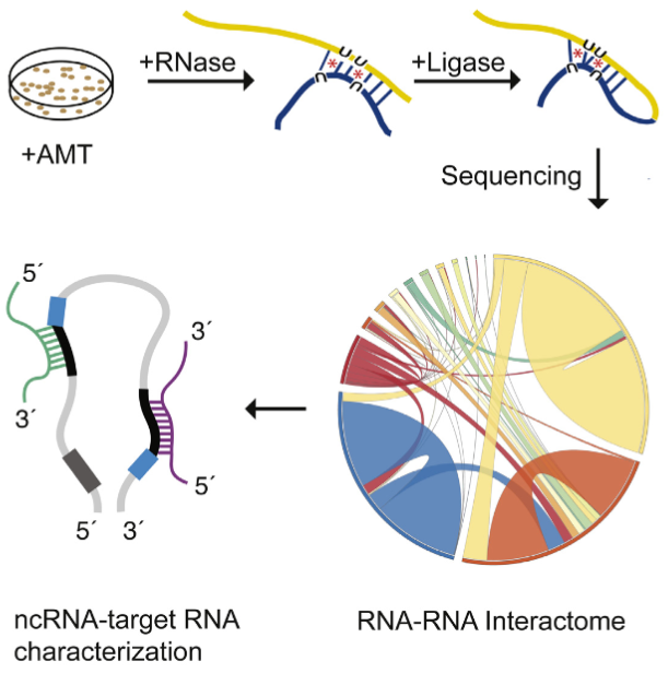
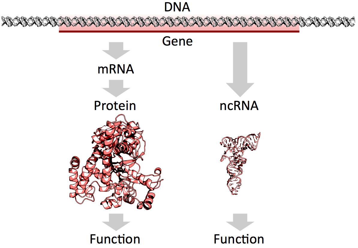
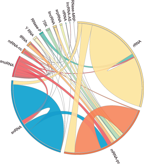
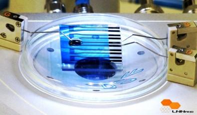
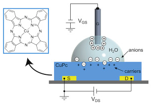
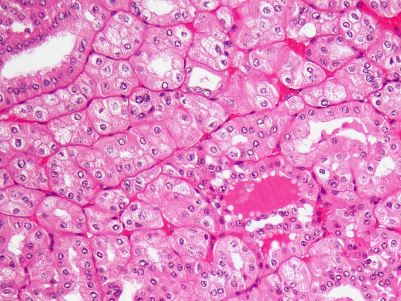