Feeds:
Posts
Comments

Posts Tagged ‘Vascular smooth muscle’

Lesson 3 Cell Signaling & Motility: G Proteins, Signal Transduction: Curations and Articles of reference as supplemental information: #TUBiol3373

Curator: Stephen J. Williams, Ph.D.

Updated 7/15/2019

Lesson 3 Powerpoint (click link below):

cell signaling and motility 3 finalissima sjw

Four papers to choose from for your February 11 group presentation:

Structural studies of G protein Coupled receptor

Shapiro-2009-Annals_of_the_New_York_Academy_of_Sciences

G protein as target in neurodegerative disease

fish technique


Today’s lesson 3 explains how extracellular signals are transduced (transmitted) into the cell through receptors to produce an agonist-driven event (effect).  This lesson focused on signal transduction from agonist through G proteins (GTPases), and eventually to the effectors of the signal transduction process.  Agonists such as small molecules like neurotransmitters, hormones, nitric oxide were discussed however later lectures will discuss more in detail the large growth factor signalings which occur through receptor tyrosine kinases and the Ras family of G proteins as well as mechanosignaling through Rho and Rac family of G proteins.

Transducers: The Heterotrimeric G Proteins (GTPases)

An excellent review of heterotrimeric G Proteins found in the brain is given by

Heterotrimeric G Proteins by Eric J Nestler and Ronald S Duman.


from Seven-Transmembrane receptors – Scientific Figure on ResearchGate. Available from: https://www.researchgate.net/figure/Examples-of-heterotrimeric-G-protein-effectors_tbl1_11180073 [accessed 4 Feb, 2019] and see references within


See below for the G Protein Cycle


<a href=”https://www.researchgate.net/figure/32-The-G-protein-cycle-In-the-absence-of-agonist-A-GPCRs-are-mainly-in-the-low_fig2_47933733″><img src=”https://www.researchgate.net/profile/Veli_Pekka_Jaakola/publication/47933733/figure/fig2/AS:669499451781133@1536632516635/32-The-G-protein-cycle-In-the-absence-of-agonist-A-GPCRs-are-mainly-in-the-low.ppm&#8221; alt=”.3.2: The G protein cycle. In the absence of agonist (A), GPCRs are mainly in the low affinity state (R). After agonist binding, the receptor is activated in the high affinity state (R*), and the agonist-GPCR-G protein complex is formed. GTP replaces GDP in Gα. After that the G protein dissociates into the Gα subunit and the Gβγ heterodimer, which then activate several effector proteins. The built-in GTPase activity of the Gα subunit cleaves the terminal phosphate group of GTP, and the GDP bound Gα subunit reassociates with Gβγ heterodimer. This results in the deactivation of both Gα and Gβγ. The G protein cycle returns to the basal state. RGS, regulator of G protein signalling.”/></a>


From Citation: Review: A. M. Preininger, H. E. Hamm, G protein signaling: Insights from new structures. Sci. STKE2004, re3 (2004)


For a tutorial on G Protein coupled receptors (GPCR) see

https://www.khanacademy.org/test-prep/mcat/organ-systems/biosignaling/v/g-protein-coupled-receptors


cyclic AMP (cAMP) signaling to the effector Protein Kinase A (PKA)

from https://courses.washington.edu/conj/gprotein/cyclicamp.htm

Cyclic AMP is an important second messenger. It forms, as shown, when the membrane enzyme adenylyl cyclase is activated (as indicated, by the alpha subunit of a G protein).


The cyclic AMP then goes on the activate specific proteins. Some ion channels, for example, are gated by cyclic AMP. But an especially important protein activated by cyclic AMP is protein kinase A, which goes on the phosphorylate certain cellular proteins. The scheme below shows how cyclic AMP activates protein kinase A.

Updated 7/15/2019

Additional New Studies on Regulation of the Beta 2 Adrenergic Receptor

We had discussed regulation of the G protein coupled beta 2 adrenergic receptor by the B-AR receptor kinase (BARK)/B arrestin system which uncouples and desensitizes the receptor from its G protein system.  In an article by Xiangyu Liu in Science in 2019, the authors describe another type of allosteric modulation (this time a POSITIVE allosteric modulation) in the intracellular loop 2.  See below:

Mechanism of β2AR regulation by an intracellular positive allosteric modulator

Xiangyu Liu1,*, Ali Masoudi2,*, Alem W. Kahsai2,*, Li-Yin Huang2, Biswaranjan Pani2Dean P. Staus2, Paul J. Shim2, Kunio Hirata3,4, Rishabh K. Simhal2, Allison M. Schwalb2, Paula K. Rambarat2, Seungkirl Ahn2, Robert J. Lefkowitz2,5,6,Brian Kobilka1

Positive reinforcement in a GPCR

Many drug discovery efforts focus on G protein–coupled receptors (GPCRs), a class of receptors that regulate many physiological processes. An exemplar is the β2-adrenergic receptor (β2AR), which is targeted by both blockers and agonists to treat cardiovascular and respiratory diseases. Most GPCR drugs target the primary (orthosteric) ligand binding site, but binding at allosteric sites can modulate activation. Because such allosteric sites are less conserved, they could possibly be targeted more specifically. Liu et al. report the crystal structure of β2AR bound to both an orthosteric agonist and a positive allosteric modulator that increases receptor activity. The structure suggests why the modulator compound is selective for β2AR over the closely related β1AR. Furthermore, the structure reveals that the modulator acts by enhancing orthosteric agonist binding and stabilizing the active conformation of the receptor.

Abstract

Drugs targeting the orthosteric, primary binding site of G protein–coupled receptors are the most common therapeutics. Allosteric binding sites, elsewhere on the receptors, are less well-defined, and so less exploited clinically. We report the crystal structure of the prototypic β2-adrenergic receptor in complex with an orthosteric agonist and compound-6FA, a positive allosteric modulator of this receptor. It binds on the receptor’s inner surface in a pocket created by intracellular loop 2 and transmembrane segments 3 and 4, stabilizing the loop in an α-helical conformation required to engage the G protein. Structural comparison explains the selectivity of the compound for β2– over the β1-adrenergic receptor. Diversity in location, mechanism, and selectivity of allosteric ligands provides potential to expand the range of receptor drugs.


Recent structures of GPCRs bound to allosteric modulators have revealed that receptor surfaces are decorated with diverse cavities and crevices that may serve as allosteric modulatory sites (1). This substantiates the notion that GPCRs are structurally plastic and can be modulated by a variety of allosteric ligands through distinct mechanisms (2-7). Most of these structures have been solved with negative allosteric modulators (NAMs), which stabilize receptors in their inactive states (1). To date, only a single structure of an active GPCR bound to a small-molecule positive allosteric modulator (PAM) has been reported, namely, the M2 muscarinic acetylcholine receptor with LY2119620 (8). Thus, mechanisms of PAMs and their potential binding sites remain largely unexplored.

F1.large


Fig 1. Structure of the active state T4L-B2AR in complex with the orthosteric agonist BI-167107, nanobody 689, and compound 6FA.  (A) The chemical structure of compound-6FA (Cmpd-6FA). (B) Isoproterenol (ISO) competition binding with 125I-cyanopindolol (CYP) to the β2AR reconstituted in nanodisks in the presence of vehicle (0.32% dimethylsulfoxide; DMSO), Cmpd-6, or Cmpd-6FA at 32 μM. Values were normalized to percentages of the maximal 125I-CYP binding level obtained from a one-site competition binding–log IC50 (median inhibitory concentration) curve fit. Binding curves were generated by GraphPad Prism. Points on curves represent mean ± SEM obtained from five independent experiments performed in duplicate. (C) Analysis of Cmpd-6FA interaction with the BI-167107–bound β2AR by ITC. Representative thermogram (inset) and binding isotherm, of three independent experiments, with the best titration curve fit are shown. Summary of thermodynamic parameters obtained by ITC: binding affinity (KD = 1.2 ± 0.1 μM), stoichiometry (N = 0.9 ± 0.1 sites), enthalpy (ΔH = 5.0 ± 1.2 kcal mol−1), and entropy (ΔS =13 ± 2.0 cal mol−1 deg−1). (D) Side view of T4L-β2AR bound to the orthosteric agonist BI-167107, nanobody 6B9 (Nb6B9), and Cmpd-6FA. The gray box indicates the membrane layer as defined by the OPM database. (E) Close-up view of Cmpd-6FA binding site. Covering Cmpd-6FA is 2Fo– Fc electron density contoured at 1.0 σ (green mesh).From Science  28 Jun 2019:
Vol. 364, Issue 6447, pp. 1283-1287


F3.large

Fig 3. Fig. 3 Mechanism of allosteric activation of the β2AR by Cmpd-6FA.

(A) Superposition of the inactive β2AR bound to the antagonist carazolol (PDB code: 2RH1) and the active β2AR bound to the agonist BI-167107, Cmpd-6FA, and Nb6B9. Close-up view of the Cmpd-6FA binding site is shown. The residues of the inactive (yellow) and active (blue) β2AR are depicted, and the hydrogen bond formed between Asp1303.49and Tyr141ICL2 in the active state is indicated by a black dashed line. (B) Topography of Cmpd-6FA binding surface on the active β2AR (left, blue) and the corresponding surface of the inactive β2AR (right, yellow) with Cmpd-6FA (orange sticks) docked on top. Molecular surfaces are of only those residues involved in interaction with Cmpd-6FA. Steric clash between Cmpd-6FA and the surface of inactive β2AR is represented by a purple asterisk. (C) Overlay of the β2AR bound to BI-167107, Nb6B9, and Cmpd-6FA with the β2AR–Gscomplex (PDB code: 3SN6). The inset shows the position of Phe139ICL2 relative to the α subunit of Gs. (D) Superposition of the active β2AR bound to the agonist BI-167107, Nb6B9, and Cmpd-6FA (blue) with the inactive β2AR bound to carazolol (yellow) (PDB code: 2RH1) as viewed from the cytoplasm. For clarity, Nb6B9 and the orthosteric ligands are omitted. The arrows indicate shifts in the intracellular ends of the TM helices 3, 5, and 6 upon activation and their relative distances.


Allosteric sites may not face the same evolutionary pressure as do orthosteric sites, and thus are more divergent across subtypes within a receptor family (2426). Therefore, allosteric sites may provide a greater source of specificity for targeting GPCRs.


  1. D. M. Thal, A. Glukhova, P. M. Sexton, A. Christopoulos, Structural insights into G-protein-coupled receptor allostery. Nature 559, 45–53 (2018). doi:10.1038/s41586-018-0259-zpmid:29973731CrossRefPubMedGoogle Scholar


  1. D. Wacker, R. C. Stevens, B. L. Roth, How Ligands Illuminate GPCR Molecular Pharmacology. Cell 170, 414–427 (2017).

doi:10.1016/j.cell.2017.07.009pmid:28753422CrossRefPubMedGoogle Scholar


  1. D. P. Staus, R. T. Strachan, A. Manglik, B. Pani, A. W. Kahsai, T. H. Kim, L. M. Wingler, S. Ahn, A. Chatterjee, A. Masoudi, A. C. Kruse, E. Pardon, J. Steyaert, W. I. Weis, R. S. Prosser, B. K. Kobilka, T. Costa, R. J. Lefkowitz, Allosteric nanobodies reveal the dynamic range and diverse mechanisms of G-protein-coupled receptor activation. Nature 535, 448–452 (2016). doi:10.1038/nature18636pmid:27409812CrossRefPubMedGoogle Scholar


  1. A. Manglik, T. H. Kim, M. Masureel, C. Altenbach, Z. Yang, D. Hilger, M. T. Lerch, T. S. Kobilka, F. S. Thian, W. L. Hubbell, R. S. Prosser, B. K. Kobilka, Structural Insights into the Dynamic Process of β2-Adrenergic Receptor Signaling. Cell 161, 1101–1111 (2015). doi:10.1016/j.cell.2015.04.043pmid:25981665CrossRefPubMedGoogle Scholar


5,   L. Ye, N. Van Eps, M. Zimmer, O. P. Ernst, R. S. Prosser, Activation of the A2A adenosine G-protein-coupled receptor by conformational selection. Nature 533, 265–268 (2016). doi:10.1038/nature17668pmid:27144352CrossRefPubMedGoogle Scholar


  1. N. Van Eps, L. N. Caro, T. Morizumi, A. K. Kusnetzow, M. Szczepek, K. P. Hofmann, T. H. Bayburt, S. G. Sligar, O. P. Ernst, W. L. Hubbell, Conformational equilibria of light-activated rhodopsin in nanodiscs. Proc. Natl. Acad. Sci. U.S.A. 114, E3268–E3275 (2017). doi:10.1073/pnas.1620405114pmid:28373559Abstract/FREE Full TextGoogle Scholar


  1. R. O. Dror, H. F. Green, C. Valant, D. W. Borhani, J. R. Valcourt, A. C. Pan, D. H. Arlow, M. Canals, J. R. Lane, R. Rahmani, J. B. Baell, P. M. Sexton, A. Christopoulos, D. E. Shaw, Structural basis for modulation of a G-protein-coupled receptor by allosteric drugs. Nature 503, 295–299 (2013). doi:10.1038/nature12595pmid:24121438CrossRefPubMedWeb of ScienceGoogle Scholar


  1. A. C. Kruse, A. M. Ring, A. Manglik, J. Hu, K. Hu, K. Eitel, H. Hübner, E. Pardon, C. Valant, P. M. Sexton, A. Christopoulos, C. C. Felder, P. Gmeiner, J. Steyaert, W. I. Weis, K. C. Garcia, J. Wess, B. K. Kobilka, Activation and allosteric modulation of a muscarinic acetylcholine receptor. Nature 504, 101–106 (2013). doi:10.1038/nature12735pmid:24256733


Additional information on Nitric Oxide as a Cellular Signal

Nitric oxide is actually a free radical and can react with other free radicals, resulting in a very short half life (only a few seconds) and so in the body is produced locally to its site of action (i.e. in endothelial cells surrounding the vascular smooth muscle, in nerve cells). In the late 1970s, Dr. Robert Furchgott observed that acetylcholine released a substance that produced vascular relaxation, but only when the endothelium was intact. This observation opened this field of research and eventually led to his receiving a Nobel prize. Initially, Furchgott called this substance endothelium-derived relaxing factor (EDRF), but by the mid-1980s he and others identified this substance as being NO.

Nitric oxide is produced from metabolism of endogenous substances like L-arginine, catalyzed by one of three isoforms of nitric oxide synthase (for link to a good article see here) or release from exogenous compounds like drugs used to treat angina pectoris like amyl nitrate or drugs used for hypertension such as sodium nitroprusside.

The following articles are a great reference to the chemistry, and physiological and pathological Roles of Nitric Oxide:

46. The Molecular Biology of Renal Disorders: Nitric Oxide – Part III

Curator and Author: Larry H Bernstein, MD, FACP

https://pharmaceuticalintelligence.com/2012/11/26/the-molecular-biology-of-renal-disorders/

47. Nitric Oxide Function in Coagulation – Part II

Curator and Author: Larry H. Bernstein, MD, FCAP

https://pharmaceuticalintelligence.com/2012/11/26/nitric-oxide-function-in-coagulation/

48. Nitric Oxide, Platelets, Endothelium and Hemostasis

Curator and Author: Larry H Bernstein, MD, FACP

https://pharmaceuticalintelligence.com/2012/11/08/nitric-oxide-platelets-endothelium-and-hemostasis/

49. Interaction of Nitric Oxide and Prostacyclin in Vascular Endothelium

Curator and Author: Larry H Bernstein, MD, FACP

https://pharmaceuticalintelligence.com/2012/09/14/interaction-of-nitric-oxide-and-prostacyclin-in-vascular-endothelium/

50. Nitric Oxide and Immune Responses: Part 1

Curator and Author:  Aviral Vatsa PhD, MBBS

https://pharmaceuticalintelligence.com/2012/10/18/nitric-oxide-and-immune-responses-part-1/

51. Nitric Oxide and Immune Responses: Part 2

Curator and Author:  Aviral Vatsa PhD, MBBS

https://pharmaceuticalintelligence.com/2012/10/28/nitric-oxide-and-immune-responses-part-2/

56. Nitric Oxide and iNOS have Key Roles in Kidney Diseases – Part II

Curator and Author: Larry H Bernstein, MD, FACP

https://pharmaceuticalintelligence.com/2012/11/26/nitric-oxide-and-inos-have-key-roles-in-kidney-diseases/

57. New Insights on Nitric Oxide donors – Part IV

Curator and Author: Larry H Bernstein, MD, FACP

https://pharmaceuticalintelligence.com/2012/11/26/new-insights-on-no-donors/

59. Nitric Oxide has a ubiquitous role in the regulation of glycolysis -with a concomitant influence on mitochondrial function

Curator and Author: Larry H Bernstein, MD, FACP

https://pharmaceuticalintelligence.com/2012/09/16/nitric-oxide-has-a-ubiquitous-role-in-the-regulation-of-glycolysis-with-         a-concomitant-influence-on-mitochondrial-function/

Biochemistry of the Coagulation Cascade and Platelet Aggregation: Nitric Oxide: Platelets, Circulatory Disorders, and Coagulation Effects

Nitric Oxide Function in Coagulation – Part II

Nitric oxide is implicated in many pathologic processes as well.  Nitric oxide post translational modifications have been attributed to nitric oxide’s role in pathology however, although the general mechanism by which nitric oxide exerts its physiological effects is by stimulation of soluble guanylate cyclase to produce cGMP, these post translational modifications can act as a cellular signal as well.  For more information of NO pathologic effects and how NO induced post translational modifications can act as a cellular signal see the following:

Nitric Oxide Covalent Modifications: A Putative Therapeutic Target?

58. Crucial role of Nitric Oxide in Cancer

Curator and Author: Ritu Saxena, Ph.D.

https://pharmaceuticalintelligence.com/2012/10/16/crucial-role-of-nitric-oxide-in-cancer/

Note:  A more comprehensive ebook on Nitric Oxide and Disease Perspectives is found at

Cardiovascular Diseases, Volume One: Perspectives on Nitric Oxide in Disease Mechanisms

available on Kindle Store @ Amazon.com

http://www.amazon.com/dp/B00DINFFYC

Read Full Post »

Sensors and Signaling in Oxidative Stress

Author and Curator: Larry H. Bernstein, MD, FCAP

Article XI Sensors and Signaling in Oxidative Stress

Image created by Adina Hazan 06/30/2021

This is article ELEVEN in the following series on Calcium Role in Cardiovascular Diseases

Part I: Identification of Biomarkers that are Related to the Actin Cytoskeleton
Larry H Bernstein, MD, FCAP
http://pharmaceuticalintelligence.com/2012/12/10/identification-of-biomarkers-
that-are-related-to-the-actin-cytoskeleton/

Part II: Role of Calcium, the Actin Skeleton, and Lipid Structures in Signaling and Cell Motility
Larry H. Bernstein, MD, FCAP, Stephen Williams, PhD and Aviva Lev-Ari, PhD, RN
http://pharmaceuticalintelligence.com/2013/08/26/role-of-calcium-the-actin-
skeleton-and-lipid-structures-in-signaling-and-cell-motility/

Part III: Renal Distal Tubular Ca2+ Exchange Mechanism in Health and Disease
Larry H. Bernstein, MD, FCAP, Stephen J. Williams, PhD
and Aviva Lev-Ari, PhD, RN
http://pharmaceuticalintelligence.com/2013/09/02/renal-distal-tubular-ca2-
exchange-mechanism-in-health-and-disease/

Part IV: The Centrality of Ca(2+) Signaling and Cytoskeleton Involving Calmodulin Kinases and
Ryanodine Receptors in Cardiac Failure, Arterial Smooth Muscle, Post-ischemic Arrhythmia,
Similarities and Differences, and Pharmaceutical Targets
Larry H Bernstein, MD, FCAP, Justin Pearlman, MD, PhD, FACC and Aviva Lev-Ari, PhD, RN
http://pharmaceuticalintelligence.com/2013/09/08/the-centrality-of-ca2-signaling-and-cytoskeleton-
involving-calmodulin-kinases-and-ryanodine-receptors-in-cardiac-failure-arterial-smooth-muscle-
post-ischemic-arrhythmia-similarities-and-differen/

Part V: Ca2+-Stimulated Exocytosis:  The Role of Calmodulin and Protein Kinase C in Ca2+ Regulation of Hormone and Neurotransmitter

Larry H Bernstein, MD, FCAP
and
Aviva Lev-Ari, PhD, RN

http://pharmaceuticalintelligence.com/2013/12/23/calmodulin-and-protein-kinase-c-drive-the-ca2-regulation-of-hormone-and-neurotransmitter-release-that-triggers-ca2-stimulated-exocytosis/

Part VI: Calcium Cycling (ATPase Pump) in Cardiac Gene Therapy: Inhalable Gene Therapy for Pulmonary
Arterial Hypertension and Percutaneous Intra-coronary Artery Infusion for Heart Failure: Contributions by Roger J. Hajjar, MD
Aviva Lev-Ari, PhD, RN
http://pharmaceuticalintelligence.com/2013/08/01/calcium-molecule-in-cardiac-gene-therapy-inhalable-gene-therapy-
for-pulmonary-arterial-hypertension-and-percutaneous-intra-coronary-artery-infusion-for-heart-failure-contributions-by-roger-j-hajjar/

Part VII: Cardiac Contractility & Myocardium Performance: Ventricular Arrhythmias and Non-ischemic Heart Failure –
Therapeutic Implications for Cardiomyocyte Ryanopathy (Calcium Release-related Contractile Dysfunction) and Catecholamine Responses
Justin Pearlman, MD, PhD, FACC, Larry H Bernstein, MD, FCAP and Aviva Lev-Ari, PhD, RN
http://pharmaceuticalintelligence.com/2013/08/28/cardiac-contractility-myocardium-performance-ventricular-arrhythmias-
and-non-ischemic-heart-failure-therapeutic-implications-for-cardiomyocyte-ryanopathy-calcium-release-related-contractile/

Part VIII: Disruption of Calcium Homeostasis: Cardiomyocytes and Vascular Smooth Muscle Cells:
The Cardiac and Cardiovascular Calcium Signaling Mechanism
Justin Pearlman, MD, PhD, FACC, Larry H Bernstein, MD, FCAP and Aviva Lev-Ari, PhD, RN
http://pharmaceuticalintelligence.com/2013/09/12/disruption-of-calcium-homeostasis-cardiomyocytes-and-vascular-smooth-
muscle-cells-the-cardiac-and-cardiovascular-calcium-signaling-mechanism/

Part IX: Calcium-Channel Blockers, Calcium Release-related Contractile Dysfunction
(Ryanopathy) and Calcium as Neurotransmitter Sensor
Justin Pearlman, MD, PhD, FACC, Larry H Bernstein, MD, FCAP and Aviva Lev-Ari, PhD, RN
http://pharmaceuticalintelligence.com/2013/09/16/calcium-channel-blocker-calcium-as-neurotransmitter-sensor-
and-calcium-release-related-contractile-dysfunction-ryanopathy/

Part X: Synaptotagmin functions as a Calcium Sensor: How Calcium Ions Regulate the fusion of
vesicles with cell membranes during Neurotransmission
Larry H Bernstein, MD, FCAP and Aviva Lev-Ari, PhD, RN
http://pharmaceuticalintelligence.com/2013/09/10/synaptotagmin-functions-as-a-calcium-sensor-how-calcium-ions-
regulate-the-fusion-of-vesicles-with-cell-membranes-during-neurotransmission/

Part XI: Sensors and Signaling in Oxidative Stress
Larry H. Bernstein, MD, FCAP
http://pharmaceuticalintelligence.com/2013/11/01/sensors-and-signaling-in-oxidative-stress/

Part XII: Atherosclerosis Independence: Genetic Polymorphisms of Ion Channels Role in the Pathogenesis of Coronary Microvascular Dysfunction and Myocardial Ischemia (Coronary Artery Disease (CAD))

Larry H Bernstein, MD, FCAP and Aviva Lev-Ari, PhD, RN

http://pharmaceuticalintelligence.com/2013/12/21/genetic-polymorphisms-of-ion-channels-have-a-role-in-the-pathogenesis-of-coronary-microvascular-dysfunction-and-ischemic-heart-disease/

This important article on oxidative stress was published in Free Radical Biol. and Med.

Nrf2:INrf2(Keap1) Signaling in Oxidative Stress

James W. Kaspar, Suresh K. Niture, and Anil K. Jaiswal*
Department of Pharmacology, University of Maryland School of Medicine, Baltimore, MD

Free Radic Biol Med. 2009 Nov; 47(9): 1304–1309.           http://dx.doi.org/10.1016/j.freeradbiomed.2009.07.035

Nrf2:INrf2(Keap1) are cellular sensors of chemical and radiation induced oxidative and electrophilic stress. Nrf2 is a nuclear transcription factor that controls the expression and coordinated induction of a battery of defensive genes encoding detoxifying enzymes and antioxidant proteins. This is a mechanism of critical importance for cellular protection and cell survival. Nrf2 is retained in the cytoplasm by an inhibitor INrf2. INrf2 functions as an adapter for Cul3/Rbx1 mediated degradation of Nrf2. In response to oxidative/electrophilic stress, Nrf2 is switched on and then off by distinct early and delayed mechanisms. Oxidative/electrophilic modification of INrf2cysteine151 and/or PKC phosphorylation of Nrf2serine40 results in

  • the escape or release of Nrf2 from INrf2.

Nrf2 is stabilized and

  • translocates to the nucleus,
  • forms heterodimers with unknown proteins, and
  • binds antioxidant response element (ARE) that
  • leads to coordinated activation of gene expression.
  • It takes less than fifteen minutes from the time of exposure to switch on nuclear import of Nrf2. This is followed by activation of a delayed mechanism that controls switching off of Nrf2 activation of gene expression. GSK3β phosphorylates Fyn at unknown threonine residue(s) leading to nuclear localization of Fyn. Fyn phosphorylates Nrf2tyrosine568
  • resulting in nuclear export of Nrf2, binding with INrf2 and
  • degradation of Nrf2.

The switching on and off of Nrf2 protects cells against free radical damage, prevents apoptosis and promotes cell survival.

Introduction

Oxidative stress is induced by a vast range of factors including xenobiotics, drugs, heavy metals and ionizing radiation. Oxidative stress leads to the generation of Reactive Oxygen Species (ROS) and electrophiles. ROS and electrophiles generated can have a profound impact on survival, growth development and evolution of all living organisms [1,2] ROS include

  • both free radicals, such as the superoxide anion and the hydroxyl radical, and
  • oxidants such as hydrogen peroxide [3].

ROS and electrophiles can cause diseases such as cancer, cardiovascular complications, acute and chronic inflammation, and neurodegenerative diseases [1]. Therefore, it is obvious that

  • cells must constantly labor to control levels of ROS, preventing them from accumulation.

Much of what we know about the mechanisms of protection against oxidative stress has come from the study of prokaryotic cells [4,5]. Prokaryotic cells utilize transcription factors OxyR and SoxRS to sense the redox state of the cell, and

  • during oxidative stress these factors induce the expression of nearly eighty defensive genes [5].

Eukaryotic cells have similar mechanisms to protect against oxidative stress [Fig. 1; ref. 3,6–9]. Initial effect of oxidative/electrophilic stress leads to activation of a battery of defensive gene expression that leads to detoxification of chemicals and ROS and prevention of free radical generation and cell survival [Fig. 1].

Fig 1.  Chemical and radiation exposure and coordinated induction of defensive genes.

Fig. 1. Chemical and radiation exposure and coordinated induction of defensive genes.

Of these genes, some are enzymes such as NAD(P)H:quinine oxidoreductase 1 (NQO1), NRH:quinone oxidoreductase 2 (NQO2), glutathione S-transferase Ya subunit (GST Ya Subunit), heme oxygenase 1 (HO-1), and γ-glutamylcysteine synthetase (γ-GCS), also known as glutamate cysteine ligase (GCL). Other genes have end products that regulate a wide variety of cellular activities including

  • signal transduction,
  • proliferation, and
  • immunologic defense reactions.

There is a wide variety of factors associated with the cellular response to oxidative stress. For example,

  • NF-E2 related factor 2 (Nrf2),
  • heat shock response activator protein 1, and
  • NF-kappaB promote cell survival,

whereas activation of c-jun, N-terminal kinases (JNK), p38 kinase and TP53 may lead to cell cycle arrest and apoptosis [10]. The Nrf2 pathway is regarded as the most important in the cell to protect against oxidative stress. [3,6–9]. It is noteworthy that accumulation of ROS and/or electrophiles leads to oxidative/electrophile stress,

  • membrane damage,
  • DNA adducts formation and
  • mutagenicity [Fig. 1].

These changes lead to degeneration of tissues and premature aging, apoptotic cell death, cellular transformation and cancer.

Antioxidant Response Element and Nrf2

Promoter analysis identified a cis-acting enhancer sequence designated as the antioxidant response element (ARE) that

  • controls the basal and inducible expression of antioxidant genes in response to xenobiotics, antioxidants, heavy metals and UV light [11].

The ARE sequence is responsive to a broad range of structurally diverse chemicals apart from β-nafthoflavone and phenolic antioxidants [12]. Mutational analysis revealed GTGACA***GC to be the core sequence of the ARE [11,13–14]. This core sequence is present in all Nrf2 downstream genes that respond to antioxidants and xenobiotics [3,6–9]. Nrf2 binds to the ARE and regulates ARE-mediated antioxidant enzyme genes expression and induction in response to a variety of stimuli including antioxidants, xenobiotics, metals, and UV irradiation [6,15–21].

Nrf2 is ubiquitously expressed in a wide range of tissue and cell types [22–24] and belongs to a subset of basic leucine zipper genes (bZIP) sharing a conserved structural domain designated as a cap’n’collar domain which is highly conserved in Drosphila transcription factor CNC (Fig. 2; ref. 25].

Fig. 2. Schematic Presentation of Various Domains of Nrf (Nrf1, Nrf2, Nrf3) and INrf2

Fig. 2. Schematic Presentation of Various Domains of Nrf (Nrf1, Nrf2, Nrf3) and INrf2

Nrf, NF-E2 Related Factor; INrf2, Inhibitor of Nrf2; NTR, N-Terminal Region; BTB, Broad complex, Tramtrack, Bric-a-brac; IVR, Intervening/linker Region; DGR, Kelch domain/ diglycine repeats; CTR, C-Terminal Region.

The basic region, just upstream of the leucine zipper region,

  • is responsible for DNA binding [3] and
  • the acidic region is required for transcriptional activation.

ARE-mediated transcriptional activation requires heterodimerization of Nrf2 with other bZIP proteins including Jun (c-Jun, Jun-D, and Jun-B) and small Maf (MafG, MafK, MafF) proteins [18– 20,26–27].

Initial evidence demonstrating the role of Nrf2 in antioxidant-induction of detoxifying enzymes came from studies on

  • the role of Nrf2 in ARE-mediated regulation of NQO1 gene expression [17].

Nrf2 was subsequently shown to be involved in

  • the transcriptional activation of other ARE-responsive genes such as
    • GST Ya, γ-GCS, HO-1, antioxidants, proteasomes, and drug transporters [3,6–9,28–33].

Overexpression of Nrf2 cDNA was shown to upregulate the expression and induction of the NQO1 gene in response to antioxidants and xenobiotics [17]. In addition, Nrf2-null mice exhibited a marked

  • decrease in the expression and induction of NQO1,
  • indicating that Nrf2 plays an essential role in the in vivo regulation of NQO1 in response to oxidative stress [26].

The importance of this transcription factor in upregulating ARE-mediated gene expression has been demonstrated by several in vivo and in vitro studies [reviewed in ref. 3]. The results indicate that Nrf2 is an important activator of phase II antioxidant genes [3,8].

Negative Regulation of Nrf2 mediated by INrf2

A cytosolic inhibitor (INrf2), also known as Keap1 (Kelch-like ECH-associating protein 1), of Nrf2 was identified and reported [Fig. 2; ref. 34–35]. INrf2, existing as a dimer [36], retains Nrf2 in the cytoplasm. Analysis of the INrf2 amino acid sequence and domain structure-function analyses have revealed that

  • INrf2 has a BTB (broad complex, tramtrack, bric-a-brac)/ POZ (poxvirus, zinc finger) domain and
  • a Kelch domain [34–35] also known as the DGR domain (Double glycine repeat) [37].

Keap1 has three additional domains/regions:

  1. the N-terminal region (NTR),
  2. the invervening region (IVR), and
  3. the C-terminal region (CTR) [8].

The BTB/POZ domain has been shown to be

  • a protein-protein interaction domain.

In the Drosophila Kelch protein, and in IPP,

  • the Kelch domain binds to actin [38–39]
  • allowing the scaffolding of INrf2 to the actin cytoskeleton
    • which plays an important role in Nrf2 retention in the cytosol [40].

The main function of INrf2 is to serve as

  • an adapter for the Cullin3/Ring Box 1 (Cul3/Rbx1) E3 ubiquitin ligase complex [41–43].

Cul3 serves as a scaffold protein that forms the E3 ligase complex with Rbx1 and recruits a cognate E2 enzyme [8].

INrf2

  1. via its N-terminal BTB/POZ domain binds to Cul3 [44] and
  2. via its C-terminal Kelch domain binds to the substrate Nrf2
  • leading to the ubiquitination and degradation of Nrf2 through the 26S proteasome [45–49].

Under normal cellular conditions, the cytosolic INrf2/Cul3-Rbx1 complex is constantly degrading Nrf2. When a cell is exposed to oxidative stress Nrf2 dissociates from the INrf2 complex, stabilizes and translocates into the nucleus leading to activation of ARE-mediated gene expression [3,6–9]. An alternative theory is that Nrf2 in response to oxidative stress escapes INrf2 degradation, stabilizes and translocates in the nucleus [49–50]. We suggested the theory of escape of Nrf2 from INrf2 [49] and similar suggestion was also made in another report [50]. However, the follow up studies in our laboratory could not support the escape theory. Escape theory is a possibility but has to be proven by experiments before it can be adapted. Therefore, we will use the release of Nrf2 from INrf2 in the rest of this review.

Numerous reports have suggested that

  • any mechanism that modifies INrf2 and/or Nrf2 disrupting the Nrf2:INrf2 interaction will result in the upregulation of ARE-mediated gene expression.

A model Nrf2:INrf2 signaling from antioxidant and xenobiotic to activation of ARE-mediated defensive gene expression is shown in Fig. 3.

Fig. 3. Nrf2 signaling in ARE-mediated coordinated activation of defensive genes

Fig. 3. Nrf2 signaling in ARE-mediated coordinated activation of defensive genes

Since the metabolism of antioxidants and xenobiotics results in the generation of ROS and electrophiles [51], it is thought that these molecules might act as second messengers, activating ARE-mediated gene expression. Several protein kinases including PKC, ERK, MAPK, p38, and PERK [49,52– 56] are known to modify Nrf2 and activate its release from INrf2. Among these mechanisms,

  1. oxidative/electrophilic stress mediated phosphorylation of Nrf2 at serine40 by PKC is necessary for Nrf2 release from INrf2, but
  2. is not required for Nrf2 accumulation in the nucleus [49,52–53].

In addition to post-translational modification in Nrf2, several crucial residues in INrf2 have also been proposed to be important for activation of Nrf2. Studies based on

  • the electrophile mediated modification,
  • location and
  • mutational analyses revealed
    • that three cysteine residues, Cys151, Cys273 and Cys288 are crucial for INrf2 activity [50].

INrf2 itself undergoes ubiquitination by the Cul3 complex, via a proteasomal independent pathway,

  • which was markedly increased in response to phase II inducers such as antioxidants [57].

It has been suggested that normally INrf2 targets Nrf2 for ubiquitin mediated degradation but

  • electrophiles may trigger a switch of Cul3 dependent ubiquitination from Nrf2 to INrf2 resulting in ARE gene induction.

The redox modulation of cysteines in INrf2

  • might be a mechanism redundant to the phosphorylation of Nrf2 by PKC, or that
  • the two mechanisms work in concert.

In addition to cysteine151 modification,

  • phosphorylation of Nrf2 has also been shown to play a role in INrf2 retention and release of Nrf2.

Serine104 of INrf2 is required for dimerization of INrf2, and

  • mutations of serine104 led to the disruption of the INrf2 dimer leading to the release of Nrf2 [36].

Recently, Eggler at al. demonstrated that modifying specific cysteines of the electrophile-sensing human INrf2 protein is insufficient to disrupt binding to the Nrf2 domain Neh2 (58). Upon introduction of electrophiles, modification of INrf2C151 leads to a change in the conformation of the BTB domain by means of perturbing the homodimerization site, disrupting Neh2 ubiquitination, and causing ubiquitination of INrf2. Modification of INrf2 cysteines by electrophiles does not lead to disruption of the INrf2–Nrf2 complex. Rather, the switch of ubiquitination from Nrf2 to INrf2 leads to Nrf2 nuclear accumulation.

More recently, our laboratory demonstrated that phosphorylation and de-phosphorylation of tyrosine141 in INrf2 regulates its stability and degradation, respectively [59]. The de-phosphorylation of tyrosine141 caused destabilization and degradation of INrf2 leading to the release of Nrf2. Furthermore, we showed that prothymosin-α mediates nuclear import of the INrf2/Cul3-Rbx1 complex [60]. The INrf2/Cul3-Rbx1 complex inside the nucleus exchanges prothymosin-α with Nrf2 resulting in degradation of Nrf2. These results led to the conclusion that prothymosin-α mediated nuclear import of INrf2/Cul3-Rbx1 complex leads to ubiquitination and degradation of nuclear Nrf2 presumably to regulate nuclear level of Nrf2 and rapidly switch off the activation of Nrf2 downstream gene expression. An auto-regulatory loop also exists within the Nrf2 pathway [61]. An ARE was identified in the INrf2 promoter that facilitates Nrf2 binding causing induction of the INrf2 gene. Nrf2 regulates INrf2 by controlling its transcription, and INrf2 controls Nrf2 by serving as an adaptor for degradation.

Other Regulatory Mediators of Nrf2

Bach1 (BTB and CNC homology 1, basic leucine zipper transcription factor 1) is a transcription repressor [62] that is ubiquitously expressed in tissues [63–64] and distantly related to Nrf2 [8]. In the absence of cellular stress, Bach1 heterodimers with small Maf proteins [65] that bind to the (ARE) [66] repressing gene expression. In the presence of oxidative stress, Bach1 releases from the ARE and is replaced by Nrf2. Bach1 competes with Nrf2 for binding to the ARE leading to suppression of Nrf2 downstream genes [66].

Nuclear import of Nrf2, from time of exposure to stabilization, takes roughly two hours [67]. This is followed by activation of a delayed mechanism involving Glycogen synthase kinase 3 beta (GSK3f3) that controls switching off of Nrf2 activation of gene expression (Fig. 3). GSK3f3 is a multifunctional serine/threonine kinase, which plays a major role in various signaling pathways [68]. GSK3f3 phosphorylates Fyn, a tyrosine kinase, at unknown threonine residue(s) leading to nuclear localization of Fyn [69]. Fyn phosphorylates Nrf2 tyrosine 568 resulting in nuclear export of Nrf2, binding with INrf2 and degradation of Nrf2 [70].

The negative regulation of Nrf2 by Bach1 and GSK3f3/Fyn are important in repressing Nrf2 downstream genes that were induced in response to oxidative/electrophilic stress. The tight control of Nrf2 is vital for the cells against free radical damage, prevention of apoptosis and cell survival [3,6–9,70].

Nrf2 in Cytoprotection, Cancer and Drug Resistance

Nrf2 is a major protective mechanism against xenobiotics capable of damaging DNA and initiating carcinogenesis [71]. Inducers of Nrf2 function as blocking agents that prevents carcinogens from reaching target sites, inhibits parent molecules undergoing metabolic activation, or subsequently preventing carcinogenic species from interacting with crucial cellular macromolecules, such as DNA, RNA, and proteins [72]. A plausible mechanism by which blocking agents impart their chemopreventive activity is the induction of detoxification and antioxidant enzymes [73]. Oltipraz, 3H-1,2,-dithiole-3-thione (D3T), Sulforaphane, and Curcumin can be considered potential chemopreventive agents because

  • these compounds have all been shown to induce Nrf2 [74–81].

Studies have shown a role of Nrf2 in protection against cadmium and manganese toxicity [82]. Nrf2 also plays an important role in reduction of methyl mercury toxicity [83]. Methylmercury activates Nrf2 and the activation of Nrf2 is essential for reduction of methylmercury by facilitating its excretion into extracellular space. In vitro and in vivo studies have shown a role of Nrf2 in neuroprotection and protection against Parkinson’s disease [84– 86]. Disruption of Nrf2 impairs the resolution of hyperoxia-induced acute lung injury and inflammation in mice [87]. Nrf2-knockout mice were more prone to

  • tumor growth when exposed to carcinogens such as benzo[a]pyrene, diesel exhaust, and N-nitrosobutyl (4-hydroxybutyl) amine [88–90].

INrf2/Nrf2 signaling is also shown to regulate oxidative stress tolerance and lifespan in Drosophila [91].

A role of Nrf2 in drug resistance is suggested based on its property to induce detoxifying and antioxidant enzymes (92–97). The loss of INrf2 (Keap1) function is shown to

  • lead to nuclear accumulation of Nrf2, activation of metabolizing enzymes and drug resistance (95).

Studies have reported mutations resulting in dysfunctional INrf2 in lung, breast and bladder cancers (96–100). A recent study reported that somatic mutations also occur in the coding region of Nrf2, especially in cancer patients with a history of smoking or suffering from squamous cell carcinoma (101). These mutations abrogate its interaction with INrf2 and nuclear accumulation of Nrf2. This gives advantage to

  • cancer cell survival and
  • undue protection from anti-cancer treatments.

However, the understanding of the mechanism of Nrf2 induced drug resistance remains in its infancy. In addition, the studies on Nrf2 regulated downstream pathways that contribute to drug resistance remain limited.

Future Perspectives

Nrf2 creates a new paradigm in cytoprotection, cancer prevention and drug resistance. Considerable progress has been made to better understand all mechanisms involved within the intracellular pathways regulating Nrf2 and its downstream genes. Preliminary studies demonstrate that

  • deactivation of Nrf2 is as important as activation of Nrf2.

Further studies are needed to better understand the negative regulation of Nrf2. Also better understanding of the negative regulation of Nrf2 could help design a new class of effective chemopreventive compounds not only targeting Nrf2 activation, but also

  • targeting the negative regulators of Nrf2.

Abbreviations: 

Nrf2    NF-E2 related factor 2;  INrf2   Inhibitor of Nrf2 also known as Keap1;   ROS    Reactive oxygen species.

References (1-15 of 101)

1. Breimer LH. Molecular Mechanisms of oxygen radical carcinogenesis and mutagenesis: the role of DNA base damage. Mol Carcinog 1990;3:188–197. [PubMed: 2206282]

2. Meneghini R. Iron homeostasis, oxidative stress, and DNA damage. Free Radic Biol Med 1997;23:783– 792. [PubMed: 9296456]

3. Jaiswal AK. Nrf2 signaling in coordinated activation of antioxidant gene expression. Free Radic Biol Med 2004;36:1199–1207. [PubMed: 15110384]

4. Bauer CE, Elsen S, Bird TH. Mechanisms for redox control of gene expression. Annu Rev Microbiol 1999;53:495–523. [PubMed: 10547699]

5. Zheng M, Storz G. Redox sensing by prokaryotic transcription factors. Biochem Pharm 2000;59:1–6. [PubMed: 10605928]

6. Dhakshinamoorthy S, Long DJ II, Jaiswal AK. Antioxidant regulation of genes encoding enzymes that detoxify xenobiotics and carcinogens. Current Topics in Cellular Regulation 2000;36:201–206. [PubMed: 10842753]

7. Zhang DD. Mechanistic studies of the Nrf2-Keap1 signaling pathway. Drug Metab Rev 2006;38:769– 789. [PubMed: 17145701]

8. Kobayashi M, Yamamoto M. Nrf2-Keap1 regulation of cellular defense mechanisms against electrophiles and reactive oxygen species. Adv Enzyme Regul 2006;46:113–140. [PubMed: 16887173]

9. Copple IM, Goldring CE, Kitteringham NR, Park BK. The Nrf2-Keap1 defense pathway: role in protection against drug-induced toxicity. Toxicology 2008;246:24–33. [PubMed: 18083283]

10. Halliwell, B.; Gutteridge, JMC. Free radicals in biology and medicine. Vol. 4. Oxford University Press; 2007.

  • 11. Rushmore TH, Morton MR, Pickett CB. The antioxidant responsive element. Actiavtion by oxidative stress and identification of the DNA consensus sequence required for functional activity. J Biol Chem 1991;266:11632–11639. [PubMed: 1646813]
  • 12. Rushmore TH, King RG, Paulson KE, Pickett CB. Regulation of glutathione S-transferase Ya subunit gene expression: identification of a unique xenobiotics-responsive element controlling inducible expression by planar aromatic compounds. Proc Natl Acad Sci USA 1990;87:3826–3830. [PubMed: 2160079]
  • 13. Xie T, Belinsky M, Xu Y, Jaiswal AK. ARE and TRE-mediated regulation of gene expression: response to xenobiotics and antioxidants. J Biol Chem 1995;270:6894–6900. [PubMed: 7896838]
  • 14.

Rushmore TH, Pickett CB. Glutathione S-transferases, structures, regulation, and therapeutic implications. J Biol Chem 1993;268:11475–11478. [PubMed: 8505281]

15. Jaiswal AK. Regulation of genes encoding NAD(P)H:quinone oxidoreductases. Free Radic Biol Med 2000;29:254–252. [PubMed: 11035254]

Read Full Post »

Calcium-Channel Blocker, Calcium Release-related Contractile Dysfunction (Ryanopathy) and Calcium as Neurotransmitter Sensor

Author, and Content Consultant to e-SERIES A: Cardiovascular Diseases: Justin Pearlman, MD, PhD, FACC

Author and Curator: Larry H Bernstein, MD, FCAP

and Article Curator: Aviva Lev-Ari, PhD, RN
Article IX Calcium-Channel Blocker, Calcium Release-related Contractile Dysfunction (Ryanopathy) and Calcium as Neurotransmitter Sensor
Image created by Adina Hazan 06/30/2021

Introduction

Author: Larry H Bernstein, MD, FACC  

This Chapter is one of a series of articles on calcium activation, in this case, in the signaling of smooth muscle cells by the interacting neural innervation.    The process occurs by calcium triggering neurotransmitter release by initiating synaptic vesicle fusion.   The mechanism by which this occurs is addressed in detail, and involves the interaction of soluble N-acetylmaleimide-sensitive factor (SNARE) and SM proteins, and in addition, the discovery of a calcium-dependsent Syt1 (C) domain of protein- kinase C isoenzyme, which binds to phospholipids.
The 2013 Lasker Prize was awarded to Richard Schell (Genentech) and Thomas Sudolf (Stanford University) for their discoveries concerning the molecular machinery and regulatory mechanism that underlie the rapid release of neurotransmitters, a process that underlies all of the brain’s activities. They identified and isolated many of this reaction’s key elements, unraveled central aspects of its fundamental mechanism, and deciphered how cells govern it with extreme precision. These advances have provided a molecular framework for understanding some of the most devastating disorders that afflict humans as well as normal functions such as learning and memory, explaining unresolved hypotheses derived from the earlier work in the 1950sof the late Bernard Katz.  We also see that the work clarifies the debates initiated by the Nobelist Santiago Ramon y Cajal (1891) concerning the development of neural networks.  A biological relay system achieves these feats. Neurotransmission kicks off with an electrical pulse that runs down a nerve cell, or neuron. When that signal reaches the tip, calcium enters the cell. In response, the neuron liberates chemical messengers—neurotransmitters.
In the 1950s, the late Bernard Katz figured out that cells eject neurotransmitters in fixed amounts.  Balloon-like containers—vesicles—each hold set quantities of the chemicals. Calcium incites these lipid-bound sacs to fuse with the membrane that encases the cell, and their contents spill out. The picture that emerges from the later work is that synaptic vesicle exocytosis operates by a general mechanism of membrane fusion that revealed itself to be a model for all membrane fusion, but that is uniquely regulated by a calcium-sensor protein called synaptotagmin. The general membrane-fusion mechanism thus identified is mediated by SNARE- (for soluble NSF-receptors) and SM-proteins (for Sec1/Munc18-like proteins), largely discovered at the synapse, with synaptotagmin acting together with a molecular assistant called complexin as a clamp and activator of the membrane fusion mediated by the SNARE- and SM-proteins. Strikingly, the biochemical properties of synaptotagmin were found to precisely correspond to the extraordinary calcium-triggering properties of release, and to account for a regulatory pathway that also applies to other types of calcium-triggered fusion, for example fusion observed in hormone secretion and fertilization. At the synapse, finally, these interdependent machines — the fusion apparatus and its synaptotagmin-dependent control mechanism — are embedded in a proteinaceous active zone that links them to calcium channels, and regulates the docking and priming of synaptic vesicles for subsequent calcium-triggered fusion. Thus, work on neurotransmitter release revealed a hierarchy of molecular machines that mediate the fusion of synaptic vesicles, the calcium-control of this fusion, and the embedding of calcium-controlled fusion in the context of the presynaptic terminal at the synapse.
This portion of the discussion deals with the interaction of the neuronal endings interwoven into smooth muscle.   The calcium triggering of smooth muscle contractions is similar with respect to airways and arteries, urinary bladder, uterine contraction, and gastrointestinl tract.
The basic mechanism that underlie this MOTIF taken as variations of that described above are well described  by Michael J. Berridge in ‘Smooth muscle cell calcium activation mechanisms’. (J Physiol. 2008 Nov 1;586(Pt 21):5047-61.
http://dx.doi.org/10.1113/jphysiol.2008.160440.  Epub 2008 Sep 11.)
This is illustrated in his graphical examples.
Figure 1. The three main mechanisms responsible for generating the Ca2+ transients that trigger smooth muscle cell (SMC) contraction. From: Smooth muscle cell calcium activation mechanisms.
 Fig 1 Ca2+
A, receptor-operated channels (ROCs) or a membrane oscillator induces the membrane depolarization (ΔV) that triggers Ca2+ entry and contraction.
B, a cytosolic Ca2+ oscillator induces the Ca2+ signal that drives contraction.
C, a cytosolic Ca2+ oscillator in interstitial cells of Cajal (ICCs) or atypical SMCs induces the membrane depolarization that spreads through the gap junctions to activate neighbouring SMCs. Reproduced from Berridge (2008), with permission.
Michael J Berridge. J Physiol. 2008 November 1;586(Pt 21):5047-5061.  http://www.ncbi.nlm.nih.gov/pmc/articles/instance/2652144/bin/tjp0586-5047-f1.jpg

Figure 5. Vascular or airway SMCs are driven by a cytosolic oscillator that generates a periodic release of Ca2+ from the endoplasmic reticulum that usually appears as a propagating Ca2+ wave. From: Smooth muscle cell calcium activation mechanisms.

tjp0586-5047-f5   Vascular or airway SMCs are driven by a cytosolic oscillator that generates a periodic release of Ca2+ from the endoplasmic reticulum

The oscillator is induced/modulated by neurotransmitters such as acetylcholine (ACh), 5-hydroxytryptamine (5-HT), noradrenaline (NA) and endothelin-1 (ET-1), which act through inositol 1,4,5-trisphosphate (InsP3) that initiates the oscillatory mechanism. The sequence of steps 1–9 is described in the text. Reproduced from Berridge (2008), with permission.
Michael J Berridge. J Physiol. 2008 November 1;586(Pt 21):5047-5061.    http://www.ncbi.nlm.nih.gov/pmc/articles/instance/2652144/bin/tjp0586-5047-f5.jpg

Figure 7. The cytosolic Ca2+ oscillator responsible for pacemaker activity in interstitial cells of Cajal releases periodic pulses of Ca2+ that form a Ca2+ wave. From: Smooth muscle cell calcium activation mechanisms.

tjp0586-5047-f7 The cytosolic Ca2+ oscillator responsible for pacemaker activity in interstitial cells of Cajal releases periodic pulses of Ca2+ that form a Ca2+ wave.

The increase in Ca2+ activates Cl− channels (CLCA) to give the spontaneous transient inward currents (STICs) that sum to form the spontaneous transient depolarizations (STD) resulting in the slow waves of membrane depolarization (see inset). Current flow through gap junctions allows these waves to spread into neighbouring smooth muscle cells to activate contraction. See text for a description of the oscillator that drives this activation process. Reproduced from Berridge (2008), with permission.
Michael J Berridge. J Physiol. 2008 November 1;586(Pt 21):5047-5061.  http://www.ncbi.nlm.nih.gov/pmc/articles/instance/2652144/bin/tjp0586-5047-f7.gif

This article is the Part IX in a series of articles on Activation and Dysfunction of the Calcium Release Mechanisms in Cardiomyocytes and Vascular Smooth Muscle Cells.

The Series consists of the following articles:

Part I: Identification of Biomarkers that are Related to the Actin Cytoskeleton

Larry H Bernstein, MD, FCAP

http://pharmaceuticalintelligence.com/2012/12/10/identification-of-biomarkers-that-are-related-to-the-actin-cytoskeleton/

Part II: Role of Calcium, the Actin Skeleton, and Lipid Structures in Signaling and Cell Motility

Larry H. Bernstein, MD, FCAP, Stephen Williams, PhD and Aviva Lev-Ari, PhD, RN

http://pharmaceuticalintelligence.com/2013/08/26/role-of-calcium-the-actin-skeleton-and-lipid-structures-in-signaling-and-cell-motility/

Part III: Renal Distal Tubular Ca2+ Exchange Mechanism in Health and Disease

Larry H. Bernstein, MD, FCAP, Stephen J. Williams, PhD
 and Aviva Lev-Ari, PhD, RN

http://pharmaceuticalintelligence.com/2013/09/02/renal-distal-tubular-ca2-exchange-mechanism-in-health-and-disease/

Part IV: The Centrality of Ca(2+) Signaling and Cytoskeleton Involving Calmodulin Kinases and Ryanodine Receptors in Cardiac Failure, Arterial Smooth Muscle, Post-ischemic Arrhythmia, Similarities and Differences, and Pharmaceutical Targets

Larry H Bernstein, MD, FCAP, Justin Pearlman, MD, PhD, FACC and Aviva Lev-Ari, PhD, RN

http://pharmaceuticalintelligence.com/2013/09/08/the-centrality-of-ca2-signaling-and-cytoskeleton-involving-calmodulin-kinases-and-ryanodine-receptors-in-cardiac-failure-arterial-smooth-muscle-post-ischemic-arrhythmia-similarities-and-differen/

Part V: Ca2+-Stimulated Exocytosis:  The Role of Calmodulin and Protein Kinase C in Ca2+ Regulation of Hormone and Neurotransmitter

Larry H Bernstein, MD, FCAP
and
Aviva Lev-Ari, PhD, RN

http://pharmaceuticalintelligence.com/2013/12/23/calmodulin-and-protein-kinase-c-drive-the-ca2-regulation-of-hormone-and-neurotransmitter-release-that-triggers-ca2-stimulated-exocytosis/

Part VI: Calcium Cycling (ATPase Pump) in Cardiac Gene Therapy: Inhalable Gene Therapy for Pulmonary Arterial Hypertension and Percutaneous Intra-coronary Artery Infusion for Heart Failure: Contributions by Roger J. Hajjar, MD

Aviva Lev-Ari, PhD, RN

http://pharmaceuticalintelligence.com/2013/08/01/calcium-molecule-in-cardiac-gene-therapy-inhalable-gene-therapy-for-pulmonary-arterial-hypertension-and-percutaneous-intra-coronary-artery-infusion-for-heart-failure-contributions-by-roger-j-hajjar/

Part VII: Cardiac Contractility & Myocardium Performance: Ventricular Arrhythmiasand Non-ischemic Heart Failure – Therapeutic Implications for Cardiomyocyte Ryanopathy (Calcium Release-related Contractile Dysfunction) and Catecholamine Responses

Justin Pearlman, MD, PhD, FACC, Larry H Bernstein, MD, FCAP and Aviva Lev-Ari, PhD, RN

http://pharmaceuticalintelligence.com/2013/08/28/cardiac-contractility-myocardium-performance-ventricular-arrhythmias-and-non-ischemic-heart-failure-therapeutic-implications-for-cardiomyocyte-ryanopathy-calcium-release-related-contractile/

Part VIII: Disruption of Calcium Homeostasis: Cardiomyocytes and Vascular Smooth Muscle Cells: The Cardiac and Cardiovascular Calcium Signaling Mechanism

Justin Pearlman, MD, PhD, FACC, Larry H Bernstein, MD, FCAP and Aviva Lev-Ari, PhD, RN

http://pharmaceuticalintelligence.com/2013/09/12/disruption-of-calcium-homeostasis-cardiomyocytes-and-vascular-smooth-muscle-cells-the-cardiac-and-cardiovascular-calcium-signaling-mechanism/

Part IX: Calcium-Channel Blockers, Calcium Release-related Contractile Dysfunction (Ryanopathy) and Calcium as Neurotransmitter Sensor

Justin Pearlman, MD, PhD, FACC, Larry H Bernstein, MD, FCAP and Aviva Lev-Ari, PhD, RN

Part X: Synaptotagmin functions as a Calcium Sensor: How Calcium Ions Regulate the fusion of vesicles with cell membranes during Neurotransmission

Larry H Bernstein, MD, FCAP and Aviva Lev-Ari, PhD, RN

http://pharmaceuticalintelligence.com/2013/09/10/synaptotagmin-functions-as-a-calcium-sensor-how-calcium-ions-regulate-the-fusion-of-vesicles-with-cell-membranes-during-neurotransmission/

Part XI: Sensors and Signaling in Oxidative Stress

Larry H. Bernstein, MD, FCAP

http://pharmaceuticalintelligence.com/2013/11/01/sensors-and-signaling-in-oxidative-stress/

Part XII: Atherosclerosis Independence: Genetic Polymorphisms of Ion Channels Role in the Pathogenesis of Coronary Microvascular Dysfunction and Myocardial Ischemia (Coronary Artery Disease (CAD))

Larry H Bernstein, MD, FCAP and Aviva Lev-Ari, PhD, RN

http://pharmaceuticalintelligence.com/2013/12/21/genetic-polymorphisms-of-ion-channels-have-a-role-in-the-pathogenesis-of-coronary-microvascular-dysfunction-and-ischemic-heart-disease/

This article has FIVE Sections:

Section One

Innovations in Combination Drug Therapy: Calcium-Channel Blocker –  Amlodipine (Norvasc) in single-pill combinations (SPCs) of drugs

Section Two

Calcium-Channel Blockers: Drug Class and Indications

Section Three

Brand and Generic Calcium Channel Blocking Agents

Section Four

Dysfunction of the Calcium Release Mechanism

Section Five

The Calcium Sensor: How Calcium Ions Regulate the fusion of vesicles with cell membranes during Neurotransmission

 Section One

Innovations in Combination Drug Therapy:

Calcium-Channel Blocker, Amlodipine (Norvasc) in Single-Pill Combinations (SPCs) of Drugs

Latest development on Cardiovascular Pharmacotherapy relates to the development of a Duo Combination Therapy to include a leading  Calcium-Channel Blocker, Amlodipine (Norvasc), as one of the two drug classes in one pill:The research investigated the therapeutic efficacy achieved via a comparison of a two single-pill combinations (SPCs) of drugs:

  • telmisartan/amlodipine (T/A) [ARB/CCB]

and

  • telmisartan/hydrochlorothiazide (T/H) [ARB/Diuterics]
Drug classes:
ARB – telmisartan
CCB – amlodipine
Diuretics – hydrochlorothiazide

A review of the benefits of early treatment initiation with single-pill combinations of telmisartan with amlodipine or hydrochlorothiazide

Authors: Segura J, Ruilope LM

Published Date September 2013 Volume 2013:9 Pages 521 – 528

http://www.dovepress.com/articles.php?article_id=14373

Published: 16 September 2013, Dovepress Journal: Vascular Health and Risk Management

Julian Segura, Luis Miguel Ruilope

Department of Nephrology, Hospital 12 de Octubre, Madrid, Spain

Abstract:

This review discusses the rationale for earlier use of single-pill combinations (SPCs) of antihypertensive drugs, with a focus on telmisartan/amlodipine (T/A) and telmisartan/hydrochlorothiazide (T/H) SPCs.
  • Compared with the respective monotherapies, the once-daily T/A and T/H SPCs have been shown to result in significantly higher blood pressure (BP) reductions, BP goal rates, and response rates in patients at all stages of hypertension.
  • As expected, BP reductions are highest with the highest dose (T80/A10 and T80/H25) SPCs. Subgroup analyses of the telmisartan trials have reported the efficacy of both SPCs to be consistent, regardless of the patients’ age, race, and coexisting diabetes, obesity, or renal impairment.
  • In patients with mild-to-moderate hypertension, the T/A combination provides superior 24-hour BP-lowering efficacy compared with either treatment administered as monotherapy.
  • Similarly, the T/H SPC treatment provides superior 24-hour BP-lowering efficacy, especially in the last 6 hours relative to other renin–angiotensin system inhibitor-based SPCs.
  • The T/A SPC is associated with a lower incidence of edema than amlodipine monotherapy, and
  • The T/H SPC with a lower incidence of hypokalemia than hydrochlorothiazide monotherapy
  • Existing evidence supports the use of the T/A SPC for the treatment of hypertensive patients with prediabetes, diabetes, or metabolic syndrome, due to the metabolic neutrality of both component drugs, and the use of the T/H SPC for those patients with edema or in need of volume reduction.
Keywords: angiotensin receptor blockers, or ARBs, calcium-channel blocker, or CCBs, essential hypertension, diuretic, , renin-angiotensin system inhibitor, or ACEI
http://www.dovepress.com/articles.php?article_id=14373
We reported on 5/29/2012

Triple Combination Therapy: ARB and Calcium Channel Blocker and Diuretics

In July 2010, a triple combination drug for hypertension was approved by the US Food and Drug Administration. Tribenzor contains olmesartan medoxomil, amlodipine and hydrochlorothiazide, according to Monthly Prescribing Reference.

TRIBENZOR is a Daiichi Sankyo’s product- ARB and Calcium Channel Blocker and Diuretic

How TRIBENZOR work

Tribenzor contains olmesartan medoxomil, amlodipine and hydrochlorothiazide. High blood pressure makes the heart work harder to pump blood through the body and causes damage to blood vessels. TRIBENZOR can help your blood vessels relax and reduce the amount of fluid in your blood. This can make your blood pressure lower. Medicines that lower blood pressure may lower your chance of having a stroke or a heart attack.

Some people may need more than 1—or even more than 2—medicines to help control their blood pressure. TRIBENZOR combines 3 effective medicines in 1 convenient pill. Read the following chart to learn how each medicine works in its own way to help lower blood pressure.

TRIBENZOR: 3 effective medicines in 1 pill

The medicine in TRIBENZOR How it works What it does
Angiotensin II receptor blocker Blocks a natural chemical in your body that causes blood vessels to narrow.

Lowers

Yours

blood

pressure

Calcium channel blocker Blocks the narrowing effect of calcium on your blood vessels. This helps your blood vessels relax.
Diuretic (water pill) Helps your kidneys flush extra fluid and salt from your body. This lowers the amount of fluid in your blood.

http://www.tribenzor.com/how_works.html

            Effectively lower blood pressure. People taking the 3 medicines in TRIBENZOR had greater reductions in blood pressure than did people taking any 2 of the medicines combined

            Start to work quickly. People taking TRIBENZOR saw results in as little as 2 weeks

AZOR is a Daiichi Sankyo’s product- ARB and Calcium Channel Blocker

How AZOR work

AZOR relaxes and widens blood vessels to help lower blood pressure.

You may have already tried another blood pressure medicine that works a certain way to lower blood pressure. But 1 blood pressure medicine may not be enough for you. You may find the help you need with the 2 effective medicines in AZOR.

AZOR combines 2 effective medicines in 1 convenient pill.

Learn how each medicine in AZOR works in its own way to help lower blood pressure.

The medicine in AZOR How it works What it does
Angiotensin II receptor blocker (ARB) Blocks a natural chemical in your body that causes blood vessels to narrow. This helps your blood vessels relax and widen.

Lowers

Your

Blood

pressure

Calcium channel blocker Blocks the narrowing effect of calcium on your blood vessels. This helps your blood vessels relax.

http://www.AZOR.com/how_works.html

Section Two

Calcium-Channel Blockers: Drug Class and Indications

In Sudhof’s Lasker Award presentation he refers to the biochemical properties of synaptotagmin were found to precisely correspond to the extraordinary calcium-triggering properties of release, and to account for a regulatory pathway that also applies to other types of calcium-triggered fusion, for example fusion observed in hormone secretion.  A CCB would have to block the calcium-triggering properties of release, and consequently, would block the release of neurohormones.  This is because the fusion apparatus and its synaptotagmin-dependent control mechanism linked to the calcium channels, docking and priming synaptic vesicles, being blocked, disables the calcium-control of the vesicle fusion that is necessary for neurotransmitter release. Consequently, the end result would be increased vascular flow from the inhibition.

What are calcium channel blockers and how do they work?

In order to pump blood, the heart needs oxygen. The harder the heart works, the more oxygen it requires. Angina (heart pain) occurs when the supply of oxygen to the heart is inadequate for the amount of work the heart must do. By dilating the arteries, CCBs reduce the pressure in the arteries. This makes it easier for the heart to pump blood, and, as a result, the heart needs less oxygen. By reducing the heart’s need for oxygen, CCBs relieve or prevent angina. CCBs also are used for treating high blood pressure because of their blood pressure-lowering effects. CCBs also slow the rate at which the heart beats and are therefore used for treating certain types of abnormally rapid heart rhythms.

For what conditions are calcium channel blockers used?

CCBs are used for treating high blood pressure, angina, and abnormal heart rhythms (for example, atrial fibrillationparoxysmal supraventricular tachycardia).

They also may be used after a heart attack, particularly among patients who cannot tolerate beta-blocking drugs, have atrial fibrillation, or require treatment for their angina.

Unlike beta blockers, CCBs have not been shown to reduce mortality or additional heart attacks after a heart attack.

CCBs are as effective as ACE inhibitors in reducing blood pressure, but they may not be as effective as ACE inhibitors in preventing the kidney failure caused by high blood pressure or diabetes.

They also are used for treating:

CCBs are also used in the prevention of migraine headaches.

Are there any differences among calcium channel blockers?

CCBs differ in their duration of action, the process by which they are eliminated from the body, and, most importantly, in their ability to affect heart rate and contraction. Some CCBs [for example, amlodipine (Norvasc)] have very little effect on heart rate and contraction so they are safer to use in individuals who have heart failure or bradycardia (a slow heart rate). Verapamil (Calan, Isoptin) and diltiazem (Cardizem) have the greatest effects on the heart and reduce the strength and rate of contraction. Therefore, they are used in reducing heart rate when the heart is beating too fast.

What are the side effects of calcium channel blockers?

  • The most common side effects of CCBs are constipationnausea,headacherashedema (swelling of the legs with fluid), low blood pressure, drowsiness, and dizziness.
  • Liver dysfunction and over growth of gums may also occur. When diltiazem (Cardizem) or verapamil (Calan, Isoptin) are given to individuals with heart failure, symptoms of heart failure may worsen because these drugs reduce the ability of the heart to pump blood.
  • Like other blood pressure medications, CCBs are associated with sexual dysfunction.

http://www.medicinenet.com/calcium_channel_blockers/article.htm

Section Three

Brand and Generic Calcium Channel Blocking Agents

A drug may be classified by the chemical type of the active ingredient or by the way it is used to treat a particular condition. Each drug can be classified into one or more drug classes.

Calcium channel blockers block voltage gated calcium channels and inhibits the influx of calcium ions into cardiac and smooth muscle cells. The decrease in intracellular calcium reduces the strength of heart muscle contraction, reduces conduction of impulses in the heart, and causes vasodilatation.

Decrease in intracellular calcium in the heart decreases cardiac contractility. Decreased calcium in the vascular smooth muscle reduces its contraction and therefore causes vasodilatation.

Decrease in cardiac contractility decreases cardiac output and vasodilatation decreases total peripheral resistance, both of which cause a drop in blood pressure.

Calcium channel blocking agents are used to treat hypertension.

Filter by: — all conditions –AnginaAngina Pectoris ProphylaxisArrhythmiaAtrial FibrillationAtrial FlutterBipolar DisorderCluster HeadachesCoronary Artery DiseaseHeart FailureHigh Blood PressureHypertensive EmergencyHypertrophic CardiomyopathyIdiopathic Hypertrophic Subaortic StenosisIschemic StrokeMigraine PreventionNocturnal Leg CrampsPremature LaborRaynaud’s SyndromeSubarachnoid HemorrhageSupraventricular Tachycardia

Drug Name ( View by: Brand | Generic )
Afeditab CR (Pro, More…)generic name: nifedipine
Diltia XT (Pro, More…)generic name: diltiazem
Diltiazem Hydrochloride SR (More…)generic name: diltiazem
Nimotop (Pro, More…)generic name: nimodipine
Verelan PM (Pro, More…)generic name: verapamil
Cartia XT (Pro, More…)generic name: diltiazem
Adalat (More…)generic name: nifedipine
Calan SR (Pro, More…)generic name: verapamil
Cardizem (Pro, More…)generic name: diltiazem
Diltiazem Hydrochloride CD (More…)generic name: diltiazem
Isoptin SR (Pro, More…)generic name: verapamil
Nifediac CC (Pro, More…)generic name: nifedipine
Tiazac (Pro, More…)generic name: diltiazem
Procardia (Pro, More…)generic name: nifedipine
Adalat CC (Pro, More…)generic name: nifedipine
Cardizem LA (Pro, More…)generic name: diltiazem
Calan (Pro, More…)generic name: verapamil
Procardia XL (Pro, More…)generic name: nifedipine
Isoptin (More…)generic name: verapamil
Nifedical XL (Pro, More…)generic name: nifedipine
Plendil (Pro, More…)generic name: felodipine
Taztia XT (Pro, More…)generic name: diltiazem
Cardizem CD (Pro, More…)generic name: diltiazem
Norvasc (Pro, More…)generic name: amlodipine
Verelan (Pro, More…)generic name: verapamil
Cardene SR (Pro, More…)generic name: nicardipine
DynaCirc CR (Pro, More…)generic name: isradipine
Sular (Pro, More…)generic name: nisoldipine
Cardene (Pro, More…)generic name: nicardipine
Cardene IV (Pro, More…)generic name: nicardipine
Cleviprex (Pro, More…)generic name: clevidipine
Covera-HS (Pro, More…)generic name: verapamil
Dilacor XR (Pro, More…)generic name: diltiazem
Dilt-XR (Pro, More…)generic name: diltiazem
Diltiazem Hydrochloride XR (More…)generic name: diltiazem
Diltiazem Hydrochloride XT (More…)generic name: diltiazem
Diltzac (Pro, More…)generic name: diltiazem
Dynacirc (Pro, More…)generic name: isradipine
Matzim LA (Pro, More…)generic name: diltiazem
Nymalize (Pro, More…)generic name: nimodipine
Vascor (More…)generic name: bepridil

Section Four

Dysfunction of the Calcium Release Mechanism

For Disruption of Calcium Homeostasis in Vascular Smooth Muscle Cells, see

Part IV: The Centrality of Ca(2+) Signaling and Cytoskeleton Involving Calmodulin Kinases and Ryanodine Receptors in Cardiac Failure, Arterial Smooth Muscle, Post-ischemic Arrhythmia, Similarities and Differences, and Pharmaceutical Targets

Larry H Bernstein, MD, FCAP, Justin Pearlman, MD, PhD, FACC and Aviva Lev-Ari, PhD, RN

http://pharmaceuticalintelligence.com/2013/09/08/the-centrality-of-ca2-signaling-and-cytoskeleton-involving-calmodulin-kinases-and-ryanodine-receptors-in-cardiac-failure-arterial-smooth-muscle-post-ischemic-arrhythmia-similarities-and-differen/

Part V: Heart, Vascular Smooth Muscle, Excitation-Contraction Coupling (E-CC), Cytoskeleton, Cellular Dynamics and Ca2 Signaling

Larry H Bernstein, MD, FCAP, Justin Pearlman, MD, PhD, FACC and Aviva Lev-Ari, PhD, RN

http://pharmaceuticalintelligence.com/2013/08/26/heart-smooth-muscle-excitation-contraction-coupling-cytoskeleton-cellular-dynamics-and-ca2-signaling/

For Disruption of Calcium Homeostasis in Cardiomyocyte Cells, see

Part VI: Calcium Cycling (ATPase Pump) in Cardiac Gene Therapy: Inhalable Gene Therapy for Pulmonary Arterial Hypertension and Percutaneous Intra-coronary Artery Infusion for Heart Failure: Contributions by Roger J. Hajjar, MD

Aviva Lev-Ari, PhD, RN

http://pharmaceuticalintelligence.com/2013/08/01/calcium-molecule-in-cardiac-gene-therapy-inhalable-gene-therapy-for-pulmonary-arterial-hypertension-and-percutaneous-intra-coronary-artery-infusion-for-heart-failure-contributions-by-roger-j-hajjar/

Part VII: Cardiac Contractility & Myocardium Performance: Ventricular Arrhythmias and Non-ischemic Heart Failure – Therapeutic Implications for Cardiomyocyte Ryanopathy (Calcium Release-related Contractile Dysfunction) and Catecholamine Responses

Justin Pearlman, MD, PhD, FACC, Larry H Bernstein, MD, FCAP and Aviva Lev-Ari, PhD, RN

http://pharmaceuticalintelligence.com/2013/08/28/cardiac-contractility-myocardium-performance-ventricular-arrhythmias-and-non-ischemic-heart-failure-therapeutic-implications-for-cardiomyocyte-ryanopathy-calcium-release-related-contractile/

Part VIII: Disruption of Calcium Homeostasis: Cardiomyocytes and Vascular Smooth Muscle Cells: The Cardiac and Cardiovascular Calcium Signaling Mechanism

Justin Pearlman, MD, PhD, FACC, Larry H Bernstein, MD, FCAP and Aviva Lev-Ari, PhD, RN

http://pharmaceuticalintelligence.com/2013/09/12/disruption-of-calcium-homeostasis-cardiomyocytes-and-vascular-smooth-muscle-cells-the-cardiac-and-cardiovascular-calcium-signaling-mechanism/

Section Five

The Calcium Sensor: How Calcium Ions Regulate the fusion of vesicles with cell membranes during Neurotransmission

This topic is covered in

Synaptotagmin functions as a Calcium Sensor: How Calcium Ions Regulate the fusion of vesicles with cell membranes during Neurotransmission

Larry H Bernstein, MD, FCAP and Aviva Lev-Ari, PhD, RN

http://pharmaceuticalintelligence.com/2013/09/10/synaptotagmin-functions-as-a-calcium-sensor-how-calcium-ions-regulate-the-fusion-of-vesicles-with-cell-membranes-during-neurotransmission/

Part V: Heart, Vascular Smooth Muscle, Excitation-Contraction Coupling (E-CC), Cytoskeleton, Cellular Dynamics and Ca2 Signaling

Larry H Bernstein, MD, FCAP, Justin Pearlman, MD, PhD, FACC and Aviva Lev-Ari, PhD, RN

http://pharmaceuticalintelligence.com/2013/08/26/heart-smooth-muscle-excitation-contraction-coupling-cytoskeleton-cellular-dynamics-and-ca2-signaling/

Summary

Work on neurotransmitter release revealed a hierarchy of molecular machines that mediate the fusion of synaptic vesicles, the calcium-control of this fusion, and the embedding of calcium-controlled fusion in the context of the presynaptic terminal at the synapse. The neural transmission is described as a biological relay system. Neurotransmission kicks off with an electrical pulse that runs down a nerve cell, or neuron. When that signal reaches the tip, calcium enters the cell. In response, the neuron liberates chemical messengers—neurotransmitters.  When the calcium-controlled fusion at the presynaptic junction is blocked, as with a CCB, neurotransmitters are not released.  The activity of the neurotransmitters would be to cause smaooth muscle contraction of the vessel.  The CCB would cause relaxation and flow.

http://www.nature.com/focus/Lasker/2013/pdf/ES-Lasker13-Sudhof.pdf

Part IX of this series of articles discussed the mechanism of the signaling of smooth muscle cells by the interacting parasympathetic neural innervation that occurs by calcium triggering neurotransmitter release by initiating synaptic vesicle fusion. It involves the interaction of soluble N-acetylmaleimide-sensitive factor (SNARE) and SM proteins, and in addition, the discovery of a calcium-dependent Syt1 (C) domain of protein- kinase C isoenzyme, which binds to phospholipids. It is reasonable to consider that it differs from motor neuron activation of skeletal muscles, mainly because the innervation is in the involuntary domain. The cranial nerve rooted innervation has evolved comes from the spinal ganglia at the corresponding level of the spinal cord. It is in this specific neural function that we find a mechanistic interaction with adrenergic hormonal function, a concept intimated by the late Richard Bing. Only recently has there been a plausible concept that brings this into serious consideration. Moreover, the review of therapeutic drugs that are used in blocking adrenergic receptors are closely related to the calcium-channels. Interesting too is the participation of a phospholipid bound protein-kinase isoenzyme C calcium-dependent domain Syt1. The neurohormonal connection lies in the observation by Katz in the 1950’s that the vesicles of the neurons hold and eject fixed amounts of neurotransmitters.  The mechanism of this action will be futher discussed in Part X.

Read Full Post »

Synaptotagmin functions as a Calcium Sensor: How Calcium Ions Regulate the fusion of vesicles with cell membranes during Neurotransmission

Reporters: Larry H Bernstein, MD, FCAP and Aviva Lev-Ari, PhD, RN

Article X Synaptotagmin functions as a Calcium Sensor How Calcium Ions Regulate the fusion of vesicles with cell membranes during Neurotransmission

Image created by Adina Hazan 06/30/2021

This article is the Part X in a series of articles on Activation and Dysfunction of the Calcium Release Mechanisms in Cardiomyocytes and Vascular Smooth Muscle Cells.

The Series consists of the following articles:

Part I: Identification of Biomarkers that are Related to the Actin Cytoskeleton

Larry H Bernstein, MD, FCAP

http://pharmaceuticalintelligence.com/2012/12/10/identification-of-biomarkers-that-are-related-to-the-actin-cytoskeleton/

Part II: Role of Calcium, the Actin Skeleton, and Lipid Structures in Signaling and Cell Motility

Larry H. Bernstein, MD, FCAP, Stephen Williams, PhD and Aviva Lev-Ari, PhD, RN

http://pharmaceuticalintelligence.com/2013/08/26/role-of-calcium-the-actin-skeleton-and-lipid-structures-in-signaling-and-cell-motility/

Part III: Renal Distal Tubular Ca2+ Exchange Mechanism in Health and Disease

Larry H. Bernstein, MD, FCAP, Stephen J. Williams, PhD
 and Aviva Lev-Ari, PhD, RN

http://pharmaceuticalintelligence.com/2013/09/02/renal-distal-tubular-ca2-exchange-mechanism-in-health-and-disease/

Part IV: The Centrality of Ca(2+) Signaling and Cytoskeleton Involving Calmodulin Kinases and Ryanodine Receptors in Cardiac Failure, Arterial Smooth Muscle, Post-ischemic Arrhythmia, Similarities and Differences, and Pharmaceutical Targets

Larry H Bernstein, MD, FCAP, Justin Pearlman, MD, PhD, FACC and Aviva Lev-Ari, PhD, RN

http://pharmaceuticalintelligence.com/2013/09/08/the-centrality-of-ca2-signaling-and-cytoskeleton-involving-calmodulin-kinases-and-ryanodine-receptors-in-cardiac-failure-arterial-smooth-muscle-post-ischemic-arrhythmia-similarities-and-differen/

Part V: Ca2+-Stimulated Exocytosis:  The Role of Calmodulin and Protein Kinase C in Ca2+ Regulation of Hormone and Neurotransmitter

Larry H Bernstein, MD, FCAP
and
Aviva Lev-Ari, PhD, RN

http://pharmaceuticalintelligence.com/2013/12/23/calmodulin-and-protein-kinase-c-drive-the-ca2-regulation-of-hormone-and-neurotransmitter-release-that-triggers-ca2-stimulated-exocytosis/

Part VI: Calcium Cycling (ATPase Pump) in Cardiac Gene Therapy: Inhalable Gene Therapy for Pulmonary Arterial Hypertension and Percutaneous Intra-coronary Artery Infusion for Heart Failure: Contributions by Roger J. Hajjar, MD

Aviva Lev-Ari, PhD, RN

http://pharmaceuticalintelligence.com/2013/08/01/calcium-molecule-in-cardiac-gene-therapy-inhalable-gene-therapy-for-pulmonary-arterial-hypertension-and-percutaneous-intra-coronary-artery-infusion-for-heart-failure-contributions-by-roger-j-hajjar/

Part VII: Cardiac Contractility & Myocardium Performance: Ventricular Arrhythmiasand Non-ischemic Heart Failure – Therapeutic Implications for Cardiomyocyte Ryanopathy (Calcium Release-related Contractile Dysfunction) and Catecholamine Responses

Justin Pearlman, MD, PhD, FACC, Larry H Bernstein, MD, FCAP and Aviva Lev-Ari, PhD, RN

http://pharmaceuticalintelligence.com/2013/08/28/cardiac-contractility-myocardium-performance-ventricular-arrhythmias-and-non-ischemic-heart-failure-therapeutic-implications-for-cardiomyocyte-ryanopathy-calcium-release-related-contractile/

Part VIII: Disruption of Calcium Homeostasis: Cardiomyocytes and Vascular Smooth Muscle Cells: The Cardiac and Cardiovascular Calcium Signaling Mechanism

Justin Pearlman, MD, PhD, FACC, Larry H Bernstein, MD, FCAP and Aviva Lev-Ari, PhD, RN

http://pharmaceuticalintelligence.com/2013/09/12/disruption-of-calcium-homeostasis-cardiomyocytes-and-vascular-smooth-muscle-cells-the-cardiac-and-cardiovascular-calcium-signaling-mechanism/

Part IX: Calcium-Channel Blockers, Calcium Release-related Contractile Dysfunction (Ryanopathy) and Calcium as Neurotransmitter Sensor

Justin Pearlman, MD, PhD, FACC, Larry H Bernstein, MD, FCAP and Aviva Lev-Ari, PhD, RN

Part X: Synaptotagmin functions as a Calcium Sensor: How Calcium Ions Regulate the fusion of vesicles with cell membranes during Neurotransmission

Larry H Bernstein, MD, FCAP and Aviva Lev-Ari, PhD, RN

http://pharmaceuticalintelligence.com/2013/09/10/synaptotagmin-functions-as-a-calcium-sensor-how-calcium-ions-regulate-the-fusion-of-vesicles-with-cell-membranes-during-neurotransmission/

Part XI: Sensors and Signaling in Oxidative Stress

Larry H. Bernstein, MD, FCAP

http://pharmaceuticalintelligence.com/2013/11/01/sensors-and-signaling-in-oxidative-stress/

Part XII: Atherosclerosis Independence: Genetic Polymorphisms of Ion Channels Role in the Pathogenesis of Coronary Microvascular Dysfunction and Myocardial Ischemia (Coronary Artery Disease (CAD))

Larry H Bernstein, MD, FCAP and Aviva Lev-Ari, PhD, RN

http://pharmaceuticalintelligence.com/2013/12/21/genetic-polymorphisms-of-ion-channels-have-a-role-in-the-pathogenesis-of-coronary-microvascular-dysfunction-and-ischemic-heart-disease/

Introduction

Author: Larry H Bernstein, MD, FCAP 

This introduction is based on two sources:

#1:

Michael J. Berridge, Smooth muscle cell calcium activation mechanisms

The Babraham Institute, Babraham, Cambridge CB22 4AT, UK

J Physiol 586.21 (2008) pp 5047–5061

http://jp.physoc.org/content/586/21/5047.full.pdf

and

#2

Thomas C Südhof, A molecular machine for neurotransmitter release: synaptotagmin and beyond

http://www.nature.com/focus/Lasker/2013/pdf/ES-Lasker13-Sudhof.pdf

Part IX of this series of articles discussed the mechanism of the signaling of smooth muscle cells by the interacting parasympathetic neural innervation that occurs by calcium triggering neurotransmitter release by initiating synaptic vesicle fusion.   It involves the interaction of soluble N-acetylmaleimide-sensitive factor (SNARE) and SM proteins, and in addition, the discovery of a calcium-dependsent Syt1 (C) domain of protein- kinase C isoenzyme, which binds to phospholipids.  It is reasonable to consider that it differs from motor neuron activation of skeletal muscles, mainly because the innervation is in the involuntary domain.   The cranial nerve rooted innervation has evolved comes from the spinal ganglia at the corresponding level of the spinal cord.  It is in this specific neural function that we find a mechanistic interaction with adrenergic hormonal function, a concept intimated by the late Richard Bing.  Only recently has there been a plausible concept that brings this into serious consideration.  Moreover, the review of therapeutic drugs that are used in blocking adrenergic receptors are closely related to the calcium-channels.  Interesting too is the participation of a phospholipid bound protein-kinase isoenzyme C calcium-dependent domain Syt1.  The neurohormonal connection lies in the observation by Katz in the 1950’s that the vesicles of the neurons hold and eject fixed amounts of neurotransmitters.

In Sudhof’s Lasker Award presentation he refers to the biochemical properties of synaptotagmin were found to precisely correspond to the extraordinary calcium-triggering properties of release, and to account for a regulatory pathway that also applies to other types of calcium-triggered fusion, for example fusion observed in hormone secretion and fertilization. At the synapse, finally, these interdependent machines — the fusion apparatus and its synaptotagmin-dependent control mechanism — are embedded in a proteinaceous active zone that links them to calcium channels, and regulates the docking and priming of synaptic vesicles for subsequent calcium-triggered fusion. Thus, work on neurotransmitter release revealed a hierarchy of molecular machines that mediate the fusion of synaptic vesicles, the calcium-control of this fusion, and the embedding of calcium-controlled fusion in the context of the presynaptic terminal at the synapse.  The neural transmission is described as a biological relay system. Neurotransmission kicks off with an electrical pulse that runs down a nerve cell, or neuron. When that signal reaches the tip, calcium enters the cell. In response, the neuron liberates chemical messengers—neurotransmitters—which travel to the next neuron and thus pass the baton.

He further stipulates that synaptic vesicle exocytosis operates by a general mechanism of membrane fusion that revealed itself to be a model for all membrane fusion, but that is uniquely regulated by a calcium-sensor protein called synaptotagmin.  Neurotransmission is thus a combination of electrical signal and chemical transport.

http://www.nature.com/focus/Lasker/2013/pdf/ES-Lasker13-Sudhof.pdf

Several SMC types illustrate how signaling mechanisms have been adapted to control different contractile functions with particular emphasis on how Ca2+ signals are activated.

[1] Neural activation of vas deferens smooth muscle cells

Noradrenaline (NA) acts by stimulating α1-adrenoreceptors to produce InsP3, which then releases Ca2+ that may induce an intracellular Ca2+ wave similar to that triggered by the ATP-dependent entry of external Ca2+. In addition, the α1-adrenoreceptors also activate the smooth muscle Rho/Rho kinase signalling pathway that serves to increase the Ca2+ sensitivity of the contractile machinery.

[2] Detrusor smooth muscle cells

The bladder, which functions to store and expel urine, is surrounded by layers of detrusor SMCs. The latter have two operational modes: during bladder filling they remain relaxed but contract vigorously to expel urine during micturition. The switch from relaxation to contraction, which is triggered by neurotransmitters released from parasympathetic nerves, depends on the acceleration of an endogenous membrane oscillator that produces the repetitive trains of action potentials that drive contraction.

This mechanism of activation is also shared by [1], and uterine contraction.  SMCs are activated by membrane depolarization (ΔV) that opens L-type voltage-operated channels (VOCs) allowing external Ca2+ to flood into the cell to trigger contraction. This depolarization is induced either by ionotropic receptors (vas deferens) or a membrane oscillator (bladder and uterus). The membrane oscillator, which resides in the plasma membrane, generates the periodic pacemaker depolarizations responsible for the action potentials that drive contraction.

The main components of the membrane oscillator are the Ca2+ and K+ channels that sequentially depolarize and hyperpolarize the membrane, respectively. This oscillator generates the periodic pacemaker depolarizations that trigger each action potential. The resulting Ca2+ signal lags behind the action potential because it spreads into the cell as a slower Ca2+ wave mediated by the type 2 RYRs.

Neurotransmitters such as ATP and acetylcholine (ACh), which are released from parasympathetic axonal varicosities that innervate the bladder, activate or accelerate the oscillator by inducing membrane depolarization (ΔV).

[3]  The depolarizing signal that activates gastrointestinal, urethral and ureter SMCs is as follows:

A number of SMCs are activated by pacemaker cells such as the interstitial cells of Cajal (ICCs) (gastrointestinal and urethral SMCs) or atypical SMCs (ureter). These pacemaker cells have a cytosolic oscillator that generates the repetitive Ca2+ transients that activate inward currents that spread through the gap junctions to provide the depolarizing signal (ΔV) that triggers contraction.

[4]  Our greatest interest has been in this mechanism.  The rhythmical contractions of vascular, lymphatic, airway and corpus cavernosum SMCs depend on an endogenous pacemaker driven by a cytosolic Ca2+ oscillator that is responsible for the periodic release of Ca2+ from the endoplasmic reticulum. The periodic pulses of Ca2+ often cause membrane depolarization, but this is not part of the primary activation mechanism but has a secondary role to synchronize and amplify the oscillatory mechanism. Neurotransmitters and hormones act by modulating the frequency of the cytosolic oscillator.

Vascular or airway SMCs are driven by a cytosolic oscillator that generates a periodic release of Ca2+ from the endoplasmic reticulum that usually appears as a propagating Ca2+ wave.

Step 1. The initiation and/or modulation of this oscillator depends upon the action of transmitters and hormones such as ACh, 5-HT, NA and endothelin-1 (ET-1) that increase the formation of InsP3 and diacylglycerol (DAG), both of which promote oscillatory activity.

Step 2. The oscillator is very dependent on Ca2+ entry to provide the Ca2+ necessary to charge up the stores for each oscillatory cycle. The nature of these entry mechanisms vary between cell types.

Step 3. The entry of external Ca2+ charges up the ER to sensitize the RYRs and InsP3 receptors prior to the next phase of release. An important determinant of this sensitivity is the luminal concentration of Ca2+ and as this builds up the release channels become sensitive to Ca2+ and can participate in the process of Ca2+-induced Ca2+ release (CICR), which is responsible for orchestrating the regenerative release of Ca2+ from the ER. The proposed role of cyclic ADP-ribose (cADPR) in airway SMCs is consistent with this aspect of the model on the basis of its proposed action of stimulating the SERCA pump to enhance store loading and such a mechanism has been described in colonic SMCs.

Step 4. The mechanism responsible for initiating Ca2+ release may depend either on the RYRs or the InsP3 receptors (I). RYR channels are sensitive to store loading and the InsP3 receptors will be sensitized by the agonist-dependent formation of InsP3.

Step 5. This initial release of Ca2+ is then amplified by regenerative Ca2+ release by either the RYRs or InsP3 receptors, depending on the cell type.

Step 6. The global Ca2+ signal then activates contraction.

Step 7. The recovery phase depends on the sarco-endoplasmic reticulum Ca2+-ATPase (SERCA), that pumps some of the Ca2+ back into the ER, and the plasma membrane Ca2+-ATPase (PMCA), that pumps Ca2+ out of the cell.

Step 8. One of the effects of the released Ca2+ is to stimulate Ca2+-sensitive K+ channels such as the BK and SK channels that will lead to membrane hyperpolarization. The BK channels are activated by Ca2+ sparks resulting from the opening of RYRs.

Step 9.  Another action of Ca2+ is to stimulate Ca2+-sensitive chloride channels (CLCA) (Liu & Farley, 1996; Haddock & Hill, 2002), which result in membrane depolarization to activate the CaV1.2 channels that introduce Ca2+ into the cell resulting in further membrane depolarization (ΔV).

Step 10. This depolarization can spread to neighbouring cells by current flow through the gap junctions to provide a synchronization mechanism in those cases where the oscillators are coupled together to provide vasomotion.

SOURCE

Smooth muscle cell calcium activation mechanisms. Berridge MJ.
J Physiol. 2008; 586(Pt 21):5047-61.   http://dx.doi.org/10.1113/jphysiol.2008.160440

Synaptotagmin functions as a Calcium Sensor

Thomas C. Südhof is at the Department of Molecular and Cellular Physiology and the Howard Hughes Medical Institute, Stanford University School of Medicine, Palo Alto, California, USA

Prof.  Thomas C. Südhof explains:

Fifty years ago, Bernard Katz’s seminal work revealed that calcium triggers neurotransmitter release by stimulating ultrafast synaptic vesicle fusion. But how a presynaptic terminal achieves the speed and precision of calcium-triggered fusion remained unknown. My colleagues and I set out to study this fundamental problem more than two decades ago.

How do the synaptic vesicle and the plasma membrane fuse during transmitter release? How does calcium trigger synaptic vesicle fusion? How is calcium influx localized to release sites in order to enable the fast coupling of an action potential to transmitter release? Together with contributions made by other scientists, most prominently James Rothman, Reinhard Jahn and Richard Scheller, and assisted by luck and good fortune, we have addressed these questions over the last decades.

As he described below, we now know of a general mechanism of membrane fusion that operates by the interaction of SNAREs (for soluble N-ethylmaleimide–sensitive factor (NSF)-attachment protein receptors) and SM proteins (for Sec1/Munc18-like proteins). We also have now a general mechanism of calcium-triggered fusion that operates by calcium binding to synaptotagmins, plus a general mechanism of vesicle positioning adjacent to calcium channels, which involves the interaction of the so-called RIM proteins with these channels and synaptic vesicles. Thus, a molecular framework that accounts for the astounding speed and precision of neurotransmitter release has emerged. In describing this framework, I have been asked to describe primarily my own work. I apologize for the many omissions of citations to work of others; please consult a recent review for additional references1.

http://www.nature.com/focus/Lasker/2013/pdf/ES-Lasker13-Sudhof.pdf

Outlook

Our work, together with that of other researchers, uncovered a plausible mechanism explaining how membranes undergo rapid fusion during transmitter release, how such fusion is regulated by calcium and how the calcium-controlled fusion of synaptic vesicles is spatially organized in the presynaptic terminal. Nevertheless, many new questions now arise that are not just details but of great importance. For example, what are the precise physicochemical mechanisms underlying fusion, and what is the role of the fusion mechanism we outlined in brain diseases? Much remains to be done in this field.

How calcium controls membrane fusion

The above discussion describes the major progress that was made in determining the mechanism of membrane fusion. At the same time, my laboratory was focusing on a question crucial for neuronal function: how is this process triggered in microseconds when calcium enters the presynaptic terminal?

While examining the fusion machinery, we wondered how it could possibly be controlled so tightly by calcium. Starting with the description of synaptotagmin-1 (Syt1)5, we worked over two decades to show that calcium-dependent exocytosis is mediated by synaptotagmins as calcium sensors.

Synaptotagmins are evolutionarily conserved transmembrane proteins with two cytoplasmic C2 domains (Fig. 3a)5,6. When we cloned Syt1, nothing was known about C2 domains except that they represented the ‘second constant sequence’ in protein-kinase C isozymes. Because protein kinase C had been shown to interact with phospholipids by an unknown mechanism, we speculated that Syt1 C2 domains may bind phospholipids, which we indeed found to be the case5. We also found that this interaction is calcium dependent6,7 and that a single C2 domain mediates calcium-dependent phospholipid binding (Fig. 3b)8. In addition, the Syt1 C2 domains also bind syntaxin-1 and the SNARE complex6,9. All of these observations were first made for Syt1 C2 domains, but they have since been generalized to other C2 domains.

As calcium-binding modules, C2 domains were unlike any other calcium-binding protein known at the time. Beginning in 1995, we obtained atomic structures of calcium-free and calcium-bound Syt1 C2 domains10 in collaboration with structural biologists, primarily Jose Rizo (Fig. 3c). These structures provided the first insights into how C2 domains bind calcium and allowed us to test the role of Syt1 calcium binding in transmitter release11.

The biochemical properties of Syt1 suggested that it constituted Katz’s long-sought calcium sensor for neurotransmitter release. Initial experiments in C. elegans and Drosophila, however, disappointingly indicated otherwise. The ‘synaptotagmin calcium-sensor hypothesis’ seemed unlikely until our electrophysiological analyses of Syt1 knockout mice revealed that Syt1 is required for all fast synchronous synaptic fusion in forebrain neurons but is dispensable for other types of fusion (Fig. 4)12. These experiments established that Syt1 is essential for fast calcium-triggered release, but not for fusion as such.

Although the Syt1 knockout analysis supported the synaptotagmin calcium-sensor hypothesis, it did not exclude the possibility that Syt1 positions vesicles next to voltage-gated calcium channels (a function now known to be mediated by RIMs and RIM-BPs; see below),

with calcium binding to Syt1 performing a role unrelated to calcium sensing and transmitter release. To directly test whether calcium binding to Syt1 triggers release, we introduced a point mutation into the endogenous mouse Syt1 gene locus. This mutation decreased the Syt1 calcium-binding affinity by about twofold11. Electrophysiological recordings revealed that this mutation also decreased the calcium affinity of neurotransmitter release approximately twofold, formally proving that Syt1 is the calcium sensor for release (Fig. 5). In addition to mediating calcium triggering of release, Syt1 controls (‘clamps’) the rate of spontaneous release occurring in the absence of action potentials, thus serving as an essential mediator of the speed and precision of release by association with SNARE complexes and phospholipids (Fig. 6a,b).

It was initially surprising that the Syt1 knockout produced a marked phenotype because the brain expresses multiple synaptotagmins6. However, we found that only three synaptotagmins—Syt1, Syt2 and Syt9—mediate fast synaptic vesicle exocytosis13. Syt2 triggers release faster, and Syt9 slower, than Syt1. Most forebrain neurons express only Syt1, but not Syt2 or Syt9, accounting for the profound Syt1 knockout phenotype. Syt2 is the predominant calcium sensor of very fast synapses in the brainstem14, whereas Syt9 is primarily present in the limbic system13. Thus, the kinetic properties of Syt1, Syt2 and Syt9 correspond to the functional needs of the synapses that contain them.

Parallel experiments in neuroendocrine cells revealed that, in addition to Syt1, Syt7 functions as a calcium sensor for hormone exocytosis. Moreover, experiments in olfactory neurons uncovered a role for Syt10 as a calcium sensor for insulin-like growth factor-1 exocytosis15, showing that, even in a single neuron, different synaptotagmins act as calcium sensors for distinct fusion reactions. Viewed together with results by other groups, these observations indicated that calcium-triggered exocytosis generally depends on synaptotagmin calcium sensors and that different synaptotagmins confer specificity onto exocytosis pathways.

We had originally identified complexin as a small protein bound to SNARE complexes (Fig. 6b)16. Analysis of complexin-deficient neurons showed that complexin represents a cofactor for synaptotagmin that functions both as a clamp and as an activator of calcium-triggered fusion17. Complexin-deficient neurons exhibit a phenotype milder than that of Syt1-deficient neurons, with a selective suppression of fast synchronous exocytosis and an increase in spontaneous exocytosis, which suggests that complexin and synaptotagmins are functionally interdependent.

How does a small molecule like complexin, composed of only ~130 amino acid residues, act to activate and clamp synaptic vesicles for synaptotagmin action? Atomic structures revealed that, when bound to assembled SNARE complexes, complexin contains two short a-helices flanked by flexible sequences (Fig. 6c). One of the a-helices is bound to the SNARE complex and is essential for all complexin function18. The second a-helix is required only for the clamping, and not for the activating function of complexin17. The flexible N-terminal sequence of complexin, conversely, mediates only the activating, but not the clamping, function of the protein. Our current model is that complexin binding to SNAREs activates the SNARE–SM protein complex and that at least part of complexin competes with synaptotagmin for SNARE complex binding. Calcium-activated synaptotagmin displaces this part of complexin, thereby triggering fusion-pore opening (Fig. 6a)1,18.

REFERENCES

1. Südhof, T.C. & Rothman, J.E. Membrane fusion: grappling with SNARE and SM proteins. Science 323, 474–477 (2009).

2. Hata, Y., Slaughter, C.A. & Südhof, T.C. Synaptic vesicle fusion complex contains unc-18 homologue bound to syntaxin. Nature 366, 347–351 (1993).

3. Burré, J. et al. a-synuclein promotes SNARE-complex assembly in vivo and in vitro. Science 329, 1663–1667 (2010).

4. Khvotchev, M. et al. Dual modes of Munc18–1/SNARE interactions are coupled by functionally critical binding to syntaxin-1 N-terminus. J. Neurosci. 27, 12147–12155 (2007).

5. Perin, M.S., Fried, V.A., Mignery, G.A., Jahn, R. & Südhof, T.C. Phospholipid binding by a synaptic vesicle protein homologous to the regulatory region of protein kinase C. Nature 345, 260–263 (1990).

6. Li, C. et al. Ca2+-dependent and Ca2+-independent activities of neural and nonneural synaptotagmins. Nature 375, 594–599 (1995).

7. Brose, N., Petrenko, A.G., Südhof, T.C. & Jahn, R. Synaptotagmin: a Ca2+ sensor on the synaptic vesicle surface. Science 256, 1021–1025 (1992).

8. Davletov, B.A. & Südhof, T.C. A single C2-domain from synaptotagmin I is sufficient for high affinity Ca2+/phospholipid-binding. J. Biol. Chem. 268, 26386–26390 (1993).

9. Pang, Z.P., Shin, O.-H., Meyer, A.C., Rosenmund, C. & Südhof, T.C. A gain-of-function mutation in synaptotagmin-1 reveals a critical role of Ca2+-dependent SNARE-complex binding in synaptic exocytosis. J. Neurosci. 26, 12556–12565 (2006).

10. Sutton, R.B., Davletov, B.A., Berghuis, A.M., Südhof, T.C. & Sprang, S.R. Structure of the first C2-domain of synaptotagmin I: a novel Ca2+/phospholipid binding fold. Cell 80, 929–938 (1995).

11. Fernández-Chacón, R. et al. Synaptotagmin I functions as a Ca2+-regulator of release probability. Nature 410, 41–49 (2001).

12. Geppert, M. et al. Synaptotagmin I: a major Ca2+ sensor for transmitter release at a central synapse. Cell 79, 717–727 (1994).

13. Xu, J., Mashimo, T. & Südhof, T.C. Synaptotagmin-1, -2, and -9: Ca2+-sensors for fast release that specify distinct presynaptic properties in subsets of neurons. Neuron 54, 567–581 (2007).

14. Sun, J. et al. A dual Ca2+-sensor model for neuro-transmitter release in a central synapse. Nature 450, 676–682 (2007).

15. Cao, P., Maximov, A. & Südhof, T.C. Activity-dependent IGF-1 exocytosis is controlled by the Ca2+-sensor synaptotagmin-10. Cell 145, 300–311 (2011).

16. McMahon, H.T., Missler, M., Li, C. & Südhof, T.C. Complexins: cytosolic proteins that regulate SNAP-receptor function. Cell 83, 111–119 (1995).

17. Maximov, A., Tang, J., Yang, X., Pang, Z. & Südhof, T.C. Complexin controls the force transfer from SNARE complexes to membranes in fusion. Science 323, 516–521 (2009).

18. Tang, J. et al. Complexin/synaptotagmin-1 switch controls fast synaptic vesicle exocytosis. Cell 126, 1175–1187 (2006).

19. Wang, Y., Okamoto, M., Schmitz, F., Hofman, K. & Südhof, T.C. RIM: a putative Rab3-effector in regulating synaptic vesicle fusion. Nature 388, 593–598 (1997).

20. Kaeser, P.S. et al. RIM proteins tether Ca2+-channels to presynaptic active zones via a direct PDZ-domain interaction. Cell 144, 282–295 (2011).

21. Schoch, S. et al. RIM1a forms a protein scaffold for regulating neurotransmitter release at the active zone. Nature 415, 321–326 (2002).

22. Verhage, M. et al. Synaptic assembly of the brain in the absence of neurotransmitter secretion. Science 287, 864–869 (2000).


SOURCE

http://www.nature.com/focus/Lasker/2013/pdf/ES-Lasker13-Sudhof.pdf

NATURE MEDICINE | SPOONFUL OF MEDICINE

Lasker Awards go to rapid neurotransmitter release and modern cochlear implant

09 Sep 2013 | 13:38 EDT | Posted by Roxanne Khamsi | Category: 

Lasker_logo 2Posted on behalf of Arielle Duhaime-RossA very brainy area of research has scooped up one of this year’s $250,000 Lasker prizes, announced today: The Albert Lasker Basic Medical Research Award has gone to two researchers who shed light on the molecular mechanisms behind the rapid release of neurotransmitters—findings that have implications for understanding the biology of mental illnesses such as schizophrenia, as well the cellular functions underlying learning and memory formation.By systematically analyzing proteins capable of quickly releasing chemicals in the brain, Genentech’s Richard Scheller and Stanford University’s Thomas Südhofadvanced our understanding of how calcium ions regulate the fusion of vesicles with cell membranes during neurotransmission. Among Scheller’s achievements is the identification of three proteins—SNAP-25, syntaxin and VAMP/synaptobrevin—that have a vital role in neurotransmission and molecular machinery recycling. Moreover, Südhof’s observations elucidated how a protein called synaptotagmin functions as a calcium sensor, allowing these ions to enter the cell. Thanks to these discoveries, scientists were later able to understand how abnormalities in the function of these proteins contribute to some of the world’s most destructive neurological illnesses. (For an essay by Südhof on synaptotagmin, click here.)The Lasker-DeBakey Clinical Medical Research Award went to three researchers whose work led to the development of the modern cochlear implant, which allows the profoundly deaf to perceive sound. During the 1960s and 1970s Greame Clark of the University of Melbourne and Ingeborg Hochmair, CEO of cochlear implant manufacturer MED-EL, independently designed implant components that, when combined, transformed acoustical information into electrical signals capable of exciting the auditory nerve. Duke University’s Blake Wilson later contributed his “continuous interleaved sampling” system, which gave the majority of cochlear implant wearers the ability to understand speech clearly without visual cues. (For a viewpoint by Graeme addressing the evolving science of cochlear implants, click here.)Bill and Melinda Gates were also honored this year with the Lasker-Bloomberg Public Service Award. Through their foundation, the couple has made large investments in helping people living in developing countries gain access to vaccines and drugs. The Seattle-based Bill & Melinda Gates Foundation also runs programs to educate women about proper nutrition for their families and themselves. The organization has a broad mandate in public health; one of its most well known projects is the development of a low-cost toilet that will have the ability to operate without water.The full collection of Lasker essays, as well as a Q&A between Lasker president Claire Pomeroy and the Gateses, can be found here.

Summary

Author: Larry H Bernstein, MD, FCAP

Chapter IX focused on VSM of the artery and related the action of calcium-channel blockers (CCMs) to the presynaptic interruption of synaptic-vesicle fusion necessary for CA+ release that leads to neurotransmitter secretion.  Under the circumstance neurotransmitter activation, the is VSM contraction (associated with tone).  The effect of CCB action on neurotransmitter action, there is a resultant vascular dilation facilitating flow.    In this section, we extend the mechanism to other smooth muscle related action in various organs.

[1] Neural activation of vas deferens smooth muscle cells

Noradrenaline (NA) acts by stimulating α1-adrenoreceptors to produce InsP3, which then releases Ca2+ that may induce an intracellular Ca2+ wave similar to that triggered by the ATP-dependent entry of external Ca2+. In addition, the α1-adrenoreceptors also activate the smooth muscle Rho/Rho kinase signaling pathway that serves to increase the Ca2+ sensitivity of the contractile machinery.

[2]  Urinary bladder and micturition

The bladder, which functions to store and expel urine, is surrounded by layers of detrusor SMCs. The latter have two operational modes: during bladder filling they remain relaxed but contract vigorously to expel urine during micturition. The switch from relaxation to contraction, which is triggered by neurotransmitters released from parasympathetic nerves, depends on the acceleration of an endogenous membrane oscillator that produces the repetitive trains of action potentials that drive contraction.

SMCs are activated by membrane depolarization (ΔV) that opens L-type voltage-operated channels

This mechanism of activation is also shared by [1], and uterine contraction. SMCs are activated by membrane depolarization (ΔV) that opens L-type voltage-operated channels (VOCs) allowing external Ca2+ to flood into the cell to trigger contraction. This depolarization is induced either by ionotropic receptors (vas deferens) or a membrane oscillator (bladder and uterus). The membrane oscillator, which resides in the plasma membrane,  generates the periodic pacemaker depolarizations responsible for the action potentials that drive contraction.

The main components of the membrane oscillator are the Ca2+ and K+ channels that sequentially depolarize and hyperpolarize the membrane, respectively. This oscillator generates the periodic pacemaker   depolarizations that trigger each action potential. The resulting Ca2+ signal lags behind the action potential because it spreads into the cell as a slower Ca2+ wave mediated by the type 2 RYRs.   Neurotransmitters such as ATP and acetylcholine (ACh), which are released from parasympathetic axonal varicosities that innervate the bladder, activate or accelerate the oscillator by inducing membrane depolarization (ΔV).

[3] The depolarizing signal that activates gastrointestinal, urethral and ureter SMCs is as follows:

A number of SMCs are activated by pacemaker cells such as the interstitial cells of Cajal (ICCs) (gastrointestinal and urethral SMCs) or atypical SMCs (ureter). These pacemaker cells have a cytosolic oscillator that generates the repetitive Ca2+ transients that activate inward currents that spread through the gap junctions to provide the depolarizing signal (ΔV) that triggers contraction. Our greatest interest has been in this mechanism. The rhythmical contractions of vascular, lymphatic, airway and corpus cavernosum SMCs depend on an endogenous pacemaker driven by a cytosolic Ca2+ oscillator that is responsible for the periodic release of Ca2+ from the endoplasmic reticulum. The periodic pulses of Ca2+ often cause membrane depolarization, but this is not part of the primary activation mechanism but has a secondary role to synchronize and amplify the oscillatory mechanism. Neurotransmitters and hormones act by modulating the frequency of the cytosolic oscillator.

Vascular or airway SMCs are driven by a cytosolic oscillator that generates a periodic release of Ca2+ from the endoplasmic reticulum that usually appears as a propagating Ca2+ wave.

The following points are repeated:

Step 1. The initiation and/or modulation of this oscillator depends upon the action of transmitters and hormones such as ACh, 5-HT, NA and endothelin-1 (ET-1) that increase the formation of InsP3 and diacylglycerol (DAG), both of which promote oscillatory activity.

Step 2. The oscillator is very dependent on Ca2+ entry to provide the Ca2+ necessary to charge up the stores for each oscillatory cycle. The nature of these entry mechanisms vary between cell types.

Step 3. The entry of external Ca2+ charges up the ER to sensitize the RYRs and InsP3 receptors prior to the next phase of release.

The proposed role of cyclic ADP-ribose (cADPR) in airway SMCs is consistent with this aspect of the model on the basis of its proposed action of stimulating the SERCA pump to enhance store loading and such a mechanism has been described in colonic SMCs.

Step 4. The mechanism responsible for initiating Ca2+ release may depend either on the RYRs or the InsP3 receptors (I). RYR channels are sensitive to store loading and the InsP3 receptors will be sensitized by the agonist-dependent formation of InsP3.

The global Ca2+ signal then activates contraction

Smooth muscle cell calcium activation mechanisms. Berridge MJ.
J Physiol. 2008; 586(Pt 21):5047-61. http://dx.doi.org/10.1113/jphysiol.2008.160440

Read Full Post »

Cardiac Contractility & Myocardial Performance: Therapeutic Implications of Ryanopathy (Calcium Release-related Contractile Dysfunction) and Catecholamine Responses

Author, and Content Consultant to e-SERIES A: Cardiovascular Diseases: Justin Pearlman, MD, PhD, FACC

Author and Curator: Larry H Bernstein, MD, FCAP

and Article Curator: Aviva Lev-Ari, PhD, RN
Article VII Cardiac Contractility &amp; Myocardium Performance Ventricular Arrhythmias and Non-ischemic Heart Failure
Image created by Adina Hazan 06/30/2021
Voice of Justin Pearlman, MD, PhD, FACC

Catechols refer to the stress hormones that control our response to fright, flight and fight, e.g., epinephrine, also known as adrenaline. Sudden elevation of catechols increases heart rate and also the strength of heart contraction (contractility). In the short term, that provides a boost that supports special demands to run faster, work harder. Like the healthcare system, it is not sustainable in high gear. Excess catechol push causes heart failure (catechol toxicity). Race horses routinely develop pulmonary edema by the end of a race – those pretreated for that with the diuretic LASIX have an L next to their entry in the race ticket.  The same issues occur as a whole-body system and at the subcellular level. Catechols increase amount and speed of the release of calcium which in turn triggers heart muscle contraction. However, the failing heart has elevated levels of calcium that impair oxygen utilization. The following discussions address the linkages between catechols and calcium traffic, including both the catechol and calcium stimulation of speed and strength, and their detrimental effects over time.

This article is Part VII in a continuation to the following article series on tightly related topics of the Calcium Release Mechanism.

 The Series consists of the following articles:

Part I: Identification of Biomarkers that are Related to the Actin Cytoskeleton

Larry H Bernstein, MD, FCAP

http://pharmaceuticalintelligence.com/2012/12/10/identification-of-biomarkers-that-are-related-to-the-actin-cytoskeleton/

Part II: Role of Calcium, the Actin Skeleton, and Lipid Structures in Signaling and Cell Motility

Larry H. Bernstein, MD, FCAP, Stephen Williams, PhD and Aviva Lev-Ari, PhD, RN

http://pharmaceuticalintelligence.com/2013/08/26/role-of-calcium-the-actin-skeleton-and-lipid-structures-in-signaling-and-cell-motility/

Part III: Renal Distal Tubular Ca2+ Exchange Mechanism in Health and Disease

Larry H. Bernstein, MD, FCAP, Stephen J. Williams, PhD
 and Aviva Lev-Ari, PhD, RN

http://pharmaceuticalintelligence.com/2013/09/02/renal-distal-tubular-ca2-exchange-mechanism-in-health-and-disease/

Part IV: The Centrality of Ca(2+) Signaling and Cytoskeleton Involving Calmodulin Kinases and Ryanodine Receptors in Cardiac Failure, Arterial Smooth Muscle, Post-ischemic Arrhythmia, Similarities and Differences, and Pharmaceutical Targets

Larry H Bernstein, MD, FCAP, Justin Pearlman, MD, PhD, FACC and Aviva Lev-Ari, PhD, RN

http://pharmaceuticalintelligence.com/2013/09/08/the-centrality-of-ca2-signaling-and-cytoskeleton-involving-calmodulin-kinases-and-ryanodine-receptors-in-cardiac-failure-arterial-smooth-muscle-post-ischemic-arrhythmia-similarities-and-differences/

Part V: Ca2+-Stimulated Exocytosis:  The Role of Calmodulin and Protein Kinase C in Ca2+ Regulation of Hormone and Neurotransmitter

Larry H Bernstein, MD, FCAP
and
Aviva Lev-Ari, PhD, RN

http://pharmaceuticalintelligence.com/2013/12/23/calmodulin-and-protein-kinase-c-drive-the-ca2-regulation-of-hormone-and-neurotransmitter-release-that-triggers-ca2-stimulated-exocytosis/

Part VI: Calcium Cycling (ATPase Pump) in Cardiac Gene Therapy: Inhalable Gene Therapy for Pulmonary Arterial Hypertension and Percutaneous Intra-coronary Artery Infusion for Heart Failure: Contributions by Roger J. Hajjar, MD

Aviva Lev-Ari, PhD, RN

http://pharmaceuticalintelligence.com/2013/08/01/calcium-molecule-in-cardiac-gene-therapy-inhalable-gene-therapy-for-pulmonary-arterial-hypertension-and-percutaneous-intra-coronary-artery-infusion-for-heart-failure-contributions-by-roger-j-hajjar/

Part VII: Cardiac Contractility & Myocardium Performance: Ventricular Arrhythmias and Non-ischemic Heart Failure – Therapeutic Implications for Cardiomyocyte Ryanopathy (Calcium Release-related Contractile Dysfunction) and Catecholamine Responses

Justin Pearlman, MD, PhD, FACC, Larry H Bernstein, MD, FCAP and Aviva Lev-Ari, PhD, RN

http://pharmaceuticalintelligence.com/2013/08/28/cardiac-contractility-myocardium-performance-ventricular-arrhythmias-and-non-ischemic-heart-failure-therapeutic-implications-for-cardiomyocyte-ryanopathy-calcium-release-related-contractile/

Part VIII: Disruption of Calcium Homeostasis: Cardiomyocytes and Vascular Smooth Muscle Cells: The Cardiac and Cardiovascular Calcium Signaling Mechanism

Justin Pearlman, MD, PhD, FACC, Larry H Bernstein, MD, FCAP and Aviva Lev-Ari, PhD, RN

http://pharmaceuticalintelligence.com/2013/09/12/disruption-of-calcium-homeostasis-cardiomyocytes-and-vascular-smooth-muscle-cells-the-cardiac-and-cardiovascular-calcium-signaling-mechanism/

Part IXCalcium-Channel Blockers, Calcium Release-related Contractile Dysfunction (Ryanopathy) and Calcium as Neurotransmitter Sensor

Justin Pearlman, MD, PhD, FACC, Larry H Bernstein, MD, FCAP and Aviva Lev-Ari, PhD, RN

Part X: Synaptotagmin functions as a Calcium Sensor: How Calcium Ions Regulate the fusion of vesicles with cell membranes during Neurotransmission

Larry H Bernstein, MD, FCAP and Aviva Lev-Ari, PhD, RN

http://pharmaceuticalintelligence.com/2013/09/10/synaptotagmin-functions-as-a-calcium-sensor-how-calcium-ions-regulate-the-fusion-of-vesicles-with-cell-membranes-during-neurotransmission/

Part XI: Sensors and Signaling in Oxidative Stress

Larry H. Bernstein, MD, FCAP

http://pharmaceuticalintelligence.com/2013/11/01/sensors-and-signaling-in-oxidative-stress/

Part XII: Atherosclerosis Independence: Genetic Polymorphisms of Ion Channels Role in the Pathogenesis of Coronary Microvascular Dysfunction and Myocardial Ischemia (Coronary Artery Disease (CAD))

Larry H Bernstein, MD, FCAP and Aviva Lev-Ari, PhD, RN

http://pharmaceuticalintelligence.com/2013/12/21/genetic-polymorphisms-of-ion-channels-have-a-role-in-the-pathogenesis-of-coronary-microvascular-dysfunction-and-ischemic-heart-disease/

and
Advanced Topics in Sepsis and the Cardiovascular System at its End Stage
Larry H Bernstein, MD, FCAP
Pharmacol Ther. 2009 August; 123(2): 151–177.
PMCID: PMC2704947

Ryanodine receptor-mediated arrhythmias and sudden cardiac death

This article has the following sections:

Introduction to Calcium Release Mechanism in Vascular Smooth Muscle and in Cardiomyocytes

Author: Justin D Pearlman, MD, PhD, FACC PENDING

I. Cellular Contractility Capacity — Actin, Cellular Dynamics and Calcium Efflux: Emergence of the Calcium Release-related Contractile Dysfunction

Author: Justin D Pearlman, MD, PhD, FACC

II. Integration and Interpretation of Research Results in Two Labs: Mark E Anderson’s and Roger Hajjar’s Lab

Author: Justin D Pearlman, MD, PhD, FACC PENDING

Mark Anderson’s Laboratory at the University of Iowa Carver College of Medicine recently summarized the critical roles of calcium in heart failure and arrhythmia in an article in Circulation Research. That laboratory elucidated critical facts, such as the controlling role of phosphorylation of ryanodine receptors among other details of the control and impact of Ca²⁺ homeostatic and structural proteins, ion channels, and enzymes. Their review focuses on the molecular mechanisms of defective Ca²⁺ cycling in heart failure and knowledge of those pathways may translate into new innovative therapies. The highly conserved Ca2+/calmodulin-dependent protein kinase II (CaMKII)plays an essential role in cardiac myocytes. Electrichemical activation of the cariac contraction cycle triggers a transient increase in the intracellular Ca2+ concentration ([Ca2+]i) which activates CaMKII activated through the binding of Ca2+-bound calmodulin (CaM). The activated CaMKII molecules phosphorylate many intracellular target proteins, including the sarcolemmal L-type Ca2+ channel, the ryanodine receptor, and the Ca2+ pump on the sarcoplasmic reticulum. Intersubunit autophosphorylation (positive feedback) promotes accumulation of the active CaMKII. Phosphorylated CaMKII maintains its catalytic activity until it is inactivated by constitutive phosphatase activity.

Roger J. Hajjar MD is the Director of the Cardiovascular Research Center, a cutting-edge translational research laboratory at Mt Sinai Medical Center. He is the Arthur & Janet C. Ross Professor of Medicine, Professor of Gene & Cell Medicine, Director of the Cardiology Fellowship Program, and Co-Director of the Transatlantic Cardiovascular Research Center, which combines Mount Sinai Cardiology Laboratories with those of the Universite de Paris – Madame Curie. He earned a bachelors of science degree in Biomedical Engineering at Johns Hopkins University and a medical degree from Harvard Medical School and the Harvard-MIT Division of Health Sciences and Technology. He completed his fellowship in cardiology at Massachusetts General Hospital in Boston, then became a staff cardiologist in the Heart Failure & Cardiac Transplantation Center, followed by Director of the Cardiovascular Laboratory of Integrative Physiology and Imaging, before moving to Mt. Sinai.

Roger J. Hajjar, MD and his team of investigators translate scientific findings into therapies for cardiovascular diseases. Dr. Hajjar’s team pioneered a potential gene therapy for heart failure, AAV1.SERCA2a, which can revive malfunctioning myocardium. His laboratory has completed Phase 1 and Phase 2 First-in-Man clinical trials of SERCA2a gene transfer in patients with advanced heart failure, and Phase 3 validation began in 2011. His laboratory also studies how to block signaling pathways in cardiac hypertrophy, aging, apoptosis, and diastolic failure.

Calcium Cycling (ATPase Pump) in Cardiac Gene Therapy: Inhalable Gene Therapy for Pulmonary Arterial Hypertension and Percutaneous Intra-coronary Artery Infusion for Heart Failure: Contributions by Roger J. Hajjar, MD

Aviva Lev-Ari, PhD, RN

http://pharmaceuticalintelligence.com/2013/08/01/calcium-molecule-in-cardiac-gene-therapy-inhalable-gene-therapy-for-pulmonary-arterial-hypertension-and-percutaneous-intra-coronary-artery-infusion-for-heart-failure-contributions-by-roger-j-hajjar/

Anderson Publications (2006-2013)

2013
•He BJ, Anderson ME. Aldosterone and Cardiovascular Disease: the heart of the matter. Trends in Endocrinology & Metabolism 24(1):21-30, 2013. [PMID: 23040074]
•Luo M, Anderson ME, Mechanisms of altered Ca2+ handling in heart failure. Circ Res 113(6):690-708. 2013 [PMID: 23989713]
•Anderson ME. Why has it taken so long to learn what we still don’t know? Circ Res 113(7):840-2. 2013 [PMID: 24030016]
•Thomas C, Anderson ME. In memoriam: John B. Stokes, MD. Semin Nephrol. 33(3):207-8, 2013. [PMID: 23953797]
•Gyorke S, Ho HT, Anderson ME, et al. Ryanodine receptor phosphorylation by oxidized CaMKII contributes to the cardiotoxic effects of cardiac glycosides. Cardiovas Res [PMID: Accepted for publication]
•Kline J, Anderson ME, et al, βIV-spectrin and CaMKII facilitate Kir6.2 regulation in pancreatic beta cells. Proc Natl Acad Sci. [PMID: Accepted for publication]
•Maier LS, Sag C, Anderson ME, Ionizing Radiation Regulates Cardiac Ca handling via increased ROS and activated CaMKII. Bas Res in Card [PMID: Accepted for publication]
•Chen B, Guo A, Zhang C, Chen R, Zhu Y, Hong J, Kutschke W, Zimmerman K, Weiss RM, Zingman L, Anderson ME, Wehrens XH, Song LS. Critical roles of Junctophilin-2 T-tubule and excitation-contraction coupling maturation during postnatal development. Cardiovas Res 2013 Oct 1; 100(1):54-62. [PMID: 23860812] [PMC3778961]
•Purohit A, Rokita AG, Xiaoqun G, Biyi C, Koval OM, Voigt N, Neef S, Sowa T, Gao Z, Luczak E, Stefansdottir H, Behunin AC, Li N, El-Accaoui RN, Yang B, Swaminathan PD, Weiss RM, Wehrens XH, Song LS, Dobrev D, Maier LS, Anderson ME. Oxidized CaMKII Triggers Atrial Fibrillation. Circulation 2013 Sep 12 [Epub ahead of print] [PMID: 24030498]
•Yoshida-Moriguchi T, Willer T, Anderson ME, Venzke D, Whyte T, Muntoni F, Lee H, Nelson SF, Yu L, Campbell, KP. SGK196 is a glycosylation-specific O-mannose kinase required for dystroglycan function. Science 2013 Aug 23; 341(6148): 896-9. [PMID:23929950]
•Scott JA, Klutho PJ, El Accaoui R, Nguyen E, Venema AN, Xie L, Jiang S, Dibbern M, Scroggins S, Prasad AM, Luczak ED, Davis MK, Li W, Guan X, Backs J, Schlueter AJ, Weiss RM, Miller FJ, Anderson ME, Grumbach IM. The Multifunctional Ca2+/Calmodulin-Dependent Kinase IIδ (CaMKIIδ) Regulates Arteriogenesis in a Mouse Model of Flow-Mediated Remodeling. PLoS One 2013 Aug 8; 8(8):e71550. [PMID: 23951185] [PMC3738514]
•Scholten A, Preisinger C, Corradini E, Bourgonje VJ, Hennrick ML, van Veen TA, Swaminathan PD, Joiner ML, Vos MA, Anderson ME, Heck AJ. A Phosphoproteomics Study Based on In Vivo Inhibition Reveals Sites of Calmodulin Dependent Protein Kinase II Regulation in the Heart. J Am Heart Assoc 2013 Aug 7; 2(4):e000318. [PMID: 23926118]
•Prasad AM, Nuno DW, Koval OM, Ketsawatsomkron P, Li W, Li H, Shen Y, Joiner ML, Kutschke W, Weiss RM, Sigmund CD, Anderson ME, Lamping KG, Grumbach IM. Differential Control of Calcium Homeostatis and Vascular Reactivity by Ca2+/Calmodulin-Dependent Kinase II. Hypertension 2013 Aug; 62(2):434-41.[PMID:23753415]
•Sanders PN, Koval OM, Jaffer OA, Prasad AM, Businga TR, Scott JA, Hayden PJ, Luczak ED, Dickey DD, Allamargot C, Olivier AK, Meyerholz DK, Robison AJ, Winder DG, Blackwell TS, Dworski R, Sammut D, Wagner BA, Buettner GR, Pope MR, Miller FJ, Dibbern ME, Haitchi HM, Mohler PJ, Howarth PH, Zabner J, Kline JN, Grumbach IM, Anderson ME. CaMKII is Essential for the Proasthmatic Effects of Oxidation. Sci Trans Med 2013 Jul 24; 5(195):195 ra97. [PMID: 23884469] Chosen as a “From the Cover” article in STM and with a commentary in JAMA. 310(9):894. doi: 10.1001/jama.2013.277035
•Wolf RM, Glynn P, Hashemi S, Zarei K, Mitchell CC, Anderson ME, Mohler PJ, Hund TJ. Atrial fibrillation and sinus node dysfunction in human ankyrin-B syndrome: A computational analysis. Am J Physiol Heart and Circ Physiol 2013 May; 304(9):H1253-66. [PMID: 23436330] [PMC3652094]
•Ather S, Wang W, Wang Q, Li N, Anderson ME, Wehrens XH. Inhibition of CaMKII Phosphorylation of RyR2 Prevents Inducible Ventricular Arrhythmias in Mice with Duchenne Muscular Dystrophy. Heart Rhythm 2013 Apr; (10)4:592-9 [PMID: 23246599] [PMC3605194]
•Yang J, Maity B, Huang J, Gao Z, Stewart A, Weiss RM, Anderson ME, Fisher RA. G- protein inactivator RGS6 mediates myocardial cell apoptosis and cardiomyopathy caused by doxorubicin. Cancer Res 2013 Mar 15; 73(6): 1662-7. [PMID: 23338613] [PMC3602152]
•Luo M, Guan X, Luczak ED, Di L, Kutschke W, Gao Z, Yang J, Glynn P , Sossalla S, Swaminathan PD, Weiss RM, Yang B, Rokita AG,5, Maier LS, Efimov I, Hund TJ, Anderson ME. Diabetes increases mortality after myocardial infarction by oxidizing CaMKII. J Clin Invest 2013 Mar 1; 123(3):1262-74. [PMID: 23426181] [ PMC3673230]
•Sierra A, Zhu Z, Sapay N, Sharotri V, Kline CF, Luczak ED, Subbotina E, Sivaprasadarao A, Snyder PM, Mohler PJ, Anderson ME, Vivaudou M, Zingman LV, Hodgson-Zingman DM. Regulation of cardiac ATP-sensitive potassium channel surface expression by calcium/calmodulin-dependent protein kinase II. J Biol Chem 2013 Jan 18; 288(3):1568-81. [PMID: 23223335] [PMC3548467]
•Gao Z, Rasmussen TP, Li Y , Kutschke W , Koval OM, Wu Y, Wu Y, Hall DD, Joiner ML, Wu XQ, Swaminathan PD, Purohit A, Zimmerman KA, Weiss RM, Philipson K , Song LS, Hund TJ, Anderson ME. Genetic inhibition of Na+-Ca2+ exchanger current disables fight or flight sinoatrial node activity without affecting resting heart rate. Circ Res 2013 Jan 18;112(2):309-17. [PMID: 23192947][Epub: e157-e179] [PMC3562595]
•Degrande ST, Little S, Nixon DJ, Wright P, Snyder J, Dun W, Murphy N, Kilic A, Higgins R, Binkley PF, Boyden PA, Carnes CA, Anderson ME, Hund TJ, Mohler PJ. Molecular mechanisms underlying cardiac protein phosphatase 2A regulation in heart. J Biol Chem 2013 Jan 11; 288(2):1032-46. [PMID: 23204520] [PMC3542989]
•He BJ, Anderson ME. Aldosterone and Cardiovascular Disease: the heart of the matter. Trends in Endocrinology & Metabolism 24(1):21-30, 2013. [PMID: 23040074]
• Luo M, Anderson ME, Mechanisms of altered Ca2+ handling in heart failure. Circ Res 113(6):690-708. 2013 [PMID: 23989713]
•Anderson ME. Why has it taken so long to learn what we still don’t know? Circ Res 113(7):840-2. 2013 [PMID: 24030016]
• Thomas C, Anderson ME. In memoriam: John B. Stokes, MD. Semin Nephrol. 33(3):207-8, 2013. [PMID: 23953797]

2012
•Wang Y and Anderson ME. Chapter 22: Intracellular Signaling Pathways in Cardiac Remodeling. Muscle: Fundamental Biology and Mechanisms of Disease. J. Hill and E. Olson (Eds), Elsevier, pp 299-308, 2012.
• Ather S, Wang W, Wang Q, Li N, Anderson ME, Wehrens XH. Inhibition of CaMKII Phosphorylation of RyR2 Prevents Inducible Ventricular Arrhythmiasin Mice with Duchenne Muscular Dystrophy. Heart Rhythm. 2012 Dec 11. doi:pii: S1547-5271(12)01450-6. 10.1016/j.hrthm.2012.12.016. PubMed PMID: 23246599.
• Sierra A, Zhu Z, Sapay N, Sharotri V, Kline CF, Luczak ED, Subbotina E, Sivaprasadarao A, Snyder PM, Mohler PJ, Anderson ME, Vivaudou M, Zingman LV, Hodgson-Zingman DM. Regulation of cardiac ATP-sensitive potassium channel surface expression by calcium/calmodulin-dependent protein kinase II. J Biol Chem. 2012 Dec 6. [Epub ahead of print] PubMed PMID: 23223335.
• Degrande S, Nixon D, Koval O, Curran JW, Wright P, Wang Q, Kashef F, Chiang D, Li N, Wehrens XH, Anderson ME, Hund TJ, Mohler PJ. CaMKII inhibition rescues proarrhythmic phenotypes in the model of human ankyrin-B syndrome. Heart Rhythm. 2012 Dec;9(12):2034-41. doi: 10.1016/j.hrthm.2012.08.026. Epub 2012 Aug 28. PubMed PMID: 23059182.
• Degrande ST, Little S, Nixon DJ, Wright P, Snyder J, Dun W, Murphy N, Kilic A, Higgins R, Binkley PF, Boyden PA, Carnes CA, Anderson ME, Hund TJ, Mohler PJ. Molecular mechanisms underlying cardiac protein phosphatase 2A regulation in heart. J Biol Chem. 2012 Nov 30. [Epub ahead of print] PubMed PMID: 23204520.
• Gao Z, Rasmussen TP, Li Y, Kutschke W, Koval OM, Wu Y, Wu Y, Hall DD, Joiner ML, Wu X, Dominic Swaminathan P, Purohit A, Zimmerman KA, Weiss RM, Philipson K, Song LS, Hund TJ, Anderson ME. Genetic Inhibition of Na+-Ca2+ Exchanger Current Disables Fight or Flight Sinoatrial Node Activity Without Affecting Resting Heart Rate. Circ Res. 2012 Nov 27. PubMed PMID: 23192947
• Joiner ML, Koval OM, Li J, He BJ, Allamargot C, Gao Z, Luczak ED, Hall DD, Fink BD, Chen B, Yang J, Moore SA, Scholz TD, Strack S, Mohler PJ, Sivitz WI, Song LS, Anderson ME. CaMKII determines mitochondrial stress responses in heart. Nature. 2012 Nov 8;491(7423):269-73. doi: 10.1038/nature11444. Epub 2012 Oct 10. PubMed PMID: 23051746; PubMed Central PMCID: PMC3471377.
• Rokita AG, Anderson ME. New therapeutic targets in cardiology: arrhythmias and Ca2+/calmodulin-dependent kinase II (CaMKII). Circulation. 2012 Oct 23;126(17):2125-39. doi: 10.1161/CIRCULATIONAHA.112.124990. Review. PubMed PMID: 23091085; PubMed Central PMCID: PMC3532717.
• Koval OM, Snyder JS, Wolf RM, Pavlovicz RE, Glynn P, Curran J, Leymaster ND, Dun W, Wright PJ, Cardona N, Qian L, Mitchell CC, Boyden PA, Binkley PF, Li C, Anderson ME, Mohler PJ, Hund TJ. Ca2+/calmodulin-dependent protein kinase II-based regulation of voltage-gated Na+ channel in cardiac disease. Circulation. 2012 Oct 23;126(17):2084-94. doi: 10.1161/CIRCULATIONAHA.112.105320. Epub 2012Sep 24. PubMed PMID: 23008441.
• Wagner S, Rokita AG, Anderson ME, Maier LS. Redox Regulation of Sodium and Calcium Handling. Antioxid Redox Signal. 2012 Oct 3. [Epub ahead of print] PubMed PMID: 22900788.
• Wu Y, Luczak ED, Lee EJ, Hidalgo C, Yang J, Gao Z, Li J, Wehrens XH, Granzier H, Anderson ME. CaMKII effects on inotropic but not lusitropic force frequency responses require phospholamban. J Mol Cell Cardiol. 2012 Sep;53(3):429-36. doi: 10.1016/j.yjmcc.2012.06.019. Epub 2012 Jul 11. PubMed PMID: 22796260.
• Majumdar S, Anderson ME, Xu CR, Yakovleva TV, Gu LC, Malefyt TR, Siahaan TJ. Methotrexate (MTX)-cIBR conjugate for targeting MTX to leukocytes: conjugate stability and in vivo efficacy in suppressing rheumatoid arthritis. J Pharm Sci. 2012 Sep;101(9):3275-91. doi: 10.1002/jps.23164. Epub 2012 Apr 26. PubMed PMID: 22539217.
• Kashef F, Li J, Wright P, Snyder J, Suliman F, Kilic A, Higgins RS, Anderson ME, Binkley PF, Hund TJ, Mohler PJ. Ankyrin-B protein in heart failure: identification of a new component of metazoan cardioprotection. J Biol Chem. 2012 Aug 31;287(36):30268-81. doi: 10.1074/jbc.M112.368415. Epub 2012 Jul 9. PubMed PMID: 22778271; PubMed Central PMCID: PMC3436279.
• Chen B, Guo A, Gao Z, Wei S, Xie YP, Chen SR, Anderson ME, Song LS. In situ confocal imaging in intact heart reveals stress-induced Ca(2+) release variability in a murine catecholaminergic polymorphic ventricular tachycardia model of type 2 ryanodine receptor(R4496C+/-) mutation. Circ Arrhythm Electrophysiol. 2012 Aug 1;5(4):841-9. doi: 10.1161/CIRCEP.111.969733. Epub 2012 Jun 21. PubMed PMID: 22722659; PubMed Central PMCID: PMC3421047.
• Swaminathan PD, Purohit A, Hund TJ, Anderson ME. Calmodulin-dependent protein kinase II: linking heart failure and arrhythmias. Circ Res. 2012 Jun 8;110(12):1661-77. doi: 10.1161/CIRCRESAHA.111.243956. Review. PubMed PMID: 22679140.
• Chen B, Li Y, Jiang S, Xie YP, Guo A, Kutschke W, Zimmerman K, Weiss RM, Miller FJ, Anderson ME, Song LS. β-Adrenergic receptor antagonists ameliorate myocyte T-tubule remodeling following myocardial infarction. FASEB J. 2012 Jun;26(6):2531-7. doi: 10.1096/fj.11-199505. Epub 2012 Feb 28. PubMed PMID: 22375019; PubMed Central PMCID: PMC3360148.
• Scott JA, Xie L, Li H, Li W, He JB, Sanders PN, Carter AB, Backs J, Anderson ME, Grumbach IM. The multifunctional Ca2+/calmodulin-dependent kinase II regulates vascular smooth muscle migration through matrix metalloproteinase 9. Am J Physiol Heart Circ Physiol. 2012 May 15;302(10):H1953-64. doi: 10.1152/ajpheart.00978.2011. Epub 2012 Mar 16. PubMed PMID: 22427508; PubMed Central PMCID: PMC3362103.
• Gudmundsson H, Curran J, Kashef F, Snyder JS, Smith SA, Vargas-Pinto P, Bonilla IM, Weiss RM, Anderson ME, Binkley P, Felder RB, Carnes CA, Band H, Hund TJ, Mohler PJ. Differential regulation of EHD3 in human and mammalian heart failure. J Mol Cell Cardiol. 2012 May;52(5):1183-90. doi: 10.1016/j.yjmcc.2012.02.008. Epub 2012 Mar 3. PubMed PMID: 22406195; PubMed Central PMCID: PMC3360944.
• Singh MV, Swaminathan PD, Luczak ED, Kutschke W, Weiss RM, Anderson ME. MyD88 mediated inflammatory signaling leads to CaMKII oxidation, cardiac hypertrophy and death after myocardial infarction. J Mol Cell Cardiol. 2012 May;52(5):1135-44. doi: 10.1016/j.yjmcc.2012.01.021. Epub 2012 Feb 3. PubMed PMID: 22326848; PubMed Central PMCID: PMC3327770.
• Qian H, Matt L, Zhang M, Nguyen M, Patriarchi T, Koval OM, Anderson ME, He K, Lee HK, Hell JW. β2-Adrenergic receptor supports prolonged theta tetanus-induced LTP. J Neurophysiol. 2012 May;107(10):2703-12. doi: 10.1152/jn.00374.2011. Epub 2012 Feb 15. PubMed PMID: 22338020; PubMed Central PMCID: PMC3362273.

2011
• Xie YP, Chen B, Sanders P, Guo A, Li Y, Zimmerman K, Wang LC, Weiss RM, Grumbach IM, Anderson ME, Song LS. Sildenafil Prevents and Reverses Transverse-Tubule Remodeling and Ca2+ Handling Dysfunction in Right Ventricle Failure Induced by Pulmonary Artery Hypertension. Hypertension. 2011 Dec 27.[Epub ahead of print] PubMed PMID: 22203744.
•He BJ, Joiner ML, Singh MV, Luczak ED, Swaminathan PD, Koval OM, Kutschke W, Allamargot C, Yang J, Guan X, Zimmerman K, Grumbach IM, Weiss RM, Spitz DR, Sigmund CD, Blankesteijn WM, Heymans S, Mohler PJ, Anderson ME. Oxidation of CaMKII determines the cardiotoxic effects of aldosterone. Nat Med. 2011 Nov 13;17(12):1610-8. doi: 10.1038/nm.2506. PubMed PMID: 22081025.
• Zhu Z, Burnett CM, Maksymov G, Stepniak E, Sierra A, Subbotina E, Anderson ME, Coetzee WA, Hodgson-Zingman DM, Zingman LV. Reduction in number of sarcolemmal KATP channels slows cardiac action potential duration shortening under hypoxia. Biochem Biophys Res Commun. 2011 Dec 2;415(4):637-41. Epub 2011 Nov 3. PubMed PMID: 22079630; PubMed Central PMCID: PMC3230708.
•Albert CM, Chen PS, Anderson ME, Cain ME, Fishman GI, Narayan SM, Olgin JE, Spooner PM, Stevenson WG, Van Wagoner DR, Packer DL; Heart Rhythm Society Research Task Force. Full report from the first annual Heart Rhythm Society Research Forum: a vision for our research future, “dream, discover, develop, deliver”. Heart Rhythm. 2011 Dec;8(12):e1-12. Epub 2011 Nov 7. PubMed PMID: 22079558.
•Cunha SR, Hund TJ, Hashemi S, Voigt N, Li N, Wright P, Koval O, Li J, Gudmundsson H, Gumina RJ, Karck M, Schott JJ, Probst V, Le Marec H, Anderson ME, Dobrev D, Wehrens XH, Mohler PJ. Defects in ankyrin-based membrane protein targeting pathways underlie atrial fibrillation. Circulation. 2011 Sep 13;124(11):1212-22. Epub 2011 Aug 22. PubMed PMID: 21859974; PubMed Central PMCID: PMC3211046.
•Sag CM, Köhler AC, Anderson ME, Backs J, Maier LS. CaMKII-dependent SR Ca leak contributes to doxorubicin-induced impaired Ca handling in isolated cardiac myocytes. J Mol Cell Cardiol. 2011 Nov;51(5):749-59. Epub 2011 Jul 26. PubMed PMID: 21819992; PubMed Central PMCID: PMC3226826.
•Swaminathan PD, Purohit A, Soni S, Voigt N, Singh MV, Glukhov AV, Gao Z, He BJ, Luczak ED, Joiner ML, Kutschke W, Yang J, Donahue JK, Weiss RM, Grumbach IM, Ogawa M, Chen PS, Efimov I, Dobrev D, Mohler PJ, Hund TJ, Anderson ME. Oxidized CaMKII causes cardiac sinus node dysfunction in mice. J Clin Invest. 2011 Aug 1;121(8):3277-88. doi: 10.1172/JCI57833. Epub 2011 Jul 25. PubMed PMID: 21785215; PubMed Central PMCID: PMC3223923.
•Erickson JR, He BJ, Grumbach IM, Anderson ME. CaMKII in the cardiovascular system: sensing redox states. Physiol Rev. 2011 Jul;91(3):889-915. Review. PubMed PMID: 21742790.
•Anderson ME. Pathways for CaMKII activation in disease. Heart Rhythm. 2011 Sep;8(9):1501-3. Epub 2011 May 3. PubMed PMID: 21699838; PubMed Central PMCID: PMC3163819.
•Swaminathan PD, Anderson ME. CaMKII inhibition: breaking the cycle of electrical storm? Circulation. 2011 May 24;123(20):2183-6. Epub 2011 May 9. PubMed PMID: 21555705.
•Schulman H, Anderson ME. Ca/Calmodulin-dependent Protein Kinase II in Heart Failure. Drug Discov Today Dis Mech. 2010 Summer;7(2):e117-e122. PubMed PMID: 21503275; PubMed Central PMCID: PMC3077766.
•Zingman LV, Zhu Z, Sierra A, Stepniak E, Burnett CM, Maksymov G, Anderson ME, Coetzee WA, Hodgson-Zingman DM. Exercise-induced expression of cardiacATP-sensitive potassium channels promotes action potential shortening and energy conservation. J Mol Cell Cardiol. 2011 Jul;51(1):72-81. Epub 2011 Mar 23. PubMed PMID: 21439969; PubMed Central PMCID: PMC3103621.
•Gao Z, Singh MV, Hall DD, Koval OM, Luczak ED, Joiner ML, Chen B, Wu Y, Chaudhary AK, Martins JB, Hund TJ, Mohler PJ, Song LS, Anderson ME. Catecholamine-independent heart rate increases require Ca2+/calmodulin-dependent protein kinase II. Circ Arrhythm Electrophysiol. 2011 Jun 1;4(3):379-87. Epub 2011 Mar 15. PubMed PMID: 21406683; PubMed Central PMCID: PMC3116039.
•Singh MV, Anderson ME. Is CaMKII a link between inflammation and hypertrophy in heart? J Mol Med (Berl). 2011 Jun;89(6):537-43. Epub 2011 Jan 29. Review. PubMed PMID: 21279501.
•Anderson ME, Brown JH, Bers DM. CaMKII in myocardial hypertrophy and heart failure. J Mol Cell Cardiol. 2011 Oct;51(4):468-73. Epub 2011 Jan 27. Review. PubMed PMID: 21276796; PubMed Central PMCID: PMC3158288.
•Wagner S, Ruff HM, Weber SL, Bellmann S, Sowa T, Schulte T, Anderson ME, Grandi E, Bers DM, Backs J, Belardinelli L, Maier LS. Reactive oxygen species-activated Ca/calmodulin kinase IIδ is required for late I(Na) augmentation leading to cellular Na and Ca overload. Circ Res. 2011 Mar 4;108(5):555-65. Epub 2011 Jan 20. PubMed PMID: 21252154; PubMed Central PMCID:PMC3065330.

2010
•Hund TJ, Koval OM, Li J, Wright PJ, Qian L, Snyder JS, Gudmundsson H, Kline CF, Davidson NP, Cardona N, Rasband MN, Anderson ME, Mohler PJ. A β(IV)-spectrin/CaMKII signaling complex is essential for membrane excitability in mice. J Clin Invest. 2010 Oct 1;120(10):3508-19
•Yang J, Huang J, Maity B, Gao Z, Lõrca R, Gudmundsson H, Li J, Stewart A, Swaminathan PD, Ibeawuchi SR, Shepherd A, Chen CK, Kutschke W, Mohler PJ, Mohapatra DP, Anderson ME, Fisher RA. RGS6, a Modulator of Parasympathetic Activation in Heart. Circ Res. 2010 Sep 23. [Epub ahead of print]
•Li J, Kline CF, Hund TJ, Anderson ME, Mohler PJ. Ankyrin-B regulates Kir6.2 membrane expression and function in heart J Biol Chem. 2010 Sep 10;285(37):28723-30.
•Wei S, Guo A, Chen B, Kutschke W, Xie YP, Zimmerman K, Weiss RM, Anderson ME, Cheng H, Song LS. T-tubule remodeling during transition from hypertrophy to heart failure. Circ Res. 2010 Aug 20;107(4):520-31.
•Glukhov AV, Fedorov VV, Anderson ME, Mohler PJ, Efimov IR. Functional anatomy of the murine sinus node: high-resolution optical mapping of ankyrin-B heterozygous mice.Am J Physiol Heart Circ Physiol. 2010 Aug;299(2):H482-91.
•Gudmundsson H, Hund TJ, Wright PJ, Kline CF, Snyder JS, Qian L, Koval OM, Cunha SR, George M, Rainey MA, Kashef FE, Dun W, Boyden PA, Anderson ME, Band H, Mohler PJ. EH domain proteins regulate cardiac membrane protein targeting. Circ Res. 2010 Jul 9;107(1):84-95.
•Gao Z, Chen B, Joiner ML, Wu Y, Guan X, Koval OM, Chaudhary AK, Cunha SR, Mohler PJ, Martins JB, Song LS, Anderson ME .I(f) and SR Ca(2+) release both contribute to pacemaker activity in canine sinoatrial node cells. J Mol Cell Cardiol. 2010 Jul;49(1):33-40.
•Witczak CA, Jessen N, Warro DM, Toyoda T, Fujii N, Anderson ME, Hirshman MF, Goodyear LJ. CaMKII regulates contraction- but not insulin-induced glucose uptake in mouse skeletal muscle. Am J Physiol Endocrinol Metab. 2010 Jun;298(6):E1150-60.
•Koval OM, Guan X, Wu Y, Joiner ML, Gao Z, Chen B, Grumbach IM, Luczak ED, Colbran RJ, Song LS, Hund TJ, Mohler PJ, Anderson ME. CaV1.2 beta-subunit coordinates CaMKII-triggered cardiomyocyte death and afterdepolarizations. Proc Natl Acad Sci U S A. 2010 Mar 16;107(11):4996-5000.
•Li H, Li W, Gupta AK, Mohler PJ, Anderson ME, Grumbach IM. Calmodulin kinase II is required for angiotensin II-mediated vascular smooth muscle hypertrophy. Am J Physiol Heart Circ Physiol. 2010 Feb;298(2):H688-98.

2009
• Singh, M.V., Kapoun, A., Higgins, L., Kutschke, W., Thurman, J.M., Singh, M., Yang, J., Guan, X., Lowe, J., Weiss, R.M., Zimmerman, K., Zhang, R., Yull, F.E., Blackwell, T.S., Mohler, P.J., Anderson, M.E. Ca2+/calmodulin-dependent kinase II triggers cell membrane injury by inducing complement factor B gene expression in the mouse heart. J. Clin. Invest. 119(4):986-996, 2009. (Commentary in Nat Med 15:375, 2009)
• Wu Y, Gao Z, Chen B, Koval O, Singh M, Guan X, Hund T, Kutschke WJ, Sarma S, Grumbach I, Wehrens X, Mohler P, Song L, Anderson M.E. Calmodulin kinase II is required for fight or flight sinoatrial node physiology. Proc. Natl. Acad. Sci. 106:5972-5977, 2009. (Commentary in Sci Signaling, 2:ec130, 2009)
• Chelu M, Sarma S, Sood S, Wang S, Oort V, Jeroen R, Skapura D, Li N, Santonastasi M, Mueller F, Schotten U, Anderson ME, Valderrabano M, Dobrev D, Wehrens XHT. Calmodulin kinase II mediated sarcoplasmic reticulum calcium leak promotes atrial fibrillation. J. Clin. Invest. 119(7): 1940-1951, 2009.
• Timmins J, Ozcan L, Seimon TA, Li G, Malagelada C, Backs J, Backs T, Bassel-Duby R, Olson EN, Anderson ME, and Tabas I. Calcium/calmodulin-dependent protein kinase II links endoplasmic reticulum stress with Fas and mitochondrial apoptosis pathways.J. Clin. Invest. 119(10):2925-2941, 2009.
• Chen B, Wu Y, Mohler PJ, Anderson ME, Song L-S. Local control of Ca2+-induced Ca2+ release in mouse sinoatrial node cells. J. Mol. Cell. Cardiol. 47(5):706-715, 2009.
• Kline CF, Kurata HT, Hund TJ, Cunha SR, Koval OM, Wright PJ, Christensen M, Anderson ME, Nichols CG, Mohler PJ. Dual Role of K ATP channel C-terminal motif in membrane targeting and metabolic regulation. Proc. Natl. Acad. Sci. 106 (39):16669-74, 2009.
• Christensen MD, Dun W, Boyden PA, Anderson ME, Mohler PJ, and Hund TJ. Oxidized calmodulin kinase II regulates conduction following myocardial infarction: A computational analysis. PLoS Comput Biol. 2009. (Accepted).

2008
•Erickson JR, Anderson ME. CaMKII and its role in cardiac arrhythmia. JCardiovasc Electrophysiol. 2008 Dec;19(12):1332-6. Epub 2008 Sep 17. PubMed PMID:18803570.
•Thiel WH, Chen B, Hund TJ, Koval OM, Purohit A, Song LS, Mohler PJ, Anderson ME. Proarrhythmic defects in Timothy syndrome require calmodulin kinase II. Circulation. 2008 Nov 25;118(22):2225-34. Epub 2008 Nov 10. PubMed PMID:19001023.
•Le Scouarnec S, Bhasin N, Vieyres C, Hund TJ, Cunha SR, Koval O, Marionneau C, Chen B, Wu Y, Demolombe S, Song LS, Le Marec H, Probst V, Schott JJ, Anderson ME, Mohler PJ. Dysfunction in ankyrin-B-dependent ion channel and transporter targeting causes human sinus node disease. Proc Natl Acad Sci U S A. 2008 Oct7;105(40):15617-22. Epub 2008 Oct 1. PubMed PMID: 18832177; PubMed Central PMCID: PMC2563133.
•Couchonnal LF, Anderson ME. The role of calmodulin kinase II in myocardial physiology and disease. Physiology (Bethesda). 2008 Jun;23:151-9. Review. PubMed PMID: 18556468.
•Erickson JR, Joiner ML, Guan X, Kutschke W, Yang J, Oddis CV, Bartlett RK, Lowe JS, O’Donnell SE, Aykin-Burns N, Zimmerman MC, Zimmerman K, Ham AJ, Weiss RM, Spitz DR, Shea MA, Colbran RJ, Mohler PJ, Anderson ME. A dynamic pathway for calcium-independent activation of CaMKII by methionine oxidation. Cell. 2008 May 2;133(3):462-74. PubMed PMID: 18455987; PubMed Central PMCID: PMC2435269.
•Werdich AA, Lima EA, Dzhura I, Singh MV, Li J, Anderson ME, Baudenbacher FJ. Differential effects of phospholamban and Ca2+/calmodulin-dependent kinase II on [Ca2+]i transients in cardiac myocytes at physiological stimulation frequencies. Am J Physiol Heart Circ Physiol. 2008 May;294(5):H2352-62. Epub 2008 Mar 21. PubMed PMID: 18359893.
•Mohler PJ, Anderson ME. New insights into genetic causes of sinus node disease and atrial fibrillation. J Cardiovasc Electrophysiol. 2008 May;19(5):516-8. Epub 2008 Feb 21. PubMed PMID: 18298510.
•Grueter CE, Abiria SA, Wu Y, Anderson ME, Colbran RJ. Differential regulated interactions of calcium/calmodulin-dependent protein kinase II with isoforms of voltage-gated calcium channel beta subunits. Biochemistry. 2008 Feb12;47(6):1760-7. Epub 2008 Jan 19.
PubMed PMID: 18205403; PubMed Central PMCID: PMC2814322.
•Khoo MS, Grueter CE, Eren M, Yang J, Zhang R, Bass MA, Lwin ST, Mendes LA, Vaughan DE, Colbran RJ, Anderson ME. Calmodulin kinase II inhibition disrupts cardiomyopathic effects of enhanced green fluorescent protein. J Mol Cell Cardiol. 2008 Feb;44(2):405-10.
Epub 2007 Nov 28. PubMed PMID: 18048055; PubMed Central PMCID: PMC2695824.
•Lowe JS, Palygin O, Bhasin N, Hund TJ, Boyden PA, Shibata E, Anderson ME, Mohler PJ. Voltage-gated Nav channel targeting in the heart requires an ankyrin-G dependent cellular pathway. J Cell Biol. 2008 Jan 14;180(1):173-86. Epub 2008 Jan7. PubMed PMID: 18180363; PubMed Central PMCID: PMC2213608.

2007
•Khoo MS, Grueter CE, Eren M, Yang J, Zhang R, Bass MA, Lwin ST, Mendes LA, Vaughan DE, Colbran RJ, Anderson ME. Calmodulin kinase II inhibition disrupts cardiomyopathic effects of enhanced green fluorescent protein. J Mol Cell Cardiol. 2008 Feb;44(2):405-10.
Epub 2007 Nov 28. PubMed PMID: 18048055; PubMed Central PMCID: PMC2695824.
•Li J, Marionneau C, Koval O, Zingman L, Mohler PJ, Nerbonne JM, Anderson ME. Calmodulin kinase II inhibition enhances ischemic preconditioning by augmenting ATP-sensitive K+ current. Channels (Austin). 2007 Sep-Oct;1(5):387-94. Epub 2007 Dec 17. PubMed PMID: 18690039.
•Werdich AA, Baudenbacher F, Dzhura I, Jeyakumar LH, Kannankeril PJ, Fleischer S, LeGrone A, Milatovic D, Aschner M, Strauss AW, Anderson ME, Exil VJ. Polymorphic ventricular tachycardia and abnormal Ca2+ handling in very-long-chain acyl-CoA dehydrogenase null mice. Am J Physiol Heart Circ Physiol. 2007
May;292(5):H2202-11. Epub 2007 Jan 5. PubMed PMID: 17209005. Anderson ME, Mohler PJ. MicroRNA may have macro effect on sudden death. Nat Med. 2007 Apr;13(4):410-1. PubMed PMID: 17415373.
•Anderson ME. Multiple downstream proarrhythmic targets for calmodulin kinase II: moving beyond an ion channel-centric focus. Cardiovasc Res. 2007 Mar 1;73(4):657-66. Epub 2006 Dec 12. Review. PubMed PMID: 17254559.
•Grimm M, El-Armouche A, Zhang R, Anderson ME, Eschenhagen T. Reduced contractile response to alpha1-adrenergic stimulation in atria from mice with chronic cardiac calmodulin kinase II inhibition. J Mol Cell Cardiol. 2007 Mar;42(3):643-52. Epub 2006 Dec 28. PubMed PMID: 17292391.
•Grueter CE, Colbran RJ, Anderson ME. CaMKII, an emerging molecular driver for calcium homeostasis, arrhythmias, and cardiac dysfunction. J Mol Med. 2007 Jan;85(1):5-14. Epub 2006 Nov 21. Review. PubMed PMID: 17119905.

2006
• Wu Y, Shintani A, Greuter C, Zhang R, Yang J, Kranias EG, Colbran RJ, Anderson ME. Calmodulin kinase II determines dynamic Ca2+ responses in heart. J Mol Cell Cardiol 2006; 40:213-23.
• Yang Y, Zhu WZ, Joiner M-L, Zhang R, Oddis CV, Hou Y, Yang J, Price EE jr, Gleaves L, Erin M, Ni G, Vaughn DE, Xiao R-P, Anderson ME. Calmodulin kinase inhibition protects against myocardial apoptosis in vivo. Am J Physiol 2006; 291:H3065-H3075.
•Kannankeril PJ, Mitchell BM, Goonasekera SA, Chelu MG, Zhang W, Sood S, Kearney DL, Danila CI, De Biasi M, Pautler RG, Roden DM, Taffet GE, Dirksen RT, Anderson ME, Hamilton SL. Mice with the R176Q cardiac ryanodine receptor mutation exhibit catecholamine-induced ventricular tachycardia and mild cardiomyopathy. Proc Natl Acad Sci 2006; 103:12179-12184.
• Khoo MSC, Zhang R, Ni G, Greuter C, Yang Y, Zhang W, Mendes L, Olson EN, Colbran RJ, Anderson ME. Death, cardiac dysfunction and arrhythmias due to up-regulation of calmodulin kinase II in calcineurin-induced cardiomyopathy. Circulation 2006; 114:1352-1359. Published with an accompanying editorial.
• Grueter CE, Abiria SA, Dzhura I, Wu Y, Hamm A-J, Mohler PJ, Anderson ME, Colbran RJ. Molecular basis for facilitation of native Ca2+ channels by CaMKII. Mol Cell 2006; 23:641-650. Selected as a recommended citation by the Faculty of 1000 Biology.
• Li J, Shah V, Hell J, Nerbonne JM, Anderson ME. Calmodulin kinase II inhibition shortens action potential duration by up-regulation of K+ currents. Circ Res 2006; 99:1092-1099. PMID: 17038644. Published with an accompanying editorial.
•Anderson ME, Higgins, LS, Schulman H. Disease mechanisms and emerging therapies: Protein kinases and their inhibitors in myocardial disease. Nature Clin Prac 2006; 3:437-445.

III. Therapeutic Implications of Pharmacological Agents for Cardiac  Contractility Dysfunction: “The Fire From Within The Biggest Ca2+ Channel Erupts and Dribbles” by Anderson, ME

Author: Justin D Pearlman, MD, PhD, FACC PENDING – 

Therapeutic Implications of these physiological research discoveries

JDP: RECOMMEND SPLIT TO TWO: a. contractility b. arrhythmia

IV. Selective Research Contributions on Calcium Release-related Contractile Dysfunction

Curator: Aviva Lev-Ari, PhD, RN

Summary

Author: Justin D Pearlman, MD, PhD, FACC

PENDING

Author: Larry H Bernstein, MD, FCAP

 PENDING

V. Bibliography on Calcium Release Mechanisms in Vascular Smooth Muscle, in Cardiomyocytes and the Role in Heart Failure

Curator: Aviva Lev-Ari, PhD, RN

  • Anderson ME, General Hospital Iowa City and University of Iowa
  • Wilson S. Colucci, MD, Heart Failure Lab at BMC
  • William Gregory Stevenson, M.D. Heart Failure Lab at BWH

Introduction to Calcium Release Mechanism in Vascular Smooth Muscle and in Cardiomyocytes

Author: Justin D Pearlman, MD, PhD, FACC
PENDING

I. Cellular Contractility Capacity — Actin, Cellular Dynamics and Calcium Efflux: Emergence of  the Calcium Release-related Contractile Dysfunction

Author: Justin D Pearlman, MD, PhD, FACC

The pumping action of the heart is mediated by repeated cycles of the release and re-uptake of calcium stored within cardiac myocytes. Similar to skeletal muscle function, the protein complex of actinomycin creates mechanical motion when calcium interacts with the threads of the protein strand tropomyosin which are wound around an actin protein filament  with the third protein troponin strung out like beads along the string. Calcium (Ca++) released from the storage space (sarcoplasmic reticulum) combines with troponin to actuate a shift in the tropomyosin threads, exposing myosin binding sites to adenosinetriphosphate (ATP, the energy source), which, in turn, consume the high-energy bond of ATP and concommitantly break and make cross-bridges resulting in shifted position (filament sliding, contraction). The spiral layers of these filaments within the heart result in a reduction of chamber size. Normally the two atrial chambers contract first, to boost the load of blood in the ventricles, then the ventricles contract, relying on one-way valves to impose a forward direction to the blood ejected from the heart.
Calcium and Myosin in Muscle Contraction
There is barely enough ATP around to complete a single heart beat, so ATP is replenished from a higher energy storage form, phosphocreatine (PCr, aka creatinephosphate), which in turn in reconstituted during the relaxation phase of the heart (low pressure) when oxygenated blood, glucose, and fatty acids are delivered to local mitochondria to restock energy stores. Thus the contraction cycle, unlike a continual pump, provides low pressure respite after each high pressure contraction, which facilitates delivery of oxygenated nutrient blood to the heart muscle to replenish its energy for the action. When switching to a mechanical total heart replacement, it is not necessary to preserve the pulsatile pattern, which primarily serves to facilitate energizing the biologic pump.
The volume of blood ejected by the left ventricle from a single heart beat is called the stroke volume (SV). The amount of blood in the left ventricle just before the heart beat is called the end-diastolic volume (EDV), and just after, the end-systolic volume (ESV), so SV=EDV-ESV. The portion of the filled left ventricle that gets pumped forward through the aortic valve by a single heart beat is called the ejection fraction (EF). Thus EF = SV/EDV, expressed as a percentage. The cardiac output (CO) in liters/minute is simply the product of stroke volume and heart rate (HR): CO = SV x HR.
Heart failure has three clinical forms: high output failure, systolic failure and diastolic heart failure. With high output failure (elevated SV x HR), the demands of the body are elevated beyond the normal capacity of the heart to supply cardiac output. With systolic failure (low EF) the pumping action of the heart is insufficient to meet the needs of fresh blood delivery to the various organs of the body (including in particular the heart, brain, liver, and kidneys). Note that the heart does not draw any significant nutrients or oxygen from the blood in its chambers – rather, it is first in line after the oxygenated blood is pumped out through the aortic valve to tax 10% of the cardiac output via the coronary arteries. In diastolic failure, the LV resists filling (stiff LV) so the back pressure to the lungs is elevated, resulting in pulmonary congestion. Many textbooks incorrectly describe diastolic heart failure as heart failure with a normal EF; however, that would imply that diastolic heart failure (stiff LV) can be “cured” by a myocardial infarction (heart attack) so that the EF drops. Contrary to that mistaken description, the addition of reduced EF to a patient with diastolic heart failure results in combined systolic and diastolic heart failure. Inadequate delivery of blood from low EF has been called “forward failure” and pulmonary congestion from a stiff LV “backward failure” but those terms are not synonymous with systolic and diastolic failure, as low EF also contributes to congestive heart failure, and stiff LV can impede adequate filling, so each has components for forward and backward failure.
One can plot a curve relating stroke volume to the end diastolic volume, called the “Frank-Starling curve” whereby an increase in EDV is generally accommodated by an increase in SV.  That adaptive feature is achieved by a stimulation of calcium-mediated increase in contractility (speed and strength of contraction) .  In heart failure, the usual amounts of calcium stores are not adequate to meet the demands. Consequently, remodeling occurs, which includes reversion towards a fetal phenotype in which the sarcoplasmic reticulum stores and releases a greater amount of calcium. While this does result in some augmentation of contractility, it occurs at a cost. The higher levels of calcium can interfere with mitochondrial function and reduce the energy efficiency of oxygen replenishment of phosphocreatine and ATP. In research by the author of this section (JDP), the timing of oxygen uptake and utilization is adversely affected by this remodeling, as demonstrated by oxygen uptake sensitive dynamic cardiac MRI.
Thus strategies to genetically re-engineer cardiac function by modifying calcium uptake and release to elevate contractility at a given workload have potentially harmful consequences in terms of lowering the energy efficiency of the heart. If the blood supply of the heart is good (non-ischemic heart failure), one can expect opportunities for benefit. However, if the blood supply to the heart is limited (ischemic heart failure), such changes may be detrimental. Furthermore, the impediments to mitochondrial function may contribute to other adverse effects of remodeling, including in particular activation of fibrosis (adverse remodeling promoting worsened diastolic failure).

II. Integration and Interpretation of Research Results in Two Labs: Mark E Anderson’s and Roger Hajjar’s Lab

Author: Justin D Pearlman, MD, PhD, FACC

PENDING


III. Therapeutic Implications of Pharmacological Agents for Cardiac Contractility Dysfunction: “The Fire From Within The Biggest Ca2+ Channel Erupts and Dribbles” by Anderson, ME

Treatment Selection

Author: Justin D Pearlman, MD, PhD, FACC

PENDING

Positive inotropic agents

Positive inotropic agents increase myocardial contractility, and are used to support cardiac function in conditions such as decompensated congestive heart failurecardiogenic shockseptic shockmyocardial infarction,cardiomyopathy, etc. Examples of positive inotropic agents include:

Negative inotropic agents

Negative inotropic agents decrease myocardial contractility, and are used to decrease cardiac workload in conditions such as angina. While negative inotropism may precipitate or exacerbate heart failure, certain beta blockers (e.g. carvedilolbisoprolol and metoprolol) have been believed to reduce morbidity and mortality in congestive heart failure. Quite recently, however, the effectiveness of beta blockers has come under renewed critical scientific scrutiny.

Class IA antiarrhythmics such as

Class IC antiarrhythmics such as

and

Therapeutic Implications

1. Arrhythmias

2. Heart Failure

Author: Justin D Pearlman, MD, PhD, FACC

 PENDING

Therapeutic Implications

Author: Larry H Bernstein, MD, FCAP

The above list of inotropic agents consists of agents developed to increase the contractile force of the heart and have had a long history of use.  Even though they have been proved valid, they are not part of the specific advances that we are seeing that justifies a cardiology specialty in cardiac electrophysiology, the disorders, and the treatments.  The developments we now witness were unknown and perhaps unexpected a quarter of a century ago.  The methods required to understand the myocardiocyte were not yet developed.  Our understanding is now based on a refined knowledge of the Ca(2+) release mechanism between the sarcomere and the myocyte cytoplasm, the Ca(2+) transport, the ion pores, the role of RyR2 and the phosphorylation of the Ca(2+) release mechanism.  This and more will lead to far better therapeutic advances in the next few years based on earlier detection of changes preceding heart failure, and the possibility of treatments for potential life-threatening arrhythmias will be averted.  


IV. Selective Research Contributions on Calcium Release-related Contractile Dysfunction

Curator: Aviva Lev-Ari, PhD, RN

Heart Fail Monit. 2001;1(4):122-5.

Ischemic versus non-ischemic heart failure: should the etiology be determined?

Source

Department of Medicine, University Hospital Zurich, Switzerland.

Abstract

In epidemiological surveys and in large-scale therapeutic trials, the prognosis of patients with ischemic heart failure is worse than in patients with a non-ischemic etiology. Even heart transplant candidates may respond better to intensified therapy if they have non-ischemic heart failure. The term ‘non-ischemic heart failure’ includes various subgroups such as hypertensive heart disease, myocarditis, alcoholic cardiomyopathy and cardiac dysfunction due to rapid atrial fibrillation. Some of these causes are reversible. The therapeutic effect of essential drugs such as angiotensin-converting enzyme inhibitors, beta-blockers and diuretics does not, in general, significantly differ between ischemic and non-ischemic heart failure. However, in some trials, response to certain drugs (digoxin, tumor necrosis factor-alpha, inhibition with pentoxifylline, growth hormone and amiodarone) was found to be better in non-ischemic patients. Patients with ischemic heart failure and non-contracting ischemic viable myocardium may, on the other hand, considerably improve following revascularization. In view of prognostic and possible therapeutic differences, the etiology of heart failure should be determined routinely in all patients. http://www.ncbi.nlm.nih.gov/pubmed/12634896

Upregulation of β3-Adrenoceptors and Altered Contractile Response to Inotropic Amines in Human Failing Myocardium

  1. Stéphane Moniotte, MD;
  2. Lester Kobzik, MD;
  3. Olivier Feron, PhD;
  4. Jean-Noël Trochu, MD;
  5. Chantal Gauthier, PhD;
  6. Jean-Luc Balligand, MD, PhD

+Author Affiliations


  1. From the Department of Medicine, Unit of Pharmacology and Therapeutics, University of Louvain Medical School (S.M., O.F., J.-L.B.), Brussels, Belgium; INSERM U533, Physiopathologie et Pharmacologie Cellulaires et Moléculaires (J.-N.T., C.G.) and Faculté des Sciences et Techniques (C.G.), Nantes, France; and Department of Pathology, Brigham and Women’s Hospital, and Physiology Program, Harvard School of Public Health (L.K.), Boston, Mass.
  1. Correspondence to Jean-Luc Balligand, Department of Medicine, Unit of Pharmacology and Therapeutics, FATH 5349, University of Louvain Medical School, 53 avenue Mounier, B1200 Brussels, Belgium, e-mail Balligand@mint.ucl.ac.be; or Chantal Gauthier, INSERM U533, Physiopathologie et Pharmacologie Cellulaires et Moléculaires, 44093 Nantes, France,

Abstract

Background—Contrary to β1– and β2-adrenoceptors, β3-adrenoceptors mediate a negative inotropic effect in human ventricular muscle. To assess their functional role in heart failure, our purpose was to compare the expression and contractile effect of β3-adrenoceptors in nonfailing and failing human hearts.

Methods and Results—We analyzed left ventricular samples from 29 failing (16 ischemic and 13 dilated cardiomyopathic) hearts (ejection fraction 18.6±2%) and 25 nonfailing (including 12 innervated) explanted hearts (ejection fraction 64.2±3%). β3-Adrenoceptor proteins were identified by immunohistochemistry in ventricular cardiomyocytes from nonfailing and failing hearts. Contrary to β1-adrenoceptor mRNA, Western blot analysis of β3-adrenoceptor proteins showed a 2- to 3-fold increase in failing compared with nonfailing hearts. A similar increase was observed for Gαi-2 proteins that couple β3-adrenoceptors to their negative inotropic effect. Contractile tension was measured in electrically stimulated myocardial samples ex vivo. In failing hearts, the positive inotropic effect of the nonspecific amine isoprenaline was reduced by 75% compared with that observed in nonfailing hearts. By contrast, the negative inotropic effect of β3-preferential agonists was only mildly reduced.

Conclusions—Opposite changes occur in β1– and β3-adrenoceptor abundance in the failing left ventricle, with an imbalance between their inotropic influences that may underlie the functional degradation of the human failing heart.

Key Words:

http://circ.ahajournals.org/content/103/12/1649.short

Increased beta-receptor density and improved hemodynamic response to catecholamine stimulation during long-term metoprolol therapy in heart failure from dilated cardiomyopathy.

  1. S M Heilbrunn;
  2. P Shah;
  3. M R Bristow;
  4. H A Valantine;
  5. R Ginsburg;
  6. M B Fowler

+Author Affiliations


  1. Cardiology Division, Stanford University School of Medicine, CA.
Abstract

Severe heart failure is associated with a reduction in myocardial beta-adrenergic receptor density and an impaired contractile response to catecholamine stimulation. Metoprolol was administered during a 6-month period to 14 patients with dilated cardiomyopathy to examine its effects on these abnormalities. The mean daily dose of metoprolol for the group was 105 mg (range, 75-150 mg). Myocardial beta-receptor density, resting hemodynamic output, and peak left ventricular dP/dt response to dobutamine infusions were compared in 9, 14, and 7 patients, respectively, before and after 6 months of metoprolol therapy while the patients were on therapy. The second hemodynamic study was performed 1-2 hours after the morning dose of metoprolol had been given. Myocardial beta-receptor density increased from 39 +/- 7 to 80 +/- 12 fmol/mg (p less than 0.05). Resting hemodynamic output showed a rise in stroke work index from 27 +/- 4 to 43 +/- 3 g/m/m2, p less than 0.05, and ejection fraction rose from 0.26 +/- 0.03 to 0.39 +/- 0.03 after 6 months of metoprolol therapy, p less than 0.05. Before metoprolol therapy, dobutamine caused a 21 +/- 4% increase in peak positive left ventricular dP/dt; during metoprolol therapy, the same dobutamine infusion rate increased peak positive dP/dt by 74 +/- 18% (p less than 0.05). Thus, long-term metoprolol therapy is associated with an increase in myocardial beta-receptor density, significant improvement in resting hemodynamic output, and improved contractile response to catecholamine stimulation. These changes indicate a restoration of beta-adrenergic sensitivity associated with metoprolol therapy, possibly related to the observed up-regulation of beta-adrenergic receptors.

http://circ.ahajournals.org/content/79/3/483.short

Ryanopathy: causes and manifestations of RyR2 dysfunction in heart failure

Belevych AE, Radwański PB, Carnes CA, Györke S. College of Medicine, The Ohio State University, Columbus, OH. Cardiovasc Res. 2013; 98(2):240-7. doi: 10.1093/cvr/cvt024. Epub 2013 Feb 12. PMID: 23408344 PMCID: PMC3633158 [Available on 2014/5/1] The cardiac ryanodine receptor (RyR2), a Ca(2+) release channel on the membrane of the sarcoplasmic reticulum (SR), plays a key role in determining the strength of the heartbeat by supplying Ca(2+) required for contractile activation. Abnormal RyR2 function is recognized as an important part of the pathophysiology of heart failure (HF). While in the normal heart, the balance between the cytosolic and intra-SR Ca(2+) regulation of RyR2 function maintains the contraction-relaxation cycle, in HF, this behaviour is compromised by excessive post-translational modifications of the RyR2. Such modification of the Ca(2+) release channel impairs the ability of the RyR2 to properly deactivate leading to a spectrum of Ca(2+)-dependent pathologies that include cardiac systolic and diastolic dysfunction, arrhythmias, and structural remodeling. In this article, we present an overview of recent advances in our understanding of the underlying causes and pathological consequences of abnormal RyR2 function in the failing heart. We also discuss the implications of these findings for HF therapy.

Circ Res. 2005 Dec 9;97(12):1314-22. Epub 2005 Nov 3.

Ca2+/calmodulin-dependent protein kinase modulates cardiac ryanodine receptor phosphorylation and sarcoplasmic reticulum Ca2+ leak in heart failure.

Source

Department of Medicine, University of Illinois at Chicago, IL 60612, USA.

Abstract

Abnormal release of Ca from sarcoplasmic reticulum (SR) via the cardiac ryanodine receptor (RyR2) may contribute to contractile dysfunction and arrhythmogenesis in heart failure (HF). We previously demonstrated decreased Ca transient amplitude and SR Ca load associated with increased Na/Ca exchanger expression and enhanced diastolic SR Ca leak in an arrhythmogenic rabbit model of nonischemic HF. Here we assessed expression and phosphorylation status of key Ca handling proteins and measured SR Ca leak in control and HF rabbit myocytes. With HF, expression of RyR2 and FK-506 binding protein 12.6 (FKBP12.6) were reduced, whereas inositol trisphosphate receptor (type 2) and Ca/calmodulin-dependent protein kinase II (CaMKII) expression were increased 50% to 100%. The RyR2 complex included more CaMKII (which was more activated) but less calmodulin, FKBP12.6, and phosphatases 1 and 2A. The RyR2 was more highly phosphorylated by both protein kinase A (PKA) and CaMKII. Total phospholamban phosphorylation was unaltered, although it was reduced at the PKA site and increased at the CaMKII site. SR Ca leak in intact HF myocytes (which is higher than in control) was reduced by inhibition of CaMKII but was unaltered by PKA inhibition. CaMKII inhibition also increased SR Ca content in HF myocytes. Our results suggest that CaMKII-dependent phosphorylation of RyR2 is involved in enhanced SR diastolic Ca leak and reduced SR Ca load in HF, and may thus contribute to arrhythmias and contractile dysfunction in HF.

Editorial Comment on the above article abstract made by Anderson, ME

http://www.ncbi.nlm.nih.gov/pubmed/16269653

The Fire From Within – The Biggest Ca2+ Channel Erupts and Dribbles

  1. Mark E. Anderson

+Author Affiliations


  1. From the University of Iowa, Carver College of Medicine, Iowa City.
  1. Correspondence to Mark E. Anderson, MD, PhD, University of Iowa, Carver College ofMedicine, 200 Hawkins Drive, Room E 315 GH, Iowa City, IA 53342-1081. E-mail mark-e-anderson@uiowa.edu

Key Words:

See related article, pages 1314–1322

CaMKII Is a Pluripotent Signaling Molecule in Heart

The multifunctional Ca2+ and calmodulin (CaM)-dependent protein kinase II (CaMKII) is a serine threonine kinase that is abundant in heart where it phosphorylates Ca2+ihomeostatic proteins. It seems likely that CaMKII plays an important role in cardiac physiology because these target proteins significantly overlap with the more extensively studied serine threonine kinase, protein kinase A (PKA), which is a key arbiter of catecholamine responses in heart. However, the physiological functions of CaMKII remain poorly understood, whereas the potential role of CaMKII in signaling myocardial dysfunction and arrhythmias has become an area of intense focus. CaMKII activity and expression are upregulated in failing human hearts and in many animal models of structural heart disease.1 CaMKII inhibitory drugs can prevent cardiac arrhythmias2,3 and suppress afterdepolarizations4 that are a probable proximate focal cause of arrhythmias in heart failure. CaMKII inhibition in mice reduces left ventricular dilation and prevents disordered intracellular Ca2+ (Ca2+i) homeostasis after myocardial infarction.5 CaMKII overexpression in mouse heart causes severe cardiac hypertrophy, dysfunction, and sudden death that is heralded by increased SR Ca2+ leak6; these findings go a long way to making a case for CaMKII as a causative signal in heart disease and arrhythmias but do not identify critical molecular targets or test the potential role of CaMKII in a large non-rodent animal model. The work by Ai et al in this issue of Circulation Research makes an important contribution by demonstrating CaMKII upregulation causes increased Ca2+ leak from ryanodine receptor (RyR) Ca2+ release channels in a clinically-relevant model of structural heart disease.7

Ryanodine Receptors Are Central

Ca2+i release controls cardiac contraction, and most of the Ca2+i for contraction is released from the intracellular sarcoplasmic reticulum (SR) through ryanodine receptors (RyR). RyRs are huge proteins (565 kDa) that assemble with a fourfold symmetry to form a functional Ca2+ release channel. Approximately 90% of the RyR is not directly required to form the pore but instead protrudes into the cytoplasm where it binds numerous proteins, including PKA, CaMKII, CaM, and FK12.6 (calstabin). Cardiac contraction is initiated when Ca2+ current (ICa), through sarcolemmal L-type Ca2+ channels (LTCC), triggers RyR opening by a Ca2+-induced Ca2+ release (CICR) mechanism. LTCCs “face off” with RyRs across a highly ordered cytoplasmic cleft that delineates a kind of Ca2+furnace during each CICR-initiated heart beat (Figure). CICR has an obvious need to function reliably, so it is astounding to consider how this feed forward process is intrinsically unstable. The increased instability of CICR in heart failure is directly relevant to arrhythmias initiated by afterdepolarizations. RyRs partly rely on a collaboration of Ca2+-sensing proteins in the SR lumen to grade their opening probability and the amount of SR Ca2+ release to a given ICa stimulus. Thus the SR Ca2+ content is an important parameter for setting the inotropic state, and heart failure is generally a condition of reduced SR Ca2+ content and diminished myocardial contraction.

Ca2+-induced Ca2+ release (CICR) in health and disease. Each heart beat is initiated by cell membrane depolarization that opens Ca2+channels. The Ca2+ current (ICa) induces ryanodine receptor (RyR) opening that allows release of myofilament activating Ca2+ for contraction. In healthy CICR, RyRs close during diastole while Ca2+ is removed from the cytoplasm by uptake into the sarcoplasmic reticulum (SR). In heart failure the SR has reduced Ca2+ content so that the amount of Ca2+ released to the myofilaments is smaller than in health. RyR hyperphosphorylation by CaMKII promotes repetitive RyR openings leading to a Ca2+ leak in diastole. This leak contributes to the reduction in SR Ca2+ content and can engage the electrogenic Na+-Ca2+ exchanger to trigger afterdepolarizations and arrhythmias.

Kinases Facilitate Communication Between LTCCs and RyRs

LTCCs and RyRs form the protein machinery for initiating contraction in cardiac and skeletal muscle, but in cardiac muscle communication between these proteins occurs without a requirement for physical contact. PKA is preassociated with LTCCs and RyRs, and PKA-dependent phosphorylation increases LTCC8 and RyR9opening. The resultant increase in Ca2+i is an important reason for the positive inotropic response to cathecholamines. The multifunctional Ca2+/calmodulin-dependent protein kinase II (CaMKII) is activated by increased Ca2+I, and so catecholamine stimulation activates CaMKII in addition to PKA.5 In contrast to PKA, which is tightly linked to inotropy, CaMKII inhibition does not cause a reduction in fractional shortening during acute catecholamine stimulation in mice.5 Prolonged catecholamine exposure does reduce contractile function by uncertain mechanisms that require CaMKII.10 CaMKII colocalizes with LTCCs11 and RyRs,12 and CaMKII can also increase LTCC13 and RyR12 opening probability in cardiac myocytes. The ultrastructural environment of LTCCs and RyRs is well-suited for a Ca2+i-responsive kinase to serve as a coordinating signal between LTCCs and RyRs during CICR. The recently identified role of CaMKII in heart failure suggests the possibility that excessive CaMKII activity could cause or contribute to CICR defects present in heart failure

Heart Failure Is a Disease of Disordered Ca2+i Homeostasis

The key clinical phenotypes of contractile dysfunction and electrical instability in heart failure involve problems with Ca2+i homeostasis. Broad changes in Ca2+I-handling proteins can occur in various heart failure models, but in general heart failure is marked by a reduction in the capacity for SR Ca2+ uptake, enhanced activity of the sarcolemmal Na+-Ca2+ exchanger, and reduction in CICR-coordinated SR Ca2+ release. On the other hand, the opening probability of individual LTCCs is increased in human heart failure,14suggesting that posttranslational modifications may also be mechanistically important for understanding these Ca2+i disturbances at Ca2+ homeostatic proteins.

Is Heart Failure a Disease of Enzymatic Over-Activity?

Heart failure is marked by hyper-adrenergic tone, and beta adrenergic receptor antagonist drugs (beta blockers) are a mainstay of therapy for reducing mortality in heart failure patients. The Marks group pioneered the concept that RyRs are hyperphosphorylated by PKA in patients with heart failure and showed that successful therapies, ranging from beta blockers to left ventricular assist devices, reduce RyR phosphorylation in step with improved mechanical function. They have developed a large body of evidence in patients and in animal models that PKA phosphorylation of Ser2809 on cardiac RyRs destabilizes binding of FK12.6 to RyRs and promotes increased RyR opening that causes an insidious Ca2+ leak. This leak is potentially problematic because it can reduce SR Ca2+ content (to depress inotropy), engage pathological Ca2+-dependent transcriptional programs (to promote myocyte hypertrophy), and activate arrhythmia-initiating afterdepolarizations (to cause sudden death). Indeed, RyR hyperphosphorylation can produce arrhythmias as well as mechanical dysfunction, whereas a drug that prevents FK12.6 dissociation from RyR also reduces or prevents arrhythmias.15 Taken together these findings make a strong case that RyR hyperphosphorylation (a result of net excess kinase activity) is a central event in heart failure and sudden death.

Not all findings point to hyperphosphorylation of RyR by PKA and subsequent FK12.6 dissociation as critical determinants of heart failure16 and arrhythmias.17 For example, studies in isolated and permeabilized ventricular myocytes failed to show an increase in RyR openings, called sparks, which are monitored by photoemission of a Ca2+-sensitive fluorescent dye.18 FKBP12.6 dissociation is not universally reported to follow RyR phosphorylation by PKA.19 Furthermore, FKBP12.6 binding to RyR is not affected during catecholamine stimulation that results in arrhythmias in a mouse model of catecholamine-induced ventricular tachycardia,20,21 a genetic disorder of hypersensitive RyR Ca2+release. These findings challenge the PKA hypothesis and make room, conceptually, to consider the role of additional signals for modulating RyR activity in heart disease.

Both PKA and CaMKII may phosphorylate Ser2809, but recently CaMKII was found to exclusively phosphorylate Ser2815 and this phosphorylation caused increased RyR opening.12 However, the PKA and CaMKII responses may be mechanistically distinct because CaMKII evoked increased RyR opening in the absence of FK12.6 dissociation. These findings together with the fact that CaMKII activity is recruited under conditions of increased PKA activity suggest that CaMKII might also be important in regulating RyRs in heart failure.

The article by Ai et al shows that expression of a CaMKII splice variant that is resident in cytoplasm (CaMKIIδc) was increased, and there was enhanced phosphorylation of the recently identified CaMKII site (Ser2815) on RyR. Both Ser2815 and the PKA site (Ser2809) were hyperphosphorylated in failing hearts, but phosphorylation of the CaMKII site was greater than the PKA site. Because both Ser2809 and Ser2815 can increase RyR openings, it seemed likely that PKA and CaMKII would work together to increase Ca2+leak. Surprisingly, CaMKII inhibition but not PKA inhibition suppressed the leak. These experiments were performed with meticulous attention to matching SR Ca2+ load, a technically difficult accomplishment that is not performed by most groups evaluating SR Ca2+ release. Thus, differences in the SR intraluminal Ca2+ could not account for these findings. Although these experiments were carefully controlled, one potential limitation is that the experiments relied exclusively on CaMKII and PKA inhibitor drugs that are notorious for nonspecific actions at ion channel proteins. They also showed that the ratio of inositol tris phosphate receptors (IP3R) to RyRs was increased in failing left ventricular myocytes. IP3R are important for regulating Ca2+i in many cells types, including atrial myocytes, but their role in ventricle remains uncertain. The finding that the IP3R are increased at the expense of RyR suggests that Ca2+i release sites are fundamentally reordered in heart failure but leaves the impact of this change untested. IP3R are also a target for CaMKII, so interesting questions remain about the potential role for this channel and CaMKII in heart failure, at least in this model.

What We Learned and What We Need to Know

CaMKII activity seems to be part and parcel of the adrenergic signaling seen in structural heart disease. This work shows us that CaMKII can contribute directly to increased SR Ca2+ leak in a clinically relevant model of heart failure that is marked by arrhythmias and sudden death.22 Acute experiments with CaMKII inhibitory drugs strongly suggest that SR Ca2+ leak is principally linked to CaMKII rather than PKA activity. Excessive SR Ca2+ release can activate inward (forward mode) Na+-Ca2+ exchanger current to cause delayed afterdepolarizations and arrhythmias and CaMKII inhibition can prevent these inward Na+-Ca2+ exchanger currents.23 An important next step toward translating these findings will be to evaluate the effects of chronic CaMKII inhibition in this model to see whether it reverses cardiac dysfunction, arrhythmias, and whether chronic CaMKII inhibitor therapy can stop the RyR leak to refill the SR. It will be necessary to have improved pharmacological agents with fewer nonspecific effects to convincingly perform these experiments. These future experiments will tell us whether CaMKII inhibition is a potentially viable therapy for structural heart disease and arrhythmias in a non-genetic non-mouse model. We need to know whether CaMKII inhibition is really a highly-specific form of beta blockade that can preserve inotropic responses to catecholamines while preventing the adverse consequences of catecholamines in heart failure.5

Acknowledgments

This work was supported in part by grants from the National Institutes of Health (HL070250, HL62494, and HL046681). Dr Anderson is an Established Investigator of the American Heart Association.

Footnotes

  • The opinions expressed in this editorial are not necessarily those of the editors or of the American Heart Association

References

  1. Zhang T, Brown JH. Role of Ca2+/calmodulin-dependent protein kinase II in cardiac hypertrophy and heart failure. Cardiovasc Res2004; 63: 476–486.
  2. Mazur A, Roden DM, Anderson ME. Systemic administration of calmodulin antagonist W-7 or protein kinase A inhibitor H-8 prevents torsade de pointes in rabbits. Circulation1999; 100: 2437–2442.
  3. Wu Y, Temple J, Zhang R, Dzhura I, Zhang W, Trimble RW, Roden DM, Passier R, Olson EN, Colbran RJ, Anderson ME. Calmodulin kinase II and arrhythmias in a mouse model of cardiac hypertrophy. Circulation2002; 106: 1288–1293.
  4. Anderson ME, Braun AP, Wu Y, Lu T, Schulman H, Sung RJ. KN-93, an inhibitor of multifunctional Ca++/calmodulin-dependent protein kinase, decreases early afterdepolarizations in rabbit heart. J Pharm Exp Ther1998; 287: 996–1006.
  5. Zhang R, Khoo MS, Wu Y, Yang Y, Grueter CE, Ni G, Price EE, Thiel W, Guatimosim S, Song LS, Madu EC, Shah AN, Vishnivetskaya TA, Atkinson JB, Gurevich VV, Salama G, Lederer WJ, Colbran RJ, Anderson ME. Calmodulin kinase II inhibition protects against structural heart disease. Nature Med2005; 11:409–417.
  6. Maier LS, Zhang T, Chen L, DeSantiago J, Brown JH, Bers DM. Transgenic CaMKIIdeltaC overexpression uniquely alters cardiac myocyte Ca2+ handling: reduced SR Ca2+ load and activated SR Ca2+ release. Circ Res2003; 92: 904–911.
  7. Ai X, Curran JW, Shannon TR, Bers DM, Pogwizd SM Ca2+/-calmodulin-dependent protein kinase modulates cardiac RyR2 phosphorylation and SR Ca2+leak in heart failure. Circ Res2005; 97: 1314–1322.
  8. Yue DT, Herzig S, Marban E. Beta-adrenergic stimulation of calcium channels occurs by potentiation of high-activity gating modes. Proc Nat Acad Sci U S A.1990; 87: 753–757.
  9. Marx SO, Reiken S, Hisamatsu Y, Jayaraman T, Burkhoff D, Rosemblit N, Marks AR. PKA phosphorylation dissociates FKBP12.6 from the calcium release channel (ryanodine receptor): Defective regulation in failing hearts. Cell2000; 101:365–376.
  10. Wang W, Zhu W, Wang S, Yang D, Crow MT, Xiao RP, Cheng H. Sustained beta1-adrenergic stimulation modulates cardiac contractility by Ca2+/calmodulin kinase signaling pathway. Circ Res2004; 95: 798–806.
  11. Dzhura I, Wu Y, Colbran RJ, Corbin JD, Balser JR, Anderson ME. Cytoskeletal disrupting agents prevent calmodulin kinase, IQ domain and voltage-dependent facilitation of L-type Ca2+ channels. J Physiol2002; 545: 399–406.
  12. Wehrens XH, Lehnart SE, Reiken SR, Marks AR. Ca2+/calmodulin-dependent protein kinase II phosphorylation regulates the cardiac ryanodine receptor. Circ Res.2004; 94: e61–e70.
  13. Dzhura I, Wu Y, Colbran RJ, Balser JR, Anderson ME. Calmodulin kinase determines calcium-dependent facilitation of L-type calcium channels. Nature Cell Biol2000; 2: 173–177.
  14. Schroder F, Handrock R, Beuckelmann DJ, Hirt S, Hullin R, Priebe L, Schwinger RH, Weil J, Herzig S. Increased availability and open probability of single L-type calcium channels from failing compared with nonfailing human ventricle. Circulation.1998; 98: 969–976.
  15. Wehrens XH, Lehnart SE, Reiken SR, Deng SX, Vest JA, Cervantes D, Coromilas J, Landry DW, Marks AR. Protection from cardiac arrhythmia through ryanodine receptor-stabilizing protein calstabin2. Science2004; 304: 292–296.
  16. Bers DM, Eisner DA, Valdivia HH. Sarcoplasmic reticulum Ca2+ and heart failure: Roles of diastolic leak and Ca2+ transport. Circ Res2003; 93: 487–490.
  17. Houser SR. Can novel therapies for arrhythmias caused by spontaneous sarcoplasmic reticulum Ca2+ release be developed using mouse models? Circ Res.2005; 96: 1031–1032.
  18. Li Y, Kranias EG, Mignery GA, Bers DM. Protein kinase A phosphorylation of the ryanodine receptor does not affect calcium sparks in mouse ventricular myocytes.Circ Res2002; 90: 309–316.
  19. Xiao B, Sutherland C, Walsh MP, Chen SR. Protein kinase A phosphorylation at serine-2808 of the cardiac Ca2+-release channel (ryanodine receptor) does not dissociate 12.6-kDa FK506-binding protein (FKBP12.6). Circ Res2004; 94: 487–495.
  20. Cerrone M, Colombi B, Santoro M, di Barletta MR, Scelsi M, Villani L, Napolitano C, Priori SG. Bidirectional ventricular tachycardia and fibrillation elicited in a knock-in mouse model carrier of a mutation in the cardiac ryanodine receptor. Circ Res2005;96: e77–e82.
  21. George CH, Higgs GV, Lai FA. Ryanodine receptor mutations associated with stress-induced ventricular tachycardia mediate increased calcium release in stimulated cardiomyocytes. Circ Res2003; 93: 531–540.
  22. Pogwizd SM, Schlotthauer K, Li L, Yuan W, Bers DM. Arrhythmogenesis and contractile dysfunction in heart failure: Roles of sodium-calcium exchange, inward rectifier potassium current, and residual beta-adrenergic responsiveness. Circ Res.2001; 88: 1159–1167.
  23. Wu Y, Roden DM, Anderson ME. Calmodulin kinase inhibition prevents development of the arrhythmogenic transient inward current. Circ Res1999; 84:906–912.

 SOURCE

Other tightly related articles by Prof. Anderson, ME

http://www.atgcchecker.com/pubmed/16339492

Summary

Author: Justin D Pearlman, MD, PhD, FACC

PENDING

Author: Larry H Bernstein, MD, FCAP

 PENDING

V. Bibliography on Calcium Release Mechanisms in Vascular Smooth Muscle, in Cardiomyocytes and the Role in Heart Failure 

Curator: Aviva Lev-Ari, PhD, RN

  • Anderson ME, General Hospital Iowa City and University of Iowa
  • Wilson S. Colucci, MD, Heart Failure Lab at BMC
  • William Gregory Stevenson, M.D. Heart Failure Lab at BWH

Anderson ME, General Hospital Iowa City and University of Iowa

Latest 20 Publications by Prof. Anderson ME on Heart Failure, Calcium and Calmodulin-dependent protein kinase II: linking heart failure and arrhythmias.

Mark E. Anderson, MD, PhD

Clinical Profile Head, Department of Internal Medicine Director, Cardiovascular Research Center Professor of Internal Medicine  – Cardiovascular Medicine Professor of Molecular Physiology and Biophysics

Contact Information

Primary Office: SE308 GH General Hospital Iowa City, IA 52242 Lab: 2270C CBRB Iowa City, IA 52242 Email: mark-e-anderson@uiowa.edu Web: Dr. Anderson’s Laboratory Web: Transatlantic CaMKII Alliance website (Fondation Leducq)

Dr. Anderson is clinically trained as a cardiac electrophysiologist. His research is focused on cellular signaling and ionic mechanisms that cause heart failure and sudden cardiac death. The multifunctional Ca2+/calmodulin dependent protein kinase II (CaMKII) is upregulated in heart disease and arrhythmias. Work in the Anderson laboratory implicates CaMKII as a signal that drives myocardial hypertrophy, apoptosis, mechanical dysfunction and electrical instability. The laboratory work ranges from molecular structure activity analysis of CaMKII to systems physiology using genetically modified mice to dissect cellular mechanisms of CaMKII signaling in heart. http://www.medicine.uiowa.edu/dept_primary_apr.aspx?appointment=Internal%20Medicine&id=andersonmar

Results: 1 to 20 of 419

Li J, Marionneau C, Zhang R, Shah V, Hell JW, Nerbonne JM, Anderson ME. Circ Res. 2006 Nov 10;99(10):1092-9. Epub 2006 Oct 12.
PMID:

17038644 [PubMed – indexed for MEDLINE] Free Article

Related citations

Select item 186900392.
Calmodulin kinase II inhibition enhances ischemic preconditioning by augmenting ATP-sensitive K+ current.

Li J, Marionneau C, Koval O, Zingman L, Mohler PJ, Nerbonne JM, Anderson ME. Channels (Austin). 2007 Sep-Oct;1(5):387-94. Epub 2007 Dec 17.
PMID:

18690039 [PubMed – indexed for MEDLINE] Free Article

Related citations

Select item 122088073.
Calmodulin kinase II and arrhythmias in a mouse model of cardiac hypertrophy.

Wu Y, Temple J, Zhang R, Dzhura I, Zhang W, Trimble R, Roden DM, Passier R, Olson EN, Colbran RJ, Anderson ME. Circulation. 2002 Sep 3;106(10):1288-93.
PMID:

12208807 [PubMed – indexed for MEDLINE] Free Article

Related citations

Select item 164135754.
Suppression of dynamic Ca(2+) transient responses to pacing in ventricular myocytes from mice with genetic calmodulin kinase II inhibition.

Wu Y, Shintani A, Grueter C, Zhang R, Hou Y, Yang J, Kranias EG, Colbran RJ, Anderson ME. J Mol Cell Cardiol. 2006 Feb;40(2):213-23. Epub 2006 Jan 18.
PMID:

16413575 [PubMed – indexed for MEDLINE]

Related citations

Select item 159222735.
Calmodulin kinase II activity is required for normal atrioventricular nodal conduction.

Khoo MS, Kannankeril PJ, Li J, Zhang R, Kupershmidt S, Zhang W, Atkinson JB, Colbran RJ, Roden DM, Anderson ME. Heart Rhythm. 2005 Jun;2(6):634-40.
PMID:

15922273 [PubMed – indexed for MEDLINE]

Related citations

Select item 169829376.
Death, cardiac dysfunction, and arrhythmias are increased by calmodulin kinase II in calcineurin cardiomyopathy.

Khoo MS, Li J, Singh MV, Yang Y, Kannankeril P, Wu Y, Grueter CE, Guan X, Oddis CV, Zhang R, Mendes L, Ni G, Madu EC, Yang J, Bass M, Gomez RJ, Wadzinski BE, Olson EN, Colbran RJ, Anderson ME. Circulation. 2006 Sep 26;114(13):1352-9. Epub 2006 Sep 18.
PMID:

16982937 [PubMed – indexed for MEDLINE] Free Article

Related citations

Select item 208646737.
RGS6, a modulator of parasympathetic activation in heart.

Yang J, Huang J, Maity B, Gao Z, Lorca RA, Gudmundsson H, Li J, Stewart A, Swaminathan PD, Ibeawuchi SR, Shepherd A, Chen CK, Kutschke W, Mohler PJ, Mohapatra DP, Anderson ME, Fisher RA. Circ Res. 2010 Nov 26;107(11):1345-9. doi: 10.1161/CIRCRESAHA.110.224220. Epub 2010 Sep 23.
PMID:

20864673 [PubMed – indexed for MEDLINE] Free PMC Article

Related citations

Select item 180480558.
Calmodulin kinase II inhibition disrupts cardiomyopathic effects of enhanced green fluorescent protein.

Khoo MS, Grueter CE, Eren M, Yang J, Zhang R, Bass MA, Lwin ST, Mendes LA, Vaughan DE, Colbran RJ, Anderson ME. J Mol Cell Cardiol. 2008 Feb;44(2):405-10. Epub 2007 Nov 28.
PMID:

18048055 [PubMed – indexed for MEDLINE] Free PMC Article

Related citations

Select item 192739099.
Ca2+/calmodulin-dependent kinase II triggers cell membrane injury by inducing complement factor B gene expression in the mouse heart.

Singh MV, Kapoun A, Higgins L, Kutschke W, Thurman JM, Zhang R, Singh M, Yang J, Guan X, Lowe JS, Weiss RM, Zimmermann K, Yull FE, Blackwell TS, Mohler PJ, Anderson ME. J Clin Invest. 2009 Apr;119(4):986-96. doi: 10.1172/JCI35814. Epub 2009 Mar 9.
PMID:

19273909 [PubMed – indexed for MEDLINE] Free PMC Article

Related citations

Select item 2279626010.
CaMKII effects on inotropic but not lusitropic force frequency responses require phospholamban.

Wu Y, Luczak ED, Lee EJ, Hidalgo C, Yang J, Gao Z, Li J, Wehrens XH, Granzier H, Anderson ME. J Mol Cell Cardiol. 2012 Sep;53(3):429-36. doi: 10.1016/j.yjmcc.2012.06.019. Epub 2012 Jul 11.
PMID:

22796260 [PubMed – indexed for MEDLINE]

Related citations

Select item 1280798611.
C terminus L-type Ca2+ channel calmodulin-binding domains are ‘auto-agonist’ ligands in rabbit ventricular myocytes.

Dzhura I, Wu Y, Zhang R, Colbran RJ, Hamilton SL, Anderson ME. J Physiol. 2003 Aug 1;550(Pt 3):731-8. Epub 2003 Jun 13.
PMID:

12807986 [PubMed – indexed for MEDLINE] Free PMC Article

Related citations

Select item 2061038012.
Ankyrin-B regulates Kir6.2 membrane expression and function in heart.

Li J, Kline CF, Hund TJ, Anderson ME, Mohler PJ. J Biol Chem. 2010 Sep 10;285(37):28723-30. doi: 10.1074/jbc.M110.147868. Epub 2010 Jul 7.
PMID:

20610380 [PubMed – indexed for MEDLINE] Free PMC Article

Related citations

Select item 2087700913.
A β(IV)-spectrin/CaMKII signaling complex is essential for membrane excitability in mice.

Hund TJ, Koval OM, Li J, Wright PJ, Qian L, Snyder JS, Gudmundsson H, Kline CF, Davidson NP, Cardona N, Rasband MN, Anderson ME, Mohler PJ. J Clin Invest. 2010 Oct;120(10):3508-19. doi: 10.1172/JCI43621. Epub 2010 Sep 27.
PMID:

20877009 [PubMed – indexed for MEDLINE] Free PMC Article

Related citations

Select item 1686169714.
Calmodulin kinase II inhibition protects against myocardial cell apoptosis in vivo.

Yang Y, Zhu WZ, Joiner ML, Zhang R, Oddis CV, Hou Y, Yang J, Price EE, Gleaves L, Eren M, Ni G, Vaughan DE, Xiao RP, Anderson ME. Am J Physiol Heart Circ Physiol. 2006 Dec;291(6):H3065-75. Epub 2006 Jul 21.
PMID:

16861697 [PubMed – indexed for MEDLINE] Free Article

Related citations

Select item 2305174615.
CaMKII determines mitochondrial stress responses in heart.

Joiner ML, Koval OM, Li J, He BJ, Allamargot C, Gao Z, Luczak ED, Hall DD, Fink BD, Chen B, Yang J, Moore SA, Scholz TD, Strack S, Mohler PJ, Sivitz WI, Song LS, Anderson ME. Nature. 2012 Nov 8;491(7423):269-73. doi: 10.1038/nature11444. Epub 2012 Oct 10.
PMID:

23051746 [PubMed – indexed for MEDLINE] Free PMC Article

Related citations

Select item 2267914016.
Calmodulin-dependent protein kinase II: linking heart failure and arrhythmias.

Swaminathan PD, Purohit A, Hund TJ, Anderson ME. Circ Res. 2012 Jun 8;110(12):1661-77. doi: 10.1161/CIRCRESAHA.111.243956. Review.
PMID:

22679140 [PubMed – indexed for MEDLINE] Free Article

Related citations

Select item 1729239117.
Reduced contractile response to alpha1-adrenergic stimulation in atria from mice with chronic cardiac calmodulin kinase II inhibition.

Grimm M, El-Armouche A, Zhang R, Anderson ME, Eschenhagen T. J Mol Cell Cardiol. 2007 Mar;42(3):643-52. Epub 2006 Dec 28.
PMID:

17292391 [PubMed – indexed for MEDLINE]

Related citations

Select item 1579358218.
Calmodulin kinase II inhibition protects against structural heart disease.

Zhang R, Khoo MS, Wu Y, Yang Y, Grueter CE, Ni G, Price EE Jr, Thiel W, Guatimosim S, Song LS, Madu EC, Shah AN, Vishnivetskaya TA, Atkinson JB, Gurevich VV, Salama G, Lederer WJ, Colbran RJ, Anderson ME. Nat Med. 2005 Apr;11(4):409-17. Epub 2005 Mar 27.
PMID:

15793582 [PubMed – indexed for MEDLINE]

Related citations

Select item 1835989319.
Differential effects of phospholamban and Ca2+/calmodulin-dependent kinase II on [Ca2+]i transients in cardiac myocytes at physiological stimulation frequencies.

Werdich AA, Lima EA, Dzhura I, Singh MV, Li J, Anderson ME, Baudenbacher FJ. Am J Physiol Heart Circ Physiol. 2008 May;294(5):H2352-62. doi: 10.1152/ajpheart.01398.2006. Epub 2008 Mar 21.
PMID:

18359893 [PubMed – indexed for MEDLINE] Free Article

Related citations

Select item 2233802020.
β2-Adrenergic receptor supports prolonged theta tetanus-induced LTP.

Qian H, Matt L, Zhang M, Nguyen M, Patriarchi T, Koval OM, Anderson ME, He K, Lee HK, Hell JW. J Neurophysiol. 2012 May;107(10):2703-12. doi: 10.1152/jn.00374.2011. Epub 2012 Feb 15.

Publications by Prof. Wilson S. Colucci, MD on Heart Failure

Wilson S. Colucci, MD
Title Professor
Institution Boston University School of Medicine
Department Medicine
Division Cardiovascular Medicine
Address 75 E. Newton St Boston, MA 02118
Telephone (617) 638-8706
Title Chief – Section of Medicine, Cardiovascular Medicine
Institution Boston University School of Medicine
Department Medicine
Division Cardiovascular Medicine
1. Qin F, Siwik DA, Lancel S, Zhang J, Kuster GM, Luptak I, Wang L, Tong X, Kang YJ, Cohen RA, Colucci WS. Hydrogen Peroxide-Mediated SERCA Cysteine 674 Oxidation Contributes to Impaired Cardiac Myocyte Relaxation in Senescent Mouse Heart. J Am Heart Assoc. 2013; 2(4):e000184.
View in: PubMed
2. Gopal DM, Kommineni M, Ayalon N, Koelbl C, Ayalon R, Biolo A, Dember LM, Downing J, Siwik DA, Liang CS, Colucci WS. Relationship of plasma galectin-3 to renal function in patients with heart failure: effects of clinical status, pathophysiology of heart failure, and presence or absence of heart failure. J Am Heart Assoc. 2012 Oct; 1(5):e000760.
View in: PubMed
3. Calamaras TD, Lee C, Lan F, Ido Y, Siwik DA, Colucci WS. Post-translational Modification of Serine/Threonine Kinase LKB1 via Adduction of the Reactive Lipid Species 4-Hydroxy-trans-2-nonenal (HNE) at Lysine Residue 97 Directly Inhibits Kinase Activity. J Biol Chem. 2012 Dec 7; 287(50):42400-6.
View in: PubMed
4. Kivikko M, Nieminen MS, Pollesello P, Pohjanjousi P, Colucci WS, Teerlink JR, Mebazaa A. The clinical effects of levosimendan are not attenuated by sulfonylureas. Scand Cardiovasc J. 2012 Dec; 46(6):330-8.
View in: PubMed
5. Kumar V, Calamaras TD, Haeussler DJ, Colucci W, Cohen RA, McComb ME, Pimental DR, Bachschmid MM. Cardiovascular Redox and Ox Stress Proteomics. Antioxid Redox Signal. 2012 May 18.
View in: PubMed
6. Qin F, Siwik DA, Luptak I, Hou X, Wang L, Higuchi A, Weisbrod RM, Ouchi N, Tu VH, Calamaras TD, Miller EJ, Verbeuren TJ, Walsh K, Cohen RA, Colucci WS. The polyphenols resveratrol and s17834 prevent the structural and functional sequelae of diet-induced metabolic heart disease in mice. Circulation. 2012 Apr 10; 125(14):1757-64.
View in: PubMed
7. Mazzini M, Tadros T, Siwik D, Joseph L, Bristow M, Qin F, Cohen R, Monahan K, Klein M, Colucci W. Primary carnitine deficiency and sudden death: in vivo evidence of myocardial lipid peroxidation and sulfonylation of sarcoendoplasmic reticulum calcium ATPase 2. Cardiology. 2011; 120(1):52-8.
View in: PubMed
8. Schulze PC, Biolo A, Gopal D, Shahzad K, Balog J, Fish M, Siwik D, Colucci WS. Dynamics in insulin resistance and plasma levels of adipokines in patients with acute decompensated and chronic stable heart failure. J Card Fail. 2011 Dec; 17(12):1004-11.
View in: PubMed
9. Liesa M, Luptak I, Qin F, Hyde BB, Sahin E, Siwik DA, Zhu Z, Pimentel DR, Xu XJ, Ruderman NB, Huffman KD, Doctrow SR, Richey L, Colucci WS, Shirihai OS. Mitochondrial transporter ATP binding cassette mitochondrial erythroid is a novel gene required for cardiac recovery after ischemia/reperfusion. Circulation. 2011 Aug 16; 124(7):806-13.
View in: PubMed
10. Jessup M, Greenberg B, Mancini D, Cappola T, Pauly DF, Jaski B, Yaroshinsky A, Zsebo KM, Dittrich H, Hajjar RJ. Calcium Upregulation by Percutaneous Administration of Gene Therapy in Cardiac Disease (CUPID): a phase 2 trial of intracoronary gene therapy of sarcoplasmic reticulum Ca2+-ATPase in patients with advanced heart failure. Circulation. 2011 Jul 19; 124(3):304-13.
View in: PubMed
11. Papanicolaou KN, Khairallah RJ, Ngoh GA, Chikando A, Luptak I, O’Shea KM, Riley DD, Lugus JJ, Colucci WS, Lederer WJ, Stanley WC, Walsh K. Mitofusin-2 maintains mitochondrial structure and contributes to stress-induced permeability transition in cardiac myocytes. Mol Cell Biol. 2011 Mar; 31(6):1309-28.
View in: PubMed
12. Kivikko M, Sundberg S, Karlsson MO, Pohjanjousi P, Colucci WS. Acetylation status does not affect levosimendan’s hemodynamic effects in heart failure patients. Scand Cardiovasc J. 2011 Apr; 45(2):86-90.
View in: PubMed
13. Zannad F, McMurray JJ, Krum H, van Veldhuisen DJ, Swedberg K, Shi H, Vincent J, Pocock SJ, Pitt B. Eplerenone in patients with systolic heart failure and mild symptoms. N Engl J Med. 2011 Jan 6; 364(1):11-21.
View in: PubMed
14. Velagaleti RS, Gona P, Sundström J, Larson MG, Siwik D, Colucci WS, Benjamin EJ, Vasan RS. Relations of biomarkers of extracellular matrix remodeling to incident cardiovascular events and mortality. Arterioscler Thromb Vasc Biol. 2010 Nov; 30(11):2283-8.
View in: PubMed
15. Lancel S, Qin F, Lennon SL, Zhang J, Tong X, Mazzini MJ, Kang YJ, Siwik DA, Cohen RA, Colucci WS. Oxidative posttranslational modifications mediate decreased SERCA activity and myocyte dysfunction in Galphaq-overexpressing mice. Circ Res. 2010 Jul 23; 107(2):228-32.
View in: PubMed
16. Jeong MY, Walker JS, Brown RD, Moore RL, Vinson CS, Colucci WS, Long CS. AFos inhibits phenylephrine-mediated contractile dysfunction by altering phospholamban phosphorylation. Am J Physiol Heart Circ Physiol. 2010 Jun; 298(6):H1719-26.
View in: PubMed
17. Kuster GM, Lancel S, Zhang J, Communal C, Trucillo MP, Lim CC, Pfister O, Weinberg EO, Cohen RA, Liao R, Siwik DA, Colucci WS. Redox-mediated reciprocal regulation of SERCA and Na+-Ca2+ exchanger contributes to sarcoplasmic reticulum Ca2+ depletion in cardiac myocytes. Free Radic Biol Med. 2010 May 1; 48(9):1182-7.
View in: PubMed
18. Qin F, Lennon-Edwards S, Lancel S, Biolo A, Siwik DA, Pimentel DR, Dorn GW, Kang YJ, Colucci WS. Cardiac-specific overexpression of catalase identifies hydrogen peroxide-dependent and -independent phases of myocardial remodeling and prevents the progression to overt heart failure in G(alpha)q-overexpressing transgenic mice. Circ Heart Fail. 2010 Mar; 3(2):306-13.
View in: PubMed
19. Biolo A, Fisch M, Balog J, Chao T, Schulze PC, Ooi H, Siwik D, Colucci WS. Episodes of acute heart failure syndrome are associated with increased levels of troponin and extracellular matrix markers. Circ Heart Fail. 2010 Jan; 3(1):44-50.
View in: PubMed
20. Lazar HL, Bao Y, Siwik D, Frame J, Mateo CS, Colucci WS. Nesiritide enhances myocardial protection during the revascularization of acutely ischemic myocardium. J Card Surg. 2009 Sep-Oct; 24(5):600-5.
View in: PubMed
21. Lancel S, Zhang J, Evangelista A, Trucillo MP, Tong X, Siwik DA, Cohen RA, Colucci WS. Nitroxyl activates SERCA in cardiac myocytes via glutathiolation of cysteine 674. Circ Res. 2009 Mar 27; 104(6):720-3.
View in: PubMed
22. Dhingra R, Pencina MJ, Schrader P, Wang TJ, Levy D, Pencina K, Siwik DA, Colucci WS, Benjamin EJ, Vasan RS. Relations of matrix remodeling biomarkers to blood pressure progression and incidence of hypertension in the community. Circulation. 2009 Mar 3; 119(8):1101-7.
View in: PubMed
23. Biolo A, Greferath R, Siwik DA, Qin F, Valsky E, Fylaktakidou KC, Pothukanuri S, Duarte CD, Schwarz RP, Lehn JM, Nicolau C, Colucci WS. Enhanced exercise capacity in mice with severe heart failure treated with an allosteric effector of hemoglobin, myo-inositol trispyrophosphate. Proc Natl Acad Sci U S A. 2009 Feb 10; 106(6):1926-9.
View in: PubMed
24. Brooks WW, Conrad CH, Robinson KG, Colucci WS, Bing OH. L-arginine fails to prevent ventricular remodeling and heart failure in the spontaneously hypertensive rat. Am J Hypertens. 2009 Feb; 22(2):228-34.
View in: PubMed
25. Holubarsch CJ, Colucci WS, Meinertz T, Gaus W, Tendera M. The efficacy and safety of Crataegus extract WS 1442 in patients with heart failure: the SPICE trial. Eur J Heart Fail. 2008 Dec; 10(12):1255-63.
View in: PubMed
26. Olshansky B, Sabbah HN, Hauptman PJ, Colucci WS. Parasympathetic nervous system and heart failure: pathophysiology and potential implications for therapy. Circulation. 2008 Aug 19; 118(8):863-71.
View in: PubMed
27. Hare JM, Mangal B, Brown J, Fisher C, Freudenberger R, Colucci WS, Mann DL, Liu P, Givertz MM, Schwarz RP. Impact of oxypurinol in patients with symptomatic heart failure. Results of the OPT-CHF study. J Am Coll Cardiol. 2008 Jun 17; 51(24):2301-9.
View in: PubMed
28. Torre-Amione G, Anker SD, Bourge RC, Colucci WS, Greenberg BH, Hildebrandt P, Keren A, Motro M, Moyé LA, Otterstad JE, Pratt CM, Ponikowski P, Rouleau JL, Sestier F, Winkelmann BR, Young JB. Results of a non-specific immunomodulation therapy in chronic heart failure (ACCLAIM trial): a placebo-controlled randomised trial. Lancet. 2008 Jan 19; 371(9608):228-36.
View in: PubMed
29. Fonarow GC, Lukas MA, Robertson M, Colucci WS, Dargie HJ. Effects of carvedilol early after myocardial infarction: analysis of the first 30 days in Carvedilol Post-Infarct Survival Control in Left Ventricular Dysfunction (CAPRICORN). Am Heart J. 2007 Oct; 154(4):637-44.
View in: PubMed
30. Wang TJ, Larson MG, Benjamin EJ, Siwik DA, Safa R, Guo CY, Corey D, Sundstrom J, Sawyer DB, Colucci WS, Vasan RS. Clinical and echocardiographic correlates of plasma procollagen type III amino-terminal peptide levels in the community. Am Heart J. 2007 Aug; 154(2):291-7.
View in: PubMed
31. Colucci WS, Kolias TJ, Adams KF, Armstrong WF, Ghali JK, Gottlieb SS, Greenberg B, Klibaner MI, Kukin ML, Sugg JE. Metoprolol reverses left ventricular remodeling in patients with asymptomatic systolic dysfunction: the REversal of VEntricular Remodeling with Toprol-XL (REVERT) trial. Circulation. 2007 Jul 3; 116(1):49-56.
View in: PubMed
32. Torre-Amione G, Bourge RC, Colucci WS, Greenberg B, Pratt C, Rouleau JL, Sestier F, Moyé LA, Geddes JA, Nemet AJ, Young JB. A study to assess the effects of a broad-spectrum immune modulatory therapy on mortality and morbidity in patients with chronic heart failure: the ACCLAIM trial rationale and design. Can J Cardiol. 2007 Apr; 23(5):369-76.
View in: PubMed
33. Shibata R, Izumiya Y, Sato K, Papanicolaou K, Kihara S, Colucci WS, Sam F, Ouchi N, Walsh K. Adiponectin protects against the development of systolic dysfunction following myocardial infarction. J Mol Cell Cardiol. 2007 Jun; 42(6):1065-74.
View in: PubMed
34. Givertz MM, Andreou C, Conrad CH, Colucci WS. Direct myocardial effects of levosimendan in humans with left ventricular dysfunction: alteration of force-frequency and relaxation-frequency relationships. Circulation. 2007 Mar 13; 115(10):1218-24.
View in: PubMed
35. Louhelainen M, Vahtola E, Kaheinen P, Leskinen H, Merasto S, Kytö V, Finckenberg P, Colucci WS, Levijoki J, Pollesello P, Haikala H, Mervaala EM. Effects of levosimendan on cardiac remodeling and cardiomyocyte apoptosis in hypertensive Dahl/Rapp rats. Br J Pharmacol. 2007 Apr; 150(7):851-61.
View in: PubMed
36. Kuster GM, Siwik DA, Pimentel DR, Colucci WS. Role of reversible, thioredoxin-sensitive oxidative protein modifications in cardiac myocytes. Antioxid Redox Signal. 2006 Nov-Dec; 8(11-12):2153-9.
View in: PubMed
37. Arnlöv J, Evans JC, Benjamin EJ, Larson MG, Levy D, Sutherland P, Siwik DA, Wang TJ, Colucci WS, Vasan RS. Clinical and echocardiographic correlates of plasma osteopontin in the community: the Framingham Heart Study. Heart. 2006 Oct; 92(10):1514-5.
View in: PubMed
38. Pimentel DR, Adachi T, Ido Y, Heibeck T, Jiang B, Lee Y, Melendez JA, Cohen RA, Colucci WS. Strain-stimulated hypertrophy in cardiac myocytes is mediated by reactive oxygen species-dependent Ras S-glutathiolation. J Mol Cell Cardiol. 2006 Oct; 41(4):613-22.
View in: PubMed
39. Gheorghiade M, van Veldhuisen DJ, Colucci WS. Contemporary use of digoxin in the management of cardiovascular disorders. Circulation. 2006 May 30; 113(21):2556-64.
View in: PubMed
40. De Luca L, Colucci WS, Nieminen MS, Massie BM, Gheorghiade M. Evidence-based use of levosimendan in different clinical settings. Eur Heart J. 2006 Aug; 27(16):1908-20.
View in: PubMed
41. Cohn JN, Colucci W. Cardiovascular effects of aldosterone and post-acute myocardial infarction pathophysiology. Am J Cardiol. 2006 May 22; 97(10A):4F-12F.
View in: PubMed
42. Izumiya Y, Shiojima I, Sato K, Sawyer DB, Colucci WS, Walsh K. Vascular endothelial growth factor blockade promotes the transition from compensatory cardiac hypertrophy to failure in response to pressure overload. Hypertension. 2006 May; 47(5):887-93.
View in: PubMed
43. Kotlyar E, Vita JA, Winter MR, Awtry EH, Siwik DA, Keaney JF, Sawyer DB, Cupples LA, Colucci WS, Sam F. The relationship between aldosterone, oxidative stress, and inflammation in chronic, stable human heart failure. J Card Fail. 2006 Mar; 12(2):122-7.
View in: PubMed
44. Ahmed A, Rich MW, Love TE, Lloyd-Jones DM, Aban IB, Colucci WS, Adams KF, Gheorghiade M. Digoxin and reduction in mortality and hospitalization in heart failure: a comprehensive post hoc analysis of the DIG trial. Eur Heart J. 2006 Jan; 27(2):178-86.
View in: PubMed
45. Bianchi P, Kunduzova O, Masini E, Cambon C, Bani D, Raimondi L, Seguelas MH, Nistri S, Colucci W, Leducq N, Parini A. Oxidative stress by monoamine oxidase mediates receptor-independent cardiomyocyte apoptosis by serotonin and postischemic myocardial injury. Circulation. 2005 Nov 22; 112(21):3297-305.
View in: PubMed
46. Maytin M, Colucci WS. Cardioprotection: a new paradigm in the management of acute heart failure syndromes. Am J Cardiol. 2005 Sep 19; 96(6A):26G-31G.
View in: PubMed
47. Shiojima I, Sato K, Izumiya Y, Schiekofer S, Ito M, Liao R, Colucci WS, Walsh K. Disruption of coordinated cardiac hypertrophy and angiogenesis contributes to the transition to heart failure. J Clin Invest. 2005 Aug; 115(8):2108-18.
View in: PubMed
48. Sam F, Kerstetter DL, Pimental DR, Mulukutla S, Tabaee A, Bristow MR, Colucci WS, Sawyer DB. Increased reactive oxygen species production and functional alterations in antioxidant enzymes in human failing myocardium. J Card Fail. 2005 Aug; 11(6):473-80.
View in: PubMed
49. Rude MK, Duhaney TA, Kuster GM, Judge S, Heo J, Colucci WS, Siwik DA, Sam F. Aldosterone stimulates matrix metalloproteinases and reactive oxygen species in adult rat ventricular cardiomyocytes. Hypertension. 2005 Sep; 46(3):555-61.
View in: PubMed
50. Pfister O, Mouquet F, Jain M, Summer R, Helmes M, Fine A, Colucci WS, Liao R. CD31- but Not CD31+ cardiac side population cells exhibit functional cardiomyogenic differentiation. Circ Res. 2005 Jul 8; 97(1):52-61.
View in: PubMed
51. Communal C, Colucci WS. The control of cardiomyocyte apoptosis via the beta-adrenergic signaling pathways. Arch Mal Coeur Vaiss. 2005 Mar; 98(3):236-41.
View in: PubMed
52. Kuster GM, Pimentel DR, Adachi T, Ido Y, Brenner DA, Cohen RA, Liao R, Siwik DA, Colucci WS. Alpha-adrenergic receptor-stimulated hypertrophy in adult rat ventricular myocytes is mediated via thioredoxin-1-sensitive oxidative modification of thiols on Ras. Circulation. 2005 Mar 8; 111(9):1192-8.
View in: PubMed
53. McMurray J, Køber L, Robertson M, Dargie H, Colucci W, Lopez-Sendon J, Remme W, Sharpe DN, Ford I. Antiarrhythmic effect of carvedilol after acute myocardial infarction: results of the Carvedilol Post-Infarct Survival Control in Left Ventricular Dysfunction (CAPRICORN) trial. J Am Coll Cardiol. 2005 Feb 15; 45(4):525-30.
View in: PubMed
54. Bianchi P, Pimentel DR, Murphy MP, Colucci WS, Parini A. A new hypertrophic mechanism of serotonin in cardiac myocytes: receptor-independent ROS generation. FASEB J. 2005 Apr; 19(6):641-3.
View in: PubMed
55. Kuster GM, Kotlyar E, Rude MK, Siwik DA, Liao R, Colucci WS, Sam F. Mineralocorticoid receptor inhibition ameliorates the transition to myocardial failure and decreases oxidative stress and inflammation in mice with chronic pressure overload. Circulation. 2005 Feb 1; 111(4):420-7.
View in: PubMed
56. Taniyama Y, Ito M, Sato K, Kuester C, Veit K, Tremp G, Liao R, Colucci WS, Ivashchenko Y, Walsh K, Shiojima I. Akt3 overexpression in the heart results in progression from adaptive to maladaptive hypertrophy. J Mol Cell Cardiol. 2005 Feb; 38(2):375-85.
View in: PubMed
57. Colucci WS (Editor): Atlas of Heart Failure – Cardiac Function and Dysfunction, Fourth Edition, Braunwald E (Series Editor). Current Medicine. 2005.
58. Shibata R, Ouchi N, Ito M, Kihara S, Shiojima I, Pimentel DR, Kumada M, Sato K, Schiekofer S, Ohashi K, Funahashi T, Colucci WS, Walsh K. Adiponectin-mediated modulation of hypertrophic signals in the heart. Nat Med. 2004 Dec; 10(12):1384-9.
View in: PubMed
59. Freudenberger RS, Schwarz RP, Brown J, Moore A, Mann D, Givertz MM, Colucci WS, Hare JM. Rationale, design and organisation of an efficacy and safety study of oxypurinol added to standard therapy in patients with NYHA class III – IV congestive heart failure. Expert Opin Investig Drugs. 2004 Nov; 13(11):1509-16.
View in: PubMed
60. Trueblood NA, Inscore PR, Brenner D, Lugassy D, Apstein CS, Sawyer DB, Colucci WS. Biphasic temporal pattern in exercise capacity after myocardial infarction in the rat: relationship to left ventricular remodeling. Am J Physiol Heart Circ Physiol. 2005 Jan; 288(1):H244-9.
View in: PubMed
61. Sundström J, Evans JC, Benjamin EJ, Levy D, Larson MG, Sawyer DB, Siwik DA, Colucci WS, Wilson PW, Vasan RS. Relations of plasma total TIMP-1 levels to cardiovascular risk factors and echocardiographic measures: the Framingham heart study. Eur Heart J. 2004 Sep; 25(17):1509-16.
View in: PubMed
62. Ito M, Adachi T, Pimentel DR, Ido Y, Colucci WS. Statins inhibit beta-adrenergic receptor-stimulated apoptosis in adult rat ventricular myocytes via a Rac1-dependent mechanism. Circulation. 2004 Jul 27; 110(4):412-8.
View in: PubMed
63. Gheorghiade M, Adams KF, Colucci WS. Digoxin in the management of cardiovascular disorders. Circulation. 2004 Jun 22; 109(24):2959-64.
View in: PubMed
64. Sundström J, Evans JC, Benjamin EJ, Levy D, Larson MG, Sawyer DB, Siwik DA, Colucci WS, Sutherland P, Wilson PW, Vasan RS. Relations of plasma matrix metalloproteinase-9 to clinical cardiovascular risk factors and echocardiographic left ventricular measures: the Framingham Heart Study. Circulation. 2004 Jun 15; 109(23):2850-6.
View in: PubMed
65. Colucci WS. Landmark study: the Carvedilol Post-Infarct Survival Control in Left Ventricular Dysfunction Study (CAPRICORN). Am J Cardiol. 2004 May 6; 93(9A):13B-6B.
View in: PubMed
66. Mann DL, McMurray JJ, Packer M, Swedberg K, Borer JS, Colucci WS, Djian J, Drexler H, Feldman A, Kober L, Krum H, Liu P, Nieminen M, Tavazzi L, van Veldhuisen DJ, Waldenstrom A, Warren M, Westheim A, Zannad F, Fleming T. Targeted anticytokine therapy in patients with chronic heart failure: results of the Randomized Etanercept Worldwide Evaluation (RENEWAL). Circulation. 2004 Apr 6; 109(13):1594-602.
View in: PubMed
67. Vasan RS, Evans JC, Benjamin EJ, Levy D, Larson MG, Sundstrom J, Murabito JM, Sam F, Colucci WS, Wilson PW. Relations of serum aldosterone to cardiac structure: gender-related differences in the Framingham Heart Study. Hypertension. 2004 May; 43(5):957-62.
View in: PubMed
68. Maytin M, Siwik DA, Ito M, Xiao L, Sawyer DB, Liao R, Colucci WS. Pressure overload-induced myocardial hypertrophy in mice does not require gp91phox. Circulation. 2004 Mar 9; 109(9):1168-71.
View in: PubMed
69. Sam F, Xie Z, Ooi H, Kerstetter DL, Colucci WS, Singh M, Singh K. Mice lacking osteopontin exhibit increased left ventricular dilation and reduced fibrosis after aldosterone infusion. Am J Hypertens. 2004 Feb; 17(2):188-93.
View in: PubMed
70. Giles TD, Chatterjee K, Cohn JN, Colucci WS, Feldman AM, Ferrans VJ, Roberts R. Definition, classification, and staging of the adult cardiomyopathies: a proposal for revision. J Card Fail. 2004 Feb; 10(1):6-8.
View in: PubMed
71. Siwik DA, Colucci WS. Regulation of matrix metalloproteinases by cytokines and reactive oxygen/nitrogen species in the myocardium. Heart Fail Rev. 2004 Jan; 9(1):43-51.
View in: PubMed
72. Sawyer DB, Colucci WS. Oxidative stress in heart failure; (Chapter 12). In: Mann DL (ed) Heart Failure: A Companion to Braunwald’s Heart Disease. Saunders. 2004; 181-92.
73. Maytin M, Sawyer DB and Colucci WS. Role of reactive oxygen species in the regulation of cardiac myocyte phenotype. In: Pathophysiology of Cardiovascular Disease. Dhalla NS, Rupp H, Angel A and Pierce GN (eds). 51-7:Kluwer Academic Publishers . 2004.
74. Kuramochi Y, Lim CC, Guo X, Colucci WS, Liao R, Sawyer DB. Myocyte contractile activity modulates norepinephrine cytotoxicity and survival effects of neuregulin-1beta. Am J Physiol Cell Physiol. 2004 Feb; 286(2):C222-9.
View in: PubMed
75. Torre-Amione G, Young JB, Colucci WS, Lewis BS, Pratt C, Cotter G, Stangl K, Elkayam U, Teerlink JR, Frey A, Rainisio M, Kobrin I. Hemodynamic and clinical effects of tezosentan, an intravenous dual endothelin receptor antagonist, in patients hospitalized for acute decompensated heart failure. J Am Coll Cardiol. 2003 Jul 2; 42(1):140-7.
View in: PubMed
76. Kwon SH, Pimentel DR, Remondino A, Sawyer DB, Colucci WS. H(2)O(2) regulates cardiac myocyte phenotype via concentration-dependent activation of distinct kinase pathways. J Mol Cell Cardiol. 2003 Jun; 35(6):615-21.
View in: PubMed
77. Communal C, Singh M, Menon B, Xie Z, Colucci WS, Singh K. beta1 integrins expression in adult rat ventricular myocytes and its role in the regulation of beta-adrenergic receptor-stimulated apoptosis. J Cell Biochem. 2003 May 15; 89(2):381-8.
View in: PubMed
78. Gheorghiade M, Colucci WS, Swedberg K. Beta-blockers in chronic heart failure. Circulation. 2003 Apr 1; 107(12):1570-5.
View in: PubMed
79. Remondino A, Kwon SH, Communal C, Pimentel DR, Sawyer DB, Singh K, Colucci WS. Beta-adrenergic receptor-stimulated apoptosis in cardiac myocytes is mediated by reactive oxygen species/c-Jun NH2-terminal kinase-dependent activation of the mitochondrial pathway. Circ Res. 2003 Feb 7; 92(2):136-8.
View in: PubMed
80. Kivikko M, Lehtonen L, Colucci WS. Sustained hemodynamic effects of intravenous levosimendan. Circulation. 2003 Jan 7; 107(1):81-6.
View in: PubMed
81. Sam F, Sawyer DB and Colucci WS. Myocardial nitric oxide in cardiac remodeling. In: Inflammation and Cardiac Diseases. Feuerstein GZ, Libby P and Mann DL (eds). Birkhäuser. 2003; 155-170.
82. Siwik DA, Pimentel DR, Xiao L, Singh K, Sawyer DB, and Colucci WS. Adrenergic and mechanical regulation of oxidative stress in the myocardium. In: Kukin ML, Fuster V (eds). Oxidative Stress and Cardiac Failure. Armonk, NY:Futura Publishing Co., Inc.. 2003; 153-171.
83. Ooi H, Colucci WS, Givertz MM. Endothelin mediates increased pulmonary vascular tone in patients with heart failure: demonstration by direct intrapulmonary infusion of sitaxsentan. Circulation. 2002 Sep 24; 106(13):1618-21.
View in: PubMed
84. Hare JM, Nguyen GC, Massaro AF, Drazen JM, Stevenson LW, Colucci WS, Fang JC, Johnson W, Givertz MM, Lucas C. Exhaled nitric oxide: a marker of pulmonary hemodynamics in heart failure. J Am Coll Cardiol. 2002 Sep 18; 40(6):1114-9.
View in: PubMed
85. Maytin M, Colucci WS. Molecular and cellular mechanisms of myocardial remodeling. J Nucl Cardiol. 2002 May-Jun; 9(3):319-27.
View in: PubMed
86. Xiao L, Pimentel DR, Wang J, Singh K, Colucci WS, Sawyer DB. Role of reactive oxygen species and NAD(P)H oxidase in alpha(1)-adrenoceptor signaling in adult rat cardiac myocytes. Am J Physiol Cell Physiol. 2002 Apr; 282(4):C926-34.
View in: PubMed
87. Sawyer DB, Siwik DA, Xiao L, Pimentel DR, Singh K, Colucci WS. Role of oxidative stress in myocardial hypertrophy and failure. J Mol Cell Cardiol. 2002 Apr; 34(4):379-88.
View in: PubMed
88. Communal C, Colucci WS, Remondino A, Sawyer DB, Port JD, Wichman SE, Bristow MR, Singh K. Reciprocal modulation of mitogen-activated protein kinases and mitogen-activated protein kinase phosphatase 1 and 2 in failing human myocardium. J Card Fail. 2002 Apr; 8(2):86-92.
View in: PubMed
89. Cuffe MS, Califf RM, Adams KF, Benza R, Bourge R, Colucci WS, Massie BM, O’Connor CM, Pina I, Quigg R, Silver MA, Gheorghiade M. Short-term intravenous milrinone for acute exacerbation of chronic heart failure: a randomized controlled trial. JAMA. 2002 Mar 27; 287(12):1541-7.
View in: PubMed
90. Leier CV, Silver MA, Rich MW, Eichhorn EJ, Fowler MB, Giles TD, Johnstone DE, Le Jemtel TH, Lachmann JS, Levine TB, Armstrong PW, Dec WG, Jessup M, Howlett J, Hershberger RE, Cohn JN, Adams KF, Colucci WS, Warner-Stevenson L, Hosenpud JD, Bristow MR, Pina I, Baughman KL, Binkley PF, Ventura HO, Francis GS, White M, Miller LW, Berry B, Missov E. Nuggets, pearls, and vignettes of master heart failure clinicians. Part 4–treatment. Congest Heart Fail. 2002 Mar-Apr; 8(2):98-124.
View in: PubMed
91. Colucci WS (Section Editor, “Heart Failure”): In: Cardiovascular Therapeutics, Antman E (Editor-in-Chief) Philadelphia: Saunders, 2002. . Colucci WS (Section Editor, “Heart Failure”). In: Cardiovascular Therapeutics, Antman E (Editor-in-Chief). Saunders. 2002.
92. Sawyer DB, Colucci WS. Molecular and cellular events in myocardial hypertrophy and failure. In: “Heart Failure: Cardiac Function and Dysfunction”, Colucci WS (ed): In: Atlas of Heart Diseases, Third Edition, Braunwald E (Editor-in-Chief). Philadelphia:Current Medicine. 2002.
93. Givertz MM, Colucci WS. Beta-Blockers. In: “Heart Failure: Cardiac Function and Dysfunction”, Colucci WS (ed): In: Atlas of Heart Diseases, Third Edition, Braunwald E (Editor-in-Chief). Philadelphia:Current Medicine. 2002.
94. Givertz MM, Colucci WS. Treatment of heart failure: New approaches. In: “Heart Failure: Cardiac Function and Dysfunction”, Colucci WS (ed): In: Atlas of Heart Diseases, Third Edition, Braunwald E (Editor-in-Chief). Philadelphia:Current Medicine. 2002.
95. Colucci WS (Editor): Atlas of Heart Failure – Cardiac Function and Dysfunction, Third Edition, Braunwald E (Series Editor). Philadelphia:Current Medicine. 2002.
96. Singh K, Xiao L, Remondino A, Sawyer DB, Colucci WS. Adrenergic regulation of cardiac myocyte apoptosis. J Cell Physiol. 2001 Dec; 189(3):257-65.
View in: PubMed
97. Pimentel DR, Amin JK, Xiao L, Miller T, Viereck J, Oliver-Krasinski J, Baliga R, Wang J, Siwik DA, Singh K, Pagano P, Colucci WS, Sawyer DB. Reactive oxygen species mediate amplitude-dependent hypertrophic and apoptotic responses to mechanical stretch in cardiac myocytes. Circ Res. 2001 Aug 31; 89(5):453-60.
View in: PubMed
98. Sam F, Sawyer DB, Xie Z, Chang DL, Ngoy S, Brenner DA, Siwik DA, Singh K, Apstein CS, Colucci WS. Mice lacking inducible nitric oxide synthase have improved left ventricular contractile function and reduced apoptotic cell death late after myocardial infarction. Circ Res. 2001 Aug 17; 89(4):351-6.
View in: PubMed
99. Xie Z, Pimental DR, Lohan S, Vasertriger A, Pligavko C, Colucci WS, Singh K. Regulation of angiotensin II-stimulated osteopontin expression in cardiac microvascular endothelial cells: role of p42/44 mitogen-activated protein kinase and reactive oxygen species. J Cell Physiol. 2001 Jul; 188(1):132-8.
View in: PubMed
100. Loh E, Elkayam U, Cody R, Bristow M, Jaski B, Colucci WS. A randomized multicenter study comparing the efficacy and safety of intravenous milrinone and intravenous nitroglycerin in patients with advanced heart failure. J Card Fail. 2001 Jun; 7(2):114-21.
View in: PubMed
101. Trueblood NA, Xie Z, Communal C, Sam F, Ngoy S, Liaw L, Jenkins AW, Wang J, Sawyer DB, Bing OH, Apstein CS, Colucci WS, Singh K. Exaggerated left ventricular dilation and reduced collagen deposition after myocardial infarction in mice lacking osteopontin. Circ Res. 2001 May 25; 88(10):1080-7.
View in: PubMed
102. Givertz MM, Slawsky MT, Moraes DL, McIntyre KM, Colucci WS. Noninvasive determination of pulmonary artery wedge pressure in patients with chronic heart failure. Am J Cardiol. 2001 May 15; 87(10):1213-5; A7.
View in: PubMed
103. Yancy CW, Fowler MB, Colucci WS, Gilbert EM, Bristow MR, Cohn JN, Lukas MA, Young ST, Packer M. Race and the response to adrenergic blockade with carvedilol in patients with chronic heart failure. N Engl J Med. 2001 May 3; 344(18):1358-65.
View in: PubMed
104. Fowler MB, Vera-Llonch M, Oster G, Bristow MR, Cohn JN, Colucci WS, Gilbert EM, Lukas MA, Lacey MJ, Richner R, Young ST, Packer M. Influence of carvedilol on hospitalizations in heart failure: incidence, resource utilization and costs. U.S. Carvedilol Heart Failure Study Group. J Am Coll Cardiol. 2001 May; 37(6):1692-9.
View in: PubMed
105. Jain M, DerSimonian H, Brenner DA, Ngoy S, Teller P, Edge AS, Zawadzka A, Wetzel K, Sawyer DB, Colucci WS, Apstein CS, Liao R. Cell therapy attenuates deleterious ventricular remodeling and improves cardiac performance after myocardial infarction. Circulation. 2001 Apr 10; 103(14):1920-7.
View in: PubMed
106. Xiao L, Pimental DR, Amin JK, Singh K, Sawyer DB, Colucci WS. MEK1/2-ERK1/2 mediates alpha1-adrenergic receptor-stimulated hypertrophy in adult rat ventricular myocytes. J Mol Cell Cardiol. 2001 Apr; 33(4):779-87.
View in: PubMed
107. Podesser BK, Siwik DA, Eberli FR, Sam F, Ngoy S, Lambert J, Ngo K, Apstein CS, Colucci WS. ET(A)-receptor blockade prevents matrix metalloproteinase activation late postmyocardial infarction in the rat. Am J Physiol Heart Circ Physiol. 2001 Mar; 280(3):H984-91.
View in: PubMed
108. Colucci WS. Nesiritide for the treatment of decompensated heart failure. J Card Fail. 2001 Mar; 7(1):92-100.
View in: PubMed
109. Givertz MM, Sawyer DB, Colucci WS. Antioxidants and myocardial contractility: illuminating the “Dark Side” of beta-adrenergic receptor activation? Circulation. 2001 Feb 13; 103(6):782-3.
View in: PubMed
110. Siwik DA, Pagano PJ, Colucci WS. Oxidative stress regulates collagen synthesis and matrix metalloproteinase activity in cardiac fibroblasts. Am J Physiol Cell Physiol. 2001 Jan; 280(1):C53-60.
View in: PubMed
111. Amin JK, Xiao L, Pimental DR, Pagano PJ, Singh K, Sawyer DB, Colucci WS. Reactive oxygen species mediate alpha-adrenergic receptor-stimulated hypertrophy in adult rat ventricular myocytes. J Mol Cell Cardiol. 2001 Jan; 33(1):131-9.
View in: PubMed
112. Ooi H and Colucci WS. Pharmacological Treatment of Heart Failure; (Chapter 34). In: Hardman JG, Limbird LE and Gilman AG (eds): Goodman & Gilman’s The Pharmacological Basis of Therapeutics, 10th Edition, McGraw Hill. McGraw Hill. 2001; 901-932.
113. Colucci WS and Braunwald E. Pathophysiology of Heart Failure, (Chapter 16). In: Braunwald E (ed): Heart Disease. 6th Edition. Philadelphia:WB Saunders Co. 2001; 503-533.
114. Colucci WS and Schoen FJ. Primary Tumors of the Heart; (Chapter 49). In: Braunwald E. (ed): Heart Disease. 6th Edition. Philadelphia:WB Saunders Co. 2001; 1807-22.
115. Ooi H and Colucci WS. Congestive Heart Failure. In: Rakel & Bope: Conn’s Current Therapy. Philadelphia:WB Saunders Co. 2001; pp. 310-14.
116. Colucci WS. Heart Failure. In: Essential Atlas of Heart Diseases, Second Edition, Braunwald E (Editor–in-Chief). Philadelphia:Current Medicine. 2001.
117. Holubarsch CJ, Colucci WS, Meinertz T, Gaus W, Tendera M. Survival and prognosis: investigation of Crataegus extract WS 1442 in congestive heart failure (SPICE)–rationale, study design and study protocol. Eur J Heart Fail. 2000 Dec; 2(4):431-7.
View in: PubMed
118. Lim CC, Apstein CS, Colucci WS, Liao R. Impaired cell shortening and relengthening with increased pacing frequency are intrinsic to the senescent mouse cardiomyocyte. J Mol Cell Cardiol. 2000 Nov; 32(11):2075-82.
View in: PubMed
119. Nagata K, Communal C, Lim CC, Jain M, Suter TM, Eberli FR, Satoh N, Colucci WS, Apstein CS, Liao R. Altered beta-adrenergic signal transduction in nonfailing hypertrophied myocytes from Dahl salt-sensitive rats. Am J Physiol Heart Circ Physiol. 2000 Nov; 279(5):H2502-8.
View in: PubMed
120. Slawsky MT, Colucci WS, Gottlieb SS, Greenberg BH, Haeusslein E, Hare J, Hutchins S, Leier CV, LeJemtel TH, Loh E, Nicklas J, Ogilby D, Singh BN, Smith W. Acute hemodynamic and clinical effects of levosimendan in patients with severe heart failure. Study Investigators. Circulation. 2000 Oct 31; 102(18):2222-7.
View in: PubMed
121. Satoh N, Suter TM, Liao R, Colucci WS. Chronic alpha-adrenergic receptor stimulation modulates the contractile phenotype of cardiac myocytes in vitro. Circulation. 2000 Oct 31; 102(18):2249-54.
View in: PubMed
122. Moraes DL, Colucci WS, Givertz MM. Secondary pulmonary hypertension in chronic heart failure: the role of the endothelium in pathophysiology and management. Circulation. 2000 Oct 3; 102(14):1718-23.
View in: PubMed
123. Singh K, Communal C, Colucci WS. Inhibition of protein phosphatase 1 induces apoptosis in neonatal rat cardiac myocytes: role of adrenergic receptor stimulation. Basic Res Cardiol. 2000 Oct; 95(5):389-96.
View in: PubMed
124. Colucci WS, Elkayam U, Horton DP, Abraham WT, Bourge RC, Johnson AD, Wagoner LE, Givertz MM, Liang CS, Neibaur M, Haught WH, LeJemtel TH. Intravenous nesiritide, a natriuretic peptide, in the treatment of decompensated congestive heart failure. Nesiritide Study Group. N Engl J Med. 2000 Jul 27; 343(4):246-53.
View in: PubMed
125. Sam F, Sawyer DB, Chang DL, Eberli FR, Ngoy S, Jain M, Amin J, Apstein CS, Colucci WS. Progressive left ventricular remodeling and apoptosis late after myocardial infarction in mouse heart. Am J Physiol Heart Circ Physiol. 2000 Jul; 279(1):H422-8.
View in: PubMed
126. Givertz MM, Colucci WS, LeJemtel TH, Gottlieb SS, Hare JM, Slawsky MT, Leier CV, Loh E, Nicklas JM, Lewis BE. Acute endothelin A receptor blockade causes selective pulmonary vasodilation in patients with chronic heart failure. Circulation. 2000 Jun 27; 101(25):2922-7.
View in: PubMed
127. Siwik DA, Chang DL, Colucci WS. Interleukin-1beta and tumor necrosis factor-alpha decrease collagen synthesis and increase matrix metalloproteinase activity in cardiac fibroblasts in vitro. Circ Res. 2000 Jun 23; 86(12):1259-65.
View in: PubMed
128. Communal C, Colucci WS, Singh K. p38 mitogen-activated protein kinase pathway protects adult rat ventricular myocytes against beta -adrenergic receptor-stimulated apoptosis. Evidence for Gi-dependent activation. J Biol Chem. 2000 Jun 23; 275(25):19395-400.
View in: PubMed
129. Brooks WW, Bing OH, Boluyt MO, Malhotra A, Morgan JP, Satoh N, Colucci WS, Conrad CH. Altered inotropic responsiveness and gene expression of hypertrophied myocardium with captopril. Hypertension. 2000 Jun; 35(6):1203-9.
View in: PubMed
130. Sanders GP, Mendes LA, Colucci WS, Givertz MM. Noninvasive methods for detecting elevated left-sided cardiac filling pressure. J Card Fail. 2000 Jun; 6(2):157-64.
View in: PubMed
131. Colucci WS, Sawyer DB, Singh K, Communal C. Adrenergic overload and apoptosis in heart failure: implications for therapy. J Card Fail. 2000 Jun; 6(2 Suppl 1):1-7.
View in: PubMed
132. Bisognano JD, Weinberger HD, Bohlmeyer TJ, Pende A, Raynolds MV, Sastravaha A, Roden R, Asano K, Blaxall BC, Wu SC, Communal C, Singh K, Colucci W, Bristow MR, Port DJ. Myocardial-directed overexpression of the human beta(1)-adrenergic receptor in transgenic mice. J Mol Cell Cardiol. 2000 May; 32(5):817-30.
View in: PubMed
133. Sawyer DB, Colucci WS. Mitochondrial oxidative stress in heart failure: “oxygen wastage” revisited. Circ Res. 2000 Feb 4; 86(2):119-20.
View in: PubMed
134. Singh K, Communal C, Sawyer DB, Colucci WS. Adrenergic regulation of myocardial apoptosis. Cardiovasc Res. 2000 Feb; 45(3):713-9.
View in: PubMed
135. Cuffe MS, Califf RM, Adams KF, Bourge RC, Colucci W, Massie B, O’Connor CM, Pina I, Quigg R, Silver M, Robinson LA, Leimberger JD, Gheorghiade M. Rationale and design of the OPTIME CHF trial: outcomes of a prospective trial of intravenous milrinone for exacerbations of chronic heart failure. Am Heart J. 2000 Jan; 139(1 Pt 1):15-22.
View in: PubMed
136. Sawyer DB, Colucci, WS. Myocardial Nitric Oxide in Heart Failure. In: Loscalzo J and Vita JA, (ed): Contemporary Cardiology: Nitric Oxide and the Cardiovascular System. Totowa, NJ:Humana Press Inc. 2000; pp. 309-19.
137. Sawyer DB, Colucci WS. Role of oxidative stress, cytokines and apoptosis in myocardial dysfunction. In: Tardiff J-C and Bourassa MG, ed. Antioxidants and Cardiovascular Disease. Dordrecht:Kluwar. 2000.
138. Communal C, Singh K, Sawyer DB, Colucci WS. Opposing effects of beta(1)- and beta(2)-adrenergic receptors on cardiac myocyte apoptosis : role of a pertussis toxin-sensitive G protein. Circulation. 1999 Nov 30; 100(22):2210-2.
View in: PubMed
139. Sam F, Colucci WS. Role of endothelin-1 in myocardial failure. Proc Assoc Am Physicians. 1999 Sep-Oct; 111(5):417-22.
View in: PubMed
140. Siwik DA, Tzortzis JD, Pimental DR, Chang DL, Pagano PJ, Singh K, Sawyer DB, Colucci WS. Inhibition of copper-zinc superoxide dismutase induces cell growth, hypertrophic phenotype, and apoptosis in neonatal rat cardiac myocytes in vitro. Circ Res. 1999 Jul 23; 85(2):147-53.
View in: PubMed
141. Singh K, Sirokman G, Communal C, Robinson KG, Conrad CH, Brooks WW, Bing OH, Colucci WS. Myocardial osteopontin expression coincides with the development of heart failure. Hypertension. 1999 Feb; 33(2):663-70.
View in: PubMed
142. Givertz MM, Colucci WS. Treatment of heart failure: New approaches. In: “Heart Failure: Cardiac Function and Dysfunction”, Colucci WS (ed): In: Atlas of Heart Diseases, Second Edition, Braunwald E (Editor-in-Chief). Philadelphia:Current Medicine. 1999.
143. Colucci WS (Editor): Atlas of Heart Failure – Cardiac Function and Dysfunction, Second Edition, Braunwald E (Series Editor). Philadelphia:Current Medicine. 1999.
144. Sawyer DB, Colucci WS. Molecular and cellular events in myocardial hypertrophy and failure. In: “Heart Failure: Cardiac Function and Dysfunction”, Colucci WS (ed): In: Atlas of Heart Diseases, Second Edition, Braunwald E (Editor-in-Chief). Philadelphia:Current Medicine. 1999.
145. Colucci WS. The effects of norepinephrine on myocardial biology: implications for the therapy of heart failure. Clin Cardiol. 1998 Dec; 21(12 Suppl 1):I20-4.
View in: PubMed
146. Sawyer DB, Colucci WS. Nitric oxide in the failing myocardium. Cardiol Clin. 1998 Nov; 16(4):657-64, viii.
View in: PubMed
147. Communal C, Singh K, Pimentel DR, Colucci WS. Norepinephrine stimulates apoptosis in adult rat ventricular myocytes by activation of the beta-adrenergic pathway. Circulation. 1998 Sep 29; 98(13):1329-34.
View in: PubMed
148. Sam F, Colucci WS. Endothelin-1 in heart failure: does it play a role? Cardiologia. 1998 Sep; 43(9):889-92.
View in: PubMed
149. Pagano PJ, Chanock SJ, Siwik DA, Colucci WS, Clark JK. Angiotensin II induces p67phox mRNA expression and NADPH oxidase superoxide generation in rabbit aortic adventitial fibroblasts. Hypertension. 1998 Aug; 32(2):331-7.
View in: PubMed
150. Givertz MM, Colucci WS. New targets for heart-failure therapy: endothelin, inflammatory cytokines, and oxidative stress. Lancet. 1998 Aug; 352 Suppl 1:SI34-8.
View in: PubMed
151. Eberli FR, Sam F, Ngoy S, Apstein CS, Colucci WS. Left-ventricular structural and functional remodeling in the mouse after myocardial infarction: assessment with the isovolumetrically-contracting Langendorff heart. J Mol Cell Cardiol. 1998 Jul; 30(7):1443-7.
View in: PubMed
152. Lo MW, Toh J, Emmert SE, Ritter MA, Furtek CI, Lu H, Colucci WS, Uretsky BF, Rucinska E. Pharmacokinetics of intravenous and oral losartan in patients with heart failure. J Clin Pharmacol. 1998 Jun; 38(6):525-32.
View in: PubMed
153. Calderone A, Thaik CM, Takahashi N, Chang DL, Colucci WS. Nitric oxide, atrial natriuretic peptide, and cyclic GMP inhibit the growth-promoting effects of norepinephrine in cardiac myocytes and fibroblasts. J Clin Invest. 1998 Feb 15; 101(4):812-8.
View in: PubMed
154. Hare JM, Givertz MM, Creager MA, Colucci WS. Increased sensitivity to nitric oxide synthase inhibition in patients with heart failure: potentiation of beta-adrenergic inotropic responsiveness. Circulation. 1998 Jan 20; 97(2):161-6.
View in: PubMed
155. Colucci WS. Molecular and cellular mechanisms of myocardial failure. Am J Cardiol. 1997 Dec 4; 80(11A):15L-25L.
View in: PubMed
156. Cohn JN, Fowler MB, Bristow MR, Colucci WS, Gilbert EM, Kinhal V, Krueger SK, Lejemtel T, Narahara KA, Packer M, Young ST, Holcslaw TL, Lukas MA. Safety and efficacy of carvedilol in severe heart failure. The U.S. Carvedilol Heart Failure Study Group. J Card Fail. 1997 Sep; 3(3):173-9.
View in: PubMed
157. Givertz MM, Hartley LH, Colucci WS. Long-term sequential changes in exercise capacity and chronotropic responsiveness after cardiac transplantation. Circulation. 1997 Jul 1; 96(1):232-7.
View in: PubMed
158. Hare JM, Shernan SK, Body SC, Graydon E, Colucci WS, Couper GS. Influence of inhaled nitric oxide on systemic flow and ventricular filling pressure in patients receiving mechanical circulatory assistance. Circulation. 1997 May 6; 95(9):2250-3.
View in: PubMed
159. Cohn JN, Bristow MR, Chien KR, Colucci WS, Frazier OH, Leinwand LA, Lorell BH, Moss AJ, Sonnenblick EH, Walsh RA, Mockrin SC, Reinlib L. Report of the National Heart, Lung, and Blood Institute Special Emphasis Panel on Heart Failure Research. Circulation. 1997 Feb 18; 95(4):766-70.
View in: PubMed
160. Colucci WS, Braunwald E. Cardiac tumors, cardiac manifestations of systemic diseases, and traumatic cardiac injury, Chapter 241. In: Fauci AS, Braunwald E, Isselbacher KJ, Wilson JD, Martin JB, Kasper DL, Hauser SL, Longo DL, eds. Harrison’s Principles of Internal Medicine, 14th Edition. New York:McGraw-Hill. 1997; pp 1341-4.
161. Colucci WS, Schoen FJ, Braunwald E. Primary tumors of the heart, Chapter 42. In: Braunwald E, ed. Heart Disease, 5th Edition. Philadelphia:WB Saunders Co. 1997; pp 1464-77.
162. Colucci WS, Braunwald E. Pathophysiology of heart failure, Chapter 13. In: Braunwald E, ed. Heart Disease, 5th Edition. Philadelphia:WB Saunders Co. 1997; pp 394-420.
163. Colucci WS. Heart Failure. In: Essential Atlas of Heart Diseases, First Edition, Braunwald E (Editor–in-Chief). Philadelphia:Current Medicine. 1997.
164. Braunwald E, Colucci WS, Grossman W. Clinical aspects of heart failure, Chapter 15. In: Braunwald E, ed. Heart Disease, 5th Edition. Philadelphia:WB Saunders Co.. 1997; pp 445-70.
165. Newton GE, Parker AB, Landzberg JS, Colucci WS, Parker JD. Muscarinic receptor modulation of basal and beta-adrenergic stimulated function of the failing human left ventricle. J Clin Invest. 1996 Dec 15; 98(12):2756-63.
View in: PubMed
166. Keaney JF, Hare JM, Balligand JL, Loscalzo J, Smith TW, Colucci WS. Inhibition of nitric oxide synthase augments myocardial contractile responses to beta-adrenergic stimulation. Am J Physiol. 1996 Dec; 271(6 Pt 2):H2646-52.
View in: PubMed
167. Faggiano P, Colucci WS. The force-frequency relation in normal and failing heart. Cardiologia. 1996 Dec; 41(12):1155-64.
View in: PubMed
168. Packer M, Colucci WS, Sackner-Bernstein JD, Liang CS, Goldscher DA, Freeman I, Kukin ML, Kinhal V, Udelson JE, Klapholz M, Gottlieb SS, Pearle D, Cody RJ, Gregory JJ, Kantrowitz NE, LeJemtel TH, Young ST, Lukas MA, Shusterman NH. Double-blind, placebo-controlled study of the effects of carvedilol in patients with moderate to severe heart failure. The PRECISE Trial. Prospective Randomized Evaluation of Carvedilol on Symptoms and Exercise. Circulation. 1996 Dec 1; 94(11):2793-9.
View in: PubMed
169. Colucci WS, Packer M, Bristow MR, Gilbert EM, Cohn JN, Fowler MB, Krueger SK, Hershberger R, Uretsky BF, Bowers JA, Sackner-Bernstein JD, Young ST, Holcslaw TL, Lukas MA. Carvedilol inhibits clinical progression in patients with mild symptoms of heart failure. US Carvedilol Heart Failure Study Group. Circulation. 1996 Dec 1; 94(11):2800-6.
View in: PubMed
170. Givertz MM, Hare JM, Loh E, Gauthier DF, Colucci WS. Effect of bolus milrinone on hemodynamic variables and pulmonary vascular resistance in patients with severe left ventricular dysfunction: a rapid test for reversibility of pulmonary hypertension. J Am Coll Cardiol. 1996 Dec; 28(7):1775-80.
View in: PubMed
171. Colucci WS. Apoptosis in the heart. N Engl J Med. 1996 Oct 17; 335(16):1224-6.
View in: PubMed
172. Snider GL, Colucci WS, Sawin CT. A trial of increased access to primary care. N Engl J Med. 1996 Sep 19; 335(12):896; author reply 897-8.
View in: PubMed
173. Packer M, Bristow MR, Cohn JN, Colucci WS, Fowler MB, Gilbert EM, Shusterman NH. The effect of carvedilol on morbidity and mortality in patients with chronic heart failure. U.S. Carvedilol Heart Failure Study Group. N Engl J Med. 1996 May 23; 334(21):1349-55.
View in: PubMed
174. Colucci WS. Myocardial endothelin. Does it play a role in myocardial failure? Circulation. 1996 Mar 15; 93(6):1069-72.
View in: PubMed
175. Maki T, Gruver EJ, Davidoff AJ, Izzo N, Toupin D, Colucci W, Marks AR, Marsh JD. Regulation of calcium channel expression in neonatal myocytes by catecholamines. J Clin Invest. 1996 Feb 1; 97(3):656-63.
View in: PubMed
176. Colucci WS. Pathophysiologic and clinical considerations in the treatment of heart failure: An overview. Chapter 8. In: Cardiovascular Therapeutics, Smith TW (Editor-in-Chief). Philadelphia:WB Saunders. 1996; pp 171-175.
177. Stevenson LW, Colucci WS. Management of patients hospitalized with heart failure, Chapter 10. In Cardiovascular Therapeutics, Smith TW (Editor-in-Chief). Philadelphia:WB Saunders. 1996; pp 199-209.
178. Colucci WS. Principles and practice of inotropic therapy, Chapter 126. In: Messerli FH, ed. Cardiovascular Drug Therapy, 2nd Edition. Philadelphia:WB Saunders Co. 1996; pp 1146-1150.
179. Colucci WS (Section Editor, “Heart Failure”). In: Cardiovascular Therapeutics, Smith TW (Editor-in-Chief). Philadelphia:Saunders. 1996.
180. Calderone A, Takahashi N, Izzo NJ, Thaik CM, Colucci WS. Pressure- and volume-induced left ventricular hypertrophies are associated with distinct myocyte phenotypes and differential induction of peptide growth factor mRNAs. Circulation. 1995 Nov 1; 92(9):2385-90.
View in: PubMed
181. Hare JM, Loh E, Creager MA, Colucci WS. Nitric oxide inhibits the positive inotropic response to beta-adrenergic stimulation in humans with left ventricular dysfunction. Circulation. 1995 Oct 15; 92(8):2198-203.
View in: PubMed
182. Parker JD, Newton GE, Landzberg JS, Floras JS, Colucci WS. Functional significance of presynaptic alpha-adrenergic receptors in failing and nonfailing human left ventricle. Circulation. 1995 Oct 1; 92(7):1793-800.
View in: PubMed
183. Hare JM, Colucci WS. Role of nitric oxide in the regulation of myocardial function. Prog Cardiovasc Dis. 1995 Sep-Oct; 38(2):155-66.
View in: PubMed
184. Thaik CM, Calderone A, Takahashi N, Colucci WS. Interleukin-1 beta modulates the growth and phenotype of neonatal rat cardiac myocytes. J Clin Invest. 1995 Aug; 96(2):1093-9.
View in: PubMed
185. Levy AP, Levy NS, Loscalzo J, Calderone A, Takahashi N, Yeo KT, Koren G, Colucci WS, Goldberg MA. Regulation of vascular endothelial growth factor in cardiac myocytes. Circ Res. 1995 May; 76(5):758-66.
View in: PubMed
186. Loh E, Barnett JV, Feldman AM, Couper GS, Vatner DE, Colucci WS, Galper JB. Decreased adenylate cyclase activity and expression of Gs alpha in human myocardium after orthotopic cardiac transplantation. Circ Res. 1995 May; 76(5):852-60.
View in: PubMed
187. Hare JM, Keaney JF, Balligand JL, Loscalzo J, Smith TW, Colucci WS. Role of nitric oxide in parasympathetic modulation of beta-adrenergic myocardial contractility in normal dogs. J Clin Invest. 1995 Jan; 95(1):360-6.
View in: PubMed
188. Colucci WS (Editor): Atlas of Heart Failure – Cardiac Function and Dysfunction, First Edition, Braunwald E (Series Editor). Philadelphia:Current Medicine. 1995.
189. Colucci WS. Treatment of stable heart failure: New approaches. In “Heart Failure: Cardiac Function and Dysfunction”, Colucci WS (ed): In: Atlas of Heart Diseases, Braunwald E (Editor-in-Chief). Philadelphia:Current Medicine. 1995.
190. Thaik C, Colucci WS. Molecular and cellular abnormalities in hypertrophied and failing myocardium. In “Heart Failure: Cardiac Function and Dysfunction”, Colucci WS (ed): In: Atlas of Heart Diseases, Braunwald E (Editor-in-Chief). Philadelphia:Current Medicine. 1995.
191. Colucci WS. Secondary molecular alterations in failing human myocardium. In: Molecular Interventions and Local Drug Delivery in Cardiovascular Disease, Edelman ER (ed). London:WB Saunders. 1995.
192. Loh E, Stamler JS, Hare JM, Loscalzo J, Colucci WS. Cardiovascular effects of inhaled nitric oxide in patients with left ventricular dysfunction. Circulation. 1994 Dec; 90(6):2780-5.
View in: PubMed
193. Takahashi N, Calderone A, Izzo NJ, Mäki TM, Marsh JD, Colucci WS. Hypertrophic stimuli induce transforming growth factor-beta 1 expression in rat ventricular myocytes. J Clin Invest. 1994 Oct; 94(4):1470-6.
View in: PubMed
194. Izzo NJ, Colucci WS. Regulation of alpha 1B-adrenergic receptor half-life: protein synthesis dependence and effect of norepinephrine. Am J Physiol. 1994 Mar; 266(3 Pt 1):C771-5.
View in: PubMed
195. Izzo NJ, Tulenko TN, Colucci WS. Phorbol esters and norepinephrine destabilize alpha 1B-adrenergic receptor mRNA in vascular smooth muscle cells. J Biol Chem. 1994 Jan 21; 269(3):1705-10.
View in: PubMed
196. Landzberg JS, Parker JD, Gauthier DF, Colucci WS. Effects of intracoronary acetylcholine and atropine on basal and dobutamine-stimulated left ventricular contractility. Circulation. 1994 Jan; 89(1):164-8.
View in: PubMed
197. Matoba Y, Colucci WS, Fields BN, Smith TW. The reovirus M1 gene determines the relative capacity of growth of reovirus in cultured bovine aortic endothelial cells. J Clin Invest. 1993 Dec; 92(6):2883-8.
View in: PubMed
198. Colucci WS, Sonnenblick EH, Adams KF, Berk M, Brozena SC, Cowley AJ, Grabicki JM, Kubo SA, LeJemtel T, Littler WA, et al. Efficacy of phosphodiesterase inhibition with milrinone in combination with converting enzyme inhibitors in patients with heart failure. The Milrinone Multicenter Trials Investigators. J Am Coll Cardiol. 1993 Oct; 22(4 Suppl A):113A-118A.
View in: PubMed
199. Schmidt TA, Allen PD, Colucci WS, Marsh JD, Kjeldsen K. No adaptation to digitalization as evaluated by digitalis receptor (Na,K-ATPase) quantification in explanted hearts from donors without heart disease and from digitalized recipients with end-stage heart failure. Am J Cardiol. 1993 Jan 1; 71(1):110-4.
View in: PubMed
200. Packer M, Narahara KA, Elkayam U, Sullivan JM, Pearle DL, Massie BM, Creager MA, and the Principal Investigators of the Reflect Study. Double-blind, placebo-controlled study of the efficacy of flosequinan in patients with chronic heart failure. J Am Coll Cardiol. 1993; 22:65-72.
201. Colucci WS. In situ assessment of – and -Adrenergic responses in failing human myocardium. Circulation. 1993; 87(Suppl VII):63-7.
202. Feldman AM, Bristow MR, Parmley WW, Carson PE, Pepine CJ, Gilbert EM, Strobeck JE, Hendrix GH, Powers ER, Bain RP, White BH, for the Vesnarinone Study Group. Effects of vesnarinone on morbidity and mortality in patients with heart failure. N Engl J Med. 1993; 329:149-55.
203. Bialecki RA, Kulik TJ, Colucci WS. Stretching increases calcium influx and efflux in cultured pulmonary arterial smooth muscle cells. Am J Physiol. 1992 Nov; 263(5 Pt 1):L602-6.
View in: PubMed
204. Sen L, Bialecki RA, Smith E, Smith TW, Colucci WS. Cholesterol increases the L-type voltage-sensitive calcium channel current in arterial smooth muscle cells. Circ Res. 1992 Oct; 71(4):1008-14.
View in: PubMed
205. Willich SN, Tofler GH, Brezinski DA, Schafer AI, Muller JE, Michel T, Colucci WS. Platelet alpha 2 adrenoceptor characteristics during the morning increase in platelet aggregability. Eur Heart J. 1992 Apr; 13(4):550-5.
View in: PubMed
206. Bialecki RA, Tulenko TN, Colucci WS. Cholesterol enrichment increases basal and agonist-stimulated calcium influx in rat vascular smooth muscle cells. J Clin Invest. 1991 Dec; 88(6):1894-900.
View in: PubMed
207. Kulik TJ, Bialecki RA, Colucci WS, Rothman A, Glennon ET, Underwood RH. Stretch increases inositol trisphosphate and inositol tetrakisphosphate in cultured pulmonary vascular smooth muscle cells. Biochem Biophys Res Commun. 1991 Oct 31; 180(2):982-7.
View in: PubMed
208. Landzberg JS, Parker JD, Gauthier DF, Colucci WS. Effects of myocardial alpha 1-adrenergic receptor stimulation and blockade on contractility in humans. Circulation. 1991 Oct; 84(4):1608-14.
View in: PubMed
209. Parker JD, Landzberg JS, Bittl JA, Mirsky I, Colucci WS. Effects of beta-adrenergic stimulation with dobutamine on isovolumic relaxation in the normal and failing human left ventricle. Circulation. 1991 Sep; 84(3):1040-8.
View in: PubMed
210. Creager MA, Quigg RJ, Ren CJ, Roddy MA, Colucci WS. Limb vascular responsiveness to beta-adrenergic receptor stimulation in patients with congestive heart failure. Circulation. 1991 Jun; 83(6):1873-9.
View in: PubMed
211. Colucci WS. Cardiovascular effects of milrinone. Am Heart J. 1991 Jun; 121(6 Pt 2):1945-7.
View in: PubMed
212. Sperti G, Colucci WS. Calcium influx modulates DNA synthesis and proliferation in A7r5 vascular smooth muscle cells. Eur J Pharmacol. 1991 Apr 25; 206(4):279-84.
View in: PubMed
213. Sen L, Liang BT, Colucci WS, Smith TW. Enhanced alpha 1-adrenergic responsiveness in cardiomyopathic hamster cardiac myocytes. Relation to the expression of pertussis toxin-sensitive G protein and alpha 1-adrenergic receptors. Circ Res. 1990 Nov; 67(5):1182-92.
View in: PubMed
214. Colucci WS. In vivo studies of myocardial beta-adrenergic receptor pharmacology in patients with congestive heart failure. Circulation. 1990 Aug; 82(2 Suppl):I44-51.
View in: PubMed
215. Izzo NJ, Seidman CE, Collins S, Colucci WS. Alpha 1-adrenergic receptor mRNA level is regulated by norepinephrine in rabbit aortic smooth muscle cells. Proc Natl Acad Sci U S A. 1990 Aug; 87(16):6268-71.
View in: PubMed
216. Arnold JM, Ribeiro JP, Colucci WS. Muscle blood flow during forearm exercise in patients with severe heart failure. Circulation. 1990 Aug; 82(2):465-72.
View in: PubMed
217. Creager MA, Hirsch AT, Dzau VJ, Nabel EG, Cutler SS, Colucci WS. Baroreflex regulation of regional blood flow in congestive heart failure. Am J Physiol. 1990 May; 258(5 Pt 2):H1409-14.
View in: PubMed
218. Treasure CB, Vita JA, Cox DA, Fish RD, Gordon JB, Mudge GH, Colucci WS, Sutton MG, Selwyn AP, Alexander RW, et al. Endothelium-dependent dilation of the coronary microvasculature is impaired in dilated cardiomyopathy. Circulation. 1990 Mar; 81(3):772-9.
View in: PubMed
219. Ribeiro JP, White HD, Hartley LH, Colucci WS. Acute increase in exercise capacity with milrinone: lack of correlation with resting hemodynamic responses. Braz J Med Biol Res. 1990; 23(11):1069-78.
View in: PubMed
220. Bialecki RA, Izzo NJ, Colucci WS. Endothelin-1 increases intracellular calcium mobilization but not calcium uptake in rabbit vascular smooth muscle cells. Biochem Biophys Res Commun. 1989 Oct 16; 164(1):474-9.
View in: PubMed
221. Colucci WS. Myocardial and vascular actions of milrinone. Eur Heart J. 1989 Aug; 10 Suppl C:32-8.
View in: PubMed
222. Quigg RJ, Rocco MB, Gauthier DF, Creager MA, Hartley LH, Colucci WS. Mechanism of the attenuated peak heart rate response to exercise after orthotopic cardiac transplantation. J Am Coll Cardiol. 1989 Aug; 14(2):338-44.
View in: PubMed
223. Colucci WS, Ribeiro JP, Rocco MB, Quigg RJ, Creager MA, Marsh JD, Gauthier DF, Hartley LH. Impaired chronotropic response to exercise in patients with congestive heart failure. Role of postsynaptic beta-adrenergic desensitization. Circulation. 1989 Aug; 80(2):314-23.
View in: PubMed
224. Denniss AR, Colucci WS, Allen PD, Marsh JD. Distribution and function of human ventricular beta adrenergic receptors in congestive heart failure. J Mol Cell Cardiol. 1989 Jul; 21(7):651-60.
View in: PubMed
225. Denniss AR, Marsh JD, Quigg RJ, Gordon JB, Colucci WS. Beta-adrenergic receptor number and adenylate cyclase function in denervated transplanted and cardiomyopathic human hearts. Circulation. 1989 May; 79(5):1028-34.
View in: PubMed
226. Colucci WS. Positive inotropic/vasodilator agents. Cardiol Clin. 1989 Feb; 7(1):131-44.
View in: PubMed
227. Colucci WS. Observations on the intracoronary administration of milrinone and dobutamine to patients with congestive heart failure. Am J Cardiol. 1989 Jan 3; 63(2):17A-22A.
View in: PubMed
228. Arai Y, Saul JP, Albrecht P, Hartley LH, Lilly LS, Cohen RJ, Colucci WS. Modulation of cardiac autonomic activity during and immediately after exercise. Am J Physiol. 1989 Jan; 256(1 Pt 2):H132-41.
View in: PubMed
229. Colucci WS, Parker JD. Effects of beta-adrenergic agents on systolic and diastolic myocardial function in patients with and without heart failure. J Cardiovasc Pharmacol. 1989; 14 Suppl 5:S28-37.
View in: PubMed
230. Leatherman GF, Shook TL, Leatherman SM, Colucci WS. Use of a conductance catheter to detect increased left ventricular inotropic state by end-systolic pressure-volume analysis. Basic Res Cardiol. 1989; 84 Suppl 1:247-56.
View in: PubMed
231. Colucci WS, Akers M, Wise GM. Differential effects of norepinephrine and phorbol ester on alpha-1 adrenergic receptor number and surface-accessibility in DDT1 MF-2 cells. Biochem Biophys Res Commun. 1988 Oct 31; 156(2):924-30.
View in: PubMed
232. Colucci WS. Do positive inotropic agents adversely affect the survival of patients with chronic congestive heart failure? III. Antagonist’s viewpoint. J Am Coll Cardiol. 1988 Aug; 12(2):566-9.
View in: PubMed
233. Creager MA, Hirsch AT, Nabel EG, Cutler SS, Colucci WS, Dzau VJ. Responsiveness of atrial natriuretic factor to reduction in right atrial pressure in patients with chronic congestive heart failure. J Am Coll Cardiol. 1988 Jun; 11(6):1191-8.
View in: PubMed
234. Saul JP, Arai Y, Berger RD, Lilly LS, Colucci WS, Cohen RJ. Assessment of autonomic regulation in chronic congestive heart failure by heart rate spectral analysis. Am J Cardiol. 1988 Jun 1; 61(15):1292-9.
View in: PubMed
235. Lee RT, Mudge GH, Colucci WS. Coronary artery fistula after mitral valve surgery. Am Heart J. 1988 May; 115(5):1128-30.
View in: PubMed
236. Fish RD, Sperti G, Colucci WS, Clapham DE. Phorbol ester increases the dihydropyridine-sensitive calcium conductance in a vascular smooth muscle cell line. Circ Res. 1988 May; 62(5):1049-54.
View in: PubMed
237. Colucci WS, Denniss AR, Leatherman GF, Quigg RJ, Ludmer PL, Marsh JD, Gauthier DF. Intracoronary infusion of dobutamine to patients with and without severe congestive heart failure. Dose-response relationships, correlation with circulating catecholamines, and effect of phosphodiesterase inhibition. J Clin Invest. 1988 Apr; 81(4):1103-10.
View in: PubMed
238. Givertz MM, Colucci WS. Inotropic and vasoactive agents in the cardiac intensive care unit, Chapter 45. In: Brown DL, ed. Cardiac Intensive Care. Philadelphia:WB Saunders Co. 1988; pp. 545-54.
239. Colucci WS, Leatherman GF, Ludmer PL, Gauthier DF. Beta-adrenergic inotropic responsiveness of patients with heart failure: studies with intracoronary dobutamine infusion. Circ Res. 1987 Oct; 61(4 Pt 2):I82-6.
View in: PubMed
240. Nabel EG, Colucci WS, Lilly LS, Cutler SS, Majzoub JA, St John Sutton MG, Dzau VJ, Creager MA. Relationship of cardiac chamber volume to baroreflex activity in normal humans. J Clin Endocrinol Metab. 1987 Sep; 65(3):475-81.
View in: PubMed
241. Ribeiro JP, Knutzen A, Rocco MB, Hartley LH, Colucci WS. Periodic breathing during exercise in severe heart failure. Reversal with milrinone or cardiac transplantation. Chest. 1987 Sep; 92(3):555-6.
View in: PubMed
242. Ludmer PL, Baim DS, Antman EM, Gauthier DF, Rocco MB, Friedman PL, Colucci WS. Effects of milrinone on complex ventricular arrhythmias in congestive heart failure secondary to ischemic or idiopathic dilated cardiomyopathy. Am J Cardiol. 1987 Jun 1; 59(15):1351-5.
View in: PubMed
243. Colucci WS. Usefulness of calcium antagonists for congestive heart failure. Am J Cardiol. 1987 Jan 30; 59(3):52B-58B.
View in: PubMed
244. Ribeiro JP, White HD, Arnold JM, Hartley LH, Colucci WS. Exercise responses before and after long-term treatment with oral milrinone in patients with severe heart failure. Am J Med. 1986 Nov; 81(5):759-64.
View in: PubMed
245. Arnold JM, Ludmer PL, Wright RF, Ganz P, Braunwald E, Colucci WS. Role of reflex sympathetic withdrawal in the hemodynamic response to an increased inotropic state in patients with severe heart failure. J Am Coll Cardiol. 1986 Aug; 8(2):413-8.
View in: PubMed
246. Baim DS, Colucci WS, Monrad ES, Smith HS, Wright RF, Lanoue A, Gauthier DF, Ransil BJ, Grossman W, Braunwald E. Survival of patients with severe congestive heart failure treated with oral milrinone. J Am Coll Cardiol. 1986 Mar; 7(3):661-70.
View in: PubMed
247. Colucci WS, Wright RF, Jaski BE, Fifer MA, Braunwald E. Milrinone and dobutamine in severe heart failure: differing hemodynamic effects and individual patient responsiveness. Circulation. 1986 Mar; 73(3 Pt 2):III175-83.
View in: PubMed
248. Colucci WS, Alexander RW. Norepinephrine-induced alteration in the coupling of alpha 1-adrenergic receptor occupancy to calcium efflux in rabbit aortic smooth muscle cells. Proc Natl Acad Sci U S A. 1986 Mar; 83(6):1743-6.
View in: PubMed
249. Colucci WS, Gimbrone MA, Alexander RW. Phorbol diester modulates alpha-adrenergic receptor-coupled calcium efflux and alpha-adrenergic receptor number in cultured vascular smooth muscle cells. Circ Res. 1986 Mar; 58(3):393-8.
View in: PubMed
250. Colucci WS, Wright RF, Braunwald E. New positive inotropic agents in the treatment of congestive heart failure. Mechanisms of action and recent clinical developments. 2. N Engl J Med. 1986 Feb 6; 314(6):349-58.
View in: PubMed
251. Colucci WS. Adenosine 3′,5′-cyclic-monophosphate-dependent regulation of alpha 1-adrenergic receptor number in rabbit aortic smooth muscle cells. Circ Res. 1986 Feb; 58(2):292-7.
View in: PubMed
252. Colucci WS, Wright RF, Braunwald E. New positive inotropic agents in the treatment of congestive heart failure. Mechanisms of action and recent clinical developments. 1. N Engl J Med. 1986 Jan 30; 314(5):290-9.
View in: PubMed
253. Ludmer PL, Wright RF, Arnold JM, Ganz P, Braunwald E, Colucci WS. Separation of the direct myocardial and vasodilator actions of milrinone administered by an intracoronary infusion technique. Circulation. 1986 Jan; 73(1):130-7.
View in: PubMed
254. Powers RE, Colucci WS. An increase in putative voltage dependent calcium channel number following reserpine treatment. Biochem Biophys Res Commun. 1985 Oct 30; 132(2):844-9.
View in: PubMed
255. White HD, Ribeiro JP, Hartley LH, Colucci WS. Immediate effects of milrinone on metabolic and sympathetic responses to exercise in severe congestive heart failure. Am J Cardiol. 1985 Jul 1; 56(1):93-8.
View in: PubMed
256. Colucci WS, Brock TA, Gimbrone MA, Alexander RW. Nonlinear relationship between alpha 1-adrenergic receptor occupancy and norepinephrine-stimulated calcium flux in cultured vascular smooth muscle cells. Mol Pharmacol. 1985 May; 27(5):517-24.
View in: PubMed
257. Kern MJ, Horowitz JD, Ganz P, Gaspar J, Colucci WS, Lorell BH, Barry WH, Mudge GH. Attenuation of coronary vascular resistance by selective alpha 1-adrenergic blockade in patients with coronary artery disease. J Am Coll Cardiol. 1985 Apr; 5(4):840-6.
View in: PubMed
258. Fifer MA, Colucci WS, Lorell BH, Jaski BE, Barry WH. Inotropic, vascular and neuroendocrine effects of nifedipine in heart failure: comparison with nitroprusside. J Am Coll Cardiol. 1985 Mar; 5(3):731-7.
View in: PubMed
259. Colucci WS, Fifer MA, Lorell BH, Wynne J. Calcium channel blockers in congestive heart failure: theoretic considerations and clinical experience. Am J Med. 1985 Feb 22; 78(2B):9-17.
View in: PubMed
260. Jaski BE, Fifer MA, Wright RF, Braunwald E, Colucci WS. Positive inotropic and vasodilator actions of milrinone in patients with severe congestive heart failure. Dose-response relationships and comparison to nitroprusside. J Clin Invest. 1985 Feb; 75(2):643-9.
View in: PubMed
261. Colucci WS, Ludmer PL, Wright RF, Arnold JM, Ganz P, Braunwald E. Myocardial and vascular effects of intracoronary versus intravenous milrinone. Trans Assoc Am Physicians. 1985; 98:136-45.
View in: PubMed
262. Colucci WS, Brock TA, Atkinson WJ, Alexander RW, Gimbrone MA. Cultured vascular smooth muscle cells: an in vitro system for study of alpha-adrenergic receptor coupling and regulation. J Cardiovasc Pharmacol. 1985; 7 Suppl 6:S79-86.
View in: PubMed
263. Monrad ES, McKay RG, Baim DS, Colucci WS, Fifer MA, Heller GV, Royal HD, Grossman W. Improvement in indexes of diastolic performance in patients with congestive heart failure treated with milrinone. Circulation. 1984 Dec; 70(6):1030-7.
View in: PubMed
264. Colucci WS, Gimbrone MA, Alexander RW. Regulation of myocardial and vascular alpha-adrenergic receptor affinity. Effects of guanine nucleotides, cations, estrogen, and catecholamine depletion. Circ Res. 1984 Jul; 55(1):78-88.
View in: PubMed
265. Braunwald E, Colucci WS. Evaluating the efficacy of new inotropic agents. J Am Coll Cardiol. 1984 Jun; 3(6):1570-4.
View in: PubMed
266. Ganz P, Gaspar J, Colucci WS, Barry WH, Mudge GH, Alexander RW. Effects of prostacyclin on coronary hemodynamics at rest and in response to cold pressor testing in patients with angina pectoris. Am J Cardiol. 1984 Jun 1; 53(11):1500-4.
View in: PubMed
267. Colucci WS, Brock TA, Gimbrone MA, Alexander RW. Regulation of alpha 1-adrenergic receptor-coupled calcium flux in cultured vascular smooth muscle cells. Hypertension. 1984 Mar-Apr; 6(2 Pt 2):I19-24.
View in: PubMed
268. Braunwald E, Colucci WS. Vasodilator therapy of heart failure. Has the promissory note been paid? N Engl J Med. 1984 Feb 16; 310(7):459-61.
View in: PubMed
269. Colucci WS, Braunwald E. Adrenergic receptors: new concepts and implications for cardiovascular therapeutics. Cardiovasc Clin. 1984; 14(3):39-59.
View in: PubMed
270. Colucci WS, Jaski BE, Fifer MA, Wright RF, Braunwald E. Milrinone: a positive inotropic vasodilator. Trans Assoc Am Physicians. 1984; 97:124-33.
View in: PubMed
271. Polak JF, Holman BL, Wynne J, Colucci WS. Right ventricular ejection fraction: an indicator of increased mortality in patients with congestive heart failure associated with coronary artery disease. J Am Coll Cardiol. 1983 Aug; 2(2):217-24.
View in: PubMed
272. Colucci WS. New developments in alpha-adrenergic receptor pharmacology: implications for the initial treatment of hypertension. Am J Cardiol. 1983 Feb 24; 51(4):639-43.
View in: PubMed
273. Colucci WS, Lorell BH, Schoen FJ, Warhol MJ, Grossman W. Hypertrophic obstructive cardiomyopathy due to Fabry’s disease. N Engl J Med. 1982 Oct 7; 307(15):926-8.
View in: PubMed
274. Colucci WS. Alpha-adrenergic receptor blockade with prazosin. Consideration of hypertension, heart failure, and potential new applications. Ann Intern Med. 1982 Jul; 97(1):67-77.
View in: PubMed
275. Colucci WS, Gimbrone MA, McLaughlin MK, Halpern W, Alexander RW. Increased vascular catecholamine sensitivity and alpha-adrenergic receptor affinity in female and estrogen-treated male rats. Circ Res. 1982 Jun; 50(6):805-11.
View in: PubMed
276. Rude RE, Grossman W, Colucci WS, Benotti JR, Carabello BA, Wynne J, Malacoff R, Braunwald E. Problems in assessment of new pharmacologic agents for the heart failure patient. Am Heart J. 1981 Sep; 102(3 Pt 2):584-90.
View in: PubMed
277. Colucci WS, Alexander RW, Mudge GH, Rude RE, Holman BL, Wynne J, Grossman W, Braunwald E. Acute and chronic effects of pirbuterol on left ventricular ejection fraction and clinical status in severe congestive heart failure. Am Heart J. 1981 Sep; 102(3 Pt 2):564-8.
View in: PubMed
278. Colucci WS, Williams GH, Braunwald E. Clinical, hemodynamic, and neuroendocrine effects of chronic prazosin therapy for congestive heart failure. Am Heart J. 1981 Sep; 102(3 Pt 2):615-21.
View in: PubMed
279. Colucci WS, Alexander RW, Williams GH, Rude RE, Holman BL, Konstam MA, Wynne J, Mudge GH, Braunwald E. Decreased lymphocyte beta-adrenergic-receptor density in patients with heart failure and tolerance to the beta-adrenergic agonist pirbuterol. N Engl J Med. 1981 Jul 23; 305(4):185-90.
View in: PubMed
280. Colucci WS, Holman BL, Wynne J, Carabello B, Malacoff R, Grossman W, Braunwald E. Improved right ventricular function and reduced pulmonary vascular resistance during prazosin therapy of congestive heart failure. Am J Med. 1981 Jul; 71(1):75-80.
View in: PubMed
281. Colucci WS, Williams GH, Alexander RW, Braunwald E. Mechanisms and implications of vasodilator tolerance in the treatment of congestive heart failure. Am J Med. 1981 Jul; 71(1):89-99.
View in: PubMed
282. Rude RE, Turi Z, Brown EJ, Lorell BH, Colucci WS, Mudge GH, Taylor CR, Grossman W. Acute effects of oral pirbuterol on myocardial oxygen metabolism and systemic hemodynamics in chronic congestive heart failure. Circulation. 1981 Jul; 64(1):139-45.
View in: PubMed
283. Dzau VJ, Colucci WS, Hollenberg NK, Williams GH. Relation of the renin-angiotensin-aldosterone system to clinical state in congestive heart failure. Circulation. 1981 Mar; 63(3):645-51.
View in: PubMed
284. Colucci WS, Gimbrone MA, Alexander RW. Regulation of the postsynaptic alpha-adrenergic receptor in rat mesenteric artery. Effects of chemical sympathectomy and epinephrine treatment. Circ Res. 1981 Jan; 48(1):104-11.
View in: PubMed
285. Colucci WS, Williams GH, Braunwald E. Increased plasma norepinephrine levels during prazosin therapy for severe congestive heart failure. Ann Intern Med. 1980 Sep; 93(3):452-3.
View in: PubMed
286. Dzau VJ, Colucci WS, Williams GH, Curfman G, Meggs L, Hollenberg NK. Sustained effectiveness of converting-enzyme inhibition in patients with severe congestive heart failure. N Engl J Med. 1980 Jun 19; 302(25):1373-9.
View in: PubMed
287. Colucci WS, Gimbrone MA, Alexander RW. Characterization of postsynaptic alpha-adrenergic receptors by [3H]-dihydroergocryptine binding in muscular arteries from the rat mesentery. Hypertension. 1980 Mar-Apr; 2(2):149-55.
View in: PubMed
288. Colucci WS, Wynne J, Holman BL, Braunwald E. Long-term therapy of heart failure with prazosin: a randomized double blind trial. Am J Cardiol. 1980 Feb; 45(2):337-44.
View in: PubMed
289. Poole-Wilson PA, Colucci WS, Chatterjee K, Coats AJS, Massie BM (Editors). Heart Failure. New York:Churchill Livingstone. 1977.

Publications on Heart Failure by Prof. William Gregory Stevenson, M.D.

Title Professor of Medicine
Institution Brigham and Women’s Hospital
Department Medicine
Address Brigham and Women’s Hospital Cardiovascular 75 Francis St Boston MA 02115
Phone 617/732-7535
Fax 617/732-7134
  1. Givertz MM, Teerlink JR, Albert NM, Westlake Canary CA, Collins SP, Colvin-Adams M, Ezekowitz JA, Fang JC, Hernandez AF, Katz SD, Krishnamani R, Stough WG, Walsh MN, Butler J, Carson PE, Dimarco JP, Hershberger RE, Rogers JG, Spertus JA, Stevenson WG, Sweitzer NK, Tang WH, Starling RC. Acute decompensated heart failure: update on new and emerging evidence and directions for future research. J Card Fail. 2013 Jun; 19(6):371-89.
    View in: PubMed
  2. Tokuda M, Kojodjojo P, Tung S, Tedrow UB, Nof E, Inada K, Koplan BA, Michaud GF, John RM, Epstein LM, Stevenson WG. Acute failure of catheter ablation for ventricular tachycardia due to structural heart disease: causes and significance. J Am Heart Assoc. 2013; 2(3):e000072.
    View in: PubMed
  3. Ng J, Barbhaiya C, Chopra N, Reichlin T, Nof E, Tadros T, Stevenson WG, John RM. Automatic external defibrillators-friend or foe? Am J Emerg Med. 2013 Aug; 31(8):1292.e1-2.
    View in: PubMed
  4. Steven D, Sultan A, Reddy V, Luker J, Altenburg M, Hoffmann B, Rostock T, Servatius H, Stevenson WG, Willems S, Michaud GF. Benefit of pulmonary vein isolation guided by loss of pace capture on the ablation line: results from a prospective 2-center randomized trial. J Am Coll Cardiol. 2013 Jul 2; 62(1):44-50.
    View in: PubMed
  5. Kojodjojo P, Tokuda M, Bohnen M, Michaud GF, Koplan BA, Epstein LM, Albert CM, John RM, Stevenson WG, Tedrow UB. Electrocardiographic left ventricular scar burden predicts clinical outcomes following infarct-related ventricular tachycardia ablation. Heart Rhythm. 2013 Aug; 10(8):1119-24.
    View in: PubMed
  6. Nof E, Stevenson WG, Epstein LM, Tedrow UB, Koplan BA. Catheter Ablation of Atrial Arrhythmias After Cardiac Transplantation: Findings at EP Study Utility of 3-D Mapping and Outcomes. J Cardiovasc Electrophysiol. 2013 May; 24(5):498-502.
    View in: PubMed
  7. Michaud GF, Stevenson WG. Feeling a little loopy? J Cardiovasc Electrophysiol. 2013 May; 24(5):553-5.
    View in: PubMed
  8. Epstein AE, Dimarco JP, Ellenbogen KA, Estes NA, Freedman RA, Gettes LS, Gillinov AM, Gregoratos G, Hammill SC, Hayes DL, Hlatky MA, Newby LK, Page RL, Schoenfeld MH, Silka MJ, Stevenson LW, Sweeney MO, Tracy CM, Epstein AE, Darbar D, Dimarco JP, Dunbar SB, Estes NA, Ferguson TB, Hammill SC, Karasik PE, Link MS, Marine JE, Schoenfeld MH, Shanker AJ, Silka MJ, Stevenson LW, Stevenson WG, Varosy PD, Anderson JL, Jacobs AK, Halperin JL, Albert NM, Creager MA, Demets D, Ettinger SM, Guyton RA, Hochman JS, Kushner FG, Ohman EM, Stevenson W, Yancy CW. 2012 ACCF/AHA/HRS Focused Update Incorporated Into the ACCF/AHA/HRS 2008 Guidelines for Device-Based Therapy of Cardiac Rhythm Abnormalities: A Report of the American College of Cardiology Foundation/American Heart Association Task Force on Practice Guidelines and the Heart Rhythm Society. Circulation. 2013 Jan 22; 127(3):e283-352.
    View in: PubMed
  9. Tracy CM, Epstein AE, Darbar D, Dimarco JP, Dunbar SB, Mark Estes NA, Ferguson TB, Hammill SC, Karasik PE, Link MS, Marine JE, Schoenfeld MH, Shanker AJ, Silka MJ, Stevenson LW, Stevenson WG, Varosy PD, Epstein AE, Dimarco JP, Ellenbogen KA, Mark Estes NA, Freedman RA, Gettes LS, Marc Gillinov A, Gregoratos G, Hammill SC, Hayes DL, Hlatky MA, Kristin Newby L, Page RL, Schoenfeld MH, Silka MJ, Warner Stevenson L, Sweeney MO, Anderson JL, Jacobs AK, Halperin JL, Albert NM, Creager MA, Demets D, Ettinger SM, Guyton RA, Hochman JS, Kushner FG, Ohman EM, Stevenson W, Yancy CW. 2012 ACCF/AHA/HRS focused update of the 2008 guidelines for device-based therapy of cardiac rhythm abnormalities: A report of the American College of Cardiology Foundation/American Heart Association Task Force on Practice Guidelines. J Thorac Cardiovasc Surg. 2012 Dec; 144(6):e127-45.
    View in: PubMed
  10. John RM, Tedrow UB, Koplan BA, Albert CM, Epstein LM, Sweeney MO, Miller AL, Michaud GF, Stevenson WG. Ventricular arrhythmias and sudden cardiac death. Lancet. 2012 Oct 27; 380(9852):1520-9.
    View in: PubMed
  11. Tracy CM, Epstein AE, Darbar D, DiMarco JP, Dunbar SB, Estes NA, Ferguson TB, Hammill SC, Karasik PE, Link MS, Marine JE, Schoenfeld MH, Shanker AJ, Silka MJ, Stevenson LW, Stevenson WG, Varosy PD, Ellenbogen KA, Freedman RA, Gettes LS, Gillinov AM, Gregoratos G, Hayes DL, Page RL, Stevenson LW, Sweeney MO. 2012 ACCF/AHA/HRS focused update of the 2008 guidelines for device-based therapy of cardiac rhythm abnormalities: a report of the American College of Cardiology Foundation/American Heart Association Task Force on Practice Guidelines. Circulation. 2012 Oct 2; 126(14):1784-800.
    View in: PubMed
  12. Tracy CM, Epstein AE, Darbar D, Dimarco JP, Dunbar SB, Estes NA, Ferguson TB, Hammill SC, Karasik PE, Link MS, Marine JE, Schoenfeld MH, Shanker AJ, Silka MJ, Stevenson LW, Stevenson WG, Varosy PD. 2012 ACCF/AHA/HRS Focused Update of the 2008 Guidelines for Device-Based Therapy of Cardiac Rhythm Abnormalities: a report of the American College of Cardiology Foundation/American Heart Association Task Force on Practice Guidelines. Heart Rhythm. 2012 Oct; 9(10):1737-53.
    View in: PubMed
  13. Tokuda M, Tedrow UB, Kojodjojo P, Inada K, Koplan BA, Michaud GF, John RM, Epstein LM, Stevenson WG. Catheter ablation of ventricular tachycardia in nonischemic heart disease. Circ Arrhythm Electrophysiol. 2012 Oct 1; 5(5):992-1000.
    View in: PubMed
  14. John RM, Stevenson WG. Ventricular arrhythmias in patients with implanted cardioverter defibrillators. Trends Cardiovasc Med. 2012 Oct; 22(7):169-73.
    View in: PubMed
  15. Waldo AL, Wilber DJ, Marchlinski FE, Stevenson WG, Aker B, Boo LM, Jackman WM. Safety of the open-irrigated ablation catheter for radiofrequency ablation: safety analysis from six clinical studies. Pacing Clin Electrophysiol. 2012 Sep; 35(9):1081-9.
    View in: PubMed
  16. Tedrow UB, Sobieszczyk P, Stevenson WG. Transvenous ethanol ablation of ventricular tachycardia. Heart Rhythm. 2012 Oct; 9(10):1640-1.
    View in: PubMed
  17. Stevenson WG, Tedrow UB. Ablation for ventricular tachycardia during stable sinus rhythm. Circulation. 2012 May 8; 125(18):2175-7.
    View in: PubMed
  18. Wissner E, Stevenson WG, Kuck KH. Catheter ablation of ventricular tachycardia in ischaemic and non-ischaemic cardiomyopathy: where are we today? A clinical review. Eur Heart J. 2012 Jun; 33(12):1440-50.
    View in: PubMed
  19. Vollmann D, Stevenson WG, Lüthje L, Sohns C, John RM, Zabel M, Michaud GF. Misleading long post-pacing interval after entrainment of typical atrial flutter from the cavotricuspid isthmus. J Am Coll Cardiol. 2012 Feb 28; 59(9):819-24.
    View in: PubMed
  20. Stevenson WG, Hernandez AF, Carson PE, Fang JC, Katz SD, Spertus JA, Sweitzer NK, Tang WH, Albert NM, Butler J, Westlake Canary CA, Collins SP, Colvin-Adams M, Ezekowitz JA, Givertz MM, Hershberger RE, Rogers JG, Teerlink JR, Walsh MN, Stough WG, Starling RC. Indications for cardiac resynchronization therapy: 2011 update from the Heart Failure Society of America Guideline Committee. J Card Fail. 2012 Feb; 18(2):94-106.
    View in: PubMed
  21. Inada K, Tokuda M, Roberts-Thomson KC, Steven D, Seiler J, Tedrow UB, Stevenson WG. Relation of high-pass filtered unipolar electrograms to bipolar electrograms during ventricular mapping. Pacing Clin Electrophysiol. 2012 Feb; 35(2):157-63.
    View in: PubMed
  22. Albert CM, Chen PS, Anderson ME, Cain ME, Fishman GI, Narayan SM, Olgin JE, Spooner PM, Stevenson WG, Van Wagoner DR, Packer DL. Full report from the first annual Heart Rhythm Society Research Forum: a vision for our research future, “dream, discover, develop, deliver”. Heart Rhythm. 2011 Dec; 8(12):e1-12.
    View in: PubMed
  23. Stevenson WG, John RM. Ventricular arrhythmias in patients with implanted defibrillators. Circulation. 2011 Oct 18; 124(16):e411-4.
    View in: PubMed
  24. Tokuda M, Sobieszczyk P, Eisenhauer AC, Kojodjojo P, Inada K, Koplan BA, Michaud GF, John RM, Epstein LM, Sacher F, Stevenson WG, Tedrow UB. Transcoronary ethanol ablation for recurrent ventricular tachycardia after failed catheter ablation: an update. Circ Arrhythm Electrophysiol. 2011 Dec; 4(6):889-96.
    View in: PubMed
  25. John RM, Stevenson WG. Catheter-based ablation for ventricular arrhythmias. Curr Cardiol Rep. 2011 Oct; 13(5):399-406.
    View in: PubMed
  26. Martinek M, Stevenson WG, Inada K, Tokuda M, Tedrow UB. QRS characteristics fail to reliably identify ventricular tachycardias that require epicardial ablation in ischemic heart disease. J Cardiovasc Electrophysiol. 2012 Feb; 23(2):188-93.
    View in: PubMed
  27. Asimaki A, Tandri H, Duffy ER, Winterfield JR, Mackey-Bojack S, Picken MM, Cooper LT, Wilber DJ, Marcus FI, Basso C, Thiene G, Tsatsopoulou A, Protonotarios N, Stevenson WG, McKenna WJ, Gautam S, Remick DG, Calkins H, Saffitz JE. Altered desmosomal proteins in granulomatous myocarditis and potential pathogenic links to arrhythmogenic right ventricular cardiomyopathy. Circ Arrhythm Electrophysiol. 2011 Oct; 4(5):743-52.
    View in: PubMed
  28. Wijnmaalen AP, Roberts-Thomson KC, Steven D, Klautz RJ, Willems S, Schalij MJ, Stevenson WG, Zeppenfeld K. Catheter ablation of ventricular tachycardia after left ventricular reconstructive surgery for ischemic cardiomyopathy. Heart Rhythm. 2012 Jan; 9(1):10-7.
    View in: PubMed
  29. Stevenson WG, Couper GS. A surgical option for ventricular tachycardia caused by nonischemic cardiomyopathy. Circ Arrhythm Electrophysiol. 2011 Aug; 4(4):429-31.
    View in: PubMed
  30. Tokuda M, Kojodjojo P, Epstein LM, Koplan BA, Michaud GF, Tedrow UB, Stevenson WG, John RM. Outcomes of cardiac perforation complicating catheter ablation of ventricular arrhythmias. Circ Arrhythm Electrophysiol. 2011 Oct; 4(5):660-6.
    View in: PubMed
  31. Kosmidou I, Inada K, Seiler J, Koplan B, Stevenson WG, Tedrow UB. Role of repeat procedures for catheter ablation of postinfarction ventricular tachycardia. Heart Rhythm. 2011 Oct; 8(10):1516-22.
    View in: PubMed
  32. Bohnen M, Stevenson WG, Tedrow UB, Michaud GF, John RM, Epstein LM, Albert CM, Koplan BA. Incidence and predictors of major complications from contemporary catheter ablation to treat cardiac arrhythmias. Heart Rhythm. 2011 Nov; 8(11):1661-6.
    View in: PubMed
  33. Wijnmaalen AP, Stevenson WG, Schalij MJ, Field ME, Stephenson K, Tedrow UB, Koplan BA, Putter H, Epstein LM, Zeppenfeld K. ECG identification of scar-related ventricular tachycardia with a left bundle-branch block configuration. Circ Arrhythm Electrophysiol. 2011 Aug; 4(4):486-93.
    View in: PubMed
  34. Steven D, Roberts-Thomson KC, Inada K, Seiler J, Koplan BA, Tedrow UB, Sweeney MO, Epstein LE, Stevenson WG. Long-term follow-up in patients with presumptive Brugada syndrome treated with implanted defibrillators. J Cardiovasc Electrophysiol. 2011 Oct; 22(10):1115-9.
    View in: PubMed
  35. Bohnen M, Shea JB, Michaud GF, John R, Stevenson WG, Epstein LM, Tedrow UB, Albert C, Koplan BA. Quality of life with atrial fibrillation: do the spouses suffer as much as the patients? Pacing Clin Electrophysiol. 2011 Jul; 34(7):804-9.
    View in: PubMed
  36. Fuster V, Rydén LE, Cannom DS, Crijns HJ, Curtis AB, Ellenbogen KA, Halperin JL, Kay GN, Le Huezey JY, Lowe JE, Olsson SB, Prystowsky EN, Tamargo JL, Wann LS, Smith SC, Priori SG, Estes NA, Ezekowitz MD, Jackman WM, January CT, Lowe JE, Page RL, Slotwiner DJ, Stevenson WG, Tracy CM, Jacobs AK, Anderson JL, Albert N, Buller CE, Creager MA, Ettinger SM, Guyton RA, Halperin JL, Hochman JS, Kushner FG, Ohman EM, Stevenson WG, Tarkington LG, Yancy CW. 2011 ACCF/AHA/HRS focused updates incorporated into the ACC/AHA/ESC 2006 guidelines for the management of patients with atrial fibrillation: a report of the American College of Cardiology Foundation/American Heart Association Task Force on practice guidelines. Circulation. 2011 Mar 15; 123(10):e269-367.
    View in: PubMed
  37. Wann LS, Curtis AB, Ellenbogen KA, Estes NA, Ezekowitz MD, Jackman WM, January CT, Lowe JE, Page RL, Slotwiner DJ, Stevenson WG, Tracy CM, Fuster V, Rydén LE, Cannom DS, Crijns HJ, Curtis AB, Ellenbogen KA, Halperin JL, Kay GN, Le Heuzey JY, Lowe JE, Olsson SB, Prystowsky EN, Tamargo JL, Wann LS, Jacobs AK, Anderson JL, Albert N, Creager MA, Ettinger SM, Guyton RA, Halperin JL, Hochman JS, Kushner FG, Ohman EM, Stevenson WG, Yancy CW. 2011 ACCF/AHA/HRS focused update on the management of patients with atrial fibrillation (update on Dabigatran): a report of the American College of Cardiology Foundation/American Heart Association Task Force on practice guidelines. Circulation. 2011 Mar 15; 123(10):1144-50.
    View in: PubMed
  38. Wann LS, Curtis AB, Ellenbogen KA, Estes NA, Ezekowitz MD, Jackman WM, January CT, Lowe JE, Page RL, Slotwiner DJ, Stevenson WG, Tracy CM. 2011 ACCF/AHA/HRS focused update on the management of patients with atrial fibrillation (update on dabigatran): a report of the American College of Cardiology Foundation/American Heart Association Task Force on practice guidelines. J Am Coll Cardiol. 2011 Mar 15; 57(11):1330-7.
    View in: PubMed
  39. Wann LS, Curtis AB, Ellenbogen KA, Estes NA, Ezekowitz MD, Jackman WM, January CT, Lowe JE, Page RL, Slotwiner DJ, Stevenson WG, Tracy CM, Fuster V, Rydén LE, Cannom DS, Crijns HJ, Curtis AB, Ellenbogen KA, Halperin JL, Kay GN, Le Heuzey JY, Lowe JE, Olsson SB, Prystowsky EN, Tamargo JL, Wann LS, Jacobs AK, Anderson JL, Albert N, Creager MA, Ettinger SM, Guyton RA, Halperin JL, Hochman JS, Kushner FG, Ohman EM, Stevenson WG, Yancy CW. 2011 ACCF/AHA/HRS focused update on the management of patients with atrial fibrillation (update on dabigatran). A report of the American College of Cardiology Foundation/American Heart Association Task Force on Practice Guidelines. Heart Rhythm. 2011 Mar; 8(3):e1-8.
    View in: PubMed
  40. Dukkipati SR, d’Avila A, Soejima K, Bala R, Inada K, Singh S, Stevenson WG, Marchlinski FE, Reddy VY. Long-term outcomes of combined epicardial and endocardial ablation of monomorphic ventricular tachycardia related to hypertrophic cardiomyopathy. Circ Arrhythm Electrophysiol. 2011 Apr; 4(2):185-94.
    View in: PubMed
  41. Tedrow UB, Stevenson WG. Recording and interpreting unipolar electrograms to guide catheter ablation. Heart Rhythm. 2011 May; 8(5):791-6.
    View in: PubMed
  42. Wann LS, Curtis AB, January CT, Ellenbogen KA, Lowe JE, Estes NA, Page RL, Ezekowitz MD, Slotwiner DJ, Jackman WM, Stevenson WG, Tracy CM, Fuster V, Rydén LE, Cannom DS, Le Heuzey JY, Crijns HJ, Lowe JE, Curtis AB, Olsson SB, Ellenbogen KA, Prystowsky EN, Halperin JL, Tamargo JL, Kay GN, Wann LS, Jacobs AK, Anderson JL, Albert N, Hochman JS, Buller CE, Kushner FG, Creager MA, Ohman EM, Ettinger SM, Stevenson WG, Guyton RA, Tarkington LG, Halperin JL, Yancy CW. 2011 ACCF/AHA/HRS focused update on the management of patients with atrial fibrillation (Updating the 2006 Guideline): a report of the American College of Cardiology Foundation/American Heart Association Task Force on Practice Guidelines. J Am Coll Cardiol. 2011 Jan 11; 57(2):223-42.
    View in: PubMed
  43. Wann LS, Curtis AB, January CT, Ellenbogen KA, Lowe JE, Estes NA, Page RL, Ezekowitz MD, Slotwiner DJ, Jackman WM, Stevenson WG, Tracy CM, Fuster V, Rydén LE, Cannom DS, Le Heuzey JY, Crijns HJ, Lowe JE, Curtis AB, Olsson S, Ellenbogen KA, Prystowsky EN, Halperin JL, Tamargo JL, Kay GN, Wann LS, Jacobs AK, Anderson JL, Albert N, Hochman JS, Buller CE, Kushner FG, Creager MA, Ohman EM, Ettinger SM, Stevenson WG, Guyton RA, Tarkington LG, Halperin JL, Yancy CW. 2011 ACCF/AHA/HRS focused update on the management of patients with atrial fibrillation (Updating the 2006 Guideline): a report of the American College of Cardiology Foundation/American Heart Association Task Force on Practice Guidelines. Heart Rhythm. 2011 Jan; 8(1):157-76.
    View in: PubMed
  44. Wann LS, Curtis AB, January CT, Ellenbogen KA, Lowe JE, Estes NA, Page RL, Ezekowitz MD, Slotwiner DJ, Jackman WM, Stevenson WG, Tracy CM, Fuster V, Rydén LE, Cannom DS, Le Heuzey JY, Crijns HJ, Lowe JE, Curtis AB, Olsson S, Ellenbogen KA, Prystowsky EN, Halperin JL, Tamargo JL, Kay GN, Wann L, Jacobs AK, Anderson JL, Albert N, Hochman JS, Buller CE, Kushner FG, Creager MA, Ohman EM, Ettinger SM, Stevenson WG, Guyton RA, Tarkington LG, Halperin JL, Yancy CW. 2011 ACCF/AHA/HRS focused update on the management of patients with atrial fibrillation (updating the 2006 guideline): a report of the American College of Cardiology Foundation/American Heart Association Task Force on Practice Guidelines. Circulation. 2011 Jan 4; 123(1):104-23.
    View in: PubMed
  45. Stevenson WG, Asirvatham SJ. Teaching rounds in cardiac electrophysiology. Circ Arrhythm Electrophysiol. 2010 Dec; 3(6):563.
    View in: PubMed
  46. Rosman JZ, John RM, Stevenson WG, Epstein LM, Tedrow UB, Koplan BA, Albert CM, Michaud GF. Resetting criteria during ventricular overdrive pacing successfully differentiate orthodromic reentrant tachycardia from atrioventricular nodal reentrant tachycardia despite interobserver disagreement concerning QRS fusion. Heart Rhythm. 2011 Jan; 8(1):2-7.
    View in: PubMed
  47. Gautam S, John RM, Stevenson WG, Jain R, Epstein LM, Tedrow U, Koplan BA, McClennen S, Michaud GF. Effect of therapeutic INR on activated clotting times, heparin dosage, and bleeding risk during ablation of atrial fibrillation. J Cardiovasc Electrophysiol. 2011 Mar; 22(3):248-54.
    View in: PubMed
  48. Inada K, Seiler J, Roberts-Thomson KC, Steven D, Rosman J, John RM, Sobieszczyk P, Stevenson WG, Tedrow UB. Substrate characterization and catheter ablation for monomorphic ventricular tachycardia in patients with apical hypertrophic cardiomyopathy. J Cardiovasc Electrophysiol. 2011 Jan; 22(1):41-8.
    View in: PubMed
  49. Sacher F, Roberts-Thomson K, Maury P, Tedrow U, Nault I, Steven D, Hocini M, Koplan B, Leroux L, Derval N, Seiler J, Wright MJ, Epstein L, Haissaguerre M, Jais P, Stevenson WG. Epicardial ventricular tachycardia ablation a multicenter safety study. J Am Coll Cardiol. 2010 May 25; 55(21):2366-72.
    View in: PubMed
  50. Britton KA, Stevenson WG, Levy BD, Katz JT, Loscalzo J. Clinical problem-solving. The beat goes on. N Engl J Med. 2010 May 6; 362(18):1721-6.
    View in: PubMed
  51. Ross JJ, Britton KA, Desai AS, Stevenson WG. Interactive medical case. The beat goes on. N Engl J Med. 2010 Apr 15; 362(15):e53.
    View in: PubMed
  52. Tedrow UB, Stevenson WG. Arrhythmias: Catheter ablation for prevention of ventricular tachycardia. Nat Rev Cardiol. 2010 Apr; 7(4):181-2.
    View in: PubMed
  53. Sacher F, Wright M, Tedrow UB, O’Neill MD, Jais P, Hocini M, Macdonald R, Davies DW, Kanagaratnam P, Derval N, Epstein L, Peters NS, Stevenson WG, Haissaguerre M. Wolff-Parkinson-White ablation after a prior failure: a 7-year multicentre experience. Europace. 2010 Jun; 12(6):835-41.
    View in: PubMed
  54. Inada K, Roberts-Thomson KC, Seiler J, Steven D, Tedrow UB, Koplan BA, Stevenson WG. Mortality and safety of catheter ablation for antiarrhythmic drug-refractory ventricular tachycardia in elderly patients with coronary artery disease. Heart Rhythm. 2010 Jun; 7(6):740-4.
    View in: PubMed
  55. Steven D, Seiler J, Roberts-Thomson KC, Inada K, Stevenson WG. Mapping of atrial tachycardias after catheter ablation for atrial fibrillation: use of bi-atrial activation patterns to facilitate recognition of origin. Heart Rhythm. 2010 May; 7(5):664-72.
    View in: PubMed
  56. Stevenson WG, Tedrow U. Preventing ventricular tachycardia with catheter ablation. Lancet. 2010 Jan 2; 375(9708):4-6.
    View in: PubMed
  57. Al-Khatib SM, Calkins H, Eloff BC, Packer DL, Ellenbogen KA, Hammill SC, Natale A, Page RL, Prystowsky E, Jackman WM, Stevenson WG, Waldo AL, Wilber D, Kowey P, Yaross MS, Mark DB, Reiffel J, Finkle JK, Marinac-Dabic D, Pinnow E, Sager P, Sedrakyan A, Canos D, Gross T, Berliner E, Krucoff MW. Planning the Safety of Atrial Fibrillation Ablation Registry Initiative (SAFARI) as a Collaborative Pan-Stakeholder Critical Path Registry Model: a Cardiac Safety Research Consortium “Incubator” Think Tank. Am Heart J. 2010 Jan; 159(1):17-24.
    View in: PubMed
  58. Seiler J, Stevenson WG. Atrial fibrillation in congestive heart failure. Cardiol Rev. 2010 Jan-Feb; 18(1):38-50.
    View in: PubMed
  59. Steven D, Roberts-Thomson KC, Seiler J, Inada K, Tedrow UB, Mitchell RN, Sobieszczyk PS, Eisenhauer AC, Couper GS, Stevenson WG. Ventricular tachycardia arising from the aortomitral continuity in structural heart disease: characteristics and therapeutic considerations for an anatomically challenging area of origin. Circ Arrhythm Electrophysiol. 2009 Dec; 2(6):660-6.
    View in: PubMed
  60. Roberts-Thomson KC, Seiler J, Steven D, Inada K, Michaud GF, John RM, Koplan BA, Epstein LM, Stevenson WG, Tedrow UB. Percutaneous access of the epicardial space for mapping ventricular and supraventricular arrhythmias in patients with and without prior cardiac surgery. J Cardiovasc Electrophysiol. 2010 Apr; 21(4):406-11.
    View in: PubMed
  61. Steven D, Reddy VY, Inada K, Roberts-Thomson KC, Seiler J, Stevenson WG, Michaud GF. Loss of pace capture on the ablation line: a new marker for complete radiofrequency lesions to achieve pulmonary vein isolation. Heart Rhythm. 2010 Mar; 7(3):323-30.
    View in: PubMed
  62. Roberts-Thomson KC, Steven D, Seiler J, Inada K, Koplan BA, Tedrow UB, Epstein LM, Stevenson WG. Coronary artery injury due to catheter ablation in adults: presentations and outcomes. Circulation. 2009 Oct 13; 120(15):1465-73.
    View in: PubMed
  63. See VY, Roberts-Thomson KC, Stevenson WG, Camp PC, Koplan BA. Atrial arrhythmias after lung transplantation: epidemiology, mechanisms at electrophysiology study, and outcomes. Circ Arrhythm Electrophysiol. 2009 Oct; 2(5):504-10.
    View in: PubMed
  64. Stevenson WG, Saltzman JR. Gastroesophageal reflux and atrial-esophageal fistula. Heart Rhythm. 2009 Oct; 6(10):1463-4.
    View in: PubMed
  65. Aliot EM, Stevenson WG, Almendral-Garrote JM, Bogun F, Calkins CH, Delacretaz E, Della Bella P, Hindricks G, Jaïs P, Josephson ME, Kautzner J, Kay GN, Kuck KH, Lerman BB, Marchlinski F, Reddy V, Schalij MJ, Schilling R, Soejima K, Wilber D. EHRA/HRS Expert Consensus on Catheter Ablation of Ventricular Arrhythmias: developed in a partnership with the European Heart Rhythm Association (EHRA), a Registered Branch of the European Society of Cardiology (ESC), and the Heart Rhythm Society (HRS); in collaboration with the American College of Cardiology (ACC) and the American Heart Association (AHA). Heart Rhythm. 2009 Jun; 6(6):886-933.
    View in: PubMed
  66. Aliot EM, Stevenson WG, Almendral-Garrote JM, Bogun F, Calkins CH, Delacretaz E, Bella PD, Hindricks G, Jaïs P, Josephson ME, Kautzner J, Kay GN, Kuck KH, Lerman BB, Marchlinski F, Reddy V, Schalij MJ, Schilling R, Soejima K, Wilber D. EHRA/HRS Expert Consensus on Catheter Ablation of Ventricular Arrhythmias: developed in a partnership with the European Heart Rhythm Association (EHRA), a Registered Branch of the European Society of Cardiology (ESC), and the Heart Rhythm Society (HRS); in collaboration with the American College of Cardiology (ACC) and the American Heart Association (AHA). Europace. 2009 Jun; 11(6):771-817.
    View in: PubMed
  67. Raymond JM, Sacher F, Winslow R, Tedrow U, Stevenson WG. Catheter ablation for scar-related ventricular tachycardias. Curr Probl Cardiol. 2009 May; 34(5):225-70.
    View in: PubMed
  68. Lee JC, Steven D, Roberts-Thomson KC, Raymond JM, Stevenson WG, Tedrow UB. Atrial tachycardias adjacent to the phrenic nerve: recognition, potential problems, and solutions. Heart Rhythm. 2009 Aug; 6(8):1186-91.
    View in: PubMed
  69. Steven D, Roberts-Thomson KC, Seiler J, Michaud GF, John RM, Stevenson WG. Fibrillation in the superior vena cava mimicking atrial tachycardia. Circ Arrhythm Electrophysiol. 2009 Apr; 2(2):e4-7.
    View in: PubMed
  70. Roberts-Thomson KC, Seiler J, Steven D, Inada K, John R, Michaud G, Stevenson WG. Short AV response to atrial extrastimuli during narrow complex tachycardia: what is the mechanism? J Cardiovasc Electrophysiol. 2009 Aug; 20(8):946-8.
    View in: PubMed
  71. Koplan BA, Stevenson WG. Ventricular tachycardia and sudden cardiac death. Mayo Clin Proc. 2009 Mar; 84(3):289-97.
    View in: PubMed
  72. Khairy P, Stevenson WG. Catheter ablation in tetralogy of Fallot. Heart Rhythm. 2009 Jul; 6(7):1069-74.
    View in: PubMed
  73. Stevenson WG, Tedrow UB, Koplan BA. Management of ventricular tachycardia complicating cardiac surgery. Heart Rhythm. 2009 Aug; 6(8 Suppl):S66-9.
    View in: PubMed
  74. Lee JC, Epstein LM, Huffer LL, Stevenson WG, Koplan BA, Tedrow UB. ICD lead proarrhythmia cured by lead extraction. Heart Rhythm. 2009 May; 6(5):613-8.
    View in: PubMed
  75. Tedrow U, Stevenson WG. Strategies for epicardial mapping and ablation of ventricular tachycardia. J Cardiovasc Electrophysiol. 2009 Jun; 20(6):710-3.
    View in: PubMed
  76. Stevenson WG. Ventricular scars and ventricular tachycardia. Trans Am Clin Climatol Assoc. 2009; 120:403-12.
    View in: PubMed
  77. Stevenson WG, Wilber DJ, Natale A, Jackman WM, Marchlinski FE, Talbert T, Gonzalez MD, Worley SJ, Daoud EG, Hwang C, Schuger C, Bump TE, Jazayeri M, Tomassoni GF, Kopelman HA, Soejima K, Nakagawa H. Irrigated radiofrequency catheter ablation guided by electroanatomic mapping for recurrent ventricular tachycardia after myocardial infarction: the multicenter thermocool ventricular tachycardia ablation trial. Circulation. 2008 Dec 16; 118(25):2773-82.
    View in: PubMed
  78. Seiler J, Lee JC, Roberts-Thomson KC, Stevenson WG. Intracardiac echocardiography guided catheter ablation of incessant ventricular tachycardia from the posterior papillary muscle causing tachycardia–mediated cardiomyopathy. Heart Rhythm. 2009 Mar; 6(3):389-92.
    View in: PubMed
  79. Eckart RE, Field ME, Hruczkowski TW, Forman DE, Dorbala S, Di Carli MF, Albert CE, Maisel WH, Epstein LM, Stevenson WG. Association of electrocardiographic morphology of exercise-induced ventricular arrhythmia with mortality. Ann Intern Med. 2008 Oct 7; 149(7):451-60, W82.
    View in: PubMed
  80. Goldberger JJ, Cain ME, Hohnloser SH, Kadish AH, Knight BP, Lauer MS, Maron BJ, Page RL, Passman RS, Siscovick D, Stevenson WG, Zipes DP. American Heart Association/american College of Cardiology Foundation/heart Rhythm Society scientific statement on noninvasive risk stratification techniques for identifying patients at risk for sudden cardiac death: a scientific statement from the American Heart Association Council on Clinical Cardiology Committee on Electrocardiography and Arrhythmias and Council on Epidemiology and Prevention. Heart Rhythm. 2008 Oct; 5(10):e1-21.
    View in: PubMed
  81. Goldberger JJ, Cain ME, Hohnloser SH, Kadish AH, Knight BP, Lauer MS, Maron BJ, Page RL, Passman RS, Siscovick D, Siscovick D, Stevenson WG, Zipes DP. American Heart Association/American College of Cardiology Foundation/Heart Rhythm Society scientific statement on noninvasive risk stratification techniques for identifying patients at risk for sudden cardiac death: a scientific statement from the American Heart Association Council on Clinical Cardiology Committee on Electrocardiography and Arrhythmias and Council on Epidemiology and Prevention. Circulation. 2008 Sep 30; 118(14):1497-1518.
    View in: PubMed
  82. Goldberger JJ, Cain ME, Hohnloser SH, Kadish AH, Knight BP, Lauer MS, Maron BJ, Page RL, Passman RS, Siscovick D, Stevenson WG, Zipes DP. American Heart Association/American College of Cardiology Foundation/Heart Rhythm Society Scientific Statement on Noninvasive Risk Stratification Techniques for Identifying Patients at Risk for Sudden Cardiac Death. A scientific statement from the American Heart Association Council on Clinical Cardiology Committee on Electrocardiography and Arrhythmias and Council on Epidemiology and Prevention. J Am Coll Cardiol. 2008 Sep 30; 52(14):1179-99.
    View in: PubMed
  83. Seiler J, Roberts-Thomson KC, Raymond JM, Vest J, Delacretaz E, Stevenson WG. Steam pops during irrigated radiofrequency ablation: feasibility of impedance monitoring for prevention. Heart Rhythm. 2008 Oct; 5(10):1411-6.
    View in: PubMed
  84. Roy D, Talajic M, Nattel S, Wyse DG, Dorian P, Lee KL, Bourassa MG, Arnold JM, Buxton AE, Camm AJ, Connolly SJ, Dubuc M, Ducharme A, Guerra PG, Hohnloser SH, Lambert J, Le Heuzey JY, O’Hara G, Pedersen OD, Rouleau JL, Singh BN, Stevenson LW, Stevenson WG, Thibault B, Waldo AL. Rhythm control versus rate control for atrial fibrillation and heart failure. N Engl J Med. 2008 Jun 19; 358(25):2667-77.
    View in: PubMed
  85. Sacher F, Tedrow UB, Field ME, Raymond JM, Koplan BA, Epstein LM, Stevenson WG. Ventricular tachycardia ablation: evolution of patients and procedures over 8 years. Circ Arrhythm Electrophysiol. 2008 Aug; 1(3):153-61.
    View in: PubMed
  86. Vest JA, Seiler J, Stevenson WG. Clinical use of cooled radiofrequency ablation. J Cardiovasc Electrophysiol. 2008 Jul; 19(7):769-73.
    View in: PubMed
  87. Stevenson WG, Berul CI. Arrhythmia and Electrophysiology: the eagle can land. Circ Arrhythm Electrophysiol. 2008 Apr; 1(1):1.
    View in: PubMed
  88. Roberts-Thomson KC, Seiler J, Raymond JM, Stevenson WG. Exercise induced tachycardia with atrioventricular dissociation: what is the mechanism? Heart Rhythm. 2009 Mar; 6(3):426-8.
    View in: PubMed
  89. Zeppenfeld K, Stevenson WG. Ablation of ventricular tachycardia in patients with structural heart disease. Pacing Clin Electrophysiol. 2008 Mar; 31(3):358-74.
    View in: PubMed
  90. Cooper JM, Sapp JL, Robinson D, Epstein LM, Stevenson WG. A rewarming maneuver demonstrates the contribution of blood flow to electrode cooling during internally irrigated RF ablation. J Cardiovasc Electrophysiol. 2008 Apr; 19(4):409-14.
    View in: PubMed
  91. Zeppenfeld K, Schalij MJ, Bartelings MM, Tedrow UB, Koplan BA, Soejima K, Stevenson WG. Catheter ablation of ventricular tachycardia after repair of congenital heart disease: electroanatomic identification of the critical right ventricular isthmus. Circulation. 2007 Nov 13; 116(20):2241-52.
    View in: PubMed
  92. Eckart RE, Hruczkowski TW, Tedrow UB, Koplan BA, Epstein LM, Stevenson WG. Sustained ventricular tachycardia associated with corrective valve surgery. Circulation. 2007 Oct 30; 116(18):2005-11.
    View in: PubMed
  93. Sacher F, Sobieszczyk P, Tedrow U, Eisenhauer AC, Field ME, Selwyn A, Raymond JM, Koplan B, Epstein LM, Stevenson WG. Transcoronary ethanol ventricular tachycardia ablation in the modern electrophysiology era. Heart Rhythm. 2008 Jan; 5(1):62-8.
    View in: PubMed
  94. Sacher F, Vest J, Raymond JM, Stevenson WG. Incessant donor-to-recipient atrial tachycardia after bilateral lung transplantation. Heart Rhythm. 2008 Jan; 5(1):149-51.
    View in: PubMed
  95. Sacher F, Vest J, Raymond JM, Stevenson WG. Atrial pacing inducing narrow QRS tachycardia followed by wide complex tachycardia. J Cardiovasc Electrophysiol. 2007 Nov; 18(11):1213-5.
    View in: PubMed
  96. Stevenson WG, Soejima K. Catheter ablation for ventricular tachycardia. Circulation. 2007 May 29; 115(21):2750-60.
    View in: PubMed
  97. Koplan BA, Stevenson WG. Sudden arrhythmic death syndrome. Heart. 2007 May; 93(5):547-8.
    View in: PubMed
  98. Parkash R, Stevenson WG. Atrial fibrillation and clinical events in chronic heart failure. J Am Coll Cardiol. 2007 Jan 23; 49(3):376; author reply 376-7.
    View in: PubMed
  99. Sacher F, Jais P, Stephenson K, O’Neill MD, Hocini M, Clementy J, Stevenson WG, Haissaguerre M. Phrenic nerve injury after catheter ablation of atrial fibrillation. Indian Pacing Electrophysiol J. 2007; 7(1):1-6.
    View in: PubMed
  100. Tedrow UB, Stevenson WG, Wood MA, Shepard RK, Hall K, Pellegrini CP, Ellenbogen KA. Activation sequence modification during cardiac resynchronization by manipulation of left ventricular epicardial pacing stimulus strength. Pacing Clin Electrophysiol. 2007 Jan; 30(1):65-9.
    View in: PubMed
  101. Dzau VJ, Antman EM, Black HR, Hayes DL, Manson JE, Plutzky J, Popma JJ, Stevenson W. The cardiovascular disease continuum validated: clinical evidence of improved patient outcomes: part I: Pathophysiology and clinical trial evidence (risk factors through stable coronary artery disease). Circulation. 2006 Dec 19; 114(25):2850-70.
    View in: PubMed
  102. Dzau VJ, Antman EM, Black HR, Hayes DL, Manson JE, Plutzky J, Popma JJ, Stevenson W. The cardiovascular disease continuum validated: clinical evidence of improved patient outcomes: part II: Clinical trial evidence (acute coronary syndromes through renal disease) and future directions. Circulation. 2006 Dec 19; 114(25):2871-91.
    View in: PubMed
  103. Stevenson WG, Tedrow U. Management of atrial fibrillation in patients with heart failure. Heart Rhythm. 2007 Mar; 4(3 Suppl):S28-30.
    View in: PubMed
  104. Tedrow U, Stevenson WG. Substrate mapping and the aging atrium. Heart Rhythm. 2007 Feb; 4(2):145-6.
    View in: PubMed
  105. Eckart RE, Hruczkowski TW, Stevenson WG, Epstein LM. Myopotentials leading to ventricular fibrillation detection after advisory defibrillator generator replacement. Pacing Clin Electrophysiol. 2006 Nov; 29(11):1273-6.
    View in: PubMed
  106. Perloff JK, Middlekauf HR, Child JS, Stevenson WG, Miner PD, Goldberg GD. Usefulness of post-ventriculotomy signal averaged electrocardiograms in congenital heart disease. Am J Cardiol. 2006 Dec 15; 98(12):1646-51.
    View in: PubMed
  107. Koplan BA, Epstein LM, Albert CM, Stevenson WG. Survival in octogenarians receiving implantable defibrillators. Am Heart J. 2006 Oct; 152(4):714-9.
    View in: PubMed
  108. Veenhuyzen GD, Hruczkowski T, Dhir SK, Stevenson WG. Another way to prove the presence and participation of an accessory pathway in supraventricular tachycardia? J Cardiovasc Electrophysiol. 2006 Oct; 17(10):1147-9.
    View in: PubMed
  109. Yan AT, Shayne AJ, Brown KA, Gupta SN, Chan CW, Luu TM, Di Carli MF, Reynolds HG, Stevenson WG, Kwong RY. Characterization of the peri-infarct zone by contrast-enhanced cardiac magnetic resonance imaging is a powerful predictor of post-myocardial infarction mortality. Circulation. 2006 Jul 4; 114(1):32-9.
    View in: PubMed
  110. Sapp JL, Cooper JM, Zei P, Stevenson WG. Large radiofrequency ablation lesions can be created with a retractable infusion-needle catheter. J Cardiovasc Electrophysiol. 2006 Jun; 17(6):657-61.
    View in: PubMed
  111. Field ME, Miyazaki H, Epstein LM, Stevenson WG. Narrow complex tachycardia after slow pathway ablation: continue ablating? J Cardiovasc Electrophysiol. 2006 May; 17(5):557-9.
    View in: PubMed
  112. Tedrow UB, Kramer DB, Stevenson LW, Stevenson WG, Baughman KL, Epstein LM, Lewis EF. Relation of right ventricular peak systolic pressure to major adverse events in patients undergoing cardiac resynchronization therapy. Am J Cardiol. 2006 Jun 15; 97(12):1737-40.
    View in: PubMed
  113. Ames A, Stevenson WG. Cardiology patient page. Catheter ablation of atrial fibrillation. Circulation. 2006 Apr 4; 113(13):e666-8.
    View in: PubMed
  114. Koplan BA, Soejima K, Baughman K, Epstein LM, Stevenson WG. Refractory ventricular tachycardia secondary to cardiac sarcoid: electrophysiologic characteristics, mapping, and ablation. Heart Rhythm. 2006 Aug; 3(8):924-9.
    View in: PubMed
  115. Zei PC, Stevenson WG. Epicardial catheter mapping and ablation of ventricular tachycardia. Heart Rhythm. 2006 Mar; 3(3):360-3.
    View in: PubMed
  116. Miyazaki H, Stevenson WG, Stephenson K, Soejima K, Epstein LM. Entrainment mapping for rapid distinction of left and right atrial tachycardias. Heart Rhythm. 2006 May; 3(5):516-23.
    View in: PubMed
  117. Parkash R, Stevenson WG, Epstein LM, Maisel WH. Predicting early mortality after implantable defibrillator implantation: a clinical risk score for optimal patient selection. Am Heart J. 2006 Feb; 151(2):397-403.
    View in: PubMed
  118. Stevenson WG, Epstein LM. Endpoints for ablation of atrial fibrillation. Heart Rhythm. 2006 Feb; 3(2):146-7.
    View in: PubMed
  119. Stevenson LW, Stevenson WG. Cost-effectiveness of ICDs. N Engl J Med. 2006 Jan 12; 354(2):205-7; author reply 205-7.
    View in: PubMed
  120. Nazarian S, Maisel WH, Miles JS, Tsang S, Stevenson LW, Stevenson WG. Impact of implantable cardioverter defibrillators on survival and recurrent hospitalization in advanced heart failure. Am Heart J. 2005 Nov; 150(5):955-60.
    View in: PubMed
  121. Intini A, Goldstein RN, Jia P, Ramanathan C, Ryu K, Giannattasio B, Gilkeson R, Stambler BS, Brugada P, Stevenson WG, Rudy Y, Waldo AL. Electrocardiographic imaging (ECGI), a novel diagnostic modality used for mapping of focal left ventricular tachycardia in a young athlete. Heart Rhythm. 2005 Nov; 2(11):1250-2.
    View in: PubMed
  122. Parkash R, Maisel WH, Toca FM, Stevenson WG. Atrial fibrillation in heart failure: high mortality risk even if ventricular function is preserved. Am Heart J. 2005 Oct; 150(4):701-6.
    View in: PubMed
  123. Reynolds DW, Chen PS, Deal BJ, Donahue JK, Ellenbogen KA, Epstein AE, Friedman PA, Hammill SC, Hohnloser SH, Kanter RJ, Lindsay BD, Natale A, Saffitz J, Stevenson WG. Highlights of Heart Rhythm 2005, the Annual Scientific Sessions of the Heart Rhythm Society, May 4-7, 2005, New Orleans, Louisiana. Heart Rhythm. 2005 Sep; 2(9):1025-33.
    View in: PubMed
  124. Stevenson WG, Soejima K. Recording techniques for clinical electrophysiology. J Cardiovasc Electrophysiol. 2005 Sep; 16(9):1017-22.
    View in: PubMed
  125. Tedrow U, Stevenson WG, Benzaquen LR. Apical left ventricular aneurysm presenting with malignant ventricular tachycardia responsive to aneurysmectomy. Heart. 2005 May; 91(5):623.
    View in: PubMed
  126. Brunckhorst CB, Delacretaz E, Soejima K, Maisel WH, Friedman PL, Stevenson WG. Impact of changing activation sequence on bipolar electrogram amplitude for voltage mapping of left ventricular infarcts causing ventricular tachycardia. J Interv Card Electrophysiol. 2005 Mar; 12(2):137-41.
    View in: PubMed
  127. Stevenson WG. Catheter ablation of monomorphic ventricular tachycardia. Curr Opin Cardiol. 2005 Jan; 20(1):42-7.
    View in: PubMed
  128. Stevenson WG. To freeze or burn the epicardium? Heart Rhythm. 2005 Jan; 2(1):91-2.
    View in: PubMed
  129. Stevenson WG, Chaitman BR, Ellenbogen KA, Epstein AE, Gross WL, Hayes DL, Strickberger SA, Sweeney MO. Clinical assessment and management of patients with implanted cardioverter-defibrillators presenting to nonelectrophysiologists. Circulation. 2004 Dec 21; 110(25):3866-9.
    View in: PubMed
  130. Tedrow U, Maisel WH, Epstein LM, Soejima K, Stevenson WG. Feasibility of adjusting paced left ventricular activation by manipulating stimulus strength. J Am Coll Cardiol. 2004 Dec 7; 44(11):2249-52.
    View in: PubMed
  131. Stevenson WG, Stevenson LW. Atrial fibrillation and heart failure–five more years. N Engl J Med. 2004 Dec 2; 351(23):2437-40.
    View in: PubMed
  132. Brunckhorst CB, Delacretaz E, Soejima K, Jackman WM, Nakagawa H, Kuck KH, Ben-Haim SA, Seifert B, Stevenson WG. Ventricular mapping during atrial and right ventricular pacing: relation of electrogram parameters to ventricular tachycardia reentry circuits after myocardial infarction. J Interv Card Electrophysiol. 2004 Dec; 11(3):183-91.
    View in: PubMed
  133. Curtis AB, Abraham WT, Chen PS, Ellenbogen KA, Epstein AE, Friedman PA, Hohnloser SH, Kanter RJ, Stevenson WG. Highlights of Heart Rhythm 2004, the Annual Scientific Sessions of the Heart Rhythm Society: May 19 to 22, 2004, in San Francisco, California. J Am Coll Cardiol. 2004 Oct 19; 44(8):1550-6.
    View in: PubMed
  134. Stevenson WG, Cooper J, Sapp J. Optimizing RF output for cooled RF ablation. J Cardiovasc Electrophysiol. 2004 Oct; 15(10 Suppl):S24-7.
    View in: PubMed
  135. Soejima K, Stevenson WG. Athens, athletes, and arrhythmias: the cardiologist’s dilemma. J Am Coll Cardiol. 2004 Sep 1; 44(5):1059-61.
    View in: PubMed
  136. Cooper JM, Sapp JL, Tedrow U, Pellegrini CP, Robinson D, Epstein LM, Stevenson WG. Ablation with an internally irrigated radiofrequency catheter: learning how to avoid steam pops. Heart Rhythm. 2004 Sep; 1(3):329-33.
    View in: PubMed
  137. Soejima K, Couper G, Cooper JM, Sapp JL, Epstein LM, Stevenson WG. Subxiphoid surgical approach for epicardial catheter-based mapping and ablation in patients with prior cardiac surgery or difficult pericardial access. Circulation. 2004 Sep 7; 110(10):1197-201.
    View in: PubMed
  138. Brunckhorst CB, Delacretaz E, Soejima K, Maisel WH, Friedman PL, Stevenson WG. Identification of the ventricular tachycardia isthmus after infarction by pace mapping. Circulation. 2004 Aug 10; 110(6):652-9.
    View in: PubMed
  139. Friedman PL, Dubuc M, Green MS, Jackman WM, Keane DT, Marinchak RA, Nazari J, Packer DL, Skanes A, Steinberg JS, Stevenson WG, Tchou PJ, Wilber DJ, Worley SJ. Catheter cryoablation of supraventricular tachycardia: results of the multicenter prospective “frosty” trial. Heart Rhythm. 2004 Jul; 1(2):129-38.
    View in: PubMed
  140. Sapp JL, Soejima K, Cooper JM, Epstein LM, Stevenson WG. Ablation lesion size correlates with pacing threshold: a physiological basis for use of pacing to assess ablation lesions. Pacing Clin Electrophysiol. 2004 Jul; 27(7):933-7.
    View in: PubMed
  141. Soejima K, Stevenson WG, Sapp JL, Selwyn AP, Couper G, Epstein LM. Endocardial and epicardial radiofrequency ablation of ventricular tachycardia associated with dilated cardiomyopathy: the importance of low-voltage scars. J Am Coll Cardiol. 2004 May 19; 43(10):1834-42.
    View in: PubMed
  142. Tedrow U, Sweeney MO, Stevenson WG. Physiology of cardiac resynchronization. Curr Cardiol Rep. 2004 May; 6(3):189-93.
    View in: PubMed
  143. Sapp JL, Cooper JM, Soejima K, Sorrell T, Lopera G, Satti SD, Koplan BA, Epstein LM, Edelman E, Rogers C, Stevenson WG. Deep myocardial ablation lesions can be created with a retractable needle-tipped catheter. Pacing Clin Electrophysiol. 2004 May; 27(5):594-9.
    View in: PubMed
  144. Stevenson WG, Sweeney MO. Single site left ventricular pacing for cardiac resynchronization. Circulation. 2004 Apr 13; 109(14):1694-6.
    View in: PubMed
  145. Koplan BA, Parkash R, Couper G, Stevenson WG. Combined epicardial-endocardial approach to ablation of inappropriate sinus tachycardia. J Cardiovasc Electrophysiol. 2004 Feb; 15(2):237-40.
    View in: PubMed
  146. Lopera G, Stevenson WG, Soejima K, Maisel WH, Koplan B, Sapp JL, Satti SD, Epstein LM. Identification and ablation of three types of ventricular tachycardia involving the his-purkinje system in patients with heart disease. J Cardiovasc Electrophysiol. 2004 Jan; 15(1):52-8.
    View in: PubMed
  147. Blomström-Lundqvist C, Scheinman MM, Aliot EM, Alpert JS, Calkins H, Camm AJ, Campbell WB, Haines DE, Kuck KH, Lerman BB, Miller DD, Shaeffer CW, Stevenson WG, Tomaselli GF, Antman EM, Smith SC, Alpert JS, Faxon DP, Fuster V, Gibbons RJ, Gregoratos G, Hiratzka LF, Hunt SA, Jacobs AK, Russell RO, Priori SG, Blanc JJ, Budaj A, Burgos EF, Cowie M, Deckers JW, Garcia MA, Klein WW, Lekakis J, Lindahl B, Mazzotta G, Morais JC, Oto A, Smiseth O, Trappe HJ. ACC/AHA/ESC guidelines for the management of patients with supraventricular arrhythmias–executive summary. a report of the American college of cardiology/American heart association task force on practice guidelines and the European society of cardiology committee for practice guidelines (writing committee to develop guidelines for the management of patients with supraventricular arrhythmias) developed in collaboration with NASPE-Heart Rhythm Society. J Am Coll Cardiol. 2003 Oct 15; 42(8):1493-531.
    View in: PubMed
  148. Blomström-Lundqvist C, Scheinman MM, Aliot EM, Alpert JS, Calkins H, Camm AJ, Campbell WB, Haines DE, Kuck KH, Lerman BB, Miller DD, Shaeffer CW, Stevenson WG, Tomaselli GF, Antman EM, Smith SC, Alpert JS, Faxon DP, Fuster V, Gibbons RJ, Gregoratos G, Hiratzka LF, Hunt SA, Jacobs AK, Russell RO, Priori SG, Blanc JJ, Budaj A, Burgos EF, Cowie M, Deckers JW, Garcia MA, Klein WW, Lekakis J, Lindahl B, Mazzotta G, Morais JC, Oto A, Smiseth O, Trappe HJ. ACC/AHA/ESC guidelines for the management of patients with supraventricular arrhythmias–executive summary: a report of the American College of Cardiology/American Heart Association Task Force on Practice Guidelines and the European Society of Cardiology Committee for Practice Guidelines (Writing Committee to Develop Guidelines for the Management of Patients With Supraventricular Arrhythmias). Circulation. 2003 Oct 14; 108(15):1871-909.
    View in: PubMed
  149. Delacretaz E, Soejima K, Brunckhorst CB, Maisel WH, Friedman PL, Stevenson WG. Assessment of radiofrequency ablation effect from unipolar pacing threshold. Pacing Clin Electrophysiol. 2003 Oct; 26(10):1993-6.
    View in: PubMed
  150. Soejima K, Stevenson WG. Catheter ablation of ventricular tachycardia in patients with ischemic heart disease. Curr Cardiol Rep. 2003 Sep; 5(5):364-8.
    View in: PubMed
  151. Tung S, Soejima K, Maisel WH, Suzuki M, Epstein L, Stevenson WG. Recognition of far-field electrograms during entrainment mapping of ventricular tachycardia. J Am Coll Cardiol. 2003 Jul 2; 42(1):110-5.
    View in: PubMed
  152. Stevenson WG, Soejima K. Inside or out? Another option for incessant ventricular tachycardia. J Am Coll Cardiol. 2003 Jun 4; 41(11):2044-5.
    View in: PubMed
  153. Brunckhorst CB, Stevenson WG, Soejima K, Maisel WH, Delacretaz E, Friedman PL, Ben-Haim SA. Relationship of slow conduction detected by pace-mapping to ventricular tachycardia re-entry circuit sites after infarction. J Am Coll Cardiol. 2003 Mar 5; 41(5):802-9.
    View in: PubMed
  154. Koplan BA, Stevenson WG, Epstein LM, Aranki SF, Maisel WH. Development and validation of a simple risk score to predict the need for permanent pacing after cardiac valve surgery. J Am Coll Cardiol. 2003 Mar 5; 41(5):795-801.
    View in: PubMed
  155. Ellison KE, Stevenson WG, Sweeney MO, Epstein LM, Maisel WH. Management of arrhythmias in heart failure. Congest Heart Fail. 2003 Mar-Apr; 9(2):91-9.
    View in: PubMed
  156. Stevenson WG, Epstein LM. Predicting sudden death risk for heart failure patients in the implantable cardioverter-defibrillator age. Circulation. 2003 Feb 4; 107(4):514-6.
    View in: PubMed
  157. Maisel WH, Stevenson WG, Epstein LM. Changing trends in pacemaker and implantable cardioverter defibrillator generator advisories. Pacing Clin Electrophysiol. 2002 Dec; 25(12):1670-8.
    View in: PubMed
  158. Khan HH, Maisel WH, Ho C, Suzuki M, Soejima K, Solomon S, Stevenson WG. Effect of radiofrequency catheter ablation of ventricular tachycardia on left ventricular function in patients with prior myocardial infarction. J Interv Card Electrophysiol. 2002 Dec; 7(3):243-7.
    View in: PubMed
  159. Fenelon G, Stambler BS, Huvelle E, Brugada P, Stevenson WG. Left ventricular dysfunction is associated with prolonged average ventricular fibrillation cycle length in patients with implantable cardioverter defibrillators. J Interv Card Electrophysiol. 2002 Dec; 7(3):249-54.
    View in: PubMed
  160. Soejima K, Stevenson WG, Maisel WH, Sapp JL, Epstein LM. Electrically unexcitable scar mapping based on pacing threshold for identification of the reentry circuit isthmus: feasibility for guiding ventricular tachycardia ablation. Circulation. 2002 Sep 24; 106(13):1678-83.
    View in: PubMed
  161. Maisel WH, Stevenson WG. Syncope–getting to the heart of the matter. N Engl J Med. 2002 Sep 19; 347(12):931-3.
    View in: PubMed
  162. Maisel WH, Stevenson WG, Epstein LM. Reduced atrial blood flow in patients with coronary artery disease. Coron Artery Dis. 2002 Aug; 13(5):283-90.
    View in: PubMed
  163. Soejima K, Stevenson WG. Ventricular tachycardia associated with myocardial infarct scar: a spectrum of therapies for a single patient. Circulation. 2002 Jul 9; 106(2):176-9.
    View in: PubMed
  164. Brunckhorst CB, Stevenson WG, Jackman WM, Kuck KH, Soejima K, Nakagawa H, Cappato R, Ben-Haim SA. Ventricular mapping during atrial and ventricular pacing. Relationship of multipotential electrograms to ventricular tachycardia reentry circuits after myocardial infarction. Eur Heart J. 2002 Jul; 23(14):1131-8.
    View in: PubMed
  165. Friedman RA, Walsh EP, Silka MJ, Calkins H, Stevenson WG, Rhodes LA, Deal BJ, Wolff GS, Demaso DR, Hanisch D, Van Hare GF. NASPE Expert Consensus Conference: Radiofrequency catheter ablation in children with and without congenital heart disease. Report of the writing committee. North American Society of Pacing and Electrophysiology. Pacing Clin Electrophysiol. 2002 Jun; 25(6):1000-17.
    View in: PubMed
  166. Stevenson WG, Ellison KE, Sweeney MO, Epstein LM, Maisel WH. Management of arrhythmias in heart failure. Cardiol Rev. 2002 Jan-Feb; 10(1):8-14.
    View in: PubMed
  167. Maisel WH, Rawn JD, Stevenson WG. Atrial fibrillation after cardiac surgery. Ann Intern Med. 2001 Dec 18; 135(12):1061-73.
    View in: PubMed
  168. Sapp J, Soejima K, Couper GS, Stevenson WG. Electrophysiology and anatomic characterization of an epicardial accessory pathway. J Cardiovasc Electrophysiol. 2001 Dec; 12(12):1411-4.
    View in: PubMed
  169. Sweeney MO, Ellison KE, Stevenson WG. Implantable cardioverter defibrillators in heart failure. Cardiol Clin. 2001 Nov; 19(4):653-67.
    View in: PubMed
  170. Maisel WH, Stevenson WG, Tung S, Blier LE, Brunckhorst CB. Less is more: 4:2:1 block. Circulation. 2001 Sep 4; 104(10):E50.
    View in: PubMed
  171. Delacrétaz E, Stevenson WG. Catheter ablation of ventricular tachycardia in patients with coronary heart disease. Part II: Clinical aspects, limitations, and recent developments. Pacing Clin Electrophysiol. 2001 Sep; 24(9 Pt 1):1403-11.
    View in: PubMed
  172. Maisel WH, Sweeney MO, Stevenson WG, Ellison KE, Epstein LM. Recalls and safety alerts involving pacemakers and implantable cardioverter-defibrillator generators. JAMA. 2001 Aug 15; 286(7):793-9.
    View in: PubMed
  173. Soejima K, Suzuki M, Maisel WH, Brunckhorst CB, Delacretaz E, Blier L, Tung S, Khan H, Stevenson WG. Catheter ablation in patients with multiple and unstable ventricular tachycardias after myocardial infarction: short ablation lines guided by reentry circuit isthmuses and sinus rhythm mapping. Circulation. 2001 Aug 7; 104(6):664-9.
    View in: PubMed
  174. Delacretaz E, Stevenson WG. Catheter ablation of ventricular tachycardia in patients with coronary heart disease: part I: Mapping. Pacing Clin Electrophysiol. 2001 Aug; 24(8 Pt 1):1261-77.
    View in: PubMed
  175. Delacretaz E, Ganz LI, Soejima K, Friedman PL, Walsh EP, Triedman JK, Sloss LJ, Landzberg MJ, Stevenson WG. Multi atrial maco-re-entry circuits in adults with repaired congenital heart disease: entrainment mapping combined with three-dimensional electroanatomic mapping. J Am Coll Cardiol. 2001 May; 37(6):1665-76.
    View in: PubMed
  176. Soejima K, Delacretaz E, Suzuki M, Brunckhorst CB, Maisel WH, Friedman PL, Stevenson WG. Saline-cooled versus standard radiofrequency catheter ablation for infarct-related ventricular tachycardias. Circulation. 2001 Apr 10; 103(14):1858-62.
    View in: PubMed
  177. Soejima K, Stevenson WG, Maisel WH, Delacretaz E, Brunckhorst CB, Ellison KE, Friedman PL. The N + 1 difference: a new measure for entrainment mapping. J Am Coll Cardiol. 2001 Apr; 37(5):1386-94.
    View in: PubMed
  178. Delacretaz E, Soejima K, Gottipaty VK, Brunckhorst CB, Friedman PL, Stevenson WG. Single catheter determination of local electrogram prematurity using simultaneous unipolar and bipolar recordings to replace the surface ECG as a timing reference. Pacing Clin Electrophysiol. 2001 Apr; 24(4 Pt 1):441-9.
    View in: PubMed
  179. Stevenson WG, Maisel WH. Electrocardiography artifact: what you do not know, you do not recognize. Am J Med. 2001 Apr 1; 110(5):402-3.
    View in: PubMed
  180. Stevenson WG, Soejima K. Knowing where to look. J Cardiovasc Electrophysiol. 2001 Mar; 12(3):367-8.
    View in: PubMed
  181. Stevenson WG, Stevenson LW. Prevention of sudden death in heart failure. J Cardiovasc Electrophysiol. 2001 Jan; 12(1):112-4.
    View in: PubMed
  182. Stevenson WG, Delacretaz E. Radiofrequency catheter ablation of ventricular tachycardia. Heart. 2000 Nov; 84(5):553-9.
    View in: PubMed
  183. Stevenson WG, Delacretaz E. Strategies for catheter ablation of scar-related ventricular tachycardia. Curr Cardiol Rep. 2000 Nov; 2(6):537-44.
    View in: PubMed
  184. Soejima K, Stevenson WG, Delacretaz E, Brunckhorst CB, Maisel WH, Friedman PL. Identification of left atrial origin of ectopic tachycardia during right atrial mapping: analysis of double potentials at the posteromedial right atrium. J Cardiovasc Electrophysiol. 2000 Sep; 11(9):975-80.
    View in: PubMed
  185. Weinfeld MS, Drazner MH, Stevenson WG, Stevenson LW. Early outcome of initiating amiodarone for atrial fibrillation in advanced heart failure. J Heart Lung Transplant. 2000 Jul; 19(7):638-43.
    View in: PubMed
  186. Maisel WH, Stevenson WG. Sudden death and the electrophysiological effects of angiotensin-converting enzyme inhibitors. J Card Fail. 2000 Jun; 6(2):80-2.
    View in: PubMed
  187. Ellison KE, Stevenson WG, Sweeney MO, Lefroy DC, Delacretaz E, Friedman PL. Catheter ablation for hemodynamically unstable monomorphic ventricular tachycardia. J Cardiovasc Electrophysiol. 2000 Jan; 11(1):41-4.
    View in: PubMed
  188. Delacretaz E, Stevenson WG, Ellison KE, Maisel WH, Friedman PL. Mapping and radiofrequency catheter ablation of the three types of sustained monomorphic ventricular tachycardia in nonischemic heart disease. J Cardiovasc Electrophysiol. 2000 Jan; 11(1):11-7.
    View in: PubMed
  189. Delacretaz E, Soejima K, Stevenson WG, Friedman PL. Short ventriculoatrial intervals during orthodromic atrioventricular reciprocating tachycardia: what is the mechanism? J Cardiovasc Electrophysiol. 2000 Jan; 11(1):121-4.
    View in: PubMed
  190. Soejima K, Delacretaz E, Stevenson WG, Friedman PL. DDD-pacing-induced cardiomyopathy following AV node ablation for persistent atrial tachycardia. J Interv Card Electrophysiol. 1999 Dec; 3(4):321-3.
    View in: PubMed
  191. Stevenson WG, Stevenson LW. Atrial fibrillation in heart failure. N Engl J Med. 1999 Sep 16; 341(12):910-1.
    View in: PubMed
  192. Kocovic DZ, Harada T, Friedman PL, Stevenson WG. Characteristics of electrograms recorded at reentry circuit sites and bystanders during ventricular tachycardia after myocardial infarction. J Am Coll Cardiol. 1999 Aug; 34(2):381-8.
    View in: PubMed
  193. Delacretaz E, Stevenson WG, Winters GL, Mitchell RN, Stewart S, Lynch K, Friedman PL. Ablation of ventricular tachycardia with a saline-cooled radiofrequency catheter: anatomic and histologic characteristics of the lesions in humans. J Cardiovasc Electrophysiol. 1999 Jun; 10(6):860-5.
    View in: PubMed
  194. Delacretaz E, Stevenson WG, Winters GL, Friedman PL. Radiofrequency ablation of atrial flutter. Circulation. 1999 Apr 13; 99(14):E1-2.
    View in: PubMed
  195. Friedman PL, Stevenson WG. Proarrhythmia. Am J Cardiol. 1998 Oct 16; 82(8A):50N-58N.
    View in: PubMed
  196. Ellison KE, Friedman PL, Ganz LI, Stevenson WG. Entrainment mapping and radiofrequency catheter ablation of ventricular tachycardia in right ventricular dysplasia. J Am Coll Cardiol. 1998 Sep; 32(3):724-8.
    View in: PubMed
  197. Lefroy DC, Fang JC, Stevenson LW, Hartley LH, Friedman PL, Stevenson WG. Recipient-to-donor atrioatrial conduction after orthotopic heart transplantation: surface electrocardiographic features and estimated prevalence. Am J Cardiol. 1998 Aug 15; 82(4):444-50.
    View in: PubMed
  198. Stevenson WG, Friedman PL, Kocovic D, Sager PT, Saxon LA, Pavri B. Radiofrequency catheter ablation of ventricular tachycardia after myocardial infarction. Circulation. 1998 Jul 28; 98(4):308-14.
    View in: PubMed
  199. Stevenson WG, Delacretaz E, Friedman PL, Ellison KE. Identification and ablation of macroreentrant ventricular tachycardia with the CARTO electroanatomical mapping system. Pacing Clin Electrophysiol. 1998 Jul; 21(7):1448-56.
    View in: PubMed
  200. Lefroy DC, Ellison KE, Friedman PL, Stevenson WG. Arrhythmia of the month: shortening of ventriculoatrial conduction time during radiofrequency catheter ablation of a concealed accessory pathway. J Cardiovasc Electrophysiol. 1998 Apr; 9(4):445-7.
    View in: PubMed
  201. Ganz LI, Couper GS, Friedman PL, Stevenson WG, Ellison K. Use of telemetered permanent pacemaker intracardiac electrograms to diagnose ventricular tachycardia. Am J Cardiol. 1997 Dec 1; 80(11):1511-3.
    View in: PubMed
  202. stevenson WG, Friedman PL, Ganz LI. Radiofrequency catheter ablation of ventricular tachycardia late after myocardial infarction. J Cardiovasc Electrophysiol. 1997 Nov; 8(11):1309-19.
    View in: PubMed
  203. Stevenson WG, Ellison KE, Lefroy DC, Friedman PL. Ablation therapy for cardiac arrhythmias. Am J Cardiol. 1997 Oct 23; 80(8A):56G-66G.
    View in: PubMed
  204. Ellison KE, Stevenson WG, Couper GS, Friedman PL. Ablation of ventricular tachycardia due to a postinfarct ventricular septal defect: identification and transection of a broad reentry loop. J Cardiovasc Electrophysiol. 1997 Oct; 8(10):1163-6.
    View in: PubMed
  205. Harada T, Stevenson WG, Kocovic DZ, Friedman PL. Catheter ablation of ventricular tachycardia after myocardial infarction: relation of endocardial sinus rhythm late potentials to the reentry circuit. J Am Coll Cardiol. 1997 Oct; 30(4):1015-23.
    View in: PubMed
  206. Stevenson WG, Sweeney MO. Arrhythmias and sudden death in heart failure. Jpn Circ J. 1997 Sep; 61(9):727-40.
    View in: PubMed
  207. Maisel WH, Kuntz KM, Reimold SC, Lee TH, Antman EM, Friedman PL, Stevenson WG. Risk of initiating antiarrhythmic drug therapy for atrial fibrillation in patients admitted to a university hospital. Ann Intern Med. 1997 Aug 15; 127(4):281-4.
    View in: PubMed
  208. Stevenson WG, Sweeney MO. Pharmacologic and nonpharmacologic treatment of ventricular arrhythmias in heart failure. Curr Opin Cardiol. 1997 May; 12(3):242-50.
    View in: PubMed
  209. Stevenson WG, Friedman PL, Sager PT, Saxon LA, Kocovic D, Harada T, Wiener I, Khan H. Exploring postinfarction reentrant ventricular tachycardia with entrainment mapping. J Am Coll Cardiol. 1997 May; 29(6):1180-9.
    View in: PubMed
  210. Hadjis TA, Stevenson WG, Harada T, Friedman PL, Sager P, Saxon LA. Preferential locations for critical reentry circuit sites causing ventricular tachycardia after inferior wall myocardial infarction. J Cardiovasc Electrophysiol. 1997 Apr; 8(4):363-70.
    View in: PubMed
  211. Hadjis TA, Harada T, Stevenson WG, Friedman PL. Effect of recording site on postpacing interval measurement during catheter mapping and entrainment of postinfarction ventricular tachycardia. J Cardiovasc Electrophysiol. 1997 Apr; 8(4):398-404.
    View in: PubMed
  212. Merliss AD, Seifert MJ, Collins RF, Higgins JP, Reimold SC, Lee RT, Friedman PL, Stevenson WG. Catheter ablation of idiopathic left ventricular tachycardia associated with a false tendon. Pacing Clin Electrophysiol. 1996 Dec; 19(12 Pt 1):2144-6.
    View in: PubMed
  213. Stevenson WG, Stevenson LW, Middlekauff HR, Fonarow GC, Hamilton MA, Woo MA, Saxon LA, Natterson PD, Steimle A, Walden JA, Tillisch JH. Improving survival for patients with atrial fibrillation and advanced heart failure. J Am Coll Cardiol. 1996 Nov 15; 28(6):1458-63.
    View in: PubMed
  214. Stevenson WG, Ridker PM. Should survivors of myocardial infarction with low ejection fraction be routinely referred to arrhythmia specialists? JAMA. 1996 Aug 14; 276(6):481-5.
    View in: PubMed
  215. Friedman PL, Stevenson WG, Kocovic DZ. Autonomic dysfunction after catheter ablation. J Cardiovasc Electrophysiol. 1996 May; 7(5):450-9.
    View in: PubMed
  216. Ganz LI, Stevenson WG. Catheter mapping and ablation of ventricular tachycardia. Coron Artery Dis. 1996 Jan; 7(1):29-35.
    View in: PubMed
  217. Stevenson WG, Stevenson LW, Middlekauff HR, Fonarow GC, Hamilton MA, Woo MA, Saxon LA, Natterson PD, Steimle A, Walden JA, et al. Improving survival for patients with advanced heart failure: a study of 737 consecutive patients. J Am Coll Cardiol. 1995 Nov 15; 26(6):1417-23.
    View in: PubMed
  218. Stevenson WG. Ventricular tachycardia after myocardial infarction: from arrhythmia surgery to catheter ablation. J Cardiovasc Electrophysiol. 1995 Oct; 6(10 Pt 2):942-50.
    View in: PubMed
  219. Bartlett TG, Mitchell R, Friedman PL, Stevenson WG. Histologic evolution of radiofrequency lesions in an old human myocardial infarct causing ventricular tachycardia. J Cardiovasc Electrophysiol. 1995 Aug; 6(8):625-9.
    View in: PubMed
  220. Stevenson WG, Sager PT, Natterson PD, Saxon LA, Middlekauff HR, Wiener I. Relation of pace mapping QRS configuration and conduction delay to ventricular tachycardia reentry circuits in human infarct scars. J Am Coll Cardiol. 1995 Aug; 26(2):481-8.
    View in: PubMed
  221. Stevenson WG. Mechanisms and management of arrhythmias in heart failure. Curr Opin Cardiol. 1995 May; 10(3):274-81.
    View in: PubMed
  222. Stevenson WG, Sager PT, Friedman PL. Entrainment techniques for mapping atrial and ventricular tachycardias. J Cardiovasc Electrophysiol. 1995 Mar; 6(3):201-16.
    View in: PubMed
  223. Stevenson WG. Functional approach to site-by-site catheter mapping of ventricular reentry circuits in chronic infarctions. J Electrocardiol. 1994; 27 Suppl:130-8.
    View in: PubMed

Read Full Post »

Calcium Cycling (ATPase Pump) in Cardiac Gene Therapy: Inhalable Gene Therapy for Pulmonary Arterial Hypertension and Percutaneous Intra-coronary Artery Infusion for Heart Failure: Contributions by Roger J. Hajjar, MD

Calcium Cycling (ATPase Pump) in Cardiac Gene Therapy: Inhalable Gene Therapy for Pulmonary Arterial Hypertension and Percutaneous Intra-coronary Artery Infusion for Heart Failure: Contributions by Roger J. Hajjar, MD

Curator: Aviva Lev-Ari, PhD, RN

Article VI Calcium Cycling (ATPase Pump) in Cardiac Gene Therapy Inhalable Gene Therapy for Pulmonary Arterial Hypertension and Percutaneous Intra-coronary Artery Infusion for Heart

Image created by Adina Hazan 06/30/2021

This article is Part VI in a Series of articles on Calcium Release Mechanism, the series consists of the following articles:

Part I: Identification of Biomarkers that are Related to the Actin Cytoskeleton

Larry H Bernstein, MD, FCAP

http://pharmaceuticalintelligence.com/2012/12/10/identification-of-biomarkers-that-are-related-to-the-actin-cytoskeleton/

Part II: Role of Calcium, the Actin Skeleton, and Lipid Structures in Signaling and Cell Motility

Larry H. Bernstein, MD, FCAP, Stephen Williams, PhD and Aviva Lev-Ari, PhD, RN

http://pharmaceuticalintelligence.com/2013/08/26/role-of-calcium-the-actin-skeleton-and-lipid-structures-in-signaling-and-cell-motility/

Part III: Renal Distal Tubular Ca2+ Exchange Mechanism in Health and Disease

Larry H. Bernstein, MD, FCAP, Stephen J. Williams, PhD
 and Aviva Lev-Ari, PhD, RN

http://pharmaceuticalintelligence.com/2013/09/02/renal-distal-tubular-ca2-exchange-mechanism-in-health-and-disease/

Part IV: The Centrality of Ca(2+) Signaling and Cytoskeleton Involving Calmodulin Kinases and Ryanodine Receptors in Cardiac Failure, Arterial Smooth Muscle, Post-ischemic Arrhythmia, Similarities and Differences, and Pharmaceutical Targets

Larry H Bernstein, MD, FCAP, Justin Pearlman, MD, PhD, FACC and Aviva Lev-Ari, PhD, RN

http://pharmaceuticalintelligence.com/2013/09/08/the-centrality-of-ca2-signaling-and-cytoskeleton-involving-calmodulin-kinases-and-ryanodine-receptors-in-cardiac-failure-arterial-smooth-muscle-post-ischemic-arrhythmia-similarities-and-differen/

Part V: Ca2+-Stimulated Exocytosis:  The Role of Calmodulin and Protein Kinase C in Ca2+ Regulation of Hormone and Neurotransmitter

Larry H Bernstein, MD, FCAP
and
Aviva Lev-Ari, PhD, RN

http://pharmaceuticalintelligence.com/2013/12/23/calmodulin-and-protein-kinase-c-drive-the-ca2-regulation-of-hormone-and-neurotransmitter-release-that-triggers-ca2-stimulated-exocytosis/

Part VI: Calcium Cycling (ATPase Pump) in Cardiac Gene Therapy: Inhalable Gene Therapy for Pulmonary Arterial Hypertension and Percutaneous Intra-coronary Artery Infusion for Heart Failure: Contributions by Roger J. Hajjar, MD

Aviva Lev-Ari, PhD, RN

http://pharmaceuticalintelligence.com/2013/08/01/calcium-molecule-in-cardiac-gene-therapy-inhalable-gene-therapy-for-pulmonary-arterial-hypertension-and-percutaneous-intra-coronary-artery-infusion-for-heart-failure-contributions-by-roger-j-hajjar/

Part VII: Cardiac Contractility & Myocardium Performance: Ventricular Arrhythmias and Non-ischemic Heart Failure – Therapeutic Implications for Cardiomyocyte Ryanopathy (Calcium Release-related Contractile Dysfunction) and Catecholamine Responses

Justin Pearlman, MD, PhD, FACC, Larry H Bernstein, MD, FCAP and Aviva Lev-Ari, PhD, RN

http://pharmaceuticalintelligence.com/2013/08/28/cardiac-contractility-myocardium-performance-ventricular-arrhythmias-and-non-ischemic-heart-failure-therapeutic-implications-for-cardiomyocyte-ryanopathy-calcium-release-related-contractile/

Part VIII: Disruption of Calcium Homeostasis: Cardiomyocytes and Vascular Smooth Muscle Cells: The Cardiac and Cardiovascular Calcium Signaling Mechanism

Justin Pearlman, MD, PhD, FACC, Larry H Bernstein, MD, FCAP and Aviva Lev-Ari, PhD, RN

http://pharmaceuticalintelligence.com/2013/09/12/disruption-of-calcium-homeostasis-cardiomyocytes-and-vascular-smooth-muscle-cells-the-cardiac-and-cardiovascular-calcium-signaling-mechanism/

Part IXCalcium-Channel Blockers, Calcium Release-related Contractile Dysfunction (Ryanopathy) and Calcium as Neurotransmitter Sensor

Justin Pearlman, MD, PhD, FACC, Larry H Bernstein, MD, FCAP and Aviva Lev-Ari, PhD, RN

Part X: Synaptotagmin functions as a Calcium Sensor: How Calcium Ions Regulate the fusion of vesicles with cell membranes during Neurotransmission

Larry H Bernstein, MD, FCAP and Aviva Lev-Ari, PhD, RN

http://pharmaceuticalintelligence.com/2013/09/10/synaptotagmin-functions-as-a-calcium-sensor-how-calcium-ions-regulate-the-fusion-of-vesicles-with-cell-membranes-during-neurotransmission/

Part XI: Sensors and Signaling in Oxidative Stress

Larry H. Bernstein, MD, FCAP

http://pharmaceuticalintelligence.com/2013/11/01/sensors-and-signaling-in-oxidative-stress/

Part XII: Atherosclerosis Independence: Genetic Polymorphisms of Ion Channels Role in the Pathogenesis of Coronary Microvascular Dysfunction and Myocardial Ischemia (Coronary Artery Disease (CAD))

Larry H Bernstein, MD, FCAP and Aviva Lev-Ari, PhD, RN

http://pharmaceuticalintelligence.com/2013/12/21/genetic-polymorphisms-of-ion-channels-have-a-role-in-the-pathogenesis-of-coronary-microvascular-dysfunction-and-ischemic-heart-disease/

This article has THREE parts:

Part I: Scientific Leader in Cardiology, Contributions by Roger J. Hajjar, MD to Gene Therapy

Part II: Cardiac Gene Therapy: Inhalable Gene Therapy for Pulmonary Arterial Hypertension

Part III: Cardiac Gene Therapy: Percutaneous Intra-coronary Artery Infusion for Heart Failure

The following two discoveries in Cardiac Gene Therapies represent the FRONTIER IN CARDIOLOGY for 2012 – 2013: Solution Advancement for Improving Myocardial Contractility

Part I: Scientific Leader in Cardiology, Contributions by Roger J. Hajjar, MD to Gene Therapy

Roger J. Hajjar, MD, a pioneering Mount Sinai researcher who has published cutting-edge studies on heart failure, has been named the recipient of the 2013 BCVS Distinguished Achievement Award by theAmerican Heart Association and the Council on Basic Cardiovascular Sciences. Dr. Hajjar, who is The Arthur and Janet C. Ross Professor of Medicine and Director of The Helmsley Trust Translational Research Center, will be honored at the American Heart Association’s Scientific Sessions Annual Conference later this year.

“Dr. Hajjar will receive the award for his groundbreaking contributions to developing gene therapy treatments for cardiac disease,” says Joshua Hare, MD, who is President-elect of the Council on Basic Cardiovascular Sciences. He will also be recognized for his work on behalf of the Council.

Over the years, Dr. Hajjar’s laboratory has made important basic science discoveries that were translated into clinical trials. Most recently, Dr. Hajjar and his researchers identified a possible new drug target for treating or preventing heart failure. Says Mark A. Sussman, PhD, a former president of the Council, “Dr. Hajjar was among the first, and certainly the most successful, in combining gene therapy and treatment of heart failure. He shows a relentless pursuit of translating basic science into real-world treatment of heart disease.”

This article was first published in Inside Mount Sinai.

http://blog.mountsinai.org/blog/roger-j-hajjar-md-to-be-honored-for-research/

John Hopkins, Distinguished Alumnus Award 2011

Roger J. Hajjar, Engr ’86
Dr. Roger Hajjar received his bachelor’s degree in biomedical engineering from Johns Hopkins University in 1986. A cardiologist and translational scientist, he is a leader in gene therapy techniques and model testing for cardiovascular diseases. Dr. Hajjar is professor of medicine and cardiology, and professor of gene and cell medicine at Mount Sinai Medical Center in New York, as well as research director of Mount Sinai’s Wiener Family Cardiovascular Research Laboratories. Dr. Hajjar was recruited to Mt. Sinai from Harvard Medical School where he was assistant professor of medicine and staff cardiologist in the Heart Failure & Cardiac Transplantation Center. He received his medical degree from Harvard Medical School and trained in internal medicine and cardiology at Massachusetts General Hospital in Boston. Dr. Hajjar has concentrated his research efforts on understanding the basic mechanisms of heart failure. He has developed gene transfer methods and techniques in the heart to improve contractility. Dr. Hajjar’s laboratory focuses on targeting signaling pathways in cardiac myocytes to improve contractile function in heart failure and to block signaling pathways in hypertrophy and apoptosis. Dr. Hajjar has significant expertise in gene therapy. In 1996, he won the Young Investigator Award of the American Heart Association (Council on Circulation). In 1999, Dr. Hajjar was awarded the prestigious Doris Duke Clinical Scientist award and won first prize at the Astra Zeneca Young Investigator Forum. Dr. Hajjar holds a number of NIH grants.

http://alumni.jhu.edu/distinguishedalumni2011

Dr Hajjar is the Director of the Cardiovascular Research Center, and the Arthur & Janet C. Ross Professor of Medicine at Mount Sinai School of Medicine, New York, NY. He received his BS in Biomedical Engineering from Johns Hopkins University and his MD from Harvard Medical School and the Harvard-MIT Division of Health Sciences & Technology. He completed his training in internal medicine, cardiology and research fellowships at Massachusetts General Hospital in Boston.

Dr. Hajjar is an internationally renowned scientific leader in the field of cardiac gene therapy for heart failure. His laboratory focuses on molecular mechanisms of heart failure and has validated the cardiac sarcoplasmic reticulum calcium ATPase pump, SERCA2a, as a target in heart failure, developed methodologies for cardiac directed gene transfer that are currently used by investigators throughout the world, and examined the functional consequences of SERCA2a gene transfer in failing hearts. His basic science laboratory remains one of the preeminent laboratories for the investigation of calcium cycling in failing hearts and targeted gene transfer in various animal models. The significance of Dr Hajjar’s research has been recognized with the initiation and recent successful completion of phase 1 and phase 2 First-in-Man clinical trials of SERCA2a gene transfer in patients with advanced heart failure under his guidance.

Prior to joining Mount Sinai, Dr. Hajjar served as Director of the Cardiovascular Laboratory of Integrative Physiology and Imaging at Massachusetts General Hospital and Associate Professor of Medicine at Harvard Medical School. Dr. Hajjar has also been a staff cardiologist in the Heart Failure & Cardiac Transplantation Center at Massachusetts General Hospital.

Dr. Hajjar has won numerous awards and distinctions, including the Young Investigator Award of the American Heart Association. He was awarded a Doris Duke Clinical Scientist award and has won first prize at the Astra Zeneca Young Investigator Forum. He is a member of the American Society for Clinical Investigation. He was recently awarded the Distinguished Alumnus Award from Johns Hopkins University and the Mount Sinai Dean’s award for Excellence in Translational Science. He has authored over 260 peer-reviewed publications.

http://heart.sdsu.edu/~website/IRRI/Pages/faculty/roger-hajjar-md.html

Meet the Director of Mount Sinai’s Cardiovascular Research Center

“Cardiovascular diseases are the number one cause of death globally. In order to tackle them in all aspects, we must unite improved diagnostic techniques with more refined therapies.”

Roger J. Hajjar, MD, Director of the Cardiovascular Research Center, the Arthur & Janet C. Ross Professor of Medicine, Professor of Gene & Cell Medicine, Director of the Cardiology Fellowship Program, and Co-Director of the Transatlantic Cardiovascular Research Center, which combines Mount Sinai Cardiology Laboratories with those of the Universite de Paris – Madame Curie.

In the late 1990s, the possibility that discoveries in genetics and genomics could have a positive impact on the diagnosis, treatment, and prevention of cardiovascular diseases seemed to be just a distant promise. Today, a little more than a decade later, the promise is beginning to take shape. Roger J. Hajjar, MD and his multidisciplinary team of investigators are beginning to translate scientific findings into real therapies for cardiovascular diseases. As Director of the Cardiovascular Research Institute and a cardiologist by training, Dr. Hajjar guides the growth of a cutting-edge translational research laboratory, which is positioning Mount Sinai as the leader in cardiovascular genomics.

An internationally recognized scientific leader in the field of cardiac gene therapy for heart failure, Dr. Hajjar is expanding studies of the basic mechanisms of cardiac diseases and identification of high-risk groups and genomic predictors so that they can be part of the daily clinical care of patients. Unique biorepositories combined with cardiovascular areas of excellence across Mount Sinai make possible crucial genetic studies.

First Gene Therapy for Heart Failure

Under Dr. Hajjar’s leadership, the Cardiovascular Research Center has already developed the world’s first potential gene therapy for heart failure. Known as AAV1.SERCA2a, this therapy actually revives heart tissue that has stopped working properly. It has led to new treatment possibilities for patients with advanced heart failure, whose options used to be severely limited. The significance of this research has been recognized with the initiation and successful completion Phase 1 and Phase 2 First-in-Man clinical trials of SERCA2a gene transfer in patients with advanced heart failure. Phase 3 validation begins in 2011.

The Cardiovascular Research Center’s next research projects, already underway, focus on using novel gene therapy vectors to target diastolic heart failure, ventricular arrhythmias, pulmonary hypertension, and myocardial infarctions.

In addition to targeting signaling pathways to aid failing heart cells, ongoing work at the Cardiovascular Research Center involves studying how to block signaling pathways in cardiac hypertrophy as well as apoptosis. The laboratory team is also targeting a number of signaling pathways in the aging heart to improve dystolic function.

Prior to joining Mount Sinai in 2007, Dr. Hajjar served as Director of the Cardiovascular Laboratory of Integrative Physiology and Imaging at Massachusetts General Hospital and Associate Professor of Medicine at Harvard Medical School. Dr. Hajjar has also been a staff cardiologist in the Heart Failure & Cardiac Transplantation Center at Massachusetts General Hospital. After earning a bachelors of science degree in Biomedical Engineering from Johns Hopkins University and a medical degree from Harvard Medical School and the Harvard-MIT Division of Health Sciences and Technology, he completed his training in internal medicine, cardiology and research fellowships at Massachusetts General Hospital in Boston.

Scientific Advisors

Roger J. Hajjar, MD, Co-Founder and a Scientific Advisor of Celladon Co, plans to commercialize AAV1.SERCA2a for the treatment of heart failure.
Dr. Roger J. Hajjar is the Director of the Cardiovascular Research Center at the Mt. Sinai School of Medicine. Previously, he was the Director of the Cardiovascular Laboratory of Integrative Physiology and Imaging at Massachusetts General Hospital (MGH) and Associate Professor of Medicine at Harvard Medical School. Dr. Hajjar has an active basic science laboratory and concentrates his research efforts on understanding the basic mechanisms of heart failure. He has developed gene transfer methods and techniques targeting the heart as a therapeutic modality to improve contractility in heart failure. Dr. Hajjar’s laboratory focuses on targeting signaling pathways in cardiac myocytes to improve contractile function in heart failure and to block signaling pathways in hypertrophy and apoptosis.

Gene Therapy: Volume 19, Issue 6 (June 2012)

Special Issue: Cardiovascular Gene Therapy

Guest Editor

Roger J Hajjar MD, Mount Sinai School of Medicine, New York, NY Director, Cardiovascular Research Institute, Arthur & Janet C Ross Professor of Medicine

SDF-1 in myocardial repair  

M S Penn, J Pastore, T Miller and R Aras

Gene Ther 19: 583-587; doi:10.1038/gt.2012.32

Abstract | Full Text | PDF

Gene- and cell-based bio-artificial pacemaker: what basic and translational lessons have we learned?  

R A Li

Gene Ther 19: 588-595; doi:10.1038/gt.2012.33

Abstract | Full Text | PDF

Sarcoplasmic reticulum and calcium cycling targeting by gene therapy  

J-S Hulot, G Senyei and R J Hajjar

Gene Ther 19: 596-599; advance online publication, May 17, 2012; doi:10.1038/gt.2012.34

Abstract | Full Text | PDF

Gene therapy for ventricular tachyarrhythmias  

J K Donahue

Gene Ther 19: 600-605; advance online publication, April 26, 2012; doi:10.1038/gt.2012.35

Abstract | Full Text | PDF

Prospects for gene transfer for clinical heart failure  

T Tang, M H Gao and H Kirk Hammond

Gene Ther 19: 606-612; advance online publication, April 26, 2012; doi:10.1038/gt.2012.36

Abstract | Full Text | PDF

Targeting S100A1 in heart failure  

J Ritterhoff and P Most

Gene Ther 19: 613-621; advance online publication, February 16, 2012; doi:10.1038/gt.2012.8

Abstract | Full Text | PDF

VEGF gene therapy: therapeutic angiogenesis in the clinic and beyond  

M Giacca and S Zacchigna

Gene Ther 19: 622-629; advance online publication, March 1, 2012; doi:10.1038/gt.2012.17

Abstract | Full Text | PDF

Vein graft failure: current clinical practice and potential for gene therapeutics  

S Wan, S J George, C Berry and A H Baker

Gene Ther 19: 630-636; advance online publication, March 29, 2012; doi:10.1038/gt.2012.29

Abstract | Full Text | PDF

Percutaneous methods of vector delivery in preclinical models  

D Ladage, K Ishikawa, L Tilemann, J Müller-Ehmsen and Y Kawase

Gene Ther 19: 637-641; advance online publication, March 15, 2012; doi:10.1038/gt.2012.14

Abstract | Full Text | PDF

Lentiviral vectors and cardiovascular diseases: a genetic tool for manipulating cardiomyocyte differentiation and function  

E Di Pasquale, M V G Latronico, G S Jotti and G Condorelli

Gene Ther 19: 642-648; advance online publication, March 1, 2012; doi:10.1038/gt.2012.19

Abstract | Full Text | PDF

Intracellular transport of recombinant adeno-associated virus vectors  

M Nonnenmacher and T Weber

Gene Ther 19: 649-658; advance online publication, February 23, 2012; doi:10.1038/gt.2012.6

Abstract | Full Text | PDF

Gene delivery technologies for cardiac applications  

M G Katz, A S Fargnoli, L A Pritchette and C R Bridges

Gene Ther 19: 659-669; advance online publication, March 15, 2012; doi:10.1038/gt.2012.11

Abstract | Full Text | PDF

Cardiac gene therapy in large animals: bridge from bench to bedside  

K Ishikawa, L Tilemann, D Ladage, J Aguero, L Leonardson, K Fish and Y Kawase

Gene Ther 19: 670-677; advance online publication, February 2, 2012; doi:10.1038/gt.2012.3

Abstract | Full Text | PDF

Progress in gene therapy of dystrophic heart disease  

Y Lai and D Duan

Gene Ther 19: 678-685; advance online publication, February 9, 2012; doi:10.1038/gt.2012.10

Abstract | Full Text | PDF

Targeting GRK2 by gene therapy for heart failure: benefits above β-blockade  

J Reinkober, H Tscheschner, S T Pleger, P Most, H A Katus, W J Koch and P W J Raake

Gene Ther 19: 686-693; advance online publication, February 16, 2012; doi:10.1038/gt.2012.9

Abstract | Full Text | PDF

Directed evolution of novel adeno-associated viruses for therapeutic gene delivery  

M A Bartel, J R Weinstein and D V Schaffer

Gene Ther 19: 694-700; advance online publication, March 8, 2012; doi:10.1038/gt.2012.20

Abstract | Full Text | PDF

http://www.nature.com/gt/journal/v19/n6/index.html

Part II: Cardiac Gene Therapy: Inhalable Gene Therapy for Pulmonary Arterial Hypertension

Public release date: 30-Jul-2013

Contact: Lauren Woods
lauren.woods@mountsinai.org
212-241-2836
The Mount Sinai Hospital / Mount Sinai School of Medicine

Inhalable gene therapy may help pulmonary arterial hypertension patients

Gene therapy when inhaled may restore function of a crucial enzyme in the lungs to reverse deadly PAH

The deadly condition known as pulmonary arterial hypertension (PAH), which afflicts up to 150,000 Americans each year, may be reversible by using an inhalable gene therapy, report an international team of researchers led by investigators at the Cardiovascular Research Center at Icahn School of Medicine at Mount Sinai.

In their new study, reported in the July 30 issue of the journal Circulation, scientists demonstrated that gene therapy administered through a nebulizer-like inhalation device can completely reverse PAH in rat models of the disease. In the lab, researchers also showed in pulmonary artery PAH patient tissue samples reduced expression of the SERCA2a, an enzyme critical for proper pumping of calcium in calcium compartments within the cells. SERCA2a gene therapy could be sought as a promising therapeutic intervention in PAH.

“The gene therapy could be delivered very easily to patients through simple inhalation — just like the way nebulizers work to treat asthma,” says study co-senior investigator Roger J. Hajjar, MD, Director of the Cardiovascular Research Center and the Arthur & Janet C. Ross Professor of Medicine and Professor of Gene & Cell at Icahn School of Medicine at Mount Sinai. “We are excited about testing this therapy in PAH patients who are in critical need of intervention.”

This same SERCA2a dysfunction also occurs in heart failure. This new study utilizes the same gene therapy currently being tested in patients to reverse congestive heart failure in a large phase III clinical trial in the United States and Europe.

“What we have shown is that gene therapy restores function of this crucial enzyme in diseased lungs,” says Dr. Hajjar. “We are delighted with these new findings because it suggests that a gene therapy that is already showing great benefit in congestive heart failure patients may be able to help PAH patients who currently have no good treatment options — and are in critical need of a life sustaining therapy.”

When SERCA2a is down-regulated, calcium stays longer in the cells than it should, and it induces pathways that lead to overgrowth of new and enlarged cells. According to researchers, the delivery of the SERCA2a gene produces SERCA2a enzymes, which helps both heart and lung cells restore their proper use of calcium.

“We are now on a path toward PAH patient clinical trials in the near future,” says Dr. Hajjar, who developed the gene therapy approach. Studies in large animal models are now underway. SERCA2a gene therapy has already been approved by the National Institutes of Health for human study.

A Simple Inhalation Corrects Deadly Dysfunction

PAH most commonly results from heart failure in the left side of the heart or from a pulmonary embolism, when clots in the legs travel to the lungs and cause blockages. When the lung is damaged from these conditions, the tissue starts to quickly produce new and enlarged cells, which narrows pulmonary arteries. This increases the pressure inside them. The high pressure in these arteries resists the heart’s effort to pump through them and the blood flow between the heart and lungs is reduced. The right side of the heart then must overcome the resistance and work harder to push the blood through the pulmonary arteries into the lungs. Over time, the right ventricle becomes thickened and enlarged and heart failure develops.

The gene therapy that Dr. Hajjar developed uses a modified adeno-associated viral-vector that is derived from a parvovirus. It works by introducing a healthy SERCA2a gene into cells, but this gene does not incorporate into a patient’s chromosome, according to the study’s lead author, Lahouaria Hadri, PhD, an Instructor of Medicine in Cardiology at Icahn School of Medicine at Mount Sinai.

“The clinical trials in congestive heart failure have shown already that the gene therapy is very safe,” says Dr. Hadri. Between 40-50 percent of individuals have antecedent antibodies to the adeno-associated vectors, so potential patients need to be screened before gene therapy to make sure they are eligible to receive the vectors. In patients without antibodies, the restorative enzyme gene therapy does not cause an immune response, according to Dr. Hadri.

The clinical application of the gene therapy for patients with PAH will most likely differ from those with heart failure. The replacement gene needs to be injected through the coronary arteries of heart failure patients using catheters, while in PAH patients, the gene will need to be administered through inhalation.

This study was supported by National Institutes of Health grants (K01HL103176, K08111207, R01 HL078691, HL057263, HL071763, HL080498, HL083156, and R01 HL105301).

Other study co-authors include Razmig G. Kratlian, MD, Ludovic Benard, PhD, Kiyotake Ishikawa, MD, Jaume Aguero, MD, Dennis Ladage, MD, Irene C.Turnbull, MD, Erik Kohlbrenner, BA, Lifan Liang, MD, Jean-Sébastien Hulot, MD, PhD, and Yoshiaki Kawase, MD, from Icahn School of Medicine at Mount Sinai; Bradley A. Maron, MD and the study’s co-senior author Jane A. Leopold, MD, from Brigham and Women’s Hospital and Harvard Medical School in Boston, MA; Christophe Guignabert, PhD, from Hôpital Antoine-Béclère, Clamart, France; Peter Dorfmüller, MD, PhD, and Marc Humbert, MD, PhD, both of the Hôpital Antoine-Béclère and INSERM U999, Centre Chirurgical Marie-Lannelongue, Le Plessis-Robinson, France; Borja Ibanez, MD, from Fundación Centro Nacional de Investigaciones Cardiovasculares, Carlos III (CNIC), Madrid, Spain; and Krisztina Zsebo, PhD, of Celladon Corporation, San Diego, CA.

  • Dr. Hajjar and co-author Dr. Zsebo, have ownership interest in Celladon Corporation, which is developing AAV1.SERCA2a for the treatment of heart failure. Also,
  • Dr. Hajjar and co-authors Dr. Kawase and Dr. Ladage hold intellectual property around SERCA2a gene transfer as a treatment modality for PAH. In addition,
  • co-author Dr. Maron receives funding from Gilead Sciences, Inc. to study experimental pulmonary hypertension.
  • Other study co-authors have no financial interests to declare.

Therapeutic Efficacy of AAV1.SERCA2a in Monocrotaline-Induced Pulmonary Arterial Hypertension

  1. Lahouaria Hadri, PhD;
  2. Razmig G. Kratlian, MD;
  3. Ludovic Benard, PhD;
  4. Bradley A. Maron, MD;
  5. Peter Dorfmüller, MD, PhD;
  6. Dennis Ladage, MD;
  7. Christophe Guignabert, PhD;
  8. Kiyotake Ishikawa, MD;
  9. Jaume Aguero, MD;
  10. Borja Ibanez, MD;
  11. Irene C. Turnbull, MD;
  12. Erik Kohlbrenner, BA;
  13. Lifan Liang, MD;
  14. Krisztina Zsebo, PhD;
  15. Marc Humbert, MD, PhD;
  16. Jean-Sébastien Hulot, MD, PhD;
  17. Yoshiaki Kawase, MD;
  18. Roger J. Hajjar, MD*;
  19. Jane A. Leopold, MD*

+Author Affiliations


  1. From the Cardiovascular Research Center, Icahn School of Medicine at Mount Sinai, New York, NY (L.H., R.G.K., L.B., D.L., K.I., J.A., I.C.T., E.K., L.L., J.-S.H., Y.K., R.J.H.); Cardiovascular Medicine Division, Brigham and Women’s Hospital and Harvard Medical School, Boston, MA (B.A.M., J.A.L.); Hôpital Antoine-Béclère, Clamart, France (P.D., C.G., M.H.); INSERM U999, Centre Chirurgical Marie-Lannelongue, Le Plessis-Robinson, France (P.D., M.H.); Fundación Centro Nacional de Investigaciones Cardiovasculares, Carlos III (CNIC), Madrid, Spain (B.I.); and Celladon Corporation, San Diego, CA (K.Z.).
  1. Correspondence to Lahouaria Hadri, PhD, Cardiovascular Research Center, Box 1030, Icahn School of Medicine at Mount Sinai, 1470 Madison Ave, New York, NY 10029. E-mail lahouaria.hadri@mssm.edu

Abstract

Background—Pulmonary arterial hypertension (PAH) is characterized by dysregulated proliferation of pulmonary artery smooth muscle cells leading to (mal)adaptive vascular remodeling. In the systemic circulation, vascular injury is associated with downregulation of sarcoplasmic reticulum Ca2+-ATPase 2a (SERCA2a) and alterations in Ca2+homeostasis in vascular smooth muscle cells that stimulate proliferation. We, therefore, hypothesized that downregulation of SERCA2a is permissive for pulmonary vascular remodeling and the development of PAH.

Methods and Results—SERCA2a expression was decreased significantly in remodeled pulmonary arteries from patients with PAH and the rat monocrotaline model of PAH in comparison with controls. In human pulmonary artery smooth muscle cells in vitro, SERCA2a overexpression by gene transfer decreased proliferation and migration significantly by inhibiting NFAT/STAT3. Overexpresion of SERCA2a in human pulmonary artery endothelial cells in vitro increased endothelial nitric oxide synthase expression and activation. In monocrotaline rats with established PAH, gene transfer of SERCA2a via intratracheal delivery of aerosolized adeno-associated virus serotype 1 (AAV1) carrying the human SERCA2a gene (AAV1.SERCA2a) decreased pulmonary artery pressure, vascular remodeling, right ventricular hypertrophy, and fibrosis in comparison with monocrotaline-PAH rats treated with a control AAV1 carrying β-galactosidase or saline. In a prevention protocol, aerosolized AAV1.SERCA2a delivered at the time of monocrotaline administration limited adverse hemodynamic profiles and indices of pulmonary and cardiac remodeling in comparison with rats administered AAV1 carrying β-galactosidase or saline.

Conclusions—Downregulation of SERCA2a plays a critical role in modulating the vascular and right ventricular pathophenotype associated with PAH. Selective pulmonary SERCA2a gene transfer may offer benefit as a therapeutic intervention in PAH.

Key Words:

  • Received January 24, 2013.
  • Accepted June 13, 2013.

http://circ.ahajournals.org/content/128/5/512.abstract?sid=9b3b4fcc-e158-4e5d-bb8b-125586e2ec12

Circulation.2013; 128: 512-523 Published online before print June 26, 2013,doi: 10.1161/​CIRCULATIONAHA.113.001585

Part III: Cardiac Gene Therapy: Percutaneous Intra-coronary Artery Infusion for Heart Failure

Etiology of Heart Failure

  • Alcoholic
  • Hypertensive
  • Idiopathic
  • Inflammatory
  • Ischemic
  • Pregnancy-related
  • Toxic
  • Valvular Heart DIsease

Administration of Cardiac Gene Therapy for Heart Failure: via Percutaneous Intra-coronary Artery Infusion

  • Gene delivery to viable myocardium

dominance and coronary artery anatomy from angiography determines infusion scenario

  • Antegrade epicardial coronary artery infusion over 10 minutes

60 mL divided into 1,2,3 infusions depending on anatomy

Delivered via commercially available angiographic injection system & guide or diagnostic catheters

Dr. Roger J. Hajjar of the Mount Sinai School of Medicine will present at the ASGCT 15th Annual Meeting during a Scientific Symposium entitled: Cell and Gene Therapy in Cardiovascular Disease on Wednesday, May 16, 2012 at 8:00 am. Below is a brief preview of his presentation.

Roger J. Hajjar, MD

Mount Sinai School of Medicine

New York, NY

Novel Developments in Gene Therapy for Cardiovascular Diseases

Chronic heart failure is a leading cause of hospitalization affecting nearly 6 million people in the U.S. with 670,000 new cases diagnosed every year. Heart failure leads to about 280,000 deaths annually.

Congestive heart failure remains a progressive disease with a desperate need for innovative therapies to reverse the course of ventricular dysfunction. The most common symptoms of heart failure are shortness of breath, feeling tired and swelling in the ankles, feet, legs and sometimes the abdomen. Recent advances in understanding the molecular basis of myocardial dysfunction, together with the evolution of increasingly efficient gene transfer technology have placed heart failure within reach of gene-based therapies.

One of the key abnormalities in both human and experimental HF is a defect in sarcoplasmic reticulum (SR) function, which controls Ca2+ handling in cardiac myocytes on a beat to beat basis. Deficient SR Ca2+ uptake during relaxation has been identified in failing hearts from both humans and animal models and has been associated with a decrease in the activity of the SR Ca2+-ATPase (SERCA2a).

Over the last ten years we have undertaken a program of targeting important calcium cycling proteins in experimental models of heart by somatic gene transfer. This has led to the completion of a first-in-man phase 1 clinical trial of gene therapy for heart failure using adeno-associated vector (AAV) type 1 carrying SERCA2a. In this Phase I trial, there was evidence of clinically meaningful improvements in functional status and/or cardiac function which were observed in the majority of patients at various time points. The safety profile of AAV gene therapy along with the positive biological signals obtained from this phase 1 trial has led to the initiation and recent completion of a phase 2 trial of AAV1.SERCA2a in NYHA class III/IV patients. In the phase 2 trial, gene transfer of SERCA2a was found to be safe and associated with benefit in clinical outcomes, symptoms, functional status, NT-proBNP and cardiac structure.

The 12 month data presented showed that heart failure, which is a progressive disease, became stabilized in high dose AAV1.SERCA2a-treated patients: heart failure symptoms, exercise tolerance, serum biomarkers and cardiac function essentially improved or remained the same while these parameters deteriorated substantially in patients treated with placebo and concurrent optimal drug and device therapy. More recently, the 2-year CUPID data from long-term follow-up demonstrate a durable benefit in preventing major cardiovascular events.

The recent successful and safe completion of the CUPID trial along with the start of more recent phase 1 trials usher a new era for gene therapy for the treatment of heart failure. Furthermore, novel AAV derivatives with high cardiotropism and resistant to neutralizing antibodies are being developed to target a large number of cardiovascular diseases.

http://www.execinc.com/hosted/emails/asgct/file/Hajjar2(1).pdf

Power Point Presentation on Cardiac Gene Therapy –

VIEW SLIDE SHOW

http://my.americanheart.org/idc/groups/heart-public/@wcm/@global/documents/downloadable/ucm_311680.pdf

Gene Therapy for Heart Failure

  1. Lisa Tilemann,
  2. Kiyotake Ishikawa,
  3. Thomas Weber,
  4. Roger J. Hajjar

+Author Affiliations


  1. From the Cardiovascular Research Center, Mount Sinai Medical Center, New York, NY.
  1. Correspondence to Roger J. Hajjar, MD, Mount Sinai Medical Center, One Gustave Levy Place, Box 1030, New York, NY 10029. E-mail roger.hajjar@mssm.edu

Abstract

Congestive heart failure accounts for half a million deaths per year in the United States. Despite its place among the leading causes of morbidity, pharmacological and mechanic remedies have only been able to slow the progression of the disease. Today’s science has yet to provide a cure, and there are few therapeutic modalities available for patients with advanced heart failure. There is a critical need to explore new therapeutic approaches in heart failure, and gene therapy has emerged as a viable alternative. Recent advances in understanding of the molecular basis of myocardial dysfunction, together with the evolution of increasingly efficient gene transfer technology, have placed heart failure within reach of gene-based therapy. The recent successful and safe completion of a phase 2 trial targeting the sarcoplasmic reticulum calcium ATPase pump (SERCA2a), along with the start of more recent phase 1 trials, opens a new era for gene therapy for the treatment of heart failure.

Circulation Research.2012; 110: 777-793 doi: 10.1161/​CIRCRESAHA.111.252981

Key Words:

  • Received December 8, 2011.
  • Revision received January 29, 2012.
  • Accepted January 30, 2012.

Conclusions 

With a better understanding of the molecular mechanisms associated with heart failure and improved vectors with cardiotropic properties, gene therapy can now be considered as a viable adjunctive treatment to mechanical and pharmacological therapies for heart failure. In the coming years, more targets will emerge that are amenable to genetic manipulations, along with more advanced vector systems, which will undoubtedly lead to safer and more effective clinical trials in gene therapy for heart failure.

http://circres.ahajournals.org/content/110/5/777.full.pdf+html

Hijjar1
Figure 1.

AAV entry. 1 indicates receptor binding and endocytosis; 2, escape into cytoplasm; 3, nuclear import; 4, capsid disassembly; 5, double-strand synthesis; and 6, transcription.

Hijjar2

Figure 2.

Generation of mutant AAV library and directed evolution to identify cardiotropic AAVs. A, Creation of a library of AAVs through DNA shuffling.B, Selection of cardiotropic AAVs through directed evolution.

Hijjar3

Figure 3.

Antegrade coronary artery infusion. A, Coronary artery infusion. The vector is injected through a catheter without interruption of the coronary flow. B, Coronary artery infusion with occlusion of a coronary artery: The vector is injected through the lumen of an inflated angioplasty catheter. C, Coronary artery infusion with simultaneous blocking of a coronary artery and a coronary vein: The vector is injected through an inflated angioplasty catheter and resides in the coronary circulation until both balloons are deflated.

Hijjar4

Figure 4.

V-Focus system and retrograde coronary venous infusion. A, Recirculating antegrade coronary artery infusion: The vector is injected into a coronary artery, collected from the coronary sinus and after oxygenation readministered into the coronary artery. B, Retrograde coronary venous infusion with simultaneous blocking of a coronary artery and a coronary vein: The vector is injected into a coronary vein and resides in the coronary circulation until both balloons are deflated.

Hijjar5

Figure 5.

Direct myocardial injection and pericardial injection. A, Percutaneous myocardial injection: The vector is injected with an injection catheter via an endocardial approach.B, Surgical myocardial injection: The vector is injected via an epicardial approach. C, Percutaneous pericardial injection: The vector is injected via a substernal approach.

Hijjar6

Figure 6.

Excitation-contraction coupling in cardiac myocytes provides multiple targets for gene therapy.

SOURCE

http://circres.ahajournals.org/content/110/5/777.figures-only

Articles citing this article

  • ‘Ryanopathy’: causes and manifestations of RyR2 dysfunction in heart failureCardiovasc Res. 2013;98:240-247,
  • Up-regulation of sarcoplasmic reticulum Ca2+ uptake leads to cardiac hypertrophy, contractile dysfunction and early mortality in mice deficient in CASQ2Cardiovasc Res. 2013;98:297-306,
  • Myocardial Delivery of Stromal Cell-Derived Factor 1 in Patients With Ischemic Heart Disease: Safe and PromisingCirc. Res.. 2013;112:746-747,
  • Circulation Research Thematic Synopsis: Cardiovascular GeneticsCirc. Res.. 2013;112:e34-e50,
  • Gene and cytokine therapy for heart failure: molecular mechanisms in the improvement of cardiac functionAm. J. Physiol. Heart Circ. Physiol.. 2012;303:H501-H512,
  • Ryanodine Receptor Phosphorylation and Heart Failure: Phasing Out S2808 and “Criminalizing” S2814Circ. Res.. 2012;110:1398-1402,

http://circres.ahajournals.org/content/110/5/777.figures-only

For 154 References on Cardiac Gene Therapy

http://circres.ahajournals.org/content/110/5/777.full.pdf+html

Read Full Post »

Drug Eluting Stents: On MIT’s Edelman Lab’s Contributions to Vascular Biology and its Pioneering Research on DES

Drug Eluting Stents: On MIT‘s Edelman Lab’s Contributions to Vascular Biology and its Pioneering Research on DES

Author: Larry H Bernstein, MD, FACP

and 

Curator: Aviva Lev-Ari, PhD, RN
http://PharmaceuticalIntelligence.com/2013/04/25/Contributions
-to-vascular-biology/

This is the first of a three part series on the evolution of vascular biology and the studies of the effects of biomaterials in vascular reconstruction and on drug delivery, which has embraced a collaboration of cardiologists at Harvard Medical School , Affiliated Hospitals, and MIT,
requiring cardiovascular scientists at the PhD and MD level, physicists, and computational biologists working in concert, and
an exploration of the depth of the contributions by a distinguished physician, scientist, and thinker.

The first part – Vascular Biology and Disease – will cover the advances in the research on

  • vascular biology,
  • signaling pathways,
  • drug diffusion across the endothelium and
  • the interactions with the underlying muscularis (media),
  • with additional considerations for type 2 diabetes mellitus.

The second part – Stents and Drug Delivery – will cover the

  • purposes,
  • properties and
  • evolution of stent technology with
  • the acquired knowledge of the pharmacodynamics of drug interactions and drug distribution.

The third part – Problems and Promise of Biomaterials Technology – will cover the shortcomings of the cardiovascular devices, and opportunities for improvement

Vascular Biology and Cardiovascular Disease

Early work on endothelial injury and drug release principles

The insertion of a catheter for the administration of heparin is not an innocuous procedure. Heparin is infused to block coagulation, lowering the risk of a dangerous

  • clot formation and
  • dissemination.

It was shown experimentally that the continuous infusion of heparin

  • suppresses smooth muscle proliferation after endothelial injury. It may lead to
  • hemorrhage as a primary effect.

The anticoagulant property of heparin was removed by chemical modification without loss of the anti-proliferative effect.

In this study, MIT researches placed ethylene-vinyl acetate copolymer matrices containing standard and modified heparin adjacent to rat carotid arteries at the time of balloon deendothelialization.

Matrix delivery of both heparin compounds effectively diminished this proliferation in comparison to controls without producing systemic anticoagulation or side effects.

This mode of therapy appeared more effective than administering the agents by either

  • intravenous pumps or
  • heparin/polymer matrices placed in a subcutaneous site distant from the injured carotid artery

This indicated that the site of placement at the site of injury is a factor in the microenvironment, and is a preference for avoiding restenosis after angioplasty and other interventions.

This raised the question of why the proliferation of vascular muscle occurs in the first place.
 Edelman, Nugent and Karnovsky  (1) showed that the proliferation required first the denudation of vascular surface endothelium. This exposed the underlayer to the effect of basic fibroblast growth factor, which stimulates mitogenesis of the exposed cell, explained by the endothelium as a barrier from circulating bFGF.

To answer this question, they compared the effect of

  • 125I-labelled bFGF intravenously given with perivascular controlled bFGF release.
  • Polymeric controlled release devices delivered bFGF to the extravascular space without transendothelial transport. 
Deposition within the blood vessel wall was rapidly distributed circumferentially and was substantially greater than that observed following intravenous injection.

The amount of bFGF deposited in arteries adjacent to the release devices was 40 times that deposited in similar arteries in animals who received a single intravenous bolus of bFGF.

The presence of intimal hyperplasia increased deposition of perivascularly released bFGF 2.4-fold but decreased the deposition of intravenously injected bFGF by 67%.

  • bFGF was 5- to 30-fold more abundant in solid organs after intravenous injection than it was following perivascular release, and
  • bFGF deposition was greatest in the kidney, liver, and spleen and was substantially lower in the heart and lung.

This result indicated that vascular deposition of bFGF is independent of endothelium, and

  • bFGF delivery is effectively perivascular. (2)

Drug activity studies have to be done in well controlled and representative conditions.
 Edelsman’s Lab researchers studied the

  • dose response of injured arteries to exogenous heparin in vivo by providing steady and predictable arterial levels of drug.
  • Controlled-release devices were fabricated to direct heparin uniformly and at a steady rate to the adventitial surface of balloon-injured rat carotid arteries.

Researchers predicted the distribution of heparin throughout the arterial wall using computational simulations and correlated these concentrations with the biologic response of the tissues.

Researchers determined from this process that an in vivo arterial concentration of 0.3 mg/ml of heparin is required to maximallyinhibit intimal hyperplasia after injury.

This estimation of the required tissue concentration of a drug is

  • independent of the route of administration and
  • applies to all forms of drug release.

In this way the Team was able to

  • evaluate the potential of  widely disparate forms of drug release and, to finally
  • create some rigorous criteria by which to guide the development of particular delivery strategies for local diseases. (3)

Chiefly, the following three effects:

(1) Effect of controlled adventitial heparin delivery on smooth muscle cell proliferation following endothelial injury. ER Edelman, DH Adams, and MJ Karnovsky. PNAS May 1990; 87: 3773-3777.


(2) Perivascular and intravenous administration of basic fibroblast growth factor: Vascular and solid organ deposition. ER Edelman, MA Nugent, and MJ Karnovsky. PNAS Feb 1993; 90: 1513-1517.


(3) Tissue concentration of heparin, not administered dose, correlates with the biological response of injured arteries in vivo. MA Lovich and ER Edelman. PNAS Sep 1999; 96: 11111–11116.

Vascular Injury and Repair

Perlecan is a heparin-sulfate proteoglycan that might be critical for regulation of vascular repair by inhibiting the binding and mitogenic activity of basic fibroblast growth factor-2 (bFGF-2) in vascular smooth muscle cells .

The Team generated

  • Clones of endothelial cells expressing an antisense vector targeting domain III of perlecan. The transfected cells produced significantly less perlecan than parent cells, and they had reduced bFGF in vascular smooth muscle cells.
  • Endothelial cells were seeded onto three-dimensional polymeric matrices and implanted adjacent to porcine carotid arteries subjected to deep injury.
  • The parent endothelial cells prevented thrombosis, but perlecan deficient cells were ineffective.

The ability of endothelial cells to inhibit intimal hyperplasia, however, was only in part suppressed by perlecan. The differential regulation by perlecan of these aspects of vascular repair may clarify why control of clinical clot formation does not lead to full control of intimal hyperplasia.

The use of genetically modified tissue engineered cells provides a new approach for dissecting the role of specific factors within the blood vessel wall.(1) Successful implementation of local arterial drug delivery requires transmural distribution of drug. The physicochemical properties of the applied compound govern its transport and tissue binding.

  • Hydrophilic compounds are cleared rapidly.
  • Hydrophobic drugs bind to fixed tissue elements, potentially prolonging tissue residence and biological effect.

Local vascular drug delivery provides

  • elevated concentrations of drug in the target tissue while
  • minimizing systemic side effects.

To better characterize local pharmacokinetics the Team examined the arterial transport of locally applied dextran and dextran derivatives in vivo.

Using a two-compartment pharmacokinetic model to correct

  • The measured transmural flux of these compounds for systemic
  • Redistribution and elimination as delivered from a photo-polymerizable hydrogel.
  • The diffusivities and the transendothelial permeabilities were strongly dependent on molecular weight and charge
  • For neutral dextrans, the diffusive resistance increased with molecular weightapproximately 4.1-fold between the molecular weights of 10 and 282 kDa.
  • Endothelial resistance increased 28-fold over the same molecular weight range.
  • The effective medial diffusive resistance was unaffected by cationic charge as such molecules moved identically to neutral compounds, but increased approximately 40% when dextrans were negatively charged.

Transendothelial resistance was 20-fold lower for the cationic dextrans, and 11-fold higher for the anionic dextrans, when both were compared to neutral counterparts.

These results suggest that, while

  • low molecular weight drugs will rapidly traverse the arterial wall with the endothelium posing a minimal barrier,
  • the reverse is true for high molecular weight agents.

The deposition and distribution of locally released vascular therapeutic compounds might be predicted based upon chemical properties, such as molecular weight and charge. (2)

Paclitaxel is hydrophobic and has therapeutic potential against proliferative vascular disease.
 The favorable preclinical data with this compound may, in part, result from preferential tissue binding.
 The complexity of Paclitaxel pharmacokinetics required in-depth investigation if this drug is to reach its full clinical potential in proliferative vascular diseases.

Equilibrium distribution of Paclitaxel reveals partitioning above and beyond perfusate concentration and a spatial gradient of drug across the arterial wall.

The effective diffusivity (Deff) was estimated from the Paclitaxel distribution data to

  • facilitate comparison of transport of Paclitaxel through arterial parenchyma with that of other vasoactive agents and to
  • characterize the disparity between endovascular and perivascular application of drug.

This transport parameter described the motion of drug in tissues given an applied concentration gradient and includes, in addition to diffusion,

  • the impact of steric hindrance within the arterial interstitium;
  • nonspecific binding to arterial elements; and, in the preparation used here,
  • convective effects from the applied transmural pressure gradient.

At all times, the effective diffusivity for endovascular delivery exceeded that of perivascular delivery. The arterial transport of Paclitaxel was quantified through application ex vivo and measurement of the subsequent transmural distribution.

  • Arterial Paclitaxel deposition at equilibrium varied across the arterial wall.
  • Permeation into the wall increased with time, from 15 minutes to 4 hours, and
  • varied with the origin of delivery.

In contrast to hydrophilic compounds, the concentration in tissue exceeded the applied concentration and the rate of transport was markedly slower. Furthermore, endovascular and perivascular Paclitaxel application led to differences in deposition across the blood vessel wall.

This leads to a conclusion that Paclitaxel interacts with arterial tissue elements  as it moves under the forces of

  • diffusion and
  • convection and
  • can establish substantial partitioning and spatial gradients across the tissue. (3)

Endovascular drug-eluting stents have changed the practice of  cardiovascular vascularization, and yet it is unclear how they so dramatically reduce restenosis

We don’t know how to distinguish between the different formulations available.
 Researchers are now questioning whether individual properties of different drugs beyond lipid avidity effect arterial transport and distribution.

In bovine internal carotid segments, tissue-loading profiles for

  • Hydrophobic Paclitaxel and Rapamycin are indistinguishable, reaching load steady state after 2 days.
  • Hydrophilic dextran reaches equilibrium in hours.

Paclitaxel and Rapamycin bind to the artery at 30–40 times bulk concentration, and bind to specific tissue elements.

Transmural drug distribution profiles are markedly different for the two compounds.

  • Rapamycin binds specifically to FKBP12 binding protein and it distributes evenly through the artery,
  • Paclitaxel binds specifically to microtubules, and remains primarily in the subintimal space.

The binding of Rapamycin and Paclitaxel to specific intracellular proteins plays an essential role in

  • determining arterial transport and distribution and in
  • distinguishing one compound from another.

These results offer further insight into the

  • mechanism of local drug delivery and the
  • specific use of existing drug-eluting stent formulations. (4)

The Role of Amyloid beta (A) in Creation of Vascular Toxic Plaque

Amyloid beta (A) is a peptide family produced and deposited in neurons and endothelial cells (EC).
It is found at subnanomolar concentrations in the plasma of healthy individuals.
 Simple conformational changes produce a form of A-beta , A-beta 42, which creates toxic plaque in the brains of Alzheimer’s patients.

Oxidative stress induced blood brain barrier degeneration has been proposed as a key factor for A-beta 42 toxicity.

This cannot account for lack of injury from the same peptide in healthy tissues.
Researchers hypothesized that cell state mediates A-beta’s effect.
 They examined the viability in the presence of A-beta secreted from transfected
Chinese hamster ovary cells (CHO) of

  • aortic Endothelial Cells (EC),
  • vascular smooth muscle cells (SMC) and
  • epithelial cells (EPI) in different states

A-beta was more toxic to all cell types when they were subconfluent.
 Subconfluent EC sprouted and SMC and EPI were inhibited by A-beta.
Confluent EC were virtually resistant to A-beta and suppressed A-beta production by A-beta +CHO.

Products of subconfluent EC overcame this resistant state, stimulating the production and toxicity of A-beta 42. Confluent EC overgrew >35% beyond their quiescent state in the presence of A-beta conditioned in media from subconfluent EC.

These findings imply that A-beta 42 may well be even more cytotoxic to cells in injured or growth states and potentially explain the variable and potent effects of this protein.

One may now need to consider tissue and cell state in addition to local concentration of and exposure duration to A-beta.

The specific interactions of A-beta and EC in a state-dependent fashion may help understand further the common and divergent forms of vascular and cerebral toxicity of A-beta and the spectrum of AD. (5)

(1) Perlecan is required to inhibit thrombosis after deep vascular injury and contributes
to endothelial cell-mediated inhibition of intimal hyperplasia. MA Nugent, HM Nugent,
RV Iozzoi, K Sanchack, and ER Edelman. PNAS Jun 2000; 97(12): 6722-6727


(2) Correlation of transarterial transport of various dextrans with their physicochemical properties.
O Elmalak, MA Lovich, E Edelman. Biomaterials 2000; 21: 2263-2272


(3) Arterial Paclitaxel Distribution and Deposition. CJ Creel, MA Lovich, ER Edelman. Circ Res. 2000;86:879-884


(4) Specific binding to intracellular proteins determines arterial transport properties for rapamycin and Paclitaxel.
AD Levin, N Vukmirovic, Chao-Wei Hwang, and ER Edelman. PNAS Jun 2004; 101(25): 9463–9467.
www.pnas.org/cgi/doi/10.1073/pnas.0400918101

(5) Amyloid beta toxicity dependent upon endothelial cell state. M Balcells, JS Wallins, ER Edelman.
Neuroscience Letters 441 (2008) 319–322

Endothelial Damage as an Inflammatory State

Autoimmunity may drive vascular disease through anti-endothelial cell (EC) antibodies. This raises a question about whether an increased morbidity of cardiovascular diseases in concert with systemic illnesses may involve these antibodies.

Matrix-embedded ECs act as powerful regulators of vascular repair accompanied by significant reduction in expected systemic and local inflammation.

The Lab researchers compared the immune response against free and matrix-embedded ECs in naive mice and mice with heightened EC immune reactivity. Mice were presensitized to EC with repeated subcutaneous injections of saline-suspended porcine EC (PAE) (5*10^5 cells).

On day 42, both naive mice (controls) and mice with heightened EC immune reactivity received 5*10^5 matrix-embedded or free PAEs. Circulating PAE-specific antibodies and effector T-cells were analyzed 90 days after implantation for –

  • PAE-specific antibody-titers,
  • frequency of CD4+-effector cells, and
  • xenoreactive splenocytes

These were 2- to 4-fold lower (P<0.0001) when naıve mice were injected with matrix-embedded instead of saline-suspended PAEs.

Though basal levels of circulating antibodies were significantly elevated after serial PAE injections (2210+341 mean fluorescence intensity, day 42) and almost doubled again 90 days after injection of a fourth set of free PAEs, antibody levels declined by half in recipients of matrix-embedded PAEs at day 42 (P<0.0001), as did levels of CD4+-effector cells and xenoreactive splenocytes.

A significant immune response to implantation of free PAE is elicited in naıve mice, that is even more pronounced in mice with pre-developed anti-endothelial immunity.

Matrix-embedding protects xenogeneic ECs against immune reaction in naive mice and in mice with heightened immune reactivity.

Matrix-embedded EC might offer a promising approach for treatment of advanced cardiovascular disease. (1)

Researchers examined the molecular mechanisms through which

mechanical force and hypertension modulate

endothelial cell regulation of vascular homeostasis.

Exposure to mechanical strain increased the paracrine inhibition of vascular smooth muscle cells (VSMCs) by endothelial cells.

Mechanical strain stimulated the production by endothelial cells of perlecan and heparan-sulfate glycosaminoglycans. By inhibiting the expression of perlecan with an antisense vector researchers demonstrated that perlecan was essential to the strain-mediated effects on endothelial cell growth control.

Mechanical regulation of perlecan expression in endothelial cells was

  • governed by a mechano-transduction pathway
  • requiring transforming growth factor (TGF-β) signaling and
  • intracellular signaling through the ERK pathway.

Immunohistochemical staining of the aortae of spontaneously hypertensive rats
demonstrated strong correlations between

  • endothelial TGF-β,
  • phosphorylated signaling intermediates, and
  • arterial thickening.

Studies on ex vivo arteries exposed to varying levels of pressure demonstrated that

ERK and TGF-beta signaling were required for pressure-induced upregulation of endothelial HSPG.

The Team’s findings suggest a novel feedback control mechanism in which

  • net arterial remodeling to hemodynamic forces is controlled by a dynamic interplay between growth stimulatory signals from vSMCs and
  • growth inhibitory signals from endothelial cells. (2)

Heparan-sulfate proteoglycans (HSPGs) are potent regulators of vascular remodeling and repair.
 The major enzyme capable of degrading HSPGs is heparanase, which led us to examine
the role of heparanase in controlling

  • arterial structure,
  • mechanics, and
  • remodeling.

In vitro studies suggested heparanase expression in endothelial cells serves as a negative regulator of endothelial inhibition of vascular smooth muscle cell (vSMC) proliferation.

ECs inhibit vSMC proliferation through the interplay between

  • growth stimulatory signals from vSMCs and
  • growth inhibitory signals from ECs.

This would be expected if ECs had HSPGs that are degraded by heparanase.
Arterial structure and remodeling to injury is modified by heparanase expression.
Transgenic mice overexpressing heparanase had

  • increased arterial thickness,
  • cellular density, and
  • mechanical compliance.

Endovascular stenting studies in Zucker rats demonstrated increased heparanase expression in the neointima of obese, hyperlipidemic rats in comparison to lean rats.

The extent of heparanase expression within the neointima strongly correlated with the neointimal thickness following injury. To test the effects of heparanase overexpression on arterial repair, researchers developed a novel murine model of stent injury using small diameter self-expanding stents.

Using this model, researchers found that increased

  • neointimal formation and
  • macrophage recruitment occurs in transgenic mice overexpressing heparanase.
  • Taken together, these results support a role for heparanase in the regulation of arterial structure, mechanics, and repair. (3)

The first host–donor reaction in transplantation occurs at the blood–tissue interface.
When the primary component of the implant (donor) is the endothelial cells, it incites an immunologic reaction. Injections of free endothelial cell implants elicit a profound major histocompatibility complex (MHC) II dominated immune response.

Endothelial cells embedded within three-dimensional matrices behave like quiescent endothelial cells.

Perivascular implants of such embedded ECs cells are the most potent inhibitor of intimal hyperplasia and thrombosis following controlled vascular injury, but without any immune reactivity.

Allo- and even exenogenic endothelial cells evoke no significant humoral or
cellular immune response in immune-competent hosts when embedded within matrices.
 Moreover,  endothelial implants are immune-modulatory, reducing the extent of the memory response to previous free cell implants.

Attenuated immunogenicity results in muted activation of adaptive and innate immune cells. These findings point toward a pivotal role of matrix–cell-interconnectivity for

  • the cellular immune phenotype and might therefore assist in the design  of
  • extracellular matrix components for successful tissue engineering. (4)

Because changes in subendothelial matrix composition are associated with alterations of the endothelial immune phenotype, researchers sought to understand if

  • cytokine-induced NF-κB activity and
  • downstream effects depend on substrate adherence of endothelial cells (EC).

The team compared the upstream

  • phosphorylation cascade,
  • activation of NF-ĸβ, and
  • expression/secretion

of downstream effects of EC grown on tissue culture polystyrene plates (TCPS) with EC embedded within collagen-based matrices (MEEC).

Adhesion of natural killer (NK) cells was quantified in vitro and in vivo.

  • NF-κβ subunit p65 nuclear levels were significantly lower and
  • p50 significantly higher in cytokine-stimulated MEEC than in EC-TCPS.

Despite similar surface expression of TNF-α receptors, MEEC had significantly decreased secretion and expression of IL-6, IL-8, MCP-1, VCAM-1, and ICAM-1.

Attenuated fractalkine expression and secretion in MEEC (two to threefold lower than in EC-TCPS; p < 0.0002) correlated with 3.7-fold lower NK cell adhesion to EC (6,335 ± 420 vs. 1,735 ± 135 cpm; p < 0.0002).

Furthermore, NK cell infiltration into sites of EC implantation in vivo was significantly reduced when EC were embedded within matrix.

Matrix embedding enables control of EC substratum interaction.

This in turn regulates chemokine and surface molecule expression and secretion, in particular – of those compounds within NF-κβ pathways,

  • chemoattraction of NK cells,
  • local inflammation, and
  • tissue repair. (5)

Monocyte recruitment and interaction with the endothelium is imperative to vascular recovery.

Tie2 plays a key role in endothelial health and vascular remodeling.
Researchers studied monocyte-mediated Tie2/angiopoietin signaling following interaction of primary monocytes with endothelial cells and its role in endothelial cell survival.

The direct interaction of primary monocytes with subconfluent endothelial cells

resulted in transient secretion of angiopoietin-1 from monocytes and

the activation of endothelial Tie2. This effect was abolished by preactivation of monocytes with tumor necrosis factor-α (TNFα).

Although primary monocytes contained high levels of

  • both angiopoietin 1 and 2,
  • endothelial cells contained primarily angiopoietin 2.

Seeding of monocytes on serum-starved endothelial cells reduced caspase-3 activity by 46+5.1%, and 52+5.8% after TNFα treatment, and it decreased single-stranded DNA levels by 41+4.2% and 40+ 3.5%, respectively.

This protective effect of monocytes on endothelial cells was reversed by Tie2 silencing with specific short interfering RNA.

The antiapoptotic effect of monocytes was further supported by the

  • activation of cell survival signaling pathways involving phosphatidylinositol 3-kinase,
  • STAT3, and
  • AKT.

Monocytes and endothelial cells form a unique Tie2/angiopoietin-1 signaling system that affects endothelial cell survival and may play critical a role in vascular remodeling and homeostasis. (6)

(1) Cell–Matrix Contact Prevents Recognition and Damage of Endothelial Cells in States of Heightened Immunity.
H Methe, ER Edelman. Circulation. 2006;114[suppl I]:I-233–I-238.
http://www.circulationaha.org/DOI/10.1161/CIRCULATIONAHA.105.000687

(2) Endothelial Cells Provide Feedback Control for Vascular Remodeling Through a Mechanosensitive Autocrine
TGFβ Signaling Pathway. AB Baker, DS Ettenson, M Jonas, MA Nugent, RV Iozzo, ER Edelman.
Circ. Res. 2008;103;289-297   http://dx.doi.org/10.1161/CIRCRESAHA.108.179465http://circres.ahajournals.org/cgi/content/full/103/3/289

(3) Heparanase Alters Arterial Structure, Mechanics, and Repair Following Endovascular Stenting in Mice.
AB Baker, A Groothuis, M Jonas, DS Ettenson…ER Edelman.   Circ. Res. 2009;104;380-387;
http://dx.doi.org/10.1161/CIRCRESAHA.108.180695  http://circres.ahajournals.org/cgi/content/full/104/3/380

(4) The effect of three-dimensional matrix-embedding of endothelial cells on the humoral and cellular immune response.
H Methe, S Hess, ER Edelman. Seminars in Immunology 20 (2008) 117–122. http://dx.doi.org/10.1016/j.smim.2007.12.005

(5) NF-kB Activity in Endothelial Cells Is Modulated by Cell Substratum Inter-actions and Influences Chemokine-Mediated
Adhesion of Natural Killer Cells.  S Hess, H Methe, Jong-Oh Kim, ER Edelman.
Cell Transplantation 2009; 18: 261–273


(6) Primary Monocytes Regulate Endothelial Cell Survival Through Secretion of Angiopoietin-1 and Activation of Endothelial Tie2.
SY Schubert, A Benarroch, J Monter-Solans and ER Edelman. Arterioscler Thromb Vasc Biol 2011;31;870-875
http://dx.doi.org/10.1161/ATVBAHA.110.218255

Neointimal Formation, Shear Stress, and Remodelling with Reference to Diabetes

Innate immunity is of major importance in vascular repair. The present study evaluated whether

  • systemic and transient depletion of monocytes and macrophages with
  • liposome-encapsulated bisphosphonates inhibits experimental in-stent neointimal formation.

The Experiment

Rabbits fed on a hypercholesterolemic diet underwent bilateral iliac artery balloon denudation and stent deployment.

Liposomal alendronate (3 or 6 mg/kg) was given concurrently with stenting.

  • Monocyte counts were reduced by 90% 24 to 48 hours aftera single injection of liposomal alendronate, returning to basal levels at 6 days.

This treatment significantly reduced

  • intimal area at 28 days, from 3.88+0.93 to 2.08+0.58 and 2.16 +0.62 mm2.
  • Lumen area was increased from 2.87+0.44 to 3.57­+0.65 and 3.45+0.58 mm2, and
  • arterial stenosis was reduced from 58 11% to 37 8% and 38 7% in controls, in rabbits treated with 3 mg/kg, and with 6 mg/kg, respectively (mean+SD, n=8 rabbits/group, P< 0.01 for all 3 parameters).

No drug-related adverse effects were observed.
Reduction in neointimal formation was associated with

  • reduced arterial macrophage infiltration and proliferation at 6 days and with an
  • equal reduction in intimal macrophage and smooth muscle cell content at 28 days after injury.

Conversely, drug regimens ineffective in reducing monocyte levels did not inhibit neointimal formation.
Researchers have shown that a

  • single liposomal bisphosphonates injection concurrent with injury reduces in-stent neointimal formation and
  • arterial stenosis in hypercholesterolemic rabbits, accompanied by systemic transient depletion of monocytes and macrophages. (1)

Diabetes and insulin resistance are associated with increased disease risk and poor outcomes from cardiovascular interventions.

Even drug-eluting stents exhibit reduced efficacy in patients with diabetes.
Researchers reported the first study of vascular response to stent injury in insulin-resistant and diabetic animal models.

Endovascular stents were expanded in the aortae of

  • obese insulin-resistant and
  • type 2 diabetic Zucker rats,
  • in streptozotocin-induced type 1 diabetic Sprague-Dawley rats, and
  • in matched controls.

Insulin-resistant rats developed thicker neointima (0.46+0.08 versus 0.37+0.06 mm2, P 0.05), with  decreased lumen area (2.95+0.26 versus 3.29+0.15 mm2, P 0.03) 14 days after stenting compared with controls, but without increased vascular inflammation (tissue macrophages).

Insulin-resistant and diabetic rat vessels did exhibit markedly altered signaling pathway activation 1 and 2 weeks after stenting, with up to a 98% increase in p-ERK (anti-phospho ERK) and a 54% reduction in p-Akt (anti-phospho Akt) stained cells. Western blotting confirmed a profound effect of insulin resistance and diabetes on Akt and ERK signaling in stented segments. p-ERK/p-Akt ratio in stented segments uniquely correlated with neointimal response (R2 = 0.888, P< 0.04) , but not in lean controls.

Transfemoral aortic stenting in rats provides insight into vascular responses in insulin resistance and diabetes.

Shifts in ERK and Akt signaling related to insulin resistance may reflect altered tissue repair in diabetes accompanied by a

  • shift in metabolic : proliferative balance.

These findings may help explain the increased vascular morbidity in diabetes and suggest specific therapies for patients with insulin resistance and diabetes. (2)

Researchers investigated the role of Valsartan (V) alone or in combination with Simvastatin (S) on coronary atherosclerosis and vascular remodeling, and tested the hypothesis that V or V/S attenuate the pro-inflammatory effect of low endothelial shear stress (ESS).

Twenty-four diabetic, hyperlipidemic swine were allocated into Early (n = 12) and Late (n=12) groups.
Diabetic swine in each group were treated with Placebo (n=4), V (n = 4) and V/S (n = 4) and  followed for 8 weeks in the Early group and 30 weeks in the Late group.

Blood pressure, serum cholesterol and glucose were similar across the treatment subgroups.
ESS was calculated in plaque-free subsegments of interest (n = 109) in the Late group at week 23.
Coronary arteries of this group were harvested at week 30, and the subsegments of interest were identified, and analyzed histopathologically.

Intravascular geometrically correct 3-dimensional reconstruction of the coronary arteries of 12 swine was performed 23 weeks after initiation of diabetes mellitus and a hyperlipidemic diet. Local endothelial shear stress was calculated

  • in plaque-free subsegments of interest (n=142) with computational fluid dynamics, and
  • the coronary arteries (n=31) were harvested and the same subsegments were identified at 30 weeks.

V alone or with S

  • reduced the severity of inflammation in high-risk plaques.
Both regimens attenuated the severity of enzymatic degradation of the arterial wall, reducing the severity of expansive remodeling.
  • attenuated the pro-inflammatory effect of low ESS.
V alone or with S
  • exerts a beneficial effect of reducing and stabilizing high-risk plaque characteristics independent of a blood pressure- and lipid-lowering effect. (3)

This study tested the hypothesis that low endothelial shear stress  augments the

  • expression of matrix-degrading proteases, promoting the
  • formation of thin-capped atheromata.

Researchers assessed the messenger RNA and protein expression, and elastolytic activity of selected elastases and their endogenous inhibitors.

Subsegments with low endothelial shear stress at week 23 showed

  • reduced endothelial coverage,
  • enhanced lipid accumulation, and
  • intense infiltration of activated inflammatory cells at week 30.

These lesions showed increased expression of messenger RNAs encoding

  • matrix metalloproteinase-2, -9, and -12, and cathepsins K and S
  • relative to their endogenous inhibitors and
  • increased elastolytic activity.

Expression of these enzymes correlated positively with the severity of internal elastic lamina fragmentation.

Thin-capped atheromata in regions with

  • lower preceding endothelial shear stress had
  • reduced endothelial coverage,
  • intense lipid and inflammatory cell accumulation,
  • enhanced messenger RNA expression and
  • elastolytic activity of MMPs and cathepsins with
  • severe internal elastic lamina fragmentation.

Low endothelial shear stress induces endothelial discontinuity and

  • accumulation of activated inflammatory cells, thereby
  • augmenting the expression and activity of elastases in the intima and
  • shifting the balance with their inhibitors toward matrix breakdown.

Team’s results provide new insight into the mechanisms of regional formation of plaques with thin fibrous caps. (4)

Elevated CRP levels predict increased incidence of cardiovascular events and poor outcomes following interventions. There is the suggestion that CRP is also a mediator of vascular injury.

Transgenic mice carrying the human CRP gene (CRPtg) are predisposed to arterial thrombosis post-injury.

Researchers examined whether CRP similarly modulates the proliferative and hyperplastic phases of vascular repair in CRPtg when thrombosis is controlled with daily aspirin and heparin at the time of trans-femoral arterial wire-injury.

Complete thrombotic arterial occlusion at 28 days was comparable for wild-type and CRPtg mice (14 and 19%, respectively). Neointimal area at 28d was 2.5 fold lower in CRPtg (4190±3134 m2, n = 12) compared to wild-types (10,157±8890 m2, n = 11, p < 0.05).

Likewise, neointimal/media area ratio was 1.10±0.87 in wild-types and 0.45±0.24 in CRPtg (p < 0.05).

  • Seven days post-injury, cellular proliferation and apoptotic cell number in the intima were both less pronounced in CRPtg than wild-type.
  • No differences were seen in leukocyte infiltration or endothelial coverage.
CRPtg mice had significantly reduced p38 MAPK signaling pathway activation following injury.

The pro-thrombotic phenotype of CRPtg mice was suppressed by aspirin/heparin, revealing CRP’s influence on neointimal growth after trans-femoral arterial wire-injury.

  • Signaling pathway activation,
  • cellular proliferation, and
  • neointimal formation

were all reduced in CRPtg following vascular injury.
 Increasingly the Team was aware of CRP multipotent effects.
 Once considered only a risk factor, and recently a harmful agent, CRP is a far more complex regulator of vascular biology. (5)

(1) Liposomal Alendronate Inhibits Systemic Innate Immunity and Reduces In-Stent Neointimal
Hyperplasia in Rabbits. HD Danenberg, G Golomb, A Groothuis, J Gao…, ER Edelman.
Circulation. 2003;108:2798-2804


(2) Vascular Neointimal Formation and Signaling Pathway Activation in Response to Stent Injury
in Insulin-Resistant and Diabetic Animals. M Jonas, ER Edelman, A Groothuis, AB Baker, P Seifert, C Rogers.
Circ. Res. 2005;97;725-733.        http://dx.doi.org/10.1161/01.RES.0000183730.52908.C6
http://circres.ahajournals.org/cgi/content/full/97/7/725

(3) Attenuation of inflammation and expansive remodeling by Valsartan alone or in combination with
Simvastatin in high-risk coronary atherosclerotic plaques. YS Chatzizisis, M Jonas, R Beigel, AU Coskun…
ER Edelman, CL Feldman, PH Stone.  Atherosclerosis 203 (2009) 387–394


(4) Augmented Expression and Activity of Extracellular Matrix-Degrading Enzymes in Regions of Low
Endothelial Shear Stress Colocalize With Coronary Atheromata With Thin Fibrous Caps in Pigs.
YS Chatzizisis, AB Baker, GK Sukhova,…P Libby, CL Feldman, ER Edelman, PH Stone
Circulation 2011;123;621-630     http://dx.doi.org/10.1161/CIRCULATIONAHA.110.970038
http://circ.ahajournals.org/cgi/content/full/123/6/621


(5) Neointimal formation is reduced after arterial injury in human crp transgenic mice
HD Danenberg, E Grad, RV Swaminathan, Z Chenc,…ER Edelman
Atherosclerosis 201 (2008) 85–91

A Rattle Bag of Science and the Art of Translation

Science Translational Medicine – A rattle bag of science and the art of translation
E. R. Edelman, G. A. FitzGerald.
Sci.Transl. Med. 3, 104ed3 (2011). http://dx.doi.org/10.1126/scitranslmed.3002131

Elazer R. Edelman is the Thomas D. and Virginia W. Cabot Professor of Health Sciences and Technology at MIT,
Professor of Medicine at Harvard Medical School, a coronary care unit cardiologist at the Brigham and Women’s
Hospital, and Director of the Harvard-MIT Biomedical Engineering Center. E-mail: ere@mit.edu

Garret A. FitzGerald is the McNeil Professor in Translational Medicine and Therapeutics, Chair of the Department of
Pharmacology, and Director of the Institute for Translational Medicine & Therapeutics, University of Pennsylvania.
E-mail: garret@upenn.edu

In 2011, the American Association for the Advancement of Science (AAAS)  founded Science Translational Medicine (STM)
to disseminate interdisciplinary science integrating basic and clinical research that defines and fosters new therapeutics, devices, and diagnostics.

Conceived and nourished under the creative vision of Elias Zerhouni and Katrina Kelner, the journal has attracted widespread attention.
Now, as we assume the mantle of co-chief scientific advisors, we look back on the journal’s early accomplishments, restate our mission, and make clear the kinds of manuscripts we seek and accept for publication.

STM’s mission, as articulated by Elias and Katrina, was to

“promote human health by providing a forum for communication and cross-fertilization among basic, translational, and clinical research practitioners and trainees from all relevant established and emerging disciplines.”

This statement remains relevant and accurate today.
 With this mission on our masthead, STM now receives ~25 manuscripts (full-length research articles) per week and publishes ~10% of them. Roughly half of the submissions are deemed inappropriate for the journal and are returned without review within 8 to 10 days of receipt.

Of those papers that undergo full peer review,

decisions to reject are made within 48 days and

the mean time to acceptance (including the revision period) is 125 days.

There is now an average wait of only 24 days between acceptance and publication.

Defining TRANSLATIONAL Medicine

In accord with the journal’s broad readership, the ideal manuscript meets five criteria: It
(i) reports a discovery of translational relevance with high-impact potential;
(ii) has a conceptual focus with interdisciplinary appeal;
(iii) elucidates a biological mechanism;
(iv) is innovative and novel; and
(v) is presented in clear, broadly accessible language.
 STM seeks to publish research that describes

  • how innovative concepts drive the creative biomedical science
  • that ultimately improves the quality of people’s lives—

This is the broadest of our journal’s criteria but is the one that sets us apart as well.
Translational relevance does not require demonstration of benefit in humans but does require the evident potential to advance clinical medicine, thus impacting the direction of our culture and the welfare of our communities. Conceptual focus and mechanistic emphasis discriminate our papers from those that contain observational descriptions of technical findings for which value is restricted to a specific discipline.

However, innovation and novelty may apply to a fundamental scientific discovery or to the nature of its application and relevance to the translational process. Criteria enable the journal to consider versatile technological advances that apply new and creative thinking but may not necessarily offer fresh insights into biological mechanisms. Finally, while the subsequent additional efforts of the STM editorial staff are not to be discounted, the clarity of writing and coherence of argument presented within a submitted manuscript are likely to facilitate its progress through the challenge of peer review.

On Causes – Hippocrates, Aristotle, Robert Koch, and the Dread Pirate Roberts

Elazer R. Edelman
Circulation 2001;104:2509-2512

The idea of risk factors for vascular disease has evolved

  • from a dichotomous to continuous hazard analysis and
  • from the consideration of a few factors to
  • mechanistic investigation of many interrelated risks.

However, confusion still abounds regarding issues of association and causation. Originally, the simple presence of

  • tobacco abuse, hypertension, and/or hypercholesterolemia were tallied, and
  • the cumulative score was predictive of subsequent coronary artery disease.

Since then, dose responses have been defined for these and other factors and it has been suggested that almost 300 factors place patients at risk; these factors include elevations in plasma homocysteine.
 Recent studies shed interesting light on the mechanism of this potentially causal relationship, which was first noted in 1969.

Aside from putative effects on vessel wall dynamics, there is now direct evidence that homocysteine is atherogenic. Twenty-fold increases in plasma homocysteine achieved by dietary manipulation of apoE–/– mice increased aortic root lesion size 2-fold and produced a prolonged chronic inflammatory mural response accompanied by elevations in vascular cell adhesion molecule-1 (VCAM) and tumor necrosis factor-a (TNF-a).

In long term followup, homocysteine levels elevated by

  • dietary supplementation with methionine or homocysteine
  • promoted lesion size and plaque fibrosis in these
  • atherosclerosis-prone mice early in life, but without influencing ultimate plaque burden as the animals aged.

A number of mechanisms were proposed by which homocysteine achieved this effect, including

  • promotion of inflammation,
  • regulation of lipoprotein metabolism, and
  • modification of critical biochemical pathways and
  • metabolites including nitric oxide (NO).

See p 2569
In the present issue of Circulation,

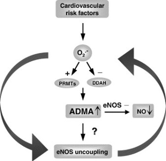
Stühlinger et al 7 advance these mechanistic insights one critical step further by defining homocysteine’s effects at an enzymatic level.

The group led by Lentz published an association between levels of the

  • endogenous inhibitor of Nirtic Oxide synthase,
  • asymmetric dimethyl arginine (ADMA), and
  • homocysteine in cultured endothelial cells and in the serum of cynomolgus monkeys.

Such an association is interesting because the L-arginine–NO synthase pathway seems to be a critical component in the full range of endothelial cell biology and vascular dysfunction.

Stühlinger et al 7  now show that increased cultured endothelial cell elaboration of ADMA by homocysteine and its precursor L-methionine is associated with a dose-dependent impairment of the activity of endothelial dimethylarginine dimethylaminohydrolase (DDAH), the enzyme that degrades ADMA. Homocysteine directly inhibited DDAH activity in a cell-free system by targeting a critical sulfhydryl group on this enzyme.

Thus, one could envision that the balance of cardiovascular health and disease could well be determined by the ability of an intact Nirtic Oxide synthase system to overcome environmental, dietary, and even genetic factors.

In patients with altered enzymatic defense systems,

  • elevated homocysteine,
  • oxidized lipoproteins,
  • inflammation, and other
  • vasotoxins

may dominate even the most potent defense mechanisms.
These studies raise a number of issues.
Do we need to add to our list of established cardiovascular risk factors to accommodate new findings and associations?
Is there a final common pathway for all risk factors or perhaps even a unified factor theory into which all potential risks can be grouped?
And, as always, should we consider Nirtic Oxide at the core of this universality?
Finally, should we change our focus altogether and speak not of risk factors but of

  • genetic predisposition,
  • extent of biochemical aberration, and
  • degree of physical damage?

Some would view these remarkable success stories and the repeated association of hyperhomocyst(e)inemia with coronary, cerebral, and peripheral vascular disease and simply advocate for increased folic acid intake for all.

Indeed, this intervention of negligible cost and

  • insignificant side effect is already partially in place;
  • many foods are fortified with folate to prevent congenital neural tube defects.

This reader considers the seminal work by Vernon Young and Yves Ingenbleek on the relationship between

  • S8 and regions distant from lava flows in Asia and Indian subcontinents,
  • where they have determined hyperhomocysteinemia and the consequence associated with:
  • veganism (not voluntary)
  • impaired methyl donor reactions and transsulfuration pathways (not corrected by B12, folate)
  • loss of lean body mass due to the constant relationship of S:N (insufficient from plant sources)

What happens, when we fail to continue to pursue causality,

  • the linkage of biological significance or scientific plausibility with
  • epidemiologically or statistically significant association?

In medicine, risk becomes the likelihood that people without a disease will acquire the disease through contact with factors thought to increase disease risk.

All of these risk factors are then, by nature, imprecise and nonspecific.
 They are stochastic measures of what will happen to normal people who fall into particular measures of these parameters.

The daring may be willing to accept these risks, citing friend and foe who live well beyond or for far lesser times than anticipated by risk alone. Such concerns may well become moot if we can simultaneously identify patients at risk

  • by linking phenotype with genotype,
  • gene expression with protein elaboration, and
  • environmental exposures with the biochemical consequences and
  • direct anatomic aberrations they induce.

This kind of characterization may well replace a family history of arterial disease as a rough estimate of

  • genotype,
  • serum cholesterol as an indirect measure of the health of lipoprotein metabolism,
  • serum glucose as a crude determinant of the ravages of diabetes mellitus,
  • blood pressure measurement as a marker of long-standing endogenous exposure to altered flow, and
  • tobacco abuse as a maker of long-standing exposure to exogenous toxins.

Rather than identifying patients on the basis of their serum cholesterol, we will have a direct measure of their

  • LDL receptor number,
  • internalization rate,
  • macrophage content in the blood vessel wall,
  • metalloproteinase activity, etc.
  • insulin receptor metabolism,
  • oxidative state, and
  • glycated burden.
  • Serum glucose will similarly give way to these tests

Evaluating a new way to open clogged arteries: Computational model offers insight into mechanisms of drug-coated balloons.

A new study from MIT analyzes the potential usefulness of a new treatment that combines the benefits of angioplasty balloons and drug-releasing stents, but may pose fewer risks. With this new approach, a balloon is inflated in the artery for only a brief period, during which it releases a drug that prevents cells from accumulating and clogging the arteries over time.
While approved for limited use in Europe, these drug-coated balloons are still in development in the United States and have not received FDA approval. The MIT study, which models the behavior of the balloons, should help scientists optimize their performance and aid regulators in evaluating their effectiveness and safety.
“Until now, people who evaluate such technology could not distinguish hype from promise,” says Elazer Edelman, the Thomas D. and Virginia W. Cabot Professor of Health Sciences and Technology and senior author of the paper describing the study, which appeared online recently in the journal Circulation.
Lead author of the paper is Vijaya Kolachalama, a former MIT postdoc who is now a principal member of the technical staff at the Charles Stark Draper Laboratory.
Edelman’s lab is investigating a possible alternative to the current treatments: drug-coated balloons. “We’re trying to understand how and when this therapy could work and identify the conditions in which it may not,” Kolachalama says. “It has its merits; it has some disadvantages.”

Modeling drug release

The drug-coated balloons are delivered by a catheter and inflated at the narrowed artery for about 30 seconds, sometimes longer. During that time, the balloon coating, containing a drug such as Zotarolimus, is released from the balloon. The properties of the coating allow the drug to be absorbed in the body’s tissues. Once the drug is released, the balloon is removed.
In their new study, Kolachalama, Edelman and colleagues set out to rigorously characterize the properties of the drug-coated balloons. After performing experiments in tissue grown in the lab and in pigs, they developed a computer model that explains the dynamics of drug release and distribution. They found that factors such as the size of the balloon, the duration of delivery time, and the composition of the drug coating all influence how long the drug stays at the injury site and how effectively it clears the arteries.
One significant finding is that when the drug is released, some of it sticks to the lining of the blood vessels. Over time, that drug is slowly released back into the tissue, which explains why the drug’s effects last much longer than the initial 30-second release period.
“This is the first time we can explain the reasons why drug-coated balloons can work,” Kolachalama says. “The study also offers areas where people can consider thinking about optimizing drug transfer and delivery.”

http://circ.ahajournals.org/content/127/20/2047.short  
http://www.mit.edu/people/vbk/Circulation_2013.pdf 
http://www.sciencedaily.com/…13/05/130521121513.ht…    
Circulation, 2013; 127 (20): 2047 – 2055
http://dx.doi.org/10.1161/CIRCULATIONAHA.113.002051;


Conclusion

MIT’s Edelman’s Lab conducted the pioneering work in Vascular biology, animal models of drug eluting stents and was at the forefront of Empirical Molecular Cardiology in its studies in vascular physiology, biology and biomaterials for medical devices.

Related articles

MUC1* Ligand, NM23-H1, Is a Novel Growth Factor That Maintains Human Stem Cells in a More Naïve State (plosone.org)

Mass. General team develops implantable, bioengineered rat kidney (eurekalert.org)

Suppression of JAK2/STAT3 Signaling Reduces End-to-End Arterial Anastomosis Induced Cell Proliferation in Common Carotid Arteries of Rats (plosone.org)

Blood Vessel Function and Breathing Control Adversely Affected by Cutting Back on Sleep (medindia.net)

miRNA Biogenesis Enzyme Drosha Is Required for Vascular Smooth Muscle Cell Survival (plosone.org)

Cell-Permeable Peptide Shows Promise For Controlling Cardiovascular Disease (medicalnewstoday.com)

The Heart Revolution By Kilmer McCully, Martha McCully

HarperCollinsPublishers, 1969

http://books.google.com/books?id=iYLbuZFxEt8C&pg=PR20&dq=New+York+Times+homocysteine+and+Cholesterol&hl=en&sa=X&ei=_0F7UfDRA8zB4APozIHQAQ&ved=0CEMQ6AEwAg


Other Related Articles that were published on this Open Access Online Scientific Journal include the following:

Modeling Targeted Therapy

Larry H Bernstein, MD, FACP 3/2/2013

Quantum Biology And Computational Medicine

Larry H Bernstein, MD, FACP 4/3/2013

Virtual Biopsy – is it possible?

Larry H Bernstein, MD, FACP 3/3/2013

Reprogramming cell fate  3/2/2013

Larry H Bernstein, MD, FACP

How Methionine Imbalance with Sulfur-Insufficiency Leads to Hyperhomocysteinemia

Larry H Bernstein, MD, FACP 4/4/2013

http://pharmaceuticalintelligence.com/2013/04/04/sulfur-deficiency-and-hyperhomocusteinemia/

Amyloidosis with Cardiomyopathy

Larry H Bernstein, MD, FACP 3/31/2013

http://pharmaceuticalintelligence.com/2013/03/31/amyloidosis-with-cardiomyopathy/

Nitric Oxide, Platelets, Endothelium and Hemostasis

Larry H Bernstein, MD, FACP 11/8/2012

http://pharmaceuticalintelligence.com/2012/11/08/nitric-oxide-platelets-endothelium-and-hemostasis/

Mitochondrial Damage and Repair under Oxidative Stress

Larry H Bernstein, MD, FACP 10/28/2012

http://pharmaceuticalintelligence.com/2012/10/28/mitochondrial-damage-and-repair-under-oxidative-stress/

Endothelial Function and Cardiovascular Disease

Larry H Bernstein, MD, FACP 10/25/2012

http://pharmaceuticalintelligence.com/2012/10/25/endothelial-function-and-cardiovascular-disease/

Endothelial Dysfunction, Diminished Availability of cEPCs, Increasing CVD Risk for Macrovascular Disease –Therapeutic Potential of cEPCs

Aviva Lev-Ari, PhD, RN 8/27/2012

Revascularization: PCI, Prior History of PCI vs CABG

Aviva Lev-Ari, PhD, RN 4/25/2013

http://pharmaceuticalintelligence.com/2013/04/25/revascularization-pci-prior-history-of-pci-vs-cabg/

Cholesteryl Ester Transfer Protein (CETP) Inhibitor: Potential of Anacetrapib to treat Atherosclerosis and CAD

Aviva Lev-Ari, PhD, RN 4/7/2013

http://pharmaceuticalintelligence.com/2013/04/07/cholesteryl-ester-transfer-protein-cetp-inhibitor-potential-of-anacetrapib-to-treat-atherosclerosis-and-cad/

Hypertriglyceridemia concurrent Hyperlipidemia: Vertical Density Gradient Ultracentrifugation a Better Test to Prevent Undertreatment of High-Risk Cardiac Patients

Aviva Lev-Ari, PhD, RN 4/4/2013

http://pharmaceuticalintelligence.com/2013/04/04/hypertriglyceridemia-concurrent-hyperlipidemia-vertical-density-gradient-ultracentrifugation-a-better-test-to-prevent-undertreatment-of-high-risk-cardiac-patients/

Fight against Atherosclerotic Cardiovascular Disease: A Biologics not a Small Molecule – Recombinant Human lecithin-cholesterol acyltransferase (rhLCAT) attracted AstraZeneca to acquire AlphaCore

Aviva Lev-Ari, PhD, RN 4/3/2013

http://pharmaceuticalintelligence.com/2013/04/03/fight-against-atherosclerotic-cardiovascular-disease-a-biologics-not-a-small-molecule-recombinant-human-lecithin-cholesterol-acyltransferase-rhlcat-attracted-astrazeneca-to-acquire-alphacore/

High-Density Lipoprotein (HDL): An Independent Predictor of Endothelial Function & Atherosclerosis, A Modulator, An Agonist, A Biomarker for Cardiovascular Risk

Aviva Lev-Ari, PhD, RN 3/31/2013

http://pharmaceuticalintelligence.com/2013/03/31/high-density-lipoprotein-hdl-an-independent-predictor-of-endothelial-function-artherosclerosis-a-modulator-an-agonist-a-biomarker-for-cardiovascular-risk/

Acute Chest Pain/ER Admission: Three Emerging Alternatives to Angiography and PCI

Aviva Lev-Ari, PhD, RN 3/10/2013

http://pharmaceuticalintelligence.com/2013/03/10/acute-chest-painer-admission-three-emerging-alternatives-to-angiography-and-pci/

Genomics & Genetics of Cardiovascular Disease Diagnoses: A Literature Survey of AHA’s Circulation Cardiovascular Genetics, 3/2010 – 3/2013

Lev-Ari, A. and L H Bernstein 3/7/2013

http://pharmaceuticalintelligence.com/2013/03/07/genomics-genetics-of-cardiovascular-disease-diagnoses-a-literature-survey-of-ahas-circulation-cardiovascular-genetics-32010-32013/

The Heart: Vasculature Protection – A Concept-based Pharmacological Therapy including THYMOSIN

Aviva Lev-Ari, PhD, RN 2/28/2013

http://pharmaceuticalintelligence.com/2013/02/28/the-heart-vasculature-protection-a-concept-based-pharmacological-therapy-including-thymosin/

Arteriogenesis and Cardiac Repair: Two Biomaterials – Injectable Thymosin beta4 and Myocardial Matrix Hydrogel

Aviva Lev-Ari, PhD, RN 2/27/2013

http://pharmaceuticalintelligence.com/2013/02/27/arteriogenesis-and-cardiac-repair-two-biomaterials-injectable-thymosin-beta4-and-myocardial-matrix-hydrogel/

Coronary artery disease in symptomatic patients referred for coronary angiography: Predicted by Serum Protein Profiles

Aviva Lev-Ari, PhD, RN 12/29/2012

http://pharmaceuticalintelligence.com/2012/12/29/coronary-artery-disease-in-symptomatic-patients-referred-for-coronary-angiography-predicted-by-serum-protein-profiles/

Special Considerations in Blood Lipoproteins, Viscosity, Assessment and Treatment

Bernstein, HL and Lev-Ari, A. 11/28/2012

http://pharmaceuticalintelligence.com/2012/11/28/special-considerations-in-blood-lipoproteins-viscosity-assessment-and-treatment/

Peroxisome proliferator-activated receptor (PPAR-gamma) Receptors Activation: PPARγ transrepression for Angiogenesis in Cardiovascular Disease and PPARγ transactivation for Treatment of Diabetes

Aviva Lev-Ari, PhD, RN 11/13/2012

http://pharmaceuticalintelligence.com/2012/11/13/peroxisome-proliferator-activated-receptor-ppar-gamma-receptors-activation-pparγ-transrepression-for-angiogenesis-in-cardiovascular-disease-and-pparγ-transactivation-for-treatment-of-dia/

Clinical Trials Results for Endothelin System: Pathophysiological role in Chronic Heart Failure, Acute Coronary Syndromes and MI – Marker of Disease Severity or Genetic Determination?

Aviva Lev-Ari, PhD, RN 10/19/2012

http://pharmaceuticalintelligence.com/2012/10/19/clinical-trials-results-for-endothelin-system-pathophysiological-role-in-chronic-heart-failure-acute-coronary-syndromes-and-mi-marker-of-disease-severity-or-genetic-determination/

Endothelin Receptors in Cardiovascular Diseases: The Role of eNOS Stimulation

Aviva Lev-Ari, PhD, RN 10/4/2012

http://pharmaceuticalintelligence.com/2012/10/04/endothelin-receptors-in-cardiovascular-diseases-the-role-of-enos-stimulation/

Inhibition of ET-1, ETA and ETA-ETB, Induction of NO production, stimulation of eNOS and Treatment Regime with PPAR-gamma agonists (TZD): cEPCs Endogenous Augmentation for Cardiovascular Risk Reduction – A Bibliography

Aviva Lev-Ari, PhD, RN 10/4/2012

http://pharmaceuticalintelligence.com/2012/10/04/inhibition-of-et-1-eta-and-eta-etb-induction-of-no-production-and-stimulation-of-enos-and-treatment-regime-with-ppar-gamma-agonists-tzd-cepcs-endogenous-augmentation-for-cardiovascular-risk-reduc/

Positioning a Therapeutic Concept for Endogenous Augmentation of cEPCs — Therapeutic Indications for Macrovascular Disease: Coronary, Cerebrovascular and Peripheral

Aviva Lev-Ari, PhD, RN 8/29/2012

http://pharmaceuticalintelligence.com/2012/08/29/positioning-a-therapeutic-concept-for-endogenous-augmentation-of-cepcs-therapeutic-indications-for-macrovascular-disease-coronary-cerebrovascular-and-peripheral/

Cardiovascular Outcomes: Function of circulating Endothelial Progenitor Cells (cEPCs): Exploring Pharmaco-therapy targeted at Endogenous Augmentation of cEPCs

Aviva Lev-Ari, PhD, RN 8/28/2012

http://pharmaceuticalintelligence.com/2012/08/28/cardiovascular-outcomes-function-of-circulating-endothelial-progenitor-cells-cepcs-exploring-pharmaco-therapy-targeted-at-endogenous-augmentation-of-cepcs/

Endothelial Dysfunction, Diminished Availability of cEPCs, Increasing CVD Risk for Macrovascular Disease – Therapeutic Potential of cEPCs

Aviva Lev-Ari, PhD, R N 8/27/2012

http://pharmaceuticalintelligence.com/2012/08/27/endothelial-dysfunction-diminished-availability-of-cepcs-increasing-cvd-risk-for-macrovascular-disease-therapeutic-potential-of-cepcs/

Vascular Medicine and Biology: CLASSIFICATION OF FAST ACTING THERAPY FOR PATIENTS AT HIGH RISK FOR MACROVASCULAR EVENTS Macrovascular Disease – Therapeutic Potential of cEPCs

Aviva Lev-Ari, PhD, RN 8/24/2012

http://pharmaceuticalintelligence.com/2012/08/24/vascular-medicine-and-biology-classification-of-fast-acting-therapy-for-patients-at-high-risk-for-macrovascular-events-macrovascular-disease-therapeutic-potential-of-cepcs/

Cardiovascular Disease (CVD) and the Role of agent alternatives in endothelial Nitric Oxide Synthase (eNOS) Activation and Nitric Oxide Production

Aviva Lev-Ari, PhD, RN 7/19/2012

http://pharmaceuticalintelligence.com/2012/07/19/cardiovascular-disease-cvd-and-the-role-of-agent-alternatives-in-endothelial-nitric-oxide-synthase-enos-activation-and-nitric-oxide-production/

Resident-cell-based Therapy in Human Ischaemic Heart Disease: Evolution in the PROMISE of Thymosin beta4 for Cardiac Repair

Aviva Lev-Ari, PhD, RN 4/30/2012

http://pharmaceuticalintelligence.com/2012/04/30/93/

Triple Antihypertensive Combination Therapy Significantly Lowers Blood Pressure in Hard-to-Treat Patients with Hypertension and Diabetes

Aviva Lev-Ari, PhD, RN 5/29/2012

http://pharmaceuticalintelligence.com/2012/05/29/445/

Macrovascular Disease – Therapeutic Potential of cEPCs: Reduction Methods for CV Risk

Aviva Lev-Ari, PhD, RN 7/2/2012

http://pharmaceuticalintelligence.com/2012/07/02/macrovascular-disease-therapeutic-potential-of-cepcs-reduction-methods-for-cv-risk/

Mitochondria Dysfunction and Cardiovascular Disease – Mitochondria: More than just the “powerhouse of the cell”

Aviva Lev-Ari, PhD, RN 7/9/2012

http://pharmaceuticalintelligence.com/2012/07/09/mitochondria-more-than-just-the-powerhouse-of-the-cell/

Bystolic’s generic Nebivolol – positive effect on circulating Endothelial Proginetor Cells endogenous augmentation

Aviva Lev-Ari, PhD, RN 7/16/2012

http://pharmaceuticalintelligence.com/2012/07/16/bystolics-generic-nebivolol-positive-effect-on-circulating-endothilial-progrnetor-cells-endogenous-augmentation/

Arteriogenesis and Cardiac Repair: Two Biomaterials – Injectable Thymosin beta4 and Myocardial Matrix Hydrogel

Aviva Lev-Ari, PhD, RN 2/27/2013

http://pharmaceuticalintelligence.com/2013/02/27/arteriogenesis-and-cardiac-repair-two-biomaterials-injectable-thymosin-beta4-and-myocardial-matrix-hydrogel/

Cardiac Surgery Theatre in China vs. in the US: Cardiac Repair Procedures, Medical Devices in Use, Technology in Hospitals, Surgeons’ Training and Cardiac Disease Severity”

Aviva Lev-Ari, PhD, RN 1/8/2013

http://pharmaceuticalintelligence.com/2013/01/08/cardiac-surgery-theatre-in-china-vs-in-the-us-cardiac-repair-procedures-medical-devices-in-use-technology-in-hospitals-surgeons-training-and-cardiac-disease-severity/

Heart Remodeling by Design – Implantable Synchronized Cardiac Assist Device: Abiomed’s Symphony

Aviva Lev-Ari, PhD, RN 7/23/2012

http://pharmaceuticalintelligence.com/2012/07/23/heart-remodeling-by-design-implantable-synchronized-cardiac-assist-device-abiomeds-symphony/

Acute Chest Pain/ER Admission: Three Emerging Alternatives to Angiography and PCI

Aviva Lev-Ari, PhD, RN 3/10/2013

http://pharmaceuticalintelligence.com/2013/03/10/acute-chest-painer-admission-three-emerging-alternatives-to-angiography-and-pci/

Dilated Cardiomyopathy: Decisions on implantable cardioverter-defibrillators (ICDs) using left ventricular ejection fraction (LVEF) and Midwall Fibrosis: Decisions on Replacement using late gadolinium enhancement cardiovascular MR (LGE-CMR)

Aviva Lev-Ari, PhD, RN 3/10/2013
http://pharmaceuticalintelligence.com/2013/03/10/dilated-cardiomyopathy-decisions-on-implantable-cardioverter-defibrillators-icds-using-left-ventricular-ejection-fraction-lvef-and-midwall-fibrosis-decisions-on-replacement-using-late-gadolinium/

The Heart: Vasculature Protection – A Concept-based Pharmacological Therapy including THYMOSIN

Aviva Lev-Ari, PhD, RN 2/28/2013
http://pharmaceuticalintelligence.com/2013/02/28/the-heart-vasculature-protection-a-concept-based-pharmacological-therapy-including-thymosin/

FDA Pending 510(k) for The Latest Cardiovascular Imaging Technology

Aviva Lev-Ari, PhD, RN 1/28/2013
http://pharmaceuticalintelligence.com/2013/01/28/fda-pending-510k-for-the-latest-cardiovascular-imaging-technology/

PCI Outcomes, Increased Ischemic Risk associated with Elevated Plasma Fibrinogen not Platelet Reactivity

Aviva Lev-Ari, PhD, RN 1/10/2013
http://pharmaceuticalintelligence.com/2013/01/10/pci-outcomes-increased-ischemic-risk-associated-with-elevated-plasma-fibrinogen-not-platelet-reactivity/

The ACUITY-PCI score: Will it Replace Four Established Risk Scores — TIMI, GRACE, SYNTAX, and Clinical SYNTAX

Aviva Lev-Ari, PhD, RN
http://pharmaceuticalintelligence.com/2013/01/03/the-acuity-pci-score-will-it-replace-four-established-risk-scores-timi-grace-syntax-and-clinical-syntax/

Coronary artery disease in symptomatic patients referred for coronary angiography: Predicted by Serum Protein Profiles

Aviva Lev-Ari, PhD, RN
http://pharmaceuticalintelligence.com/2012/12/29/coronary-artery-disease-in-symptomatic-patients-referred-for-coronary-angiography-predicted-by-serum-protein-profiles/

Heart Renewal by pre-existing Cardiomyocytes: Source of New Heart Cell Growth Discovered

Aviva Lev-Ari, PhD, RN 12/23/2012
http://pharmaceuticalintelligence.com/2012/12/23/heart-renewal-by-pre-existing-cardiomyocytes-source-of-new-heart-cell-growth-discovered/

Cardiovascular Risk Inflammatory Marker: Risk Assessment for Coronary Heart Disease and Ischemic Stroke – Atherosclerosis.

Aviva Lev-Ari, PhD, RN 10/30/2012
http://pharmaceuticalintelligence.com/2012/10/30/cardiovascular-risk-inflammatory-marker-risk-assessment-for-coronary-heart-disease-and-ischemic-stroke-atherosclerosis/

To Stent or Not? A Critical Decision

Aviva Lev-Ari, PhD, RN 10/23/2012
http://pharmaceuticalintelligence.com/2012/10/23/to-stent-or-not-a-critical-decision/

New Definition of MI Unveiled, Fractional Flow Reserve (FFR)CT for Tagging Ischemia

Aviva Lev-Ari, PhD, RN 8/27/2012
http://pharmaceuticalintelligence.com/2012/08/27/new-definition-of-mi-unveiled-fractional-flow-reserve-ffrct-for-tagging-ischemia/

Ethical Considerations in Studying Drug Safety — The Institute of Medicine Report

Aviva Lev-Ari, PhD, RN 8/23/2012
http://pharmaceuticalintelligence.com/2012/08/23/ethical-considerations-in-studying-drug-safety-the-institute-of-medicine-report/

New Drug-Eluting Stent Works Well in STEMI

Aviva Lev-Ari, PhD, RN 8/22/2012
http://pharmaceuticalintelligence.com/2012/08/22/new-drug-eluting-stent-works-well-in-stemi/

Expected New Trends in Cardiology and Cardiovascular Medical Devices

Aviva Lev-Ari, PhD, RN 8/17/2012
http://pharmaceuticalintelligence.com/2012/08/17/expected-new-trends-in-cardiology-and-cardiovascular-medical-devices/

Coronary Artery Disease – Medical Devices Solutions: From First-In-Man Stent Implantation, via Medical Ethical Dilemmas to Drug Eluting Stents

Aviva Lev-Ari, PhD, RN 8/13/2012

http://pharmaceuticalintelligence.com/2012/08/13/coronary-artery-disease-medical-devices-solutions-from-first-in-man-stent-implantation-via-medical-ethical-dilemmas-to-drug-eluting-stents/

Percutaneous Endocardial Ablation of Scar-Related Ventricular Tachycardia

Aviva Lev-Ari, PhD, RN 7/18/2012

http://pharmaceuticalintelligence.com/2012/07/18/percutaneous-endocardial-ablation-of-scar-related-ventricular-tachycardia/

Competition in the Ecosystem of Medical Devices in Cardiac and Vascular Repair: Heart Valves, Stents, Catheterization Tools and Kits for Open Heart and Minimally Invasive Surgery (MIS)

Aviva Lev-Ari, PhD, RN 6/22/2012

http://pharmaceuticalintelligence.com/2012/06/22/competition-in-the-ecosystem-of-medical-devices-in-cardiac-and-vascular-repair-heart-valves-stents-catheterization-tools-and-kits-for-open-heart-and-minimally-invasive-surgery-mis/

Global Supplier Strategy for Market Penetration & Partnership Options (Niche Suppliers vs. National Leaders) in the Massachusetts Cardiology & Vascular Surgery Tools and Devices Market for Cardiac Operating Rooms and Angioplasty Suites

Aviva Lev-Ari, PhD, RN 6/22/2012

http://pharmaceuticalintelligence.com/2012/06/22/global-supplier-strategy-for-market-penetration-partnership-options-niche-suppliers-vs-national-leaders-in-the-massachusetts-cardiology-vascular-surgery-tools-and-devices-market-for-car/

Blood_Vessels

Blood_Vessels (Photo credit: shoebappa)

Visceral Myopathy in Statins

Visceral Myopathy in Statins (Photo credit: Snipergirl)

Medical science has advanced significantly sin...

Medical science has advanced significantly since 1507, when Leonardo da Vinci drew this diagram of the internal organs and vascular systems of a woman. (Photo credit: Wikipedia)

English: Lee Hood, MD, PhD, President and Co-f...

English: Lee Hood, MD, PhD, President and Co-found of the Institute for Systems Biology (Photo credit: Wikipedia)

Read Full Post »

Identification of Biomarkers that are Related to the Actin Cytoskeleton

Curator and Writer: Larry H Bernstein, MD, FCAP

Article I Identification of Biomarkers that are Related to the Actin Cytoskeleton

This is Part I in a series of articles on Calcium and Cell motility.

The Series consists of the following articles:

Part I: Identification of Biomarkers that are Related to the Actin Cytoskeleton

Larry H Bernstein, MD, FCAP

http://pharmaceuticalintelligence.com/2012/12/10/identification-of-biomarkers-that-are-related-to-the-actin-cytoskeleton/

Part II: Role of Calcium, the Actin Skeleton, and Lipid Structures in Signaling and Cell Motility

Larry H. Bernstein, MD, FCAP, Stephen Williams, PhD and Aviva Lev-Ari, PhD, RN

http://pharmaceuticalintelligence.com/2013/08/26/role-of-calcium-the-actin-skeleton-and-lipid-structures-in-signaling-and-cell-motility/

Part III: Renal Distal Tubular Ca2+ Exchange Mechanism in Health and Disease

Larry H. Bernstein, MD, FCAP, Stephen J. Williams, PhD
 and Aviva Lev-Ari, PhD, RN

http://pharmaceuticalintelligence.com/2013/09/02/renal-distal-tubular-ca2-exchange-mechanism-in-health-and-disease/

Part IV: The Centrality of Ca(2+) Signaling and Cytoskeleton Involving Calmodulin Kinases and Ryanodine Receptors in Cardiac Failure, Arterial Smooth Muscle, Post-ischemic Arrhythmia, Similarities and Differences, and Pharmaceutical Targets

Larry H Bernstein, MD, FCAP, Justin Pearlman, MD, PhD, FACC and Aviva Lev-Ari, PhD, RN

http://pharmaceuticalintelligence.com/2013/09/08/the-centrality-of-ca2-signaling-and-cytoskeleton-involving-calmodulin-kinases-and-ryanodine-receptors-in-cardiac-failure-arterial-smooth-muscle-post-ischemic-arrhythmia-similarities-and-differen/

Part V: Ca2+-Stimulated Exocytosis:  The Role of Calmodulin and Protein Kinase C in Ca2+ Regulation of Hormone and Neurotransmitter

Larry H Bernstein, MD, FCAP
and
Aviva Lev-Ari, PhD, RN

http://pharmaceuticalintelligence.com/2013/12/23/calmodulin-and-protein-kinase-c-drive-the-ca2-regulation-of-hormone-and-neurotransmitter-release-that-triggers-ca2-stimulated-exocytosis/

Part VI: Calcium Cycling (ATPase Pump) in Cardiac Gene Therapy: Inhalable Gene Therapy for Pulmonary Arterial Hypertension and Percutaneous Intra-coronary Artery Infusion for Heart Failure: Contributions by Roger J. Hajjar, MD

Aviva Lev-Ari, PhD, RN

http://pharmaceuticalintelligence.com/2013/08/01/calcium-molecule-in-cardiac-gene-therapy-inhalable-gene-therapy-for-pulmonary-arterial-hypertension-and-percutaneous-intra-coronary-artery-infusion-for-heart-failure-contributions-by-roger-j-hajjar/

Part VII: Cardiac Contractility & Myocardium Performance: Ventricular Arrhythmias and Non-ischemic Heart Failure – Therapeutic Implications for Cardiomyocyte Ryanopathy (Calcium Release-related Contractile Dysfunction) and Catecholamine Responses

Justin Pearlman, MD, PhD, FACC, Larry H Bernstein, MD, FCAP and Aviva Lev-Ari, PhD, RN

http://pharmaceuticalintelligence.com/2013/08/28/cardiac-contractility-myocardium-performance-ventricular-arrhythmias-and-non-ischemic-heart-failure-therapeutic-implications-for-cardiomyocyte-ryanopathy-calcium-release-related-contractile/

Part VIII: Disruption of Calcium Homeostasis: Cardiomyocytes and Vascular Smooth Muscle Cells: The Cardiac and Cardiovascular Calcium Signaling Mechanism

Justin Pearlman, MD, PhD, FACC, Larry H Bernstein, MD, FCAP and Aviva Lev-Ari, PhD, RN

http://pharmaceuticalintelligence.com/2013/09/12/disruption-of-calcium-homeostasis-cardiomyocytes-and-vascular-smooth-muscle-cells-the-cardiac-and-cardiovascular-calcium-signaling-mechanism/

Part IXCalcium-Channel Blockers, Calcium Release-related Contractile Dysfunction (Ryanopathy) and Calcium as Neurotransmitter Sensor

Justin Pearlman, MD, PhD, FACC, Larry H Bernstein, MD, FCAP and Aviva Lev-Ari, PhD, RN

Part X: Synaptotagmin functions as a Calcium Sensor: How Calcium Ions Regulate the fusion of vesicles with cell membranes during Neurotransmission

Larry H Bernstein, MD, FCAP and Aviva Lev-Ari, PhD, RN

http://pharmaceuticalintelligence.com/2013/09/10/synaptotagmin-functions-as-a-calcium-sensor-how-calcium-ions-regulate-the-fusion-of-vesicles-with-cell-membranes-during-neurotransmission/

Part XI: Sensors and Signaling in Oxidative Stress

Larry H. Bernstein, MD, FCAP

http://pharmaceuticalintelligence.com/2013/11/01/sensors-and-signaling-in-oxidative-stress/

Part XII: Atherosclerosis Independence: Genetic Polymorphisms of Ion Channels Role in the Pathogenesis of Coronary Microvascular Dysfunction and Myocardial Ischemia (Coronary Artery Disease (CAD))

Larry H Bernstein, MD, FCAP and Aviva Lev-Ari, PhD, RN

http://pharmaceuticalintelligence.com/2013/12/21/genetic-polymorphisms-of-ion-channels-have-a-role-in-the-pathogenesis-of-coronary-microvascular-dysfunction-and-ischemic-heart-disease/

In this article the Author will cover two types of biomarker on the function of actin in cytoskeleton mobility in situ.

  • First, is an application in developing the actin or other component, for a biotarget and then, to be able to follow it as

(a) a biomarker either for diagnosis, or

(b) for the potential treatment prediction of disease free survival.

  • Second, is mostly in the context of MI, for which there is an abundance of work to reference, and a substantial body of knowledge about

(a) treatment and long term effects of diet, exercise, and

(b) underlying effects of therapeutic drugs.

1.  Cell Membrane (cytoskeletal) Plasticity

Refer to … Squeezing Ovarian Cancer Cells to Predict Metastatic Potential: Cell Stiffness as Possible Biomarker

Reporter/curator: Prabodh Kandala, PhD

New Georgia Tech research shows that cell stiffness could be a valuable clue for doctors as they search for and treat cancerous cells before they’re able to spread. The findings, which are published in the journal PLoS One, found that highly metastatic ovarian cancer cells are several times softer than less metastatic ovarian cancer cells. This study used atomic force microscopy (AFM) to study the mechanical properties of various ovarian cell lines. A soft mechanical probe “tapped” healthy, malignant and metastatic ovarian cells to measure their stiffness. In order to spread, metastatic cells must push themselves into the bloodstream. As a result, they must be highly deformable and softer. This study results indicate that cell stiffness may be a useful biomarker to evaluate the relative metastatic potential of ovarian and perhaps other types of cancer cells.

Comparative gene expression analyses indicate that the reduced stiffness of highly metastatic HEY A8 cells is associated with actin cytoskeleton remodeling and microscopic examination of actin fiber structure in these cell lines is consistent with this prediction.   The results suggest either of two approaches. Atomic Force Microscopy is not normally used by pathologists in diagnostics. Electron microscopy requires space for making and cutting the embedded specimen, and a separate room for the instrument. The instrument is large and the technique was not suitable for anything other than research initially until EM gained importance in Renal Pathology. It has not otherwise been used.  This new method looks like it might be more justified over a spectrum of cases.

A.  Atomic Force Microscopy

So the first point related to microscopy is whether AFM has feasibility for routine clinical use in the pathologists’ hands. This requires:

  1. suitable size of equipment
  2.  suitable manipulation of the specimen
  3. The question of whether you are using overnight fixed specimen, or whether the material is used unfixed
  4. Nothing is said about staining of cells for identification.
  5. Then there is the question about whether this will increase the number of Pathologist Assistants used across the country, which I am not against.   This would be the end of “house” trained PAs, and gives more credence to the too few PA programs across the country. The PA programs have to be reviewed and accredited by NAACLS (I served 8 years on the Board). A PA is represented on the Board, and programs are inspected by qualified peers.   There is no academic recognition given to this for tenure and promotion in Pathology Departments, and a pathologist is selected for a medical advisory role by the ASCP, and must be a Medical Advisor to a MLS accredited Program.   The fact is that PAs do gross anatomic dictation of selected specimens, and they do autopsies under the guidance of a pathologist. This is the reality of the profession today. The pathologist has to be in attendance at a variety of quality review conferences, for surgical morbidity and mortality to obstetrics review, and the Cancer Review. Cytopathology and cytogenetics are in the pathology domain.   In the case of tumors of the throat, cervix, and accessible orifices, it seems plausible to receive a swab for preparation. However, sampling error is greater than for a biopsy. A directed needle biopsy or a MIS specimen is needed for the ovary.

B.  identification of biomarkers that are related to the actin cytoskeleton

The alternative to the first approach is the identification of biomarkers that are related to the actin cytoskeleton, perhaps in the nature of the lipid or apoprotein isoform that gives the cell membrane deformability. The method measuring by Atomic Force Microscopy is shown with the current method of cytological screening, and I call attention to cells clustered together that have a scant cytoplasm surrounding nuclei occupying 1/2 to 3/4 of the cell radius.  The cells are not anaplastic, but the clumps are suggestive of glnad forming epithelium.

English: Animation showing 3-D nature of clust...

English: Animation showing 3-D nature of cluster. Image:Serous carcinoma 2a – cytology.jpg (Photo credit: Wikipedia)

The cell membrane, also called the plasma memb...

The cell membrane, also called the plasma membrane or plasmalemma, is a semipermeable lipid bilayer common to all living cells. It contains a variety of biological molecules, primarily proteins and lipids, which are involved in a vast array of cellular processes. It also serves as the attachment point for both the intracellular cytoskeleton and, if present, the cell wall. (Photo credit: Wikipedia)

English: AFM bema detection

AFM non contact mode

AFM non contact mode (Photo credit: Wikipedia)

C.  The diagnosis of ovarian cancer can be problematic because it can have a long period of growth undetected.

On the other hand, it is easily accessible once there is reason to suspect it. They are terrible to deal with because they metastasize along the abdominal peritoneum and form a solid cake. It is a problem of location and silence until it is late. Once they do announce a presence on the abdominal wall, there is probably a serous effusion. It was not possible to rely on a single marker, but when CA125 was introduced, Dr. Marguerite Pinto, Chief of Cytology at Bridgeport Hospital-Yale New Haven Health came to the immnunochemistry lab and we worked out a method for analyzing effusions, as we had already done with carcinoembryonic antigen.       The use of CEA and CA125 was published by Pinto and Bernstein as a first that had an impact.  This was followed by a study with the Chief of Oncology, Dr. Martin Rosman, that showed that the 30 month survival of patients post treatment is predicted by the half-life of disappearance of CA125 in serum.  At the time of this writing, I am not sure of the extent of its use 20 years later. History has taught us that adoption can be slow, depending very much on dissemination from major academic medical centers.  On the other hand, concepts can also be stuck at academic medical centers because of a rigid and unprepared mindset in the professional community.  The best example of this is the story of Ignaz Semmelweis, the best student of Rokitansky in Vienna for discovering the cause and prevention of childbirth fever at a time that nursemaids had far better results at obstetrical delivery than physicians.  Contrary to this, Edward Jenner, the best student of John Hunter (anatomist, surgeon, and physician to James Hume), discovered vaccination from the observation that milkmaids did not get smallpox (cowpox was a better alternative).
Only this year a Nobel Prize in Physics was awarded to an Israeli scientist who, working in the US, was unable to convince his associates of his discovery of PSEUDOCRYSTALS. – Diagnostic efficiency of carcinoembryonic antigen and CA125 in the cytological evaluation of effusions. M M Pinto, L H Bernstein, R A Rudolph, D A Brogan, M Rosman Arch Pathol Lab Med 1992; 116(6):626-631 ICID: 825503 Article type: Review article – Immunoradiometric assay of CA 125 in effusions. Comparison with carcinoembryonic antigen. M M Pinto, L H Bernstein, D A Brogan, E Criscuolo Cancer 1987; 59(2):218-222 ICID: 825555 Article type: Review article – Carcinoembryonic antigen in effusions. A diagnostic adjunct to cytology. M M Pinto, L H Bernstein, D A Brogan, E M Criscuolo Acta Cytologica 1987; 31(2):113-118 ICID: 825557

Predictive Modeling

Ovarian Cancer a plot of the CA125 elimination half-life vs the Kullback-Liebler distance

Ca125 half-life vs Kullback Entropy                                                          HL vs Survival KM plot 

Troponin(s) T, I, C  and the contractile apparatus  (contributed by Aviva Lev-Ari, PhD, RN)


For 2012 – 2013 Frontier Contribution in Cardiology on Gene Therapy Solutions for Improving Myocardial Contractility, see

Lev-Ari, A. 8/1/2013 Calcium Cycling (ATPase Pump) in Cardiac Gene Therapy: Inhalable Gene Therapy for Pulmonary Arterial Hypertension and Percutaneous Intra-coronary Artery Infusion for Heart Failure: Contributions by Roger J. Hajjar, MD

http://pharmaceuticalintelligence.com/2013/08/01/calcium-molecule-in-cardiac-gene-therapy-inhalable-gene-therapy-for-pulmonary-arterial-hypertension-and-percutaneous-intra-coronary-artery-infusion-for-heart-failure-contributions-by-roger-j-hajjar/

For explanation of Conduction prior to Myocardial Contractility, see

Lev-Ari, A. 4/28/2013 Genetics of Conduction Disease: Atrioventricular (AV) Conduction Disease (block): Gene Mutations – Transcription, Excitability, and Energy Homeostasis

http://pharmaceuticalintelligence.com/2013/04/28/genetics-of-conduction-disease-atrioventricular-av-conduction-disease-block-gene-mutations-transcription-excitability-and-energy-homeostasis/

The contraction of skeletal muscle is triggered by nerve impulses, which stimulate the release of Ca2+ from the sarcoplasmic reticulum—a specialized network of internal membranes, similar to the endoplasmic reticulum, that stores high concentrations of Ca2+ ions. The release of Ca2+ from the sarcoplasmic reticulum increases the concentration of Ca2+ in the cytosol from approximately 10-7 to 10-5 M. The increased Ca2+ concentration signals muscle contraction via the action of two accessory proteins bound to the actin filaments: tropomyosin and troponin (Figure 11.25). Tropomyosin is a fibrous protein that binds lengthwise along the groove of actin filaments. In striated muscle, each tropomyosin molecule is bound to troponin, which is a complex of three polypeptides: troponin C (Ca2+-binding), troponin I (inhibitory), and troponin T (tropomyosin-binding).

  • When the concentration of Ca2+ is low, the complex of the troponins with tropomyosin blocks the interaction of actin and myosin, so the muscle does not contract.
  • At high concentrations, Ca2+ binding to troponin C shifts the position of the complex, relieving this inhibition and allowing contraction to proceed.

Figure 11.25

Association of tropomyosin and troponins with actin filaments. (A) Tropomyosin binds lengthwise along actin filaments and, in striated muscle, is associated with a complex of three troponins: troponin I (TnI), troponin C (TnC), and troponin T (TnT). In (more…)
Contractile Assemblies of Actin and Myosin in Nonmuscle Cells

Contractile assemblies of actin and myosin, resembling small-scale versions of muscle fibers, are present also in nonmuscle cells. As in muscle, the actin filaments in these contractile assemblies are interdigitated with bipolar filaments of myosin II, consisting of 15 to 20 myosin II molecules, which produce contraction by sliding the actin filaments relative to one another (Figure 11.26). The actin filaments in contractile bundles in nonmuscle cells are also associated with tropomyosin, which facilitates their interaction with myosin II, probably by competing with filamin for binding sites on actin.

Figure 11.26

Contractile assemblies in nonmuscle cells. Bipolar filaments of myosin II produce contraction by sliding actin filaments in opposite directions.

Two examples of contractile assemblies in nonmuscle cells, stress fibers and adhesion belts, were discussed earlier with respect to attachment of the actin cytoskeleton to regions of cell-substrate and cell-cell contacts (see Figures 11.13 and 11.14). The contraction of stress fibers produces tension across the cell, allowing the cell to pull on a substrate (e.g., the extracellular matrix) to which it is anchored. The contraction of adhesion belts alters the shape of epithelial cell sheets: a process that is particularly important during embryonic development, when sheets of epithelial cells fold into structures such as tubes.

The most dramatic example of actin-myosin contraction in nonmuscle cells, however, is provided by cytokinesis—the division of a cell into two following mitosis (Figure 11.27). Toward the end of mitosis in animal cells, a contractile ring consisting of actin filaments and myosin II assembles just underneath the plasma membrane. Its contraction pulls the plasma membrane progressively inward, constricting the center of the cell and pinching it in two. Interestingly, the thickness of the contractile ring remains constant as it contracts, implying that actin filaments disassemble as contraction proceeds. The ring then disperses completely following cell division.

Figure 11.27

Cytokinesis. Following completion of mitosis (nuclear division), a contractile ring consisting of actin filaments and myosin II divides the cell in two.

http://www.ncbi.nlm.nih.gov/books/NBK9961/

2.  Use of Troponin(s) in Diagnosis

Troponins T and I are released into the circulation at the time of an acute coronary syndrome (ACS).  Troponin T was first introduced by Roche (developed in Germany) for the Roche platform as a superior biomarker for identifying acute myocardial infarction (AMI), because of a monoclonal specificity to the cardiac troponin T.  It could not be measured on any other platform (limited license patent), so the Washington University Clinical Chemistry group developed a myocardiocyte specific troponin I that quickly became widely available to Beckman, and was adapted to other instruments.  This was intended to replace the CK isoenzyme MB, that is highly elevated in rhabdomyolysis associated with sepsis or with anesthesia in special cases.

The troponins I and T had a tenfold scale difference, and the Receiver Operator Curve Generated cutoff was accurate for AMI, but had significant elevation with end-stage renal disease.  The industry worked in concert to develop a high sensitivity assay for each because there were some missed AMIs just below the ROC cutoff, which could be interpreted as Plaque Rupture.  However, the concept of plaque rupture had to be reconsidered, and we are left with type1 and type 2 AMI (disregarding the case of post PCI or CABG related).   This led to the current establishment of 3 standard deviations above the lowest measureable level at 10% coefficient of variation.  This has been discussed sufficiently elsewhere.  It did introduce a problem in the use of the test as a “silver bullet” once the finer distinctions aqnd the interest in using the test for prognosis as well as diagnosis.   This is where the use of another protein associated with heart failure came into play – either the B type natriuretic peptide, or its propeptide, N-terminal pro BNP.  The prognostic value of using these markers, secreted by the HEART and acting on the kidneys (sodium reabsorption) has proved useful.  But there has not been a multivariate refinement of the use of a two biomarker approach.  In the following part D, I illustrate what can be done with an algorithmic approach to multiple markers.

Software Agent for Diagnosis of AMI

Isaac E. Mayzlin, Ph.D., David Mayzlin, Larry H. Bernstein, M.D. The so called gold standard of proof of a method is considered the Receiver-Operating Characteristic Curve, developed for detecting “enemy planes or missiles”, and adopted first by radiologists in medicine.  This matches the correct “hits” to the actual calssification and it is generally taught as a plot of sensitivity vs (1 – specifity).  But what if you had no “training” variable?  Work inspired by Eugene Rypka’s bacterial classification led to Rosser Rudolph’s application of the Entropy of Shannon and Weaver to identify meaningful information, referring to what was Kullback-Liebler distance as “effective information”.  This allowed Rudolph and Bernstein to classify using disease biomarkers obtaing the same results as the ROC curve using an apl program.  The same data set was used by Bernstein, Adan et al. previously, and was again used by Izaak Mayzlin from University of Moscow with a new wrinkle.  Dr. Mayzlin created a neural network (Maynet), and then did a traditional NN with training on the data, and also clustered the data using geometric distance clustering and trained on the clusters.  It was interesting to see that the optimum cluster separation was closely related to the number of classes and the accuracy of classification.  An earlier simpler model using the slope of the MB isoenzyme increase and percent of total CK activity was perhaps related to Burton Sobel’s work on CK-MB disappearance rate for infarct size. The main process consists of three successive steps: (1)       clustering performed on training data set, (2)       neural network’s training on clusters from previous step, and (3)       classifier’s accuracy evaluation on testing data. The classifier in this research will be the ANN, created on step 2, with output in the range [0,1], that provides binary result (1 – AMI, 0 – not AMI), using decision point 0.5. Table  1.  Effect  of  selection  of  maximum  distance  on  the  number  of  classes  formed  and  on  the accuracy of recognition by ANN

Clustering Distance Factor F(D = F * R) Number ofClasses Number of Nodes in The Hidden Layers Number of Misrecognized Patterns inThe TestingSet of 43 Percent ofMisrecognized
10.90.80.7 2414135 1,  02,  03,  01,  02,  03,  0 3,  2 3,  2 121121 1 1 2.34.62.32.34.62.3 2.3 2.3

Creatine kinase B-subunit activity in serum in cases of suspected myocardial infarction: a prediction model based on the slope of MB increase and percentage CK-MB activity. L H Bernstein, G Reynoso Clin Chem 1983; 29(3):590-592 ICID: 825549 Diagnosis of acute myocardial infarction from two measurements of creatine kinase isoenzyme MB with use of nonparametric probability estimation. L H Bernstein, I J Good, G I Holtzman, M L Deaton, J Babb.  Clin Chem 1989; 35(3):444-447 ICID: 825570 – Information induction for predicting acute myocardial infarction. R A Rudolph, L H Bernstein, J Babb. Clin Chem 1988; 34(10):2031-2038 ICID: 825568

Related articles

Related articles published on this Open Access Online Scientific Journal, include the following:

Calcium Cycling (ATPase Pump) in Cardiac Gene Therapy: Inhalable Gene Therapy for Pulmonary Arterial Hypertension and Percutaneous Intra-coronary Artery Infusion for Heart Failure: Contributions by Roger J. Hajjar, MD

Aviva Lev-Ari, PhD, RN 8/1/2013

http://pharmaceuticalintelligence.com/2013/08/01/calcium-molecule-in-cardiac-gene-therapy-inhalable-gene-therapy-for-pulmonary-arterial-hypertension-and-percutaneous-intra-coronary-artery-infusion-for-heart-failure-contributions-by-roger-j-hajjar/

High-Sensitivity Cardiac Troponin Assays- Preparing the United States for High-Sensitivity Cardiac Troponin Assays

Larry Bernstein, MD, FCAP 6/13/2013

http://pharmaceuticalintelligence.com/2013/06/13/high-sensitivity-cardiac-troponin-assays/

Dealing with the Use of the High Sensitivity Troponin (hs cTn) Assays

Larry Bernstein and Aviva Lev-Ari  5/18/2013

http://pharmaceuticalintelligence.com/2013/05/18/dealing-with-the-use-of-the-hs-ctn-assays/

Acute Chest Pain/ER Admission: Three Emerging Alternatives to Angiography and PCI – Corus CAD, hs cTn, CCTA

Aviva Lev-Ari  3/10/2013

http://pharmaceuticalintelligence.com/2013/03/10/acute-chest-painer-admission-three-emerging-alternatives-to-angiography-and-pci/

  • Redberg’s conclusions are correct for the initial screening. The issue has been whether to do further testing for low or intermediate risk patients.
  • The most intriguing finding that is not at all surprising is that the CCTA added very little in the suspect group with small or moderate risk.
  • The ultra sensitive troponin threw the ROC out the window
  • The improved assay does pick up minor elevations of troponin in the absence of MI.

Critical Care | Abstract | Cardiac ischemia in patients with septic …
Aviva Lev-Ari  6/26/2013
http://pharmaceuticalintelligence.com/2013/06/26/critical-care-abstract-cardiac-ischemia-in-patients-with-septic/

  • refer to:  Cardiac ischemia in patients with septic shock randomized to vasopressin or norepinephrine

Mehta S, Granton J,  Gordon AC, Cook DJ, et al.
Critical Care 2013, 17:R117   http://dx.doi.org/10.1186/cc12789
Troponin and CK levels, and rates of ischemic ECG changes were similar in the VP and NE groups. In multivariable analysis

  • only APACHE II was associated with 28-day mortality (OR 1.07, 95% CI 1.01-1.14, p=0.033).

Assessing Cardiovascular Disease with Biomarkers

Larry H Bernstein, MD, FCAP 12/25/2012

http://pharmaceuticalintelligence.com/2012/12/25/assessing-cardiovascular-disease-with-biomarkers/

Vascular Medicine and Biology: CLASSIFICATION OF FAST ACTING THERAPY FOR PATIENTS AT HIGH RISK FOR MACROVASCULAR EVENTS Macrovascular Disease – Therapeutic Potential of cEPCs

Aviva Lev-Ari, PhD, RN 8/24/2012

http://pharmaceuticalintelligence.com/2012/08/24/vascular-medicine-and-biology-classification-of-fast-acting-therapy-for-patients-at-high-risk-for-macrovascular-events-macrovascular-disease-therapeutic-potential-of-cepcs/

 PENDING Integration

  • ‘Ryanopathy’: causes and manifestations of RyR2 dysfunction in heart failureCardiovasc Res. 2013;98:240-247,
  • Up-regulation of sarcoplasmic reticulum Ca2+ uptake leads to cardiac hypertrophy, contractile dysfunction and early mortality in mice deficient in CASQ2Cardiovasc Res. 2013;98:297-306,
  • Myocardial Delivery of Stromal Cell-Derived Factor 1 in Patients With Ischemic Heart Disease: Safe and PromisingCirc. Res.. 2013;112:746-747,
  • Circulation Research Thematic Synopsis: Cardiovascular GeneticsCirc. Res.. 2013;112:e34-e50,
  • Gene and cytokine therapy for heart failure: molecular mechanisms in the improvement of cardiac functionAm. J. Physiol. Heart Circ. Physiol.. 2012;303:H501-H512,
  • Ryanodine Receptor Phosphorylation and Heart Failure: Phasing Out S2808 and “Criminalizing” S2814Circ. Res.. 2012;110:1398-1402,

http://circres.ahajournals.org/content/110/5/777.figures-only

Read Full Post »

Telling NO to Cardiac Risk

DDAH Says NO to ADMA(1); The DDAH/ADMA/NOS Pathway(2)

Author-Writer-Reporter:  Stephen J. Williams, PhD

Endothelium-derived nitric oxide (NO) has been shown to be vasoprotective.  Nitric oxide enhances endothelial cell survival, inhibits excessive proliferation of vascular smooth muscle cells, regulates vascular smooth muscle tone, and prevents platelets from sticking to the endothelial wall.  Together with evidence from preclinical and human studies, it is clear that impairment of the NOS pathway increases risk of cardiovascular disease (3-5).

This post contains two articles on the physiological regulation of nitric oxide (NO) by an endogenous NO synthase inhibitor asymmetrical dimethylarginine (ADMA) and ADMA metabolism by the enzyme DDAH(1,2).  Previous posts on nitric oxide, referenced at the bottom of the page, provides excellent background and further insight for this posting. In summary plasma ADMA levels are elevated in patients with cardiovascular disease and several large studies have shown that plasma ADMA is an independent biomarker for cardiovascular-related morbidity and mortality(6-8).

admacardiacrisk

admaeffects

Figure 1 A. Cardiac risks of ADMA B. Effects of ADMA (Photo credit: Wikipedia)

ADMA Production and Metabolism

Nuclear proteins such as histones can be methylated on arginine residues by protein-arginine methyltransferases, enzymes which use S-adenosylmethionine as methyl groups.  This methylation event is thought to regulate protein function, much in the way of protein acetylation and phosphorylation (9).  And much like phosphorylation, these modifications are reversible through methylesterases.   The proteolysis of these arginine-methyl modifications lead to the liberation of free guanidine-methylated arginine residues such as L-NMMA, asymmetric dimethylarginine (ADMA) and symmetrical methylarginine (SDMA).

The first two, L-NMMA and ADMA, have been shown to inhibit the activity of the endothelial NOS.  This protein turnover is substantial: for instance the authors note that each day 40% of constitutive protein in adult liver is newly synthesized protein. And in several diseases, such as muscular dystrophy, ischemic heart disease, and diabetes, it has been known since the 1970’s that protein catabolism rates are very high, with corresponding increased urinary excretion of ADMA(10-13).  Methylarginines are excreted in the urine by cationic transport.  However, the majority of ADMA and L-NMMA are degraded within the cell by dimethylaminohydrolase (DDAH), first cloned and purified in rat(14).

endogenous NO inhibitors from pubchem

Figure 2.  Endogenous inhibitors of NO synthase.  Chemical structures generated from PubChem.

DDAH

DDAH specifically hydrolyzes ADMA and L-NMMA to yield citruline and demethylamine and usually shows co-localization with NOS. Pharmacologic inhibition of DDAH activity causes accumulation of ADMA and can reverse the NO-mediated bradykinin-induced relaxation of human saphenous vein.

Two isoforms have been found in human:

  • DDAH1 (found in brain and kidney and associated with nNOS) and
  • DDAH2 (highly expressed in heart, placenta, and kidney and associated with eNOS).

DDAH2 can be upregulated by all-trans retinoic acid (atRA can increase NO production).  Increased reactive oxygen species and possibly homocysteine, a risk factor for cardiovascular disease, can decrease DDAH activity(15,16).

  • The importance of DDAH activity can also be seen in transgenic mice which overexpress DDAH, exhibiting increased NO production, increased insulin sensitivity, and reduced vascular resistance  (17).  Likewise,
  • Transgenic mice, null for the DDAH1, showed increase in blood pressure, decreased NO production, and significant increase in tissue and plasma ADMA and L-NMMA.

amdanosfigure

Figure 3.  The DDAH/ADMA/NOS cycle. Figure adapted from Cooke and Ghebremarian (1).

As mentioned in the article by Cooke and Ghebremariam, the authors state: the weight of the evidence indicates that DDAH is a worthy therapeutic target. Agents that increase DDAH expression are known, and 1 of these, a farnesoid X receptor agonist, is in clinical trials

http://www.interceptpharma.com

An alternate approach is to

  • develop an allosteric activator of the enzyme.  Although
  • development of an allosteric activator is not a typical pharmaceutical approach, recent studies indicate that this may be achievable aim(18).

References:

1.            Cooke, J. P., and Ghebremariam, Y. T. : DDAH says NO to ADMA.(2011) Arteriosclerosis, thrombosis, and vascular biology 31, 1462-1464

2.            Tran, C. T., Leiper, J. M., and Vallance, P. : The DDAH/ADMA/NOS pathway.(2003) Atherosclerosis. Supplements 4, 33-40

3.            Niebauer, J., Maxwell, A. J., Lin, P. S., Wang, D., Tsao, P. S., and Cooke, J. P.: NOS inhibition accelerates atherogenesis: reversal by exercise. (2003) American journal of physiology. Heart and circulatory physiology 285, H535-540

4.            Miyazaki, H., Matsuoka, H., Cooke, J. P., Usui, M., Ueda, S., Okuda, S., and Imaizumi, T. : Endogenous nitric oxide synthase inhibitor: a novel marker of atherosclerosis.(1999) Circulation 99, 1141-1146

5.            Wilson, A. M., Shin, D. S., Weatherby, C., Harada, R. K., Ng, M. K., Nair, N., Kielstein, J., and Cooke, J. P. (2010): Asymmetric dimethylarginine correlates with measures of disease severity, major adverse cardiovascular events and all-cause mortality in patients with peripheral arterial disease. Vasc Med 15, 267-274

6.            Kielstein, J. T., Impraim, B., Simmel, S., Bode-Boger, S. M., Tsikas, D., Frolich, J. C., Hoeper, M. M., Haller, H., and Fliser, D. : Cardiovascular effects of systemic nitric oxide synthase inhibition with asymmetrical dimethylarginine in humans.(2004) Circulation 109, 172-177

7.            Kielstein, J. T., Donnerstag, F., Gasper, S., Menne, J., Kielstein, A., Martens-Lobenhoffer, J., Scalera, F., Cooke, J. P., Fliser, D., and Bode-Boger, S. M. : ADMA increases arterial stiffness and decreases cerebral blood flow in humans.(2006) Stroke; a journal of cerebral circulation 37, 2024-2029

8.            Mittermayer, F., Krzyzanowska, K., Exner, M., Mlekusch, W., Amighi, J., Sabeti, S., Minar, E., Muller, M., Wolzt, M., and Schillinger, M. : Asymmetric dimethylarginine predicts major adverse cardiovascular events in patients with advanced peripheral artery disease.(2006) Arteriosclerosis, thrombosis, and vascular biology 26, 2536-2540

9.            Kakimoto, Y., and Akazawa, S.: Isolation and identification of N-G,N-G- and N-G,N’-G-dimethyl-arginine, N-epsilon-mono-, di-, and trimethyllysine, and glucosylgalactosyl- and galactosyl-delta-hydroxylysine from human urine. (1970) The Journal of biological chemistry 245, 5751-5758

10.          Inoue, R., Miyake, M., Kanazawa, A., Sato, M., and Kakimoto, Y.: Decrease of 3-methylhistidine and increase of NG,NG-dimethylarginine in the urine of patients with muscular dystrophy. (1979) Metabolism: clinical and experimental 28, 801-804

11.          Millward, D. J.: Protein turnover in skeletal muscle. II. The effect of starvation and a protein-free diet on the synthesis and catabolism of skeletal muscle proteins in comparison to liver. (1970) Clinical science 39, 591-603

12.          Goldberg, A. L., and St John, A. C.: Intracellular protein degradation in mammalian and bacterial cells: Part 2. (1976) Annual review of biochemistry 45, 747-803

13.          Dice, J. F., and Walker, C. D.: Protein degradation in metabolic and nutritional disorders. (1979) Ciba Foundation symposium, 331-350

14.          Ogawa, T., Kimoto, M., and Sasaoka, K.: Purification and properties of a new enzyme, NG,NG-dimethylarginine dimethylaminohydrolase, from rat kidney. (1989) The Journal of biological chemistry 264, 10205-10209

15.          Ito, A., Tsao, P. S., Adimoolam, S., Kimoto, M., Ogawa, T., and Cooke, J. P.: Novel mechanism for endothelial dysfunction: dysregulation of dimethylarginine dimethylaminohydrolase. (1999) Circulation 99, 3092-3095

16.          Stuhlinger, M. C., Tsao, P. S., Her, J. H., Kimoto, M., Balint, R. F., and Cooke, J. P. : Homocysteine impairs the nitric oxide synthase pathway: role of asymmetric dimethylarginine.(2001) Circulation 104, 2569-2575

17.          Sydow, K., Mondon, C. E., Schrader, J., Konishi, H., and Cooke, J. P.: Dimethylarginine dimethylaminohydrolase overexpression enhances insulin sensitivity. (2008) Arteriosclerosis, thrombosis, and vascular biology 28, 692-697

18.          Zorn, J. A., and Wells, J. A.: Turning enzymes ON with small molecules. (2010) Nature chemical biology 6, 179-188

Other research papers on Nitric Oxide and Cardiac Risk  were published on this Scientific Web site as follows:

The Nitric Oxide and Renal is presented in FOUR parts:

Part I: The Amazing Structure and Adaptive Functioning of the Kidneys: Nitric Oxide

Part II: Nitric Oxide and iNOS have Key Roles in Kidney Diseases

Part III: The Molecular Biology of Renal Disorders: Nitric Oxide

Part IV: New Insights on Nitric Oxide donors

Cardiac Arrhythmias: A Risk for Extreme Performance Athletes

What is the role of plasma viscosity in hemostasis and vascular disease risk?

Cardiovascular Risk Inflammatory Marker: Risk Assessment for Coronary Heart Disease and Ischemic Stroke – Atherosclerosis.

Endothelial Dysfunction, Diminished Availability of cEPCs, Increasing CVD Risk for Macrovascular Disease – Therapeutic Potential of cEPCs

Biochemistry of the Coagulation Cascade and Platelet Aggregation – Part I

Nitric Oxide Function in Coagulation

Read Full Post »

Nitric Oxide has a Ubiquitous Role in the Regulation of Glycolysis – with a Concomitant Influence on Mitochondrial Function


Reporter, Editor, and Topic Co-Leader: Larry H. Bernstein, MD, FACP, Clinical Pathologist and Biochemist


Apoptosis signaling pathways

Apoptosis signaling pathways (Photo credit: AJC1)

This discussion is a followup on a series of articles elucidating the importance of NO, eNOS, iNOS, cardiovascular and vascular endothelium effects, and therapeutic targets.

This mechanism of action and signaling actions have been introduced so that we identify endocrine, paracrine, and such effects in the normal, stressed, and dysfunctional state. The size and breadth of this vital adaptive process is now further explored.

The title is short, befitting a subtitle.  The full topic may be considered “Nitric Oxide has a ubiquitous role in the regulation of glycolysis -with a concomitant influence on mitochondrial function that is active in endothelium, platelets, vascular smooth muscle and neural cells and the balance has a role in chronic inflammation, asthma, hypertension, sepsis and cancer”.

Vascular endothelium

Vascular endothelium (Photo credit: Wikipedia)

Related articles


Nitric Oxide Synthase

Nitric Oxide Synthase (Photo credit: Wikipedia)


Nitric Oxide has a ubiquitous role in the regulation of glycolysis with a concomitant influence on mitochondrial function that is active in endothelium, platelets, vascular smooth muscle and neural cells and the balance has a role in chronic inflammation, asthma, hypertension, sepsis and cancer.

Uncoupling of aerobic glycolysis
Potential cytotoxic mediators of endothelial cell (EC) apoptosis include increased formation of reactive oxygen and nitrogen species (ROSRNS) during the atherosclerotic process. Nitric oxide (NO) has a biphasic action on oxidative cell killing with low concentrations protecting against cell death, whereas higher concentrations are cytotoxic. High levels of NO can be produced by inducible nitric-oxide synthase in response to cytokine stimulation, primarily from macrophages, and elevated levels of NO is injurious to endothelium.Ccytochrome c release and caspase activation are involved in NO induced apoptosis. ROS also induces mitochondrial DNA damage in ECs, and this damage is accompanied by a decrease in mitochondrial RNA (mtRNA) transcripts, mitochondrial protein synthesis, and cellular ATP levels. Mitochondria have been recognized to play a pivotal role in the signaling cascade of apoptosis leading to atherosclerosis-induced damage in endothelial cells.
The processes involved in the signaling pathways leading to apoptosis are complex but have some degree of convergence between cell types including those in the vasculature. Release of cytochrome c from mitochondria is a proapoptotic signal, which activates several downstream signaling events including formation of the apoptosome and activation of caspases. Ubiquinol cytochrome c reductase (complex III) is a site for ROS formation, and cytochrome c oxidase (complex IV) is a target for the interaction of NO in mitochondria.
The impact of the inhibition of mitochondrial protein synthesis is particularly important in NO-dependent cytotoxicity, and depends also on other factors such as glycolysis. These authors examined whether the inhibition of mitochondrial protein synthesis by chloramphenicol increases the susceptibility of endothelial cells to undergo NO-dependent apoptosis in glucose-free media. Bovine aortic endothelial cells were treated with chloramphenicol, which resulted in a decreased ratio of mitochondrial complex IV to cytochrome c and increased oxidant production in the cell. Inhibition of mitochondrial protein synthesis was associated with a greater susceptibility of the cells to apoptosis induced by NO in glucose-free medium.
Inhibition of mitochondrial protein synthesis results in increased endothelial cell susceptibility to nitric oxide-induced apoptosis. A Ramachandran, DR Moellering, E Ceaser, S Shiva, J Xu, and V Darley-Usmar. PNAS May 14, 2002: 99(10): 6643–6648 http://www.pnas.orgcgidoi10.1073pnas.102019899

Nitric oxide (NO) is a ubiquitous signaling molecule whose physiological roles mediated through the activation of the soluble guanylate cyclase are now clearly recognized. At physiological concentrations, NO also inhibits the mitochondrial enzyme cytochrome c oxidase (complex IV) in competition with oxygen, and recently we have suggested that the interplay between the two gases allows this enzyme to act as an oxygen sensor in cells. In addition, NO plays a variety of patho-physiological roles, some of which also may be the consequence of its action at a mitochondrial level. We have characterized the sequence of events that follow inhibition of complex IV by continuous exposure to NO.
The mitochondrion is a key organelle in the control of cell death. Nitric oxide (NO) inhibits complex IV in the respiratory chain and is reported to possess both proapoptotic and antiapoptotic actions. We investigated the effects of continuous inhibition of respiration by NO on mitochondrial energy status and cell viability. Serum-deprived human T cell leukemia (Jurkat) cells were exposed to NO at a concentration that caused continuous and complete (;85%) inhibition of respiration. Serum deprivation caused progressive loss of mitochondrial membrane potential (Dcm) and apoptotic cell death. In the presence of NO, Dcm was maintained compared to controls, and cells were protected from apoptosis. Similar results were obtained by using staurosporin as the apoptotic stimulus. As exposure of serum-deprived cells to NO progressed (>5 h), however, Dcm fell, correlating with the appearance of early apoptotic features and a decrease in cell viability. Glucose deprivation or iodoacetate treatment of cells in the presence of NO resulted in a collapse of Dcm, demonstrating involvement of glycolytic ATP in its maintenance. Under these conditions cell viability also was decreased. Treatment with oligomycin and or bongkrekic acid indicated that the maintenance of Dcm during exposure to NO is caused by reversal of the ATP synthase and other electrogenic pumps. Thus, blockade of complex IV by NO initiates a protective action in the mitochondrion to maintain Dcm; this results in prevention of apoptosis. It is likely that during cellular stress involving increased generation of NO this compound will trigger a similar sequence of events, depending on its concentration and duration of release. (mitochondrial membrane potential ; apoptosis ; necrosis)

The effect of nitric oxide on cell respiration: A key to understanding its role in cell survival or death. B Beltra, A Mathur, MR Duchen, JD. Erusalimsky, and S Moncada. PNAS Dec 19, 2000; 97(26):4602–14607.

Another study by this group shows that inhibition of respiration by exogenous nitric oxide (NO) in Jurkat cells leads to mitochondrial membrane hyperpolarization dependent on the utilization of glycolytic ATP by the F1Fo-ATPase and other transporters acting in reverse mode. This process also occurs in astrocytes, which are highly glycolytic cells, but not in neurons , which do not invoke glycolysis to maintain ATP concentrations. In addition, this hyperpolarization correlates with protection against apoptotic cell death. Others found an early phase of mitochondrial hyperpolarization after treatment of a variety of cells with different pro-apoptotic stimuli, which precedes the generation of free. At present, no satisfactory explanation has been proposed to explain the mechanism of hyperpolarization, the reasons why free radicals are released from the mitochondrion, or the connection of these phenomena with apoptosis.
The authors surmise that a pro-apoptotic stimulus, anti-Fas Ab, leads to release of endogenous NO from Jurkat cells in sufficient amounts to inhibit cell respiration and cause a hyperpolarization dependent on the reversal of the F1Fo-ATPase. Moreover, the reduction of the mitochondrial electron transport chain, after inhibition of cytochrome oxidase by NO, leads to generation of superoxide anion (O2). They suggest the process is a cellular defense response that may be overcome by pro-apoptotic mechanisms that occur in parallel.

Inhibition of mitochondrial respiration by endogenous nitric oxide: A critical step in Fas signaling. B Beltran, M Quintero, E Garcıa-Zaragoza, E O’Connor, JV. Esplugues, and Salvador Moncada. PNAS June 25, 2002 99(13): 8892–8897. http://www.pnas.orgcgidoi10.1073pnas.092259799

Nitric oxide has been shown to render cells resistant to oxidative stress. Mechanisms proposed for the ability of nitric oxide to protect cells against oxidative stress include reactions of nitric oxide and the induction of adaptive responses that require protein synthesis. Nitric oxide forms iron complexes preventing the formation of strong oxidants. In addition, reactions of nitric oxide with lipid and or organic radicals protect against membrane peroxidation and peroxidative chemistry-induced cell injury. Exposure to low, nonlethal doses of nitric oxide induces adaptive responses that render cells resistant to lethal concentrations of nitric oxide and or peroxides, such as, the induction of hemoxygenase-1 (HO-1) and Mn superoxide dismutase. The up-regulation of HO-1 was accompanied by an increase in ferritin to account for the release of iron from HO-1, indicating a role of both iron heme and nonheme iron for peroxide-mediated cellular injury. Further, nitric oxide, by regulating critical mitochondrial functions such as respiration, membrane potential, and release of cytochrome c, is able to trigger defense mechanisms against cell death induced by pro-apoptotic stimuli.
This study investigates the potential contribution of nitric oxide’s ability to protect cells from oxidative stress, low steady state levels of nitric oxide generated by endothelial nitric oxide synthase (eNOS) and the mechanisms of protection against H2O2. Spontaneously transformed human ECV304 cells, which normally do not express eNOS, were stably transfected with a green fluorescent-tagged eNOS cDNA. The eNOS-transfected cells were found to be resistant to injury and delayed death following a 2-h exposure to H2O2 (50–150 mM). Inhibition of nitric oxide synthesis abolished the protective effect against H2O2 exposure. The ability of nitric oxide to protect cells depended on the presence of respiring mitochondria. ECV3041 eNOS cells with diminished mitochondria respiration are injured to the same extent as non-transfected ECV304 cells, and recovery of mitochondrial respiration restores the ability of nitric oxide to protect against H2O2-induced death. Nitric oxide had a profound effect in cell metabolism, because ECV3041eNOS cells had lower steady state levels of ATP and higher utilization of glucose via the glycolytic pathway than ECV304 cells. However, the protective effect of nitric oxide against H2O2 exposure is not reproduced in ECV304 cells after treatment with azide and oligomycin suggesting that the dynamic regulation of respiration by nitric oxide represent a critical and unrecognized primary line of defense against oxidative stress.