Feeds:
Posts
Comments

Posts Tagged ‘Thomas C. Südhof’

Atherosclerosis Independence: Genetic Polymorphisms of Ion Channels Role in the Pathogenesis of Coronary Microvascular Dysfunction and Myocardial Ischemia (Coronary Artery Disease (CAD))

Reviewer and Co-Curator: Larry H Bernstein, MD, FCAP

and

Curator: Aviva Lev-Ari, PhD, RN

Article XII Atherosclerosis Independence Genetic Polymorphisms of Ion Channels Role in the Pathogenesis of Coronary Microvascular Dysfunction and Myocardial Ischemia (Coronary Artery

Image created by Adina Hazan 06/30/2021

The role of ion channels in Na(+)-K(+)-ATPase: regulation of ion
transport across the plasma membrane has been studied by our Team in 2012 and 2013. This is article TWELVE in a 13 article series listed at the end of this article.

Chiefly, our sources of inspiration were the following:

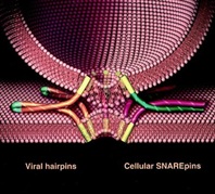
1.            2013 Nobel work on vesicles and calcium flux at the neuromuscular junction

Machinery Regulating Vesicle Traffic, A Major Transport System in our Cells 

The 2013 Nobel Prize in Physiology or Medicine is awarded to Dr. James E. Rothman, Dr. Randy W. Schekman and Dr. Thomas C. Südhof for their discoveries of machinery regulating vesicle traffic, a major transport system in our cells. This represents a paradigm shift in our understanding of how the eukaryotic cell, with its complex internal compartmentalization, organizes the routing of molecules packaged in vesicles to various intracellular destinations, as well as to the outside of the cell. Specificity in the delivery of molecular cargo is essential for cell function and survival. 

http://www.nobelprize.org/nobel_prizes/medicine/laureates/2013/advanced-medicineprize2013.pdf

Synaptotagmin functions as a Calcium Sensor: How Calcium Ions Regulate the fusion of vesicles with cell membranes during Neurotransmission

Larry H Bernstein, MD, FCAP and Aviva Lev-Ari, PhD, RN

http://pharmaceuticalintelligence.com/2013/09/10/synaptotagmin-functions-as-a-calcium-sensor-how-calcium-ions-regulate-the-fusion-of-vesicles-with-cell-membranes-during-neurotransmission/

2. Perspectives on Nitric Oxide in Disease Mechanisms

available on Kindle Store @ Amazon.com

http://www.amazon.com/dp/B00DINFFYC

http://pharmaceuticalintelligence.com/biomed-e-books/series-a-e-books-on-cardiovascular-diseases/perspectives-on-nitric-oxide-in-disease-mechanisms-v2/

3.            Professor David Lichtstein, Hebrew University of Jerusalem, Dean, School of Medicine

Lichtstein’s main research focus is the regulation of ion transport across the plasma membrane of eukaryotic cells. His work led to the discovery that specific steroids that have crucial roles, as the regulation of cell viability, heart contractility, blood pressure and brain function. His research has implications for the fundamental understanding of body functions, as well as for several pathological states such as heart failure, hypertension and neurological and psychiatric diseases.

Physiologist, Professor Lichtstein, Chair in Heart Studies at The Hebrew University elected Dean of the Faculty of Medicine at The Hebrew University of Jerusalem

Reporter: Aviva Lev-Ari, PhD, RN

http://pharmaceuticalintelligence.com/2013/12/18/physiologist-professor-lichtstein-chair-in-heart-studies-at-the-hebrew-university-elected-dean-of-the-faculty-of-medicine-at-the-hebrew-university-of-jerusalem/

4.            Professor Roger J. Hajjar, MD at Mount Sinai School of Medicine

Calcium Cycling (ATPase Pump) in Cardiac Gene Therapy: Inhalable Gene Therapy for Pulmonary Arterial Hypertension and Percutaneous Intra-coronary Artery Infusion for Heart Failure: Contributions by Roger J. Hajjar, MD

Aviva Lev-Ari, PhD, RN

http://pharmaceuticalintelligence.com/2013/08/01/calcium-molecule-in-cardiac-gene-therapy-inhalable-gene-therapy-for-pulmonary-arterial-hypertension-and-percutaneous-intra-coronary-artery-infusion-for-heart-failure-contributions-by-roger-j-hajjar/

5.            Seminal Curations by Dr. Aviva Lev-Ari on Genetics and Genomics of Cardiovascular Diseases with a focus on Conduction and Cardiac Contractility

Aviva Lev-Ari, PhD, RN

Aviva Lev-Ari, PhD, RN

Aviva Lev-Ari, PhD, RN and Larry H. Bernstein, MD, FCAP

Justin Pearlman, MD, PhD, FACC, Larry H Bernstein, MD, FCAP and Aviva Lev-Ari, PhD, RN

Other related research by the Team of Leaders in Pharmaceutical Business Intelligence published on the Open Access Online Scientific Journal

http://pharmaceuticalintelligence.com

See References to articles at the end of this article on

  • ION CHANNEL and Cardiovascular Diseases

http://pharmaceuticalintelligence.com/?s=Ion+Channel

  • Calcium Role in Cardiovascular Diseases – The Role of Calcium Calmodulin Kinase  (CKCaII) and Ca(2) flux
  • Mitochondria and Oxidative Stress Role in Cardiovascular Diseases

Thus, the following article follows a series of articles on ion-channels and cardiac contractility mentioned, above. The following article is closely related to the work of Prof. Lichtstein.

These investigators studied the possible correlation between

  • Myocardial Ischemia (Coronary Artery Disease (CAD)) aka Ischemic Heart Disease (IHD) and
  • single-nucleotide polymorphisms  (SNPs) genes encoding several regulators involved in Coronary Blood Flow Regulation (CBFR), including
  • ion channels acting in vascular smooth muscle and/or
  • endothelial cells of coronary arteries.

They completely analyzed exon 3 of both KCNJ8 and KCNJ11 genes (Kir6.1 and Kir6.2 subunit, respectively) as well as

  • the whole coding region of KCN5A gene (Kv1.5 channel).
The work suggests certain genetic polymorphisms may represent a non-modifiable protective factor that could be used
  • to identify individuals at relatively low-risk for cardiovascular disease
  • an independent protective role of the
    • rs5215_GG against developing CAD and
    • a trend for rs5219_AA to be associated with protection against coronary microvascular dysfunction

Their findings are a lead into further investigations on ion channels and IHD affecting the microvasculature.

Role of genetic polymorphisms of ion channels in the pathophysiology of coronary microvascular dysfunction and ischemic heart disease

BasicResCardiol(2013)108:387   http//dx.dio.org/10.1007/s00395-013-0387-4

F Fedele1•M Mancone1•WM Chilian2•P Severino2•E Canali•S Logan•ML DeMarchis3•M Volterrani4•R Palmirotta3•F Guadagni3

1Department of Cardiovascular, Respiratory, Nephrology, Anesthesiology and Geriatric Sciences,Sapienza University of Rome, UmbertoI Policlinic, Rome, Italy  e-mail:francesco.fedele@uniroma1.it
2Department of Integrative Medical Sciences, Northeastern Ohio Universities College of Medicine, Rootstown,OH
3Department of Advanced Biotechnologies and Bioimaging, IRCCS San Raffaele Pisana,Rome,It
4Cardiovascular Research Unit, Department of Medical Sciences, Centre for Clinical and Basic Research, Raffaele Pisana, Rome, Italy (CBFR)

BasicResCardiol(2013)108:387   http//dx.dio.org/10.1007/s00395-013-0387-4
This article is published with open access at Springerlink.com

Abstract

Conventionally,ischemic heart disease (IHD) study is equated with large vessel coronary disease (CAD). However, recent evidence has suggested

  • a role of compromised microvascular regulation in the etiology of IHD.

Because regulation of coronary blood flow likely involves

  • activity of specific ion-channels, and
  • key factors involved in endothelium-dependent dilation,

genetic anomalies of ion-channels or specific endothelial-regulators may underlie coronary microvascular disease.

We aimed to evaluate the clinical impact of single-nucleotide polymorphisms in genes encoding for

  • ion-channels expressed in the coronary vasculature and the possible
  • correlation with IHD resulting from microvascular dysfunction.

242 consecutive patients who were candidates for coronary angiography were enrolled. A prospective, observational, single-center study was conducted, 

  • analyzing genetic polymorphisms relative to

(1) NOS3 encoding for endothelial nitric oxide synthase (eNOS);
(2) ATP2A2 encoding for the Ca/H-ATP-ase pump (SERCA);
(3) SCN5A encoding for the voltage-dependent Na channel (Nav1.5);
(4) KCNJ8 and in KCNJ11 encoding for the Kir6.1and Kir6.2 subunits
of genetic K-ATP channels, respectively;and
(5) KCN5A encoding for the voltage-gated K channel (Kv1.5).

No significant associations between clinical IHD manifestations and

  • polymorphisms for SERCA, Kir6.1, and Kv1.5. were observed (p[0.05),

whereas specific polymorphisms detected in eNOS, as well as in Kir6.2 and Nav1.5 were found to be correlated with

  • IHD and microvascular dysfunction.

 Interestingly, genetic polymorphisms of ion-channels  seem to have an important clinical impact

  • influencing the susceptibility for microvascular dysfunction and (IHD,
  • independent of the presence of classic cardiovascular risk factors: atherosclerosis   

http//dx.dio.org/10.1007/s00395-013-0387-4

Keywords: Ion-channels, Genetic polymorphisms, Coronary microcirculation, Endothelium, Atherosclerosis Ischemic heart disease

 Introduction

Historically, in the interrogation of altered vascular function in patientswith ischemic heart disease (IHD), scientists have focused their attention on the correlation between

  • endothelial dysfunction and
  • atherosclerosis [11, 53, 6567].

The endothelium-independent dysfunction in coronary microcirculation and its possible correlations with  

  • atherosclerotic disease and
  • myocardial ischemia has not been extensively investigated.

In normal conditions, coronary blood flow regulation (CBFR) is mediated by several different systems, including

  • endothelial,
  • nervous,
  • neurohumoral,
  • myogenic, and
  • metabolic mechanisms [2, 10, 14, 15, 63, 64, 69].

Physiologic CBFR depends also on several ion channels, such as

  • ATP-sensitive potassium (KATP) channels,
  • voltage-gated potassium (Kv) channels,
  • voltage-gated sodium (Nav) channels, and others.

Ion channels regulate the concentration of calcium in both

  • coronary smooth muscle and endothelial cells, which
  • modulates the degree of contractile tone in vascular muscle and
  • the amount of nitric oxide that is produced by the endothelium

Ion channels play a primary role in the rapid response of both

  • the endothelium and vascular smooth muscle cells of coronary arterioles
  • to the perpetually fluctuating demands of the myocardium for blood flow
    [5, 6, 13, 18, 33, 45, 46, 51, 52, 61, 73, 75].

Despite this knowledge, there still exists an important gap about 

  • the clinical relevance and 
  • causes of microvascular dysfunction in IHD

By altering the overall

  • regulation of blood flow in the coronary system,
  • microvascular dysfunction could alter the normal distribution of shear forces in large coronary arteries

Proximal coronary artery stenosis could

  • contribute to microvascular dysfunction [29, 60]. But
  • ion channels play a critical role in microvascular endothelial
  • and smooth muscle function.

Therefore, we hypothesized  that alterations of coronary ion  channels could be the primary cause in a chain of events leading to

  • microvascular dysfunction and 
  • myocardial ischemia

independent of the presence of atherosclerosis.

Therefore, the objective of our study was to evaluate the possible correlation between

  • IHD and single-nucleotide polymorphisms  (SNPs) for genes encoding several regulators involved in CBFR, including
  • ion channels acting in vascular smooth muscle and/or
  • endothelial cells of coronary arteries.

Discussion

Implications of the present work. This study describes the possible correlation of polymorphisms in genes encoding for CBFR effectors (i.e., ion channels, nitric oxide synthase, and SERCA) with the susceptibility for microcirculation dysfunction and IHD.

Our main findings are as follows: (Group 3 – Normal Patients – anatomically and functionally normal coronary arteries).

  • In Group 3, the genotype distribution of SNP rs5215 (Kir6.2/KCNJ11) moderately deviates from the HW equilibrium (p = 0.05).
  • In Group 1 (CAD), the polymorphism rs6599230 of Nav1.5/SCN5A showed deviation from HW equilibrium (p = 0.017).
  • The genotypic distribution of rs1799983 polymorphism for eNOS/NOS3 is inconsistent with the HW equilibrium in groups 1, 2, and 3 (p = 0.0001, p = 0.0012 and p = 0.0001, respectively).

Haplotype analyses revealed that there is no linkage disequilibrium between polymorphisms of the analyzed genes. There was no significant difference in the prevalence of T2DM (p = 0.185) or dyslipidemia (p = 0.271) between groups, as shown in Table2. In regards to genetic characteristics, no significant differences between the three.

1. A marked HW disequilibrium in the genotypic distribution of rs1799983 polymorphism for eNOS/NOS3 was observed in all three populations. Moreover, this SNP seems to be an independent risk factor for microvascular dysfunction, as evidenced by multivariate analysis;
2. The SNPs rs5215_GG, rs5218_CT, and rs5219_AA for Kir6.2/KCJ11 could reduce susceptibility to IHD, since they were present more frequently in patients with anatomically and functionally normal coronary arteries;
3. In particular, with regard to rs5215 for Kir6.2/KCJ11, we observed a moderate deviation from the HW equilibrium in the genotypic
distribution in the control group. In addition, this genotype appears to be an independent protective factor in the development of IHD, as evidenced by multivariate analysis;
4. Furthermore, the trend observed for the SNP  rs5219_AA of Kir6.2/KCNJ11 may suggest an independent protective factor  in the development of coronary microvascular dysfunction
5. The rs1805124_GG genotype of Nav1.5/SCN5A seems to play a role against CAD;
6. No association seems to exist between the polymorphisms of SERCA/ATP2A2, Kir6.1/KCNJ8, and Kv1.5/KCNA5 and the presence of IHD;
7. All groups are comparable regarding the cardiovascular risk factors of T2DM and dyslipidemia, illustrating a potentially important implication of genetic polymorphisms in the susceptibility to IHD.

It is important to underline that the control group (Group 3) is a high-risk population, because of their cardiovascular risk factors

  • hypertension = 17 %,
  • T2DM = 34.1 %,
  • dyslipidemia = 41.4 %,

with an appropriate indication for coronary angiography, in accordance with current guidelines. Nevertheless, these patients were demonstrated to have both anatomically and functionally normal coronary arteries. Moreover, as shown in Tables 2 and 3, we observed that

  • rs5215_GG, rs5218_CT and rs5219_AA for Kir6.2/KCNJ11 had a higher prevalence in this group,compared to patients with CAD
  • and patients with microvascular dysfunction.

Moreover, as shown in Table 4, the presence of the rs5215_GG polymorphism for the Kir6.2 subunit was

  • inversely correlated with the prevalence of cardiovascular risk factors and CAD,whereas
  • rs5219_AA of the Kir6.2 subunit trended towards an inverse correlation with coronary microvascular dysfunction.

On the other hand, the SNP rs1799983_GT of eNOS was

  • confirmed to be an independent risk factor for microvascular dysfunction.

Our data suggest that the presence of certain genetic polymorphisms may represent a non-modifiable protective factor that could be used

  • to identify individuals at relatively low-risk for cardiovascular disease,
  • regardless of the presence of T2DM and dyslipidemia.

Current Clinical and Research Context

In normal coronary arteries, particularly the coronary microcirculation, there are several different mechanisms of CBFR, including

  • endothelial, neural, myogenic, and metabolic mediators [2, 8, 10, 12, 14, 15, 37, 55, 63, 64, 69].

In particular, endothelium-dependent vasodilation acts mainly via eNOS-derived nitric oxide (NO) in response to acetylcholine and shear stress.

  • NO increases intracellular cyclic guanosine monophosphate. It also causes vasodilation via
  • activation of both K-Ca channels and K-ATP channels.

Recent data suggested a pathophysiologically relevant role for the polymorphisms of eNOS/NOS3 in human coronary vasomotion [40–43]. Our data suggest that rs1799983_GT at exon 7 (Glu298Asp, GAG-GAT) of eNOS/NOS3 represents

  • an independent risk factor for coronary micro-vascular dysfunction, which agrees with a recent meta-analysis reporting an
  • association of this SNP with CAD in Asian populations [74]. In addition,
  • this SNP has been associated with endothelial dysfunction, although the mechanisms are not well defined [30].

Consistently, a recent study performed on 60 Indian patients with documented history of CAD reported a significantly higher frequency of rs1799983 (p.05) compared to control subjects, indicating that

  • variations in NOS3 gene may be useful clinical markers of endothelial dysfunction in CAD [54].
Interestingly, another association between rs1799983_GT and impaired collateral development has been observed in patientswith a

  • high-grade coronary stenosis or occlusion [19].
As is well known, the significance of the mechanisms of CBFR is partly determined by the location within the coronary vasculature. For instance, for vessels with a diameter of < 200 µm—which comprise the coronary microcirculation—metabolic regulation of coronary blood flow is considered the most important mechanism [24, 63]. Importantly, many of these mediators of metabolic regulation act through specific ion channels. In particular, in both coronary artery smooth muscle cells and endothelial cells
  • potassium channels determine the resting membrane potential (Em) and serve as targets of endogenous and therapeutic vasodilators [9, 27].
Several types of K+ channels are expressed in the coronary tree.
  • The K-ATP channels couple cell metabolic demand to conductance, via pore-forming (Kir6.1 and/or Kir6.2) subunits and regulatory
    [sulphonylurea-binding (SUR 1, 2A, or 2B)] subunits.
  • Kir6.x allows for channel inhibition by ATP, while SURx is responsible for channel activation by ADP and Mg2+.
K-ATP channel activation results in an outward flux of potassium and

  • consequent hyperpolarization, resulting in
  • voltage-gated calcium channel closure,
  • decreased Ca2+ influx, and ultimately
  • vasodilation [1, 5, 18, 20, 21, 33, 61, 62, 73, 75].

Our data do not support any significant difference regarding the Kir6.1 subunit of the K-ATP channel. On the other hand, this study suggests

  • an important role of specific SNPs for the Kir6.2 subunit (Tables 2, 3)—i.e., rs5215, rs5219, and rs5218—

in the susceptibility to IHD and microvascular dysfunction. These SNPs are among the most studied K-ATP channel polymorphisms, especially in the context of diabetes mellitus. In fact, in both Caucasian and Asian populations, these three SNPs as well as other genetic polymorphisms for the KCNJ11 gene have been associated with diabetes mellitus [34, 35, 44, 50, 57, 58, 70].

Nevertheless, the precise

  • structure–function impacts of the various amino acid substitutions remain unclear.

The rs5215 and rs5219 polymorphisms, also known as I337V and E23K, respectively, are highly linked with reported

  • concordance rates between 72 and 100 % [22, 23, 56].

The high concordance between rs5219 and rs5215 suggests that these polymorphisms

  • may have originated in a common ancestor, further indicating a
  • possible evolutionary advantage to their maintenance in the general population [49].

In our study, multivariate analysis suggests both an independent protective role of the

  • rs5215_GG against developing CAD and
  • a trend for rs5219_AA to be associated with protection against coronary microvascular dysfunction (Table 4a, b).
  • The variant rs5215_GG is a missense SNP located in the gene KCNJ11 at exon 1009 (ATC-GTC) and results in
    the substitution of isoleucine (I) residue with valine (V) [23].

Future studies are necessary to better understand the influence of this single amino acid variant on the function of the channel.

In humans, vasodilation of the coronary microvasculature in response to hypoxia and K-ATP channel opening
  • are both impaired in diabetes mellitus [39].
It is also described that gain-of-function mutations of the KCNJ11 gene cause neonatal diabetes mellitus, and loss-of-function mutations lead to congenital hyperinsulinism [43]. Our study is not discordant with previous studies about the correlation of SNPs of the Kir6.2 subunit and diabetes mellitus. Rather, our findings show that these SNPs are correlated with anatomically and functionally normal coronary arteries,
  • independent of the presence of either diabetes mellitus or dyslipidemia.
These data suggest the possibility that these particular SNPs may identify individuals with decreased risk for coronary microcirculatory dysfunction and IHD,
  • regardless of the presence of T2DM and/or dyslipidemia.

However, further studies are necessary to confirm these findings. In this context, to better investigate the implications of genetic variation in the K-ATP channel,

  • future studies should include ion channel’s functional modification due to the SNPs and analysis of SUR subunits.

More than 40-kV channel subunits have been identified in the heart, and sections of human coronary smooth muscle cells demonstrate Kv1.5 immunoreactivity [16, 17, 27, 38]. Through constant regulation of smooth muscle tone, Kv channels contribute to the control of coronary microvascular resistance [4, 7]. Pharmacologic molecules that inhibit Kv1.5 channels such as

  • pergolide [25],
  • 4-amino-pyridine [32], and
  • correolide [17]

lead to coronary smooth muscle cell contraction and block the coupling between

  • cardiac metabolic demand and
  • coronary blood flow.

However, no significant differences were identified between the study groups in terms of the particular polymorphisms for Kv1.5 that were analyzed in this study. Expression of

  • the voltage-dependent Na+ channel (Nav) has been demonstrated in coronary microvascular endothelia cells [3, 66].

Our analysis reveals a possible implication of the polymorphism rs1805124_GG for Nav1.5 channel with the presence of anatomically and functionally normal coronary arteries. This SNP leads to a homozygous 1673A-G transition, resulting in a His558-to-Arg (H558R) substitution. It is important to underline that

  • our data are the first to correlate the polymorphism rs1805124_GG with IHD.

Further research is necessary to confirm the observed implication.

Finally, we have analyzed the sarco/endoplasmic reticulum calcium transporting Ca2+-ATPase (SERCA), which is fundamental in the regulation of intracellular Ca2+ concentration [6].

SERCA is an intracellular pump that

  • catalyzes the hydrolysis of ATP coupled with the
  • translocation of calcium from the cytosol into the lumen of the sarcoplasmic reticulum.

Although this pump plays a critical role in regulation of the contraction/relaxation cycle, our analysis did not reveal any apparent association between

  • genetic variants of SERCA and the
  • prevalence of microvascular dysfunction or IHD.

Conclusions

This pilot study is the first to compare the prevalence of SNPs in genes encoding coronary ion channels between patients
  • with CAD or microvascular dysfunction and those with both anatomically and functionally normal coronary arteries.
Taken together, these results suggest the possibility of associations between SNPs and IHD and microvascular dysfunction, although

  • the precise manners by which specific genetic polymorphisms affect ion channel function and expression
have to be clarified by further research involving larger cohorts.

Limitations and future perspectives

Notable limitations of this pilot study are as follows:

1. Due to the lack of pre-existing data, the power calculation was performed in advance on the basis of assumptions of allele frequencies and the population at risk.
2. The sample size for each group is small, mainly due to both the difficulty in enrolling patients with normal coronary arteries and normal microvascular function (group 3) and the elevated costs of the supplies such as Doppler flow wires.
3. There is a lack of ethnic diversity of our cohort.
4. Currently, there is an absence of supportive findings in another independent cohort or population. However, our pilot study included patients within a well-defined, specific population and was aimed to identify the presence of statistical associations between selected genetic polymorphisms and the prevalence of a specific disease.
5. There is a lack of functional characterization of the described genetic polymorphisms.
6. We have not identified any correlation between novel SNPs and IHD. Nevertheless, we completely analyzed exon 3 of both KCNJ8 and KCNJ11 genes (Kir6.1 and Kir6.2 subunit, respectively) as well as the whole coding region of KCN5A gene (Kv1.5 channel).  Moreover, we examined previously described SNPs since there are no data in the literature regarding the possible association of the prevalences of those polymorphisms in the examined population.More extensive studies are necessary to confirm our  findings, possibly with a larger number of patients. Future investigations are also required to confirm the roles of ion  channels in the pathogenesis of coronary microvascular dysfunction and IHD. These studies should involve analysis of both other subunits of the K-ATP channels

  • sulfonylurea receptor, SURx and further coronary ion channels (e.g., calcium-dependent K channels), as well as
  • in vitro evaluation of ion channel activity by patch clamp and analysis of channel expression in the human cardiac tissue.

Moreover, to better address the significance of microvascular dysfunction in IHD, it could be interesting to analyze

  • typical atherosclerosis susceptibility genes (e.g., PPAP2B, ICAM1, et al.).

Methods

In this prospective, observational, single-center study – 242 consecutive patients admitted to our department were enrolled with

  • the indication to undergo coronary angiography .

All patients matched inclusion criteria

  1. age [18];
  2. suspected or documented diagnosis of acute coronary syndrome or stable angina
  3. with indication(s) for coronary angiography, in accordance with current guidelines [36, 68], and
  4. the same ethno-geographic Caucasian origin) and

Exclusion Criteria

  1. previous allergic reaction to iodine contrast,
  2. renal failure,
  3. simultaneous genetic disease,
  4. cardiogenic shock,
  5. non- ischemic cardiomyopathy

All patients signed an informed consent document  –

prior to participation in the study, which included

  • acknowledgement of the testing procedures to be performed
    (i.e., coronary angiography; intracoronary tests; genetic analysis, and processing of personal data).

The study was approved by the Institution’s Ethics Committee.
All clinical and instrumental characteristics were collected in a dedicated  database.

 Study Design

(a)  Standard therapies were administered, according to current guidelines [36, 68].
(b) An echocardiography was performed before and after coronary angiography
(c)  Coronary angiography was performed using radial artery or femoral artery
Judkins approach via sheath insertion.
(d) In patients showing normal epicardial arteries, intracoronary functional tests
were performed through Doppler flow wire to evaluate

  1. both endothelium-dependent microvascular function
    [via intracoronary (IC) infusion of acetylcholine (2.5–10 lg)] and
  2. nonendothelium-dependent microvascular function
    [via IC infusion of adenosine (5 lg)] [31]. 

(e) In all enrolled patients, a peripheral blood sample for genetic analysis was taken. 

On the basis  of the  coronary angiography and the intracoronary functional tests, 

  • the 242 patients were divided into three groups (see also Fig. 1).
  1. Group 1: 155 patients with anatomic coronary alteration
    (comprising patients with acute coronary syndrome and chronic stable angina).

    • microvascular dysfunction defined as coronary flow reserve (CFR) \ 2.5
    • after IC infusion of acetylcholine and adenosine].
  2. Group 2: 46 patients with functional coronary alteration
    [normal coronary arteries as assessed by angiography, and

    • as assessed by angiography and with normal functional tests
      (CFR C 2.5 after intracoronary infusion of acetylcholine and adenosine) (Fig. 1).
  3. Group 3: 41 patients with anatomically and functionally normal coronary arteries


BRC 2013 fedele genetic polymorphisms of ion channels.pdf_page_2

Fig. 1 Study design: 242 consecutive not randomized patients matching inclusion and exclusion criteria were enrolled.
In all patients, coronary angiography was performed, according to current ESC/ACC/AHA guidelines. In patients with
angiographically normal coronary artery, intracoronary functional tests were performed. In 242 patients
(155 with coronary artery disease, 46 patients with micro-vascular dysfunction, endothelium and/or non-endothelium
dependent, and 41 patients with anatomically and functionally normal coronary arteries) genetic analysis was performed.

Genetic Analysis

In conformity with the study protocol, ethylenediaminetetraacetic acid (EDTA) whole blood samples were collected according
to the international guidelines reported in the literature [48]. Samples were transferred to the Interinstitutional Multidisciplinary
BioBank (BioBIM) of IRCCS San Raffaele Pisana (Rome) and stored at -80 C until DNA extraction. Bibliographic research by
PubMed and web tools OMIM (http://www.ncbi.nlm.nih.gov/omim), Entrez SNP (http://www.ncbi.nlm.nih.gov/snp), and
Ensembl (http://www.ensembl.org/index.html) were used to select variants of genes involved in signaling pathways

  • related to ion channels and/or reported to be associated with
  • microvascular dysfunction and/or myocardial ischemia and/or
  • diseases correlated to IHD, such as diabetes mellitus.
Polymorphisms for the following genes were analyzed:
  1. NOS3 (endothelial nitric oxide synthase, eNOS),
  2. ATP2A2 (Ca2+/H+-ATPase pump, SERCA2),
  3. SCN5A (voltage-dependent Na+ channel,
  4. Nav1.5),
  5. KCNJ11 (ATP-sensitive K+ channel, Kir6.2 subunit),
  6. KCNJ8 (ATP-sensitive K+ channel, Kir6.1 subunit) and
  7. KCNA5 (voltage-gated K+ channel, Kv1.5).

In particular, we completely analyzed by direct sequencing

  • exon 3 of KCNJ8 (Kir6.1 subunit), which includes eight SNPs, as well as
  • the whole coding region of KCNA5 (Kv1.5 channel), which includes 32 SNPs and
  • four previously described variants [26, 47, 71, 72].
We also examined
  • the whole coding region of KCNJ11 (Kir6.2 subunit), for which sequence variants are described [26, 28].

All SNPs and sequence variants analyzed—a total of 62 variants of 6 genes—are listed in Table 1.

BRC 2013 fedele genetic polymorphisms of ion channels_page_004
BRC 2013 fedele genetic polymorphisms of ion channels_page_005

DNA was isolated from EDTA anticoagulated whole blood using the MagNA Pure LC instrument and theMagNA Pure LC
total DNA isolation kit I (Roche Diagnostics, Mannheim, Germany) according to the manufacturer’s instructions. Standard
PCR was performed in a GeneAmp PCR System 9700 (Applied Biosystems, CA) using HotStarTaq Master Mix
(HotStarTaq Master Mix Kit, QIAGEN Inc, CA). PCR conditions and primer sequences are listed in Table 1.

In order to exclude preanalytical and analytical errors, all direct sequencing analyses were carried out on both
strands using Big Dye Terminator v3.1 Cycle Sequencing kit
(Applied Biosystems), run on an ABI 3130
Genetic Analyzer (Applied Biosystems), and repeated on PCR products obtained from new nucleic acid extractions.
All data analyses were performed in a blind fashion.

Statistical Analysis

This report, intended as pilot study, is the first to compare

  • the prevalence of SNPs in genes encoding  several effectors (including ion channels)
  • involved in CBFR between these groups of patients.

No definite sample size could be calculated to establish a power analysis. groups of patients. However, assuming

  • a 15 % prevalence of normal  macrovascular and microvascular coronary findings in unselected patients
    undergoing coronary angiography,

we estimated that

  • a sample size of at least 150 patients could enable the computation of two-sided 95 % confidence intervals for
    • such prevalence estimates ranging between -5.0 and + 5.0 %.

The significance of the differences of observed alleles and genotypes between groups, as well as

  • analysis of multiple inheritance models for SNPs were also tested
    (co-dominant, dominant, recessive, over-dominant and log-additive)
  • using a free web-based application (http://213. 151.99.166/index.php?module=Snpstats)
  • designed from a genetic epidemiology  point of view to analyze association studies.

Akaike Information Criterion (AIC) was used to determine the best-fitting inheritance model for analyzed SNPs,

  • with the model with the lowest AIC reflecting the best balance of  goodness-of-fit and parsimony.

Moreover,  the allelic frequencies were estimated by gene counting, and the genotypes were scored. For each gene,

  • the observed numbers of each genotype were compared with those expected for a population in Hardy–Weinberg (HW) equilibrium
  • using a free web-based application  (http://213.151.99.166/index.php?module=Snpstats) [59].

Linkage disequilibrium coefficient (D0) and  haplotype analyses were assessed using  the  Haploview 4.1 program.
Statistical analysis was performed using SPSS software package for Windows v.16.0 (SPSS Inc., Chicago, IL).

All categorical variables are expressed as percentages, and all continuous variables as mean ± standard deviation.
Differences between categorical variables

  • were analyzed by Pearson’s Chi-SQ test.

Given the presence of three groups, differences  between continuous variables, were calculated using
(including the number of SNPs tested),

  • one-way ANOVA; a post-hoc analysis with Bonferroni correction was made for multiple comparisons.

Univariate and multivariate logistic regression analyses

  • the independent impact of genetic polymorphisms on
    • coronary artery disease and microvascular dysfunction,

were performed to assess the independent impact of

  • genetic polymorphisms on coronary artery disease
    and microvascular dysfunction
    ,

while adjusting for other confounding variables.  The following parameters were entered into the model:

  • age,
  • male gender,
  • type 2 diabetes mellitus (T2DM),
  • systemic arterial hypertension,
  • dyslipidemia,
  • smoking status, and
  • family history of myocardial infarction (MI).

Only variables with a p value < 0.10 after univariate analysis were entered

  • into the multivariable model as covariates.

A two-tailed p < 0.05 was considered statistically significant.

Definition of Cardiovascular Risk Factors

Patients were classified as having T2DM if they had

  • fasting levels of glucose of >126 mg/dL in two separate measurements or
  • if they were taking hypoglycemic drugs.

Systemic arterial hypertension was defined as

  • systolic blood pressure  > 140 mmHg / diastolic blood pressure > 90 mmHg
  • in two separate measurements or
  • if the patient was currently taking antihypertensive drugs.

Dyslipidemia was considered to be present if

  • serum cholesterol levels were>220 mg/dL or
  • if the patient was being treated with cholesterol-lowering drugs.

Family history of MI was defined as a first-degree relative with MI before the age of 60 years.

Results

Sixty-two polymorphisms distributed among six genes coding for
  • nitric oxide synthase,
  • the SERCA pump, and
  • ion channels
    • were screened for sequence variations using PCR amplification and
    • direct DNA sequencing analysis

in the population of

  • 155 patients with CAD (group 1),
  • 46 patients with microvascular dysfunction (group 2), and
  • 41 patients with normal coronary arteries and
    • normal endothelium dependent and endothelium-independent vasodilation (group 3).
In Group 3, the genotype distribution of

  • SNP rs5215 (Kir6.2/KCNJ11) moderately deviates from the HW equilibrium (p = 0.05).
In Group 1 (CAD), the polymorphism

  • rs6599230 of Nav1.5/SCN5A showed deviation from HW equilibrium (p = 0.017).
The genotypic distribution of groups in terms of polymorphisms for
  • eNOS/NOS3, SERCA/ATP2A2, Nav1.5/SCN5A, Kir6.1/KCNJ8, or Kv1.5/KCNA5
were noticed. However, significant differences (p.05) for the SNPs
  • rs5215_GG, and
  • rs5219_AA of Kir6.2/KCNJ11 were observed,
as shown in Table 2. 

Table 3 displays 
significant differences between normal subjects (group 3) and
  • patients with either CAD (group 1) or microvascular dysfunction (group 2).

BRC 2013 fedele genetic polymorphisms of ion channels_page_006

When correcting for other covariates as risk factors, the rs5215_GG genotype of Kir6.2/KCNJ11 was found to be 

  • significantly associated with CAD after multivariate analysis (OR = 0.319, p = 0.047, 95 % CI = 0.100–0.991), evidencing
  • a ‘‘protective’’ role of this genotype, as shown in Table 4a.

Similarly, a trend that supports this role of Kir6.2/KCNJ11 was also observed

  • in microvascular dysfunction for rs5219 AA. In contrast,
  • rs1799983_GT for eNOS/NOS3 was identified as an independent risk factor

following multivariate analysis (Table 4b), which agrees with literature findings as described below. 

BRC 2013 fedele genetic polymorphisms of ion channels_page_007

SOURCE for TABLES

BasicResCardiol(2013)108:387   http//dx.dio.org/10.1007/s00395-013-0387-4

Conflict of interest On behalf of all authors, the corresponding author states that there is no conflict of interest.
Open Access This article is distributed under the terms of the Creative Commons Attribution License which permits any use, distribution, and reproduction in any medium, provided the original author(s) and the source are credited.

REFERENCES

1. Alekseev AE, Hodgson DM, Karger AB, Park S, Zingman LV, Terzic A (2005) ATP-sensitive K? channel channel/enzyme multimer: metabolic gating in the heart. J Mol
Cell Cardiol 38:895–905. doi:10.1016/j.yjmcc.2005.02.022
2. Baumgart D, Naber C, Haude M, Oldenburg O, Erbel R, Heusch G, Siffert W (1999) G protein beta3 subunit 825T allele and enhanced coronary vasoconstriction on
alpha(2)-adrenoceptor activation. Circ Res 85:965–969. doi:10.1161/01.RES.85.10.965
3. Belardinelli L, Shryock JC, Fraser H (2006) Inhibition of the late sodium current as a potential cardioprotective principle: effects of the late sodium current inhibitor
ranolazine. Heart 92:6–14. doi:10.1136/hrt.2005.078790
4. Berwick ZC, Moberly SP, Kohr MC, Morrical EB, Kurian MM, Dick GM, Tune JD (2012) Contribution of voltage-dependent K+ and Ca2+ channels to coronary pressure-
flow autoregulation. Basic Res Cardiol 107:264. doi:10.1007/s00395-012-0264-6
5. Brayden JE (2002) Functional roles of KATP channels in vascular smooth muscle. Clin Exp Pharmacol Physiol 29:312–316. doi:10.1046/j.1440-1681.2002.03650.x
6. Brini M, Carafoli E (2009) Calcium pumps in health and disease. Physiol Rev 89:1341–1378. doi:10.1152/physrev.00032.2008
7. Chen TT, Luykenaar KD, Walsh EJ, Walsh MP, Cole WC (2006) Key role of Kv1 channels in vasoregulation. Circ Res 99:53–60. doi:10.1161/01.RES.0000229654.45090.57
8. Cohen KD, Jackson WF (2005) Membrane hyperpolarization is not required for sustained muscarinic agonist-induced increases in intracellular Ca2+ in arteriolar endothelial
cells. Microcirculation 12:169–182. doi:10.1080/10739680590904973
9. Daut J, Maier-Rudolph W, von Beckerath N, Mehrke G, Gu¨nter K, Goedel-Meinen L (1990) Hypoxic dilation of coronary arteries is mediated by ATP-sensitive potassium
channels. Science 247:1341–1344. doi:10.1126/science.2107575
10. Davidson SM, Duchen MR (2007) Endothelial mitochondria: contributing to vascular function and disease. Circ Res 100:1128–1141. doi:10.1161/01.RES.0000261970.18328.1d
…. 75

SOURCE for References 1-75

Basic Res Cardiol (2013) 108:387   http://dx.doi.org/10.1007/s00395-013-0387-4

Other related articles published on this Open Access Online Scientific Journal include the following: 

ION CHANNEL and Cardiovascular Diseases

http://pharmaceuticalintelligence.com/?s=Ion+Channel

Calcium Role in Cardiovascular Diseases

Part I: Identification of Biomarkers that are Related to the Actin Cytoskeleton

Larry H Bernstein, MD, FCAP

http://pharmaceuticalintelligence.com/2012/12/10/identification-of-biomarkers-that-are-related-to-the-actin-cytoskeleton/

Part II: Role of Calcium, the Actin Skeleton, and Lipid Structures in Signaling and Cell Motility

Larry H. Bernstein, MD, FCAP, Stephen Williams, PhD and Aviva Lev-Ari, PhD, RN

http://pharmaceuticalintelligence.com/2013/08/26/role-of-calcium-the-actin-skeleton-and-lipid-structures-in-signaling-and-cell-motility/

Part III: Renal Distal Tubular Ca2+ Exchange Mechanism in Health and Disease

Larry H. Bernstein, MD, FCAP, Stephen J. Williams, PhD
 and Aviva Lev-Ari, PhD, RN

http://pharmaceuticalintelligence.com/2013/09/02/renal-distal-tubular-ca2-exchange-mechanism-in-health-and-disease/

Part IV: The Centrality of Ca(2+) Signaling and Cytoskeleton Involving Calmodulin Kinases and Ryanodine Receptors in Cardiac Failure, Arterial Smooth Muscle, Post-ischemic Arrhythmia, Similarities and Differences, and Pharmaceutical Targets

Larry H Bernstein, MD, FCAP, Justin Pearlman, MD, PhD, FACC and Aviva Lev-Ari, PhD, RN

http://pharmaceuticalintelligence.com/2013/09/08/the-centrality-of-ca2-signaling-and-cytoskeleton-involving-calmodulin-kinases-and-ryanodine-receptors-in-cardiac-failure-arterial-smooth-muscle-post-ischemic-arrhythmia-similarities-and-differen/

Part V: Ca2+-Stimulated Exocytosis:  The Role of Calmodulin and Protein Kinase C in Ca2+ Regulation of Hormone and Neurotransmitter

Larry H Bernstein, MD, FCAP
and
Aviva Lev-Ari, PhD, RN

http://pharmaceuticalintelligence.com/2013/12/23/calmodulin-and-protein-kinase-c-drive-the-ca2-regulation-of-hormone-and-neurotransmitter-release-that-triggers-ca2-stimulated-exocytosis/

Part VI: Calcium Cycling (ATPase Pump) in Cardiac Gene Therapy: Inhalable Gene Therapy for Pulmonary Arterial Hypertension and Percutaneous Intra-coronary Artery Infusion for Heart Failure: Contributions by Roger J. Hajjar, MD

Aviva Lev-Ari, PhD, RN

http://pharmaceuticalintelligence.com/2013/08/01/calcium-molecule-in-cardiac-gene-therapy-inhalable-gene-therapy-for-pulmonary-arterial-hypertension-and-percutaneous-intra-coronary-artery-infusion-for-heart-failure-contributions-by-roger-j-hajjar/

Part VII: Cardiac Contractility & Myocardium Performance: Ventricular Arrhythmias and Non-ischemic Heart Failure – Therapeutic Implications for Cardiomyocyte Ryanopathy (Calcium Release-related Contractile Dysfunction) and Catecholamine Responses

Justin Pearlman, MD, PhD, FACC, Larry H Bernstein, MD, FCAP and Aviva Lev-Ari, PhD, RN

http://pharmaceuticalintelligence.com/2013/08/28/cardiac-contractility-myocardium-performance-ventricular-arrhythmias-and-non-ischemic-heart-failure-therapeutic-implications-for-cardiomyocyte-ryanopathy-calcium-release-related-contractile/

Part VIII: Disruption of Calcium Homeostasis: Cardiomyocytes and Vascular Smooth Muscle Cells: The Cardiac and Cardiovascular Calcium Signaling Mechanism

Justin Pearlman, MD, PhD, FACC, Larry H Bernstein, MD, FCAP and Aviva Lev-Ari, PhD, RN

http://pharmaceuticalintelligence.com/2013/09/12/disruption-of-calcium-homeostasis-cardiomyocytes-and-vascular-smooth-muscle-cells-the-cardiac-and-cardiovascular-calcium-signaling-mechanism/

Part IX: Calcium-Channel Blockers, Calcium Release-related Contractile Dysfunction (Ryanopathy) and Calcium as Neurotransmitter Sensor

Justin Pearlman, MD, PhD, FACC, Larry H Bernstein, MD, FCAP and Aviva Lev-Ari, PhD, RN

http://pharmaceuticalintelligence.com/2013/09/16/calcium-channel-blocker-calcium-as-neurotransmitter-sensor-and-calcium-release-related-contractile-dysfunction-ryanopathy/

Part X: Synaptotagmin functions as a Calcium Sensor: How Calcium Ions Regulate the fusion of vesicles with cell membranes during Neurotransmission

Larry H Bernstein, MD, FCAP and Aviva Lev-Ari, PhD, RN

http://pharmaceuticalintelligence.com/2013/09/10/synaptotagmin-functions-as-a-calcium-sensor-how-calcium-ions-regulate-the-fusion-of-vesicles-with-cell-membranes-during-neurotransmission/

Part XI: Sensors and Signaling in Oxidative Stress

Larry H. Bernstein, MD, FCAP

http://pharmaceuticalintelligence.com/2013/11/01/sensors-and-signaling-in-oxidative-stress/

Part XII: Atherosclerosis Independence: Genetic Polymorphisms of Ion Channels Role in the Pathogenesis of Coronary Microvascular Dysfunction and Myocardial Ischemia (Coronary Artery Disease (CAD))

Larry H Bernstein, MD, FCAP and Aviva Lev-Ari, PhD, RN

http://pharmaceuticalintelligence.com/2013/12/21/genetic-polymorphisms-of-ion-channels-have-a-role-in-the-pathogenesis-of-coronary-microvascular-dysfunction-and-ischemic-heart-disease/

Mitochondria and Oxidative Stress Role in Cardiovascular Diseases

Reversal of Cardiac Mitochondrial Dysfunction

Larry H. Bernstein, MD, FCAP

http://pharmaceuticalintelligence.com/2013/04/14/reversal-of-cardiac-mitochondrial-dysfunction/

Calcium Signaling, Cardiac Mitochondria and Metabolic Syndrome

Larry H. Bernstein, MD, FCAP and Aviva Lev-Ari, PhD, RN

http://pharmaceuticalintelligence.com/2013/11/09/calcium-signaling-cardiac-mitochondria-and-metabolic-syndrome/

Mitochondrial Dysfunction and Cardiac Disorders

Larry H. Bernstein, MD, FCAP
http://pharmaceuticalintelligence.com/2013/04/14/mitochondrial-dysfunction-and-cardiac-disorders/

Mitochondrial Metabolism and Cardiac Function

Larry H. Bernstein, MD, FCAP

http://pharmaceuticalintelligence.com/2013/04/14/mitochondrial-metabolism-and-cardiac-function/

Mitochondria and Cardiovascular Disease: A Tribute to Richard Bing

Larry H. Bernstein, MD, FCAP

http://pharmaceuticalintelligence.com/2013/04/14/chapter-5-mitochondria-and-cardiovascular-disease/

MIT Scientists on Proteomics: All the Proteins in the Mitochondrial Matrix Identified

Aviva Lev-Ari, PhD, RN

http://pharmaceuticalintelligence.com/2013/02/03/mit-scientists-on-proteomics-all-the-proteins-in-the-mitochondrial-matrix-identified/

Mitochondrial Dynamics and Cardiovascular Diseases

Ritu Saxena, Ph.D.

http://pharmaceuticalintelligence.com/2012/11/14/mitochondrial-dynamics-and-cardiovascular-diseases/

Mitochondrial Damage and Repair under Oxidative Stress

Larry H Bernstein, MD, FCAP

http://pharmaceuticalintelligence.com/2012/10/28/mitochondrial-damage-and-repair-under-oxidative-stress/

Nitric Oxide has a Ubiquitous Role in the Regulation of Glycolysis -with a Concomitant Influence on Mitochondrial Function

Larry H. Bernstein, MD, FACP

http://pharmaceuticalintelligence.com/2012/09/16/nitric-oxide-has-a-ubiquitous-role-in-the-regulation-of-glycolysis-with-a-concomitant-influence-on-mitochondrial-function/

Mitochondrial Mechanisms of Disease in Diabetes Mellitus

Aviva Lev-Ari, PhD, RN

http://pharmaceuticalintelligence.com/2012/08/01/mitochondrial-mechanisms-of-disease-in-diabetes-mellitus/

Mitochondria Dysfunction and Cardiovascular Disease – Mitochondria: More than just the “Powerhouse of the Cell”

Ritu Saxena, PhD

http://pharmaceuticalintelligence.com/2012/07/09/mitochondria-more-than-just-the-powerhouse-of-the-cell/

Read Full Post »

Nobel Prize in Physiology or Medicine 2013 for Cell Transport: James E. Rothman of Yale University; Randy W. Schekman of the University of California, Berkeley; and Dr. Thomas C. Südhof of Stanford University

Reporter: Aviva Lev-Ari, PhD, RN

Comments by Graduate Students of the nobel Prize Recipients and other in NYT, 10/7/2013:

I had the privilege of meeting Randy Schekman a few times when I was a postdoc at Berkeley. In addition to pioneering the understand of cellular trafficking, he was also a great colleague and educator (of undergrads, grad students, postdocs). Hats off to a wonderful scientist who also pays it forward to future generations as a mentor!

Last couple years, including this year, the Nobel for Physiology or Medicine Award has been dominated by Cell Biologists. I think this highlights how understanding cells is really the key to most medicine.
Paul Knoepfler
http://www.ipscell.com

I guess UC Berkeley will have to add a few more Nobel Laureate Parking Spots on their campus now!
Yes, in parking-challenged Berkeley campus, some of the best parking spots are reserved for the Nobel Laureate Faculty. They have so many winners, and rather spotty on-campus parking, so they don’t want such brains to go hunt for parking. They reason that the Laureates should be doing better things, like more research, or assisting newer researchers and students. A most elegant solution!
I don’t think there is any other institution anywhere in the world that has dedicated parking for their Nobel-winning employees. Or has so many Nobels on the payroll. But then, there is just one Cal.
This prize is another testament to UC Berkeley’s standing.
Congratulations to the scientists, and a big thank you to their institutions that allowed them the freedom and resources to pursue their ideas.

Randy Schekman awarded 2013 Nobel Prize in Physiology or Medicine

By Robert Sanders, Media Relations | October 7, 2013

BERKELEY —

ScheckmanRandy Schekman, who will share the 2013 Nobel Prize in Physiology or Medicine (Peg Skorpinski photo)

Randy W. Schekman, professor of molecular and cell biology at the University of California, Berkeley, has won the 2013 Nobel Prize in Physiology or Medicine for his role in revealing the machinery that regulates the transport and secretion of proteins in our cells. He shares the prize with James E. Rothman of Yale University and Thomas C. Südhof of Stanford University.

Discoveries by Schekman about how yeast secrete proteins led directly to the success of the biotechnology industry, which was able to coax yeast to release useful protein drugs, such as insulin and human growth hormone. The three scientists’ research on protein transport in cells, and how cells control this trafficking to secrete hormones and enzymes, illuminated the workings of a fundamental process in cell physiology.

Schekman is UC Berkeley’s 22nd Nobel Laureate, and the first to receive the prize in the area of physiology or medicine.

In a statement, the 50-member Nobel Assembly lauded Rothman, Schekman and Südhof for making known “the exquisitely precise control system for the transport and delivery of cellular cargo. Disturbances in this system have deleterious effects and contribute to conditions such as neurological diseases, diabetes, and immunological disorders.”

“My first reaction was, ‘Oh, my god!’ said Schekman, 64, who was awakened at his El Cerrito home with the good news at 1:30 a.m. “That was also my second reaction.”

Be part of our developing story on Storify and Twitter: Tweet your congratulations to Professor Schekman, using hashtag #BerkeleyNobel.

Also see:

Happy ending for Berkeley’s newest Nobel winner

Schekman and Rothman separately mapped out one of the body’s critical networks, the system in all cells that shuttles hormones and enzymes out and adds to the cell surface so it can grow and divide. This system, which utilizes little membrane bubbles to ferry molecules around the cell interior, is so critical that errors in the machinery inevitably lead to death.

“Ten percent of the proteins that cells make are secreted, including growth factors and hormones, neurotransmitters by nerve cells and insulin from pancreas cells,” said Schekman, a Howard Hughes Medical Institute Investigator and a faculty member in the Li Ka Shing Center for Biomedical and Health Sciences.

Schekman on the phoneSchekman takes a call at home after getting the news. (Carol Ness photo)

In what some thought was a foolish decision, Schekman decided in 1976, when he first joined the College of Letters and Science at UC Berkeley, to explore this system in yeast. In the ensuing years, he mapped out the machinery by which yeast cells sort, package and deliver proteins via membrane bubbles to the cell surface, secreting proteins important in yeast communication and mating. Yeast also use the process to deliver receptors to the surface, the cells’ main way of controlling activities such as the intake of nutrients like glucose.

In the 1980s and ’90s, these findings enabled the biotechnology industry to exploit the secretion system in yeast to create and release pharmaceutical products and industrial enzymes. Today, diabetics worldwide use insulin produced and discharged by yeast, and most of the hepatitis B vaccine used around the world is secreted by yeast. Both systems were developed by Chiron Corp. of Emeryville, Calif., now part of Novartis International AG, during the 20 years Schekman consulted for the company.

Various diseases, including some forms of diabetes and a form of hemophilia, involve a hitch in the secretion system of cells, and Schekman is now investigating a possible link to Alzheimer’s disease.

“Our findings have aided people in understanding these diseases,” said Schekman.

Based on the machinery discovered by Schekman and Rothman, Südhof subsequently discovered how nerve cells release signaling molecules, called neurotransmitters, which they use to communicate.

For his scientific contributions, Schekman was elected to the National Academy of Sciences in 1992, received the Gairdner International Award in 1996 and the Lasker Award for basic and clinical research in 2002. He was elected president of the American Society for Cell Biology in 1999. On Oct. 3, Schekman received the Otto Warburg Medal of the German Society for Biochemistry and Molecular Biology, which is considered the highest German award in the fields of biochemistry and molecular biology.

Schekman, formerly editor of the journal Proceedings of the National Academy of Sciences, currently is editor-in-chief of the new open access journal eLife.

Schekman and his wife, Nancy Walls, have two adult children.

MORE INFORMATION

SOURCE

tanford Report, October 7, 2013

Thomas Südhof wins Nobel Prize in Physiology or Medicine

Neuroscientist Thomas Südhof, MD, professor of molecular and cellular physiology at the Stanford School of Medicine, won the 2013 Nobel Prize in Physiology or Medicine.

BY KRISTA CONGER

Steve FischThomas SudhofThomas Sudhof won the 2013 Nobel Prize in Physiology or Medicine.

Neuroscientist Thomas Südhof, MD, professor of molecular and cellular physiology at the Stanford University School of Medicine, won the 2013 Nobel Prize in Physiology or Medicine.

He shared the prize with James Rothman, PhD, a former Stanford professor of biochemistry, andRandy Schekman, PhD, who earned his doctorate at Stanford under the late Arthur Kornberg, MD, another winner of the Nobel Prize in Physiology or Medicine.

The three were awarded the prize “for their discoveries of machinery regulating vesicle traffic, a major transport system in our cells.” Rothman is now a professor at Yale University, and Schekman is a professor at UC-Berkeley.

“I’m absolutely surprised,” said Südhof, who was in the remote town of Baeza in Spain to attend a conference and give a lecture. “Every scientist dreams of this. I didn’t realize there was chance I would be awarded the prize. I am stunned and really happy to share the prize with James Rothman and Randy Schekman.”

The three winners will share a prize that totals roughly $1.2 million, with about $413,600 going to each.

Robert Malenka, MD, Stanford’s Nancy Friend Pritzker Professor in Psychiatry and Behavioral Sciences, is at the conference with Südhof, a close collaborator. “He’s dazed, tired and happy,” Malenka said by phone. “The only time I’ve seen him happier was when his children were born.”

Südhof, the Avram Goldstein Professor in the School of Medicine, received the award for his work in exploring how neurons in the brain communicate with one another across gaps called synapses. Although his work has focused on the minutiae of how molecules interact on the cell membranes, the fundamental questions he’s pursuing are large.

“The brain works by neurons communicating via synapses,” Südhof said in a phone conversation this morning. “We’d like to understand how synapse communication leads to learning on a larger scale. How are the specific connections established? How do they form? And what happens in schizophrenia and autism when these connections are compromised?” In 2009, he published research describing how a gene implicated in autism and schizophrenia alters mice’s synapses and produces behavioral changes in the mice, such as excessive grooming and impaired nest building, that are reminiscent of these human neuropsychiatric disorders.

Lloyd Minor, MD, dean of the School of Medicine, said, “Thomas Südhof is a consummate citizen of science. His unrelenting curiosity, his collaborative spirit, his drive to ascertain the minute details of cellular workings, and his skill to carefully uncover these truths — taken together it’s truly awe-inspiring.

“He has patiently but relentlessly probed one of the fundamental questions of medical science — perhaps the fundamental question in neuroscience: How nerve cells communicate with each other. The answer is at the crux of human biology and of monumental importance to human health. Dr. Südhof’s receipt of this prize is inordinately well-deserved, and I offer him my heartfelt congratulations. His accomplishment represents what Stanford Medicine and the biomedical revolution are all about.”

The Nobel committee called Südhof on his cell phone after trying his home in Menlo Park, Calif. His wife, Lu Chen, PhD, associate professor of neurosurgery and of psychiatry and behavioral sciences, then gave the committee his cell phone number to reach him in Spain.

“The phone rang three times before I decided to go downstairs and pick it up,” Chen said. “I thought it was one of my Chinese relatives who couldn’t figure out the time zone.”

Chen and Südhof have two young children, and Südhof has four adult children from a previous marriage. “I was very surprised,” Chen said, “but he’s more concerned about how I’ll get the kids up this morning in time for school.”

“I was expecting a call from a colleague about the conference I’m here to attend, so I pulled off in a parking lot,” said Südhof, who was driving from Madrid to Baeza at the time he received the announcement. “I hadn’t slept at all the previous night, and I certainly wasn’t expecting a call from the Nobel committee.”

On the day he got the call from the Nobel committee, he was scheduled to give a talk at a conference, Membrane Traffic at the Synapse: The Cell Biology of Synaptic Plasticity, held in a 17th-century building that now serves as a conference center.

“Professor Sudhof’s contributions to the understanding of how cells operate have been of enormous importance to medicine, and to his own work in understanding how connections form within the human brain,” said Stanford President John Hennessy. “The recognition by the Nobel committee is a remarkable achievement.”

Südhof, who is also a Howard Hughes Medical Institute investigator, has spent the past 30 years prying loose the secrets of the synapse, the all-important junction where information, in the form of chemical messengers called neurotransmitters, is passed from one neuron to another. The firing patterns of our synapses underwrite our consciousness, emotions and behavior. The simple act of taking a step forward, experiencing a fleeting twinge of regret, recalling an incident from the morning commute or tasting a doughnut requires millions of simultaneous and precise synaptic firing events throughout the brain and peripheral nervous system.

Even a moment’s consideration of the total number of synapses in the typical human brain adds up to instant regard for that organ’s complexity. Coupling neuroscientists’ ballpark estimate of 200 billion neurons in a healthy adult brain with the fact that any single neuron may share synaptic contacts with as few as one or as many as 1 million other neurons (the median is somewhere in the vicinity of 10,000) suggests that your brain holds perhaps 2 quadrillion synapses — 10,000 times the number of stars in the Milky Way.

“The computing power of a human or animal brain is much, much higher than that of any computer,” said Südhof. “A synapse is not just a relay station. It is not even like a computer chip, which is an immutable element. Every synapse is like a nanocomputer all by itself. The amount of neurotransmitter released, or even whether that release occurs at all, depends on that particular synapse’s previous experience.”

Much of a neuron can be visualized as a long, hollow cord whose outer surface conducts electrical impulses in one direction. At various points along this cordlike extension are bulbous nozzles known as presynaptic terminals, each one housing myriad tiny, balloon-like vesicles containing neurotransmitters and each one abutting a downstream (or postsynaptic) neuron.

When an electrical impulse traveling along a neuron reaches one of these presynaptic terminals, calcium from outside the neuron floods in through channels that open temporarily, and a portion of the neurotransmitter-containing vesicles fuse with the surrounding bulb’s outer membrane and spill their contents into the narrow gap separating the presynaptic terminal from the postsynaptic neuron’s receiving end.

Südhof, along with other researchers worldwide, has identified integral protein components critical to the membrane fusion process. Südhof purified key protein constituents sticking out of the surfaces of neurotransmitter-containing vesicles, protruding from nearby presynaptic-terminal membranes, or bridging them. Then, using biochemical, genetic and physiological techniques, he elucidated the ways in which the interactions among these proteins contribute to carefully orchestrated membrane fusion: As a result, synaptic transmission is today one of the best-understood phenomena in neuroscience.

Südhof, who was born in Germany in 1955, received an MD in 1982 from Georg-August-Universität in Göttingen. He came to Stanford in 2008 after 25 years at the University of Texas Southwestern Medical Center at Dallas, where he first worked as a postdoctoral fellow at the laboratories of Michael Brown, MD, and Joseph Goldstein, MD.. Brown and Goldstein were awarded the Nobel Prize in Physiology or Medicine in 1985 for their work in understanding the regulation of cholesterol metabolism. In 1986, Südhof established his own laboratory at the university.

Südhof became an HHMI investigator in 1991, and moved to Stanford as a professor in molecular and cellular physiology in 2008.

The proteins Südhof has focused on for close to three decades are disciplined specialists. They recruit vesicles, bring them into “docked” positions near the terminals, herd calcium channels to the terminal membrane, and, cued by calcium, interweave like two sides of a zipper and force the vesicles into such close contact with terminal membranes that they fuse with them and release neurotransmitters into the synaptic gap. Although these specialists perform defined roles at the synapses, similar proteins, discovered later by Südhof and others, play comparable roles in other biological processes ranging from hormone secretion to fertilization of an egg during conception to immune cells’ defense against foreign invaders.

“We’ve made so many major advances during the past 50 years in this field, but there’s still much more to learn,” said Südhof, who in a 2010 interview with The Lancet credited his bassoon instructor as his most influential teacher for helping him to learn the discipline to practice for hours on end. “Understanding how the brain works is one of the most fundamental problems in neuroscience.”

Südhof’s accomplishments also earned him the 2013 Lasker Basic Medical Research Award. He is a member of the National Academy of Sciences, the Institute of Medicine and the American Academy of Arts & Sciences. He also is a recipient of the 2010 Kavli Prize in neuroscience.

In the Lancet interview, Südhof defined basic research as an approach often neglected in the pursuit of medicine. “This ‘solid descriptive science,’ like neuroanatomy or biochemistry, [are] disciplines that cannot claim to immediately understand functions or provide cures, but which form the basis for everything we do.”

Südhof said this morning he is excited to speak with his family about the prize, although it may be too much for his youngest children, ages 3 and 4, to grasp. “I will try to explain it to them,” he said. “It will be a wonderful occasion.” He noted that he has already received congratulatory calls from two of his four adult children. For them, the news may have come as less of a surprise.

“The Nobel prize became an inevitable topic of conversation when Tom won the Lasker award,” Chen said. “But the two of us share a feeling that one should never work for prizes.”

“Everyone has pegged him as a potential Nobel prize winner for many years,” said Malenka, who described the scene at the conference during the lunch hour. “It was just a matter of time. The attendees were clapping and cheering for him.”

Although he plans to return to the United States as soon as possible, Südhof has no plans to let the award slow his research — or even his plans for the day. He responded to an inquiry with a characteristically low-key reply. “Well, I think I’ll go ahead and give my talk.”

SOURCE

Rothman Lab

Membrane fusion is a fundamental biological process for organelle formation, nutrient uptake, and the secretion of hormones and neurotransmitters.

It is central to vesicular transport, storage, and release in many areas of endocrine and exocrine physiology, and imbalances in these processes give rise to important diseases, such as diabetes.

We employ diverse biophysical, biochemical, and cell biological approaches to characterize the fundamental participants in intracellular transport processes.

flippedcellfull
Time lapse images of fusing flipped-SNARE cells.

SNARE Overview

Over 30 years ago, we observed what we interpreted to be vesicular transport in crude extracts of tissue culture cells. In subsequent years we found that we had reconstituted vesicle trafficking in the Golgi, including the process of membrane fusion. Using this assay as a guide, we purified as a required factor the NEM-Sensitive Fusion protein (NSF). This led to the purification of the Soluble NSF Attachment Factor (SNAP), which bound NSF to Golgi membranes, and then with these tools discovered that the receptors for SNAP in membranes were actually complexes of proteins (which we called SNAREs) which we envisioned could potentially partner as a bridge between membranes to contribute to the process of membrane fusion and provide specificity to it (as captured in the ‘SNARE hypothesis’ proposed at the time).

We now know that organisms have a large family of SNARE proteins that indeed form cognate partnerships in just this way, and that NSF is an ATPase that (using SNAP as an adaptor protein) disrupts the SNARE complex after fusion is complete so its subunits can be recycled for repeated use. Recombinant cognate SNAREs introduced into artificial bilayers or expressed ectopically on the outside of cells ( “flipped SNAREs”) spontaneously and efficiently result in membrane (or cell) fusion, demonstrating that the SNARE complex is not only necessary but is sufficient for fusion. There are many proteins known and rapidly being discovered which closely regulate this vital process, but the muscle – if not always the brains – is in the SNAREs. Compartmental specificity is encoded to a remarkable degree in the functional partnering of SNARE proteins, a fact which is in no way inconsistent with the emerging contribution of upstream regulatory components (like rabGTPases and tethering complexes) to domain/compartment specificity.

Current Research & Projects

Our lab is working to elucidate the underlying mechanisms of vesicular transport within cells and the secretion of proteins and neurotransmitters.

Projects include:

  1. The biochemical and biophysical mechanisms of vesicle budding and fusion;
  2. Cellular regulation of vesicle fusion in exocytosis and synaptic transmission;
  3. Structural and functional organization of the Golgi apparatus from a cellular systems view.

We take an interdisciplinary approach which includes cell-free biochemistry, single molecule biophysics, high resolution optical imaging of single events/single molecules in the cell and in cell-free formats.

The overall goal is to understand transport pathways form structural mechanism to cellular physiology. The latter is facilitated by high throughput functional genomics at the cellular level (see Yale Center for High Throughput Cell Biology).

SNAREpins

We have a strong interest in new lab members who bring backgrounds in chemistry, physics, and engineering.

SOURCE

http://medicine.yale.edu/cellbio/rothman/index.aspx

3 Americans Win Joint Nobel Prize in Medicine

Reuters

From left: Randy W. Schekman, Thomas C. Südhof and James E. Rothman.

<nyt_byline>

By 
Published: October 7, 2013 151 Comments

Three Americans won the Nobel Prize in Physiology or Medicine Monday for discovering the machinery that regulates how cells transport major molecules in a cargo system that delivers them to the right place at the right time in cells.

Science Twitter Logo.
 

The Karolinska Institute in Stockholmannounced the winners: James E. Rothman of Yale University; Randy W. Schekman of the University of California, Berkeley; and Dr. Thomas C. Südhof of Stanford University.

The molecules are moved around cells in small packages called vesicles, and each scientist discovered different facets that are needed to ensure that the right cargo is shipped to the correct destination at precisely the right time.

Their research solved the mystery of how cells organize their transport system, the Karolinska committee said. Dr. Schekman discovered a set of genes that were required for vesicle traffic. Dr. Rothman unraveled protein machinery that allows vesicles to fuse with their targets to permit transfer of cargo. Dr. Südhof revealed how signals instruct vesicles to release their cargo with precision.

The tiny vesicles, which have a covering known as membranes, shuttle the cargo between different compartments or fuse with the membrane. The transport system activates nerves. It also controls the release of hormones.

Disturbances in this exquisitely precise control system cause serious damage that, in turn, can contribute to conditions like neurological diseases, diabetes and immunological disorders.

Dr. Schekman, 64, who was born in St. Paul, used yeast cells as a model system when he began his research in the 1970s. He found that vesicles piled up in parts of the cell and that the cause was genetic. He went on to identify three classes of genes that control different facets of the cell’s transport system. Dr. Schekman studied at the University of California in Los Angeles and at Stanford University, where he obtained his Ph.D. in 1974.

In 1976, he joined the faculty of the University of California, Berkeley, where he is currently professor in the Department of Molecular and Cell Biology. Dr. Schekman is also an investigator at the Howard Hughes Medical Institute.

Dr. Rothman, 63, who was born in Haverhill, Mass., studied vesicle transport in mammalian cells in the 1980s and 1990s. He discovered that a protein complex allows vesicles to dock and fuse with their target membranes. In the fusion process, proteins on the vesicles and target membranes bind to each other like the two sides of a zipper. The fact that there are many such proteins and that they bind only in specific combinations ensures that cargo is delivered to a precise location.

The same principle operates inside the cell and when a vesicle binds to the cell’s outer membrane to release its contents. Dr. Rothman received a Ph.D. from Harvard Medical School in 1976, was a postdoctoral fellow at Massachusetts Institute of Technology, and moved in 1978 to Stanford University, where he started his research on the vesicles of the cell. Dr. Rothman has also worked at Princeton University, Memorial Sloan-Kettering Cancer Institute and Columbia University.

In 2008, he joined the faculty of Yale University where he is currently professor and chairman in the Department of Cell Biology. Some of the genes Dr. Schekman discovered in yeast coded for proteins correspond to those Dr. Rothman identified in mammals. Collectively, they mapped critical components of the cell´s transport machinery.

Dr. Südhof, 57, who was born in Göttingen, Germany, studied neurotransmission, the process by which nerve cells communicate with other cells in the brain. At the time he set out to explore the field 25 years ago, much of it was virgin scientific territory. Researchers had not identified a single protein in the neurotransmission process.

Dr. Südhof helped transform what had been a rough outline into a number of molecular activities to provide insights into the elaborate mechanisms at the crux of neurological activities, from the simplest to the most sophisticated. He did so by systematically identifying, purifying and analyzing proteins that can rapidly release chemicals that underlie the brain’s activities. The transmission process can take less than a thousandth of a second.

Dr. Südhof studied at the Georg-August-Universität in Göttingen, where he received a medical degree in 1982 and a doctorate in neurochemistry the same year. In 1983, he moved to the University of Texas Southwestern Medical Center in Dallas. Dr. Südhof, who has American citizenship, became an investigator at the Howard Hughes Medical Institute in 1991 and was appointed professor of molecular and cellular physiology at Stanford University in 2008.

All three scientists have won other awards, including the Lasker Prize, for their research.

<nyt_correction_bottom>

This article has been revised to reflect the following correction:

Correction: October 7, 2013

An earlier version of this article misstated Randy W. Schekman’s age. He is 64, not 65.

SOURCE

http://www.nytimes.com/2013/10/08/health/3-win-joint-nobel-prize-in-medicine.html?_r=0

Nobel for Cell Transport

October 07, 2013

This year’s Nobel Prize in Physiology or Medicine is going jointly to three scientists for their work figuring out how cells transport their cargo, according to the Karolinska Institute. They will share the $1.25 million prize.

“Imagine hundreds of thousands of people who are traveling around hundreds of miles of streets; how are they going to find the right way? Where will the bus stop and open its doors so that people can get out?” says Nobel committee secretary Goran Hansson, according to the Associated Press. “There are similar problems in the cell.”

By studying yeast cells with defective vesicles, Randy Schekman from the University of California, Berkeley, uncovered three classes of genes that control transportation within the cell, the New York Times adds. Schekman was awakened in California by the call from Stockholm. “I wasn’t thinking too straight. I didn’t have anything elegant to say,” he tells the AP. “All I could say was ‘Oh my God,’ and that was that.” Schekman adds that he called his lab manager to arrange a celebration in the lab.

Meanwhile, Yale University’s James Rothman discovered a protein complex that allows vesicles to bind to their intended membrane targets, getting the vesicle contents to a specific location. Rothman notes that he recently lost funding for work building on his discovery, and says that he hopes that having won the Nobel will help him when he reapplies.

And Thomas Südhof at Stanford University systematically studied how nerve cells communicate, finding that vesicles full of neurotransmitters bind to cell membranes to release their contents through a molecular mechanism that responds to the presence of calcium ions. He was on his way to a give a talk when he got his call. “I got the call while I was driving and like a good citizen I pulled over and picked up the phone,” Südhof says to the AP. “To be honest, I thought at first it was a joke. I have a lot of friends who might play these kinds of tricks.”

SOURCE

Other related articles published on these Open Access Online Scientific Journal include the following:

The Series on Cardiovascular Disease and the role of Calcium Signaling consists of the following articles:

Part I: Identification of Biomarkers that are Related to the Actin Cytoskeleton

Larry H Bernstein, MD, FCAP

http://pharmaceuticalintelligence.com/2012/12/10/identification-of-biomarkers-that-are-related-to-the-actin-cytoskeleton/

Part II: Role of Calcium, the Actin Skeleton, and Lipid Structures in Signaling and Cell Motility

Larry H. Bernstein, MD, FCAP, Stephen Williams, PhD and Aviva Lev-Ari, PhD, RN

http://pharmaceuticalintelligence.com/2013/08/26/role-of-calcium-the-actin-skeleton-and-lipid-structures-in-signaling-and-cell-motility/

Part III: Renal Distal Tubular Ca2+ Exchange Mechanism in Health and Disease

Larry H. Bernstein, MD, FCAP, Stephen J. Williams, PhD
 and Aviva Lev-Ari, PhD, RN

http://pharmaceuticalintelligence.com/2013/09/02/renal-distal-tubular-ca2-exchange-mechanism-in-health-and-disease/

Part IV: The Centrality of Ca(2+) Signaling and Cytoskeleton Involving Calmodulin Kinases and Ryanodine Receptors in Cardiac Failure, Arterial Smooth Muscle, Post-ischemic Arrhythmia, Similarities and Differences, and Pharmaceutical Targets

Larry H Bernstein, MD, FCAP, Justin Pearlman, MD, PhD, FACC and Aviva Lev-Ari, PhD, RN

http://pharmaceuticalintelligence.com/2013/09/08/the-centrality-of-ca2-signaling-and-cytoskeleton-involving-calmodulin-kinases-and-ryanodine-receptors-in-cardiac-failure-arterial-smooth-muscle-post-ischemic-arrhythmia-similarities-and-differen/

Part V: Heart, Vascular Smooth Muscle, Excitation-Contraction Coupling (E-CC), Cytoskeleton, Cellular Dynamics and Ca2 Signaling

Larry H Bernstein, MD, FCAP, Justin Pearlman, MD, PhD, FACC and Aviva Lev-Ari, PhD, RN

http://pharmaceuticalintelligence.com/2013/08/26/heart-smooth-muscle-excitation-contraction-coupling-cytoskeleton-cellular-dynamics-and-ca2-signaling/

Part VI: Calcium Cycling (ATPase Pump) in Cardiac Gene Therapy: Inhalable Gene Therapy for Pulmonary Arterial Hypertension and Percutaneous Intra-coronary Artery Infusion for Heart Failure: Contributions by Roger J. Hajjar, MD

Aviva Lev-Ari, PhD, RN

http://pharmaceuticalintelligence.com/2013/08/01/calcium-molecule-in-cardiac-gene-therapy-inhalable-gene-therapy-for-pulmonary-arterial-hypertension-and-percutaneous-intra-coronary-artery-infusion-for-heart-failure-contributions-by-roger-j-hajjar/

Part VII: Cardiac Contractility & Myocardium Performance: Ventricular Arrhythmiasand Non-ischemic Heart Failure – Therapeutic Implications for Cardiomyocyte Ryanopathy (Calcium Release-related Contractile Dysfunction) and Catecholamine Responses

Justin Pearlman, MD, PhD, FACC, Larry H Bernstein, MD, FCAP and Aviva Lev-Ari, PhD, RN

http://pharmaceuticalintelligence.com/2013/08/28/cardiac-contractility-myocardium-performance-ventricular-arrhythmias-and-non-ischemic-heart-failure-therapeutic-implications-for-cardiomyocyte-ryanopathy-calcium-release-related-contractile/

Part VIII: Disruption of Calcium Homeostasis: Cardiomyocytes and Vascular Smooth Muscle Cells: The Cardiac and Cardiovascular Calcium Signaling Mechanism

Justin Pearlman, MD, PhD, FACC, Larry H Bernstein, MD, FCAP and Aviva Lev-Ari, PhD, RN

http://pharmaceuticalintelligence.com/2013/09/12/disruption-of-calcium-homeostasis-cardiomyocytes-and-vascular-smooth-muscle-cells-the-cardiac-and-cardiovascular-calcium-signaling-mechanism/

Part IX: Calcium-Channel Blockers, Calcium Release-related Contractile Dysfunction (Ryanopathy) and Calcium as Neurotransmitter Sensor

Justin Pearlman, MD, PhD, FACC, Larry H Bernstein, MD, FCAP and Aviva Lev-Ari, PhD, RN

Part X: Synaptotagmin functions as a Calcium Sensor: How Calcium Ions Regulate the fusion of vesicles with cell membranes during Neurotransmission

Larry H Bernstein, MD, FCAP and Aviva Lev-Ari, PhD, RN

http://pharmaceuticalintelligence.com/2013/09/10/synaptotagmin-functions-as-a-calcium-sensor-how-calcium-ions-regulate-the-fusion-of-vesicles-with-cell-membranes-during-neurotransmission/

Read Full Post »

Synaptotagmin functions as a Calcium Sensor: How Calcium Ions Regulate the fusion of vesicles with cell membranes during Neurotransmission

Reporters: Larry H Bernstein, MD, FCAP and Aviva Lev-Ari, PhD, RN

Article X Synaptotagmin functions as a Calcium Sensor How Calcium Ions Regulate the fusion of vesicles with cell membranes during Neurotransmission

Image created by Adina Hazan 06/30/2021

This article is the Part X in a series of articles on Activation and Dysfunction of the Calcium Release Mechanisms in Cardiomyocytes and Vascular Smooth Muscle Cells.

The Series consists of the following articles:

Part I: Identification of Biomarkers that are Related to the Actin Cytoskeleton

Larry H Bernstein, MD, FCAP

http://pharmaceuticalintelligence.com/2012/12/10/identification-of-biomarkers-that-are-related-to-the-actin-cytoskeleton/

Part II: Role of Calcium, the Actin Skeleton, and Lipid Structures in Signaling and Cell Motility

Larry H. Bernstein, MD, FCAP, Stephen Williams, PhD and Aviva Lev-Ari, PhD, RN

http://pharmaceuticalintelligence.com/2013/08/26/role-of-calcium-the-actin-skeleton-and-lipid-structures-in-signaling-and-cell-motility/

Part III: Renal Distal Tubular Ca2+ Exchange Mechanism in Health and Disease

Larry H. Bernstein, MD, FCAP, Stephen J. Williams, PhD
 and Aviva Lev-Ari, PhD, RN

http://pharmaceuticalintelligence.com/2013/09/02/renal-distal-tubular-ca2-exchange-mechanism-in-health-and-disease/

Part IV: The Centrality of Ca(2+) Signaling and Cytoskeleton Involving Calmodulin Kinases and Ryanodine Receptors in Cardiac Failure, Arterial Smooth Muscle, Post-ischemic Arrhythmia, Similarities and Differences, and Pharmaceutical Targets

Larry H Bernstein, MD, FCAP, Justin Pearlman, MD, PhD, FACC and Aviva Lev-Ari, PhD, RN

http://pharmaceuticalintelligence.com/2013/09/08/the-centrality-of-ca2-signaling-and-cytoskeleton-involving-calmodulin-kinases-and-ryanodine-receptors-in-cardiac-failure-arterial-smooth-muscle-post-ischemic-arrhythmia-similarities-and-differen/

Part V: Ca2+-Stimulated Exocytosis:  The Role of Calmodulin and Protein Kinase C in Ca2+ Regulation of Hormone and Neurotransmitter

Larry H Bernstein, MD, FCAP
and
Aviva Lev-Ari, PhD, RN

http://pharmaceuticalintelligence.com/2013/12/23/calmodulin-and-protein-kinase-c-drive-the-ca2-regulation-of-hormone-and-neurotransmitter-release-that-triggers-ca2-stimulated-exocytosis/

Part VI: Calcium Cycling (ATPase Pump) in Cardiac Gene Therapy: Inhalable Gene Therapy for Pulmonary Arterial Hypertension and Percutaneous Intra-coronary Artery Infusion for Heart Failure: Contributions by Roger J. Hajjar, MD

Aviva Lev-Ari, PhD, RN

http://pharmaceuticalintelligence.com/2013/08/01/calcium-molecule-in-cardiac-gene-therapy-inhalable-gene-therapy-for-pulmonary-arterial-hypertension-and-percutaneous-intra-coronary-artery-infusion-for-heart-failure-contributions-by-roger-j-hajjar/

Part VII: Cardiac Contractility & Myocardium Performance: Ventricular Arrhythmiasand Non-ischemic Heart Failure – Therapeutic Implications for Cardiomyocyte Ryanopathy (Calcium Release-related Contractile Dysfunction) and Catecholamine Responses

Justin Pearlman, MD, PhD, FACC, Larry H Bernstein, MD, FCAP and Aviva Lev-Ari, PhD, RN

http://pharmaceuticalintelligence.com/2013/08/28/cardiac-contractility-myocardium-performance-ventricular-arrhythmias-and-non-ischemic-heart-failure-therapeutic-implications-for-cardiomyocyte-ryanopathy-calcium-release-related-contractile/

Part VIII: Disruption of Calcium Homeostasis: Cardiomyocytes and Vascular Smooth Muscle Cells: The Cardiac and Cardiovascular Calcium Signaling Mechanism

Justin Pearlman, MD, PhD, FACC, Larry H Bernstein, MD, FCAP and Aviva Lev-Ari, PhD, RN

http://pharmaceuticalintelligence.com/2013/09/12/disruption-of-calcium-homeostasis-cardiomyocytes-and-vascular-smooth-muscle-cells-the-cardiac-and-cardiovascular-calcium-signaling-mechanism/

Part IX: Calcium-Channel Blockers, Calcium Release-related Contractile Dysfunction (Ryanopathy) and Calcium as Neurotransmitter Sensor

Justin Pearlman, MD, PhD, FACC, Larry H Bernstein, MD, FCAP and Aviva Lev-Ari, PhD, RN

Part X: Synaptotagmin functions as a Calcium Sensor: How Calcium Ions Regulate the fusion of vesicles with cell membranes during Neurotransmission

Larry H Bernstein, MD, FCAP and Aviva Lev-Ari, PhD, RN

http://pharmaceuticalintelligence.com/2013/09/10/synaptotagmin-functions-as-a-calcium-sensor-how-calcium-ions-regulate-the-fusion-of-vesicles-with-cell-membranes-during-neurotransmission/

Part XI: Sensors and Signaling in Oxidative Stress

Larry H. Bernstein, MD, FCAP

http://pharmaceuticalintelligence.com/2013/11/01/sensors-and-signaling-in-oxidative-stress/

Part XII: Atherosclerosis Independence: Genetic Polymorphisms of Ion Channels Role in the Pathogenesis of Coronary Microvascular Dysfunction and Myocardial Ischemia (Coronary Artery Disease (CAD))

Larry H Bernstein, MD, FCAP and Aviva Lev-Ari, PhD, RN

http://pharmaceuticalintelligence.com/2013/12/21/genetic-polymorphisms-of-ion-channels-have-a-role-in-the-pathogenesis-of-coronary-microvascular-dysfunction-and-ischemic-heart-disease/

Introduction

Author: Larry H Bernstein, MD, FCAP 

This introduction is based on two sources:

#1:

Michael J. Berridge, Smooth muscle cell calcium activation mechanisms

The Babraham Institute, Babraham, Cambridge CB22 4AT, UK

J Physiol 586.21 (2008) pp 5047–5061

http://jp.physoc.org/content/586/21/5047.full.pdf

and

#2

Thomas C Südhof, A molecular machine for neurotransmitter release: synaptotagmin and beyond

http://www.nature.com/focus/Lasker/2013/pdf/ES-Lasker13-Sudhof.pdf

Part IX of this series of articles discussed the mechanism of the signaling of smooth muscle cells by the interacting parasympathetic neural innervation that occurs by calcium triggering neurotransmitter release by initiating synaptic vesicle fusion.   It involves the interaction of soluble N-acetylmaleimide-sensitive factor (SNARE) and SM proteins, and in addition, the discovery of a calcium-dependsent Syt1 (C) domain of protein- kinase C isoenzyme, which binds to phospholipids.  It is reasonable to consider that it differs from motor neuron activation of skeletal muscles, mainly because the innervation is in the involuntary domain.   The cranial nerve rooted innervation has evolved comes from the spinal ganglia at the corresponding level of the spinal cord.  It is in this specific neural function that we find a mechanistic interaction with adrenergic hormonal function, a concept intimated by the late Richard Bing.  Only recently has there been a plausible concept that brings this into serious consideration.  Moreover, the review of therapeutic drugs that are used in blocking adrenergic receptors are closely related to the calcium-channels.  Interesting too is the participation of a phospholipid bound protein-kinase isoenzyme C calcium-dependent domain Syt1.  The neurohormonal connection lies in the observation by Katz in the 1950’s that the vesicles of the neurons hold and eject fixed amounts of neurotransmitters.

In Sudhof’s Lasker Award presentation he refers to the biochemical properties of synaptotagmin were found to precisely correspond to the extraordinary calcium-triggering properties of release, and to account for a regulatory pathway that also applies to other types of calcium-triggered fusion, for example fusion observed in hormone secretion and fertilization. At the synapse, finally, these interdependent machines — the fusion apparatus and its synaptotagmin-dependent control mechanism — are embedded in a proteinaceous active zone that links them to calcium channels, and regulates the docking and priming of synaptic vesicles for subsequent calcium-triggered fusion. Thus, work on neurotransmitter release revealed a hierarchy of molecular machines that mediate the fusion of synaptic vesicles, the calcium-control of this fusion, and the embedding of calcium-controlled fusion in the context of the presynaptic terminal at the synapse.  The neural transmission is described as a biological relay system. Neurotransmission kicks off with an electrical pulse that runs down a nerve cell, or neuron. When that signal reaches the tip, calcium enters the cell. In response, the neuron liberates chemical messengers—neurotransmitters—which travel to the next neuron and thus pass the baton.

He further stipulates that synaptic vesicle exocytosis operates by a general mechanism of membrane fusion that revealed itself to be a model for all membrane fusion, but that is uniquely regulated by a calcium-sensor protein called synaptotagmin.  Neurotransmission is thus a combination of electrical signal and chemical transport.

http://www.nature.com/focus/Lasker/2013/pdf/ES-Lasker13-Sudhof.pdf

Several SMC types illustrate how signaling mechanisms have been adapted to control different contractile functions with particular emphasis on how Ca2+ signals are activated.

[1] Neural activation of vas deferens smooth muscle cells

Noradrenaline (NA) acts by stimulating α1-adrenoreceptors to produce InsP3, which then releases Ca2+ that may induce an intracellular Ca2+ wave similar to that triggered by the ATP-dependent entry of external Ca2+. In addition, the α1-adrenoreceptors also activate the smooth muscle Rho/Rho kinase signalling pathway that serves to increase the Ca2+ sensitivity of the contractile machinery.

[2] Detrusor smooth muscle cells

The bladder, which functions to store and expel urine, is surrounded by layers of detrusor SMCs. The latter have two operational modes: during bladder filling they remain relaxed but contract vigorously to expel urine during micturition. The switch from relaxation to contraction, which is triggered by neurotransmitters released from parasympathetic nerves, depends on the acceleration of an endogenous membrane oscillator that produces the repetitive trains of action potentials that drive contraction.

This mechanism of activation is also shared by [1], and uterine contraction.  SMCs are activated by membrane depolarization (ΔV) that opens L-type voltage-operated channels (VOCs) allowing external Ca2+ to flood into the cell to trigger contraction. This depolarization is induced either by ionotropic receptors (vas deferens) or a membrane oscillator (bladder and uterus). The membrane oscillator, which resides in the plasma membrane, generates the periodic pacemaker depolarizations responsible for the action potentials that drive contraction.

The main components of the membrane oscillator are the Ca2+ and K+ channels that sequentially depolarize and hyperpolarize the membrane, respectively. This oscillator generates the periodic pacemaker depolarizations that trigger each action potential. The resulting Ca2+ signal lags behind the action potential because it spreads into the cell as a slower Ca2+ wave mediated by the type 2 RYRs.

Neurotransmitters such as ATP and acetylcholine (ACh), which are released from parasympathetic axonal varicosities that innervate the bladder, activate or accelerate the oscillator by inducing membrane depolarization (ΔV).

[3]  The depolarizing signal that activates gastrointestinal, urethral and ureter SMCs is as follows:

A number of SMCs are activated by pacemaker cells such as the interstitial cells of Cajal (ICCs) (gastrointestinal and urethral SMCs) or atypical SMCs (ureter). These pacemaker cells have a cytosolic oscillator that generates the repetitive Ca2+ transients that activate inward currents that spread through the gap junctions to provide the depolarizing signal (ΔV) that triggers contraction.

[4]  Our greatest interest has been in this mechanism.  The rhythmical contractions of vascular, lymphatic, airway and corpus cavernosum SMCs depend on an endogenous pacemaker driven by a cytosolic Ca2+ oscillator that is responsible for the periodic release of Ca2+ from the endoplasmic reticulum. The periodic pulses of Ca2+ often cause membrane depolarization, but this is not part of the primary activation mechanism but has a secondary role to synchronize and amplify the oscillatory mechanism. Neurotransmitters and hormones act by modulating the frequency of the cytosolic oscillator.

Vascular or airway SMCs are driven by a cytosolic oscillator that generates a periodic release of Ca2+ from the endoplasmic reticulum that usually appears as a propagating Ca2+ wave.

Step 1. The initiation and/or modulation of this oscillator depends upon the action of transmitters and hormones such as ACh, 5-HT, NA and endothelin-1 (ET-1) that increase the formation of InsP3 and diacylglycerol (DAG), both of which promote oscillatory activity.

Step 2. The oscillator is very dependent on Ca2+ entry to provide the Ca2+ necessary to charge up the stores for each oscillatory cycle. The nature of these entry mechanisms vary between cell types.

Step 3. The entry of external Ca2+ charges up the ER to sensitize the RYRs and InsP3 receptors prior to the next phase of release. An important determinant of this sensitivity is the luminal concentration of Ca2+ and as this builds up the release channels become sensitive to Ca2+ and can participate in the process of Ca2+-induced Ca2+ release (CICR), which is responsible for orchestrating the regenerative release of Ca2+ from the ER. The proposed role of cyclic ADP-ribose (cADPR) in airway SMCs is consistent with this aspect of the model on the basis of its proposed action of stimulating the SERCA pump to enhance store loading and such a mechanism has been described in colonic SMCs.

Step 4. The mechanism responsible for initiating Ca2+ release may depend either on the RYRs or the InsP3 receptors (I). RYR channels are sensitive to store loading and the InsP3 receptors will be sensitized by the agonist-dependent formation of InsP3.

Step 5. This initial release of Ca2+ is then amplified by regenerative Ca2+ release by either the RYRs or InsP3 receptors, depending on the cell type.

Step 6. The global Ca2+ signal then activates contraction.

Step 7. The recovery phase depends on the sarco-endoplasmic reticulum Ca2+-ATPase (SERCA), that pumps some of the Ca2+ back into the ER, and the plasma membrane Ca2+-ATPase (PMCA), that pumps Ca2+ out of the cell.

Step 8. One of the effects of the released Ca2+ is to stimulate Ca2+-sensitive K+ channels such as the BK and SK channels that will lead to membrane hyperpolarization. The BK channels are activated by Ca2+ sparks resulting from the opening of RYRs.

Step 9.  Another action of Ca2+ is to stimulate Ca2+-sensitive chloride channels (CLCA) (Liu & Farley, 1996; Haddock & Hill, 2002), which result in membrane depolarization to activate the CaV1.2 channels that introduce Ca2+ into the cell resulting in further membrane depolarization (ΔV).

Step 10. This depolarization can spread to neighbouring cells by current flow through the gap junctions to provide a synchronization mechanism in those cases where the oscillators are coupled together to provide vasomotion.

SOURCE

Smooth muscle cell calcium activation mechanisms. Berridge MJ.
J Physiol. 2008; 586(Pt 21):5047-61.   http://dx.doi.org/10.1113/jphysiol.2008.160440

Synaptotagmin functions as a Calcium Sensor

Thomas C. Südhof is at the Department of Molecular and Cellular Physiology and the Howard Hughes Medical Institute, Stanford University School of Medicine, Palo Alto, California, USA

Prof.  Thomas C. Südhof explains:

Fifty years ago, Bernard Katz’s seminal work revealed that calcium triggers neurotransmitter release by stimulating ultrafast synaptic vesicle fusion. But how a presynaptic terminal achieves the speed and precision of calcium-triggered fusion remained unknown. My colleagues and I set out to study this fundamental problem more than two decades ago.

How do the synaptic vesicle and the plasma membrane fuse during transmitter release? How does calcium trigger synaptic vesicle fusion? How is calcium influx localized to release sites in order to enable the fast coupling of an action potential to transmitter release? Together with contributions made by other scientists, most prominently James Rothman, Reinhard Jahn and Richard Scheller, and assisted by luck and good fortune, we have addressed these questions over the last decades.

As he described below, we now know of a general mechanism of membrane fusion that operates by the interaction of SNAREs (for soluble N-ethylmaleimide–sensitive factor (NSF)-attachment protein receptors) and SM proteins (for Sec1/Munc18-like proteins). We also have now a general mechanism of calcium-triggered fusion that operates by calcium binding to synaptotagmins, plus a general mechanism of vesicle positioning adjacent to calcium channels, which involves the interaction of the so-called RIM proteins with these channels and synaptic vesicles. Thus, a molecular framework that accounts for the astounding speed and precision of neurotransmitter release has emerged. In describing this framework, I have been asked to describe primarily my own work. I apologize for the many omissions of citations to work of others; please consult a recent review for additional references1.

http://www.nature.com/focus/Lasker/2013/pdf/ES-Lasker13-Sudhof.pdf

Outlook

Our work, together with that of other researchers, uncovered a plausible mechanism explaining how membranes undergo rapid fusion during transmitter release, how such fusion is regulated by calcium and how the calcium-controlled fusion of synaptic vesicles is spatially organized in the presynaptic terminal. Nevertheless, many new questions now arise that are not just details but of great importance. For example, what are the precise physicochemical mechanisms underlying fusion, and what is the role of the fusion mechanism we outlined in brain diseases? Much remains to be done in this field.

How calcium controls membrane fusion

The above discussion describes the major progress that was made in determining the mechanism of membrane fusion. At the same time, my laboratory was focusing on a question crucial for neuronal function: how is this process triggered in microseconds when calcium enters the presynaptic terminal?

While examining the fusion machinery, we wondered how it could possibly be controlled so tightly by calcium. Starting with the description of synaptotagmin-1 (Syt1)5, we worked over two decades to show that calcium-dependent exocytosis is mediated by synaptotagmins as calcium sensors.

Synaptotagmins are evolutionarily conserved transmembrane proteins with two cytoplasmic C2 domains (Fig. 3a)5,6. When we cloned Syt1, nothing was known about C2 domains except that they represented the ‘second constant sequence’ in protein-kinase C isozymes. Because protein kinase C had been shown to interact with phospholipids by an unknown mechanism, we speculated that Syt1 C2 domains may bind phospholipids, which we indeed found to be the case5. We also found that this interaction is calcium dependent6,7 and that a single C2 domain mediates calcium-dependent phospholipid binding (Fig. 3b)8. In addition, the Syt1 C2 domains also bind syntaxin-1 and the SNARE complex6,9. All of these observations were first made for Syt1 C2 domains, but they have since been generalized to other C2 domains.

As calcium-binding modules, C2 domains were unlike any other calcium-binding protein known at the time. Beginning in 1995, we obtained atomic structures of calcium-free and calcium-bound Syt1 C2 domains10 in collaboration with structural biologists, primarily Jose Rizo (Fig. 3c). These structures provided the first insights into how C2 domains bind calcium and allowed us to test the role of Syt1 calcium binding in transmitter release11.

The biochemical properties of Syt1 suggested that it constituted Katz’s long-sought calcium sensor for neurotransmitter release. Initial experiments in C. elegans and Drosophila, however, disappointingly indicated otherwise. The ‘synaptotagmin calcium-sensor hypothesis’ seemed unlikely until our electrophysiological analyses of Syt1 knockout mice revealed that Syt1 is required for all fast synchronous synaptic fusion in forebrain neurons but is dispensable for other types of fusion (Fig. 4)12. These experiments established that Syt1 is essential for fast calcium-triggered release, but not for fusion as such.

Although the Syt1 knockout analysis supported the synaptotagmin calcium-sensor hypothesis, it did not exclude the possibility that Syt1 positions vesicles next to voltage-gated calcium channels (a function now known to be mediated by RIMs and RIM-BPs; see below),

with calcium binding to Syt1 performing a role unrelated to calcium sensing and transmitter release. To directly test whether calcium binding to Syt1 triggers release, we introduced a point mutation into the endogenous mouse Syt1 gene locus. This mutation decreased the Syt1 calcium-binding affinity by about twofold11. Electrophysiological recordings revealed that this mutation also decreased the calcium affinity of neurotransmitter release approximately twofold, formally proving that Syt1 is the calcium sensor for release (Fig. 5). In addition to mediating calcium triggering of release, Syt1 controls (‘clamps’) the rate of spontaneous release occurring in the absence of action potentials, thus serving as an essential mediator of the speed and precision of release by association with SNARE complexes and phospholipids (Fig. 6a,b).

It was initially surprising that the Syt1 knockout produced a marked phenotype because the brain expresses multiple synaptotagmins6. However, we found that only three synaptotagmins—Syt1, Syt2 and Syt9—mediate fast synaptic vesicle exocytosis13. Syt2 triggers release faster, and Syt9 slower, than Syt1. Most forebrain neurons express only Syt1, but not Syt2 or Syt9, accounting for the profound Syt1 knockout phenotype. Syt2 is the predominant calcium sensor of very fast synapses in the brainstem14, whereas Syt9 is primarily present in the limbic system13. Thus, the kinetic properties of Syt1, Syt2 and Syt9 correspond to the functional needs of the synapses that contain them.

Parallel experiments in neuroendocrine cells revealed that, in addition to Syt1, Syt7 functions as a calcium sensor for hormone exocytosis. Moreover, experiments in olfactory neurons uncovered a role for Syt10 as a calcium sensor for insulin-like growth factor-1 exocytosis15, showing that, even in a single neuron, different synaptotagmins act as calcium sensors for distinct fusion reactions. Viewed together with results by other groups, these observations indicated that calcium-triggered exocytosis generally depends on synaptotagmin calcium sensors and that different synaptotagmins confer specificity onto exocytosis pathways.

We had originally identified complexin as a small protein bound to SNARE complexes (Fig. 6b)16. Analysis of complexin-deficient neurons showed that complexin represents a cofactor for synaptotagmin that functions both as a clamp and as an activator of calcium-triggered fusion17. Complexin-deficient neurons exhibit a phenotype milder than that of Syt1-deficient neurons, with a selective suppression of fast synchronous exocytosis and an increase in spontaneous exocytosis, which suggests that complexin and synaptotagmins are functionally interdependent.

How does a small molecule like complexin, composed of only ~130 amino acid residues, act to activate and clamp synaptic vesicles for synaptotagmin action? Atomic structures revealed that, when bound to assembled SNARE complexes, complexin contains two short a-helices flanked by flexible sequences (Fig. 6c). One of the a-helices is bound to the SNARE complex and is essential for all complexin function18. The second a-helix is required only for the clamping, and not for the activating function of complexin17. The flexible N-terminal sequence of complexin, conversely, mediates only the activating, but not the clamping, function of the protein. Our current model is that complexin binding to SNAREs activates the SNARE–SM protein complex and that at least part of complexin competes with synaptotagmin for SNARE complex binding. Calcium-activated synaptotagmin displaces this part of complexin, thereby triggering fusion-pore opening (Fig. 6a)1,18.

REFERENCES

1. Südhof, T.C. & Rothman, J.E. Membrane fusion: grappling with SNARE and SM proteins. Science 323, 474–477 (2009).

2. Hata, Y., Slaughter, C.A. & Südhof, T.C. Synaptic vesicle fusion complex contains unc-18 homologue bound to syntaxin. Nature 366, 347–351 (1993).

3. Burré, J. et al. a-synuclein promotes SNARE-complex assembly in vivo and in vitro. Science 329, 1663–1667 (2010).

4. Khvotchev, M. et al. Dual modes of Munc18–1/SNARE interactions are coupled by functionally critical binding to syntaxin-1 N-terminus. J. Neurosci. 27, 12147–12155 (2007).

5. Perin, M.S., Fried, V.A., Mignery, G.A., Jahn, R. & Südhof, T.C. Phospholipid binding by a synaptic vesicle protein homologous to the regulatory region of protein kinase C. Nature 345, 260–263 (1990).

6. Li, C. et al. Ca2+-dependent and Ca2+-independent activities of neural and nonneural synaptotagmins. Nature 375, 594–599 (1995).

7. Brose, N., Petrenko, A.G., Südhof, T.C. & Jahn, R. Synaptotagmin: a Ca2+ sensor on the synaptic vesicle surface. Science 256, 1021–1025 (1992).

8. Davletov, B.A. & Südhof, T.C. A single C2-domain from synaptotagmin I is sufficient for high affinity Ca2+/phospholipid-binding. J. Biol. Chem. 268, 26386–26390 (1993).

9. Pang, Z.P., Shin, O.-H., Meyer, A.C., Rosenmund, C. & Südhof, T.C. A gain-of-function mutation in synaptotagmin-1 reveals a critical role of Ca2+-dependent SNARE-complex binding in synaptic exocytosis. J. Neurosci. 26, 12556–12565 (2006).

10. Sutton, R.B., Davletov, B.A., Berghuis, A.M., Südhof, T.C. & Sprang, S.R. Structure of the first C2-domain of synaptotagmin I: a novel Ca2+/phospholipid binding fold. Cell 80, 929–938 (1995).

11. Fernández-Chacón, R. et al. Synaptotagmin I functions as a Ca2+-regulator of release probability. Nature 410, 41–49 (2001).

12. Geppert, M. et al. Synaptotagmin I: a major Ca2+ sensor for transmitter release at a central synapse. Cell 79, 717–727 (1994).

13. Xu, J., Mashimo, T. & Südhof, T.C. Synaptotagmin-1, -2, and -9: Ca2+-sensors for fast release that specify distinct presynaptic properties in subsets of neurons. Neuron 54, 567–581 (2007).

14. Sun, J. et al. A dual Ca2+-sensor model for neuro-transmitter release in a central synapse. Nature 450, 676–682 (2007).

15. Cao, P., Maximov, A. & Südhof, T.C. Activity-dependent IGF-1 exocytosis is controlled by the Ca2+-sensor synaptotagmin-10. Cell 145, 300–311 (2011).

16. McMahon, H.T., Missler, M., Li, C. & Südhof, T.C. Complexins: cytosolic proteins that regulate SNAP-receptor function. Cell 83, 111–119 (1995).

17. Maximov, A., Tang, J., Yang, X., Pang, Z. & Südhof, T.C. Complexin controls the force transfer from SNARE complexes to membranes in fusion. Science 323, 516–521 (2009).

18. Tang, J. et al. Complexin/synaptotagmin-1 switch controls fast synaptic vesicle exocytosis. Cell 126, 1175–1187 (2006).

19. Wang, Y., Okamoto, M., Schmitz, F., Hofman, K. & Südhof, T.C. RIM: a putative Rab3-effector in regulating synaptic vesicle fusion. Nature 388, 593–598 (1997).

20. Kaeser, P.S. et al. RIM proteins tether Ca2+-channels to presynaptic active zones via a direct PDZ-domain interaction. Cell 144, 282–295 (2011).

21. Schoch, S. et al. RIM1a forms a protein scaffold for regulating neurotransmitter release at the active zone. Nature 415, 321–326 (2002).

22. Verhage, M. et al. Synaptic assembly of the brain in the absence of neurotransmitter secretion. Science 287, 864–869 (2000).


SOURCE

http://www.nature.com/focus/Lasker/2013/pdf/ES-Lasker13-Sudhof.pdf

NATURE MEDICINE | SPOONFUL OF MEDICINE

Lasker Awards go to rapid neurotransmitter release and modern cochlear implant

09 Sep 2013 | 13:38 EDT | Posted by Roxanne Khamsi | Category: 

Lasker_logo 2Posted on behalf of Arielle Duhaime-RossA very brainy area of research has scooped up one of this year’s $250,000 Lasker prizes, announced today: The Albert Lasker Basic Medical Research Award has gone to two researchers who shed light on the molecular mechanisms behind the rapid release of neurotransmitters—findings that have implications for understanding the biology of mental illnesses such as schizophrenia, as well the cellular functions underlying learning and memory formation.By systematically analyzing proteins capable of quickly releasing chemicals in the brain, Genentech’s Richard Scheller and Stanford University’s Thomas Südhofadvanced our understanding of how calcium ions regulate the fusion of vesicles with cell membranes during neurotransmission. Among Scheller’s achievements is the identification of three proteins—SNAP-25, syntaxin and VAMP/synaptobrevin—that have a vital role in neurotransmission and molecular machinery recycling. Moreover, Südhof’s observations elucidated how a protein called synaptotagmin functions as a calcium sensor, allowing these ions to enter the cell. Thanks to these discoveries, scientists were later able to understand how abnormalities in the function of these proteins contribute to some of the world’s most destructive neurological illnesses. (For an essay by Südhof on synaptotagmin, click here.)The Lasker-DeBakey Clinical Medical Research Award went to three researchers whose work led to the development of the modern cochlear implant, which allows the profoundly deaf to perceive sound. During the 1960s and 1970s Greame Clark of the University of Melbourne and Ingeborg Hochmair, CEO of cochlear implant manufacturer MED-EL, independently designed implant components that, when combined, transformed acoustical information into electrical signals capable of exciting the auditory nerve. Duke University’s Blake Wilson later contributed his “continuous interleaved sampling” system, which gave the majority of cochlear implant wearers the ability to understand speech clearly without visual cues. (For a viewpoint by Graeme addressing the evolving science of cochlear implants, click here.)Bill and Melinda Gates were also honored this year with the Lasker-Bloomberg Public Service Award. Through their foundation, the couple has made large investments in helping people living in developing countries gain access to vaccines and drugs. The Seattle-based Bill & Melinda Gates Foundation also runs programs to educate women about proper nutrition for their families and themselves. The organization has a broad mandate in public health; one of its most well known projects is the development of a low-cost toilet that will have the ability to operate without water.The full collection of Lasker essays, as well as a Q&A between Lasker president Claire Pomeroy and the Gateses, can be found here.

Summary

Author: Larry H Bernstein, MD, FCAP

Chapter IX focused on VSM of the artery and related the action of calcium-channel blockers (CCMs) to the presynaptic interruption of synaptic-vesicle fusion necessary for CA+ release that leads to neurotransmitter secretion.  Under the circumstance neurotransmitter activation, the is VSM contraction (associated with tone).  The effect of CCB action on neurotransmitter action, there is a resultant vascular dilation facilitating flow.    In this section, we extend the mechanism to other smooth muscle related action in various organs.

[1] Neural activation of vas deferens smooth muscle cells

Noradrenaline (NA) acts by stimulating α1-adrenoreceptors to produce InsP3, which then releases Ca2+ that may induce an intracellular Ca2+ wave similar to that triggered by the ATP-dependent entry of external Ca2+. In addition, the α1-adrenoreceptors also activate the smooth muscle Rho/Rho kinase signaling pathway that serves to increase the Ca2+ sensitivity of the contractile machinery.

[2]  Urinary bladder and micturition

The bladder, which functions to store and expel urine, is surrounded by layers of detrusor SMCs. The latter have two operational modes: during bladder filling they remain relaxed but contract vigorously to expel urine during micturition. The switch from relaxation to contraction, which is triggered by neurotransmitters released from parasympathetic nerves, depends on the acceleration of an endogenous membrane oscillator that produces the repetitive trains of action potentials that drive contraction.

SMCs are activated by membrane depolarization (ΔV) that opens L-type voltage-operated channels

This mechanism of activation is also shared by [1], and uterine contraction. SMCs are activated by membrane depolarization (ΔV) that opens L-type voltage-operated channels (VOCs) allowing external Ca2+ to flood into the cell to trigger contraction. This depolarization is induced either by ionotropic receptors (vas deferens) or a membrane oscillator (bladder and uterus). The membrane oscillator, which resides in the plasma membrane,  generates the periodic pacemaker depolarizations responsible for the action potentials that drive contraction.

The main components of the membrane oscillator are the Ca2+ and K+ channels that sequentially depolarize and hyperpolarize the membrane, respectively. This oscillator generates the periodic pacemaker   depolarizations that trigger each action potential. The resulting Ca2+ signal lags behind the action potential because it spreads into the cell as a slower Ca2+ wave mediated by the type 2 RYRs.   Neurotransmitters such as ATP and acetylcholine (ACh), which are released from parasympathetic axonal varicosities that innervate the bladder, activate or accelerate the oscillator by inducing membrane depolarization (ΔV).

[3] The depolarizing signal that activates gastrointestinal, urethral and ureter SMCs is as follows:

A number of SMCs are activated by pacemaker cells such as the interstitial cells of Cajal (ICCs) (gastrointestinal and urethral SMCs) or atypical SMCs (ureter). These pacemaker cells have a cytosolic oscillator that generates the repetitive Ca2+ transients that activate inward currents that spread through the gap junctions to provide the depolarizing signal (ΔV) that triggers contraction. Our greatest interest has been in this mechanism. The rhythmical contractions of vascular, lymphatic, airway and corpus cavernosum SMCs depend on an endogenous pacemaker driven by a cytosolic Ca2+ oscillator that is responsible for the periodic release of Ca2+ from the endoplasmic reticulum. The periodic pulses of Ca2+ often cause membrane depolarization, but this is not part of the primary activation mechanism but has a secondary role to synchronize and amplify the oscillatory mechanism. Neurotransmitters and hormones act by modulating the frequency of the cytosolic oscillator.

Vascular or airway SMCs are driven by a cytosolic oscillator that generates a periodic release of Ca2+ from the endoplasmic reticulum that usually appears as a propagating Ca2+ wave.

The following points are repeated:

Step 1. The initiation and/or modulation of this oscillator depends upon the action of transmitters and hormones such as ACh, 5-HT, NA and endothelin-1 (ET-1) that increase the formation of InsP3 and diacylglycerol (DAG), both of which promote oscillatory activity.

Step 2. The oscillator is very dependent on Ca2+ entry to provide the Ca2+ necessary to charge up the stores for each oscillatory cycle. The nature of these entry mechanisms vary between cell types.

Step 3. The entry of external Ca2+ charges up the ER to sensitize the RYRs and InsP3 receptors prior to the next phase of release.

The proposed role of cyclic ADP-ribose (cADPR) in airway SMCs is consistent with this aspect of the model on the basis of its proposed action of stimulating the SERCA pump to enhance store loading and such a mechanism has been described in colonic SMCs.

Step 4. The mechanism responsible for initiating Ca2+ release may depend either on the RYRs or the InsP3 receptors (I). RYR channels are sensitive to store loading and the InsP3 receptors will be sensitized by the agonist-dependent formation of InsP3.

The global Ca2+ signal then activates contraction

Smooth muscle cell calcium activation mechanisms. Berridge MJ.
J Physiol. 2008; 586(Pt 21):5047-61. http://dx.doi.org/10.1113/jphysiol.2008.160440

Read Full Post »