Feeds:
Posts
Comments

Posts Tagged ‘Yale University’

Nobel Prize in Physiology or Medicine 2013 for Cell Transport: James E. Rothman of Yale University; Randy W. Schekman of the University of California, Berkeley; and Dr. Thomas C. Südhof of Stanford University

Reporter: Aviva Lev-Ari, PhD, RN

Comments by Graduate Students of the nobel Prize Recipients and other in NYT, 10/7/2013:

I had the privilege of meeting Randy Schekman a few times when I was a postdoc at Berkeley. In addition to pioneering the understand of cellular trafficking, he was also a great colleague and educator (of undergrads, grad students, postdocs). Hats off to a wonderful scientist who also pays it forward to future generations as a mentor!

Last couple years, including this year, the Nobel for Physiology or Medicine Award has been dominated by Cell Biologists. I think this highlights how understanding cells is really the key to most medicine.
Paul Knoepfler
http://www.ipscell.com

I guess UC Berkeley will have to add a few more Nobel Laureate Parking Spots on their campus now!
Yes, in parking-challenged Berkeley campus, some of the best parking spots are reserved for the Nobel Laureate Faculty. They have so many winners, and rather spotty on-campus parking, so they don’t want such brains to go hunt for parking. They reason that the Laureates should be doing better things, like more research, or assisting newer researchers and students. A most elegant solution!
I don’t think there is any other institution anywhere in the world that has dedicated parking for their Nobel-winning employees. Or has so many Nobels on the payroll. But then, there is just one Cal.
This prize is another testament to UC Berkeley’s standing.
Congratulations to the scientists, and a big thank you to their institutions that allowed them the freedom and resources to pursue their ideas.

Randy Schekman awarded 2013 Nobel Prize in Physiology or Medicine

By Robert Sanders, Media Relations | October 7, 2013

BERKELEY —

ScheckmanRandy Schekman, who will share the 2013 Nobel Prize in Physiology or Medicine (Peg Skorpinski photo)

Randy W. Schekman, professor of molecular and cell biology at the University of California, Berkeley, has won the 2013 Nobel Prize in Physiology or Medicine for his role in revealing the machinery that regulates the transport and secretion of proteins in our cells. He shares the prize with James E. Rothman of Yale University and Thomas C. Südhof of Stanford University.

Discoveries by Schekman about how yeast secrete proteins led directly to the success of the biotechnology industry, which was able to coax yeast to release useful protein drugs, such as insulin and human growth hormone. The three scientists’ research on protein transport in cells, and how cells control this trafficking to secrete hormones and enzymes, illuminated the workings of a fundamental process in cell physiology.

Schekman is UC Berkeley’s 22nd Nobel Laureate, and the first to receive the prize in the area of physiology or medicine.

In a statement, the 50-member Nobel Assembly lauded Rothman, Schekman and Südhof for making known “the exquisitely precise control system for the transport and delivery of cellular cargo. Disturbances in this system have deleterious effects and contribute to conditions such as neurological diseases, diabetes, and immunological disorders.”

“My first reaction was, ‘Oh, my god!’ said Schekman, 64, who was awakened at his El Cerrito home with the good news at 1:30 a.m. “That was also my second reaction.”

Be part of our developing story on Storify and Twitter: Tweet your congratulations to Professor Schekman, using hashtag #BerkeleyNobel.

Also see:

Happy ending for Berkeley’s newest Nobel winner

Schekman and Rothman separately mapped out one of the body’s critical networks, the system in all cells that shuttles hormones and enzymes out and adds to the cell surface so it can grow and divide. This system, which utilizes little membrane bubbles to ferry molecules around the cell interior, is so critical that errors in the machinery inevitably lead to death.

“Ten percent of the proteins that cells make are secreted, including growth factors and hormones, neurotransmitters by nerve cells and insulin from pancreas cells,” said Schekman, a Howard Hughes Medical Institute Investigator and a faculty member in the Li Ka Shing Center for Biomedical and Health Sciences.

Schekman on the phoneSchekman takes a call at home after getting the news. (Carol Ness photo)

In what some thought was a foolish decision, Schekman decided in 1976, when he first joined the College of Letters and Science at UC Berkeley, to explore this system in yeast. In the ensuing years, he mapped out the machinery by which yeast cells sort, package and deliver proteins via membrane bubbles to the cell surface, secreting proteins important in yeast communication and mating. Yeast also use the process to deliver receptors to the surface, the cells’ main way of controlling activities such as the intake of nutrients like glucose.

In the 1980s and ’90s, these findings enabled the biotechnology industry to exploit the secretion system in yeast to create and release pharmaceutical products and industrial enzymes. Today, diabetics worldwide use insulin produced and discharged by yeast, and most of the hepatitis B vaccine used around the world is secreted by yeast. Both systems were developed by Chiron Corp. of Emeryville, Calif., now part of Novartis International AG, during the 20 years Schekman consulted for the company.

Various diseases, including some forms of diabetes and a form of hemophilia, involve a hitch in the secretion system of cells, and Schekman is now investigating a possible link to Alzheimer’s disease.

“Our findings have aided people in understanding these diseases,” said Schekman.

Based on the machinery discovered by Schekman and Rothman, Südhof subsequently discovered how nerve cells release signaling molecules, called neurotransmitters, which they use to communicate.

For his scientific contributions, Schekman was elected to the National Academy of Sciences in 1992, received the Gairdner International Award in 1996 and the Lasker Award for basic and clinical research in 2002. He was elected president of the American Society for Cell Biology in 1999. On Oct. 3, Schekman received the Otto Warburg Medal of the German Society for Biochemistry and Molecular Biology, which is considered the highest German award in the fields of biochemistry and molecular biology.

Schekman, formerly editor of the journal Proceedings of the National Academy of Sciences, currently is editor-in-chief of the new open access journal eLife.

Schekman and his wife, Nancy Walls, have two adult children.

MORE INFORMATION

SOURCE

tanford Report, October 7, 2013

Thomas Südhof wins Nobel Prize in Physiology or Medicine

Neuroscientist Thomas Südhof, MD, professor of molecular and cellular physiology at the Stanford School of Medicine, won the 2013 Nobel Prize in Physiology or Medicine.

BY KRISTA CONGER

Steve FischThomas SudhofThomas Sudhof won the 2013 Nobel Prize in Physiology or Medicine.

Neuroscientist Thomas Südhof, MD, professor of molecular and cellular physiology at the Stanford University School of Medicine, won the 2013 Nobel Prize in Physiology or Medicine.

He shared the prize with James Rothman, PhD, a former Stanford professor of biochemistry, andRandy Schekman, PhD, who earned his doctorate at Stanford under the late Arthur Kornberg, MD, another winner of the Nobel Prize in Physiology or Medicine.

The three were awarded the prize “for their discoveries of machinery regulating vesicle traffic, a major transport system in our cells.” Rothman is now a professor at Yale University, and Schekman is a professor at UC-Berkeley.

“I’m absolutely surprised,” said Südhof, who was in the remote town of Baeza in Spain to attend a conference and give a lecture. “Every scientist dreams of this. I didn’t realize there was chance I would be awarded the prize. I am stunned and really happy to share the prize with James Rothman and Randy Schekman.”

The three winners will share a prize that totals roughly $1.2 million, with about $413,600 going to each.

Robert Malenka, MD, Stanford’s Nancy Friend Pritzker Professor in Psychiatry and Behavioral Sciences, is at the conference with Südhof, a close collaborator. “He’s dazed, tired and happy,” Malenka said by phone. “The only time I’ve seen him happier was when his children were born.”

Südhof, the Avram Goldstein Professor in the School of Medicine, received the award for his work in exploring how neurons in the brain communicate with one another across gaps called synapses. Although his work has focused on the minutiae of how molecules interact on the cell membranes, the fundamental questions he’s pursuing are large.

“The brain works by neurons communicating via synapses,” Südhof said in a phone conversation this morning. “We’d like to understand how synapse communication leads to learning on a larger scale. How are the specific connections established? How do they form? And what happens in schizophrenia and autism when these connections are compromised?” In 2009, he published research describing how a gene implicated in autism and schizophrenia alters mice’s synapses and produces behavioral changes in the mice, such as excessive grooming and impaired nest building, that are reminiscent of these human neuropsychiatric disorders.

Lloyd Minor, MD, dean of the School of Medicine, said, “Thomas Südhof is a consummate citizen of science. His unrelenting curiosity, his collaborative spirit, his drive to ascertain the minute details of cellular workings, and his skill to carefully uncover these truths — taken together it’s truly awe-inspiring.

“He has patiently but relentlessly probed one of the fundamental questions of medical science — perhaps the fundamental question in neuroscience: How nerve cells communicate with each other. The answer is at the crux of human biology and of monumental importance to human health. Dr. Südhof’s receipt of this prize is inordinately well-deserved, and I offer him my heartfelt congratulations. His accomplishment represents what Stanford Medicine and the biomedical revolution are all about.”

The Nobel committee called Südhof on his cell phone after trying his home in Menlo Park, Calif. His wife, Lu Chen, PhD, associate professor of neurosurgery and of psychiatry and behavioral sciences, then gave the committee his cell phone number to reach him in Spain.

“The phone rang three times before I decided to go downstairs and pick it up,” Chen said. “I thought it was one of my Chinese relatives who couldn’t figure out the time zone.”

Chen and Südhof have two young children, and Südhof has four adult children from a previous marriage. “I was very surprised,” Chen said, “but he’s more concerned about how I’ll get the kids up this morning in time for school.”

“I was expecting a call from a colleague about the conference I’m here to attend, so I pulled off in a parking lot,” said Südhof, who was driving from Madrid to Baeza at the time he received the announcement. “I hadn’t slept at all the previous night, and I certainly wasn’t expecting a call from the Nobel committee.”

On the day he got the call from the Nobel committee, he was scheduled to give a talk at a conference, Membrane Traffic at the Synapse: The Cell Biology of Synaptic Plasticity, held in a 17th-century building that now serves as a conference center.

“Professor Sudhof’s contributions to the understanding of how cells operate have been of enormous importance to medicine, and to his own work in understanding how connections form within the human brain,” said Stanford President John Hennessy. “The recognition by the Nobel committee is a remarkable achievement.”

Südhof, who is also a Howard Hughes Medical Institute investigator, has spent the past 30 years prying loose the secrets of the synapse, the all-important junction where information, in the form of chemical messengers called neurotransmitters, is passed from one neuron to another. The firing patterns of our synapses underwrite our consciousness, emotions and behavior. The simple act of taking a step forward, experiencing a fleeting twinge of regret, recalling an incident from the morning commute or tasting a doughnut requires millions of simultaneous and precise synaptic firing events throughout the brain and peripheral nervous system.

Even a moment’s consideration of the total number of synapses in the typical human brain adds up to instant regard for that organ’s complexity. Coupling neuroscientists’ ballpark estimate of 200 billion neurons in a healthy adult brain with the fact that any single neuron may share synaptic contacts with as few as one or as many as 1 million other neurons (the median is somewhere in the vicinity of 10,000) suggests that your brain holds perhaps 2 quadrillion synapses — 10,000 times the number of stars in the Milky Way.

“The computing power of a human or animal brain is much, much higher than that of any computer,” said Südhof. “A synapse is not just a relay station. It is not even like a computer chip, which is an immutable element. Every synapse is like a nanocomputer all by itself. The amount of neurotransmitter released, or even whether that release occurs at all, depends on that particular synapse’s previous experience.”

Much of a neuron can be visualized as a long, hollow cord whose outer surface conducts electrical impulses in one direction. At various points along this cordlike extension are bulbous nozzles known as presynaptic terminals, each one housing myriad tiny, balloon-like vesicles containing neurotransmitters and each one abutting a downstream (or postsynaptic) neuron.

When an electrical impulse traveling along a neuron reaches one of these presynaptic terminals, calcium from outside the neuron floods in through channels that open temporarily, and a portion of the neurotransmitter-containing vesicles fuse with the surrounding bulb’s outer membrane and spill their contents into the narrow gap separating the presynaptic terminal from the postsynaptic neuron’s receiving end.

Südhof, along with other researchers worldwide, has identified integral protein components critical to the membrane fusion process. Südhof purified key protein constituents sticking out of the surfaces of neurotransmitter-containing vesicles, protruding from nearby presynaptic-terminal membranes, or bridging them. Then, using biochemical, genetic and physiological techniques, he elucidated the ways in which the interactions among these proteins contribute to carefully orchestrated membrane fusion: As a result, synaptic transmission is today one of the best-understood phenomena in neuroscience.

Südhof, who was born in Germany in 1955, received an MD in 1982 from Georg-August-Universität in Göttingen. He came to Stanford in 2008 after 25 years at the University of Texas Southwestern Medical Center at Dallas, where he first worked as a postdoctoral fellow at the laboratories of Michael Brown, MD, and Joseph Goldstein, MD.. Brown and Goldstein were awarded the Nobel Prize in Physiology or Medicine in 1985 for their work in understanding the regulation of cholesterol metabolism. In 1986, Südhof established his own laboratory at the university.

Südhof became an HHMI investigator in 1991, and moved to Stanford as a professor in molecular and cellular physiology in 2008.

The proteins Südhof has focused on for close to three decades are disciplined specialists. They recruit vesicles, bring them into “docked” positions near the terminals, herd calcium channels to the terminal membrane, and, cued by calcium, interweave like two sides of a zipper and force the vesicles into such close contact with terminal membranes that they fuse with them and release neurotransmitters into the synaptic gap. Although these specialists perform defined roles at the synapses, similar proteins, discovered later by Südhof and others, play comparable roles in other biological processes ranging from hormone secretion to fertilization of an egg during conception to immune cells’ defense against foreign invaders.

“We’ve made so many major advances during the past 50 years in this field, but there’s still much more to learn,” said Südhof, who in a 2010 interview with The Lancet credited his bassoon instructor as his most influential teacher for helping him to learn the discipline to practice for hours on end. “Understanding how the brain works is one of the most fundamental problems in neuroscience.”

Südhof’s accomplishments also earned him the 2013 Lasker Basic Medical Research Award. He is a member of the National Academy of Sciences, the Institute of Medicine and the American Academy of Arts & Sciences. He also is a recipient of the 2010 Kavli Prize in neuroscience.

In the Lancet interview, Südhof defined basic research as an approach often neglected in the pursuit of medicine. “This ‘solid descriptive science,’ like neuroanatomy or biochemistry, [are] disciplines that cannot claim to immediately understand functions or provide cures, but which form the basis for everything we do.”

Südhof said this morning he is excited to speak with his family about the prize, although it may be too much for his youngest children, ages 3 and 4, to grasp. “I will try to explain it to them,” he said. “It will be a wonderful occasion.” He noted that he has already received congratulatory calls from two of his four adult children. For them, the news may have come as less of a surprise.

“The Nobel prize became an inevitable topic of conversation when Tom won the Lasker award,” Chen said. “But the two of us share a feeling that one should never work for prizes.”

“Everyone has pegged him as a potential Nobel prize winner for many years,” said Malenka, who described the scene at the conference during the lunch hour. “It was just a matter of time. The attendees were clapping and cheering for him.”

Although he plans to return to the United States as soon as possible, Südhof has no plans to let the award slow his research — or even his plans for the day. He responded to an inquiry with a characteristically low-key reply. “Well, I think I’ll go ahead and give my talk.”

SOURCE

Rothman Lab

Membrane fusion is a fundamental biological process for organelle formation, nutrient uptake, and the secretion of hormones and neurotransmitters.

It is central to vesicular transport, storage, and release in many areas of endocrine and exocrine physiology, and imbalances in these processes give rise to important diseases, such as diabetes.

We employ diverse biophysical, biochemical, and cell biological approaches to characterize the fundamental participants in intracellular transport processes.

flippedcellfull
Time lapse images of fusing flipped-SNARE cells.

SNARE Overview

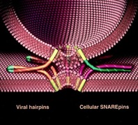
Over 30 years ago, we observed what we interpreted to be vesicular transport in crude extracts of tissue culture cells. In subsequent years we found that we had reconstituted vesicle trafficking in the Golgi, including the process of membrane fusion. Using this assay as a guide, we purified as a required factor the NEM-Sensitive Fusion protein (NSF). This led to the purification of the Soluble NSF Attachment Factor (SNAP), which bound NSF to Golgi membranes, and then with these tools discovered that the receptors for SNAP in membranes were actually complexes of proteins (which we called SNAREs) which we envisioned could potentially partner as a bridge between membranes to contribute to the process of membrane fusion and provide specificity to it (as captured in the ‘SNARE hypothesis’ proposed at the time).

We now know that organisms have a large family of SNARE proteins that indeed form cognate partnerships in just this way, and that NSF is an ATPase that (using SNAP as an adaptor protein) disrupts the SNARE complex after fusion is complete so its subunits can be recycled for repeated use. Recombinant cognate SNAREs introduced into artificial bilayers or expressed ectopically on the outside of cells ( “flipped SNAREs”) spontaneously and efficiently result in membrane (or cell) fusion, demonstrating that the SNARE complex is not only necessary but is sufficient for fusion. There are many proteins known and rapidly being discovered which closely regulate this vital process, but the muscle – if not always the brains – is in the SNAREs. Compartmental specificity is encoded to a remarkable degree in the functional partnering of SNARE proteins, a fact which is in no way inconsistent with the emerging contribution of upstream regulatory components (like rabGTPases and tethering complexes) to domain/compartment specificity.

Current Research & Projects

Our lab is working to elucidate the underlying mechanisms of vesicular transport within cells and the secretion of proteins and neurotransmitters.

Projects include:

  1. The biochemical and biophysical mechanisms of vesicle budding and fusion;
  2. Cellular regulation of vesicle fusion in exocytosis and synaptic transmission;
  3. Structural and functional organization of the Golgi apparatus from a cellular systems view.

We take an interdisciplinary approach which includes cell-free biochemistry, single molecule biophysics, high resolution optical imaging of single events/single molecules in the cell and in cell-free formats.

The overall goal is to understand transport pathways form structural mechanism to cellular physiology. The latter is facilitated by high throughput functional genomics at the cellular level (see Yale Center for High Throughput Cell Biology).

SNAREpins

We have a strong interest in new lab members who bring backgrounds in chemistry, physics, and engineering.

SOURCE

http://medicine.yale.edu/cellbio/rothman/index.aspx

3 Americans Win Joint Nobel Prize in Medicine

Reuters

From left: Randy W. Schekman, Thomas C. Südhof and James E. Rothman.

<nyt_byline>

By 
Published: October 7, 2013 151 Comments

Three Americans won the Nobel Prize in Physiology or Medicine Monday for discovering the machinery that regulates how cells transport major molecules in a cargo system that delivers them to the right place at the right time in cells.

Science Twitter Logo.
 

The Karolinska Institute in Stockholmannounced the winners: James E. Rothman of Yale University; Randy W. Schekman of the University of California, Berkeley; and Dr. Thomas C. Südhof of Stanford University.

The molecules are moved around cells in small packages called vesicles, and each scientist discovered different facets that are needed to ensure that the right cargo is shipped to the correct destination at precisely the right time.

Their research solved the mystery of how cells organize their transport system, the Karolinska committee said. Dr. Schekman discovered a set of genes that were required for vesicle traffic. Dr. Rothman unraveled protein machinery that allows vesicles to fuse with their targets to permit transfer of cargo. Dr. Südhof revealed how signals instruct vesicles to release their cargo with precision.

The tiny vesicles, which have a covering known as membranes, shuttle the cargo between different compartments or fuse with the membrane. The transport system activates nerves. It also controls the release of hormones.

Disturbances in this exquisitely precise control system cause serious damage that, in turn, can contribute to conditions like neurological diseases, diabetes and immunological disorders.

Dr. Schekman, 64, who was born in St. Paul, used yeast cells as a model system when he began his research in the 1970s. He found that vesicles piled up in parts of the cell and that the cause was genetic. He went on to identify three classes of genes that control different facets of the cell’s transport system. Dr. Schekman studied at the University of California in Los Angeles and at Stanford University, where he obtained his Ph.D. in 1974.

In 1976, he joined the faculty of the University of California, Berkeley, where he is currently professor in the Department of Molecular and Cell Biology. Dr. Schekman is also an investigator at the Howard Hughes Medical Institute.

Dr. Rothman, 63, who was born in Haverhill, Mass., studied vesicle transport in mammalian cells in the 1980s and 1990s. He discovered that a protein complex allows vesicles to dock and fuse with their target membranes. In the fusion process, proteins on the vesicles and target membranes bind to each other like the two sides of a zipper. The fact that there are many such proteins and that they bind only in specific combinations ensures that cargo is delivered to a precise location.

The same principle operates inside the cell and when a vesicle binds to the cell’s outer membrane to release its contents. Dr. Rothman received a Ph.D. from Harvard Medical School in 1976, was a postdoctoral fellow at Massachusetts Institute of Technology, and moved in 1978 to Stanford University, where he started his research on the vesicles of the cell. Dr. Rothman has also worked at Princeton University, Memorial Sloan-Kettering Cancer Institute and Columbia University.

In 2008, he joined the faculty of Yale University where he is currently professor and chairman in the Department of Cell Biology. Some of the genes Dr. Schekman discovered in yeast coded for proteins correspond to those Dr. Rothman identified in mammals. Collectively, they mapped critical components of the cell´s transport machinery.

Dr. Südhof, 57, who was born in Göttingen, Germany, studied neurotransmission, the process by which nerve cells communicate with other cells in the brain. At the time he set out to explore the field 25 years ago, much of it was virgin scientific territory. Researchers had not identified a single protein in the neurotransmission process.

Dr. Südhof helped transform what had been a rough outline into a number of molecular activities to provide insights into the elaborate mechanisms at the crux of neurological activities, from the simplest to the most sophisticated. He did so by systematically identifying, purifying and analyzing proteins that can rapidly release chemicals that underlie the brain’s activities. The transmission process can take less than a thousandth of a second.

Dr. Südhof studied at the Georg-August-Universität in Göttingen, where he received a medical degree in 1982 and a doctorate in neurochemistry the same year. In 1983, he moved to the University of Texas Southwestern Medical Center in Dallas. Dr. Südhof, who has American citizenship, became an investigator at the Howard Hughes Medical Institute in 1991 and was appointed professor of molecular and cellular physiology at Stanford University in 2008.

All three scientists have won other awards, including the Lasker Prize, for their research.

<nyt_correction_bottom>

This article has been revised to reflect the following correction:

Correction: October 7, 2013

An earlier version of this article misstated Randy W. Schekman’s age. He is 64, not 65.

SOURCE

http://www.nytimes.com/2013/10/08/health/3-win-joint-nobel-prize-in-medicine.html?_r=0

Nobel for Cell Transport

October 07, 2013

This year’s Nobel Prize in Physiology or Medicine is going jointly to three scientists for their work figuring out how cells transport their cargo, according to the Karolinska Institute. They will share the $1.25 million prize.

“Imagine hundreds of thousands of people who are traveling around hundreds of miles of streets; how are they going to find the right way? Where will the bus stop and open its doors so that people can get out?” says Nobel committee secretary Goran Hansson, according to the Associated Press. “There are similar problems in the cell.”

By studying yeast cells with defective vesicles, Randy Schekman from the University of California, Berkeley, uncovered three classes of genes that control transportation within the cell, the New York Times adds. Schekman was awakened in California by the call from Stockholm. “I wasn’t thinking too straight. I didn’t have anything elegant to say,” he tells the AP. “All I could say was ‘Oh my God,’ and that was that.” Schekman adds that he called his lab manager to arrange a celebration in the lab.

Meanwhile, Yale University’s James Rothman discovered a protein complex that allows vesicles to bind to their intended membrane targets, getting the vesicle contents to a specific location. Rothman notes that he recently lost funding for work building on his discovery, and says that he hopes that having won the Nobel will help him when he reapplies.

And Thomas Südhof at Stanford University systematically studied how nerve cells communicate, finding that vesicles full of neurotransmitters bind to cell membranes to release their contents through a molecular mechanism that responds to the presence of calcium ions. He was on his way to a give a talk when he got his call. “I got the call while I was driving and like a good citizen I pulled over and picked up the phone,” Südhof says to the AP. “To be honest, I thought at first it was a joke. I have a lot of friends who might play these kinds of tricks.”

SOURCE

Other related articles published on these Open Access Online Scientific Journal include the following:

The Series on Cardiovascular Disease and the role of Calcium Signaling consists of the following articles:

Part I: Identification of Biomarkers that are Related to the Actin Cytoskeleton

Larry H Bernstein, MD, FCAP

http://pharmaceuticalintelligence.com/2012/12/10/identification-of-biomarkers-that-are-related-to-the-actin-cytoskeleton/

Part II: Role of Calcium, the Actin Skeleton, and Lipid Structures in Signaling and Cell Motility

Larry H. Bernstein, MD, FCAP, Stephen Williams, PhD and Aviva Lev-Ari, PhD, RN

http://pharmaceuticalintelligence.com/2013/08/26/role-of-calcium-the-actin-skeleton-and-lipid-structures-in-signaling-and-cell-motility/

Part III: Renal Distal Tubular Ca2+ Exchange Mechanism in Health and Disease

Larry H. Bernstein, MD, FCAP, Stephen J. Williams, PhD
 and Aviva Lev-Ari, PhD, RN

http://pharmaceuticalintelligence.com/2013/09/02/renal-distal-tubular-ca2-exchange-mechanism-in-health-and-disease/

Part IV: The Centrality of Ca(2+) Signaling and Cytoskeleton Involving Calmodulin Kinases and Ryanodine Receptors in Cardiac Failure, Arterial Smooth Muscle, Post-ischemic Arrhythmia, Similarities and Differences, and Pharmaceutical Targets

Larry H Bernstein, MD, FCAP, Justin Pearlman, MD, PhD, FACC and Aviva Lev-Ari, PhD, RN

http://pharmaceuticalintelligence.com/2013/09/08/the-centrality-of-ca2-signaling-and-cytoskeleton-involving-calmodulin-kinases-and-ryanodine-receptors-in-cardiac-failure-arterial-smooth-muscle-post-ischemic-arrhythmia-similarities-and-differen/

Part V: Heart, Vascular Smooth Muscle, Excitation-Contraction Coupling (E-CC), Cytoskeleton, Cellular Dynamics and Ca2 Signaling

Larry H Bernstein, MD, FCAP, Justin Pearlman, MD, PhD, FACC and Aviva Lev-Ari, PhD, RN

http://pharmaceuticalintelligence.com/2013/08/26/heart-smooth-muscle-excitation-contraction-coupling-cytoskeleton-cellular-dynamics-and-ca2-signaling/

Part VI: Calcium Cycling (ATPase Pump) in Cardiac Gene Therapy: Inhalable Gene Therapy for Pulmonary Arterial Hypertension and Percutaneous Intra-coronary Artery Infusion for Heart Failure: Contributions by Roger J. Hajjar, MD

Aviva Lev-Ari, PhD, RN

http://pharmaceuticalintelligence.com/2013/08/01/calcium-molecule-in-cardiac-gene-therapy-inhalable-gene-therapy-for-pulmonary-arterial-hypertension-and-percutaneous-intra-coronary-artery-infusion-for-heart-failure-contributions-by-roger-j-hajjar/

Part VII: Cardiac Contractility & Myocardium Performance: Ventricular Arrhythmiasand Non-ischemic Heart Failure – Therapeutic Implications for Cardiomyocyte Ryanopathy (Calcium Release-related Contractile Dysfunction) and Catecholamine Responses

Justin Pearlman, MD, PhD, FACC, Larry H Bernstein, MD, FCAP and Aviva Lev-Ari, PhD, RN

http://pharmaceuticalintelligence.com/2013/08/28/cardiac-contractility-myocardium-performance-ventricular-arrhythmias-and-non-ischemic-heart-failure-therapeutic-implications-for-cardiomyocyte-ryanopathy-calcium-release-related-contractile/

Part VIII: Disruption of Calcium Homeostasis: Cardiomyocytes and Vascular Smooth Muscle Cells: The Cardiac and Cardiovascular Calcium Signaling Mechanism

Justin Pearlman, MD, PhD, FACC, Larry H Bernstein, MD, FCAP and Aviva Lev-Ari, PhD, RN

http://pharmaceuticalintelligence.com/2013/09/12/disruption-of-calcium-homeostasis-cardiomyocytes-and-vascular-smooth-muscle-cells-the-cardiac-and-cardiovascular-calcium-signaling-mechanism/

Part IX: Calcium-Channel Blockers, Calcium Release-related Contractile Dysfunction (Ryanopathy) and Calcium as Neurotransmitter Sensor

Justin Pearlman, MD, PhD, FACC, Larry H Bernstein, MD, FCAP and Aviva Lev-Ari, PhD, RN

Part X: Synaptotagmin functions as a Calcium Sensor: How Calcium Ions Regulate the fusion of vesicles with cell membranes during Neurotransmission

Larry H Bernstein, MD, FCAP and Aviva Lev-Ari, PhD, RN

http://pharmaceuticalintelligence.com/2013/09/10/synaptotagmin-functions-as-a-calcium-sensor-how-calcium-ions-regulate-the-fusion-of-vesicles-with-cell-membranes-during-neurotransmission/

Read Full Post »

Ribozymes and RNA Machines –  Work of Jennifer A. Doudna

Reporter: Aviva Lev-Ari, PhD, RN

article-21.1.1-ribozymes-and-rna-machines-e28093-work-of-jennifer-a.-doudna

Article 21.1.1- Ribozymes and RNA Machines – Work of Jennifer A. Doudna

UPDATED 3/27/2014

New DNA-editing technology spawns bold UC initiative

http://newscenter.berkeley.edu/2014/03/18/new-dna-editing-technology-spawns-bold-uc-initiative/

Crispr Goes Global

http://vcresearch.berkeley.edu/news/profile/doudna_jennifer

UPDATED 3/5/2014

Double Nicking by RNA-Guided CRISPR Cas9 for Enhanced Genome Editing Specificity

http://www.cell.com/retrieve/pii/S0092867413010155

One-Step Generation of Mice Carrying Mutations in Multiple Genes by CRISPR/Cas-Mediated Genome Engineering

http://www.cell.com/retrieve/pii/S0092867413004674

RNA-Guided Human Genome Engineering via Cas9

http://www.sciencemag.org/content/suppl/2013/01/03/science.1232033.DC1

SOURCE

From: Expert CRISPR/Cas9 Publications <Expert_CRISPRCas9_Publications@mail.vresp.com>
Date: Tue, 04 Mar 2014 17:03:01 +0000
To: <avivalev-ari@alum.berkeley.edu>
Subject: CRISPR-mediated gene editing resources

UPDATED on 11/10/2013

Exclusive: ‘Jaw-dropping’ breakthrough hailed as landmark in fight against hereditary diseases as Crispr technique heralds genetic revolution

Development to revolutionise study and treatment of a range of diseases from cancer, incurable viruses such as HIV to inherited genetic disorders such as sickle-cell anaemia and Huntington’s disease

SCIENCE EDITOR

Thursday 07 November 2013

A breakthrough in genetics – described as “jaw-dropping” by one Nobel scientist – has created intense excitement among DNA experts around the world who believe the discovery will transform their ability to edit the genomes of all living organisms, including humans.

Click image above to enlarge graphic

The development has been hailed as a milestone in medical science because it promises to revolutionise the study and treatment of a range of diseases, from cancer and incurable viruses to inherited genetic disorders such as sickle-cell anaemia and Down syndrome.

For the first time, scientists are able to engineer any part of the human genome with extreme precision using a revolutionary new technique called Crispr, which has been likened to editing the individual letters on any chosen page of an encyclopedia without creating spelling mistakes. The landmark development means it is now possible to make the most accurate and detailed alterations to any specific position on the DNA of the 23 pairs of human chromosomes without introducing unintended mutations or flaws, scientists said.

The technique is so accurate that scientists believe it will soon be used in gene-therapy trials on humans to treat incurable viruses such as HIV or currently untreatable genetic disorders such as Huntington’s disease. It might also be used controversially to correct gene defects in human IVF embryos, scientists said.

Until now, gene therapy has had largely to rely on highly inaccurate methods of editing the genome, often involving modified viruses that insert DNA at random into the genome – considered too risky for many patients.

The new method, however, transforms genetic engineering because it is simple and easy to edit any desired part of the DNA molecule, right down to the individual chemical building-blocks or nucleotides that make up the genetic alphabet, researchers said.

“Crispr is absolutely huge. It’s incredibly powerful and it has many applications, from agriculture to potential gene therapy in humans,” said Craig Mello of the University of Massachusetts Medical School, who shared the 2006 Nobel Prize for medicine for a previous genetic discovery called RNA interference.

“This is really a triumph of basic science and in many ways it’s better than RNA interference. It’s a tremendous breakthrough with huge implications for molecular genetics. It’s a real game-changer,” Professor Mello told The Independent.

“It’s one of those things that you have to see to believe. I read the scientific papers like everyone else but when I saw it working in my own lab, my jaw dropped. A total novice in my lab got it to work,” Professor Mello said.

In addition to engineering the genes of plants and animals, which could accelerate the development of GM crops and livestock, the Crispr technique dramatically “lowers the threshold” for carrying out “germline” gene therapy on human IVF embryos, Professor Mello added.

The new method of gene therapy makes it simple and easy to edit any desired part of the DNA molecule (Getty Creative)

The new method of gene therapy makes it simple and easy to edit any desired part of the DNA molecule (Getty Creative) Germline gene therapy on sperm, eggs or embryos to eliminate inherited diseases alters the DNA of all subsequent generations, but fears over its safety, and the prospect of so-called “designer babies”, has led to it being made illegal in Britain and many other countries.

The new gene-editing technique could address many of the safety concerns because it is so accurate. Some scientists now believe it is only a matter of time before IVF doctors suggest that it could be used to eliminate genetic diseases from affected families by changing an embryo’s DNA before implanting it into the womb.

“If this new technique succeeds in allowing perfectly targeted correction of abnormal genes, eliminating safety concerns, then the exciting prospect is that treatments could be developed and applied to the germline, ridding families and all their descendants of devastating inherited disorders,” said Dagan Wells, an IVF scientist at Oxford University.

“It would be difficult to argue against using it if it can be shown to be as safe, reliable and effective as it appears to be. Who would condemn a child to terrible suffering and perhaps an early death when a therapy exists, capable of repairing the problem?” Dr Wells said.

The Crispr process was first identified as a natural immune defence used by bacteria against invading viruses. Last year, however, scientists led by Jennifer Doudna at the University of California, Berkeley, published a seminal study showing that Crispr can be used to target any region of a genome with extreme precision with the aid of a DNA-cutting enzyme called CAS9.

Since then, several teams of scientists showed that the Crispr-CAS9 system used by Professor Doudna could be adapted to work on a range of life forms, from plants and nematode worms to fruit flies and laboratory mice.

Earlier this year, several teams of scientists demonstrated that it can also be used accurately to engineer the DNA of mouse embryos and even human stem cells grown in culture. Geneticists were astounded by how easy, accurate and effective it is at altering the genetic code of any life form – and they immediately realised the therapeutic potential for medicine.

“The efficiency and ease of use is completely unprecedented. I’m jumping out of my skin with excitement,” said George Church, a geneticist at Harvard University who led one of the teams that used Crispr to edit the human genome for the first time.

“The new technology should permit alterations of serious genetic disorders. This could be done, in principle, at any stage of development from sperm and egg cells and IVF embryos up to the irreversible stages of the disease,” Professor Church said.

David Adams, a DNA scientist at the Wellcome Trust Sanger Institute in Cambridge, said that the technique has the potential to transform the way scientists are able to manipulate the genes of all living organisms, especially patients with inherited diseases, cancer or lifelong HIV infection.

“This is the first time we’ve been able to edit the genome efficiently and precisely and at a scale that means individual patient mutations can be corrected,” Dr Adams said.

“There have been other technologies for editing the genome but they all leave a ‘scar’ behind or foreign DNA in the genome. This leaves no scars behind and you can change the individual nucleotides of DNA – the ‘letters’ of the genetic textbook – without any other unwanted changes,” he said.

Timeline: Landmarks in DNA science

Restriction enzymes: allowed scientists to cut the DNA molecule at or near a recognised genetic sequence. The enzymes work well in microbes but are more difficult to target in the more complex genomes of plants and animals. Their discovery in the 1970s opened the way for the age of genetic engineering, from GM crops to GM animals, and led to the 1978 Nobel Prize for medicine.

Polymerase chain reaction (PCR): a technique developed in 1983 by Kary Mullis (below, credit: Getty) in California allowed scientists to amplify the smallest amounts of DNA – down to a single molecule – to virtually unlimited quantities. It quickly became a standard technique, especially in forensic science, where it is used routinely in analysing the smallest tissue samples left at crime scenes. Many historical crimes have since been solved with the help of the PCR test. Mullis won the Nobel Prize for chemistry in 1993.

RNA interference: scientists working on the changing colour of petunia plants first noticed this phenomenon, which was later shown to involve RNA, a molecular cousin to DNA. In 1998, Craig Mello and Andrew Fire in the US demonstrated the phenomenon on nematode worms, showing that small strands of RNA could be used to turn down the activity of genes, rather like a dimmer switch. They shared the 2006 Nobel Prize for physiology or medicine for the discovery.

Zinc fingers: complex proteins called zinc fingers, first used on mice in 1994, can cut DNA at selected sites in the genome, with the help of enzymes. Another DNA-cutting technique called Talens can do something similar. But both are cumbersome to use and difficult to operate in practice – unlike the Crispr technique.

VIEW VIDEO

http://www.independent.co.uk/news/science/indyplus-video-crispr-technique-8925604.html

a video of how the Crispr system derived from bacteria works on human cells to correct genetic defects

SOURCE

http://www.independent.co.uk/news/science/exclusive-jawdropping-breakthrough-hailed-as-landmark-in-fight-against-hereditary-diseases-as-crispr-technique-heralds-genetic-revolution-8925295.html?goback=%2Egde_2106240_member_5804987154979381248#%21

Jennifer A. Doudna

Professor of Chemistry
Professor of Biochemistry & Molecular Biology

email: doudna@berkeley.edu
office: 708A Stanley Hall
phone: 510-643-0225
fax: 510-643-0008

lab: 731 Stanley Hall
lab phone: 510-643-0113
lab fax: 510-643-0080

Research Group URL
Recent Publications

Research Interests

Chemical Biology

Ribozymes and RNA Machines: RNA forms a variety of complex globular structures, some of which function like enzymes or form functional complexes with proteins. There are three major areas of focus in the lab: catalytic RNA, the function of RNA in the signal recognition particle and the mechanism of RNA-mediated internal initiation of protein synthesis. We are interested in understanding and comparing catalytic strategies used by RNA to those of protein enzymes, focusing on self-splicing introns and the self-cleaving RNA from hepatitis delta virus (HDV), a human pathogen. We are also investigating RNA-mediated initiation of protein synthesis, focusing on the internal ribosome entry site (IRES) RNA from Hepatitis C virus. Cryo-EM, x-ray crystallography and biochemical experiments are focused on understanding the structure and mechanism of the IRES and its amazing ability to hijack the mammalian ribosome and associated translation factors. A third area of focus in the lab is the signal recognition particle, which contains a highly conserved RNA required for targeting proteins for export out of cells. Each of these projects seeks to understand the molecular basis for RNA function, using a combination of structural, biophysical and biochemical approaches.

Biography

Medical School, 1989-1991; Post-doctoral fellow, University of Colorado, 1991-1994; Assistant/Associate professor, (1994-1998), Professor, (1999-2001), Yale University. Professor of Biochemistry & Molecular Biology, UC Berkeley, (2002-). Howard Hughes Medical Investigator 1997 to present. Packard Foundation Fellow Award, 1996; NSF Alan T. Waterman Award, 2000. Member, National Academy of Sciences, 2002. Member, American Academy of Arts and Sciences, 2003; American Association for the Advancement of Science Fellow Award, 2008; Member, Institute of Medicine of the National Academies, 2010.

READ MORE

Diagnosing Diseases & Gene Therapy: Precision Genome Editing and Cost-effective microRNA Profiling

http://pharmaceuticalintelligence.com/2013/03/28/diagnosing-diseases-gene-therapy-precision-genome-editing-and-cost-effective-microrna-profiling/

Read Full Post »

The potential contribution of informatics to healthcare is more than currently estimated

Reporter: Larry H Bernstein, MD, FCAP


I call attention to an interesting article that just came out.   The estimate of improved costsavings in healthcare and diagnostic accuracy is extimated to be substantial.   I have written about the unused potential that we have not yet seen.  In short, there is justification in substantial investment in resources to this, as has been proposed as a critical goal.  Does this mean a reduction in staffing?  I wouldn’t look at it that way.  The two huge benefits that would accrue are:


  1. workflow efficiency, reducing stress and facilitating decision-making.
  2. scientifically, primary knowledge-based  decision-support by well developed algotithms that have been at the heart of computational-genomics.


Can computers save health care? IU research shows lower costs, better outcomes

Cost per unit of outcome was $189, versus $497 for treatment as usual

 Last modified: Monday, February 11, 2013


BLOOMINGTON, Ind. — New research from Indiana University has found that machine learning — the same computer science discipline that helped create voice recognition systems, self-driving cars and credit card fraud detection systems — can drastically improve both the cost and quality of health care in the United States.


 Physicians using an artificial intelligence framework that predicts future outcomes would have better patient outcomes while significantly lowering health care costs.


Using an artificial intelligence framework combining Markov Decision Processes and Dynamic Decision Networks, IU School of Informatics and Computing researchers Casey Bennett and Kris Hauser show how simulation modeling that understands and predicts the outcomes of treatment could


  • reduce health care costs by over 50 percent while also
  • improving patient outcomes by nearly 50 percent.


The work by Hauser, an assistant professor of computer science, and Ph.D. student Bennett improves upon their earlier work that


  • showed how machine learning could determine the best treatment at a single point in time for an individual patient.


By using a new framework that employs sequential decision-making, the previous single-decision research


  • can be expanded into models that simulate numerous alternative treatment paths out into the future;
  • maintain beliefs about patient health status over time even when measurements are unavailable or uncertain; and
  • continually plan/re-plan as new information becomes available.

In other words, it can “think like a doctor.”  (Perhaps better because of the limitation in the amount of information a bright, competent physician can handle without error!)


“The Markov Decision Processes and Dynamic Decision Networks enable the system to deliberate about the future, considering all the different possible sequences of actions and effects in advance, even in cases where we are unsure of the effects,” Bennett said.  Moreover, the approach is non-disease-specific — it could work for any diagnosis or disorder, simply by plugging in the relevant information.  (This actually raises the question of what the information input is, and the cost of inputting.)


The new work addresses three vexing issues related to health care in the U.S.:


  1. rising costs expected to reach 30 percent of the gross domestic product by 2050;
  2. a quality of care where patients receive correct diagnosis and treatment less than half the time on a first visit;
  3. and a lag time of 13 to 17 years between research and practice in clinical care.

  Framework for Simulating Clinical Decision-Making


“We’re using modern computational approaches to learn from clinical data and develop complex plans through the simulation of numerous, alternative sequential decision paths,” Bennett said. “The framework here easily out-performs the current treatment-as-usual, case-rate/fee-for-service models of health care.”  (see the above)


Bennett is also a data architect and research fellow with Centerstone Research Institute, the research arm of Centerstone, the nation’s largest not-for-profit provider of community-based behavioral health care. The two researchers had access to clinical data, demographics and other information on over 6,700 patients who had major clinical depression diagnoses, of which about 65 to 70 percent had co-occurring chronic physical disorders like diabetes, hypertension and cardiovascular disease.  Using 500 randomly selected patients from that group for simulations, the two


  • compared actual doctor performance and patient outcomes against
  • sequential decision-making models

using real patient data.

They found great disparity in the cost per unit of outcome change when the artificial intelligence model’s


  1. cost of $189 was compared to the treatment-as-usual cost of $497.
  2. the AI approach obtained a 30 to 35 percent increase in patient outcomes
Bennett said that “tweaking certain model parameters could enhance the outcome advantage to about 50 percent more improvement at about half the cost.”


While most medical decisions are based on case-by-case, experience-based approaches, there is a growing body of evidence that complex treatment decisions might be effectively improved by AI modeling.  Hauser said “Modeling lets us see more possibilities out to a further point –  because they just don’t have all of that information available to them.”  (Even then, the other issue is the processing of the information presented.)


Using the growing availability of electronic health records, health information exchanges, large public biomedical databases and machine learning algorithms, the researchers believe the approach could serve as the basis for personalized treatment through integration of diverse, large-scale data passed along to clinicians at the time of decision-making for each patient. Centerstone alone, Bennett noted, has access to health information on over 1 million patients each year. “Even with the development of new AI techniques that can approximate or even surpass human decision-making performance, we believe that the most effective long-term path could be combining artificial intelligence with human clinicians,” Bennett said. “Let humans do what they do well, and let machines do what they do well. In the end, we may maximize the potential of both.”


Artificial Intelligence Framework for Simulating Clinical Decision-Making: A Markov Decision Process Approach” was published recently in Artificial Intelligence in Medicine. The research was funded by the Ayers Foundation, the Joe C. Davis Foundation and Indiana University.


For more information or to speak with Hauser or Bennett, please contact Steve Chaplin, IU Communications, at 812-856-1896 or stjchap@iu.edu.


IBM Watson Finally Graduates Medical School


It’s been more than a year since IBM’s Watson computer appeared on Jeopardy and defeated several of the game show’s top champions. Since then the supercomputer has been furiously “studying” the healthcare literature in the hope that it can beat a far more hideous enemy: the 400-plus biomolecular puzzles we collectively refer to as cancer.


Anomaly Based Interpretation of Clinical and Laboratory Syndromic Classes

Larry H Bernstein, MD, Gil David, PhD, Ronald R Coifman, PhD.  Program in Applied Mathematics, Yale University, Triplex Medical Science.


 Statement of Inferential  Second Opinion

 Realtime Clinical Expert Support and Validation System

Gil David and Larry Bernstein have developed, in consultation with Prof. Ronald Coifman, in the Yale University Applied Mathematics Program, a software system that is the equivalent of an intelligent Electronic Health Records Dashboard that provides
  • empirical medical reference and suggests quantitative diagnostics options.

Background

The current design of the Electronic Medical Record (EMR) is a linear presentation of portions of the record by
  • services, by
  • diagnostic method, and by
  • date, to cite examples.

This allows perusal through a graphical user interface (GUI) that partitions the information or necessary reports in a workstation entered by keying to icons.  This requires that the medical practitioner finds

  • the history,
  • medications,
  • laboratory reports,
  • cardiac imaging and EKGs, and
  • radiology
in different workspaces.  The introduction of a DASHBOARD has allowed a presentation of
  • drug reactions,
  • allergies,
  • primary and secondary diagnoses, and
  • critical information about any patient the care giver needing access to the record.
 The advantage of this innovation is obvious.  The startup problem is what information is presented and how it is displayed, which is a source of variability and a key to its success.

Proposal

We are proposing an innovation that supercedes the main design elements of a DASHBOARD and
  • utilizes the conjoined syndromic features of the disparate data elements.
So the important determinant of the success of this endeavor is that it facilitates both
  1. the workflow and
  2. the decision-making process
  • with a reduction of medical error.
 This has become extremely important and urgent in the 10 years since the publication “To Err is Human”, and the newly published finding that reduction of error is as elusive as reduction in cost.  Whether they are counterproductive when approached in the wrong way may be subject to debate.
We initially confine our approach to laboratory data because it is collected on all patients, ambulatory and acutely ill, because the data is objective and quality controlled, and because
  • laboratory combinatorial patterns emerge with the development and course of disease.  Continuing work is in progress in extending the capabilities with model data-sets, and sufficient data.
It is true that the extraction of data from disparate sources will, in the long run, further improve this process.  For instance, the finding of both ST depression on EKG coincident with an increase of a cardiac biomarker (troponin) above a level determined by a receiver operator curve (ROC) analysis, particularly in the absence of substantially reduced renal function.
The conversion of hematology based data into useful clinical information requires the establishment of problem-solving constructs based on the measured data.  Traditionally this has been accomplished by an intuitive interpretation of the data by the individual clinician.  Through the application of geometric clustering analysis the data may interpreted in a more sophisticated fashion in order to create a more reliable and valid knowledge-based opinion.
The most commonly ordered test used for managing patients worldwide is the hemogram that often incorporates the review of a peripheral smear.  While the hemogram has undergone progressive modification of the measured features over time the subsequent expansion of the panel of tests has provided a window into the cellular changes in the production, release or suppression of the formed elements from the blood-forming organ to the circulation.  In the hemogram one can view data reflecting the characteristics of a broad spectrum of medical conditions.
Progressive modification of the measured features of the hemogram has delineated characteristics expressed as measurements of
  • size,
  • density, and
  • concentration,
resulting in more than a dozen composite variables, including the
  1. mean corpuscular volume (MCV),
  2. mean corpuscular hemoglobin concentration (MCHC),
  3. mean corpuscular hemoglobin (MCH),
  4. total white cell count (WBC),
  5. total lymphocyte count,
  6. neutrophil count (mature granulocyte count and bands),
  7. monocytes,
  8. eosinophils,
  9. basophils,
  10. platelet count, and
  11. mean platelet volume (MPV),
  12. blasts,
  13. reticulocytes and
  14. platelet clumps,
  15. perhaps the percent immature neutrophils (not bands)
  16. as well as other features of classification.
The use of such variables combined with additional clinical information including serum chemistry analysis (such as the Comprehensive Metabolic Profile (CMP)) in conjunction with the clinical history and examination complete the traditional problem-solving construct. The intuitive approach applied by the individual clinician is limited, however,
  1. by experience,
  2. memory and
  3. cognition.
The application of rules-based, automated problem solving may provide a more reliable and valid approach to the classification and interpretation of the data used to determine a knowledge-based clinical opinion.
The classification of the available hematologic data in order to formulate a predictive model may be accomplished through mathematical models that offer a more reliable and valid approach than the intuitive knowledge-based opinion of the individual clinician.  The exponential growth of knowledge since the mapping of the human genome has been enabled by parallel advances in applied mathematics that have not been a part of traditional clinical problem solving.  In a univariate universe the individual has significant control in visualizing data because unlike data may be identified by methods that rely on distributional assumptions.  As the complexity of statistical models has increased, involving the use of several predictors for different clinical classifications, the dependencies have become less clear to the individual.  The powerful statistical tools now available are not dependent on distributional assumptions, and allow classification and prediction in a way that cannot be achieved by the individual clinician intuitively. Contemporary statistical modeling has a primary goal of finding an underlying structure in studied data sets.
In the diagnosis of anemia the variables MCV,MCHC and MCH classify the disease process  into microcytic, normocytic and macrocytic categories.  Further consideration of
proliferation of marrow precursors,
  • the domination of a cell line, and
  • features of suppression of hematopoiesis

provide a two dimensional model.  Several other possible dimensions are created by consideration of

  • the maturity of the circulating cells.
The development of an evidence-based inference engine that can substantially interpret the data at hand and convert it in real time to a “knowledge-based opinion” may improve clinical problem solving by incorporating multiple complex clinical features as well as duration of onset into the model.
An example of a difficult area for clinical problem solving is found in the diagnosis of SIRS and associated sepsis.  SIRS (and associated sepsis) is a costly diagnosis in hospitalized patients.   Failure to diagnose sepsis in a timely manner creates a potential financial and safety hazard.  The early diagnosis of SIRS/sepsis is made by the application of defined criteria (temperature, heart rate, respiratory rate and WBC count) by the clinician.   The application of those clinical criteria, however, defines the condition after it has developed and has not provided a reliable method for the early diagnosis of SIRS.  The early diagnosis of SIRS may possibly be enhanced by the measurement of proteomic biomarkers, including transthyretin, C-reactive protein and procalcitonin.  Immature granulocyte (IG) measurement has been proposed as a more readily available indicator of the presence of
  • granulocyte precursors (left shift).
The use of such markers, obtained by automated systems in conjunction with innovative statistical modeling, may provide a mechanism to enhance workflow and decision making.
An accurate classification based on the multiplicity of available data can be provided by an innovative system that utilizes  the conjoined syndromic features of disparate data elements.  Such a system has the potential to facilitate both the workflow and the decision-making process with an anticipated reduction of medical error.

This study is only an extension of our approach to repairing a longstanding problem in the construction of the many-sided electronic medical record (EMR).  On the one hand, past history combined with the development of Diagnosis Related Groups (DRGs) in the 1980s have driven the technology development in the direction of “billing capture”, which has been a focus of epidemiological studies in health services research using data mining.

In a classic study carried out at Bell Laboratories, Didner found that information technologies reflect the view of the creators, not the users, and Front-to-Back Design (R Didner) is needed.  He expresses the view:

“Pre-printed forms are much more amenable to computer-based storage and processing, and would improve the efficiency with which the insurance carriers process this information.  However, pre-printed forms can have a rather severe downside. By providing pre-printed forms that a physician completes
to record the diagnostic questions asked,
  • as well as tests, and results,
  • the sequence of tests and questions,
might be altered from that which a physician would ordinarily follow.  This sequence change could improve outcomes in rare cases, but it is more likely to worsen outcomes. “

Decision Making in the Clinical Setting.   Robert S. Didner

 A well-documented problem in the medical profession is the level of effort dedicated to administration and paperwork necessitated by health insurers, HMOs and other parties (ref).  This effort is currently estimated at 50% of a typical physician’s practice activity.  Obviously this contributes to the high cost of medical care.  A key element in the cost/effort composition is the retranscription of clinical data after the point at which it is collected.  Costs would be reduced, and accuracy improved, if the clinical data could be captured directly at the point it is generated, in a form suitable for transmission to insurers, or machine transformable into other formats.  Such data capture, could also be used to improve the form and structure of how this information is viewed by physicians, and form a basis of a more comprehensive database linking clinical protocols to outcomes, that could improve the knowledge of this relationship, hence clinical outcomes.
 How we frame our expectations is so important that
  • it determines the data we collect to examine the process.
In the absence of data to support an assumed benefit, there is no proof of validity at whatever cost.   This has meaning for
  • hospital operations, for
  • nonhospital laboratory operations, for
  • companies in the diagnostic business, and
  • for planning of health systems.
In 1983, a vision for creating the EMR was introduced by Lawrence Weed and others.  This is expressed by McGowan and Winstead-Fry.
J J McGowan and P Winstead-Fry. Problem Knowledge Couplers: reengineering evidence-based medicine through interdisciplinary development, decision support, and research.
Bull Med Libr Assoc. 1999 October; 87(4): 462–470.   PMCID: PMC226622    Copyright notice


Example of Markov Decision Process (MDP) trans...

Example of Markov Decision Process (MDP) transition automaton (Photo credit: Wikipedia)

Control loop of a Markov Decision Process

Control loop of a Markov Decision Process (Photo credit: Wikipedia)


English: IBM's Watson computer, Yorktown Heigh...

English: IBM’s Watson computer, Yorktown Heights, NY (Photo credit: Wikipedia)

English: Increasing decision stakes and system...

English: Increasing decision stakes and systems uncertainties entail new problem solving strategies. Image based on a diagram by Funtowicz, S. and Ravetz, J. (1993) “Science for the post-normal age” Futures 25:735–55 (http://dx.doi.org/10.1016/0016-3287(93)90022-L). (Photo credit: Wikipedia)


Read Full Post »

mRNA Interference with Cancer Expression

Reporter: Larry H Bernstein, MD, FCAP

Genetic switch shuts down lung cancer tumors

Genetic switch shuts down lung cancer tumors in mice October 25, 2012 in Cancer Yale researchers manipulated a tiny genetic switch and halted growth of aggressive lung cancer tumors in mice and even prevented tumors from forming. Ads by Google Stage 4 Cancer Treatments – Chat w/a Cancer Info Expert About Stage 4 Cancer Treatment Options. – http://www.CancerCenter.com The activation of a single microRNA managed to neutralize the effects of two of the most notorious genes in cancer’s arsenal, suggesting it may have a role treating several forms of cancer, the researchers report in the Nov. 1 issue of the journal Cancer Research. “This is pretty much the best pre-clinical data that show microRNAs can be effective in lung cancer treatment,” said Frank Slack, professor of molecular, cellular & developmental biology, researcher for the Yale Cancer Center, and senior author of the paper. “These cancer genes are identical to ones found in many forms of human cancers and we are hopeful the microRNA will be of therapeutic benefit in human cancer.” Unlike drugs that act upon existing proteins, microRNAs are small pieces of genetic material that can shut down and turn off genes that produce the proteins. Slack and co-author Andrea Kasinski wanted to see if one of these microRNAs, miR-34, could block the actions of K-Ras and p53 genes, which promote proliferation and survival of cancer cells, respectively. Mice with these two mutant genes invariably develop tumors but were cancer-free when researchers activated miR-34. Also, tumor growth was halted in mice that were treated with miR-34 after they had developed cancer. Journal reference: Cancer Research Provided by Yale University

Read more at: http://medicalxpress.com/news/2012-10-genetic-lung-cancer-tumors-mice.html#jCp

Read Full Post »


Reported by: Dr. Venkat S. Karra, Ph.D.


Brain structures involved in dealing with fear...


Major depression or chronic stress can cause the loss of brain volume, a condition that contributes to both emotional and cognitive impairment. Now a team of researchers led by Yale University scientists has discovered one reason why this occurs—a single genetic switch that triggers loss of brain connections in humans and depression in animal models.


The findings, reported in Nature Medicine, show that the genetic switch known as a transcription factor represses the expression of several genes that are necessary for the formation of synaptic connections between brain cells, which in turn could contribute to loss of brain mass in the prefrontal cortex.


“We wanted to test the idea that stress causes a loss of brain synapses in humans,” said senior author Ronald Duman, the Elizabeth Mears and House Jameson Professor of Psychiatry and professor of neurobiology and of pharmacology. “We show that circuits normally involved in emotion, as well as cognition, are disrupted when this single transcription factor is activated.”


The research team analyzed tissue of depressed and non-depressed patients donated from a brain bank and looked for different patterns of gene activation. The brains of patients who had been depressed exhibited lower levels of expression in genes that are required for the function and structure of brain synapses. Lead author and postdoctoral researcher H.J. Kang discovered that at least five of these genes could be regulated by a single transcription factor called GATA1. When the transcription factor was activated, rodents exhibited depressive-like symptoms, suggesting GATA1 plays a role not only in the loss of connections between neurons but also in symptoms of depression.


Duman theorizes that genetic variations in GATA1 may one day help identify people at high risk for major depression or sensitivity to stress.


“We hope that by enhancing synaptic connections, either with novel medications or behavioral therapy, we can develop more effective antidepressant therapies,” Duman said.


source:


http://www.rdmag.com/News/2012/08/Life-Sciences-Team-Discovers-How-Stress-Depression-Can-Shrink-The-Brain/


Read Full Post »

The Automated Second Opinion Generator

Author: Larry H. Bernstein, MD, FCAP

Gil David and Larry Bernstein have developed a first generation software agent under the supervision of Prof. Ronal Coifman, in the Yale University Applied Mathematics Program that is the equivalent of an intelligent EHR Dashboard that learns.  What is a Dashboard?   A Dashboard is a visual display of essential metrics. The primary purpose is to gather information and generate the metrics relatively quickly, and analyze it, meeting the highest standard of accuracy.  This invention is a leap across traditional boundaries of Health Information Technology in that it integrates and digests extractable information sources from the medical record using the laboratory, the extractable vital signs, EKG, for instance, and documented clinical descriptors to form one or more  provisional diagnoses describing the patient status by inference from a nonparametric network algorithm.  This is the first generation of a “convergence” of medicine and information science.  The diagnoses are complete only after review of thousands of records to which diagnoses are first provided, and then training the algorithm, and validating the software by applying to a second set of data, and reviewing the accuracy of the diagnoses.

The only limitation of the algorithm is sparsity of data in some subsets, which doesn’t permit a probability calculation until sufficient data is obtained.  The limitation is not so serious because it does not disable the system from recognizing at least 95 percent of the information used in medical decision-making, and adequately covers the top 15 medical diagnoses.  An example of this exception would be the diagnosis of alpha or beta thalassemia, with a microcytic picture (MCV low) and RBC high with a low Hgb).  The accuracy is very high because the anomaly detection used for classifying the data creates aggregates that have common features.  The aggregates themselves are consistent within separatory  rules that pertain to any class.  As the model grows, however, there is unknown potential for there to be prognostic, as well as diagnostic information within classes (subclasses), and a further potential to uncover therapeutic differences within classes – which will be made coherent with new classes of drugs (personalized medicine) that are emerging from the “convergence” of genomics, metabolomics, and translational biology.

The fact that such algorithms have already been used for limited data sets and unencumbered diagnoses in many cases using the approach of studies with inclusions and exclusions common for clinical trials, the approach has proved ever more costly when used outside the study environment.   The elephant in the room is age-related co-morbidities and co-existence of obesity, lipid derangements, renal function impairment, genetic and environmental factors that are hidden from view.  The approach envisioned is manageable, overcoming these obstacles, and handles both inputs and outputs with considerable ease.

We anticipate that the effect of implementing this artificial intelligence diagnostic amplifier would result in higher physician productivity at a time of great human resource limitation(s), safer prescribing practices, rapid identification of unusual patients, better assignment of patients to observation, inpatient beds, intemsive care, or referral to clinic, shortened length of patients ICU and bed days.  If the observation of systemic issues in “To err is human” is now 10 years old with marginal improvement at great cost, this should be a quantum leap forward for the patient, the physician, the caregiving team, and the society that adopts it.


Read Full Post »

%d bloggers like this: