Feeds:
Posts
Comments

Posts Tagged ‘depression’

Reporter and Curator: Dr. Sudipta Saha, Ph.D.


The relationship between gut microbial metabolism and mental health is one of the most intriguing and controversial topics in microbiome research. Bidirectional microbiota–gut–brain communication has mostly been explored in animal models, with human research lagging behind. Large-scale metagenomics studies could facilitate the translational process, but their interpretation is hampered by a lack of dedicated reference databases and tools to study the microbial neuroactive potential.


Out of all the many ways, the teeming ecosystem of microbes in a person’s gut and other tissues might affect health. But, its potential influences on the brain may be the most provocative for research. Several studies in mice had indicated that gut microbes can affect behavior, and small scale studies on human beings suggested this microbial repertoire is altered in depression. Studies by two large European groups have found that several species of gut bacteria are missing in people with depression. The researchers can’t say whether the absence is a cause or an effect of the illness, but they showed that many gut bacteria could make substances that affect the nerve cell function—and maybe the mood.


Butyrate-producing Faecalibacterium and Coprococcus bacteria were consistently associated with higher quality of life indicators. Together with DialisterCoprococcus spp. was also depleted in depression, even after correcting for the confounding effects of antidepressants. Two kinds of microbes, Coprococcus and Dialister, were missing from the microbiomes of the depressed subjects, but not from those with a high quality of life. The researchers also found the depressed people had an increase in bacteria implicated in Crohn disease, suggesting inflammation may be at fault.


Looking for something that could link microbes to mood, researchers compiled a list of 56 substances important for proper functioning of nervous system that gut microbes either produce or break down. They found, for example, that Coprococcus seems to have a pathway related to dopamine, a key brain signal involved in depression, although they have no evidence how this might protect against depression. The same microbe also makes an anti-inflammatory substance called butyrate, and increased inflammation is implicated in depression.


Still, it is very much unclear that how microbial compounds made in the gut might influence the brain. One possible channel is the vagus nerve, which links the gut and brain. Resolving the microbiome-brain connection might lead to novel therapies. Some physicians and companies are already exploring typical probiotics, oral bacterial supplements, for depression, although they don’t normally include the missing gut microbes identified in the new study.


References:


https://www.sciencemag.org/news/2019/02/evidence-mounts-gut-bacteria-can-influence-mood-prevent-depression?utm_source=Nature+Briefing


https://www.nature.com/articles/s41564-018-0337-x


https://www.ncbi.nlm.nih.gov/pubmed/22968153


https://www.ncbi.nlm.nih.gov/pubmed/24888394


https://www.ncbi.nlm.nih.gov/pubmed/27067014


Read Full Post »

battling depression

Larry H Bernstein, MD, FCAP, Curator

LPBI

Israeli scientists tout new method for battling depression

By focusing not on the brain’s neurons but on cells known as microglia, researchers may have mapped a path to innovative treatments

October 22, 2015
Prof. Raz Yirmiya, who heads Hebrew University’s psychoneuroimmunology laboratory, is the senior author of a new paper, “Depression as a microglial disease,” published in the October issue of Trends in Neurosciences.

The paper urges a new focus in depression research away from neurons, the cells that make up the brain’s thinking faculties and allow it to control the body, and toward brain cells called microglia.

About 10 percent of brain cells are microglia, which serve as a kind of infrastructure for the brain, holding neurons in place, passing oxygen and other nutrients to them and fighting brain infections.

Microglia play a special role in repairing brain damage and trauma to neurons, the researchers note.

“Our views on microglia have dramatically changed over the last decade,” Yirmiya said in a Thursday statement by Hebrew University.

“We now know that these cells play a role in the formation and fine-tuning of the connections between neurons [known as synapses] during brain development, as well as in changes [to] these connections throughout life. These roles are important for normal brain and behavioral functions, including pain, mood and cognitive abilities.”

According to Yirmiya, who wrote the paper together with fellow Hebrew University researchers Neta Rimmerman and Ronen Reshef, “Studies in humans, using post-mortem brain tissues or special imaging techniques, as well as studies in animal models of depression, demonstrated that when the structure and function of microglia change, these cells can no longer regulate normal brain and behavior processes and this can lead to depression.”

Major depression “afflicts one in six people at some point in their life,” the university’s statement on the new research noted, and is “the leading global cause of disability – surpassing cardiovascular and respiratory diseases, cancer and HIV/AIDS combined.”

“Progress in the understanding of the biology of depression has been slow,” the researchers note in the paper. New research into the role of malfunctioning microglia could lead to a new push toward finding different and perhaps more effective treatment.

Changes in microglia are found in patients with conditions that correlate with high instances of depression, including “infection, injury, trauma, aging, autoimmune diseases such as multiple sclerosis and neurodegenerative diseases such as Alzheimer’s disease,” the statement explains. Microglia in these conditions take on an “activated” state, growing larger and round while secreting chemicals that lead to inflammation in the brain.

Microglia also change their shape and functioning when exposed to chronic unpredictable psychological stress, which is one of the leading causes of depression. Such conditions have also been found – in research carried out in Yirmiya’s Hebrew University laboratory – to correlate with microglia dying and remaining cells appearing shrunken and degenerated.

Drugs that inhibit overactive microglia or stimulate suppressed microglia may be the key to dramatic new treatments, they argue.

“Prof. Yirmiya’s new research could have a profound impact on the future development of anti-depressant medications. Present drugs do not always have the desired effect on patients, so there is an urgent need to discover novel biological mechanisms and drug targets for diagnosing the root cause of depression and for treating depressed patients appropriately,” the statement read.

New drugs that restore function to diseased microglia, the researchers suggest, may turn out to be as effective as current fast-acting anti-depressants.

https://tpc.googlesyndication.com/pagead/js/r20151014/r20110914/abg.js/

Read Full Post »

MONOAMINE, NEUROTROPHIC FACTOR, & PHARMACOKINTIC HYPOTHESES OF DEPRESSION, OLD AND NEWER TREATMENTS

Curator: Zohi Sternberg, PhD

Department of Neurology, University of Buffalo, Baird MS Center, Buffalo, NY.

Depression is the second leading cause of disability worldwide, which struck 20% of women and 15% of men at least one episode in their lifetime. Depression is characterized by two or more weeks of depressed mood or diminished interest, associated with symptoms such as disturbed sleep, decrease in appetite and libido, psychomotor changes, reduced concentration, excessive guilt and suicidal thoughts or attempts.

A variety of genetic, environmental and neurobiological factors are implicated in depression. The genetic contribution to the manifestation of depression has been estimated as 40-50%. However, combinations of multiple genetic factors may be involved in the development of disease, because a defect in a single gene usually fails to induce the expression of multifaceted symptoms of depression. In addition, non-genetic factors such cellular abnormalities and stress, interact with genetic factors, contributing to the prevalence of depression.

Monoamine hypothesis of depression

Monoamine reuptake inhibitors

Among cellular abnormalities contributing to depression is the dysfunction in the monoamine system. The deficiency or imbalances in the monoamine neurotransmitters, such as serotonin, dopamine and norepinephrine has been known to be the cause of depression. Typical antidepressants, such as 5-hydroxytryptamine (5-HT) selective reuptake inhibitors (SSRIs), exert clinical effects by blocking monoamine reuptake by the 5-HT transporter (Fig 1). However, these antidepressants are effective in less than 50% of patients and show a wide spectrum of undesired side effects. In addition, the chronic use of antidepressants is required to observe clinical benefits.

Neurotrophic hypothesis of depression

Although neurotrophic factors are critical signaling molecules for nervous system development, they continue to play an important role in the survival, function and adaptive plasticity of neurons in the adult brain. Of the different families of neurotrophic and/or growth factors expressed in the brain, the most extensively studied in depression is the nerve growth factor (NGF) family, which includes NGF, brain-derived neurotrophic factor (BDNF), and neurotrophin 3 and 4 (NT3 and NT4). Most notable of these has been BDNF and its receptor, tropomyosin-related kinase B (TrkB), a transmembrane receptor with an intracellular tyrosine kinase domain. BDNF–TrkB downstream signaling includes activation of phosphatidyl inositol-3 kinase (PI3K)-Akt (serine threonine kinase or protein kinase B), Ras–mitogen-activated protein kinase (MAPK) and the phospholipaseCg (PLCg)–Ca2+ pathways (Fig 2)

Early studies demonstrated that stress decreases the level of BDNF in the hippocampus.  These results, coupled with brain-imaging studies, which reported a decreased volume of limbic brain regions in depressive patients, have led to a neurotrophic hypothesis of depression. Studies show that BDNF has the potential to produce an antidepressant response in behavioral models of depression, and that genetic deletion or blockade of BDNF blocks the effects of antidepressant treatments. However, it is it noteworthy that BDNF has various function depending on the site it is expressed. For example, BDNF in the mesolimbic dopamine system produces prodepressive effects and increases susceptibility to social defeat, effects that could oppose the antidepressant actions of BDNF in the Prefrontal cortex and hippocampus. Interestingly, BDNF administration by routes that would affect multiple brain regions (intracerebroventricular or systemic) produce antidepressant responses.

Furthermore, the deletion or mutation of BDNF results in a state of increased susceptibility to other factors, such as stress. This type of gene-environment interaction is supported by studies demonstrating that BDNF heterozygous deletion mutant mice display depressive behavior only when exposed to mild stress that has no effect in wild-type mice.  A chronic treatment with antidepressant is required to induce an increase in BDNF mRNA expression levels. However, it is a matter of debate to whether BDNF underlies the deleterious effects of stress and depression and, conversely, whether induction of BDNF mediates the beneficial effects of antidepressants. Nevertheless, a single nucleotide polymorphism (SNP) of BDNF, Val66Met, provides supporting evidence from human, as well as rodent studies, for a role of BDNF in depressive behavior.

Figure 1. Signaling pathways regulated by chronic antidepressant treatments. Typical antidepressants, such as 5-hydroxytryptamine (5-HT) selective reuptake inhibitors (SSRIs), block monoamine reuptake by the 5-HT transporter (SERT). This leads to regulation of postsynaptic G protein-coupled receptors, which couple to a variety of second messenger systems, including the cAMP–protein kinase A (PKA)–cAMP response element-binding (CREB) pathway [4,6] These effects require chronic SSRI treatment, owing to the requirement for desensitization of 5-HT autoreceptors, and because 5-HT is a neuromodulator that produces slow neuronal responses. By contrast, glutamate produces fast excitation of neurons via stimulation of ionotropic receptors, including AMPA and NMDA receptors, resulting in depolarization and rapid intracellular signaling, such as induction of Ca2+-calmodulin-dependent protein kinase (CAMK). Glutamate and 5-HT signaling lead to regulation of multiple physiological responses, including regulation of synaptic plasticity, as well as gene expression. One target of antidepressant treatment and CREB signaling is brain-derived neurotrophic factor (BDNF). BDNF transcripts may remain in the soma or are targeted for transport to dendrites, where they are subject to activity-dependent translation and release. A common BDNF polymorphism, Val66Met, which is encoded by G196A, blocks the trafficking of BDNF to dendrites. The induction of BDNF and other neurotrophic factors contributes to the actions of antidepressant treatments, including neuroprotection, neuroplasticity and neurogenesis. Abbreviation: SNP, single nucleotide polymorphism

Newer treatments of depression

NMDA receptor antagonists

Typical antidepressants do not increase BDNF release, which could further contribute to the delayed response, as well as limited efficacy of these agents. Furthermore, in patients with severe depression, who are resistant to typical antidepressants, a novel class of antidepressants, the NMDA receptor antagonists, such as ketamine, are being evaluated. This class of antidepressants produces a rapid antidepressant action, an effect not seen with the typical antidepressants. The mechanism of action of NMDA receptor antagonists involves increased glutamate transmission and induction of synaptogenesis, involving the upregulation of BDNF and other neurotrophic factors. The comparison of typical antidepressants with novel, rapid-acting NMDA antagonists also highlights the difference between agents that act on neuromodulatory systems (i.e.monoamines) compared with those that act on the major excitatory neurotransmitter system (i.e.glutamate).

GABA receptor antagonists

Furthermore, gamma amino butyric acid (GABA)-ergic system is known to play a role in mood disorders.  GABA system is the prominent inhibitory neurotransmitter in the brain, and a link between GABA(A) receptors and anxiety has been established. However, the role of these receptors in depression has not been validated. GABA(B) receptors, have been also postulated to be involved in the etiology of depression. Studies of GABA(B) receptor antagonists and GABA(B) receptor knockout mice suggest that the blockade, or loss of function of GABA(B) receptors causes an antidepressant-like phenotype.  The antidepressant effect of GABA(B) blockade has speculated to be via interacting with the serotonergic system and BDNF. GABA(B) receptor antagonists may represent a new class of antidepressant compounds

Figure 2. Brain-derived neurotrophic factor (BDNF)–tropomyosin-related kinase B (TrkB) signaling pathways. BDNF binding to the extracellular domain of TrkB induces dimerization and activation of the intracellular tyrosine kinase domain. This results in autophosphorylation of tyrosine residues that then serve as sites for interaction with adaptor proteins and activation of intracellular signaling cascades, including the Ras–microtubule-associated protein kinase (MAPK), phosphatidyl inositol-3 kinase (PI3K)/serine threonine kinase (Akt) and phospholipase C (PLC)-g pathways. Phosphorylation of tyrosine 515 of TrkB leads to recruitment of the Src homology 2 domaincontaining (Shc) adaptor protein, followed by recruitment of growth factor receptor-bound protein 2 (Grb2) and son of sevenless (SOS) and activation of the Ras–MAPK pathway (right). Shc–Grb2 can also lead to recruitment of Grb2-associated binder-1 (GAB1) and activation of the PI3K–Akt pathway (left). Phosphorylation of the TrkB tyrosine residue 816 results in recruitment of PLCg, which leads to the formation of inositol triphosphate (IP3) and regulation of intracellular Ca2+ and diacylglycerol (DAG), which activates CAMK and protein kinase C (PKC). These pathways control many different aspects of cellular function, including synaptic plasticity, survival and growth and/or differentiation. Basic and clinical studies demonstrate that BDNF and components of the Ras–MAPK and PI3K–Akt pathways are decreased by stress and depression, and increased by antidepressant treatments. Abbreviations: ERK, extracellular signal regulated kinase; MEK, MAP/ERK kinase; MKP1, MAP kinase phosphatase 1; PDK1, 3-phosphoinositide-dependent protein kinase 1; PTEN, phosphatase and tensin homolog.

Sphingomyelinase /ceramide inhibitors

The lysosomal lipid metabolizing enzyme, acid sphingomyelinase (ASM), cleaves sphingomyelin into ceramide and phosphorylcholine. In a pilot study, the activity of this enzyme was increased in peripheral blood cells of patients with major depressive disorder, making the ASM an interesting molecular target of antidepressant drugs.

It is postulated that the accumulation of the enzyme in the lyzozome may play a role in depressive disorders (pharmacokinetic hypothesis), and its inhibition by cationic amphiphilic substances, which traverse the BBB, and which cause the detachment of the enzyme from the inner lysosomal membranes, and its consecutive inactivation, could improve depressive disorders. In addition, ASM inhibitors could exert antidepressant effects by modulating monoamine receptor and transporter, since ASM inhibitors are known to inhibit interferon-alpha-induced 5-HT uptake.

In addition, ceramide and its metabolite sphingosine-1-phosphate have been shown to also antagonistically regulate apoptosis, cellular differentiation, proliferation and cell migration. Therefore, inhibitors of ASM or ceramide hold promise for a number of new clinical therapies for Alzheimer’s disease and cancer.

Deep brain stimulation

Another new promising method of treating depression, as well as other clinical pathologies such as obsessive-compulsive disorders, Tourette’s syndrome, substance abuse, dementia and anxiety is deep brain stimulation (DBS).  These pathologies are classified as the dysfunctions of networks which process motivational and affective stimuli. DBS permits the selective and basically reversible modulation of such networks, with marginal or non adverse effects. In the field of treatment-resistant depressive disorders, uncontrolled studies have been published with initial satisfactory and concordant indications of the therapeutic effect of DBS in a variety of target areas of the brain.

Key words: Depression, Monoamines, NMDA receptor, GABA, Deep brain stimulation, Ceramide, Sphingomyelinase, Cancer, Alzheimer;s diseases

 References

1.      Albert PR. The neurobiology of depression, revisiting the serotonin hypothesis. I. Cellular and molecular mechanisms. Phil. Trans. R. Soc. B. 2012

2.      Lee S. Jeong J. Kwak Y. Park SK. Depression research: where are we now?.[Review]  Molecular Brain. 3:8, 2010.

3.      Ronald S. Duman and Bhavya Voleti. Signaling pathways underlying the pathophysiology and treatment of depression: novel mechanisms for rapid-acting agents. Trends in Neurosciences January 2012, Vol. 35, No. 1

4.      Cryan JF. Slattery DA. GABAB receptors and depression. Current status. [Review] Advances in Pharmacology. 58:427-51, 2010.

5. Schlapfer TE. Kayser S. The development of deep brain stimulation as a putative treatment for resistant psychiatric disorders]. [Review] Nervenarzt. 81(6):696-701, 2010 Jun.

6. Su HC. Ma CT. Lin CF. Wu HT. Chuang YH. Chen LJ. Tsao CW. The acid sphingomyelinase inhibitors block interferon-alpha-induced serotonin uptake via a COX-2/Akt/ERK/STAT-dependent pathway in T cells. International Immunopharmacology. 11(11):1823-31, 2011 Nov.

7. Kornhuber J. Tripal P. Reichel M. Muhle C. Rhein C. Muehlbacher M. Groemer TW. Gulbins E Functional Inhibitors of Acid Sphingomyelinase (FIASMAs): a novel pharmacological group of drugs with broad clinical applications. [Review] Cellular Physiology & Biochemistry. 26(1):9-20, 2010.

8. Kornhuber J. Muehlbacher M. Trapp S. Pechmann S. Friedl A. Reichel M. Muhle C. Terfloth L. Groemer TW. Spitzer GM. Liedl KR. Gulbins E. Tripal P. Identification of novel functional inhibitors of acid sphingomyelinase. PLoS ONE [Electronic Resource]. 6(8):e23852, 2011.

Read Full Post »

Reporter: Aviva Lev-Ari, PhD, RN

Dysthymia: Often Chronic, Always Serious

Johns Hopkins Health Alert

Dysthymia is a chronic form of depression that is characterized by the presence of a depressed mood for most of the day, for more days than not, over a period of at least two years. Dysthymia may be intermittent and interspersed with periods of feeling normal, but these periods of improvement last for no more than two months.

Dysthymia often goes unnoticed. And because of its chronic nature, the person may come to believe, “I’ve always been this way.” In addition to depressed mood, symptoms of dysthymia include two or more of the following:

It is far better to treat dysthymia than to think of it as a minor condition. Bypassing treatment places people at increased risk for subsequently developing major depression. In fact, about 10 percent of people with dysthymia also have recurrent episodes of major depression, a condition known as double depression.

What causes of dysthymia?  Some medical conditions, including neurological disorders (such as multiple sclerosis and stroke), hypothyroidism, fibromyalgia and chronic fatigue syndrome, are associated with dysthymia. Investigators believe that, in these cases, developing dysthymia is not a psychological reaction to being ill but rather is a biological effect of these disorders.

There are many reasons for this connection. It may be that these medical conditions interfere with the action of neurotransmitters, or that medications (such as corticosteroids or beta-blockers) taken for a medical illness may trigger the dysthymia or that both dysthymia and the medical illness are related in some other way, reinforcing each other in a complicated manner.

Dysthymia can also follow severe psychological stress, such as losing a spouse or caring for a chronically ill loved one. Older people who have never had psychiatric disorders are particularly susceptible to developing dysthymia after significant life stresses.

Posted in Depression and Anxiety on October 16, 2012


Medical Disclaimer: This information is not intended to substitute for the advice of a physician. Click here for additional information: Johns Hopkins Health Alerts Disclaimer


Read Full Post »

The eTNS System. (PRNewsFoto/NeuroSigma)

Reporter: Howard Donohue, PhD (EAW)

Following the arrival in the 1990s of a drug for treating depression called fluoxetine (better known by its brand name, Prozac) – a “selective serotonin reuptake inhibitor” (SSRI) – it’s probably fair to say that not many drugs have become as deeply engrained in the public’s general awareness as those of this type. Perhaps one reason for this could be the sheer number of people affected by depression and to whom SSRIs are relevant as a possible treatment (one study has estimated that depression affected upwards of 30 million Europeans in the year 2010 [1]). Perhaps another reason could be the various controversies that have surrounded SSRIs over the years, from stories of increased suicide risk in children [2] to evidence of biases and the “selective” publishing of clinical data favoring the effectiveness of these drugs [3]. Of course, despite the controversies, SSRIs (along with other classes of antidepressant drug) continue to be a mainstay, but let’s not forget, amid their popularity, that there are other ways to treat depressive illnesses. And in maximizing the benefits of treatment for the individual, it’s important to realize that any one of these approaches might work well for one person, but not for another. Among the non-pharmacologic ways to treat depression are psychological approaches, for example cognitive behavioral therapy, or alternatively, “brain stimulation” approaches such as electroconvulsive therapy (ECT). ECT is a method to induce a mild seizure in the patient by means of electrical activity applied to the brain via electrodes connected to the temples.

On the subject of ECT; you could be forgiven for thinking that it’s not very nice, especially if you’ve seen the plights of characters like Randle Patrick “Mac” McMurphy, portrayed by Jack Nicholson in One Flew Over the Cuckoo’s Nest or Russell Crowe’s portrayal of Dr. John Nash (based on the real-life Nobel Laureate in Economics by the same name) in A Beautiful Mind. Nonetheless, despite the treatment in Hollywood of ECT as a sinister, repressive, and even brutal procedure, the reality is obviously different and it continues to have a place in medical practice for the treatment of severely depressed patients to this day. This isn’t to say that controversies don’t exist within the medical community concerning certain side effects (such as memory loss), but in balancing this, we should remember that many – if not most – medical procedures have their drawbacks (hopefully, the benefits will far outweigh the drawbacks). Putting aside any thoughts on whether ECT is good or bad, it is recognition and consideration of the drawbacks that helps drive the evolution of medical technologies.

So, in illustrating the evolution that is happening in the field of brain stimulation for treating neurological disorders (in this case, depression and also epilepsy), the recent approval in Europe of an “external Trigeminal Nerve Stimulation” (eTNS) technique provides an excellent example. The technique, called the MonarchTM and exclusively licensed to Neurosigma Inc. (a Los Angeles-based medical device company) “for the adjunctive treatment of epilepsy and major depressive disorder, for adults and children 9 years and older”, is a non-invasive form of neuromodulation therapy [4]. It was invented at the University of California, Los Angleles (UCLA) and has been in development for over 10 years [4]. It works by using a low-energy stimulus to stimulate branches of the trigeminal nerve, a nerve that can affect the activity of several key brain regions believed to be involved in depression and epilepsy. In contrast to ECT, the stimulus is restricted to the soft tissues of the forehead without direct penetration to the brain, which thereby facilitates a non-invasive form of neuromodulation [4]. Following European approval, Neurosigma affirmed in a press release that eTNS is “supported by years of safety and compelling efficacy data generated in clinical trials conducted at UCLA and the University of Southern California (USC)” [4]. In realizing the future potential of eTNS, Neurosigma’s business strategy is now geared toward steps for its adoption at major epilepsy and depression centers in the EU, as well as endeavors to make it available to patients in the US and other countries [4].

To answer the question of whether eTNS will rise to prominence as an effective treatment in the fight against depression and epilepsy, only time will tell. But if it does, as well as being a valuable addition to the armamentarium against these debilitating diseases, maybe its non-invasive nature will mean that the film directors have a harder time in “demonizing” it for dramatic effect. Well anyway, let’s hope so.

References

  1. Wittchen et al. Eur Neuropsychopharmacol 2011: 21:655-79.
  2. http://news.bbc.co.uk/2/hi/health/3656110.stm
  3. Turner et al. N Engl J Med 2008; 358:252-60.
  4. http://www.prnewswire.com/news-releases/neurosigma-receives-ce-certification-168578146.html

Read Full Post »


Reporter: Howard Donohue, PhD (EAW)

The hypothalamic-pituitary-adrenal (HPA) axis – which can be thought of as a series of closely linked endocrine structures in the brain – has a key role in triggering the body’s stress response through the secretion of cortisol. In explaining how the HPA axis is itself regulated, for example how its activity is increased in response to a perceived environmental threat, we can infer that the diverse brain areas with which it shares neural interconnections have a crucial role (for a review, see [1]). An equally important question relates to how the activity of the HPA axis is returned to normal when the stress response is no longer needed. To answer this, it is well known that the same “neurosteroid” hormones released by the HPA axis that trigger stress-related biological adaptations also serve to dampen its activity through a “negative feedback” mechanism. In re-defining the biological model of how neurosteroids control the HPA axis, a study led by Jamie Maguire, PhD at Tufts University (Boston, MA) provides some fascinating insights [2]. Moreover, this study has some extremely interesting and counter-intuitive implications for understanding the functions of the “inhibitory” brain chemical gamma-aminobutyric acid (GABA), which is best known for opposing the effects of “excitatory” brain chemicals in order to balance the flow of electrical activity in the brain.

To study how the HPA axis is regulated by neurosteroids, Maguire’s team performed investigations in mice using the neurosteroid tetrahydrodeoxycorticosterone (THDOC). The investigators found that THDOC, when applied to a discrete population of cells in the thalamus called the paraventricular nucleus (PVN), resulted in a decrease in blood levels of corticosterone (the mouse equivalent of the human stress hormone, cortisol). This finding highlights the importance of the PVN as a key anatomical locus in the brain where neurosteroids act, and is consistent with the traditional view of neurosteroids as “negative regulators” of the HPA axis. However, in mice that underwent a stressful “restraint” procedure, it was found that a prior treatment with THDOC (thirty minutes before the stressful experience) resulted in augmentation of corticosterone levels (i.e. relative to mice that underwent the stressful experience but did not receive prior THDOC treatment). In parallel, it was shown that while application of THDOC normally decreased the electrical activity of PVN cells, it actually led to increases in mice that had undergone restraint. Taken together, these findings provide evidence that neurosteroids can have opposite effects on the HPA axis depending on the “stressed” state of the organism.

Thinking about how a neurosteroid hormone can exert opposite effects on PVN cells in the thalamus may be confusing, but what may be more confusing is that these different actions depend on the same “inhibitory” brain chemical, GABA (a neurotransmitter), as well as the same molecular “machinery” (or receptors) with which GABA interacts. This was demonstrated by using mice in which a particular sub-component (or subunit) of the GABA receptor, the gamma subunit, had been genetically deleted; neurosteroids had absolutely no effect on the activity of the HPA axis (neither positive nor negative) in these gamma subunit-deficient mice.

How is it possible to explain the seemingly paradoxical finding that neurosteroids can exert opposite effects on the HPA axis through the same neurotransmitter system? In addressing this question, it is important to remember that although neurotransmitters may be thought of as excitatory or inhibitory, their ability to trigger these effects depends solely on the molecular and cellular apparatus with which they interact. Normally, the inhibitory actions of GABA upon the electrical activity of nerve cells depend on the maintenance of an “electrochemical” gradient by a “transporter” molecule called KCC2 (which transports chloride ions out of cells). Maguire’s team showed that “dephosphorylation” (i.e. the removal of a small chemical moiety – the phosphate group – which is covalently bound at a specific site on the molecule) of KCC2 resulted in lower detectable levels of this transporter in the PVN. Similarly to innumerable other examples in biology where dephosphorylation (or the reverse, phosphorylation) serves as an exquisite regulatory mechanism for controlling the activity of molecular networks, removal of the phosphate group from KCC2 acts as a molecular “switch” that causes the breakdown of the electrochemical gradient. The outcome is that GABA has an excitatory influence on neural activity instead of the inhibitory influence with which it is usually associated.

In common with many important contributions to scientific understanding, these findings should serve as a reminder that it is often necessary to challenge and question what is already “accepted” in our theoretical models, in the light of unexpected and sometimes counter-intuitive experimental results. Whatever the line of scientific inquiry may be, the reward for doing so will be a deeper and more comprehensive understanding of the natural phenomena being studied. The findings of Maguire and colleagues, published in the Journal of Neuroscience, have possible therapeutic implications for disorders associated with disrupted function of the HPA axis, including epilepsy and depression.

References

1. http://www.nature.com/nrn/journal/v10/n6/full/nrn2647.html

2. http://www.jneurosci.org/content/31/50/18198.long


Read Full Post »


Reported by: Dr. Venkat S. Karra, Ph.D.


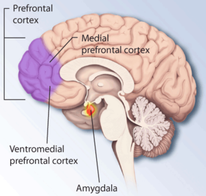
Brain structures involved in dealing with fear...


Major depression or chronic stress can cause the loss of brain volume, a condition that contributes to both emotional and cognitive impairment. Now a team of researchers led by Yale University scientists has discovered one reason why this occurs—a single genetic switch that triggers loss of brain connections in humans and depression in animal models.


The findings, reported in Nature Medicine, show that the genetic switch known as a transcription factor represses the expression of several genes that are necessary for the formation of synaptic connections between brain cells, which in turn could contribute to loss of brain mass in the prefrontal cortex.


“We wanted to test the idea that stress causes a loss of brain synapses in humans,” said senior author Ronald Duman, the Elizabeth Mears and House Jameson Professor of Psychiatry and professor of neurobiology and of pharmacology. “We show that circuits normally involved in emotion, as well as cognition, are disrupted when this single transcription factor is activated.”


The research team analyzed tissue of depressed and non-depressed patients donated from a brain bank and looked for different patterns of gene activation. The brains of patients who had been depressed exhibited lower levels of expression in genes that are required for the function and structure of brain synapses. Lead author and postdoctoral researcher H.J. Kang discovered that at least five of these genes could be regulated by a single transcription factor called GATA1. When the transcription factor was activated, rodents exhibited depressive-like symptoms, suggesting GATA1 plays a role not only in the loss of connections between neurons but also in symptoms of depression.


Duman theorizes that genetic variations in GATA1 may one day help identify people at high risk for major depression or sensitivity to stress.


“We hope that by enhancing synaptic connections, either with novel medications or behavioral therapy, we can develop more effective antidepressant therapies,” Duman said.


source:


http://www.rdmag.com/News/2012/08/Life-Sciences-Team-Discovers-How-Stress-Depression-Can-Shrink-The-Brain/


Read Full Post »

Reporter: Aviva Lev-Ari, PhD, RN

http://www.nejm.org/doi/full/10.1056/NEJMra1104781?query=TOC

Fifty years ago, when the curator of the Boston Medical Library, Henry Viets, collated a score of the most important articles that had been published in the first century and a half of the New England Journal of Medicine, 10 of the 20 related to neurologic conditions. Viets HR. A score of significant papers published in the Journal during the last hundred and fifty years. N Engl J Med 1962;266:23-28

On the occasion of its 200th anniversary, one might ask why so many articles on neurologic and psychiatric diseases have been published in the Journal and what impact these pieces have had on their respective fields. Although the Journal is replete with reports of neurologic conditions that have entered the canon of medicine, it has been the large number and the breadth of clinical trials that have redefined neurology and psychiatry as active, therapeutic specialties. This article reviews the evolution of our understanding of neurologic and psychiatric conditions during the past two centuries.

Modern scientific neurology, based on neuropathology and neurophysiology, began after World War II. By 1952, patients with tabes dorsalis and general paresis had practically disappeared from hospital wards. Antimicrobial agents had changed the course of bacterial and tuberculous meningitis, but poliomyelitis was still a nemesis. Denny-Brown‘s Shattuck Lecture on the state of neurology that year in the Journal pronounced that, freed from its preoccupation with syphilology and now separating itself from internal medicine and psychiatry, neurology had “entered on an extremely productive and virile phase.” Denny-Brown DE. The changing pattern of neurologic medicine. N Engl J Med1952;246:839-846

Denny-Brown emphasized recent contributions on the neurologic aspects of liver failure, the recognition that transient ischemic attacks were due to occlusion of the carotid artery, and the emerging understanding of increased intracranial pressure. There was little question at the time that neurologic and psychiatric diseases both acted on the same brain, but Denny-Brown’s position in his Shattuck Lecture marked the emergence of American neurology, pointedly disengaging it from the popular practice of neuropsychiatry and aligning it with internal medicine. He provided a modern manifesto for neurology, which nonetheless remained largely an elegant diagnostic specialty.

TWO INFLUENTIAL ARTICLES

A seemingly mundane clinical neurology article in the pages of the Journal had a highly felicitous effect on the relief of human suffering by addressing the problem of sciatica. That 1934 report by Mixter and Barr

Mixter WJ, Barr JS. Rupture of the intervertebral disc with involvement of the spinal canal. N Engl J Med 1934;211:210-215 established intervertebral disk rupture with nerve-root compression as the mechanism and also provided a remarkably sophisticated analysis of cervical-disk herniation and cord compression. It presented detailed instructions for the cure — in both instances, laminectomy. Twelve terse pages provided meticulous clinical descriptions and drawings of the corrective operation (Figure 1FIGURE 1Mixter and Barr’s Descriptions and Operation for Disk Rupture.). The enduring impact of that article, as well as the continuity of attention to this topic in the Journal, was affirmed by a 2007 report on a clinical trial that showed the superiority of surgery over conservative management. Peul WC, van Houwelingen HC, van den Hout WB, et al. Surgery versus prolonged conservative treatment for sciatica. N Engl J Med 2007;356:2245-2256

The excess of ill-advised laminectomies for back pain that followed Mixter and Barr’s article was not their doing; in a less frequently cited but more detailed contribution published in the Journal 6 years after their first report, they state, “We wish to emphasize at this point that a large proportion of the cases of sciatica resolve spontaneously or under conservative orthopedic treatment.” Mixter WJ, Barr JS. Protrusion of the lower lumbar intervertebral disks. N Engl J Med1940;223:523-529

Second article, An influential article in psychiatry from the New England Journal of Medicine with vast secular influence was “Neurasthenia, or Nervous Exhaustion” by George Beard, published in 1869. Beard G. Neurasthenia, or nervous exhaustion. Boston Med Surg J 1869;80:217-221

He spoke of “a condition of the system that is, perhaps, more frequently than any other in our time at least, the cause and effect of disease. . . . Both anemia and neurasthenia are most frequently met with in civilized intellectual communities. They are part of the compensation for our progress and refinement.” Beard G. Neurasthenia, or nervous exhaustion. Boston Med Surg J 1869;80:217-221

The use of the label “neurasthenia,” and probably its reported incidence, expanded greatly after Beard’s article appeared. It was called the “American disease” by the illustrious French neurologist, J.-M. Charcot, who took it up for study as a strictly neurologic condition. By medicalizing the misfortunes of life, for which care is still so frequently sought, physicians became comfortable invoking a patient’s constitutional makeup, as if it were a physical entity, as the substrate for exhaustion, melancholia, depression, discouragement, weakness, and headache.

THE RISE OF BIOLOGIC PSYCHIATRY

Some topics in every field turn out to be fraught with specious ideas, and neurology and psychiatry have grappled with many of them. We cannot know whether the absence of articles on phrenology in BMSJ was an implicit message or whether it was a reflection of Boston’s medical orthodoxy. Many positive reports of magnetism applied to nervous conditions and endorsements of autointoxication as the cause of mental diseases were, however, published in the Journal. Briggs LV. A consideration of auto-intoxication and auto-infection as cause of various mental disorders. Boston Med Surg J 1905;152:1-5, 36

Most of these quaint notions simply reflected the medical ideologies of the time. For an example of early continuity of subject matter regarding the nerves that has appeared in the Journal one has only to look at Brown-Séquard’s work on electrical activity of the nervous system and J.C. Warren’s derisive article “Animal Magnetism,” stating that the concept, “which some thirty years ago excited great attention . . . has since been viewed as one of the remarkable impositions on the credulity of mankind.” Warren JC. Animal magnetism. Boston Med Surg J 1814;3:40-46

But the use of electricity for brain disorders is now again invoked through the technology of deep-brain stimulation. Escaping from the stigma of psychosurgery, articles in the Journal have expanded the application of deep-brain stimulation well beyond its usual use in Parkinson’s disease and found substantiation for its use in intractable obsessive–compulsive disorder. Mallet L, Polosan M, Jaafari N, et al. Subthalamic nucleus stimulation in severe obsessive-compulsive disorder. N Engl J Med 2008;359:2121-2134[Erratum, N Engl J Med 2009;361:1027.] and in dystonia. Kupsch A, Benecke R, Muller J, et al. Pallidal deep-brain stimulation in primary generalized or segmental dystonia. N Engl J Med 2006;355:1978-1990 and Vidailhet M, Vercueil L, Houeto JL, et al. Bilateral deep brain stimulation of the globus pallidus in primary generalized dystonia. N Engl J Med 2005;352:459-467

When psychodynamic systems that attributed mental diseases to unconscious conflicts were popularized in the mid-20th century, there was a conspicuous absence of articles about them in theJournal. The exception was psychosomatic medicine. In a 1948 article in the Journal, A.O. Ludwig wrote that “emotional influences acting over longer or shorter periods result at first in disturbed physiology and eventually in structural change. Peptic ulcer is the simplest example . . . [u]lcerative colitis . . . asthma, hay fever and urticaria, certain skin diseases, such as eczema and neurodermatitis, migraine, possibly certain cases of epilepsy; hypertension and rheumatoid arthritis.” Ludwig AO. The practical importance of modern concepts of psychosomatic relations. N Engl J Med 1948;238:175-178. Concerning much of 20th-century psychiatry, including psychoanalysis, however, the Journal spoke through its silence. With the ascent of biologic psychiatry in the first decade of the 21st century, large pragmatic “efficacy” trials identified by their acronyms have appeared in the pages of the Journal — for depression (STAR*D), Trivedi MH, Fava M, Wisniewski SR, et al. Medication augmentation after the failure of SSRIs for depression. N Engl J Med 2006;354:1243-1252 psychosis (CATIE), Lieberman JA, Stroup TS, McEvoy JP, et al. Effectiveness of antipsychotic drugs in patients with chronic schizophrenia. N Engl J Med 2005;353:1209-1223[Erratum, N Engl J Med 2010;363:1092-3.] and dementia (CATIE-AD). Schneider LS, Tariot PN, Dagerman KS, et al. Effectiveness of atypical antipsychotic drugs in patients with Alzheimer’s disease. N Engl J Med 2006;355:1525-1538

These studies affirm the value but expose the shortcomings and risks of medical treatment for mental conditions and suggest that the field may have moved a bit too far, as in a quip by the late Leon Eisenberg, from “brainlessness to mindlessness.” Eisenberg L. Mindlessness and brainlessness in psychiatry. Br J Psychiatry 1986;148:497-508

These articles about psychiatry published in the Journal are influencing that field, as other articles have in neurology and neurosurgery, bringing the study of mental life and its diseases back into contemporary medicine and thereby rejoining the specialties of the brain.

For 200 years, the Journal has seen neurology and psychiatry evolve from a European to an international scope, from an emphasis on diagnosis to an emphasis on treatment, and has published many of the fundamental descriptions of nervous and mental diseases while cultivating a new and potent therapeutic course. One would expect the next 100 years to bring a new outlook on diseases of the nervous system that is based on fundamental biology, but the need for keen observation of the individual patient is not likely to be supplanted.

Read Full Post »