Feeds:
Posts
Comments

Posts Tagged ‘homocysteine’

Serum Folate and Homocysteine, Mood Disorders, and Aging

Larry H. Bernstein, MD, FCAP, Curator

LPBI


Dietary Folate and the Risk of Depression in Finnish MiddleAged Men

Tolmunen T, et al.
PSYCHOTHER AND PSYCHOSOM · OCT 2004; 73:334-339    DOI: http://dx.doi.org:/10.1159/000080385


Serum Folate, Vitamin B-12, and Homocysteine and Their Association With Depressive Symptoms Among U.S. Adults

M.A. BEYDOUN, M.R. SHROFF, H.A. BEYDOUN AND A.B. ZONDERMAN
PSYCHOSOM MED · NOV 2010;             DOI: http://dx.doi.org:/10.1097/PSY.0b013e3181f61863

Objective: To examine, in a nationally representative sample of U.S. adults, the associations of serum folate, vitamin B-12, and total homocysteine (tHcy) levels with depressive symptoms. Several nutritional and physiological factors have been linked to depression in adults, including low folate and vitamin B-12 and elevated tHcy levels.
Methods: Data on U.S. adults (age, 20–85 years; n 2524) from the National Health and Nutrition Examination Survey during the period 2005 to 2006 were used. Depressive symptoms were measured with the Patient Health Questionnaire (PHQ), and elevated symptoms were defined as a PHQ total score of 10. Serum folate, vitamin B-12, and tHcy were mainly expressed as tertiles. Multiple ordinary least square (OLS), logistic, and zero-inflated Poisson regression models were conducted in the main analysis.
Results: Overall, mean PHQ score was significantly higher among women compared with men. Elevated depressive symptoms (PHQ score of 10) were inversely associated with folate status, particularly among women (fully adjusted odds ratio [tertiles T3 versus T1] 0.37; 95% confidence interval, 0.17–0.86), but not significantly related to tHcy or vitamin B-12. No interaction was noted between the three exposures in affecting depressive symptoms. In older adults (50 years) and both sexes combined, tHcy was positively associated with elevated depressive symptoms (fully adjusted odds ratio [tertiles T2 versus T1] 3.01; 95% confidence interval, 1.01–9.03), although no significant dose-response relationship was found. Conclusions: Future interventions to improve mental health outcomes among U.S. adults should take into account dietary and other factors that would increase levels of serum folate.
Key words: depression, folate, vitamin B-12, homocysteine, adults.


Relationship of homocysteine, folic acid and vitamin B12 depression in a middle-aged community sample

P.S. SACHDEV, et al.   PSYCHOL MED · MAY 2005;   35, 529–538         http://dx.doi.org: /10.1017/S0033291704003721 

Background. Case control studies have supported a relationship between low folic acid and vitamin B12 and high homocysteine levels as possible predictors of depression. The results from epidemiological studies are mixed and largely from elderly populations.
Method. A random subsample of 412 persons aged 60–64 years from a larger community sample underwent psychiatric and physical assessments, and brain MRI scans. Subjects were assessed using the PRIME-MD Patient Health Questionnaire for syndromal depression and severity of depressive symptoms. Blood measures included serum folic acid, vitamin B12, homocysteine and creatinine levels, and total antioxidant capacity. MRI scans were quantified for brain atrophy, subcortical atrophy, and periventricular and deep white-matter hyperintensity on T2-weighted imaging.
Results. Being in the lowest quartile of homocysteine was associated with fewer depressive symptoms, after adjusting for sex, physical health, smoking, creatinine, folic acid and B12 levels. Being in the lowest quartile of folic acid was associated with increased depressive symptoms, after adjusting for confounding factors, but adjustment for homocysteine reduced the incidence rate ratio for folic acid to a marginal level. Vitamin B12 levels did not have a significant association with depressive symptoms. While white-matter hyperintensities had significant correlations with both homocysteine and depressive symptoms, the brain measures and total antioxidant capacity did not emerge as significant mediating variables. Conclusions. Low folic acid and high homocysteine, but not low vitamin B12 levels, are correlates of depressive symptoms in community-dwelling middle-aged individuals. The effects of folic acid and homocysteine are overlapping but distinct.


Association of folate intake with the occurrence of depressive episodes in middle-aged French men and women

P. Astorg, et al.    BRIT J  NUTR · AUG 2008; 100, 183–18       http://dx.doi.org:/10.1017/S0007114507873612

A low folate intake or a low folate status have been found to be associated with a higher frequency of depression in populations, but the existence and the direction of a causal link between folate intake or status and depression is still uncertain. The aim of this study was to seek the relation between the habitual folate intake in middle-aged men and women and the occurrence of depressive episodes. In a subsample of 1864 subjects (809 men and 1055 women) from the French SU.VI.MAX cohort, dietary habits have been measured at the beginning of the follow-up (six 24 h records) and declarations of antidepressant prescription, taken as markers of depressive episodes, have been recorded during the 8-year follow-up. No significant association was observed between folate intake and the risk of any depressive episode or of a single depressive episode during the follow-up, in both men and women. In contrast, the risk of experiencing recurrent depressive episodes (two or more) during the follow-up was strongly reduced in men with high folate intake (OR 0·25 (95% CI 0·06, 0·98) for the highest tertile v. the lowest, P for trend 0·046). This association was not observed in women. These results suggest that a low folate intake may increase the risk of recurrent depression in men.   Folate: Depression: Cohort studies


Homocysteine, vitamin B12, and folic acid levels in Alzheimer’s disease, mild cognitive impairment, and healthy elderly: baseline characteristics in subjects of the Australian Imaging Biomarker Lifestyle study.

Faux NG1, Ellis KA, Porter L, Fowler CJ,…, Ames D, Masters CL, Bush AI.
J Alzheimers Dis. 2011; 27(4):909-22.    http://dx.doi.org:/10.3233/JAD-2011-110752.

There is some debate regarding the differing levels of plasma homocysteine, vitamin B12 and serum folate between healthy controls (HC), mild cognitive impairment (MCI), and Alzheimer’s disease (AD). As part of the Australian Imaging Biomarker Lifestyle (AIBL) study of aging cohort, consisting of 1,112 participants (768 HC, 133 MCI patients, and 211 AD patients), plasma homocysteine, vitamin B12, and serum and red cell folate were measured at baseline to investigate their levels, their inter-associations, and their relationships with cognition. The results of this cross-sectional study showed that homocysteine levels were increased in female AD patients compared to female HC subjects (+16%, p-value < 0.001), but not in males. Red cell folate, but not serum folate, was decreased in AD patients compared to HC (-10%, p-value = 0.004). Composite z-scores of short- and long-term episodic memory, total episodic memory, and global cognition all showed significant negative correlations with homocysteine, in all clinical categories. Increasing red cell folate had a U-shaped association with homocysteine, so that high red cell folate levels were associated with worse long-term episodic memory, total episodic memory, and global cognition. These findings underscore the association of plasma homocysteine with cognitive deterioration, although not unique to AD, and identified an unexpected abnormality of red cell folate.


Homocysteine and folate as risk factors for dementia and Alzheimer disease1,2,3

Giovanni RavagliaPaola FortiFabiola Maioli, …., Nicoletta BrunettiElisa Porcellini, and Federico Licastro
Am J Clin Nutr Sept 2005; 82(3): 636-643


Background: In cross-sectional studies, elevated plasma total homocysteine (tHcy) concentrations have been associated with cognitive impairment and dementia. Incidence studies of this issue are few and have produced conflicting results.

Objective: We investigated the relation between high plasma tHcy concentrations and risk of dementia and Alzheimer disease (AD) in an elderly population.

Design: A dementia-free cohort of 816 subjects (434 women and 382 men; mean age: 74 y) from an Italian population-based study constituted our study sample. The relation of baseline plasma tHcy to the risk of newly diagnosed dementia and AD on follow-up was examined. A proportional hazards regression model was used to adjust for age, sex, education, apolipoprotein E genotype, vascular risk factors, and serum concentrations of folate and vitamin B-12.

Results: Over an average follow-up of 4 y, dementia developed in 112 subjects, including 70 who received a diagnosis of AD. In the subjects with hyperhomocysteinemia (plasma tHcy > 15 μmol/L), the hazard ratio for dementia was 2.08 (95% CI: 1.31, 3.30; P = 0.002). The corresponding hazard ratio for AD was 2.11 (95% CI: 1.19, 3.76; P = 0.011). Independently of hyperhomocysteinemia and other confounders, low folate concentrations (≤11.8 nmol/L) were also associated with an increased risk of both dementia (1.87; 95% CI: 1.21, 2.89; P = 0.005) and AD (1.98; 95% CI: 1.15, 3.40; P = 0.014), whereas the association was not significant for vitamin B-12.

Conclusions: Elevated plasma tHcy concentrations and low serum folate concentrations are independent predictors of the development of dementia and AD.


In Western societies, the prevalence and economic costs of Alzheimer disease (AD) are soaring in step with the increased number of elders in the population (1). Therefore, it is important to identify modifiable risk factors for this disease. The sulfur amino acid homocysteine is a unique candidate for this role because of its direct neurotoxicity (24) and its association with cerebrovascular disease (5), which is currently believed to play a significant role in AD etiology (6). Moreover, elevated concentrations of plasma total homocysteine (tHcy) are an indicator of inadequate folate and vitamin B-12 status (7) and can directly affect brain function via altered methylation reactions (8).

An association between AD and elevated tHcy concentrations has been reported in case-control (9, 10) and cross-sectional (11, 12) studies. Moreover, in nondemented elderly populations, plasma tHcy is inversely associated with poor performance at simultaneously performed tests of global cognitive function (1315) and specific cognitive skills (13, 16). However, cross-sectional studies cannot determine causality. Only 2 longitudinal studies investigated the relation between hyperhomocysteinemia and risk of incident AD, but their results were inconsistent; the Framingham Study reported a strong association (17), and the Washington Heights–Inwood Columbia Ageing Project (WHICAP) reported no association (18). Clarification of this issue is important because consistent evidence of a prospective association between homocysteine and AD would more strongly support the need for intervention trials testing the effectiveness of homocysteine-lowering vitamin therapy in preventing dementia.

Therefore, we examined baseline plasma tHcy in relation to risk of incident dementia and AD in the Conselice Study of Brain Aging (CSBA), an Italian population-based study of older persons.

Study population

The CSBA is a population-based survey, already described in detail elsewhere (19,20), the principal aim of which is to provide data about epidemiology and risk factors for dementia in the elderly. Its design includes both cross-sectional and longitudinal components. The study was approved by the Institutional Review Board of the Department of Internal Medicine, Cardioangiology, and Hepatology, University of Bologna, and written informed consent was obtained from all participants.

Briefly, in 1999–2000, 1016 (75%) of the 1353 individuals aged ≥65 y residing in the Italian municipality of Conselice (province of Ravenna, Emilia Romagna region) participated in the prevalence study. Data on cognitive status at the follow-up examination in 2003–2004 were collected for 861 of the 937 participants free of dementia at baseline. A flow chart detailing the derivation of the incidence sample used in this study is reported in Figure 1.

This prospective population-based study was the first to replicate previous findings from the Framingham Study (17), indicating that hyperhomocysteinemia doubles the risk of developing dementia and AD independently of several major confounders. Our results disagree with the negative findings recently reported in the WHICAP study (18). Possible explanations for this difference are the acknowledged insufficient statistical power of the WHICAP study, the rather homogeneously high tHcy concentrations of its sample—which did not permit enough variability to detect an association—and methodologic issues related to the prolonged time between blood sample collection and processing, which could have affected tHcy measurements.

Inconsistent results were also given by the only 2 studies that examined the association between homocysteine and cognitive decline at follow-up as measured with the MMSE (30, 31). These studies, however, differed in sample size and in which confounders were taken into account. Moreover, MMSE is a reliable global screening measure of cognitive function but was not developed to estimate changes in cognitive function or to diagnose dementia (32).

The substantial evidence that tHcy is an independent vascular risk factor (5) supports the role of hyperhomocysteinemia in AD. Subjects with vascular risk factors and cerebrovascular disease have an increased risk of AD (6), and hyperhomocysteinemia has been related to cerebral macro- and microangiopathy, endothelial dysfunction, impaired nitric oxide activity, and increased oxidative stress (3335). Moreover, as shown in cell cultures, homocysteine can directly cause brain damage through several mechanisms: increased glutamate excitoxicity via activation of N-methyl-D-aspartate receptors (2), enhancement of β-amyloid peptide generation (4), impairment of DNA repair, and sensitization of neurons to amyloid toxicity (3).

On the basis of cross-sectional observations, some authors have suggested that elevated plasma tHcy concentrations are not a causative factor in dementia and AD but are only a marker for concomitant vascular disease, independently of cognitive status (36, 37). Results from other cross-sectional investigations (9, 12, 38), as well as those from the present investigation and the Framingham Study (17), argue against this interpretation, but only intervention trials can give the ultimate proof of a causal relation between hyperhomocysteinemia and AD.

In contrast with both the Framingham (17) and WHICAP (18) studies, we also found that, independent of homocysteine and other confounders (including vitamin B-12), low serum folate is associated with an increased risk of incident dementia and AD. Mandatory folate fortification of food might partially explain the negative results of the US studies, whereas in Italy, where folate fortification is not practiced, relative folate deficiency may be endemic among the elderly population. Nondemented patients with poor cognitive performance and AD patients often exhibit poor folate status (reviewed in 8), but only one study specifically examined B vitamins in relation to incident dementia. In a selected sample of nondemented Swedish elderly participants in the Kungsholmen Study, low serum folate and vitamin B-12 were predictive of AD at 3 y of follow-up (39). The sample, however, was small (370 subjects), and a clear association was detected only when both vitamins were taken into account.

Biologic explanatory mechanisms relating folate deficiency to dementia include impaired methylation reactions in the central nervous system, with a consequent insufficient supply of methyl groups, which are required for the synthesis of myelin, neurotransmitters, membrane phospholipids, and DNA (8). However, because of the study design and the relatively short follow-up time, we cannot definitely establish whether the independent association between low folate and dementia risk indicates an actual effect of folate status on cognitive function or, on the contrary, that subtle functional alterations may affect the dietary intake of folate in the early preclinical stages of dementia.


Neurotoxicity associated with dual actions of homocysteine at the N-methyl-D-aspartate receptor

Stuart A. Lipton*Won-Ki KimYun-Beom Choi*,…, Derrick R. Arnelle§, and Jonathan S. Stamler
P
NAS 1997; 94(11):5923–5928    http://www.pnas.org/content/94/11/5923.abstract

Severely elevated levels of total homocysteine (approximately millimolar) in the blood typify the childhood disease homocystinuria, whereas modest levels (tens of micromolar) are commonly found in adults who are at increased risk for vascular disease and stroke. Activation of the coagulation system and adverse effects of homocysteine on the endothelium and vessel wall are believed to underlie disease pathogenesis. Here we show that homocysteine acts as an agonist at the glutamate binding site of the N-methyl-D-aspartate receptor, but also as a partial antagonist of the glycine coagonist site. With physiological levels of glycine, neurotoxic concentrations of homocysteine are on the order of millimolar. However, under pathological conditions in which glycine levels in the nervous system are elevated, such as stroke and head trauma, homocysteine’s neurotoxic (agonist) attributes at 10–100 μM levels outweigh its neuroprotective (antagonist) activity. Under these conditions neuronal damage derives from excessive Ca2+ influx and reactive oxygen generation. Accordingly, homocysteine neurotoxicity through overstimulation of N-methyl-D-aspartate receptors may contribute to the pathogenesis of both homocystinuria and modest hyperhomocysteinemia.


Vitamin B12 and folate in relation to the development of Alzheimer’s disease

H-X. Wang, Å. WahlinH. Basun, …, B. Winblad, and L. Fratiglioni
Neurology May 8, 2001; 56(9):1188-1194    http:/​/​dx.​doi.​org/​10.​1212/​WNL.​56.​9.​1188

Objective: To explore the associations of low serum levels of vitamin B12 and folate with AD occurrence.

Methods: A population-based longitudinal study in Sweden, the Kungsholmen Project. A random sample of 370 nondemented persons, aged 75 years and older and not treated with B12 and folate, was followed for 3 years to detect incident AD cases. Two cut-off points were used to define low levels of vitamin B12 (≤150 and ≤250 pmol/L) and folate (≤10 and ≤12 nmol/L), and all analyses were performed using both definitions. AD and other types of dementia were diagnosed by specialists according to DSM-III-R criteria.

Results: When using B12 ≤150pmol/L and folate ≤10 nmol/L to define low levels, compared with people with normal levels of both vitamins, subjects with low levels of B12or folate had twice higher risks of developing AD (relative risk [RR] = 2.1, 95% CI = 1.2 to 3.5). These associations were even stronger in subjects with good baseline cognition (RR = 3.1, 95% CI = 1.1 to 8.4). Similar relative risks of AD were found in subjects with low levels of B12or folate and among those with both vitamins at low levels. A comparable pattern was detected when low vitamin levels were defined as B12 ≤250 pmol/L and folate ≤12 nmol/L.

Conclusions: This study suggests that vitamin B12 and folate may be involved in the development of AD. A clear association was detected only when both vitamins were taken into account, especially among the cognitively intact subjects. No interaction was found between the two vitamins. Monitoring serum B12 and folate concentration in the elderly may be relevant for prevention of AD.


Assessing the association between homocysteine and cognition: reflections on Bradford Hill, meta-analyses, and causality

,
Hyperhomocysteinemia is a recognized risk factor for cognitive decline and incident dementia in older adults. Two recent reports addressed the cumulative epidemiological evidence for this association but expressed conflicting opinions. Here, the evidence is reviewed in relation to Sir Austin Bradford Hill’s criteria for assessing “causality,” and the latest meta-analysis of the effects of homocysteine-lowering on cognitive function is critically examined. The meta-analysis included 11 trials, collectively assessing 22 000 individuals, that examined the effects of B vitamin supplements (folic acid, vitamin B12, vitamin B6) on global or domain-specific cognitive decline. It concluded that homocysteine-lowering with B vitamin supplements has no significant effect on cognitive function. However, careful examination of the trials in the meta-analysis indicates that no conclusion can be made regarding the effects of homocysteine-lowering on cognitive decline, since the trials typically did not include individuals who were experiencing such decline. Further definitive trials in older adults experiencing cognitive decline are still urgently needed.
Mouse model for deficiency of methionine synthase reductase exhibits short-term memory impairment and disturbances in brain choline metabolism
, , , , , , ,
Biochem. J. 2014 461: 205212    http://dx.doi.org:/10.1042/BJ20131568
Hyperhomocysteinaemia can contribute to cognitive impairment and brain atrophy. MTRR (methionine synthase reductase) activates methionine synthase, which catalyses homocysteine remethylation to methionine. Severe MTRR deficiency results in homocystinuria with cognitive and motor impairments. An MTRR polymorphism may influence homocysteine levels and reproductive outcomes. The goal of the present study was to determine whether mild hyperhomocysteinaemia affects neurological function in a mouse model with Mtrr deficiency. Mtrr+/+, Mtrr+/gt and Mtrrgt/gtmice (3 months old) were assessed for short-term memory, brain volumes and hippocampal morphology. We also measured DNA methylation, apoptosis, neurogenesis, choline metabolites and expression of ChAT (choline acetyltransferase) and AChE (acetylcholinesterase) in the hippocampus. Mtrrgt/gt mice exhibited short-term memory impairment on two tasks. They had global DNA hypomethylation and decreased choline, betaine and acetylcholine levels. Expression of ChAT and AChE was increased and decreased respectively. At 3 weeks of age, they showed increased neurogenesis. In the cerebellum, mutant mice had DNA hypomethylation, decreased choline and increased expression of ChAT. Our work demonstrates that mild hyperhomocysteinaemia is associated with memory impairment. We propose a mechanism whereby a deficiency in methionine synthesis leads to hypomethylation and compensatory disturbances in choline metabolism in the hippocampus. This disturbance affects the levels of acetylcholine, a critical neurotransmitter in learning and memory.

Read Full Post »

Adenosine Receptor Agonist Increases Plasma Homocysteine

Larry H. Bernstein, MD, FCAP, Curator

LPBI


The Adenosine Receptor Agonist 5’-N-Ethylcarboxamide-Adenosine Increases Mouse Serum Total Homocysteine Levels, Which Is a Risk Factor for Cardiovascular Diseases

Spring Zhou Editor at Scientific Research Publishing

I would like to share this paper with you. Any comments on this article are welcome.


An increase in total homocysteine (Hcy) levels (protein-bound and free Hcy in the serum) has been identified as a risk factor for vascular diseases. Hcy is a product of the methionine cycle and is a precursor of glutathione in the transsulfuration pathway. The methionine cycle mainly occurs in the liver, with Hcy being exported out of the liver and subsequently bound to serum proteins. When the non-specific adenosine receptor agonist 5’-N-ethylcarboxamide-adenosine (NECA; 0.1 or 0.3 mg/kg body weight) was intraperitoneally administered to mice that had been fasted for 16 h, total Hcy levels in the serum significantly increased 1 h after its administration. The NECA treatment may have inhibited transsulfuration because glutathione levels were significantly decreased in the liver. After the intraperitoneal administration of a high dose of NECA (0.3 mg/kg body weight), elevations in total Hcy levels in the serum continued for up to 10 h. The mRNA expression of methionine metabolic enzymes in the liver was significantly reduced 6 h after the administration of NECA. NECA-induced elevations in total serum Hcy levels may be maintained in the long term through the attenuated expression of methionine metabolic enzymes.


Comments:

  1.  Is level of protein consumption a factor?
  2. Is reliance on plant food products a factor?
  3. What are the levels of transthyretin?
  4. Is there a concomitant decrease in vitamin A or vitamin D?


The Adenosine Receptor Agonist 5’-N-Ethylcarboxamide-Adenosine Increases Mouse Serum Total Homocysteine Levels, Which Is a Risk Factor for Cardiovascular Diseases

Shigeko Fujimoto Sakata*, Koichi Matsuda, Yoko Horikawa, Yasuto Sasaki     Faculty of Nutrition, Kobe Gakuin University, Kobe, Japan.

http://www.scirp.org/journal/PaperInformation.aspx    DOI: 10.4236/pp.2015.610048

Cite this paper

Sakata, S. , Matsuda, K. , Horikawa, Y. and Sasaki, Y. (2015) The Adenosine Receptor Agonist 5’-N-Ethylcarboxamide-Adenosine Increases Mouse Serum Total Homocysteine Levels, Which Is a Risk Factor for Cardiovascular Diseases. Pharmacology & Pharmacy, 6, 461-470. doi: 10.4236/pp.2015.610048.
An increase in total serum homocysteine levels (total Hcy: serum protein-bound and free Hcy) has been identified as a risk factor for cardiovascular disease [1] [2] and liver fibrosis [3]. The normal range of total Hcy in adults is typically 5 – 15 μM, with the mean level being approximately 10 μM [2]. Plasma Hcy concentrations were previously found to be strongly associated with the presence and number of small infarctions, or infarction of the putamen in elderly diabetic patients [4]. High levels of Hcy have been shown to induce endoplasmic reticulum (ER) stress and increase the production of reactive oxygen species (ROS) [5]. Hcy has strong reducibility and modifies disulfide bonds in proteins. Only 1% to 2% of Hcy occurs as thiol homocysteine in the serum; 75% of Hcy has been suggested to bind to proteins through disulfide bonds with protein cysteines [6]. Hcy is formed as an intermediary in methionine metabolism [7] [8]. Methionine metabolism mainly occurs in the livers of mammals. Methionine receives an adenosine group from ATP to become S-adenosylmethionine (AdoMet) in the methionine cycle. This reaction is catalyzed in the liver by liver-specific methionine adenosyltransferase I/III (MAT I/III), which is encoded by the methionine adenosyltransferase 1A (MAT1A) gene [9]. AdoMet then transfers its methyl group to a large number of compounds, a process that is catalyzed by various methyltransferases (e.g., glycine N-methyltransferase: GNMT; DNA methyltransferase; phosphatidylethanolamine N-methyl- transferase), to produce S-adenosylhomocysteine (AdoHcy). Hcy is formed from AdoHcy by AdoHcy hydrolase (SAHH). The reaction that generates Hcy from AdoHcy is reversible, and AdoHcy from Hcy is shown to be thermodynamically favored over the synthesis of Hcy [10]. A previous study reported that Hcy levels were very low in the liver [11]. This reaction then proceeds toward the synthesis of Hcy when the products (Hcy and adenosine) are removed by further metabolism [12]. Three enzymes metabolize Hcy, with the betaine-homocysteine S-methyltransferase (BHMT) and methionine synthase (MS) reactions both yielding methionine. A large proportion of Hcy in the liver is remethylated by BHMT [3]. The third enzyme, cystathionine β-synthase (CBS) catalyzes Hcy to cystathionine in the transsulfuration pathway. Previous studies of whole body methionine kinetics demonstrated that 62% of Hcy was converted to cystathionine during each cycle in males fed a basal diet, resulting in the production of glutathione (GSH), while 38% of Hcy was remethylated to methionine [13]. Hcy is located at an important regulatory branch point: remethylation to methionine; conversion to cystathionine; export from the cells.
A decrease in intracellular ATP levels, accompanied by the accumulation of 5’-AMP and subsequently adenosine, is known to follow ischemia. Adenosine levels in interstitial fluids were shown to increase 100 – 1000- fold from basal levels (10 – 300 nM) with ischemia [14]. Furthermore, adenosine levels in hepatocytes were increased by a hypoxic challenge, with excess amounts of adenosine being exported out of cells [14]. Adenosine levels were also found to increase 10-fold due to hypoxia, stress, and inflammation [15]. Adenosine has been shown to activate A1, A2a, and A3 receptors with EC50 values in the range of 0.2 – 0.7 μM, and also A2b receptors with an EC50 of 24 μM [16]. A1 and A3 receptors have been classified as adenylate cyclase inhibitory receptors, and A2a and A2b receptors as adenylate cyclase-activating receptors [17]. The activation of adenosine receptors accompanied by ischemia may increase total Hcy levels in the serum because hepatic ischemia is known to decrease the content of GSH and activity of MAT [18].
We previously reported that the non-specific adenosine receptor agonist 5’-N-ethylcarboxamide-adenosine (NECA) increased serum glucose levels and the expression of a glucogenic enzyme (glucose 6-phosphatase) in the liver [19] [20]. Based on the dose of NECA administered in these studies and plasma concentrations after the administration of other adenosine agonists [21], it was inferred that the serum NECA concentration was in the μM range and also that NECA activated adenosine A2b receptors. In the present study, we measured methionine metabolites, including Hcy, in NECA-treated mice in order to determine whether the activation of adenosine receptors increased total Hcy levels in the serum. The results obtained clearly demonstrated that NECA increased total Hcy levels in the serum.
Measurement of Methionine Metabolites AdoMet and AdoHcy levels in the liver were measured using an HPLC method [25] and total GSH in the liver was measured using a microtiter plate assay [26], as described previously [23]. Total Hcy and total cysteine levels (total Cys: free and protein-bound cysteine) in the serum were measured using an HPLC method [27]. Briefly, a mixture of 50 μL of serum, 25 μL of an internal standard, and 25 μL of phosphate-buffered saline (PBS, pH 7.4) was incubated with 10 μL of 100 mg/mL TCEP for 30 min at room temperature in order to reduce and release protein-bound thiols. After this incubation, 90 μL of 100 mg/mL trichloroacetic acid containing 1 mmol/L EDTA was added for deproteinization, centrifuged at 15,000 ×g for 10 min, and 50 μL of the supernatant was added to a tube containing 10 μL of 1.55 mol/L NaOH; 125 μL of 0.125 mol/L borate buffer containing 4 mmol/L EDTA, pH 9.5; and 50 μL of 1 mg/mL SBD-F in the borate buffer. The sample was then incubated for 60 min at 60˚C. HPLC was performed on a Waters M-600 pump equipped with a Waters 2475 Multi λ Fluorescence Detector (385 nm excitation, 515 nm emission). The separation of SBD-derivatized thiols was performed on a μ-BONDASPHERE C18 column (Waters, 5 μm, 100 A, 150 × 3.9 mm) with a 20-μL injection volume and 0.1 mol/L acetate buffer, pH 5.5, containing 30 ml/L methanol as the mobile phase at a flow rate of 1.0 mL/min and column temperature of 29˚C.
3.1. Effects of NECA on Total Hcy and Total Cys Levels in the Serum As shown in Table 1, serum total Hcy and total Cys levels significantly increased after 16 h of fasting. The administration of a low dose of NECA (NECA0.1 group) to mice fasted for 16 h resulted in higher serum total Hcy levels than those in the control group at 1 h (Experiment 1). Serum total Hcy levels were also significantly elevated at 3 h (Experiment 2), but were not significantly different from those in the control group at 6 h (Experiment 3). The administration of a high dose of NECA (NECA0.3 group) resulted in significantly higher serum total Hcy levels than those in the control group at 1 h, 3 h, 6 h, and 10 h (Experiments 4, 5, 6, and 7), gradually increasing Hcy levels to 19.7 μM. The effects of NECA on serum total Cys levels were the same as those on total Hcy levels.
Table 1. Effects of NECA on the content of total homocysteine and total cysteine in the serum.

3.2. Effects of NECA on Other Methionine Metabolite Levels in the Liver We previously reported that fasting for 16 h decreased AdoMet and GSH levels, and increased AdoHcy levels in the livers of mice [23]. In the present study, as shown in Table 2, the administration of a low dose of NECA (NECA0.1 group) to mice fasted for 16 h resulted in lower liver GSH levels than those in the control group at 1 h (Experiment 1). Liver GSH levels were also significantly lower at 3 h (Experiment 2), while GSH levels were not significantly different from those in the control group at 6 h (Experiment 3). The administration of a high dose of NECA (NECA0.3 group) resulted in liver GSH levels that were significantly lower than those in the control group at 1 h, 6 h, and 10 h (Experiments 4, 6, and 7). The effects of NECA on total Hcy levels in the serum and GSH levels in the liver were similar at each dose and time. Furthermore, the low and high doses of NECA both led to significantly higher AdoMet levels than those in the control group at 1 h (Experiments 1 and 4). AdoMet levels at 3 h, 6 h, and 10 h were not significantly different from those in the control group (Experiments 2, 3, 5, 6, and 7). AdoHcy levels were significantly lower in the NECA0.3 group than in the control group 6 h and 10 h after the administration of NECA (Experiments 6 and 7), while the administration of a low dose of NECA had less of an impact on AdoHcy levels.

Table 2. Effects of NECA on the content of methionine metabolites in the liver.

3.3. Effects of NECA on mRNA Expression of Methionine Cycle Enzymes in the Liver Figure 1 shows changes in the mRNA expression of methionine cycle enzymes in Experiments 4, 5, and 6. The expression of methionine cycle enzymes did not significantly change 1 h after the administration of NECA. The expression of MAT1A mRNA was significantly decreased in the liver 6 h after the NECA treatment, while that of MAT2A was increased. The changes observed in the expression of MAT in the present study were consistent with previous findings obtained in ischemic livers [18] or with liver regeneration [28]. The expression of GNMT, which eliminates excess AdoMet, was significantly decreased 6 h after the NECA treatment. The expression of CBS, which converts Hcy to cystathionine through the transsulfuration pathway, and BHMT, which converts Hcy to methionine, was also decreased at 6 h.

Figure 1 shows changes in the mRNA expression of methionine cycle enzymes in Experiments 4, 5, and 6. The expression of methionine cycle enzymes did not significantly change 1 h after the administration of NECA. The expression of MAT1A mRNA was significantly decreased in the liver 6 h after the NECA treatment, while that of MAT2A was increased. The changes observed in the expression of MAT in the present study were consistent with previous findings obtained in ischemic livers [18] or with liver regeneration [28]. The expression of GNMT, which eliminates excess AdoMet, was significantly decreased 6 h after the NECA treatment. The expression of CBS, which converts Hcy to cystathionine through the transsulfuration pathway, and BHMT, which converts Hcy to methionine, was also decreased at 6 h.
Figure 1. Effects of NECA on the mRNA expression of methionine cycle enzymes in the mouse liver. Northern hybridization was performed on the liver RNA of mice in experiments 4, 5, and 6. The mean ± SEM of the ratio of each enzyme mRNA to the level of the 18S rRNA signal is shown as an arbitrary unit. Unpaired Student’s t-tests were used to compare NECA- treated groups with the control groups. *p < 0.05, **p < 0.01: significantly different from each control.
4. Discussion In the present study, an increase in total Hcy levels and AdoMet levels, and decrease in GSH levels occurred 1 h after the NECA treatment. These results were not due to changes in the expression of methionine metabolic enzymes, which remained unchanged 1 h after the NECA treatment (Figure 1). The effects of NECA on methionine metabolism are summarized in Figure 2. No previous study has demonstrated that adenosine has the ability to directly affect CBS; however, the overproduction of carbon monoxide (CO), which is generated by heme oxygenase (HO), is found to inhibit transsulfuration [11]. CO has been shown to inhibit CBS activity and increase AdoMet concentrations [11]. Adenosine and NECA were previously reported to markedly induce HO in macrophages [29]. Hcy, which is a substrate of CBS, may be increased by NECA via the CO-induced inhibition of CBS, and GSH may be decreased by the CO-induced inhibition of transsulfuration. However, the mechanism by which NECA affects transsulfuration in the short term has not yet been elucidated.
Figure 2. Effects of NECA on the methionine metabolic pathway. MAT: methionine adenosyltransferase, GNMT: glycine N-methyltransferase, CBS: cystathionine β-synthase, BHMT: betaine-homocysteine S-methyltransferase, MS: methionine synthase (Map is based on Sakata SF 2005).
GSH was maintained at a low level for up to 10 h by the NECA0.3 treatment and transsulfuration may have been continuously inhibited by the NECA0.3 treatment. Total Hcy levels were also continuously increased for up to 10 h by the NECA0.3 treatment, and decreased AdoHcy levels were observed 6 h and 10 h after the NECA0.3 treatment. Long-term elevations in serum total Hcy levels by NECA may be maintained by attenuating the expression of methionine metabolic enzymes via the following mechanisms: The expression of methionine metabolic enzymes in the liver was reduced 6 h after the NECA0.3 treatment (Figure 1); the flow of the methionine cycle may have been decreased by changes in the expression of MAT (decreased liver-specific MAT1A expression and increased non-liver type MAT2A expression) because MATIII (Km for methionine: 215 μM – 7 mM) is the true liver-specific isoform responsible for methionine metabolism [30] and the generation rate of AdoMet by MATII (non-liver type enzyme) was modest with a low Km (80 μM for methionine) [31]; inhibition of the methyltransferases, BHMT [32] and GNMT [33], induces hyperhomocysteinemia; decreases in AdoHcy levels may be caused by reductions in methyltransferase levels. However, the mechanisms by which NECA continuously increased total Hcy levels have not yet been elucidated in detail. 5. Conclusion The present study confirmed that the non-specific adenosine receptor agonist NECA continuously increased total Hcy levels in the serum. The inhibition of adenosine receptors may decrease the risk of cardiovascular diseases because an increase in serum total Hcy levels is a known risk factor.

References

[1] Antoniades, C., Antonopoulos, A.S., Tousoulis, D., Marinou, K. and Stefanadis, C. (2009) Homocysteine and Coronary Atherosclerosis: from Folate Fortification to the Recent Clinical Trials. European Heart Journal, 30, 6-15.
http://dx.doi.org/10.1093/eurheartj/ehn515
[2] Refsum, H., Ueland, P.M., Nygard, O. and Vollset, S.E. (1998) Homocysteine and Cardiovascular Disease. Annual Review of Medicine, 49, 31-62.
http://dx.doi.org/10.1146/annurev.med.49.1.31
[3] Garcia-Tevijano, E.R., Berasain, C., Rodriguez, J.A., Corrales, F.J., Arias, R., Martin-Duce, A., Caballeria, J., Mato, J.M. and Avila, M.A. (2001) Hyperhomocysteinemia in Liver Cirrhosis: Mechanisms and Role in Vascular and Hepatic Fibrosis. Hypertension, 38, 1217-1221.
http://dx.doi.org/10.1161/hy1101.099499
[4] Araki, A., Ito, H., Majima, Y., Hosoi, T. and Orimo, H. (2003) Association between Plasma Homocysteine Concentrations and Asymptomatic Cerebral Infarction or Leukoaraiosis in Elderly Diabetic Patients. Geriatrics & Gerontology International, 3, 15-23.
http://dx.doi.org/10.1046/j.1444-1586.2003.00051.x
[5] Elanchezhian, R., Palsamy, P., Madson, C.J., Lynch, D.W. and Shinohara, T. (2012) Age-Related Cataracts: Homocysteine Coupled Endoplasmic Reticulum Stress and Suppression of Nrf2-Dependent Antioxidant Protection. Chemico-Biological Interactions, 200, 1-10.
http://dx.doi.org/10.1016/j.cbi.2012.08.017
[6] Mudd, S.H., Finkelstein, J.D., Refsum, H., Ueland, P.M., Malinow, M.R., Lentz, S.R., Jacobsen, D.W., Brattstrom, L., Wilcken, B., Wilcken, D.E., Blom, H.J., Stabler, S.P., Allen, R.H., Selhub, J. and Rosenberg, I.H. (2000) Homocysteine and Its Disulfide Derivatives: A Suggested Consensus Terminology. Arteriosclerosis Thrombosis and Vascular Biology, 20, 1704-1706.
http://dx.doi.org/10.1161/01.ATV.20.7.1704
[7] Finkelstein, J.D. (1990) Methionine Metabolism in Mammals. The Journal of Nutritional Biochemistry, 1, 228-237.
http://dx.doi.org/10.1016/0955-2863(90)90070-2
[8] Stipanuk, M.H. (2004) Sulfur Amino Acid Metabolism: Pathways for Production and Removal of Homocysteine and Cysteine. Annual Review of Nutrition, 24, 539-577.
http://dx.doi.org/10.1146/annurev.nutr.24.012003.132418
[9] Chou, J.Y. (2000) Molecular Genetics of Hepatic Methionine Adenosyltransferase Deficiency. Pharmacology & Therapeutics, 85, 1-9.
http://dx.doi.org/10.1016/s0163-7258(99)00047-9
[10] De La Haba, G. and Cantoni, G.L. (1959) The Enzymatic Synthesis of S-Adenosyl-L-Homocysteine from Adenosine and Homocysteine. The Journal of Biological Chemistry, 234, 603-608.
http://www.jbc.org/content/234/3/603.short

…. more


Read Full Post »

The biochemistry of S amino acids

Larry H. Bernstein, MD, FCAP, Curator

LPBI

Amino Acid and Sulfur Metabolism

Dr. Rainer Höfgen

http://www.mpimp-golm.mpg.de/5892/2hoefgen

 Sulfur is together with nitrogen, phosphorous and potassium a plant macronutrient and a crucial element affecting plant growth, plant performance and yield. The group of Dr. Rainer Hoefgen focuses on characterising the regulation of cysteine and methionine as a result of sulfate uptake and assimilation in the model plant Arabidopsis thaliana.

Cysteine and methionine are two essential amino acids which contain sulfur. We are also looking at interconnections between sulfur metabolism and other plant nutrients. Further, we are investigating means of improving the nutritional quality of crops, with a current focus on rice (Oryza sativa) with respect to a balanced amino acid composition.

In our studies of plant sulfur metabolism, we use two mutually supporting approaches as the basis of our research portfolio. The first is a targeted, pathway-oriented approach aimed at understanding pathway architecture and coordination, and the regulation of the sulfur-containing metabolites as such. The second is a non-biased approach in which functional genomics is used to work out how sulfur metabolism is embedded and controlled within the whole plant system.

sulfur uptake and assimilation

sulfur uptake and assimilation

Zoom Image

Sulfur is a required macronutrient, sulfur uptake and assimilation are crucial determinants in how quickly plants grow and cope with various stresses, and therefore, in how well crops yield.

Inorganic sulfate is taken up through plant roots and, via cysteine biosynthesis, incorporated as organic sulfur. Our investigations focus on fundamental questions about cysteine (cys) and methionine (met) biosynthesis and on the possibility of engineering crop plants enriched in these sulfur-containing amino acids. Methionine is essential for non-ruminant mammals (including man) and uptake of cysteine reduces the methionine requirement. We have used transgenic strategies to generate many plant lines affected in cysteine and methionine biosynthesis, and subjected them to detailed molecular and biochemical analyses. Recently, we embarked on a course to study sulfur metabolism in a holistic way, rather than focusing on single pathways as such. By applying functional genomic tools like transcript, metabolite, and protein profiling in our analysis of transgenic potato (Solanum tuberosum) and of the model plant Arabidopsis thaliana, we are heading for a better understanding of the sulfur metabolism network in plants.

To learn about the control mechanisms involved in sulfur-containing amino acid biosynthesis, we are isolating and studying the involved genes and their promoters. The model plant systems of our investigations are potato and Arabidopsis, although a limited amount of work is also dedicated to rice (Oryza sativa), cucumber (Cucumis sativus), and tomato (Lycopersicon esculentum). Various transgenic plants exhibiting reduced or increased expression of relevant genes in the pathway have been produced and analysed. Fundamental knowledge of pathway regulation has been obtained as well as an improvement of the nutritional quality of a crop plant: Nutritional quality is largely determined by methionine, which is often the most limited of the essential amino acids.

The main thrust of our research recently shifted to analysing sulfur metabolism networks. In a systems biology approach, we investigate the response of Arabidopsis to different periods or degrees of sulfur starvation by applying non-biased, multiparallel tools including transcript, protein, and metabolite profiling. Our results are integrated to form working models for further detailed investigations with a focus on regulatory aspects of metabolism. This work entails the detailed analysis of Arabidopsis mutants and pulls many of our earlier results together into biological context (eg. the increased thiol levels seen during SAT over-expression, glutathione involvement in stress response mechanisms towards active oxygen species, etc.). Our long-term goal is to imbed sulfur metabolism in a broader context such as carbohydrate and nitrogen metabolic networks, which will occur through close collaborations with external and in house research groups.


metabolite profiling

metabolite profiling

http://www.mpimp-golm.mpg.de/12388/teaser_image_horizontal.jpg


Plants are sessile organisms; if they are to survive and reproduce, they must adapt to the growth conditions in which they find themselves. We use variations in sulfur levels as a stimulus and analyse the complex response using diverse multiparallel techniques, particularly transcript and metabolite profiling, trying to piece together the total system response. The plant of choice here is, obviously, Arabidopsis thaliana, although results obtained in this model system are likely to be transferable to other plant species and crop plants. The goal is to provide a consistent and holistic description of plant sulfur metabolism and its regulation.

H Hesse and R Höfgen (2001) Application of Genomics in Agriculture. In: Molecular analysis of plant adapatation to the environment. Eds: Malcolm J. Hawkesford, Peter Buchner. Kluwer AP, Dordrecht, The Netherlands, 61-79

V Nikiforova, J Freitag, S Kempa, M Adamik, H Hesse, R Hoefgen (2003) Transcriptome analysis of sulfur depletion in Arabidopsis thaliana: Interlacing of biosynthetic pathways provides response specificity. The Plant Journal, 33, 633-650.


Regulation

Plants adapt to available sulfur soil levels by regulating gene expression and protein activity to maintain homeostasis. Sulfur availability in the environment is not static, nor is the plant’s dependence on sulfur at various developmental stages. Thus, one can assume not only that the activities of regulatory proteins are dynamic, but also that changes in the expression of transcription factors involved in triggering downstream gene expression change temporally. Sulfur deprivation triggers a slow adaptive process that resets the level of sulfur homeostasis. Using transcript profiling, we have been able to identify a number of transcription factors involved in this process, which are now the target of further investigations.


Metabolome analysis and bioinformatics

system response

system response

Zoom Image

http://www.mpimp-golm.mpg.de/12342/Figure_2_Sulfur_Metabolism1.jpg

Gene expression, metabolite spectrum and enzyme activities change under sulfur-limiting conditions.

The response of steady state transcription levels to the sulfur stimulus is but the first chapter of the story. To understand the system response, we have to turn the page and look at protein profiles – levels and activities – and before closing the book, at metabolite profiles, which adjust rapidly in response to changes in protein expression. We are now focusing on metabolome analysis: The same samples used for transcriptome analysis are examined using element analysis (ICP-AES) and metabolite analyses (HPLC, CE, GC/MS, GC/TOF, LC/MS), either in house or in collaboration with outside research groups.

Malcolm J. Hawkesford, Rothamsted Research, UK

As these analyses are refined and data accumulates, it will become more and more important to overlay and compare transcript and metabolite profiles in order to try to generate an in silico representation of the plant sulfur regulatory complement. Various approaches are and will be followed here: bioinformatic tools have to be developed and/or adapted to fully mine the data. Otherwise, it will not be possible to fully describe the system: by looking only at the most highly expressed genes in isolation, we would simply be scratching at the surface.


Transcriptome Analysis

gene expression

gene expression

Zoom Image

Scatterplots of gene expression of the ratio -/+ S

http://www.mpimp-golm.mpg.de/12424/Figure_4_Scatterplot1.jpg

Plants and some photoautotrophic bacteria assimilate inorganic sulfur from sulfates into cysteine, the first sulfur-containing organic compound and, effectively, the sole molecular doorway for reduced sulfur in all living beings. This essential process has been as finely tuned through millennia of evolution as photosynthesis. Cysteine is subsequently converted to methionine, and then into a variety of other sulfur-containing organic compounds. Sulfur assimilation is even more spendy in terms of reduction equivalents than nitrogen assimilation. Obviously, such a costly enterprise is highly controlled in juxtaposition with the rest of metabolism.

To elucidate this network of interactions, we stimulate Arabidopsis with sulfur (i.e. sulfate) at its rhizosphere with various concentrations and at different developmental stages to institute periods of starvation and replenishment. The plant tissue samples (roots, shoots) are then subjected to array hybridisation/transcript profiling after RNA extraction using either macro-arrays of 7,200 non-redundant genes on nylon filters and now full genome chips. The expression profiles are processed to select differentially expressed genes. Depending on the duration of treatment, anything between a handful and thousands of genes exhibit altered expression mirroring the gradual response of the system to conditions of altered sulfur availability. Among these responsive genes we expect to find sulfur-regulated genes; genes involved in perception, signalling, and immediate responses; and genes further down the line involved in more pleiotropic mechanisms like general stress responses. Since they arise in response to sulfur stimulation, the latter are still regarded as sulfur-responsive genes.

Sulfur-responsive genes are grouped by functional category or biosynthetic pathway. As expected, genes of the sulfur assimilation pathway are altered in expression. Furthermore, genes involved in the flavonoid, auxin, and jasmonate biosynthesis pathways are up regulated when sulfur is limiting. We focus most of our attention, however, on the regulatory elements, transcription factors.

V Nikiforova, J Freitag, S Kempa, M Adamik, H Hesse, R Hoefgen (2003) Transcriptome analysis of sulfur depletion in Arabidopsis thaliana: Interlacing of biosynthetic pathways provides response specificity. The Plant Journal, 33, 633-650

Further reading

MY Hirai, T Fujiwara, M Awazuhara, T Kimura, M Noji, K Saito (2003) Global expression profiling of sulfur-starved Arabidopsis by DNA macroarray reveals the role of O-acetyl-L-serine as a general regulator of gene expression in response to sulfur nutrition. Plant Journal. 33(4)651-663

A Maruyama-Nakashita, E Inoue, A Watanabe-Takahashi, T Yarnaya, and H Takahashi (2003) Transcriptome profiling of sulfur-responsive genes in Arabidopsis reveals global effects of sulfur nutrition on multiple metabolic pathways. Plant Physiology. 132(2)597-605

Sulfur and Other Plant Nutrients

The plant sulfur assimilation pathway is intricately interconnected with various other pathways and regulatory circuits.

Systems Analysis of Plant Sulfur Metabolism

Every organism is a complex, multi-elemental, multi-functional system living in an ever-changing environment. The viability of the system is provided by, and likewise dependent upon, flexible, effective control circuits of multiple informational fluxes, which interconnect in a dense network of metabolic physiological responses.

[more]


L-cysteine L-Met

L-cysteine L-Met

Methionine is synthesised from cysteine and phosphohomoserine

Methionine is synthesised from cysteine and phosphohomoserine

http://www.mpimp-golm.mpg.de/12530/teaser_image_horizontal.jpg


Pathway Analysis of Sulfur Containing Amino Acids

To learn about the control mechanisms involved in the biosynthesis of sulfur-containing amino acids, we are isolating and studying genes involved and their promoters. Methionine is synthesised from cysteine and phosphohomoserine via the enzymes cystathionine gamma-synthase (CgS), cystathionine beta-lyase (CbL), and methionine synthase (MS); we have cloned and characterised these three genes in potato.

Biosynthesis of Sulfur-Containing Amino Acids

Biosynthesis of Sulfur-Containing Amino Acids

http://www.mpimp-golm.mpg.de/12502/Figure_51.jpg

Genes from Arabidopsis and potato and, when appropriate, E. coli involved in cysteine and methionine biosynthesis have also been cloned, including various isoforms of O-acetylserine (thiol)-lyase, the enzyme that converts O-acetylserine to cys; ATP-sulfurylase, the enzyme activating the inert sulfate through binding to ATP; and serine acetyltransferase (SAT), the enzyme catalysing the activation of serine to O-acetylserine. We manipulated the expression of these genes in an attempt to create conditions in which flux to either cysteine or methionine is increased.

For example, the over-expression of SAT using an E. coli gene targeted to plastids resulted in cysteine and glutathione (a tripeptide containing glutamic acid, cysteine, and glycine) levels almost twice as high as usual. By blocking the competing pathway to threonine using the partial antisense inhibition of threonine synthase in Arabidopsis and potato, we were able to increase leaf and tuber methionine levels significantly. Moreover, analysis of these transformants made it clear that there are species-specific differences in the regulation of methionine biosynthesis.

Our results in Nicotiana plumbaginifolia and potato have established the essential, but not rate-limiting, role of CbL in plant methionine biosynthesis. Furthermore, we found that regulation at the level of CgS differs between the plant species Arabidopsis and potato. Our objective now is to deepen our understanding of the regulation of methionine biosynthesis and to exploit what we learn in order to improve the nutritional quality of crop plants, which is largely determined by methionine content.

Cysteine Biosynthesis

Cysteine biosynthesis represents the essential step in the incorporation of inorganic sulfide to organic sulfur in plants. In order to gain insight into the control mechanisms involved in cysteine biosynthesis, we are isolating and studying the involved genes and their promoters, including genes coding for O-acetylserine(thiol)-lyase (OAS-TL), the enzyme which converts O-acetylserine to cysteine, and serine acetyltransferase (SAT), the enzyme catalysing the activation of serine to O-acetylserine.

Serine acetyltransferase

Serine acetyltransferase

Zoom Image

Serine acetyltransferase

http://www.mpimp-golm.mpg.de/12602/Figure_6_Serine_Acetyltransferase1.jpg

In addition, spatial and developmental aspects of regulation are investigated with respect to gene expression and enzyme activity. We are manipulating the expression of various genes in transgenic potato plants in an attempt to create conditions in which flux to either cysteine or methionine is increased. For example, the heterologous over-expression of an E. coli SAT gene targeted to plastids resulted in a doubling of both cysteine and glutathione (a tripeptide containing glutamic acid, cysteine, and glycine that is involved in stress tolerance) levels. However, these alterations had no effect on outward plant appearance or on the expression and enzymatic activity of OAS-TL. This example demonstrates the importance of SAT in plant cysteine biosynthesis and shows that the accumulation of cysteine and related sulfur-containing compounds is limited by the supply of activated carbon backbones derived from serine. We are currently investigating this and other transgenic plants affected in cysteine and methionine biosynthesis in respect to sulfur assimilation and glutathione-mediated stress tolerance.

Despite the increase of reduced organic sulfur in our potato SAT over-producers, we did not observe an increase in methionine, although other groups reported methionine increases when using a similar approach in maize (Tsakraklides et al., 2002). Again, species specific differences, probably as a result of adaptation to specific environmental or physiological conditions, have to be taken into account, especially when generalising and transferring these data to plant breeding.

V Nikiforova, S Kempa, M Zeh, S Maimann, O Kreft, A P Casazza, K Riedel, E Tauberger, R Hoefgen, H Hesse. (2002) Engineering of cysteine and methionine biosynthesis in potato. Amino Acids 22(259-278).

K Harms, P von Ballmoos, C Brunold, R Höfgen, and H Hesse (2000) Expression of a bacterial serine acetyltransferase in transgenic potato plants leads to increased levels of cysteine and glutathione. Plant J. 22, 335-343

Further reading

MJ Hawkesford (2003) Transporter gene families in plants: the sulphate transporter gene family – redundancy or specialization? Physiologia Plantarum, 117,155-163

G Tsakraklides, M Martin, R Chalam,, MC Tarczynski, A Schmidt, and T Leustek (2002) Sulfate reduction is increased in transgenic Arabidopsis thaliana expressing 5′-adenylylsulfate reductase from Pseudomonas aeruginosa. Plant J. 32, 879

Annu Rev Nutr. 1986;6:179-209.
Metabolism of sulfur-containing amino acids.

Met metabolism occurs primarily by activation of Met to AdoMet and further metabolism of AdoMet by either the transmethylation-transsulfuration pathway or the polyamine biosynthetic pathway. The catabolism of the methyl group and sulfur atom of Met ultimately appears to be dependent upon the transmethylation-transsulfuration pathway because the MTA formed as the co-product of polyamine synthesis is efficiently recycled to Met. On the other hand, the fate of the four-carbon chain of Met appears to depend upon the initial fate of the Met molecule. During transsulfuration, the carbon chain is released as alpha-ketobutyrate, which is further metabolized to CO2. In the polyamine pathway, the carboxyl carbon of Met is lost in the formation of dAdoMet, whereas the other three carbons are ultimately excreted as polyamine derivatives and degradation products. The role of the transamination pathway of Met metabolism is not firmly established. Cys (which may be formed from the sulfur of Met and the carbons of serine via the transsulfuration pathway) appears to be converted to taurine and CO2 primarily by the cysteinesulfinate pathway, and to sulfate and pyruvate primarily by desulfuration pathways in which a reduced form of sulfur with a relatively long biological half-life appears to be an intermediate. With the exception of the nitrogen of Met that is incorporated into polyamines, the nitrogen of Met or Cys is incorporated into urea after it is released as ammonium [in the reactions catalyzed by cystathionase with either cystathionine (from Met) or cystine (from Cys) as substrate] or it is transferred to a keto acid (in Cys or Met transamination). Many areas of sulfur-containing amino acid metabolism need further study. The magnitude of AdoMet flux through the polyamine pathway in the intact animal as well as details about the reactions involved in this pathway remain to be determined. Both the pathways and the possible physiological role of alternate (AdoMet-independent) Met metabolism, including the transamination pathway, must be elucidated. Despite the growing interest in taurine, investigation of Cys metabolism has been a relatively inactive area during the past two decades. Apparent discrepancies in the reported data on Cys metabolism need to be resolved. Future work should consider the role of extrahepatic tissues in amino acid metabolism as well as species differences in the relative roles of various pathways in the metabolism of Met and Cys.

The Sulfur-Containing Amino Acids: An Overview1,2

John T. Brosnan3 and Margaret E. Brosnan

J. Nutr. June 2006; 136(6): 1636S-1640S

http://jn.nutrition.org/content/136/6/1636S.full

Methionine and cysteine may be considered to be the principal sulfur-containing amino acids because they are 2 of the canonical 20 amino acids that are incorporated into proteins. However, homocysteine and taurine also play important physiological roles (Fig. 1). Why does nature employ sulfur in her repertoire of amino acids? The other canonical amino acids are comprised only of carbon, hydrogen, oxygen, and nitrogen atoms. Because both sulfur and oxygen belong to the same group (Group 6) of the Periodic Table and, therefore, are capable of making similar covalent linkages, the question may be restated: why would methionine and cysteine analogs, in which the sulfur atom is replaced by oxygen, not serve the same functions? One of the critical differences between oxygen and sulfur is sulfur’s lower electronegativity. Indeed, oxygen is the second most electronegative element in the periodic table. This accounts for the use of sulfur in methionine; replacement of the sulfur with oxygen would result in a much less hydrophobic amino acid. Cysteine readily forms disulfide linkages because of the ease with which it dissociates to form a thiolate anion. Serine, on the other hand, which differs from cysteine only in the substitution of an oxygen for the sulfur, does not readily make dioxide linkages. The difference results from the fact that thiols are much stronger acids than are alcohols, so that the alcohol group in serine does not dissociate at physiological pH. Substitution of oxygen for sulfur inS-adenosylmethionine would produce so powerful a methylating agent that it would promiscuously methylate cellular nucleophiles without the need for an enzyme.

FIGURE 1 

Structures of the sulfur-containing amino acids.

Methionine and cysteine in proteins.

Although both methionine and cysteine play critical roles in cell metabolism, we suggest that, in general, the 20 canonical amino acids were selected for the roles they play in proteins, not their roles in metabolism. It is important, therefore, to review the role played by these amino acids in proteins. Methionine is among the most hydrophobic of the amino acids. This means that most of the methionine residues in globular proteins are found in the interior hydrophobic core; in membrane-spanning protein domains, methionine is often found to interact with the lipid bilayer. In some proteins a fraction of the methionine residues are somewhat surface exposed. These are susceptible to oxidation to methionine sulfoxide residues. Levine et al. (1) regard these methionine residues as endogenous antioxidants in proteins. In E. coli glutamine synthetase, they tend to be arrayed around the active site and may guard access to this site by reactive oxygen species. Oxidation of these methionine residues has little effect on the catalytic activity of the enzyme. These residues may be reduced to methionine by means of the enzyme methionine sulfoxide reductase (2). Thus, an oxidation–reduction cycle occurs in which exposed methionine residues are oxidized (e.g., by H2O2) to methionine sulfoxide residues, which are subsequently reduced:FormulaFormula

It is considered that the impaired activity of methionine sulfoxide reductase and the subsequent accumulation of methionine sulfoxide residues are associated with age-related diseases, neurodegeneration, and shorter lifespan (2).

Methionine is the initiating amino acid in the synthesis of eukaryotic proteins; N-formyl methionine serves the same function in prokaryotes. Because most of these methionine residues are subsequently removed, it is apparent that their role lies in the initiation of translation, not in protein structure. In eukaryotes, translation initiation involves the association of the initiator tRNA (met-tRNAimet) with eIF-2 and the 40S ribosomal subunit together with a molecule of mRNA. Drabkin and Rajbhandary (3) suggest that the hydrophobic nature of methionine is key to the binding of the initiator tRNA to eIF-2. Using appropriate double mutations (in codon and anticodon), they were able to show that the hydrophobic valine could be used for initiation in mammalian cells but that the polar glutamine was very poor.

Cysteine plays a critical role in protein structure by virtue of its ability to form inter- and intrachain disulfide bonds with other cysteine residues. Most disulfide linkages are found in proteins destined for export or residence on the plasma membrane. These disulfide bonds can be formed nonenzymatically; protein disulfide isomerase, an endoplasmic reticulum protein, can reshuffle any mismatched disulfides to ensure the correct protein folding (4).

S-Adenosylmethionine.

S-Adenosylmethionine (SAM)4 is a key intermediate in methionine metabolism. Discovered in 1953 by Cantoni (5) as the “active methionine” required for the methylation of guanidioacetate to creatine, it is now evident that SAM is a coenzyme of remarkable versatility (Fig. 2). In addition to its role as a methyl donor, SAM serves as a source of methylene groups (for the synthesis of cyclopropyl fatty acids), amino groups (in biotin synthesis), aminoisopropyl groups (in the synthesis of polyamines and, also, in the synthesis of ethylene, used by plants to promote plant ripening), and 5′-deoxyadenosyl radicals. SAM also serves as a source of sulfur atoms in the synthesis of biotin and lipoic acid (6). In mammals, however, the great bulk of SAM is used in methyltransferase reactions. The key to SAM’s utility as a methyl donor lies in the sulfonium ion and in the electrophilic nature of the carbon atoms that are adjacent to the sulfur atom. The essence of these methyltransferase reactions is that the positively charged sulfonium renders the adjoining methyl group electron-poor, which facilitates its attack on electron-rich acceptors (nucleophiles).

Metabolic versatility of S-adenosylmethionine.

Metabolic versatility of S-adenosylmethionine.

FIGURE 2 

Metabolic versatility of S-adenosylmethionine.

SAM can donate its methyl group to a wide variety of acceptors, including amino acid residues in proteins, DNA, RNA, small molecules, and even to a metal, the methylation of arsenite (7,8). At present, about 60 methyltransferases have been identified in mammals. However, the number is almost certainly much larger. A bioinformatic analysis of a number of genomes, including the human genome, by Katz et al. (9) has suggested that Class-1 SAM-dependent methyltransferases account for 0.6–1.6% of open reading frames in these genomes. This would indicate about 300 Class 1 methyltransferases in humans, in addition to a smaller number of Class 2 and 3 enzymes. In humans, it appears that guanidinoacetate N-methyltransferase (responsible for creatine synthesis) and phosphatidylethanolamine N-methyltransferase (synthesis of phosphatidylcholine) are the major users of SAM (10). In addition, there is substantial flux through the glycine N-methyltransferase (GNMT) when methionine intakes are high (11). An important property of all known SAM-dependent methyltransferases is that they are inhibited by their product, S-adenosylhomocysteine (SAH).

Methionine metabolism.

Methionine metabolism begins with its activation to SAM (Fig. 3) by methionine adenosyltransferase (MAT). The reaction is unusual in that all 3 phosphates are removed from ATP, an indication of the “high-energy” nature of this sulfonium ion. SAM then donates its methyl group to an acceptor to produce SAH. SAH is hydrolyzed to homocysteine and adenosine by SAH hydrolase. This sequence of reactions is referred to as transmethylation and is ubiquitously present in cells. Homocysteine may be methylated back to methionine by the ubiquitously distributed methionine synthase (MS) and, also, in the liver as well as the kidney of some species, by betaine:homocysteine methyltransferase (BHMT). MS employs 5-methyl-THF as its methyl donor, whereas BHMT employs betaine, which is produced during choline oxidation as well as being provided by the diet (10). Both MS and BHMT effect remethylation, and the combination of transmethylation andremethylation comprise the methionine cycle, which occurs in most, if not all, cells.

FIGURE 3 
Major pathways of sulfur-containing amino acid metabolism.

Major pathways of sulfur-containing amino acid metabolism.

Major pathways of sulfur-containing amino acid metabolism.

The methionine cycle does not result in the catabolism of methionine. This is brought about by the transsulfuration pathway, which converts homocysteine to cysteine by the combined actions of cystathionine β-synthase (CBS) and cystathionine γ-lyase (CGL). The transsulfuration pathway has a very limited tissue distribution; it is restricted to the liver, kidney, intestine, and pancreas. The conversion of methionine to cysteine is an irreversible process, which accounts for the well-known nutritional principle that cysteine is not a dietary essential amino acid provided that adequate methionine is available, but methionine is a dietary essential amino acid, regardless of cysteine availability. This pathway for methionine catabolism suggests a paradox: is methionine catabolism constrained by the need for methylation reactions? If this were so, the methionine in a methionine-rich diet might exceed the methylation demand so that full catabolism could not occur via this pathway. GNMT methylates glycine to sarcosine, which may, in turn, be metabolized by sarcosine dehydrogenase to regenerate the glycine and oxidize the methyl group to 5,10-methylene-THF.

Application of sophisticated stable isotope tracer methodology to methionine metabolism in humans has yielded estimates of transmethylation, remethylation, and transsulfuration. Such studies reveal important points of regulation. For example, the sparing effect of cysteine on methionine requirements is evident as an increase in the fraction of the homocysteine pool that is remethylated and a decrease in the fraction that undergoes transsulfuration (12). In young adults ingesting a diet containing 1–1.5 g protein·kg−1·d−1, about 43% of the homocysteine pool was remethylated, and 57% was metabolized through the transsulfuration pathway (transmethylation = 9.7, transulfuration = 5.4, remethylation = 4.4 μmol·kg−1·h−1) (13).

Methionine metabolism affords a remarkable example of the role of vitamins in cell chemistry. MS utilizes methylcobalamin as a prosthetic group, 1 of only 2 mammalian enzymes that are known to require Vitamin B-12. The methyl group utilized by MS is provided from the folic acid 1-carbon pool. Methylenetetrahydrofolate reductase (MTHFR), which reduces 5,10-methylene-THF to 5-methyl-THF, contains FAD as a prosthetic group. Both of the enzymes in the transsulfuration pathway (CBS and CGL) contain pyridoxal phosphate. It is hardly surprising, therefore, that deficiencies of each of these vitamins (Vitamin B-12, folic acid, riboflavin, and pyridoxine) are associated with elevated plasma homocysteine levels. The oxidative decarboxylation of the α-ketobutyrate produced by CGL is brought about by pyruvate dehydrogenase so that niacin (NAD), thiamine (thiamine pyrophosphate), and pantothenic acid (coenzyme A) may also be regarded as being required for methionine metabolism.

Not only are vitamins required for methionine metabolism, but methionine metabolism plays a crucial role in the cellular assimilation of folate. MS has 2 principal functions. In addition to its role in methionine conservation, MS converts 5-methyl-THF to THF, thereby making it available to support DNA synthesis and other functions. Because 5-methyl-THF is the dominant circulating form that is taken into cells, MS is essential for cellular folate assimilation. Impaired MS activity (e.g., brought about by cobalamin deficiency) results in the accumulation of the folate coenzymes as 5-methyl-THF, the so-called methyl trap (14). This hypothesis explains the fact that Vitamin B-12 deficiency causes a functional cellular folate deficiency.

The combined transmethylation and transsulfuration pathways are responsible for the catabolism of the great bulk of methionine. However, there is also evidence for the occurrence of a SAM-independent catabolic pathway that begins with a transamination reaction (15). This is a very minor pathway under normal circumstances, but it becomes more significant at very high methionine concentrations. Because it produces powerful toxins such as methane thiol, it has been considered to be responsible for methionine toxicity. The identity of the initiating transaminase is uncertain; the glutamine transaminase can act on methionine, but it is thought to be unlikely to do so under physiological conditions (15). In view of the likelihood that this pathway plays a role in methionine toxicity, more work is warranted on its components, tissue distribution, and physiological function.

Regulation of methionine metabolism.

The major means by which methionine metabolism is regulated are 1) allosteric regulation by SAM and 2) regulation of the expression of key enzymes. In the liver, SAM exerts powerful effects at a variety of loci. The liver-specific MAT has a highKm for methionine and, therefore, is well fitted to remove excess dietary methionine. It exhibits the unusual property of feedback activation; it is activated by its product, SAM (16). This property has been incorporated into a computer model of hepatic methionine metabolism, and it is clear that it renders methionine disposal exquisitely sensitive to the methionine concentration (17). SAM is also an allosteric activator of CBS and an allosteric inhibitor of MTHFR (18). Therefore, elevated SAM promotes transsulfuration (methionine oxidation) and inhibits remethylation (methionine conservation). Many of the enzymes involved in methionine catabolism (MAT 1, GNMT, CBS) are increased in activity on ingestion of a high-protein diet (18).

In addition to its function in methionine catabolism, the transsulfuration pathway also provides cysteine for glutathione synthesis. Cysteine availability is often limiting for glutathione synthesis, and it appears that in a number of cells (e.g., hepatocytes), at least half of the cysteine required is provided by transsulfuration, even in the presence of physiological concentrations of cysteine (19). Transsulfuration is sensitive to the balance of prooxidants and antioxidants; peroxides increase the transsulfuration flux, whereas antioxidants decrease it (20). It is thought that redox regulation of the transsulfuration pathway occurs at the level of CBS, which contains a heme that may serve as a sensor of the oxidative environment (21).

Taurine.

Taurine is remarkable, both for its high concentrations in animal tissues and because of the variety of functions that have been ascribed to it. Taurine is the most abundant free amino acid in animal tissues. Table 1 shows that, although taurine accounts for only 3% of the free amino acid pool in plasma, it accounts for 25%, 50%, 53%, and 19%, respectively, of this pool in liver, kidney, muscle, and brain. The magnitude of the intracellular taurine pool deserves comment. For example, skeletal muscle contains 15.6 μmol of taurine per gram of tissue, which amounts to an intracellular concentration of about 25 mM. In addition to its role in the synthesis of the bile salt taurocholate, taurine has been proposed, inter alia, to act as an antioxidant, an intracellular osmolyte, a membrane stabilizer, and a neurotransmitter. It is an essential nutrient for cats; kittens born to mothers fed taurine-deficient diets exhibit retinal degeneration (24). Taurine is found in mother’s milk, may be conditionally essential for human infants, and is routinely added to most infant formulas. Recent work has begun to reveal taurine’s action in the retina. It appears that taurine, via an effect on a glycine receptor, promotes the generation of rod photoreceptor cells from retinal progenitor cells (25).

View this table:

TABLE 1

Taurine concentrations in rat tissues (22,23)

Perspective.

The sulfur-containing amino acids present a fascinating subject to the protein chemist, the nutritionist, and the metabolic scientist, alike. They play critical roles in protein synthesis, structure, and function. Their metabolism is vital for many critical functions. SAM, a remarkably versatile molecule, is said to be second, only to ATP, in the number of enzymes that require it. Vitamins play a crucial role in the metabolism of these amino acids, which, in turn, play a role in folic acid assimilation. Despite the great advances in our knowledge of the sulfur-containing amino acids, there are important areas where further work is required. These include methionine transamination and the molecular basis for the many functions of taurine.

Disorders of Sulfur Amino Acid Metabolism

  • Generoso Andria,  Brian Fowler,  Gianfranco Sebastio

Chapter  Inborn Metabolic Diseases  pp 224-231

Editors

http://link.springer.com/chapter/10.1007%2F978-3-662-04285-4_18

http://dx.doi.org:/10.1007/978-3-662-04285-4_18

Several defects can exist in the conversion of the sulfur-containing amino acid methionine to cysteine and the ultimate oxidation of cysteine to inorganic sulfate (Fig. 18.1). Cystathionine-β-synthase (CBS) deficiency is the most important. It is associated with severe abnormalities of four organs or organ systems: the eye (dislocation of the lens), the skeleton (dolichostenomelia and arachnodactyly), the vascular system (thromboembolism), and the central nervous system (mental retardation, cerebrovascular accidents). A low-methionine, highcystine diet, pyridoxine, folate, and betaine in various combinations, and antithrombotic treatment may halt the otherwise unfavorable course of the disease. Methionine adenosyltransferase deficiency and γ-cystathionase deficiency usually do not require treatment. Isolated sulfite oxidase deficiency leads (in its severe form) to refractory convulsions, lens dislocation, and early death. No effective treatment exists.

  1. 1.

    Rubba P, Faccenda F, Pauciullo P, Carbone L, Mancini M, Strisciuglio P, Carrozzo R, Sartorio R, Del Giudice E, Andria G (1990) Early signs of vascular disease in homocystinuria: a noninvasive study by ultrasound methods in eight families with cystathionine ß-synthase deficiency. Metabolism 39: 1191–1195 PubMedCrossRef

  2. 2.

    Kang S-S, Wong PWK, Malinow MR (1992) Hyperhomocyst(e)inemia as a risk factor for occlusive vascular disease. Annu Rev Nutr 12: 279–288 PubMedCrossRef

  3. 3.

    Boushey CJ, Beresford SA, Omenn GS, Motulsky AG (1995) A quantitative assessment of plasma homocysteine as a risk factor for vascular disease. Probable benefits of increasing folic acid intakes. JAMA 274: 1049–1057

  4. 4.

    Mudd SH, Skovby F, Levy HL, Pettigrew KD, Wilcken B, Pyeritz RE, Andria G, Boers GHJ, Bromberg IL, Cerone R, Fowler B, Grobe H, Schmidt H, Schweitzer L (1985) The natural history of homocystinuria due to cystathionine (3-synthase deficiency. Am J Hum Genet 37: 1–31 PubMed

  5. 5.

    de Franchis R, Sperandeo MP, Sebastio G, Andria G. The Italian Collaborative Study Group on Homocystinuria (1998) Clinical aspects of cystathionine ß-synthase deficiency: how wide is the spectrum? Eur J Pediatr 157: S67–7o

  6. 6.

    Kraus JP (1994) Molecular basis of phenotype expression in homocystinuria. J Inherited Metab Dis 17: 383–390 PubMedCrossRef

  7. more…

Read Full Post »

Summary – Volume 4, Part 2: Translational Medicine in Cardiovascular Diseases

Summary – Volume 4, Part 2:  Translational Medicine in Cardiovascular Diseases

Author and Curator: Larry H Bernstein, MD, FCAP


We have covered a large amount of material that involves

  • the development,
  • application, and
  • validation of outcomes of medical and surgical procedures

that are based on translation of science from the laboratory to the bedside, improving the standards of medical practice at an accelerated pace in the last quarter century, and in the last decade.  Encouraging enabling developments have been:

1. The establishment of national and international outcomes databases for procedures by specialist medical societies

Stent Design and Thrombosis: Bifurcation Intervention, Drug Eluting Stents (DES) and Biodegrable Stents
Curator: Aviva Lev-Ari, PhD, RN
http://pharmaceuticalintelligence.com/2013/08/06/stent-design-and-thrombosis-bifurcation-intervention-drug-eluting-stents-des-and-biodegrable-stents/

On Devices and On Algorithms: Prediction of Arrhythmia after Cardiac Surgery and ECG Prediction of an Onset of Paroxysmal Atrial Fibrillation
Author, and Content Consultant to e-SERIES A: Cardiovascular Diseases: Justin Pearlman, MD, PhD, FACC
http://pharmaceuticalintelligence.com/2013/05/07/on-devices-and-on-algorithms-arrhythmia-after-cardiac-surgery-prediction-and-ecg-prediction-of-paroxysmal-atrial-fibrillation-onset/

Mitral Valve Repair: Who is a Patient Candidate for a Non-Ablative Fully Non-Invasive Procedure?
Author, and Content Consultant to e-SERIES A: Cardiovascular Diseases: Justin Pearlman, MD, PhD, FACC and Article Curator: Aviva Lev-Ari, PhD, RN
http://pharmaceuticalintelligence.com/2013/11/04/mitral-valve-repair-who-is-a-candidate-for-a-non-ablative-fully-non-invasive-procedure/

Cardiovascular Complications: Death from Reoperative Sternotomy after prior CABG, MVR, AVR, or Radiation; Complications of PCI; Sepsis from Cardiovascular Interventions
Author, Introduction and Summary: Justin D Pearlman, MD, PhD, FACC and Article Curator: Aviva Lev-Ari, PhD, RN
http://pharmaceuticalintelligence.com/2013/07/23/cardiovascular-complications-of-multiple-etiologies-repeat-sternotomy-post-cabg-or-avr-post-pci-pad-endoscopy-andor-resultant-of-systemic-sepsis/

Survivals Comparison of Coronary Artery Bypass Graft (CABG) and Percutaneous Coronary Intervention (PCI) /Coronary Angioplasty
Larry H. Bernstein, MD, Writer And Aviva Lev-Ari, PhD, RN, Curator
http://pharmaceuticalintelligence.com/2013/06/23/comparison-of-cardiothoracic-bypass-and-percutaneous-interventional-catheterization-survivals/

Revascularization: PCI, Prior History of PCI vs CABG
Curator: Aviva Lev-Ari, PhD, RN
http://pharmaceuticalintelligence.com/2013/04/25/revascularization-pci-prior-history-of-pci-vs-cabg/

Outcomes in High Cardiovascular Risk Patients: Prasugrel (Effient) vs. Clopidogrel (Plavix); Aliskiren (Tekturna) added to ACE or added to ARB
Reporter and Curator: Aviva Lev-Ari, PhD, RN
http://pharmaceuticalintelligence.com/2012/08/27/outcomes-in-high-cardiovascular-risk-patients-prasugrel-effient-vs-clopidogrel-plavix-aliskiren-tekturna-added-to-ace-or-added-to-arb/

Endovascular Lower-extremity Revascularization Effectiveness: Vascular Surgeons (VSs), Interventional Cardiologists (ICs) and Interventional Radiologists (IRs)
Curator: Aviva Lev-Ari, PhD, RN
http://pharmaceuticalintelligence.com/2012/08/13/coronary-artery-disease-medical-devices-solutions-from-first-in-man-stent-implantation-via-medical-ethical-dilemmas-to-drug-eluting-stents/

and more

2. The identification of problem areas, particularly in activation of the prothrombotic pathways, infection control to an extent, and targeting of pathways leading to progression or to arrythmogenic complications.

Cardiovascular Complications: Death from Reoperative Sternotomy after prior CABG, MVR, AVR, or Radiation; Complications of PCI; Sepsis from Cardiovascular Interventions Author, Introduction and Summary: Justin D Pearlman, MD, PhD, FACC and Article Curator: Aviva Lev-Ari, PhD, RN
http://pharmaceuticalintelligence.com/2013/07/23/cardiovascular-complications-of-multiple-etiologies-repeat-sternotomy-post-cabg-or-avr-post-pci-pad-endoscopy-andor-resultant-of-systemic-sepsis/

Anticoagulation genotype guided dosing
Larry H. Bernstein, MD, FCAP, Author and Curator
http://pharmaceuticalintelligence.com/2013/12/08/anticoagulation-genotype-guided-dosing/

Stent Design and Thrombosis: Bifurcation Intervention, Drug Eluting Stents (DES) and Biodegrable Stents
Curator: Aviva Lev-Ari, PhD, RN
http://pharmaceuticalintelligence.com/2013/08/06/stent-design-and-thrombosis-bifurcation-intervention-drug-eluting-stents-des-and-biodegrable-stents/

The Effects of Aprotinin on Endothelial Cell Coagulant Biology
Co-Author (Kamran Baig, MBBS, James Jaggers, MD, Jeffrey H. Lawson, MD, PhD) and Curator
http://pharmaceuticalintelligence.com/2013/07/20/the-effects-of-aprotinin-on-endothelial-cell-coagulant-biology/

Outcomes in High Cardiovascular Risk Patients: Prasugrel (Effient) vs. Clopidogrel (Plavix); Aliskiren (Tekturna) added to ACE or added to ARB
Reporter and Curator: Aviva Lev-Ari, PhD, RN
http://pharmaceuticalintelligence.com/2012/08/27/outcomes-in-high-cardiovascular-risk-patients-prasugrel-effient-vs-clopidogrel-plavix-aliskiren-tekturna-added-to-ace-or-added-to-arb/

Pharmacogenomics – A New Method for Druggability  Author and Curator: Demet Sag, PhD
http://pharmaceuticalintelligence.com/2014/04/28/pharmacogenomics-a-new-method-for-druggability/

Advanced Topics in Sepsis and the Cardiovascular System at its End Stage    Author: Larry H Bernstein, MD, FCAP
http://pharmaceuticalintelligence.com/2013/08/18/advanced-topics-in-Sepsis-and-the-Cardiovascular-System-at-its-End-Stage/

3. Development of procedures that use a safer materials in vascular management.

Stent Design and Thrombosis: Bifurcation Intervention, Drug Eluting Stents (DES) and Biodegrable Stents
Curator: Aviva Lev-Ari, PhD, RN
http://pharmaceuticalintelligence.com/2013/08/06/stent-design-and-thrombosis-bifurcation-intervention-drug-eluting-stents-des-and-biodegrable-stents/

Biomaterials Technology: Models of Tissue Engineering for Reperfusion and Implantable Devices for Revascularization
Author and Curator: Larry H Bernstein, MD, FACP and Curator: Aviva Lev-Ari, PhD, RN
http://pharmaceuticalintelligence.com/2013/05/05/bioengineering-of-vascular-and-tissue-models/

Vascular Repair: Stents and Biologically Active Implants
Author and Curator: Larry H Bernstein, MD, FACP and Curator: Aviva Lev-Ari, RN, PhD
http://pharmaceuticalintelligence.com/2013/05/04/stents-biologically-active-implants-and-vascular-repair/

Drug Eluting Stents: On MIT’s Edelman Lab’s Contributions to Vascular Biology and its Pioneering Research on DES
Author: Larry H Bernstein, MD, FACP and Curator: Aviva Lev-Ari, PhD, RN
http://PharmaceuticalIntelligence.com/2013/04/25/Contributions-to-vascular-biology/

MedTech & Medical Devices for Cardiovascular Repair – Curations by Aviva Lev-Ari, PhD, RN
http://pharmaceuticalintelligence.com/2014/04/17/medtech-medical-devices-for-cardiovascular-repair-curation-by-aviva-lev-ari-phd-rn/

4. Discrimination of cases presenting for treatment based on qualifications for medical versus surgical intervention.

Treatment Options for Left Ventricular Failure – Temporary Circulatory Support: Intra-aortic balloon pump (IABP) – Impella Recover LD/LP 5.0 and 2.5, Pump Catheters (Non-surgical) vs Bridge Therapy: Percutaneous Left Ventricular Assist Devices (pLVADs) and LVADs (Surgical)
Author: Larry H Bernstein, MD, FCAP And Curator: Justin D Pearlman, MD, PhD, FACC
http://pharmaceuticalintelligence.com/2013/07/17/treatment-options-for-left-ventricular-failure-temporary-circulatory-support-intra-aortic-balloon-pump-iabp-impella-recover-ldlp-5-0-and-2-5-pump-catheters-non-surgical-vs-bridge-therapy/

Coronary Reperfusion Therapies: CABG vs PCI – Mayo Clinic preprocedure Risk Score (MCRS) for Prediction of in-Hospital Mortality after CABG or PCI
Writer and Curator: Larry H. Bernstein, MD, FCAP and Curator: Aviva Lev-Ari, PhD, RN
http://pharmaceuticalintelligence.com/2013/06/30/mayo-risk-score-for-percutaneous-coronary-intervention/

ACC/AHA Guidelines for Coronary Artery Bypass Graft Surgery Reporter: Aviva Lev-Ari, PhD, RN
http://pharmaceuticalintelligence.com/2013/11/05/accaha-guidelines-for-coronary-artery-bypass-graft-surgery/

Mitral Valve Repair: Who is a Patient Candidate for a Non-Ablative Fully Non-Invasive Procedure?
Author, and Content Consultant to e-SERIES A: Cardiovascular Diseases: Justin Pearlman, MD, PhD, FACC and Article Curator: Aviva Lev-Ari, PhD, RN
http://pharmaceuticalintelligence.com/2013/11/04/mitral-valve-repair-who-is-a-candidate-for-a-non-ablative-fully-non-invasive-procedure/ 

5.  This has become possible because of the advances in our knowledge of key related pathogenetic mechanisms involving gene expression and cellular regulation of complex mechanisms.

What is the key method to harness Inflammation to close the doors for many complex diseases?
Author and Curator: Larry H Bernstein, MD, FCAP
http://pharmaceuticalintelligence.com/2014/03/21/what-is-the-key-method-to-harness-inflammation-to-close-the-doors-for-many-complex-diseases/

CVD Prevention and Evaluation of Cardiovascular Imaging Modalities: Coronary Calcium Score by CT Scan Screening to justify or not the Use of Statin
Curator: Aviva Lev-Ari, PhD, RN
http://pharmaceuticalintelligence.com/2014/03/03/cvd-prevention-and-evaluation-of-cardiovascular-imaging-modalities-coronary-calcium-score-by-ct-scan-screening-to-justify-or-not-the-use-of-statin/

Richard Lifton, MD, PhD of Yale University and Howard Hughes Medical Institute: Recipient of 2014 Breakthrough Prizes Awarded in Life Sciences for the Discovery of Genes and Biochemical Mechanisms that cause Hypertension
Curator: Aviva Lev-Ari, PhD, RN
http://pharmaceuticalintelligence.com/2014/03/03/richard-lifton-md-phd-of-yale-university-and-howard-hughes-medical-institute-recipient-of-2014-breakthrough-prizes-awarded-in-life-sciences-for-the-discovery-of-genes-and-biochemical-mechanisms-tha/

Pathophysiological Effects of Diabetes on Ischemic-Cardiovascular Disease and on Chronic Obstructive Pulmonary Disease (COPD)
Curator:  Larry H. Bernstein, MD, FCAP
http://pharmaceuticalintelligence.com/2014/01/15/pathophysiological-effects-of-diabetes-on-ischemic-cardiovascular-disease-and-on-chronic-obstructive-pulmonary-disease-copd/

Atherosclerosis Independence: Genetic Polymorphisms of Ion Channels Role in the Pathogenesis of Coronary Microvascular Dysfunction and Myocardial Ischemia (Coronary Artery Disease (CAD))
Reviewer and Co-Curator: Larry H Bernstein, MD, CAP and Curator: Aviva Lev-Ari, PhD, RN
http://pharmaceuticalintelligence.com/2013/12/21/genetic-polymorphisms-of-ion-channels-have-a-role-in-the-pathogenesis-of-coronary-microvascular-dysfunction-and-ischemic-heart-disease/

Notable Contributions to Regenerative Cardiology  Author and Curator: Larry H Bernstein, MD, FCAP and Article Commissioner: Aviva Lev-Ari, PhD, RD
http://pharmaceuticalintelligence.com/2013/10/20/notable-contributions-to-regenerative-cardiology/

As noted in the introduction, any of the material can be found and reviewed by content, and the eTOC is identified in attached:

http://wp.me/p2xfv8-1W


This completes what has been presented in Part 2, Vol 4 , and supporting references for the main points that are found in the Leaders in Pharmaceutical Intelligence Cardiovascular book.  Part 1 was concerned with Posttranslational Modification of Proteins, vital for understanding cellular regulation and dysregulation.  Part 2 was concerned with Translational Medical Therapeutics, the efficacy of medical and surgical decisions based on bringing the knowledge gained from the laboratory, and from clinical trials into the realm opf best practice.  The time for this to occur in practice in the past has been through roughly a generation of physicians.  That was in part related to the busy workload of physicians, and inability to easily access specialty literature as the volume and complexity increased.  This had an effect of making access of a family to a primary care provider through a lifetime less likely than the period post WWII into the 1980s.

However, the growth of knowledge has accelerated in the specialties since the 1980’s so that the use of physician referral in time became a concern about the cost of medical care.  This is not the place for or a matter for discussion here.  It is also true that the scientific advances and improvements in available technology have had a great impact on medical outcomes.  The only unrelated issue is that of healthcare delivery, which is not up to the standard set by serial advances in therapeutics, accompanied by high cost due to development costs, marketing costs, and development of drug resistance.

I shall identify continuing developments in cardiovascular diagnostics, therapeutics, and bioengineering that is and has been emerging.

1. Mechanisms of disease

REPORT: Mapping the Cellular Response to Small Molecules Using Chemogenomic Fitness Signatures 

Science 11 April 2014:
Vol. 344 no. 6180 pp. 208-211
http://dx.doi.org/10.1126/science.1250217

Abstract: Genome-wide characterization of the in vivo cellular response to perturbation is fundamental to understanding how cells survive stress. Identifying the proteins and pathways perturbed by small molecules affects biology and medicine by revealing the mechanisms of drug action. We used a yeast chemogenomics platform that quantifies the requirement for each gene for resistance to a compound in vivo to profile 3250 small molecules in a systematic and unbiased manner. We identified 317 compounds that specifically perturb the function of 121 genes and characterized the mechanism of specific compounds. Global analysis revealed that the cellular response to small molecules is limited and described by a network of 45 major chemogenomic signatures. Our results provide a resource for the discovery of functional interactions among genes, chemicals, and biological processes.

Yeasty HIPHOP

Laura Zahn
Sci. Signal. 15 April 2014; 7(321): ec103.   http://dx.doi.org/10.1126/scisignal.2005362

In order to identify how chemical compounds target genes and affect the physiology of the cell, tests of the perturbations that occur when treated with a range of pharmacological chemicals are required. By examining the haploinsufficiency profiling (HIP) and homozygous profiling (HOP) chemogenomic platforms, Lee et al.(p. 208) analyzed the response of yeast to thousands of different small molecules, with genetic, proteomic, and bioinformatic analyses. Over 300 compounds were identified that targeted 121 genes within 45 cellular response signature networks. These networks were used to extrapolate the likely effects of related chemicals, their impact upon genetic pathways, and to identify putative gene functions

Key Heart Failure Culprit Discovered

A team of cardiovascular researchers from the Cardiovascular Research Center at Icahn School of Medicine at Mount Sinai, Sanford-Burnham Medical Research Institute, and University of California, San Diego have identified a small, but powerful, new player in thIe onset and progression of heart failure. Their findings, published in the journal Nature  on March 12, also show how they successfully blocked the newly discovered culprit.
Investigators identified a tiny piece of RNA called miR-25 that blocks a gene known as SERCA2a, which regulates the flow of calcium within heart muscle cells. Decreased SERCA2a activity is one of the main causes of poor contraction of the heart and enlargement of heart muscle cells leading to heart failure.

Using a functional screening system developed by researchers at Sanford-Burnham, the research team discovered miR-25 acts pathologically in patients suffering from heart failure, delaying proper calcium uptake in heart muscle cells. According to co-lead study authors Christine Wahlquist and Dr. Agustin Rojas Muñoz, developers of the approach and researchers in Mercola’s lab at Sanford-Burnham, they used high-throughput robotics to sift through the entire genome for microRNAs involved in heart muscle dysfunction.

Subsequently, the researchers at the Cardiovascular Research Center at Icahn School of Medicine at Mount Sinai found that injecting a small piece of RNA to inhibit the effects of miR-25 dramatically halted heart failure progression in mice. In addition, it also improved their cardiac function and survival.

“In this study, we have not only identified one of the key cellular processes leading to heart failure, but have also demonstrated the therapeutic potential of blocking this process,” says co-lead study author Dr. Dongtak Jeong, a post-doctoral fellow at the Cardiovascular Research Center at Icahn School of  Medicine at Mount Sinai in the laboratory of the study’s co-senior author Dr. Roger J. Hajjar.

Publication: Inhibition of miR-25 improves cardiac contractility in the failing heart.Christine Wahlquist, Dongtak Jeong, Agustin Rojas-Muñoz, Changwon Kho, Ahyoung Lee, Shinichi Mitsuyama, Alain Van Mil, Woo Jin Park, Joost P. G. Sluijter, Pieter A. F. Doevendans, Roger J. :  Hajjar & Mark Mercola.     Nature (March 2014)    http://www.nature.com/nature/journal/vaop/ncurrent/full/nature13073.html


“Junk” DNA Tied to Heart Failure

Deep RNA Sequencing Reveals Dynamic Regulation of Myocardial Noncoding RNAs in Failing Human Heart and Remodeling With Mechanical Circulatory Support

Yang KC, Yamada KA, Patel AY, Topkara VK, George I, et al.
Circulation 2014;  129(9):1009-21.
http://dx.doi.org/10.1161/CIRCULATIONAHA.113.003863              http://circ.ahajournals.org/…/CIRCULATIONAHA.113.003863.full

The myocardial transcriptome is dynamically regulated in advanced heart failure and after LVAD support. The expression profiles of lncRNAs, but not mRNAs or miRNAs, can discriminate failing hearts of different pathologies and are markedly altered in response to LVAD support. These results suggest an important role for lncRNAs in the pathogenesis of heart failure and in reverse remodeling observed with mechanical support.

Junk DNA was long thought to have no important role in heredity or disease because it doesn’t code for proteins. But emerging research in recent years has revealed that many of these sections of the genome produce noncoding RNA molecules that still have important functions in the body. They come in a variety of forms, some more widely studied than others. Of these, about 90% are called long noncoding RNAs (lncRNAs), and exploration of their roles in health and disease is just beginning.

The Washington University group performed a comprehensive analysis of all RNA molecules expressed in the human heart. The researchers studied nonfailing hearts and failing hearts before and after patients received pump support from left ventricular assist devices (LVAD). The LVADs increased each heart’s pumping capacity while patients waited for heart transplants.

In their study, the researchers found that unlike other RNA molecules, expression patterns of long noncoding RNAs could distinguish between two major types of heart failure and between failing hearts before and after they received LVAD support.

“The myocardial transcriptome is dynamically regulated in advanced heart failure and after LVAD support. The expression profiles of lncRNAs, but not mRNAs or miRNAs, can discriminate failing hearts of different pathologies and are markedly altered in response to LVAD support,” wrote the researchers. “These results suggest an important role for lncRNAs in the pathogenesis of heart failure and in reverse remodeling observed with mechanical support.”

‘Junk’ Genome Regions Linked to Heart Failure

In a recent issue of the journal Circulation, Washington University investigators report results from the first comprehensive analysis of all RNA molecules expressed in the human heart. The researchers studied nonfailing hearts and failing hearts before and after patients received pump support from left ventricular assist devices (LVAD). The LVADs increased each heart’s pumping capacity while patients waited for heart transplants.

“We took an unbiased approach to investigating which types of RNA might be linked to heart failure,” said senior author Jeanne Nerbonne, the Alumni Endowed Professor of Molecular Biology and Pharmacology. “We were surprised to find that long noncoding RNAs stood out.

In the new study, the investigators found that unlike other RNA molecules, expression patterns of long noncoding RNAs could distinguish between two major types of heart failure and between failing hearts before and after they received LVAD support.

“We don’t know whether these changes in long noncoding RNAs are a cause or an effect of heart failure,” Nerbonne said. “But it seems likely they play some role in coordinating the regulation of multiple genes involved in heart function.”

Nerbonne pointed out that all types of RNA molecules they examined could make the obvious distinction: telling the difference between failing and nonfailing hearts. But only expression of the long noncoding RNAs was measurably different between heart failure associated with a heart attack (ischemic) and heart failure without the obvious trigger of blocked arteries (nonischemic). Similarly, only long noncoding RNAs significantly changed expression patterns after implantation of left ventricular assist devices.

Comment

Decoding the noncoding transcripts in human heart failure

Xiao XG, Touma M, Wang Y
Circulation. 2014; 129(9): 958960,  http://dx.doi.org/10.1161/CIRCULATIONAHA.114.007548 

Heart failure is a complex disease with a broad spectrum of pathological features. Despite significant advancement in clinical diagnosis through improved imaging modalities and hemodynamic approaches, reliable molecular signatures for better differential diagnosis and better monitoring of heart failure progression remain elusive. The few known clinical biomarkers for heart failure, such as plasma brain natriuretic peptide and troponin, have been shown to have limited use in defining the cause or prognosis of the disease.1,2 Consequently, current clinical identification and classification of heart failure remain descriptive, mostly based on functional and morphological parameters. Therefore, defining the pathogenic mechanisms for hypertrophic versus dilated or ischemic versus nonischemic cardiomyopathies in the failing heart remain a major challenge to both basic science and clinic researchers. In recent years, mechanical circulatory support using left ventricular assist devices (LVADs) has assumed a growing role in the care of patients with end-stage heart failure.3 During the earlier years of LVAD application as a bridge to transplant, it became evident that some patients exhibit substantial recovery of ventricular function, structure, and electric properties.4 This led to the recognition that reverse remodeling is potentially an achievable therapeutic goal using LVADs. However, the underlying mechanism for the reverse remodeling in the LVAD-treated hearts is unclear, and its discovery would likely hold great promise to halt or even reverse the progression of heart failure.


Efficacy and Safety of Dabigatran Compared With Warfarin in Relation to Baseline Renal Function in Patients With Atrial Fibrillation: A RE-LY (Randomized Evaluation of Long-term Anticoagulation Therapy) Trial Analysis

Circulation. 2014; 129: 951-952     http://dx.doi.org/10.1161/​CIR.0000000000000022

In patients with atrial fibrillation, impaired renal function is associated with a higher risk of thromboembolic events and major bleeding. Oral anticoagulation with vitamin K antagonists reduces thromboembolic events but raises the risk of bleeding. The new oral anticoagulant dabigatran has 80% renal elimination, and its efficacy and safety might, therefore, be related to renal function. In this prespecified analysis from the Randomized Evaluation of Long-Term Anticoagulant Therapy (RELY) trial, outcomes with dabigatran versus warfarin were evaluated in relation to 4 estimates of renal function, that is, equations based on creatinine levels (Cockcroft-Gault, Modification of Diet in Renal Disease (MDRD), Chronic Kidney Disease Epidemiology Collaboration [CKD-EPI]) and cystatin C. The rates of stroke or systemic embolism were lower with dabigatran 150 mg and similar with 110 mg twice daily irrespective of renal function. Rates of major bleeding were lower with dabigatran 110 mg and similar with 150 mg twice daily across the entire range of renal function. However, when the CKD-EPI or MDRD equations were used, there was a significantly greater relative reduction in major bleeding with both doses of dabigatran than with warfarin in patients with estimated glomerular filtration rate ≥80 mL/min. These findings show that dabigatran can be used with the same efficacy and adequate safety in patients with a wide range of renal function and that a more accurate estimate of renal function might be useful for improved tailoring of anticoagulant treatment in patients with atrial fibrillation and an increased risk of stroke.

Aldosterone Regulates MicroRNAs in the Cortical Collecting Duct to Alter Sodium Transport.

Robert S Edinger, Claudia Coronnello, Andrew J Bodnar, William A Laframboise, Panayiotis V Benos, Jacqueline Ho, John P Johnson, Michael B Butterworth

Journal of the American Society of Nephrology (Impact Factor: 8.99). 04/2014;     http://dx. DO.org/I:10.1681/ASN.2013090931

Source: PubMed

ABSTRACT A role for microRNAs (miRs) in the physiologic regulation of sodium transport in the kidney has not been established. In this study, we investigated the potential of aldosterone to alter miR expression in mouse cortical collecting duct (mCCD) epithelial cells. Microarray studies demonstrated the regulation of miR expression by aldosterone in both cultured mCCD and isolated primary distal nephron principal cells.

Aldosterone regulation of the most significantly downregulated miRs, mmu-miR-335-3p, mmu-miR-290-5p, and mmu-miR-1983 was confirmed by quantitative RT-PCR. Reducing the expression of these miRs separately or in combination increased epithelial sodium channel (ENaC)-mediated sodium transport in mCCD cells, without mineralocorticoid supplementation. Artificially increasing the expression of these miRs by transfection with plasmid precursors or miR mimic constructs blunted aldosterone stimulation of ENaC transport.

Using a newly developed computational approach, termed ComiR, we predicted potential gene targets for the aldosterone-regulated miRs and confirmed ankyrin 3 (Ank3) as a novel aldosterone and miR-regulated protein.

A dual-luciferase assay demonstrated direct binding of the miRs with the Ank3-3′ untranslated region. Overexpression of Ank3 increased and depletion of Ank3 decreased ENaC-mediated sodium transport in mCCD cells. These findings implicate miRs as intermediaries in aldosterone signaling in principal cells of the distal kidney nephron.


2. Diagnostic Biomarker Status

A prospective study of the impact of serial troponin measurements on the diagnosis of myocardial infarction and hospital and 6-month mortality in patients admitted to ICU with non-cardiac diagnoses.

Marlies Ostermann, Jessica Lo, Michael Toolan, Emma Tuddenham, Barnaby Sanderson, Katie Lei, John Smith, Anna Griffiths, Ian Webb, James Coutts, John hambers, Paul Collinson, Janet Peacock, David Bennett, David Treacher

Critical care (London, England) (Impact Factor: 4.72). 04/2014; 18(2):R62.   http://dx.doi.org/:10.1186/cc13818

Source: PubMed

ABSTRACT Troponin T (cTnT) elevation is common in patients in the Intensive Care Unit (ICU) and associated with morbidity and mortality. Our aim was to determine the epidemiology of raised cTnT levels and contemporaneous electrocardiogram (ECG) changes suggesting myocardial infarction (MI) in ICU patients admitted for non-cardiac reasons.
cTnT and ECGs were recorded daily during week 1 and on alternate days during week 2 until discharge from ICU or death. ECGs were interpreted independently for the presence of ischaemic changes. Patients were classified into 4 groups: (i) definite MI (cTnT >=15 ng/L and contemporaneous changes of MI on ECG), (ii) possible MI (cTnT >=15 ng/L and contemporaneous ischaemic changes on ECG), (iii) troponin rise alone (cTnT >=15 ng/L), or (iv) normal. Medical notes were screened independently by two ICU clinicians for evidence that the clinical teams had considered a cardiac event.
Data from 144 patients were analysed [42% female; mean age 61.9 (SD 16.9)]. 121 patients (84%) had at least one cTnT level >=15 ng/L. A total of 20 patients (14%) had a definite MI, 27% had a possible MI, 43% had a cTNT rise without contemporaneous ECG changes, and 16% had no cTNT rise. ICU, hospital and 180 day mortality were significantly higher in patients with a definite or possible MI.Only 20% of definite MIs were recognised by the clinical team. There was no significant difference in mortality between recognised and non-recognised events.At time of cTNT rise, 100 patients (70%) were septic and 58% were on vasopressors. Patients who were septic when cTNT was elevated had an ICU mortality of 28% compared to 9% in patients without sepsis. ICU mortality of patients who were on vasopressors at time of cTNT elevation was 37% compared to 1.7% in patients not on vasopressors.
The majority of critically ill patients (84%) had a cTnT rise and 41% met criteria for a possible or definite MI of whom only 20% were recognised clinically. Mortality up to 180 days was higher in patients with a cTnT rise.


Prognostic performance of high-sensitivity cardiac troponin T kinetic changes adjusted for elevated admission values and the GRACE score in an unselected emergency department population.

Moritz BienerMatthias MuellerMehrshad VafaieAllan S JaffeHugo A Katus,Evangelos Giannitsis

Clinica chimica acta; international journal of clinical chemistry (Impact Factor: 2.54). 04/2014;   http://dx.doi.org/10.1016/j.cca.2014.04.007

Source: PubMed

ABSTRACT To test the prognostic performance of rising and falling kinetic changes of high-sensitivity cardiac troponin T (hs-cTnT) and the GRACE score.
Rising and falling hs-cTnT changes in an unselected emergency department population were compared.
635 patients with a hs-cTnT >99th percentile admission value were enrolled. Of these, 572 patients qualified for evaluation with rising patterns (n=254, 44.4%), falling patterns (n=224, 39.2%), or falling patterns following an initial rise (n=94, 16.4%). During 407days of follow-up, we observed 74 deaths, 17 recurrent AMI, and 79 subjects with a composite of death/AMI. Admission values >14ng/L were associated with a higher rate of adverse outcomes (OR, 95%CI:death:12.6, 1.8-92.1, p=0.01, death/AMI:6.7, 1.6-27.9, p=0.01). Neither rising nor falling changes increased the AUC of baseline values (AUC: rising 0.562 vs 0.561, p=ns, falling: 0.533 vs 0.575, p=ns). A GRACE score ≥140 points indicated a higher risk of death (OR, 95%CI: 3.14, 1.84-5.36), AMI (OR,95%CI: 1.56, 0.59-4.17), or death/AMI (OR, 95%CI: 2.49, 1.51-4.11). Hs-cTnT changes did not improve prognostic performance of a GRACE score ≥140 points (AUC, 95%CI: death: 0.635, 0.570-0.701 vs. 0.560, 0.470-0.649 p=ns, AMI: 0.555, 0.418-0.693 vs. 0.603, 0.424-0.782, p=ns, death/AMI: 0.610, 0.545-0.676 vs. 0.538, 0.454-0.622, p=ns). Coronary angiography was performed earlier in patients with rising than with falling kinetics (median, IQR [hours]:13.7, 5.5-28.0 vs. 20.8, 6.3-59.0, p=0.01).
Neither rising nor falling hs-cTnT changes improve prognostic performance of elevated hs-cTnT admission values or the GRACE score. However, rising values are more likely associated with the decision for earlier invasive strategy.


Troponin assays for the diagnosis of myocardial infarction and acute coronary syndrome: where do we stand?

Arie Eisenman

ABSTRACT: Under normal circumstances, most intracellular troponin is part of the muscle contractile apparatus, and only a small percentage (< 2-8%) is free in the cytoplasm. The presence of a cardiac-specific troponin in the circulation at levels above normal is good evidence of damage to cardiac muscle cells, such as myocardial infarction, myocarditis, trauma, unstable angina, cardiac surgery or other cardiac procedures. Troponins are released as complexes leading to various cut-off values depending on the assay used. This makes them very sensitive and specific indicators of cardiac injury. As with other cardiac markers, observation of a rise and fall in troponin levels in the appropriate time-frame increases the diagnostic specificity for acute myocardial infarction. They start to rise approximately 4-6 h after the onset of acute myocardial infarction and peak at approximately 24 h, as is the case with creatine kinase-MB. They remain elevated for 7-10 days giving a longer diagnostic window than creatine kinase. Although the diagnosis of various types of acute coronary syndrome remains a clinical-based diagnosis, the use of troponin levels contributes to their classification. This Editorial elaborates on the nature of troponin, its classification, clinical use and importance, as well as comparing it with other currently available cardiac markers.

Expert Review of Cardiovascular Therapy 07/2006; 4(4):509-14.   http://dx.doi.org/:10.1586/14779072.4.4.509 


Impact of redefining acute myocardial infarction on incidence, management and reimbursement rate of acute coronary syndromes.

Carísi A Polanczyk, Samir Schneid, Betina V Imhof, Mariana Furtado, Carolina Pithan, Luis E Rohde, Jorge P Ribeiro

ABSTRACT: Although redefinition for acute myocardial infarction (AMI) has been proposed few years ago, to date it has not been universally adopted by many institutions. The purpose of this study is to evaluate the diagnostic, prognostic and economical impact of the new diagnostic criteria for AMI. Patients consecutively admitted to the emergency department with suspected acute coronary syndromes were enrolled in this study. Troponin T (cTnT) was measured in samples collected for routine CK-MB analyses and results were not available to physicians. Patients without AMI by traditional criteria and cTnT > or = 0.035 ng/mL were coded as redefined AMI. Clinical outcomes were hospital death, major cardiac events and revascularization procedures. In-hospital management and reimbursement rates were also analyzed. Among 363 patients, 59 (16%) patients had AMI by conventional criteria, whereas additional 75 (21%) had redefined AMI, an increase of 127% in the incidence. Patients with redefined AMI were significantly older, more frequently male, with atypical chest pain and more risk factors. In multivariate analysis, redefined AMI was associated with 3.1 fold higher hospital death (95% CI: 0.6-14) and a 5.6 fold more cardiac events (95% CI: 2.1-15) compared to those without AMI. From hospital perspective, based on DRGs payment system, adoption of AMI redefinition would increase 12% the reimbursement rate [3552 Int dollars per 100 patients evaluated]. The redefined criteria result in a substantial increase in AMI cases, and allow identification of high-risk patients. Efforts should be made to reinforce the adoption of AMI redefinition, which may result in more qualified and efficient management of ACS.

International Journal of Cardiology 03/2006; 107(2):180-7. · 5.51 Impact Factor   http://www.sciencedirect.com/science/article/pii/S0167527305005279


3. Biomedical Engineerin3g

Safety and Efficacy of an Injectable Extracellular Matrix Hydrogel for Treating Myocardial Infarction 

Sonya B. Seif-Naraghi, Jennifer M. Singelyn, Michael A. Salvatore,  et al.
Sci Transl Med 20 February 2013 5:173ra25  http://dx.doi.org/10.1126/scitranslmed.3005503

Acellular biomaterials can stimulate the local environment to repair tissues without the regulatory and scientific challenges of cell-based therapies. A greater understanding of the mechanisms of such endogenous tissue repair is furthering the design and application of these biomaterials. We discuss recent progress in acellular materials for tissue repair, using cartilage and cardiac tissues as examples of application with substantial intrinsic hurdles, but where human translation is now occurring.

 Acellular Biomaterials: An Evolving Alternative to Cell-Based Therapies

J. A. Burdick, R. L. Mauck, J. H. Gorman, R. C. Gorman,
Sci. Transl. Med. 2013; 5, (176): 176 ps4    http://stm.sciencemag.org/content/5/176/176ps4

Acellular biomaterials can stimulate the local environment to repair tissues without the regulatory and scientific challenges of cell-based therapies. A greater understanding of the mechanisms of such endogenous tissue repair is furthering the design and application of these biomaterials. We discuss recent progress in acellular materials for tissue repair, using cartilage and cardiac tissues as examples of applications with substantial intrinsic hurdles, but where human translation is now occurring.


Instructive Nanofiber Scaffolds with VEGF Create a Microenvironment for Arteriogenesis and Cardiac Repair

Yi-Dong Lin, Chwan-Yau Luo, Yu-Ning Hu, Ming-Long Yeh, Ying-Chang Hsueh, Min-Yao Chang, et al.
Sci Transl Med 8 August 2012; 4(146):ra109.   http://dx.doi.org/ 10.1126/scitranslmed.3003841

Angiogenic therapy is a promising approach for tissue repair and regeneration. However, recent clinical trials with protein delivery or gene therapy to promote angiogenesis have failed to provide therapeutic effects. A key factor for achieving effective revascularization is the durability of the microvasculature and the formation of new arterial vessels. Accordingly, we carried out experiments to test whether intramyocardial injection of self-assembling peptide nanofibers (NFs) combined with vascular endothelial growth factor (VEGF) could create an intramyocardial microenvironment with prolonged VEGF release to improve post-infarct neovascularization in rats. Our data showed that when injected with NF, VEGF delivery was sustained within the myocardium for up to 14 days, and the side effects of systemic edema and proteinuria were significantly reduced to the same level as that of control. NF/VEGF injection significantly improved angiogenesis, arteriogenesis, and cardiac performance 28 days after myocardial infarction. NF/VEGF injection not only allowed controlled local delivery but also transformed the injected site into a favorable microenvironment that recruited endogenous myofibroblasts and helped achieve effective revascularization. The engineered vascular niche further attracted a new population of cardiomyocyte-like cells to home to the injected sites, suggesting cardiomyocyte regeneration. Follow-up studies in pigs also revealed healing benefits consistent with observations in rats. In summary, this study demonstrates a new strategy for cardiovascular repair with potential for future clinical translation.

Manufacturing Challenges in Regenerative Medicine

I. Martin, P. J. Simmons, D. F. Williams.
Sci. Transl. Med. 2014; 6(232): fs16.   http://dx.doi.org/10.1126/scitranslmed.3008558

Along with scientific and regulatory issues, the translation of cell and tissue therapies in the routine clinical practice needs to address standardization and cost-effectiveness through the definition of suitable manufacturing paradigms.


Read Full Post »

Hyperhomocysteinemia interaction with Protein C and Increased Thrombotic Risk

Reporter and Curator: Larry H Bernstein, MD, FCAP


This document explores the relationship between thromboembolic risk related to hyperhomocysteinemia related to the HHcy interaction with and blocking the protective effect of APC.

Previous Venous Thromboembolism Relationships With Plasma Homocysteine Levels

Marco Cattaneo, Franca Franchi, Maddalena L. Zighetti, Ida Martinelli, Daniela Asti, P. Mannuccio Mannucci
Arterioscler Thromb Vasc Biol. 1998;18:1371-1375.
Received January 28, 1998; revision accepted March 16, 1998. From the Angelo Bianchi Bonomi Hemophilia and Thrombosis Center, Institute of Internal Medicine, IRCCS Ospedale Maggiore, University of Milano, Italy.
Correspondence to Marco Cattaneo, MD, Hemophilia and Thrombosis Center, Via Pace 9, 20122 Milano, Italy. E-mail marco.cattaneo@unimi.it © 1998 American Heart Association, Inc. 1371 Original Contributions

Abstract—

The proteolytic enzyme activated protein C (APC) is a normal plasma component, indicating that protein C (PC) is continuously activated in vivo. High concentrations of homocysteine (Hcy) inhibit the activation of PC in vitro;

  • this effect may account for the high risk for thrombosis in patients with hyperhomocysteinemia (HyperHcy).

We measured the plasma levels of APC in 128 patients with previous venous thromboembolism (VTE) and in 98 age- and sex-matched healthy controls and

  • correlated them with the plasma levels of total Hcy (tHcy) measured before and after an oral methionine loading (PML).

Forty- eight patients had HyperHcy and 80 had normal levels of tHcy. No subject was known to have any of the congenital or acquired thrombophilic states at the time of the study.  Because the plasma levels of APC and PC were correlated in healthy controls,  the APC/PC ratios were also analyzed.

Plasma APC levels and APC/PC ratios were significantly higher in VTE patients than in controls (P < 0.03 and 0.0004, respectively).

  • Most of the increase in APC levels and APC/PC ratios were attributable to patients with HyperHcy.

Patients with normal tHcy had intermediate values, which did not differ significantly from those of healthy controls.

  • There was no correlation between the plasma levels of tHcy or its PML increments and APC or APC/PC ratios in controls.
  • The fasting plasma levels of APC and APC/PC ratios of 10 controls did not increase 4 hours PML, despite a 2-fold increase in tHcy.

This study indicates that

  • APC plasma levels are sensitive markers of activation of the hemostatic system in vivo and
  • that Hcy does not interfere with the activation of PC in vivo.

Key Words: homocysteine, protein C, thromboembolism, activated protein C, hypercoagulability,  T mechanism.

The zymogen protein C is converted to the active protease, activated protein C (APC),

  • through proteolytic cleavage by thrombin bound to its endothelial membrane receptor thrombomodulin.1

The demonstration that APC is a normal plasma component,2,3whose enzymatic activity can be detected with specific and sensitive methods,4,5indicates that

  • the protein C anticoagulant pathway is continuously activated in vivo.

Measurement of APC plasma levels might therefore be helpful in determining the in vivo integrity of the protein C anticoagulant pathway. More generally,

  • APC levels might mirror the in vivo activation of the coagulation system and
  • serve as a marker of thrombin activity in the circulation.4

The mechanism(s) by which a moderate elevation of plasma levels of homocysteine (Hcy) increases the risk for arterial and venous thrombotic disease is still unclear.6,7 In vitro studies showed that

  • Hcy inhibits the thrombomodulin- dependent protein C activation to APC and
  • interferes with the expression of thrombomodulin on human umbilical vein endothelial cells.8–10

These findings may be relevant to unravel the thrombogenic mechanism of Hcy, because

the protein C anticoagulant system is of major physiological importance in the regulation of the hemostatic  congenital or acquired disorders

  • characterized by impaired production or function of APC are associated with a high risk for venous thromboembolism (VTE).11

It must be noted, how ever, that these in vitro findings have been obtained by using very high concentrations of Hcy,

  • at least 1 order of magnitude higher than the plasma concentrations found in patients with homozygous homocystinuria.12,13

Their clinical relevance is therefore uncertain and awaits confirmation from ex vivo and/or in vivo studies in humans. In this study, we compared the plasma levels of APC with those of the prothrombin fragment F1,2, a marker of thrombin generation,14in healthy subjects and patients with previous episodes of VTE and

  • tested whether the levels are affected by plasma Hcy concentrations.

Methods

Materials

L-Methionine, tri-n-butylphosphine, 7-fluoro-2,1,3-benzoxadiazole- 4-sulfonamide (ABDF), L-cystine, Tween 20, Tween 80, benzamidine, and HEPES were from Sigma. (4-Amidinophenyl)-methanesulfonylfluoride (APMSF) was from Boehringer, BSA from Calbiochem, and the chromogenic substrate L-homocystine, ovalbumin, S-2366 from Chromogenics. The monoclonal antibody directed against the light chain of protein C (C3-Mab) was a kind gift of Dr H.P. Schwarz (Immuno, Vienna, Austria). All other chemicals were of reagent grade. Subjects We studied 128 patients with previous VTE and 98 healthy controls. All diagnoses of thrombotic episodes, excluding those of superficial veins, had been confirmed by objective methods: compression ultrasonography or venography for deep vein thrombosis; and ventilation/perfusion scintigraphy for pulmonary embolism. The contemporary presence of deep vein thrombosis in patients with superficial vein thrombosis had not been excluded by objective methods. Table 1 shows the characteristics of the patients studied.

They belonged to a cohort of 315 patients who had been screened for thrombophilic states at our Center between December 1993 and July 1995 and were selected on the basis of the following characteristics:
(1) absence of congenital or acquired thrombophilic states except hyperhomocysteinemia (HyperHcy) (see below);
(2) oral anticoagu- lant therapy discontinued at least 1 month before screening;
(3) at least 4 months elapsed since the last thrombotic episode; and
(4) willingness to participate in the study.

The screening for thrombophilia included the following tests:

  • prothrombin time;
  • activated partial thromboplastin time;
  • thrombin time;
  • plasma levels of fibrinogen,
  • protein C,
  • protein S, and
  • antithrombin;
  • APC resistance; and
  • screening for antiphospholipid syndrome15 and
  • plasma levels of total homocysteine (tHcy)

before and 4 hours after an oral methionine load. Patients with abnormal APC resistance were also screened for factor V Leiden.16

The study was designed and completed before the demonstration that the mutation G20210A of the prothrombin gene is a risk factor for deep vein thrombosis.17 This mutation therefore was looked for retrospectively only in those subjects whose DNA was still available for analysis (all controls and 50 patients): 5 patients (10%) and 2 controls (2.1%) were heterozygous for the mutation. Of the 128 patients enrolled in the study,

  • 48 had hyperhomocysteinemia (VTE-HyperHcy) according to the diagnostic criteria outlined below, and
  • 80 had normal Hcy levels (VTE-NormoHcy).
    • The healthy controls, who were age and sex matched with the patients (male/female, 50/45; median age, 40 years [range, 20 to 73 years]), had been chosen from the same geographical area and with the same socioeconomic background as the patients.
  1. Previous episodes of thrombosis had been ruled out by a validated structured questionnaire.18
  2. No subject had abnormal liver or renal function, or overt autoimmune or neoplastic disease.
  3. Informed consent to participate in the study was obtained from all subjects.
  4. The study was approved by the ethics committee of the University of Milano.

Study Protocol

After an overnight fast, blood samples were drawn between 8:30 and 9:30 AM in K3-EDTA for measurement of total Hcy (tHcy), in 0.013 mol/L trisodium citrate for measurement of F1?2 and protein C, and in citrate plus 0.03 mol/L benzamidine (a reversible inhibitor of APC) for measurement of APC. L-Methionine (3.8 g/m2body surface area) was then administered orally in approximately 200 mL of orange juice. Four hours later, a second blood sample was collected in EDTA for tHcy measurement from all subjects and in citrate plus benzamidine for measurement of APC plasma levels from 10 controls. All subjects remained in the fasting state until the second blood sample had been taken. Plasma Hcy Assay Blood samples in K3-EDTA were immediately placed on ice and centrifuged at 2000xG, 4°C, for 15 minutes. The supernatant was stored in aliquots at < 70°C until assay.
The plasma levels of tHcy (free and protein bound) were determined by high-performance liquid chromatography (Waters Millipore 6000A pump, Millipore) and fluorescence detection (Waters 474) by the method of Ubbink et al,19with slight modifications.20 Briefly, 100 uL of plasma was incubated with 10 uL of 10% tri-n-butylphosphine in dimethylfor- mamide at 4°C for 30 minutes to reduce homocystine and mixed disulfide and deconjugate Hcy from plasma proteins. Then, 100 uL of 10% trichloroacetic acid was added, and the mixture was centrifuged in an Eppendorf microcentrifuge at 13 000 rpm for 10 minutes.
After centrifugation, the mixture was incubated with 1 mg/mL ABDF in borate buffer to derivatize the thiols. The mobile phase, pumped at 1 mL/min, consisted of 0.1 mol/L potassium dihydrogenophosphate, 0.06 mmol/L EDTA, and 12% acetonitrile (pH = 2.1).

Criteria for Diagnosis of HyperHcy  HyperHcy was diagnosed when
  1. fasting plasma levels of tHcy or its postmethionine load absolute increments above fasting levels exceeded the 95th percentiles of distribution of values obtained in 388 healthy controls.
Measurement of Plasma APC  Plasma APC levels were measured with < enzyme capture assay, essentially as described by Gruber and Griffin.4 Blood samples were

TABLE 1.
Patients With Previous VTE-NormoHcy

Demographic Characteristics of Patients With Previous VTE-HyperHcy
VTE-HyperHcyVTE-NormoHcy                                                                                                        4880
No. Males/females                                                                                                                                                23/25
Median age, y (range)                                                                                                                                     36 (19–69)
Median age at the first thrombotic episode, y (range)                                                                     32 (17–62)
Time elapsed since last episode, mo (range)                                                                                        14 (4–70)
Time elapsed since discontinuation of oral anticoagulant therapy, mo (range)                   11 (1–64)Type of first thrombotic episode
Deep vein thrombosis                                                                                                                                       31/49
Pulmonary embolism                                                                                                                                    36 (14–62)
Superficial vein thrombosis                                                                                                                       31 (13–60)
Venous thrombosis of other sites                                                                                                           14 (4–90)                                                                                                                                                                          
With 1 or more episodes                                                                                                                              11 (1–70)
2233                                                                                                                                                                    26 (54.2%)
With circumstantial risk factors* at first episode                                                                             44 (55%)
*The following circumstantial risk factors were considered: surgery (26), trauma (50), immobilization (47), pregnancy/puerperium (16,21), and oral contraceptives (22).

1372

Activated Protein C, Thrombosis, and Homocysteine

centrifuged within 60 minutes from collection at 1200xG, 4°C, for 30 minutes to obtain platelet-poor plasma, which was frozen in aliquots at < 70°C. A plasma pool from 30 healthy individuals (15 men, 15 women) was obtained in the same way and used to prepare the standards.
(removed)…  The chromogenic substrate for APC S-2366 (0.46 mmol/L in Tris-buffered saline, pH 7.4) was then added to the wells. After incubation of the sealed plates at 4°C in wet chambers for 3 weeks, hydrolysis of the substrate was monitored at a dual wavelength setting of 405/655 nm.
The concentration of APC in the unknown samples was calculated from the absorbance of each sample with the standard curve as a reference. Results were expressed as percentage of pooled normal plasma. Measurement of Plasma F1?2 F1?2 was assayed by a commercial ELISA (Behringwerke), as previously described.21

Statistical Analysis

The two-tailed t test was used to compare VTE patients and healthy controls. ANOVA was used to compare VTE-HyperHcy, VTE controls, and healthy controls, followed by the Dunnett’s test for internal contrasts. The Pearson r value was calculated for correla- tions between the variables studied.

Results

The results obtained in all VTE patients and controls are presented, including those with the heterozygous G20210A mutation of the prothrombin gene. A subanalysis of the results obtained in the 40 patients and 98 controls, in whom the mutation was looked for, revealed that

  • exclusion of the subjects heterozygous for the mutation did not significantly affect the results.

Plasma tHcy Levels

The mean (SD) fasting levels of plasma tHcy were significantly higher in VTE-HyperHcy (28.8?19.5 ?mol/L) than in VTE-NormoHcy (12.0+5.2, P<0.001) and healthy con- trols (11.0+5.3, P<0.001). The mean postmethionine load increments of tHcy above fasting levels were also higher in VTE-HyperHcy (32.9+13.5 umol/L) than in VTE- NormoHcy (19.8+7.5, P<0.001) and healthy controls (16.1+7.6, P<0.001). Differences between VTE-NormoHcy and healthy controls were not statistically significant. Six healthy controls (6.3%) had HyperHcy, according to the diagnostic criteria previously outlined. Plasma Levels of APC Healthy Controls The mean plasma level of APC in healthy controls was 116(20%). There was a statistically significant correlation between the plasma levels of APC and those of protein C (r?0.48, P?0.001) (Figure 1). Therefore, because APC levels are influenced by the concentration of their zymogen, both the absolute APC levels and the activated protein C/protein C (APC/PC) ratios were used for subsequent analysis. The mean value of the APC/PC ratio in healthy controls was 1.01?0.2.

There was no correlation between the plasma levels of APC (not shown) or the APC/PC ratios and the fasting plasma levels of tHcy (Figure 2) or its postmethionine load increments above fasting levels (not shown). The mean APC plasma levels and APC/PC ratios were similar in healthy controls whose tHcy plasma levels fell within the first (115 and 1.0), second (118 and 0.96), or third (115 and 1.01) tertiles of distribution. The mean fasting plasma levels of APC and the APC/PC ratios of 10 healthy controls

– did not significantly differ from those measured in the same subjects 4 hours after an oral methionine load,
– which increased the concentration of tHcy by more than 2-fold (Table 2).

VTE Patients

The mean plasma levels of APC and APC/PC ratios were higher in VTE patients than in healthy controls (124?32 versus 116?20, P?0.03 and 1.12?0.32 versus 0.99?0.19, P?0.0004). This difference was mostly due to VTE- HyperHcy patients whose plasma APC levels and APC/PC ratios were significantly higher than those of healthy controls (Table 3). In contrast, differences between VTE-NormoHcy and healthy controls and between VTE-HyperHcy and VTE- NormoHcy did not reach statistical significance (Table 3). Results did not change substantially when we excluded patients with thrombosis of the superficial veins (APC levels, 124+26 in VTE-HyperHcy and 121?31 in VTE-NormoHcy; APC/PC ratio, 1.17?0.25 in VTE-HyperHcy and 1.09?0.3 Figure 1. Correlation between the plasma levels of protein C and APC in 98 healthy volunteers. Values are expressed as per- centage of the concentrations measured in pooled normal plasma from 30 healthy blood donors. Figure 2. Correlation between the fasting plasma levels of tHcy and APC/PC ratios of 98 healthy volunteers. Cattaneo et al September 1998 1373 in VTE-NormoHcy) or women taking oral contraceptives (APC levels, 115?19 in controls, 130?29 in VTE- HyperHcy, and 121+33 in VTE-NormoHcy; APC/PC ratio, 0.98?0.23 in controls, 1.13?0.4 in VTE-HyperHcy, and 1.08?0.3 in VTE-NormoHcy). The prevalence of high APC/PC ratios was significantly higher in VTE patients than in controls, independent of the tHcy levels in their plasma (Table 4),

-whereas that of high plasma APC levels was significantly increased in VTE- HyperHcy patients only (Table 4).

Plasma Levels of F1?2

The mean plasma level of F1?2 in VTE patients (1.6?0.5 nmol/L) did not significantly differ from that measured in healthy controls (1.5?0.6 nmol/L). There was no statistically significant difference between plasma levels of F1?2 in VTE-HyperHcy (1.6?0.6 nmol/L), VTE-NormoHcy (1.6?0.6 nmol/L), and healthy controls. The mean F1?2 plasma levels were similar in healthy controls whose plasma levels of tHcy fell within the first, second, or third tertiles of distribution (not shown). F1?2 levels and APC/PC ratios were significantly correlated in controls (r?0.28, P?0.005) but not in VTE-HyperHcy (r? ?0.03, P?0.05) or VTE- NormoHcy (r?0.08, P?0.05).

Discussion

This study shows that

–  patients with previous episodes of VTE have higher circulating plasma levels of APC than healthy controls, particularly if they have HyperHcy.

The patients studied had none of the known congenital or acquired thrombophilic states, in which

–  the circulating levels of markers of activation of the coagulation system may be increased.21–24Even
– though the recently described G20210A mutation of the prothrombin gene17could be looked for retrospectively in only approximately one third of the pa- tients, also those patients in whom the prothrombin mutation was ruled out had high APC levels,
– excluding that they were mainly due to the presence of the mutation.

APC is generated from its plasma precursor, protein C, on activation by thrombin-thrombomodulin complex on the endothelial cell surface, probably acting in concert with the endothelial cell protein C receptor.1Subcoagulant amounts of thrombin in the circulation may increase the plasma levels of endogenous APC, which can therefore be considered markers of a hypercoagulable state.4Accordingly, the high APC plasma levels that we measured in patients with previous episodes of VTE may be interpreted as an index of ongoing thrombin formation,
despite the fact that at least 4 months (and a median of 14 months) elapsed since their last thrombotic episode. However,

–  the plasma concentrations of F1?2, a marker of thrombin generation, were not increased signifi cantly in the same VTE patients and were not correlated with APC levels or APC/PC ratios.

In contrast to VTE patients, a statistically significant correlation between APC and F1?2 plasma levels was found in healthy controls. On the basis of these data, we hypothesize that

–  the increased plasma levels of APC found in patients with previous episodes of VTE are not caused by heightened thrombin generation but by alternative mechanisms. Although we did not measure markers of activation of the fibrinolytic system,

– the possibility that high plasma levels of plasmin could be responsible for protein C activation25in these patients should be considered.

The greatest increase of APC plasma levels in VTE patients was observed in subjects with fasting and/or postmethionine-loading HyperHcy. VTE patients with nor mal plasma levels of tHcy had lower concentrations of APC than patients with HyperHcy, but this

–  difference could be due to chance alone, because it was not statistically significant. These results contrast with the alleged inhibitory effect of Hcy on protein C activation that was shown in in vitro studies.8–10

Our data obtained in healthy individuals

– support the view that Hcy does not affect protein C activation in vivo, because the
– mean plasma levels of APC of subjects in the highest tertile of distribution of tHcy levels were not different from those of subjects in the lowest tertile. Moreover,
– the rapid increase in plasma tHcy brought about by an oral methionine load did not affect the concentration of circulating APC

TABLE 2. Healthy Controls Before and 4 Hours After Methionine Loading (PML) Plasma Levels tHcy, APC, and APC/PC Ratios in 10 tHcy, ?mol/LAPC, %APC/PC Ratio Baseline 4 h PML* P† 10.5?3.8 29.5?7.6 0.0001 118?43 113?32 0.57 0.98?0.2 0.95?0.1 0.7 Data are mean?SD. *Methionine was given orally at a dose of 3.8 g/m2body surface area. †t test for paired samples. TABLE 4. APC/PC Ratios in Healthy Controls, Patients With Previous VTE-HyperHcy, and Patients With Previous VTE-NormoHcy Prevalences of High Plasma Levels of APC and Subjectsn With High APC LevelsWith High APC/PC Ratio n (%)OR (95% CI) n (%)OR (95% CI) Healthy controls 98 10 (10.2) 1.0 (reference) 10 (10.2) 1.0 (reference) VTE-HyperHcy48 12 (25.0) 2.9 (1.1–8.3) VTE-NormoHcy80 16 (20.0) 2.2 (0.9–5.7) 16 (33.3) 4.4 (1.7–11.4) 22 (27.5) 3.3 (1.4–8.1) CI indicates confidence interval. The cutoff points, which corresponded to the 90th percentiles of distribution among healthy controls, were 143.1% for APC levels and 1.22 for APC/PC ratios.

TABLE 3. Controls, Patients With Previous VTE-HyperHcy, and Patients With Previous VTE-NormoHcy

Plasma Levels of APC and APC/PC Ratios in Healthy Subjects nAPC,* %APC/PC Ratio† Healthy controls VTE-HyperHcy VTE-NormoHcy P (ANOVA) 98 48 80 116?20 128?29 121?33 0.03 0.99?0.19 1.15?0.33 1.10?0.31 0.002 Data are mean?SD. *VTE-HyperHcy versus VTE-NormoHcy (Dunnett’s test), P?NS; VTE- HyperHcy versus healthy controls, P?0.01; VTE-NormoHcy versus healthy controls, P?NS. †VTE-HyperHcy versus VTE-NormoHcy (Dunnett’s test), P?NS; VTE- yperHcy versus healthy controls, P?0.001; VTE-NormoHcy versus healthy controls, P?0.01. 1374

Activated Protein C, Thrombosis, and Homocysteine

Therefore, the results of our study suggest that Hcy does not negatively influence the plasma APC levels and argue against the hypothesis that

– it inhibits the activation of protein C in vivo by interfering with the activity of thrombomodulin.

Recently, Lentz et al,26in an experimental study of mon- keys with diet-induced moderate HyperHcy, showed that

– the thrombin-stimulated endothelium of aortas from hyperhomocysteinemic animals activated protein C in vitro less effectively than that of control animals.

This study, which supports the hypothesis that Hcy interferes with protein C activation, is in apparent contradiction with our results. At least two possible explanations for their different results can be proposed.

First, Hcy would not affect protein C activation that is ongoing in vivo under physiological conditions, whereas it would interfere with its activation at sites at which athero- genic or thrombogenic stimuli injured the endothelium and increased the local concentration of thrombin.
Second, due to the different relative densities of endothelial cell protein C receptor and thrombomodulin on the endothelium of large vessels and capillaries,1the regulation of protein C activation may differ in the two vascular districts. Although Lentz et al26 measured protein C activation by the endothelium of the aorta, we measured circulating APC, which mostly reflects protein C activation occurring in the microcirculation.

On the basis of the considerations above, we speculate that
– Hcy does not interfere with protein C activation ongoing in the micro- circulation under physiological conditions, whereas
– it could inhibit protein C activation on large, injured vessels.

In conclusion, our study shows that APC plasma levels are high in patients with previous episodes of VTE in whom the plasma levels of F1?2 are normal. Therefore, APC plasma levels represent a sensitive marker of activation of the hemostatic system. In addition, the study showed that high Hcy levels are not associated with heightened thrombin generation and do not interfere with the activation of protein C under physiological conditions in vivo. Further studies are needed to unravel the mechanism(s) by which HyperHcy increases the risks for atherosclerosis and thrombosis.

References

1. Esmon CT, Ding W, Yasuhiro K, Gu J-M, Ferrel G, Regan LM, Stearns- Kurosawa DJ, Kurosawa S, Mather T, Laszik Z, Esmon NL. The protein C pathway: new insights. Thromb Haemost. 1997;78:70–74.
2. Bauer KA, Kass BL, Beeler DL, Rosenberg RD. Detection of protein C activation in humans. J Clin Invest. 1984;74:2033–2041.
3. Heeb MJ, Mosher D, Griffin JH. Inhibition and complexation of activated protein C by two major inhibitors in plasma. Blood. 1989;73:446–454.
4. Gruber A, Griffin JH. Direct detection of activated protein C in blood from human subjects. Blood. 1992;79:2340–2348.
5. Espan ˜a F, Zuazu I, Vicente V, Estelle ´s A, Marco P, Aznar J. Quantifi- cation of circulating activated protein C in human plasma by immuno- assays: enzyme levels are proportional to total protein C levels. Thromb Haemost. 1996;75:56–61.
6. Cattaneo M. Hyperhomocysteinemia: a risk factor for arterial and venous thrombotic disease. Int J Clin Lab Res. 1997;27:139–144.
7. Harpel PC, Zhang X, Borth W. Homocysteine and hemostasis: patho- genetic mechanisms predisposing to thrombosis. J Nutr. 1996;126: 1285S–1289S.
8. Rodgers GM, Conn MT. Homocysteine, an atherogenic stimulus, reduces protein C activation by arterial and venous endothelial cells. Blood. 1990;75:895–901.
9. Lentz SR, Sadler JE. Inhibition of thrombomodulin surface expression and protein C activation by the thrombogenic agent homocysteine. J Clin Invest. 1991;88:1906–1914.
10. Hayashi T, Honda G, Suzuki K. An atherogenic stimulus homocysteine inhibits cofactor activity of thrombomodulin and enhances thrombo- modulin expression in human umbilical vein endothelial cells. Blood. 1992;79:2930–2936.
saee original manuscript for further referencesz  and for figures (not shown)

Read Full Post »

Late Onset of Alzheimer’s Disease and One-carbon Metabolism

Reporter and Curator: Dr. Sudipta Saha, Ph.D.

Abbreviations:

AD (Alzheimer’s disease)

amyloid-beta ()

late onset AD (LOAD)

GSK-3β (glycogen synthase kinase 3-beta)

PP2A (protein phosphatase 2A)

homocysteine (HCY)

S-adenosylmethionine (SAM)

methionine synthase (MS)

betaine-homocysteine methyltransferase (BHMT)

cystathionine beta synthase (CBS)

cysteine (Cys)

glutathione (GSH)

S-adenosylhomocysteine (SAH)

adenosine (Ado)

presenilin 1 (PSEN1)

beta-site APP cleaving enzyme 1 (BACE)

The two main molecular signs of AD are:

  • Extracellular deposits of Amyloid-beta (Aβ) peptides (amyloidogenic pathway) and
  • Intracellular deposits of phosphorylated protein TAU (fibrillogenic pathway)

For many years, both these two pathways (amyloidogenic and fibrillogenic) contended the role of “responsible” for AD onset in the researchers’ debates, even originating respectively the two groups of “BAptists” and “TAUists” scientists. In the recent years, however, these absolutist hypotheses were confuted by the emerging data evidencing that late onset AD (LOAD) has the characteristics to be considered a multifactorial disease and by scientific reports demonstrating possible interconnection between (but not limited to) the two above-mentioned “pathogenic” pathways.

For example, it was demonstrated that

  • GSK-3β (glycogen synthase kinase 3-beta), a phosphorylase involved in tau phosphorylation, is also responsible for APP (Amyloid Precursor Protein) phosphorylation and that
  • Aβ peptides are able to induce GSK-3β.

Among the several possible cocauses and interconnected pathways involved in LOAD onset and progression, a very rapidly emerging topic is related to the role of epigenetics. Moreover, it was hypothesized that methylation impairment could be a common promoter and/or a connection between amyloid and tau pathogenic pathways involving not only DNA methylation but also protein methylation mechanisms. This observation rises from studies on PP2A (protein phosphatase 2A) protein methylation showing that downregulation of neuronal PP2A methylation occurs in affected brain regions from AD patients, causing the accumulation of both phosphorylated tau and APP isoforms and increased secretion of Aβ peptides.

Altered methylation metabolism could represent the connection between B vitamins and LOAD. B vitamins are essential cofactors of homocysteine (HCY) metabolism, also called 1-carbon metabolism. One-carbon metabolism is a complex biochemical pathway regulated by the presence of folate, vitamin B12 and B6 (among other metabolites), and leading to the production of methyl donor molecule S-adenosylmethionine (SAM). High HCY and low B vitamin levels are associated to LOAD, even if a cause-effect relationship is still far to be ascertained; moreover, a clear correlation between HCY and Aβ levels has been found.

In addition, SAM, the principal metabolite in the HCY cycle and the main methyl donor in eukaryotes, appears to be altered in some neurological disorders, including AD. HCY, a thiol containing amino acid produced during the methionine metabolism via the adenosylated compound SAM, once formed is either converted to cysteine by transsulfuration or remethylated to form methionine. In the remethylation pathway HCY is remethylated by the vitamin B12-dependent enzyme methionine synthase (MS) using 5-methyltetrahydrofolate as cosubstrate. Alternatively, mainly in liver, betaine can donate a methyl group in a vitamin B12-independent reaction, catalyzed by betaine-homocysteine methyltransferase (BHMT). In the transsulfuration pathway, HCY can condense with serine to form cystathionine in a reaction catalyzed by the cystathionine beta synthase (CBS), a vitamin B6-dependent enzyme, and the cystathionine is hydrolyzed to cysteine (Cys). Cysteine is used for protein synthesis, metabolized to sulfate, or used for glutathione (GSH) synthesis. The tripeptide GSH is the most abundant intracellular nonprotein thiol, and it is a versatile reductant, serving multiple biological functions, acting, among others, as a quencher of free radicals and a cosubstrate in the enzymatic reduction of peroxides. HCY accumulation causes the accumulation of S-adenosylhomocysteine (SAH) because of the reversibility of the reaction converting SAH to HCY and adenosine (Ado); the equilibrium dynamic favors SAH synthesis. The reaction proceeds in the hydrolytic direction only if HCY and adenosine are efficiently removed. SAH is a strong DNA methyltransferases inhibitor, which reinforces DNA hypomethylation (Chiang et al., 1996). Thus, an alteration of the metabolism through either remethylation or transsulfuration pathways can lead to hyperhomocysteinemia, decrease of SAM/SAH ratio (methylation potential; MP), and alteration of GSH levels, suggesting that hypomethylation is a mechanism through which HCY is involved in vascular disease and AD, together with the oxidative damage. To add insult to injury, oxidative stress also promotes the formation of oxidized derivatives of HCY, like homocysteic acid and homocysteine sulfinic acid. These compounds, through the interaction with glutamate receptors, generate intracellular free radicals.

The first observations about B vitamins or HCY deficiency in neurological disorders were hypothesized in the 80 seconds. Despite this recent acknowledgement, alterations of HCY levels and related compounds were only recently widely recognized as risk factors for LOAD and other forms of dementia. Few mechanisms are suggested as possible protagonists in the toxic pathway of HCY in LOAD onset:

  • oxidative stress and neurotoxicity,
  • vascular damage,
  • alteration of cholesterol and lipids,
  • alteration of protein function by methylation and
  • deregulation of gene expression by DNA methylation.

These results were obtained by using both transgenic and dietary models of hyperhomocysteinemia or altered 1-carbon metabolism. On the one hand, this variety of experimental models allowed to investigate multiple aspects of the biochemical alterations and their consequences; on the other, the lacking of common methods or goals generated a large body of literature in part overlapping for some aspects but fragmentary or incomplete for others. This aspect represents, together with the scarce interplay between clinical/epidemiological and biomolecular research, one of the reasons for the poor relevance given by the scientific community to the role of 1-carbon metabolism in certain diseases like dementia.

A causal connection between 1-carbon alterations:

  • hyperhomocysteinemia,
  • low B vitamins,
  • low SAM, or
  • high SAH

and biological alterations responsible for LOAD onset and progression is still missing. So, it was previously demonstrated that 1-carbon metabolism was related to AD-like hallmarks (increased Aβ production) via PSEN1 (presenilin 1) and BACE (beta-site APP cleaving enzyme 1) upregulation in cellular and animal models. More recently, it was added to the rising literature body dealing with 1-carbon metabolism and GSK-3β and PP2A modulation; it was also demonstrated that PSEN1 promoter is regulated by site-specific DNA methylation in cell cultures and mice and that this modulation of methylation is dependent on the regulation of the DNA methylation machinery. Although all the proposed pathways of HCY toxicity are possibly involved and nonmutually exclusive, as suggested by the multifactorial origin of LOAD, the recent advances in the connection between epigenetics and LOAD (as discussed above) stress a primary role for methylation dishomeostasis dependent on 1-carbon metabolism alterations.

Source References:

http://www.sciencedirect.com/science/article/pii/S0197458011000741

http://www.sciencedirect.com/science/article/pii/0306987784901543

http://www.sciencedirect.com/science/article/pii/S1044743107002953

http://onlinelibrary.wiley.com/doi/10.1196/annals.1297.059/abstract;jsessionid=FE6A683C10230B201295DDF1388DAC68.d02t01

http://www.nejm.org/doi/full/10.1056/NEJMoa011613

Other articles related to this topic were published on this Open Access Online Scientific Journal, including the following:

Introduction to Nanotechnology and Alzheimer disease

Tilda Barliya PhD, RN 03/14/2013

http://pharmaceuticalintelligence.com/2013/03/14/introduction-to-nanotechnology-and-alzheimer-disease/

Alzheimer’s disease conundrum – Are we near the end of the puzzle?

Larry H Bernstein, MD, FCAP, RN 03/09/2013

http://pharmaceuticalintelligence.com/2013/03/09/alzheimers-disease-conundrum-are-we-near-the-end-of-the-puzzle/

Ustekinumab New Drug Therapy for Cognitive Decline resulting from Neuroinflammatory Cytokine Signaling and Alzheimer’s Disease

Aviva Lev-Ari, PhD, RN 02/27/2013

http://pharmaceuticalintelligence.com/2013/02/27/ustekinumab-new-drug-therapy-for-cognitive-decline-resulting-from-neuroinflammatory-cytokine-signaling-and-alzheimers-disease/

The Alzheimer Scene around the Web

Larry H Bernstein, MD, FCAP, Reporter, RN 11/02/2012

http://pharmaceuticalintelligence.com/2012/11/02/the-alzheimer-scene-around-the-web/

Alzheimer’s before Symptoms show: Imaging Techniques for Detection and Pre-Clinical Diagnosis

Aviva Lev-Ari, PhD, RN 09/29/2012

http://pharmaceuticalintelligence.com/2012/09/29/alzheimers-before-symptoms-show-imaging-techniques-for-detection-and-pre-clinical-diagnosis/

Blood markers for Alzheimer’s disease

Dr. Venkat S Karra, Ph.D., RN 09/05/2012

http://pharmaceuticalintelligence.com/2012/09/05/blood-markers-for-alzheimers-disease/

THREE new drugs for Alzheimer’s Disease: Two Antibodies against AMYLOID and one IV Immune Globulin

Aviva Lev-Ari, PhD, RN 07/17/2012

http://pharmaceuticalintelligence.com/2012/07/17/three-new-drugs-for-alzheimers-disease-two-antibodies-against-amyloid-and-one-iv-immune-globulin/

New ADNI Project to Perform Whole-genome Sequencing of Alzheimer’s Patients,

Aviva Lev-Ari, PhD, RN 07/03/2012

http://pharmaceuticalintelligence.com/2012/07/03/new-adni-project-to-perform-whole-genome-sequencing-of-alzheimers-patients/

New Bio-markers in Alzheimer’s & Stress Induced Changes in the Brains of Alzheimer’s Patients

Dr. Venkat S Karra, Ph.D., RN 06/26/2012

http://pharmaceuticalintelligence.com/2012/06/26/new-bio-markers-in-alzeihmers-stress-induced-changes-in-the-brains-of-alzheimers-patients/


How Methionine Imbalance with Sulfur-Insufficiency Leads to Hyperhomocysteinemia

Larry H Bernstein, MD, FACP, RN 04/04/2013

http://pharmaceuticalintelligence.com/2013/04/04/sulfur-deficiency-and-hyperhomocusteinemia/


Problems of vegetarianism

Dr. Sudipta Saha, Ph.D., RN 04/22/2013

http://pharmaceuticalintelligence.com/2013/04/22/problems-of-vegetarianism/


Amyloidosis with Cardiomyopathy

Larry H Bernstein, MD, FACP, RN 03/31/2013

http://pharmaceuticalintelligence.com/2013/03/31/amyloidosis-with-cardiomyopathy/


Liver endoplasmic reticulum stress and hepatosteatosis

Larry H Bernstein, MD, FACP, RN 03/10/2013

http://pharmaceuticalintelligence.com/2013/03/10/liver-endoplasmic-reticulum-stress-and-hepatosteatosis/


Assessing Cardiovascular Disease with Biomarkers

Larry H Bernstein, MD, FACP, RN 12/25/2012

http://pharmaceuticalintelligence.com/2012/12/25/assessing-cardiovascular-disease-with-biomarkers/


Telling NO to Cardiac Risk

Stephen J. Williams, PhD, RN 12/10/2012

http://pharmaceuticalintelligence.com/2012/12/10/telling-no-to-cardiac-risk/


A Second Look at the Transthyretin Nutrition Inflammatory Conundrum

Larry H Bernstein, MD, FACP, RN 12/03/2012

http://pharmaceuticalintelligence.com/2012/12/03/a-second-look-at-the-transthyretin-nutrition-inflammatory-conundrum/


Special Considerations in Blood Lipoproteins, Viscosity, Assessment and Treatment

Larry H Bernstein, MD, FACP, RN 11/28/2012

http://pharmaceuticalintelligence.com/2012/11/28/special-considerations-in-blood-lipoproteins-viscosity-assessment-and-treatment/


The Molecular Biology of Renal Disorders: Nitric Oxide – Part III

Larry H Bernstein, MD, FACP, RN 11/26/2012

http://pharmaceuticalintelligence.com/2012/11/26/the-molecular-biology-of-renal-disorders/


Nitric Oxide Function in Coagulation

Larry H Bernstein, MD, FACP, RN 11/26/2012

http://pharmaceuticalintelligence.com/2012/11/26/nitric-oxide-function-in-coagulation/


The Potential for Nitric Oxide Donors in Renal Function Disorders

Larry H Bernstein, MD, FACP, RN 11/20/2012

http://pharmaceuticalintelligence.com/2012/11/20/the-potential-for-nitric-oxide-donors-in-renal-function-disorders/


Nitric Oxide, Platelets, Endothelium and Hemostasis

Larry H Bernstein, MD, FACP, RN 11/08/2012

http://pharmaceuticalintelligence.com/2012/11/08/nitric-oxide-platelets-endothelium-and-hemostasis/


Expanding the Genetic Alphabet and linking the genome to the metabolome

Larry H Bernstein, MD, FACP, RN 09/24/2012

http://pharmaceuticalintelligence.com/2012/09/24/expanding-the-genetic-alphabet-and-linking-the-genome-to-the-metabolome/


Interaction of Nitric Oxide and Prostacyclin in Vascular Endothelium

Larry H Bernstein, MD, FACP, RN 09/14/2012

http://pharmaceuticalintelligence.com/2012/09/14/interaction-of-nitric-oxide-and-prostacyclin-in-vascular-endothelium/


Positioning a Therapeutic Concept for Endogenous Augmentation of cEPCs — Therapeutic Indications for Macrovascular Disease: Coronary, Cerebrovascular and Peripheral

Aviva Lev-Ari, PhD, RN 08/29/2012

http://pharmaceuticalintelligence.com/2012/08/29/positioning-a-therapeutic-concept-for-endogenous-augmentation-of-cepcs-therapeutic-indications-for-macrovascular-disease-coronary-cerebrovascular-and-peripheral/


Drug Eluting Stents: On MIT’s Edelman Lab’s Contributions to Vascular Biology and its Pioneering Research on DES

Larry H Bernstein, MD, FACP, RN 04/25/2013

http://pharmaceuticalintelligence.com/2013/04/25/contributions-to-vascular-biology/


Personalized Medicine in NSCLC

Larry H Bernstein, MD, FACP, RN 03/03/2013

http://pharmaceuticalintelligence.com/2013/03/03/personalized-medicine-in-nsclc/


Nitric Oxide and Immune Responses: Part 2

Aviral Vatsa PhD, MBBS, RN 10/28/2012

http://pharmaceuticalintelligence.com/2012/10/28/nitric-oxide-and-immune-responses-part-2/


Mitochondrial Damage and Repair under Oxidative Stress

Larry H Bernstein, MD, FACP, RN 10/28/2012

http://pharmaceuticalintelligence.com/2012/10/28/mitochondrial-damage-and-repair-under-oxidative-stress/


Is the Warburg Effect the cause or the effect of cancer: A 21st Century View?

Larry H Bernstein, MD, FACP, RN 10/17/2012

http://pharmaceuticalintelligence.com/2012/10/17/is-the-warburg-effect-the-cause-or-the-effect-of-cancer-a-21st-century-view/


Ubiquitin-Proteosome pathway, Autophagy, the Mitochondrion, Proteolysis and Cell Apoptosis: Part III

Larry H Bernstein, MD, FACP, RN 02/14/2012

http://pharmaceuticalintelligence.com/2013/02/14/ubiquinin-proteosome-pathway-autophagy-the-mitochondrion-proteolysis-and-cell-apoptosis-reconsidered/

Special Considerations in Blood Lipoproteins, Viscosity, Assessment and Treatment

Larry H Bernstein, MD, FACP, RN 11/28/2012

http://pharmaceuticalintelligence.com/2012/11/28/special-considerations-in-blood-lipoproteins-viscosity-assessment-and-treatment/

Nitric Oxide and iNOS have Key Roles in Kidney Diseases – Part II

Larry H Bernstein, MD, FACP, RN 11/26/2012

http://pharmaceuticalintelligence.com/2012/11/26/nitric-oxide-and-inos-have-key-roles-in-kidney-diseases/

New Insights on Nitric Oxide donors – Part IV

Larry H Bernstein, MD, FACP, RN 11/26/2012

http://pharmaceuticalintelligence.com/2012/11/26/new-insights-on-no-donors/

The Essential Role of Nitric Oxide and Therapeutic NO Donor Targets in Renal Pharmacotherapy

Larry H Bernstein, MD, FACP, RN 11/26/2012

http://pharmaceuticalintelligence.com/2012/11/26/the-essential-role-of-nitric-oxide-and-therapeutic-no-donor-targets-in-renal-pharmacotherapy/

Paclitaxel vs Abraxane (albumin-bound paclitaxel)

Tilda Barliya PhD, RN 11/17/2012

http://pharmaceuticalintelligence.com/2012/11/17/paclitaxel-vs-abraxane-albumin-bound-paclitaxel/

Ubiquinin-Proteosome pathway, autophagy, the mitochondrion, proteolysis and cell apoptosis

Larry H Bernstein, MD, FACP, RN 10/30/2012

http://pharmaceuticalintelligence.com/2012/10/30/ubiquinin-proteosome-pathway-autophagy-the-mitochondrion-proteolysis-and-cell-apoptosis/

Advances in Separations Technology for the “OMICs” and Clarification of Therapeutic Targets

Larry H Bernstein, MD, FACP, RN 10/22/2012

http://pharmaceuticalintelligence.com/2012/10/22/advances-in-separations-technology-for-the-omics-and-clarification-of-therapeutic-targets/

Nitric Oxide and Immune Responses: Part 1

Aviral Vatsa PhD, MBBS, RN 10/18/2012

http://pharmaceuticalintelligence.com/2012/10/18/nitric-oxide-and-immune-responses-part-1/

Crucial role of Nitric Oxide in Cancer

Ritu Saxena, Ph.D., RN 10/16/2012

http://pharmaceuticalintelligence.com/2012/10/16/crucial-role-of-nitric-oxide-in-cancer/

Nitric Oxide Covalent Modifications: A Putative Therapeutic Target?

Stephen J. Williams, PhD, RN 09/24/2012

http://pharmaceuticalintelligence.com/2012/09/24/nitric-oxide-covalent-modifications-a-putative-therapeutic-target/

Nitric Oxide Signalling Pathways

Aviral Vatsa, PhD, MBBS, RN 08/22/2012

http://pharmaceuticalintelligence.com/2012/08/22/nitric-oxide-signalling-pathways/

Proteomics and Biomarker Discovery

Larry H Bernstein, MD, FACP, RN 08/21/2012

http://pharmaceuticalintelligence.com/2012/08/21/proteomics-and-biomarker-discovery/

The rationale and use of inhaled NO in Pulmonary Artery Hypertension and Right Sided Heart Failure

Larry H Bernstein, MD, FACP, RN 08/20/2012

http://pharmaceuticalintelligence.com/2012/08/20/the-rationale-and-use-of-inhaled-no-in-pulmonary-artery-hypertension-and-right-sided-heart-failure/

Bystolic’s generic Nebivolol – positive effect on circulating Endothelial Progenitor Cells endogenous augmentation

Larry H Bernstein, MD, FACP, RN 07/16/2012

http://pharmaceuticalintelligence.com/2012/07/16/bystolics-generic-nebivolol-positive-effect-on-circulating-endothilial-progrnetor-cells-endogenous-augmentation/

The mechanism of action of the drug ‘Acthar’ for Systemic Lupus Erythematosus (SLE)

 Dr. Venkat S. Karra, Ph.D., RN 07/08/2012

http://pharmaceuticalintelligence.com/2012/07/08/the-mechanism-of-action-of-the-drug-acthar-for-systemic-lupus-erythematosus-sle/

Arthritis, Cancer: New Screening Technique Yields Elusive Compounds to Block Immune-Regulating Enzyme

Prabodh Kandala, PhD, RN 05/11/2012

http://pharmaceuticalintelligence.com/2012/05/11/arthritis-cancer-new-screening-technique-yields-elusive-compounds-to-block-immune-regulating-enzyme/

In Focus: Targeting of Cancer Stem Cells

Ritu Saxena, Ph.D, RN 03/27/2013

http://pharmaceuticalintelligence.com/2013/03/27/in-focus-targeting-of-cancer-stem-cells/

Novel Cancer Hypothesis Suggests Antioxidants Are Harmful

Ritu Saxena, Ph.D, RN 01/27/2013

http://pharmaceuticalintelligence.com/2013/01/27/novel-cancer-hypothesis-suggests-antioxidants-are-harmful/

What can we expect of tumor therapeutic response?

Larry H Bernstein, MD, FACP, RN 12/05/2012

http://pharmaceuticalintelligence.com/2012/12/05/what-can-we-expect-of-tumor-therapeutic-response/

Nitric Oxide has a ubiquitous role in the regulation of glycolysis -with a concomitant influence on mitochondrial function

Larry H Bernstein, MD, FACP, RN 09/16/2012

http://pharmaceuticalintelligence.com/2012/09/16/nitric-oxide-has-a-ubiquitous-role-in-the-regulation-of-glycolysis-with-a-concomitant-influence-on-mitochondrial-function/

Targeting Mitochondrial-bound Hexokinase for Cancer Therapy

Ziv Raviv, PhD, RN 04/06/2013

http://pharmaceuticalintelligence.com/2013/04/06/targeting-mitochondrial-bound-hexokinase-for-cancer-therapy/

Genomics-based cure for diabetes on-the-way

Ritu Saxena, Ph.D, RN 03/04/2013

http://pharmaceuticalintelligence.com/2013/03/04/genomics-based-cure-for-diabetes-on-the-way/

PLATO Trial on ACS: BRILINTA (ticagrelor) better than Plavix® (clopidogrel bisulfate): Lowering chances of having another heart attack

Aviva Lev-Ari, PhD, RN 12/28/2012

http://pharmaceuticalintelligence.com/2012/12/28/plato-trial-on-acs-brilinta-ticagrelor-better-than-plavix-clopidogrel-bisulfate-lowering-chances-of-having-another-heart-attack/

Biochemistry of the Coagulation Cascade and Platelet Aggregation – Part I

Larry H Bernstein, MD, FACP, RN 11/26/2012

http://pharmaceuticalintelligence.com/2012/11/26/biochemistry-of-the-coagulation-cascade-and-platelet-aggregation/

Mitochondria: Origin from oxygen free environment, role in aerobic glycolysis, metabolic adaptation

Larry H Bernstein, MD, FACP, RN 09/26/2012

http://pharmaceuticalintelligence.com/2012/09/26/mitochondria-origin-from-oxygen-free-environment-role-in-aerobic-glycolysis-metabolic-adaptation/

Mitochondrial Mechanisms of Disease in Diabetes Mellitus

Aviva Lev-Ari, PhD, RN 08/01/2012

http://pharmaceuticalintelligence.com/2012/08/01/mitochondrial-mechanisms-of-disease-in-diabetes-mellitus/

Cardiovascular Disease (CVD) and the Role of Agent Alternatives in endothelial Nitric Oxide Synthase (eNOS) Activation and Nitric Oxide Production

Aviva Lev-Ari, PhD, RN 07/19/2012

http://pharmaceuticalintelligence.com/2012/07/19/cardiovascular-disease-cvd-and-the-role-of-agent-alternatives-in-endothelial-nitric-oxide-synthase-enos-activation-and-nitric-oxide-production/

Mitochondria: More than just the “powerhouse of the cell”

Ritu Saxena, Ph.D, RN 07/09/2012

http://pharmaceuticalintelligence.com/2012/07/09/mitochondria-more-than-just-the-powerhouse-of-the-cell/

Ovarian Cancer and fluorescence-guided surgery: A report

Tilda Barliya PhD, RN 01/19/2013

http://pharmaceuticalintelligence.com/2013/01/19/ovarian-cancer-and-fluorescence-guided-surgery-a-report/

NO Nutritional remedies for hypertension and atherosclerosis. It’s 12 am: do you know where your electrons are?

Meg Baker, Ph.D., Registered Patent Agent, RN 10/07/2012

http://pharmaceuticalintelligence.com/2012/10/07/no-nutritional-remedies-for-hypertension-and-atherosclerosis-its-12-am-do-you-know-where-your-electrons-are/

High Doses of Certain Dietary Supplements Increase Cancer Risk

Prabodh Kandala, PhD, RN 05/17/2012

http://pharmaceuticalintelligence.com/2012/05/17/high-doses-of-certain-dietary-supplements-increase-cancer-risk/

Read Full Post »

How Methionine Imbalance with Sulfur-Insufficiency Leads to Hyperhomocysteinemia

Curator: Larry H Bernstein, MD, FACP

The Open Clinical Chemistry Journal, 2011; 4: 34-44

http://occj.com/1874-2416/11 2011/
http://dx.doi.org/11.2011/occl/1874-2416/
Bentham Open   Open Access

Introduction:  The following document is a seminal article concerning the relationship between hyoerhomocysteinemia and cardiovascular and other diseases. It provides a new insight based on the metabolism of S8 and geographic factors affecting the distribution, the differences of plant and animal sources of dietary intake,
and the great impact on methylation reactions.  The result is the finding that hyperhomocysteine is a “signal”, just as CRP is a measure of IL-6, IL-1, TNFa -mediated inflammatory response.  A deficiency of S8 due to the unavailability of S8, leads to CVD, and is seen in sulfur deficient regions with inadequate soil content and with veganism.  Hyperhomocysteinemia is also an indicator of CVD risk in the well fed populations, and that gives us a good reason to ASK WHY?

I have trimmed the content to make the necessary points that would be sufficient for this content.  The article can be viewed at OCCJ online.

The Oxidative Stress of Hyperhomocysteinemia Results from Reduced Bioavailability of Sulfur-Containing Reductants

Yves Ingenbleek*
Laboratory of Nutrition, Faculty of Pharmacy, University Louis Pasteur Strasbourg, France

Abstract

A combination of subclinical malnutrition and S8-deficiency

  • maximizes the defective production of Cys, GSH and H2S reductants,
  • explaining persistence of unabated oxidative burden.

The clinical entity

  • increases the risk of developing cardiovascular diseases (CVD) and stroke
    • in underprivileged plant-eating populations
    • regardless of Framingham criteria and vitamin-B status.

Although unrecognized up to now,

  • the nutritional disorder is one of the commonest worldwide,
  • reaching top prevalence in populated regions of Southeastern Asia.

Increased risk of hyperhomocysteinemia and oxidative stress may also affect

  • individuals suffering from intestinal malabsorption or
  • westernized communities having adopted vegan dietary lifestyles.

Vegetarian subjects

  • consuming subnormal amounts of methionine (Met) are characterized by
  • subclinical protein malnutrition causing reduction in size of their lean body mass (LBM) best
  • identified by the serial measurement of plasma transthyretin (TTR).

As a result, the transsulfuration pathway is depressed at cystathionine-beta-synthase (CbS) level

  • triggering the upstream sequestration of homocysteine (Hcy) in biological fluids and
  • promoting its conversion to Met.

Maintenance of beneficial Met homeostasis is

  • counterpoised by the drop of cysteine (Cys) and glutathione (GSH) values downstream to
  • CbS causing in turn declining generation of hydrogen sulfide (H2S) from enzymatic sources.

The biogenesis of H2S via non-enzymatic reduction is further inhibited in areas where

  • earth’s crust is depleted in elemental sulfur (S8) and sulfate oxyanions.

Keywords: Vegetarianism, malnutrition, sulfur-deficiency, hyperhomocysteinemia, oxidative stress, hydrogen sulfide, cardiovascular diseases, developing countries, Asia.

Homocysteine (Hcy) Generated by Transmethylation Pathway and Degraded via Transsulfuration Pathway

Homocysteine (Hcy) is a nonproteogenic sulfur containing amino acid (SAA)

  • generated by the intrahepatic transmethylation (TM) of dietary Met.
  • It may either be recycled to Met following remethylation (RM) pathways or
  • catabolized along the transsulfuration (TS) cascade.

Under normal circumstances, the Met-Hcy cycle stands under the regulatory control of three water soluble B-vitamins:

  • folates (5-methyl-tetrahydrofolates, B9) are regarded as the main factor working as donor of the CH3 group involved in the remethylation process,
  • pyridoxine (pyridoxal-5’-phosphate, PLP, B6) plays the role of co-factor of both
  • cystathionase enzymes belonging to the TS pathway and cobalamins (B12) ensure that of methionine-synthase.

Met-Hcy-Met Cycle

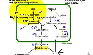
The main steps of the Met _ Hcy _Met cycle are summarized in Fig. (1).

FIGURE 1 NR H2S

Fig. (1). Schematic representation of the methionine cycle and homocysteine degradation pathways.

Compounds: ATP, adenosyltriphosphate; THF, tetrahydrofolate; SAM, S- adenosylmethionine; SAH, adenosylhomocysteine; Cysta, cystathionine; Cys, cysteine;
GSH, glutathione; H2S, hydrogen sulfide; Tau, taurine; SO4-2 , sulfate oxyanions.
Enzymes: (1) Met-adenosyltransferase; (2) SAM-methyltransferases; (3) adenosyl-homocysteinase; (4) methylene-THF reductase; (5) Metsynthase; (6) cystathionine
-b-synthase
, CbS;  (7) cystathionine-b-lyase, CbL; (8) g-glutamyl-synthase; (9) g-glutamyl-transpeptidase; (10)oxidase; (11) reductase; (12) cysteine-dioxygenase, CDO.

Metabolic pathways

Met molecules supplied by dietary proteins are

  • submitted to TM processes
  • releasing Hcy which may in turn either
    • undergo Hcy_Met RM pathways or be
    • irreversibly committed into TS decay.

Impairment of CbS activity in protein malnutrition, entails

  • supranormal accumulation of Hcy in body fluids,
  • stimulation of (5) activity and maintenance of Met homeostasis.

This last beneficial effect is counteracted by

  • decreased concentration of most components generated downstream to CbS,
  • explaining the depressed CbS- and CbL-mediated enzymatic production of *H2S along the TS cascade.

The restricted dietary intake of elemental S is a limiting factor for

  • its non-enzymatic reduction to **H2S which contributes to
  • downsizing a common body pool (dotted circle).(Fig 1)

Combined protein- and S-deficiencies work in concert

  • to deplete Cys, GSH and H2S from their body reserves,
  • impeding these reducing molecules from countering
  • the oxidative stress imposed by hyperhomocysteinemia.

Hyperhomocysteinemia

Hyperhomocysteinemia (HHcy) is an acquired metabolic anomaly first identified by McCully [1]

The current consensus is that dietary deficiency in any of
three water soluble vitamins may operate as causal factor of HHcy.

  • PLP–deficiency may trigger the upstream accumulation of Hcy in biological fluids [2] whereas
  • the shortage of vitamins B9 or B12 is held responsible for its downstream sequestration [3,4].

HHcy is regarded as a major causal determinant of CVD

  1. working as an independent and graded risk factor
  2. unrelated to the classical Framingham criteria such as
  • hypercholesterolemia,
  • dyslipidemia,
  • sedentary lifestyle,
  • diabetes and
  • smoking.

Hcy may invade the intracellular space of many tissues and locally generate [5]

  • endothelial dysfunction working as early harbinger of blood vessel injuries and atherosclerosis.

Most investigators contend

  • that production of harmful reactive oxygen and nitrogen species (ROS, NOS), notably
    • hydrogen peroxide (H2O2), superoxide anion (O2 .-) and peroxinitrite (ONOO.-),
    • constitutes a major culprit in the development of HHcy-induced vascular damages [7-10].

Accumulation of ROS
associated with increased risk for

  • cardiovascular diseases [11]
  • stroke [12],
  • arterial hypertension [6],
  • kidney dysfunction [13],
  • Alzheimer’s disease [14],
  • cognitive deterioration [15],
  • inflammatory bowel disease [16] and
  • bone remodeling [17].

These effects overlook the protective roles played by

  • extra- and intracellular reductants such as cysteine (Cys) and glutathione (GSH)
    • in the sequence of events leading from HHcy to tissue damage.

Hydrogen Sulfide (H2S)

After the discovery of nitric oxide (NO) and carbon oxide (CO), hydrogen sulfide (H2S) is the

  • third gaseous signaling messenger found in mammalian tissues [18].

H2S is a reducing molecule displaying strong scavenging properties

  • as the gasotransmitter significantly attenuates [19, 20] or
  • even abolishes [21,22] the oxidative injury imposed by HHcy burden.

The endogenous production of the naturally occurring H2S reductant depends on

  • Cys bioavailability through
  • the mediation of TS enzymes [23,24].

H2S may also be produced in human tissues starting from elemental sulfur,

  • by a non-enzymatic reaction requiring the presence of Cys, GSH, and glucose [25,26].

It would be worth disentangling the respective roles played by

  1. Cys,
  2. GSH
  3. H2S
  • for the prevention and restoration of HHcy-induced oxidative lesions.
  •  but the plasma concentration of Cys and GSH is severely depressed in
  • subclinically malnourished HHcy patients [27],
    •  impeding appropriate biogenesis of H2S molecules.

The present paper reviews the biological consequences

  • resulting from the complex interplay existing between the 3 reducing molecules,
  • to gain insight into the pathophysiologic mechanisms associated with HHcy states.

CLINICAL BACKGROUND

Numerous surveys have conclusively shown that the water soluble vitamin deficiency concept,

  • provides only partial causal account of the HHcy metabolic anomaly.

The components of body composition, mainly

  • the size of lean body mass (LBM),
  • constitutes a critical determinant of HHcy status [28,29].

Because nitrogen (N) and sulfur (S) concentrations

  • maintain tightly correlated ratios in tissues, we hypothesize 
  • defective N intake and accretion rate would cause concomitant and
  • proportionate depletion of total body N (TBN) and total body S (TBS) stores [30].

Our clinical investigation undertaken in Central Africa in apparently healthy but

  • nevertheless subclinically malnourished vegetarian subjects has
  • documented that reduced size of LBM could lead to HHcy states [27].

The field study conducted in the Republic of Chad, populated by the Sara ethnic group [27], is a  semi-arid region and

  • the staple food consists mainly of cassava, sweet potatoes, beans, millets and groundnuts.

Participants were invited to fill in a detailed dietary questionnaire whose results were compared with values reported in food composition tables [32-34] [27].
The dietary inquiry indicates that participants

  • consumed a significantly lower mean SAA intake (10.4 mg.kg-1.d-1)[27]
  • than the Recommended Dietary Allowances (RDAs) (13 mg.kg-1.d-1)[33,34].

Blood Analytes

The blood lipid profiles of rural subjects were confined within normal ranges

  • ruling out this class of parameters as causal risk factors for CVD disorders.

The normal levels measured for pyridoxine, folates, and cobalamins

  •  precluded these vitamins from playing any significant role in the rise of Hcy

plasma concentrations [27]. Analysis of plasma SAAs revealed

  • unmodified methioninemia, significantly 
  • elevated Hcy values (18.6 umol/L)
  • contrasting with significantly decreased plasma Cys and GSH values [27].

The significant lowering of classical

  • anthropometric parameters
    •  (body weight, BW;
    • body mass index, BMI)
  • together with that of the main plasma and urinary biomarkers of
    • metabolic (visceral) and
    • structural (muscular) compartments point to

an estimated 10 % shrinking of LBM [27].

Transthyretin (TTR)  and Lean Body Mass (LBM)

We have attached peculiar importance to the measurement of plasma transthyretin (TTR)

  1. this indicator integrates the evolutionary trends outlined by body protein reserves [35],
  2. providing from birth until death an overall and balanced estimate of LBM fluctuations [29].
  • In the absence of any superimposed inflammatory condition,
    • LBM and TTR profiles indeed reveal striking similarities [29].

Scientists belonging to the Foundation for Blood Research (Scarborough, Maine, 04074, USA) have recently published a large number of TTR results recorded
in 68,720 healthy US citizens aged 0-100 yr which constitute a comprehensive reference material to follow the shape of LBM fluctuations in relation with sex and age [29].

  • TTR concentrations plotted against Hcy values reveal a strongly negative correlation (r = –0.71)  [29,30], confirming that
      • the depletion of TBN and TBS stores plays a predominant role in the development of HHcy states.

The body of a reference man weighing 70 kg contains 64 M of N (1,800 g) and 4,400 mM of S (140 g) [36]. Our vegetarian subjects consume diets providing
low fat and high fiber content conferring a large spectrum of well described health benefits notably for the prevention of several chronic disorders such as
cancer and diabetes, together with an effective protection against the risk of hypercholesterolemia-induced CVD [37,38].
Plant-based regimens, however, do not supply appropriate amounts of

  • nitrogenous substrates of good biological value which are required to adequately fulfill mammalian tissue needs [30].
  • vegetable items contain suboptimal concentrations of both SAAs [33,34,39] below the customary RDA guidelines.

This dietary handicap may be further deteriorated by

  • unsuitable food processing [40] and by
  • the presence in plant products of naturally occurring anti-nutritional factors
    • such as tannins in cereal grains and
    • anti-trypsin or anti-chymotrypsin inhibitors in soybeans and kidney beans [41].

LBM loss

LBM shrinking may be the result of either

  • dysmaturation of body protein tissues as an effect of protracted dietary SAA deprivation
  • or of cytokine-induced depletion of body stores.

Although causally unrelated and evolving along dissimilar adaptive processes,

  • both physiopathologic entities lead to comparable LBM downsizing best
    • identified by declining plasma TTR ( measured alone or within combined formulas )
    • and subsequently rising Hcy values.

All parameters are downregulated with the sole exception of RM flux rates, indicating that

  • maintenance of Met homeostasis remains a high metabolic priority in protein-depleted states.

Stressful disorders are characterized by

  • overstimulation of all
  1. TM
  2. RM
  3. TS flux rates.

The severity and duration of initial impact determine the magnitude of protein tissue breakdown,

  • rendering an account of N : S urinary losses,
  • fluctuations of albuminuria and of
  • insulin resistance striving to contain LBM integrity.

Both physiopathologic entities are compromized in reducing the oxidative burden imposed by HHcy states owing to

  • defective synthesis and/or
  • enhanced overconsumption of Cys-GSH-H2S reducing molecules,
  • a condition still worsened by its co-existence with elemental S-deficiency.

IMPAIRMENT OF THE TRANSSULFURATION PATHWAY

The hypothesis that subclinical protein malnutrition might be involved in the occurrence of HHcy states via inhibition of cystathionine-b-synthase (CbS) activity
first arose in Senegal in 1986 [42] and was later corroborated in Central Africa [43]. The concept was clearly counterintuitive in that it was unexpected that

  • high Hcy plasma values might result from low intake of its precursor Met molecule.

Despite the low SAA intake of our vegetarian patients [27], plasma Met concentrations disclosed noticeable stability permitting

  • maintenance of the synthesis and functioning of myriads of Met dependent molecular, structural and metabolic compounds

These clinical investigations have received strong support from recent mouse [45] and rat [46] experiments submitted to Met-restricted regimens.
At the end of the Met-deprivation period, both animal species did manifest meaningful HHcy states (p<0.001) contrasting with

  • significantly lower BW (p<0.001) reduced by 33 % [45] and 44 % [46] of control, respectively.
  • the uniqueness of Met behavior stands in accordance with balance studies performed on large mammalian species showing
  • that the complete withdrawal of Met from otherwise normal diets causes the greatest rate of body loss,
    • nearly equal to that generated by protein-free regimens [47,48].

This efficient Met homeostatic mechanism is classically ascribed to a PLP-like inhibition of CbS activity exerted through

  • allosteric binding of S-adenosylmethionine (SAM) to the C-terminal regulatory domain of the enzyme [49,50].

The loss of CbS activity may develop via a (post)translational defect

  • independently from intrahepatic SAM concentrations [45].

We have postulated the existence of an independent sensor mechanism set in motion by TBS pool shrinkage and

  • reduced bioavailability of Met – its main building block – working as an inhibitory feedback loop of CbS activity [30].

Such Met-bodystat, likely to be centrally mediated, is to maintain unaltered Met disposal in conditions of

  • decreased dietary provision implies the fulfillment
  • of high metabolic priorities of survival value [30,44].

Whereas HHcy may be regarded as the dark side of a beneficial adaptive machinery [43],

  • impairment of the TS pathway also depresses the production of compounds situated downstream to the CbS blockade level,
  • notably Cys and GSH, keeping in mind that Cys may undergo reversible GSH conversion (Fig. 1).

The plasma concentration of both Cys and GSH reductants is indeed significantly decreased in our vegetarian subjects

  • by 33 % and 67 % of control, displaying negative correlations (r = –0.67 and –0.37, respectively) with HHcy values [27].

Reduced dietary intake of the preformed Cys molecule [27] and diminished Cys release from protein breakdown in malnourished states [51]

  • may contribute to the lowering effect.

The significantly decreased GSH blood levels may similarly be attributed to dietary composition since the tripeptide is mainly found in meat products

  • but is virtually absent from cereals, roots, milk and dairy items [52] and
  • because regimens lacking SAAs may lessen the production of blood GSH and its intrahepatic sequestration [53].

BIOGENESIS OF HYDROGEN SULFIDE

The TS degradation pathway schematically proceeds along two main PLP-dependent enzymatic reactions working in succession (Fig. 1).

  • The first is catalyzed by CbS (EC 4.2.1.22) governing the replacement of the hydroxyl group of serine with Hcy to generate Cysta plus H2O.
    • Cys may however substitute for serine and the replacement of its sulfhydryl group with Hcy releases Cysta and H2S instead of water [54].
  • The second is regulated by cystathionine-g-lyase (CgL, EC 4.4.1.1.) hydrolyzing Cysta to release Cys and alpha-ketobutyrate plus ammonia as side-products [55].
    •  Cys may also undergo nonoxidative desulfuration pathways leading to H2S or sulfanesulfur production [56] under the control of CbS or CgL enzymes.
    •  Cys may otherwise undergo oxidative conversion regulated by cysteine-dioxygenase (CDO, EC 1.13.11.20) which
      • catalyzes the replacement of the SH- group of Cys by SO3 – to yield cysteine-sulfinate [56].

This last compound may be further decarboxylated to hypotaurine that is finally oxidized to Tau (67 %) and SO4 2- oxyanions (33 %) [56]. CbS and CgL,  both cytosolic enzymes,

  • their relative contribution to the generation of H2S may vary according to
    • animal strains,
    • tissue specificities and
    • nutritional or physiopathological circumstances [23,24].

CbS and CgL are expressed in most organs such as liver, kidneys, brain, heart, large vessels, ileum and pancreas [57,58] potentially

  • subjected to HHcy-induced ROS injury while keeping the capacity to desulfurate Cys and to
  • locally produce H2S as cytoprotectant signaling agent.

CbS is the principal TS enzyme found in

  • cerebral glial cells and astrocytes [59].

CgL predominates in the

  • vascular system [60] whereas
      • both enzymes are present in the renal proximal tubules [61].

H2S is the third gaseous substrate found in the biosphere [18] after NO and CO. All three gases are characterized by

  • severe toxicity when inhaled at high concentrations.

In particular, H2S produced by anaerobic fermentation is

  • capable of causing respiratory death by
  • inhibition of mitochondrial cytochrome C oxidase [62].

NO, CO and H2S are synthesized from arginine, glycine and Cys, respectively, exerting at low concentrations major biological functions in living organisms.
Most of our knowledge on these atypical signal messengers [63] are derived from animal experiments and tissue cultures. These transmitter molecules may

  • share some properties in common such as penetration of cellular membranes independently from specific receptors [64].

They are also manifesting dissimilar activities: whereas NO and CO activate guanylyl cyclase to generate biological responses via cGMP-dependent kinases,

  • H2S induces Ca2+-dependent effects through ATP-sensitive K+ channels [65].

Some of these potentialities may work in concert while others operate antagonistically. For instance,

  • NO and H2S express vasorelaxant tone on endogenous smooth muscle [66]
  • but reveal different effects on large artery vessels [67].

These gaseous substances maintain whole body homeostasis through complex interactions and multifaceted crosstalks between signaling pathways.
Elemental S (32.064 as atomic mass) is a primordial constituent of lava flows in areas of volcanic or sedimental origin usually presenting as crown-shaped
stable octamolecules – hence its S8 symbolic denomination – which may conglomerate to form brimstone rocks. The vegetable kingdom is

  • unable to assimilate S8 and requires as prior step its natural or bacterial oxidation to SO4 2- derivatives before launching
  • the synthesis of SAA molecules along narrowly regulated metabolic pathways [30,44].

Distinct anabolic processes are identified in mammalian tissues which lack the enzymatic equipment required to organize sulfate oxyanions

  • but possess the capacity of direct S8 conversion into H2S.

S8 is poorly soluble in tap waters [68] may be taken up and transported to mammalian tissues loosely fastened to serum albumin (SA) [69].
S may also be covalently bound to intracellular S-atoms taking the form of sulfane-sulfur compounds [70] either

  • firmly attached to cytosolic organelles or in
  • untied form to mitochondria [57,58,71,72] to undergo
  • later release in response to specific endogenous requirements [71].

Sulfane-sulfur compounds are somewhat unstable and may decompose in the presence of reducing agents allowing the restitution of S [70,71].
S may either endorse the role of stimulatory factor of several mammalian apoenzyme activities as shown for

    • succinic dehydrogenase [73] and NADH dehydrogenase [74] or
  • operate as inhibitory agent of other mammalian apoenzymes such as
    • adenylate kinase [75] and liver tyrosine aminotransferase [76].

Elemental S resulting from dietary supply or from sulfane-sulfur decay may be subjected to

non-enzymatic reduction in the presence of Cys and GSH [25,26] and/or reducing equivalents obtained from

  • glucose oxidation [25], hence yielding at physiological pH additional provision of H2S.

The gaseous mediator is a weakly acidic molecule endowed with strong lipophilic affinities. In experimental models, the blockade of the TS cascade

  • at CbS or CgL levels significantly depresses or even
  • abolishes the vitally required production of Cys
  • operating at the crossroad of multiple converting processes (Fig. 1).

Addition of Cys to the incubation milieu

  • resumes the generation of H2S [19] in a Cys concentration-dependent manner [77].

The compounds situated downstream both cystathionases in the context of SAA deprivation

  • keep their functional potentialities
  • but are unable to express their converting Cys – H2S capacities
    • in the absence of precursor substrate.

Summing up

inhibition of CbS activity contributes to

  • promote efficient RM processes and
  • maintenance of Met homeostasis

but entails as side-effects

  1. upstream sequestration of Hcy molecules in biological fluids
  2. while decreasing the bioavailability of Cys and GSH
    • working as limiting factors for H2S production.

These last adverse effects thus constitute the Achilles heel of a remarkable adaptive machinery.

ROLES PLAYED BY HYDROGEN SULFIDE

The first demonstration that human tissues may reduce S to H2S was incidentally provided in 1924 when a man given colloid sulfur

  • for the treatment of polyarthritis did rapidly exhale the typical rotten egg malodor [78].
  • H2S may be produced by the intestinal flora [79] and serves as a metabolic fuel for colonocytes [80].
  • Prevention of endogenous poisoning by excessive enteral production is insured by the detoxifying activities of mucosal cells [81],
    • hindering any systemic effect of the gaseous substrate.

The normal H2S concentration measured in mammalian plasmas usually ranges from 10 to 100 μM with a mean average turning around 40-50 μM [19,21,82,83].
This H2S plasma level, appearing as the net product of organs possessing CbS and CgL enzymes and supplemented by the non-enzymatic conversion of S,

  • flows transiently into the vasculature and freely penetrates into all body cells.
  • Supposing that the gaseous reductant is evenly distributed in total body water (45 L in a 70 kg reference man) allows an estimate of
    • bioavailable H2S pool turning around 2 mM which represents, in terms of S participation, largely less than 1 / 1,000 of TBS.

The peculiar adaptive physiology of vegetarian subjects renders very unlikely that their TBS pool might be solicited to release

  • S-substrates prone to undergo conversion to nascent H2S molecules since
  •  they adapt to declining energy and nutrient intakes
  • by switching overall body economy toward downregulated steady state activities.

The release from TBS of substantial amounts of S-compounds occurs

  • only during the onset of hypercatabolic states as documented in trauma patients [31]
  • and in infectious diseases [84], exacting as preliminary step
  • cytokine-induced breakdown of tissue proteins, a selective hallmark of stressful disorders [85].

H2S in fulfilling ROS Scavenger Tasks

The limited disposal of H2S endogenously produced might be readily exhausted in fulfilling ROS scavenging tasks at the site of oxidative lesions.
All body organs generating H2S from TS enzymes are

  • simultaneously producers and consumers of the gaseous substrate whose actual concentration
  • reflects the balance between synthetic and catabolic rates [86].

Clinical investigations show that H2S concentrations found in cerebral homogenates from Alzheimer’s disease (AD) patients are

  • very much lower than expected from values measured in healthy brains [87], suggesting that
  • the gaseous messenger is locally submitted to enhanced consumption rates reflecting disease severity.

The concept is strongly supported by studies pointing to the

  • negative correlation linking the severity of AD to H2S plasma values [88].
  • in pediatric [89] and elderly [90] hypertensive patients as well
  • more severe HHcy-dependent oxidative burden is
    • associated with more intense H2S uptake rates.
  • These H2S cleansing properties are mainly exerted by mitochondrial organelles
    • known to be centrally involved in oxidative disorders [20,91].

Malnourished subjects deprived of Cys and GSH disposal thus incur the risk of H2S-deficiency

  • rendering them unable to properly overcome HHcy-imposed oxidative lesions.

The rapid exhaustion of H2S stores have detrimental consequences as shown disclosing

  • the beneficial effects of exogenous administration of commonly used sulfide salt donors (Na2S and NaHS)
  • generating H2S gas once in solution.

Such supply significantly augments

  • H2S plasma concentrations allowing to counteract ROS damages. 

H2S was primarily recognized as a physiological substrate working as

  • neuromodulator [92] and soon later as
  • vasorelaxant factor [65].

H2S is now regarded as endowed with a broader spectrum of biological properties [18],

  • operating as a general protective mediator
    • against most degenerative organ injuries,
  • being capable of neutralizing or
  • abolishing most ROS harmful effects.

Table 1 collects findings displaying that H2S may promote the synthesis and activity of several

  • anti-oxidative enzymes (catalases, Cu- and Mn-superoxide dismutases, GSH-peroxidases) and
  • stimulate the production of anti-inflammatory reactants (interleukin-10) or
  • conversely downregulate
    • pro-oxidative enzymes (collagenases, elastases),
    • pro-inflammatory cytokines (interleukine-1b, tumor-necrosis factor a) and
    • immune reactions (hyperleukocytosis, diapedesis, phagocytosis).

It has been calculated that 81.5% of H2S undergoes catabolic disintegration in the form of hydrosulfide anion (HS-) or sulfide anion (S2-) [117].
Since S is the main element in the diprotonated H2S molecule (34.08 as molecular mass), it may be considered that

  • partial or complete repair of HHcy-induced lesions constitutes the therapeutic proof that
  • S-deficiency is causally involved in the development of ROS damages.

The concept is sustained by the observation that all synthetic drugs (diclofenac, indomethacine, sildenafil) utilized as surrogate providers of H2S [64,118] are

  • characterized by a large diversity of molecular conformations but
  • share in common the presence of Satom(s) mimicking, once released,
  • H2S-like pharmacological properties.

It remains to be clarified whether the beneficial effects of S-fortification to S-deficient subjects are mediated, among other possible mechanisms, via

  • stimulation [73,74] of anti-oxidative enzymes or inhibition [75,76] of pro-oxidative enzymes.

It is only very recently that the essentiality of S has been recognized, causing Hcy elevation in deficient individuals [119]. It is worth reminding that the

  • gaseous NO substrate may work in concert or antagonistically [66,83] to fine-tuning the helpful properties exerted by H2S on body tissues.

Preliminary studies suggest for instance that NO operates, in combination with H2S, as a potential modulator of endothelial remodeling since

  •  NO-synthase isoforms contribute to the activation of  metalloproteinases involved in the regulation of the collagen/elastin balance defining vascular elastance [83,120].

SUBCLINICAL MALNUTRITION AS WORLDWIDE  SCOURGE

A growing body of data collected along the last decades indicates that

large proportions of mankind still suffer varying degrees of protein and energy deficiency that is associated with

  • increased morbidity and mortality rates.

The determinants of malnutrition are complex and interrelated, comprising

  • socioeconomic and political conditions,
  • insufficient dietary intakes,
  • inadequate caring practices and
  • superimposed inflammatory burden.

Children living in developing countries are paying a heavy toll to chronic malnutrition [121,122] whereas adult populations are handicapped by

  • feeble physical and working capacities,
  • increased vulnerability to infectious complications and
  • reduced life expectancy [123,124].

Cross-sectional studies collected in the eighties indicate that chronic malnutrition remains a worldwide scourge with

  • top prevalence recorded in Asia, whereas
  • sub-Saharan Africa endures medium nutritional distress and
  • Latin America appears as the least affected [125,126].

Along the last decades, significant progresses have been achieved in some countries such as Vietnam [127] and Bangladesh [128]

  • owing to appropriate education programs and improved economic development.

Inequalities however persist between middle class population groups mainly located in affluent urban areas and

  • underprivileged rural communities remaining stagnant on the sidelines of household income growth.

Representative models of these socio-economic disparities in global nutrition and health are illustrated in the two most populated countries in the world, China and India.
Large surveys undertaken in 105 counties of China and recently published have concluded that the rural communities haven’t yet reached the stage of overall welfare [129].
In India, similar investigations have documented that extreme poverty still prevails in the northern mountainous states of the subcontinent [130]. Taken together, southern
Asian countries fail to overcome malnutrition burden [131]. In some African countries, there exists even upward trends suggesting nutritional

deterioration over the years [132] still aggravated by a severe drought. The assessment of malnutrition in children usually rely on anthropometric criteria such as height-for-age, weight for-height, mid upper arm circumference and skinfold thickness allowing to draw the degree of stunting and wasting from these estimates. In adult subjects, BW and BMI are currently selected parameters to which some biochemical measurements are frequently added, notably SA, classical marker of protein nutritional status, and creatininuria (u-Cr), held as indicator of sarcopenia. The former biometric approaches are very useful in that they correctly provide a static picture of the declared stages of malnutrition but fail to recognize the dynamic mechanisms occurring during the preceding months and the adaptive alterations running behind.

Table 1. Reversal of HHcy-Induced Oxidative Damages by Administration of Exogenous H2S

BRAIN EFFECTS

H2S is overproduced in response to neuronal excitation [93], and

  1. increases the sensitivity of N-methyl-D-aspartate (NMDA) reactions to glutamate in hippocampal neurons [23,94].
  2.  improves long-term potentiation, a synaptic model of memory [92,93]
  3. stimulates the inhibitory effects of catalase and superoxide dismutase (SOD) in oxidative stress of endothelial cells [95].
  4.  regulates Ca 2+ homeostasis in microglial cells [96]and it inhibits TNFa expression in microglial cultures [97].
  5.  protects brain cells from neurotoxicity by preventing the rise of ROS in mitochondria [98].

CARDIOVASCULAR EFFECTS

  1. H2S releases vascular smooth muscle,
  2. inhibits platelet aggregation and
  3. reduces the force output of the left ventricule of the heart [18].
  4. maintains vascular smooth muscle tone [66] and
  5. insures protection against arterial hypertension [99].
  6. modifies leucocyte-vascular epithelium interactions in vivo  by
    1. modulating leucocyte adhesion and
    2. diapedesis at the site of inflammation [100].
  7. attenuates myocardial ischemia-reperfusion injury by
    1. depressing IL-1b and mitochondrial function [20].
  8. upregulates the expression of depressed anti-oxidative enzymes in heart infarction and
    1. inhibits myocardial injury [21].
  9. alleviates smooth muscle pain by
    1. stimulating K+ ATP channels [101].
  10. prevents apoptosis of human neutrophil cells
    1. by inhibiting p38 MAP kinase and caspase 3 [102].
  11. potentiates angiogenesis and wound healing [103].

RENAL EFFECTS

  1. H2S downregulates the increased activity of metalloproteinases 2 and 9 involved in extracellular matrix degradation (elastases, collagenases) [19].
  2. Prevents apoptotic cell death in renal cortical tissues [19].
  3. Improves the expression of desmin (marker of podocyte injury) and
  4. restores the drop of nephrin (component of normal slit diaphragm) in the cortical tissues
    1. resulting in reduced proteinuria [19].
  5. Induces hypometabolism revealing protective effects on renal function and survival [104].
  6. Normalizes GSH status and production of ROS in renal diseases [19].
  7. Controls renal ischemia-reperfusion injury and dysfunction [105].
  8. Depresses the expression of inflammatory molecules involved in glomerulosclerosis [106].
  9. Increases renal blood flow, glomerular filtration and urinary Na+ excretion [77].

OTHER ORGAN EFFECTS
Gastrointestinal

  1. H2S insures protection against ROS stress in gastric mucosal epithelia [22].
  2. Accelerates gastric ulcer healing [107].
  3. Reduces gastric injury caused by nonsteroidal anti-inflammatory drugs [108].
  4. Relaxes ileal smooth muscle tone and increases colonic secretions [79].
  5. Attenuates intestinal ischemia-reperfusion injury by increasing SOD and GSH peroxidase status [109].
  6. Stimulates insulin secretion [110] and controls inflammatory events associated with acute pancreatitis [111].
  7. Alleviates hepatic ischemia-reperfusion injury [112].

Pulmonary

  1. Prevents lung oxidative stress in hypoxic pulmonary hypertension caused by low GSH content [113].
  2. Promotes SOD and catalase activities and reduces the production of malondialdehyde in oxidative lung injury [114].
  3. Reduces lung inflammation and remodeling in asthmatic animals [115] and in pulmonary hypertension [116].  ..(see OCCJ 2011;4:34-44)

Assessing Protein-Depleted States

  1.  SA is an insensitive marker of protein-depleted states compared to TTR [134]
  2. SA is an indicator of population than of individual protein status in subclinical PEM.
  3. u-Cr is likewise a meagerly informative tool as 10 % loss of muscle mass is required before it reaches significantly decreased urinary concentrations [135].

The data imply that the magnitude of subclinical malnutrition is largely

  • underscored when classical biometric and laboratory investigations are performed.

Moreover, ruling out the protein component involved in HHcy epidemiology and confining solely attention to the B-vitamin triad led to unachieved conclusions.

  • surveys undertaken in Taiwan [136] and in India [137] established HHcy variance turning around 30 %, indicating that
  • a sizeable percentage of subjects do not come within the vitamin shortage concept.
  • only one recent review recommending the use of TTR in vegetarian subjects [138].

The main reason for making the choice of TTR is grounded on the striking similar plasma profile disclosed by this marker with both LBM and Hcy [29].
Under healthy conditions, the 3 parameters –

  • TTR,
  • LBM,
  • Hcy –
    • indeed show low  concentrations at birth,
    • linear increase without sexual difference in preadolescent children,
    • gender dimorphism in teenagers with higher values recorded in adolescent male subjects
    • thereafter maintenance of distinct plateau levels during adulthood [29,139,140].

Under morbid circumstances, the plasma concentrations of

  • Hcy manifest gradual elevation
  • negatively correlated with LBM downsizing and
  • TTR decline.

In vegetarian subjects and subclinically malnourished patients,

  • rising Hcy and
  • diminished TTR plasma concentrations look as mirror image of each other,
    • revealing divergent distortion from normal and
    • allowing early detection of preclinical steps
    • at the very same time both SA and u-Cr markers still remain silent.

Any disease process characterized by quantitative or qualitative dietary protein restriction or intestinal malabsorption

  • may cause LBM shrinking,
  • downregulation of TTR concentrations and
  • subsequent HHcy upsurge.

These conditions are documented in frank kwashiorkor [141], subclinical protein restriction [27,43] and anorexia nervosa [142].
In patients submitted to weight-reducing programs,

  • LBM was found the sole independent variable
  • negatively correlated with rising Hcy values [143].

Morbid obesity may be alleviated by medical treatment [143] or surgical gastroplasty [144,145],

  • conditions frequently associated with secondary malabsorptive syndromes and malnutrition [146],

How does this account play out in the typical patient with excessive body fat, lipoprotein disoreder, and perhaps diabetes and disordered sleep – an account of acquired HHcy?
Have the studies been done?  Would you expect to see a clear benefit from reduced HHcy_emia  based on a 30 min daily walk, and

  • eating of well fat trimmed meats, fruits and vegetables, and fish, flax seed, or krill oil?

In westernized countries, subclinical protein-depleted states are illustrated in immigrants originating from

  • developing regions but keeping alive their traditional feeding practices [147] or
  • by communities having adopted, for socio-cultural reasons, strict vegan dietary lifestyles [148].

THE ADDITIONAL BURDEN OF S-DEFICIENCY

After N, K and P, elemental S is recognized as the fourth most important macronutrient required for plant development. The essentiality of S in the vegetable kingdom
arose from observations made many decades ago by pedologists and agronomists [149,150] revealing that the withdrawal of sulfate salts from nutrient sources produces
rapid growth retardation,

  • depressed chlorophyllous synthesis,
  • yellowing of leaves and
  • reduction in fertility and crop yields.

A large number of field studies, mainly initiated for economical reasons, has provided continuing gain in fundamental and applied knowledge and led to the overall consensus

  • that SO4 2- -deficiency is a major wordwide problem [151,152].

Field investigations have shown that the concentration of SO4 2- oxyanions in soils and drinking waters

  • may reveal considerable variations ranging from less than 2 mg/L to more than 1 g/L,
  • meaning a ratio exceeding 1 / 500 under extreme circumstances [30].

The main causal factors responsible for unequal distribution of SO4 2- oxyanions are geographical distance from eruptive sites and

  • intensity of soil weathering in rainy countries.

SO4 2- -dependent nutritional deficiencies entail detrimental effects to most African and Latin American crops [151]

  • reaching nevertheless top incidence in southeastern Asia [151,153].
  • and the Indo-Gangetic plain extending from Pakistan to Bangladesh and covering the North of India and Nepal [154].
  1. Intensive agricultural production,
  2. lack of animal manure and
  3. use of fertilizers providing N, K and P substrates
  4. but devoid of sulfate salts may further aggravate that imbalanced situation.

As global population increases steadily and the production of staple plants predicted to escalate considerably,

  • SO4 2- deficient disorders are expected to become more pregnant along the coming years [155] with significant harmful impact for mankind.

Nevertheless, effective preventive efforts are developed in some countries aiming at fortification of soils mainly

  • by ammonium sulfate or calcium sulfate (gypsum) salts,
  • resulting in meaningful improvements in crop yield,
  • SAAs content and biological value and
  • opening more optimistic perspectives for livestock and human consumption [152,155-158].

Contrasting with the tremendously high amounts of data accumulated over decades by pedologists and agronomists on sulfate requirements and metabolism,
the available knowledge on elemental sulfur in human nutrition looks like a black hole. Despite the fact that S8 follows H, C, O, N, Ca and P as the seven most
abundant element in mammalian tissues, it appears as a forgotten item. Not the slightest attention is dedicated to S8 in the authoritative “Present Knowledge
in Nutrition” series of monographs even though they go over most oligo- and trace-elements in minute detail.

The geographical distribution of S8 throughout the earth’s crust is not well-known

  • as extreme paucity of measurements in soils and tap waters prevents reaching a comprehensive overview.

Nevertheless, and because S8 is the obligatory precursor substrate for the oxidative production of sulfate salts,

a decremental dispersion pattern paralleling those of SO4 2- oxyanions is likely to occur with

  • highest values recorded in the vicinity of volcano sources
  •  and lowest values found in remote and washed-out areas.

Obviously, a great deal of research on elemental S remains to be completed by clinical biochemists before rejoining the status of plant agronomy.
Taken together, these data imply that subclinically malnourished subjects living in areas recognized as

  • SO4 2- -deficient for the vegetable kingdom also
  • incur increased risks to become S8-depleted.

This clinical entity most probably prevails in all regions, notably Northern India, where protein malnutrition [130] and sulfur-deficiency [154] coexist.
Combination of both nutritional deprivations explains why the bulk of local dwellers, including young subjects [159,160], may develop HHcy states and CVD disorders

  • characterized by strong refractoriness to vitamin-B supplementation [160] or
  • high incidence of stroke [161] unrelated to the classical Framingham criteria.

The current consensus is that “the problem of CVD in South Asia is different in etiology and magnitude from other parts of the world” [162]. These disquieting findings are
confirmed in several Asian countries [163] and have prompted local cardiologists to exhort their governments to focus more attention on CVD epidemiology [164].

CONCLUDING REMARKS

  1.  vegetarian subjects are not protected against the risk of CVD and stroke which should no longer be regarded as solely affecting populations living in westernized societies
  • whose morbidity and mortality risks are stratified by classical Framingham criteria.
  • Likewise hypercholesterolemia, hyperhomocysteinemia should be incriminated as
    • emblematic risk factor for a panoply of CVD and related disorders.
  • Whereas the causality of cholesterol and lipid fractions largely prevails in affluent societies consuming high amounts of animal-based items,
    • that of homocysteine predominates in population groups whose dietary lifestyle gives more importance to plant products.

 MAIN PHYSICO-CHEMICAL AND METABOLIC CHARACTERISTICS* OF 3 CARRIER-PROTEINS INVOLVED IN THE STRESS RESPONSE

CBG

TTR

RBP

Molecular mass (Da.)

42,650

54,980

21,200

Conformation

monomeric

tetrameric

monomeric

Amino acid sequence

383

4 x 127

182

Carbohydrate load

18 % glycosylated

unglycosylated

unglycosylated

Hormonal binding sites

one for cortisol

two for TH

one for retinol

Association constant (M-1)

3 x 10⁷

7 x 10⁷ (T4)

1.9 x 10⁷

Normal plasma concentration

30 mg/L.

300 mg/L.

50 mg/L.

Biological half-life

5 days

2 days

14 hrs

Bound ligand  concentration

120 µg/L.

80 µg TT4/L.

500 µg/L.

Free ligand concentration

5 µg/L.

20 ng FT4/L.

1 µg/L.

Ratio free : bound ligands

4 %

0.034 %

0.14 %

Distribution volume of free moieties

18 L.

12 L.

18 L.

STIMULATORY AND INHIBITORY EFFECTS MODULATED

BY GLUCOCORTICOIDS

TARGET SYSTEMS


INDUCED EFFECTS

REF.

Thymidine kinase

_

transcription of induced DNA into RNA

112

Alkaline phosphodiesterase I

_

cleavage of phosphodiester bonds

113

Tyrosine transaminase

_

transfer of tyrosine amino group

114

Tryptophane oxygenase

_

formylkynurenine and Trp catabolites

115

Alkaline phosphatase

_

release of P from phosphoric esters

116

Phosphoenolpyruvate carboxykinase (liver)

_

glycolysis from pyruvate and ATP production

117

Mannolsyltransferases

_

dolichol-linked glycosylation of APRs

118

Haptoglobin

_

APR combining with hemoglobin

119

α1-Anti (chymo) trypsin (α1 AT, α1 ACT)

_

serpin molecules allowing N-sparing effects

120

α1-Acid glycoprotein (AGP)

_

glycosylated APR with antibody-like actions

121

Serum amyloid protein (SAA)

_

defense systems against oxidative burst

122

γ-Fibrinogen

_

clotting processes and tissue repair

123

C-Reactive Protein (CRP)

_

complement processes and opsonization

124

Corticosteroid-binding globulin (CBG)

_

CBG levels, favoring free hypercortisolemia

100

Phosphoenolpyruvate carboxykinase (adipocytes)

_

ATP turnover and glycolysis

113

THE DUAL MORBID ENTITIES CAUSING LBM DOWNSIZING AND SUBSEQUENT Hcy UPSURGE 

Primary causal factor

  1. Reduced dietary intake of methionine (39,151,152)
  2. Cytokine-induced tissue breakdown (164,165)

Main clinical conditions

  1. Protein malnutrition,
  2. veganism,
  3. intestinal malabsorption (139,155,156,158-160,281)
  4. Trauma,
  5. sepsis,
  6. burns,
  7. Inflammatory & neoplastic disorders (163,166,170,176,179,180)

Physiopathologic mechanisms

  1. Unachieved LBM replenishment (30,33)
  2. Excessive LBM losses (33,167,179)

Overall protein metabolic status

  1. Downregulated
  2. Upregulated

Plasma biomarker(s) of protein status

  1. Transthyretin (TTR) (144,145)
  2. TTR coupled with CRP or other inflammatory indices (31,177,178,284,285)

Insulin resistance status

  1. Normal or low (286)
  2. Increased in proportion of tissue breakdown (177,178,181-183)

status of Cys-GSH-H2S reducing molecules

  1. Decreased enzymatic and non-enzymatic production (39,161,162,287)
  2. Increased production cancelled out by tissue overconsumption (78,171)

Urinary SO42- and S-compounds

  1. Decreased kidney output (76,78,79)
  2. Variable depending on exogenous SAA supply and
  • extent of tissue breakdown (78,163,168,173)

Transmethylation pathway

  1. Depressed (48,93)
  2. Overstimulated (169)

Remethylation pathway

  1. Stimulated (76,83,153)
  2. Overstimulated (169)

Transsulfuration pathway

  1. Inhibited (49,76,83)
  2. Overstimulated (170,173)


REFERENCES

[1] McCully, K.S. Vascular pathology of homocysteinemia: implications
for the pathogenesis of arteriosclerosis. Am. J. Pathol., 1969,
56, 111-128.
[2] Ubbink, J.B.; van der Merwe, A.; Delport, R.; Allen, R.H.; Stabler,
S.P.; Riezler, R.; Vermaak, W.J. The effect of subnormal vitamin
B-6 status on homocysteine metabolism. J. Clin. Invest., 1996, 98,
177-184.
[4] Stabler, S.P.; Allen, R.H.; Savage, D.G.; Lindenbaum, J. Clinical
spectrum and diagnosis of cobalamin deficiency. Blood, 1990, 76,
871-881.
[6] Cheng, Z.; Yang, X.; Wang, H. Hyperhomocysteinemia and endothelial
dysfunction. Curr. Hypertens. Rev., 2009, 5,158-165.
[7] Loscalzo, J. The oxidant stress of hyperhomocyst(e)inemia. J. Clin.
Invest., 1996, 98, 5-7.
[8] Jacobsen, D.W. Hyperhomocysteinemia and oxidative stress: Time
for a reality check ? Arterioscler. Thromb. Vasc. Biol., 2000, 20,
1182-1184.
[10] McCully, K.S. Chemical pathology of homocysteine: IV. Excitotoxicity,
oxidative stress, endothelial dysfunction, and inflammation.
Annals Clin. Lab. Sci., 2009, 39, 219-232.
[11] Bautista, L.E.; Arenas, I.A.; Penuela, A.; Martinez, I.X. Total
plasma homocysteine level and risk of cardiovascular disease: a
meta-analysis of prospective cohort studies. J. Clin. Epidemiol.,
2002, 55, 882-887.
[12] Furie, K.L.; Kelly, P. J. Homocyst(e)ine and stroke. Semin. Neurol.,
2006, 26, 24-32.
[13] Van Guldener, C. Homocysteine and the kidney. Curr. Drug Metab.,
2005, 6, 23-26.
[14] McCaddon, A.; Davies, G.; Hudson, P.; Tandy, S.; Cattell, H. Total
serum homocysteine in senile dementia of Alzheimer type. Int. J.
Geriatr. Psychiatry 1998, 13, 235-239.
[15] Troen, A.M.; Rosenberg, I.H. Homocysteine and cognitive function.
Semin. Vasc. Med., 2005, 5, 209-214.
[16] Danese, S.; Sgambato, A.; Papa, A.; Scaldaferri, F.; Pola, R.; Sans,
M.; Lovecchio M. Homocysteine triggers mucosal microvascular
activation in inflammatory bowel disease. Am. J. Gastroenterol.,
2005, 100, 886-895.
[20] Elrod, J.W.; Calvert, J.W.; Morrison, J.; Doeller, J.E.; Kraus, D.W.;
Tao, L.; Jiao, X.; Scalia, R.; Kiss, L.; Szabó, C.; Kimura, H.;
Chow, C.W.; Lefer, D.J. Hydrogen sulfide attenuates myocardial
ischemia-reperfusion injury by preservation of mitochondrial function.
Proc. Natl. Acad. Sci. USA, 2007, 104, 15560-15565.
[21] Chang, L.; Geng, B.; Yu, F.; Zhao, J .; Jiang, H.; Du, J.; Tang C.
Hydrogen sulfide inhibits myocardial injury induced by homocysteine
in rats. Amino Acids, 2008, 34, 573-585.
[22] Yonezawa, D.; Sekiguchi, F.; Miyamoto, M.; Taniguchi, E.; Honjo,
M.; Masuko, T.; Nishikawa, H.; Kawabata, A. A protective role of
hydrogen sulfide against oxidative stress in rat gastric mucosal epithelium.
Toxicology, 2007, 241, 11-18.
[23] Dominy, J.E.; Stipanuk, M.H. New roles for cysteine and transulfuration
enzymes: production of H2S, a neuromodulator and smooth
muscle relaxant. Nutr. Rev. 2004, 62, 348-353.
[27] Ingenbleek, Y.; McCully, K. Vegetarianism produces subclinical
malnutrition, hyperhomocysteinemia and atherogenesis. Nutrition.
Doi:10.1016/j.nut.2011.04.009: on line 27th August 2011.
[28] Battezzatti, A.; Bertoli, S.; San Romerio, A.; Testolin, G. Body
composition: an important determinant of homocysteine and methionine
concentrations in healthy individuals. Nutr. Metab. Cardiovasc.
Dis., 2007, 17, 525-534.
[29] Ingenbleek, Y. Plasma Transthyretin Reflects the Fluctuations of
Lean Body Mass in Health and Disease. In: Recent Advances in
Transthyretin Evolution, Structure and Biological Functions;
Richardson, S.J. and Cody, V., Eds.; Springer Verlag : Berlin,
2009, pp. 329-357.
[30] Ingenbleek, Y. The nutritional relationship linking sulfur to nitrogen
in living organisms. J. Nutr., 2006, 136, S1641-S1651.
[31] Cuthbertson, D.P. The distribution of nitrogen and sulphur in the
urine during conditions of increased catabolism. Biochem. J., 1931,
25, 236-244.
[34] Souci, S.W.; Fachman, W.; Kraut, H. Food Composition and Nutrition
Tables. 5th ed., CRC Press: Boca Raton, 1994.
[35] Ingenbleek, Y.; Young, V.R. Significance of transthyretin in protein
metabolism. Clin. Chem. Lab. Med., 2002, 40, 1281-1291.
[36] Forbes, G.B. Body Composition. In: Present Knowledge in Nutrition;
7th ed.; Ziegler, E.E. and Filer L.J, Eds.; ILSI Press: Washington
D.C. 1996; pp. 7-12.
42 The Open Clinical Chemistry Journal, 2011, Volume 4 Yves Ingenbleek
[37] Walter, P. Effects of the vegetarian diets on aging and longevity.
Nutr. Rev., 1997, 55, S61-S65.
[38] Key, T.J.; Appleby, P.N.; Rosell, M.S. Health effects of vegetarian
and vegan diets. Proc. Nutr. Soc., 2006, 65, 35-41.
[39] Young, V.R.; Pellet, P.L. Plant proteins in relation to human protein
and amino acid nutrition. Am. J. Clin. Nutr., 1994, 59, S1203-
S1212.
[42] Ingenbleek, Y.; Barclay, D.; Dirren, H. Nutritional significance of
alterations in serum amino acid patterns in goitrous patients. Am. J.
Clin. Nutr., 1986, 43, 310-319.
[43] Ingenbleek, Y.; Hardillier, E.; Jung, L. Subclinical protein malnutrition
is a determinant of hyperhomocysteinemia. Nutrition, 2002,
18, 40-46.
[44] Ingenbleek, Y.; Young, V.R. The essentiality of sulfur is closely
related to nitrogen metabolism: a clue to hyperhomocysteinemia.
Nutr. Res. Rev., 2004, 17, 135-153.
[45] Tang, B.; Mustafa, A.; Gupta, S.; Melnyk, S.; James S.J.; Kruger,
W.D. Methionine-deficient diet induces post-transcriptional downregulation
of cystathionine-g-synthase. Nutrition, 2010, 26, 1170-
1175.
[46] Elshorbagy, A.K.; Valdivia-Garcia, M.; Refsum, H.; Smith, A.D.;
Mattocks, D.A.; Perrone, C.E. Sulfur amino acids in methionine restricted
rats: Hyperhomocysteinemia. Nutrition, 2010, 26, 1201-
1204.
[47] Owens, F.N.; Bergen, W.G. Nitrogen metabolism in ruminant
animals: historical perspective, current understanding and future
implications. J. Anim. Sci., 1983, 57 (Suppl. 2), 498-518.

[55] Stipanuk, M.H. Sulfur amino acid metabolism : pathways for production
and removal of homocysteine and cysteine. Annu. Rev.
Nutr., 2004, 24, 539-577.
[56] Stipanuk, M.H.; Ueki, I. Dealing with methionine/homocysteine
sulfur : cysteine metabolism to taurine and inorganic sulfur. J. Inherit.
Metab. Dis., 2011, 34, 17-32.
[57] Kamoun, P. Endogenous production of hydrogen sulfide in mammals.
Amino Acids, 2004, 26, 243-254.
[58] Kimura, H. Hydrogen sulfide : its production, release and functions.
Amino Acids, 2011, 41, 113-121.
[59] Enokido, Y.; Suzuki, E.; Iwasawa, K.; Namekata, K.; Okazawa, H.;
Kimura, H. Cystathionine 􀀁-synthase, a key enzyme for homocysteine
metabolism, is preferentially expressed in the radial
glia/astrocyte lineage of developing mouse CNS. FASEB J., 2005,
19, 1854-1856.
[60] Zhao, W.; Ndisang, J.F.; Wang, R. Modulation of endogenous
production of H2S in rat tissues. Can. J. Physiol. Pharmacol., 2003,
81, 848-853.
[61] House, J.D.; Brosnan, M.E.; Brosnan, J.T. Characterization of
homocysteine metabolism in the rat kidney. Biochem. J., 1997,
328, 287-292.
[62] Dorman, D.C.; Moulin, F.J.; McManus, B.E.; Mahle, K.C.; James,
R.A.; Struve, M.F. Cytochrome oxidase inhibition induced by acute
hydrogen sulfide inhalation : correlation with tissue sulfide concentrations
in the rat brain, liver, lung, and nasal epithelium. Toxicol.
Sci., 2002, 65, 18-25.
[64] Mancardi, D.; Penna, C.; Merlino, A.; Del Soldato, P.; Wink, D.A.;
Pagliaro, P. Physiological and pharmacological features of the
novel gasotransmitter : Hydrogen sulfide. Biochim. Biophys. Acta,
2009, 1787, 864-872.
[65] Zhao, W.; Zhang, J.; Lu, Y.; Wang, R. The vasorelaxant effect of
H(2)S as a novel endogenous gaseous K(ATP) channel opener.
EMBO J., 2001, 20, 6008-6016.
[66] Hosoki, R.; Matsuki, N.; Kimura, H. The possible role of hydrogen
sulfide as an endogenous smooth muscle relaxant in synergy with
nitric oxide. Biochem. Biophys. Res. Commun., 1997, 237, 527-
531.
[75] Russell, P.J.; Conner, J.; Sisson, S. Sulfur specifically inhibits
adenylate kinase in assays for creatine kinase. Clin. Chem., 1984,
30, 1555-1557.
[76] Hargrove, J.L.; Wichman, R.D. A cystine-dependent inactivator of
tyrosine aminotransferase co-purifies with 􀀂-cystathionase (cysteine
desulfurase). J. Biol. Chem., 1987, 262, 7351-7357.
[77] Xia, M.; Chen, L.; Muh, R.W.; Li, P.L.; Li, N. Production and
actions of hydrogen sulfide, a novel gaseous bioactive substance, in
the kidneys. J. Pharmacol. Exp. Ther., 2009, 329, 1056-1062.
[78] Monaghan, W.J.; Garai, F. Treatment of acute and chronic polyarthritis,
arthritis deformans, and septicemias, with activated colloid
sulphur. Med. J. Record, 1924, 120, 24-26.
[79] Fiorucci, S.; Distrutti, E.; Cirino, G.; Wallace, J.L. The emerging
role of hydrogen sulfide in the gastrointestinal tract and liver. Gastroenterology,
2006, 131, 259-271.
[80] Goubern, M.; Andriamihaja, M.; Nubel, T.; Blachier, F.; Bouillaud,
F. Sulfide, the first inorganic substrate for human cells. FASEB J.,
2007, 21, 1699-1706.
[81] Furne, J.; Springfield, J.; Koenig, T.; DeMaster, E.; Levitt, MD.
Oxidation of hydrogen sulfide and methanethiol to thiosulfate by
rat tissues: a specialized function of the colonic mucosa. Biochem.
Pharmacol., 2001, 62, 255-259.
[82] Chen, YH.; Yao, W.Z.; Geng, B.; Ding, Y.L.; Lu, M.; Zhao, M.W.;
Tang, C.S. Endogenous hydrogen sulfide in patients with COPD.
Chest, 2005, 128, 3205-3211.
[83] Whiteman, M.; Moore, P.K. Hydrogen sulfide and the vasculature:
a novel vasculoprotective entity and regulator of nitric oxide
bioavailability? J. Cell. Mol. Med., 2009, 13, 488-507.
[85] Ingenbleek, Y.; Bernstein, L. The stressful condition as a nutritionally
dependent adaptive dichotomy. Nutrition, 1999, 15, 305-320.
Malnutrition, Sulfur-Deficiency and Hyperhomocysteinemia The Open Clinical Chemistry Journal, 2011, Volume 4 43
[89] Chen, L.; Ingrid, S.; Ding, Y.G.; Liu, Y.; Qi, J.G.; Tang, J.B. Imbalance
of endogenous homocysteine and hydrogen sulfide metabolic
pathway in essential hypertensive children. Chin. Med. J.,
2007, 120, 389-393.
[90] Sun, N.L.; Xi, Y.; Yang, S.N.; Ma, Z.; Tang, C.S. Plasma hydrogen
sulfide and homocysteine levels in hypertensive patients with different
blood pressure levels and complications. Zonghua Xin Xue
Guan Bing Za Zhi, 2007, 35, 1145-1148.
[91] Lin, M.T.; Beal, M.F. Mitochondrial dysfunction and oxidative
stress in neurodegenerative diseases. Nature, 2006, 443, 787-795.
[97] Hu, L.F.; Wong, P.T.; Moore, P.K.; Bian, J.S. Hydrogen sulfide
attenuates lipopolysaccharide-induced inflammation by inhibition
of p38 mitogen-activated protein kinase in microglia. J.
Neurochem., 2007, 100, 1121-1128.
[98] Tang, X.Q.; Shen, X.T.; Huang, Y.E.; Ren, Y.K.; Chen, R.Q.; Hu,
B.; He, J.Q.; Yin, W.L.; Xu, J.H.; Jiang, Z.S. Hydrogen sulfide antagonizes
homocysteine-induced neurotoxicity in PC12 cells. Neurosci.
Res., 2010, 68, 241-249.
[99] Yang, G.; Wu, L.; Jiang, B.; Yang, W.; Qi, J.; Cao, K.; Meng, Q.;
Mustafa, A.K.; Mu, W.; Zhang, S. H2S as a physiologic vasorelaxant
: hypertension in mice with deletion of cystathionine gammalyase.
Science, 2008, 322, 587-590.
[100] P.; Vitale, M. Hydrogen sulfide prevents apoptosis of human PMN
via inhibition of p38 and caspase 3. Lab. Invest., 2006, 86, 391-
397.
[105] Tripatara, P.; Patel, N.S.; Collino, M.; Gallichio, M.; Kieswich, J.;
Castiglia, S.; Benetti, E.; Stewart, K.N.; Brown, P.A.; Yaqoob,
M.M.; Fantozzi, R.; Thiemermann, C. Generation of endogenous
hydrogen sulfide by cystathionine gamma-lyase limits renal ischemia/
reperfusion injury and dysfunction. Lab. Invest., 2008, 88,
1038-1048.
[106] Sen, U.; Munjal, C.; Qipshidze, N.; Abe, O.; Gargoum, R.; Tyagi,
S.C. Hydrogen sulfide regulates homocysteine-mediated glomerulosclerosis.
Am. J. Nephrol., 2010, 31, 442-455.
[114] Fu, Z.; Liu, X.; Geng, B.; Fang, L.; Tang, C. Hydrogen sulfide
protects rat lung from ischemia-reperfusion injury. Life Sci., 2008,
82, 1196-1202.
[115] Chen, Y.H.; Wu, R.; Geng, B.; Qi, Y.F.; Wang, P.P.; Yao, W.Z.;
Tang, C.S. Endogenous hydrogen sulfide reduces airway inflammation
and remodeling in a rat model of asthma. Cytokine 2009, 45,
117-123.
[116] Li, X.; Jin, H.; Bin, G.; Wang, L.; Tang, C.; Du, J. Endogenous
hydrogen sulfide regulates pulmonary artery collagen remodeling
in rats with high pulmonary blood flow. Exp. Biol. Med.
(Maywood), 2009, 234, 504-512.
[117] Dombkowski, R.A.; Russell, M.J.; Olson, K.R. Hydrogen sulfide as
an endogenous regulator of vascular smooth muscle tone in trout.
Am. J. Physiol. Regul. Integr. Comp. Physiol., 2004, 286, R678-
R685.
[118] Wallace, J.L. Hydrogen sulfide-releasing anti-inflammatory drugs.
Trends Pharmacol. Sci., 2007, 28, 501-505.
[119] Ingenbleek, Y. Hyperhomocysteinemia is a biomarker of sulfurdeficiency
in human morbidities. Open Clin. Chem. J., 2009, 2, 49-
60.
[120] Steed, M.M.; Tyagi, S.C. Mechanisms of cardiovascular remodeling
in hyperhomocysteinemia. Antioxid. Redox. Signal., 2011, 15,
1927-1943.
[121] Black, R.E.; Morris, S.S.; Bryce, J. Where and why are 10 millions
children dying every year? Lancet, 2003, 361, 2226-2234.
[122] El-Ghannam, A.R. The global problems of child malnutrition and
mortality in different world regions. J. Health Soc. Policy, 2003,
16, 1-26.
[128] Faruque, A.S.; Shamsir Ahmed, A.M.; Tahmeed Ahmed.; Munirul
M.; Iqbal Hossain, M.; Roy, S.K.; Nurul Alam; Iqbal Kabir; Sack,
D.A. Nutrition : Basis for healthy children and mothers in Bangladesh.
J. Health Popul. Nutr., 2008, 26, 325-339.
[129] Wang, X.; Wang, Y.; Kang, C. Feeding practices in 105 countries
of rural China. Child Care Health Dev., 2005, 31, 417-423.
44 The Open Clinical Chemistry Journal, 2011, Volume 4 Yves Ingenbleek
[130] Antony, G.M.; Laxmaiah, A. Human development, poverty, health
& nutrition situation in India. Indian J. Med. Res., 2008, 128, 198-
205.
[134] Ingenbleek, Y.; De Visscher, M ; De Nayer, P. Measurement of
prealbumin as index of protein-calorie malnutrition. Lancet, 1972,
ii, 106-109.
[135] Shenkin, A.; Cederblad, G.; Elia, M.; Isaksson, B. International
Federation of Clinical Chemistry. Laboratory assessment of protein-
energy status. Clin. Chim. Acta, 1996, 253, S5-S59.
[136] Hung, C.J.; Huang, P.C.; Lu, S.C.; Li, Y.H.; Huang, H.B.; Lin,
B.F.; Chang, S.J.; Chou, H.F. Plasma homocysteine levels in Taiwanese
vegetarians are higher than those of omnivores. J. Nutr.,
2002, 132, 152-158.
[138] Benzie, I.F.; Wachtel-Galor, S. Biomarkers in long-term vegetarian
diets. Adv. Clin. Chem., 2009, 47, 171-222.
[139] Must, A.; Jacques, P.F.; Rogers, G.; Rosenberg, I.H.; Selhub, J.
Serum total homocysteine concentrations in children and adolescents
: results from the third National Health and Nutrition Examination
Survey (NHANES III). J. Nutr., 2003, 133, 2643-2649.
[141] Abdel, G.A.; Abdullah, S.H.; Kordofani, A.Y. Plasma homocysteine
levels in cardiovascular disease, malaria and protein-energy
malnutrition in Sudan. East Mediterr. Health J., 2009, 15, 1432-
1439.
[142] Moyano, D.; Vilaseca, M.A.; Artuch, R.; Valls, C.; Lambruschini,
N. Plasma total-homocysteinemia in anorexia nervosa. Eur. J. Clin.
Nutr., 1998, 52, 172-175.
[143] Gallistl, S.; Sudi, K.M.; Erwa, W.; Aigner, R.; Borkenstein, M.
Determinants of homocysteine during weight reduction in obese
children and adolescents. Metabolism, 2001, 50, 1220-1223.
[144] Borzon-Chazot, F.; Harthe, C.; Teboul, F.; Labrousse, F.; Gaume,
C.; Guadagnino, L.; Claustrat, B.; Berthezène, F.; Moulin, P. Occurrence
of hyperhomocysteinemia 1 year after gastroplasty for severe
obesity. J. Clin. Endocrinol. Metab., 1999, 84, 541-545.
[145] Sheu, W.H.; Wu, H.S.; Wang, C.W.; Wan, C.J.; Lee, W.J. Elevated
plasma homocysteine concentrations after gastroplasty in morbidly
obese subjects. Intern. Med., 2001, 40, 584-588.
[147] Chandalia, M.; Abate, N.; Cabo-Chan, A.V. Jr.; Devaraj, S.; Jialal,
I.; Grundy, S.M. Hyperhomocysteinemia in Asian Indians living in
the United States. J. Clin. Endocrinol. Metab., 2003, 88, 1089-
1095.
[148] Koebnick, C.; Garcia, A.L.; Dagnelie, P.C.; Strassner, C.; Lindemans,
J.; Katz, N.; Leitzmann, C.; Hoffmann, I. Long-term consumption
of a raw food diet is associated with favorable serum
LDL cholesterol and triglycerides but also with elevated plasma
homocysteine and low HDL cholesterol in humans. J. Nutr., 2005,
135, 2372-2375.
[149] Coleman, R. The importance of sulfur as a plant nutrient in world
crop production. Soil Sci., 1966, 101, 230-239.
[150] Dijkshoorn, W.; Van Wijk, A.L. The sulphur requirements of
plants as evidenced by the sulphur-nitrogen ratio in the organic
matter: a review of published data. Plant Soil, 1967, 26, 129-157.
[152] Jez, J.M. Sulfur: A Missing Link between Soils, Crops, and Nutrition;
American Society of Agronomy, Crop Science Society of
America, Soil Science Society of America: Madison, 2008.
[154] Khurana, M.P.S.; Sadana, U.S.; Bijay-Singh. Sulfur-Nutrition of
Crops in the Indo-Gangetic Plains of South Asia. In: Sulfur: A
Missing Link between Soils, Crops, and Nutrition; Jez, J.M., Ed.;
American Society of Agronomy, Crop Science Society of America,
Soil Science Society of America: Madison, 2008; Vol. 50, pp. 11-
24.
[157] Galili, G.; Amir, R.; Hoefgen, R.; Hesse, H. Improving the levels
of essential amino acids and sulfur metabolites in plants. Biol.
Chem., 2005, 386, 817-831.
[159] Kumar, Y.; Das, R.; Garewal, G.; Bali, H.K. High prevalence of
hyperhomocysteinemia in young population of North India – a potential
risk factor for coronary artery disease? Thromb. Res., 2009,
123, 800-802.
[160] Ghosh, K.; Khare, A.; Shetty, S. Fasting plasma homocysteine
levels are increased in young patients with acute myocardial infarction
in Western India. Indian Heart J., 2007, 59, 242-245.
[161] Christopher, R.; Nagaraja, D.; Shankar, S.K. Homocysteine and
cerebral stroke in developing countries. Curr. Med. Chem., 2007,
14, 2393-2401.
Received: September 30, 2011 Revised: October 12, 2011 Accepted: October 12, 2011
© Yves Ingenbleek; Licensee Bentham Open.
This is an open access article licensed under the terms of the Creative Commons Attribution Non-Commercial License (http://creativecommons.org/licenses/
by-nc/3.0/) which permits unrestricted, non-commercial use, distribution and reproduction in any medium, provided the work is properly cited.

Related articles

English: Biosynthesis of cysteine from homocys...

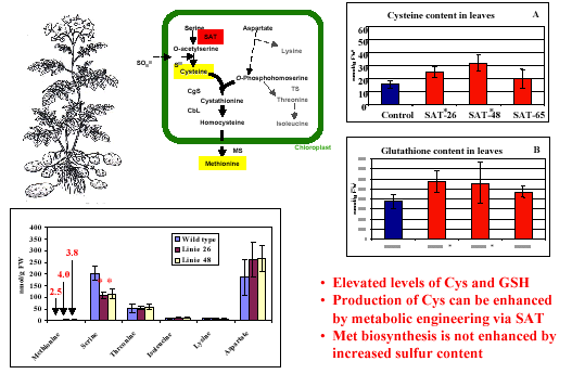
English: Biosynthesis of cysteine from homocysteine and serine via cystathione intermediate (Photo credit: Wikipedia)

Read Full Post »

A Second Look at the Transthyretin Nutrition Inflammatory Conundrum

Subtitle: Transthyretin and the Systemic Inflammatory Response


Author and Curator: Larry H. Bernstein, MD, FACP, Clinical Pathologist, Biochemist, and Transfusion Physician


Brief introduction

Transthyretin  (also known as prealbumin) has been widely used as a biomarker for identifying protein-energy malnutrition (PEM) and for monitoring the improvement of nutritional status after implementing a nutritional intervention by enteral feeding or by parenteral infusion. This has occurred because transthyretin (TTR) has a rapid removal from the circulation in 48 hours and it is readily measured by immunometric assay. Nevertheless, concerns have been raised about the use of TTR in the ICU setting, which prompted a review of the  benefit of using this test in acute and chronic care. TTR is easily followed in the underweight and the high risk populations in an ambulatory setting, which has a significant background risk of chronic diseases. It is sensitive to the systemic inflammatory response syndrome (SIRS), and needs to be understood in the context of acute illness to be used effectively. There are a number of physiologic changes associated with SIRS and the injury/repair process that affect TTR. The most important point is that in the context of an ICU setting, the contribution of TTR is significant in a complex milieu.  A much better understanding of the significance of this program has emerged from studies of nitrogen and sulfur in health and disease.

Transthyretin protein structure

Transthyretin protein structure (Photo credit: Wikipedia)

Age-standardised disability-adjusted life year...

Age-standardised disability-adjusted life year (DALY) rates from Protein-energy malnutrition by country (per 100,000 inhabitants). (Photo credit: Wikipedia)

_________________________________________________________________________________________________________

The systemic inflammatory response syndrome C-reactive protein and transthyretin conundrum.
Larry H Bernstein
Clin Chem Lab Med 2007; 45(11):0
ICID: 939932
Article type: Editorial

The Transthyretin Inflammatory State Conundrum
Larry H. Bernstein
Current Nutrition & Food Science, 2012, 8, 00-00

Keywords: Tranthyretin (TTR), systemic inflammatory response syndrome (SIRS), protein-energy malnutrition (PEM), C- reactive protein, cytokines, hypermetabolism, catabolism, repair.

Transthyretin has been widely used as a biomarker for identifying protein-energy malnutrition (PEM) and for monitoring the improvement of nutritional status after implementing a nutritional intervention by enteral feeding or by parenteral infusion. This has occurred because transthyretin (TTR) has a rapid removal from the circulation in 48 hours and it is readily measured by immunometric assay. Nevertheless, concerns have been raised about the use of TTR in the ICU setting, which prompts a review of the actual benefit of using this test in a number of settings. TTR is easily followed in the underweight and the high risk populations in an ambulatory setting, which has a significant background risk of chronic diseases. It is sensitive to the systemic inflammatory response syndrome (SIRS), and needs to be understood in the context of acute illness to be used effectively.

There are a number of physiologic changes associated with SIRS and the injury/repair process that affect TTR and  in the context of an ICU setting, the contribution of TTR is essential.  The only consideration is the timing of initiation since the metabolic burden is sufficiently high that a substantial elevation is expected in the first 3 days post admission, although the level of this biomarker is related to the severity of injury. Despite the complexity of the situation, TTR is not to be considered a test “for all seasons”. In the context of age, prolonged poor meal intake, chronic or acute illness, TTR needs to be viewed in a multivariable lens, along with estimated lean body mass, C-reactive protein, the absolute lymphocyte count, presence of neutrophilia, and perhaps procalcitonin if there is remaining uncertainty. Furthermore, the reduction of risk of associated complication requires a systematized approach to timely identification, communication, and implementation of a suitable treatment plan.

The most important point is that in the context of an ICU setting, the contribution of TTR is significant in a complex milieu.

_________________________________________________________________________________________________________

Title: The Automated Malnutrition Assessment
Accepted 29 April 2012. http://www.nutritionjrnl.com. Nutrition (2012), doi:10.1016/j.nut.2012.04.017.
Authors: Gil David, PhD; Larry Howard Bernstein, MD; Ronald R Coifman, PhD
Article Type: Original Article

Keywords: Network Algorithm; unsupervised classification; malnutrition screening; protein energy malnutrition (PEM); malnutrition risk; characteristic metric; characteristic profile; data characterization; non-linear differential diagnosis

We have proposed an automated nutritional assessment (ANA) algorithm that provides a method for malnutrition risk prediction with high accuracy and reliability.  The problem of rapidly identifying risk and severity of malnutrition is crucial for minimizing medical and surgical complications. These are not easily performed or adequately expedited. We characterized for each patient a unique profile and mapped similar patients into a classification. We also found that the laboratory parameters were sufficient for the automated risk prediction.

_________________________________________________________________________________________________________

Title: The Increasing Role for the Laboratory in Nutritional Assessment
Article Type: Editorial
Section/Category: Clinical Investigation
Accepted 22 May 2012. http://www.elsevier.com/locate/clinbiochem.
Clin Biochem (2012), doi:10.1016/j.clinbiochem.2012.05.024
Keywords: Protein Energy Malnutrition; Nutritional Screening; Laboratory Testing
Author: Dr. Larry Howard Bernstein, MD

The laboratory role in nutritional management of the patient has seen remarkable growth while there have been dramatic changes in technology over the last 25 years, and it is bound to be transformative in the near term. This editorial is an overview of the importance of the laboratory as an active participant in nutritional care.

The discipline emerged divergently along separate paths with unrelated knowledge domains in physiological chemistry, pathology, microbiology, immunology and blood cell recognition, and then cross-linked emerging into clinical biochemistry, hematology-oncology, infectious diseases, toxicology and therapeutics, genetics, pharmacogenomics, translational genomics and clinical diagnostics.

In reality, the more we learn about nutrition, the more we uncover of metabolic diversity of individuals, the family, and societies in adapting and living in many unique environments and the basic reactions, controls, and responses to illness. This course links metabolism to genomics and individual diversity through metabolomics, which will be enlightened by chemical and bioenergetic insights into biology and translated into laboratory profiling.

Vitamin deficiencies were discovered as clinical entities with observed features as a result of industrialization (rickets and vitamin D deficiency) and mercantile trade (scurvy and vitamin C)[2].  Advances in chemistry led to the isolation of each deficient “substance”.  In some cases, a deficiency of a vitamin and what is later known as an “endocrine hormone” later have confusing distinctions (vitamin D, and islet cell insulin).

The accurate measurement and roles of trace elements, enzymes, and pharmacologic agents was to follow within the next two decades with introduction of atomic absorption, kinetic spectrophotometers, column chromatography and gel electrophoresis.  We had fully automated laboratories by the late 1960s, and over the next ten years basic organ panels became routine.   This was a game changer.

Today child malnutrition prevalence is 7 percent of children under the age of 5 in China, 28 percent in sub-Saharan African, and 43 percent in India, while under-nutrition is found mostly in rural areas with 10 percent of villages and districts accounting for 27-28 percent of all Indian underweight children. This may not be surprising, but it is associated with stunting and wasting, and it has not receded with India’s economic growth. It might go unnoticed viewed alongside a growing concurrent problem of worldwide obesity.

The post WWII images of holocaust survivors awakened sensitivity to nutritional deprivation.

In the medical literature, Studley [HO Studley.  Percentage of weight loss. Basic Indicator of surgical risk in patients with chronic peptic ulcer.  JAMA 1936; 106(6):458-460.  doi:10.1001/jama.1936.02770060032009] reported the association between weight loss and poor surgical outcomes in 1936.  Ingenbleek et al [Y Ingenbleek, M De Vissher, PH De Nayer. Measurement of prealbumin as index of protein-calorie malnutrition. Lancet 1972; 300[7768]: 106-109] first reported that prealbumin (transthyretin, TTR) is a biomarker for malnutrition after finding very low TTR levels in African children with Kwashiorkor in 1972, which went unnoticed for years.  This coincided with the demonstration by Stanley Dudrick  [JA Sanchez, JM Daly. Stanley Dudrick, MD. A Paradigm ShiftArch Surg. 2010; 145(6):512-514] that beagle puppies fed totally through a catheter inserted into the superior vena cava grew, which method was then extended to feeding children with short gut.  Soon after Bistrian and Blackburn [BR Bistrian, GL Blackburn, E Hallowell, et al. Protein status of general surgical patients. JAMA 1974; 230:858; BR Bistrian, GL Blackburn, J Vitale, et al. Prevalence of malnutrition in general medicine patients, JAMA, 1976, 235:1567] showed that malnourished hospitalized medical and surgical patients have increased length of stay, increased morbidity, such as wound dehiscence and wound infection, and increased postoperative mortality, later supported by many studies.

Michael Meguid,MD, PhD, founding editor of Nutrition [Elsevier] held a nutrition conference “Skeleton in the Closet – 20 years later” in Los Angeles in 1995, at which a Beckman Prealbumin Roundtable was held, with Thomas Baumgartner and Michael M Meguid as key participants.  A key finding was that to realize the expected benefits of a nutritional screening and monitoring program requires laboratory support. A Ross Roundtable, chaired by Dr. Lawrence Kaplan, resulted in the first Standard of Laboratory Practice Document of the National Academy of Clinical Biochemists on the use of the clinical laboratory in nutritional support and monitoring. Mears then showed a real benefit to a laboratory interactive program in nutrition screening based on TTR [E Mears. Outcomes of continuous process improvement of a nutritional care program incorporating serum prealbumin measurements. Nutrition 1996; 12 (7/8): 479-484].

A later Ross Roundtable on Quality in Nutritional Care included a study of nutrition screening and time to dietitian intervention organized by Brugler and Di Prinzio that showed a decreased length of hospital stay with $1 million savings in the first year (which repeated), which included reduced cost for dietitian evaluations and lower complication rates.

Presentations were made at the 1st International Transthyretin Congress in Strasbourg, France by Mears [E Mears.  The role of visceral protein markers in protein calorie malnutrition. Clin Chem Lab Med 2002; 40:1360-1369] on the impact of TTR in screening for PEM in a public hospital in Louisiana, and by Potter [MA Potter, G Luxton. Prealbumin measurement as a screening tool for patients with protein calorie malnutrition in emergency hospital admissions: a pilot study.  Clin Invest Med. 1999; 22(2):44-52] that indicated a 17% in-hospital mortality rate in a Canadian hospital for patients with PCM compared with 4% without PCM (p < 0.02), while only 42% of patients with PCM received nutritional supplementation. Cost analysis of screening with prealbumin level projected a saving of $414 per patient screened.  Ingenbleek and Young [Y Ingenbleek, VR Young.  Significance of transthyretin in protein metabolism.  Clin Chem Lab Med. 2002; 40(12):1281–1291.  ISSN (Print) 1434-6621, DOI: 10.1515/ CCLM.2002.222, December 2002. published online: 01/06/2005] tied the TTR to basic effects reflected in protein metabolism.

_______________________________________________________________________________________________

Transthyretin as a marker to predict outcome in critically ill patients.
Arun Devakonda, Liziamma George, Suhail Raoof, Adebayo Esan, Anthony Saleh, Larry H Bernstein
Clin Biochem 2008; 41(14-15):1126-1130
ICID: 939927
Article type: Original article

TTR levels correlate with patient outcomes and are an accurate predictor of patient recovery in non-critically ill patients, but it is uncertain whether or not TTR level correlates with level of nutrition support and outcome in critically ill patients. This issue has been addressed only in critically ill patients on total parenteral nutrition and there was no association reported with standard outcome measures. We revisit this in all patients admitted to a medical intensive care unit.

Serum TTR was measured on the day of admission, day 3 and day 7 of their ICU stay. APACHE II and SOFA score was assessed on the day of admission. A registered dietician for their entire ICU stay assessed the nutritional status and nutritional requirement. Patients were divided into three groups based on initial TTR level and the outcome analysis was performed for APACHE II score, SOFA score, ICU length of stay, hospital length of stay, and mortality.

TTR showed excellent concordance with the univariate or multivariate classification of patients with PEM or at high malnutrition risk, and followed for seven days in the ICU, it is a measure of the metabolic burden.  TTR levels decline from day 1 to day 7 in spite of providing nutritional support. Twenty-five patients had an initial TTR serum concentration more than 17 mg/dL (group 1), forty-eight patients had mild malnutrition with a concentration between 10 and 17 mg/dL (group 2), Forty-five patients had severe malnutrition with a concentration less than 10 mg/dL (group 3).  Initial TTR level had inverse correlation with ICU length of stay, hospital length of stay, and APACHE II score, SOFA score; and predicted mortality, especially in group 3.

___________________________________________________________________________________________________________

A simplified nutrition screen for hospitalized patients using readily available laboratory and patient
information.
Linda Brugler, Ana K Stankovic, Madeleine Schlefer, Larry Bernstein
Nutrition 2005; 21(6):650-658
ICID: 825623
Article type: Review article
The role of visceral protein markers in protein calorie malnutrition.
Linda Brugler, Ana Stankovic, Larry Bernstein, Frederick Scott, Julie O’Sullivan-Maillet
Clin Chem Lab Med 2002; 40(12):1360-1369
ICID: 636207
Article type: Original article

The Automated Nutrition Score is a data-driven extension of continuous quality improvement.

Larry H Bernstein
Nutrition 2009; 25(3):316-317
ICID: 939934

______________________________________________________________________________________________________
Transthyretin: its response to malnutrition and stress injury. clinical usefulness and economic implications.
LH Bernstein, Y Ingenbleek
Clin Chem Lab Med 2002; 40(12):1344-1348
ICID: 636205
Article type: Original article

_______________________________________________________________________________________________________

THE NUTRITIONALLY-DEPENDENT ADAPTIVE DICHOTOMY (NDAD) AND STRESS HYPERMETABOLISM
Yves Ingenbleek  MD  PhD  and  Larry Bernstein MD
J CLIN LIGAND ASSAY  (out of print)

The acute reaction to stress is characterized by major metabolic, endocrine and immune alterations. According to classical descriptions, these changes clinically present as a succession of 3 adaptive steps – ebb phase, catabolic flow phase, and anabolic flow phase. The ebb phase, shock and resuscitation, is immediate, lasts several hours, and is characterized by hypokinesis, hypothermia, hemodynamic instability and reduced basal metabolic rate. The catabolic flow phase, beginning within 24 hours and lasting several days, is characterized by catabolism with the flow of gluconeogenic substrates and ketone bodies in response to the acute injury. The magnitude of the response depends on the acuity and the severity of the stress. The last, a reparative anabolic flow phase, lasts weeks and is characterized by the accretion of amino acids (AAs) to rebuilding lean body mass.

The current opinion is that the body economy is reset during the course of stress at novel thresholds of metabolic priorities. This is exemplified mainly by proteolysis of muscle, by an effect on proliferating gut mucosa and lymphoid tissue as substrates are channeled to support wound healing, by altered syntheses of liver proteins with preferential production of acute phase proteins (APPs) and local repair in inflamed tissues (3). The first two stages demonstrate body protein breakdown exceeding the rate of protein synthesis, resulting in a negative nitrogen (N) balance, muscle wasting and weight loss. In contrast, the last stage displays reversed patterns, implying progressive recovery of endogenous N pools and body weight.

These adaptive alterations undergo continuing elucidation. The identification of cytokines, secreted by activated macrophages/monocytes or other reacting cells, has provided further insights into the molecular mechanisms controlling energy expenditure, redistribution of protein pools, reprioritization of syntheses and secretory processes.

The free fraction of hormones bound to specific binding-protein(s) [BP(s)] manifests biological activities, and any change in the BP blood level modifies the effect of the hormone on the end target organ.  The efficacy of these adaptive responses may be severely impaired in protein-energy malnourished (PEM) patients. This is especially critical with respect to changes of the circulating levels of transthyretin (TTR), retinol-binding protein (RBP) and corticosteroid-binding globulin (CBG) conveying thyroid hormones (TH), retinol and cortisol, respectively.  This reaction is characterized by cytokine mediated autocrine, paracrine and endocrine changes. Among the many inducing molecules identified, interleukins 1 and 6 (Il-1, Il-6) and tumor necrosis factor a (TNF) are associated with enhanced production of 3 counterregulatory hormonal families (cortisol, catecholamines and glucagon). Growth hormone (GH) and TH also have roles in these metabolic adjustments.

There is overproduction of cortisol mediated by several cytokines acting on both the adrenal cortex (10) and on the pituitary through hypothalamic CRH with loss of feedback regulation of ACTH production (11). Hypercortisolemia is a major finding observed after surgery (12), sepsis (13), and medical insults, usually correlated with severity of insult and of complications. Rising cortisol values parallel hyperglycemic trends, as an effect of both gluconeogenesis and insulin resistance. Working in concert with TNF, glucocorticoids govern the breakdown of muscle mass, which is regarded as the main factor responsible for the negative N balance.

Under normal conditions, GH exerts both lipolytic and anabolic influences in the whole body economy under the dual control of the hypothalamic hormones somatocrinin (GHRH) and somatostatin (SRIH). GH secretion is usually depressed by rising blood concentrations of glucose and free fatty acids (FFAs) but is paradoxicaly elevated despite hyperglycemia in stressed patients.

The oversecretion of counterregulatory hormones working in concert generates subtle equilibria between glycogenolytic/glycolytic/gluconeogenic adaptive processes. The net result is the neutralization of the main hypoglycemic and anabolic activities of insulin and the development of a persisting and controlled hyperglycemic tone in the stressed body. The molecular mechanisms whereby insulin resistance occurs in the course of stress refer to
cytokine-  and  hormone-induced  phosphorylation abnormalities affecting receptor signaling. The insulin-like anabolic processes of GH are mediated by IGF1 working as relay agent. The expected high IGF1 surge associated with GH oversecretion is not observed in severe stress as plasma values are usually found at the lower limit of normal or even in the subnormal range.  The end result of this dissociation between high GH and low IGF1 levels is to favor the proteolysis of muscle mass to release AAs for gluconeogenesis and the breakdown of adipose tissue to provide ketogenic substrates.

The acute stage of stress is associated with the onset of a low T3 syndrome typically delineated by the drop of both total (TT3) and free (FT3) triiodothyronine plasma levels in the subnormal range. In contrast, both total (TT4) and free (FT4) thyroxine values usually remain within normal ranges with declining trends observed for TT4 and rising tendencies for FT4 (44). This last free compound is regarded as the sensor reflecting the actual thyroid status and governing the release of TSH whereas FT3 works as the active hormonal mediator at nuclear receptor level. The maintenance of an euthyroid sick syndrome is compatible with the down-regulation of most metabolic and energetic processes in healthy tissues. These inhibitory effects , negatively affecting all functional steps of the hypothalamo-pituitary-thyroid axis concern TSH production, iodide uptake, transport and organification into iodotyrosyl residues, peroxidase coupling activity as well as thyroglobulin synthesis and TH leakage. Taken together, the above-mentioned data indicate that the development of hyperglycemia and of insulin-resistance in healthy tissues – mainly in the muscle mass – are hallmarks resulting from the coordinated activities of the counterregulatory hormones.

A growing body of recent data suggest that the stressed territory, whatever the causal agent – bacterial or viral sepsis, auto-immune disorder, traumatic or toxic shock, burns, cancer – manifest differentiated metabolic and immune reactions. The amplitude, duration and efficacy of these responses are reportedly impaired along several ways in PEM patients. These last detrimental effects are accompanied by a number of medical, social and economical consequences, such as extended length of hospital stay and increased complication / mortality rates. It is therefore mandatory to correctly identify and follow up the nutritional status of hospitalized patients. Such approaches are prerequisite to timely and scientifically grounded nutritional and pharmacological mediated interventions.

Contrary to the rest of the body, energy requirements of the inflamed territory are primarily fulfilled by anaerobic glycolysis, an effect triggered by the inhibition of key-enzymes of carbohydrate metabolism, notably pyruvate-dehydrogenase. This non-oxidative combustion of glucose reveals low conversion efficiency but offers the major advantage to maintain, in the context of hyperglycemia, fuel provision to poorly irrigated and/or edematous tissues. The depression of the 5’-monodeiodinating activity (5’-DA) plays a pivotal role in these adaptive changes, yielding inactive reverse T3 (rT3) as index of impaired T4 to T3 conversion rates, but at the same time there is an augmented supply of bioactive T3 molecules and local overstimulation of thyro-dependent processes characterized by thyroid down-regulation.  The same differentiated evolutionary pattern applies to IGF1. In spite of lowered plasma total concentrations, the proportion of IGF1 released in free form may be substantially increased owing to the proteolytic degradation of IGFBP-3 in the intravascular compartment. The digestion of  BP-3 results from the surge of several proteases occurring the course of stress, yielding biologically active IGF1 molecules available for the repair of damaged tissues. In contrast, healthy receptors oppose a strong resistance to IGF1 ligands freed in the general circulation, likely induced by an acquired phosphorylation defect very similar in nature to that for the insulin transduction pathway.

PEM is the generic denomination of a broad spectrum of nutritional disorders, commonly found in hospital settings, and whose extreme poles are identified as marasmus and kwashiorkor. The former condition is usually regarded as the result of long-lasting starvation leading to the loss of lean body mass and fat reserves but relatively well-preserved liver function and immune capacities. The latter condition is typically the consequence of (sub)acute deprivation predominantly affecting the protein content of staplefood, an imbalance causing hepatic steatosis, fall of visceral proteins, edema and increased vulnerability to most stressful factors. PEM may be hypometabolic or hypermetabolic, usually coexists with other diseased states and is frequently associated with complications. Identification of PEM calls upon a large set of clinical and analytical disciplines comprising anthropometry, immunology, hematology and biochemistry.

CBG, TTR and RBP share in common the transport of specific ligands exerting their metabolic effects at nuclear receptor level. Released from their specific BPs in free form, cortisol, FT4 and retinol immediately participe to the strenghtening of the positive and negative responses to stressful stimuli. CBG is a relatively weak responder to short-term nutritional influences (73)  although long-lasting PEM is reportedly capable of causing its significant diminution (74). The dramatic drop of CBG in the course of stress appears as the combined effect of Il-6-induced posttranscriptional blockade of its liver synthesis (75) and peripheral overconsumption by activated neutrophils (61). The divergent alterations outlined by CBG and total cortisolemia result in an increased disposal of free ligand reaching proportions considerably higher than the 4 % recorded under physiological conditions.

The appellation of negative APPs that was once given to the visceral group of carrier-proteins. The NDAD concept takes the opposite view, defending the opinion that their suppressed synthesis releases free ligands which positively contribute to strengthen all aspects of the stress reaction, justifying the ABR denomination. This implies that the role played by ABRs should no longer be interpreted in terms of concentrations but in terms of functionality.

++++++++++++++++++++++++++++++++++++++++++++++++++++++++++++++++++++++++++++++++++++++++++++

THE OXIDATIVE STRESS OF HYPERHOMOCYSTEINEMIA RESULTS FROM REDUCED BIOAVAILABILITY OF SULFUR-CONTAINING REDUCTANTS.
Yves Ingenbleek. The Open Clinical Chemistry Journal, 2011, 4, 34-44.

Vegetarian subjects consuming subnormal amounts of methionine (Met) are characterized by subclinical protein malnutrition causing reduction in size of their lean body mass (LBM) best identified by the serial measurement of plasma transthyretin (TTR). As a result, the transsulfuration pathway is depressed at cystathionine-β-synthase (CβS) level triggering the upstream sequestration of homocysteine (Hcy) in biological fluids and promoting its conversion to Met. Maintenance of beneficial Met homeostasis is counterpoised by the drop of cysteine (Cys) and glutathione (GSH) values downstream to CβS causing in turn declining generation of hydrogen sulfide (H2S) from enzymatic sources. The biogenesis of H2S via non-enzymatic reduction is further inhibited in areas where earth’s crust is depleted in elemental sulfur (S8) and sulfate oxyanions. Combination of subclinical malnutrition and S8-deficiency thus maximizes the defective production of Cys, GSH and H2S reductants, explaining persistence of unabated oxidative burden. The clinical entity increases the risk of developing cardiovascular diseases (CVD) and stroke in underprivileged plant-eating populations regardless of Framingham criteria and vitamin-B status. Although unrecognized up to now, the nutritional disorder is one of the commonest worldwide, reaching top prevalence in populated regions of Southeastern Asia. Increased risk of hyperhomocysteinemia and oxidative stress may also affect individuals suffering from intestinal malabsorption or westernized communities having adopted vegan dietary lifestyles.

Metabolic pathways: Met molecules supplied by dietary proteins are submitted to TM processes allowing to release Hcy which may in turn either undergo Hcy – Met RM pathways or be irreversibly committed into TS decay. Impairment of CbS activity, as described in protein malnutrition, entails supranormal accumulation of Hcy in body fluids, stimulation of activity and maintenance of Met homeostasis. This last beneficial effect is counteracted by decreased concentration of most components generated downstream to CbS, explaining the depressed CbS- and CbL-mediated enzymatic production of H2S along the TS cascade. The restricted dietary intake of elemental S further operates as a limiting factor for its non-enzymatic reduction to H2S which contributes to downsizing a common body pool. Combined protein- and S-deficiencies work in concert to deplete Cys, GSH and H2S from their body reserves, hence impeding these reducing molecules to properly face the oxidative stress imposed by hyperhomocysteinemia.

see also …

McCully, K.S. Vascular pathology of homocysteinemia: implications for the pathogenesis of arteriosclerosis. Am. J. Pathol., 1996, 56, 111-128.

Cheng, Z.; Yang, X.; Wang, H. Hyperhomocysteinemia and endothelial dysfunction. Curr. Hypertens. Rev., 2009, 5,158-165.

Loscalzo, J. The oxidant stress of hyperhomocyst(e)inemia. J. Clin.Invest., 1996, 98, 5-7.

Ingenbleek, Y.; Hardillier, E.; Jung, L. Subclinical protein malnutrition is a determinant of hyperhomocysteinemia. Nutrition, 2002, 18, 40-46.

Ingenbleek, Y.; Young, V.R. The essentiality of sulfur is closely related to nitrogen metabolism: a clue to hyperhomocysteinemia. Nutr. Res. Rev., 2004, 17, 135-153.

Hosoki, R.; Matsuki, N.; Kimura, H. The possible role of hydrogen sulfide as an endogenous smooth muscle relaxant in synergy with nitric oxide. Biochem. Biophys. Res. Commun., 1997, 237, 527-531.

Tang, B.; Mustafa, A.; Gupta, S.; Melnyk, S.; James S.J.; Kruger, W.D. Methionine-deficient diet induces post-transcriptional downregulation of cystathionine-􀀁-synthase. Nutrition, 2010, 26, 1170-1175.

Elshorbagy, A.K.; Valdivia-Garcia, M.; Refsum, H.; Smith, A.D.; Mattocks, D.A.; Perrone, C.E. Sulfur amino acids in methioninerestricted rats: Hyperhomocysteinemia. Nutrition, 2010, 26, 1201- 1204.

_______________________________________________________________________________________________________

Yves Ingenbleek. Plasma Transthyretin Reflects the Fluctuations of Lean Body Mass in Health and Disease. Chapter 20. In S.J. Richardson and V. Cody (eds.), Recent Advances in Transthyretin Evolution, Structure and Biological Functions, DOI: 10.1007/978‐3‐642‐00646‐3_20, # Springer‐Verlag Berlin Heidelberg 2009.

Transthyretin (TTR) is a 55-kDa protein secreted mainly by the choroid plexus and the liver. Whereas its intracerebral production appears as a stable secretory process allowing even distribution of intrathecal thyroid hormones, its hepatic synthesis is influenced by nutritional and inflammatory circumstances working concomitantly. Both morbid conditions are governed by distinct pathogenic mechanisms leading to the reduction in size of lean body mass (LBM). The liver production of TTR integrates the dietary and stressful components of any disease spectrum, explaining why it is the sole plasma protein whose evolutionary patterns closely follow the shape outlined by LBM fluctuations. Serial measurement of TTR therefore provides unequalled information on the alterations affecting overall protein nutritional status. Recent advances in TTR physiopathology emphasize the detecting power and preventive role played by the protein in hyperhomocysteinemic states, acquired metabolic disorders currently ascribed to dietary restriction in water-soluble vitamins. Sulfur (S)-deficiency is proposed as an additional causal factor in the sizeable proportion of hyperhomocysteinemic patients characterized by adequate vitamin intake but experiencing varying degrees of nitrogen (N)-depletion. Owing to the fact that N and S coexist in plant and animal tissues within tightly related concentrations, decreasing LBM as an effect of dietary shortage and/or excessive hypercatabolic losses induces proportionate S-losses. Regardless of water-soluble vitamin status, elevation of homocysteine plasma levels is negatively correlated with LBM reduction and declining TTR plasma levels. These findings occur as the result of impaired cystathionine-b-synthase activity, an enzyme initiating the transsulfuration pathway and whose suppression promotes the upstream accumulation and remethylation of homocysteine molecules. Under conditions of N- and S-deficiencies, the maintenance of methionine homeostasis indicates high metabolic priority.

Schematically, the human body may be divided into two major compartments, namely fat mass (FM) and FFM that is obtained by substracting
FM from body weight (BW). The fat cell mass sequesters about 80% of the total body lipids, is poorly hydrated and contains only small quantities of lean tissues and nonfat constituents. FFM comprises the sizeable part of lean tissues and minor mineral compounds among which are Ca, P, Na, and Cl pools totaling about 1.7 kg or 2.5% of BW in a healthy man weighing 70 kg. Subtraction of mineral mass from FFM provides LBM, a composite aggregation of organs and tissues with specific functional properties. LBM is thus nearly but not strictly equivalent to FFM. With extracellular mineral content subtracted, LBM accounts for most of total body proteins (TBP) and of TBN assuming a mean 6.25 ratio between protein and N content.

SM accounts for 45% of TBN whereas the remaining 55% is in nonmuscle lean tissues. The LBM of the reference man contains 98% of total
body potassium (TBK) and the bulk of total body sulfur (TBS). TBK and TBS reach equal intracellular amounts (140 g each) and share distribution patterns (half in SM and half in the rest of cell mass).  The body content of K and S largely exceeds that of magnesium (19 g), iron (4.2 g) and zinc (2.3 g). The average hydration level of LBM in healthy subjects of all age is 73% with the proportion of the intracellular/extracellular fluid spaces being 4:3. SM is of particular relevance in nutritional studies due to its capacity to serve as a major reservoir of amino acids (AAs) and as a dispenser of gluconeogenic substrates. An indirect estimate of SM size consists in the measurement of urinary creatinine, end-product of the nonenzymatic hydrolysis of phosphocreatine which is limited to muscle cells.

During ageing, all the protein components of the human body decrease regularly. This shrinking tendency is especially well documented for SM  whose absolute amount is preserved until the end of the fifth decade, consistent with studies showing unmodified muscle structure, intracellular K content and working capacit. TBN and TBK are highly correlated in healthy subjects and both parameters manifest an age-dependent curvilinear decline
with an accelerated decrease after 65 years.  The trend toward sarcopenia is more marked and rapid in elderly men than in elderly women decreasing strength and functional capacity. The downward SM slope may be somewhat prevented by physical training or accelerated by supranormal cytokine status as reported in apparently healthy aged persons suffering low-grade inflammation. 2002) or in critically ill patients whose muscle mass undergoes proteolysis and contractile dysfunction.

The serial measurement of plasma TTR in healthy children shows that BP values are low in the neonatal period and rise linearly with superimposable concentrations in both sexes during infant growth consistent with superimposable N accretion and protein synthesis rates. Starting from the sixties, TTR values progressively decline showing steeper slopes in elderly males. The lowering trend seems to be initiated by the attenuation of androgen influences and trophic stimuli with increasing age. The normal human TTR trajectory from birth to death has been well documented by scientists belonging to the Foundation for Blood Research. TTR is the first plasma protein to decline in response to marginal protein restricion, thus working as an early signal warning that adaptive mechanisms maintaining homeostasis are undergoing decompensation.

TTR was proposed as a marker of protein nutritional status following a clinical investigation undertaken in 1972 on protein-energy malnourished (PEM) Senegalese children (Ingenbleek et al. 1972). By comparison with ALB and transferrin (TF) plasma values, TTR revealed a much higher degree of sensitivity to changes in protein status that has been attributed to its shorter biological half-life (2 days) and to its unusual Trp richness (Ingenbleek et al. 1972, 1975a). Transcription of the TTR gene in the liver is directed by CCAAT/enhancer binding protein (C/EBP) bound to hepatocyte nuclear factor 1 (HNF1) under the control of several other HNFs. The mechanism responsible for the suppressed TTR synthesis in PEM-states is a restricted AA and energy supply working as limiting factors (Ingenbleek and Young 2002). The rapidly turning over TTR protein is highly responsive to any change in protein flux and energy supply, being clearly situated on the cutting edge of the equipoise.

LBM shrinking may be the consequence of either dietary restriction reducing protein syntheses to levels compatible with survival or that of cytokine-induced tissue proteolysis exceeding protein synthesis and resulting in a net body negative N balance. The size of LBM in turn determines plasma TTR concentrations whose liver production similarly depends on both dietary provision and inflammatory conditions. In animal cancer models, reduced TBN pools were correlated with decreasing plasma TTR values and provided the same predictive ability. In kidney patients, LBM is proposed as an excellent predictor of outcome working in the same direction as TTR plasma levels.  High N intake, supposed to preserve LBM reserves, reduces significantly the mortality rate of kidney patients and is positively correlated with the alterations of TTR plasma concentrations appearing as the sole predictor of final outcome. It is noteworthy that most SELDI or MALDI workers interested in defining protein nutritional status have chosen TTR as a biomarker, showing that there exists a large consensus considering the BP as the most reliable indicator of protein depletion in most morbid circumstances.

Total homocysteine (tHcy) is a S-containing AA not found in customary diets but endogenously produced in the body of mammals by the enzymatic transmethylation of methionine (Met), one of the eight IAAs supplied by staplefoods. tHcy may either serve as precursor substrate for the synthesis of new Met molecules along the remethylation (RM) pathway or undergo irreversible kidney leakage through a cascade of derivatives defining the transsulfuration (TS) pathway. Hcy is thus situated at the crossroad of RM and TS pathways that are regulated by three water-soluble vitamins (pyridoxine, B6; folates, B9; cobalamins, B12).

Significant positive correlations are found between tHcy and plasma urea and plasma creatinine, indicating that both visceral and muscular tissues undergo proteolytic degradation throughout the course of rampant inflammatory burden. In healthy individuals, tHcy plasma concentrations maintain positive correlations with LBM and TTR from birth until the end of adulthood. Starting from the onset of normal old age, tHcy values become disconnected from LBM control and reveal diverging trends with TTR values. Of utmost importance is the finding that, contrary to all protein
components which are downregulated in protein-depleted states, tHcy values are upregulated.  Hyperhomocysteinemia is an acquired clinical entity characterized by mild or moderate elevation in tHcy blood values found in apparently healthy individuals (McCully 1969). This distinct morbid condition appears as a public health problem of increasing importance in the general population, being regarded as an independent and graded risk factor for vascular pathogenesis unrelated to hypercholesterolemia, arterial hypertension, diabetes and smoking.

Studies grounded on stepwise multiple regression analysis have concluded that the two main watersoluble vitamins account for only 28% of tHcy variance whereas vitamins B6, B9, and B12, taken together, did not account for more than 30–40% of variance. Moreover, a number of hyperhomocysteinemic conditions are not responsive to folate and pyridoxine supplementation. This situation prompted us to search for other causal factors which might fill the gap between the public health data and the vitamin triad deficiencies currently incriminated. We suggest that S – the forgotten element – plays central roles in nutritional epidemiology (Ingenbleek and Young 2004).

Aminoacidemia studies performed in PEM children, adult patients and elderly subjects have reported that the concentrations of plasma IAAs invariably display lowering trends as the morbid condition worsens. The depressed tendency is especially pronounced in the case of tryptophan and for the so-called branched-chain AAs (BCAAs, isoleucine, leucine, valine) the decreases in which are regarded as a salient PEM feature following the direction outlined by TTR (Ingenbleek et al. 1986). Met constitutes a notable exception to the above described evolutionary profiles, showing unusual stability in chronically protein depleted states.

Maintenance of normal methioninemia is associated with supranormal tHcy blood values in PEMadults (Ingenbleek et al. 1986) and increased tHcy leakage in the urinary output of PEM children. In contrast, most plasma and urinary S-containing compounds produced along the TS pathway downstream to CbSconverting step (Fig. 20.1) display significantly diminished values. This is notably the case for cystathionine (Ingenbleek et al. 1986), glutathione, taurine, and sulfaturia. Such distorted patterns are reminiscent of abnormalities defining homocystinuria, an inborn disease of Met metabolism characterized by CbS refractoriness to pyridoxine stimuli, thereby promoting the upstream retention of tHcy in biological fluids. It
was hypothesized more than 20 years ago (Ingenbleek et al. 1986) that PEM is apparently able to similarly depress CbS activity, suggesting that the enzyme is a N-status sensitive step working as a bidirectional lockgate, overstimulated by high Met intake (Finkelstein and Martin 1986) and downregulated under N-deprivation conditions (Ingenbleek et al. 2002). Confirmation that N dietary deprivation may inhibit CbS activity has recently provided. The tHcy precursor pool is enlarged in biological fluids, boosting Met remethylation processes along the RM pathway, consistent with studies showing overstimulation of Met-synthase activity in conditions of protein restriction. In other words, high tHcy plasma concentrations observed in PEM states are the dark side of adaptive mechanisms for maintaining Met homeostasis. This is consistent with the unique role played by Met in the preservation of N body stores.

The classical interpretation that strict vegans, who consume plenty of folates in their diet and manifest nevertheless higher tHcy plasma concentrations than omnivorous counterparts, needs to be revisited. On the basis of hematological and biochemical criteria, cobalamin deficiency is one of the most prevalent vitamin-deficiencies wordwide, being often incriminated as deficient in vegan subjects. It seems, however, likely that its true causal impact on rising tHcy values is substantially overestimated in most studies owing to the modest contribution played by cobalamins on tHcy
variance analyses. In contrast, there exists a growing body of converging data indicating that the role played by the protein component is largely underscored in vegan studies. It is worth recalling that S is the main intracellular anion coexisting with N within a constant mean S:N ratio (1:14.5) in animal tissues and dietary products of animal origin (Ingenbleek 2006). The mean S:N ratio found in plant items ranges from 1:20 to 1:35, a proportion that does not optimally meet human tissue requirements (Ingenbleek 2006), paving the way for borderline S and N deficiencies.

A recent Taiwanese investigation on hyperhomocysteinemic nuns consuming traditional vegetarian regimens consisting of mainly rice, soy products,
vegetables and fruits with few or no dairy items illustrates such clinical misinterpretation (Hung et al. 2002). The authors reported that folates and cobalamins, taken together, accounted for only 28.6% of tHcy variance in the vegetarian cohort whereas pyridoxine was inoperative (Hung et al. 2002). The daily vegetable N and Met intakes were situated highly significantly (p < 0.001) below the recommended allowances for humans (FAO/WHO/United Nations University 1985), causing a stage of unrecognized PEM documented by significantly depressed BCAA plasma
concentrations. Met levels escaped the overall decline in IAAs levels, emphasizing that efficient homeostatic mechanisms operate at the expense of an acquired hyperhomocysteinemic state. The diagnosis of subclinical PEM was missed because the authors ignored the exquisitely sensitive TTR detecting power. A proper PEM identification would have allowed the authors to confirm the previously described TTR–tHcy relationship that was established in Western Africa from comparable field studies involving country dwellers living on plant products.

The concept that acute or chronic stressful conditions may exert similar inhibitory effects on CbS activity and thereby promote hyperhomocysteinemic states is founded on previous studies showing that hypercatabolic states are characterized by increased urinary N and S losses maintaining tightly correlated depletion rates (Cuthbertson 1931; Ingenbleek and Young 2004; Sherman and Hawk 1900) which reflect the S:N ratio found in tissues undergoing cytokine induced proteolysis. This has been documented in coronary infarction and in acute pancreatitis where tHcy elevation evolves too rapidly to allow for a nutritional vitamin B-deficit explanation.  tHcy is considered stable in plasma and the two investigations report unaltered folate and cobalamin plasma concentrations.

The clinical usefulness of TTR as a nutritional biomarker, described in the early seventies (Ingenbleek et al. 1972) has been substantially disregarded by the scientific community for nearly four decades. This long-lasting reluctance expressed by many investigators is largely due to the fact that protein malnutrition and stressful disorders of various causes have combined inhibitory effects on hepatic TTR synthesis. Declining TTR plasma concentrations may result from either dietary protein and energy restrictions or from cytokine-induced transcriptional blockade (Murakami et al. 1988) of its hepatic synthesis. The proposed marker was therefore seen as having high sensitivity but poor specificity. Recent advances in protein metabolism settle the controversy by throwing further light on the relationships between TTR and the N-components of body composition.

The developmental patterns of LBM and TTR exhibit striking similarities. Both parameters rise from birth to puberty, manifest gender dimorphism during full sexual maturity then decrease during ageing. Uncomplicated PEM primarily affects both visceral and structural pools of LBM with distinct kinetics, reducing protein synthesis to levels compatible with prolonged survival. In acute or chronic stressful disorders, LBM undergoes muscle proteolysis exceeding the upregulation of protein syntheses in liver and injured areas, yielding a net body negative N balance. These adaptive responses are well identified by the measurement of TTR plasma concentrations which therefore appear as a plasma marker for LBM fluctuations.
Attenuation of stress and/or introduction of nutritional rehabilitation restores both LBM and TTR to normal values following parallel slopes. TTR fulfills, therefore, a unique position in assessing actual protein nutritional status, monitoring the efficacy of dietetic support and predicting the patient’s outcome (Bernstein and Pleban 1996).

see also…

Acosta PB, Yannicelli S, Ryan AS, Arnold G, Marriage BJ, Plewinska M, Bernstein L, Fox J, Lewis V, Miller M, Velazquez A (2005) Nutritional therapy improves growth and protein status of children with a urea cycle enzyme defect. Mol Genet Metab 86:448–455.

Arroyave G, Wilson D, Be´har M, Scrimshaw NS (1961) Serum and urinary creatinine in children with severe protein malnutrition. Am J Clin Nutr 9:176–179.

Bates CJ, Mansoor MA, van der Pols J, Prentice A, Cole TJ, Finch S (1997) Plasma total homocysteine in a representative sample of 972 British men and women aged 65 and over. Eur J Clin Nutr 51:691–697.

Battezzatti A, Bertoli S, San Romerio A, Testolin G (2007) Body composition: An important determinant of homocysteine and methionine concentrations in healthy individuals. Nutr Metab Cardiovasc Dis 17:525–534.

Bernstein LH, Bachman TE, Meguid M, Ament M, Baumgartner T, Kinosian B, Martindale R, Spiekerman M (1995) Prealbumin in nutritional care Consensus Group. Measurement of visceral protein status in assessing protein and energy malnutrition: Standard of care. Nutrition 11:169–171

Bernstein LH, Ingenbleek Y (2002) Transthyretin: Its response to malnutrition and stress injury. Clinical usefulness and economical implications. Clin Chem Lab Med 40:1344–1348.

Boorsook H, Dubnoff JW (1947) The hydrolysis of phosphocreatine and the origin of creatinine. J Biol Chem 168:493–510.

Briend A, Garenne M, Maire B, Fontaine O, Dieng F (1989) Nutritional status, age and survival: The muscle mass hypothesis. Eur J Clin Nutr 43:715–726

Brouillette J, Quirion R (2007) Transthyretin: A key gene involved in the maintenance of memory capacities during aging. Neurobiol Aging 29:1721–1732

Chertow GM, Goldstein-Fuchs DJ, Lazarus JM, Kaysen GA (2005) Prealbumin, mortality, and cause-specific hospitalization in hemodialysis patients. Kidney Int 68:2794–2800

Cohn SH, Gartenhaus W, Sawitsky A, Rai K, Zanzi I, Vaswani A, Ellis KJ, Yasumura S, Cortes E, Vartsky D (1981) Compartmental body composition of cancer patients by measurement of total body nitrogen, potassium and water. Metabolism 30:222–229

Cuthbertson DP (1931) The distribution of nitrogen and sulphur in the urine during conditions of increased catabolism. Biochem J 25:236–244

Devakonda A, George L, Raoof S, Esan A, Saleh A, Bernstein LH (2008) Transthyretin as a marker to predict outcome in critically ill patients. Clin Biochem 41:1126–1130

Ellis KJ, Yasumura S, Vartsky D, Vaswani AN, Cohn SH (1982) Total body nitrogen in health and disease: Effects of age, weight, height, and sex. J Lab Clin Med 99:917–926

Etchamendy N, Enderlin V, Marighetto A, Vouimba RM, Pallet V, Jaffard R, Higueret P (2001) Alleviation of a selective age-related relational memory deficit in mice by pharmacologically induced normalization of brain retinoid signaling. J Neurosci 21:6423–6429

Evans WJ (1991) Reversing sarcopenia: How weight training can build strength and vitality. Geriatrics 51:46–53

Evans WJ, Campbell WW (1993) Sarcopenia and age-related changes in body composition and functional capacity. J Nutr 123:465–468

Finkelstein JD, Martin JJ (1984) Methionine metabolism in mammals. Distribution of methionine between competing pathways. J Biol Chem 259:9508–9513

Garg UC, Zheng ZJ, Folsom AR, Moyer YS, Tsai MY, McGovern P, Eckfeldt JH (1997) Short-term and long-term variability of plasma homocysteine measurement. Clin Chem 43:141–145

Goodman AB, Pardee AB (2003) Evidence for defective retinoid transport and function in late onset Alzheimer’s disease. Proc Natl Acad Sci USA 100:2901–2905

Gray GE, Landel AM, Meguid MM (1994) Taurine-supplemented total parenteral nutrition and taurine status of malnourished cancer patients. Nutrition 10:11–15

Heymsfield SB, McManus C, Stevens V, Smith J (1982) Muscle mass: Reliable indicator of protein-energy malnutrition and outcome. Am J Clin Nutr 35:1192–1199

Ingenbleek Y (2006) The nutritional relationship linking sulfur to nitrogen in living organisms. J Nutr 136:S1641–S1651
Ingenbleek Y (2008) Plasma transthyretin indicates the direction of both nitrogen balance and retinoid status in health and disease. Open Clin Chem J 1:1–12
Ingenbleek Y, Bernstein LH (1999a) The stressful condition as a nutritionally dependent adaptive dichotomy. Nutrition 15:305–320
Ingenbleek Y, Bernstein LH (1999b) The nutritionally dependent adaptive dichotomy (NDAD) and stress hypermetabolism. J Clin Ligand Assay 22:259–267
Ingenbleek Y, Carpentier YA (1985) A prognostic inflammatory and nutritional index scoring critically ill patients. Internat J Vitam Nutr Res 55:91–101

Ingenbleek Y, Young VR (1994) Transthyretin (prealbumin) in health and disease: Nutritional implications. Annu Rev Nutr 14:495–533
Ingenbleek Y, Young VR (2002) Significance of transthyretin in protein metabolism. Clin Chem Lab Med 40:1281–1291
Ingenbleek Y, Young VR (2004) The essentiality of sulfur is closely related to nitrogen metabolism. Nutr Res Rev 17:135–151

Pharma Intell Links

Nitric Oxide and iNOS have Key Roles in Kidney Diseases – Part II
Biochemistry of the Coagulation Cascade and Platelet Aggregation – Part I 
Mitochondrial dynamics and cardiovascular diseases 
“Seductive Nutrition”: Making Popular Dishes a Bit Healthier – Culinary Institute of America
Low Bioavailability of Nitric Oxide due to Misbalance in Cell Free Hemoglobin in Sickle Cell Disease – A Computational Model
Ubiquinin-Proteosome pathway, autophagy, the mitochondrion, proteolysis and cell apoptosis
Nitric Oxide and Immune Responses: Part 2
Mitochondrial Damage and Repair under Oxidative Stress
Endothelial Function and Cardiovascular Disease
Nitric Oxide and Sepsis, Hemodynamic Collapse, and the Search for Therapeutic Options
Is the Warburg Effect the cause or the effect of cancer: A 21st Century View?
Sepsis, Multi-organ Dysfunction Syndrome, and Septic Shock: A Conundrum of Signaling Pathways Cascading Out of Control
Mitochondria: Origin from oxygen free environment, role in aerobic glycolysis, metabolic adaptation
Metabolite Identification Combining Genetic and Metabolic Information: Genetic association links unknown metabolites to functionally related genes
Clinical Trials Results for Endothelin System: Pathophysiological role in Chronic Heart Failure, Acute Coronary Syndromes and MI – Marker of Disease Severity or Genetic Determination?
Nitric Oxide Covalent Modifications: A Putative Therapeutic Target?

Read Full Post »

Special Considerations in Blood Lipoproteins, Viscosity, Assessment and Treatment

Special Considerations in Blood Lipoproteins, Viscosity, Assessment and Treatment

Author: Larry H. Bernstein, MD, FCAP

and

Curator: Aviva Lev-Ari, PhD, RN

This is the second of a two part discussion of viscosity, hemostasis, and vascular risk

This is Part II of a series on blood flow and shear stress effects on hemostasis and vascular disease.

See Part I on viscosity, triglycerides and LDL, and thrombotic risk.


Hemostatic Factors in Thrombophilia

Objectives.—To review the state of the art relating to elevated hemostatic factor levels as a potential risk factor for thrombosis, as reflected by the medical literature and the consensus opinion of recognized experts in the field, and to make recommendations for the use of specific measurements of hemostatic factor levels in the assessment of thrombotic risk in individual patients.

Data Sources.—Review of the medical literature, primarily from the last 10 years.

Data Extraction and Synthesis.—After an initial assessment of the literature, key points were identified. Experts were assigned to do an in-depth review of the literature and to prepare a summary of their findings and recommendations.

A draft manuscript was prepared and circulated to every participant in the College of American Pathologists Conference XXXVI: Diagnostic Issues in Thrombophilia prior to the conference. Each of the key points and associated recommendations was then presented for discussion at the conference. Recommendations were accepted if a consensus of the 27 experts attending the conference was reached. The results of the discussion were used to revise the manuscript into its final form.

Consensus was reached on 8 recommendations concerning the use of hemostatic factor levels in the assessment of thrombotic risk in individual patients.

The underlying premise for measuring elevated coagulation factor levels is that if the average level of the factor is increased in the patient long-term, then the patient may be at increased risk of thrombosis long-term. Both risk of thrombosis and certain factors increase with age (eg, fibrinogen, factor VII, factor VIII, factor IX, and von Willebrand factor). Are these effects linked or do we need age specific ranges? Do acquired effects like other diseases or medications affect factor levels, and do the same risk thresholds apply in these instances? How do we assure that the level we are measuring is a true indication of the patient’s average baseline level and not a transient change? Fibrinogen, factor VIII, and von Willebrand factor are all strong acute-phase reactants.

Risk of bleeding associated with coagulation factor levels increases with roughly log decreases in factor levels. Compared to normal (100%), 60% to 90% decreases in a coagulation factor may be associated with excess bleeding with major trauma, 95% to 98% decreases with minor trauma, and .99% decrease with spontaneous hemorrhage. In contrast, the difference between low risk and high risk for thrombosis may be separated by as little as 15% above normal.

It may be possible to define relative cutoffs for specific factors, for example, 50% above the mean level determined locally in healthy subjects for a certain factor. Before coagulation factor levels can be routinely used to assess individual risk, work must be done to better standardize and calibrate the assays used.

Detailed discussion of the rationale for each of these recommendations is presented in the article. This is an evolving area of research. While routine use of factor level measurements is not recommended, improvements in assay methodology and further clinical studies may change these recommendations in the future.

Chandler WL, Rodgers GM, Sprouse JT, Thompson AR.  Elevated Hemostatic Factor Levels as Potential Risk Factors for Thrombosis.  Arch Pathol Lab Med. 2002;126:1405–1414

Model System for Hemostatic Behavior

This study explores the behavior of a model system in response to perturbations in

  • tissue factor
  • thrombomodulin surface densities
  • tissue factor site dimensions
  • wall shear rate.

The classic time course is characterized by

  • initiation and
  • amplification of thrombin generation
  • the existence of threshold-like responses

This author defines a new parameter, the „effective prothrombotic zone‟,  and its dependence on model parameters. It was found that prothrombotic effects may extend significantly beyond the dimensions of the spatially discrete site of tissue factor expression in both axial and radial directions. Furthermore, he takes advantage of the finite element modeling approach to explore the behavior of systems containing multiple spatially distinct sites of TF expression in a physiologic model. The computational model is applied to assess individualized thrombotic risk from clinical data of plasma coagulation factor levels. He proposes a systems-based parameter with deep venous thrombosis using computational methods in combination with biochemical panels to predict hypercoagulability for high risk populations.


The Vascular Surface

The ‘resting’ endothelium synthesizes and presents a number of antithrombogenic molecules including

  • heparan sulfate proteoglycans
  • ecto-adenosine diphosphatase
  • prostacyclin
  • nitric oxide
  • thrombomodulin.

In response to various stimuli

  • inflammatory mediators
  • hypoxia
  • oxidative stress
  • fluid shear stress

the cell surface becomes ‘activated’ and serves to organize membrane-associated enzyme complexes of coagulation.

Fluid Phase Models of Coagulation

Leipold et al. developed a model of the tissue factor pathway as a design aid for the development of exogenous serine protease inhibitors. In contrast, Guo et al. focused on the reactions of the contact, or intrinsic pathway, to study parameters relevant to material-induced thrombosis, including procoagulant surface area.

Alternative approaches to modeling the coagulation cascade have been pursued including the use of stochastic activity networks to represent the intrinsic, extrinsic, and common pathways through fibrin formation and a kinetic Monte Carlo simulation of TF-initiated thrombin generation. Generally, fluid phase models of the kinetics of coagulation are both computationally and experimentally less complex. As such, the computational models are able to incorporate a large number of species and their reactions, and empirical data is often available for regression analysis and model validation. The range of complexity and motivations for these models is wide, and the models have been used to describe various phenomena including the ‘all-or-none’ threshold behavior of thrombin generation. However, the role of blood flow in coagulation is well recognized in promoting the delivery of substrates to the vessel wall and in regulating the thrombin response by removing activated clotting factors.

Flow Based Models of Coagulation

In 1990, Basmadjian presented a mathematical analysis of the effect of flow and mass transport on a single reactive event at the vessel wall and consequently laid the foundation for the first flow-based models of coagulation. It was proposed that for vessels greater than 0.1 mm in diameter, reactive events at the vessel wall could be adequately described by the assumption of a concentration boundary layer very close to the reactive surface, within which the majority of concentration changes took place. The height of the boundary layer and the mass transfer coefficient that described transport to and from the vessel wall were shown to stabilize on a time scale much shorter than the time scale over which concentration changes were empirically observed. Thus, the vascular space could be divided into two compartments, a boundary volume and a bulk volume, and furthermore, changes within the bulk phase could be considered negligible, thereby reducing the previously intractable problem to a pseudo-one compartment model described by a system of ordinary differential equations.

Basmadjian et al. subsequently published a limited model of six reactions, including two positive feedback reactions and two inhibitory reactions, of the common pathway of coagulation triggered by exogenous factor IXa under flow. As a consequence of the definition of the mass transfer coefficient, the kinetic parameters were dependent on the boundary layer height. Furthermore, the model did not explicitly account for intrinsic tenase or prothrombinase formation, but rather derived a rate expression for reaction in the presence of a cofactor. The major finding of the study was the predicted effect of increased mass transport to enhance thrombin generation by decreasing the induction time up to a critical mass transfer rate, beyond which transport significantly decreased peak thrombin levels thereby reducing overall thrombin production.

Kuharsky and Fogelson formulated a more comprehensive, pseudo-one compartment model of tissue factor-initiated coagulation under flow, which included the description of 59 distinct fluid- and surface-bound species. In contrast to the Baldwin-Basmadjian model, which defined a mass transfer coefficient as a rate of transport to the vessel surface, the Kuharsky-Fogelson model defined the mass transfer coefficient as a rate of transport into the boundary volume, thus eliminating the dependence of kinetic parameters on transport parameters. The computational study focused on the threshold response of thrombin generation to the availability of membrane binding sites. Additionally, the model suggested that adhered platelets may play a role in blocking the activity of the TF/ VIIa complex. Fogelson and Tania later expanded the model to include the protein C and TFPI pathways.

Modeling surface-associated reactions under flow uses finite element method (FEM), which is a technique for solving partial differential equations by dividing the vascular space into a finite number of discrete elements. Hall et al. used FEM to simulate factor X activation over a surface presenting TF in a parallel plate flow reactor. The steady state model was defined by the convection-diffusion equation and Michaelis-Menten reaction kinetics at the surface. The computational results were compared to experimental data for the generation of factor Xa by cultured rat vascular smooth muscle cells expressing TF.

Based on discrepancies between numerical and experimental studies, the catalytic activity of the TF/ VIIa complex may be shear-dependent. Towards the overall objective of developing an antithrombogenic biomaterial, Tummala and Hall studied the kinetics of factor Xa inhibition by surface-immobilized recombinant TFPI under unsteady flow conditions. Similarly, Byun et al. investigated the association and dissociation kinetics of ATIII inactivation of thrombin accelerated by surface-immobilized heparin under steady flow conditions. To date, finite element models that detail surface-bound reactions under flow have been restricted to no more than a single reaction catalyzed by a single surface-immobilized species.


Models of Coagulation Incorporating Spatial Parameter

Major findings include the roles of these specific coagulation pathways in the

  • initiation
  • amplification
  • termination phases of coagulation.

Coagulation near the activating surface was determined by TF/VIIa catalyzed factor Xa production, which was rapidly inhibited close to the wall. In contrast, factor IXa diffused farther from the surface, and thus factor Xa generation and clot formation away from the reactive wall was dependent on intrinsic tenase (IXa/ VIIIa) activity. Additionally, the concentration wave of thrombin propagated away from the activation zone at a rate which was dependent on the efficiency of inhibitory mechanisms.

Experimental and ‘virtual’ addition of plasma-phase thrombomodulin resulted in dose-dependent termination of thrombin generation and provided evidence of spatial localization of clot formation by TM with final clot lengths of 0.2-2 mm under diffusive conditions.

These studies provide an interesting analysis of the roles of specific factors in relation to space due to diffusive effects, but neglect the essential role of blood flow in the transport analysis. Additionally, the spatial dynamics of clot localization by thrombomodulin would likely be affected by restricting the inhibitor to its physiologic site on the vessel surface.

Finite Element Modeling

Finite element method (FEM) is a numerical technique for solving partial differential equations. Originally proposed in the 1940s to approach structural analysis problems in civil engineering, FEM now finds application in a wide variety of disciplines. The computational method relies on mesh discretization of a continuous domain which subdivides the space into a finite number of ‘elements’. The physics of each element are defined by its own set of physical properties and boundary conditions, and the simultaneous solution of the equations describing the individual elements approximate the behavior of the overall domain.

Sumanas W. Jordan, PhD Thesis. A Mathematical Model of Tissue Factor-Induced Blood Coagulation: Discrete Sites of Initiation and Regulation under Conditions of Flow.

Doctor of Philosophy in Biomedical Engineering. Emory University, Georgia Institute of Technology. May 2010.  Under supervision of: Dr. Elliot L. Chaikof, Departments of Surgery and Biomedical Engineering.

Blood Coagulation (Thrombin) and Protein C Pat...

Blood Coagulation (Thrombin) and Protein C Pathways (Blood_Coagulation_and_Protein_C_Pathways.jpg) (Photo credit: Wikipedia)

Coagulation cascade

Coagulation cascade (Photo credit: Wikipedia)


Cardiovascular Physiology: Modeling, Estimation and Signal Processing

With cardiovascular diseases being among the main causes of death in the world, quantitative modeling, assessment and monitoring of cardiovascular dynamics, and functioning play a critical role in bringing important breakthroughs to cardiovascular care. Quantification of cardiovascular physiology and its control mechanisms from physiological recordings, by use of mathematical models and algorithms, has been proved to be of important value in understanding the causes of cardiovascular diseases and assisting the diagnostic and prognostic process. This E-Book is derived from the Frontiers in Computational Physiology and Medicine Research Topic entitled “Engineering Approaches to Study Cardiovascular Physiology: Modeling, Estimation and Signal Processing.”

There are two review articles. The first review article by Chen et al. (2012) presents a unified point process probabilistic framework to assess heart beat dynamics and autonomic cardiovascular control. Using clinical recordings of healthy subjects during Propofol anesthesia, the authors demonstrate the effectiveness of their approach by applying the proposed paradigm to estimate

  • instantaneous heart rate (HR),
  • heart rate variability (HRV),
  • respiratory sinus arrhythmia (RSA)
  • baroreflex sensitivity (BRS).

The second review article, contributed by Zhang et al. (2011), provides a comprehensive overview of tube-load model parameter estimation for monitoring arterial hemodynamics.

The remaining eight original research articles can be mainly classified into two categories. The two articles from the first category emphasize modeling and estimation methods. In particular, the paper “Modeling the autonomic and metabolic effects of obstructive sleep apnea: a simulation study” by Cheng and Khoo (2012), combines computational modeling and simulations to study the autonomic and metabolic effects of obstructive sleep apnea (OSA).

The second paper, “Estimation of cardiac output and peripheral resistance using square-wave-approximated aortic flow signal” by Fazeli and Hahn (2012), presents a model-based approach to estimate cardiac output (CO) and total peripheral resistance (TPR), and validates the proposed approach via in vivo experimental data from animal subjects.

The six articles in the second category focus on application of signal processing techniques and statistical tools to analyze cardiovascular or physiological signals in practical applications. the paper “Modulation of the sympatho-vagal balance during sleep: frequency domain study of heart rate variability and respiration” by Cabiddu et al. (2012), uses spectral and cross-spectral analysis of heartbeat and respiration signals to assess autonomic cardiac regulation and cardiopulmonary coupling variations during different sleep stages in healthy subjects.

The paper “increased non-gaussianity of heart rate variability predicts cardiac mortality after an acute myocardial infarction” by Hayano et al. (2011) uses a new non-gaussian index to assess the HRV of cardiac mortality using 670 post-acute myocardial infarction (AMI) patients. the paper “non-gaussianity of low frequency heart rate variability and sympathetic activation: lack of increases in multiple system atrophy and parkinson disease” by Kiyono et al. (2012), applies a non-gaussian index to assess HRV in patients with multiple system atrophy (MSA) and parkinson diseases and reports the relation between the non-gaussian intermittency of the heartbeat and increased sympathetic activity. The paper “Information domain approach to the investigation of cardio-vascular, cardio-pulmonary, and vasculo-pulmonary causal couplings” by Faes et al. (2011), proposes an information domain approach to evaluate nonlinear causality among heartbeat, arterial pressure, and respiration measures during tilt testing and paced breathing protocols. The paper “integrated central-autonomic multifractal complexity in the heart rate variability of healthy humans” by Lin and Sharif (2012), uses a relative multifractal complexity measure to assess HRV in healthy humans and discusses the related implications in central autonomic interactions. Lastly, the paper “Time scales of autonomic information flow in near-term fetal sheep” by Frasch et al. (2012), analyzes the autonomic information flow (AIF) with kullback–leibler entropy in fetal sheep as a function of vagal and sympathetic modulation of fetal HRV during atropine and propranolol blockade.

In summary, this Research Topic attempts to give a general panorama of the possible state-of-the-art modeling methodologies, practical tools in signal processing and estimation, as well as several important clinical applications, which can altogether help deepen our understanding about heart physiology and pathology and further lead to new scientific findings. We hope that the readership of Frontiers will appreciate this collected volume and enjoy reading the presented contributions. Finally, we are grateful to all contributed authors, reviewers, and editorial staffs who had all put tremendous effort to make this E-Book a reality.

Cabiddu, R., Cerutti, S., Viardot, G., Werner, S., and Bianchi, A. M. (2012). Modulation of the sympatho-vagal balance during sleep: frequency domain study of heart rate variability and respiration. Front. Physio. 3:45. doi: 10.3389/fphys.2012.00045

Chen, Z., Purdon, P. L., Brown, E. N., and Barbieri, R. (2012). A unified point process probabilistic framework to assess heartbeat dynamics and autonomic cardiovascular control. Front. Physio. 3:4. doi: 10.3389/fphys.2012.00004

Cheng, L., and Khoo, M. C. K. (2012). Modeling the autonomic and metabolic effects of obstructive sleep apnea: a simulation study. Front. Physio. 2:111. doi: 10.3389/fphys.2011.00111

Faes, L., Nollo, G., and Porta, A. (2011). Information domain approach to the investigation of cardio-vascular, cardio-pulmonary, and vasculo-pulmonary causal couplings. Front. Physio. 2:80. doi: 10.3389/fphys.2011.00080

Fazeli, N., and Hahn, J.-O. (2012). Estimation of cardiac output and peripheral resistance using square-wave-approximated aortic flow signal. Front. Physio. 3:298. doi: 10.3389/fphys.2012.00298

Frasch, M. G., Frank, B., Last, M., and Müller, T. (2012). Time scales of autonomic information flow in near-term fetal sheep. Front. Physio. 3:378. doi: 10.3389/fphys.2012.00378

Hayano, J., Kiyono, K., Struzik, Z. R., Yamamoto, Y., Watanabe, E., Stein, P. K., et al. (2011). Increased non-gaussianity of heart rate variability predicts cardiac mortality after an acute myocardial infarction. Front. Physio. 2:65. doi: 10.3389/fphys.2011.00065

Kiyono, K., Hayano, J., Kwak, S., Watanabe, E., and Yamamoto, Y. (2012). Non-Gaussianity of low frequency heart rate variability and sympathetic activation: lack of increases in multiple system atrophy and Parkinson disease. Front. Physio. 3:34. doi: 10.3389/fphys.2012.00034

Lin, D. C., and Sharif, A. (2012). Integrated central-autonomic multifractal complexity in the heart rate variability of healthy humans. Front. Physio. 2:123. doi: 10.3389/fphys.2011.00123

Zhang, G., Hahn, J., and Mukkamala, R. (2011). Tube-load model parameter estimation for monitoring arterial hemodynamics. Front. Physio. 2:72. doi: 10.3389/fphys.2011.00072

Citation: Chen Z and Barbieri R (2012) Editorial: engineering approaches to study cardiovascular physiology: modeling, estimation, and signal processing. Front. Physio. 3:425. doi: 10.3389/fphys.2012.00425

fluctuations of cerebral blood flow and metabolic demand following hypoxia in neonatal brain

Most of the research investigating the pathogenesis of perinatal brain injury following hypoxia-ischemia has focused on excitotoxicity, oxidative stress and an inflammatory response, with the response of the developing cerebrovasculature receiving less attention. This is surprising as the presentation of devastating and permanent injury such as germinal matrix-intraventricular haemorrhage (GM-IVH) and perinatal stroke are of vascular origin, and the origin of periventricular leukomalacia (PVL) may also arise from poor perfusion of the white matter. This highlights that cerebrovasculature injury following hypoxia could primarily be responsible for the injury seen in the brain of many infants diagnosed with hypoxic-ischemic encephalopathy (HIE).

The highly dynamic nature of the cerebral blood vessels in the fetus, and the fluctuations of cerebral blood flow and metabolic demand that occur following hypoxia suggest that the response of blood vessels could explain both regional protection and vulnerability in the developing brain.

This review discusses the current concepts on the pathogenesis of perinatal brain injury, the development of the fetal cerebrovasculature and the blood brain barrier (BBB), and key mediators involved with the response of cerebral blood vessels to hypoxia.

Baburamani AA, Ek CJ, Walker DW and Castillo-Melendez M. Vulnerability of the developing brain to hypoxic-ischemic damage: contribution of the cerebral vasculature to injury and repair? Front. Physio. 2012;  3:424. doi: 10.3389/fphys.2012.00424

remodeling of coronary and cerebral arteries and arterioles 

Effects of hypertension on arteries and arterioles often manifest first as a thickened wall, with associated changes in passive material properties (e.g., stiffness) or function (e.g., cellular phenotype, synthesis and removal rates, and vasomotor responsiveness). Less is known, however, regarding the relative evolution of such changes in vessels from different vascular beds.

We used an aortic coarctation model of hypertension in the mini-pig to elucidate spatiotemporal changes in geometry and wall composition (including layer-specific thicknesses as well as presence of collagen, elastin, smooth muscle, endothelial, macrophage, and hematopoietic cells) in three different arterial beds, specifically aortic, cerebral, and coronary, and vasodilator function in two different arteriolar beds, the cerebral and coronary.

Marked geometric and structural changes occurred in the thoracic aorta and left anterior descending coronary artery within 2 weeks of the establishment of hypertension and continued to increase over the 8-week study period. In contrast, no significant changes were observed in the middle cerebral arteries from the same animals. Consistent with these differential findings at the arterial level, we also found a diminished nitric oxide-mediated dilation to adenosine at 8 weeks of hypertension in coronary arterioles, but not cerebral arterioles.

These findings, coupled with the observation that temporal changes in wall constituents and the presence of macrophages differed significantly between the thoracic aorta and coronary arteries, confirm a strong differential progressive remodeling within different vascular beds.

These results suggest a spatiotemporal progression of vascular remodeling, beginning first in large elastic arteries and delayed in distal vessels.

Hayenga HN, Hu J-J, Meyer CA, Wilson E, Hein TW, Kuo L and Humphrey JD  Differential progressive remodeling of coronary and cerebral arteries and arterioles in an aortic coarctation model of hypertension. Front. Physio. 2012; 3:420. doi: 10.3389/fphys.2012.00420

C-reactive protein oxidant-mediated release of pro-thrombotic  factor

Inflammation and the generation of reactive oxygen species (ROS) have been implicated in the initiation and progression of atherosclerosis. Although C-reactive protein (CRP) has traditionally been considered to be a biomarker of inflammation, recent in vitro and in vivo studies have provided evidence that CRP, itself, exerts pro-thrombotic effects on vascular cells and may thus play a critical role in the development of atherothrombosis. Of particular importance is that CRP interacts with Fcγ receptors on cells of the vascular wall giving rise to the release of pro-thrombotic factors. The present review focuses on distinct sources of CRP-mediated ROS generation as well as the pivotal role of ROS in CRP-induced tissue factor expression. These studies provide considerable insight into the role of the oxidative mechanisms in CRP-mediated stimulation of pro-thrombotic factors and activation of platelets. Collectively, the available data provide strong support for ROS playing an important intermediary role in the relationship between CRP and atherothrombosis.

Zhang Z, Yang Y, Hill MA and Wu J.  Does C-reactive protein contribute to atherothrombosis via oxidant-mediated release of pro-thrombotic factors and activation of platelets? Front. Physio.  2012; 3:433. doi: 10.3389/fphys.2012.00433

CRP association with Peripheral Vascular Disease

To determine whether the increase in plasma levels of C-Reactive Protein (CRP), a non-specifi c reactant in the acute-phase of systemic infl ammation, is associated with clinical severity of peripheral arterial disease (PAD).

This is a cross-sectional study at a referral hospital center of institutional practice in Madrid, Spain.  These investigators took a stratifi ed random sampling of 3370 patients with symptomatic PAD from the outpatient vascular laboratory database in 2007 in the order of their clinical severity:

  • the fi rst group of patients with mild chronological clinical severity who did not require surgical revascularization,
  • the second group consisted of patients with moderate clinical severity who had only undergone only one surgical revascularization procedure and
  • the third group consisted of patients who were severely affected and had undergone two or more surgical revascularization procedures of the lower extremities in different areas or needed late re-interventions.

The Neyman affi xation was used to calculate the sample size with a fi xed relative error of 0.1.

A homogeneity analysis between groups and a unifactorial analysis of comparison of medians for CRP was done.

The groups were homogeneous for

  • age
  • smoking status
  • Arterial Hypertension
  • diabetes mellitus
  • dyslipemia
  • homocysteinemia and
  • specifi c markers of infl ammation.

In the unifactorial analysis of multiple comparisons of medians according to Scheffé, it was observed that

the median values of CRP plasma levels were increased in association with higher clinical severity of PAD

  • 3.81 mg/L [2.14–5.48] vs.
  • 8.33 [4.38–9.19] vs.
  • 12.83 [9.5–14.16]; p  0.05

as a unique factor of tested ones.

Plasma levels of CRP are associated with not only the presence of atherosclerosis but also with its chronological clinical severity.

De Haro J, Acin F, Medina FJ, Lopez-Quintana A, and  March JR.  Relationship Between the Plasma Concentration of C-Reactive Protein and Severity of Peripheral Arterial Disease.
Clinical Medicine: Cardiology 2009;3: 1–7

Hemostasis induced by hyperhomocysteinemia

Elevated concentration of homocysteine (Hcy) in human tissues, defined as hyperhomocysteinemia has been correlated with some diseases, such as

  • cardiovascular
  • neurodegenerative
  • kidney disorders

L-Homocysteine (Hcy) is an endogenous amino acid, containing a free thiol group, which in healthy cells is involved in methionine and cysteine synthesis/resynthesis. Indirectly, Hcy participates in methyl, folate, and cellular thiol metabolism. Approximately 80% of total plasma Hcy is protein-bound, and only a small amount exists as a free reduced Hcy (about 0.1 μM). The majority of the unbound fraction of Hcy is oxidized, and forms dimers (homocystine) or mixed disulphides consisting of cysteine and Hcy.

Two main pathways of Hcy biotoxicity are discussed:

  1. Hcy-dependent oxidative stress – generated during oxidation of the free thiol group of Hcy. Hcy binds via a disulphide bridge with

—     plasma proteins

—     or with other low-molecular plasma  thiols

—     or with a second Hcy molecule.

Accumulation of oxidized biomolecules alters the biological functions of many cellular pathways.

  1. Hcy-induced protein structure modifications, named homocysteinylation.

Two main types of homocysteinylation exist: S-homocysteinylation and N-homocysteinylation; both considered as posttranslational protein modifications.

a)      S-homocysteinylation occurs when Hcy reacts, by its free thiol group, with another free thiol derived from a cysteine residue in a protein molecule.

These changes can alter the thiol-dependent redox status of proteins.

b)      N-homocysteinylation takes place after acylation of the free ε-amino lysine groups of proteins by the most reactive form of Hcy — its cyclic thioester (Hcy thiolactone — HTL), representing up to 0.29% of total plasma Hcy.

Homocysteine occurs in human blood plasma in several forms, including the most reactive one, the homocysteine thiolactone (HTL) — a cyclic thioester, which represents up to 0.29% of total plasma Hcy. In human blood, N-homocysteinylated (N-Hcy-protein) and S-homocysteinylated proteins (S-Hcy-protein) such as NHcy-hemoglobin, N-(Hcy-S-S-Cys)-albumin, and S-Hcyalbumin are known. Other pathways of Hcy biotoxicity might be apoptosis and excitotoxicity mediated through glutamate receptors. The relationship between homocysteine and risk appears to hold for total plasma concentrations of homocysteine between 10 and 30 μM.

Different forms of homocysteine present in human blood.

*Total level of homocysteine — the term “total homocysteine” describes the pool of homocysteine released by reduction of all disulphide bonds in the sample (Perla-Kajan et al., 2007; Zimny, 2008; Manolescu et al., 2010, modified).

The form of Hcy The concentration in human blood
Homocysteine thiolactone (HTL) 0–35 nM
Protein N-linked homocysteine:
N-Hcy-hemoglobin, N-(Hcy-S-S-Cys)-albumin
about 15.5 μM: 12.7 μM, 2.8 μM
Protein S-linked homocysteine — S-Hcy-albumin about 7.3 μM*
Homocystine (Hcy-S-S-Hcy) and combined with cysteine to from mixed disulphides (Hcy-S-S-Cys) about 2 μM*
Free reduced Hcy about 0.1 μM*

As early as in the 1960s it was noted that the risk of atherosclerosis is markedly increased in patients with homocystinuria, an inherited disease resulting from homozygous CBS deficiency and characterized by episodes of

—     thromboembolism

—     mental retardation

—     lens dislocation

—     hepatic steatosis

—     osteoporosis.

—     very high concentrations of plasma homocysteine and methionine.

Patients with homocystinuria have very severe hyperhomocysteinemia, with plasma homocysteine concentration reaching even 400 μM, and represent a very small proportion of the population (approximately 1 in 200,000 individuals). Heterozygous lack of CBS, CBS mutations and polymorphism of the methylenetetrahydrofolate reductase gene are considered to be the most probable causes of hyperhomocysteinemia.

The effects of hyperhomocysteinemia include the complex process of hemostasis, which regulates the properties of blood flow. Interactions of homocysteine and its different derivatives, including homocysteine thiolactone, with the major components of hemostasis are:

  • endothelial cells
  • platelets
  • fibrinogen
  • plasminogen

Elevated plasma Hcy (>15 μM; Hcy) is associated with an increased risk of cardiovascular diseases

  • thrombosis
  • thrombosis related diseases
  • ischemic brain stroke (independent of other, conventional risk factors of this disease)

Every increase of 2.5 μM in plasma Hcy may be associated with an increase of stroke risk of about 20%.  Total plasma Hcy level above 20 μM are associated with a nine-fold increase of the myocardial infarction and stroke risk, in comparison to the concentrations below 9 μM. The increase of Hcy concentration has been also found in other human pathologies, including neurodegenerative diseases

Modifications of hemostatic proteins (N-homocysteinylation or S-homocysteinylation) induced by Hcy or its thiolactone seem to be the main cause of homocysteine biotoxicity in hemostatic abnormalities.

Hcy and HTL may act as oxidants, but various polyphenolic antioxidants are able to inhibit the oxidative damage induced by Hcy or HTL. Therefore, we have to consider the role of phenolic antioxidants in hyperhomocysteinemia –induced changes in hemostasis.

The synthesis of homocysteine thiolactone is associated with the activation of the amino acid by aminoacyl-tRNA synthetase (AARS). Hcy may also undergo erroneous activation, e.g. by methionyl-t-RNA synthetase (MetRS). In the first step of conversion of Hcy to HTL, MetRS misactivates Hcy giving rise to homocysteinyl-adenylate. In the next phase, the homocysteine side chain thiol group reacts with the activated carboxyl group and HTL is produced. The level of HTL synthesis in cultured cells depends on Hcy and Met levels.

Hyperhomocysteinemia and Changes in Fibrinolysis and Coagulation Process

The fibrinolytic activity of blood is regulated by specific inhibitors; the inhibition of fibrinolysis takes place at the level of plasminogen activation (by PA-inhibitors: plasminogen activator inhibitor type-1, -2; PAI-1 or PAI-2) or at the level of plasmin activity (mainly by α2-antiplasmin). Hyperhomocysteinemia disturbs hemostasis and shifts the hemostatic mechanisms in favor of thrombosis. The recent reports indicate that the prothrombotic state observed in hyperhomocysteinemia may arise not only due to endothelium dysfunction or blood platelet and coagulation activation, but also due to impaired fibrinolysis. Hcy-modified fibrinogen is more resistant to the fibrinolytic action. Oral methionine load increases total Hcy, but may diminish the fibrinolytic activity of the euglobulin plasma fraction. Homocysteine-lowering therapies may increase fibrinolytic activity, thereby, prevent atherothrombotic events in patients with cardiovascular diseases after the first myocardial infarction.

Homocysteine — Fibronectin Interaction and its Consequences

Fibronectin (Fn) plays key roles in

  • cell adhesion
  • migration
  • embryogenesis
  • differentiation
  • hemostasis
  • thrombosis
  • wound healing
  • tissue remodeling

Interaction of FN with fibrin, mediated by factor XIII transglutaminase, is thought to be important for cell adhesion or cell migration into fibrin clots. After tissue injury, a blood clot formation serves the dual role of restoring vascular integrity and serving as a temporary scaffold for the wound healing process. Fibrin and plasma FN, the major protein components of blood clots, are essential to perform these functions. In the blood clotting process, after fibrin deposition, plasma FN-fibrin matrix is covalently crosslinked, and it then promotes fibroblast adhesion, spreading, and migration into the clot.

Homocysteine binds to several human plasma proteins, including fibronectin. If homocysteine binds to fibronectin via a disulphide linkage, this binding results in a functional change, namely, the inhibition of fibrin binding by fibronectin. This inhibition may lead to a prolonged recovery from a thrombotic event and contribute to vascular occlusion.

Grape seeds are one of the richest plant sources of phenolic substances, and grape seed extract reduces the toxic effect of Hcys and HTL on fibrinolysis. The grape seed extract (12.5–50 μg/ml) supported plasminogen to plasmin conversion inhibited by Hcys or HTL. In vitro experiments showed in the presence of grape seed extract (at the highest tested concentration — 50 μg/ml) the increase of about 78% (for human plasminogen-treated with Hcys) and 56% (for human plasma-treated with Hcys). Thus, in the in vitro model system, that the grape seed extract (12.5–50 μg/ml) diminished the reduction of thiol groups and of lysine ε-amino groups in plasma proteins treated with Hcys (0.1 mM) or HTL (1 μM). In the presence of the grape seed extract at the concentration of 50 μg/ml, the level of reduction of thiol groups reached about 45% (for plasma treated with Hcys) and about 15% (for plasma treated with HTL).

In the presence of the grape seed extract at the concentration of 50 μg/ml, the level of reduction of thiol groups reached about 45% (for plasma treated with Hcys) and about 15% (for plasma treated with HTL).Very similar protective effects of the grape seed extract were observed in the measurements of lysine ε-amino groups in plasma proteins treated with Hcys or HTL. These results indicated that the extract from berries of Aronia melanocarpa (a rich source of phenolic substances) reduces the toxic effects of Hcy and HTL on the hemostatic properties of fibrinogen and plasma. These findings indicate a possible protective action of the A. melanocarpa extract in hyperhomocysteinemia-induced cardiovascular disorders. Moreover, the extract from berries of A. melanocarpa, due to its antioxidant action, significantly attenuated the oxidative stress (assessed by measuring of the total antioxidant status — TAS) in plasma in a model of hyperhomocysteinemia.

Proposed model for the protective role of phenolic antioxidants on selected elements of hemostasis during hyperhomocysteinemia.

various antioxidants (present in human diet), including phenolic compounds, may reduce the toxic effects of Hcy or its derivatives on hemostasis. These findings give hope for the develop development of dietary supplements, which will be capable of preventing thrombosis which occurs under pathological conditions, observed also in hyperhomocysteinemia, such as plasma procoagulant activity and oxidative stress.

Malinowska J,  Kolodziejczyk J and Olas B. The disturbance of hemostasis induced by hyper-homocysteinemia; the role of antioxidants. Acta Biochimica Polonica 2012; 59(2): 185–194.

Lipoprotein (a)

Lipoprotein (a) (Lp(a)), for the first time described in 1963 by Berg belongs to the lipoproteins with the strongest atherogenic effect. Its importance for the development of various atherosclerotic vasculopathies (coronary heart disease, ischemic stroke, peripheral vasculopathy, abdominal aneurysm) was recognized considerably later.

Lipoprotein(a) (Lp(a)), an established risk marker of cardiovascular diseases, is independent from other risk markers. The main difference of Lp(a) compared to low density lipoprotein (LDL) is the apo(a) residue, covalently bound to apoB is covalently by a disulfide-bridge. Apo(a) synthesis is performed in the liver, probably followed by extracellular assembly to the apoB location of the LDL.


ApoB-100_______LDL¬¬___ S-S –    9

Apo(a) has been detected bound to triglyceride-rich lipoproteins (Very Low Density Lipoproteins; VLDL). Corresponding to the structural similarity to LDL, both particles are very similar to each other with regard to their composition. It is a glycoprotein which underlies a large genetic polymorphism caused by a variation of the kringle-IV-type-2 repeats of the protein, characterized by a structural homology to plasminogen. Apo(a)’s structural homology to plasminogen, shares the gene localization on chromosome 6. The kringle repeats present a particularly characteristic structure, which have a high similarity to kringle IV (K IV) of plasminogen. Apo(a) also has a kringle V structure of plasminogen and also a protease domain, which cannot be activated, as opposed to the one of plasminogen. At least 30 genetically determined apo(a) isoforms were identified in man.

Features:

  • Non covalent binding of kringle -4 types 7 and 8 of apo (a) to apo B
  • Disulfide bond at Cys4326 of ApoB (near its receptor binding domain ) and the only free cysteine group in K –IV type 9 (Cys4057) of apo(a )
  • Binding to fibrin and cell membranes
  • Enhancement by small isoforms ; high concentrations compared to plasminogen and homocysteine
  • Binding to different lysine rich components of the coagulation system (e. g. TFPI)
  • Intense homology to plasminogen but no protease activity
ApoB-100_______LDL¬¬___ S-S – 9

The synthesis of Lp(a), which thus occurs as part of an assembly, is a two-step process.

  • In a first step, which can be competitively inhibited by lysine analogues, the free sulfhydryl groups of apo(a) and apoB are brought close together.
  • The binding of apo(a) then occurs near the apoB domain which binds to the LDL receptor, resulting in a reduced affinity of Lp(a) to the LDL-receptor.

Particles that show a reduced affinity to the LDL receptor are not able to form stable compounds with apo(a). Thus the largest part of apo(a) is present as apo(a) bound to LDL. Only a small, quantitatively variable part of apo(a) remains as free apo(a) and probably plays an important role in the metabolism and physiological function of Lp(a).

The Lp(a) plasma concentration in the population is highly skewed and determined to more than 90 % by genetic factors. In healthy subjects the Lp(a)-concentration is correlated with its synthesis.

It is assumed that the kidney has a specific function in Lp(a) catabolizm, since nephrotic syndrome and terminal kidney failure are associated with an elevation of the Lp(a) plasma concentration. One consequence of the poor knowledge of the metabolic path of Lp(a) is the fact that so far pharmaceutical science has failed to develop drugs that are able to reduce elevated Lp(a) plasma concentrations to a desirable level.

Plasma concentrations of Lp(a) are affected by different diseases (e.g. diseases of liver and kidney), hormonal factors (e.g. sexual steroids, glucocorticoids, thyroid hormones), individual and environmental factors (e.g. age, cigarette smoking) as well as pharmaceuticals (e.g. derivatives of nicotinic acid) and therapeutic procedures (lipid apheresis). This review describes the physiological regulation of Lp(a) as well as factors influencing its plasma concentration.

Apart from its significance as an important agent in the development of atherosclerosis, Lp(a) has even more physiological functions, e.g. in

  • wound healing
  • angiogenesis
  • hemostasis

However, in the meaning of a pleiotropic mechanism the favorable action mechanisms are opposed by pathogenic mechanisms, whereby the importance of Lp(a) in atherogenesis is stressed.

Lp(a) in Atherosclerosis

In transgenic, hyperlipidemic and Lp(a) expressing Watanabe rabbits, Lp(a) leads to enhanced atherosclerosis. Under the influence of Lp(a), the binding of Lp(a) to glycoproteins, e.g. laminin, results – via its apo(a)-part – both in

  • an increased invasion of inflammatory cells and in
  • an activation of smooth vascular muscle cells

with subsequent calcifications in the vascular wall.

The inhibition of transforming growth factor-β1 (TGF-β1) activation is another mechanism via which Lp(a) contributes to the development of atherosclerotic vasculopathies. TGF-β1 is subject to proteolytic activation by plasmin and its active form leads to an inhibition of the proliferation and migration of smooth muscle cells, which play a central role in the formation and progression of atherosclerotic vascular diseases.

In man, Lp(a) is an important risk marker which is independent of other risk markers. Its importance, partly also under consideration of the molecular weight and other genetic polymorphisms, could be demonstrated by a high number of epidemiological and clinical studies investigating the formation and progression of atherosclerosis, myocardial infarction, and stroke.

Lp(a) in Hemostasis

Lp(a) is able to competitively inhibit the binding of plasminogen to fibrinogen and fibrin, and to inhibit the fibrin-dependent activation of plasminogen to plasmin via the tissue plasminogen activator, whereby apo(a) isoforms of low molecular weight have a higher affinity to fibrin than apo(a) isoforms of higher molecular weight. Like other compounds containing sulfhydryl groups, homocysteine enhances the binding of Lp(a) to fibrin.

Pleiotropic effect of Lp(a).

Prothrombotic :

  • Binding to fibrin
  • Competitive inhibition of plasminogen
  • Stimulation of plasminogen activator inhibitor I and II (PAI -I, PAI -II)
  • Inactivation of tissue factor pathway inhibitor (TFPI)

Antithrombotic :

  • Inhibition of platelet activating factor acetylhydrolase (PAF -AH)
  • Inhibition of platelet activating factor
  • Inhibition of collagen dependent platelet aggregation
  • Inhibition of secretion of serotonin und thromboxane

Lp(a) in Angiogenesis

Lp(a) is also important for the process of angiogenesis and the sprouting of new vessels.

  • angiogenesis starts with the remodelling of matrix proteins and
  • activation of matrix metalloproteinases (MMP).

The latter ones are usually synthesised as

  • inactive zymogens and
  • require activation by proteases,

Recall that Apo(a) is not activated by proteases. The angiogenesis is also accomplished by plasminogen. Lp(a) and apo(a) and its fragments has an antiangiogenetic and metastasis inhibiting effect related to the structural homology with plasminogen without the protease activity.

Siekmeier R, Scharnagl H, Kostner GM, T. Grammer T, Stojakovic T and März W.  Variation of Lp(a) Plasma Concentrations in Health and Disease.  The Open Clinical Chemistry Journal, 2010; 3: 72-89.

LDL-Apheresis

In 1985, Brown and Goldstein were awarded the Nobel Prize for medicine for their work on the regulation of cholesterol metabolism. On the basis of numerous studies, they were able to demonstrate that circulating low-density lipoprotein (LDL) is absorbed into the cell through receptor linked endocytosis. The absorption of LDL into the cell is specific and is mediated by a LDL receptor. In patients with familial hypercholesterolemia, this receptor is changed, and the LDL particles can no longer be recognized. Their absorption can thus no longer be mediated, leading to an accumulation of LDL in blood.

Furthermore, an excess supply of cholesterol also blocks the 3-hydrox-3 methylglutaryl-Co enzyme A (HMG CoA), reductase enzyme, which otherwise inhibits the cholesterol synthesis rate. Brown and Goldstein also determined the structure of the LDL receptor. They discovered structural defects in this receptor in many patients with familial hypercholesterolemia. Thus, familial hypercholesterolemia was the first metabolic disease that could be tracked back to the mutation of a receptor gene.

Dyslipoproteinemia in combination with diabetes mellitus causes a cumulative insult to the vasculature resulting in more severe disease which occurs at an earlier age in large and small vessels as well as capillaries. The most common clinical conditions resulting from this combination are myocardial infarction and lower extremity vascular disease. Ceriello et al. show an independent and cumulative effect of postprandial hypertriglyceridemia and hyperglycemia on endothelial function, suggesting oxidative stress as common mediator of such effect. The combination produces greater morbidity and mortality than either alone.

As an antiatherogenic factor, HDL cholesterol correlates inversely to the extent of postprandial lipemia. A high concentration of HDL is a sign that triglyceride-rich particles are quickly decomposed in the postprandial phase of lipemia. Conversely, with a low HDL concentration this decomposition is delayed. Thus, excessively high triglyceride concentrations are accompanied by very low HDL counts. This combination has also been associated with an increased risk of pancreatitis.

The importance of lipoprotein (a) (Lp(a)) as an atherogenic substance has also been recognized in recent years. Lp(a) is very similar to LDL. But it also contains Apo(a), which is very similar to plasminogen, enabling Lp(a) to bind to fibrin clots. Binding of plasminogen is prevented and fibrinolysis obstructed. Thrombi are integrated into the walls of the arteries and become plaque components.

Another strong risk factor for accelerated atherogenesis, which must be mentioned here, are the widespread high homocysteine levels found in dialysis patients. This risk factor is independent of classic risk factors such as high cholesterol and LDL levels, smoking, hypertension, and obesity, and much more predictive of coronary events in dialysis patients than are these better-known factors. Homocysteine is a sulfur aminoacid produced in the metabolism of methionine. Under normal conditions, about 50 percent of homocysteine is remethylated to methionine and the remaining via the transsulfuration pathway.

Defining hyperhomocysteinemia as levels greater than the 90th percentile of controls and elevated Lp(a) level as greater than 30mg/dL, the frequency of the combination increased with declining renal function. Fifty-eight percent of patients with a GFR less than 10mL/min had both hyperhomocysteinemia and elevated Lp(a) levels, and even in patients with mild renal impairment, 20 percent of patients had both risk factors present.

The prognosis of patients suffering from severe hyperlipidemia, sometimes combined with elevated lipoprotein (a) levels, and coronary heart disease refractory to diet and lipid-lowering drugs is poor. For such patients, regular treatment with low-density lipoprotein (LDL) apheresis is the therapeutic option. Today, there are five different LDL-apheresis systems available: cascade filtration or lipid filtration, immunoadsorption, heparin-induced LDL precipitation, dextran sulfate LDL adsorption, and the LDL hemoperfusion. The requirement that the original level of cholesterol is to be reduced by at least 60 percent is fulfilled by all these systems.

There is a strong correlation between hyperlipidemia and atherosclerosis. Besides the elimination of other risk factors, in severe hyperlipidemia therapeutic strategies should focus on a drastic reduction of serum lipoproteins. Despite maximum conventional therapy with a combination of different kinds of lipid-lowering drugs, sometimes the goal of therapy cannot be reached. Hence, in such patients, treatment with LDL-apheresis is indicated. Technical and clinical aspects of these five different LDL-apheresis methods are depicted. There were no significant differences with respect to or concerning all cholesterols, or triglycerides observed.

High plasma levels of Lp(a) are associated with an increased risk for atherosclerotic coronary heart       disease
(CHD) by a mechanism yet to be determined. Because of its structural properties, Lp(a) can have both atherogenic and thrombogenic potentials. The means for correcting the high plasma levels of Lp(a) are still limited in effectiveness. All drug therapies tried thus far have failed. The most effective therapeutic methods in lowering Lp(a) are the LDL-apheresismethods. Since 1993, special immunoadsorption polyclonal antibody columns (Pocard, Moscow, Russia) containing sepharose bound anti-Lp(a) have been available for the treatment of patients with elevated Lp(a) serum concentrations.

With respect to elevated lipoprotein (a) levels, however, the immunoadsorption method seems to be most effective. The different published data clearly demonstrate that treatment with LDL-apheresis in patients suffering from severe hyperlipidemia refractory to maximum conservative therapy is effective and safe in long-term application.

LDL-apheresis decreases not only LDL mass but also improves the patient’s life expectancy. LDL-apheresis performed with different techniques decreases the susceptibility of LDL to oxidation. This decrease may be related to a temporary mass imbalance between freshly produced and older LDL particles. Furthermore, the baseline fatty acid pattern influences pretreatment and postreatment susceptibility to oxidation.

Bambauer R, Bambauer C, Lehmann B, Latza R, and Ralf Schiel R. LDL-Apheresis: Technical and Clinical Aspects. The Scientific World Journal 2012; Article ID 314283, pp 1-19. doi:10.1100/2012/314283

Summary:  This discussion is a two part sequence that first establishes the known strong relationship between blood flow viscosity, shear stress, and plasma triglycerides (VLDL) as risk factors for hemostatic disorders leading to thromboembolic disease, and the association with atherosclerotic disease affecting the heart, the brain (via carotid blood flow), peripheral circulation,the kidneys, and retinopathy as well.

The second part discusses the modeling of hemostasis and takes into account the effects of plasma proteins involved with red cell and endothelial interaction, which is related to part I.  The current laboratory assessment of thrombophilias is taken from a consensus document of the American Society for Clinical Pathology.  The problems encountered are sufficient for the most common problems of coagulation testing and monitoring, but don’t address the large number of patients who are at risk for complications of accelerated vasoconstrictive systemic disease that precede serious hemostatic problems.  Special attention is given to Lp(a) and to homocysteine.  Lp(a) is a protein that has both prothrombotic and antithrombotic characteristics, and is a homologue of plasminogen and is composed of an apo(a) bound to LDL.  Unlike plasminogen, it has no protease activity.   Homocysteine elevation is a known risk factor for downstream myocardial infarct.  Homocysteine is a mirror into sulfur metabolism, so an increase is an independent predictor of risk, not fully discussed here.  The modification of risk is discussed by diet modification.  In the most serious cases of lipoprotein disorders, often including Lp(a) the long term use of LDL-apheresis is described.

see Relevent article that appears in NEJM from American College of Cardiology

Apolipoprotein(a) Genetic Sequence Variants Associated With Systemic Atherosclerosis and Coronary Atherosclerotic Burden but Not With Venous Thromboembolism

Helgadottir A, Gretarsdottir S, Thorleifsson G, et al

J Am Coll Cardiol. 2012;60:722-729

Study Summary

The LPA gene codes for apolipoprotein(a), which, when linked with low-density lipoprotein particles, forms lipoprotein(a) [Lp(a)] — a well-studied molecule associated with coronary artery disease (CAD). The Lp(a) molecule has both atherogenic and thrombogenic effects in vitro , but the extent to which these translate to differences in how atherothrombotic disease presents is unknown.

LPA contains many single-nucleotide polymorphisms, and 2 have been identified by previous groups as being strongly associated with levels of Lp(a) and, as a consequence, strongly associated with CAD. However, because atherosclerosis is thought to be a systemic disease, it is unclear to what extent Lp(a) leads to atherosclerosis in other arterial beds (eg, carotid, abdominal aorta, and lower extremity), as well as to other thrombotic disorders (eg, ischemic/cardioembolic stroke and venous thromboembolism). Such distinctions are important, because therapies that might lower Lp(a) could potentially reduce forms of atherosclerosis beyond the coronary tree.

To answer this question, Helgadottir and colleagues compiled clinical and genetic data on the LPA gene from thousands of previous participants in genetic research studies from across the world. They did not have access to Lp(a) levels, but by knowing the genotypes for 2 LPA variants, they inferred the levels of Lp(a) on the basis of prior associations between these variants and Lp(a) levels. [1] Their studies included not only individuals of white European descent but also a significant proportion of black persons, in order to widen the generalizability of their results.

Their main findings are that LPA variants (and, by proxy, Lp(a) levels) are associated with CAD,  peripheral arterial disease, abdominal aortic aneurysm, number of CAD vessels, age at onset of CAD diagnosis, and large-artery atherosclerosis-type stroke. They did not find an association with cardioembolic or small-vessel disease-type stroke; intracranial aneurysm; venous thrombosis; carotid intima thickness; or, in a small subset of individuals, myocardial infarction.

Viewpoint

The main conclusion to draw from this work is that Lp(a) is probably a strong causal factor in not only CAD, but also the development of atherosclerosis in other arterial trees. Although there is no evidence from this study that Lp(a) levels contribute to venous thrombosis, the investigators do not exclude a role for Lp(a) in arterial thrombosis.

Large-artery atherosclerosis stroke is thought to involve some element of arterial thrombosis or thromboembolism, [2] and genetic substudies of randomized trials of aspirin demonstrate that individuals with LPA variants predicted to have elevated levels of Lp(a) benefit the most from antiplatelet therapy. [3] Together, these data suggest that Lp(a) probably has clinically relevant effects on the development of atherosclerosis and arterial thrombosis.

Of  note, the investigators found no association between Lp(a) and carotid intima thickness, suggesting that either intima thickness is a poor surrogate for the clinical manifestations of atherosclerosis or that Lp(a) affects a distinct step in the atherosclerotic disease process that is not demonstrable in the carotid arteries.

Although Lp(a) testing is available, these studies do not provide any evidence that testing for Lp(a) is of clinical benefit, or that screening for atherosclerosis should go beyond well-described clinical risk factors, such as low-density lipoprotein cholesterol levels, high-density lipoprotein levels, hypertension, diabetes, smoking, and family history. Until evidence demonstrates that adding information on Lp(a) levels to routine clinical practice improves the ability of physicians to identify those at highest risk for atherosclerosis, Lp(a) testing should remain a research tool. Nevertheless, these findings do suggest that therapies to lower Lp(a) may have benefits that extend to forms of atherothrombosis beyond the coronary tree.

The finding of this study is interesting:

[1] It consistent with Dr. William LaFramboise..   examination specifically at APO B100, which is part of Lp(a) with some 14 candidate predictors for a more accurate exclusion of patients who don’t need intervention.          Apo B100 was not one of 5 top candidates.

William LaFramboise • Our study (http://www.ncbi.nlm.nih.gov/pubmed/23216991) comprised discovery research using targeted immunochemical screening of retrospective patient samples using both Luminex and Aushon platforms as opposed to shotgun proteomics. Hence the costs constrained sample numbers. Nevertheless, our ability to predict outcome substantially exceeded available methods:

The Framingham CHD scores were statistically different between groups (P <0.001, unpaired Student’s t test) but they classified only 16% of the subjects without significant CAD (10 of 63) at a 95% sensitivity for patients with CAD. In contrast, our algorithm incorporating serum values for OPN, RES, CRP, MMP7 and IFNγ identified 63% of the subjects without significant CAD (40 of 63) at 95% sensitivity for patients with CAD. Thus, our multiplex serum protein classifier correctly identified four times as many patients as the Framingham index.

This study is consistent with the concept of CAD, PVD, and atheromatous disease is a systemic vascular disease, but the point that is made is that it appears to have no relationship to venous thrombosis. The importance for predicting thrombotic events is considered serious.   The venous flow does not have the turbulence of large arteries, so the conclusion is no surprise.  The flow in capillary beds is a linear cell passage with minimal viscosity or turbulence.  The finding of no association with carotid artery disease  is interpreted to mean that the Lp(a) might be an earlier finding than carotid intimal thickness.  It is reassuring to find a recommendation for antiplatelet therapy for individuals with LPA variants based on randomized trials of aspirin substudies.

If that is the conclusion from the studies, and based on the strong association between the prothrombotic (pleiotropic) effect and the association with hyperhomocysteinemia, my own impression is that the recommendation is short-sighted.

[2]  Lp(a) is able to competitively inhibit the binding of plasminogen to fibrinogen and fibrin, and to inhibit the fibrin-dependent activation of plasminogen to plasmin via the tissue plasminogen activator, whereby apo(a) isoforms of low molecular weight have a higher affinity to fibrin than apo(a) isoforms of higher molecular weight. Like other compounds containing sulfhydryl groups, homocysteine enhances the binding of Lp(a) to fibrin.

Prothrombotic :

  • Binding to fibrin
  • Competitive inhibition of plasminogen
  • Stimulation of plasminogen activator inhibitor I and II (PAI -I, PAI -II)
  • Inactivation of tissue factor pathway inhibitor (TFPI)

Source for Lp(a)

Artherogenesis: Predictor of CVD – the Smaller and Denser LDL Particles

http://pharmaceuticalintelligence.com/2012/11/15/artherogenesis-predictor-of-cvd-the-smaller-and-denser-ldl-particles/

References on Triglycerides and blood viscosity

Lowe GD, Lee AJ, Rumley A, et al. Blood viscosity and risk of cardiovascular events: the Edinburgh Artery Study. Br J Haematol 1997; 96:168-173.


Sloop GD. A unifying theory of atherogenesis. Med Hypotheses. 1996; 47:321-5.
Smith WC, Lowe GD, et al. Rheological determinants of blood pressure in a Scottish adult population. J Hypertens 1992; 10:467-72.

Letcher RL, Chien S, et al. Direct relationship between blood pressure and blood viscosity in normal and hypertensive subjects. Role of fibrinogen and concentration. Am J Med 1981; 70:1195-1202.


Devereux RB, Case DB, Alderman MH, et al. Possible role of increased blood viscosity in the hemodynamics of systemic hypertension. Am J Cardiol 2000; 85:1265-1268.


Levenson J, Simon AC, Cambien FA, Beretti C. Cigarette smoking and hypertension. Factors independently associated with blood hyperviscosity and arterial rigidity. Arteriosclerosis 1987; 7:572-577.


Sloop GD, Garber DW. The effects of low-density lipoprotein and high-density lipoprotein on blood viscosity correlate with their association with risk of atherosclerosis in humans. Clin Sci 1997; 92:473-479.

Lowe GD. Blood viscosity, lipoproteins, and cardiovascular risk. Circulation 1992; 85:2329-2331.


Rosenson RS, Shott S, Tangney CC. Hypertriglyceridemia is associated with an elevated blood viscosity: triglycerides and blood viscosity. Atherosclerosis 2002; 161:433-9.


Stamos TD, Rosenson RS. Low high density lipoprotein levels are associated with an elevated blood viscosity. Atherosclerosis 1999; 146:161-5.


Hoieggen A, Fossum E, Moan A, Enger E, Kjeldsen SE. Whole-blood viscosity and the insulin-resistance syndrome. J Hypertens 1998; 16:203-10.


de Simone G, Devereux RB, Chien S, et al. Relation of blood viscosity to demographic and physiologic variables and to cardiovascular risk factors in apparently normal adults. Circulation 1990; 81:107-17.


Rosenson RS, McCormick A, Uretz EF. Distribution of blood viscosity values and biochemical correlates in healthy adults. Clin Chem 1996; 42:1189-95.


Tamariz LJ, Young JH, Pankow JS, et al. Blood viscosity and hematocrit as risk factors for type 2 diabetes mellitus: The Atherosclerosis Risk in Communities (ARIC) Study. Am J Epidemiol 2008; 168:1153-60.


Jax TW, Peters AJ, Plehn G, Schoebel FC. Hemostatic risk factors in patients with coronary artery disease and type 2 diabetes – a two year follow-up of 243 patients. Cardiovasc Diabetol 2009; 8:48.


Ernst E, Weihmayr T, et al. Cardiovascular risk factors and hemorheology. Physical fitness, stress and obesity. Atherosclerosis 1986; 59:263-9.


Hoieggen A, Fossum E, et al. Whole-blood viscosity and the insulin-resistance syndrome. J Hypertens 1998; 16:203-10.


Carroll S, Cooke CB, Butterly RJ. Plasma viscosity, fibrinogen and the metabolic syndrome: effect of obesity and cardiorespiratory fitness. Blood Coagul Fibrinolysis 2000; 11:71-8.


Ernst E, Koenig W, Matrai A, et al. Blood rheology in healthy cigarette smokers. Results from the MONICA project, Augsburg. Arteriosclerosis 1988; 8:385-8.


Ernst E. Haemorheological consequences of chronic cigarette smoking. J Cardiovasc Risk 1995; 2:435-9.


Lowe GD, Drummond MM, Forbes CD, Barbenel JC. The effects of age and cigarette-smoking on blood and plasma viscosity in men. Scott Med J 1980; 25:13-7.


Kameneva MV, Watach MJ, Borovetz HS. Gender difference in rheologic properties of blood and risk of cardiovascular diseases. Clin Hemorheol Microcirc 1999; 21:357-363.


Fowkes FG, Pell JP, Donnan PT, et al. Sex differences in susceptibility to etiologic factors for peripheral atherosclerosis. Importance of plasma fibrinogen and blood viscosity. Arterioscler Thromb 1994; 14:862-8.


Coppola L, Caserta F, De Lucia D, et al. Blood viscosity and aging. Arch Gerontol Geriatr 2000; 31:35-42.


Read Full Post »

Expanding the Genetic Alphabet and Linking the Genome to the Metabolome

English: The citric acid cycle, also known as ...

English: The citric acid cycle, also known as the tricarboxylic acid cycle (TCA cycle) or the Krebs cycle. Produced at WikiPathways. (Photo credit: Wikipedia)

Expanding the Genetic Alphabet and Linking the Genome to the Metabolome


Reporter& Curator:  Larry Bernstein, MD, FCAP


Unlocking the diversity of genomic expression within tumorigenesis and “tailoring” of therapeutic options

1. Reshaping the DNA landscape between diseases and within diseases by the linking of DNA to treatments

In the NEW York Times of 9/24,2012 Gina Kolata reports on four types of breast cancer and the reshaping of breast cancer DNA treatment based on the findings of the genetically distinct types, which each have common “cluster” features that are driving many cancers.  The discoveries were published online in the journal Nature on Sunday (9/23).  The study is considered the first comprehensive genetic analysis of breast cancer and called a roadmap to future breast cancer treatments.  I consider that if this is a landmark study in cancer genomics leading to personalized drug management of patients, it is also a fitting of the treatment to measurable “combinatorial feature sets” that tie into population biodiversity with respect to known conditions.   The researchers caution that it will take years to establish transformative treatments, and this is clearly because in the genetic types, there are subsets that have a bearing on treatment “tailoring”.   In addition, there is growing evidence that the Watson-Crick model of the gene is itself being modified by an expansion of the alphabet used to construct the DNA library, which itself will open opportunities to explain some of what has been considered junk DNA, and which may carry essential information with respect to metabolic pathways and pathway regulation.  The breast cancer study is tied to the  “Cancer Genome Atlas” Project, already reported.  It is expected that this work will tie into building maps of genetic changes in common cancers, such as, breast, colon, and lung.  What is not explicit I presume is a closely related concept, that the translational challenge is closely related to the suppression of key proteomic processes tied into manipulating the metabolome.

Saha S. Impact of evolutionary selection on functional regions: The imprint of evolutionary selection on ENCODE regulatory elements is manifested between species and within human populations. 9/12/2012. PharmaceuticalIntelligence.Wordpress.com

Hawrylycz MJ, Lein ES, Guillozet-Bongaarts AL, Shen EH, Ng L, et al. An anatomically comprehensive atlas of the adult human brain transcriptome. Nature  Sept 14-20, 2012

Sarkar A. Prediction of Nucleosome Positioning and Occupancy Using a Statistical Mechanics Model. 9/12/2012. PharmaceuticalIntelligence.WordPress.com

Heijden et al.   Connecting nucleosome positions with free energy landscapes. (Proc Natl Acad Sci U S A. 2012, Aug 20 [Epub ahead of print]).  http://www.ncbi.nlm.nih.gov/pubmed/22908247

2. Fiddling with an expanded genetic alphabet – greater flexibility in design of treatment (pharmaneogenesis?)

Diagram of DNA polymerase extending a DNA stra...

Diagram of DNA polymerase extending a DNA strand and proof-reading. (Photo credit: Wikipedia)

A clear indication of this emerging remodeling of the genetic alphabet is a new
study led by scientists at The Scripps Research Institute appeared in the
June 3, 2012 issue of Nature Chemical Biology that indicates the genetic code as
we know it may be expanded to include synthetic and unnatural sequence pairing (Study Suggests Expanding the Genetic Alphabet May Be Easier than Previously Thought, Genome). They infer that the genetic instructions for living organisms
that is composed of four bases (C, G, A and T)— is open to unnatural letters. An expanded “DNA alphabet” could carry more information than natural DNA, potentially coding for a much wider range of molecules and enabling a variety of powerful applications. The implications of the application of this would further expand the translation of portions of DNA to new transciptional proteins that are heretofore unknown, but have metabolic relavence and therapeutic potential. The existence of such pairing in nature has been studied in Eukariotes for at least a decade, and may have a role in biodiversity. The investigators show how a previously identified pair of artificial DNA bases can go through the DNA replication process almost as efficiently as the four natural bases.  This could as well be translated into human diversity, and human diseases.

The Romesberg laboratory collaborated on the new study and his lab have been trying to find a way to extend the DNA alphabet since the late 1990s. In 2008, they developed the efficiently replicating bases NaM and 5SICS, which come together as a complementary base pair within the DNA helix, much as, in normal DNA, the base adenine (A) pairs with thymine (T), and cytosine (C) pairs with guanine (G). It had been clear that their chemical structures lack the ability to form the hydrogen bonds that join natural base pairs in DNA. Such bonds had been thought to be an absolute requirement for successful DNA replication, but that is not the case because other bonds can be in play.

The data strongly suggested that NaM and 5SICS do not even approximate the edge-to-edge geometry of natural base pairs—termed the Watson-Crick geometry, after the co-discoverers of the DNA double-helix. Instead, they join in a looser, overlapping, “intercalated” fashion that resembles a ‘mispair.’ In test after test, the NaM-5SICS pair was efficiently replicable even though it appeared that the DNA polymerase didn’t recognize it. Their structural data showed that the NaM-5SICS pair maintain an abnormal, intercalated structure within double-helix DNA—but remarkably adopt the normal, edge-to-edge, “Watson-Crick” positioning when gripped by the polymerase during the crucial moments of DNA replication. NaM and 5SICS, lacking hydrogen bonds, are held together in the DNA double-helix by “hydrophobic” forces, which cause certain molecular structures (like those found in oil) to be repelled by water molecules, and thus to cling together in a watery medium.

The finding suggests that NaM-5SICS and potentially other, hydrophobically bound base pairs could be used to extend the DNA alphabet and that Evolution’s choice of the existing four-letter DNA alphabet—on this planet—may have been developed allowing for life based on other genetic systems.

3.  Studies that consider a DNA triplet model that includes one or more NATURAL nucleosides and looks closely allied to the formation of the disulfide bond and oxidation reduction reaction.

This independent work is being conducted based on a similar concep. John Berger, founder of Triplex DNA has commented on this. He emphasizes Sulfur as the most important element for understanding evolution of metabolic pathways in the human transcriptome. It is a combination of sulfur 34 and sulphur 32 ATMU. S34 is element 16 + flourine, while S32 is element 16 + phosphorous. The cysteine-cystine bond is the bridge and controller between inorganic chemistry (flourine) and organic chemistry (phosphorous). He uses a dual spelling, using  sulfphur to combine the two referring to the master catalyst of oxidation-reduction reactions. Various isotopic alleles (please note the duality principle which is natures most important pattern). Sulfphur is Methionine, S adenosylmethionine, cysteine, cystine, taurine, gluthionine, acetyl Coenzyme A, Biotin, Linoic acid, H2S, H2SO4, HSO3-, cytochromes, thioredoxin, ferredoxins, purple sulfphur anerobic bacteria prokaroytes, hydrocarbons, green sulfphur bacteria, garlic, penicillin and many antibiotics; hundreds of CSN drugs for parasites and fungi antagonists. These are but a few names which come to mind. It is at the heart of the Krebs cycle of oxidative phosphorylation, i.e. ATP. It is also a second pathway to purine metabolism and nucleic acids. It literally is the key enzymes between RNA and DNA, ie, SH thiol bond oxidized to SS (dna) cysteine through thioredoxins, ferredoxins, and nitrogenase. The immune system is founded upon sulfphur compounds and processes. Photosynthesis Fe4S4 to Fe2S3 absorbs the entire electromagnetic spectrum which is filtered by the Allen belt some 75 miles above earth. Look up chromatium vinosum or allochromatium species.  There is reasonable evidence it is the first symbiotic species of sulfphur anerobic bacteria (Fe4S4) with high potential mvolts which drives photosynthesis while making glucose with H2S.
He envisions a sulfphur control map to automate human metabolism with exact timing sequences, at specific three dimensional coordinates on Bravais crystalline lattices. He proposes adding the inosine-xanthosine family to the current 5 nucleotide genetic code. Finally, he adds, the expanded genetic code is populated with “synthetic nucleosides and nucleotides” with all kinds of customized functional side groups, which often reshape nature’s allosteric and physiochemical properties. The inosine family is nature’s natural evolutionary partner with the adenosine and guanosine families in purine synthesis de novo, salvage, and catabolic degradation. Inosine has three major enzymes (IMPDH1,2&3 for purine ring closure, HPGRT for purine salvage, and xanthine oxidase and xanthine dehydrogenase.

English: DNA replication or DNA synthesis is t...

English: DNA replication or DNA synthesis is the process of copying a double-stranded DNA molecule. This process is paramount to all life as we know it. (Photo credit: Wikipedia)

3. Nutritional regulation of gene expression,  an essential role of sulfur, and metabolic control 

Finally, the research carried out for decades by Yves Ingenbleek and the late Vernon Young warrants mention. According to their work, sulfur is again tagged as essential for health. Sulfur (S) is the seventh most abundant element measurable in human tissues and its provision is mainly insured by the intake of methionine (Met) found in plant and animal proteins. Met is endowed with unique functional properties as it controls the ribosomal initiation of protein syntheses, governs a myriad of major metabolic and catalytic activities and may be subjected to reversible redox processes contributing to safeguard protein integrity.

Consuming diets with inadequate amounts of methionine (Met) are characterized by overt or subclinical protein malnutrition, and it has serious morbid consequences. The result is reduction in size of their lean body mass (LBM), best identified by the serial measurement of plasma transthyretin (TTR), which is seen with unachieved replenishment (chronic malnutrition, strict veganism) or excessive losses (trauma, burns, inflammatory diseases).  This status is accompanied by a rise in homocysteine, and a concomitant fall in methionine.  The ratio of S to N is quite invariant, but dependent on source.  The S:N ratio is typical 1:20 for plant sources and 1:14.5 for animal protein sources.  The key enzyme involved with the control of Met in man is the enzyme cystathionine-b-synthase, which declines with inadequate dietary provision of S, and the loss is not compensated by cobalamine for CH3- transfer.

As a result of the disordered metabolic state from inadequate sulfur intake (the S:N ratio is lower in plants than in animals), the transsulfuration pathway is depressed at cystathionine-β-synthase (CβS) level triggering the upstream sequestration of homocysteine (Hcy) in biological fluids and promoting its conversion to Met. They both stimulate comparable remethylation reactions from homocysteine (Hcy), indicating that Met homeostasis benefits from high metabolic priority. Maintenance of beneficial Met homeostasis is counterpoised by the drop of cysteine (Cys) and glutathione (GSH) values downstream to CβS causing reducing molecules implicated in the regulation of the 3 desulfuration pathways

4. The effect on accretion of LBM of protein malnutrition and/or the inflammatory state: in closer focus

Hepatic synthesis is influenced by nutritional and inflammatory circumstances working concomitantly and liver production of  TTR integrates the dietary and stressful components of any disease spectrum. Thus we have a depletion of visceral transport proteins made by the liver and fat-free weight loss secondary to protein catabolism. This is most accurately reflected by TTR, which is a rapid turnover protein, but it is involved in transport and is essential for thyroid function (thyroxine-binding prealbumin) and tied to retinol-binding protein. Furthermore, protein accretion is dependent on a sulfonation reaction with 2 ATP.  Consequently, Kwashiorkor is associated with thyroid goiter, as the pituitary-thyroid axis is a major sulfonation target. With this in mind, it is not surprising why TTR is the sole plasma protein whose evolutionary patterns closely follow the shape outlined by LBM fluctuations. Serial measurement of TTR therefore provides unequaled information on the alterations affecting overall protein nutritional status. Recent advances in TTR physiopathology emphasize the detecting power and preventive role played by the protein in hyper-homocysteinemic states.

Individuals submitted to N-restricted regimens are basically able to maintain N homeostasis until very late in the starvation processes. But the N balance study only provides an overall estimate of N gains and losses but fails to identify the tissue sites and specific interorgan fluxes involved. Using vastly improved methods the LBM has been measured in its components. The LBM of the reference man contains 98% of total body potassium (TBK) and the bulk of total body sulfur (TBS). TBK and TBS reach equal intracellular amounts (140 g each) and share distribution patterns (half in SM and half in the rest of cell mass). The body content of K and S largely exceeds that of magnesium (19 g), iron (4.2 g) and zinc (2.3 g).

TBN and TBK are highly correlated in healthy subjects and both parameters manifest an age-dependent curvilinear decline with an accelerated decrease after 65 years. Sulfur Methylation (SM) undergoes a 15% reduction in size per decade, an involutive process. The trend toward sarcopenia is more marked and rapid in elderly men than in elderly women decreasing strength and functional capacity. The downward SM slope may be somewhat prevented by physical training or accelerated by supranormal cytokine status as reported in apparently healthy aged persons suffering low-grade inflammation or in critically ill patients whose muscle mass undergoes proteolysis.

5.  The results of the events described are:

  • Declining generation of hydrogen sulfide (H2S) from enzymatic sources and in the non-enzymatic reduction of elemental S to H2S.
  • The biogenesis of H2S via non-enzymatic reduction is further inhibited in areas where earth’s crust is depleted in elemental sulfur (S8) and sulfate oxyanions.
  • Elemental S operates as co-factor of several (apo)enzymes critically involved in the control of oxidative processes.

Combination of protein and sulfur dietary deficiencies constitute a novel clinical entity threatening plant-eating population groups. They have a defective production of Cys, GSH and H2S reductants, explaining persistence of an oxidative burden.

6. The clinical entity increases the risk of developing:

  • cardiovascular diseases (CVD) and
  • stroke

in plant-eating populations regardless of Framingham criteria and vitamin-B status.
Met molecules supplied by dietary proteins are submitted to transmethylation processes resulting in the release of Hcy which:

  • either undergoes Hcy — Met RM pathways or
  • is committed to transsulfuration decay.

Impairment of CβS activity, as described in protein malnutrition, entails supranormal accumulation of Hcy in body fluids, stimulation of activity and maintenance of Met homeostasis. The data show that combined protein- and S-deficiencies work in concert to deplete Cys, GSH and H2S from their body reserves, hence impeding these reducing molecules to properly face the oxidative stress imposed by hyperhomocysteinemia.

Although unrecognized up to now, the nutritional disorder is one of the commonest worldwide, reaching top prevalence in populated regions of Southeastern Asia. Increased risk of hyperhomocysteinemia and oxidative stress may also affect individuals suffering from intestinal malabsorption or westernized communities having adopted vegan dietary lifestyles.

Ingenbleek Y. Hyperhomocysteinemia is a biomarker of sulfur-deficiency in human morbidities. Open Clin. Chem. J. 2009 ; 2 : 49-60.

7. The dysfunctional metabolism in transitional cell transformation

A third development is also important and possibly related. The transition a cell goes through in becoming cancerous tends to be driven by changes to the cell’s DNA. But that is not the whole story. Large-scale techniques to the study of metabolic processes going on in cancer cells is being carried out at Oxford, UK in collaboration with Japanese workers. This thread will extend our insight into the metabolome. Otto Warburg, the pioneer in respiration studies, pointed out in the early 1900s that most cancer cells get the energy they need predominantly through a high utilization of glucose with lower respiration (the metabolic process that breaks down glucose to release energy). It helps the cancer cells deal with the low oxygen levels that tend to be present in a tumor. The tissue reverts to a metabolic profile of anaerobiosis.  Studies of the genetic basis of cancer and dysfunctional metabolism in cancer cells are complementary. Tomoyoshi Soga’s large lab in Japan has been at the forefront of developing the technology for metabolomics research over the past couple of decades (metabolomics being the ugly-sounding term used to describe research that studies all metabolic processes at once, like genomics is the study of the entire genome).

Their results have led to the idea that some metabolic compounds, or metabolites, when they accumulate in cells, can cause changes to metabolic processes and set cells off on a path towards cancer. The collaborators have published a perspective article in the journal Frontiers in Molecular and Cellular Oncology that proposes fumarate as such an ‘oncometabolite’. Fumarate is a standard compound involved in cellular metabolism. The researchers summarize that shows how accumulation of fumarate when an enzyme goes wrong affects various biological pathways in the cell. It shifts the balance of metabolic processes and disrupts the cell in ways that could favor development of cancer.  This is of particular interest because “fumarate” is the intermediate in the TCA cycle that is converted to malate.

Animation of the structure of a section of DNA...

Animation of the structure of a section of DNA. The bases lie horizontally between the two spiraling strands. (Photo credit: Wikipedia)

The Keio group is able to label glucose or glutamine, basic biological sources of fuel for cells, and track the pathways cells use to burn up the fuel.  As these studies proceed, they could profile the metabolites in a cohort of tumor samples and matched normal tissue. This would produce a dataset of the concentrations of hundreds of different metabolites in each group. Statistical approaches could suggest which metabolic pathways were abnormal. These would then be the subject of experiments targeting the pathways to confirm the relationship between changed metabolism and uncontrolled growth of the cancer cells.

Related articles

Read Full Post »

%d bloggers like this: