Feeds:
Posts
Comments

Archive for the ‘Human Immune System in Health and in Disease’ Category

Complex Models of Signaling: Therapeutic Implications

Complex Models of Signaling: Therapeutic Implications

Curator: Larry H. Bernstein, MD, FCAP

Updated 6/24/2019

Fishy Business: Effect of Omega-3 Fatty Acids on Zinc Transporters and Free Zinc Availability in Human Neuronal Cells

Damitha De Mel and Cenk Suphioglu *

NeuroAllergy Research Laboratory (NARL), School of Life and Environmental Sciences, Faculty of Science, Engineering and Built Environment, Waurn Ponds, Victoria, Australia.

Nutrients 2014, 6, 3245-3258; http://dx.doi.org:/10.3390/nu6083245

Omega-3 (ω-3) fatty acids are one of the two main families of long chain polyunsaturated fatty acids (PUFA). The main omega-3 fatty acids in the mammalian body are

  • α-linolenic acid (ALA), docosahexaenoic acid (DHA) and eicosapentaenoic acid (EPA).

Central nervous tissues of vertebrates are characterized by a high concentration of omega-3 fatty acids. Moreover, in the human brain,

  • DHA is considered as the main structural omega-3 fatty acid, which comprises about 40% of the PUFAs in total.

DHA deficiency may be the cause of many disorders such as depression, inability to concentrate, excessive mood swings, anxiety, cardiovascular disease, type 2 diabetes, dry skin and so on.

On the other hand,

  • zinc is the most abundant trace metal in the human brain.

There are many scientific studies linking zinc, especially

  • excess amounts of free zinc, to cellular death.

Neurodegenerative diseases, such as Alzheimer’s disease, are characterized by altered zinc metabolism. Both animal model studies and human cell culture studies have shown a possible link between

  • omega-3 fatty acids, zinc transporter levels and
  • free zinc availability at cellular levels.

Many other studies have also suggested a possible

  • omega-3 and zinc effect on neurodegeneration and cellular death.

Therefore, in this review, we will examine

  • the effect of omega-3 fatty acids on zinc transporters and
  • the importance of free zinc for human neuronal cells.

Moreover, we will evaluate the collective understanding of

  • mechanism(s) for the interaction of these elements in neuronal research and their
  • significance for the diagnosis and treatment of neurodegeneration.

Epidemiological studies have linked high intake of fish and shellfish as part of the daily diet to

  • reduction of the incidence and/or severity of Alzheimer’s disease (AD) and senile mental decline in

Omega-3 fatty acids are one of the two main families of a broader group of fatty acids referred to as polyunsaturated fatty acids (PUFAs). The other main family of PUFAs encompasses the omega-6 fatty acids. In general, PUFAs are essential in many biochemical events, especially in early post-natal development processes such as

  • cellular differentiation,
  • photoreceptor membrane biogenesis and
  • active synaptogenesis.

Despite the significance of these

two families, mammals cannot synthesize PUFA de novo, so they must be ingested from dietary sources. Though belonging to the same family, both

  • omega-3 and omega-6 fatty acids are metabolically and functionally distinct and have
  • opposing physiological effects. In the human body,
  • high concentrations of omega-6 fatty acids are known to increase the formation of prostaglandins and
  • thereby increase inflammatory processes [10].

the reverse process can be seen with increased omega-3 fatty acids in the body.

Many other factors, such as

  1. thromboxane A2 (TXA2),
  2. leukotriene
  3. B4 (LTB4),
  4. IL-1,
  5. IL-6,
  6. tumor necrosis factor (TNF) and
  7. C-reactive protein,

which are implicated in various health conditions, have been shown to be increased with high omega-6 fatty acids but decreased with omega-3 fatty acids in the human body.

Dietary fatty acids have been identified as protective factors in coronary heart disease, and PUFA levels are known to play a critical role in

  • immune responses,
  • gene expression and
  • intercellular communications.

omega-3 fatty acids are known to be vital in

  • the prevention of fatal ventricular arrhythmias, and
  • are also known to reduce thrombus formation propensity by decreasing platelet aggregation, blood viscosity and fibrinogen levels

.Since omega-3 fatty acids are prevalent in the nervous system, it seems logical that a deficiency may result in neuronal problems, and this is indeed what has been identified and reported.

The main omega-3 fatty acids in the mammalian body are

  1. α-linolenic acid (ALA),
  2. docosahexenoic acid (DHA) and
  3. eicosapentaenoic acid (EPA).

In general, seafood is rich in omega-3 fatty acids, more specifically DHA and EPA (Table 1). Thus far, there are nine separate epidemiological studies that suggest a possible link between

  • increased fish consumption and reduced risk of AD
  • and eight out of ten studies have reported a link between higher blood omega-3 levels

Table 1. Total percentage of omega-3 fatty acids in common foods and supplements.

Food/Supplement EPA DHA ALA Total %
Fish
SalmonSardine

Anchovy

Halibut

Herring

Mackerel

Tuna

Fresh Bluefin

XX

X

X

X

X

X

X

XX

X

X

X

X

X

X

>50%>50%

>50%

>50%

>50%

>50%

>50%

>50%

Oils/Supplements
Fish oil capsulesCod liver oils

Salmon oil

Sardine oil

XX

X

X

XX

X

X

>50%>50%

>50%

>50%

Black currant oilCanola oil Mustard seed oils

Soybean oil

Walnut oil

Wheat germ oil

XX

X

X

X

X

10%–50%10%–50%

10%–50%

10%–50%

10%–50%

10%–50%

Seeds and other foods
Flaxseeds/LinseedsSpinach

Wheat germ Human milk

Peanut butter

Soybeans

Olive oil

Walnuts

XX

X

X

X

X

X

X

>50%>50%

10%–50%

10%–50%

<10%

<10%

<10%

<10%


Table adopted from Maclean C.H. et al. [18].

In another study conducted with individuals of 65 years of age or older (n = 6158), it was found that

  • only high fish consumption, but
  • not dietary omega-3 acid intake,
  • had a protective effect on cognitive decline

In 2005, based on a meta-analysis of the available epidemiology and preclinical studies, clinical trials were conducted to assess the effects of omega-3 fatty acids on cognitive protection. Four of the trials completed have shown

a protective effect of omega-3 fatty acids only among those with mild cognitive impairment conditions.

A  trial of subjects with mild memory complaints demonstrated

  • an improvement with 900 mg of DHA.

We review key findings on

  • the effect of the omega-3 fatty acid DHA on zinc transporters and the
  • importance of free zinc to human neuronal cells.

DHA is the most abundant fatty acid in neural membranes, imparting appropriate

  • fluidity and other properties,

and is thus considered as the most important fatty acid in neuronal studies. DHA is well conserved throughout the mammalian species despite their dietary differences. It is mainly concentrated

  • in membrane phospholipids at synapses and
  • in retinal photoreceptors and
  • also in the testis and sperm.

In adult rats’ brain, DHA comprises approximately

  • 17% of the total fatty acid weight, and
  • in the retina it is as high as 33%.

DHA is believed to have played a major role in the evolution of the modern human –

  • in particular the well-developed brain.

Premature babies fed on DHA-rich formula show improvements in vocabulary and motor performance.

Analysis of human cadaver brains have shown that

  • people with AD have less DHA in their frontal lobe
  • and hippocampus compared with unaffected individuals

Furthermore, studies in mice have increased support for the

  • protective role of omega-3 fatty acids.

Mice administrated with a dietary intake of DHA showed

  • an increase in DHA levels in the hippocampus.

Errors in memory were decreased in these mice and they demonstrated

  • reduced peroxide and free radical levels,
  • suggesting a role in antioxidant defense.

Another study conducted with a Tg2576 mouse model of AD demonstrated that dietary

  • DHA supplementation had a protective effect against reduction in
  • drebrin (actin associated protein), elevated oxidation, and to some extent, apoptosis via
  • decreased caspase activity.


Zinc

Zinc is a trace element, which is indispensable for life, and it is the second most abundant trace element in the body. It is known to be related to

  • growth,
  • development,
  • differentiation,
  • immune response,
  • receptor activity,
  • DNA synthesis,
  • gene expression,
  • neuro-transmission,
  • enzymatic catalysis,
  • hormonal storage and release,
  • tissue repair,
  • memory,
  • the visual process

and many other cellular functions. Moreover, the indispensability of zinc to the body can be discussed in many other aspects,  as

  • a component of over 300 different enzymes
  • an integral component of a metallothioneins
  • a gene regulatory protein.

Approximately 3% of all proteins contain

  • zinc binding motifs .

The broad biological functionality of zinc is thought to be due to its stable chemical and physical properties. Zinc is considered to have three different functions in enzymes;

  1. catalytic,
  2. coactive and

Indeed, it is the only metal found in all six different subclasses

of enzymes. The essential nature of zinc to the human body can be clearly displayed by studying the wide range of pathological effects of zinc deficiency. Anorexia, embryonic and post-natal growth retardation, alopecia, skin lesions, difficulties in wound healing, increased hemorrhage tendency and severe reproductive abnormalities, emotional instability, irritability and depression are just some of the detrimental effects of zinc deficiency.

Proper development and function of the central nervous system (CNS) is highly dependent on zinc levels. In the mammalian organs, zinc is mainly concentrated in the brain at around 150 μm. However, free zinc in the mammalian brain is calculated to be around 10 to 20 nm and the rest exists in either protein-, enzyme- or nucleotide bound form. The brain and zinc relationship is thought to be mediated

  • through glutamate receptors, and
  • it inhibits excitatory and inhibitory receptors.

Vesicular localization of zinc in pre-synaptic terminals is a characteristic feature of brain-localized zinc, and

  • its release is dependent on neural activity.

Retardation of the growth and development of CNS tissues have been linked to low zinc levels. Peripheral neuropathy, spina bifida, hydrocephalus, anencephalus, epilepsy and Pick’s disease have been linked to zinc deficiency. However, the body cannot tolerate excessive amounts of zinc.

The relationship between zinc and neurodegeneration, specifically AD, has been interpreted in several ways. One study has proposed that β-amyloid has a greater propensity to

  • form insoluble amyloid in the presence of
  • high physiological levels of zinc.

Insoluble amyloid is thought to

  • aggregate to form plaques,

which is a main pathological feature of AD. Further studies have shown that

  • chelation of zinc ions can deform and disaggregate plaques.

In AD, the most prominent injuries are found in

  • hippocampal pyramidal neurons, acetylcholine-containing neurons in the basal forebrain, and in
  • somatostatin-containing neurons in the forebrain.

All of these neurons are known to favor

  • rapid and direct entry of zinc in high concentration
  • leaving neurons frequently exposed to high dosages of zinc.

This is thought to promote neuronal cell damage through oxidative stress and mitochondrial dysfunction. Excessive levels of zinc are also capable of

  • inhibiting Ca2+ and Na+ voltage gated channels
  • and up-regulating the cellular levels of reactive oxygen species (ROS).

High levels of zinc are found in Alzheimer’s brains indicating a possible zinc related neurodegeneration. A study conducted with mouse neuronal cells has shown that even a 24-h exposure to high levels of zinc (40 μm) is sufficient to degenerate cells.

If the human diet is deficient in zinc, the body

  • efficiently conserves zinc at the tissue level by compensating other cellular mechanisms

to delay the dietary deficiency effects of zinc. These include reduction of cellular growth rate and zinc excretion levels, and

  • redistribution of available zinc to more zinc dependent cells or organs.

A novel method of measuring metallothionein (MT) levels was introduced as a biomarker for the

  • assessment of the zinc status of individuals and populations.

In humans, erythrocyte metallothionein (E-MT) levels may be considered as an indicator of zinc depletion and repletion, as E-MT levels are sensitive to dietary zinc intake. It should be noted here that MT plays an important role in zinc homeostasis by acting

  • as a target for zinc ion binding and thus
  • assisting in the trafficking of zinc ions through the cell,
  • which may be similar to that of zinc transporters

Zinc Transporters

Deficient or excess amounts of zinc in the body can be catastrophic to the integrity of cellular biochemical and biological systems. The gastrointestinal system controls the absorption, excretion and the distribution of zinc, although the hydrophilic and high-charge molecular characteristics of zinc are not favorable for passive diffusion across the cell membranes. Zinc movement is known to occur

  • via intermembrane proteins and zinc transporter (ZnT) proteins

These transporters are mainly categorized under two metal transporter families; Zip (ZRT, IRT like proteins) and CDF/ZnT (Cation Diffusion Facilitator), also known as SLC (Solute Linked Carrier) gene families: Zip (SLC-39) and ZnT (SLC-30). More than 20 zinc transporters have been identified and characterized over the last two decades (14 Zips and 8 ZnTs).

Members of the SLC39 family have been identified as the putative facilitators of zinc influx into the cytosol, either from the extracellular environment or from intracellular compartments (Figure 1).

The identification of this transporter family was a result of gene sequencing of known Zip1 protein transporters in plants, yeast and human cells. In contrast to the SLC39 family, the SLC30 family facilitates the opposite process, namely zinc efflux from the cytosol to the extracellular environment or into luminal compartments such as secretory granules, endosomes and synaptic vesicles; thus decreasing intracellular zinc availability (Figure 1). ZnT3 is the most important in the brain where

  • it is responsible for the transport of zinc into the synaptic vesicles of
  • glutamatergic neurons in the hippocampus and neocortex,


Figure 1. Putative cellular localization of some of the different human zinc transporters (i.e., Zip1- Zip4 and ZnT1- ZnT7). Arrows indicate the direction of zinc passage by the appropriate putative zinc transporters in a generalized human cell. Although there are fourteen Zips and eight ZnTs known so far, only the main zinc transporters are illustrated in this figure for clarity and brevity.

Figure 1: Subcellular localization and direction of transport of the zinc transporter families, ZnT and ZIP. Arrows show the direction of zinc mobilization for the ZnT (green) and ZIP (red) proteins. A net gain in cytosolic zinc is achieved by the transportation of zinc from the extracellular region and organelles such as the endoplasmic reticulum (ER) and Golgi apparatus by the ZIP transporters. Cytosolic zinc is mobilized into early secretory compartments such as the ER and Golgi apparatus by the ZnT transporters. Figures were produced using Servier Medical Art, http://www.servier.com/.   http://www.hindawi.com/journals/jnme/2012/173712.fig.001.jpg

zinc transporters

zinc transporters


Early zinc signaling (EZS) and late zinc signaling (LZS)

Early zinc signaling (EZS) and late zinc signaling (LZS)

http://www.hindawi.com/journals/jnme/2012/floats/173712/thumbnails/173712.fig.002_th.jpg


Figure 2: Early zinc signaling (EZS) and late zinc signaling (LZS). EZS involves transcription-independent mechanisms where an extracellular stimulus directly induces an increase in zinc levels within several minutes by releasing zinc from intracellular stores (e.g., endoplasmic reticulum). LSZ is induced several hours after an external stimulus and is dependent on transcriptional changes in zinc transporter expression. Components of this figure were produced using Servier Medical Art, http://www.servier.com/ and adapted from Fukada et al. [30].


DHA and Zinc Homeostasis

Many studies have identified possible associations between DHA levels, zinc homeostasis, neuroprotection and neurodegeneration. Dietary DHA deficiency resulted in

  • increased zinc levels in the hippocampus and
  • elevated expression of the putative zinc transporter, ZnT3, in the rat brain.

Altered zinc metabolism in neuronal cells has been linked to neurodegenerative conditions such as AD. A study conducted with transgenic mice has shown a significant link between ZnT3 transporter levels and cerebral amyloid plaque pathology. When the ZnT3 transporter was silenced in transgenic mice expressing cerebral amyloid plaque pathology,

  • a significant reduction in plaque load
  • and the presence of insoluble amyloid were observed.

In addition to the decrease in plaque load, ZnT3 silenced mice also exhibited a significant

  • reduction in free zinc availability in the hippocampus
  • and cerebral cortex.

Collectively, the findings from this study are very interesting and indicate a clear connection between

  • zinc availability and amyloid plaque formation,

thus indicating a possible link to AD.

DHA supplementation has also been reported to limit the following:

  1. amyloid presence,
  2. synaptic marker loss,
  3. hyper-phosphorylation of Tau,
  4. oxidative damage and
  5. cognitive deficits in transgenic mouse model of AD.

In addition, studies by Stoltenberg, Flinn and colleagues report on the modulation of zinc and the effect in transgenic mouse models of AD. Given that all of these are classic pathological features of AD, and considering the limiting nature of DHA in these processes, it can be argued that DHA is a key candidate in preventing or even curing this debilitating disease.

In order to better understand the possible links and pathways of zinc and DHA with neurodegeneration, we designed a study that incorporates all three of these aspects, to study their effects at the cellular level. In this study, we were able to demonstrate a possible link between omega-3 fatty acid (DHA) concentration, zinc availability and zinc transporter expression levels in cultured human neuronal cells.

When treated with DHA over 48 h, ZnT3 levels were markedly reduced in the human neuroblastoma M17 cell line. Moreover, in the same study, we were able to propose a possible

  • neuroprotective mechanism of DHA,

which we believe is exerted through

  • a reduction in cellular zinc levels (through altering zinc transporter expression levels)
  • that in turn inhibits apoptosis.

DHA supplemented M17 cells also showed a marked depletion of zinc uptake (up to 30%), and

  • free zinc levels in the cytosol were significantly low compared to the control

This reduction in free zinc availability was specific to DHA; cells treated with EPA had no significant change in free zinc levels (unpublished data). Moreover, DHA-repleted cells had

  • low levels of active caspase-3 and
  • high Bcl-2 levels compared to the control treatment.

These findings are consistent with previous published data and further strengthen the possible

  • correlation between zinc, DHA and neurodegeneration.

On the other hand, recent studies using ZnT3 knockout (ZnT3KO) mice have shown the importance of

  • ZnT3 in memory and AD pathology.

For example, Sindreu and colleagues have used ZnT3KO mice to establish the important role of

  • ZnT3 in zinc homeostasis that modulates presynaptic MAPK signaling
  • required for hippocampus-dependent memory

Results from these studies indicate a possible zinc-transporter-expression-level-dependent mechanism for DHA neuroprotection.

Collectively from these studies, the following possible mechanism can be proposed (Figure 2).

possible benefits of DHA in neuroprotection through reduction of ZnT3 transporter

possible benefits of DHA in neuroprotection through reduction of ZnT3 transporter


Figure 2. Proposed neuroprotection mechanism of docosahexaenoic acid (DHA) in reference to synaptic zinc. Schematic diagram showing possible benefits of DHA in neuroprotection through reduction of ZnT3 transporter expression levels in human neuronal cells, which results in a reduction of zinc flux and thus lowering zinc concentrations in neuronal synaptic vesicles, and therefore contributing to a lower incidence of neurodegenerative diseases (ND), such as Alzheimer’s disease (AD).

More recent data from our research group have also shown a link between the expression levels of histone H3 and H4 proteins in human neuronal cells in relation to DHA and zinc. Following DHA treatment, both H3 and H4 levels were up-regulated. In contrast, zinc treatment resulted in a down-regulation of histone levels. Both zinc and DHA have shown opposing effects on histone post-translational modifications, indicating a possible distinctive epigenetic pattern. Upon treatment with zinc, M17 cells displayed an increase in histone deacetylase (HDACs) and a reduction in histone acetylation. Conversely, with DHA treatment, HDAC levels were significantly reduced and the acetylation of histones was up-regulated. These findings also support a possible interaction between DHA and zinc availability.

Conclusions

It is possible to safely claim that there is more than one potential pathway by which DHA and zinc interact at a cellular level, at least in cultured human neuronal cells. Significance and importance of both DHA and zinc in neuronal survival is attested by the presence of these multiple mechanisms.
Most of these reported studies were conducted using human neuroblastoma cells, or similar cell types, due to the lack of live mature human neuronal cells. Thus, the results may differ from results achieved under actual human physiological conditions due to the structural and functional differences between these cells and mature human neurons. Therefore, an alternative approach that can mimic the human neuronal cells more effectively would be advantageous.

Sphingosine-1-phosphate signaling as a therapeutic target          

E Giannoudaki, DJ Swan, JA Kirby, S Ali

Applied Immunobiology and Transplantation Research Group, Institute of Cellular Medicine, Faculty of Medical Sciences, Newcastle University, Newcastle upon Tyne, UK

Cell Health and Cytoskeleton 2012; 4: 63–72

S1P is a 379Da member of the lysophospholipid family. It is the direct metabolite of sphingosine through the action of two sphingosine kinases, SphK1 and SphK2. The main metabolic pathway starts with the hydrolysis of sphingomyelin, a membrane sphingolipid, into ceramide by the enzyme sphingomyelinase and the subsequent production of sphingosine by ceramidase (Figure 1). Ceramide can also be produced de novo in the endoplasmic reticulum (ER) from serine and palmitoyl coenzyme A through multiple intermediates. S1P production is regulated by various S1P-specific and general lipid phosphatases, as well as S1P lyase, which irreversibly degrades S1P into phosphoethanolamine and hexadecanal. The balance between intracellular S1P and its metabolite ceramide can determine cellular fate. Ceramide promotes apoptosis, while S1P suppresses cell death and promotes cell survival. This creates an S1P ceramide “rheostat” inside the cells. S1P lyase expression in tissue is higher than it is in erythrocytes and platelets, the main “suppliers” of S1P in blood. This causes a tissue–blood gradient of S1P, which is important in many S1P-mediated responses, like the lymphocyte egress from lymphoid organs.

S1P signaling overview

S1P is produced inside cells; however, it can also be found extracellularly, in a variety of different tissues. It is abundant in the blood, at concentrations of 0.4–1.5 μM, where it is mainly secreted by erythrocytes and platelets. Blood S1P can be found separately, but mainly it exists in complexes with high-density lipoprotein (HDL) (∼60%).  Many of the cardioprotective effects of HDL are hypothesized to involve S1P. Before 1996, S1P was thought to act mainly intracellularly as a second messenger. However, the identification of several GPCRs that bind S1P led to the initiation of many studies on

  • extracellular S1P signaling through those receptors.

There are five receptors that have been identified currently. These can be coupled with different G-proteins. Assuming that each receptor coupling with a G protein has a slightly different function, one can recognize the complexity of S1P receptor signaling.

S1P as a second messenger

S1P is involved in many cellular processes through its GPCR signaling; studies demonstrate that S1P also acts at an intracellular level. Intracellular S1P plays a role in maintaining the balance of cell survival signal toward apoptotic signals, creating a

  • cell “rheostat” between S1P and its precursor ceramide.

Important evidence that S1P can act intracellularly as a second messenger came from yeast (Saccharomyces cerevisiae) and plant (Arabidopsis thaliana) cells. Yeast cells do not express any S1P receptors, although they can be affected by S1P during heat-shock responses. Similarly, Arabidopsis has only one GPCR-like protein, termed “GCR1,” which does not bind S1P, although S1P regulates stomata closure during drought.

Sphingosine-1-phosphate

Sphingosine-1-phosphate

In mammals, the sphingosine kinases have been found to localize in different cell compartments, being responsible for the accumulation of S1P in those compartments to give intracellular signals. In mitochondria, for instance,

  • S1P was recently found to interact with prohibitin 2,

a conserved protein that maintains mitochondria assembly and function. According to the same study,

SphK2 is the major producer of S1P in mitochondria and the knockout of its gene can cause

  • disruption of mitochondrial respiration and cytochrome c oxidase function.

SphK2 is also present in the nucleus of many cells and has been implicated to cause cell cycle arrest, and it causes S1P accumulation in the nucleus. It seems that nuclear S1P is affiliated with the histone deacetylases HDAC1 and HDAC2,

  • inhibiting their activity, thus having an indirect effect in epigenetic regulation of gene expression.

In the ER, SphK2 has been identified to translocate during stress, and promote apoptosis. It seems that S1P has specific targets in the ER that cause apoptosis, probably through calcium mobilization signals.

Sphingosine 1-phosphate (S1P) is a small bioactive lipid molecule that is involved in several processes both intracellularly and extracellularly. It acts intracellularly

  • to promote the survival and growth of the cell,

through its interaction with molecules in different compartments of the cell.

It can also exist at high concentrations extracellularly, in the blood plasma and lymph. This causes an S1P gradient important for cell migration. S1P signals through five G protein-coupled receptors, S1PR1–S1PR5, whose expression varies in different types of cells and tissue. S1P signaling can be involved in physiological and pathophysiological conditions of the cardiovascular, nervous, and immune systems and diseases such as ischemia/reperfusion injury, autoimmunity, and cancer. In this review, we discuss how it can be used to discover novel therapeutic targets.

The involvement of S1P signaling in disease

In a mouse model of myocardial ischemia-reperfusion injury (IRI), S1P and its carrier, HDL, can help protect myocardial tissue and decrease the infarct size. It seems they reduce cardiomyocyte apoptosis and neutrophil recruitment to the ischemic tissue and may decrease leukocyte adhesion to the endothelium. This effect appears to be S1PR3 mediated, since in S1PR3 knockout mice it is alleviated.

Ischemia activates SphK1, which is then translocated to the plasma membrane. This leads to an increase of intracellular S1P, helping to promote cardiomyocyte survival against apoptosis, induced by ceramide. SphK1 knockout mice cannot be preconditioned against IRI, whereas SphK1 gene induction in the heart protects it from IRI. Interestingly, a recent study shows SphK2 may also play a role, since its knockout reduces the cardioprotective effects of preconditioning. Further, administration of S1P or sphingosine during reperfusion results in better recovery and attenuation of damage to cardiomyocytes. As with preconditioning, SphK1 deficiency also affects post-conditioning of mouse hearts after ischemia reperfusion (IR).

S1P does not only protect the heart from IRI. During intestinal IR, multiple organs can be damaged, including the lungs. S1P treatment of mice during intestinal IR seems to have a protective effect on lung injury, probably due to suppression of iNOS-induced nitric oxide generation. In renal IRI, SphK1 seems to be important, since its deficiency increased the damage in kidney tissue, whereas the lentiviral overexpression of the SphK1 gene protected from injury. Another study suggests that, after IRI, apoptotic renal cells release S1P, which recruits macrophages through S1PR3 activation and might contribute to kidney regeneration and restoration of renal epithelium. However, SphK2 is negatively implicated in hepatic IRI, its inhibition helping protect hepatocytes and restoring mitochondrial function.

Further studies are implicating S1P signaling or sphingosine kinases in several kinds of cancer as well as autoimmune diseases.

Figure 2 FTY720-P causes retention of T cells in the lymph nodes.

Notes: C57BL/6 mice were injected with BALB/c splenocytes in the footpad to create an allogenic response then treated with FTY720-P or vehicle every day on days 2 to 5. On day 6, the popliteal lymph nodes were removed. Popliteal node-derived cells were mixed with BALB/c splenocytes in interferon gamma (IFN-γ) cultured enzyme-linked immunosorbent spot reactions. Bars represent the mean number of IFN-γ spot-forming cells per 1000 popliteal node-derived cells, from six mice treated with vehicle and seven with FTY720-P. **P , 0.01.  (not shown)

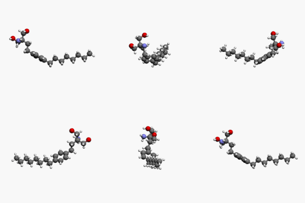
Fingolimod (INN, trade name Gilenya, Novartis) is an immunomodulating drug, approved for treating multiple sclerosis. It has reduced the rate of relapses in relapsing-remitting multiple sclerosis by over half. Fingolimod is a sphingosine-1-phosphate receptor modulator, which sequesters lymphocytes in lymph nodes, preventing them from contributing to an autoimmune reaction.

Fingolimod3Dan

Fingolimod3Dan


http://upload.wikimedia.org/wikipedia/commons/thumb/4/48/Fingolimod3Dan.gif/200px-Fingolimod3Dan.gif

The S1P antagonist FTY720 has been approved by the US Food and Drug Administration to be used as a drug against multiple sclerosis (MS). FTY720 is in fact a prodrug, since it is phosphorylated in vivo by SphK2 into FTY720-P, an S1P structural analog, which can activate S1PR1, 3, 4, and 5. FTY720-P binding to S1PR1 causes internalization of the receptor, as does S1P – but instead of recycling it back to the cell surface, it promotes its ubiquitination and degradation at the proteasome. This has a direct effect on lymphocyte trafficking through the lymph nodes, since it relies on S1PR1 signaling and S1P gradient (Figure 2). In MS, it stops migrating lymphocytes into the brain, but it may also have direct effects on the CNS through neuroprotection. FTY720 can pass the blood–brain barrier and it could be phosphorylated by local sphingosine kinases to act through S1PR1 and S1PR3 receptors that are mainly expressed in the CNS. In MS lesions, astrocytes upregulate those two receptors and it has been shown that FTY720-P treatment in vitro inhibits astrocyte production of inflammatory cytokines. A recent study confirms the importance of S1PR3 signaling on activated astrocytes, as well as SphK1, that are upregulated and promote the secretion of the potentially neuroprotective cytokine CXCL-1.

There are several studies implicating the intracellular S1P ceramide rheostat to cancer cell survival or apoptosis and resistance to chemotherapy or irradiation in vitro. Studies with SphK1 inhibition in pancreatic, prostate cancers, and leukemia, show increased ceramide/S1P ratio and induction of apoptosis. However, S1P receptor signaling plays conflicting roles in cancer cell migration and metastasis.

Modulation of S1P signaling: therapeutic potential

S1P signaling can be involved in many pathophysiological conditions. This means that we could look for therapeutic targets in all the molecules taking part in S1P signaling and production, most importantly the S1P receptors and the sphingosine kinases. S1P agonists and antagonists could also be used to modulate S1P signaling during pathological conditions.

S1P can have direct effects on the cardiovascular system. During IRI, intracellular S1P can protect the cardiomyocytes and promote their survival. Pre- or post-conditioning of the heart with S1P could be used as a treatment, but upregulation of sphingosine kinases could also increase intracellular S1P bioavailability. S1P could also have effects on endothelial cells and neutrophil trafficking. Vascular endothelial cells mainly express S1PR1 and S1PR3; only a few types express S1PR2. S1PR1 and S1PR3 activation on these cells has been shown to enhance their chemotactic migration, probably through direct phosphorylation of S1PR1 by Akt, in a phosphatidylinositol 3-kinase and Rac1-dependent signaling pathway. Moreover, it stimulates endothelial cell proliferation through an ERK pathway. S1PR2 activation, however, inhibits endothelial cell migration, morphogenesis, and angiogenesis, most likely through Rho-dependent inhibition of Rac signaling pathway, as Inoki et al showed in mouse cells with the use of S1PR1 and S1PR3 specific antagonists.

Regarding permeability of the vascular endothelium and endothelial barrier integrity, S1P receptors can have different effects. S1PR1 activation enhances endothelial barrier integrity by stimulation of cellular adhesion and upregulation of adhesion molecules. However, S1PR2 and S1PR3 have been shown to have barrier-disrupting effects in vitro, and vascular permeability increasing effects in vivo. All the effects S1P can have on vascular endothelium and smooth muscle cells suggest that activation of S1PR2, not S1PR1 and S1PR3, signaling, perhaps with the use of S1PR2 specific agonists, could be used therapeutically to inhibit angiogenesis and disrupt vasculature, suppressing tumor growth and progression.

An important aspect of S1P signaling that is being already therapeutically targeted, but could be further investigated, is immune cell trafficking. Attempts have already been made to regulate lymphocyte cell migration with the use of the drug FTY720, whose phosphorylated form can inhibit the cells S1PR1-dependent egress from the lymph nodes, causing lymphopenia. FTY720 is used as an immunosuppressant for MS but is also being investigated for other autoimmune conditions and for transplantation. Unfortunately, Phase II and III clinical trials for the prevention of kidney graft rejection have not shown an advantage over standard therapies. Moreover, FTY720 can have some adverse cardiac effects, such as bradycardia. However, there are other S1PR1 antagonists that could be considered instead, including KRP-203, AUY954, and SEW2871. KRP-203 in particular has been shown to prolong rat skin and heart allograft survival and attenuate chronic rejection without causing bradycardia, especially when combined with other immunomodulators.

There are studies that argue S1P pretreatment has a negative effect on neutrophil chemotaxis toward the chemokine CXCL-8 (interleukin-8) or the potent chemoattractant formyl-methionyl-leucyl-phenylalanine. S1P pretreatment might also inhibit trans-endothelial migration of neutrophils, without affecting their adhesion to the endothelium. S1P effects on neutrophil migration toward CXCL-8 might be the result of S1PRs cross-linking with the CXCL-8 receptors in neutrophils, CXCR-1 and CXCR-2. Indeed, there is evidence suggesting S1PR4 and S1PR3 form heterodimers with CXCR-1 in neutrophils. Another indication that S1P plays a role in neutrophil trafficking is a recent paper on S1P lyase deficiency, a deficiency that impairs neutrophil migration from blood to tissue in knockout mice.

S1P lyase and S1PRs in neutrophils may be new therapeutic targets against IRI and inflammatory conditions in general. Consistent with these results, another study has shown that inhibition of S1P lyase can have a protective effect on the heart after IRI and this effect is alleviated when pretreated with an S1PR1 and S1PR3 antagonist. Inhibition was achieved with a US Food and Drug Administration-approved food additive, 2-acetyl-4-tetrahydroxybutylimidazole, providing a possible new drug perspective. Another S1P lyase inhibitor, LX2931, a synthetic analog of 2-acetyl-4-tetrahydroxybutylimidazole, has been shown to cause peripheral lymphopenia when administered in mice, providing a potential treatment for autoimmune diseases and prevention of graft rejection in transplantation. This molecule is currently under Phase II clinical trials in rheumatoid arthritis patients.

S1P signaling research has the potential to discover novel therapeutic targets. S1P signaling is involved in many physiological and pathological processes. However, the complexity of S1P signaling makes it necessary to consider every possible pathway, either through its GPCRs, or intracellularly, with S1P as a second messenger. Where the activation of one S1P receptor may lead to the desired outcome, the simultaneous activation of another S1P receptor may lead to the opposite outcome. Thus, if we are to target a specific signaling pathway, we might need specific agonists for S1P receptors to activate one S1P receptor pathway, while, at the same time, we might need to inhibit another through S1P receptor antagonists.

Evidence of sphingolipid signaling in cancer

Biologically active lipids are important cellular signaling molecules and play a role in cell communication and cancer cell proliferation, and cancer stem cell biology.  A recent study in ovarian cancer cell lines shows that exogenous sphingosine 1 phosphate (SIP1) or overexpression of the sphingosine kinase (SPHK1) increases ovarian cancer cell proliferation, invasion and contributes to cancer stem cell like phenotype.  The diabetes drug metformin was shown to be an inhibitor of SPHK1 and reduce ovarian cancer tumor growth.

 2019 Apr;17(4):870-881. doi: 10.1158/1541-7786.MCR-18-0409. Epub 2019 Jan 17.

SPHK1 Is a Novel Target of Metformin in Ovarian Cancer.

Abstract

The role of phospholipid signaling in ovarian cancer is poorly understood. Sphingosine-1-phosphate (S1P) is a bioactive metabolite of sphingosine that has been associated with tumor progression through enhanced cell proliferation and motility. Similarly, sphingosine kinases (SPHK), which catalyze the formation of S1P and thus regulate the sphingolipid rheostat, have been reported to promote tumor growth in a variety of cancers. The findings reported here show that exogenous S1P or overexpression of SPHK1 increased proliferation, migration, invasion, and stem-like phenotypes in ovarian cancer cell lines. Likewise, overexpression of SPHK1 markedly enhanced tumor growth in a xenograft model of ovarian cancer, which was associated with elevation of key markers of proliferation and stemness. The diabetes drug, metformin, has been shown to have anticancer effects. Here, we found that ovarian cancer patients taking metformin had significantly reduced serum S1P levels, a finding that was recapitulated when ovarian cancer cells were treated with metformin and analyzed by lipidomics. These findings suggested that in cancer the sphingolipid rheostat may be a novel metabolic target of metformin. In support of this, metformin blocked hypoxia-induced SPHK1, which was associated with inhibited nuclear translocation and transcriptional activity of hypoxia-inducible factors (HIF1α and HIF2α). Further, ovarian cancer cells with high SPHK1 were found to be highly sensitive to the cytotoxic effects of metformin, whereas ovarian cancer cells with low SPHK1 were resistant. Together, the findings reported here show that hypoxia-induced SPHK1 expression and downstream S1P signaling promote ovarian cancer progression and that tumors with high expression of SPHK1 or S1P levels might have increased sensitivity to the cytotoxic effects of metformin. IMPLICATIONS: Metformin targets sphingolipid metabolism through inhibiting SPHK1, thereby impeding ovarian cancer cell migration, proliferation, and self-renewal.

Nrf2:INrf2(Keap1) Signaling in Oxidative Stress

James W. Kaspar, Suresh K. Niture, and Anil K. Jaiswal*

Department of Pharmacology, University of Maryland School of Medicine, Baltimore, MD

Free Radic Biol Med. 2009 Nov 1; 47(9): 1304–1309. http://dx.doi.org:/10.1016/j.freeradbiomed.2009.07.035

Nrf2:INrf2(Keap1) are cellular sensors of chemical and radiation induced oxidative and electrophilic stress. Nrf2 is a nuclear transcription factor that

  • controls the expression and coordinated induction of a battery of defensive genes encoding detoxifying enzymes and antioxidant proteins.

This is a mechanism of critical importance for cellular protection and cell survival. Nrf2 is retained in the cytoplasm by an inhibitor INrf2. INrf2 functions as an adapter for

  • Cul3/Rbx1 mediated degradation of Nrf2.
  • In response to oxidative/electrophilic stress,
  • Nrf2 is switched on and then off by distinct

early and delayed mechanisms.

Oxidative/electrophilic modification of INrf2cysteine151 and/or PKC phosphorylation of Nrf2serine40 results in the escape or release of Nrf2 from INrf2. Nrf2 is stabilized and translocates to the nucleus, forms heterodimers with unknown proteins, and binds antioxidant response element (ARE) that leads to coordinated activation of gene expression. It takes less than fifteen minutes from the time of exposure

  • to switch on nuclear import of Nrf2.

This is followed by activation of a delayed mechanism that controls

  • switching off of Nrf2 activation of gene expression.

GSK3β phosphorylates Fyn at unknown threonine residue(s) leading to

  • nuclear localization of Fyn.

Fyn phosphorylates Nrf2tyrosine568 resulting in

  • nuclear export of Nrf2,
  • binding with INrf2 and
  • degradation of Nrf2.

The switching on and off of Nrf2 protects cells against free radical damage, prevents apoptosis and promotes cell survival.

NPRA-mediated suppression of AngII-induced ROS production contributes to the antiproliferative effects of B-type natriuretic peptide in VSMC

Pan Gao, De-Hui Qian, Wei Li,  Lan Huang
Mol Cell Biochem (2009) 324:165–172

http://dx.doi.org/10.1007/s11010-008-9995-y

Excessive proliferation of vascular smooth cells (VSMCs) plays a critical role in the pathogenesis of diverse vascular disorders, and inhibition of VSMCs proliferation has been proved to be beneficial to these diseases.

In this study, we investigated the antiproliferative effect of

  • B-type natriuretic peptide (BNP), a natriuretic peptide with potent antioxidant capacity,

on rat aortic VSMCs, and the possible mechanisms involved. The results indicate that

  • BNP potently inhibited Angiotensin II (AngII)-induced VSMCs proliferation,

as evaluated by [3H]-thymidine incorporation assay. Consistently, BNP significantly decreased

  • AngII-induced intracellular reactive oxygen species (ROS)
  • and NAD(P)H oxidase activity.

8-Br-cGMP, a cGMP analog,

  • mimicked these effects.

To confirm its mechanism, siRNA of natriuretic peptide receptor-A(NRPA) strategy technology was used

  • to block cGMP production in VSMCs, and
  • siNPRA attenuated the inhibitory effects of BNP in VSMCs.

Taken together, these results indicate that

  • BNP was capable of inhibiting VSMCs proliferation by
  • NPRA/cGMP pathway,

which might be associated with

  • the suppression of ROS production.

These results might be related, at least partly, to the anti-oxidant property of BNP.

Cellular prion protein is required for neuritogenesis: fine-tuning of multiple signaling pathways involved in focal adhesions and actin cytoskeleton dynamics

A Alleaume-Butaux, C Dakowski, M Pietri, S Mouillet-Richard, Jean-Marie Launay, O Kellermann, B Schneider

1INSERM, UMR-S 747, 2Paris Descartes University, Sorbonne Paris, 3Public Hospital of Paris, Department of Biochemistry, Paris, France; 4Pharma Research Department, Hoffmann La Roche Ltd, Basel, Switzerland

Cell Health and Cytoskeleton 2013; 5: 1–12

Neuritogenesis is a complex morphological phenomena accompanying neuronal differentiation. Neuritogenesis relies on the initial breakage of the rather spherical symmetry of neuroblasts and the formation of buds emerging from the postmitotic neuronal soma. Buds then evolve into neurites, which later convert into an axon or dendrites. At the distal tip of neurites, the growth cone integrates extracellular signals and guides the neurite to its target. The acquisition of neuronal polarity depends on deep modifications of the neuroblast cytoskeleton characterized by the remodeling and activation of focal adhesions (FAs) and localized destabilization of the actin network in the neuronal sphere.Actin instability in unpolarized neurons allows neurite sprouting, ie, the protrusion of microtubules, and subsequent neurite outgrowth. Once the neurite is formed, actin microfilaments recover their stability and exert a sheathed action on neurites, a dynamic process necessary for the maintenance and integrity of neurites.

A combination of extrinsic and intrinsic cues pilots the architectural and functional changes in FAs and the actin network along neuritogenesis. This process includes neurotrophic factors (nerve growth factor, brain derived neurotrophic factor, neurotrophin, ciliary neurotrophic factor, glial derived neurotrophic factor) and their receptors, protein components of the extracellular matrix (ECM) (laminin, vitronectin, fibronectin), plasma membrane integrins and neural cell adhesion molecules (NCAM), and intracellular molecular protagonists such as small G proteins (RhoA, Rac, Cdc42) and their downstream targets.

Neuritogenesis is a dynamic phenomenon associated with neuronal differentiation that allows a rather spherical neuronal stem cell to develop dendrites and axon, a prerequisite for the integration and transmission of signals. The acquisition of neuronal polarity occurs in three steps:

(1) neurite sprouting, which consists of the formation of buds emerging from the postmitotic neuronal soma;

(2) neurite outgrowth, which represents the conversion of buds into neurites, their elongation and evolution into axon or dendrites; and

(3) the stability and plasticity of neuronal polarity.

In neuronal stem cells, remodeling and activation of focal adhesions (FAs) associated with deep modifications of the actin cytoskeleton is a prerequisite for neurite sprouting and subsequent neurite outgrowth. A multiple set of growth factors and interactors located in the extracellular matrix and the plasma membrane orchestrate neuritogenesis

  • by acting on intracellular signaling effectors,
  • notably small G proteins such as RhoA, Rac, and Cdc42,
  • which are involved in actin turnover and the dynamics of FAs.

The cellular prion protein (PrPC), a glycosylphosphatidylinositol

  • (GPI)-anchored membrane protein

mainly known for its role in a group of fatal

  • neurodegenerative diseases,

has emerged as a central player in neuritogenesis.

Here, we review the contribution of PrPC to neuronal polarization and detail the current knowledge on the

  • signaling pathways fine-tuned by PrPC
  • to promote neurite sprouting, outgrowth, and maintenance.

We emphasize that PrPC-dependent neurite sprouting is a process in which PrPC

  • governs the dynamics of FAs and the actin cytoskeleton
  • via β1 integrin signaling.

The presence of PrPC is necessary to render neuronal stem cells

  • competent to respond to neuronal inducers and
  • to develop neurites.

In differentiating neurons, PrPC exerts

  • a facilitator role towards neurite elongation.

This function relies on the interaction of PrPC with a set of diverse partners such as

  1. elements of the extracellular matrix,
  2. plasma membrane receptors,
  3. adhesion molecules, and
  4. soluble factors that control actin cytoskeleton turnover through Rho-GTPase signaling.

Once neurons have reached their terminal stage of differentiation and acquired their polarized morphology, PrPC also

  • takes part in the maintenance of neurites.

By acting on tissue nonspecific alkaline phosphatase, or

  • matrix metalloproteinase type 9,

PrPC stabilizes interactions between

  • neurites and the extracellular matrix.

Keywords: prion, neuronal differentiation

Read Full Post »

Cytoskeleton and Cell Membrane Physiology


Curator: Larry H Bernstein, MD, FCAP


cell-membrane

cell-membrane

early evolution of lipid membranes and the three domains of life

early evolution of lipid membranes and the three domains of life

Definition and Function

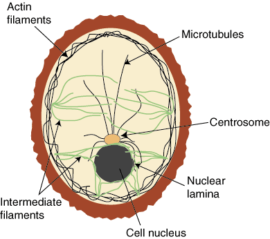
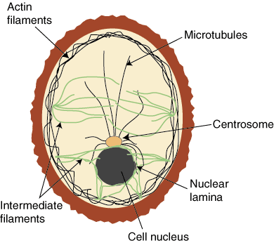
The cytoskeleton is a series of intercellular proteins that help a cell with

  1. shape,
  2. support, and
  3. movement.

Cytoskeleton has three main structural components:

  1. microfilaments,
  2. intermediate filaments, and
  3. movement

The cytoskeleton mediates movement by

  • helping the cell move in its environment and
  • mediating the movement of the cell’s components.

Thereby it provides an important structural framework for the cell –

  • the framework for the movement of organelles, contiguous with the cell membrane, around the cytoplasm. By the activity of
  • the network of protein microfilaments, intermediate filaments, and microtubules.

The structural framework supports cell function as follows:

Cell shape. For cells without cell walls, the cytoskeleton determines the shape of the cell. This is one of the functions of the intermediate filaments.

Cell movement. The dynamic collection of microfilaments and microtubles can be continually in the process of assembly and disassembly, resulting in forces that move the cell. There can also be sliding motions of these structures. Audesirk and Audesirk give examples of white blood cells “crawling” and the migration and shape changes of cells during the development of multicellular organisms.

Organelle movement. Microtubules and microfilaments can help move organelles from place to place in the cell. In endocytosis a vesicle formed engulfs a particle abutting the cell. Microfilaments then attach to the vesicle and pull it into the cell. Much of the complex synthesis and distribution function of the endoplasmic reticulum and the Golgi complex makes use of transport vescicles,  associated with the cytoskeleton.

Cell division. During cell division, microtubules accomplish the movement of the chromosones to the daughter nucleus. Also, a ring of microfilaments helps divide two developing cells by constricting the central region between the cells (fission).

References:
Hickman, et al. Ch 4 Hickman, Cleveland P., Roberts, Larry S., and Larson, Allan, Integrated Principles of Zoology, 9th. Ed., Wm C. Brown, 1995.
Audesirk & Audesirk Ch 6 Audesirk, Teresa and Audesirk, Gerald, Biology, Life on Earth, 5th Ed., Prentice-Hall, 1999.
http://hyperphysics.phy-astr.gsu.edu/hbase/biology/bioref.html#c1
http://hyperphysics.phy-astr.gsu.edu/hbase/biology/cytoskel.html

Intermediate filaments are 8-12 nanometers in diameter and are twisted together in a cord shape. They are composed of keratin and keratin-like proteins.  These filaments are tough and resist tension.

Microtubules are composed of alpha and beta tubulin that form long, hollow cylinders.  These are fairly strong proteins and are the largest component of cytoskeleton at 25 nanometers. Tubular monomers can be lengthened or shortened from the positive end.

Microtubules have three different functions.

They make up the cell’s

  1. centriole
  2. the flagella and cilia of a cell, and
  3. they serve as “tracks” for transport vesicles to move along.

http://biology.kenyon.edu/HHMI/Biol113/cytoskeleton.htm

Key Points 

Microtubules

  1. help the cell resist compression,
  2. provide a track along which vesicles can move throughout the cell, and
  3. are the components of cilia and flagella.

Cilia and flagella are hair-like structures that

  1. assist with locomotion in some cells, as well as
  2. line various structures to trap particles.

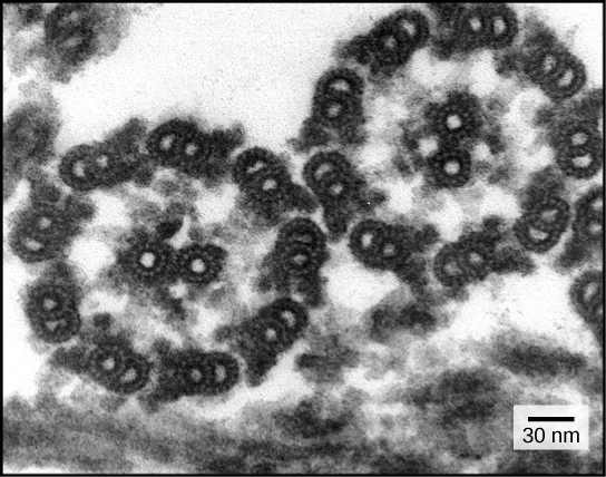
The structures of cilia and flagella are a “9+2 array,” meaning that

  • a ring of nine microtubules is surrounded by two more microtubules.

Microtubules attach to replicated chromosomes

  • during cell division and
  • pull them apart to opposite ends of the pole,
  • allowing the cell to divide with a complete set of chromosomes in each daughter cell.

Microtubules are the largest element of the cytoskeleton.

The walls of the microtubule are made of

  • polymerized dimers of α-tubulin and β-tubulin, two globular proteins.

https://figures.boundless.com/18608/full/figure-04-05-04ab.jpe

With a diameter of about 25 nm, microtubules are the widest components of the cytoskeleton.

https://figures.boundless.com/18608/full/figure-04-05-04ab.jpe

They help the cell

  • resist compression,
  • provide a track along which vesicles move through the cell, and
  • pull replicated chromosomes to opposite ends of a dividing cell.

Like microfilaments, microtubules can dissolve and reform quickly.

Microtubules are also the structural elements of flagella, cilia, and centrioles (the latter are the two perpendicular bodies of the centrosome). In animal cells, the centrosome is the microtubule-organizing center. In eukaryotic cells, flagella and cilia are quite different structurally from their counterparts in prokaryotes.

Intermediate Filaments

Intermediate filaments (IFs) are cytoskeletal components found in animal cells. They are composed of a family of related proteins sharing common structural and sequence features.

epithelial cells

epithelial cells

https://figures.boundless.com/22035/full/epithelial-cells.jpe

flagella and cilia share a common structural arrangement of microtubules called a “9 + 2 array.” This is an appropriate name because a single flagellum or cilium is made of a ring of nine microtubule doublets surrounding a single microtubule doublet in the center.

9+2 array

9+2 array

https://figures.boundless.com/18609/full/figure-04-05-05.jpe

https://www.boundless.com/physiology/textbooks/boundless-anatomy-and-physiology-textbook/cellular-structure-and-function-3/the-cytoskeleton-46/the-composition-and-function-of-the-cytoskeleton-348-11460/

http://jcs.biologists.org/content/115/22/4215/F4.large.jpg

The `Spectraplakins’: cytoskeletal giants with characteristics of both spectrin and plakin families

Katja Röper, Stephen L. Gregory and Nicholas H. Brown
J Cell Sci Nov 15, 2002; 115: 4215-4225
http://dx.doi.org:/10.1242/​jcs.00157

cytoskel

cytoskel

http://plantphys.info/plant_physiology/images/cytoskelfcns.gif

cytoskeleton

cytoskeleton

http://img.sparknotes.com/figures/D/d479f5da672c08a54f986ae699069d7a/cytoskeleton.gif

The sequential endosymbiotic origins of eukaryotes: Compared to bacteria and archaea, the typical eukaryotic cell is much more structurally complex.

While the prokaryotes have a rigid cell wall, the ancestral eukaryote appears to have been wall-less (the walls of plant cells appear to represent a adaptation, and are not homologous to prokaryotic cell walls).

In addition to a nucleus (wherein the cell’s DNA is located, and which we will return to in the next section), there are cytoskeletal structures, including distinctive flagella (quite different from those found in prokaryotes), an active (motile) plasma membrane, capable of engulfing other cells, and multiple internal membrane systems. (A more complete description of cell structure is beyond this version of Biofundamentals).

In aerobic bacteria and cyanobacteria, the electron transport chains associated with ATP synthesis (through either photosynthesis or aerobic respiration) located within the plasma membrane (and in the case of cyanobacteria, internal membrane systems as well).

The same processes (aerobic respiration and photosynthesis) occur within eukaryotic cells. Animals have aerobic respiration, while plants have both).

However, these processes do not occur on the plasma membrane, but rather within distinct cytoplasmic organelles: mitochondria for aerobic respiration and chloroplasts for photosynthesis. All eukaryotic cells have mitochondria, plants (which are eukaryotic) have both mitochondria and chloroplasts.

An intriguing evolutionary question was, are these processes related, that is, are the processes of aerobic respiration and photosynthesis found in eukaryotes homologous to the processes found in bacteria and cyanobacteria, or did they originate independently.

The path to understanding that homologous nature of these processes began with studies of cell structure.

http://virtuallaboratory.colorado.edu/Biofundamentals/lectureNotes-Revision/Topic2I_Symbiosis.htm

spectrin protein superfamily.large

spectrin protein superfamily.large

http://mmbr.asm.org/content/70/3/605/F4.large.jpg

The role of secreted factors and extracellular matrix

The role of secreted factors and extracellular matrix

Focal Adhesions: Transmembrane Junctions Between the Extracellular Matrix and the Cytoskeleton

K Burridge, K Fath, T Kelly, G Nuckolls, and C Turner
Ann Rev Cell Biol Nov 1988; 4: 487-525

http://dx.doi.org:/10.1146/annurev.cb.04.110188.002415

the extracellular matrix (ECM) is a collection of extracellular molecules secreted by cells that

  • provides structural and biochemical support to the surrounding cells.[1]

Because multicellularity evolved independently in different multicellular lineages, the composition of ECM varies between multicellular structures; however,

  • cell adhesion,
  • cell-to-cell communication and
  • differentiation

are common functions of the ECM.[2]

The animal extracellular matrix includes

  • the interstitial matrix and
  • the basement membrane.[3]

Interstitial matrix is present between various animal cells (i.e., in the intercellular spaces).

Gels of polysaccharides and fibrous proteins

  • fill the interstitial space and act as
  • a compression buffer against the stress placed on the ECM.[4]

Basement membranes are sheet-like depositions of ECM on which various epithelial cells rest.

The Extracellular Matrix (ECM)
http://userpage.chemie.fu-berlin.de/biochemie/aghaucke/lehre/cytoskelet-ECM.pdf

Mechanical support to tissues

http://www.nature.com/scitable/content/ne0000/ne0000/ne0000/ne0000/14707425/U4CP5-1_FibronectinIntegri_ksm.jpg

http://www.nature.com/scitable/content/integrin-connects-the-extracellular-matrix-with-the-14707425

Organization of cells into tissues

  1. Activation of signaling pathways (cell growth, proliferation; development); examples:
  2. TGF-β, integrins
  3. specialized roles (tendon, bone; cartilage; cell movement during development; basal lamina in epithelia)

Components

  1. proteoglycans
  2. collagen fibers (mechanical strength)
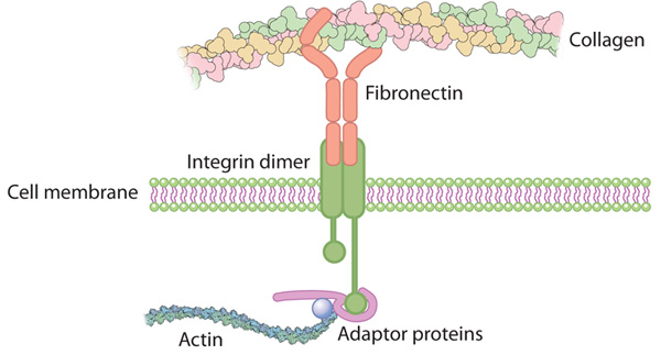
  3. multiadhesive matrix proteins (linking cell surface receptors to the (ECM)

Integrin connects the extracellular matrix with the actin cytoskeleton inside the cell

Fibronectin Integrin

Fibronectin Integrin

http://www.nature.com/scitable/content/ne0000/ne0000/ne0000/ne0000/14707425/U4CP5-1_FibronectinIntegri_ksm.jpg

http://www.nature.com/scitable/content/integrin-connects-the-extracellular-matrix-with-the-14707425

Continuous membrane-cytoskeleton adhesion requires continuous accommodation to lipid and cytoskeleton dynamics.

Sheetz MP, Sable JE, Döbereiner HG.
Annu Rev Biophys Struct Biomol. 2006;35:417-34.

The plasma membrane of most animal cells conforms to the cytoskeleton and only occasionally separates to form blebs. Previous studies indicated that

  • many weak interactions between cytoskeleton and the lipid bilayer
  • kept the surfaces together to counteract the normal outward pressure of cytoplasm.

Either the loss of adhesion strength or the formation of gaps in the cytoskeleton enables the pressure to form blebs. Membrane-associated cytoskeleton proteins, such as spectrin and filamin, can

  • control the movement and aggregation of membrane proteins and lipids,
    e.g., phosphoinositol phospholipids (PIPs), as well as blebbing.

At the same time, lipids (particularly PIPs) and membrane proteins affect

  • cytoskeleton and signaling dynamics.

We consider here the roles of the major phosphatidylinositol-4,5-diphosphate (PIP2) binding protein, MARCKS, and PIP2 levels in controlling cytoskeleton dynamics. Further understanding of dynamics will provide important clues about how membrane-cytoskeleton adhesion rapidly adjusts to cytoskeleton and membrane dynamics. http://www.ncbi.nlm.nih.gov/pubmed/16689643

Interaction of membrane/lipid rafts with the cytoskeleton: impact on signaling and function: membrane/lipid rafts, mediators of cytoskeletal arrangement and cell signaling.

Head BP, Patel HH, Insel PA   Epub 2013 Jul 27.
Biochim Biophys Acta. 2014 Feb;1838(2):532-45.
http://dx.doi.org:/10.1016/j.bbamem.2013.07.018

The plasma membrane in eukaryotic cells contains microdomains that are

  • enriched in certain glycosphingolipids, gangliosides, and sterols (such as cholesterol) to form membrane/lipid rafts (MLR).

These regions exist as caveolae, morphologically observable flask-like invaginations, or as a less easily detectable planar form. MLR are scaffolds for many molecular entities, including

  • signaling receptors and ion channels that
  • communicate extracellular stimuli to the intracellular milieu.

Much evidence indicates that this organization and/or the clustering of MLR into more active signaling platforms

  • depends upon interactions with and dynamic rearrangement of the cytoskeleton.

Several cytoskeletal components and binding partners, as well as enzymes that regulate the cytoskeleton, localize to MLR and help

  • regulate lateral diffusion of membrane proteins and lipids in response to extracellular events
    (e.g., receptor activation, shear stress, electrical conductance, and nutrient demand).

MLR regulate

  • cellular polarity,
  • adherence to the extracellular matrix,
  • signaling events (including ones that affect growth and migration), and
  • are sites of cellular entry of certain pathogens, toxins and nanoparticles.

The dynamic interaction between MLR and the underlying cytoskeleton thus regulates many facets of the function of eukaryotic cells and their adaptation to changing environments. Here, we review general features of MLR and caveolae and their role in several aspects of cellular function, including

  • polarity of endothelial and epithelial cells,
  • cell migration,
  • mechanotransduction,
  • lymphocyte activation,
  • neuronal growth and signaling, and
  • a variety of disease settings.

This article is part of a Special Issue entitled: Reciprocal influences between cell cytoskeleton and membrane channels, receptors and transporters. Guest Editor: Jean Claude Hervé.

Cell control by membrane–cytoskeleton adhesion

Michael P. Sheetz
Nature Reviews Molecular Cell Biology 2, 392-396 (May 2001) | http://dx.doi.doi:/10.1038/35073095

The rates of mechanochemical processes, such as endocytosis, membrane extension and membrane resealing after cell wounding, are known to be controlled biochemically, through interaction with regulatory proteins. Here, I propose that these rates are also controlled physically, through an apparently continuous adhesion between plasma membrane lipids and cytoskeletal proteins.

Lipid Rafts, Signalling and the Cytoskeleton
http://www.bms.ed.ac.uk/research/others/smaciver/Cyto-Topics/lipid_rafts_and_the_cytoskeleton.htm

Lipid rafts are specialised membrane domains enriched in certain lipids cholesterol and proteins. The existence of lipid rafts was first hypothesised in 1988 (Simons & van Meer, 1988; Simon & Ikonen, 1997), but what we know as “caveolae” were first observed  much earlier (Palade, 1953; Yamada, 1955).  Caveolae are flask shaped invaginations on the cell surface that are a type of membrane raft, these were named “caveolae intracellulare” (Yamada, 1955).  After a long argument (Jacobson & Dietrich, 1999), most now consider that these rafts actually exist, however, there is some confusion surrounding the classification of these rafts. It presently seems that there could be three types; caveolae, glycosphingolipid enriched membranes (GEM), and polyphospho inositol rich rafts. It may also be that there are inside rafts (PIP2 rich and caveolae) and outside rafts (GEM).

The fatty-acid chains of lipids within the rafts tend to be extended and so more tightly packed, creating domains with higher order. It is therefore thought that  rafts exist in a separate ordered phase that floats in a sea of poorly ordered lipids.  Glycosphingolipids, and other lipids with long, straight acyl chains are preferentially incorporated into the rafts.

Caveolae are similar in composition to GEMs that lack caveolae and in fact cells that lack caveolin-1 do not have morphologically identifiable caveolae but instead have extra GEM.  These cells can then be transfected with caveolin-1 cDNA and the caveolae then appear.  This suggests that GEM are merely caveolae without caveolin-1.  Caveolin-1 is a 21kDa integral membrane protein that binds cholesterol (Maruta et al, 1995). In cells lacking caveolin-1, caveolin-2 is synthesised but remains in the Golgi.  Caveolin 1 and 2 colocalise when expressed in the same cells and they may form hetero-dimers (Scherer et al, 1997). Caveolin-3 is expressed in muscle where it forms muscle-type caveolae.  Caveolin-3 is involved in certain types of muscular dystrophy (Galbiati et al, ). A slightly confusing finding is that caveolae are the reported site of integrin signalling ().  It is difficult to imagine integrins being available in the depths of membrane invaginations for binding extra-cellular ligands.

The function of rafts

Many functions have been attributed to rafts, from cholesterol transport, endocytosis and signal transduction.  The later is almost certainly the case. It has been suggested that the primary function of caveolae was in constitutive endocytic trafficking but recent data show that this is not the case, instead caveolae are very stable regions of membranes that are not involved in  endocytosis (Thompsen et al, 2002).

lipid raft

lipid raft

Rafts and the Cytoskeleton

Many actin binding proteins are known to bind to polyphosphoinositides and to be regulated by them (see PI and ABPs), by a series of protein domains such as PH, PX and ENTH (see Domains).  It is consequently scarcely surprising that some ABPs are suggested to link the actin cytoskeleton and PIP2-enriched rafts. One of these is gelsolin, a Ca2+, pH and polyphosphoinositide regulated actin capping and severing protein (see Gelsolin Family), that partitions into rafts isolated biochemically from brain (Fanatsu et al, 2000).

GEMs too are suggested to link to the actin cytoskeleton through ABPs particularly ERM proteins through EBP50, a protein that binds members of the ERM proteins through the ERM C-terminus (Brdickova et al, 2001).

References:–

Brdickova, N., Brdicka, T., Andrea, L., Spicka, J., Angelisova, P., Milgram, S. L. & Horejsi, V. (2001) Interaction between two adaptor proteins, PAG and EBP50: a possible link between membrane rafts and actin cytoskeleton.  FEBS letters. 507, 133-136.

Cary, L. A. & Cooper, J. A. (2000) Molecular switches in lipid rafts.  Nature. 404, 945-947.

Czarny, M., Fiucci, G., Lavie, Y., Banno, Y., Nozawa, Y. & Liscovitch, M. (2000) Phospholipase D2: functional interaction with caveolin in low-density membrane microdomains.,  FEBS letters.

Foger, N., Funatsu, N., Kumanogoh, H., Sokawa, Y. & Maekawa, S. (2000) Identification of gelsolin as an actin regulatory component in a Triton insoluble low density fraction (raft) of newborn bovine brain.  Neuroscience Research. 36, 311-317.

Galbiati, F., Engelman, J. A., Volonte, D., Zhang, X. L., Minetti, C., Li, M., Hou jr, H., Kneitz, B., Edelman, W. & Lisanti, M. P. (2001) Caveolin-3 null mice show a loss of caveolae, changes in the microdomain distribution of the dystrophin-glycoprotein complex, and T-tubule abnormalities.  J. Biol.Chem. 276, 21425-21433.

…  (more)

centralpore-small  Gating and Ion Conductivity

centralpore-small Gating and Ion Conductivity

Interaction of epithelial ion channels with the actin-based cytoskeleton.

Mazzochi C, Benos DJ, Smith PR.
Am J Physiol Renal Physiol. 2006 Dec;291(6):F1113-22. Epub 2006 Aug 22

The interaction of ion channels with the actin-based cytoskeleton in epithelial cells

  • not only maintains the polarized expression of ion channels within specific membrane domains,
  • it also functions in the intracellular trafficking and regulation of channel activity.

Initial evidence supporting an interaction between

  • epithelial ion channels and the
  • actin-based cytoskeleton

came from patch-clamp studies of the effects of cytochalasins on channel activity. Cytochalasins were shown to

  • either activate or inactivate epithelial ion channels.

An interaction between

  • the actin-based cytoskeleton and epithelial ion channels

was further supported by the fact that the addition of monomeric or filamentous actin to excised patches had an effect on channel activity comparable to that of cytochalasins. Through the recent application of molecular and proteomic approaches, we now know that

  • the interactions between epithelial ion channels and actin can either be direct or indirect,
  • the latter being mediated through scaffolding or actin-binding proteins
  • that serve as links between the channels and the actin-based cytoskeleton.

This review discusses recent advances in our understanding of the interactions between epithelial ion channels and the actin-based cytoskeleton, and the roles these interactions play in regulating the cell surface expression, activity, and intracellular trafficking of epithelial ion channels.

epithelial ion channels

epithelial ion channels

Actin cytoskeleton regulates ion channel activity in retinal neurons.

Maguire G, Connaughton V, Prat AG, Jackson GR Jr, Cantiello HF.
Neuroreport. 1998 Mar 9;9(4):665-70

The actin cytoskeleton is an important contributor to the integrity of cellular shape and responses in neurons. However, the molecular mechanisms associated with functional interactions between the actin cytoskeleton and neuronal ion channels are largely unknown. Whole-cell and single channel recording techniques were thus applied to identified retinal bipolar neurons of the tiger salamander (Ambystoma tigrinum) to assess the role of acute changes in actin-based cytoskeleton dynamics in the regulation of voltage-gated ion channels. Disruption of endogenous actin filaments after brief treatment (20-30 min) with cytochalasin D (CD) activated voltage-gated K+ currents in bipolar cells, which were largely prevented by intracellular perfusion with the actin filament-stabilizer agent, phalloidin. Either CD treatment under cell-attached conditions or direct addition of actin to excised, inside-out patches of bipolar cells activated and/or increased single K+ channels. Thus, acute changes in actin-based cytoskeleton dynamics regulate voltage-gated ion channel activity in bipolar cells.

Cytoskeletal Basis of Ion Channel Function in Cardiac Muscle

Matteo Vatta, Ph.D1,2 and Georgine Faulkner, Ph.D3

The publisher’s final edited version of this article is available at Future Cardiol

The heart is a force-generating organ that responds to self-generated electrical stimuli from specialized cardiomyocytes. This function is modulated by sympathetic and parasympathetic activity.

In order to contract and accommodate the repetitive morphological changes induced by the cardiac cycle,

  • cardiomyocytes depend on their highly evolved and specialized cytoskeletal apparatus.

Defects in components of the cytoskeleton, in the long term, affect

  • the ability of the cell to compensate at
  • both functional and structural levels.

In addition to the structural remodeling, the myocardium becomes

  • increasingly susceptible to altered electrical activity leading to arrhythmogenesis.

The development of arrhythmias secondary to structural remodeling defects has been noted, although the detailed molecular mechanisms are still elusive. Here I will review the current knowledge of the molecular and functional relationships between the cytoskeleton and ion channels and, I will discuss the future impact of new data on molecular cardiology research and clinical practice. 

Stretch-activated ion channel

Stretch-activated or stretch-gated ion channels are

  • ion channels which open their pores in response to
  • mechanical deformation of a neuron’s plasma membrane.

[Also see mechanosensitive ion channels and mechanosensitive channels, with which they may be synonymous]. Opening of the ion channels

  • depolarizes the afferent neuron producing an action potential with sufficient depolarization.[1]

Channels open in response to two different mechanisms: the prokaryotic model and the mammalian hair cell model.[2][3] Stretch-activated ion channels have been shown to detect vibration, pressure, stretch, touch, sounds, tastes, smell, heat, volume, and vision.[4][5][6] Stretch-activated ion channels have been categorized into

three distinct “superfamilies”:

  1. the ENaC/DEG family,
  2. the TRP family, and
  3. the K1 selective family.

These channels are involved with bodily functions such as blood pressure regulation.[7] They are shown to be associated with many cardiovascular diseases.[3] Stretch-activated channels were first observed in chick skeletal muscles by Falguni Guharay and Frederick Sachs in 1983 and the results were published in 1984.[8] Since then stretch-activated channels have been found in cells from bacteria to humans as well as plants.

Mechanosensitivity of cell membranes. Ion channels, lipid matrix and cytoskeleton.

Petrov AG, Usherwood PN.
Eur Biophys J. 1994;23(1):1-19

Physical and biophysical mechanisms of mechano-sensitivity of cell membranes are reviewed. The possible roles of

  • the lipid matrix and of
  • the cytoskeleton in membrane mechanoreception

are discussed. Techniques for generation of static strains and dynamic curvatures of membrane patches are considered. A unified model for

  • stress-activated and stress-inactivated ion channels

under static strains is described. A review of work on

  • stress-sensitive pores in lipid-peptide model membranes

is presented. The possible role of flexoelectricity in mechano-electric transduction, e.g. in auditory receptors is discussed. Studies of

  • flexoelectricity in model lipid membranes, lipid-peptide membranes and natural membranes containing ion channels

are reviewed. Finally, possible applications in molecular electronics of mechanosensors employing some of the recognized principles of mechano-electric transduction in natural membranes are discussed.Marhaba, R. & Zoller, M. (2001) Involvement of CD44 in cytoskeleton rearrangement and raft reorganization in T cells.  J.Cell Sci. 114, 1169-1178.

FIGURE 2 | The transient pore model.

peroxisomal matrix protein

peroxisomal matrix protein

FROM THE FOLLOWING ARTICLE:
Peroxisomal matrix protein import: the transient pore model

Ralf Erdmann & Wolfgang Schliebs
Nature Reviews Molecular Cell Biology 6, 738-742 (September 2005)
http://dx.doi.org:/10.1038/nrm1710

Peroxisomal matrix protein import: the transient pore model
The transient pore model

The peroxisomal import receptor peroxin-5 (Pex5) recognizes peroxisomal targeting signal-1 (PTS1)-containing cargo proteins in the cytosol. It then moves to the peroxisome where it inserts into the peroxisomal membrane to become an integral part of the protein-import apparatus. Pex14 and/or Pex13, which are associated with Pex17, are proposed to be involved in tethering the receptor to the membrane and in the assembly, stabilization and rearrangement of the translocon. Cargo release into the peroxisomal matrix is thought to be initiated by intraperoxisomal factors — for example, the competitive binding of the intraperoxisomal Pex8, which also has a PTS1. The disassembly and recycling of Pex5 is triggered by a cascade of protein–protein interactions at the peroxisomal membrane that results in the Pex1-, Pex6-driven, ATP-dependent dislocation of Pex5 from the peroxisomal membrane to the cytosol. Pex1 and Pex6 are AAA+ (ATPases associated with a variety of cellular activities) peroxins that are associated with the peroxisome membrane through Pex15 in yeast or its orthologue PEX26 in mammals. Pex4, which is membrane-anchored through Pex22, is a member of the E2 family of ubiquitin-conjugating enzymes, and Pex2, Pex10 and Pex12 contain the RING-finger motif that is a characteristic element of E3 ubiquitin ligases. Mono- or di-ubiquitylation are reversible steps that seem to be required for the efficient recycling of import receptors, whereas polyubiquitylation might signal the proteasome-dependent degradation of receptors when the physiological dislocation of receptors is blocked. Ub, ubiquitin.

Nature Reviews Molecular Cell Biology 6, 738-742 (September 2005) |
http://dx.doi.org:/10.1038/nrm1710

FROM THE FOLLOWING ARTICLE:

peroxisomal protein pore model

peroxisomal protein pore model


Peroxisomal matrix protein import: the transient pore model

Ralf Erdmann & Wolfgang Schliebs
Nature Reviews Molecular Cell Biology 6, 738-742 (September 2005)
http://dx.doi.org:/10.1038/nrm1710

Peroxisomal matrix protein import: the transient pore model

Peroxin-13 (Pex13), Pex14 and Pex17 are constituents of the docking complex for cycling peroxisomal import receptors. Another protein assembly in the peroxisomal membrane comprises the RING-finger-motif-containing peroxins Pex2, Pex10 and Pex12. This motif is a characteristic element of E3 ubiquitin ligases, and this subcomplex is linked to the docking complex by Pex8, which is peripherally attached to the lumenal side of the peroxisomal membrane. Pex4 is a member of the E2 family of ubiquitin-conjugating enzymes and is anchored to the peroxisomal membrane through the cytosolic domain of Pex22. Pex1 and Pex6 are interacting AAA+ proteins (ATPases associated with a variety of cellular activities), which are attached to the membrane through binding to Pex15 in yeast or to its mammalian counterpart PEX26.

Peroxisomal matrix protein import: the transient pore model

Ralf Erdmann & Wolfgang Schliebs

Peroxisomes import folded, even oligomeric, proteins, which distinguishes the peroxisomal translocation machinery from the well-characterized translocons of other organelles. How proteins are transported across the peroxisomal membrane is unclear. Here, we propose a mechanistic model that conceptually divides the import process into three consecutive steps: the formation of a

  • translocation pore by the import receptor,
  • the ubiquitylation of the import receptors, and
  • pore disassembly/receptor recycling.

Phytosphingosine

Masoud Naderi Maralani

Identification of the phytosphingosine metabolic pathway leading to odd-numbered fatty acids

The long-chain base ​phytosphingosine is a component of sphingolipids and exists in yeast, plants and some mammalian tissues. ​Phytosphingosine is unique in that it possesses an additional hydroxyl group compared with other long-chain bases. However, its metabolism is unknown. Here we show that ​phytosphingosine is metabolized to odd-numbered fatty acids and is incorporated into glycerophospholipids both in yeast and mammalian cells. Disruption of the yeast gene encoding long-chain base 1-phosphate lyase, which catalyzes the committed step in the metabolism of ​phytosphingosine to glycerophospholipids, causes an ~40% reduction in the level of phosphatidylcholines that contain a C15 fatty acid. We also find that ​2-hydroxypalmitic acid is an intermediate of the phytosphingosine metabolic pathway. Furthermore, we show that the yeast ​MPO1 gene, whose product belongs to a large, conserved protein family of unknown function, is involved in ​phytosphingosine metabolism. Our findings provide insights into fatty acid diversity and identify a pathway by which hydroxyl group-containing lipids are metabolized.  nature.com nature.com

About GPCRs

G-protein-coupled receptors (GPCRs) are a class of membrane proteins that allow the transmission of a wide variety of signals over the cell membrane, between different cells and over long distances inside the body. The molecular mechanisms of action of GPCRs were worked in great detail by Brian Kobilka and Robert Lefkowitz for which they were jointly awarded the Nobel Prize in Chemistry for 2012. Read More

Read Full Post »

Introduction to Metabolomics

Introduction to Metabolomics

Author: Larry H. Bernstein, MD, FCAP


This is the first volume of the Series D: e-Books on BioMedicine – Metabolomics, Immunology, Infectious Diseases.  It is written for comprehension at the third year medical student level, or as a reference for licensing board exams, but it is also written for the education of a first time bachalaureate degree reader in the biological sciences.  Hopefully, it can be read with great interest by the undergraduate student who is undecided in the choice of a career.

In the Preface, I failed to disclose that the term Metabolomics applies to plants, animals, bacteria, and both prokaryotes and eukaryotes.  The metabolome for each organism is unique, but from an evolutionary perspective has metabolic pathways in common, and expressed in concert with the environment that these living creatures exist. The metabolome of each has adaptive accommodation with suppression and activation of pathways that are functional and necessary in balance, for its existence.  Was it William Faulkner who said in his Nobel Prize acceptance that mankind shall not merely exist, but survive? That seems to be the overlying theme for all of life. If life cannot persist, a surviving “remnant” might continue. The history of life may well be etched into the genetic code, some of which is not expressed.

This work is apportioned into chapters in a sequence that is first directed at the major sources for the energy and the structure of life, in the carbohydrates, lipids, and fats, which are sourced from both plants and animals, and depending on their balance, results in an equilibrium, and a disequilibrium we refer to as disease.  There is also a need to consider the nonorganic essentials which are derived from the soil, from water, and from the energy of the sun and the air we breathe, or in the case of water-bound metabolomes, dissolved gases.

In addition to the basic essential nutrients and their metabolic utilization, they are under cellular metabolic regulation that is tied to signaling pathways.  In addition, the genetic expression of the organism is under regulatory control by the interaction of RNAs that interact with the chromatin genetic framework, with exosomes, and with protein modulators.This is referred to as epigenetics, but there are also drivers of metabolism that are shaped by the interactions between enzymes and substartes, and are related to the tertiary structure of a protein.  The framework for diseases in a separate chapter.  Pharmaceutical interventions that are designed to modulate specific metabolic targets are addressed as the pathways are unfolded. Neutraceuticals and plant based nutrition are covered in Chapter 8.

Chapter 1: Metabolic Pathways

Chapter 2. Lipid Metabolism

Chapter 3. Cell Signaling

Chapter 4. Protein Synthesis and Degradation

Chapter 5: Sub-cellular Structure

Chapter 6: Proteomics

Chapter 7: Metabolomics

Chapter 8. Impairments in Pathological States: Endocrine Disorders; Stress Hypermetabolism and Cancer

Read Full Post »

Metabolomics Summary and Perspective

Metabolomics Summary and Perspective

Author and Curator: Larry H Bernstein, MD, FCAP 


This is the final article in a robust series on metabolism, metabolomics, and  the “-OMICS-“ biological synthesis that is creating a more holistic and interoperable view of natural sciences, including the biological disciplines, climate science, physics, chemistry, toxicology, pharmacology, and pathophysiology with as yet unforeseen consequences.

There have been impressive advances already in the research into developmental biology, plant sciences, microbiology, mycology, and human diseases, most notably, cancer, metabolic , and infectious, as well as neurodegenerative diseases.

Acknowledgements:

I write this article in honor of my first mentor, Harry Maisel, Professor and Emeritus Chairman of Anatomy, Wayne State University, Detroit, MI and to my stimulating mentors, students, fellows, and associates over many years:

Masahiro Chiga, MD, PhD, Averill A Liebow, MD, Nathan O Kaplan, PhD, Johannes Everse, PhD, Norio Shioura, PhD, Abraham Braude, MD, Percy J Russell, PhD, Debby Peters, Walter D Foster, PhD, Herschel Sidransky, MD, Sherman Bloom, MD, Matthew Grisham, PhD, Christos Tsokos, PhD,  IJ Good, PhD, Distinguished Professor, Raool Banagale, MD, Gustavo Reynoso, MD,Gustave Davis, MD, Marguerite M Pinto, MD, Walter Pleban, MD, Marion Feietelson-Winkler, RD, PhD,  John Adan,MD, Joseph Babb, MD, Stuart Zarich, MD,  Inder Mayall, MD, A Qamar, MD, Yves Ingenbleek, MD, PhD, Emeritus Professor, Bette Seamonds, PhD, Larry Kaplan, PhD, Pauline Y Lau, PhD, Gil David, PhD, Ronald Coifman, PhD, Emeritus Professor, Linda Brugler, RD, MBA, James Rucinski, MD, Gitta Pancer, Ester Engelman, Farhana Hoque, Mohammed Alam, Michael Zions, William Fleischman, MD, Salman Haq, MD, Jerard Kneifati-Hayek, Madeleine Schleffer, John F Heitner, MD, Arun Devakonda,MD, Liziamma George,MD, Suhail Raoof, MD, Charles Oribabor,MD, Anthony Tortolani, MD, Prof and Chairman, JRDS Rosalino, PhD, Aviva Lev Ari, PhD, RN, Rosser Rudolph, MD, PhD, Eugene Rypka, PhD, Jay Magidson, PhD, Izaak Mayzlin, PhD, Maurice Bernstein, PhD, Richard Bing, Eli Kaplan, PhD, Maurice Bernstein, PhD.

This article has EIGHT parts, as follows:

Part 1

Metabolomics Continues Auspicious Climb

Part 2

Biologists Find ‘Missing Link’ in the Production of Protein Factories in Cells

Part 3

Neuroscience

Part 4

Cancer Research

Part 5

Metabolic Syndrome

Part 6

Biomarkers

Part 7

Epigenetics and Drug Metabolism

Part 8

Pictorial

genome cartoon

genome cartoon

 iron metabolism

iron metabolism

personalized reference range within population range

personalized reference range within population range

Part 1.  MetabolomicsSurge

metagraph  _OMICS

metagraph _OMICS

Metabolomics Continues Auspicious Climb

Jeffery Herman, Ph.D.
GEN May 1, 2012 (Vol. 32, No. 9)

Aberrant biochemical and metabolite signaling plays an important role in

  • the development and progression of diseased tissue.

This concept has been studied by the science community for decades. However, with relatively

  1. recent advances in analytical technology and bioinformatics as well as
  2. the development of the Human Metabolome Database (HMDB),

metabolomics has become an invaluable field of research.

At the “International Conference and Exhibition on Metabolomics & Systems Biology” held recently in San Francisco, researchers and industry leaders discussed how

  • the underlying cellular biochemical/metabolite fingerprint in response to
  1. a specific disease state,
  2. toxin exposure, or
  3. pharmaceutical compound
  • is useful in clinical diagnosis and biomarker discovery and
  • in understanding disease development and progression.

Developed by BASF, MetaMap® Tox is

  • a database that helps identify in vivo systemic effects of a tested compound, including
  1. targeted organs,
  2. mechanism of action, and
  3. adverse events.

Based on 28-day systemic rat toxicity studies, MetaMap Tox is composed of

  • differential plasma metabolite profiles of rats
  • after exposure to a large variety of chemical toxins and pharmaceutical compounds.

“Using the reference data,

  • we have developed more than 110 patterns of metabolite changes, which are
  • specific and predictive for certain toxicological modes of action,”

said Hennicke Kamp, Ph.D., group leader, department of experimental toxicology and ecology at BASF.

With MetaMap Tox, a potential drug candidate

  • can be compared to a similar reference compound
  • using statistical correlation algorithms,
  • which allow for the creation of a toxicity and mechanism of action profile.

“MetaMap Tox, in the context of early pre-clinical safety enablement in pharmaceutical development,” continued Dr. Kamp,

  • has been independently validated “
  • by an industry consortium (Drug Safety Executive Council) of 12 leading biopharmaceutical companies.”

Dr. Kamp added that this technology may prove invaluable

  • allowing for quick and accurate decisions and
  • for high-throughput drug candidate screening, in evaluation
  1. on the safety and efficacy of compounds
  2. during early and preclinical toxicological studies,
  3. by comparing a lead compound to a variety of molecular derivatives, and
  • the rapid identification of the most optimal molecular structure
  • with the best efficacy and safety profiles might be streamlined.
Dynamic Construct of the –Omics

Dynamic Construct of the –Omics

Targeted Tandem Mass Spectrometry

Biocrates Life Sciences focuses on targeted metabolomics, an important approach for

  • the accurate quantification of known metabolites within a biological sample.

Originally used for the clinical screening of inherent metabolic disorders from dried blood-spots of newborn children, Biocrates has developed

  • a tandem mass spectrometry (MS/MS) platform, which allows for
  1. the identification,
  2. quantification, and
  3. mapping of more than 800 metabolites to specific cellular pathways.

It is based on flow injection analysis and high-performance liquid chromatography MS/MS.

Clarification of Pathway-Specific Inhibition by Fourier Transform Ion Cyclotron Resonance.Mass Spectrometry-Based Metabolic Phenotyping Studies F5.large

common drug targets

common drug targets

The MetaDisIDQ® Kit is a

  • “multiparamatic” diagnostic assay designed for the “comprehensive assessment of a person’s metabolic state” and
  • the early determination of pathophysiological events with regards to a specific disease.

MetaDisIDQ is designed to quantify

  • a diverse range of 181 metabolites involved in major metabolic pathways
  • from a small amount of human serum (10 µL) using isotopically labeled internal standards,

This kit has been demonstrated to detect changes in metabolites that are commonly associated with the development of

  • metabolic syndrome, type 2 diabetes, and diabetic nephropathy,

Dr. Dallman reports that data generated with the MetaDisIDQ kit correlates strongly with

  • routine chemical analyses of common metabolites including glucose and creatinine

Biocrates has also developed the MS/MS-based AbsoluteIDQ® kits, which are

  • an “easy-to-use” biomarker analysis tool for laboratory research.

The kit functions on MS machines from a variety of vendors, and allows for the quantification of 150-180 metabolites.

The SteroIDQ® kit is a high-throughput standardized MS/MS diagnostic assay,

  • validated in human serum, for the rapid and accurate clinical determination of 16 known steroids.

Initially focusing on the analysis of steroid ranges for use in hormone replacement therapy, the SteroIDQ Kit is expected to have a wide clinical application.

Hormone-Resistant Breast Cancer

Scientists at Georgetown University have shown that

  • breast cancer cells can functionally coordinate cell-survival and cell-proliferation mechanisms,
  • while maintaining a certain degree of cellular metabolism.

To grow, cells need energy, and energy is a product of cellular metabolism. For nearly a century, it was thought that

  1. the uncoupling of glycolysis from the mitochondria,
  2. leading to the inefficient but rapid metabolism of glucose and
  3. the formation of lactic acid (the Warburg effect), was

the major and only metabolism driving force for unchecked proliferation and tumorigenesis of cancer cells.

Other aspects of metabolism were often overlooked.

“.. we understand now that

  • cellular metabolism is a lot more than just metabolizing glucose,”

said Robert Clarke, Ph.D., professor of oncology and physiology and biophysics at Georgetown University. Dr. Clarke, in collaboration with the Waters Center for Innovation at Georgetown University (led by Albert J. Fornace, Jr., M.D.), obtained

  • the metabolomic profile of hormone-sensitive and -resistant breast cancer cells through the use of UPLC-MS.

They demonstrated that breast cancer cells, through a rather complex and not yet completely understood process,

  1. can functionally coordinate cell-survival and cell-proliferation mechanisms,
  2. while maintaining a certain degree of cellular metabolism.

This is at least partly accomplished through the upregulation of important pro-survival mechanisms; including

  • the unfolded protein response;
  • a regulator of endoplasmic reticulum stress and
  • initiator of autophagy.

Normally, during a stressful situation, a cell may

  • enter a state of quiescence and undergo autophagy,
  • a process by which a cell can recycle organelles
  • in order to maintain enough energy to survive during a stressful situation or,

if the stress is too great,

  • undergo apoptosis.

By integrating cell-survival mechanisms and cellular metabolism

  • advanced ER+ hormone-resistant breast cancer cells
  • can maintain a low level of autophagy
  • to adapt and resist hormone/chemotherapy treatment.

This adaptation allows cells

  • to reallocate important metabolites recovered from organelle degradation and
  • provide enough energy to also promote proliferation.

With further research, we can gain a better understanding of the underlying causes of hormone-resistant breast cancer, with

  • the overall goal of developing effective diagnostic, prognostic, and therapeutic tools.

NMR

Over the last two decades, NMR has established itself as a major tool for metabolomics analysis. It is especially adept at testing biological fluids. [Bruker BioSpin]

Historically, nuclear magnetic resonance spectroscopy (NMR) has been used for structural elucidation of pure molecular compounds. However, in the last two decades, NMR has established itself as a major tool for metabolomics analysis. Since

  • the integral of an NMR signal is directly proportional to
  • the molar concentration throughout the dynamic range of a sample,

“the simultaneous quantification of compounds is possible

  • without the need for specific reference standards or calibration curves,” according to Lea Heintz of Bruker BioSpin.

NMR is adept at testing biological fluids because of

  1.  high reproducibility,
  2. standardized protocols,
  3. low sample manipulation, and
  4. the production of a large subset of data,

Bruker BioSpin is presently involved in a project for the screening of inborn errors of metabolism in newborn children from Turkey, based on their urine NMR profiles. More than 20 clinics are participating to the project that is coordinated by INFAI, a specialist in the transfer of advanced analytical technology into medical diagnostics. The construction of statistical models are being developed

  • for the detection of deviations from normality, as well as
  • automatic quantification methods for indicative metabolites

Bruker BioSpin recently installed high-resolution magic angle spinning NMR (HRMAS-NMR) systems that can rapidly analyze tissue biopsies. The main objective for HRMAS-NMR is to establish a rapid and effective clinical method to assess tumor grade and other important aspects of cancer during surgery.

Combined NMR and Mass Spec

There is increasing interest in combining NMR and MS, two of the main analytical assays in metabolomic research, as a means

  • to improve data sensitivity and to
  • fully elucidate the complex metabolome within a given biological sample.
  •  to realize a potential for cancer biomarker discovery in the realms of diagnosis, prognosis, and treatment.

.

Using combined NMR and MS to measure the levels of nearly 250 separate metabolites in the patient’s blood, Dr. Weljie and other researchers at the University of Calgary were able to rapidly determine the malignancy of a  pancreatic lesion (in 10–15% of the cases, it is difficult to discern between benign and malignant), while avoiding unnecessary surgery in patients with benign lesions.

When performing NMR and MS on a single biological fluid, ultimately “we are,” noted Dr. Weljie,

  1. “splitting up information content, processing, and introducing a lot of background noise and error and
  2. then trying to reintegrate the data…
    It’s like taking a complex item, with multiple pieces, out of an IKEA box and trying to repackage it perfectly into another box.”

By improving the workflow between the initial splitting of the sample, they improved endpoint data integration, proving that

  • a streamlined approach to combined NMR/MS can be achieved,
  • leading to a very strong, robust and precise metabolomics toolset.

Metabolomics Research Picks Up Speed

Field Advances in Quest to Improve Disease Diagnosis and Predict Drug Response

John Morrow Jr., Ph.D.
GEN May 1, 2011 (Vol. 31, No. 9)

As an important discipline within systems biology, metabolomics is being explored by a number of laboratories for

  • its potential in pharmaceutical development.

Studying metabolites can offer insights into the relationships between genotype and phenotype, as well as between genotype and environment. In addition, there is plenty to work with—there are estimated to be some 2,900 detectable metabolites in the human body, of which

  1. 309 have been identified in cerebrospinal fluid,
  2. 1,122 in serum,
  3. 458 in urine, and
  4. roughly 300 in other compartments.

Guowang Xu, Ph.D., a researcher at the Dalian Institute of Chemical Physics.  is investigating the causes of death in China,

  • and how they have been changing over the years as the country has become a more industrialized nation.
  •  the increase in the incidence of metabolic disorders such as diabetes has grown to affect 9.7% of the Chinese population.

Dr. Xu,  collaborating with Rainer Lehman, Ph.D., of the University of Tübingen, Germany, compared urinary metabolites in samples from healthy individuals with samples taken from prediabetic, insulin-resistant subjects. Using mass spectrometry coupled with electrospray ionization in the positive mode, they observed striking dissimilarities in levels of various metabolites in the two groups.

“When we performed a comprehensive two-dimensional gas chromatography, time-of-flight mass spectrometry analysis of our samples, we observed several metabolites, including

  • 2-hydroxybutyric acid in plasma,
  •  as potential diabetes biomarkers,” Dr. Xu explains.

In other, unrelated studies, Dr. Xu and the German researchers used a metabolomics approach to investigate the changes in plasma metabolite profiles immediately after exercise and following a 3-hour and 24-hour period of recovery. They found that

  • medium-chain acylcarnitines were the most distinctive exercise biomarkers, and
  • they are released as intermediates of partial beta oxidation in human myotubes and mouse muscle tissue.

Dr. Xu says. “The traditional approach of assessment based on a singular biomarker is being superseded by the introduction of multiple marker profiles.”

Typical of the studies under way by Dr. Kaddurah-Daouk and her colleaguesat Duke University

  • is a recently published investigation highlighting the role of an SNP variant in
  • the glycine dehydrogenase gene on individual response to antidepressants.
  •  patients who do not respond to the selective serotonin uptake inhibitors citalopram and escitalopram
  • carried a particular single nucleotide polymorphism in the GD gene.

“These results allow us to pinpoint a possible

  • role for glycine in selective serotonin reuptake inhibitor response and
  • illustrate the use of pharmacometabolomics to inform pharmacogenomics.

These discoveries give us the tools for prognostics and diagnostics so that

  • we can predict what conditions will respond to treatment.

“This approach to defining health or disease in terms of metabolic states opens a whole new paradigm.

By screening hundreds of thousands of molecules, we can understand

  • the relationship between human genetic variability and the metabolome.”

Dr. Kaddurah-Daouk talks about statins as a current

  • model of metabolomics investigations.

It is now known that the statins  have widespread effects, altering a range of metabolites. To sort out these changes and develop recommendations for which individuals should be receiving statins will require substantial investments of energy and resources into defining the complex web of biochemical changes that these drugs initiate.
Furthermore, Dr. Kaddurah-Daouk asserts that,

  • “genetics only encodes part of the phenotypic response.

One needs to take into account the

  • net environment contribution in order to determine
  • how both factors guide the changes in our metabolic state that determine the phenotype.”

Interactive Metabolomics

Researchers at the University of Nottingham use diffusion-edited nuclear magnetic resonance spectroscopy to assess the effects of a biological matrix on metabolites. Diffusion-edited NMR experiments provide a way to

  • separate the different compounds in a mixture
  • based on the differing translational diffusion coefficients (which reflect the size and shape of the molecule).

The measurements are carried out by observing

  • the attenuation of the NMR signals during a pulsed field gradient experiment.

Clare Daykin, Ph.D., is a lecturer at the University of Nottingham, U.K. Her field of investigation encompasses “interactive metabolomics,”which she defines as

“the study of the interactions between low molecular weight biochemicals and macromolecules in biological samples ..

  • without preselection of the components of interest.

“Blood plasma is a heterogeneous mixture of molecules that

  1. undergo a variety of interactions including metal complexation,
  2. chemical exchange processes,
  3. micellar compartmentation,
  4. enzyme-mediated biotransformations, and
  5. small molecule–macromolecular binding.”

Many low molecular weight compounds can exist

  • freely in solution,
  • bound to proteins, or
  • within organized aggregates such as lipoprotein complexes.

Therefore, quantitative comparison of plasma composition from

  • diseased individuals compared to matched controls provides an incomplete insight to plasma metabolism.

“It is not simply the concentrations of metabolites that must be investigated,

  • but their interactions with the proteins and lipoproteins within this complex web.

Rather than targeting specific metabolites of interest, Dr. Daykin’s metabolite–protein binding studies aim to study

  • the interactions of all detectable metabolites within the macromolecular sample.

Such activities can be studied through the use of diffusion-edited nuclear magnetic resonance (NMR) spectroscopy, in which one can assess

  • the effects of the biological matrix on the metabolites.

“This can lead to a more relevant and exact interpretation

  • for systems where metabolite–macromolecule interactions occur.”

Diffusion-edited NMR experiments provide a way to separate the different compounds in a mixture based on

  • the differing translational diffusion coefficients (which reflect the size and shape of the molecule).

The measurements are carried out by observing

  • the attenuation of the NMR signals during a pulsed field gradient experiment.

Pushing the Limits

It is widely recognized that many drug candidates fail during development due to ancillary toxicity. Uwe Sauer, Ph.D., professor, and Nicola Zamboni, Ph.D., researcher, both at the Eidgenössische Technische Hochschule, Zürich (ETH Zürich), are applying

  • high-throughput intracellular metabolomics to understand
  • the basis of these unfortunate events and
  • head them off early in the course of drug discovery.

“Since metabolism is at the core of drug toxicity, we developed a platform for

  • measurement of 50–100 targeted metabolites by
  • a high-throughput system consisting of flow injection
  • coupled to tandem mass spectrometry.”

Using this approach, Dr. Sauer’s team focused on

  • the central metabolism of the yeast Saccharomyces cerevisiae, reasoning that
  • this core network would be most susceptible to potential drug toxicity.

Screening approximately 41 drugs that were administered at seven concentrations over three orders of magnitude, they observed changes in metabolome patterns at much lower drug concentrations without attendant physiological toxicity.

The group carried out statistical modeling of about

  • 60 metabolite profiles for each drug they evaluated.

This data allowed the construction of a “profile effect map” in which

  • the influence of each drug on metabolite levels can be followed, including off-target effects, which
  • provide an indirect measure of the possible side effects of the various drugs.

Dr. Sauer says.“We have found that this approach is

  • at least 100 times as fast as other omics screening platforms,”

“Some drugs, including many anticancer agents,

  • disrupt metabolism long before affecting growth.”
killing cancer cells

killing cancer cells

Furthermore, they used the principle of 13C-based flux analysis, in which

  • metabolites labeled with 13C are used to follow the utilization of metabolic pathways in the cell.

These 13C-determined intracellular responses of metabolic fluxes to drug treatment demonstrate

  • the functional performance of the network to be rather robust,
conformational changes leading to substrate efflux.

conformational changes leading to substrate efflux.

leading Dr. Sauer to the conclusion that

  • the phenotypic vigor he observes to drug challenges
  • is achieved by a flexible make up of the metabolome.

Dr. Sauer is confident that it will be possible to expand the scope of these investigations to hundreds of thousands of samples per study. This will allow answers to the questions of

  • how cells establish a stable functioning network in the face of inevitable concentration fluctuations.

Is Now the Hour?

There is great enthusiasm and agitation within the biotech community for

  • metabolomics approaches as a means of reversing the dismal record of drug discovery

that has accumulated in the last decade.

While the concept clearly makes sense and is being widely applied today, there are many reasons why drugs fail in development, and metabolomics will not be a panacea for resolving all of these questions. It is too early at this point to recognize a trend or a track record, and it will take some time to see how this approach can aid in drug discovery and shorten the timeline for the introduction of new pharmaceutical agents.

Degree of binding correlated with function

Degree of binding correlated with function

Diagram_of_a_two-photon_excitation_microscope_

Diagram_of_a_two-photon_excitation_microscope_

Part 2.  Biologists Find ‘Missing Link’ in the Production of Protein Factories in Cells

Biologists at UC San Diego have found

  • the “missing link” in the chemical system that
  • enables animal cells to produce ribosomes

—the thousands of protein “factories” contained within each cell that

  • manufacture all of the proteins needed to build tissue and sustain life.
‘Missing Link’

‘Missing Link’

Their discovery, detailed in the June 23 issue of the journal Genes & Development, will not only force

  • a revision of basic textbooks on molecular biology, but also
  • provide scientists with a better understanding of
  • how to limit uncontrolled cell growth, such as cancer,
  • that might be regulated by controlling the output of ribosomes.

Ribosomes are responsible for the production of the wide variety of proteins that include

  1. enzymes;
  2. structural molecules, such as hair,
  3. skin and bones;
  4. hormones like insulin; and
  5. components of our immune system such as antibodies.

Regarded as life’s most important molecular machine, ribosomes have been intensively studied by scientists (the 2009 Nobel Prize in Chemistry, for example, was awarded for studies of its structure and function). But until now researchers had not uncovered all of the details of how the proteins that are used to construct ribosomes are themselves produced.

In multicellular animals such as humans,

  • ribosomes are made up of about 80 different proteins
    (humans have 79 while some other animals have a slightly different number) as well as
  • four different kinds of RNA molecules.

In 1969, scientists discovered that

  • the synthesis of the ribosomal RNAs is carried out by specialized systems using two key enzymes:
  • RNA polymerase I and RNA polymerase III.

But until now, scientists were unsure if a complementary system was also responsible for

  • the production of the 80 proteins that make up the ribosome.

That’s essentially what the UC San Diego researchers headed by Jim Kadonaga, a professor of biology, set out to examine. What they found was the missing link—the specialized

  • system that allows ribosomal proteins themselves to be synthesized by the cell.

Kadonaga says that he and coworkers found that ribosomal proteins are synthesized via

  • a novel regulatory system with the enzyme RNA polymerase II and
  • a factor termed TRF2,”

“For the production of most proteins,

  1. RNA polymerase II functions with
  2. a factor termed TBP,
  3. but for the synthesis of ribosomal proteins, it uses TRF2.”
  •  this specialized TRF2-based system for ribosome biogenesis
  • provides a new avenue for the study of ribosomes and
  • its control of cell growth, and

“it should lead to a better understanding and potential treatment of diseases such as cancer.”

Coordination of the transcriptome and metabolome

Coordination of the transcriptome and metabolome

the potential advantages conferred by distal-site protein synthesis

the potential advantages conferred by distal-site protein synthesis

Other authors of the paper were UC San Diego biologists Yuan-Liang Wang, Sascha Duttke and George Kassavetis, and Kai Chen, Jeff Johnston, and Julia Zeitlinger of the Stowers Institute for Medical Research in Kansas City, Missouri. Their research was supported by two grants from the National Institutes of Health (1DP2OD004561-01 and R01 GM041249).

Turning Off a Powerful Cancer Protein

Scientists have discovered how to shut down a master regulatory transcription factor that is

  • key to the survival of a majority of aggressive lymphomas,
  • which arise from the B cells of the immune system.

The protein, Bcl6, has long been considered too complex to target with a drug since it is also crucial

  • to the healthy functioning of many immune cells in the body, not just B cells gone bad.

The researchers at Weill Cornell Medical College report that it is possible

  • to shut down Bcl6 in diffuse large B-cell lymphoma (DLBCL)
  • while not affecting its vital function in T cells and macrophages
  • that are needed to support a healthy immune system.

If Bcl6 is completely inhibited, patients might suffer from systemic inflammation and atherosclerosis. The team conducted this new study to help clarify possible risks, as well as to understand

  • how Bcl6 controls the various aspects of the immune system.

The findings in this study were inspired from

  • preclinical testing of two Bcl6-targeting agents that Dr. Melnick and his Weill Cornell colleagues have developed
  • to treat DLBCLs.

These experimental drugs are

  • RI-BPI, a peptide mimic, and
  • the small molecule agent 79-6.

“This means the drugs we have developed against Bcl6 are more likely to be

  • significantly less toxic and safer for patients with this cancer than we realized,”

says Ari Melnick, M.D., professor of hematology/oncology and a hematologist-oncologist at NewYork-Presbyterian Hospital/Weill Cornell Medical Center.

Dr. Melnick says the discovery that

  • a master regulatory transcription factor can be targeted
  • offers implications beyond just treating DLBCL.

Recent studies from Dr. Melnick and others have revealed that

  • Bcl6 plays a key role in the most aggressive forms of acute leukemia, as well as certain solid tumors.

Bcl6 can control the type of immune cell that develops in the bone marrow—playing many roles

  • in the development of B cells, T cells, macrophages, and other cells—including a primary and essential role in
  • enabling B-cells to generate specific antibodies against pathogens.

According to Dr. Melnick, “When cells lose control of Bcl6,

  • lymphomas develop in the immune system.

Lymphomas are ‘addicted’ to Bcl6, and therefore

  • Bcl6 inhibitors powerfully and quickly destroy lymphoma cells,” .

The big surprise in the current study is that rather than functioning as a single molecular machine,

  • Bcl6 functions like a Swiss Army knife,
  • using different tools to control different cell types.

This multifunction paradigm could represent a general model for the functioning of other master regulatory transcription factors.

“In this analogy, the Swiss Army knife, or transcription factor, keeps most of its tools folded,

  • opening only the one it needs in any given cell type,”

He makes the following analogy:

  • “For B cells, it might open and use the knife tool;
  • for T cells, the cork screw;
  • for macrophages, the scissors.”

“this means that you only need to prevent the master regulator from using certain tools to treat cancer. You don’t need to eliminate the whole knife,” . “In fact, we show that taking out the whole knife is harmful since

  • the transcription factor has many other vital functions that other cells in the body need.”

Prior to these study results, it was not known that a master regulator could separate its functions so precisely. Researchers hope this will be a major benefit to the treatment of DLBCL and perhaps other disorders that are influenced by Bcl6 and other master regulatory transcription factors.

The study is published in the journal Nature Immunology, in a paper titled “Lineage-specific functions of Bcl-6 in immunity and inflammation are mediated by distinct biochemical mechanisms”.

Part 3. Neuroscience

Vesicles influence function of nerve cells 
Oct, 06 2014        source: http://feeds.sciencedaily.com

Neurons (blue) which have absorbed exosomes (green) have increased levels of the enzyme catalase (red), which helps protect them against peroxides.

Neurons (blue) which have absorbed exosomes (green) have increased levels of the enzyme catalase (red), which helps protect them against peroxides.

Neurons (blue) which have absorbed exosomes (green) have increased levels of the enzyme catalase (red), which helps protect them against peroxides.

Tiny vesicles containing protective substances

  • which they transmit to nerve cells apparently
  • play an important role in the functioning of neurons.

As cell biologists at Johannes Gutenberg University Mainz (JGU) have discovered,

  • nerve cells can enlist the aid of mini-vesicles of neighboring glial cells
  • to defend themselves against stress and other potentially detrimental factors.

These vesicles, called exosomes, appear to stimulate the neurons on various levels:

  • they influence electrical stimulus conduction,
  • biochemical signal transfer, and
  • gene regulation.

Exosomes are thus multifunctional signal emitters

  • that can have a significant effect in the brain.
Exosome

Exosome

The researchers in Mainz already observed in a previous study that

  • oligodendrocytes release exosomes on exposure to neuronal stimuli.
  • these are absorbed by the neurons and improve neuronal stress tolerance.

Oligodendrocytes, a type of glial cell, form an

  • insulating myelin sheath around the axons of neurons.

The exosomes transport protective proteins such as

  • heat shock proteins,
  • glycolytic enzymes, and
  • enzymes that reduce oxidative stress from one cell type to another,
  • but also transmit genetic information in the form of ribonucleic acids.

“As we have now discovered in cell cultures, exosomes seem to have a whole range of functions,” explained Dr. Eva-Maria Krmer-Albers. By means of their transmission activity, the small bubbles that are the vesicles

  • not only promote electrical activity in the nerve cells, but also
  • influence them on the biochemical and gene regulatory level.

“The extent of activities of the exosomes is impressive,” added Krmer-Albers. The researchers hope that the understanding of these processes will contribute to the development of new strategies for the treatment of neuronal diseases. Their next aim is to uncover how vesicles actually function in the brains of living organisms.

http://labroots.com/user/news/article/id/217438/title/vesicles-influence-function-of-nerve-cells

The above story is based on materials provided by Universitt Mainz.

Universitt Mainz. “Vesicles influence function of nerve cells.” ScienceDaily. ScienceDaily, 6 October 2014. www.sciencedaily.com/releases/2014/10/141006174214.htm

Neuroscientists use snail research to help explain “chemo brain”

10/08/2014
It is estimated that as many as half of patients taking cancer drugs experience a decrease in mental sharpness. While there have been many theories, what causes “chemo brain” has eluded scientists.

In an effort to solve this mystery, neuroscientists at The University of Texas Health Science Center at Houston (UTHealth) conducted an experiment in an animal memory model and their results point to a possible explanation. Findings appeared in The Journal of Neuroscience.

In the study involving a sea snail that shares many of the same memory mechanisms as humans and a drug used to treat a variety of cancers, the scientists identified

  • memory mechanisms blocked by the drug.

Then, they were able to counteract or

  • unblock the mechanisms by administering another agent.

“Our research has implications in the care of people given to cognitive deficits following drug treatment for cancer,” said John H. “Jack” Byrne, Ph.D., senior author, holder of the June and Virgil Waggoner Chair and Chairman of the Department of Neurobiology and Anatomy at the UTHealth Medical School. “There is no satisfactory treatment at this time.”

Byrne’s laboratory is known for its use of a large snail called Aplysia californica to further the understanding of the biochemical signaling among nerve cells (neurons).  The snails have large neurons that relay information much like those in humans.

When Byrne’s team compared cell cultures taken from normal snails to

  • those administered a dose of a cancer drug called doxorubicin,

the investigators pinpointed a neuronal pathway

  • that was no longer passing along information properly.

With the aid of an experimental drug,

  • the scientists were able to reopen the pathway.

Unfortunately, this drug would not be appropriate for humans, Byrne said. “We want to identify other drugs that can rescue these memory mechanisms,” he added.

According the American Cancer Society, some of the distressing mental changes cancer patients experience may last a short time or go on for years.

Byrne’s UT Health research team includes co-lead authors Rong-Yu Liu, Ph.D., and Yili Zhang, Ph.D., as well as Brittany Coughlin and Leonard J. Cleary, Ph.D. All are affiliated with the W.M. Keck Center for the Neurobiology of Learning and Memory.

Byrne and Cleary also are on the faculty of The University of Texas Graduate School of Biomedical Sciences at Houston. Coughlin is a student at the school, which is jointly operated by UT Health and The University of Texas MD Anderson Cancer Center.

The study titled “Doxorubicin Attenuates Serotonin-Induced Long-Term Synaptic Facilitation by Phosphorylation of p38 Mitogen-Activated Protein Kinase” received support from National Institutes of Health grant (NS019895) and the Zilkha Family Discovery Fellowship.

Doxorubicin Attenuates Serotonin-Induced Long-Term Synaptic Facilitation by Phosphorylation of p38 Mitogen-Activated Protein Kinase

Source: Univ. of Texas Health Science Center at Houston

http://www.rdmag.com/news/2014/10/neuroscientists-use-snail-research-help-explain-E2_9_Cchemo-brain

Doxorubicin Attenuates Serotonin-Induced Long-Term Synaptic Facilitation by Phosphorylation of p38 Mitogen-Activated Protein Kinase

Rong-Yu Liu*,  Yili Zhang*,  Brittany L. Coughlin,  Leonard J. Cleary, and  John H. Byrne   +Show Affiliations
The Journal of Neuroscience, 1 Oct 2014, 34(40): 13289-13300;
http://dx.doi.org:/10.1523/JNEUROSCI.0538-14.2014

Doxorubicin (DOX) is an anthracycline used widely for cancer chemotherapy. Its primary mode of action appears to be

  • topoisomerase II inhibition, DNA cleavage, and free radical generation.

However, in non-neuronal cells, DOX also inhibits the expression of

  • dual-specificity phosphatases (also referred to as MAPK phosphatases) and thereby
  1. inhibits the dephosphorylation of extracellular signal-regulated kinase (ERK) and
  2. p38 mitogen-activated protein kinase (p38 MAPK),
  3. two MAPK isoforms important for long-term memory (LTM) formation.

Activation of these kinases by DOX in neurons, if present,

  • could have secondary effects on cognitive functions, such as learning and memory.

The present study used cultures of rat cortical neurons and sensory neurons (SNs) of Aplysia

  • to examine the effects of DOX on levels of phosphorylated ERK (pERK) and
  • phosphorylated p38 (p-p38) MAPK.

In addition, Aplysia neurons were used to examine the effects of DOX on

  • long-term enhanced excitability, long-term synaptic facilitation (LTF), and
  • long-term synaptic depression (LTD).

DOX treatment led to elevated levels of

  • pERK and p-p38 MAPK in SNs and cortical neurons.

In addition, it increased phosphorylation of

  • the downstream transcriptional repressor cAMP response element-binding protein 2 in SNs.

DOX treatment blocked serotonin-induced LTF and enhanced LTD induced by the neuropeptide Phe-Met-Arg-Phe-NH2. The block of LTF appeared to be attributable to

  • overriding inhibitory effects of p-p38 MAPK, because
  • LTF was rescued in the presence of an inhibitor of p38 MAPK
    (SB203580 [4-(4-fluorophenyl)-2-(4-methylsulfinylphenyl)-5-(4-pyridyl)-1H-imidazole]) .

These results suggest that acute application of DOX might impair the formation of LTM via the p38 MAPK pathway.
Terms: Aplysia chemotherapy ERK  p38 MAPK serotonin synaptic plasticity

Technology that controls brain cells with radio waves earns early BRAIN grant

10/08/2014

bright spots = cells with increased calcium after treatment with radio waves,  allows neurons to fire

bright spots = cells with increased calcium after treatment with radio waves, allows neurons to fire

BRAIN control: The new technology uses radio waves to activate or silence cells remotely. The bright spots above represent cells with increased calcium after treatment with radio waves, a change that would allow neurons to fire.

A proposal to develop a new way to

  • remotely control brain cells

from Sarah Stanley, a research associate in Rockefeller University’s Laboratory of Molecular Genetics, headed by Jeffrey M. Friedman, is

  • among the first to receive funding from U.S. President Barack Obama’s BRAIN initiative.

The project will make use of a technique called

  • radiogenetics that combines the use of radio waves or magnetic fields with
  • nanoparticles to turn neurons on or off.

The National Institutes of Health is one of four federal agencies involved in the BRAIN (Brain Research through Advancing Innovative Neurotechnologies) initiative. Following in the ambitious footsteps of the Human Genome Project, the BRAIN initiative seeks

  • to create a dynamic map of the brain in action,

a goal that requires the development of new technologies. The BRAIN initiative working group, which outlined the broad scope of the ambitious project, was co-chaired by Rockefeller’s Cori Bargmann, head of the Laboratory of Neural Circuits and Behavior.

Stanley’s grant, for $1.26 million over three years, is one of 58 projects to get BRAIN grants, the NIH announced. The NIH’s plan for its part of this national project, which has been pitched as “America’s next moonshot,” calls for $4.5 billion in federal funds over 12 years.

The technology Stanley is developing would

  • enable researchers to manipulate the activity of neurons, as well as other cell types,
  • in freely moving animals in order to better understand what these cells do.

Other techniques for controlling selected groups of neurons exist, but her new nanoparticle-based technique has a

  • unique combination of features that may enable new types of experimentation.
  • it would allow researchers to rapidly activate or silence neurons within a small area of the brain or
  • dispersed across a larger region, including those in difficult-to-access locations.

Stanley also plans to explore the potential this method has for use treating patients.

“Francis Collins, director of the NIH, has discussed

  • the need for studying the circuitry of the brain,
  • which is formed by interconnected neurons.

Our remote-control technology may provide a tool with which researchers can ask new questions about the roles of complex circuits in regulating behavior,” Stanley says.
Rockefeller University’s Laboratory of Molecular Genetics
Source: Rockefeller Univ.

Part 4.  Cancer

Two Proteins Found to Block Cancer Metastasis

Why do some cancers spread while others don’t? Scientists have now demonstrated that

  • metastatic incompetent cancers actually “poison the soil”
  • by generating a micro-environment that blocks cancer cells
  • from settling and growing in distant organs.

The “seed and the soil” hypothesis proposed by Stephen Paget in 1889 is now widely accepted to explain how

  • cancer cells (seeds) are able to generate fertile soil (the micro-environment)
  • in distant organs that promotes cancer’s spread.

However, this concept had not explained why some tumors do not spread or metastasize.

The researchers, from Weill Cornell Medical College, found that

  • two key proteins involved in this process work by
  • dramatically suppressing cancer’s spread.

The study offers hope that a drug based on these

  • potentially therapeutic proteins, prosaposin and Thrombospondin 1 (Tsp-1),

might help keep human cancer at bay and from metastasizing.

Scientists don’t understand why some tumors wouldn’t “want” to spread. It goes against their “job description,” says the study’s senior investigator, Vivek Mittal, Ph.D., an associate professor of cell and developmental biology in cardiothoracic surgery and director of the Neuberger Berman Foundation Lung Cancer Laboratory at Weill Cornell Medical College. He theorizes that metastasis occurs when

  • the barriers that the body throws up to protect itself against cancer fail.

But there are some tumors in which some of the barriers may still be intact. “So that suggests

  • those primary tumors will continue to grow, but that
  • an innate protective barrier still exists that prevents them from spreading and invading other organs,”

The researchers found that, like typical tumors,

  • metastasis-incompetent tumors also send out signaling molecules
  • that establish what is known as the “premetastatic niche” in distant organs.

These niches composed of bone marrow cells and various growth factors have been described previously by others including Dr. Mittal as the fertile “soil” that the disseminated cancer cell “seeds” grow in.

Weill Cornell’s Raúl Catena, Ph.D., a postdoctoral fellow in Dr. Mittal’s laboratory, found an important difference between the tumor types. Metastatic-incompetent tumors

  • systemically increased expression of Tsp-1, a molecule known to fight cancer growth.
  • increased Tsp-1 production was found specifically in the bone marrow myeloid cells
  • that comprise the metastatic niche.

These results were striking, because for the first time Dr. Mittal says

  • the bone marrow-derived myeloid cells were implicated as
  • the main producers of Tsp-1,.

In addition, Weill Cornell and Harvard researchers found that

  • prosaposin secreted predominantly by the metastatic-incompetent tumors
  • increased expression of Tsp-1 in the premetastatic lungs.

Thus, Dr. Mittal posits that prosaposin works in combination with Tsp-1

  • to convert pro-metastatic bone marrow myeloid cells in the niche
  • into cells that are not hospitable to cancer cells that spread from a primary tumor.
  • “The very same myeloid cells in the niche that we know can promote metastasis
  • can also be induced under the command of the metastatic incompetent primary tumor to inhibit metastasis,”

The research team found that

  • the Tsp-1–inducing activity of prosaposin
  • was contained in only a 5-amino acid peptide region of the protein, and
  • this peptide alone induced Tsp-1 in the bone marrow cells and
  • effectively suppressed metastatic spread in the lungs
  • in mouse models of breast and prostate cancer.

This 5-amino acid peptide with Tsp-1–inducing activity

  • has the potential to be used as a therapeutic agent against metastatic cancer,

The scientists have begun to test prosaposin in other tumor types or metastatic sites.

Dr. Mittal says that “The clinical implications of the study are:

  • “Not only is it theoretically possible to design a prosaposin-based drug or drugs
  • that induce Tsp-1 to block cancer spread, but
  • you could potentially create noninvasive prognostic tests
  • to predict whether a cancer will metastasize.”

The study was reported in the April 30 issue of Cancer Discovery, in a paper titled “Bone Marrow-Derived Gr1+ Cells Can Generate a Metastasis-Resistant Microenvironment Via Induced Secretion of Thrombospondin-1”.

Disabling Enzyme Cripples Tumors, Cancer Cells

First Step of Metastasis

First Step of Metastasis

Published: Sep 05, 2013  http://www.technologynetworks.com/Metabolomics/news.aspx?id=157138

Knocking out a single enzyme dramatically cripples the ability of aggressive cancer cells to spread and grow tumors.

The paper, published in the journal Proceedings of the National Academy of Sciences, sheds new light on the importance of lipids, a group of molecules that includes fatty acids and cholesterol, in the development of cancer.

Researchers have long known that cancer cells metabolize lipids differently than normal cells. Levels of ether lipids – a class of lipids that are harder to break down – are particularly elevated in highly malignant tumors.

“Cancer cells make and use a lot of fat and lipids, and that makes sense because cancer cells divide and proliferate at an accelerated rate, and to do that,

  • they need lipids, which make up the membranes of the cell,”

said study principal investigator Daniel Nomura, assistant professor in UC Berkeley’s Department of Nutritional Sciences and Toxicology. “Lipids have a variety of uses for cellular structure, but what we’re showing with our study is that

  • lipids can send signals that fuel cancer growth.”

In the study, Nomura and his team tested the effects of reducing ether lipids on human skin cancer cells and primary breast tumors. They targeted an enzyme,

  • alkylglycerone phosphate synthase, or AGPS,
  • known to be critical to the formation of ether lipids.

The researchers confirmed that

  1. AGPS expression increased when normal cells turned cancerous.
  2. inactivating AGPS substantially reduced the aggressiveness of the cancer cells.

“The cancer cells were less able to move and invade,” said Nomura.

The researchers also compared the impact of

  • disabling the AGPS enzyme in mice that had been injected with cancer cells.

Nomura. observes -“Among the mice that had the AGPS enzyme inactivated,

  • the tumors were nonexistent,”

“The mice that did not have this enzyme

  • disabled rapidly developed tumors.”

The researchers determined that

  • inhibiting AGPS expression depleted the cancer cells of ether lipids.
  • AGPS altered levels of other types of lipids important to the ability of the cancer cells to survive and spread, including
    • prostaglandins and acyl phospholipids.

“What makes AGPS stand out as a treatment target is that the enzyme seems to simultaneously

  • regulate multiple aspects of lipid metabolism
  • important for tumor growth and malignancy.”

Future steps include the

  • development of AGPS inhibitors for use in cancer therapy,

“This study sheds considerable light on the important role that AGPS plays in ether lipid metabolism in cancer cells, and it suggests that

  • inhibitors of this enzyme could impair tumor formation,”

said Benjamin Cravatt, Professor and Chair of Chemical Physiology at The Scripps Research Institute, who is not part of the UC.

Agilent Technologies Thought Leader Award Supports Translational Research Program
Published: Mon, March 04, 2013

The award will support Dr DePinho’s research into

  • metabolic reprogramming in the earliest stages of cancer.

Agilent Technologies Inc. announces that Dr. Ronald A. DePinho, a world-renowned oncologist and researcher, has received an Agilent Thought Leader Award.

DePinho is president of the University of Texas MD Anderson Cancer Center. DePinho and his team hope to discover and characterize

  • alterations in metabolic flux during tumor initiation and maintenance, and to identify biomarkers for early detection of pancreatic cancer together with
  • novel therapeutic targets.

Researchers on his team will work with scientists from the university’s newly formed Institute of Applied Cancer Sciences.

The Agilent Thought Leader Award provides funds to support personnel as well as a state-of-the-art Agilent 6550 iFunnel Q-TOF LC/MS system.

“I am extremely pleased to receive this award for metabolomics research, as the survival rates for pancreatic cancer have not significantly improved over the past 20 years,” DePinho said. “This technology will allow us to

  • rapidly identify new targets that drive the formation, progression and maintenance of pancreatic cancer.

Discoveries from this research will also lead to

  • the development of effective early detection biomarkers and novel therapeutic interventions.”

“We are proud to support Dr. DePinho’s exciting translational research program, which will make use of

  • metabolomics and integrated biology workflows and solutions in biomarker discovery,”

said Patrick Kaltenbach, Agilent vice president, general manager of the Liquid Phase Division, and the executive sponsor of this award.

The Agilent Thought Leader Program promotes fundamental scientific advances by support of influential thought leaders in the life sciences and chemical analysis fields.

The covalent modifier Nedd8 is critical for the activation of Smurf1 ubiquitin ligase in tumorigenesis

Ping Xie, Minghua Zhang, Shan He, Kefeng Lu, Yuhan Chen, Guichun Xing, et al.
Nature Communications
  2014; 5(3733).  http://dx.doi.org:/10.1038/ncomms4733

Neddylation, the covalent attachment of ubiquitin-like protein Nedd8, of the Cullin-RING E3 ligase family

  • regulates their ubiquitylation activity.

However, regulation of HECT ligases by neddylation has not been reported to date. Here we show that

  • the C2-WW-HECT ligase Smurf1 is activated by neddylation.

Smurf1 physically interacts with

  1. Nedd8 and Ubc12,
  2. forms a Nedd8-thioester intermediate, and then
  3. catalyses its own neddylation on multiple lysine residues.

Intriguingly, this autoneddylation needs

  • an active site at C426 in the HECT N-lobe.

Neddylation of Smurf1 potently enhances

  • ubiquitin E2 recruitment and
  • augments the ubiquitin ligase activity of Smurf1.

The regulatory role of neddylation

  • is conserved in human Smurf1 and yeast Rsp5.

Furthermore, in human colorectal cancers,

  • the elevated expression of Smurf1, Nedd8, NAE1 and Ubc12
  • correlates with cancer progression and poor prognosis.

These findings provide evidence that

  • neddylation is important in HECT ubiquitin ligase activation and
  • shed new light on the tumour-promoting role of Smurf1.
 Swinging domains in HECT E3

Swinging domains in HECT E3

Subject terms: Biological sciences Cancer Cell biology

Figure 1: Smurf1 expression is elevated in colorectal cancer tissues.

Smurf1 expression is elevated in colorectal cancer tissues.

Smurf1 expression is elevated in colorectal cancer tissues.

(a) Smurf1 expression scores are shown as box plots, with the horizontal lines representing the median; the bottom and top of the boxes representing the 25th and 75th percentiles, respectively; and the vertical bars representing the ra

Figure 2: Positive correlation of Smurf1 expression with Nedd8 and its interacting enzymes in colorectal cancer.

Positive correlation of Smurf1 expression with Nedd8 and its interacting enzymes in colorectal cancer

Positive correlation of Smurf1 expression with Nedd8 and its interacting enzymes in colorectal cancer

(a) Representative images from immunohistochemical staining of Smurf1, Ubc12, NAE1 and Nedd8 in the same colorectal cancer tumour. Scale bars, 100 μm. (b–d) The expression scores of Nedd8 (b, n=283 ), NAE1 (c, n=281) and Ubc12 (d, n=19…

Figure 3: Smurf1 interacts with Ubc12.

Smurf1 interacts with Ubc12

Smurf1 interacts with Ubc12

(a) GST pull-down assay of Smurf1 with Ubc12. Both input and pull-down samples were subjected to immunoblotting with anti-His and anti-GST antibodies. Smurf1 interacted with Ubc12 and UbcH5c, but not with Ubc9. (b) Mapping the regions…

Figure 4: Nedd8 is attached to Smurf1through C426-catalysed autoneddylation.

Nedd8 is attached to Smurf1through C426-catalysed autoneddylation

Nedd8 is attached to Smurf1through C426-catalysed autoneddylation

(a) Covalent neddylation of Smurf1 in vitro.Purified His-Smurf1-WT or C699A proteins were incubated with Nedd8 and Nedd8-E1/E2. Reactions were performed as described in the Methods section. Samples were analysed by western blotting wi…

Figure 5: Neddylation of Smurf1 activates its ubiquitin ligase activity.

Neddylation of Smurf1 activates its ubiquitin ligase activity.

Neddylation of Smurf1 activates its ubiquitin ligase activity.

(a) In vivo Smurf1 ubiquitylation assay. Nedd8 was co-expressed with Smurf1 WT or C699A in HCT116 cells (left panels). Twenty-four hours post transfection, cells were treated with MG132 (20 μM, 8 h). HCT116 cells were transfected with…

http://www.nature.com/ncomms/2014/140513/ncomms4733/carousel/ncomms4733-f1.jpg

http://www.nature.com/ncomms/2014/140513/ncomms4733/carousel/ncomms4733-f2.jpg

http://www.nature.com/ncomms/2014/140513/ncomms4733/carousel/ncomms4733-f3.jpg

http://www.nature.com/ncomms/2014/140513/ncomms4733/carousel/ncomms4733-f4.jpg

http://www.nature.com/ncomms/2014/140513/ncomms4733/carousel/ncomms4733-f5.jpg

http://www.nature.com/ncomms/2014/140513/ncomms4733/carousel/ncomms4733-f6.jpg

http://www.nature.com/ncomms/2014/140513/ncomms4733/carousel/ncomms4733-f7.jpg

http://www.nature.com/ncomms/2014/140513/ncomms4733/carousel/ncomms4733-f8.jpg

The deubiquitylase USP33 discriminates between RALB functions in autophagy and innate immune response

M Simicek, S Lievens, M Laga, D Guzenko, VN. Aushev, et al.
Nature Cell Biology 2013; 15, 1220–1230    http://dx.doi.org:/10.1038/ncb2847

The RAS-like GTPase RALB mediates cellular responses to nutrient availability or viral infection by respectively

  • engaging two components of the exocyst complex, EXO84 and SEC5.
  1. RALB employs SEC5 to trigger innate immunity signalling, whereas
  2. RALB–EXO84 interaction induces autophagocytosis.

How this differential interaction is achieved molecularly by the RAL GTPase remains unknown.

We found that whereas GTP binding

  • turns on RALB activity,

ubiquitylation of RALB at Lys 47

  • tunes its activity towards a particular effector.

Specifically, ubiquitylation at Lys 47

  • sterically inhibits RALB binding to EXO84, while
  • facilitating its interaction with SEC5.

Double-stranded RNA promotes

  • RALB ubiquitylation and
  • SEC5–TBK1 complex formation.

In contrast, nutrient starvation

  • induces RALB deubiquitylation
  • by accumulation and relocalization of the deubiquitylase USP33
  • to RALB-positive vesicles.

Deubiquitylated RALB

  • promotes the assembly of the RALB–EXO84–beclin-1 complexes
  • driving autophagosome formation. Thus,
  • ubiquitylation within the effector-binding domain
  • provides the switch for the dual functions of RALB in
    • autophagy and innate immune responses.

Part 5. Metabolic Syndrome

Single Enzyme is Necessary for Development of Diabetes

Published: Aug 20, 2014 http://www.technologynetworks.com/Metabolomics/news.aspx?ID=169416

12-LO enzyme promotes the obesity-induced oxidative stress in the pancreatic cells.

An enzyme called 12-LO promotes the obesity-induced oxidative stress in the pancreatic cells that leads

  • to pre-diabetes, and diabetes.

12-LO’s enzymatic action is the last step in

  • the production of certain small molecules that harm the cell,

according to a team from Indiana University School of Medicine, Indianapolis.

The findings will enable the development of drugs that can interfere with this enzyme, preventing or even reversing diabetes. The research is published ahead of print in the journal Molecular and Cellular Biology.

In earlier studies, these researchers and their collaborators at Eastern Virginia Medical School showed that

  • 12-LO (which stands for 12-lipoxygenase) is present in these cells
  • only in people who become overweight.

The harmful small molecules resulting from 12-LO’s enzymatic action are known as HETEs, short for hydroxyeicosatetraenoic acid.

  1. HETEs harm the mitochondria, which then
  2. fail to produce sufficient energy to enable
  3. the pancreatic cells to manufacture the necessary quantities of insulin.

For the study, the investigators genetically engineered mice that

  • lacked the gene for 12-LO exclusively in their pancreas cells.

Mice were either fed a low-fat or high-fat diet.

Both the control mice and the knockout mice on the high fat diet

  • developed obesity and insulin resistance.

The investigators also examined the pancreatic beta cells of both knockout and control mice, using both microscopic studies and molecular analysis. Those from the knockout mice were intact and healthy, while

  • those from the control mice showed oxidative damage,
  • demonstrating that 12-LO and the resulting HETEs
  • caused the beta cell failure.

Mirmira notes that fatty diet used in the study was the Western Diet, which comprises mostly saturated-“bad”-fats. Based partly on a recent study of related metabolic pathways, he says that

  • the unsaturated and mono-unsaturated fats-which comprise most fats in the healthy,
  • relatively high fat Mediterranean diet-are unlikely to have the same effects.

“Our research is the first to show that 12-LO in the beta cell

  • is the culprit in the development of pre-diabetes, following high fat diets,” says Mirmira.

“Our work also lends important credence to the notion that

  • the beta cell is the primary defective cell in virtually all forms of diabetes and pre-diabetes.”

A New Player in Lipid Metabolism Discovered

Published: Aug18, 2014  http://www.technologynetworks.com/Metabolomics/news.aspx?ID=169356

Specially engineered mice gained no weight, and normal counterparts became obese

  • on the same high-fat, obesity-inducing Western diet.

Specially engineered mice that lacked a particular gene did not gain weight

  • when fed a typical high-fat, obesity-inducing Western diet.

Yet, these mice ate the same amount as their normal counterparts that became obese.

The mice were engineered with fat cells that lacked a gene called SEL1L,

  • known to be involved in the clearance of mis-folded proteins
  • in the cell’s protein making machinery called the endoplasmic reticulum (ER).

When mis-folded proteins are not cleared but accumulate,

  • they destroy the cell and contribute to such diseases as
  1. mad cow disease,
  2. Type 1 diabetes and
  3. cystic fibrosis.

“The million-dollar question is why don’t these mice gain weight? Is this related to its inability to clear mis-folded proteins in the ER?” said Ling Qi, associate professor of molecular and biochemical nutrition and senior author of the study published online July 24 in Cell Metabolism. Haibo Sha, a research associate in Qi’s lab, is the paper’s lead author.

Interestingly, the experimental mice developed a host of other problems, including

  • postprandial hypertriglyceridemia,
  • and fatty livers.

“Although we are yet to find out whether these conditions contribute to the lean phenotype, we found that

  • there was a lipid partitioning defect in the mice lacking SEL1L in fat cells,
  • where fat cells cannot store fat [lipids], and consequently
  • fat goes to the liver.

During the investigation of possible underlying mechanisms, we discovered

  • a novel function for SEL1L as a regulator of lipid metabolism,” said Qi.

Sha said “We were very excited to find that

  • SEL1L is required for the intracellular trafficking of
  • lipoprotein lipase (LPL), acting as a chaperone,” .

and added that “Using several tissue-specific knockout mouse models,

  • we showed that this is a general phenomenon,”

Without LPL, lipids remain in the circulation;

  • fat and muscle cells cannot absorb fat molecules for storage and energy combustion,

People with LPL mutations develop

  • postprandial hypertriglyceridemia similar to
  • conditions found in fat cell-specific SEL1L-deficient mice, said Qi.

Future work will investigate the

  • role of SEL1L in human patients carrying LPL mutations and
  • determine why fat cell-specific SEL1L-deficient mice remain lean under Western diets, said Sha.

Co-authors include researchers from Cedars-Sinai Medical Center in Los Angeles; Wageningen University in the Netherlands; Georgia State University; University of California, Los Angeles; and the Medical College of Soochow University in China.

The study was funded by the U.S. National Institutes of Health, the Netherlands Organization for Health Research and Development National Institutes of Health, the Cedars-Sinai Medical Center, Chinese National Science Foundation, the American Diabetes Association, Cornell’s Center for Vertebrate Genomics and the Howard Hughes Medical Institute.

Part 6. Biomarkers

Biomarkers Take Center Stage

Josh P. Roberts
GEN May 1, 2013 (Vol. 33, No. 9)  http://www.genengnews.com/

While work with biomarkers continues to grow, scientists are also grappling with research-related bottlenecks, such as

  1. affinity reagent development,
  2. platform reproducibility, and
  3. sensitivity.

Biomarkers by definition indicate some state or process that generally occurs

  • at a spatial or temporal distance from the marker itself, and

it would not be an exaggeration to say that biomedicine has become infatuated with them:

  1. where to find them,
  2. when they may appear,
  3. what form they may take, and
  4. how they can be used to diagnose a condition or
  5. predict whether a therapy may be successful.

Biomarkers are on the agenda of many if not most industry gatherings, and in cases such as Oxford Global’s recent “Biomarker Congress” and the GTC “Biomarker Summit”, they hold the naming rights. There, some basic principles were built upon, amended, and sometimes challenged.

In oncology, for example, biomarker discovery is often predicated on the premise that

  • proteins shed from a tumor will traverse to and persist in, and be detectable in, the circulation.

By quantifying these proteins—singularly or as part of a larger “signature”—the hope is

  1. to garner information about the molecular characteristics of the cancer
  2. that will help with cancer detection and
  3. personalization of the treatment strategy.

Yet this approach has not yet turned into the panacea that was hoped for. Bottlenecks exist in

  • affinity reagent development,
  • platform reproducibility, and
  • sensitivity.

There is also a dearth of understanding of some of the

  • fundamental principles of biomarker biology that we need to know the answers to,

said Parag Mallick, Ph.D., whose lab at Stanford University is “working on trying to understand where biomarkers come from.”

There are dogmas saying that

  • circulating biomarkers come solely from secreted proteins.

But Dr. Mallick’s studies indicate that fully

  • 50% of circulating proteins may come from intracellular sources or
  • proteins that are annotated as such.

“We don’t understand the processes governing

  • which tumor-derived proteins end up in the blood.”

Other questions include “how does the size of a tumor affect how much of a given protein will be in the blood?”—perhaps

  • the tumor is necrotic at the center, or
  • it’s hypervascular or hypovascular.

He points out “The problem is that these are highly nonlinear processes at work, and

  • there is a large number of factors that might affect the answer to that question,” .

Their research focuses on using

  1. mass spectrometry and
  2. computational analysis
  • to characterize the biophysical properties of the circulating proteome, and
  • relate these to measurements made of the tumor itself.

Furthermore, he said – “We’ve observed that the proteins that are likely to

  • first show up and persist in the circulation, ..
  • are more stable than proteins that don’t,”
  • “we can quantify how significant the effect is.”

The goal is ultimately to be able to

  1. build rigorous, formal mathematical models that will allow something measured in the blood
  2. to be tied back to the molecular biology taking place in the tumor.

And conversely, to use those models

  • to predict from a tumor what will be found in the circulation.

“Ultimately, the models will allow you to connect the dots between

  • what you measure in the blood and the biology of the tumor.”

Bound for Affinity Arrays

Affinity reagents are the main tools for large-scale protein biomarker discovery. And while this has tended to mean antibodies (or their derivatives), other affinity reagents are demanding a place in the toolbox.

Affimers, a type of affinity reagent being developed by Avacta, consist of

  1. a biologically inert, biophysically stable protein scaffold
  2. containing three variable regions into which
  3. distinct peptides are inserted.

The resulting three-dimensional surface formed by these peptides

  • interacts and binds to proteins and other molecules in solution,
  • much like the antigen-binding site of antibodies.

Unlike antibodies, Affimers are relatively small (13 KDa),

  • non-post-translationally modified proteins
  • that can readily be expressed in bacterial culture.

They may be made to bind surfaces through unique residues

  • engineered onto the opposite face of the Affimer,
  • allowing the binding site to be exposed to the target in solution.

“We don’t seem to see in what we’ve done so far

  • any real loss of activity or functionality of Affimers when bound to surfaces—

they’re very robust,” said CEO Alastair Smith, Ph.D.

Avacta is taking advantage of this stability and its large libraries of Affimers to develop

  • very large affinity microarrays for
  • drug and biomarker discovery.

To date they have printed arrays with around 20–25,000 features, and Dr. Smith is “sure that we can get toward about 50,000 on a slide,” he said. “There’s no real impediment to us doing that other than us expressing the proteins and getting on with it.”

Customers will be provided with these large, complex “naïve” discovery arrays, readable with standard equipment. The plan is for the company to then “support our customers by providing smaller arrays with

  • the Affimers that are binding targets of interest to them,” Dr. Smith foretold.

And since the intellectual property rights are unencumbered,

  • Affimers in those arrays can be licensed to the end users
  • to develop diagnostics that can be validated as time goes on.

Around 20,000-Affimer discovery arrays were recently tested by collaborator Professor Ann Morgan of the University of Leeds with pools of unfractionated serum from patients with symptoms of inflammatory disease. The arrays

  • “rediscovered” elevated C-reactive protein (CRP, the clinical gold standard marker)
  • as well as uncovered an additional 22 candidate biomarkers.
  • other candidates combined with CRP, appear able to distinguish between different diseases such as
  1. rheumatoid arthritis,
  2. psoriatic arthritis,
  3. SLE, or
  4. giant cell arteritis.

Epigenetic Biomarkers

Methylation of adenine

Sometimes biomarkers are used not to find disease but

  • to distinguish healthy human cell types, with
  •  examples being found in flow cytometry and immunohistochemistry.

These widespread applications, however, are difficult to standardize, being

  • subject to arbitrary or subjective gating protocols and other imprecise criteria.

Epiontis instead uses an epigenetic approach. “What we need is a unique marker that is

  • demethylated only in one cell type and
  • methylated in all the other cell types,”

Each cell of the right cell type will have

  • two demethylated copies of a certain gene locus,
  • allowing them to be enumerated by quantitative PCR.

The biggest challenge is finding that unique epigenetic marker. To do so they look through the literature for proteins and genes described as playing a role in the cell type’s biology, and then

  • look at the methylation patterns to see if one can be used as a marker,

They also “use customized Affymetrix chips to look at the

  • differential epigenetic status of different cell types on a genomewide scale.”

explained CBO and founder Ulrich Hoffmueller, Ph.D.

The company currently has a panel of 12 assays for 12 immune cell types. Among these is an assay for

  • regulatory T (Treg) cells that queries the Foxp3 gene—which is uniquely demethylated in Treg
  • even though it is transiently expressed in activated T cells of other subtypes.

Also assayed are Th17 cells, difficult to detect by flow cytometry because

  • “the cells have to be stimulated in vitro,” he pointed out.

Developing New Assays for Cancer Biomarkers

Researchers at Myriad RBM and the Cancer Prevention Research Institute of Texas are collaborating to develop

  • new assays for cancer biomarkers on the Myriad RBM Multi-Analyte Profile (MAP) platform.

The release of OncologyMAP 2.0 expanded Myriad RBM’s biomarker menu to over 250 analytes, which can be measured from a small single sample, according to the company. Using this menu, L. Stephen et al., published a poster, “Analysis of Protein Biomarkers in Prostate and Colorectal Tumor Lysates,” which showed the results of

  • a survey of proteins relevant to colorectal (CRC) and prostate (PC) tumors
  • to identify potential proteins of interest for cancer research.

The study looked at CRC and PC tumor lysates and found that 102 of the 115 proteins showed levels above the lower limit of quantification.

  • Four markers were significantly higher in PC and 10 were greater in CRC.

For most of the analytes, duplicate sections of the tumor were similar, although some analytes did show differences. In four of the CRC analytes, tumor number four showed differences for CEA and tumor number 2 for uPA.

Thirty analytes were shown to be

  • different in CRC tumor compared to its adjacent tissue.
  • Ten of the analytes were higher in adjacent tissue compared to CRC.
  • Eighteen of the markers examined demonstrated  —-

significant correlations of CRC tumor concentration to serum levels.

“This suggests.. that the Oncology MAP 2.0 platform “provides a good method for studying changes in tumor levels because many proteins can be assessed with a very small sample.”

Clinical Test Development with MALDI-ToF

While there have been many attempts to translate results from early discovery work on the serum proteome into clinical practice, few of these efforts have progressed past the discovery phase.

Matrix-assisted laser desorption/ionization-time of flight (MALDI-ToF) mass spectrometry on unfractionated serum/plasma samples offers many practical advantages over alternative techniques, and does not require

  • a shift from discovery to development and commercialization platforms.

Biodesix claims it has been able to develop the technology into

  • a reproducible, high-throughput tool to
  • routinely measure protein abundance from serum/plasma samples.

“.. we improved data-analysis algorithms to

  • reproducibly obtain quantitative measurements of relative protein abundance from MALDI-ToF mass spectra.

Heinrich Röder, CTO points out that the MALDI-ToF measurements

  • are combined with clinical outcome data using
  • modern learning theory techniques
  • to define specific disease states
  • based on a patient’s serum protein content,”

The clinical utility of the identification of these disease states can be investigated through a retrospective analysis of differing sample sets. For example, Biodesix clinically validated its first commercialized serum proteomic test, VeriStrat®, in 85 different retrospective sample sets.

Röder adds that “It is becoming increasingly clear that

  • the patients whose serum is characterized as VeriStrat Poor show
  • consistently poor outcomes irrespective of
  1. tumor type,
  2. histology, or
  3. molecular tumor characteristics,”

MALDI-ToF mass spectrometry, in its standard implementation,

  • allows for the observation of around 100 mostly high-abundant serum proteins.

Further, “while this does not limit the usefulness of tests developed from differential expression of these proteins,

  • the discovery potential would be greatly enhanced
  • if we could probe deeper into the proteome
  • while not giving up the advantages of the MALDI-ToF approach,”

Biodesix reports that its new MALDI approach, Deep MALDI™, can perform

  • simultaneous quantitative measurement of more than 1,000 serum protein features (or peaks) from 10 µL of serum in a high-throughput manner.
  • it increases the observable signal noise ratio from a few hundred to over 50,000,
  • resulting in the observation of many lower-abundance serum proteins.

Breast cancer, a disease now considered to be a collection of many complexes of symptoms and signatures—the dominant ones are labeled Luminal A, Luminal B, Her2, and Basal— which suggests different prognose, and

  • these labels are considered too simplistic for understanding and managing a woman’s cancer.

Studies published in the past year have looked at

  1. somatic mutations,
  2. gene copy number aberrations,
  3. gene expression abnormalities,
  4. protein and miRNA expression, and
  5. DNA methylation,

coming up with a list of significantly mutated genes—hot spots—in different categories of breast cancers. Targeting these will inevitably be the focus of much coming research.

“We’ve been taking these large trials and profiling these on a variety of array or sequence platforms. We think we’ll get

  1. prognostic drivers
  2. predictive markers for taxanes and
  3. monoclonal antibodies and
  4. tamoxifen and aromatase inhibitors,”
    explained Brian Leyland-Jones, Ph.D., director of Edith Sanford Breast Cancer Research. “We will end up with 20–40 different diseases, maybe more.”

Edith Sanford Breast Cancer Research is undertaking a pilot study in collaboration with The Scripps Research Institute, using a variety of tests on 25 patients to see how the information they provide complements each other, the overall flow, and the time required to get and compile results.

Laser-captured tumor samples will be subjected to low passage whole-genome, exome, and RNA sequencing (with targeted resequencing done in parallel), and reverse-phase protein and phosphorylation arrays, with circulating nucleic acids and circulating tumor cells being queried as well. “After that we hope to do a 100- or 150-patient trial when we have some idea of the best techniques,” he said.

Dr. Leyland-Jones predicted that ultimately most tumors will be found

  • to have multiple drivers,
  • with most patients receiving a combination of two, three, or perhaps four different targeted therapies.

Reduce to Practice

According to Randox, the evidence Investigator is a sophisticated semi-automated biochip sys­tem designed for research, clinical, forensic, and veterinary applications.

Once biomarkers that may have an impact on therapy are discovered, it is not always routine to get them into clinical practice. Leaving regulatory and financial, intellectual property and cultural issues aside, developing a diagnostic based on a biomarker often requires expertise or patience that its discoverer may not possess.

Andrew Gribben is a clinical assay and development scientist at Randox Laboratories, based in Northern Ireland, U.K. The company utilizes academic and industrial collaborators together with in-house discovery platforms to identify biomarkers that are

  • augmented or diminished in a particular pathology
  • relative to appropriate control populations.

Biomarkers can be developed to be run individually or

  • combined into panels of immunoassays on its multiplex biochip array technology.

Specificity can also be gained—or lost—by the affinity of reagents in an assay. The diagnostic potential of Heart-type fatty acid binding protein (H-FABP) abundantly expressed in human myocardial cells was recognized by Jan Glatz of Maastricht University, The Netherlands, back in 1988. Levels rise quickly within 30 minutes after a myocardial infarction, peaking at 6–8 hours and return to normal within 24–30 hours. Yet at the time it was not known that H-FABP was a member of a multiprotein family, with which the polyclonal antibodies being used in development of an assay were cross-reacting, Gribben related.

Randox developed monoclonal antibodies specific to H-FABP, funded trials investigating its use alone, and multiplexed with cardiac biomarker assays, and, more than 30 years after the biomarker was identified, in 2011, released a validated assay for H-FABP as a biomarker for early detection of acute myocardial infarction.

Ultrasensitive Immunoassays for Biomarker Development

Research has shown that detection and monitoring of biomarker concentrations can provide

  • insights into disease risk and progression.

Cytokines have become attractive biomarkers and candidates

  • for targeted therapies for a number of autoimmune diseases, including rheumatoid arthritis (RA), Crohn’s disease, and psoriasis, among others.

However, due to the low-abundance of circulating cytokines, such as IL-17A, obtaining robust measurements in clinical samples has been difficult.

Singulex reports that its digital single-molecule counting technology provides

  • increased precision and detection sensitivity over traditional ELISA techniques,
  • helping to shed light on biomarker verification and validation programs.

The company’s Erenna® immunoassay system, which includes optimized immunoassays, offers LLoQ to femtogram levels per mL resolution—even in healthy populations, at an improvement of 1-3 fold over standard ELISAs or any conventional technology and with a dynamic range of up to 4-logs, according to a Singulex official, who adds that

  • this sensitivity improvement helps minimize undetectable samples that
  • could otherwise delay or derail clinical studies.

The official also explains that the Singulex solution includes an array of products and services that are being applied to a number of programs and have enabled the development of clinically relevant biomarkers, allowing translation from discovery to the clinic.

In a poster entitled “Advanced Single Molecule Detection: Accelerating Biomarker Development Utilizing Cytokines through Ultrasensitive Immunoassays,” a case study was presented of work performed by Jeff Greenberg of NYU to show how the use of the Erenna system can provide insights toward

  • improving the clinical utility of biomarkers and
  • accelerating the development of novel therapies for treating inflammatory diseases.

A panel of inflammatory biomarkers was examined in DMARD (disease modifying antirheumatic drugs)-naïve RA (rheumatoid arthritis) vs. knee OA (osteoarthritis) patient cohorts. Markers that exhibited significant differences in plasma concentrations between the two cohorts included

  • CRP, IL-6R alpha, IL-6, IL-1 RA, VEGF, TNF-RII, and IL-17A, IL-17F, and IL-17A/F.

Among the three tested isoforms of IL-17,

  • the magnitude of elevation for IL-17F in RA patients was the highest.

“Singulex provides high-resolution monitoring of baseline IL-17A concentrations that are present at low levels,” concluded the researchers. “The technology also enabled quantification of other IL-17 isoforms in RA patients, which have not been well characterized before.”

The Singulex Erenna System has also been applied to cardiovascular disease research, for which its

  • cardiac troponin I (cTnI) digital assay can be used to measure circulating
  • levels of cTnI undetectable by other commercial assays.

Recently presented data from Brigham and Women’s Hospital and the TIMI-22 study showed that

  • using the Singulex test to serially monitor cTnI helps
  • stratify risk in post-acute coronary syndrome patients and
  • can identify patients with elevated cTnI
  • who have the most to gain from intensive vs. moderate-dose statin therapy,

according to the scientists involved in the research.

The study poster, “Prognostic Performance of Serial High Sensitivity Cardiac Troponin Determination in Stable Ischemic Heart Disease: Analysis From PROVE IT-TIMI 22,” was presented at the 2013 American College of Cardiology (ACC) Annual Scientific Session & Expo by R. O’Malley et al.

Biomarkers Changing Clinical Medicine

Better Diagnosis, Prognosis, and Drug Targeting Are among Potential Benefits

  1. John Morrow Jr., Ph.D.

Researchers at EMD Chemicals are developing biomarker immunoassays

  • to monitor drug-induced toxicity including kidney damage.

The pace of biomarker development is accelerating as investigators report new studies on cancer, diabetes, Alzheimer disease, and other conditions in which the evaluation and isolation of workable markers is prominently featured.

Wei Zheng, Ph.D., leader of the R&D immunoassay group at EMD Chemicals, is overseeing a program to develop biomarker immunoassays to

  • monitor drug-induced toxicity, including kidney damage.

“One of the principle reasons for drugs failing during development is because of organ toxicity,” says Dr. Zheng.
“proteins liberated into the serum and urine can serve as biomarkers of adverse response to drugs, as well as disease states.”

Through collaborative programs with Rules-Based Medicine (RBM), the EMD group has released panels for the profiling of human renal impairment and renal toxicity. These urinary biomarker based products fit the FDA and EMEA guidelines for assessment of drug-induced kidney damage in rats.

The group recently performed a screen for potential protein biomarkers in relation to

  • kidney toxicity/damage on a set of urine and plasma samples
  • from patients with documented renal damage.

Additionally, Dr. Zheng is directing efforts to move forward with the multiplexed analysis of

  • organ and cellular toxicity.

Diseases thought to involve compromised oxidative phosphorylation include

  • diabetes, Parkinson and Alzheimer diseases, cancer, and the aging process itself.

Good biomarkers allow Dr. Zheng to follow the mantra, “fail early, fail fast.” With robust, multiplexible biomarkers, EMD can detect bad drugs early and kill them before they move into costly large animal studies and clinical trials. “Recognizing the severe liability that toxicity presents, we can modify the structure of the candidate molecule and then rapidly reassess its performance.”

Scientists at Oncogene Science a division of Siemens Healthcare Diagnostics, are also focused on biomarkers. “We are working on a number of antibody-based tests for various cancers, including a test for the Ca-9 CAIX protein, also referred to as carbonic anhydrase,” Walter Carney, Ph.D., head of the division, states.

CAIX is a transmembrane protein that is

  • overexpressed in a number of cancers, and, like Herceptin and the Her-2 gene,
  • can serve as an effective and specific marker for both diagnostic and therapeutic purposes.
  • It is liberated into the circulation in proportion to the tumor burden.

Dr. Carney and his colleagues are evaluating patients after tumor removal for the presence of the Ca-9 CAIX protein. If

  • the levels of the protein in serum increase over time,
  • this suggests that not all the tumor cells were removed and the tumor has metastasized.

Dr. Carney and his team have developed both an immuno-histochemistry and an ELISA test that could be used as companion diagnostics in clinical trials of CAIX-targeted drugs.

The ELISA for the Ca-9 CAIX protein will be used in conjunction with Wilex’ Rencarex®, which is currently in a

  • Phase III trial as an adjuvant therapy for non-metastatic clear cell renal cancer.

Additionally, Oncogene Science has in its portfolio an FDA-approved test for the Her-2 marker. Originally approved for Her-2/Neu-positive breast cancer, its indications have been expanded over time, and was approved

  • for the treatment of gastric cancer last year.

It is normally present on breast cancer epithelia but

  • overexpressed in some breast cancer tumors.

“Our products are designed to be used in conjunction with targeted therapies,” says Dr. Carney. “We are working with companies that are developing technology around proteins that are

  • overexpressed in cancerous tissues and can be both diagnostic and therapeutic targets.”

The long-term goal of these studies is to develop individualized therapies, tailored for the patient. Since the therapies are expensive, accurate diagnostics are critical to avoid wasting resources on patients who clearly will not respond (or could be harmed) by the particular drug.

“At this time the rate of response to antibody-based therapies may be very poor, as

  • they are often employed late in the course of the disease, and patients are in such a debilitated state
  • that they lack the capacity to react positively to the treatment,” Dr. Carney explains.

Nanoscale Real-Time Proteomics

Stanford University School of Medicine researchers, working with Cell BioSciences, have developed a

  • nanofluidic proteomic immunoassay that measures protein charge,
  • similar to immunoblots, mass spectrometry, or flow cytometry.
  • unlike these platforms, this approach can measure the amount of individual isoforms,
  • specifically, phosphorylated molecules.

“We have developed a nanoscale device for protein measurement, which I believe could be useful for clinical analysis,” says Dean W. Felsher, M.D., Ph.D., associate professor at Stanford University School of Medicine.

Critical oncogenic transformations involving

  • the activation of the signal-related kinases ERK-1 and ERK-2 can now be followed with ease.

“The fact that we measure nanoquantities with accuracy means that

  • we can interrogate proteomic profiles in clinical patients,

by drawing tiny needle aspirates from tumors over the course of time,” he explains.

“This allows us to observe the evolution of tumor cells and

  • their response to therapy
  • from a baseline of the normal tissue as a standard of comparison.”

According to Dr. Felsher, 20 cells is a large enough sample to obtain a detailed description. The technology is easy to automate, which allows

  • the inclusion of hundreds of assays.

Contrasting this technology platform with proteomic analysis using microarrays, Dr. Felsher notes that the latter is not yet workable for revealing reliable markers.

Dr. Felsher and his group published a description of this technology in Nature Medicine. “We demonstrated that we could take a set of human lymphomas and distinguish them from both normal tissue and other tumor types. We can

  • quantify changes in total protein, protein activation, and relative abundance of specific phospho-isoforms
  • from leukemia and lymphoma patients receiving targeted therapy.

Even with very small numbers of cells, we are able to show that the results are consistent, and

  • our sample is a random profile of the tumor.”

Splice Variant Peptides

“Aberrations in alternative splicing may generate

  • much of the variation we see in cancer cells,”

says Gilbert Omenn, Ph.D., director of the center for computational medicine and bioinformatics at the University of Michigan School of Medicine. Dr. Omenn and his colleague, Rajasree Menon, are

  • using this variability as a key to new biomarker identification.

It is becoming evident that splice variants play a significant role in the properties of cancer cells, including

  • initiation, progression, cell motility, invasiveness, and metastasis.

Alternative splicing occurs through multiple mechanisms

  • when the exons or coding regions of the DNA transcribe mRNA,
  • generating initiation sites and connecting exons in protein products.

Their translation into protein can result in numerous protein isoforms, and

  • these isoforms may reflect a diseased or cancerous state.

Regulatory elements within the DNA are responsible for selecting different alternatives; thus

  • the splice variants are tempting targets for exploitation as biomarkers.
Analyses of the splice-site mutation

Analyses of the splice-site mutation

Despite the many questions raised by these observations, splice variation in tumor material has not been widely studied. Cancer cells are known for their tremendous variability, which allows them to

  • grow rapidly, metastasize, and develop resistance to anticancer drugs.

Dr. Omenn and his collaborators used

  • mass spec data to interrogate a custom-built database of all potential mRNA sequences
  • to find alternative splice variants.

When they compared normal and malignant mammary gland tissue from a mouse model of Her2/Neu human breast cancers, they identified a vast number (608) of splice variant proteins, of which

  • peptides from 216 were found only in the tumor sample.

“These novel and known alternative splice isoforms

  • are detectable both in tumor specimens and in plasma and
  • represent potential biomarker candidates,” Dr. Omenn adds.

Dr. Omenn’s observations and those of his colleague Lewis Cantley, Ph.D., have also

  • shed light on the origins of the classic Warburg effect,
  • the shift to anaerobic glycolysis in tumor cells.

The novel splice variant M2, of muscle pyruvate kinase,

  • is observed in embryonic and tumor tissue.

It is associated with this shift, the result of

  • the expression of a peptide splice variant sequence.

It is remarkable how many different areas of the life sciences are tied into the phenomenon of splice variation. The changes in the genetic material can be much greater than point mutations, which have been traditionally considered to be the prime source of genetic variability.

“We now have powerful methods available to uncover a whole new category of variation,” Dr. Omenn says. “High-throughput RNA sequencing and proteomics will be complementary in discovery studies of splice variants.”

Splice variation may play an important role in rapid evolutionary changes, of the sort discussed by Susumu Ohno and Stephen J. Gould decades ago. They, and other evolutionary biologists, argued that

  • gene duplication, combined with rapid variability, could fuel major evolutionary jumps.

At the time, the molecular mechanisms of variation were poorly understood, but today

  • the tools are available to rigorously evaluate the role of
  • splice variation and other contributors to evolutionary change.

“Biomarkers derived from studies of splice variants, could, in the future, be exploited

  • both for diagnosis and prognosis and
  • for drug targeting of biological networks,
  • in situations such as the Her-2/Neu breast cancers,” Dr. Omenn says.

Aminopeptidase Activities

“By correlating the proteolytic patterns with disease groups and controls, we have shown that

  • exopeptidase activities contribute to the generation of not only cancer-specific
  • but also cancer type specific serum peptides.

according to Paul Tempst, Ph.D., professor and director of the Protein Center at the Memorial Sloan-Kettering Cancer Center.

So there is a direct link between peptide marker profiles of disease and differential protease activity.” For this reason Dr. Tempst argues that “the patterns we describe may have value as surrogate markers for detection and classification of cancer.”

To investigate this avenue, Dr. Tempst and his colleagues have followed

  • the relationship between exopeptidase activities and metastatic disease.

“We monitored controlled, de novo peptide breakdown in large numbers of biological samples using mass spectrometry, with relative quantitation of the metabolites,” Dr. Tempst explains. This entailed the use of magnetic, reverse-phase beads for analyte capture and a MALDI-TOF MS read-out.

“In biomarker discovery programs, functional proteomics is usually not pursued,” says Dr. Tempst. “For putative biomarkers, one may observe no difference in quantitative levels of proteins, while at the same time, there may be substantial differences in enzymatic activity.”

In a preliminary prostate cancer study, the team found a significant difference

  • in activity levels of exopeptidases in serum from patients with metastatic prostate cancer
  • as compared to primary tumor-bearing individuals and normal healthy controls.

However, there were no differences in amounts of the target protein, and this potential biomarker would have been missed if quantitative levels of protein had been the only criterion of selection.

It is frequently stated that “practical fusion energy is 30 years in the future and always will be.” The same might be said of functional, practical biomarkers that can pass muster with the FDA. But splice variation represents a new handle on this vexing problem. It appears that we are seeing the emergence of a new approach that may finally yield definitive diagnostic tests, detectable in serum and urine samples.

Part 7. Epigenetics and Drug Metabolism

DNA Methylation Rules: Studying Epigenetics with New Tools

The tools to unravel the epigenetic control mechanisms that influence how cells control access of transcriptional proteins to DNA are just beginning to emerge.

Patricia Fitzpatrick Dimond, Ph.D.

http://www.genengnews.com/media/images/AnalysisAndInsight/Feb7_2013_24454248_GreenPurpleDNA_EpigeneticsToolsII3576166141.jpg

New tools may help move the field of epigenetic analysis forward and potentially unveil novel biomarkers for cellular development, differentiation, and disease.

DNA sequencing has had the power of technology behind it as novel platforms to produce more sequencing faster and at lower cost have been introduced. But the tools to unravel the epigenetic control mechanisms that influence how cells control access of transcriptional proteins to DNA are just beginning to emerge.

Among these mechanisms, DNA methylation, or the enzymatically mediated addition of a methyl group to cytosine or adenine dinucleotides,

  • serves as an inherited epigenetic modification that
  • stably modifies gene expression in dividing cells.

The unique methylomes are largely maintained in differentiated cell types, making them critical to understanding the differentiation potential of the cell.

In the DNA methylation process, cytosine residues in the genome are enzymatically modified to 5-methylcytosine,

  • which participates in transcriptional repression of genes during development and disease progression.

5-methylcytosine can be further enzymatically modified to 5-hydroxymethylcytosine by the TET family of methylcytosine dioxygenases. DNA methylation affects gene transcription by physically

  • interfering with the binding of proteins involved in gene transcription.

Methylated DNA may be bound by methyl-CpG-binding domain proteins (MBDs) that can

  • then recruit additional proteins. Some of these include histone deacetylases and other chromatin remodeling proteins that modify histones, thereby
  • forming compact, inactive chromatin, or heterochromatin.

While DNA methylation doesn’t change the genetic code,

  • it influences chromosomal stability and gene expression.

Epigenetics and Cancer Biomarkers

multistage chemical carcinogenesis

multistage chemical carcinogenesis

And because of the increasing recognition that DNA methylation changes are involved in human cancers, scientists have suggested that these epigenetic markers may provide biological markers for cancer cells, and eventually point toward new diagnostic and therapeutic targets. Cancer cell genomes display genome-wide abnormalities in DNA methylation patterns,

  • some of which are oncogenic and contribute to genome instability.

In particular, de novo methylation of tumor suppressor gene promoters

  • occurs frequently in cancers, thereby silencing them and promoting transformation.

Cytosine hydroxymethylation (5-hydroxymethylcytosine, or 5hmC), the aforementioned DNA modification resulting from the enzymatic conversion of 5mC into 5-hydroxymethylcytosine by the TET family of oxygenases, has been identified

  • as another key epigenetic modification marking genes important for
  • pluripotency in embryonic stem cells (ES), as well as in cancer cells.

The base 5-hydroxymethylcytosine was recently identified as an oxidation product of 5-methylcytosine in mammalian DNA. In 2011, using sensitive and quantitative methods to assess levels of 5-hydroxymethyl-2′-deoxycytidine (5hmdC) and 5-methyl-2′-deoxycytidine (5mdC) in genomic DNA, scientists at the Department of Cancer Biology, Beckman Research Institute of the City of Hope, Duarte, California investigated

  • whether levels of 5hmC can distinguish normal tissue from tumor tissue.

They showed that in squamous cell lung cancers, levels of 5hmdC showed

  • up to five-fold reduction compared with normal lung tissue.

In brain tumors,5hmdC showed an even more drastic reduction

  • with levels up to more than 30-fold lower than in normal brain,
  • but 5hmdC levels were independent of mutations in isocitrate dehydrogenase-1, the enzyme that converts 5hmC to 5hmdC.

Immunohistochemical analysis indicated that 5hmC is “remarkably depleted” in many types of human cancer.

  • there was an inverse relationship between 5hmC levels and cell proliferation with lack of 5hmC in proliferating cells.

Their data suggest that 5hmdC is strongly depleted in human malignant tumors,

  • a finding that adds another layer of complexity to the aberrant epigenome found in cancer tissue.

In addition, a lack of 5hmC may become a useful biomarker for cancer diagnosis.

Enzymatic Mapping

But according to New England Biolabs’ Sriharsa Pradhan, Ph.D., methods for distinguishing 5mC from 5hmC and analyzing and quantitating the cell’s entire “methylome” and “hydroxymethylome” remain less than optimal.

The protocol for bisulphite conversion to detect methylation remains the “gold standard” for DNA methylation analysis. This method is generally followed by PCR analysis for single nucleotide resolution to determine methylation across the DNA molecule. According to Dr. Pradhan, “.. bisulphite conversion does not distinguish 5mC and 5hmC,”

Recently we found an enzyme, a unique DNA modification-dependent restriction endonuclease, AbaSI, which can

  • decode the hydryoxmethylome of the mammalian genome.

You easily can find out where the hydroxymethyl regions are.”

AbaSI, recognizes 5-glucosylatedmethylcytosine (5gmC) with high specificity when compared to 5mC and 5hmC, and

  • cleaves at narrow range of distances away from the recognized modified cytosine.

By mapping the cleaved ends, the exact 5hmC location can, the investigators reported, be determined.

Dr. Pradhan and his colleagues at NEB; the Department of Biochemistry, Emory University School of Medicine, Atlanta; and the New England Biolabs Shanghai R&D Center described use of this technique in a paper published in Cell Reports this month, in which they described high-resolution enzymatic mapping of genomic hydroxymethylcytosine in mouse ES cells.

In the current report, the authors used the enzyme technology for the genome-wide high-resolution hydroxymethylome, describing simple library construction even with a low amount of input DNA (50 ng) and the ability to readily detect 5hmC sites with low occupancy.

As a result of their studies, they propose that

factors affecting the local 5mC accessibility to TET enzymes play important roles in the 5hmC deposition

  • including include chromatin compaction, nucleosome positioning, or TF binding.
  •  the regularly oscillating 5hmC profile around the CTCF-binding sites, suggests 5hmC ‘‘writers’’ may be sensitive to the nucleosomal environment.
  • some transiently stable 5hmCs may indicate a poised epigenetic state or demethylation intermediate, whereas others may suggest a locally accessible chromosomal environment for the TET enzymatic apparatus.

“We were able to do complete mapping in mouse embryonic cells and are pleased about what this enzyme can do and how it works,” Dr. Pradhan said.

And the availability of novel tools that make analysis of the methylome and hypomethylome more accessible will move the field of epigenetic analysis forward and potentially novel biomarkers for cellular development, differentiation, and disease.

Patricia Fitzpatrick Dimond, Ph.D. (pdimond@genengnews.com), is technical editor at Genetic Engineering & Biotechnology News.

Epigenetic Regulation of ADME-Related Genes: Focus on Drug Metabolism and Transport

Published: Sep 23, 2013

Epigenetic regulation of gene expression refers to heritable factors that are functionally relevant genomic modifications but that do not involve changes in DNA sequence.

Examples of such modifications include

  • DNA methylation, histone modifications, noncoding RNAs, and chromatin architecture.

Epigenetic modifications are crucial for

packaging and interpreting the genome, and they have fundamental functions in regulating gene expression and activity under the influence of physiologic and environmental factors.

In this issue of Drug Metabolism and Disposition, a series of articles is presented to demonstrate the role of epigenetic factors in regulating

  • the expression of genes involved in drug absorption, distribution, metabolism, and excretion in organ development, tissue-specific gene expression, sexual dimorphism, and in the adaptive response to xenobiotic exposure, both therapeutic and toxic.

The articles also demonstrate that, in addition to genetic polymorphisms, epigenetics may also contribute to wide inter-individual variations in drug metabolism and transport. Identification of functionally relevant epigenetic biomarkers in human specimens has the potential to improve prediction of drug responses based on patient’s epigenetic profiles.

http://www.technologynetworks.com/Metabolomics/news.aspx?ID=157804

This study is published online in Drug Metabolism and Disposition

Part 8.  Pictorial Maps

 Prediction of intracellular metabolic states from extracellular metabolomic data

MK Aurich, G Paglia, Ottar Rolfsson, S Hrafnsdottir, M Magnusdottir, MM Stefaniak, BØ Palsson, RMT Fleming &

Ines Thiele

Metabolomics Aug 14, 2014;

http://dx.doi.org:/10.1007/s11306-014-0721-3

http://link.springer.com/article/10.1007/s11306-014-0721-3/fulltext.html#Sec1

http://link.springer.com/static-content/images/404/art%253A10.1007%252Fs11306-014-0721-3/MediaObjects/11306_2014_721_Fig1_HTML.gif

Metabolic models can provide a mechanistic framework

  • to analyze information-rich omics data sets, and are
  • increasingly being used to investigate metabolic alternations in human diseases.

An expression of the altered metabolic pathway utilization is the selection of metabolites consumed and released by cells. However, methods for the

  • inference of intracellular metabolic states from extracellular measurements in the context of metabolic models remain underdeveloped compared to methods for other omics data.

Herein, we describe a workflow for such an integrative analysis

  • emphasizing on extracellular metabolomics data.

We demonstrate,

  • using the lymphoblastic leukemia cell lines Molt-4 and CCRF-CEM,

how our methods can reveal differences in cell metabolism. Our models explain metabolite uptake and secretion by predicting

  • a more glycolytic phenotype for the CCRF-CEM model and
  • a more oxidative phenotype for the Molt-4 model,
  • which was supported by our experimental data.

Gene expression analysis revealed altered expression of gene products at

  • key regulatory steps in those central metabolic pathways, and

literature query emphasized the role of these genes in cancer metabolism.

Moreover, in silico gene knock-outs identified unique

  •  control points for each cell line model, e.g., phosphoglycerate dehydrogenase for the Molt-4 model.

Thus, our workflow is well suited to the characterization of cellular metabolic traits based on

  • -extracellular metabolomic data, and it allows the integration of multiple omics data sets
  • into a cohesive picture based on a defined model context.

Keywords Constraint-based modeling _ Metabolomics _ Multi-omics _ Metabolic network _ Transcriptomics

1 Introduction

Modern high-throughput techniques have increased the pace of biological data generation. Also referred to as the ‘‘omics avalanche’’, this wealth of data provides great opportunities for metabolic discovery. Omics data sets

  • contain a snapshot of almost the entire repertoire of mRNA, protein, or metabolites at a given time point or

under a particular set of experimental conditions. Because of the high complexity of the data sets,

  • computational modeling is essential for their integrative analysis.

Currently, such data analysis is a bottleneck in the research process and methods are needed to facilitate the use of these data sets, e.g., through meta-analysis of data available in public databases [e.g., the human protein atlas (Uhlen et al. 2010) or the gene expression omnibus (Barrett et al.  2011)], and to increase the accessibility of valuable information for the biomedical research community.

Constraint-based modeling and analysis (COBRA) is

  • a computational approach that has been successfully used to
  • investigate and engineer microbial metabolism through the prediction of steady-states (Durot et al.2009).

The basis of COBRA is network reconstruction: networks are assembled in a bottom-up fashion based on

  • genomic data and extensive
  • organism-specific information from the literature.

Metabolic reconstructions capture information on the

  • known biochemical transformations taking place in a target organism
  • to generate a biochemical, genetic and genomic knowledge base (Reed et al. 2006).

Once assembled, a

  • metabolic reconstruction can be converted into a mathematical model (Thiele and Palsson 2010), and
  • model properties can be interrogated using a great variety of methods (Schellenberger et al. 2011).

The ability of COBRA models

  • to represent genotype–phenotype and environment–phenotype relationships arises
  • through the imposition of constraints, which
  • limit the system to a subset of possible network states (Lewis et al. 2012).

Currently, COBRA models exist for more than 100 organisms, including humans (Duarte et al. 2007; Thiele et al. 2013).

Since the first human metabolic reconstruction was described [Recon 1 (Duarte et al. 2007)],

  • biomedical applications of COBRA have increased (Bordbar and Palsson 2012).

One way to contextualize networks is to

  • define their system boundaries according to the metabolic states of the system, e.g., disease or dietary regimes.

The consequences of the applied constraints can

  • then be assessed for the entire network (Sahoo and Thiele 2013).

Additionally, omics data sets have frequently been used

  • to generate cell-type or condition-specific metabolic models.

Models exist for specific cell types, such as

  1. enterocytes (Sahoo and Thiele2013),
  2. macrophages (Bordbar et al. 2010),
  3. adipocytes (Mardinoglu et al. 2013),
  4. even multi-cell assemblies that represent the interactions of brain cells (Lewis et al. 2010).

All of these cell type specific models, except the enterocyte reconstruction

  • were generated based on omics data sets.

Cell-type-specific models have been used to study

  • diverse human disease conditions.

For example, an adipocyte model was generated using

  • transcriptomic, proteomic, and metabolomics data.

This model was subsequently used to investigate metabolic alternations in adipocytes

  • that would allow for the stratification of obese patients (Mardinoglu et al. 2013).

The biomedical applications of COBRA have been

  1. cancer metabolism (Jerby and Ruppin, 2012).
  2. predicting drug targets (Folger et al. 2011; Jerby et al. 2012).

A cancer model was generated using

  • multiple gene expression data sets and subsequently used
  • to predict synthetic lethal gene pairs as potential drug targets
  • selective for the cancer model, but non-toxic to the global model (Recon 1),

a consequence of the reduced redundancy in the cancer specific model (Folger et al. 2011).

In a follow up study, lethal synergy between FH and enzymes of the heme metabolic pathway

  • were experimentally validated and resolved the mechanism by which FH deficient cells,
    e.g., in renal-cell cancer cells survive a non-functional TCA cycle (Frezza et al. 2011).

Contextualized models, which contain only the subset of reactions active in a particular tissue (or cell-) type,

  • can be generated in different ways (Becker and Palsson, 2008; Jerby et al. 2010).

However, the existing algorithms mainly consider

  • gene expression and proteomic data
  • to define the reaction sets that comprise the contextualized metabolic models.

These subset of reactions are usually defined

  • based on the expression or absence of expression of the genes or proteins (present and absent calls),
  • or inferred from expression values or differential gene expression.

Comprehensive reviews of the methods are available (Blazier and Papin, 2012; Hyduke et al. 2013). Only the compilation of a large set of omics data sets

  • can result in a tissue (or cell-type) specific metabolic model, whereas

the representation of one particular experimental condition is achieved

  • through the integration of omics data set generated from one experiment only (condition-specific cell line model).

Recently, metabolomic data sets have become more comprehensive and

  • using these data sets allow direct determination of the metabolic network components (the metabolites).

Additionally, metabolomics has proven to be stable, relatively inexpensive, and highly reproducible (Antonucci et al. 2012). These factors make metabolomic data sets particularly valuable for

  • interrogation of metabolic phenotypes.

Thus, the integration of these data sets is now an active field of research (Li et al. 2013; Mo et al. 2009; Paglia et al. 2012b; Schmidt et al. 2013).

Generally, metabolomic data can be incorporated into metabolic networks as

  • qualitative, quantitative, and thermodynamic constraints (Fleming et al. 2009; Mo et al. 2009).

Mo et al. used metabolites detected in the

  • spent medium of yeast cells to determine intracellular flux states through a sampling analysis (Mo et al. 2009),
  • which allowed unbiased interrogation of the possible network states (Schellenberger and Palsson 2009) and
  • prediction of internal pathway use.
Modes of transcriptional regulation during the YMC

Modes of transcriptional regulation during the YMC

Such analyses have also been used to reveal the effects of

  1. enzymopathies on red blood cells (Price et al. 2004),
  2. to study effects of diet on diabetes (Thiele et al. 2005) and
  3. to define macrophage metabolic states (Bordbar et al. 2010).

This type of analysis is available as a function in the COBRA toolbox (Schellenberger et al. 2011).

In this study, we established a workflow

  • for the generation and analysis of condition-specific metabolic cell line models
  • that can facilitate the interpretation of metabolomic data.

Our modeling yields meaningful predictions regarding

  • metabolic differences between two lymphoblastic leukemia cell lines (Fig. 1A).

Fig. 1

metabol leukem cell lines11306_2014_721_Fig1_HTML

metabol leukem cell lines11306_2014_721_Fig1_HTML

A Combined experimental and computational pipeline to study human metabolism.

  1. Experimental work and omics data analysis steps precede computational modeling.
  2. Model predictions are validated based on targeted experimental data.
  3. Metabolomic and transcriptomic data are used for model refinement and submodel extraction.
  4. Functional analysis methods are used to characterize the metabolism of the cell-line models and compare it to additional experimental data.
  5. The validated models are subsequently used for the prediction of drug targets.

B Uptake and secretion pattern of model metabolites. All metabolite uptakes and secretions that were mapped during model generation are shown.

  • Metabolite uptakes are depicted on the left, and
  • secreted metabolites are shown on the right.
  1. A number of metabolite exchanges mapped to the model were unique to one cell line.
  2. Differences between cell lines were used to set quantitative constraints for the sampling analysis.

C Statistics about the cell line-specific network generation.

D Quantitative constraints.

For the sampling analysis, an additional set of constraints was imposed on the cell line specific models,

  • emphasizing the differences in metabolite uptake and secretion between cell lines.

Higher uptake of a metabolite was allowed

  • in the model of the cell line that consumed more of the metabolite in vitro, whereas
  • the supply was restricted for the model with lower in vitro uptake.

This was done by establishing the same ratio between the models bounds as detected in vitro.

X denotes the factor (slope ratio) that distinguishes the bounds, and

  • which was individual for each metabolite.

(a) The uptake of a metabolite could be x times higher in CCRF-CEM cells,

(b) the metabolite uptake could be x times higher in Molt-4,

(c) metabolite secretion could be x times higher in CCRF-CEM, or

(d) metabolite secretion could be x times higher in Molt-4 cells.LOD limit of detection.

The consequence of the adjustment was, in case of uptake, that one model was constrained to a lower metabolite uptake (A, B), and the difference depended on the ratio detected in vitro. In case of secretion, one model

  • had to secrete more of the metabolite, and again
  • the difference depended on the experimental difference detected between the cell lines

2 Results

We set up a pipeline that could be used to infer intracellular metabolic states

  • from semi-quantitative data regarding metabolites exchanged between cells and their environment.

Our pipeline combined the following four steps:

  1. data acquisition,
  2. data analysis,
  3. metabolic modeling and
  4. experimental validation of the model predictions (Fig. 1A).

We demonstrated the pipeline and the predictive potential to predict metabolic alternations in diseases such as cancer based on

^two lymphoblastic leukemia cell lines.

The resulting Molt-4 and CCRF-CEM condition-specific cell line models could explain

^  metabolite uptake and secretion
^  by predicting the distinct utilization of central metabolic pathways by the two cell lines.
^  the CCRF-CEM model resembled more a glycolytic, commonly referred to as ‘Warburg’ phenotype,
^  our model predicted a more respiratory phenotype for the Molt-4 model.

We found these predictions to be in agreement with measured gene expression differences

  • at key regulatory steps in the central metabolic pathways, and they were also
  • consistent with additional experimental data regarding the energy and redox states of the cells.

After a brief discussion of the data generation and analysis steps, the results derived from model generation and analysis will be described in detail.

2.1 Pipeline for generation of condition-specific metabolic cell line models

integration of exometabolomic (EM) data

integration of exometabolomic (EM) data

2.1.1 Generation of experimental data

We monitored the growth and viability of lymphoblastic leukemia cell lines in serum-free medium (File S2, Fig. S1). Multiple omics data sets were derived from these cells.Extracellular metabolomics (exo-metabolomic) data,

integration of exometabolomic (EM) data

integration of exometabolomic (EM) data

^  comprising measurements of the metabolites in the spent medium of the cell cultures (Paglia et al. 2012a),
^ were collected along with transcriptomic data, and these data sets were used to construct the models.

2.1.4 Condition-specific models for CCRF-CEM and Molt-4 cells

To determine whether we had obtained two distinct models, we evaluated the reactions, metabolites, and genes of the two models. Both the Molt-4 and CCRF-CEM models contained approximately half of the reactions and metabolites present in the global model (Fig. 1C). They were very similar to each other in terms of their reactions, metabolites, and genes (File S1, Table S5A–C).

(1) The Molt-4 model contained seven reactions that were not present in the CCRF-CEM model (Co-A biosynthesis pathway and exchange reactions).
(2) The CCRF-CEM contained 31 unique reactions (arginine and proline metabolism, vitamin B6 metabolism, fatty acid activation, transport, and exchange reactions).
(3) There were 2 and 15 unique metabolites in the Molt-4 and CCRF-CEM models, respectively (File S1, Table S5B).
(4) Approximately three quarters of the global model genes remained in the condition-specific cell line models (Fig. 1C).
(5) The Molt-4 model contained 15 unique genes, and the CCRF-CEM model had 4 unique genes (File S1, Table S5C).
(6) Both models lacked NADH dehydrogenase (complex I of the electron transport chain—ETC), which was determined by the absence of expression of a mandatory subunit (NDUFB3, Entrez gene ID 4709).

Rather, the ETC was fueled by FADH2 originating from succinate dehydrogenase and from fatty acid oxidation, which through flavoprotein electron transfer

FADH2

FADH2

  • could contribute to the same ubiquinone pool as complex I and complex II (succinate dehydrogenase).

Despite their different in vitro growth rates (which differed by 11 %, see File S2, Fig. S1) and
^^^ differences in exo-metabolomic data (Fig. 1B) and transcriptomic data,
^^^ the internal networks were largely conserved in the two condition-specific cell line models.

2.1.5 Condition-specific cell line models predict distinct metabolic strategies

Despite the overall similarity of the metabolic models, differences in their cellular uptake and secretion patterns suggested distinct metabolic states in the two cell lines (Fig. 1B and see “Materials and methods” section for more detail). To interrogate the metabolic differences, we sampled the solution space of each model using an Artificial Centering Hit-and-Run (ACHR) sampler (Thiele et al. 2005). For this analysis, additional constraints were applied, emphasizing the quantitative differences in commonly uptaken and secreted metabolites. The maximum possible uptake and maximum possible secretion flux rates were reduced
^^^ according to the measured relative differences between the cell lines (Fig. 1D, see “Materials and methods” section).

We plotted the number of sample points containing a particular flux rate for each reaction. The resulting binned histograms can be understood as representing the probability that a particular reaction can have a certain flux value.

A comparison of the sample points obtained for the Molt-4 and CCRF-CEM models revealed

  • a considerable shift in the distributions, suggesting a higher utilization of glycolysis by the CCRF-CEM model
    (File S2, Fig. S2).

This result was further supported by differences in medians calculated from sampling points (File S1, Table S6).
The shift persisted throughout all reactions of the pathway and was induced by the higher glucose uptake (34 %) from the extracellular medium in CCRF-CEM cells.

The sampling median for glucose uptake was 34 % higher in the CCRF-CEM model than in Molt-4 model (File S2, Fig. S2).

The usage of the TCA cycle was also distinct in the two condition-specific cell-line models (Fig. 2). Interestingly,
the models used succinate dehydrogenase differently (Figs. 2, 3).

TCA_reactions

TCA_reactions

The Molt-4 model utilized an associated reaction to generate FADH2, whereas

  • in the CCRF-CEM model, the histogram was shifted in the opposite direction,
  • toward the generation of succinate.

Additionally, there was a higher efflux of citrate toward amino acid and lipid metabolism in the CCRF-CEM model (Fig. 2). There was higher flux through anaplerotic and cataplerotic reactions in the CCRF-CEM model than in the Molt-4 model (Fig. 2); these reactions include

(1) the efflux of citrate through ATP-citrate lyase,
(2) uptake of glutamine,
(3) generation of glutamate from glutamine,
(4) transamination of pyruvate and glutamate to alanine and to 2-oxoglutarate,
(5) secretion of nitrogen, and
(6) secretion of alanine.

energetics-of-cellular-respiration

energetics-of-cellular-respiration

The Molt-4 model showed higher utilization of oxidative phosphorylation (Fig. 3), again supported by
elevated median flux through ATP synthase (36 %) and other enzymes, which contributed to higher oxidative metabolism. The sampling analysis therefore revealed different usage of central metabolic pathways by the condition-specific models.

Fig. 2

Differences in the use of  the TCA cycle by the CCRF-CEM model (red) and the Molt-4 model (blue).

Differences in the use of the TCA cycle by the CCRF-CEM model (red) and the Molt-4 model (blue).

Differences in the use of the TCA cycle by the CCRF-CEM model (red) and the Molt-4 model (blue).

The table provides the median values of the sampling results. Negative values in histograms and in the table describe reversible reactions with flux in the reverse direction. There are multiple reversible reactions for the transformation of isocitrate and α-ketoglutarate, malate and fumarate, and succinyl-CoA and succinate. These reactions are unbounded, and therefore histograms are not shown. The details of participating cofactors have been removed.

Figure 3.

Molt-4 has higher median flux through ETC reactions II–IV 11306_2014_721_Fig3_HTML

Molt-4 has higher median flux through ETC reactions II–IV 11306_2014_721_Fig3_HTML

Atp ATP, cit citrate, adp ADP, pi phosphate, oaa oxaloacetate, accoa acetyl-CoA, coa coenzyme-A, icit isocitrate, αkg α-ketoglutarate, succ-coa succinyl-CoA, succ succinate, fumfumarate, mal malate, oxa oxaloacetate,
pyr pyruvate, lac lactate, ala alanine, gln glutamine, ETC electron transport chain

Ingenuity network analysis showing up (red) and downregulation (green) of miRNAs involved in PC and their target genes

Ingenuity network analysis showing up (red) and downregulation (green) of miRNAs involved in PC and their target genes

metabolic pathways 1476-4598-10-70-1

metabolic pathways 1476-4598-10-70-1

Metabolic Systems Research Team fig2

Metabolic Systems Research Team fig2

Metabolic control analysis of respiration in human cancer tissue. fphys-04-00151-g001

Metabolic control analysis of respiration in human cancer tissue. fphys-04-00151-g001

Metabolome Informatics Research fig1

Metabolome Informatics Research fig1

Modelling of Central Metabolism network3

Modelling of Central Metabolism network3

N. gaditana metabolic pathway map ncomms1688-f4

N. gaditana metabolic pathway map ncomms1688-f4

protein changes in biological mechanisms

protein changes in biological mechanisms

Read Full Post »

Metabolic Reactions Need Just Enough

Author and Curator: Larry H Bernstein, MD, FCAP 


This is another installment of the metabolomics series that has delved into the relationship between the building blocks of life.
There would be no life without the genetic code, which has changed over the span of life in our universe, but with retention of the instructions that have selective advantage under the existing conditions, which include environmental temperature, water, metallic elements, and the most abundant elements essential for organic reactions – carbon, hydrogen, oxygen, nitrogen, sulfur, to which we would add iron, calcium, sodium, chloride, potassium, magnesium, cadmium, manganese, nickel and selenium.   Many consider it a miracle that life would evolve out of this primordial mix.  Those who are of a different mind have spent generations in human history piecing together the evidence that our existence and our improvement has elements to understand, and is subject to improvement.  This is encountered in the sciences and, to a serious extent in the humanities as well.  This is why we have gone from the most basic to the more comprehensive, if also seemingly incomprehensible because of complexities, uncertainties, and insufficient information to complete the puzzle, which may never be completed.  The pursuit has led our society from – village, to town, to city, to metropolis, with intermingling of societies, as if societies become like living organisms of another order.  If this is the case, then war and peace, and competition for resources, and barriers, and issues of control are another dimension of an intricate network.  This is what propagates the imaginings of Science Fiction noire.


Part I.  Everything works in concert

Getting metabolism right

10/08/2014 – Larry Hardesty, MIT News Office


Metabolic networks are mathematical models of chemical reactions

Metabolic networks are mathematical models of chemical reactions


Image: Jose-Luis Olivares/MIT

Metabolic networks are mathematical models of every possible sequence of chemical reactions available to an organ or organism, and they’re used to design microbes for manufacturing processes or to study disease. Based on both genetic analysis and empirical study, they can take years to assemble.

An analytic tool developed at Massachusetts Institute of Technology (MIT) suggests that many of those models may be wrong, but the same tool may make it fairly straightforward to repair them.

“They have all these models in this database at [the Univ. of California at] San Diego,” says Bonnie Berger, a professor of applied mathematics and computer science at MIT and one of the tool’s developers. Many of them have computational errors because they were calculated with floating-point arithmetic, used to increase efficiency. The MIT team has proved that you need to compute them in exact arithmetic. They found that models that were believed to be realistic don’t produce growth that is expected.

The new tool, and the analyses performed with it has been published in Nature Communications, with Leonid Chindelevitch, first author, a graduate student in Berger’s group, now a postdoctoral researcher at the Harvard School of Public Health. He and Berger are joined by Aviv Regev, an associate professor of biology at MIT, and Jason Trigg, another of Berger’s former students.

Pruning the network
Metabolic networks, Chindelevitch says, “describe the set of all reactions that are available to a particular organism that we might be interested in. So if we’re interested in yeast or E. coli or the tuberculosis bacterium, this is a way to put together everything we know about what this organism can do to transform some substances into some other substances.

  1. it gets nutrients from the environment,
  2. it will transform them by its own internal mechanisms

The network thus represents every sequence of chemical reactions catalyzed by enzymes encoded in an organism’s DNA that could

  • lead from particular nutrients
  • to particular chemical products.

Every node of the network represents an intermediary stage in some chain of reactions.

To simplify such networks enough to enable exact arithmetical analysis, Chindelevitch and Berger developed an algorithm that

  1. first identifies all the sequences of reactions that, for one reason or another, can’t occur within the context of the model;
  2. it then deletes these.
  3. it identifies clusters of reactions that always work in concert: Whatever their intermediate products may be, they effectively perform a single reaction.
  4. The algorithm then collapses those clusters into a single reaction.

Chindelevitch and Berger were able to mathematically prove that these modifications wouldn’t affect the outcome of the analysis.

“What the exact-arithmetic approach allows you to do is respect the key assumption of the model, which is that

  • at steady state, every metabolite is neither produced in excess nor depleted in excess,” Chindelevitch says. “The production balances the consumption for every substance.”

When Chindelevitch and Berger applied their analysis to 89 metabolic-network models in the San Diego database, they found that 44 of them contained errors or omissions:

  • If the products of all the reactions in the networks were in equilibrium, the organisms modeled would be unable to grow.

Patching it up
By adapting algorithms used in the field of compressed sensing, however, Chindelevitch and Berger are also able to identify

  • likely locations of network errors.

Compressed sensing exploits the observation that some complex signals—such as audio recordings or digital images—that are computationally intensive to acquire can, upon acquisition, be compressed. It performs the initial sampling in a clever way that allows it to build up the simpler representation without having to pass through a more complex representation. Chindelevitch and Berger’s algorithm can isolate just those links in a metabolic network that contribute most to its chemical imbalance.
Source: Massachusetts Institute of Technology

Researchers purified the protein and used electron microscopy to reveal its structure.

Scientists have taken pictures of the BRCA2 protein, showing how it works to repair damaged DNA, providing insight into how mutations in the gene that encodes BRCA2 would raise the risk of breast and ovarian cancers. Though the protein is known to be involved in DNA repair, its shape and mechanism have been unclear.

Researchers at Imperial College London and the Cancer Research UK London Research Institute purified the protein and used electron microscopy to reveal its structure and how it interacts with other proteins and DNA. The results are published in Nature Structural and Molecular Biology.

The lifetime risk of breast cancer for women with BRCA2 mutations is 40 to 85 per cent, depending on the mutation, compared with around 12 per cent for the general population. Many women who test positive for BRCA1 and BRCA2 mutations choose to undergo surgery to reduce their risk of breast cancer. The BRCA1 and BRCA2 genes encode proteins involved in DNA repair.

The study, led by Professor Xiaodong Zhang from the Department of Medicine at Imperial College London and Dr Stephen West at the London Research Institute, according to Professor Zhang, “is our first view of how the protein looks and how it works”. “Once we have added more detail to the picture, we can design ways to correct defects in BRCA2 and help cells repair DNA more effectively to prevent cancer”, but also think about how to make autophagy (protein repair) less effective in cancer cells, so that they die.”

The study found that BRCA2 proteins work in pairs – which the researchers found surprising since BRCA2 is one of the largest proteins in the cell.

BRCA2 works in partnership with another protein called

BRCA2 helps RAD51 molecules to

  • assemble on strands of broken DNA and form filaments.

The RAD51 filaments then search for

  • matching strands of DNA in order to repair the break.

The findings showed that

  • each pair of BRCA2 proteins binds two sets of RAD51 that run in opposite directions.

This allows it to work on strands of broken DNA that point in either direction. They also show that BRCA2’s job is to help RAD51 form short filaments at multiple sites along the DNA, presumably to increase the efficiency of establishing longer filaments required to search for matching strands.


Unlocking The Non-Coding Half of Human Genome

Texas A&M biologists unlock non-coding half of human genome with novel DNA sequencing technique.    Oct 07, 2014  http://www.technologynetworks.com/Genomics

An obscure swatch of human DNA once thought to be nothing more than biological trash may actually offer a treasure trove of insight into complex genetic-related diseases, thanks to a novel technique developed by biologists at Texas A&M University, doctoral candidate John C. Aldrich and Dr. Keith A. Maggert, an associate professor in the Department of Biology, in measuring variation in heterochromatin. This tightly packed section of the non-coding human genome, was until recently thought to have no discernable function.

Aldrich monitored the dynamics of the heterochromatic sequence in Drosophyla by modifying the quantitative polymerase chain reaction (QPCR) used to amplify specific DNA sequences, adding a fluorescent dye that allowed him to monitor the fruit-fly DNA changes and to observe any variations.

Aldrich’s findings, published in the online edition of the journal PLOS ONE, showed that differences in the heterochromatin exist, confirming that the junk DNA is not stagnant as researchers originally had believed and that mutations which could affect other parts of the genome occur in non-coding DNA.

“This work opens up the non-coding half of the genome.”  The coding regions, contain the information necessary for a cell to make proteins, but far less is known about the non-coding regions, beyond the fact that

  • they are not directly related to making proteins.

Maggert said. “In my opinion, there are about 30,000 protein-coding genes. The rest of the DNA –

  • greater than 90 percent –
  • either controls those genes and therefore is technically part of them, or
  • is within this mush that we study and, thanks to John, can now measure.

The heterochromatin that we study definitely has effects, but it’s not possible to think of it as discrete genes. So, we prefer to think of it as

  • 30,000 protein-coding genes plus this one big, complex one that can orchestrate the other 30,000.”

When human DNA was finally sequenced with the completion of the Human Genome Project in 2003, researchers determined that only two percent of the genome (about 21,000 genes) represented coding DNA. Since then, numerous other studies have emerged debating the functionality, or lack thereof, of non-coding, so-called “junk DNA.”

“There is so much talk about understanding the connection between genetics and disease and finding personalized therapies,” Maggert said. “However, this topic is incomplete unless biologists can look at the entire genome.

Breakthrough allows researchers to watch molecules “wiggle”

10/08/2014


time-resolved crystallography

time-resolved crystallography

A new crystallographic technique developed at the University of Leeds,
published in the journal Nature Methods,  describes a new way of doing time-resolved crystallography, a method that researchers use to observe changes within
the structure of molecules. Fast time-resolved crystallography (Laue crystallography) has only been available at three sites worldwide. This resulted in only a handful of proteins having been studied using the technique. The new method will allow researchers across the world to carry out dynamic crystallography.

Further, it is likely to provide a major boost to research on understanding how molecules work. Understanding how structure and dynamics are linked to function is key to designing better medicines targeted at specific states of molecules, helping to avoid unwanted side effects.

“A time-resolved structure is a bit like having a movie for crystallographers,” said Professor Arwen Pearson, who led a team of researchers in the University’s Faculty of Biological Sciences and School of Chemistry. “Life wiggles. It moves about and, to understand it,

  • you need to be able to see how biological structures move at the atomic scale. This breakthrough allows us to do that.”

Traditional x-ray crystallography fires x-rays into crystallized molecules and creates an image that allows researchers to work out the atomic structure of the molecules. A major limitation is that the picture created is the average of all the molecules in a crystal and their motions over the time of an experiment.

Dr. Briony Yorke, the lead researcher on the project, said: “A static picture is not very helpful if you want to observe how molecular structures work. ..it is hard to really understand something without seeing it in action.”

The existing method of getting around the problem could be compared to the laborious process of making an animated film. Scientists “synchronise” a set of molecules in an identical state and then activate, or “pump”, the changes in the molecules. They take a crystallographic snapshot of the structure after a set time. The researchers then have to repeat the process. This approach was first proposed by the British Nobel Prize winning chemist George Porter in the 1940s. However, there are only three x-ray generators, in the world that are capable of delivering a powerful enough beam to create a crystallographic image..

The new method uses clever mathematics (a Hadamard Transform) to open up the field to much less powerful “beamlines”, that scientists use to harness powerful synchrotron light for crystallography and other techniques. This will enable facilities, to do time-resolved crystallography.

As in Porter’s method, in the new approach researchers synchronise their molecules and activate them. However, they then make a series of crystallographic “probes” of the moving structures using a pattern of light pulses. These pulses build up a single crystallographic image—a bit like a long exposure photograph. The researchers then repeat the experiment using  different patterns of light pulses and create different “long exposure” images, repeated until all of the pulse patterns created (using a mathematical formula) have been completed. Even though  the “long exposure” images created from the pulse patterns are blurred, the differences between the pulse patterns that created them allow researchers to extract a moving picture of the molecules’ changing structures.

Professor Pearson said that this method doesn’t need the very strong light required by the Porter method, thereby overcoming many of the current limitations.” Co-author Professor Godfrey Beddard, Emeritus Professor of Chemical Physics at the University of Leeds, said: “We demonstrate this method for crystallography, but it will work for any time-resolved experiment where the probe can be encoded. This new method means that, instead of having to go to one of the three instruments in the world that can currently do time-resolved crystallography, you can go to any beamline at any synchrotron—basically it massively opens the field for these kinds of experiments.”

Co-author Dr Robin Owen, Principal Beamline Scientist at Diamond Light Source, said: “The beauty of the approach is that it uses existing equipment in a new way to facilitate new science. The novel use of the Hadamard transform, or multiple-exposure, approach helps open the door for time-resolved science at a much wider range of beamlines and synchrotron sources than is currently possible. By exploiting the approach we will be able to obtain multiple sequential images of a protein while it carries out its function, providing a much clearer understanding of the relationship between structure and function.”

Professor Paul Raithby, Chair of Inorganic Chemistry at the University of Bath, a leading expert on time-resolved crystallography, who was not one of the authors of the paper, said: “This is a very exciting development in the area of macromolecular and molecular crystallography.  The new method will allow us to “watch” chemical and biological processes as they happen in a way that has not been possible previously,…”

The research was funded by the Wellcome Trust and was conducted at the University of Leeds and the Diamond Light Source. Professor Pearson is now Professor of Experimental Biophysics at The Hamburg Centre for Ultrafast Imaging (CUI) of Universität Hamburg. Dr Yorke is now a postdoctoral research fellow, also at Universität Hamburg.

Time-resolved crystallography using the Hadamard Transform

Time-resolved crystallography and protein design: signalling photoreceptors and optogenetics

Keith Moffat
University of Chicago
Phil. Trans. R. Soc. B 17 July 2014; 369(1647): 20130568
http://dx.doi.org:/ 10.1098/rstb.2013.0568
http://rstb.royalsocietypublishing.org/content/369/1647/20130568.abstract

Time-resolved X-ray crystallography and solution scattering have been successfully conducted on proteins on time-scales down to around 100 ps, set by the duration of the hard X-ray pulses emitted by synchrotron sources. The advent of hard X-ray free-electron lasers (FELs), which emit extremely intense, very brief, coherent X-ray pulses, opens the exciting possibility of time-resolved experiments with femtosecond time resolution on macromolecular structure, in both single crystals and solution. The X-ray pulses emitted by an FEL differ greatly in many properties from those emitted by a synchrotron, in ways that at first glance make time-resolved measurements of X-ray scattering with the required accuracy extremely challenging. This opens up several questions which I consider in this brief overview. Are there likely to be chemically and biologically interesting structural changes to be revealed on the femtosecond time-scale? How shall time-resolved experiments best be designed and conducted to exploit the properties of FELs and overcome challenges that they pose? To date, fast time-resolved reactions have been initiated by a brief laser pulse, which obviously requires that the system under study be light-sensitive. Although this is true for proteins of the visual system and for signalling photoreceptors, it is not naturally the case for most interesting biological systems. To generate more biological targets for time-resolved study, can this limitation be overcome by optogenetic, chemical or other means?


Part 2. Metabolomics and Systems Biology

Metabolomics in systems biology.

Weckwerth W.
Annu Rev Plant Biol. 2003;54:669-89.   http://www.ncbi.nlm.nih.gov/pubmed/14503007
The primary aim of “omic” technologies is the non-targeted

  • identification of all gene products (transcripts, proteins, and metabolites)
  • present in a specific biological sample.

These technologies reveal unexpected properties of biological systems.

A second and more challenging aspect of omic technologies is the

  • refined analysis of quantitative dynamics in biological systems.
  • gas and liquid chromatography coupled to mass spectrometry are well suited for coping with
    1. high sample numbers in reliable measurement times with respect to both
    2. technical accuracy and
    3. the identification and quantitation of small-molecular-weight metabolites.

This potential is a prerequisite for the analysis of dynamic systems. Thus, metabolomics is a key technology for systems biology. The aim of this review is to

(a) provide an in-depth overview about metabolomic technology,
(b) explore how metabolomic networks can be connected to the underlying reaction pathway structure, and
(c) discuss the need to investigate integrative biochemical networks.     PMID:14503007

Systems Biology, Metabolomics, and Cancer Metabolism

Masaru Tomita, Kenjiro Kami
Institute for Advanced Biosciences, Keio University, Tsuruoka,  Japan; Systems Biology Program, Graduate School of Media and Governance, Keio University, Fujisawa, Japan; and Human Metabolome Technologies Inc., Tsuruoka, Japan.
Science 25 May 2012; 336(6084): 990-991   http://dx.doi.org:/10.1126/science.1223066

Recent breakthroughs in cancer metabolism include

  • the identification of an alternative glycolytic pathway in proliferative cells

(1) and an essential role for the serine synthesis pathway in breast cancer
(2). With a data-driven approach, as opposed to the conventional hypothesis-driven approach, in this issue, on page 1040, Jain et al.
(3) determined that rapidly proliferating cancer cells require large amounts of the nonessential amino acid glycine, which has clear and direct implications for cancer therapy.
Source: Univ. of Leeds

Metabolite Profiling Identifies a Key Role for Glycine in Rapid Cancer
Mohit Jain et al.
Science 336, 1040 (2012);
http://dx.doi.org:/10.1126/science.1218595

New Signaling Pathways for Hormones and Cyclic Adenosine 3′,5′-Monophosphate Action in Endocrine Cells

JoAnne S. Richards
Molec Endocrinol 1 Feb, 2001; 15(2)
http://dx.doi.org/10.1210/mend.15.2.0606

The glycoprotein hormones, ACTH, TSH, FSH, and LH

  • regulate diverse functions in endocrine cells.

Although cAMP and PKA have long been shown to mediate specific intracellular signaling events including

  • the transcription of specific genes via the CREB-CBP complex,

recent observations have indicated that

  • PKA does not account for all of the intracellular targets of cAMP.
  1. TSH stimulation of thyroid cell proliferation is not completely blocked by PKA inhibitors.
  2. TSH and FSH can stimulate PKB phosphorylation by a PKA independent but PI3-K/PDK1-dependent pathway.

An FSH inducible kinase, Sgk,

  1. has recently been shown to be a close relative of PKB.
  2. Sgk is a target of PI3-K-PDK1 pathway,

indicating that some effects previously ascribed to PKB

  • may be mediated by this inducible kinase.

The identification of novel cAMP-binding proteins

  1. exhibiting guanine nucleotide exchange (GEF) activity
    (cAMP-GEFS; Epacs)
  2. opens new doors for cAMP action that include activation of small GTPases
    1. such as Rap1a, Rap2, and possibly Ras.

These GTPases are known activators of downstream kinase cascades,

  • including p38MAPK and Erk1/2 as well as PI3-K.

Thus, FSH and TSH activation of PKB and Sgk may occur via

  • this alternative cAMP pathway that involves
  • cAMP-GEFs and
  • the activation of the PI3-K/PDK1 pathway.

Molecular Control of Immune/Inflammatory Responses: Interactions Between Nuclear Factor-κB and Steroid Receptor-Signaling Pathways

Lorraine I. McKay, and John A. Cidlowski
Endocr Rev 1 Aug, 1999; 20(4)
 http://dx.doi.org/10.1210/edrv.20.4.0375

Nuclear Factor-κB (NF-κB)

  1. NF-κB is a dimeric transcription factor
  2. The regulatory subunit IκB is an inhibitor of NF-κB
  3. Activation and function of NF-κB
  4. The transcription factor NF-κB interacts with multiple transcription factors and transcriptional co-factors
  5. Transgenic animals suggest a complex role for NF-κB family members in immunity and development

Steroid Hormones/Receptors: Glucocorticoids and the Glucocorticoid Receptor (GR)

  1. Glucocorticoid mechanism of action: the GR
  2. Glucocorticoid physiology
  3. GR/NF-κB interactions
  4. GR interacts with other transcription factors and transcriptional cofactors

NF-κB and GR Antagonism: Physiological Significance?

Interactions Between NF-κB and Other Steroid Hormone Receptors

  1. Androgen receptor (AR)
  2. Estrogen receptor (ER)
  3. Progesterone receptor (PR)

Structural Biochemistry/Cell Signaling Pathways/Endocrine System

There are many types of signaling involved in the endocrine system including: autocrine, paracrine, and juxtacrine. Autocrine hormones act on the secreting cell itself, paracrine hormones act only on neighboring cells, and juxtacrine hormones act either on the emitting cell or adjacent cells.

Relationship of Metabolomics to Traditional Metabolism

The traditional methodology of analytical biochemistry as it relates to metabolism is slowly and carefully being replaced by the newer and far more effective methods of the new field Metabolomics. This is being done simply because the old methods of classic metabolism can’t yield the type of data needed for the aims of systems biology and metabolic engineering by concentrating on

  • single pathways and only
  • minor interactions between them.

In comparison Metabolomics is far more effective for a wide variety of systems biology concerns, like

  • nutrigenomics and toxicology.

Previously all attempts had been concentrated on

  • proteomics and genomics

because keeping track of the entire metabolome was an extraordinarily difficult task. But as more cheap and effective methods of doing this were developed Metabolomics steadily became more effective than even proteomics and genomics.

The differences are strong enough to necessitate a rethinking of the experimental processes and procedures and the integrations of data sharing and acquistion. Even the nomenclature and terminology is undergoing an overhaul showing just how much of a radical change in focus and method Metabolomics is. This doesn’t mean that the reductionism method is useless by any means. Parts of the biochemical processes and the metabolic systems of organisms can be better understood through reductionism Classical analytical biochemistry for metabolism is not being replaced. It just has a brand new systems orientated partner in the new and exciting biological and biochemistry fields of study and application that are opening up even now.

The focus of this resource is specifically

  1. the description of Metabolism as a concept and
  2. partially the description of the classical methodology of investigating its function and predicting its actions
    1. normally and
    2. when perturbed.

It describes the classic methods of investigating and quantifying metabolism

  • as following a reductionist approach by focusing on single metabolic pathways or
  • on minor interactions between several pathways. see picture)

The methods used here often were

  • the tracking of radioactive tracers through a pathway or
  • the tracking of metabolic levels of certain key metabolites and biomarkers.

Slightly newer pre Metabolomics methods included using

  • genomic and proteomic data to apply holistic mathematical and statistic analysis to the metabolic systems overall. (see picture)

These methods were still less effective than Metabolomics would presumably be.


Terms

Reductionism

An approach to understanding the function and nature of a complex entity or process by reducing it to the interactions of its parts and subprocesses. wiki/Reductionism


Metabolic Network

The complete set of metabolic and physical processes that determine the physiological and biochemical properties of a cell. wiki/Metabolic_network

Radioactive Tracer

A radioactive molecule used to track the flow of molecules and atoms within a set of reactions.

Metabolic Pathway

A naming convention in biochemistry, the word pathway describes a collection of related chemical reactions that all happen in sequence. Metabolic pathways are specifically biochemical pathways of the metabolome.

Molecular Dynamics

a form of computer simulation that attempts to model the motions and interactions of atoms and molecules under the known laws of physics. In the context of this resource it was one of the methods of classical biochemistry, using the reduced aspects of chemistry to try to model the whole. wiki/Molecular_dynamics

Ontology (information science)

The representation of a set of concepts within a domain and the relationships between those concepts. wiki/Ontology_(information_science). In the context of this resource the domain is metabolic networks and the metabolome as well as the science of Metabolomics and the concepts contained within.

Controlled Vocabularies (CV’s)

Collection of terms and descriptions of concepts that are forced to follow specific rules or conventions to allow for maximum usefulness in the discourse about a field of study.

Disparate resources 

Diverse or markedly different resources. This state in resources can often be a cause of problems for data communication.

Systems Biology

The new realm of biological study that concentrates on the systematic analysis of complex interactions in biological systems. This represents a move away from reductionism in biology towards the perspective of integration.

Metabolite

The products and intermediate materials of metabolic processes.
wiki/Metabolite#Metabolites

Hypercycles (chemical) 

A self reproducing macromolecular system in which the RNAs and enzymes cooperate (see picture) The macromolecules also cooperate to provide primitive translation abilities which allows information to be translated into enzymes.
pespmc1.vub.ac.be

Metabonomics 

“The quantitative measurement of the dynamic multiparametric metabolic response of loving systems to pathophysiological stimul or genetic modification” wiki/Metabolite#Metabonomics

Nutrigenomics 

The study of the relation between nutrition and genomics with the application of boosting and monitoring human health. wiki/Nutrigenomics

Metabolic engineering

The optimization of the regulatory and genetic processes in a cell in order to produce certain substances more efficiently and faster. The entire context of this article orientates around making this sort of thing easier and more effective.
wiki/Metabolic_engineering

Holistic Approach

An approach that avoids the idea that the parts could yield an idea of what the whole would do and instead attempts to understand the function of the whole system. (gleaned from context in the article)

Hierarchical Metabolic Regulation

A set of theories that state that metabolic regulation operates in a hierarchy, that the genetic level is the first level, the protein translation level is the next level and the enzymatic regulation level is after that. It also states that complex interactions between level 2 and 3 often occur and blend the two together. (gleaned from context in the article)

Diauxic

Double growth. A description of the growth phases of a bacterial colony that is metabolizing a mixture of metabolites, usually sugars. wiki/Diauxie

Metabolomics Society Workgroups

Biological metadata workgroups are responsible for detailing the metadata of the experiments for Metabolomics and setting up the standards for running a Metabolomics experiment as detailed by the Metabolomics Society Metabolomics Society Webpage.

The chemical analysis workgroup’s job is to “identify, develop and disseminate best chemical analysis practices in all aspects of Metabolomics” CAWG. It’s not their job to determine how experiments should be run but to establish a set of minimal standards to follow.

The Data Processing workgroup concentrates on establishing standards for algorithms and data reporting DPWG.

The Ontology workgroup will concentrate on making the language of Metabolomics coherent and understandable as well as relevant to the sciences OWG.

The exchange format Workgroup concentrates on the exchange of information and the format of analysis. EFGW.
The focus of this article is to describe the impact of the expansion of traditional sciences into “–omics” a shorthand reference for a systems biology approach that expands

  • from a single function or pathway (something like genetics or metabolism) into
  • an integrated system model (like genomics and metabolomics).

It goes over specifically the advances made in each field and how those advances serve to benefit metabolic engineering overall. The article first describes

  1. the nature of the situation giving background on what we know about regulation and the hierarchy of the regulation of metabolic processes (see picture) and then
  2. goes deeper into the contributions of proteomics, systems biology, genomics and finally metabolomics (see picture).
  3. They wrap up the article discussing how this will benefit metabolic engineering more than previous techniques.

This article connects to Biochemistry


The article itself however is suggesting a move to the more systems orientated approach in Metabolomics (among other -omics) because the older methods of concentrating on single pathways and small scale integration simply does not give the knowledge necessary to achieve the aims that metabolic engineers wish to achieve. This relates to our Metabolomics projects and their contrast to the techniques and information we’ve learned that follows the more traditional approach of

  • reduction of the systems to stand alone pathways with
  • small levels of integration.

his article focuses entirely on Metabolomics and whether it will be a scientific contender in the near future. It initially describes the history of Metabolomics and how it fits into the entire scheme of biological investigation and prediction for systems biology (see picture) as well as the past difficulties in working in this relatively new field. Because the numbers of metabolites that need to be kept track of at once are so high, the sciences have put more energy into proteomics and genomics previously. However the new techniques being used are high thorough put and cheap to use. Due to this Metabolomics has easily surpassed past Metabolism investigation methods and is beginning to surpass proteomics and genomics as well.

The article describes several major success stories for Metabolomics including comparisons of silent phenotypes in yeast, a high throughput diagnosis of

  • coronary artery disease, and
  • monitoring gene therapy in Duchenne Muscular Dystrophy

among several others. These things in particular are in contrast to previous investigations of simple metabolism mostly due to their higher level of application. Metabolomics is simply capable of a far greater effect on the application of biochemistry than the original reductionist approaches of metabolism

The article also discusses the sheer volume of data that needs to be cataloged and measured before full effectiveness was reached and how

  • cross correlations between Metabolomics and other “-omics” technologies can have major mutual benefits.

Metabolomics is an effective

  • rapid phenotyping tool for mutant tracking in genomics and can
  • speed up the data acquisition in many genomics investigations
  • as well as giving a more accurate view (see picture).

The article also discusses in slightly less detail the need for powerful databases and accounts for the fact that the technology and methods already exist to create and populate these data storage and manipulation tools. The article proceedes to point out the need for new and more powerful analysis technology due to the sheer amount of data that one needs to acquire. New Software is especially needed to manipulate and analyze the data as it comes in. The article concludes by stating the great potential Metabolomics has both

  • in working with other “-omics” and
  • in revolutionizing metabolic profiling

but states that the Metabolomics needs to carefully consider a lot of different factors to get its foot in the door, especially in terms of metadata.

The focus of this article is describing the issues surrounding the previous metabolic profiling approaches that centered themselves on reductionism pathway analysis. It points out the shortcomings of attempts to draw genome scale metabolic networks using the typical pathway methods.

The article is a useful view into the methodology of traditional metabolism. For instance, it describes in the background how many biochemists would study one particular pathway, like glycolysis without taking into account other seemingly unrelated pathways that could interact with it. This article cited the usefulness of having large-scale representations of the metabolic profile and how it allowed a scientist to track perturbations of the metabolic system in multiple locations therefore boosting the efficiency and accuracy of metabolic investigation.

The article also discusses the issues with overlapping nodes and proposes a system in which concentration and focus of the metabolic profile and drawing may be chosen by the individual using it, to eliminate overlapping nodes but avoiding the loss of necessary data and context. They propose a software system using several algorithms to draw the metabolic maps in a more effective way. Several of these test maps are shown (see picture).

The article suggests using mixed bipartite graphs to model the data (see picture) and multi scale clustering in the drawing algorithm in order to help group together the drawing in a way that can be tracked visually and easily but not result in data loss. (see picture). The drawing method also draws metanodes to further enhance visualization with a recursive algorithm that draws the subgraphs from the most nested to the least nested. (see picture)

The article tested the software and methods and compared the drawing to other methodology tracking whether the drawing method was more or less accurate and whether it was easier or more difficult to read.

http://en.wikipedia.org/wiki/Metabolism#Investigation_and_manipulation

http://www.pubmedcentral.nih.gov/articlerender.fcgi?tool=pmcentrez&artid=1197421#id2593737 

http://www.pubmedcentral.nih.gov/articlerender.fcgi?tool=pmcentrez&artid=1626538

The Cinderella story of metabolic profiling: does metabolomics get to go to the functional genomics ball?

Metabolic network visualization eliminating node redundance and preserving metabolic pathways


2 Metabolites

o2.1 Metabolites and their pathways

2.1.1 KEGG Pathways

2.1.2 MetaCyc

2.1.3 The Human Metabolome Database

2.1.4 Institute for Analytical Sciences


Guanosine Monophosphate (GMP)


Guanosine monophosphate structure

Guanosine monophosphate structure

Guanosine monophosphate structure


Researchers have utilized chemical proteomics in order to identify the novel target molecules of cyclic guanosine monophosphate (cGMP), with the intention of obtaining a better understanding of the cGMP pathway. Experiments were conducted on cGMP that had been immobilized onto agarose beads with linkers directed at three different cGMP positions. The employment of agarose beads allowed for maximum accessibility of cGMP to its binding partners.

Using a pull-down assay with the beads as bait on tissue lysates, nine proteins were identified via Matrix-Assisted Laser Desorption/Ionization Time-of-Flight (MALDI-TOF) mass spectrometry. A portion of these proteins consisted of previously identified cGMP targets, which included

  • cGMP-dependent protein kinase and
  • cGMP-stimulated phosphodiesterase.

Evidence from competition binding assays determined that protein interactions occurred by

  • specific binding of cGMP
  • into the binding pockets of its target proteins,
  • and were also highly stereo-specific to cGMP

against other nucleotides. MAPK1 was confirmed

  • as one of the identified target proteins

via immunoblotting with an anti-MAPK1 antibody. Further evidence was provided by observing the

  • stimulation of mitogen-activated protein kinase 1 signaling
  • by membrane-permeable cGMP,

in the treated cells. Further research in the field of proteomics is expected to yield more efficient tools and techniques applicable to the identification and analysis of bioactive molecules and their target proteins.

cGMP binding protein isolation revealed that

  • the brain tissue samples had a higher concentration of cGMP binding proteins
  • than did the heart or liver tissue samples.

This observation implied that there is a

  • more diverse cGMP signal transduction role in the brain than in the heart or liver.

In addition, an increase of MAPK phosphorylation was discovered via immunoblotting with an anti-phospho MAPK antibody. Researchers have determined that

  • direct interactions occur between cGMP binding proteins and cGMP.

The binding proteins are also strongly believed to be regulated by the concentration of cellular cGMP. Further research in the field of proteomics is expected to yield more efficient tools and techniques applicable to the identification and analysis of bioactive molecules and their target proteins.

References:

http://www.jbmb.or.kr/fulltext/jbmb/view.php?vol=36&page=299

:

Nucleotide Metabolism

http://www.med.unibs.it/~marchesi/nucmetab.html 

This resource provides a very comprehensive overview of multiple aspects of nucleotide metabolism. These include

  • biosynthesis,
  • catabolism,
  • salvage pathways, and
  • regulation as well as
  • clinical significance of both purine and pyrimidine nucleotides.

Regulation of deoxyribonucleotides (dNTP’s) and interconversion of nucleotides are also discussed.

An advantage to this website is that mechanisms are displayed pictorially to make it easier to follow and understand the movement of electrons, bonds, charge, molecules and substituents in these complicated pathways.

When analyzing the mechanism for purine nucleotide biosynthesis, there are many common metabolic features present, which we’ve discussed throughout the quarter.
Purine nucleotides are built upon a sugar.

In the first step, catalyzed by glutamine-PRPP amidotransferase, glutamine acts as a source of ammonia and PPi (inorganic pyrophosphate) is released. The release of this PPi can lead to its cleavage to form two inorganic phosphates. The cleavage of this phosphoanhydride bond provides energy to drive reactions forward.

In the steps two, four and five, ATP, an activated molecule is used for energy. In the third and ninth step, tetrahydrofolate, a cofactor, acts to perform 1-carbon transfers at intermediate oxidation levels.

Glutamine is used again in the fourth step as a source of ammonia. Step six is a carboxylation reaction, and it’s very unusual that the cofactor biotin is not utilized. Most other carboxylation reactions are biotin dependent.

The fumarate produced in step eight can be used to replenish citric acid cycle intermediates, meaning that purine nucleotide synthesis acts as an anaplerotic reaction.

Targets of Natural Compounds Vs. Targets of Chemotherapy Drugs

http://www.e-articles.info/e/a/title/Targets-of-Natural-Compounds-VS-Targets-of-Chemotherapy-Drugs/

Cancer cells that receive a high throughput of proliferation signals keep dividing uncontrollably, but if not bombarded with these signals will enter apoptosis.

This resource discusses the differences between what natural compounds target and what chemotherapy drugs target in order to reduce the flow of information to a cell leading to cell proliferation, in order to prevent cancer These drugs specifically target the structure of nucleotides and the integrity of them within DNA as well as enzymes that participate in the synthesis phase such as DNA polymerase and topoisomerase in order to prevent completion of the cell cycle.  Chemotherapeutic agents act by inhibiting enzymes in the nucleotide biosynthesis pathway because cancer cells have a greater requirement for nucleotides as DNA precursors. Glutamine analogs such as azaserine and acivicin inhibit glutamine amidotransferase, making it impossible for glutamine to act as a nitrogen donor.

Purine and Pyrimidine Metabolism Disorders

http://www.merck.com/mmpe/sec19/ch296/ch296i.html

Under normal conditions, nucleotides act as components of cellular energy systems, signaling, and DNA and RNA production. However, when an enzyme has a defect causing it to malfunction leading to accumulation of compounds in blood, urine, or tissues, this can result in diseased states which can severely affect people and their everyday lives. This resource discusses several disorders of nucleotide metabolism; including disorders of purine salvage, purine nucleotide synthesis, purine catabolism, and pyrimidine metabolism. Not only is the nature of several deficiencies discussed, but diagnosis as well as possible treatment and diet adjustments are mentioned.

  1. Lesch-Nyhan syndrome is a disorder of purine salvage and results from a deficiency in the hypoxanthine-guanine phosphoribosyl transferase (HPRT) enzyme which normally aids in salvage pathway for hypoxanthine and guanine leading to uric acid overproduction.
  2. Adenosine deaminase deficiency is a disorder of purine catabolism, which results in accumulation of adenosine due to inability of enzyme to convert adenosine and deoxyadenosine to inosine and deoxyinosine.
  3. High levels of adenosine causes an increase in levels of ATP and dATP, and the latter inhibits ribonucleotide reductase causing underproduction of the other deoxribunucleotides compromising DNA replication. Immune cells are sensitive to this and this deficiency causes Severe Combined Immunodeficiency.
  4. Xanthine oxidase deficiency is a disorder of purine catabolism in which there is a buildup of xanthine due to the incapability of the enzyme to produce uric acid from xanthine and hypoxanthine.


Article #1: Enhanced Activity of the Purine Nucleotide Cycle of the Exercising Muscle in Patients with Hyperthyroidism

http://jcem.endojournals.org/cgi/content/full/86/5/2205


Article #2: Hypoxanthine-guanine phosophoribosyltransferase (HPRT) deficiency: Lesch-Nyhan syndrome

http://pubmedcentral.nih.gov/picrender.fcgi?tool=pmcentrez&artid=2234399&blobtype=pdf


Article #3: Anaplerotic processes in human skeletal muscle during brief dynamic exercise

http://pubmedcentral.nih.gov/picrender.fcgi?artid=1159539&blobtype=pdf


Salvage pathways of purine and pyrimidine nucleotides 

http://biocyc.org/META/NEW-IMAGE?type=PATHWAY&object=P1-PWY


Salvage pathways of pyrimidine ribonucleotides 

http://biocyc.org/META/NEW-IMAGE?type=PATHWAY&object=PWY0-163


Salvage pathways of pyrimidine deoxyribonucleotides 

http://biocyc.org/META/NEW-IMAGE?type=PATHWAY&object=PWY0-181


Read Full Post »

Imaging-guided cancer treatment

Imaging-guided cancer treatment

Writer & reporter: Dror Nir, PhD

It is estimated that the medical imaging market will exceed $30 billion in 2014 (FierceMedicalImaging). To put this amount in perspective; the global pharmaceutical market size for the same year is expected to be ~$1 trillion (IMS) while the global health care spending as a percentage of Gross Domestic Product (GDP) will average 10.5% globally in 2014 (Deloitte); it will reach ~$3 trillion in the USA.

Recent technology-advances, mainly miniaturization and improvement in electronic-processing components is driving increased introduction of innovative medical-imaging devices into critical nodes of major-diseases’ management pathways. Consequently, in contrast to it’s very small contribution to global health costs, medical imaging bears outstanding potential to reduce the future growth in spending on major segments in this market mainly: Drugs development and regulation (e.g. companion diagnostics and imaging surrogate markers); Disease management (e.g. non-invasive diagnosis, guided treatment and non-invasive follow-ups); and Monitoring aging-population (e.g. Imaging-based domestic sensors).

In; The Role of Medical Imaging in Personalized Medicine I discussed in length the role medical imaging assumes in drugs development.  Integrating imaging into drug development processes, specifically at the early stages of drug discovery, as well as for monitoring drug delivery and the response of targeted processes to the therapy is a growing trend. A nice (and short) review highlighting the processes, opportunities, and challenges of medical imaging in new drug development is: Medical imaging in new drug clinical development.

The following is dedicated to the role of imaging in guiding treatment.

Precise treatment is a major pillar of modern medicine. An important aspect to enable accurate administration of treatment is complementing the accurate identification of the organ location that needs to be treated with a system and methods that ensure application of treatment only, or mainly to, that location. Imaging is off-course, a major component in such composite systems. Amongst the available solution, functional-imaging modalities are gaining traction. Specifically, molecular imaging (e.g. PET, MRS) allows the visual representation, characterization, and quantification of biological processes at the cellular and subcellular levels within intact living organisms. In oncology, it can be used to depict the abnormal molecules as well as the aberrant interactions of altered molecules on which cancers depend. Being able to detect such fundamental finger-prints of cancer is key to improved matching between drugs-based treatment and disease. Moreover, imaging-based quantified monitoring of changes in tumor metabolism and its microenvironment could provide real-time non-invasive tool to predict the evolution and progression of primary tumors, as well as the development of tumor metastases.

A recent review-paper: Image-guided interventional therapy for cancer with radiotherapeutic nanoparticles nicely illustrates the role of imaging in treatment guidance through a comprehensive discussion of; Image-guided radiotherapeutic using intravenous nanoparticles for the delivery of localized radiation to solid cancer tumors.

 Graphical abstract

 Abstract

One of the major limitations of current cancer therapy is the inability to deliver tumoricidal agents throughout the entire tumor mass using traditional intravenous administration. Nanoparticles carrying beta-emitting therapeutic radionuclides [DN: radioactive isotops that emits electrons as part of the decay process a list of β-emitting radionuclides used in radiotherapeutic nanoparticle preparation is given in table1 of this paper.) that are delivered using advanced image-guidance have significant potential to improve solid tumor therapy. The use of image-guidance in combination with nanoparticle carriers can improve the delivery of localized radiation to tumors. Nanoparticles labeled with certain beta-emitting radionuclides are intrinsically theranostic agents that can provide information regarding distribution and regional dosimetry within the tumor and the body. Image-guided thermal therapy results in increased uptake of intravenous nanoparticles within tumors, improving therapy. In addition, nanoparticles are ideal carriers for direct intratumoral infusion of beta-emitting radionuclides by convection enhanced delivery, permitting the delivery of localized therapeutic radiation without the requirement of the radionuclide exiting from the nanoparticle. With this approach, very high doses of radiation can be delivered to solid tumors while sparing normal organs. Recent technological developments in image-guidance, convection enhanced delivery and newly developed nanoparticles carrying beta-emitting radionuclides will be reviewed. Examples will be shown describing how this new approach has promise for the treatment of brain, head and neck, and other types of solid tumors.

The challenges this review discusses

  • intravenously administered drugs are inhibited in their intratumoral penetration by high interstitial pressures which prevent diffusion of drugs from the blood circulation into the tumor tissue [1–5].
  • relatively rapid clearance of intravenously administered drugs from the blood circulation by kidneys and liver.
  • drugs that do reach the solid tumor by diffusion are inhomogeneously distributed at the micro-scale – This cannot be overcome by simply administering larger systemic doses as toxicity to normal organs is generally the dose limiting factor.
  • even nanoparticulate drugs have poor penetration from the vascular compartment into the tumor and the nanoparticles that do penetrate are most often heterogeneously distributed

How imaging could mitigate the above mentioned challenges

  • The inclusion of an imaging probe during drug development can aid in determining the clearance kinetics and tissue distribution of the drug non-invasively. Such probe can also be used to determine the likelihood of the drug reaching the tumor and to what extent.

Note: Drugs that have increased accumulation within the targeted site are likely to be more effective as compared with others. In that respect, Nanoparticle-based drugs have an additional advantage over free drugs with their potential to be multifunctional carriers capable of carrying both therapeutic and diagnostic imaging probes (theranostic) in the same nanocarrier. These multifunctional nanoparticles can serve as theranostic agents and facilitate personalized treatment planning.

  • Imaging can also be used for localization of the tumor to improve the placement of a catheter or external device within tumors to cause cell death through thermal ablation or oxidative stress secondary to reactive oxygen species.

See the example of Vintfolide in The Role of Medical Imaging in Personalized Medicine

vinta

Note: Image guided thermal ablation methods include radiofrequency (RF) ablation, microwave ablation or high intensity focused ultrasound (HIFU). Photodynamic therapy methods using external light devices to activate photosensitizing agents can also be used to treat superficial tumors or deeper tumors when used with endoscopic catheters.

  • Quality control during and post treatment

For example: The use of high intensity focused ultrasound (HIFU) combined with nanoparticle therapeutics: HIFU is applied to improve drug delivery and to trigger drug release from nanoparticles. Gas-bubbles are playing the role of the drug’s nano-carrier. These are used both to increase the drug transport into the cell and as ultrasound-imaging contrast material. The ultrasound is also used for processes of drug-release and ablation.

 HIFU

Additional example; Multifunctional nanoparticles for tracking CED (convection enhanced delivery)  distribution within tumors: Nanoparticle that could serve as a carrier not only for the therapeutic radionuclides but simultaneously also for a therapeutic drug and 4 different types of imaging contrast agents including an MRI contrast agent, PET and SPECT nuclear diagnostic imaging agents and optical contrast agents as shown below. The ability to perform multiple types of imaging on the same nanoparticles will allow studies investigating the distribution and retention of nanoparticles initially in vivo using non-invasive imaging and later at the histological level using optical imaging.

 multi

Conclusions

Image-guided radiotherapeutic nanoparticles have significant potential for solid tumor cancer therapy. The current success of this therapy in animals is most likely due to the improved accumulation, retention and dispersion of nanoparticles within solid tumor following image-guided therapies as well as the micro-field of the β-particle which reduces the requirement of perfectly homogeneous tumor coverage. It is also possible that the intratumoral distribution of nanoparticles may benefit from their uptake by intratumoral macrophages although more research is required to determine the importance of this aspect of intratumoral radionuclide nanoparticle therapy. This new approach to cancer therapy is a fertile ground for many new technological developments as well as for new understandings in the basic biology of cancer therapy. The clinical success of this approach will depend on progress in many areas of interdisciplinary research including imaging technology, nanoparticle technology, computer and robot assisted image-guided application of therapies, radiation physics and oncology. Close collaboration of a wide variety of scientists and physicians including chemists, nanotechnologists, drug delivery experts, radiation physicists, robotics and software experts, toxicologists, surgeons, imaging physicians, and oncologists will best facilitate the implementation of this novel approach to the treatment of cancer in the clinical environment. Image-guided nanoparticle therapies including those with β-emission radionuclide nanoparticles have excellent promise to significantly impact clinical cancer therapy and advance the field of drug delivery.

Read Full Post »

NIH Considers Guidelines for CAR-T therapy: Report from Recombinant DNA Advisory Committee

Reporter: Stephen J. Williams, Ph.D.

UPDATED 5/27/2024

The practice of pharmacovigilence, both premarketing and postmarketing, has very well defined best practices concerning most small molecule drugs and even medical devices.  However, for many cell based therapies and many gene based therapies, often still administered within the university, academic setting, pharmacovigilence reporting and adherence may be a not as efficient and thorough as conducted by large big pharmaceutical firms.  Big pharma will devote massive resources for the conduct of pharmacovigilence data collecting and analysis.  For many cell based therapies, like CAR-T therapies and some gene therapies are almost conducted as clinical trials within university medical centers, which may not have the resources for a large pharmacovigilence program.

In a report by IQVIA, oncologists were asked about their concerns with cell based therapies.  A recurring concern involved the lack of information on the adverse events related to these therapies, especially after an oncologist’s patient would return from administration of their CAR-T therapy and then both patient and oncologist felt ‘on their own’.

Most recently the FDA has issued black box warning on many CAR-T therapies for their risk in inducing secondary malignancies (see What does this mean for Immunotherapy? FDA put a temporary hold on Juno’s JCAR015, Three Death of Cerebral Edema in CAR-T Clinical Trial and Kite Pharma announced Phase II portion of its CAR-T ZUMA-1 trial).

Source: https://www.fiercepharma.com/ai-and-machine-learning/oncologists-have-shopping-car-t-full-complaints-safety-questions-cell?utm_medium=email&utm_source=nl&utm_campaign=LS-NL-FiercePharma+Tracker&oly_enc_id=2360C5096034F3G

Note: the IQVIA will be submitted as an abstract at the current ASCO meeting

UPDATED 5/10/2022

In the mid to late 1970’s a public debate (and related hysteria) had emerged surrounding two emerging advances in recombinant DNA technology;

  1. the development of vectors useful for cloning pieces of DNA (the first vector named pBR322) and
  2. the discovery of bacterial strains useful in propagating such vectors

As discussed by D. S, Fredrickson of NIH’s Dept. of Education and Welfare in his historical review” A HISTORY OF THE RECOMBINANT DNA GUIDELINES IN THE UNITED STATES” this international concern of the biological safety issues of this new molecular biology tool led the National Institute of Health to coordinate a committee (the NIH Recombinant DNA Advisory Committee) to develop guidelines for the ethical use, safe development, and safe handling of such vectors and host bacterium. The first conversations started in 1974 and, by 1978, initial guidelines had been developed. In fact, as Dr. Fredrickson notes, public relief was voiced even by religious organizations (who had the greatest ethical concerns)

On December 16, 1978, a telegram purporting to be from the Vatican was hand delivered to the office of Joseph A. Califano, Jr., Secretary of Health, Education,

and Welfare. “Habemus regimen recombinatum,” it proclaimed, in celebration of the

end of a long struggle to revise the NIH Guidelines for Research Involving

Recombinant DNA Molecules

The overall Committee resulted in guidelines (2013 version) which assured the worldwide community that

  • organisms used in such procedures would have limited pathogenicity in humans
  • vectors would be developed in a manner which would eliminate their ability to replicate in humans and have defined antibiotic sensitivity

So great was the success and acceptance of this committee and guidelines, the NIH felt the Recombinant DNA Advisory Committee should meet regularly to discuss and develop ethical guidelines and clinical regulations concerning DNA-based therapeutics and technologies.

A PowerPoint Slideshow: Introduction to NIH OBA and the History of Recombinant DNA Oversight can be viewed at the following link:

http://www.powershow.com/view1/e1703-ZDc1Z/Introduction_to_NIH_OBA_and_the_History_of_Recombinant_DNA_Oversight_powerpoint_ppt_presentation

Please see the following link for a video discussion between Dr. Paul Berg, who pioneered DNA recombinant technology, and Dr. James Watson (Commemorating 50 Years of DNA Science):

http://media.hhmi.org/interviews/berg_watson.html

The Recombinant DNA Advisory Committee has met numerous times to discuss new DNA-based technologies and their biosafety and clinical implication including:

A recent Symposium was held in the summer of 2010 to discuss ethical and safety concerns and discuss potential clinical guidelines for use of an emerging immunotherapy technology, the Chimeric Antigen Receptor T-Cells (CART), which at that time had just been started to be used in clinical trials.

Considerations for the Clinical Application of Chimeric Antigen Receptor T Cells: Observations from a Recombinant DNA Advisory Committee Symposium Held June 15, 2010[1]

Contributors to the Symposium discussing opinions regarding CAR-T protocol design included some of the prominent members in the field including:

Drs. Hildegund C.J. Ertl, John Zaia, Steven A. Rosenberg, Carl H. June, Gianpietro Dotti, Jeffrey Kahn, Laurence J. N. Cooper, Jacqueline Corrigan-Curay, And Scott E. Strome.

The discussions from the Symposium, reported in Cancer Research[1]. were presented in three parts:

  1. Summary of the Evolution of the CAR therapy
  2. Points for Future Consideration including adverse event reporting
  3. Considerations for Design and Implementation of Trials including mitigating toxicities and risks

1. Evolution of Chimeric Antigen Receptors

Early evidence had suggested that adoptive transfer of tumor-infiltrating lymphocytes, after depletion of circulating lymphocytes, could result in a clinical response in some tumor patients however developments showed autologous T-cells (obtained from same patient) could be engineered to express tumor-associated antigens (TAA) and replace the TILS in the clinical setting.

However there were some problems noticed.

  • Problem: HLA restriction of T-cells. Solution: genetically engineer T-cells to redirect T-cell specificity to surface TAAs
  • Problem: 1st generation vectors designed to engineer T-cells to recognize surface epitopes but engineered cells had limited survival in patients.   Solution: development of 2nd generation vectors with co-stimulatory molecules such as CD28, CD19 to improve survival and proliferation in patients

A summary table of limitations of the two types of genetically-modified T-cell therapies were given and given (in modified form) below

                                                                                                Type of Gene-modified T-Cell

Limitations aβ TCR CAR
Affected by loss or decrease of HLA on tumor cells yes no
Affected by altered tumor cell antigen processing? yes no
Need to have defined tumor target antigen? no yes
Vector recombination with endogenous TCR yes no

A brief history of construction of 2nd and 3rd generation CAR-T cells given by cancer.gov:

http://www.cancer.gov/cancertopics/research-updates/2013/CAR-T-Cells

cartdiagrampic

Differences between  second- and third-generation chimeric antigen receptor T cells. (Adapted by permission from the American Association for Cancer Research: Lee, DW et al. The Future Is Now: Chimeric Antigen Receptors as New Targeted Therapies for Childhood Cancer. Clin Cancer Res; 2012;18(10); 2780–90. doi:10.1158/1078-0432.CCR-11-1920)

Constructing a CAR T Cell (from cancer.gov)

The first efforts to engineer T cells to be used as a cancer treatment began in the early 1990s. Since then, researchers have learned how to produce T cells that express chimeric antigen receptors (CARs) that recognize specific targets on cancer cells.

The T cells are genetically modified to produce these receptors. To do this, researchers use viral vectors that are stripped of their ability to cause illness but that retain the capacity to integrate into cells’ DNA to deliver the genetic material needed to produce the T-cell receptors.

The second- and third-generation CARs typically consist of a piece of monoclonal antibody, called a single-chain variable fragment (scFv), that resides on the outside of the T-cell membrane and is linked to stimulatory molecules (Co-stim 1 and Co-stim 2) inside the T cell. The scFv portion guides the cell to its target antigen. Once the T cell binds to its target antigen, the stimulatory molecules provide the necessary signals for the T cell to become fully active. In this fully active state, the T cells can more effectively proliferate and attack cancer cells.

2. Adverse Event Reporting and Protocol Considerations

The symposium had been organized mainly in response to two reported deaths of patients enrolled in a CART trial, so that clinical investigators could discuss and formulate best practices for the proper conduct and analysis of such trials. One issue raised was lack of pharmacovigilence procedures (adverse event reporting). Although no pharmacovigilence procedures (either intra or inter-institutional) were devised from meeting proceedings, it was stressed that each institution should address this issue as well as better clinical outcome reporting.

Case Report of a Serious Adverse Event Following the Administration of T Cells Transduced With a Chimeric Antigen Receptor Recognizing ERBB2[2] had reported the death of a patient on trial.

In A phase I clinical trial of adoptive transfer of folate receptor-alpha redirected autologous T cells for recurrent ovarian cancer[3] authors: Lana E Kandalaft*, Daniel J Powell and George Coukos from University of Pennsylvania recorded adverse events in pilot studies using a CART modified to recognize the folate receptor, so it appears any adverse event reporting system is at the discretion of the primary investigator.

Other protocol considerations suggested by the symposium attendants included:

  • Plan for translational clinical lab for routine blood analysis
  • Subject screening for pulmonary and cardiac events
  • Determine possibility of insertional mutagenesis
  • Informed consent
  • Analysis of non T and T-cell subsets, e.g. natural killer cells and CD*8 cells

3. Consideration for Design of Trials and Mitigating Toxicities

  • Early Toxic effects– Cytokine Release Syndrome– The effectiveness of CART therapy has been manifested by release of high levels of cytokines resulting in fever and inflammatory sequelae. One such cytokine, interleukin 6, has been attributed to this side effect and investigators have successfully used an IL6 receptor antagonist, tocilizumab (Acterma™), to alleviate symptoms of cytokine release syndrome (see review Adoptive T-cell therapy: adverse events and safety switches by Siok-Keen Tey).


Below is a video form Dr. Renier Brentjens, M.D., Ph.D. for Memorial Sloan Kettering concerning the finding he made that the adverse event from cytokine release syndrome may be a function of the tumor cell load, and if they treat the patient with CAR-T right after salvage chemotherapy the adverse events are alleviated..

Please see video below:

http link: https://www.youtube.com/watch?v=4Gg6elUMIVE

  • Early Toxic effects – Over-activation of CAR T-cells; mitigation by dose escalation strategy (as authors in reference [3] proposed). Most trials give billions of genetically modified cells to a patient.
  • Late Toxic Effects – long-term depletion of B-cells . For example CART directing against CD19 or CD20 on B cells may deplete the normal population of CD19 or CD20 B-cells over time; possibly managed by IgG supplementation

Below is a curation of various examples of the need for developing a Pharmacovigilence Framework for Engineered T-Cell Therapies

As shown above the first reported side effects from engineered T-cell or CAR-T therapies stemmed from the first human trial occuring at University of Pennsylvania, the developers of the first CAR-T therapy.  The clinical investigators however anticipated the issue of a potential cytokine storm and had developed ideas in the pre-trial phase of how to ameliorate such toxicity using anti-cytokine antibodies.  However, until the trial was underway they were unsure of which cytokines would be prominent in causing a cytokine storm effect from the CAR-T therapy.  Fortunately, the investigators were able to save patient 1 (described here in other posts) using anti-IL1 and 10 antibodies.  


Over the years, however, multiple trials had to be discontinued as shown below in the following posts:

What does this mean for Immunotherapy? FDA put a temporary hold on Juno’s JCAR015, Three Death of Celebral Edema in CAR-T Clinical Trial and Kite Pharma announced Phase II portion of its CAR-T ZUMA-1 trial

The NIH has put a crimp in the clinical trial work of Steven Rosenberg, Kite Pharma’s star collaborator at the National Cancer Institute. The feds slammed the brakes on the production of experimental drugs at two of its facilities–including cell therapies that Rosenberg works with–after an internal inspection found they weren’t in compliance with safety and quality regulations.

In this instance Kite was being cited for manufacturing issues, apparantly fungal contamination in their cell therapy manufacturing facility.  However shortly after other CAR-T developers were having tragic deaths in their initial phase 1 safety studies.

Juno Halts Cancer Trial Using Gene-Altered Cells After 3 Deaths


Juno halts its immunotherapy trial for cancer after three patient deaths

By DAMIAN GARDE @damiangarde and MEGHANA KESHAVAN @megkesh

JULY 7, 2016

In Juno patient deaths, echoes seen of earlier failed company

By SHARON BEGLEY @sxbegle

JULY 8, 2016

https://www.statnews.com/2016/07/08/juno-echoes-of-dendreon/

After a deadly clinical trial, will immune therapies for cancer be a bust?

By DAMIAN GARDE @damiangarde

JULY 8, 2016

This led to warnings by FDA and alteration of their trials as well as the use of their CART as a monotherapy

Hours after Juno CAR-T study deaths announced, Kite enrolls CAR-T PhII

Well That Was Quick! FDA Lets Juno Restart Trial With a New Combination Chemotherapuetic

 at Seattle Times

FDA lets Juno restart cancer-treatment trial

Certainly with so many issues there would seem to be more rigorous work to either establish a pharmacovigilence framework or to develop alternative engineered T cells with a safer profile

However here we went again

New paper sheds fresh light on Tmunity’s high-profile CAR-T deaths
Jason Mast
Editor
The industry-wide effort to push CAR-T therapies — wildly effective in several blood cancers — into solid tumors took a hit last year when Tmunity, a biotech founded by CAR-T pioneer Carl June and backed by several blue-chip VCs, announced it shut down its lead program for prostate cancer after two patients died.

On a personal note this trial was announced in a Bio International meeting here in Philadelphia a few years ago in 2019

see Live Conference Coverage on this site

eProceedings for BIO 2019 International Convention, June 3-6, 2019 Philadelphia Convention Center; Philadelphia PA, Real Time Coverage by Stephen J. Williams, PhD @StephenJWillia2

and the indication was for prostate cancer, in particular hormone resistant castration resistant.  Another one was planned for pancreatic cancer from the same group and the early indications were favorable.

From Onclive

Source: https://www.onclive.com/view/car-t-cell-therapy-trial-in-solid-tumors-halted-following-2-patient-deaths 

Tmunity Therapeutics, a clinical-stage biotherapeutics company, has halted the development of its lead CAR T-cell product following the deaths of 2 patients who were enrolled to a trial investigating its use in solid tumors.1

The patients reportedly died from immune effector cell-associated neurotoxicity syndrome (ICANS), which is a known adverse effect associated with CAR T-cell therapies.

“What we are discovering is that the cytokine profiles we see in solid tumors are completely different from hematologic cancers,” Oz Azam, co-founder of Tmunity said in an interview with Endpoints News. “We observed ICANS. And we had 2 patient deaths as a result of that. We navigated the first event and obviously saw the second event, and as a result of that we have shut down the version one of that program and pivoted quickly to our second generation.”

Previously, with first-generation CAR T-cell therapies in patients with blood cancers, investigators were presented with the challenge of overcoming cytokine release syndrome. Now ICANS, or macrophage activation, is proving to have deadly effects in the realm of solid tumors. Carl June, the other co-founder of Tmunity, noted that investigators will now need to dedicate their efforts to engineering around this, as had been done with tocilizumab (Actemra) in 2012.

The company is dedicated to the development of novel approaches that produce best-in-class control over T-cell activation and direction in the body.2 The product examined in the trial was developed to utilize engineered patient cells to target prostate-specific membrane antigen; it was also designed to use a dominant TGFβ receptor to block an important checkpoint involved in cancer.

Twenty-four patients were recruited for the dose-escalating study and the company plans to release data from high-dose cohorts later in 2021.

“We are going to present all of this in a peer-reviewed publication because we want to share this with the field,” Azam said. “Because everything we’ve encountered, no matter what…people are going to encounter this when they get into the clinic, and I don’t think they’ve really understood yet because so many are preclinical companies that are not in the clinic with solid tumors. And the rubber meets the road when you get in the clinic, because the ultimate in vivo model is the human model.”

Azam added that the company plans to develop a new investigational new drug for version 2, which they hope will result in a safer product.

References

  1. Carroll J. Exclusive: Carl June’s Tmunity encounters a lethal roadblock as 2 patient deaths derail lead trial, raise red flag forcing rethink of CAR-T for solid tumors. Endpoints News. June 2, 2021. Accessed June 3, 2021. https://bit.ly/3wPYWm0
  2. Research and Development. Tmunity Therapeutics website. Accessed June 3, 2021. https://bit.ly/3fOH3OR

Forward to 2022

Reprogramming a new type of T cell to go after cancers with less side effects, longer impact

A Sloan Kettering Institute research team thinks new, killer, innate-like T cells could make promising candidates to treat cancers that so far haven’t responded to immunotherapy treatments. (koto_feja)

Immunotherapy is one of the more appealing and effective kinds of cancer treatment when it works, but the relatively new approach is still fairly limited in the kinds of cancer it can be used for. Researchers at the Sloan Kettering Institute have discovered a new kind of immune cell and how it could be used to expand the reach of immunotherapy treatments to a much wider pool of patients.

The cells in question are called killer innate-like T cells, a threatening name for a potentially lifesaving innovation. Unlike normal killer T cells, killer innate-like T cells stay active much longer and can burrow further into potentially cancerous tissue to attack tumors. The research team first reported these cells in 2016, but it’s only recently that they were able to properly understand and identify them.

“We think these killer innate-like T cells could be targeted or genetically engineered for cancer therapy,” said the study’s lead author, Ming Li, Ph.D., in a press release. “They may be better at reaching and killing solid tumors than conventional T cells.”

Below is the referenced paper from Pubmed:

Evaluation of the safety and efficacy of humanized anti-CD19 chimeric antigen receptor T-cell therapy in older patients with relapsed/refractory diffuse large B-cell lymphoma based on the comprehensive geriatric assessment system

Affiliations 

Abstract

Anti-CD19 chimeric antigen receptor (CAR) T-cell therapy has led to unprecedented results to date in relapsed/refractory (R/R) diffuse large B-cell lymphoma (DLBCL), yet its clinical application in elderly patients with R/R DLBCL remains somewhat limited. In this study, a total of 31 R/R DLBCL patients older than 65 years of age were enrolled and received humanized anti-CD19 CAR T-cell therapy. Patients were stratified into a fit, unfit, or frail group according to the comprehensive geriatric assessment (CGA). The fit group had a higher objective response (OR) rate (ORR) and complete response (CR) rate than that of the unfit/frail group, but there was no difference in the part response (PR) rate between the groups. The unfit/frail group was more likely to experience AEs than the fit group. The peak proportion of anti-CD19 CAR T-cells in the fit group was significantly higher than that of the unfit/frail group. The CGA can be used to effectively predict the treatment response, adverse events, and long-term survival.

Introduction

Diffuse large B-cell lymphoma (DLBCL) is the most common subtype of non-Hodgkin lymphoma (NHL), accounting for 30–40% of cases, with the median age of onset being older than 65 years [1]. Although the five-year survival rate for patients with DLBCL has risen to more than 60% with the application of standardized treatments and hematopoietic stem cell transplantation, nearly half of patients progress to relapsed/refractory (R/R) DLBCL. Patients with R/R DLBCL, especially elderly individuals, have a poor prognosis [2,3], so new treatments are needed to prolong survival and improve the prognosis of this population.

As a revolutionary immunotherapy therapy, anti-CD19 chimeric antigen receptor (CAR) T-cell therapy has achieved unprecedented results in hematological tumors [4]. As CD19 is expressed on the surface of most B-cell malignant tumors but not on pluripotent bone marrow stem cells, CD19 has been used as a target for B-cell malignancies, including B-cell acute lymphoblastic leukemia, NHL, multiple myeloma, and chronic lymphocytic leukemia [5]. Despite the wide application and high efficacy of anti-CD19 CAR T-cell therapy, reports of adverse events (AEs) such as cytokine release syndrome (CRS) and immune effector cell-associated neurotoxic syndrome (ICANS) have influenced its use [6]. Especially in elderly patients, AEs associated with anti-CD19 CAR T-cell therapy might be more obvious.

Although anti-CD19 CAR T-cell therapy has been reported in the treatment of NHL, including R/R DLBCL, few studies to date have assessed the safety of anti-CD19 CAR T-cell therapy in elderly R/R DLBCL patients, and its clinical application in the elderly R/R DLBCL population is limited. In ZUMA-1 [7] to R/R DLBCL patients who received CAR T-cell therapy, the CR rate in patients ≥65 years was higher than that of in patients <65 years (75% vs. 53%). Lin et al. [8] reported 49 R/R DLBCL patients (24 patients >65 years, 25 patients <65 years) who received CAR T-cell therapy with a median follow-up of 179 days. The CR rate at 100 days was 51%, while the 6-month progression-free survival (PFS) and overall survival (OS) were 48% and 71%, respectively. Neither of the two studies carried out a comprehensive geriatric assessment (CGA) of fit, unfit, and frail groups of R/R DLBCL patients over 65 years of age and further analyzed the differences in efficacy and side effects in the three groups. The CGA is an effective system designed to evaluate the prognosis and improve the survival of elderly patients with cancer. The CGA system includes age, activities of daily living (ADL), instrumental ADL (IADL), and the Cumulative Illness Rating Score for Geriatrics (CIRS-G) [9].

In this study, elderly R/R DLBCL patients were grouped according to their CGA results (fit vs. unfit/frail) before receiving humanized anti-CD19 CAR T-cell therapy. We then analyzed the efficacy and AEs of anti-CD19 CAR T-cell therapy and compared findings between these groups.


Well it appears that the discriminator was only fitness going into the trial  a bit odd that the whole field appears to be lacking in development of Safety Biomarkers.


However Genentech (subsidiary of Roche) may now be using some data to develop therapies which may combat resistance to CART therapies which may provide at least, for now, a toxicokinetic approach to reducing AEs by lowering the amount of CARTs needed to be administered.


Source: https://www.fiercebiotech.com/research/genentech-uncovers-how-cancer-cells-resist-t-cell-attack-potential-boon-immunotherapy

Roche’s Genentech is exploring inhibiting ESCRT as an anticancer strategy, said Ira Mellman, Ph.D., Genentech’s vice president of cancer immunology. (Roche)

Cancer cells deploy various tactics to avoid being targeted and killed by the immune system. A research team led by Roche’s Genentech has now identified one such method that cancer cells use to resist T-cell assault by repairing damage.

To destroy their targets, cancer-killing T cells known as cytotoxic T lymphocytes (CTLs) secrete the toxin perforin to form little pores in the target cells’ surface. Another type of toxin called granzymes are delivered directly into the cells through those portals to induce cell death.

By using high-res imaging in live cells, the Genentech-led team found that the membrane damage caused by perforin could trigger a repair response. The tumor cells could recruit endosomal sorting complexes required for transport (ESCRT) proteins to remove the lesions, thereby preventing granzymes from entering, the team showed in a new study published in Science.

The following is the Science paper

Membrane repair in target cell defenses

Killer T cells destroy virus-infected and cancer cells by secreting two protein toxins that act as a powerful one-two punch. Pore-forming toxins, perforins, form holes in the plasma membrane of the target cell. Cytotoxic proteins released by T cells then pass through these portals, inducing target cell death. Ritter et al. combined high-resolution imaging data with functional analysis to demonstrate that tumor-derived cells fight back (see the Perspective by Andrews). Protein complexes of the ESCRT family were able to repair perforin holes in target cells, thereby delaying or preventing T cell–induced killing. ESCRT-mediated membrane repair may thus provide a mechanism of resistance to immune attack. —SMH

Abstract

Cytotoxic T lymphocytes (CTLs) and natural killer cells kill virus-infected and tumor cells through the polarized release of perforin and granzymes. Perforin is a pore-forming toxin that creates a lesion in the plasma membrane of the target cell through which granzymes enter the cytosol and initiate apoptosis. Endosomal sorting complexes required for transport (ESCRT) proteins are involved in the repair of small membrane wounds. We found that ESCRT proteins were precisely recruited in target cells to sites of CTL engagement immediately after perforin release. Inhibition of ESCRT machinery in cancer-derived cells enhanced their susceptibility to CTL-mediated killing. Thus, repair of perforin pores by ESCRT machinery limits granzyme entry into the cytosol, potentially enabling target cells to resist cytolytic attack.
Cytotoxic lymphocytes, including cytotoxic T lymphocytes (CTLs) and natural killer (NK) cells, are responsible for identifying and destroying virus-infected or tumorigenic cells. To kill their targets, CTLs and NK cells secrete a pore-forming toxin called perforin through which apoptosis-inducing serine proteases (granzymes) are delivered directly into the cytosol. Successful killing of target cells often requires multiple hits from single or multiple T cells (1). This has led to the idea that cytotoxicity is additive, often requiring multiple rounds of sublethal lytic granule secretion events before a sufficient threshold of cytosolic granzyme activity is reached to initiate apoptosis in the target (2).
Loss of plasma membrane integrity induced by cytolytic proteins or mechanical damage leads to a membrane repair response. Damage results in an influx of extracellular Ca2+, which has been proposed to lead to the removal of the membrane lesion by endocytosis, resealing of the lesions by lysosomal secretion, or budding into extracellular vesicles (3). Perforin pore formation was initially reported to enhance endocytosis of perforin (4), but subsequent work has challenged this claim (5). Endosomal sorting complexes required for transport (ESCRT) proteins can repair small wounds and pores in the plasma membrane caused by bacterial pore-forming toxins, mechanical wounding, and laser ablation (6, 7). ESCRT proteins are transiently recruited to sites of membrane damage in a Ca2+-dependent fashion, where they assemble budding structures that shed to eliminate the wound and restore plasma membrane integrity. ESCRT-dependent membrane repair has been implicated in the resealing of endogenous pore-mediated plasma membrane damage during necroptosis (8) and pyroptosis (9).

Localization of target-derived ESCRT proteins to the cytolytic synapse

To investigate whether ESCRT-mediated membrane repair might be involved in the removal of perforin pores during T cell killing, we first determined whether ESCRT proteins in cancer-derived cells were recruited to sites of CTL engagement after perforin secretion. We used CTLs from OT-I mice that express a high-affinity T cell receptor (TCR) that recognizes the ovalbumin peptide SIINFEKL (OVA257-264) bound to the major histocompatibility complex (MHC) allele H-2Kb (10). We performed live-cell microscopy of OT-I CTLs engaging SIINFEKL-pulsed target cells that express enhanced green fluorescent protein (EGFP)–tagged versions of Tsg101 or Chmp4b, two ESCRT proteins implicated in membrane repair (6). To correlate recruitment of ESCRT proteins with perforin exposure in time, we monitored CTL-target interaction in media with a high concentration of propidium iodide (PI), a cell-impermeable fluorogenic dye that can rapidly diffuse through perforin pores to bind and illuminate nucleic acids in the cytosol and nucleus of the target (5). EGFP-tagged ESCRT proteins were consistently recruited to the site of CTL engagement within 30 to 60 s after PI influx (Fig. 1, A and B). EGFP-Tsg101 and EGFP-Chmp4b in target cells accumulated at the cytolytic synapse after PI influx in 25 of 27 (92.6%) and 31 of 33 (93.9%) of conjugates monitored, respectively, compared with a cytosolic EGFP control, which was not recruited (Fig. 1C and movies S1 to S3). Notably, ESCRT-laden material, presumably membrane fragments, frequently detached from the target cell and adhered to the surface of the CTL (Fig. 1, D and E, and movie S2). We observed this phenomenon in ~60% of conjugates imaged in which targets expressed EGFP-Tsg101 or EGFP-Chmp4b (17 of 27 and 20 of 33 conjugates, respectively; Fig. 1D). Shedding of ESCRT-positive membrane from the cell after repair occurs after laser-induced plasma membrane wounding (6, 7). Plasma membrane fragments shed from the target cell into the synaptic cleft likely contain ligands for CTL-resident receptors. Target cell death would separate the CTL and target, revealing target-derived material on the CTL surface.
FIG. 1. Fluorescently tagged ESCRT proteins in targets localize to site of CTL killing after perforin secretion.
(A) Live-cell spinning disk confocal imaging of a fluorescently labeled OT-I CTL (magenta) engaging an MC38 cancer cell expressing EGFP-Tsg101 (green) in media containing 100 μM PI (red). Yellow arrowheads highlight ESCRT recruitment. T-0:00 is the first frame of PI influx into the target cell (time in minutes:seconds). Scale bar, 10 μm. (B) Graph of EGFP-Tsg101 and PI fluorescence intensity at the IS within the target over time, from example in (A). AU, arbitrary units. (C and D) Quantification of CTL-target conjugates exhibiting accumulation of EGFP at the synapse after PI influx (C) or detectable EGFP-labeled material associated with CTL after target interaction (D) (EGFP condition: N = 22 conjugates in seven independent experiments; EGFP-Tsg101 condition: N = 27 conjugates in nine independent experiments; EGFP-Chmp4b condition: N = 33 conjugates in 24 independent experiments). (E) Live-cell spinning disk confocal imaging of OT-I CTL (magenta) killing MC38 expressing EGFP-Chmp4b (green), demonstrating the presence of target-derived EGFP-Chmp4b material (yellow arrowheads) associated with CTL membrane after a productive target encounter. T-0:00 is the first frame of PI influx into the target cell. Scale bar, 10 μm.
OPEN IN VIEWER

3D cryo-SIM and FIB-SEM imaging of CTLs caught in the act of killing target cells

Although live-cell imaging indicated that ESCRT complexes were rapidly recruited at sites of T cell–target cell contact, light microscopy alone is of insufficient resolution to establish that this event occurred at the immunological synapse (IS). We thus sought to capture a comprehensive view of the IS in the moments immediately after secretion of lytic granules. We used cryo–fluorescence imaging followed by correlative focused ion beam–scanning electron microscopy (FIB-SEM), which can achieve isotropic three-dimensional (3D) imaging of whole cells at 8-nm resolution or better (11–13). To capture the immediate response of target cells after perforin exposure, we developed a strategy whereby cryo-fixed CTL-target conjugates were selected shortly after perforation, indicated by the presence of a PI gradient in the target (fig. S1A). In live-cell imaging experiments, PI fluorescence across the nucleus of SIINFEKL-pulsed ID8 target cells began as a gradient and became homogeneous 158 ± 64 s, on average, after initial PI influx (N = 31 conjugates; fig. S1, B and C, and movie S4). Thus, fixed CTL-target conjugates that exhibited a gradient of PI across the nucleus would have been captured within ~3 min of perforin exposure.
Coverslips of CTL-target conjugates underwent high-pressure freezing and were subsequently imaged with wide-field cryogenic fluorescence microscopy followed by 3D cryo–structured illumination microscopy (3D cryo-SIM) performed in a customized optical cryostat (14). We selected candidate conjugates for FIB-SEM imaging on the basis of whether a gradient of PI fluorescence was observed across the nucleus of the target emanating from an attached CTL (movie S5). FIB-SEM imaging of the CTL-target conjugate at 8-nm isotropic voxels resulted in a stack of >10,000 individual electron microscopy (EM) images. The image stack was then annotated using a human-assisted machine learning–computer vision platform to segment the plasma membranes of each cell along with cell nuclei and various organelles (https://ariadne.ai/).
We captured four isotropic 3D 8-nm-resolution EM datasets of CTLs killing cancer cells moments after the secretion of lytic granule contents (Fig. 2A and movie S6). Semiautomated segmentation of the cell membranes, nuclei, lytic granules, Golgi apparatus, mitochondria, and centrosomes of the T cells allow for easier visualization and analysis of the 3D EM data. All FIB-SEM datasets and segmentations can be explored online at https://openorganelle.janelia.org (see links in the supplementary materials). Reconstructed views of the segmented data clearly demonstrate the polarization of the centrosome, Golgi apparatus, and lytic granules to the IS—all of which are hallmarks of CTL killing [Fig. 2A, i to iii, and movie S6, time stamp (TS) 1:33] (15, 16). On the target cell side, we noted cytoplasmic alterations consistent with cell damage including enhanced electron density of mitochondria adjacent to the IS (fig. S2A). Close visual scanning of the postsynaptic target cell membrane in the raw EM data failed to reveal obvious perforin pores, which have diameters (16 to 22 nm) close to the limit of resolution for this technique (17).
FIG. 2. Eight-nm-resolution 3D FIB-SEM imaging of whole CTL-target conjugate.
(A) 3D rendering of segmented plasma membrane predictions derived from isotropic 8-nm-resolution FIB-SEM imaging of a high-pressure frozen OT-I CTL (red) captured moments after secretion of lytic granules toward a peptide-pulsed ID8 ovarian cancer cell (blue). (i) Side-on sliced view corresponding to the gray horizontal line within the inset box in (A). Seen here are 3D renderings of the segmented plasma membrane of the cancer cell (blue) as well as the CTL plasma membrane (red), centrosome (gold), Golgi apparatus (cyan), lytic granules (purple), mitochondria (green), and nucleus (gray). (ii and iii) A zoomed-in view from the dashed white box in (i) shows the details of the IS (ii) and a single corresponding FIB-SEM slice docked onto the segmented data (iii). (B) Single top-down FIB-SEM slice showing overlaid target cell (blue) and CTL (red) segmentation. (i) Zoomed-in view from dashed white box in (B) details the intercellular material (IM) (gray) between the CTL and target at the IS. (C) Zoomed-in image of a 3D rendering of the surface of the target cell plasma membrane (white) opposite the intercellular material (IM) at the IS. Yellow arrowheads mark plasma membrane buds protruding into the synaptic cleft. (i and ii) Accompanying images demonstrate the orientation of the view in (C) with the rendering of the CTL (red) present (i) and removed (ii), and the dashed yellow box in (ii) indicates the area of detail shown in (C).
OPEN IN VIEWER
The segmentation of the two cells illustrates the detailed topography of the plasma membrane of the CTL and target at the IS (fig. S2B). The raw EM and segmentation data reveal a dense accumulation of particles, vesicles, and multilamellar membranous materials, which crowd the synaptic cleft between the CTL and the target (Fig. 2B and movie S6, TS 0:40 to 0:50). The source of this intercellular material (IM) was likely in part the lytic granules because close inspection revealed similar particles and dense vesicles located within as-yet-unreleased granules (fig. S2C). To determine whether some of the membranous material within the intercellular space might also have been derived from the target cell, we examined the surface topology of the postsynaptic target cell. We noted multiple tubular and bud-like protrusions of the target cell membrane that extended into the synaptic space; thus, at least some of the membrane structures observed were still in continuity with the target cell (Fig. 2C and movie S6, TS 0:58 to 1:11). ESCRT proteins have been shown to generate budding structures in the context of plasma membrane repair (6), which led us to next assess where target-derived ESCRT proteins are distributed in the context of the postsecretion IS.
To map the localization of target-derived ESCRT proteins onto a high-resolution landscape of the IS, we captured three FIB-SEM datasets that have associated 3D cryo-SIM fluorescence data for mEmerald-Chmp4b localization (Fig. 3A, fig. S3, and movie S7). This correlative light and electron microscopy (CLEM) revealed that mEmerald-Chmp4b expressed in the target cell was specifically recruited to the target plasma membrane opposite the secreted IM (Fig. 3, B and C). The topography of the plasma membrane at the site of ESCRT recruitment was markedly convoluted, exhibiting many bud-like projections (movie S7, TS 0:37 to 0:40). mEmerald-Chmp4b fluorescence also overlapped with some vesicular structures in the intercellular synaptic space (Fig. 3C). Together, the live-cell imaging and the 3D cryo-SIM and FIB-SEM CLEM demonstrate the localization of ESCRT proteins at the synapse that was the definitive site of CTL killing and was thus spatially and temporally correlated to perforin secretion. These data implicate the ESCRT complex in the repair of perforin pores.
FIG. 3. Correlative 3D cryo-SIM and FIB-SEM reveal localization of target-derived ESCRT within the cytolytic IS.
(A) Three example datasets showing correlative 3D cryo-SIM and FIB-SEM imaging of OT-I CTLs (red) captured moments after secretion of lytic granules toward peptide-pulsed ID8 cancer cells (blue) expressing mEmerald-Chmp4b (green fluorescence). (B and C) Single FIB-SEM slices corresponding to the orange boxes in (A), overlaid with CTL and cancer cell segmentation (B) or correlative cryo-SIM fluorescence of mEmerald-Chmp4b derived from the target cell (C).
OPEN IN VIEWER

Function of ESCRT proteins in repair of perforin pores

We next investigated whether ESCRT inhibition could enhance the susceptibility of target cells to CTL-mediated killing. Prolonged inactivation of the ESCRT pathway is itself cytotoxic (9). We thus developed strategies to ablate ESCRT function that would allow us a window of time to assess CTL killing (fig. S4). We used two approaches to block ESCRT function: CRISPR knockout of the Chmp4b gene or overexpression of VPS4aE228Q (E228Q, Glu228 → Gln), a dominant-negative kinase allele that impairs ESCRT function (fig. S4, A to C) (10). We took care to complete our assessment of target killing well in advance of spontaneous target cell death (fig. S4D).
We tested the capacity of OT-I CTLs to kill targets presenting one of four previously characterized peptides that demonstrate a range of potencies at stimulating the OT-I TCR: SIINFEKL (N4), the cognate peptide, and three separate variants (in order of highest to lowest affinity), SIITFEKL (T4), SIIQFEHL (Q4H7), and SIIGFEKL (G4) (18, 19). Target cells were pulsed with peptide, washed, transferred to 96-well plates, and allowed to adhere before the addition of OT-I CTLs. Killing was assessed by monitoring the uptake of a fluorogenic caspase 3/7 indicator (Fig. 4, A to D, and fig. S5A). Killing was significantly more efficient in ESCRT-inhibited target cells for both CRISPR depletion of Chmp4b (Fig. 4, A to C) and expression of the dominant-negative VPS4aE228Q (Fig. 4D). The difference in killing between the ESCRT-inhibited and control cells was greater when the lower-potency T4, Q4H7, and G4 peptides were used. Nevertheless, ESCRT inhibition moderately improved killing efficiency even in the case of the high-potency SIINFEKL peptide. ESCRT inhibition had no effect on MHC class I expression on the surface of target cells (fig. S5B). Thus, ESCRT inhibition could sensitize target cells to perforin- and granzyme-mediated killing, especially at physiologically relevant TCR-peptide MHC affinities.
FIG. 4. ESCRT inhibition enhances susceptibility of cancer cells to CTL killing and recombinant lytic proteins.
(A) Representative time-lapse data of killing of peptide-pulsed Chmp4b knockout (KO) or control B16-F10 cells by OT-I CTLs. Affinity of the pulsed peptide to OT-I TCR decreases from left to right. Error bars indicate SDs. (B) Images extracted from T4 medium-affinity peptide condition show software-detected caspase 3/7+ events in control and Chmp4b KO conditions. (C and D) Data representing the 4-hour time point of assays measuring OT-I T cells killing either Chmp4b KO (C) or VPS4 dominant-negative (D) target cells with matched controls. Error bars indicate SDs of data. Data are representative of at least three independent experimental replicates. pMHC, peptide-MHC; HA, hemagglutinin. (E and F) Determination of sublytic dose of Prf. B16-F10 cells expressing VPS4a (WT or E228Q) were exposed to increasing concentrations of Prf. Cell viability was determined by morphological gating (E). FSC, forward scatter; SSC, side scatter. (G and H) B16-F10 cells expressing VPS4a (WT or E228Q) were exposed to a sublytic dose of Prf in combination with increasing concentrations of recombinant GZMB (rGZMB). Cell death was determined by Annexin V–allophycocyanin (APC) staining (G). Controls include a condition with no perforin and 5000 ng/ml rGZMB and sublytic perforin with no rGZMB. Graphs in (F) and (H) represent the means of three experiments, and error bars indicate SDs. Statistical significance was determined by multiple unpaired t tests with alpha = 0.05. ns, not significant; *P < 0.05; **P < 0.01; ***P < 0.001.
OPEN IN VIEWER
We next directly tested the effects of ESCRT inhibition when target cells were exposed to both recombinant perforin (Prf) and granzyme B (GZMB), the most potently proapoptotic granzyme in humans and mice (20). Prf alone at high concentrations can lyse cells (4), so we first determined a sublytic Prf concentration that would temporarily permeabilize the plasma membrane but permit the cells to recover. B16-F10 cells expressing either VPS4aWT (WT, wild-type) or VPS4aE228Q were exposed to a range of Prf concentrations in the presence of PI, and cell viability and PI uptake were assessed using flow cytometry. Cells that expressed dominant-negative VPS4aE228Q were more sensitive to Prf alone than ESCRT-competent cells (Fig. 4, E and F). At 160 ng/ml Prf, there was no significant difference in cell viability for either condition. Cells in the live gate that were PI+ had been permeabilized by Prf but recovered. Although the percentage of PI+ live cells was similar under both sets of conditions, the mean fluorescence intensity of PI was higher in live ESCRT-inhibited cells (fig. S6). A delay in plasma membrane resealing could account for this difference.
We reasoned that delaying perforin pore repair might also enhance GZMB uptake into the target. ESCRT-inhibited cells were more sensitive to combined perforin-GZMB when cell death was measured by Annexin V staining (Fig. 4, G and H). Similar results were observed when these experiments were repeated with a murine lymphoma cancer cell line (fig. S7). The observation that ESCRT-inhibited target cells are more sensitive to both CTL-secreted and Prf-GZMB supports the hypothesis that the ESCRT pathway contributes to membrane repair after Prf exposure.
Escaping cell death is one of the hallmarks of cancer. Our findings suggest that ESCRT-mediated membrane repair of perforin pores may restrict accessibility of the target cytosol to CTL-secreted granzyme, thus promoting survival of cancer-derived cells under cytolytic attack. Although other factors may contribute to setting the threshold for target susceptibility to killing, the role of active repair of perforin pores must now be considered as a clear contributing factor.

Acknowledgments

We thank members of the Mellman laboratory for advice, discussion, and reagents; B. Haley for assistance with plasmid construct design; the Genentech FACS Core Facility for technical assistance; S. Van Engelenburg of Denver University for invaluable discussions and guidance; A. Wanner, S. Spaar, and the Ariande AI AG (https://ariadne.ai/) for assistance with FIB-SEM segmentation, CLEM coregistration, data presentation, and rendering; D. Bennett of the Janelia Research Campus for assisting with data upload to https://openorganelle.janelia.org; and the Genentech Postdoctoral Program for support.
Funding: A.T.R. and I.M. are funded by Genentech/Roche. C.S.X., G.S., A.W., D.A., N.I., and H.F.H. are funded by the Howard Hughes Medical Institute (HHMI).

Please look for a Followup Post concerning “Developing a Pharmacovigilence Framework for Engineered T-Cell Therapies”


References

  1. Ertl HC, Zaia J, Rosenberg SA, June CH, Dotti G, Kahn J, Cooper LJ, Corrigan-Curay J, Strome SE: Considerations for the clinical application of chimeric antigen receptor T cells: observations from a recombinant DNA Advisory Committee Symposium held June 15, 2010. Cancer research 2011, 71(9):3175-3181.
  2. Morgan RA, Yang JC, Kitano M, Dudley ME, Laurencot CM, Rosenberg SA: Case report of a serious adverse event following the administration of T cells transduced with a chimeric antigen receptor recognizing ERBB2. Molecular therapy : the journal of the American Society of Gene Therapy 2010, 18(4):843-851.
  3. Kandalaft LE, Powell DJ, Jr., Coukos G: A phase I clinical trial of adoptive transfer of folate receptor-alpha redirected autologous T cells for recurrent ovarian cancer. Journal of translational medicine 2012, 10:157.

Other posts on this site on Immunotherapy and Cancer include

Report on Cancer Immunotherapy Market & Clinical Pipeline Insight

New Immunotherapy Could Fight a Range of Cancers

Combined anti-CTLA4 and anti-PD1 immunotherapy shows promising results against advanced melanoma

Molecular Profiling in Cancer Immunotherapy: Debraj GuhaThakurta, PhD

Pancreatic Cancer: Genetics, Genomics and Immunotherapy

$20 million Novartis deal with ‘University of Pennsylvania’ to develop Ultra-Personalized Cancer Immunotherapy

Upcoming Meetings on Cancer Immunogenetics

Tang Prize for 2014: Immunity and Cancer

ipilimumab, a Drug that blocks CTLA-4 Freeing T cells to Attack Tumors @DM Anderson Cancer Center

Juno’s approach eradicated cancer cells in 10 of 12 leukemia patients, indicating potential to transform the standard of care in oncology

Read Full Post »

Larry H Bernstein, MD, FCAP, Curator

Leaders in Pharmaceutical Innovation

High sensitivity c-Reactive Protein

High sensitivity C-reactive protein (hsCRP)
Author: Larry Bernstein, M.D.,  (see Reviewers/Authors page)
Revised: 12 December 2010, last major update December 2010
Copyright: (c) 2003-2010, PathologyOutlines.com, Inc.

http://dx.doi.org:/PathologyOutlines.com/cardiac

General
=========================================================================

  • hsCRP is an enhanced sensitivity C-reactive protein (CRP) immunoassay with a lowered measurement cutoff

Methodology
=========================================================================

  • Laser nephelometry

Indications
=========================================================================

  • In the JUPITER trial of apparently healthy persons without hyperlipidemia but with elevated
    high-sensitivity C-reactive protein levels, rosuvastatin significantly reduced the incidence of major
    cardiovascular events ( N Engl J Med 2008;359:2195)
  • This effect is thought to be due to the effect of statins on inflammation, which is detected by hsCRP
  • hsCRP assessment for cardiovascular disease in asymptomatic individuals seems to be most useful for
    those classified as intermediate risk on the basis of traditional risk factors (e.g. an NCEP-ATP III global
    risk score between 5% and 20%), and who do not already warrant chronic treatment with aspirin and a statin

Limitations
=========================================================================

  • Most useful for patients with intermediate risk for cardiovascular disease (Circ Cardiovasc Qual Outcomes
    2008;1:92, Ann Intern Med 2009;151:483)
  • For low risk patients, if their risk increases 3x (e.g. from 1% to 3%), their absolute cardiovascular risk
    is still low, so the hsCRP test has no practical value
  • High risk patients are candidates for chronic aspirin and lipid-lowering therapy regardless of their hsCRP test results
  • However, a recent study concludes that risk based statin treatment without hs-CRP testing is more cost-effective
    than hs-CRP screening, assuming that statins have good long-term safety and provide benefits among low-risk
    people with normal hs-CRP (Circulation 2010;122:1478)

Reference ranges
=========================================================================

  • Low risk: under 1 mg/L
  • Intermediate risk: 1-3 mg/L
  • High risk: > 3 mg/L

Additional references
=========================================================================

  • Wikipedia, Circulation 2006;113:2335, N Engl J Med 2001;344:1959

How to use C-reactive protein in acute coronary care
LM. Biasucci,W Koenig, J Mair, C Mueller, M Plebani, B Lindahl, N Rifai, P Venge, C Hamm, et al.
Eur Heart J  Nov 2013;  http://dx.doi.org:/10.1093/eurheartj/eht435

In patients with acute myocardial infarction (AMI), C-reactive protein increases within 4–6 h of symptoms,
peaks 2–4 days later, and returns to baseline after 7–10 days. Because of evidence that atherosclerosis
is an inflammatory disease, high-sensitivity C-reactive protein can be used as a biomarker of risk
in primary prevention
and in patients with known cardiovascular disease.
The upper reference limit is method-dependent but usually 8 mg/L for standard assays. The distribution of high-
sensitivity C-reactive protein concentrations is skewed in both genders with a 50th percentile of 1.5 mg/L (excluding
women on hormone replacement therapy).  C-reactive protein concentrations are increased by smoking, obesity, and
hormone replacement therapy and reduced by exercise, moderate alcohol drinking, and statin use. Correction for these
factors is essential in reference range studies.
C-reactive protein assays are not standardized. We recommend the use of third-generation high-sensitivity C-reactive
protein assays that combine features of standard and high-sensitivity C-reactive protein assays. Required assay precision
should be < 10% in the range of 3 and 10 mg/L.

Read Full Post »

Larry H Bernstein, MD, Curator

Leaders in Pharmaceutical Intelligence


Natriuretic Peptides (BNP and Amino-terminal proBNP)

Author: Larry Bernstein, M.D.,
(see Reviewers/Authors page)
Revised: 12 December 2010, last major update December 2010
Copyright: (c) 2003-2010, PathologyOutlines.com, Inc.
http://dx.doi.org:/PathologyOutlines.com/cardiac

General
=========================================================================

  • Brain natriuretic peptide (BNP), now known as B-type natriuretic peptide (also BNP),
    is a 32 amino acid polypeptide secreted by the cardiac ventricles in response to
    excessive stretching of cardiomyocytes (Wikipedia)
  • BNP was originally identified in extracts of porcine brain, although in humans
    it is produced mainly in the cardiac ventricles
  • BNP is co-secreted with a 76 amino acid N-terminal fragment (NT-proBNP),
    which is biologically inactive Indications

=========================================================================

  • Evaluation of dyspneic patient with suspected congestive heart failure,
    regardless of renal function (J Am Coll Cardiol 2006;47:91)
  • B-type natriuretic peptide levels are higher in patients with congestive heart
    failure than in dyspnea from other causes (J Am Coll Cardiol 2002;39:202,
    N Engl J Med 2004;350:647)
  • NT-proBNP measurement is a valuable addition to standard clinical
    assessment for the identification and exclusion of acute CHF in the
    emergency department setting (Am J Cardiol 2005;95:9480)

Clinical features
=========================================================================

  • Reduces misdiagnosis of congestive heart failure, which occurs
    50% to 75% of the time
  • NT-proBNP is superior to BNP for predicting mortality and morbidity for heart
    failure (Clin Chem 2006;52:1528), and coexisting renal disease and heart failure
    (Clin Chem 2007;53:1511)

Reference ranges
=========================================================================

  • BNP levels below 100 pg/mL indicate no heart failure

Limitations
=========================================================================

  • Determination of endogenous BNP with the AxSYM assay using frozen
    plasma samples may not be valid after 1 day, but NT-proBNP as
    measured by the Elecsys assay may be stored at -20 degrees C for
    at least four months without a relevant loss of the immunoreactive
    analyte (Clin Chem Lab Med 2004;42:942)

Additional references
=========================================================================

  • Clin Chem 2007;53:1928, Am J Kidney Dis 2005;46:610,
    Hypertension 2005;46:118, Hypertension 2006;47:874,
    Eur J Heart Fail 2004;6:269

Natriuretic peptides for risk stratification of patients with acute
coronary
 syndromes  
M Galvani,  D Ferrini, F Ottani. Eur J Heart Fail 2004;  6: 327–333.
http://eurjhf.oxfordjournals.org

Both BNP and NT-proBNP possess several characteristics of the ideal biomarker,
showing independent and incremental prognostic value above traditional clinical,
electrocardiographic, and biochemical (particularly troponin) risk indicators. Specifically,
in ACS patients, BNP and NT-proBNP have powerful prognostic value both in patients
without a history of previous heart failure or without clinical or instrumental signs of
left ventricular dysfunction on admission or during hospital stay.

Our results show that the prognostic value of natriuretic peptides is similar:
(1) both at short- and long-term;
(2) when natriuretic peptides are measured at first patient contact or during hospital stay;
(3) for BNP or NT-proBNP; and
(4) in patients with ST elevation myocardial infarction or no ST elevation ACS.


Steady-State Levels of Troponin and Brain Natriuretic Peptide for Prediction
of Long-Term
 Outcome after Acute Heart Failure with or without Stage 3 to 4
Chronic Kidney Disease

Y Endo, S Kohsaka, T Nagai, K Koide, M Takahashi, et al.
Br J Med Med Res 2012; 2(4): 490-500.
http://dx.doi.org:/10.9734/BJMMR/2012/1384

The population was predominantly male (69.3%), and the mean age was 66.6±15.3 years.
Patients with higher BNP levels or detectable TnT had a worse prognosis (BNP45.0% vs.
18.8%, p<0.001; TnT 43.8% vs. 25.1%, p=0.002, respectively). The primary event rate
was additively worse among patients with both increased BNP levels and detectable TnT
compared to those with increased levels of BNP or detectable TnT alone (log-rank p<0.001).
A similar trend was observed in the subgroup of patients with CKD stage III–V (n=172).

The Effect of Correction of Mild Anemia in Severe, Resistant Congestive
Heart Failure
 Using Subcutaneous Erythropoietin and Intravenous Iron:
A Randomized Controlled Study

DS. Silverberg, D Wexler, D Sheps, M Blum, G Keren, et al.  JACC 2001; 37(7).
PII S0735-1097(01)01248-7  http://www.ncbi.nlm.nih.gov/pubmed/11401110

When anemia in CHF is treated with EPO and IV iron, a marked improvement in
cardiac and patient function is seen, associated with less hospitalization and renal
impairment and less need for diuretics. (J Am Coll Cardiol 2001;37:1775– 80)


Hemoglobin on NT proBNP

Hemoglobin on NT proBNP


What is the best approximation of reference normal for NT-proBNP?
Clinical levels for enhanced assessment
 of NT-proBNP (CLEAN) 

Larry H. Bernstein1*, Michael Y. Zions1,4, Mohammed E. Alam1,5, Salman A. Haq1,
John F. Heitner1, Stuart Zarich2, Bette Seamonds3 and Stanley Berger3
1New York Methodist Hospital, Brooklyn, NY; 2Bridgeport Hospital, Bridgeport, CT;
3Mercy Catholic Medical Center, Darby, Phila, PA;  4Touro College, &  5Medgar
Evers College, Brooklyn, NY
Journal of Medical Laboratory and Diagnosis 04/2011; 2:16-21.
http://www.academicjournals.org/jmld

The natriuretic peptides, B-type natriuretic peptide (BNP) and NT-proBNP that
have emerged as tools for diagnosing congestive heart failure (CHF) are affected
by age and renal insufficiency (RI).  NTproBNP is used in rejecting CHF and as a
marker of risk for patients with acute coronary syndromes. This observational study
was undertaken to evaluate the reference value for interpreting NT-proBNP
concentrations. The hypothesis is that increasing concentrations of NT-proBNP
are associated with the effects of multiple co-morbidities, not merely CHF,
resulting in altered volume status or myocardial filling pressures.

NT-proBNP was measured in a population with normal trans-thoracic echocardiograms
(TTE) and free of anemia or renal impairment. Exclusion conditions were the following
co-morbidities:

  • anemia as defined by WHO,
  • atrial fibrillation (AF),
  • elevated troponin T exceeding 0.070 mg/dl,
  • systolic or diastolic blood pressure exceeding 140 and 90 respectively,
  • ejection fraction less than 45%,
  • left ventricular hypertrophy (LVH),
  • left ventricular wall relaxation impairment, and
  • renal insufficiency (RI) defined by creatinine clearance < 60ml/min using
    the MDRD formula .

Study participants were seen in acute care for symptoms of shortness of breath
suspicious for CHF requiring evaluation with cardiac NTproBNP assay. The median
NT-proBNP for patients under 50 years is 60.5 pg/ml with an upper limit of 462 pg/ml,
and for patients over 50 years the median was 272.8 pg/ml with an upper limit of
998.2 pg/ml.
We suggest that NT-proBNP levels can be more accurately interpreted only after
removal of the major co-morbidities that affect an increase in this  peptide in serum.
The PRIDE study guidelines should be applied until presence or absence of
comorbidities is diagnosed. With no comorbidities, the reference range for normal
over 50 years of age remains steady at ~1000 pg/ml. The effect shown in previous
papers likely is due to increasing concurrent comorbidity with age.

NT-proBNP profile of combined population taken from 3 sites and donors.

Age    Under 50 years 50-69 years 70 and over
NT-proBNP
N 
Mean   
95% CI of Mean
Median   
95% CI of median
2.5-97.5 percentile   
25-75 percentile
209
35.9
29.8-43.3
27.6
24.8-33.6
5.0-1364
14.9-55.8
126
182.4
132.1-251.9
142.3
92.3-219.0
10.8-11604
42.1-565
82
611.7
425.2-880.1
564.2
419.7-1007.7
28.8-14242
210.2-2062


We observe the following changes with respect to NTproBNP and age:

(i) Sharp increase in NT-proBNP at over age 50

(ii) Increase in NT-proBNP at 7% per decade over 50

(iii) Decrease in eGFR at 4% per decade over 50

(iv) Slope of NT-proBNP increase with age is related to proportion of patients with
eGFR less than 90

(v) NT-proBNP increase can be delayed or accelerated based on disease
comorbidities

NT-proBNP sensitivity and specificity with RI prevalence

NT-proBNP sensitivity and specificity with RI prevalence

Figure 1. Plot of NT-proBNP sensitivity and specificity with RI prevalence.
GFRe scale: 0, > 120; 1, 90- 119; 2, 60-89; 3, 40-59; 4, 15-39; 5, under 15 ml/min.

NKF staging by GFRe interval and NT-proBNP (CHF removed).

NKF staging by GFRe interval and NT-proBNP (CHF removed).


Figure 2  plots the mean and 95% CI of NTproBNP (CHF removed) by the National Kidney Foundation
staging for eGFR interval (eGFR scale: 0, > 120; 1, 90 to 119;2, 60 to 89; 3, 40 to 59; 4, 15 to 39; 5,
under 15 ml/min). We created a new variable to minimize the effects of age and eGFR variability by
correcting these large effects in the whole sample population.

Adjustment of the NT-proBNP for eGFR and for age over 50 differences. We have
carried out a normalization to adjust for both eGFR and for age over 50:

(i) Take Log of NT-proBNP and multiply by 1000

(ii) Divide the result by eGFR (using MDRD9 or Cockroft Gault10)

(iii) Compare results for age under 50, 50-70, and over 70 years

(iv) Adjust to age under 50 years by multiplying by 0.66 and 0.56.

The equation does not require weight because the results are reported normalized
to 1.73 m2 body surface area, which is an accepted average adult surface area.


fn.log-NT-proBNP vs age

fn.log-NT-proBNP vs age

Figure 3.  Plot of 1000*log (NT-proBNP)/GFR vs age at  eGFR over 90  and 60 ml/min

scatterplot and regression line with centroid and confidence interval for fn.logNTproBNP vs age

scatterplot and regression line with centroid and confidence interval for fn.logNTproBNP vs age

Figure 4. Superimposed scatterplot and regression line with centroid and
confidence interval for 1000*log(NT-proBNP)/eGFR vs age (anemia removed)
at eGFR over 40 and 90 ml/min. (Black: eGFR > 90, Blue:  eGFR > 40)  


Ref Range NTpro NKLogNTpro

Ref Range NTpro NKLogNTpro


Reference range for NT-proBNP before and after adjusting


Amino-Terminal Pro-Brain Natriuretic Peptide, Renal Function, and
Outcomes in Acute Heart Failure
RRJ. van Kimmenade,  JL. Januzzi, JR,  AL. Baggish, et al. JACC 2006; 48(8).: 1621-7.

We sought to study the individual and integrative role of amino-terminal pro-brain natriuretic
peptide (NT-proBNP) and parameters of renal function for prognosis in heart failure. The
combination of NT-proBNP with measures of renal function better predicts short-term outcome
in acute heart failure than either parameter alone. Among heart failure patients, the objective
parameter of NT-proBNP seems more useful to delineate the “cardiorenal syndrome” than the
previous criteria of a clinical diagnosis of heart failure.


NT-proBNP testing for diagnosis and short-term prognosis in acute destabilized
heart failure: an international pooled analysis of 1256 patients The International
Collaborative of NT-proBNP Study
Januzzi, R van Kimmenade, J Lainchbury, A Bayes-Genis, J Ordonez-Llanos, et al.
Eur Heart J 2006; 27, 330–337. http://dx.doi.org:/10.1093/eurheartj/ehi631

Differences in NT-proBNP levels among 1256 patients with and without acute HF and the relationship
between NT-proBNPlevels and HF symptomswere examined.Optimal cut-points for diagnosis and
prognosis were identified and verified using bootstrapping and multi-variable logistic regression techniques.

Seven hundred and twenty subjects (57.3%) had acute HF, whose median NT-proBNP was considerably
higher than those without (4639 vs. 108 pg/mL, P < 0.001), and levels of NT-proBNP correlated with HF
symptom severity (P < 0.008). An optimal strategy to identify acute HF was to use age-related cut-points
of 450, 900, and 1800 pg/mL for ages < 50, 50–75, and  > 75, which yielded 90% sensitivity and 84% specificity
for acute HF. An age-independent cut-point of 300 pg/mL had 98% negative predictive value to exclude acute
HF. Among those with acute HF, a presenting NT-proBNP concentration > 5180 pg/mL was strongly predictive
of death by 76 days [odds ratio = 5.2, 95% confidence interval (CI) =2.2 – 8.1, P < 0.001].

Effect of B-type natriuretic peptide-guided treatment of chronic heart failure on total mortality
and hospitalization: an individual patient meta-analysis
RW. Troughton, CM. Frampton, HP Brunner-La Rocca, M Pfisterer, LW.M. Eurlings, et al.
Eur Heart J Mar 2014; 35, 1559–1567.
http://dx.doi.org:/10.1093/eurheartj/ehu090

We sought to perform an individual patient data meta-analysis to evaluate the effect of NP-guided treatment
of heart failure on all-cause mortality.  The survival benefit from NP-guided therapy was seen in younger (< 75
years) patients [0.62 (0.45–0.85); P = 0.004] but not older (≥75 years) patients [0.98 (0.75–1.27); P = 0.96].
Hospitalization due to heart failure [0.80 (0.67–0.94); P = 0.009] or cardiovascular disease [0.82 (0.67–0.99);
P = 0.048] was significantly lower in NP-guided patients with no heterogeneity between studies and no interaction
with age or LVEF.


Diagnostic and prognostic evaluation of left ventricular systolic heart failure by plasma N-terminal
pro-brain natriuretic peptide concentrations in a large sample of the general population

BA Groenning, I Raymond, PR Hildebrandt, JC Nilsson, M Baumann, F Pedersen.
Heart 2004; 90:297–303.  http://dx.doi.org:/10.1136/hrt.2003.026021

Value of NT-proBNP in evaluating patients with symptoms of heart failure and impaired left ventricular (LV) systolic
function; prognostic value of NT-proBNP for mortality and hospital admissions. In 38 (5.6%) participants LV ejection
fraction (LVEF) was ( 40%. NT-proBNP identified patients with symptoms of heart failure and LVEF ( 40% with a
sensitivity of 0.92, a specificity of 0.86, AUC of 0.94.  NT-proBNP was the strongest independent predictor of mortality
(hazard ratio (HR) = 5.70, p , 0.0001), hospital admissions for heart failure (HR = 13.83, p , 0.0001), and other cardiac
admissions (HR = 3.69, p , 0.0001). Mortality (26 v 6, p = 0.0003), heart failure admissions (18 v 2, p = 0.0002), and
admissions for other cardiac causes (44 v 13, p , 0.0001) were significantly higher in patients with NTproBNP above the
study median (32.5 pmol/l).


Testing for BNP and NT-proBNP in the Diagnosis and Prognosis of Heart Failure
Evidence Report/Technology Assessment – Number 142. Agency for Healthcare Research and Quality.
Prepared by: McMaster University Evidence-based Practice Center, Hamilton, ON, Canada
C Balion, PL. Santaguida, S Hill, A Worster, M McQueen, et al.
http://archive.ahrq.gov/downloads/pub/evidence/pdf/bnp/bnp.pdf

Question 1: What are the determinants of both BNP and NT-proBNP?
Question 2a: What are the clinical performance characteristics of both BNP and NTproBNP
measurement in patients with symptoms suggestive of HF or with known HF?
Question 2b: Does measurement of BNP or NT-proBNP add independent diagnostic information
to the traditional diagnostic measures of HF in patients with suggestive HF?
Question 3a: Do BNP or NT-proBNP levels predict cardiac events in populations at risk of CAD,
with diagnosed CAD and HF?
Question 3b: What are the screening performance characteristics of BNP or NT-proBNP in
general asymptomatic populations?
Question 4: Can BNP or NT-proBNP measurement be used to monitor response to therapy?        

Diagnosis: In all settings both BNP and NT-proBNP show good diagnostic properties as a rule out test for HF.
Prognosis: BNP and NT-proBNP are consistent independent predictors of mortality and other cardiac composite
endpoints for populations with risk of CAD, diagnosed CAD, and diagnosed HF. There is insufficient evidence to
determine the value of B-type natriuretic peptides for screening of HF.
Monitoring Treatment: There is insufficient evidence to demonstrate that BNP and NT-proBNP levels
show change in response to therapies to manage stable chronic HF patients.

Guide-IT Trial

Biomarker-Guided HF Therapy: Is It Cost-Effective
www.medscape.org/viewarticle/764686_transcript

Jan 29, 2013 – Uploaded by DCLRI
Michael Felker, MD, MHS
Associate Professor in the Division of Cardiology
Duke University Medical Center
www.youtube.com/watch?v=AW0480EE2kw

GUIDE-IT will last five years and involve approximately 45 trial sites in the United States. The first group of
patients will be enrolled by the end of 2012.

The trial tests NT-proBNP guided therapy with a COMPANION diagnostic biomarker used to optimize already
available and effective therapies for heart failure. It may identify  patients who will benefit from intensified therapy,
and  who would not have been known using only signs and symptoms of heart failure as it is currently the practice.
The NT-proBNP biomarker would enable doctors to create personalized treatment plans for patients to substantially
reduce mortality and morbidity

 Risk stratification in acute heart failure: Rationale and design of the
STRATIFY and DECIDE studies 

SP. Collins, CJ. Lindsell, CA. Jenkins, FE. Harrell, et al.
Am Heart J 2012;164:825-34.
http://dx.doi.org/10.1016/j.ahj.2012.07.033

Two studies (STRATIFY and DECIDE) have been funded by the National Heart Lung and Blood Institute with
the goal of developing prediction rules to facilitate early decision making in AHF. Using prospectively gathered
evaluation and treatment data from the acute setting (STRATIFY) and early inpatient stay (DECIDE), rules will
be generated to predict risk for death and serious complications.
A rigorous analysis plan has been developed to construct the prediction rules that will maximally extract both the
statistical and clinical properties of every data element. Upon completion of this study we will subsequently externally
test the prediction rules in a heterogeneous patient cohort.

N-terminal pro-B-type natriuretic peptide and the prediction of primary cardiovascular
events: results from 15-year follow-up of WOSCOPS

P Welsh, O Doolin, P Willeit, C Packard, P Macfarlane, S Cobbe, et al.
Eur Heart J Aug  2012.
http://dx.doi.org:/10.1093/eurheartj/ehs239

To test whether N-terminal pro-B-type natriuretic peptide (NT-proBNP) was independently associated with, and
improved the prediction of, cardiovascular disease (CVD) in a primary prevention cohort. N-terminal pro-B-type
natriuretic peptide predicts CVD events in men without clinical evidence of CHD, angina, or history of stroke,
and appears related more strongly to the risk for fatal events.
NT-proBNP was associated with an increased risk of all CVD [HR: 1.17 (95% CI: 1.11–1.23) per standard deviation
increase in log NT-proBNP] after adjustment for classical and clinical cardiovascular risk factors plus C-reactive protein.
N-terminal pro-B-type natriuretic peptide was more strongly related to the rsk of fatal [HR: 1.34 (95% CI: 1.19–1.52)]
than non-fatal CVD [HR: 1.17 (95% CI: 1.10–1.24)] (P = 0.022). The addition of NT-proBNP to traditional risk factors
improved the C-index (+0.013; P = 0.001). The continuous net reclassification index improved with the addition of NT-
proBNP by 19.8% (95% CI: 13.6–25.9%) compared with 9.8% (95% CI: 4.2–15.6%) with the addition of C-reactive protein.


Utility of B-Natriuretic Peptide in Detecting Diastolic Dysfunction: Comparison With
Doppler Velocity Recordings
E Lubien, A DeMaria, P Krishnaswamy, P Clopton, J Koon…A Maisel.
http://circ.ahajournals.org/content/105/5/595
Circulation. 2002;105:595-601
http://dx.doi.org:/10.1161/hc0502.103010

Although Doppler echocardiography has been used to identify abnormal left ventricular (LV) diastolic filling dynamics,
inherent limitations suggest the need for additional measures of diastolic dysfunction. Because data suggest that B-natriuretic
peptide (BNP) partially reflects ventricular pressure, we hypothesized that BNP levels could predict diastolic abnormalities
in patients with normal systolic function. A rapid assay for BNP can reliably detect the presence of diastolic abnormalities
on echocardiography. In  patients with normal systolic function, elevated BNP levels and diastolic filling abnormalities might
help to reinforce the diagnosis diastolic dysfunction

Association of common variants in NPPA and NPPB with circulating natriuretic
peptides and blood pressure.
C Newton-Cheh, MG Larson, RS Vasan, D Levy, KD Bloch, et al.
Nat Genet. 2009 Mar; 41(3): 348–353.
http://dx.doi.org:/10.1038/ng.328

We examined the association of common variants at the NPPA-NPPB locus with circulating concentrations of the
natriuretic peptides, which have blood pressure–lowering properties. In 29,717 individuals, the alleles of rs5068
and rs198358 that showed association with increased circulating natriuretic peptide concentrations were also found
to be associated with lower systolic (P = 2 ×10⁻⁶ and 6 × 10⁻⁵, respectively) and diastolic blood pressure (P = 1 × 10⁻⁶
and 5 × 10⁻⁵), as well as reduced odds of hypertension (OR = 0.85, 95% CI = 0.79–0.92, P = 4 × 10⁻⁵; OR = 0.90, 95%
CI = 0.85–0.95, P = 2 × 10⁻⁴, respectively).

2013 ACC/AHA Guideline on the Assessment of Cardiovascular Risk
DC. Goff, Jr, DM. Lloyd-Jones, G Bennett, S Coady, RB. D’Agostino, Sr, et al.
Circulation. 2013;  http://circ.ahajournals.org/content/early/2013/11/11/01.cir.0000437741.48606.98.citation
http://dx.doi.org:/10.1161/01.cir.0000437741.48606.98

The ACC and AHA have collaborated with the National Heart, Lung, and Blood Institute (NHLBI) and stakeholder
and professional organizations to develop clinical practice guidelines for assessment of CV risk, lifestyle modifications
to reduce CV risk, and management of blood cholesterol, overweight and obesity in adults.
Although the Task Force led the final development of these prevention guidelines, they differ from other ACC/AHA
guidelines. First, as opposed to an extensive compendium of clinical information, these documents are significantly
more limited in scope and focus on selected CQs in each topic, based on the highest quality evidence available.
Recommendations were derived from randomized trials, meta-analyses, and observational studies evaluated for quality,
and were not formulated when sufficient evidence was not available. Second, the text accompanying each recommendation
is succinct, summarizing the evidence for each question. Third, the format of the recommendations differs from other
ACC/AHA guidelines. Each recommendation has been mapped from the NHLBI grading format to the ACC/AHA Class
of Recommendation/Level of Evidence (COR/LOE) construct (Table 1) and is expressed in both formats.


Read Full Post »

Plant-based Nutrition, Neutraceuticals and Alternative Medicine: Article Compilation the Journal PharmaceuticalIntelligence.com

Curator: Larry H. Bernstein, MD, FCAP


  1. Green tea polyphenols alleviate early BBB damage
    http://pharmaceuticalintelligence.com/2013/07/31/green-tea-polyphenols-alleviate-early-bbb-damage-during/
  2. What do you know about Plants and Neutraceuticals?

Author and Curator, Larry H. Bernstein, MD, FCAP

http://pharmaceuticalintelligence.com/2014/07/30/what-do-you-know-about-plants-and-neutraceuticals/

  1. The Final Considerations of the Role of Platelets and Platelet Endothelial Reactions in Atherosclerosis and Novel Treatments

Author and Curator: Larry H Bernstein, MD, FCAP

http://pharmaceuticalintelligence.com/2013/10/15/the-final-considerations-of-the-role-of-platelets-and-platelet-endothelial-reactions-in-atherosclerosis-and-novel-treatments/

  1. Endothelial Function and Cardiovascular Disease

Author and Curator: Larry H Bernstein, MD, FCAP

http://pharmaceuticalintelligence.com/2012/10/25/endothelial-function-and-cardiovascular-disease/

  1. NO Nutritional remedies for hypertension and atherosclerosis. It’s 12 am: do you know where your electrons are?

Author and Reporter: Meg Baker, Ph.D., Registered Patent Agent

http://pharmaceuticalintelligence.com/2012/10/07/no-nutritional-remedies-for-hypertension-and-atherosclerosis-its-12-am-do-you-know-where-your-electrons-are/

  1. Cocoa and Heart Health

Reporter: Larry H Bernstein, MD, FCAP

http://pharmaceuticalintelligence.com/2013/11/17/cocoa-and-heart-health/

  1. Metabolomics: its applications in food and nutrition research

Reporter and Curator: Dr. Sudipta Saha, Ph.D.

http://pharmaceuticalintelligence.com/2013/05/12/metabolomics-its-applications-in-food-and-nutrition-research/

  1. Japanese knotweed extract (Polygonum cuspidatum) Resveratrol 98%

Reporter: Larry H Bernstein, MD, FCAP   Stanford Lee, Shanghai Natural Bio-engineering Co., Ltd
Key products: resveratrol, curcumin,artemisinin,artemether,artesunate,dihydroartemisinin,Lumefantrine,etc
https://www.linkedin.com/today/post/article/20140805055958-283555965-japanese-knotweed-extract-polygonum-cuspidatum-resveratrol-98?/

http://pharmaceuticalintelligence.com/2014/08/20/japanese-knotweed-extract-polygonum-cuspidatum-resveratrol-98/

  1. Antimicrobial resistance
    Reporter: Larry H Bernstein, MD, FCAP   
    http://pharmaceuticalintelligence.com/2014/08/18/antimicrobial-resistance/
  2. Macrocycles in new drug discovery
    Reporter: Larry H Bernstein, MD, FCAP     Jamie Mallinson, Ian Collins
    Future Medicinal Chemistry, Jul 2012, Vol. 4, No. 11, Pages 1409-1438.

Natural product macrocycles and their synthetic derivatives

http://pharmaceuticalintelligence.com/2014/08/16/macrocycles-in-new-drug-discovery/

  1. Lipid Metabolism

ALA and LA, LCPUFAs (EPA, DHA, and AA), eicosanoids, delta-3-desaturase, prostaglandins, leukotrienes

Ginseng fights fatigue in cancer patients, Mayo Clinic-led study finds http://pharmaceuticalintelligence.com/2014/08/15/lipid-metabolism/

  1. Ginseng fights fatigue in cancer patients, Mayo Clinic-led study finds

Reporter: Larry H Bernstein, MD, FCAP

http://pharmaceuticalintelligence.com/2014/08/10/ginseng-fights-fatigue-in-cancer-patients-mayo-clinic-led-study-finds/

  1. Scientists develop new cancer-killing compound from salad plant / 1,200 times more specific in killing certain kinds of cancer cells than currently available drugs
    Reporter: Larry H Bernstein, MD, FCAP
    http://pharmaceuticalintelligence.com/2014/07/17/scientists-develop-new-cancer-killing-compound-from-salad-plant-1200-times-more-specific-in-killing-certain-kinds-of-cancer-cells-than-currently-available-drugs/
  2. Protein heals wounds, boosts immunity and protects from cancer – Lactoferrin
    Reporter: Larry H Bernstein, MD, FCAP
    http://pharmaceuticalintelligence.com/2014/07/17/protein-heals-wounds-boosts-immunity-and-protects-from-cancer-lactoferrin/
  3. Inula helenium ( elecampane ) 100% Effective against MRSA in vitro, 200 Strains
    Reporter: Larry H Bernstein, MD, FCAP
    http://pharmaceuticalintelligence.com/2014/07/15/inula-helenium-elecampane-100-effective-against-mrsa-in-vitro-200-strains/
  4. Thymoquinone, an extract of nigella sativa seed oil, blocked pancreatic cancer cell growth and killed the cells by enhancing the process of programmed cell death.
    Reporter: Larry H Bernstein, MD, FCAP
    http://pharmaceuticalintelligence.com/2014/07/15/thymoquinone-an-extract-of-nigella-sativa-seed-oil-blocked-pancreatic-cancer-cell-growth-and-killed-the-cells-by-enhancing-the-process-of-programmed-cell-death/
  5. Cinnamon is lethal weapon against E. coli O157:H7
    Reporter: Larry H Bernstein, MD, FCAP
    http://pharmaceuticalintelligence.com/2014/07/15/cinnamon-is-lethal-weapon-against-e-coli-o157h7/
  6. Garlic compound fights source of food-borne illness better than antibiotics (100 times more effective than two popular antibiotics )

Reporter: Larry H Bernstein, MD, FCAP

http://pharmaceuticalintelligence.com/2014/07/15/garlic-compound-fights-source-of-food-borne-illness-better-than-antibiotics-100-times-more-effective-than-two-popular-antibiotics/

  1. Reference Genes in the Human Gut Microbiome: The BGI Catalogue

Reporter: Aviva Lev-Ari, PhD, RN

http://pharmaceuticalintelligence.com/2014/07/14/reference-genes-in-the-human-gut-microbiome-the-bgi-catalogue/

  1. Study suggests consuming whey protein before meals could help improve blood glucose control in people with diabetes
    Reporter: Larry H Bernstein, MD, FCAP
    http://pharmaceuticalintelligence.com/2014/07/12/study-suggests-consuming-whey-protein-before-meals-could-help-improve-blood-glucose-control-in-people-with-diabetes/
  2. Omega-3 fatty acids, depleting the source, and protein insufficiency in renal disease
    Larry H. Bernstein, MD, FCAP, Curator
    http://pharmaceuticalintelligence.com/2014/07/06/omega-3-fatty-acids-depleting-the-source-and-protein-insufficiency-in-renal-disease/
  3. Health benefit of anthocyanins from apples and berries noted for men
    Larry H. Bernstein, MD, FCAP, Curator
    http://pharmaceuticalintelligence.com/2014/07/06/health-benefit-of-anthocyanins-from-apples-and-berries-noted-for-men/
  4. Carrots Cut Men’s Prostate Cancer Risk by 50%
    Larry H. Bernstein, MD, FCAP, Reporter
    http://pharmaceuticalintelligence.com/2014/07/03/carrots-cut-mens-prostate-cancer-risk-by-50/
  5. A Recipe To Make Cannabis Oil For A Chemotherapy Alternative
    Larry H. Bernstein, MD, FCAP, Reporter
    http://pharmaceuticalintelligence.com/2014/07/02/a-recipe-to-make-cannabis-oil-for-a-chemotherapy-alternative/
  6. Plant flavonoid found to reduce inflammatory response in the brain: luteolin
    Larry H. Bernstein, MD, FCAP, Reporter
    http://pharmaceuticalintelligence.com/2014/06/29/plant-flavonoid-found-to-reduce-inflammatory-response-in-the-brain-luteolin/
  7. Omega-3 fatty acids protect eyes against retinopathy, study finds
    Larry H. Bernstein, MD, FCAP, Reporter
    http://pharmaceuticalintelligence.com/2014/06/28/omega-3-fatty-acids-protect-eyes-against-retinopathy-study-finds/
  8. Scientists identify new pathogenic and protective microbes associated with severe diarrhea
    Larry H. Bernstein, MD, FCAP, Reporter
    http://pharmaceuticalintelligence.com/2014/06/28/scientists-identify-new-pathogenic-and-protective-microbes-associated-with-severe-diarrhea/
  9. 2,000-year-old herb regulates autoimmunity and inflammation / Chang Shan, from a type of hydrangea that grows in Tibet and Nepal
    Larry H. Bernstein, MD, FCAP, Reporter
    http://pharmaceuticalintelligence.com/2014/06/27/2000-year-old-herb-regulates-autoimmunity-and-inflammation-chang-shan-from-a-type-of-hydrangea-that-grows-in-tibet-and-nepal/
  10. Turmeric-based drug effective on Alzheimer flies
    Larry H. Bernstein, MD, FCAP, Reporter
    http://pharmaceuticalintelligence.com/2014/06/27/turmeric-based-drug-effective-on-alzheimer-flies/
  11. Plant flavonoid luteolin blocks cell signaling pathways in colon cancer cells
    Larry H. Bernstein, MD, FCAP, Reporter
    http://pharmaceuticalintelligence.com/2014/06/26/plant-flavonoid-luteolin-blocks-cell-signaling-pathways-in-colon-cancer-cells/
  12. Study Finds Shu Gan Liang Xue Herbal Formula Has Breast Cancer Anti Tumor Effect
    Larry H. Bernstein, MD, FCAP, Reporter
    http://pharmaceuticalintelligence.com/2014/06/25/study-finds-shu-gan-liang-xue-herbal-formula-has-breast-cancer-anti-tumor-effect/
  13. HMPC Q&A Documents on Herbal Medicinal Products published
    Larry H. Bernstein, MD, FCAP, Reporter
    http://pharmaceuticalintelligence.com/2014/06/25/hmpc-qa-documents-on-herbal-medicinal-products-published/
  14. Garden Cress Extract Kills 97% of Breast Cancer Cells in Vitro
    Larry H. Bernstein, MD, FCAP, Reporter
    http://pharmaceuticalintelligence.com/2014/06/21/garden-cress-extract-kills-97-of-breast-cancer-cells-in-vitro/
  15. Moringa Oleifera Kills 97% of Pancreatic Cancer Cells in Vitro
    Larry H. Bernstein, MD, FCAP, Reporter
    http://pharmaceuticalintelligence.com/2014/06/21/moringa-oleifera-kills-97-of-pancreatic-cancer-cells-in-vitro/

16. The Discovery and Properties of Avemar – Fermented Wheat Germ Extract: Carcinogenesis Suppressor
Larry H. Bernstein, MD, FCAP, Author and Curator
http://pharmaceuticalintelligence.com/2014/06/09/the-discovery-and-properties-of-avemar-fermented-wheat-germ-extract-carcinogenesis-suppressor-2/


Read Full Post »

« Newer Posts - Older Posts »