Imaging-guided cancer treatment
Writer & reporter: Dror Nir, PhD
It is estimated that the medical imaging market will exceed $30 billion in 2014 (FierceMedicalImaging). To put this amount in perspective; the global pharmaceutical market size for the same year is expected to be ~$1 trillion (IMS) while the global health care spending as a percentage of Gross Domestic Product (GDP) will average 10.5% globally in 2014 (Deloitte); it will reach ~$3 trillion in the USA.
Recent technology-advances, mainly miniaturization and improvement in electronic-processing components is driving increased introduction of innovative medical-imaging devices into critical nodes of major-diseases’ management pathways. Consequently, in contrast to it’s very small contribution to global health costs, medical imaging bears outstanding potential to reduce the future growth in spending on major segments in this market mainly: Drugs development and regulation (e.g. companion diagnostics and imaging surrogate markers); Disease management (e.g. non-invasive diagnosis, guided treatment and non-invasive follow-ups); and Monitoring aging-population (e.g. Imaging-based domestic sensors).
In; The Role of Medical Imaging in Personalized Medicine I discussed in length the role medical imaging assumes in drugs development. Integrating imaging into drug development processes, specifically at the early stages of drug discovery, as well as for monitoring drug delivery and the response of targeted processes to the therapy is a growing trend. A nice (and short) review highlighting the processes, opportunities, and challenges of medical imaging in new drug development is: Medical imaging in new drug clinical development.
The following is dedicated to the role of imaging in guiding treatment.
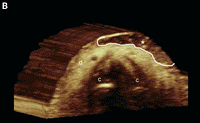
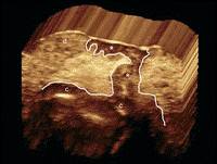
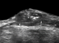
Precise treatment is a major pillar of modern medicine. An important aspect to enable accurate administration of treatment is complementing the accurate identification of the organ location that needs to be treated with a system and methods that ensure application of treatment only, or mainly to, that location. Imaging is off-course, a major component in such composite systems. Amongst the available solution, functional-imaging modalities are gaining traction. Specifically, molecular imaging (e.g. PET, MRS) allows the visual representation, characterization, and quantification of biological processes at the cellular and subcellular levels within intact living organisms. In oncology, it can be used to depict the abnormal molecules as well as the aberrant interactions of altered molecules on which cancers depend. Being able to detect such fundamental finger-prints of cancer is key to improved matching between drugs-based treatment and disease. Moreover, imaging-based quantified monitoring of changes in tumor metabolism and its microenvironment could provide real-time non-invasive tool to predict the evolution and progression of primary tumors, as well as the development of tumor metastases.
A recent review-paper: Image-guided interventional therapy for cancer with radiotherapeutic nanoparticles nicely illustrates the role of imaging in treatment guidance through a comprehensive discussion of; Image-guided radiotherapeutic using intravenous nanoparticles for the delivery of localized radiation to solid cancer tumors.

Abstract
One of the major limitations of current cancer therapy is the inability to deliver tumoricidal agents throughout the entire tumor mass using traditional intravenous administration. Nanoparticles carrying beta-emitting therapeutic radionuclides [DN: radioactive isotops that emits electrons as part of the decay process a list of β-emitting radionuclides used in radiotherapeutic nanoparticle preparation is given in table1 of this paper.) that are delivered using advanced image-guidance have significant potential to improve solid tumor therapy. The use of image-guidance in combination with nanoparticle carriers can improve the delivery of localized radiation to tumors. Nanoparticles labeled with certain beta-emitting radionuclides are intrinsically theranostic agents that can provide information regarding distribution and regional dosimetry within the tumor and the body. Image-guided thermal therapy results in increased uptake of intravenous nanoparticles within tumors, improving therapy. In addition, nanoparticles are ideal carriers for direct intratumoral infusion of beta-emitting radionuclides by convection enhanced delivery, permitting the delivery of localized therapeutic radiation without the requirement of the radionuclide exiting from the nanoparticle. With this approach, very high doses of radiation can be delivered to solid tumors while sparing normal organs. Recent technological developments in image-guidance, convection enhanced delivery and newly developed nanoparticles carrying beta-emitting radionuclides will be reviewed. Examples will be shown describing how this new approach has promise for the treatment of brain, head and neck, and other types of solid tumors.
The challenges this review discusses
- intravenously administered drugs are inhibited in their intratumoral penetration by high interstitial pressures which prevent diffusion of drugs from the blood circulation into the tumor tissue [1–5].
- relatively rapid clearance of intravenously administered drugs from the blood circulation by kidneys and liver.
- drugs that do reach the solid tumor by diffusion are inhomogeneously distributed at the micro-scale – This cannot be overcome by simply administering larger systemic doses as toxicity to normal organs is generally the dose limiting factor.
- even nanoparticulate drugs have poor penetration from the vascular compartment into the tumor and the nanoparticles that do penetrate are most often heterogeneously distributed
How imaging could mitigate the above mentioned challenges
- The inclusion of an imaging probe during drug development can aid in determining the clearance kinetics and tissue distribution of the drug non-invasively. Such probe can also be used to determine the likelihood of the drug reaching the tumor and to what extent.
Note: Drugs that have increased accumulation within the targeted site are likely to be more effective as compared with others. In that respect, Nanoparticle-based drugs have an additional advantage over free drugs with their potential to be multifunctional carriers capable of carrying both therapeutic and diagnostic imaging probes (theranostic) in the same nanocarrier. These multifunctional nanoparticles can serve as theranostic agents and facilitate personalized treatment planning.
- Imaging can also be used for localization of the tumor to improve the placement of a catheter or external device within tumors to cause cell death through thermal ablation or oxidative stress secondary to reactive oxygen species.
See the example of Vintfolide in The Role of Medical Imaging in Personalized Medicine

Note: Image guided thermal ablation methods include radiofrequency (RF) ablation, microwave ablation or high intensity focused ultrasound (HIFU). Photodynamic therapy methods using external light devices to activate photosensitizing agents can also be used to treat superficial tumors or deeper tumors when used with endoscopic catheters.
- Quality control during and post treatment
For example: The use of high intensity focused ultrasound (HIFU) combined with nanoparticle therapeutics: HIFU is applied to improve drug delivery and to trigger drug release from nanoparticles. Gas-bubbles are playing the role of the drug’s nano-carrier. These are used both to increase the drug transport into the cell and as ultrasound-imaging contrast material. The ultrasound is also used for processes of drug-release and ablation.

Additional example; Multifunctional nanoparticles for tracking CED (convection enhanced delivery) distribution within tumors: Nanoparticle that could serve as a carrier not only for the therapeutic radionuclides but simultaneously also for a therapeutic drug and 4 different types of imaging contrast agents including an MRI contrast agent, PET and SPECT nuclear diagnostic imaging agents and optical contrast agents as shown below. The ability to perform multiple types of imaging on the same nanoparticles will allow studies investigating the distribution and retention of nanoparticles initially in vivo using non-invasive imaging and later at the histological level using optical imaging.

Conclusions
Image-guided radiotherapeutic nanoparticles have significant potential for solid tumor cancer therapy. The current success of this therapy in animals is most likely due to the improved accumulation, retention and dispersion of nanoparticles within solid tumor following image-guided therapies as well as the micro-field of the β-particle which reduces the requirement of perfectly homogeneous tumor coverage. It is also possible that the intratumoral distribution of nanoparticles may benefit from their uptake by intratumoral macrophages although more research is required to determine the importance of this aspect of intratumoral radionuclide nanoparticle therapy. This new approach to cancer therapy is a fertile ground for many new technological developments as well as for new understandings in the basic biology of cancer therapy. The clinical success of this approach will depend on progress in many areas of interdisciplinary research including imaging technology, nanoparticle technology, computer and robot assisted image-guided application of therapies, radiation physics and oncology. Close collaboration of a wide variety of scientists and physicians including chemists, nanotechnologists, drug delivery experts, radiation physicists, robotics and software experts, toxicologists, surgeons, imaging physicians, and oncologists will best facilitate the implementation of this novel approach to the treatment of cancer in the clinical environment. Image-guided nanoparticle therapies including those with β-emission radionuclide nanoparticles have excellent promise to significantly impact clinical cancer therapy and advance the field of drug delivery.