Feeds:
Posts
Comments

Archive for the ‘Hematology’ Category

Summary to Metabolomics

Summary to Metabolomics

Author and Curator: Larry H. Bernstein, MD, FCAP 

This concludes a long step-by-step journey into rediscovering biological processes from the genome as a framework to the remodeled and reconstituted cell through a number of posttranscription and posttranslation processes that modify the proteome and determine the metabolome.  The remodeling process continues over a lifetime. The process requires a balance between nutrient intake, energy utilization for work in the lean body mass, energy reserves, endocrine, paracrine and autocrine mechanisms, and autophagy.  It is true when we look at this in its full scope – What a creature is man?

http://masspec.scripps.edu/metabo_science/recommended_readings.php
 Recommended Readings and Historical Perspectives

Metabolomics is the scientific study of chemical processes involving metabolites. Specifically, metabolomics is the “systematic study of the unique chemical fingerprints that specific cellular processes leave behind”, the study of their small-molecule metabolite profiles.[1] The metabolome represents the collection of all metabolites in a biological cell, tissue, organ or organism, which are the end products of cellular processes.[2] mRNA gene expression data and proteomic analyses reveal the set of gene products being produced in the cell, data that represents one aspect of cellular function. Conversely, metabolic profiling can give an instantaneous snapshot of the physiology of that cell. One of the challenges of systems biology and functional genomics is to integrate proteomic, transcriptomic, and metabolomic information to provide a better understanding of cellular biology.

The term “metabolic profile” was introduced by Horning, et al. in 1971 after they demonstrated that gas chromatography-mass spectrometry (GC-MS) could be used to measure compounds present in human urine and tissue extracts. The Horning group, along with that of Linus Pauling and Arthur B. Robinson led the development of GC-MS methods to monitor the metabolites present in urine through the 1970s.

Concurrently, NMR spectroscopy, which was discovered in the 1940s, was also undergoing rapid advances. In 1974, Seeley et al. demonstrated the utility of using NMR to detect metabolites in unmodified biological samples.This first study on muscle highlighted the value of NMR in that it was determined that 90% of cellular ATP is complexed with magnesium. As sensitivity has improved with the evolution of higher magnetic field strengths and magic angle spinning, NMR continues to be a leading analytical tool to investigate metabolism. Efforts to utilize NMR for metabolomics have been influenced by the laboratory of Dr. Jeremy Nicholson at Birkbeck College, University of London and later at Imperial College London. In 1984, Nicholson showed 1H NMR spectroscopy could potentially be used to diagnose diabetes mellitus, and later pioneered the application of pattern recognition methods to NMR spectroscopic data.

In 2005, the first metabolomics web database, METLIN, for characterizing human metabolites was developed in the Siuzdak laboratory at The Scripps Research Institute and contained over 10,000 metabolites and tandem mass spectral data. As of September 2012, METLIN contains over 60,000 metabolites as well as the largest repository of tandem mass spectrometry data in metabolomics.

On 23 January 2007, the Human Metabolome Project, led by Dr. David Wishart of the University of Alberta, Canada, completed the first draft of the human metabolome, consisting of a database of approximately 2500 metabolites, 1200 drugs and 3500 food components. Similar projects have been underway in several plant species, most notably Medicago truncatula and Arabidopsis thaliana for several years.

As late as mid-2010, metabolomics was still considered an “emerging field”. Further, it was noted that further progress in the field depended in large part, through addressing otherwise “irresolvable technical challenges”, by technical evolution of mass spectrometry instrumentation.

Metabolome refers to the complete set of small-molecule metabolites (such as metabolic intermediates, hormones and other signaling molecules, and secondary metabolites) to be found within a biological sample, such as a single organism. The word was coined in analogy with transcriptomics and proteomics; like the transcriptome and the proteome, the metabolome is dynamic, changing from second to second. Although the metabolome can be defined readily enough, it is not currently possible to analyse the entire range of metabolites by a single analytical method. The first metabolite database(called METLIN) for searching m/z values from mass spectrometry data was developed by scientists at The Scripps Research Institute in 2005. In January 2007, scientists at the University of Alberta and the University of Calgary completed the first draft of the human metabolome. They catalogued approximately 2500 metabolites, 1200 drugs and 3500 food components that can be found in the human body, as reported in the literature. This information, available at the Human Metabolome Database (www.hmdb.ca) and based on analysis of information available in the current scientific literature, is far from complete.

Each type of cell and tissue has a unique metabolic ‘fingerprint’ that can elucidate organ or tissue-specific information, while the study of biofluids can give more generalized though less specialized information. Commonly used biofluids are urine and plasma, as they can be obtained non-invasively or relatively non-invasively, respectively. The ease of collection facilitates high temporal resolution, and because they are always at dynamic equilibrium with the body, they can describe the host as a whole.

Metabolites are the intermediates and products of metabolism. Within the context of metabolomics, a metabolite is usually defined as any molecule less than 1 kDa in size.
A primary metabolite is directly involved in the normal growth, development, and reproduction. A secondary metabolite is not directly involved in those processes.  By contrast, in human-based metabolomics, it is more common to describe metabolites as being either endogenous (produced by the host organism) or exogenous. Metabolites of foreign substances such as drugs are termed xenometabolites. The metabolome forms a large network of metabolic reactions, where outputs from one enzymatic chemical reaction are inputs to other chemical reactions.

Metabonomics is defined as “the quantitative measurement of the dynamic multiparametric metabolic response of living systems to pathophysiological stimuli or genetic modification”. The word origin is from the Greek μεταβολή meaning change and nomos meaning a rule set or set of laws. This approach was pioneered by Jeremy Nicholson at Imperial College London and has been used in toxicology, disease diagnosis and a number of other fields. Historically, the metabonomics approach was one of the first methods to apply the scope of systems biology to studies of metabolism.

There is a growing consensus that ‘metabolomics’ places a greater emphasis on metabolic profiling at a cellular or organ level and is primarily concerned with normal endogenous metabolism. ‘Metabonomics’ extends metabolic profiling to include information about perturbations of metabolism caused by environmental factors (including diet and toxins), disease processes, and the involvement of extragenomic influences, such as gut microflora. This is not a trivial difference; metabolomic studies should, by definition, exclude metabolic contributions from extragenomic sources, because these are external to the system being studied.

Toxicity assessment/toxicology. Metabolic profiling (especially of urine or blood plasma samples) detects the physiological changes caused by toxic insult of a chemical (or mixture of chemicals).

Functional genomics. Metabolomics can be an excellent tool for determining the phenotype caused by a genetic manipulation, such as gene deletion or insertion. Sometimes this can be a sufficient goal in itself—for instance, to detect any phenotypic changes in a genetically-modified plant intended for human or animal consumption. More exciting is the prospect of predicting the function of unknown genes by comparison with the metabolic perturbations caused by deletion/insertion of known genes.

Nutrigenomics is a generalised term which links genomics, transcriptomics, proteomics and metabolomics to human nutrition. In general a metabolome in a given body fluid is influenced by endogenous factors such as age, sex, body composition and genetics as well as underlying pathologies. The large bowel microflora are also a very significant potential confounder of metabolic profiles and could be classified as either an endogenous or exogenous factor. The main exogenous factors are diet and drugs. Diet can then be broken down to nutrients and non- nutrients.

http://en.wikipedia.org/wiki/Metabolomics

Jose Eduardo des Salles Roselino

The problem with genomics was it was set as explanation for everything. In fact, when something is genetic in nature the genomic reasoning works fine. However, this means whenever an inborn error is found and only in this case the genomic knowledge afterwards may indicate what is wrong and not the completely way to put biology upside down by reading everything in the DNA genetic as well as non-genetic problems.

Coordination of the transcriptome and metabolome by the circadian clock PNAS 2012

Coordination of the transcriptome and metabolome by the circadian clock PNAS 2012

analysis of metabolomic data and differential metabolic regulation for fetal lungs, and maternal blood plasma

conformational changes leading to substrate efflux.img

conformational changes leading to substrate efflux.img

The cellular response is defined by a network of chemogenomic response signatures.

The cellular response is defined by a network of chemogenomic response signatures.

Dynamic Construct of the –Omics

Dynamic Construct of the –Omics

 genome cartoon

genome cartoon

central dogma phenotype

central dogma phenotype

Read Full Post »

Transcription Modulation

Author and Curator: Larry H. Bernstein, MD, FCAP 


This portion of the transcription series deals with transcription factors and the effects of their binding on metabolism. This also has implications for pharmaceutical target identification.

The Functional Consequences of Variation in Transcription Factor Binding
DA. Cusanovich, B Pavlovic, JK. Pritchard*, Y Gilad*
1 Department of Human Genetics, 2 Howard Hughes Medical Institute, University of Chicago, Chicago, IL 3 Departments of Genetics and Biology and Howard Hughes Medical Institute, Stanford University, Stanford, CA.
PLoS Genet 2014;10(3):e1004226.  http://dx.doi.org:/10.1371/journal.pgen.1004226

One goal of human genetics is to understand how the information for precise and dynamic gene expression programs is encoded in the genome. The interactions of transcription factors (TFs) with DNA regulatory elements clearly

  • play an important role in determining gene expression outputs, yet
  • the regulatory logic underlying functional transcription factor binding is poorly understood.

An important question in genomics is to understand how a class of proteins called ‘‘transcription factors’’ controls the expression level of other genes in the genome in a cell type-specific manner – a process that is essential to human development. One major approach to this problem is to study where these transcription factors bind in the genome, but this does not tell us about the effect of that binding on gene expression levels and

  • it is generally accepted that much of the binding does not strongly influence gene expression.

To address this issue, we artificially reduced the concentration of 59 different transcription factors in the cell and then

  • examined which genes were impacted by the reduced transcription factor level.

Our results implicate some attributes

  • that might influence what binding is functional, but they also suggest that
  • a simple model of functional vs. non-functional binding may not suffice.

Many studies have focused on characterizing the genomic locations of TF binding, but

  • it is unclear whether TF binding at any specific locus has
  • functional consequences with respect to gene expression output.

We knocked down 59 TFs and chromatin modifiers in one HapMap lymphoblastoid cell line

  • to evaluate the context of functional TF binding.

We then identified genes whose expression was affected by the knockdowns

  • by intersecting the gene expression data with transcription factor binding data
    (based on ChIP-seq and DNase-seq)
  • within 10 kb of the transcription start sites of expressed genes.

This combination of data allowed us to infer functional TF binding.
Only a small subset of genes bound by a factor were

  • differentially expressed following the knockdown of that factor,
  • suggesting that most interactions between TF and chromatin
  • do not result in measurable changes in gene expression levels
  • of putative target genes.

We found that functional TF binding is enriched

  • in regulatory elements that harbor a large number of TF binding sites,
  • at sites with predicted higher binding affinity, and
  • at sites that are enriched in genomic regions annotated as ‘‘active enhancers.’’

We aim to be able to predict the expression pattern of a gene based on its regulatory
sequence alone. However, the regulatory code of the human genome is much more complicated than

  • the triplet code of protein coding sequences, and is highly context-specific,
  • depending on cell-type and other factors.

Moreover, regulatory regions are not necessarily organized into

  • discrete, easily identifiable regions of the genome and
  • may exert their influence on genes over large genomic distances

Genomic studies addressing questions of the regulatory logic of the human genome have largely taken one of two approaches.

  1. collecting transcription factor binding maps using techniques such as ChIPseq
    and DNase-seq
  2. mapping various quantitative trait loci (QTL), such as gene expression levels
    (eQTLs) [7], DNA methylation (meQTLs) [8] and chromatin accessibility (dsQTLs)

Cumulatively, binding map studies and QTL map studies have

  • led to many insights into the principles and mechanisms of gene regulation.

However, there are questions that neither mapping approach on its own is well equipped to address. One outstanding issue is

  • the fraction of factor binding in the genome that is ‘‘functional’’,
    which we define here to mean that
  • disturbing the protein-DNA interaction leads to a measurable
  • downstream effect on gene regulation.

Transcription factor knockdown could be used to address this problem, whereby

  • the RNA interference pathway is employed to greatly reduce
  • the expression level of a specific target gene by using small interfering RNAs (siRNAs).

The response to the knockdown can then be measured by collecting RNA after the knockdown and

  • measuring global changes in gene expression patterns
  • after specifically attenuating the expression level of a given factor.

Combining a TF knockdown approach with TF binding data can help us to

  • distinguish functional binding from non-functional binding

This approach has previously been applied to the study of human TFs, although for the most part studies have only focused on

  • the regulatory relationship of a single factor with its downstream targets.

The FANTOM consortium knocked down 52 different transcription factors in

  • the THP-1 cell line, an acute monocytic leukemia-derived cell line, and
  • used a subset of these to validate certain regulatory predictions based on binding motif enrichments.

We and others previously studied the regulatory architecture of gene expression in

  • the model system of HapMap lymphoblastoid cell lines (LCLs) using both
  • binding map strategies and QTL mapping strategies.

We now sought to use knockdown experiments targeting transcription factors in a HapMap LCL

  • to refine our understanding of the gene regulatory circuitry of the human genome.

Therefore, We integrated the results of the knockdown experiments with previous data on TF binding to

  • better characterize the regulatory targets of 59 different factors and
  • to learn when a disruption in transcription factor binding
  • is most likely to be associated with variation in the expression level of a nearby gene.

Gene expression levels following the knockdown were compared to

  • expression data collected from six samples that were transfected with negative control siRNA.

The expression data from all samples were normalized together using

  • quantile  normalization followed by batch correction using the RUV-2 method.

We then performed several quality control analyses to confirm

  1. that the quality of the data was high,
  2. that there were no outlier samples, and
  3. that the normalization methods reduced the influence of confounders

In order to identify genes that were expressed at a significantly different level

  • in the knockdown samples compared to the negative controls,
  • we used likelihood-ratio tests within the framework of a fixed effect linear model.

Following normalization and quality control of the arrays,

  • we identified genes that were differentially expressed between
  • the three knockdown replicates of each factor and the six controls.

Depending on the factor targeted, the knockdowns resulted in

  • between 39 and 3,892 differentially expressed genes at an FDR of 5%
    (Figure 1B; see Table S3 for a summary of the results).

The knockdown efficiency for the 59 factors ranged

  • from 50% to 90% (based on qPCR; Table S1).

The qPCR measurements of the knockdown level were significantly

  • correlated with estimates of the TF expression levels
  • based on the microarray data (P =0.001; Figure 1C).

Reassuringly, we did not observe a significant correlation between

  • the knockdown efficiency of a given factor and
  • the number of genes classified as differentially expressed foci.

Because we knocked down 59 different factors in this experiment

  • we were able to assess general patterns associated with the perturbation of transcription factors
  • beyond merely the number of affected target genes.

Globally, despite the range in the number of genes we identified as

  • differentially expressed in each knockdown,
  • the effect sizes of the differences in expression were relatively modest and
  • consistent in magnitude across all knockdowns.

The median effect size following the knockdown experiment for genes classified as

  • differentially expressed at an FDR of 5% in any knockdown was
  • a 9.2% difference in expression level between the controls and the knockdown (Figure 2),
  • while the median effect size for any individual knockdown experiment ranged between 8.1% and 11.0%.
    (this was true whether we estimated the knockdown effect based on qPCR (P = 0.10; Figure 1D) or microarray (P = 0.99; not shown) data.

Nor did we observe a correlation between

  • variance in qPCR-estimated knockdown efficiency (between replicates) and
  • the number of genes differentially expressed (P = 0.94; Figure 1E).

We noticed that the large variation in the number of differentially expressed genes

  • extended even to knockdowns of factors from the same gene family.

Figure 1. Differential expression analysis.
(a) Examples of differential expression analysis results for the genes HCST and IRF4. The top two panels are ‘MA plots’ of the mean Log2(expression level) between the knockdown arrays and the controls for each gene (x-axis) to the Log2(Fold-Change) between the knockdowns and controls (y-axis). Differentially expressed genes at an FDR of 5% are plotted in yellow (points 50% larger). The gene targeted by the siRNA is highlighted in red. The bottom two panels are ‘volcano plots’ of the Log2(Fold-Change) between the knockdowns and controls (x-axis) to the P-value for differential expression (y-axis). The dashed line marks the 5% FDR threshold. Differentially expressed genes at an FDR of 5% are plotted in yellow (points 50% larger). The red dot marks the gene targeted by the siRNA.
(b) Barplot of number of differentially expressed genes in each knockdown experiment.
(c) Comparison of the knockdown level measured by qPCR (RNA sample collected 48 hours posttransfection) and the knockdown level measured by microarray.
(d) Comparison of the level of knockdown of the transcription factor at 48 hrs (evaluated by qPCR; x-axis) and the number of genes differentially expressed in the knockdown experiment (y-axis).
(e) Comparison of the variance in knockdown efficiency between replicates for each transcription factor (evaluated by qPCR; x-axis) and the number of differentially expressed genes in the knockdown experiment (y-axis).

Differential expression analysis

Differential expression analysis

http://dx.doi.org:/10.1371/journal.pgen.1004226.g001

Figure 2. Effect sizes for differentially expressed genes.
Boxplots of absolute Log2(fold-change) between knockdown arrays and control arrays for all genes identified as differentially expressed in each experiment. Outliers are not plotted. The gray bar indicates the interquartile range across all genes differentially expressed in all knockdowns. Boxplots are ordered by the number of genes differentially expressed in each experiment. Outliers were not plotted.

Effect sizes for differentially expressed genes

Effect sizes for differentially expressed genes

http://dx.doi.org:/10.1371/journal.pgen.1004226.g002

Knocking down SREBF2 (1,286 genes differentially expressed), a key regulator of cholesterol homeostasis,

  • results in changes in the expression of genes that are
  • significantly enriched for cholesterol and sterol biosynthesis annotations.

While not all factors exhibited striking enrichments for relevant functional categories and pathways,

  • the overall picture is that perturbations of many of the factors
  • primarily affected pathways consistent with their known biology.

In order to assess functional TF binding, we next incorporated

  • binding maps together with the knockdown expression data.

We combined binding data based on DNase-seq footprints in 70 HapMap LCLs, reported by Degner et al. (Table S5)

  • and from ChIP-seq experiments in LCL GM12878, published by ENCODE.

We were thus able to obtain genome wide binding maps for a total of 131 factors that were either

  • directly targeted by an siRNA in our experiment (29 factors) or were
  • differentially expressed in one of the knockdown experiments.

We classified a gene as a bound target of a particular factor when

  • binding of that factor was inferred within 10kb of the transcription start site (TSS) of the target gene.

Using this approach, we found that the 131 TFs were bound

  • in proximity to a median of 1,922 genes per factor (range 11 to 7,053 target genes).

We considered binding of a factor to be functional if the target gene

  • was differentially expressed after perturbing the expression level the bound transcription factor.

We then asked about the concordance between

  • the transcription factor binding data and the knockdown expression data.
  •  the extent to which differences in gene expression levels following the knockdowns
  • might be predicted by binding of the transcription factors
  • within the putative regulatory regions of the responsive genes. and also
  • what proportion of putative target (bound) genes of a given TF were
  • differentially expressed following the knockdown of the factor.

Focusing only on the binding sites classified using the DNase-seq data
(which were assigned to a specific instance of the binding motif, unlike the ChIP data),

  • we examined sequence features that might distinguish functional binding.

In particular, whether binding at conserved sites was more likely to be functional  and

  • whether binding sites that better matched the known PWM for the factor were more likely to be functional.

We did not observe a significant shift in the conservation of functional binding sites (Wilcoxon rank sum P = 0.34),

  • but we did observe that binding around differentially expressed genes occurred at sites
  • that were significantly better matches to the canonical binding motif.

Figure 3. Intersecting binding data and expression data for each knockdown.
(a) Example Venn diagrams showing the overlap of binding and differential expression for the knockdowns of HCST and IRF4 (the same genes as in Figure 1).
(b) Boxplot summarizing the distribution of the fraction of all expressed genes that are bound by the targeted gene or downstream factors.
(c) Boxplot summarizing the distribution of the fraction of bound genes that are classified as differentially expressed, using an FDR of either 5% or 20%.

Intersecting binding data and expression data for each knockdown

Intersecting binding data and expression data for each knockdown

http://dx.doi.org:/10.1371/journal.pgen.1004226.g003

Considering bound targets determined from either the ChIP-seq or DNase-seq data, we observed that

  • differentially expressed genes were associated with both
  • a higher number of binding events for the relevant factors within 10 kb of the TSS (P,10216; Figure 4A)
  • as well as with a larger number of different binding factors
    (considering the siRNA-targeted factor and any TFs that were DE in the knockdown; P,10216; Figure 4B).

Figure 4. Degree of binding correlated with function. Boxplots comparing
(a) the number of sites bound, and
(b) the number of differentially expressed transcription factors binding events near functionally or non-functionally bound genes. We considered binding for siRNA-targeted factor and any factor differentially expressed in the knockdown.
(c) Focusing only on genes differentially expressed in common between each pairwise set of knockdowns we tested for enrichments of functional binding (y-axis). Pairwise comparisons between knockdown experiments were binned by the fraction of differentially expressed transcription factors in common between the two experiments. For these boxplots, outliers were not plotted.

Degree of binding correlated with function

Degree of binding correlated with function

http://dx.doi.org:/10.1371/journal.pgen.1004226.g004

We examined the distribution of binding about the TSS. Most factor binding was concentrated

  • near the TSS whether or not the genes were classified as differentially expressed (Figure 5A).
  • the distance from the TSS to the binding sites was significantly longer for differentially expressed genes (P,10216; Fig. 5B).

Figure 5. Distribution of functional binding about the TSS.
(a) A density plot of the distribution of bound sites within 10 kb of the TSS for both functional and non-functional genes. Inset is a zoom-in of the region +/21 kb from the TSS (b) Boxplots comparing the distances from the TSS to the binding sites for functionally bound genes and non-functionally bound genes. For the boxplots, 0.001 was added before log10 transforming the distances and outliers were not plotted.

Distribution of functional binding about the TSS

Distribution of functional binding about the TSS

http://dx.doi.doi:/10.1371/journal.pgen.1004226.g005

We investigated the distribution of factor binding across various chromatin states, as defined by Ernst et al. This dataset lists

  • regions of the genome that have been assigned to different activity states
  • based on ChIP-seq data for various histone modifications and CTCF binding.

For each knockdown, we separated binding events

  • by the genomic state in which they occurred and then
  • tested whether binding in that state was enriched around differentially expressed genes.

After correcting for multiple testing of genes that were differentially expressed.

  • 19 knockdowns showed significant enrichment for binding in ‘‘strong enhancers’’
  • four knockdowns had significant enrichments for ‘‘weak enhancers’’,
  • eight knockdowns showed significant depletion of binding in ‘‘active promoters’’ ,
  • six knockdowns had significant depletions for ‘‘transcription elongation’’,

Did the factors tended to have a consistent effect (either up- or down-regulation)

  • on the expression levels of genes they purportedly regulated?

All factors we tested are associated with both up- and down-regulation of downstream targets (Figure 6).

A slight majority of downstream target genes were expressed at higher levels

  • following the knockdown for 15 of the 29 factors for which we had binding information (Figure 6B).

The factor that is associated with the largest fraction (68.8%) of up-regulated target genes following the knockdown is EZH2,

  • the enzymatic component of the Polycomb group complex.

On the other end of the spectrum was JUND, a member of the AP-1 complex, for which

  • 66.7% of differentially expressed targets were down-regulated following the knockdown.

Figure 6. Magnitude and direction of differential expression after knockdown.
(a) Density plot of all Log2(fold-changes) between the knockdown arrays and controls for genes that are differentially expressed at 5% FDR in one of the knockdown experiments as well as bound by the targeted transcription factor.
(b) Plot of the fraction of differentially expressed putative direct targets that were up-regulated in each of the knockdown experiments.

Magnitude and direction of differential expression after knockdown

Magnitude and direction of differential expression after knockdown

http://dx.doi.org:/10.1371/journal.pgen.1004226.g006

We found no correlation between the number of paralogs and the fraction of bound targets that were differentially expressed. We also did not observe a significant correlation when we considered whether

  • the percent identity of the closest paralog might be predicative of
  • the fraction of bound genes that were differentially expressed following the knockdown (Figure S8).

While there is compelling evidence for our inferences, the current chromatin functional annotations

  • do not fully explain the regulatory effects of the knockdown experiments.

For example, the enrichments for binding in ‘‘strong enhancer’’ regions of the genome range from 7.2% to 50.1% (median = 19.2%),

  • much beyond what is expected by chance alone, but far from accounting for all functional binding.

In addition to considering

  • the distinguishing characteristics of functional binding, we also examined
  • the direction of effect that perturbing a transcription factor had on the expression level of its direct targets.

We specifically addressed whether

  • knocking down a particular factor tended to drive expression of its putatively direct (namely, bound) targets up or down,
  • which can be used to infer that the factor represses or activates the target, respectively.

Transcription factors have traditionally been thought of primarily as activators, and previous work from our group is consistent with that notion. Surprisingly, the most straightforward inference from the present study is that

  • many of the factors function as repressors at least as often as they function as activators.
  1. EZH2 had a negative regulatory relationship with the largest fraction of direct targets (68.8%),
    consistent with – the known role of EZH2 as the active member of the Polycomb group complex PC2
  2. while JUND seemed to have a positive regulatory relationship with the largest fraction of direct targets (66.7%),
    and with – the biochemical characterization of the AP-1 complex (of which JUND is a component) as a transactivator.

More generally, however, our results, combined with the previous work from our group and others make for a complicated view

  • of the role of transcription factors in gene regulation as
  • it seems difficult to reconcile the inference from previous work that
  • many transcription factors should primarily act as activators with the results presented here.

One somewhat complicated hypothesis, which nevertheless can resolve the apparent discrepancy, is that

  • the ‘‘repressive’’ effects we observe for known activators may be
  • at sites in which the activator is acting as a weak enhancer of transcription and
  • that reducing the cellular concentration of the factor
  • releases the regulatory region to binding by an alternative, stronger activator.

To more explicitly address the effect that our proximity-based definition of target genes might have on our analyses, we reanalyzed

  • the overlap between factor binding and differential expression following the knockdowns
  • using an independent, empirically determined set of target genes.

Thurman et al. used correlations in DNase hypersensitivity between

  • intergenic hypersensitive sites and promoter hypersensitive sites across diverse tissues
  • to assign intergenic regulatory regions to specific genes,
  • independently of proximity to a particular promoter.

We performed this alternative analysis in which we

  • assigned binding events to genes based on the classification of Thurman et al.

We then considered the overlap between binding and differential expression in this new data set. The results were largely

  • consistent with our proximity-based observations.

A median of 9.5% of genes that were bound by a factor were

  • also differentially expressed following the knockdown of that factor
    (compared to 11.1% when the assignment of binding sites to genes is based on proximity).

From the opposite perspective, a median of 28.0% of differentially expressed genes were bound by that factor
(compared to 32.3% for the proximity based definition). The results of this analysis are summarized in Table S7.

Our results should not be considered a comprehensive census of regulatory events in the human genome. Instead, we adopted a gene-centric approach,

  • focusing only on binding events near the genes for which we could measure expression
  • to learn some of the principles of functional transcription factor binding.

In light of our observations a reassessment of our estimates of binding may be warranted. In particular, because functional binding is skewed away from promoters (our system is apparently not well-suited to observe functional promoter binding, perhaps because of protection by large protein complexes),

  • a more conservative estimate of the fraction of binding that is indeed functional would not consider data within the promoter.

Importantly, excluding the putative promoter region from our analysis (i.e. only considering a window .1 kb from the TSS and ,10 kb from the TSS)

  • does not change our conclusions.

Considering this smaller window,

  • a median of 67.0% of expressed genes are still classified as bound by
  1. either the knocked down transcription factor or
  2. a downstream factors that is differentially expressed in each experiment,

yet a median of only 8.1% of the bound genes are

  • also differentially expressed after the knockdowns.

Much of what distinguishes functional binding (as we define it) has yet to be explained. We are unable to explain much of the differential expression observed in our experiments by the presence of least one relevant binding event. This may not be altogether surprising, as

  • we are only considering binding in a limited window around the transcription start site.

To address these issues, more factors should be perturbed to further evaluate the robustness of our results and to add insight. Together, such studies will help us develop a more sophisticated understanding of functional transcription factor binding in particular, the gene regulatory logic more generally.

Assessing quality and completeness of human transcriptional regulatory pathways on a genome-wide scale

E Shmelkov, Z Tang, I Aifantis, A Statnikov*
Biology Direct 2011; 6(15).  http://www.biology-direct.com/content/6/1/15

Recently the biological pathways have become a common and probably the most popular form of representing biochemical information for hypothesis generation and validation. These maps store wide knowledge of complex molecular interactions and regulations occurring in the living organism in a simple and obvious way, often using intuitive graphical notation. Two major types of biological pathways could be distinguished.

  1. Metabolic pathways incorporate complex networks of protein-based interactions and modifications, while
  2. signal transduction and transcriptional regulatory pathways are usually considered to provide information on mechanisms of transcription

While there are a lot of data collected on human metabolic processes,

  • the content of signal transduction and transcriptional regulatory pathways varies greatly in quality and completeness.

An indicative comparison of MYC transcriptional targets reported in ten different pathway databases reveals that these databases differ greatly from each other (Figure 1). Given that MYC is involved

  • in the transcriptional regulation of approximately 15% of all genes,

one cannot argue that the majority of pathway databases that contain

  • less than thirty putative transcriptional targets of MYC are even close to complete.

More importantly, to date there have been no prior genome-wide evaluation studies (that are based on genome-wide binding and gene expression assays) assessing pathway databases

Background: While pathway databases are becoming increasingly important in most types of biological and translational research, little is known about the quality and completeness of pathways stored in these databases. The present study conducts a comprehensive assessment of transcriptional regulatory pathways in humans for seven well-studied transcription factors:

  1. MYC,
  2. NOTCH1,
  3. BCL6,
  4. TP53,
  5. AR,
  6. STAT1,
  7. RELA.

The employed benchmarking methodology first involves integrating

  • genome-wide binding with functional gene expression data
  • to derive direct targets of transcription factors.

Then the lists of experimentally obtained direct targets

  • are compared with relevant lists of transcriptional targets from 10 commonly used pathway databases.

Results: The results of this study show that for the majority of pathway databases,

  • the overlap between experimentally obtained target genes and
  • targets reported in transcriptional regulatory pathway databases is
  • surprisingly small and often is not statistically significant.

The only exception is MetaCore pathway database which

  • yields statistically significant intersection with experimental results in 84% cases.

The lists of experimentally derived direct targets obtained in this study can be used

  • to reveal new biological insight in transcriptional regulation,  and we
  • suggest novel putative therapeutic targets in cancer.

Conclusions: Our study opens a debate on validity of using many popular pathway databases to obtain transcriptional regulatory targets. We conclude that the choice of pathway databases should be informed by

  • solid scientific evidence and rigorous empirical evaluation.

In the current study we perform

(1) an evaluation of ten commonly used pathway databases,

  • assessing the transcriptional regulatory pathways, considered in the current study as
  • the interactions of the type ‘transcription factor-transcriptional targets’.

This involves integration of human genome wide functional microarray or RNA-seq gene expression data with

  • protein-DNA binding data from ChIP-chip, ChIP-seq, or ChIP-PET platforms
  • to find direct transcriptional targets of the seven well known transcription factors:
  • MYC, NOTCH1, BCL6, TP53, AR, STAT1, and RELA.

The choice of transcription factors is based on their important role in oncogenesis and availability of binding and expression data in the public domain.

(2) the lists of experimentally derived direct targets are used to assess the quality and completeness of 84 transcriptional regulatory pathways from four publicly available (BioCarta, KEGG, WikiPathways and Cell Signaling Technology) and six commercial (MetaCore, Ingenuity Pathway Analysis, BKL TRANSPATH, BKL TRANSFAC, Pathway Studio and GeneSpring Pathways) pathway databases.

(3) We measure the overlap between pathways and experimentally obtained target genes and assess statistical significance of this overlap, and we demonstrate that experimentally derived lists of direct transcriptional targets

  • can be used to reveal new biological insight on transcriptional regulation.

We show this by analyzing common direct transcriptional targets of

  • MYC, NOTCH1 and RELA
  • that act in interconnected molecular pathways.

Detection of such genes is important as it could reveal novel targets of cancer therapy.

Figure 1 Number of genes in common between MYC transcriptional targets derived from ten different pathway databases. Cells are colored according to their values from white (low values) to red (high values). (not shown)

statistical methodology for comparison

statistical methodology for comparison

Figure 2 Illustration of statistical methodology for comparison between a gold-standard and a pathway database

Since we are seeking to compare gene sets from different studies/databases, it is essential to transform genes to standard identifiers. That is why we transformed all
gene sets to the HUGO Gene Nomenclature Committee approved gene symbols and names. In order to assess statistical significance of the overlap between the resulting gene sets, we used the hypergeometric test at 5% a-level with false discovery rate correction for multiple comparisons by the method of Benjamini and Yekutieli. The alternative hypothesis of this test is that two sets of genes (set A from pathway
database and set B from experiments) have greater number of genes in common than two randomly selected gene sets with the same number of genes as in sets A and B. For example, consider that for some transcription factor there are 300 direct targets in the pathway database #1 and 700 in the experimentally derived list (gold-standard), and their intersection is 16 genes (Figure 2a). If we select on random from a total of
20,000 genes two sets with 300 and 700 genes each, their overlap would be greater or equal to 16 genes in 6.34% times. Thus, this overlap will not be statistically significant at 5% a-level (p = 0.0634). On the other hand, consider that for the pathway database #2, there are 30 direct targets of that transcription factor, and their intersection with the 700-gene gold-standard is only 6 genes. Even though the size of this intersection is rather small, it is unlikely to randomly select 30 genes (out of 20,000) with an overlap greater or equal to 6 genes with a 700-gene gold-standard (p = 0.0005, see Figure 2a). This overlap is statistically significant at 5% a-level.

We also calculate an enrichment fold change ratio (EFC) for every intersection between a gold-standard and a pathway database. For a given pair of a gold-standard and a pathway database, EFC is equal to the observed number of genes in their intersection, divided by the expected size of intersection under the null hypothesis (plus machine epsilon, to avoid division by zero). Notice however that larger values of EFC may correspond to databases that are highly incomplete and contain only a few relations. For example, consider that for some transcription factor there are 300 direct targets in the pathway database #1 and 50 in the experimentally derived list (gold-standard), and their intersection is 30 genes (Figure 2b). If we select on random from a total of 20,000 genes two sets with 300 and 50 genes each, their expected overlap under the null hypothesis will be equal to 0.75. Thus, the EFC ratio will be equal to 40 (= 30/0.75). On the other hand, consider that for the pathway database #2, there are 2 direct
targets of that transcription factor, and their intersection with the 50-gene gold-standard is only 1 gene. Even though the expected overlap under the null hypothesis will be equal to 0.005 and EFC equal to 200 (5 times bigger than for the database #1), the size of this intersection with the gold-standard is 30 times less than for database #1 (Figure 2b).

Figure 3 Comparison between different pathway databases and experimentally derived gold-standards for all considered transcription factors. Value in a given cell is a number of overlapping genes between a gold-standard and a pathway-derived gene set. Cells
are colored according to their values from white (low values) to red (high values). Underlined values in red represent statistically significant intersections. (not shown)

Figure 4 Summary of the pathway databases assessment. Green cells represent statistically significant intersections between experimentally derived gold-standards and transcriptional regulatory pathways. White cells denote results that are not statistically significant. Numbers are the enrichment fold change ratios (EFC) calculated for each intersection. (not shown)

At the core of this study was creation of gold-standards of transcriptional regulation in humans that can be compared with target genes reported in transcriptional regulatory pathways. We focused on seven well known transcription factors and obtained gold-standards

  • by integrating genome-wide transcription factor-DNA binding data (from ChIP-chip, ChIP-seq, or ChIP-PET platforms)
  • with functional gene expression microarray and RNA-seq data.

The latter data allows to survey changes in the transcriptomes on a genome-wide scale

  • after the inhibition or over-expression of the transcription factor in question.

However, change in the expression of a particular gene could be caused either by the direct effect of the removal or introduction of a given transcription factor, as well as by an indirect effect, through the change in expression level of some other gene(s). It is essential

  • to integrate data from these two sources to
  • obtain an accurate list of gene targets that are directly regulated by a transcription factor.

It is worth noting that tested pathway databases typically do not give distinction between cell-lines, experimental conditions, and other details relevant to experimental systems in which data were obtained. These databases in a sense propose a ‘universal’ list of transcriptional targets. However, it is known that

  • transcriptional regulation in a cell is dynamic and works differently for different systems and stimuli.

This accentuates the major limitation of pathway databases and emphasizes

  • importance of deriving a specific list of transcriptional targets for the current experimental system.

In this study we followed the latter approach by developing gold-standards for specific cell characterized biological systems and experimental conditions.

The approach used here  for building gold-standards of direct mechanistic knowledge has several limitations. (see article).  Nevertheless, our results suggest that multiple transcription factors can co-operate and control both physiological differentiation and malignant transformation, as demonstrated utilizing combinatorial gene-profiling for

  • NOTCH1, MYC and RELA targets.

These studies might lead us to multi-pathway gene expression “signatures”

  • essential for the prediction of genes that could be targeted in cancer treatments.

In agreement with this hypothesis, several of the genes identified in our analysis have been suggested to be putative therapeutic targets in leukemia, with either preclinical or clinical trials underway (CDK4, CDK6, GSK3b, MYC, LCK, NFkB2, BCL2L1, NOTCH1).

Single-molecule tracking in live cells reveals distinct target-search strategies of transcription factors in the nucleus

I Izeddin†, V Récamier†‡, L Bosanac, II Cissé, L Boudarene, et al.
1Functional Imaging of Transcription, Institut de Biologie de l’Ecole Normale Supérieure (IBENS), Inserm, and CNRS UMR; 2Laboratoire Kastler Brossel, CNRS UMR, Departement de Physique et Institut de Biologie
de l’Ecole Normale Supérieure (IBENS), Paris, Fr; 3Transcription Imaging Consortium, Janelia Farm Research Campus, Howard Hughes Medical Institute, Ashburn, US; + more.
Biophysics and structural biology | Cell biology eLife 2014;3:e02230. http://dx.doi.org:/10.7554/eLife.02230

Transcription factors are

  • proteins that control the expression of genes in the nucleus, and
  • they do this by binding to other proteins or DNA.

First, however, these regulatory proteins need to overcome the challenge of

  • finding their targets in the nucleus, which is crowded with other proteins and DNA.

Much research to date has focused on measuring how fast proteins can diffuse and spread out throughout the nucleus. However these measurements only make sense if these proteins have access to the same space within the nucleus.

Now, Izeddin, Récamier et al. have developed a new technique to track

  • single protein molecules in the nucleus of mammalian cells.

A transcription factor called c-Myc and another protein called P-TEFb

  • were tracked and while they diffused at similar rates,
  • they ‘explored’ the space inside the nucleus in very different ways.

Izeddin, Récamier et al. found that c-Myc explores the nucleus in a so-called ‘non-compact’ manner: this means that it

  • can move almost everywhere inside the nucleus, and has an equal chance
  • of reaching any target regardless of its position in this space.

P-TEFb, on the other hand, searches

  • the nucleus in a ‘compact’ way.

This means that it is constrained to follow a specific path

  • through the nucleus and is therefore guided to its potential targets.

Izeddin, Récamier et al. explain that

  • the different ‘search strategies’ used by these two proteins
  • influence how long it takes them to find their targets and
  • how far they can travel in a given time.

These findings, together with information about

  • where and when different proteins interact in the nucleus,

will be essential to understand how the organization of the genome within the nucleus

  • can control the expression of genes.

The next challenge will now be to

  • uncover what determines a
  • protein’s search strategy in the nucleus, as well as
  • the potential ways that this strategy might be regulated.

Mueller et al., 2010; Normanno et al., 2012). These transient interactions are essential to ensure a fine regulation of binding site occupancy—by competition or by altering the TF concentration—but must also be persistent enough to enable the assembly of multicomponent complexes (Dundr, 2002; Darzacq and Singer, 2008; Gorski et al., 2008; Cisse et al., 2013).
In parallel to the experimental evidence of the fast diffusive motion of nuclear factors, our understanding of the intranuclear space has evolved from a homogeneous environment to an organelle where spatial arrangement among genes and regulatory sequences play an important role in transcriptional control (Heard and Bickmore, 2007). The nucleus of eukaryotes displays a hierarchy of organized structures (Gibcus and Dekker, 2013) and is often referred to as a
crowded environment.
How crowding influences transport properties of macromolecules and organelles in the cell is a fundamental question in quantitative molecular biology. While a restriction of the available space for diffusion can slow down transport processes, it can also channel molecules towards their targets increasing their chance to meet interacting partners. A widespread observation in quantitative cell biology is that the diffusion of molecules is anomalous, often attributed to crowding in the nucleoplasm, cytoplasm, or in the membranes of the cell (Höfling and Franosch, 2013). An open debate remains on how to determine whether diffusion is anomalous or normal (Malchus and Weiss, 2009; Saxton, 2012), and the mechanisms behind anomalous diffusion (Saxton, 2007). The answer to these questions bears important consequences for the understanding of the biochemical reactions of the cell.
The problem of diffusing molecules in non-homogenous media has been investigated in different fields. Following the seminal work of de Gennes (1982a), (1982b) in polymer physics, the study of diffusivity of particles and their reactivity has been generalized to random or disordered media (Kopelman, 1986; Lindenberg et al., 1991). These works have set a framework to interpret the mobility of macromolecular complexes in the cell, and recently in terms of kinetics of biochemical reactions (Condamin et al., 2007). Experimental evidence has also been found, showing the influence
of the glass-like properties of the bacterial cytoplasm in the molecular dynamics of intracellular processes (Parry et al., 2014). These studies demonstrate that the geometry of the medium in which diffusion takes place has important repercussions for the search kinetics of molecules. The notion of compact and non-compact exploration was introduced by de Gennes (1982a) in the context of dense polymers and describes two fundamental types of diffusive behavior. While a non-compact explorer leaves a significant number of available sites unvisited, a compact explorer performs a redundant
exploration of the space. In chemistry, the influence of compactness is well established to describe dimensional effects on reaction rates (Kopelman, 1986).
In this study, we aim to elucidate the existence of different types of mobility of TFs in the eukaryotic nucleus, as well as the principles governing nuclear exploration of factors relevant to transcriptional control. To this end, we used single-molecule (SM) imaging to address the relationship between the nuclear geometry and the search dynamics of two nuclear factors having distinct functional roles: the proto-oncogene c-Myc and the positive transcription elongation factor (P-TEFb). c-Myc is a basic helix-loop-helix DNA-binding transcription factor that binds to E-Boxes; 18,000 E-boxes are found in the genome, and c-Myc affects the transcription of numerous genes (Gallant and Steiger, 2009).
Recently, c-Myc has been demonstrated to be a general transcriptional activator upregulating transcription of nearly all genes (Lin et al., 2012; Nie et al., 2012). P-TEFb is an essential actor in the transcription regulation driven by RNA Polymerase II. P-TEFb is a cyclin-dependent kinase, comprising a CDK9 and a Cyclin T subunit. It phosphorylates the elongation control factors SPT5 and NELF to allow productive elongation of class II gene transcription (Wada et al., 1998). The carboxy-terminal domain (CTD) of the catalytic subunit RPB1 of polymerase II is also a major target of P-TEFb (Zhou et al., 2012). c-Myc and P-TEFb are therefore two good examples of transcriptional regulators binding to numerous sites in the nucleus; the latter binds to the transcription machinery itself and the former directly to DNA.

Single particle tracking (SPT) constitutes a powerful method to probe the mobility of molecules in living cells (Lord et al., 2010). In the nucleus, SPT has been first employed to investigate the dynamics of mRNAs (Fusco et al., 2003; Shav-Tal et al., 2004) or for rheological measurements of the nucleoplasm using inert probes (Bancaud et al., 2009). Recently, the tracking of single nuclear factors has been facilitated by the advent of efficient in situ tagging methods such as Halo
tags (Mazza et al., 2012). An alternative approach takes advantage of photoconvertible tags (Lippincott-Schwartz and Patterson, 2009) and photoactivated localization microscopy (PALM) (Betzig et al., 2006; Hess et al., 2006). Single particle tracking PALM (sptPALM) was first used to achieve high-density diffusion maps of membrane proteins (Manley et al., 2008). However, spt-PALM experiments have typically been limited to proteins with slow mobility (Manley et al., 2008) or those that undergo restricted motions (Frost et al., 2010; English et al., 2011).

Recently, by inclusion of light-sheet illumination, it has been used to determine the binding characteristics of TFs to DNA (Gebhardt et al., 2013). In this study, we developed a new sptPALM procedure adapted for the recording of individual proteins rapidly diffusing in the nucleus of mammalian cells. We used the photoconvertible fluorophore Dendra2 (Gurskaya et al., 2006) and took advantage of tilted illumination (Tokunaga et al., 2008). A careful control of the photoconversion rate minimized the background signal due to out-of-focus activated molecules, and we could thus follow the motion of individual proteins freely diffusing within the nuclear volume. With this sptPALM technique, we recorded large data sets (on the order of 104 single translocations in a single imaging session), which were essential for a proper statistical analysis of the search dynamics.
We applied our technique to several nuclear proteins and found that diffusing factors do not sense a unique nucleoplasmic architecture: c-Myc and P-TEFb adopt different nuclear space-exploration strategies, which drastically change the way they reach their specific targets. The differences observed between the two factors were not due to their diffusive kinetic parameters but to the geometry of their exploration path. c-Myc and our control protein, ‘free’ Dendra2, showed free diffusion in a three-dimensional nuclear space. In contrast, P-TEFb explored the nuclear volume by sampling a space of reduced dimensionality, displaying characteristics of exploration constrained in fractal structures.
The role of the space-sampling mode in the search strategy has long been discussed from a theoretical point of view (de Gennes, 1982a; Kopelman, 1986; Lindenberg et al., 1991). Our experimental results support the notion that it could indeed be a key parameter for diffusion-limited chemical reactions in the closed environment of the nucleus (Bénichou et al., 2010). We discuss the implications of our observations in terms of gene expression control, and its relation to the spatial organization of genes within the nucleus.

Read Full Post »

Introduction to Metabolomics

Introduction to Metabolomics

Author: Larry H. Bernstein, MD, FCAP


This is the first volume of the Series D: e-Books on BioMedicine – Metabolomics, Immunology, Infectious Diseases.  It is written for comprehension at the third year medical student level, or as a reference for licensing board exams, but it is also written for the education of a first time bachalaureate degree reader in the biological sciences.  Hopefully, it can be read with great interest by the undergraduate student who is undecided in the choice of a career.

In the Preface, I failed to disclose that the term Metabolomics applies to plants, animals, bacteria, and both prokaryotes and eukaryotes.  The metabolome for each organism is unique, but from an evolutionary perspective has metabolic pathways in common, and expressed in concert with the environment that these living creatures exist. The metabolome of each has adaptive accommodation with suppression and activation of pathways that are functional and necessary in balance, for its existence.  Was it William Faulkner who said in his Nobel Prize acceptance that mankind shall not merely exist, but survive? That seems to be the overlying theme for all of life. If life cannot persist, a surviving “remnant” might continue. The history of life may well be etched into the genetic code, some of which is not expressed.

This work is apportioned into chapters in a sequence that is first directed at the major sources for the energy and the structure of life, in the carbohydrates, lipids, and fats, which are sourced from both plants and animals, and depending on their balance, results in an equilibrium, and a disequilibrium we refer to as disease.  There is also a need to consider the nonorganic essentials which are derived from the soil, from water, and from the energy of the sun and the air we breathe, or in the case of water-bound metabolomes, dissolved gases.

In addition to the basic essential nutrients and their metabolic utilization, they are under cellular metabolic regulation that is tied to signaling pathways.  In addition, the genetic expression of the organism is under regulatory control by the interaction of RNAs that interact with the chromatin genetic framework, with exosomes, and with protein modulators.This is referred to as epigenetics, but there are also drivers of metabolism that are shaped by the interactions between enzymes and substartes, and are related to the tertiary structure of a protein.  The framework for diseases in a separate chapter.  Pharmaceutical interventions that are designed to modulate specific metabolic targets are addressed as the pathways are unfolded. Neutraceuticals and plant based nutrition are covered in Chapter 8.

Chapter 1: Metabolic Pathways

Chapter 2. Lipid Metabolism

Chapter 3. Cell Signaling

Chapter 4. Protein Synthesis and Degradation

Chapter 5: Sub-cellular Structure

Chapter 6: Proteomics

Chapter 7: Metabolomics

Chapter 8. Impairments in Pathological States: Endocrine Disorders; Stress Hypermetabolism and Cancer

Read Full Post »

Metabolomics Summary and Perspective

Metabolomics Summary and Perspective

Author and Curator: Larry H Bernstein, MD, FCAP 


This is the final article in a robust series on metabolism, metabolomics, and  the “-OMICS-“ biological synthesis that is creating a more holistic and interoperable view of natural sciences, including the biological disciplines, climate science, physics, chemistry, toxicology, pharmacology, and pathophysiology with as yet unforeseen consequences.

There have been impressive advances already in the research into developmental biology, plant sciences, microbiology, mycology, and human diseases, most notably, cancer, metabolic , and infectious, as well as neurodegenerative diseases.

Acknowledgements:

I write this article in honor of my first mentor, Harry Maisel, Professor and Emeritus Chairman of Anatomy, Wayne State University, Detroit, MI and to my stimulating mentors, students, fellows, and associates over many years:

Masahiro Chiga, MD, PhD, Averill A Liebow, MD, Nathan O Kaplan, PhD, Johannes Everse, PhD, Norio Shioura, PhD, Abraham Braude, MD, Percy J Russell, PhD, Debby Peters, Walter D Foster, PhD, Herschel Sidransky, MD, Sherman Bloom, MD, Matthew Grisham, PhD, Christos Tsokos, PhD,  IJ Good, PhD, Distinguished Professor, Raool Banagale, MD, Gustavo Reynoso, MD,Gustave Davis, MD, Marguerite M Pinto, MD, Walter Pleban, MD, Marion Feietelson-Winkler, RD, PhD,  John Adan,MD, Joseph Babb, MD, Stuart Zarich, MD,  Inder Mayall, MD, A Qamar, MD, Yves Ingenbleek, MD, PhD, Emeritus Professor, Bette Seamonds, PhD, Larry Kaplan, PhD, Pauline Y Lau, PhD, Gil David, PhD, Ronald Coifman, PhD, Emeritus Professor, Linda Brugler, RD, MBA, James Rucinski, MD, Gitta Pancer, Ester Engelman, Farhana Hoque, Mohammed Alam, Michael Zions, William Fleischman, MD, Salman Haq, MD, Jerard Kneifati-Hayek, Madeleine Schleffer, John F Heitner, MD, Arun Devakonda,MD, Liziamma George,MD, Suhail Raoof, MD, Charles Oribabor,MD, Anthony Tortolani, MD, Prof and Chairman, JRDS Rosalino, PhD, Aviva Lev Ari, PhD, RN, Rosser Rudolph, MD, PhD, Eugene Rypka, PhD, Jay Magidson, PhD, Izaak Mayzlin, PhD, Maurice Bernstein, PhD, Richard Bing, Eli Kaplan, PhD, Maurice Bernstein, PhD.

This article has EIGHT parts, as follows:

Part 1

Metabolomics Continues Auspicious Climb

Part 2

Biologists Find ‘Missing Link’ in the Production of Protein Factories in Cells

Part 3

Neuroscience

Part 4

Cancer Research

Part 5

Metabolic Syndrome

Part 6

Biomarkers

Part 7

Epigenetics and Drug Metabolism

Part 8

Pictorial

genome cartoon

genome cartoon

 iron metabolism

iron metabolism

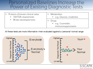
personalized reference range within population range

personalized reference range within population range

Part 1.  MetabolomicsSurge

metagraph  _OMICS

metagraph _OMICS

Metabolomics Continues Auspicious Climb

Jeffery Herman, Ph.D.
GEN May 1, 2012 (Vol. 32, No. 9)

Aberrant biochemical and metabolite signaling plays an important role in

  • the development and progression of diseased tissue.

This concept has been studied by the science community for decades. However, with relatively

  1. recent advances in analytical technology and bioinformatics as well as
  2. the development of the Human Metabolome Database (HMDB),

metabolomics has become an invaluable field of research.

At the “International Conference and Exhibition on Metabolomics & Systems Biology” held recently in San Francisco, researchers and industry leaders discussed how

  • the underlying cellular biochemical/metabolite fingerprint in response to
  1. a specific disease state,
  2. toxin exposure, or
  3. pharmaceutical compound
  • is useful in clinical diagnosis and biomarker discovery and
  • in understanding disease development and progression.

Developed by BASF, MetaMap® Tox is

  • a database that helps identify in vivo systemic effects of a tested compound, including
  1. targeted organs,
  2. mechanism of action, and
  3. adverse events.

Based on 28-day systemic rat toxicity studies, MetaMap Tox is composed of

  • differential plasma metabolite profiles of rats
  • after exposure to a large variety of chemical toxins and pharmaceutical compounds.

“Using the reference data,

  • we have developed more than 110 patterns of metabolite changes, which are
  • specific and predictive for certain toxicological modes of action,”

said Hennicke Kamp, Ph.D., group leader, department of experimental toxicology and ecology at BASF.

With MetaMap Tox, a potential drug candidate

  • can be compared to a similar reference compound
  • using statistical correlation algorithms,
  • which allow for the creation of a toxicity and mechanism of action profile.

“MetaMap Tox, in the context of early pre-clinical safety enablement in pharmaceutical development,” continued Dr. Kamp,

  • has been independently validated “
  • by an industry consortium (Drug Safety Executive Council) of 12 leading biopharmaceutical companies.”

Dr. Kamp added that this technology may prove invaluable

  • allowing for quick and accurate decisions and
  • for high-throughput drug candidate screening, in evaluation
  1. on the safety and efficacy of compounds
  2. during early and preclinical toxicological studies,
  3. by comparing a lead compound to a variety of molecular derivatives, and
  • the rapid identification of the most optimal molecular structure
  • with the best efficacy and safety profiles might be streamlined.
Dynamic Construct of the –Omics

Dynamic Construct of the –Omics

Targeted Tandem Mass Spectrometry

Biocrates Life Sciences focuses on targeted metabolomics, an important approach for

  • the accurate quantification of known metabolites within a biological sample.

Originally used for the clinical screening of inherent metabolic disorders from dried blood-spots of newborn children, Biocrates has developed

  • a tandem mass spectrometry (MS/MS) platform, which allows for
  1. the identification,
  2. quantification, and
  3. mapping of more than 800 metabolites to specific cellular pathways.

It is based on flow injection analysis and high-performance liquid chromatography MS/MS.

Clarification of Pathway-Specific Inhibition by Fourier Transform Ion Cyclotron Resonance.Mass Spectrometry-Based Metabolic Phenotyping Studies F5.large

common drug targets

common drug targets

The MetaDisIDQ® Kit is a

  • “multiparamatic” diagnostic assay designed for the “comprehensive assessment of a person’s metabolic state” and
  • the early determination of pathophysiological events with regards to a specific disease.

MetaDisIDQ is designed to quantify

  • a diverse range of 181 metabolites involved in major metabolic pathways
  • from a small amount of human serum (10 µL) using isotopically labeled internal standards,

This kit has been demonstrated to detect changes in metabolites that are commonly associated with the development of

  • metabolic syndrome, type 2 diabetes, and diabetic nephropathy,

Dr. Dallman reports that data generated with the MetaDisIDQ kit correlates strongly with

  • routine chemical analyses of common metabolites including glucose and creatinine

Biocrates has also developed the MS/MS-based AbsoluteIDQ® kits, which are

  • an “easy-to-use” biomarker analysis tool for laboratory research.

The kit functions on MS machines from a variety of vendors, and allows for the quantification of 150-180 metabolites.

The SteroIDQ® kit is a high-throughput standardized MS/MS diagnostic assay,

  • validated in human serum, for the rapid and accurate clinical determination of 16 known steroids.

Initially focusing on the analysis of steroid ranges for use in hormone replacement therapy, the SteroIDQ Kit is expected to have a wide clinical application.

Hormone-Resistant Breast Cancer

Scientists at Georgetown University have shown that

  • breast cancer cells can functionally coordinate cell-survival and cell-proliferation mechanisms,
  • while maintaining a certain degree of cellular metabolism.

To grow, cells need energy, and energy is a product of cellular metabolism. For nearly a century, it was thought that

  1. the uncoupling of glycolysis from the mitochondria,
  2. leading to the inefficient but rapid metabolism of glucose and
  3. the formation of lactic acid (the Warburg effect), was

the major and only metabolism driving force for unchecked proliferation and tumorigenesis of cancer cells.

Other aspects of metabolism were often overlooked.

“.. we understand now that

  • cellular metabolism is a lot more than just metabolizing glucose,”

said Robert Clarke, Ph.D., professor of oncology and physiology and biophysics at Georgetown University. Dr. Clarke, in collaboration with the Waters Center for Innovation at Georgetown University (led by Albert J. Fornace, Jr., M.D.), obtained

  • the metabolomic profile of hormone-sensitive and -resistant breast cancer cells through the use of UPLC-MS.

They demonstrated that breast cancer cells, through a rather complex and not yet completely understood process,

  1. can functionally coordinate cell-survival and cell-proliferation mechanisms,
  2. while maintaining a certain degree of cellular metabolism.

This is at least partly accomplished through the upregulation of important pro-survival mechanisms; including

  • the unfolded protein response;
  • a regulator of endoplasmic reticulum stress and
  • initiator of autophagy.

Normally, during a stressful situation, a cell may

  • enter a state of quiescence and undergo autophagy,
  • a process by which a cell can recycle organelles
  • in order to maintain enough energy to survive during a stressful situation or,

if the stress is too great,

  • undergo apoptosis.

By integrating cell-survival mechanisms and cellular metabolism

  • advanced ER+ hormone-resistant breast cancer cells
  • can maintain a low level of autophagy
  • to adapt and resist hormone/chemotherapy treatment.

This adaptation allows cells

  • to reallocate important metabolites recovered from organelle degradation and
  • provide enough energy to also promote proliferation.

With further research, we can gain a better understanding of the underlying causes of hormone-resistant breast cancer, with

  • the overall goal of developing effective diagnostic, prognostic, and therapeutic tools.

NMR

Over the last two decades, NMR has established itself as a major tool for metabolomics analysis. It is especially adept at testing biological fluids. [Bruker BioSpin]

Historically, nuclear magnetic resonance spectroscopy (NMR) has been used for structural elucidation of pure molecular compounds. However, in the last two decades, NMR has established itself as a major tool for metabolomics analysis. Since

  • the integral of an NMR signal is directly proportional to
  • the molar concentration throughout the dynamic range of a sample,

“the simultaneous quantification of compounds is possible

  • without the need for specific reference standards or calibration curves,” according to Lea Heintz of Bruker BioSpin.

NMR is adept at testing biological fluids because of

  1.  high reproducibility,
  2. standardized protocols,
  3. low sample manipulation, and
  4. the production of a large subset of data,

Bruker BioSpin is presently involved in a project for the screening of inborn errors of metabolism in newborn children from Turkey, based on their urine NMR profiles. More than 20 clinics are participating to the project that is coordinated by INFAI, a specialist in the transfer of advanced analytical technology into medical diagnostics. The construction of statistical models are being developed

  • for the detection of deviations from normality, as well as
  • automatic quantification methods for indicative metabolites

Bruker BioSpin recently installed high-resolution magic angle spinning NMR (HRMAS-NMR) systems that can rapidly analyze tissue biopsies. The main objective for HRMAS-NMR is to establish a rapid and effective clinical method to assess tumor grade and other important aspects of cancer during surgery.

Combined NMR and Mass Spec

There is increasing interest in combining NMR and MS, two of the main analytical assays in metabolomic research, as a means

  • to improve data sensitivity and to
  • fully elucidate the complex metabolome within a given biological sample.
  •  to realize a potential for cancer biomarker discovery in the realms of diagnosis, prognosis, and treatment.

.

Using combined NMR and MS to measure the levels of nearly 250 separate metabolites in the patient’s blood, Dr. Weljie and other researchers at the University of Calgary were able to rapidly determine the malignancy of a  pancreatic lesion (in 10–15% of the cases, it is difficult to discern between benign and malignant), while avoiding unnecessary surgery in patients with benign lesions.

When performing NMR and MS on a single biological fluid, ultimately “we are,” noted Dr. Weljie,

  1. “splitting up information content, processing, and introducing a lot of background noise and error and
  2. then trying to reintegrate the data…
    It’s like taking a complex item, with multiple pieces, out of an IKEA box and trying to repackage it perfectly into another box.”

By improving the workflow between the initial splitting of the sample, they improved endpoint data integration, proving that

  • a streamlined approach to combined NMR/MS can be achieved,
  • leading to a very strong, robust and precise metabolomics toolset.

Metabolomics Research Picks Up Speed

Field Advances in Quest to Improve Disease Diagnosis and Predict Drug Response

John Morrow Jr., Ph.D.
GEN May 1, 2011 (Vol. 31, No. 9)

As an important discipline within systems biology, metabolomics is being explored by a number of laboratories for

  • its potential in pharmaceutical development.

Studying metabolites can offer insights into the relationships between genotype and phenotype, as well as between genotype and environment. In addition, there is plenty to work with—there are estimated to be some 2,900 detectable metabolites in the human body, of which

  1. 309 have been identified in cerebrospinal fluid,
  2. 1,122 in serum,
  3. 458 in urine, and
  4. roughly 300 in other compartments.

Guowang Xu, Ph.D., a researcher at the Dalian Institute of Chemical Physics.  is investigating the causes of death in China,

  • and how they have been changing over the years as the country has become a more industrialized nation.
  •  the increase in the incidence of metabolic disorders such as diabetes has grown to affect 9.7% of the Chinese population.

Dr. Xu,  collaborating with Rainer Lehman, Ph.D., of the University of Tübingen, Germany, compared urinary metabolites in samples from healthy individuals with samples taken from prediabetic, insulin-resistant subjects. Using mass spectrometry coupled with electrospray ionization in the positive mode, they observed striking dissimilarities in levels of various metabolites in the two groups.

“When we performed a comprehensive two-dimensional gas chromatography, time-of-flight mass spectrometry analysis of our samples, we observed several metabolites, including

  • 2-hydroxybutyric acid in plasma,
  •  as potential diabetes biomarkers,” Dr. Xu explains.

In other, unrelated studies, Dr. Xu and the German researchers used a metabolomics approach to investigate the changes in plasma metabolite profiles immediately after exercise and following a 3-hour and 24-hour period of recovery. They found that

  • medium-chain acylcarnitines were the most distinctive exercise biomarkers, and
  • they are released as intermediates of partial beta oxidation in human myotubes and mouse muscle tissue.

Dr. Xu says. “The traditional approach of assessment based on a singular biomarker is being superseded by the introduction of multiple marker profiles.”

Typical of the studies under way by Dr. Kaddurah-Daouk and her colleaguesat Duke University

  • is a recently published investigation highlighting the role of an SNP variant in
  • the glycine dehydrogenase gene on individual response to antidepressants.
  •  patients who do not respond to the selective serotonin uptake inhibitors citalopram and escitalopram
  • carried a particular single nucleotide polymorphism in the GD gene.

“These results allow us to pinpoint a possible

  • role for glycine in selective serotonin reuptake inhibitor response and
  • illustrate the use of pharmacometabolomics to inform pharmacogenomics.

These discoveries give us the tools for prognostics and diagnostics so that

  • we can predict what conditions will respond to treatment.

“This approach to defining health or disease in terms of metabolic states opens a whole new paradigm.

By screening hundreds of thousands of molecules, we can understand

  • the relationship between human genetic variability and the metabolome.”

Dr. Kaddurah-Daouk talks about statins as a current

  • model of metabolomics investigations.

It is now known that the statins  have widespread effects, altering a range of metabolites. To sort out these changes and develop recommendations for which individuals should be receiving statins will require substantial investments of energy and resources into defining the complex web of biochemical changes that these drugs initiate.
Furthermore, Dr. Kaddurah-Daouk asserts that,

  • “genetics only encodes part of the phenotypic response.

One needs to take into account the

  • net environment contribution in order to determine
  • how both factors guide the changes in our metabolic state that determine the phenotype.”

Interactive Metabolomics

Researchers at the University of Nottingham use diffusion-edited nuclear magnetic resonance spectroscopy to assess the effects of a biological matrix on metabolites. Diffusion-edited NMR experiments provide a way to

  • separate the different compounds in a mixture
  • based on the differing translational diffusion coefficients (which reflect the size and shape of the molecule).

The measurements are carried out by observing

  • the attenuation of the NMR signals during a pulsed field gradient experiment.

Clare Daykin, Ph.D., is a lecturer at the University of Nottingham, U.K. Her field of investigation encompasses “interactive metabolomics,”which she defines as

“the study of the interactions between low molecular weight biochemicals and macromolecules in biological samples ..

  • without preselection of the components of interest.

“Blood plasma is a heterogeneous mixture of molecules that

  1. undergo a variety of interactions including metal complexation,
  2. chemical exchange processes,
  3. micellar compartmentation,
  4. enzyme-mediated biotransformations, and
  5. small molecule–macromolecular binding.”

Many low molecular weight compounds can exist

  • freely in solution,
  • bound to proteins, or
  • within organized aggregates such as lipoprotein complexes.

Therefore, quantitative comparison of plasma composition from

  • diseased individuals compared to matched controls provides an incomplete insight to plasma metabolism.

“It is not simply the concentrations of metabolites that must be investigated,

  • but their interactions with the proteins and lipoproteins within this complex web.

Rather than targeting specific metabolites of interest, Dr. Daykin’s metabolite–protein binding studies aim to study

  • the interactions of all detectable metabolites within the macromolecular sample.

Such activities can be studied through the use of diffusion-edited nuclear magnetic resonance (NMR) spectroscopy, in which one can assess

  • the effects of the biological matrix on the metabolites.

“This can lead to a more relevant and exact interpretation

  • for systems where metabolite–macromolecule interactions occur.”

Diffusion-edited NMR experiments provide a way to separate the different compounds in a mixture based on

  • the differing translational diffusion coefficients (which reflect the size and shape of the molecule).

The measurements are carried out by observing

  • the attenuation of the NMR signals during a pulsed field gradient experiment.

Pushing the Limits

It is widely recognized that many drug candidates fail during development due to ancillary toxicity. Uwe Sauer, Ph.D., professor, and Nicola Zamboni, Ph.D., researcher, both at the Eidgenössische Technische Hochschule, Zürich (ETH Zürich), are applying

  • high-throughput intracellular metabolomics to understand
  • the basis of these unfortunate events and
  • head them off early in the course of drug discovery.

“Since metabolism is at the core of drug toxicity, we developed a platform for

  • measurement of 50–100 targeted metabolites by
  • a high-throughput system consisting of flow injection
  • coupled to tandem mass spectrometry.”

Using this approach, Dr. Sauer’s team focused on

  • the central metabolism of the yeast Saccharomyces cerevisiae, reasoning that
  • this core network would be most susceptible to potential drug toxicity.

Screening approximately 41 drugs that were administered at seven concentrations over three orders of magnitude, they observed changes in metabolome patterns at much lower drug concentrations without attendant physiological toxicity.

The group carried out statistical modeling of about

  • 60 metabolite profiles for each drug they evaluated.

This data allowed the construction of a “profile effect map” in which

  • the influence of each drug on metabolite levels can be followed, including off-target effects, which
  • provide an indirect measure of the possible side effects of the various drugs.

Dr. Sauer says.“We have found that this approach is

  • at least 100 times as fast as other omics screening platforms,”

“Some drugs, including many anticancer agents,

  • disrupt metabolism long before affecting growth.”
killing cancer cells

killing cancer cells

Furthermore, they used the principle of 13C-based flux analysis, in which

  • metabolites labeled with 13C are used to follow the utilization of metabolic pathways in the cell.

These 13C-determined intracellular responses of metabolic fluxes to drug treatment demonstrate

  • the functional performance of the network to be rather robust,
conformational changes leading to substrate efflux.

conformational changes leading to substrate efflux.

leading Dr. Sauer to the conclusion that

  • the phenotypic vigor he observes to drug challenges
  • is achieved by a flexible make up of the metabolome.

Dr. Sauer is confident that it will be possible to expand the scope of these investigations to hundreds of thousands of samples per study. This will allow answers to the questions of

  • how cells establish a stable functioning network in the face of inevitable concentration fluctuations.

Is Now the Hour?

There is great enthusiasm and agitation within the biotech community for

  • metabolomics approaches as a means of reversing the dismal record of drug discovery

that has accumulated in the last decade.

While the concept clearly makes sense and is being widely applied today, there are many reasons why drugs fail in development, and metabolomics will not be a panacea for resolving all of these questions. It is too early at this point to recognize a trend or a track record, and it will take some time to see how this approach can aid in drug discovery and shorten the timeline for the introduction of new pharmaceutical agents.

Degree of binding correlated with function

Degree of binding correlated with function

Diagram_of_a_two-photon_excitation_microscope_

Diagram_of_a_two-photon_excitation_microscope_

Part 2.  Biologists Find ‘Missing Link’ in the Production of Protein Factories in Cells

Biologists at UC San Diego have found

  • the “missing link” in the chemical system that
  • enables animal cells to produce ribosomes

—the thousands of protein “factories” contained within each cell that

  • manufacture all of the proteins needed to build tissue and sustain life.
‘Missing Link’

‘Missing Link’

Their discovery, detailed in the June 23 issue of the journal Genes & Development, will not only force

  • a revision of basic textbooks on molecular biology, but also
  • provide scientists with a better understanding of
  • how to limit uncontrolled cell growth, such as cancer,
  • that might be regulated by controlling the output of ribosomes.

Ribosomes are responsible for the production of the wide variety of proteins that include

  1. enzymes;
  2. structural molecules, such as hair,
  3. skin and bones;
  4. hormones like insulin; and
  5. components of our immune system such as antibodies.

Regarded as life’s most important molecular machine, ribosomes have been intensively studied by scientists (the 2009 Nobel Prize in Chemistry, for example, was awarded for studies of its structure and function). But until now researchers had not uncovered all of the details of how the proteins that are used to construct ribosomes are themselves produced.

In multicellular animals such as humans,

  • ribosomes are made up of about 80 different proteins
    (humans have 79 while some other animals have a slightly different number) as well as
  • four different kinds of RNA molecules.

In 1969, scientists discovered that

  • the synthesis of the ribosomal RNAs is carried out by specialized systems using two key enzymes:
  • RNA polymerase I and RNA polymerase III.

But until now, scientists were unsure if a complementary system was also responsible for

  • the production of the 80 proteins that make up the ribosome.

That’s essentially what the UC San Diego researchers headed by Jim Kadonaga, a professor of biology, set out to examine. What they found was the missing link—the specialized

  • system that allows ribosomal proteins themselves to be synthesized by the cell.

Kadonaga says that he and coworkers found that ribosomal proteins are synthesized via

  • a novel regulatory system with the enzyme RNA polymerase II and
  • a factor termed TRF2,”

“For the production of most proteins,

  1. RNA polymerase II functions with
  2. a factor termed TBP,
  3. but for the synthesis of ribosomal proteins, it uses TRF2.”
  •  this specialized TRF2-based system for ribosome biogenesis
  • provides a new avenue for the study of ribosomes and
  • its control of cell growth, and

“it should lead to a better understanding and potential treatment of diseases such as cancer.”

Coordination of the transcriptome and metabolome

Coordination of the transcriptome and metabolome

the potential advantages conferred by distal-site protein synthesis

the potential advantages conferred by distal-site protein synthesis

Other authors of the paper were UC San Diego biologists Yuan-Liang Wang, Sascha Duttke and George Kassavetis, and Kai Chen, Jeff Johnston, and Julia Zeitlinger of the Stowers Institute for Medical Research in Kansas City, Missouri. Their research was supported by two grants from the National Institutes of Health (1DP2OD004561-01 and R01 GM041249).

Turning Off a Powerful Cancer Protein

Scientists have discovered how to shut down a master regulatory transcription factor that is

  • key to the survival of a majority of aggressive lymphomas,
  • which arise from the B cells of the immune system.

The protein, Bcl6, has long been considered too complex to target with a drug since it is also crucial

  • to the healthy functioning of many immune cells in the body, not just B cells gone bad.

The researchers at Weill Cornell Medical College report that it is possible

  • to shut down Bcl6 in diffuse large B-cell lymphoma (DLBCL)
  • while not affecting its vital function in T cells and macrophages
  • that are needed to support a healthy immune system.

If Bcl6 is completely inhibited, patients might suffer from systemic inflammation and atherosclerosis. The team conducted this new study to help clarify possible risks, as well as to understand

  • how Bcl6 controls the various aspects of the immune system.

The findings in this study were inspired from

  • preclinical testing of two Bcl6-targeting agents that Dr. Melnick and his Weill Cornell colleagues have developed
  • to treat DLBCLs.

These experimental drugs are

  • RI-BPI, a peptide mimic, and
  • the small molecule agent 79-6.

“This means the drugs we have developed against Bcl6 are more likely to be

  • significantly less toxic and safer for patients with this cancer than we realized,”

says Ari Melnick, M.D., professor of hematology/oncology and a hematologist-oncologist at NewYork-Presbyterian Hospital/Weill Cornell Medical Center.

Dr. Melnick says the discovery that

  • a master regulatory transcription factor can be targeted
  • offers implications beyond just treating DLBCL.

Recent studies from Dr. Melnick and others have revealed that

  • Bcl6 plays a key role in the most aggressive forms of acute leukemia, as well as certain solid tumors.

Bcl6 can control the type of immune cell that develops in the bone marrow—playing many roles

  • in the development of B cells, T cells, macrophages, and other cells—including a primary and essential role in
  • enabling B-cells to generate specific antibodies against pathogens.

According to Dr. Melnick, “When cells lose control of Bcl6,

  • lymphomas develop in the immune system.

Lymphomas are ‘addicted’ to Bcl6, and therefore

  • Bcl6 inhibitors powerfully and quickly destroy lymphoma cells,” .

The big surprise in the current study is that rather than functioning as a single molecular machine,

  • Bcl6 functions like a Swiss Army knife,
  • using different tools to control different cell types.

This multifunction paradigm could represent a general model for the functioning of other master regulatory transcription factors.

“In this analogy, the Swiss Army knife, or transcription factor, keeps most of its tools folded,

  • opening only the one it needs in any given cell type,”

He makes the following analogy:

  • “For B cells, it might open and use the knife tool;
  • for T cells, the cork screw;
  • for macrophages, the scissors.”

“this means that you only need to prevent the master regulator from using certain tools to treat cancer. You don’t need to eliminate the whole knife,” . “In fact, we show that taking out the whole knife is harmful since

  • the transcription factor has many other vital functions that other cells in the body need.”

Prior to these study results, it was not known that a master regulator could separate its functions so precisely. Researchers hope this will be a major benefit to the treatment of DLBCL and perhaps other disorders that are influenced by Bcl6 and other master regulatory transcription factors.

The study is published in the journal Nature Immunology, in a paper titled “Lineage-specific functions of Bcl-6 in immunity and inflammation are mediated by distinct biochemical mechanisms”.

Part 3. Neuroscience

Vesicles influence function of nerve cells 
Oct, 06 2014        source: http://feeds.sciencedaily.com

Neurons (blue) which have absorbed exosomes (green) have increased levels of the enzyme catalase (red), which helps protect them against peroxides.

Neurons (blue) which have absorbed exosomes (green) have increased levels of the enzyme catalase (red), which helps protect them against peroxides.

Neurons (blue) which have absorbed exosomes (green) have increased levels of the enzyme catalase (red), which helps protect them against peroxides.

Tiny vesicles containing protective substances

  • which they transmit to nerve cells apparently
  • play an important role in the functioning of neurons.

As cell biologists at Johannes Gutenberg University Mainz (JGU) have discovered,

  • nerve cells can enlist the aid of mini-vesicles of neighboring glial cells
  • to defend themselves against stress and other potentially detrimental factors.

These vesicles, called exosomes, appear to stimulate the neurons on various levels:

  • they influence electrical stimulus conduction,
  • biochemical signal transfer, and
  • gene regulation.

Exosomes are thus multifunctional signal emitters

  • that can have a significant effect in the brain.
Exosome

Exosome

The researchers in Mainz already observed in a previous study that

  • oligodendrocytes release exosomes on exposure to neuronal stimuli.
  • these are absorbed by the neurons and improve neuronal stress tolerance.

Oligodendrocytes, a type of glial cell, form an

  • insulating myelin sheath around the axons of neurons.

The exosomes transport protective proteins such as

  • heat shock proteins,
  • glycolytic enzymes, and
  • enzymes that reduce oxidative stress from one cell type to another,
  • but also transmit genetic information in the form of ribonucleic acids.

“As we have now discovered in cell cultures, exosomes seem to have a whole range of functions,” explained Dr. Eva-Maria Krmer-Albers. By means of their transmission activity, the small bubbles that are the vesicles

  • not only promote electrical activity in the nerve cells, but also
  • influence them on the biochemical and gene regulatory level.

“The extent of activities of the exosomes is impressive,” added Krmer-Albers. The researchers hope that the understanding of these processes will contribute to the development of new strategies for the treatment of neuronal diseases. Their next aim is to uncover how vesicles actually function in the brains of living organisms.

http://labroots.com/user/news/article/id/217438/title/vesicles-influence-function-of-nerve-cells

The above story is based on materials provided by Universitt Mainz.

Universitt Mainz. “Vesicles influence function of nerve cells.” ScienceDaily. ScienceDaily, 6 October 2014. www.sciencedaily.com/releases/2014/10/141006174214.htm

Neuroscientists use snail research to help explain “chemo brain”

10/08/2014
It is estimated that as many as half of patients taking cancer drugs experience a decrease in mental sharpness. While there have been many theories, what causes “chemo brain” has eluded scientists.

In an effort to solve this mystery, neuroscientists at The University of Texas Health Science Center at Houston (UTHealth) conducted an experiment in an animal memory model and their results point to a possible explanation. Findings appeared in The Journal of Neuroscience.

In the study involving a sea snail that shares many of the same memory mechanisms as humans and a drug used to treat a variety of cancers, the scientists identified

  • memory mechanisms blocked by the drug.

Then, they were able to counteract or

  • unblock the mechanisms by administering another agent.

“Our research has implications in the care of people given to cognitive deficits following drug treatment for cancer,” said John H. “Jack” Byrne, Ph.D., senior author, holder of the June and Virgil Waggoner Chair and Chairman of the Department of Neurobiology and Anatomy at the UTHealth Medical School. “There is no satisfactory treatment at this time.”

Byrne’s laboratory is known for its use of a large snail called Aplysia californica to further the understanding of the biochemical signaling among nerve cells (neurons).  The snails have large neurons that relay information much like those in humans.

When Byrne’s team compared cell cultures taken from normal snails to

  • those administered a dose of a cancer drug called doxorubicin,

the investigators pinpointed a neuronal pathway

  • that was no longer passing along information properly.

With the aid of an experimental drug,

  • the scientists were able to reopen the pathway.

Unfortunately, this drug would not be appropriate for humans, Byrne said. “We want to identify other drugs that can rescue these memory mechanisms,” he added.

According the American Cancer Society, some of the distressing mental changes cancer patients experience may last a short time or go on for years.

Byrne’s UT Health research team includes co-lead authors Rong-Yu Liu, Ph.D., and Yili Zhang, Ph.D., as well as Brittany Coughlin and Leonard J. Cleary, Ph.D. All are affiliated with the W.M. Keck Center for the Neurobiology of Learning and Memory.

Byrne and Cleary also are on the faculty of The University of Texas Graduate School of Biomedical Sciences at Houston. Coughlin is a student at the school, which is jointly operated by UT Health and The University of Texas MD Anderson Cancer Center.

The study titled “Doxorubicin Attenuates Serotonin-Induced Long-Term Synaptic Facilitation by Phosphorylation of p38 Mitogen-Activated Protein Kinase” received support from National Institutes of Health grant (NS019895) and the Zilkha Family Discovery Fellowship.

Doxorubicin Attenuates Serotonin-Induced Long-Term Synaptic Facilitation by Phosphorylation of p38 Mitogen-Activated Protein Kinase

Source: Univ. of Texas Health Science Center at Houston

http://www.rdmag.com/news/2014/10/neuroscientists-use-snail-research-help-explain-E2_9_Cchemo-brain

Doxorubicin Attenuates Serotonin-Induced Long-Term Synaptic Facilitation by Phosphorylation of p38 Mitogen-Activated Protein Kinase

Rong-Yu Liu*,  Yili Zhang*,  Brittany L. Coughlin,  Leonard J. Cleary, and  John H. Byrne   +Show Affiliations
The Journal of Neuroscience, 1 Oct 2014, 34(40): 13289-13300;
http://dx.doi.org:/10.1523/JNEUROSCI.0538-14.2014

Doxorubicin (DOX) is an anthracycline used widely for cancer chemotherapy. Its primary mode of action appears to be

  • topoisomerase II inhibition, DNA cleavage, and free radical generation.

However, in non-neuronal cells, DOX also inhibits the expression of

  • dual-specificity phosphatases (also referred to as MAPK phosphatases) and thereby
  1. inhibits the dephosphorylation of extracellular signal-regulated kinase (ERK) and
  2. p38 mitogen-activated protein kinase (p38 MAPK),
  3. two MAPK isoforms important for long-term memory (LTM) formation.

Activation of these kinases by DOX in neurons, if present,

  • could have secondary effects on cognitive functions, such as learning and memory.

The present study used cultures of rat cortical neurons and sensory neurons (SNs) of Aplysia

  • to examine the effects of DOX on levels of phosphorylated ERK (pERK) and
  • phosphorylated p38 (p-p38) MAPK.

In addition, Aplysia neurons were used to examine the effects of DOX on

  • long-term enhanced excitability, long-term synaptic facilitation (LTF), and
  • long-term synaptic depression (LTD).

DOX treatment led to elevated levels of

  • pERK and p-p38 MAPK in SNs and cortical neurons.

In addition, it increased phosphorylation of

  • the downstream transcriptional repressor cAMP response element-binding protein 2 in SNs.

DOX treatment blocked serotonin-induced LTF and enhanced LTD induced by the neuropeptide Phe-Met-Arg-Phe-NH2. The block of LTF appeared to be attributable to

  • overriding inhibitory effects of p-p38 MAPK, because
  • LTF was rescued in the presence of an inhibitor of p38 MAPK
    (SB203580 [4-(4-fluorophenyl)-2-(4-methylsulfinylphenyl)-5-(4-pyridyl)-1H-imidazole]) .

These results suggest that acute application of DOX might impair the formation of LTM via the p38 MAPK pathway.
Terms: Aplysia chemotherapy ERK  p38 MAPK serotonin synaptic plasticity

Technology that controls brain cells with radio waves earns early BRAIN grant

10/08/2014

bright spots = cells with increased calcium after treatment with radio waves,  allows neurons to fire

bright spots = cells with increased calcium after treatment with radio waves, allows neurons to fire

BRAIN control: The new technology uses radio waves to activate or silence cells remotely. The bright spots above represent cells with increased calcium after treatment with radio waves, a change that would allow neurons to fire.

A proposal to develop a new way to

  • remotely control brain cells

from Sarah Stanley, a research associate in Rockefeller University’s Laboratory of Molecular Genetics, headed by Jeffrey M. Friedman, is

  • among the first to receive funding from U.S. President Barack Obama’s BRAIN initiative.

The project will make use of a technique called

  • radiogenetics that combines the use of radio waves or magnetic fields with
  • nanoparticles to turn neurons on or off.

The National Institutes of Health is one of four federal agencies involved in the BRAIN (Brain Research through Advancing Innovative Neurotechnologies) initiative. Following in the ambitious footsteps of the Human Genome Project, the BRAIN initiative seeks

  • to create a dynamic map of the brain in action,

a goal that requires the development of new technologies. The BRAIN initiative working group, which outlined the broad scope of the ambitious project, was co-chaired by Rockefeller’s Cori Bargmann, head of the Laboratory of Neural Circuits and Behavior.

Stanley’s grant, for $1.26 million over three years, is one of 58 projects to get BRAIN grants, the NIH announced. The NIH’s plan for its part of this national project, which has been pitched as “America’s next moonshot,” calls for $4.5 billion in federal funds over 12 years.

The technology Stanley is developing would

  • enable researchers to manipulate the activity of neurons, as well as other cell types,
  • in freely moving animals in order to better understand what these cells do.

Other techniques for controlling selected groups of neurons exist, but her new nanoparticle-based technique has a

  • unique combination of features that may enable new types of experimentation.
  • it would allow researchers to rapidly activate or silence neurons within a small area of the brain or
  • dispersed across a larger region, including those in difficult-to-access locations.

Stanley also plans to explore the potential this method has for use treating patients.

“Francis Collins, director of the NIH, has discussed

  • the need for studying the circuitry of the brain,
  • which is formed by interconnected neurons.

Our remote-control technology may provide a tool with which researchers can ask new questions about the roles of complex circuits in regulating behavior,” Stanley says.
Rockefeller University’s Laboratory of Molecular Genetics
Source: Rockefeller Univ.

Part 4.  Cancer

Two Proteins Found to Block Cancer Metastasis

Why do some cancers spread while others don’t? Scientists have now demonstrated that

  • metastatic incompetent cancers actually “poison the soil”
  • by generating a micro-environment that blocks cancer cells
  • from settling and growing in distant organs.

The “seed and the soil” hypothesis proposed by Stephen Paget in 1889 is now widely accepted to explain how

  • cancer cells (seeds) are able to generate fertile soil (the micro-environment)
  • in distant organs that promotes cancer’s spread.

However, this concept had not explained why some tumors do not spread or metastasize.

The researchers, from Weill Cornell Medical College, found that

  • two key proteins involved in this process work by
  • dramatically suppressing cancer’s spread.

The study offers hope that a drug based on these

  • potentially therapeutic proteins, prosaposin and Thrombospondin 1 (Tsp-1),

might help keep human cancer at bay and from metastasizing.

Scientists don’t understand why some tumors wouldn’t “want” to spread. It goes against their “job description,” says the study’s senior investigator, Vivek Mittal, Ph.D., an associate professor of cell and developmental biology in cardiothoracic surgery and director of the Neuberger Berman Foundation Lung Cancer Laboratory at Weill Cornell Medical College. He theorizes that metastasis occurs when

  • the barriers that the body throws up to protect itself against cancer fail.

But there are some tumors in which some of the barriers may still be intact. “So that suggests

  • those primary tumors will continue to grow, but that
  • an innate protective barrier still exists that prevents them from spreading and invading other organs,”

The researchers found that, like typical tumors,

  • metastasis-incompetent tumors also send out signaling molecules
  • that establish what is known as the “premetastatic niche” in distant organs.

These niches composed of bone marrow cells and various growth factors have been described previously by others including Dr. Mittal as the fertile “soil” that the disseminated cancer cell “seeds” grow in.

Weill Cornell’s Raúl Catena, Ph.D., a postdoctoral fellow in Dr. Mittal’s laboratory, found an important difference between the tumor types. Metastatic-incompetent tumors

  • systemically increased expression of Tsp-1, a molecule known to fight cancer growth.
  • increased Tsp-1 production was found specifically in the bone marrow myeloid cells
  • that comprise the metastatic niche.

These results were striking, because for the first time Dr. Mittal says

  • the bone marrow-derived myeloid cells were implicated as
  • the main producers of Tsp-1,.

In addition, Weill Cornell and Harvard researchers found that

  • prosaposin secreted predominantly by the metastatic-incompetent tumors
  • increased expression of Tsp-1 in the premetastatic lungs.

Thus, Dr. Mittal posits that prosaposin works in combination with Tsp-1

  • to convert pro-metastatic bone marrow myeloid cells in the niche
  • into cells that are not hospitable to cancer cells that spread from a primary tumor.
  • “The very same myeloid cells in the niche that we know can promote metastasis
  • can also be induced under the command of the metastatic incompetent primary tumor to inhibit metastasis,”

The research team found that

  • the Tsp-1–inducing activity of prosaposin
  • was contained in only a 5-amino acid peptide region of the protein, and
  • this peptide alone induced Tsp-1 in the bone marrow cells and
  • effectively suppressed metastatic spread in the lungs
  • in mouse models of breast and prostate cancer.

This 5-amino acid peptide with Tsp-1–inducing activity

  • has the potential to be used as a therapeutic agent against metastatic cancer,

The scientists have begun to test prosaposin in other tumor types or metastatic sites.

Dr. Mittal says that “The clinical implications of the study are:

  • “Not only is it theoretically possible to design a prosaposin-based drug or drugs
  • that induce Tsp-1 to block cancer spread, but
  • you could potentially create noninvasive prognostic tests
  • to predict whether a cancer will metastasize.”

The study was reported in the April 30 issue of Cancer Discovery, in a paper titled “Bone Marrow-Derived Gr1+ Cells Can Generate a Metastasis-Resistant Microenvironment Via Induced Secretion of Thrombospondin-1”.

Disabling Enzyme Cripples Tumors, Cancer Cells

First Step of Metastasis

First Step of Metastasis

Published: Sep 05, 2013  http://www.technologynetworks.com/Metabolomics/news.aspx?id=157138

Knocking out a single enzyme dramatically cripples the ability of aggressive cancer cells to spread and grow tumors.

The paper, published in the journal Proceedings of the National Academy of Sciences, sheds new light on the importance of lipids, a group of molecules that includes fatty acids and cholesterol, in the development of cancer.

Researchers have long known that cancer cells metabolize lipids differently than normal cells. Levels of ether lipids – a class of lipids that are harder to break down – are particularly elevated in highly malignant tumors.

“Cancer cells make and use a lot of fat and lipids, and that makes sense because cancer cells divide and proliferate at an accelerated rate, and to do that,

  • they need lipids, which make up the membranes of the cell,”

said study principal investigator Daniel Nomura, assistant professor in UC Berkeley’s Department of Nutritional Sciences and Toxicology. “Lipids have a variety of uses for cellular structure, but what we’re showing with our study is that

  • lipids can send signals that fuel cancer growth.”

In the study, Nomura and his team tested the effects of reducing ether lipids on human skin cancer cells and primary breast tumors. They targeted an enzyme,

  • alkylglycerone phosphate synthase, or AGPS,
  • known to be critical to the formation of ether lipids.

The researchers confirmed that

  1. AGPS expression increased when normal cells turned cancerous.
  2. inactivating AGPS substantially reduced the aggressiveness of the cancer cells.

“The cancer cells were less able to move and invade,” said Nomura.

The researchers also compared the impact of

  • disabling the AGPS enzyme in mice that had been injected with cancer cells.

Nomura. observes -“Among the mice that had the AGPS enzyme inactivated,

  • the tumors were nonexistent,”

“The mice that did not have this enzyme

  • disabled rapidly developed tumors.”

The researchers determined that

  • inhibiting AGPS expression depleted the cancer cells of ether lipids.
  • AGPS altered levels of other types of lipids important to the ability of the cancer cells to survive and spread, including
    • prostaglandins and acyl phospholipids.

“What makes AGPS stand out as a treatment target is that the enzyme seems to simultaneously

  • regulate multiple aspects of lipid metabolism
  • important for tumor growth and malignancy.”

Future steps include the

  • development of AGPS inhibitors for use in cancer therapy,

“This study sheds considerable light on the important role that AGPS plays in ether lipid metabolism in cancer cells, and it suggests that

  • inhibitors of this enzyme could impair tumor formation,”

said Benjamin Cravatt, Professor and Chair of Chemical Physiology at The Scripps Research Institute, who is not part of the UC.

Agilent Technologies Thought Leader Award Supports Translational Research Program
Published: Mon, March 04, 2013

The award will support Dr DePinho’s research into

  • metabolic reprogramming in the earliest stages of cancer.

Agilent Technologies Inc. announces that Dr. Ronald A. DePinho, a world-renowned oncologist and researcher, has received an Agilent Thought Leader Award.

DePinho is president of the University of Texas MD Anderson Cancer Center. DePinho and his team hope to discover and characterize

  • alterations in metabolic flux during tumor initiation and maintenance, and to identify biomarkers for early detection of pancreatic cancer together with
  • novel therapeutic targets.

Researchers on his team will work with scientists from the university’s newly formed Institute of Applied Cancer Sciences.

The Agilent Thought Leader Award provides funds to support personnel as well as a state-of-the-art Agilent 6550 iFunnel Q-TOF LC/MS system.

“I am extremely pleased to receive this award for metabolomics research, as the survival rates for pancreatic cancer have not significantly improved over the past 20 years,” DePinho said. “This technology will allow us to

  • rapidly identify new targets that drive the formation, progression and maintenance of pancreatic cancer.

Discoveries from this research will also lead to

  • the development of effective early detection biomarkers and novel therapeutic interventions.”

“We are proud to support Dr. DePinho’s exciting translational research program, which will make use of

  • metabolomics and integrated biology workflows and solutions in biomarker discovery,”

said Patrick Kaltenbach, Agilent vice president, general manager of the Liquid Phase Division, and the executive sponsor of this award.

The Agilent Thought Leader Program promotes fundamental scientific advances by support of influential thought leaders in the life sciences and chemical analysis fields.

The covalent modifier Nedd8 is critical for the activation of Smurf1 ubiquitin ligase in tumorigenesis

Ping Xie, Minghua Zhang, Shan He, Kefeng Lu, Yuhan Chen, Guichun Xing, et al.
Nature Communications
  2014; 5(3733).  http://dx.doi.org:/10.1038/ncomms4733

Neddylation, the covalent attachment of ubiquitin-like protein Nedd8, of the Cullin-RING E3 ligase family

  • regulates their ubiquitylation activity.

However, regulation of HECT ligases by neddylation has not been reported to date. Here we show that

  • the C2-WW-HECT ligase Smurf1 is activated by neddylation.

Smurf1 physically interacts with

  1. Nedd8 and Ubc12,
  2. forms a Nedd8-thioester intermediate, and then
  3. catalyses its own neddylation on multiple lysine residues.

Intriguingly, this autoneddylation needs

  • an active site at C426 in the HECT N-lobe.

Neddylation of Smurf1 potently enhances

  • ubiquitin E2 recruitment and
  • augments the ubiquitin ligase activity of Smurf1.

The regulatory role of neddylation

  • is conserved in human Smurf1 and yeast Rsp5.

Furthermore, in human colorectal cancers,

  • the elevated expression of Smurf1, Nedd8, NAE1 and Ubc12
  • correlates with cancer progression and poor prognosis.

These findings provide evidence that

  • neddylation is important in HECT ubiquitin ligase activation and
  • shed new light on the tumour-promoting role of Smurf1.
 Swinging domains in HECT E3

Swinging domains in HECT E3

Subject terms: Biological sciences Cancer Cell biology

Figure 1: Smurf1 expression is elevated in colorectal cancer tissues.

Smurf1 expression is elevated in colorectal cancer tissues.

Smurf1 expression is elevated in colorectal cancer tissues.

(a) Smurf1 expression scores are shown as box plots, with the horizontal lines representing the median; the bottom and top of the boxes representing the 25th and 75th percentiles, respectively; and the vertical bars representing the ra

Figure 2: Positive correlation of Smurf1 expression with Nedd8 and its interacting enzymes in colorectal cancer.

Positive correlation of Smurf1 expression with Nedd8 and its interacting enzymes in colorectal cancer

Positive correlation of Smurf1 expression with Nedd8 and its interacting enzymes in colorectal cancer

(a) Representative images from immunohistochemical staining of Smurf1, Ubc12, NAE1 and Nedd8 in the same colorectal cancer tumour. Scale bars, 100 μm. (b–d) The expression scores of Nedd8 (b, n=283 ), NAE1 (c, n=281) and Ubc12 (d, n=19…

Figure 3: Smurf1 interacts with Ubc12.

Smurf1 interacts with Ubc12

Smurf1 interacts with Ubc12

(a) GST pull-down assay of Smurf1 with Ubc12. Both input and pull-down samples were subjected to immunoblotting with anti-His and anti-GST antibodies. Smurf1 interacted with Ubc12 and UbcH5c, but not with Ubc9. (b) Mapping the regions…

Figure 4: Nedd8 is attached to Smurf1through C426-catalysed autoneddylation.

Nedd8 is attached to Smurf1through C426-catalysed autoneddylation

Nedd8 is attached to Smurf1through C426-catalysed autoneddylation

(a) Covalent neddylation of Smurf1 in vitro.Purified His-Smurf1-WT or C699A proteins were incubated with Nedd8 and Nedd8-E1/E2. Reactions were performed as described in the Methods section. Samples were analysed by western blotting wi…

Figure 5: Neddylation of Smurf1 activates its ubiquitin ligase activity.

Neddylation of Smurf1 activates its ubiquitin ligase activity.

Neddylation of Smurf1 activates its ubiquitin ligase activity.

(a) In vivo Smurf1 ubiquitylation assay. Nedd8 was co-expressed with Smurf1 WT or C699A in HCT116 cells (left panels). Twenty-four hours post transfection, cells were treated with MG132 (20 μM, 8 h). HCT116 cells were transfected with…

http://www.nature.com/ncomms/2014/140513/ncomms4733/carousel/ncomms4733-f1.jpg

http://www.nature.com/ncomms/2014/140513/ncomms4733/carousel/ncomms4733-f2.jpg

http://www.nature.com/ncomms/2014/140513/ncomms4733/carousel/ncomms4733-f3.jpg

http://www.nature.com/ncomms/2014/140513/ncomms4733/carousel/ncomms4733-f4.jpg

http://www.nature.com/ncomms/2014/140513/ncomms4733/carousel/ncomms4733-f5.jpg

http://www.nature.com/ncomms/2014/140513/ncomms4733/carousel/ncomms4733-f6.jpg

http://www.nature.com/ncomms/2014/140513/ncomms4733/carousel/ncomms4733-f7.jpg

http://www.nature.com/ncomms/2014/140513/ncomms4733/carousel/ncomms4733-f8.jpg

The deubiquitylase USP33 discriminates between RALB functions in autophagy and innate immune response

M Simicek, S Lievens, M Laga, D Guzenko, VN. Aushev, et al.
Nature Cell Biology 2013; 15, 1220–1230    http://dx.doi.org:/10.1038/ncb2847

The RAS-like GTPase RALB mediates cellular responses to nutrient availability or viral infection by respectively

  • engaging two components of the exocyst complex, EXO84 and SEC5.
  1. RALB employs SEC5 to trigger innate immunity signalling, whereas
  2. RALB–EXO84 interaction induces autophagocytosis.

How this differential interaction is achieved molecularly by the RAL GTPase remains unknown.

We found that whereas GTP binding

  • turns on RALB activity,

ubiquitylation of RALB at Lys 47

  • tunes its activity towards a particular effector.

Specifically, ubiquitylation at Lys 47

  • sterically inhibits RALB binding to EXO84, while
  • facilitating its interaction with SEC5.

Double-stranded RNA promotes

  • RALB ubiquitylation and
  • SEC5–TBK1 complex formation.

In contrast, nutrient starvation

  • induces RALB deubiquitylation
  • by accumulation and relocalization of the deubiquitylase USP33
  • to RALB-positive vesicles.

Deubiquitylated RALB

  • promotes the assembly of the RALB–EXO84–beclin-1 complexes
  • driving autophagosome formation. Thus,
  • ubiquitylation within the effector-binding domain
  • provides the switch for the dual functions of RALB in
    • autophagy and innate immune responses.

Part 5. Metabolic Syndrome

Single Enzyme is Necessary for Development of Diabetes

Published: Aug 20, 2014 http://www.technologynetworks.com/Metabolomics/news.aspx?ID=169416

12-LO enzyme promotes the obesity-induced oxidative stress in the pancreatic cells.

An enzyme called 12-LO promotes the obesity-induced oxidative stress in the pancreatic cells that leads

  • to pre-diabetes, and diabetes.

12-LO’s enzymatic action is the last step in

  • the production of certain small molecules that harm the cell,

according to a team from Indiana University School of Medicine, Indianapolis.

The findings will enable the development of drugs that can interfere with this enzyme, preventing or even reversing diabetes. The research is published ahead of print in the journal Molecular and Cellular Biology.

In earlier studies, these researchers and their collaborators at Eastern Virginia Medical School showed that

  • 12-LO (which stands for 12-lipoxygenase) is present in these cells
  • only in people who become overweight.

The harmful small molecules resulting from 12-LO’s enzymatic action are known as HETEs, short for hydroxyeicosatetraenoic acid.

  1. HETEs harm the mitochondria, which then
  2. fail to produce sufficient energy to enable
  3. the pancreatic cells to manufacture the necessary quantities of insulin.

For the study, the investigators genetically engineered mice that

  • lacked the gene for 12-LO exclusively in their pancreas cells.

Mice were either fed a low-fat or high-fat diet.

Both the control mice and the knockout mice on the high fat diet

  • developed obesity and insulin resistance.

The investigators also examined the pancreatic beta cells of both knockout and control mice, using both microscopic studies and molecular analysis. Those from the knockout mice were intact and healthy, while

  • those from the control mice showed oxidative damage,
  • demonstrating that 12-LO and the resulting HETEs
  • caused the beta cell failure.

Mirmira notes that fatty diet used in the study was the Western Diet, which comprises mostly saturated-“bad”-fats. Based partly on a recent study of related metabolic pathways, he says that

  • the unsaturated and mono-unsaturated fats-which comprise most fats in the healthy,
  • relatively high fat Mediterranean diet-are unlikely to have the same effects.

“Our research is the first to show that 12-LO in the beta cell

  • is the culprit in the development of pre-diabetes, following high fat diets,” says Mirmira.

“Our work also lends important credence to the notion that

  • the beta cell is the primary defective cell in virtually all forms of diabetes and pre-diabetes.”

A New Player in Lipid Metabolism Discovered

Published: Aug18, 2014  http://www.technologynetworks.com/Metabolomics/news.aspx?ID=169356

Specially engineered mice gained no weight, and normal counterparts became obese

  • on the same high-fat, obesity-inducing Western diet.

Specially engineered mice that lacked a particular gene did not gain weight

  • when fed a typical high-fat, obesity-inducing Western diet.

Yet, these mice ate the same amount as their normal counterparts that became obese.

The mice were engineered with fat cells that lacked a gene called SEL1L,

  • known to be involved in the clearance of mis-folded proteins
  • in the cell’s protein making machinery called the endoplasmic reticulum (ER).

When mis-folded proteins are not cleared but accumulate,

  • they destroy the cell and contribute to such diseases as
  1. mad cow disease,
  2. Type 1 diabetes and
  3. cystic fibrosis.

“The million-dollar question is why don’t these mice gain weight? Is this related to its inability to clear mis-folded proteins in the ER?” said Ling Qi, associate professor of molecular and biochemical nutrition and senior author of the study published online July 24 in Cell Metabolism. Haibo Sha, a research associate in Qi’s lab, is the paper’s lead author.

Interestingly, the experimental mice developed a host of other problems, including

  • postprandial hypertriglyceridemia,
  • and fatty livers.

“Although we are yet to find out whether these conditions contribute to the lean phenotype, we found that

  • there was a lipid partitioning defect in the mice lacking SEL1L in fat cells,
  • where fat cells cannot store fat [lipids], and consequently
  • fat goes to the liver.

During the investigation of possible underlying mechanisms, we discovered

  • a novel function for SEL1L as a regulator of lipid metabolism,” said Qi.

Sha said “We were very excited to find that

  • SEL1L is required for the intracellular trafficking of
  • lipoprotein lipase (LPL), acting as a chaperone,” .

and added that “Using several tissue-specific knockout mouse models,

  • we showed that this is a general phenomenon,”

Without LPL, lipids remain in the circulation;

  • fat and muscle cells cannot absorb fat molecules for storage and energy combustion,

People with LPL mutations develop

  • postprandial hypertriglyceridemia similar to
  • conditions found in fat cell-specific SEL1L-deficient mice, said Qi.

Future work will investigate the

  • role of SEL1L in human patients carrying LPL mutations and
  • determine why fat cell-specific SEL1L-deficient mice remain lean under Western diets, said Sha.

Co-authors include researchers from Cedars-Sinai Medical Center in Los Angeles; Wageningen University in the Netherlands; Georgia State University; University of California, Los Angeles; and the Medical College of Soochow University in China.

The study was funded by the U.S. National Institutes of Health, the Netherlands Organization for Health Research and Development National Institutes of Health, the Cedars-Sinai Medical Center, Chinese National Science Foundation, the American Diabetes Association, Cornell’s Center for Vertebrate Genomics and the Howard Hughes Medical Institute.

Part 6. Biomarkers

Biomarkers Take Center Stage

Josh P. Roberts
GEN May 1, 2013 (Vol. 33, No. 9)  http://www.genengnews.com/

While work with biomarkers continues to grow, scientists are also grappling with research-related bottlenecks, such as

  1. affinity reagent development,
  2. platform reproducibility, and
  3. sensitivity.

Biomarkers by definition indicate some state or process that generally occurs

  • at a spatial or temporal distance from the marker itself, and

it would not be an exaggeration to say that biomedicine has become infatuated with them:

  1. where to find them,
  2. when they may appear,
  3. what form they may take, and
  4. how they can be used to diagnose a condition or
  5. predict whether a therapy may be successful.

Biomarkers are on the agenda of many if not most industry gatherings, and in cases such as Oxford Global’s recent “Biomarker Congress” and the GTC “Biomarker Summit”, they hold the naming rights. There, some basic principles were built upon, amended, and sometimes challenged.

In oncology, for example, biomarker discovery is often predicated on the premise that

  • proteins shed from a tumor will traverse to and persist in, and be detectable in, the circulation.

By quantifying these proteins—singularly or as part of a larger “signature”—the hope is

  1. to garner information about the molecular characteristics of the cancer
  2. that will help with cancer detection and
  3. personalization of the treatment strategy.

Yet this approach has not yet turned into the panacea that was hoped for. Bottlenecks exist in

  • affinity reagent development,
  • platform reproducibility, and
  • sensitivity.

There is also a dearth of understanding of some of the

  • fundamental principles of biomarker biology that we need to know the answers to,

said Parag Mallick, Ph.D., whose lab at Stanford University is “working on trying to understand where biomarkers come from.”

There are dogmas saying that

  • circulating biomarkers come solely from secreted proteins.

But Dr. Mallick’s studies indicate that fully

  • 50% of circulating proteins may come from intracellular sources or
  • proteins that are annotated as such.

“We don’t understand the processes governing

  • which tumor-derived proteins end up in the blood.”

Other questions include “how does the size of a tumor affect how much of a given protein will be in the blood?”—perhaps

  • the tumor is necrotic at the center, or
  • it’s hypervascular or hypovascular.

He points out “The problem is that these are highly nonlinear processes at work, and

  • there is a large number of factors that might affect the answer to that question,” .

Their research focuses on using

  1. mass spectrometry and
  2. computational analysis
  • to characterize the biophysical properties of the circulating proteome, and
  • relate these to measurements made of the tumor itself.

Furthermore, he said – “We’ve observed that the proteins that are likely to

  • first show up and persist in the circulation, ..
  • are more stable than proteins that don’t,”
  • “we can quantify how significant the effect is.”

The goal is ultimately to be able to

  1. build rigorous, formal mathematical models that will allow something measured in the blood
  2. to be tied back to the molecular biology taking place in the tumor.

And conversely, to use those models

  • to predict from a tumor what will be found in the circulation.

“Ultimately, the models will allow you to connect the dots between

  • what you measure in the blood and the biology of the tumor.”

Bound for Affinity Arrays

Affinity reagents are the main tools for large-scale protein biomarker discovery. And while this has tended to mean antibodies (or their derivatives), other affinity reagents are demanding a place in the toolbox.

Affimers, a type of affinity reagent being developed by Avacta, consist of

  1. a biologically inert, biophysically stable protein scaffold
  2. containing three variable regions into which
  3. distinct peptides are inserted.

The resulting three-dimensional surface formed by these peptides

  • interacts and binds to proteins and other molecules in solution,
  • much like the antigen-binding site of antibodies.

Unlike antibodies, Affimers are relatively small (13 KDa),

  • non-post-translationally modified proteins
  • that can readily be expressed in bacterial culture.

They may be made to bind surfaces through unique residues

  • engineered onto the opposite face of the Affimer,
  • allowing the binding site to be exposed to the target in solution.

“We don’t seem to see in what we’ve done so far

  • any real loss of activity or functionality of Affimers when bound to surfaces—

they’re very robust,” said CEO Alastair Smith, Ph.D.

Avacta is taking advantage of this stability and its large libraries of Affimers to develop

  • very large affinity microarrays for
  • drug and biomarker discovery.

To date they have printed arrays with around 20–25,000 features, and Dr. Smith is “sure that we can get toward about 50,000 on a slide,” he said. “There’s no real impediment to us doing that other than us expressing the proteins and getting on with it.”

Customers will be provided with these large, complex “naïve” discovery arrays, readable with standard equipment. The plan is for the company to then “support our customers by providing smaller arrays with

  • the Affimers that are binding targets of interest to them,” Dr. Smith foretold.

And since the intellectual property rights are unencumbered,

  • Affimers in those arrays can be licensed to the end users
  • to develop diagnostics that can be validated as time goes on.

Around 20,000-Affimer discovery arrays were recently tested by collaborator Professor Ann Morgan of the University of Leeds with pools of unfractionated serum from patients with symptoms of inflammatory disease. The arrays

  • “rediscovered” elevated C-reactive protein (CRP, the clinical gold standard marker)
  • as well as uncovered an additional 22 candidate biomarkers.
  • other candidates combined with CRP, appear able to distinguish between different diseases such as
  1. rheumatoid arthritis,
  2. psoriatic arthritis,
  3. SLE, or
  4. giant cell arteritis.

Epigenetic Biomarkers

Methylation of adenine

Sometimes biomarkers are used not to find disease but

  • to distinguish healthy human cell types, with
  •  examples being found in flow cytometry and immunohistochemistry.

These widespread applications, however, are difficult to standardize, being

  • subject to arbitrary or subjective gating protocols and other imprecise criteria.

Epiontis instead uses an epigenetic approach. “What we need is a unique marker that is

  • demethylated only in one cell type and
  • methylated in all the other cell types,”

Each cell of the right cell type will have

  • two demethylated copies of a certain gene locus,
  • allowing them to be enumerated by quantitative PCR.

The biggest challenge is finding that unique epigenetic marker. To do so they look through the literature for proteins and genes described as playing a role in the cell type’s biology, and then

  • look at the methylation patterns to see if one can be used as a marker,

They also “use customized Affymetrix chips to look at the

  • differential epigenetic status of different cell types on a genomewide scale.”

explained CBO and founder Ulrich Hoffmueller, Ph.D.

The company currently has a panel of 12 assays for 12 immune cell types. Among these is an assay for

  • regulatory T (Treg) cells that queries the Foxp3 gene—which is uniquely demethylated in Treg
  • even though it is transiently expressed in activated T cells of other subtypes.

Also assayed are Th17 cells, difficult to detect by flow cytometry because

  • “the cells have to be stimulated in vitro,” he pointed out.

Developing New Assays for Cancer Biomarkers

Researchers at Myriad RBM and the Cancer Prevention Research Institute of Texas are collaborating to develop

  • new assays for cancer biomarkers on the Myriad RBM Multi-Analyte Profile (MAP) platform.

The release of OncologyMAP 2.0 expanded Myriad RBM’s biomarker menu to over 250 analytes, which can be measured from a small single sample, according to the company. Using this menu, L. Stephen et al., published a poster, “Analysis of Protein Biomarkers in Prostate and Colorectal Tumor Lysates,” which showed the results of

  • a survey of proteins relevant to colorectal (CRC) and prostate (PC) tumors
  • to identify potential proteins of interest for cancer research.

The study looked at CRC and PC tumor lysates and found that 102 of the 115 proteins showed levels above the lower limit of quantification.

  • Four markers were significantly higher in PC and 10 were greater in CRC.

For most of the analytes, duplicate sections of the tumor were similar, although some analytes did show differences. In four of the CRC analytes, tumor number four showed differences for CEA and tumor number 2 for uPA.

Thirty analytes were shown to be

  • different in CRC tumor compared to its adjacent tissue.
  • Ten of the analytes were higher in adjacent tissue compared to CRC.
  • Eighteen of the markers examined demonstrated  —-

significant correlations of CRC tumor concentration to serum levels.

“This suggests.. that the Oncology MAP 2.0 platform “provides a good method for studying changes in tumor levels because many proteins can be assessed with a very small sample.”

Clinical Test Development with MALDI-ToF

While there have been many attempts to translate results from early discovery work on the serum proteome into clinical practice, few of these efforts have progressed past the discovery phase.

Matrix-assisted laser desorption/ionization-time of flight (MALDI-ToF) mass spectrometry on unfractionated serum/plasma samples offers many practical advantages over alternative techniques, and does not require

  • a shift from discovery to development and commercialization platforms.

Biodesix claims it has been able to develop the technology into

  • a reproducible, high-throughput tool to
  • routinely measure protein abundance from serum/plasma samples.

“.. we improved data-analysis algorithms to

  • reproducibly obtain quantitative measurements of relative protein abundance from MALDI-ToF mass spectra.

Heinrich Röder, CTO points out that the MALDI-ToF measurements

  • are combined with clinical outcome data using
  • modern learning theory techniques
  • to define specific disease states
  • based on a patient’s serum protein content,”

The clinical utility of the identification of these disease states can be investigated through a retrospective analysis of differing sample sets. For example, Biodesix clinically validated its first commercialized serum proteomic test, VeriStrat®, in 85 different retrospective sample sets.

Röder adds that “It is becoming increasingly clear that

  • the patients whose serum is characterized as VeriStrat Poor show
  • consistently poor outcomes irrespective of
  1. tumor type,
  2. histology, or
  3. molecular tumor characteristics,”

MALDI-ToF mass spectrometry, in its standard implementation,

  • allows for the observation of around 100 mostly high-abundant serum proteins.

Further, “while this does not limit the usefulness of tests developed from differential expression of these proteins,

  • the discovery potential would be greatly enhanced
  • if we could probe deeper into the proteome
  • while not giving up the advantages of the MALDI-ToF approach,”

Biodesix reports that its new MALDI approach, Deep MALDI™, can perform

  • simultaneous quantitative measurement of more than 1,000 serum protein features (or peaks) from 10 µL of serum in a high-throughput manner.
  • it increases the observable signal noise ratio from a few hundred to over 50,000,
  • resulting in the observation of many lower-abundance serum proteins.

Breast cancer, a disease now considered to be a collection of many complexes of symptoms and signatures—the dominant ones are labeled Luminal A, Luminal B, Her2, and Basal— which suggests different prognose, and

  • these labels are considered too simplistic for understanding and managing a woman’s cancer.

Studies published in the past year have looked at

  1. somatic mutations,
  2. gene copy number aberrations,
  3. gene expression abnormalities,
  4. protein and miRNA expression, and
  5. DNA methylation,

coming up with a list of significantly mutated genes—hot spots—in different categories of breast cancers. Targeting these will inevitably be the focus of much coming research.

“We’ve been taking these large trials and profiling these on a variety of array or sequence platforms. We think we’ll get

  1. prognostic drivers
  2. predictive markers for taxanes and
  3. monoclonal antibodies and
  4. tamoxifen and aromatase inhibitors,”
    explained Brian Leyland-Jones, Ph.D., director of Edith Sanford Breast Cancer Research. “We will end up with 20–40 different diseases, maybe more.”

Edith Sanford Breast Cancer Research is undertaking a pilot study in collaboration with The Scripps Research Institute, using a variety of tests on 25 patients to see how the information they provide complements each other, the overall flow, and the time required to get and compile results.

Laser-captured tumor samples will be subjected to low passage whole-genome, exome, and RNA sequencing (with targeted resequencing done in parallel), and reverse-phase protein and phosphorylation arrays, with circulating nucleic acids and circulating tumor cells being queried as well. “After that we hope to do a 100- or 150-patient trial when we have some idea of the best techniques,” he said.

Dr. Leyland-Jones predicted that ultimately most tumors will be found

  • to have multiple drivers,
  • with most patients receiving a combination of two, three, or perhaps four different targeted therapies.

Reduce to Practice

According to Randox, the evidence Investigator is a sophisticated semi-automated biochip sys­tem designed for research, clinical, forensic, and veterinary applications.

Once biomarkers that may have an impact on therapy are discovered, it is not always routine to get them into clinical practice. Leaving regulatory and financial, intellectual property and cultural issues aside, developing a diagnostic based on a biomarker often requires expertise or patience that its discoverer may not possess.

Andrew Gribben is a clinical assay and development scientist at Randox Laboratories, based in Northern Ireland, U.K. The company utilizes academic and industrial collaborators together with in-house discovery platforms to identify biomarkers that are

  • augmented or diminished in a particular pathology
  • relative to appropriate control populations.

Biomarkers can be developed to be run individually or

  • combined into panels of immunoassays on its multiplex biochip array technology.

Specificity can also be gained—or lost—by the affinity of reagents in an assay. The diagnostic potential of Heart-type fatty acid binding protein (H-FABP) abundantly expressed in human myocardial cells was recognized by Jan Glatz of Maastricht University, The Netherlands, back in 1988. Levels rise quickly within 30 minutes after a myocardial infarction, peaking at 6–8 hours and return to normal within 24–30 hours. Yet at the time it was not known that H-FABP was a member of a multiprotein family, with which the polyclonal antibodies being used in development of an assay were cross-reacting, Gribben related.

Randox developed monoclonal antibodies specific to H-FABP, funded trials investigating its use alone, and multiplexed with cardiac biomarker assays, and, more than 30 years after the biomarker was identified, in 2011, released a validated assay for H-FABP as a biomarker for early detection of acute myocardial infarction.

Ultrasensitive Immunoassays for Biomarker Development

Research has shown that detection and monitoring of biomarker concentrations can provide

  • insights into disease risk and progression.

Cytokines have become attractive biomarkers and candidates

  • for targeted therapies for a number of autoimmune diseases, including rheumatoid arthritis (RA), Crohn’s disease, and psoriasis, among others.

However, due to the low-abundance of circulating cytokines, such as IL-17A, obtaining robust measurements in clinical samples has been difficult.

Singulex reports that its digital single-molecule counting technology provides

  • increased precision and detection sensitivity over traditional ELISA techniques,
  • helping to shed light on biomarker verification and validation programs.

The company’s Erenna® immunoassay system, which includes optimized immunoassays, offers LLoQ to femtogram levels per mL resolution—even in healthy populations, at an improvement of 1-3 fold over standard ELISAs or any conventional technology and with a dynamic range of up to 4-logs, according to a Singulex official, who adds that

  • this sensitivity improvement helps minimize undetectable samples that
  • could otherwise delay or derail clinical studies.

The official also explains that the Singulex solution includes an array of products and services that are being applied to a number of programs and have enabled the development of clinically relevant biomarkers, allowing translation from discovery to the clinic.

In a poster entitled “Advanced Single Molecule Detection: Accelerating Biomarker Development Utilizing Cytokines through Ultrasensitive Immunoassays,” a case study was presented of work performed by Jeff Greenberg of NYU to show how the use of the Erenna system can provide insights toward

  • improving the clinical utility of biomarkers and
  • accelerating the development of novel therapies for treating inflammatory diseases.

A panel of inflammatory biomarkers was examined in DMARD (disease modifying antirheumatic drugs)-naïve RA (rheumatoid arthritis) vs. knee OA (osteoarthritis) patient cohorts. Markers that exhibited significant differences in plasma concentrations between the two cohorts included

  • CRP, IL-6R alpha, IL-6, IL-1 RA, VEGF, TNF-RII, and IL-17A, IL-17F, and IL-17A/F.

Among the three tested isoforms of IL-17,

  • the magnitude of elevation for IL-17F in RA patients was the highest.

“Singulex provides high-resolution monitoring of baseline IL-17A concentrations that are present at low levels,” concluded the researchers. “The technology also enabled quantification of other IL-17 isoforms in RA patients, which have not been well characterized before.”

The Singulex Erenna System has also been applied to cardiovascular disease research, for which its

  • cardiac troponin I (cTnI) digital assay can be used to measure circulating
  • levels of cTnI undetectable by other commercial assays.

Recently presented data from Brigham and Women’s Hospital and the TIMI-22 study showed that

  • using the Singulex test to serially monitor cTnI helps
  • stratify risk in post-acute coronary syndrome patients and
  • can identify patients with elevated cTnI
  • who have the most to gain from intensive vs. moderate-dose statin therapy,

according to the scientists involved in the research.

The study poster, “Prognostic Performance of Serial High Sensitivity Cardiac Troponin Determination in Stable Ischemic Heart Disease: Analysis From PROVE IT-TIMI 22,” was presented at the 2013 American College of Cardiology (ACC) Annual Scientific Session & Expo by R. O’Malley et al.

Biomarkers Changing Clinical Medicine

Better Diagnosis, Prognosis, and Drug Targeting Are among Potential Benefits

  1. John Morrow Jr., Ph.D.

Researchers at EMD Chemicals are developing biomarker immunoassays

  • to monitor drug-induced toxicity including kidney damage.

The pace of biomarker development is accelerating as investigators report new studies on cancer, diabetes, Alzheimer disease, and other conditions in which the evaluation and isolation of workable markers is prominently featured.

Wei Zheng, Ph.D., leader of the R&D immunoassay group at EMD Chemicals, is overseeing a program to develop biomarker immunoassays to

  • monitor drug-induced toxicity, including kidney damage.

“One of the principle reasons for drugs failing during development is because of organ toxicity,” says Dr. Zheng.
“proteins liberated into the serum and urine can serve as biomarkers of adverse response to drugs, as well as disease states.”

Through collaborative programs with Rules-Based Medicine (RBM), the EMD group has released panels for the profiling of human renal impairment and renal toxicity. These urinary biomarker based products fit the FDA and EMEA guidelines for assessment of drug-induced kidney damage in rats.

The group recently performed a screen for potential protein biomarkers in relation to

  • kidney toxicity/damage on a set of urine and plasma samples
  • from patients with documented renal damage.

Additionally, Dr. Zheng is directing efforts to move forward with the multiplexed analysis of

  • organ and cellular toxicity.

Diseases thought to involve compromised oxidative phosphorylation include

  • diabetes, Parkinson and Alzheimer diseases, cancer, and the aging process itself.

Good biomarkers allow Dr. Zheng to follow the mantra, “fail early, fail fast.” With robust, multiplexible biomarkers, EMD can detect bad drugs early and kill them before they move into costly large animal studies and clinical trials. “Recognizing the severe liability that toxicity presents, we can modify the structure of the candidate molecule and then rapidly reassess its performance.”

Scientists at Oncogene Science a division of Siemens Healthcare Diagnostics, are also focused on biomarkers. “We are working on a number of antibody-based tests for various cancers, including a test for the Ca-9 CAIX protein, also referred to as carbonic anhydrase,” Walter Carney, Ph.D., head of the division, states.

CAIX is a transmembrane protein that is

  • overexpressed in a number of cancers, and, like Herceptin and the Her-2 gene,
  • can serve as an effective and specific marker for both diagnostic and therapeutic purposes.
  • It is liberated into the circulation in proportion to the tumor burden.

Dr. Carney and his colleagues are evaluating patients after tumor removal for the presence of the Ca-9 CAIX protein. If

  • the levels of the protein in serum increase over time,
  • this suggests that not all the tumor cells were removed and the tumor has metastasized.

Dr. Carney and his team have developed both an immuno-histochemistry and an ELISA test that could be used as companion diagnostics in clinical trials of CAIX-targeted drugs.

The ELISA for the Ca-9 CAIX protein will be used in conjunction with Wilex’ Rencarex®, which is currently in a

  • Phase III trial as an adjuvant therapy for non-metastatic clear cell renal cancer.

Additionally, Oncogene Science has in its portfolio an FDA-approved test for the Her-2 marker. Originally approved for Her-2/Neu-positive breast cancer, its indications have been expanded over time, and was approved

  • for the treatment of gastric cancer last year.

It is normally present on breast cancer epithelia but

  • overexpressed in some breast cancer tumors.

“Our products are designed to be used in conjunction with targeted therapies,” says Dr. Carney. “We are working with companies that are developing technology around proteins that are

  • overexpressed in cancerous tissues and can be both diagnostic and therapeutic targets.”

The long-term goal of these studies is to develop individualized therapies, tailored for the patient. Since the therapies are expensive, accurate diagnostics are critical to avoid wasting resources on patients who clearly will not respond (or could be harmed) by the particular drug.

“At this time the rate of response to antibody-based therapies may be very poor, as

  • they are often employed late in the course of the disease, and patients are in such a debilitated state
  • that they lack the capacity to react positively to the treatment,” Dr. Carney explains.

Nanoscale Real-Time Proteomics

Stanford University School of Medicine researchers, working with Cell BioSciences, have developed a

  • nanofluidic proteomic immunoassay that measures protein charge,
  • similar to immunoblots, mass spectrometry, or flow cytometry.
  • unlike these platforms, this approach can measure the amount of individual isoforms,
  • specifically, phosphorylated molecules.

“We have developed a nanoscale device for protein measurement, which I believe could be useful for clinical analysis,” says Dean W. Felsher, M.D., Ph.D., associate professor at Stanford University School of Medicine.

Critical oncogenic transformations involving

  • the activation of the signal-related kinases ERK-1 and ERK-2 can now be followed with ease.

“The fact that we measure nanoquantities with accuracy means that

  • we can interrogate proteomic profiles in clinical patients,

by drawing tiny needle aspirates from tumors over the course of time,” he explains.

“This allows us to observe the evolution of tumor cells and

  • their response to therapy
  • from a baseline of the normal tissue as a standard of comparison.”

According to Dr. Felsher, 20 cells is a large enough sample to obtain a detailed description. The technology is easy to automate, which allows

  • the inclusion of hundreds of assays.

Contrasting this technology platform with proteomic analysis using microarrays, Dr. Felsher notes that the latter is not yet workable for revealing reliable markers.

Dr. Felsher and his group published a description of this technology in Nature Medicine. “We demonstrated that we could take a set of human lymphomas and distinguish them from both normal tissue and other tumor types. We can

  • quantify changes in total protein, protein activation, and relative abundance of specific phospho-isoforms
  • from leukemia and lymphoma patients receiving targeted therapy.

Even with very small numbers of cells, we are able to show that the results are consistent, and

  • our sample is a random profile of the tumor.”

Splice Variant Peptides

“Aberrations in alternative splicing may generate

  • much of the variation we see in cancer cells,”

says Gilbert Omenn, Ph.D., director of the center for computational medicine and bioinformatics at the University of Michigan School of Medicine. Dr. Omenn and his colleague, Rajasree Menon, are

  • using this variability as a key to new biomarker identification.

It is becoming evident that splice variants play a significant role in the properties of cancer cells, including

  • initiation, progression, cell motility, invasiveness, and metastasis.

Alternative splicing occurs through multiple mechanisms

  • when the exons or coding regions of the DNA transcribe mRNA,
  • generating initiation sites and connecting exons in protein products.

Their translation into protein can result in numerous protein isoforms, and

  • these isoforms may reflect a diseased or cancerous state.

Regulatory elements within the DNA are responsible for selecting different alternatives; thus

  • the splice variants are tempting targets for exploitation as biomarkers.
Analyses of the splice-site mutation

Analyses of the splice-site mutation

Despite the many questions raised by these observations, splice variation in tumor material has not been widely studied. Cancer cells are known for their tremendous variability, which allows them to

  • grow rapidly, metastasize, and develop resistance to anticancer drugs.

Dr. Omenn and his collaborators used

  • mass spec data to interrogate a custom-built database of all potential mRNA sequences
  • to find alternative splice variants.

When they compared normal and malignant mammary gland tissue from a mouse model of Her2/Neu human breast cancers, they identified a vast number (608) of splice variant proteins, of which

  • peptides from 216 were found only in the tumor sample.

“These novel and known alternative splice isoforms

  • are detectable both in tumor specimens and in plasma and
  • represent potential biomarker candidates,” Dr. Omenn adds.

Dr. Omenn’s observations and those of his colleague Lewis Cantley, Ph.D., have also

  • shed light on the origins of the classic Warburg effect,
  • the shift to anaerobic glycolysis in tumor cells.

The novel splice variant M2, of muscle pyruvate kinase,

  • is observed in embryonic and tumor tissue.

It is associated with this shift, the result of

  • the expression of a peptide splice variant sequence.

It is remarkable how many different areas of the life sciences are tied into the phenomenon of splice variation. The changes in the genetic material can be much greater than point mutations, which have been traditionally considered to be the prime source of genetic variability.

“We now have powerful methods available to uncover a whole new category of variation,” Dr. Omenn says. “High-throughput RNA sequencing and proteomics will be complementary in discovery studies of splice variants.”

Splice variation may play an important role in rapid evolutionary changes, of the sort discussed by Susumu Ohno and Stephen J. Gould decades ago. They, and other evolutionary biologists, argued that

  • gene duplication, combined with rapid variability, could fuel major evolutionary jumps.

At the time, the molecular mechanisms of variation were poorly understood, but today

  • the tools are available to rigorously evaluate the role of
  • splice variation and other contributors to evolutionary change.

“Biomarkers derived from studies of splice variants, could, in the future, be exploited

  • both for diagnosis and prognosis and
  • for drug targeting of biological networks,
  • in situations such as the Her-2/Neu breast cancers,” Dr. Omenn says.

Aminopeptidase Activities

“By correlating the proteolytic patterns with disease groups and controls, we have shown that

  • exopeptidase activities contribute to the generation of not only cancer-specific
  • but also cancer type specific serum peptides.

according to Paul Tempst, Ph.D., professor and director of the Protein Center at the Memorial Sloan-Kettering Cancer Center.

So there is a direct link between peptide marker profiles of disease and differential protease activity.” For this reason Dr. Tempst argues that “the patterns we describe may have value as surrogate markers for detection and classification of cancer.”

To investigate this avenue, Dr. Tempst and his colleagues have followed

  • the relationship between exopeptidase activities and metastatic disease.

“We monitored controlled, de novo peptide breakdown in large numbers of biological samples using mass spectrometry, with relative quantitation of the metabolites,” Dr. Tempst explains. This entailed the use of magnetic, reverse-phase beads for analyte capture and a MALDI-TOF MS read-out.

“In biomarker discovery programs, functional proteomics is usually not pursued,” says Dr. Tempst. “For putative biomarkers, one may observe no difference in quantitative levels of proteins, while at the same time, there may be substantial differences in enzymatic activity.”

In a preliminary prostate cancer study, the team found a significant difference

  • in activity levels of exopeptidases in serum from patients with metastatic prostate cancer
  • as compared to primary tumor-bearing individuals and normal healthy controls.

However, there were no differences in amounts of the target protein, and this potential biomarker would have been missed if quantitative levels of protein had been the only criterion of selection.

It is frequently stated that “practical fusion energy is 30 years in the future and always will be.” The same might be said of functional, practical biomarkers that can pass muster with the FDA. But splice variation represents a new handle on this vexing problem. It appears that we are seeing the emergence of a new approach that may finally yield definitive diagnostic tests, detectable in serum and urine samples.

Part 7. Epigenetics and Drug Metabolism

DNA Methylation Rules: Studying Epigenetics with New Tools

The tools to unravel the epigenetic control mechanisms that influence how cells control access of transcriptional proteins to DNA are just beginning to emerge.

Patricia Fitzpatrick Dimond, Ph.D.

http://www.genengnews.com/media/images/AnalysisAndInsight/Feb7_2013_24454248_GreenPurpleDNA_EpigeneticsToolsII3576166141.jpg

New tools may help move the field of epigenetic analysis forward and potentially unveil novel biomarkers for cellular development, differentiation, and disease.

DNA sequencing has had the power of technology behind it as novel platforms to produce more sequencing faster and at lower cost have been introduced. But the tools to unravel the epigenetic control mechanisms that influence how cells control access of transcriptional proteins to DNA are just beginning to emerge.

Among these mechanisms, DNA methylation, or the enzymatically mediated addition of a methyl group to cytosine or adenine dinucleotides,

  • serves as an inherited epigenetic modification that
  • stably modifies gene expression in dividing cells.

The unique methylomes are largely maintained in differentiated cell types, making them critical to understanding the differentiation potential of the cell.

In the DNA methylation process, cytosine residues in the genome are enzymatically modified to 5-methylcytosine,

  • which participates in transcriptional repression of genes during development and disease progression.

5-methylcytosine can be further enzymatically modified to 5-hydroxymethylcytosine by the TET family of methylcytosine dioxygenases. DNA methylation affects gene transcription by physically

  • interfering with the binding of proteins involved in gene transcription.

Methylated DNA may be bound by methyl-CpG-binding domain proteins (MBDs) that can

  • then recruit additional proteins. Some of these include histone deacetylases and other chromatin remodeling proteins that modify histones, thereby
  • forming compact, inactive chromatin, or heterochromatin.

While DNA methylation doesn’t change the genetic code,

  • it influences chromosomal stability and gene expression.

Epigenetics and Cancer Biomarkers

multistage chemical carcinogenesis

multistage chemical carcinogenesis

And because of the increasing recognition that DNA methylation changes are involved in human cancers, scientists have suggested that these epigenetic markers may provide biological markers for cancer cells, and eventually point toward new diagnostic and therapeutic targets. Cancer cell genomes display genome-wide abnormalities in DNA methylation patterns,

  • some of which are oncogenic and contribute to genome instability.

In particular, de novo methylation of tumor suppressor gene promoters

  • occurs frequently in cancers, thereby silencing them and promoting transformation.

Cytosine hydroxymethylation (5-hydroxymethylcytosine, or 5hmC), the aforementioned DNA modification resulting from the enzymatic conversion of 5mC into 5-hydroxymethylcytosine by the TET family of oxygenases, has been identified

  • as another key epigenetic modification marking genes important for
  • pluripotency in embryonic stem cells (ES), as well as in cancer cells.

The base 5-hydroxymethylcytosine was recently identified as an oxidation product of 5-methylcytosine in mammalian DNA. In 2011, using sensitive and quantitative methods to assess levels of 5-hydroxymethyl-2′-deoxycytidine (5hmdC) and 5-methyl-2′-deoxycytidine (5mdC) in genomic DNA, scientists at the Department of Cancer Biology, Beckman Research Institute of the City of Hope, Duarte, California investigated

  • whether levels of 5hmC can distinguish normal tissue from tumor tissue.

They showed that in squamous cell lung cancers, levels of 5hmdC showed

  • up to five-fold reduction compared with normal lung tissue.

In brain tumors,5hmdC showed an even more drastic reduction

  • with levels up to more than 30-fold lower than in normal brain,
  • but 5hmdC levels were independent of mutations in isocitrate dehydrogenase-1, the enzyme that converts 5hmC to 5hmdC.

Immunohistochemical analysis indicated that 5hmC is “remarkably depleted” in many types of human cancer.

  • there was an inverse relationship between 5hmC levels and cell proliferation with lack of 5hmC in proliferating cells.

Their data suggest that 5hmdC is strongly depleted in human malignant tumors,

  • a finding that adds another layer of complexity to the aberrant epigenome found in cancer tissue.

In addition, a lack of 5hmC may become a useful biomarker for cancer diagnosis.

Enzymatic Mapping

But according to New England Biolabs’ Sriharsa Pradhan, Ph.D., methods for distinguishing 5mC from 5hmC and analyzing and quantitating the cell’s entire “methylome” and “hydroxymethylome” remain less than optimal.

The protocol for bisulphite conversion to detect methylation remains the “gold standard” for DNA methylation analysis. This method is generally followed by PCR analysis for single nucleotide resolution to determine methylation across the DNA molecule. According to Dr. Pradhan, “.. bisulphite conversion does not distinguish 5mC and 5hmC,”

Recently we found an enzyme, a unique DNA modification-dependent restriction endonuclease, AbaSI, which can

  • decode the hydryoxmethylome of the mammalian genome.

You easily can find out where the hydroxymethyl regions are.”

AbaSI, recognizes 5-glucosylatedmethylcytosine (5gmC) with high specificity when compared to 5mC and 5hmC, and

  • cleaves at narrow range of distances away from the recognized modified cytosine.

By mapping the cleaved ends, the exact 5hmC location can, the investigators reported, be determined.

Dr. Pradhan and his colleagues at NEB; the Department of Biochemistry, Emory University School of Medicine, Atlanta; and the New England Biolabs Shanghai R&D Center described use of this technique in a paper published in Cell Reports this month, in which they described high-resolution enzymatic mapping of genomic hydroxymethylcytosine in mouse ES cells.

In the current report, the authors used the enzyme technology for the genome-wide high-resolution hydroxymethylome, describing simple library construction even with a low amount of input DNA (50 ng) and the ability to readily detect 5hmC sites with low occupancy.

As a result of their studies, they propose that

factors affecting the local 5mC accessibility to TET enzymes play important roles in the 5hmC deposition

  • including include chromatin compaction, nucleosome positioning, or TF binding.
  •  the regularly oscillating 5hmC profile around the CTCF-binding sites, suggests 5hmC ‘‘writers’’ may be sensitive to the nucleosomal environment.
  • some transiently stable 5hmCs may indicate a poised epigenetic state or demethylation intermediate, whereas others may suggest a locally accessible chromosomal environment for the TET enzymatic apparatus.

“We were able to do complete mapping in mouse embryonic cells and are pleased about what this enzyme can do and how it works,” Dr. Pradhan said.

And the availability of novel tools that make analysis of the methylome and hypomethylome more accessible will move the field of epigenetic analysis forward and potentially novel biomarkers for cellular development, differentiation, and disease.

Patricia Fitzpatrick Dimond, Ph.D. (pdimond@genengnews.com), is technical editor at Genetic Engineering & Biotechnology News.

Epigenetic Regulation of ADME-Related Genes: Focus on Drug Metabolism and Transport

Published: Sep 23, 2013

Epigenetic regulation of gene expression refers to heritable factors that are functionally relevant genomic modifications but that do not involve changes in DNA sequence.

Examples of such modifications include

  • DNA methylation, histone modifications, noncoding RNAs, and chromatin architecture.

Epigenetic modifications are crucial for

packaging and interpreting the genome, and they have fundamental functions in regulating gene expression and activity under the influence of physiologic and environmental factors.

In this issue of Drug Metabolism and Disposition, a series of articles is presented to demonstrate the role of epigenetic factors in regulating

  • the expression of genes involved in drug absorption, distribution, metabolism, and excretion in organ development, tissue-specific gene expression, sexual dimorphism, and in the adaptive response to xenobiotic exposure, both therapeutic and toxic.

The articles also demonstrate that, in addition to genetic polymorphisms, epigenetics may also contribute to wide inter-individual variations in drug metabolism and transport. Identification of functionally relevant epigenetic biomarkers in human specimens has the potential to improve prediction of drug responses based on patient’s epigenetic profiles.

http://www.technologynetworks.com/Metabolomics/news.aspx?ID=157804

This study is published online in Drug Metabolism and Disposition

Part 8.  Pictorial Maps

 Prediction of intracellular metabolic states from extracellular metabolomic data

MK Aurich, G Paglia, Ottar Rolfsson, S Hrafnsdottir, M Magnusdottir, MM Stefaniak, BØ Palsson, RMT Fleming &

Ines Thiele

Metabolomics Aug 14, 2014;

http://dx.doi.org:/10.1007/s11306-014-0721-3

http://link.springer.com/article/10.1007/s11306-014-0721-3/fulltext.html#Sec1

http://link.springer.com/static-content/images/404/art%253A10.1007%252Fs11306-014-0721-3/MediaObjects/11306_2014_721_Fig1_HTML.gif

Metabolic models can provide a mechanistic framework

  • to analyze information-rich omics data sets, and are
  • increasingly being used to investigate metabolic alternations in human diseases.

An expression of the altered metabolic pathway utilization is the selection of metabolites consumed and released by cells. However, methods for the

  • inference of intracellular metabolic states from extracellular measurements in the context of metabolic models remain underdeveloped compared to methods for other omics data.

Herein, we describe a workflow for such an integrative analysis

  • emphasizing on extracellular metabolomics data.

We demonstrate,

  • using the lymphoblastic leukemia cell lines Molt-4 and CCRF-CEM,

how our methods can reveal differences in cell metabolism. Our models explain metabolite uptake and secretion by predicting

  • a more glycolytic phenotype for the CCRF-CEM model and
  • a more oxidative phenotype for the Molt-4 model,
  • which was supported by our experimental data.

Gene expression analysis revealed altered expression of gene products at

  • key regulatory steps in those central metabolic pathways, and

literature query emphasized the role of these genes in cancer metabolism.

Moreover, in silico gene knock-outs identified unique

  •  control points for each cell line model, e.g., phosphoglycerate dehydrogenase for the Molt-4 model.

Thus, our workflow is well suited to the characterization of cellular metabolic traits based on

  • -extracellular metabolomic data, and it allows the integration of multiple omics data sets
  • into a cohesive picture based on a defined model context.

Keywords Constraint-based modeling _ Metabolomics _ Multi-omics _ Metabolic network _ Transcriptomics

1 Introduction

Modern high-throughput techniques have increased the pace of biological data generation. Also referred to as the ‘‘omics avalanche’’, this wealth of data provides great opportunities for metabolic discovery. Omics data sets

  • contain a snapshot of almost the entire repertoire of mRNA, protein, or metabolites at a given time point or

under a particular set of experimental conditions. Because of the high complexity of the data sets,

  • computational modeling is essential for their integrative analysis.

Currently, such data analysis is a bottleneck in the research process and methods are needed to facilitate the use of these data sets, e.g., through meta-analysis of data available in public databases [e.g., the human protein atlas (Uhlen et al. 2010) or the gene expression omnibus (Barrett et al.  2011)], and to increase the accessibility of valuable information for the biomedical research community.

Constraint-based modeling and analysis (COBRA) is

  • a computational approach that has been successfully used to
  • investigate and engineer microbial metabolism through the prediction of steady-states (Durot et al.2009).

The basis of COBRA is network reconstruction: networks are assembled in a bottom-up fashion based on

  • genomic data and extensive
  • organism-specific information from the literature.

Metabolic reconstructions capture information on the

  • known biochemical transformations taking place in a target organism
  • to generate a biochemical, genetic and genomic knowledge base (Reed et al. 2006).

Once assembled, a

  • metabolic reconstruction can be converted into a mathematical model (Thiele and Palsson 2010), and
  • model properties can be interrogated using a great variety of methods (Schellenberger et al. 2011).

The ability of COBRA models

  • to represent genotype–phenotype and environment–phenotype relationships arises
  • through the imposition of constraints, which
  • limit the system to a subset of possible network states (Lewis et al. 2012).

Currently, COBRA models exist for more than 100 organisms, including humans (Duarte et al. 2007; Thiele et al. 2013).

Since the first human metabolic reconstruction was described [Recon 1 (Duarte et al. 2007)],

  • biomedical applications of COBRA have increased (Bordbar and Palsson 2012).

One way to contextualize networks is to

  • define their system boundaries according to the metabolic states of the system, e.g., disease or dietary regimes.

The consequences of the applied constraints can

  • then be assessed for the entire network (Sahoo and Thiele 2013).

Additionally, omics data sets have frequently been used

  • to generate cell-type or condition-specific metabolic models.

Models exist for specific cell types, such as

  1. enterocytes (Sahoo and Thiele2013),
  2. macrophages (Bordbar et al. 2010),
  3. adipocytes (Mardinoglu et al. 2013),
  4. even multi-cell assemblies that represent the interactions of brain cells (Lewis et al. 2010).

All of these cell type specific models, except the enterocyte reconstruction

  • were generated based on omics data sets.

Cell-type-specific models have been used to study

  • diverse human disease conditions.

For example, an adipocyte model was generated using

  • transcriptomic, proteomic, and metabolomics data.

This model was subsequently used to investigate metabolic alternations in adipocytes

  • that would allow for the stratification of obese patients (Mardinoglu et al. 2013).

The biomedical applications of COBRA have been

  1. cancer metabolism (Jerby and Ruppin, 2012).
  2. predicting drug targets (Folger et al. 2011; Jerby et al. 2012).

A cancer model was generated using

  • multiple gene expression data sets and subsequently used
  • to predict synthetic lethal gene pairs as potential drug targets
  • selective for the cancer model, but non-toxic to the global model (Recon 1),

a consequence of the reduced redundancy in the cancer specific model (Folger et al. 2011).

In a follow up study, lethal synergy between FH and enzymes of the heme metabolic pathway

  • were experimentally validated and resolved the mechanism by which FH deficient cells,
    e.g., in renal-cell cancer cells survive a non-functional TCA cycle (Frezza et al. 2011).

Contextualized models, which contain only the subset of reactions active in a particular tissue (or cell-) type,

  • can be generated in different ways (Becker and Palsson, 2008; Jerby et al. 2010).

However, the existing algorithms mainly consider

  • gene expression and proteomic data
  • to define the reaction sets that comprise the contextualized metabolic models.

These subset of reactions are usually defined

  • based on the expression or absence of expression of the genes or proteins (present and absent calls),
  • or inferred from expression values or differential gene expression.

Comprehensive reviews of the methods are available (Blazier and Papin, 2012; Hyduke et al. 2013). Only the compilation of a large set of omics data sets

  • can result in a tissue (or cell-type) specific metabolic model, whereas

the representation of one particular experimental condition is achieved

  • through the integration of omics data set generated from one experiment only (condition-specific cell line model).

Recently, metabolomic data sets have become more comprehensive and

  • using these data sets allow direct determination of the metabolic network components (the metabolites).

Additionally, metabolomics has proven to be stable, relatively inexpensive, and highly reproducible (Antonucci et al. 2012). These factors make metabolomic data sets particularly valuable for

  • interrogation of metabolic phenotypes.

Thus, the integration of these data sets is now an active field of research (Li et al. 2013; Mo et al. 2009; Paglia et al. 2012b; Schmidt et al. 2013).

Generally, metabolomic data can be incorporated into metabolic networks as

  • qualitative, quantitative, and thermodynamic constraints (Fleming et al. 2009; Mo et al. 2009).

Mo et al. used metabolites detected in the

  • spent medium of yeast cells to determine intracellular flux states through a sampling analysis (Mo et al. 2009),
  • which allowed unbiased interrogation of the possible network states (Schellenberger and Palsson 2009) and
  • prediction of internal pathway use.
Modes of transcriptional regulation during the YMC

Modes of transcriptional regulation during the YMC

Such analyses have also been used to reveal the effects of

  1. enzymopathies on red blood cells (Price et al. 2004),
  2. to study effects of diet on diabetes (Thiele et al. 2005) and
  3. to define macrophage metabolic states (Bordbar et al. 2010).

This type of analysis is available as a function in the COBRA toolbox (Schellenberger et al. 2011).

In this study, we established a workflow

  • for the generation and analysis of condition-specific metabolic cell line models
  • that can facilitate the interpretation of metabolomic data.

Our modeling yields meaningful predictions regarding

  • metabolic differences between two lymphoblastic leukemia cell lines (Fig. 1A).

Fig. 1

metabol leukem cell lines11306_2014_721_Fig1_HTML

metabol leukem cell lines11306_2014_721_Fig1_HTML

A Combined experimental and computational pipeline to study human metabolism.

  1. Experimental work and omics data analysis steps precede computational modeling.
  2. Model predictions are validated based on targeted experimental data.
  3. Metabolomic and transcriptomic data are used for model refinement and submodel extraction.
  4. Functional analysis methods are used to characterize the metabolism of the cell-line models and compare it to additional experimental data.
  5. The validated models are subsequently used for the prediction of drug targets.

B Uptake and secretion pattern of model metabolites. All metabolite uptakes and secretions that were mapped during model generation are shown.

  • Metabolite uptakes are depicted on the left, and
  • secreted metabolites are shown on the right.
  1. A number of metabolite exchanges mapped to the model were unique to one cell line.
  2. Differences between cell lines were used to set quantitative constraints for the sampling analysis.

C Statistics about the cell line-specific network generation.

D Quantitative constraints.

For the sampling analysis, an additional set of constraints was imposed on the cell line specific models,

  • emphasizing the differences in metabolite uptake and secretion between cell lines.

Higher uptake of a metabolite was allowed

  • in the model of the cell line that consumed more of the metabolite in vitro, whereas
  • the supply was restricted for the model with lower in vitro uptake.

This was done by establishing the same ratio between the models bounds as detected in vitro.

X denotes the factor (slope ratio) that distinguishes the bounds, and

  • which was individual for each metabolite.

(a) The uptake of a metabolite could be x times higher in CCRF-CEM cells,

(b) the metabolite uptake could be x times higher in Molt-4,

(c) metabolite secretion could be x times higher in CCRF-CEM, or

(d) metabolite secretion could be x times higher in Molt-4 cells.LOD limit of detection.

The consequence of the adjustment was, in case of uptake, that one model was constrained to a lower metabolite uptake (A, B), and the difference depended on the ratio detected in vitro. In case of secretion, one model

  • had to secrete more of the metabolite, and again
  • the difference depended on the experimental difference detected between the cell lines

2 Results

We set up a pipeline that could be used to infer intracellular metabolic states

  • from semi-quantitative data regarding metabolites exchanged between cells and their environment.

Our pipeline combined the following four steps:

  1. data acquisition,
  2. data analysis,
  3. metabolic modeling and
  4. experimental validation of the model predictions (Fig. 1A).

We demonstrated the pipeline and the predictive potential to predict metabolic alternations in diseases such as cancer based on

^two lymphoblastic leukemia cell lines.

The resulting Molt-4 and CCRF-CEM condition-specific cell line models could explain

^  metabolite uptake and secretion
^  by predicting the distinct utilization of central metabolic pathways by the two cell lines.
^  the CCRF-CEM model resembled more a glycolytic, commonly referred to as ‘Warburg’ phenotype,
^  our model predicted a more respiratory phenotype for the Molt-4 model.

We found these predictions to be in agreement with measured gene expression differences

  • at key regulatory steps in the central metabolic pathways, and they were also
  • consistent with additional experimental data regarding the energy and redox states of the cells.

After a brief discussion of the data generation and analysis steps, the results derived from model generation and analysis will be described in detail.

2.1 Pipeline for generation of condition-specific metabolic cell line models

integration of exometabolomic (EM) data

integration of exometabolomic (EM) data

2.1.1 Generation of experimental data

We monitored the growth and viability of lymphoblastic leukemia cell lines in serum-free medium (File S2, Fig. S1). Multiple omics data sets were derived from these cells.Extracellular metabolomics (exo-metabolomic) data,

integration of exometabolomic (EM) data

integration of exometabolomic (EM) data

^  comprising measurements of the metabolites in the spent medium of the cell cultures (Paglia et al. 2012a),
^ were collected along with transcriptomic data, and these data sets were used to construct the models.

2.1.4 Condition-specific models for CCRF-CEM and Molt-4 cells

To determine whether we had obtained two distinct models, we evaluated the reactions, metabolites, and genes of the two models. Both the Molt-4 and CCRF-CEM models contained approximately half of the reactions and metabolites present in the global model (Fig. 1C). They were very similar to each other in terms of their reactions, metabolites, and genes (File S1, Table S5A–C).

(1) The Molt-4 model contained seven reactions that were not present in the CCRF-CEM model (Co-A biosynthesis pathway and exchange reactions).
(2) The CCRF-CEM contained 31 unique reactions (arginine and proline metabolism, vitamin B6 metabolism, fatty acid activation, transport, and exchange reactions).
(3) There were 2 and 15 unique metabolites in the Molt-4 and CCRF-CEM models, respectively (File S1, Table S5B).
(4) Approximately three quarters of the global model genes remained in the condition-specific cell line models (Fig. 1C).
(5) The Molt-4 model contained 15 unique genes, and the CCRF-CEM model had 4 unique genes (File S1, Table S5C).
(6) Both models lacked NADH dehydrogenase (complex I of the electron transport chain—ETC), which was determined by the absence of expression of a mandatory subunit (NDUFB3, Entrez gene ID 4709).

Rather, the ETC was fueled by FADH2 originating from succinate dehydrogenase and from fatty acid oxidation, which through flavoprotein electron transfer

FADH2

FADH2

  • could contribute to the same ubiquinone pool as complex I and complex II (succinate dehydrogenase).

Despite their different in vitro growth rates (which differed by 11 %, see File S2, Fig. S1) and
^^^ differences in exo-metabolomic data (Fig. 1B) and transcriptomic data,
^^^ the internal networks were largely conserved in the two condition-specific cell line models.

2.1.5 Condition-specific cell line models predict distinct metabolic strategies

Despite the overall similarity of the metabolic models, differences in their cellular uptake and secretion patterns suggested distinct metabolic states in the two cell lines (Fig. 1B and see “Materials and methods” section for more detail). To interrogate the metabolic differences, we sampled the solution space of each model using an Artificial Centering Hit-and-Run (ACHR) sampler (Thiele et al. 2005). For this analysis, additional constraints were applied, emphasizing the quantitative differences in commonly uptaken and secreted metabolites. The maximum possible uptake and maximum possible secretion flux rates were reduced
^^^ according to the measured relative differences between the cell lines (Fig. 1D, see “Materials and methods” section).

We plotted the number of sample points containing a particular flux rate for each reaction. The resulting binned histograms can be understood as representing the probability that a particular reaction can have a certain flux value.

A comparison of the sample points obtained for the Molt-4 and CCRF-CEM models revealed

  • a considerable shift in the distributions, suggesting a higher utilization of glycolysis by the CCRF-CEM model
    (File S2, Fig. S2).

This result was further supported by differences in medians calculated from sampling points (File S1, Table S6).
The shift persisted throughout all reactions of the pathway and was induced by the higher glucose uptake (34 %) from the extracellular medium in CCRF-CEM cells.

The sampling median for glucose uptake was 34 % higher in the CCRF-CEM model than in Molt-4 model (File S2, Fig. S2).

The usage of the TCA cycle was also distinct in the two condition-specific cell-line models (Fig. 2). Interestingly,
the models used succinate dehydrogenase differently (Figs. 2, 3).

TCA_reactions

TCA_reactions

The Molt-4 model utilized an associated reaction to generate FADH2, whereas

  • in the CCRF-CEM model, the histogram was shifted in the opposite direction,
  • toward the generation of succinate.

Additionally, there was a higher efflux of citrate toward amino acid and lipid metabolism in the CCRF-CEM model (Fig. 2). There was higher flux through anaplerotic and cataplerotic reactions in the CCRF-CEM model than in the Molt-4 model (Fig. 2); these reactions include

(1) the efflux of citrate through ATP-citrate lyase,
(2) uptake of glutamine,
(3) generation of glutamate from glutamine,
(4) transamination of pyruvate and glutamate to alanine and to 2-oxoglutarate,
(5) secretion of nitrogen, and
(6) secretion of alanine.

energetics-of-cellular-respiration

energetics-of-cellular-respiration

The Molt-4 model showed higher utilization of oxidative phosphorylation (Fig. 3), again supported by
elevated median flux through ATP synthase (36 %) and other enzymes, which contributed to higher oxidative metabolism. The sampling analysis therefore revealed different usage of central metabolic pathways by the condition-specific models.

Fig. 2

Differences in the use of  the TCA cycle by the CCRF-CEM model (red) and the Molt-4 model (blue).

Differences in the use of the TCA cycle by the CCRF-CEM model (red) and the Molt-4 model (blue).

Differences in the use of the TCA cycle by the CCRF-CEM model (red) and the Molt-4 model (blue).

The table provides the median values of the sampling results. Negative values in histograms and in the table describe reversible reactions with flux in the reverse direction. There are multiple reversible reactions for the transformation of isocitrate and α-ketoglutarate, malate and fumarate, and succinyl-CoA and succinate. These reactions are unbounded, and therefore histograms are not shown. The details of participating cofactors have been removed.

Figure 3.

Molt-4 has higher median flux through ETC reactions II–IV 11306_2014_721_Fig3_HTML

Molt-4 has higher median flux through ETC reactions II–IV 11306_2014_721_Fig3_HTML

Atp ATP, cit citrate, adp ADP, pi phosphate, oaa oxaloacetate, accoa acetyl-CoA, coa coenzyme-A, icit isocitrate, αkg α-ketoglutarate, succ-coa succinyl-CoA, succ succinate, fumfumarate, mal malate, oxa oxaloacetate,
pyr pyruvate, lac lactate, ala alanine, gln glutamine, ETC electron transport chain

Ingenuity network analysis showing up (red) and downregulation (green) of miRNAs involved in PC and their target genes

Ingenuity network analysis showing up (red) and downregulation (green) of miRNAs involved in PC and their target genes

metabolic pathways 1476-4598-10-70-1

metabolic pathways 1476-4598-10-70-1

Metabolic Systems Research Team fig2

Metabolic Systems Research Team fig2

Metabolic control analysis of respiration in human cancer tissue. fphys-04-00151-g001

Metabolic control analysis of respiration in human cancer tissue. fphys-04-00151-g001

Metabolome Informatics Research fig1

Metabolome Informatics Research fig1

Modelling of Central Metabolism network3

Modelling of Central Metabolism network3

N. gaditana metabolic pathway map ncomms1688-f4

N. gaditana metabolic pathway map ncomms1688-f4

protein changes in biological mechanisms

protein changes in biological mechanisms

Read Full Post »

Compilation of References in Leaders in Pharmaceutical Intelligence about proteomics, metabolomics, signaling pathways, and cell regulation

Compilation of References in Leaders in Pharmaceutical Intelligence about
proteomics, metabolomics, signaling pathways, and cell regulation

Curator: Larry H. Bernstein, MD, FCAP


Proteomics

  1. The Human Proteome Map Completed
    Reporter and Curator: Larry H. Bernstein, MD, FCAP
    http://pharmaceuticalintelligence.com/2014/08/28/the-human-proteome-map-completed/
  1. Proteomics – The Pathway to Understanding and Decision-making in Medicine
    Author and Curator, Larry H Bernstein, MD, FCAP
    http://pharmaceuticalintelligence.com/2014/06/24/proteomics-the-pathway-to-understanding-and-decision-making-in-medicine/
  1. Advances in Separations Technology for the “OMICs” and Clarification of Therapeutic Targets
    Author and Curator, Larry H Bernstein, MD, FCAP
    http://pharmaceuticalintelligence.com/2012/10/22/advances-in-separations-technology-for-the-omics-and-clarification-of-therapeutic-targets/
  1. Expanding the Genetic Alphabet and Linking the Genome to the Metabolome
    Author and Curator, Larry H Bernstein, MD, FCAP
    http://pharmaceuticalintelligence.com/2012/09/24/expanding-the-genetic-alphabet-and-linking-the-genome-to-the-metabolome/
  1. Synthesizing Synthetic Biology: PLOS Collections
    Reporter: Aviva Lev-Ari
    http://pharmaceuticalintelligence.com/2012/08/17/synthesizing-synthetic-biology-plos-collections/


Metabolomics

  1. Extracellular evaluation of intracellular flux in yeast cells
    Larry H. Bernstein, MD, FCAP, Reviewer and Curator
    http://pharmaceuticalintelligence.com/2014/08/25/extracellular-evaluation-of-intracellular-flux-in-yeast-cells/ 
  2. Metabolomic analysis of two leukemia cell lines. I.
    Larry H. Bernstein, MD, FCAP, Reviewer and Curator
    http://pharmaceuticalintelligence.com/2014/08/23/metabolomic-analysis-of-two-leukemia-cell-lines-_i/ 
  3. Metabolomic analysis of two leukemia cell lines. II.
    Larry H. Bernstein, MD, FCAP, Reviewer and Curator
    http://pharmaceuticalintelligence.com/2014/08/24/metabolomic-analysis-of-two-leukemia-cell-lines-ii/ 
  4. Metabolomics, Metabonomics and Functional Nutrition: the next step in nutritional metabolism and biotherapeutics
    Reviewer and Curator, Larry H. Bernstein, MD, FCAP
    http://pharmaceuticalintelligence.com/2014/08/22/metabolomics-metabonomics-and-functional-nutrition-the-next-step-in-nutritional-metabolism-and-biotherapeutics/ 
  5. Buffering of genetic modules involved in tricarboxylic acid cycle metabolism provides homeomeostatic regulation
    Larry H. Bernstein, MD, FCAP, Reviewer and curator
    http://pharmaceuticalintelligence.com/2014/08/27/buffering-of-genetic-modules-involved-in-tricarboxylic-acid-cycle-metabolism-provides-homeomeostatic-regulation/


Metabolic Pathways

  1. Pentose Shunt, Electron Transfer, Galactose, more Lipids in brief
    Reviewer and Curator: Larry H. Bernstein, MD, FCAP
    http://pharmaceuticalintelligence.com/2014/08/21/pentose-shunt-electron-transfer-galactose-more-lipids-in-brief/
  2. Mitochondria: More than just the “powerhouse of the cell”
    Reviewer and Curator: Ritu Saxena
    http://pharmaceuticalintelligence.com/2012/07/09/mitochondria-more-than-just-the-powerhouse-of-the-cell/
  3. Mitochondrial fission and fusion: potential therapeutic targets?
    Reviewer and Curator: Ritu saxena
    http://pharmaceuticalintelligence.com/2012/10/31/mitochondrial-fission-and-fusion-potential-therapeutic-target/ 
  4. Mitochondrial mutation analysis might be “1-step” away
    Reviewer and Curator: Ritu Saxena
    http://pharmaceuticalintelligence.com/2012/08/14/mitochondrial-mutation-analysis-might-be-1-step-away/
  5. Selected References to Signaling and Metabolic Pathways in PharmaceuticalIntelligence.com
    Curator: Larry H. Bernstein, MD, FCAP
    http://pharmaceuticalintelligence.com/2014/08/14/selected-references-to-signaling-and-metabolic-pathways-in-leaders-in-pharmaceutical-intelligence/
  6. Metabolic drivers in aggressive brain tumors
    Prabodh Kandal, PhD
    http://pharmaceuticalintelligence.com/2012/11/11/metabolic-drivers-in-aggressive-brain-tumors/ 
  7. Metabolite Identification Combining Genetic and Metabolic Information: Genetic association links unknown metabolites to functionally related genes
    Author and Curator: Aviva Lev-Ari, PhD, RD
    http://pharmaceuticalintelligence.com/2012/10/22/metabolite-identification-combining-genetic-and-metabolic-information-genetic-association-links-unknown-metabolites-to-functionally-related-genes/
  8. Mitochondria: Origin from oxygen free environment, role in aerobic glycolysis, metabolic adaptation
    Author and curator:Larry H Bernstein, MD, FCAP
    http://pharmaceuticalintelligence.com/2012/09/26/mitochondria-origin-from-oxygen-free-environment-role-in-aerobic-glycolysis-metabolic-adaptation/
  9. Therapeutic Targets for Diabetes and Related Metabolic Disorders
    Reporter, Aviva Lev-Ari, PhD, RD
    http://pharmaceuticalintelligence.com/2012/08/20/therapeutic-targets-for-diabetes-and-related-metabolic-disorders/
  10. Buffering of genetic modules involved in tricarboxylic acid cycle metabolism provides homeomeostatic regulation
    Larry H. Bernstein, MD, FCAP, Reviewer and curator
    http://pharmaceuticalintelligence.com/2014/08/27/buffering-of-genetic-modules-involved-in-tricarboxylic-acid-cycle-metabolism-provides-homeomeostatic-regulation/
  11. The multi-step transfer of phosphate bond and hydrogen exchange energy
    Curator:Larry H. Bernstein, MD, FCAP,
    http://pharmaceuticalintelligence.com/2014/08/19/the-multi-step-transfer-of-phosphate-bond-and-hydrogen-exchange-energy/
  12. Studies of Respiration Lead to Acetyl CoA
    Author and Curator: Larry H. Bernstein, MD, FCAP
    http://pharmaceuticalintelligence.com/2014/08/18/studies-of-respiration-lead-to-acetyl-coa/
  13. Lipid Metabolism
    Author and Curator: Larry H. Bernstein, MD, FCAP
    http://pharmaceuticalintelligence.com/2014/08/15/lipid-metabolism/
  14. Carbohydrate Metabolism
    Author and Curator: Larry H. Bernstein, MD, FCAP
    http://pharmaceuticalintelligence.com/2014/08/13/carbohydrate-metabolism/
  15. Prologue to Cancer – e-book Volume One – Where are we in this journey?
    Author and Curator: Larry H. Bernstein, MD, FCAP
    http://pharmaceuticalintelligence.com/2014/04/13/prologue-to-cancer-ebook-4-where-are-we-in-this-journey/
  16. Introduction – The Evolution of Cancer Therapy and Cancer Research: How We Got Here?
    Author and Curator: Larry H. Bernstein, MD, FCAP
    http://pharmaceuticalintelligence.com/2014/04/04/introduction-the-evolution-of-cancer-therapy-and-cancer-research-how-we-got-here/
  17. Inhibition of the Cardiomyocyte-Specific Kinase TNNI3K
    Author and Curator: Larry H. Bernstein, MD, FCAP
    http://pharmaceuticalintelligence.com/2013/11/01/inhibition-of-the-cardiomyocyte-specific-kinase-tnni3k/
  18. The Binding of Oligonucleotides in DNA and 3-D Lattice Structures
    Author and Curator: Larry H. Bernstein, MD, FCAP
    http://pharmaceuticalintelligence.com/2013/05/15/the-binding-of-oligonucleotides-in-dna-and-3-d-lattice-structures/
  19. Mitochondrial Metabolism and Cardiac Function
    Author and Curator: Larry H. Bernstein, MD, FCAP
    http://pharmaceuticalintelligence.com/2013/04/14/mitochondrial-metabolism-and-cardiac-function/
  20. How Methionine Imbalance with Sulfur-Insufficiency Leads to Hyperhomocysteinemia
    Curator: Larry H. Bernstein, MD, FCAP
    http://pharmaceuticalintelligence.com/2013/04/04/sulfur-deficiency-leads_to_hyperhomocysteinemia/
  21. AMPK Is a Negative Regulator of the Warburg Effect and Suppresses Tumor Growth In Vivo
    Author and Curator: SJ. Williams
    http://pharmaceuticalintelligence.com/2013/03/12/ampk-is-a-negative-regulator-of-the-warburg-effect-and-suppresses-tumor-growth-in-vivo/
  22. A Second Look at the Transthyretin Nutrition Inflammatory Conundrum
    Author and Curator: Larry H. Bernstein, MD, FCAP
    http://pharmaceuticalintelligence.com/2012/12/03/a-second-look-at-the-transthyretin-nutrition-inflammatory-conundrum/
  23. Overview of Posttranslational Modification (PTM)
    Writer and Curator: Larry H. Bernstein, MD, FCAP
    http://pharmaceuticalintelligence.com/2014/07/29/overview-of-posttranslational-modification-ptm/
  24. Malnutrition in India, high newborn death rate and stunting of children age under five years
    Writer and Curator: Larry H. Bernstein, MD, FCAP
    http://pharmaceuticalintelligence.com/2014/07/15/malnutrition-in-india-high-newborn-death-rate-and-stunting-of-children-age-under-five-years/
  25. Update on mitochondrial function, respiration, and associated disorders
    Writer and Curator: Larry H. Bernstein, MD, FCAP
    http://pharmaceuticalintelligence.com/2014/07/08/update-on-mitochondrial-function-respiration-and-associated-disorders/
  26. Omega-3 fatty acids, depleting the source, and protein insufficiency in renal disease
    Larry H. Bernstein, MD, FCAP, Curator
    http://pharmaceuticalintelligence.com/2014/07/06/omega-3-fatty-acids-depleting-the-source-and-protein-insufficiency-in-renal-disease/ 
  27. Late Onset of Alzheimer’s Disease and One-carbon Metabolism
    Reporter and Curator: Dr. Sudipta Saha, Ph.D.
    http://pharmaceuticalintelligence.com/2013/05/06/alzheimers-disease-and-one-carbon-metabolism/
  28. Problems of vegetarianism
    Reporter and Curator: Dr. Sudipta Saha, Ph.D.
    http://pharmaceuticalintelligence.com/2013/04/22/problems-of-vegetarianism/


Signaling Pathways

  1. Introduction to e-Series A: Cardiovascular Diseases, Volume Four Part 2: Regenerative Medicine
    Larry H. Bernstein, MD, FCAP, writer, and Aviva Lev- Ari, PhD, RN  http://pharmaceuticalintelligence.com/2014/04/27/larryhbernintroduction_to_cardiovascular_diseases-translational_medicine-part_2/
  2. Epilogue: Envisioning New Insights in Cancer Translational Biology
    Series C: e-Books on Cancer & Oncology
    Author & Curator: Larry H. Bernstein, MD, FCAP, Series C Content Consultant
    http://pharmaceuticalintelligence.com/2014/03/29/epilogue-envisioning-new-insights/
  3. Ca2+-Stimulated Exocytosis:  The Role of Calmodulin and Protein Kinase C in Ca2+ Regulation of Hormone and Neurotransmitter  Writer and Curator: Larry H Bernstein, MD, FCAP and Curator and Content Editor: Aviva Lev-Ari, PhD, RN
    http://pharmaceuticalintelligence.com/2013/12/23/calmodulin-and-protein-kinase-c-drive-the-ca2-regulation-of-hormone-and-neurotransmitter-release-that-triggers-ca2-stimulated-exocy
  4. Cardiac Contractility & Myocardial Performance: Therapeutic Implications of Ryanopathy (Calcium Release-related Contractile Dysfunction) and Catecholamine Responses
    Author, and Content Consultant to e-SERIES A: Cardiovascular Diseases: Justin Pearlman, MD, PhD, FACC
    Author and Curator: Larry H Bernstein, MD, FCAP and Article Curator: Aviva Lev-Ari, PhD, RN
    http://pharmaceuticalintelligence.com/2013/08/28/cardiac-contractility-myocardium-performance-ventricular-arrhythmias-and-non-ischemic-heart-failure-therapeutic-implications-for-cardiomyocyte-ryanopathy-calcium-release-related-contractile/
  5. Role of Calcium, the Actin Skeleton, and Lipid Structures in Signaling and Cell Motility
    Author and Curator: Larry H Bernstein, MD, FCAP Author: Stephen Williams, PhD, and Curator: Aviva Lev-Ari, PhD, RN
    http://pharmaceuticalintelligence.com/2013/08/26/role-of-calcium-the-actin-skeleton-and-lipid-structures-in-signaling-and-cell-motility/
  6. Identification of Biomarkers that are Related to the Actin Cytoskeleton
    Larry H Bernstein, MD, FCAP, Author and Curator
    http://pharmaceuticalintelligence.com/2012/12/10/identification-of-biomarkers-that-are-related-to-the-actin-cytoskeleton/
  7. Advanced Topics in Sepsis and the Cardiovascular System at its End Stage
    Author and Curator: Larry H Bernstein, MD, FCAP
    http://pharmaceuticalintelligence.com/2013/08/18/advanced-topics-in-Sepsis-and-the-Cardiovascular-System-at-its-End-Stage/
  8. The Delicate Connection: IDO (Indolamine 2, 3 dehydrogenase) and Cancer Immunology
    Demet Sag, PhD, Author and Curator
    http://pharmaceuticalintelligence.com/2013/08/04/the-delicate-connection-ido-indolamine-2-3-dehydrogenase-and-immunology/
  9. IDO for Commitment of a Life Time: The Origins and Mechanisms of IDO, indolamine 2, 3-dioxygenase
    Demet Sag, PhD, Author and Curator
    http://pharmaceuticalintelligence.com/2013/08/04/ido-for-commitment-of-a-life-time-the-origins-and-mechanisms-of-ido-indolamine-2-3-dioxygenase/
  10. Confined Indolamine 2, 3 dioxygenase (IDO) Controls the Homeostasis of Immune Responses for Good and Bad
    Author and Curator: Demet Sag, PhD, CRA, GCP
    http://pharmaceuticalintelligence.com/2013/07/31/confined-indolamine-2-3-dehydrogenase-controls-the-hemostasis-of-immune-responses-for-good-and-bad/
  11. Signaling Pathway that Makes Young Neurons Connect was discovered @ Scripps Research Institute
    Reporter: Aviva Lev-Ari, PhD, RN
    http://pharmaceuticalintelligence.com/2013/06/26/signaling-pathway-that-makes-young-neurons-connect-was-discovered-scripps-research-institute/
  12. Naked Mole Rats Cancer-Free
    Writer and Curator: Larry H. Bernstein, MD, FCAP
    http://pharmaceuticalintelligence.com/2013/06/20/naked-mole-rats-cancer-free/
  13. Amyloidosis with Cardiomyopathy
    Writer and Curator: Larry H. Bernstein, MD, FCAP
    http://pharmaceuticalintelligence.com/2013/03/31/amyloidosis-with-cardiomyopathy/
  14. Liver endoplasmic reticulum stress and hepatosteatosis
    Larry H Bernstein, MD, FACP
    http://pharmaceuticalintelligence.com/2013/03/10/liver-endoplasmic-reticulum-stress-and-hepatosteatosis/
  15. The Molecular Biology of Renal Disorders: Nitric Oxide – Part III
    Curator and Author: Larry H Bernstein, MD, FACP
    http://pharmaceuticalintelligence.com/2012/11/26/the-molecular-biology-of-renal-disorders/
  16. Nitric Oxide Function in Coagulation – Part II
    Curator and Author: Larry H. Bernstein, MD, FCAP
    http://pharmaceuticalintelligence.com/2012/11/26/nitric-oxide-function-in-coagulation/
  17. Nitric Oxide, Platelets, Endothelium and Hemostasis
    Curator and Author: Larry H Bernstein, MD, FACP
    http://pharmaceuticalintelligence.com/2012/11/08/nitric-oxide-platelets-endothelium-and-hemostasis/
  18. Interaction of Nitric Oxide and Prostacyclin in Vascular Endothelium
    Curator and Author: Larry H Bernstein, MD, FACP
    http://pharmaceuticalintelligence.com/2012/09/14/interaction-of-nitric-oxide-and-prostacyclin-in-vascular-endothelium/
  19. Nitric Oxide and Immune Responses: Part 1
    Curator and Author:  Aviral Vatsa PhD, MBBS
    http://pharmaceuticalintelligence.com/2012/10/18/nitric-oxide-and-immune-responses-part-1/
  20. Nitric Oxide and Immune Responses: Part 2
    Curator and Author:  Aviral Vatsa PhD, MBBS
    http://pharmaceuticalintelligence.com/2012/10/28/nitric-oxide-and-immune-responses-part-2/
  21. Nitric Oxide and iNOS have Key Roles in Kidney Diseases – Part II
    Curator and Author: Larry H Bernstein, MD, FACP
    http://pharmaceuticalintelligence.com/2012/11/26/nitric-oxide-and-inos-have-key-roles-in-kidney-diseases/
  22. New Insights on Nitric Oxide donors – Part IV
    Curator and Author: Larry H Bernstein, MD, FACP
    http://pharmaceuticalintelligence.com/2012/11/26/new-insights-on-no-donors/
  23. Crucial role of Nitric Oxide in Cancer
    Curator and Author: Ritu Saxena, Ph.D.
    http://pharmaceuticalintelligence.com/2012/10/16/crucial-role-of-nitric-oxide-in-cancer/
  24. Nitric Oxide has a ubiquitous role in the regulation of glycolysis -with a concomitant influence on mitochondrial function
    Curator and Author: Larry H Bernstein, MD, FACP
    http://pharmaceuticalintelligence.com/2012/09/16/nitric-oxide-has-a-ubiquitous-role-in-the-regulation-of-glycolysis-with-a-concomitant-influence-on-mitochondrial-function/
  25. Nitric Oxide and Immune Responses: Part 2
    Author and Curator: Aviral Vatsa, PhD, MBBS
    http://pharmaceuticalintelligence.com/2012/10/28/nitric-oxide-and-immune-responses-part-2/
  26. Mitochondrial Damage and Repair under Oxidative Stress
    Author and Curator: Larry H. Bernstein, MD, FCAP
    http://pharmaceuticalintelligence.com/2012/10/28/mitochondrial-damage-and-repair-under-oxidative-stress/
  27. Is the Warburg Effect the cause or the effect of cancer: A 21st Century View?
    Curator and Author: Larry H Bernstein, MD, FACP
    http://pharmaceuticalintelligence.com/2012/10/17/is-the-warburg-effect-the-cause-or-the-effect-of-cancer-a-21st-century-view/
  28. Targeting Mitochondrial-bound Hexokinase for Cancer Therapy
    Curator and Author: Ziv Raviv, PhD, RN 04/06/2013
    http://pharmaceuticalintelligence.com/2013/04/06/targeting-mitochondrial-bound-hexokinase-for-cancer-therapy/
  29. Ubiquinin-Proteosome pathway, autophagy, the mitochondrion, proteolysis and cell apoptosis
    Curator and Author: Larry H Bernstein, MD, FACP
    http://pharmaceuticalintelligence.com/2012/10/30/ubiquinin-proteosome-pathway-autophagy-the-mitochondrion-proteolysis-and-cell-apoptosis/
  30. Ubiquitin-Proteosome pathway, Autophagy, the Mitochondrion, Proteolysis and Cell Apoptosis: Part III
    Curator and Author: Larry H Bernstein, MD, FACP
    http://pharmaceuticalintelligence.com/2013/02/14/ubiquinin-proteosome-pathway-autophagy-the-mitochondrion-proteolysis-and-cell-apoptosis-reconsidered/
  31. Biochemistry of the Coagulation Cascade and Platelet Aggregation – Part I
    Curator and Author: Larry H Bernstein, MD, FACP
    http://pharmaceuticalintelligence.com/2012/11/26/biochemistry-of-the-coagulation-cascade-and-platelet-aggregation/


Genomics, Transcriptomics, and Epigenetics

  1. What is the meaning of so many RNAs?
    Writer and Curator: Larry H. Bernstein, MD, FCAP
    http://pharmaceuticalintelligence.com/2014/08/06/what-is-the-meaning-of-so-many-rnas/
  2. RNA and the transcription the genetic code
    Larry H. Bernstein, MD, FCAP, Writer and Curator
    http://pharmaceuticalintelligence.com/2014/08/02/rna-and-the-transcription-of-the-genetic-code/
  3. A Primer on DNA and DNA Replication
    Writer and Curator: Larry H. Bernstein, MD, FCAP
    http://pharmaceuticalintelligence.com/2014/07/29/a_primer_on_dna_and_dna_replication/
  4. Pathology Emergence in the 21st Century
    Author and Curator: Larry Bernstein, MD, FCAP
    http://pharmaceuticalintelligence.com/2014/08/03/pathology-emergence-in-the-21st-century/
  5. RNA and the transcription the genetic code
    Writer and Curator, Larry H. Bernstein, MD, FCAP
    http://pharmaceuticalintelligence.com/2014/08/02/rna-and-the-transcription-of-the-genetic-code/
  6. Commentary on Biomarkers for Genetics and Genomics of Cardiovascular Disease: Views by Larry H Bernstein, MD, FCAP
    Author: Larry H Bernstein, MD, FCAP
    http://pharmaceuticalintelligence.com/2014/07/16/commentary-on-biomarkers-for-genetics-and-genomics-of-cardiovascular-disease-views-by-larry-h-bernstein-md-fcap/
  7. Observations on Finding the Genetic Links in Common Disease: Whole Genomic Sequencing Studies
    Author an Curator: Larry H Bernstein, MD, FCAP
    http://pharmaceuticalintelligence.com/2013/05/18/observations-on-finding-the-genetic-links/
  8. Silencing Cancers with Synthetic siRNAs
    Larry H. Bernstein, MD, FCAP, Reviewer and Curator
    http://pharmaceuticalintelligence.com/2013/12/09/silencing-cancers-with-synthetic-sirnas/
  9. Cardiometabolic Syndrome and the Genetics of Hypertension: The Neuroendocrine Transcriptome Control Points
    Reporter: Aviva Lev-Ari, PhD, RN
    http://pharmaceuticalintelligence.com/2013/12/12/cardiometabolic-syndrome-and-the-genetics-of-hypertension-the-neuroendocrine-transcriptome-control-points/
  10. Developments in the Genomics and Proteomics of Type 2 Diabetes Mellitus and Treatment Targets
    Larry H. Bernstein, MD, FCAP, Reviewer and Curator
    http://pharmaceuticalintelligence.com/2013/12/08/developments-in-the-genomics-and-proteomics-of-type-2-diabetes-mellitus-and-treatment-targets/
  11. CT Angiography & TrueVision™ Metabolomics (Genomic Phenotyping) for new Therapeutic Targets to Atherosclerosis
    Reporter: Aviva Lev-Ari, PhD, RN
    http://pharmaceuticalintelligence.com/2013/11/15/ct-angiography-truevision-metabolomics-genomic-phenotyping-for-new-therapeutic-targets-to-atherosclerosis/
  12. CRACKING THE CODE OF HUMAN LIFE: The Birth of BioInformatics & Computational Genomics
    Genomics Curator, Larry H Bernstein, MD, FCAP
    http://pharmaceuticalintelligence.com/2014/08/30/cracking-the-code-of-human-life-the-birth-of-bioinformatics-computational-genomics/
  13. Big Data in Genomic Medicine
    Author and Curator, Larry H Bernstein, MD, FCAP
    http://pharmaceuticalintelligence.com/2012/12/17/big-data-in-genomic-medicine/
  14.  From Genomics of Microorganisms to Translational Medicine
    Author and Curator: Demet Sag, PhD
    http://pharmaceuticalintelligence.com/2014/03/20/without-the-past-no-future-but-learn-and-move-genomics-of-microorganisms-to-translational-medicine/
  15.  Summary of Genomics and Medicine: Role in Cardiovascular Diseases
    Author and Curator, Larry H Bernstein, MD, FCAP
    http://pharmaceuticalintelligence.com/2014/01/06/summary-of-genomics-and-medicine-role-in-cardiovascular-diseases/

Read Full Post »

Cell Research News – What’s to Follow?

Larry H. Bernstein, MD, FCAP, Reporter

Leaders in Pharmaceutical Intelligence

http://pharmaceuticalintelligence.com/2014/08/26/larryhbern/Cell_Research_News_-_What’s_to_Follow?


Stem Cell Research ‘Holy Grail’ Uncovered, Thanks to Zebrafish

By Estel Grace Masangkay

With help from the zebrafish, a team of Australian researchers has uncovered how
hematopoietic stem cells (HSC) renew themselves.

HSCs refers to stem cells present in the blood and bone marrow that are used 
for  the replenishment of the body’s supply of blood and immune cells – 

  • in transplants for leukemia and myeloma.
  • Stem cells have the potential to transform into vital cells

    including muscle, bone, and blood vessels.

Understanding how HSCs form and renew themselves has potential application in the
treatment of

  • spinal cord injuries
  • degenerative disorders
  • diabetes.

Professor Peter Currie, of the Australian Regen Med Institute at Victoria’s Monash
University, led a research team to discover a crucial part of HSC’s development. Using 
a high-resolution microscopy, Prof. Curie’s team 

  • caught zebrafish embyonic SCs on film as they formed. 
  • the researchers were studying muscle mutations in the aquatic animal.

“Zebrafish make ESCs in exactly the same way as humans do, but their embryos and
larvae develop free living, but the larvae are both free swimming and transparent, so one could see every cell in the body forming, including ESCs,” explained Prof. Currie.

The researchers noticed in films that a

  •  ‘buddy cell’ came along to help the ESCs form.

Called endotome cells, 

  • they aided pre-ESCs to turn into ESCs.  

Prof. Currie said that endotome cells act as helper cells for pre-ESCs , 

  • helping them progress to become fully fledged stem cells.

The team not only

  • identified some of the cells and signals 
  • required for ESC formation, but also 
  • pinpointed the genes required 
  • for endotome formation in the first place.

The next step for the researchers is to 

  • locate the signals present in the endotome cells 
  • that trigger ESC formation in the embryo. 

This may provide clues for developing

  • specific blood cells on demand for blood-related disorders. 

Professor Currie also pointed out the discovery’s potential for 

  • correcting genetic defects in the cell and 
  • transplanting them back in the body to treat disorders.

The team’s work was published in the international journal Nature.


Jell-O Like Biomaterial Could Hold Key to Cancer Cell Destruction

by Estel Grace Masangkay

Scientists from Penn State University reported that a biomaterial made of tiny 
molecules was able to attract and destroy cancer cells.

Professor Yong Wang and bioengineering faculty at Penn State, built the 
tissue-like biomaterial to accomplish what chemotherapy could not –

  • kill every cancer cell without leaving
  • the possibility of a recurrence.

Prof. Wang and team built polymers 

  • from tiny molecules called monomers. They
  • then wove the polymers into 3D networks 

called hydrogels. Hydrogel is soft and flexible, 
like Jell-O, and it contains a lot of water, and

  • can be safely put into the body, unlike 

other implants that the body often tries 

  • to get rid of through the immune response.

“We want to make sure the materials we are using are compatible in the body.”

The researchers 

  • attached aptamers to the hydrogels, 
  • which release bio-chemical signal-only molecules 
  • that draw in cancer cells. 

Once attracted, the cancer cells are entrapped in the Jell-O-like substance. 

What happens next is 

  • an oligonucleotide binds to the protein-binding site of the aptamer 
  • and triggers the release of anticancer drugs at the proper time.

“Once we trap the cancer cells, we can deliver anticancer drugs 

  • to that specific location to kill them. 

This technique would help avoid the need for systemic medications that kill not only cancer cells, but normal cells as well. Systemic chemotherapy drugs

  • make patients devastatingly sick and possibly 
  • leave behind cancer cells to wreak havoc another day

If our new technique has any side effects at all, it would be only local side 
effects and not whole-body systemic side effects,” explained Prof. Wang.

The initial results of the research were published by Prof. Wang in the 
Journal of the American Chemical Society in 2012. Prof. Wang also shared 
the latest results of his work at the Society for Biomaterials Meeting &
 Exposition in April this year.


Read Full Post »

Metabolomic analysis of two leukemia cell lines. II.

Larry H. Bernstein, MD, FCAP, Reviewer and Curator

Leaders in Pharmaceutical Intelligence


In Part I of metabolomics of two leukemia cell lines, we have established a major premise for the study, an insight into the use of an experimental model, and some insight into questions raised.

I here return to examine these before pursuing more detail in the study.

Q1. What strong metabolic pathways come into focus in this study?

Answer – The aerobic and anaerobic glycolytic pathways, with a difference measured in the extent of participation of mitochondrial oxidative phosphorylation.

Q2. Would we expect to also gain insight into the effect, on balance, played by a suppressed ubiquitin pathway?

Answer – lets look into this in Part II.

Q3. Would the synthesis of phospholipid and the maintenance of membrane structures requires availability of NADPH, which would be a reversal of the TCA cycle at the cost of delta G in catabolic energy, be consistent with increased dependence of anaerobic glycolysis  with unchecked replication?

Answer: Part II might show this, as the direction and the difference between the cell lines is consistent with a Warburg (Pasteur) effect.

Recall the observation that the model is based on experimental results from  lymphocytic leukemia cell lines in cell culture.  The internal metabolic state is inferred from measurement of external metabolites.

The classification of the lymphocytic leukemias in humans is based on T-cell and B-cell lineages, but actually uses cell differentiation (CD) markers on the cytoskeleton for recognition.  It is only a conjecture that if the cells line were highly anaplastic, they might not be sustainable in cell culture in perpetuity.
The analogue of these cells to what I would expect to see in humans is the SLL having the characteristic marking: CD5, see http://www.pathologyoutlines.com/topic/lymphomaSLL.html

Micro description
=======================================================

● Effacement of nodal architecture by pale staining pseudofollicles or proliferation centers with ill-defined borders, containing small round mature lymphocytes, prolymphocytes (larger than small lymphocytes, abundant basophilic cytoplasm, prominent nucleoli), paraimmunoblasts (larger cells with distinct nucleoli) and many smudge cells
● Pseudofollicular centers are highlighted by decreasing light through the condenser at low power; cells have pale cytoplasm but resemble soccer balls or smudge cells on peripheral smear (cytoplasm is bubbly in mantle cell lymphoma); may have plasmacytoid features
● May have marginal zone, perifollicular or interfollicular patterns, but these cases also have proliferation centers (Mod Pathol 2000;13:1161)
● Interfollicular pattern: large, reactive germinal centers; resembles follicular lymphoma but germinal centers are bcl2 negative and tumor cells resemble SLL by morphology and immunostains
(Am J Clin Path 2000;114:41)
● Paraimmunoblastic variant: diffuse proliferation of paraimmunoblasts (normally just in pseudoproliferation centers); rare, <30 reported cases; usually multiple lymphadenopathies and rapid disease progression; case report in 69 year old man (Hum Pathol 2002;33:1145); consider as mantile cell lymphoma if t(11;14)(q13;q32) is present; may also represent CD5+ diffuse large B cell lymphoma
● Bone marrow: small focal aggregates of variable size with irregular, poorly circumscribed outlines; lymphocytes are well differentiated, small, round with minimal atypia; may have foci of transformation; rarely has granulomas (J Clin Pathol 2005;58:815)
● Marrow infiltrative patterns are also described as diffuse (unmutated IgH genes, ZAP-70+, more aggressive), nodular (associated with IgH hypermutation, ZAP-70 negative) or mixed (variable mutation of IgH, variable ZAP-70, Hum Pathol 2006;37:1153)


Positive stains
=======================================================

● CD5, CD19, CD20 (dim), CD23, surface Ig light chain, surface IgM (dim)
● Also CD43, CD79a, CD79b (dim in 20%, Arch Pathol Lab Med 2003;127:561), bcl2
● Variable CD11c, FMC7 (42%)
Negative stains
=======================================================

● CD10, cyclin D1
Molecular
=======================================================

● Trisomy 12 (30%, associated with atypical CLL and CD79b), deletion 13q14 (25-50%),
deletion of 11q23 (worse prognosis, 10-20%)


Results

We set up a pipeline that could be used to

  • infer intracellular metabolic states from semi-quantitative data
  • regarding metabolites exchanged between cells and their environment.

Our pipeline combined the following four steps:

  1. data acquisition,
  2. data analysis,
  3. metabolic modeling and
  4.  experimental validation of
  • the model predictions (Fig. 1A).

We demonstrated the pipeline and the predictive potential

  • to predict metabolic alternations in diseases such as cancer
  • based on two lymphoblastic leukemia cell lines.

The resulting Molt-4 and CCRF-CEM condition-specific cell line models were able

  • to explain metabolite uptake and secretion
  •  by predicting the distinct utilization of central metabolic pathways by the two cell lines.

Whereas the CCRF-CEM model

  • resembled more a glycolytic, commonly referred to as ‘Warburg’ phenotype,
  • our predictions suggested  a more respiratory phenotype for the Molt-4  model.

We found these predictions to be in agreement with measured gene expression differences

  • at key regulatory steps in the central metabolic pathways, and
  • they were also consistent with  data regarding the energy and redox states of the cells.

After a brief discussion of the data generation and analysis steps, the results

  • derived from model generation and analysis will be described in detail.


2.1 Pipeline for generation of condition-specific metabolic cell line models

2.1.1 Generation of experimental data

We monitored the growth and viability of lymphoblastic leukemia cell lines in
serum- free medium (File S2, Fig. S1). Multiple omics  data sets  were derived  from these cells.

Extracellular metabolomics (exo-metabolomic) data,

  • comprising measurements of the metabolites in the spent medium of the cell cultures
    (Paglia et al. 2012a),
  • were collected along with transcriptomic data, and
  • these data sets were used to construct the models.


2.1.4 Condition-specific models for CCRF-CEM and Molt-4 cells

To determine whether we had obtained two distinct models,

  • we evaluated the reactions, metabolites, and genes of the two models.

Both the Molt-4 and CCRF-CEM models contained approximately

  • half of the reactions and metabolites present in the global model (Fig. 1C).

They were very similar to each other in terms of their

  • reactions,
  • metabolites, and
  • genes (File S1, Table S5A–C).

The Molt– 4 model contained

  • seven reactions that were not present in the CCRF-CEM model
    (Co-A biosynthesis pathway and exchange reactions).

In contrast, the CCRF-CEM  contained

31 unique reactions

  • arginine and proline metabolism,
  • vitamin B6  metabolism,
  • fatty acid activation,
  • transport, and exchange reaction.
  • There  were 2 and 15 unique metabolites in the Molt-4 and CCRF-CEM models,  respectively
    (File S1, Table S5B).
    Approximately three quarters of the global  model  genesremained in the condition-specific cell line models  (Fig. 1C).

The Molt-4 model contained

  • 15 unique genes, and

the CCRF-CEM model had

  • 4 unique genes (File S1, Table S5C).

Both models lacked NADH dehydrogenase
(complex I of the electron transport chain—ETC),

  •  determined by  the  absence of expression of a mandatory subunit
    (NDUFB3, Entrez gene ID 4709).

The ETC was fueled by FADH2 originating from

  1. succinate dehydrogenase and
  2. from fatty acid oxidation, which
  • through flavoprotein electron transfer
  • could contribute to the same ubiquinone pool as
  • complex I and complex II (succinate dehydrogenase).

Despite their different in vitro growth rates
(which differed by 11 %, see File S2, Fig. S1) and

  • differences in exo-metabolomic data (Fig. 1B) and
  • transcriptomic data,
  • the internal networks were largely conserved
  • in the two condition-specific cell line models.


2.1.5 Condition-specific cell line models predict distinct metabolic strategies

Despite the overall similarity of the metabolic models,

  • differences in their cellular uptake and secretion patterns suggested
  • distinct metabolic states in the two cell lines
    (Fig. 1B and see “Materials and methods” section for more detail).

To interrogate the metabolic differences, we sampled the solution space

  • of each model  using an Artificial Centering Hit-and-Run (ACHR) sampler (Thiele et al. 2005).

For this  analysis, additional constraints were applied, emphasizing

  • the  quantitative differences in commonly uptaken and secreted metabolites.

The  maximum possible uptake and maximum possible secretion flux rates were

  • reduced according to the measured relative differences between the cell lines
    (Fig. 1D, see “Materials and methods” section).

We plotted the number of sample points containing a particular flux rate for each reaction. The resulting

  • binned histograms can be understood as representing the probability that
  • a particular reaction can have a certain flux value.

A comparison of the sample points obtained for the Molt-4 and CCRF-CEM models revealed

  • a  considerable shift in the distributions, suggesting
  • a higher utilization of  glycolysis by the CCRF-CEM model (File S2, Fig. S2).

This result  was further  supported by differences

  • in medians calculated from sampling points (File S1,  Table S6).

The shift persisted throughout all reactions of the pathway and

  • was  induced by the higher glucose uptake (35 %) from
  • the extracellular medium in CCRF-CEM cells.

The sampling median for glucose uptake was 34 % higher

  • in the  CCRF-CEM model than in Molt-4 model (File S2, Fig. S2).

The usage of the  TCA cycle was also distinct in the two condition-specific cell-line models (Fig. 2).

  • the models used succinate dehydrogenase differently (Figs. 2, 3).

The Molt-4 model utilized an associated reaction to generate FADH2, whereas

  • in  the CCRF-CEM model, the histogram was shifted in the opposite direction,
  • toward  the generation of succinate.

Additionally, there was a higher efflux of  citrate toward

  • amino acid and lipid metabolism in the CCRF-CEM model (Fig. 2).

There was higher flux through anaplerotic and cataplerotic reactions

  • in the CCRF-CEM model than in the Molt-4 model (Fig. 2);
  • these reactions include the efflux  of citrate through


  1. ATP-citrate lyase,
  2. uptake of glutamine,
  3. generation of  glutamate from glutamine,
  4. transamination of pyruvate and
  5.  glutamate to alanine  and to 2-oxoglutarate,
  6. secretion of nitrogen, and
  7. secretion of alanine.

The Molt-4 model showed higher utilization of oxidative phosphorylation (Fig. 3),

  • supported by elevated median flux through ATP synthase (36 %) and other  enzymes,
  • which contributed to higher oxidative metabolism.

The sampling  analysis therefore revealed different usage of

  • central metabolic pathways by the condition-specific models.


Fig. 2

Differences in the use of the TCA cycle by the CCRF-CEM

Differences in the use of the TCA cycle by the CCRF-CEM

Differences in the use of the TCA cycle by the CCRF-CEM model (red) and the Molt-4 model (blue).
The table provides the median values of the sampling results. Negative values in histograms and Table

  • describe reversible  reactions with flux in the reverse direction.

There are multiple reversible  reactions for the transformation of

  1. isocitrate and α-ketoglutarate,
  2. malate and  fumarate, and
  3. succinyl-CoA and succinate.

These reactions are  unbounded,  and therefore histograms are not shown.
The details of participating cofactors  have been removed.

Atp ATP, cit citrate, adp ADP, pi phosphate, oaa oxaloacetate, accoa acetyl-CoA, coa coenzyme-A,
icit isocitrate, αkg α-ketoglutarate, succ–coa succinyl-CoA, succ succinate, fumfumarate, mal malate,
oxa oxaloacetate,  pyr pyruvate, lac lactate, ala alanine, gln glutamine, ETC electron transport  chain.


Electronic supplementary material The online version of this article
http://dx.doi.org:/10.1007/s11306-014-0721-3 
contains supplementary material,  which  is available to authorized users.

  1.  K. Aurich _ G. Paglia _ O ´ . Rolfsson _ S. Hrafnsdo´ ttir _
  2. Magnu´sdo´ ttir _ B. Ø. Palsson _ R. M. T. Fleming _ I. Thiele. Center for Systems Biology,
    University of Iceland, Reykjavik, Iceland
  3.  K. Aurich _ R. M. T. Fleming _ I. Thiele (&). Luxembourg Centre for Systems Biomedicine,
    University of Luxembourg, Campus Belval, Esch-Sur-Alzette, Luxembourg
    e-mail: ines.thiele@uni.lu
  4. M. Stefaniak. School of Health Science, Faculty of Food Science and Nutrition,
    University of Iceland, Reykjavik, Iceland
  5. Ø. Palsson. Department of Bioengineering, University of California San Diego, La Jolla, CA, USA

http://link.springer.com/static-content/images/404/art%253A10.1007%252
Fs11306-014-0721-3/MediaObjects/11306_2014_721_Fig3_HTML.gif


Fig. 3

Fatty acid oxidation and ETC _Fig3

Fatty acid oxidation and ETC _Fig3


Sampling reveals different utilization of oxidative phosphorylation by the

  • generated models.

Different distributions are observed for the CCRF-CEM model (red) and the Molt-4 model (blue).

  • Molt-4 has higher  median  flux through ETC reactions II–IV.

The table provides the median values  of the sampling results. Negative values in the histograms and in the table describe

  • reversible reactions with flux in the reverse direction.

Both models lack Complex I of the ETC because of constraints

  • arising from the mapping of transcriptomic data.

Electron transfer flavoprotein and

  • electron transfer flavoprotein–ubiquinone oxidoreductase
  •  both also carry higher flux in the Molt-4 model


2.1.6 Experimental validation of energy and redox status of CCRF-CEM and Molt-4 cells

Cancer cells have to balance their needs

  •  for energy and biosynthetic precursors, and they have
  • to maintain redox homeostasis to proliferate (Cairns et al. 2011).

We conducted enzymatic assays of cell lysates to measure levels and/or ratios of

  • ATP,
  • NADPH + NADP,
  • NADH + NAD, and
  • glutathione.

These measurements were used to provide support for

  • the in silico predicted metabolic differences (Fig. 4).

Additionally, an Oxygen Radical Absorbance Capacity (ORAC) assay was used

  • to evaluate the cellular antioxidant status (Fig. 4B).

Total concentrations of NADH + NAD, GSH + GSSG, NADPH + NADP and ATP, were higher in Molt-4 cells  (Fig. 4A).

The higher ATP concentration in Molt-4 cells could either result from

  • high production rates, or intracellular  accumulation connected to high or
  • low reactions fluxes (Fig. 4A).

Our simplified view that oxidative Molt-4 produces less ATP and was contradicted by

  • the higher ATP concentrations measured (Fig. 4L).

Yet we want to emphasize that concentrations

  • cannot be compared to flux values,
  • since we are modeling at steady-state.

NADH/NAD+ ratios for both cell lines were shifted toward NADH (Fig. 4D, E), but

  • the shift toward NADH was more pronounced in CCRF-CEM (Fig. 4E),
  • which matched  our expectation based on the higher utilization of
  • glycolysis and 2-oxoglutarate  dehydrogenase in the CCRF-CEM model (Fig. 4L).


Fig. 4 (not shown)

A–K  Experimentally determined ATP, NADH + NAD, NADPH + NADP, and GSH + GSSG concentrations, and ROS detoxification in the CCRF-CEM and Molt-4 cells.

L Expectations for cellular energy and redox states. Expectations are based on predicted metabolic differences of the Molt-4 and CCRF-CEM models

2.1.7 Comparison of network utilization and alteration in gene expression

With the assumption that

  • differential expression of particular genes would cause reaction flux changes,

we determined how the differences in gene expression (between CCRF-CEM and Molt-4)

  • compared to the flux differences observed in the  models.

Specifically, we checked whether the reactions associated with genes upregulated
(significantly more expressed in CCRF-CEM cells compared to Molt-4  cells)

  • were indeed more utilized by the CCRF-CEM model,

and we  checked  whether downregulated genes

  • were associated with reactions more utilized by the Molt-4 model.

The set of downregulated genes was associated with 15 reactions, and

  • the set of 49 upregulated genes was associated with 113 reactions in the models.

Reactions were defined as differently utilized

  • if the difference in flux exceeded 10 % (considering only non-loop reactions).

Of the reactions associated with upregulated genes,

  • 72.57 % were more utilized by the CCRF-CEM model, and
  • 2.65 % were more utilized by the Molt-4 model (File S1, Table S7).

In contrast, all 15 reactions associated with the 12 downregulated genes

  • were more utilized in the CCRF-CEM model (File S1, Table S8).

After this initial analysis, we approached the question from a different angle, asking

  • whether the majority of the reactions associated with each individual gene
  • upregulated in CCRF-CEM were more utilized by the CCRF-CEM model.
  •  this was the case for 77.55 % of the upregulated genes.

The majority of reactions associated with two (16.67 %) downregulated genes

  • were more utilized by the Molt-4 model.

Taken together, our comparisons of the

  • direction of gene expression with the fluxes of the two cancer cell-line models
  • confirmed that reactions associated with upregulated genes in the CCRF-CEM
    cells were generally more utilized by the CCRF-CEM model.

2.1.8 Accumulation of DEGs and AS genes at key metabolic steps

After we confirmed that most reactions associated with upregulated genes

  • were more utilized by the CCRF-CEM model,

we checked the locations of DEGs within the network. In this analysis, we paid special attention to

  • the central metabolic pathways that we had found
  • to be distinctively utilized by the two models.

Several DEGs and AS events were associated with

  • glycolysis,
  • the ETC,
  • pyruvate metabolism, and
  • the PPP (Table 1).


Table 1

DEGs and AS events of central metabolic and cancer-related pathways

Full lists of DEGs and AS are provided in the supplementary material.

Upregulated significantly more expressed in CCRF-CEM compared to Molt-4 cells

PPP pentose phosphate pathway, OxPhos oxidative phosphorylation, Glycolysis/gluconglycolysis/gluconeogenesis, Pyruvate met. pyruvate metabolism

Moreover, in glycolysis, the DEGs and/or AS genes

  • were associated with all three rate-limiting steps, i.e., the steps mediated by
  1. hexokinase,
  2. pyruvate kinase, and
  3. phosphofructokinase.

Of these key enzymes,

  • hexokinase 1 (Entrez Gene ID: 3098) was alternatively spliced,
  • pyruvate kinase (PKM, Entrez gene ID: 5315) was significantly more
    expressed in the CCRF-CEM cells (Table 1),

in agreement with the higher in silico predicted flux.

However, in contrast to the observed

  • higher utilization of glycolysis in the CCRF-CEM model,
  • the gene associated with the rate-limiting glycolysis step, phosphofructokinase (Entrez Gene ID: 5213),
  • was significantly upregulated in Molt-4 cells relative to CCRF-CEM cells.

This higher expression was detected for only a single isozyme, however. Two of
the three genes associated with phosphofructokinase were also subject to
alternative splicing (Table 1). In addition to the key enzymes, fructose
bisphosphate aldolase (Entrez Gene ID: 230) was also significantly

  • upregulated in Molt-4 cells relative to CCRF-CEM cells,
  • in contrast to the predicted higher utilization of glycolysis in the CCRF-CEM model.

Additionally, glucose-6P-dehydrogenase (G6PD), which catalyzes

  • the first reaction and committed step of the PPP,
  • was an AS gene (Table 1).

A second AS gene associated with

  •  the PPP reaction of the deoxyribokinase
  • was RBKS (Entrez Gene ID: 64080).

This gene is also associated with ribokinase, but ribokinase was removed

  • because of the lack of ribose uptake or secretion.

Single AS genes were associated with different complexes of the ETC (Table 1).

Literature query revealed that at least 13 genes associated with alternative

  • splicing events were mentioned previously in connection with both alternative
    splicing and cancer (File S1, Table S14), and
  • 37 genes were associated with cancer, e.g., upregulated, downregulated at the
    level of mRNA or protein, or otherwise
  • connected to cancer metabolism and signaling.

One general observation was that there was a surprising

  • accumulation of metabolite transporters among the AS.

Overall, the high incidence of

  • differential gene expression events at metabolic control points
  • increases the plausibility of the in silico predictions.


2.1.9 Single gene deletion

Analyses of essential genes in metabolic models have been used

  • to predict candidate drug targets for cancer cells (Folger et al. 2011).

Here, we conducted an in silico gene deletion study for all model genes to identify

  • a unique set of knock-out (KO) genes
  • for each condition-specific cell line model.

The analysis yielded 63 shared lethal KO genes and

  • distinct sets of KO genes for the CCRF-CEM model (11 genes) and the Molt-4 model (3 genes).

For three of the unique CCRF-CEM KO genes,

  • the genes were only present in the CCRF-CEM model (File S1, Table S9).


The essential genes for both models were then

  • related to the cell-line-specific differences in metabolite uptake and secretion (Fig. 1B).

The CCRF-CEM model

  1. needed to generate putrescine from ornithine
    (ORNDC, Entrez Gene ID: 4953)
  2. to subsequently produce 5-methylthioadenosine for secretion (Fig. 1B).
  3. S-adenosylmethioninamine produced by adenosylmethionine decarboxylase
    (arginine and proline metabolism, associated with Entrez Gene ID: 262)
  • is a substrate required for generation of 5-methylthioadenosine.

Another example of a KO gene connected to an enforced exchange reaction was

  • glutamic-oxaloacetic transaminase 1 (GOT1, Entrez Gene ID: 2805).

Without GOT1, the CCRF-CEM model was forced to secrete

  • 4-hydroxyphenylpyruvate (Fig. 1B),
  • the second product of tyrosine transaminase,
  • which is produced only by that enzyme.


One KO gene in the Molt-4 model (Entrez Gene ID: 26227) was associated with

  • phosphoglycerate dehydrogenase (PGDH),
  • which catalyzes the conversion of 3-phospho-d-glycerate to 3-phosphohydroxypyruvate
  • while generating NADH from NAD+.

This KO gene is particularly interesting, given

  • the involvement of this reaction in a novel pathway for ATP generation in rapidly proliferating cells
    (Locasale et al. 2011; Vander Heiden 2011; Vazquez et al. 2011).

Reactions associated with unique KO genes were in many cases utilized more by the model, in which

  • the gene KO was lethal,
  • underlining the potential importance of these reactions for the models.

Thus, single gene deletion provided unique sets of lethal genes that could be

  • specifically targeted to kill these cells.


3 Discussion

In the current study, we explored the possibility of

  • semi-quantitatively integrating metabolomic data with
  • the human genome-scale reconstruction to facilitate analysis.

By constructing condition-specific cell line models

  • to provide a structured framework,
  • we derived insights that could not have been obtained from data analysis alone.

We derived condition-specific cell line models

  • for CCRF-CEM and
  • Molt-4 cells

that were able to explain the observed exo-metabolomic differences (Fig. 1B).

Despite the overall similarities between the models, the analysis revealed

  • distinct usage of central metabolic pathways (Figs. 2, 3, 4),
  • which we validated based on experimental data and
  • differential gene expression.

The additional data sufficiently supported

  • metabolic differences in the cell lines,
  • providing confidence in the generated models and the model-based predictions.

We used the validated models

  • to predict unique sets of lethal genes
  • to identify weak links in each model.

These weak links may represent potential drug targets.

Integrating omics data with the human genome-scale reconstruction

  • provides a structured framework (i.e., pathways)
  • that is based on careful consideration of the available biochemical literature
    (Thiele and Palsson2010).

This network context can simplify omics data analysis, and

  • it allows even non-biochemical experts
  • to gain fast and comprehensive insights
  • into the metabolic aspects of omics data sets.

Compared to transcriptomic data,

  • methods for the integration and analysis of metabolomic data
  • in the context of metabolic models are less well established,

although it is an active field of research (Li et al. 2013; Paglia et al. 2012b).
In contrast to other studies, our approach emphasizes

  • the representation of experimental conditions rather than
  • the reconstruction of a generic, cell-line-specific network,
  • which would require the combination of data sets from
  • many experimental conditions and extensive manual curation.

Rather, our way of model construction allowed us to efficiently

  • assess the metabolic characteristics of cells.

Despite the fact, that only a limited number of exchanged metabolites can be

  • measured by available metabolomics platforms and
  • at reasonable time-scale,

and that pathways of measured metabolites might still be unknown to date
(File S1, Tables S2–S3), our methods have the potential

  • to reveal metabolic characteristics of cells
  • which could be useful for biomedicine and personalized health.

The reasons why some cancers respond to certain treatments and not others
remain unclear, and choosing a treatment for a specific patient is often difficult
(Vander Heiden 2011). One potential application of our approach could be the
characterization of cancer phenotypes to explore how cancer cells or other cell
types

  • with particular metabolic characteristics respond to drugs.

The generation of our condition-specific cell line models involved

  • only limited manual curation,
  • making this approach a fast way to place metabolomic data
  • into a network context.

Model building mainly involves

  • the rigid reduction of metabolite exchanges
  • to match the observed metabolite exchange pattern
  • with as few additional metabolite exchanges as possible.

It should be noted that this reduction determines,

  • which pathways can be utilized by the model.

Our approach mostly conserved the internal network redundancy. However, a

  • more significant reduction may be achieved using different data.

Generally, a trade-off exists between the reduction of the internal network and

  • the increasing number of network gaps that need to be curated
  • by using additional omics data, such as transcriptomics and proteomics.

One way to prevent the emergence of network gaps would be

  • to use mapping algorithms that conserve network functionality,
    such as GIMME (Becker and Palsson 2008).

However, several additional methods exist for the integration of
transcriptomic data (Blazier and Papin 2012), and

  • which model-building method is best depends on the available data.

Interestingly, the lack of a significant contribution of our

  • gene expression data to the reduction of network size
  • suggests that the use of transcriptomic data is not necessary
  • to identify distinct metabolic strategies;
  • rather, the integration of exo-metabolomic data alone
    may provide sufficient insight.

However, sampling of the cell line models constrained

  • according to the exo-metabolomic profiles only, or
  • increasing the cutoff for the generation of absent and present calls (p < 0.01),
  • did not yield the same insights as presented herein (File S1, Table S18).

Only recently Gene Inactivation Moderated by Metabolism, Metabolomics and
Expression (GIM(3)E) became available, which

  • enforces minimum turnover of detected metabolites
  • based on intracellular metabolomics data as well as
  • gene expression microarray data (Schmidt et al. 2013).

In contrast to this approach, we emphasized our analysis on the

  • relative differences in the exo-metabolomic data of two cell lines.

GIM(3)E constitutes another integration method when the analysis should be

  • emphasized on intracellular metabolomics data (Schmidt et al. 2013).

The metabolic differences predicted by the models are generally plausible.
Cancers are known to be heterogeneous (Cairns et al. 2011), and

  • the contribution of oxidative phosphorylation to cellular ATP production
    may vary (Zu and Guppy 2004).

Moreover, leukemia cell lines have been shown

  • to depend on glucose, glutamine, and fatty acids to varying extents
  • to support proliferation.

Such dependence may cause the cells to adapt their metabolism

  • to the environmental conditions (Suganuma et al. 2010).

In addition to identifying supporting data in the literature, we performed

  • several analyses to validate the models and model predictions.

Our expectations regarding the levels and ratios of metabolites

  • relevant to energy and redox state were largely met (Fig. 4L).

The more pronounced shift of the NADH/NAD+ ratio

  • toward NADH in the CCRF-CEM cells
  • was in agreement with the predicted Warburg phenotype (Fig. 4),
  • and the higher lactate secretion in the CCRF-CEM cells (File S2, Fig. S2)
  • implies an increase in NADH relative to NAD+
    (Chiarugi et al. 2012; Nikiforov et al. 2011), again
  • matching the known Warburg phenotype.

ROS production is enhanced in certain types of cancer (Droge 2002; Ha et al. 2000), and

  • the generation of ROS is thought to contribute to
  1. mutagenesis,
  2. tumor promotion, and
  3. tumor progression (Dreher and Junod1996; Ha et al. 2000).

However, decreased mitochondrial glucose oxidation and

  • a transition to aerobic glycolysis
  • protect cells against ROS damage during biosynthesis and cell division
    (Brand and Hermfisse1997).

The higher ROS detoxification capability in Molt-4 cells, in combination with

  • higher spermidine dismutase utilization by the Molt-4 model (Fig. 4),
  • provided a consistent picture of the predicted respiratory phenotype (Fig. 4L).

Control of NADPH maintains the redox potential through GSH and

  • protects against oxidative stress, yet
  • changes in the NADPH ratio in response to oxidative damage
  • are not well understood (Ogasawara et al.2009).

Under stress conditions, as assumed for Molt-4 cells,

  • the NADPH/NADP+ ratio is expected to decrease because of
  • the continuous reduction of GSSG (Fig. 4L), and
  • this was confirmed in the Molt-4 cells (Fig. 4).

The higher amounts of GSH found in Molt-4 cells in vitro may demonstrate

  • an additional need for ROS scavengers because of
  • a greater reliance on oxidative metabolism.

Cancer is related to metabolic reprogramming, which results from

  • alterations of gene expression and
  • the expression of specific isoforms or
  • splice forms to support proliferation
    (Cortes-Cros et al. 2013; Marin-Hernandez et al. 2009).

The gene expression differences detected between the two cell lines in this study
supported the existence of

  • metabolic differences in these cell lines, particularly because
  • key steps of the metabolic pathways central to cancer metabolism
  • seemed to be differentially regulated (Table 1).

The detailed analysis of the respective

  • differences on the pathway fluxes exceeds the scope of this study, which was to
  • demonstrate the potential of the integration of exo-metabolomic data into the network context.

We found discrepancies between differential gene regulation and

  • the flux differences between the two models as well as
  • the utilization AS gene-associated reaction.

This is not surprising, since analysis of the detailed system is required

  • to make any further assumptions on the impact that
  • the differential regulation or splicing might have on the reaction flux,
  • given that for many of the concerned enzymes isozymes exist, or
  • only one of multiple subunits of a protein complex was concerned.

Additionally, reaction fluxes are regulated by numerous post-translational factors, e.g.,

  • protein modification,
  • inhibition through proteins or metabolites,
  • alter reaction fluxes (Lenzen 2014),

which are out of the scope of constraint-based steady-state modeling.

Rather, the results of the presented  approach

  • demonstrate how the models can be used to generate
  • informed hypothesis that can guide experimental work.

The combination of our tailored metabolic models and

  • differential gene expression analysis seems well-suited
  • to determine the potential drivers
  • involved in metabolic differences between cells.

Such information could be valuable for drug discovery, especially when more

  • peripheral metabolic pathways are considered.

Statistical comparisons of gene expression data with sampling-derived flux data

  • could be useful in future studies (Mardinoglu et al. 2013).

A single-gene-deletion analysis revealed that PGDH was

  • a lethal KO gene for the Molt-4 model only.

Differences in PGDH protein levels

  • correspond to the amount of glycolytic carbon
  • diverted into glycine biosynthesis.

Rapidly proliferating cells may use an

  • alternative glycolytic pathway for ATP generation,
  • which may provide an advantage in the case of
  • extensive oxidative phosphorylation and proliferation
    (Locasale et al.2011; Vander Heiden 2011; Vazquez et al. 2011).

For breast cancer cell lines, variable dependency on

  • the expression of PGDH has already been demonstrated
    (Locasale et al. 2011).

This example of a unique KO gene demonstrates how

  • in silico gene deletion in metabolomics-driven models
  • can identify the metabolic pathways used by cancer cells.

This approach can provide valuable information for drug discovery.

In conclusion, our contextualization method produced

  • metabolic models that agreed in many ways with the validation data sets.

The analyses described in this study have great potential to reveal

  • the mechanisms of metabolic reprogramming,
  • not only in cancer cells but also in other cells affected by diseases, and
  • for drug discovery in general.


…

4.3 Analysis of the extracellular metabolome

Mass spectrometry analysis of the exo-metabolome was performed by
Metabolon®, Inc. (Durham, NC, USA) using a standardized analytical platform.
In total, 75 extracellular metabolites were detected in the initial data set for at
least 1 of the 2 cell lines (Paglia et al. 2012a). Of these metabolites, 15 were not
part of our global model and were discarded. Apart from being absent in our
global model, an independent search in HMDB (Wishart et al. 2013) revealed no
pathway information was available for most of these metabolites (File S1, Tables S2–S3).
It should be noted that metabolites e.g.,

  • N-acetylisoleucine,
  • N-acetyl-methionine or pseudouridine,

constitute protein and RNA degradation products, which were out of the scope
of the metabolic network.

Thiamin (Vitamin B1) was part of the minimal medium of essential compounds
supplied to both models.Riboflavin (Vitamin B2) and Trehalose were excluded
since these compounds cannot be produced by human cells. Erythrose and
fructose were also excluded. In contrast 46 metabolites that were part of the
global model. The data set included two different time points, which allowed us
to treat the increase/decrease of a metabolite signal between time points as

  • evidence for uptake or secretion when the change was greater than 5 %
    from what was observed in the control (File S1, Tables S2–S3).

We found 12 metabolites that were taken up by both cell lines and
10 metabolites that were commonly secreted by both cell lines over
the course of the experiment.

Molt-4 cells took up three metabolites not taken up by CCRF-CEM cells, and
secreted one metabolite not secreted by CCRF-CEM cells. Two of the three
uniquely uptaken metabolites were essential amino acids:

  1. valine and
  2. methionine.

It is unlikely that these metabolites were not taken up by the CCRF-CEM cells,
and the CCRF-CEM model was allowed to take up this metabolite. Therefore,
no quantitative constraints were applied for the sampling analysis either.
CCRF-CEM cells had

  • four unique uptaken
  • and seven unique secreted metabolites
    (exchange not detected in Molt-4 cells).


4.4 Network refinement based on exo-metabolic data

Despite its comprehensiveness, the human metabolic reconstruction is

  • not complete with respect to extracellular metabolite transporters
    (Sahoo et al. 2014; Thiele et al. 2013).

Accordingly, we identified metabolite transport systems

  • from the literature for metabolites that were already part of the global model,
  • but whose extracellular transport was not yet accounted for.

Diffusion reactions were included whenever a respective transporter could not be identified.

In total, 34 reactions [11 exchange reactions, 16 transport reactions and 7 demand reactions
(File S1, Table S11)] were added to Recon 2 (Thiele et al. 2013), and 2 additional reactions
were added to the global model (File S1, Table S10).

4.5 Expression profiling

Molt-4 and CCRF-CEM cells were grown in advanced RPMI 1640 and 2 mM
GlutaMax, and the cells were resuspended in medium containing DMSO
(0.67 %) at a concentration of 5 × 105 cells/mL. The cell suspension (2 mL)
was seeded in 12-well plates in triplicate. After 48 h of growth, the cells
were collected by centrifugation at 201×g for 5 min. Cell pellets were snap-frozen
in liquid N2 and kept frozen until RNA extraction and analysis by Aros
(Aarhus, Denmark).

4.6 Analysis of transcriptomic data

We used the Affymetrix GeneChip Human Exon 1.0 ST Array to measure whole
genome exon expression. We generated detection above background (DABG) calls
using ROOT (version 22) and the XPS package for R (version 11.1), with Robust
Multi-array Analysis summarization. Calls for data mapping were assigned based
on p < 0.05 as the cutoff probability to distinguish presence versus absence for
the 1,278 model genes (File S1, Table S12).

Differential gene expression and alternative splicing analyses were performed by
using AltAnalyse software (v2.02beta) with default options on the raw data files
(CEL files). The Homo sapiens Ensemble 65 database was used, probe set filtering
was kept as DABG p < 0.05, and non-log expression < 70 was used for
constitutive probe sets to determine gene expression levels. For the comparison,
CCRF-CEM was the experimental group and Molt-4 was the baseline group. The
set of DEGs between cell lines was identified based on a p < 0.05 FDR cutoff
(File S1, Table S13A–B). Alternative splicing analysis was performed on core probe sets
with a minimum alternative exon score of 2 and a maximum absolute gene
expression change of 3 because alternative splicing is a less critical factor among
highly DEGs (File S1, Table S14). Gene expression data, complete lists of DABG p-values,
DEGs and alternative splicing events have been deposited in the Gene
Expression Omnibus
 (GEO) database (Accession number: GSE53123).


4.7 Deriving cell-type-specific subnetworks

Transcriptomic data were mapped to the model in a manual fashion (COBRA
function: deleteModelGenes). Specifically, reactions dependent on gene products
that were called as “absent” were constrained to zero, such that fluxes through
these reactions were disabled. Submodels were extracted based on the set of
reactions carrying flux (network pruning) by running fastFVA
(Gudmundsson and Thiele 2010) after mapping the metabolomic and
transcriptomic data using the COBRA toolbox (Schellenberger et al. 2011).


…..


Electronic supplementary material

Below is the link to the electronic supplementary material.

File S1. Supplementary material 1 (XLSX 915 kb)

File S2. Supplementary material 2 (DOCX 448 kb)

References

Antonucci, R., Pilloni, M. D., Atzori, L., & Fanos, V. (2012). Pharmaceutical research and metabolomics in the newborn. Journal of Maternal-Fetal and Neonatal Medicine, 25, 22–26.PubMedCrossRef

Barrett, T., Troup, D. B., Wilhite, S. E., Ledoux, P., Evangelista, C., Kim, I. F., et al. (2011). NCBI GEO: archive for functional genomics data sets—10 years on. Nucleic Acids Research, 39, D1005–D1010.PubMedCentralPubMedCrossRef

Beck, M., Schmidt, A., Malmstroem, J., Claassen, M., Ori, A., Szymborska, A., et al. (2011). The quantitative proteome of a human cell line.Molecular Systems Biology, 7, 549.PubMedCentralPubMedCrossRef

Becker, S. A., & Palsson, B. O. (2008). Context-specific metabolic networks are consistent with experiments. PLoS Computational Biology, 4, e1000082.PubMedCentralPubMedCrossRef

Blazier, A. S., & Papin, J. A. (2012). Integration of expression data in genome-scale metabolic network reconstructions. Frontiers in Physiology, 3, 299.PubMedCentralPubMedCrossRef

Bordbar, A., Lewis, N. E., Schellenberger, J., Palsson, B. O., & Jamshidi, N. (2010). Insight into human alveolar macrophage and M. tuberculosisinteractions via metabolic reconstructions. Molecular Systems Biology, 6, 422.PubMedCentralPubMedCrossRef

Bordbar, A., & Palsson, B. O. (2012). Using the reconstructed genome-scale human metabolic network to study physiology and pathology. Journal of Internal Medicine, 271, 131–141.PubMedCentralPubMedCrossRef

Brand, K. A., & Hermfisse, U. (1997). Aerobic glycolysis by proliferating cells: a protective strategy against reactive oxygen species. FASEB Journal, 11, 388–395.PubMed

Cairns, R. A., Harris, I. S., & Mak, T. W. (2011). Regulation of cancer cell metabolism. Nature Reviews Cancer, 11, 85–95.PubMedCrossRef

Chance, B., Sies, H., & Boveris, A. (1979). Hydroperoxide metabolism in mammalian organs. Physiological Reviews, 59, 527–605.PubMed

Chapman, E. H., Kurec, A. S., & Davey, F. R. (1981). Cell volumes of normal and malignant mononuclear cells. Journal of Clinical Pathology, 34, 1083–1090.PubMedCentralPubMedCrossRef

Chiarugi, A., Dolle, C., Felici, R., & Ziegler, M. (2012). The NAD metabolome—a key determinant of cancer cell biology. Nature Reviews Cancer, 12, 741–752.PubMedCrossRef

Cortes-Cros, M., Hemmerlin, C., Ferretti, S., Zhang, J., Gounarides, J. S., Yin, H., et al. (2013). M2 isoform of pyruvate kinase is dispensable for tumor maintenance and growth. Proceedings of the National Academy of Sciences of the United States of America, 110, 489–494.PubMedCentralPubMedCrossRef

Dreher, D., & Junod, A. F. (1996). Role of oxygen free radicals in cancer development. European Journal of Cancer, 32a, 30–38.PubMedCrossRef

Droge, W. (2002). Free radicals in the physiological control of cell function. Physiological Reviews, 82, 47–95.PubMed

Duarte, N. C., Becker, S. A., Jamshidi, N., Thiele, I., Mo, M. L., Vo, T. D., et al. (2007). Global reconstruction of the human metabolic network based on genomic and bibliomic data. Proceedings of the National Academy of Sciences of the United States of America, 104, 1777–1782.PubMedCentralPubMedCrossRef

Durot, M., Bourguignon, P. Y., & Schachter, V. (2009). Genome-scale models of bacterial metabolism: Reconstruction and applications. FEMS Microbiology Reviews, 33, 164–190.PubMedCentralPubMedCrossRef

Fleming, R. M., Thiele, I., & Nasheuer, H. P. (2009). Quantitative assignment of reaction directionality in constraint-based models of metabolism: Application to Escherichia coli. Biophysical Chemistry, 145, 47–56.PubMedCentralPubMedCrossRef

Folger, O., Jerby, L., Frezza, C., Gottlieb, E., Ruppin, E., & Shlomi, T. (2011). Predicting selective drug targets in cancer through metabolic networks. Molecular Systems Biology, 7, 501.PubMedCentralPubMedCrossRef

Frezza, C., Zheng, L., Folger, O., Rajagopalan, K. N., MacKenzie, E. D., Jerby, L., et al. (2011). Haem oxygenase is synthetically lethal with the tumour suppressor fumarate hydratase. Nature, 477, 225–228.PubMedCrossRef

Ganske, F., & Dell, E. J. (2006). ORAC assay on the FLUOstar OPTIMA to determine antioxidant capacity. BMG LABTECH.

Gudmundsson, S., & Thiele, I. (2010). Computationally efficient flux variability analysis. BMC Bioinformatics, 11, 489.PubMedCentralPubMedCrossRef

Ha, H. C., Thiagalingam, A., Nelkin, B. D., & Casero, R. A, Jr. (2000). Reactive oxygen species are critical for the growth and differentiation of medullary thyroid carcinoma cells. Clinical Cancer Research, 6, 3783–3787.PubMed

Hyduke, D. R., Lewis, N. E., & Palsson, B. O. (2013). Analysis of omics data with genome-scale models of metabolism. Molecular BioSystems, 9, 167–174.PubMedCentralPubMedCrossRef

Jerby, L., & Ruppin, E. (2012). Predicting drug targets and biomarkers of cancer via genome-scale metabolic modeling. Clinical Cancer Research,18, 5572–5584.PubMedCrossRef

Jerby, L., Shlomi, T., & Ruppin, E. (2010). Computational reconstruction of tissue-specific metabolic models: Application to human liver metabolism.Molecular Systems Biology, 6, 401.PubMedCentralPubMedCrossRef

Jerby, L., Wolf, L., Denkert, C., Stein, G. Y., Hilvo, M., Oresic, M., et al. (2012). Metabolic associations of reduced proliferation and oxidative stress in advanced breast cancer. Cancer Research, 72, 5712–5720.PubMedCrossRef

Lenzen, S. (2014). A fresh view of glycolysis and glucokinase regulation: History and current status. Journal of Biological Chemistry, 289, 12189–12194.PubMedCrossRef

Lewis, N. E., Nagarajan, H., & Palsson, B. O. (2012). Constraining the metabolic genotype–phenotype relationship using a phylogeny of in silico methods. Nature Reviews Microbiology, 10, 291–305.PubMedCentralPubMed

Lewis, N. E., Schramm, G., Bordbar, A., Schellenberger, J., Andersen, M. P., Cheng, J. K., et al. (2010). Large-scale in silico modeling of metabolic interactions between cell types in the human brain. Nature Biotechnology, 28, 1279–1285.PubMedCentralPubMedCrossRef

Li, S., Park, Y., Duraisingham, S., Strobel, F. H., Khan, N., Soltow, Q. A., et al. (2013). Predicting network activity from high throughput metabolomics. PLoS Computational Biology, 9, e1003123.PubMedCentralPubMedCrossRef

Locasale, J. W., Grassian, A. R., Melman, T., Lyssiotis, C. A., Mattaini, K. R., Bass, A. J., et al. (2011). Phosphoglycerate dehydrogenase diverts glycolytic flux and contributes to oncogenesis. Nature Genetics, 43, 869–874.PubMedCentralPubMedCrossRef

Mardinoglu, A., Agren, R., Kampf, C., Asplund, A., Nookaew, I., Jacobson, P., et al. (2013). Integration of clinical data with a genome-scale metabolic model of the human adipocyte. Molecular Systems Biology, 9, 649.PubMedCentralPubMedCrossRef

Marin-Hernandez, A., Gallardo-Perez, J. C., Ralph, S. J., Rodriguez-Enriquez, S., & Moreno-Sanchez, R. (2009). HIF-1alpha modulates energy metabolism in cancer cells by inducing over-expression of specific glycolytic isoforms. Mini Reviews in Medicinal Chemistry, 9, 1084–1101.PubMedCrossRef

Mir, M., Wang, Z., Shen, Z., Bednarz, M., Bashir, R., Golding, I., et al. (2011). Optical measurement of cycle-dependent cell growth. Proceedings of the National Academy of Sciences of the United States of America, 108, 13124–13129.PubMedCentralPubMedCrossRef

Mo, M. L., Palsson, B. O., & Herrgard, M. J. (2009). Connecting extracellular metabolomic measurements to intracellular flux states in yeast. BMC Systems Biology, 3, 37.PubMedCentralPubMedCrossRef

Nikiforov, A., Dolle, C., Niere, M., & Ziegler, M. (2011). Pathways and subcellular compartmentation of NAD biosynthesis in human cells: From entry of extracellular precursors to mitochondrial NAD generation. The Journal of biological chemistry, 286, 21767–21778.PubMedCentralPubMedCrossRef

Ogasawara, Y., Funakoshi, M., & Ishii, K. (2009). Determination of reduced nicotinamide adenine dinucleotide phosphate concentration using high-performance liquid chromatography with fluorescence detection: Ratio of the reduced form as a biomarker of oxidative stress. Biological & Pharmaceutical Bulletin, 32, 1819–1823.CrossRef

Paglia, G., Hrafnsdottir, S., Magnusdottir, M., Fleming, R. M., Thorlacius, S., Palsson, B. O., et al. (2012a). Monitoring metabolites consumption and secretion in cultured cells using ultra-performance liquid chromatography quadrupole-time of flight mass spectrometry (UPLC-Q-ToF-MS).Analytical and Bioanalytical Chemistry, 402, 1183–1198.PubMedCrossRef

Paglia, G., Palsson, B. O., & Sigurjonsson, O. E. (2012b). Systems biology of stored blood cells: Can it help to extend the expiration date? Journal of Proteomics, 76, 163–167.PubMedCrossRef

Price, N. D., Schellenberger, J., & Palsson, B. O. (2004). Uniform sampling of steady-state flux spaces: Means to design experiments and to interpret enzymopathies. Biophysical Journal, 87, 2172–2186.PubMedCentralPubMedCrossRef

Reed, J. L., Famili, I., Thiele, I., & Palsson, B. O. (2006). Towards multidimensional genome annotation. Nature Reviews Genetics, 7, 130–141.PubMedCrossRef

Sahoo, S., Aurich, M. K., Jonsson, J. J., & Thiele, I. (2014). Membrane transporters in a human genome-scale metabolic knowledgebase and their implications for disease. Frontiers in Physiology, 5, 91.PubMedCentralPubMedCrossRef

Sahoo, S., & Thiele, I. (2013). Predicting the impact of diet and enzymopathies on human small intestinal epithelial cells. Human Molecular Genetics, 22, 2705–2722.PubMedCentralPubMedCrossRef

Schellenberger, J., & Palsson, B. O. (2009). Use of randomized sampling for analysis of metabolic networks. The Journal of biological chemistry,284, 5457–5461.PubMedCrossRef

Schellenberger, J., Que, R., Fleming, R. M., Thiele, I., Orth, J. D., Feist, A. M., et al. (2011). Quantitative prediction of cellular metabolism with constraint-based models: The COBRA Toolbox v2.0. Nature Protocols, 6, 1290–1307.PubMedCentralPubMedCrossRef

Schmidt, B. J., Ebrahim, A., Metz, T. O., Adkins, J. N., Palsson, B. O., & Hyduke, D. R. (2013). GIM3E: Condition-specific models of cellular metabolism developed from metabolomics and expression data. Bioinformatics (Oxford, England), 29, 2900–2908.CrossRef

Suganuma, K., Miwa, H., Imai, N., Shikami, M., Gotou, M., Goto, M., et al. (2010). Energy metabolism of leukemia cells: Glycolysis versus oxidative phosphorylation. Leukemia & Lymphoma, 51, 2112–2119.CrossRef

Thiele, I., & Palsson, B. O. (2010). A protocol for generating a high-quality genome-scale metabolic reconstruction. Nature Protocols, 5, 93–121.PubMedCentralPubMedCrossRef

Thiele, I., Price, N. D., Vo, T. D., & Palsson, B. O. (2005). Candidate metabolic network states in human mitochondria. Impact of diabetes, ischemia, and diet. The Journal of biological chemistry, 280, 11683–11695.PubMedCrossRef

Thiele, I., Swainston, N., Fleming, R. M., Hoppe, A., Sahoo, S., Aurich, M. K., et al. (2013). A community-driven global reconstruction of human metabolism. Nature Biotechnology, 31, 419–425.PubMedCrossRef

Uhlen, M., Oksvold, P., Fagerberg, L., Lundberg, E., Jonasson, K., Forsberg, M., et al. (2010). Towards a knowledge-based human protein Atlas.Nature Biotechnology, 28, 1248–1250.PubMedCrossRef

Vander Heiden, M. G. (2011). Targeting cancer metabolism: A therapeutic window opens. Nature Reviews Drug Discovery, 10, 671–684.PubMedCrossRef

Vazquez, A., Markert, E. K., & Oltvai, Z. N. (2011). Serine biosynthesis with one carbon catabolism and the glycine cleavage system represents a novel pathway for ATP generation. PLoS ONE, 6, e25881.PubMedCentralPubMedCrossRef

Wishart, D. S., Jewison, T., Guo, A. C., Wilson, M., Knox, C., Liu, Y., et al. (2013). HMDB 3.0—The human metabolome database in 2013. Nucleic Acids Research, 41, D801–D807.PubMedCentralPubMedCrossRef

Zu, X. L., & Guppy, M. (2004). Cancer metabolism: Facts, fantasy, and fiction. Biochemical and Biophysical Research Communications, 313, 459–465.PubMedCrossRef


Read Full Post »

Metabolomic analysis of two leukemia cell lines. I.

Larry H. Bernstein, MD, FCAP, Reviewer and Curator

Leaders in Pharmaceutical Intelligence


I have just posted a review of metabolomics.  In the last few weeks, the Human Metabolome was published.  I am hopeful that my decision has taken the right path to prepare my readers adequately if they will have read the articles that preceded this.  I pondered how I would present this massive piece of work, a study using two leukemia cell lines and mapping the features and differences that drive the carcinogenesis pathways, and identify key metabolic signatures in these differentiated cell types and subtypes.  It is a culmination of a large collaborative effort that required cell culture, enzymatic assays, mass spectrometry, the full measure of which I need not present here, and a very superb validation of the model with a description of method limitations or conflicts.  This is a beautiful piece of work carried out by a small group by today’s standards.

I shall begin this by asking a few questions that will be addressed in the article, which I need to beak up into parts, to draw the readers in more effectively.

Q 1. What metabolic pathways do you expect to have the largest role in the study about to be presented?

Q2. What are the largest metabolic differences that one expects to see in compairing the two lymphoblastic cell lines?

Q3. What methods would be used to extract the information based on external metabolites, enzymes, substrates, etc., to create the model for the cell internal metabolome?


Abstract

Metabolic models can provide a mechanistic framework to analyze information-rich omics data sets, and are increasingly being used

  • to investigate metabolic alternations in human diseases.

An expression of the altered metabolic pathway utilization is

  • the selection of metabolites consumed and released by cells.

However, methods for the inference of intracellular metabolic states from extracellular measurements in the context of metabolic models

  • remain underdeveloped compared to methods for other omics data.

Herein, we describe a workflow for such an integrative analysis

  • extracting the information from extracellular metabolomics data.

We demonstrate, using the lymphoblastic leukemia cell lines Molt-4 and CCRF-CEM, how

  • our methods can reveal differences in cell metabolism.

Our models explain metabolite uptake and secretion by

  • predicting a more glycolytic phenotype for the CCRF-CEM model and
  • a more oxidative phenotype for the Molt-4 model, which
  • was supported by our experimental data.

Gene expression analysis revealed altered expression of gene products at

  • key regulatory steps in those central metabolic pathways,

and literature query emphasized

  • the role of these genes in cancer metabolism.

Moreover, in silico gene knock-outs identified

  • unique control points for each cell line model, e.g., phosphoglycerate dehydrogenase for the Molt-4 model.

Thus, our workflow is well suited to the characterization of cellular metabolic traits based on

  • extracellular metabolomic data, and
  • it allows the integration of multiple omics data sets into a cohesive picture based on a defined model context.

Keywords Constraint-based modeling _ Metabolomics _Multi-omics _ Metabolic network _ Transcriptomics


Reviewer Summary:

  1. A model is introduced to demonstrate a lymphocytic integrated data set using to cell lines.
  2. The method is required to integrate extracted data sets from extracellular metabolites to an intracellular picture of cellular metabolism for each cell line.
  3. The method predicts a more glycolytic or a more oxidative metabolic framework for one or the othe cell line.
  4. The genetic phenotypes differ with a unique control point for each cell line.
  5. The model presents an integration of omics data sets into a cohesive picture based on the model context.

Without having seen the full presentation –

  1. Is the method a snapshot of the neoplastic processes described?
  2. Does the model give insight into the cellular metabolism of an initial cell state for either one or both cell lines?
  3. Would one be able to predict a therapeutic strategy based on the model for either or both cell lines?

Before proceeding further into the study, I would conjecture that there is no way of knowing the initial state ( consistent with what is described by Ilya Prigogine for a self-organizing system) because the model is based on the study of cultured cells that had an unknown metabolic control profile in a host proliferating bone marrow that is likely B-cell origin.  So this is a snapshot of a stable state of two incubated cell lines.  Then the question that is raised is whether there is not only a genetic-phenotypic relationship between the cells in culture and the external metabolites produced, but also whether differences can be discerned between the  internal metabolic constructions that would fit into a family tree.


Introduction

Modern high-throughput techniques

  • have increased the pace of biological data generation.

Also referred to as the ‘‘omics avalanche’’, this wealth of data

  • provides great opportunities for metabolic discovery.

Omics data sets contain a snapshot of almost the entire repertoire of

  • mRNA, protein, or metabolites at a given time point or
  • under a particular set of experimental conditions.

Because of the high complexity of the data sets,

  • computational modeling is essential for their integrative analysis.

Currently, such data analysis

  • is a bottleneck in the research process and
  • methods are needed to facilitate the use of these data sets, e.g.,
  1. through meta-analysis of data available in public databases
    [e.g., the human protein atlas (Uhlen et al. 2010)
  2. or the gene expression omnibus (Barrett  et al.  2011)], and
  3. to increase the accessibility of valuable information
    for the biomedical research community.

Constraint-based modeling and analysis (COBRA) is

  • a computational approach that has been successfully used
  • to investigate and engineer microbial metabolism through
    the prediction of steady-states (Durot et al.2009).

The basis of COBRA is network reconstruction: networks are assembled

  1. in a bottom-up fashion based on genomic data and
  2. extensive organism-specific information from the literature.

Metabolic reconstructions

  1. capture information on the known biochemical transformations
    taking place in a target organism
  2. to generate a biochemical, genetic and genomic knowledge base
    (Reed et al. 2006).

Once assembled, a metabolic reconstruction

  • can be converted into a mathematical model
    (Thiele and Palsson 2010), and
  • model properties can be interrogated using a great variety of methods
    (Schellenberger et al. 2011).

The ability of COBRA models to represent

  • genotype–phenotype and environment–phenotype relationships
  • arises through the imposition of constraints,
  • which limit the system to a subset of possible network states
    (Lewis et al. 2012).

Currently, COBRA models exist for more than 100 organisms, including humans
(Duarte et al. 2007; Thiele et al. 2013).

Since the first human metabolic reconstruction was described
[Recon 1 (Duarte et al. 2007)],

  • biomedical applications of COBRA have increased
    (Bordbar and Palsson 2012).

One way to contextualize networks is to

  • define their system boundaries
  • according to the metabolic states of the system,
    e.g., disease or dietary regimes.

The consequences of the applied constraints

  • can then be assessed for the entire network
    (Sahoo and Thiele 2013).

Additionally, omics data sets have frequently been used

  • to generate cell-type or condition-specific metabolic models.

Models exist for specific cell types, such as

  • enterocytes (Sahoo and Thiele2013),
  • macrophages (Bordbar et al. 2010), and
  • adipocytes (Mardinoglu et al. 2013), and
  • even multi-cell assemblies that represent
    the interactions of brain cells (Lewis et al. 2010).

All of these cell type specific models,

  • except the enterocyte reconstruction
  • were generated based on omics data sets.

Cell-type-specific models have been used

  • to study diverse human disease conditions.

For example, an adipocyte model was generated using

  • transcriptomic,
  • proteomic, and
  • metabolomics data.

This model was subsequently used to investigate

  • metabolic alternations in adipocytes
  • that would allow for the stratification of obese patients
    (Mardinoglu et al. 2013).

One highly active field within the biomedical applications of COBRA is

  • cancer metabolism (Jerby and Ruppin, 2012).

Omics-driven large-scale models have been used

  • to predict drug targets (Folger et al. 2011; Jerby et al. 2012).

A cancer model was generated using

  • multiple gene expression data sets and
  • subsequently used to predict synthetic lethal gene pairs
  • as potential drug targets selective for the cancer model,
  • but non-toxic to the global model (Recon 1),
  • a consequence of the reduced redundancy in the
    cancer specific model (Folger et al. 2011).

In a follow up study, lethal synergy between

  • FH and enzymes of the heme metabolic pathway
    were experimentally validated and
  • resolved the mechanism by which FH deficient cells,
    e.g., in renal-cell cancer cells
  • survive a non-functional TCA cycle (Frezza et al. 2011).

Contextualized models, which contain only 

  • the subset of reactions active in 
  • a particular tissue (or cell-) type,
  • can be generated in different ways
    (Becker and Palsson, 2008; Jerby et al. 2010).

However, the existing algorithms mainly consider

  • gene expression and proteomic data to define the reaction sets
  • that comprise the contextualized metabolic models.

These subset of reactions are usually defined based on

  • the expression or absence of expression of the genes or proteins
    (present and absent calls), or
  • inferred from expression values or differential gene expression.

Comprehensive reviews of the methods are available
(Blazier and Papin, 2012; Hyduke et al. 2013).

Only the compilation of a large set of omics data sets

  • can result in a tissue (or cell-type) specific metabolic model, whereas

the representation of one particular experimental condition is achieved through

  • the integration of omics data set generated from one experiment only
    (condition-specific cell line model).

Recently, metabolomic data sets

  • have become more comprehensive and using these data sets allow
  • direct determination of the metabolic network components (the metabolites).

Additionally, metabolomics has proven to be

  1. stable,
  2. relatively inexpensive, and
  3. highly reproducible
    (Antonucci et al. 2012).

These factors make metabolomic data sets

  •  particularly valuable for interrogation of metabolic phenotypes. 

Thus, the integration of these data sets is now an active field of research
(Li et al. 2013; Mo et al. 2009; Paglia et al. 2012b; Schmidt et al. 2013).

Generally, metabolomic data can be incorporated into metabolic networks as

  1. qualitative,
  2. quantitative, and
  3. thermodynamic constraints
    (Fleming et al. 2009; Mo et al. 2009).

Mo et al. used metabolites detected in the spent medium
of yeast cells to determine

  • intracellular flux states through a sampling analysis (Mo et al. 2009),
  • which allowed unbiased interrogation of the possible network states
    (Schellenberger and Palsson 2009)
  • and prediction of internal pathway use.

Such analyses have also been used

  • to reveal the effects of enzymopathies on red blood cells (Price et al. 2004),
  • to study effects of diet on diabetes (Thiele et al. 2005) and
  • to define macrophage metabolic states (Bordbar et al. 2010).

This type of analysis is available as a function in the COBRA toolbox
(Schellenberger et al. 2011).


In this study, we established a workflow for the generation and analysis of

  • condition-specific metabolic cell line models that
  • can facilitate the interpretation of metabolomic data.

Our modeling yields meaningful predictions regarding

  • metabolic differences between two lymphoblastic leukemia cell lines
    (Fig. 1A).
Differences in the use of the TCA cycle by the CCRF-CEM

Differences in the use of the TCA cycle by the CCRF-CEM


http://link.springer.com/static-content/images/404/art%253A10.1007%252
Fs11306-014-0721-3/MediaObjects/11306_2014_721_Fig1_HTML.gif

Fig. 1

A  Combined experimental and computational pipeline to study human metabolism.
Experimental work and omics data analysis steps precede computational modeling. Model

  • predictions are validated based on targeted experimental data.

Metabolomic and transcriptomic data are used for

  • model refinement and submodel extraction.

Functional analysis methods are used to characterize

  • the metabolism of the cell-line models and compare it to additional experimental
    data.

The validated models are subsequently 

  • used for the prediction of drug targets.

B Uptake and secretion pattern of model.
All metabolite uptakes and secretions that were mapped during model
generation are shown.
Metabolite uptakes are depicted on the left, and

  • secreted metabolites are shown on the right.

A number of metabolite exchanges mapped to the model

  • were unique to one cell line.

Differences between cell lines were used to set

  • quantitative constraints for the sampling analysis.

C Statistics about the cell line-specific network generation.

D  Quantitative constraints.
For the sampling analysis, an additional

  • set of constraints was imposed on the cell line specific models,
  • emphasizing the differences in metabolite uptake and secretion between cell lines.

Higher uptake of a metabolite was allowed in the model of the cell line

  • that consumed more of the metabolite in vitro, whereas
  • the supply was restricted for the model with lower in vitro uptake.

This was done by establishing the same ratio between the models bounds as detected in vitro.
X denotes the factor(slope ratio) that

  1. distinguishes the bounds, and
  2. which was individual for each metabolite.
  • (a) The uptake of a metabolite could be x times higher in CCRF-CEM cells,
    (b) the metabolite uptake could be x times higher in Molt-4,
    (c) metabolite secretion could be x times higher in CCRF-CEM, or
    (d) metabolite secretion could be x times higher in Molt-4 cells. LOD limit of detection.

The consequence of the adjustment was, in case of uptake, that  one model

  1. was constrained to a lower metabolite uptake (A, B), and the difference
  2. depended on the ratio detected in vitro.

In case of secretion,

  • one model had to secrete more of the metabolite, and again

the difference depended on

  • the experimental difference detected between the cell lines.

Q5. What is your expectation that this type of integrative approach could be used for facilitating medical data interpretations?

The most inventive approach was made years ago by using data constructions from the medical literature by a pioneer in the medical record development, but the technology was  not what it is today, and the cost of data input was high.  Nevertheless, the data acquisition would not be uniform across institutions, except for those that belong to a consolidated network with all of the data in the cloud, and the calculations would be carried out with a separate engine.  However, whether the uniform capture of the massive amount of data needed is not possible in the near foreseeable future.  There is no accurate way of assessing the system cost, and predicting the benefits.  In carrying this model forward there has to be a minimal amount of insufficient data.  The developments in the regulatory sphere have created a high barrier.

This concludes a first portion of this presentation.


Read Full Post »

Metabolomics, Metabonomics and Functional Nutrition: the next step in nutritional metabolism and biotherapeutics

Metabolomics, Metabonomics and Functional Nutrition: the next step in nutritional metabolism and biotherapeutics

Reviewer and Curator: Larry H. Bernstein, MD, FCAP 


The human genome is estimated to encode over 30,000 genes, and to be responsible for generating more than 100,000 functionally distinct proteins. Understanding the interrelationships among

  1. genes,
  2. gene products, and
  3. dietary habits

is fundamental to identifying those who will benefit most from or be placed at risk by intervention strategies.

Unraveling the multitude of

  • nutrigenomic,
  • proteomic, and
  • metabolomic patterns

that arise from the ingestion of foods or their

  • bioactive food components

will not be simple but is likely to provide insights into a tailored approach to diet and health. The use of new and innovative technologies, such as

  • microarrays,
  • RNA interference, and
  • nanotechnologies,

will provide needed insights into molecular targets for specific bioactive food components and

  • how they harmonize to influence individual phenotypes(1).

Nutrigenetics asks the question how individual genetic disposition, manifesting as

  • single nucleotide polymorphisms,
  • copy-number polymorphisms and
  • epigenetic phenomena,

affects susceptibility to diet.

Nutrigenomics addresses the inverse relationship, that is how diet influences

  • gene transcription,
  • protein expression and
  • metabolism.

A major methodological challenge and first pre-requisite of nutrigenomics is integrating

  • genomics (gene analysis),
  • transcriptomics (gene expression analysis),
  • proteomics (protein expression analysis) and
  • metabonomics (metabolite profiling)

to define a “healthy” phenotype. The long-term deliverable of nutrigenomics is personalised nutrition (2).

Science is beginning to understand how genetic variation and epigenetic events

  • alter requirements for, and responses to, nutrients (nutrigenomics).

At the same time, methods for profiling almost all of the products of metabolism in a single sample of blood or urine are being developed (metabolomics). Relations between

  • diet and nutrigenomic and metabolomic profiles and
  • between those profiles and health

have become important components of research that could change clinical practice in nutrition.

Most nutrition studies assume that all persons have average dietary requirements, and the studies often

  • do not plan for a large subset of subjects who differ in requirements for a nutrient.

Large variances in responses that occur when such a population exists

  • can result in statistical analyses that argue for a null effect.

If nutrition studies could better identify responders and differentiate them from nonresponders on the basis of nutrigenomic or metabolomic profiles,

  • the sensitivity to detect differences between groups could be greatly increased, and
  • the resulting dietary recommendations could be appropriately targeted (3).

In recent years, nutrition research has moved from classical epidemiology and physiology to molecular biology and genetics. Following this trend,

  • Nutrigenomics has emerged as a novel and multidisciplinary research field in nutritional science that
  • aims to elucidate how diet can influence human health.

It is already well known that bioactive food compounds can interact with genes affecting

  • transcription factors,
  • protein expression and
  • metabolite production.

The study of these complex interactions requires the development of

  • advanced analytical approaches combined with bioinformatics.

Thus, to carry out these studies

  • Transcriptomics,
  • Proteomics and
  • Metabolomics

approaches are employed together with an adequate integration of the information that they provide(4).

Metabonomics is a diagnostic tool for metabolic classification of individuals with the asset of quantitative, non-invasive analysis of easily accessible human body fluids such as urine, blood and saliva. This feature also applies to some extent to Proteomics, with the constraint that

  • the latter discipline is more complex in terms of composition and dynamic range of the sample.

Apart from addressing the most complex “Ome”, Proteomics represents

  • the only platform that delivers not only markers for disposition and efficacy
  • but also targets of intervention.

Application of integrated Omic technologies will drive the understanding of

  • interrelated pathways in healthy and pathological conditions and
  • will help to define molecular ‘switchboards’,
  • necessary to develop disease related biomarkers.

This will contribute to the development of new preventive and therapeutic strategies for both pharmacological and nutritional interventions (5).

Human health is affected by many factors. Diet and inherited genes play an important role. Food constituents,

  • including secondary metabolites of fruits and vegetables, may
  • interact directly with DNA via methylation and changes in expression profiles (mRNA, proteins)
  • which results in metabolite content changes.

Many studies have shown that

  • food constituents may affect human health and
  • the exact knowledge of genotypes and food constituent interactions with
  • both genes and proteins may delay or prevent the onset of diseases.

Many high throughput methods have been employed to get some insight into the whole process and several examples of successful research, namely in the field of genomics and transcriptomics, exist. Studies on epigenetics and RNome significance have been launched. Proteomics and metabolomics need to encompass large numbers of experiments and linked data. Due to the nature of the proteins, as well as due to the properties of various metabolites, experimental approaches require the use of

  • comprehensive high throughput methods and a sufficiency of analysed tissue or body fluids (6).

New experimental tools that investigate gene function at the subcellular, cellular, organ, organismal, and ecosystem level need to be developed. New bioinformatics tools to analyze and extract meaning

  • from increasingly systems-based datasets will need to be developed.

These will require, in part, creation of entirely new tools. An important and revolutionary aspect of “The 2010 Project”  is that it implicitly endorses

  • the allocation of resources to attempts to assign function to genes that have no known function.

This represents a significant departure from the common practice of defining and justifying a scientific goal based on the biological phenomena. The rationale for endorsing this radical change is that

  • for the first time it is feasible to envision a whole-systems approach to gene and protein function.

This whole-systems approach promises to be orders of magnitude more efficient than the conventional approach (7).

The Institute of Medicine recently convened a workshop to review the state of the various domains of nutritional genomics research and policy and to provide guidance for further development and translation of this knowledge into nutrition practice and policy (8). Nutritional genomics holds the promise to revolutionize both clinical and public health nutrition practice and facilitate the establishment of

(a) genome-informed nutrient and food-based dietary guidelines for disease prevention and healthful aging,

(b) individualized medical nutrition therapy for disease management, and

(c) better targeted public health nutrition interventions (including micronutrient fortification and supplementation) that

  • maximize benefit and minimize adverse outcomes within genetically diverse human populations.

As the field of nutritional genomics matures, which will include filling fundamental gaps in

  • knowledge of nutrient-genome interactions in health and disease and
  • demonstrating the potential benefits of customizing nutrition prescriptions based on genetics,
  • registered dietitians will be faced with the opportunity of making genetically driven dietary recommendations aimed at improving human health.

The new era of nutrition research translates empirical knowledge to evidence-based molecular science (9). Modern nutrition research focuses on

  • promoting health,
  • preventing or delaying the onset of disease,
  • optimizing performance, and
  • assessing risk.

Personalized nutrition is a conceptual analogue to personalized medicine and means adapting food to individual needs. Nutrigenomics and nutrigenetics

  • build the science foundation for understanding human variability in
  • preferences, requirements, and responses to diet and
  • may become the future tools for consumer assessment

motivated by personalized nutritional counseling for health maintenance and disease prevention.

The primary aim of ―omic‖ technologies is

  • the non-targeted identification of all gene products (transcripts, proteins, and metabolites) present in a specific biological sample.

By their nature, these technologies reveal unexpected properties of biological systems.

A second and more challenging aspect of ―omic‖ technologies is

  • the refined analysis of quantitative dynamics in biological systems (10).

For metabolomics, gas and liquid chromatography coupled to mass spectrometry are well suited for coping with

  • high sample numbers in reliable measurement times with respect to
  • both technical accuracy and the identification and quantitation of small-molecular-weight metabolites.

This potential is a prerequisite for the analysis of dynamic systems. Thus, metabolomics is a key technology for systems biology.

In modern nutrition research, mass spectrometry has developed into a tool

  • to assess health, sensory as well as quality and safety aspects of food.

In this review, we focus on health-related benefits of food components and, accordingly,

  • on biomarkers of exposure (bioavailability) and bioefficacy.

Current nutrition research focuses on unraveling the link between

  • dietary patterns,
  • individual foods or
  • food constituents and

the physiological effects at cellular, tissue and whole body level

  • after acute and chronic uptake.

The bioavailability of bioactive food constituents as well as dose-effect correlations are key information to understand

  • the impact of food on defined health outcomes.

Both strongly depend on appropriate analytical tools

  • to identify and quantify minute amounts of individual compounds in highly complex matrices–food or biological fluids–and
  • to monitor molecular changes in the body in a highly specific and sensitive manner.

Based on these requirements,

  • mass spectrometry has become the analytical method of choice
  • with broad applications throughout all areas of nutrition research (11).

Recent advances in high data-density analytical techniques offer unrivaled promise for improved medical diagnostics in the coming decade. Genomics, proteomics and metabonomics (as well as a whole slew of less well known ―omics‖ technologies) provide a detailed descriptor of each individual. Relating the large quantity of data on many different individuals to their current (and possibly even future) phenotype is a task not well suited to classical multivariate statistics. The datasets generated by ―omics‖ techniques very often violate the requirements for multiple regression. However, another statistical approach exists, which is already well established in areas such as medicinal chemistry and process control, but which is new to medical diagnostics, that can overcome these problems. This approach, called megavariate analysis (MVA),

  • has the potential to revolutionise medical diagnostics in a broad range of diseases.

It opens up the possibility of expert systems that can diagnose the presence of many different diseases simultaneously, and

  • even make exacting predictions about the future diseases an individual is likely to suffer from (12).

Cardiovascular diseases

Cardiovascular diseases are the leading cause of morbidity and mortality in Western countries. Although coronary thrombosis is the final event in acute coronary syndromes,

  • there is increasing evidence that inflammation also plays a role in development of atherosclerosis and its clinical manifestations, such as
  • myocardial infarction, stroke, and peripheral vascular disease.

The beneficial cardiovascular health effects of

  • diets rich in fruits and vegetables are in part mediated by their flavanol content.

This concept is supported by findings from small-scale intervention studies with surrogate endpoints including

  1. endothelium-dependent vasodilation,
  2. blood pressure,
  3. platelet function, and
  4. glucose tolerance.

Mechanistically, short term effects on endothelium-dependent vasodilation

  • following the consumption of flavanol-rich foods, as well as purified flavanols,
  • have been linked to an increased nitric oxide bioactivity.

The critical biological target(s) for flavanols have yet to be identified (13), but we are beginning to see over the horizon.

Nutritional sciences

Nutrition sciences apply

  1. transcriptomics,
  2. proteomics and
  3. metabolomics

to molecularly assess nutritional adaptations.

Transcriptomics can generate a

  • holistic overview on molecular changes to dietary interventions.

Proteomics is most challenging because of the higher complexity of proteomes as compared to transcriptomes and metabolomes. However, it delivers

  • not only markers but also
  • targets of intervention, such as
  • enzymes or transporters, and
  • it is the platform of choice for discovering bioactive food proteins and peptides.

Metabolomics is a tool for metabolic characterization of individuals and

  • can deliver metabolic endpoints possibly related to health or disease.

Omics in nutrition should be deployed in an integrated fashion to elucidate biomarkers

  • for defining an individual’s susceptibility to diet in nutritional interventions and
  • for assessing food ingredient efficacy (14).

The more elaborate tools offered by metabolomics opened the door to exploring an active role played by adipose tissue that is affected by diet, race, sex, and probably age and activity. When the multifactorial is brought into play, and the effect of changes in diet and activities studied we leave the study of metabolomics and enter the world of ―metabonomics‖. Adiponectin and adipokines arrive (15-22). We shall discuss ―adiposity‖ later.

Potential Applications of Metabolomics

Either individually or grouped as a profile, metabolites are detected by either

  • nuclear magnetic resonance spectroscopy or mass spectrometry.

There is potential for a multitude of uses of metabolome research, including

  1. the early detection and diagnosis of cancer and as
  2. both a predictive and pharmacodynamic marker of drug effect.

However, the knowledge regarding metabolomics, its technical challenges, and clinical applications is unappreciated

  • even though when used as a translational research tool,
  • it can provide a link between the laboratory and clinic.

Precise numbers of human metabolites is unknown, with estimates ranging from the thousands to tens of thousands. Metabolomics is a term that encompasses several types of analyses, including

(a) metabolic fingerprinting, which measures a subset of the whole profile with little differentiation or quantitation of metabolites;

(b) metabolic profiling, the quantitative study of a group of metabolites, known or unknown, within or associated with a particular metabolic pathway; and

(c) target isotope-based analysis, which focuses on a particular segment of the metabolome by analyzing

  • only a few selected metabolites that comprise a specific biochemical pathway.


Dynamic Construct of the –Omics

Dynamic Construct of the –Omics


Dynamic Construct of the –Omics


Iron metabolism – Anemia

Hepcidin is a key hormone governing mammalian iron homeostasis and may be directly or indirectly involved in the development of most iron deficiency/overload and inflammation-induced anemia. The anemia of chronic disease (ACD) is characterized by macrophage iron retention induced by cytokines and hepcidin regulation. Hepcidin controls cellular iron efflux on binding to the iron export protein ferroportin. While patients present with both ACD and iron deficiency anemia (ACD/IDA), the latter results from chronic blood loss. Iron retention during inflammation occurs in macrophages and the spleen, but not in the liver. In ACD, serum hepcidin concentrations are elevated, which is related to reduced duodenal and macrophage expression of ferroportin. Individuals with ACD/IDA have significantly lower hepcidin levels than ACD subjects. ACD/IDA patients, in contrast to ACD subjects, were able to absorb dietary iron from the gut and to mobilize iron from macrophages. Hepcidin elevation may affect iron transport in ACD and ACD/IDA and it is more responsive to iron demand with IDA than to inflammation. Hepcidin determination may aid in selecting appropriate therapy for these patients (23).

There is correlation between serum hepcidin, iron and inflammatory indicators associated with anemia of chronic disease (ACD), ACD, ACD concomitant iron-deficiency anemia (ACD/IDA), pure IDA and acute inflammation (AcI) patients. Hepcidin levels in anemia types were statistically different, from high to low: ACD, AcI > ACD/IDA > the control > IDA. Serum ferritin levels were significantly increased in ACD and AcI patients but were decreased significantly in ACD/IDA and IDA. Elevated serum EPO concentrations were found in ACD, ACD/IDA and IDA patients but not in AcI patients and the controls. A positive correlation exists between hepcidin and IL-6 levels only in ACD/IDA, AcI and the control groups. A positive correlation between hepcidin and ferritin was marked in the control group, while a negative correlation between hepcidin and ferritin was noted in IDA. The significant negative correlation between hepcidin expression and reticulocyte count was marked in both ACD/IDA and IDA groups. If the hepcidin role in pathogenesis of ACD, ACD/IDA and IDA, it could be a potential marker for detection and differentiation of these anemias (24).

Cancer

Because cancer cells are known to possess a highly unique metabolic phenotype, development of specific biomarkers in oncology is possible and might be used in identifying fingerprints, profiles, or signatures to detect the presence of cancer, determine prognosis, and/or assess the pharmacodynamic effects of therapy (25).

HDM2, a negative regulator of the tumor suppressor p53, is over-expressed in many cancers that retain wild-type p53. Consequently, the effectiveness of chemotherapies that induce p53 might be limited, and inhibitors of the HDM2–p53 interaction are being sought as tumor-selective drugs. A binding site within HDM2 has been dentified which can be blocked with peptides inducing p53 transcriptional activity. A recent report demonstrates the principle using drug-like small molecules that target HDM2 (26).

Obesity, CRP, interleukins, and chronic inflammatory disease

Elevated CRP levels and clinically raised CRP levels were present in 27.6% and 6.7% of the population, respectively. Both overweight (body mass index [BMI], 25-29.9 kg/m2) and obese (BMI, 30 kg/m2) persons were more likely to have elevated CRP levels than their normal-weight counterparts (BMI, <25 kg/m2). After adjusting for potential confounders, the odds ratio (OR) for elevated CRP was 2.13 for obese men and 6.21 for obese women. In addition, BMI was associated with clinically raised CRP levels in women, with an OR of 4.76 (95% CI, 3.42-6.61) for obese women. Waist-to-hip ratio was positively associated with both elevated and clinically raised CRP levels, independent of BMI. Restricting the analyses to young adults (aged 17-39 years) and excluding smokers, persons with inflammatory disease, cardiovascular disease, or diabetes mellitus and estrogen users did not change the main findings (27).

A study of C-reactive protein and interleukin-6 with measures of obesity and of chronic infection as their putative determinants related levels of C-reactive protein and interleukin-6 to markers of the insulin resistance syndrome and of endothelial dysfunction. Levels of C-reactive protein were significantly related to those of interleukin-6 (r=0.37, P<0.0005) and tumor necrosis factor-a (r=0.46, P<0.0001), and concentrations of C-reactive protein were related to insulin resistance as calculated from the homoeostasis model and to markers of endothelial dysfunction (plasma levels of von Willebrand factor, tissue plasminogen activator, and cellular fibronectin). A mean standard deviation score of levels of acute phase markers correlated closely with a similar score of insulin resistance syndrome variables (r=0.59, P<0.00005) and the data suggested that adipose tissue is an important determinant of a low level, chronic inflammatory state as reflected by levels of interleukin-6, tumor necrosis factor-a, and C-reactive protein (28).

A number of other studies have indicated the inflammatory ties of visceral obesity to adipose tissue metabolic profiles, suggesting a role in ―metabolic syndrome‖. There is now a concept of altered liver metabolism in ―non-alcoholic‖ fatty liver disease (NAFLD) progressing from steatosis to steatohepatitis (NASH) (31,32).

These unifying concepts were incomprehensible 50 years ago. It was only known that insulin is anabolic and that insulin deficiency (or resistance) would have consequences in the point of entry into the citric acid cycle, which generates 16 ATPs. In fat catabolism, triglycerides are hydrolyzed to break them into fatty acids and glycerol. In the liver the glycerol can be converted into glucose via dihydroxyacetone phosphate and glyceraldehyde-3-phosphate by way of gluconeogenesis. In the case of this cycle there is a tie in with both catabolism and anabolism.


TCA_reactions

TCA_reactions

 http://www.newworldencyclopedia.org/entry/Image:TCA_reactions.gif


For bypass of the Pyruvate Kinase reaction of Glycolysis, cleavage of 2 ~P bonds is required. The free energy change associated with cleavage of one ~P bond of ATP is insufficient to drive synthesis of phosphoenolpyruvate (PEP), since PEP has a higher negative G of phosphate hydrolysis than ATP.

The two enzymes that catalyze the reactions for bypass of the Pyruvate Kinase reaction are the following:

(a) Pyruvate Carboxylase (Gluconeogenesis) catalyzes:

pyruvate + HCO3 + ATP — oxaloacetate + ADP + Pi

(b) PEP Carboxykinase (Gluconeogenesis) catalyzes:

oxaloacetate + GTP — phosphoenolpyruvate + GDP + CO2

The concept of anomalies in the pathways with respect to diabetes was sketchy then, and there was much to be filled in. This has been substantially done, and is by no means complete. However, one can see how this comes into play with diabetic ketoacidosis accompanied by gluconeogenesis and in severe injury or sepsis with peripheral proteolysis to provide gluconeogenic precursors. The reprioritization of liver synthetic processes is also brought into play with the conundrum of protein-energy malnutrition.

The picture began to be filled in with the improvements in technology that emerged at the end of the 1980s with the ability to profile tissue and body fluids by NMR and by MS. There was already a good inkling of a relationship of type 2 diabetes to major indicators of CVD (29,30). And a long suspected relationship between obesity and type 2 diabetes was evident. But how did it tie together?

End Stage Renal Disease and Cardiovascular Risk

Mortality is markedly elevated in patients with end-stage renal disease. The leading cause of death is cardiovascular disease.

As renal function declines,

  • the prevalence of both malnutrition and cardiovascular disease increase.

Malnutrition and vascular disease correlate with the levels of

  • markers of inflammation in patients treated with dialysis and in those not yet on dialysis.

The causes of inflammation are likely to be multifactorial. CRP levels are associated with cardio-vascular risk in the general population.

The changes in endothelial cell function,

  • in plasma proteins, and
  • in lpiids in inflammation

are likely to be atherogenic.

That cardiovascular risk is inversely correlated with serum cholesterol in dialysis patients, suggests that

  • hyperlipidemia plays a minor role in the incidence of cardiovascular disease.

Hypoalbuminemia, ascribed to malnutrition, has been one of the most powerful risk factors that predict all-cause and cardiovascular mortality in dialysis patients. The presence of inflammation, as evidenced by increased levels of specific cytokines (interleukin-6 and tumor necrosis factor a) or acute-phase proteins (C-reactive protein and serum amyloid A), however, has been found to be associated with vascular disease in the general population as well as in dialysis patients. Patients have

  • loss of muscle mass and changes in plasma composition—decreases in serum albumin, prealbumin, and transferrin levels, also associated with malnutrition.

Inflammation alters

  • lipoprotein structure and function as well as
  • endothelial structure and function

to favor atherogenesis and increases

  • the concentration of atherogenic proteins in serum.

In addition, proinflammatory compounds, such as

  • advanced glycation end products, accumulate in renal failure, and
  • defense mechanisms against oxidative injury are reduced,

contributing to inflammation and to its effect on the vascular endothelium (33,34).

Endogenous copper can play an important role in postischemic reperfusion injury, a condition associated with endothelial cell activation and increased interleukin 8 (IL-8) production. Excessive endothelial IL-8 secreted during trauma, major surgery, and sepsis may contribute to the development of systemic inflammatory response syndrome (SIRS), adult respiratory distress syndrome (ARDS), and multiple organ failure (MOF). No previous reports have indicated that copper has a direct role in stimulating human endothelial IL-8 secretion. Copper did not stimulate secretion of other cytokines. Cu(II) appeared to be the primary copper ion responsible for the observed increase in IL-8 because a specific high-affinity Cu(II)-binding peptide, d-Asp-d-Ala-d-Hisd-Lys (d-DAHK), completely abolished this effect in a dose-dependent manner. These results suggest that Cu(II) may induce endothelial IL-8 by a mechanism independent of known Cu(I) generation of reactive oxygen species (35).

Blood coagulation plays a key role among numerous mediating systems that are activated in inflammation. Receptors of the PAR family serve as sensors of serine proteinases of the blood clotting system in the target cells involved in inflammation. Activation of PAR_1 by thrombin and of PAR_2 by factor Xa leads to a rapid expression and exposure on the membrane of endothelial cells of both adhesive proteins that mediate an acute inflammatory reaction and of the tissue factor that initiates the blood coagulation cascade. Other receptors that can modulate responses of the cells activated by proteinases through PAR receptors are also involved in the association of coagulation and inflammation together with the receptors of the PAR family. The presence of PAR receptors on mast cells is responsible for their reactivity to thrombin and factor Xa , essential to the inflammation and blood clotting processes (36).

The understanding of regulation of the inflammatory process in chronic inflammatory diseases is advancing.

Evidence consistently indicates that T-cells play a key role in initiating and perpetuating inflammation, not only via the production of soluble mediators but also via cell/cell contact interactions with a variety of cell types through membrane receptors and their ligands. Signalling through CD40 and CD40 ligand is a versatile pathway that is potently involved in all these processes. Many inflammatory genes relevant to atherosclerosis are influenced by the transcriptional regulator nuclear factor κ B (NFκB). In these events T-cells become activated by dendritic cells or inflammatory cytokines, and these T-cells activate, in turn, monocytes / macrophages, endothelial cells, smooth muscle cells and fibroblasts to produce pro-inflammatory cytokines, chemokines, the coagulation cascade in vivo, and finally matrix metalloproteinases, responsible for tissue destruction. Moreover, CD40 ligand at inflammatory sites stimulates fibroblasts and tissue monocyte/macrophage production of VEGF, leading to angiogenesis, which promotes and maintains the chronic inflammatory process.

NFκB plays a pivotal role in co-ordinating the expression of genes involved in the immune and inflammatory response, evoking tumor necrosis factor α (TNFα), chemokines such as monocyte chemoattractant protein-1 (MCP-1) and interleukin (IL)-8, matrix metalloproteinase enzymes (MMP), and genes involved in cell survival. A complex array of mechanisms, including T cell activation, leukocyte extravasation, tissue factor expression, MMP expression and activation, as well induction of cytokines and chemokines, implicated in atherosclerosis, are regulated by NFκB.

Expression of NFκB in the atherosclerotic milieu may have a number of potentially harmful consequences. IL-1 activates NFκB upregulating expression of MMP-1, -3, and -9. Oxidized LDL increases macrophage MMP-9, associated with increased nuclear binding of NFκB and AP-1. Expression of tissue factor, initiating the coagulation cascade, is regulated by NFκB. In atherosclerotic plaque cells, tissue factor antigen and activity were inhibited following over-expression of IκBα and dominant-negative IKK-2, but not by dominant negative IKK-1 or NIK. Tis supports the concept that activation of the ―canonical‖ pathway upregulates pro-thrombotic mediators involved in disease. Many of the cytokines and chemokines which have been detected in human atherosclerotic plaques are also regulated by NFκB. Over-expression of IκBα inhibits release of TNFα, IL-1, IL-6, and IL-8 in macrophages stimulated with LPS and CD40 ligand (CD40L). This report describes how NFκB activation upregulates major pro-inflammatory and pro-thrombotic mediators of atherosclerosis (37-41).

This review is both focused and comprehensive. The details of evolving methods are avoided in order to build the argument that a very rapid expansion of discovery has been evolving depicting disease, disease mechanisms, disease associations, metabolic biomarkers, study of effects of diet and diet modification, and opportunities for targeted drug development. The extent of future success will depend on the duration and strength of the developed interventions, and possibly the avoidance of dead end interventions that are unexpectedly bypassed. I anticipate the prospects for the interplay between genomics, metabolomics, metabonomics, and personalized medicine may be realized for several of the most common conditions worldwide within a few decades (42-44).

References

  1. Trujillo E, Davis C, Milner J. Nutrigenomics, proteomics, metabolomics, and the practice of dietetics. J Am Diet Assoc. 2006;106(3):403-13.
  2. Kussmann M, Raymond F, Affolter M. OMICS-driven biomarker discovery in nutrition and health. J Biotechnol. 2006;124(4):758-87.
  3. (Zeisel SH. Nutrigenomics and metabolomics will change clinical nutrition and public health practice: insights from studies on dietary requirements for choline. Am J Clin Nutr. 2007;86(3):542-8.
  4. García-Cañas V, Simó C, León C, Cifuentes A. Advances in Nutrigenomics research: novel and future analytical approaches to investigate the biological activity of natural compounds and food functions. J Pharm Biomed Anal. 2010;51(2):290-304.
  5. Kussmann M, Blum S. OMICS-derived targets for inflammatory gut disorders: opportunities for the development of nutrition related biomarkers. Endocr Metab Immune Disord Drug Targets. 2007;7(4):271-87.
  6. Ovesná J, Slabý O, Toussaint O, Kodícek M, et al. High throughput ‘omics’ approaches to assess the effects of phytochemicals in human health studies. Br J Nutr. 2008;99 E Suppl 1:ES127-34.
  7. Workshop Report: ―The 2010 Project‖. Chory J, Ecker JR, Briggs S, et al. A Blueprint for Understanding How Plants Are Built and How to Improve Them. Plant Physiology 2000;123:423–425, http://www.plantphysiol.org.
  8. Stover PJ, Caudill MA. Genetic and epigenetic contributions to human nutrition and health: managing genome-diet interactions. J Am Diet Assoc. 2008 Sep;108(9):1480-7.
  9. Kussmann M, Panchaud A, Affolter M.. Proteomics in nutrition: status quo and outlook for biomarkers and bioactives. J Proteome Res. 201;9(10):4876-87.
  10. Wolfram Weckwerth. Metabolomics in Systems Biology. Annual Review of Plant Biology 2003; 54: 669-689.
  11. Kussmann M, Affolter M, Nagy K, Holst B, Fay LB. Mass spectrometry in nutrition: understanding dietary health effects at the molecular level. Mass Spectrom Rev. 2007;26(6):727-50.
  12. Grainger DJ. Megavariate Statistics meets High Data-density Analytical Methods: The Future of Medical Diagnostics? IRTL Reviews 2003;1:1-6.
  13. Heiss; C, Keen CL, Kelm M. Flavanols and Cardiovascular Disease Prevention. European Heart Journal 2010;31(21):2583-2592.
  14. Kussmann M, Rezzi S, Daniel H. Profiling techniques in nutrition and health research. Curr Opin Biotechnol. 2008;19 (2):83-99.
  15. Ohashi N, Ito C, Fujikawa R, Yamamoto H, et al. The impact of visceral adipose tissue and high-molecular weight adiponectin on cardia-ankle vascular index in asymptomatic Japanese subjects. Metabolism 2009; 58:1023-9. [CAVI and VAT and HMW adiponectin levels];
  1. Zha JM, Di WJ, Zhu T, Xie T, et al. Comparison of gene transcription between subcutaneous and visceral adipose tissue in chinese adults. Endocr J 2009;56:934-44. [TLR4 signaling, 11 beta-HSD1 and GR levels in VAT];
  2. Albert L, Girola A, Gilardini L, Conti A, et al. Type 2 diabetes and metabolic syndrome are associated with increased expression of 11 beta-hydroxysteroid dehydrogenase 1 in obese subjects. Int J Obesity (Lond) 2007;31:1826-31;
  3. Fabbrini E, Markos F, Mohammed BS, Pietka T, et al. Intrahepatic fat, not visceral fat, is linked with metabolic complications of obesity. PNAS 2009;106:15430-5;
  4. Tong J, Boyko EJ, Utzschneider KM, McNeely MJ, et al. Intraabdominal fat accumulation predicts the development of the metabolic syndrome in non-diabetic Japanese-Americans. Diabetologia 2007;50:1156-60;
  5. Kim K, Valentine RJ, Shin Y, Gong K. Association of visceral adiposity and exercise participation with C- reactive protein, insulin resistance, and endothelial dysfunction in Korean healthy adults. Metabolism 2008;57:1181-9. [(VAT-EC exhibits a marked angiogenic and proinflammatory state];
  6. Villaret A, Galitzky J, Decaunes P, Exteve D, et al. Adipose tisue endothelial cells from obese human subjects: differences among depots in angiogenic, metabolic, and inflammatory gene expression and cellular senescence. Diabetes 2010;59:2755-63;
  7. van Dijk -, Feskens EJ, Bos MB, Hoelen DW, et al. A saturated fatty acid-rich diet induces an obesity-linked proinflammatory gene expression profile in adipose tissue of subjects at risk of metabolic syndrome. Am J Clin Nutr 2009;90:1656-64.[MUFA in LDL lowering].
  8. Theurl I, Aigner E, Theurl M, Nairz M, et al. Regulation of iron homeostasis in anemia of chronic disease and iron deficiency anemia: diagnostic and therapeutic implications. Blood. 2009;113(21):5277-86
  9. Cheng PP, Jiao XY, Wang XH, Lin JH, Cai YM. Hepcidin expression in anemia of chronic disease and concomitant iron-deficiency anemia. Clin Exp Med. 2010 May 25. [Epub ahead of print].
  10. Spratlin JL, Serkova NJ, and Eckhardt SG. Clinical Applications of Metabolomics in Oncology: A Review. Clin Cancer Res. 2009 ;15; 15(2): 431–440.
  11. Fischer PM, Lane DP. Small molecule inhibitors of thep53 suppressor HDM2: have protein-protein interactions come of age as drug targets? Trends in Pharm Sci 2004;25(7):343-346.
  12. Visser M, Bouter LM, McQuillan GM, Wener HM. Elevated C-Reactive Protein Levels in Overweight and Obese Adults. JAMA. 1999;282:2131-2135.
  13. Yudkin JS, Stehouwer CDA, Emeis JJ, Coppack SW. C-Reactive Protein in Healthy Subjects: Associations With Obesity, Insulin Resistance, and Endothelial Dysfunction : A Potential Role for Cytokines Originating From Adipose Tissue? Arterioscler. Thromb. Vasc. Biol. 1999; 19:972-978.
  14. Visvikis-Siest S, Siest G. The STANISLAS cohort: a 10-year followup of supposed healthy families. Gene-environment interactions, reference values and evaluation of biomarkers in prevention of cardiovascular diseases. Clin Chem Lab Med 2008;46:733-47.
  15. Schmidt MI, Duncan BB. Diabesity: an inflammatory metabolic condition. Clin Chem Lab Med 2003;41:1120-1130.
  16. Fenkci S, Rota S, Sabir N, Akdag B. Ultrasonographic and biochemical evaluation of visceral obesity in obese women with non-alcoholic fatty liver disease. Eur J Med Res 2007;12:68-73. (VAT, HOMA)
  17. Lee JW, Lee HR, Shim JY, Im JA, et al. Viscerally obese women with normal body weight have greater brachial-ankle pulse wave velocity than non viscerally obese women with excessive body weight. Clin Endocrinol (Oxf) 2007;66:572-8. [visceral obesity – high trigly, high baPWV, greater SFA and thigh SFA].
  18. Kaysen GE. The Microinflammatory State in Uremia: Causes and Potential Consequences. J Am Soc Nephrol 2001;12:1549–1557.
  19. Kaysen GE. Role of Inflammation and Its Treatment in ESRD Patients. Blood Purif 2002;20:70–80.
  20. Bar-Or D, Thomas GW, Yukl RL, Rael LT, et al. Copper stimulates the synthesis and release of interleukin-8 in human endothelial cells: a possible early role in systemic inflammatory responses. Shock 2003;20(2):154–158.
  21. Dugina TN, Kiseleva EV, Chistov IV, Umarova BA, and Strukova SM. Receptors of the PAR Family as a Link between Blood Coagulation and Inflammation. Biochemistry (Moscow), 2002; 67(1):65-74. [Translated from Biokhimiya 2002;67(1):77-87].
  22. Monaco C, Andreakos E, Kiriakidis S, Feldmann M, and and Ewa Paleolog. T-Cell-Mediated Signalling in Immune, Inflammatory and Angiogenic Processes: The Cascade of Events Leading to Inflammatory Diseases. Current Drug Targets – Inflammation & Allergy, 2004, 3, 35-42.
  23. Monaco C, Grosjean J, and Paleolog E. The role of the NFκB pathway in atherosclerosis. [E-mail: e.paleolog@imperial.ac.uk]
  24. Libby P, Ridker PM, and Maseri A. Inflammation and atherosclerosis. Circulation 2002;105:1135-43.
  25. Karin M, Yamamoto Y, Wang QM. The IKK NF-kappa B system: a treasure trove for drug development. Nat Rev Drug Discov 2004;3:17-26.
  26. Karin M, Ben-Neriah Y. Phosphorylation meets ubiquitination: the control of NF-[kappa]B activity. Annu Rev Immunol 2000;18:621-63.
  27. Lee DY, Bowen BP, and Northen TR. Mass spectrometry–based metabolomics, analysis of metabolite-protein interactions, and imaging. BioTechniques 2010;49:557-565.
  28. Faca V, Krasnoselsky A, and Hanash S. Innovative proteomic approaches for cancer biomarker discove.
  29. Sharp, P, and MIT faculty. ‘Convergence’ offers potential for revolutionary advance in biomedicine. The Third Revolution: Convergence of the Life Sciences, Physical Sciences and Engineering. White paper. Reported in Biotechnology Jan 5, 2011. [Convergence is a new paradigm that can yield critical advances in a broad array of sectors]


Read Full Post »

The role and importance of transcription factors

Larry H. Bernstein, MD, FCAP, Writer and Curator

http://pharmaceuticalintelligence.com/2014/8/05/The-role-and-importance-of-transcripton-factors

The following is a second in the 2nd series that is focused on the topic of the impact of genomics and transcriptomics in the evolution of 21st century of medicine, which shall have to be more efficient and more effective by the end of this decade, if the prediction for the funding of Medicare is expected to run out. Even so, Social Security was devised by none other than the Otto von Bismarck, who unified Germany, and United Kingdom has had a charity hospital care system begun to protect the widows of the ravages of war, and nursing was developed by Florence Nightengale as a result of the experience of war. It can only be concluded that the care for the elderly, the infirm, and those who have little resources to live on has a long history in western civilization, and it will not cease to exist as a public social obligation anytime soon. The 20th century saw an explosive development of physics; organic, inorganic, biochemistry, and medicinal chemistry, and the elucidation of the genetic code and its mechanism of translation in plants, microorganisms, and eukaryotes.  All of which occurred irrespective of the most horrendous wars that have reshaped the world map.

The following are the second portions of a puzzle in construction that is intended to move into deeper complexities introduced by proteomics, cell metabolism, metabolomics, and signaling.  This is the only manner by which I can begin to appreciate what a wonder it is to view and live in this world with all its imperfections.

We have already visited the transcription process, by which an RNA sequence is read.  This is essential for protein synthesis through the ordering of the amino acids in the primary structure. However, there are microRNAs and noncoding RNAs, and there are transcription factors.  The transcription factors bind to chromatin, and the RNAs also have some role in regulating the transcription process. We shall examine this further.

  1. RNA and the transcription the genetic code

Larry H. Bernstein, MD, FCAP, Writer and Curator
http://pharmaceuticalintelligence.com/2014/08/02/rna-and-the-transcription-of-the-genetic-code/

  1. The role and importance of transcription factors?
    Larry H. Bernstein, MD, FCAP, Writer and Curator
    http://pharmaceuticalintelligence.com/2014/8/05/What-is-the-meaning-of-so-many-RNAs
  2. What is the meaning of so many RNAs?

Larry H. Bernstein, MD, FCAP, Writer and Curator
http://pharmaceuticalintelligence.com/2014/8/05/What-is-the-meaning-of-so-many-RNAs

  1. Pathology Emergence in the 21st Century
    Larry Bernstein, MD, FCAP, Author and Curator
    http://pharmaceuticalintelligence.com/2014/08/03/pathology-emergence-in-the-21st-century/
  2. The Arnold Relman Challenge: US HealthCare Costs vs US HealthCare Outcomes

Larry H. Bernstein, MD, FCAP, Reviewer and Curator; and
Aviva Lev-Ari, PhD, RN, Curator
http://pharmaceuticalintelligence.com/2014/08/05/the-relman-challenge/


Quantifying transcription factor kinetics: At work or at play?

Posted online on September 11, 2013. (doi:10.3109/10409238.2013.833891)

Florian Mueller1,2, Timothy J. Stasevich3, Davide Mazza4, and James G. McNally5
1Institut Pasteur, Computational Imaging and Modeling Unit, CNRS, Paris, Fr
2Functional Imaging of Transcription, Institut de Biologie de l’Ecole Normale Supérieure, Paris, Fr
3Graduate School of Frontier Biosciences, Osaka University, Osaka, Jp
4Istituto Scientifico Ospedale San Raffaele, Centro di Imaging Sperimentale e Università Vita-Salute
San Raffaele, Milano, It, and
5Fluorescence Imaging Group, National Cancer Institute, NIH, Bethesda, MD, USA

Read More: http://informahealthcare.com/doi/abs/10.3109/10409238.2013.833891?goback=%2Egde_3795224_member_273907669#%2EUjYZ8jMt8mo%2Elinkedin

Abstract

Transcription factors (TFs) interact dynamically in vivo with chromatin binding sites. Here we summarize and compare the four different techniques that are currently used to measure these kinetics in live cells, namely fluorescence recovery after photobleaching (FRAP), fluorescence correlation spectroscopy (FCS), single molecule tracking (SMT) and competition ChIP (CC). We highlight the principles underlying each of these approaches as well as their advantages and disadvantages. A comparison of data from each of these techniques raises an important question: do measured transcription kinetics reflect biologically functional interactions at specific sites (i.e. working TFs) or do they reflect non-specific interactions (i.e. playing TFs)? To help resolve this dilemma we discuss five key unresolved biological questions related to the functionality of transient and prolonged binding events at both specific promoter response elements as well as non-specific sites. In support of functionality, we review data suggesting that TF residence times are tightly regulated, and that this regulation modulates transcriptional output at single genes. We argue that in addition to this site-specific regulatory role, TF residence times also determine the fraction of promoter targets occupied within a cell thereby impacting the functional status of cellular gene networks. Thus, TF residence times are key parameters that could influence transcription in multiple ways.

Keywords: Competition-ChIP, kinetic modeling, live-cell imaging, non-specific binding, specific binding, transcription, transcription factor dynamics http://informahealthcare.com/doi/abs/10.3109/10409238.2013.833891?goback=%2Egde_3795224_member_273907669#%2EUjYZ8jMt8mo%2Elinkedin

The Transcription Factor Titration Effect Dictates Level of Gene ExpressionCalifornia Institute of Technology

Robert C. Brewster, Franz M. Weinert, Hernan G. Garcia, Dan Song, Mattias Rydenfelt, and Rob Phillips  CalTech
 Cell Mar 13, 2014; 156:1312–1323,.

Models of transcription are often built around a picture of RNA polymerase and transcription factors (TFs) acting on a single copy of a promoter. However, most TFs are shared between multiple genes with varying binding affinities. Beyond that, genes often exist at high copy number—in multiple identical copies on the chromosome or on plasmids or viral vectors with copy numbers in the hundreds. Using a thermodynamic model, we characterize the interplay between TF copy number and the demand for that TF. We demonstrate the parameter-free predictive power of this model as a function of the copy number of the TF and the number and affinities of the available specific binding sites; such predictive control is important for the understanding of transcription and the desire to quantitatively design the output of genetic circuits. Finally, we use these experiments to dynamically measure plasmid copy number through the cell cycle.


Optimal reference genes for normalization of qRT-PCR data from archival formalin-fixed, paraffin-embedded breast tumors controlling for tumor cell content and decay of mRNA.

Tramm T, Sørensen BS, Overgaard J, Alsner J.

Diagn Mol Pathol. 2013 Sep;22(3):181-7. http://dx.doi.org:/10.1097/PDM.0b013e318285651e

Gene-expression analysis is increasingly performed on degraded mRNA from formalin-fixed, paraffin-embedded tissue (FFPE), giving the option of examining retrospective cohorts. The aim of this study was to select robust reference genes showing stable expression over time in FFPE, controlling for various content of tumor tissue and decay of mRNA because of variable length of storage of the tissue.

Sixteen reference genes were quantified by qRT-PCR in 40 FFPE breast tumor samples, stored for 1 to 29 years. Samples included 2 benign lesions and 38 carcinomas with varying tumor content. Stability of the reference genes were determined by the geNorm algorithm. mRNA was successfully extracted from all samples, and the 16 genes quantified in the majority of samples.

Results showed 14% loss of amplifiable mRNA per year, corresponding to a half-life of 4.6 years. The 4 most stable expressed genes were CALM2, RPL37A, ACTB, and RPLP0. Several of the other examined genes showed considerably instability over time (GAPDH, PSMC4, OAZ1, IPO8).

In conclusion, we identified 4 genes robustly expressed over time and independent of neoplastic tissue content in the FFPE block.   PMID:23846446


Structures of Cas9 Endonucleases Reveal RNA-Mediated Conformational Activation

Martin Jinek1,*,†, Fuguo Jiang2,*, David W. Taylor3,4,*, Samuel H. Sternberg5,*, Emine Kaya2, et al.


1Department of Biochemistry, University of Zurich, CH-8057 Zurich, Switzerland. 2Department of Molecular and Cell Biology,3Howard Hughes Medical Institute, 4California Institute for Quantitative Biosciences, 5Department of Chemistry, 6Physical Biosciences Division, Lawrence Berkeley National Laboratory, Berkeley, CA 94720,. 7The Laboratory for Molecular Infection Medicine Sweden, Umeå University, Umeå S-90187, Sweden. 8Helmholtz Centre for Infection Research, Department of Regulation in Infection Biology, D-38124 Braunschweig, Germany. 9Hannover Medical School, D-30625 Hannover, Germany. 10Life Sciences Division, Lawrence Berkeley National Laboratory, Berkeley, CA 94720.

↵‡ Present address: Friedrich Miescher Institute for Biomedical Research, Maulbeerstrasse 66 CH-4058 Basel, Switzerland.

↵§ Present address: Department of Agricultural and Biological Engineering, University of Florida, Gainesville, FL 32611, USA.


Science  http://dx.doi.org:/10.1126/science.1247997


Type II CRISPR-Cas systems use an RNA-guided DNA endonuclease, Cas9,

  • to generate double-strand breaks in invasive DNA during an adaptive bacterial immune response.

Cas9 has been harnessed as a powerful tool for genome editing and gene regulation in many eukaryotic organisms.

Here, we report 2.6 and 2.2 Å resolution crystal structures of two major Cas9 enzymes subtypes,

  • revealing the structural core shared by all Cas9 family members.

The architectures of Cas9 enzymes define nucleic acid binding clefts, and

single-particle electron microscopy reconstructions show that the two structural lobes harboring these clefts undergo guide

  • RNA-induced reorientation to form a central channel where DNA substrates are bound.

The observation that extensive structural rearrangements occur before target DNA duplex binding

  • implicates guide RNA loading as a key step in Cas9 activation.

MicroRNA function in endothelial cells
Dr. Virginie Mattot
Angiogenesis, endothelium activation
Solving the mystery of an unknown target gene using microRNA Target Site Blockers

Dr. Virgine Mattot works in the team “Angiogenesis, endothelium activation and Cancer” directed by Dr. Fabrice Soncin at the Institut de Biologie de Lille in France where she studies the roles played by microRNAs in endothelial cells during physiological and pathological processes such as angiogenesis or endothelium activation. She has been using Target Site Blockers to investigate the role of microRNAs on putative targets which functions are yet unknown.

What is the main focus of the research conducted in your lab?

We are studying endothelial cell functions with a particular interest in angiogenesis and endothelium activation during physiological and tumoral vascular development.

How did your research lead to the study of microRNAs?

A few years ago, we identified

  • an endothelial cell-specific gene which
  • harbors a microRNA in its intronic sequence.

We have since been working on understanding the functions of

  • both this new gene and its intronic microRNA in endothelial cells.

What is the aim of your current project?

While we were searching for the functions of the intronic microRNA,

  • we identified an unknown gene as a putative target.

The aim of my project was to investigate if this unknown gene was actually a genuine target and if regulation of this gene by the microRNA was involved in endothelial cell function. We had already characterized the endothelial cell phenotype associated with the inhibition of our intronic microRNA. We then used miRCURY LNA™ Target Site Blockers to demonstrate

  • the expression of this unknown gene is actually controlled by this microRNA.
  • the microRNA regulates specific endothelial cell properties through regulation of this unknown gene.

How did you perform the experiments and analyze the results?

LNA™ enhanced target site blockers (TSB) for our microRNA were designed by Exiqon. We

  • transfected the TSBs into endothelial cells using our standard procedure and
  • analysed the induced phenotype.

As a control for these experiments, a mutated version of the TSB was designed by Exiqon and transfected into endothelial cells. We first verified that this TSB was functional by analyzing

  • the expression of the miRNA target against which the TSB was directed
  • we then showed the TSB induced similar phenotypes as those when we inhibited the microRNA in the same cells.

What do you find to be the main benefits/advantage of the LNA™ microRNA target site blockers from Exiqon?

Target Site Blockers are efficient tools to demonstrate the specific involvement of

  • putative microRNA targets in the function played by this microRNA.

What would be your advice to colleagues about getting started with microRNA functional analysis?

  • it is essential to perform both gain and loss of functions experiments.

 Changing the core of transcription

Different members of the TAF family of proteins work in differentiated cells, such as motor neurons or brown fat cells, to control the expression of genes that are specific to each cell type.

Katherine A Jones
Jones. eLife 2014;3:e03575. http://dx.doi.org:/10.7554/eLife.03575


Related research articles: Herrera FJ, Yamaguchi T, Roelink H, Tjian R. 2014. Core promoter factor TAF9B regulates neuronal gene expression. eLife 3:e02559. http://dx.doi.org:/10.7554eLife.02559

Zhou H, Wan B, Grubisic I, Kaplan T, Tjian R. 2014. TAF7L modulates brown adipose tissue formation. eLife 3:e02811. Http://dx.doi.org:/10.7554/eLife.02811


Motor neurons (green) being grown in vitro

Motor neurons (green) being grown in vitro

Image Motor neurons (green) being grown in vitro


In a developing organism, different genes are expressed at different times


  • the pattern of gene expression can often change abruptly.


Expressing a gene involves multiple steps:


  • the DNA must be transcribed into a molecule of messenger RNA,
  • which is then trans­lated into a protein.


The mechanisms that start the transcription of protein-coding genes in rap­idly growing cells are reasonably well understood: two types of proteins—


  • DNA-binding activators and general transcription factors—


cooperate to recruit an enzyme called RNA polymerase, which then transcribes the gene (Kadonaga, 2012).


These proteins bind to a region of the gene called the promoter, which is


  • upstream from the protein-coding region of the gene.


TATA-binding protein is a general transcrip­tion factor that

  • binds to certain sequences of DNA bases found within promoters

14 TATA-binding protein associated factors (TAFs) are included into two different protein complexes called TFIID and SAGA (Müller et al., 2010). which, in budding yeast, can recruit TATA-binding protein to gene promoters (Basehoar et al., 2004), but not all genes require all of the general transcription factors, and some genes require both TFIID and SAGA complexes.

Although the steps that are required to switch on genes when cells are rapidly dividing are fairly well known,

  • the same is not true for cells that are differentiating into specialised cell types.

In these cells, many transcription factors are downregulated and

  • the entire pattern of gene expression changes dramatically.

Moreover, certain TAFs are strongly up-regulated during differentiation. The core transcriptional machinery is essentially rebuilt at the genes that are expressed in differentiated cells.

Over the years Robert Tjian of the University of California Berkeley and co-workers have illu­minated how individual TAFs can affect how a cell differentiates in different contexts (Figure 1). Now, in eLife, Francisco Herrera of UC Berkeley and co-workers—including Teppei Yamaguchi, Henk Roelink and Tjian—have identified a critical role for a TAF called TAF9B in the expression of genes in motor neurons (Herrera et al., 2014).

Herrera et al. found that TAF9B predominantly associates with the SAGA complex, rather than the TFIID complex, in the motor neuron cells. Mice in which the gene for TAF9B had been deleted had less neuronal tissue in the developing spinal cord. Moreover, the genes that are involved in forming the branches of neurons were not properly regu¬lated in these mice.

Recently, in another eLife paper, Tjian and co-workers at Berkeley, Fudan University and the Hebrew University of Jerusalem—including Haiying Zhou as first author, Bo Wan, Ivan Grubisic and Tommy Kaplan—reported that another TAF protein, called TAF7L, works as part of the TFIID complex to up-regulate genes that direct cells to become brown adipose tissue (Zhou et al., 2014).


TATA-binding protein associated factors

TATA-binding protein associated factors

Figure 1. TATA-binding protein associated factors (TAFs) regulate transcription in specific cell types. TAF3, for example, works with another transcription factor to regulate the expression of genes that are critical for the differentiation of the endoderm in the early embryo (Liu et al., 2011). TAF3 also forms a complex with the TATA-related factor, TRF3, to regulate Myogenin and other muscle-specific genes to form myotubes (Deato et al., 2008). TAF7L interacts with another transcription factor to activate genes involved in the formation of adipocytes (‘fat cells’) and adipose tissue (Zhou et al., 2013; Zhou et al., 2014). Finally, TAF9B is a key regulator of transcription in motor neurons (Herrera et al., 2014). The names of some of the genes regulated by the TAFs are shown in brackets.

TAF9B

Deleting the gene for TAF9B in mouse embryonic stem cells revealed that this TAF

  • is not needed for the growth of stem cells, or
  • required for the expression of genes that prevent differentiation:

both of these processes are known to be highly-dependent upon the TFIID complex
(Pijnappel et al., 2013). However,

  • genes that would normally be expressed specifically in neurons were not
  • up-regulated when cells without the TAF9B gene started to specialise.

Herrera et al. identified numerous genes that can only be switched on when the TAF9B protein is present, which means that it joins a growing list of TAF proteins that are dedicated to controllingthe expression of genes in specialised cell types.

TAF9B activates neuron-specific genes by binding to sites that

  • reside outside of these genes’ core promoters.

Further, many of these sites were also bound by a master regulator of motor neuron-specific genes.

TAF7L


Whilst most of the fat tissue in humans is white adipose tissue, which contains cells that store fatty molecules, some is brown adipose tissue, or ‘brown fat’, that instead generates heat. When TAF7L promotes the differentiation of brown fat, it up-regulates genes that are targeted by a tran­scription

factor called PPAR-γ; last year it was shown that this transcription factor also promotes the differentiation of white adipose tissue (Zhou et al., 2013).
Mice without the TAF7L gene had 40% less brown fat than wild-type mice, and also grew too much skeletal muscle tissue. TAF7L was specifi­cally required to activate genes that control how brown fat develops and functions. Thus TAF7L expression appears to shift the fate of a stem cell towards brown adipose tissue, potentially at the expense of skeletal muscle, as both cell types develop from the same group of stem cells.

When stem cells with less TAF7L than normal are differentiated in vitro, they yield more muscle than fat cells. Conversely, cells with an excess of TAF7L express brown fat-specific genes and switch off muscle-specific genes.

The work of Herrera et al. and Zhou et al. reinforces the idea that different TAFs

  • provide the flexibility needed to control gene expression in a tissue-specific manner, and
  • enable differenti­ating cells to change which genes they express rapidly.

However many interesting questions remain:

Which signals lead to the destruction of core transcription factors?
Are core promoter ele­ments at tissue-specific genes designed to rec­ognise variant TAFs?
What determines whether variant TAFs are incorporated within TFIID, SAGA, or other complexes?

Shortly after RNA polymerase II starts to tran­scribe a gene, it briefly pauses. Interestingly, a DNA sequence associated with this pausing, called the pause button, closely matches the sequences that bind to two subunits of TFIID (TAF6 and TAF9; Kadonaga, 2012). Consequently, TAF6 and TAF9 might be involved in pausing transcription, and if so, the variant TAF9B could play a similar role at motor neuron genes.

Molecular basis of transcription pausing

Jeffrey W. Roberts
Science 344, 1226 (2014);  http://dx.doi.org:/10.1126/science.1255712
http://www.sciencemag.org/content/344/6189/1226.full.html

During RNA synthesis, RNA polymerase moves erratically along DNA, frequently
resting as it produces an RNA copy of the DNA sequence. Such pausing helps coordinate the appearance of a transcript with its utilization by cellular processes; to this end,

  • the movement of RNA polymerase is modulated by mechanisms that determine its rate. For example,
  • pausing is critical to regulatory activities of the enzyme such as the termination of transcription. It is also
  • essential during early modifications of eukaryotic RNA polymerase II that activate the enzyme for elongation.


Two reports analyzing transcription pausing on a global scale in Escherichia coli, by Larson et al. ( 1) and by Vvedenskaya et al. ( 2) on page 1285 of this issue, suggest


  • new functions of pausing and important aspects of its molecular basis.


The studies of Larson et al. and Vvedenskaya et al. follow decades of analysis of

bacterial transcription that has illuminated the molecular basis of polymerase pausing

events that serve critical regulatory functions.


A transcription pause specified by the DNA sequence synchronizes the translation of RNA into protein


  • with the transcription of leader regions of operons (groups of genes transcribed together) for amino acid biosynthesis;


  • this coordination controls amino acid synthesis in response to amino acid availability ( 3).

A protein induced pause occurs when the E. coli initiation factor σ70 restrains RNA polymerase by binding a second occurrence of the “–10” promoter element.


This paused polymerase provides a structure for engaging a transcription antiterminator (the bacteriophage λ Q protein) ( 4) that, in turn, inhibits transcription

pauses, including those essential for transcription termination.


Biochemical and structural analyses have identified an endpoint of the pausing process called the “elemental pause” in which the catalytic structure in the active site is distorted,


  • preventing further nucleotide addition ( 7).


The elemental paused state also involves distinct


  • conformational changes in the polymerase that may favor transcription termination
  • and allow the his and related pauses to be stabilized by RNA hairpins ( 8).

A consensus sequence for ubiquitous pauses was identified, with two important elements:


  • a preference for pyrimidine [mostly cytosine (C)] at the newly formed RNA end
  • followed by G to be incorporated next—just as found for the his pause; and a preference for G at position –10 of the RNA (10 nucleotides before the 3’ end)


Polymerase, paused

Polymerase, paused

Polymerase, paused. During transcription, RNA exists in two states as RNA polymerase progresses: pretranslocated, just after the addition of the last nucleotide [here, cytosine (C)];

and posttranslocated, after all nucleic acids have shifted in register by one nucleotide relative

to the enzyme, exposing the active site for binding of the next substrate molecule [here, guanine (G)]. The pretranslocated state is dominant in the pause. The critical G-C base (RNA-DNA) pair at position –10 in the pretranslocated state and the nontemplate DNA strand G bound in the

polymerase in the posttranslocated state are marked with an asterisk.
Binding of G at position 􀀀1 to CRE only occurs in the posttranslocated state, which would thus

be favored over the pretranslocated state. Hence, if G binding inhibits pausing, then the rate-limiting paused structure must be in the pretranslocated state (a conclusion also made by Larson et al. from biochemical experiments).
This is an important insight into the sequence of protein–nucleic acid interactions that occur in pausing. Vvedenskaya et al. suggest that the actual role of the G binding site is to promote translocation and thus

inhibit pausing, to smooth out adventitious pauses in genomic DNA.
The studies by Larson et al. and Vvedenskaya et al. provide a refined and detailed analysis of DNA sequence–induced transcription pausing.
Processive Antitermination

Robert A. Weisberg1* and Max E. Gottesman2

Section on Microbial Genetics, Laboratory of Molecular Genetics, National Institute of Child Health and

Human Development, National Institutes of Health, Bethesda, Maryland 20892-2785,1 and

Institute of Cancer Research, Columbia University, New York, New York 100322

Journal Of Bacteriology, Jan. 1999; 181(2): 359–367.
After initiating synthesis of RNA at a promoter, RNA polymerase (RNAP) normally continues to elongate the transcript until it reaches a termination site. Important elements of termination sites are transcribed before polymerase translocation stops, and the resulting RNA is an active element of the termination pathway. Nascent transcripts of intrinsic sites can halt transcription without the assistance of additional factors, and

those of Rho-dependent sites recruit the Rho termination protein to the elongation complex. In both cases, RNAP, the transcript, and the template dissociate (reviewed in references 76 and 80).


Termination is rarely, if ever, completely efficient, and the expression of downstream genes can be controlled by altering the efficiency of terminator readthrough. Two distinct mechanisms of elongation control have been reported for bacterial RNA polymerases. In one, exemplified by attenuation of the his and trp operons of Salmonella typhimurium and Escherichia coli, respectively,

  • a single terminator is inactivated by interaction with an upstream sequence in the transcript, with a terminator-specific protein, or with a translating ribosome that follows closely behind RNAP (reviewed in references 35 and 104).

In a second, whose prototype is antitermination of phage l early transcription,

  • polymerase is stably modified to a terminator-resistant form after it leaves the promoter.

In this case, the modified enzyme not only transcribes through sequential downstream terminators,

  • but also it is less sensitive to the pause sites that normally delay transcript elongation.

Both pathways are widespread in nature, but in this minireview we consider only the second,

  • known as processive antitermination
    (for previous reviews, see references 22, 23, 27, and 32).

The recent explosive growth in our understanding of transcription elongation (reviewed in references 57, 96, and 99) make this an especially appropriate time to survey regulatory elements that target the transcription elongation complex.

Antitermination in l is induced by two quite distinct mechanisms.

  • the result of interaction between l N protein and its targets in the early phage transcripts,
  • an interaction between the l Q protein and its target in the late phage promoter.

We describe the N mechanism first. Lambda N, a small basic protein of the arginine- rich motif (ARM) (Fig. 1) family of RNA binding proteins, binds to a 15-nucleotide (nt) stem-loop called BOXB (17) (Fig. 2).


FIG. 1. [not shown] (A) Alignment of phage N proteins and the HK022 Nun protein. The color groupings reflect the frequency of amino acid substitutions in evolutionarily related protein domains: an amino acid is more likely to be replaced by one in the same color group than by one in a different color group in related proteins (34).

The amino-proximal ARM regions were aligned by eye and according to the structures of the P22 and l ARMs complexed to their cognate nut sites (see text and Fig. 2), and the remainder of the proteins was aligned by ClustalW (38). The dots indicate gaps introduced to improve the alignment. Aside from the ARM regions, the

proteins fall into three very distantly related (or unrelated) families: (i) l and phage 21; (ii) P22, phage L, and HK97; and (iii) HK022 Nun.


FIG. 2. [not shown] BOXA and BOXB RNAs and their interaction with the ARM of their cognate N proteins. The amino acid-nucleotide interactions are shown to the left except for BOXB of phage 21, for which the structure of the complex is unknown. The sequences of BOXA and BOXA-BOXB spacer are shown to the right. The dots

to the left and right of the spacer sequences are for alignment. (A) l N-ARM-BOXB complex (adapted from reference 48 with permission of the publisher). Open circles, pentagons, and rectangles represent phosphates, riboses, and bases, respectively. Watson-Crick base pairs (????) are indicated. The zigzag line denotes a sheared

G z A base pair. Open circles, open rectangles, and arrowheads depict ionic, hydrophobic, and hydrogen-bonding interactions, respectively. Guanine-11, indicated by a bold rectangle, is extruded from the BOXB loop (see text). (B) P22 N-ARM-BOXB complex (adapted from reference 15 with permission of the publisher). Open

circles, pentagons, rectangles, and ovals represent phosphates, riboses, bases, and amino acids, respectively. The solid pentagons indicate riboses with a C29-endo pucker.

Base stacking ( ), intermolecular hydrogen bonding or electrostatic interactions (,—–), intermolecular hydrophobic or van der Waals interactions (4), intramolecular hydrogen bonds (– – – –) and Watson-Crick base pairs (?????) are indicated. Cytosine-11 is extruded from the loop (see text). Note that the amino-terminal amino acid

residue in the complex corresponds to Asn-14 in the complete protein (Fig. 1), and the displayed amino acids are numbered accordingly. (C) NUTL site of phage 21. The arrows indicate the inverted sequence repeats of BOXB.


FIG. 3. [not skown] HK022 put sites and folded PUT RNAs. (A) Alignment of putL and putR (43). The numbers give distances from the start sites of the PL and PR promoters, respectively, and the pairs of arrows indicate inverted sequence repeats. (B) Folded PUTL and PUTR RNAs. The structures, which were generated by energy

minimization as described (43), have been partially confirmed by genetic and biochemical studies (7, 43).
The active bacterial elongation complex consists of

  • core RNAP,
  • template, and
  • RNA product.

The 39 end of the RNA

  • is engaged in the active site of the enzyme,
  • The following ;8 nt are hybridized to the template strand of the DNA, and
  • the next ;9 nt remain closely associated with RNAP (64).
  • About 17 nt of the nontemplate DNA strand are separated from the template strand in the transcription bubble.

Elongation complexes can also contain NusA and/or NusG. These proteins, which

  • increase the stability of the N-mediated antitermination complex (see above),
  • have different effects on elongation.
  • NusA decreases and NusG increases the elongation rate, and
  • both proteins alter termination efficiency in a terminator-specific manner (13, 14, 86; see reference 76).

An elongation complex, unless located at a terminator, is extraordinarily stable,

  • even when translocation is prevented by removal of substrates.

Recent observations suggest that this stability depends mainly on

  • interactions between RNAP and the RNA-DNA hybrid as well as
  • between polymerase and the downstream duplex DNA template (63, 87).

Nascent RNA emerging from the hybrid region and upstream duplex DNA

  • do not appear to be required.

The strength of the RNA-DNA hybrid is believed to

  • assure the lateral stability of the complex.


Reducing the strength of the RNA-DNA bonds, for example

  • by incorporation of nucleotide analogs,
  • favors backsliding of RNAP on the template, with consequent
  • disengagement of the 39 RNA end from the active site, and
  • concerted retreat of the RNA-DNA hybrid region from the 39 end (65).

Such a disengaged complex retains its resistance to dissociation and

  • is capable of resuming elongation if the original or a newly created 39 end reengages with the active site (10, 44, 45, 65, 71, 95).

Intrinsic terminators consist of a guanine- and cytosine-rich RNA hairpin stem

  • immediately followed by a short uracil-rich segment
  • within which termination can occur.


If termination does not occur at this point,

  • polymerase continues to elongate the transcript with normal processivity
  • until it reaches the next terminator.

Neither the stem nor the uracil-rich segment

  • is sufficient for termination, although
  • either can transiently slow elongation.

The weakness of base pairing between rU and dA

  • destabilizes the RNA-DNA hybrid in the uracil-rich segment, and
  • this probably contributes to termination.

Formation of the hairpin stem as nascent terminator RNA emerges from polymerase

  • destabilizes the RNA-DNA hybrid and
  • interrupts contacts between the emerging nascent RNA and RNAP (62a).

It might also interfere with the stabilizing interactions between

  • RNAP and the hybrid or those between RNAP and
  • the downstream region of the template.

Cross-linking of nucleic acid to RNAP suggests that

  • both the downstream DNA and the nascent RNA
  • that emerges from the hybrid region, and
  • within which the terminator hairpin might form,
  • are located close to the same regions of the enzyme (64).

Conversely, modifications that render RNAP termination resistant

  • could prevent the terminator stem from destabilizing one or more of these targets,
  • at least while the 39 end of the RNA is within the uracil rich segment of the terminator.

The l N and Q proteins and HK022 PUT RNA

  • also suppress Rho-dependent terminators (43a, 79, 103) which,
  • in contrast to intrinsic terminators, lack a precisely determined termination point.

Rho is an RNA-dependent ATPase that binds to cytosine-rich, unstructured regions in nascent RNA and acts preferentially

  • to terminate elongation complexes that are paused at nearby downstream sites
    (19, 29, 46, 47, 59, 60).

Rho possesses RNA-DNA helicase activity, and this activity is directional,

  • unwinding DNA paired to the 39 end of the RNA molecule (11, 90).
  • This corresponds to the location of the hybrid and of RNAP
    in an active ternary elongation complex.

The ability of antiterminators to suppress Rho-dependent and -independent terminators

  • suggests that they prevent a step that is common to both classes.

Given the helicase activity of Rho, a likely candidate for this step is disruption of the RNA-DNA

hybrid. However, other candidates, such as destabilization of RNAP-template or RNAP-hybrid interactions, are also plausible.

Alternatively, the ability of N, Q, and PUT to suppress RNAP pausing (31, 43, 54, 74)

  • suggests that they prevent Rho-dependent termination
  • by accelerating polymerase away from Rho bound at upstream RNA sites.

This explanation raises the problem of why NusG,

  • which also accelerates polymerase,
  • enhances rather than suppresses Rho-dependent termination (see above).

Clearly, the molecular details of processive antitermination remain poorly understood despite the 30 years that have elapsed since its discovery.


System wide analyses have underestimated protein abundances and the importance of transcription in mammals

OPEN ACCESS

Jingyi Jessica Li1, 2, Peter J Bickel1 and Mark D Biggin3

1Department of Statistics, University of California, Berkeley, CA, USA

2Departments of Statistics and Human Genetics, University of California, Los Angeles, CA, USA

3Genomics Division, Lawrence Berkeley National Laboratory, Berkeley, CA, USA

Academic editor – Barbara Engelhardt   http://dx.doi.org:/10.7717/peerj.270

Distributed under Creative-Commons CC-0

ABSTRACT

Large scale surveys in mammalian tissue culture cells suggest that the protein ex-

pressed at the median abundance is present at 8,000_16,000 molecules per cell and

that differences in mRNA expression between genes explain only 10_40% of the dif-

ferences in protein levels. We find, however, that these surveys have significantly un-

derestimated protein abundances and the relative importance of transcription.

Using individual measurements for 61 housekeeping proteins to rescale whole proteome

data from Schwanhausser et al. (2011), we find that the median protein detected is

expressed at 170,000 molecules per cell and that our corrected protein abundance

estimates show a higher correlation with mRNA abundances than do the uncorrected

protein data. In addition, we estimated the impact of further errors in mRNA and

protein abundances using direct experimental measurements of these errors.

The resulting analysis suggests that mRNA levels explain at least

  • 56% of the differences in protein abundance for the 4,212 genes

detected by Schwanhausser et al. (2011), though because one major source of error

could not be estimated the true percent contribution should be higher.
We also employed a second, independent strategy to

  • determine the contribution of mRNA levels to protein expression.

The variance in translation rates directly measured by ribosome profiling is only 12%

of that inferred by Schwanhausser et al. (2011), and

  • the measured and inferred translation rates correlate poorly (R2 D 13).

Based on this, our second strategy suggests that

  • mRNA levels explain _81% of the variance in protein levels.

We also determined the percent contributions of

  • transcription,
  • RNA degradation,
  • translation
  • and protein degradation

to the variance in protein abundances using both of our strategies.

While the magnitudes of the two estimates vary, they both suggest that

  • transcription plays a more important role than the earlier studies implied and
  • translation a much smaller role.

Finally, the above estimates only apply to those genes whose mRNA and protein expression was detected. Based on a detailed analysis by Hebenstreit et al. (2012), we estimate that approximately

  • 40% of genes in a given cell within a population express no mRNA.

Since there can be no translation in the absence of mRNA, we argue that

  • differences in translation rates can play no role in determining the expression levels for the _40% of genes that are non-expressed.

Subjects Bioinformatics, Computational Biology

Keywords Transcription, Translation, Mass spectrometry, Gene expression, Protein abundance

How to cite this article Li et al. (2014), System wide analyses have underestimated protein abundances and the importance of transcription in mammals. PeerJ 2:e270; 

http://dx.doi.org:/10.7717/peerj.270


Assessing quality and completeness of human transcriptional regulatory pathways on a genome-wide scale

Evgeny Shmelkov1,2, Zuojian Tang2, Iannis Aifantis3, Alexander Statnikov2,4

Shmelkov et al. Biology Direct 2011, 6:15  http://www.biology-direct.com/content/6/1/15


Background: Pathway databases are becoming increasingly important and almost omnipresent in most types of biological and translational research. However, little is known about the quality and completeness of pathways stored in these databases. The present study conducts a comprehensive assessment of transcriptional regulatory pathways in humans for seven well-studied transcription factors: MYC, NOTCH1, BCL6, TP53, AR, STAT1, and RELA.

The employed benchmarking methodology first

  • involves integrating genome-wide binding with functional gene expression data to derive direct targets of transcription factors.
  • Then the lists of experimentally obtained direct targets are compared with relevant lists of transcriptional targets from 10 commonly used pathway databases.

Results: The results of this study show that for the majority of pathway databases,

  • the overlap between experimentally obtained target genes and targets reported in transcriptional regulatory pathway databases is surprisingly small and often is not statistically significant.

The only exception is MetaCore pathway database which yields statistically significant intersection with experimental results in 84% cases. Additionally, we suggest that

  • the lists of experimentally derived direct targets obtained in this study can be used to reveal new biological insight in transcriptional regulation and
  • suggest novel putative therapeutic targets in cancer.

Conclusions: Our study opens a debate on validity of using many popular pathway databases to obtain transcriptional regulatory targets. We conclude that the choice of pathway databases should be informed by solid scientific evidence and rigorous empirical evaluation.


Illustration of statistical methodology

Illustration of statistical methodology


Figure 2 Illustration of statistical methodology for comparison

between a gold-standard and a pathway database


Additional material

Additional file 1: Supplementary Information. Table S1: Functional gene expression data. Table 2: Transcription factor-DNA binding data. Table S3: Most confident direct transcriptional targets of each of the four transcription factors. These targets were obtained by overlapping several gold-standards obtained with different datasets for the same transcription factor. Table S4: Genes directly regulated by two or more of the three transcription factors: MYC, NOTCH1, and RELA. Figure S1: Comparison of gene sets of transcriptional targets derived from ten different pathway databases by Jaccard index. In case, where Jaccard index of an overlap could not be determined due to comparison of two empty gene lists, we assigned value 0. Cells are colored according to the Jaccard index, from white (Jaccard index equal to 0) to dark-orange (Jaccard index equal to 1). Each sub-figure gives results for a different transcription factor: (a) AR, (b) BCL6, (c) MYC, (d) NOTCH1, (e) RELA, (f) STAT1, (g) TP53


http://dx.doi.org:/10.1186/1745-6150-6-15


Cite this article as: Shmelkov et al.: Assessing quality and completeness of human transcriptional regulatory pathways on a genome-wide scale. Biology Direct 2011 6:15


The Functional Consequences of Variation in Transcription Factor Binding
Darren A. Cusanovich1, Bryan Pavlovic1,2, Jonathan K. Pritchard1,2,3*, Yoav Gilad1*

1 Department of Human Genetics, University of Chicago, 2 Howard Hughes Medical Institute, University of Chicago, Chicago,

Illinois, 3 Departments of Genetics and Biology and Howard Hughes Medical Institute, Stanford University, Stanford, California,


One goal of human genetics is to understand how the information for precise and dynamic gene expression programs is encoded in the genome. The interactions of transcription factors (TFs) with DNA regulatory elements clearly play an important role in determining gene expression outputs, yet the regulatory logic underlying functional transcription factor binding is poorly understood. Many studies have focused on characterizing the genomic locations of TF binding, yet it is unclear to what extent TF binding at any specific locus has functional consequences with respect to gene expression output.

To evaluate the context of functional TF binding we knocked down

  • 59 TFs and chromatin modifiers in one HapMap lymphoblastoid cell line.
  • We identified genes whose expression was affected by the knockdowns.
  • We intersected the gene expression data with transcription factor binding data
    (based on ChIP-seq and DNase-seq) within 10 kb of the transcription start sites

This combination of data allowed us to infer functional TF binding.

  • we found that only a small subset of genes bound by a factor were differentially expressed following the knockdown of that factor, suggesting that
  • most interactions between TF and chromatin do not result in measurable changes in gene expression levels of putative target genes.
  • functional TF binding is enriched in regulatory elements that harbor
    • a large number of TF binding sites,
    • at sites with predicted higher binding affinity, and
    • at sites that are enriched in genomic regions annotated as ‘‘active enhancers.’’

Author Summary

An important question in genomics is to understand how a class of proteins called ‘‘transcription factors’’ controls the expression level of other genes in the genome in a cell type-specific manner – a process that is essential to human development. One major approach to this problem is to

study where these transcription factors bind in the genome, but this does not tell us about the effect of that binding on gene expression levels and it is generally accepted that much of the binding does not strongly influence gene expression. To address this issue, we artificially reduced the concentration of 59 different transcription factors in the cell and then examined which genes were impacted by the reduced transcription factor level. Our results implicate some attributes that might

influence what binding is functional, but they also suggest that a simple model of functional vs. non-functional binding may not suffice.

Citation: Cusanovich DA, Pavlovic B, Pritchard JK, Gilad Y (2014) The Functional Consequences of Variation in Transcription Factor Binding. PLoS Genet 10(3):e1004226. http://dx.doi.org:/10.1371/journal.pgen.1004226

Editor: Yitzhak Pilpel, Weizmann Institute of Science, Israel


Effect sizes for differentially expressed genes

Effect sizes for differentially expressed genes

Figure 2. Effect sizes for differentially expressed genes.

Boxplots of absolute Log2(fold-change) between knockdown arrays

and control arrays for all genes identified as differentially expressed in

each experiment. Outliers are not plotted. The gray bar indicates the

interquartile range across all genes differentially expressed in all

knockdowns. Boxplots are ordered by the number of genes differentially

expressed in each experiment. Outliers were not plotted.

http://dx.doi.org:/10.1371/journal.pgen.1004226.g002


Intersecting binding data and expression data for each knockdown

Intersecting binding data and expression data for each knockdown


Figure 3. Intersecting binding data and expression data for each knockdown. (a) Example Venn diagrams showing the overlap of binding and differential expression for the knockdowns of HCST and IRF4 (the same genes as in Figure 1). (b) Boxplot summarizing the distribution of the fraction of all expressed genes that are bound by the targeted gene or downstream factors. (c) Boxplot summarizing the distribution of the fraction of

bound genes that are classified as differentially expressed, using an FDR of either 5% or 20%.

http://dx.doi.org:/10.1371/journal.pgen.1004226.g003


Degree of binding correlated with function

Degree of binding correlated with function


Figure 4. Degree of binding correlated with function. Boxplots comparing (a) the number of sites bound, and (b) the number of differentially expressed transcription factors binding events near functionally or non-functionally bound genes. We considered binding for siRNA-targeted factor and any factor differentially expressed in the knockdown. (c) Focusing only on genes differentially expressed in common between each pairwise set of knockdowns we tested for enrichments of functional binding (y-axis). Pairwise comparisons between knock-down experiments were binned by the fraction of differentially expressed transcription factors in common between the two experiments. For these boxplots, outliers were not plotted.

http://dx.doi.org:/10.1371/journal.pgen.1004226.g004


Distribution of functional binding about the TSS

Distribution of functional binding about the TSS


Figure 5. Distribution of functional binding about the TSS. (a) A density plot of the distribution of bound sites within 10 kb of the TSS for both functional and non-functional genes. Inset is a zoom-in of the region +/21 kb from the TSS (b) Boxplots comparing the distances from the TSS to the binding sites for functionally bound genes and non-functionally bound genes. For the boxplots, 0.001 was added before log10 transforming

the distances and outliers were not plotted.

http://dx.doi.org:/10.1371/journal.pgen.1004226.g005


Magnitude and direction of differential expression after knockdown

Magnitude and direction of differential expression after knockdown


Figure 6. Magnitude and direction of differential expression after knockdown. (a) Density plot of all Log2(fold-changes) between the knockdown arrays and controls for genes that are differentially expressed at 5% FDR in one of the knockdown experiments as well as bound by the targeted transcription factor. (b) Plot of the fraction of differentially expressed putative direct targets that were up-regulated in each of the knockdown experiments.

http://dx.doi.org:/10.1371/journal.pgen.1004226.g006


To test whether the number of paralogs or the degree of similarity with the closest paralog for each transcription factor knocked down might influence the number of genes differentially expressed in our experiments, we obtained definitions of paralogy and the calculations of percent identity for 29 different factors from Ensembl’s BioMart (http://useast.ensembl.org/biomart/martview/) [31]. We used genome build GRCh37.p13.

For each gene, we counted the number of paralogs classified as a ‘‘within_species_paralog’’. After selecting only genes considered a ‘‘within_species_paralog’’, we also assigned the maximum percent identity as the closest paralog.

To evaluate the effect that an independent assignment of target genes to regulatory regions might have on our analyses, we used the definition of target genes defined by Thurman et al. (ftp://ftp.ebi.ac.uk/pub/databases/…)

which use correlations in DNase hypersensitivity between distal and proximal regulatory regions across different cell types to link distal elements to putative target genes [38].

We intersected the midpoints of our called binding events (defined above) with these regulatory elements in order to assign our binding events to specific target genes and then re-analyzed the overlap between

binding and differential expression in our experiments.

PLOS Genetics 6 Mar 2014; 10 (3), e1004226


The essential biology of the endoplasmic reticulum stress response

for structural and computational biologists

Sadao Wakabayashia, Hiderou Yoshidaa,*

aDepartment of Molecular Biochemistry, Graduate School of Life Science,

University of Hyogo, Hyogo 678-1297, Japan

CSBJ Mar 2013; 6(7), e201303010, http://dx.doi.org/10.5936/csbj.201303010


Abstract: The endoplasmic reticulum (ER) stress response is a cytoprotective mechanism that maintains homeostasis of the ER by

  • upregulating the capacity of the ER in accordance with cellular demands.

If the ER stress response cannot function correctly, because of reasons such as aging, genetic mutation or environmental stress,

  • unfolded proteins accumulate in the ER and cause ER stress-induced apoptosis,
  • resulting in the onset of folding diseases,
    • including Alzheimer’s disease and diabetes mellitus.

Although the mechanism of the ER stress response has been analyzed extensively by biochemists, cell biologists and molecular biologists, many aspects remain to be elucidated. For example,

  • it is unclear how sensor molecules detect ER stress, or
  • how cells choose the two opposite cell fates
    (survival or apoptosis) during the ER stress response.

To resolve these critical issues, structural and computational approaches will be indispensable, although the mechanism of the ER stress response is complicated and difficult to understand holistically at a glance. Here, we provide a concise introduction to the mammalian ER stress response for structural and computational biologists.

The basic mechanism of the mammalian ER stress response

The mammalian ER stress response consists of three pathways: the ATF6, IRE1 and PERK pathways, of which the main functions are

  • augmentation of folding and ERAD capacity, and
  • translational attenuation, respectively.

Although these response pathways cross-talk with each other and have several branched subpathways, we focus on the main pathways in this section.

  • The ATF6 pathway regulates the transcriptional induction of ER chaperone genes
  • pATF6(P) is a sensor molecule comprising a type II transmembrane protein residing on the ER membrane (Figure 2).

When pATF6(P) detects ER stress,

  • the protein is transported to the Golgi apparatus through vesicular transport in a COP-II vesicle
  • and is sequentially cleaved by two proteases residing in the Golgi,
    • namely site 1 protease (S1P) and site 2 protease (S2P)

The cytoplasmic portion of pATF6(P) (pATF6(N)) is

  1. released from the Golgi membrane,
  2. translocates into the nucleus,
  3. binds to an enhancer element called the ER stress response element (ERSE),
  4. and activates the transcription of ER chaperone genes,
  • including BiP, GRP94, calreticulin and protein disulfide isomerase (PDI)

The consensus nucleotide sequence of ERSE is CCAAT(N9)CCACG, and pATF6(N) recognizes both the CCACG portion and another transcription factor NF-Y,

  • which binds to the CCAAT portion

NF-Y is a general transcription factor required for

  • the transcription of various human genes


Figure 2. The ATF6 pathway. The sensor molecule pATF6(P) located on the ER membrane is transported to the Golgi apparatus by transport vesicles in response to ER stress. In the Golgi apparatus, pATF6(P) is sequentially cleaved by two proteases, S1P and S2P, resulting in release of the cytoplasmic portion pATF6(N) from the ER membrane. pATF6(N) translocates into the nucleus and activates transcription of ER chaperone genes through binding to the cis-acting enhancer ERSE.


Figure 3. The IRE1 pathway. In normal growth conditions, the sensor molecule IRE1 is an inactive monomer, whereas IRE1 forms an active oligomer in response to ER stress. Activated IRE1 converts unspliced XBP1 mRNA to mature mRNA by the cytoplasmic mRNA splicing. From mature XBP1 mRNA, an active transcription factor pXBP1(S) is translated and activates the transcription of ERAD genes through binding to the enhancer UPRE.


Figure 4. The PERK pathway. When PERK detects unfolded proteins in the ER, PERK phosphorylates eIF2α, resulting in translational attenuation and translational induction of ATF4. ATF4 activates the transcription of target genes encoding translation factors, anti-oxidation factors and a transcription factor CHOP. Other kinases such as PKR, GCN2 and HRI also phosphorylate eIF2α, and phosphorylated eIF2α is dephosphorylated by CReP, PP1C-GADD34 and p58IPK


Figure 7. Three functions of pXBP1(U). pXBP1(U) translated from XBP1(U) mRNA binds to pXBP1(S) and enhances its degradation. The CTR region of pXBP1(U) interacts with the ribosome tunnel and slows translation, while the HR2 region anchors XBP1(U) mRNA to the ER membrane, in order to enhance splicing of XBP1(U) mRNA by IRE1.


Figure 8. Major pathways of ER stress-induced apoptosis. ER stress induces apoptosis through various pathways, including transcriptional induction of CHOP by the PERK and ATF6 pathways, the IRE1-TRAF2 pathway and the caspase-12 pathway.

If cells are damaged by strong and sustained ER stress that they cannot deal with and ER stress still persists and hampers the survival of the organism, the ER stress response activates the apoptotic pathways and disposes of damaged cells from the body.

Computational simulation of response pathways to analyze the decision mechanism that determines cell fate (survival or apoptosis) provides a valuable analysis tool, although there have been few such studies to date.

Read Full Post »

« Newer Posts - Older Posts »