Author and Curator: Ritu Saxena, PhD


Word Cloud By Danielle Smolyar
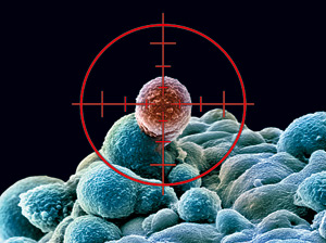
What are cancer stem cells?
Cancer is a debilitating disease estimated to be responsible for about 7.6 million deaths in 2008 (Jemal A, et al, CA Cancer J Clin, Mar-Apr 2011;61(2):69-90). Thus, extensive research is underway to deal with the various types of cancer. The concept of cancer stem cells (CSC) has surfaced in in the past decade after identification and characterization of CSC-enriched populations in several different types of cancer (Lapidot T, et al, Nature, 17 Feb 1994;367(6464):645-8; Reya T, et al, Nature, 1 Nov 2001;414(6859):105-11; Trumpp A and Wiestler OD, et al, Nat Clin Pract Oncol, Jun 2008;5(6):337-47). Although there has been lot of debate on the cell of origin of CSC, according to the classical concept CSC are defined by their functional properties.
Functional properties of CSC
- CSCs are at the top of tumor hierarchy. Regenerative tissues follow a hierarchical organization with adult stem cells at the top maintaining tissues and normal adult cells during homeostasis and regeneration during cell loss from injury. Similarly, several tumors follow the hierarchy with CSC at the top. Hierarchical organization has been reported in several cancer types including but not limited to breast cancer, brain cancer, colon cancer, leukemia and pancreatic cancer (Lapidot T, et al, Nature, 17 Feb 1994;367(6464):645-8; Al-Hajj M, et al, PNAS USA, 1 Apr 200;100(7):3983-8; Singh SK, et al, Nature, 18 Nov 2004;432(7015):396-401; Dalerba P, et al, PNAS USA, 12 Jun 2007;104(24):10158-63; Hermann PC, et al, Cell Stem Cell, 13 Sep 2007;1(3):313-23).
- CSCs possess unlimited self-renewal capacity similar to that of physiological stem cells and unlike other differentiated cell types within the tumor. Cancer stem cells can also generate non-CSC progeny that is comprised of differentiated cells and forms tumor bulk.
- Some CSs exhibit quiescent or dormant stage. Although not observed in all CSC types, some CSCs have been found to shuttle between quiescent, slow-cycling, and active states. The CSCs in their dormant and slow-cycling stage are less likely to be affected by conventional anti-tumor therapies which generally target rapidly dividing cells. Dormant stage is exhibited even in adult stem cells and the dormant normal stem cells can regain cell division potential during tissue injury (Wilson A, et al, Cell, 12 Dec 2008;135(6):1118-29). Thus, it has been speculated that dormant CSC might be a reason for tumor relapse even after pathologic complete response is observed post therapy.
- Some CSCs are resistant to conventional anti-cancer therapies. This leads to accumulation of CSC that might result in relapse after anti-cancer therapy. For instance, Li et al (2008) reported that CSC accumulated in the breast of women with locally advanced tumors after cytotoxic chemotherapy had eliminated the bulk of the tumor cells (Li X,et al, J Natl Cancer Inst, 7 May 2008;100(9):672-9). A similar observation was made by Oravecz-Wilson et al (2009) stating that despite remarkable responses to the tyrosine kinase inhibitor imatinib, CML patients show imatinib refractoriness because leukemia stem cells in CML are resistant tyrosine kinase (Oravecz-Wilson KI, et al, Cancer Cell, 4 Aug 2009;16(2):137-48).
- The CSC niche. CSC functional traits might be sustained by this microenvironment, termed “niche”. The niche is the environment in which stem cells reside and is responsible for the maintenance of unique stem cell properties such as self-renewal and an undifferentiated state. The heterogeneous populations which constitute a niche include both stem cells and surrounding differentiated cells. The necessary intrinsic pathways that are utilized by this cancer stem cell population to maintain both self-renewal and the ability to differentiate are believed to be a result of the environment where cancer stem cells reside. (Cabarcas SM, et al, Int J Cancer, 15 Nov 2011;129(10):2315-27). For instance, properties of CSC in glioma in a mouse xenograft model were maintained by vascular endothelial cells (Calabrese C, et al, Cancer Cell, Jan 2007;11(1):69-82). Several molecules including interleukin 6 have been observed to play a role in tumor proliferation and hence, participate in maintaining tumorigenic and self-renewal potential of CSC. Moreover, the CSC niche might not only regulate CSCs traits but might also directly provide CSC features to non-CSC population.
What is the origin of CSC?
According to current thinking, CSC result from epithelial-mesenchymal transition (EMT) when cells switch from a polarized epithelial to a non-polarized mesenchymal cell type with stem cell properties, including migratory behavior, self-renewal and generation of differentiated progeny, and reduced responsiveness to conventional cancer therapies (Scheel C and Weinberg RA, Semin Cancer Biol, Oct 2012;22(5-6):396-403; Crews LA and Jamieson CH, Cancer Lett, 17 Aug 2012). Evidence is accumulating that cancers of distinct subtypes within an organ may derive from different ‘cells of origin’. The tumor cell of origin is the cell type from which the disease is derived after it undergoes oncogenic mutation. It might take a series of mutations to achieve the CSC phenotype (Visvader JE, Nature, 20 Jan 2011;469(7330):314-22). Also, CSCs have been reported to originate from stem cells in some cases.
Biomarkers for CSC
CSC targeting therapy could either eliminate CSCs by either killing them after differentiating them from other tumor population, and/or by disrupting their niche. Efficient eradication of CSCs may require the combined ablation of CSCs themselves and their niches. Identifying appropriate biomarkers of CSC is a very important aim for CSCs to be useful as targets of anti-cancer therapies in order to possibly prevent relapse. Using cell surface markers, CSCs have been isolated and purified from cancers of breast, brain, thyroid, cervix, lung, blood (leukemia), skin (melanoma), organs of the gastrointestinal and reproductive tracts, and the retina. The challenge, however, is that CSCs share similar markers with normal cells which makes CSCs targeting difficult as it would harm normal cells in the process. More recently, advanced techniques such as signal sequence trap (SST) PCR screening methods have been developed to identify a leukemia-specific stem cell marker (CD96). After a small subset of human AML cells displayed tumorigenic properties, Leukemia Stem Cells (LSCs) were identified as leukemia cells with CD23+/CD38+ markers. These cells closely resemble hematopeotic stem cells (HSCs) (Bonnet D and Dick JR, Nat Med, Jul 1997;3(7):730-7). In solid tumors, a significant discovery was made when CSCs in breast cancer were identified within the ESA+/CD44+/CD24low-neg population of mammary pleural effusion and tumor samples (Al-Hajj M, et al, PNAS USA, 1 Apr 200;100(7):3983-8).
After these two landmark publications, CSCs were identified in many more solid and hematopoietic human tumors as well. In addition, within a tumor type, CSC-enriched populations display heterogeneity in markers. For example, only 1% of breast cancer cells simultaneously express both reported CSC phenotypes ESA+/CD44+/
CD24low-neg and ALDH-1+ (Ginestier C, et al, Cell Stem Cell, 1 Nov 2007;1(5):555-67). The discrepancy might be due to different techniques used to identify the markers and also a reflection of the molecular heterogeneity within the tumors. Recent advances in genome wide expression profiling studies have led to the identification of different subtypes in a particular type of cancer. Breast cancer was recently classified into different subtypes and this genetic heterogeneity is likely paralleled by a heterogeneous CSC complexity.
Conclusion
A lot of research is currently underway on various aspects of CSCs including biomarker identification, cell of origin, and clinical trials targeting CSC population in cancer. The concept of CSCs has evolved quite a bit since their discovery. Recently, identification of high genetic heterogeneity within a tumor has been in focus and subsequently it has been observed that several CSC clones can coexist and compete with each other within a tumor. Adding complexity to their identity is the fact that CSCs may have unstable phenotypes and genotypes. Taken together, the dynamics associated with CSCs makes it difficult to identify reliable and robust biomarkers and develop efficient targeted therapies. Thus, a major thrust of research should be to focus on the unfolding of the dynamic identity of CSCs in tumor types and at different that might lead to the identification and targeting of highly specific CSCs biomarkers.
Reference
Jemal A, et al, CA Cancer J Clin, Mar-Apr 2011;61(2):69-90
Reya T, et al, Nature, 1 Nov 2001;414(6859):105-11
Trumpp A and Wiestler OD, et al, Nat Clin Pract Oncol, Jun 2008;5(6):337-47
Lapidot T, et al, Nature, 17 Feb 1994;367(6464):645-8
Singh SK, et al, Nature, 18 Nov 2004;432(7015):396-401
Dalerba P, et al, PNAS USA, 12 Jun 2007;104(24):10158-63
Hermann PC, et al, Cell Stem Cell, 13 Sep 2007;1(3):313-23
Wilson A, et al, Cell, 12 Dec 2008;135(6):1118-29
Li X,et al, J Natl Cancer Inst, 7 May 2008;100(9):672-9
Oravecz-Wilson KI, et al, Cancer Cell, 4 Aug 2009;16(2):137-48
Cabarcas SM, et al, Int J Cancer, 15 Nov 2011;129(10):2315-27
Calabrese C, et al, Cancer Cell, Jan 2007;11(1):69-82
Scheel C and Weinberg RA, Semin Cancer Biol, Oct 2012;22(5-6):396-403
Crews LA and Jamieson CH, Cancer Lett, 17 Aug 2012
Visvader JE, Nature, 20 Jan 2011;469(7330):314-22
Bonnet D and Dick JR, Nat Med, Jul 1997;3(7):730-7
Al-Hajj M, et al, PNAS USA, 1 Apr 200;100(7):3983-8
Ginestier C, et al, Cell Stem Cell, 1 Nov 2007;1(5):555-67
Baccelli I and Trumpp AJ, Cell Biol, 6 Aug 2012;198(3):281-93
Zhao L, et al, Eur Surg Res, 2012;49(1):8-15
Pharmaceutical Intelligence posts:
http://pharmaceuticalintelligence.com/2012/08/15/to-die-or-not-to-die-time-and-order-of-combination-drugs-for-triple-negative-breast-cancer-cells-a-systems-level-analysis/
Authors: Anamika Sarkar, PhD and Ritu Saxena, PhD
http://pharmaceuticalintelligence.com/2013/03/07/the-importance-of-cancer-prevention-programs-new-perceptions-for-fighting-cancer/ Author: Ziv Raviv, PhD
http://pharmaceuticalintelligence.com/2013/03/03/treatment-for-metastatic-her2-breast-cancer/ Reporter: Larry H Bernstein, MD
http://pharmaceuticalintelligence.com/2013/03/02/recurrence-risk-for-breast-cancer/
Larry H Bernstein, MD
http://pharmaceuticalintelligence.com/2013/02/14/prostate-cancer-androgen-driven-pathomechanism-in-early-onset-forms-of-the-disease/ Curator: Aviva Lev-Ari, PhD, RN
http://pharmaceuticalintelligence.com/2013/01/15/exploring-the-role-of-vitamin-c-in-cancer-therapy/ Curator: Ritu Saxena, PhD
http://pharmaceuticalintelligence.com/2013/01/12/harnessing-personalized-medicine-for-cancer-management-prospects-of-prevention-and-cure-opinions-of-cancer-scientific-leaders-httppharmaceuticalintelligence-com/ Curator: Aviva Lev-Ari, PhD, RN
http://pharmaceuticalintelligence.com/2013/01/10/the-molecular-pathology-of-breast-cancer-progression/ Author and reporter: Tilda Barliya PhD
http://pharmaceuticalintelligence.com/2012/11/30/histone-deacetylase-inhibitors-induce-epithelial-to-mesenchymal-transition-in-prostate-cancer-cells/ Reporter and Curator: Stephen J. Williams, PhD
http://pharmaceuticalintelligence.com/2012/10/22/blood-vessel-generating-stem-cells-discovered/ Reporter: Ritu Saxena, PhD
http://pharmaceuticalintelligence.com/2012/10/17/stomach-cancer-subtypes-methylation-based-identified-by-singapore-led-team/ Reporter: Aviva Lev-Ari, PhD, RN
http://pharmaceuticalintelligence.com/2012/09/17/natural-agents-for-prostate-cancer-bone-metastasis-treatment/ Reporter: Ritu Saxena, PhD
http://pharmaceuticalintelligence.com/2012/08/28/cardiovascular-outcomes-function-of-circulating-endothelial-progenitor-cells-cepcs-exploring-pharmaco-therapy-targeted-at-endogenous-augmentation-of-cepcs/ Aviva Lev-Ari, PhD, RN
Read Full Post »