Feeds:
Posts
Comments

Posts Tagged ‘tumors’

Author and Curator: Ritu Saxena, Ph.D

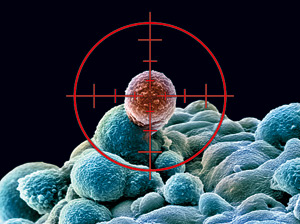
Although cancer stem cells constitute only a small percentage of the tumor burden, their self-renewal capacity and possible link with recurrence of cancer post treatment makes them a sought after therapeutic target in cancer. The post on cancer stem cells published on the 22nd of March, 2013, describes the identity of CSCs, their functional characteristics, possible cell of origin and biomarkers. This post focuses on the therapeutic potential of CSCs, their resistance to conventional anti-tumor therapies and current therapeutic targets including biomarkers, signaling pathways and niches.

CSCs Are Resistant to conventional anticancer therapies including chemotherapy, radiotherapy and surgery that are used either alone or in combination. However, these strategies have failed several times to eradicate CSCs resulting in metastasis and relapse, hence, a fatal disease outcome.

The properties of CSCs that contribute to or lead to chemoresistance include:

Quiescent Phenotype

Chemotherapeutic agents target fast-growing cells; however, some CSCs that remain in the dormant or quiescent stage are spared from lethal damage. Later, when the dormant CSCs enter cell cycle, tumor proliferation is stimulated.

Antiapoptosis

Antiapoptotic proteins such as BCL-2 and some self-renewal pathways such as transforming growth factor β, Wnt/ β -catenin or BMI-1 are activated in CSCs. Consequently, DNA damage repair capability of CSCs is enhanced after genotoxic stress or activation of autocrine loops through the production of growth factors like epidermal growth factor (Moserle L, Cancer Lett, 1 Feb 2010;288(1):1-9).

Expression of Drug Efflux Pumps

CSCs express some proteins that have typically been known to contribute to multidrug resistance. The proteins are drug efflux pumps ABCC1, ABCG2 or MDR1. Multidrug resistance-associated proteins (ABCC subfamily) are members of the ATP-binding cassette (ABC) superfamily of transport proteins and act as cellular efflux transporters for a wide variety of substrates, in particular glutathione, glucuronide and sulfate conjugates of diverse compounds.

Radiotherapy is mainly used in breast cancer and glioblastoma multiforme. In glioblastoma multiforme, the properties of CSCs that contribute to radiotherapy resistance is the presence of CD133 marker. CD133+ CSCs preferentially activate DNA damage repair pathway and significantly induced checkpoint kinases that leads to reduced apoptosis in CSCs compared to the CD133- tumor cells (Bao S, Nature, 7 Dec 2006;444(7120):756-60).

Radiotherapy resistance in breast cancer is due to reduced levels of reactive oxygen species in CSCs. In addition, radiation resistance of progenitor cells in an immortalized breast cancer cell line was mediated by the Wnt/β catenin pathway proteins (Diehn M, et al, Nature, 9 Apr 2009;458(7239):780-3; Chen MS, et al, J Cell Sci, 1 Feb 2007;120(Pt 3):468-77).

As mentioned in the previous post on CSCs, CSC targeting therapy could either eliminate CSCs by either killing them after differentiating them from other tumor population, and/or by disrupting their niche. Efficient eradication of CSCs may require the combined ablation of CSCs themselves and their niches. Thus, identification of appropriate and specific markers of CSCs is crucial for targeting them and preventing tumor relapse. Table 1 (adapted from a review article on CSCs by Zhao et al) describes the currently used biomarkers for CSC-targeted therapy (Zhao L, et al, Eur Surg Res, 2012;49(1):8-15).

Table 1

Specific Target Cancer type Marker properties and therapy
Targeting cell markers
CD24+CD44+ESA+ Pancreatic cancer Pancreatic CSCs, elevated during tumorigenesis
CD44+CD24–ESA+ Breast cancer Breast CSCs
EpCAM high CD44+CD166+ Colorectal cancer
CD34+CD38– AML broad use as a target for chemotherapy
CD133+ Prostate cancer and breast cancer 5-transmembrane domain cell surface glycoprotein,also a marker for neuron epithelial, hematopoietic and endothelialprogenitor cells
Stro1+CD105+CD44+ Bone sarcoma
Nodal/activin Knockdown or pharmacological inhibition of its receptorAlk4/7 abrogated self-renewal capacity and in vivo tumorigenicity of CSCs.
Targeting signaling pathways
Hedgehog signaling Upregulated in several cancer types inhibitors: GDC-0449,PF04449913, BMS-833923, IPI-926 and TAK-441
Wnt/β-catenin signaling CML, squamous cell carcinoma Be required for CSC self-renewal and tumor growthinhibitors: PRI-724, WIF-1 and telomerase
Notch signaling Several cancer types An important regulator in normal development, adult stem cell maintenance,and tumorigenesis in multiple organs,inhibitors: RO4929097, BMS-906024, IPI-926 and MK0752
PI3K/Akt/PTEN/mTOR, Several cancer types The pathway is deregulated in many tumors and used to preferentially target CSCsinhibitors: temsirolimus, everolimus FDA-approved therapy for renal cell carcinoma
Targeting CSC Niche
Angiogenesis Niche Colon cancer, breast cancer, NSCLC Inhibitor: bevacizumab results in a disruption of the CSC niche, depleted vasculature and a dramatic reduction in the number of CSCs.
Hypoxia (HIF pathway) Ovarian cancer, lung cancer, cervical cancer Inhibitors: topotecan and digoxin have been approved for ovarian, lung and cervical cancer
Targeting Micro RNA
miR-200 family Inhibits EMT and cancer cell migration by direct targeting of E-cadherin transcriptional repressors ZEB1 and ZEB2
Let-7 family Regulates BT-IC stem cell-like properties by silencing more than one target
miR-124 Related to neuronal differentiation, targets laminin γ1 and integrin β1.
miR-21 Suppresses the self-renewal of embryonic stem cells

The challenge is to develop an effective treatment regimen that prevents survival, self-renewal and differentiation of CSCs and also disturbs their niche without damaging normal stem cells. In order to evaluate the efficiency of CSC-targeting therapies, in vitro models and mouse xenotransplantation models have been used for preclinical studies. Some potential CSC targeting agents in preclinical stages include notch inhibitors for glioblastoma stem cells and telomerase peptide vaccination after chemoradiotherapy of non-small cell lung cancer stem cells Stem Cells (Hovinga KE, et al, Jun 2010;28(6):1019-29; Serrano D, Mol Cancer, 9 Aug 2011;10:96). In addition, several phase II and phase III trials are currently underway to test CSC-targeting drugs focusing on efficacy and safety of treatment.

Reference:

Bao S, Nature, 7 Dec 2006;444(7120):756-60).

Diehn M, et al, Nature, 9 Apr 2009;458(7239):780-3

Chen MS, et al, J Cell Sci, 1 Feb 2007;120(Pt 3):468-77

Zhao L, et al, Eur Surg Res, 2012;49(1):8-15

Hovinga KE, et al, Jun 2010;28(6):1019-29

Serrano D, Mol Cancer, 9 Aug 2011;10:96

Pharmaceutical Intelligence posts:

http://pharmaceuticalintelligence.com/2013/03/22/in-focus-identity-of-cancer-stem-cells/ Author and curator: Ritu Saxena, PhD

http://pharmaceuticalintelligence.com/2012/08/15/to-die-or-not-to-die-time-and-order-of-combination-drugs-for-triple-negative-breast-cancer-cells-a-systems-level-analysis/ Authors: Anamika Sarkar, PhD and Ritu Saxena, PhD

http://pharmaceuticalintelligence.com/2013/03/07/the-importance-of-cancer-prevention-programs-new-perceptions-for-fighting-cancer/ Author: Ziv Raviv, PhD

http://pharmaceuticalintelligence.com/2013/03/03/treatment-for-metastatic-her2-breast-cancer/ Reporter: Larry H Bernstein, MD

http://pharmaceuticalintelligence.com/2013/03/02/recurrence-risk-for-breast-cancer/ Larry H Bernstein, MD

http://pharmaceuticalintelligence.com/2013/02/14/prostate-cancer-androgen-driven-pathomechanism-in-early-onset-forms-of-the-disease/ Curator: Aviva Lev-Ari, PhD, RN

http://pharmaceuticalintelligence.com/2013/01/15/exploring-the-role-of-vitamin-c-in-cancer-therapy/ Curator: Ritu Saxena, PhD

http://pharmaceuticalintelligence.com/2013/01/12/harnessing-personalized-medicine-for-cancer-management-prospects-of-prevention-and-cure-opinions-of-cancer-scientific-leaders-httppharmaceuticalintelligence-com/ Curator: Aviva Lev-Ari, PhD, RN

http://pharmaceuticalintelligence.com/2013/01/10/the-molecular-pathology-of-breast-cancer-progression/ Author and reporter: Tilda Barliya PhD

http://pharmaceuticalintelligence.com/2012/11/30/histone-deacetylase-inhibitors-induce-epithelial-to-mesenchymal-transition-in-prostate-cancer-cells/ Reporter and Curator: Stephen J. Williams, PhD

http://pharmaceuticalintelligence.com/2012/10/22/blood-vessel-generating-stem-cells-discovered/ Reporter: Ritu Saxena, PhD

http://pharmaceuticalintelligence.com/2012/10/17/stomach-cancer-subtypes-methylation-based-identified-by-singapore-led-team/ Reporter: Aviva Lev-Ari, PhD, RN

http://pharmaceuticalintelligence.com/2012/09/17/natural-agents-for-prostate-cancer-bone-metastasis-treatment/ Reporter: Ritu Saxena, PhD

http://pharmaceuticalintelligence.com/2012/08/28/cardiovascular-outcomes-function-of-circulating-endothelial-progenitor-cells-cepcs-exploring-pharmaco-therapy-targeted-at-endogenous-augmentation-of-cepcs/ Aviva Lev-Ari, PhD, RN


Read Full Post »

Author and Curator: Ritu Saxena, PhD

Image

Screen Shot 2021-07-19 at 6.28.21 PM

Word Cloud By Danielle Smolyar

What are cancer stem cells?

Cancer is a debilitating disease estimated to be responsible for about 7.6 million deaths in 2008 (Jemal A, et al, CA Cancer J Clin, Mar-Apr 2011;61(2):69-90). Thus, extensive research is underway to deal with the various types of cancer. The concept of cancer stem cells (CSC) has surfaced in in the past decade after identification and characterization of CSC-enriched populations in several different types of cancer (Lapidot T, et al, Nature, 17 Feb 1994;367(6464):645-8; Reya T, et al, Nature, 1 Nov 2001;414(6859):105-11;  Trumpp A and Wiestler OD, et al, Nat Clin Pract Oncol, Jun 2008;5(6):337-47). Although there has been lot of debate on the cell of origin of CSC, according to the classical concept CSC are defined by their functional properties.

Functional properties of CSC

  • CSCs are at the top of tumor hierarchy. Regenerative tissues follow a hierarchical organization with adult stem cells at the top maintaining tissues and normal adult cells during homeostasis and regeneration during cell loss from injury. Similarly, several tumors follow the hierarchy with CSC at the top. Hierarchical organization has been reported in several cancer types including but not limited to breast cancer, brain cancer, colon cancer, leukemia and pancreatic cancer (Lapidot T, et al, Nature, 17 Feb 1994;367(6464):645-8; Al-Hajj M, et al, PNAS USA, 1 Apr 200;100(7):3983-8; Singh SK, et al, Nature, 18 Nov 2004;432(7015):396-401; Dalerba P, et al, PNAS USA, 12 Jun 2007;104(24):10158-63; Hermann PC, et al, Cell Stem Cell, 13 Sep 2007;1(3):313-23).
  • CSCs possess unlimited self-renewal capacity similar to that of physiological stem cells and unlike other differentiated cell types within the tumor. Cancer stem cells can also generate non-CSC progeny that is comprised of differentiated cells and forms tumor bulk.
  • Some CSs exhibit quiescent or dormant stage. Although not observed in all CSC types, some CSCs have been found to shuttle between quiescent, slow-cycling, and active states. The CSCs in their dormant and slow-cycling stage are less likely to be affected by conventional anti-tumor therapies which generally target rapidly dividing cells. Dormant stage is exhibited even in adult stem cells and the dormant normal stem cells can regain cell division potential during tissue injury (Wilson A, et al, Cell,  12 Dec 2008;135(6):1118-29). Thus, it has been speculated that dormant CSC might be a reason for tumor relapse even after pathologic complete response is observed post therapy.
  • Some CSCs are resistant to conventional anti-cancer therapies. This leads to accumulation of CSC that might result in relapse after anti-cancer therapy. For instance, Li et al (2008) reported that CSC accumulated in the breast of women with locally advanced tumors after cytotoxic chemotherapy had eliminated the bulk of the tumor cells (Li X,et al, J Natl Cancer Inst, 7 May 2008;100(9):672-9). A similar observation was made by Oravecz-Wilson et al (2009) stating that despite remarkable responses to the tyrosine kinase inhibitor imatinib, CML patients show imatinib refractoriness because leukemia stem cells in CML are resistant tyrosine kinase (Oravecz-Wilson KI, et al, Cancer Cell, 4 Aug 2009;16(2):137-48).
  • The CSC niche. CSC functional traits might be sustained by this microenvironment, termed “niche”. The niche is the environment in which stem cells reside and is responsible for the maintenance of unique stem cell properties such as self-renewal and an undifferentiated state. The heterogeneous populations which constitute a niche include both stem cells and surrounding differentiated cells. The necessary intrinsic pathways that are utilized by this cancer stem cell population to maintain both self-renewal and the ability to differentiate are believed to be a result of the environment where cancer stem cells reside. (Cabarcas SM, et al, Int J Cancer, 15 Nov 2011;129(10):2315-27). For instance, properties of CSC in glioma in a mouse xenograft model were maintained by vascular endothelial cells (Calabrese C, et al, Cancer Cell, Jan 2007;11(1):69-82). Several molecules including interleukin 6 have been observed to play a role in tumor proliferation and hence, participate in maintaining tumorigenic and self-renewal potential of CSC. Moreover, the CSC niche might not only regulate CSCs traits but might also directly provide CSC features to non-CSC population.

What is the origin of CSC?

According to current thinking, CSC result from epithelial-mesenchymal transition (EMT) when cells switch from a polarized epithelial to a non-polarized mesenchymal cell type with stem cell properties, including migratory behavior, self-renewal and generation of differentiated progeny, and reduced responsiveness to conventional cancer therapies (Scheel C and Weinberg RA, Semin Cancer Biol, Oct 2012;22(5-6):396-403; Crews LA and Jamieson CH, Cancer Lett, 17 Aug 2012). Evidence is accumulating that cancers of distinct subtypes within an organ may derive from different ‘cells of origin’. The tumor cell of origin is the cell type from which the disease is derived after it undergoes oncogenic mutation. It might take a series of mutations to achieve the CSC phenotype (Visvader JE, Nature, 20 Jan 2011;469(7330):314-22). Also, CSCs have been reported to originate from stem cells in some cases.

Biomarkers for CSC

CSC targeting therapy could either eliminate CSCs by either killing them after differentiating them from other tumor population, and/or by disrupting their niche. Efficient eradication of CSCs may require the combined ablation of CSCs themselves and their niches. Identifying appropriate biomarkers of CSC is a very important aim for CSCs to be useful as targets of anti-cancer therapies in order to possibly prevent relapse. Using cell surface markers, CSCs have been isolated and purified from cancers of breast, brain, thyroid, cervix, lung, blood (leukemia), skin (melanoma), organs of the gastrointestinal and reproductive tracts, and the retina. The challenge, however, is that CSCs share similar markers with normal cells which makes CSCs targeting difficult as it would harm normal cells in the process. More recently, advanced techniques such as signal sequence trap (SST) PCR screening methods have been developed to identify a leukemia-specific stem cell marker (CD96). After a small subset of human AML cells displayed tumorigenic properties, Leukemia Stem Cells (LSCs) were identified as leukemia cells with CD23+/CD38+ markers. These cells closely resemble hematopeotic stem cells (HSCs) (Bonnet D and Dick JR, Nat Med, Jul 1997;3(7):730-7). In solid tumors, a significant discovery was made when CSCs in breast cancer were identified within the ESA+/CD44+/CD24low-neg population of mammary pleural effusion and tumor samples (Al-Hajj M, et al, PNAS USA, 1 Apr 200;100(7):3983-8).

After these two landmark publications, CSCs were identified in many more solid and hematopoietic human tumors as well. In addition, within a tumor type, CSC-enriched populations display heterogeneity in markers. For example, only 1% of breast cancer cells simultaneously express both reported CSC phenotypes ESA+/CD44+/

CD24low-neg and ALDH-1+ (Ginestier C, et al, Cell Stem Cell, 1 Nov 2007;1(5):555-67). The discrepancy might be due to different techniques used to identify the markers and also a reflection of the molecular heterogeneity within the tumors. Recent advances in genome wide expression profiling studies have led to the identification of different subtypes in a particular type of cancer. Breast cancer was recently classified into different subtypes and this genetic heterogeneity is likely paralleled by a heterogeneous CSC complexity.

Conclusion

A lot of research is currently underway on various aspects of CSCs including biomarker identification, cell of origin, and clinical trials targeting CSC population in cancer. The concept of CSCs has evolved quite a bit since their discovery. Recently, identification of high genetic heterogeneity within a tumor has been in focus and subsequently it has been observed that several CSC clones can coexist and compete with each other within a tumor. Adding complexity to their identity is the fact that CSCs may have unstable phenotypes and genotypes. Taken together, the dynamics associated with CSCs makes it difficult to identify reliable and robust biomarkers and develop efficient targeted therapies. Thus, a major thrust of research should be to focus on the unfolding of the dynamic identity of CSCs in tumor types and at different that might lead to the identification and targeting of highly specific CSCs biomarkers.

Reference

Jemal A, et al, CA Cancer J Clin, Mar-Apr 2011;61(2):69-90

Reya T, et al, Nature, 1 Nov 2001;414(6859):105-11

Trumpp A and Wiestler OD, et al, Nat Clin Pract Oncol, Jun 2008;5(6):337-47

Lapidot T, et al, Nature, 17 Feb 1994;367(6464):645-8

Singh SK, et al, Nature, 18 Nov 2004;432(7015):396-401

Dalerba P, et al, PNAS USA, 12 Jun 2007;104(24):10158-63

Hermann PC, et al, Cell Stem Cell, 13 Sep 2007;1(3):313-23

Wilson A, et al, Cell,  12 Dec 2008;135(6):1118-29

Li X,et al, J Natl Cancer Inst, 7 May 2008;100(9):672-9

Oravecz-Wilson KI, et al, Cancer Cell, 4 Aug 2009;16(2):137-48

Cabarcas SM, et al, Int J Cancer, 15 Nov 2011;129(10):2315-27

Calabrese C, et al, Cancer Cell, Jan 2007;11(1):69-82

Scheel C and Weinberg RA, Semin Cancer Biol, Oct 2012;22(5-6):396-403

Crews LA and Jamieson CH, Cancer Lett, 17 Aug 2012

Visvader JE, Nature, 20 Jan 2011;469(7330):314-22

Bonnet D and Dick JR, Nat Med, Jul 1997;3(7):730-7

Al-Hajj M, et al, PNAS USA, 1 Apr 200;100(7):3983-8

Ginestier C, et al, Cell Stem Cell, 1 Nov 2007;1(5):555-67

Baccelli I and Trumpp AJ, Cell Biol, 6 Aug 2012;198(3):281-93

Zhao L, et al, Eur Surg Res, 2012;49(1):8-15

Pharmaceutical Intelligence posts:

http://pharmaceuticalintelligence.com/2012/08/15/to-die-or-not-to-die-time-and-order-of-combination-drugs-for-triple-negative-breast-cancer-cells-a-systems-level-analysis/

Authors: Anamika Sarkar, PhD and Ritu Saxena, PhD

http://pharmaceuticalintelligence.com/2013/03/07/the-importance-of-cancer-prevention-programs-new-perceptions-for-fighting-cancer/ Author: Ziv Raviv, PhD

http://pharmaceuticalintelligence.com/2013/03/03/treatment-for-metastatic-her2-breast-cancer/ Reporter: Larry H Bernstein, MD

http://pharmaceuticalintelligence.com/2013/03/02/recurrence-risk-for-breast-cancer/

Larry H Bernstein, MD

http://pharmaceuticalintelligence.com/2013/02/14/prostate-cancer-androgen-driven-pathomechanism-in-early-onset-forms-of-the-disease/ Curator: Aviva Lev-Ari, PhD, RN

http://pharmaceuticalintelligence.com/2013/01/15/exploring-the-role-of-vitamin-c-in-cancer-therapy/ Curator: Ritu Saxena, PhD

http://pharmaceuticalintelligence.com/2013/01/12/harnessing-personalized-medicine-for-cancer-management-prospects-of-prevention-and-cure-opinions-of-cancer-scientific-leaders-httppharmaceuticalintelligence-com/ Curator: Aviva Lev-Ari, PhD, RN

http://pharmaceuticalintelligence.com/2013/01/10/the-molecular-pathology-of-breast-cancer-progression/ Author and reporter: Tilda Barliya PhD

http://pharmaceuticalintelligence.com/2012/11/30/histone-deacetylase-inhibitors-induce-epithelial-to-mesenchymal-transition-in-prostate-cancer-cells/ Reporter and Curator: Stephen J. Williams, PhD

http://pharmaceuticalintelligence.com/2012/10/22/blood-vessel-generating-stem-cells-discovered/ Reporter: Ritu Saxena, PhD

http://pharmaceuticalintelligence.com/2012/10/17/stomach-cancer-subtypes-methylation-based-identified-by-singapore-led-team/ Reporter: Aviva Lev-Ari, PhD, RN

http://pharmaceuticalintelligence.com/2012/09/17/natural-agents-for-prostate-cancer-bone-metastasis-treatment/ Reporter: Ritu Saxena, PhD

http://pharmaceuticalintelligence.com/2012/08/28/cardiovascular-outcomes-function-of-circulating-endothelial-progenitor-cells-cepcs-exploring-pharmaco-therapy-targeted-at-endogenous-augmentation-of-cepcs/ Aviva Lev-Ari, PhD, RN

Read Full Post »

 Curator: Ritu Saxena, Ph.D.

Vitamin C or Ascorbic acid (AA) or Ascorbate

Biochemical role: AA serves a basic biochemical role of accelerating hydroxylation in several biochemical reactions. It provides electrons to metal ions, the reduced forms of which are required for the full enzymatic activity of some enzymes. Most emphasized role of AA is as a cofactor for the enzyme required for the biosynthesis of collagen.

Molecular structure and the oxidized form of AA, dihydroascorbic acid, bear similarity to that of glucose.

Biological role: AA is an essential vitamin for humans and its deficiency leads to disease called Scurvy characterized by initial symptoms of malaise and lethargy, followed by formation of spots on the skin, spongy gums, and bleeding from the mucous membranes. As scurvy advances, there can be open, suppurating wounds, loss of teeth, jaundice, fever, neuropathy and death. AA is water soluble and found in high concentrations in several tissues including eye lens, WBCs, adrenal glad and pituitary gland. Some of the roles of ascorbate include:

  1. Carnitine synthesis from lysine
  2. Neurotransmitter synthesis,
  3. Cytochrome P-450 activity,
  4. Cholesterol metabolism,
  5. Detoxification of exogenous compounds,
  6. Antioxidant
  7. Possibly an ergogenic aid (Ergogenic aids are substances, devices, or practices that enhance an individual’s energy use, production, or recovery.)

Vitamin C and Cancer

As early as in 1949, vitamin C was implicated in cancer therapy. Since then, several research articles have been published exploring the role of ascorbate in cancer therapy. Among the plethora of literature discussing the relationship between vitamin C and cancer, one of the very significant and comprehensive reviews was published in 1979 in Cancer Research (2).

Mechanisms of action of AA (1) with respect to cancer have been divided and subdivided into the following:

  1. Primary mechanisms
  2. Secondary mechanisms
  • Preventive mechanism

Ascorbate acts as a cancer preventive agent by virtue of its strong antioxidant activities. Being one of the strongest reductants and radical scavenger, it absorbs unstable oxygen, nitrogen, and sulphur-centered radicals. AA can prevent biomembranes from peroxidative damage from peroxyl radicals. Ascorbate can trap peroxyl radicals and lead to their peroxidation in the aqueous phase before they reach the lipid rich biomembranes and cause damage. Ascorbate has been speculated to have a biomembrane protective action by its synergistic antioxidant activity with vitamin E (tocopherol).  Vitamin E is lipid-soluble and tocopheroxyl radical is generated in the cell membranes as a result of its antioxidant activity.  Ascorbate reacts with the tocopheroxyl radical and regenerates tocopherol transferring the oxidative challenge to the aqueous phase. At this point, the less active ascorbate radical might be reduced to AA by an NADP-dependent system. The probably mechanism might explain the reduction of nitrates via ascorbate to prevent the formation of carcinogenic nitrosamines.

  • Anticancer mechanisms

1. Primary anticancer mechanisms

i.     Oxidative, oxidant and pro-oxidant properties: Ascorbate has been reported to be cytotoxic at high concentrations, which has been demonstrated in a number of malignant cell lines. Transcription factor NFkB is potentially activated via ascorbate and its radicals leading to the inhibition of cell growth. Also, ascorbate inhibits certain prostaglandins leading to decrease in cell proliferation.

ii.     Hydrogen peroxide: On oxidation with oxygen, ascorbate produces a hydrogen peroxide, a reactive oxygen species. Hydrogen peroxide can generate several other reactive species and can have several damaging effects on cells including decrease in cell viability by damaging cell membranes of malignant cells. The amount of these reactive species produced via oxidation is limited in healthy cells unlike that in malignant cells where they exist in large amounts. The amount of hydrogen peroxide generated has been correlated to the amount of ascorbate in the cells. The reactive species can lead to multiple negative effects on cells including DNA strand breaks, lipid peroxidation leading to membrane function disruption, cellular ATP depletion.

Authors state that “the failure to maintain high ATP production may be a consequence of oxidative inactivation of key enzymes especially those related to the Krebs cycle and the electron transport chain.” This might result in alteration of transmembrane potential and distortion of mitochondrial function, suggestive of the important role of mitochondria in the process of carcinogenesis. In this paper, vitamin C has been correlated with cancer with the involvement of altered mitochondrial function. In addition, ascorbate has been detected in mitochondria where it is also regenerated. Different aspects of mitochondrial involvement in cancer have been discussed in several posts published earlier (3-8).

iii.     Other oxidation products of AA: Other oxidation products of AA include 2,3-diketoglutonic acid, and 5-methyl 1-3, 4-dehydrotetrone and other degradation products, have demonstrated antitumor activity. Additionally, some degradation and oxidation products of AA, gamma-cronolactone and 3-hydroxyl-2-pyrone, have been found to inhibit tumor growth. The mechanism of their antitumor actions is complex and might involve multitude of steps, including generation of reactive oxygen species, lipid peroxidation, inducing structural changes in important cellular proteins, inhibition of mitosis and so on.

iv.     Intracellular transport of ascorbate and its tumor specificity: Oxidized ascorbate, dihydroascorbic acid, is transported intracellularly where it is reduced back to ascorbate. Owing to its structural similarity with glucose, dihydroascorbic transport is facilitated via glucose transporters (GLUTs). Ascrobate in its reduced form is transported through a sodium-dependent cotransporter in some cells. Tumor cells require large amounts of glucose, which leads to an increase in the number of GLUTs, hence, resulting in an increase in ascorbate concentration within cancer cells. Because of this selective increased uptake of ascorbate and its cytotoxic effects in cancer cells (generation of hydrogen peroxide, DNA damage, other cytotoxic effects), AA has become a selective, nontoxic chemotherapeutic agent. The difference in the levels of catalase enzyme has been found to lead to intracellular tumor selectivity in cancer cells.

Ascorbate induced cytotoxicity in cancer cells involves its final electron acceptor, oxygen, which interferes with the anaerobic respiration within malignant cells. This gives an important clue for the involvement of mitochondria in malignant cells.

v.     Intravenous AA: High concentrations of AA in plasma (>200mg/dL) have been found to be cytotoxic to cancer cells. Clinically high plasma concentrations of AA can be achieved by its intravenous administration. It was observed that 60g infusion of AA given to cancer patients for 60 minutes followed by 20g given over the next 60 minutes resulted in a 240 minutes high plasma AA concentration of >400mg/dL, that is known to be cytotoxic.

Lipoic acid when administered with AA, is able to reduce the high-dose requirement of AA for its cytotoxic activity reducing it from 700mg/dL to 120mg/dL. Lipoic acid can recycle vitamin C, mediate the reduction of dihydroascorbic acid and improves mitochondrial function. Thus, energy intermediates such as coenzyme Q, vitamin K3, B-complex vitamins, alpha-ketoglutarate aspartate, magnesium might aid in cancer therapy by intercting with ascorbate, directly or indirectly, thereby stimuating/interacting/correcting aerobic mitochondrial respiration.

Hence, the pro-oxidant activity of vitamin C is being referred to as the primary mechanism of anticancer action.

2. Secondary anticancer mechanisms

i.     AA and intracellular matrix: Collagen is an important constituent of the matrix and its concentration determines the strength of the tissue along with its resistance to the infiltration of malignant cancer cells. In Scurvy, a disease resulting from a chronic deficiency of vitamin C, there is generalized tissue disintegration, dissolution of intercellular ground substance and the disruption of collagen bundles. This disintegration leads to ulceration; bacterial colonization and general undifferentiated cellular proliferation with specialized cells reverting back to their primitive form, very much like cancer.  Lack of ascorbate causes a reduction in the hydroxylation of prolyl and lysyl residues into hydroxyproline and hydroxylysine, leading to instability of the collagen triple helix, a common feature in scurvy and also in cancer. Thus, a secondary mechanism of ascorbic acid anticancer mechanism would be to repair these sites, which is emphasized by its role in wound healing, including surgical recovery and other traumatic injuries.

ii.     Ascorbate and immunocompetence: Ascorbate plays several roles for the efficient functioning of immune system in ways that are invoved in both humoral and cell-mediated.  Ascorbate provides humoral immunocompetence as it is essential for immunoglobulin synthesis. In addition, lymphocytes, seminal cells involved in cell-mediated immunity have been found to contain high concentrations of ascorbate. Other immune system roles include, aid in active phagocytosis and enhancing of interferon production.

Classical vitamin C and Cancer controversy-A possible explanation

Conflicting results were obtained from the studies performed by Pauling (Pauling Institute) and Cameron (Mayo Clinic) with vitamin C and its effect on cancer, the issue was debated a few decades ago. Both the studies, however, used oral doses of ascorbate (10g). Gonzalez et al, authors of the review on which the post is based, analyzed and expressed their views on the controversy. They state that the plasma concentration cannot be replicated when the dose is given orally as opposed to when the dose is given intravenously. According to their research, when AA is administered intravenously, higher plasma levels of ascorbate are achieved that could be retained for longer time periods. Also, the authors advocate the use of substantially higher doses (25-200g) to be given intravenously for selective toxicity towards cancer cells.

Modern vitamin C and Cancer controversy-Chemotherapy and radiation

A recent concern regarding the antioxidants like vitamin C is that they might reduce the effectiveness of chemotherapy and radiation by reducing the potency of free radicals necessary for killing cells. A publication by Agus et al (13) has a major role to play in this misconception. The authors describe how cancer cells acquire and concentrate vitamin C providing malignant cells with metabolic advantage. However, details or explanations regarding the theory are missing. Some studies, on the other hand, explain that high concentrations of AA in cancer cells is cytotoxic and is achieved because of similarity in structure between AA and glucose. Cancer cells uptake AA derivative, dehydroascorbic acid via glucose transporters (GLUTs).

In a case report published in PNAS in 1985 (12), two patients with ovarian cancer stage IIIC were found to respond positively to chemotherapy along with high-dose of antioxidants. Antioxidant, AA was administered intravenously to maintain a high plasma dose of 200 mg/dL. The two patients didn’t show disease recurrence after three years of chemotherapy and vitamin C administration. Vast literature exists on the topic indicating that antioxidants, including ascorbate, provide beneficial effects in several cancers without reducing the efficacy of chemotherapy or radiation during treatment of these cancers. Some data, in fact, suggests increase in effectiveness of chemotherapy when supplemented with antioxidants along with an increase in adverse effects. The topic has been summarized and discussed in a series of articles by Lawson and Brignall (9-11).

REFERENCES

The post is primarily based on the following two review articles:

1. González MJ et al. Orthomolecular oncology review: ascorbic acid and cancer 25 years later.  Integr Cancer Ther. 2005 Mar;4(1):32-44.

2. Cameron E, Pauling L, Leibovitz B. Ascorbic acid and cancer: a review. Cancer Res. 1979 Mar;39(3):663-81.

Other articles  on Mitochondria and Cancer were published on this Open Source Online Scientific Journal

3. Ritu Saxena. Mitochondria and Cancer: An overview of mechanisms

4. Ritusaxena. β Integrin emerges as an important player in mitochondrial dysfunction associated Gastric Cancer.

5. Larry H Bernstein. Mitochondria: Origin from oxygen free environment, role in aerobic glycolysis, metabolic adaptation

6. Ritu Saxena. Mitochondria and Cancer: An overview of mechanisms

7. Larry H Bernstein. Mitochondrial Damage and Repair under Oxidative Stress

8. Larry H Bernstein. What can we expect of tumor therapeutic response?

Research articles:

9. Lamson DW, Brignall MS. Antioxidants and cancer, part 3: quercetin. Altern Med Rev. 2000 Jun;5(3):196-208. Review.

10. Lamson DW, Brignall MS. Antioxidants and cancer therapy II: quick reference guide. Altern Med Rev. 2000 Apr;5(2):152-63.

11. Lamson DW, Brignall MS. Antioxidants in cancer therapy; their actions and interactions with oncologic therapies. Altern Med Rev. 1999 Oct;4(5):304-29.

12. Bensch KG, Fleming JE, Lohman W. The role of ascorbic acid in senile cataracts. Proc Natl Acad Sci USA 1985;82:7193-7196.

13. Agus DB, Vera JG, Golde DW. Stand allocation: a mechanism by which tumors obtain vitamin C. Cancer Res. 1999;59:4555-4558.

Read Full Post »

%d bloggers like this: