Feeds:
Posts
Comments

Posts Tagged ‘CML’

New PCR Based Test May be Able to Detect Low Levels of Persistent CML, Guiding TKI therapy Choices

from CancerNetwork.com: Novel Assay Could Help Guide Treatment Cessation Decisions in CML

Reporter: Stephen J. Williams, Ph.D.

News | February 15, 2016 | Chronic Myeloid Leukemia, Hematologic Malignancies, Leukemia & Lymphoma
By Dave Levitan
A new personalized DNA-based assay can detect very low levels of persistent disease in chronic myeloid leukemia (CML) patients thought to be in deep remission, according to a new study. The test could help with treatment choices regarding cessation of tyrosine kinase inhibitor (TKI) therapy in these patients.
A number of recent studies have examined the possibility that some patients could stop TKI therapy after achieving deep molecular remission. “However, the safe introduction of a TKI-withdrawal strategy would require a reliable and cost-effective method for the identification of those patients with the lowest likelihood of relapse,” wrote study authors led by Alistair G. Reid, BSc, PhD, of Imperial College London.

Because the likelihood of relapse after withdrawal from therapy is probably related to persistence of residual disease, testing for low levels of BCR-ABL1–positive disease is key. In the new study, the researchers tested an assay using personalized DNA-based polymerase chain reaction (dPCR) involving identification of t(9;22) fusion junctions. The results were published in the Journal of Molecular Diagnostics.

They successfully mapped genomic breakpoints in 32 of 32 samples from CML patients with early-stage disease. Next, they tested 46 samples from 6 patients following treatment with a TKI and compared results to other quantitative PCR methods; 10 of the samples were used as positive controls, while the others were considered to be in deep molecular remission.

Of those 36 samples, dPCR detected persistent disease in 81%. This was more sensitive than two other PCR-based approaches, including RT-dPCR (25%) and DNA-based quantitative PCR (19%).

“The technologies described allow for the assignment of absolute quantities to both BCR-ABL1 DNA and RNA targets, facilitating for the first time direct comparison of mean expression vs cellular disease burden,” the authors wrote. They added that it remains to be explored whether the risk of relapse after withdrawal of therapy is related to just the number of CML cells or also to the degree of transcriptional activity in those cells.

“If validated in clinical trials of stopping TKIs, this technique will permit a more personalized approach to recommendations for dose reduction or drug cessation in individual patients, ensuring that therapy is withdrawn only from patients with the highest chance of long-term remission,” said study author Jane F. Apperley, MD, PhD, also of Imperial College London, in a press release. “The technique we describe, with which we successfully mapped a disease-specific junction in all patients tested, is relatively simple, cost-effective, and suited to a high-throughput laboratory.”

– See more at: http://www.cancernetwork.com/chronic-myeloid-leukemia/novel-assay-could-help-guide-treatment-cessation-decisions-cml?GUID=D63BFB74-A7FD-4892-846F-A7D1FFE0F131&XGUID=&rememberme=1&ts=09032016#sthash.EAzX9VUl.dpuf

– See more at: http://www.cancernetwork.com/chronic-myeloid-leukemia/novel-assay-could-help-guide-treatment-cessation-decisions-cml?GUID=D63BFB74-A7FD-4892-846F-A7D1FFE0F131&XGUID=&rememberme=1&ts=09032016#sthash.EAzX9VUl.dpuf

Read Full Post »

Novel Mechanisms of Resistance to Novel Agents


Curators: Larry H. Berstein, M.D. FACP & Stephen J. Williams, Ph.D.

For most of the history of chemotherapy drug development, predicting the possible mechanisms of drug resistance that ensued could be surmised from the drug’s pharmacologic mechanism of action. In other words, a tumor would develop resistance merely by altering the pathways/systems which the drug relied on for mechanism of action. For example, as elucidated in later chapters in this book, most cytotoxic chemotherapies like cisplatin and cyclophosphamide were developed to bind DNA and disrupt the cycling cell, thereby resulting in cell cycle arrest and eventually cell death or resulting in such a degree of genotoxicity which would result in great amount of DNA fragmentation. These DNA-damaging agents efficacy was shown to be reliant on their ability to form DNA adducts and lesions. Therefore increasing DNA repair could result in a tumor cell becoming resistant to these drugs. In addition, if drug concentration was merely decreased in these cells, by an enhanced drug efflux as seen with the ABC transporters, then there would be less drug available for these DNA adducts to be generated. A plethora of literature has been generated on this particular topic.

However in the era of chemotherapies developed against targets only expressed in tumor cells (such as Gleevec against the Bcr-Abl fusion protein in chronic myeloid leukemia), this paradigm had changed as clinical cases of resistance had rapidly developed soon after the advent of these compounds and new paradigms of resistance mechanisms were discovered.

speed of imitinib resistance

Imatinib resistance can be seen quickly after initiation of therapy

mellobcrablresistamplification

Speed of imatinib resistance a result of rapid gene amplification of BCR/ABL target, thereby decreasing imatinib efficacy


Although there are many other new mechanisms of resistance to personalized medicine agents (which are discussed later in the chapter) this post is a curation of cellular changes which are not commonly discussed in reviews of chemoresistance and separated in three main categories:

Cellular Diversity and Adaptation

Identifying Cancers and Resistance

Cancer Drug-Resistance Mechanism

p53 tumor drug resistance gene target

Variability of Gene Expression and Drug Resistance


Expression of microRNAs and alterations in RNA resulting in chemo-resistance

Drug-resistance Mechanism in Tumor Cells

Overexpression of miR-200c induces chemoresistance in esophageal cancers mediated through activation of the Akt signaling pathway


The miRNA–drug resistance connection: a new era of personalized medicine using noncoding RNA begins


Gene Duplication of Therapeutic Target


The advent of Gleevec (imatinib) had issued in a new era of chemotherapy, a personalized medicine approach by determining the and a lifesaver to chronic myeloid leukemia (CML) patients whose tumors displayed expression of the Bcr-Abl fusion gene. However it was not long before clinical resistance was seen to this therapy and, it was shown amplification of the drug target can lead to tumor outgrowth despite adequate drug exposure. le Coutre, Weisberg and Mahon23, 24, 25 all independently generated imatinib-resistant clones through serial passage of the cells in imatinib-containing media and demonstrated elevated Abl kinase activity due to a genetic amplification of the Bcr–Abl sequence. However, all of these samples were derived in vitro and may not represent a true mode of clinical resistance. Nevertheless, Gorre et al.26 obtained specimens, directly patients demonstrating imatinib resistance, and using fluorescence in situ hybridization analysis, genetic duplication of the Bcr–Abl gene was identified as one possible source of the resistance. Additional sporadic examples of amplification of the Bcr–Abl sequence have been clinically described, but the majority of patients presenting with either primary or secondary imatinib resistance fail to clinically demonstrate Abl amplification as a primary mode of treatment failure.

This is seen in the following papers:

Clinical resistance to STI-571 cancer therapy caused by BCR-ABL gene mutation or amplification.Gorre ME, Mohammed M, Ellwood K, Hsu N, Paquette R, Rao PN, Sawyers CL. Science. 2001 Aug 3;293(5531):876-80. Epub 2001 Jun 21.

and in another original paper by le Coutre et. al.

Induction of resistance to the Abelson inhibitor STI571 in human leukemic cells through gene amplification. le Coutre P1, Tassi E, Varella-Garcia M, Barni R, Mologni L, Cabrita G, Marchesi E, Supino R, Gambacorti-Passerini C. Blood. 2000 Mar 1;95(5):1758-66

The 2-phenylaminopyrimidine derivative STI571 has been shown to selectively inhibit the tyrosine kinase domain of the oncogenic bcr/abl fusion protein. The activity of this inhibitor has been demonstrated so far both in vitro with bcr/abl expressing cells derived from leukemic patients, and in vivo on nude mice inoculated with bcr/abl positive cells. Yet, no information is available on whether leukemic cells can develop resistance to bcr/abl inhibition. The human bcr/abl expressing cell line LAMA84 was cultured with increasing concentrations of STI571. After approximately 6 months of culture, a new cell line was obtained and named LAMA84R. This newly selected cell line showed an IC50 for the STI571 (1.0 microM) 10-fold higher than the IC50 (0.1 microM) of the parental sensitive cell line. Treatment with STI571 was shown to increase both the early and late apoptotic fraction in LAMA84 but not in LAMA84R. The induction of apoptosis in LAMA84 was associated with the activation of caspase 3-like activity, which did not develop in the resistant LAMA84R cell line. LAMA84R cells showed increased levels of bcr/abl protein and mRNA when compared to LAMA84 cells. FISH analysis with BCR- and ABL-specific probes in LAMA84R cells revealed the presence of a marker chromosome containing approximately 13 to 14 copies of the BCR/ABL gene. Thus, overexpression of the Bcr/Abl protein mediated through gene amplification is associated with and probably determines resistance of human leukemic cells to STI571 in vitro. (Blood. 2000;95:1758-1766)

This is actually the opposite case with other personalized therapies like the EGFR inhibitor gefinitib where actually the AMPLIFICATION of the therapeutic target EGFR is correlated with better response to drug in

Molecular mechanisms of epidermal growth factor receptor (EGFR) activation and response to gefitinib and other EGFR-targeting drugs.Ono M, Kuwano M. Clin Cancer Res. 2006 Dec 15;12(24):7242-51. Review.

Abstract

The epidermal growth factor receptor (EGFR) family of receptor tyrosine kinases, including EGFR, HER2/erbB2, and HER3/erbB3, is an attractive target for antitumor strategies. Aberrant EGFR signaling is correlated with progression of various malignancies, and somatic tyrosine kinase domain mutations in the EGFR gene have been discovered in patients with non-small cell lung cancer responding to EGFR-targeting small molecular agents, such as gefitinib and erlotinib. EGFR overexpression is thought to be the principal mechanism of activation in various malignant tumors. Moreover, an increased EGFR copy number is associated with improved survival in non-small cell lung cancer patients, suggesting that increased expression of mutant and/or wild-type EGFR molecules could be molecular determinants of responses to gefitinib. However, as EGFR mutations and/or gene gains are not observed in all patients who respond partially to treatment, alternative mechanisms might confer sensitivity to EGFR-targeting agents. Preclinical studies showed that sensitivity to EGFR tyrosine kinase inhibitors depends on how closely cell survival and growth signalings are coupled with EGFR, and also with HER2 and HER3, in each cancer. This review also describes a possible association between EGFR phosphorylation and drug sensitivity in cancer cells, as well as discussing the antiangiogenic effect of gefitinib in association with EGFR activation and phosphatidylinositol 3-kinase/Akt activation in vascular endothelial cells.


Mutant Variants of Therapeutic Target


resistant subclones in tissue samples and Tyrosine Kinase tumor activity


Mitochondrial Isocitrate Dehydrogenase and Variants

Mutational Landscape of Rare Childhood Brain Cancer: Analysis of 60 Intercranial Germ Cell Tumor Cases using NGS, SNP and Expression Array Analysis – Signaling Pathways KIT/RAS are affected by mutations in IGCTs


AND seen with the ALK inhibitors as well (as seen in the following papers

Acquisition of cancer stem cell-like properties in non-small cell lung cancer with acquired resistance to afatinib.

Hashida S, Yamamoto H, Shien K, Miyoshi Y, Ohtsuka T, Suzawa K, Watanabe M, Maki Y, Soh J, Asano H, Tsukuda K, Miyoshi S, Toyooka S. Cancer Sci. 2015 Oct;106(10):1377-84. doi: 10.1111/cas.12749. Epub 2015 Sep 30.

In vivo imaging models of bone and brain metastases and pleural carcinomatosis with a novel human EML4-ALK lung cancer cell line.

Nanjo S, Nakagawa T, Takeuchi S, Kita K, Fukuda K, Nakada M, Uehara H, Nishihara H, Hara E, Uramoto H, Tanaka F, Yano S. Cancer Sci. 2015 Mar;106(3):244-52. doi: 10.1111/cas.12600. Epub 2015 Feb 17.

Identification of a novel HIP1-ALK fusion variant in Non-Small-Cell Lung Cancer (NSCLC) and discovery of ALK I1171 (I1171N/S) mutations in two ALK-rearranged NSCLC patients with resistance to Alectinib. Ou SH, Klempner SJ, Greenbowe JR, Azada M, Schrock AB, Ali SM, Ross JS, Stephens PJ, Miller VA.J Thorac Oncol. 2014 Dec;9(12):1821-5

Reports of chemoresistance due to variants have also been seen with the BRAF inhibitors like vemurafenib and dabrafenib:

The RAC1 P29S hotspot mutation in melanoma confers resistance to pharmacological inhibition of RAF.

Watson IR, Li L, Cabeceiras PK, Mahdavi M, Gutschner T, Genovese G, Wang G, Fang Z, Tepper JM, Stemke-Hale K, Tsai KY, Davies MA, Mills GB, Chin L.Cancer Res. 2014 Sep 1;74(17):4845-52. doi: 10.1158/0008-5472.CAN-14-1232-T. Epub 2014 Jul 23


Read Full Post »

Development of Chemoresistance to Targeted Therapies: Alterations of Cell Signaling, & the Kinome [11.4.1.2]


Curator, Reporter: Stephen J. Williams, Ph.D.

The advent of molecular targeted therapies like Imatinib (Gleevec), and other tyrosine kinase inhibitors (TKI) has been transformative to cancer therapy. However, as with all chemotherapeutics, including radiation therapy, the development of chemo-resistance toward personalized, molecular therapies has been disastrous to the successful treatment of cancer. The fact that chemo-resistance develops to personalized therapies was a serious disappointment to clinicians (although most expected this to be the case) but more surprisingly it was the rapidity of onset and speed of early reported cases which may have been the biggest shocker.

A post on resistance to other TKIs (to EGFR and ALK) can be seen here: http://pharmaceuticalintelligence.com/2013/11/01/resistance-to-receptor-of-tyrosine-kinase/

History of Development of Resistance to Imatinib (Gleevec)

The Melo group published a paper in Blood showing that short exposure to STI571 (imatinib; trade name Gleevec®) could result in drug resistant clones

Selection and characterization of BCR-ABL positive cell lines with differential sensitivity to the tyrosine kinase inhibitor STI571: diverse mechanisms of resistance. Blood. 2000 Aug 1;96(3):1070-9.

Mahon FX1, Deininger MW, Schultheis B, Chabrol J, Reiffers J, Goldman JM, Melo JV.

Abstract

Targeting the tyrosine kinase activity of Bcr-Abl with STI571 is an attractive therapeutic strategy in chronic myelogenous leukemia (CML). A few CML cell lines and primary progenitors are, however, resistant to this compound. We investigated the mechanism of this resistance in clones of the murine BaF/3 cells transfected with BCR-ABL and in 4 human cell lines from which sensitive (s) and resistant (r) clones were generated by various methods. Although the resistant cells were able to survive in the presence of STI571, their proliferation was approximately 30% lower than that of their sensitive counterparts in the absence of the compound. The concentration of STI571 needed for a 50% reduction in viable cells after a 3-day exposure was on average 10 times higher in the resistant (2-3 micromol/L) than in the sensitive (0.2-0.25 micromol/L) clones. The mechanism of resistance to STI571 varied among the cell lines. Thus, in Baf/BCR-ABL-r, LAMA84-r, and AR230-r, there was up-regulation of the Bcr-Abl protein associated with amplification of the BCR-ABL gene. In K562-r, there was no Bcr-Abl overexpression, but the IC(50) for the inhibition of Bcr-Abl autophosphorylation was increased in the resistant clones. Sequencing of the Abl kinase domain revealed no mutations. The multidrug resistance P-glycoprotein (Pgp) was overexpressed in LAMA84-r, indicating that at least 2 mechanisms of resistance operate in this cell line. KCL22-r showed neither Bcr-Abl up-regulation nor a higher threshold for tyrosine kinase inhibition by STI571. We conclude that BCR-ABL-positive cells can evade the inhibitory effect of STI571 by different mechanisms, such as Bcr-Abl overexpression, reduced intake mediated by Pgp, and, possibly, acquisition of compensatory mutations in genes other than BCR-ABL.

mellobcrablresistamplification

FISH analysis of AR230 and LAMA84 sensitive and resistant clones, with probes for the ABL (red signal) and theBCR (green signal) genes. BCR-ABL is identified as a red–green or yellow fused signal. Adapted from Mahon et al., Blood 2000; 96(3):1070-9.

This rapid onset of imatinib resistance also see in the clinic and more prominent in advance disease

From NCCN 2nd Annual Congress: Hematologic Malignancies – Update on Primary Therapy, Second-Line Therapy, and New Agents for Chronic Myelogenous Leukemia (Slides with Transcript)

http://www.medscape.org/viewarticle/564097

There is some evidence that even looking earlier makes some sense in determining what the prognosis is. This is from Timothy Hughes’ group in Adelaide, and he is looking at an earlier molecular time point, 3 months after initiation of therapy. And what you have done here is you have taken the 3-month mark and you have said, “Well, based on your response at 3 months, what is your likelihood that in the future you will either get a major molecular response or become resistant?”

3monthimitanibresist

If you look at the accumulation of imatinib resistance to find if it is either initially not responding or becoming resistant after a good response, it goes up with type of disease and phase of disease. So if you look at patients who have early chronic phase disease — that is, they start getting imatinib less than a year from the diagnosis — their chance of failure is pretty low. With later disease — they are in a chronic phase but they have had disease more than a year before they get imatinib — it is higher. If you see patients with accelerated phase or blast crisis, the chances are that they will fail sometime in the future.

speed of imitinib resistance

Therefore, because not all resistant samples show gene amplification of Bcr/Abl and the rapidity of onset of resistance, many feel that there are other mechanisms of resistance at play, like kinome plasticity.

Kinome Plasticity Contributes to TKI resistance

Beyond gene amplification, other mechanisms of imitanib and other tyrosine kinase inhibitors (TKI) include alterations in compensatory signaling pathways. This can be referred to as kinome plasticity and is explained in the following abstracts from the AACR 2015 meeting.

Systems-pharmacology dissection of a drug synergy in imatinib-resistant CML

Georg E Winter, Uwe Rix, Scott M Carlson, Karoline V Gleixner, Florian Grebien, Manuela Gridling, André C Müller, Florian P Breitwieser, Martin Bilban, Jacques Colinge, Peter Valent, Keiryn L Bennett, Forest M White & Giulio Superti-Furga. Nature Chemical Biology 8,905–912(2012)

Occurrence of the BCR-ABLT315I gatekeeper mutation is among the most pressing challenges in the therapy of chronic myeloid leukemia (CML). Several BCR-ABL inhibitors have multiple targets and pleiotropic effects that could be exploited for their synergistic potential. Testing combinations of such kinase inhibitors identified a strong synergy between danusertib and bosutinib that exclusively affected CML cells harboring BCR-ABLT315I. To elucidate the underlying mechanisms, we applied a systems-level approach comprising phosphoproteomics, transcriptomics and chemical proteomics. Data integration revealed that both compounds targeted Mapk pathways downstream of BCR-ABL, resulting in impaired activity of c-Myc. Using pharmacological validation, we assessed that the relative contributions of danusertib and bosutinib could be mimicked individually by Mapk inhibitors and collectively by downregulation of c-Myc through Brd4 inhibition. Thus, integration of genome- and proteome-wide technologies enabled the elucidation of the mechanism by which a new drug synergy targets the dependency of BCR-ABLT315I CML cells on c-Myc through nonobvious off targets.

nchembio.1085-F2kinomegleevecresistance

Please see VIDEO and SLIDESHARE of a roundtable Expert Discussion on CML

Curated Content From the 2015 AACR National Meeting on Drug Resistance Mechanisms and tyrosine kinase inhibitors

Session Title: Mechanisms of Resistance: From Signaling Pathways to Stem Cells
Session Type: Major Symposium
Session Start/End Time: Tuesday, Apr 21, 2015, 10:30 AM -12:30 PM
Location: Terrace Ballroom II-III (400 Level), Pennsylvania Convention Center
CME: CME-Designated
CME/CE Hours: 2
Session Description: Even the most effective cancer therapies are limited due to the development of one or more resistance mechanisms. Acquired resistance to targeted therapies can, in some cases, be attributed to the selective propagation of a small population of intrinsically resistant cells. However, there is also evidence that cancer drugs themselves can drive resistance by triggering the biochemical- or genetic-reprogramming of cells within the tumor or its microenvironment. Therefore, understanding drug resistance at the molecular and biological levels may enable the selection of specific drug combinations to counteract these adaptive responses. This symposium will explore some of the recent advances addressing the molecular basis of cancer cell drug resistance. We will address how tumor cell signaling pathways become rewired to facilitate tumor cell survival in the face of some of our most promising cancer drugs. Another topic to be discussed involves how drugs select for or induce the reprogramming of tumor cells toward a stem-like, drug resistant fate. By targeting the molecular driver(s) of rewired signaling pathways and/or cancer stemness it may be possible to select drug combinations that prevent the reprogramming of tumors and thereby delay or eliminate the onset of drug resistance.
Presentations:
Chairperson
Tuesday, Apr 21, 2015, 10:30 AM -12:30 PM
David A. Cheresh. UCSD Moores Cancer Center, La Jolla, CA
Introduction
Tuesday, Apr 21, 2015, 10:30 AM -10:40 AM
Resistance to tyrosine kinase inhibitors: Heterogeneity and therapeutic strategies.
Tuesday, Apr 21, 2015, 10:40 AM -10:55 AM
Jeffrey A. Engelman. Massachusetts General Hospital, Boston, MA
Discussion
Tuesday, Apr 21, 2015, 10:55 AM -11:00 AM
NG04: Clinical acquired resistance to RAF inhibitor combinations in BRAF mutant colorectal cancer through MAPK pathway alterations
Tuesday, Apr 21, 2015, 11:00 AM -11:15 AM
Ryan B. Corcoran, Leanne G. Ahronian, Eliezer Van Allen, Erin M. Coffee, Nikhil Wagle, Eunice L. Kwak, Jason E. Faris, A. John Iafrate, Levi A. Garraway, Jeffrey A. Engelman. Massachusetts General Hospital Cancer Center, Boston, MA, Dana-Farber Cancer Institute, Boston, MA
Discussion
Tuesday, Apr 21, 2015, 11:15 AM -11:20 AM
SY27-02: Tumour heterogeneity and therapy resistance in melanoma
Tuesday, Apr 21, 2015, 11:20 AM -11:35 AM
Claudia Wellbrock. Univ. of Manchester, Manchester, United Kingdom
Discussion
Tuesday, Apr 21, 2015, 11:35 AM -11:40 AM
SY27-03: Breast cancer stem cell state transitions mediate therapeutic resistance
Tuesday, Apr 21, 2015, 11:40 AM -11:55 AM
Max S. Wicha. University of Michigan, Comprehensive Cancer Center, Ann Arbor, MI
Discussion
Tuesday, Apr 21, 2015, 11:55 AM -12:00 PM
SY27-04: Induction of cancer stemness and drug resistance by EGFR blockade
Tuesday, Apr 21, 2015, 12:00 PM -12:15 PM
David A. Cheresh. UCSD Moores Cancer Center, La Jolla, CA
Discussion
Tuesday, Apr 21, 2015, 12:15 PM -12:20 PM
General Discussion
Tuesday, Apr 21, 2015, 12:20 PM -12:30 PM

Targeting Macromolecular Signaling Complexes 
Room 115, Pennsylvania Convention Center

Drug Resistance 
Hall A (200 Level), Pennsylvania Convention Center
Resistance to Pathway-Targeted Therapeutics 1 
Section 33

Molecular Mechanisms of Sensitivity or Resistance to Pathway-Targeted Agents 
Room 118, Pennsylvania Convention Center

Targeting Signaling Pathways in Cancer 
Room 204, Pennsylvania Convention Center
Exploiting the MAPK Pathway in Cancer 
Room 115, Pennsylvania Convention Center

PLEASE see the attached WORD file which includes ALL abstracts, posters, and talks on this subject from the AACR 2015 national meeting BELOW

 AACR2015resistancekinome

Other posts related to, Cancer, Chemotherapy, Gleevec and Resistance on this Open Access Journal Include

Imatinib (Gleevec) May Help Treat Aggressive Lymphoma: Chronic Lymphocytic Leukemia (CLL)

Treatments for Acute Leukemias [2.4.4A]

Therapeutic Implications for Targeted Therapy from the Resurgence of Warburg ‘Hypothesis’

Hematologic Malignancies [6.2]

Overview of Posttranslational Modification (PTM)

Novel Modeling Methods for Genomic Data Analysis & Evolutionary Systems Biology to Design Dosing Regimens to Minimize Resistance

Mechanisms of Drug Resistance

Using RNA-seq and targeted nucleases to identify mechanisms of drug resistance in acute myeloid leukemia

An alternative approach to overcoming the apoptotic resistance of pancreatic cancer

Resistance to Receptor of Tyrosine Kinase

Read Full Post »

Author and Curator: Ritu Saxena, PhD

Image

Screen Shot 2021-07-19 at 6.28.21 PM

Word Cloud By Danielle Smolyar

What are cancer stem cells?

Cancer is a debilitating disease estimated to be responsible for about 7.6 million deaths in 2008 (Jemal A, et al, CA Cancer J Clin, Mar-Apr 2011;61(2):69-90). Thus, extensive research is underway to deal with the various types of cancer. The concept of cancer stem cells (CSC) has surfaced in in the past decade after identification and characterization of CSC-enriched populations in several different types of cancer (Lapidot T, et al, Nature, 17 Feb 1994;367(6464):645-8; Reya T, et al, Nature, 1 Nov 2001;414(6859):105-11;  Trumpp A and Wiestler OD, et al, Nat Clin Pract Oncol, Jun 2008;5(6):337-47). Although there has been lot of debate on the cell of origin of CSC, according to the classical concept CSC are defined by their functional properties.

Functional properties of CSC

  • CSCs are at the top of tumor hierarchy. Regenerative tissues follow a hierarchical organization with adult stem cells at the top maintaining tissues and normal adult cells during homeostasis and regeneration during cell loss from injury. Similarly, several tumors follow the hierarchy with CSC at the top. Hierarchical organization has been reported in several cancer types including but not limited to breast cancer, brain cancer, colon cancer, leukemia and pancreatic cancer (Lapidot T, et al, Nature, 17 Feb 1994;367(6464):645-8; Al-Hajj M, et al, PNAS USA, 1 Apr 200;100(7):3983-8; Singh SK, et al, Nature, 18 Nov 2004;432(7015):396-401; Dalerba P, et al, PNAS USA, 12 Jun 2007;104(24):10158-63; Hermann PC, et al, Cell Stem Cell, 13 Sep 2007;1(3):313-23).
  • CSCs possess unlimited self-renewal capacity similar to that of physiological stem cells and unlike other differentiated cell types within the tumor. Cancer stem cells can also generate non-CSC progeny that is comprised of differentiated cells and forms tumor bulk.
  • Some CSs exhibit quiescent or dormant stage. Although not observed in all CSC types, some CSCs have been found to shuttle between quiescent, slow-cycling, and active states. The CSCs in their dormant and slow-cycling stage are less likely to be affected by conventional anti-tumor therapies which generally target rapidly dividing cells. Dormant stage is exhibited even in adult stem cells and the dormant normal stem cells can regain cell division potential during tissue injury (Wilson A, et al, Cell,  12 Dec 2008;135(6):1118-29). Thus, it has been speculated that dormant CSC might be a reason for tumor relapse even after pathologic complete response is observed post therapy.
  • Some CSCs are resistant to conventional anti-cancer therapies. This leads to accumulation of CSC that might result in relapse after anti-cancer therapy. For instance, Li et al (2008) reported that CSC accumulated in the breast of women with locally advanced tumors after cytotoxic chemotherapy had eliminated the bulk of the tumor cells (Li X,et al, J Natl Cancer Inst, 7 May 2008;100(9):672-9). A similar observation was made by Oravecz-Wilson et al (2009) stating that despite remarkable responses to the tyrosine kinase inhibitor imatinib, CML patients show imatinib refractoriness because leukemia stem cells in CML are resistant tyrosine kinase (Oravecz-Wilson KI, et al, Cancer Cell, 4 Aug 2009;16(2):137-48).
  • The CSC niche. CSC functional traits might be sustained by this microenvironment, termed “niche”. The niche is the environment in which stem cells reside and is responsible for the maintenance of unique stem cell properties such as self-renewal and an undifferentiated state. The heterogeneous populations which constitute a niche include both stem cells and surrounding differentiated cells. The necessary intrinsic pathways that are utilized by this cancer stem cell population to maintain both self-renewal and the ability to differentiate are believed to be a result of the environment where cancer stem cells reside. (Cabarcas SM, et al, Int J Cancer, 15 Nov 2011;129(10):2315-27). For instance, properties of CSC in glioma in a mouse xenograft model were maintained by vascular endothelial cells (Calabrese C, et al, Cancer Cell, Jan 2007;11(1):69-82). Several molecules including interleukin 6 have been observed to play a role in tumor proliferation and hence, participate in maintaining tumorigenic and self-renewal potential of CSC. Moreover, the CSC niche might not only regulate CSCs traits but might also directly provide CSC features to non-CSC population.

What is the origin of CSC?

According to current thinking, CSC result from epithelial-mesenchymal transition (EMT) when cells switch from a polarized epithelial to a non-polarized mesenchymal cell type with stem cell properties, including migratory behavior, self-renewal and generation of differentiated progeny, and reduced responsiveness to conventional cancer therapies (Scheel C and Weinberg RA, Semin Cancer Biol, Oct 2012;22(5-6):396-403; Crews LA and Jamieson CH, Cancer Lett, 17 Aug 2012). Evidence is accumulating that cancers of distinct subtypes within an organ may derive from different ‘cells of origin’. The tumor cell of origin is the cell type from which the disease is derived after it undergoes oncogenic mutation. It might take a series of mutations to achieve the CSC phenotype (Visvader JE, Nature, 20 Jan 2011;469(7330):314-22). Also, CSCs have been reported to originate from stem cells in some cases.

Biomarkers for CSC

CSC targeting therapy could either eliminate CSCs by either killing them after differentiating them from other tumor population, and/or by disrupting their niche. Efficient eradication of CSCs may require the combined ablation of CSCs themselves and their niches. Identifying appropriate biomarkers of CSC is a very important aim for CSCs to be useful as targets of anti-cancer therapies in order to possibly prevent relapse. Using cell surface markers, CSCs have been isolated and purified from cancers of breast, brain, thyroid, cervix, lung, blood (leukemia), skin (melanoma), organs of the gastrointestinal and reproductive tracts, and the retina. The challenge, however, is that CSCs share similar markers with normal cells which makes CSCs targeting difficult as it would harm normal cells in the process. More recently, advanced techniques such as signal sequence trap (SST) PCR screening methods have been developed to identify a leukemia-specific stem cell marker (CD96). After a small subset of human AML cells displayed tumorigenic properties, Leukemia Stem Cells (LSCs) were identified as leukemia cells with CD23+/CD38+ markers. These cells closely resemble hematopeotic stem cells (HSCs) (Bonnet D and Dick JR, Nat Med, Jul 1997;3(7):730-7). In solid tumors, a significant discovery was made when CSCs in breast cancer were identified within the ESA+/CD44+/CD24low-neg population of mammary pleural effusion and tumor samples (Al-Hajj M, et al, PNAS USA, 1 Apr 200;100(7):3983-8).

After these two landmark publications, CSCs were identified in many more solid and hematopoietic human tumors as well. In addition, within a tumor type, CSC-enriched populations display heterogeneity in markers. For example, only 1% of breast cancer cells simultaneously express both reported CSC phenotypes ESA+/CD44+/

CD24low-neg and ALDH-1+ (Ginestier C, et al, Cell Stem Cell, 1 Nov 2007;1(5):555-67). The discrepancy might be due to different techniques used to identify the markers and also a reflection of the molecular heterogeneity within the tumors. Recent advances in genome wide expression profiling studies have led to the identification of different subtypes in a particular type of cancer. Breast cancer was recently classified into different subtypes and this genetic heterogeneity is likely paralleled by a heterogeneous CSC complexity.

Conclusion

A lot of research is currently underway on various aspects of CSCs including biomarker identification, cell of origin, and clinical trials targeting CSC population in cancer. The concept of CSCs has evolved quite a bit since their discovery. Recently, identification of high genetic heterogeneity within a tumor has been in focus and subsequently it has been observed that several CSC clones can coexist and compete with each other within a tumor. Adding complexity to their identity is the fact that CSCs may have unstable phenotypes and genotypes. Taken together, the dynamics associated with CSCs makes it difficult to identify reliable and robust biomarkers and develop efficient targeted therapies. Thus, a major thrust of research should be to focus on the unfolding of the dynamic identity of CSCs in tumor types and at different that might lead to the identification and targeting of highly specific CSCs biomarkers.

Reference

Jemal A, et al, CA Cancer J Clin, Mar-Apr 2011;61(2):69-90

Reya T, et al, Nature, 1 Nov 2001;414(6859):105-11

Trumpp A and Wiestler OD, et al, Nat Clin Pract Oncol, Jun 2008;5(6):337-47

Lapidot T, et al, Nature, 17 Feb 1994;367(6464):645-8

Singh SK, et al, Nature, 18 Nov 2004;432(7015):396-401

Dalerba P, et al, PNAS USA, 12 Jun 2007;104(24):10158-63

Hermann PC, et al, Cell Stem Cell, 13 Sep 2007;1(3):313-23

Wilson A, et al, Cell,  12 Dec 2008;135(6):1118-29

Li X,et al, J Natl Cancer Inst, 7 May 2008;100(9):672-9

Oravecz-Wilson KI, et al, Cancer Cell, 4 Aug 2009;16(2):137-48

Cabarcas SM, et al, Int J Cancer, 15 Nov 2011;129(10):2315-27

Calabrese C, et al, Cancer Cell, Jan 2007;11(1):69-82

Scheel C and Weinberg RA, Semin Cancer Biol, Oct 2012;22(5-6):396-403

Crews LA and Jamieson CH, Cancer Lett, 17 Aug 2012

Visvader JE, Nature, 20 Jan 2011;469(7330):314-22

Bonnet D and Dick JR, Nat Med, Jul 1997;3(7):730-7

Al-Hajj M, et al, PNAS USA, 1 Apr 200;100(7):3983-8

Ginestier C, et al, Cell Stem Cell, 1 Nov 2007;1(5):555-67

Baccelli I and Trumpp AJ, Cell Biol, 6 Aug 2012;198(3):281-93

Zhao L, et al, Eur Surg Res, 2012;49(1):8-15

Pharmaceutical Intelligence posts:

http://pharmaceuticalintelligence.com/2012/08/15/to-die-or-not-to-die-time-and-order-of-combination-drugs-for-triple-negative-breast-cancer-cells-a-systems-level-analysis/

Authors: Anamika Sarkar, PhD and Ritu Saxena, PhD

http://pharmaceuticalintelligence.com/2013/03/07/the-importance-of-cancer-prevention-programs-new-perceptions-for-fighting-cancer/ Author: Ziv Raviv, PhD

http://pharmaceuticalintelligence.com/2013/03/03/treatment-for-metastatic-her2-breast-cancer/ Reporter: Larry H Bernstein, MD

http://pharmaceuticalintelligence.com/2013/03/02/recurrence-risk-for-breast-cancer/

Larry H Bernstein, MD

http://pharmaceuticalintelligence.com/2013/02/14/prostate-cancer-androgen-driven-pathomechanism-in-early-onset-forms-of-the-disease/ Curator: Aviva Lev-Ari, PhD, RN

http://pharmaceuticalintelligence.com/2013/01/15/exploring-the-role-of-vitamin-c-in-cancer-therapy/ Curator: Ritu Saxena, PhD

http://pharmaceuticalintelligence.com/2013/01/12/harnessing-personalized-medicine-for-cancer-management-prospects-of-prevention-and-cure-opinions-of-cancer-scientific-leaders-httppharmaceuticalintelligence-com/ Curator: Aviva Lev-Ari, PhD, RN

http://pharmaceuticalintelligence.com/2013/01/10/the-molecular-pathology-of-breast-cancer-progression/ Author and reporter: Tilda Barliya PhD

http://pharmaceuticalintelligence.com/2012/11/30/histone-deacetylase-inhibitors-induce-epithelial-to-mesenchymal-transition-in-prostate-cancer-cells/ Reporter and Curator: Stephen J. Williams, PhD

http://pharmaceuticalintelligence.com/2012/10/22/blood-vessel-generating-stem-cells-discovered/ Reporter: Ritu Saxena, PhD

http://pharmaceuticalintelligence.com/2012/10/17/stomach-cancer-subtypes-methylation-based-identified-by-singapore-led-team/ Reporter: Aviva Lev-Ari, PhD, RN

http://pharmaceuticalintelligence.com/2012/09/17/natural-agents-for-prostate-cancer-bone-metastasis-treatment/ Reporter: Ritu Saxena, PhD

http://pharmaceuticalintelligence.com/2012/08/28/cardiovascular-outcomes-function-of-circulating-endothelial-progenitor-cells-cepcs-exploring-pharmaco-therapy-targeted-at-endogenous-augmentation-of-cepcs/ Aviva Lev-Ari, PhD, RN

Read Full Post »

Reporter: Aviva Lev-Ari, PhD, RN

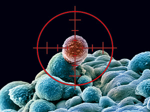
Based on the results of a new study, researchers are developing a clinical trial to test imatinib(Gleevec) in patients with anaplastic large cell lymphoma (ALCL), an aggressive type of non-Hodgkin lymphoma that primarily affects children and young adults.

The researchers found that a protein called PDGFRB is important to the development of a common form of ALCL. PDGFRB, a growth factor receptor protein, is a target of imatinib. Imatinib had anticancer effects in both a mouse model of ALCL and a patient with the disease, Dr. Lukas Kenner of the Medical University of Vienna in Austria and his colleagues reported October 14 in Nature Medicine.

http://www.cancer.gov/ncicancerbulletin/103012/page3#b

VIEW VIDEO by  Mayo Clinic Dr. Tim Call describes Chronic Lymphocytic Leukemia (CLL), its diagnosis, and treatment options for patients with CLL.

http://www.youtube.com/watch?v=GCMuizn980c&feature=related

VIEW VIDEO  by Leading CLL researcher Dr. Thomas Kipps discusses the latest in the course of the disease including: symptoms, causes, and risk factors. Dr. Kipps also highlights discoveries made in his lab that may lead to developing new therapies to treat this disease. Series: “Stein Institute for Research on Aging” [9/2010] [Health and Medicine] [Show ID: 19345]

http://www.youtube.com/watch?v=QToR46370OI&feature=related

Understanding the Molecular Basis of Imatinib Mesylate Therapy in Chronic Myelogenous Leukemia and the Related Mechanisms of Resistance1

Commentary re: A. N. Mohamed et al., The Effect of Imatinib Mesylate on Patients with Philadelphia Chromosome-positive Chronic Myeloid Leukemia with Secondary Chromosomal Aberrations. Clin. Cancer Res., 9: 1333–1337, 2003.

  1. Guido Marcucci2,
  2. Danilo Perrotti, and
  3. Michael A. Caligiuri

+Author Affiliations


  1. Division of Hematology and Oncology, Department of Internal Medicine [G. M., M. A. C.], Division of Human Cancer Genetics, Department of Molecular Virology, Immunology and Medical Genetics [G. M., D. P., M. A. C.], The James Cancer Hospital and The Comprehensive Cancer Center at The Ohio State University, Columbus, Ohio 43210

Introduction

CML3 is a myeloproliferative disorder with an incidence of approximately 1–1.5 cases/100,000 population/year, a slight male predominance, and a peak between 50 and 60 years of age (1) . This condition arises from a transformed pluripotent hematopoietic precursor associated with t(9;22)(q34;q11.2) that gives rise to the Ph′ chromosome. At the molecular level, this cytogenetic aberration results in the fusion of the ABL gene at chromosome band 9q34 with the BCR gene at chromosome band 22q11.2. The resultantBCR/ABL fusion gene encodes a chimeric protein necessary and sufficient to confer the leukemic phenotype. In a minority of the cases, despite absence of the Ph′ chromosome, molecular methodologies (i.e., fluorescence in situ hybridization, Southern, or RT-PCR) can still detect BCR/ABL as the product of complex cytogenetic aberrations involving other chromosomes in addition to 9q34 and 22q11.2, or cryptic genomic rearrangements of the BCR and ABL genes. Taken all together, >95% of the CML cases are associated with BCR/ABL expression that, therefore, represents the hallmark of this condition.

CML is a bi- or triphasic disease (2) . Patients usually present in a chronic proliferative phase characterized by splenomegaly and accumulation of neutrophils in their various stages of maturation. Basophils and blasts are normally <20% and 10%, respectively, in CP CML. Evolution to the BP (or blast crisis) is defined by an increase (≥20%) in myeloid or lymphoid blasts in blood, BM, or extramedullary locations. In many cases, the transformation is characterized by a passage through an AP, in which the failure of a patient to respond to therapy is accompanied by an increase in percentage of blasts (10–19%); a ≥20% increase in basophils, uncontrolled thrombocytosis, or thrombocytopenia; and acquisition of new cytogenetic abnormalities such as trisomy 8, isochromosome 17q, or a second Ph chromosome and/or genetic inactivation of the p53 gene.

The ultimate goal of treatment for CML is prevention of blast crisis, because, once this occurs, the prognosis is dismal. Although a rapid reduction of the white cell count can be achieved with chemotherapy (i.e., busulfan or hydroxyurea) in CP CML, these drugs usually fail to prevent disease progression. IFN-α was the first agent proven capable of modifying the biological history of CML by prolonging survival in patients who achieved CHR and MCR (<35% of Ph′+cells; Ref. 3 ). A second breakthrough in the treatment of CML occurred when 60–80% of patients undergoing alloBMT in CP were reported to be disease free at 5 years (456) . However, both IFN-α and alloBMT have considerable treatment-induced toxicity that attenuated the initial enthusiasm for these results, and novel, less toxic therapeutic strategies are being explored. In 2001, Druker et al. (7)reported the first Phase I study with imatinib mesylate (STI571, Gleevec), a specific inhibitor of BCR/ABL oncogenic activity. In this study, the authors demonstrated that high rates of CHR and MCR were achieved with relatively few side effects in patients with CML refractory or intolerant to IFN-α, opening new avenues for molecularly targeted therapies in this disease. Subsequently, encouraging results were also obtained for CML patients in BP (8) .

Is BCR/ABLthe Driving Force for Leukemogenesis in CML?

The transforming activity of BCR/ABL has long been demonstrated using in vitro and in vivo models. In initial studies, transfection of the BCR/ABL gene fusion resulted in malignant transformation of normal fibroblasts, and induced independent survival and proliferation in growth factor-dependent cell lines. Expression of BCR/ABL was also shown to be necessary and sufficient to induce leukemogenesis in animal models (reviewed in Ref. 9 ). Expression of BCR/ABL in mice was achieved by either introduction (“knock-in”) of the fusion gene in the mouse genome or by infecting murine stem cells with BCR/ABL-containing retroviral vectors. In knock-in transgenic mice with conditionalBCR/ABL expression, a low penetrance phenotype of acute B- or T-cell leukemia was reported. In contrast, in mice sublethally radiated and transplanted with syngeneic retrovirally transfected BCR/ABL+ stem cells, a condition mimicking human myeloproliferative disorders with neutrophil increase, BM expansion, hepatosplenomegaly, extramedullary hematopoiesis, and pulmonary leukostasis was observed.

How Does BCR/ABL Transform Cells?

The fusion partner ABL is a member of the nonreceptor tyrosine kinase family (10) . This gene encodes a protein containing a tyrosine kinase activity domain (SH1) in addition to two other regulatory domains (SH2 and SH3) that mediate protein-protein interaction and modulate activation of signal transduction. A nuclear localization domain is also present, supporting a shuttling activity of the ABL protein between cytoplasm and nucleus. Genetic disruption of ABL in mice results in lymphopenia, runting, and perinatal mortality. The other fusion partner, BCR, is a protein with multiple functional domains involved in oligomerization, SH-2 binding, serine/threonine kinase activity, and activation of members of the Rho small GTP-ase family of proteins (10) . Structural analysis of this gene suggests a role as a mediator of signaling transduction. With targeted disruption of BCR, mice have increased susceptibility to septic shock, but normal hematopoiesis.

In CML, each of the two partner genes are disrupted at specific breakpoints and fuse to create the chimeric BCR/ABL gene (11) . The most common rearrangements give rise to fusion transcripts identified as b2a2 or b3a2, which, in turn, are translated into a 210 kDa protein. Alternative BCR breakpoints can be located in minor cluster (m-BCR) or micro (μ) cluster (μ-BCR) regions and result in fusion transcripts that encode smaller (190 kDa) or larger (230 kDa) products, respectively. Although usually associated with Ph′+ acute lymphoblastic PiQO leukemia, the protein can also be detected in ≥90% of the CML patients from alternative splicing of p210. p230 is instead usually associated with CML patients presenting with an unusual predominance of neutrophils, resembling chronic neutrophilic leukemia. In each of these BCR/ABL variants, the ABL tyrosine kinase domain autophosphorylates and becomes constitutionally activated. Such BCR/ABLderegulated tyrosine kinase activity is, in fact, responsible for transformation of the hematopoietic stem cell and maintenance of the leukemic phenotype by recruiting and activating transducing signal pathways (i.e., RAS, RAF, extracellular signal-regulated kinase, c-Jun NH2-terminal kinase, phosphatidylinositol 3′-kinase, cCbl, CRKL, Janus-activated kinase-signal transducers and activators of transcription, PKC and PLCγ) involved in: (a) enhanced gene transcription (i.e., c-myc, c-Jun, reviewed in Ref. 12 ); (b) altered mRNA processing, nuclear export, and translation (i.e., bcl-xL, CAAT/enhancer binding protein α, and p53; reviewed in Ref. 13 , 14 ); and (c) increased or decreased protein stability (i.e., Abi proteins; DNA-PKcs; FUS, and hnRNP A1; reviewed in Ref. 13). This, in turn, leads to enhanced proliferative potential and survival, altered motility and trafficking, and suppression of granulocytic differentiation (1 , 13 , 15 , 16) .

Mechanisms of Action of Imatinib Mesylate

BCR/ABL is an ideal target for molecular targeted therapy, because this fusion protein is present in all of the CML cells, is absent from nonmalignant cells, and is necessary and sufficient to induce leukemia. Imatinib mesylate is a 2-phenylaminopyrimidine tyrosine kinase inhibitor with specific activity for ABL, platelet derived growth factor receptor, c-kit, and Albeson-related gene (17) . The pharmacological basis of this interaction has been elucidated by crystallographic studies. Imatinib mesylate binds to the amino acids of the BCR/ABL tyrosine kinase ATP binding site and stabilizes the inactive, non-ATP-binding form of BCR/ABL, thereby preventing tyrosine autophosphorylation and, in turn, phosphorylation of its substrates. This process ultimately results in “switching-off” the downstream signaling pathways that promote leukemogenesis. Preclinical in vitro and in vivo data indicated an impressive selective activity of imatinib mesylate on cells expressing BCR/ABL, and supported a rapid transition of this compound from the bench to the clinic.

To date, imatinib mesylate has been evaluated in several Phase I and II clinical trials of patients with IFN-α-resistant chronic, accelerated, or BP CML (7 , 8 , 181920) . From the collective analysis of these studies, imatinib mesylate appears to effectively induce high CHR and cytogenetic response rates with relatively few side effects. In patients with CP CML who have failed IFN-α the CHR was 95%, MCR 60%, and complete cytogenetic remission 46%. Notably, in these patients achievement of MCR at the 3-month time point correlated with improved progression-free survival. In AP and in blast crisis, the CHRs were 34% and 8%, MCRs were 24% and 16%, and complete cytogenetic remissions were 17%, and 7%, respectively. Disease progression was 11% at 18 months for CP, 40% at 12 months for AP, and 80% at 18 months for BP. Finally, preliminary data from an interim analysis of a phase III study of untreated CML patients randomized between imatinib mesylate versus IFN-α and ARA-C indicate a significantly better CHR, complete cytogenetic remission, and progression-free survival for the imatinib mesylate group after a median follow-up of 14 months (21) . However, a longer follow-up will be necessary to assess whether this compound can also impact on the natural history of the disease and prevent or delay transformation to blast crisis.

Mechanisms of Resistance to Imatinib Mesylate

During disease progression, CML progenitor cells acquire a number of genetic alterations, most probably because of increased genomic instability, that may explain the aggressive phenotype, chemotherapeutic drug resistance, and poor prognosis of CML in BP. Despite the exciting results obtained with imatinib mesylate noted above, CML patients eventually show resistance at a rate of 80% in BP, 40–50% in AP, and 10% in CP post-IFNα failure, at 2 years (19) . Identification of the molecular basis of resistance is important, because it could provide insight into disease progression and into the design of novel therapeutic strategies to prevent and overcome treatment resistance.

CML patients with imatinib mesylate resistance can be stratified into those with primary refractory disease, most frequently in accelerated or BPs, and those who relapse after initial response, who are most frequently in CP. On the basis of the presence or absence of BCR/ABL tyrosine kinase activity in leukemia cells, it is also possible to discriminate between cases with BCR/ABL-dependent and -independent mechanisms of imatinib mesylate resistance. Notably, because the BCR/ABL enzymatic activity cannot be easily measured in blood or BM patient samples, levels of phosphorylation of the BCR/ABL substrate CRKL have been used as a surrogate end point for the tyrosine kinase activity(22) .

In patients with higher levels of CRKL phosphorylation despite treatment with imatinib mesylate, resistance has been found to result from at least three different BCR/ABL-dependent mechanisms: BCR/ABL gene amplification, BCR/ABL mutations, and high plasma levels of AGP (reviewed in Ref. 23 ).

The association of BCR/ABL gene amplification with resistance to imatinib mesylate is consistent with the reliance of CML blast crisis cells on BCR/ABL expression/activity for their proliferation and survival, and with the reported enhanced expression of BCR/ABL in these cells (24) . Indeed, high levels of BCR/ABL expression, which are detected frequently in CML-blast crisis but not CP cells, appear to be required for suppression of myeloid differentiation and increased resistance to chemotherapy-induced apoptosis ofBCR/ABL-expressing cells. Specifically, high levels of BCR/ABL kinase activity are required for hnRNP E2-dependent inhibition of CAAT/enhancer binding protein α, the major regulator of granulocytic differentiation (25) , and for the La-dependent enhancement of MDM2 expression, which, in turn, results in functional inactivation of p53(14) , also required for myeloid blastic transformation (26 , 27) Although the mechanisms underlying such an increase of BCR/ABL expression are unclear, a double Ph′ chromosome is likely to be responsible for the enhanced BCR-ABL levels in some cases.

Other mechanisms of imatinib mesylate resistance involve mutations in the BCR/ABLgene itself. Several different mutations have been detected in at least 13 amino acids of the ATP-binding site or other regions of the tyrosine kinase domain, and the list is growing (23 , 28 , 29) . These mutations usually prevent imatinib mesylate from binding to BCR/ABL, thereby resulting in lack of inhibition of the tyrosine kinase activity. Among these mutations, substitution of a threonine to isoleucine at position 315 of ABL that prevents imatinib mesylate from binding to the ATP-binding domain, is the first described and one of the most frequent (22) .

A third mechanism of resistance relies on plasma levels of AGP. It has been shown that AGP binds imatinib mesylate at physiological concentrations in vitro and in vivo, and blocks the ability of imatinib mesylate to inhibit BCR/ABL kinase activity in a dose-dependent manner (reviewed in Ref. 23 ). Finally, in patients with primary refractoriness to imatinib mesylate, resistance more often occurs in absence of significant CRKL phosphorylation, suggesting activation of BCR/ABL-independent leukemogenic pathways.

In this issue, Mohamed et al. (30) hypothesized that clonal evolution, defined as acquisition of additional cytogenetic abnormalities other than t(9;22)(q34;q11), is a marker for genomic instability, and thereby, in this setting, additional “hits” can occur to activate BCR/ABL-independent leukemogenic mechanisms. Given this premise, CML patients with a more complex karyotype were expected to be more resistant to imatinib mesylate. To test their hypothesis, these authors analyzed 58 BCR/ABL-positive patients with IFN-α-resistant CP (n = 13), AP (n = 24), or BP (n = 21) CML with additional cytogenetic abnormalities who were each treated with imatinib mesylate. Of the 58 patients, 15 (CP = 46%; AP = 25%; BP = 14%) achieved a cytogenetic response, and 12 had a complete cytogenetic remission. With a follow-up of 17–30 months, 7 (12%) remained in complete remission on imatinib mesylate, supporting the notion that a subset of CML patients with t(9;22)(q34;q11) and additional cytogenetic abnormalities can achieve a sustained response. Because the molecular mechanisms of resistance in these patients were not fully evaluated, the contribution of additional cytogenetic abnormalities as an independent predictor of resistance to imatinib mesylate could not be directly addressed. Similar results were also reported in the imatinib mesylate initial studies (18 , 20) , and more recently by Schoch et al. (31) . These authors reported on 31 patients with additional chromosomal abnormalities present before the start of imatinib mesylate therapy. Additional cytogenetic abnormalities were less frequent in chronic than other phases of the disease (35.5% versus 68.8%; P = 0.0008), and when corrected for the difference in disease stage, they did not influence response to imatinib mesylate. These results have been confirmed recently by a larger study reported by Cortes et al. (32) where 498 CML patients in CP or AP were treated with imatinib mesylate. Of the 498, 121 had additional cytogenetic abnormalities. In a multivariate analysis at the 3-month time point, lack of cytogenetic response, but not presence of additional cytogenetic abnormalities, was found to be a negative prognostic factor for survival

Although a longer follow-up is necessary to draw definitive conclusions, these findings suggest that additional cytogenetic aberrations do not appear to impact on disease response, and, therefore, karyotype analysis should not be used to stratify patients for therapeutic alternatives to imatinib mesylate. Furthermore, despite clonal evolution, the oncogenic potential of BCR/ABL appears to remain the driving force for leukemogenesis, and, therefore, may continue to serve as a therapeutic target. Finally, in patients with additional cytogenetic abnormalities who are resistant to imatinib mesylate, evaluation for levels of CRKL-phosphorylation should be done to sort out the nature of the resistance to this treatment and to understand the interplay between BCR/ABL-dependent and -independent mechanisms in disease progression.

Concluding Remarks

The results obtained with imatinib mesylate to date are truly impressive, but longer follow-up will be necessary to establish whether prevention or delay of blast crisis can be achieved and whether an improved overall survival for the majority of patients with CML can be obtained. It is clear that complete cytogenetic remission is achievable in most patients with CP CML, and that those who do not achieve this important end point between 3 and 6 months are likely to have a poor outcome. However, in patients with complete cytogenetic remission, other challenges remain. In these patients, for instance, molecular remission defined as the absence of BCR/ABL fusion transcript by RT-PCR is usually not achieved. The reasons for persistent low levels of residual disease after imatinib mesylate and the prognostic significance of these findings are unknown. It is possible that small numbers of mutated and resistant BCR/ABL-positive subclones remain essentially unaffected by this treatment, and if a proliferation advantage is subsequently acquired in these cells, they may drive disease recurrence. Therefore, in patients with complete cytogenetic remission, monitoring of the BCR/ABL fusion transcript levels by quantitative RT-PCR has been suggested to predict impending relapse, and if rising levels of BCR/ABL expression are detected, consideration could be given to alternative strategies including alloBMT.

However, emerging data support the notion that hematologic or cytogenenetic remission can be achieved in imatinib-relapsed CP CML patients with higher doses of this agent. Kantarjian et al. reported on 54 patients with CML in CP who were initially treated with 400 mg of imatinib mesylate and, subsequently, with a higher dose (i.e., 800 mg) when hematologic or cytogenetic resistance or relapse developed (33) . Among 20 patients treated for hematologic resistance or relapse, 13 (65%) achieved CHR without complete cytogenetic remission, and among 34 patients treated for cytogenetic resistance or relapse, 19 (56%) achieved a complete cytogenetic remission or MCR. However, the mechanisms of resistance to standard dose were not evaluated in these patients and, therefore, correlation of these mechanisms with clinical response to the higher doses was not possible. Nevertheless, these data are intriguing and pose the question of whether a higher dose of imatinib mesylate could be used at the time of the initial diagnosis or during molecular relapse to prevent overt leukemia recurrence or blast transformation. Future studies will no doubt address these important issues. Regardless, it is undeniable that imatinib mesylate has changed our approach to CML and paved the way for additional molecular targeted strategies in leukemia.

Footnotes

  • 1 Supported in part by P30-CA16058, and K08-CA90469 Grants from the National Cancer Institute, Bethesda, MD, The Elsa U. Pardee Cancer Research Foundation, and The Coleman Leukemia Research Foundation.

  • 2 To whom requests for reprints should be addressed, at The Ohio State University, 458A Starling-Loving Hall, 320 West 10th Avenue, Columbus, OH 43210. Phone: (614) 293-7597; Fax: (614) 293-7527; E-mail: marcucci-1@medctr.osu.edu.

  • 3 The abbreviations used are: CML, chronic myelogenous leukemia; Ph′, Philadelphia; ABL, Abelson; RT-PCR, reverse transcription-PCR; BM, bone marrow; CHR, complete hematologic remission; MCR, major cytogenetic response; alloBMT, allogeneic bone marrow transplantation; AGP, α1 glycoprotein; CP, chronic phase; AP, accelerated phase; BP, blastic phase.

  • Received March 3, 2003.
  • Accepted March 3, 2003.

Imatinib May Help Treat Aggressive Lymphoma

Based on the results of a new study, researchers are developing a clinical trial to test imatinib (Gleevec) in patients with anaplastic large cell lymphoma (ALCL), an aggressive type of non-Hodgkin lymphoma that primarily affects children and young adults.

The researchers found that a protein called PDGFRB is important to the development of a common form of ALCL. PDGFRB, a growth factor receptor protein, is a target of imatinib. Imatinib had anticancer effects in both a mouse model of ALCL and a patient with the disease, Dr. Lukas Kenner of the Medical University of Vienna in Austria and his colleagues reported October 14 in Nature Medicine.

The authors decided to investigate the effect of imatinib after finding a link between PDGFRB and a genetic abnormality that is found in many patients with ALCL. Previous work had shown that this genetic change—a translocation that leads to the production of an abnormal fusion protein called NPM-ALK—stimulates the production of two proteins, transcription factors called JUN and JUNB.

In the new study, experiments in mice revealed that these proteins promote lymphoma development by increasing the levels of PDGFRB.

Because imatinib inhibits PDGFRB, the authors tested the effect of the drug in mice with the NPM-ALK change and found that it improved their survival. They also found that imatinib given together with the ALK inhibitor crizotinib (Xalkori) greatly reduced the growth of NPM-ALK-positive lymphoma cells in mice.

To test the treatment strategy in people, they identified a terminally ill patient with NPM-ALK-positive ALCL who had no other treatment options and agreed to try imatinib. The patient began to improve within 10 days of starting the therapy and has been free of the disease for 22 months, the authors reported.

The observation that inhibiting both ALK and PDGFRB “reduces lymphoma growth and alleviates relapse rates” led the authors to suggest that the findings might be relevant to lymphomas with PDGFRB but without the NPM-ALK protein. “Our findings suggest that imatinib is a potential therapeutic option for patients with crizotinib-resistant lymphomas.”

A planned clinical trial will be based on the expression of PDGFRB in tumors.

SOURCE:

http://www.cancer.gov/ncicancerbulletin/103012/page3#b

References

  1. Sawyers C. L. Chronic myeloid leukemia. N. Engl. J. Med., 340: 1330-1340,1999.
  2. Vardiman J. W., Harris N. L., Brunning R. D. The World Health Organization (WHO) classification of the myeloid neoplasms. Blood, 100: 2292-2302, 2002.
  3. Chronic Myeloid Leukemia Trialists’ Collaborative Group Interferon α versus chemotherapy for chronic myeloid leukemia: a meta-analysis of seven randomized trials. J. Natl. Cancer Inst., 89: 1616-1620, 1997.
  4. Horowitz M. M., Rowlings P. A., Passweg J. R. Allogeneic bone marrow transplantation for CML: a report from the International Bone Marrow Transplant Registry. Bone Marrow Transplant, 17 (Suppl.3): S5-S6, 1996.
  5. Clift R. A., Radich J., Appelbaum F. R., Martin P., Flowers M. E., Deeg H. J., Storb R., Thomas E. D. Long-term follow-up of a randomized study comparing cyclophosphamide and total body irradiation with busulfan and cyclophosphamide for patients receiving allogenic marrow transplants during chronic phase of chronic myeloid leukemia. Blood, 94: 3960-3962, 1999.
  6. van Rhee F., Szydlo R. M., Hermans J., Devergie A., Frassoni F., Arcese W., de Witte T., Kolb H. J., Niederwiser D., Jacobsen N., Gahrton G., Bandini G., Carreras E., Bacigalupo A., Michallet M., Ruutu T., Reiffers J., Goldman J. M., Apperley J., Gratwohl A. Long-term results after allogeneic bone marrow transplantation for chronic myelogenous leukemia in chronic phase: a report from the Chronic Leukemia Working Party of the European Group for Blood and Marrow Transplantation. Bone Marrow Transplant, 20: 553-560, 1997.
  7. Druker B. J., Talpaz M., Resta D. J., Peng B., Buchdunger E., Ford J. M., Lydon N. B., Kantarjian H., Capdeville R., Ohno-Jones S., Sawyers C. L. Efficacy and safety of a specific inhibitor of the BCR-ABL tyrosine kinase in chronic myeloid leukemia. N. Engl. J. Med., 344: 1031-1037, 2001.
  8. Druker B. J., Sawyers C. L., Kantarjian H., Resta D. J., Reese S. F., Ford J. M., Capdeville R., Talpaz M. Activity of a specific inhibitor of the BCR-ABL tyrosine kinase in the blast crisis of chronic myeloid leukemia and acute lymphoblastic leukemia with the Philadelphia chromosome. N. Engl. J. Med., 344: 1038-1042,2001.
  9. Van Etten R. A. Studying the pathogenesis of BCR-ABL+ leukemia in mice.Oncogene, 21: 8643-8651, 2002.
  10. Pane F., Intrieri M., Quintarelli C., Izzo B., Muccioli G. C., Salvatore F. BCR/ABL genes and leukemic phenotype: from molecular mechanisms to clinical correlations. Oncogene, 21: 8652-8667, 2002.
  11. Faderl S., Talpaz M., Estrov Z., O’Brien S., Kurzrock R., Kantarjian H. M. The biology of chronic myeloid leukemia. N. Engl. J. Med., 341: 164-172, 1999.
  12. Zou X., Calame K. Signaling pathways activated by oncogenic forms of Abl tyrosine kinase. J. Biol. Chem., 274: 18141-18144, 1999.
  13. Perrotti D., Calabretta B. Post-transcriptional mechanisms in BCR/ABL leukemogenesis: role of shuttling RNA-binding proteins. Oncogene, 21: 8577-8583,2002.
  14. Trotta R., Vignudelli T., Candini C., Intine R., Pecorari L., Guerzoni C., Santilli G., Byrom B., Goldoni G., Ford L., Caligiuri M., Maraia R., Perrotti D., Calabretta B. BCR/ABL activates mdm2 mRNA translation via the La antigen. Cancer Cell, 3:145-160, 2003.
  15. Osarogiagbon U. R., McGlave P. B. Chronic myelogenous leukemia. Curr. Opin. Hematol., 6: 241-246, 1999.
  16. Gordon M. Y. Biological consequences of the BCR/ABL fusion gene in humans and mice. J. Clin. Pathol., 52: 719-722, 1999.
  17. Druker B. J. Imatinib alone and in combination for chronic myeloid leukemia.Semin. Hematol., 40: 50-58, 2003.
  18. Sawyers C. L., Hochhaus A., Feldman E., Goldman J. M., Miller C. B., Ottmann O. G., Schiffer C. A., Talpaz M., Guilhot F., Deininger M. W., Fischer T., O’Brien S. G., Stone R. M., Gambacorti-Passerini C. B., Russell N. H., Reiffers J. J., Shea T. C., Chapuis B., Coutre S., Tura S., Morra E., Larson R. A., Saven A., Peschel C., Gratwohl A., Mandelli F., Ben-Am M., Gathmann I., Capdeville R., Paquette R. L., Druker B. J. Imatinib induces hematologic and cytogenetic responses in patients with chronic myelogenous leukemia in myeloid blast crisis: results of a phase II study. Blood, 99: 3530-3539, 2002.
  19. Kantarjian H., Sawyers C., Hochhaus A., Guilhot F., Schiffer C., Gambacorti-Passerini C., Niederwieser D., Resta D., Capdeville R., Zoellner U., Talpaz M., Druker B., Goldman J., O’Brien S. G., Russell N., Fischer T., Ottmann O., Cony-Makhoul P., Facon T., Stone R., Miller C., Tallman M., Brown R., Schuster M., Loughran T., Gratwohl A., Mandelli F., Saglio G., Lazzarino M., Russo D., Baccarani M., Morra E. Hematologic and cytogenetic responses to imatinib mesylate in chronic myelogenous leukemia. N. Engl. J. Med., 346: 645-652, 2002.
  20. Talpaz M., Silver R. T., Druker B. J., Goldman J. M., Gambacorti-Passerini C., Guilhot F., Schiffer C. A., Fischer T., Deininger M. W., Lennard A. L., Hochhaus A., Ottmann O. G., Gratwohl A., Baccarani M., Stone R., Tura S., Mahon F. X., Fernandes-Reese S., Gathmann I., Capdeville R., Kantarjian H. M., Sawyers C. L. Imatinib induces durable hematologic and cytogenetic responses in patients with accelerated phase chronic myeloid leukemia: results of a phase 2 study. Blood, 99:1928-1937, 2002.
  21. Druker B. J., O’Brien S. G., Cortes J., Radich J. Chronic myelogenous leukemia. Hematology (Am. Soc. Hematol. Educ. Program), : 111-135, 2002.
  22. Gorre M. E., Ellwood-Yen K., Chiosis G., Rosen N., Sawyers C. L. BCR-ABL point mutants isolated from patients with imatinib mesylate-resistant chronic myeloid leukemia remain sensitive to inhibitors of the BCR-ABL chaperone heat shock protein 90. Blood, 100: 3041-3044, 2002.
  23. Gambacorti-Passerini C. B., Gunby R. H., Piazza R., Galietta A., Rostagno R., Scapozza L. Molecular mechanisms of resistance to imatinib in Philadelphia-chromosome-positive leukaemias. Lancet Oncol., 4: 75-85, 2003.
  24. Elmaagacli A. H., Beelen D. W., Opalka B., Seeber S., Schaefer U. W. The amount of BCR-ABL fusion transcripts detected by the real-time quantitative polymerase chain reaction method in patients with Philadelphia chromosome positive chronic myeloid leukemia correlates with the disease stage. Ann. Hematol., 79: 424-431, 2000.
  25. Perrotti D., Cesi V., Trotta R., Guerzoni C., Santilli G., Campbell K., Iervolino A., Condorelli F., Gambacorti-Passerini C., Caligiuri M. A., Calabretta B. BCR-ABL suppresses C/EBPα expression through inhibitory action of hnRNP E2. Nat. Genet., 30: 48-58, 2002.
  26. Honda H., Ushijima T., Wakazono K., Oda H., Tanaka Y., Aizawa S., Ishikawa T., Yazaki Y., Hirai H. Acquired loss of p53 induces blastic transformation in p210(bcr/abl)-expressing hematopoietic cells: a transgenic study for blast crisis of human CML. Blood, 95: 1144-1150, 2000.
  27. Skorski T., Nieborowska-Skorska M., Wlodarski P., Perrotti D., Martinez R., Wasik M. A., Calabretta B. Blastic transformation of p53-deficient bone marrow cells by p210bcr/abl tyrosine kinase. Proc. Natl. Acad. Sci. USA, 93: 13137-13142,1996.
  28. Hochhaus A., Kreil S., Corbin A. S., La Rosee P., Muller M. C., Lahaye T., Hanfstein B., Schoch C., Cross N. C., Berger U., Gschaidmeier H., Druker B. J., Hehlmann R. Molecular and chromosomal mechanisms of resistance to imatinib (STI571) therapy. Leukemia (Baltimore), 16: 2190-2196, 2002.
  29. O’Dwyer M. E. Chronic myelogenous leukemia. Curr. Opin. Oncol., 15: 10-15,2003.
  30. Mohamed A. N., Pemberton P., Zonder J., Schiffer C. A. The effect of imatinib mesylate on patients with philadelphia chromosome-positive chronic myeloid leukemia with secondary chromosomal aberrations. Clin. Cancer Res., 9: 00-00,2003.
  31. Schoch C., Haferlach T., Kern W., Schnittger S., Berger U., Hehlmann R., Hiddemann W., Hochhaus A. Occurrence of additional chromosome aberrations in chronic myeloid leukemia patients treated with imatinib mesylate. Leukemia (Baltimore), 17: 461-463, 2003.
  32. Cortes J. E., Talpaz M., Giles F., O’Brien S., Rios M. B., Shan J., Garcia-Manero G., Faderl S., Thomas D. A., Wierda, W., Ferrajoli A., Jeha S., Kantarjian H. M. Prognostic significance of cytogenetic clonal evolution in patients with chronic myelogenous leukemia on imatinib mesylate therapy. Blood, : 2003.
  33. Kantarjian H. M., Talpaz M., O’Brien S., Giles F., Garcia-Manero G., Faderl S., Thomas D., Shan J., Rios M. B., Cortes J. Dose escalation of imatinib mesylate can overcome resistance to standard-dose therapy in patients with chronic myelogenous leukemia. Blood, 101: 473-475, 2003.

SOURCE:

Clin. Cancer Res., 9: 1333–1337, 2003.

Related Research on the Open Source On-Line Scientific Journal

Acute Myeloid Leukemia (AML): Role of Chromatin Accessibility during Hematopoiesis, Aviva Lev-Ari, PhD, RN

http://pharmaceuticalintelligence.com/2012/08/31/acute-myeloid-leukemia-aml-role-of-chromatin-accessibility-during-hematopoiesis/

RNA Sequencing led to Targeting FLT3 Gene on Chromosome 13 for Receptor Blockage causing REMISSION in Adult Acute Lymphoblastic Leukemia (ALL), Aviva Lev-Ari, PhD, RN

http://pharmaceuticalintelligence.com/2012/07/10/rna-sequencing-led-to-targeting-flt3-gene-on-chromosome-13-for-receptor-blockage-causing-remission-in-adult-acute-lymphoblastic-leukemia-all/

Read Full Post »

%d bloggers like this: