Healthcare analytics, AI solutions for biological big data, providing an AI platform for the biotech, life sciences, medical and pharmaceutical industries, as well as for related technological approaches, i.e., curation and text analysis with machine learning and other activities related to AI applications to these industries.
Chapter 1: Evolution of the Foundation for Diagnostics and Pharmaceuticals Industries
1.1 Outline of Medical Discoveries between 1880 and 1980
1.2 The History of Infectious Diseases and Epidemiology in the late 19th and 20th Century
1.3 The Classification of Microbiota
1.4 Selected Contributions to Chemistry from 1880 to 1980
1.5 The Evolution of Clinical Chemistry in the 20th Century
1.6 Milestones in the Evolution of Diagnostics in the US HealthCare System: 1920s to Pre-Genomics
Chapter 2. The search for the evolution of function of proteins, enzymes and metal catalysts in life processes
2.1 The life and work of Allan Wilson
2.2 The evolution of myoglobin and hemoglobin
2.3 More complexity in proteins evolution
2.4 Life on earth is traced to oxygen binding
2.5 The colors of life function
2.6 The colors of respiration and electron transport
2.7 Highlights of a green evolution
Chapter 3. Evolution of New Relationships in Neuroendocrine States
3.1 Pituitary endocrine axis
3.2 Thyroid function
3.3 Sex hormones
3.4 Adrenal Cortex
3.5 Pancreatic Islets
3.6 Parathyroids
3.7 Gastointestinal hormones
3.8 Endocrine action on midbrain
3.9 Neural activity regulating endocrine response
3.10 Genomic Promise for Neurodegenerative Diseases, Dementias, Autism Spectrum, Schizophrenia, and Serious Depression
Chapter 4. Problems of the Circulation, Altitude, and Immunity
4.1 Innervation of Heart and Heart Rate
4.2 Action of hormones on the circulation
4.3 Allogeneic Transfusion Reactions
4.4 Graft-versus Host reaction
4.5 Unique problems of perinatal period
4.6. High altitude sickness
4.7 Deep water adaptation
4.8 Heart-Lung-and Kidney
4.9 Acute Lung Injury
4.10 Reconstruction of Life Processes requires both Genomics and Metabolomics to explain Phenotypes and Phylogenetics
Chapter 5. Problems of Diets and Lifestyle Changes
5.1 Anorexia nervosa
5.2 Voluntary and Involuntary S-insufficiency
5.3 Diarrheas – bacterial and nonbacterial
5.4 Gluten-free diets
5.5 Diet and cholesterol
5.6 Diet and Type 2 diabetes mellitus
5.7 Diet and exercise
5.8 Anxiety and quality of Life
5.9 Nutritional Supplements
Chapter 6. Advances in Genomics, Therapeutics and Pharmacogenomics
6.1 Natural Products Chemistry
6.2 The Challenge of Antimicrobial Resistance
6.3 Viruses, Vaccines and immunotherapy
6.4 Genomics and Metabolomics Advances in Cancer
6.5 Proteomics – Protein Interaction
6.6 Pharmacogenomics
6.7 Biomarker Guided Therapy
6.8 The Emergence of a Pharmaceutical Industry in the 20th Century: Diagnostics Industry and Drug Development in the Genomics Era: Mid 80s to Present
6.09 The Union of Biomarkers and Drug Development
6.10 Proteomics and Biomarker Discovery
6.11 Epigenomics and Companion Diagnostics
Chapter 7
Integration of Physiology, Genomics and Pharmacotherapy
7.1 Richard Lifton, MD, PhD of Yale University and Howard Hughes Medical Institute: Recipient of 2014 Breakthrough Prizes Awarded in Life Sciences for the Discovery of Genes and Biochemical Mechanisms that cause Hypertension
7.2 Calcium Cycling (ATPase Pump) in Cardiac Gene Therapy: Inhalable Gene Therapy for Pulmonary Arterial Hypertension and Percutaneous Intra-coronary Artery Infusion for Heart Failure: Contributions by Roger J. Hajjar, MD
7.3 Diagnostics and Biomarkers: Novel Genomics Industry Trends vs Present Market Conditions and Historical Scientific Leaders Memoirs
7.4 Synthetic Biology: On Advanced Genome Interpretation for Gene Variants and Pathways: What is the Genetic Base of Atherosclerosis and Loss of Arterial Elasticity with Aging
Dr. Ahmad Masri and Dr. C Michael Gibson discuss outpatient worsening heart failure in transthyretin amyloid cardiomyopathy in the Attribute-CM trial, May 29, 2026
The following are key points to remember from this update on the treatment of cardiac transthyretin amyloidosis:
Transthyretin (TTR) is a highly conserved protein involved in transportation of thyroxine (T4) and retinol-binding protein. TTR is synthesized mostly by the liver and is rich in beta strands with an intrinsic propensity to aggregate into insoluble amyloid fibers, which deposit within tissue leading to the development of TTR-related amyloidosis (ATTR). ATTR can follow the deposition of either variant TTR (ATTRv, previously known as mutant ATTR) or wild type TTR (ATTRwt).
Cardiac ATTR has a favorable survival rate compared to light chain (AL) amyloidosis, with a median survival of 75 versus 11 months. However, ATTR cardiomyopathy is a progressive disorder but newer therapeutic options include tafamidis (positive phase 3 clinical trial), and possibly patisiran and inotersen.
Inhibition of the Synthesis of Mutated Transthyretin
Liver transplantation removes the source of mutated TTR molecules and prolongs survival, with a 20-year survival of 55.3%. However, tissue accumulation of TTR can continue after liver transplantation because TTR amyloid fibers promote subsequent deposition of ATTRwt. Combined liver–heart transplantation is feasible in younger patients with ATTRv cardiomyopathy and a small series suggests better prognosis than cardiac transplantation.
Inhibition of TTR gene expression: Patisiran is a small interfering RNA blocking the expression of both variant and wt TTR. On the basis of the APOLLO trial, it was approved for therapy of adults with ATTRv-related polyneuropathy both in the United States and European Union. In this trial, patisiran promoted favorable myocardial remodeling based on echocardiographic and N-terminal B-type natriuretic peptide (NT-BNP) changes (this effect was not demonstrated for inotersen) and is still under investigation for tafamidis.
Antisense oligonucleotides inotersen inhibits the production of both variant and wt TTR. Based on the findings of the NEURO-TTR trial, the Food and Drug Administration (FDA) approved this agent for patients with ATTRv-related polyneuropathy. In the NEURO-TTR trial, cardiomyopathy was present in 63%, but the study was not powered to measure effects of inotersen on heart disease. Inotersen can cause thrombocytopenia and must be used cautiously with bleeding risk.
Tetramer Stabilization
Selective stabilizers include tafamidis and AG10. Tafamidis is a benzoxazole and a small molecule that inhibits the dissociation of TTR tetramers by binding the T4-binding sites. The phase ATTR-ACT study showed that when comparing the pooled tafamidis arms (80 and 20 mg) with the placebo arm, tafamidis was associated with lower all-cause mortality than placebo (78 of 264 [29.5%] vs. 76 of 177 [42.9%]; hazard ratio, 0.70; 95% confidence interval, 0.51-0.96) and a lower rate of cardiovascular hospitalizations. Based on the results of the ATTR-ACT trial, it has received Breakthrough Therapy designation from the FDA for treatment of ATTR cardiomyopathy.
Nonselective agents: Diflunisal, a nonsteroidal anti-inflammatory drug, is reported to stabilize TTR tetramers. More studies are needed to confirm its clinical efficacy.
Inhibition of Oligomer Aggregation and Oligomer Disruption
Epigallocatechin gallate is the most abundant catechin in green tea. One single-center open-label 12-month study did not show survival benefits or any change in echocardiographic parameters or NT-BNP compared to baseline.
Degradation and Reabsorption of Amyloid Fibers
Doxycycline-taurosodeoxycholic acid (TUDCA) has been evaluated in two small studies and the results appear to be modest. More data are needed to confirm its efficacy.
Antibodies targeting serum amyloid P protein or amyloid fibrils: Patient enrollment for miridesap followed by anti-SAP antibodies was suspended, and this approach is not being evaluated currently. However, a monoclonal antibody designed to specifically target TTR amyloid deposits (PRX004) has entered clinical evaluation, with an ongoing phase 1 study on ATTRv.
Supportive Treatment of Cardiac Involvement
Drug therapies: Although angiotensin-converting enzyme (ACE) inhibitors/angiotensin-receptor blockers (ARBs) and beta-blockers may have been poorly tolerated in the ATTR-ACT trial, 30% of the patients were on ACE inhibitors/ARBs. There are no data with digoxin in TTR amyloid, and non-dihydropyridine calcium channel blockers are contraindicated due to negative inotropy.
Implantable cardioverter-defibrillators (ICDs): In one study, which included 53 patients with amyloid, ICD shocks occurred exclusively in the AL amyloid group and none in the TTR amyloid patients. Higher defibrillation thresholds and complication rates are of concern.
Cardiac pacing: In a large series of ATTRv-related polyneuropathy (n = 262), a pacemaker was implanted in 110 patients with His ventricular interval >700 ms. The authors recommend that any conduction disturbance on 12-lead electrocardiogram (ECG) warrants further investigation with Holter monitoring to determine candidacy for a pacemaker.
Left ventricular assist device (LVAD): Although an LVAD is technically feasible, it is associated with high short-term mortality and worse outcomes than in dilated cardiomyopathy.
Cardiac transplantation: This is a valuable option for patients with end-stage heart failure when significant extracardiac disease is excluded. In one study with 10 patients, only episodes of amyloid recurrence occurred.
This is an outstanding overview of this topic and recommended reading for anyone who cares for patients with cardiac transthyretin amyloid.
First-Ever Evidence that Patisiran Reduces Pathogenic, Misfolded TTR Monomers and Oligomers in FAP Patients
We reported data from our ongoing Phase 2 open-label extension (OLE) study of patisiran, an investigational RNAi therapeutic targeting transthyretin (TTR) for the treatment of TTR-mediated amyloidosis (ATTR amyloidosis) patients with familial amyloidotic polyneuropathy (FAP). Alnylam scientists and collaborators from The Scripps Research Institute and Misfolding Diagnostics, Inc. were able to measure the effects of patisiran on pathogenic, misfolded TTR monomers and oligomers in FAP patients. Results showed a rapid and sustained reduction in serum non-native conformations of TTR (NNTTR) of approximately 90%. Since NNTTR is pathogenic in ATTR amyloidosis and the level of NNTTR reduction correlated with total TTR knockdown, these results provide direct mechanistic evidence supporting the therapeutic hypothesis that TTR knockdown has the potential to result in clinical benefit. Furthermore, complete 12-month data from all 27 patients that enrolled in the patisiran Phase 2 OLE study showed sustained mean maximum reductions in total serum TTR of 91% for over 18 months and a mean 3.1-point decrease in mNIS+7 at 12 months, which compares favorably to an estimated increase in mNIS+7 of 13 to 18 points at 12 months based upon analysis of historical data sets in untreated FAP patients with similar baseline characteristics. Importantly, patisiran administration continues to be generally well tolerated out to 21 months of treatment.
We are encouraged by these new data that provide continued support for our hypothesis that patisiran has the potential to halt neuropathy progression in patients with FAP. If these results are replicated in a randomized, double-blind, placebo-controlled study, we believe that patisiran could emerge as an important treatment option for patients suffering from this debilitating, progressive and life-threatening disease.
Hereditary ATTR Amyloidosis with Polyneuropathy (hATTR-PN)
ATTR amyloidosis is a progressive, life-threatening disease caused by misfolded transthyretin (TTR) proteins that accumulate as amyloid fibrils in multiple organs, but primarily in the peripheral nerves and heart. ATTR amyloidosis can lead to significant morbidity, disability, and mortality. The TTR protein is produced primarily in the liver and is normally a carrier for retinol binding protein – one of the vehicles used to transport vitamin A around the body. Mutations in the TTR gene cause misfolding of the protein and the formation of amyloid fibrils that typically contain both mutant and wild-type TTR that deposit in tissues such as the peripheral nerves and heart, resulting in intractable peripheral sensory neuropathy, autonomic neuropathy, and/or cardiomyopathy.
ATTR represents a major unmet medical need with significant morbidity and mortality. There are over 100 reported TTR mutations; the particular TTR mutation and the site of amyloid deposition determine the clinical manifestations of the disease whether it is predominantly symptoms of neuropathy or cardiomyopathy.
Specifically, hereditary ATTR amyloidosis with polyneuropathy (hATTR-PN), also known as familial amyloidotic polyneuropathy (FAP), is an inherited, progressive disease leading to death within 5 to 15 years. It is due to a mutation in the transthyretin (TTR) gene, which causes misfolded TTR proteins to accumulate as amyloid fibrils predominantly in peripheral nerves and other organs. hATTR-PN can cause sensory, motor, and autonomic dysfunction, resulting in significant disability and death.
It is estimated that hATTR-PN, also known as FAP, affects approximately 10,000 people worldwide. Patients have a life expectancy of 5 to 15 years from symptom onset, and the only treatment options for early stage disease are liver transplantation and TTR stabilizers such as tafamidis (approved in Europe) and diflunisal. Unfortunately liver transplantation has limitations, including limited organ availability as well as substantial morbidity and mortality. Furthermore, transplantation eliminates the production of mutant TTR but does not affect wild-type TTR, which can further deposit after transplantation, leading to cardiomyopathy and worsening of neuropathy. There is a significant need for novel therapeutics to treat patients who have inherited mutations in the TTR gene.
Our ATTR program is the lead effort in our Genetic Medicine Strategic Therapeutic Area (STAr) product development and commercialization strategy, which is focused on advancing innovative RNAi therapeutics toward genetically defined targets for the treatment of rare diseases with high unmet medical need. We are developing patisiran (ALN-TTR02), an intravenously administered RNAi therapeutic, to treat the hATTR-PN form of the disease.
Patisiran for the Treatment hATTR-PN
APOLLO Phase 3 Trial
In 2012, Alnylam entered into an exclusive alliance with Genzyme, a Sanofi company, to develop and commercialize RNAi therapeutics, including patisiran and revusiran, for the treatment of ATTR amyloidosis in Japan and the broader Asian-Pacific region. In early 2014, this relationship was extended as a significantly broader alliance to advance RNAi therapeutics as genetic medicines. Under this new agreement, Alnylam will lead development and commercialization of patisiran in North America and Europe while Genzyme will develop and commercialize the product in the rest of world.
Hereditary ATTR Amyloidosis with Cardiomyopathy (hATTR-CM)
ATTR amyloidosis is a progressive, life-threatening disease caused by misfolded transthyretin (TTR) proteins that accumulate as amyloid fibrils in multiple organs, but primarily in the peripheral nerves and heart. ATTR amyloidosis can lead to significant morbidity, disability, and mortality. The TTR protein is produced primarily in the liver and is normally a carrier for retinol binding protein – one of the vehicles used to transport vitamin A around the body. Mutations in the TTR gene cause misfolding of the protein and the formation of amyloid fibrils that typically contain both mutant and wild-type TTR that deposit in tissues such as the peripheral nerves and heart, resulting in intractable peripheral sensory neuropathy, autonomic neuropathy, and/or cardiomyopathy.
ATTR represents a major unmet medical need with significant morbidity and mortality. There are over 100 reported TTR mutations; the particular TTR mutation and the site of amyloid deposition determine the clinical manifestations of the disease, whether it is predominantly symptoms of neuropathy or cardiomyopathy.
Specifically, hereditary ATTR amyloidosis with cardiomyopathy (hATTR-CM), also known as familial amyloidotic cardiomyopathy (FAC), is an inherited, progressive disease leading to death within 2 to 5 years. It is due to a mutation in the transthyretin (TTR) gene, which causes misfolded TTR proteins to accumulate as amyloid fibrils primarily in the heart. Hereditary ATTR amyloidosis with cardiomyopathy can result in heart failure and death.
While the exact numbers are not known, it is estimated hATTR-CM, also known as FAC affects at least 40,000 people worldwide. hATTR-CM is fatal within 2 to 5 years of diagnosis and treatment is currently limited to supportive care. Wild-type ATTR amyloidosis (wtATTR amyloidosis), also known as senile systemic amyloidosis, is a nonhereditary, progressive disease leading to death within 2 to 5 years. It is caused by misfolded transthyretin (TTR) proteins that accumulate as amyloid fibrils in the heart. Wild-type ATTR amyloidosis can cause cardiomyopathy and result in heart failure and death. There are no approved therapies for the treatment of hATTR-CM or SSA; hence there is a significant unmet need for novel therapeutics to treat these patients.
Our ATTR program is the lead effort in our Genetic Medicine Strategic Therapeutic Area (STAr) product development and commercialization strategy, which is focused on advancing innovative RNAi therapeutics toward genetically defined targets for the treatment of rare diseases with high unmet medical need. We are developing revusiran (ALN-TTRsc), a subcutaneously administered RNAi therapeutic for the treatment of hATTR-CM.
Revusiran for the Treatment of hATTR-CM
ENDEAVOUR Phase 3 Trial
In 2012, Alnylam entered into an exclusive alliance with Genzyme, a Sanofi company, to develop and commercialize RNAi therapeutics, including patisiran and revusiran, for the treatment of ATTR amyloidosis in Japan and the broader Asian-Pacific region. In early 2014, this relationship was extended as a broader alliance to advance RNAi therapeutics as genetic medicines. Under this new agreement, Alnylam and Genzyme have agreed to co-develop and co-commercialize revusiran in North America and Europe, with Genzyme developing and commercializing the product in the rest of world. This broadened relationship on revusiran is aimed at expanding and accelerating the product’s global value.
We presented pre-clinical data with ALN-TTRsc02, an investigational RNAi therapeutic targeting transthyretin (TTR) for the treatment of TTR-mediated amyloidosis (ATTR amyloidosis). In pre-clinical studies, including those in non-human primates (NHPs), ALN-TTRsc02 achieved potent and highly durable knockdown of serum TTR of up to 99% with multi-month durability achieved after just a single dose, supportive of a potentially once quarterly dose regimen. Results from studies comparing TTR knockdown activity of ALN-TTRsc02 to that of revusiran showed that ALN-TTRsc02 has a markedly superior TTR knockdown profile. Further, in initial rat toxicology studies, ALN-TTRsc02 was found to be generally well tolerated with no significant adverse events at doses as high as 100 mg/kg.
Heart failure with a preserved ejection fraction (HFPEF) is a clinical syndrome that has no pharmacologic therapies approved for this use to date. In light of failed medicines, cardiologists have refocused treatment strategies based on the theory that HFPEF is a heterogeneous clinical syndrome with different etiologies. Classification of HFPEF according to etiologic subtype may, therefore, identify cohorts with treatable pathophysiologic mechanisms and may ultimately pave the way forward for developing meaningful HFPEF therapies.1
A wealth of data now indicates that amyloid infiltration is an important mechanism underlying HFPEF. Inherited mutations in transthyretin cardiac amyloidosis (ATTRm) or the aging process in wild-type disease (ATTRwt) cause destabilization of the transthyretin (TTR) protein into monomers or oligomers, which aggregate into amyloid fibrils. These insoluble fibrils accumulate in the myocardium and result in diastolic dysfunction, restrictive cardiomyopathy, and eventual congestive heart failure (Figure 1). In an autopsy study of HFPEF patients, almost 20% without antemortem suspicion of amyloid had left ventricular (LV) TTR amyloid deposition.2 Even more resounding evidence for the contribution of TTR amyloid to HFPEF was a study in which 120 hospitalized HFPEF patients with LV wall thickness ≥12 mm underwent technetium-99m 3,3-diphosphono-1,2-propranodicarboxylic acid (99mTc-DPD) cardiac imaging,3,4 a bone isotope known to have high sensitivity and specificity for diagnosing TTR cardiac amyloidosis.5,6 Moderate-to-severe myocardial uptake indicative of TTR cardiac amyloid deposition was detected in 13.3% of HFPEF patients who did not have TTR gene mutations. Therefore, TTR cardiac amyloid deposition, especially in older adults, is not rare, can be easily identified, and may contribute to the underlying pathophysiology of HFPEF.
Figure 1
As no U.S. Food and Drug Administration-approved drugs are currently available for the treatment of HFPEF or TTR cardiac amyloidosis, the development of medications that attenuate or prevent TTR-mediated organ toxicity has emerged as an important therapeutic goal. Over the past decade, a host of therapies and therapeutic drug classes have emerged in clinical trials (Table 1), and these may herald a new direction for treating HFPEF secondary to TTR amyloid.
Table 1
TTR Silencers (siRNA and Antisense Oligonucleotides)
siRNA
Ribonucleic acid interference (RNAi) has surfaced as an endogenous cellular mechanism for controlling gene expression. Small interfering RNAs (siRNAs) delivered into cells can disrupt the production of target proteins.7,8 A formulation of lipid nanoparticle and triantennary N-acetylgalactosamine (GalNAc) conjugate that delivers siRNAs to hepatocytes is currently in clinical trials.9 Prior research demonstrated these GalNAc-siRNA conjugates result in robust and durable knockdown of a variety of hepatocyte targets across multiple species and appear to be well suited for suppression of TTR gene expression and subsequent TTR protein production.
The TTR siRNA conjugated to GalNAc, ALN-TTRSc, is now under active investigation as a subcutaneous injection in phase 3 clinical trials in patients with TTR cardiac amyloidosis.10 Prior phase 2 results demonstrated that ALN-TTRSc was generally well tolerated in patients with significant TTR disease burden and that it reduced both wild-type and mutant TTR gene expression by a mean of 87%. Harnessing RNAi technology appears to hold great promise for treating patients with TTR cardiac amyloidosis. The ability of ALN-TTRSc to lower both wild-type and mutant proteins may provide a major advantage over liver transplantation, which affects the production of only mutant protein and is further limited by donor shortage, cost, and need for immunosuppression.
Antisense Oligonucleotides
Antisense oligonucleotides (ASOs) are under clinical investigation for their ability to inhibit hepatic expression of amyloidogenic TTR protein. Currently, the ASO compound, ISIS-TTRRx, is under investigation in a phase 3 multicenter, randomized, double-blind, placebo-controlled clinical trial in patients with familial amyloid polyneuropathy (FAP).11 The primary objective is to evaluate its efficacy as measured by change in neuropathy from baseline relative to placebo. Secondary measures will evaluate quality of life (QOL), modified body mass index (mBMI) by albumin, and pharmacodynamic effects on retinol binding protein. Exploratory objectives in a subset of patients with LV wall thickness ≥13 mm without a history of persistent hypertension will examine echocardiographic parameters, N-terminal pro–B-type natriuretic peptide (NT-proBNP), and polyneuropathy disability score relative to placebo. These data will facilitate analysis of the effect of antisense oligonucleotide-mediated TTR suppression on the TTR cardiac phenotype with a phase 3 trial anticipated to begin enrollment in 2016.
TTR Stabilizers (Diflunisal, Tafamidis)
Diflunisal
Several TTR-stabilizing agents are in various stages of clinical trials. Diflunisal, a traditionally used and generically available nonsteroidal anti-inflammatory drug (NSAID), binds and stabilizes familial TTR variants against acid-mediated fibril formation in vitro and is now in human clinical trials.12,13 The use of diflunisal in patients with TTR cardiac amyloidosis is controversial given complication of chronic inhibition of cyclooxygenase (COX) enzymes, including gastrointestinal bleeding, renal dysfunction, fluid retention, and hypertension that may precipitate or exacerbate heart failure in vulnerable individuals.14-17 In TTR cardiac amyloidosis, an open-label cohort study suggested that low-dose diflunisal with careful monitoring along with a prophylactic proton pump inhibitor could be safely administered to compensated patients.18 An association was observed, however, between chronic diflunisal use and adverse changes in renal function suggesting that advanced kidney disease may be prohibitive in diflunisal therapy.In FAP patients with peripheral or autonomic neuropathy randomized to diflunisal or placebo, diflunisal slowed progression of neurologic impairment and preserved QOL over two years of follow-up.19 Echocardiography demonstrated cardiac involvement in approximately 50% of patients.20 Longer-term safety and efficacy data over an average 38 ± 31 months in 40 Japanese patients with hereditary ATTR amyloidosis who were not candidates for liver transplantation showed that diflunisal was mostly well tolerated.12 The authors cautioned the need for attentive monitoring of renal function and blood cell counts. Larger multicenter collaborations are needed to determine diflunisal’s true efficacy in HFPEF patients with TTR cardiac amyloidosis.
Tafamidis
Tafamidis is under active investigation as a novel compound that binds to the thyroxine-binding sites of the TTR tetramer, inhibiting its dissociation into monomers and blocking the rate-limiting step in the TTR amyloidogenesis cascade.21 The TTR compound was shown in an 18-month double-blind, placebo-controlled trial to slow progression of neurologic symptoms in patients with early-stage ATTRm due to the V30M mutation.22 When focusing on cardiomyopathy in a phase 2, open-label trial, tafamidis also appeared to effectively stabilize TTR tetramers in non-V30M variants, wild-type and V122I, as well as biochemical and echocardiographic parameters.23,24 Preliminary data suggests that clinically stabilized patients had shorter disease duration, lower cardiac biomarkers, less myocardial thickening, and higher EF than those who were not stabilized, suggesting early institution of therapy may be beneficial. A phase 3 trial has completed enrollment and will evaluate the efficacy, safety, and tolerability of tafamidis 20 or 80 mg orally vs. placebo.25 This will contribute to long-term safety and efficacy data needed to determine the therapeutic effects of tafamidis among ATTRm variants.
Amyloid Degraders (Doxycycline/TUDCA and Anti-SAP Antibodies)
Doxycycline/TUDCA
While silencer and stabilizer drugs are aimed at lowering amyloidogenic precursor protein production, they cannot remove already deposited fibrils in an infiltrated heart. Removal of already deposited fibrils by amyloid degraders would be an important therapeutic strategy, particularly in older adults with heavily infiltrated hearts reflected by thick walls, HFPEF, systolic heart failure, and restrictive cardiomyopathy. Combined doxycycline and tauroursodeoxycholic acid (TUDCA) disrupt TTR amyloid fibrils and appeared to have an acceptable safety profile in a small phase 2 open-label study among 20 TTR patients. No serious adverse reactions or clinical progression of cardiac or neuropathic involvement was observed over one year.26 An active phase 2, single-center, open-label, 12-month study will assess primary outcome measures including mBMI, neurologic impairment score, and NT-proBNP.27 Another phase 2 study is examining the tolerability and efficacy of doxycycline/TUDCA over an 18-month period in patients with TTR amyloid cardiomyopathy.28 Additionally, a study in patients with TTR amyloidosis is ongoing to determine the effect of doxycycline alone on neurologic function, cardiac biomarkers, echocardiographic parameters, modified body mass index, and autonomic neuropathy.29
Anti-SAP Antibodies
In order to safely clear established amyloid deposits, the role of the normal, nonfibrillar plasma glycoprotein present in all human amyloid deposits, serum amyloid P component (SAP), needs to be more clearly understood.30 In mice with amyloid AA type deposits, administration of antihuman SAP antibody triggered a potent giant cell reaction that removed massive visceral amyloid deposits without adverse effects.31 In humans with TTR cardiac amyloidosis, anti-SAP antibody treatments could be feasible because the bis-D proline compound, CPHPC, is capable of clearing circulating human SAP, which allow anti-SAP antibodies to reach residual deposited SAP. In a small, open-label, single-dose-escalation, phase 1 trial involving 15 patients with systemic amyloidosis, none of whom had clinical evidence of cardiac amyloidosis, were treated with CPHPC followed by human monoclonal IgG1 anti-SAP antibody.32 No serious adverse events were reported and amyloid deposits were cleared from the liver, kidney, and lymph node. Anti-SAP antibodies hold promise as a potential amyloid therapy because of their potential to target all forms of amyloid deposits across multiple tissue types.
Mutant or wild-type TTR cardiac amyloidoses are increasingly recognized as a cause of HFPEF. Clinicians need to be aware of this important HFPEF etiology because the diverse array of emerging disease-modifying agents for TTR cardiac amyloidosis in human clinical trials has the potential to herald a new era for the treatment of HFPEF.
References
Maurer MS, Mancini D. HFpEF: is splitting into distinct phenotypes by comorbidities the pathway forward? J Am Coll Cardiol 2014;64:550-2.
Mohammed SF, Mirzoyev SA, Edwards WD, et al. Left ventricular amyloid deposition in patients with heart failure and preserved ejection fraction. JACC Heart Fail 2014;2:113-22.
González-López E, Gallego-Delgado M, Guzzo-Merello G, et al. Wild-type transthyretin amyloidosis as a cause of heart failure with preserved ejection fraction. Eur Heart J 2015.
Castano A, Bokhari S, Maurer MS. Unveiling wild-type transthyretin cardiac amyloidosis as a significant and potentially modifiable cause of heart failure with preserved ejection fraction. Eur Heart J 2015 Jul 28. [Epub ahead of print]
Rapezzi C, Merlini G, Quarta CC, et al. Systemic cardiac amyloidoses: disease profiles and clinical courses of the 3 main types. Circulation 2009;120:1203-12.
Bokhari S, Castano A, Pozniakoff T, Deslisle S, Latif F, Maurer MS. (99m)Tc-pyrophosphate scintigraphy for differentiating light-chain cardiac amyloidosis from the transthyretin-related familial and senile cardiac amyloidoses. Circ Cardiovasc Imaging 2013;6:195-201.
Fire A, Xu S, Montgomery MK, Kostas SA, Driver SE, Mello CC. Potent and specific genetic interference by double-stranded RNA in Caenorhabditis elegans. Nature 1998;391:806-11.
Elbashir SM, Harborth J, Lendeckel W, Yalcin A, Weber K, Tuschl T. Duplexes of 21-nucleotide RNAs mediate RNA interference in cultured mammalian cells. Nature 2001;411:494-8.
Kanasty R, Dorkin JR, Vegas A, Anderson D. Delivery materials for siRNA therapeutics. Nature Mater 2013;12:967-77.
U.S. National Institutes of Health. Phase 2 Study to Evaluate ALN-TTRSC in Patients With Transthyretin (TTR) Cardiac Amyloidosis (ClinicalTrials.gov website). 2014. Available at: https://www.clinicaltrials.gov/ct2/show/NCT01981837. Accessed 8/19/2015.
U.S. National Institutes of Health. Efficacy and Safety of ISIS-TTRRx in Familial Amyloid Polyneuropathy (Clinical Trials.gov Website. 2013. Available at: http://www.clinicaltrials.gov/ct2/show/NCT01737398. Accessed 8/19/2015.
Sekijima Y, Dendle MA, Kelly JW. Orally administered diflunisal stabilizes transthyretin against dissociation required for amyloidogenesis. Amyloid 2006;13:236-49.
Tojo K, Sekijima Y, Kelly JW, Ikeda S. Diflunisal stabilizes familial amyloid polyneuropathy-associated transthyretin variant tetramers in serum against dissociation required for amyloidogenesis. Neurosci Res 2006;56:441-9.
Epstein M. Non-steroidal anti-inflammatory drugs and the continuum of renal dysfunction. J Hypertens Suppl 2002;20:S17-23.
Wallace JL. Pathogenesis of NSAID-induced gastroduodenal mucosal injury. Best Pract Res Clin Gastroenterol 2001;15:691-703.
Mukherjee D, Nissen SE, Topol EJ. Risk of cardiovascular events associated with selective COX-2 inhibitors. JAMA 2001;286:954-9.
Page J, Henry D. Consumption of NSAIDs and the development of congestive heart failure in elderly patients: an underrecognized public health problem. Arch Intern Med 2000;160:777-84.
Castano A, Helmke S, Alvarez J, Delisle S, Maurer MS. Diflunisal for ATTR cardiac amyloidosis. Congest Heart Fail 2012;18:315-9.
Berk JL, Suhr OB, Obici L, et al. Repurposing diflunisal for familial amyloid polyneuropathy: a randomized clinical trial. JAMA 2013;310:2658-67.
Quarta CCF, Solomon RH Suhr SD, et al. The prevalence of cardiac amyloidosis in familial amyloidotic polyneuropathy with predominant neuropathy: The Diflunisal Trial. International Symposium on Amyloidosis 2014:88-9.
Hammarstrom P, Jiang X, Hurshman AR, Powers ET, Kelly JW. Sequence-dependent denaturation energetics: A major determinant in amyloid disease diversity. Proc Natl Acad Sci U S A 2002;99 Suppl 4:16427-32.
Coelho T, Maia LF, Martins da Silva A, et al. Tafamidis for transthyretin familial amyloid polyneuropathy: a randomized, controlled trial. Neurology 2012;79:785-92.
Merlini G, Plante-Bordeneuve V, Judge DP, et al. Effects of tafamidis on transthyretin stabilization and clinical outcomes in patients with non-Val30Met transthyretin amyloidosis. J Cardiovasc Transl Res 2013;6:1011-20.
Maurer MS, Grogan DR, Judge DP, et al. Tafamidis in transthyretin amyloid cardiomyopathy: effects on transthyretin stabilization and clinical outcomes. Circ Heart Fail 2015;8:519-26.
U.S. National Institutes of Health. Safety and Efficacy of Tafamidis in Patients With Transthyretin Cardiomyopathy (ATTR-ACT) (ClinicalTrials.gov website). 2014. Available at: http://www.clinicaltrials.gov/show/NCT01994889. Accessed 8/19/2015.
Obici L, Cortese A, Lozza A, et al. Doxycycline plus tauroursodeoxycholic acid for transthyretin amyloidosis: a phase II study. Amyloid 2012;19 Suppl 1:34-6.
U.S. National Institutes of Health. Safety, Efficacy and Pharmacokinetics of Doxycycline Plus Tauroursodeoxycholic Acid in Transthyretin Amyloidosis (ClinicalTrials.gov website). 2011. Available at: http://www.clinicaltrials.gov/ct2/show/NCT01171859. Accessed 8/19/2015.
U.S. National Institutes of Health. Tolerability and Efficacy of a Combination of Doxycycline and TUDCA in Patients With Transthyretin Amyloid Cardiomyopathy (ClinicalTrials.gov website). 2013. Available at: http://www.clinicaltrials.gov/ct2/show/NCT01855360. Accessed 8/19/2015.
U.S. National Institutes of Health. Safety and Effect of Doxycycline in Patients With Amyloidosis (ClinicalTrials.gov website).2015. Available at: https://clinicaltrials.gov/ct2/show/NCT01677286. Accessed 8/19/2015.
Pepys MB, Dash AC. Isolation of amyloid P component (protein AP) from normal serum as a calcium-dependent binding protein. Lancet 1977;1:1029-31.
Bodin K, Ellmerich S, Kahan MC, et al. Antibodies to human serum amyloid P component eliminate visceral amyloid deposits. Nature 2010;468:93-7.
Richards DB, Cookson LM, Berges AC, et al. Therapeutic Clearance of Amyloid by Antibodies to Serum Amyloid P Component. N Engl J Med 2015;373:1106-14.
…
The Acid-Mediated Denaturation Pathway of Transthyretin Yields a Conformational Intermediate That Can Self-Assemble into Amyloid†
Transthyretin (TTR) amyloid fibril formation is observed during partial acid denaturation and while refolding acid-denatured TTR, implying that amyloid fibril formation results from the self-assembly of a conformational intermediate. The acid denaturation pathway of TTR has been studied in detail herein employing a variety of biophysical methods to characterize the intermediate(s) capable of amyloid fibril formation. At physiological concentrations, tetrameric TTR remains associated from pH 7 to pH 5 and is incapable of amyloid fibril formation. Tetrameric TTR dissociates to a monomer in a process that is dependent on both pH and protein concentration below pH 5. The extent of amyloid fibril formation correlates with the concentration of the TTR monomer having an altered, but defined, tertiary structure over the pH range of 5.0−3.9. The inherent Trp fluorescence-monitored denaturation curve of TTR exhibits a plateau over the pH range where amyloid fibril formation is observed (albeit at a higher concentration), implying that a steady-state concentration of the amyloidogenic intermediate with an altered tertiary structure is being detected. Interestingly, 1-anilino-8-naphthalenesulfonate fluorescence is at a minimum at the pH associated with maximal amyloid fibril formation (pH 4.4), implying that the amyloidogenic intermediate does not have a high extent of hydrophobic surface area exposed, consistent with a defined tertiary structure. Transthyretin has two Trp residues in its primary structure, Trp-41 and Trp-79, which are conveniently located far apart in the tertiary structure of TTR. Replacement of each Trp with Phe affords two single Trp containing variants which were used to probe local pH-dependent tertiary structural changes proximal to these chromophores. The pH-dependent fluorescence behavior of the Trp-79-Phe mutant strongly suggests that Trp-41 is located near the site of the tertiary structural rearrangement that occurs in the formation of the monomeric amyloidogenic intermediate, likely involving the C-strand−loop−D-strand region. Upon further acidification of TTR (below pH 4.4), the structurally defined monomeric amyloidogenic intermediate begins to adopt alternative conformations that are not amyloidogenic, ultimately forming an A-state conformation below pH 3 which is also not amyloidogenic. In summary, analytical equilibrium ultracentrifugation, SDS−PAGE, far- and near-UV CD, fluorescence, and light scattering studies suggest that the amyloidogenic intermediate is a monomeric predominantly β-sheet structure having a well-defined tertiary structure.
Prevention of Transthyretin Amyloid Disease by Changing Protein Misfolding Energetics
Per Hammarström*, R. Luke Wiseman*, Evan T. Powers, Jeffery W. Kelly†+ Author Affiliations
Genetic evidence suggests that inhibition of amyloid fibril formation by small molecules should be effective against amyloid diseases. Known amyloid inhibitors appear to function by shifting the aggregation equilibrium away from the amyloid state. Here, we describe a series of transthyretin amyloidosis inhibitors that functioned by increasing the kinetic barrier associated with misfolding, preventing amyloidogenesis by stabilizing the native state. The trans-suppressor mutation, threonine 119 → methionine 119, which is known to ameliorate familial amyloid disease, also functioned through kinetic stabilization, implying that this small-molecule strategy should be effective in treating amyloid diseases.
Rational design of potent human transthyretin amyloid disease inhibitors
Thomas Klabunde1,2, H. Michael Petrassi3, Vibha B. Oza3, Prakash Raman3, Jeffery W. Kelly3 & James C. Sacchettini1
The human amyloid disorders, familial amyloid polyneuropathy, familial amyloid cardiomyopathy and senile systemic amyloidosis, are caused by insoluble transthyretin (TTR) fibrils, which deposit in the peripheral nerves and heart tissue. Several nonsteroidal anti-inflammatory drugs and structurally similar compounds have been found to strongly inhibit the formation of TTR amyloid fibrils in vitro. These include flufenamic acid, diclofenac, flurbiprofen, and resveratrol. Crystal structures of the protein–drug complexes have been determined to allow detailed analyses of the protein–drug interactions that stabilize the native tetrameric conformation of TTR and inhibit the formation of amyloidogenic TTR. Using a structure-based drug design approach ortho-trifluormethylphenyl anthranilic acid and N-(meta-trifluoromethylphenyl) phenoxazine 4,6-dicarboxylic acid have been discovered to be very potent and specific TTR fibril formation inhibitors. This research provides a rationale for a chemotherapeutic approach for the treatment of TTR-associated amyloid diseases.
First European consensus for diagnosis, management, and treatment of transthyretin familial amyloid polyneuropathy
Adams, Davida; Suhr, Ole B.b; Hund, Ernstc; Obici, Laurad; Tournev, Ivailoe,f; Campistol, Josep M.g; Slama, Michel S.h; Hazenberg, Bouke P.i; Coelho, Teresaj; from the European Network for TTR-FAP (ATTReuNET)
Purpose of review: Early and accurate diagnosis of transthyretin familial amyloid polyneuropathy (TTR-FAP) represents one of the major challenges faced by physicians when caring for patients with idiopathic progressive neuropathy. There is little consensus in diagnostic and management approaches across Europe.
Recent findings: The low prevalence of TTR-FAP across Europe and the high variation in both genotype and phenotypic expression of the disease means that recognizing symptoms can be difficult outside of a specialized diagnostic environment. The resulting delay in diagnosis and the possibility of misdiagnosis can misguide clinical decision-making and negatively impact subsequent treatment approaches and outcomes.
Summary: This review summarizes the findings from two meetings of the European Network for TTR-FAP (ATTReuNET). This is an emerging group comprising representatives from 10 European countries with expertise in the diagnosis and management of TTR-FAP, including nine National Reference Centres. The current review presents management strategies and a consensus on the gold standard for diagnosis of TTR-FAP as well as a structured approach to ongoing multidisciplinary care for the patient. Greater communication, not just between members of an individual patient’s treatment team, but also between regional and national centres of expertise, is the key to the effective management of TTR-FAP.
Transthyretin familial amyloid polyneuropathy (TTR-FAP) is a highly debilitating and irreversible neurological disorder presenting symptoms of progressive sensorimotor and autonomic neuropathy [1▪,2▪,3]. TTR-FAP is caused by misfolding of the transthyretin (TTR) protein leading to protein aggregation and the formation of amyloid fibrils and, ultimately, to amyloidosis (commonly in the peripheral and autonomic nervous system and the heart) [4,5]. TTR-FAP usually proves fatal within 7–12 years from the onset of symptoms, most often due to cardiac dysfunction, infection, or cachexia [6,7▪▪].
The prevalence and disease presentation of TTR-FAP vary widely within Europe. In endemic regions (northern Portugal, Sweden, Cyprus, and Majorca), patients tend to present with a distinct genotype in large concentrations, predominantly a Val30Met substitution in the TTR gene [8–10]. In other areas of Europe, the genetic footprint of TTR-FAP is more varied, with less typical phenotypic expression [6,11]. For these sporadic or scattered cases, a lack of awareness among physicians of variable clinical features and limited access to diagnostic tools (i.e., pathological studies and genetic screening) can contribute to high rates of misdiagnosis and poorer patient outcomes [1▪,11]. In general, early and late-onset variants of TTR-FAP, found within endemic and nonendemic regions, present several additional diagnostic challenges [11,12,13▪,14].
Delay in the time to diagnosis is a major obstacle to the optimal management of TTR-FAP. With the exception of those with a clearly diagnosed familial history of FAP, patients still invariably wait several years between the emergence of first clinical signs and accurate diagnosis [6,11,14]. The timely initiation of appropriate treatment is particularly pertinent, given the rapidity and irreversibility with which TTR-FAP can progress if left unchecked, as well as the limited effectiveness of available treatments during the later stages of the disease [14]. This review aims to consolidate the existing literature and present an update of the best practices in the management of TTR-FAP in Europe. A summary of the methods used to achieve a TTR-FAP diagnosis is presented, as well as a review of available treatments and recommendations for treatment according to disease status.
Patients with TTR-FAP can present with a range of symptoms [11], and care should be taken to acquire a thorough clinical history of the patient as well as a family history of genetic disease. Delay in diagnosis is most pronounced in areas where TTR-FAP is not endemic or when there is no positive family history [1▪]. TTR-FAP and TTR-familial amyloid cardiomyopathy (TTR-FAC) are the two prototypic clinical disease manifestations of a broader disease spectrum caused by an underlying hereditary ATTR amyloidosis [19]. In TTR-FAP, the disease manifestation of neuropathy is most prominent and definitive for diagnosis, whereas cardiomyopathy often suggests TTR-FAC. However, this distinction is often superficial because cardiomyopathy, autonomic neuropathy, vitreous opacities, kidney disease, and meningeal involvement all may be present with varying severity for each patient with TTR-FAP.
Among early onset TTR-FAP with usually positive family history, symptoms of polyneuropathy present early in the disease process and usually predominate throughout the progression of the disease, making neurological testing an important diagnostic aid [14]. Careful clinical examination (e.g., electromyography with nerve conduction studies and sympathetic skin response, quantitative sensation test, quantitative autonomic test) can be used to detect, characterize, and scale the severity of neuropathic abnormalities involving small and large nerve fibres [10]. Although a patient cannot be diagnosed definitively with TTR-FAP on the basis of clinical presentation alone, symptoms suggesting the early signs of peripheral neuropathy, autonomic dysfunction, and cardiac conduction disorders or infiltrative cardiomyopathy are all indicators that further TTR-FAP diagnostic investigation is warranted. Late-onset TTR-FAP often presents as sporadic cases with distinct clinical features (e.g., milder autonomic dysfunction) and can be more difficult to diagnose than early-onset TTR-FAP (Table 2) [1▪,11,12,13▪,14,20].
Genetic testing is carried out to allow detection of specific amyloidogenic TTR mutations (Table 1), using varied techniques depending on the expertise and facilities available in each country (Table S2, http://links.lww.com/CONR/A39). A targeted approach to detect a specific mutation can be used for cases belonging to families with previous diagnosis. In index cases of either endemic and nonendemic regions that do not have a family history of disease, are difficult to confirm, and have atypical symptoms, TTR gene sequencing is required for the detection of both predicted and new amyloidogenic mutations [26,27].
Following diagnosis, the neuropathy stage and systemic extension of the disease should be determined in order to guide the next course of treatment (Table 4) [3,30,31]. The three stages of TTR-FAP severity are graded according to a patient’s walking disability and degree of assistance required [30]. Systemic assessment, especially of the heart, eyes, and kidney, is also essential to ensure all aspects of potential impact of the disease can be detected [10].
The goals of cardiac investigations are to detect serious conduction disorders with the risk of sudden death and infiltrative cardiomyopathy. Electrocardiograms (ECG), Holter-ECG, and intracardiac electrophysiology study are helpful to detect conduction disorders. Echocardiograms, cardiac magnetic resonance imaging, scintigraphy with bone tracers, and biomarkers (e.g., brain natriuretic peptide, troponin) can all help to diagnose infiltrative cardiomyopathy[10]. An early detection of cardiac abnormalities has obvious benefits to the patient, given that the prophylactic implantation of pacemakers was found to prevent 25% of major cardiac events in TTR-FAP patients followed up over an average of 4 years [32▪▪]. Assessment of cardiac denervation with 123-iodine meta-iodobenzylguanidine is a powerful prognostic marker in patients diagnosed with FAP [33].
…..
Tafamidis
Tafamidis is a first-in-class therapy that slows the progression of TTR amyloidogenesis by stabilizing the mutant TTR tetramer, thereby preventing its dissociation into monomers and amyloidogenic and toxic intermediates [55,56]. Tafamidis is currently indicated in Europe for the treatment of TTR amyloidosis in adult patients with stage I symptomatic polyneuropathy to delay peripheral neurological impairment [57].
In an 18-month, double-blind, placebo-controlled study of patients with early-onset Val30Met TTR-FAP, tafamidis was associated with a 52% lower reduction in neurological deterioration (P = 0.027), a preservation of nerve function, and TTR stabilization versus placebo [58▪▪]. However, only numerical differences were found for the coprimary endpoints of neuropathy impairment [neuropathy impairment score in the lower limb (NIS-LL) responder rates of 45.3% tafamidis vs 29.5% placebo; P = 0.068] and quality of life scores [58▪▪]. A 12-month, open-label extension study showed that the reduced rates of neurological deterioration associated with tafamidis were sustained over 30 months, with earlier initiation of tafamidis linking to better patient outcomes (P = 0.0435) [59▪]. The disease-slowing effects of tafamidis may be dependent on the early initiation of treatment. In an open-label study with Val30Met TTR-FAP patients with late-onset and advanced disease (NIS-LL score >10, mean age 56.4 years), NIS-LL and disability scores showed disease progression despite 12 months of treatment with tafamidis, marked by a worsening of neuropathy stage in 20% and the onset of orthostatic hypotension in 22% of patients at follow-up [60▪].
Tafamidis is not only effective in patients exhibiting the Val30Met mutation; it also has proven efficacy, in terms of TTR stabilization, in non-Val30Met patients over 12 months [61]. Although tafamidis has demonstrated safe use in patients with TTR-FAP, care should be exercised when prescribing to those with existing digestive problems (e.g., diarrhoea, faecal incontinence) [60▪].
Diflunisal is a nonsteroidal anti-inflammatory drug (NSAID) that, similar to tafamidis, slows the rate of amyloidogenesis by preventing the dissociation, misfolding, and misassembly of the mutated TTR tetramer [62,63]. Off-label use has been reported for patients with stage I and II disease, although diflunisal is not currently licensed for the treatment of TTR-FAP.
Evidence for the clinical effectiveness of diflunisal in TTR-FAP derives from a placebo-controlled, double-blind, 24-month study in 130 patients with clinically detectable peripheral or autonomic neuropathy[64▪]. The deterioration in NIS scores was significantly more pronounced in patients receiving placebo compared with those taking diflunisal (P = 0.001), and physical quality of life measures showed significant improvement among diflunisal-treated patients (P = 0.001). Notable during this study was the high rate of attrition in the placebo group, with 50% more placebo-treated patients dropping out of this 2-year study as a result of disease progression, advanced stage of the disease, and varied mutations.
One retrospective analysis of off-label use of diflunisal in patients with TTR-FAP reported treatment discontinuation in 57% of patients because of adverse events that were largely gastrointestinal [65]. Conclusions on the safety of diflunisal in TTR-FAP will depend on further investigations on the impact of known cardiovascular and renal side-effects associated with the NSAID drug class [66,67].
Novel Discoveries in Molecular Biology and Biomedical Science, Volume 2 (Volume Two: Latest in Genomics Methodologies for Therapeutics: Gene Editing, NGS and BioInformatics, Simulations and the Genome Ontology), Part 1: Next Generation Sequencing (NGS)
Novel Discoveries in Molecular Biology and Biomedical Science
Curator: Larry H. Bernstein, MD, FCAP
UPDATED on 6/1/2016
The following is a collection of current articles on noncoding DNA, synthetic genome engineering, protein regulation of apoptosis, drug design, and geometrics.
No longer ‘junk DNA’ — shedding light on the ‘dark matter’ of the genome
A new tool called “LIGR-Seq” enables scientists to explore in depth what non-coding RNAs actually do in human cells May 23, 2016
he LIGR-seq method for global-scale mapping of RNA-RNA interactions in vivo to reveal unexpected functions for uncharacterized RNAs that act via base-pairing interactions (credit: University of Toronto)
What used to be dismissed by many as “junk DNA” has now become vitally important, as accelerating genomic data points to the importance of non-coding RNAs (ncRNAs) — a genome’s messages that do not specifically code for proteins — in development and disease.
But our progress in understanding these molecules has been slow because of the lack of technologies that allow for systematic mapping of their functions.
Now, professor Benjamin Blencowe’s team at the University of Toronto’s Donnelly Centre has developed a method called “LIGR-seq” that enables scientists to explore in depth what ncRNAs do in human cells.
The study, described in Molecular Cell, was published on May 19, along with two other papers, in Molecular Cell and Cell, respectively, from Yue Wan’s group at the Genome Institute of Singapore and Howard Chang’s group at Stanford University in California, who developed similar methods to study RNAs in different organisms.
Of the 3 billion letters in the human genome, only two per cent make up the protein-coding genes. The genes are copied, or transcribed, into messenger RNA (mRNA) molecules, which provide templates for building proteins that do most of the work in the cell. Much of the remaining 98 per cent of the genome was initially considered by some as lacking in functional importance. However, large swaths of the non-coding genome — between half and three quarters of it — are also copied into RNA.
So then what might the resulting ncRNAs do? That depends on whom you ask. Some researchers believe that most ncRNAs have no function, that they are just a by-product of the genome’s powerful transcription machinery that makes mRNA. However, it is emerging that many ncRNAs do have important roles in gene regulation — some ncRNAs act as carriages for shuttling the mRNAs around the cell, or provide a scaffold for other proteins and RNAs to attach to and do their jobs.
But the majority of available data has trickled in piecemeal or through serendipitous discovery. And with emerging evidence that ncRNAs could drive disease progression, such as cancer metastasis, there was a great need for a technology that would allow a systematic functional analysis of ncRNAs.
“Up until now, with existing methods, you had to know what you are looking for because they all require you to have some information about the RNA of interest. The power of our method is that you don’t need to preselect your candidates; you can see what’s occurring globally in cells, and use that information to look at interesting things we have not seen before and how they are affecting biology,” says Eesha Sharma, a PhD candidate in Blencowe’s group who, along with postdoctoral fellow Tim Sterne-Weiler, co-developed the method.
The human RNA-RNA interactome, showing interactions detected by LIGR-seq (credit: University of Toronto)
The new ‘‘LIGation of interacting RNA and high-throughput sequencing’’ (LIGR-seq) tool captures interactions between different RNA molecules. When two RNA molecules have matching sequences — strings of letters copied from the DNA blueprint — they will stick together like Velcro. With LIGR-seq, the paired RNA structures are removed from cells and analyzed by state-of-the-art sequencing methods to precisely identify the RNAs that are stuck together.
“Most researchers in the life sciences agree that there’s an urgent need to understand what ncRNAs do. This technology will open the door to developing a new understanding of ncRNA function,” says Blencowe, who is also a professor in the Department of Molecular Genetics.
Not having to rely on pre-existing knowledge will boost the discovery of RNA pairs that have never been seen before. Scientists can also, for the first time, look at RNA interactions as they occur in living cells, in all their complexity, unlike in the juices of mashed up cells that they had to rely on before. This is a bit like moving on to explore marine biology from collecting shells on the beach to scuba-diving among the coral reefs, where the scope for discovery is so much bigger.
Actually, ncRNAs come in multiple flavors: there’s rRNA, tRNA, snRNA, snoRNA, piRNA, miRNA, and lncRNA, to name a few, where prefixes reflect the RNA’s place in the cell or some aspect of its function. But the truth is that no one really knows the extent to which these ncRNAs control what goes on in the cell, or how they do this.
Discoveries
Nonetheless, the new technology developed by Blencowe’s group has been able to pick up new interactions involving all classes of RNAs and has already revealed some unexpected findings.
The team discovered new roles for small nucleolar RNAs (snoRNAs), which normally guide chemical modifications of other ncRNAs. It turns out that some snoRNAs can also regulate stability of a set of protein-coding mRNAs. In this way, snoRNAs can also directly influence which proteins are made, as well as their abundance, adding a new level of control in cell biology.
And this is only the tip of the iceberg; the researchers plan to further develop and apply their technology to investigate the ncRNAs in different settings.
“We would like to understand how ncRNAs function during development. We are particularly interested in their role in the formation of neurons. But we will also use our method to discover and map changes in RNA-RNA interactions in the context of human diseases,” says Blencowe.
Abstract of Global Mapping of Human RNA-RNA Interactions
The majority of the human genome is transcribed into non-coding (nc)RNAs that lack known biological functions or else are only partially characterized. Numerous characterized ncRNAs function via base pairing with target RNA sequences to direct their biological activities, which include critical roles in RNA processing, modification, turnover, and translation. To define roles for ncRNAs, we have developed a method enabling the global-scale mapping of RNA-RNA duplexes crosslinked in vivo, “LIGation of interacting RNA followed by high-throughput sequencing” (LIGR-seq). Applying this method in human cells reveals a remarkable landscape of RNA-RNA interactions involving all major classes of ncRNA and mRNA. LIGR-seq data reveal unexpected interactions between small nucleolar (sno)RNAs and mRNAs, including those involving the orphan C/D box snoRNA, SNORD83B, that control steady-state levels of its target mRNAs. LIGR-seq thus represents a powerful approach for illuminating the functions of the myriad of uncharacterized RNAs that act via base-pairing interactions.
Understanding the unknown functions of these genes may lead to the creation of new diagnostic tests for clinical laboratories and anatomic pathology groups
Once again, J. Craig Venter, PhD, is charting new ground in gene sequencing andgenomic science. This time his research team has built upon the first synthetic cell they created in 2010 to build a more sophisticated synthetic cell. Their findings from this work may give pathologists and medical laboratory scientists new tools to diagnose disease.
Recently the research team at the J. Craig Venter Institute (JCVI) and Synthetic Genomics, Inc. (SGI) published their latest findings. Among the things they learned is that science still does not understand the functions of about a third of the genes required for their synthetic cells to function.
JCVI-syn3.0 Could Radically Alter Understanding of Human Genome
Based in La Jolla, Calif., and Rockville, Md., JCVI is a not-for-profit research institute aiming to advance genomics. Building upon its first synthetic cell—Mycoplasma mycoides (M. mycoides) JCVI-syn1.0, which JCVI constructed in 2010—the same team of scientists created the first minimal synthetic bacterial cell, which they calledJCVI-syn3.0. This new artificial cell contains 531,560 base pairs and just 473 genes, which means it is the smallest genome of any organism that can be grown in laboratory media, according to a JCVI-SGI statement.
For pathologists and medical laboratory leaders, the creation of a synthetic life form is a milestone toward better understanding genome sequencing and how this new knowledge may help advance both diagnostics and therapeutics.
“What we’ve done is important because it is a step toward completely understanding how a living cell works,” Clyde Hutchison III, PhD, told New Scientist. “If we can really understand how the cell works, then we will be able to design cells efficiently for the production of pharmaceutical and other useful products.” Hutchison is Professor Emeritus of Microbiology and Immunology at the University of North Carolina at Chapel Hill, Distinguished Professor at the J. Craig Venter Institute, a member of the National Academy of Sciences, and a fellow of the American Academy of Arts and Sciences.
Clyde Hutchison, III, PhD (above), Professor Emeritus of Microbiology and Immunology at the University of North Carolina at Chapel Hill and Distinguished Professor at the J. Craig Venter Institute, stated that his team’s “goal is to have a cell for which the precise biological function of every gene is known.” (Photo credit: JCVI.)
Understanding a Gene’s True Purpose
According to the JCVI researchers, 149 genes have no known purpose. They are, however, necessary for life and health.
“We know about two-thirds of the essential biology, and we’re missing a third,” stated J. Craig Venter, PhD, Founder and CEO of JCVI, in a story published by MedPage Today.
This knowledge is based upon decades of research. JCVI seeks to create a minimal cell operating system to understand biology, while also providing what the JCVI statement called a “chassis for use in industrial applications.”
What Do these Genes Do Anyway?
The JCVI team found that among most genes’ biological functions:
“JCVI-syn3.0 is a working approximation of a minimal cellular genome—a compromise between a small genome size and a workable growth rate for an experimental organism. It retains almost all the genes that are involved in the synthesis and processing of macromolecules. Unexpectedly, it also contains 149 genes with unknown biological functions, suggesting the presence of undiscovered functions that are essential for life,” the researchers told the journal Science.
More research is needed, the scientists say, into the 149 genes that appear to lack specific biologic functions.
Unlocking Mystery of the 149 Genes Could Lead to Advances in Genomic Science
“Finding so many genes without a known function is unsettling, but it’s exciting because it’s left us with much still to learn. It’s like the ‘dark matter’ of biology,” said Alistair Elfick, PhD, Chair of Synthetic Biological Engineering, University of Edinburgh, UK, in the New Scientist article.
Studies such as JCVI’s research is key to broadening understanding and framing appropriate questions about scientific, ethical, and economic implications of synthetic biology.
The creation of a synthetic cell will have a profound and positive impact on understanding of biology and how life works, JCVI said.
Such research may inspire new whole genome synthesis tools and semi-automated processes that could dramatically affect clinical laboratory procedures. It also could lead to new techniques and tools for advanced vaccine and pharmaceuticals, JCVI pointed out.
No single technique has set the molecular biology field ablaze with excitement and potential like the CRISPR-Cas9 genome editing system has following its introduction only a few short years ago. The following articles represent the flexibility of this technique to potentially treat a host of genetic disorders and possibly even prevent the onset of disease.
Scientists recently convened at the CRISPR Precision Gene Editing Congress, held in Boston, to discuss the new technology. As with any new technique, scientists have discovered that CRISPR comes with its own set of challenges, and the Congress focused its discussion around improving specificity, efficiency, and delivery.
With a staggering number of papers published in the past several years involving the characterization and use of the CRISPR/Cas9 gene editing system, it is surprising that researchers are still finding new features of the versatile molecular scissor enzyme.
If a Cas9 nuclease variant could be engineered that was less grabby, it might loosen its grip on DNA sequences throughout the genome—except those sequences representing on-target sites. That’s the assumption that guided a new investigation by researchers at Massachusetts General Hospital.
The gene-editing technology known as CRISPR-Cas9 is starting to raise expectations in the therapeutic realm. In fact, CRISPR-Cas9 and other CRISPR systems are moving so close to therapeutic uses that the technology’s ethical implications are starting to attract notice.
Published: Tuesday, May 24, 2016 A comparison of synthetic gene-activating Cas9 proteins can help guide research and development of therapeutic approaches.
The CRISPR-Cas9 system has come to be known as the quintessential tool that allows researchers to edit the DNA sequences of many organisms and cell types. However, scientists are also increasingly recognizing that it can be used to activate the expression of genes. To that end, they have built a number of synthetic gene activating Cas9 proteins to study gene functions or to compensate for insufficient gene expression in potential therapeutic approaches.
“The possibility to selectively activate genes using various engineered variants of the CRISPR-Cas9 system left many researchers questioning which of the available synthetic activating Cas9 proteins to use for their purposes. The main challenge was that all had been uniquely designed and tested in different settings; there was no side-by-side comparison of their relative potentials,” said George Church, Ph.D., who is Core Faculty Member at the Wyss Institute for Biologically Inspired Engineering at Harvard University, leader of its Synthetic Biology Platform, and Professor of Genetics at Harvard Medical School. “We wanted to provide that side-by-side comparison to the biomedical research community.”
In a study published on 23 May in Nature Methods, the Wyss Institute team reports how it rigorously compared and ranked the most commonly used artificial Cas9 activators in different cell types from organisms including humans, mice and flies. The findings provide a valuable guide to researchers, allowing them to streamline their endeavors.
The team also included Wyss Core Faculty Member James Collins, Ph.D., who also is the Termeer Professor of Medical Engineering & Science and Professor of Biological Engineering at the Massachusetts Institute of Technology (MIT)’s Department of Biological Engineering and Norbert Perrimon, Ph.D., a Professor of Genetics at Harvard Medical School.
Gene activating Cas9 proteins are fused to variable domains borrowed from proteins with well-known gene activation potentials and engineered so that the DNA editing ability is destroyed. In some cases, the second component of the CRISPR-Cas9 system, the guide RNA that targets the complex to specific DNA sequences, also has been engineered to bind gene-activating factors.
“We first surveyed seven advanced Cas9 activators, comparing them to each other and the original Cas9 activator that served to provide proof-of-concept for the gene activation potential of CRISPR-Cas9. Three of them, provided much higher gene activation than the other candidates while maintaining high specificities toward their target genes,” said Marcelle Tuttle, Research Fellow at the Wyss and a co-lead author of the study.
The team went on to show that the three top candidates were comparable in driving the highest level of gene expression in cells from humans, mice and fruit flies, irrespective of their tissue and developmental origins. The researchers also pinpointed ways to further maximize gene activation employing the three leading candidates.
“In some cases, maximum possible activation of a target gene is necessary to achieve a cellular or therapeutic effect. We managed to cooperatively enhance expression of specific genes when we targeted them with three copies of a top performing activator using three different guide RNAs,” said Alejandro Chavez, Ph.D., a Postdoctoral Fellow and the study’s co-first author.
“The ease of use of CRISPR-Cas9 offers enormous potential for development of genome therapeutics. This study provides valuable new design criteria that will help enable synthetic biologists and bioengineers to develop more effective targeted genome engineering technologies in the future,” said Wyss Institute Founding Director Donald Ingber, M.D., Ph.D., who is the Judah Folkman Professor of Vascular Biology at Harvard Medical School and the Vascular Biology Program at Boston Children’s Hospital, and also Professor of Bioengineering at the Harvard John A. Paulson School of Engineering and Applied Sciences.
Engineering T Cells to Functionally Cure HIV-1 Infection
Despite the ability of antiretroviral therapy to minimize human immunodeficiency virus type 1 (HIV-1) replication and increase the duration and quality of patients’ lives, the health consequences and financial burden associated with the lifelong treatment regimen render a permanent cure highly attractive. Although T cells play an important role in controlling virus replication, they are themselves targets of HIV-mediated destruction. Direct genetic manipulation of T cells for adoptive cellular therapies could facilitate a functional cure by generating HIV-1–resistant cells, redirecting HIV-1–specific immune responses, or a combination of the two strategies. In contrast to a vaccine approach, which relies on the production and priming of HIV-1–specific lymphocytes within a patient’s own body, adoptive T-cell therapy provides an opportunity to customize the therapeutic T cells prior to administration. However, at present, it is unclear how to best engineer T cells so that sustained control over HIV-1 replication can be achieved in the absence of antiretrovirals. This review focuses on T-cell gene-engineering and gene-editing strategies that have been performed in efforts to inhibit HIV-1 replication and highlights the requirements for a successful gene therapy–mediated functional cure.
Automated top-down design technique simplifies creation of DNA origami nanostructures
Nanoparticles for drug delivery and cell targeting, nanoscale robots, custom-tailored optical devices, and DNA as a storage medium are among the possible applications
May 27, 2016
The boldfaced line, known as a spanning tree, follows the desired geometric shape of the target DNA origami design method, touching each vertex just once. A spanning tree algorithm is used to map out the proper routing path for the DNA strand. (credit: Public Domain)
MIT, Baylor College of Medicine, and Arizona State University Biodesign Institute researchers have developed a radical new top-down DNA origami* design method based on a computer algorithm that allows for creating designs for DNA nanostructures by simply inputting a target shape.
DNA origami (using DNA to design and build geometric structures) has already proven wildly successful in creating myriad forms in 2- and 3- dimensions, which conveniently self-assemble when the designed DNA sequences are mixed together. The tricky part is preparing the proper DNA sequence and routing design for scaffolding and staple strands to achieve the desired target structure. Typically, this is painstaking work that must be carried out manually.
The new algorithm, which is reported together with a novel synthesis approach in the journal Science, promises to eliminate all that and expands the range of possible applications of DNA origami in biomolecular science and nanotechnology. Think nanoparticles for drug delivery and cell targeting, nanoscale robots in medicine and industry, custom-tailored optical devices, and most interesting: DNA as a storage medium, offering retention times in the millions of years.**
Shape-shifting, top-down software
Unlike traditional DNA origami, in which the structure is built up manually by hand, the team’s radical top-down autonomous design method begins with an outline of the desired form and works backward in stages to define the required DNA sequence that will properly fold to form the finished product.
“The Science paper turns the problem around from one in which an expert designs the DNA needed to synthesize the object, to one in which the object itself is the starting point, with the DNA sequences that are needed automatically defined by the algorithm,” said Mark Bathe, an associate professor of biological engineering at MIT, who led the research. “Our hope is that this automation significantly broadens participation of others in the use of this powerful molecular design paradigm.”
The algorithm, which is known as DAEDALUS (DNA Origami Sequence Design Algorithm for User-defined Structures) after the Greek craftsman and artist who designed labyrinths that resemble origami’s complex scaffold structures, can build any type of 3-D shape, provided it has a closed surface. This can include shapes with one or more holes, such as a torus.
A simplified version of the top-down procedure used to design scaffolded DNA origami nanostructures. It starts with a polygon corresponding to the target shape. Software translates a wireframe version of this structure into a plan for routing DNA scaffold and staple strands. That enables a 3D DNA-based atomic-level structural model that is then validated using 3D cryo-EM reconstruction. (credit: adapted from Biodesign Institute images)
With the new technique, the target geometric structure is first described in terms of a wire mesh made up of polyhedra, with a network of nodes and edges. A DNA scaffold using strands of custom length and sequence is generated, using a “spanning tree” algorithm — basically a map that will automatically guide the routing of the DNA scaffold strand through the entire origami structure, touching each vertex in the geometric form once. Complementary staple strands are then assigned and the final DNA structural model or nanoparticle self-assembles, and is then validated using 3D cryo-EM reconstruction.
The software allows for fabricating a variety of geometric DNA objects, including 35 polyhedral forms (Platonic, Archimedean, Johnson and Catalan solids), six asymmetric structures, and four polyhedra with nonspherical topology, using inverse design principles — no manual base-pair designs needed.
To test the method, simpler forms known as Platonic solids were first fabricated, followed by increasingly complex structures. These included objects with nonspherical topologies and unusual internal details, which had never been experimentally realized before. Further experiments confirmed that the DNA structures produced were potentially suitable for biological applications since they displayed long-term stability in serum and low-salt conditions.
Biological research uses
The research also paves the way for designing nanoscale systems mimicking the properties of viruses, photosynthetic organisms, and other sophisticated products of natural evolution. One such application is a scaffold for viral peptides and proteins for use as vaccines. The surface of the nanoparticles could be designed with any combination of peptides and proteins, located at any desired location on the structure, in order to mimic the way in which a virus appears to the body’s immune system.
The researchers demonstrated that the DNA nanoparticles are stable for more than six hours in serum, and are now attempting to increase their stability further.
The nanoparticles could also be used to encapsulate the CRISPR-Cas9 gene editing tool. The CRISPR-Cas9 tool has enormous potential in therapeutics, thanks to its ability to edit targeted genes. However, there is a significant need to develop techniques to package the tool and deliver it to specific cells within the body, Bathe says.
This is currently done using viruses, but these are limited in the size of package they can carry, restricting their use. The DNA nanoparticles, in contrast, are capable of carrying much larger gene packages and can easily be equipped with molecules that help target the right cells or tissue.
The most exciting aspect of the work, however, is that it should significantly broaden participation in the application of this technology, Bathe says, much like 3-D printing has done for complex 3-D geometric models at the macroscopic scale.
* DNA origami brings the ancient Japanese method of paper folding down to the molecular scale. The basics are simple: Take a length of single-stranded DNA and guide it into a desired shape, fastening the structure together using shorter “staple strands,” which bind in strategic places along the longer length of DNA. The method relies on the fact that DNA’s four nucleotide letters—A, T, C, & G stick together in a consistent manner — As always pairing with Ts and Cs with Gs.
The DNA molecule in its characteristic double stranded form is fairly stiff, compared with single-stranded DNA, which is flexible. For this reason, single stranded DNA makes for an ideal lace-like scaffold material. Further, its pairing properties are predictable and consistent (unlike RNA).
** A single gram of DNA can store about 700 terabytes of information — an amount equivalent to 14,000 50-gigabyte Blu-ray disks — and could potentially be operated with a fraction of the energy required for other information storage options.
Essential role of miRNAs in orchestrating the biology of the tumor microenvironment
MicroRNAs (miRNAs) are emerging as central players in shaping the biology of the Tumor Microenvironment (TME). They do so both by modulating their expression levels within the different cells of the TME and by being shuttled among different cell populations within exosomes and other extracellular vesicles. This review focuses on the state-of-the-art knowledge of the role of miRNAs in the complexity of the TME and highlights limitations and challenges in the field. A better understanding of the mechanisms of action of these fascinating micro molecules will lead to the development of new therapeutic weapons and most importantly, to an improvement in the clinical outcome of cancer patients. Keywords: Exosomes, microRNAs, Tumor microenvironment, Cancer
While cancer treatment and survival have improved worldwide, the need for further understanding of the underlying tumor biology remains. In recent years, there has been a significant shift in scientific focus towards the role of the tumor microenvironment (TME) on the development, growth, and metastatic spread of malignancies. The TME is defined as the surrounding cellular environment enmeshed around the tumor cells including endothelial cells, lymphocytes, macrophages, NK cells, other cells of the immune system, fibroblasts, mesenchymal stem cells (MSCs), and the extracellular matrix (ECM). Each of these components interacts with and influences the tumor cells, continually shifting the balance between pro- and anti-tumor phenotype. One of the predominant methods of communication between these cells is through extracellular vesicles and their microRNA (miRNA) cargo. Extracellular vesicles (EVs) are between 30 nm to a few microns in diameter, are surrounded by a phospholipid bilayer membrane, and are released from a variety of cell types into the local environment. There are three well characterized groups of EVs: 1) exosomes, typically 30–100 nm, 2) microvesicles (or ectosomes), typically 100–1000 nm, and 3) large oncosomes, typically 1–10 μm. Each of these categories has a distinctly unique biogenesis and purpose in cellcell communication despite the fact that current laboratory methods do not always allow precise differentiation. EVs are found to be enriched with membrane-bound proteins, lipid raft-associated and cytosolic proteins, lipids, DNA, mRNAs, and miRNAs, all of which can be transferred to the recipient cell upon fusion to allow cell-cell communications [1]. Of these, miRNAs have been of particular interest in cancer research, both as modifiers of transcription and translation as well as direct inhibitors or enhancers of key regulatory proteins. These miRNAs are a large family of small non-coding RNAs (19–24 nucleotides) and are known to be aberrantly expressed, both in terms of content as well as number, in both the tumor cells and the cells of the TME. Synthesis of these mature miRNA is a complex process, starting with the transcription of long, capped, and polyadenylated pri-miRNA by RNA polymerase II. These are cropped into a 60–100 nucleotide hairpinstructure pre-miRNA by the microprocessor, a heterodimer of Drosha (a ribonuclease III enzyme) and DGCR8 (DiGeorge syndrome critical region gene 8). The premiRNA is then exported to the cytoplasm by exportin 5, cleaved by Dicer, and separated into single strands by helicases. The now mature miRNA are incorporated into the RNA-induced silencing complex (RISC), a cytoplasmic effector machine of the miRNA pathway. The primary mechanism of action of the mature miRNA-RISC complex is through their binding to the 3’ untranslated region, or less commonly the 5’ untranslated region, of target mRNA, leading to protein downregulation either via translational repression or mRNA degradation. More recently, it has been shown that miRNAs can also upregulate the expression of target genes [2]. MiRNA genes are mostly intergenic and are transcribed by independent promoters [3] but can also be encoded by introns, sharing the same promoter of their host gene [4]. MiRNAs undergo the same regulatory mechanisms of any other protein coding gene (promoter methylation, histone modifications, etc.…) [5, 6]. Interestingly, each miRNA may have contradictory effects both within varying tumor cell lines and within different cells of the TME. In this review, we provide a state-of-the-art description of the key role that miRNAs have in the communication between tumor cells and the TME and their subsequent effects on the malignant phenotype. Finally, this review has made every effort to clarify, whenever possible, whether the reference is to the −3p or the -5p miRNA. Whenever such clarification has not been provided, this indicates that it was not possible to infer such information from the cited bibliography.
Angiogenesis and miRNAs Cellular plasticity, critical in the development of malignancy, includes the many diverse mechanisms elicited by cancer cells to increase their malignant potential and develop increasing treatment resistance. One such mechanism, angiogenesis, is critical to the development of metastatic disease, affecting both the growth of malignant cells locally and their survival at distant sites. In the last ten years, miRNAs, often packaged in tumor cell-derived exosomes, have emerged as important contributors to the complicated regulation and balance of pro- and anti-angiogenic factors.
Most commonly, miRNAs derived from cancer cells have oncogenic activity, promoting angiogenesis and tumor growth and survival. The most-well characterized of the pro-angiogenic miRNAs, the miR-17-92 cluster encoding six miRNAs (miR-17, −18a, −19a, −19b, −20a, and −92a), is found on chromosome 13, and is highly conserved among vertebrates [7]. The complex and multifaceted functions of the miR-17-92 cluster are summarized in Fig. 1. Amplification, both at the genetic and RNA level, of miR-17-92 was initially found in several lymphoma cell lines and has subsequently been observed in multiple mouse tumor models [7].
Central role of the miR-17-92 cluster in the biology of the TME. The miR-17-92 cluster encoding miR-17, −18a, −19b, −20a, and -92a is upregulated in multiple tumor types and interacts with various components of the TME to finely “tune” the TME through a complex combination of pro- and anti-tumoral effects
Most commonly, miRNAs derived from cancer cells have oncogenic activity, promoting angiogenesis and tumor growth and survival. The most-well characterized of the pro-angiogenic miRNAs, the miR-17-92 cluster encoding six miRNAs (miR-17, −18a, −19a, −19b, −20a, and −92a), is found on chromosome 13, and is highly conserved among vertebrates [7]. The complex and multifaceted functions of the miR-17-92 cluster are summarized in Fig. 1. Amplification, both at the genetic and RNA level, of miR-17-92 was initially found in several lymphoma cell lines and has subsequently been observed in multiple mouse tumor models [7]. Up-regulation of this particular locus has further been confirmed in miRnome analysis across multiple different tumor types, including lung, breast, stomach, prostate, colon, and pancreatic cancer [8]. The miR-17-92 cluster is directly activated by Myc and modulates a variety of downstream transcription factors important in cell cycle regulation and apoptosis including activation of E2F family and Cyclin-dependent kinase inhibitor (CDKN1A) and downregulation of BCL2L11/BIM and p21 [7]. In addition to promoting cell cycle progression and inhibiting apoptosis, the miR-17-92 cluster also downregulates thrombospondin-1 (Tsp1) and connective tissue growth factor (CTGF), important antiangiogenic proteins [7]. Similarly, microvesicles from colorectal cancer cells contain miR-1246 and TGF-β which are transferred to endothelial cells to silence promyelocytic leukemia protein (PML) and activate Smad 1/5/8 signaling promoting proliferation and migration [9]. Likewise, lung cancer cell line derived microvesicles contain miR-494, in response to hypoxia, which targets PTEN in the endothelial cells promoting angiogenesis through the Akt/eNOS pathway [10]. Lastly, exosomal miR-135b from multiple myeloma cells suppresses the HIF-1/FIH-1 pathway in endothelial cells, increasing angiogenesis [11]. A summary of the studies showing the functions of exosomal miRNAs in shaping the biology of the TME is provided in Table 1.
Table 1
Actions of exosomal miRNAs exchanged between cells of the TME
The most common target of anti-angiogenic therapy is VEGF, and not unsurprisingly, multiple miRNAs (including miR-9, miR-20b, miR-130, miR-150, and miR-497) promote angiogenesis through the induction of the VEGF pathway. The most studied of these is the up-regulation of miR-9 which has been linked to a poor prognosis in multiple tumor types, including breast cancer, non-small cell lung cancer, and melanoma [12]. The two oncogenes MYC and MYCN activate miR-9 and cause E-cadherin downregulation resulting in the upregulated transcription of VEGF [13]. In addition, miR-9 has been shown to upregulate the JAK-STAT pathway, supporting endothelial cell migration and tumor angiogenesis [13]. Both amplification of miR-20b and miR-130 as well as miR-497 suppression regulate VEGF through hypoxia inducible factor 1α (HIF-1α) supporting increased angiogenesis [14, 15, 16, 17]. …..
The pivotal discovery in 2012 by Mitra et al. laid the ground-work for our current knowledge on the interactions between tumor-derived miRNAs and fibroblasts. In combination, the down-regulation of miR-214 and miR-31 and the up-regulation of miR-155 trigger the reprogramming of quiescent fibroblasts to CAFs [32]. As expected, the reverse regulation of these miRNAs reduced the migration and invasion of co-cultured ovarian cancer cells [32]. While the pathway of miR-155’s involvement in CAF biology is still being elucidated, the pathways of miR-214 and miR-31 have been established. In endometrial cancer, miR-31 was found to target the homeobox gene SATB2, leading to enhanced tumor cell migration and invasion [33]. MiR-214 similarly has an inverse correlation with its chemokine target, C-C motif Ligand 5 (CCL5) [32]. CCL5 secretion has been associated with enhanced motility, invasion, and metastatic potential through NF-κB-mediated MMP9 activation and through generation and differentiation of myeloid-derived suppressor cells (MDSCs) [34, 35, 36]. Furthermore, miR-210 and miR-133b overexpression and miR-149 suppression have been subsequently found to independently trigger the conversion to CAFs, possibly through paracrine stimulation, and to additionally promote EMT in prostate and gastric cancer, respectively [37, 38,39]. MiR-210 additionally enlists monocytes and encourages angiogenesis [37]. …
Another function of CAFs is the destruction of the ECM and its remodeling with a tumor-supportive composition and structure which includes modulation of specific integrins and metalloproteinases as some of the most studied miRNA targets. The 23 matrix metalloproteinases (MMPs) are critical in the ECM degradation, disruption of the growth signal balance, resistance to apoptosis, establishment of a favorable metastatic niche, and promotion of angiogenesis [54]. As expected, miRNAs have been found to regulate the actions of MMPs, together working to promote cancer cell growth, invasiveness, and metastasis. In HCC, MMP2 and 9 expression is up-regulated by miR-21 via PTEN pathway downregulation. Similarly, in cholangiocarcinoma it was observed that reduced levels of miR-138 induced up-regulation of RhoC, leading to increased levels of the same two MMPs [55, 56]. ….
As has been shown throughout this review, miRNAs have an important and varied effect on human carcinogenesis by shaping the biology of the TME towards a more permissive pro-tumoral phenotype. The complex events leading to such an outcome are currently quite universally defined as the “educational” process of cancer cells on the surrounding TME. While the initial focus was on the direction from the cancer cell to the surrounding TME, increasingly interest is centered on the implications of a more dynamic bidirectional exchange of genetic information. MiRNAs represent only part of the cargo of the extracellular vesicles, but an increasing scientific literature points towards their pivotal role in creating the micro-environmental conditions for cancer cell growth and dissemination. The nearby future will have to address several questions still unanswered. First, it is absolutely necessary to clarify which miRNAs and to what extent they are involved in this process. The contradictory results of some studies can be explained by the differences in tumor-types and by different concentrations of miRNAs used for functional studies. Understanding whether different concentrations of the same miRNA elicit different target effects and therefore changes the biology of the TME, will represent a significant consideration in the development of this field. It is certainly very attractive (especially in an attempt to develop new and desperately needed better cancer biomarkers) to think that concentrations of miRNAs within the TME are reflected systemically in the circulating levels of that same miRNA, however this has not yet been irrefutably demonstrated. Moreover, the study of the paracrine interactions among different cell populations of the TME and their reciprocal effects has been limited to two, maximum three cell populations. This is still way too far from describing the complexity of the TME and only the development of new tridimensional models of the TME will be able to cast a more conclusive light on such complexity. Finally, the pharmacokinetics of miRNA-containing vesicles is in its infancy at best, and needs to be further developed if the goal is development of new therapies based on the use of exosomic miRNAs. Therefore, the future of miRNA research, particularly in its role in the TME, holds still a lot of questions that need answering. However, for these exact same reasons, this is an incredibly exciting time for research in this field. We can envision a not too far future in which these concerns will be satisfactorily addressed and our understanding of the role of miRNAs within the TME will allow us to use them as new therapeutic weapons to successfully improve the clinical outcome of cancer patients.
Researchers at the Walter and Eliza Hall Institute in Australia have discovered a new way to trigger cell death that could lead to drugs to treat cancer and autoimmune disease.
Programmed cell death (a.k.a. apoptosis) is a natural process that removes unwanted cells from the body. Failure of apoptosis can allow cancer cells to grow unchecked or immune cells to inappropriately attack the body.
The protein known as Bak is central to apoptosis. In healthy cells, Bak sits in an inert state but when a cell receives a signal to die, Bak transforms into a killer protein that destroys the cell.
Triggering the cancer-apoptosis trigger
Institute researchers Sweta Iyer, PhD, Ruth Kluck, PhD, and colleagues unexpectedly discovered that an antibody they had produced to study Bak actually bound to the Bak protein and triggered its activation. They hope to use this discovery to develop drugs that promote cell death.
The researchers used information about Bak’s three-dimensional structure to find out precisely how the antibody activated Bak. “It is well known that Bak can be activated by a class of proteins called ‘BH3-only proteins’ that bind to a groove on Bak. We were surprised to find that despite our antibody binding to a completely different site on Bak, it could still trigger activation,” Kluck said. “The advantage of our antibody is that it can’t be ‘mopped up’ and neutralized by pro-survival proteins in the cell, potentially reducing the chance of drug resistance occurring.”
Drugs that target this new activation site could be useful in combination with other therapies that promote cell death by mimicking the BH3-only proteins. The researchers are now working with collaborators to develop their antibody into a drug that can access Bak inside cells.
Their findings have just been published in the open-access journal Nature Communications. The research was supported by the National Health and Medical Research Council, the Australian Research Council, the Victorian State Government Operational Infrastructure Support Scheme, and the Victorian Life Science Computation Initiative.
Abstract of Identification of an activation site in Bak and mitochondrial Bax triggered by antibodies
During apoptosis, Bak and Bax are activated by BH3-only proteins binding to the α2–α5 hydrophobic groove; Bax is also activated via a rear pocket. Here we report that antibodies can directly activate Bak and mitochondrial Bax by binding to the α1–α2 loop. A monoclonal antibody (clone 7D10) binds close to α1 in non-activated Bak to induce conformational change, oligomerization, and cytochrome c release. Anti-FLAG antibodies also activate Bak containing a FLAG epitope close to α1. An antibody (clone 3C10) to the Bax α1–α2 loop activates mitochondrial Bax, but blocks translocation of cytosolic Bax. Tethers within Bak show that 7D10 binding directly extricates α1; a structural model of the 7D10 Fab bound to Bak reveals the formation of a cavity under α1. Our identification of the α1–α2 loop as an activation site in Bak paves the way to develop intrabodies or small molecules that directly and selectively regulate these proteins.
“Cure” is a word that’s dominated the rhetoric in the war on cancer for decades. But it’s a word that medical professionals tend to avoid. While the American Cancer Society reports that cancer treatment has improved markedly over the decades and the five-year survival rate is impressively high for many cancers, oncologists still refrain from declaring their cancer-free patients cured. Why?
Patients are declared cancer-free (also called complete remission) when there are no more signs of detectable disease.
However, minuscule clusters of cancer cells below the detection level can remain in a patient’s body after treatment. Moreover, such small clusters of straggler cells may undergo metastasis, where they escape from the initial tumor into the bloodstream and ultimately settle in a distant site, often a vital organ such as the lungs, liver or brain.
When a colony of these metastatic cells reaches a detectable size, the patient is diagnosed with recurrent metastatic cancer. About one in three breast cancer patients diagnosed with early-stage cancer later develop metastatic disease, usually within five years of initial remission.
By the time metastatic cancer becomes evident, it is much more difficult to treat than when it was originally diagnosed.
What if these metastatic cells could be detected earlier, before they established a “foothold” in a vital organ? Better yet, could these metastatic cancer cells be intercepted, preventing them them from lodging in a vital organ in the first place?
The implant is a tiny porous polymer disc (basically a miniature sponge, no larger than a pencil eraser) that can be inserted just under a patient’s skin. Implantation triggers the immune system’s “foreign body response,” and the implant starts to soak up immune cells that travel to it. If the implant can catch mobile immune cells, then why not mobile metastatic cancer cells?
We gave implants to mice specially bred to model metastatic breast cancer. When the mice had palpable tumors but no evidence of metastatic disease, the implant was removed and analyzed.
Cancer cells were indeed present in the implant, while the other organs (potential destinations for metastatic cells) still appeared clean. This means that the implant can be used to spot previously undetectable metastatic cancer before it takes hold in an organ.
For patients with cancer in remission, an implant that can detect tumor cells as they move through the body would be a diagnostic breakthrough. But having to remove it to see if it has captured any cancer cells is not the most convenient or pleasant detection method for human patients.
Detecting cancer cells with noninvasive imaging
There could be a way around this, though: a special imaging method under development at Northwestern University called Inverse Spectroscopic Optical Coherence Tomography (ISOCT). ISOCT detects molecular-level differences in the way cells in the body scatter light. And when we scan our implant with ISOCT, the light scatter pattern looks different when it’s full of normal cells than when cancer cells are present. In fact, the difference is apparent when even as few as 15 out of the hundreds of thousands of cells in the implant are cancer cells.
There’s a catch – ISOCT cannot penetrate deep into tissue. That means it is not a suitable imaging technology for finding metastatic cells buried deep in internal organs. However, when the cancer cell detection implant is located just under the skin, it may be possible to detect cancer cells trapped in it using ISOCT. This could offer an early warning sign that metastatic cells are on the move.
This early warning could prompt doctors to monitor their patients more closely or perform additional tests. Conversely, if no cells are detected in the implant, a patient still in remission could be spared from unneeded tests.
The ISOCT results show that noninvasive imaging of the implant is feasible. But it’s a method still under development, and thus it’s not widely available. To make scanning easier and more accessible, we’re working to adapt more ubiquitous imaging technologies like ultrasound to detect tiny quantities of tumor cells in the implant.
Besides providing a way to detect tiny numbers of cancer cells before they can form new tumors in other parts of the body, our implant offers an even more intriguing possibility: diverting metastatic cells away from vital organs, and sequestering them where they cannot cause any damage.
In our mouse studies, we found that metastatic cells got caught in the implant before they were apparent in vital organs. When metastatic cells eventually made their way into the organs, the mice with implants still had significantly fewer tumor cells in their organs than implant-free controls. Thus, the implant appears to provide a therapeutic benefit, most likely by taking the metastatic cells it catches out of the circulation, preventing them from lodging anywhere vital.
Interestingly, we have not seen cancer cells leave the implant once trapped, or form a secondary tumor in the implant. Ongoing work aims to learn why this is. Whether the cells can stay safely immobilized in the implant or if it would need to be removed periodically will be important questions to answer before the implant could be used in human patients.
What the future may hold
For now, our work aims to make the implant more effective at drawing and detecting cancer cells. Since we tested the implant with metastatic breast cancer cells, we also want to see if it will work on other types of cancer. Additionally, we’re studying the cells the implant traps, and learning how the implant interacts with the body as a whole. This basic research should give us insight into the process of metastasis and how to treat it.
In the future (and it might still be far off), we envision a world where recovering cancer patients can receive a detector implant to stand guard for disease recurrence and prevent it from happening. Perhaps the patient could even scan their implant at home with a smartphone and get treatment early, when the disease burden is low and the available therapies may be more effective. Better yet, perhaps the implant could continually divert all the cancer cells away from vital organs on its own, like Iron Man’s electromagnet that deflects shrapnel from his heart.
This solution is still not a “cure.” But it would transform a formidable disease that one out of three cancer survivors would otherwise ultimately die from into a condition with which they could easily live.
New PSA Test Examines Protein Structures to Detect Prostate Cancers
5/16/2016 by Cleveland Clinic
A promising new test is detecting prostate cancer more precisely than current tests, by identifying molecular changes in the prostate specific antigen (PSA) protein, according to Cleveland Clinic research presented today at the American Urological Association annual meeting.
The study – part of an ongoing multicenter prospective clinical trial – found that the IsoPSATM test can also differentiate between high-risk and low-risk disease, as well as benign conditions.
Although widely used, the current PSA test relies on detection strategies that have poor specificity for cancer – just 25 percent of men who have a prostate biopsy due to an elevated PSA level actually have prostate cancer, according to the National Cancer Institute – and an inability to determine the aggressiveness of the disease.
The IsoPSA test, however, identifies prostate cancer in a new way. Developed by Cleveland Clinic, in collaboration with Cleveland Diagnostics, Inc., IsoPSA identifies the molecular structural changes in protein biomarkers. It is able to detect cancer by identifying these structural changes, as opposed to current tests that simply measure the protein’s concentration in a patient’s blood.
“While the PSA test has undoubtedly been one of the most successful biomarkers in history, its limitations are well known. Even currently available prostate cancer diagnostic tests rely on biomarkers that can be affected by physiological factors unrelated to cancer,” said Eric Klein, M.D., chair of Cleveland Clinic’s Glickman Urological & Kidney Institute. “These study results show that using structural changes in PSA protein to detect cancer is more effective and can help prevent unneeded biopsies in low-risk patients.”
The clinical trial involves six healthcare institutions and 132 patients, to date. It examined the ability of IsoPSA to distinguish patients with and without biopsy-confirmed evidence of cancer. It also evaluated the test’s precision in differentiating patients with high-grade (Gleason = 7) cancer from those with low-grade (Gleason = 6) disease and benign findings after standard ultrasound-guided biopsy of the prostate.
Substituting the IsoPSA structure-based composite index for the standard PSA resulted in improvement in diagnostic accuracy. Compared with serum PSA testing, IsoPSA performed better in both sensitivity and specificity.
“We took an ‘out of the box’ approach that has shown success in detecting prostate cancer but also has the potential to address other clinically important questions such as clinical surveillance of patients after treatment,” said Mark Stovsky, M.D., staff member, Cleveland Clinic Glickman Urological & Kidney Institute’s Department of Urology. Stovsky has a leadership position (Chief Medical Officer) and investment interest in Cleveland Diagnostics, Inc. “In general, the clinical utility of prostate cancer early detection and screening tests is often limited by the fact that biomarker concentrations may be affected by physiological processes unrelated to cancer, such as inflammation, as well as the relative lack of specificity of these biomarkers to the cancer phenotype. In contrast, clinical research data suggests that the IsoPSA assay can interrogate the entire PSA isoform distribution as a single stand-alone diagnostic tool which can reliably identify structural changes in the PSA protein that correlate with the presence or absence and aggressiveness of prostate cancer.”
Point of Care, Highly Accurate Cervical Cancer Screening
Fifty-five million times a year, American women go to their gynecologist for a Pap Smear. After waiting a few weeks for the results, more than 3.5 million of them are called back to the physician for a follow up visualization of the cervix. Beyond the stress related to possibly having cancer, the women are then subjected to a colposcopic exam, and all too often, a painful biopsy. Then more stressful waiting for a final diagnosis from the pathologist.
Cervical cancer develops slowly, allowing for successful treatment, when identified on time. Regions with high screening compliancy have low mortality rates from this cancer. In the US, for instance, where screening rates are close to 90%, only 4,200 women die from cervical cancer, annually, or 2.6 women per 100,000. However, the screening process in the developed world is long, complicated and not optimized.
In developing regions however, cervical cancer is a leading cause of women death. Over 85% of the total deaths from this cancer are in developing countries. Regions suffering from low screening rates include not only Africa, India and China, but many Eastern European countries as well. According to an OECD report from 2014, the cervical cancer screening rates in Romania and Hungary are as low as 14.6% and 36.7% respectively. The mortality rates in these countries are high, 16 in 100,000 women in Romania and 7.7 in 100,000 in Hungary.
The current screening process for cervical cancer detection is long, beginning with a Pap or HPV test. Cytology results take weeks to receive. A positive result requires follow-up testing by colposcopy and often biopsy. In countries where there is little access to medical care, or where screening compliancy is low, the chances of successful detection via this multi-step process are small. Developing regions and non-compliant countries require a point of care diagnostic method, which eliminates the need for return visits.
Additional limitations to cervical cancer screening are the low sensitivity and specificity rates of Pap tests and the high false positive rates of HPV test, leading to unnecessary colposcopies. Both cytology and colposcopy testing are highly dependent on operator proficiency for accurate diagnosis.
Biop has developed a new technology for the optimization of this process, into one, three minute, painless optical scan. The vaginal probe uses advanced optical, imaging and non-imaging technologies to identify and classify epithelium based cancers and pre-cancerous lesions. The probe is inserted into the vaginal canal, and scans the entire cervix. The resulting images and optical signatures created from the light, and captured by the sensors, are analyzed by the proprietary algorithm. The result is two pictures, on the physician’s screen; a high resolution photograph of the patient’s cervix, immediately next to a hot/cold map indicating a precise classification and location of any diseased lesions.
Deep learning applied to drug discovery and repurposing
Deep neural networks for drug discovery (credit: Insilico Medicine, Inc.)
Scientists from Insilico Medicine, Inc. have trained deep neural networks (DNNs) to predict the potential therapeutic uses of 678 drugs, using gene-expression data obtained from high-throughput experiments on human cell lines from Broad Institute’s LINCS databases and NIH MeSH databases.
The supervised deep-learning drug-discovery engine used the properties of small molecules, transcriptional data, and literature to predict efficacy, toxicity, tissue-specificity, and heterogeneity of response.
“We used LINCS data from Broad Institute to determine the effects on cell lines before and after incubation with compounds, co-author and research scientist Polina Mamoshina explained to KurzweilIAI.
“We used gene expression data of total mRNA from cell lines extracted and measured before incubation with compound X and after incubation with compound X to identify the response on a molecular level. The goal is to understand how gene expression (the transcriptome) will change after drug uptake. It is a differential value, so we need a reference (molecular state before incubation) to compare.”
The research is described in a paper in the upcoming issue of the journal Molecular Pharmaceutics.
Helping pharmas accelerate R&D
Alex Zhavoronkov, PhD, Insilico Medicine CEO, who coordinated the study, said the initial goal of their research was to help pharmaceutical companies significantly accelerate their R&D and increase the number of approved drugs. “In the process we came up with more than 800 strong hypotheses in oncology, cardiovascular, metabolic, and CNS spaces and started basic validation,” he said.
The team measured the “differential signaling pathway activation score for a large number of pathways to reduce the dimensionality of the data while retaining biological relevance.” They then used those scores to train the deep neural networks.*
“This study is a proof of concept that DNNs can be used to annotate drugs using transcriptional response signatures, but we took this concept to the next level,” said Alex Aliper, president of research, Insilico Medicine, Inc., lead author of the study.
Via Pharma.AI, a newly formed subsidiary of Insilico Medicine, “we developed a pipeline for in silico drug discovery — which has the potential to substantially accelerate the preclinical stage for almost any therapeutic — and came up with a broad list of predictions, with multiple in silico validation steps that, if validated in vitro and in vivo, can almost double the number of drugs in clinical practice.”
Despite the commercial orientation of the companies, the authors agreed not to file for intellectual property on these methods and to publish the proof of concept.
Deep-learning age biomarkers
According to Mamoshina, earlier this month, Insilico Medicine scientists published the first deep-learned biomarker of human age — aiming to predict the health status of the patient — in a paper titled “Deep biomarkers of human aging: Application of deep neural networks to biomarker development” by Putin et al, in Aging; and an overview of recent advances in deep learning in a paper titled “Applications of Deep Learning in Biomedicine” by Mamoshina et al., also in Molecular Pharmaceutics.
Insilico Medicine is located in the Emerging Technology Centers at Johns Hopkins University in Baltimore, Maryland, in collaboration with Datalytic Solutions and Mind Research Network.
* In this study, scientists used the perturbation samples of 678 drugs across A549, MCF-7 and PC-3 cell lines from the Library of Integrated Network-Based Cellular Signatures (LINCS) project developed by the National Institutes of Health (NIH) and linked those to 12 therapeutic use categories derived from MeSH (Medical Subject Headings) developed and maintained by the National Library of Medicine (NLM) of the NIH.
To train the DNN, scientists utilized both gene level transcriptomic data and transcriptomic data processed using a pathway activation scoring algorithm, for a pooled dataset of samples perturbed with different concentrations of the drug for 6 and 24 hours. Cross-validation experiments showed that DNNs achieve 54.6% accuracy in correctly predicting one out of 12 therapeutic classes for each drug.
One peculiar finding of this experiment was that a large number of drugs misclassified by the DNNs had dual use, suggesting possible application of DNN confusion matrices in drug repurposing. FutureTechnologies Media Group | Video presentation Insilico medicine
Abstract of Deep learning applications for predicting pharmacological properties of drugs and drug repurposing using transcriptomic data
Deep learning is rapidly advancing many areas of science and technology with multiple success stories in image, text, voice and video recognition, robotics and autonomous driving. In this paper we demonstrate how deep neural networks (DNN) trained on large transcriptional response data sets can classify various drugs to therapeutic categories solely based on their transcriptional profiles. We used the perturbation samples of 678 drugs across A549, MCF-7 and PC-3 cell lines from the LINCS project and linked those to 12 therapeutic use categories derived from MeSH. To train the DNN, we utilized both gene level transcriptomic data and transcriptomic data processed using a pathway activation scoring algorithm, for a pooled dataset of samples perturbed with different concentrations of the drug for 6 and 24 hours. When applied to normalized gene expression data for “landmark genes,” DNN showed cross-validation mean F1 scores of 0.397, 0.285 and 0.234 on 3-, 5- and 12-category classification problems, respectively. At the pathway level DNN performed best with cross-validation mean F1 scores of 0.701, 0.596 and 0.546 on the same tasks. In both gene and pathway level classification, DNN convincingly outperformed support vector machine (SVM) model on every multiclass classification problem. For the first time we demonstrate a deep learning neural net trained on transcriptomic data to recognize pharmacological properties of multiple drugs across different biological systems and conditions. We also propose using deep neural net confusion matrices for drug repositioning. This work is a proof of principle for applying deep learning to drug discovery and development.
A novel nanoscale organic transistor-based biosensor that can detect molecules associated with neurodegenerative diseases and some types of cancer has been developed by researchers at the National Nanotechnology Laboratory (LNNano) in Brazil.
The transistor, mounted on a glass slide, contains the reduced form of the peptide glutathione (GSH), which reacts in a specific way when it comes into contact with the enzyme glutathione S-transferase (GST), linked to Parkinson’s, Alzheimer’s and breast cancer, among other diseases.
Sensitive water-gated copper phthalocyanine (CuPc) thin-film transistor (credit: Rafael Furlan de Oliveira et al./Organic Electronics)
“The device can detect such molecules even when they’re present at very low levels in the examined material, thanks to its nanometric sensitivity,” explained Carlos Cesar Bof Bufon, Head of LNNano’s Functional Devices & Systems Lab (DSF).
Bufon said the system can be adapted to detect other substances by replacing the analytes (detection compounds). The team is working on paper-based biosensors to further lower the cost, improve portability, and facilitate fabrication and disposal.
The research is published in the journal Organic Electronics.
Abstract of Water-gated phthalocyanine transistors: Operation and transduction of the peptide–enzyme interaction
The use of aqueous solutions as the gate medium is an attractive strategy to obtain high charge carrier density (1012 cm−2) and low operational voltages (<1 V) in organic transistors. Additionally, it provides a simple and favorable architecture to couple both ionic and electronic domains in a single device, which is crucial for the development of novel technologies in bioelectronics. Here, we demonstrate the operation of transistors containing copper phthalocyanine (CuPc) thin-films gated with water and discuss the charge dynamics at the CuPc/water interface. Without the need for complex multilayer patterning, or the use of surface treatments, water-gated CuPc transistors exhibited low threshold (100 ± 20 mV) and working voltages (<1 V) compared to conventional CuPc transistors, along with similar charge carrier mobilities (1.2 ± 0.2) x 10−3 cm2 V−1 s−1. Several device characteristics such as moderate switching speeds and hysteresis, associated with high capacitances at low frequencies upon bias application (3.4–12 μF cm−2), indicate the occurrence of interfacial ion doping. Finally, water-gated CuPc OTFTs were employed in the transduction of the biospecific interaction between tripeptide reduced glutathione (GSH) and glutathione S-transferase (GST) enzyme, taking advantage of the device sensitivity and multiparametricity.
The study offers understanding of potential therapeutic targets.
Building on data from The Cancer Genome Atlas (TCGA) project, a multi-institutional team of scientists have completed the first large-scale “proteogenomic” study of breast cancer, linking DNA mutations to protein signaling and helping pinpoint the genes that drive cancer. Conducted by members of the National Cancer Institute’s Clinical Proteomic Tumor Analysis Consortium (CPTAC), including Baylor College of Medicine, Broad Institute of MIT and Harvard, Fred Hutchinson Cancer Research Center, New York University Langone Medical Center, and Washington University School of Medicine, the study takes aim at proteins, the workhorses of the cell, and their modifications to better understand cancer.
Appearing in the Advance Online Publication of Nature, the study illustrates the power of integrating genomic and proteomic data to yield a more complete picture of cancer biology than either analysis could do alone. The effort produced a broad overview of the landscape of the proteome (all the proteins found in a cell) and the phosphoproteome (the sites at which proteins are tagged by phosphorylation, a chemical modification that drives communication in the cell) across a set of 77 breast cancer tumors that had been genomically characterized in the TCGA project. Although the TCGA produced an extensive catalog of somatic mutations found in cancer, the effects of many of those mutations on cellular functions or patients’ outcomes are unknown.
In addition, not all mutated genes are true “drivers” of cancer — some are merely “passenger” mutations that have little functional consequence. And some mutations are found within very large DNA regions that are deleted or present in extra copies, so winnowing the list of candidate genes by studying the activity of their protein products can help identify therapeutic targets. “We don’t fully understand how complex cancer genomes translate into the driving biology that causes relapse and mortality,” said Matthew Ellis, director of the Lester and Sue Smith Breast Center at Baylor College of Medicine and a senior author of the paper.
“These findings show that proteogenomic integration could one day prove to be a powerful clinical tool, allowing us to traverse the large knowledge gap between cancer genomics and clinical action.” In this study, the researchers at the Broad Institute analyzed breast tumors using accurate mass, high-resolution mass spectrometry, a technology that extends the coverage of the proteome far beyond the coverage that can be achieved by traditional antibody-based methods. This allowed them to scale their efforts and quantify more than 12,000 proteins and 33,000 phosphosites, an extremely deep level of coverage.
Scripps scientists have designed a drug candidate that decreases growth of breast cancer cells.
In a development that could lead to a new generation of drugs to precisely treat a range of diseases, scientists from the Florida campus of The Scripps Research Institute (TSRI) have for the first time designed a drug candidate that decreases the growth of tumor cells in animal models in one of the hardest to treat cancers—triple negative breast cancer.
“This is the first example of taking a genetic sequence and designing a drug candidate that works effectively in an animal model against triple negative breast cancer,” said TSRI Professor Matthew Disney. “The study represents a clear breakthrough in precision medicine, as this molecule only kills the cancer cells that express the cancer-causing gene—not healthy cells. These studies may transform the way the lead drugs are identified—by using the genetic makeup of a disease.”
The study, published by the journal Proceedings of the National Academy of Sciences, demonstrates that the Disney lab’s compound, known as Targaprimir-96, triggers breast cancer cells to kill themselves via programmed cell death by precisely targeting a specific RNA that ignites the cancer.
Short-Cut to Drug Candidates
While the goal of precision medicine is to identify drugs that selectively affect disease-causing biomolecules, the process has typically involved time-consuming and expensive high-throughput screens to test millions of potential drug candidates to identify those few that affect the target of interest. Disney’s approach eliminates these screens.
The new study uses the lab’s computational approach called Inforna, which focuses on developing designer compounds that bind to RNA folds, particularly microRNAs.
MicroRNAs are short molecules that work within all animal and plant cells, typically functioning as a “dimmer switch” for one or more genes, binding to the transcripts of those genes and preventing protein production. Some microRNAs have been associated with diseases. For example, microRNA-96, which was the target of the new study, promotes cancer by discouraging programmed cell death, which can rid the body of cells that grow out of control.
In the new study, the drug candidate was tested in animal models over a 21-day course of treatment. Results showed decreased production of microRNA-96 and increased programmed cell death, significantly reducing tumor growth. Since targaprimir-96 was highly selective in its targeting, healthy cells were unaffected.
In contrast, Disney noted, a typical cancer therapeutic targets and kills cells indiscriminately, often leading to side effects that can make these drugs difficult for patients to tolerate.
Benjamin Zealley and Aubrey D.N.J. de Grey Commentary on Some Recent Theses Relevant to Combating Aging: June 2015
Cancer Autoantibody Biomarker Discovery and Validation Using Nucleic Acid Programmable Protein Array Jie Wang, PhD, Arizona State University
Currently in the United States, many patients with cancer do not benefit from population-based screening due to challenges associated with the existing cancer screening scheme. Blood-based diagnostic assays have the potential to detect diseases in a non-invasive way. Proteins released from small early tumors may only be present intermittently and are diluted to tiny concentrations in the blood, making them difficult to use as biomarkers. However, they can induce autoantibody (AAb) responses, which can amplify the signal and persist in the blood even if the antigen is gone. Circulating autoantibodies are a promising class of molecules that have the potential to serve as early detection biomarkers for cancers. This PhD thesis aims to screen for autoantibody biomarkers for the early detection of two deadly cancers, basal-like breast cancer and lung adenocarcinoma. First, a method was developed to display proteins in both native and denatured conformations on a protein array. This method adopted a novel protein tag technology, called a HaloTag, to immobilize proteins covalently on the surface of a glass slide. The covalent attachment allowed these proteins to endure harsh treatment without becoming dissociated from the slide surface, which enabled the profiling of antibody responses against both conformational and linear epitopes. Next, a plasma screening protocol was optimized to increase significantly the signal-to-noise ratio of protein array–based AAb detection. Following this, the AAb responses in basal-like breast cancer were explored using nucleic acid programmable protein arrays (NAPPA) containing 10,000 full-length human proteins in 45 cases and 45 controls. After verification in a large sample set (145 basal-like breast cancer cases, 145 controls, 70 non-basal breast cancer) by enzyme-linked immunosorbent assay (ELISA), a 13-AAb classifier was developed to differentiate patients from controls with a sensitivity of 33% at 98% specificity. A similar approach was also applied to the lung cancer study to identify AAbs that distinguished lung cancer patients from computed tomography–positive benign pulmonary nodules (137 lung cancer cases, 127 smoker controls, 170 benign controls). In this study, two panels of AAbs were discovered that showed promising sensitivity and specificity. Six out of eight AAb targets were also found to have elevated mRNA levels in lung adenocarcinoma patients using TCGA data. These projects as a whole provide novel insights into the association between AAbs and cancer, as well as general B cell antigenicity against self-proteins.
Comment: There are two widely supported models for cancer development and progression—the clonal evolution (CE) model and the cancer stem cell (CSC) model. Briefly, the former claims that most or all cells in a tumor contribute to its maintenance; as newer and more aggressive clones develop by random mutation, they become responsible for driving growth. The range of different mutational profiles generated is assumed to be large enough to account for disease recurrence after therapy (due to rare resistant clones) and metastasis (clones arising with the ability to travel to distant sites). The CSC model instead asserts that a small number of mutated stem cells are the origin of the primary cell mass, drive metastasis through the intermittent release of undifferentiated, highly mobile progeny, and account for recurrence due to a generally quiescent metabolic profile conferring potent resistance to chemotherapy. In either case, the immunological visibility of an early tumor may be highly sporadic. Clones arising early in CE differ little in proteomic terms from healthy host cells; those that do trigger a response are unlikely to have acquired robust resistance to immune attack, so are destroyed quickly in favor of their stealthier brethren. Likewise, CSCs share some of the immune privilege of normal stem cells and, due to their inherent ability to produce differentiated progeny with distinct proteomic signatures, are partially protected from attacks on their descendants. Consequently, such well-hidden cells may remain in the body for years to decades. The autoantibody panel developed in this study for basal-like breast cancer exhibits exceptional specificity despite a comparatively small training set. Given its ease of application, this suggests great promise for a more exhaustively trained classifier as a populationlevel screening tool.
Condition-Specific Differential Sub-Network Analysis for Biological Systems Deepali Jhamb, PhD, Indiana University
Biological systems behave differently under different conditions. Advances in sequencing technology over the last decade have led to the generation of enormous amounts of condition-specific data. However, these measurements often fail to identify low-abundance genes and proteins that can be biologically crucial. In this work, a novel textmining system was first developed to extract condition-specific proteins from the biomedical literature. The literaturederived data was then combined with proteomics data to construct condition-specific protein interaction networks. Furthermore, an innovative condition-specific differential analysis approach was designed to identify key differences, in the form of sub-networks, between any two given biological systems. The framework developed here was implemented to understand the differences between limb regenerationcompetent Ambystoma mexicanum and regeneration-deficient Xenopus laevis. This study provides an exhaustive systems-level analysis to compare regeneration competent and deficient sub-networks to show how different molecular entities inter-connect with each other and are rewired during the formation of an accumulation blastema in regenerating axolotl limbs. This study also demonstrates the importance of literature-derived knowledge, specific to limb regeneration, to augment the systems biology analysis. Our findings show that although the proteins might be common between the two given biological conditions, they can have a high dissimilarity based on their biological and topological properties in the sub-network. The knowledge gained from the distinguishing features of limb regeneration in amphibians can be used in future to induce regeneration chemically in mammalian systems. The approach developed in this dissertation is scalable and adaptable to understanding differential sub-networks between any two biological systems. This methodology will not only facilitate the understanding of biological processes and molecular functions that govern a given system, but will also provide novel intuitions about the pathophysiology of diseases/conditions.
Comment: We have long advocated a principle of directly comparing young and old bodies as a means to identify the classes of physical damage that accumulate in the body during aging. This approach circumvents our ignorance of the full etiology of each particular disease manifestation, a phenomenally difficult question given the ethical issues of experimenting on human subjects, the lengthy ‘‘incubation time’’ of aging-related diseases, and the complex interconnections between their risk factors—innate and environmental. Repairing such damage has the potential to prevent pathology before symptoms appear, an approach now becoming increasingly mainstream.11 However, a naı¨ve comparison faces a number of difficulties, even given a sufficiently large sample set to compensate for inter-individual variation. Most importantly, the causal significance of a given species cannot be reliably determined from its simple prevalence.12 The catalytic nature of cell biology means that those entities whose abundance changes the most profoundly in absolute terms are quite unlikely to be the drivers of that change and may even spontaneously revert to baseline levels in the absence of on-going stimulation. Meanwhile, functionality is often heavily influenced independently of abundance by post-translational modifications that may escape direct detection. Sub-network analysis uses computational means to identify groups of genes and/or proteins that vary in a synchronized way with some parameter, indicating functional connectivity. The application of methods such as those developed here to the comparison of a wide range of younger and older conditions will facilitate the identification of processes—not merely individual factors—that are impaired with age, and thus will help greatly in identifying the optimal points for intervention.
Development of a Light Actuated Drug Delivery-on-Demand System Chase Linsley, PhD, University of California, Los Angeles
The need for temporal–spatial control over the release of biologically active molecules has motivated efforts to engineer novel drug delivery-on-demand strategies actuated via light irradiation. Many systems, however, have been limited to in vitro proof-of-concept due to biocompatibility issues with the photo-responsive moieties or the light wavelength, intensity, and duration. To overcome these limitations, the objective of this dissertation was to design a light-actuated drug delivery-on-demand strategy that uses biocompatible chromophores and safe wavelengths of light, thereby advancing the clinical prospects of light-actuated drug delivery-on-demand systems. This was achieved by: (1) Characterizing the photothermal response of biocompatible visible light and near-infrared-responsive chromophores and demonstrating the feasibility and functionality of the light actuated on-demand drug delivery system in vitro; and (2) designing a modular drug delivery-on-demand system that could control the release of biologically active molecules over an extended period of time. Three biocompatible chromophores—Cardiogreen, Methylene Blue, and riboflavin—were identified and demonstrated significant photothermal response upon exposure to near-infrared and visible light, and the amount of temperature change was dependent upon light intensity, wavelength, as well as chromophore concentration. As a proof-of-concept, pulsatile release of a model protein from a thermally responsive delivery vehicle fabricated from poly(N-isopropylacrylamide) was achieved over 4 days by loading the delivery vehicle with Cardiogreen and irradiating with near-infrared light. To extend the useful lifetime of the light-actuated drug delivery-on-demand system, a modular, reservoir-valve system was designed. Using poly(ethylene glycol) as a reservoir for model small molecule drugs combined with a poly(N-isopropylacrylamide) valve spiked with chromophore-loaded liposomes, pulsatile release was achieved over 7 days upon light irradiation. Ultimately, this drug delivery strategy has potential for clinical applications that require explicit control over the presentation of biologically active molecules. Further research into the design and fabrication of novel biocompatible thermally responsive delivery vehicles will aid in the advancement of the light-actuated drug delivery-on-demand strategy described here. Comment: Our combined comments on this thesis and the next one appear after the next abstract.
Light-Inducible Gene Regulation in Mammalian Cells Lauren Toth, PhD, Duke University
The growing complexity of scientific research demands further development of advanced gene regulation systems. For instance, the ultimate goal of tissue engineering is to develop constructs that functionally and morphologically resemble the native tissue they are expected to replace. This requires patterning of gene expression and control of cellular phenotype within the tissue-engineered construct. In the field of synthetic biology, gene circuits are engineered to elucidate mechanisms of gene regulation and predict the behavior of more complex systems. Such systems require robust gene switches that can quickly turn gene expression on or off. Similarly, basic science requires precise genetic control to perturb genetic pathways or understand gene function. Additionally, gene therapy strives to replace or repair genes that are responsible for disease. The safety and efficacy of such therapies require control of when and where the delivered gene is expressed in vivo.
Unfortunately, these fields are limited by the lack of gene regulation systems that enable both robust and flexible cellular control. Most current gene regulation systems do not allow for the manipulation of gene expression that is spatially defined, temporally controlled, reversible, and repeatable. Rather, they provide incomplete control that forces the user to choose to control gene expression in either space or time, and whether the system will be reversible or irreversible. The recent emergence of the field of optogenetics—the ability to control gene expression using light—has made it possible to regulate gene expression with spatial, temporal, and dynamic control. Light-inducible systems provide the tools necessary to overcome the limitations of other gene regulation systems, which can be slow, imprecise, or cumbersome to work with. However, emerging light-inducible systems require further optimization to increase their efficiency, reliability, and ease of use.
Initially, we engineered a light-inducible gene regulation system that combines zinc finger protein technology and the light-inducible interaction between Arabidopsis thaliana plant proteins GIGANTEA (GI) and the light oxygen voltage (LOV) domain of FKF1. Zinc finger proteins (ZFPs) can be engineered to target almost any DNA sequence through tandem assembly of individual zinc finger domains that recognize a specific 3-bp DNA sequence. Fusion of three different ZFPs to GI (GI-ZFP) successfully targeted the fusion protein to the specific DNA target sequence of the ZFP. Due to the interaction between GI and LOV, co-expression of GI-ZFP with a fusion protein consisting of LOV fused to three copies of the VP16 transactivation domain (LOV-VP16) enabled blue-light dependent recruitment of LOV-VP16 to the ZFP target sequence. We showed that placement of three to nine copies of a ZFP target sequence upstream of a luciferase or enhanced green fluorescent protein (eGFP) transgene enabled expression of the transgene in response to blue light. Gene activation was both reversible and tunable on the basis of duration of light exposure, illumination intensity, and the number of ZFP binding sites upstream of the transgene. Gene expression could also be patterned spatially by illuminating the cell culture through photomasks containing various patterns.
Although this system was useful for controlling the expression of a transgene, for many applications it is useful to control the expression of a gene in its natural chromosomal position. Therefore, we capitalized on recent advances in programmed gene activation to engineer an optogenetic tool that could easily be targeted to new, endogenous DNA sequences without re-engineering the light inducible proteins. This approach took advantage of CRISPR/Cas9 technology, which uses a gene-specific guide RNA (gRNA) to facilitate Cas9 targeting and binding to a desired sequence, and the light-inducible heterodimerizers CRY2 and CIB1 from Arabidopsis thaliana to engineer a lightactivated CRISPR/Cas9 effector (LACE) system. We fused the full-length (FL) CRY2 to the transcriptional activator VP64 (CRY2FL-VP64) and the amino-terminal fragment of CIB1 to the amino, carboxyl, or amino and carboxyl terminus of a catalytically inactive Cas9. When CRY2-VP64 and one of the CIBN/dCas9 fusion proteins are expressed with a gRNA, the CIBN/dCas9 fusion protein localizes to the gRNA target. In the presence of blue light, CRY2FL binds to CIBN, which translocates CRY2FL-VP64 to the gene target and activates transcription. Unlike other optogenetic systems, the LACE system can be targeted to new endogenous loci by solely manipulating the specificity of the gRNA without having to re-engineer the light-inducible proteins. We achieved light-dependent activation of the IL1RN, HBG1/2, or ASCL1 genes by delivery of the LACE system and four gene-specific gRNAs per promoter region. For some gene targets, we achieved equivalent activation levels to cells that were transfected with the same gRNAs and the synthetic transcription factor dCas9-VP64. Gene activation was also shown to be reversible and repeatable through modulation of the duration of blue light exposure, and spatial patterning of gene expression was achieved using an eGFP reporter and a photomask.
Finally, we engineered a light-activated genetic ‘‘on’’ switch (LAGOS) that provides permanent gene expression in response to an initial dose of blue light illumination. LAGOS is a lentiviral vector that expresses a transgene only upon Cre recombinase–mediated DNA recombination. We showed that this vector, when used in conjunction with a light-inducible Cre recombinase system, could be used to express MyoD or the synthetic transcription factor VP64- MyoD in response to light in multiple mammalian cell lines, including primary mouse embryonic fibroblasts. We achieved light-mediated up-regulation of downstream myogenic markers myogenin, desmin, troponin T, and myosin heavy chains I and II as well as fusion of C3H10T1/2 cells into myotubes that resembled a skeletal muscle cell phenotype. We also demonstrated LAGOS functionality in vivo by engineering the vector to express human VEGF165 and human ANG1 in response to light. HEK 293T cells stably expressing the LAGOS vector and transiently expressing the light-inducible Cre recombinase proteins were implanted into mouse dorsal window chambers. Mice that were illuminated with blue light had increased micro-vessel density compared to mice that were not illuminated. Analysis of human vascular endothelial growth factor (VEGF) and human ANG1 levels by enzyme-linked immunosorbent assay (ELISA) revealed statistically higher levels of VEGF and ANG1 in illuminated mice compared to non-illuminated mice.
In summary, the objective of this work was to engineer robust light-inducible gene regulation systems that can control genes and cellular fate in a spatial and temporal manner. These studies combine the rapid advances in gene targeting and activation technology with natural light-inducible plant protein interactions. Collectively, this thesis presents several optogenetic systems that are expected to facilitate the development of multicellular cell and tissue constructs for use in tissue engineering, synthetic biology, gene therapy, and basic science both in vitro and in vivo.
Comment: Although it is easy to characterize technological progress as following in the wake of scientific discoveries, the reverse is almost equally true; advances in technique open the door to types of experiment previously intractable or impossible. Such is currently the case for the field of optically controlled biotechnology, which has exploded into prominence, particularly over the last half-decade. Light of an appropriate wavelength can penetrate mammalian tissues to a depth of up to a couple of centimeters, rendering much of the living body accessible to optical study and control—still more if the detector/source is integrated into an endoscopic or fiber optic probe. Techniques borrowed from the semiconductor industry allow patterns of illumination to be controlled down to the nanometer scale, ideal for addressing individual cells. The highly controlled time course of such experiments, as compared to traditional means of gene activation, such as the addition of a chemical agent to the medium, eliminates confounding variables, and simplifies data analysis. Furthermore, this level of immediate control opens the door to closed-loop systems where the activity of entities under optical control can be continuously tuned in relation to some parameter(s). In the first of these two illuminating theses, a vehicle is developed that permits light-driven release of a small molecule. Such a system could be employed to target a systemically administered antibiotic or anti-neoplastic agent to a site of infection or cancer while sparing other bodily tissues from toxicity. Because most modern drugs cannot be produced in the body, even given arbitrarily good control of cellular biochemistry, this technique will have lasting value in numerous clinical contexts. In the second thesis, the level of precision achieved is even more profound; the CRISPR/Cas9 system has received much recent attention13 in its own right for its capacity to target arbitrary genetic sequences without an arduous protein-engineering step. The LACE system described stands to permit genetic manipulation with almost arbitrarily good spatial, temporal, and genomic site-specific control, using only means available to a typical university laboratory.
Targeting T Cells for the Immune-Modulation of Human Diseases Regina Lin, PhD, Duke University
Dysregulated inflammation underlies the pathogenesis of a myriad of human diseases ranging from cancer to autoimmunity. As coordinators, executers, and sentinels of host immunity, T cells represent a compelling target population for immune-modulation. In fact, the antigen-specificity, cytotoxicity, and promise of long-lived of immune-protection make T cells ideal vehicles for cancer immunotherapy. Interventions for autoimmune disorders, on the other hand, aim to dampen T cell–mediated inflammation and promote their regulatory functions. Although significant strides have been made in targeting T cells for immune modulation, current approaches remain less than ideal and leave room for improvement. In this dissertation, I seek to improve on current T cell-targeted immunotherapies, by identifying and pre-clinically characterizing their mechanisms of action and in vivo therapeutic efficacy.
CD8+ cytotoxic T cells have potent anti-tumor activity and therefore are leading candidates for use in cancer immunotherapy. The application of CD8+ T cells for clinical use has been limited by the susceptibility of ex vivo– expanded CD8+ T cells to become dysfunctional in response to immunosuppressive microenvironments. To enhance the efficacy of adoptive cell transfer therapy (ACT), we established a novel microRNA (miRNA)-targeting approach that augments CTL cytotoxicity and preserves immunocompetence. Specifically, we screened for miRNAs that modulate cytotoxicity and identified miR-23a as a strong functional repressor of the transcription factor Blimp-1, which promotes CTL cytotoxicity and effector cell differentiation. In a cohort of advanced lung cancer patients, miR- 23a was up-regulated in tumor-infiltrating CD8+ T cells, and its expression correlated with impaired anti-tumor potential of patient CD8+ T cells. We determined that tumor-derived transforming growth factor-b (TGF-b) directly suppresses CD8+ T cell immune function by elevating miR-23a and down-regulating Blimp-1. Functional blockade of miR-23a in human CD8+ T cells enhanced granzyme B expression; and in mice with established tumors, immunotherapy with just a small number of tumor-specific CD8+ T cells in which miR-23a was inhibited robustly hindered tumor progression. Together, our findings provide a miRNA-based strategy that subverts the immunosuppression of CD8+ T cells that is often observed during adoptive cell transfer tumor immunotherapy and identify a TGF-bmediated tumor immune-evasion pathway
Having established that miR-23a-inhibition can enhance the quality and functional resilience of anti-tumor CD8+ T cells, especially within the immune-suppressive tumor microenvironment, we went on to interrogate the translational applicability of this strategy in the context of chimeric antigen receptor (CAR)-modified CD8+ T cells. Although CAR T cells hold immense promise for ACT, CAR T cells are not completely curative due to their in vivo functional suppression by immune barriers—such as TGF-b—within the tumor microenvironment. Because TGF-b poses a substantial immune barrier in the tumor microenvironment, we sought to investigate whether inhibiting miR-23a in CAR T cells can confer immune competence to afford enhanced tumor clearance. To this end, we retrovirally transduced wild-type and miR-23a–deficient CD8+ T cells with the EGFRvIII-CAR, which targets the PepvIII tumorspecific epitope expressed by glioblastomas (GBM). Our in vitro studies demonstrated that while wild-type EGFRvIIICAR T cells were vulnerable to functional suppression by TGF-b, miR-23a abrogation rendered EGFRvIII-CAR T cells immune-resistant to TGF-b. Rigorous preclinical studies are currently underway to evaluate the efficacy of miR-23adeficient EGFRvIII-CAR T cells for GBM immunotherapy.
Last, we explored novel immune-suppressive therapies by the biological characterization of pharmacological agents that could target T cells. Although immune-suppressive drugs are classical therapies for a wide range of autoimmune diseases, they are accompanied by severe adverse effects. This motivated our search for novel immunesuppressive agents that are efficacious and lack undesirable side effects. To this end, we explored the potential utility of subglutinol A, a natural product isolated from the endophytic fungus Fusarium subglutinans. We showed that subglutinol A exerts multimodal immune-suppressive effects on activated T cells in vitro. Subglutinol A effectively blocked T cell proliferation and survival, while profoundly inhibiting pro-inflammatory interferon-c (IFN-c) and interleukin-17 (IL-17) production by fully differentiated effector Th1 and Th17 cells. Our data further revealed that subglutinol A might exert its anti-inflammatory effects by exacerbating mitochondrial damage in T cells, but not in innate immune cells or fibroblasts. Additionally, we demonstrated that subglutinol A significantly reduced lymphocytic infiltration into the footpad and ameliorated footpad swelling in the mouse model of Th1-driven delayed-type hypersensitivity. These results suggest the potential of subglutinol A as a novel therapeutic for inflammatory diseases.
Comment: Immunotherapy is among the most promising approaches to cancer treatment, having the specificity and scope to selectively target transformed cells wherever they may reside within the body and the potential to install a permanent defense against disease recurrence. By the time a typical cancer is clinically diagnosed, however, it has already found means to survive a prolonged period of potential immune attack. The mechanisms by which tumors evade immune surveillance are beginning to be elucidated,15,16 and include both direct suppression of effector cells and progressive editing of the host’s immune repertoire to disfavor future attack. It is inherently difficult to interfere with these defenses directly, due to the selection pressures in genetically heterogeneous neoplastic tissue. Much effort is thus being focused on methods for rendering therapeutically delivered immune cells resistant to their effects. The cytokine TGF-b is paradoxically known to function as both a tumor suppressor in healthy tissue and as a tumorderived species associated with multiple cancer-promoting activities, including enhanced immune evasion. This work identifies the pathway by which TGF-b compromises cytotoxic T cell function in the tumor microenvironment, and demonstrates an effective method for blocking this signal. In many clinical cases, however, editing of the patient’s immune repertoire has already removed or rendered anergic those immune cells able to recognize their cancer. Thus, the finding that blocking TGF-b signaling also appears to enhance the effectiveness of CAR-modified T cells— engineered with an antibody fragment targeting them with high affinity to a particular tumor-associated epitope—is a welcome addition to these already promising results.
Novel Fibonacci and non-Fibonacci structure in the sunflower: results of a citizen science experiment
Jonathan Swinton, Erinma Ochu, The MSI Turing’s Sunflower Consortium
This citizen science study evaluates the occurrence of Fibonacci structure in the spirals of sunflower (Helianthus annuus) seedheads. This phenomenon has competing biomathematical explanations, and our core premise is that observation of both Fibonacci and non-Fibonacci structure is informative for challenging such models. We collected data on 657 sunflowers. In our most reliable data subset, we evaluated 768 clockwise or anticlockwise parastichy numbers of which 565 were Fibonacci numbers, and a further 67 had Fibonacci structure of a predefined type. We also found more complex Fibonacci structures not previously reported in sunflowers. This is the third, and largest, study in the literature, although the first with explicit and independently checkable inclusion and analysis criteria and fully accessible data. This study systematically reports for the first time, to the best of our knowledge, seedheads without Fibonacci structure. Some of these are approximately Fibonacci, and we found in particular that parastichy numbers equal to one less than a Fibonacci number were present significantly more often than those one more than a Fibonacci number. An unexpected further result of this study was the existence of quasi-regular heads, in which no parastichy number could be definitively assigned.
Introduction
Fibonacci structure can be found in hundreds of different species of plants [1]. This has led to a variety of competing conceptual and mathematical models that have been developed to explain this phenomenon. It is not the purpose of this paper to survey these: reviews can be found in [1–4], with more recent work including [5–10]. Instead, we focus on providing empirical data useful for differentiating them.
These models are in some ways now very mathematically satisfying in that they can explain high Fibonacci numbers based on a small number of plausible assumptions, though they are not so satisfying to experimental scientists [11]. Despite an increasingly detailed molecular and biophysical understanding of plant organ positioning [12–14], the very parsimony and generality of the mathematical explanations make the generation and testing of experimental hypotheses difficult. There remains debate about the appropriate choice of mathematical models, and whether they need to be central to our understanding of the molecular developmental biology of the plant. While sunflowers provide easily the largest Fibonacci numbers in phyllotaxis, and thus, one might expect, some of the stronger constraints on any theory, there is a surprising lack of systematic data to support the debate. There have been only two large empirical studies of spirals in the capitulum, or head, of the sunflower: Weisse [15] and Schoute [16], which together counted 459 heads; Schoute found numbers from the main Fibonacci sequence 82% of the time and Weise 95%. The original motivation of this study was to add a third replication to these two historical studies of a widely discussed phenomenon. Much more recently, a study of a smaller sample of 21 seedheads was carried out by Couder [17], who specifically searched for non-Fibonacci examples, whereas Ryan et al. [18] studied the arrangement of seeds more closely in a small sample of Helianthus annuus and a sample of 33 of the related perennial H. tuberosus.
Neither the occurrence of Fibonacci structure nor the developmental biology leading to it are at all unique to sunflowers. As common in other species, the previous sunflower studies found not only Fibonacci numbers, but also the occasional occurrence of the double Fibonacci numbers, Lucas numbers and F4 numbers defined below [1]. It is worth pointing out the warning of Cooke [19] that numbers from these sequences make up all but three of the first 17 integers. This means that it is particularly valuable to look at specimens with large parastichy numbers, such as the sunflowers, where the prevalence of Fibonacci structure is at its most striking.
Neither Schoute nor Weisse reported their precise technique for assigning parastichy numbers to their samples, and it is noteworthy that neither author reported any observation of non-Fibonacci structure. One of the objectives of this study was to rigorously define Fibonacci structure in advance and to ensure that the assignment method, though inevitably subjective, was carefully documented.
This paper concentrates on the patterning of seeds towards the outer rim of sunflower seedheads. The number of ray florets (the ‘petals’, typically bright yellow) or the green bracts behind them tends to have a looser distribution around a Fibonacci number. In the only mass survey of these, Majumder & Chakravarti [20] counted ray florets on 1002 sunflower heads and found a distribution centred on 21.
This citizen science study evaluates the occurrence of Fibonacci structure in the spirals of sunflower (Helianthus annuus) seedheads. This phenomenon has competing biomathematical explanations, and our core premise is that observation of both Fibonacci and non-Fibonacci structure is informative for challenging such models. We collected data on 657 sunflowers. In our most reliable data subset, we evaluated 768 clockwise or anticlockwise parastichy numbers of which 565 were Fibonacci numbers, and a further 67 had Fibonacci structure of a predefined type. We also found more complex Fibonacci structures not previously reported in sunflowers. This is the third, and largest, study in the literature, although the first with explicit and independently checkable inclusion and analysis criteria and fully accessible data. This study systematically reports for the first time, to the best of our knowledge, seedheads without Fibonacci structure. Some of these are approximately Fibonacci, and we found in particular that parastichy numbers equal to one less than a Fibonacci number were present significantly more often than those one more than a Fibonacci number. An unexpected further result of this study was the existence of quasi-regular heads, in which no parastichy number could be definitively assigned.
Incorporation of irregularity into the mathematical models of phyllotaxis is relatively recent: [17] gave an example of a disordered pattern arising directly from the deterministic model while more recently the authors have begun to consider the effects of stochasticity [10,21]. Differentiating between these models will require data that go beyond capturing the relative prevalence of different types of Fibonacci structure, so this study was also designed to yield the first large-scale sample of disorder in the head of the sunflower.
The Fibonacci sequence is the sequence of integers 1,2,3,5,8,13,21,34,55,89,144… in which each member after the second is the sum of the two preceding. The Lucas sequence is the sequence of integers 1,3,4,7,11,18,29,47,76,123… obeying the same rule but with a different starting condition; the F4 sequence is similarly 1,4,5,9,14,23,37,60,97,…. The double Fibonacci sequence 2,4,6,10,16,26,42,68,110,… is double the Fibonacci sequence. We say that a parastichy number which is any of these numbers has Fibonacci structure. The sequencesF5=1,5,6,11,17,28,45,73,… and F8=1,8,9,17,26,43,69,112… also arise from the same rule, but as they had not been previously observed in sunflowers we did not include these in the pre-planned definition of Fibonacci structure for parsimony. One example of adjacent pairs from each of these sequences was, in fact, observed but both examples are classified as non-Fibonacci below. A parastichy number which is any of 12,20,33,54,88,143 is also not classed as having Fibonacci structure but is distinguished as a Fibonacci number minus one in some of the analyses, and similarly 14,22,35,56,90,145 as Fibonacci plus one.
When looking at a seedhead such as in figure 1 the eye naturally picks out at least one family of parastichies or spirals: in this case, there is a clockwise family highlighted in blue in the image on the right-hand side.
Figure 5 plots the individual pairs observed. On the reference line, the ratio of the numbers is equal to the golden ratio so departures from the line mark departures from Fibonacci structure, which are less evident in the more reliable photoreviewed dataset. It can be seen from table 3 that Fibonacci pairings dominate the dataset.
Observed pairings of Fibonacci types of clockwise and anticlockwise parastichy numbers. Other means any parastichy number which neither has Fibonacci structure nor is Fibonacci ±1. Of all the Fibonacci ±1/Fibonacci pairs, only sample 191, a (21,20) pair, was not close to an adjacent Fibonacci pair.
One typical example of a Fibonacci pair is shown in figure 6, with a double Fibonacci case infigure 1 and a Lucas one in figure 7. There was no photoreviewed example of an F4 pairing. The sole photoreviewed assignment of a parastichy number to the F4 sequence was the anticlockwise parastichy number 37 in sample 570, which was relatively disordered. The clockwise parastichy number was 55, lending support to the idea this may have been a perturbation of a (34,55) pattern. We also found adjacent members of higher-order Fibonacci series. Figures 8 and 9 each show well-ordered examples with parastichy counts found adjacent in the F5 and F8 series, respectively: neither of these have been previously reported in the sunflower.
Sunflower 095. An (89,55) example with 89 clockwise parastichies and 55 anticlockwise ones, extending right to the rim of the head. Because these are clear and unambiguous, the other parastichy families which are visible towards the centre are not counted here.
Sunflower 667. Anticlockwise parastichies only, showing competing parastichy families which are distinct but in some places overlapping.
Our core results are twofold. First, and unsurprisingly, Fibonacci numbers, and Fibonacci structure more generally, are commonly found in the patterns in the seedheads of sunflowers. Given the extent to which Fibonacci patterns have attracted pseudo-scientific attention [33], this substantial replication of limited previous studies needs no apology. We have also published, for the first time, examples of seedheads related to the F5 and F8 sequences but by themselves they do not add much to the evidence base. Our second core result, though, is a systematic survey of cases where Fibonacci structure, defined strictly or loosely, did not appear. Although not common, such cases do exist and should shed light on the underlying developmental mechanisms. This paper does not attempt to shed that light, but we highlight the observations that any convincing model should explain. First, the prevalence of Lucas numbers is higher than those of double Fibonacci numbers in all three large datasets in the literature, including ours, and there are sporadic appearances of F4, F5 and F8 sequences. Second, counts near to but not exactly equal to Fibonacci structure are also observable: we saw a parastichy count of 54 more often than the most common Lucas count of 47. Sometimes, ambiguity arises in the counting process as to whether an exact Fibonacci-structured number might be obtained instead, but there are sufficiently many unambiguous cases to be confident this is a genuine phenomenon. Third, among these approximately Fibonacci counts, those which are a Fibonacci number minus one are significantly more likely to be seen than a Fibonacci number plus one. Fourth, it is not uncommon for the parastichy families in a seedhead to have strong departures from rotational symmetry: this can have the effect of yielding parastichy numbers which have large departures from Fibonacci structure or which are completely uncountable. This is related to the appearance of competing parastichy families. Fifth, it is common for the parastichy count in one direction to be more orderly and less ambiguous than that in the other. Sixth, seedheads sometimes possess completely disordered regions which make the assignment of parastichy numbers impossible. Some of these observations are unsurprising, some can be challenged by different counting protocols, and some are likely to be easily explained by the mathematical properties of deformed lattices, but taken together they pose a challenge for further research.
It is in the nature of this crowd-sourced experiment with multiple data sources that it is much easier to show variability than it is to find correlates of that variability. We tried a number of cofactor analyses that found no significant effect of geography, growing conditions or seed type but if they do influence Fibonacci structure, they are likely to be much easier to detect in a single-experimenter setting.
We have been forced by our results to extend classifications of seedhead patterns beyond structured Fibonacci to approximate Fibonacci ones. Clearly, the more loose the definition of approximate Fibonacci, the easier it is to explain away departures from model predictions. Couder [17] found one case of a (54,87) pair that he interpreted as a triple Lucas pair 3×(18,29). While mathematically true, in the light of our data, it might be more compellingly be thought of as close to a (55,89) ideal than an exact triple Lucas one. Taken together, this need to accommodate non-exact patterns, the dominance of one less over one more than Fibonacci numbers, and the observation of overlapping parastichy families suggest that models that explicitly represent noisy developmental processes may be both necessary and testable for a full understanding of this fascinating phenomenon. In conclusion, this paper provides a testbed against which a new generation of mathematical models can and should be built.
UC Berkeley researchers have found a long-elusive Achilles’ heel within “triple-negative” breast tumors, a common type of breast cancer that is difficult to treat. The scientists then used a drug-like molecule to successfully target this vulnerability, killing cancer cells in the lab and shrinking tumors in mice.
“We were looking for targets that drive cancer metabolism in triple-negative breast cancer, and we found one that was very specific to this type of cancer,” said Daniel K. Nomura, an associate professor of chemistry and of nutritional sciences and toxicology at UC Berkeley and senior author for the study, which is published online ahead of print in Cell Chemical Biology.
Triple-negative breast cancers account for about one in five breast cancers, and they are deadlier than other forms of breast cancer, in part because no drugs have been developed to specifically target these tumors.
Triple-negative breast cancers do not rely on the hormones estrogen and progesterone for growth, nor on human epidermal growth factor receptor 2 (HER2). Because they do not depend on these three targets, they are not vulnerable to modern hormonal therapies or to the HER2-targeted drug Herceptin (trastuzumab).
Instead, oncologists treat triple-negative breast cancer with older chemotherapies that target all dividing cells. If triple-negative breast cancer spreads beyond the breast to distant sites within the body, an event called metastasis, there are few treatment options.
Tumor cells develop abnormal metabolism, which they rely on to get the energy boost they need to fuel their rapid growth. In their new study, the research team used an innovative approach to search for active enzymes that triple-negative breast cancers use differently for metabolism in comparison to other cells and even other tumors.
Inhibiting cancer metabolism
They discovered that cells from triple-negative breast cancer cells rely on vigorous activity by an enzyme called glutathione-S-transferase Pi1 (GSTP1). They showed that in cancer cells, GSTP1 regulates a type of metabolism called glycolysis, and that inhibition of GSTP1 impairs glycolytic metabolism in triple-negative cancer cells, starving them of energy, nutrients and signaling capability. Normal cells do not rely as much on this particular metabolic pathway to obtain usable chemical energy, but cells within many tumors heavily favor glycolysis.
Co-author Eranthie Weerapana, an associate professor of chemistry at Boston College, developed a molecule named LAS17 that tightly and irreversibly attaches to the target site on the GSTP1 molecule. By binding tightly to GSTP1, LAS17 inhibits activity of the enzyme. The researchers found that LAS17 was highly specific for GSTP1, and did not attach to other proteins in cells.
According to Nomura, LAS17 did not appear to have toxic side effects in mice, where it shrank tumors grown to an invasive stage from surgically transplanted, human, triple-negative breast cancer cells that had long been maintained in lab cultures.
The research team intends to continue studying LAS17, Nomura said, with the next step being to study tumor tissue resected from human triple-negative breast cancers and transplanted directly into mice.
“Inhibiting GSTP1 impairs glycolytic metabolism,” Nomura said. “More broadly, this inhibition starves triple-negative breast cancer cells, preventing them from making the macromolecules they need, including the lipids they need to make membranes and the nucleic acids they need to make DNA. It also prevents these cells from making enough ATP, the molecule that is the basic energy fuel for cells.”
Beyond the metabolic role they first sought to track down, GSTP1 also appears to aid signaling within triple-negative breast cancer cells, helping to spur tumor growth, the researchers found.
Technique identifies Achilles’ heels
Nomura said it was surprising that a single, unique target emerged from the research team’s search.
The method used by the researchers, called “reactivity-based chemoproteomics,” can quickly lead to specific targetable sites — the Achilles’ heels — on proteins of interest, and eventually to drug development strategies, Nomura said.
The approach is to search for protein targets that are actively functioning within cells, instead of first using the well-trod path of surveying all genes to identify the specific genes that have taken the first step toward protein production. With that more conventional strategy, the switching on, or “expression,” of genes is evidenced by the easily quantified molecule called messenger RNA, made by the cell from a gene’s DNA template.
Nomura’s team instead first used chemical probes that can react with certain configurations of two of the amino acid building blocks of protein — cysteine and lysine — known to be involved in several kinds of important structural and functional transitions that active proteins can undergo.
“A lot can happen after the first step in protein production, and we believe our method for identifying fully formed, active proteins is more useful for tracking down relevant differences in cellular physiology,” Nomura said.
The researchers analyzed and compared cells from five distinct triple-negative breast cancers that had been grown in cell cultures for generations, along with cells from four distinct breast cancers that were not triple negative.
The scientists used a chemical identification technique known as mass spectrometry to narrow down the set of proteins that had active lysines and cysteines to just those that were metabolic enzymes. Only then did they use the more conventional approach of measuring gene expression in the different cancer cell types.
GSTP1 was the only metabolically active enzyme that was specifically expressed only in triple-negative breast cancer cells compared to other breast cancer cell types, the researchers found. Separate analysis of databases of human breast cancer by UC San Francisco co-authors confirmed that GSTP1 is overexpressed in patients with triple-negative breast cancers in comparison to patients with other breast cancers.
In addition to Nomura and Weerapana, study authors included Sharon Louie, Elizabeth Grossman, Lucky Ding, Tucker Huffman and David Miyamoto, from UC Berkeley; Roman Camarda and Andrei Goga, from UC San Francisco, and Lisa Crawford, from Boston College. Study funders included the National Institutes of Health, the American Cancer Society, the U.S. Department of Defense, and the Searle Scholar Foundation.
Breast cancer metastasized to the liver (Courtesy of National Cancer Institute)
UC Berkeley researchers have found a long-elusive Achilles’ heel within “triple-negative” breast tumors, a common type of breast cancer that is difficult to treat. The scientists then used a drug-like molecule to successfully target this vulnerability, killing cancer cells in the lab and shrinking tumors in mice.
“We were looking for targets that drive cancer metabolism in triple-negative breast cancer, and we found one that was very specific to this type of cancer,” said Daniel K. Nomura, an associate professor of chemistry and of nutritional sciences and toxicology at UC Berkeley and senior author for the study, which is published online ahead of print on May 12 in Cell Chemical Biology.
Triple-negative breast cancers account for about one in five breast cancers, and they are deadlier than other forms of breast cancer, in part because no drugs have been developed to specifically target these tumors.
Triple-negative breast cancers do not rely on the hormones estrogen and progesterone for growth, nor on human epidermal growth factor receptor 2 (HER2). Because they do not depend on these three targets, they are not vulnerable to modern hormonal therapies or to the HER2-targeted drug Herceptin (trastuzumab).
Instead, oncologists treat triple-negative breast cancer with older chemotherapies that target all dividing cells. If triple-negative breast cancer spreads beyond the breast to distant sites within the body, an event called metastasis, there are few treatment options.
Tumor cells develop abnormal metabolism, which they rely on to get the energy boost they need to fuel their rapid growth. In their new study, the research team used an innovative approach to search for active enzymes that triple-negative breast cancers use differently for metabolism in comparison to other cells and even other tumors.
Inhibiting cancer metabolism
They discovered that cells from triple-negative breast cancer cells rely on vigorous activity by an enzyme called glutathione-S-transferase Pi1 (GSTP1). They showed that in cancer cells, GSTP1 regulates a type of metabolism called glycolysis, and that inhibition of GSTP1 impairs glycolytic metabolism in triple-negative cancer cells, starving them of energy, nutrients and signaling capability. Normal cells do not rely as much on this particular metabolic pathway to obtain usable chemical energy, but cells within many tumors heavily favor glycolysis.
•We used chemoproteomics to profile metabolic drivers of breast cancer
•GSTP1 is a novel triple-negative breast cancer-specific target
•GSTP1 inhibition impairs triple-negative breast cancer pathogenicity
•GSTP1 inhibition impairs GAPDH activity to affect metabolism and signaling
Breast cancers possess fundamentally altered metabolism that fuels their pathogenicity. While many metabolic drivers of breast cancers have been identified, the metabolic pathways that mediate breast cancer malignancy and poor prognosis are less well understood. Here, we used a reactivity-based chemoproteomic platform to profile metabolic enzymes that are enriched in breast cancer cell types linked to poor prognosis, including triple-negative breast cancer (TNBC) cells and breast cancer cells that have undergone an epithelial-mesenchymal transition-like state of heightened malignancy. We identified glutathione S-transferase Pi 1 (GSTP1) as a novel TNBC target that controls cancer pathogenicity by regulating glycolytic and lipid metabolism, energetics, and oncogenic signaling pathways through a protein interaction that activates glyceraldehyde-3-phosphate dehydrogenase activity. We show that genetic or pharmacological inactivation of GSTP1 impairs cell survival and tumorigenesis in TNBC cells. We put forth GSTP1 inhibitors as a novel therapeutic strategy for combatting TNBCs through impairing key cancer metabolism and signaling pathways.
Disease related changes in proteomics, protein folding, protein-protein interaction, Volume 2 (Volume Two: Latest in Genomics Methodologies for Therapeutics: Gene Editing, NGS and BioInformatics, Simulations and the Genome Ontology), Part 1: Next Generation Sequencing (NGS)
Disease related changes in proteomics, protein folding, protein-protein interaction
Curator: Larry H. Bernstein, MD, FCAP
LPBI
Frankenstein Proteins Stitched Together by Scientists
The Frankenstein monster, stitched together from disparate body parts, proved to be an abomination, but stitched together proteins may fare better. They may, for example, serve specific purposes in medicine, research, and industry. At least, that’s the ambition of scientists based at the University of North Carolina. They have developed a computational protocol called SEWING that builds new proteins from connected or disconnected pieces of existing structures. [Wikipedia]
Unlike Victor Frankenstein, who betrayed Promethean ambition when he sewed together his infamous creature, today’s biochemists are relatively modest. Rather than defy nature, they emulate it. For example, at the University of North Carolina (UNC), researchers have taken inspiration from natural evolutionary mechanisms to develop a technique called SEWING—Structure Extension With Native-substructure Graphs. SEWING is a computational protocol that describes how to stitch together new proteins from connected or disconnected pieces of existing structures.
“We can now begin to think about engineering proteins to do things that nothing else is capable of doing,” said UNC’s Brian Kuhlman, Ph.D. “The structure of a protein determines its function, so if we are going to learn how to design new functions, we have to learn how to design new structures. Our study is a critical step in that direction and provides tools for creating proteins that haven’t been seen before in nature.”
Traditionally, researchers have used computational protein design to recreate in the laboratory what already exists in the natural world. In recent years, their focus has shifted toward inventing novel proteins with new functionality. These design projects all start with a specific structural “blueprint” in mind, and as a result are limited. Dr. Kuhlman and his colleagues, however, believe that by removing the limitations of a predetermined blueprint and taking cues from evolution they can more easily create functional proteins.
Dr. Kuhlman’s UNC team developed a protein design approach that emulates natural mechanisms for shuffling tertiary structures such as pleats, coils, and furrows. Putting the approach into action, the UNC team mapped 50,000 stitched together proteins on the computer, and then it produced 21 promising structures in the laboratory. Details of this work appeared May 6 in the journal Science, in an article entitled, “Design of Structurally Distinct Proteins Using Strategies Inspired by Evolution.”
“Helical proteins designed with SEWING contain structural features absent from other de novo designed proteins and, in some cases, remain folded at more than 100°C,” wrote the authors. “High-resolution structures of the designed proteins CA01 and DA05R1 were solved by x-ray crystallography (2.2 angstrom resolution) and nuclear magnetic resonance, respectively, and there was excellent agreement with the design models.”
Essentially, the UNC scientists confirmed that the proteins they had synthesized contained the unique structural varieties that had been designed on the computer. The UNC scientists also determined that the structures they had created had new surface and pocket features. Such features, they noted, provide potential binding sites for ligands or macromolecules.
“We were excited that some had clefts or grooves on the surface, regions that naturally occurring proteins use for binding other proteins,” said the Science article’s first author, Tim M. Jacobs, Ph.D., a former graduate student in Dr. Kuhlman’s laboratory. “That’s important because if we wanted to create a protein that can act as a biosensor to detect a certain metabolite in the body, either for diagnostic or research purposes, it would need to have these grooves. Likewise, if we wanted to develop novel therapeutics, they would also need to attach to specific proteins.”
Currently, the UNC researchers are using SEWING to create proteins that can bind to several other proteins at a time. Many of the most important proteins are such multitaskers, including the blood protein hemoglobin.
Histone Mutation Deranges DNA Methylation to Cause Cancer
In some cancers, including chondroblastoma and a rare form of childhood sarcoma, a mutation in histone H3 reduces global levels of methylation (dark areas) in tumor cells but not in normal cells (arrowhead). The mutation locks the cells in a proliferative state to promote tumor development. [Laboratory of Chromatin Biology and Epigenetics at The Rockefeller University]
They have been called oncohistones, the mutated histones that are known to accompany certain pediatric cancers. Despite their suggestive moniker, oncohistones have kept their oncogenic secrets. For example, it has been unclear whether oncohistones are able to cause cancer on their own, or whether they need to act in concert with additional DNA mutations, that is, mutations other than those affecting histone structures.
While oncohistone mechanisms remain poorly understood, this particular question—the oncogenicity of lone oncohistones—has been resolved, at least in part. According to researchers based at The Rockefeller University, a change to the structure of a histone can trigger a tumor on its own.
This finding appeared May 13 in the journal Science, in an article entitled, “Histone H3K36 Mutations Promote Sarcomagenesis Through Altered Histone Methylation Landscape.” The article describes the Rockefeller team’s study of a histone protein called H3, which has been found in about 95% of samples of chondoblastoma, a benign tumor that arises in cartilage, typically during adolescence.
The Rockefeller scientists found that the H3 lysine 36–to–methionine (H3K36M) mutation impairs the differentiation of mesenchymal progenitor cells and generates undifferentiated sarcoma in vivo.
After the scientists inserted the H3 histone mutation into mouse mesenchymal progenitor cells (MPCs)—which generate cartilage, bone, and fat—they watched these cells lose the ability to differentiate in the lab. Next, the scientists injected the mutant cells into living mice, and the animals developed the tumors rich in MPCs, known as an undifferentiated sarcoma. Finally, the researchers tried to understand how the mutation causes the tumors to develop.
The scientists determined that H3K36M mutant nucleosomes inhibit the enzymatic activities of several H3K36 methyltransferases.
“Depleting H3K36 methyltransferases, or expressing an H3K36I mutant that similarly inhibits H3K36 methylation, is sufficient to phenocopy the H3K36M mutation,” the authors of the Science study wrote. “After the loss of H3K36 methylation, a genome-wide gain in H3K27 methylation leads to a redistribution of polycomb repressive complex 1 and de-repression of its target genes known to block mesenchymal differentiation.”
Essentially, when the H3K36M mutation occurs, the cell becomes locked in a proliferative state—meaning it divides constantly, leading to tumors. Specifically, the mutation inhibits enzymes that normally tag the histone with chemical groups known as methyls, allowing genes to be expressed normally.
In response to this lack of modification, another part of the histone becomes overmodified, or tagged with too many methyl groups. “This leads to an overall resetting of the landscape of chromatin, the complex of DNA and its associated factors, including histones,” explained co-author Peter Lewis, Ph.D., a professor at the University of Wisconsin-Madison and a former postdoctoral fellow in laboratory of C. David Allis, Ph.D., a professor at Rockefeller.
The finding—that a “resetting” of the chromatin landscape can lock the cell into a proliferative state—suggests that researchers should be on the hunt for more mutations in histones that might be driving tumors. For their part, the Rockefeller researchers are trying to learn more about how this specific mutation in histone H3 causes tumors to develop.
“We want to know which pathways cause the mesenchymal progenitor cells that carry the mutation to continue to divide, and not differentiate into the bone, fat, and cartilage cells they are destined to become,” said co-author Chao Lu, Ph.D., a postdoctoral fellow in the Allis lab.
Once researchers understand more about these pathways, added Dr. Lewis, they can consider ways of blocking them with drugs, particularly in tumors such as MPC-rich sarcomas—which, unlike chondroblastoma, can be deadly. In fact, drugs that block these pathways may already exist and may even be in use for other types of cancers.
“One long-term goal of our collaborative team is to better understand fundamental mechanisms that drive these processes, with the hope of providing new therapeutic approaches,” concluded Dr. Allis.
Histone H3K36 mutations promote sarcomagenesis through altered histone methylation landscape
Missense mutations (that change one amino acid for another) in histone H3 can produce a so-called oncohistone and are found in a number of pediatric cancers. For example, the lysine-36–to-methionine (K36M) mutation is seen in almost all chondroblastomas. Lu et al. show that K36M mutant histones are oncogenic, and they inhibit the normal methylation of this same residue in wild-type H3 histones. The mutant histones also interfere with the normal development of bone-related cells and the deposition of inhibitory chromatin marks.
Several types of pediatric cancers reportedly contain high-frequency missense mutations in histone H3, yet the underlying oncogenic mechanism remains poorly characterized. Here we report that the H3 lysine 36–to–methionine (H3K36M) mutation impairs the differentiation of mesenchymal progenitor cells and generates undifferentiated sarcoma in vivo. H3K36M mutant nucleosomes inhibit the enzymatic activities of several H3K36 methyltransferases. Depleting H3K36 methyltransferases, or expressing an H3K36I mutant that similarly inhibits H3K36 methylation, is sufficient to phenocopy the H3K36M mutation. After the loss of H3K36 methylation, a genome-wide gain in H3K27 methylation leads to a redistribution of polycomb repressive complex 1 and de-repression of its target genes known to block mesenchymal differentiation. Our findings are mirrored in human undifferentiated sarcomas in which novel K36M/I mutations in H3.1 are identified.
Mitochondria? We Don’t Need No Stinking Mitochondria!
Diagram comparing typical eukaryotic cell to the newly discovered mitochondria-free organism. [Karnkowska et al., 2016, Current Biology 26, 1–11]
The organelle that produces a significant portion of energy for eukaryotic cells would seemingly be indispensable, yet over the years, a number of organisms have been discovered that challenge that biological pretense. However, these so-called amitochondrial species may lack a defined organelle, but they still retain some residual functions of their mitochondria-containing brethren. Even the intestinal eukaryotic parasite Giardia intestinalis, which was for many years considered to be mitochondria-free, was proven recently to contain a considerably shriveled version of the organelle.
Now, an international group of scientists has released results from a new study that challenges the notion that mitochondria are essential for eukaryotes—discovering an organism that resides in the gut of chinchillas that contains absolutely no trace of mitochondria at all.
“In low-oxygen environments, eukaryotes often possess a reduced form of the mitochondrion, but it was believed that some of the mitochondrial functions are so essential that these organelles are indispensable for their life,” explained lead study author Anna Karnkowska, Ph.D., visiting scientist at the University of British Columbia in Vancouver. “We have characterized a eukaryotic microbe which indeed possesses no mitochondrion at all.”
Mysterious Eukaryote Missing Mitochondria
Researchers uncover the first example of a eukaryotic organism that lacks the organelles.
Monocercomonoides sp. PA203VLADIMIR HAMPL, CHARLES UNIVERSITY, PRAGUE, CZECH REPUBLIC
Scientists have long thought that mitochondria—organelles responsible for energy generation—are an essential and defining feature of a eukaryotic cell. Now, researchers from Charles University in Prague and their colleagues are challenging this notion with their discovery of a eukaryotic organism,Monocercomonoides species PA203, which lacks mitochondria. The team’s phylogenetic analysis, published today (May 12) in Current Biology,suggests that Monocercomonoides—which belong to the Oxymonadida group of protozoa and live in low-oxygen environments—did have mitochondria at one point, but eventually lost the organelles.
“This is quite a groundbreaking discovery,” said Thijs Ettema, who studies microbial genome evolution at Uppsala University in Sweden and was not involved in the work.
“This study shows that mitochondria are not so central for all lineages of living eukaryotes,” Toni Gabaldonof the Center for Genomic Regulation in Barcelona, Spain, who also was not involved in the work, wrote in an email to The Scientist. “Yet, this mitochondrial-devoid, single-cell eukaryote is as complex as other eukaryotic cells in almost any other aspect of cellular complexity.”
Charles University’s Vladimir Hampl studies the evolution of protists. Along with Anna Karnkowska and colleagues, Hampl decided to sequence the genome of Monocercomonoides, a little-studied protist that lives in the digestive tracts of vertebrates. The 75-megabase genome—the first of an oxymonad—did not contain any conserved genes found on mitochondrial genomes of other eukaryotes, the researchers found. It also did not contain any nuclear genes associated with mitochondrial functions.
“It was surprising and for a long time, we didn’t believe that the [mitochondria-associated genes were really not there]. We thought we were missing something,” Hampl told The Scientist. “But when the data kept accumulating, we switched to the hypothesis that this organism really didn’t have mitochondria.”
Because researchers have previously not found examples of eukaryotes without some form of mitochondria, the current theory of the origin of eukaryotes poses that the appearance of mitochondria was crucial to the identity of these organisms.
“We now view these mitochondria-like organelles as a continuum from full mitochondria to very small . Some anaerobic protists, for example, have only pared down versions of mitochondria, such as hydrogenosomes and mitosomes, which lack a mitochondrial genome. But these mitochondrion-like organelles perform essential functions of the iron-sulfur cluster assembly pathway, which is known to be conserved in virtually all eukaryotic organisms studied to date.
Yet, in their analysis, the researchers found no evidence of the presence of any components of this mitochondrial pathway.
Like the scaling down of mitochondria into mitosomes in some organisms, the ancestors of modernMonocercomonoides once had mitochondria. “Because this organism is phylogenetically nested among relatives that had conventional mitochondria, this is most likely a secondary adaptation,” said Michael Gray, a biochemist who studies mitochondria at Dalhousie University in Nova Scotia and was not involved in the study. According to Gray, the finding of a mitochondria-deficient eukaryote does not mean that the organelles did not play a major role in the evolution of eukaryotic cells.
To be sure they were not missing mitochondrial proteins, Hampl’s team also searched for potential mitochondrial protein homologs of other anaerobic species, and for signature sequences of a range of known mitochondrial proteins. While similar searches with other species uncovered a few mitochondrial proteins, the team’s analysis of Monocercomonoides came up empty.
“The data is very complete,” said Ettema. “It is difficult to prove the absence of something but [these authors] do a convincing job.”
To form the essential iron-sulfur clusters, the team discovered that Monocercomonoides use a sulfur mobilization system found in the cytosol, and that an ancestor of the organism acquired this system by lateral gene transfer from bacteria. This cytosolic, compensating system allowed Monocercomonoides to lose the otherwise essential iron-sulfur cluster-forming pathway in the mitochondrion, the team proposed.
“This work shows the great evolutionary plasticity of the eukaryotic cell,” said Karnkowska, who participated in the study while she was a postdoc at Charles University. Karnkowska, who is now a visiting researcher at the University of British Columbia in Canada, added: “This is a striking example of how far the evolution of a eukaryotic cell can go that was beyond our expectations.”
“The results highlight how many surprises may await us in the poorly studied eukaryotic phyla that live in under-explored environments,” Gabaldon said.
Ettema agreed. “Now that we’ve found one, we need to look at the bigger picture and see if there are other examples of eukaryotes that have lost their mitochondria, to understand how adaptable eukaryotes are.”
Karnkowska et al., “A eukaryote without a mitochondrial organelle,” Current Biology,doi:10.1016/j.cub.2016.03.053, 2016.
•Monocercomonoides sp. is a eukaryotic microorganism with no mitochondria
•The complete absence of mitochondria is a secondary loss, not an ancestral feature
•The essential mitochondrial ISC pathway was replaced by a bacterial SUF system
The presence of mitochondria and related organelles in every studied eukaryote supports the view that mitochondria are essential cellular components. Here, we report the genome sequence of a microbial eukaryote, the oxymonad Monocercomonoides sp., which revealed that this organism lacks all hallmark mitochondrial proteins. Crucially, the mitochondrial iron-sulfur cluster assembly pathway, thought to be conserved in virtually all eukaryotic cells, has been replaced by a cytosolic sulfur mobilization system (SUF) acquired by lateral gene transfer from bacteria. In the context of eukaryotic phylogeny, our data suggest that Monocercomonoides is not primitively amitochondrial but has lost the mitochondrion secondarily. This is the first example of a eukaryote lacking any form of a mitochondrion, demonstrating that this organelle is not absolutely essential for the viability of a eukaryotic cell.
This method catches a bait protein together with its associated protein partners in virus-like particles that are budded from human cells. Like this, cell lysis is not needed and protein complexes are preserved during purification.
With his feet in both a proteomics lab and an interactomics lab, VIB/UGent professor Sven Eyckerman is well aware of the shortcomings of conventional approaches to analyze protein complexes. The lysis conditions required in mass spectrometry–based strategies to break open cell membranes often affect protein-protein interactions. “The first step in a classical study on protein complexes essentially turns the highly organized cellular structure into a big messy soup”, Eyckerman explains.
Inspired by virus biology, Eyckerman came up with a creative solution. “We used the natural process of HIV particle formation to our benefit by hacking a completely safe form of the virus to abduct intact protein machines from the cell.” It is well known that the HIV virus captures a number of host proteins during its particle formation. By fusing a bait protein to the HIV-1 GAG protein, interaction partners become trapped within virus-like particles that bud from mammalian cells. Standard proteomic approaches are used next to reveal the content of these particles. Fittingly, the team named the method ‘Virotrap’.
The Virotrap approach is exceptional as protein networks can be characterized under natural conditions. By trapping protein complexes in the protective environment of a virus-like shell, the intact complexes are preserved during the purification process. The researchers showed the method was suitable for detection of known binary interactions as well as mass spectrometry-based identification of novel protein partners.
Virotrap is a textbook example of bringing research teams with complementary expertise together. Cross-pollination with the labs of Jan Tavernier (VIB/UGent) and Kris Gevaert (VIB/UGent) enabled the development of this platform.
Jan Tavernier: “Virotrap represents a new concept in co-complex analysis wherein complex stability is physically guaranteed by a protective, physical structure. It is complementary to the arsenal of existing interactomics methods, but also holds potential for other fields, like drug target characterization. We also developed a small molecule-variant of Virotrap that could successfully trap protein partners for small molecule baits.”
Kris Gevaert: “Virotrap can also impact our understanding of disease pathways. We were actually surprised to see that this virus-based system could be used to study antiviral pathways, like Toll-like receptor signaling. Understanding these protein machines in their natural environment is essential if we want to modulate their activity in pathology.“
Trapping mammalian protein complexes in viral particles
Cell lysis is an inevitable step in classical mass spectrometry–based strategies to analyse protein complexes. Complementary lysis conditions, in situ cross-linking strategies and proximal labelling techniques are currently used to reduce lysis effects on the protein complex. We have developed Virotrap, a viral particle sorting approach that obviates the need for cell homogenization and preserves the protein complexes during purification. By fusing a bait protein to the HIV-1 GAG protein, we show that interaction partners become trapped within virus-like particles (VLPs) that bud from mammalian cells. Using an efficient VLP enrichment protocol, Virotrap allows the detection of known binary interactions and MS-based identification of novel protein partners as well. In addition, we show the identification of stimulus-dependent interactions and demonstrate trapping of protein partners for small molecules. Virotrap constitutes an elegant complementary approach to the arsenal of methods to study protein complexes.
Proteins mostly exert their function within supramolecular complexes. Strategies for detecting protein–protein interactions (PPIs) can be roughly divided into genetic systems1 and co-purification strategies combined with mass spectrometry (MS) analysis (for example, AP–MS)2. The latter approaches typically require cell or tissue homogenization using detergents, followed by capture of the protein complex using affinity tags3 or specific antibodies4. The protein complexes extracted from this ‘soup’ of constituents are then subjected to several washing steps before actual analysis by trypsin digestion and liquid chromatography–MS/MS analysis. Such lysis and purification protocols are typically empirical and have mostly been optimized using model interactions in single labs. In fact, lysis conditions can profoundly affect the number of both specific and nonspecific proteins that are identified in a typical AP–MS set-up. Indeed, recent studies using the nuclear pore complex as a model protein complex describe optimization of purifications for the different proteins in the complex by examining 96 different conditions5. Nevertheless, for new purifications, it remains hard to correctly estimate the loss of factors in a standard AP–MS experiment due to washing and dilution effects during treatments (that is, false negatives). These considerations have pushed the concept of stabilizing PPIs before the actual homogenization step. A classical approach involves cross-linking with simple reagents (for example, formaldehyde) or with more advanced isotope-labelled cross-linkers (reviewed in ref. 2). However, experimental challenges such as cell permeability and reactivity still preclude the widespread use of cross-linking agents. Moreover, MS-generated spectra of cross-linked peptides are notoriously difficult to identify correctly. A recent lysis-independent solution involves the expression of a bait protein fused to a promiscuous biotin ligase, which results in labelling of proteins proximal to the activity of the enzyme-tagged bait protein6. When compared with AP–MS, this BioID approach delivers a complementary set of candidate proteins, including novel interaction partners7, 8. Such particular studies clearly underscore the need for complementary approaches in the co-complex strategies.
The evolutionary stress on viruses promoted highly condensed coding of information and maximal functionality for small genomes. Accordingly, for HIV-1 it is sufficient to express a single protein, the p55 GAG protein, for efficient production of virus-like particles (VLPs) from cells9, 10. This protein is highly mobile before its accumulation in cholesterol-rich regions of the membrane, where multimerization initiates the budding process11. A total of 4,000–5,000 GAG molecules is required to form a single particle of about 145 nm (ref. 12). Both VLPs and mature viruses contain a number of host proteins that are recruited by binding to viral proteins. These proteins can either contribute to the infectivity (for example, Cyclophilin/FKBPA13) or act as antiviral proteins preventing the spreading of the virus (for example, APOBEC proteins14).
We here describe the development and application of Virotrap, an elegant co-purification strategy based on the trapping of a bait protein together with its associated protein partners in VLPs that are budded from the cell. After enrichment, these particles can be analysed by targeted (for example, western blotting) or unbiased approaches (MS-based proteomics). Virotrap allows detection of known binary PPIs, analysis of protein complexes and their dynamics, and readily detects protein binders for small molecules.
Concept of the Virotrap system
Classical AP–MS approaches rely on cell homogenization to access protein complexes, a step that can vary significantly with the lysis conditions (detergents, salt concentrations, pH conditions and so on)5. To eliminate the homogenization step in AP–MS, we reasoned that incorporation of a protein complex inside a secreted VLP traps the interaction partners under native conditions and protects them during further purification. We thus explored the possibility of protein complex packaging by the expression of GAG-bait protein chimeras (Fig. 1) as expression of GAG results in the release of VLPs from the cells9, 10. As a first PPI pair to evaluate this concept, we selected the HRAS protein as a bait combined with the RAF1 prey protein. We were able to specifically detect the HRAS–RAF1 interaction following enrichment of VLPs via ultracentrifugation (Supplementary Fig. 1a). To prevent tedious ultracentrifugation steps, we designed a novel single-step protocol wherein we co-express the vesicular stomatitis virus glycoprotein (VSV-G) together with a tagged version of this glycoprotein in addition to the GAG bait and prey. Both tagged and untagged VSV-G proteins are probably presented as trimers on the surface of the VLPs, allowing efficient antibody-based recovery from large volumes. The HRAS–RAF1 interaction was confirmed using this single-step protocol (Supplementary Fig. 1b). No associations with unrelated bait or prey proteins were observed for both protocols.
Figure 1: Schematic representation of the Virotrap strategy.
Expression of a GAG-bait fusion protein (1) results in submembrane multimerization (2) and subsequent budding of VLPs from cells (3). Interaction partners of the bait protein are also trapped within these VLPs and can be identified after purification by western blotting or MS analysis (4).
Virotrap for the detection of binary interactions
We next explored the reciprocal detection of a set of PPI pairs, which were selected based on published evidence and cytosolic localization15. After single-step purification and western blot analysis, we could readily detect reciprocal interactions between CDK2 and CKS1B, LCP2 and GRAP2, and S100A1 and S100B (Fig. 2a). Only for the LCP2 prey we observed nonspecific association with an irrelevant bait construct. However, the particle levels of the GRAP2 bait were substantially lower as compared with those of the GAG control construct (GAG protein levels in VLPs; Fig. 2a, second panel of the LCP2 prey). After quantification of the intensities of bait and prey proteins and normalization of prey levels using bait levels, we observed a strong enrichment for the GAG-GRAP2 bait (Supplementary Fig. 2).
…..
Virotrap for unbiased discovery of novel interactions
For the detection of novel interaction partners, we scaled up VLP production and purification protocols (Supplementary Fig. 5 and Supplementary Note 1 for an overview of the protocol) and investigated protein partners trapped using the following bait proteins: Fas-associated via death domain (FADD), A20 (TNFAIP3), nuclear factor-κB (NF-κB) essential modifier (IKBKG), TRAF family member-associated NF-κB activator (TANK), MYD88 and ring finger protein 41 (RNF41). To obtain specific interactors from the lists of identified proteins, we challenged the data with a combined protein list of 19 unrelated Virotrap experiments (Supplementary Table 1 for an overview). Figure 3 shows the design and the list of candidate interactors obtained after removal of all proteins that were found in the 19 control samples (including removal of proteins from the control list identified with a single peptide). The remaining list of confident protein identifications (identified with at least two peptides in at least two biological repeats) reveals both known and novel candidate interaction partners. All candidate interactors including single peptide protein identifications are given in Supplementary Data 2 and also include recurrent protein identifications of known interactors based on a single peptide; for example, CASP8 for FADD and TANK for NEMO. Using alternative methods, we confirmed the interaction between A20 and FADD, and the associations with transmembrane proteins (insulin receptor and insulin-like growth factor receptor 1) that were captured using RNF41 as a bait (Supplementary Fig. 6). To address the use of Virotrap for the detection of dynamic interactions, we activated the NF-κB pathway via the tumour necrosis factor (TNF) receptor (TNFRSF1A) using TNFα (TNF) and performed Virotrap analysis using A20 as bait (Fig. 3). This resulted in the additional enrichment of receptor-interacting kinase (RIPK1), TNFR1-associated via death domain (TRADD), TNFRSF1A and TNF itself, confirming the expected activated complex20.
Figure 3: Use of Virotrap for unbiased interactome analysis
Lysis conditions used in AP–MS strategies are critical for the preservation of protein complexes. A multitude of lysis conditions have been described, culminating in a recent report where protein complex stability was assessed under 96 lysis/purification protocols5. Moreover, the authors suggest to optimize the conditions for every complex, implying an important workload for researchers embarking on protein complex analysis using classical AP–MS. As lysis results in a profound change of the subcellular context and significantly alters the concentration of proteins, loss of complex integrity during a classical AP–MS protocol can be expected. A clear evolution towards ‘lysis-independent’ approaches in the co-complex analysis field is evident with the introduction of BioID6 and APEX25 where proximal proteins, including proteins residing in the complex, are labelled with biotin by an enzymatic activity fused to a bait protein. A side-by-side comparison between classical AP–MS and BioID showed overlapping and unique candidate binding proteins for both approaches7, 8, supporting the notion that complementary methods are needed to provide a comprehensive view on protein complexes. This has also been clearly demonstrated for binary approaches15 and is a logical consequence of the heterogenic nature underlying PPIs (binding mechanism, requirement for posttranslational modifications, location, affinity and so on).
In this report, we explore an alternative, yet complementary method to isolate protein complexes without interfering with cellular integrity. By trapping protein complexes in the protective environment of a virus-like shell, the intact complexes are preserved during the purification process. This constitutes a new concept in co-complex analysis wherein complex stability is physically guaranteed by a protective, physical structure. A comparison of our Virotrap approach with AP–MS shows complementary data, with specific false positives and false negatives for both methods (Supplementary Fig. 7).
The current implementation of the Virotrap platform implies the use of a GAG-bait construct resulting in considerable expression of the bait protein. Different strategies are currently pursued to reduce bait expression including co-expression of a native GAG protein together with the GAG-bait protein, not only reducing bait expression but also creating more ‘space’ in the particles potentially accommodating larger bait protein complexes. Nevertheless, the presence of the bait on the forming GAG scaffold creates an intracellular affinity matrix (comparable to the early in vitro affinity columns for purification of interaction partners from lysates26) that has the potential to compete with endogenous complexes by avidity effects. This avidity effect is a powerful mechanism that aids in the recruitment of cyclophilin to GAG27, a well-known weak interaction (Kd=16 μM (ref. 28)) detectable as a background association in the Virotrap system. Although background binding may be increased by elevated bait expression, weaker associations are readily detectable (for example, MAL—MYD88-binding study; Fig. 2c).
The size of Virotrap particles (around 145 nm) suggests limitations in the size of the protein complex that can be accommodated in the particles. Further experimentation is required to define the maximum size of proteins or the number of protein complexes that can be trapped inside the particles.
….
In conclusion, Virotrap captures significant parts of known interactomes and reveals new interactions. This cell lysis-free approach purifies protein complexes under native conditions and thus provides a powerful method to complement AP–MS or other PPI data. Future improvements of the system include strategies to reduce bait expression to more physiological levels and application of advanced data analysis options to filter out background. These developments can further aid in the deployment of Virotrap as a powerful extension of the current co-complex technology arsenal.
New Autism Blood Biomarker Identified
Researchers at UT Southwestern Medical Center have identified a blood biomarker that may aid in earlier diagnosis of children with autism spectrum disorder, or ASD
In a recent edition of Scientific Reports, UT Southwestern researchers reported on the identification of a blood biomarker that could distinguish the majority of ASD study participants versus a control group of similar age range. In addition, the biomarker was significantly correlated with the level of communication impairment, suggesting that the blood test may give insight into ASD severity.
“Numerous investigators have long sought a biomarker for ASD,” said Dr. Dwight German, study senior author and Professor of Psychiatry at UT Southwestern. “The blood biomarker reported here along with others we are testing can represent a useful test with over 80 percent accuracy in identifying ASD.”
ASD1 – was 66 percent accurate in diagnosing ASD. When combined with thyroid stimulating hormone level measurements, the ASD1-binding biomarker was 73 percent accurate at diagnosis
A Search for Blood Biomarkers for Autism: Peptoids
Autism spectrum disorder (ASD) is a neurodevelopmental disorder characterized by impairments in social interaction and communication, and restricted, repetitive patterns of behavior. In order to identify individuals with ASD and initiate interventions at the earliest possible age, biomarkers for the disorder are desirable. Research findings have identified widespread changes in the immune system in children with autism, at both systemic and cellular levels. In an attempt to find candidate antibody biomarkers for ASD, highly complex libraries of peptoids (oligo-N-substituted glycines) were screened for compounds that preferentially bind IgG from boys with ASD over typically developing (TD) boys. Unexpectedly, many peptoids were identified that preferentially bound IgG from TD boys. One of these peptoids was studied further and found to bind significantly higher levels (>2-fold) of the IgG1 subtype in serum from TD boys (n = 60) compared to ASD boys (n = 74), as well as compared to older adult males (n = 53). Together these data suggest that ASD boys have reduced levels (>50%) of an IgG1 antibody, which resembles the level found normally with advanced age. In this discovery study, the ASD1 peptoid was 66% accurate in predicting ASD.
….
Peptoid libraries have been used previously to search for autoantibodies for neurodegenerative diseases19 and for systemic lupus erythematosus (SLE)21. In the case of SLE, peptoids were identified that could identify subjects with the disease and related syndromes with moderate sensitivity (70%) and excellent specificity (97.5%). Peptoids were used to measure IgG levels from both healthy subjects and SLE patients. Binding to the SLE-peptoid was significantly higher in SLE patients vs. healthy controls. The IgG bound to the SLE-peptoid was found to react with several autoantigens, suggesting that the peptoids are capable of interacting with multiple, structurally similar molecules. These data indicate that IgG binding to peptoids can identify subjects with high levels of pathogenic autoantibodies vs. a single antibody.
In the present study, the ASD1 peptoid binds significantly lower levels of IgG1 in ASD males vs. TD males. This finding suggests that the ASD1 peptoid recognizes antibody(-ies) of an IgG1 subtype that is (are) significantly lower in abundance in the ASD males vs. TD males. Although a previous study14 has demonstrated lower levels of plasma IgG in ASD vs. TD children, here, we additionally quantified serum IgG levels in our individuals and found no difference in IgG between the two groups (data not shown). Furthermore, our IgG levels did not correlate with ASD1 binding levels, indicating that ASD1 does not bind IgG generically, and that the peptoid’s ability to differentiate between ASD and TD males is related to a specific antibody(-ies).
ASD subjects underwent a diagnostic evaluation using the ADOS and ADI-R, and application of the DSM-IV criteria prior to study inclusion. Only those subjects with a diagnosis of Autistic Disorder were included in the study. The ADOS is a semi-structured observation of a child’s behavior that allows examiners to observe the three core domains of ASD symptoms: reciprocal social interaction, communication, and restricted and repetitive behaviors1. When ADOS subdomain scores were compared with peptoid binding, the only significant relationship was with Social Interaction. However, the positive correlation would suggest that lower peptoid binding is associated with better social interaction, not poorer social interaction as anticipated.
The ADI-R is a structured parental interview that measures the core features of ASD symptoms in the areas of reciprocal social interaction, communication and language, and patterns of behavior. Of the three ADI-R subdomains, only the Communication domain was related to ASD1 peptoid binding, and this correlation was negative suggesting that low peptoid binding is associated with greater communication problems. These latter data are similar to the findings of Heuer et al.14 who found that children with autism with low levels of plasma IgG have high scores on the Aberrant Behavior Checklist (p < 0.0001). Thus, peptoid binding to IgG1 may be useful as a severity marker for ASD allowing for further characterization of individuals, but further research is needed.
It is interesting that in serum samples from older men, the ASD1 binding is similar to that in the ASD boys. This is consistent with the observation that with aging there is a reduction in the strength of the immune system, and the changes are gender-specific25. Recent studies using parabiosis26, in which blood from young mice reverse age-related impairments in cognitive function and synaptic plasticity in old mice, reveal that blood constituents from young subjects may contain important substances for maintaining neuronal functions. Work is in progress to identify the antibody/antibodies that are differentially binding to the ASD1 peptoid, which appear as a single band on the electrophoresis gel (Fig. 4).
……..
The ADI-R is a structured parental interview that measures the core features of ASD symptoms in the areas of reciprocal social interaction, communication and language, and patterns of behavior. Of the three ADI-R subdomains, only the Communication domain was related to ASD1 peptoid binding, and this correlation was negative suggesting that low peptoid binding is associated with greater communication problems. These latter data are similar to the findings of Heuer et al.14 who found that children with autism with low levels of plasma IgG have high scores on the Aberrant Behavior Checklist (p < 0.0001). Thus, peptoid binding to IgG1 may be useful as a severity marker for ASD allowing for further characterization of individuals, but further research is needed.
Titration of IgG binding to ASD1 using serum pooled from 10 TD males and 10 ASD males demonstrates ASD1’s ability to differentiate between the two groups. (B)Detecting IgG1 subclass instead of total IgG amplifies this differentiation. (C) IgG1 binding of individual ASD (n=74) and TD (n=60) male serum samples (1:100 dilution) to ASD1 significantly differs with TD>ASD. In addition, IgG1 binding of older adult male (AM) serum samples (n=53) to ASD1 is significantly lower than TD males, and not different from ASD males. The three groups were compared with a Kruskal-Wallis ANOVA, H = 10.1781, p<0.006. **p<0.005. Error bars show SEM. (D) Receiver-operating characteristic curve for ASD1’s ability to discriminate between ASD and TD males.
Association between peptoid binding and ADOS and ADI-R subdomains
Higher scores in any domain on the ADOS and ADI-R are indicative of more abnormal behaviors and/or symptoms. Among ADOS subdomains, there was no significant relationship between Communication and peptoid binding (z = 0.04, p = 0.966), Communication + Social interaction (z = 1.53, p = 0.127), or Stereotyped Behaviors and Restrictive Interests (SBRI) (z = 0.46, p = 0.647). Higher scores on the Social Interaction domain were significantly associated with higher peptoid binding (z = 2.04, p = 0.041).
Among ADI-R subdomains, higher scores on the Communication domain were associated with lower levels of peptoid binding (z = −2.28, p = 0.023). There was not a significant relationship between Social Interaction (z = 0.07, p = 0.941) or Restrictive/Repetitive Stereotyped Behaviors (z = −1.40, p = 0.162) and peptoid binding.
Computational Model Finds New Protein-Protein Interactions
Researchers at University of Pittsburgh have discovered 500 new protein-protein interactions (PPIs) associated with genes linked to schizophrenia.
Using a computational model they developed, researchers at the University of Pittsburgh School of Medicine have discovered more than 500 new protein-protein interactions (PPIs) associated with genes linked to schizophrenia. The findings, published online in npj Schizophrenia, a Nature Publishing Group journal, could lead to greater understanding of the biological underpinnings of this mental illness, as well as point the way to treatments.
There have been many genome-wide association studies (GWAS) that have identified gene variants associated with an increased risk for schizophrenia, but in most cases there is little known about the proteins that these genes make, what they do and how they interact, said senior investigator Madhavi Ganapathiraju, Ph.D., assistant professor of biomedical informatics, Pitt School of Medicine.
“GWAS studies and other research efforts have shown us what genes might be relevant in schizophrenia,” she said. “What we have done is the next step. We are trying to understand how these genes relate to each other, which could show us the biological pathways that are important in the disease.”
Each gene makes proteins and proteins typically interact with each other in a biological process. Information about interacting partners can shed light on the role of a gene that has not been studied, revealing pathways and biological processes associated with the disease and also its relation to other complex diseases.
Dr. Ganapathiraju’s team developed a computational model called High-Precision Protein Interaction Prediction (HiPPIP) and applied it to discover PPIs of schizophrenia-linked genes identified through GWAS, as well as historically known risk genes. They found 504 never-before known PPIs, and noted also that while schizophrenia-linked genes identified historically and through GWAS had little overlap, the model showed they shared more than 100 common interactors.
“We can infer what the protein might do by checking out the company it keeps,” Dr. Ganapathiraju explained. “For example, if I know you have many friends who play hockey, it could mean that you are involved in hockey, too. Similarly, if we see that an unknown protein interacts with multiple proteins involved in neural signaling, for example, there is a high likelihood that the unknown entity also is involved in the same.”
Dr. Ganapathiraju and colleagues have drawn such inferences on protein function based on the PPIs of proteins, and made their findings available on a website Schizo-Pi. This information can be used by biologists to explore the schizophrenia interactome with the aim of understanding more about the disease or developing new treatment drugs.
Schizophrenia interactome with 504 novel protein–protein interactions
(GWAS) have revealed the role of rare and common genetic variants, but the functional effects of the risk variants remain to be understood. Protein interactome-based studies can facilitate the study of molecular mechanisms by which the risk genes relate to schizophrenia (SZ) genesis, but protein–protein interactions (PPIs) are unknown for many of the liability genes. We developed a computational model to discover PPIs, which is found to be highly accurate according to computational evaluations and experimental validations of selected PPIs. We present here, 365 novel PPIs of liability genes identified by the SZ Working Group of the Psychiatric Genomics Consortium (PGC). Seventeen genes that had no previously known interactions have 57 novel interactions by our method. Among the new interactors are 19 drug targets that are targeted by 130 drugs. In addition, we computed 147 novel PPIs of 25 candidate genes investigated in the pre-GWAS era. While there is little overlap between the GWAS genes and the pre-GWAS genes, the interactomes reveal that they largely belong to the same pathways, thus reconciling the apparent disparities between the GWAS and prior gene association studies. The interactome including 504 novel PPIs overall, could motivate other systems biology studies and trials with repurposed drugs. The PPIs are made available on a webserver, called Schizo-Pi at http://severus.dbmi.pitt.edu/schizo-pi with advanced search capabilities.
Schizophrenia (SZ) is a common, potentially severe psychiatric disorder that afflicts all populations.1 Gene mapping studies suggest that SZ is a complex disorder, with a cumulative impact of variable genetic effects coupled with environmental factors.2 As many as 38 genome-wide association studies (GWAS) have been reported on SZ out of a total of 1,750 GWAS publications on 1,087 traits or diseases reported in the GWAS catalog maintained by the National Human Genome Research Institute of USA3 (as of April 2015), revealing the common variants associated with SZ.4 The SZ Working Group of the Psychiatric Genomics Consortium (PGC) identified 108 genetic loci that likely confer risk for SZ.5 While the role of genetics has been clearly validated by this study, the functional impact of the risk variants is not well-understood.6,7 Several of the genes implicated by the GWAS have unknown functions and could participate in possibly hitherto unknown pathways.8 Further, there is little or no overlap between the genes identified through GWAS and ‘candidate genes’ proposed in the pre-GWAS era.9
Interactome-based studies can be useful in discovering the functional associations of genes. For example,disrupted in schizophrenia 1 (DISC1), an SZ related candidate gene originally had no known homolog in humans. Although it had well-characterized protein domains such as coiled-coil domains and leucine-zipper domains, its function was unknown.10,11 Once its protein–protein interactions (PPIs) were determined using yeast 2-hybrid technology,12 investigators successfully linked DISC1 to cAMP signaling, axon elongation, and neuronal migration, and accelerated the research pertaining to SZ in general, and DISC1 in particular.13 Typically such studies are carried out on known protein–protein interaction (PPI) networks, or as in the case of DISC1, when there is a specific gene of interest, its PPIs are determined by methods such as yeast 2-hybrid technology.
Knowledge of human PPI networks is thus valuable for accelerating discovery of protein function, and indeed, biomedical research in general. However, of the hundreds of thousands of biophysical PPIs thought to exist in the human interactome,14,15 <100,000 are known today (Human Protein Reference Database, HPRD16 and BioGRID17 databases). Gold standard experimental methods for the determination of all the PPIs in human interactome are time-consuming, expensive and may not even be feasible, as about 250 million pairs of proteins would need to be tested overall; high-throughput methods such as yeast 2-hybrid have important limitations for whole interactome determination as they have a low recall of 23% (i.e., remaining 77% of true interactions need to be determined by other means), and a low precision (i.e., the screens have to be repeated multiple times to achieve high selectivity).18,19Computational methods are therefore necessary to complete the interactome expeditiously. Algorithms have begun emerging to predict PPIs using statistical machine learning on the characteristics of the proteins, but these algorithms are employed predominantly to study yeast. Two significant computational predictions have been reported for human interactome; although they have had high false positive rates, these methods have laid the foundation for computational prediction of human PPIs.20,21
We have created a new PPI prediction model called High-Confidence Protein–Protein Interaction Prediction (HiPPIP) model. Novel interactions predicted with this model are making translational impact. For example, we discovered a PPI between OASL and DDX58, which on validation showed that an increased expression of OASL could boost innate immunity to combat influenza by activating the RIG-I pathway.22 Also, the interactome of the genes associated with congenital heart disease showed that the disease morphogenesis has a close connection with the structure and function of cilia.23Here, we describe the HiPPIP model and its application to SZ genes to construct the SZ interactome. After computational evaluations and experimental validations of selected novel PPIs, we present here 504 highly confident novel PPIs in the SZ interactome, shedding new light onto several uncharacterized genes that are associated with SZ.
We developed a computational model called HiPPIP to predict PPIs (see Methods and Supplementary File 1). The model has been evaluated by computational methods and experimental validations and is found to be highly accurate. Evaluations on a held-out test data showed a precision of 97.5% and a recall of 5%. 5% recall out of 150,000 to 600,000 estimated number of interactions in the human interactome corresponds to 7,500–30,000 novel PPIs in the whole interactome. Note that, it is likely that the real precision would be higher than 97.5% because in this test data, randomly paired proteins are treated as non-interacting protein pairs, whereas some of them may actually be interacting pairs with a small probability; thus, some of the pairs that are treated as false positives in test set are likely to be true but hitherto unknown interactions. In Figure 1a, we show the precision versus recall of our method on ‘hub proteins’ where we considered all pairs that received a score >0.5 by HiPPIP to be novel interactions. In Figure 1b, we show the number of true positives versus false positives observed in hub proteins. Both these figures also show our method to be superior in comparison to the prediction of membrane-receptor interactome by Qi et al’s.24 True positives versus false positives are also shown for individual hub proteins by our method in Figure 1cand by Qi et al’s.23 in Figure 1d. These evaluations showed that our predictions contain mostly true positives. Unlike in other domains where ranked lists are commonly used such as information retrieval, in PPI prediction the ‘false positives’ may actually be unlabeled instances that are indeed true interactions that are not yet discovered. In fact, such unlabeled pairs predicted as interactors of the hub gene HMGB1 (namely, the pairs HMGB1-KL and HMGB1-FLT1) were validated by experimental methods and found to be true PPIs (See the Figures e–g inSupplementary File 3). Thus, we concluded that the protein pairs that received a score of ⩾0.5 are highly confident to be true interactions. The pairs that receive a score less than but close to 0.5 (i.e., in the range of 0.4–0.5) may also contain several true PPIs; however, we cannot confidently say that all in this range are true PPIs. Only the PPIs predicted with a score >0.5 are included in the interactome.
Computational evaluation of predicted protein–protein interactions on hub proteins: (a) precision recall curve. (b) True positive versus false positives in ranked lists of hub type membrane receptors for our method and that by Qi et al. True positives versus false positives are shown for individual membrane receptors by our method in (c) and by Qi et al. in (d). Thick line is the average, which is also the same as shown in (b). Note:x-axis is recall in (a), whereas it is number of false positives in (b–d). The range of y-axis is observed by varying the threshold from 1.0–0 in (a), and to 0.5 in (b–d).
SZ interactome
By applying HiPPIP to the GWAS genes and Historic (pre-GWAS) genes, we predicted over 500 high confidence new PPIs adding to about 1400 previously known PPIs.
Schizophrenia interactome: network view of the schizophrenia interactome is shown as a graph, where genes are shown as nodes and PPIs as edges connecting the nodes. Schizophrenia-associated genes are shown as dark blue nodes, novel interactors as red color nodes and known interactors as blue color nodes. The source of the schizophrenia genes is indicated by its label font, where Historic genes are shown italicized, GWAS genes are shown in bold, and the one gene that is common to both is shown in italicized and bold. For clarity, the source is also indicated by the shape of the node (triangular for GWAS and square for Historic and hexagonal for both). Symbols are shown only for the schizophrenia-associated genes; actual interactions may be accessed on the web. Red edges are the novel interactions, whereas blue edges are known interactions. GWAS, genome-wide association studies of schizophrenia; PPI, protein–protein interaction.
We have made the known and novel interactions of all SZ-associated genes available on a webserver called Schizo-Pi, at the addresshttp://severus.dbmi.pitt.edu/schizo-pi. This webserver is similar to Wiki-Pi33 which presents comprehensive annotations of both participating proteins of a PPI side-by-side. The difference between Wiki-Pi which we developed earlier, and Schizo-Pi, is the inclusion of novel predicted interactions of the SZ genes into the latter.
Despite the many advances in biomedical research, identifying the molecular mechanisms underlying the disease is still challenging. Studies based on protein interactions were proven to be valuable in identifying novel gene associations that could shed new light on disease pathology.35 The interactome including more than 500 novel PPIs will help to identify pathways and biological processes associated with the disease and also its relation to other complex diseases. It also helps identify potential drugs that could be repurposed to use for SZ treatment.
Functional and pathway enrichment in SZ interactome
When a gene of interest has little known information, functions of its interacting partners serve as a starting point to hypothesize its own function. We computed statistically significant enrichment of GO biological process terms among the interacting partners of each of the genes using BinGO36 (see online at http://severus.dbmi.pitt.edu/schizo-pi).
Protein aggregation and aggregate toxicity: new insights into protein folding, misfolding diseases and biological evolution
Massimo Stefani · Christopher M. Dobson
Abstract The deposition of proteins in the form of amyloid fibrils and plaques is the characteristic feature of more than 20 degenerative conditions affecting either the central nervous system or a variety of peripheral tissues. As these conditions include Alzheimer’s, Parkinson’s and the prion diseases, several forms of fatal systemic amyloidosis, and at least one condition associated with medical intervention (haemodialysis), they are of enormous importance in the context of present-day human health and welfare. Much remains to be learned about the mechanism by which the proteins associated with these diseases aggregate and form amyloid structures, and how the latter affect the functions of the organs with which they are associated. A great deal of information concerning these diseases has emerged, however, during the past 5 years, much of it causing a number of fundamental assumptions about the amyloid diseases to be reexamined. For example, it is now apparent that the ability to form amyloid structures is not an unusual feature of the small number of proteins associated with these diseases but is instead a general property of polypeptide chains. It has also been found recently that aggregates of proteins not associated with amyloid diseases can impair the ability of cells to function to a similar extent as aggregates of proteins linked with specific neurodegenerative conditions. Moreover, the mature amyloid fibrils or plaques appear to be substantially less toxic than the prefibrillar aggregates that are their precursors. The toxicity of these early aggregates appears to result from an intrinsic ability to impair fundamental cellular processes by interacting with cellular membranes, causing oxidative stress and increases in free Ca2+ that eventually lead to apoptotic or necrotic cell death. The ‘new view’ of these diseases also suggests that other degenerative conditions could have similar underlying origins to those of the amyloidoses. In addition, cellular protection mechanisms, such as molecular chaperones and the protein degradation machinery, appear to be crucial in the prevention of disease in normally functioning living organisms. It also suggests some intriguing new factors that could be of great significance in the evolution of biological molecules and the mechanisms that regulate their behaviour.
The genetic information within a cell encodes not only the specific structures and functions of proteins but also the way these structures are attained through the process known as protein folding. In recent years many of the underlying features of the fundamental mechanism of this complex process and the manner in which it is regulated in living systems have emerged from a combination of experimental and theoretical studies [1]. The knowledge gained from these studies has also raised a host of interesting issues. It has become apparent, for example, that the folding and unfolding of proteins is associated with a whole range of cellular processes from the trafficking of molecules to specific organelles to the regulation of the cell cycle and the immune response. Such observations led to the inevitable conclusion that the failure to fold correctly, or to remain correctly folded, gives rise to many different types of biological malfunctions and hence to many different forms of disease [2]. In addition, it has been recognised recently that a large number of eukaryotic genes code for proteins that appear to be ‘natively unfolded’, and that proteins can adopt, under certain circumstances, highly organised multi-molecular assemblies whose structures are not specifically encoded in the amino acid sequence. Both these observations have raised challenging questions about one of the most fundamental principles of biology: the close relationship between the sequence, structure and function of proteins, as we discuss below [3].
It is well established that proteins that are ‘misfolded’, i.e. that are not in their functionally relevant conformation, are devoid of normal biological activity. In addition, they often aggregate and/or interact inappropriately with other cellular components leading to impairment of cell viability and eventually to cell death. Many diseases, often known as misfolding or conformational diseases, ultimately result from the presence in a living system of protein molecules with structures that are ‘incorrect’, i.e. that differ from those in normally functioning organisms [4]. Such diseases include conditions in which a specific protein, or protein complex, fails to fold correctly (e.g. cystic fibrosis, Marfan syndrome, amyotonic lateral sclerosis) or is not sufficiently stable to perform its normal function (e.g. many forms of cancer). They also include conditions in which aberrant folding behaviour results in the failure of a protein to be correctly trafficked (e.g. familial hypercholesterolaemia, α1-antitrypsin deficiency, and some forms of retinitis pigmentosa) [4]. The tendency of proteins to aggregate, often to give species extremely intractable to dissolution and refolding, is of course also well known in other circumstances. Examples include the formation of inclusion bodies during overexpression of heterologous proteins in bacteria and the precipitation of proteins during laboratory purification procedures. Indeed, protein aggregation is well established as one of the major difficulties associated with the production and handling of proteins in the biotechnology and pharmaceutical industries [5].
Considerable attention is presently focused on a group of protein folding diseases known as amyloidoses. In these diseases specific peptides or proteins fail to fold or to remain correctly folded and then aggregate (often with other components) so as to give rise to ‘amyloid’ deposits in tissue. Amyloid structures can be recognised because they possess a series of specific tinctorial and biophysical characteristics that reflect a common core structure based on the presence of highly organised βsheets [6]. The deposits in strictly defined amyloidoses are extracellular and can often be observed as thread-like fibrillar structures, sometimes assembled further into larger aggregates or plaques. These diseases include a range of sporadic, familial or transmissible degenerative diseases, some of which affect the brain and the central nervous system (e.g. Alzheimer’s and Creutzfeldt-Jakob diseases), while others involve peripheral tissues and organs such as the liver, heart and spleen (e.g. systemic amyloidoses and type II diabetes) [7, 8]. In other forms of amyloidosis, such as primary or secondary systemic amyloidoses, proteinaceous deposits are found in skeletal tissue and joints (e.g. haemodialysis-related amyloidosis) as well as in several organs (e.g. heart and kidney). Yet other components such as collagen, glycosaminoglycans and proteins (e.g. serum amyloid protein) are often present in the deposits protecting them against degradation [9, 10, 11]. Similar deposits to those in the amyloidoses are, however, found intracellularly in other diseases; these can be localised either in the cytoplasm, in the form of specialised aggregates known as aggresomes or as Lewy or Russell bodies or in the nucleus (see below).
The presence in tissue of proteinaceous deposits is a hallmark of all these diseases, suggesting a causative link between aggregate formation and pathological symptoms (often known as the amyloid hypothesis) [7, 8, 12]. At the present time the link between amyloid formation and disease is widely accepted on the basis of a large number of biochemical and genetic studies. The specific nature of the pathogenic species, and the molecular basis of their ability to damage cells, are however, the subject of intense debate [13, 14, 15, 16, 17, 18, 19, 20]. In neurodegenerative disorders it is very likely that the impairment of cellular function follows directly from the interactions of the aggregated proteins with cellular components [21, 22]. In the systemic non-neurological diseases, however, it is widely believed that the accumulation in vital organs of large amounts of amyloid deposits can by itself cause at least some of the clinical symptoms [23]. It is quite possible, however, that there are other more specific effects of aggregates on biochemical processes even in these diseases. The presence of extracellular or intracellular aggregates of a specific polypeptide molecule is a characteristic of all the 20 or so recognised amyloid diseases. The polypeptides involved include full length proteins (e.g. lysozyme or immunoglobulin light chains), biological peptides (amylin, atrial natriuretic factor) and fragments of larger proteins produced as a result of specific processing (e.g. the Alzheimer βpeptide) or of more general degradation [e.g. poly(Q) stretches cleaved from proteins with poly(Q) extensions such as huntingtin, ataxins and the androgen receptor]. The peptides and proteins associated with known amyloid diseases are listed in Table 1. In some cases the proteins involved have wild type sequences, as in sporadic forms of the diseases, but in other cases these are variants resulting from genetic mutations associated with familial forms of the diseases. In some cases both sporadic and familial diseases are associated with a given protein; in this case the mutational variants are usually associated with early-onset forms of the disease. In the case of the neurodegenerative diseases associated with the prion protein some forms of the diseases are transmissible. The existence of familial forms of a number of amyloid diseases has provided significant clues to the origins of the pathologies. For example, there are increasingly strong links between the age at onset of familial forms of disease and the effects of the mutations involved on the propensity of the affected proteins to aggregate in vitro. Such findings also support the link between the process of aggregation and the clinical manifestations of disease [24, 25].
The presence in cells of misfolded or aggregated proteins triggers a complex biological response. In the cytosol, this is referred to as the ‘heat shock response’ and in the endoplasmic reticulum (ER) it is known as the ‘unfolded protein response’. These responses lead to the expression, among others, of the genes for heat shock proteins (Hsp, or molecular chaperone proteins) and proteins involved in the ubiquitin-proteasome pathway [26]. The evolution of such complex biochemical machinery testifies to the fact that it is necessary for cells to isolate and clear rapidly and efficiently any unfolded or incorrectly folded protein as soon as it appears. In itself this fact suggests that these species could have a generally adverse effect on cellular components and cell viability. Indeed, it was a major step forward in understanding many aspects of cell biology when it was recognised that proteins previously associated only with stress, such as heat shock, are in fact crucial in the normal functioning of living systems. This advance, for example, led to the discovery of the role of molecular chaperones in protein folding and in the normal ‘housekeeping’ processes that are inherent in healthy cells [27, 28]. More recently a number of degenerative diseases, both neurological and systemic, have been linked to, or shown to be affected by, impairment of the ubiquitin-proteasome pathway (Table 2). The diseases are primarily associated with a reduction in either the expression or the biological activity of Hsps, ubiquitin, ubiquitinating or deubiquitinating enzymes and the proteasome itself, as we show below [29, 30, 31, 32], or even to the failure of the quality control mechanisms that ensure proper maturation of proteins in the ER. The latter normally leads to degradation of a significant proportion of polypeptide chains before they have attained their native conformations through retrograde translocation to the cytosol [33, 34].
….
It is now well established that the molecular basis of protein aggregation into amyloid structures involves the existence of ‘misfolded’ forms of proteins, i.e. proteins that are not in the structures in which they normally function in vivo or of fragments of proteins resulting from degradation processes that are inherently unable to fold [4, 7, 8, 36]. Aggregation is one of the common consequences of a polypeptide chain failing to reach or maintain its functional three-dimensional structure. Such events can be associated with specific mutations, misprocessing phenomena, aberrant interactions with metal ions, changes in environmental conditions, such as pH or temperature, or chemical modification (oxidation, proteolysis). Perturbations in the conformational properties of the polypeptide chain resulting from such phenomena may affect equilibrium 1 in Fig. 1 increasing the population of partially unfolded, or misfolded, species that are much more aggregation-prone than the native state.
Fig. 1 Overview of the possible fates of a newly synthesised polypeptide chain. The equilibrium ① between the partially folded molecules and the natively folded ones is usually strongly in favour of the latter except as a result of specific mutations, chemical modifications or partially destabilising solution conditions. The increased equilibrium populations of molecules in the partially or completely unfolded ensemble of structures are usually degraded by the proteasome; when this clearance mechanism is impaired, such species often form disordered aggregates or shift equilibrium ② towards the nucleation of pre-fibrillar assemblies that eventually grow into mature fibrils (equilibrium ③). DANGER! indicates that pre-fibrillar aggregates in most cases display much higher toxicity than mature fibrils. Heat shock proteins (Hsp) can suppress the appearance of pre-fibrillar assemblies by minimising the population of the partially folded molecules by assisting in the correct folding of the nascent chain and the unfolded protein response target incorrectly folded proteins for degradation.
……
Little is known at present about the detailed arrangement of the polypeptide chains themselves within amyloid fibrils, either those parts involved in the core βstrands or in regions that connect the various β-strands. Recent data suggest that the sheets are relatively untwisted and may in some cases at least exist in quite specific supersecondary structure motifs such as β-helices [6, 40] or the recently proposed µ-helix [41]. It seems possible that there may be significant differences in the way the strands are assembled depending on characteristics of the polypeptide chain involved [6, 42]. Factors including length, sequence (and in some cases the presence of disulphide bonds or post-translational modifications such as glycosylation) may be important in determining details of the structures. Several recent papers report structural models for amyloid fibrils containing different polypeptide chains, including the Aβ40 peptide, insulin and fragments of the prion protein, based on data from such techniques as cryo-electron microscopy and solid-state magnetic resonance spectroscopy [43, 44]. These models have much in common and do indeed appear to reflect the fact that the structures of different fibrils are likely to be variations on a common theme [40]. It is also emerging that there may be some common and highly organised assemblies of amyloid protofilaments that are not simply extended threads or ribbons. It is clear, for example, that in some cases large closed loops can be formed [45, 46, 47], and there may be specific types of relatively small spherical or ‘doughnut’ shaped structures that can result in at least some circumstances (see below).
…..
The similarity of some early amyloid aggregates with the pores resulting from oligomerisation of bacterial toxins and pore-forming eukaryotic proteins (see below) also suggest that the basic mechanism of protein aggregation into amyloid structures may not only be associated with diseases but in some cases could result in species with functional significance. Recent evidence indicates that a variety of micro-organisms may exploit the controlled aggregation of specific proteins (or their precursors) to generate functional structures. Examples include bacterial curli [52] and proteins of the interior fibre cells of mammalian ocular lenses, whose β-sheet arrays seem to be organised in an amyloid-like supramolecular order [53]. In this case the inherent stability of amyloid-like protein structure may contribute to the long-term structural integrity and transparency of the lens. Recently it has been hypothesised that amyloid-like aggregates of serum amyloid A found in secondary amyloidoses following chronic inflammatory diseases protect the host against bacterial infections by inducing lysis of bacterial cells [54]. One particularly interesting example is a ‘misfolded’ form of the milk protein α-lactalbumin that is formed at low pH and trapped by the presence of specific lipid molecules [55]. This form of the protein has been reported to trigger apoptosis selectively in tumour cells providing evidence for its importance in protecting infants from certain types of cancer [55]. ….
Amyloid formation is a generic property of polypeptide chains ….
It is clear that the presence of different side chains can influence the details of amyloid structures, particularly the assembly of protofibrils, and that they give rise to the variations on the common structural theme discussed above. More fundamentally, the composition and sequence of a peptide or protein affects profoundly its propensity to form amyloid structures under given conditions (see below).
Because the formation of stable protein aggregates of amyloid type does not normally occur in vivo under physiological conditions, it is likely that the proteins encoded in the genomes of living organisms are endowed with structural adaptations that mitigate against aggregation under these conditions. A recent survey involving a large number of structures of β-proteins highlights several strategies through which natural proteins avoid intermolecular association of β-strands in their native states [65]. Other surveys of protein databases indicate that nature disfavours sequences of alternating polar and nonpolar residues, as well as clusters of several consecutive hydrophobic residues, both of which enhance the tendency of a protein to aggregate prior to becoming completely folded [66, 67].
……
Precursors of amyloid fibrils can be toxic to cells
It was generally assumed until recently that the proteinaceous aggregates most toxic to cells are likely to be mature amyloid fibrils, the form of aggregates that have been commonly detected in pathological deposits. It therefore appeared probable that the pathogenic features underlying amyloid diseases are a consequence of the interaction with cells of extracellular deposits of aggregated material. As well as forming the basis for understanding the fundamental causes of these diseases, this scenario stimulated the exploration of therapeutic approaches to amyloidoses that focused mainly on the search for molecules able to impair the growth and deposition of fibrillar forms of aggregated proteins. ….
Structural basis and molecular features of amyloid toxicity
The presence of toxic aggregates inside or outside cells can impair a number of cell functions that ultimately lead to cell death by an apoptotic mechanism [95, 96]. Recent research suggests, however, that in most cases initial perturbations to fundamental cellular processes underlie the impairment of cell function induced by aggregates of disease-associated polypeptides. Many pieces of data point to a central role of modifications to the intracellular redox status and free Ca2+ levels in cells exposed to toxic aggregates [45, 89, 97, 98, 99, 100, 101]. A modification of the intracellular redox status in such cells is associated with a sharp increase in the quantity of reactive oxygen species (ROS) that is reminiscent of the oxidative burst by which leukocytes destroy invading foreign cells after phagocytosis. In addition, changes have been observed in reactive nitrogen species, lipid peroxidation, deregulation of NO metabolism [97], protein nitrosylation [102] and upregulation of heme oxygenase-1, a specific marker of oxidative stress [103]. ….
Results have recently been reported concerning the toxicity towards cultured cells of aggregates of poly(Q) peptides which argues against a disease mechanism based on specific toxic features of the aggregates. These results indicate that there is a close relationship between the toxicity of proteins with poly(Q) extensions and their nuclear localisation. In addition they support the hypotheses that the toxicity of poly(Q) aggregates can be a consequence of altered interactions with nuclear coactivator or corepressor molecules including p53, CBP, Sp1 and TAF130 or of the interaction with transcription factors and nuclear coactivators, such as CBP, endowed with short poly(Q) stretches ([95] and references therein)…..
Concluding remarks
The data reported in the past few years strongly suggest that the conversion of normally soluble proteins into amyloid fibrils and the toxicity of small aggregates appearing during the early stages of the formation of the latter are common or generic features of polypeptide chains. Moreover, the molecular basis of this toxicity also appears to display common features between the different systems that have so far been studied. The ability of many, perhaps all, natural polypeptides to ‘misfold’ and convert into toxic aggregates under suitable conditions suggests that one of the most important driving forces in the evolution of proteins must have been the negative selection against sequence changes that increase the tendency of a polypeptide chain to aggregate. Nevertheless, as protein folding is a stochastic process, and no such process can be completely infallible, misfolded proteins or protein folding intermediates in equilibrium with the natively folded molecules must continuously form within cells. Thus mechanisms to deal with such species must have co-evolved with proteins. Indeed, it is clear that misfolding, and the associated tendency to aggregate, is kept under control by molecular chaperones, which render the resulting species harmless assisting in their refolding, or triggering their degradation by the cellular clearance machinery [166, 167, 168, 169, 170, 171, 172, 173, 175, 177, 178].
Misfolded and aggregated species are likely to owe their toxicity to the exposure on their surfaces of regions of proteins that are buried in the interior of the structures of the correctly folded native states. The exposure of large patches of hydrophobic groups is likely to be particularly significant as such patches favour the interaction of the misfolded species with cell membranes [44, 83, 89, 90, 91, 93]. Interactions of this type are likely to lead to the impairment of the function and integrity of the membranes involved, giving rise to a loss of regulation of the intracellular ion balance and redox status and eventually to cell death. In addition, misfolded proteins undoubtedly interact inappropriately with other cellular components, potentially giving rise to the impairment of a range of other biological processes. Under some conditions the intracellular content of aggregated species may increase directly, due to an enhanced propensity of incompletely folded or misfolded species to aggregate within the cell itself. This could occur as the result of the expression of mutational variants of proteins with decreased stability or cooperativity or with an intrinsically higher propensity to aggregate. It could also occur as a result of the overproduction of some types of protein, for example, because of other genetic factors or other disease conditions, or because of perturbations to the cellular environment that generate conditions favouring aggregation, such as heat shock or oxidative stress. Finally, the accumulation of misfolded or aggregated proteins could arise from the chaperone and clearance mechanisms becoming overwhelmed as a result of specific mutant phenotypes or of the general effects of ageing [173, 174].
The topics discussed in this review not only provide a great deal of evidence for the ‘new view’ that proteins have an intrinsic capability of misfolding and forming structures such as amyloid fibrils but also suggest that the role of molecular chaperones is even more important than was thought in the past. The role of these ubiquitous proteins in enhancing the efficiency of protein folding is well established [185]. It could well be that they are at least as important in controlling the harmful effects of misfolded or aggregated proteins as in enhancing the yield of functional molecules.
Nutritional Status is Associated with Faster Cognitive Decline and Worse Functional Impairment in the Progression of Dementia: The Cache County Dementia Progression Study1
Nutritional status may be a modifiable factor in the progression of dementia. We examined the association of nutritional status and rate of cognitive and functional decline in a U.S. population-based sample. Study design was an observational longitudinal study with annual follow-ups up to 6 years of 292 persons with dementia (72% Alzheimer’s disease, 56% female) in Cache County, UT using the Mini-Mental State Exam (MMSE), Clinical Dementia Rating Sum of Boxes (CDR-sb), and modified Mini Nutritional Assessment (mMNA). mMNA scores declined by approximately 0.50 points/year, suggesting increasing risk for malnutrition. Lower mMNA score predicted faster rate of decline on the MMSE at earlier follow-up times, but slower decline at later follow-up times, whereas higher mMNA scores had the opposite pattern (mMNA by time β= 0.22, p = 0.017; mMNA by time2 β= –0.04, p = 0.04). Lower mMNA score was associated with greater impairment on the CDR-sb over the course of dementia (β= 0.35, p < 0.001). Assessment of malnutrition may be useful in predicting rates of progression in dementia and may provide a target for clinical intervention.
Shared Genetic Risk Factors for Late-Life Depression and Alzheimer’s Disease
Background: Considerable evidence has been reported for the comorbidity between late-life depression (LLD) and Alzheimer’s disease (AD), both of which are very common in the general elderly population and represent a large burden on the health of the elderly. The pathophysiological mechanisms underlying the link between LLD and AD are poorly understood. Because both LLD and AD can be heritable and are influenced by multiple risk genes, shared genetic risk factors between LLD and AD may exist. Objective: The objective is to review the existing evidence for genetic risk factors that are common to LLD and AD and to outline the biological substrates proposed to mediate this association. Methods: A literature review was performed. Results: Genetic polymorphisms of brain-derived neurotrophic factor, apolipoprotein E, interleukin 1-beta, and methylenetetrahydrofolate reductase have been demonstrated to confer increased risk to both LLD and AD by studies examining either LLD or AD patients. These results contribute to the understanding of pathophysiological mechanisms that are common to both of these disorders, including deficits in nerve growth factors, inflammatory changes, and dysregulation mechanisms involving lipoprotein and folate. Other conflicting results have also been reviewed, and few studies have investigated the effects of the described polymorphisms on both LLD and AD. Conclusion: The findings suggest that common genetic pathways may underlie LLD and AD comorbidity. Studies to evaluate the genetic relationship between LLD and AD may provide insights into the molecular mechanisms that trigger disease progression as the population ages.
Association of Vitamin B12, Folate, and Sulfur Amino Acids With Brain Magnetic Resonance Imaging Measures in Older Adults: A Longitudinal Population-Based Study
Importance Vitamin B12, folate, and sulfur amino acids may be modifiable risk factors for structural brain changes that precede clinical dementia.
Objective To investigate the association of circulating levels of vitamin B12, red blood cell folate, and sulfur amino acids with the rate of total brain volume loss and the change in white matter hyperintensity volume as measured by fluid-attenuated inversion recovery in older adults.
Design, Setting, and Participants The magnetic resonance imaging subsample of the Swedish National Study on Aging and Care in Kungsholmen, a population-based longitudinal study in Stockholm, Sweden, was conducted in 501 participants aged 60 years or older who were free of dementia at baseline. A total of 299 participants underwent repeated structural brain magnetic resonance imaging scans from September 17, 2001, to December 17, 2009.
Main Outcomes and Measures The rate of brain tissue volume loss and the progression of total white matter hyperintensity volume.
Results In the multi-adjusted linear mixed models, among 501 participants (300 women [59.9%]; mean [SD] age, 70.9 [9.1] years), higher baseline vitamin B12 and holotranscobalamin levels were associated with a decreased rate of total brain volume loss during the study period: for each increase of 1 SD, β (SE) was 0.048 (0.013) for vitamin B12 (P < .001) and 0.040 (0.013) for holotranscobalamin (P = .002). Increased total homocysteine levels were associated with faster rates of total brain volume loss in the whole sample (β [SE] per 1-SD increase, –0.035 [0.015]; P = .02) and with the progression of white matter hyperintensity among participants with systolic blood pressure greater than 140 mm Hg (β [SE] per 1-SD increase, 0.000019 [0.00001]; P = .047). No longitudinal associations were found for red blood cell folate and other sulfur amino acids.
Conclusions and Relevance This study suggests that both vitamin B12 and total homocysteine concentrations may be related to accelerated aging of the brain. Randomized clinical trials are needed to determine the importance of vitamin B12supplementation on slowing brain aging in older adults.
Notes from Kurzweill
This vitamin stops the aging process in organs, say Swiss researchers
A potential breakthrough for regenerative medicine, pending further studies
Improved muscle stem cell numbers and muscle function in NR-treated aged mice: Newly regenerated muscle fibers 7 days after muscle damage in aged mice (left: control group; right: fed NR). (Scale bar = 50 μm). (credit: Hongbo Zhang et al./Science) http://www.kurzweilai.net/images/improved-muscle-fibers.png
EPFL researchers have restored the ability of mice organs to regenerate and extend life by simply administering nicotinamide riboside (NR) to them.
NR has been shown in previous studies to be effective in boosting metabolism and treating a number of degenerative diseases. Now, an article by PhD student Hongbo Zhang published in Science also describes the restorative effects of NR on the functioning of stem cells for regenerating organs.
As in all mammals, as mice age, the regenerative capacity of certain organs (such as the liver and kidneys) and muscles (including the heart) diminishes. Their ability to repair them following an injury is also affected. This leads to many of the disorders typical of aging.
Mitochondria —> stem cells —> organs
To understand how the regeneration process deteriorates with age, Zhang teamed up with colleagues from ETH Zurich, the University of Zurich, and universities in Canada and Brazil. By using several biomarkers, they were able to identify the molecular chain that regulates how mitochondria — the “powerhouse” of the cell — function and how they change with age. “We were able to show for the first time that their ability to function properly was important for stem cells,” said Auwerx.
Under normal conditions, these stem cells, reacting to signals sent by the body, regenerate damaged organs by producing new specific cells. At least in young bodies. “We demonstrated that fatigue in stem cells was one of the main causes of poor regeneration or even degeneration in certain tissues or organs,” said Zhang.
How to revitalize stem cells
Which is why the researchers wanted to “revitalize” stem cells in the muscles of elderly mice. And they did so by precisely targeting the molecules that help the mitochondria to function properly. “We gave nicotinamide riboside to 2-year-old mice, which is an advanced age for them,” said Zhang.
“This substance, which is close to vitamin B3, is a precursor of NAD+, a molecule that plays a key role in mitochondrial activity. And our results are extremely promising: muscular regeneration is much better in mice that received NR, and they lived longer than the mice that didn’t get it.”
Parallel studies have revealed a comparable effect on stem cells of the brain and skin. “This work could have very important implications in the field of regenerative medicine,” said Auwerx. This work on the aging process also has potential for treating diseases that can affect — and be fatal — in young people, like muscular dystrophy (myopathy).
So far, no negative side effects have been observed following the use of NR, even at high doses. But while it appears to boost the functioning of all cells, it could include pathological ones, so further in-depth studies are required.
Abstract of NAD+ repletion improves mitochondrial and stem cell function and enhances life span in mice
Adult stem cells (SCs) are essential for tissue maintenance and regeneration yet are susceptible to senescence during aging. We demonstrate the importance of the amount of the oxidized form of cellular nicotinamide adenine dinucleotide (NAD+) and its impact on mitochondrial activity as a pivotal switch to modulate muscle SC (MuSC) senescence. Treatment with the NAD+ precursor nicotinamide riboside (NR) induced the mitochondrial unfolded protein response (UPRmt) and synthesis of prohibitin proteins, and this rejuvenated MuSCs in aged mice. NR also prevented MuSC senescence in the Mdx mouse model of muscular dystrophy. We furthermore demonstrate that NR delays senescence of neural SCs (NSCs) and melanocyte SCs (McSCs), and increased mouse lifespan. Strategies that conserve cellular NAD+ may reprogram dysfunctional SCs and improve lifespan in mammals.
Discriminating the gene target of a distal regulatory element from other nearby transcribed genes is a challenging problem with the potential to illuminate the causal underpinnings of complex diseases. We present TargetFinder, a computational method that reconstructs regulatory landscapes from diverse features along the genome. The resulting models accurately predict individual enhancer–promoter interactions across multiple cell lines with a false discovery rate up to 15 times smaller than that obtained using the closest gene. By evaluating the genomic features driving this accuracy, we uncover interactions between structural proteins, transcription factors, epigenetic modifications, and transcription that together distinguish interacting from non-interacting enhancer–promoter pairs. Most of this signature is not proximal to the enhancers and promoters but instead decorates the looping DNA. We conclude that complex but consistent combinations of marks on the one-dimensional genome encode the three-dimensional structure of fine-scale regulatory interactions.
Gene Editing with CRISPR gets Crisper, Volume 2 (Volume Two: Latest in Genomics Methodologies for Therapeutics: Gene Editing, NGS and BioInformatics, Simulations and the Genome Ontology), Part 2: CRISPR for Gene Editing and DNA Repair
Gene Editing with CRISPR gets Crisper
Curators: Larry H. Bernstein, MD, FCAP and Aviva Lev-Ari, PhD, RN
CRISPR Moves from Butchery to Surgery
More Genomes Are Going Under the CRISPR Knife, So Surgical Standards Are Rising
The Dharmacon subsidary of GE Healthcare provides the Edit-R Lentiviral Gene Engineering platform. It is based on the natural S. pyrogenes system, but unlike that system, which uses a single guide RNA (sgRNA), the platform uses two component RNAs, a gene-specific CRISPR RNA (crRNA) and a universal trans-activating crRNA (tracrRNA). Once hybridized to the universal tracrRNA (blue), the crRNA (green) directs the Cas9 nuclease to a specific genomic region to induce a double- strand break.
Scientists recently convened at the CRISPR Precision Gene Editing Congress, held in Boston, to discuss the new technology. As with any new technique, scientists have discovered that CRISPR comes with its own set of challenges, and the Congress focused its discussion around improving specificity, efficiency, and delivery.
In the naturally occurring system, CRISPR-Cas9 works like a self-vaccination in the bacterial immune system by targeting and cleaving viral DNA sequences stored from previous encounters with invading phages. The endogenous system uses two RNA elements, CRISPR RNA (crRNA) and trans-activating RNA (tracrRNA), which come together and guide the Cas9 nuclease to the target DNA.
Early publications that demonstrated CRISPR gene editing in mammalian cells combined the crRNA and tracrRNA sequences to form one long transcript called asingle-guide RNA (sgRNA). However, an alternative approach is being explored by scientists at the Dharmacon subsidiary of GE Healthcare. These scientists have a system that mimics the endogenous system through a synthetic two-component approach thatpreserves individual crRNA and tracrRNA. The tracrRNA is universal to any gene target or species; the crRNA contains the information needed to target the gene of interest.
Predesigned Guide RNAs
In contrast to sgRNAs, which are generated through either in vitro transcription of a DNA template or a plasmid-based expression system, synthetic crRNA and tracrRNA eliminate the need for additional cloning and purification steps. The efficacy of guide RNA (gRNA), whether delivered as a sgRNA or individual crRNA and tracrRNA, depends not only on DNA binding, but also on the generation of an indel that will deliver the coup de grâce to gene function.
“Almost all of the gRNAs were able to create a break in genomic DNA,” said Louise Baskin, senior product manager at Dharmacon. “But there was a very wide range in efficiency and in creating functional protein knock-outs.”
To remove the guesswork from gRNA design, Dharmacon developed an algorithm to predict gene knockout efficiency using wet-lab data. They also incorporated specificity as a component of their algorithm, using a much more comprehensive alignment tool to predict potential off-target effects caused by mismatches and bulges often missed by other alignment tools. Customers can enter their target gene to access predesigned gRNAs as either two-component RNAs or lentiviral sgRNA vectors for multiple applications.
“We put time and effort into our algorithm to ensure that our guide RNAs are not only functional but also highly specific,” asserts Baskin. “As a result, customers don’t have to do any design work.”
MilliporeSigma’s CRISPR Epigenetic Activator is based on fusion of a nuclease-deficient Cas9 (dCas9) to the catalytic histone acetyltransferase (HAT) core domain of the human E1A-associated protein p300. This technology allows researchers to target specific DNA regions or gene sequences. Researchers can localize epigenetic changes to their target of interest and see the effects of those changes in gene expression.
Knockout experiments are a powerful tool for analyzing gene function. However, for researchers who want to introduce DNA into the genome, guide design, donor DNA selection, and Cas9 activity are paramount to successful DNA integration.MilliporeSigma offers two formats for donor DNA: double-stranded DNA (dsDNA) plasmids and single-stranded DNA (ssDNA) oligonucleotides. The most appropriate format depends on cell type and length of the donor DNA. “There are some cell types that have immune responses to dsDNA,” said Gregory Davis, Ph.D., R&D manager, MilliporeSigma.
The ssDNA format can save researchers time and money, but it has a limited carrying capacity of approximately 120 base pairs.In addition to selecting an appropriate donor DNA format, controlling where, how, and when the Cas9 enzyme cuts can affect gene-editing efficiency. Scientists are playing tug-of-war, trying to pull cells toward the preferred homology-directed repair (HDR) and away from the less favored nonhomologous end joining (NHEJ) repair mechanism.One method to achieve this modifies the Cas9 enzyme to generate a nickase that cuts only one DNA strand instead of creating a double-strand break. Accordingly, MilliporeSigma has created a Cas9 paired-nickase system that promotes HDR, while also limiting off-target effects and increasing the number of sequences available for site-dependent gene modifications, such as disease-associated single nucleotide polymorphisms (SNPs).“The best thing you can do is to cut as close to the SNP as possible,” advised Dr. Davis. “As you move the double-stranded break away from the site of mutation you get an exponential drop in the frequency of recombination.”
Ribonucleo-protein Complexes
Another strategy to improve gene-editing efficiency, developed by Thermo Fisher, involves combining purified Cas9 protein with gRNA to generate a stable ribonucleoprotein (RNP) complex. In contrast to plasmid- or mRNA-based formats, which require transcription and/or translation, the Cas9 RNP complex cuts DNA immediately after entering the cell. Rapid clearance of the complex from the cell helps to minimize off-target effects, and, unlike a viral vector, the transient complex does not introduce foreign DNA sequences into the genome.
To deliver their Cas9 RNP complex to cells, Thermo Fisher has developed a lipofectamine transfection reagent called CRISPRMAX. “We went back to the drawing board with our delivery, screened a bunch of components, and got a brand-new, fully optimized lipid nanoparticle formulation,” explained Jon Chesnut, Ph.D., the company’s senior director of synthetic biology R&D. “The formulation is specifically designed for delivering the RNP to cells more efficiently.”
Besides the reagent and the formulation, Thermo Fisher has also developed a range of gene-editing tools. For example, it has introduced the Neon® transfection system for delivering DNA, RNA, or protein into cells via electroporation. Dr. Chesnut emphasized the company’s focus on simplifying complex workflows by optimizing protocols and pairing everything with the appropriate up- and downstream reagents.
From Mammalian Cells to Microbes
One of the first sources of CRISPR technology was the Feng Zhang laboratory at the Broad Institute, which counted among its first licensees a company called GenScript. This company offers a gene-editing service called GenCRISPR™ to establish mammalian cell lines with CRISPR-derived gene knockouts.
“There are a lot of challenges with mammalian cells, and each cell line has its own set of issues,” said Laura Geuss, a marketing specialist at GenScript. “We try to offer a variety of packages that can help customers who have difficult-to-work-with cells.” These packages include both viral-based and transient transfection techniques.
However, the most distinctive service offered by GenScript is its microbial genome-editing service for bacteria (Escherichia coli) and yeast (Saccharomyces cerevisiae). The company’s strategy for gene editing in bacteria can enable seamless knockins, knockouts, or gene replacements by combining CRISPR with lambda red recombineering. Traditionally one of the most effective methods for gene editing in microbes, recombineering allows editing without restriction enzymes through in vivo homologous recombination mediated by a phage-based recombination system such as lambda red.
On its own, lambda red technology cannot target multiple genes, but when paired with CRISPR, it allows the editing of multiple genes with greater efficiency than is possible with CRISPR alone, as the lambda red proteins help repair double-strand breaks in E. coli. The ability to knockout different gene combinations makes Genscript’s microbial editing service particularly well suited for the optimization of metabolic pathways.
Pooled and Arrayed Library Strategies
Scientists are using CRISPR technology for applications such as metabolic engineering and drug development. Yet another application area benefitting from CRISPR technology is cancer research. Here, the use of pooled CRISPR libraries is becoming commonplace. Pooled CRISPR libraries can help detect mutations that affect drug resistance, and they can aid in patient stratification and clinical trial design.
Pooled screening uses proliferation or viability as a phenotype to assess how genetic alterations, resulting from the application of a pooled CRISPR library, affect cell growth and death in the presence of a therapeutic compound. The enrichment or depletion of different gRNA populations is quantified using deep sequencing to identify the genomic edits that result in changes to cell viability.
MilliporeSigma provides pooled CRISPR libraries ranging from the whole human genome to smaller custom pools for these gene-function experiments. For pharmaceutical and biotech companies, Horizon Discovery offers a pooled screening service, ResponderSCREEN, which provides a whole-genome pooled screen to identify genes that confer sensitivity or resistance to a compound. This service is comprehensive, taking clients from experimental design all the way through to suggestions for follow-up studies.
Horizon Discovery maintains a Research Biotech business unit that is focused on target discovery and enabling translational medicine in oncology. “Our internal backbone gives us the ability to provide expert advice demonstrated by results,” said Jon Moore, Ph.D., the company’s CSO.
In contrast to a pooled screen, where thousands of gRNA are combined in one tube, an arrayed screen applies one gRNA per well, removing the need for deep sequencing and broadening the options for different endpoint assays. To establish and distribute a whole-genome arrayed lentiviral CRISPR library, MilliporeSigma partnered with the Wellcome Trust Sanger Institute. “This is the first and only arrayed CRISPR library in the world,” declared Shawn Shafer, Ph.D., functional genomics market segment manager, MilliporeSigma. “We were really proud to partner with Sanger on this.”
Pooled and arrayed screens are powerful tools for studying gene function. The appropriate platform for an experiment, however, will be determined by the desired endpoint assay.
The QX200 Droplet Digital PCR System from Bio-Rad Laboratories can provide researchers with an absolute measure of target DNA molecules for EvaGreen or probe-based digital PCR applications. The system, which can provide rapid, low-cost, ultra-sensitive quantification of both NHEJ- and HDR-editing events, consists of two instruments, the QX200 Droplet Generator and the QX200 Droplet Reader, and their associated consumables.
Finally, one last challenge for CRISPR lies in the detection and quantification of changes made to the genome post-editing. Conventional methods for detecting these alterations include gel methods and next-generation sequencing. While gel methods lack sensitivity and scalability, next-generation sequencing is costly and requires intensive bioinformatics.
To address this gap, Bio-Rad Laboratories developed a set of assay strategies to enable sensitive and precise edit detection with its Droplet Digital PCR (ddPCR) technology. The platform is designed to enable absolute quantification of nucleic acids with high sensitivity, high precision, and short turnaround time through massive droplet partitioning of samples.
Using a validated assay, a typical ddPCR experiment takes about five to six hours to complete. The ddPCR platform enables detection of rare mutations, and publications have reported detection of precise edits at a frequency of <0.05%, and of NHEJ-derived indels at a frequency as low as 0.1%. In addition to quantifying precise edits, indels, and computationally predicted off-target mutations, ddPCR can also be used to characterize the consequences of edits at the RNA level.
According to a recently published Science paper, the laboratory of Charles A. Gersbach, Ph.D., at Duke University used ddPCR in a study of muscle function in a mouse model of Duchenne muscular dystrophy. Specifically, ddPCR was used to assess the efficiency of CRISPR-Cas9 in removing the mutated exon 23 from the dystrophin gene. (Exon 23 deletion by CRISPR-Cas9 resulted in expression of the modified dystrophin gene and significant enhancement of muscle force.)
Quantitative ddPCR showed that exon 23 was deleted in ~2% of all alleles from the whole-muscle lysate. Further ddPCR studies found that 59% of mRNA transcripts reflected the deletion.
“There’s an overarching idea that the genome-editing field is moving extremely quickly, and for good reason,” asserted Jennifer Berman, Ph.D., staff scientist, Bio-Rad Laboratories. “There’s a lot of exciting work to be done, but detection and quantification of edits can be a bottleneck for researchers.”
The gene-editing field is moving quickly, and new innovations are finding their way into the laboratory as researchers lay the foundation for precise, well-controlled gene editing with CRISPR.
Researchers utilized a systems biology approach to develop new methods to assess drug sensitivity in cells. [The Institute for Systems Biology]
Understanding how cells respond and proliferate in the presence of anticancer compounds has been the foundation of drug discovery ideology for decades. Now, a new study from scientists at Vanderbilt University casts significant suspicion on the primary method used to test compounds for anticancer activity in cells—instilling doubt on methods employed by the entire scientific enterprise and pharmaceutical industry to discover new cancer drugs.
“More than 90% of candidate cancer drugs fail in late-stage clinical trials, costing hundreds of millions of dollars,” explained co-senior author Vito Quaranta, M.D., director of the Quantitative Systems Biology Center at Vanderbilt. “The flawed in vitro drug discovery metric may not be the only responsible factor, but it may be worth pursuing an estimate of its impact.”
The Vanderbilt investigators have developed what they believe to be a new metric for evaluating a compound’s effect on cell proliferation—called the DIP (drug-induced proliferation) rate—that overcomes the flawed bias in the traditional method.
The findings from this study were published recently in Nature Methods in an article entitled “An Unbiased Metric of Antiproliferative Drug Effect In Vitro.”
For more than three decades, researchers have evaluated the ability of a compound to kill cells by adding the compound in vitro and counting how many cells are alive after 72 hours. Yet, proliferation assays that measure cell number at a single time point don’t take into account the bias introduced by exponential cell proliferation, even in the presence of the drug.
“Cells are not uniform, they all proliferate exponentially, but at different rates,” Dr. Quaranta noted. “At 72 hours, some cells will have doubled three times and others will not have doubled at all.”
Dr. Quaranta added that drugs don’t all behave the same way on every cell line—for example, a drug might have an immediate effect on one cell line and a delayed effect on another.
The research team decided to take a systems biology approach, a mixture of experimentation and mathematical modeling, to demonstrate the time-dependent bias in static proliferation assays and to develop the time-independent DIP rate metric.
“Systems biology is what really makes the difference here,” Dr. Quaranta remarked. “It’s about understanding cells—and life—as dynamic systems.”This new study is of particular importance in light of recent international efforts to generate data sets that include the responses of thousands of cell lines to hundreds of compounds. Using the
Cancer Cell Line Encyclopedia (CCLE) and
Genomics of Drug Sensitivity in Cancer (GDSC) databases
will allow drug discovery scientists to include drug response data along with genomic and proteomic data that detail each cell line’s molecular makeup.
“The idea is to look for statistical correlations—these particular cell lines with this particular makeup are sensitive to these types of compounds—to use these large databases as discovery tools for new therapeutic targets in cancer,” Dr. Quaranta stated. “If the metric by which you’ve evaluated the drug sensitivity of the cells is wrong, your statistical correlations are basically no good.”
The Vanderbilt team evaluated the responses from four different melanoma cell lines to the drug vemurafenib, currently used to treat melanoma, with the standard metric—used for the CCLE and GDSC databases—and with the DIP rate. In one cell line, they found a glaring disagreement between the two metrics.
“The static metric says that the cell line is very sensitive to vemurafenib. However, our analysis shows this is not the case,” said co-lead study author Leonard Harris, Ph.D., a systems biology postdoctoral fellow at Vanderbilt. “A brief period of drug sensitivity, quickly followed by rebound, fools the static metric, but not the DIP rate.”
Dr. Quaranta added that the findings “suggest we should expect melanoma tumors treated with this drug to come back, and that’s what has happened, puzzling investigators. DIP rate analyses may help solve this conundrum, leading to better treatment strategies.”
The researchers noted that using the DIP rate is possible because of advances in automation, robotics, microscopy, and image processing. Moreover, the DIP rate metric offers another advantage—it can reveal which drugs are truly cytotoxic (cell killing), rather than merely cytostatic (cell growth inhibiting). Although cytostatic drugs may initially have promising therapeutic effects, they may leave tumor cells alive that then have the potential to cause the cancer to recur.
The Vanderbilt team is currently in the process of identifying commercial entities that can further refine the software and make it widely available to the research community to inform drug discovery.
An unbiased metric of antiproliferative drug effect in vitro
In vitro cell proliferation assays are widely used in pharmacology, molecular biology, and drug discovery. Using theoretical modeling and experimentation, we show that current metrics of antiproliferative small molecule effect suffer from time-dependent bias, leading to inaccurate assessments of parameters such as drug potency and efficacy. We propose the drug-induced proliferation (DIP) rate, the slope of the line on a plot of cell population doublings versus time, as an alternative, time-independent metric.
Researchers develop a technique to direct chromosome recombination with CRISPR/Cas9, allowing high-resolution genetic mapping of phenotypic traits in yeast.
Researchers used CRISPR/Cas9 to make a targeted double-strand break (DSB) in one arm of a yeast chromosome labeled with a green fluorescent protein (GFP) gene. A within-cell mechanism called homologous repair (HR) mends the broken arm using its homolog, resulting in a recombined region from the site of the break to the chromosome tip. When this cell divides by mitosis, each daughter cell will contain a homozygous section in an outcome known as “loss of heterozygosity” (LOH). One of the daughter cells is detectable because, due to complete loss of the GFP gene, it will no longer be fluorescent.REPRINTED WITH PERMISSION FROM M.J. SADHU ET AL., SCIENCE
When mapping phenotypic traits to specific loci, scientists typically rely on the natural recombination of chromosomes during meiotic cell division in order to infer the positions of responsible genes. But recombination events vary with species and chromosome region, giving researchers little control over which areas of the genome are shuffled. Now, a team at the University of California, Los Angeles (UCLA), has found a way around these problems by using CRISPR/Cas9 to direct targeted recombination events during mitotic cell division in yeast. The team described its technique today (May 5) in Science.
“Current methods rely on events that happen naturally during meiosis,” explained study coauthor Leonid Kruglyak of UCLA. “Whatever rate those events occur at, you’re kind of stuck with. Our idea was that using CRISPR, we can generate those events at will, exactly where we want them, in large numbers, and in a way that’s easy for us to pull out the cells in which they happened.”
Generally, researchers use coinheritance of a trait of interest with specific genetic markers—whose positions are known—to figure out what part of the genome is responsible for a given phenotype. But the procedure often requires impractically large numbers of progeny or generations to observe the few cases in which coinheritance happens to be disrupted informatively. What’s more, the resolution of mapping is limited by the length of the smallest sequence shuffled by recombination—and that sequence could include several genes or gene variants.
“Once you get down to that minimal region, you’re done,” said Kruglyak. “You need to switch to other methods to test every gene and every variant in that region, and that can be anywhere from challenging to impossible.”
But programmable, DNA-cutting champion CRISPR/Cas9 offered an alternative. During mitotic—rather than meiotic—cell division, rare, double-strand breaks in one arm of a chromosome preparing to split are sometimes repaired by a mechanism called homologous recombination. This mechanism uses the other chromosome in the homologous pair to replace the sequence from the break down to the end of the broken arm. Normally, such mitotic recombination happens so rarely as to be impractical for mapping purposes. With CRISPR/Cas9, however, the researchers found that they could direct double-strand breaks to any locus along a chromosome of interest (provided it was heterozygous—to ensure that only one of the chromosomes would be cut), thus controlling the sites of recombination.
Combining this technique with a signal of recombination success, such as a green fluorescent protein (GFP) gene at the tip of one chromosome in the pair, allowed the researchers to pick out cells in which recombination had occurred: if the technique failed, both daughter cells produced by mitotic division would be heterozygous, with one copy of the signal gene each. But if it succeeded, one cell would end up with two copies, and the other cell with none—an outcome called loss of heterozygosity.
“If we get loss of heterozygosity . . . half the cells derived after that loss of heterozygosity event won’t have GFP anymore,” study coauthor Meru Sadhu of UCLA explained. “We search for these cells that don’t have GFP out of the general population of cells.” If these non-fluorescent cells with loss of heterozygosity have the same phenotype as the parent for a trait of interest, then CRISPR/Cas9-targeted recombination missed the responsible gene. If the phenotype is affected, however, then the trait must be linked to a locus in the recombined, now-homozygous region, somewhere between the cut site and the GFP gene.
By systematically making cuts using CRISPR/Cas9 along chromosomes in a hybrid, diploid strain ofSaccharomyces cerevisiae yeast, picking out non-fluorescent cells, and then observing the phenotype, the UCLA team demonstrated that it could rapidly identify the phenotypic contribution of specific gene variants. “We can simply walk along the chromosome and at every [variant] position we can ask, does it matter for the trait we’re studying?” explained Kruglyak.
For example, the team showed that manganese sensitivity—a well-defined phenotypic trait in lab yeast—could be pinpointed using this method to a single nucleotide polymorphism (SNP) in a gene encoding the Pmr1 protein (a manganese transporter).
Jason Moffat, a molecular geneticist at the University of Toronto who was not involved in the work, toldThe Scientist that researchers had “dreamed about” exploiting these sorts of mechanisms for mapping purposes, but without CRISPR, such techniques were previously out of reach. Until now, “it hasn’t been so easy to actually make double-stranded breaks on one copy of a pair of chromosomes, and then follow loss of heterozygosity in mitosis,” he said, adding that he hopes to see the approach translated into human cell lines.
Applying the technique beyond yeast will be important, agreed cell and developmental biologist Ethan Bier of the University of California, San Diego, because chromosomal repair varies among organisms. “In yeast, they absolutely demonstrate the power of [this method],” he said. “We’ll just have to see how the technology develops in other systems that are going to be far less suited to the technology than yeast. . . . I would like to see it implemented in another system to show that they can get the same oomph out of it in, say, mammalian somatic cells.”
Kruglyak told The Scientist that work in higher organisms, though planned, is still in early stages; currently, his team is working to apply the technique to map loci responsible for trait differences between—rather than within—yeast species.
“We have a much poorer understanding of the differences across species,” Sadhu explained. “Except for a few specific examples, we’re pretty much in the dark there.”
Linkage and association studies have mapped thousands of genomic regions that contribute to phenotypic variation, but narrowing these regions to the underlying causal genes and variants has proven much more challenging. Resolution of genetic mapping is limited by the recombination rate. We developed a method that uses CRISPR to build mapping panels with targeted recombination events. We tested the method by generating a panel with recombination events spaced along a yeast chromosome arm, mapping trait variation, and then targeting a high density of recombination events to the region of interest. Using this approach, we fine-mapped manganese sensitivity to a single polymorphism in the transporter Pmr1. Targeting recombination events to regions of interest allows us to rapidly and systematically identify causal variants underlying trait differences.
Thank you, David, for the kind words and comments. We agree that the most immediate applications of the CRISPR-based recombination mapping will be in unicellular organisms and cell culture. We also think the method holds a lot of promise for research in multicellular organisms, although we did not mean to imply that it “will be an efficient mapping method for all multicellular organisms”. Every organism will have its own set of constraints as well as experimental tools that will be relevant when adapting a new technique. To best help experts working on these organisms, here are our thoughts on your questions.
You asked about mutagenesis during recombination. We Sanger sequenced 72 of our LOH lines at the recombination site and did not observe any mutations, as described in the supplementary materials. We expect the absence of mutagenesis is because we targeted heterozygous sites where the untargeted allele did not have a usable PAM site; thus, following LOH, the targeted site is no longer present and cutting stops. In your experiments you targeted sites that were homozygous; thus, following recombination, the CRISPR target site persisted, and continued cutting ultimately led to repair by NHEJ and mutagenesis.
As to the more general question of the optimal mapping strategies in different organisms, they will depend on the ease of generating and screening for editing events, the cost and logistics of maintaining and typing many lines, and generation time, among other factors. It sounds like in Drosophila today, your related approach of generating markers with CRISPR, and then enriching for natural recombination events that separate them, is preferable. In yeast, we’ve found the opposite to be the case. As you note, even in Drosophila, our approach may be preferable for regions with low or highly non-uniform recombination rates.
Finally, mapping in sterile interspecies hybrids should be straightforward for unicellular hybrids (of which there are many examples) and for cells cultured from hybrid animals or plants. For studies in hybrid multicellular organisms, we agree that driving mitotic recombination in the early embryo may be the most promising approach. Chimeric individuals with mitotic clones will be sufficient for many traits. Depending on the system, it may in fact be possible to generate diploid individuals with uniform LOH genotype, but this is certainly beyond the scope of our paper. The calculation of the number of lines assumes that the mapping is done in a single step; as you note in your earlier comment, mapping sequentially can reduce this number dramatically.
This is a lovely method and should find wide applicability in many settings, especially for microorganisms and cell lines. However, it is not clear that this approach will be, as implied by the discussion, an efficient mapping method for all multicellular organisms. I have performed similar experiments in Drosophila, focused on meiotic recombination, on a much smaller scale, and found that CRISPR-Cas9 can indeed generate targeted recombination at gRNA target sites. In every case I tested, I found that the recombination event was associated with a deletion at the gRNA site, which is probably unimportant for most mapping efforts, but may be a concern in some specific cases, for example for clinical applications. It would be interesting to know how often mutations occurred at the targeted gRNA site in this study.
The wider issue, however, is whether CRISPR-mediated recombination will be more efficient than other methods of mapping. After careful consideration of all the costs and the time involved in each of the steps for Drosophila, we have decided that targeted meiotic recombination using flanking visible markers will be, in most cases, considerably more efficient than CRISPR-mediated recombination. This is mainly due to the large expense of injecting embryos and the extensive effort and time required to screen injected animals for appropriate events. It is both cheaper and faster to generate markers (with CRISPR) and then perform a large meiotic recombination mapping experiment than it would be to generate the lines required for CRISPR-mediated recombination mapping. It is possible to dramatically reduce costs by, for example, mapping sequentially at finer resolution. But this approach would require much more time than marker-assisted mapping. If someone develops a rapid and cheap method of reliably introducing DNA into Drosophila embryos, then this calculus might change.
However, it is possible to imagine situations where CRISPR-mediated mapping would be preferable, even for Drosophila. For example, some genomic regions display extremely low or highly non-uniform recombination rates. It is possible that CRISPR-mediated mapping could provide a reasonable approach to fine mapping genes in these regions.
The authors also propose the exciting possibility that CRISPR-mediated loss of heterozygosity could be used to map traits in sterile species hybrids. It is not entirely obvious to me how this experiment would proceed and I hope the authors can illuminate me. If we imagine driving a recombination event in the early embryo (with maternal Cas9 from one parent and gRNA from a second parent), then at best we would end up with chimeric individuals carrying mitotic clones. I don’t think one could generate diploid animals where all cells carried the same loss of heterozygosity event. Even if we could, this experiment would require construction of a substantial number of stable transgenic lines expressing gRNAs. Mapping an ~20Mbp chromosome arm to ~10kb would require on the order of two-thousand transgenic lines. Not an undertaking to be taken lightly. It is already possible to perform similar tests (hemizygosity tests) using D. melanogaster deficiency lines in crosses with D. simulans, so perhaps CRISPR-mediated LOH could complement these deficiency screens for fine mapping efforts. But, at the moment, it is not clear to me how to do the experiment.
Cancer detection and therapeutics, Volume 2 (Volume Two: Latest in Genomics Methodologies for Therapeutics: Gene Editing, NGS and BioInformatics, Simulations and the Genome Ontology), Part 1: Next Generation Sequencing (NGS)
Two announcements yesterday (April 21) suggest that deep learning algorithms rival human skills in detecting cancer from ultrasound images and in identifying cancer in pathology reports.
Samsung Medison RS80A ultrasound imaging system (credit: Samsung)
Samsung Medison, a global medical equipment company and an affiliate of Samsung Electronics, has just updated its RS80A ultrasound imaging system with a deep learning algorithm for breast-lesion analysis.
The “S-Detect for Breast” feature uses big data collected from breast-exam cases and recommends whether the selected lesion is benign or malignant. It’s used in in lesion segmentation, characteristic analysis, and assessment processes, providing “more accurate results.”
“We saw a high level of conformity from analyzing and detecting lesion in various cases by using the S-Detect,” said professor Han Boo Kyung, a radiologist at Samsung Medical Center.
“Users can reduce taking unnecessary biopsies and doctors-in-training will likely have more reliable support in accurately detecting malignant and suspicious lesions.”
Deep learning is better than humans in extracting meaning from cancer pathology reports
Meanwhile, researchers from the Regenstrief Institute and Indiana University School of Informatics and Computing at Indiana University-Purdue University Indianapolis say they’ve found that open-source machine learning tools are as good as — or better than — humans in extracting crucial meaning from free-text (unstructured) pathology reports and detecting cancer cases. The computer tools are also faster and less resource-intensive.
(U.S. states require cancer cases to be reported to statewide cancer registries for disease tracking, identification of at-risk populations, and recognition of unusual trends or clusters. This free-text information can be difficult for health officials to interpret, which can further delay health department action, when action is needed.)
“We think that its no longer necessary for humans to spend time reviewing text reports to determine if cancer is present or not,” said study senior author Shaun Grannis*, M.D., M.S., interim director of the Regenstrief Center of Biomedical Informatics.
Awash in oceans of data
“We have come to the point in time that technology can handle this. A human’s time is better spent helping other humans by providing them with better clinical care. Everything — physician practices, health care systems, health information exchanges, insurers, as well as public health departments — are awash in oceans of data. How can we hope to make sense of this deluge of data? Humans can’t do it — but computers can.”
This is especially relevant for underserved nations, where a majority of clinical data is collected in the form of unstructured free text, he said.
The researchers sampled 7,000 free-text pathology reports from over 30 hospitals that participate in the Indiana Health Information Exchange and used open source tools, classification algorithms, and varying feature selection approaches to predict if a report was positive or negative for cancer. The results indicated that a fully automated review yielded results similar or better than those of trained human reviewers, saving both time and money.
Major infrastructure advance
“We found that artificial intelligence was as least as accurate as humans in identifying cancer cases from free-text clinical data. For example the computer ‘learned’ that the word ‘sheet’ or ‘sheets’ signified cancer as ‘sheet’ or ‘sheets of cells’ are used in pathology reports to indicate malignancy.
“This is not an advance in ideas; it’s a major infrastructure advance — we have the technology, we have the data, we have the software from which we saw accurate, rapid review of vast amounts of data without human oversight or supervision.”
The study was published in the April 2016 issue of the Journal of Biomedical Informatics. It was conducted with support from the Centers for Disease Control and Prevention.
Co-authors of the study include researchers at the IU Fairbanks School of Public Health, the IU School of Medicine and the School of Science at IUPUI.
* Grannis, a Regenstrief Institute investigator and an associate professor of family medicine at the IU School of Medicine, is the architect of the Regenstrief syndromic surveillance detector for communicable diseases and led the technical implementation of Indiana’s Public Health Emergency Surveillance System — one of the nation’s largest. Studies over the past decade have shown that this system detects outbreaks of communicable diseases seven to nine days earlier and finds four times as many cases as human reporting while providing more complete data.
Yann Lecun is Director of AI Research, Facebook and a noted deep-learning expert.
Toward better public health reporting using existing off the shelf approaches: A comparison of alternative cancer detection approaches using plaintext medical data and non-dictionary based feature selection
• Cancer cases can be identified in unstructured clinical data to support public health reporting.
• Such cancer detection methods do not require complex external ontologies or human intervention.
• Such approaches can identify cases with sensitivity, specificity, PPV, accuracy, and AUC exceeding 80–90%.
• Automated cancer detection methods perform as well as approaches that require costly clinician input.
• These approaches may be generalized for other health analytics applications and healthcare domains.
Abstract
Objectives
Increased adoption of electronic health records has resulted in increased availability of free text clinical data for secondary use. A variety of approaches to obtain actionable information from unstructured free text data exist. These approaches are resource intensive, inherently complex and rely on structured clinical data and dictionary-based approaches. We sought to evaluate the potential to obtain actionable information from free text pathology reports using routinely available tools and approaches that do not depend on dictionary-based approaches.
Materials and methods
We obtained pathology reports from a large health information exchange and evaluated the capacity to detect cancer cases from these reports using 3 non-dictionary feature selection approaches, 4 feature subset sizes, and 5 clinical decision models: simple logistic regression, naïve bayes, k-nearest neighbor, random forest, and J48 decision tree. The performance of each decision model was evaluated using sensitivity, specificity, accuracy, positive predictive value, and area under the receiver operating characteristics (ROC) curve.
Results
Decision models parameterized using automated, informed, and manual feature selection approaches yielded similar results. Furthermore, non-dictionary classification approaches identified cancer cases present in free text reports with evaluation measures approaching and exceeding 80–90% for most metrics.
Conclusion
Our methods are feasible and practical approaches for extracting substantial information value from free text medical data, and the results suggest that these methods can perform on par, if not better, than existing dictionary-based approaches. Given that public health agencies are often under-resourced and lack the technical capacity for more complex methodologies, these results represent potentially significant value to the public health field.
Schematic of a magnetic microbubble used in the study, containing gas core (blue) and shell of magnetic iron-oxide nanoparticles (red) that form a dense shell (center) around drug-containing nanoparticles. When stimulated by ultrasound at resonant frequencies, the microbubbles explode, releasing the nanoparticles, which can travel hundreds of micrometers into tumor tissue to deliver anticancer drugs and can also be imaged on an MRI machine. (credit: Yu Gao et al./NPG Asia Materials) http://www.kurzweilai.net/images/magnetic-microbubble.jpg
They created micro-sized gas bubbles coated with anticancer drug particles embedded in iron oxide nanoparticles and used MRI or other magnetic sources to direct these microbubbles to gather around a specific tumor. Then they used ultrasound to vibrate the microbubbles, providing the energy to direct the drug particles into a targeted kill zone in the tumor. The magnetic nanoparticles also allow for imaging in an MRI machine.
The microbubbles were successfully tested in mice and the study has been published by the Nature Publishing Groupin Asia Materials.
Overcoming limitations of chemotherapy
This innovative technique was developed by a multidisciplinary team of scientists led by Asst Prof C. J. Xu from the School of Chemical and Biomedical Engineering and Assoc. Prof Claus-Dieter Ohl from the School of Physical and Mathematical Sciences.
Xu, who is also a researcher at the NTU-Northwestern Institute for Nanomedicine, said their new method may solve some of the most pressing problems faced in chemotherapy used to treat cancer.
The main problem is that current chemotherapy drugs cannot be easily targeted. The drug particles flow in the bloodstream, damaging both healthy and cancerous cells. Typically, these drugs are flushed away quickly in organs such as the lungs and liver, limiting their effectiveness.
The remaining drugs are also unable to penetrate deep into the core of the tumor, leaving some cancer cells alive, which could lead to a resurgence in tumor growth.
The microbubbles are magnetic, so after injecting them into the bloodstream, they can be clustered around the tumor using magnets to ensure that they don’t kill the healthy cells, explains Xu, who has been working on cancer diagnosis and drug delivery systems since 2004.
“More importantly, our invention is the first of its kind that allows drug particles to be directed deep into a tumor in a few milliseconds. They can penetrate a depth of 50 cell layers or more (about 200 micrometers) — twice the width of a human hair. This helps to ensure that the drugs can reach the cancer cells on the surface and also inside the core of the tumor.”
According to Clinical Associate Professor Chia Sing Joo, a Senior Consultant at the Tan Tock Seng Hospital’s Endoscopy Centre and the Urology & Continence Clinic, “For anticancer drugs to achieve their best effectiveness, they need to penetrate into the tumor efficiently in order to reach the cytoplasm of all the cancer cells that are being targeted without affecting the normal cells.
“Currently, this can [only] be achieved by means of a direct injection into the tumor or by administering a large dosage of anticancer drugs, which can be painful, expensive, impractical and might have various side effects. If successful, I envisage [the new drug-delivery system] can be a good alternative treatment in the future, one which is low cost and yet effective for the treatment of cancers involving solid tumors, as it might minimize the side effects of drugs.” Joo is a surgeon experienced in the treatment of prostate, bladder and kidney cancer and a consultant for this study.
According to Ohl, an expert in biophysics who has published previous studies involving drug delivery systems and bubble dynamics, “most prototype drug delivery systems on the market face three main challenges before they can be commercially successful: they have to be non-invasive, patient-friendly, and yet cost-effective. We were able to come up with our solution that addresses these three challenges.”
The 12-person study team included scientists from City University of Hong Kong and Technion – Israel Institute of Technology (Technion). The team plans to use this new drug delivery system in studies on lung and liver cancer using animal models, and eventually clinical studies.
They estimate that it will take another eight to ten years before it reaches human clinical trials.
Abstract of Controlled nanoparticle release from stable magnetic microbubble oscillations
Magnetic microbubbles (MMBs) are microbubbles (MBs) coated with magnetic nanoparticles (NPs). MMBs not only maintain the acoustic properties of MBs, but also serve as an important contrast agent for magnetic resonance imaging. Such dual-modality functionality makes MMBs particularly useful for a wide range of biomedical applications, such as localized drug/gene delivery. This article reports the ability of MMBs to release their particle cargo on demand under stable oscillation. When stimulated by ultrasound at resonant frequencies, MMBs of 450 nm to 200 μm oscillate in volume and surface modes. Above an oscillation threshold, NPs are released from the MMB shell and can travel hundreds of micrometers from the surface of the bubble. The migration of NPs from MMBs can be described with a force balance model. With this technology, we deliver doxorubicin-containing poly(lactic-co-glycolic acid) particles across a physiological barrier bothin vitro and in vivo, with a 18-fold and 5-fold increase in NP delivery to the heart tissue of zebrafish and tumor tissue of mouse, respectively. The penetration of released NPs in tissues is also improved. The ability to remotely control the release of NPs from MMBs suggests opportunities for targeted drug delivery through/into tissues that are not easily diffused through or penetrated.
Artificial protein controls first self-assembly of C60 fullerenes
New discovery expected to lead to new materials with properties such as higher strength, lighter weight, and greater chemical reactivity, resulting in applications ranging from medicine to energy and electronics
A Dartmouth College scientist and his collaborators* have created the first high-resolution co-assembly between a protein and buckminsterfullerene (C60), aka fullerene and buckyball (a sphere-like molecule composed of 60 carbon atoms and shaped like a soccer ball).
“This is a proof-of-principle study demonstrating that proteins can be used as effective vehicles for organizing nanomaterials by design,” says senior author Gevorg Grigoryan, an assistant professor of computer science at Dartmouth and senior author of a study discussed in an open-access paper in the journal in Nature Communications.
Proteins organize and orchestrate essentially all molecular processes in our cells. The goal of the new study was to create a new artificial protein that can direct the self-assembly of fullerene into ordered superstructures.
COP, a stable tetramer (a polymer derived from four identical single molecule) in isolation, interacts with C60 (fullerene) molecules via a surface-binding site and further self-assembles into a co-crystalline array called C60Sol–COP (credit: Kook-Han Kim et al./Nature Communications) http://www.kurzweilai.net/images/self-assembly-with-fullernene.jpg
Grigoryan and his colleagues show that that their artificial protein organizes a fullerene into a lattice called C60Sol–COP. COP, a protein that is a stable tetramer (a polymer derived from four identical single molecules), interacted with fullerene molecules via a surface-binding site and further self-assembled into an ordered crystalline superstructure. Interestingly, the superstructure exhibits high charge conductance, whereas both the protein-alone crystal and amorphous C60 are electrically insulating.
Grigoryan says that if we learn to do the programmable self-assembly of precisely organized molecular building blocks more generally, it will lead to a range of new materials with properties such as higher strength, lighter weight, and greater chemical reactivity, resulting in a host of applications, from medicine to energy and electronics.
Fullerenes are currently used in nanotechnology because of their high heat resistance and electrical superconductivity (when doped), but the molecule has been difficult to organize in useful ways.
* The study also included researchers from Dartmouth College, Sungkyunkwan University, the New Jersey Institute of Technology, the National Institute of Science Education and Research, the University of California-San Francisco, the University of Pennsylvania, and the Institute for Basic Science.
Abstract of Protein-directed self-assembly of a fullerene crystal
Learning to engineer self-assembly would enable the precise organization of molecules by design to create matter with tailored properties. Here we demonstrate that proteins can direct the self-assembly of buckminsterfullerene (C60) into ordered superstructures. A previously engineered tetrameric helical bundle binds C60 in solution, rendering it water soluble. Two tetramers associate with one C60, promoting further organization revealed in a 1.67-Å crystal structure. Fullerene groups occupy periodic lattice sites, sandwiched between two Tyr residues from adjacent tetramers. Strikingly, the assembly exhibits high charge conductance, whereas both the protein-alone crystal and amorphous C60 are electrically insulating. The affinity of C60 for its crystal-binding site is estimated to be in the nanomolar range, with lattices of known protein crystals geometrically compatible with incorporating the motif. Taken together, these findings suggest a new means of organizing fullerene molecules into a rich variety of lattices to generate new properties by design.
Programmable self-assembly of molecular building blocks is a highly desirable way of achieving bottom-up control over novel functions and materials. Applications of molecular assemblies are well explored in the literature, ranging from optoelectronic1, 2, magnetic3, and photovoltaic4 devices to chemical and bioanalytical sensing5, and medicine6. However, it has been a daunting challenge to quantitatively describe and control the driving forces that govern self-assembly, particularly given the broad range of molecular building blocks one would like to organize. In this respect, nature’s self-assembling macromolecules hold considerable promise as standard chassis for encoding precise organization. By learning to engineer the assembly of these molecules, myriad other molecular building blocks can be co-organized in desired ways through non-covalent or covalent attachment. The protein polymer is a particularly attractive candidate for a standard assembly chassis given its rich chemical alphabet, diversity of available assembly geometries, broad ability to engage other molecular moieties, and the possibility of engineered function. Considerable progress has been made in the area of engineering protein assemblies7, 8, using either computational9, 10,11, 12, 13, 14 or rational approaches15, 16, 17, 18, but the problem remains a grand challenge. A major difficulty lies in accounting for the enormous continuum of possible assembly geometries available to proteins to engineer a sequence that predictably prefers just one. General design principles, which provide predictive rules of assembly, are thus of enormous utility in limiting the geometric search space and enabling robust design11, 19.
In this work, we demonstrate the first ever high-resolution structure of co-assembly between a protein and buckminsterfullerene (C60), which suggests a simple structural mode for protein–fullerene co-organization. Three separate crystal structures, resolved to 1.67, 1.76 and 2.35Å, reveal a protein lattice with C60 groups occupying periodic sites wedged between two helical segments, each donating a Tyr residue. A half site of the motif is estimated to have nM-scale affinity for C60, such that binding of fullerene appears to direct the organization of protein units in the co-crystal. The assembly exhibits a nm-spaced helical arrangement of fullerenes along a crystallographic axis, endowing the crystal with electrical conductance properties. We closely investigate the interfacial geometry of the C60-binding motif, finding it to be common among protein crystal lattices. C60 and its derivatives have been previously reported to interact with several proteins20, 21, 22, 23, 24, 25, although a high-resolution structure of a protein–C60 has been lacking. Still, prior evidence of interaction indicates that fullerenes and proteins are compatible as materials. This, together with the simple (and naturally recurrent) geometry of the C60-binding motif we discover, suggests that it may be possible to use the structural principles emergent from our study to generate a variety of C60–protein co-assemblies to further explore and exploit the properties of fullerenes26.
The aim of programmable self-assembly is to anticipate and harness unique collective properties that arise from precisely organized molecular building blocks. To this end, achieving atomic-level precision is crucial. This work demonstrates the first atomic resolution structures of a fullerene–protein assembly, establishing the feasibility of creating such objects, and further suggests a possible design principle for engineering such assemblies in general. How robust the discovered C60-binding motif is towards designing novel assemblies will need to be tested through a number of future design studies. However, the straightforward manner in which self-organization arose in our case, the simplicity of the C60-organizing motif in the lattice, together with its high affinity and the ubiquity of associated interfaces in natural protein lattices, are certainly promising with respect to the general applicability of the design principle. Our work also demonstrates the potential utility of exploring C60/protein co-organization, as derived supercrystals already showed synergistic charge conductance properties. Taken together, these results point to an exciting direction of inquiry towards generating protein–fullerene assemblies for the study and design of novel properties.
Scientists turn skin cells into heart and brain cells using only drugs — no stem cells required
Closer to the natural regeneration that happens in animals like newts and salamanders and no medical-safety and embryo concerns
Neurons created from chemically induced neural stem cells. The cells were created from skin cells that were reprogrammed into neural stem cells using a cocktail of only nine chemicals. This is the first time cellular reprogramming has been accomplished without adding external genes to the cells. (credit: Mingliang Zhang, PhD, Gladstone Institutes) http://www.kurzweilai.net/images/Neurons-Created-From-Chemically-Induced-Neural-Stem-Cells.jpg
Scientists at the Gladstone Institutes have used chemicals to transform skin cells into heart cells and brain cells, instead of adding external genes — making this accomplishment a breakthrough, according to the scientists.
The research lays the groundwork for one day being able to regenerate lost or damaged cells directly with pharmaceutical drugs — a more efficient and reliable method to reprogram cells and one that avoids medical concerns surrounding genetic engineering.
Instead, in two studies published in an open-access paper in Science and in Cell Stem Cell, the team of scientists at the Roddenberry Center for Stem Cell Biology and Medicine at Gladstone used chemical cocktails to gradually coax skin cells to change into organ-specific stem-cell-like cells and ultimately into heart or brain cells.
“This method brings us closer to being able to generate new cells at the site of injury in patients,” said Gladstone senior investigator Sheng Ding, PhD, the senior author on both studies. “Our hope is to one day treat diseases like heart failure or Parkinson’s disease with drugs that help the heart and brain regenerate damaged areas from their own existing tissue cells. This process is much closer to the natural regeneration that happens in animals like newts and salamanders, which has long fascinated us.”
Transplanted adult heart cells do not survive or integrate properly into the heart and few stem cells can be coaxed into becoming heart cells.
Instead, in the Science study, the researchers used a cocktail of nine chemicals to change human skin cells into beating heart cells. By trial and error, they found the best combination of chemicals to begin the process by changing the cells into a state resembling multipotent stem cells (cells that can turn into many different types of cells in a particular organ). A second cocktail of chemicals and growth factors then helped transition the cells to become heart muscle cells.
With this method, more than 97% of the cells began beating, a characteristic of fully developed, healthy heart cells. The cells also responded appropriately to hormones, and molecularly, they resembled heart muscle cells, not skin cells. What’s more, when the cells were transplanted into a mouse heart early in the process, they developed into healthy-looking heart muscle cells within the organ.
“The ultimate goal in treating heart failure is a robust, reliable way for the heart to create new muscle cells,” said Srivastava, co-senior author on the Science paper. “Reprogramming a patient’s own cells could provide the safest and most efficient way to regenerate dying or diseased heart muscle.”
Rejuvenating the brain with neural stem cell-like cells
In the second study, authored by Gladstone postdoctoral scholar Mingliang Zhang, PhD, and published in Cell Stem Cell, the scientists created neural stem-cell-like cells from mouse skin cells using a similar approach.
The chemical cocktail again consisted of nine molecules, some of which overlapped with those used in the first study. Over ten days, the cocktail changed the identity of the cells, until all of the skin-cell genes were turned off and the genes of the neural stem-cell-like cells were gradually turned on.
When transplanted into mice, the neural stem-cell-like cells spontaneously developed into the three basic types of brain cells: neurons, oligodendrocytes, and astrocytes. The neural stem-cell-like cells were also able to self-replicate, making them ideal for treating neurodegenerative diseases or brain injury.
With their improved safety, these neural stem-cell-like cells could one day be used for cell replacement therapy in neurodegenerative diseases like Parkinson’s disease and Alzheimer’s disease, according to co-senior author Yadong Huang, MD, PhD, a senior investigator at Gladstone. “In the future, we could even imagine treating patients with a drug cocktail that acts on the brain or spinal cord, rejuvenating cells in the brain in real time.”
Abstract of Conversion of human fibroblasts into functional cardiomyocytes by small molecules
Reprogramming somatic fibroblasts into alternative lineages would provide a promising source of cells for regenerative therapy. However, transdifferentiating human cells to specific homogeneous, functional cell types is challenging. Here we show that cardiomyocyte-like cells can be generated by treating human fibroblasts with a combination of nine compounds (9C). The chemically induced cardiomyocyte-like cells (ciCMs) uniformly contracted and resembled human cardiomyocytes in their transcriptome, epigenetic, and electrophysiological properties. 9C treatment of human fibroblasts resulted in a more open-chromatin conformation at key heart developmental genes, enabling their promoters/enhancers to bind effectors of major cardiogenic signals. When transplanted into infarcted mouse hearts, 9C-treated fibroblasts were efficiently converted to ciCMs. This pharmacological approach for lineage-specific reprogramming may have many important therapeutic implications after further optimization to generate mature cardiac cells.
Abstract of Pharmacological Reprogramming of Fibroblasts into Neural Stem Cells by Signaling-Directed Transcriptional Activation
Cellular reprogramming using chemically defined conditions, without genetic manipulation, is a promising approach for generating clinically relevant cell types for regenerative medicine and drug discovery. However, small-molecule approaches for inducing lineage-specific stem cells from somatic cells across lineage boundaries have been challenging. Here, we report highly efficient reprogramming of mouse fibroblasts into induced neural stem cell-like cells (ciNSLCs) using a cocktail of nine components (M9). The resulting ciNSLCs closely resemble primary neural stem cells molecularly and functionally. Transcriptome analysis revealed that M9 induces a gradual and specific conversion of fibroblasts toward a neural fate. During reprogramming specific transcription factors such as Elk1 and Gli2 that are downstream of M9-induced signaling pathways bind and activate endogenous master neural genes to specify neural identity. Our study provides an effective chemical approach for generating neural stem cells from mouse fibroblasts and reveals mechanistic insights into underlying reprogramming processes.
A research group at VU University Amsterdam (The Netherlands) has shown that an ultrafast laser technique can reveal exactly where brain tumors are, producing images in less than a minute and enabling surgeons to removetumors without compromising healthy tissue.
Pathologists typically use staining methods, in which chemicals like hematoxylin and eosin turn different tissue components blue and red, revealing its structure and whether there are any tumor cells. But for a definitive diagnosis, this process can take up to 24 hours—which means surgeons may not realize some cancerous tissue has escaped from their attention until after surgery, requiring a second operation and more risk.
But the research team’s new ultrafast laser technique is label-free—instead, they fire short, 20-fs-long laser pulses into the tissue, and when three photons converge at the same time and place, the photons interact with the nonlinear optical properties of the tissue. Through well-known phenomena in optics called second- and third-harmonic generation, these interactions produce a single photon.
The key is that the incoming and outgoing photons have different wavelengths. The incoming photons are at 1200 nm, long enough to penetrate deep into the tissue. The single photon that is produced, however, is at 600 or 400 nm, depending on if it’s second- or third-harmonic generation. The shorter wavelengths mean the photon can scatter in the tissue. The scattered photon thus contains information about the tissue, and when it reaches a detector—in this case, a high-sensitivity gallium arsenide phosphide (GaAsP) photomultiplier tube—it reveals what the tissue looks like inside.
Tissue from a patient diagnosed with low-grade glioma. The green image is taken with the new method, while the pink uses conventional hematoxylin and eosin staining. Going from the upper left to the lower right, both images show increasing cell density because of more tumor tissue. The insets reveal the high density of tumor cells. (Credit: N.V. Kuzmin et al, VU University Amsterdam, The Netherlands)
The research team used the technique to analyze glial brain tumors, which are particularly deadly because it’s hard to get rid of tumor cells by surgery, irradiation, and chemotherapy without substantial collateral damage to the surrounding brain tissue. They tested their method on samples of glial brain tumors from humans, finding that the histological detail in these images was as good—if not better—than those made with conventional staining techniques. They were able to make most images in under a minute. The smaller ones took less than a second, while larger images of a few square millimeters took five minutes—making it possible to do it in real time in the operating room, according to Marloes Groot of VU University Amsterdam, who led the work.
Now that they’ve shown their approach works, the researchers are developing a handheld device that a surgeon can use to identify a tumor’s border during surgery. The incoming laser pulses can only reach a depth of about 100 µm into the tissue. To reach farther, Groot envisions attaching a needle that can pierce the tissue and deliver photons deeper, allowing diagnosis during an operation and possibly before surgery begins.
Third harmonic generation imaging for fast, label-free pathology of human brain tumors
N. V. Kuzmin, P. Wesseling, P. C. de Witt Hamer, D. P. Noske, G. D. Galgano, H. D. Mansvelder, J. C. Baayen, and M. L. Groot Biomedical Optics Express > Volume 7 > Issue 5 > Page 1889
In brain tumor surgery, recognition of tumor boundaries is key. However, intraoperative assessment of tumor boundaries by the neurosurgeon is difficult. Therefore, there is an urgent need for tools that provide the neurosurgeon with pathological information during the operation. We show that third harmonic generation (THG) microscopy provides label-free, real-time images of histopathological quality; increased cellularity, nuclear pleomorphism, and rarefaction of neuropil in fresh, unstained human brain tissue could be clearly recognized. We further demonstrate THG images taken with a GRIN objective, as a step toward in situ THG microendoscopy of tumor boundaries. THG imaging is thus a promising tool for optical biopsies.
Two immunotherapies that target the cell programmed death (PD) pathway are now available, and both nivolumab (Opdivo, Bristol-Myers Squibb Company) and pembrolizumab (Keytruda, Merck Sharp & Dohme Corp) are approved for treating advanced, refractory, non–small cell lung cancer (NSCLC). Across several studies in patients with NSCLC, response to these agents has been correlated with PD-L1 staining, which determines PD-L1 levels in the tumor tissue. How do the available assays for PD-L1 compare?
The linear correlation between three commercially available assays is good across a range of cutoff points, concluded a presentation at the 2016 American Association for Clinical Research Annual Meeting.
Cutoffs are defined as the percentage of cells expressing PD-L1 when analyzed histochemically. “The dataset builds confidence that the assays may be used according to the cutoff clinically validated for the drug in question,” Marianne J. Radcliffe, MD, diagnostic associate director at AstraZeneca, toldMedscape Medical News.
“The correlation is good between the assays across the range examined,” she added.
However, a recently published study showed a high rate of discordance between another set of PD-L1 assays that were tested.
Dr Marianne Radcliffe
“Different diagnostic tests yield different results, depending on the cutoff for each assay. We need to harmonize the assays so clinicians are talking about the same thing,” Brendon Stiles, MD, associate professor of cardiothoracic surgery at Weill Cornell Medicine and New York-Presbyterian Hospital, New York City, told Medscape Medical News.
For Dr Stiles, these studies raise the issue that it is difficult to compare results of diagnostic testing across the different drugs and even with the same drug that are derived from different assays. “More importantly, it raises confusion in clinical practice when a patient’s sample stains positive for PD-L1 with one assay and negative with another,” he said.
“The commercial strategy for developing companion diagnostics for each drug is not in the best interests of the patients. It generates confusion among both clinicians and patients,” Dr Stiles commented. “We need to know if these assays can be used interchangeably,” he said.
As new agents come into the clinic, Dr Stiles believes there should be a universal yes-or-no answer, so that clinicians can use the assay to help decide on the use of immunotherapy.
Three Assays Tested
The study presented by Dr Radcliffe and colleagues investigated three commercially available assays, Ventana SP263, Dako 22C3, and Dako 28-8, with regard to how they compare at different cutoffs. Different studies use different cutoffs to express positivity.
Ventana SP263 was developed as a companion diagnostic for durvalumab (under development by AstraZeneca) using a rabbit monoclonal antibody. Positivity is defined as ≥25% staining of tumor cells.
Dako 22C3 was developed, and is approved, as a companion diagnostic for pembrolizumab. It uses a mouse monoclonal antibody. Positivity is defined as ≥1% and ≥50% staining of tumor cells.
Dako 28-8 was developed as a companion diagnostic for nivolumab and uses a rabbit monoclonal antibody (different from the one used in the Ventana SP263). In clinical practice, this assay is used as a complementary diagnostic for nivolumab, but the drug is approved for use regardless of PD-L1 expression. Positivity is defined as ≥1%, ≥5%, or ≥10% staining of tumor cells.
Ventana SP142 was not included in the study because it is not commercially available, Dr Ratcliffe indicated.
The three assays were used on consecutive sections of 500 archival NSCLC tumor samples obtained from commercial vendors. A single pathologist trained by the manufacturer read all samples in batches on an assay-by-assay basis. Samples were assessed per package inserts provided by Ventana and Dako in a Clinical Laboratory Improvement Amendments program-certified laboratory.
Dr Ratcliffe indicated that between reads of samples from the same patient, there was a washout period for the pathologist to remove bias.
The NSCLC samples included patients with stage I (38%), II (39%), III (20%), and IV (<1%) disease. Histologies included nonsquamous (54%) and squamous (43%) cancers.
All three PD-L1 assays showed similar patterns of staining in the range of 0% to 100%, Dr Ratcliffe indicated.
The correlation between any two of the assays was determined from tumor cell membrane staining. The correlation was linear with Spearman correlation of 0.911 for Ventana SP263 vs Dako 22C3; 0.935 for Ventana SP263 vs Dako 28-8; and 0.954 for Dako 28-8 vs Dako 22C3.
“With an overall predictive value of >90%, the assays have closely aligned dynamic ranges, but more work is needed,” Dr Ratcliffe said. “In general, scoring of immunohistochemical assays can be more variable between 1% and 10%, and we plan to look at this in more detail,” she said. These samples need to be reviewed by an independent pathologist, she added.
Dr Radcliffe said that currently, “Direct clinical efficacy data supporting a specific diagnostic test should still be considered as the highest standard of proof for diagnostic clinical utility.”
Why Correlations Are Needed
Pembrolizumab is approved for use only in patients with PD-L1-positive, previously treated NSCLC. A similar patient profile is being considered for nivolumab, for which testing for PD-L1 expression is not required.
For new PD-immunotherapy agents in clinical development, it is not clear whether PD-L1 testing will be mandated.
However, in clinical practice, it is clear that some patients respond to therapy, even if they are PD-L1 negative, as defined from the study. “Is it a failure of the assay, tumor heterogeneity, or is there another time point when PD-L1 expression is turned on?” Dr Stiles asked.
Dr Stiles also pointed out that a recent publication from Yale researchers showed a high a rate of discordance. In this study, PD-L1 expression was determined using two rabbit monoclonal antibodies. Both of these were different from the ones used in the Ventana SP263 and Dako 28-8 assays.
In this study, whole-tissue sections from 49 NSCLC samples were used, and a corresponding tissue microarray was also used with the same 49 samples. Researchers showed that in 49 NSCLC tissue samples, there was intra-assay variability, with results showing fair to poor concordance with the two antibodies. “Assessment of 588 serial section fields of view from whole tissue showed discordant expression at a frequency of 25%.
“Objective determination of PD-L1 protein levels in NSCLC reveals heterogeneity within tumors and prominent interassay variability or discordance. This could be due to different antibody affinities, limited specificity, or distinct target epitopes. Efforts to determine the clinical value of these observations are under way,” the study authors conclude.
The Blueprint Proposal
Coincidentally, a blueprint proposal was announced here at the AACR meeting at a workshop entitled FDA-AACR-ASCO Complexities in Personalized Medicine: Harmonizing Companion Diagnostics across a Class of Targeted Therapies.
The blueprint proposal was developed by four pharmaceutical giants (Bristol-Myers Squibb Company, Merck & Co, Inc, AstraZeneca PLC, and Genentech, Inc) and two diagnostic companies (Agilent Technologies, Inc/Dako Corp and Roche/Ventana Medical Systems, Inc).
In this proposal, the development of an evidence base for PD-1/PD-L1 companion diagnostic characterization for NSCLC would be built into studies conducted in the preapproval stage. Once the tests are approved, the information will lay the foundation for postapproval studies to inform stakeholders (eg, patients, physicians, pathologists) on how the test results can best be used to make treatment decisions.
Importance Early-phase trials with monoclonal antibodies targeting PD-1 (programmed cell death protein 1) and PD-L1 (programmed cell death 1 ligand 1) have demonstrated durable clinical responses in patients with non–small-cell lung cancer (NSCLC). However, current assays for the prognostic and/or predictive role of tumor PD-L1 expression are not standardized with respect to either quantity or distribution of expression.
Objective To demonstrate PD-L1 protein distribution in NSCLC tumors using both conventional immunohistochemistry (IHC) and quantitative immunofluorescence (QIF) and compare results obtained using 2 different PD-L1 antibodies.
Design, Setting, and Participants PD-L1 was measured using E1L3N and SP142, 2 rabbit monoclonal antibodies, in 49 NSCLC whole-tissue sections and a corresponding tissue microarray with the same 49 cases. Non–small-cell lung cancer biopsy specimens from 2011 to 2012 were collected retrospectively from the Yale Thoracic Oncology Program Tissue Bank. Human melanoma Mel 624 cells stably transfected with PD-L1 as well as Mel 624 parental cells, and human term placenta whole tissue sections were used as controls and for antibody validation. PD-L1 protein expression in tumor and stroma was assessed using chromogenic IHC and the AQUA (Automated Quantitative Analysis) method of QIF. Tumor-infiltrating lymphocytes (TILs) were scored in hematoxylin-eosin slides using current consensus guidelines. The association between PD-L1 protein expression, TILs, and clinicopathological features were determined.
Main Outcomes and Measures PD-L1 expression discordance or heterogeneity using the diaminobenzidine chromogen and QIF was the main outcome measure selected prior to performing the study.
Results Using chromogenic IHC, both antibodies showed fair to poor concordance. The PD-L1 antibodies showed poor concordance (Cohen κ range, 0.124-0.340) using conventional chromogenic IHC and showed intra-assay heterogeneity (E1L3N coefficient of variation [CV], 6.75%-75.24%; SP142 CV, 12.17%-109.61%) and significant interassay discordance using QIF (26.6%). Quantitative immunofluorescence showed that PD-L1 expression using both PD-L1 antibodies was heterogeneous. Using QIF, the scores obtained with E1L3N and SP142 for each tumor were significantly different according to nonparametric paired test (P < .001). Assessment of 588 serial section fields of view from whole tissue showed discordant expression at a frequency of 25%. Expression of PD-L1 was correlated with high TILs using both E1L3N (P = .007) and SP142 (P = .02).
Conclusions and Relevance Objective determination of PD-L1 protein levels in NSCLC reveals heterogeneity within tumors and prominent interassay variability or discordance. This could be due to different antibody affinities, limited specificity, or distinct target epitopes. Efforts to determine the clinical value of these observations are under way.
A Blueprint Proposal for Companion Diagnostic Comparability
A highlight of the FDA-AACR-ASCO Complexities in Personalized Medicine: Harmonizing Companion Diagnostics across a Class of Targeted Therapies workshop was the unveiling of a blueprint proposal developed by four pharmaceutical companies (Bristol-Myers Squibb Company, Merck & Co. Inc., AstraZeneca PLC, and Genentech, Inc.) and two diagnostic companies (Agilent Technologies, Inc./Dako Corp and Roche/Ventana Medical Systems, Inc.). The proposed study would help build an evidence base for PD-1/PD-L1 companion diagnostic characterization for non-small cell lung cancer in the pre-approval stage, such that once the tests are approved, the information generated can lay the groundwork for post-approval studies that will help inform patients, physicians, pathologists, and others on how best to use the test results to determine treatment decisions.
Introduction We are in an era of rapid incorporation of basic scientific discoveries into the drug development pipeline. Currently, numerous sponsors are developing therapeutic products that may use similar or identical biomarkers for therapeutic selection, measured or detected by an in vitro companion diagnostic device. The current practice is to independently develop a companion diagnostic for each therapeutic. Thus, the matrix of therapeutics and companion diagnostics, if each therapeutic were approved in conjunction with a companion diagnostic, may present a complex challenge for testing and decision making in the clinic, potentially putting patients at risk if inappropriate diagnostic tests were used to make treatment decisions. To address this challenge, there is a desire to understand assay comparability and/or standardize analytical and clinical performance characteristics supporting claims that are shared across companion diagnostic devices. Pathologists and oncologists also need clarity on how to interpret test results to inform downstream treatment options for their patients.
Clearly using each of the companion diagnostics to select one of the several available targeted therapies in the same class is not practical and may be impossible. Likewise, having a single test or assay as a sole companion test for all of the multiple therapeutic options within a class is also impractical since the individual therapies have differing modes of action, intended use populations, specificities, safety and efficacy outcomes. Thus, a single assay or test may not adequately capture the appropriate patient population that may benefit (or not) from each individual therapeutic option within a class of therapies. Furthermore, aligning multiple sponsors’ study designs and timelines in order that they all adopt a single companion test may inadvertently slow down development of critical therapeutic products and delay patient access to these life-saving products.
Any solution to this challenge will be multifaceted and will, by necessity, involve multiple stakeholders. Thus, the US Food and Drug Administration (FDA), the American Association for Cancer Research (AACR) and American Society of Clinical Oncology (ASCO) convened a workshop titled “Complexities in Personalized Medicine: Harmonizing Companion Diagnostics Across a Class of Targeted Therapies” to draw out and assess possible solutions. Recognizing that the complex scientific, regulatory and market forces at play here require a collaborative effort, an industry workgroup volunteered to develop a blueprint proposal of potential solutions using nonsmall cell lung cancer (NSCLC) as the use case indication.
Goal and Scope of Blueprint The imminent arrival to the market of multiple PD1 / PD-L1 compounds and the possibility of one or more associated companion diagnostics is unprecedented in the field of oncology. Some may assume that since these products target the same biological pathway, they are interchangeable; however, each PD1/PD-L1 compound is unique with respect to its clinical pharmacology and each compound is being developed in the context of a unique biological scientific hypothesis and registration strategy. Similarly, each companion diagnostic has been optimized within the individual therapeutic development programs to meet specific development goals, e.g., 1) validation for patient selection, 2) subgroup analysis as a prognostic variable, or 3) enrichment.
Further, each companion diagnostic test is optimized for its specific therapy and with its own unique performance characteristics and scoring/interpretation guidelines.
The blueprint development group recognizes that to assume that any one of the available tests could be used for guiding the treatment decision with any one or all of the drugs available in this class presents a potential risk to patients that must be addressed.
The goal of this proposal is to agree and deliver, via cross industry collaboration, a package of information /data upon which analytic comparison of the various diagnostic assays may be conducted, potentially paving the way for post-market standardization and/or practice guideline development as appropriate.
A comparative study of PD-L1 diagnostic assays and the classification of patients as PD-L1 positive and PD-L1 negative
Presentation Time:
Monday, Apr 18, 2016, 8:00 AM -12:00 PM
Location:
Section 10
Poster Board Number:
18
Author Block:
Marianne J. Ratcliffe1, Alan Sharpe2, Anita Midha1, Craig Barker2, Paul Scorer2, Jill Walker2. 1AstraZeneca, Alderley Park, United Kingdom; 2AstraZeneca, Cambridge, United Kingdom
Abstract Body:
Background: PD-1/PD-L1 directed antibodies are emerging as effective therapeutics in multiple oncology settings. Keynote 001 and Checkmate 057 have shown more frequent response to PD-1 targeted therapies in NSCLC patients with high tumour PD-L1 expression than patients with low or no PD-L1 expression. Multiple diagnostic PD-L1 tests are available using different antibody clones, different staining protocols and diverse scoring algorithms. It is vital to compare these assays to allow appropriate interpretation of clinical outcomes. Such understanding will promote harmonization of PD-L1 testing in clinical practice.
Methods: Approximately 500 tumour biopsy samples from NSCLC patients, including squamous and non-squamous histologies, will be assessed using three leading PD-L1 diagnostics assays. PD-L1 assessment by the Ventana SP263 assay that is currently being used in Durvalumab clinical trials (positivity cut off: ≥25% tumour cells with membrane staining) will be compared with the Dako 28-8 assay (used in the Nivolumab Checkmate 057 trial at the 1%, 5% and 10% tumour membrane positivity cut offs), and the Dako 22C3 assay (used in the Pembrolizumab Keynote 001 trial) at the 1% and 50% cut offs).
Results: Preliminary data from 81 non-squamous patients indicated good concordance between the Ventana SP263 and Dako 28-8 assays. Optimal overall percent agreement (OPA) was observed between Dako 28-8 at the 10% cut off and the Ventana SP263 assay (OPA; 96%, Positive percent agreement (PPA); 91%, Negative percent agreement (NPA); 98%), where the Ventana SP263 assay was set as the reference. Data on the full cohort will be presented for all three assays, and a lower 95% confidence interval calculated using the Clopper-Pearson method.
Conclusions: This study indicates that the patient population defined by Ventana SP263 at the 25% cut off is similar to that identified by the Dako-28-8 assay at the 10% tumour membrane cut off. This, together with data on the 22C3 assay, will enable cross comparison of studies using different PD-L1 tests, and widen options for harmonization of PD-L1 diagnostic testing.
Approximately two-thirds of cancer patients who received a programmed death 1 (PD-1) or programmed death ligand 1 (PD-L1) inhibitor in clinical trials experienced treatment-related adverse events, according to a systematic review and meta-analysis recently published in JAMA Oncology. The study findings may facilitate discussions with cancer patients who are considering PD-1 or PD-L1 therapy.
“The vast majority of patients with advanced cancer want to be on the [PD-1 or PD-L1] therapy,” Eric H. Bernicker, MD, a thoracic medical oncologist with Houston Methodist Cancer Center, told Cancer Network. Not involved in the current study, Bernicker explained that patients perceive these therapies to have “very different” side effects and risks from chemotherapy.
While they do, Bernicker explained, it’s important to underscore, which this study does, that these are not “completely innocuous” therapies. The study findings allow physicians to give numbers to patients and families when counseling them about the risks involved, he said.
The systematic review and meta-analysis is based on data from 125 clinical trials and 20,128 participants. Clinical trials were identified by systematically searching for published clinical trials that evaluated single-agent PD-1 and PD-L1 inhibitors and reported treatment-related adverse events in PubMed, Web of Science, Embase, and Scopus. The majority of trials evaluated nivolumab (n = 46) or pembrolizumab (n = 49), and the most common cancer types were lung cancer (n = 26), genitourinary cancer (n = 22), melanoma (n = 16), and gastrointestinal cancer (n = 14).
In all, 66.0% of clinical trial participants reported at least 1 adverse event of any grade, and 14.0% reported at least 1 grade 3 or higher adverse event. The most frequently reported adverse events of any grade were fatigue (18.26%), pruritus (10.61%), and diarrhea (9.47%). As for grade 3 or higher events, the most commonly reported were fatigue (0.89%), anemia (0.78%), and aspartate aminotransferase (AST) increase (0.75%).
Frequently reported immune-related adverse events of any grade included diarrhea (9.47%), AST increase (3.39%), vitiligo (3.26%), alanine aminotransferase (ALT) increase (3.14%), pneumonitis (2.79%), and colitis (1.24%). Grade 3 or higher immune-related adverse events included AST increase (0.75%), ALT increase (0.70%), pneumonitis (0.67%), diarrhea (0.59%), and colitis (0.47%).
If present, certain adverse events had increased likelihood of being grade 3 or higher, including hepatitis (risk ratio [RR], 50.59%), pneumonitis (RR, 24.01%), type 1 diabetes (RR, 41.86%), and colitis (RR, 37.90%).
“In terms of the rough percentage of side effects and the breadth of the side effects, this is pretty much what most of us see in the clinic,” Bernicker said, noting that none of the findings were particularly surprising.
Although no differences in adverse event incidence were found across different cancer types, differences were found between PD-1 and PD-L1 inhibitors in a subgroup analysis. Overall, compared with PD-L1 inhibitors, PD-1 inhibitors had a higher mean incidence of grade 3 or higher events (odds ratio [OR], 1.58; 95% CI, 1.00–2.54). Specifically, nivolumab had a higher mean incidence of grade 3 or higher events (OR, 1.81; 95% CI, 1.04–3.01) compared with PD-L1 inhibitors.
Bernicker commented that these incidence differences on the basis of drug type were “intriguing” but not clinically useful, given that PD-1 and PD-L1 inhibitors are not interchangeable. He said the finding “needs to be further looked at.”
A Concise Review of Cardiovascular Biomarkers of Hypertension
Curator: Larry H. Bernstein, MD, FCAP
LPBI
Revised 5/25/2016
Introduction
While a large body of work had been done on cholesterol synthesis, HDL and LDL cholesterol, triglycerides, and lipoproteins for a quarter century, and the concept of metabolic syndrome was emerging, there was neither a unifying concept nor a sufficient multivariable approach to apply the use of laboratory markers to clinical practice. The mathematical foundation for such an evaluation of the biological markers and the computational tools were maturing at the turn of the 20th century, and the interest in outcomes research for improved healthcare practice was maturing. In addition, there was now heavy investment in health information systems that would support emerging health networks of a rapidly consolidating patient base. This has become important for the pharmaceutical industry and for allied health sciences to enable a suitable method of measuring the effectiveness of drug and of lifestyle changes to improve the population health.
The importance of finding biomarkers for hypertension is significant as stated above. I refer to observations in a lecture by Teresa Seeman, Ph.D., Professor, UCLA Geffen School of Medicine (1).
The missed cased of hypertension in the U.S. alone has been examined by the NHANES studies. Table I
shows the poor identification of this serious chronic condition. The next table (Table II)*, also from NHANES (Seeman study) looks at Allostatic Load for biomarkers using component biomarker measurement criterion cutpoints. Table III* gives the odds ratios for mortality by Allostatic Load Score.
An explanatory problem for our difficulty with diagnosis of a number of hypertension disease “subsets” is that there is peripheral hypertension that might be idiopathic, or it might be related to coexisting diseases with both inflammatory and vascular structural dynamics nature. In addition, this may be concurrent with pulmonary hypertension, systemic hypertension, and progressive renal disease. This discussion is reserved for later. As stated, the late or missed diagnosis of systemic or essential idiopathic hypertension is illustrated in the three Seeman tables (1).
Table III*. Odds of mortality by Allostatic Load Score.
ALS
Odds Ratio
7-8
5
6
2.6
5
2.3
4
2.1
3
1.8
2
1.5
1
1.4
*From T. Seaman, UCLA Geffen SOM
I refer to cardiovascular diseases in reference to an aggregate of diseases affecting the heart, the circulatory system from large artery to the capillary, the lungs and kidneys, excluding the lymphatics.
These major disease entities are both separate and interrelated, not necessarily found in the same combinations. However, they account for a growing proportion of illness, apart from cancers, that affect the aging population of western societies. In the discussion that follows, I shall construct a picture of the pathophysiology of cardiovascular diseases, describe the major biomarkers for the assessment of these, point out the relationship of these to hypertension, and try to develop a more targeted approach to the assessment of hypertension and related disorders.
Chronic kidney disease (CKD) is defined as persistent kidney damage accompanied by a reduction in the glomerular filtration rate (GFR) and the presence of albuminuria. The rise in incidence of CKD is attributed to an aging populace and increases in hypertension (HTN), diabetes, and obesity within the U.S. population. CKD is associated with a host of complications including electrolyte imbalances, mineral and bone disorders, anemia, dyslipidemia, and HTN. It is well known that CKD is a risk factor for cardiovascular disease (CVD), and that a reduced GFR and albuminuria are independently associated with an increase in cardiovascular and all-cause mortality.
The relationship between CKD and HTN is cyclic, as CKD can contribute to or cause HTN (3). Elevated BP leads to damage of blood vessels within the kidney, as well as throughout the body. This damage impairs the kidney’s ability to filter fluid and waste from the blood, leading to an increase of fluid volume in the blood—thus causing an increase in BP.
A cursory description of the blood circulation
The full circulation involves the heart as a pump, and the arteries and veins, comprising small and large vessels, and capillaries at the point of delivery of oxygen and capture of carbon dioxide, and of transfer of substrates to tissues. The brain, liver, pancreas and spleen, and endocrines are not further considered here, except for a consideration on neuro-humoral peptides that have emerged in the regulation of blood pressure and are essential to the stress response. The lung and the liver are both important with respect to the exchange of air and metabolites, and both have secondary circulations, the pulmonary and the portal vascular circulations. In the case of the lungs, the vena cava flows into the right atrium, which delivers unoxygenated blood to the lungs via the right ventricle and right pulmonary artery, which returns to the left atrium by way of the right pulmonary vein. The blood from the left atrium that flows into the left ventricle is ejected into the aorta. The coronary arteries that nourish the heart are at the base of the aorta. The heart muscle is a syncytium, unlike striated muscle, and it is densely packed with mitochondria, suitable for continuous contraction under vasovagal control. This is the anatomical construct, but the physiology is still being clarified because normal function and disease are both a matter of regulatory control.
In order to understand hypertension, we have to view the heart functioning over a long period of time.
In a still frame picture, we envision the left ventricle contracts emptying the oxygenated blood into the circulation. The ejection of blood into the aorta is called systole, by which the blood is delivered by the force of contraction into the circulation. The filling pressure is called diastole. So we have a filling and an emptying, and heard by the stethoscope is a lub-dub, synchronously repeated. A normal systolic blood pressure is below 120. A systolic blood pressure of 120 to 139 means you have prehypertension, or borderline high blood pressure. Even people with prehypertension are at a higher risk of developing heart disease. A systolic blood pressure number of 140 or higher is considered to be hypertension, or high blood pressure. The diastolic blood pressure number or the bottom number indicates the pressure in the arteries when the heart rests between beats. A normal diastolic blood pressure number is less than 80. A diastolic blood pressure between 80 and 89 indicates prehypertension. A diastolic blood pressure number of 90 or higher is considered to be hypertension or high blood pressure. So now we have identified a systolic and a diastolic high blood pressure. Systolic pressure increases with vigorous activity, and becomes normal when the activity resides. The systolic blood pressure increases with age. Over time, consistently high blood pressure weakens and damages the blood vessels so affected. Moreover, changes in the body’s normal functions may cause high blood pressure, including changes to kidney fluid and salt balances, the renin-angiotensin-aldosterone system, sympathetic nervous system activity, and blood vessel structure and function.
Starling’s Law of the Heart
Two principal intrinsic mechanisms, namely the Frank-Starling mechanism and rate induced regulation, enable the myocardium to adapt to changes in hemodynamic conditions. The Frank-Starling mechanism (also referred to as Starling’s law of the heart), is invoked in response to changes in the resting length of the myocardial fibers. Rate-induced regulation is invoked in response to changes in the frequency of the heartbeat. (3-9).
Frank and Starling (3, 4) showed that an increase in diastolic volume caused an increase in systolic performance. The stretch effect persists across a range of myocardial contractile states, but during exercise it plays only a lesser role augmenting ventricular function maximal exercise. This is because in healthy human subjects adrenergic reflex mechanisms modulate myocardial performance, heart rate, vascular impedance and coronary flow during exercise and changes in these variables can overshadow the effect of fiber stretch or even prevent an increase in end-diastolic volume during stress (5). (See you- tube (6).
According to Lakatta muscle length modulates the extent of myofilament calcium ion (Ca2+) activation (7-9). Similarly, the fiber length during a contraction, which is determined in part by the load encountered during shortening, also determines the extent of myofilament Ca2+ activation. Therefore, the terms preload, afterload and myocardial contractile state lose part of their significance in light of current knowledge.
Biology and High Blood Pressure
Researchers continue to study how various changes in normal body functions cause high blood pressure. The key functions affected in high blood pressure include (10):
The kidneys normally regulate the body’s salt balance by retaining sodium and water and excreting potassium. Imbalances in this kidney function can expand blood volumes, which can cause high blood pressure.
Renin-Angiotensin-Aldosterone System
The renin-angiotensin-aldosterone system makes angiotensin and aldosterone hormones. Angiotensin narrows or constricts blood vessels, which can lead to an increase in blood pressure. Aldosterone controls how the kidneys balance fluid and salt levels. Increased aldosterone levels or activity may change this kidney function, leading to increased blood volumes and high blood pressure.
Sympathetic Nervous System Activity
The sympathetic nervous system has important functions in blood pressure regulation, including heart rate, blood pressure, and breathing rate. Researchers are investigating whether imbalances in this system cause high blood pressure.
Blood Vessel Structure and Function
Changes in the structure and function of small and large arteries may contribute to high blood pressure. The angiotensin pathway and the immune system may stiffen small and large arteries, which can affect blood pressure.
Two or more types of hypertension
Systemic hypertension
Idiopathic hypertension
Hypertension from chronic renal disease
Pulmonary artery hypertension
Hypertension associated with systemic chronic inflammatory disease (rheumatoid arthritis and other collagen vascular diseases)
Genetic Causes of High Blood Pressure
Much of the understanding of the body systems involved in high blood pressure has come from genetic studies. High blood pressure often runs in families. Years of research have identified many genes and other mutations associated with high blood pressure, some in the renal salt regulatory and renin-angiotensin-aldosterone pathways. However, these known genetic factors only account for 2 to 3 percent of all cases. Emerging research suggests that certain DNA changes during fetal development also may cause the development of high blood pressure later in life.
Other medical causes of high blood pressure include other medical conditions such as chronic kidney disease, sleep apnea, thyroid problems, or certain tumors.
The common complications of hypertension and their signs and symptoms include:
The pulse pressure is the difference between systolic (the upper number) and diastolic (the lower number) (11).
Systemic pulse pressure = Psystolic – Pdiastolic
The pulse pressure is 40 mmHg for a typical blood pressure reading of 120/80 mmHg.
Pulse pressure (PP) is proportional to stroke volume (SV), the amount of blood pumped from the heart in one beat, and inversely proportional to the compliance or flexibility of the blood vessels, mainly the aorta.
A low (also called narrow) pulse pressure means that not much blood is being expelled from the heart, and can be caused by a number of factors, including severe blood loss due to trauma, congestive heart failure, shock, a narrowing of the valve leading from the heart to the aorta (stenosis), and fluid accumulating around the heart (tamponade).
High (or wide) pulse pressures occur during exercise, as stroke volume increases and the overall resistance to blood flow decreases. It can also occur for many reasons, such as hardening of the arteries (which can have numerous causes), various deficiencies in the aorta (mainly) or other arteries, including leaks, fistulas, and a usually-congenital condition known as AVM, pain/anxiety, fever, anemia, pregnancy, and more. Certain medications for high blood pressure can widen pulse pressure, while others narrow it. A chronic increase in pulse pressure is a risk factor for heart disease, and can lead to the type of arrhythmia called atrial fibrillation or A-Fib.
Hypertension Background and Definition
The prevalence of CKD has steadily increased over the past two decades, and was reported to affect over 13% of the U.S. population in 2004. In 2009, more than 570,000 people in the United States were classified as having end-stage renal disease (ESRD), including nearly 400,000 dialysis patients and over 17,000 transplant recipients. A patient is determined to have ESRD when he or she requires replacement therapy, including dialysis or kidney transplantation. A National Health Examination Survey (NHANES) spanning 2005-2006 showed that 29% of US adults 18 years of age and older were hypertensive, and of those with high blood pressure (BP), 78% were aware they were hypertensive, 68% were being treated with antihypertensive agents, and only 64% of treated individuals had controlled hypertension (12, 13). In addition, data from NHANES 1999-2006 estimated that 30% of adults 20 years of age and older have prehypertension, defined as an untreated SBP of 120-139 mm Hg or untreated DBP of 80-89 mmHg (12, 13).
Hypertension is the most important modifiable risk factor for coronary heart disease (the leading cause of death in North America), stroke (the third leading cause), congestive heart failure, end-stage renal disease, and peripheral vascular disease. The 2010 Institute for Clinical Systems Improvement (ICSI) guideline (14) on the diagnosis and treatment of hypertension indicates that systolic blood pressure (SBP) should be the major factor to detect, evaluate, and treat hypertension In adults aged 50 years and older. The 2013 joint European Society of Hypertension (ESH) (15) and the European Society of Cardiology (ESC) (16) guidelines recommend that ambulatory blood-pressure monitoring (ABPM) be incorporated into the assessment of cardiovascular risk factors and hypertension.
The JNC 7 (17) identifies the following as major cardiovascular risk factors:
Tobacco use, particularly cigarettes, including chewing tobacco
Elevated LDL cholesterol (or total cholesterol ≥240 mg/dL) or low HDL cholesterol: component of metabolic syndrome
Diabetes mellitus: component of metabolic syndrome
Obesity (BMI ≥30 kg/m 2): component of metabolic syndrome
Age greater than 55 years for men or greater than 65 years for women: increased risk begins at the respective ages; the Adult Treatment Panel III used earlier age cut points to suggest the need for earlier action
Estimated glomerular filtration rate less than 60 mL/min
Microalbuminuria
Family history of premature cardiovascular disease (men < 55 years; women < 65 years)
Lack of exercise
The Eighth Report of the JNC (JNC 8), released in December 2013 no longer recommends just thiazide-type diuretics as initial therapy in most patients. In essence, the JNC 8 recommends treating to 150/90 mm Hg in patients over age 60 years; for everybody else, the goal BP is 140/90 (18).
Biomarkers Associated with Hypertension
The biomarkers associated with hypertension are for the most part derived from features that characterize the disordered physiology. We might first consider the measurement of blood pressure. Then it becomes necessary to analyze the physiological elements that largely contribute to blood pressure. Finally, there are several biomarkers that have loomed large as measures are myocardial function or myocardial cell death, and are also not independent of renal function, that are indicators of short term and long term cardiovascular status. Having already indicated the importance of measurement of pulse, diastolic and systolic blood pressure in the routine examination of physical status, which is related to cardiac output we shall pay attention to the pulse pressure and pulse wave velocity. These were defined in the preceding discussion. They are critically related to the development of hypertension and in the long term, they emerge significantly earlier than either congestive heart failure, chronic kidney disease, acute coronary syndrome, stroke, or cardio-renal syndrome.
Even though cardiovascular disease (CVD), the leading cause of death in developed countries, is not predicted by classic risk factors, there are elements of the risk factor association that need further exploration and will be dissected, such as activity level, obesity, lipids, diabetes mellitus, family history and stress. Further analysis will point to endocrine and/or metabolic factors that drive cardiovascular risk.
In taking into account the blood pressure measurements, we consider the pulse pressure (PP) and the pulse wave velocity (PWV). If we refer back to the stroke volume and the Law of the Heart, the systolic blood pressure (SBP) is increased with increased left ventricular output that raises the left ventricular (LV) afterload. This coincides with a decrease in diastolic pressure (DBP) that accompanies a change in coronary artery perfusion (CAP). Thus, many studies point to increased SBP as a strong risk factor for stroke and CVD. However, there are sufficient studies that indicate the brachial artery pulse pressure (PP) is a strong determinant of CVD and stroke, and these two elements, SBP and brachial artery PP, may be an indicator of increased arterial stiffness in hypertensive patients and the general population. Brachial PP is also a determinant of recurrent events after acute coronary syndrome (ACS) or with left ventricular hypertrophy (LVH), or the risk of CHF in the aging population, and of all-cause-mortality in the general population. In addition, the aortic PWV calculated from the Framingham equations was a suitable predictor of CVD risk. In a classic study of arterial stiffness and of CVD and all-cause mortality in an essential hypertension cohort at the Broussais Hospital between 1980 and 1996 (19), the carotid-femoral PWV was measured as an indicator of aortic stiffness, and it was found to be significantly associated with all-cause and CVD mortality independent of previous CVD, age, and diabetes. They tested the hypothesis that aortic stiffness is a predictor of cardiovascular and all-cause mortality in hypertensive patients based on the consideration that the elastic properties of the aorta and central arteries are the major determinants of systemic arterial impedance, and the PWV measured along the aortic and aorto-iliac pathway is the most clinically relevant. They assessed arterial stiffness by measuring the PWV using the Moens-Korteweg equation based on the increase of the square root of the elasticity modulus in stiffer arteries (20).
PWV as a Diagnostic Test
To assess the performance of PWV considered as a diagnostic test, with the use of receiver operating characteristic (ROC) curves, they calculated sensitivities, specificities, positive predictive values, and negative predictive values of PWV at different cutoff values, first to detect the presence of AA in the overall population and second to detect patients with high 10-year cardiovascular mortality risk in the subgroup of 462 patients without AA with age range from 30 to 74 years. Optimal cutoff values of PWV were defined as the maximization of the sum of sensitivity and specificity.
The main finding of the study was that PWV was a strong predictor of cardiovascular risks as determined by the Framingham equations in a population of treated or untreated subjects with essential hypertension (21). They measured the PWV from foot-to-foot transit time in the aorta for a noninvasive evaluation of regional aortic stiffness, which allows an estimate of the distance traveled by the pulse. The presence of a PWV > 13 m/s, taken alone, appeared as a strong predictor of cardiovascular mortality with high performance values (21). Their work and other studies (22, 23) established increased pulse pressure, the major hemodynamic consequence of increased aortic PWV, as a strong independent predictor of cardiac mortality, mainly MI, in populations of normotensive and hypertensive subjects.
In addition to the findings above, the PWV was found to be an independent predictor of future increase in SBP and of incident hypertension in the Baltimore study (21). The authors reported that in a subset of 306 subjects who were normotensive at baseline, hypertension developed in 105 (34%) during a median follow-up of 4.3 years (range 2 to 12 years). PWV was also an independent predictor of incident hypertension (hazard ratio 1.10 per 1 m/s increase in PWV, 95% confidence interval 1.00 to 1.30, p = 0.03) in individuals with a follow-up duration greater than the median. The authors (21) concluded that carotid-femoral PWV measured using nondirectional transcutaneous Doppler probes (model 810A, 9 to 10-Mhz probes, Parks Medical Electronics, Inc., Aloha, Oregon) could be done to identify normotensive individuals who should be targeted for the implementation of interventions aimed at preventing or delaying the progression of subclinical arterial stiffening and the onset of hypertension. They reported that age, BMI, and MAP were independently associated with higher SBP on the last visit (Table IV); in addition, PWV was also independently associated with higher SBP on the last visit, and explained 4% of its variance. As shown in Table V, age, BMI, and MAP (p = 0.09, p = 0.009, p < 0.0001 respectively for the interaction terms with time) were predictors of the longitudinal changes in SBP. In addition, PWV was also an independent predictor of the longitudinal increase in SBP (p = 0.003 for the interaction term with time).
In addition, they report that in the group with follow-up duration greater than the median (in which all subjects remained normotensive for the first 4.3 years), beyond age (hazard ratio [HR] 1.02 per 1 year, 95% confidence interval [CI] 0.99 to 1.04, p = 0.2) and SBP (HR 1.05 per 1 mm Hg, 95% CI 1.01 to 1.09, p = 0.006), both HDL (HR 0.96 per 1 mg/dl, 95% CI 0.93 to 0.99, p = 0.02) and PWV (HR 1.10 per 1 m/s, 95% CI 1.00 to 1.30, p = 0.03) (Fig. 1) were independent predictors of incident HTN.
Their findings in a longitudinal projection indicate that PWV, a marker of central arterial stiffening, is an independent determinant of longitudinal SBP increase in healthy BLSA volunteers, and an independent risk factor for incident hypertension among normotensive subjects followed up for longer than 4 years. The study was accompanied by a commentary in the same journal that states: “Pulse wave velocity (PWV) is a simple measure of the time taken by the pressure wave to travel over a specific distance. By virtue of its intrinsic relation to the mechanical properties of the artery by the Moens–Kortweg formula (PWV=√(Eh/2)Rρ; where E is the Young’s Modulus of the arterial wall, h the wall thickness, R the end- diastolic radius and ρ is the density of blood)(20), and buoyed a number of longitudinal studies that reported on the independent predictive value of PWV measurement for cardiovascular events and mortality in various populations, PWV is now widely accepted as the ‘gold standard’ measure of arterial stiffness.
Table IV Multiple Regression Analysis Evaluating the Predictors of Last Visit SBP 21
Variable
Parameter
Estimate
Standard
Error
p Value
Age (yrs)
0.32
0.06
<0.0001
Gender (men)
0.65
1.78
0.71
Race (white)
−1.22
2.00
0.54
Smoking (ever)
2.48
1.61
0.12
BMI (kg/m2)*
0.61
0.22
0.006
MAP (mm Hg)*
0.60
0.08
<0.0001
PWV (m/s)*
1.56
0.38
<0.0001
Heart rate (beats/min)
0.08
0.06
0.20
Total cholesterol (mg/dl)
−0.005
0.02
0.83
Triglycerides (mg/dl)
−0.009
0.01
0.50
HDL cholesterol (mg/dl)
−0.001
0.07
0.98
Glucose (mg/dl)
−0.02
0.06
0.75
Table V Predictors of Longitudinal SBP Derived From a Linear Mixed-Effects Regression Model 21
The interest in this physiological measure is illustrated by the increasing number and diversity of research publications in this arena related to human hypertension, relating PWV to pathophysiological processes (for example, homocysteine, inflammation and extracellular matrix turnover and disorders related to hypertension, such as sleep apnea). The epidemiology, genetic associations and prognostic implications of PWV (and arterial stiffness) have also been reported as has the relationship to hemodynamics, cardiac structure and function.” (24) Furthermore, arterial stiffening may be “characterized by an increase in (central) PP and changes in the morphology of the arterial waveform, both of which can now be measured non-invasively using tonometers from commercially available devices. Wave reflection is typically characterized by aortic pressure augmentation (ΔP) and the augmentation index (ΔP/PP) (Figure 3)(24). Higher augmented pressure, as an index of wave reflection, has been linked to adverse clinical outcomes in different populations.”
Figure 3.24
Analysis of the pressure waveform. The initial systolic pressure is labelled as P1 and augmented pressure ( P) is typically measured as the difference between peak pressure (P2) and P1. Augmentation index is P/PP. PP, pulse pressure. http://www.nature.com/jhh/journal/v22/n10/images/jhh200847f1.gif24
A review by Payne et al. (25) states that aortic stiffness and arterial pulse wave reflections determine elevated central systolic pressure and are associated with risk of adverse cardiovascular outcomes. This is because an impaired compensatory mechanism through matrix metalloproteinases of remodeling to compensate for changes in wall stress, possibly related to angiotensin II and inhibition of the vascular adhesion protein semicarbazide-sensitive amine oxidase, related to reduced elastin fiber cross-linking. This has implications for pharmacological agents that target age-related advanced glycation end-product cross-links. This also brings into consideration NO playing a considerable role. But they caution that the endogenous NO synthase inhibitors asymmetric dimethylarginine and L-NG-monomethyl arginine associated with clinical atherosclerosis don’t appear to be associated with arterial stiffening. The matter leaves much to be explained. The mechanisms underlying arterial stiffness could well require insights into inflammation, calcification, vascular growth and remodeling, and endothelial dysfunction. Nevertheless, arterial stiffness is independently associated with cardiovascular outcome in most of the situations where it has been examined. Given this train of thinking, O’Rourke (26) considers a progressive arterial dilatation with repeated cycles of stress that leads to degeneration of the arterial wall and increases the pressure wave impulse and wave velocity, augmenting the pressure in late systole. Drugs may reduce wave reflection, but have no direct effect on arterial stiffness. However, reduction in wave reflection decreases aortic systolic pressure augmentation. DK Arnett (26) depicts the effect of persistently elevated blood pressure in the following diagram (Figure 4).
Figure 4.26 Both transient and sustained stiffening of the artery are likely to be present in hypertension.
An initial elevation in blood pressure may establish a positive feedback in which hypertension biomechanically increases arterial stiffness without any structural change. This elevated blood pressure might later lead to additional vascular hypertrophy and hyperplasia, collagen deposition, and atherosclerosis, and fixed elevations in arterial stiffness. As to a genetic factor, she refers to a gene contributing to pulse pressure on chromosome 8 located at 32 cM, which also contains the lipoprotein lipase (LPL) gene which has been associated with hypertension. LPL may be an important candidate gene for pulse pressure. She specifically identifies a relationship between genetic regions contributing to aortic compliance in African American sibships ascertained for hypertension in Figure 5 (27). These results suggest there may be influential genetic regions contributing to aortic compliance in African American sibships ascertained for hypertension (27). Collectively, these two studies, the first to our knowledge, indicate the presence of genetic factors influencing hypertension.
Other authors state that PWV has a direct relationship to intrinsic elasticity of the arterial wall, and it is an independent predictor of CVD related morbidity and mortality, but it is not associated with classical risk factors for atherosclerosis (28). They point out that PWV doesn’t increase during early stages of atherosclerosis, as measured by intima-media thickness and non-calcified atheroma, but it does increase in the presence of aortic calcification that occurs with advanced atherosclerotic plaque. Age-related
PWV measurement. Carotid-to-femoral PWV is calculated by dividing the distance (d) between the two arterial sites by the difference in time of pressure wave arrival between the carotid (t1) and femoral artery (t2) referenced to the R wave of the electrocardiogram.
Figure 5. Linkage of arterial compliance on chromosome 2: HyperGEN27
Widening of the pulse pressure is the major cause of age-related increase in prevalence of hypertension and is related to arterial stiffening. (28) Commonly used points for measuring the PWV are the carotid and femoral artery because they are superficial and easy to access. Arterial distensibility is measured by the Bramwell and Hill equation (29): PWV = √(V × ΔP/ρ × ΔV), where ρ is blood density. This is shown in Figure 6.
Furthermore, these authors (28) report arterial stiffness increases with age by approximately 0.1 m/s/y in East Asian populations with low prevalences of atherosclerosis, but some authors have found accelerated stiffening between 50 and 60 years of age. In contrast, stiffness of peripheral arteries increases less or not at all with increasing age. Again, ageing of the arterial media is associated with increased expression of matrix metalloproteinases (MMP), which are members of the zinc-dependent endopeptidase family and are involved in degradation of vascular elastin and collagen fibers. Several different types of MMP exist in the vascular wall, but in relation to arterial stiffness, much interest has focused on MMP-2 and MMP-9. This concludes the discussion of PP and PWV in the evolution of hypertension.
Diagnostic Biomarkers of essential hypertension.
Ioannidis and Tzoulaki (30) reviewed the literature on 10 popular ‘‘new’’ biomarkers and found that each one had accrued more than 6000 publications.1 The predictive effects of these popular blood biomarkers for coronary heart disease in the general population are listed in Table VI (31).
Table VI.* Predictive Value of New Biomarkers 30,31
Biomarker
Adjusted Relative Risk (95% C.I.)
Triglycerides
0.99 (0.94–1.05)
C-reactive protein
1.39 (1.32–1.47)
Fibrinogen
1.45 (1.34–1.57)
Interleukin 6
1.27 (1.19–1.35)
BNP or NT-proBNP
1.42 (1.24–1.63)
Serum albumin
1.2 (1.1–1.3)
ICAM-1
(0.75–1.64)
Homocysteine
1.05 (1.03–1.07)
Uric acid
1.09 (1.03–1.16)
*Ionnidis and Tzoulaki from Giles
The majority of these biomarkers show small effects, if any, even in combination. Giles (31) points out that an elevated homocysteine level might be of great importance to a young person with a myocardial infarction and a positive family history of similar occurrences. Emerging biomarkers, eg, asymmetric and symmetric dimethylarginine and galectin-3, are promising more specific biomarkers based on pathophysiologies for cardiovascular disease. Even then, blood pressure remains the biomarker par excellence for hypertension and for many other cardiovascular entities.
The importance of blood pressure was highlighted by the report of the cardiovascular lifetime risk pooling project.(10) Starting at 55 years of age, 61,585 men and women were followed over an average of 14 years, ie, 700,000 person-years. Individuals who maintained or decreased their blood pressure to normal levels had the lowest remaining lifetime risk for cardiovascular disease (22–41%) compared with individuals who had or developed hypertension by 55 years of age (42–69%). The study indicated that efforts should continue to emphasize the importance of lowering blood pressure and avoiding or delaying the incidence of hypertension to reduce the lifetime risk for cardiovascular disease
A small study involving 120 hypertensive patients with or without heart failure tried to establish a multi-biomarker approach to heart failure (HF) in hypertensive patients using N-terminal pro BNP (32). The following biomarkers were included in the study: Collagen III N-terminal propeptide (PIIINP), cystatin C (CysC), lipocalin-2/NGAL, syndecan-4, tumor necrosis factor-α (TNF-α), interleukin 1 receptor type I (IL1R1), galectin-3, cardiotrophin-1 (CT-1), transforming growth factor β (TGF-β) and N-terminal pro-brain natriuretic peptide (NT-proBNP). The highest discriminative value for HF was observed for NT-proBNP (area under the receiver operating characteristic curve (AUC) = 0.873) and TGF-β (AUC = 0.878). On the basis of ROC curve analysis they found that CT-1 > 152 pg/mL, TGF-β < 7.7 ng/mL, syndecan > 2.3 ng/mL, NT-proBNP > 332.5 pg/mL, CysC > 1 mg/L and NGAL > 39.9 ng/mL were significant predictors of overt HF. There was only a small improvement in predictive ability of the multi-biomarker panel including the four biomarkers with the best performance in the detection of HF (NT-proBNP, TGF-β, CT-1, CysC) compared to the panel with NT-proBNP, TGF-β and CT-1 (absent CysC). The biomarkers with different pathophysiological backgrounds (NT-proBNP, TGF-β, CT-1) give additive prognostic value for incident compared to NT-proBNP alone.
Inflammation has been associated with pathophysiology of hypertension and vascular damage. Resistant hypertensive patients (RHTN) have unfavorable prognosis due to poor blood pressure control and higher prevalence of target organ damage. Endothelial dysfunction and arterial stiffness are involved in such condition. Previous studies showed that RHTN patients have higher arterial stiffness and endothelial dysfunction than controlled hypertensive and normotensive subjects. The relationship between high blood pressure levels and arterial stiffness may be explained in part, by inflammatory pathways. Previous studies also found that hypertensive subjects have higher levels of inflammatory cytokines including TNF-α, IL-10, IL-1β and CRP. Moreover, IL-1β correlates with arterial stiffness and levels of blood pressure, which are particularly high in patients with resistant hypertension. Increased inflammatory cytokines levels might be related to the development of vascular damage and to the higher cardiovascular risk of resistant hypertensive patients. Elevated BP may cause cardiovascular structural and functional alterations leading to organ damage such as left ventricular hypertrophy, arterial and renal dysfunction. TNF-α inhibition reduced systolic BP and endothelial inflammation in SHR [33]. They also found that IL-1β correlates with arterial stiffness and levels of blood pressure, even after adjust for age and glucose [33]. These investigators then demonstrated that isoprostane levels, an oxidative stress marker, were associated with endothelial dysfunction in these patients [33].
Chao et al. carried out studies of kallistatin (34-36). Kallistatin is an endogenous protein in human plasma as a tissue Kallikrein-Binding Protein (KBP). Tissue kallikrein is a serine protease that releases vasodilating kinin peptides from kininogen substrate. The tissue kallikrein-kinin system is involved in mediating beneficial effects in hypertension as well as cardiac, cerebral and renal injury. KBP was later identified as a serine protease inhibitor (serpin) because of its ability to inhibit tissue kallikrein activity, and was subsequently named “kallistatin”. Kallistatin is mainly expressed in the liver, but is also present in the heart, kidney and blood vessel. Kallistatin protein contains two structural elements: an active site and a heparin-binding domain. The active site of kallistatin is crucial for complex formation with tissue kallikrein, and thus tissue kallikrein inhibition.
Kallistatin is expressed in tissues relevant to cardiovascular function, and has consequently been shown to have vasodilating properties. Kallistatin has pleiotropic effects in vasodilation and inhibition of inflammation, angiogenesis, oxidative stress, fibrosis, and cancer progression. Injection of a neutralizing Kallistatin antibody into hypertensive rats aggravates cardiovascular and renal injury in association with increased inflammation, oxidative stress and tissue remodeling. Neither the blood pressure-lowering effect nor the vasorelaxation ability of kallistatin is abolished by icatibant (Hoe140, a kinin B2 receptor antagonist), indicating that kallistatin-mediated vasodilation is unrelated to the tissue kallikrein-kinin system.
The findings reported indicate that kallistatin exerts beneficial effects against hypertension and organ damage. Kallistatin levels in circulation, body fluids or tissues were lower in patients with liver disease, septic syndrome, diabetic retinopathy, severe pneumonia, inflammatory bowel disease, and cancer of the colon and prostate. In addition, reduced plasma kallistatin levels are associated with adiposity and metabolic risk in apparently healthy African American youths. Considered a negative acute-phase protein, circulating kallistatin levels as well as hepatic expression are rapidly reduced within 24 hours after Lipopolysaccharide (LPS) induced endotoxemia in mice. Similarly, circulating kallistatin levels are markedly decreased in patients with septic syndrome and liver disease. Taking together, the studies indicate that kallistatin exhibits potent anti-inflammatory activity.
The pathogenesis of hypertension and cardiovascular and renal diseases is tightly linked to increased oxidative stress and reduced NO bioavailability (37-39). Time-dependent elevation of circulating oxygen species are associated with reduced kallistatin levels in animal models of hypertension and cardiovascular and renal injury. Stimulation of NO formation by kallistatin may lead to inhibition of oxidative stress and thus multi-organ damage. On the other hand, endogenous kallistatin depletion by neutralizing antibody increased oxidative stress and aggravated cardiovascular and renal damage.
A human kallistatin gene polymorphism has been shown to correlate with a decreased risk of developing acute kidney injury during septic shock. Kallistatin levels are markedly reduced in both humans and mice with sepsis syndrome. However, kallistatin administration protects against lethality and organ injury in animal models of toxic septic shock. Moreover, kallistatin levels are decreased in patients with liver disease, septic shock, inflammatory bowel disease, severe pneumonia and acute respiratory distress syndrome. Taken together, the results indicate that kallistatin has the potential to be a molecular biomarker for patients with sepsis, cardiovascular and metabolic disorders.
Pulmonary hypertension (PH) is defined as a mean pulmonary artery pressure of .25 mmHg at rest or .30 mmHg with exercise. Right heart catheterization is required for the definitive diagnosis. Subsequent investigations are instituted to further characterize the disease. The 6-min walk test (6MWT), a measure of exercise capacity, and the New York Heart Association (NYHA)/World Health Organization (WHO) functional classification, a measure of severity, are used to follow the clinical course while receiving treatment, and these both correlate with disease severity and prognosis (43).
Pulmonary arterial hypertension (PAH) is a progressive disease of the pulmonary vasculature that leads to exercise limitation, right heart failure, and death. There is a need for biomarkers that can aid in early detection, disease surveillance, and treatment monitoring in PAH. Several potential molecules have been investigated; however, only brain natriuretic peptide is currently recommended at diagnosis and for follow-up of PAH patients.
ANP is released from storage granules in atrial tissue, while BNP is secreted from ventricular tissue in a constitutive fashion. ANP secretion is stimulated by atrial stretch caused by atrial volume overload; BNP is released in response to ventricular stretch. Natriuretic peptides act on the kidney, causing natriuresis and diuresis, and relax vascular smooth muscle, causing arterial and venous dilatation, leading to reduced blood pressure and ventricular preload. ANP and BNP are released as prohormones and then cleaved into the active peptide and an inactive N-terminal fragment (43).
Natriuretic peptide precursors are released in response to atrial and ventricular stretch, cleaved into active molecules and inactive precursors and convert guanosine 59-triphosphate (GTP) to cyclic guanosine monophosphate (cGMP), leading to their various physiological actions.
There are a number of confounding factors in the interpretation of natriuretic peptide levels, including left heart disease, sex, age and renal dysfunction. Since most studies exclude patients with left heart disease and renal dysfunction, it becomes problematic extrapolating these results to an unselected population (43).
Endothelin-1 (ET-1) is a peptide found in abundance in the human lung and, through action of endothelin receptors (ETA and ETB) on vascular smooth muscle cells, is implicated in the pathogenesis of PAH. Endothelin receptor antagonists are approved for the treatment of PAH. Levels of circulating ET-1 and related molecules are logical biomarkers of interest in PAH. ET-1 is elevated in PAH compared to controls, and correlates with pulmonary hemodynamic parameters. In addition, higher ET-1 levels are associated with increased mortality in patients treated for PAH. ET-1’s precursor, big-ET-1, has a longer half-life and hence is more stable than ET-1.
Endothelin-1 ET-1 is a potent endogenous vasoconstrictor and proliferative cytokine. The ET-1 gene is translated to prepro-ET-1 which is then cleaved, by the action of an intracellular endopeptidase, to form the biologically inactive big ET-1. ET-converting enzymes further cleave this to form functional ET-1 . There are two ET receptor isoforms, termed type A (ETA), located predominantly on vascular smooth muscle cells, and type B (ETB), predominantly expressed on vascular endothelial cells but also on arterial smooth muscle. Activation of both receptor subtypes, when located on vascular smooth muscle, results in vasoconstriction and cell proliferation. In addition, the endothelial ETB receptor mediates vasodilatation and clearance of ET-1 (43).
Prepro-ET-1 is cleaved to inactive big ET-1 and then further cleaved to form active ET-1. This acts on vascular smooth muscle via the ETA and ETB receptors, causing vasoconstriction and cell proliferation, and on endothelial cells via ETB receptors, releasing nitric oxide (NO) and prostacyclin (PGI2), causing vasorelaxation.
As a biomarker, ADMA has been evaluated in several different classes of PH (43, 44). In IPAH, plasma levels are significantly higher than in healthy, matched controls. In such patients, plasma ADMA correlates positively with right atrial pressure, and negatively with mixed venous oxygen saturation, stroke volume, cardiac index and survival. On stepwise multiple regression analysis, ADMA is an independent predictor of mortality and, using Kaplan–Meier survival curves, patients with supramedian ADMA levels have significantly worse survival than those with inframedian levels.
Patients with idiopathic PAH, plasma levels of Ang-1 and Ang-2 were higher in PAH patients as compared to healthy controls. Moreover, higher plasma levels of Ang-2 were associated with lower CI and mixed venous oxygen saturation (SvO2) and higher PVR, and, with therapy initiation, changes in Ang-2 correlated with changes in hemodynamics (45, 46).
Endostatin is an antiangiogenic peptide. It is synthesized by myocardium, is detectable in the peripheral circulation of patients with decompensated heart failure, and predicts mortality.48 In PAH, reduced RV myocardial oxygen delivery is felt to contribute to a transition from RV adaptation to failure (46).
Cyclic guanosine monophosphate (cGMP) is an intracellular second messenger of nitric oxide and an indirect marker of natriuretic peptide production (46).
Human pentraxin 3 (PTX3) is a protein synthesized by vascular cells that regulates angiogenesis, inflammation, and cell proliferation (46).
N-terminal propeptide of procollagen III (PIIINP), carboxy-terminal telopeptide of collagen I (CITP), matrix metalloproteinase-9 (MMP-9), and tissue inhibitor of metalloproteinase I (TIMP-1)(46).
Osteopontin (OPN) is a matricellular protein that mediates cell migration, adhesion, remodeling, and survival of the vascular and inflammatory cells (46).
F2-isoprostane is a marker of lipid peroxidation of arachidonic acid, which stimulates endothelial cell proliferation and ET-1 synthesis and may play a role in the pathogenesis of PAH (46).
Circulating fibrocytes are bone marrow-derived cells (CD45 /collagen I ) that contribute to organ fibrosis and extracellular matrix deposition (46).
Circulating miRs (46)
Despite many other substances being investigated as potential biomarkers in PAH, more research is needed to validate the results of small studies and assess their clinical utility. Widespread clinical use of current investigational biomarkers will require validated clinical laboratory techniques and increased knowledge of levels in the healthy population as well as other disease states.
Here are important tests in clinical practice (47):
6-min walk distance
Cardiac index
WHO FC
PIIINP
Higher tertiles associated with worse disease
worse renal function
higher right atrial pressure (RAP)
CITP – vascular remodeling
Recent guidelines (17, 18) encourage the use of screening examinations, such as an echocardiogram (UCG), in high-risk populations for the early detection of PAH . To detect PAH in patients with connective tissue disease (CTD), the obvious screening tests are an UCG and spirometry, including assessment of the diffusing capacity of the lung for carbon monoxide (DLCO). Previous studies have suggested that B-type natriuretic peptide (BNP) and its N-terminal prohormone (NT-proBNP) are potential biomarkers for PAH. However, neither BNP nor NT-pro BNP are specific biomarkers of the degeneration of the pulmonary artery; rather, they are biomarkers of cardiac burden resulting from right heart failure.
Human pentraxin 3 (PTX3) is a specific biomarker for PAH, reflecting pulmonary vascular proteins. They are divided into short and long pentraxins on the basis of their primary structure.
C-Reactive protein (CRP) and serum amyloid P are the classic short pentraxins that are produced in the liver in response to systemic inflammatory cytokines (48). In contrast, PTX3 is one of the long pentraxins. It is synthesized by local vascular cells, such as smooth muscle cells, endothelial cells and fibroblasts, as well as innate immunity cells at sites of inflammation. PTX3 plays a key role in the regulation of cell proliferation and angiogenesis (49).
Increased plasma PTX3 levels have been reported in patients with acute myocardial injury in the
24 h after admission to hospital, with levels returning to normal after 3 days. Similarly, PTX3 levels are higher in patients with unstable angina pectoris, with the changes in PTX3 levels found to be independent of other coronary risk factors, such as obesity and diabetes mellitus. Finally, high serum PTX3 levels have been reported in patents with vasculitis, such as small-vessel vasculitis and Takayasu aortitis.
Mean plasma PTX3 concentrations in the CTD-PAH and CTD patients were 5.02+0.69 ng/mL (range 1.82–12.94 ng/mL) and 2.40+0.14 ng/mL (range 0.70–4.29 ng/mL), respectively (Table 2). Log transformation of the data revealed significantly higher PTX3 levels in CTD-PAH than in CTD patients (1.49+0.12 vs. 0.82+0.06 log ng/mL, respectively; P = 0.001).(not shown)(50)
Figure 1. Serum pentraxin 3 (PTX3) concentrations in 50 patients with pulmonary arterial hypertension (PAH) and 100 healthy controls, and their correlation with serum concentrations of other biomarkers. A: Comparison of PTX3 concentrations in PAH patients and healthy controls. Mean plasma PTX3 concentrations were 4.4060.37 and 1.94+0.09 ng/mL in the controls and PAH patients, respectively. B: Distribution of log-transformed PTX3 concentrations in PAH patients and healthy controls. C: Log-transformed PTX3 concentrations were significantly higher in patients with PAH than in healthy controls (1.34+0.07 vs. 0.55+0.05 log ng/mL, respectively; P,0.001). D, E: There was no correlation between plasma concentrations of PTX3 and either B-type natriuretic peptide (BNP; r=0.33, P=0.02) or C-reactive protein (CRP; r=0.21, P=0.14) in PAH patients. (not shown) (50)
Table 2. Clinical characteristics and biomarkers in patients with connective tissue disease, with or without pulmonary arterial hypertension.
Figure 3. Receiver operating characteristic (ROC) curves for pentraxin 3 (PTX3) and other biomarkers in patients with connective tissue disease (CTD). The areas under the ROC curve (AUCROC) for PTX3 was 0.866 (95% confidence interval (CI) 0.757–0.974). The star indicates the threshold concentration of 2.85 ng/mL PTX3 that maximized true-positive and false-negative results (sensitivity 94.1%, specificity 73.5%). The AUCROC for C-reactive protein (CRP) was 0.518 (95% CI 0.333–0.704), whereas that for B-type natriuretic peptide (BNP) was 0.670 (95% CI 0.497–0.842). (50) http://dx.doi.org:/10.1371/journal.pone.0045834.g003
This study was to determine whether PTX3, the regulation of which is independent of that of the systemic inflammatory marker CRP, is a useful biomarker for diagnosing PAH. The investigators found that PTX3 may be a more sensitive biomarker for PAH than BNP, which is, to date, the most established biomarker for PAH, especially in patients with CTD-PAH. Their findings suggest that PTX3 does not reflect the cardiac burden due to the pulmonary hypertension, but rather the activity of pulmonary vascular degeneration because PTX3 levels were significantly decreased after active treatment specifically for PAH (50). PLoS ONE 7(9): e45834. http://dx.doi.org:/10.1371/journal.pone.0045834.
Pharmacologic treatment for pulmonary arterial hypertension (PAH) remains suboptimal and mortality rates are still high, even with pulmonary vasodilator therapy. In addition, we have only an incomplete understanding of the pathobiology of PAH, which is characterized at the tissue level by fibrosis, hypertrophy and plexiform remodeling of the distal pulmonary arterioles. Novel therapeutic approaches that might target pulmonary vascular remodeling, rather than pulmonary vaso-reactivity, require precise patient phenotyping both in terms of clinical status and disease subtype. However, current risk stratification models are cumbersome and not precise enough for choosing or assessing the results of therapeutic intervention. Biomarkers used in patients with left heart failure, such as troponin-T and N-terminal pro-B-type natriuretic peptide (NT-proBNP) are elevated in PAH patients but tend to simply reflect increased circulating plasma volumes and elevated right heart pressure, rather than conveying information about disease mechanism.
In this issue of Heart, Calvier and colleagues (see page 390) (51)propose galectin-3 as a useful biomarker in PAH. The rationale for this hypothesis is that elevated aldosterone levels induce an increase in serum levels of galectin-3, a β-galactoside-binding lectin expressed by circulating myocytes, endothelial cells and other cardiovascular cell types. Among other effects, activation of the aldosterone/galactin-3 pathway promotes fibrosis (51), suggesting that elevated levels will correlate with the severity of PAH due to increased pulmonary arteriolar remodeling. To test this hypothesis, serum levels were measured in a total of 57 patients – 41 with idiopathic PAH (iPAH) and 16 with PAH associated with a connective tissue disorder (CTD). The magnitude of elevation in serum levels of aldosterone, galectin-3 and NT-proBNP each correlated with the severity of PAH. However, as shown in figure 1, although serum levels of galectin-3 were elevated in both iPAH and PAH-CTD patients, aldosterone was elevated only in those with iPAH.
In addition, elevated vascular cell adhesion molecule 1 (VCAM-1) and proinflammatory, anti-angiogenic interleukin 12 (IL-12) in were elevated only in PAH-CTD patients, not in those in iPAH. These data suggest that aldosterone and galectin-3 can be used as biomarkers “in tandem” that reflect both the severity and cause of PAH (52).
In the accompanying editorial, Maron (see page 335) summarizes the knowledge gaps in PAH and concludes: “Taken together, Calvier and colleagues provide a key contribution to an underdeveloped area of pulmonary vascular medicine and in doing so identify galectin-3/aldosterone as promising biomarker(s) for informing both disease pathobiology and clinical status in PAH. The rationale of this pursuit in PAH was based, in part, on lessons earned from left heart failure in which the importance of systemically circulating vasoactive factors to clinical trajectory is well established. In this regard, the current work not only develops a novel scientific avenue worthy of further investigation, but also adds to the evolving body of evidence implicating a role for neurohumoral activation in the pathophysiology of PAH”.
Rheumatoid arthritis (RA) affects about 1% of the population and is known to be a significant risk factor for cardiovascular disease, with a 3-fold increased risk of myocardial infarction, a 2-fold increased risk of sudden death and a 50% increase in cardiovascular mortality rates. However, outcomes after PCI in RA patients have not been well characterized and there is little data on the possible effects of disease modifying therapy for RA on risk of restenosis after percutaneous coronary intervention (PCI). In a single center retrospective cohort study, Sintek and colleagues (53)(see page 363) compared the primary endpoint of repeat target vessel revascularization (TVR) in 143 RA patients matched to 541 other.
Pathophysiological targets of differing imaging modalities, demonstrate targets for tracers/contrast agents/pharmacotherapy used in SPECT, PET, MRI and echocardiography to assess myocardial viability. (Not shown. Adapted from Schuster et al., J Am Coll Cardiol 2012; 59:359–70.)
Ischemic cardiomyopathy implies significant left ventricular systolic dysfunction with an underlying pathophysiology that includes myocardial scarring, hibernation and stunning, or a combination of these disease states. The role of imaging in assessment of myocardial viability is emphasized (not shown) (54) with brief summaries of the role of echocardiography, single photon emission computed tomography (SPECT), positron emission tomography (PET), and magnetic resonance imaging (MRI). The effects of revascularization in patients with ischemic cardiomyopathy remain controversial. Instead, the key elements of evidence based therapy for ischemic cardiomyopathy are standard medical therapy for heart failure combined with implantable cardiac defibrillation (ICD) and/or biventricular pacing device therapy in appropriate patients.
The relationship between the heart and the kidney in hypertension and heart failure
Hypertension is undoubtedly a factor in the treatment of chronic kidney disease because of the relationship between kidney function and BP components that have been studied in people with CKD, diabetes, and hypertension. Cystatin C was used to evaluate the association between kidney function and both SBP and DBP and 24-h creatinine clearance (CrCl) among 906 participants in the Heart and Soul Study. (56). The study investigators hypothesized that although both creatinine and cystatin C are freely filtered at the glomerulus, a major difference between them is that creatinine is secreted by renal tubules, whereas cystatin C is metabolized by the proximal tubule and only a small fraction appears in the urine. In addition, Cystatin C has also been shown to be a stronger predictor of adverse outcomes than serum creatinine. Based on the more linear relationship of cystatin C with GFR, they hypothesized that cystatin C would have a stronger association with SBP than conventional measures of kidney function. Their results found that SBP was linearly associated with cystatin C concentrations (1.19 ± 0.55 mm Hg increase per 0.4 mg/L cystatin C, P = .03) across the range of kidney functions, but only in subjects with CrCl <60 mL/min (6.4 ± 2.13 mm Hg increase per 28 mL/min, P = .003), not >60 mL/min. Further, the DBP was not associated with cystatin C or CrCl. However, PP was linearly associated with both cystatin C (1.28 ± 0.55 mm Hg per 0.4 mg/L cystatin, P = .02) and CrCl <60 mL/min (7.27 ± 2.16 mm Hg per 28 mL/min, P = .001). The relationship between SBP and cystatin C by decile is shown in Figure 7 and Table 3.
The results for both Cystatin C and for eGFR are in agreement with incidence rates for heart failure (57)categorized by ejection fraction (EF) and kidney function over 1992−2000 in the Cardiovascular Health Study. Estimated glomerular filtration rate (mL/min per 1.73 m2) is labeled as “eGFR”. (http://www.ncbi.nlm.nih.gov/pmc/articles/PMC2258307/bin/nihms-39968-f0002.jpg).
The association of cystatin C with risk for SHF appeared linear across quartiles of cystatin C (57) and slightly stronger at the highest categories of cystatin C, whereas the lower three quartiles of cystatin C had similar risks for DHF. Participants with an estimated GFR ≥ 60 mL/min per 1.73 m2 had an equal likelihood of developing DHF or SHF, whereas participants with an estimated GFR < 60 mL/min per 1.73 m2 had a greater likelihood of developing SHF.
When an interaction term for HF type (SHF or DHF) was inserted into a fully adjusted standard Cox proportional hazards model with HF with either type of EF as the outcome, the association of continuous cystatin C with SHF was significantly greater than the association of cystatin C with DHF ( P value for interaction < 0.001). The association of estimated GFR and SHF compared with DHF was weaker (P value for interaction = 0.06 for the fully adjusted model).
Ascending quartiles of cystatin C were associated with increasing adjusted risk for the development of “unclassified” HF, defined by the absence of a point-of-care EF measurement. The magnitude of the fully adjusted hazard ratios for the association between cystatin C and risk of unclassified HF were intermediate between those described for DHF and SHF [hazard ratios (95% confidence intervals) for each higher quartile of cystatin C 1.00 (reference), 1.12 (0.80−1.57), 1.84 (1.34−2.51), 2.18 (1.58−3.00)]. The authors state that increased left atrial filling pressures trigger the release of atrial natriuretic peptide and inhibition of vasopressin, which leads to decreased renal sympathetic tone and diuresis early in the pathogenesis of HF (57). They suggest that even relatively small decrements in k58idney function contribute to the risk of SHF.
Aldosterone plays a key role in homeostatic control and maintenance of blood pressure (BP) by regulation of extracellular volume, vascular tone, and cardiac output. Taking this assumption further, a study unrelated to that above explored the magnitude of the effect of relative aldosterone excess in predicting peripheral as well as aortic blood pressure in a cohort of patients undergoing coronary angiography. (58) They found that mean peripheral systolic blood pressure (SBP) and diastolic blood pressure (DBP) of the entire cohort were 141 ± 24 mm Hg and 81 ± 11 mm Hg, respectively. Median SBP and aortic SBP increased steadily and significantly from aldosterone/renin ratio (ARR), respectively; p < 0.0001 for both) after multivariate adjustment for parameters potentially influencing BP. ARR emerged as the second most significant independent predictor (after age) of mean SBP and as the most important predictor of mean DBP in this patient cohort. The authors stress the importance of the ARR in modulating BP over a much wider range than is currently appreciated, as it was already known that the ARR was positively associated with pulse wave velocity in young normotensive healthy adults, indicating that relative aldosterone excess might affect arterial remodeling and precede BP rise as a result of increased vascular stiffness. In this study the ARR was calculated as the PAC/PRC ratio (pg/ml/pg/ml). An ARR >50 pg/ml had a sensitivity and specificity of ARR of 89% and 96%, respectively, for primary aldosteronism. The ARR was modeled as a continuous ratio (with log-transformed values). The study carried out a multivariate stepwise regression analysis for predictors of BP (not shown). They illustrate (not shown) that marked increases in PRC are a major characteristic of lower ARR categories, and that across a broad range of ARR values, inappropriately elevated aldosterone levels exert a strong effect on BP values and constitute the most important and second-most important predictor of DBP and SBP, respectively.
Cystatin C may be ordered when a health practitioner is not satisfied with the results of other tests, such as a creatinine or creatinine clearance, or wants to check for early kidney dysfunction, particularly in the elderly, and/or wants to monitor known impairment over time. In diverse populations it has been found to improve the estimate of GFR when combined in an equation with blood creatinine. A high level in the blood corresponds to a decreased glomerular filtration rate (GFR) and hence to kidney dysfunction. Since cystatin C is produced throughout the body at a constant rate and removed and broken down by the kidneys, it should remain at a steady level in the blood if the kidneys are working efficiently and the GFR is normal.
Chronic kidney disease (CKD) is defined as the presence of: persistent and usually progressive reduction in GFR (GFR <60 mL/min/1.73 m2) and/or albuminuria (>30 mg of urinary albumin per gram of urinary creatinine), regardless of GFR. Cystatin C is an index of GFR, especially in patients where serum creatinine may be misleading (eg, very obese, elderly, or malnourished patients); for such patients, use of CKD-EPI cystatin C equation is recommended to estimate GFR. Cystatin C eGFR may have advantages over creatinine eGFR in certain patient groups in whom muscle mass is abnormally high or low (for example quadriplegics, very elderly, or malnourished individuals). Blood levels of cystatin C also equilibrate more quickly than creatinine, and therefore, serum cystatin C may be more accurate than serum creatinine when kidney function is rapidly changing (59) (for example amongst hospitalized individuals).
It is a low molecular weight (13,250 kD) cysteine proteinase inhibitor that is produced by all nucleated cells and found in body fluids, including serum. Since it is formed at a constant rate and freely filtered by the kidneys, its serum concentration is inversely correlated with the glomerular filtration rate (GFR); that is, high values indicate low GFRs while lower values indicate higher GFRs, similar to creatinine. While both cystatin C and creatinine are freely filtered by glomeruli, cystatin C is reabsorbed and metabolized by proximal renal tubules. Thus, under normal conditions, cystatin C does not enter the final excreted urine to any significant degree, and the serum concentration is unaffected by infections, inflammatory or neoplastic states, or by body mass, diet, or drugs. GFR can be estimated (eGFR) from serum cystatin C utilizing an equation which includes the age and gender of the patient (CKD-EPI cystatin C equation, developed by Inker et al. (59) It demonstrated good correlation with measured iothalamate clearance in patients with all common causes of kidney disease, including kidney transplant recipients.
According to the National Kidney Foundation Kidney Disease Outcome Quality Initiative (K/DOQI) classification, among patients with CKD, irrespective of diagnosis, the stage of disease should be assigned based on the level of kidney function:
In a study to evaluate cystatin C as a measure of renal function in comparison to serum creatinine, 500 patients had cystatin C measured by nephelometry and glomerular filtration rate (GFR) measured by nonradiolabeled iothalamate clearance (59). In addition, serum creatinine was measured and the patients’ medical records reviewed. The correlation of 1/cystatin C with GFR (r=0.90) was significantly superior than 1/creatinine (r=0.82, p<0.05) with GFR. The superior correlation of 1/cystatin C with GFR was observed in the various clinical subgroups of patients studied (ie, subjects with no suspected renal disease, renal transplant patients, recipients of some other transplant, patients with glomerular disease, and patients with non-glomerular renal disease). The findings indicated that cystatin C may be superior to serum creatinine for the assessment of GFR in a wide spectrum of patients (59). Others have similarly found that cystatin C correlates better than serum creatinine for assessment of GFR. (60)
Patients were screened for 3 chronic kidney disease (CKD) studies in the United States (n = 2,980) and a clinical population in Paris, France (n = 438)(61). GFR was measured by using urinary clearance of iodine125-iothalamate in the US studies and chromium51-EDTA in the Paris study. GFR was calculated using the 4 new equations based on serum cystatin C alone, serum cystatin C, serum creatinine, or both with age, sex, and race. New equations were developed by using linear regression with log GFR as the outcome in two thirds of data from US studies. Internal validation was performed in the remaining one third of data from US CKD studies; external validation was performed in the Paris study.
Mean mGFR, serum creatinine, and serum cystatin C values were 48 mL/min/1.73 m2 (5th to 95th percentile, 15 to 95), 2.1 mg/dL, and 1.8 mg/L, respectively. For the new equations, coefficients for age, sex, and race were significant in the equation with serum cystatin C, but 2- to 4-fold smaller than in the equation with serum creatinine (62, 63). Measures of performance in new equations were consistent across the development and internal and external validation data sets. Percentages of estimated GFR within 30% of mGFR for equations based on serum cystatin C alone, serum cystatin C, serum creatinine, or both levels with age, sex, and race were 81%, 83%, 85%, and 89%, respectively. The equation using serum cystatin C level alone yields estimates with small biases in age, sex, and race subgroups, which are improved in equations including these variables. It is concluded that Serum cystatin C level alone provides GFR estimates not linked to muscle mass, and that an equation including serum cystatin C level in combination with serum creatinine level, age, sex, and race provides the most accurate estimates.
The authors report that absence of urinary excretion has made it difficult to rigorously evaluate cystatin C as a filtration marker and to examine its non-GFR determinants. They also point out that a high level of variation in the cystatin C assay (64, 65), and standardization and calibration of clinical laboratories will be important to obtain accurate GFR estimation using cystatin C, as has been shown for creatinine.
The study reported above was followed by a major study by Inker LA, et al. (59). Their findings are summarized as follows. Mean measured GFRs were 68 and 70 ml per minute per 1.73 m2 of body-surface area in the development and validation data sets, respectively. In the validation data set, the creatinine–cystatin C equation performed better than equations that used creatinine or cystatin C alone. Bias was similar among the three equations, with a median difference between measured and estimated GFR of 3.9 ml per minute per 1.73 m2 with the combined equation, as compared with 3.7 and 3.4 ml per minute per 1.73 m2 with the creatinine equation and the cystatin C equation (P=0.07 and P=0.05), respectively. Precision was improved with the combined equation (interquartile range of the difference, 13.4 vs. 15.4 and 16.4 ml per minute per 1.73 m2, respectively [P=0.001 and P<0.001]), and the results were more accurate (percentage of estimates that were >30% of measured GFR, 8.5 vs. 12.8 and 14.1, respectively [P<0.001 for both comparisons]). In participants whose estimated GFR based on creatinine was 45 to 74 ml per minute per 1.73 m2, the combined equation improved the classification of measured GFR as either less than 60 ml per minute per 1.73 m2 or greater than or equal to 60 ml per minute per 1.73 m2 (net reclassification index, 19.4% [P<0.001]) and correctly reclassified 16.9% of those with an estimated GFR of 45 to 59 ml per minute per 1.73 m2 as having a GFR of 60 ml or higher per minute per 1.73 m2.
Other studies have established the importance of cystatin C levels(66, 67) and the factors influencing cystatin C levels on renal function measurement (68), including an implication that cystatin C, an alternative measure of kidney function, was a stronger predictor of the risk of cardiovascular events and death than either creatinine or the estimated GFR (69). This includes the Dallas Heart Study (30) finding that cystatin C was independently associated with a specific cardiac phenotype of concentric hypertrophy, including increased LV mass, concentricity, and wall thickness, but it was not associated with LV systolic function or volume. This association was particularly robust in hypertensives and blacks. The Cystatin C concentrations within stages of CKD are shown in Table 5 (70).
Table 5
Cystatin C level
Stage a
Description
GFR range a (ml/min/1.73 m2)
Native kidney disease b
Transplant recipient c
1
Normal or increased GFR
90
0.80
0.87
2
Mildly decreased GFR
60 to 89
0.80 to 1.09
0.87 to 1.23
3
Moderately decreased GFR
30 to 59
1.10 to 1.86
1.24 to 2.24
4
Severely decreased GFR
15 to 29
1.87 to 3.17
2.25 to 4.10
5
Kidney Failure
<15
>3.17
>4.10
a GFR estimates and CKD stage will be inaccurate if there is a calibration difference with the Dade-Behring BN II Nephelometer assay used in this study.
b Using the prediction equation: GFR=66.8 (cystatin C)-1.30.
c Using the prediction equation: GFR=76.6 (cystatin C)-1.16.
Copeptin, a novel marker
Urinary albumin excretion is a powerful predictor of progressive cardiovascular and renal disease. Copeptin is the inactive C-terminal fragment of the vasopressin precursor. It is a reliable marker of vasopressin secretion serves as a useful substitute for circulating vasopressin concentration. This allows for the indirect measurement of vasopressin in epidemiological studies. Moreover, it has been shown that copeptin is a candidate biomarker for pneumonia 32), a predictor of outcome in heart failure, and is a powerful predictor of renal disease associated with albumin excretion (71). Figure 8 shows the association between copeptin and 24-hour urinary volume, 24-h urinary osmolality and osmolality (71).
Figure 8
Association between quintiles of copeptin and median 24-h UAE (upper panel) and prevalence of microalbuminuria (lower panel) for males and females. Differences between the quintiles were tested by Kruskal–Wallis test. UAE, urinary albumin excretion.
Table 6 shows the association between copeptin concentration and urinary albumin excretion (UAE) in a log-log plot (71).
Model
Corrected for
β
95% CI for β
P
Males
1
− (Crude)
0.25
0.20–0.30
<0.001
2
As 1+age
0.21
0.16–0.26
<0.001
3
As 2+MAP, BMI, smoking, glucose, cholesterol, CRP, and eGFR
0.10
0.05–0.16
<0.001
4
As 3+diuretics and ACEi/ARB.
0.09
0.04–0.15
0.001
Females
1
− (Crude)
0.19
0.15–0.23
<0.001
2
As 1+age
0.17
0.14–0.22
<0.001
3
As 2+MAP, BMI, smoking, glucose, cholesterol, CRP, and eGFR
0.16
0.11–0.21
<0.001
4
As 3+diuretics and ACEi/ARB.
0.17
0.12–0.21
<0.001
ACEi, angiotensin-converting enzyme inhibitor; ARB, angiotensin-II-receptor blocker; BMI, body mass index; CHD, coronary heart disease; CI, confidence interval; CRP, C-reactive protein; eGFR, estimated glomerular filtration rate; MAP, mean arterial pressure.
Log copeptin concentration was entered in the regression analyses as independent and log UAE as the dependent variable. Copeptin was associated with UAE in all age groups, but this association is the strongest when subjects are older. Twenty-four-hour urinary volume and 24-h urinary osmolarity were significantly different, with 24-h urinary volume being higher and 24-h urinary osmolarity being lower in the oldest age group when compared with the youngest age group. In both males and females, high copeptin concentration (a surrogate for vasopressin) is associated with low 24-h urinary volume and high 24-h urinary osmolarity. However, urinary osmolarity was independently associated with UAE, but it was weaker than that between copeptin and UAE. This might indicate that induction of specific glomerular hyperfiltration or decreased tubular albumin reabsorption are associated with this relationship. In addition, subjects with higher levels of copeptin had lower renal function. These investigators concluded that copeptin (a reliable substitute for vasopressin) is associated with UAE and microalbuminuria, consistent with the hypothesis that vasopressin induces UAE (72). Other studies indicated that copeptin levels are increased in patients with pulmonary artery hypertension (73), and
higher serum copeptin levels, a surrogate for arginine vasopressin (AVP) release, are associated not only with systolic and diastolic blood pressure but also with several components of metabolic syndrome (74) including obesity, elevated concentration of triglycerides, albuminuria, and serum uric acid level.
Natriuretic peptides in the evaluation of heart failure
The brain type natriuretic peptide (BNP) and the N-terminal pro B-type natriuretic peptide (NT proBNP), but not yet the atrial natriuretic peptide have gained prominence in the evaluation of patients with CHF, which may be with or without preserved ejection fraction . Richards et al. (75) make the following points.
Threshold values of B-type natriuretic peptide (BNP) and N-terminal prohormone B-type natriuretic peptide (NT-proBNP) validated for diagnosis of undifferentiated acutely decompensated heart failure (ADHF) remain useful in patients with heart failure with preserved ejection fraction (HFPEF), with minor loss of diagnostic performance.
BNP and NT-proBNP measured on admission with ADHF are powerfully predictive of in-hospital mortality in both HFPEF and heart failure with reduced EF (HFREF), with similar or greater risk in HFPEF as in HFREF associated with any given level of either peptide.
In stable treated heart failure, plasma natriuretic peptide concentrations often fall below cut-point values used for the diagnosis of ADHF in the emergency department; in HFPEF, levels average approximately half those in HFREF.
BNP and NT-proBNP are powerful independent prognostic markers in both chronic HFREF and chronic HFPEF, and the risk of important clinical adverse outcomes for a given peptide level is similar regardless of left ventricular ejection fraction.
Serial measurement of BNP or NT-proBNP to monitor status and guide treatment in chronic heart failure may be more applicable in HFREF than in HFPEF.
In addition, they point out the following:
BNP and NT-proBNP fall below ADHF thresholds in stable HFREF in approximately 50% and 20% of cases, respectively. Levels in stable HFPEF are even lower, approximately half those in HFREF.
Whereas BNPs have 90% sensitivity for asymptomatic LVEF of less than 40% in the community (a precursor state for HFREF), they offer no clear guide to the presence of early community based HFPEF.
Guidelines recommend BNP and NT-proBNP as adjuncts to the diagnosis of acute and chronic HF and for risk stratification. Refinements for application to HFPEF are needed.
The prognostic power of NPs is similar in HFREF and HFPEF. Defined levels of BNP and NT-proBNP correlate with similar short-term and long-term risks of important clinical adverse outcomes in both HFREF and HFPEF.
They provide a diagnostic algorithm for suspected heart failure (75)(Figure 9).
Figure 9
Diagnostic algorithm for suspected heart failure presenting either acutely or nonacutely
Diagnostic algorithm for suspected heart failure presenting either acutely or nonacutely. a In the acute setting, mid-regional pro–atrial natriuretic peptide may also be used (cutoff point 120 pmol/L; ie, <120 pmol/L 5 heart failure unlikely). b Other causes of elevated natriuretic peptide levels in the acute setting are an acute coronary syndrome, atrial or ventricular arrhythmias, pulmonary embolism, and severe chronic obstructive pulmonary disease with elevated right heart pressures, renal failure, and sepsis. Other causes of an elevated natriuretic level in the nonacute setting are old age (>75 years), atrial arrhythmias, left ventricular hypertrophy, chronic obstructive pulmonary disease, and chronic kidney disease. c Exclusion cutoff points for natriuretic peptides are chosen to minimize the false-negative rate while reducing unnecessary referrals for echocardiography. Treatment may reduce natriuretic peptide concentration, and natriuretic peptide concentrations may not be markedly elevated in patients with heart failure with preserved ejection fraction.
Patients with acute pulmonary symptoms and with acute myocardial infarct present with dyspnea to the Emergency Department. The evaluation is made particularly difficult in a patient for whom there is no prior history. Maisel et al. (76) presented the utility of the midregion proadrenomedullin (MR-proADM) in all patients presenting with acute shortness of breath. They found that MR-proADM was superior to BNP or troponin for predicting 90-day all-cause mortality in patients presenting with acute dyspnea (c index = 0.755, p < 0.0001). Furthermore, MR-proADM added significantly to all clinical variables (all adjusted hazard ratios: HR=3.28), and it was also superior to all other biomarkers.
There is a large body of recent work that has enlarged our view of hypertension, kidney disease, cardiovascular disease, including heart failure with (HFpEF) or without preserved ejection fraction. I shall here refer to my review in Leaders in Pharmaceutical Innovation (78). The piece contains a study that I published (79) with collaborators in Brooklyn, Bridgeport and Philadelphia that is no longer available from the publisher.
The natriuretic peptides, B-type natriuretic peptide (BNP) and NT-proBNP that have emerged as tools for diagnosing congestive heart failure (CHF) are affected by age and renal insufficiency (RI). NTproBNP is used in rejecting CHF and as a marker of risk for patients with acute coronary syndromes. This observational study was undertaken to evaluate the reference value for interpreting NT-proBNP concentrations. The hypothesis is that increasing concentrations of NT-proBNP are associated with the effects of multiple co-morbidities, not merely CHF,
resulting in altered volume status or myocardial filling pressures.
NT-proBNP was measured in a population with normal trans-thoracic echocardiograms
(TTE) and free of anemia or renal impairment. Exclusion conditions were the following
co-morbidities:
anemia as defined by WHO,
atrial fibrillation (AF),
elevated troponin T exceeding 0.070 mg/dl,
systolic or diastolic blood pressure exceeding 140 and 90 respectively,
ejection fraction less than 45%,
left ventricular hypertrophy (LVH),
left ventricular wall relaxation impairment, and
renal insufficiency (RI) defined by creatinine clearance < 60ml/min using
the MDRD formula .
Study participants were seen in acute care for symptoms of shortness of breath suspicious for CHF requiring evaluation with cardiac NTproBNP assay. The median NT-proBNP for patients under 50 years is 60.5 pg/ml with an upper limit of 462 pg/ml, and for patients over 50 years the median was 272.8 pg/ml with an upper limit of 998.2 pg/ml.
We suggested that NT-proBNP levels can be more accurately interpreted only after removal of the major co-morbidities that affect an increase in this peptide in serum. The PRIDE study guidelines (http://www.pridestudy.org/) should be applied until presence or absence of comorbidities is diagnosed. With no comorbidities, the reference range for normal over 50 years of age remains steady at ~1000 pg/ml. The effect shown in previous papers likely is due to increasing concurrent comorbidity with age.
We observed the following changes with respect to NTproBNP and age:
(i) Sharp increase in NT-proBNP at over age 50
(ii) Increase in NT-proBNP at 7% per decade over 50
(iii) Decrease in eGFR at 4% per decade over 50
(iv) Slope of NT-proBNP increase with age is related to proportion of patients with eGFR less than 90
(v) NT-proBNP increase can be delayed or accelerated based on disease comorbidities
The mean and 95% CI of NTproBNP (CHF removed) by the National Kidney Foundation staging for eGFR interval (eGFR scale: 0, > 120; 1, 90 to 119;2, 60 to 89; 3, 40 to 59; 4, 15 to 39; 5, under 15 ml/min). We created a new variable to minimize the effects of age and eGFR variability by correcting these large effects in the whole sample population.
Adjustment of the NT-proBNP for both eGFR and for age over 50 differences. We have carried out a normalization to adjust for both eGFR and for age over 50:
(i) Take Log of NT-proBNP and multiply by 1000
(ii) Divide the result by eGFR (using MDRD9 or Cockroft Gault10)
(iii) Compare results for age under 50, 50-70, and over 70 years
(iv) Adjust to age under 50 years by multiplying by 0.66 and 0.56.
Figure 10
NKF staging by GFRe interval and NT-proBNP (CHF removed).
The equation does not require weight because the results are reported normalized
to 1.73 m2 body surface area, which is an accepted average adult surface area.
This is illustrated in Figure 11.
Figure 11
Plot of 1000*log (NT-proBNP)/GFR vs age at eGFR over 90 and 60 ml/min
Figure 12 compares the reference ranges for NTproBNP before and after adjustment.
before adjustment; b) after adjustment. c) the scatterplot for 1000xlog(NT proBNP) versus 1000xlog(NT-proBNP/eGFR). Superimposed scatterplot and regression line with centroid and
confidence interval for 1000*log(NT-proBNP)/eGFR vs age (anemia removed)
at eGFR over 40 and 90 ml/min. (Black: eGFR > 90, Blue: eGFR > 40)
More recent work is enlightening. Hijazi et al. (80) studied the incremental value of measuring N-terminal pro–B-type natriuretic peptide (NT-proBNP) levels in addition to established risk factors (including the CHA2DS2VASc [heart failure, hypertension, age 75 years and older, diabetes, and previous stroke or transient ischemic attack, vascular disease, age 65 to 74 years, and sex category) for the prediction of cardiovascular and bleeding events. They concluded that NT-proBNP levels are often elevated in atrial fibrillation (AF) and it is independently associated with an increased risk for stroke and mortality. NT-proBNP improves risk stratification beyond the CHA2DS2VASc score and might be a novel tool for improved stroke prediction in AF. The
efficacy of apixaban compared with warfarin was independent of the NT-proBNP level. Moreover, natriuretic peptides are regulatory hormones associated with cardiac remodeling, namely, left ventricular hypertrophy and systolic/diastolic dysfunction. Another study reported that the risk of death of patients with plasma NT-proBNP 133 pg/mL (third tertile of the distribution) was 3.3 times that of patients with values 50.8 pg/mL (first tertile; hazard ratio: 3.30 [95% CI: 0.90 to 12.29]). This predictive value was independent of, and superior to, that of 2 ECG indexes of left ventricular hypertrophy, the Sokolov-Lyon index and the amplitude of the R wave in lead aVL and it persisted in patients without ECG left ventricular hypertrophy (81).
Many patients presenting with acute dyspnea (including those with ADHF) have multiple coexisting medical disorders that may complicate their diagnosis and management. These patients presenting with acute dyspnea may have longer hospital length of stay and are at high risk for repeat hospitalization or death. In this presentation testing for brain natriuretic peptide (BNP) or NT-proBNP has been shown to be valuable for an accurate and efficient diagnosis and prognostication of HF (82).
The biological activity of BNP, the product of an intracellular peptide (proBNP108) that is converted to NT-proBNP, includes stimulation of natriuresis and vasorelaxation; inhibition of renin, aldosterone, and sympathetic nervous activity; inhibition of fibrosis; and improvement in myocardial relaxation.
Biology of the natriuretic peptide system. BNP indicates brain natriuretic peptide; NT-proBNP, amino-terminal pro-B-type natriuretic peptide; and DPP-IV, dipeptidyl peptidase-4.
The authors remind us that approximately 20% of patients with acute dyspnea have BNP or NT-proBNP levels that are above the cutoff point to exclude HF but too low to definitively identify it (82). Knowledge of the differential diagnosis of non-HF elevation of NP, as well as interpretation of the BNP or NT-proBNP value in the context of a clinical assessment is essential. Across all stages of HF, elevated BNP or NT-proBNP concentrations are at least comparable prognostic predictors of mortality and cardiovascular events relative to traditional predictors of outcome in this setting, with increasing NP concentrations predicting worse prognosis in a linear fashion. This prognostic value may be used to stratify patients at the highest risk of adverse outcomes (see Figure 2In this page). Age-adjusted Kaplan-Meier survival curve of mortality at 1 year associated with an elevated amino-terminal pro-B-type natriuretic peptide (NT-proBNP) concentration at emergency department presentation with dyspnea in those with acutely decompensated heart failure. Reproduced from Januzzi et al22. (82)
The importance of determining diastolic and systolic function and for measurement of pulmonary artery pressure by echocardiography is clear, as NT-proBNP levels may be increased with increase in pulmonary pressure as well as conditions that increase cardiac output. Although Hijazi et al. used the Cockcroft-Gault (CG) equation to determine the glomerular filtration rate (GFR) the CG equation may find higher eGFR in older individuals (80). In addition, elevated NT-proBNP independently predicts all-cause mortality and morbidity of patients with AF. A prominent disease with elevated NT-proBNP is a respiratory system disease, such as chronic obstructive pulmonary disease, pulmonary embolism, and interstitial lung disease, in which B-type natriuretic peptide levels are elevated in response to the pressure of the right side of the heart. The authors conclude that one should keep in mind that NT-proBNP alone may be inadequate.
NT-proBNP level is used for the detection of acute CHF and as a predictor of survival. However, a number of factors, including renal function, may affect the NT-proBNP levels. This study aims to provide a more precise way of interpreting NT-proBNP levels based on GFR, independent of age. This study includes 247 pts in whom CHF and known confounders of elevated NT-proBNP were excluded, to show the relationship of GFR in association with age. The effect of eGFR on NT-proBNP level was adjusted by dividing 1000 x log(NT-proBNP) by eGFR then further adjusting for age in order to determine a normalized NT-proBNP value. The normalized NT-proBNP levels were affected by eGFR independent of the age of the patient. A normalizing function based on eGFR eliminates the need for an age-based reference ranges for NT-proBNP (79).
The routine use of natiuretic peptides in severely dyspneic patients has recently been called into question. We hypothesized that the diagnostic utility of Amino Terminal pro Brain Natiuretic Peptide (NT-proBNP) is diminished in a complex elderly population (83)
We studied 502 consecutive patients in whom NT-proBNP values were obtained to evaluate severe dyspnea in the emergency department (84). The diagnostic utility of NT-proBNP for the diagnosis of congestive heart failure (CHF) was assessed utilizing several published guidelines, as well as the manufacturer’s suggested age dependent cut-off points. The area under the receiver operator curve (AUC) for NT-proBNP was 0.70. Using age-related cut points, the diagnostic accuracy of NT-proBNP for the diagnosis of CHF was below prior reports (70% vs. 83%). Age and estimated creatinine clearance correlated directly with NT-proBNP levels, while hematocrit correlated inversely. Both age > 50 years and to a lesser extent hematocrit < 30% affected the diagnostic accuracy of NT-proBNP, while renal function had no effect. In multivariate analysis, a prior history of CHF was the best predictor of current CHF, odds ratio (OR) = 45; CI: 23-88.
The diagnostic accuracy of NT-proBNP for the evaluation of CHF appears less robust in an elderly population with a high prevalence of prior CHF. Age and hematocrit levels, may adversely affect the diagnostic accuracy off NT-proBNP (85).
Obesity and hypertension.
Obesity is associated with an increased risk of hypertension. In the past 5 years there have been dramatic advances into the genetic and neurobiological mechanisms of obesity with the discovery of leptin and novel neuropeptide pathways regulating appetite and metabolism. In this brief review, we argue that these mounting advances into the neurobiology of obesity have and will continue to provide new insights into the regulation of arterial pressure in obesity. We focus our comments on the sympathetic, vascular, and renal mechanisms of leptin and melanocortin receptor agonists and on the regulation of arterial pressure in rodent models of genetic obesity. Three concepts are proposed (86).
First, the effect of obesity on blood pressure may depend critically on the genetic-neurobiological mechanisms underlying the obesity. Second, obesity is not consistently associated with increased blood pressure, at least in rodent models. Third, the blood pressure response to obesity may be critically influenced by modifying alleles in the genetic background.
Leptin plays an important role in regulation of body weight through regulation of food intake and sympathetically mediated thermogenesis. The hypothalamic melanocortin system, via activation of the melanocortin-4 receptor (MC4-R), decreases appetite and weight, but its effects on sympathetic nerve activity (SNA) are unknown. In addition, it is not known whether sympathoactivation to leptin is mediated by the melanocortin system.
The following study (87) tested the interactions between these systems in regulation of brown adipose tissue (BAT) and renal and lumbar SNA in anesthetized Sprague-Dawley rats. Intracerebroventricular administration of the MC4-R agonist MT-II (200 to 600 pmol) produced a dose-dependent sympathoexcitation affecting BAT and renal and lumbar beds. This response was completely blocked by the MC4-R antagonist SHU9119 (30 pmol ICV). Administration of leptin (1000 m g/kg IV) slowly increased BAT SNA (baseline, 4166 spikes/s; 6 hours, 196628 spikes/s; P50.001) and renal SNA (baseline, 116616 spikes/s; 6 hours, 169626 spikes/s; P50.014).
Intracerebroventricular administration of SHU9119 did not inhibit leptin-induced BAT sympathoexcitation (baseline, 3567 spikes/s; 6 hours, 158634 spikes/s; P50.71 versus leptin alone). However, renal sympathoexcitation to leptin was completely blocked by SHU9119 (baseline, 142617 spikes/s; 6 hours, 146625 spikes/s; P50.007 versus leptin alone). The study (87) demonstrates that the hypothalamic melanocortin system can act to increase sympathetic nerve traffic to thermogenic BAT and other tissues. Our data also suggest that leptin increases renal SNA through activation of hypothalamic melanocortin receptors. In contrast, sympathoactivation to thermogenic BAT by leptin appears to be independent of the melanocortin system.
Troponins
The introduction of the first generation troponins T and I was an important event leading to the declining use of creatine kinase isoenzyme MB because of the short half-life in the circulation of CKMB and the possibility of missing a late presenting ACS. The situation then would call for the measurement of lactate dehydrogenase isoenzyme 1 (H-type), which had a decline in use. The troponins T and I are proteins associated with the muscle contractile element with high specificity for the cardiomyocyte apparatus, which increased rapidly after ACS and which had estimated diagnostic cutoffs of 0.08 mg/dl and 1 mg/dl respectively. The choice of marker was largely dependent of the instrument platform. These biomarkers went through several generations of improvement to improve the diagnostic sensitivity to a cutoff at 2 SD of the lower limit of detection, magnifying confusion in interpretation that had always existed. These cardiospecific markers are elevated in patients with hypertension and specifically, long term CKD. This was clarified by introducing the terms Type 1 and Type 2 myocardial infarct, designating the classic ACS due to plaque rupture as Type 1. However, the type 2 class might well be non-homogeneous. In any case, these are the best we have in detecting myocardial ischemic damage with biomarker release.
Discussion
This discussion has covered a large body of research involving hypertension, the kidney, and cardiovascular humoral mechanisms of control with a broad brush. The work that has been done is far more than is cited. There are several biomarkers that we have considered. They are not only laboratory based measurements. They are: PWV, cystatin C, eGFR, copeptin, BNP or NT-BNP, Midregional prohormone adrenomedullin (MR-ADM), urinary albumin excretion, and the aldosterone/renin ratio.
The preceding discussion reminds us of the story of the blind men palpating an elephant, set in a poem by John Godfrey Saxe. These blind men were asked to tell of their experiences palpating different parts of an elephant, without seeing the entire animal Figure 1. Each of the blind men was able to palpate one part of the elephant, and thus was able to describe it in terms that were “partly in the right.” However, because none of them was able to encompass the entire elephant in their hands, they were also “in the wrong,” in that they failed to identify the whole elephant (88). The blind men and the elephant. Poem by John Godfrey Saxe (Cartoon originally copyrighted by the authors (88); G. Renee Guzlas, artist). http://www.nature.com/ki/journal/v62/n5/thumbs/4493262f1bth.gif
These authors advanced the “elephant” as the increased oxidative burden in the uremic milieu of patients with chronic kidney disease. I introduce the concept in the diagnostic dilemma about what biomarkers are diagnostically informative in hypertension and ischemic CVD poses a conundrum. In reviewing the full gamut of biomarkers, we have a replay of the Lone Ranger and the silver bullet. The problem is that there is no “silver” bullet. We are accustomed to rely on clinical observations that are themselves weak covariates in actual experience. The studies that have been done to validate the effectiveness of key biomarkers are well designed and show relevance in the populations studied. However, they are insufficient by themselves in the emergent care population.
Impediments to a solution to the problem
Tests are ordered by physicians based on the findings in a clinical history and physical examination. Test that are ordered are reimbursed by insurance carriers, Medicare and Medicaid based on a provisional diagnosis. The provisional diagnosis generates an ICD10 code, which has been most recently revised with a weighted input from the insurers that is not in favor of considered clinical evidence. Moreover, the provider of care is graded based on the number of patients seen and the tests performed on a daily basis over any period. Given this situation, and in addition, the requirement to interact with an outmoded information system that is more helpful to the insurer and less helpful to the provider, it is not surprising that there is a large burnout of the nursing and physician practitioner workforce. If the diagnosis is inconclusive at the time of patient examination, then the work is not reimbursable based on ICD10 coding requirements that are disease specific. This problem breaks down into a workload and a reimbursement inconsistency, neither of which makes sense in terms of the original studies on Diagnosis Related Groups (89) at Yale by Robert Fetter’s group. The problem is made worse by the design and selection of healthcare information systems.
Many have pointed out the flaws in current EHR design that impede the optimum use of data and hinder workflow. Researchers have suggested that EHRs can be part of a learning health system to better capture and use data to improve clinical practice, create new evidence, educate, and support research efforts. The health care system suffers from both inefficient and ineffective use of data. Data are suboptimally displayed to users, undernetworked, underutilized, and wasted. Errors, inefficiencies, and increased costs occur on the basis of unavailable data in a system that does not coordinate the exchange of information, or adequately support its use (90). Clinicians’ schedules are stretched to the limit and yet the system in which they work exerts little effort to streamline and support carefully engineered care processes. Information for decision-making is difficult to access in the context of hurried real-time workflows(91)
The solution to the problem
The current design of the Electronic Medical Record (EMR) is a linear presentation of portions of the record by services, by diagnostic method, and by date, to cite examples. This allows perusal through a graphical user interface (GUI) that partitions the information or necessary reports in a workstation entered by keying to icons. This requires that the medical practitioner finds the history, medications, laboratory reports, cardiac imaging and EKGs, and radiology in different workspaces. The introduction of a DASHBOARD has allowed a presentation of drug reactions, allergies, primary and secondary diagnoses, and critical information about any patient the care giver needing access to the record. The advantage of this innovation is obvious. The startup problem is what information is presented and how it is displayed, which is a source of variability and a key to its success.
Gil David and Larry Bernstein have developed, in consultation with Prof. Ronald Coifman, in the Yale University Applied Mathematics Program, a software system that is the equivalent of an intelligent Electronic Health Records Dashboard (92)( that provides empirical medical reference and suggests quantitative diagnostics options.
The most commonly ordered test used for managing patients worldwide is the hemogram that often incorporates the review of a peripheral smear. While the hemogram has undergone progressive modification of the measured features over time the subsequent expansion of the panel of tests has provided a window into the cellular changes in the production, release or suppression of the formed elements from the blood-forming organ to the circulation. In the hemogram one can view data reflecting the characteristics of a broad spectrum of medical conditions.
How we frame our expectations is so important that it determines the data we collect to examine the process. In the absence of data to support an assumed benefit, there is no proof of validity at whatever cost. This has meaning for hospital operations, for nonhospital laboratory operations, for companies in the diagnostic business, and for planning of health systems.
In 1983, a vision for creating the EMR was introduced by Lawrence Weed, expressed by McGowan and Winstead-Fry (93)
The data presented has to be comprehended in context with vital signs, key symptoms, and an accurate medical history. Consequently, the limits of memory and cognition are tested in medical practice on a daily basis. We deal with problems in the interpretation of data presented to the physician, and how through better design of the software that presents this data the situation could be improved. The computer architecture that the physician uses to view the results is more often than not presented as the designer would prefer, and not as the end-user would like.
Eugene Rypka contributed greatly to clarifying the extraction of features (94) in a series of articles, which set the groundwork for the methods used today in clinical microbiology. The method he describes is termed S-clustering, and will have a significant bearing on how we can view hematology data. He describes S-clustering as extracting features from endogenous data that amplify or maximize structural information to create distinctive classes. The method classifies by taking the number of features with sufficient variety to map into a theoretic standard. The mapping is done by a truth table, and each variable is scaled to assign values for each: message choice. The number of messages and the number of choices forms an N-by N table. He points out that the message choice in an antibody titer would be converted from 0 + ++ +++ to 0 1 2 3.
Bernstein and colleagues had a series of studies using Kullback-Liebler Distance (effective information) for clustering to examine the latent structure of the elements commonly used for diagnosis of myocardial infarction (95-97)(CK-MB, LD and the isoenzyme-1 of LD), protein-energy malnutrition (serum albumin, serum transthyretin, condition associated with protein malnutrition (see Jeejeebhoy and subjective global assessment), prolonged period with no oral intake), prediction of respiratory distress syndrome of the newborn (RDS), and prediction of lymph nodal involvement of prostate cancer, among other studies. The exploration of syndromic classification has made a substantial contribution to the diagnostic literature, but has only been made useful through publication on the web of calculators and nomograms (such as Epocrates and Medcalc) accessible to physicians through an iPhone. These are not an integral part of the EMR, and the applications require an anticipation of the need for such processing.
Gil David et al. (90, 92) introduced an AUTOMATED processing of the data available to the ordering physician and can anticipate an enormous impact in diagnosis and treatment of perhaps half of the top 20 most common causes of hospital admission that carry a high cost and morbidity. For example: anemias (iron deficiency, vitamin B12 and folate deficiency, and hemolytic anemia or myelodysplastic syndrome); pneumonia; systemic inflammatory response syndrome (SIRS) with or without bacteremia; multiple organ failure and hemodynamic shock; electrolyte/acid base balance disorders; acute and chronic liver disease; acute and chronic renal disease; diabetes mellitus; protein-energy malnutrition; acute respiratory distress of the newborn; acute coronary syndrome; congestive heart failure; disordered bone mineral metabolism; hemostatic disorders; leukemia and lymphoma; malabsorption syndromes; and cancer(s)[breast, prostate, colorectal, pancreas, stomach, liver, esophagus, thyroid, and parathyroid]. The same approach has also been applied to the problem of hospital malnutrition, but it has not been sufficiently applied to hypertension, cardiovascular diseases, acute coronary syndrome, chronic renal failure.
We have developed (David G, Bernstein L, and Coifman) (92) a software system that is the equivalent of an intelligent Electronic Health Records Dashboard that provides empirical medical reference and suggests quantitative diagnostics options. The primary purpose is to gather medical information, generate metrics, analyze them in realtime and provide a differential diagnosis, meeting the highest standard of accuracy. The system builds its unique characterization and provides a list of other patients that share this unique profile, therefore utilizing the vast aggregated knowledge (diagnosis, analysis, treatment, etc.) of the medical community. The main mathematical breakthroughs are provided by accurate patient profiling and inference methodologies in which anomalous subprofiles are extracted and compared to potentially relevant cases. As the model grows and its knowledge database is extended, the diagnostic and the prognostic become more accurate and precise. We anticipate that the effect of implementing this diagnostic amplifier would result in higher physician productivity at a time of great human resource limitations, safer prescribing practices, rapid identification of unusual patients, better assignment of patients to observation, inpatient beds, intensive care, or referral to clinic, shortened length of patients ICU and bed days.
The main benefit is a real time assessment as well as diagnostic options based on comparable cases, flags for risk and potential problems as illustrated in the following case acquired on 04/21/10. The patient was diagnosed by our system with severe SIRS at a grade of 0.61 .
Method for data organization and classification via characterization metrics.
The database is organized to enable linking a given profile to known profiles. This is achieved by associating a patient to a peer group of patients having an overall similar profile, where the similar profile is obtained through a randomized search for an appropriate weighting of variables. Given the selection of a patients’ peer group, we build a metric that measures the dissimilarity of the patient from its group. This is achieved through a local iterated statistical analysis in the peer group.
This characteristic metric is used to locate other patients with similar unique profiles, for each of whom we repeat the procedure described above. This leads to a network of patients with similar risk condition. Then, the classification of the patient is inferred from the medical known condition of some of the patients in the linked network.
How do we organize the data and linkages provided in the first place?
Predictors: PWV, cystatin C, creatinine, urea, eGFR, copeptin, BNP or NT-BNP, TnI or TnT, Midregional prohormone adrenomedullin (MR-ADM), urinary albumin excretion, and the aldosterone/renin ratio, homocysteine, transthyretin, glucose, albumin, chol/LDL, LD, Na+, K+, Cl–, HCO3–, pH.
Conditioning data: take log transform for large ascending values, OR take deciles of variables, if necessary. This could apply to NT-proBNP, BNP, TnI, TnT, CK and LD.
Arrange predictor variables in columns and patient-sequence in rows. This is a bidimentional table. The problem is to assign diagnoses to each patient-in sequence. There can be more than one diagnosis.
In reality the patient-sequence or identifier is not relevant. Only the condition assignment is. The condition assignments are made in a column adjacent to the patient, and they fall into rows.
The construct appears to be a 2×2, but it is actually an n-dimensional matrix. Each patient position has one or more diagnoses.
Multivariate statistical analysis is used to extend this analysis to two or more predictors. In this case a multiple linear regression or a linear discriminant function would be used to predict a dependent variable from two or more independent variables. If there is linear association dependency of the variables is assumed and the test of hypotheses requires that the variances of the predictors are normally distributed. A method using a log-linear model circumvents the problem of the distributional dependency in a method called ordinal regression. There is also a relationship of analysis of variance, a method of examining differences between the means of two or more groups. Then there is linear discriminant analysis, a method by which we examine the linear separation between groups rather than the linear association between groups. Finally, the neural network is a nonlinear, nonparametric model for classifying data with several variables into distinct classes. In this case we might imagine a curved line drawn around the groups to divide the classes. The focus of this discussion will be the use of linear regression and explore other methods for classification purposes (98).
The real issue is how a combination of variables falls into a table with meaningful information. We are concerned with accurate assignment into uniquely variable groups by information in test relationships. One determines the effectiveness of each variable by its contribution to information gain in the system. The reference or null set is the class having no information. Uncertainty in assigning to a classification is only relieved by providing sufficient information. One determines the effectiveness of each variable by its contribution to information gain in the system. The possibility for realizing a good model for approximating the effects of factors supported by data used for inference owes much to the discovery of Kullback-Liebler distance or “information” (99), and Akaike (100) found a simple relationship between K-L information and Fisher’s maximized log-likelihood function. A solid foundation in this work was elaborated by Eugene Rypka (101). Of course, this was made far less complicated by the genetic complement that defines its function, which made more accessible the study of biochemical pathways. In addition, the genetic relationships in plant genetics were accessible to Ronald Fisher for the application of the linear discriminant function. In the last 60 years the application of entropy comparable to the entropy of physics, information, noise, and signal processing, has been fully developed by Shannon, Kullback, and others, and has been integrated with modern statistics, as a result of the seminal work of Akaike, Leo Goodman, Magidson and Vermunt, and unrelated work by Coifman. Dr. Magidson writes about Latent Class Model evolution:
The recent increase in interest in latent class models is due to the development of extended algorithms which allow today’s computers to perform LC analyses on data containing more than just a few variables, and the recent realization that the use of such models can yield powerful improvements over traditional approaches to segmentation, as well as to cluster, factor, regression and other kinds of analysis.
Perhaps the application to medical diagnostics had been slowed by limitations of data capture and computer architecture as well as lack of clarity in definition of what are the most distinguishing features needed for diagnostic clarification. Bernstein and colleagues (102-104) had a series of studies using Kullback-Liebler Distance (effective information) for clustering to examine the latent structure of the elements commonly used for diagnosis of myocardial infarction (CK-MB, LD and the isoenzyme-1 of LD), protein-energy malnutrition (serum albumin, serum transthyretin, condition associated with protein malnutrition (see Jeejeebhoy and subjective global assessment), prolonged period with no oral intake), prediction of respiratory distress syndrome of the newborn (RDS), and prediction of lymph nodal involvement of prostate cancer, among other studies. The exploration of syndromic classification has made a substantial contribution to the diagnostic literature, but has only been made useful through publication on the web of calculators and nomograms (such as Epocrates and Medcalc) accessible to physicians through an iPhone. These are not an integral part of the EMR, and the applications require an anticipation of the need for such processing.
Gil David et al. introduced an AUTOMATED processing of the data (104) available to the ordering physician and can anticipate an enormous impact in diagnosis and treatment of perhaps half of the top 20 most common causes of hospital admission that carry a high cost and morbidity. For example: anemias (iron deficiency, vitamin B12 and folate deficiency, and hemolytic anemia or myelodysplastic syndrome); pneumonia; systemic inflammatory response syndrome (SIRS) with or without bacteremia; multiple organ failure and hemodynamic shock; electrolyte/acid base balance disorders; acute and chronic liver disease; acute and chronic renal disease; diabetes mellitus; protein-energy malnutrition; acute respiratory distress of the newborn; acute coronary syndrome; congestive heart failure; disordered bone mineral metabolism; hemostatic disorders; leukemia and lymphoma; malabsorption syndromes; and cancer(s)[breast, prostate, colorectal, pancreas, stomach, liver, esophagus, thyroid, and parathyroid].
Our database organized to enable linking a given profile to known profiles(102-104). This is achieved by associating a patient to a peer group of patients having an overall similar profile, where the similar profile is obtained through a randomized search for an appropriate weighting of variables. Given the selection of a patients’ peer group, we build a metric that measures the dissimilarity of the patient from its group. This is achieved through a local iterated statistical analysis in the peer group.
We then use this characteristic metric to locate other patients with similar unique profiles, for each of whom we repeat the procedure described above. This leads to a network of patients with similar risk condition. Then, the classification of the patient is inferred from the medical known condition of some of the patients in the linked network. Given a set of points (the database) and a newly arrived sample (point), we characterize the behavior of the newly arrived sample, according to the database. Then, we detect other points in the database that match this unique characterization. This collection of detected points defines the characteristic neighborhood of the newly arrived sample. We use the characteristic neighborhood in order to classify the newly arrived sample. This process of differential diagnosis is repeated for every newly arrived point. The medical colossus we have today has become a system out of control and beset by the elephant in the room – an uncharted complexity.
Frank O. Zur Dynamik des Herzmuskels. J Biol. 1895;32:370-447. Translation from German: Chapman CP, Wasserman EB. On the dynamics of cardiac muscle. Am Heart J. 1959; 58:282-317.
Starling EH. Linacre Lecture on the Law of the Heart. London, England: Longmans; 1918.
Lakatta EG. Starling’s Law of the Heart Is Explained by an Intimate Interaction of Muscle Length and Myofilament Calcium Activation. J Am Coll Cardiol 1987; 10:1157-64.
Lakatta EG, DiFrancesco D. J Mol Cell Cardiol. 2009 Aug; 47(2):157-70. http://dx.doi.org:/10.1016/j.yjmcc.2009.03.022. What keeps us ticking: a funny current, a calcium clock, or both?
Lakatta EG, Maltsev VA, Vinogradova TM. A coupled SYSTEM of intracellular Ca2+ clocks and surface membrane voltage clocks controls the time keeping mechanism of the heart’s pacemaker. Circ Res.2010 Mar 5; 106(4):659-73. http://dx.doi.org:/10.1161/CIRCRESAHA.109.206078.
Roger VL, Go AS, Lloyd-Jones DM, et al. Heart disease and stroke statistics–2012 update: a report from the American Heart Association. Circulation. 2012 Jan 3. 125(1):e2-e220. [Medline].
Institute for Clinical Systems Improvement (ICSI). Hypertension diagnosis and treatment. Bloomington, Minn: Institute for Clinical Systems Improvement (ICSI); 2010.
Whelton PK, Appel LJ, Sacco RL, Anderson CA, Antman EM, Campbell N, et al. Sodium, Blood Pressure, and Cardiovascular Disease: Further Evidence Supporting the American Heart Association Sodium Reduction Recommendations. Circulation. 2012 Nov 2. [Medline].
O’Riordan M. New European Hypertension Guidelines Released: Goal Is Less Than 140 mm Hg for All. Medscape [serial online]. Available at http://www.medscape.com/viewarticle/806367. Accessed: June 24, 2013.
Chobanian AV, Bakris GL, Black HR, Cushman WC, Green LA, Izzo JL Jr, et al. Seventh report of the Joint National Committee on Prevention, Detection, Evaluation, and Treatment of High Blood Pressure. Hypertension. 2003 Dec. 42(6):1206-52. [Medline].
[Guideline] James PA, Oparil S, Carter BL, et al. 2014 Evidence-based guideline for the management of high blood pressure in adults: report from the panel members appointed to the Eighth Joint National Committee (JNC 8). JAMA. 2013 Dec 18. [Medline]. [Full Text].
Shahmirzadi D, Li RX, Konofagou EE. Pulse-Wave Propagation in Straight-Geometry Vessels
for Stiffness Estimation: Theory, Simulations, Phantoms and In Vitro Findings. J Biomechanical Engineering Oct 26, 2012; 134(114502): 1-7. http://dx.doi.org:/10.1115/1.4007747
Najjar SS, Scuteri A, Shetty V, …, Lakatta EG. Pulse wave velocity is an independent predictor of the longitudinal increase in systolic blood pressure and of incident hypertension in the Baltimore Longitudinal Study of Aging. J Am Coll Cardiol.2008 Apr 8; 51(14):1377-83. http://dx.doi.org:/10.1016/j.jacc.2007.10.065
Lim HS and Lip GYH. Arterial stiffness: beyond pulse wave velocity and its measurement. Journal of Human Hypertension (2008) 22, 656–658; http://dx.doi.org:/10.1038/jhh.2008.4
Arnett DK1, Tyroler HA, …., Howard G, Heiss G. Hypertension and subclinical carotid artery atherosclerosis in blacks and whites. The Atherosclerosis Risk in Communities Study. ARIC Investigators. Arch Intern Med. 1996 Sep 23; 156(17):1983-9. http://archinte.jamanetwork.com/article.aspx?articleid=622453
Cecelja M, Chowienczyk P. Role of arterial stiffness in cardiovascular disease. JRSM Cardiovascular Disease July 2012; 1(4):11 http://cvd.sagepub.com/content/1/4/11.full
Peralta CA, Whooley MA, Ix JH, and Shlipak MG . Kidney Function and Systolic Blood Pressure New Insights from Cystatin C: Data from the Heart and Soul Study. Am J Hypertens. 2006 Sep; 19(9):939–946. http://dx.doi.org:/10.1016/j.amjhyper.2006.02.007
Asmar R, Benetos A, Topouchian J, Laurent P, Pannier B, Brisac AM , Target R, Levy BI.
Assessment of Arterial Distensibility by Automatic Pulse Wave Velocity Measurement: Validation and Clinical Application Studies. Hypertension. 1995; 26: 485-490. http://dx.doi.org:/10.1161/01.HYP.26.3.485
Ioannidis JPA, Tzoulaki I. Minimal and null predictive effects for the most popular blood biomarkers of cardiovascular disease. Circ Res. 2012; 110:658–662.
Bielecka-Dabrowa A, Gluba-Brzózka A, Michalska-Kasiczak M, Misztal M, Rysz J and Banach M. The Multi-Biomarker Approach for Heart Failure in Patients with Hypertension. Int. J. Mol. Sci. 2015; 16: 10715-10733; http://dx.doi.org:/10.3390/ijms160510715
Barbaro N, Moreno H. Inflammatory biomarkers associated with arterial stiffness in resistant hypertension. Inflamm Cell Signal 2014; 1: e330. http://dx.doi.org:/10.14800/ics.330.
Chao J, Schmaier A, Chen LM, Yang Z, Chao L. Kallistatin, a novel human tissue kallikrein inhibitor: levels in body fluids, blood cells, and tissues in health and disease. J Lab Clin Med. 1996 Jun; 127(6):612-20. http://www.ncbi.nlm.nih.gov/pubmed/8648266
Nozik-Grayck E1, Stenmark KR. Role of reactive oxygen species in chronic hypoxia-induced pulmonary hypertension and vascular remodeling. Adv Exp Med Biol. 2007; 618:101-12. http://www.ncbi.nlm.nih.gov/pubmed/18269191
Paravicin TM and Touyz RM. NADPH Oxidases, Reactive Oxygen Species, and Hypertension. Diabetes Care 2008 Feb; 31(Supplement 2): S170-S180. http://dx.doi.org/10.2337/dc08-s247
Zhang S, Yang T, Xu X, Wang M, Zhong L, et al. Oxidative stress and nitric oxide signaling related biomarkers in patients with pulmonary hypertension: a case control study. BMC Pulm Med. 2015; 15: 50. http://dx.doi.org:/1186/s12890-015-0045-8
Kierszniewska-Stępień D, Pietras T, Ciebiada M, Górski P, and Stępień H. Concentration of angiopoietins 1 and 2 and their receptor Tie-2 in peripheral blood in patients with chronic obstructive pulmonary disease. Postepy Dermatol Alergol. 2015 Dec; 32(6): 443–448. http://dx.doi.org:/5114/pdia.2014.44008
Circulating Biomarkers in Pulmonary Arterial Hypertension. Al-Zamani M, Trammell AW, Safdar Z. Adv Pulm Hypertension 2015; 14(1):21-27.
Safdar et al. Hea rt Fai lure JACC 2014; 2(4): 412– 21.
Camozzi M, Zacchigna S, Rusnati M, Coltrini D, et al. Pentraxin 3 Inhibits Fibroblast Growth Factor 2–Dependent Activation of Smooth Muscle Cells In Vitro and Neointima Formation In Vivo. Arterioscler Thromb Vasc Biol. 2005; 25:1837-1842. http://atvb.ahajournals.org/content/25/9/1837.full.pdf
Presta M, Camozzi M, Salvatori G, and Rusnatia M. Role of the soluble pattern recognition receptor PTX3 in vascular biology. J Cell Mol Med. 2007 Jul; 11(4): 723–738. http://dx.doi.org:/1111/j.1582-4934.2007.00061.x
Tamura Y, Ono T, Kuwana M, Inoue K, Takei M, Yamamoto T, Kawakami T, et al. Human pentraxin 3 (PTX3) as a novel biomarker for the diagnosis of pulmonary arterial hypertension.
PLoS One. 2012;7(9):e45834. http://dx.doi.org:/10.1371/journal.pone.0045834.
Calvier L, Legchenko E, Grimm L, Sallmon H, Hatch A, Plouffe BD, Schroeder C, et al. Galectin-3 and aldosterone as potential tandem biomarkers in pulmonary arterial hypertension. Heart. 2016 Mar 1; 102(5):390-6. http://dx.doi.org:/10.1136/heartjnl-2015-308365
Sintek MA, Sparrow CT, Mikuls TR, Lindley KJ, Bach RG, et al. Repeat revascularisation outcomes after percutaneous coronary intervention in patients with rheumatoid arthritis. Heart. 2016 Mar 1; 102(5):363-9. http://dx.doi.org:/10.1136/heartjnl-2015-308634.
Camici PG, Wijns W, Borgers M, De Silva R, et al. Pathophysiological Mechanisms of Chronic Reversible Left Ventricular Dysfunction due to Coronary Artery Disease (Hibernating Myocardium). Circulation. 1997; 96: 3205-3214. http://dx.doi.org:/10.1161/01.CIR.96.9.3205
Rahimtoola SH, Dilsizian V, Kramer CM, Marwick TH, and Jean-Louis J. Vanoverschelde.
Chronic Ischemic Left Ventricular Dysfunction: From Pathophysiology to Imaging and its Integration into Clinical Practice. JACC Cardiovasc Imaging. 2008 Jul 7; 1(4): 536–555.
Peralta CA, Whooley MA, Ix JH, Shlipak MG. Kidney function and systolic blood pressure new insights from cystatin C: data from the Heart and Soul Study. Am J Hypertens. 2006 Sep; 19(9):939-46. http://dx.doi.org:/10.1016/j.amjhyper.2006.02.007
Moran A, Katz R, Nicolas L. Smith NL, Fried LF, Sarnak MJ, …, Shlipak, MG. Cystatin C Concentration as a Predictor of Systolic and Diastolic Heart Failure. J Card Fail. 2008 Feb; 14(1): 19–26. http://dx.doi.org:/10.1016/j.cardfail.2007.09.002
Tomaschitz A, Maerz W, Pilz S, at al. Aldosterone/Renin Ratio Determines Peripheral and Blood Pressure Values Over a Broad Range. J Am Coll Cardiol 2010-5-11; 55(19):2171-2180.
Inker LA, Schmid CH, Tighiouart H, et al: Estimating glomerular filtration rate from serum creatinine and cystatin C. N Engl J Med 2012 Jul; 367(1):20-29. http://dx.doi.org:/10.1056/NEJMoa1114248
Buehrig CK, Larson TS, Bergert JH, et al: Cystatin C is superior to serum creatinine for the assessment of renal function. J Am Soc Nephrol 2001; 12:194A
Grubb AO: Cystatin C – properties and use as a diagnostic marker. Adv Clin Chem 2000; 35:63-99
Stevens LA, Coresh J, Schmid CH, Feldman H, et al. Estimating GFR using serum cystatin C alone and in combination with serum creatinine: a pooled analysis of 3,418 individuals with CKD.
Am J Kidney Dis. 2008 Mar; 51(3):395-406. http://dx.doi.org:/10.1053/j.ajkd.2007.11.018
Levey AS, Coresh J, Greene T, et al. Using standardized serum creatinine values in the modification of diet in renal disease study equation for estimating glomerular filtration rate. Ann Intern Med. 2006;145:247–254.
Stevens LA, Coresh J, Greene T, Levey AS. Assessing kidney function – measured and estimated glomerular filtration rate. N Engl J Med. 2006; 354:2473–2483.
Grubb A, Nyman U, Bjork J, et al. Simple cystatin C-based prediction equations for glomerular filtration rate compared with the Modification of Diet in Renal Disease Prediction Equation for adults and the Schwartz and the Counahan-Barratt Prediction Equations for children. Clin Chem. 2005; 51:1420–1431.
Knight EL, Verhave JC, Spiegelman D, et al. Factors influencing serum cystatin C levels other than renal function and the impact on renal function measurement. Kidney International 2004; 65:1416–1421; http://dx.doi.org:/10.1111/j.1523-1755.2004.00517.x
Parag C. Patel, Colby R. Ayers, Sabina A. Murphy, et al. Association of Cystatin C with Left Ventricular Structure and Function (The Dallas Heart Study). Circulation: Heart Failure. 2009; 2: 98-104. http://dx.doi.org:/10.1161/CIRCHEARTFAILURE.108.807271.
Rule AD, Bergstralh EJ, Slezak JM, Bergert J, Larson TS. Glomerular filtraton rate estimated by cystatin C among different clinical presentations. Kidney Int. 2006; 69:399–405. http://dx.doi.org:/10.1038/sj.ki.5000073
Muller B, Morgenthaler N, Stolz D, et al. Circulating levels of copeptin, a novel biomarker, in lower respiratory tract infections. Eur J Clin Invest 2007;37, 145–152.
Stoiser B, Mortl D, Hulsmann M, et al. Copeptin, a fragment of the vasopressin precursor, as a novel predictor of outcome in heart failure. Eur J Clin Invest Nov 2006; 36(11):771–778. http://dx.doi.org:/10.1111/j.1365-2362.2006.01724.x
Meijer E, Bakker SJL, Helbesma N, et al. Copeptin, a surrogate marker of vasopressin, is associated with microalbuminuria in a large population cohort. Kidney Intl 2010; 77:29–36. http://dx.doi.org:/10.1038/ki.2009.397
Nickel NP, Lichtinghagen R, Golpon H, et al. Circulating levels of copeptin predict outcome in patients with pulmonary arterial hypertension. Respir Res. Nov 19, 2013; 14:130. http://dx.doi.org:/10.1186/1465-9921-14-130
Tenderenda-Banasiuk E, Wasilewska A, Filonowicz R, et al. Serum copeptin levels in adolescents with primary hypertension. Pediatr Nephrol. 2014; 29(3): 423–429. doi: 10.1007/s00467-013-2683-5
Richards M, Januzzi JL, and Troughton RW. Natriuretic Peptides in Heart Failure with Preserved Ejection Fraction. Heart Failure Clin 2014; 10:453–470. http://dx.doi.org/10.1016/j.hfc.2014.04.006
Maisel A, Mueller C, Nowak M and Peacock WF, et al. Midregion Prohormone Adrenomedullin and Prognosis in Patients Presenting with Acute Dyspnea Results from the BACH (Biomarkers in Acute Heart Failure) Trial. J Am Coll Cardiol 2011; 58(10):1057–67. http://dx.doi.org:/10.1016/j.jacc.2011.06.006.
Bernstein LH, Zions MY, Alam ME, et al. What is the best approximation of reference normal for NT-proBNP? Clinical levels for enhanced assessment of NT-proBNP (CLEAN). J Med Lab and Diag 04/2011; 2:16-21. http://www.academicjournals.org/jmld
Hijazi Z., Wallentin L., Siegbahn A., et al; N-terminal pro-B-type natriuretic peptide for risk assessment in patients with atrial fibrillation: insights from the ARISTOTLE trial (Apixaban for the Prevention of Stroke in Subjects With Atrial Fibrillation. J Am Coll Cardiol. 2013; 61:2274-2284
Paget V, Legedz L, Gaudebout N, et al. N-Terminal Pro-Brain Natriuretic Peptide A Powerful Predictor of Mortality in Hypertension. Hypertension. 2011; 57:702-709 http://hyper.ahajournals.org/content/57/4/702.full.pdf]
Balta S, Demirkol S, Aydogan M, and Celik T. Higher N-Terminal Pro–B-Type Natriuretic Peptide May Be Related to Very Different Conditions. J Am Coll Cardiol. 2013; 62(17):1634-1635. http://dx.doi.org:/10.1016/j.jacc.2013.04.093
Afaq MA, Shoraki A, Oleg I, Bernstein L, and Stuart W. Zarich. Validity of Amino Terminal pro-Brain Natiuretic Peptide in a Medically Complex Elderly Population. J Clin Med Res. 2011 Aug; 3(4): 156–163. doi: 10.4021/jocmr606w
Mark AL, Correia M, MorganDA, et al. New Concepts From the Emerging Biology of Obesity. Hypertension. 1999; 33[part II]:537-541.
Celi LA, Marshall JD, Lai Y, Stone DJ. Disrupting Electronic Health Records Systems: The Next Generation. JMIR Med Inform 2015 (23.10.15); 3(4) :e34 http://dx.doi.org:/10.2196/medinform.4192
McGowan JJ and Winstead-Fry P. Problem Knowledge Couplers: reengineering evidence-based medicine through interdisciplinary development, decision support, and research. Bull Med Libr Assoc. 1999 October; 87(4):462–470.)
Rudolph, R. A., Bernstein, L. H. and Babb, J. Information induction for predicting acute myocardial infarction. Clinical Chemistry 1988; 34: 2031-2038.
Bernstein LH, Qamar A, McPherson C, Zarich S. Evaluating a new graphical ordinal logit method (GOLDminer) in the diagnosis of myocardial infarction utilizing clinical features and laboratory data. Yale J Biol Med 1999; 72:259-268.
Bernstein LH, Good IJ, Holtzman, Deaton ML, Babb J. Diagnosis of acute myocardial infarction from two measurements of creatine kinase isoenzyme MB with use of nonparametric probability estimation. Clin Chem 1989; 35(3):444-447.
Posada D and Buckley TR. Model Selection and Model Averaging in Phylogenetics: Advantages of Akaike Information Criterion and Bayesian Approaches over Likelihood Ratio Tests. Syst. Biol. 200; 53(5):793–808. http://dx.doi.org:/10.1080/10635150490522304
Bernstein LH, David G, Coifman RR. Generating Evidence Based Interpretation of Hematology Screens via Anomaly Characterization. Open Clin Chem J 2011; 4:10-16






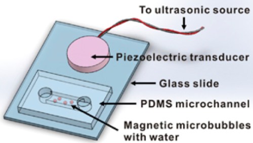


 2012pharmaceutical
 sjwilliamspa


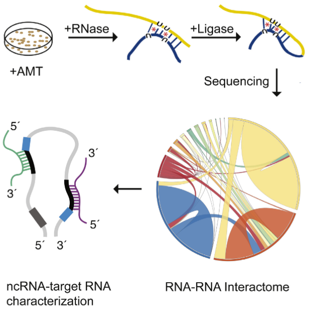
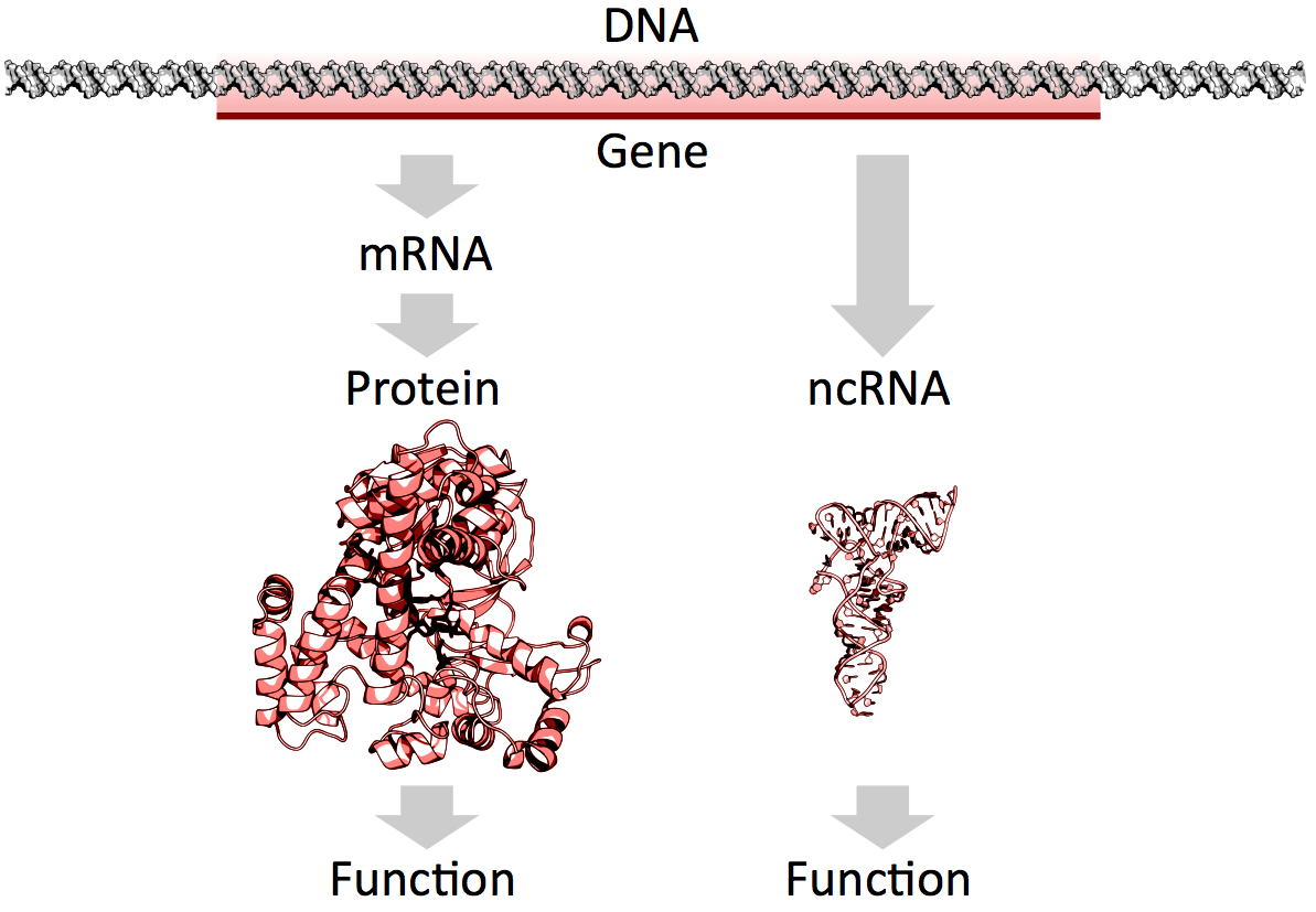
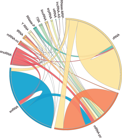


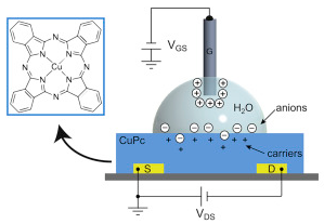


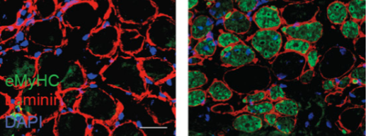


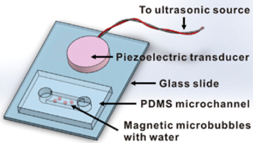
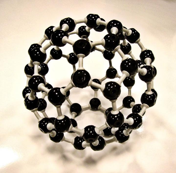




This is a lovely method and should find wide applicability in many settings, especially for microorganisms and cell lines. However, it is not clear that this approach will be, as implied by the discussion, an efficient mapping method for all multicellular organisms. I have performed similar experiments in Drosophila, focused on meiotic recombination, on a much smaller scale, and found that CRISPR-Cas9 can indeed generate targeted recombination at gRNA target sites. In every case I tested, I found that the recombination event was associated with a deletion at the gRNA site, which is probably unimportant for most mapping efforts, but may be a concern in some specific cases, for example for clinical applications. It would be interesting to know how often mutations occurred at the targeted gRNA site in this study.
The wider issue, however, is whether CRISPR-mediated recombination will be more efficient than other methods of mapping. After careful consideration of all the costs and the time involved in each of the steps for Drosophila, we have decided that targeted meiotic recombination using flanking visible markers will be, in most cases, considerably more efficient than CRISPR-mediated recombination. This is mainly due to the large expense of injecting embryos and the extensive effort and time required to screen injected animals for appropriate events. It is both cheaper and faster to generate markers (with CRISPR) and then perform a large meiotic recombination mapping experiment than it would be to generate the lines required for CRISPR-mediated recombination mapping. It is possible to dramatically reduce costs by, for example, mapping sequentially at finer resolution. But this approach would require much more time than marker-assisted mapping. If someone develops a rapid and cheap method of reliably introducing DNA into Drosophila embryos, then this calculus might change.
However, it is possible to imagine situations where CRISPR-mediated mapping would be preferable, even for Drosophila. For example, some genomic regions display extremely low or highly non-uniform recombination rates. It is possible that CRISPR-mediated mapping could provide a reasonable approach to fine mapping genes in these regions.
The authors also propose the exciting possibility that CRISPR-mediated loss of heterozygosity could be used to map traits in sterile species hybrids. It is not entirely obvious to me how this experiment would proceed and I hope the authors can illuminate me. If we imagine driving a recombination event in the early embryo (with maternal Cas9 from one parent and gRNA from a second parent), then at best we would end up with chimeric individuals carrying mitotic clones. I don’t think one could generate diploid animals where all cells carried the same loss of heterozygosity event. Even if we could, this experiment would require construction of a substantial number of stable transgenic lines expressing gRNAs. Mapping an ~20Mbp chromosome arm to ~10kb would require on the order of two-thousand transgenic lines. Not an undertaking to be taken lightly. It is already possible to perform similar tests (hemizygosity tests) using D. melanogaster deficiency lines in crosses with D. simulans, so perhaps CRISPR-mediated LOH could complement these deficiency screens for fine mapping efforts. But, at the moment, it is not clear to me how to do the experiment.