Feeds:
Posts
Comments

Archive for the ‘Regulated Clinical Trials: Design, Methods, Components and IRB related issues’ Category

Clinical Trial Services: Foundation Medicine & EmergingMed to Partner

Reporter: Aviva Lev-Ari, PhD, RN


Foundation Medicine, EmergingMed Partner for Clinical Trial Services

November 03, 2014


By a GenomeWeb staff reporter

NEW YORK (GenomeWeb) – Foundation Medicine and EmergingMed said today that they will partner to offer clinical trial navigation services for health care providers and their patients who have received one of Foundation Medicine’s tumor genomic profiling tests.

The firms will provide concierge services to help physicians identify appropriate clinical trials for patients based on the results of FoundationOne or FoundationOne Heme. EmergingMed will provide “Clinical Trial Navigators” to educate and match patients to clinical trials based on their genomic profiles.

“By providing clinical trial navigation services, we aim to facilitate timely and accurate clinical trial information and enrollment support services for physicians and patients, enabling greater access to treatment options based on the unique genomic profile of a patient’s cancer,” Foundation Medicine Chief Medical Officer Vincent Miller said in a statement.

Currently, there are over 800 candidate therapies that target genomic alterations in clinical trials, but “patients and physicians must identify and act on relevant options when the patient’s clinical profile is aligned with the often short enrollment window for each trial,” EmergingMed CEO Courtney Hudson said in a statement.

Miller added that these investigational therapies “offer relevant and rational options for patients with cancer, particularly for those whose cancer has progressed or returned following standard treatment.”

The new service will also send updates to users when new clinical trials emerge that match a patient’s genomic and clinical profile, Hudson said.

The service will be added at no extra cost and will be widely available to patients and their healthcare providers before the end of the year.

SOURCE

http://www.genomeweb.com/sequencing/foundation-medicine-emergingmed-partner-clinical-trial-services


Read Full Post »

Announcement of UPDATES to New Medicine-Oncology KnowledgeBase Data Base for September 2014

Curator: Katie Siafaca


Latest Updates

September 2014 New Drug Updates and Additions

Designation Brand Name Generic Name
OncoVEX GM-CSF, OncoVEXGMCSF, OncoVEXGM-CSF, Tvec, t-vec Talimogene laherparepvec
IL-17E
BIBF 1120 Vargatef Nintedanib
CV9202
CV9104
EPZ005687, EPZ-6438, E7438
EPZ-5676, EPZ004777
RG3638, anti-c-Met, PRO143966, RO5490258 MetMab Onartuzumab
G-202 Mipsagargin
AB0024, GS-6624 Simtuzumab
GS-5745
PEGPH20
L-DOS47
IPI-145 Duvelisib
KTN3379
5B1
MEDI4736
L-BLP25, BLP-25 (formerly BP1-7-KLH), EMD531444, emepepimut-S Formerly Stimuvax Tecemotide
MSB0010718C
MM-121, MM121, SAR256212
PEP02, MM-398, nal-IRI
CRA-024781, CG-781, PCI-24781, S-78454 Abexinostat
RX-3117
TG4010, MVA-MUC-1-IL-2, MVA-MUC1-IL-2


September 2014 Marketed Drug Updates and Additions

Brand Name Generic Name Other Designation
Pixuvri Pixantrone dimaleate BBR 2778
Cyramza Ramucirumab IMC-1121b, IMC 1121B, LY3009806
Perjeta (formerly Omnitarg) Pertuzumab RG1273, R1273, 2C4, rhuMAb-2C4
Zydelig Idelalisib CAL-101, GS-1101
Keytruda Pembrolizumab (formerly lambrolizumab) MK-3475
Lymphoseek Technetium Tc 99m tilmanocept
Zykadia Ceritinib LDK378
Afinitor Everolimus RAD001, RAD001C
Zelboraf Vemurafenib PLX4032, PLX4720, RG7204, RO5185426


September 2014 Company Updates and Additions

  • AADi
  • Abcodia
  • Accelovance
  • Adimab
  • Affymax
  • Akinion Pharmaceuticals
  • Aprea
  • Aptose Biosciences
  • BeiGene
  • Bioceros
  • Biogen Idec
  • Blueprint Medicines
  • BTG
  • Can-Fite BioPharma
  • Caris Life Sciences
  • Celgene
  • Cellerant Therapeutics
  • Champions Oncology
  • CureVac
  • DanDrit Biotech
  • Epizyme
  • Eutropics Pharmaceuticals
  • F2G
  • Genetic Technologies
  • Ignyta
  • ImaginAb
  • Johnson & Johnson (J&J)
  • Karolinska Development
  • Kolltan Pharmaceuticals
  • Lpath
  • Merus
  • Novartis
  • OncoCyte
  • Oncology Analytics (OA)
  • OncoNOx
  • Oncothyreon
  • Personal Genome Diagnostics (PGDx)
  • ProNAi Therapeutics
  • Propanc Health Group
  • Protagonist Therapeutics
  • Regeneron Pharmaceuticals
  • Relmada Therapeutics
  • Rosetta Genomics
  • Scholar Rock
  • Sentinel Oncology
  • Sevion Therapeutics
  • SutroVax
  • to-BBB Technologies (to-BBB)
  • Transgene
  • Veracyte
  • Zenith Epigenetics


September 2014 Targets in Oncology Updates and Additions

  • Fibroblast growth factor receptor 4 (FGFr4)
  • KIT
  • MUC16
  • V-raf murine sarcoma viral oncogene homolog B1 (BRAF)

SOURCE

http://www.nmok.net/oksite/updates.html

From: info <info@newmedinc.com>
Reply-To: <info@newmedinc.com>
Date: Wed, 29 Oct 2014 13:41:44 -0700
To: “Aviva Lev-Ari, PhD, RN” <avivalev-ari@alum.berkeley.edu>
Subject: DB coverage


Read Full Post »

Larry H. Bernstein, MD, FCAP, Reporter, Reposted

Leaders in Pharmaceutical Intelligence

DR ANTHONY MELVIN CRASTO …..FOR BLOG HOME CLICK HERE

http://pharmaceuticalintelligence.com/10/29/2010/larryhbern/Rofecoxib

ROFECOXIB

MK-966, MK-0966, Vioxx

162011-90-7

C17-H14-O4-S
314.3596
\
Percent Composition: C 64.95%, H 4.49%, O 20.36%, S 10.20%
LitRef: Selective cyclooxygenase-2 (COX-2) inhibitor. Prepn: Y. Ducharme et al., WO 9500501; eidem, US5474995 (both 1995 to Merck Frosst).
Therap-Cat: Anti-inflammatory; analgesic.

Rofecoxib /ˌrɒfɨˈkɒksɪb/ is a nonsteroidal anti-inflammatory drug (NSAID) that has now been withdrawn over safety concerns. It was marketed by Merck & Co. to treat osteoarthritisacute pain conditions, and dysmenorrhoea. Rofecoxib was approved by the Food and Drug Administration (FDA) on May 20, 1999, and was marketed under the brand names VioxxCeoxx, and Ceeoxx.

Rofecoxib

Rofecoxib

Rofecoxib gained widespread acceptance among physicians treating patients with arthritis and other conditions causing chronic or acute pain. Worldwide, over 80 million people were prescribed rofecoxib at some time.[1]

On September 30, 2004, Merck withdrew rofecoxib from the market because of concerns about increased risk of heart attack and stroke associated with long-term, high-dosage use. Merck withdrew the drug after disclosures that it withheld information about rofecoxib’s risks from doctors and patients for over five years, resulting in between 88,000 and 140,000 cases of serious heart disease.[2] Rofecoxib was one of the most widely used drugs ever to be withdrawn from the market. In the year before withdrawal, Merck had sales revenue of US$2.5 billion from Vioxx.[3] Merck reserved $970 million to pay for its Vioxx-related legal expenses through 2007, and have set aside $4.85bn for legal claims from US citizens.

Rofecoxib was available on prescription in both tablet-form and as an oral suspension. It was available by injection for hospital use.


 Mode of action
 Cyclooxygenase (COX) has two well-studied isoforms, called COX-1 and COX-2.
  • COX-1 mediates the synthesis of prostaglandins responsible for protection of the stomach lining, while
  • COX-2 mediates the synthesis of prostaglandins responsible for pain and inflammation.
prostaglandin PGE2

prostaglandin PGE2

By creating “selective” NSAIDs that inhibit COX-2, but not COX-1, the same pain relief as traditional NSAIDs is offered, but with greatly reduced risk of fatal or debilitating peptic ulcers. Rofecoxib is a selective COX-2 inhibitor, or “coxib”.

Others include Merck’s etoricoxib (Arcoxia), Pfizer’s celecoxib (Celebrex) and valdecoxib (Bextra). Interestingly, at the time of its withdrawal, rofecoxib was the only coxib with clinical evidence of its superior gastrointestinal adverse effect profile over conventional NSAIDs. This was largely based on the VIGOR (Vioxx GI Outcomes Research) study, which compared the efficacy and adverse effect profiles of rofecoxib and naproxen.[4]

Pharmacokinetics

The therapeutic recommended dosages were 12.5, 25, and 50 mg with an approximate bioavailability of 93%.[5][6][7] Rofecoxib crossed the placenta and blood–brain barrier,[5][6][8]and took 1–3 hours to reach peak plasma concentration with an effective half-life (based on steady-state levels) of approximately 17 hours.[5][7][9] The metabolic products are cis-dihydro and trans-dihydro derivatives of rofecoxib[5][9] which are primarily excreted through urine.

Fabricated efficacy studies

On March 11, 2009, Scott S. Reuben, former chief of acute pain at Baystate Medical Center, Springfield, Mass., revealed that data for 21 studies he had authored for the efficacy of the drug (along with others such as celecoxib) had been fabricated in order to augment the analgesic effects of the drugs. There is no evidence that Reuben colluded with Merck in falsifying his data. Reuben was also a former paid spokesperson for the drug company Pfizer (which owns the intellectual property rights for marketing celecoxib in the United States). The retracted studies were not submitted to either the FDA or the European Union’s regulatory agencies prior to the drug’s approval. Drug manufacturer Merckhad no comment on the disclosure.[10]

Adverse drug reactions

VIOXX sample blister pack.jpg

Aside from the reduced incidence of gastric ulceration, rofecoxib exhibits a similar adverse effect profile to other NSAIDs.

Prostaglandin is a large family of lipids. Prostaglandin I2/PGI2/prostacyclin is just one member of it. Prostaglandins other than PGI2 (such as PGE2) also play important roles in vascular tone regulation. Prostacyclin/thromboxane are produced by both COX-1 and COX-2, and rofecoxib suppresses just COX-2 enzyme, so there is no reason to believe that prostacyclin levels are significantly reduced by the drug. And there is no reason to believe that only the balance between quantities of prostacyclin and thromboxane is the determinant factor for vascular tone.[11] Indeed Merck has stated that there was no effect on prostacyclin production in blood vessels in animal testing.[12] Other researchers have speculated that the cardiotoxicity may be associated with maleic anhydride metabolites formed when rofecoxib becomes ionized under physiological conditions. (Reddy & Corey, 2005)

 Adverse cardiovascular events

VIGOR study and publishing controversy

The VIGOR (Vioxx GI Outcomes Research) study, conducted by Bombardier, et al., which compared the efficacy and adverse effect profiles of rofecoxib and naproxen, had indicated a significant 4-fold increased risk of acute myocardial infarction (heart attack) in rofecoxib patients when compared with naproxen patients (0.4% vs 0.1%, RR 0.25) over the 12 month span of the study. The elevated risk began during the second month on rofecoxib. There was no significant difference in the mortality from cardiovascular events between the two groups, nor was there any significant difference in the rate of myocardial infarction between the rofecoxib and naproxen treatment groups in patients without high cardiovascular risk. The difference in overall risk was by the patients at higher risk of heart attack, i.e. those meeting the criteria for low-dose aspirin prophylaxis of secondary cardiovascular events (previous myocardial infarction, angina, cerebrovascular accidenttransient ischemic attack, or coronary artery bypass).

Merck’s scientists interpreted the finding as a protective effect of naproxen, telling the FDA that the difference in heart attacks “is primarily due to” this protective effect (Targum, 2001). Some commentators have noted that naproxen would have to be three times as effective as aspirin to account for all of the difference (Michaels 2005), and some outside scientists warned Merck that this claim was implausible before VIGOR was published.[13] No evidence has since emerged for such a large cardioprotective effect of naproxen, although a number of studies have found protective effects similar in size to those of aspirin.[14][15] Though Dr. Topol’s 2004 paper criticized Merck’s naproxen hypothesis, he himself co-authored a 2001 JAMA article stating “because of the evidence for an antiplatelet effect of naproxen, it is difficult to assess whether the difference in cardiovascular event rates in VIGOR was due to a benefit from naproxen or to a prothrombotic effect from rofecoxib.” (Mukherjee, Nissen and Topol, 2001.)

The results of the VIGOR study were submitted to the United States Food and Drug Administration (FDA) in February 2001. In September 2001, the FDA sent a warning letter to the CEO of Merck, stating, “Your promotional campaign discounts the fact that in the VIGOR study, patients on Vioxx were observed to have a four to five fold increase in myocardial infarctions (MIs) compared to patients on the comparator non-steroidal anti-inflammatory drug (NSAID), Naprosyn (naproxen).”[16] This led to the introduction, in April 2002, of warnings on Vioxx labeling concerning the increased risk of cardiovascular events (heart attack and stroke).

Months after the preliminary version of VIGOR was published in the New England Journal of Medicine, the journal editors learned that certain data reported to the FDA were not included in the NEJM article. Several years later, when they were shown a Merck memo during the depositions for the first federal Vioxx trial, they realized that these data had been available to the authors months before publication. The editors wrote an editorial accusing the authors of deliberately withholding the data.[17] They released the editorial to the media on December 8, 2005, before giving the authors a chance to respond. NEJM editor Gregory Curfman explained that the quick release was due to the imminent presentation of his deposition testimony, which he feared would be misinterpreted in the media. He had earlier denied any relationship between the timing of the editorial and the trial. Although his testimony was not actually used in the December trial, Curfman had testified well before the publication of the editorial.[18]

The editors charged that “more than four months before the article was published, at least two of its authors were aware of critical data on an array of adverse cardiovascular events that were not included in the VIGOR article.” These additional data included three additional heart attacks, and raised the relative risk of Vioxx from 4.25-fold to 5-fold. All the additional heart attacks occurred in the group at low risk of heart attack (the “aspirin not indicated” group) and the editors noted that the omission “resulted in the misleading conclusion that there was a difference in the risk of myocardial infarction between the aspirin indicated and aspirin not indicated groups.” The relative risk for myocardial infarctions among the aspirin not indicated patients increased from 2.25 to 3 (although it remained statitistically insignificant). The editors also noted a statistically significant (2-fold) increase in risk for serious thromboembolic events for this group, an outcome that Merck had not reported in the NEJM, though it had disclosed that information publicly in March 2000, eight months before publication.[19]

The authors of the study, including the non-Merck authors, responded by claiming that the three additional heart attacks had occurred after the prespecified cutoff date for data collection and thus were appropriately not included. (Utilizing the prespecified cutoff date also meant that an additional stroke in the naproxen population was not reported.) Furthermore, they said that the additional data did not qualitatively change any of the conclusions of the study, and the results of the full analyses were disclosed to the FDA and reflected on the Vioxx warning label. They further noted that all of the data in the “omitted” table were printed in the text of the article. The authors stood by the original article.[20]

NEJM stood by its editorial, noting that the cutoff date was never mentioned in the article, nor did the authors report that the cutoff for cardiovascular adverse events was before that for gastrointestinal adverse events. The different cutoffs increased the reported benefits of Vioxx (reduced stomach problems) relative to the risks (increased heart attacks).[19]

Some scientists have accused the NEJM editorial board of making unfounded accusations.[21][22] Others have applauded the editorial. Renowned research cardiologist Eric Topol,[23] a prominent Merck critic, accused Merck of “manipulation of data” and said “I think now the scientific misconduct trial is really fully backed up”.[24] Phil Fontanarosa, executive editor of the prestigious Journal of the American Medical Association, welcomed the editorial, saying “this is another in the long list of recent examples that have generated real concerns about trust and confidence in industry-sponsored studies”.[25]

On May 15, 2006, the Wall Street Journal reported that a late night email, written by an outside public relations specialist and sent to Journal staffers hours before the Expression of Concern was released, predicted that “the rebuke would divert attention to Merck and induce the media to ignore the New England Journal of Medicine‘s own role in aiding Vioxx sales.”[26]

“Internal emails show the New England Journal’s expression of concern was timed to divert attention from a deposition in which Executive Editor Gregory Curfman made potentially damaging admissions about the journal’s handling of the Vioxx study. In the deposition, part of the Vioxx litigation, Dr. Curfman acknowledged that lax editing might have helped the authors make misleading claims in the article.” The Journal stated that NEJM‘s “ambiguous” language misled reporters into incorrectly believing that Merck had deleted data regarding the three additional heart attacks, rather than a blank table that contained no statistical information; “the New England Journal says it didn’t attempt to have these mistakes corrected.”[26]

APPROVe study

In 2001, Merck commenced the APPROVe (Adenomatous Polyp PRevention On Vioxx) study, a three-year trial with the primary aim of evaluating the efficacy of rofecoxib for theprophylaxis of colorectal polypsCelecoxib had already been approved for this indication, and it was hoped to add this to the indications for rofecoxib as well. An additional aim of the study was to further evaluate the cardiovascular safety of rofecoxib.

The APPROVe study was terminated early when the preliminary data from the study showed an increased relative risk of adverse thrombotic cardiovascular events (includingheart attack and stroke), beginning after 18 months of rofecoxib therapy. In patients taking rofecoxib, versus placebo, the relative risk of these events was 1.92 (rofecoxib 1.50 events vs placebo 0.78 events per 100 patient years). The results from the first 18 months of the APPROVe study did not show an increased relative risk of adverse cardiovascular events. Moreover, overall and cardiovascular mortality rates were similar between the rofecoxib and placebo populations.[28]

In summary, the APPROVe study suggested that long-term use of rofecoxib resulted in nearly twice the risk of suffering a heart attack or stroke compared to patients receiving a placebo.

Other studies

Several very large observational studies have also found elevated risk of heart attack from rofecoxib. For example, a recent retrospective study of 113,000 elderly Canadians suggested a borderline statistically significant increased relative risk of heart attacks of 1.24 from Vioxx usage, with a relative risk of 1.73 for higher-dose Vioxx usage. (Levesque, 2005). Another study, using Kaiser Permanente data, found a 1.47 relative risk for low-dose Vioxx usage and 3.58 for high-dose Vioxx usage compared to current use of celecoxib, though the smaller number was not statistically significant, and relative risk compared to other populations was not statistically significant. (Graham, 2005).

Furthermore, a more recent meta-study of 114 randomized trials with a total of 116,000+ participants, published in JAMA, showed that Vioxx uniquely increased risk of renal (kidney) disease, and heart arrhythmia.[31]

Other COX-2 inhibitors

Any increased risk of renal and arrhythmia pathologies associated with the class of COX-2 inhibitors, e.g. celecoxib (Celebrex), valdecoxib (Bextra), parecoxib (Dynastat),lumiracoxib, and etoricoxib is not evident,[31] although smaller studies[32][33] had demonstrated such effects earlier with the use of celecoxib, valdecoxib and parecoxib.

Nevertheless, it is likely that trials of newer drugs in the category will be extended in order to supply additional evidence of cardiovascular safety. Examples are some more specific COX-2 inhibitors, including etoricoxib (Arcoxia) and lumiracoxib (Prexige), which are currently (circa 2005) undergoing Phase III/IV clinical trials.

Besides, regulatory authorities worldwide now require warnings about cardiovascular risk of COX-2 inhibitors still on the market. For example, in 2005, EU regulators required the following changes to the product information and/or packaging of all COX-2 inhibitors:[34]

  • Contraindications stating that COX-2 inhibitors must not be used in patients with established ischaemic heart disease and/or cerebrovascular disease (stroke), and also in patients with peripheral arterial disease
  • Reinforced warnings to healthcare professionals to exercise caution when prescribing COX-2 inhibitors to patients with risk factors for heart disease, such as hypertension, hyperlipidaemia (high cholesterol levels), diabetes and smoking
  • Given the association between cardiovascular risk and exposure to COX-2 inhibitors, doctors are advised to use the lowest effective dose for the shortest possible duration of treatment

Other NSAIDs

Since the withdrawal of Vioxx it has come to light that there may be negative cardiovascular effects with not only other COX-2 inhibitiors, but even the majority of other NSAIDs. It is only with the recent development of drugs like Vioxx that drug companies have carried out the kind of well executed trials that could establish such effects and these sort of trials have never been carried out in older “trusted” NSAIDs such as ibuprofendiclofenac and others. The possible exceptions may be aspirin and naproxen due to their anti-platelet aggregation properties.

Withdrawal

Due to the findings of its own APPROVe study, Merck publicly announced its voluntary withdrawal of the drug from the market worldwide on September 30, 2004.[35]

In addition to its own studies, on September 23, 2004 Merck apparently received information about new research by the FDA that supported previous findings of increased risk of heart attack among rofecoxib users (Grassley, 2004). FDA analysts estimated that Vioxx caused between 88,000 and 139,000 heart attacks, 30 to 40 percent of which were probably fatal, in the five years the drug was on the market.[36]

On November 5, the medical journal The Lancet published a meta-analysis of the available studies on the safety of rofecoxib (Jüni et al., 2004). The authors concluded that, owing to the known cardiovascular risk, rofecoxib should have been withdrawn several years earlier. The Lancet published an editorial which condemned both Merck and the FDA for the continued availability of rofecoxib from 2000 until the recall. Merck responded by issuing a rebuttal of the Jüni et al. meta-analysis that noted that Jüni omitted several studies that showed no increased cardiovascular risk. (Merck & Co., 2004).

In 2005, advisory panels in both the U.S. and Canada encouraged the return of rofecoxib to the market, stating that rofecoxib’s benefits outweighed the risks for some patients. The FDA advisory panel voted 17-15 to allow the drug to return to the market despite being found to increase heart risk. The vote in Canada was 12-1, and the Canadian panel noted that the cardiovascular risks from rofecoxib seemed to be no worse than those from ibuprofen—though the panel recommended that further study was needed for all NSAIDs to fully understand their risk profiles. Notwithstanding these recommendations, Merck has not returned rofecoxib to the market.[37]

In 2005, Merck retained Debevoise & Plimpton LLP to investigate Vioxx study results and communications conducted by Merck. Through the report, it was found that Merck’s senior management acted in good faith, and that the confusion over the clinical safety of Vioxx was due to the sales team’s overzealous behavior. The report that was filed gave a timeline of the events surrounding Vioxx and showed that Merck intended to operate honestly throughout the process. Any mistakes that were made regarding the mishandling of clinical trial results and withholding of information was the result of oversight, not malicious behavior….The report was published in February 2006, and Merck was satisfied with the findings of the report and promised to consider the recommendations contained in the Martin Report. Advisers to the US Food and Drug Administration (FDA) have voted, by a narrow margin, that it should not ban Vioxx — the painkiller withdrawn by drug-maker Merck.

They also said that Pfizer’s Celebrex and Bextra, two other members of the family of painkillers known as COX-2 inhibitors, should remain available, despite the fact that they too boost patients’ risk of heart attack and stroke. url = http://www.nature.com/drugdisc/news/articles/433790b.html The recommendations of the arthritis and drug safety advisory panel offer some measure of relief to the pharmaceutical industry, which has faced a barrage of criticism for its promotion of the painkillers. But the advice of the panel, which met near Washington DC over 16–18 February, comes with several strings attached.

For example, most panel members said that manufacturers should be required to add a prominent warning about the drugs’ risks to their labels; to stop direct-to-consumer advertising of the drugs; and to include detailed, written risk information with each prescription. The panel also unanimously stated that all three painkillers “significantly increase the risk of cardiovascular events”.

External links

For more details and references.. they are provided in the entirety in the original post

Read Full Post »

Mapping the Universe of Pharmaceutical Business Intelligence: The Model developed by LPBI and the Model of Best Practices LLC

Mapping the Universe of Pharmaceutical Business Intelligence: The Model developed by LPBI and the Model of Best Practices LLC


Author and Curator of Model A: Aviva Lev-Ari, PhD, RN

Reporter on Model B: Aviva Lev-Ari, PhD, RN


This article provides the e-Reader with a MAP for navigation through two different Business Models that Co-exist in the EcoSystem of an industry called Pharmaceutical Business Intelligence.

Model A: is represented by Six Ventures of Leaders in Pharmaceutical Business Intelligence (LPBI), based in Boston, Philadelphia, CT, CA and Israel

Model B: is represented by Best Practices, LLC, headquartered in Chapel Hill, NC, with Offices in NYC and in Mumbai, India.


We concluded that the two models are viable, represent fast growth, the models and non-competing and are in full complementarity, thus, expanding the domain and the practice of the industrial sector, aka, Pharmaceutical Business Intelligence.


Model A:

Leaders in Pharmaceutical Business Intelligence (LPBI),

Boston, Philadelphia, CT, CA and Israel 

Team members


Our Growth Needs: Leaders in Pharmaceutical Business Intelligence


 Our Business Portfolio

VENTURE #1:

e-Publishing: Medicine, HealthCare, Life Sciences, BioMed, Pharmaceutical

  • Open Access Online Scientific Journal

http://pharmaceuticalintelligence.com Site statistics http://pharmaceuticalintelligence.com/wp-admin/index.php?page=stats

  • Scoop.it!.com

  1. http://www.scoop.it/t/cardiotoxicity
  2. http://www.scoop.it/t/cardiovascular-and-vascular-imaging
  3. http://www.scoop.it/t/cardiovascular-disease-pharmaco-therapy

VENTURE #2:

1. BioMedical e-Books e-Series: Cardiovascular, Genomics, Cancer, BioMed, Patient Centered Medicine

http://pharmaceuticalintelligence.com/biomed-e-books/

2. on Amazon’s Kindle e-Books List since 6/2013

3. Plans for Volume 1,2,3 – Hardcover

VENTURE #3:

International Scientific Delegations

http://pharmaceuticalintelligence.com/scientific-delegation/

  • Shanghai, May 2015
  • Barcelona, Spain, November 2015
  • Amsterdam, May 2016
  • Geneva, November 2016


VENTURE #4:

Funding, Deals & Partnerships

http://pharmaceuticalintelligence.com/joint-ventures/


VENTURE #5:

IP Invented HERE!

1.  Development of a NEW Nitric Oxide monitor to Alpha Szenszor Inc. sensor portfolio. A concept for a low cost POC e-nose, capable of real time ppb detection of Cancer The Cancer Team at Leaders in Pharmaceutical Business Intelligence under the leadership of Dr. Williams

2.  Development of a NEW Nitric Oxide monitor to Alpha Szenszor Inc. sensor portfolio. A concept for Inhaled Nitric Oxide for the Adult HomeCare Market – IP by Dr. Pearlman and Dr. A. Lev-Ari

a.  iknow iNO is i-kNOw – Inhaled Nitric Oxide for the HomeCare Markethttp://pharmaceuticalintelligence.com/2013/10/16/iknow-ino-is-i-know-inhaled-nitric-oxide-for-the-homecare-market/

b. electronic Book on Nitric Oxide by Nitric Oxide Team @ Leaders in Pharmaceutical Business Intelligence (LPBI)

Perspectives on Nitric Oxide in Disease Mechanisms

http://www.amazon.com/dp/B00DINFFY

c. The rationale and use of inhaled NO in Pulmonary Artery Hypertension and Right Sided Heart Failure Larry H. Bernstein 8/20/2012

d. Inhaled Nitric Oxide in Adults: Clinical Trials and Meta Analysis Studies – Recent Findings

Aviva Lev-Ari, PhD, RN, 6/2/2013

e. Clinical Indications for Use of Inhaled Nitric Oxide (iNO) in the Adult Patient Market: Clinical Outcomes after Use, Therapy Demand and Cost of Care

Aviva Lev-Ari, PhD, RN, 6/3/2013

3.  Cancer Genomics for NEW product development in diagnosis and treatment of Cancer Patients using sensory technology with applications for Radiation Therapy –The Cancer Team at Leaders in Pharmaceutical Business Intelligence under leadership of TBA

4.  Developing Mitral Valve Disease: MRI Methods and Devices for Percutaneous Mitral Valve Replacement and Mitral Valve Repair Augmentation of Patented Technology using RF – Dr. Pearlman’s IP Non-Hardware Mitral Annuloplasty – Dr. Justin D. Pearlman

http://pharmaceuticalintelligence.com/joint-ventures/valvecure-llc/non-hardware-mitral-annuloplasty-dr-justin-d-pearlman/

5.  Novel Technology using MRI for Vascular Lesions, Tumors, Hyperactive Glands and non-Surgical Cosmetic Reconstruction – Dr. Pearlman’s IP

http://pharmaceuticalintelligence.com/biomed-e-books/series-a-e-books-on-cardiovascular-diseases/httppharmaceuticalintelligence-combiomed-e-bookscardiovascular-diseases-causes-risks-and-management/cvd-business-affairs/mitral-valve-disease-mri-methods-and-devices/


VENTURE # 6:

PRESS Coverage of Conferences

http://pharmaceuticalintelligence.com/press-coverage/

Model B:

 
Best Practices, LLC, Chapel Hill, NC, Mumbai, India, Branch in New York


Best Practices, LLC
6350 Quadrangle Drive, Suite 200,
Chapel HillNC 27517

+1 919-403-0251

SOURCE

http://www.best-in-class.com/sitemap


Read Full Post »

Imaging-guided cancer treatment

Imaging-guided cancer treatment

Writer & reporter: Dror Nir, PhD

It is estimated that the medical imaging market will exceed $30 billion in 2014 (FierceMedicalImaging). To put this amount in perspective; the global pharmaceutical market size for the same year is expected to be ~$1 trillion (IMS) while the global health care spending as a percentage of Gross Domestic Product (GDP) will average 10.5% globally in 2014 (Deloitte); it will reach ~$3 trillion in the USA.

Recent technology-advances, mainly miniaturization and improvement in electronic-processing components is driving increased introduction of innovative medical-imaging devices into critical nodes of major-diseases’ management pathways. Consequently, in contrast to it’s very small contribution to global health costs, medical imaging bears outstanding potential to reduce the future growth in spending on major segments in this market mainly: Drugs development and regulation (e.g. companion diagnostics and imaging surrogate markers); Disease management (e.g. non-invasive diagnosis, guided treatment and non-invasive follow-ups); and Monitoring aging-population (e.g. Imaging-based domestic sensors).

In; The Role of Medical Imaging in Personalized Medicine I discussed in length the role medical imaging assumes in drugs development.  Integrating imaging into drug development processes, specifically at the early stages of drug discovery, as well as for monitoring drug delivery and the response of targeted processes to the therapy is a growing trend. A nice (and short) review highlighting the processes, opportunities, and challenges of medical imaging in new drug development is: Medical imaging in new drug clinical development.

The following is dedicated to the role of imaging in guiding treatment.

Precise treatment is a major pillar of modern medicine. An important aspect to enable accurate administration of treatment is complementing the accurate identification of the organ location that needs to be treated with a system and methods that ensure application of treatment only, or mainly to, that location. Imaging is off-course, a major component in such composite systems. Amongst the available solution, functional-imaging modalities are gaining traction. Specifically, molecular imaging (e.g. PET, MRS) allows the visual representation, characterization, and quantification of biological processes at the cellular and subcellular levels within intact living organisms. In oncology, it can be used to depict the abnormal molecules as well as the aberrant interactions of altered molecules on which cancers depend. Being able to detect such fundamental finger-prints of cancer is key to improved matching between drugs-based treatment and disease. Moreover, imaging-based quantified monitoring of changes in tumor metabolism and its microenvironment could provide real-time non-invasive tool to predict the evolution and progression of primary tumors, as well as the development of tumor metastases.

A recent review-paper: Image-guided interventional therapy for cancer with radiotherapeutic nanoparticles nicely illustrates the role of imaging in treatment guidance through a comprehensive discussion of; Image-guided radiotherapeutic using intravenous nanoparticles for the delivery of localized radiation to solid cancer tumors.

 Graphical abstract

 Abstract

One of the major limitations of current cancer therapy is the inability to deliver tumoricidal agents throughout the entire tumor mass using traditional intravenous administration. Nanoparticles carrying beta-emitting therapeutic radionuclides [DN: radioactive isotops that emits electrons as part of the decay process a list of β-emitting radionuclides used in radiotherapeutic nanoparticle preparation is given in table1 of this paper.) that are delivered using advanced image-guidance have significant potential to improve solid tumor therapy. The use of image-guidance in combination with nanoparticle carriers can improve the delivery of localized radiation to tumors. Nanoparticles labeled with certain beta-emitting radionuclides are intrinsically theranostic agents that can provide information regarding distribution and regional dosimetry within the tumor and the body. Image-guided thermal therapy results in increased uptake of intravenous nanoparticles within tumors, improving therapy. In addition, nanoparticles are ideal carriers for direct intratumoral infusion of beta-emitting radionuclides by convection enhanced delivery, permitting the delivery of localized therapeutic radiation without the requirement of the radionuclide exiting from the nanoparticle. With this approach, very high doses of radiation can be delivered to solid tumors while sparing normal organs. Recent technological developments in image-guidance, convection enhanced delivery and newly developed nanoparticles carrying beta-emitting radionuclides will be reviewed. Examples will be shown describing how this new approach has promise for the treatment of brain, head and neck, and other types of solid tumors.

The challenges this review discusses

  • intravenously administered drugs are inhibited in their intratumoral penetration by high interstitial pressures which prevent diffusion of drugs from the blood circulation into the tumor tissue [1–5].
  • relatively rapid clearance of intravenously administered drugs from the blood circulation by kidneys and liver.
  • drugs that do reach the solid tumor by diffusion are inhomogeneously distributed at the micro-scale – This cannot be overcome by simply administering larger systemic doses as toxicity to normal organs is generally the dose limiting factor.
  • even nanoparticulate drugs have poor penetration from the vascular compartment into the tumor and the nanoparticles that do penetrate are most often heterogeneously distributed

How imaging could mitigate the above mentioned challenges

  • The inclusion of an imaging probe during drug development can aid in determining the clearance kinetics and tissue distribution of the drug non-invasively. Such probe can also be used to determine the likelihood of the drug reaching the tumor and to what extent.

Note: Drugs that have increased accumulation within the targeted site are likely to be more effective as compared with others. In that respect, Nanoparticle-based drugs have an additional advantage over free drugs with their potential to be multifunctional carriers capable of carrying both therapeutic and diagnostic imaging probes (theranostic) in the same nanocarrier. These multifunctional nanoparticles can serve as theranostic agents and facilitate personalized treatment planning.

  • Imaging can also be used for localization of the tumor to improve the placement of a catheter or external device within tumors to cause cell death through thermal ablation or oxidative stress secondary to reactive oxygen species.

See the example of Vintfolide in The Role of Medical Imaging in Personalized Medicine

vinta

Note: Image guided thermal ablation methods include radiofrequency (RF) ablation, microwave ablation or high intensity focused ultrasound (HIFU). Photodynamic therapy methods using external light devices to activate photosensitizing agents can also be used to treat superficial tumors or deeper tumors when used with endoscopic catheters.

  • Quality control during and post treatment

For example: The use of high intensity focused ultrasound (HIFU) combined with nanoparticle therapeutics: HIFU is applied to improve drug delivery and to trigger drug release from nanoparticles. Gas-bubbles are playing the role of the drug’s nano-carrier. These are used both to increase the drug transport into the cell and as ultrasound-imaging contrast material. The ultrasound is also used for processes of drug-release and ablation.

 HIFU

Additional example; Multifunctional nanoparticles for tracking CED (convection enhanced delivery)  distribution within tumors: Nanoparticle that could serve as a carrier not only for the therapeutic radionuclides but simultaneously also for a therapeutic drug and 4 different types of imaging contrast agents including an MRI contrast agent, PET and SPECT nuclear diagnostic imaging agents and optical contrast agents as shown below. The ability to perform multiple types of imaging on the same nanoparticles will allow studies investigating the distribution and retention of nanoparticles initially in vivo using non-invasive imaging and later at the histological level using optical imaging.

 multi

Conclusions

Image-guided radiotherapeutic nanoparticles have significant potential for solid tumor cancer therapy. The current success of this therapy in animals is most likely due to the improved accumulation, retention and dispersion of nanoparticles within solid tumor following image-guided therapies as well as the micro-field of the β-particle which reduces the requirement of perfectly homogeneous tumor coverage. It is also possible that the intratumoral distribution of nanoparticles may benefit from their uptake by intratumoral macrophages although more research is required to determine the importance of this aspect of intratumoral radionuclide nanoparticle therapy. This new approach to cancer therapy is a fertile ground for many new technological developments as well as for new understandings in the basic biology of cancer therapy. The clinical success of this approach will depend on progress in many areas of interdisciplinary research including imaging technology, nanoparticle technology, computer and robot assisted image-guided application of therapies, radiation physics and oncology. Close collaboration of a wide variety of scientists and physicians including chemists, nanotechnologists, drug delivery experts, radiation physicists, robotics and software experts, toxicologists, surgeons, imaging physicians, and oncologists will best facilitate the implementation of this novel approach to the treatment of cancer in the clinical environment. Image-guided nanoparticle therapies including those with β-emission radionuclide nanoparticles have excellent promise to significantly impact clinical cancer therapy and advance the field of drug delivery.

Read Full Post »

Larry H Bernstein, MD, Curator

Leaders in Pharmaceutical Intelligence


Natriuretic Peptides (BNP and Amino-terminal proBNP)

Author: Larry Bernstein, M.D.,
(see Reviewers/Authors page)
Revised: 12 December 2010, last major update December 2010
Copyright: (c) 2003-2010, PathologyOutlines.com, Inc.
http://dx.doi.org:/PathologyOutlines.com/cardiac

General
=========================================================================

  • Brain natriuretic peptide (BNP), now known as B-type natriuretic peptide (also BNP),
    is a 32 amino acid polypeptide secreted by the cardiac ventricles in response to
    excessive stretching of cardiomyocytes (Wikipedia)
  • BNP was originally identified in extracts of porcine brain, although in humans
    it is produced mainly in the cardiac ventricles
  • BNP is co-secreted with a 76 amino acid N-terminal fragment (NT-proBNP),
    which is biologically inactive Indications

=========================================================================

  • Evaluation of dyspneic patient with suspected congestive heart failure,
    regardless of renal function (J Am Coll Cardiol 2006;47:91)
  • B-type natriuretic peptide levels are higher in patients with congestive heart
    failure than in dyspnea from other causes (J Am Coll Cardiol 2002;39:202,
    N Engl J Med 2004;350:647)
  • NT-proBNP measurement is a valuable addition to standard clinical
    assessment for the identification and exclusion of acute CHF in the
    emergency department setting (Am J Cardiol 2005;95:9480)

Clinical features
=========================================================================

  • Reduces misdiagnosis of congestive heart failure, which occurs
    50% to 75% of the time
  • NT-proBNP is superior to BNP for predicting mortality and morbidity for heart
    failure (Clin Chem 2006;52:1528), and coexisting renal disease and heart failure
    (Clin Chem 2007;53:1511)

Reference ranges
=========================================================================

  • BNP levels below 100 pg/mL indicate no heart failure

Limitations
=========================================================================

  • Determination of endogenous BNP with the AxSYM assay using frozen
    plasma samples may not be valid after 1 day, but NT-proBNP as
    measured by the Elecsys assay may be stored at -20 degrees C for
    at least four months without a relevant loss of the immunoreactive
    analyte (Clin Chem Lab Med 2004;42:942)

Additional references
=========================================================================

  • Clin Chem 2007;53:1928, Am J Kidney Dis 2005;46:610,
    Hypertension 2005;46:118, Hypertension 2006;47:874,
    Eur J Heart Fail 2004;6:269

Natriuretic peptides for risk stratification of patients with acute
coronary
 syndromes  
M Galvani,  D Ferrini, F Ottani. Eur J Heart Fail 2004;  6: 327–333.
http://eurjhf.oxfordjournals.org

Both BNP and NT-proBNP possess several characteristics of the ideal biomarker,
showing independent and incremental prognostic value above traditional clinical,
electrocardiographic, and biochemical (particularly troponin) risk indicators. Specifically,
in ACS patients, BNP and NT-proBNP have powerful prognostic value both in patients
without a history of previous heart failure or without clinical or instrumental signs of
left ventricular dysfunction on admission or during hospital stay.

Our results show that the prognostic value of natriuretic peptides is similar:
(1) both at short- and long-term;
(2) when natriuretic peptides are measured at first patient contact or during hospital stay;
(3) for BNP or NT-proBNP; and
(4) in patients with ST elevation myocardial infarction or no ST elevation ACS.


Steady-State Levels of Troponin and Brain Natriuretic Peptide for Prediction
of Long-Term
 Outcome after Acute Heart Failure with or without Stage 3 to 4
Chronic Kidney Disease

Y Endo, S Kohsaka, T Nagai, K Koide, M Takahashi, et al.
Br J Med Med Res 2012; 2(4): 490-500.
http://dx.doi.org:/10.9734/BJMMR/2012/1384

The population was predominantly male (69.3%), and the mean age was 66.6±15.3 years.
Patients with higher BNP levels or detectable TnT had a worse prognosis (BNP45.0% vs.
18.8%, p<0.001; TnT 43.8% vs. 25.1%, p=0.002, respectively). The primary event rate
was additively worse among patients with both increased BNP levels and detectable TnT
compared to those with increased levels of BNP or detectable TnT alone (log-rank p<0.001).
A similar trend was observed in the subgroup of patients with CKD stage III–V (n=172).

The Effect of Correction of Mild Anemia in Severe, Resistant Congestive
Heart Failure
 Using Subcutaneous Erythropoietin and Intravenous Iron:
A Randomized Controlled Study

DS. Silverberg, D Wexler, D Sheps, M Blum, G Keren, et al.  JACC 2001; 37(7).
PII S0735-1097(01)01248-7  http://www.ncbi.nlm.nih.gov/pubmed/11401110

When anemia in CHF is treated with EPO and IV iron, a marked improvement in
cardiac and patient function is seen, associated with less hospitalization and renal
impairment and less need for diuretics. (J Am Coll Cardiol 2001;37:1775– 80)


Hemoglobin on NT proBNP

Hemoglobin on NT proBNP


What is the best approximation of reference normal for NT-proBNP?
Clinical levels for enhanced assessment
 of NT-proBNP (CLEAN) 

Larry H. Bernstein1*, Michael Y. Zions1,4, Mohammed E. Alam1,5, Salman A. Haq1,
John F. Heitner1, Stuart Zarich2, Bette Seamonds3 and Stanley Berger3
1New York Methodist Hospital, Brooklyn, NY; 2Bridgeport Hospital, Bridgeport, CT;
3Mercy Catholic Medical Center, Darby, Phila, PA;  4Touro College, &  5Medgar
Evers College, Brooklyn, NY
Journal of Medical Laboratory and Diagnosis 04/2011; 2:16-21.
http://www.academicjournals.org/jmld

The natriuretic peptides, B-type natriuretic peptide (BNP) and NT-proBNP that
have emerged as tools for diagnosing congestive heart failure (CHF) are affected
by age and renal insufficiency (RI).  NTproBNP is used in rejecting CHF and as a
marker of risk for patients with acute coronary syndromes. This observational study
was undertaken to evaluate the reference value for interpreting NT-proBNP
concentrations. The hypothesis is that increasing concentrations of NT-proBNP
are associated with the effects of multiple co-morbidities, not merely CHF,
resulting in altered volume status or myocardial filling pressures.

NT-proBNP was measured in a population with normal trans-thoracic echocardiograms
(TTE) and free of anemia or renal impairment. Exclusion conditions were the following
co-morbidities:

  • anemia as defined by WHO,
  • atrial fibrillation (AF),
  • elevated troponin T exceeding 0.070 mg/dl,
  • systolic or diastolic blood pressure exceeding 140 and 90 respectively,
  • ejection fraction less than 45%,
  • left ventricular hypertrophy (LVH),
  • left ventricular wall relaxation impairment, and
  • renal insufficiency (RI) defined by creatinine clearance < 60ml/min using
    the MDRD formula .

Study participants were seen in acute care for symptoms of shortness of breath
suspicious for CHF requiring evaluation with cardiac NTproBNP assay. The median
NT-proBNP for patients under 50 years is 60.5 pg/ml with an upper limit of 462 pg/ml,
and for patients over 50 years the median was 272.8 pg/ml with an upper limit of
998.2 pg/ml.
We suggest that NT-proBNP levels can be more accurately interpreted only after
removal of the major co-morbidities that affect an increase in this  peptide in serum.
The PRIDE study guidelines should be applied until presence or absence of
comorbidities is diagnosed. With no comorbidities, the reference range for normal
over 50 years of age remains steady at ~1000 pg/ml. The effect shown in previous
papers likely is due to increasing concurrent comorbidity with age.

NT-proBNP profile of combined population taken from 3 sites and donors.

Age    Under 50 years 50-69 years 70 and over
NT-proBNP

Mean   
95% CI of Mean
Median   
95% CI of median
2.5-97.5 percentile   
25-75 percentile
209
35.9
29.8-43.3
27.6
24.8-33.6
5.0-1364
14.9-55.8
126
182.4
132.1-251.9
142.3
92.3-219.0
10.8-11604
42.1-565
82
611.7
425.2-880.1
564.2
419.7-1007.7
28.8-14242
210.2-2062


We observe the following changes with respect to NTproBNP and age:

(i) Sharp increase in NT-proBNP at over age 50

(ii) Increase in NT-proBNP at 7% per decade over 50

(iii) Decrease in eGFR at 4% per decade over 50

(iv) Slope of NT-proBNP increase with age is related to proportion of patients with
eGFR less than 90

(v) NT-proBNP increase can be delayed or accelerated based on disease
comorbidities

NT-proBNP sensitivity and specificity with RI prevalence

NT-proBNP sensitivity and specificity with RI prevalence

Figure 1. Plot of NT-proBNP sensitivity and specificity with RI prevalence.
GFRe scale: 0, > 120; 1, 90- 119; 2, 60-89; 3, 40-59; 4, 15-39; 5, under 15 ml/min.

NKF staging by GFRe interval and NT-proBNP (CHF removed).

NKF staging by GFRe interval and NT-proBNP (CHF removed).


Figure 2  plots the mean and 95% CI of NTproBNP (CHF removed) by the National Kidney Foundation
staging for eGFR interval (eGFR scale: 0, > 120; 1, 90 to 119;2, 60 to 89; 3, 40 to 59; 4, 15 to 39; 5,
under 15 ml/min). We created a new variable to minimize the effects of age and eGFR variability by
correcting these large effects in the whole sample population.

Adjustment of the NT-proBNP for eGFR and for age over 50 differences. We have
carried out a normalization to adjust for both eGFR and for age over 50:

(i) Take Log of NT-proBNP and multiply by 1000

(ii) Divide the result by eGFR (using MDRD9 or Cockroft Gault10)

(iii) Compare results for age under 50, 50-70, and over 70 years

(iv) Adjust to age under 50 years by multiplying by 0.66 and 0.56.

The equation does not require weight because the results are reported normalized
to 1.73 m2 body surface area, which is an accepted average adult surface area.


fn.log-NT-proBNP vs age

fn.log-NT-proBNP vs age

Figure 3.  Plot of 1000*log (NT-proBNP)/GFR vs age at  eGFR over 90  and 60 ml/min

scatterplot and regression line with centroid and confidence interval for fn.logNTproBNP vs age

scatterplot and regression line with centroid and confidence interval for fn.logNTproBNP vs age

Figure 4. Superimposed scatterplot and regression line with centroid and
confidence interval for 1000*log(NT-proBNP)/eGFR vs age (anemia removed)
at eGFR over 40 and 90 ml/min. (Black: eGFR > 90, Blue:  eGFR > 40)  


Ref Range NTpro NKLogNTpro

Ref Range NTpro NKLogNTpro


Reference range for NT-proBNP before and after adjusting


Amino-Terminal Pro-Brain Natriuretic Peptide, Renal Function, and
Outcomes in Acute Heart Failure
RRJ. van Kimmenade,  JL. Januzzi, JR,  AL. Baggish, et al. JACC 2006; 48(8).: 1621-7.

We sought to study the individual and integrative role of amino-terminal pro-brain natriuretic
peptide (NT-proBNP) and parameters of renal function for prognosis in heart failure. The
combination of NT-proBNP with measures of renal function better predicts short-term outcome
in acute heart failure than either parameter alone. Among heart failure patients, the objective
parameter of NT-proBNP seems more useful to delineate the “cardiorenal syndrome” than the
previous criteria of a clinical diagnosis of heart failure.


NT-proBNP testing for diagnosis and short-term prognosis in acute destabilized
heart failure: an international pooled analysis of 1256 patients The International
Collaborative of NT-proBNP Study
Januzzi, R van Kimmenade, J Lainchbury, A Bayes-Genis, J Ordonez-Llanos, et al.
Eur Heart J 2006; 27, 330–337. http://dx.doi.org:/10.1093/eurheartj/ehi631

Differences in NT-proBNP levels among 1256 patients with and without acute HF and the relationship
between NT-proBNPlevels and HF symptomswere examined.Optimal cut-points for diagnosis and
prognosis were identified and verified using bootstrapping and multi-variable logistic regression techniques.

Seven hundred and twenty subjects (57.3%) had acute HF, whose median NT-proBNP was considerably
higher than those without (4639 vs. 108 pg/mL, P < 0.001), and levels of NT-proBNP correlated with HF
symptom severity (P < 0.008). An optimal strategy to identify acute HF was to use age-related cut-points
of 450, 900, and 1800 pg/mL for ages < 50, 50–75, and  > 75, which yielded 90% sensitivity and 84% specificity
for acute HF. An age-independent cut-point of 300 pg/mL had 98% negative predictive value to exclude acute
HF. Among those with acute HF, a presenting NT-proBNP concentration > 5180 pg/mL was strongly predictive
of death by 76 days [odds ratio = 5.2, 95% confidence interval (CI) =2.2 – 8.1, P < 0.001].

Effect of B-type natriuretic peptide-guided treatment of chronic heart failure on total mortality
and hospitalization: an individual patient meta-analysis
RW. Troughton, CM. Frampton, HP Brunner-La Rocca, M Pfisterer, LW.M. Eurlings, et al.
Eur Heart J Mar 2014; 35, 1559–1567.
http://dx.doi.org:/10.1093/eurheartj/ehu090

We sought to perform an individual patient data meta-analysis to evaluate the effect of NP-guided treatment
of heart failure on all-cause mortality.  The survival benefit from NP-guided therapy was seen in younger (< 75
years) patients [0.62 (0.45–0.85); P = 0.004] but not older (≥75 years) patients [0.98 (0.75–1.27); P = 0.96].
Hospitalization due to heart failure [0.80 (0.67–0.94); P = 0.009] or cardiovascular disease [0.82 (0.67–0.99);
P = 0.048] was significantly lower in NP-guided patients with no heterogeneity between studies and no interaction
with age or LVEF.


Diagnostic and prognostic evaluation of left ventricular systolic heart failure by plasma N-terminal
pro-brain natriuretic peptide concentrations in a large sample of the general population

BA Groenning, I Raymond, PR Hildebrandt, JC Nilsson, M Baumann, F Pedersen.
Heart 2004; 90:297–303.  http://dx.doi.org:/10.1136/hrt.2003.026021

Value of NT-proBNP in evaluating patients with symptoms of heart failure and impaired left ventricular (LV) systolic
function; prognostic value of NT-proBNP for mortality and hospital admissions. In 38 (5.6%) participants LV ejection
fraction (LVEF) was ( 40%. NT-proBNP identified patients with symptoms of heart failure and LVEF ( 40% with a
sensitivity of 0.92, a specificity of 0.86, AUC of 0.94.  NT-proBNP was the strongest independent predictor of mortality
(hazard ratio (HR) = 5.70, p , 0.0001), hospital admissions for heart failure (HR = 13.83, p , 0.0001), and other cardiac
admissions (HR = 3.69, p , 0.0001). Mortality (26 v 6, p = 0.0003), heart failure admissions (18 v 2, p = 0.0002), and
admissions for other cardiac causes (44 v 13, p , 0.0001) were significantly higher in patients with NTproBNP above the
study median (32.5 pmol/l).


Testing for BNP and NT-proBNP in the Diagnosis and Prognosis of Heart Failure
Evidence Report/Technology Assessment – Number 142. Agency for Healthcare Research and Quality.
Prepared by: McMaster University Evidence-based Practice Center, Hamilton, ON, Canada
C Balion, PL. Santaguida, S Hill, A Worster, M McQueen, et al.
http://archive.ahrq.gov/downloads/pub/evidence/pdf/bnp/bnp.pdf

Question 1: What are the determinants of both BNP and NT-proBNP?
Question 2a: What are the clinical performance characteristics of both BNP and NTproBNP
measurement in patients with symptoms suggestive of HF or with known HF?
Question 2b: Does measurement of BNP or NT-proBNP add independent diagnostic information
to the traditional diagnostic measures of HF in patients with suggestive HF?
Question 3a: Do BNP or NT-proBNP levels predict cardiac events in populations at risk of CAD,
with diagnosed CAD and HF?
Question 3b: What are the screening performance characteristics of BNP or NT-proBNP in
general asymptomatic populations?
Question 4: Can BNP or NT-proBNP measurement be used to monitor response to therapy?        

Diagnosis: In all settings both BNP and NT-proBNP show good diagnostic properties as a rule out test for HF.
Prognosis: BNP and NT-proBNP are consistent independent predictors of mortality and other cardiac composite
endpoints for populations with risk of CAD, diagnosed CAD, and diagnosed HF. There is insufficient evidence to
determine the value of B-type natriuretic peptides for screening of HF.
Monitoring Treatment: There is insufficient evidence to demonstrate that BNP and NT-proBNP levels
show change in response to therapies to manage stable chronic HF patients.

Guide-IT Trial

Biomarker-Guided HF Therapy: Is It Cost-Effective
www.medscape.org/viewarticle/764686_transcript

Jan 29, 2013 – Uploaded by DCLRI
Michael Felker, MD, MHS
Associate Professor in the Division of Cardiology
Duke University Medical Center
www.youtube.com/watch?v=AW0480EE2kw

GUIDE-IT will last five years and involve approximately 45 trial sites in the United States. The first group of
patients will be enrolled by the end of 2012.

The trial tests NT-proBNP guided therapy with a COMPANION diagnostic biomarker used to optimize already
available and effective therapies for heart failure. It may identify  patients who will benefit from intensified therapy,
and  who would not have been known using only signs and symptoms of heart failure as it is currently the practice.
The NT-proBNP biomarker would enable doctors to create personalized treatment plans for patients to substantially
reduce mortality and morbidity

 Risk stratification in acute heart failure: Rationale and design of the
STRATIFY and DECIDE studies 

SP. Collins, CJ. Lindsell, CA. Jenkins, FE. Harrell, et al.
Am Heart J 2012;164:825-34.
http://dx.doi.org/10.1016/j.ahj.2012.07.033

Two studies (STRATIFY and DECIDE) have been funded by the National Heart Lung and Blood Institute with
the goal of developing prediction rules to facilitate early decision making in AHF. Using prospectively gathered
evaluation and treatment data from the acute setting (STRATIFY) and early inpatient stay (DECIDE), rules will
be generated to predict risk for death and serious complications.
A rigorous analysis plan has been developed to construct the prediction rules that will maximally extract both the
statistical and clinical properties of every data element. Upon completion of this study we will subsequently externally
test the prediction rules in a heterogeneous patient cohort.

N-terminal pro-B-type natriuretic peptide and the prediction of primary cardiovascular
events: results from 15-year follow-up of WOSCOPS

P Welsh, O Doolin, P Willeit, C Packard, P Macfarlane, S Cobbe, et al.
Eur Heart J Aug  2012.
http://dx.doi.org:/10.1093/eurheartj/ehs239

To test whether N-terminal pro-B-type natriuretic peptide (NT-proBNP) was independently associated with, and
improved the prediction of, cardiovascular disease (CVD) in a primary prevention cohort. N-terminal pro-B-type
natriuretic peptide predicts CVD events in men without clinical evidence of CHD, angina, or history of stroke,
and appears related more strongly to the risk for fatal events.
NT-proBNP was associated with an increased risk of all CVD [HR: 1.17 (95% CI: 1.11–1.23) per standard deviation
increase in log NT-proBNP] after adjustment for classical and clinical cardiovascular risk factors plus C-reactive protein.
N-terminal pro-B-type natriuretic peptide was more strongly related to the rsk of fatal [HR: 1.34 (95% CI: 1.19–1.52)]
than non-fatal CVD [HR: 1.17 (95% CI: 1.10–1.24)] (P = 0.022). The addition of NT-proBNP to traditional risk factors
improved the C-index (+0.013; P = 0.001). The continuous net reclassification index improved with the addition of NT-
proBNP by 19.8% (95% CI: 13.6–25.9%) compared with 9.8% (95% CI: 4.2–15.6%) with the addition of C-reactive protein.


Utility of B-Natriuretic Peptide in Detecting Diastolic Dysfunction: Comparison With
Doppler Velocity Recordings
E Lubien, A DeMaria, P Krishnaswamy, P Clopton, J Koon…A Maisel.
http://circ.ahajournals.org/content/105/5/595
Circulation. 2002;105:595-601
http://dx.doi.org:/10.1161/hc0502.103010

Although Doppler echocardiography has been used to identify abnormal left ventricular (LV) diastolic filling dynamics,
inherent limitations suggest the need for additional measures of diastolic dysfunction. Because data suggest that B-natriuretic
peptide (BNP) partially reflects ventricular pressure, we hypothesized that BNP levels could predict diastolic abnormalities
in patients with normal systolic function. A rapid assay for BNP can reliably detect the presence of diastolic abnormalities
on echocardiography. In  patients with normal systolic function, elevated BNP levels and diastolic filling abnormalities might
help to reinforce the diagnosis diastolic dysfunction

Association of common variants in NPPA and NPPB with circulating natriuretic
peptides and blood pressure.
C Newton-Cheh, MG Larson, RS Vasan, D Levy, KD Bloch, et al.
Nat Genet. 2009 Mar; 41(3): 348–353.
http://dx.doi.org:/10.1038/ng.328

We examined the association of common variants at the NPPA-NPPB locus with circulating concentrations of the
natriuretic peptides, which have blood pressure–lowering properties. In 29,717 individuals, the alleles of rs5068
and rs198358 that showed association with increased circulating natriuretic peptide concentrations were also found
to be associated with lower systolic (P = 2 ×10⁻⁶ and 6 × 10⁻⁵, respectively) and diastolic blood pressure (P = 1 × 10⁻⁶
and 5 × 10⁻⁵), as well as reduced odds of hypertension (OR = 0.85, 95% CI = 0.79–0.92, P = 4 × 10⁻⁵; OR = 0.90, 95%
CI = 0.85–0.95, P = 2 × 10⁻⁴, respectively).

2013 ACC/AHA Guideline on the Assessment of Cardiovascular Risk
DC. Goff, Jr, DM. Lloyd-Jones, G Bennett, S Coady, RB. D’Agostino, Sr, et al.
Circulation. 2013;  http://circ.ahajournals.org/content/early/2013/11/11/01.cir.0000437741.48606.98.citation
http://dx.doi.org:/10.1161/01.cir.0000437741.48606.98

The ACC and AHA have collaborated with the National Heart, Lung, and Blood Institute (NHLBI) and stakeholder
and professional organizations to develop clinical practice guidelines for assessment of CV risk, lifestyle modifications
to reduce CV risk, and management of blood cholesterol, overweight and obesity in adults.
Although the Task Force led the final development of these prevention guidelines, they differ from other ACC/AHA
guidelines. First, as opposed to an extensive compendium of clinical information, these documents are significantly
more limited in scope and focus on selected CQs in each topic, based on the highest quality evidence available.
Recommendations were derived from randomized trials, meta-analyses, and observational studies evaluated for quality,
and were not formulated when sufficient evidence was not available. Second, the text accompanying each recommendation
is succinct, summarizing the evidence for each question. Third, the format of the recommendations differs from other
ACC/AHA guidelines. Each recommendation has been mapped from the NHLBI grading format to the ACC/AHA Class
of Recommendation/Level of Evidence (COR/LOE) construct (Table 1) and is expressed in both formats.


Read Full Post »

Lipid Metabolism

Lipid Metabolism

Reporter and Curator: Larry H. Bernstein, MD, FCAP 


This is fourth of a series of articles, lipid metabolism, that began with signaling and signaling pathways. These discussion lay the groundwork to proceed in later discussions that will take on a somewhat different approach. These are critical to develop a more complete point of view of life processes.  I have indicated that many of the protein-protein interactions or protein-membrane interactions and associated regulatory features have been referred to previously, but the focus of the discussion or points made were different.  The role of lipids in circulating plasma proteins as biomarkers for coronary vascular disease can be traced to the early work of Frederickson and the classification of lipid disorders.  The very critical role of lipids in membrane structure in health and disease has had much less attention, despite the enormous importance, especially in the nervous system.

  1. Signaling and signaling pathways
  2. Signaling transduction tutorial.
  3. Carbohydrate metabolism

3.1  Selected References to Signaling and Metabolic Pathways in Leaders in Pharmaceutical Intelligence

  1. Lipid metabolism
  2. Protein synthesis and degradation
  3. Subcellular structure
  4. Impairments in pathological states: endocrine disorders; stress hypermetabolism; cancer.


Lipid Metabolism

http://www.elmhurst.edu/~chm/vchembook/622overview.html

Overview of Lipid Catabolism:

The major aspects of lipid metabolism are involved with

  • Fatty Acid Oxidationto produce energy or
  • the synthesis of lipids which is called Lipogenesis.

The metabolism of lipids and carbohydrates are related by the conversion of lipids from carbohydrates. This can be seen in the diagram. Notice the link through actyl-CoA, the seminal discovery of Fritz Lipmann. The metabolism of both is upset by diabetes mellitus, which results in the release of ketones (2/3 betahydroxybutyric acid) into the circulation.


metabolism of fats

metabolism of fats


http://www.elmhurst.edu/~chm/vchembook/images/590metabolism.gif

The first step in lipid metabolism is the hydrolysis of the lipid in the cytoplasm to produce glycerol and fatty acids.

Since glycerol is a three carbon alcohol, it is metabolized quite readily into an intermediate in glycolysis, dihydroxyacetone phosphate. The last reaction is readily reversible if glycerol is needed for the synthesis of a lipid.

The hydroxyacetone, obtained from glycerol is metabolized into one of two possible compounds. Dihydroxyacetone may be converted into pyruvic acid, a 3-C intermediate at the last step of glycolysis to make energy.

In addition, the dihydroxyacetone may also be used in gluconeogenesis (usually dependent on conversion of gluconeogenic amino acids) to make glucose-6-phosphate for glucose to the blood or glycogen depending upon what is required at that time.

Fatty acids are oxidized to acetyl CoA in the mitochondria using the fatty acid spiral. The acetyl CoA is then ultimately converted into ATP, CO2, and H2O using the citric acid cycle and the electron transport chain.

There are two major types of fatty acids – ω-3 and ω-6.  There are also saturated and unsaturated with respect to the existence of double bonds, and monounsaturated and polyunsatured.  Polyunsaturated fatty acids (PUFAs) are important in long term health, and it will be seen that high cardiovascular risk is most associated with a low ratio of ω-3/ω-6, the denominator being from animal fat. Ω-3 fatty acids are readily available from fish, seaweed, and flax seed. More can be said of this later.

Fatty acids are synthesized from carbohydrates and occasionally from proteins. Actually, the carbohydrates and proteins have first been catabolized into acetyl CoA. Depending upon the energy requirements, the acetyl CoA enters the citric acid cycle or is used to synthesize fatty acids in a process known as LIPOGENESIS.

The relationships between lipid and carbohydrate metabolism are
summarized in Figure 2.


fattyacidspiral

fattyacidspiral

http://www.elmhurst.edu/~chm/vchembook/images/620fattyacidspiral.gif


 Energy Production Fatty Acid Oxidation:

Visible” ATP:

In the fatty acid spiral, there is only one reaction which directly uses ATP and that is in the initiating step. So this is a loss of ATP and must be subtracted later.

A large amount of energy is released and restored as ATP during the oxidation of fatty acids. The ATP is formed from both the fatty acid spiral and the citric acid cycle.


Connections to Electron Transport and ATP:

One turn of the fatty acid spiral produces ATP from the interaction of the coenzymes FAD (step 1) and NAD+ (step 3) with the electron transport chain. Total ATP per turn of the fatty acid spiral is:

Electron Transport Diagram – (e.t.c.)

Step 1 – FAD into e.t.c. = 2 ATP
Step 3 – NAD+ into e.t.c. = 3 ATP
Total ATP per turn of spiral = 5 ATP

In order to calculate total ATP from the fatty acid spiral, you must calculate the number of turns that the spiral makes. Remember that the number of turns is found by subtracting one from the number of acetyl CoA produced. See the graphic on the left bottom.

Example with Palmitic Acid = 16 carbons = 8 acetyl groups

Number of turns of fatty acid spiral = 8-1 = 7 turns

ATP from fatty acid spiral = 7 turns and 5 per turn = 35 ATP.

This would be a good time to remember that single ATP that was needed to get the fatty acid spiral started. Therefore subtract it now.

NET ATP from Fatty Acid Spiral = 35 – 1 = 34 ATP

Review ATP Summary for Citric Acid Cycle:The acetyl CoA produced from the fatty acid spiral enters the citric acid cycle. When calculating ATP production, you have to show how many acetyl CoA are produced from a given fatty acid as this controls how many “turns” the citric acid cycle makes.Starting with acetyl CoA, how many ATP are made using the citric acid cycle? E.T.C = electron transport chain

 Step  ATP produced
7  1
Step 4 (NAD+ to E.T.C.) 3
Step 6 (NAD+ to E.T.C.)  3
Step10 (NAD+ to E.T.C.)  3
Step 8 (FAD to E.T.C.) 2
 NET 12 ATP


 ATP Summary for Palmitic Acid – Complete Metabolism:The phrase “complete metabolism” means do reactions until you end up with carbon dioxide and water. This also means to use fatty acid spiral, citric acid cycle, and electron transport as needed.Starting with palmitic acid (16 carbons) how many ATP are made using fatty acid spiral? This is a review of the above panel E.T.C = electron transport chain

 Step  ATP (used -) (produced +)
Step 1 (FAD to E.T.C.) +2
Step 4 (NAD+ to E.T.C.) +3
Total ATP  +5
 7 turns  7 x 5 = 35
initial step  -1
 NET  34 ATP

The fatty acid spiral ends with the production of 8 acetyl CoA from the 16 carbon palmitic acid.

Starting with one acetyl CoA, how many ATP are made using the citric acid cycle? Above panel gave the answer of 12 ATP per acetyl CoA.

E.T.C = electron transport chain

 Step  ATP produced
One acetyl CoA per turn C.A.C. +12 ATP
8 Acetyl CoA = 8 turns C.A.C. 8 x 12 = 96 ATP
Fatty Acid Spiral 34 ATP
GRAND TOTAL  130 ATP


Fyodor Lynen

Feodor Lynen was born in Munich on 6 April 1911, the son of Wilhelm Lynen, Professor of Mechanical Engineering at the Munich Technische Hochschule. He received his Doctorate in Chemistry from Munich University under Heinrich Wieland, who had won the Nobel Prize for Chemistry in 1927, in March 1937 with the work: «On the Toxic Substances in Amanita». in 1954 he became head of the Max-Planck-Institut für Zellchemie, newly created for him as a result of the initiative of Otto Warburg and Otto Hahn, then President of the Max-Planck-Gesellschaft zur Förderung der Wissenschaften.

Lynen’s work was devoted to the elucidation of the chemical details of metabolic processes in living cells, and of the mechanisms of metabolic regulation. The problems tackled by him, in conjunction with German and other workers, include the Pasteur effect, acetic acid degradation in yeast, the chemical structure of «activated acetic acid» of «activated isoprene», of «activated carboxylic acid», and of cytohaemin, degradation of fatty acids and formation of acetoacetic acid, degradation of tararic acid, biosynthesis of cysteine, of terpenes, of rubber, and of fatty acids.

In 1954 Lynen received the Neuberg Medal of the American Society of European Chemists and Pharmacists, in 1955 the Liebig Commemorative Medal of the Gesellschaft Deutscher Chemiker, in 1961 the Carus Medal of the Deutsche Akademie der Naturforscher «Leopoldina», and in 1963 the Otto Warburg Medal of the Gesellschaft für Physiologische Chemie. He was also a member of the U>S> National Academy of Sciences, and shared the Nobel Prize in Physiology and Medicine with Konrad Bloch in 1964, and was made President of the Gesellschaft Deutscher Chemiker (GDCh) in 1972.

This biography was written at the time of the award and first published in the book series Les Prix Nobel. It was later edited and republished in Nobel Lectures, and shortened by myself.

The Pathway from “Activated Acetic Acid” to the Terpenes and Fatty Acids

My first contact with dynamic biochemistry in 1937 occurred at an exceedingly propitious time. The remarkable investigations on the enzyme chain of respiration, on the oxygen-transferring haemin enzyme of respiration, the cytochromes, the yellow enzymes, and the pyridine proteins had thrown the first rays of light on the chemical processes underlying the mystery of biological catalysis, which had been recognised by your famous countryman Jöns Jakob Berzelius. Vitamin B2 , which is essential to the nourishment of man and of animals, had been recognised by Hugo Theorell in the form of the phosphate ester as the active group of an important class of enzymes, and the fermentation processes that are necessary for Pasteur’s “life without oxygen”

had been elucidated as the result of a sequence of reactions centered around “hydrogen shift” and “phosphate shift” with adenosine triphosphate as the phosphate-transferring coenzyme. However, 1,3-diphosphoglyceric acid, the key substance to an understanding of the chemical relation between oxidation and phosphorylation, still lay in the depths of the unknown. Never-

theless, Otto Warburg was on its trail in the course of his investigations on the fermentation enzymes, and he was able to present it to the world in 1939.


This was the period in which I carried out my first independent investigation, which was concerned with the metabolism of yeast cells after freezing in liquid air, and which brought me directly into contact with the mechanism of alcoholic fermentation. This work taught me a great deal, and yielded two important pieces of information.


  • The first was that in experiments with living cells, special attention must be given to the permeability properties of the cell membranes, and
  • the second was that the adenosine polyphosphate system plays a vital part in the cell,
    • not only in energy transfer, but
    • also in the regulation of the metabolic processes.


.

This investigation aroused by interest in problems of metabolic regulation, which led me to the investigation of the Pasteur effects, and has remained with me to the present day.


My subsequent concern with the problem of the acetic acid metabolism arose from my stay at Heinrich Wieland’s laboratory. Workers here had studied the oxidation of acetic acid by yeast cells, and had found that though most of the acetic acid undergoes complete oxidation, some remains in the form of succinic and citric acids.


The explanation of these observations was provided-by the Thunberg-Wieland process, according to which two molecules of acetic acid are dehydrogenated to succinic acid, which is converted back into acetic acid via oxaloacetic acid, pyruvic acid, and acetaldehyde, or combines at the oxaloacetic acid stage with a further molecule of acetic acid to form citric acid (Fig. 1). However, an experimental check on this view by a Wieland’s student Robert Sonderhoffs brought a surprise. The citric acid formed when trideuteroacetic acid was supplied to yeast cells contained the expected quantity of deuterium, but the succinic acid contained only half of the four deuterium atoms required by Wieland’s scheme.


This investigation aroused by interest in problems of metabolic regulation, which led me to the investigation of the Pasteur effects, and has remained with me to the present day. My subsequent concern with the problem of the acetic acid metabolism arose from my stay at Heinrich Wieland’s laboratory. Workers here had studied the oxidation of acetic acid by yeast cells, and had found that though most of the acetic acid undergoes complete oxidation, some remains in the form of succinic and citric acid

The answer provided by Martius was that citric acid  is in equilibrium with isocitric acid and is oxidised to cr-ketoglutaric acid, the conversion of which into succinic acid had already been discovered by Carl Neuberg (Fig. 1).

It was possible to assume with fair certainty from these results that the succinic acid produced by yeast from acetate is formed via citric acid. Sonderhoff’s experiments with deuterated acetic acid led to another important discovery.

In the analysis of the yeast cells themselves, it was found that while the carbohydrate fraction contained only insignificant quantities of deuterium, large quantities of heavy hydrogen were present in the fatty acids formed and in the sterol fraction. This showed that

  • fatty acids and sterols were formed directly from acetic acid, and not indirectly via the carbohydrates.

As a result of Sonderhoff’s early death, these important findings were not pursued further in the Munich laboratory.

  • This situation was elucidated only by Konrad Bloch’s isotope experiments, on which he reports.

My interest first turned entirely to the conversion of acetic acid into citric acid, which had been made the focus of the aerobic degradation of carbohydrates by the formulation of the citric acid cycle by Hans Adolf Krebs. Unlike Krebs, who regarded pyruvic acid as the condensation partner of acetic acid,

  • we were firmly convinced, on the basis of the experiments on yeast, that pyruvic acid is first oxidised to acetic acid, and only then does the condensation take place.

Further progress resulted from Wieland’s observation that yeast cells that had been “impoverished” in endogenous fuels by shaking under oxygen were able to oxidise added acetic acid only after a certain “induction period” (Fig. 2). This “induction period” could be shortened by addition of small quantities of a readily oxidisable substrate such as ethyl alcohol, though propyl and butyl alcohol were also effective. I explained this by assuming that acetic acid is converted, at the expense of the oxidation of the alcohol, into an “activated acetic acid”, and can only then condense with oxalacetic acid.

In retrospect, we find that I had come independently on the same group of problems as Fritz Lipmann, who had discovered that inorganic phosphate is indispensable to the oxidation of pyruvic acid by lactobacilli, and had detected acetylphosphate as an oxidation product. Since this anhydride of acetic acid and phosphoric acid could be assumed to be the “activated acetic acid”.

I learned of the advances that had been made in the meantime in the investigation of the problem of “activated acetic acid”. Fritz Lipmann has described the development at length in his Nobel Lecture’s, and I need not repeat it. The main advance was the recognition that the formation of “activated acetic acid” from acetate involved not only ATP as an energy source, but also the newly discovered coenzyme A, which contains the vitamin pantothenic acid, and that “activated acetic acid” was probably an acetylated coenzyme  A.

http://www.nobelprize.org/nobel_prizes/medicine/laureates/1964/lynen-bio.html

http://onlinelibrary.wiley.com/store/10.1002/anie.201106003/asset/image_m/mcontent.gif?v=1&s=1e6dc789dfa585fe48947e92cc5dfdcabd8e2677

Fyodor Lynen

Lynen’s most important research at the University of Munich focused on intermediary metabolism, cholesterol synthesis, and fatty acid biosynthesis. Metabolism involves all the chemical processes by which an organism converts matter and energy into forms that it can use. Metabolism supplies the matter—the molecular building blocks an organism needs for the growth of new tissues. These building blocks must either come from the breakdown of molecules of food, such as glucose (sugar) and fat, or be built up from simpler molecules within the organism.

Cholesterol is one of the fatty substances found in animal tissues. The human body produces cholesterol, but this substance also enters the body in food. Meats, egg yolks, and milk products, such as butter and cheese, contain cholesterol. Such organs as the brain and liver contain much cholesterol. Cholesterol is a type of lipid, one of the classes of chemical compounds essential to human health. It makes up an important part of the membranes of each cell in the body. The body also uses cholesterol to produce vitamin D and certain hormones.

All fats are composed of an alcohol called glycerol and substances called fatty acids. A fatty acid consists of a long chain of carbon atoms, to which hydrogen atoms are attached. There are three types of fatty acids: saturated, monounsaturated, and polyunsaturated.

Living cells manufacture complicated chemical compounds from simpler substances through a process called biosynthesis. For example, simple molecules called amino acids are put together to make proteins. The biosynthesis of both fatty acids and cholesterol begins with a chemically active form of acetate, a two-carbon molecule. Lynen discovered that the active form of acetate is a coenzyme, a heat-stabilized, water-soluble portion of an enzyme, called acetyl coenzyme A. Lynen and his colleagues demonstrated that the formation of cholesterol begins with the condensation of two molecules of acetyl coenzyme A to form acetoacetyl coenzyme A, a four-carbon molecule.

http://science.howstuffworks.com/dictionary/famous-scientists/biologists/feodor-lynen-info.htm

Fyodor Lynen

Fyodor Lynen


SREBPs: activators of the complete program of cholesterol and fatty acid synthesis in the liver

Jay D. Horton1,2, Joseph L. Goldstein1 and Michael S. Brown1

1Department of Molecular Genetics, and
2Department of Internal Medicine, University of Texas Southwestern Medical Center, Dallas, Texas, USA

J Clin Invest. 2002;109(9):1125–1131.
http://dx.doi.org:/10.1172/JCI15593
Lipid homeostasis in vertebrate cells is regulated by a family of membrane-bound transcription factors designated sterol regulatory element–binding proteins (SREBPs). SREBPs directly activate the expression of more than 30 genes dedicated to the synthesis and uptake of cholesterol, fatty acids, triglycerides, and phospholipids, as well as the NADPH cofactor required to synthesize these molecules (14). In the liver, three SREBPs regulate the production of lipids for export into the plasma as lipoproteins and into the bile as micelles. The complex, interdigitated roles of these three SREBPs have been dissected through the study of ten different lines of gene-manipulated mice. These studies form the subject of this review.

SREBPs: activation through proteolytic processing

SREBPs belong to the basic helix-loop-helix–leucine zipper (bHLH-Zip) family of transcription factors, but they differ from other bHLH-Zip proteins in that they are synthesized as inactive precursors bound to the endoplasmic reticulum (ER) (1, 5). Each SREBP precursor of about 1150 amino acids is organized into three domains: (a) an NH2-terminal domain of about 480 amino acids that contains the bHLH-Zip region for binding DNA; (b) two hydrophobic transmembrane–spanning segments interrupted by a short loop of about 30 amino acids that projects into the lumen of the ER; and (c) a COOH-terminal domain of about 590 amino acids that performs the essential regulatory function described below.

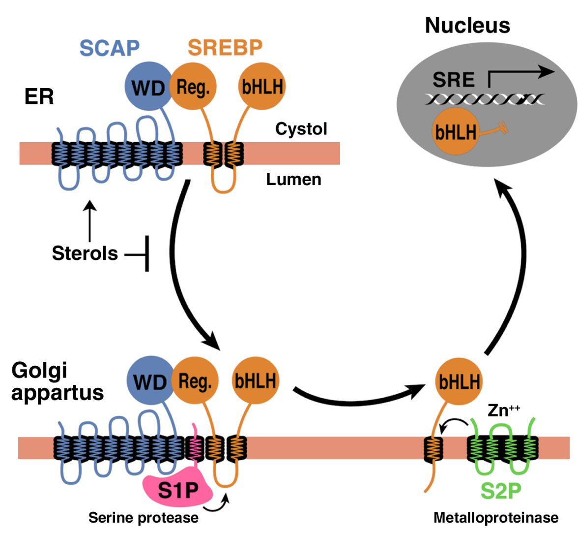
In order to reach the nucleus and act as a transcription factor, the NH2-terminal domain of each SREBP must be released from the membrane proteolytically (Figure 1). Three proteins required for SREBP processing have been delineated in cultured cells, using the tools of somatic cell genetics (see ref. 5for review). One is an escort protein designated SREBP cleavage–activating protein (SCAP). The other two are proteases, designated Site-1 protease (S1P) and Site-2 protease (S2P). Newly synthesized SREBP is inserted into the membranes of the ER, where its COOH-terminal regulatory domain binds to the COOH-terminal domain of SCAP (Figure 1).


Figure 1

Model for the sterol-mediated proteolytic release of SREBPs from membranes JCI0215593.f1

Model for the sterol-mediated proteolytic release of SREBPs from membranes JCI0215593.f1


Model for the sterol-mediated proteolytic release of SREBPs from membranes. SCAP is a sensor of sterols and an escort of SREBPs. When cells are depleted of sterols, SCAP transports SREBPs from the ER to the Golgi apparatus, where two proteases, Site-1 protease (S1P) and Site-2 protease (S2P), act sequentially to release the NH2-terminal bHLH-Zip domain from the membrane. The bHLH-Zip domain enters the nucleus and binds to a sterol response element (SRE) in the enhancer/promoter region of target genes, activating their transcription. When cellular cholesterol rises, the SCAP/SREBP complex is no longer incorporated into ER transport vesicles, SREBPs no longer reach the Golgi apparatus, and the bHLH-Zip domain cannot be released from the membrane. As a result, transcription of all target genes declines. Reprinted from ref. 5 with permission.

http://dm5migu4zj3pb.cloudfront.net/manuscripts/15000/15593/large/JCI0215593.f1.jpg

SCAP is both an escort for SREBPs and a sensor of sterols. When cells become depleted in cholesterol, SCAP escorts the SREBP from the ER to the Golgi apparatus, where the two proteases reside. In the Golgi apparatus, S1P, a membrane-bound serine protease, cleaves the SREBP in the luminal loop between its two membrane-spanning segments, dividing the SREBP molecule in half (Figure 1). The NH2-terminal bHLH-Zip domain is then released from the membrane via a second cleavage mediated by S2P, a membrane-bound zinc metalloproteinase. The NH2-terminal domain, designated nuclear SREBP (nSREBP), translocates to the nucleus, where it activates transcription by binding to nonpalindromic sterol response elements (SREs) in the promoter/enhancer regions of multiple target genes.


Figure 1


When the cholesterol content of cells rises, SCAP senses the excess cholesterol through its membranous sterol-sensing domain, changing its conformation in such a way that the SCAP/SREBP complex is no longer incorporated into ER transport vesicles. The net result is that SREBPs lose their access to S1P and S2P in the Golgi apparatus, so their bHLH-Zip domains cannot be released from the ER membrane, and the transcription of target genes ceases (1, 5). The biophysical mechanism by which SCAP senses sterol levels in the ER membrane and regulates its movement to the Golgi apparatus is not yet understood. Elucidating this mechanism will be fundamental to understanding the molecular basis of cholesterol feedback inhibition of gene expression.

SREBPs: two genes, three proteins

The mammalian genome encodes three SREBP isoforms, designated SREBP-1a, SREBP-1c, and SREBP-2. SREBP-2 is encoded by a gene on human chromosome 22q13. Both SREBP-1a and -1c are derived from a single gene on human chromosome 17p11.2 through the use of alternative transcription start sites that produce alternate forms of exon 1, designated 1a and 1c (1). SREBP-1a is a potent activator of all SREBP-responsive genes, including those that mediate the synthesis of cholesterol, fatty acids, and triglycerides. High-level transcriptional activation is dependent on exon 1a, which encodes a longer acidic transactivation segment than does the first exon of SREBP-1c. The roles of SREBP-1c and SREBP-2 are more restricted than that of SREBP-1a. SREBP-1c preferentially enhances transcription of genes required for fatty acid synthesis but not cholesterol synthesis. Like SREBP-1a, SREBP-2 has a long transcriptional activation domain, but it preferentially activates cholesterol synthesis (1). SREBP-1a and SREBP-2 are the predominant isoforms of SREBP in most cultured cell lines, whereas SREBP-1c and SREBP-2 predominate in the liver and most other intact tissues (6).

When expressed at higher than physiologic levels, each of the three SREBP isoforms can activate all enzymes indicated in Figure 2, which shows the biosynthetic pathways used to generate cholesterol and fatty acids. However, at normal levels of expression, SREBP-1c favors the fatty acid biosynthetic pathway and SREBP-2 favors cholesterologenesis. SREBP-2–responsive genes in the cholesterol biosynthetic pathway include those for the enzymes HMG-CoA synthase, HMG-CoA reductase, farnesyl diphosphate synthase, and squalene synthase. SREBP-1c–responsive genes include those for ATP citrate lyase (which produces acetyl-CoA) and acetyl-CoA carboxylase and fatty acid synthase (which together produce palmitate [C16:0]). Other SREBP-1c target genes encode a rate-limiting enzyme of the fatty acid elongase complex, which converts palmitate to stearate (C18:0) (ref.7); stearoyl-CoA desaturase, which converts stearate to oleate (C18:1); and glycerol-3-phosphate acyltransferase, the first committed enzyme in triglyceride and phospholipid synthesis (3). Finally, SREBP-1c and SREBP-2 activate three genes required to generate NADPH, which is consumed at multiple stages in these lipid biosynthetic pathways (8) (Figure 2).


Figure 2


major metabolic intermediates in the pathways for synthesis of cholesterol, fatty acids, and triglycerides JCI0215593.f2

major metabolic intermediates in the pathways for synthesis of cholesterol, fatty acids, and triglycerides JCI0215593.f2


http://dm5migu4zj3pb.cloudfront.net/manuscripts/15000/15593/large/JCI0215593.f2.jpg


Genes regulated by SREBPs. The diagram shows the major metabolic intermediates in the pathways for synthesis of cholesterol, fatty acids, and triglycerides. In vivo, SREBP-2 preferentially activates genes of cholesterol metabolism, whereas SREBP-1c preferentially activates genes of fatty acid and triglyceride metabolism. DHCR, 7-dehydrocholesterol reductase; FPP, farnesyl diphosphate; GPP, geranylgeranyl pyrophosphate synthase; CYP51, lanosterol 14α-demethylase; G6PD, glucose-6-phosphate dehydrogenase; PGDH, 6-phosphogluconate dehydrogenase; GPAT, glycerol-3-phosphate acyltransferase.

Genes regulated by SREBPs. The diagram shows the major metabolic intermediates in the pathways for synthesis of cholesterol, fatty acids, and triglycerides. In vivo, SREBP-2 preferentially activates genes of cholesterol metabolism, whereas SREBP-1c preferentially activates genes of fatty acid and triglyceride metabolism. DHCR, 7-dehydrocholesterol reductase; FPP, farnesyl diphosphate; GPP, geranylgeranyl pyrophosphate synthase; CYP51, lanosterol 14α-demethylase; G6PD, glucose-6-phosphate dehydrogenase; PGDH, 6-phosphogluconate dehydrogenase; GPAT, glycerol-3-phosphate acyltransferase.

Knockout and transgenic mice

Ten different genetically manipulated mouse models that either lack or overexpress a single component of the SREBP pathway have been generated in the last 6 years (916). The key molecular and metabolic alterations observed in these mice are summarized in Table 1.


Table 1
Alterations in hepatic lipid metabolism in gene-manipulated mice overexpressing or lacking SREBPs

http://dm5migu4zj3pb.cloudfront.net/manuscripts/15000/15593/small/JCI0215593.t1.gif

Knockout mice that lack all nSREBPs die early in embryonic development. For instance, a germline deletion of S1p, which prevents the processing of all SREBP isoforms, results in death before day 4 of development (15, 17). Germline deletion of Srebp2 leads to 100% lethality at a later stage of embryonic development than does deletion of S1p (embryonic day 7–8). In contrast, germline deletion of Srebp1, which eliminates both the 1a and the 1c transcripts, leads to partial lethality, in that about 15–45% of Srebp1–/– mice survive (13). The surviving homozygotes manifest elevated levels of SREBP-2 mRNA and protein (Table 1), which presumably compensates for the loss of SREBP-1a and -1c. When the SREBP-1c transcript is selectively eliminated, no embryonic lethality is observed, suggesting that the partial embryonic lethality in the Srebp1–/– mice is due to the loss of the SREBP-1a transcript (16).

To bypass embryonic lethality, we have produced mice in which all SREBP function can be disrupted in adulthood through induction of Cre recombinase. For this purpose, loxP recombination sites were inserted into genomic regions that flank crucial exons in the Scap or S1p genes (so-called floxed alleles) (14, 15). Mice homozygous for the floxed gene and heterozygous for a Cre recombinase transgene, which is under control of an IFN-inducible promoter (MX1-Cre), can be induced to delete Scap or S1p by stimulating IFN expression. Thus, following injection with polyinosinic acid–polycytidylic acid, a double-stranded RNA that provokes antiviral responses, the Cre recombinase is produced in liver and disrupts the floxed gene by recombination between the loxP sites.

Cre-mediated disruption of Scap or S1p dramatically reduces nSREBP-1 and nSREBP-2 levels in liver and diminishes expression of all SREBP target genes in both the cholesterol and the fatty acid synthetic pathways (Table 1). As a result, the rates of synthesis of cholesterol and fatty acids fall by 70–80% in Scap- and S1p-deficient livers.

In cultured cells, the processing of SREBP is inhibited by sterols, and the sensor for this inhibition is SCAP (5). To learn whether SCAP performs the same function in liver, we have produced transgenic mice that express a mutant SCAP with a single amino acid substitution in the sterol-sensing domain (D443N) (12). Studies in tissue culture show that SCAP(D443N) is resistant to inhibition by sterols. Cells that express a single copy of this mutant gene overproduce cholesterol (18). Transgenic mice that express this mutant version of SCAP in the liver exhibit a similar phenotype (12). These livers manifest elevated levels of nSREBP-1 and nSREBP-2, owing to constitutive SREBP processing, which is not suppressed when the animals are fed a cholesterol-rich diet. nSREBP-1 and -2 increase the expression of all SREBP target genes shown in Figure 2, thus stimulating cholesterol and fatty acid synthesis and causing a marked accumulation of hepatic cholesterol and triglycerides (Table 1). This transgenic model provides strong in vivo evidence that SCAP activity is normally under partial inhibition by endogenous sterols, which keeps the synthesis of cholesterol and fatty acids in a partially repressed state in the liver.

http://dm5migu4zj3pb.cloudfront.net/manuscripts/15000/15593/small/JCI0215593.t1.gif

Function of individual SREBP isoforms in vivo

To study the functions of individual SREBPs in the liver, we have produced transgenic mice that overexpress truncated versions of SREBPs (nSREBPs) that terminate prior to the membrane attachment domain. These nSREBPs enter the nucleus directly, bypassing the sterol-regulated cleavage step. By studying each nSREBP isoform separately, we could determine their distinct activating properties, albeit when overexpressed at nonphysiologic levels.

Overexpression of nSREBP-1c in the liver of transgenic mice produces a triglyceride-enriched fatty liver with no increase in cholesterol (10). mRNAs for fatty acid synthetic enzymes and rates of fatty acid synthesis are elevated fourfold in this tissue, whereas the mRNAs for cholesterol synthetic enzymes and the rate of cholesterol synthesis are not increased (8). Conversely, overexpression of nSREBP-2 in the liver increases the mRNAs only fourfold. This increase in cholesterol synthesis is even more remarkable when encoding all cholesterol biosynthetic enzymes; the most dramatic is a 75-fold increase in HMG-CoA reductase mRNA (11). mRNAs for fatty acid synthesis enzymes are increased to a lesser extent, consistent with the in vivo observation that the rate of cholesterol synthesis increases 28-fold in these transgenic nSREBP-2 livers, while fatty acid synthesis increases one considers the extent of cholesterol overload in this tissue, which would ordinarily reduce SREBP processing and essentially abolish cholesterol synthesis (Table 1).

http://dm5migu4zj3pb.cloudfront.net/manuscripts/15000/15593/small/JCI0215593.t1.gif

We have also studied the consequences of overexpressing SREBP-1a, which is expressed only at low levels in the livers of adult mice, rats, hamsters, and humans (6). nSREBP-1a transgenic mice develop a massive fatty liver engorged with both cholesterol and triglycerides (9), with heightened expression of genes controlling cholesterol biosynthesis and, still more dramatically, fatty acid synthesis (Table 1). The preferential activation of fatty acid synthesis (26-fold increase) relative to cholesterol synthesis (fivefold increase) explains the greater accumulation of triglycerides in their livers. The relative representation of the various fatty acids accumulating in this tissue is also unusual. Transgenic nSREBP-1a livers contain about 65% oleate (C18:1), markedly higher levels than the 15–20% found in typical wild-type livers (8) — a result of the induction of fatty acid elongase and stearoyl-CoA desaturase-1 (7). Considered together, the overexpression studies indicate that both SREBP-1 isoforms show a relative preference for activating fatty acid synthesis, whereas SREBP-2 favors cholesterol.

The phenotype of animals lacking the Srebp1 gene, which encodes both the SREBP-1a and -1c transcripts, also supports the notion of distinct hepatic functions for SREBP-1 and SREBP-2 (13). Most homozygous SREBP-1 knockout mice die in utero. The surviving Srebp1–/– mice show reduced synthesis of fatty acids, owing to reduced expression of mRNAs for fatty acid synthetic enzymes (Table 1). Hepatic nSREBP-2 levels increase in these mice, presumably in compensation for the loss of nSREBP-1. As a result, transcription of cholesterol biosynthetic genes increases, producing a threefold increase in hepatic cholesterol synthesis (Table 1).

http://dm5migu4zj3pb.cloudfront.net/manuscripts/15000/15593/small/JCI0215593.t1.gif

The studies in genetically manipulated mice clearly show that, as in cultured cells, SCAP and S1P are required for normal SREBP processing in the liver. SCAP, acting through its sterol-sensing domain, mediates feedback regulation of cholesterol synthesis. The SREBPs play related but distinct roles: SREBP-1c, the predominant SREBP-1 isoform in adult liver, preferentially activates genes required for fatty acid synthesis, while SREBP-2 preferentially activates the LDL receptor gene and various genes required for cholesterol synthesis. SREBP-1a and SREBP-2, but not SREBP-1c, are required for normal embryogenesis.

Transcriptional regulation of SREBP genes

Regulation of SREBPs occurs at two levels — transcriptional and posttranscriptional. The posttranscriptional regulation discussed above involves the sterol-mediated suppression of SREBP cleavage, which results from sterol-mediated suppression of the movement of the SCAP/SREBP complex from the ER to the Golgi apparatus (Figure 1). This form of regulation is manifest not only in cultured cells (1), but also in the livers of rodents fed cholesterol-enriched diets (19).

http://dm5migu4zj3pb.cloudfront.net/manuscripts/15000/15593/small/JCI0215593.f1.gif

The transcriptional regulation of the SREBPs is more complex. SREBP-1c and SREBP-2 are subject to distinct forms of transcriptional regulation, whereas SREBP-1a appears to be constitutively expressed at low levels in liver and most other tissues of adult animals (6). One mechanism of regulation shared by SREBP-1c and SREBP-2 involves a feed-forward regulation mediated by SREs present in the enhancer/promoters of each gene (20, 21). Through this feed-forward loop, nSREBPs activate the transcription of their own genes. In contrast, when nSREBPs decline, as in Scap or S1p knockout mice, there is a secondary decline in the mRNAs encoding SREBP-1c and SREBP-2 (14, 15).

Three factors selectively regulate the transcription of SREBP-1c: liver X-activated receptors (LXRs), insulin, and glucagon. LXRα and LXRβ, nuclear receptors that form heterodimers with retinoid X receptors, are activated by a variety of sterols, including oxysterol intermediates that form during cholesterol biosynthesis (2224). An LXR-binding site in the SREBP-1c promoter activates SREBP-1c transcription in the presence of LXR agonists (23). The functional significance of LXR-mediated SREBP-1c regulation has been confirmed in two animal models. Mice that lack both LXRα and LXRβ express reduced levels of SREBP-1c and its lipogenic target enzymes in liver and respond relatively weakly to treatment with a synthetic LXR agonist (23). Because a similar blunted response is found in mice that lack SREBP-1c, it appears that LXR increases fatty acid synthesis largely by inducing SREBP-1c (16). LXR-mediated activation of SREBP-1c transcription provides a mechanism for the cell to induce the synthesis of oleate when sterols are in excess (23). Oleate is the preferred fatty acid for the synthesis of cholesteryl esters, which are necessary for both the transport and the storage of cholesterol.

LXR-mediated regulation of SREBP-1c appears also to be one mechanism by which unsaturated fatty acids suppress SREBP-1c transcription and thus fatty acid synthesis. Rodents fed diets enriched in polyunsaturated fatty acids manifest reduced SREBP-1c mRNA expression and low rates of lipogenesis in liver (25). In vitro, unsaturated fatty acids competitively block LXR activation of SREBP-1c expression by antagonizing the activation of LXR by its endogenous ligands (26). In addition to LXR-mediated transcriptional inhibition, polyunsaturated fatty acids lower SREBP-1c levels by accelerating degradation of its mRNA (27). These combined effects may contribute to the long-recognized ability of polyunsaturated fatty acids to lower plasma triglyceride levels.

SREBP-1c and the insulin/glucagon ratio

The liver is the organ responsible for the conversion of excess carbohydrates to fatty acids to be stored as triglycerides or burned in muscle. A classic action of insulin is to stimulate fatty acid synthesis in liver during times of carbohydrate excess. The action of insulin is opposed by glucagon, which acts by raising cAMP. Multiple lines of evidence suggest that insulin’s stimulatory effect on fatty acid synthesis is mediated by an increase in SREBP-1c. In isolated rat hepatocytes, insulin treatment increases the amount of mRNA for SREBP-1c in parallel with the mRNAs of its target genes (28, 29). The induction of the target genes can be blocked if a dominant negative form of SREBP-1c is expressed (30). Conversely, incubating primary hepatocytes with glucagon or dibutyryl cAMP decreases the mRNAs for SREBP-1c and its associated lipogenic target genes (30, 31).

In vivo, the total amount of SREBP-1c in liver and adipose tissue is reduced by fasting, which suppresses insulin and increases glucagon levels, and is elevated by refeeding (32, 33). The levels of mRNA for SREBP-1c target genes parallel the changes in SREBP-1c expression. Similarly, SREBP-1c mRNA levels fall when rats are treated with streptozotocin, which abolishes insulin secretion, and rise after insulin injection (29). Overexpression of nSREBP-1c in livers of transgenic mice prevents the reduction in lipogenic mRNAs that normally follows a fall in plasma insulin levels (32). Conversely, in livers of Scap knockout mice that lack all nSREBPs in the liver (14) or knockout mice lacking either nSREBP-1c (16) or both SREBP-1 isoforms (34), there is a marked decrease in the insulin-induced stimulation of lipogenic gene expression that normally occurs after fasting/refeeding. It should be noted that insulin and glucagon also exert a posttranslational control of fatty acid synthesis though changes in the phosphorylation and activation of acetyl-CoA carboxylase. The posttranslational regulation of fatty acid synthesis persists in transgenic mice that overexpress nSREBP-1c (10). In these mice, the rates of fatty acid synthesis, as measured by [3H]water incorporation, decline after fasting even though the levels of the lipogenic mRNAs remain high (our unpublished observations).

Taken together, the above evidence suggests that SREBP-1c mediates insulin’s lipogenic actions in liver. Recent in vitro and in vivo studies involving adenoviral gene transfer suggest that SREBP-1c may also contribute to the regulation of glucose uptake and glucose synthesis. When overexpressed in hepatocytes, nSREBP-1c induces expression of glucokinase, a key enzyme in glucose utilization. It also suppresses phosphoenolpyruvate carboxykinase, a key gluconeogenic enzyme (35, 36).

SREBPs in disease

Many individuals with obesity and insulin resistance also have fatty livers, one of the most commonly encountered liver abnormalities in the US (37). A subset of individuals with fatty liver go on to develop fibrosis, cirrhosis, and liver failure. Evidence indicates that the fatty liver of insulin resistance is caused by SREBP-1c, which is elevated in response to the high insulin levels. Thus, SREBP-1c levels are elevated in the fatty livers of obese (ob/ob) mice with insulin resistance and hyperinsulinemia caused by leptin deficiency (38, 39). Despite the presence of insulin resistance in peripheral tissues, insulin continues to activate SREBP-1c transcription and cleavage in the livers of these insulin-resistant mice. The elevated nSREBP-1c increases lipogenic gene expression, enhances fatty acid synthesis, and accelerates triglyceride accumulation (31, 39). These metabolic abnormalities are reversed with the administration of leptin, which corrects the insulin resistance and lowers the insulin levels (38).

Metformin, a biguanide drug used to treat insulin-resistant diabetes, reduces hepatic nSREBP-1 levels and dramatically lowers the lipid accumulation in livers of insulin-resistant ob/ob mice (40). Metformin stimulates AMP-activated protein kinase (AMPK), an enzyme that inhibits lipid synthesis through phosphorylation and inactivation of key lipogenic enzymes (41). In rat hepatocytes, metformin-induced activation of AMPK also leads to decreased mRNA expression of SREBP-1c and its lipogenic target genes (41), but the basis of this effect is not understood.

The incidence of coronary artery disease increases with increasing plasma LDL-cholesterol levels, which in turn are inversely proportional to the levels of hepatic LDL receptors. SREBPs stimulate LDL receptor expression, but they also enhance lipid synthesis (1), so their net effect on plasma lipoprotein levels depends on a balance between opposing effects. In mice, the plasma levels of lipoproteins tend to fall when SREBPs are either overexpressed or underexpressed. In transgenic mice that overexpress nSREBPs in liver, plasma cholesterol and triglycerides are generally lower than in control mice (Table 1), even though these mice massively overproduce fatty acids, cholesterol, or both. Hepatocytes of nSREBP-1a transgenic mice overproduce VLDL, but these particles are rapidly removed through the action of LDL receptors, and they do not accumulate in the plasma. Indeed, some nascent VLDL particles are degraded even before secretion by a process that is mediated by LDL receptors (42). The high levels of nSREBP-1a in these animals support continued expression of the LDL receptor, even in cells whose cholesterol concentration is elevated. In LDL receptor–deficient mice carrying the nSREBP-1a transgene, plasma cholesterol and triglyceride levels rise tenfold (43).

Mice that lack all SREBPs in liver as a result of disruption of Scap or S1p also manifest lower plasma cholesterol and triglyceride levels (Table 1).

http://dm5migu4zj3pb.cloudfront.net/manuscripts/15000/15593/small/JCI0215593.t1.gif

In these mice, hepatic cholesterol and triglyceride synthesis is markedly reduced, and this likely causes a decrease in VLDL production and secretion. LDL receptor mRNA and LDL clearance from plasma is also significantly reduced in these mice, but the reduction in LDL clearance is less than the overall reduction in VLDL secretion, the net result being a decrease in plasma lipid levels (15). However, because

humans and mice differ substantially with regard to LDL receptor expression, LDL levels, and other aspects of lipoprotein metabolism,

it is difficult to predict whether human plasma lipids will rise or fall when the SREBP pathway is blocked or activated.

SREBPs in liver: unanswered questions

The studies of SREBPs in liver have exposed a complex regulatory system whose individual parts are coming into focus. Major unanswered questions relate to the ways in which the transcriptional and posttranscriptional controls on SREBP activity are integrated so as to permit independent regulation of cholesterol and fatty acid synthesis in specific nutritional states. A few clues regarding these integration mechanisms are discussed below.

Whereas cholesterol synthesis depends almost entirely on SREBPs, fatty acid synthesis is only partially dependent on these proteins. This has been shown most clearly in cultured nonhepatic cells such as Chinese hamster ovary cells. In the absence of SREBP processing, as when the Site-2 protease is defective, the levels of mRNAs encoding cholesterol biosynthetic enzymes and the rates of cholesterol synthesis decline nearly to undetectable levels, whereas the rate of fatty acid synthesis is reduced by only 30% (44). Under these conditions, transcription of the fatty acid biosynthetic genes must be maintained by factors other than SREBPs. In liver, the gene encoding fatty acid synthase (FASN) can be activated transcriptionally by upstream stimulatory factor, which acts in concert with SREBPs (45). The FASN promoter also contains an LXR element that permits a low-level response to LXR ligands even when SREBPs are suppressed (46). These two transcription factors may help to maintain fatty acid synthesis in liver when nSREBP-1c is low.

Another mechanism of differential regulation is seen in the ability of cholesterol to block the processing of SREBP-2, but not SREBP-1, under certain metabolic conditions. This differential regulation has been studied most thoroughly in cultured cells such as human embryonic kidney (HEK-293) cells. When these cells are incubated in the absence of fatty acids and cholesterol, the addition of sterols blocks processing of SREBP-2, but not SREBP-1, which is largely produced as SREBP-1a in these cells (47). Inhibition of SREBP-1 processing requires an unsaturated fatty acid, such as oleate or arachidonate, in addition to sterols (47). In the absence of fatty acids and in the presence of sterols, SCAP may be able to carry SREBP-1 proteins, but not SREBP-2, to the Golgi apparatus. Further studies are necessary to document this apparent independent regulation of SREBP-1 and SREBP-2 processing and to determine its mechanism.


Acknowledgments

Support for the research cited from the authors’ laboratories was provided by grants from the NIH (HL-20948), the Moss Heart Foundation, the Keck Foundation, and the Perot Family Foundation. J.D. Horton is a Pew Scholar in the Biomedical Sciences and is the recipient of an Established Investigator Grant from the American Heart Association and a Research Scholar Award from the American Digestive Health Industry.

References

  1. Brown, MS, Goldstein, JL. The SREBP pathway: regulation of cholesterol metabolism by proteolysis of a membrane-bound transcription factor. Cell 1997. 89:331-340.

View this article via: PubMed

  1. Horton, JD, Shimomura, I. Sterol regulatory element-binding proteins: activators of cholesterol and fatty acid biosynthesis. Curr Opin Lipidol 1999. 10:143-150.

View this article via: PubMed

  1. Edwards, PA, Tabor, D, Kast, HR, Venkateswaran, A. Regulation of gene expression by SREBP and SCAP. Biochim Biophys Acta 2000. 1529:103-113.

View this article via: PubMed

  1. Sakakura, Y, et al. Sterol regulatory element-binding proteins induce an entire pathway of cholesterol synthesis. Biochem Biophys Res Commun 2001. 286:176-183.

View this article via: PubMed

  1. Goldstein, JL, Rawson, RB, Brown, MS. Mutant mammalian cells as tools to delineate the sterol regulatory element-binding protein pathway for feedback regulation of lipid synthesis. Arch Biochem Biophys 2002. 397:139-148.

View this article via: PubMed

  1. Shimomura, I, Shimano, H, Horton, JD, Goldstein, JL, Brown, MS. Differential expression of exons 1a and 1c in mRNAs for sterol regulatory element binding protein-1 in human and mouse organs and cultured cells. J Clin Invest 1997. 99:838-845.

View this article via: JCI.org PubMed

  1. Moon, Y-A, Shah, NA, Mohapatra, S, Warrington, JA, Horton, JD. Identification of a mammalian long chain fatty acyl elongase regulated by sterol regulatory element-binding proteins. J Biol Chem 2001. 276:45358-45366.

View this article via: PubMed

  1. Shimomura, I, Shimano, H, Korn, BS, Bashmakov, Y, Horton, JD. Nuclear sterol regulatory element binding proteins activate genes responsible for entire program of unsaturated fatty acid biosynthesis in transgenic mouse liver. J Biol Chem 1998. 273:35299-35306.

View this article via: PubMed

  1. Shimano, H, et al. Overproduction of cholesterol and fatty acids causes massive liver enlargement in transgenic mice expressing truncated SREBP-1a. J Clin Invest 1996. 98:1575-1584.

View this article via: JCI.org PubMed

  1. Shimano, H, et al. Isoform 1c of sterol regulatory element binding protein is less active than isoform 1a in livers of transgenic mice and in cultured cells. J Clin Invest 1997. 99:846-854.

View this article via: JCI.org PubMed

  1. Horton, JD, et al. Activation of cholesterol synthesis in preference to fatty acid synthesis in liver and adipose tissue of transgenic mice overproducing sterol regulatory element-binding protein-2. J Clin Invest 1998. 101:2331-2339.

View this article via: JCI.org PubMed

  1. Korn, BS, et al. Blunted feedback suppression of SREBP processing by dietary cholesterol in transgenic mice expressing sterol-resistant SCAP(D443N). J Clin Invest 1998. 102:2050-2060.

View this article via: JCI.org PubMed

  1. Shimano, H, et al. Elevated levels of SREBP-2 and cholesterol synthesis in livers of mice homozygous for a targeted disruption of the SREBP-1 gene. J Clin Invest 1997. 100:2115-2124.

View this article via: JCI.org PubMed

  1. Matsuda, M, et al. SREBP cleavage-activating protein (SCAP) is required for increased lipid synthesis in liver induced by cholesterol deprivation and insulin elevation. Genes Dev 2001. 15:1206-1216.

View this article via: PubMed

  1. Yang, J, et al. Decreased lipid synthesis in livers of mice with disrupted Site-1 protease gene. Proc Natl Acad Sci USA 2001. 98:13607-13612.

View this article via: PubMed

Liang, G, et al. Diminished hepatic response to fasting/refeeding and liver X receptor agonists in mice with selective deficiency of sterol regulatory element-binding protein-1c. J Biol Chem 2002. 277:9520-9528.

http://www.jci.org/articles/view/15593


Structural Biochemistry/Lipids/Membrane Lipids

< Structural Biochemistry‎ | Lipids

Membrane proteins rely on their interaction with membrane lipids to uphold its structure and maintain its functions as a protein. For membrane proteins to purify and crystallize, it is essential for the membrane protein to be in the appropriate lipid environment. Lipids assist in crystallization and stabilize the protein and provide lattice contacts. Lipids can also help obtain membrane protein structures in a native conformation. Membrane protein structures contain bound lipid molecules. Biological membranes are important in life, providing permeable barriers for cells and their organelles. The interaction between membrane proteins and lipids facilitates basic processes such as respiration, photosynthesis, transport, signal transduction and motility. These basic processes require a diverse group of proteins, which are encoded by 20-30% of an organism’s annotated genes.

There exist a great number of membrane lipids. Specifically, eukaryotic cells have a very complex collection of lipids that rely on many of the cell’s resources for its synthesis. Interactions between proteins and lipids can be very specific. Specific types of lipids can make a structure stable, provide control in insertion and folding processes, and help to assemble multisubunit complexes or supercomplexes, and most importantly, can significantly affect a membrane protein’s functions. Protein and lipid interactions are not sufficiently tight, meaning that lipids are retained during membrane protein purification. Since cellular membranes are fluid arrangements of lipids, some lipids affect interesting changes to membrane due to their characteristics. Glycosphigolipids and cholesterol tend to form small islands within the membranes, called lipid rafts, due to their physical properties. Some proteins also tend to cluster in lipid raft, while others avoid being in lipid rafts. However, the existence of lipid rafts in cells seems to be transitory.

Recent progress in determining membrane protein structure has brought attention to the importance of maintaining a favorable lipid environment so proteins to crystallize and purify successfully. Lipids assist in crystallization by stabilizing the protein fold and the relationships between subunits or monomers. The lipid content in protein-lipid detergent complexes can be altered by adjusting solubilisation and purification protocols, also by adding native or non-native lipids.

There are three type of membrane lipids: 1. Phospholipids: major class of membrane lipids. 2. glycolipids. 3. Cholesterols. Membrane lipids were started with eukaryotes and bacteria.

http://en.wikibooks.org/wiki/Structural_Biochemistry/Lipids/Membrane_Lipids

Types of Membrane Lipids

Lipids are often used as membrane constituents. The three major classes that membrane lipids are divided into are phospholipids, glycolipids, and cholesterol. Lipids are found in eukaryotes and bacteria. Although the lipids in archaea have many features that are related to the membrane formation that is similar with lipids of other organisms, they are still distinct from one another. The membranes of archaea differ in composition in three major ways. Firstly, the nonpolar chains are joined to a glycerol backbone by ether instead of esters, allowing for more resistance to hydrolysis. Second, the alkyl chains are not linear, but branched and make them more resistant to oxidation. The ability of archaeal lipids to resist hydrolysis and oxidation help these types of organisms to withstand the extreme conditions of high temperature, low pH, or high salt concentration. Lastly, the stereochemistry of the central glycerol is inverted. Membrane lipids have an extensive repertoire, but they possess a critical common structural theme in which they are amphipathic molecules, meaning they contain both a hydrophilic and hydrophobic moiety.

Membrane lipids are all closed bodies or boundaries separating substituent parts of the cell. The thickness of membranes is usually between 60 and 100 angstroms. These bodies are constructed from non-covalent assemblies. Their polar heads align with each other and their non-polar hydrocarbon tails align as well. The resulting stability is credited to hydrophobic interaction which proves to be quite stable due to the length of their hydrocarbon tails.


Membrane Lipids

Lipid Vesicles

Lipid vesicles, also known as liposomes, are vesicles that are essentially aqueous vesicles that are surrounded by a circular phospholipid bilayer. Like the other phospholipid structures, they have the hydrocarbon/hydrophobic tails facing inward, away from the aqueous solution, and the hydrophilic heads facing towards the aqueous solution. These vesicles are structures that form enclosed compartments of ions and solutes, and can be utilized to study the permeability of certain membranes, or to transfer these ions or solutes to certain cells found elsewhere.

Liposomes as vesicles can serve various clinical uses. Injecting liposomes containing medicine or DNA (for gene therapy) into patients is a possible method of drug delivery. The liposomes fuse with other cells’ membranes and therefore combine their contents with that of the patient’s cell. This method of drug delivery is less toxic than direct exposure because the liposomes carry the drug directly to cells without any unnecessary intermediate steps.

Because of the hydrophobic interactions among several phospholipids and glycolipids, a certain structure called the lipid bilayer or bimolecular sheet is favored. As mentioned earlier, phospholipids and glycolipids have both hydrophilic and hydrophobic moieties; thus, when several phospholipids or glycolipids come together in an aqueous solution, the hydrophobic tails interact with each other to form a hydrophobic center, while the hydrophilic heads interact with each other forming a hydrophilic coating on each side of the bilayer.

http://upload.wikimedia.org/wikibooks/en/b/ba/Liposome_final%2A.png

http://upload.wikimedia.org/wikibooks/en/f/fa/Membrane_bilayer.jpg


Liposome_

Liposome_


Membrane_bilayer

Membrane_bilayer


Evidence Report/Technology Assessment   Number 89


Effects of Omega-3 Fatty Acids on Lipids and Glycemic Control in Type II Diabetes and the Metabolic Syndrome and on Inflammatory Bowel Disease, Rheumatoid Arthritis, Renal Disease, Systemic Lupus Erythematosus, and Osteoporosis


Prepared for:

Agency for Healthcare Research and Quality

U.S. Department of Health and Human Services

540 Gaither Road

Rockville, MD 20850

http://www.ahrq.gov

Contract No. 290-02-0003


Chapter 1. Introduction

This report is one of a group of evidence reports prepared by three Agency for Healthcare Research and Quality (AHRQ)-funded Evidence-Based Practice Centers (EPCs) on the role of omega-3 fatty acids (both from food sources and from dietary supplements) in the prevention or treatment of a variety of diseases. These reports were requested and funded by the Office of Dietary Supplements, National Institutes of Health. The three EPCs – the Southern California EPC (SCEPC, based at RAND), the Tufts-New England Medical Center (NEMC) EPC, and the University of Ottawa EPC – have each produced evidence reports. To ensure consistency of approach, the three EPCs collaborated on selected methodological elements, including literature search strategies, rating of evidence, and data table design.

The aim of these reports is to summarize the current evidence on the effects of omega-3 fatty acids on prevention and treatment of cardiovascular diseases, cancer, child and maternal health, eye health, gastrointestinal/renal diseases, asthma, immune- mediated diseases, tissue/organ transplantation, mental health, and neurological diseases and conditions. In addition to informing the research community and the public on the effects of omega-3 fatty acids on various health conditions, it is anticipated that the findings of the reports will also be used to help define the agenda for future research.

This report focuses on the effects of omega-3 fatty acids on immune- mediated diseases, bone metabolism, and gastrointestinal/renal diseases. Subsequent reports from the SCEPC will focus on cancer and neurological diseases and conditions.

This chapter provides a brief review of the current state of knowledge about the metabolism, physiological functions, and sources of omega-3 fatty acids.


The Recognition of Essential Fatty Acids

Dietary fat has long been recognized as an important source of energy for mammals, but in the late 1920s, researchers demonstrated the dietary requirement for particular fatty acids, which came to be called essential fatty acids. It was not until the advent of intravenous feeding, however, that the importance of essential fatty acids was widely accepted: Clinical signs of essential fatty acid deficiency are generally observed only in patients on total parenteral nutrition who received mixtures devoid of essential fatty acids or in those with malabsorption syndromes.

These signs include dermatitis and changes in visual and neural function. Over the past 40 years, an increasing number of physiological functions, such as immunomodulation, have been attributed to the essential fatty acids and their metabolites, and this area of research remains quite active.1, 2

Fatty Acid Nomenclature

The fat found in foods consists largely of a heterogeneous mixture of triacylglycerols (triglycerides)–glycerol molecules that are each combined with three fatty acids. The fatty acids can be divided into two categories, based on chemical properties: saturated fatty acids, which are usually solid at room temperature, and unsaturated fatty acids, which are liquid at room temperature. The term “saturation” refers to a chemical structure in which each carbon atom in the fatty acyl chain is bound to (saturated with) four other atoms, these carbons are linked by single bonds, and no other atoms or molecules can attach; unsaturated fatty acids contain at least one pair of carbon atoms linked by a double bond, which allows the attachment of additional atoms to those carbons (resulting in saturation). Despite their differences in structure, all fats contain approximately the same amount of energy (37 kilojoules/gram, or 9 kilocalories/gram).

The class of unsaturated fatty acids can be further divided into monounsaturated and polyunsaturated fatty acids. Monounsaturated fatty acids (the primary constituents of olive and canola oils) contain only one double bond. Polyunsaturated fatty acids (PUFAs) (the primary constituents of corn, sunflower, flax seed and many other vegetable oils) contain more than one double bond. Fatty acids are often referred to using the number of carbon atoms in the acyl chain, followed by a colon, followed by the number of double bonds in the chain (e.g., 18:1 refers to the 18-carbon monounsaturated fatty acid, oleic acid; 18:3 refers to any 18-carbon PUFA with three double bonds).

PUFAs are further categorized on the basis of the location of their double bonds. An omega or n notation indicates the number of carbon atoms from the methyl end of the acyl chain to the first double bond. Thus, for example, in the omega-3 (n-3) family of PUFAs, the first double bond is 3 carbons from the methyl end of the molecule. The trivial names, chemical names and abbreviations for the omega-3 fatty acids are detailed in Table 1.1.  Finally, PUFAs can be categorized according to their chain length. The 18-carbon n-3 and n-6 short-chain PUFAs are precursors to the longer 20- and 22-carbon PUFAs, called long-chain PUFAs (LCPUFAs).

Fatty Acid Metabolism

Mammalian cells can introduce double bonds into all positions on the fatty acid chain except the n-3 and n-6 position. Thus, the short-chain alpha- linolenic acid (ALA, chemical abbreviation: 18:3n-3) and linoleic acid (LA, chemical abbreviation: 18:2n-6) are essential fatty acids.

No other fatty acids found in food are considered ‘essential’ for humans, because they can all be synthesized from the short chain fatty acids.

Following ingestion, ALA and LA can be converted in the liver to the long chain, more unsaturated n-3 and n-6 LCPUFAs by a complex set of synthetic pathways that share several enzymes (Figure 1). LC PUFAs retain the original sites of desaturation (including n-3 or n-6). The omega-6 fatty acid LA is converted to gamma-linolenic acid (GLA, 18:3n-6), an omega- 6 fatty acid that is a positional isomer of ALA. GLA, in turn, can be converted to the longerchain omega-6 fatty acid, arachidonic acid (AA, 20:4n-6). AA is the precursor for certain classes of an important family of hormone- like substances called the eicosanoids (see below).

The omega-3 fatty acid ALA (18:3n-3) can be converted to the long-chain omega-3 fatty acid, eicosapentaenoic acid (EPA; 20:5n-3). EPA can be elongated to docosapentaenoic acid (DPA 22:5n-3), which is further desaturated to docosahexaenoic acid (DHA; 22:6n-3). EPA and DHA are also precursors of several classes of eicosanoids and are known to play several other critical roles, some of which are discussed further below.

The conversion from parent fatty acids into the LC PUFAs – EPA, DHA, and AA – appears to occur slowly in humans. In addition, the regulation of conversion is not well understood, although it is known that ALA and LA compete for entry into the metabolic pathways.

Physiological Functions of EPA and AA

As stated earlier, fatty acids play a variety of physiological roles. The specific biological functions of a fatty acid are determined by the number and position of double bonds and the length of the acyl chain.

Both EPA (20:5n-3) and AA (20:4n-6) are precursors for the formation of a family of hormone- like agents called eicosanoids. Eicosanoids are rudimentary hormones or regulating – molecules that appear to occur in most forms of life. However, unlike endocrine hormones, which travel in the blood stream to exert their effects at distant sites, the eicosanoids are autocrine or paracrine factors, which exert their effects locally – in the cells that synthesize them or adjacent cells. Processes affected include the movement of calcium and other substances into and out of cells, relaxation and contraction of muscles, inhibition and promotion of clotting, regulation of secretions including digestive juices and hormones, and control of fertility, cell division, and growth.3

The eicosanoid family includes subgroups of substances known as prostaglandins, leukotrienes, and thromboxanes, among others. As shown in Figure 1.1, the long-chain omega-6 fatty acid, AA (20:4n-6), is the precursor of a group of eicosanoids that include series-2 prostaglandins and series-4 leukotrienes. The omega-3 fatty acid, EPA (20:5n-3), is the precursor to a group of eicosanoids that includes series-3 prostaglandins and series-5 leukotrienes. The AA-derived series-2 prostaglandins and series-4 leukotrienes are often synthesized in response to some emergency such as injury or stress, whereas the EPA-derived series-3 prostaglandins and series-5 leukotrienes appear to modulate the effects of the series-2 prostaglandins and series-4 leukotrienes (usually on the same target cells). More specifically, the series-3 prostaglandins are formed at a slower rate and work to attenuate the effects of excessive levels of series-2 prostaglandins. Thus, adequate production of the series-3 prostaglandins seems to protect against heart attack and stroke as well as certain inflammatory diseases like arthritis, lupus, and asthma.3.

EPA (22:6 n-3) also affects lipoprotein metabolism and decreases the production of substances – including cytokines, interleukin 1ß (IL-1ß), and tumor necrosis factor a (TNF-a) – that have pro-inflammatory effects (such as stimulation of collagenase synthesis and the expression of adhesion molecules necessary for leukocyte extravasation [movement from the circulatory system into tissues]).2 The mechanism responsible for the suppression of cytokine production by omega-3 LC PUFAs remains unknown, although suppression of omega-6-derived eicosanoid production by omega-3 fatty acids may be involved, because the omega-3 and omega-6 fatty acids compete for a common enzyme in the eicosanoid synthetic pathway, delta-6 desaturase.

DPA (22:5n-3) (the elongation product of EPA) and its metabolite DHA (22:6n-3) are frequently referred to as very long chain n-3 fatty acids (VLCFA). Along with AA, DHA is the major PUFA found in the brain and is thought to be important for brain development and function. Recent research has focused on this role and the effect of supplementing infant formula with DHA (since DHA is naturally present in breast milk but not in formula).

Dietary Sources and Requirements

Both ALA and LA are present in a variety of foods. LA is present in high concentrations in many commonly used oils, including safflower, sunflower, soy, and corn oil. ALA is present in some commonly used oils, including canola and soybean oil, and in some leafy green vegetables. Thus, the major dietary sources of ALA and LA are PUFA-rich vegetable oils. The proportion of LA to ALA as well as the proportion of those PUFAs to others varies considerably by the type of oil. With the exception of flaxseed, canola, and soybean oil, the ratio of LA to ALA in vegetable oils is at least 10 to 1. The ratios of LA to ALA for flaxseed, canola, and soy are approximately 1: 3.5, 2:1, and 8:1, respectively; however, flaxseed oil is not typically consumed in the North American diet. It is estimated that on average in the U.S., LA accounts for 89% of the total PUFAs consumed, and ALA accounts for 9%. Another estimate suggests that Americans consume 10 times more omega-6 than omega-3 fatty acids.4 Table 1.2 shows the proportion of omega 3 fatty acids for a number of foods.

Syntheis and Degradation

Source of Acetyl CoA for Fatty Acid Synthesis

Source of Acetyl CoA for Fatty Acid Synthesis

step 1

step 1

condensation reaction with malonyl ACP

ACP (acyl carrier protein)

ACP (acyl carrier protein)

synthesis requires acetyl CoA from citrate shuttle

synthesis requires acetyl CoA from citrate shuttle

conversion to fatty acyl co A in cytoplasm

conversion to fatty acyl co A in cytoplasm

ACP (acyl carrier protein)

ACP (acyl carrier protein)

FA synthesis not exactly reverse of catabolism

FA synthesis not exactly reverse of catabolism


Fatty Acid Synthase

Fatty Acid Synthase

complete FA synthesis

complete FA synthesis

Desaturation

Desaturation

Elongation and Desaturation of Fatty Acids

Elongation and Desaturation of Fatty Acids

release of FAs from adiposites

release of FAs from adiposites

Fatty acid beta oxidation and Krebs cycle produce NAD, NADH, FADH2

Fatty acid beta oxidation and Krebs cycle produce NAD, NADH, FADH2

ketone bodies

ketone bodies

metabolism of ketone bodies

metabolism of ketone bodies

Arachidonoyl-mimicking

Arachidonoyl-mimicking

Arachidonate pathways

Arachidonate pathways

arachidonic acid derivatives

arachidonic acid derivatives

major metabolic intermediates in the pathways for synthesis of cholesterol, fatty acids, and triglycerides

major metabolic intermediates in the pathways for synthesis of cholesterol, fatty acids, and triglycerides

Model for the sterol-mediated proteolytic release of SREBPs from membrane

Model for the sterol-mediated proteolytic release of SREBPs from membrane

hormone regulation

hormone regulation

 insulin receptor and and insulin receptor signaling pathway (IRS)

insulin receptor and and insulin receptor signaling pathway (IRS)

 islet brain glucose signaling

islet brain glucose signaling


Fish source

Fish source

omega FAs

omega FAs


Excessive omega 6s

Excessive omega 6s

omega 6s

omega 6s

diet and cancer

diet and cancer

Patients at risk of FA deficiency

Patients at risk of FA deficiency

PPAR role

PPAR role

PPAR role

PPAR role

Omega 6_3 pathways

Omega 6_3 pathways

n3 vs n6 PUFAs

n3 vs n6 PUFAs

triene-teraene ratio

triene-teraene ratio

arachidonic acid, leukotrienes, PG and thromboxanes

arachidonic acid, leukotrienes, PG and thromboxanes

Cox 2 and cancer

Cox 2 and cancer

Lipidomics of atherosclerotic plaques

Lipidomics of atherosclerotic plaques


Effect of TPN on EFAD

Effect of TPN on EFAD

benefits of omega 3s

benefits of omega 3s

food consumption

food consumption


Read Full Post »

Genomics, Proteomics and standards

Larry H. Bernstein, MD, FCAP, Curator

http://pharmaceuticalintelligence/7/6/2014/Genomics, Proteomics and standards

This article is a look at where the biomedical research sciences are in developing standards for development in the near term.


Let’s Not Wait for the FDA: Raising the Standards of Biomarker Development – A New Series

published by Theral Timpson on Tue, 07/01/2014 – 15:03

We talk a lot on this show about the potential of personalized medicine. Never before have we learned at such breakneck speed just how our bodies function. The pace of biological research staggers the mind and hints at a time when we will “crack the code” of the system that is homo sapiens, going from picking the low hanging fruit to a more rational approach. The high tech world has put at the fingertips of biologists just the tools to do it. There is plenty of compute, plenty of storage available to untangle, or decipher the human body. Yet still, we talk of potential.

Chat with anyone heavily involved in the life science industry–be it diagnostics or pharma– and you’ll quickly hear that we must have better biomarkers.

Next week we launch a series, Let’s Not Wait for the FDA: Raising the Standards of Biomarker Development, where we will pursue the “hotspots” that are haunting those in the field.

The National Biomarker Development Alliance (NBDA) is a non profit organization based at Arizona State University and led by the formidable Anna Barker, former deputy director of the NCI. The aim of the NBDA is to identify problem areas in biomarker development–from the biospecimen and sampling issues to experiment design to bioinformatics challenges–and raise the standards in each area. This series of interviews is based on their approach. We will purse each of these topics with a special guest.

The place to start is with samples. The majority of researchers who are working on biomarker assays don’t give much thought to the “story” of their samples. Yet the quality of their research will never exceed the quality of the samples with which they start–a very scary thought according toCarolyn Compton, a former pathologist, now professor of pathology at ASU and Johns Hopkins. Carolyn worked originally as a clinical pathologist and knows first hand the the issues around sample degradation. She left the clinic when she was recruited to the NCI with the mission of bringing more awareness to the issue of bio specimens. She joins us as our first guest in the series.

That Carolyn has straddled the world of the clinic and the world of research is key to her message. And it’s key to this series. As we see an increased push to “translate” research into clinical applications, we find that these two worlds do not work enough together.

Researchers spend a lot of time analyzing data and developing causal relationships from certain biological molecules to a disease. But how often do these researchers consider how the history of a sample might be altering their data?

“Garbage in, garbage out,” says Carolyn, who links low quality samples with the abysmal non-reproducable rate of most published research.

Two of our guests in the series have worked on the adaptive iSpy breast cancer trials. These are innovative clinical trials that have been designed to “adapt” to the specific biology of those in the trial. Using the latest advances in genetics, the iSPY trials aim to match experimental drugs with the molecular makeup of tumors most likely to respond to them. And the trials are testing multiple drugs at once.

Don Berry is known for bringing statistics to clinical trials. He designed the iSpy trials and joins us to explain how these new trials work and of the promise of the adaptive design.

Laura Esserman is the director of the breast cancer center at UCSC and has been heavily involved in the implementation of the iSpy trials. Esserman is concerned that “if we keep doing conventional clinical trials, people are going to give up on doing them.” An MBA as well as an MD, Esserman brings what she learned about innovation in the high-tech industry to treatment for breast cancer.

From there we turn to the topic of “systems biology” where we will chat with George Poste, a tour de force when it comes to considering all of the various aspects of biology. Anyone who has ever been present for one of George’s presentations has no doubt come away scratching your head wondering if we’ll ever really glimpse the whole system that is a human being. If there is one brain that has seen all the rooms and hallways of our complex system, it’s George Poste.

We’ll finish the series by interviewing David Haussler from UCSC of Genome Browser fame. Recently Haussler has worked extensively on an NCI project, The Cancer Genome Atlas, to bring together data sets and connect cancer researchers around the world. What is the promise and pitfalls David sees with the latest bioinformatics tools?

George Poste says that in the literature we have identified 150,000 biomarkers that have causal linkage to disease. Yet only 100 of these have been commercialized and are used in the clinic. Why is the number so low? We hope to come up with some answers in this series.


Why Hasn’t Clinical Genetics Taken Off? (part 2)

published by Sultan Meghji on Fri, 06/20/2014 – 14:49


In my previous post, I made the broad comment that education of the patient and front line doctors was the single largest barrier to entry for clinical genetics. Here I look at the steps in the scientific process and where the biggest opportunities lie:

The Sequencing (still)

PCR is a perfectly reasonable technology for sequencing in the research lab today, but the current configuration of technologies need to change. We need to move away from an expert level skill set and a complicated chemistry process in the lab to a disposable, consumer friendly set of technologies. I’m not convinced PCR is the right technology for that and would love to see nanopore be a serious contender, but lack of funding for a broad spectrum of both physics-only as well as physical-electrical startups have slowed the progress of these technologies. And waiting in the wings, other technologies are spinning up in research labs around the world. Price is no longer a serious problem in the space – reliable, repeatable, easy to use sequencing technologies are. The complexity of the current technology (both in terms of sample preparation and machine operation) is a big hurdle.

The Analysis (compute)

Over the last few years, quite a bit of commentary and effort has been put into making the case that the compute is a significant challenge (including more than a few comments by yours truly in that vein!). Today, it can be said with total confidence that compute is NOT a problem. Compute has been commoditized. Through excellent new software to advanced platforms and new hardware, it is a trivial exercise to do the analysis and costs tiny amounts of money ($<25 per sample on a cloud provider appears to be the going rate for a clinical exome in terms of platform & infrastructure cost). Integration with the sequencer and downstream medical middleware is the biggest opportunity.

The Analysis (value)

The bigger challenge on the analysis is the specific things being analyzed as mapped to the needs of the patient. We are still in a world where the vast majority of the sequencing work is being done in support of a specific patient with a specific disease. There isn’t even broad consensus yet in the scientific community about the basics of the pipeline (see my blog posthere for an attempt at capturing what I’m seeing in the market). A movement away from the recent trend in studying specific indications (esp. cancer) is called for. Broadening the sample population will allow us to pick simpler, clearer and easier pipelines which will then make them more adoptable. It would be a massive benefit to the world if the scientific, medical and regulatory communities would get together and start creating, in a crowdsourced manner, a small number of databases that are specifically useful to healthy people. Targeting things like nutrition, athletics, metabolism, and other normal aspects of daily life. A dataset that could, when any one person’s DNA is references, would find something useful. Including the regulators is key so that we can begin to move away from the old fashioned model of clearances that still permeate the industry.

The Regulators

Beyond the broader issues around education I referenced in my previous post, there is a massive upgrade in the regulation infrastructure that is needed. We still live in a world of fax machines, overnight shipping of paper documents and personal relationships all being more important than the quality of the science you as an innovator are bringing to bear.

Consider the recent massive growth in wearables, fitness trackers and other instrumentation local to the human body. Why must we treat clinical genetics simply as a diagnostic and not, as it should be, as a fundamental set of quantitative data about your body that you can leverage in a myriad of ways. Direct to consumer (DTC) genetics companies, most notably 23andme, have approached this problem poorly – instead of making it valuable to the average consumer, what they’ve done is attempted to straddle the line between medical and not. The Fitbit model has shown very clearly that lifestyle activities can be directly harnessed to build commercial value in scaling health related activities without becoming a regulatory issue. It’s time for genetics to do the same thing.


Development and Role of the Human Reference Sequence in Personal Genomics

Posted by @finchtalk on July 3, 2014

discovery in a digital world


A few weeks back, we published a review about the development and role of the human reference genome. A key point of the reference genome is that it is not a single sequence. Instead it is an assembly of consensus sequences that are designed to deal with variation in the human population and uncertainty in the data. The reference is a map and like a geographical maps evolves though increased understanding over time.

From the Wiley On Line site:

Abstract

Genome maps, like geographical maps, need to be interpreted carefully. Although maps are essential to exploration and navigation they cannot be completely accurate. Humans have been mapping the world for several millennia, but genomes have been mapped and explored for just a single century with the greatest advancements in making a sequence reference map of the human genome possible in the past 30 years. After the deoxyribonucleic acid (DNA) sequence of the human genome was completed in 2003, the reference sequence underwent several improvements and today provides the underlying comparative resource for a multitude genetic assays and biochemical measurements. However, the ability to simplify genetic analysis through a single comprehensive map remains an elusive goal.

Key Concepts:

  • Maps are incomplete and contain errors.
  • DNA sequence data are interpreted through biochemical experiments or comparisons to other DNA sequences.
  • A reference genome sequence is a map that provides the essential coordinate system for annotating the functional regions of the genome and comparing differences between individuals’ genomes.
  • The reference genome sequence is always product of understanding at a set point in time and continues to evolve.
  • DNA sequences evolve through duplication and mutation and, as a result, contain many repeated sequences of different sizes, which complicates data analysis.
  • DNA sequence variation happens on large and small scales with respect to the lengths of the DNA differences to include single base changes, insertions, deletions, duplications and rearrangements.
  • DNA sequences within the human population undergo continual change and vary highly between individuals.
  • The current reference genome sequence is a collection of sequences, an assembly, that include sequences assembled into chromosomes, sequences that are part of structurally complex regions that cannot be assembled, patches (fixes) that cannot be included in the primary sequence, and high variability sequences that are organised into alternate loci.
  • Genetic analysis is error prone and the data require validation because the methods for collecting DNA sequences create artifacts and the reference sequence used for comparative analyses is incomplete.

Keywords:DNA sequencing


Read Full Post »

Larry H. Bernstein, MD, FCAP, Curator

http://pharmaceuticalintelligence.com/6/7/2014/Immune activation, immunity, antibacterial activity

This segment is an update on activation of innate immunity, which has had a great amount of basic science resurgence in the last several decades.  It also addresses the issue of antibiotic resistance, which shall be covered more fully in later segments. Antimicrobial resistance is a growing threat, and a challenge to the pharmaceutical industry.  Moreover, worldwide travel increases the possibility of transfer of strains of virus and microbiota to distant communities.


8-OH-dG: A novel immune activator.

Innate immunity against viral or pathogenic infection involves sensing of non-self-molecules, otherwise known at pathogen-associated molecular patterns (PAMPs).  This same sensing mechanism can be applied to damaged self-molecules, which are called damage-associated molecular patterns (DAMPs).  One type of molecular pattern, for both groups, is cytosolic or extracellular DNA.  However, there is not an extensive amount of research showing specifically what type of DAMP DNA molecule is best at activating this immune sensing response.  A recent study investigated the mechanism behind how oxidized DNA from UV damage activates an immune sensing response.

A group of researchers found that, compared to a variety of types of cellular damage, damage from UV irradiation created a strong immune response (type I IFN response), seen across different types of immune regulatory cells.  This was compared with freeze/thaw, physical damage and nutritional deprivation, each of which did not produce a noticeable immune response. Additionally, this immune response was seen when DNA was exposed to UV-A and UV-B (the type of radiation produced by our sun) and UV-C radiation.

DNA can be damaged by UV light directly, or through reactive oxygen species (ROS) caused by UV light.  A well-known mark of DNA damaged by ROS is the oxidation of guanine to create 8-hydroxyguanine (8-OH-dG).  These researchers saw an increase in 8-OH-dG dependant on the level of UV dose, and this also correlated with an increase in immune response; showing that DNA damage created by UV light in the form of 8-OH-dG is sufficient to activate an immune response. This study shows that 8-OH-dG can be classified at a DAMP.

Next, this group wanted to place a mechanism to these observations. They found that the ability of oxidation-damaged DNA to activate an immune response was dependant on cGAS and STING.  Free DNA in the cytosol binds cGAS, a cGAMP synthase.  This action produces a messenger molecule which proceeds to bind to and activate STING, an endoplasmic reticulum protein.  STING activation will ultimately stimulate a type I IFN response.

When a cell’s own DNA is damaged, the cell’s machinery does all it can to repair it.  This sometimes involves erasing, or degrading, the DNA that has been damaged.  The enzyme, TREX1 exonuclease, has this job in a cell.  However, this group found that when DNA was modified with an 8-OH-dG, it was resistant to this degradation by TREX1.  This implies that the observed increase in immune response due to the presence of 8-OH-dG occurred because of an accumulation of damaged DNA, because it was not being degraded by TREX1 and could therefore sufficiently activate cGAS and STING.

This type of study has important implications for autoimmune diseases like lupus erythematosus (LE), which is characterized by its abnormally high number of autoantibodies against DNA.  It is possible that this uncontrollable immune response is activated by oxidation-damaged DNA.  Studies in this area, therefore, hold great importance.

– See more at: http://www.stressmarq.com/Blog/November-2013/8-OH-dG-A-novel-immune-activator.aspx#sthash.CvSdK0H1.dpuf


Oxidative Damage of DNA Confers Resistance to Cytosolic Nuclease TREX1 Degradation and Potentiates STING-Dependent Immune Sensing

Nadine Gehrke, Christina Mertens, Thomas Zillinger, Jörg Wenzel,…,Winfried Barchet

DOI: http://dx.doi.org/10.1016/j.immuni.2013.08.004

Highlights

  • •UV or ROS damage potentiates immunorecognition of DNA via cGAS and STING
  • •The oxidation product 8-OHG in DNA is sufficient for enhanced immunorecognition
  • •Oxidized self-DNA acts as a DAMP and induces skin lesions in lupus-prone mice
  • •Oxidized DNA is resistant to cytosolic nuclease TREX1-mediated degradation

Summary

Immune sensing of DNA is critical for antiviral immunity but can also trigger autoimmune diseases such as lupus erythematosus (LE). Here we have provided evidence for the involvement of a damage-associated DNA modification in the detection of cytosolic DNA. The oxidized base 8-hydroxyguanosine (8-OHG), a marker of oxidative damage in DNA, potentiated cytosolic immune recognition by decreasing its susceptibility to 3′ repair exonuclease 1 (TREX1)-mediated degradation. Oxidizative modifications arose physiologically in pathogen DNA during lysosomal reactive oxygen species (ROS) exposure, as well as in neutrophil extracellular trap (NET) DNA during the oxidative burst. 8-OHG was also abundant in UV-exposed skin lesions of LE patients and colocalized with type I interferon (IFN). Injection of oxidized DNA in the skin of lupus-prone mice induced lesions that closely matched respective lesions in patients. Thus, oxidized DNA represents a prototypic damage-associated molecular pattern (DAMP) with important implications for infection, sterile inflammation, and autoimmunity.

ribonuclease TREX1 and immunity

ribonuclease TREX1 and immunity


Immunity 19 Sep 2013;39(3), p482–495,

New Weapon in Fight Against ‘Superbugs’

Some harmful bacteria are increasingly resistant to treatment with antibiotics. A discovery might be able to help the antibiotics treat the disease.

By  ANN LUKITS  June 30, 2014 8:47 p.m. ET


Some harmful bacteria are increasingly resistant to treatment with antibiotics. This common fungus found in soil might be able to help the antibiotics combat diseases. Corbis

A soil sample from a national park in eastern Canada has produced a compound that appears to reverse antibiotic resistance in dangerous bacteria.

fungus with antimicrobial activity


Scientists at McMaster University in Ontario discovered that the compound almost instantly turned off a gene in several harmful bacteria that makes them highly resistant to treatment with a class of antibiotics used to fight so-called superbug infections. The compound, called aspergillomarasmine A, or AMA, was extracted from a common fungus found in soil and mold.

Antibiotic resistance is a growing public-health threat. Common germs such asEscherichia coli, or E. coli, are becoming harder to treat because they increasingly don’t respond to antibiotics. Some two million people in the U.S. are infected each year by antibiotic-resistant bacteria and 23,000 die as a result, according to the Centers for Disease Control and Prevention. The World Health Organization has called antibiotic resistance a threat to global public health.

The Canadian team was able to disarm a gene—New Delhi Metallo-beta-Lactamase-1, or NDM-1—that has become “public enemy No. 1” since its discovery in 2009, says Gerard Wright, director of McMaster’s Michael G. DeGroote Institute for Infectious Disease Research and lead researcher on the study. The report appears on the cover of this week’s issue of the journal Nature.

“Discovery of a fungus capable of rendering these multidrug-resistant organisms incapable of further infection is huge,” says Irena Kenneley, a microbiologist and infectious disease specialist at Frances Payne Bolton School of Nursing at Cleveland’s Case Western Reserve University. “The availability of more treatment options will ultimately save many more lives,” says Dr. Kenneley, who wasn’t involved in the McMaster research.

The McMaster team plans further experiments to determine the safety and effective dosage of AMA. It could take as long as a decade to complete clinical trials on people with superbug infections, Dr. Wright says.

The researchers found that AMA, extracted from a strain of Aspergillus versicolor and combined with a carbapenem antibiotic, inactivated the NDM-1 gene in three drug-resistant superbugs—Enterobacteriaceae, a group of bacteria that includes E. coli;Acenitobacter, which can cause pneumonia and blood infections; and Pseudomonas, which often infect patients in hospitals and nursing homes. The NDM-1 gene encodes an enzyme that helps bacteria become resistant to antibiotics and that requires zinc to survive. AMA works by removing zinc from the enzyme, freeing the antibiotic to do its job, Dr. Wright says. Although AMA was only tested on carbapenem-resistant bacteria, he expects the compound would have a similar effect when combined with other antibiotics.

AMA was first identified in the 1960s in connection with leaf wilt in plants and later investigated as a potential drug for treating high blood pressure. The compound turned up in Dr. Wright’s lab a few years ago during a random screening of organisms derived from 10,000 soil samples stored at McMaster. The sample that produced AMA was collected by one of Dr. Wright’s graduate students during a visit to a Nova Scotia park. It was the only sample of 500 tested that inhibited NDM-1 in cell cultures.

“It was a lucky hit,” says Dr. Wright. “It tells us that going back to those environmental organisms, where we got antibiotics in the first place, is a really good idea.”

The McMaster team developed a purified form of AMA for experiments on mice injected with a lethal form of drug-resistant pneumonia. Treatment with either AMA or a carbapenem antibiotic alone proved ineffective. But combining the substances resulted in more than 95% of the mice still being alive after five days. The combination was also tested on 229 cell cultures from human patients infected with resistant superbugs. The treatment resensitized 88% of the samples to carbapenem.

Still, bacteria could someday find a way to outwit AMA. “I can’t imagine anything we could make where resistance would never be an issue,” he says. “At the end of the day, this is evolution and you can’t fight evolution.”

Read Full Post »

USPTO Guidance On Patentable Subject Matter

USPTO Guidance On Patentable Subject Matter

Curator and Reporter: Larry H Bernstein, MD, FCAP

LH Bernstein

LH Bernstein


Revised 4 July, 2014

http://pharmaceuticalintelligence.com/2014/07/03/uspto-guidance-on-patentable-subject-matter


I came across a few recent articles on the subject of US Patent Office guidance on patentability as well as on Supreme Court ruling on claims. I filed several patents on clinical laboratory methods early in my career upon the recommendation of my brother-in-law, now deceased.  Years later, after both brother-in-law and patent attorney are no longer alive, I look back and ask what I have learned over $100,000 later, with many trips to the USPTO, opportunities not taken, and a one year provisional patent behind me.

My conclusion is

(1) that patents are for the protection of the innovator, who might realize legal protection, but the cost and the time investment can well exceed the cost of startup and building a small startup enterprize, that would be the next step.

(2) The other thing to consider is the capability of the lawyer or firm that represents you.  A patent that is well done can be expected to take 5-7 years to go through with due diligence.   I would not expect it to be done well by a university with many other competing demands. I might be wrong in this respect, as the climate has changed, and research universities have sprouted engines for change.  Experienced and productive faculty are encouraged or allowed to form their own such entities.

(3) The emergence of Big Data, computational biology, and very large data warehouses for data use and integration has changed the landscape. The resources required for an individual to pursue research along these lines is quite beyond an individuals sole capacity to successfully pursue without outside funding.  In addition, the changed designated requirement of first to publish has muddied the water.

Of course, one can propose without anything published in the public domain. That makes it possible for corporate entities to file thousands of patents, whether there is actual validation or not at the time of filing.  It would be a quite trying experience for anyone to pursue in the USPTO without some litigation over ownership of patent rights. At this stage of of technology development, I have come to realize that the organization of research, peer review, and archiving of data is still at a stage where some of the best systems avalailable for storing and accessing data still comes considerably short of what is needed for the most complex tasks, even though improvements have come at an exponential pace.

I shall not comment on the contested views held by physicists, chemists, biologists, and economists over the completeness of guiding theories strongly held.  Only history will tell.  Beliefs can hold a strong sway, and have many times held us back.

I am not an expert on legal matters, but it is incomprehensible to me that issues concerning technology innovation can be adjudicated in the Supreme Court, as has occurred in recent years. I have postgraduate degrees in  Medicine, Developmental Anatomy, and post-medical training in pathology and laboratory medicine, as well as experience in analytical and research biochemistry.  It is beyond the competencies expected for these type of cases to come before the Supreme Court, or even to the Federal District Courts, as we see with increasing frequency,  as this has occurred with respect to the development and application of the human genome.

I’m not sure that the developments can be resolved for the public good without a more full development of an open-access system of publishing. Now I present some recent publication about, or published by the USPTO.

DR ANTHONY MELVIN CRASTO

Dr. Melvin Castro - Organic Chemistry and New Drug Development

Dr. Melvin Castro – Organic Chemistry and New Drug Development


YOU ARE FOLLOWING THIS BLOG You are following this blog, along with 1,014 other amazing people (manage).

patentimages.storage.goog…

USPTO Guidance On Patentable Subject Matter: Impediment to Biotech Innovation

Joanna T. Brougher, David A. Fazzolare J Commercial Biotechnology 2014 20(3):Brougher

jcbiotech-patents

jcbiotech-patents


Abstract In June 2013, the U.S. Supreme Court issued a unanimous decision upending more than three decades worth of established patent practice when it ruled that isolated gene sequences are no longer patentable subject matter under 35 U.S.C. Section 101.While many practitioners in the field believed that the USPTO would interpret the decision narrowly, the USPTO actually expanded the scope of the decision when it issued its guidelines for determining whether an invention satisfies Section 101.

The guidelines were met with intense backlash with many arguing that they unnecessarily expanded the scope of the Supreme Court cases in a way that could unduly restrict the scope of patentable subject matter, weaken the U.S. patent system, and create a disincentive to innovation. By undermining patentable subject matter in this way, the guidelines may end up harming not only the companies that patent medical innovations, but also the patients who need medical care.  This article examines the guidelines and their impact on various technologies.

Keywords:   patent, patentable subject matter, Myriad, Mayo, USPTO guidelines

Full Text: PDF

References

35 U.S.C. Section 101 states “Whoever invents or discovers any new and useful process, machine, manufacture, or composition of matter, or any new and useful improvement thereof, may obtain a patent therefor, subject to the conditions and requirements of this title.

” Prometheus Laboratories, Inc. v. Mayo Collaborative Services, 566 U.S. ___ (2012)

Association for Molecular Pathology et al., v. Myriad Genetics, Inc., 569 U.S. ___ (2013).

Parke-Davis & Co. v. H.K. Mulford Co., 189 F. 95, 103 (C.C.S.D.N.Y. 1911)

USPTO. Guidance For Determining Subject Matter Eligibility Of Claims Reciting Or Involving Laws of Nature, Natural Phenomena, & Natural Products.

http://www.uspto.gov/patents/law/exam/myriad-mayo_guidance.pdf

Funk Brothers Seed Co. v. Kalo Inoculant Co., 333 U.S. 127, 131 (1948)

USPTO. Guidance For Determining Subject Matter Eligibility Of Claims Reciting Or Involving Laws of Nature, Natural Phenomena, & Natural Products.

http://www.uspto.gov/patents/law/exam/myriad-mayo_guidance.pdf

Courtney C. Brinckerhoff, “The New USPTO Patent Eligibility Rejections Under Section 101.” PharmaPatentsBlog, published May 6, 2014, accessed http://www.pharmapatentsblog.com/2014/05/06/the-new-patent-eligibility-rejections-section-101/

Courtney C. Brinckerhoff, “The New USPTO Patent Eligibility Rejections Under Section 101.” PharmaPatentsBlog, published May 6, 2014, accessed http://www.pharmapatentsblog.com/2014/05/06/the-new-patent-eligibility-rejections-section-101/

DOI: http://dx.doi.org/10.5912/jcb664


Science 4 July 2014; 345 (6192): pp. 14-15  DOI: http://dx.doi.org/10.1126/science.345.6192.14
  • IN DEPTH

INTELLECTUAL PROPERTY

Biotech feels a chill from changing U.S. patent rules

A 2013 Supreme Court decision that barred human gene patents is scrambling patenting policies.

PHOTO: MLADEN ANTONOV/AFP/GETTY IMAGES

A year after the U.S. Supreme Court issued a landmark ruling that human genes cannot be patented, the biotech industry is struggling to adapt to a landscape in which inventions derived from nature are increasingly hard to patent. It is also pushing back against follow-on policies proposed by the U.S. Patent and Trademark Office (USPTO) to guide examiners deciding whether an invention is too close to a natural product to deserve patent protection. Those policies reach far beyond what the high court intended, biotech representatives say.

“Everything we took for granted a few years ago is now changing, and it’s generating a bit of a scramble,” says patent attorney Damian Kotsis of Harness Dickey in Troy, Michigan, one of more than 15,000 people who gathered here last week for the Biotechnology Industry Organization’s (BIO’s) International Convention.

At the meeting, attorneys and executives fretted over the fate of patent applications for inventions involving naturally occurring products—including chemical compounds, antibodies, seeds, and vaccines—and traded stories of recent, unexpected rejections by USPTO. Industry leaders warned that the uncertainty could chill efforts to commercialize scientific discoveries made at universities and companies. Some plan to appeal the rejections in federal court.

USPTO officials, meanwhile, implored attendees to send them suggestions on how to clarify and improve its new policies on patenting natural products, and even announced that they were extending the deadline for public comment by a month. “Each and every one of you in this room has a moral duty … to provide written comments to the PTO,” patent lawyer and former USPTO Deputy Director Teresa Stanek Rea told one audience.

At the heart of the shake-up are two Supreme Court decisions: the ruling last year in Association for Molecular Pathology v. Myriad Genetics Inc. that human genes cannot be patented because they occur naturally (Science, 21 June 2013, p. 1387); and the 2012 Mayo v. Prometheus decision, which invalidated a patent on a method of measuring blood metabolites to determine drug doses because it relied on a “law of nature” (Science, 12 July 2013, p. 137).

Myriad and Mayo are already having a noticeable impact on patent decisions, according to a study released here. It examined about 1000 patent applications that included claims linked to natural products or laws of nature that USPTO reviewed between April 2011 and March 2014. Overall, examiners rejected about 40%; Myriad was the basis for rejecting about 23% of the applications, and Mayo about 35%, with some overlap, the authors concluded. That rejection rate would have been in the single digits just 5 years ago, asserted Hans Sauer, BIO’s intellectual property counsel, at a press conference. (There are no historical numbers for comparison.) The study was conducted by the news service Bloomberg BNA and the law firm Robins, Kaplan, Miller & Ciseri in Minneapolis, Minnesota.

USPTO is extending the decisions far beyond diagnostics and DNA?

The numbers suggest USPTO is extending the decisions far beyond diagnostics and DNA, attorneys say. Harness Dickey’s Kotsis, for example, says a client recently tried to patent a plant extract with therapeutic properties; it was different from anything in nature, Kotsis argued, because the inventor had altered the relative concentrations of key compounds to enhance its effect. Nope, decided USPTO, too close to nature.

In March, USPTO released draft guidance designed to help its examiners decide such questions, setting out 12 factors for them to weigh. For example, if an examiner deems a product “markedly different in structure” from anything in nature, that counts in its favor. But if it has a “high level of generality,” it gets dinged.

The draft has drawn extensive criticism. “I don’t think I’ve ever seen anything as complicated as this,” says Kevin Bastian, a patent attorney at Kilpatrick Townsend & Stockton in San Francisco, California. “I just can’t believe that this will be the standard.”

USPTO officials appear eager to fine-tune the draft guidance, but patent experts fear the Supreme Court decisions have made it hard to draw clear lines. “The Myriad decision is hopelessly contradictory and completely incoherent,” says Dan Burk, a law professor at the University of California, Irvine. “We know you can’t patent genetic sequences,” he adds, but “we don’t really know why.”

Get creative in using Draft Guidelines!

For now, Kostis says, applicants will have to get creative to reduce the chance of rejection. Rather than claim protection for a plant extract itself, for instance, an inventor could instead patent the steps for using it to treat patients. Other biotech attorneys may try to narrow their patent claims. But there’s a downside to that strategy, they note: Narrower patents can be harder to protect from infringement, making them less attractive to investors. Others plan to wait out the storm, predicting USPTO will ultimately rethink its guidance and ease the way for new patents.


Public comment period extended

USPTO has extended the deadline for public comment to 31 July, with no schedule for issuing final language. Regardless of the outcome, however, Stanek Rea warned a crowd of riled-up attorneys that, in the world of biopatents, “the easy days are gone.”


United States Patent and Trademark Office

Today we published and made electronically available a new edition of the Manual of Patent Examining Procedure (MPEP). Manual of Patent Examining Procedure uspto.gov http://www.uspto.gov/web/offices/pac/mpep/index.html Summary of Changes

PDF Title Page
PDF Foreword
PDF Introduction
PDF Table of Contents
PDF Chapter 600 –
PDF   Parts, Form, and Content of Application Chapter 700 –
PDF    Examination of Applications Chapter 800 –
PDF   Restriction in Applications Filed Under 35 U.S.C. 111; Double Patenting Chapter 900 –
PDF   Prior Art, Classification, and Search Chapter 1000 –
PDF  Matters Decided by Various U.S. Patent and Trademark Office Officials Chapter 1100 –
PDF   Statutory Invention Registration (SIR); Pre-Grant Publication (PGPub) and Preissuance Submissions Chapter 1200 –
PDF    Appeal Chapter 1300 –
PDF   Allowance and Issue Appendix L –
PDF   Patent Laws Appendix R –
PDF   Patent Rules Appendix P –
PDF   Paris Convention Subject Matter Index 
PDF Zipped version of the MPEP current revision in the PDF format.

Manual of Patent Examining Procedure (MPEP)Ninth Edition, March 2014

The USPTO continues to offer an online discussion tool for commenting on selected chapters of the Manual. To participate in the discussion and to contribute your ideas go to:
http://uspto-mpep.ideascale.com.

Manual of Patent Examining Procedure (MPEP) Ninth Edition, March 2014
The USPTO continues to offer an online discussion tool for commenting on selected chapters of the Manual. To participate in the discussion and to contribute your ideas go to: http://uspto-mpep.ideascale.com.

Note: For current fees, refer to the Current USPTO Fee Schedule.
Consolidated Laws – The patent laws in effect as of May 15, 2014. Consolidated Rules – The patent rules in effect as of May 15, 2014.  MPEP Archives (1948 – 2012)
Current MPEP: Searchable MPEP

The documents updated in the Ninth Edition of the MPEP, dated March 2014, include changes that became effective in November 2013 or earlier.
All of the documents have been updated for the Ninth Edition except Chapters 800, 900, 1000, 1300, 1700, 1800, 1900, 2000, 2300, 2400, 2500, and Appendix P.
More information about the changes and updates is available from the “Blue Page – Introduction” of the Searchable MPEP or from the “Summary of Changes” link to the HTML and PDF versions provided below. Discuss the Manual of Patent Examining Procedure (MPEP) Welcome to the MPEP discussion tool!

We have received many thoughtful ideas on Chapters 100-600 and 1800 of the MPEP as well as on how to improve the discussion site. Each and every idea submitted by you, the participants in this conversation, has been carefully reviewed by the Office, and many of these ideas have been implemented in the August 2012 revision of the MPEP and many will be implemented in future revisions of the MPEP. The August 2012 revision is the first version provided to the public in a web based searchable format. The new search tool is available at http://mpep.uspto.gov. We would like to thank everyone for participating in the discussion of the MPEP.

We have some great news! Chapters 1300, 1500, 1600 and 2400 of the MPEP are now available for discussion. Please submit any ideas and comments you may have on these chapters. Also, don’t forget to vote on ideas and comments submitted by other users. As before, our editorial staff will periodically be posting proposed new material for you to respond to, and in some cases will post responses to some of the submitted ideas and comments.Recently, we have received several comments concerning the Leahy-Smith America Invents Act (AIA). Please note that comments regarding the implementation of the AIA should be submitted to the USPTO via email t aia_implementation@uspto.gov or via postal mail, as indicated at the America Invents Act Web site. Additional information regarding the AIA is available at www.uspto.gov/americainventsact  We have also received several comments suggesting policy changes which have been routed to the appropriate offices for consideration. We really appreciate your thinking and recommendations!

FDA Guidance for Industry:Electronic Source Data in Clinical Investigations

Electronic Source Data

Electronic Source Data


The FDA published its new Guidance for Industry (GfI) – “Electronic Source Data in Clinical Investigations” in September 2013.
The Guidance defines the expectations of the FDA concerning electronic source data generated in the context of clinical trials. Find out more about this Guidance.
http://www.gmp-compliance.org/enews_4288_FDA%20Guidance%20for%20Industry%3A%20Electronic%20Source%20Data%20in%20Clinical%20Investigations
_8534,8457,8366,8308,Z-COVM_n.html

After more than 5 years and two draft versions, the final version of the Guidance for
Industry (GfI) – “Electronic Source Data in Clinical Investigations” was published in
September 2013. This new FDA Guidance defines the FDA’s expectations for sponsors,
CROs, investigators and other persons involved in the capture, review and retention of
electronic source data generated in the context of FDA-regulated clinical trials.In an
effort to encourage the modernization and increased efficiency of processes in clinical
trials, the FDA clearly supports the capture of electronic source data and emphasizes
the agency’s intention to support activities aimed at ensuring the reliability, quality,
integrity and traceability of this source data, from its electronic source to the electronic
submission of the data in the context of an authorization procedure. The Guidance
addresses aspects as data capture, data review and record retention. When the
computerized systems used in clinical trials are described, the FDA recommends
that the description not only focus on the intended use of the system, but also on
data protection measures and the flow of data across system components and
interfaces. In practice, the pharmaceutical industry needs to meet significant
requirements regarding organisation, planning, specification and verification of
computerized systems in the field of clinical trials. The FDA also mentions in the
Guidance that it does not intend to apply 21 CFR Part 11 to electronic health records
(EHR). Author: Oliver Herrmann Q-Infiity Source: http://www.fda.gov/downloads/Drugs/GuidanceComplianceRegulatoryInformation/
Guidances/UCM328691.pdf
Webinar: https://collaboration.fda.gov/p89r92dh8wc


Read Full Post »

« Newer Posts - Older Posts »