Healthcare analytics, AI solutions for biological big data, providing an AI platform for the biotech, life sciences, medical and pharmaceutical industries, as well as for related technological approaches, i.e., curation and text analysis with machine learning and other activities related to AI applications to these industries.
Summary of Translational Medicine – e-Series A: Cardiovascular Diseases, Volume Four – Part 1
Author and Curator: Larry H Bernstein, MD, FCAP
and
Curator: Aviva Lev-Ari, PhD, RN
Article ID #135: Summary of Translational Medicine – e-Series A: Cardiovascular Diseases, Volume Four – Part 1. Published on 4/28/2014
WordCloud Image Produced by Adam Tubman
Part 1 of Volume 4 in the e-series A: Cardiovascular Diseases and Translational Medicine, provides a foundation for grasping a rapidly developing surging scientific endeavor that is transcending laboratory hypothesis testing and providing guidelines to:
Target genomes and multiple nucleotide sequences involved in either coding or in regulation that might have an impact on complex diseases, not necessarily genetic in nature.
Target signaling pathways that are demonstrably maladjusted, activated or suppressed in many common and complex diseases, or in their progression.
Enable a reduction in failure due to toxicities in the later stages of clinical drug trials as a result of this science-based understanding.
Enable a reduction in complications from the improvement of machanical devices that have already had an impact on the practice of interventional procedures in cardiology, cardiac surgery, and radiological imaging, as well as improving laboratory diagnostics at the molecular level.
Enable the discovery of new drugs in the continuing emergence of drug resistance.
Enable the construction of critical pathways and better guidelines for patient management based on population outcomes data, that will be critically dependent on computational methods and large data-bases.
What has been presented can be essentially viewed in the following Table:
Summary Table for TM – Part 1
There are some developments that deserve additional development:
1. The importance of mitochondrial function in the activity state of the mitochondria in cellular work (combustion) is understood, and impairments of function are identified in diseases of muscle, cardiac contraction, nerve conduction, ion transport, water balance, and the cytoskeleton – beyond the disordered metabolism in cancer. A more detailed explanation of the energetics that was elucidated based on the electron transport chain might also be in order.
2. The processes that are enabling a more full application of technology to a host of problems in the environment we live in and in disease modification is growing rapidly, and will change the face of medicine and its allied health sciences.
Electron Transport and Bioenergetics
Deferred for metabolomics topic
Synthetic Biology
Introduction to Synthetic Biology and Metabolic Engineering
http://www.ibiology.org Lecturers generously donate their time to prepare these lectures. The project is funded by NSF and NIGMS, and is supported by the ASCB and HHMI.
Dr. Prather explains that synthetic biology involves applying engineering principles to biological systems to build “biological machines”.
Dr. Prather has received numerous awards both for her innovative research and for excellence in teaching. Learn more about how Kris became a scientist at
Prather 1: Synthetic Biology and Metabolic Engineering 2/6/14IntroductionLecture Overview In the first part of her lecture, Dr. Prather explains that synthetic biology involves applying engineering principles to biological systems to build “biological machines”. The key material in building these machines is synthetic DNA. Synthetic DNA can be added in different combinations to biological hosts, such as bacteria, turning them into chemical factories that can produce small molecules of choice. In Part 2, Prather describes how her lab used design principles to engineer E. coli that produce glucaric acid from glucose. Glucaric acid is not naturally produced in bacteria, so Prather and her colleagues “bioprospected” enzymes from other organisms and expressed them in E. coli to build the needed enzymatic pathway. Prather walks us through the many steps of optimizing the timing, localization and levels of enzyme expression to produce the greatest yield. Speaker Bio: Kristala Jones Prather received her S.B. degree from the Massachusetts Institute of Technology and her PhD at the University of California, Berkeley both in chemical engineering. Upon graduation, Prather joined the Merck Research Labs for 4 years before returning to academia. Prather is now an Associate Professor of Chemical Engineering at MIT and an investigator with the multi-university Synthetic Biology Engineering Reseach Center (SynBERC). Her lab designs and constructs novel synthetic pathways in microorganisms converting them into tiny factories for the production of small molecules. Dr. Prather has received numerous awards both for her innovative research and for excellence in teaching.
Calcium Cycling in Synthetic and Contractile Phasic or Tonic Vascular Smooth Muscle Cells
in INTECH
Current Basic and Pathological Approaches to
the Function of Muscle Cells and Tissues – From Molecules to HumansLarissa Lipskaia, Isabelle Limon, Regis Bobe and Roger Hajjar
Additional information is available at the end of the chapter http://dx.doi.org/10.5772/48240
1. Introduction
Calcium ions (Ca ) are present in low concentrations in the cytosol (~100 nM) and in high concentrations (in mM range) in both the extracellular medium and intracellular stores (mainly sarco/endo/plasmic reticulum, SR). This differential allows the calcium ion messenger that carries information
as diverse as contraction, metabolism, apoptosis, proliferation and/or hypertrophic growth. The mechanisms responsible for generating a Ca signal greatly differ from one cell type to another.
In the different types of vascular smooth muscle cells (VSMC), enormous variations do exist with regard to the mechanisms responsible for generating Ca signal. In each VSMC phenotype (synthetic/proliferating and contractile [1], tonic or phasic), the Ca signaling system is adapted to its particular function and is due to the specific patterns of expression and regulation of Ca.
For instance, in contractile VSMCs, the initiation of contractile events is driven by mem- brane depolarization; and the principal entry-point for extracellular Ca is the voltage-operated L-type calcium channel (LTCC). In contrast, in synthetic/proliferating VSMCs, the principal way-in for extracellular Ca is the store-operated calcium (SOC) channel.
Whatever the cell type, the calcium signal consists of limited elevations of cytosolic free calcium ions in time and space. The calcium pump, sarco/endoplasmic reticulum Ca ATPase (SERCA), has a critical role in determining the frequency of SR Ca release by upload into the sarcoplasmic
sensitivity of SR calcium channels, Ryanodin Receptor, RyR and Inositol tri-Phosphate Receptor, IP3R.
Synthetic VSMCs have a fibroblast appearance, proliferate readily, and synthesize increased levels of various extracellular matrix components, particularly fibronectin, collagen types I and III, and tropoelastin [1].
Contractile VSMCs have a muscle-like or spindle-shaped appearance and well-developed contractile apparatus resulting from the expression and intracellular accumulation of thick and thin muscle filaments [1].
Schematic representation of Calcium Cycling in Contractile and Proliferating VSMCs
Figure 1. Schematic representation of Calcium Cycling in Contractile and Proliferating VSMCs.
Left panel: schematic representation of calcium cycling in quiescent /contractile VSMCs. Contractile re-sponse is initiated by extracellular Ca influx due to activation of Receptor Operated Ca (through phosphoinositol-coupled receptor) or to activation of L-Type Calcium channels (through an increase in luminal pressure). Small increase of cytosolic due IP3 binding to IP3R (puff) or RyR activation by LTCC or ROC-dependent Ca influx leads to large SR Ca IP3R or RyR clusters (“Ca -induced Ca SR calcium pumps (both SERCA2a and SERCA2b are expressed in quiescent VSMCs), maintaining high concentration of cytosolic Ca and setting the sensitivity of RyR or IP3R for the next spike.
Contraction of VSMCs occurs during oscillatory Ca transient.
Middle panel: schematic representa tion of atherosclerotic vessel wall. Contractile VSMC are located in the media layer, synthetic VSMC are located in sub-endothelial intima.
Right panel: schematic representation of calcium cycling in quiescent /contractile VSMCs. Agonist binding to phosphoinositol-coupled receptor leads to the activation of IP3R resulting in large increase in cytosolic Ca calcium pumps (only SERCA2b, having low turnover and low affinity to Ca depletion leads to translocation of SR Ca sensor STIM1 towards PM, resulting in extracellular Ca influx though opening of Store Operated Channel (CRAC). Resulted steady state Ca transient is critical for activation of proliferation-related transcription factors ‘NFAT).
Abbreviations: PLC – phospholipase C; PM – plasma membrane; PP2B – Ca /calmodulin-activated protein phosphatase 2B (calcineurin); ROC- receptor activated channel; IP3 – inositol-1,4,5-trisphosphate, IP3R – inositol-1,4,5- trisphosphate receptor; RyR – ryanodine receptor; NFAT – nuclear factor of activated T-lymphocytes; VSMC – vascular smooth muscle cells; SERCA – sarco(endo)plasmic reticulum Ca sarcoplasmic reticulum.
Time for New DNA Synthesis and Sequencing Cost Curves
By Rob Carlson
I’ll start with the productivity plot, as this one isn’t new. For a discussion of the substantial performance increase in sequencing compared to Moore’s Law, as well as the difficulty of finding this data, please see this post. If nothing else, keep two features of the plot in mind: 1) the consistency of the pace of Moore’s Law and 2) the inconsistency and pace of sequencing productivity. Illumina appears to be the primary driver, and beneficiary, of improvements in productivity at the moment, especially if you are looking at share prices. It looks like the recently announced NextSeq and Hiseq instruments will provide substantially higher productivities (hand waving, I would say the next datum will come in another order of magnitude higher), but I think I need a bit more data before officially putting another point on the plot.
cost-of-oligo-and-gene-synthesis
Illumina’s instruments are now responsible for such a high percentage of sequencing output that the company is effectively setting prices for the entire industry. Illumina is being pushed by competition to increase performance, but this does not necessarily translate into lower prices. It doesn’t behoove Illumina to drop prices at this point, and we won’t see any substantial decrease until a serious competitor shows up and starts threatening Illumina’s market share. The absence of real competition is the primary reason sequencing prices have flattened out over the last couple of data points.
Note that the oligo prices above are for column-based synthesis, and that oligos synthesized on arrays are much less expensive. However, array synthesis comes with the usual caveat that the quality is generally lower, unless you are getting your DNA from Agilent, which probably means you are getting your dsDNA from Gen9.
Note also that the distinction between the price of oligos and the price of double-stranded sDNA is becoming less useful. Whether you are ordering from Life/Thermo or from your local academic facility, the cost of producing oligos is now, in most cases, independent of their length. That’s because the cost of capital (including rent, insurance, labor, etc) is now more significant than the cost of goods. Consequently, the price reflects the cost of capital rather than the cost of goods. Moreover, the cost of the columns, reagents, and shipping tubes is certainly more than the cost of the atoms in the sDNA you are ostensibly paying for. Once you get into longer oligos (substantially larger than 50-mers) this relationship breaks down and the sDNA is more expensive. But, at this point in time, most people aren’t going to use longer oligos to assemble genes unless they have a tricky job that doesn’t work using short oligos.
Looking forward, I suspect oligos aren’t going to get much cheaper unless someone sorts out how to either 1) replace the requisite human labor and thereby reduce the cost of capital, or 2) finally replace the phosphoramidite chemistry that the industry relies upon.
IDT’s gBlocks come at prices that are constant across quite substantial ranges in length. Moreover, part of the decrease in price for these products is embedded in the fact that you are buying smaller chunks of DNA that you then must assemble and integrate into your organism of choice.
Someone who has purchased and assembled an absolutely enormous amount of sDNA over the last decade, suggested that if prices fell by another order of magnitude, he could switch completely to outsourced assembly. This is a potentially interesting “tipping point”. However, what this person really needs is sDNA integrated in a particular way into a particular genome operating in a particular host. The integration and testing of the new genome in the host organism is where most of the cost is. Given the wide variety of emerging applications, and the growing array of hosts/chassis, it isn’t clear that any given technology or firm will be able to provide arbitrary synthetic sequences incorporated into arbitrary hosts.
Dr. Jon Rowley and Dr. Uplaksh Kumar, Co-Founders of RoosterBio, Inc., a newly formed biotech startup located in Frederick, are paving the way for even more innovation in the rapidly growing fields of Synthetic Biology and Regenerative Medicine. Synthetic Biology combines engineering principles with basic science to build biological products, including regenerative medicines and cellular therapies. Regenerative medicine is a broad definition for innovative medical therapies that will enable the body to repair, replace, restore and regenerate damaged or diseased cells, tissues and organs. Regenerative therapies that are in clinical trials today may enable repair of damaged heart muscle following heart attack, replacement of skin for burn victims, restoration of movement after spinal cord injury, regeneration of pancreatic tissue for insulin production in diabetics and provide new treatments for Parkinson’s and Alzheimer’s diseases, to name just a few applications.
While the potential of the field is promising, the pace of development has been slow. One main reason for this is that the living cells required for these therapies are cost-prohibitive and not supplied at volumes that support many research and product development efforts. RoosterBio will manufacture large quantities of standardized primary cells at high quality and low cost, which will quicken the pace of scientific discovery and translation to the clinic. “Our goal is to accelerate the development of products that incorporate living cells by providing abundant, affordable and high quality materials to researchers that are developing and commercializing these regenerative technologies” says Dr. Rowley
NHMU Lecture featuring – J. Craig Venter, Ph.D. Founder, Chairman, and CEO – J. Craig Venter Institute; Co-Founder and CEO, Synthetic Genomics Inc.
J. Craig Venter, Ph.D., is Founder, Chairman, and CEO of the J. Craig Venter Institute (JVCI), a not-for-profit, research organization dedicated to human, microbial, plant, synthetic and environmental research. He is also Co-Founder and CEO of Synthetic Genomics Inc. (SGI), a privately-held company dedicated to commercializing genomic-driven solutions to address global needs.
In 1998, Dr. Venter founded Celera Genomics to sequence the human genome using new tools and techniques he and his team developed. This research culminated with the February 2001 publication of the human genome in the journal, Science. Dr. Venter and his team at JVCI continue to blaze new trails in genomics. They have sequenced and a created a bacterial cell constructed with synthetic DNA, putting humankind at the threshold of a new phase of biological research. Whereas, we could previously read the genetic code (sequencing genomes), we can now write the genetic code for designing new species.
The science of synthetic genomics will have a profound impact on society, including new methods for chemical and energy production, human health and medical advances, clean water, and new food and nutritional products. One of the most prolific scientists of the 21st century for his numerous pioneering advances in genomics, he guides us through this emerging field, detailing its origins, current challenges, and the potential positive advances.
His work on synthetic biology truly embodies the theme of “pushing the boundaries of life.” Essentially, Venter is seeking to “write the software of life” to create microbes designed by humans rather than only through evolution. The potential benefits and risks of this new technology are enormous. It also requires us to examine, both scientifically and philosophically, the question of “What is life?”
J Craig Venter wants to digitize DNA and transmit the signal to teleport organisms
A technology trend analyst offers an overview of synthetic biology, its potential applications, obstacles to its development, and prospects for public approval.
In addition to boosting the economy, synthetic biology projects currently in development could have profound implications for the future of manufacturing, sustainability, and medicine.
Before society can fully reap the benefits of synthetic biology, however, the field requires development and faces a series of hurdles in the process. Do researchers have the scientific know-how and technical capabilities to develop the field?
Biology + Engineering = Synthetic Biology
Bioengineers aim to build synthetic biological systems using compatible standardized parts that behave predictably. Bioengineers synthesize DNA parts—oligonucleotides composed of 50–100 base pairs—which make specialized components that ultimately make a biological system. As biology becomes a true engineering discipline, bioengineers will create genomes using mass-produced modular units similar to the microelectronics and computer industries.
Currently, bioengineering projects cost millions of dollars and take years to develop products. For synthetic biology to become a Schumpeterian revolution, smaller companies will need to be able to afford to use bioengineering concepts for industrial applications. This will require standardized and automated processes.
A major challenge to developing synthetic biology is the complexity of biological systems. When bioengineers assemble synthetic parts, they must prevent cross talk between signals in other biological pathways. Until researchers better understand these undesired interactions that nature has already worked out, applications such as gene therapy will have unwanted side effects. Scientists do not fully understand the effects of environmental and developmental interaction on gene expression. Currently, bioengineers must repeatedly use trial and error to create predictable systems.
Similar to physics, synthetic biology requires the ability to model systems and quantify relationships between variables in biological systems at the molecular level.
The second major challenge to ensuring the success of synthetic biology is the development of enabling technologies. With genomes having billions of nucleotides, this requires fast, powerful, and cost-efficient computers. Moore’s law, named for Intel co-founder Gordon Moore, posits that computing power progresses at a predictable rate and that the number of components in integrated circuits doubles each year until its limits are reached. Since Moore’s prediction, computer power has increased at an exponential rate while pricing has declined.
DNA sequencers and synthesizers are necessary to identify genes and make synthetic DNA sequences. Bioengineer Robert Carlson calculated that the capabilities of DNA sequencers and synthesizers have followed a pattern similar to computing. This pattern, referred to as the Carlson Curve, projects that scientists are approaching the ability to sequence a human genome for $1,000, perhaps in 2020. Carlson calculated that the costs of reading and writing new genes and genomes are falling by a factor of two every 18–24 months. (see recent Carlson comment on requirement to read and write for a variety of limiting conditions).
Startup to Strengthen Synthetic Biology and Regenerative Medicine Industries with Cutting Edge Cell Products
Synthetic Biology: On Advanced Genome Interpretation for Gene Variants and Pathways: What is the Genetic Base of Atherosclerosis and Loss of Arterial Elasticity with Aging
Futurists have touted the twenty-first century as the century of biology based primarily on the promise of genomics. Medical researchers aim to use variations within genes as biomarkers for diseases, personalized treatments, and drug responses. Currently, we are experiencing a genomics bubble, but with advances in understanding biological complexity and the development of enabling technologies, synthetic biology is reviving optimism in many fields, particularly medicine.
Michael Brooks holds a PhD in quantum physics. He writes a weekly science column for the New Statesman,and his most recent book is The Secret Anarchy of Science.
The basic idea is that we take an organism – a bacterium, say – and re-engineer its genome so that it does something different. You might, for instance, make it ingest carbon dioxide from the atmosphere, process it and excrete crude oil.
That project is still under construction, but others, such as using synthesised DNA for data storage, have already been achieved. As evolution has proved, DNA is an extraordinarily stable medium that can preserve information for millions of years. In 2012, the Harvard geneticist George Church proved its potential by taking a book he had written, encoding it in a synthesised strand of DNA, and then making DNA sequencing machines read it back to him.
When we first started achieving such things it was costly and time-consuming and demanded extraordinary resources, such as those available to the millionaire biologist Craig Venter. Venter’s team spent most of the past two decades and tens of millions of dollars creating the first artificial organism, nicknamed “Synthia”. Using computer programs and robots that process the necessary chemicals, the team rebuilt the genome of the bacterium Mycoplasma mycoides from scratch. They also inserted a few watermarks and puzzles into the DNA sequence, partly as an identifying measure for safety’s sake, but mostly as a publicity stunt.
What they didn’t do was redesign the genome to do anything interesting. When the synthetic genome was inserted into an eviscerated bacterial cell, the new organism behaved exactly the same as its natural counterpart. Nevertheless, that Synthia, as Venter put it at the press conference to announce the research in 2010, was “the first self-replicating species we’ve had on the planet whose parent is a computer” made it a standout achievement.
Today, however, we have entered another era in synthetic biology and Venter faces stiff competition. The Steve Jobs to Venter’s Bill Gates is Jef Boeke, who researches yeast genetics at New York University.
Boeke wanted to redesign the yeast genome so that he could strip out various parts to see what they did. Because it took a private company a year to complete just a small part of the task, at a cost of $50,000, he realised he should go open-source. By teaching an undergraduate course on how to build a genome and teaming up with institutions all over the world, he has assembled a skilled workforce that, tinkering together, has made a synthetic chromosome for baker’s yeast.
Stepping into DIYbio and Synthetic Biology at ScienceHack
We got a crash course on genetics and protein pathways, and then set out to design and build our own pathways using both the “Genomikon: Violacein Factory” kit and Synbiota platform. With Synbiota’s software, we dragged and dropped the enzymes to create the sequence that we were then going to build out. After a process of sketching ideas, mocking up pathways, and writing hypotheses, we were ready to start building!
The night stretched long, and at midnight we were forced to vacate the school. Not quite finished, we loaded our delicate bacteria, incubator, and boxes of gloves onto the bus and headed back to complete our bacterial transformation in one of our hotel rooms. Jammed in between the beds and the mini-fridge, we heat-shocked our bacteria in the hotel ice bucket. It was a surreal moment.
While waiting for our bacteria, we held an “unconference” where we explored bioethics, security and risk related to synthetic biology, 3D printing on Mars, patterns in juggling (with live demonstration!), and even did a Google Hangout with Rob Carlson. Every few hours, we would excitedly check in on our bacteria, looking for bacterial colonies and the purple hue characteristic of violacein.
Most impressive was the wildly successful and seamless integration of a diverse set of people: in a matter of hours, we were transformed from individual experts and practitioners in assorted fields into cohesive and passionate teams of DIY biologists and science hackers. The ability of everyone to connect and learn was a powerful experience, and over the course of just one weekend we were able to challenge each other and grow.
Returning to work on Monday, we were hungry for more. We wanted to find a way to bring the excitement and energy from the weekend into the studio and into the projects we’re working on. It struck us that there are strong parallels between design and DIYbio, and we knew there was an opportunity to bring some of the scientific approaches and curiosity into our studio.
Introduction to e-Series A: Cardiovascular Diseases, Volume Four Part 2: Regenerative Medicine
Author and Curator: Larry H Bernstein, MD, FCAP
and
Curator: Aviva Lev-Ari, PhD, RN
This document is entirely devoted to medical and surgical therapies that have made huge strides in
simplification of interventional procedures,
reduced complexity, resulting in procedures previously requiring surgery are now done, circumstances permitting, by medical intervention.
This revolution in cardiovascular interventional therapy is regenerative medicine. It is regenerative because it is largely driven by
the introduction into the impaired vasculature of an induced pleuripotent cell, called a stem cell, although
the level of differentiation may not be a most primitive cell line.
There is also a very closely aligned development in cell biology that extends beyond and including vascular regeneration that is called synthetic biology. These developments have occurred at an accelerated rate in the last 15 years. The methods of interventional cardiology were already well developed in the mid 1980s. This was at the peak of cardiothoracic bypass surgery.
Research on the endothelial cell,
endothelial cell proliferation,
shear flow in small arteries, especially at branch points, and
endothelial-platelet interactions
led to insights about plaque formation and vessel thrombosis.
Much was learned in biomechanics about the shear flow stresses on the luminal surface of the vasculature, and there was also
the concomitant discovery of nitric oxide,
oxidative stress, and
the isoenzymes of nitric oxide synthase (eNOS, iNOS, and nNOS).
It became a fundamental tenet of vascular biology that
atherogenesis is a maladjustment to oxidative stress not only through genetic, but also
non-genetic nutritional factors that could be related to the balance of omega (ω)-3 and omega (ω)-6 fatty acids,
a pro-inflammatory state that elicits inflammatory cytokines, such as, interleukin-6 (IL6) and c-reactive protein(CRP),
insulin resistance with excess carbohydrate associated with type 2 diabetes and beta (β) cell stress,
excess trans- and saturated fats, and perhaps
the now plausible colonic microbial population of the gastrointestinal tract (GIT).
There is also an association of abdominal adiposity,
including the visceral peritoneum, with both T2DM and with arteriosclerotic vessel disease,
which is presenting at a young age, and has ties to
the effects of an adipokine, adiponectin.
Much important work has already been discussed in the domain of cardiac catheterization and research done to
prevent atheroembolization.and beyond that,
research done to implant an endothelial growth matrix.
Even then, dramatic work had already been done on
the platelet structure and metabolism, and
this has transformed our knowledge of platelet biology.
The coagulation process has been discussed in detailed in a previous document. The result was the development of a
new class of platelet aggregation inhibitors designed to block the activation of protein on the platelet surface that
is critical in the coagulation cascade.
In addition, the term long used to describe atherosclerosis, atheroma notwithstanding, is “hardening of the arteries”. This is particularly notable with respect to mid-size arteries and arterioles that feed the heart and kidneys. Whether it is preceded by or develops concurrently with chronic renal insufficiency and lowered glomerular filtration rate is perhaps arguable. However, there is now a body of evidence that points to
a change in the vascular muscularis and vessel stiffness, in addition to the endothelial features already mentioned.
This has provided a basis for
targeted pharmaceutical intervention, and
reduction in salt intake.
So we have a group of metabolic disorders, which may alone or in combination,
lead to and be associated with the long term effects of cardiovascular disease, including
congestive heart failure.
This has been classically broken down into forward and backward failure,
depending on decrease outflow through the aorta (ejection fraction), or
decreased venous return through the vena cava,
which involves increased pulmonary vascular resistance and decreased return into the left atrium.
This also has ties to several causes, which may be cardiac or vascular. This document, as the previous, has four pats. They are broadly:
Stem Cells in Cardiovascular Diseases
Regenerative Cell and Molecular Biology
Therapeutics Levels In Molecular Cardiology
Research Proposals for Endogenous Augmentation of circulating Endothelial Progenitor Cells (cEPCs)
As in the previous section, we start with the biology of the stem cell and the degeneration in cardiovascular diseases, then proceed to regeneration, then therapeutics, and finally – proposals for augmenting therapy with circulating endogenous endothelial progenitor cells (cEPCs).
Introduction to Translational Medicine (TM) – Part 1: Translational Medicine
Author and Curator: Larry H Bernstein, MD, FCAP
and
Curator: Aviva Lev-Ari, PhD, RN
Article ID #134: Introduction to Translational Medicine (TM) – Part 1: Translational Medicine. Published on 4/25/2014
WordCloud Image Produced by Adam Tubman
This document in the Series A: Cardiovascular Diseases e-Series Volume 4: Translational and Regenerative Medicine, is a measure of the postgenomic and proteomic advances in the laboratory to the practice of clinical medicine. The Chapters are preceded by several videos by prominent figures in the emergence of this transformative change. When I was a medical student, a large body of the current language and technology that has extended the practice of medicine did not exist, but a new foundation, predicated on the principles of modern medical education set forth by Abraham Flexner, was sprouting. The highlights of this evolution were:
Requirement for premedical education in biology, organic chemistry, physics, and genetics.
Medical education included two years of basic science education in anatomy, physiology, pharmacology, and pathology prior to introduction into the clinical course sequence of the last two years.
Post medical graduate education was an internship year followed by residency in pediatrics, OBGyn, internal medicine, general surgery, psychiatry, neurology, neurosurgery, pathology, radiology, and anesthesiology, emergency medicine.
Academic teaching centers were developing subspecialty centers in ophthalmology, ENT and head and neck surgery, cardiology and cardiothoracic surgery, and hematology, hematology/oncology, and neurology.
The expansion of postgraduate medical programs included significant postgraduate funding for programs by the National Institutes of Health, and the NIH had faculty development support in a system of peer-reviewed research grant programs in medical and allied sciences.
The period after the late 1980s saw a rapid expansion of research in genomics and drug development to treat emerging threats of infectious diseases as US had a large worldwide involvement after the end of the Vietnam War, and drug resistance was increasingly encountered (malaria, tick borne diseases, salmonellosis, pseudomonas aeruginosa, staphylococcus aureus, etc.).
Moreover, the post-millenium found a large, dwindling population of veterans who had served in WWII and Vietnam, and cardiovascular, musculoskeletal, dementias, and cancer were now more common. The Human Genome Project was undertaken to realign the existing knowledge of gene structure and genetic regulation with the needs for drug development, which was languishing in development failures due to unexpected toxicities.
A substantial disconnect existed between diagnostics and pharmaceutical development, which had been over-reliant on modification of known organic structures to increase potency and reduce toxicity. This was about to change with changes in medical curricula, changes in residency programs and physicians cross-training in disciplines, and the emergence of bio-pharma, based on the emerging knowledge of the cell function, and at the same time, the medical profession was developing an evidence-base for therapeutics, and more pressure was placed on informed decision-making.
The great improvement in proteomics came from GCLC/MS-MS and is described in the video interview with Dr. Gyorgy Marko-Varga, Sweden, in video 1 of 3 (Advancing Translational Medicine). This is a discussion that is focused on functional proteomics role in future diagnostics and therapy, involving a greater degree of accuracy in mass spectrometry (MS) than can be obtained by antibody-ligand binding, and is illustrated below, the last emphasizing the importance of information technology and predictive analytics
Thermo ScientificImmunoassays and LC–MS/MS have emerged as the two main approaches for quantifying peptides and proteins in biological samples. ELISA kits are available for quantification, but inherently lack the discriminative power to resolve isoforms and PTMs.
To address this issue we have developed and applied a mass spectrometry immunoassay–selected reaction monitoring (Thermo Scientific™ MSIA™ SRM technology) research method to quantify PCSK9 (and PTMs), a key player in the regulation of circulating low density lipoprotein cholesterol (LDL-C).
A Day in the (Future) Life of a Predictive Analytics Scientist
A look into a normal day in the near future, where predictive analytics is everywhere, incorporated in everything from household appliances to wearable computing devices.
During the test drive (of an automobile), the extreme acceleration makes your heart beat so fast that your personal health data sensor triggers an alarm. The health data sensor is integrated into the strap of your wrist watch. This data is transferred to your health insurance company, so you say a prayer that their data scientists are clever enough to exclude these abnormal values from your otherwise impressive health data. Based on such data, your health insurance company’s consulting unit regularly gives you advice about diet, exercise, and sleep. You have followed their advice in the past, and your performance has increased, which automatically reduced your insurance premiums. Win-win, you think to yourself, as you park the car, and decide to buy it.
In the clinical presentation at Harlan Krumholtz’ Yale Symposium, Prof. Robert Califf, Director of the Duke University Translational medicine Clinical Research Institute, defines translational medicine as effective translation of science to clinical medicine in two segments:
Adherence to current standards
Improving the enterprise by translating knowledge
He says that discrepancies between outcomes and medical science will bridge a gap in translation by traversing two parallel systems.
Physician-health organization
Personalized medicine
He emphasizes that the new basis for physician standards will be legitimized in the following:
An interesting sidebar to the scientific medical advances is the huge shift in pressure on an insurance system that has coexisted with a public system in Medicare and Medicaid, initially introduced by the health insurance industry for worker benefits (Kaiser, IBM, Rockefeller), and we are undertaking a formidable change in the ACA.
The current reality is that actuarially, the twin system that has existed was unsustainable in the long term because it is necessary to have a very large pool of the population to spread the costs, and in addition, the cost of pharmaceutical development has driven consolidation in the industry, and has relied on the successes from public and privately funded research.
I shall digress for a moment and insert a video history of DNA, that hits the high points very well, and is quite explanatory of the genomic revolution in medical science, biology, infectious disease and microbial antibiotic resistance, virology, stem cell biology, and the undeniability of evolution.
As I have noted above, genomics is necessary, but not sufficient. The story began as replication of the genetic code, which accounted for variation, but the accounting for regulation of the cell and for metabolic processes was, and remains in the domain of an essential library of proteins. Moreover, the functional activity of proteins, at least but not only if they are catalytic, shows structural variants that is characterized by small differences in some amino acids that allow for separation by net charge and have an effect on protein-protein and other interactions.
Protein chemistry is so different from DNA chemistry that it is quite safe to consider that DNA in the nucleotide sequence does no more than establish the order of amino acids in proteins. On the other hand, proteins that we know so little about their function and regulation, do everything that matters including to set what and when to read something in the DNA.
Jose Eduardo de Salles Roselino
Chapters 2, 3, and 4 sequentially examine:
The causes and etiologies of cardiovascular diseases
The diagnosis, prognosis and risks determined by – biomarkers in serum, circulating cells, and solid tissue by contrast radiography
Treatment of cardiovascular diseases by translation of science from bench to bedside, including interventional cardiology and surgical repair
These are systematically examined within a framework of:
that has been presented by the cancer team of professional experts, e-Book concept was conceived by Aviva Lev-Ari, PhD, RN, e-Series Editor-in-Chief and Founder of Leaders in Pharmaceutical Business Intelligence
Stephen J. Williams, PhD, Senior Editor, and other notable contributors in various aspects of cancer research in the emerging fields of targeted pharmacology, nanotechnology, cancer imaging, molecular pathology, transcriptional and regulatory ‘OMICS’, metabolism, medical and allied health related sciences, synthetic biology, pharmaceutical discovery, and translational medicine.
This volume and its content have been conceived and organized to capture the organized events that emerge in embryological development, leading to the major organ systems that we recognize anatomically and physiologically as an integrated being. We capture the dynamic interactions between the systems under stress that are elicited by cytokine-driven hormonal responses, long thought to be circulatory and multisystem, that affect the major compartments of fat and lean body mass, and are as much the drivers of metabolic pathway changes that emerge as epigenetics, without disregarding primary genetic diseases.
The greatest difficulty in organizing such a work is in whether it is to be merely a compilation of cancer expression organized by organ systems, or whether it is to capture developing concepts of underlying stem cell expressed changes that were once referred to as “dedifferentiation”. In proceeding through the stages of neoplastic transformation, there occur adaptive local changes in cellular utilization of anabolic and catabolic pathways, and a retention or partial retention of functional specificities.
This effectively results in the same cancer types not all fitting into the same “shoe”. There is a sequential loss of identity associated with cell migration, cell-cell interactions with underlying stroma, and metastasis., but cells may still retain identifying “signatures” in microRNA combinatorial patterns. The story is still incomplete, with gaps in our knowledge that challenge the imagination.
What we have laid out is a map with substructural ordered concepts forming subsets within the structural maps. There are the traditional energy pathways with terms aerobic and anaerobic glycolysis, gluconeogenesis, triose phosphate branch chains, pentose shunt, and TCA cycle vs the Lynen cycle, the Cori cycle, glycogenolysis, lipid peroxidation, oxidative stress, autosomy and mitosomy, and genetic transcription, cell degradation and repair, muscle contraction, nerve transmission, and their involved anatomic structures (cytoskeleton, cytoplasm, mitochondria, liposomes and phagosomes, contractile apparatus, synapse.
Then there is beneath this macro-domain the order of signaling pathways that regulate these domains and through mechanisms of cellular regulatory control have pleiotropic inhibitory or activation effects, that are driven by extracellular and intracellular energy modulating conditions through three recognized structures: the mitochondrial inner membrane, the intercellular matrix, and the ion-channels.
What remains to be done?
There is still to be elucidated the differences in patterns within cancer types the distinct phenotypic and genotypic features that mitigate anaplastic behavior. One leg of this problem lies in the density of mitochondria, that varies between organ types, but might vary also within cell type of a common function. Another leg of this problem has also appeared to lie in the cell death mechanism that relates to the proeosomal activity acting on both the ribosome and mitochondrion in a coordinated manner. This is an unsolved mystery of molecular biology.
Then there is a need to elucidate the major differences between tumors of endocrine, sexual, and structural organs, which are distinguished by primarily a synthetic or primarily a catabolic function, and organs that are neither primarily one or the other. For example, tumors of the thyroid and paratnhyroids, islet cells of pancreas, adrenal cortex, and pituitary glands have the longest 5 year survivals. They and the sexual organs are in the visceral compartment. The rest of the visceral compartment would be the liver, pancreas, salivary glands, gastrointestinal tract, and lungs (which are embryologically an outpouching of the gastrointestinal tract), kidneys and lower urinary tract. Cancers of these organs have a much less favorable survival (brain, breast and prostate, lymphatic, blood forming organ, skin). The case is intermediate for breast and prostate between the endocrine organs and GI tract, based on natural history, irrespective of the available treatments. Just consider the dilemma over what we do about screening for prostate cancer in men over the age of 60 years age who have a 70 percent incident silent carcinoma of the prostate that could be associated with unrelated cause of death. The very rapid turnover of the gastric and colonic GI epithelium, and of the subepithelial B cell mucosal lymphocytic structures is associated with a greater aggressiveness of the tumor.
However, we have to reconsider the observation by NO Kaplan than the synthetic and catabolic functions are highlighted by differences in the expressions of the balance of the two major pyridine nucleotides – DPN (NAD) and TPN (NADP) – which also might be related to the density of mitochondria which is associated with both NADP and synthetic activity, and with efficient aerobic function. These are in an equilibrium through the “transhydrogenase reaction” co-discovered by Kaplan, in Fritz Lipmann’s laboratory. There does arise a conundrum involving the regulation of mitochondria in these high turnover epithelial tissues that rely on aerobic energy, and generate ATP through TPN linked activity, when they undergo carcinogenesis. The cells replicate and they become utilizers of glycolysis, while at the same time, the cell death pathway is quiescent. The result becomes the introduction of peripheral muscle and liver synthesized protein cannabolization (cancer cachexia) to provide glucose from proteolytic amino acid sources.
There is also the structural compartment of the lean body mass. This is the heart, skeletal structures (includes smooth muscle of GI tract, uterus, urinary bladder, brain, bone, bone marrow). The contractile component is associated with sarcomas. What is most striking is that the heart, skeletal muscle, and inflammatory cells are highly catabolic, not anabolic. NO Kaplan referred tp them as DPN (NAD) tissues. This compartment requires high oxygen supply, and has a high mechanical function. But again, we return to the original observations of enrgy requirements at rest being different than at high demand. At work, skeletal muscle generates lactic acid, but the heart can use lactic acid as fuel,.
The liver is supplied by both the portal vein and the hepatic artery, so it is not prone to local ischemic injury (Zahn infarct). It is exceptional in that it carries out synthesis of all the circulating transport proteins, has a major function in lipid synthesis and in glycogenesis and glycogenolysis, with the added role of drug detoxification through the P450 system. It is not only the largest organ (except for brain), but is highly active both anabolically and catabolically (by ubiquitilation).
The expected cellular turnover rates for these tissues and their balance of catabolic and anabolic function would have to be taken into account to account for the occurrence and the activities of oncogenesis. This is by no means a static picture, but a dynamic organism constantly in flux imposed by internal and external challenges. It is also important to note the the organs have a concentration of mitochondria, associated with energy synthetic and catabolic requirements provided by oxygen supply and the electron transport mechanism for oxidative phosphorylation. For example, tissues that are primarily synthetic do not have intermitent states of resting and high demand, as seen in skeletal muscle, or perhaps myocardium (which is syncytial and uses lactic acid generated from skeletal muscle when there is high demand).
The existence of lncDNA has been discovered only as a result of the human genome project (HGP). This was previously known only as “dark DNA”. It has become clear that lncDNA has an important role in cellular regulatory activities centered in the chromatin modeling. Moreover, just as proteins exhibit functionality in their folding, related to tertiary structure and highly influenced by location of –S-S- bridges and amino acid residue distances (allosteric effects), there is a less studied effect as the chromatin becomes more compressed within the nucleus, that should have a bearing on cellular expression.
According to Jose Eduardo de Salles Roselino , when the Na/Glucose transport system (for a review Silvermann, M. in Annu. Rev. Biochem.60: 757-794(1991)) was found in kidneys as well as in key absorptive cells of digestive tract, it should be stressed its functional relationship with “internal milieu” and real meaning, homeostasis. It is easy to understand how the major topic was presented as how to prevent diarrheal deaths in infants, while detected in early stages. However, from a biochemical point of view, as presented in Schrödinger´s What is life?, (biochemistry offering a molecular view for two legs of biology, physiology and genetics). Why should it be driven to the sole target of understanding genetics? Why the understanding of physiology in molecular terms should be so neglected?
From a biochemical point of view, here in a single protein. It is found the transport of the cation most directly related to water maintenance, the internal solvent that bath our cells and the hydrocarbon whose concentration is kept under homeostatic control on that solvent. Completely at variance with what is presented in microorganisms as previously mentioned in Moyed and Umbarger revision (Ann. Rev42: 444(1962)) that does not regulates the environment where they live and appears to influence it only as an incidental result of their metabolism.
In case any attempt is made in order to explain why the best leg that supports scientific reasoning from biology for medical purposes was led to atrophy, several possibilities can be raised. However, none of them could be placed strictly in scientific terms. Factors that bare little relationship with scientific progress in general terms must also be taken into account.
One simple possibility of explanation can be found in one review (G. Scatchard – Solutions of Electrolytes Ann. Rev. Physical Chemistry 14: 161-176 (1963)). A simple reading of it and the sophisticated differences among researchers will discourage one hundred per cent of biologists to keep in touch with this line of research. Biochemists may keep on reading. However, consider that first: Complexity is not amenable to reductionist vision in all cases. Second, as coupling between scalar flows such as chemical reactions and vector flows such as diffusion flows, heat flows, and electrical current can occur only in anisotropic system…let them with their problems of solvents, ions and etc. and let our biochemical reactions on another basket. At the interface, for instance, at membrane level, we will agree that ATP is converted to ADP because it is far from equilibrium and the continuous replenishment of ATP that maintain relatively constant ATP levels inside the cell and this requires some non-stationary flow.
Our major point must be to understand that our biological limits are far clearer present in our limited ability to regulate the information stored in the DNA than in the amount of information we have in the DNA as the master regulator of the cells.
The amazing revelation that Masahiro Chiga (discovery of liver adenylate kinase distinct from that of muscle) taught me (LHB) is – draw 2 circles that intersect, one of which represents what we know, the other – what we don’t know. We don’t teach how much we don’t know! Even today, as much as 40 years ago, there is a lot we need to get on top of this.
The observation is rather similar to the presentations I (Jose Eduardo de Salles Rosalino) was previously allowed to make of the conformational energy as made by R Marcus in his Nobel lecture revised (J. of Electroanalytical Chemistry 438:(1997) p251-259. His description of the energetic coordinates of a landscape of a chemical reaction is only a two-dimensional cut of what in fact is a volcano crater (in three dimensions) ( each one varie but the sum of the two is constant. Solvational+vibrational=100% in ordinate) nuclear coordinates in abcissa. In case we could represent it by research methods that allow us to discriminate in one by one degree of different pairs of energy, we would most likely have 360 other similar representations of the same phenomenon. The real representation would take into account all those 360 representation together. In case our methodology was not that fine, for instance it discriminate only differences of minimal 10 degrees in 360 possible, will have 36 partial representations of something that to be perfectly represented will require all 36 being taken together. Can you reconcile it with ATGC? Yet, when complete genome sequences were presented they were described as we will know everything about this living being. The most important problems in biology will be viewed by limited vision always and the awareness of this limited is something we should acknowledge and teach it. Therefore, our knowledge is made up of partial representations.
Even though we may have complete genome data for the most intricate biological problems, they are not so amenable to this level of reductionism. However, from general views of signals and symptoms we could get to the most detailed molecular view and in this case the genome provides an anchor. This is somehow, what Houssay was saying to me and to Leloir when he pointed out that only in very rare occasions biological phenomena could be described in three terms: Pacco, the dog and the anesthetic (previous e-mail). The non-coding region, to me will be important guiding places for protein interactions.
Introduction – The Evolution of Cancer Therapy and Cancer Research: How We Got Here?
Author and Curator: Larry H Bernstein, MD, FCAP
The evolution of progress we have achieved in cancer research, diagnosis, and therapeutics has originated from an emergence of scientific disciplines and the focus on cancer has been recent. We can imagine this from a historical perspective with respect to two observations. The first is that the oldest concepts of medicine lie with the anatomic dissection of animals and the repeated recurrence of war, pestilence, and plague throughout the middle ages, and including the renaissance. In the awakening, architecture, arts, music, math, architecture and science that accompanied the invention of printing blossomed, a unique collaboration of individuals working in disparate disciplines occurred, and those who were privileged received an education, which led to exploration, and with it, colonialism. This also led to the need to increasingly, if not without reprisal, questioning long-held church doctrines.
It was in Vienna that Rokitansky developed the discipline of pathology, and his student Semelweis identified an association between then unknown infection and childbirth fever. The extraordinary accomplishments of John Hunter in anatomy and surgery came during the twelve years war, and his student, Edward Jenner, observed the association between cowpox and smallpox resistance. The development of a nursing profession is associated with the work of Florence Nightengale during the Crimean War (at the same time as Leo Tolstoy). These events preceded the work of Pasteur, Metchnikoff, and Koch in developing a germ theory, although Semelweis had committed suicide by infecting himself with syphilis. The first decade of the Nobel Prize was dominated by discoveries in infectious disease and public health (Ronald Ross, Walter Reed) and we know that the Civil War in America saw an epidemic of Yellow Fever, and the Armed Services Medical Museum was endowed with a large repository of osteomyelitis specimens. We also recall that the Russian physician and playwriter, Anton Checkov, wrote about the conditions in prison camps.
But the pharmacopeia was about to open with the discoveries of insulin, antibiotics, vitamins, thyroid action (Mayo brothers pioneered thyroid surgery in the thyroid iodine-deficient midwest), and pitutitary and sex hormones (isolatation, crystal structure, and synthesis years later), and Karl Landsteiner’s discovery of red cell antigenic groups (but he also pioneered in discoveries in meningitis and poliomyelitis, and conceived of the term hapten) with the introduction of transfusion therapy that would lead to transplantation medicine. The next phase would be heralded by the discovery of cancer, which was highlighted by the identification of leukemia by Rudolph Virchow, who cautioned about the limitations of microscopy. This period is highlighted by the classic work – “Microbe Hunters”.
John Hunter
Walter Reed
Robert Koch
goldberger 1916 Pellagra
Louis Pasteur
A multidisciplinary approach has led us to a unique multidisciplinary or systems view of cancer, with different fields of study offering their unique expertise, contributions, and viewpoints on the etiology of cancer. Diverse fields in immunology, biology, biochemistry, toxicology, molecular biology, virology, mathematics, social activism and policy, and engineering have made such important contributions to our understanding of cancer, that without cooperation among these diverse fields our knowledge of cancer would never had evolved as it has. In a series of posts “Heroes in Medical Research:” the work of researchers are highlighted as examples of how disparate scientific disciplines converged to produce seminal discoveries which propelled the cancer field, although, at the time, they seemed like serendipitous findings. In the post Heroes in Medical Research: Barnett Rosenberg and the Discovery of Cisplatin (Translating Basic Research to the Clinic) discusses the seminal yet serendipitous discoveries by bacteriologist Dr. Barnett Rosenberg, which eventually led to the development of cisplatin, a staple of many chemotherapeutic regimens. Molecular biologist Dr. Robert Ting, working with soon-to-be Nobel Laureate virologist Dr. James Gallo on AIDS research and the associated Karposi’s sarcoma identified one of the first retroviral oncogenes, revolutionizing previous held misconceptions of the origins of cancer (described in Heroes in Medical Research: Dr. Robert Ting, Ph.D. and Retrovirus in AIDS and Cancer). Located here will be a MONTAGE of PHOTOS of PEOPLE who made seminal discoveries and contributions in every field to cancer Each of these paths of discovery in cancer research have led to the unique strategies of cancer therapeutics and detection for the purpose of reducing the burden of human cancer. However, we must recall that this work has come at great cost, while it is indeed cause for celebration. The current failure rate of clinical trials at over 70 percent, has been a cause for disappointment, and has led to serious reconsideration of how we can proceed with greater success. The result of the evolution of the cancer field is evident in the many parts and chapters of this ebook. Volume 4 contains chapters that are in a predetermined order:
The concepts of neoplasm, malignancy, carcinogenesis, and metastatic potential, which encompass:
(a) How cancer cells bathed in an oxygen rich environment rely on anaerobic glycolysis for energy, and the secondary consequences of cachexia and sarcopenia associated with progression
invasion
ARTS protein and cancer
Glycolysis
Krebs cycle
Metabolic control analysis of respiration in human cancer tissue
akip1-expression-modulates-mitochondrial-function
(b) How advances in genetic analysis, molecular and cellular biology, metabolomics have expanded our basic knowledge of the mechanisms which are involved in cellular transformation to the cancerous state.
nucleotides
Methylation of adenine
ampk-and-ampk-related-kinase-ark-family-
ubiquitylation
(c) How molecular techniques continue to advance our understanding of how genetics, epigenetics, and alterations in cellular metabolism contribute to cancer and afford new pathways for therapeutic intervention.
genomic effects
LKB1AMPK pathway
mutation-frequencies-across-12-cancer-types
AMPK-activating drugs metformin or phenformin might provide protection against cancer
2. The distinct features of cancers of specific tissue sites of origin
3. The diagnosis of cancer by
(a) Clinical presentation
(b) Age of onset and stage of life
(c) Biomarker features
hairy cell leukemia
lymphoma leukemia
(d) Radiological and ultrasound imaging
Treatments
Prognostic differences within and between cancer types
We have introduced the emergence of a disease of great complexity that has been clouded in more questions than answers until the emergence of molecular biology in the mid 20th century, and then had to await further discoveries going into the 21st century. What gave the research impetus was the revelation of
1 the mechanism of transcription of the DNA into amino acid sequences
Proteins in Disease
2 the identification of stresses imposed on cellular function
NO beneficial effects
3 the elucidation of the substructure of the cell – cell membrane, mitochondria, ribosomes, lysosomes – and their functions, respectively
AKIP1 Expression Modulates Mitochondrial Function
4 the elucidation of oligonucleotide sequences
nucleotides
dna-replication-unwinding
dna-replication-ligation
dna-replication-primer-removal
dna-replication-leading-strand
dna-replication-lagging-strand
dna-replication-primer-synthesis
dna-replication-termination
5 the further elucidation of functionally relevant noncoding lncDNA
6 the technology to synthesis mRNA and siRNA sequences
Figure. RNAi and gene silencing
7 the repeated discovery of isoforms of critical enzymes and their pleiotropic properties
8. the regulatory pathways involved in signaling
Figure. Signaling Pathways Map
This is a brief outline of the modern progression of advances in our understanding of cancer. Let us go back to the beginning and check out a sequence of Nobel Prizes awarded and related discoveries that have a historical relationship to what we know. The first discovery was the finding by Louis Pasteur that fungi that grew in an oxygen poor environment did not put down filaments. They did not utilize oxygen and they produced used energy by fermentation. This was the basis for Otto Warburg sixty years later to make the comparison to cancer cells that grew in the presence of oxygen, but relied on anaerobic glycolysis. He used a manometer to measure respiration in tissue one cell layer thick to measure CO2 production in an adiabatic system.
Lavoisier Antoine-Laurent and Laplace Pierre-Simon (1783) Memoir on heat. Mémoirs de l’Académie des sciences. Translated by Guerlac H, Neale Watson Academic Publications, New York, 1982.
The Warburg apparatus is a manometric respirometer which was used for decades in biochemistry for measuring oxygen consumption of tissue homogenates or tissue slices.
The aqueous phase is vigorously shaken to equilibrate with a gas phase, from which oxygen is consumed while the evolved carbon dioxide is trapped, such that the pressure in the constant-volume gas phase drops proportional to oxygen consumption. The Warburg apparatus was introduced to study cell respiration, i.e. the uptake of molecular oxygen and the production of carbon dioxide by cells or tissues. Its applications were extended to the study of fermentation, when gas exchange takes place in the absence of oxygen. Thus the Warburg apparatus became established as an instrument for both aerobic and anaerobic biochemical studies [2, 3].
The respiration chamber was a detachable glass flask (F) equipped with one or more sidearms (S) for additions of chemicals and an open connection to a manometer (M; pressure gauge). A constant temperature was provided by immersion of the Warburg chamber in a constant temperature water bath. At thermal mass transfer equilibrium, an initial reading is obtained on the manometer, and the volume of gas produced or absorbed is determined at specific time intervals. A limited number of ‘titrations’ can be performed by adding the liquid contained in a side arm into the main reaction chamber. A Warburg apparatus may be equipped with more than 10 respiration chambers shaking in a common water bath. Since temperature has to be controlled very precisely in a manometric approach, the early studies on mammalian tissue respiration were generally carried out at a physiological temperature of 37 °C.
The Warburg apparatus has been replaced by polarographic instruments introduced by Britton Chance in the 1950s. Since Chance and Williams (1955) measured respiration of isolated mitochondria simultaneously with the spectrophotometric determination of cytochrome redox states, a water chacket could not be used, and measurements were carried out at room temperature (or 25 °C). Thus most later studies on isolated mitochondria were shifted to the artifical temperature of 25 °C.
Today, the importance of investigating mitochondrial performance at in vivo temperatures is recognized again in mitochondrial physiology. Incubation times of 1 hour were typical in experiments with the Warburg apparatus, but were reduced to a few or up to 20 min, following Chance and Williams, due to rapid oxygen depletion in closed, aqueous phase oxygraphs with high sample concentrations. Today, incubation times of 1 hour are typical again in high-resolution respirometry, with low sample concentrations and the option of reoxygenations.
Oesper P (1964) The history of the Warburg apparatus: Some reminiscences on its use. J Chem Educ 41: 294.
Koppenol WH, Bounds PL, Dang CV (2011) Otto Warburg’s contributions to current concepts of cancer metabolism. Nature Reviews Cancer 11: 325-337.
Gnaiger E, Kemp RB (1990) Anaerobic metabolism in aerobic mammalian cells: information from the ratio of calorimetric heat flux and respirometric oxygen flux. Biochim Biophys Acta 1016: 328-332. – “At high fructose concentrations, respiration is inhibited while glycolytic end products accumulate, a phenomenon known as the Crabtree effect. It is commonly believed that this effect is restricted to microbial and tumour cells with uniquely high glycolytic capacities (Sussman et al, 1980). However, inhibition of respiration and increase of lactate production are observed under aerobic conditions in beating rat heart cell cultures (Frelin et al, 1974) and in isolated rat lung cells (Ayuso-Parrilla et al, 1978). Thus, the same general mechanisms responsible for the integration of respiration and glycolysis in tumour cells (Sussman et al, 1980) appear to be operating to some extent in several isolated mammalian cells.”
Mitochondria are sometimes described as “cellular power plants” because they generate most of the cell’s supply of adenosine triphosphate (ATP), used as a source of chemical energy.[2] In addition to supplying cellular energy, mitochondria are involved in other tasks such as signaling, cellular differentiation, cell death, as well as the control of the cell cycle and cell growth.[3] The organelle is composed of compartments that carry out specialized functions. These compartments or regions include the outer membrane, the intermembrane space, the inner membrane, and the cristae and matrix. Mitochondrial proteins vary depending on the tissue and the species. In humans, 615 distinct types of proteins have been identified from cardiac mitochondria,[9Leonor Michaelis discovered that Janus green can be used as a supravital stain for mitochondria in 1900. Benjamin F. Kingsbury, in 1912, first related them with cell respiration, but almost exclusively based on morphological observations.[13] In 1913 particles from extracts of guinea-pig liver were linked to respiration by Otto Heinrich Warburg, which he called “grana”. Warburg and Heinrich Otto Wieland, who had also postulated a similar particle mechanism, disagreed on the chemical nature of the respiration. It was not until 1925 when David Keilin discovered cytochromes that the respiratory chain was described.[13]
The Clark Oxygen Sensor
Dr. Leland Clark – inventor of the “Clark Oxygen Sensor” (1954); the Clark type polarographic oxygen sensor remains the gold standard for measuring dissolved oxygen in biomedical, environmental and industrial applications . ‘The convenience and simplicity of the polarographic ‘oxygen electrode’ technique for measuring rapid changes in the rate of oxygen utilization by cellular and subcellular systems is now leading to its more general application in many laboratories. The types and design of oxygen electrodes vary, depending on the investigator’s ingenuity and specific requirements of the system under investigation.’Estabrook R (1967) Mitochondrial respiratory control and the polarographic measurement of ADP:O ratios. Methods Enzymol. 10: 41-47. “one approach that is underutilized in whole-cell bioenergetics, and that is accessible as long as cells can be obtained in suspension, is the oxygen electrode, which can obtain more precise information on the bioenergetic status of the in situ mitochondria than more ‘high-tech’ approaches such as fluorescent monitoring ofΔψm.” Nicholls DG, Ferguson S (2002) Bioenergetics 3. Academic Press, London.
Great Figures in Cancer
Dr. Elizabeth Blackburn,
j_michael_bishop onogene
Harold Varmus
Potts and Habener (PTH mRNA, Harvard MIT) JCI
JCI Fuller Albright and hPTH AA sequence
Dr. E. Donnall Thomas Bone Marrow Transplants
Dr Haraldzur Hausen EBV HPV
Dr. Craig Mello
Lee Hartwell – Hutchinson Cancer Res Center
Judah Folkman, MD
Gertrude B. Elien (1918-1999)
The Nobel Prize in Physiology or Medicine 1922
Archibald V. Hill, Otto Meyerhof
AV Hill –
“the production of heat in the muscle” Hill started his research work in 1909. It was due to J.N. Langley, Head of the Department of Physiology at that time that Hill took up the study on the nature of muscular contraction. Langley drew his attention to the important (later to become classic) work carried out by Fletcher and Hopkins on the problem of lactic acid in muscle, particularly in relation to the effect of oxygen upon its removal in recovery. In 1919 he took up again his study of the physiology of muscle, and came into close contact with Meyerhof of Kiel who, approaching the problem differently, arrived at results closely analogous to his study. In 1919 Hill’s friend W. Hartree, mathematician and engineer, joined in the myothermic investigations – a cooperation which had rewarding results.
Otto Meyerhof –
otto-fritz-meyerhof
lactic acid production in muscle contraction Under the influence of Otto Warburg, then at Heidelberg, Meyerhof became more and more interested in cell physiology. In 1923 he was offered a Professorship of Biochemistry in the United States, but Germany was unwilling to lose him. In 1929 he was he was placed in charge of the newly founded Kaiser Wilhelm Institute for Medical Research at Heidelberg. From 1938 to 1940 he was Director of Research at the Institut de Biologie physico-chimique at Paris, but in 1940 he moved to the United States, where the post of Research Professor of Physiological Chemistry had been created for him by the University of Pennsylvania and the Rockefeller Foundation. Meyerhof’s own account states that he was occupied chiefly with oxidation mechanisms in cells and with extending methods of gas analysis through the calorimetric measurement of heat production, and especially the respiratory processes of nitrifying bacteria. The physico-chemical analogy between oxygen respiration and alcoholic fermentation caused him to study both these processes in the same subject, namely, yeast extract. By this work he discovered a co-enzyme of respiration, which could be found in all the cells and tissues up till then investigated. At the same time he also found a co-enzyme of alcoholic fermentation. He also discovered the capacity of the SH-group to transfer oxygen; after Hopkins had isolated from cells the SH bodies concerned, Meyerhof showed that the unsaturated fatty acids in the cell are oxidized with the help of the sulfhydryl group. After studying closer the respiration of muscle, Meyerhof investigated the energy changes in muscle. Considerable progress had been achieved by the English scientists Fletcher and Hopkins by their recognition of the fact that lactic acid formation in the muscle is closely connected with the contraction process. These investigations were the first to throw light upon the highly paradoxical fact, already established by the physiologist Hermann, that the muscle can perform a considerable part of its external function in the complete absence of oxygen.
But it was indisputable that in the last resort the energy for muscle activity comes from oxidation, so the connection between activity and combustion must be an indirect one, and observed that in the absence of oxygen in the muscle, lactic acid appears, slowly in the relaxed state and rapidly in the active state, disappearing in the presence of oxygen. Obviously, then, oxygen is involved when muscle is in the relaxed state. http://upload.wikimedia.org/wikipedia/commons/e/e1/Glycolysis.jpg
The Nobel Prize committee had been receiving nominations intermittently for the previous 14 years (for Eijkman, Funk, Goldberger, Grijns, Hopkins and Suzuki but, strangely, not for McCollum in this period). Tthe Committee for the 1929 awards apparently agreed that it was high time to honor the discoverer(s) of vitamins; but who were they? There was a clear case for Grijns, but he had not been re-nominated for that particular year, and it could be said that he was just taking the relatively obvious next steps along the new trail that had been laid down by Eijkman, who was also now an old man in poor health, but there was no doubt that he had taken the first steps in the use of an animal model to investigate the nutritional basis of a clinical disorder affecting millions. Goldberger had been another important contributor, but his recent death put him out of consideration. The clearest evidence for lack of an unknown “something” in a mammalian diet was presented by Gowland Hopkins in 1912. This Cambridge biochemist was already well known for having isolated the amino acid tryptophan from a protein and demonstrated its essential nature. He fed young rats on an experimental diet, half of them receiving a daily milk supplement, and only those receiving milk grew well, Hopkins suggested that this was analogous to human diseases related to diet, as he had suggested already in a lecture published in 1906. Hopkins, the leader of the “dynamic biochemistry” school in Britain and an influential advocate for the importance of vitamins, was awarded the prize jointly with Eijkman. A door was opened. Recognition of work on the fat-soluble vitamins begun by McCollum. The next award related to vitamins was given in 1934 to George Whipple, George Minot and William Murphy “for their discoveries concerning liver therapy in cases of [then incurable pernicious] anemia,” The essential liver factor (cobalamin, or vitamin B12) was isolated in 1948, and Vitamin B12 was absent from plant foods. But William Castle in 1928 had demonstrated that the stomachs of pernicious anemia patients were abnormal in failing to secrete an “intrinsic factor”.
Szent-Györgyi was a Hungarian biochemist who had worked with Otto Warburg and had a special interest in oxidation-reduction mechanisms. He was invited to Cambridge in England in 1927 after detecting an antioxidant compound in the adrenal cortex, and there, he isolated a compound that he named hexuronic acid. Charles Glen King of the University of Pittsburgh reported success In isolating the anti-scorbutic factor in 1932, and added that his crystals had all the properties reported by Szent-Györgyi for hexuronic acid. But his work on oxidation reactions was also important. Fumarate is an intermediate in the citric acid cycle used by cells to produce energy in the form of adenosine triphosphate (ATP) from food. It is formed by the oxidation of succinate by the enzyme succinate dehydrogenase. Fumarate is then converted by the enzyme fumarase to malate. An enzyme adds water to the fumarate molecule to form malate. The malate is created by adding one hydrogen atom to a carbon atom and then adding a hydroxyl group to a carbon next to a terminal carbonyl group.
In the same year, Norman Haworth from the University of Birmingham in England received a Nobel prize from the Chemistry Committee for having advanced carbohydrate chemistry and, specifically, for having worked out the structure of Szent-Györgyi’s crystals, and then been able to synthesize the vitamin. This was a considerable achievement. The Nobel Prize in Chemistry was shared with the Swiss organic chemist Paul Karrer, cited for his work on the structures of riboflavin and vitamins A and E as well as other biologically interesting compounds. This was followed in 1938 by a further Chemistry award to the German biochemist Richard Kuhn, who had also worked on carotenoids and B-vitamins, including riboflavin and pyridoxine. But Karrer was not permitted to leave Germany at that time by the Nazi regime. However, the American work with radioisotopes at Lawrence Livermore Laboratory, UC Berkeley, was already ushering in a new era of biochemistry that would enrich our studies of metabolic pathways. The importance of work involving vitamins was acknowledged in at least ten awards in the 20th century.
1. Carpenter, K.J., Beriberi, White Rice and Vitamin B, University of California Press, Berkeley (2000).
2. Weatherall, M.W. and Kamminga, H., The making of a biochemist: the construction of Frederick Gowland Hopkins’ reputation. Medical History vol.40, pp. 415-436 (1996).
3. Becker, S.L., Will milk make them grow? An episode in the discovery of the vitamins. In Chemistry and Modern Society (J. Parascandela, editor) pp. 61-83, American Chemical Society,
Washington, D.C. (1983).
4. Carpenter, K.J., The History of Scurvy and Vitamin C, Cambridge University Press, New York (1986).
Transport and metabolism of exogenous fumarate and 3-phosphoglycerate in vascular smooth muscle.
The keto (linear) form of exogenous fructose 1,6-bisphosphate, a highly charged glycolytic intermediate, may utilize a dicarboxylate transporter to cross the cell membrane, support glycolysis, and produce ATP anaerobically. We tested the hypothesis that fumarate, a dicarboxylate, and 3-phosphoglycerate (3-PG), an intermediate structurally similar to a dicarboxylate, can support contraction in vascular smooth muscle during hypoxia. 3-PG improved maintenance of force (p < 0.05) during the 30-80 min period of hypoxia. Fumarate decreased peak isometric force development by 9.5% (p = 0.008) but modestly improved maintenance of force (p < 0.05) throughout the first 80 min of hypoxia. 13C-NMR on tissue extracts and superfusates revealed 1,2,3,4-(13)C-fumarate (5 mM) metabolism to 1,2,3,4-(13)C-malate under oxygenated and hypoxic conditions suggesting uptake and metabolism of fumarate. In conclusion, exogenous fumarate and 3-PG readily enter vascular smooth muscle cells, presumably by a dicarboxylate transporter, and support energetically important pathways.
Comparison of endogenous and exogenous sources of ATP in fueling Ca2+ uptake in smooth muscle plasma membrane vesicles.
A smooth muscle plasma membrane vesicular fraction (PMV) purified for the (Ca2+/Mg2+)-ATPase has endogenous glycolytic enzyme activity. In the presence of glycolytic substrate (fructose 1,6-diphosphate) and cofactors, PMV produced ATP and lactate and supported calcium uptake. The endogenous glycolytic cascade supports calcium uptake independent of bath [ATP]. A 10-fold dilution of PMV, with the resultant 10-fold dilution of glycolytically produced bath [ATP] did not change glycolytically fueled calcium uptake (nanomoles per milligram protein). Furthermore, the calcium uptake fueled by the endogenous glycolytic cascade persisted in the presence of a hexokinase-based ATP trap which eliminated calcium uptake fueled by exogenously added ATP. Thus, it appears that the endogenous glycolytic cascade fuels calcium uptake in PMV via a membrane-associated pool of ATP and not via an exchange of ATP with the bulk solution. To determine whether ATP produced endogenously was utilized preferentially by the calcium pump, the ATP production rates of the endogenous creatine kinase and pyruvate kinase were matched to that of glycolysis and the calcium uptake fueled by the endogenous sources was compared with that fueled by exogenous ATP added at the same rate. The rate of calcium uptake fueled by endogenous sources of ATP was approximately twice that supported by exogenously added ATP, indicating that the calcium pump preferentially utilizes ATP produced by membrane-bound enzymes.
Evidence for succinate production by reduction of fumarate during hypoxia in isolated adult rat heart cells.
Archives of Biochemistry and Biophysics (Impact Factor: 3.37). 01/1988; 259(2):527-35. http://dx.doi.org:/10.1016/0003-9861(87)90519-4 It has been demonstrated that perfusion of myocardium with glutamic acid or tricarboxylic acid cycle intermediates during hypoxia or ischemia, improves cardiac function, increases ATP levels, and stimulates succinate production. In this study isolated adult rat heart cells were used to investigate the mechanism of anaerobic succinate formation and examine beneficial effects attributed to ATP generated by this pathway. Myocytes incubated for 60 min under hypoxic conditions showed a slight loss of ATP from an initial value of 21 +/- 1 nmol/mg protein, a decline of CP from 42 to 17 nmol/mg protein and a fourfold increase in lactic acid production to 1.8 +/- 0.2 mumol/mg protein/h. These metabolite contents were not altered by the addition of malate and 2-oxoglutarate to the incubation medium nor were differences in cell viability observed; however, succinate release was substantially accelerated to 241 +/- 53 nmol/mg protein. Incubation of cells with [U-14C]malate or [2-U-14C]oxoglutarate indicates that succinate is formed directly from malate but not from 2-oxoglutarate. Moreover, anaerobic succinate formation was rotenone sensitive.
We conclude that malate reduction to succinate occurs via the reverse action of succinate dehydrogenase in a coupled reaction where NADH is oxidized (and FAD reduced) and ADP is phosphorylated.Furthermore, by transaminating with aspartate to produce oxaloacetate, 2-oxoglutarate stimulates cytosolic malic dehydrogenase activity, whereby malate is formed and NADH is oxidized.
In the form of malate, reducing equivalents and substrate are transported into the mitochondria where they are utilized for succinate synthesis.
1953 Hans Adolf Krebs –
” discovery of the citric acid cycle” and In the course of the 1920’s and 1930’s great progress was made in the study of the intermediary reactions by which sugar is anaerobically fermented to lactic acid or to ethanol and carbon dioxide. The success was mainly due to the joint efforts of the schools of Meyerhof, Embden, Parnas, von Euler, Warburg and the Coris, who built on the pioneer work of Harden and of Neuberg. This work brought to light the main intermediary steps of anaerobic fermentations.
In contrast, very little was known in the earlier 1930’s about the intermediary stages through which sugar is oxidized in living cells. When, in 1930, I left the laboratory of Otto Warburg (under whose guidance I had worked since 1926 and from whom I have learnt more than from any other single teacher), I was confronted with the question of selecting a major field of study and I felt greatly attracted by the problem of the intermediary pathway of oxidations.
These reactions represent the main energy source in higher organisms, and in view of the importance of energy production to living organisms (whose activities all depend on a continuous supply of energy) the problem seemed well worthwhile studying. http://www.johnkyrk.com/krebs.html
Interactive Krebs cycle
There are different points where metabolites enter the Krebs’ cycle. Most of the products of protein, carbohydrates and fat metabolism are reduced to the molecule acetyl coenzyme A that enters the Krebs’ cycle. Glucose, the primary fuel in the body, is first metabolized into pyruvic acid and then into acetyl coenzyme A. The breakdown of the glucose molecule forms two molecules of ATP for energy in the Embden Meyerhof pathway process of glycolysis.
On the other hand, amino acids and some chained fatty acids can be metabolized into Krebs intermediates and enter the cycle at several points. When oxygen is unavailable or the Krebs’ cycle is inhibited, the body shifts its energy production from the Krebs’ cycle to the Embden Meyerhof pathway of glycolysis, a very inefficient way of making energy.
Fritz Albert Lipmann –
“discovery of co-enzyme A and its importance for intermediary metabolism”.
In my development, the recognition of facts and the rationalization of these facts into a unified picture, have interplayed continuously. After my apprenticeship with Otto Meyerhof, a first interest on my own became the phenomenon we call the Pasteur effect, this peculiar depression of the wasteful fermentation in the respiring cell. By looking for a chemical explanation of this economy measure on the cellular level, I was prompted into a study of the mechanism of pyruvic acid oxidation, since it is at the pyruvic stage where respiration branches off from fermentation.
For this study I chose as a promising system a relatively simple looking pyruvic acid oxidation enzyme in a certain strain of Lactobacillus delbrueckii1. In 1939, experiments using minced muscle cells demonstrated that one oxygen atom can form two adenosine triphosphate molecules, and, in 1941, the concept of phosphate bonds being a form of energy in cellular metabolism was developed by Fritz Albert Lipmann.
In the following years, the mechanism behind cellular respiration was further elaborated, although its link to the mitochondria was not known.[13]The introduction of tissue fractionation by Albert Claude allowed mitochondria to be isolated from other cell fractions and biochemical analysis to be conducted on them alone. In 1946, he concluded that cytochrome oxidase and other enzymes responsible for the respiratory chain were isolated to the mitchondria. Over time, the fractionation method was tweaked, improving the quality of the mitochondria isolated, and other elements of cell respiration were determined to occur in the mitochondria.[13]
The most important event during this whole period, I now feel, was the accidental observation that in the L. delbrueckii system, pyruvic acid oxidation was completely dependent on the presence of inorganic phosphate. This observation was made in the course of attempts to replace oxygen by methylene blue. To measure the methylene blue reduction manometrically, I had to switch to a bicarbonate buffer instead of the otherwise routinely used phosphate. In bicarbonate, pyruvate oxidation was very slow, but the addition of a little phosphate caused a remarkable increase in rate. The phosphate effect was removed by washing with a phosphate free acetate buffer. Then it appeared that the reaction was really fully dependent on phosphate.
A coupling of this pyruvate oxidation with adenylic acid phosphorylation was attempted. Addition of adenylic acid to the pyruvic oxidation system brought out a net disappearance of inorganic phosphate, accounted for as adenosine triphosphate. The acetic acid subunit of acetyl CoA is combined with oxaloacetate to form a molecule of citrate. Acetyl coenzyme A acts only as a transporter of acetic acid from one enzyme to another. After Step 1, the coenzyme is released by hydrolysis to combine with another acetic acid molecule and begin the Krebs’ Cycle again.
Hugo Theorell –
“the nature and effects of oxidation enzymes”
From 1933 until 1935 Theorell held a Rockefeller Fellowship and worked with Otto Warburg at Berlin-Dahlem, and here he became interested in oxidation enzymes. At Berlin-Dahlem he produced, for the first time, the oxidation enzyme called «the yellow ferment» and he succeeded in splitting it reversibly into a coenzyme part, which was found to be flavin mononucleotide, and a colourless protein part. On return to Sweden, he was appointed Head of the newly established Biochemical Department of the Nobel Medical Institute, which was opened in 1937.
Succinate is oxidized by a molecule of FAD (Flavin Adenine Dinucleotide). The FAD removes two hydrogen atoms from the succinate and forms a double bond between the two carbon atoms to create fumarate.
They followed the path that became clear from intense collaborative work in California by George Beadle, by Avery and McCarthy, Max Delbruck, TH Morgan, Max Delbruck and by Chargaff that indicated that genetics would be important.
1965
François Jacob, André Lwoff and Jacques Monod –
” genetic control of enzyme and virus synthesis”.
In 1958 the remarkable analogy revealed by genetic analysis of lysogeny and that of the induced biosynthesis of ß-galactosidase led François Jacob, with Jacques Monod, to study the mechanisms responsible for the transfer of genetic information as well as the regulatory pathways which, in the bacterial cell, adjust the activity and synthesis of macromolecules. Following this analysis, Jacob and Monod proposed a series of new concepts, those of messenger RNA, regulator genes, operons and allosteric proteins.
Francois Jacob
Having determined the constants of growth in the presence of different carbohydrates, it occurred to me that it would be interesting to determine the same constants in paired mixtures of carbohydrates. From the first experiment on, I noticed that, whereas the growth was kinetically normal in the presence of certain mixtures (that is, it exhibited a single exponential phase), two complete growth cycles could be observed in other carbohydrate mixtures, these cycles consisting of two exponential phases separated by a-complete cessation of growth.
Lwoff, after considering this strange result for a moment, said to me, “That could have something to do with enzyme adaptation.”
“Enzyme adaptation? Never heard of it!” I said.
Lwoff’s only reply was to give me a copy of the then recent work of Marjorie Stephenson, in which a chapter summarized with great insight the still few studies concerning this phenomenon, which had been discovered by Duclaux at the end of the last century. Studied by Dienert and by Went as early as 1901 and then by Euler and Josephson, it was more or less rediscovered by Karström, who should be credited with giving it a name and attracting attention to its existence.
Lwoff’s intuition was correct. The phenomenon of “diauxy” that I had discovered was indeed closely related to enzyme adaptation, as my experiments, included in the second part of my doctoral dissertation, soon convinced me. It was actually a case of the “glucose effect” discovered by Dienert as early as 1900. That agents that uncouple oxidative phosphorylation, such as 2,4-dinitrophenol, completely inhibit adaptation to lactose or other carbohydrates suggested that “adaptation” implied an expenditure of chemical potential and therefore probably involved the true synthesis of an enzyme.
With Alice Audureau, I sought to discover the still quite obscure relations between this phenomenon and the one Massini, Lewis, and others had discovered: the appearance and selection of “spontaneous” mutants. We showed that an apparently spontaneous mutation was allowing these originally “lactose-negative” bacteria to become “lactose-positive”. However, we proved that the original strain (Lac-) and the mutant strain (Lac+) did not differ from each other by the presence of a specific enzyme system, but rather by the ability to produce this system in the presence of lactose. This mutation involved the selective control of an enzyme by a gene, and the conditions necessary for its expression seemed directly linked to the chemical activity of the system.
1974
Albert Claude, Christian de Duve and George E. Palade –
” the structural and functional organization of the cell”.
I returned to Louvain in March 1947 after a period of working with Theorell in Sweden, the Cori’s, and E Southerland in St. Louis, fortunate in the choice of my mentors, all sticklers for technical excellence and intellectual rigor, those prerequisites of good scientific work. Insulin, together with glucagon which I had helped rediscover, was still my main focus of interest, and our first investigations were accordingly directed on certain enzymatic aspects of carbohydrate metabolism in liver, which were expected to throw light on the broader problem of insulin action. But I became distracted by an accidental finding related to acid phosphatase, drawing most of my collaborators along with me. The studies led to the discovery of the lysosome, and later of the peroxisome.
In 1962, I was appointed a professor at the Rockefeller Institute in New York, now the Rockefeller University, the institution where Albert Claude had made his pioneering studies between 1929 and 1949, and where George Palade had been working since 1946. In New York, I was able to develop a second flourishing group, which follows the same general lines of research as the Belgian group, but with a program of its own.
1968
Robert W. Holley, Har Gobind Khorana and Marshall W. Nirenberg –
“interpretation of the genetic code and its function in protein synthesis”.
1969
Max Delbrück, Alfred D. Hershey and Salvador E. Luria –
” the replication mechanism and the genetic structure of viruses”.
1975 David Baltimore, Renato Dulbecco and Howard Martin Temin –
” the interaction between tumor viruses and the genetic material of the cell”.
1976
Baruch S. Blumberg and D. Carleton Gajdusek –
” new mechanisms for the origin and dissemination of infectious diseases” The editors of the Nobelprize.org website of the Nobel Foundation have asked me to provide a supplement to the autobiography that I wrote in 1976 and to recount the events that happened after the award. Much of what I will have to say relates to the scientific developments since the last essay. These are described in greater detail in a scientific memoir first published in 2002 (Blumberg, B. S., Hepatitis B. The Hunt for a Killer Virus, Princeton University Press, 2002, 2004).
1980
Baruj Benacerraf, Jean Dausset and George D. Snell
” genetically determined structures on the cell surface that regulate immunological reactions”.
1992
Edmond H. Fischer and Edwin G. Krebs
“for their discoveries concerning reversible protein phosphorylation as a biological regulatory mechanism”
1994
Alfred G. Gilman and Martin Rodbell –
“G-proteins and the role of these proteins in signal transduction in cells”
2011
Bruce A. Beutler and Jules A. Hoffmann –
” the activation of innate immunity“and the other half to Ralph M. Steinman – “the dendritic cell and its role in adaptive immunity”.
Renato L. Baserga, M.D.
Kimmel Cancer Center and Keck School of Medicine
Dr. Baserga’s research focuses on the multiple roles of the type 1 insulin-like growth factor receptor (IGF-IR) in the proliferation of mammalian cells. The IGF-IR activated by its ligands is mitogenic, is required for the establishment and the maintenance of the transformed phenotype, and protects tumor cells from apoptosis. It, therefore, serves as an excellent target for therapeutic interventions aimed at inhibiting abnormal growth. In basic investigations, this group is presently studying the effects that the number of IGF-IRs and specific mutations in the receptor itself have on its ability to protect cells from apoptosis.
This investigation is strictly correlated with IGF-IR signaling, and part of this work tries to elucidate the pathways originating from the IGF-IR that are important for its functional effects. Baserga’s group has recently discovered a new signaling pathway used by the IGF-IR to protect cells from apoptosis, a unique pathway that is not used by other growth factor receptors. This pathway depends on the integrity of serines 1280-1283 in the C-terminus of the receptor, which bind 14.3.3 and cause the mitochondrial translocation of Raf-1.
Another recent discovery of this group has been the identification of a mechanism by which the IGF-IR can actually induce differentiation in certain types of cells. When cells have IRS-1 (a major substrate of the IGF-IR), the IGF-IR sends a proliferative signal; in the absence of IRS-1, the receptor induces cell differentiation. The extinction of IRS-1 expression is usually achieved by DNA methylation.
Janardan Reddy, MD
Northwestern University
The central effort of our research has been on a detailed analysis at the cellular and molecular levels of the pleiotropic responses in liver induced by structurally diverse classes of chemicals that include fibrate class of hypolipidemic drugs, and phthalate ester plasticizers, which we designated hepatic peroxisome proliferators. Our work has been central to the establishment of several principles, namely that hepatic peroxisome proliferation is associated with increases in fatty acid oxidation systems in liver, and that peroxisome proliferators, as a class, are novel nongenotoxic hepatocarcinogens.
We introduced the concept that sustained generation of reactive oxygen species leads to oxidative stress and serves as the basis for peroxisome proliferator-induced liver cancer development. Furthermore, based on the tissue/cell specificity of pleiotropic responses and the coordinated transcriptional regulation of fatty acid oxidation system genes, we postulated that peroxisome proliferators exert their action by a receptor-mediated mechanism. This receptor concept laid the foundation for the discovery of
a three member peroxisome proliferator-activated receptor (PPARalpha-, ß-, and gamma) subfamily of nuclear receptors.
PPARalpha is responsible for peroxisome proliferator-induced pleiotropic responses, including
hepatocarcinogenesis and energy combustion as it serves as a fatty acid sensor and regulates fatty acid oxidation.
Our current work focuses on the molecular mechanisms responsible for PPAR action and generation of fatty acid oxidation deficient mouse knockout models. Transcription of specific genes by nuclear receptors is a complex process involving the participation of multiprotein complexes composed of transcription coactivators.
Jose Delgado de Salles Roselino, Ph.D.
Leloir Institute, Brazil
Warburg effect, in reality “Pasteur-effect” was the first example of metabolic regulation described. A decrease in the carbon flux originated at the sugar molecule towards the end metabolic products, ethanol and carbon dioxide that was observed when yeast cells were transferred from anaerobic environmental condition to an aerobic one. In Pasteur´s works, sugar metabolism was measured mainly by the decrease of sugar concentration in the yeast growth media observed after a measured period of time. The decrease of the sugar concentration in the media occurs at great speed in yeast grown in anaerobiosis condition and its speed was greatly reduced by the transfer of the yeast culture to an aerobic condition. This finding was very important for the wine industry of France in Pasteur time, since most of the undesirable outcomes in the industrial use of yeast were perceived when yeasts cells took very long time to create a rather selective anaerobic condition. This selective culture media was led by the carbon dioxide higher levels produced by fast growing yeast cells and by a great alcohol content in the yeast culture media. This finding was required to understand Lavoisier’s results indicating that chemical and biological oxidation of sugars produced the same calorimetric results. This observation requires a control mechanism (metabolic regulation) to avoid burning living cells by fast heat released by the sugar biological oxidative processes (metabolism). In addition, Lavoisier´s results were the first indications that both processes happened inside similar thermodynamics limits.
In much resumed form, these observations indicates the major reasons that led Warburg to test failure in control mechanisms in cancer cells in comparison with the ones observed in normal cells. Biology inside classical thermo dynamics poses some challenges to scientists. For instance, all classical thermodynamics must be measured in reversible thermodynamic conditions. In an isolated system, increase in P (pressure) leads to decrease in V (volume) all this in a condition in which infinitesimal changes in one affects in the same way the other, a continuum response. Not even a quantic amount of energy will stand beyond those parameters. In a reversible system, a decrease in V, under same condition, will led to an increase in P.
In biochemistry, reversible usually indicates a reaction that easily goes from A to B or B to A. This observation confirms the important contribution of E Schrodinger in his What´s Life: “This little book arose from a course of public lectures, delivered by a theoretical physicist to an audience of about four hundred which did not substantially dwindle, though warned at the outset that the subject-matter was a difficult one and that the lectures could not be termed popular, even though the physicist’s most dreaded weapon, mathematical deduction, would hardly be utilized. The reason for this was not that the subject was simple enough to be explained without mathematics, but rather that it was much too involved to be fully accessible to mathematics.”
Hans Krebs describes the cyclic nature of the citrate metabolism. Two major research lines search to understand the mechanism of energy transfer that explains how ADP is converted into ATP. One followed the organic chemistry line of reasoning and therefore, searched how the breakdown of carbon-carbon link could have its energy transferred to ATP synthesis. A major leader of this research line was B. Chance who tried to account for two carbon atoms of acetyl released as carbon dioxide in the series of Krebs cycle reactions. The intermediary could store in a phosphorylated amino acid the energy of carbon-carbon bond breakdown. This activated amino acid could transfer its phosphate group to ADP producing ATP. Alternatively, under the possible influence of the excellent results of Hodgkin and Huxley a second line of research appears.
The work of Hodgkin & Huxley indicated the storage of electrical potential energy in transmembrane ionic asymmetries and presented the explanation for the change from resting to action potential in excitable cells. This second line of research, under the leadership of P Mitchell postulated a mechanism for the transfer of oxide/reductive power of organic molecules oxidation through electron transfer as the key for energetic transfer mechanism required for ATP synthesis. Paul Boyer could present how the energy was transduced by a molecular machine that changes in conformation in a series of 3 steps while rotating in one direction in order to produce ATP and in opposite direction in order to produce ADP plus Pi from ATP (reversibility). Nonetheless, a victorious Peter Mitchell obtained the correct result in the conceptual dispute, over the B. Chance point of view, after he used E. Coli mutants to show H gradients in membrane and its use as energy source.
However, this should not detract from the important work of Chance. B. Chance got the simple and rapid polarographic assay method of oxidative phosphorylation and the idea of control of energy metabolism that bring us back to Pasteur. This second result seems to have been neglected in searching for a single molecular mechanism required for the understanding of the buildup of chemical reserve in our body. In respiring mitochondria the rate of electron transport, and thus the rate of ATP production, is determined primarily by the relative concentrations of ADP, ATP and phosphate in the external media (cytosol) and not by the concentration of respiratory substrate as pyruvate. Therefore, when the yield of ATP is high as is in aerobiosis and the cellular use of ATP is not changed, the oxidation of pyruvate and therefore of glycolysis is quickly (without change in gene expression), throttled down to the resting state. The dependence of respiratory rate on ADP concentration is also seen in intact cells. A muscle at rest and using no ATP has very low respiratory rate.
I have had an ongoing discussion with Jose Eduardo de Salles Roselino, inBrazil. He has made important points that need to be noted.
The constancy of composition which animals maintain in the environment surrounding their cells is one of the dominant features of their physiology. Although this phenomenon, homeostasis, has held the interest of biologists over a long period of time, the elucidation of the molecular basis for complex processes such as temperature control and the maintenance of various substances at constant levels in the blood has not yet been achieved. By comparison, metabolic regulation in microorganisms is much better understood, in part because the microbial physiologist has focused his attention on enzyme-catalyzed reactions and their control, as these are among the few activities of microorganisms amenable to quantitative study. Furthermore, bacteria are characterized by their ability to make rapid and efficient adjustments to extensive variations in most parameters of their environment; therefore, they exhibit a surprising lack of rigid requirements for their environment, and appears to influence it only as an incidental result of their metabolism. Animal cells on the other hand have only a limited capacity for adjustment and therefore require a constant milieu. Maintenance of such constancy appears to be a major goal in their physiology (Regulation of Biosynthetic Pathways H.S. Moyed and H EUmbarger Phys Rev,42 444 (1962)).
A living cell consists in a large part of a concentrated mixture of hundreds of different enzymes, each a highly effective catalyst for one or more chemical reactions involving other components of the cell. The paradox of intense and highly diverse chemical activity on the one hand and strongly poised chemical stability (biological homeostasis) on the other is one of the most challenging problems of biology (Biological feedback Control at the molecular Level D.E. Atkinson Science vol. 150: 851, 1965). Almost nothing is known concerning the actual molecular basis for modulation of an enzyme`s kinetic behavior by interaction with a small molecule. (Biological feedback Control at the molecular Level D.E. Atkinson Science vol. 150: 851, 1965). In the same article, since the core of Atkinson´s thinking seems to be strongly linked with Adenylates as regulatory effectors, the previous phrases seems to indicate a first step towards the conversion of homeostasis to an intracellular phenomenon and therefore, one that contrary to Umbarger´s consideration could be also studied in microorganisms.
Most biochemical studies using bacteria, were made before the end of the third upper part of log growth phase. Therefore, they could be considered as time-independent as S Luria presented biochemistry in Life an Unfinished Experiment. The sole ingredient on the missing side of the events that led us into the molecular biology construction was to consider that proteins, a macromolecule, would never be affected by small molecules translational kinetic energy. This, despite the fact that in a catalytic environment and its biological implications S Grisolia incorporated A K Balls observation indicating that the word proteins could be related to Proteus an old sea god that changed its form whenever he was subjected to inquiry (Phys Rev v 4,657 (1964).
In D.E. Atkinson´s work (Science vol 150 p 851, 1965), changes in protein synthesis acting together with factors that interfere with enzyme activity will lead to “fine-tuned” regulation better than enzymatic activity regulation alone. Comparison of glycemic regulation in granivorous and carnivorous birds indicate that when no important nutritional source of glucose is available, glycemic levels can be kept constant in fasted and fed birds. The same was found in rats and cats fed on high protein diets. Gluconeogenesis is controlled by pyruvate kinase inhibition. Therefore, the fact that it can discriminate between fasting alone and fasting plus exercise (carbachol) requirement of gluconeogenic activity (correspondent level of pyruvate kinase inhibition) the control of enzyme activity can be made fast and efficient without need for changes in genetic expression (20 minute after stimulus) ( Migliorini,R.H. et al Am J. Physiol.257 (Endocrinol. Met. 20): E486, 1989). Regrettably, this was not discussed in the quoted work. So, when the control is not affected by the absorption of nutritional glucose it can be very fast, less energy intensive and very sensitive mechanism of control despite its action being made in the extracellular medium (homeostasis).
FIG. 3. Regulation of ER calcium homeostasis by the BCL-2 protein family. Different anti- and proapoptotic members of the BCL-2 family of proteins are located at the ER membrane, where they have an important role regulating ER calcium content. BCL-2 and BCL-XL interact with the IP3R calcium channel, modulating its activity. BCL-2 has been shown to increase ER calcium leak through the IP3R because of an increase on its phosphorylation levels.
BAX and BAK have the opposite effect on ER calcium content, a function that may be further modulated by BH3-only proteins (such as PUMA and BIK). In addition, the activity of BCL-2 at the ER membrane is regulated by phosphorylation. JNK phosphorylates BCL-2, decreasing its antiapoptotic activity and increasing ER calcium content, whereas the phosphatase PP2A decreases this phosphorylation through a direct interaction. Alternatively, ER stress activates the IRE1/JNK pathway that may alter the activity of BCL-2 at the ER membrane. BI-1 is also located at the ER membrane, where it regulates calcium homeostasis.
CONCLUSIONS AND THERAPEUTIC PERSPECTIVES
I have summarized different pieces of evidence suggesting that the BCL-2 family of proteins has evolved to regulate multiple processes involved in cell survival under stress conditions. The global view of the current state of the field indicates that the BCL-2–related proteins are not only the “death gateway” keeper (as upstream regulators of caspases), but they also have multiple functions in essential processes for the cell. BCL-2–related proteins are particularly important in the physiologic maintenance of the ER, where they operate as
(a) a calcium rheostat,
(b) modulators of the UPR,
(c) regulators of ER network structure, and
(d) regulators of autophagy.
In addition, examples of a role of the BCL-2 family of proteins in cell-cycle regulation (87, 113), DNA damage responses (37, 114), and glucose/energy metabolism (16) are available, strongly supporting the notion that the BCL-2 protein family is a multifunctional group of proteins that, under normal conditions, participate in essential cellular process. In doing so, the BCL-2 protein family may represent specialized stress sentinels that actively participate in essential processes, allowing a constant homeostatic “quality control.” In response to irreversible cellular damage, particular BCL-2 family members may turn into direct activators of apoptosis.
Mutations in specific genes are responsible for a variety of neurologic disorders due to the misfolding and accumulation of abnormal protein aggregates in the brain. In many of these diseases, it has been suggested that alteration in the homeostasis of the ER contributes significantly to neuronal dysfunction.
These diseases include Parkinson’s disease (32, 84), Alzheimer’s disease (22), prion diseases (27, 28, 31), amyotrophic lateral sclerosis (ALS) (97), Huntington’s disease (63, 90) and many others (see list of diseases in 86). Consequently, the first steps in the death pathways downstream of ER stress represent important therapeutic targets. In this line of thinking, pharmacologic manipulation of the activity of the BCL-2 protein family may have beneficial consequences to treat these fatal diseases. Different small molecules and synthetic peptides are currently available with proven therapeutic applications in mouse disease models, including BCL-2 inhibitors (71), BAX channel inhibitors (29), BAX/BAK activator peptides (100, 101) and many others (see reviews in 52, 79). These drugs may be used as pharmacologic tools to manipulate the activity of stress-signaling pathways regulated by the BCL-2 protein family (i.e., autophagy, calcium metabolism, or the UPR) and their possible role in pathologic conditions.
SOURCE
Claudio A. Hetz.Antioxidants & Redox Signaling.Dec 2007.
Summary of Genomics and Medicine: Role in Cardiovascular Diseases
Author: Larry H. Bernstein, MD, FCAP
The articles within Chapters and Subchapters you have just read have been organized into four interconnected parts.
Genomics and Medicine
Epigenetics – Modifyable Factors Causing CVD
Determinants of CVD – Genetics, Heredity and Genomics Discoveries
Individualized Medicine Guided by Genetics and Genomics Discoveries
The first part established the
rapidly evolving science of genomics
aided by analytical and computational tools for the identification of nucleotide substitutions, or combinations of them
that have a significant association with the development of
cardiovascular diseases,
hypercoagulable state,
atherosclerosis,
microvascular disease,
endothelial disruption, and
type-2DM, to name a few.
These may well be associated with increased risk for stroke and/or peripheral vascular disease in some cases,
essentially because the involvement of the circulation is systemic in nature.
Part 1
establishes an important connection between RNA and disease expression. This development has led to
the necessity of a patient-centric approach to patient-care.
When I entered medical school, it was eight years after Watson and Crick proposed the double helix. It was also
the height of a series of discoveries elucidating key metabolic pathways.
In the period since then there have been treatments for some of the important well established metabolic diseases of
carbohydrate,
protein, and
lipid metabolism,
such as – glycogen storage disease, and some are immense challenges, such as
Tay Sachs, or
Transthyretin-Associated amyloidosis.
But we have crossed a line delineating classical Mendelian genetics to
multifactorial non-linear traits of great complexity and
involving combinatorial program analyses to resolve.
The Human Genome Project was completed in 2001, and it has opened the floodgates of genomic discovery. This resulted in the identification of
genomic alterations in
cardiovascular disease,
cancer,
microbial,
plant,
prion, and
metabolic diseases.
This has also led to
the identification of genomic targets
that are either involved in transcription or
are involved with cellular control mechanisms for targeted pharmaceutical development.
In addition, there is great pressure on the science of laboratory analytics to
codevelop with new drugs,
biomarkers that are indicators of toxicity or
of drug effectiveness.
I have not mentioned the dark matter of the genome. It has been substantially reduced, and has been termed dark because
this portion of the genome is not identified in transcription of proteins.
However, it has become a lightning rod to ongoing genomic investigation because of
an essential role in the regulation of nuclear and cytoplasmic activities.
Changes in the three dimensional structure of these genes due to
changes in Van der Waal forces and internucleotide distances lead to
conformational changes that could have an effect on cell activity.
Part 2
is an exploration of epigenetics in cardiovascular diseases. Epigenetics is
the post-genomic modification of genetic expression
by the substitution of nucleotides or by the attachment of carbohydrate residues, or
by alterations in the hydrophobic forces between sequences that weaken or strengthen their expression.
This could operate in a manner similar to the conformational changes just described. These changes
may be modifiable, and they
may be highly influenced by environmental factors, such as
smoking and environmental toxins,
diet,
physical activity, and
neutraceuticals.
While neutraceuticals is a black box industry that evolved from
the extraction of ancient herbal remedies of agricultural derivation (which could be extended to digitalis and Foxglove; or to coumadin; and to penecillin, and to other drugs that are not neutraceuticals).
The best examples are the importance of
n-3 fatty acids, and
fiber
dietary sulfur (in the source of methionine), folic acid, vitamin B12
arginine combined with citrulline to drive eNOS
and of iodine, which can’t be understated.
In addition, meat consumption involves the intake of fat that contains
the proinflammatory n-6 fatty acid.
The importance of the ratio of n-3/n-6 fatty acidsin diet is not seriously discussed when
we look at the association of fat intake and disease etiology.
Part 2 then leads into signaling pathways and proteomics.The signaling pathways are
critical to understanding the inflammatory process, just as
dietary factors tie in with a balance that is maintained by dietary intake,
possibly gut bacteria utilization of delivered substrate, and
proinflammatory factors in disaease.
These are being explored by microfluidic proteomic and metabolomic technologies that were inconceivable a half century ago.
This portion extended into the diagnosis of cardiovascular disease, and
elucidated the relationship between platelet-endothelial interaction in the formation of vascular plaque.
It explored protein, proteomic, and genomic markers
for identifying and classifying types of disease pathobiology, and
for following treatment measures.
Part 3
connected with genetics and genomic discoveries in cardiovascular diseases.
Part 4
is the tie between life style habits and disease etiology, going forward with
the pursuit of cardiovascular disease prevention.
The presentation of walking and running, and of bariatric surgery (type 2DM) are fine examples.
It further discussed gene therapy and congenital heart disease. But the most interesting presentations are
in the pharmacogenomics for cardiovascular diseases, with
volyage-gated calcium-channels, and
ApoE in the statin response.
This volume is a splendid example representative of the entire collection on cardiovascular diseases.
These presentations covered several views of the utilization of cardiac markers that have evolved for over 60 years. The first stage was the introduction of enzymatic assays and isoenzyme measurements to distinguish acute hepatitis and acute myocardial infarction, which included lactate dehydrogenase (LD isoenzymes 1, 2) at a time that late presentation of the patient in the emergency rooms were not uncommon, with the creatine kinase isoenzyme MB declining or disappeared from the circulation. The world health organization (WHO) standard definition then was the presence of two of three:
1. Typical or atypical precordial pressure in the chest, usually with radiation to the left arm
2. Electrocardiographic changes of Q-wave, not previously seen, definitive; ST- elevation of acute myocardial injury with repolarization;
T-wave inversion.
3. The release into the circulation of myocardial derived enzymes –
creatine kinase – MB (which was adapted to measure infarct size), LD-1,
both of which were replaced with troponins T and I, which are part of the actomyosin contractile apparatus.
The research on infarct size elicited a major research goal for early diagnosis and reduction of infarct size, first with fibrinolysis of a ruptured plaque, and this proceeded into the full development of a rapidly evolving interventional cardiology as well as cardiothoracic surgery, in both cases, aimed at removal of plaque or replacement of vessel. Surgery became more imperative for multivessel disease, even if only one vessel was severely affected.
So we have clinical history, physical examination, and emerging biomarkers playing a large role for more than half a century. However, the role of biomarkers broadened. Patients were treated with antiplatelet agents, and a hypercoagulable state coexisted with myocardial ischemic injury. This made the management of the patient reliant on long term followup for Warfarin with the international normalized ratio (INR) for a standardized prothrombin time (PT), and reversal of the PT required transfusion with thawed fresh frozen plasma (FFP). The partial thromboplastin test (PPT) was necessary in hospitalization to monitor the heparin effect.
Thus, we have identified the use of traditional cardiac biomarkers for:
1. Diagnosis
2. Therapeutic monitoring
The story is only the beginning. Many patients who were atypical in presentation, or had cardiovascular ischemia without plaque rupture were problematic. This led to a concerted effort to redesign the troponin assays for high sensitivity with the concern that the circulation should normally be free of a leaked structural marker of myocardial damage. But of course, there can be a slow leak or a decreased rate of removal of such protein from the circulation, and the best example of this would be the patient with significant renal insufficiency, as TnT is clear only through the kidney, and TNI is clear both by the kidney and by vascular endothelium. The introduction of the high sensitivity assay has been met with considerable confusion, and highlights the complexity of diagnosis in heart disease. Another test that is used for the diagnosis of heart failure is in the class of natriuretic peptides (BNP, pro NT-BNP, and ANP), the last of which has been under development.
While there is an exponential increase in the improvement of cardiac devices and discovery of pharmaceutical targets, the laboratory support for clinical management is not mature. There are miRNAs that may prove valuable, matrix metalloprotein(s), and potential endothelial and blood cell surface markers, they require
1. codevelopment with new medications
2. standardization across the IVD industry
3. proficiency testing applied to all laboratories that provide testing
4. the measurement on multitest automated analyzers with high capability in proteomic measurement (MS, time of flight, MS-MS)
Curation, HealthCare System in the US, and Calcium Signaling Effects on Cardiac Contraction, Heart Failure, and Atrial Fibrillation, and the Relationship of Calcium Release at the Myoneural Junction to Beta Adrenergic Release
Curator and e-book Contributor: Larry H. Bernstein, MD, FCAP
Curator and BioMedicine e-Series Editor-in-Chief: Aviva Lev Ari, PhD, RN
This portion summarises what we have covered and is now familiar to the reader. There are three related topics, and an extension of this embraces other volumes and chapters before and after this reading. This approach to the document has advantages over the multiple authored textbooks that are and have been pervasive as a result of the traditional publication technology. It has been stated by the founder of ScoopIt, that amount of time involved is considerably less than required for the original publications used, but the organization and construction is a separate creative process. In these curations we amassed on average five articles in one curation, to which, two or three curators contributed their views. There were surprises, and there were unfulfilled answers along the way. The greatest problem that is being envisioned is the building a vision that bridges and unmasks the hidden “dark matter” between the now declared “OMICS”, to get a more real perspective on what is conjecture and what is actionable. This is in some respects unavoidable because the genome is an alphabet that is matched to the mino acid sequences of proteins, which themselves are three dimensional drivers of sequences of metabolic reactions that can be altered by the accumulation of substrates in critical placements, and in addition, the proteome has functional proteins whose activity is a regulatory function and not easily identified. In the end, we have to have a practical conception, recognizing the breadth of evolutionary change, and make sense of what we have, while searching for more.
We introduced the content as follows:
1. We introduce the concept of curation in the digital context, and it’s application to medicine and related scientific discovery.
Topics were chosen were used to illustrate this process in the form of a pattern, which is mostly curation, but is significantly creative, as it emerges in the context of this e-book.
Alternative solutions in Treatment of Heart Failure (HF), medical devices, biomarkers and agent efficacy is handled all in one chapter.
PCI for valves vs Open heart Valve replacement
PDA and Complications of Surgery — only curation could create the picture of this unique combination of debate, as exemplified of Endarterectomy (CEA) vs Stenting the Carotid Artery (CAS), ischemic leg, renal artery stenosis.
2. The etiology, or causes, of cardiovascular diseases consist of mechanistic explanations for dysfunction relating to the heart or vascular system. Every one of a long list of abnormalities has a path that explains the deviation from normal. With the completion of the analysis of the human genome, in principle all of the genetic basis for function and dysfunction are delineated. While all genes are identified, and the genes code for all the gene products that constitute body functions, there remains more unknown than known.
3. Human genome, and in combination with improved imaging methods, genomics offers great promise in changing the course of disease and aging.
4. If we tie together Part 1 and Part 2, there is ample room for considering clinical outcomes based on individual and organizational factors for best performance. This can really only be realized with considerable improvement in information infrastructure, which has miles to go.
Curation
Curation is an active filtering of the web’s and peer reviewed literature found by such means – immense amount of relevant and irrelevant content. As a result content may be disruptive. However, in doing good curation, one does more than simply assign value by presentation of creative work in any category. Great curators comment and share experience across content, authors and themes.
Great curators may see patterns others don’t, or may challenge or debate complex and apparently conflicting points of view. Answers to specifically focused questions comes from the hard work of many in laboratory settings creatively establishing answers to definitive questions, each a part of the larger knowledge-base of reference. There are those rare “Einstein’s” who imagine a whole universe, unlike the three blindmen of the Sufi tale. One held the tail, the other the trunk, the other the ear, and they all said this is an elephant!
In my reading, I learn that the optimal ratio of curation to creation may be as high as 90% curation to 10% creation. Creating content is expensive. Curation, by comparison, is much less expensive. The same source says “Scoop.it is my content marketing testing “sandbox”. In sharing, he says that comments provide the framework for what and how content is shared.
Healthcare and Affordable Care Act
We enter year 2014 with the Affordable Care Act off to a slow start because of the implementation of the internet signup requiring a major repair, which is, unfortunately, as expected for such as complex job across the US, and with many states unwilling to participate. But several states – California, Connecticut, and Kentucky – had very effective state designed signups, separate from the federal system. There has been a very large rush and an extension to sign up. There are many features that we can take note of:
1. The healthcare system needed changes because we have the most costly system, are endowed with advanced technology, and we have inexcusable outcomes in several domains of care, including, infant mortality, and prenatal care – but not in cardiology.
2. These changes that are notable are:
The disparities in outcome are magnified by a large disparity in highest to lowest income bracket.
This is also reflected in educational status, and which plays out in childhood school lunches, and is also affected by larger class size and cutbacks in school programs.
This is not helped by a large paralysis in the two party political system and the three legs of government unable to deal with work and distraction.
Unemployment is high, and the banking and home construction, home buying, and rental are in realignment, but interest rates are problematic.
3. The medical care system is affected by the issues above, but the complexity is not to be discounted.
The medical schools are unable at this time to provide the influx of new physicians needed, so we depend on a major influx of physicians from other countries
The technology for laboratories, proteomic and genomic as well as applied medical research is rejuvenating the practice in cardiology more rapidly than any other field.
In fields that are imaging related the life cycle of instruments is shorter than the actual lifetime use of the instruments, which introduces a shortening of ROI.
Hospitals are consolidating into large consortia in order to maintain a more viable system for referral of specialty cases, and also is centralizing all terms of business related to billing.
There is reduction in independent physician practices that are being incorporated into the hospital enterprise with Part B billing under the Physician Organization – as in Partners in Greater Boston, with the exception of “concierge” medical practices.
There is consolidation of specialty laboratory services within state, with only the most specialized testing going out of state (Quest, LabCorp, etc.)
Medicaid is expanded substantially under the new ACA.
The federal government as provider of services is reducing the number of contractors for – medical devices, diabetes self-testing, etc.
The current rearrangements seeks to provide a balance between capital expenses and fixed labor costs that it can control, reduce variable costs (reagents, pharmaceutical), and to take in more patients with less delay and better performance – defined by outside agencies.
Cardiology, Genomics, and calcium ion signaling and ion-channels in cardiomyocyte function in health and disease – including heart failure, rhythm abnormalities, and the myoneural release of neurotransmitter at the vesicle junction.
This portion is outlined as follows:
2.1 Human Genome: Congenital Etiological Sources of Cardiovascular Disease
2.2 The Role of Calcium in Health and Disease
2.3 Vasculature and Myocardium: Diagnosing the Conditions of Disease
Genomics & Genetics of Cardiovascular Disease Diagnoses
disruption of Ca2+ homeostasis cardiac & vascular smooth muscle
synaptotagmin as Ca2+ sensor & vesicles
atherosclerosis & ion channels
It is increasingly clear that there are mutations that underlie many human diseases, and this is true of the cardiovascular system. The mutations are mistakes in the insertion of a purine nucleotide, which may or may not have any consequence. This is why the associations that are being discovered in research require careful validation, and even require demonstration in “models” before pursuing the design of pharmacological “target therapy”. The genomics in cardiovascular disease involves very serious congenital disorders that are asserted early in life, but the effects of and development of atherosclerosis involving large and medium size arteries has a slow progression and is not dominated by genomic expression. This is characterized by loss of arterial elasticity. In addition there is the development of heart failure, which involves the cardiomyocyte specifically. The emergence of regenerative medical interventions, based on pleuripotent inducible stem cell therapy is developing rapidly as an intervention in this sector.
Finally, it is incumbent on me to call attention to the huge contribution that research on calcium (Ca2+) signaling has made toward the understanding of cardiac contraction and to the maintenance of the heart rhythm. The heart is a syncytium, different than skeletal and smooth muscle, and the innervation is by the vagus nerve, which has terminal endings at vesicles which discharge at the myocyte junction. The heart specifically has calmodulin kinase CaMK II, and it has been established that calmodulin is involved in the calcium spark that triggers contraction. That is only part of the story. Ion transport occurs into or out of the cell, the latter termed exostosis. Exostosis involves CaMK II and pyruvate kinase (PKC), and they have independent roles. This also involves K+-Na+-ATPase. The cytoskeleton is also discussed, but the role of aquaporin in water transport appears elsewhere, as the transport of water between cells. When we consider the Gibbs-Donnan equilibrium, which precedes the current work by a century, we recall that there is an essential balance between extracellular Na+ + Ca2+ and the intracellular K+ + Mg2+, and this has been superceded by an incompletely defined relationship between ions that are cytoplasmic and those that are mitochondrial. The glass is half full!
The Cost to Value Conundrum in Cardiovascular Healthcare Provision
Author: Larry H. Bernstein, MD, FCAP
Article ID #98: The Cost to Value Conundrum in Cardiovascular Healthcare Provision. Published on 1/1/2014
WordCloud Image Produced by Adam Tubman
I write this introduction to Volume 2 of the e-series on Cardiovascular Diseases, which curates the basic structure and physiology of the heart, the vasculature, and related structures, e.g., the kidney, with respect to:
1. Pathogenesis
2. Diagnosis
3. Treatment
Curation is an introductory portion to Volume Two, which is necessary to introduce the methodological design used to create the following articles. More needs not to be discussed about the methodology, which will become clear, if only that the content curated is changing based on success or failure of both diagnostic and treatment technology availability, as well as the systems needed to support the ongoing advances. Curation requires:
meaningful selection,
enrichment, and
sharing combining sources and
creation of new synnthesis
Curators have to create a new perspective or idea on top of the existing media which supports the content in the original. The curator has to select from the myriad upon myriad options available, to re-share and critically view the work. A search can be overwhelming in size of the output, but the curator has to successfully pluck the best material straight out of that noise.
Part 1 is a highly important treatment that is not technological, but about the system now outdated to support our healthcare system, the most technolog-ically advanced in the world, with major problems in the availability of care related to economic disparities. It is not about technology, per se, but about how we allocate healthcare resources, about individuals’ roles in a not full list of lifestyle maintenance options for self-care, and about the important advances emerging out of the Affordable Care Act (ACA), impacting enormously on Medicaid, which depends on state-level acceptance, on community hospital, ambulatory, and home-care or hospice restructuring, which includes the reduction of management overhead by the formation of regional healthcare alliances, the incorporation of physicians into hospital-based practices (with the hospital collecting and distributing the Part B reimbursement to the physician, with “performance-based” targets for privileges and payment – essential to the success of an Accountable Care Organization (AC)). One problem that ACA has definitively address is the elimination of the exclusion of patients based on preconditions. One problem that has been left unresolved is the continuing existence of private policies that meet financial capabilities of the contract to provide, but which provide little value to the “purchaser” of care. This is a holdout that persists in for-profit managed care as an option. A physician response to the new system of care, largely fostered by a refusal to accept Medicaid, is the formation of direct physician-patient contracted care without an intermediary.
In this respect, the problem is not simple, but is resolvable. A proposal for improved economic stability has been prepared by Edward Ingram. A concern for American families and businesses is substantially addressed in a macroeconomic design concept, so that financial services like housing, government, and business finance, savings and pensions, boosting confidence at every level giving everyone a better chance of success in planning their personal savings and lifetime and business finances.
Part 2 is a collection of scientific articles on the current advances in cardiac care by the best trained physicians the world has known, with mastery of the most advanced vascular instrumentation for medical or surgical interventions, the latest diagnostic ultrasound and imaging tools that are becoming outdated before the useful lifetime of the capital investment has been completed. If we tie together Part 1 and Part 2, there is ample room for considering clinical outcomes based on individual and organizational factors for best performance. This can really only be realized with considerable improvement in information infrastructure, which has miles to go. Why should this be? Because for generations of IT support systems, they are historically focused on billing and have made insignificant inroads into the front-end needs of the clinical staff.


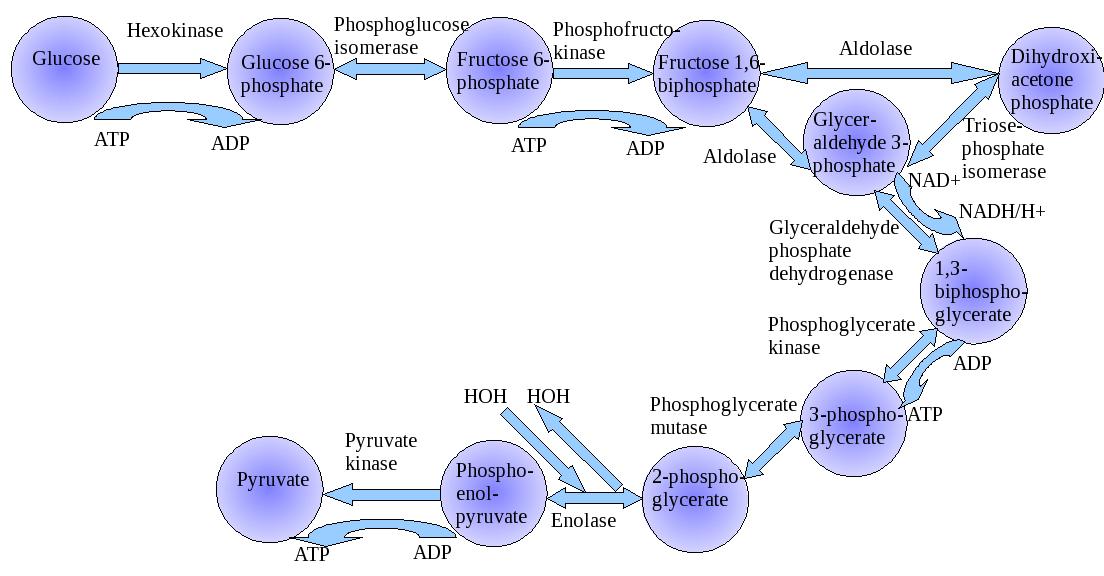