Healthcare analytics, AI solutions for biological big data, providing an AI platform for the biotech, life sciences, medical and pharmaceutical industries, as well as for related technological approaches, i.e., curation and text analysis with machine learning and other activities related to AI applications to these industries.
Aging drives cognitive and regenerative impairments in the adult brain, increasing susceptibility to neurodegenerative disorders in healthy individuals1, 2, 3, 4. Experiments using heterochronic parabiosis, in which the circulatory systems of young and old animals are joined, indicate that circulating pro-aging factors in old blood drive aging phenotypes in the brain5, 6. Here we identify β2-microglobulin (B2M), a component of major histocompatibility complex class 1 (MHC I) molecules, as a circulating factor that negatively regulates cognitive and regenerative function in the adult hippocampus in an age-dependent manner. B2M is elevated in the blood of aging humans and mice, and it is increased within the hippocampus of aged mice and young heterochronic parabionts. Exogenous B2M injected systemically, or locally in the hippocampus, impairs hippocampal-dependent cognitive function and neurogenesis in young mice. The negative effects of B2M and heterochronic parabiosis are, in part, mitigated in the hippocampus of young transporter associated with antigen processing 1 (Tap1)-deficient mice with reduced cell surface expression of MHC I. The absence of endogenous B2M expression abrogates age-related cognitive decline and enhances neurogenesis in aged mice. Our data indicate that systemic B2M accumulation in aging blood promotes age-related cognitive dysfunction and impairs neurogenesis, in part via MHC I, suggesting that B2M may be targeted therapeutically in old age.
Figure 1: Systemic B2M increases with age and impairs hippocampal-dependent cognitive function and neurogenesis
(a,b) Schematics of unpaired young versus aged mice (a), and young isochronic versus heterochronic parabionts (b). (a,b) Changes in plasma concentration of B2M with age at 3, 6, 12, 18 and 24 months (a) and between young isochronic and…
Figure 2: B2M expression increases in the aging hippocampus and impairs hippocampal-dependent cognitive function and neurogenesis.close
(a,b) Western blot and quantification of hippocampal lysates probed with B2M- and actin-specific antibodies from young (3 months) and aged (18 months) unpaired animals (a), or young isochronic and young heterochronic parabionts five wee…
Figure 3: Reducing MHC I surface expression mitigates the negative effects of heterochronic parabiosis on neurogenesis.close
(a) Schematic of young (3 months) WT and Tap1−/− isochronic parabionts and young WT and Tap1−/− heterochronic parabionts. (b,c) Representative (of six sections per mouse) images of the DG (b) and quantification of DCX immunostaining (c)…
Figure 4: Absence of B2M enhances hippocampal-dependent cognitive function and neurogenesis in aged animals.
(a–d) Learning and memory in young (3 months) and aged (17 months) WT and B2m-knockout (B2m−/−) mice by RAWM (a,c) and contextual fear conditioning (b,d). Data are from 10 young WT, 10 young B2m−/−, 8 aged WT, and 12 aged B2m−/− mice. (…
Neuroscience. 2015 Nov 12;308:75-94. doi: 10.1016/j.neuroscience.2015.09.012. Epub 2015 Sep 10.
Synergistic neuroprotection by epicatechin and quercetin: Activation of convergent mitochondrial signaling pathways.
In view of evidence that increased consumption of epicatechin (E) and quercetin (Q) may reduce the risk of stroke, we have measured the effects of combining E and Q on mitochondrial function and neuronal survival following oxygen-glucose deprivation (OGD). Relative to mouse cortical neuron cultures pretreated (24h) with either E or Q (0.1-10μM), E+Q synergistically attenuated OGD-induced neuronal cell death. E, Q and E+Q (0.3μM) increased spare respiratory capacity but only E+Q (0.3μM) preserved this crucial parameter of neuronal mitochondrial function after OGD. These improvements were accompanied by corresponding increases in cyclic AMP response element binding protein (CREB) phosphorylation and the expression of CREB-target genes that promote neuronal survival (Bcl-2) and mitochondrial biogenesis (PGC-1α). Consistent with these findings, E+Q (0.1 and 1.0μM) elevated mitochondrial gene expression (MT-ND2 and MT-ATP6) to a greater extent than E or Q after OGD. Q (0.3-3.0μM), but not E (3.0μM), elevated cytosolic calcium (Ca(2+)) spikes and the mitochondrial membrane potential. Conversely, E and E+Q (0.1 and 0.3μM), but not Q (0.1 and 0.3μM), activated protein kinase B (Akt). Nitric oxide synthase (NOS) inhibition with L-N(G)-nitroarginine methyl ester (1.0μM) blocked neuroprotection by E (0.3μM) or Q (1.0μM). Oral administration of E+Q (75mg/kg; once daily for 5days) reduced hypoxic-ischemic brain injury. These findings suggest E and Q activate Akt- and Ca(2+)-mediated signaling pathways that converge on NOS and CREB resulting in synergistic improvements in neuronal mitochondrial performance which confer profound protection against ischemic injury.
MiR-34a regulates blood–brain barrier permeability and mitochondrial function by targeting cytochrome c
The blood–brain barrier is composed of cerebrovascular endothelial cells and tight junctions, and maintaining its integrity is crucial for the homeostasis of the neuronal environment. Recently, we discovered that mitochondria play a critical role in maintaining blood–brain barrier integrity. We report for the first time a novel mechanism underlying blood–brain barrier integrity: miR-34a mediated regulation of blood–brain barrier through a mitochondrial mechanism. Bioinformatics analysis suggests miR-34a targets several mitochondria-associated gene candidates. We demonstrated that miR-34a triggers the breakdown of blood–brain barrier in cerebrovascular endothelial cell monolayer in vitro, paralleled by reduction of mitochondrial oxidative phosphorylation and adenosine triphosphate production, and decreased cytochrome c levels.
The blood–brain barrier (BBB) is composed of highly specialized cerebrovascular endothelial cells (CECs), separates brain tissue from the circulating blood, and maintains homeostasis of the neuronal environment.1 The CECs are interconnected by tight junctions including cytoplasmic zonula occludens (ZO) proteins, and various transmembrane proteins such as occludin and claudins.2 Disruption of BBB tight junctions has been well documented in cerebrovascular diseases and neurodegenerative disorders and is considered to be a pathological condition of the diseases and plays a key role in disease progression as well.2
A recent study demonstrates that the mitochondrial mechanisms regulate BBB integrity and permeability using oxygen–glucose deprivation and reoxygenation (OGD-R), anin vitro model of ischemic reperfusion injury.3 Our work demonstrates that compromised mitochondria lead to the disruption of tight junctions, opening of the BBB, and exacerbation of stroke outcomes.4 As such, regulation of mitochondrial function may affect BBB openings and could be critical in limiting the pathological progression of cerebrovascular diseases and neurodegenerative disorders.
MicroRNAs (miRNAs) are short non-coding functional RNAs that target certain messenger RNAs (mRNAs) through complementary base-pairing between the miRNAs and its mRNA targets, resulting in the inhibition of mRNA translation or degradation of mRNA.5 It has been documented that miRNAs are involved in mitochondrial structure and function, such as miR-181c which regulates mitochondrial morphology,6 miR-1 which affects mitochondrial mRNA translation,7 and miR-378 which targets mitochondrial enzymes involved in oxidative energy metabolism.8 Additionally, several miRNAs have recently been found to regulate BBB permeability. MiR-155, miR-181c, and miR-29c negatively affect BBB function by targeting tight junction protein genes directly or affecting related signal pathways.9⇓–11 The miR-34 family members were discovered computationally and later verified experimentally as a part of the p53 tumor suppressor network. Recent work demonstrates that miR-34a modulates the expression of synaptic targets and neuronal morphology and function.12 However, little is known regarding the role of miR-34a in mitochondrial function and BBB permeability.
In the present study, we report that the overexpression of miR-34a breaks down the BBB through inhibition of mitochondrial function. Furthermore, cytochrome c (CYC) is experimentally verified as a target of miR-34a in vitro.
Overexpression of miR-34a affects BBB permeability and disrupts tight junctions in CECs
To determine whether miR-34a functionally affected the BBB, we transfected CECs with miR34a plasmid versus vector control in 24-well plates, cultured the cells for 48 h, conducted a BBB permeability assay in a CEC monolayer transwell system in vitro with an additional culture of 48 h, and measured the fluorescent dye FD-4 permeability of each well (Figure 1(a)). As shown in Figure 1(a), FD-4 permeability was significantly increased in wells containing miR-34a overexpression CEC monolayer. Papp, the permeability coefficient, was also significantly higher in CECs overexpressed with miR-34a in comparison to vector controls (Figure 1(a)). Furthermore, immunohis-tochemistry staining of tight junction-related proteins revealed that ZO-1 was continuously distributed in the control, but a discontinuous distribution of ZO-1 was observed in miR-34a overexpressed CEC monolayer (Figure 1(b)). Disruption of tight junctions was not associated with cell viability in CECs transfected with plasmids for 48 h or 96 h (Supplementary Figure 2). Altogether, these data suggest that overexpression of miR-34a increases BBB permeability and compromises BBB tight junctions.
Overexpression of miR-34a increases BBB permeability in vitro. (a) A schematic protocol using fluorescein isothiocyanate–dextran-4 (FD-4) to detect BBB permeability in vitro. FD-4 permeability in CECs that overexpressed miR-34a plasmid (0.017 ng) versus control was presented as real-time rate of FD-4 mean fluorescent intensity (2-way ANOVA followed by post hoc Dunnett’s test; n = 3; **, P < 0.01; ****, P < 0.0001). Calculated apparent permeability coefficient Papp(Student’s t-test; ****, P < 0.0001) is expressed as mean ± SD. (b) Confocal fluorescence images of CECs confluent monolayers confirmed microscopically after transfection with miR-34a plasmid versus control. Fluorescent staining: tight junctions ZO-1 (red), cell nuclei (DAPI, blue). Overexpression of miR-34a apparently disrupted tight junctions and resulted in gaps between cells (white arrows). Results are representative of three independent experiments.
MiR-34a affects mitochondrial function by targeting CYC in CECs
Our recent work demonstrated that mitochondria play a pivotal role in the maintenance of BBB integrity. BBB tight junctions are rapidly disrupted if oxidative phosphorylation is reduced by mitochondrial inhibitors.4 To investigate whether the miR-34a regulates BBB openings via affecting mitochondrial function in CECs, we examined cellular energetic OCRs in CECs transfected with miR-34a plasmid versus vector control. Interestingly, overexpression of miR-34a significantly impaired mitochondrial function in CECs (Figure 2(a) and Supplementary Figure 3). Basal respiration, ATP production, maximal respiration, and spare capacity were all significantly reduced in CECs overexpressing miR-34a for 48 and 72 h (Figure 2(a)). ATP level was also substantially reduced in CECs following overexpression of miR-34a in a dose dependent manner at 72 h (Figure 2(b)).
Overexpression of mir-34a reduces mitochondrial function and decreases CYC level in cerebrovascular endothelial cells. (a) Basal respiration, ATP production, maximal respiration, and spare capacity were calculated from the bioenergetics functional assay at post-transfection 48 and 72 h (raw data in Supplementary Figure 3). Data are expressed as mean ± SD (n = 5). 1-way ANOVA followed by post hoc Tukey’s test. (*, P < 0.05; **, P < 0.01; ***, P < 0.001; ****, P < 0.0001). (b) ATP level was measured at 72 h post-transfection. Data are expressed as mean ± SD (n = 5). 1-way ANOVA followed by post hoc Tukey’s test. (****, P < 0.0001). (c) Bioinfomatic analysis of miR-34a-targeting candidates related to mitochondria. (d) Flow cytometry analysis of mitochondrial specific proteins for complex I proteins (NDUFAF1, NDUFC2 and NDUFS2), complex II protein (SDHC), complex III protein (CYB), complex IV protein (CYC oxidase, Cox IV), cytochrome c (CYCS), pyruvate dehydrogenase kinase (PDK), and voltage-dependent anion channel protein (VDAC) at 72 h post-transfection. CYC level was significantly lower in the cells that were transfected with the miR-34a plasmid. Data are presented as mean ± SD (n = 3) and analyzed by Student’s t-test, *, P < 0.05; ***, P < 0.001; ****, P < 0.0001. Results are representative of three independent experiments.
To further determine miR-34a targets and uncover the mechanism that is used to affect mitochondria, we performed a bioinformatics analysis of the miR-34a database (miRbase and TargetScan). MiR-34a potentially targets several mitochondria-associated gene candidates including succinate dehydrogenase subunit c (SDHC), cytochrome B reductase 1 (CYBRD1), cytochrome B5 reductase 3 (CYBRD5), cytochrome c (CYCS), pyruvate dehydrogenase kinase isozyme 1 and 2 (PDK1 and PDK2) (Figure 2(c). However, CECs transfected with the miR-34a plasmid had robustly decreased CYCS levels measured by flow cytometry, suggesting that CYCS is one of the miR-34a targets among the potential candidates (Figure 2(d)). Moreover, overexpression of miR-34a slightly increased potential target SDHC but did not change the protein level of CYB and PKD (Figure 2(d)). Off-target genes, NDUFAF1, and VDAC showed no significant change in protein level, but NDUFC2, NDUFS2, and Cox IV were all increased in parallel with overexpression of miR-34a (Figure 2(d)). Taken together, these results experimentally verified CYCS as a miR-34a target, which is associated with the reduction of mitochondrial oxidative phosphorylation in CECs.
In the present study, we demonstrated that the overexpression of miR-34a results in an increased BBB permeability and the disruption of tight junctions ZO-1 in CECs. Consistently, overexpression of miR-34a impaired mitochondrial oxidative phosphorylation and reduced ATP production in CECs. Bioinformatics analysis revealed series of potential miR-34a-targeting candidates related to mitochondrial function. We elucidated that CYCS is a miR-34a target, and the overexpression of miR-34a inhibited the CYCS expression and increased with the expression of other mitochondria-associated genes.
The overexpression of miR-34a disrupted tight junction protein ZO-1 (Figure 1). However, bioinformatics analysis indicated that miR-34a did not target the ZO-1 gene or other tight junction related genes, which suggests that the increased BBB permeability is not directly caused by the targeting of tight junction protein genes. The compromised mitochondrial function by overexpression of miR-34a may influence cellular metabolism in a way that is critical to maintain BBB tight junctions. Among several potential mitochondria-associated gene targets (Figure 2(c)), miR-34a initiated the reduction of CYCS level. Interestingly, potential target SDHC and other off-target gene proteins (NDUFC2, NDUFS2, and Cox IV) were concurrently upregulated (Figure 2(d)), which might be due to the compensation for the reduced target gene protein CYCS, or the disturbance of the coordinated gene translation in mitochondria. We therefore concluded that CYCS is a miR-34a target and is responsible for the miR-34a-induced reduction of mitochondrial oxidative phosphorylation.
Protein kinase C (PKC) signaling has also been shown to affect BBB or other endothelial barriers in vitro and in vivo. A recent study reported that miR-34a regulated blood–tumor barrier by targeting PKCɛ using glioma endothelial cells.13 In this study, we did not assess the PKC pathways that could contain additional targets of miR-34a. However, our data do support that miR-34a affects BBB via a mitochondrial mechanism, which is novel and may lead a new direction for designing BBB-related therapeutics.
We have noted several limitations in our study. First, we did not examine the effects of knockdown or knockout miR-34a on BBB function, which might fully establish the role of miR-34a in the BBB and mitochondria. Second, this work was conducted in cell culture models, which adequately address the mechanism of effect that miR-34a exerts on the BBB and mitochondria but do not provide evidence of its involvement in cerebrovascular or neurodegenerative conditions. Further studies in relevant experimental models are warranted.
Mitochondria play a pivotal role in cellular bioenergetics and cell survival, participating in a variety of cellular processes, including the generation of ATP, and the regulation of apoptotic signaling and other signaling pathways.14 MiR-34a targets and represses multiple genes involved in cell proliferation, apoptosis, cell cycle, migration, etc.,15 but it is not known if these effects are modulated by the observed mitochondrial effects as well. The present study provides the first description of miR-34a affecting mitochondrial activity, which could lead to a revision of current miR-34a targets and may lead to discovery of new mechanisms. The elucidation of the miR-34a’s role in mitochondrial oxidative phosphorylation and the BBB integrity offers a novel therapeutic strategy for targeting miR-34a to treat cerebrovascular and neurodegenerative diseases such as stroke and Alzheimer’s disease. These neuropathological diseases are known to involve a host of conditions that lead to mitochondrial impairment and BBB disruption. Finally, transient opening of the BBB could prove to be useful for CNS drug delivery.
A study of the brains of mice shows that structural deterioration associated with old age can be prevented by long-term aerobic exercise starting in mid-life, according to the authors of an open-access paper in the journal PLOS Biologyyesterday (October 29).
Old age is the major risk factor for Alzheimer’s disease, like many other diseases, as the authors at The Jackson Laboratory in Bar Harbor, Maine, note. Age-related cognitive deficits are due partly to changes in neuronal function, but also correlate with deficiencies in the blood supply to the brain and with low-level inflammation.
“Collectively, our data suggests that normal aging causes significant dysfunction to the cortical neurovascular unit, including basement membrane reduction and pericyte (cells that wrap around blood capillaries) loss. These changes correlate strongly with an increase in microglia/monocytes in the aged cortex,” said Ileana Soto, lead author on the study.*
Benefits of aerobic exercise
However, the researchers found that if they let the mice run freely, the structural changes that make the blood-brain barrier leaky and result in inflammation of brain tissues in old mice can be mitigated. That suggests that there are also beneficial effects of exercise on dementia in humans.**
Further work will be required to establish the mechanism(s): what is the role of the complement-producing microglia/macrophages, how does Apoe decline contribute to age-related neurovascular decline, does the leaky blood-brain barrier allow the passage of damaging factors from the circulation into the brain?
This work was funded in part by The Jackson Laboratory Nathan Shock Center, the Fraternal Order of the Eagle, the Jane B Cook Foundation and NIH.
* The authors investigated the changes in the brains of normal young and aged laboratory mice by comparing by their gene expression profiles using a technique called RNA sequencing, and by comparing their structures at high-resolution by using fluorescence microscopy and electron microscopy. The gene expression analysis indicated age-related changes in the expression of genes relevant to vascular function (including focal adhesion, vascular smooth muscle and ECM-receptor interactions), and inflammation (especially related to the complement system, which clears foreign particles) in the brain cortex.
These changes were accompanied by a decline in the function of astrocytes (key support cells in the brain) and loss of pericytes (the contractile cells that surround small capillaries and venules and maintain the blood-brain barrier). There were also effects on the basement membrane, which forms an integral part of the blood-brain barrier, as well as an increase in the density and functional activation of the immune cells known as microglia/monocytes, which scavenge the brain for infectious agents and damaged cells.
** To investigate the impact of long-term physical exercise on the brain changes seen in the aging mice, the researchers provided the animals with a running wheel from 12 months old (equivalent to middle aged in humans) and assessed their brains at 18 months (equivalent to ~60yrs old in humans, when the risk of Alzheimer’s disease is greatly increased). Young and old mice alike ran about two miles per night, and this physical activity improved the ability and motivation of the old mice to engage in the typical spontaneous behaviors that seem to be affected by aging.
This exercise significantly reduced age-related pericyte loss in the brain cortex and improved other indicators of dysfunction of the vascular system and blood-brain barrier. Exercise also decreased the numbers of microglia/monocytes expressing a crucial initiating component of the complement pathway that others have shown previously to play are role in age-related cognitive decline. Interestingly, these beneficial effects of exercise were not seen in mice deficient in a gene called Apoe, variants of which are a major genetic risk factor for Alzheimer’s disease. The authors also report that Apoe expression in the brain cortex declines in aged mice and this decline can also be prevented by exercise.
Abstract of APOE Stabilization by Exercise Prevents Aging Neurovascular Dysfunction and Complement Induction
Aging is the major risk factor for neurodegenerative diseases such as Alzheimer’s disease, but little is known about the processes that lead to age-related decline of brain structures and function. Here we use RNA-seq in combination with high resolution histological analyses to show that aging leads to a significant deterioration of neurovascular structures including basement membrane reduction, pericyte loss, and astrocyte dysfunction. Neurovascular decline was sufficient to cause vascular leakage and correlated strongly with an increase in neuroinflammation including up-regulation of complement component C1QA in microglia/monocytes. Importantly, long-term aerobic exercise from midlife to old age prevented this age-related neurovascular decline, reduced C1QA+ microglia/monocytes, and increased synaptic plasticity and overall behavioral capabilities of aged mice. Concomitant with age-related neurovascular decline and complement activation, astrocytic Apoe dramatically decreased in aged mice, a decrease that was prevented by exercise. Given the role of APOE in maintaining the neurovascular unit and as an anti-inflammatory molecule, this suggests a possible link between astrocytic Apoe, age-related neurovascular dysfunction and microglia/monocyte activation. To test this, Apoe-deficient mice were exercised from midlife to old age and in contrast to wild-type (Apoe-sufficient) mice, exercise had little to no effect on age-related neurovascular decline or microglia/monocyte activation in the absence of APOE. Collectively, our data shows that neurovascular structures decline with age, a process that we propose to be intimately linked to complement activation in microglia/monocytes. Exercise prevents these changes, but not in the absence of APOE, opening up new avenues for understanding the complex interactions between neurovascular and neuroinflammatory responses in aging and neurodegenerative diseases such as Alzheimer’s disease.
Having considered in general terms how a mitochondrion uses electron
transport to create an electrochemical proton gradient, we need to
examine the mechanisms that underlie this membrane-based energy-conversion process. In doing so, we also accomplish a larger purpose.
As emphasized at the beginning of this chapter, very similar chemi-
osmotic mechanisms are used by mitochondria, chloroplasts, archea,
and bacteria. In fact, these mechanisms underlie the function of nearly
all living organisms— including anaerobes that derive all their energy
from electron transfers between two inorganic molecules. It is therefore
rather humbling for scientists to remind themselves that the existence
of chemiosmosis has been recognized for only about 40 years.
mitochondria
Overview of The Electron Transport Chain
We begin with a look at some of the principles that underlie the electron-transport process, with the aim of explaining how it can pump protons
across a membrane.
Although protons resemble other positive ions such as Na+ and K+
in their movement across membranes, in some respects they are unique.
Hydrogen atoms are by far the most abundant type of atom in living
organisms; they are plentiful not only in all carbon-containing
biological molecules, but also in the water molecules that surround
them. The protons in water are highly mobile, flickering through the
hydrogen-bonded network of water molecules by rapidly
dissociating from one water molecule to associate with its neighbor,
as illustrated in Figure 14-20A. Protons are thought to move across a
protein pump embedded in a lipid bilayer in a similar way: they
transfer from one amino acid side chain to another, following a
special channel through the protein.
Protons are also special with respect to electron transport. Whenever
a molecule is reduced by acquiring an electron, the electron (e -) brings
with it a negative charge. In many cases, this charge is rapidly
neutralized by the addition of a proton (H+) from water, so that
the net effect of the reduction is to transfer an entire hydrogen atom,
H+ + e – (Figure 14-20B). Similarly, when a molecule is oxidized,
a hydrogen atom removed from it can be readily dissociated into
its constituent electron and proton—allowing the electron to
be transferred separately to a molecule that accepts electrons,
while the proton is passed to the water. Therefore, in a membrane
in which electrons are being passed along an electron-transport
chain, pumping protons from one side of the membrane to
another can be relatively simple. The electron carrier merely
needs to be arranged in the membrane in a way that causes it to
pick up a proton from one side of the membrane when it accepts
an electron, and to release the proton on the other side of the
membrane as the electron is passed to the next carrier molecule
in the chain (Figure 14-21).
How protons can be pumped across membranes. As an electron
passes along an electron-transport chain embedded in a lipid-bilayer
membrane, it can bind and release a proton at each step.
In this diagram, electron carrier B picks up a proton (H+)
from one (more…)
e_transfer
The Redox Potential Is a Measure of Electron Affinities
In biochemical reactions, any electrons removed from one
molecule are always passed to another, so that whenever one
molecule is oxidized, another is reduced. Like any other chemical r
eaction, the tendency of such oxidation-reduction reactions, or
redox reactions, to proceed spontaneously depends on the free-
energy change (ΔG) for the electron transfer, which in turn
depends on the relative affinities of the two molecules for electrons.
Because electron transfers provide most of the energy for living
things, it is worth spending the time to understand them. Many
readers are already familiar with acids and bases, which donate
and accept protons (see Panel 2-2, pp. 112–113). Acids and bases
exist in conjugate acid-base pairs, in which the acid is readily
converted into the base by the loss of a proton. For example,
acetic acid (CH3COOH) is converted into its conjugate base
(CH3COO-) in the reaction:
Image ch14e3.jpg
In exactly the same way, pairs of compounds such as NADH and
NAD+ are called redox pairs, since NADH is converted to NAD+
by the loss of electrons in the reaction:
Image ch14e4.jpg
NAD+_NADH
NADH is a strong electron donor: because its electrons are held
in a high-energy linkage, the free-energy change for passing its
electrons to many other molecules is favorable (see Figure 14-9).
It is difficult to form a high-energy linkage. Therefore its redox
partner, NAD+, is of necessity a weak electron acceptor.
The tendency to transfer electrons from any redox pair can be
measured experimentally. All that is required is the formation
of an electrical circuit linking a 1:1 (equimolar) mixture of the
redox pair to a second redox pair that has been arbitrarily selected
as a reference standard, so the voltage difference can be measured
between them (Panel 14-1, p. 784). This voltage difference is
defined as the redox potential; as defined, electrons move
spontaneously from a redox pair like NADH/NAD+ with a low
redox potential (a low affinity for electrons) to a redox pair like
O2/H2O with a high redox potential (a high affinity for electrons).
Thus, NADH is a good molecule for donating electrons to the
respiratory chain, while O2 is well suited to act as the “sink” for
electrons at the end of the pathway. As explained in Panel 14-1,
the difference in redox potential, ΔE0′, is a direct measure of
the standard free-energy change (ΔG°) for the transfer of an
electron from one molecule to another.
Proteins of inner space
energetics-of-cellular-respiration
Box Icon
Panel 14-1
Redox Potentials.
Electron Transfers Release Large Amounts of Energy
As just discussed, those pairs of compounds that have the most negative
redox potentials have the weakest affinity for electrons and therefore
contain carriers with the strongest tendency to donate electrons.
Conversely, those pairs that have the most positive redox potentials
have the strongest affinity for electrons and therefore contain carriers
with the strongest tendency to accept electrons. A 1:1 mixture of NADH
and NAD+ has a redox potential of -320 mV, indicating that NADH has
a strong tendency to donate electrons; a 1:1 mixture of H2O and ½O2
has a redox potential of +820 mV, indicating that O2 has a strong
tendency to accept electrons. The difference in redox potential is
1.14 volts (1140 mV), which means that the transfer of each electron
from NADH to O2 under these standard conditions is enormously
favorable, where ΔG° = -26.2 kcal/mole (-52.4 kcal/mole for the two
electrons transferred per NADH molecule; see Panel 14-1). If we
compare this free-energy change with that for the formation of the
phosphoanhydride bonds in ATP (ΔG° = -7.3 kcal/mole, see Figure 2-75), we see that more than enough energy is released by the oxidization
of one NADH molecule to synthesize several molecules of ATP from
ADP and Pi.
Phosphate dependence of pyruvate oxidation
Living systems could certainly have evolved enzymes that would
allow NADH to donate electrons directly to O2 to make water in the reaction:
Image ch14e5.jpg
But because of the huge free-energy drop, this reaction would proceed
with almost explosive force and nearly all of the energy would be released
as heat. Cells do perform this reaction, but they make it proceed much
more gradually by passing the high-energy electrons from NADH to
O2 via the many electron carriers in the electron-transport chain.
Since each successive carrier in the chain holds its electrons more
tightly, the highly energetically favorable reaction 2H+ + 2e – + ½O2
→ H2O is made to occur in many small steps. This enables nearly half
of the released energy to be stored, instead of being lost to the
environment as heat.
Spectroscopic Methods Have Been Used to Identify Many Electron
Carriers in the Respiratory Chain
Many of the electron carriers in the respiratory chain absorb visible
light and change color when they are oxidized or reduced. In general,
each has an absorption spectrum and reactivity that are distinct enough
to allow its behavior to be traced spectroscopically, even in crude mixtures.
It was therefore possible to purify these components long before their
exact functions were known. Thus, the cytochromes were discovered
in 1925 as compounds that undergo rapid oxidation and reduction in
living organisms as disparate as bacteria, yeasts, and insects. By observing
cells and tissues with a spectroscope, three types of cytochromes were
identified by their distinctive absorption spectra and designated
cytochromes a, b, and c. This nomenclature has survived, even though
cells are now known to contain several cytochromes of each type and
the classification into types is not functionally important.
The cytochromes constitute a family of colored proteins that are
related by the presence of a bound heme group, whose iron atom
changes from the ferric oxidation state (Fe3+) to the ferrous oxidation
state (Fe2+) whenever it accepts an electron. The heme group consists
of a porphyrin ring with a tightly bound iron atom held by four nitrogen
atoms at the corners of a square (Figure 14-22). A similar porphyrin ring
is responsible for the red color of blood and for the green color of
leaves, being bound to iron in hemoglobin and to magnesium in
chlorophyll, respectively.
The structure of the heme group attached covalently to cytochrome c ch14f22
Figure 14-22. The structure of the heme group attached covalently
to cytochrome c.
Figure 14-22
The structure of the heme group attached covalently to cytochrome c.
The porphyrin ring is shown in blue. There are five different
cytochromes in the respiratory chain. Because the hemes in different
cytochromes have slightly different structures and (more…)
Iron-sulfur proteins are a second major family of electron carriers. In these
proteins, either two or four iron atoms are bound to an equal number of
sulfur atoms and to cysteine side chains, forming an iron-sulfur center
on the protein (Figure 14-23). There are more iron-sulfur centers than
cytochromes in the respiratory chain. But their spectroscopic detection
requires electron spin resonance (ESR) spectroscopy, and they are less
completely characterized. Like the cytochromes, these centers carry one
electron at a time.
Figure 14-23. The structures of two types of iron-sulfur centers.
Figure 14-23
The structures of two types of iron-sulfur centers. (A) A center of the
2Fe2S type. (B) A center of the 4Fe4S type. Although they contain
multiple iron atoms, each iron-sulfur center can carry only one
electron at a time. There are more than seven different (more…)
The simplest of the electron carriers in the respiratory chain—and
the only one that is not part of a protein—is a small hydrophobic
molecule that is freely mobile in the lipid bilayer known as ubiquinone,
or coenzyme Q. A quinone (Q) can pick up or donate either one or
two electrons; upon reduction, it picks up a proton from the medium
along with each electron it carries (Figure 14-24).
Quinone electron carriers. Ubiquinone in the respiratory chain picks
up one H+ from the aqueous environment for every electron it accepts,
and it can carry either one or two electrons as part of a hydrogen atom
(yellow). When reduced ubiquinone donates (more…)
In addition to six different hemes linked to cytochromes, more than
seven iron-sulfur centers, and ubiquinone, there are also two copper
atoms and a flavin serving as electron carriers tightly bound to respiratory-chain proteins in the pathway from NADH to oxygen. This pathway
involves more than 60 different proteins in all.
As one would expect, the electron carriers have higher and higher
affinities for electrons (greater redox potentials) as one moves along
the respiratory chain. The redox potentials have been fine-tuned
during evolution by the binding of each electron carrier in a particular
protein context, which can alter its normal affinity for electrons. However,
because iron-sulfur centers have a relatively low affinity for electrons,
they predominate in the early part of the respiratory chain; in contrast,
the cytochromes predominate further down the chain, where a higher
affinity for electrons is required.
The order of the individual electron carriers in the chain was
determined by sophisticated spectroscopic measurements (Figure 14-25),
and many of the proteins were initially isolated and characterized as
individual polypeptides. A major advance in understanding the
respiratory chain, however, was the later realization that most of
the proteins are organized into three large enzyme complexes.
Figure 14-25. The general methods used to determine the path of
electrons along an electron-transport chain.
Figure 14-25
The general methods used to determine the path of electrons along
an electron-transport chain. The extent of oxidation of electron
carriers a, b, c, and d is continuously monitored by following their
distinct spectra, which differ in their oxidized and (more…)
The Respiratory Chain Includes Three Large Enzyme Complexes
Embedded in the Inner Membrane
Membrane proteins are difficult to purify as intact complexes
because they are insoluble in aqueous solutions, and some of
the detergents required to solubilize them can destroy normal
protein-protein interactions. In the early 1960s, however, it
was found that relatively mild ionic detergents, such as deoxycholate,
can solubilize selected components of the inner mitochondrial
membrane in their native form. This permitted the identification
and purification of the three major membrane-bound respiratory
enzyme complexes in the pathway from NADH to oxygen (Figure 14-26).
As we shall see in this section, each of these complexes acts as an
electron-transport-driven H+ pump; however, they were
initially characterized in terms of the electron carriers that
they interact with and contain:
Figure 14-26. The path of electrons through the three respiratory
enzyme complexes.
Figure 14-26
The path of electrons through the three respiratory enzyme complexes.
The relative size and shape of each complex are shown. During the
transfer of electrons from NADH to oxygen (red lines), ubiquinone
and cytochrome c serve as mobile carriers that ferry (more…)
The NADH dehydrogenase complex (generally known as complex I)
is the largest of the respiratory enzyme complexes, containing more
than 40 polypeptide chains. It accepts electrons from NADH and
passes them through a flavin and at least seven iron-sulfur centers
to ubiquinone. Ubiquinone then transfers its electrons to a second
respiratory enzyme complex, the cytochrome b-c1 complex.
The cytochrome b-c1 complex contains at least 11 different
polypeptide chains and functions as a dimer. Each monomer
contains three hemes bound to cytochromes and an iron-sulfur
protein. The complex accepts electrons from ubiquinone
and passes them on to cytochrome c, which carries its electron
to the cytochrome oxidase complex.
The cytochrome oxidase complex also functions as a dimer; each
monomer contains 13 different polypeptide chains, including two
cytochromes and two copper atoms. The complex accepts one electron
at a time from cytochrome c and passes them four at a time to oxygen.
The cytochromes, iron-sulfur centers, and copper atoms can carry
only one electron at a time. Yet each NADH donates two electrons,
and each O2 molecule must receive four electrons to produce water.
There are several electron-collecting and electron-dispersing points
along the electron-transport chain where these changes in electron
number are accommodated. The most obvious of these is cytochrome
oxidase.
An Iron-Copper Center in Cytochrome Oxidase Catalyzes Efficient
O2 Reduction
Because oxygen has a high affinity for electrons, it releases a
large amount of free energy when it is reduced to form water.
Thus, the evolution of cellular respiration, in which O2 is
converted to water, enabled organisms to harness much more
energy than can be derived from anaerobic metabolism. This
is presumably why all higher organisms respire. The ability of
biological systems to use O2 in this way, however, requires a
very sophisticated chemistry. We can tolerate O2 in the air we
breathe because it has trouble picking up its first electron; this
fact allows its initial reaction in cells to be controlled closely by
enzymatic catalysis. But once a molecule of O2 has picked up one
electron to form a superoxide radical (O2 -), it becomes dangerously
reactive and rapidly takes up an additional three electrons wherever
it can find them. The cell can use O2 for respiration only because
cytochrome oxidase holds onto oxygen at a special bimetallic
center, where it remains clamped between a heme-linked iron
atom and a copper atom until it has picked up a total of four electrons.
Only then can the two oxygen atoms of the oxygen molecule be
safely released as two molecules of water (Figure 14-27).
Figure 14-27. The reaction of O2 with electrons in cytochrome oxidase.
Figure 14-27
The reaction of O2 with electrons in cytochrome oxidase. As indicated,
the iron atom in heme a serves as an electron queuing point; this
heme feeds four electrons into an O2 molecule held at the bimetallic
center active site, which is formed by the other (more…)
The cytochrome oxidase reaction is estimated to account for 90%
of the total oxygen uptake in most cells. This protein complex is
therefore crucial for all aerobic life. Cyanide and azide are extremely
toxic because they bind tightly to the cell’s cytochrome oxidase
complexes to stop electron transport, thereby greatly reducing
ATP production.
Although the cytochrome oxidase in mammals contains 13
different protein subunits, most of these seem to have a subsidiary
role, helping to regulate either the activity or the assembly of the
three subunits that form the core of the enzyme. The complete
structure of this large enzyme complex has recently been determined
by x-ray crystallography, as illustrated in Figure 14-28. The atomic
resolution structures, combined with mechanistic studies of the effect
of precisely tailored mutations introduced into the enzyme by genetic
engineering of the yeast and bacterial proteins, are revealing the
detailed mechanisms of this finely tuned protein machine.
Figure 14-28. The molecular structure of cytochrome oxidase.
Figure 14-28
The molecular structure of cytochrome oxidase. This protein
is a dimer formed from a monomer with 13 different protein
subunits (monomer mass of 204,000 daltons). The three colored
subunits are encoded by the mitochondrial genome, and they
form the functional (more…)
Electron Transfers Are Mediated by Random Collisions in
the Inner Mitochondrial Membrane
The two components that carry electrons between the three
major enzyme complexes of the respiratory chain—ubiquinone
and cytochrome c—diffuse rapidly in the plane of the inner
mitochondrial membrane. The expected rate of random collisions
between these mobile carriers and the more slowly diffusing
enzyme complexes can account for the observed rates of electron
transfer (each complex donates and receives an electron about
once every 5–20 milliseconds). Thus, there is no need to postulate
a structurally ordered chain of electron-transfer proteins in the
lipid bilayer; indeed, the three enzyme complexes seem to exist as
independent entities in the plane of the inner membrane, being
present in different ratios in different mitochondria.
The ordered transfer of electrons along the respiratory chain
is due entirely to the specificity of the functional interactions
between the components of the chain: each electron carrier is
able to interact only with the carrier adjacent to it in the sequence
shown in Figure 14-26, with no short circuits.
Electrons move between the molecules that carry them in
biological systems not only by moving along covalent bonds
within a molecule, but also by jumping across a gap as large
as 2 nm. The jumps occur by electron “tunneling,” a quantum-
mechanical property that is critical for the processes we are
discussing. Insulation is needed to prevent short circuits that
would otherwise occur when an electron carrier with a low redox
potential collides with a carrier with a high redox potential. This
insulation seems to be provided by carrying an electron deep
enough inside a protein to prevent its tunneling interactions
with an inappropriate partner.
How the changes in redox potential from one electron carrier
to the next are harnessed to pump protons out of the mitochondrial
matrix is the topic we discuss next.
A Large Drop in Redox Potential Across Each of the Three Respiratory
Enzyme Complexes Provides the Energy for H+ Pumping
We have previously discussed how the redox potential reflects
electron affinities (see p. 783). An outline of the redox potentials
measured along the respiratory chain is shown in Figure 14-29.
These potentials drop in three large steps, one across each major
respiratory complex. The change in redox potential between any
two electron carriers is directly proportional to the free energy
released when an electron transfers between them. Each enzyme
complex acts as an energy-conversion device by harnessing some
of this free-energy change to pump H+ across the inner membrane,
thereby creating an electrochemical proton gradient as electrons
pass through that complex. This conversion can be demonstrated
by purifying each respiratory enzyme complex and incorporating
it separately into liposomes: when an appropriate electron donor
and acceptor are added so that electrons can pass through the complex,
H+ is translocated across the liposome membrane.
Figure 14-29. Redox potential changes along the mitochondrial
electron-transport chain.
Figure 14-29
Redox potential changes along the mitochondrial electron-transport
chain. The redox potential (designated E′0) increases as electrons
flow down the respiratory chain to oxygen. The standard free-energy
change, ΔG°, for the transfer (more…)
The Mechanism of H+ Pumping Will Soon Be Understood in
Atomic Detail
Some respiratory enzyme complexes pump one H+ per electron
across the inner mitochondrial membrane, whereas others pump
two. The detailed mechanism by which electron transport is coupled
to H+ pumping is different for the three different enzyme complexes.
In the cytochrome b-c1 complex, the quinones clearly have a role.
As mentioned previously, a quinone picks up a H+ from the aqueous
medium along with each electron it carries and liberates it when it
releases the electron (see Figure 14-24). Since ubiquinone is freely
mobile in the lipid bilayer, it could accept electrons near the inside
surface of the membrane and donate them to the cytochrome b-c1
complex near the outside surface, thereby transferring one H+
across the bilayer for every electron transported. Two protons are
pumped per electron in the cytochrome b-c1 complex, however, and
there is good evidence for a so-called Q-cycle, in which ubiquinone
is recycled through the complex in an ordered way that makes this
two-for-one transfer possible. Exactly how this occurs can now be
worked out at the atomic level, because the complete structure of
the cytochrome b-c1 complex has been determined by x-ray
crystallography (Figure 14-30).
Figure 14-30. The atomic structure of cytochrome b-c 1.
Figure 14-30
The atomic structure of cytochrome b-c 1. This protein is a dimer.
The 240,000-dalton monomer is composed of 11 different protein
molecules in mammals. The three colored proteins form the
functional core of the enzyme: cytochrome b (green), cytochrome (more…)
Allosteric changes in protein conformations driven by electron
transport can also pump H+, just as H+ is pumped when ATP
is hydrolyzed by the ATP synthase running in reverse. For both the
NADH dehydrogenase complex and the cytochrome oxidase complex,
it seems likely that electron transport drives sequential allosteric
changes in protein conformation that cause a portion of the protein
to pump H+ across the mitochondrial inner membrane. A general
mechanism for this type of H+ pumping is presented in Figure 14-31.
Figure 14-31. A general model for H+ pumping.
Figure 14-31
A general model for H+ pumping. This model for H+ pumping
by a transmembrane protein is based on mechanisms that are
thought to be used by both cytochrome oxidase and the light-driven
procaryotic proton pump, bacteriorhodopsin. The protein
is driven through (more…)
H+ Ionophores Uncouple Electron Transport from ATP Synthesis
Since the 1940s, several substances—such as 2,4-dinitrophenol—
have been known to act as uncoupling agents, uncoupling electron
transport from ATP synthesis. The addition of these low-molecular-weight organic compounds to cells stops ATP synthesis by mitochondria
without blocking their uptake of oxygen. In the presence of an
uncoupling agent, electron transport and H+ pumping continue at
a rapid rate, but no H+ gradient is generated. The explanation for
this effect is both simple and elegant: uncoupling agents are lipid-
soluble weak acids that act as H+ carriers (H+ ionophores), and
they provide a pathway for the flow of H+ across the inner mitochondrial
membrane that bypasses the ATP synthase. As a result of this short-
circuiting, the proton-motive force is dissipated completely, and
ATP can no longer be made.
Respiratory Control Normally Restrains Electron Flow
Through the Chain
When an uncoupler such as dinitrophenol is added to cells,
mitochondria increase their oxygen uptake substantially because
of an increased rate of electron transport. This increase reflects
the existence of respiratory control. The control is thought to
act via a direct inhibitory influence of the electrochemical proton
gradient on the rate of electron transport. When the gradient is
collapsed by an uncoupler, electron transport is free to run unchecked
at the maximal rate. As the gradient increases, electron transport
becomes more difficult, and the process slows. Moreover, if an
artificially large electrochemical proton gradient is experimentally
created across the inner membrane, normal electron transport
stops completely, and a reverse electron flow can be detected in
some sections of the respiratory chain. This observation suggests
that respiratory control reflects a simple balance between the
free-energy change for electron-transport-linked proton pumping
and the free-energy change for electron transport—that is, the
magnitude of the electrochemical proton gradient affects both
the rate and the direction of electron transport, just as it affects
the directionality of the ATP synthase (see Figure 14-19).
Respiratory control is just one part of an elaborate interlocking
system of feedback controls that coordinate the rates of glycolysis,
fatty acid breakdown, the citric acid cycle, and electron transport.
The rates of all of these processes are adjusted to the ATP:ADP ratio,
increasing whenever an increased utilization of ATP causes the ratio
to fall. The ATP synthase in the inner mitochondrial membrane,
for example, works faster as the concentrations of its substrates
ADP and Pi increase. As it speeds up, the enzyme lets more H+ flow
into the matrix and thereby dissipates the electrochemical proton
gradient more rapidly. The falling gradient, in turn, enhances the
rate of electron transport.
Similar controls, including feedback inhibition of several key enzymes
by ATP, act to adjust the rates of NADH production to the rate of
NADH utilization by the respiratory chain, and so on. As a result of
these many control mechanisms, the body oxidizes fats and sugars
5–10 times more rapidly during a period of strenuous exercise than
during a period of rest.
Natural Uncouplers Convert the Mitochondria in Brown Fat into
Heat-generating Machines
In some specialized fat cells, mitochondrial respiration is normally
uncoupled from ATP synthesis. In these cells, known as brown fat
cells, most of the energy of oxidation is dissipated as heat rather
than being converted into ATP. The inner membranes of the large
mitochondria in these cells contain a special transport protein that
allows protons to move down their electrochemical gradient, by-
passing ATP synthase. As a result, the cells oxidize their fat stores
at a rapid rate and produce more heat than ATP. Tissues containing
brown fat serve as “heating pads,” helping to revive hibernating animals
and to protect sensitive areas of newborn human babies from the cold.
Bacteria Also Exploit Chemiosmotic Mechanisms to Harness Energy
Bacteria use enormously diverse energy sources. Some, like animal
cells, are aerobic; they synthesize ATP from sugars they oxidize to
CO2 and H2O by glycolysis, the citric acid cycle, and a respiratory
chain in their plasma membrane that is similar to the one in the
inner mitochondrial membrane. Others are strict anaerobes, deriving
their energy either from glycolysis alone (by fermentation) or from an
electron-transport chain that employs a molecule other than oxygen
as the final electron acceptor. The alternative electron acceptor can
be a nitrogen compound (nitrate or nitrite), a sulfur compound
(sulfate or sulfite), or a carbon compound (fumarate or carbonate),
for example. The electrons are transferred to these acceptors by a
series of electron carriers in the plasma membrane that are comparable
to those in mitochondrial respiratory chains.
Despite this diversity, the plasma membrane of the vast majority of
bacteria contains an ATP synthase that is very similar to the one in
mitochondria. In bacteria that use an electron-transport chain to
harvest energy, the electron-transport pumps H+ out of the cell and
thereby establishes a proton-motive force across the plasma membrane
that drives the ATP synthase to make ATP. In other bacteria, the
ATP synthase works in reverse, using the ATP produced by glycolysis
to pump H+ and establish a proton gradient across the plasma
membrane. The ATP used for this process is generated by
fermentation processes (discussed in Chapter 2).
Thus, most bacteria, including the strict anaerobes, maintain a proton
gradient across their plasma membrane. It can be harnessed to drive
a flagellar motor, and it is used to pump Na+ out of the bacterium via
a Na+-H+ antiporter that takes the place of the Na+-K+ pump of
eucaryotic cells. This gradient is also used for the active inward transport
of nutrients, such as most amino acids and many sugars: each nutrient is
dragged into the cell along with one or more H+ through a specific symporter
(Figure 14-32). In animal cells, by contrast, most inward transport across
the plasma membrane is driven by the Na+ gradient that is established by the
Na+-K+ pump.
Figure 14-32. The importance of H+-driven transport in bacteria.
Figure 14-32
The importance of H+-driven transport in bacteria. A proton-motive force
generated across the plasma membrane pumps nutrients into the cell and
expels Na+. (A) In an aerobic bacterium, an electrochemical proton gradient
across the plasma membrane is produced (more…)
Some unusual bacteria have adapted to live in a very alkaline
environment and yet must maintain their cytoplasm at a physiological
pH. For these cells, any attempt to generate an electrochemical H+
gradient would be opposed by a large H+ concentration gradient in
the wrong direction (H+ higher inside than outside). Presumably for
this reason, some of these bacteria substitute Na+ for H+ in all of their
chemiosmotic mechanisms. The respiratory chain pumps Na+ out of
the cell, the transport systems and flagellar motor are driven by an
inward flux of Na+, and a Na+-driven ATP synthase synthesizes
ATP. The existence of such bacteria demonstrates that the principle
of chemiosmosis is more fundamental than the proton-motive force
on which it is normally based.
Summary
The respiratory chain in the inner mitochondrial membrane contains
three respiratory enzyme complexes through which electrons pass on
their way from NADH to O2.
Each of these can be purified, inserted into synthetic lipid vesicles,
and then shown to pump H+ when electrons are transported through it.
In the intact membrane, the mobile electron carriers ubiquinone and
cytochrome c complete the electron-transport chain by shuttling between
the enzyme complexes. The path of electron flow is NADH → NADH
dehydrogenase complex → ubiquinone → cytochrome b-c1 complex →
cytochrome c → cytochrome oxidase complex → molecular oxygen (O2).
The respiratory enzyme complexes couple the energetically favorable
transport of electrons to the pumping of H+ out of the matrix. The
resulting electrochemical proton gradient is harnessed to make ATP
by another transmembrane protein complex, ATP synthase, through
which H+ flows back into the matrix. The ATP synthase is a reversible
coupling device that normally converts a backflow of H+ into ATP
phosphate bond energy by catalyzing the reaction ADP + Pi → ATP,
but it can also work in the opposite direction and hydrolyze ATP to
pump H+ if the electrochemical proton gradient is sufficiently reduced.
Its universal presence in mitochondria, chloroplasts, and procaryotes
testifies to the central importance of chemiosmotic mechanisms in cells.
By agreement with the publisher, this book is accessible by the search
feature, but cannot be browsed.
The multi-step transfer of phosphate bond and hydrogen exchange energy
Curator: Larry H. Bernstein, MD, FCAP, Leaders in Pharmaceutical Intelligence
In this subtext of the series we expand on a tie between respiration and glycolysis, and the functioning of the mitochondrion to discover the key role played by oxidative phosphorylation, “acetyl coenzyme A, and electron transport. This was crucial to understanding cellular energetics, which explains the high energy of fatty acid catabolism from stored adipose tissue, and the criticality of the multi-step sequence of reactions in energy transfer.
This portion considerably provides a response to the TWO points made by Jose EDS Rosallis:
Just at the beginning, when phosphorylation of proteins is presented, I assume you must mention that some proteins are activated by phosphorylation. This is fundamental in order to present self –organization reflex upon fast regulatory mechanisms. This poiny needs further clarification, but he makes important observations here.
Even from an historical point of view. The first observation arrived from a sample due to be studied on the following day of glycogen synthetase. It was unintended left overnight out of the refrigerator. The result was it had changed from active form of the previous day to a non-active form.
The story could have being finished here, if the researcher did not decide to spent this day increasing substrate levels (it could be a simple case of denaturation of proteins that changes its conformation despite the same order of amino acids). He kept on trying and found restoration of maximal activity.
This assay was repeated with glycogen phosphorylase and the result was the opposite it increases its activity.
This led to the discovery of cAMP activated protein kinase and the assembly of a very complex system in the glycogen granule that is not a simple carbohydrate polymer. Instead
it has several proteins assembled and preserves the capacity to receive from a single event (rise in cAMP) two opposing signals with maximal efficiency,
stops glycogen synthesis, as long as levels of glucose 6 phosphate are low and
increases glycogen phosphorylation as long as AMP levels are high).
I did everything I was able to do by the end of 1970 in order to repeat these assays with
PK I, PKII and PKIII of M. Rouxii and Sutherland route to cAMP failed in this case.
I ask Leloir to suggest to my chief (SP) the idea of AA, AB, BB subunits as was observed in lactic dehydrogenase (tetramer)
(Nathan O. Kaplan discovery) indicating this as his idea. The reason was my “chief” (SP) more than once, said to me: “Leave these great ideas for the Houssay, Leloir etc…We must do our career with small things. ” However, as she also had a faulty ability for recollection she also used to arrive some time later, with the very same idea but in that case, as her idea.
[This reminds me of when I was studying the emergence of lactic dehysrogenase isoenzyme patterns in the developing eye lens of cattle, I raised reservations about Elliott Vessells challenge to Nathan Kaplan, but that not being my primary problem, my brilliant mentor (H.M.), a very young full professor of anatomy said – leave that to NOK.}
Leloir, said to me: I will not offer your interpretation to her as mine. I think it is not phosphorylation, however I think it is
glycosylation that explains the changes in the isoenzymes with the same molecular weight preserved.
This dialogue explains why during the Schroedinger’s “What is life?” reading with him he asked me if from biochemist in exile, to biochemist I expressed all of my thoughts to him. Since I had considered that Schrödinger did not confront Darlington & Haldane for being in exile. This may explain why Leloir could have answered a bad telephone call from P. Boyer, Editor of The Enzymes in a way that suggests the the pattern could be of covalent changes over a protein. Our FEBS and Eur J. Biochemistry papers on pyruvate kinase of M. Rouxii is wrongly quoted in this way on his review about pyruvate kinase of
that year(1971).
show in detail with different colors what carbons belongs to CoA a huge molecule, in comparison with the single two carbons of acetate that will produce the enormous jump in energy yield in comparison with anaerobic glycolysis. The idea is how much must have being spent in DNA sequences to build that molecule in order to use only two atoms of carbon. Very limited aspects of biology could be explained in this way. In case we follow an alternative way of thinking, it becomes clearer that proteins were made more stable by interaction with other molecules (great and small). Afterwards, it rather easy to understand how the stability of protein-RNA complexes where transmitted to RNA (vibrational +solvational reactivity stability pair of conformational energy). Latter, millions of years, or as soon as, the information of interaction leading to activity and regulation could be found in RNA, proteins like reverse transcriptase move this information to a more stable form (DNA). In this way it is easier to understand the use of CoA to make two carbon molecules more reactive.
The outline of what I am presenting in series is as follows:
ATP Dephosphorylation Coupled to Nonspontaneous Reactions
Coupled Reactions to Generate ATP
Structure and Function of the Mitochondria
Oxidation-Reduction Reactions in the Electron-Transport Chain
Electron-Carrier Proteins (NOTE: This section includes a separate link and an animation.)
Relationship Between Reduction Potentials and Free Energy
Proton Gradient as Means of Coupling Oxidative and Phosphorylation Components of Oxidative Phosphorylation
ATP Synthetase Uses Energy From Proton Gradient to Generate ATP
Every day, we build bones, move muscles, eat food, think, and perform many other activities with our bodies. All of these activities are based upon chemical reactions. However, most of these reactions are not spontaneous (i.e., they are accompanied by a positive change in free energy, DG>0) and do not occur without some other source of free energy. Hence, the body needs some sort of “free-energy currency,” (Figure 1) a molecule that can store and release free energy when it is needed to power a given biochemical reaction.
The four questions:
How does the body “spend” free-energy currency to make a nonspontaneous reaction spontaneous? The answer, which is based on thermodynamics, is to use coupled reactions.
How is food used to produce the reducing agents (NADH and FADH2) that can regenerate the free-energy currency? The answer, from biology, is found in glycolysis and the citric-acid cycle.
How are the reducing agents (NADH and FADH2) able to generate the free-energy currency molecule (ATP)? Once again, coupled reactions are key.
What mechanism does the body use to couple the reducing agent reactions and the generation of ATP? ATP is synthesized primarily by a two-step process consisting of an electron-transport chain and a proton gradient. This process is based on electrochemistry and equilibrium, as well as thermodynamics.
The free-energy change (DG) for the net reaction is given by the sum of the free-energy changes for the individual reactions. The phospholipids that form cell membranes are formed from glycerol with a phosphate group and two fatty-acid chains attached.This step actually consists of two reactions:
(1) the phosphorylation of glycerol, and
(2) the dephosphorylation of ATP (the free-energy-currency molecule). The reactions may be added as shown in Equations 2-4, below:
ATP is the most important “free-energy-currency” molecule in living organisms (see Figure 2, below). Adenosine triphosphate (ATP) is a useful free-energy currency because the dephosphorylation reaction is very spontaneous; i.e., it releases a large amount of free energy (30.5 kJ/mol). Thus, the dephosphorylation reaction of ATP to ADP and inorganic phosphate (Equation 3) is often coupled with nonspontaneous reactions (e.g., Equation 2) to drive them forward. The body’s use of ATP as a free-energy currency is a very effective strategy to cause vital nonspontaneous reactions to occur.
This is the two-dimensional (ChemDraw) structure of ATP, adenosine triphosphate. The removal of one phosphate group (green) from ATP requires the breaking of a bond (blue) and results in a large release of free energy. Removal of this phosphate group (green) results in ADP, adenosine diphosphate.
This flowchart shows that the energy used by the body for its many activities ultimately comes from the chemical energy in our food. The chemical energy in our food is converted to reducing agents (NADH and FADH2). These reducing agents are then used to make ATP. ATP stores chemical energy, so that it is available to the body in a readily accessible form.
2(Pyruvate– + Coenzyme A + NAD+ –>
Acetyl CoA + CO2 + NADH)
(6)
Citric-Acid Cycle
2(Acetyl CoA + 3 NAD++ FAD + GDP3-
+ HPO42- + 2H2O –> 2 CO2 + 3 NADH + FADH2
+ GTP4- + 2H+ + Coenzyme A)
(7)
The structures of the important molecules in Equations 5-7 are shown in Table 1, below.
How is Food Used to Make the Reducing Agents Needed for the Production of ATP?
To make ATP, energy must be absorbed. This energy is supplied by the food we eat, and then used to synthsize two reducing agents, NADH and FADH2 that are needed to produce ATP. One of the principal energy-yielding nutrients in our diet is glucose (see structure in Table 1 in the blue box below), a simple six-carbon sugar that can be broken down by the body. When the chemical bonds in glucose are broken, free energy is released. The complete breakdown of glucose into CO2 occurs in two processes: glycolysis and the citric-acid cycle. The reactions for these two processes are shown in the blue box below.
two-dimensional representations of several important molecules in Equations 5-7.
As seen in Equations 5-7 in the blue box, glycolysis and the citric-acid cycle produce a net total of only four ATP or GTP molecules (GTP is an energy-currency molecule similar to ATP) per glucose molecule. This yield isfar below the amount needed by the body for normal functioning, and in fact is far below the actual ATP yield for glucose in aerobic organisms (organisms that use molecular oxygen). For each glucose molecule the body processes, the body actually gains approximately 30 ATP molecules! (See Figure 4, below.) So, how does the body generate ATP?
The process that accounts for the high ATP yield is known as oxidative phosphorylation. A quick examination of Equations 5-7 shows that glycolysis and the citric-acid cycle generate other products besides ATP and GTP, namely NADH and FADH2 (blue). These products are molecules that are oxidized (i.e., give up electrons) spontaneously. The body uses these reducing agents (NADH and FADH2) in an oxidation-reduction reaction . As you will see later in this tutorial, it is the free energy from these redox reactions that is used to drive the production of ATP.
This flowchart shows the major steps involved in breaking down glucose from the diet and converting its chemical energy to the chemical energy in the phosphate bonds of ATP, in aerobic (oxygen-using) organisms. Note: In this flowchart, red denotes a source of carbon atoms (originally from glucose),green denotes energy-currency molecules, and blue denotes the reducing agents that can be oxidized spontaneously.
In the discussion above, we see that glucose by itself generates only a tiny amount of ATP. However, during the breakdown of glucose, a large amount of NADH and FADH2 is produced; it is these reducing agents that dramatically increase the amount of ATP produced. How does this work?
How are the reducing agents (NADH and FADH2) able to generate the free-energy currency molecule (ATP)?
As discussed in an earlier section about coupling reactions, ATP is used as free-energy currency by coupling its (spontaneous) dephosphorylation (Equation 3) with a (nonspontaneous) biochemical reaction to give a net release of free energy (i.e., a net spontaneous reaction). Coupled reactions are also used to generate ATP by phosphorylating ADP. The nonspontaneous reaction of joining ADP to inorganic phosphate to make ATP (Equation 8, below, and Figure 2, above) is coupled to the oxidation reaction of NADH or FADH2 (Equation 9, below). (Recall, NADH and FADH2 are produced in glycolysis and the citric-acid cycle as described in the blue box). For simplicity, we shall henceforth discuss only the oxidation of NADH; FADH2 follows a very similar oxidation pathway.
The oxidation reaction for NADH has a larger, but negative, DG than the positive DG required for the formation of ATP from ADP and phosphate. This set of coupled reactions is so important that it has been given a special name: oxidative phosphorylation. This name emphasizes the fact that an oxidation (of NADH) reaction (Equation 9 and Figure 5, below) is being coupled to a phosphorylation (of ADP) reaction (Equation 8, below, and Figure 2, above). In addition, we must consider the reduction reaction (gaining of electrons) that accompanies the oxidation of NADH. (Oxidation reactions are always accompanied by reduction reactions, because an electron given up by one group must be accepted by another group.) In this case, molecular oxygen (O2) is the electron acceptor, and the oxygen is reduced to water(Equation 10, below) .
The individual reactions of interest for oxidative phosphorylation are:
Phosphorylation
ADP3- + HPO42- + H+ –>
ATP4- + H2O
DGo= +30.5 kJ
(nonspontaneous)(8)
oxidation
NADH –> NAD+ + H+ + 2e
DGo= –158.2 Kj
(spontaneous)(9)
reduction
1/2 O2 + 2H+ + 2e– –> H2O
DGo= –61.9 kJ
(spontaneous) (10)
The net reaction is obtained by summing the coupled reactions, as shown in Equation 11, below.
This is a two-dimensional (ChemDraw) representation showing the change that occurs when NADH is oxidized to NAD+. “R” represents the part of the structure that is shown in black in the drawing of NADH in Table 1, and does not change during the oxidation half-reaction. The molecular changes that occur upon oxidation are shown in red.
In this tutorial, we have seen that nonspontaneous reactions in the body occur by coupling them with a very spontaneous reaction (usually the ATP reaction shown in Equation 3). We have just seen that ATP is produced by coupling the phosphorylation reaction with NADH oxidation (a very spontaneous reaction). But we have not yet answered the question: by what mechanism are these reactions coupled?
Coupling Reactions in Biological Systems
Every day your body carries out many nonspontaneous reactions. As discussed earlier, if a nonspontaneous reaction is coupled to a spontaneous reaction, as long as the sum of the free energies for the two reactions is negative, the coupled reactions will occur spontaneously. How is this coupling achieved in the body? Living systems couple reactions in several ways, but the most common method of coupling reactions is to carry out both reactions on the same enzyme. Consider again the phosphorylation of glycerol (Equations 2-4). Glycerol is phosphorylated by the enzyme glycerol kinase, which is found in your liver. The product of glycerol phosporylation, glycerol-3-phosphate (Equation 2), is used in the synthesis of phospholipids.
Glycerol kinase is a large protein comprised of about 500 amino acids. X-ray crystallography of the protein shows us that there is a deep groove or cleft in the protein where glycerol and ATP attach (see Figure 6, below). Because the enzyme holds the ATP and the glycerol in place, the phosphate can be transferred directly from the ATP to glycerol. Instead of two separate reactions where ATP loses a phosphate (Equation 3) and glycerol picks up a phosphate (Equation 2), the enzyme allows the phosphate to move directly from ATP to glycerol (Equation 4).
The coupling in oxidative phosphorylation uses a more complicated (and amazing!) mechanism, but the end result is the same: the reactions are linked together, the net free energy for the linked reactions is negative, and, therefore, the linked reactions are spontaneous.
This is a schematic representation of ATP and glycerol bound (attached) to glycerol kinase. The enzyme glycerol kinase is a dimer (consists of two identical subuits). There is a deep cleft between the subunits where ATP and glycerol bind. Since the ATP and phosphate are physically so close together when they are bound to the enzyme, the phosphate can be transferred directly from ATP to glycerol. Hence, the processes of ATP losing a phosphate (spontaneous) and glycerol gaining a phosphate (nonspontaneous) are linked together as one spontaneous process
Questions on ATP: The Body’s Free-Energy Currency (How Free-Energy Currency Works)
Biological systems involve many molecules containing phosphate groups, such as ATP. Although ATP is the most commonly used free-energy currency, any of these phosphorylated molecules could, in theory, be used as free-energy currency. The standard free-energy change (DGo) for the dephosphorylation (removal of a phosphate group) of several biological compounds is given below:
Acetyl phosphate
DGo = -47.3 kJ/mol
Adenosine triphosphate (ATP)
DGo = -30.5 kJ/mol
Glucose-6-phosphate
DGo = -13.8 kJ/mol
Phosphoenolpyruvate (PEP)
DGo = -61.9 kJ/mol
Phosphocreatine
DGo = -43.1 kJ/mol
Neglecting any differences in difficulty synthesizing or accessing these molecules by biological systems, rank the molecules in order of their efficiency as a free-energy currency (i.e., the amount of nonspontaneous reactions enabled per phosphate removed from a molecule of free-energy currency) from the most efficient to the least efficient.
What, if any, changes are there in the shape of the ring as NADH is oxidized to NAD+(see Figure 5)? (Hint: Consider which atoms lie in the same plane in each structure.)
Mechanism of Coupling the Oxidative-Phosphorylation Reactions
In order to couple the redox and phosphorylation reactions needed for ATP synthesis in the body, there must be some mechanism linking the reactions together. In cells, this is accomplished through an elegant proton-pumping system that occurs inside special double-membrane-bound organelles (specialized cellular components) known as mitochondria. A number of proteins are required to maintain this proton-pumping system and catalyze the oxidative and phosphorylation reactions.
Synthesis of ATP (Equation 8) is coupled with the oxidation of NADH (Equation 9) and the reduction of O2 (Equation 10). There are three key steps in this process:
Electrons are transferred from NADH, through a series of electron carriers, to O2. The electron carriers are proteins embedded in the inner mitochondrial membrane. (More detail about the structure of the mitochondria is presented in the next section.) (See Figure 7a.)
Transfer of electrons by these carriers generates a proton (H+) gradient across the inner mitochondrial membrane. (See Figure 7b.)
When H+ spontaneously diffuses back across the inner mitochondrial membrane, ATP is synthesized. The large positive free energy of ATP synthesis is overcome by the even larger negative free energy associated with proton flow down the concentration gradient. (See Figure 7c.)
These steps are outlined below.
Electron Transport (Oxidation-Reduction Reactions) Through a Series of Proteins in the Inner Membrane of the Mitochondria
The three major steps in oxidative phosphorylation are
(a) oxidation-reduction reactions involving electron transfers between specialized proteins embedded in the inner mitochondrial membrane;
(b) the generation of a proton (H+) gradient across the inner mitochondrial membrane (which occurs simultaneously with step (a)); and
(c) the synthesis of ATP using energy from the spontaneous diffusion of electrons down the proton gradient generated in step (b).
Note: Steps (a) and (b) show cytochrome oxidase, the final electron-carrier protein in the electron-transport chain described above. When this protein accepts an electron (green) from another protein in the electron-transport chain, an Fe(III) ion in the center of a heme group (purple) embedded in the protein is reduced to Fe(II). The coordinates for the protein were determined using x-ray crystallography, and the image was rendered using SwissPDB Viewer and POV-Ray (see References).
Cells use a proton-pumping system made up of proteins inside the mitochondria to generate ATP. Before we examine the details of ATP synthesis, we shall step back and look at the big picture by exploring the structure and function of the mitochondria, where oxidative phosphorylation occurs.
This is a schematic diagram showing the membranes of the mitochondrion. The purple shapes on the inner membrane represent proteins, which are described in the section below. An enlargement of the boxed portion of the inner membrane in this figure is shown in Figure.
The mitochondrial membranes are crucial for this organelle’s role in oxidative phosphorylation. As shown in Figure 8, mitochondria have two membranes, an inner and an outer membrane. The outer membrane ispermeable to most small molecules and ions, because it contains large protein channels called porins. The inner membrane is impermeable to most ions and polar molecules. The inner membrane is the site of oxidative phosphorylation. Although the membrane is mostly impermeable, it contains special H+ (proton) channels and pumps that enable the coupling of the redox reaction involving NADH and O2 (Equations 9-10) to the phosphorylation reaction of ADP (Equation 8), as described below (“Oxidation-Reduction Reactions and Proton Pumping in Oxidative Phosphorylation”). (Recall the discussion of protein channels in the “Maintaining the Body’s Chemistry: Dialysis in the Kidneys” Tutorial .)
As shown in Figure 8, inside the inner membrane is a space known as the matrix; the space between the two membranes is known as the intermembrane space. The matrix side of the inner membrane has a negative electrical charge relative to the intermembrane space due to an H+ gradient set up by the redox reaction (Equations 9 and 10). This charge difference is used to provide free energy (G) for the phosphorylation reaction (Equation 8).
Oxidation-Reduction Reactions and Proton Pumping in Oxidative Phosphorylation
Phosphorylation of ADP (Equation 8) is coupled to the oxidation-reduction reaction of NADH and O2 (Equations 9 and 10). Electrons are not transferred directly from NADH to O2, but rather pass through a series of intermediate electron carriers in the inner membrane of the mitochondrion. Why? This allows something very important to occur: the pumping of protons across the inner membrane of the mitochondrion. As we shall see, it is this proton pumping that is ultimately responsible for coupling the oxidation-reduction reaction to ATP synthesis.
Two major types of mitochondrial proteins (see Figure 9, below) are required for oxidative phosphorylation to occur. Both classes of proteins are located in the inner mitochondrial membrane.
The electron carriers (NADH-Q reductase, ubiquinone (Q), cytochrome reductase, cytochrome c, and cytochrome oxidase shown in shades of purple in Figure 9 below) transport electrons in a stepwise fashion from NADH to O2. Three of these carriers (NADH-Q reductase, cytochrome reductase, and cytochrome oxidase) are also proton pumps, and simultaneously pump H+ ions (protons) from the matrix to the intermembrane space. (Proton movement from one side of the membrane to the other is shown as blue arrows in Figure 9, below.) The protons that are pumped across the membrane complete the redox reaction (Equations 9 and 10). The creation of a proton gradient across the membrane is one way of storing free energy.
ATP synthetase (shown in red in Figure 9 below) allows H+ ions to diffuse back into the matrix and uses the free energy released to synthesize ATP from ADP and HPO42-. The ATP synthetase is essential for the phosphorylation to occur (Equation 8). (Proton movement from one side of the membrane to the other is shown as blue arrows in Figure 9, below.)
The electron carriers can be divided into three protein complexes (NADH-Q reductase (1), cytochrome reductase (3), and cytochrome oxidase (5)) that pump protons from the matrix to the intermembrane space, and two mobile carriers (ubiquinone (2) and cytochrome c (4)) that transfer electrons between the three proton-pumping complexes. (Gold numbers refer to the labels on each protein in Figure 9, below.) Because electrons move from one carrier to another until they are finally transferred to O2, the electron carriers (shown in Figure 9,below) are said to form an electron-transport chain.
Figure below, is a schematic representation of the proteins involved in oxidative phosphorylation. To see an animation of oxidative phosphorylation, click on “View the Movie.”
This is a schematic diagram illustrating the transfer of electrons from NADH, through the electron carriers in the electron transport chain, to molecular oxygen. Please click on the pink button below to view a QuickTime animation of the functions of the proteins embedded in the inner mitochondrial membrane that are necessary for oxidative phosphorylation. Click the blue button below to download QuickTime 4.0 to view the movie.
NADH-Q reductase (1), cytochrome reductase (3) , and cytochrome oxidase (5) are electron carriers as well as proton pumps, using the energy gained from each electron-transfer step to move protons (H+) against a concentration gradient, from the matrix to the intermembrane space.Ubiquinone (Q) (2) and cytochrome c (Cyt C) (4) are mobile electron carriers. (Ubiquinone is not actually a protein.) All of the electron carriers are shown in purple, with lighter shades representing increasingly higher reduction potentials. Together, these electron carriers form a “chain” to transport electrons from NADH to O2. The path of the electrons is shown with the green dotted line.
ATP synthetase (red) has two components: a proton channel (allowing diffusion of protons down a concentration gradient, from the intermembrane space to the matrix), and a catalytic component to catalyze the formation of ATP.
For a more complete description of each step in oxidative phosphorylation (indicated by the gold numbers), click here.
Click here for a brief description of each of the electron carriers in the electron-transport chain. It is important to note that, although NADH donates two electrons and O2 ultimately accepts four electrons, each of the carriers can only transfer one electron at a time. Hence, there are several points along the chain where electrons can be collected and dispersed. For the sake of simplicity, these points are not described in this tutorial.
In the section above, we see that the oxidation-reduction process is a series of electron transfers that occurs spontaneously and produces a proton gradient. Why are the electron tranfers from one electron carrier to the next spontaneous?
What causes electrons to be transferred down the electron-transport chain?
As seen in Table 2, below, and Figure 7a, in these carriers, the species being oxidized or reduced is Fe, which is found either in a iron-sulfur (Fe-S) group or in a heme group. (Recall the heme group from the Chem 151 tutorial “Hemoglobin and the Heme Group: Metal Complexes in the Blood“.) The iron in these groups is alternately oxidized and reduced between Fe(II) (reduced) or Fe(III) (oxidized) states.
Table 2 shows that the electrons are transferred through the electron-transport chain because of the difference in the reduction potential of the electron carriers. As explained in the green box below, the higher the electrical potential (e) of a reduction half reaction is, the greater the tendency is for the species to accept an electron. Hence, in the electron-transport chain, electrons are transferred spontaneously from carriers whose reduction results in a small electrical potential change to carriers whose reduction results in an increasingly larger electrical potential change.
Reduction Potentials and Relationship to Free Energy
An oxidation-reduction reaction consists of an oxidation half reaction and a reduction half reaction. Every half reaction has an electrical potential (e). By convention, all half reactions are written as reductions, and the electrical potential for an oxidation half-reaction is equal in magnitude, but opposite in sign, to the electrical potential for the corresponding reduction (i.e., the opposite reaction). The electrical potential for an oxidation-reduction reaction is calculated by
erxn = eoxidation + ereduction
(12)
For example, for the overall reaction of the oxidation of NADH paired with the reduction of O2, the potential can be calculated as shown below.
Reduction Potentials
ereduction
NAD+ + 2H+ + 2e– –> NADH + H+
-0.32 V
(1/2) O2 + 2H+ + 2e– –> H2O
+0.82 V
The overall reaction is
NADH + H+ –> NAD+ + 2H+ + 2e–
eoxidation = 0.32 V
(1/2) O2 + 2H+ + 2e– –> H2O
ereduction = 0.82 V
net: NADH + (1/2)O2 + H+ –>
H2O + NAD+
erxn = 1.14 V
The electrical potential (erxn) is related to the free energy (DG) by the following equation:
DG= -nFerxn
(13)
where n is the number of electrons transferred (in moles, from the balanced equation), and F is the Faraday constant (96,485 Coulombs/mole). (Using this equation, DG is given in Joules; one Joule = 1 Volt x 1 Coulomb.)
Hence the overall reaction for the oxidation of NADH paired with the reduction of O2 has a negative change in free energy (DG =-220 kJ); i.e., it is spontaneous. Thus, the higher the electrical potential of a reduction half reaction, the greater the tendency for the species to accept an electron.
Just as in the box above, the electrical potential for the overall reaction (electron transfer) between two electron carriers is the sum of the potentials for the two half reactions. As long as the potential for the overall reaction is positive the reaction is spontaneous. Hence, from Table 2 below, we see that cytochrome c1 (part of the cytochrome reductase complex, #3 in Figure 9) can spontaneously transfer an electron to cytochrome c (#4 in Figure 9). The net reaction is given by Equation 16, below.
NET: reduced cyt c1 + oxidized cyt c –>
oxidized cyt c1 + reduced cyt c
erxn = 0.030 V
(16) Spontaneous
We can also see from Table 2 that cytochrome c1cannot spontaneously transfer an electron to cytochrome b (Equation 19):
reduced cyt c1 –> oxidized cyt c1 + e–
eoxidation = – .220 V
(17)
oxidized cyt b+ e– –> reduced cyt b
ereduction = – 0.34 V
(18)
NET: reduced cyt c1 + oxidized cyt c –>
oxidized cyt c1 + reduced cyt c
erxn = – 0.56 V
(19) NOT Spontaneous
Table 2 lists the reduction potentials for each of the cytochrome proteins (i.e., the last three steps in the electron-transport chain before the electrons are accepted by O2) involved in the electron-transport chain. Note that each electron transfer is to a cytochrome with a higher reduction potential than the previous cytochrome. As described in the box above and seen in Equations 14-19, an increase in potential leads to a decrease in DG (Equation 13), and thus the transfer of electrons through the chain is spontaneous.
To view the cytochrome molecules interactively using RASMOL, please click on the name of the complex to download the pdb file.
Hence, the electron-transport chain (which works because of the difference in reduction potentials) leads to a large concentration gradient for H+. As we shall see below, this huge concentration gradient leads to the production of ATP.
Questions on Electron Carriers: Steps in the Electron-Transport Chain; Reduction Potentials and Relationship to Free Energy
Briefly, explain why electrons travel from NADH-Q reductase, to ubiquinone (Q), to cytochrome reductase, rather than in the opposite direction.
One result of the transfer of electrons from NADH-Q reductase down the electron transport chain is that the concentration of protons (H+ ions) in the intermembrane space is increased. Could cells move protons (H+ ions) from the matrix to the intermembrane space without transporting electrons? Why or why not?
ATP Synthetase: Production of ATP
We have seen that the electron-transport chain generates a large proton gradient across the inner mitochondrial membrane. But recall that the ultimate goal of oxidative phosphorylation is to generate ATP to supply readily-available free energy for the body. How does this occur? In addition to the electron-carrier proteins embedded in the inner mitochondrial membrane, a special protein called ATP synthetase (Figure 9, the red-colored protein) is also embedded in this membrane. ATP synthetase uses the proton gradient created by the electron-transport chain to drive the phosphorylation reaction that generates ATP (Figure 7c).
ATP synthetase is a protein consisting of two important segments: a transmembrane proton channel, and a catalytic component located inside the matrix. The proton-channel segment allows H+ ions to diffuse from the intermembrane space, where the concentration is high, to the matrix, where the concentration is low. Recall from the Kidney Dialysis tutorial that particles spontaneously diffuse from areas of high concentration to areas of low concentration. Thus, since the diffusion of protons through the channel component of ATP synthetase is spontaneous, this process is accompanied by a negative change in free energy (i.e., free energy is released). The catalytic component of ATP synthetase has a site where ADP can enter. Then, using the free energy released by the spontaneous diffusion of protons through the channel segment, a bond is formed between the ADP and a free phosphate group, creating an ATP molecule. The ATP is then released from the reaction site, and a new ADP molecule can enter in order to be phosphorylated.
Questions on ATP Synthetase: Production of ATP
A scientist has created a phospholipid-bilayer membrane containing ATP-synthetase proteins. Instead of a proton gradient, this scientist has created a large Cs+ gradient (many Cs+ ions on the side of the membrane without the catalytic unit, and few Cs+ ions on the side of the membrane with the catalytic unit). Would you expect the ATP-synthetase proteins in this membrane to be able to generate ATP, given an abundant supply of ADP and phosphate? Briefly, explain your answer. (HINT: Draw on your knowledge of the structure of protein channels to predict what effect replacing H+ ions with Cs+ ions would have.)
Certain toxins allow H+ ions to move freely across the inner mitochondrial membrane (i.e., without needing to pass through the channel in ATP synthetase). What effect do you expect these toxins to have on the production of ATP? Briefly, explain your answer.
Summary
In this tutorial, we have learned that the ability of the body to perform daily activities is dependent on thermodynamic, equilibrium, and electrochemical concepts. These activities, which are typically based on nonspontaneous chemical reactions, are performed by using free-energy currency. The common free-energy currency is ATP, which is a molecule that easily dephosphorylates (loses a phosphate group) and releases a large amount of free energy. In the body, the nonspontaneous reactions are coupled to this very spontaneous dephosphorylation reaction, thereby making the overall reaction spontaneous (DG < 0). As the coupled reactions occur (i.e., as the body performs daily activities), ATP is consumed and the body regenerates ATP by using energy from the food we eat (Figure 3). As seen in Figure 4, the breakdown of glucose (glycolysis) obtained from the food we eat cannot by itself generate the large amount of ATP that is needed for metabolic energy by the body. However, glycolysis and the subsequent step, the citric-acid cycle, produce two easily oxidized molecules: NADH and FADH2. These redox molecules are used in an oxidative-phosphorylation process to produce the majority of the ATP that the body uses. This oxidative-phosphorylation process consists of two steps: the oxidation of NADH (or FADH2) and the phosphorylation reaction which regenerates ATP. Oxidative phosphorylation occurs in the mitochondria, and the two reactions (oxidation of NADH or FADH2 and phosphorylation to generate ATP) are coupled by a proton gradient across the inner membrane of the mitochondria (Figure 9). As seen in Figures 7 and 9, the oxidation of NADH occurs by electron transport through a series of protein complexes located in the inner membrane of the mitochondria. This electron transport is very spontaneous and creates the proton gradient that is necessary to then drive the phosphorylation reaction that generates the ATP. Hence, oxidative-phosphorylation demonstrates that free energy can be easily transferred by proton gradients. Oxidative-phosphorylation is the primary means of generating free-energy currency for aerobic organisms, and as such is one of the most important subjects in the study of bioenergetics (the study of energy and its chemical changes in the biological world).
Additional Link:
This fun description of oxidative phosphorylation by Dr. E.J.Oakeley contains step-by-step animated illustrations of the redox reactions involved, as well as a quiz to test your understanding of the material.
References:
Alberts, B. et al. In Molecular Biology of the Cell, 3rd ed., Garland Publishing, Inc.: New York, 1994, pp. 653-684.
Becker, W.M. and Deamer, D.W. In The World of the Cell, 2nd ed., The Benjamin/Cummings Publishing Co., Inc.: Redwood City, CA, 1991, pp. 291-307.
Fasman, G.D. In Handbook of Biochemistry and Molecular Biology, 3rd ed., CRC Press, Inc.: Cleveland, OH, 1976, Vol. I (Physical and Chemical Data), pp. 132-137.
Moa, C., Ozer, Z., Zhou, M. and Uckun, F. X-Ray Structure of Glycerol Kinase Complexed with an ATP Analog Implies a Novel Mechanism for the ATP-Dependent Gylcerol Phosphorylation by Glycerol Kinase.Biochemical and Biophysical Reaearch Communications.1999, 259, 640-644.
Stryer, L. In Biochemistry, 4th. ed., W.H. Freeman and Co.: New York, 1995, pp. 490, 509, 513, 529-557.
Zubay, G. Biochemistry, 3rd. ed., Wm. C. Brown Publishers: Dubuque, IA, 1983, p. 42.
Acknowledgements:
The authors thank Dewey Holten (Washington University in St. Louis) for many helpful suggestions in the writing of this tutorial.
The development of this tutorial was supported by a grant from the Howard Hughes Medical Institute, through the Undergraduate Biological Sciences Education program, Grant HHMI# 71199-502008 to Washington University.
Copyright 1999, Washington University, All Rights Reserved.
Introduction – The Evolution of Cancer Therapy and Cancer Research: How We Got Here?
Author and Curator: Larry H Bernstein, MD, FCAP
The evolution of progress we have achieved in cancer research, diagnosis, and therapeutics has originated from an emergence of scientific disciplines and the focus on cancer has been recent. We can imagine this from a historical perspective with respect to two observations. The first is that the oldest concepts of medicine lie with the anatomic dissection of animals and the repeated recurrence of war, pestilence, and plague throughout the middle ages, and including the renaissance. In the awakening, architecture, arts, music, math, architecture and science that accompanied the invention of printing blossomed, a unique collaboration of individuals working in disparate disciplines occurred, and those who were privileged received an education, which led to exploration, and with it, colonialism. This also led to the need to increasingly, if not without reprisal, questioning long-held church doctrines.
It was in Vienna that Rokitansky developed the discipline of pathology, and his student Semelweis identified an association between then unknown infection and childbirth fever. The extraordinary accomplishments of John Hunter in anatomy and surgery came during the twelve years war, and his student, Edward Jenner, observed the association between cowpox and smallpox resistance. The development of a nursing profession is associated with the work of Florence Nightengale during the Crimean War (at the same time as Leo Tolstoy). These events preceded the work of Pasteur, Metchnikoff, and Koch in developing a germ theory, although Semelweis had committed suicide by infecting himself with syphilis. The first decade of the Nobel Prize was dominated by discoveries in infectious disease and public health (Ronald Ross, Walter Reed) and we know that the Civil War in America saw an epidemic of Yellow Fever, and the Armed Services Medical Museum was endowed with a large repository of osteomyelitis specimens. We also recall that the Russian physician and playwriter, Anton Checkov, wrote about the conditions in prison camps.
But the pharmacopeia was about to open with the discoveries of insulin, antibiotics, vitamins, thyroid action (Mayo brothers pioneered thyroid surgery in the thyroid iodine-deficient midwest), and pitutitary and sex hormones (isolatation, crystal structure, and synthesis years later), and Karl Landsteiner’s discovery of red cell antigenic groups (but he also pioneered in discoveries in meningitis and poliomyelitis, and conceived of the term hapten) with the introduction of transfusion therapy that would lead to transplantation medicine. The next phase would be heralded by the discovery of cancer, which was highlighted by the identification of leukemia by Rudolph Virchow, who cautioned about the limitations of microscopy. This period is highlighted by the classic work – “Microbe Hunters”.
John Hunter
Walter Reed
Robert Koch
goldberger 1916 Pellagra
Louis Pasteur
A multidisciplinary approach has led us to a unique multidisciplinary or systems view of cancer, with different fields of study offering their unique expertise, contributions, and viewpoints on the etiology of cancer. Diverse fields in immunology, biology, biochemistry, toxicology, molecular biology, virology, mathematics, social activism and policy, and engineering have made such important contributions to our understanding of cancer, that without cooperation among these diverse fields our knowledge of cancer would never had evolved as it has. In a series of posts “Heroes in Medical Research:” the work of researchers are highlighted as examples of how disparate scientific disciplines converged to produce seminal discoveries which propelled the cancer field, although, at the time, they seemed like serendipitous findings. In the post Heroes in Medical Research: Barnett Rosenberg and the Discovery of Cisplatin (Translating Basic Research to the Clinic) discusses the seminal yet serendipitous discoveries by bacteriologist Dr. Barnett Rosenberg, which eventually led to the development of cisplatin, a staple of many chemotherapeutic regimens. Molecular biologist Dr. Robert Ting, working with soon-to-be Nobel Laureate virologist Dr. James Gallo on AIDS research and the associated Karposi’s sarcoma identified one of the first retroviral oncogenes, revolutionizing previous held misconceptions of the origins of cancer (described in Heroes in Medical Research: Dr. Robert Ting, Ph.D. and Retrovirus in AIDS and Cancer). Located here will be a MONTAGE of PHOTOS of PEOPLE who made seminal discoveries and contributions in every field to cancer Each of these paths of discovery in cancer research have led to the unique strategies of cancer therapeutics and detection for the purpose of reducing the burden of human cancer. However, we must recall that this work has come at great cost, while it is indeed cause for celebration. The current failure rate of clinical trials at over 70 percent, has been a cause for disappointment, and has led to serious reconsideration of how we can proceed with greater success. The result of the evolution of the cancer field is evident in the many parts and chapters of this ebook. Volume 4 contains chapters that are in a predetermined order:
The concepts of neoplasm, malignancy, carcinogenesis, and metastatic potential, which encompass:
(a) How cancer cells bathed in an oxygen rich environment rely on anaerobic glycolysis for energy, and the secondary consequences of cachexia and sarcopenia associated with progression
invasion
ARTS protein and cancer
Glycolysis
Krebs cycle
Metabolic control analysis of respiration in human cancer tissue
akip1-expression-modulates-mitochondrial-function
(b) How advances in genetic analysis, molecular and cellular biology, metabolomics have expanded our basic knowledge of the mechanisms which are involved in cellular transformation to the cancerous state.
nucleotides
Methylation of adenine
ampk-and-ampk-related-kinase-ark-family-
ubiquitylation
(c) How molecular techniques continue to advance our understanding of how genetics, epigenetics, and alterations in cellular metabolism contribute to cancer and afford new pathways for therapeutic intervention.
genomic effects
LKB1AMPK pathway
mutation-frequencies-across-12-cancer-types
AMPK-activating drugs metformin or phenformin might provide protection against cancer
2. The distinct features of cancers of specific tissue sites of origin
3. The diagnosis of cancer by
(a) Clinical presentation
(b) Age of onset and stage of life
(c) Biomarker features
hairy cell leukemia
lymphoma leukemia
(d) Radiological and ultrasound imaging
Treatments
Prognostic differences within and between cancer types
We have introduced the emergence of a disease of great complexity that has been clouded in more questions than answers until the emergence of molecular biology in the mid 20th century, and then had to await further discoveries going into the 21st century. What gave the research impetus was the revelation of
1 the mechanism of transcription of the DNA into amino acid sequences
Proteins in Disease
2 the identification of stresses imposed on cellular function
NO beneficial effects
3 the elucidation of the substructure of the cell – cell membrane, mitochondria, ribosomes, lysosomes – and their functions, respectively
AKIP1 Expression Modulates Mitochondrial Function
4 the elucidation of oligonucleotide sequences
nucleotides
dna-replication-unwinding
dna-replication-ligation
dna-replication-primer-removal
dna-replication-leading-strand
dna-replication-lagging-strand
dna-replication-primer-synthesis
dna-replication-termination
5 the further elucidation of functionally relevant noncoding lncDNA
6 the technology to synthesis mRNA and siRNA sequences
Figure. RNAi and gene silencing
7 the repeated discovery of isoforms of critical enzymes and their pleiotropic properties
8. the regulatory pathways involved in signaling
Figure. Signaling Pathways Map
This is a brief outline of the modern progression of advances in our understanding of cancer. Let us go back to the beginning and check out a sequence of Nobel Prizes awarded and related discoveries that have a historical relationship to what we know. The first discovery was the finding by Louis Pasteur that fungi that grew in an oxygen poor environment did not put down filaments. They did not utilize oxygen and they produced used energy by fermentation. This was the basis for Otto Warburg sixty years later to make the comparison to cancer cells that grew in the presence of oxygen, but relied on anaerobic glycolysis. He used a manometer to measure respiration in tissue one cell layer thick to measure CO2 production in an adiabatic system.
Lavoisier Antoine-Laurent and Laplace Pierre-Simon (1783) Memoir on heat. Mémoirs de l’Académie des sciences. Translated by Guerlac H, Neale Watson Academic Publications, New York, 1982.
The Warburg apparatus is a manometric respirometer which was used for decades in biochemistry for measuring oxygen consumption of tissue homogenates or tissue slices.
The aqueous phase is vigorously shaken to equilibrate with a gas phase, from which oxygen is consumed while the evolved carbon dioxide is trapped, such that the pressure in the constant-volume gas phase drops proportional to oxygen consumption. The Warburg apparatus was introduced to study cell respiration, i.e. the uptake of molecular oxygen and the production of carbon dioxide by cells or tissues. Its applications were extended to the study of fermentation, when gas exchange takes place in the absence of oxygen. Thus the Warburg apparatus became established as an instrument for both aerobic and anaerobic biochemical studies [2, 3].
The respiration chamber was a detachable glass flask (F) equipped with one or more sidearms (S) for additions of chemicals and an open connection to a manometer (M; pressure gauge). A constant temperature was provided by immersion of the Warburg chamber in a constant temperature water bath. At thermal mass transfer equilibrium, an initial reading is obtained on the manometer, and the volume of gas produced or absorbed is determined at specific time intervals. A limited number of ‘titrations’ can be performed by adding the liquid contained in a side arm into the main reaction chamber. A Warburg apparatus may be equipped with more than 10 respiration chambers shaking in a common water bath. Since temperature has to be controlled very precisely in a manometric approach, the early studies on mammalian tissue respiration were generally carried out at a physiological temperature of 37 °C.
The Warburg apparatus has been replaced by polarographic instruments introduced by Britton Chance in the 1950s. Since Chance and Williams (1955) measured respiration of isolated mitochondria simultaneously with the spectrophotometric determination of cytochrome redox states, a water chacket could not be used, and measurements were carried out at room temperature (or 25 °C). Thus most later studies on isolated mitochondria were shifted to the artifical temperature of 25 °C.
Today, the importance of investigating mitochondrial performance at in vivo temperatures is recognized again in mitochondrial physiology. Incubation times of 1 hour were typical in experiments with the Warburg apparatus, but were reduced to a few or up to 20 min, following Chance and Williams, due to rapid oxygen depletion in closed, aqueous phase oxygraphs with high sample concentrations. Today, incubation times of 1 hour are typical again in high-resolution respirometry, with low sample concentrations and the option of reoxygenations.
Oesper P (1964) The history of the Warburg apparatus: Some reminiscences on its use. J Chem Educ 41: 294.
Koppenol WH, Bounds PL, Dang CV (2011) Otto Warburg’s contributions to current concepts of cancer metabolism. Nature Reviews Cancer 11: 325-337.
Gnaiger E, Kemp RB (1990) Anaerobic metabolism in aerobic mammalian cells: information from the ratio of calorimetric heat flux and respirometric oxygen flux. Biochim Biophys Acta 1016: 328-332. – “At high fructose concentrations, respiration is inhibited while glycolytic end products accumulate, a phenomenon known as the Crabtree effect. It is commonly believed that this effect is restricted to microbial and tumour cells with uniquely high glycolytic capacities (Sussman et al, 1980). However, inhibition of respiration and increase of lactate production are observed under aerobic conditions in beating rat heart cell cultures (Frelin et al, 1974) and in isolated rat lung cells (Ayuso-Parrilla et al, 1978). Thus, the same general mechanisms responsible for the integration of respiration and glycolysis in tumour cells (Sussman et al, 1980) appear to be operating to some extent in several isolated mammalian cells.”
Mitochondria are sometimes described as “cellular power plants” because they generate most of the cell’s supply of adenosine triphosphate (ATP), used as a source of chemical energy.[2] In addition to supplying cellular energy, mitochondria are involved in other tasks such as signaling, cellular differentiation, cell death, as well as the control of the cell cycle and cell growth.[3] The organelle is composed of compartments that carry out specialized functions. These compartments or regions include the outer membrane, the intermembrane space, the inner membrane, and the cristae and matrix. Mitochondrial proteins vary depending on the tissue and the species. In humans, 615 distinct types of proteins have been identified from cardiac mitochondria,[9Leonor Michaelis discovered that Janus green can be used as a supravital stain for mitochondria in 1900. Benjamin F. Kingsbury, in 1912, first related them with cell respiration, but almost exclusively based on morphological observations.[13] In 1913 particles from extracts of guinea-pig liver were linked to respiration by Otto Heinrich Warburg, which he called “grana”. Warburg and Heinrich Otto Wieland, who had also postulated a similar particle mechanism, disagreed on the chemical nature of the respiration. It was not until 1925 when David Keilin discovered cytochromes that the respiratory chain was described.[13]
The Clark Oxygen Sensor
Dr. Leland Clark – inventor of the “Clark Oxygen Sensor” (1954); the Clark type polarographic oxygen sensor remains the gold standard for measuring dissolved oxygen in biomedical, environmental and industrial applications . ‘The convenience and simplicity of the polarographic ‘oxygen electrode’ technique for measuring rapid changes in the rate of oxygen utilization by cellular and subcellular systems is now leading to its more general application in many laboratories. The types and design of oxygen electrodes vary, depending on the investigator’s ingenuity and specific requirements of the system under investigation.’Estabrook R (1967) Mitochondrial respiratory control and the polarographic measurement of ADP:O ratios. Methods Enzymol. 10: 41-47. “one approach that is underutilized in whole-cell bioenergetics, and that is accessible as long as cells can be obtained in suspension, is the oxygen electrode, which can obtain more precise information on the bioenergetic status of the in situ mitochondria than more ‘high-tech’ approaches such as fluorescent monitoring ofΔψm.” Nicholls DG, Ferguson S (2002) Bioenergetics 3. Academic Press, London.
Great Figures in Cancer
Dr. Elizabeth Blackburn,
j_michael_bishop onogene
Harold Varmus
Potts and Habener (PTH mRNA, Harvard MIT) JCI
JCI Fuller Albright and hPTH AA sequence
Dr. E. Donnall Thomas Bone Marrow Transplants
Dr Haraldzur Hausen EBV HPV
Dr. Craig Mello
Lee Hartwell – Hutchinson Cancer Res Center
Judah Folkman, MD
Gertrude B. Elien (1918-1999)
The Nobel Prize in Physiology or Medicine 1922
Archibald V. Hill, Otto Meyerhof
AV Hill –
“the production of heat in the muscle” Hill started his research work in 1909. It was due to J.N. Langley, Head of the Department of Physiology at that time that Hill took up the study on the nature of muscular contraction. Langley drew his attention to the important (later to become classic) work carried out by Fletcher and Hopkins on the problem of lactic acid in muscle, particularly in relation to the effect of oxygen upon its removal in recovery. In 1919 he took up again his study of the physiology of muscle, and came into close contact with Meyerhof of Kiel who, approaching the problem differently, arrived at results closely analogous to his study. In 1919 Hill’s friend W. Hartree, mathematician and engineer, joined in the myothermic investigations – a cooperation which had rewarding results.
Otto Meyerhof –
otto-fritz-meyerhof
lactic acid production in muscle contraction Under the influence of Otto Warburg, then at Heidelberg, Meyerhof became more and more interested in cell physiology. In 1923 he was offered a Professorship of Biochemistry in the United States, but Germany was unwilling to lose him. In 1929 he was he was placed in charge of the newly founded Kaiser Wilhelm Institute for Medical Research at Heidelberg. From 1938 to 1940 he was Director of Research at the Institut de Biologie physico-chimique at Paris, but in 1940 he moved to the United States, where the post of Research Professor of Physiological Chemistry had been created for him by the University of Pennsylvania and the Rockefeller Foundation. Meyerhof’s own account states that he was occupied chiefly with oxidation mechanisms in cells and with extending methods of gas analysis through the calorimetric measurement of heat production, and especially the respiratory processes of nitrifying bacteria. The physico-chemical analogy between oxygen respiration and alcoholic fermentation caused him to study both these processes in the same subject, namely, yeast extract. By this work he discovered a co-enzyme of respiration, which could be found in all the cells and tissues up till then investigated. At the same time he also found a co-enzyme of alcoholic fermentation. He also discovered the capacity of the SH-group to transfer oxygen; after Hopkins had isolated from cells the SH bodies concerned, Meyerhof showed that the unsaturated fatty acids in the cell are oxidized with the help of the sulfhydryl group. After studying closer the respiration of muscle, Meyerhof investigated the energy changes in muscle. Considerable progress had been achieved by the English scientists Fletcher and Hopkins by their recognition of the fact that lactic acid formation in the muscle is closely connected with the contraction process. These investigations were the first to throw light upon the highly paradoxical fact, already established by the physiologist Hermann, that the muscle can perform a considerable part of its external function in the complete absence of oxygen.
But it was indisputable that in the last resort the energy for muscle activity comes from oxidation, so the connection between activity and combustion must be an indirect one, and observed that in the absence of oxygen in the muscle, lactic acid appears, slowly in the relaxed state and rapidly in the active state, disappearing in the presence of oxygen. Obviously, then, oxygen is involved when muscle is in the relaxed state. http://upload.wikimedia.org/wikipedia/commons/e/e1/Glycolysis.jpg
The Nobel Prize committee had been receiving nominations intermittently for the previous 14 years (for Eijkman, Funk, Goldberger, Grijns, Hopkins and Suzuki but, strangely, not for McCollum in this period). Tthe Committee for the 1929 awards apparently agreed that it was high time to honor the discoverer(s) of vitamins; but who were they? There was a clear case for Grijns, but he had not been re-nominated for that particular year, and it could be said that he was just taking the relatively obvious next steps along the new trail that had been laid down by Eijkman, who was also now an old man in poor health, but there was no doubt that he had taken the first steps in the use of an animal model to investigate the nutritional basis of a clinical disorder affecting millions. Goldberger had been another important contributor, but his recent death put him out of consideration. The clearest evidence for lack of an unknown “something” in a mammalian diet was presented by Gowland Hopkins in 1912. This Cambridge biochemist was already well known for having isolated the amino acid tryptophan from a protein and demonstrated its essential nature. He fed young rats on an experimental diet, half of them receiving a daily milk supplement, and only those receiving milk grew well, Hopkins suggested that this was analogous to human diseases related to diet, as he had suggested already in a lecture published in 1906. Hopkins, the leader of the “dynamic biochemistry” school in Britain and an influential advocate for the importance of vitamins, was awarded the prize jointly with Eijkman. A door was opened. Recognition of work on the fat-soluble vitamins begun by McCollum. The next award related to vitamins was given in 1934 to George Whipple, George Minot and William Murphy “for their discoveries concerning liver therapy in cases of [then incurable pernicious] anemia,” The essential liver factor (cobalamin, or vitamin B12) was isolated in 1948, and Vitamin B12 was absent from plant foods. But William Castle in 1928 had demonstrated that the stomachs of pernicious anemia patients were abnormal in failing to secrete an “intrinsic factor”.
Szent-Györgyi was a Hungarian biochemist who had worked with Otto Warburg and had a special interest in oxidation-reduction mechanisms. He was invited to Cambridge in England in 1927 after detecting an antioxidant compound in the adrenal cortex, and there, he isolated a compound that he named hexuronic acid. Charles Glen King of the University of Pittsburgh reported success In isolating the anti-scorbutic factor in 1932, and added that his crystals had all the properties reported by Szent-Györgyi for hexuronic acid. But his work on oxidation reactions was also important. Fumarate is an intermediate in the citric acid cycle used by cells to produce energy in the form of adenosine triphosphate (ATP) from food. It is formed by the oxidation of succinate by the enzyme succinate dehydrogenase. Fumarate is then converted by the enzyme fumarase to malate. An enzyme adds water to the fumarate molecule to form malate. The malate is created by adding one hydrogen atom to a carbon atom and then adding a hydroxyl group to a carbon next to a terminal carbonyl group.
In the same year, Norman Haworth from the University of Birmingham in England received a Nobel prize from the Chemistry Committee for having advanced carbohydrate chemistry and, specifically, for having worked out the structure of Szent-Györgyi’s crystals, and then been able to synthesize the vitamin. This was a considerable achievement. The Nobel Prize in Chemistry was shared with the Swiss organic chemist Paul Karrer, cited for his work on the structures of riboflavin and vitamins A and E as well as other biologically interesting compounds. This was followed in 1938 by a further Chemistry award to the German biochemist Richard Kuhn, who had also worked on carotenoids and B-vitamins, including riboflavin and pyridoxine. But Karrer was not permitted to leave Germany at that time by the Nazi regime. However, the American work with radioisotopes at Lawrence Livermore Laboratory, UC Berkeley, was already ushering in a new era of biochemistry that would enrich our studies of metabolic pathways. The importance of work involving vitamins was acknowledged in at least ten awards in the 20th century.
1. Carpenter, K.J., Beriberi, White Rice and Vitamin B, University of California Press, Berkeley (2000).
2. Weatherall, M.W. and Kamminga, H., The making of a biochemist: the construction of Frederick Gowland Hopkins’ reputation. Medical History vol.40, pp. 415-436 (1996).
3. Becker, S.L., Will milk make them grow? An episode in the discovery of the vitamins. In Chemistry and Modern Society (J. Parascandela, editor) pp. 61-83, American Chemical Society,
Washington, D.C. (1983).
4. Carpenter, K.J., The History of Scurvy and Vitamin C, Cambridge University Press, New York (1986).
Transport and metabolism of exogenous fumarate and 3-phosphoglycerate in vascular smooth muscle.
The keto (linear) form of exogenous fructose 1,6-bisphosphate, a highly charged glycolytic intermediate, may utilize a dicarboxylate transporter to cross the cell membrane, support glycolysis, and produce ATP anaerobically. We tested the hypothesis that fumarate, a dicarboxylate, and 3-phosphoglycerate (3-PG), an intermediate structurally similar to a dicarboxylate, can support contraction in vascular smooth muscle during hypoxia. 3-PG improved maintenance of force (p < 0.05) during the 30-80 min period of hypoxia. Fumarate decreased peak isometric force development by 9.5% (p = 0.008) but modestly improved maintenance of force (p < 0.05) throughout the first 80 min of hypoxia. 13C-NMR on tissue extracts and superfusates revealed 1,2,3,4-(13)C-fumarate (5 mM) metabolism to 1,2,3,4-(13)C-malate under oxygenated and hypoxic conditions suggesting uptake and metabolism of fumarate. In conclusion, exogenous fumarate and 3-PG readily enter vascular smooth muscle cells, presumably by a dicarboxylate transporter, and support energetically important pathways.
Comparison of endogenous and exogenous sources of ATP in fueling Ca2+ uptake in smooth muscle plasma membrane vesicles.
A smooth muscle plasma membrane vesicular fraction (PMV) purified for the (Ca2+/Mg2+)-ATPase has endogenous glycolytic enzyme activity. In the presence of glycolytic substrate (fructose 1,6-diphosphate) and cofactors, PMV produced ATP and lactate and supported calcium uptake. The endogenous glycolytic cascade supports calcium uptake independent of bath [ATP]. A 10-fold dilution of PMV, with the resultant 10-fold dilution of glycolytically produced bath [ATP] did not change glycolytically fueled calcium uptake (nanomoles per milligram protein). Furthermore, the calcium uptake fueled by the endogenous glycolytic cascade persisted in the presence of a hexokinase-based ATP trap which eliminated calcium uptake fueled by exogenously added ATP. Thus, it appears that the endogenous glycolytic cascade fuels calcium uptake in PMV via a membrane-associated pool of ATP and not via an exchange of ATP with the bulk solution. To determine whether ATP produced endogenously was utilized preferentially by the calcium pump, the ATP production rates of the endogenous creatine kinase and pyruvate kinase were matched to that of glycolysis and the calcium uptake fueled by the endogenous sources was compared with that fueled by exogenous ATP added at the same rate. The rate of calcium uptake fueled by endogenous sources of ATP was approximately twice that supported by exogenously added ATP, indicating that the calcium pump preferentially utilizes ATP produced by membrane-bound enzymes.
Evidence for succinate production by reduction of fumarate during hypoxia in isolated adult rat heart cells.
Archives of Biochemistry and Biophysics (Impact Factor: 3.37). 01/1988; 259(2):527-35. http://dx.doi.org:/10.1016/0003-9861(87)90519-4 It has been demonstrated that perfusion of myocardium with glutamic acid or tricarboxylic acid cycle intermediates during hypoxia or ischemia, improves cardiac function, increases ATP levels, and stimulates succinate production. In this study isolated adult rat heart cells were used to investigate the mechanism of anaerobic succinate formation and examine beneficial effects attributed to ATP generated by this pathway. Myocytes incubated for 60 min under hypoxic conditions showed a slight loss of ATP from an initial value of 21 +/- 1 nmol/mg protein, a decline of CP from 42 to 17 nmol/mg protein and a fourfold increase in lactic acid production to 1.8 +/- 0.2 mumol/mg protein/h. These metabolite contents were not altered by the addition of malate and 2-oxoglutarate to the incubation medium nor were differences in cell viability observed; however, succinate release was substantially accelerated to 241 +/- 53 nmol/mg protein. Incubation of cells with [U-14C]malate or [2-U-14C]oxoglutarate indicates that succinate is formed directly from malate but not from 2-oxoglutarate. Moreover, anaerobic succinate formation was rotenone sensitive.
We conclude that malate reduction to succinate occurs via the reverse action of succinate dehydrogenase in a coupled reaction where NADH is oxidized (and FAD reduced) and ADP is phosphorylated.Furthermore, by transaminating with aspartate to produce oxaloacetate, 2-oxoglutarate stimulates cytosolic malic dehydrogenase activity, whereby malate is formed and NADH is oxidized.
In the form of malate, reducing equivalents and substrate are transported into the mitochondria where they are utilized for succinate synthesis.
1953 Hans Adolf Krebs –
” discovery of the citric acid cycle” and In the course of the 1920’s and 1930’s great progress was made in the study of the intermediary reactions by which sugar is anaerobically fermented to lactic acid or to ethanol and carbon dioxide. The success was mainly due to the joint efforts of the schools of Meyerhof, Embden, Parnas, von Euler, Warburg and the Coris, who built on the pioneer work of Harden and of Neuberg. This work brought to light the main intermediary steps of anaerobic fermentations.
In contrast, very little was known in the earlier 1930’s about the intermediary stages through which sugar is oxidized in living cells. When, in 1930, I left the laboratory of Otto Warburg (under whose guidance I had worked since 1926 and from whom I have learnt more than from any other single teacher), I was confronted with the question of selecting a major field of study and I felt greatly attracted by the problem of the intermediary pathway of oxidations.
These reactions represent the main energy source in higher organisms, and in view of the importance of energy production to living organisms (whose activities all depend on a continuous supply of energy) the problem seemed well worthwhile studying. http://www.johnkyrk.com/krebs.html
Interactive Krebs cycle
There are different points where metabolites enter the Krebs’ cycle. Most of the products of protein, carbohydrates and fat metabolism are reduced to the molecule acetyl coenzyme A that enters the Krebs’ cycle. Glucose, the primary fuel in the body, is first metabolized into pyruvic acid and then into acetyl coenzyme A. The breakdown of the glucose molecule forms two molecules of ATP for energy in the Embden Meyerhof pathway process of glycolysis.
On the other hand, amino acids and some chained fatty acids can be metabolized into Krebs intermediates and enter the cycle at several points. When oxygen is unavailable or the Krebs’ cycle is inhibited, the body shifts its energy production from the Krebs’ cycle to the Embden Meyerhof pathway of glycolysis, a very inefficient way of making energy.
Fritz Albert Lipmann –
“discovery of co-enzyme A and its importance for intermediary metabolism”.
In my development, the recognition of facts and the rationalization of these facts into a unified picture, have interplayed continuously. After my apprenticeship with Otto Meyerhof, a first interest on my own became the phenomenon we call the Pasteur effect, this peculiar depression of the wasteful fermentation in the respiring cell. By looking for a chemical explanation of this economy measure on the cellular level, I was prompted into a study of the mechanism of pyruvic acid oxidation, since it is at the pyruvic stage where respiration branches off from fermentation.
For this study I chose as a promising system a relatively simple looking pyruvic acid oxidation enzyme in a certain strain of Lactobacillus delbrueckii1. In 1939, experiments using minced muscle cells demonstrated that one oxygen atom can form two adenosine triphosphate molecules, and, in 1941, the concept of phosphate bonds being a form of energy in cellular metabolism was developed by Fritz Albert Lipmann.
In the following years, the mechanism behind cellular respiration was further elaborated, although its link to the mitochondria was not known.[13]The introduction of tissue fractionation by Albert Claude allowed mitochondria to be isolated from other cell fractions and biochemical analysis to be conducted on them alone. In 1946, he concluded that cytochrome oxidase and other enzymes responsible for the respiratory chain were isolated to the mitchondria. Over time, the fractionation method was tweaked, improving the quality of the mitochondria isolated, and other elements of cell respiration were determined to occur in the mitochondria.[13]
The most important event during this whole period, I now feel, was the accidental observation that in the L. delbrueckii system, pyruvic acid oxidation was completely dependent on the presence of inorganic phosphate. This observation was made in the course of attempts to replace oxygen by methylene blue. To measure the methylene blue reduction manometrically, I had to switch to a bicarbonate buffer instead of the otherwise routinely used phosphate. In bicarbonate, pyruvate oxidation was very slow, but the addition of a little phosphate caused a remarkable increase in rate. The phosphate effect was removed by washing with a phosphate free acetate buffer. Then it appeared that the reaction was really fully dependent on phosphate.
A coupling of this pyruvate oxidation with adenylic acid phosphorylation was attempted. Addition of adenylic acid to the pyruvic oxidation system brought out a net disappearance of inorganic phosphate, accounted for as adenosine triphosphate. The acetic acid subunit of acetyl CoA is combined with oxaloacetate to form a molecule of citrate. Acetyl coenzyme A acts only as a transporter of acetic acid from one enzyme to another. After Step 1, the coenzyme is released by hydrolysis to combine with another acetic acid molecule and begin the Krebs’ Cycle again.
Hugo Theorell –
“the nature and effects of oxidation enzymes”
From 1933 until 1935 Theorell held a Rockefeller Fellowship and worked with Otto Warburg at Berlin-Dahlem, and here he became interested in oxidation enzymes. At Berlin-Dahlem he produced, for the first time, the oxidation enzyme called «the yellow ferment» and he succeeded in splitting it reversibly into a coenzyme part, which was found to be flavin mononucleotide, and a colourless protein part. On return to Sweden, he was appointed Head of the newly established Biochemical Department of the Nobel Medical Institute, which was opened in 1937.
Succinate is oxidized by a molecule of FAD (Flavin Adenine Dinucleotide). The FAD removes two hydrogen atoms from the succinate and forms a double bond between the two carbon atoms to create fumarate.
They followed the path that became clear from intense collaborative work in California by George Beadle, by Avery and McCarthy, Max Delbruck, TH Morgan, Max Delbruck and by Chargaff that indicated that genetics would be important.
1965
François Jacob, André Lwoff and Jacques Monod –
” genetic control of enzyme and virus synthesis”.
In 1958 the remarkable analogy revealed by genetic analysis of lysogeny and that of the induced biosynthesis of ß-galactosidase led François Jacob, with Jacques Monod, to study the mechanisms responsible for the transfer of genetic information as well as the regulatory pathways which, in the bacterial cell, adjust the activity and synthesis of macromolecules. Following this analysis, Jacob and Monod proposed a series of new concepts, those of messenger RNA, regulator genes, operons and allosteric proteins.
Francois Jacob
Having determined the constants of growth in the presence of different carbohydrates, it occurred to me that it would be interesting to determine the same constants in paired mixtures of carbohydrates. From the first experiment on, I noticed that, whereas the growth was kinetically normal in the presence of certain mixtures (that is, it exhibited a single exponential phase), two complete growth cycles could be observed in other carbohydrate mixtures, these cycles consisting of two exponential phases separated by a-complete cessation of growth.
Lwoff, after considering this strange result for a moment, said to me, “That could have something to do with enzyme adaptation.”
“Enzyme adaptation? Never heard of it!” I said.
Lwoff’s only reply was to give me a copy of the then recent work of Marjorie Stephenson, in which a chapter summarized with great insight the still few studies concerning this phenomenon, which had been discovered by Duclaux at the end of the last century. Studied by Dienert and by Went as early as 1901 and then by Euler and Josephson, it was more or less rediscovered by Karström, who should be credited with giving it a name and attracting attention to its existence.
Lwoff’s intuition was correct. The phenomenon of “diauxy” that I had discovered was indeed closely related to enzyme adaptation, as my experiments, included in the second part of my doctoral dissertation, soon convinced me. It was actually a case of the “glucose effect” discovered by Dienert as early as 1900. That agents that uncouple oxidative phosphorylation, such as 2,4-dinitrophenol, completely inhibit adaptation to lactose or other carbohydrates suggested that “adaptation” implied an expenditure of chemical potential and therefore probably involved the true synthesis of an enzyme.
With Alice Audureau, I sought to discover the still quite obscure relations between this phenomenon and the one Massini, Lewis, and others had discovered: the appearance and selection of “spontaneous” mutants. We showed that an apparently spontaneous mutation was allowing these originally “lactose-negative” bacteria to become “lactose-positive”. However, we proved that the original strain (Lac-) and the mutant strain (Lac+) did not differ from each other by the presence of a specific enzyme system, but rather by the ability to produce this system in the presence of lactose. This mutation involved the selective control of an enzyme by a gene, and the conditions necessary for its expression seemed directly linked to the chemical activity of the system.
1974
Albert Claude, Christian de Duve and George E. Palade –
” the structural and functional organization of the cell”.
I returned to Louvain in March 1947 after a period of working with Theorell in Sweden, the Cori’s, and E Southerland in St. Louis, fortunate in the choice of my mentors, all sticklers for technical excellence and intellectual rigor, those prerequisites of good scientific work. Insulin, together with glucagon which I had helped rediscover, was still my main focus of interest, and our first investigations were accordingly directed on certain enzymatic aspects of carbohydrate metabolism in liver, which were expected to throw light on the broader problem of insulin action. But I became distracted by an accidental finding related to acid phosphatase, drawing most of my collaborators along with me. The studies led to the discovery of the lysosome, and later of the peroxisome.
In 1962, I was appointed a professor at the Rockefeller Institute in New York, now the Rockefeller University, the institution where Albert Claude had made his pioneering studies between 1929 and 1949, and where George Palade had been working since 1946. In New York, I was able to develop a second flourishing group, which follows the same general lines of research as the Belgian group, but with a program of its own.
1968
Robert W. Holley, Har Gobind Khorana and Marshall W. Nirenberg –
“interpretation of the genetic code and its function in protein synthesis”.
1969
Max Delbrück, Alfred D. Hershey and Salvador E. Luria –
” the replication mechanism and the genetic structure of viruses”.
1975 David Baltimore, Renato Dulbecco and Howard Martin Temin –
” the interaction between tumor viruses and the genetic material of the cell”.
1976
Baruch S. Blumberg and D. Carleton Gajdusek –
” new mechanisms for the origin and dissemination of infectious diseases” The editors of the Nobelprize.org website of the Nobel Foundation have asked me to provide a supplement to the autobiography that I wrote in 1976 and to recount the events that happened after the award. Much of what I will have to say relates to the scientific developments since the last essay. These are described in greater detail in a scientific memoir first published in 2002 (Blumberg, B. S., Hepatitis B. The Hunt for a Killer Virus, Princeton University Press, 2002, 2004).
1980
Baruj Benacerraf, Jean Dausset and George D. Snell
” genetically determined structures on the cell surface that regulate immunological reactions”.
1992
Edmond H. Fischer and Edwin G. Krebs
“for their discoveries concerning reversible protein phosphorylation as a biological regulatory mechanism”
1994
Alfred G. Gilman and Martin Rodbell –
“G-proteins and the role of these proteins in signal transduction in cells”
2011
Bruce A. Beutler and Jules A. Hoffmann –
” the activation of innate immunity“and the other half to Ralph M. Steinman – “the dendritic cell and its role in adaptive immunity”.
Renato L. Baserga, M.D.
Kimmel Cancer Center and Keck School of Medicine
Dr. Baserga’s research focuses on the multiple roles of the type 1 insulin-like growth factor receptor (IGF-IR) in the proliferation of mammalian cells. The IGF-IR activated by its ligands is mitogenic, is required for the establishment and the maintenance of the transformed phenotype, and protects tumor cells from apoptosis. It, therefore, serves as an excellent target for therapeutic interventions aimed at inhibiting abnormal growth. In basic investigations, this group is presently studying the effects that the number of IGF-IRs and specific mutations in the receptor itself have on its ability to protect cells from apoptosis.
This investigation is strictly correlated with IGF-IR signaling, and part of this work tries to elucidate the pathways originating from the IGF-IR that are important for its functional effects. Baserga’s group has recently discovered a new signaling pathway used by the IGF-IR to protect cells from apoptosis, a unique pathway that is not used by other growth factor receptors. This pathway depends on the integrity of serines 1280-1283 in the C-terminus of the receptor, which bind 14.3.3 and cause the mitochondrial translocation of Raf-1.
Another recent discovery of this group has been the identification of a mechanism by which the IGF-IR can actually induce differentiation in certain types of cells. When cells have IRS-1 (a major substrate of the IGF-IR), the IGF-IR sends a proliferative signal; in the absence of IRS-1, the receptor induces cell differentiation. The extinction of IRS-1 expression is usually achieved by DNA methylation.
Janardan Reddy, MD
Northwestern University
The central effort of our research has been on a detailed analysis at the cellular and molecular levels of the pleiotropic responses in liver induced by structurally diverse classes of chemicals that include fibrate class of hypolipidemic drugs, and phthalate ester plasticizers, which we designated hepatic peroxisome proliferators. Our work has been central to the establishment of several principles, namely that hepatic peroxisome proliferation is associated with increases in fatty acid oxidation systems in liver, and that peroxisome proliferators, as a class, are novel nongenotoxic hepatocarcinogens.
We introduced the concept that sustained generation of reactive oxygen species leads to oxidative stress and serves as the basis for peroxisome proliferator-induced liver cancer development. Furthermore, based on the tissue/cell specificity of pleiotropic responses and the coordinated transcriptional regulation of fatty acid oxidation system genes, we postulated that peroxisome proliferators exert their action by a receptor-mediated mechanism. This receptor concept laid the foundation for the discovery of
a three member peroxisome proliferator-activated receptor (PPARalpha-, ß-, and gamma) subfamily of nuclear receptors.
PPARalpha is responsible for peroxisome proliferator-induced pleiotropic responses, including
hepatocarcinogenesis and energy combustion as it serves as a fatty acid sensor and regulates fatty acid oxidation.
Our current work focuses on the molecular mechanisms responsible for PPAR action and generation of fatty acid oxidation deficient mouse knockout models. Transcription of specific genes by nuclear receptors is a complex process involving the participation of multiprotein complexes composed of transcription coactivators.
Jose Delgado de Salles Roselino, Ph.D.
Leloir Institute, Brazil
Warburg effect, in reality “Pasteur-effect” was the first example of metabolic regulation described. A decrease in the carbon flux originated at the sugar molecule towards the end metabolic products, ethanol and carbon dioxide that was observed when yeast cells were transferred from anaerobic environmental condition to an aerobic one. In Pasteur´s works, sugar metabolism was measured mainly by the decrease of sugar concentration in the yeast growth media observed after a measured period of time. The decrease of the sugar concentration in the media occurs at great speed in yeast grown in anaerobiosis condition and its speed was greatly reduced by the transfer of the yeast culture to an aerobic condition. This finding was very important for the wine industry of France in Pasteur time, since most of the undesirable outcomes in the industrial use of yeast were perceived when yeasts cells took very long time to create a rather selective anaerobic condition. This selective culture media was led by the carbon dioxide higher levels produced by fast growing yeast cells and by a great alcohol content in the yeast culture media. This finding was required to understand Lavoisier’s results indicating that chemical and biological oxidation of sugars produced the same calorimetric results. This observation requires a control mechanism (metabolic regulation) to avoid burning living cells by fast heat released by the sugar biological oxidative processes (metabolism). In addition, Lavoisier´s results were the first indications that both processes happened inside similar thermodynamics limits.
In much resumed form, these observations indicates the major reasons that led Warburg to test failure in control mechanisms in cancer cells in comparison with the ones observed in normal cells. Biology inside classical thermo dynamics poses some challenges to scientists. For instance, all classical thermodynamics must be measured in reversible thermodynamic conditions. In an isolated system, increase in P (pressure) leads to decrease in V (volume) all this in a condition in which infinitesimal changes in one affects in the same way the other, a continuum response. Not even a quantic amount of energy will stand beyond those parameters. In a reversible system, a decrease in V, under same condition, will led to an increase in P.
In biochemistry, reversible usually indicates a reaction that easily goes from A to B or B to A. This observation confirms the important contribution of E Schrodinger in his What´s Life: “This little book arose from a course of public lectures, delivered by a theoretical physicist to an audience of about four hundred which did not substantially dwindle, though warned at the outset that the subject-matter was a difficult one and that the lectures could not be termed popular, even though the physicist’s most dreaded weapon, mathematical deduction, would hardly be utilized. The reason for this was not that the subject was simple enough to be explained without mathematics, but rather that it was much too involved to be fully accessible to mathematics.”
Hans Krebs describes the cyclic nature of the citrate metabolism. Two major research lines search to understand the mechanism of energy transfer that explains how ADP is converted into ATP. One followed the organic chemistry line of reasoning and therefore, searched how the breakdown of carbon-carbon link could have its energy transferred to ATP synthesis. A major leader of this research line was B. Chance who tried to account for two carbon atoms of acetyl released as carbon dioxide in the series of Krebs cycle reactions. The intermediary could store in a phosphorylated amino acid the energy of carbon-carbon bond breakdown. This activated amino acid could transfer its phosphate group to ADP producing ATP. Alternatively, under the possible influence of the excellent results of Hodgkin and Huxley a second line of research appears.
The work of Hodgkin & Huxley indicated the storage of electrical potential energy in transmembrane ionic asymmetries and presented the explanation for the change from resting to action potential in excitable cells. This second line of research, under the leadership of P Mitchell postulated a mechanism for the transfer of oxide/reductive power of organic molecules oxidation through electron transfer as the key for energetic transfer mechanism required for ATP synthesis. Paul Boyer could present how the energy was transduced by a molecular machine that changes in conformation in a series of 3 steps while rotating in one direction in order to produce ATP and in opposite direction in order to produce ADP plus Pi from ATP (reversibility). Nonetheless, a victorious Peter Mitchell obtained the correct result in the conceptual dispute, over the B. Chance point of view, after he used E. Coli mutants to show H gradients in membrane and its use as energy source.
However, this should not detract from the important work of Chance. B. Chance got the simple and rapid polarographic assay method of oxidative phosphorylation and the idea of control of energy metabolism that bring us back to Pasteur. This second result seems to have been neglected in searching for a single molecular mechanism required for the understanding of the buildup of chemical reserve in our body. In respiring mitochondria the rate of electron transport, and thus the rate of ATP production, is determined primarily by the relative concentrations of ADP, ATP and phosphate in the external media (cytosol) and not by the concentration of respiratory substrate as pyruvate. Therefore, when the yield of ATP is high as is in aerobiosis and the cellular use of ATP is not changed, the oxidation of pyruvate and therefore of glycolysis is quickly (without change in gene expression), throttled down to the resting state. The dependence of respiratory rate on ADP concentration is also seen in intact cells. A muscle at rest and using no ATP has very low respiratory rate.
I have had an ongoing discussion with Jose Eduardo de Salles Roselino, inBrazil. He has made important points that need to be noted.
The constancy of composition which animals maintain in the environment surrounding their cells is one of the dominant features of their physiology. Although this phenomenon, homeostasis, has held the interest of biologists over a long period of time, the elucidation of the molecular basis for complex processes such as temperature control and the maintenance of various substances at constant levels in the blood has not yet been achieved. By comparison, metabolic regulation in microorganisms is much better understood, in part because the microbial physiologist has focused his attention on enzyme-catalyzed reactions and their control, as these are among the few activities of microorganisms amenable to quantitative study. Furthermore, bacteria are characterized by their ability to make rapid and efficient adjustments to extensive variations in most parameters of their environment; therefore, they exhibit a surprising lack of rigid requirements for their environment, and appears to influence it only as an incidental result of their metabolism. Animal cells on the other hand have only a limited capacity for adjustment and therefore require a constant milieu. Maintenance of such constancy appears to be a major goal in their physiology (Regulation of Biosynthetic Pathways H.S. Moyed and H EUmbarger Phys Rev,42 444 (1962)).
A living cell consists in a large part of a concentrated mixture of hundreds of different enzymes, each a highly effective catalyst for one or more chemical reactions involving other components of the cell. The paradox of intense and highly diverse chemical activity on the one hand and strongly poised chemical stability (biological homeostasis) on the other is one of the most challenging problems of biology (Biological feedback Control at the molecular Level D.E. Atkinson Science vol. 150: 851, 1965). Almost nothing is known concerning the actual molecular basis for modulation of an enzyme`s kinetic behavior by interaction with a small molecule. (Biological feedback Control at the molecular Level D.E. Atkinson Science vol. 150: 851, 1965). In the same article, since the core of Atkinson´s thinking seems to be strongly linked with Adenylates as regulatory effectors, the previous phrases seems to indicate a first step towards the conversion of homeostasis to an intracellular phenomenon and therefore, one that contrary to Umbarger´s consideration could be also studied in microorganisms.
Most biochemical studies using bacteria, were made before the end of the third upper part of log growth phase. Therefore, they could be considered as time-independent as S Luria presented biochemistry in Life an Unfinished Experiment. The sole ingredient on the missing side of the events that led us into the molecular biology construction was to consider that proteins, a macromolecule, would never be affected by small molecules translational kinetic energy. This, despite the fact that in a catalytic environment and its biological implications S Grisolia incorporated A K Balls observation indicating that the word proteins could be related to Proteus an old sea god that changed its form whenever he was subjected to inquiry (Phys Rev v 4,657 (1964).
In D.E. Atkinson´s work (Science vol 150 p 851, 1965), changes in protein synthesis acting together with factors that interfere with enzyme activity will lead to “fine-tuned” regulation better than enzymatic activity regulation alone. Comparison of glycemic regulation in granivorous and carnivorous birds indicate that when no important nutritional source of glucose is available, glycemic levels can be kept constant in fasted and fed birds. The same was found in rats and cats fed on high protein diets. Gluconeogenesis is controlled by pyruvate kinase inhibition. Therefore, the fact that it can discriminate between fasting alone and fasting plus exercise (carbachol) requirement of gluconeogenic activity (correspondent level of pyruvate kinase inhibition) the control of enzyme activity can be made fast and efficient without need for changes in genetic expression (20 minute after stimulus) ( Migliorini,R.H. et al Am J. Physiol.257 (Endocrinol. Met. 20): E486, 1989). Regrettably, this was not discussed in the quoted work. So, when the control is not affected by the absorption of nutritional glucose it can be very fast, less energy intensive and very sensitive mechanism of control despite its action being made in the extracellular medium (homeostasis).
Role of mitochondria in cancer has long been speculated. Infact, Warburg in his 1956 publication talked about how cancer cells exhibit a different mechanism of mitochondrial respiration than normal cells and how this basic difference in glucose metabolism could be utilized to develop targeted therapies against cancer cells. Several decades later, mitochondrial defects, both genetic and functional have been detected and associated with cancer. Here is a brief overview of the mechanisms by which mitochondrial defects could be associated with cancer:
1. Alteration in energy metabolism- well documented function of mitochondria is ATP production through oxidative phosphorylation that involved both mitochondrial and nuclear proteins. Various complexes are involved in the process of electron transport through the respiratory chain. Some electrons might leak, leading to formation of ROS. Further, certain mutations in the ETC could tamper with the mechanism of electron transfer resulting in increased leakage of electrons finally leading to an increase in ROS production. ROS has been associated with cancer, however, the exact mechanism is not known.
2. Alteration of apoptotic machinery- Mitochondrial houses several pro-apoptotic proteins including cytochrome c, apoptosis induced factor (AIF), endonuclease G, and smac/DIABLO. However, when these are released into from mitochondrial, apoptotic signaling is triggered and the cell goes through programmed death. For example, release of cytochrome c into the cytosol triggers a set of proteins referred to as caspases leading to apoptosis of the cell. The exact role of mtDNA mutations in the cellular response to anticancer agents that target apoptotic machinery has not been defined and a lot of research is being done in this area.
3. Somatic mutations- While germline mutations of the mtDNA have implicated in several diseases such as Pearson Marrow syndrome Kearns-Sayre-CPEO, Leber’s hereditary optic neuropathy, Leigh’s syndrome and several others, somatic mutations have also been a associated with several diseases, especially cancer. High rate of mutations in the mtDNA, much more than that of the nuclear genome is the result of several factors – the absence of histone proteins, close proximity to the electron transport chain, reduced repair machinery, lack of introns. The mtDNA mutations could be induced by endogenous or exogenous agents such as ROS, chemical agents, and/or radiation. The mutations could either be detrimental to its survival in which case it would vanish eventually. In case it confers growth advantage to the cell, the mutation would eventually develop into a homoplasmic state where all the alleles of the different copies of the mtDNA harbor it. It may cause a functional change of the protein derived from the mutated gene resulting in the alterations of mitochondrial function. It might be speculated that the mutated mtDNA results in increase in endogenous ROS production further leading to DNA damage, genetic instability and cancer development.


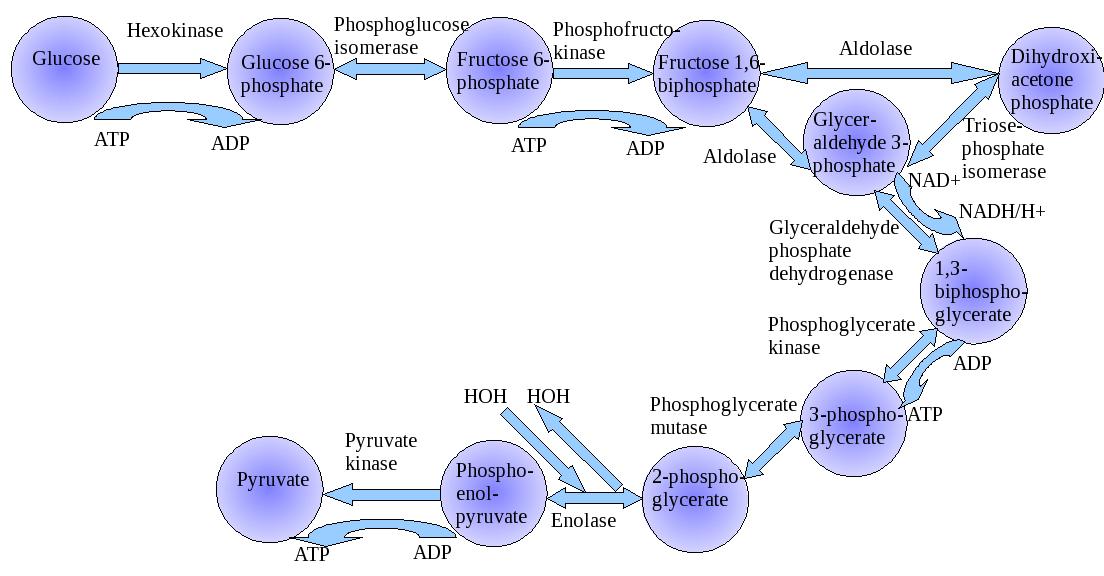