Personalized Medicine in Cancer [Chapter 3]
Writer and Curator: Larry H Bernstein, MD, FCAP
Personalized Medicine in Cancer
This chapter has the following ten Subsection:
3.1 The path to personalized medicine
3.2 Role of Nanobiotechnology in Developing Personalized Medicine for Cancer
3.3 The HER-2 Receptor and Breast Cancer: Ten Years of Targeted
Anti–HER-2 Therapy and Personalized Medicine
3.4 Personalized Medicine is not yet here
3.5 Biomarkers for personalized oncology: recent advances and future challenges.
3.6 Personalized oncology: recent advances and future challenges.
3.7 Pharmacogenomic biomarkers for personalized cancer treatment.
3.8 Limits to forecasting in personalized medicine: An overview
3.9 The genome editing toolbox: a spectrum of approaches for targeted modification
3.10 The Path to Personalized Medicine
3.1 The path to personalized medicine
Joanne M Meyer* and Geoffrey S Ginsburg
Current Opinion in Chemical Biology 2002, 6:434–438
Advances in personalized medicine, or the use of an individual’s molecular profile to direct the practice of medicine, have been greatly enabled through human genome research. This research is leading to the identification of a range of molecular markers for predisposition testing, disease screening and prognostic assessment, as well as markers used to predict and monitor drug response. Successful personalized medicine research programs will not only require strategies for developing and validating biomarkers, but also coordinating these efforts with drug discovery and clinical development.
The realization of personalized medicine, or the fine tailoring of the practice of medicine to an individual, is being fostered through numerous efforts aimed at characterizing individual differences in molecular processes underlying disease pathogenesis, disease progression and the response to therapeutics. Once these molecular differences are understood, therapeutic development will be enhanced by using the information to identify individuals more likely to benefit from a given intervention strategy. High-throughput genomic technologies are already providing the data that will serve as the foundation of personalized medicine.
Individual differences in the development of disease and response to therapeutics
Clearly, for many common diseases, there is abundant evidence to suggest that the molecular underpinnings of disease susceptibility, and its natural history, differ markedly among individuals. For example, while it has been demonstrated in numerous investigations that the development of obesity, asthma, type 2 diabetes and cardiovascular disease are under genetic control [1–4], there is no evidence to suggest that the genetic basis is due to variation in just a single gene. Instead, the consensus has emerged that subtle genetic differences in one or many of several genes serve as risk factors for these illnesses. Thus, while genetic variants in the melanocortin-4 receptor may explain some risk for developing obesity [5], and polymorphisms in PPARgamma may correlate with the risk of developing type 2 diabetes [6•], these variants do not explain all of these genetic diseases. There are certainly more genetic variants, or predisposition markers, to uncover. In the context of personalized medicine, the ultimate goal of these types of studies is to provide a suite of markers that can be used to assess one’s lifetime risk of developing disease in the presence of various environmental (e.g. diet, lifestyle) variables.
As with disease predisposition, individual differences characterize disease progression. For example, some individuals with impaired glucose tolerance will proceed quite rapidly to type 2 diabetes, whereas others proceed slowly. Similarly, individuals diagnosed with rheumatoid arthritis may or may not develop erosive disease. In both of these cases, genetic variation, that is, variation measured at the DNA level, may be a good predictor of the individual differences that emerge as disease progresses. For example, Brinkman et al. [7] have demonstrated that a polymorphism in TNF-α correlates with erosive rheumatoid arthritis, but shows no association with non-erosive disease. Alternatively, variation in disease progression may be best predicted by a combination of genetic and environmental factors, the impact of which is indexed through changes in gene expression in relevant tissues, or changes in secreted protein levels in serum or synovial fluid. In our laboratories, we are using a range of genomics technologies to find markers for disease progression that are both stable (DNA) as well as dynamic (mRNA, protein), giving us the opportunity to evaluate the utility of both types of markers in prospective studies.
Given that individual variability in disease predisposition and progression exists and has the potential of being molecularly characterized, it is not at all surprising that such differences also characterize response to therapeutics (see Figure 1). Marked individual variation in the efficacy and toxicity of therapeutic compounds is common and can have a profound impact on the success of a pharmaceutical clinical development program. Clearly, molecular markers that predict the variation in these endpoints could be extremely useful in clinical trials, drug development and clinical practice, as they would allow the identification of patients who would benefit most from the drug.
Technological advances drive broad biomarker discovery. While the existence of individual differences in disease predisposition, progression and response to therapeutics is far from a novel concept, our ability to comprehensively measure the molecular markers that track these processes, and draw proper inferences from large amounts of molecular data, is novel. Over the past decade, significant advancements have been made in technologies to discover variation at the mRNA, DNA and protein levels. Indeed, with the advent of glass and nylon microarray technologies for gene-expression studies, it is quite feasible to characterize the expression levels of 30 000 genes in tissue samples from dozens, if not hundreds, of individuals. Certainly, several years ago, although it would have been theoretically possible to assess this number of genes using northern blot analysis, it never would have been undertaken in a sample from even a single individual. In the same fashion, highthroughput technologies for DNA polymorphism discovery and single nucleotide polymorphism (SNP) genotyping, coupled with broad academic and commercial initiatives to characterize genetic variation genome-wide [8•], are resulting in catalogs of variants that can be used in large-scale experiments. To complement these efforts, searches for ‘haplotype blocks’, or correlated patterns of SNPs that can be adequately represented by fewer SNPs, are underway and have the promise of reducing the amount of genotyping required for genome-wide searches [9•,10•]. For proteinbased discovery initiatives, traditional 2D electrophoresis experiments are used in conjunction with advanced mass spectrometry to discover protein markers in a range of complex fluids, including serum, plasma, synovial fluid and cerebral spinal fluid.
Coupled with the advent of these technologies have been extensive efforts to collect appropriate tissues and fluids for mRNA, DNA and protein analysis. These collections have been part of pharmaceutical clinical trials, as well as clinical studies established for the purpose of characterizing biomarkers. The latter studies may involve small numbers of patient samples for initial biomarker discovery efforts, as well as large-scale, disease registry initiatives designed to evaluate and, in some cases, prospectively validate, biomarkers in the relevant patient populations.
Figure 1 (not shown) The role of molecular biomarkers in disease management. Areas where molecular biomarkers will benefit personalized medicine include disease predisposition, screening and prognosis, as well as drug response and drug monitoring. The nature of the markers (DNA, mRNA or protein) will vary with the disease and the stage of their application. Rx indicates treatment.
The predictive value of biomarkers The impact that advanced genomic technologies and carefully designed biomarker studies will have on the personalization of medicine is foreshadowed in the current literature. For example, Mallal et al. [13•] conducted a pharmacogenetic investigation (i.e. a genetic study of drug response) of abacavir, an HIV-1 nucleoside reverse transcriptase inhibitor. They implicated MHC alleles that predict response to hypersensitivity among 5% of the HIV cases receiving the drug. Their findings suggest that screening patients for the presence of the predisposing MHC haplotype could reduce the prevalence of hypersensitivity to abacavir from 9% to 2.5%. While this study is small in scale in its characterization of genetic variation, it adds to the existing literature on several other variants (including those in MDR1, the multidrug transporter P-glycoprotein and CYP2D6, a cytochrome P450 isoszyme) that correlate with the pharmacokinetic (drug clearance) characteristics of protease inhibitors and non-nucleoside reverse transcriptase inhibitors [14]. Additionally, genetic polymorphisms in chemokines and chemokine receptors (including RANTES, MIP-1α and CCR5) have been found to correlate with both the susceptibility to HIV-1 infection and the progression of disease [15•]. Taken together, these findings may lead to the development of a panel of polymorphisms that would personalize HIV therapy, by determining when to initiate therapy and how to choose compounds that will maximize efficacy and minimize adverse effects.
Figure 2 (not shown) Personalized medicine — integrating drug discovery and development through molecular medicine. Genomically derived biomarkers are being identified throughout the drug discovery and clinical development process. They will not only support personalized medicine, but will also enhance drug discovery and clinical development by generating new targets, validating targets and identifying patients that will benefit from novel therapeutics.
Pharmacogenetic efforts have also successfully characterized polymorphisms that correlate with response to asthma therapeutics. For example, Drazen et al. [16•] showed that a promotor polymorphism in 5-lipoxygenase, which alters transcription levels of the gene, also correlates with response to a derivative of the drug Zileuton, a 5-lipoxygenase inhibitor. Of the individuals who did not respond to Zileuton, 20% carried rare variant alleles at this locus. By contrast, all of the responders had wild-type alleles. Similarly in a study of genetic polymorphisms of the β-adrenergic receptor, Drysdale et al. [17•] demonstrated that a haplotype, or SNP signature across the gene, correlated strongly with asthma patients’ response to β-agonists. These two examples again demonstrate the possibility of using an individual’s genotype to suggest a therapeutic strategy that is more likely to be efficacious. Certainly, before such tests are incorporated into clinical practice, additional genetic markers would have to be coupled with the existing polymorphisms to make the resulting tests highly sensitive and specific.
In addition to these DNA-based strategies, recent applications of proteomics and expression profiling have generated a range of screening, prognostic and drug-response or ‘pharmacogenomic’ biomarkers. Many advances in the use of these technologies have been in oncology, where there is a tremendous need for serum-based screening markers and where tissue samples for expression profiling studies are easily obtained. For example, Petricoin et al. [18•] demonstrated that proteomic spectra, derived from a mass spectrometry analysis of serum, could be used to distinguish women with ovarian cancer from unaffected women. Indeed, the protein markers on a ‘training set’ of 100 samples and a validation set of 110 additional samples, had a sensitivity of 100% and a specificity of 94%. These encouraging results suggest that a serum-based protein assay may indeed become a viable mode of ovarian cancer screening in the general population. mRNA strategies for identifying prognostic markers for cancers have also proved successful. For example, in our collaborative studies [19•], we have shown that Melastatin, a melanocyte-specific gene identified through a genomics analysis of benign and malignant melanoma, is an effective prognostic marker for cutaneous malignant melanoma. In this work, uniform melastatin mRNA expression correlated strongly with disease-free survival, even after adjusting for other prognostic factors. In a similar fashion, mRNA strategies have generated pharmacogenomic markers for ovarian cancer. Hartmann et al. [20] studied the expression of 30 000 human genes in 51 tumors that were sensitive and resistant to platinum–paclitaxel chemotherapy and identified a subset of 10 markers that were highly predictive of outcome in an independent sample of tumors. Overall, these examples of biomarker studies in oncology demonstrate the broad application such markers will have for cancer screening, prognosis and response to therapeutics.
Turning biomarker discoveries into personalized medicines All of the examples cited provide excellent demonstrations of the power of new technologies to deliver a range of biomarkers that index individual differences in disease predisposition, progression and response to therapeutics. Thus, they clearly form a basis for the ‘personalization’ of medicine. However, the discovery of these markers is not sufficient for the pharmaceutical industry to deliver personalized medicines. Indeed, the delivery of such medicines will require the careful integration of biomarker discovery and validation programs into drug discovery and clinical development programs (see Figure 2). This integration will serve two key purposes. First, and foremost, by initiating SNP, expression profiling and proteomics biomarker programs early on in the drug discovery process, one can carefully weave the discovery and validation of biomarkers into drug discovery and development timelines; the risk of ‘retro-fitting’ biomarker programs to a clinical trial would be avoided.
Conclusions Clearly, several challenges remain to achieve a successful integration of large-scale, biomarker studies with drug development. While there has been an incredible advance in high-throughput, molecular technologies, over the past several years, further improvements in technologies and validation strategies are required to capture the true extent of individual differences in molecular markers. For example, although it is plausible to consider screening the genome for SNPs or haplotypes that correlate with disease pre-disposition or drug response, the current cost of SNP genotyping makes this impractical. Additionally, bioinformatic and statistical advances are needed to extract the most relevant data from the wealth of molecular information generated by new technologies, and these advances must be effectively communicated to the heath-care environment. Finally, and most importantly, plans must be in place to provide adequate validation for the enormous number of candidate biomarkers that will emerge from the studies. Validation will require access to large, and in some cases, prospective, collections of well annotated clinical samples with appropriate consent and security issues addressed. While these issues, as well as the commercial and regulatory considerations around the development of personalized medicines, are indeed challenging, the successful execution of biomarker programs will have an enormous impact on our ability to tailor medical practice to the individual.
3.2 Role of Nanobiotechnology in Developing Personalized Medicine for Cancer
K. K. Jain
Technol Cancer Res Treat Dec 2005; 4(6): 645-650
http://dx.doi.org:/10.1177/153303460500400608
Personalized medicine simply means the prescription of specific therapeutics best suited for an individual. Personalization of cancer therapies is based on a better understanding of the disease at the molecular level. Nanotechnology will play an important role in this area. Nanobiotechnology is being used to refine discovery of biomarkers, molecular diagnostics, drug discovery and drug delivery, which are important basic components of personalized medicine and are applicable to management of cancer as well. Examples are given of the application of quantum dots, gold nanoparticles, and molecular imaging in diagnostics and combination with therapeutics – another important feature of personalized medicine. Personalized medicine is beginning to be recognized and is expected to become a part of medical practice within the next decade. Personalized management of cancer, facilitated by nanobiotechnology, is expected to enable early detection of cancer, more effective and less toxic treatment increasing the chances of cure.
3.3 The HER-2 Receptor and Breast Cancer: Ten Years of Targeted Anti–HER-2 Therapy and Personalized Medicine
Jeffrey S. Ross, Elzbieta A. Slodkowska, W. Fraser Symmans, et al.
The Oncologist 2009; 14:320 –368
http://cme.theoncologist.com
Objectives:
- Contrast the current strengths and limitations of the three main slide-based techniques (IHC, FISH, and CISH) currently in clinical use for testing breast cancer tissues for HER-2 status.
- Compare the efficacy of trastuzumab- and lapatinib-based regimens in the adjuvant and metastatic settings as reported in published clinical trials and regulatory approval databases.
- Contrast the list of biomarkers that have been associated with clinical resistance to trastuzumab and lapatinib and describe their current level of validation.
The human epidermal growth factor receptor (HER-2) oncogene encodes a transmembrane tyrosine kinase receptor that has evolved as a major classifier of invasive breast cancer and target of therapy for the disease. The validation of the general prognostic significance of HER-2 gene amplification and protein overexpression in the absence of anti–HER-2 targeted therapy is discussed in a study of 107 published studies involving 39,730 patients, which produced an overall HER-2– positive rate of 22.2% and a mean relative risk for overall survival (OS) of 2.74. The issue of HER-2 status in primary versus metastatic breast cancer is considered along with a section on the features of metastatic HER- 2–positive disease. The major marketed slide-based HER-2 testing approaches, immunohistochemistry, fluorescence in situ hybridization, and chromogenic in situ hybridization, are presented and contrasted in detail against the background of the published American Society of Clinical Oncology–College of American Pathologists guidelines for HER-2 testing. Testing issues, such as the impact of chromosome 17 polysomy and local versus central HER-2 testing, are also discussed. Emerging novel HER-2 testing techniques, including mRNA-based testing by real-time polymerase chain reaction and DNA microarray methods, HER-2 receptor dimerization, phosphorylated HER-2 receptors, and HER-2 status in circulating tumor cells, are also considered. A series of biomarkers potentially associated with resistance to trastuzumab is discussed with emphasis on the phosphatase and tensin homologue deleted on chromosome ten/Akt and insulin-like growth factor receptor pathways. The efficacy results for the more recently approved small molecule HER- 1/HER-2 kinase inhibitor lapatinib are also presented along with a more limited review of markers of resistance for this agent. Additional topics in this section include combinations of both anti–HER-2 targeted therapies together as well as with novel agents including bevacizumab, everolimus, and tenespimycin. A series of novel HER-2–targeting agents is also presented, including pertuzumab, ertumaxomab, HER-2 vaccines, and recently discovered tyrosine kinase inhibitors. Biomarkers predictive of HER-2 targeted therapy toxicity are included, and the review concludes with a consideration of HER-2 status in the prediction of response to non–HER-2 targeted treatments including hormonal therapy, anthracyclines, and taxanes.
Biology, Pathology, Diagnosis, And Clinical Significance Of Her-2–Positive Breast Cancer
The human epidermal growth factor receptor 2 (HER-2, HER-2/neu, c-erbB-2) gene, first discovered in 1984 by Weinberg and associates [1], is localized to chromosome 17q and encodes a transmembrane tyrosine kinase receptor protein that is a member of the epidermal growth factor receptor (EGFR) or HER family (Fig. 1) [2]. This family of receptors is involved in cell– cell and cell–stroma communication primarily through a process known as signal transduction, in which external growth factors, or ligands, affect the transcription of various genes, by phosphorylating or dephosphorylating a series of transmembrane proteins and intracellular signaling intermediates, many of which possess enzymatic activity. Signal propagation occurs as the enzymatic activity of one protein turns on the enzymatic activity of the next protein in the pathway [3]. Major pathways involved in signal transduction, including the Ras/mitogen-activated protein kinase pathway, the phosphatidylinositol 3 kinase (PI3K)/Akt pathway, the Janus kinase/signal transducer and activator of transcription pathway, and the phospholipase C pathway, ultimately affect cell proliferation, survival, motility, and adhesion. Receptor activation requires three variables, a ligand, a receptor, and a dimerization partner [4]. After a ligand binds to a receptor, that receptor must interact with another receptor of identical or related structure in a process known as dimerization in order to trigger phosphorylation and activate signaling cascades. Therefore, after ligand binding to an EGFR family member, the receptor can dimerize with various members of the family (EGFR, HER-2, HER-3, or HER-4). It may dimerize with a like member of the family (homodimerization) or it may dimerize with a different member of the family (heterodimerization). The specific tyrosine residues on the intracellular portion of the HER-2/neu receptor that are phosphorylated, and hence the signaling pathways that are activated, depend on the ligand and dimerization partner. The wide variety of ligands and intracellular crosstalk with other pathways allow for significant diversity in signaling. Although no known ligand for the HER-2 receptor has been identified, it is the preferred dimerization partner of the other family members. HER-2 heterodimers are more stable [5, 6] and their signaling is more potent [7] than receptor combinations without HER-2. HER-2 gene amplification and/or protein overexpression has been identified in 10%–34% of invasive breast cancers [1]. Unlike a variety of other epithelial malignancies, in breast cancer, HER-2 gene amplification is uniformly associated with HER-2 (p185neu) protein overexpression and the incidence of single copy overexpression is exceedingly rare [8]. HER-2 gene amplification in breast cancer has been associated with increased cell proliferation, cell motility, tumor invasiveness, progressive regional and distant metastases, accelerated angiogenesis, and reduced apoptosis [9].When classified by routine clinicopathologic parameters and compared with HER-2– negative tumors, HER-2–positive breast cancer is more often of intermediate or high histologic grade, more often lacking estrogen receptors (ERs) and progesterone receptors (PgRs) (ER and PgR negative), and featuring positive lymph node metastases at presentation [1]. In the recent molecular classification of breast cancer, positive HER-2 status does not constitute a unique molecular category and is identified in both the “HER-2” and “luminal” tumor classes [10].
Figure 1 (not shown)
Figure 1. The human epidermal growth factor receptor (HER) gene family. This image depicts the complex crosstalk between members of the HER family of receptor tyrosine kinases and intracellular signaling. Activated HER receptors can function to both stimulate and inhibit downstream signaling of members of other biologic pathways. Note that HER-2 has no activating ligands and HER-3 lacks a tyrosine kinase domain. HER-2–mediated signaling is associated with cell proliferation, motility, resistance to apoptosis, invasiveness, and angiogenesis. The figure shows the complexity of signaling pathways initiated by, and influenced by, HER family protein receptors at the cell surface.
HER-2 Status and Prognosis in Breast Cancer Both morphology-based and molecular-based techniques have been used to measure HER-2/neu status in breast cancer clinical samples [11–117]. By a substantial majority, abnormalities in HER-2 expression at the gene, message, or protein level have been associated with adverse prognosis in both lymph node–negative and lymph node–positive breast cancer. Of the 107 studies considering 39,730 patients listed in Table 1, 95 (88%) of the studies determined that either HER-2 gene amplification or HER-2 (p185 neu) protein overexpression predicted breast cancer outcome on either univariate or multivariate analysis. In 68 (73%) of the 93 studies that featured multivariate analysis of outcome data, the adverse prognostic significance of HER-2 gene, message, or protein overexpression was independent of all other prognostic variables. In only 13 (12%) of the studies, no correlation between HER-2 status and clinical outcome was identified. Of these 13 noncorrelating studies, eight (62%) used immunohistochemistry (IHC) on paraffin-embedded tissues as the HER-2/protein detection technique, two (15%) used fluorescence in situ hybridization (FISH), two (15%) used Southern analysis, and one (7%) used a real-time polymerase chain reaction (RT-PCR) technique. Of the 15 studies that used the FISH technique, 13 (87%) showed univariate prognostic significance of gene amplification, and 11 of these (85%) showed prognostic significance on multivariate analysis as well. The two studies that used chromogenic in situ hybridization (CISH) HER-2 gene amplification detection techniques both found that HER-2 amplification was an independent predictor of outcome on multivariate analysis [100, 112]. However, interpretation of these studies is complicated by the fact that most studies included patients who received variable types of systemic adjuvant therapy; therefore, the pure prognostic value of HER-2 overexpression in the absence of any systemic adjuvant therapy is incompletely understood.
Table 1 HER-2 status and prognosis in breast cancer (not shown)
HER-2 Positivity Rates The frequency of HER-2 positivity in all of the studies presented in Table 1 was 22.2%, with a range of 9%–74%. The HER-2–positive rate was similar for IHC, at 22% (range, 10%–74%), and FISH, at 23.9% (range, 14.7%– 68%). In current practice, HER-2–positive rates have trended below 20%, with most investigators currently reporting that the true positive rate is in the range of 15%–20%. The HER-2– positive rate may be higher when metastatic lesions are tested, and tertiary hospitals and cancer centers report slightly higher rates than community hospitals and national reference laboratories. Relative Risk and Hazard Ratio In Table 1, a number of studies provided data as to the relative risk (RR) of untreated HER-2–positive breast cancer being associated with an adverse clinical outcome. For OS, the mean RR was 2.74 (range, 1.39 – 6.93) and the median was 2.33; for disease-free survival (DFS), the mean RR was 2.04 (range, 1.30 –3.01) and the median was 1.8. In several studies, the RR was estimated with a hazard ratio (HR) model. The mean HR was 2.12 (range, 1.6 –2.7) and the median was 2.08. HER-2 Expression and Breast Pathology The association of HER-2–positive status with specific pathologic conditions of the breast is summarized in Table 2. HER-2 overexpression has been consistently associated with higher grades and extensive forms of ductal carcinoma in situ (DCIS) and DCIS featuring comedo-type necrosis [118 –121]. The incidence of HER-2 positivity in DCIS has varied in the range of 24%–38% in the published literature, which appears to be slightly higher than that for invasive breast cancer [118 –121]. Routine testing for HER-2 status in DCIS is not widely performed. However, should anti– HER-2 targeted therapies directed at HER-2–positive DCIS result in a reduction in the development of invasive disease, the widespread use of HER-2 testing in DCIS would be adopted. Finally, the invasive carcinoma that develops in association with HER-2–positive DCIS may, on occasion, not feature a HER-2–positive status, a finding that has led investigators to believe that HER-2 gene amplification may not be required for the local progression of breast cancer [122]. Compared with invasive ductal carcinoma (IDC), HER-2 gene amplification occurs at a significantly lower rate in invasive lobular carcinoma (ILC) (10%), but has also been linked to an adverse outcome [85]. HER-2 positivity is linked exclusively to the pleomorphic variant of ILC and is not encountered in classic ILC [123]. HER-2 amplification is strongly correlated with tumor grade in both IDC and ILC. For example, in one study, only one of 73 grade I IDC cases and one of 67 low-grade classic ILC cases showed HER-2 amplification detected by FISH [86]. HER-2 overexpression and HER-2 amplification have been a consistent feature of both mammary and extramammary Paget’s disease [124, 125] (Fig. 2). HER-2 amplification and HER-2 overexpression have been associated with adverse outcome in some studies of male breast carcinoma [126 –129], but not in others [130 –132]. The incidence of HER-2 positivity appears to be lower in male breast cancer than in female breast cancer [126 –132]. Documented responses in male breast cancer to HER-2–targeting agents have been described, and therefore treatment with trastuzumab is an acceptable option for these patients, but the true activity rate remains uncertain [133]. The rate of HER-2 overexpression in mucinous (colloid) breast cancers is extremely low, although, on occasion, it has been associated with aggressive disease [134 –136]. In medullary breast carcinoma, HER-2 testing has consistently found negative results [137]. Similarly, HER-2 positivity is extremely rare in cases of tubular carcinoma [138]. HER-2 status has not been consistently linked to the presence of inflammatory breast cancer [139, 140]. Molecular studies of hereditary breast cancer including cases with either BRCA1 or BRCA2 germline mutations have found a consistently lower incidence of HER-2–positive status for these tumors [141].
Figure 2 not shown
Figure 2. Human epidermal growth factor receptor (HER)-2–positive Paget’s disease of the nipple. In this patient, who presented with HER-2–positive invasive duct carcinoma, classic clinical features of Paget’s disease of the nipple were present. A section of the nipple from the mastectomy specimen shows 3+ continuous cell membrane immunoreactivity for HER-2 protein. Nearly 100% of Paget’s disease of the breast cases are HER-2 positive (see text).
Breast sarcomas and phyllodes tumors have consistently been HER-2 negative [142]. Finally, low-level HER-2/neu overexpression has been identified in benign breast disease biopsies and is associated with a greater risk for subsequent invasive breast cancer [143].
HER-2 Status in Primary Versus Metastatic Breast Cancer The majority of studies that have compared the HER-2 status in paired primary and metastatic tumor tissues have found an overwhelming consistency in the patient’s status regardless of the method of testing (IHC versus FISH) [144 –151]. However, several recent studies indicated 20%–30% discordance rates between the HER-2 status of primary and metastatic lesions. Some of these studies have featured relatively high HER-2–positive rates on both paired specimens (> 35% positive), which has created concern about the conclusions of these reports [152]. Also, considering that 10%–30% discordance rates have been reported even when the same tumor is tested repeatedly, it remains uncertain if the discordance rates seen between primary and metastatic sites is higher than expected by the less than perfect reproducibility of the various HER-2 assays. Increasingly, emerging data suggest that there are changes in HER-2 expression between primary and metastatic disease. This is particularly true after intervening HER-2– directed therapy, but also happens in the absence of such treatment. In cases where the original primary HER-2 test result is questioned because of technical or interpretive issues and in patients where there has been an unusually long (i.e., > 5-year) interval between the primary occurrence and the detection of metastatic disease, retesting of a metastatic lesion may be warranted. Thus, although routine HER-2 testing of metastatic disease is advocated by some investigators, the preponderance of data indicates that the HER-2 status remains stable and that routine retesting of HER-2 may not be needed for most patients with metastatic disease.
Features of Metastatic HER-2–Positive Breast Cancer Metastatic HER-2–positive breast cancer retains the phenotype of the primary tumor not only in HER-2 status, but also is typically ER/PgR negative, moderate to high tumor grade, DNA aneuploid with high S phase fraction, and featuring ductal rather than lobular histology. In the era prior to the initiation of HER-2–targeted therapy, HER-2–positive breast cancer was more likely to spread early to major visceral sites including the axillary lymph nodes, bone marrow, lungs, liver, adrenal glands, and ovaries [153]. In the post–HER-2 targeted therapy era, the incidence of progressive visceral metastatic disease in HER-2–positive tumors has diminished and has frequently been superseded by the development of clinically significant central nervous system (CNS) metastatic disease [154 –157]. It is widely held that the success in the control of visceral disease with trastuzumab has unmasked previously occult CNS disease and, because of the inability of the therapeutic antibody to cross the blood– brain barrier, allowed brain metastases to progress during the extended OS duration of treated patients [154, 155]. The small-molecule drug lapatinib has shown some promise for targeting HER-2–positive CNS metastases that are resistant to trastuzumab-based therapies in initial studies [158].
Interaction of HER-2 Expression with Other Prognosis Variables HER-2 gene amplification and protein overexpression have been associated consistently with high tumor grade, DNA aneuploidy, high cell proliferation rate, negative assays for nuclear protein receptors for estrogen and progesterone, p53 mutation, topoisomerase IIa amplification, and alterations in a variety of other molecular biomarkers of breast cancer invasiveness and metastasis [159 –161].
Figure 3. Human epidermal growth factor receptor (HER)-2 testing.
(not shown) (A): Immunohistochemistry (IHC). This panel depicts the four categories of HER-2 IHC staining including 0 and 1+ (negative), 2+ (equivocal), and 3+ (positive) using the American Society of Clinical Oncology–College of American Pathologists guidelines for HER-2 IHC scoring. (B): Fluorescence in situ hybridization (FISH). This panel demonstrates a case of invasive duct carcinoma, on the left, negative for HER-2 gene amplification (gene copy number < 4) and a case of HER-2 gene–amplified breast cancer (gene copy number > 6),
FISH. The FISH technique (Fig. 3B), like IHC, is a morphology-driven slide-based DNA hybridization assay using fluorescent-labeled probes. Both the hybridization steps and the slide scoring can be automated. FISH has the advantages of a more objective scoring system and the presence of a built-in internal control consisting of the two HER-2 gene signals present both in benign cells and in malignant cells that do not feature HER-2 gene amplification.
IHC Versus FISH. Although the FISH method is more expensive and time-consuming than IHC, numerous studies have concluded that this cost is well borne by the greater accuracy and more precise use of anti–HER-2 targeted therapies [179 –180, 182–183]. FISH is considered to be more objective and reproducible in a number of systematic reviews [165, 180, 183–186]. In one study, the concordance rates between IHC and FISH were highest in tumors scored by IHC as 0 and 1+ and lowest for 2+ and 3+ cases [183]. Currently, the majority (approximately 80%) of HER-2 testing in the U.S. commences with a screen by IHC, with results of 0 and 1+ considered negative, 2+ considered equivocal and referred for FISH testing, and 3+ considered positive. In a pharmacoeconomic study of patients being considered for trastuzumab-based treatment for HER-2– positive tumors, FISH was found to be a cost-effective diagnostic approach “from a societal perspective” [187].
CISH and Silver In Situ Hybridization. The CISH method (Fig. 3E) and silver in situ hybridization (SISH) method feature the advantages of both IHC (routine microscope, lower cost, familiarity) and FISH (built-in internal control, subjective scoring, the more robust DNA target) [190, 191]. The CISH technique uses a single HER-2 probe, detects HER-2 gene copy number only, and was recently approved by the FDA to define patient eligibility for trastuzumab treatment. The SISH method employs both HER-2 and chromosome 17 centromere probes hybridized on separate slides and is currently under review by the FDA. Numerous studies have confirmed a very high concordance between CISH and FISH, typically in the 97%–99% range [191–203]. Similar to FISH, CISH has its highest correlation with IHC 0, 1+, and 3+ results and lowest correlation with IHC 2+ staining.
Chromosome 17 Polysomy. The incidence of chromosome 17 polysomy has varied from as low as 4% to as high as 30% in studies of invasive breast cancer [204 –208]. This may reflect differences in the definition of polysomy ranging from a low-level definition of more than two copies per cell to a high of more than four copies per cell of the chromosome. Most studies have linked chromosome 17 polysomy with greater HER-2 protein overexpression [204 –207], but some have found that protein overexpression only occurs in the presence of selective HER-2 gene amplification [204].
The 2007 ASCO-CAP Guidelines. In early 2007, a combined task force from ASCO and the CAP issued a series of recommendations designed to improve the accuracy of tissue-based HER-2 testing in breast cancer [212]. A summary of the ASCO-CAP guidelines is provided in Table 4. Highlights of these recommendations include (a) standardizing fixation in neutral-buffered formalin for no less than 6 hours and no more than 48 hours, (b) unlike their respective FDA-approval specifications, defining equivocal zones for the IHC, FISH, and CISH tests, (c) establishing a standardized quality assurance program for testing laboratories, and (d) requiring the participation of these laboratories in a proficiency testing program [212]. The published guidelines were designed to improve the overall precision and reliability of all types of slide-based HER-2 tests and remained neutral as to the relative superiority of one test over the others.
Figure 4. Real-time polymerase chain reaction (RT-PCR). In this RT-PCR assay using the Taqman RT-PCR System (Applied Biosystems Inc., Foster City, CA), note the detection of increased human epidermal growth factor receptor(HER)-2 mRNA expression in green detected at lower numbers of amplification cycles compared with the two housekeeping genes shown in red and blue.
Figure 5. DNA microarray. In this image, increased expression of human epidermal growth factor receptor (HER)-2 mRNA has been detected using a proprietary DNA microarray system (Millennium Pharmaceuticals, Inc., Cambridge, MA). The microarray demonstrates the coexpression of seven genes (HER-2 is second from the bottom) related to the amplification of HER-2 DNA in this case of HER-2–positive breast cancer.
Her-2–Targeted Therapy and the Treatment of Her-2–Positive Breast Cancer
Trastuzumab: HER-2 Testing and the Prediction of Response to Trastuzumab Therapy Using recombinant technologies, trastuzumab (Herceptin; Genentech, South San Francisco, CA), a monoclonal IgG1 class humanized murine antibody, was developed by the Genentech Corporation to specifically bind the extracellular portion of the HER-2 transmembrane receptor. This antibody therapy was initially targeted specifically for patients with advanced relapsed breast cancer that overexpresses HER-2 protein [262]. Since its launch in 1998, trastuzumab has become an important therapeutic option for patients with HER-2–positive breast cancer and is widely used for its approved indications in both the adjuvant and metastatic settings (Fig. 6) [185, 263–265]. Although trastuzumab is approved as a single-agent regimen, most patients are treated with trastuzumab plus cytotoxic agents. Table 5 summarizes the significant clinical trials that contributed to the regulatory approvals of trastuzumab.
This topic is scheduled for another article.
Trastuzumab Combinations. Since the FDA approval in 1998 of two trastuzumab plus chemotherapy combinations, a number of additional approaches have gained favor in the clinical practice community. The National Comprehensive Cancer Network (NCCN) Clinical Practice Guidelines [284] currently recommend the following regimens for the first-line treatment of HER-2–positive MBC: trastuzumab plus single agents— either paclitaxel (every 3 weeks or weekly), docetaxel (every 3 weeks or weekly), or vinorelbine (weekly). For combination therapies, the NCCN recommends trastuzumab plus paclitaxel and carboplatin (every 3 weeks) or docetaxel plus carboplatin. Recently, carboplatin-based trastuzumab combinations have gained interest as a result of both the apparent boost in efficacy as measured by a higher overall response rate and longer progression-free survival time and the cardioprotective benefits of avoiding an anthracycline-containing regimen [285].
Neoadjuvant Setting The results of trastuzumab-based neoadjuvant studies (Table 5) have received significant recent interest in the oncology community [289]. Virtually all completed and in progress clinical trials have demonstrated a significant enhancement in the rate of pathologic complete response (pCR), the primary endpoint in these studies, in cases of patients with HER-2–positive breast cancer that received trastuzumab in the neoadjuvant setting [290 –297]. This benefit of the addition of trastuzumab in the neoadjuvant setting appears to be independent of, if not enhanced by, the coexistence of ER positivity [297]. Among the potential explanations for the apparent greater chemosensitivity of HER-2–positive tumors cotreated with trastuzumab in the neoadjuvant setting is the concept that HER-2 gene amplification is in some way related to the growth and survival of breast cancer stem cells [298, 299].
Biomarkers of Trastuzumab Resistance Since trastuzumab was introduced for the treatment of MBC in 1998, there has been growing interest in the discovery and potential clinical utility of biomarkers designed to predict resistance to the drug. Current approaches to HER-2 testing provide a negative predictor of drug response: the test does not predict which patients will respond to trastuzumab, it predicts which patients are unlikely to benefit.
Neoadjuvant Setting The Neo-ALTTO trial is a randomized, open-label, multicenter, phase III study comparing the efficacy of neoadjuvant lapatinib plus paclitaxel with that of trastuzumab plus paclitaxel and with concomitant lapatinib and trastuzumab plus paclitaxel given as neoadjuvant treatment in HER-2– positive primary breast cancer [337].
Biomarkers of Lapatinib Resistance In that lapatinib was approved 9 years after trastuzumab, considerably less information has been published concerning markers of efficacy or resistance to the drug [331, 341– 343].
Trastuzumab Since its introduction in the MBC setting and continuing throughout its advance into use in both the adjuvant and neoadjuvant settings, trastuzumab has been associated with the development of a variety of toxicities [384]. In the original registration trial for MBC, trastuzumab was associated with a variety of adverse events, including pain, gastrointestinal disturbances, minor hematologic deficiencies, pulmonary symptoms, and congestive heart failure (CHF) [265]. Cardiac toxicity has remained the most significant limiting factor for the use of trastuzumab [384 –389]. A major consideration in the development of cardiac toxicity in patients treated with trastuzumab has been their prior or concomitant exposure to anthracycline drugs, also associated with dose-dependent irreversible heart damage [384 – 389].
Lapatinib The most frequent adverse reactions in the lapatinib– capecitabine registration trial for MBC combination were diarrhea (65%), palmar–plantar erythrodysesthesia (53%), nausea (44%), rash (28%), vomiting (26%), and fatigue (23%) [332]. In a comprehensive analysis of the clinical trials featuring lapatinib in combination with various other agents, the overall incidence of LVEF decline was 1.6%, with 0.2% of patients experiencing symptomatic CHF [389].
HER-2 Status and the Prediction of Response to Non–HER-2 Targeted Therapy The use of HER-2 status to predict responsiveness or resistance to hormonal therapies, advocated by a number of oncologists, remains controversial. It has been reported that ER-positive/HER-2–positive patients are either less responsive or completely resistant to single-agent tamoxifen [391–393]. When measured as continuous variables, the expression of HER-2 appears to be inversely related to the expression of ER and PgR even in hormone receptor–positive tumors [394].
Anthracyclines HER-2 overexpression has also been associated with enhanced response rates to anthracycline-containing chemotherapy regimens in most, but not all, studies [42, 410 – 414].
Radiation Therapy Initially, in the era prior to the introduction of anti–HER-2 targeted therapy, HER-2–positive status was associated with a higher rate of local recurrence in some studies of breast cancer treated with surgery and radiation therapy alone, but not in others [427– 429]. However, although large-scale, randomized, prospective studies are lacking, HER-2–positive tumors treated with trastuzumab-based neoadjuvant chemotherapy combined with external-beam radiation have indicated a favorable response in locally advanced breast cancer [430].
Summary The history of the discovery of the HER-2 oncogene in an animal model in 1984, the translation of this finding to the clinical behavior of human breast cancer, and the introduction of the first anti-HER targeted therapy in 1998 is clearly a triumph of “bench to bedside” medicine. In the 10 years that have now passed since the regulatory approval of the first anti–HER-2 targeted therapy, trastuzumab, thousands of preclinical and clinical studies have considered HER-2 as a prognostic factor, its ability to predict response to hormonal and cytotoxic treatments, the best way to test for it in routine specimens, and the clinical efficacy of targeting it in a wide variety of clinical settings. Given the proven efficacy of trastuzumab and lapatinib for the treatment of MBC, and also in the adjuvant and neoadjuvant settings, the critical issue as to which test (IHC versus FISH versus CISH versus mRNA based) is the most accurate and reliable method to determine HER-2 status in breast cancer has continued to increase in importance.
3.4 Personalized Medicine is not yet here
ESMO Personalized Medicine
Written by Dr Marina Garassino for ESMO
https://www.esmo.org/content/download/20122/337223/file/ESMO-Patient-Guide-Personalised-Cancer-Medicine.pdf
The aim of personalised medicine is clearly to make therapy more efficient for patients. A very, very small step in the process is to try to identify for every patient the main molecular driver of their tumour. We have to understand that patients differ between each other, although they may have the same cancer type; for example, every patient with breast cancer or bowel cancer will have a unique tumor. This is entirely new knowledge, so what we are trying to do now in the medical community is to identify for each patient his/ her type of disease and then to give the drug that will work best. We are moving forward with an incredible amount of new data and innovative knowledge on genetic characteristics and subsequent proteomic changes* in the tumor. The challenge is now about how to exploit this information in order to offer targeted treatment and generally improve patient care.
For a number of years we have classified tumors according to their site of origin and using a classification system called “TNM”. Researchers and clinicians once thought that all cancers that derived from the same site were biologically similar and they differed perhaps only in their pathohistological* grading. This grading is a score which classifies tumors from 1 to 3, where 1 is the least aggressive tumor and 3 is the most undifferentiated tumor. Other clinical differences were distinguished based on the presence of regional node metastases or distant metastases. Most of the tumors were therefore classified within the “TNM” system, where T corresponds to the diameter of the primary tumor, N to the presence of regional nodes, and M to distant metastases. For at least three decades, personalization of oncology was based only on these parameters and on the patient’s physical condition, and even now these represent the fundamental elements for treatment decisions. Chemotherapy, surgery and radiation therapy were once the only treatment options for cancer. Although these treatments are still used, oncologists know that some patients respond better to certain drugs than to others and that a surgical approach is not always indicated. In recent years, researchers have studied thousands upon thousands of samples from all types of tumors. They have discovered that tumors derived from the same body site can differ in very important ways.
Firstly, there is histology*. The pathologist is able to distinguish different subtypes of cancer with the microscope. When a patient is diagnosed with a cancer, he/she will undergo a biopsy or a fine-needle aspiration. In some tumor types, debulking or removal of the primary tumor also allows sampling for tissue examination. Some cells of the tumor which have been removed will be taken and analyzed. This examination allows the pathologist to confirm a cancer diagnosis, but, through particular colorations of the tissue sample, the pathologist is also able to provide clinicians with a lot of additional information, such as the tumor’s histological characterization, its hormone sensitivity, and its grade of differentiation*.
For example, in the treatment of lung cancer the histology provides very useful tools to decide the best drug for the treatment of the patient. Clinical studies have shown that for a patient with lung adenocarcinoma* there might be more chance of a response if the drugs pemetrexed or bevacizumab are added to the chemotherapy, while for a patient with lung cancer of squamous* histology, it would be more beneficial to add gemcitabine or vinorelbine. A similar example may be observed Personalization of Oncological Treatments: The Story 12 for other cancers. For the treatment of esophageal cancer it is mandatory to know if the tumor is squamous or not, because although deriving from the same organ, the treatment approach is completely different.
This information is a useful tool in the first step of the personalization process. For example, lung cancer can be divided as a first step into non-small cell lung cancer and small cell lung cancer, which are two completely different neoplasms*. Within the non-small cell lung cancer category, there are again several different tumor types. Breast cancer can also be divided into two major categories: the hormone-sensitive neoplasms and the HER2-positive diseases. Lung and breast cancers are only two examples, because it is possible to recognize several entities within the same tumor type for many other cancers.
Molecular subsets of lung adenocarcinoma Lung cancer subtypes
Figure 2. Lung Cancer – Not One Disease: Histological (Tissue) and Molecular Subtypes of Lung Cancer (not shown) On the left side, four histological subtypes of lung cancer. On the right side, a pie chart showing the percentage distribution of molecular subsets of lung adenocarcinoma. Adapted from Petersen I. Dtsch Arztebl Int 2011; 108(31-32):525-531 (left) and Pao W & Hutchinson KE. Nature Med 2012; 18(3): 349-351.
Personalization depends on a multidisciplinary approach; we need a range of experts, because we need the medical oncologist, the surgeon and the expertise of the molecular pathologist, who should be part of the team in a more effective, integrated way than before. We don’t need the pathology report alone; we need to interact with all professionals, including nurses, who are dealing with the patient. This, to me, will create a lot of problems in terms of organization of care and in terms of cost, but it is the only way to bring together knowledge on the biology and pathology of tumors for effective treatment in every single patient. Our effort at ESMO is to bring this broad knowledge to the general public, to medical oncologists and to the community of doctors involved in cancer.
We have to deeply analyze each tumor of every patient in order to identify those genetic characteristics that make the tumor able to survive. As a result, we can choose the appropriate drugs to target the specific alterations. The clearest examples of this process are in melanoma, lung cancer and breast cancer. For instance, in lung cancer, the presence of mutations in the epidermal growth factor receptor (EGFR) renders the tumor highly sensitive to EGFR tyrosine kinase inhibitors. When oncologists identify these mutations in a patient’s tumor, they may observe that the lesion disappears a few weeks after treatment. A similar response may be observed after treatment with BRAF inhibitors in patients with melanoma or with gastrointestinal stromal tumors (GIST) that express the c-kit gene. Unfortunately, oncogene addiction is not the only process underlying carcinogenesis* and tumor growth. The tumor environment and so-called “epigenetic” alterations* play an important role in rendering the fight against cancer more and more challenging. Despite the enormous recent advances, a specific alteration has not been identified in all cancers. The hope is that the possibility of sequencing the full genome – which means every gene – will give us new insights and therefore new drugs for our patients.
In the DNA of some individuals a “germline” mutation* may be present. This means that a particular mutation is conferring susceptibility to that person to develop a particular type of cancer during his/her life. For instance, BRCA is an alteration for which there is a particular predisposition to have a breast cancer or ovarian cancer in one’s life. A woman with a BRCA gene mutation can transmit this alteration to her female descendants, so her daughters and following generations of female family members can therefore inherit this predisposition.
Mutations that are not germline are called somatic mutations*, which are acquired mutations and are found generally only in the tumor. Distinct from germline mutations, somatic mutations are not inherited.
The move from blockbuster or empirical medicine* towards personalized medicine is a stepwise process. We are currently on the second step of stratified medicine and moving up the stairs towards personalized medicine.
Will molecular pathology evolve from pathology? You need to give a name to a tumor, and a pathologist is the professional who gives a name to tumors. The variety of cancers is broad; when we say “sarcoma”, “carcinoma”, or “lymphoma”, we actually say nothing, because we have hundreds and hundreds of diseases within these categories that need to be recognized. And the reason for recognizing them is exactly related to personalization. The biology of cancer is very complex, and admittedly we have been very naive in the past. We always thought that the problem was how genes become altered in the cancer cell, but actually it is even more complex than that and also involves the way genes direct how they are read; it is the flow of information that comes from genes to the making of their proteins which is as important as the aberration of the genome.
We are facing obstacles currently because the whole issue of tissue sampling has been regulated under the umbrella of privacy, which is of course important. Defending your rights as a human being is a key issue, but we should also try to focus a little bit on the necessity to use that tissue. Of course, we need to have rules, but the approach we are currently facing is basically preventing clinical research and translational research under the excuse of protecting our privacy as human beings, and this is an increasing obstacle. We as researchers, as molecular geneticists, as pathologists, are really looking into a future in which it is becoming increasingly difficult to try to answer the basic question of cancer genomics. Why? Because it is becoming increasingly difficult to use tissue for these purposes.
With the new therapeutic approach and the use of targeted therapy, molecular testing is gaining a very relevant role. It is very important for us, as advocates, to educate patients in these issues. So patients have to receive very clear and transparent information. It should be the doctor who explains to the patient the reason why molecular testing is performed; the doctor has to explain that molecular testing will find whether there is some tumor characteristic which can be targeted with one of these therapies, in order to determine if maybe the patient is the right candidate to receive targeted therapy and perhaps to benefit from it. The communication between the doctor and patient must be very accurate and must educate, meaning that the patient has to understand the precise situation. This can be important also to empower the patient in treatment decisions, but it is important that he/she knows that not every patient may be a candidate for receiving targeted therapy and to understand why this is the case.
- Different tumour types are increasingly divided into very small subgroups carrying a rare molecular alteration.
- Most new drugs are targeting these infrequent events.
- Clinical trials are testing the use of high throughput molecular technologies* in the context of personalized cancer medicine.
- There are a growing number of newer techniques to optimize genomic testing, including the virtual cell program, which foresees testing of a piece of patient’s tumor tissue in the laboratory in order to mimic what would happen in the human body (e.g. drug sensitivity).
- Clinical research is today focusing on target identification at the patient level.
Targeted therapy drugs work differently to standard chemotherapeutic drugs. They attack cancer cells and, in particular, the targets which are strategic points for cell survival, cell replication and metastases. They generally create little damage to normal cells. In fact, these drugs tend to have different side effects to traditional chemotherapeutic drugs. Targeted therapies are used to treat many kinds of tumors: certain types of lung, pancreatic, head and neck, liver, colorectal, breast, melanoma and kidney cancers. Targeted therapies are a major focus of cancer research today
Many future advances in cancer treatment will probably come from this area. There are many different targeted therapies in use and new forms are appearing all the time. Depending on the type of cancer and the way it spreads, targeted therapy can be used to cure the cancer, to slow the cancer’s growth, to kill cancer cells that may have spread to other parts of the body or to relieve symptoms caused by the cancer.
We can divide targeted therapies into two main categories: antibody drugs and small molecules. Antibody drugs are man-made versions of immune system proteins that have been designed to attack the external part of cells at certain targets, generally called receptors. Receptors can be considered the antennas of the cells. They transmit signals from the surrounding environment to the nucleus of the cell. Some receptors are fundamental to the vital processes of the cell. Targeting certain receptors means preventing the transmission of some survival signals to the tumor cells.
Trastuzumab (Herceptin®) is, after tamoxifen, the second targeted therapy drug ever used to treat cancer and it is a monoclonal antibody directed at a receptor called HER2. This targeted therapy greatly improves the survival rate of women with breast cancer expressing the HER2 receptor. Therefore, the determination on tissue blocks of the presence of expression of HER2 is one of the best examples of personalization of treatment.
A knowledge of the cancer characteristics and a determination of the tissue characteristics of each patient allows the doctor to select patients for the best treatment.
Other examples of monoclonal antibodies are cetuximab and panitumumab, which have been developed to treat colon cancer. At first it seemed as if these drugs were a failure, because they did not work in many patients. Then it was discovered that if a cancer cell has a specific genetic mutation, known as KRAS, these drugs will not work.
This is another excellent example of using individual tumor genetics to predict whether or not a treatment will work. In the past, the oncologist would have had to try each therapy on every patient and then change the therapy if the cancer continued to grow.
The other type of targeted therapy drugs are not antibodies. Since antibodies are large molecules, this other type is called “small-molecule” targeted therapy drugs. The small molecules attack cancer cells from the inner vital processes. Also, in this case, the small molecules prevent the broadcast of vital signals that regulate the survival of the tumor. There are several examples of targeted drugs that changed the natural history of some cancers.
One example is imatinib mesylate (Gleevec®), which is used in GIST, a rare cancer of the gastrointestinal tract, and in certain kinds of leukemia. Imatinib targets abnormal proteins, or enzymes, that form on and inside cancer cells and promote uncontrolled tumor growth. Blocking these enzymes inhibits cancer cell growth. Gefitinib (Iressa®) is used to treat advanced non-small cell lung cancer. This drug hits the internal part of the EGFR. These receptors are found on the surface of many normal cells, but certain cancer cells have many more of them. EGFR take in the signal that tells the cell to grow and divide. When gefitinib blocks this signal, it can slow or stop cell growth. However, gefitinib does not work in all patients when trying to treat lung cancer, but only
Personalization of Treatment in a particular subtype. About 10% of patients show genetic alterations called “EGFR mutations” in their tumors at diagnosis. These particular mutations mean that the EGFR is always turned on and therefore there is a continuous signal to the cell to grow and divide. Gefitinib is able to switch off this signal and to stop cell growth in this subtype of patients. After a few weeks, the tumor disappears. Unfortunately, these mutations are rare and they are mainly present in never-smokers, who are the minority of patients.
Another, similar example in lung cancer is provided by crizotinib (Xalkori®). Patients with ALK translocations, which is another rare type of alteration present mainly in never smokers, experience a rapid shrinkage in their tumors when treated with this drug.
Another example of small molecules is represented by sunitinib (Sutent®). This drug is used to treat advanced kidney cancer and some GIST. Sunitinib is considered a multitarget agent because it blocks the vascular endothelial growth factor (VEGF) receptor and other enzymes. By doing all of this, sunitinib slows cancer growth and stops tumors from creating their own blood vessels to help them grow and metastasize. In this case, no biomarkers have been identified to help select patients who are responders from patients who are nonresponders.
Exploring the clinical utility of comprehensive genomic testing. After the patient’s informed consent, tumor and normal DNA is extracted in a certified laboratory. After targeted somatic mutation testing, more extended testing is performed in a research environment. Test results are shared with the treating oncologists, and validation of research findings is pursued if any clinically relevant research findings are found. Therapeutic decisions are based only on validated test results.
We really have to strengthen and reinforce in the future all the collaborative ways to work, without any – or minimal, at least – competitive ways of thinking. We have to work together to make the science evolve and forget about the national or regional representation of research that we have had in the past. I think the priority now is to have really good networks of institutions in order to make new treatments rapidly reach our patients.
3.5 Biomarkers for personalized oncology: recent advances and future challenges.
Kalia M
Metabolism. 2015 Mar;64(3 Suppl 1):S16-21
http://dx.doi.org:/10.1016/j.metabol.2014.10.027
Cancer is a group of diseases characterized by the uncontrolled growth and spread of abnormal cells and oncology is a branch of medicine that deals with tumors. The last decade has seen significant advances in the development of biomarkers in oncology that play a critical role in understanding molecular and cellular mechanisms which drive tumor initiation, maintenance and progression. Clinical molecular diagnostics and biomarker discoveries in oncology are advancing rapidly as we begin to understand the complex mechanisms that transform a normal cell into an abnormal one. These discoveries have fueled the development of novel drug targets and new treatment strategies. The standard of care for patients with advanced-stage cancers has shifted away from an empirical treatment strategy based on the clinical-pathological profile to one where a biomarker driven treatment algorithm based on the molecular profile of the tumor is used. Recent advances in multiplex genotyping technologies and high-throughput genomic profiling by next-generation sequencing make possible the rapid and comprehensive analysis of the cancer genome of individual patients even from very little tumor biopsy material. Predictive (diagnostic) biomarkers are helpful in matching targeted therapies with patients and in preventing toxicity of standard (systemic) therapies. Prognostic biomarkers identify somatic germ line mutations, changes in DNA methylation, elevated levels of microRNA (miRNA) and circulating tumor cells (CTC) in blood. Predictive biomarkers using molecular diagnostics are currently in use in clinical practice of personalized oncotherapy for the treatment of five diseases: chronic myeloid leukemia, colon, breast, lung cancer and melanoma and these biomarkers are being used successfully to evaluate benefits that can be achieved through targeted therapy. Examples of these molecularly targeted biomarker therapies are: tyrosine kinase inhibitors in chronic myeloid leukemia and gastrointestinal tumors; anaplastic lymphoma kinase (ALK) inhibitors in lung cancer with EML4-ALk fusion; HER2/neu blockage in HER2/neu-positive breast cancer; and epidermal growth factor receptors (EGFR) inhibition in EGFR-mutated lung cancer. This review presents the current state of our knowledge of biomarkers in five selected cancers: chronic myeloid leukemia, colorectal cancer, breast cancer, non-small cell lung cancer and melanoma.
3.6 Personalized oncology: recent advances and future challenges.
Kalia M1
Metabolism. 2013 Jan;62 Suppl 1:S11-4
http://dx.doi.org:/10.1016/j.metabol.2012.08.016
Personalized oncology is evidence-based, individualized medicine that delivers the right care to the right cancer patient at the right time and results in measurable improvements in outcomes and a reduction on health care costs. Evolving topics in personalized oncology such as genomic analysis, targeted drugs, cancer therapeutics and molecular diagnostics will be discussed in this review. Biomarkers and molecular individualized medicine are replacing the traditional “one size fits all” medicine. In the next decade the treatment of cancer will move from a reactive to a proactive discipline. The essence of personalized oncology lies in the use of biomarkers. These biomarkers can be from tissue, serum, urine or imaging and must be validated. Personalized oncology based on biomarkers is already having a remarkable impact. Three different types of biomarkers are of particular importance: predictive, prognostic and early response biomarkers. Tools for implementing preemptive medicine based on genetic and molecular diagnostic and interventions will improve cancer prevention. Imaging technologies such as Computed Tomography (CT) and Positron Emitted Tomography (PET) are already influencing the early detection and management of the cancer patient. Future advances in imaging are expected to be in the field of molecular imaging, integrated diagnostics, biology driven interventional radiology and theranostics. Molecular diagnostics identify individual cancer patients who are more likely to respond positively to targeted chemotherapies. Molecular diagnostics include testing for genes, gene expression, proteins and metabolites. The use of companion molecular diagnostics is expected to grow significantly in the future and will be integrated into new cancer therapies a single (bundled) package which will provide greater efficiency, value and cost savings. This approach represents a unique opportunity for integration, increased value in personalized oncology.
3.7 Pharmacogenomic biomarkers for personalized cancer treatment.
Rodríguez-Antona C1, Taron M.
J Intern Med. 2015 Feb; 277(2):201-17
http://dx.doi.org:/10.1111/joim.12321
Personalized medicine involves the selection of the safest and most effective pharmacological treatment based on the molecular characteristics of the patient. In the case of anticancer drugs, tumor cell alterations can have a great impact on drug activity and, in fact, most biomarkers predicting response originate from these cells. On the other hand, the risk of developing severe toxicity may be related to the genetic background of the patient. Thus, understanding the molecular characteristics of both the tumor and the patient, and establishing their relation with drug outcomes will be critical for the identification of predictive biomarkers and to provide the basis for individualized treatments. This is a complex scenario where multiple genes as well as pathophysiological and environmental factors are important; in addition, tumors exhibit large inter- and intraindividual variability in space and time. Against this background, the huge amounts of biological and genetic data generated by the high-throughput technologies will facilitate pharmacogenomic progress, suggest novel druggable molecules and support the design of future strategies aimed at disease control. Here, we will review the current challenges and opportunities for pharmacogenomic studies in oncology, as well as the clinically established biomarkers. Lung and renal cancer, two areas in which huge progress has been made in the last decade, will be used to illustrate advances in personalized cancer treatment; we will review EGFR mutation as the paradigm of targeted therapies in lung cancer, and discuss the dissection of lung cancer into clinically relevant molecular subsets and novel advances that suggest an important role of single nucleotide polymorphisms in the response to antiangiogenic agents, as well as the challenges that remain in these fields. Finally, we will present new approaches and future prospects for personalizing medicine in oncology.
3.8 Limits to forecasting in personalized medicine: An overview
John Ioannidis
International Journal of Forecasting 2009; 25(4):773-783.
http://dx.doi.org:/10.1016/j.ijforecast.2009.05.003
Biomedical research is generating massive amounts of information about potential prognostic factors for health and disease. However, few prognostic factors or systems are robustly validated, and still fewer have made a convincing difference in health outcomes or in prolonging life expectancy. For most diseases and outcomes, a considerable component of the prognostic variance remains unknown, and may remain so for the foreseeable future. I discuss here some of the main problems in medical forecasting that pose obstacles to personalized medicine. Their recognition may help identify solutions to improve personalized prognosis, or at least understand and cope with the component of the future that we cannot predict. Much prognostic research is stuck at generating “publishable units”, without any interest in conclusively proving their worth, let alone moving them into real life applications. Information is reported selectively and reporting is deficient. The replication record of prognostic claims is poor. Even among replicated prognostic effects, few are convincingly shown to add much information besides what is already known through more simple, traditional measurements. There are few efforts to systematize prognostic knowledge. Most prognostic effects are subtle when traced to the molecular level, where most current research operates. Many researchers, clinicians, and the public are not appropriately educated to interpret prognostic information. We still have not even agreed on what the important health outcomes are that we want to predict and intervene for, and some subjectivity may be unavoidable. Finally, without concomitant effective, affordable, and non-harmful interventions, prognosis alone is of questionable value, and wrong prognosis or a wrong interpretation thereof can be harmful. The identification of these problems also suggests a roadmap on what could be done to amend them. Solutions include a systematic approach to the design, conduct, reporting, replication, and clinical translation of prognostic research; as well as the education of researchers, clinicians, and the general public. Finally, we need to recognize that perfect individualized health forecasting is not a realistic target in the foreseeable future, and we have to live with considerable residual uncertainty.
Limits to forecasting in personalized medicine: An overview. Available from: https://www.researchgate.net/publication/223240409_Limits_to_forecasting_in_personalized_medicine_An_overview [accessed May 12, 2015].
3.9 The genome editing toolbox: a spectrum of approaches for targeted modification
Joseph K Cheng, Hal S Alper
Current Opinion in Biotechnology 2014; 30C:87-94.
http://dx.doi.org:/10.1016/j.copbio.2014.06.005
The increase in quality, quantity, and complexity of recombinant products heavily drives the need to predictably engineer model and complex (mammalian) cell systems. However, until recently, limited tools offered the ability to precisely manipulate their genomes, thus impeding the full potential of rational cell line development processes. Targeted genome editing can combine the advances in synthetic and systems biology with current cellular hosts to further push productivity and expand the product repertoire. This review highlights recent advances in targeted genome editing techniques, discussing some of their capabilities and limitations and their potential to aid advances in pharmaceutical biotechnology.
The genome editing toolbox: a spectrum of approaches for targeted modification. Available from: https://www.researchgate.net/publication/263816651_The_genome_editing_toolbox_a_spectrum_of_approaches_for_targeted_modification [accessed May 12, 2015].
3.10 The Path to Personalized Medicine
Margaret A. Hamburg, and Francis S. Collins
N Engl J Med Jul 22, 2010; 363(4): 301-304
http://stanford.edu/class/gene210/files/readings/hamburg_collins.pdf
Researchers have discovered hundreds of genes that harbor variations contributing to human illness, identified genetic variability in patients’ responses to dozens of treatments, and begun to target the molecular causes of some diseases. In addition, scientists are developing and using diagnostic tests based on genetics or other molecular mechanisms to better predict patients’ responses to targeted therapy.
The challenge is to deliver the benefits of this work to patients. As the leaders of the National Institutes of Health (NIH) and the Food and Drug Administration (FDA), we have a shared vision of personalized medicine and the scientific and regulatory structure needed to support its growth. Together, we have been focusing on the best ways to develop new therapies and optimize prescribing by steering patients to the right drug at the right dose at the right time.
We recognize that myriad obstacles must be overcome to achieve these goals. These include scientific challenges, such as determining which genetic markers have the most clinical significance, limiting the off-target effects of gene-based therapies, and conducting clinical studies to identify genetic variants that are correlated with a drug response. There are also policy challenges, such as finding a level of regulation for genetic tests that both protects patients and encourages innovation. To make progress, the NIH and the FDA will invest in advancing translational and regulatory science, better define regulatory pathways for coordinated approval of codeveloped diagnostics and therapeutics, develop risk-based approaches for appropriate review of diagnostics to more accurately assess their validity and clinical utility, and make information about tests readily available.
Moving from concept to clinical use requires basic, translational, and regulatory science. On the basic-science front, studies are identifying many genetic variations underlying the risks of both rare and common diseases. These newly discovered genes, proteins, and pathways can represent powerful new drug targets, but currently there is insufficient evidence of a downstream market to entice the private sector to explore most of them. To fill that void, the NIH and the FDA will develop a more integrated pathway that connects all the steps between the identification of a potential therapeutic target by academic researchers and the approval of a therapy for clinical use. This pathway will include NIH-supported centers where researchers can screen thousands of chemicals to find potential drug candidates, as well as public– private partnerships to help move candidate compounds into commercial development.
The NIH will implement this strategy through such efforts as the Therapeutics for Rare and Neglected Diseases (TRND) program. With an open environment, permitting the involvement of all the world’s top experts on a given disease, the TRND program will enable certain promising compounds to be taken through the preclinical development phase — a time-consuming, high-risk phase that pharmaceutical firms call “the valley of death.” Besides accelerating the development of drugs to treat rare and neglected diseases, the TRND program may also help to identify molecularly distinct subtypes of some common diseases, which may lead to new therapeutic possibilities, either through the development of targeted drugs or the salvaging of abandoned or failed drugs by identifying subgroups of patients likely to benefit from them.
Another important step will be expanding efforts to develop tissue banks containing specimens along with information linking them to clinical outcomes. Such a resource will allow for a much broader assessment of the clinical importance of genetic variation across a range of conditions. For example, the NIH is now supporting genome analysis in participants in the Framingham Heart Study, obtaining biologic specimens from babies enrolled in the National Children’s Study, and performing detailed genetic analysis of 20 types of tumors to improve our understanding of their molecular basis.
As for translational science, the NIH is harnessing the talents and strengths of its Clinical and Translational Sciences Award program, which currently funds 46 centers and has awardees in 26 states, and its Mark O. Hatfield Clinical Research Center (the country’s largest research hospital, in Bethesda, MD) to translate basic research findings into clinical applications. Just as the NIH served as an initial home for human gene therapy, the Hatfield Center can provide specialized diagnostic services for rare and neglected diseases, offer a state-of-the-art manufacturing facility for novel therapies, and pioneer clinical trials of other innovative biologic therapies, such as those using human embryonic stem cells or induced pluripotent stem cells.
Today, about 10% of labels for FDA-approved drugs contain pharmacogenomic information — a substantial increase since the 1990s but hardly the limit of the possibilities for this aspect of personalized medicine.1 There has been an explosion in the number of validated markers but relatively little independent analysis of the validity of the tests used to identify them in biologic specimens.
The success of personalized medicine depends on having accurate diagnostic tests that identify patients who can benefit from targeted therapies. For example, clinicians now commonly use diagnostics to determine which breast tumors overexpress the human epidermal growth factor receptor type 2 (HER2), which is associated with a worse prognosis but also predicts a better response to the medication trastuzumab. A test for HER2 was approved along with the drug (as a “companion diagnostic”) so that clinicians can better target patients’ treatment (see table).
Increasingly, however, the use of therapeutic innovations for a specific patient is contingent on or guided by the results from a diagnostic test that has not been independently reviewed for accuracy and reliability by the FDA. For example, in 2006, the FDA granted approval to rituximab (Rituxan) for use as part of firstline treatment in patients with certain cancers. Since then, a laboratory has marketed a test with the claim that it can identify the approximately 20% of patients who are more likely to have a response to the drug. The FDA has not reviewed the scientific justification for this claim, but health care providers may use the test results to guide therapy. This undermines the approval process that has been established to protect patients, fails to ensure that physicians have accurate information on which to make treatment decisions, and decreases the chances that physicians will adopt a new therapeutic–diagnostic approach. The FDA is coordinating and clarifying the process that manufacturers must follow regarding their claims, including defining the times when a companion diagnostic must be approved or cleared before or concurrently with approval of the therapy. The agency will ensure that claims that a test will improve the care of patients are based on solid evidence, and developers will get straightforward, consistent advice about the standards for review and the best way to demonstrate that the combination works as intended.
In February, the NIH and the FDA announced a new collaboration on regulatory and translational science to accelerate the translation of research into medical products and therapies; this effort includes a joint funding opportunity for regulatory science. Working with academic experts, companies, doctors, patients, and the public, we intend to help make personalized medicine a reality. A recent example of this collaboration is an effort to identify new investigational agents to which certain tumors, identified by their genetic signatures, are responsive. Real progress will come when clinically beneficial new products and approaches are incorporated into clinical practice. As the field advances, we expect to see more efficient clinical trials based on a more thorough understanding of the genetic basis of disease. We also anticipate that some previously failed medications will be recognized as safe and effective and will be approved for subgroups of patients with specific genetic markers.
Read Full Post »


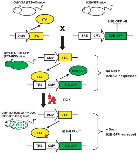