Healthcare analytics, AI solutions for biological big data, providing an AI platform for the biotech, life sciences, medical and pharmaceutical industries, as well as for related technological approaches, i.e., curation and text analysis with machine learning and other activities related to AI applications to these industries.
The bacterial makeup of human milk is influenced by the mode of breastfeeding, according to a new study. Although previously considered sterile, breast milk is now known to contain a low abundance of bacteria. While the complexities of how maternal microbiota influence the infant microbiota are still unknown, this complex community of bacteria in breast milk may help to establish the infant gut microbiota. Disruptions in this process could alter the infant microbiota, causing predisposition to chronic diseases such as allergies, asthma, and obesity. While it’s unclear how the breast milk microbiome develops, there are two theories describing its origins. One theory speculates that it originates in the maternal mammary gland, while the other theory suggests that it is due to retrograde inoculation by the infant’s oral microbiome.
To address this gap in knowledge scientists carried out bacterial gene sequencing on milk samples from 393 healthy mothers three to four months after giving birth. They used this information to examine how the milk microbiota composition is affected by maternal factors, early life events, breastfeeding practices, and other milk components. Among the many factors analyzed, the mode of breastfeeding (with or without a pump) was the only consistent factor directly associated with the milk microbiota composition. Specifically, indirect breastfeeding was associated with a higher abundance of potential opportunistic pathogens, such as Stenotrophomonas and Pseudomonadaceae. By contrast, direct breastfeeding without a pump was associated with microbes typically found in the mouth, as well as higher overall bacterial richness and diversity. Taken together, the findings suggest that direct breastfeeding facilitates the acquisition of oral microbiota from infants, whereas indirect breastfeeding leads to enrichment with environmental (pump-associated) bacteria.
The researchers argued that this study supports the theory that the breast milk microbiome is due to retrograde inoculation. Their findings indicate that the act of pumping and contact with the infant oral microbiome influences the milk microbiome, though they noted more research is needed. In future studies, the researchers will further explore the composition and function of the milk microbiota. In addition to bacteria, they will profile fungi in the milk samples. They also plan to investigate how the milk microbiota influences both the gut microbiota of infants and infant development and health. Specifically, their projects will examine the association of milk microbiota with infant growth, asthma, and allergies. This work could have important implications for microbiota-based strategies for early-life prevention of chronic conditions.
The second annual PureTech Health BIG Summit brings together an elite ensemble of leading scientific researchers, investors, and CEOs and R&D leaders from major pharmaceutical, technology, and biotech companies.
The BIG Summit is designed to stimulate ideas that will have an impact on existing pipelines and catalyze future interactions among a group of delegates that represent leaders and innovators in their fields.
Please follow the discussion on Twitter using #BIGAxisSummit
By invitation only; registration is non-transferable.
For more information, please contact PureTechHealthSummit@PureTechHealth.com
Back for final sessions at #BIGAxisSummit. @PureTechH Jim Harper of Sonde Health talking about how voice data — pacing, fine motor articulation, oscillation — can point the way to objective, quantitative measures for detecting and monitoring depression.
Paul Biondi at #BIGAxisSummit : What makes big deals happen is financial, and *deep conviction* of a big future fit. Disproportionate valuation from bidders is expected.
Love this. We often reduce everything to mathematical analyses to champion or ridicule deals. Not that simple
Bob Langer (@MIT) asks how #lymphatics affected by #aging. Santambrogio: typically blame aging #immune cells for increased disease, but aging affects lymphatics too (less efficient trafficking shown). Rejuvenating these could affect several aging-related diseases #BigAxisSummit
JP Morgan Healthcare Conference Update: Sage, Mersana, Shutdown Woes and Babies
Published: Jan 10, 2019By Alex Keown
With the J.P. Morgan Healthcare Conference winding down, companies remain busy striking deals and informing investors about pipeline advances. BioSpace snagged some of the interesting news bits to come out of the conference from Wednesday.
SAGE Therapeutics – Following a positive Phase III report that its postpartum depression treatment candidate SAGE-217 hit the mark in its late-stage clinical trial, Sage Therapeutics is eying the potential to have multiple treatment options available for patients. At the start of J.P. Morgan, Sage said that patients treated with SAGE-217 had a statistically significant improvement of 17.8 points in the Hamilton Rating Scale for Depression, compared to 13.6 for placebo. The company plans to seek approval for SAGE-2017, but before that, the FDA is expected to make a decision on Zulresso in March. Zulresso already passed muster from advisory committees in November, and if approved, would be the first drug specifically for postpartum depression. In an interview with the Business Journal, Chief Business Officer Mike Cloonan said the company believes there is room in the market for both medications, particularly since the medications address different patient populations.
Mersana Therapeutics – After a breakup with Takeda Pharmaceutical and the shelving of its lead product, Cambridge, Mass.-based Mersana is making a new path. Even though a partial clinical hold was lifted following the death of a patient the company opted to shelve development of XMT-1522. During a presentation at JPM, CEO Anna Protopapas noted that many other companies are developing therapies that target the HER2 protein, which led to the decision, according to the Boston Business Journal. Protopapas said the HER2 space is highly competitive and now the company will focus on its other asset, XMT-1536, an ADC targeting NaPi2b, an antigen highly expressed in the majority of non-squamous NSCLC and epithelial ovarian cancer. XMT-1536 is currently in Phase 1 clinical trials for NaPi2b-expressing cancers, including ovarian cancer, non-small cell lung cancer and other cancers. Data on XMT-1536 is expected in the first half of 2019.
Novavax – During a JPM presentation, Stan Erck, CEO of Novavax, pointed to the company’s RSV vaccine, which is in late-stage development. The vaccine is being developed for the mother, in order to protect an infant. The mother transfers the antibodies to the infant, which will provide the baby with protection from RSV in its first six months. Erck called the program historic. He said the Phase III program is in its fourth year and the company has vaccinated 4,636 women. He said they are tracking the women and the babies. Researchers call the mothers every week through the first six months of the baby’s life to acquire data. Erck said the company anticipates announcing trial data this quarter. If approved, Erck said the market for the vaccine could be a significant revenue driver.
“You have 3.9 million birth cohorts and we expect 80 percent to 90 percent of those mothers to be vaccinated as a pediatric vaccine and in the U.S. the market rate is somewhere between $750 million and a $1 billion and then double that for worldwide market. So it’s a large market and we will be first to market in this,” Erck said, according to a transcript of the presentation.
Denali Therapeutics – Denali forged a collaboration with Germany-based SIRION Biotech to develop gene therapies for central nervous disorders. The two companies plan to develop adeno-associated virus (AAV) vectors to enable therapeutics to cross the blood-brain barrier for clinical applications in neurodegenerative diseases including Parkinson’s, Alzheimer’s disease, ALS and certain other diseases of the CNS.
AstraZeneca – Pharma giant AstraZeneca reported that in 2019 net prices on average across the portfolio will decrease versus 2018. With a backdrop of intense public and government scrutiny over pricing, Market Access head Rick Suarez said the company is increasing its pricing transparency. Additionally, he said the company is looking at new ways to price drugs, such as value-based reimbursement agreements with payers, Pink Sheet reported.
Amarin Corporation – As the company eyes a potential label expansion approval for its cardiovascular disease treatment Vascepa, Amarin Corporation has been proactively hiring hundreds of sales reps. In the fourth quarter, the company hired 265 new sales reps, giving the company a sales team of more than 400, CEO John Thero said. Thero noted that is a label expansion is granted by the FDA, “revenues will increase at least 50 percent over what we did in the prior year, which would give us revenues of approximate $350 million in 2019.”
Government Woes – As the partial government shutdown in the United States continues into its third week, biotech leaders at JPM raised concern as the FDA’s carryover funds are dwindling. With no new funding coming in, reviews of New Drug Applications won’t be able to continue past February, Pink Sheet said. While reviews are currently ongoing, no New Drug Applications are being accepted by the FDA at this time. With the halt of NDA applications, that has also caused some companies to delay plans for an initial public offering. It’s hard to raise potential investor excitement without the regulatory support of a potential drug approval. During a panel discussion, Jonathan Leff, a partner at Deerfield Management, noted that the ongoing government shutdown is a reminder of how “overwhelmingly dependent the whole industry of biotech and drug development is on government,” Pink Sheet said.
Other posts on the JP Morgan 2019 Healthcare Conference on this Open Access Journal include:
#JPM19 Conference: Lilly Announces Agreement To Acquire Loxo Oncology, Volume 2 (Volume Two: Latest in Genomics Methodologies for Therapeutics: Gene Editing, NGS and BioInformatics, Simulations and the Genome Ontology), Part 1: Next Generation Sequencing (NGS)
#JPM19 Conference: Lilly Announces Agreement To Acquire Loxo Oncology
Reporter: Gail S. Thornton
News announced during the 37th J.P. Morgan Healthcare Conference (#JPM19): Drugmaker Eli Lilly and Company announced its plans to acquire Loxo for $8 billion, as part of its oncology strategy, which focuses “opportunities for first-in-class and best-in-class therapies.”
Please read their press release below.
INDIANAPOLIS and STAMFORD, Conn., Jan. 7, 2019 /PRNewswire/ —
Acquisition will broaden the scope of Lilly’s oncology portfolio into precision medicines through the addition of a marketed therapy and a pipeline of highly selective potential medicines for patients with genomically defined cancers.
Loxo Oncology’s pipeline includes LOXO-292, an oral RET inhibitor being studied across multiple tumor types, which recently was granted Breakthrough Therapy designation by the FDA and could launch in 2020.
Loxo Oncology’s Vitrakvi® (larotrectinib) is an oral TRK inhibitor developed and commercialized in collaboration with Bayer that was recently approved by the FDA.
Lilly will commence a tender offer to acquire all outstanding shares of Loxo Oncology for a purchase price of$235.00 per share in cash, or approximately $8.0 billion.
Lilly will conduct a conference call with the investment community and media today at 8:45 a.m. EST.
Eli Lilly and Company (NYSE: LLY) and Loxo Oncology, Inc. (NASDAQ: LOXO) today announced a definitive agreement for Lilly to acquire Loxo Oncology for $235.00 per share in cash, or approximately $8.0 billion. Loxo Oncology is a biopharmaceutical company focused on the development and commercialization of highly selective medicines for patients with genomically defined cancers.
The acquisition would be the largest and latest in a series of transactions Lilly has conducted to broaden its cancer treatment efforts with externally sourced opportunities for first-in-class and best-in-class therapies. Loxo Oncology is developing a pipeline of targeted medicines focused on cancers that are uniquely dependent on single gene abnormalities that can be detected by genomic testing. For patients with cancers that harbor these genomic alterations, a targeted medicine could have the potential to treat the cancer with dramatic effect.
Loxo Oncology has a promising portfolio of approved and investigational medicines, including:
LOXO-292, a first-in-class oral RET inhibitor that has been granted Breakthrough Therapy designation by the FDA for three indications, with an initial potential launch in 2020. LOXO-292 targets cancers with alterations to the rearranged during transfection (RET) kinase. RET fusions and mutations occur across multiple tumor types, including certain lung and thyroid cancers as well as a subset of other cancers.
LOXO-305, an oral BTK inhibitor currently in Phase 1/2. LOXO-305 targets cancers with alterations to the Bruton’s tyrosine kinase (BTK), and is designed to address acquired resistance to currently available BTK inhibitors. BTK is a validated molecular target found across numerous B-cell leukemias and lymphomas.
Vitrakvi, a first-in-class oral TRK inhibitor developed and commercialized in collaboration with Bayer that was recently approved by the U.S. Food and Drug Administration (FDA). Vitrakvi is the first treatment that targets a specific genetic abnormality to receive a tumor-agnostic indication at the time of initial FDA approval.
LOXO-195, a follow-on TRK inhibitor also being studied by Loxo Oncology and Bayer for acquired resistance to TRK inhibition, with a potential launch in 2022.
“Using tailored medicines to target key tumor dependencies offers an increasingly robust approach to cancer treatment,” said Daniel Skovronsky, M.D., Ph.D., Lilly’s chief scientific officer and president of Lilly Research Laboratories. “Loxo Oncology’s portfolio of RET, BTK and TRK inhibitors targeted specifically to patients with mutations or fusions in these genes, in combination with advanced diagnostics that allow us to know exactly which patients may benefit, creates new opportunities to improve the lives of people with advanced cancer.”
“We are gratified that Lilly has recognized our contributions to the field of precision medicine and are excited to see our pipeline benefit from the resources and global reach of the Lilly organization,” said Josh Bilenker, M.D., chief executive officer of Loxo Oncology. “Tumor genomic profiling is becoming standard-of-care, and it will be critical to continue innovating against new targets, while anticipating mechanisms of resistance to available therapies, so that patients with advanced cancer have the chance to live longer and better lives.”
“Lilly Oncology is committed to developing innovative, breakthrough medicines that will make a meaningful difference for people with cancer and help them live longer, healthier lives,” said Anne White, president of Lilly Oncology. “The acquisition of Loxo Oncology represents an exciting and immediate opportunity to expand the breadth of our portfolio into precision medicines and target cancers that are caused by specific gene abnormalities. The ability to target tumor dependencies in these populations is a key part of our Lilly Oncology strategy. We look forward to continuing to advance the pioneering scientific innovation begun by Loxo Oncology.”
“We are excited to have reached this agreement with a team that shares our commitment to ensuring that emerging translational science reaches patients in need,” said Jacob Van Naarden, chief operating officer of Loxo Oncology. “We are confident that the work we have started, which includes an FDA approved drug, and a pipeline spanning from Phase 2 to discovery, will continue to thrive in Lilly’s hands.”
Under the terms of the agreement, Lilly will commence a tender offer to acquire all outstanding shares of Loxo Oncology for a purchase price of $235.00 per share in cash, or approximately $8.0 billion. The transaction is not subject to any financing condition and is expected to close by the end of the first quarter of 2019, subject to customary closing conditions, including receipt of required regulatory approvals and the tender of a majority of the outstanding shares of Loxo Oncology’s common stock. Following the successful closing of the tender offer, Lilly will acquire any shares of Loxo Oncology that are not tendered into the tender offer through a second-step merger at the tender offer price.
The tender offer represents a premium of approximately 68 percent to Loxo Oncology’s closing stock price on January 4, 2019, the last trading day before the announcement of the transaction. Loxo Oncology’s board recommends that Loxo Oncology’s shareholders tender their shares in the tender offer. Additionally, a Loxo Oncology shareholder, beneficially owning approximately 6.6 percent of Loxo Oncology’s outstanding common stock, has agreed to tender its shares in the tender offer.
This transaction will be reflected in Lilly’s financial results and financial guidance according to Generally Accepted Accounting Principles (GAAP). Lilly will provide an update to its 2019 financial guidance, including the expected impact from the acquisition of Loxo Oncology, as part of its fourth-quarter and full-year 2018 financial results announcement on February 13, 2019.
For Lilly, Deutsche Bank is acting as the exclusive financial advisor and Weil, Gotshal & Manges LLP is acting as legal advisor in this transaction. For Loxo Oncology, Goldman Sachs & Co. LLC is acting as exclusive financial advisor and Fenwick & West LLP is acting as legal advisor.
Conference Call and Webcast Lilly will conduct a conference call with the investment community and media today at 8:45 a.m. EST to discuss the acquisition of Loxo Oncology. Investors, media and the general public can access a live webcast of the conference call through the Webcasts & Presentations link that will be posted on Lilly’s website at www.lilly.com. The webcast of the conference call will be available for replay through February 7, 2019.
About LOXO-292 LOXO-292 is an oral and selective investigational new drug in clinical development for the treatment of patients with cancers that harbor abnormalities in the rearranged during transfection (RET) kinase. RET fusions and mutations occur across multiple tumor types with varying frequency. LOXO-292 was designed to inhibit native RET signaling as well as anticipated acquired resistance mechanisms that could otherwise limit the activity of this therapeutic approach. LOXO-292 has been granted Breakthrough Therapy Designation by the U.S. FDA for three indications, and could launch as early as 2020.
About LOXO-305 LOXO-305 is an investigational, highly selective non-covalent Bruton’s tyrosine kinase (BTK) inhibitor. BTK plays a key role in the B-cell antigen receptor signaling pathway, which is required for the development, activation and survival of normal white blood cells, known as B-cells, and malignant B-cells. BTK is a validated molecular target found across numerous B-cell leukemias and lymphomas including chronic lymphocytic leukemia, Waldenstrom’s macroglobulinemia, mantle cell lymphoma and marginal zone lymphoma.
About Vitrakvi® (larotrectinib) Vitrakvi is an oral TRK inhibitor for the treatment of adult and pediatric patients with solid tumors with a neurotrophic receptor tyrosine kinase (NTRK) gene fusion without a known acquired resistance mutation that are either metastatic or where surgical resection will likely result in severe morbidity, and have no satisfactory alternative treatments or have progressed following treatment. This indication is approved under accelerated approval based on overall response rate and duration of response. Continued approval for this indication may be contingent upon verification and description of clinical benefit in confirmatory trials.
About LOXO-195 LOXO-195 is a selective TRK inhibitor that is being investigated to address potential mechanisms of acquired resistance that may emerge in patients receiving Vitrakvi® (larotrectinib) or other multikinase inhibitors with anti-TRK activity.
About Eli Lilly and Company Lilly is a global healthcare leader that unites caring with discovery to create medicines that make life better for people around the world. We were founded more than a century ago by a man committed to creating high-quality medicines that meet real needs, and today we remain true to that mission in all our work. Across the globe, Lilly employees work to discover and bring life-changing medicines to those who need them, improve the understanding and management of disease, and give back to communities through philanthropy and volunteerism. To learn more about Lilly, please visit us at www.lilly.com and www.lilly.com/newsroom/social-channels. C-LLY
About Loxo Oncology Loxo Oncology is a biopharmaceutical company focused on the development and commercialization of highly selective medicines for patients with genomically defined cancers. Our pipeline focuses on cancers that are uniquely dependent on single gene abnormalities, such that a single drug has the potential to treat the cancer with dramatic effect. We believe that the most selective, purpose-built medicines have the highest probability of maximally inhibiting the intended target, with the intention of delivering best-in-class disease control and safety. Our management team seeks out experienced industry partners, world-class scientific advisors and innovative clinical-regulatory approaches to deliver new cancer therapies to patients as quickly and efficiently as possible. For more information, please visit the company’s website at http://www.loxooncology.com.
This press release contains forward-looking statements about the benefits of Lilly’sacquisition of Loxo Oncology, Inc. (“Loxo Oncology”). It reflects Lilly‘s current beliefs; however, as with any such undertaking, there are substantial risks and uncertainties in implementing the transaction and in drug development. Among other things, there can be no guarantee that the transaction will be completed in the anticipated timeframe, or at all, or that the conditions required to complete the transaction will be met, that Lilly will realize the expected benefits of the transaction,that the molecules will be approved on the anticipated timeline or at all, or that the potential products will be commercially successful. For further discussion of these and other risks and uncertainties, see Lilly‘s most recent Form 10-K and Form 10-Q filings with the United States Securities and Exchange Commission (“the SEC”). Lilly will provide an update to certain elements of its 2019 financial guidance as part of its fourth quarter and full-year 2018 financial results announcement. Except as required by law, Lilly undertakes no duty to update forward-looking statements to reflect events after the date of this release.
This press release contains “forward-looking statements” relating to the acquisition of Loxo Oncology by Lilly. Such forward-looking statements include the ability of Loxo Oncology and Lilly to complete the transactions contemplated by the merger agreement, including the parties’ ability to satisfy the conditions to the consummation of the offer and the other conditions set forth in the merger agreement and the possibility of any termination of the merger agreement, as well as the role of targeted genomics and diagnostics in oncology treatment and acceleration of our work in developing medicines. Such forward-looking statements are based upon current expectations that involve risks, changes in circumstances, assumptions and uncertainties. Actual results may differ materially from current expectations because of risks associated with uncertainties as to the timing of the offer and the subsequent merger; uncertainties as to how many of Loxo Oncology’s stockholders will tender their shares in the offer; the risk that competing offers or acquisition proposals will be made; the possibility that various conditions to the consummation of the offer or the merger may not be satisfied or waived; the effects of disruption from the transactions contemplated by the merger agreement on Loxo Oncology’s business and the fact that the announcement and pendency of the transactions may make it more difficult to establish or maintain relationships with employees, suppliers and other business partners; the risk that stockholder litigation in connection with the offer or the merger may result in significant costs of defense, indemnification and liability; other uncertainties pertaining to the business of Loxo Oncology, including those set forth in the “Risk Factors” and “Management’s Discussion and Analysis of Financial Condition and Results of Operations” sections of Loxo Oncology’s Annual Report on Form 10-K for the year ended December 31, 2017, which is on file with the SEC and available on the SEC’s website at www.sec.gov. Additional factors may be set forth in those sections of Loxo Oncology’s Quarterly Report on Form 10-Q for the quarter endedSeptember 30, 2018, filed with the SEC in the fourth quarter of 2018. In addition to the risks described above and in Loxo Oncology’s other filings with the SEC, other unknown or unpredictable factors could also affect Loxo Oncology’s results. No forward-looking statements can be guaranteed and actual results may differ materially from such statements. The information contained in this press release is provided only as of the date of this report, and Loxo Oncology undertakes no obligation to update any forward-looking statements either contained in or incorporated by reference into this report on account of new information, future events, or otherwise, except as required by law.
Additional Information about the Acquisition and Where to Find It
The tender offer for the outstanding shares of Loxo Oncology referenced in this communication has not yet commenced. This announcement is for informational purposes only and is neither an offer to purchase nor a solicitation of an offer to sell shares of Loxo Oncology, nor is it a substitute for the tender offer materials that Lilly and its acquisition subsidiary will file with the SEC upon commencement of the tender offer. At the time the tender offer is commenced, Lilly and its acquisition subsidiary will file tender offer materials on Schedule TO, and Loxo Oncology will file a Solicitation/Recommendation Statement on Schedule 14D-9 with the SEC with respect to the tender offer. THE TENDER OFFER MATERIALS (INCLUDING AN OFFER TO PURCHASE, A RELATED LETTER OF TRANSMITTAL AND CERTAIN OTHER TENDER OFFER DOCUMENTS) AND THE SOLICITATION/RECOMMENDATION STATEMENT WILL CONTAIN IMPORTANT INFORMATION. HOLDERS OF SHARES OF LOXO ONCOLOGY ARE URGED TO READ THESE DOCUMENTS CAREFULLY WHEN THEY BECOME AVAILABLE (AS EACH MAY BE AMENDED OR SUPPLEMENTED FROM TIME TO TIME) BECAUSE THEY WILL CONTAIN IMPORTANT INFORMATION THAT HOLDERS OF LOXO ONCOLOGY SECURITIES SHOULD CONSIDER BEFORE MAKING ANY DECISION REGARDING TENDERING THEIR SECURITIES. The Offer to Purchase, the related Letter of Transmittal and certain other tender offer documents, as well as the Solicitation/Recommendation Statement, will be made available to all holders of shares of Loxo Oncology at no expense to them. The tender offer materials and the Solicitation/Recommendation Statement will be made available for free at the SEC’s web site at www.sec.gov.
In addition to the Offer to Purchase, the related Letter of Transmittal and certain other tender offer documents, as well as the Solicitation/Recommendation Statement, Lilly and Loxo Oncology file annual, quarterly and special reports and other information with the SEC. You may read and copy any reports or other information filed by Lilly or Loxo Oncology at the SEC public reference room at 100 F Street, N.E., Washington, D.C. 20549. Please call the Commission at 1-800-SEC-0330 for further information on the public reference room. Lilly’s and Loxo Oncology’s filings with the SEC are also available to the public from commercial document-retrieval services and at the website maintained by the SEC at www.sec.gov.
Other related articles published in this Open Access Online Scientific Journal include the following:
2017
FDA has approved the world’s first CAR-T therapy, Novartis for Kymriah (tisagenlecleucel) and Gilead’s $12 billion buy of Kite Pharma, no approved drug and Canakinumab for Lung Cancer (may be?)
Researchers have embraced CRISPR gene-editing as a method for altering genomes, but some have reported that unwanted DNA changes may slip by undetected. The tool can cause large DNA deletions and rearrangements near its target site on the genome. Such alterations can confuse the interpretation of experimental results and could complicate efforts to design therapies based on CRISPR. The finding is in line with previous results from not only CRISPR but also other gene-editing systems.
CRISPR -Cas9 gene editing relies on the Cas9 enzyme to cut DNA at a particular target site. The cell then attempts to reseal this break using its DNA repair mechanisms. These mechanisms do not always work perfectly, and sometimes segments of DNA will be deleted or rearranged, or unrelated bits of DNA will become incorporated into the chromosome.
Researchers often use CRISPR to generate small deletions in the hope of knocking out a gene’s function. But when examining CRISPR edits, researchers found large deletions (often several thousand nucleotides) and complicated rearrangements of DNA sequences in which previously distant DNA sequences were stitched together. Many researchers use a method for amplifying short snippets of DNA to test whether their edits have been made properly. But this approach might miss larger deletions and rearrangements.
These deletions and rearrangements occur only with gene-editing techniques that rely on DNA cutting and not with some other types of CRISPR modifications that avoid cutting DNA. Such as a modified CRISPR system to switch one nucleotide for another without cutting DNA and other systems use inactivated Cas9 fused to other enzymes to turn genes on or off, or to target RNA. Overall, these unwanted edits are a problem that deserves more attention, but this should not stop anyone from using CRISPR. Only when people use it, they need to do a more thorough analysis about the outcome.
Emerging STAR in Molecular Biology, Synthetic Virology and Genomics: Clodagh C. O’Shea: ChromEMT – Visualizing 3D chromatin structure
Article ID #241: Emerging STAR in Molecular and Cell Biology, Synthetic Virology and Genomics: Clodagh C. O’Shea: ChromEMT – Visualizing 3D chromatin structure. Published on 8/31/2017
WordCloud Image Produced by Adam Tubman
Curator: Aviva Lev-Ari, PhD, RN
On 8/28/2017, I attend and covered in REAL TIME the CHI’s 5th Immune Oncology Summit – Oncolytic Virus Immunotherapy, August 28-29, 2017 Sheraton Boston Hotel | Boston, MA
I covered in REAL TIME this event and Clodagh C. O’Shea talk at the conference.
On that evening, I e-mailed my team that
“I believe that Clodagh C. O’Sheawill get the Nobel Prizebefore CRISPR
11:00Synthetic Virology: Modular Assembly of Designer Viruses for Cancer Therapy
Clodagh O’Shea, Ph.D., Howard Hughes Medical Institute Faculty Scholar; Associate Professor, William Scandling Developmental Chair, Molecular and Cell Biology Laboratory, Salk Institute for Biological Studies
Design is the ultimate test of understanding. For oncolytic therapies to achieve their potential, we need a deep mechanistic understanding of virus and tumor biology together with the ability to confer new properties.
orthogonal capsid functionalization technologies (RapAd) and
replication assays that have enabled the rational design, directed evolution, systematic assembly and screening of powerful new vectors and oncolytic viruses.
Clodagh O’Shea’s Talk In Real Time:
Future Cancer therapies to be sophisticated as Cancer is
Targer suppresor pathways (Rb/p53)
OV are safe their efficacy ishas been limited
MOA: Specify Oncolytic Viral Replication in Tumor cells Attenuate – lack of potency
SOLUTIONS: Assembly: Assmble personalized V Tx fro libraries of functional parts
Adenovirus – natural & clinical advantages
Strategy: Technology for Assmbling Novel Adenovirus Genomes using Modular Genomic Parts
BS, Biochemistry and Microbiology, University College Cork, Ireland PhD, Imperial College London/Imperial Cancer Research Fund, U.K. Postdoctoral Fellow, UCSF Comprehensive Cancer Center, San Francisco, U.S.A
Pharmacotyping Pancreatic Cancer Patients in the Future: Two Approaches – ORGANOIDS by David Tuveson and Hans Clevers and/or MICRODOSING Devices by Robert Langer
Curator: Aviva Lev-Ari, PhD, RN
UPDATED on 4/5/2018
Featured video: Magical Bob
A fascination with magic leads Institute Professor Robert Langer to solve world problems using the marvels of chemical engineering.Watch Video
organoids, which I know you’re pretty involved in with Hans Clevers. What are your plans for organoids of pancreatic cancer?
Organoids are a really terrific model of a patient’s tumour that you generate from tissue that is either removed at the time of surgery or when they get a small needle biopsy. Culturing the tissue and observing an outgrowth of it is usually successful and when you have the cells, you can perform molecular diagnostics of any type. With a patient-derived organoid, you can sequence the exome and the RNA, and you can perform drug testing, which I call ‘pharmacotyping’, where you’re evaluating compounds that by themselves or in combination show potency against the cells. A major goal of our lab is to work towards being able to use organoids to choose therapies that will work for an individual patient – personalized medicine.
Organoids could be made moot by implantable microdevices for drug delivery into tumors, developed by Bob Langer. These devices are the size of a pencil lead and contain reservoirs that release microdoses of different drugs; the device can be injected into the tumor to deliver drugs, and can then be carefully dissected out and analyzed to gain insight into the sensitivity of cancer cells to different anticancer agents. Bob and I are kind of engaged in a friendly contest to see whether organoids or microdosing devices are going to come out on top. I suspect that both approaches will be important for pharmacotyping cancer patients in the future.
From the science side, we use organoids to discover things about pancreatic cancer. They’re great models, probably the best that I know of to rapidly discover new things about cancer because you can grow normal tissue as well as malignant tissue. So, from the same patient you can do a comparison easily to find out what’s different in the tumor. Organoids are crazy interesting, and when I see other people in the pancreatic cancer field I tell them, you should stop what you’re doing and work on these because it’s the faster way of studying this disease.
Chapter 1: Evolution of the Foundation for Diagnostics and Pharmaceuticals Industries
1.1 Outline of Medical Discoveries between 1880 and 1980
1.2 The History of Infectious Diseases and Epidemiology in the late 19th and 20th Century
1.3 The Classification of Microbiota
1.4 Selected Contributions to Chemistry from 1880 to 1980
1.5 The Evolution of Clinical Chemistry in the 20th Century
1.6 Milestones in the Evolution of Diagnostics in the US HealthCare System: 1920s to Pre-Genomics
Chapter 2. The search for the evolution of function of proteins, enzymes and metal catalysts in life processes
2.1 The life and work of Allan Wilson
2.2 The evolution of myoglobin and hemoglobin
2.3 More complexity in proteins evolution
2.4 Life on earth is traced to oxygen binding
2.5 The colors of life function
2.6 The colors of respiration and electron transport
2.7 Highlights of a green evolution
Chapter 3. Evolution of New Relationships in Neuroendocrine States
3.1 Pituitary endocrine axis
3.2 Thyroid function
3.3 Sex hormones
3.4 Adrenal Cortex
3.5 Pancreatic Islets
3.6 Parathyroids
3.7 Gastointestinal hormones
3.8 Endocrine action on midbrain
3.9 Neural activity regulating endocrine response
3.10 Genomic Promise for Neurodegenerative Diseases, Dementias, Autism Spectrum, Schizophrenia, and Serious Depression
Chapter 4. Problems of the Circulation, Altitude, and Immunity
4.1 Innervation of Heart and Heart Rate
4.2 Action of hormones on the circulation
4.3 Allogeneic Transfusion Reactions
4.4 Graft-versus Host reaction
4.5 Unique problems of perinatal period
4.6. High altitude sickness
4.7 Deep water adaptation
4.8 Heart-Lung-and Kidney
4.9 Acute Lung Injury
4.10 Reconstruction of Life Processes requires both Genomics and Metabolomics to explain Phenotypes and Phylogenetics
Chapter 5. Problems of Diets and Lifestyle Changes
5.1 Anorexia nervosa
5.2 Voluntary and Involuntary S-insufficiency
5.3 Diarrheas – bacterial and nonbacterial
5.4 Gluten-free diets
5.5 Diet and cholesterol
5.6 Diet and Type 2 diabetes mellitus
5.7 Diet and exercise
5.8 Anxiety and quality of Life
5.9 Nutritional Supplements
Chapter 6. Advances in Genomics, Therapeutics and Pharmacogenomics
6.1 Natural Products Chemistry
6.2 The Challenge of Antimicrobial Resistance
6.3 Viruses, Vaccines and immunotherapy
6.4 Genomics and Metabolomics Advances in Cancer
6.5 Proteomics – Protein Interaction
6.6 Pharmacogenomics
6.7 Biomarker Guided Therapy
6.8 The Emergence of a Pharmaceutical Industry in the 20th Century: Diagnostics Industry and Drug Development in the Genomics Era: Mid 80s to Present
6.09 The Union of Biomarkers and Drug Development
6.10 Proteomics and Biomarker Discovery
6.11 Epigenomics and Companion Diagnostics
Chapter 7
Integration of Physiology, Genomics and Pharmacotherapy
7.1 Richard Lifton, MD, PhD of Yale University and Howard Hughes Medical Institute: Recipient of 2014 Breakthrough Prizes Awarded in Life Sciences for the Discovery of Genes and Biochemical Mechanisms that cause Hypertension
7.2 Calcium Cycling (ATPase Pump) in Cardiac Gene Therapy: Inhalable Gene Therapy for Pulmonary Arterial Hypertension and Percutaneous Intra-coronary Artery Infusion for Heart Failure: Contributions by Roger J. Hajjar, MD
7.3 Diagnostics and Biomarkers: Novel Genomics Industry Trends vs Present Market Conditions and Historical Scientific Leaders Memoirs
7.4 Synthetic Biology: On Advanced Genome Interpretation for Gene Variants and Pathways: What is the Genetic Base of Atherosclerosis and Loss of Arterial Elasticity with Aging
mRNA Data Survival Analysis, Volume 2 (Volume Two: Latest in Genomics Methodologies for Therapeutics: Gene Editing, NGS and BioInformatics, Simulations and the Genome Ontology), Part 1: Next Generation Sequencing (NGS)
mRNA Data Survival Analysis
Curators: Larry H. Bernstein, MD, FCAP and Aviva Lev-Ari, PhD, RN
SURVIV for survival analysis of mRNA isoform variation
The rapid accumulation of clinical RNA-seq data sets has provided the opportunity to associate mRNA isoform variations to clinical outcomes. Here we report a statistical method SURVIV (Survival analysis of mRNA Isoform Variation), designed for identifying mRNA isoform variation associated with patient survival time. A unique feature and major strength of SURVIV is that it models the measurement uncertainty of mRNA isoform ratio in RNA-seq data. Simulation studies suggest that SURVIV outperforms the conventional Cox regression survival analysis, especially for data sets with modest sequencing depth. We applied SURVIV to TCGA RNA-seq data of invasive ductal carcinoma as well as five additional cancer types. Alternative splicing-based survival predictors consistently outperform gene expression-based survival predictors, and the integration of clinical, gene expression and alternative splicing profiles leads to the best survival prediction. We anticipate that SURVIV will have broad utilities for analysing diverse types of mRNA isoform variation in large-scale clinical RNA-seq projects.
Eukaryotic cells generate remarkable regulatory and functional complexity from a finite set of genes. Production of mRNA isoforms through alternative processing and modification of RNA is essential for generating this complexity. A prevalent mechanism for producing mRNA isoforms is the alternative splicing of precursor mRNA1. Over 95% of the multi-exon human genes undergo alternative splicing2, 3, resulting in an enormous level of plasticity in the regulation of gene function and protein diversity. In the last decade, extensive genomic and functional studies have firmly established the critical role of alternative splicing in cancer4, 5, 6. Alternative splicing is involved in a full spectrum of oncogenic processes including cell proliferation, apoptosis, hypoxia, angiogenesis, immune escape and metastasis7, 8. These cancer-associated alternative splicing patterns are not merely the consequences of disrupted gene regulation in cancer but in numerous instances actively contribute to cancer development and progression. For example, alternative splicing of genes encoding the Bcl-2 family of apoptosis regulators generates both anti-apoptotic and pro-apoptotic protein isoforms9. Alternative splicing of the pyruvate kinase M (PKM) gene has a significant impact on cancer cell metabolism and tumour growth10. A transcriptome-wide switch of the alternative splicing programme during the epithelial–mesenchymal transition plays an important role in cancer cell invasion and metastasis11, 12.
RNA sequencing (RNA-seq) has become a popular and cost-effective technology to study transcriptome regulation and mRNA isoform variation13, 14. As the cost of RNA-seq continues to decline, it has been widely adopted in large-scale clinical transcriptome projects, especially for profiling transcriptome changes in cancer. For example, as of April 2015 The Cancer Genome Atlas (TCGA) consortium had generated RNA-seq data on over 11,000 cancer patient specimens from 34 different cancer types. Within the TCGA data, breast invasive carcinoma (BRCA) has the largest sample size of RNA-seq data covering over 1,000 patients, and clinical information such as survival times, tumour stages and histological subtypes is available for the majority of the BRCA patients15. Moreover, the median follow-up time of BRCA patients is ~400 days, and 25% of the patients have more than 1,200 days of follow-up. Collectively, the large sample size and long follow-up time of the TCGA BRCA data set allow us to correlate genomic and transcriptomic profiles to clinical outcomes and patient survival times.
To date, systematic analyses have been performed to reveal the association between copy number variation, DNA methylation, gene expression and microRNA expression profiles with cancer patient survival16, 17. By contrast, despite the importance of mRNA isoform variation and alternative splicing, there have been limited efforts in transcriptome-wide survival analysis of alternative splicing in cancer patients. Most RNA-seq studies of alternative splicing in cancer transcriptomes focus on identifying ‘cancer-specific’ alternative splicing events by comparing cancer tissues with normal controls (see refs 18, 19, 20, 21, 22, 23 for examples). A recent analysis of TCGA RNA-seq data identified 163 recurrent differential alternative splicing events between cancer and normal tissues of three cancer types, among which five were found to have suggestive survival signals for breast cancer at a nominal P-value cutoff of 0.05 (ref. 21). Some other studies reported a significant survival difference between cancer patient subgroups after stratifying patients with overall mRNA isoform expression profiles24, 25. However, systematic cancer survival analyses of alternative splicing at the individual exon resolution have been lacking. Two main challenges exist for survival analyses of mRNA isoform variation and alternative splicing using RNA-seq data. The first challenge is to account for the estimation uncertainty of mRNA isoform ratios inferred from RNA-seq read counts. The statistical confidence of mRNA isoform ratio estimation depends on the RNA-seq read coverage for the events of interest, with larger read coverage leading to a more reliable estimation14. Modelling the estimation uncertainty of mRNA isoform ratio is an essential component of RNA-seq analyses of alternative splicing, as shown by various statistical algorithms developed for detecting differential alternative splicing from multi-group RNA-seq data14, 26, 27, 28,29. The second challenge, which is a general issue in survival analysis, is to properly model the association of mRNA isoform ratio with survival time, while accounting for missing data in survival time because of censoring, that is, patients still alive at the end of the survival study, whose precise survival time would be uncertain. To date, no algorithm has been developed for survival analyses of mRNA isoform variation that accounts for these sources of uncertainty simultaneously.
Here we introduce SURVIV (Survival analysis of mRNA Isoform Variation), a statistical model for identifying mRNA isoform ratios associated with patient survival times in large-scale cancer RNA-seq data sets. SURVIV models the estimation uncertainty of mRNA isoform ratios in RNA-seq data and tests the survival effects of isoform variation in both censored and uncensored survival data. In simulation studies, SURVIV consistently outperforms the conventional Cox regression survival analysis that ignores the measurement uncertainty of mRNA isoform ratio. We used SURVIV to identify alternatively spliced exons whose exon-inclusion levels significantly correlated with the survival times of invasive ductal carcinoma (IDC) patients from the TCGA breast cancer cohort. Survival-associated alternative splicing events are identified in gene pathways associated with apoptosis, oxidative stress and DNA damage repair. Importantly, we show that alternative splicing-based survival predictors outperform gene expression-based survival predictors in the TCGA IDC RNA-seq data set, as well as in TCGA data of five additional cancer types. Moreover, the integration of clinical information, gene expression and alternative splicing profiles leads to the best prediction of survival time.
SURVIV statistical model
The statistical model of SURVIV assesses the association between mRNA isoform ratio and patient survival time. While the model is generic for many types of alternative isoform variation, here we use the exon-skipping type of alternative splicing to illustrate the model (Fig. 1a). For each alternative exon involved in exon-skipping, we can use the RNA-seq reads mapping to its exon-inclusion or -skipping isoform to estimate its exon-inclusion level (denoted as ψ, or PSI that is Per cent Spliced In14). A key feature of SURVIV is that it models the RNA-seq estimation uncertainty of exon-inclusion level as influenced by the sequencing coverage for the alternative splicing event of interest. This is a critical issue in accurate quantitative analyses of mRNA isoform ratio in large-scale RNA-seq data sets14, 26, 27, 28, 29. Therefore, SURVIV contains two major components: the first to model the association of mRNA isoform ratio with patient survival time across all patients, and the second to model the estimation uncertainty of mRNA isoform ratio in each individual patient (Fig. 1a).
Figure 1: The statistical framework of the SURVIV model.
(a) For each patient k, the patient’s hazard rate λk(t) is associated with the baseline hazard rate λ0(t) and this patient’s exon-inclusion level ψk. The association of exon-inclusion level with patient survival is estimated by the survival coefficient β. The exon-inclusion level ψk is estimated from the read counts for the exon-inclusion isoform ICk and the exon-skipping isoform SCk. The proportion of the inclusion and skipping reads is adjusted by a normalization function f that considers the lengths of the exon-inclusion and -skipping isoforms (see details in Results and Supplementary Methods). (b) A hypothetical example to illustrate the association of exon-inclusion level with patient survival probability over time Sk(t), with the survival coefficient β=−1 and a constant baseline hazard rate λ0(t)=1. In this example, patients with higher exon-inclusion levels have lower hazard rates and higher survival probabilities. (c) The schematic diagram of an exon-skipping event. The exon-inclusion reads ICk are the reads from the upstream splice junction, the alternative exon itself and the downstream splice junction. The exon-skipping reads SCk are the reads from the skipping splice junction that directly connects the upstream exon to the downstream exon.
Briefly, for any individual exon-skipping event, the first component of SURVIV uses a proportional hazards model to establish the relationship between patient k’s exon-inclusion level ψk and hazard rate λk(t).
For each exon, the association between the exon-inclusion level and patient survival time is reflected by the survival coefficient β. A positive β means increased exon inclusion is associated with higher hazard rate and poorer survival, while a negative β means increased exon inclusion is associated with lower hazard rate and better survival. λ0(t) is the baseline hazard rate estimated from the survival data of all patients (see Supplementary Methods for the detailed estimation procedure). A particular patient’s survival probability over time Sk(t) can be calculated from the patient-specific hazard rate λk(t) as . Figure 1b illustrates a simple example with a negative β=−1 and a constant baseline hazard rate λ0(t)=1, where higher exon-inclusion levels are associated with lower hazard rates and higher survival probabilities.
The second component of SURVIV models the exon-inclusion level and its estimation uncertainty in individual patient samples. As illustrated in Fig. 1c, the exon-inclusion level ψk of a given exon in a particular sample can be estimated by the RNA-seq read count specific to the exon inclusion isoform (ICk) and the exon-skipping isoform (SCk). Other types of alternative splicing and mRNA isoform variation can be similarly modelled by this framework29. Given the effective lengths (that is, the number of unique isoform-specific read positions) of the exon-inclusion isoform (lI) and the exon-skipping isoform (lS), the exon-inclusion level ψk can be estimated as . Assuming that the exon-inclusion read count ICk follows a binomial distribution with the total read count nk=ICk+SCk, we have:
The binomial distribution models the estimation uncertainty of ψk as influenced by the total read count nk, in which the parameter pk represents the proportion of reads from the exon-inclusion isoform, given the exon-inclusion level ψk adjusted by a length normalization function f(ψk) based on the effective lengths of the isoforms. The definitions of effective lengths for all basic types of alternative splicing patterns are described in ref. 29.
Distinct from conventional survival analyses in which predictors do not have estimation uncertainty, the predictors in SURVIV are exon-inclusion levels ψk estimated from RNA-seq count data, and the confidence of ψk estimate for a given exon in a particular sample depends on the RNA-seq read coverage. We use the statistical framework of survival measurement error model30 to incorporate the estimation uncertainty of isoform ratio in the proportional hazards model. Using a likelihood ratio test, we test whether the exon-inclusion levels have a significant association with patient survival over the null hypothesis H0:β=0. The false discovery rate (FDR) is estimated using the Benjamini and Hochberg approach31. Details of the parameter estimation and likelihood ratio test in SURVIV are described in Supplementary Methods.
Figure 2: Simulation studies to assess the performance of SURVIV and the importance of modelling the estimation uncertainty of mRNA isoform ratio.
We compared our SURVIV model with Cox regression using point estimates of exon-inclusion levels, which does not consider the estimation uncertainty of the mRNA isoform ratio. (a) To study the effect of RNA-seq depth, we simulated the mean total splice junction read counts equal to 5, 10, 20, 50, 80 and 100 reads. We generated two sets of simulations with and without data-censoring. For each simulation, the true-positive rate (TPR) at 5% false-positive rate is plotted. The inset figure shows the empirical distribution of the mean total splice junction read counts in the TCGA IDC RNA-seq data (x axis in the log10 scale). (b) To faithfully represent the read count distribution in a real data set, we performed another simulation with read counts directly sampled from the TCGA IDC data. Sampled read counts were then multiplied by different factors ranging from 10 to 300% to simulate data sets with different RNA-seq read depth. Continuous and dashed lines represent the performance of SURVIV and Cox regression, respectively. Red lines represent the area under curve (AUC) of the ROC curve (TPR versus false-positive rate plot). Black lines represent the TPR at 5% false-positive rate.
Using these simulated data, we compared SURVIV with Cox regression in two settings, without or with censoring of the survival time. In the setting without censoring, the death and survival time of each individual is known. In the setting with censoring, certain individuals are still alive at the end of the survival study. Consequently, these patients have unknown death and survival time. Here, in the simulation with censoring, we assumed that 85% of the patients were still alive at the end of the study, similar to the censoring rate of the TCGA IDC data set. In both settings and with different depths of RNA-seq coverage, SURVIV consistently outperformed Cox regression in the true-positive rate at the same false-positive rate of 5% (Fig. 2a). As expected, we observed a more significant improvement in SURVIV over Cox regression when the RNA-seq read coverage was low (Fig. 2a).
To more faithfully recapitulate the read count distribution in a real cancer RNA-seq data set, we performed another simulation study with read counts directly sampled from the TCGA IDC data. To assess the influence of RNA-seq read depth on the performance of SURVIV and Cox regression, sampled read counts were then multiplied by different factors ranging from 10 to 300% to simulate data sets with different RNA-seq read depths (Fig. 2b). The TCGA IDC data set has an average RNA-seq depth of ~60 million paired-end reads per patient. Thus, the read depth of these simulated RNA-seq data sets ranged from ~6 million reads to 180 million reads per patient, representing low-coverage RNA-seq studies designed primarily for gene expression analysis32 up to high-coverage RNA-seq studies designed primarily for alternative isoform analysis29. At all levels of RNA-seq depth, SURVIV consistently outperformed Cox regression, as reflected by the area under curve of the receiver operating characteristic (ROC) curve as well as the true-positive rate at 5% false-positive rate (Fig. 2b). The improvement of SURVIV over Cox regression was particularly prominent when the read depth was low. For example, at 10% read depth, SURVIV had 7% improvement in area under curve (68% versus 61%) and 8% improvement in the true-positive rate at 5% false-positive rate (46% versus 38%). Collectively, these simulation results suggest that SURVIV achieves a higher accuracy by accounting for the estimation uncertainty of mRNA isoform ratio in RNA-seq data.
SURVIV analysis of TCGA IDC breast cancer data
To illustrate the practical utility of SURVIV, we used it to analyse the overall survival time of 682 IDC patients from the TCGA breast cancer (BRCA) RNA-seq data set (see Methods for details of the data source and processing pipeline). We chose to analyse IDC because it is the most frequent type of breast cancer33, comprising ~70% of patients in the TCGA breast cancer data set. To control for the effects of significant clinical parameters such as tumour stage and subtype and identify alternative splicing events associated with patient outcomes across multiple molecular and clinical subtypes, we followed the procedure of Croce and colleagues in analysing mRNA and microRNA prognostic signature of IDC33 and stratified the patients according to their clinical parameters. We then conducted SURVIV analysis in 26 clinical subgroups with at least 50 patients in each subgroup. We identified 229 exon-skipping events associated with patient survival in multiple clinical subgroups that met the criteria of SURVIV P-value≤0.01 in at least two subgroups of the same clinical parameter (cancer subtype, stage, lymph node, metastasis, tumour size, oestrogen receptor status, progesterone receptor status, HER2 status and age as shown in Fig. 3). DAVID (Database for Annotation, Visualization and Integrated Discovery) Gene Ontology analyses34 of the 229 alternative splicing events suggest an enrichment of genes in cancer-related functional categories such as intracellular signalling, apoptosis, oxidative stress and response to DNA damage (Supplementary Fig. 1). Table 1 shows a few selected examples of survival-associated alternative splicing events in cancer-related genes. Using two-means clustering of each individual exon’s inclusion levels, the 682 IDC patients can be segregated into two subgroups with significantly different survival times as illustrated by the Kaplan–Meier survival plot (Fig. 4). We also carried out hierarchical clustering of IDC patients using 176 survival-associated alternative exons (P≤0.01; SURVIV analysis of all IDC patients). Using the exon-inclusion levels of these 176 exons, we clustered IDC patients into three major subgroups, with 95, 194 and 389 patients, respectively. As illustrated by the Kaplan–Meier survival plots, the three subgroups had significantly different survival times (Supplementary Fig. 2).
Figure 3: SURVIV analysis of exon-skipping events in the TCGA IDC RNA-seq data set.
IDC patients are stratified into multiple clinical subgroups based on clinical parameters including cancer subtype, stage, lymph node status, metastasis, tumour size, oestrogen receptor status, progesterone receptor status, HER2 status and age. Only clinical subgroups with at least 50 patients are included in further analyses. Numbers of patients in the subgroups are indicated next to the names of the subgroups. Shown in the heatmap are the log10 SURVIV P-values of the 229 exons associated with patient survival (P≤0.01) in at least two subgroups of the same class of clinical parameters. Turquoise colour indicates positive correlation that higher exon-inclusion levels are associated with higher survival probabilities. Magenta colour indicates negative correlation that lower exon-inclusion levels are associated with higher survival probabilities.
Figure 4: Kaplan–Meier survival plots of IDC patients stratified by two-means clustering of the exon-inclusion levels of four survival-associated alternative splicing events.
Clustering was generated for each of the four exons separately. Black lines represent patients with high exon-inclusion levels. Red lines represent patients with low exon-inclusion levels. The P-values are from SURVIV analysis of the TCGA IDC RNA-seq data. (a) ATRIP. (b) BCL2L11. (c) CD74. (d) PCBP4.
Figure 5: Alternative splicing of STAT5A exon 5 is significantly associated with IDC patient survival.
(a) The gene structure of the STAT5A full-length isoform compared to the ΔEx5 isoform skipping the 5th exon. (b) Kaplan–Meier survival plot of IDC patients stratified by two-means clustering using exon-inclusion levels of STAT5A exon 5. The 420 patients in Group 1 (average exon 5 inclusion level=95%) have significantly higher survival probabilities than the 262 patients in Group 2 (average exon 5 inclusion level=85%) (SURVIV P=6.8e−4). (c) Exon 5 inclusion levels of IDC patients stratified by two-means clustering using exon 5 inclusion levels. Group 1 has 420 patients with average exon-inclusion level at 95%. Group 2 has 262 patients with average exon-inclusion level at 85%. (d) STAT5A exon 5 inclusion levels in normal breast tissues versus breast cancer tumour samples. Exon-inclusion levels are extracted from 86 TCGA breast cancer patients with matched normal and tumour samples. Normal breast tissues have average exon 5 inclusion level at 95%, compared to 91% average exon-inclusion level in tumour samples. Error bars represent 95% confidence interval of the mean.
Figure 6: Splicing factor regulatory network of survival-associated alternative splicing events in IDC.
(a–c) Kaplan–Meier survival plots of IDC patients stratified by the gene expression levels of three splicing factors: TRA2B (a, Cox regression P=1.8e−4), HNRNPH1 (b, P=3.4e−4) and SFRS3 (c, P=2.8e−3). Black lines represent patients with high gene expression levels. Red lines represent patients with low gene expression levels. (d) The exon-inclusion levels of a DHX30 alternative exon are negatively correlated with TRA2B gene expression levels (robust correlation coefficient r=−0.26, correlation P=1.2e−17). (e) The exon-inclusion levels of a MAP3K4 alternative exon are positively correlated withHNRNPH1 gene expression levels (robust correlation coefficient r=0.16, correlation P=2.6e−06). (f) A splicing co-expression network of the three splicing factors and their correlated survival-associated alternative exons. In total, 84 survival-associated alternative exons are significantly correlated with the three splicing factors. The positive/negative correlation between splicing factors and alternative exons is represented by blue/red lines, respectively. Exons whose inclusion levels are positively/negatively correlated with survival times are represented by blue/red dots, respectively. The size of the splicing factor circles is proportional to the number of correlated exons within the network.
Figure 7: Cross-validation of different classes of IDC survival predictors measured by the C-index
A C-index of 1 indicates perfect prediction accuracy and a C-index of 0.5 indicates random guess. The plots indicate the distribution of C-indexes from 100 rounds of cross-validation. The centre value of the box plot is the median C-index from 100 rounds of cross-validation. The notch represents the 95%confidence interval of the median. The box represents the 25 and 75% quantiles. The whiskers extended out from the box represent the 5 and 95% quantiles. Two-sided Wilcoxon test was used to compare different survival predictors. The different classes of predictors are: (a) clinical information (median C-index 0.67). (b) Gene expression (median C-index 0.68). (c) Alternative splicing (median C-index 0.71). (d) Clinical information+gene expression (median C-index 0.69). (e) Clinical information+alternative splicing (median C-index 0.73). (f) Clinical information+gene expression+alternative splicing (median C-index 0.74). Note that ‘Gene’ refers to ‘Gene-level expression’ in these plots.
Next, we carried out the SURVIV analysis in five additional cancer types in TCGA, including GBM (glioblastoma multiforme), KIRC (kidney renal clear cell carcinoma), LGG (lower grade glioma), LUSC (lung squamous cell carcinoma) and OV (ovarian serous cystadenocarcinoma). As expected, the number of significant events at different FDR or P-value significance cutoffs varied across cancer types, with LGG having the strongest survival-associated alternative splicing signals with 660 significant exon-skipping events at FDR≤5% (Supplementary Data 3 and 4). Strikingly, regardless of the number of significant events, alternative splicing-based survival predictors outperformed gene expression-based survival predictors across all cancer types (Supplementary Fig. 3), consistent with our initial observation on the IDC data set.
Alternative processing and modification of mRNA, such as alternative splicing, allow cells to generate a large number of mRNA and protein isoforms with diverse regulatory and functional properties. The plasticity of alternative splicing is often exploited by cancer cells to produce isoform switches that promote cancer cell survival, proliferation and metastasis7, 8. The widespread use of RNA-seq in cancer transcriptome studies15, 47, 48 has provided the opportunity to comprehensively elucidate the landscape of alternative splicing in cancer tissues. While existing studies of alternative splicing in large-scale cancer transcriptome data largely focused on the comparison of splicing patterns between cancer and normal tissues or between different subtypes of cancer18, 21, 49, additional computational tools are needed to characterize the clinical relevance of alternative splicing using massive RNA-seq data sets, including the association of alternative splicing with phenotypes and patient outcomes.
We have developed SURVIV, a novel statistical model for survival analysis of alternative isoform variation using cancer RNA-seq data. SURVIV uses a survival measurement error model to simultaneously model the estimation uncertainty of mRNA isoform ratio in individual patients and the association of mRNA isoform ratio with survival time across patients. Compared with the conventional Cox regression model that uses each patient’s mRNA isoform ratio as a point estimate, SURVIV achieves a higher accuracy as indicated by simulation studies under a variety of settings. Of note, we observed a particularly marked improvement of SURVIV over Cox regression for low- and moderate-depth RNA-seq data (Fig. 2b). This has important practical value because many clinical RNA-seq data sets have large sample size but relatively modest sequencing depth.
Using the TCGA IDC breast cancer RNA-seq data of 682 patients, SURVIV identified 229 alternative splicing events associated with patient survival time, which met the criteria of SURVIVP-values≤0.01 in multiple clinical subgroups. While the statistical threshold seemed loose, several lines of evidence suggest the functional and clinical relevance of these survival-associated alternative splicing events. These alternative splicing events were frequently identified and enriched in the gene functional groups important for cancer development and progression, including apoptosis, DNA damage response and oxidative stress. While some of these events may simply reflect correlation but not causal effect on cancer patient survival, other events may play an active role in regulating cancer cell phenotypes. For example, a survival-associated alternative splicing event involving exon 5 of STAT5A is known to regulate the activity of this transcription factor with important roles in epithelial cell growth and apoptosis37. Using a co-expression network analysis of splicing factor to exon correlation across all patients, we identified three splicing factors (TRA2B, HNRNPH1 and SFRS3) as potential hubs of the survival-associated alternative splicing network of IDC. The expression levels of all three splicing factors were negatively associated with patient survival times (Fig. 6a–c), and both TRA2B and HNRNPH1 were previously reported to have an impact on cancer-related molecular pathways40, 41, 42, 43, 44, 45. Finally, despite the limited power in detecting individual events, we show that the survival-associated alternative splicing events can be used to construct a predictor for patient survival, with an accuracy higher than predictors based on clinical parameters or gene expression profiles (Fig. 7). This further demonstrates the potential biological relevance and clinical utility of the identified alternative splicing events.
We performed cross-validation analyses to evaluate and compare the prognostic value of alternative splicing, gene expression and clinical information for predicting patient survival, either independently or in combination. As expected, the combined use of all three types of information led to the best prediction accuracy. Because we used penalized regression to build the prediction model, combining information from multiple layers of data did not necessarily increase the number of predictors in the model. The perhaps more surprising and intriguing result is that alternative splicing-based predictors appear to outperform gene expression-based predictors when used alone and when either type of data was combined with clinical information (Fig. 7). We observed the same trend in five additional cancer types (Supplementary Fig. 3). We note that this finding was consistent with a previous report that cancer subtype classification based on splicing isoform expression performed better than gene expression-based classification25. While this trend seems counterintuitive because accurate estimation of gene expression requires much lower RNA-seq depth than accurate estimation of alternative splicing29, one possible explanation may be the inherent characteristic of isoform ratio data. By definition, mRNA isoform ratio is estimated as the ratio of multiple mRNA isoforms from a single gene. Therefore, mRNA isoform ratio data have a ‘built-in’ internal control that could be more robust against certain artefacts and confounding issues that influence gene expression estimates across large clinical RNA-seq data sets, such as poor sample quality and RNA degradation12. Regardless of the reasons, our data call for further studies to fully explore the utility of mRNA isoform ratio data for various clinical research applications.
The SURVIV source code is available for download at https://github.com/Xinglab/SURVIV. SURVIV is a general statistical model for survival analysis of mRNA isoform ratio using RNA-seq data. The current statistical framework of SURVIV is applicable to RNA-seq based count data for all basic types of alternative splicing patterns involving two isoform choices from an alternatively spliced region, such as exon-skipping, alternative 5′ splice sites, alternative 3′ splice sites, mutually exclusive exons and retained introns, as well as other forms of alternative isoform variation such as RNA editing. With the rapid accumulation of clinical RNA-seq data sets, SURVIV will be a useful tool for elucidating the clinical relevance and potential functional significance of alternative isoform variation in cancer and other diseases.
The retina is responsible for capturing images from the visual field. Retinitis pigmentosa, which refers to a group of inherited diseases that cause retinal degeneration, causes a gradual decline in vision because retinal photoreceptor cells (rods and cones) die. Images on the left are courtesy of the National Eye Institute, NIH; image on the right is courtesy of Robert Fariss, Ph.D., and Ann Milam, Ph.D., National Eye Institute, NIH.
Metabolomics, the comprehensive evaluation of the products of cellular processes, can provide new findings and insight in a vast array of diseases and dysfunctions. Though promising, metabolomics lacks the standing of genomics or proteomics. It is, in a manner of speaking, the new kid on the “omics” block.
Even though metabolomics is still an emerging discipline, at least some quarters are giving it a warm welcome. For example, metabolomics is being advanced by the Common Fund, an initiate of the National Institutes of Health (NIH). The Common Fund has established six national metabolomics cores. In addition, individual agencies within NIH, such as the National Institute of Environmental Health Sciences (NIEHS), are releasing solicitations focused on growing more detailed metabolomics programs.
Whether metabolomic studies are undertaken with or without public support, they share certain characteristics and challenges. Untargeted or broad-spectrum studies are used for hypotheses generation, whereas targeted studies probe specific compounds or pathways. Reproducibility is a major challenge in the field; many studies cannot be reproduced in larger cohorts. Carefully defined guidance and standard operating procedures for sample collection and processing are needed.
While these challenges are being addressed, researchers are patiently amassing metabolomic insights in several areas, such as retinal diseases, neurodegenerative diseases, and autoimmune diseases. In addition, metabolomic sleuths are availing themselves of a growing selection of investigative tools.
A Metabolomic Eye on Retinal Degeneration
The retina has one of the highest metabolic activities of any tissue in the body and is composed of multiple cell types. This fact suggests that metabolomics might be helpful in understanding retinal degeneration. At least, that’s what occurred to Ellen Weiss, Ph.D., a professor of cell biology and physiology at the University of North Carolina School of Medicine at Chapel Hill. To explore this possibility, Dr. Weiss began collaborating with Susan Sumner, Ph.D., director of systems and translational sciences at RTI International.
Retinal degeneration is often studied through the use of genetic-mouse models that mimic the disease in humans. In the model used by Dr. Weiss, cells with a disease-causing mutation are the major light-sensing cells that degenerate during the disease. Individuals with the same or a similar genetic mutation will initially lose dim-light vision then, ultimately, bright-light vision and color vision.
Wild-type and mutant phenotypes, as well as dark- and light-raised animals, were compared, since retinal degeneration is exacerbated by light in this genetic model. Retinas were collected as early as day 18, prior to symptomatic disease, and analyzed. Although data analysis is ongoing, distinct differences have emerged between the phenotypes as well as between dark- and light-raised animals.
“There is a clear increase in oxidative stress in both light-raised groups but to a larger extent in the mutant phenotype,” reports Dr. Weiss. “There are global changes in metabolites that suggest mitochondrial dysfunction, and dramatic changes in lipid profiles. Now we need to understand how these metabolites are involved in this eye disease and the relevance of these perturbations.”
For example, the glial cells in the retina that upregulate a number of proteins in response to stress to attempt to save the retina are as likely as the light-receptive neurons to undergo metabolic changes.
“One of the challenges in metabolomics studies is assigning the signals that represent the metabolites or compounds in the samples,” notes Dr. Sumner. “Signals may be ‘unknown unknowns,’ compounds that have never been identified before, or ‘known unknowns,’ compounds that are known but that have not yet been assigned in the biological matrix.”
Internal and external libraries, such as the Human Metabolome Dictionary, are used to match signals. Whether or not a match exists, fragmentation patterns are used to characterize the metabolite, and when possible a standard is obtained to confirm identity. To assist with this process, the NIH Common Fund supports Metabolite Standard Synthesis Cores (MSSCs). RTI International holds an MSSC contract in addition to being a NIH-designated metabolomics core.
Mitochondrial Dysfunction in Alzheimer’s Disease
Alzheimer’s disease (AD) is difficult to diagnose early due to its asymptomatic phase; accurate diagnosis occurs only in postmortem brain tissue. To evaluate familial AD, a rare inherited form of the disease, the laboratory of Eugenia Trushina, Ph.D., associate professor of neurology and associate professor of pharmacology at the Mayo Clinic, uses mouse models to study the disease’s early molecular mechanisms.
Synaptic loss underlies cognitive dysfunction. The length of neurons dictates that mitochondria move within the cell to provide energy at the site of the synapses. An initial finding was that very early on mitochondrial trafficking was affected reducing energy supply to synapses and distant parts of the cell.
During energy production, the major mitochondrial metabolite is ATP, but the organelle also produces many other metabolites, molecules that are implicated in many pathways. One can assume that changes in energy utilization, production, and delivery are associated with some disturbance.
“Our goal,” explains Dr. Trushina, “was to get a proof of concept that we could detect in the blood of AD patients early changes of mitochondria dysfunction or other changes that could be informative of the disease over time.”
A Mayo Clinic aging study involves a cohort of patients, from healthy to those with mild cognitive impairment (MCI) through AD. Patients undergo an annual battery of tests including cognitive function along with blood and cerebrospinal fluid sampling. Metabolic signatures in plasma and cerebrospinal fluid of normal versus various disease stages were compared, and affected mitochondrial and lipid pathways identified in MCI patients that progressed to AD.
“Last year we published on a new compound that goes through the blood/brain barrier, gets into mitochondria, and very specifically, partially inhibits mitochondrial complex I activity, making the cell resistant to oxidative damage,” details Dr. Trushina. “The compound was able to either prevent or slow the disease in the animal familial models.
“Treatment not only reduced levels of amyloid plaques and phosphorylated tau, it also restored mitochondrial transport in neurons. Now we have additional compounds undergoing investigation for safety in humans, and target selectivity and engagement.”
“Mitochondria play a huge role in every aspect of our lives,” Dr. Trushina continues. “The discovery seems counterintuitive, but if mitochondria function is at the heart of AD, it may provide insight into the major sporadic form of the disease.”
Distinguishing Types of Asthma
In children, asthma generally manifests as allergy-induced asthma, or allergic asthma. And allergic asthma has commonalities with allergic dermatitis/eczema, food allergies, and allergic rhinitis. In adults, asthma is more heterogeneous, and distinct and varied subpopulations emerge. Some have nonallergic asthma; some have adult-onset asthma; and some have obesity-, occupational-, or exercise-induced asthma.
Adult asthmatics may have markers of TH2 high verus TH2 low asthma (T helper 2 cell cytokines) and they may respond to various triggers—environmental antigens, occupational antigens, irritants such as perfumes and chlorine, and seasonal allergens. Exercise, too, can trigger asthma.
One measure that can phenotype asthmatics is nitric oxide, an exhaled breath biomarker. Nitric oxide is a smooth muscle relaxant, vasodilator, and bronchodilator that can have anti-inflammatory properties. There is a wide range of values in asthmatics, and a number of values are needed to understand the trend in a particular patient. L-arginine is the amino acid that produces nitric oxide when converted to L-citrulline, a nonessential amino acid.
According to Nicholas Kenyon, M.D., a pulmonary and critical care specialist who is co-director of the University of California, Davis Asthma Network (UCAN), some metabolomic studies suggest that there is a state of L-arginine depletion during asthma attacks or in severe asthma suggesting a lack of substrate to produce nitric oxide. Dr. Kenyon is conducting clinical work on L-arginine supplementation in a double-blind cross-over intervention trial of L-arginine versus placebo. The 50-subject study in severe asthmatics should be concluded in early 2017.
Many new biologic therapies are coming to market to treat asthma; it will be challenging to determine which advanced therapy to provide to which patient. Therapeutics mostly target severe asthma populations and are for patients with evidence of higher numbers of eosinophils in the blood and lung, which include anti-IL-5 and (soon) anti-IL-13, among others.
Tools Development
Waters is developing metabolomics applications that use multivariate statistical methods to highlight compounds of interest. Typically these applications combine separation procedures, accomplished by means of liquid chromatography or gas chromatography (LC or GC), with detection methods that rely on mass spectrometry (MS). To support the identification, quantification, and analysis of LC-MS data, the company provides bioinformatics software. For example, Progenesis QI software can interrogate publicly available databases and process information about isotopic patterns, retention times, and collision cross-sections.
Mass spectrometry (MS) is the gold standard in metabolomics and lipidomics. But there is a limit to what accurate mass and resolution can achieve. For example, neither isobaric nor isomeric species are resolvable solely by MS. New orthogonal analytical tools will allow more confident identifications.
To improve metabolomics separations before MS detection, a post-ionization separation tool, like ion mobility, which is currently used to support traditional UPLC-MS and MS imaging metabolomics protocols, becomes useful. The collision-cross section (CCS), which measures the shape of molecules, can be derived, and it can be used as an additional identification coordinate.
Other new chromatographic tools are under development, such as microflow devices and UltraPerformance Convergence Chromatography (UPC2), which uses liquid CO2 as its mobile phase, to enable new ways of separating chiral metabolites. Both UPC2 and microflow technologies have decreased solvent consumption and waste disposal while maintaining UPLC-quality performance in terms of chromatographic resolution, robustness, and reproducibility.
Informatics tools are also improving. In the latest versions of Waters’ Progenesis software, typical metabolomics identification problems are resolved by allowing interrogation of publicly available databases and scoring according to accurate mass, isotopic pattern, retention time, CCS, and either theoretical or experimental fragments.
MS imaging techniques, such as MALDI and DESI, provide spatial information about the metabolite composition in tissues. These approaches can be used to support and confirm traditional analyses without sample extraction, and they allow image generation without the use of antibodies, similar to immunohistochemistry.
“Ion-mobility tools will soon be implemented for routine use, and the use of extended CCS databases will help with metabolite identification,” comments Giuseppe Astarita Ph.D., principal scientist, Waters. “More applications of ambient ionization MS will emerge, and they will allow direct-sampling analyses at atmospheric pressure with little or no sample preparation, generating real-time molecular fingerprints that can be used to discriminate among phenotypes.”
Microflow Technology
Microflow technology offers sensitivity and robustness. For example, at the Proteomics and Metabolomics Facility, Colorado State University, peptide analysis was typically performed using nanoflow chromatography; however, nanoflow chromatography is slow and technically challenging. Moving to microflow offered significant improvements in robustness and ease-of-use and resulted in improved chromatography without sacrificing sensitivity.
Conversely, small molecule applications were typically performed with analytical-scale chromatography. While this flow regime is extremely robust and fast, it can sometimes be limited in sensitivity. Moving to microflow offered significant improvements in sensitivity, 5- to 10-fold depending on the compound, without sacrificing robustness.
But broad-scale microflow adoption is hampered by a lack of available column chemistries and legacy HPLC or UPLC infrastructure that is not conducive to low-flow operation.
“We utilize microflow technology on all of our tandem quadrupole instruments for targeted quantitative assays,” says Jessica Prenni, Ph.D., director, Proteomics and Metabolomics Facility, Colorado State University. “All of our peptide quantitation is exclusively performed with microflow technology, and many of our small molecule assays. Application examples include endocannabinoids, bile acids and plant phytohormone panels.”
Compound annotation and comparability and transparency in data processing and reporting is a challenge in metabolomics research. Multiple groups are actively working on developing new tools and strategies; common best practices need to be adopted.
The continued growth of open-source spectral databases and new tools for spectral prediction from compound databases will dramatically impact the ability for metabolomics to result in novel discoveries. The move to a systems-level understanding through the combination of various omics data also will have a huge influence and be enabled by the continued development of open-source and user-friendly pathway-analysis tools.
Where Trackless Terrain Once Challenged Biomarker Development, Clearer Paths Are Emerging
Fusion detection can be carried out with traditional opposing primer-based library preparation methods, which require target- and fusion-specific primers that define the region to be sequenced. With these methods, primers are needed that flank the target region and the fusion partner, so only known fusions can be detected. An alternative method, ArcherDX’ Anchored Multiplex PCR (AMP), can be used to detect the target of interest, plus any known and unknown fusion partners. This is because AMP uses target-specific unidirectional primers, along with reverse primers, that hybridize to the sequencing adapter that is ligated to each fragment prior to amplification.
In time, the narrow, tortuous paths followed by pioneers become wider and straighter, whether the pioneers are looking to settle new land or bring new biomarkers to the clinic.
In the case of biomarkers, we’re still at the stage where pioneers need to consult guides and outfitters or, in modern parlance, consultants and technology providers. These hardy souls tend to congregate at events like the Biomarker Conference, which was held recently in San Diego.
At this event, biomarker experts discussed ways to avoid unfortunate detours on the trail from discovery and development to clinical application and regulatory approval. Of particular interest were topics such as the identification of accurate biomarkers, the explication of disease mechanisms, the stratification of patient groups, and the development of standard protocols and assay platforms. In each of these areas, presenters reported progress.
Another crucial subject is the integration of techniques such as next-generation sequencing (NGS). This particular technique has been instrumental in advancing clinical cancer genomics and continues to be the most feasible way of simultaneously interrogating multiple genes for driver mutations.
Enriching nucleic acid libraries for target genes of interest prior to NGS greatly enhances the sensitivity of detecting mutations, as the enriched regions are sequenced multiple times. This is particularly useful when analyzing clinical samples, which generate low amounts of poor-quality nucleic acids.
Most target-enrichment strategies require prior knowledge of both ends of the target region to be sequenced. Therefore, only gene fusions with known partners can be amplified for downstream NGS assays.
Archer’s Anchored Multiplex PCR (AMP™) technology overcomes this limitation, as it can enrich for novel fusions, while only requiring knowledge of one end of the fusion pair. At the heart of the AMP chemistry are unique Molecular Barcode (MBC) adapters, ligated to the 5′ ends of DNA fragments prior to amplification. The MBCs contain universal primer binding sites for PCR and a molecular barcode for identifying unique molecules. When combined with 3′ gene-specific primers, MBCs enable amplification of target regions with unknown 5′ ends.
“Tagging each molecule of input nucleic acid with a unique molecular barcode allows for de-duplication, error correction, and quantitative analysis, resulting in high sequencing consensus. With its low error rate and low limits of detection, AMP is revolutionizing the field of cancer genomics.”
In a proof-of-concept study, a single-tube 23-plex panel was designed to amplify the kinase domains of ALK, RET, ROS1, and MUSK genes by AMP. This enrichment strategy enabled identification of gene fusions with multiple partners and alternative splicing events in lung cancer, thyroid cancer, and glioblastoma specimens by NGS.
Over the last decade, the Biomarker/Translational Research Laboratory has focused on developing clinical genotyping and fluorescent in situ hybridization (FISH) assays for rapid personalized genomic testing.
“Initially, we analyzed the most prevalent hotspot mutations, about 160 in 25 cancer genes,” continued Dr. Borger. “However, this approach revealed mutations in only half of our patients. With the advent of NGS, we are able to sequence 190 exons in 39 cancer genes and obtain significantly richer genetic fingerprints, finding genetic aberrations in 92% of our cancer patients.”
Using multiplexed approaches, Dr. Borger’s team within the larger Center for Integrated Diagnostics (CID) program at MGH has established high-throughput genotyping service as an important component of routine care. While only a few susceptible molecular alterations may currently have a corresponding drug, the NGS-driven analysis may supply new information for inclusion of patients into ongoing clinical trials, or bank the result for future research and development.
“A significant impediment to discovery of clinically relevant genomic signatures is our current inability to interconnect the data,” explained Dr. Borger. “On the local level, we are striving to compile the data from clinical observations, including responses to therapy and genotyping. Globally, it is imperative that comprehensive public databases become available to the research community.”
This image, from the Massachusetts General Hospital Cancer Center, shows multicolor fluorescence in situ hybridization (FISH) analysis of cells from a patient with esophagogastric cancer. Remarkably, the FISH analysis revealed that co-amplification of the MET gene (red signal) and the EGFR gene (green signal) existed simultaneously in the same tumor cells. A chromosome 7 control probe is shown in blue.
Tumor profiling at MGH have already yielded significant discoveries. Dr. Borger’s lab, in collaboration with oncologists at the MGH Cancer Center, found significant correlations between mutations in the genes encoding the metabolic enzymes isocitrate dehydrogenase (IDH1 and IDH2) and certain types of cancers, such as cholangiocarcinoma and acute myelogenous leukemia (AML).
Historically, cancer signatures largely focus on signaling proteins. Discovery of a correlative metabolic enzyme offered a promise of diagnostics based on metabolic byproducts that may be easily identified in blood. Indeed, the metabolite 2-hydroxyglutarate accumulates to high levels in the tissues of patients carrying IDH1 and IDH2 mutations. They have reported that circulating 2-hydroxyglutarate as measured in the blood correlates with tumor burden, and could serve as an important surrogate marker of treatment response. …..
Researchers Uncover How ‘Silent’ Genetic Changes Drive Cancer
“Traditionally, it has been hard to use standard methods to quantify the amount of tRNA in the cell,” says Tavazoie. The lead authors of the article, Hani Goodarzi, formerly a postdoc in the lab and now a new assistant professor at UCSF, and research assistant Hoang Nguyen, devised and applied a new method that utilizes state-of-the-art genomic sequencing technology to measure the amount of tRNAs in different cell types.
The team chose to compare breast tissue from healthy individuals with tumor samples taken from breast cancer patients–including both primary tumors that had not spread from the breast to other body sites, and highly aggressive, metastatic tumors.
They found that the levels of two specific tRNAs were significantly higher in metastatic cells and metastatic tumors than in primary tumors that did not metastasize or healthy samples. “There are four different ways to encode for the protein building block arginine,” explains Tavazoie. “Yet only one of those–the tRNA that recognizes the codon CGG–was associated with increased metastasis.”
The tRNA that recognizes the codon GAA and encodes for a building block known as glutamic acid was also elevated in metastatic samples.
The team hypothesized that the elevated levels of these tRNAs may in fact drive metastasis. Working in mouse models of primary, non-metastatic tumors, the researchers increased the production of the tRNAs, and found that these cells became much more invasive and metastatic.
They also did the inverse experiment, with the anticipated results: reducing the levels of these tRNAs in metastatic cells decreased the incidence of metastases in the animals.
How do two tRNAs drive metastasis? The researchers teamed up with members of the Rockefeller University proteomics facility to see how protein expression changes in cells with elevated levels of these two tRNAs.
“We found global increases in many dozens of genes,” says Tavazoie, “so we analyzed their sequences and found that the majority of them had significantly increased numbers of these two specific codons.”
According to the researchers, two genes stood out among the list. Known as EXOSC2 and GRIPAP1, these genes were strongly and directly induced by elevated levels of the specific glutamic acid tRNA.
“When we mutated the GAA codons to GAG– a “silent” mutation because they both spell out the protein building block glutamic acid–we found that increasing the amount of tRNA no longer increased protein levels,” explains Tavazoie. These proteins were found to drive breast cancer metastasis.
The work challenges previous assumptions about how tRNAs function and suggests that tRNAs can modulate gene expression, according to the researchers. Tavazoie points out that “it is remarkable that within a single cell type, synonymous changes in genetic sequence can dramatically affect the levels of specific proteins, their transcripts, and the way a cell behaves.”
Testing Blood Metabolites Could Help Tailor Cancer Treatment
Scientists have found that measuring how cancer treatment affects the levels of metabolites – the building blocks of fats and proteins – can be used to assess whether the drug is hitting its intended target.
This new way of monitoring cancer therapy could speed up the development of new targeted drugs – which exploit specific genetic weaknesses in cancer cells – and help in tailoring treatment for patients.
Scientists at The Institute of Cancer Research, London, measured the levels of 180 blood markers in 41 patients with advanced cancers in a phase I clinical trial conducted with The Royal Marsden NHS Foundation Trust.
They found that investigating the mix of metabolic markers could accurately assess how cancers were responding to the targeted drug pictilisib.
Their study was funded by the Wellcome Trust, Cancer Research UK and the pharmaceutical company Roche, and is published in the journal Molecular Cancer Therapeutics.
Pictilisib is designed to specifically target a molecular pathway in cancer cells, called PI3 kinase, which has key a role in cell metabolism and is defective in a range of cancer types.
As cancers with PI3K defects grow, they can cause a decrease in the levels of metabolites in the bloodstream.
The new study is the first to show that blood metabolites are testable indicators of whether or not a new cancer treatment is hitting the correct target, both in preclinical mouse models and also in a trial of patients.
Using a sensitive technique called mass spectrometry, scientists at The Institute of Cancer Research (ICR) initially analysed the metabolite levels in the blood of mice with cancers that had defects in the PI3K pathway.
They found that the blood levels of 26 different metabolites, which were low prior to therapy, had risen considerably following treatment with pictilisib. Their findings indicated that the drug was hitting its target, and reversing the effects of the cancer on mouse metabolites.
Similarly, in humans the ICR researchers found that almost all of the metabolites – 22 out of the initial 26 – once again rose in response to pictilisib treatment, as seen in the mice.
Blood levels of the metabolites began to increase after a single dose of pictilisib, and were seen to drop again when treatment was stopped, suggesting that the effect was directly related to the drug treatment.
Metabolites vary naturally depending on the time of day or how much food a patient has eaten. But the researchers were able to provide the first strong evidence that despite this variation metabolites can be used to test if a drug is working, and could help guide decisions about treatment.
New Metabolic Pathway Reveals Aspirin-Like Compound’s Anti-Cancer Properties
Researchers at the Gladstone Institutes say they have found a new pathway by which salicylic acid, a key compound in the nonsteroidal anti-inflammatory drugs aspirin and diflunisal, stops inflammation and cancer.
In a study (“Salicylate, Diflunisal and Their Metabolites Inhibit CBP/p300 and Exhibit Anticancer Activity”) published in eLife, the investigators discovered that both salicylic acid and diflunisal suppress two key proteins that help control gene expression throughout the body. These sister proteins, p300 and CREB-binding protein (CBP), are epigenetic regulators that control the levels of proteins that cause inflammation or are involved in cell growth.
By inhibiting p300 and CBP, salicylic acid and diflunisal block the activation of these proteins and prevent cellular damage caused by inflammation. This study provides the first concrete demonstration that both p300 and CBP can be targeted by drugs and may have important clinical implications, according to Eric Verdin, M.D., associate director of the Gladstone Institute of Virology and Immunology .
“Salicylic acid is one of the oldest drugs on the planet, dating back to the Egyptians and the Greeks, but we’re still discovering new things about it,” he said. “Uncovering this pathway of inflammation that salicylic acid acts upon opens up a host of new clinical possibilities for these drugs.”
Earlier research conducted in the laboratory of co-author Stephen D. Nimer, M.D., director of Sylvester Comprehensive Cancer Center at the University of Miami Miller School of Medicine, and a collaborator of Verdin’s, established a link between p300 and the leukemia-promoting protein AML1-ETO. In the current study, scientists at Gladstone and Sylvester worked together to test whether suppressing p300 with diflunisal would suppress leukemia growth in mice. As predicted, diflunisal stopped cancer progression and shrunk the tumors in the mouse model of leukemia. ……
Novel Protein Agent Targets Cancer and Host of Other Diseases
Researchers at Georgia State University have designed a new protein compound that can effectively target the cell surface receptor integrin v3, mutations in which have been linked to a number of diseases. Initial results using this new molecule show its potential as a therapeutic treatment for an array of illnesses, including cancer.
The novel protein molecule targets integrin v3 at a novel site that has not been targeted by other scientists. The researchers found that the molecule induces apoptosis, or programmed cell death, of cells that express integrin v3. This integrin has been a focus for drug development because abnormal expression of v3 is linked to the development and progression of various diseases.
“This integrin pair, v3, is not expressed in high levels in normal tissue,” explained senior study author Zhi-Ren Liu, Ph.D., professor in the department of biology at Georgia State. “In most cases, it’s associated with a number of different pathological conditions. Therefore, it constitutes a very good target for multiple disease treatment.”
“Here we use a rational design approach to develop a therapeutic protein, which we call ProAgio, which binds to integrin αvβ3 outside the classical ligand-binding site,” the authors wrote. “We show ProAgio induces apoptosis of integrin αvβ3-expressing cells by recruiting and activating caspase 8 to the cytoplasmic domain of integrin αvβ3.”
The findings from this study were published recently in Nature Communications in an article entitled “Rational Design of a Protein That Binds Integrin αvβ3 Outside the Ligand Binding Site.” …..
“We took a unique angle,” Dr. Lui noted. “We designed a protein that binds to a different site. Once the protein binds to the site, it directly triggers cell death. When we’re able to kill pathological cells, then we’re able to kill the disease.”
The investigators performed extensive cell and molecular testing that confirmed ProAgio interacts and binds well with integrin v3. Interestingly, they found that ProAgio induces apoptosis by recruiting caspase 8—an enzyme that plays an essential role in programmed cell death—to the cytoplasmic area of integrin v3. ProAgio was much more effective in inducing cell death than other agents tested.
Noncoding RNAs Not So Noncoding
Bits of the transcriptome once believed to function as RNA molecules are in fact translated into small proteins.
In 2002, a group of plant researchers studying legumes at the Max Planck Institute for Plant Breeding Research in Cologne, Germany, discovered that a 679-nucleotide RNA believed to function in a noncoding capacity was in fact a protein-coding messenger RNA (mRNA).1 It had been classified as a long (or large) noncoding RNA (lncRNA) by virtue of being more than 200 nucleotides in length. The RNA, transcribed from a gene called early nodulin 40 (ENOD40), contained short open reading frames (ORFs)—putative protein-coding sequences bookended by start and stop codons—but the ORFs were so short that they had previously been overlooked. When the Cologne collaborators examined the RNA more closely, however, they found that two of the ORFs did indeed encode tiny peptides: one of 12 and one of 24 amino acids. Sampling the legumes confirmed that these micropeptides were made in the plant, where they interacted with a sucrose-synthesizing enzyme.
Five years later, another ORF-containing mRNA that had been posing as a lncRNA was discovered inDrosophila.2,3 After performing a screen of fly embryos to find lncRNAs, Yuji Kageyama, then of the National Institute for Basic Biology in Okazaki, Japan, suppressed each transcript’s expression. “Only one showed a clear phenotype,” says Kageyama, now at Kobe University. Because embryos missing this particular RNA lacked certain cuticle features, giving them the appearance of smooth rice grains, the researchers named the RNA “polished rice” (pri).
Turning his attention to how the RNA functioned, Kageyama thought he should first rule out the possibility that it encoded proteins. But he couldn’t. “We actually found it was a protein-coding gene,” he says. “It was an accident—we are RNA people!” The pri gene turned out to encode four tiny peptides—three of 11 amino acids and one of 32—that Kageyama and colleagues showed are important for activating a key developmental transcription factor.4
Since then, a handful of other lncRNAs have switched to the mRNA ranks after being found to harbor micropeptide-encoding short ORFs (sORFs)—those less than 300 nucleotides in length. And given the vast number of documented lncRNAs—most of which have no known function—the chance of finding others that contain micropeptide codes seems high.
Overlooked ORFs
From the late 1990s into the 21st century, as species after species had their genomes sequenced and deposited in databases, the search for novel genes and their associated mRNAs duly followed. With millions or even billions of nucleotides to sift through, researchers devised computational shortcuts to hunt for canonical gene and mRNA features, such as promoter regions, exon/intron splice sites, and, of course, ORFs.
ORFs can exist in practically any stretch of RNA sequence by chance, but many do not encode actual proteins. Because the chance that an ORF encodes a protein increases with its length, most ORF-finding algorithms had a size cut-off of 300 nucleotides—translating to 100 amino acids. This allowed researchers to “filter out garbage—that is, meaningless ORFs that exist randomly in RNAs,” says Eric Olsonof the University of Texas Southwestern Medical Center in Dallas.
Of course, by excluding all ORFs less than 300 nucleotides in length, such algorithms inevitably missed those encoding genuine small peptides. “I’m sure that the people who came up with [the cut-off] understood that this rule would have to miss anything that was shorter than 100 amino acids,” saysNicholas Ingolia of the University of California, Berkeley. “As people applied this rule more and more, they sort of lost track of that caveat.” Essentially, sORFs were thrown out with the computational trash and forgotten.
Aside from statistical practicality and human oversight, there were also technical reasons that contributed to sORFs and their encoded micropeptides being missed. Because of their small size, sORFs in model organisms such as mice, flies, and fish are less likely to be hit in random mutagenesis screens than larger ORFs, meaning their functions are less likely to be revealed. Also, many important proteins are identified based on their conservation across species, says Andrea Pauli of the Research Institute of Molecular Pathology in Vienna, but “the shorter [the ORF], the harder it gets to find and align this region to other genomes and to know that this is actually conserved.”
As for the proteins themselves, the standard practice of using electrophoresis to separate peptides by size often meant micropeptides would be lost, notes Doug Anderson, a postdoc in Olson’s lab. “A lot of times we run the smaller things off the bottom of our gels,” he says. Standard protein mass spectrometry was also problematic for identifying small peptides, says Gerben Menschaert of Ghent University in Belgium, because “there is a washout step in the protocol so that only larger proteins are retained.”
But as researchers take a deeper dive into the function of the thousands of lncRNAs believed to exist in genomes, they continue to uncover surprise micropeptides. In February 2014, for example, Pauli, then a postdoc in Alex Schier’s lab at Harvard University, discovered a hidden code in a zebrafish lncRNA. She had been hunting for lncRNAs involved in zebrafish development because “we hadn’t really anticipated that there would be any coding regions out there that had not been discovered—at least not something that is essential,” she says. But one lncRNA she identified actually encoded a 58-amino-acid micropeptide, which she called Toddler, that functioned as a signaling protein necessary for cell movements that shape the early embryo.5
Then, last year, Anderson and his colleagues reported another. Since joining Olson’s lab in 2010, Anderson had been searching for lncRNAs expressed in the heart and skeletal muscles of mouse embryos. He discovered a number of candidates, but one stood out for its high level of sequence conservation—suggesting to Anderson that it might have an important function. He was right, the RNA was important, but for a reason that neither Anderson nor Olson had considered: it was in fact an mRNA encoding a 46-amino-acid-long micropeptide.6
“When we zeroed in on the conserved region [of the gene], Doug found that it began with an ATG [start] codon and it terminated with a stop codon,” Olson says. “That’s when he looked at whether it might encode a peptide and found that indeed it did.” The researchers dubbed the peptide myoregulin, and found that it functioned as a critical calcium pump regulator for muscle relaxation.
With more and more overlooked peptides now being revealed, the big question is how many are left to be discovered. “Were there going to be dozens of [micropeptides]? Were there going to be hundreds, like there are hundreds of microRNAs?” says Ingolia. “We just didn’t know.”
Little things mean a lot. To any biologist, this time-worn maxim is old news. But it’s worth revisiting. As several articles in this issue of The Scientist illustrate, how researchers define and examine the “little things” does mean a lot.
Consider this month’s cover story, “Noncoding RNAs Not So Noncoding,” by TS correspondent Ruth Williams. Combing the human genome for open reading frames (ORFs), sequences bracketed by start and stop codons, yielded a protein-coding count somewhere in the neighborhood of 24,000. That left a lot of the genome relegated to the category of junk—or, later, to the tens of thousands of mostly mysterious long noncoding RNAs (lncRNAs). But because they had only been looking for ORFs that were 300 nucleotides or longer (i.e., coding for proteins at least 100 amino acids long), genome probers missed so-called short ORFs (sORFs), which encode small peptides. “Their diminutive size may have caused these peptides to be overlooked, their sORFs to be buried in statistical noise, and their RNAs to be miscategorized, but it does not prevent them from serving important, often essential functions, as the micropeptides characterized to date demonstrate,” writes Williams.
How little things work definitely informs another field of life science research: synthetic biology. As the functions of genes and gene networks are sussed out, bioengineers are using the information to design small, synthetic gene circuits that enable them to better understand natural networks. In “Synthetic Biology Comes into Its Own,” Richard Muscat summarizes the strides made by synthetic biologists over the last 15 years and offers an optimistic view of how such networks may be put to use in the future. And to prove him right, just as we go to press, a collaborative group led by one of syn bio’s founding fathers, MIT’s James Collins, has devised a paper-based test for Zika virus exposure that relies on a freeze-dried synthetic gene circuit that changes color upon detection of RNAs in the viral genome. The results are ready in a matter of hours, not the days or weeks current testing takes, and the test can distinguish Zika from dengue virus. “What’s really exciting here is you can leverage all this expertise that synthetic biologists are gaining in constructing genetic networks and use it in a real-world application that is important and can potentially transform how we do diagnostics,” commented one researcher about the test.
Moving around little things is the name of the game when it comes to delivering a package of drugs to a specific target or to operating on minuscule individual cells. Mini-scale delivery of biocompatible drug payloads often needs some kind of boost to overcome fluid forces or size restrictions that interfere with fine-scale manipulation. To that end, ingenious solutions that motorize delivery by harnessing osmotic changes, magnets, ultrasound, and even bacterial flagella are reviewed in “Making Micromotors Biocompatible.”
Cilengitide, a cyclic RGD pentapeptide, is currently in clinical phase III for treatment of glioblastomas and in phase II for several other tumors. This drug is the first anti-angiogenic small molecule targeting the integrins αvβ3, αvβ5 and α5β1. It was developed by us in the early 90s by a novel procedure, the spatial screening. This strategy resulted in c(RGDfV), the first superactive αvβ3 inhibitor (100 to 1000 times increased activity over the linear reference peptides), which in addition exhibited high selectivity against the platelet receptor αIIbβ3. This cyclic peptide was later modified by N-methylation of one peptide bond to yield an even greater antagonistic activity in c(RGDf(NMe)V). This peptide was then dubbed Cilengitide and is currently developed as drug by the company Merck-Serono (Germany).
This article describes the chemical development of Cilengitide, the biochemical background of its activity and a short review about the present clinical trials. The positive anti-angiogenic effects in cancer treatment can be further increased by combination with “classical” anti-cancer therapies. Several clinical trials in this direction are under investigation.
Integrins are heterodimeric receptors that are important for cell-cell and cell-extracellular matrix (ECM) interactions and are composed of one α and one β-subunit [1, 2]. These cell adhesion molecules act as transmembrane linkers between their extracellular ligands and the cytoskeleton, and modulate various signaling pathways essential in the biological functions of most cells. Integrins play a crucial role in processes such as cell migration, differentiation, and survival during embryogenesis, angiogenesis, wound healing, immune and non-immune defense mechanisms, hemostasis and oncogenic transformation [1]. The fact that many integrins are also linked with pathological conditions has converted them into very promising therapeutic targets [3]. In particular, integrins αvβ3, αvβ5 and α5β1 are involved in angiogenesis and metastasis of solid tumors, being excellent candidates for cancer therapy [4–7].
There are a number of different integrin subtypes which recognize and bind to the tripeptide sequence RGD (arginine, glycine, aspartic acid), which represents the most prominent recognition motif involved in cell adhesion. For example, the pro-angiogenic αvβ3 integrin binds various RGD-containing proteins, including fibronectin (Fn), fibrinogen (Fg), vitronectin (Vn) and osteopontin [8]. It is therefore not surprising that this integrin has been targeted for cancer therapy and that RGD-containing peptides and peptidomimetics have been designed and synthesized aiming to selectively inhibit this receptor [9, 10].
One classical strategy used in drug design is based on the knowledge about the structure of the receptor-binding pocket, preferably in complex with the natural ligand. However, this strategy, the so-called “rational structure-based design”, could not be applied in the field of integrin ligands since the first structures of integrin’s extracellular head groups were not described until 2001 for αvβ3 [11] (one year later, in 2002 the structure of this integrin in complex with Cilengitide was also reported [12]) and 2004 for αIIbβ3 [13]. Therefore, initial efforts in this field focused on a “ligand-oriented design”, which concentrated on optimizing RGD peptides by means of different chemical approaches in order to establish structure-activity relationships and identify suitable ligands.
We focused our interest in finding ligands for αvβ3 and based our approach on three chemical strategies pioneered in our group: 1) Reduction of the conformational space by cyclization; 2) Spatial screening of cyclic peptides; and 3)N-Methyl scan.
The combination of these strategies lead to the discovery of the cyclic peptidec(RGDf(NMe)V) in 1995. This peptide showed subnanomolar antagonistic activity for the αvβ3 receptor, nanomolar affinities for the closely related integrins αvβ5 and α5β1, and high selectivity towards the platelet receptor αIIbβ3. The peptide was patented together with Merck in 1997 (patent application submitted in 15.9.1995, opened in 20.3.1997) [14] and first presented with Merck’s agreement at the European Peptide Symposium in Edinburgh (September 1996) [15]. The synthesis and activity of this molecule was finally published in 1999 [16]. This peptide is now developed by Merck-Serono, (Darmstadt, Germany) under the name “Cilengitide” and has recently entered Phase III clinical trials for treating glioblastoma [17]. …..
The discovery 30 years ago of the RGD motif in Fn was a major breakthrough in science. This tripeptide sequence was also identified in other ECM proteins and was soon described as the most prominent recognition motif involved in cell adhesion. Extensive research in this direction allowed the description of a number of bidirectional proteins, the integrins, which were able to recognize and bind to the RGD sequence. Integrins are key players in the biological function of most cells and therefore the inhibition of RGD-mediated integrin-ECM interactions became an attractive target for the scientific community.
However, the lack of selectivity of linear RGD peptides represented a major pitfall which precluded any clinical application of RGD-based inhibitors. The control of the molecule’s conformation by cyclization and further spatial screening overcame these limitations, showing that it is possible to obtain privileged bioactive structures, which enhance the biological activity of linear peptides and significantly improve their receptor selectivity. Steric control imposed in RGD peptides together with their biological evaluation and extensive structural studies yielded the cyclic peptide c(RGDfV), the first small selective anti-angiogenic molecule described. N-Methylation of this cyclic peptide yielded the much potentc(RGDf(NMe)V), nowadays known as Cilengitide.
The fact that brain tumors, which are highly angiogenic, are more susceptible to the treatment with integrin antagonists, and the positive synergy observed for Cilengitide in combination with radio-chemotherapy in preclinical studies, encouraged subsequent clinical trials. Cilengitide is currently in phase III for GBM patients and in phase II for other types of cancers, with to date a promising therapeutic outcome. In addition, the absence of significant toxicity and excellent tolerance of this drug allows its combination with classical therapies such as RT or cytotoxic agents. The controlled phase III study CENTRIC was launched in 2008, with primary outcome measures due on September 2012. The results of this and other clinical studies are expected with great hope and interest.
Integrins are heterodimeric, transmembrane receptors that function as mechanosensors, adhesion molecules and signal transduction platforms in a multitude of biological processes. As such, integrins are central to the etiology and pathology of many disease states. Therefore, pharmacological inhibition of integrins is of great interest for the treatment and prevention of disease. In the last two decades several integrin-targeted drugs have made their way into clinical use, many others are in clinical trials and still more are showing promise as they advance through preclinical development. Herein, this review examines and evaluates the various drugs and compounds targeting integrins and the disease states in which they are implicated.
Integrins are heterodimeric cell surface receptors found in nearly all metazoan cell types, composed of non-covalently linked α and β subunits. In mammals, eighteen α-subunits and eight β-subunits have been identified to date 1. From this pool, 24 distinct heterodimer combinations have been observed in vivo that confer cell-to-cell and cell-to-ligand specificity relevant to the host cell and the environment in which it functions 2. Integrin-mediated interactions with the extracellular matrix (ECM) are required for the attachment, cytoskeletal organization, mechanosensing, migration, proliferation, differentiation and survival of cells in the context of a multitude of biological processes including fertilization, implantation and embryonic development, immune response, bone resorption and platelet aggregation. Integrins also function in pathological processes such as inflammation, wound healing, angiogenesis, and tumor metastasis. In addition, integrin binding has been identified as a means of viral entry into cells 3. ….
Combination of cilengitide and radiation therapy and temozolomide. The addition of cilengitide to radiotherapy and temozolomide based treatment regimens has shown promising preliminary results in ongoing Phase II trials in both newly diagnosed and progressive glioblastoma multiforme 139–140. In addition to the Phase II objectives sought, these trials are significant in that they represent progress that has made in determining tumor drug uptake and in identifying a subset of patients that may benefit from treatment. In a Phase II trial enrolling 52 patients with newly diagnosed glioblastoma multiforme receiving 500 mg cilengitide twice weekly during radiotherapy and in combination with temozolomide for 6 monthly cycles following radiotherapy, 69% achieved 6 months progression free survival compared to 54 % of patients receiving radiotherapy followed by temozolomide alone. The one-year overall survival was 67 and 62 % of patients for the cilengitide combination group and the radiotherapy and temozolomide group, respectively. Non-hematological grade 3-4 toxcities were limited, and included symptoms of fatigue, asthenia, anorexia, elevated liver function tests, deep vein thrombosis and pulmonary embolism in across a total of 5.7% of the patients. Grade 3-4 hematological malignancies were more common and included lymphopenia (53.8%), thrombocytopenia (13.4%) and neutropenia (9.6%). This trial is significant in the fact that is has provided the first evidence correlating a molecular biomarker with response to treatment. Decreased methylguanine methyltransferase (MGMT) expression was associated with favorable outcome. Patients harboring increased MGMT promoter methylation appeared to benefit more from combined treatment with cilengitide than did patients lacking promoter methylation. The significance of the MGMT promoter methylation in predicting response is likely due to inclusion of temozolomide in the treatment combination.
A similar Phase II study evaluating safety and differences in overall survival among newly diagnosed glioblastoma multiforme patients receiving radiation therapy combined with temozolomide and varying doses of cilengitide is nearing completion. Preliminary reports specify that initial safety run-in studies in 18 patients receiving doses 500, 1000 and 2000 mg cilengitide found no dose limiting toxicities. Subsequently 94 patients were randomized to receive standard therapy plus 500 or 2000 mg cilengitide. Median survival time in both cohorts was 18.9 months. At 12 months the overall survival was 79.5 % (89/112 patients).
In the last two decades great progress has been made in the discovery and development of integrin targeted therapeutics. Years of intense research into integrin function has provided an understanding of the potential applications for the treatment of disease. Advances in structural characterization of integrin-ligand interactions has proved beneficial in the design and development of potent, selective inhibitors for a number of integrins involved in platelet aggregation, inflammatory responses, angiongenesis, neovascularization and tumor growth.
The αIIbβ3 integrin antagonists were the first inhibitors to make their way into clinical use and have proven to be effective and safe drugs, contributing to the reduction of mortality and morbidity associated with acute coronary syndromes. Interestingly, the prolonged administration of small molecules targeting this integrin for long-term prevention of thrombosis related complications have not been successful, for reasons that are not yet fully understood. This suggests that modulating the intensity, duration and temporal aspects of integrin function may be more effective than simply shutting off integrin signaling in some instances. Further research into the dynamics of platelet activation and thrombosis formation may elucidate the mechanisms by which integrin activation is modulated.
The introduction of α4 targeted therapies held great promise for the treatment of inflammatory diseases. The development of Natalizumab greatly improved the quality of life for multiple sclerosis patients and those suffering with Crohn’s Disease compared to previous treatments, but the role in asthma related inflammation could not be validated. Unfortunately for MS and Crohn’s patients, immune surveillance in the central nervous system was also compromised as a direct effect α4β7 antagonism, with potentially lethal effects. Thus Natalizumab and related α4β7 targeting drugs are now limited to patients refractory to standard therapies. The design and development of α4β1 antagonists for the treatment of Crohn’s Disease may offer benefit with decreased risks. The involvement of these integrins in fetal development also raises concerns for widespread clinical use.
Integrin antagonists that target angiogenesis are progressing through clinical trials. Cilengitide has shown promising results for the treatment of glioblastomas and recurrent gliomas, cancers with notoriously low survival and cure rates. The greatest challenge facing the development of anti-angiogenic integrin targeted therapies is the overall lack of biomarkers by which to measure treatment efficacy.
Mapping the ligand-binding pocket of integrin α5β1 using a gain-of-function approach
Integrin α5β1 is a key receptor for the extracellular matrix protein fibronectin. Antagonists of human α5β1 have therapeutic potential as anti-angiogenic agents in cancer and diseases of the eye. However, the structure of the integrin is unsolved and the atomic basis of fibronectin and antagonist binding by α5β1 is poorly understood. Here we demonstrate that zebrafish α5β1 integrins do not interact with human fibronectin or the human α5β1 antagonists JSM6427 and cyclic peptide CRRETAWAC. Zebrafish α5β1 integrins do bind zebrafish fibronectin-1, and mutagenesis of residues on the upper surface and side of the zebrafish α5 subunit β-propeller domain shows that these residues are important for the recognition of RGD and synergy sites in fibronectin. Using a gain-of-function analysis involving swapping regions of the zebrafish α5 subunit with the corresponding regions of human α5 we show that blades 1-4 of the β-propeller are required for human fibronectin recognition, suggesting that fibronectin binding involves a broad interface on the side and upper face of the β-propeller domain. We find that the loop connecting blades 2 and 3 of the β-propeller (D3-A3 loop) contains residues critical for antagonist recognition, with a minor role played by residues in neighbouring loops. A new homology model of human α5β1 supports an important function for D3-A3 loop residues Trp-157 and Ala-158 in the binding of antagonists. These results will aid the development of reagents that block α5β1 functions in vivo.
Structural Basis of Integrin Regulation and Signaling
Integrins are cell adhesion molecules that mediate cell-cell, cell-extracellular matrix, and cellpathogen interactions. They play critical roles for the immune system in leukocyte trafficking and migration, immunological synapse formation, costimulation, and phagocytosis. Integrin adhesiveness can be dynamically regulated through a process termed inside-out signaling. In addition, ligand binding transduces signals from the extracellular domain to the cytoplasm in the classical outside-in direction. Recent structural, biochemical, and biophysical studies have greatly advanced our understanding of the mechanisms of integrin bidirectional signaling across the plasma membrane. Large-scale reorientations of the ectodomain of up to 200 Å couple to conformational change in ligand-binding sites and are linked to changes in α and β subunit transmembrane domain association. In this review, we focus on integrin structure as it relates to affinity modulation, ligand binding, outside-in signaling, and cell surface distribution dynamics.
The immune system relies heavily on integrins for (a) adhesion during leukocyte trafficking from the bloodstream, migration within tissues, immune synapse formation, and phagocytosis; and (b) signaling during costimulation and cell polarization. Integrins are so named because they integrate the extracellular and intracellular environments by binding to ligands outside the cell and cytoskeletal components and signaling molecules inside the cell. Integrins are noncovalently associated heterodimeric cell surface adhesion molecules. In vertebrates, 18 α subunits and 8 β subunits form 24 known αβ pairs (Figure 1). This diversity in subunit composition contributes to diversity in ligand recognition, binding to cytoskeletal components and coupling to downstream signaling pathways. Immune cells express at least 10 members of the integrin family belonging to the β2, β7, and β1 subfamilies (Table 1). The β2 and β7 integrins are exclusively expressed on leukocytes, whereas the β1 integrins are expressed on a wide variety of cells throughout the body. Distribution and ligand-binding properties of the integrins on leukocytes are summarized in Table 1. For reviews, see References 1 and 2. Mutations that block expression of the β2 integrin subfamily lead to leukocyte adhesion deficiency, a disease associated with severe immunodeficiency (3).
As adhesion molecules, integrins are unique in that their adhesiveness can be dynamically regulated through a process termed inside-out signaling or priming. Thus, stimuli received by cell surface receptors for chemokines, cytokines, and foreign antigens initiate intracellular signals that impinge on integrin cytoplasmic domains and alter adhesiveness for extracellular ligands. In addition, ligand binding transduces signals from the extracellular domain to the cytoplasm in the classical outside-in direction (outside-in signaling). These dynamic properties of integrins are central to their proper function in the immune system. Indeed, mutations or small molecules that stabilize either the inactive state or the active adhesive state—and thereby block the adhesive dynamics of leukocyte integrins—inhibit leukocyte migration and normal immune responses.

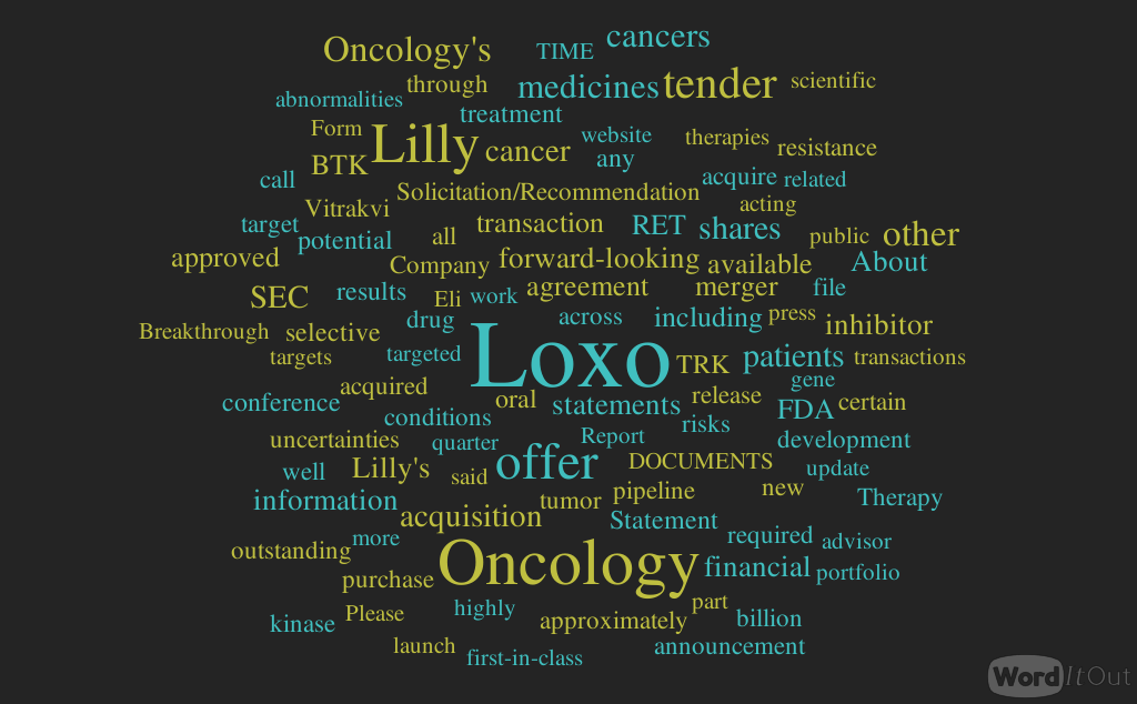


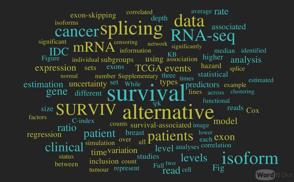

 .
.  . Assuming that the exon-inclusion read count ICk follows a binomial distribution with the total read count nk
