Feeds:
Posts
Comments

Archive for the ‘Translational Research’ Category

Cardiovascular Diseases and Pharmacological Therapy: Curations by Aviva Lev-Ari, PhD, RN

Cardiovascular Diseases and Pharmacological Therapy: Curations by Aviva Lev-Ari, PhD, RN, 2006 – 4/2018


+120 articles listed below cover the following topics:

  • National Trends: Cardiovascular-related Hospital stay, Cost of Treatment & Societal Burden
  • Introduction to Drug Types: De Novo Brand, Generic, Biologics, Biosimsilars
  • Anti-Inflammatory & Systemic Inflammatory
  • Anti-thrombotic Drug Class & Novel Oral Anticoagulants (NOACs)
  • Pharmaco-Genetics response to Congenital and Spontaneous Mutations: new drugs and new biomarkers for Atherosclerosis, Genetic-related Novel Anti-Cholesterol, Lipids, LDL, HDL, Hypertriglyceridemia Hyperlipidemia
  • Epigenetics, Gender differences and Life Style: DM, Obesity, Hormonal Markers, Diets, Chrono-therapeutics
  • BP Management: Genetics & Human Adaptive Immunity
  • Anti-arrhythmic Drugs – Atrial Fibrillation (AF) & Silent Cerebral Infarctions
  • MI, Acute Coronary Syndrome (ACS) and Heart Failure (HF)
  • Calcium &Cardiovascular Diseases: Contractile Dysfunction, Calcium as Neurotransmitter Sensor
  • Regeneration: Cardiac System (cardiomyogenesis) and Vasculature (angiogenesis)
  • Vascular Biology, Atherosclerosis and Molecular Cardiology


A new mechanism of action to attack in the treatment of coronary artery disease (CAD), Novartis developed Ilaris (canakinumab), a human monoclonal antibody targeting the interleukin-1beta innate immunity pathway

Reporter: Aviva Lev-Ari, PhD, RN

https://pharmaceuticalintelligence.com/2018/04/06/a-new-mechanism-of-action-to-attack-in-the-treatment-of-coronary-artery-disease-cad-novartis-developed-ilaris-canakinumab-a-human-monoclonal-antibody-targeting-the-interleukin-1beta-innate-i/


Advantages and Disadvantages of Novel Oral Anticoagulants (NOACs)

Reporter: Aviva Lev-Ari, PhD, RN

https://pharmaceuticalintelligence.com/2018/03/20/advantages-and-disadvantages-of-novel-oral-anticoagulants-noacs/


Acute Coronary Syndrome (ACS): Strategies in Anticoagulant Selection: Diagnostics Approaches – Genetic Testing Aids vs. Biomarkers (Troponin types and BNP)

Curator: Aviva Lev-Ari, PhD, RN

https://pharmaceuticalintelligence.com/2018/03/13/acute-coronary-syndrome-acs-strategies-in-anticoagulant-selection-diagnostics-approaches-genetic-testing-aids-vs-biomarkers-troponin-types-and-bnp/


Cholesterol Lowering Novel PCSK9 drugs: Praluent [Sanofi and Regeneron] vs Repatha [Amgen] – which drug cuts CV risks enough to make it cost-effective?

Reporter: Aviva Lev-Ari, PhD, RN

https://pharmaceuticalintelligence.com/2018/03/12/cholesterol-lowering-novel-pcsk9-drugs-praluent-sanofi-and-regeneron-vs-repatha-amgen-which-drug-cuts-cv-risks-enough-to-make-it-cost-effective/


Higher BMI (Obesity Marker): Earlier onset of incident CVD followed by Shorter overall Survival – Men and women of all ages

Reporter: Aviva Lev-Ari, PhD, RN

https://pharmaceuticalintelligence.com/2018/03/05/higher-bmi-obesity-marker-earlier-onset-of-incident-cvd-followed-by-shorter-overall-survival-men-and-women-of-all-ages/


ODYSSEY Outcomes trial evaluating the effects of a PCSK9 inhibitor, alirocumab, on major cardiovascular events in patients with an acute coronary syndrome to be presented at the American College of Cardiology meeting on March 10.

Reporter: Aviva Lev-Ari, PhD, RN

https://pharmaceuticalintelligence.com/2018/02/28/odyssey-outcomes-trial-evaluating-the-effects-of-a-pcsk9-inhibitor-alirocumab-on-major-cardiovascular-events-in-patients-with-an-acute-coronary-syndrome-to-be-presented-at-the-america/


Sex and Gender Connections: Heart and Brain Disease in Women

Reporter: Aviva Lev-Ari, PhD, RN

https://pharmaceuticalintelligence.com/2018/02/28/sex-and-gender-connections-heart-and-brain-disease-in-women/


In 2018 Cardiovascular PharmacoTherapy Market: Anti-thrombotic Drug Class Segment will continue to bring in the biggest profit and dominate production

Reporter: Aviva Lev-Ari, PhD, RN

https://pharmaceuticalintelligence.com/2018/02/27/in-2018-cardiovascular-pharmacotherapy-market-anti-thrombotic-drug-class-segment-will-continue-to-bring-in-the-biggest-profit-and-dominate-production/


Cost per Inpatient Hospital Stay: Five cardiovascular issues ranked in the top 10 – #1 Heart valve disorders, #2 Acute myocardial infarction (heart attack), #4 Coronary atherosclerosis, #7 Septicemia, #10 Acute cerebrovascular disease

Reporter: Aviva Lev-Ari, PhD, RN

https://pharmaceuticalintelligence.com/2018/02/27/cost-per-inpatient-hospital-stay-five-cardiovascular-issues-ranked-in-the-top-10-1-heart-valve-disorders-2-acute-myocardial-infarction-heart-attack-4-coronary-atherosclerosis/


There may be a genetic basis to CAD and that CXCL5 may be of therapeutic interest

Reporter: Aviva Lev-Ari, PhD, RN

https://pharmaceuticalintelligence.com/2018/02/09/there-may-be-a-genetic-basis-to-cad-and-that-cxcl5-may-be-of-therapeutic-interest/


FDA Approval marks first presentation of bivalirudin in frozen, premixed, ready-to-use formulation

Reporter: Aviva Lev-Ari, PhD, RN

https://pharmaceuticalintelligence.com/2018/01/24/fda-approval-marks-first-presentation-of-bivalirudin-in-frozen-premixed-ready-to-use-formulation/


What Level of Blood Pressure (BP) should be Treated? Comments on the New Guidelines

Reporter: Aviva Lev-Ari, PhD, RN

https://pharmaceuticalintelligence.com/2018/01/24/what-level-of-blood-pressure-bp-should-be-treated-comments-on-the-new-guidelines/


FDA approval on 12/1/2017 of Amgen’s evolocumb (Repatha) a PCSK9 inhibitor for the prevention of heart attacks, strokes, and coronary revascularizations in patients with established cardiovascular disease

Reporter: Aviva Lev-Ari, PhD, RN

https://pharmaceuticalintelligence.com/2017/12/01/fda-approval-on-12-1-2017-of-amgens-evolocumb-repatha-a-pcsk9-inhibitor-for-the-prevention-of-heart-attacks-strokes-and-coronary-revascularizations-in-patients-with-established-cardiovascular-di/


Long-term Canakinumab Treatment Lowering Inflammation Independent of Lipid Levels for Residual Inflammatory Risk Benefit – Personalized Medicine for Recurrent MI, Strokes and Cardiovascular Death

Reporter: Aviva Lev-Ari, PhD, RN

https://pharmaceuticalintelligence.com/2017/11/21/long-term-canakinumab-treatment-lowering-inflammation-independent-of-lipid-levels-for-residual-inflammatory-risk-benefit-personalized-medicine-for-recurrent-mi-strokes-and-cardiovascular-death/


Daily Highlights at 2017 American Heart Association Annual Meeting Scientific Sessions

Reporter: Aviva Lev-Ari, PhD, RN

https://pharmaceuticalintelligence.com/2017/11/14/daily-highlights-at-2017-american-heart-association-annual-meeting-scientific-sessions/


2017 Guideline for the Prevention, Detection, Evaluation and Management of High Blood Pressure in Adults – A REPORT OF THE American College of Cardiology/ American Heart Association Task Force on Clinical Practice Guidelines

Reporter: Aviva Lev-Ari, PhD, RN

https://pharmaceuticalintelligence.com/2017/11/14/2017-guideline-for-the-prevention-detection-evaluation-and-management-of-high-blood-pressure-in-adults-a-report-of-the-american-college-of-cardiology-american-heart-association-task-force-on-clin/


2017 American Heart Association Annual Meeting: Sunday’s Science at #AHA17 – Presidential Address

Reporter: Aviva Lev-Ari, PhD, RN

https://pharmaceuticalintelligence.com/2017/11/13/2017-american-heart-association-annual-meeting-sundays-science-at-aha17-presidential-address/


Systemic Inflammatory Diseases as Crohn’s disease, Rheumatoid Arthritis and Longer Psoriasis Duration May Mean Higher CVD Risk

Reporter: Aviva Lev-Ari, PhD, RN

https://pharmaceuticalintelligence.com/2017/10/09/systemic-inflammatory-diseases-as-crohns-disease-rheumatoid-arthritis-and-longer-psoriasis-duration-may-mean-higher-cvd-risk/


Shaun Coughlin from UCSF Cardiovascular Research Center to cardio group for the Novartis Institute for Biomedical Research in Cambridge, MA

Reporter: Aviva Lev-Ari, PhD, RN

https://pharmaceuticalintelligence.com/2017/08/17/shaun-coughlin-from-ucsf-cardiovascular-research-center-to-cardio-group-for-the-novartis-institute-for-biomedical-research-in-cambridge-ma/


In Europe, BigData@Heart aim to improve patient outcomes and reduce societal burden of atrial fibrillation (AF), heart failure (HF) and acute coronary syndrome (ACS).

Reporter: Aviva Lev-Ari, PhD, RN

https://pharmaceuticalintelligence.com/2017/07/10/in-europe-bigdataheart-aim-to-improve-patient-outcomes-and-reduce-societal-burden-of-atrial-fibrillation-af-heart-failure-hf-and-acute-coronary-syndrome-acs/


SNP-based Study on high BMI exposure confirms CVD and DM Risks – no associations with Stroke

Reporter: Aviva Lev-Ari, PhD, RN

https://pharmaceuticalintelligence.com/2017/07/10/snp-based-study-on-high-bmi-exposure-confirms-cvd-and-dm-risks-no-associations-with-stroke/


Tweets by @pharma_BI and @AVIVA1950 at World Medical Innovation Forum – CARDIOVASCULAR • MAY 1-3, 2017, BOSTON, MA

Curator: Aviva Lev-Ari, PhD, RN

https://pharmaceuticalintelligence.com/2017/05/05/tweets-by-pharma_bi-and-aviva1950-at-world-medical-innovation-forum-cardiovascular-%E2%80%A2-may-1-3-2017-boston-ma/


e-Proceedings for Day 1,2,3: World Medical Innovation Forum – CARDIOVASCULAR • MAY 1-3, 2017, BOSTON, MA

Curator and Reporter: Aviva Lev-Ari, PhD, RN

https://pharmaceuticalintelligence.com/2017/05/05/e-proceedings-for-day-123-world-medical-innovation-forum-cardiovascular-%E2%80%A2-may-1-3-2017-boston-ma/

REAL TIME Highlights and Tweets: Day 1,2,3: World Medical Innovation Forum – CARDIOVASCULAR • MAY 1-3, 2017, BOSTON, MA

Author and Curator: Aviva Lev-Ari, PhD, RN

https://pharmaceuticalintelligence.com/2017/05/03/deliverables-day-123-world-medical-innovation-forum-cardiovascular-%E2%80%A2-may-1-3-2017-boston-ma-httpsworldmedicalinnovation-orgagenda-highlights-of-live-day-1-world-medical/


Expedite Use of Agents in Clinical Trials: New Drug Formulary Created – The NCI Formulary is a public-private partnership between NCI, part of the National Institutes of Health, and pharmaceutical and biotechnology companies

Reporter: Aviva Lev-Ari, PhD, RN

https://pharmaceuticalintelligence.com/2017/01/12/expedite-use-of-agents-in-clinical-trials-new-drug-formulary-created-the-nci-formulary-is-a-public-private-partnership-between-nci-part-of-the-national-institutes-of-health-and-pharmaceutical-and/


Reversing Heart Disease: Combination of PCSK9 Inhibitors and Statins – Opinion by Steven Nissen, MD, Chairman of Cardiovascular Medicine at Cleveland Clinic

Reporter: Aviva Lev-Ari, PhD, RN

https://pharmaceuticalintelligence.com/2016/12/29/reversing-heart-disease-combination-of-pcsk9-inhibitors-and-statins-opinion-by-steven-nissen-md-chairman-of-cardiovascular-medicine-at-cleveland-clinicopinion-on-reversing-heart-disease-combinat/


Coronary Heart Disease Research: Sugar Industry influenced national conversation on heart disease – Adoption of Low Fat Diet vs Low Carbohydrates Diet

Reporter: Aviva Lev-Ari, PhD, RN

https://pharmaceuticalintelligence.com/2016/09/17/coronary-heart-disease-research-sugar-industry-influenced-national-conversation-on-heart-disease-adoption-of-low-fat-diet-vs-low-carbohydrates-diet/


Pathophysiology in Hypertension: Opposing Roles of Human Adaptive Immunity

Reporter: Aviva Lev-Ari, PhD, RN

https://pharmaceuticalintelligence.com/2016/08/19/pathophysiology-in-hypertension-opposing-roles-of-human-adaptive-immunity/


PCSK9 inhibitors: Reducing annual drug prices from more than $14 000 to $4536 would be necessary to meet a $100 000 per QALY threshold per JAMA

Curator: Aviva Lev-Ari, PhD, RN

https://pharmaceuticalintelligence.com/2016/08/17/pcsk9-inhibitors-reducing-annual-drug-prices-from-more-than-14%E2%80%AF000-to-4536-would-be-necessary-to-meet-a-100%E2%80%AF000-per-qaly-threshold-per-jama/


The presence of any Valvular Heart Disease (VHD) did not influence the comparison of Dabigatran [Pradaxa, Boehringer Ingelheim] with Warfarin

Reporter: Aviva Lev-Ari, PhD, RN

https://pharmaceuticalintelligence.com/2016/08/16/the-presence-of-any-valvular-heart-disease-vhd-did-not-influence-the-comparison-of-dabigatran-pradaxa-boehringer-ingelheim-with-warfarin/


Resveratrol, an antioxidant found in red wine presented since 2003 presented for its potential to lower risk for cardiovascular disease and neurodegeneration by increasing cell survival and slowing aging: 2014 Study – Diet rich in resveratrol offers no health boost

Reporter: Aviva Lev-Ari, PhD, RN

https://pharmaceuticalintelligence.com/2016/07/25/resveratrol-an-antioxidant-found-in-red-wine-2014-study-resveratrol-offers-no-health-boost/


Amgen’s Corlanor® can help Reduce the Risk of Hospitalization for Patients with worsening Heart Failure

Reporter: Aviva Lev-Ari, PhD, RN

https://pharmaceuticalintelligence.com/2016/05/04/amgens-corlanor-can-help-reduce-the-risk-of-hospitalization-for-patients-with-worsening-heart-failure/


Effectiveness of Anti-arrhythmic Drugs: Amiodarone and Lidocaine, for treating sudden cardiac arrest, increasing likelihood of Patients Surviving Emergency Transport to Hospital

Curator: Aviva Lev-Ari, PhD, RN

https://pharmaceuticalintelligence.com/2016/04/04/effectiveness-of-anti-arrhythmic-drugs-amiodarone-and-lidocaine-for-treating-sudden-cardiac-arrest-increasing-likelihood-of-patients-surviving-emergency-transport-to-hospital/


Efficacy and Tolerability of PCSK9 Inhibitors by Patients with Muscle-related Statin Intolerance – New Cleveland Clinic study published in JAMA 4/2016

Curators: Larry H. Bernstein, MD, FCAP and Aviva Lev-Ari, PhD, RN

https://pharmaceuticalintelligence.com/2016/04/03/efficacy-and-tolerability-of-pcsk9-inhibitors-by-patients-with-muscle-related-statin-intolerance-new-cleveland-clinic-study-published-in-jama-42016/


Triglycerides: Is it a Risk Factor or a Risk Marker for Atherosclerosis and Cardiovascular Disease ? The Impact of Genetic Mutations on (ANGPTL4) Gene, encoder of (angiopoietin-like 4) Protein, inhibitor of Lipoprotein Lipase

Reporters, Curators and Authors: Aviva Lev-Ari, PhD, RN and Larry H. Bernstein, MD, FCAP

https://pharmaceuticalintelligence.com/2016/03/13/triglycerides-is-it-a-risk-factor-or-a-risk-marker-for-atherosclerosis-and-cardiovascular-disease-the-impact-of-genetic-mutations-on-angptl4-gene-encoder-of-angiopoietin-like-4-protein-that-in/


In One-Hour: A Diagnosis of Heart Attack made possible by one Blood Test

Reporter: Larry H Bernstein, MD, FCAP

https://pharmaceuticalintelligence.com/2016/01/14/in-one-hour-a-diagnosis-of-heart-attack-made-possible-by-one-blood-test/


Heart-Failure–Related Mortality Rate: CDC Reports comparison of 2000, 2012, 2014  – the decease is steadily reversed

Reporter: Aviva Lev-Ari, PhD, RN

https://pharmaceuticalintelligence.com/2016/01/05/heart-failure-related-mortality-rate-cdc-reports-comparison-of-2000-2012-2014-the-decease-is-steadily-reversed/


PCSK9: A Recent Discovery in Understanding Cholesterol Regulation @ AMGEN Cardiovascular

Reporter: Aviva Lev-Ari, PhD, RN

https://pharmaceuticalintelligence.com/2015/08/04/pcsk9-a-recent-discovery-in-understanding-cholesterol-regulation-amgen-cardiovascular/


Praluent – FDA approved as Cholesterol-lowering Medicine for Patient non responsive to Statin due to Genetic origin of Hypercholesterolemia

Reporter: Aviva Lev-Ari, PhD, RN

https://pharmaceuticalintelligence.com/2015/07/27/praluent-fda-approved-as-cholesterol-lowering-medicine-for-patient-non-responsive-to-statin-due-to-genetic-origin-of-hypercholesterolemia/


Atherosclerosis: What is New in Biomarker Discovery

Reporter: Aviva Lev-Ari, PhD, RN

https://pharmaceuticalintelligence.com/2015/07/01/atherosclerosis-what-is-new-in-biomarker-discovery/


Cangrelor wins Clopidogrel (Plavix): reduction of Risk of a composite of all-cause mortality, myocardial infarction, ischemia driven revascularization, and stent thrombosis

Reporter: Aviva Lev-Ari, PhD, RN

https://pharmaceuticalintelligence.com/2015/04/16/cangrelor-wins-clopidogrel-plavix-reduction-of-risk-of-a-composite-of-all-cause-mortality-myocardial-infarction-ischemia-driven-revascularization-and-stent-thrombosis/


Sets of co-expressed Genes influence Blood Pressure Regulation: Genome-wide Association and mRNA expression @US National Heart, Lung, and Blood Institute

Reporter: Aviva Lev-Ari, PhD, RN

https://pharmaceuticalintelligence.com/2015/04/16/sets-of-co-expressed-genes-influence-blood-pressure-regulation-genome-wide-association-and-mrna-expression-us-national-heart-lung-and-blood-institute/


HDL-C: Target of Therapy – Steven E. Nissen, MD, MACC, Cleveland Clinic vs Peter Libby, MD, BWH

Reporter: Aviva Lev-Ari, PhD, RN

https://pharmaceuticalintelligence.com/2014/11/07/hdl-c-target-of-therapy-steven-e-nissen-md-macc-cleveland-clinic-vs-peter-libby-md-bwh/


Atrial Fibrillation and Silent Cerebral Infarctions: A Meta Analysis Study and Literature Review

Reporter: Aviva Lev-Ari, PhD, RN

https://pharmaceuticalintelligence.com/2014/11/04/atrial-fibrillation-and-silent-cerebral-infarctions-a-meta-analysis-study-and-literature-review/


Intracranial Vascular Stenosis: Comparison of Clinical Trials: Percutaneous Transluminal Angioplasty and Stenting (PTAS) vs. Clot-inhibiting Drugs: Aspirin and Clopidogrel (dual antiplatelet therapy) – more Strokes if Stenting

Reporter: Aviva Lev-Ari, PhD, RN

https://pharmaceuticalintelligence.com/2014/10/15/intracranial-vascular-stenosis-comparison-of-clinical-trials-percutaneous-transluminal-angioplasty-and-stenting-ptas-vs-clot-inhibiting-drugs-aspirin-and-clopidogrel-dual-antiplatelet-therapy/


Hypertension: It is Autoimmunity that Underlies its Development in Humans

Reporter: Aviva Lev-Ari, PhD, RN

https://pharmaceuticalintelligence.com/2014/10/08/hypertension-it-is-autoimmunity-that-underlies-its-development-in-humans/


OPINION LEADERSHIP on Cardiovascular Diseases

Cardiovascular Original Research: Cases in Methodology Design for Content Co-Curation

  • Cardiovascular Diseases, Volume Two: Cardiovascular Original Research: Cases in Methodology Design for Content Co-Curation. On Amazon.com since 11/30/2015

http://www.amazon.com/dp/B018Q5MCN8

 Epilogue to Volume Two

Author and Curator: Aviva Lev-Ari, PhD, RN, Editor-in-Chief, BioMed e-Series of e-Books

https://pharmaceuticalintelligence.com/2014/07/31/opinion-leadership-on-cardiovascular-diseases/


Risk of Major Cardiovascular Events by LDL-Cholesterol Level (mg/dL): Among those treated with high-dose statin therapy, more than 40% of patients failed to achieve an LDL-cholesterol target of less than 70 mg/dL.

Reporter: Aviva Lev-Ari, PhD, RN

https://pharmaceuticalintelligence.com/2014/07/29/risk-of-major-cardiovascular-events-by-ldl-cholesterol-level-mgdl-among-those-treated-with-high-dose-statin-therapy-more-than-40-of-patients-failed-to-achieve-an-ldl-cholesterol-target-of-less-th/


Commentary on Biomarkers for Genetics and Genomics of Cardiovascular Disease: Views by Larry H Bernstein, MD, FCAP

Commissioned article, Author: Larry H Bernstein, MD, FCAP

https://pharmaceuticalintelligence.com/2014/07/16/commentary-on-biomarkers-for-genetics-and-genomics-of-cardiovascular-disease-views-by-larry-h-bernstein-md-fcap/


Coagulation Therapy: Leading New Drugs – Efficacy Comparison

Curator: Aviva Lev-Ari, PhD, RN

https://pharmaceuticalintelligence.com/2014/05/10/coagulation-therapy-leading-new-drugs-efficacy-comparison/


Apixaban (Eliquis): Mechanism of Action, Drug Comparison and Additional Indications

Curator: Aviva Lev-Ari, PhD, RN

https://pharmaceuticalintelligence.com/2014/05/10/apixaban-eliquis-mechanism-of-action-drug-comparison-and-additional-indications/


Boston Heart Diagnostics (BHD) offers Statin Induced Myopathy (SLCO1B1) Genotype test and genetic tests targeting ApoE, Factor V Leiden, prothrombin (Factor II), and CYP2C19

Reporter: Aviva Lev-Ari, PhD, RN

https://pharmaceuticalintelligence.com/2014/04/17/boston-heart-diagnostics-bhd-offers-statin-induced-myopathy-slco1b1-genotype-test-and-genetic-tests-targeting-apoe-factor-v-leiden-prothrombin-factor-ii-and-cyp2c19/


@@@ Cardiovascular Diseases and Pharmacological Therapy: Curations by Aviva Lev-Ari, PhD, RN

Curator: Aviva Leve-Ari, PhD, RN

https://pharmaceuticalintelligence.com/2014/04/17/cardiovascular-diseases-and-pharmacological-therapy-curations-by-aviva-lev-ari-phd-rn/


Richard Lifton, MD, PhD of Yale University & Howard Hughes Medical Institute: Recipient of 2014 Breakthrough Prizes Awarded in Life Sciences for the Discovery of Genes and Biochemical Mechanisms that cause Hypertension

Curator: Aviva Lev-Ari, PhD, RN

https://pharmaceuticalintelligence.com/2014/03/03/richard-lifton-md-phd-of-yale-university-and-howard-hughes-medical-institute-recipient-of-2014-breakthrough-prizes-awarded-in-life-sciences-for-the-discovery-of-genes-and-biochemical-mechanisms-tha/


Differences in Health Services Utilization and Costs between Antihypertensive Medication Users Versus Nonusers in Adults with Diabetes and Concomitant Hypertension from Medical Expenditure Panel Su…

Reporter: Aviva Lev-Ari, PhD, RN

https://pharmaceuticalintelligence.com/2014/02/28/differences-in-health-services-utilization-and-costs-between-antihypertensive-medication-users-versus-nonusers-in-adults-with-diabetes-and-concomitant-hypertension-from-medical-expenditure-panel-su-2/


2014 Epidemiology and Prevention, Nutrition, Physical Activity and Metabolism Conference: San Francisco, Ca. Conference Dates: San Francisco, CA 3/18-21, 2014

Reporter: Aviva Lev-Ari, PhD, RN

https://pharmaceuticalintelligence.com/2014/02/26/2014-epidemiology-and-prevention-nutrition-physical-activity-and-metabolism-conference-san-francisco-ca-conference-dates-san-francisco-ca-318-21-2014/


2014 High Blood Pressure Research Conference, 9/9 – 9/12, 2014 — Hilton SF Union Square, San Francisco, CA

Reporter: Aviva Lev-Ari, PhD, RN

https://pharmaceuticalintelligence.com/2014/02/24/2014-high-blood-pressure-research-conference-99-912-2014-hilton-sf-union-square-san-francisco-ca/


Females and Non-Atherosclerotic Plaque: Spontaneous Coronary Artery Dissection – New Insights from Research and DNA Ongoing Study

Reporter: Aviva Lev-Ari, PhD, RN

https://pharmaceuticalintelligence.com/2014/02/12/female-and-non-atherosclerotic-plaque-spontaneous-coronary-artery-dissection-new-insights-from-research-and-dna-ongoing-study/


Hypertension – JNC 8 Guideline: Henry R. Black, MD, Michael A. Weber, MD and Raymond R. Townsend, MD

Reporter: Aviva Lev-Ari, PhD, RN

https://pharmaceuticalintelligence.com/2014/02/12/hypertension-jnc-8-guideline-henry-r-black-md-michael-a-weber-md-and-raymond-r-townsend-md/


Why Don’t You Trust Generic Drugs as Much as Brand Name …

Reporter: Aviva Lev-Ari, PhD, RN

https://pharmaceuticalintelligence.com/2014/02/10/why-dont-you-trust-generic-drugs-as-much-as-brand-name/


National Trends, 2005 – 2011: Adverse-event Rates Declined among Patients Hospitalized for Acute Myocardial Infarction or Congestive Heart Failure

Reporter: Aviva Lev-Ari, PhD, RN

https://pharmaceuticalintelligence.com/2014/02/04/national-trends-2005-2011-adverse-event-rates-declined-among-patients-hospitalized-for-acute-myocardial-infarction-or-congestive-heart-failure/


Is Pharmacogenetic-based Dosing of Warfarin Superior for Anticoagulation Control?

Curator: Aviva Lev-Ari, PhD, RN

https://pharmaceuticalintelligence.com/2014/02/04/is-pharmacogenetic-based-dosing-of-warfarin-superior-for-anticoagulation-control/


Prolonged Wakefulness: Lack of Sufficient Duration of Sleep as a Risk Factor for Cardiovascular Diseases – Indications for Cardiovascular Chrono-therapeutics

Curator: Aviva Lev-Ari, PhD, RN

https://pharmaceuticalintelligence.com/2014/02/02/prolonged-wakefulness-lack-of-sufficient-duration-of-sleep-as-a-risk-factor-for-cardiovascular-diseases-indications-for-cardiovascular-chrono-therapeutics/


Testosterone Therapy for Idiopathic Hypogonadotrophic Hypogonadism has Beneficial and Deleterious Effects on Cardiovascular Risk Factors

Curator: Aviva Lev-Ari, PhD, RN

https://pharmaceuticalintelligence.com/2014/01/30/testosterone-therapy-for-idiopathic-hypogonadotrophic-hypogonadism-has-beneficial-and-deleterious-effects-on-cardiovascular-risk-factors/


Calcium and Cardiovascular Diseases: A Series of Twelve Articles in Advanced Cardiology

Curator: Aviva Lev-Ari, PhD, RN

https://pharmaceuticalintelligence.com/2014/01/28/calcium-and-cardiovascular-diseases-a-series-of-twelve-articles-in-advanced-cardiology/


Acute Myocardial Infarction: Curations of Cardiovascular Original Research – A Bibliography

Curator: Aviva Lev-Ari, PhD, RN

https://pharmaceuticalintelligence.com/2014/01/22/acute-myocardial-infarction-curations-of-cardiovascular-original-research-a-bibliography/


On-Hours vs Off-Hours: Presentation to ER with Acute Myocardial Infarction – Lower Survival Rate if Off-Hours

Reporter: Aviva Lev-Ari, PhD, RN

https://pharmaceuticalintelligence.com/2014/01/22/on-hours-vs-off-hours-presentation-to-er-with-acute-myocardial-infarction-lower-survival-rate-if-off-hours/


2014 Winter in New England: The Effect of Record Cold Temperatures on Cardiovascular Diseases

Curator: Aviva Lev-Ari, PhD, RN

https://pharmaceuticalintelligence.com/2014/01/21/2014-winter-in-new-england-the-effect-of-record-cold-temperatures-on-cardiovascular-diseases/


Voices from the Cleveland Clinic: On the New Lipid Guidelines and On the ACC/AHA Risk Calculator

Reporter: Aviva Lev-Ari, PhD, RN

https://pharmaceuticalintelligence.com/2014/01/21/voices-from-the-cleveland-clinic-on-the-new-lipid-guidelines-and-on-the-accaha-risk-calculator/


Is it Hypertension or Physical Inactivity: Cardiovascular Risk and Mortality – New results in 3/2013

Reporter: Aviva Lev-Ari, PhD, RN

https://pharmaceuticalintelligence.com/2014/01/19/is-it-hypertension-or-physical-inactivity-cardiovascular-risk-and-mortality-new-results-in-32013/


Regeneration: Cardiac System (cardiomyogenesis) and Vasculature (angiogenesis)

Curator: Aviva Lev-Ari, PhD, RN

https://pharmaceuticalintelligence.com/2014/01/15/regeneration-cardiac-system-and-vasculature


Conceived: NEW Definition for Co-Curation in Medical Research

Curator: Aviva Lev-Ari, PhD, RN

https://pharmaceuticalintelligence.com/2014/01/04/conceived-new-definition-for-co-curation-in-medical-research/


The Young Surgeon and The Retired Pathologist: On Science, Medicine and HealthCare Policy – The Best Writers Among the WRITERS

Curator: Aviva Lev-Ari, PhD, RN

https://pharmaceuticalintelligence.com/2013/12/10/the-young-surgeon-and-the-retired-pathologist-on-science-medicine-and-healthcare-policy-best-writers-among-the-writers/


Diabetes-risk Forecasts: Serum Calcium in Upper-Normal Range (>2.5 mmol/L) as a New Biomarker

Curator: Aviva Lev-Ari, PhD, RN 

https://pharmaceuticalintelligence.com/2013/09/25/diabetes-risk-forecasts-serum-calcium-in-upper-normal-range-2-5-mmoll-as-a-new-biomarker/


Do Novel Anticoagulants Affect the PT/INR? The Cases of XARELTO (rivaroxaban) or PRADAXA (dabigatran)

Curators: Lal, V., Justin D. Pearlman, MD, PhD, FACC and Aviva Lev-Ari, PhD, RN

https://pharmaceuticalintelligence.com/2013/09/23/do-novel-anticoagulants-affect-the-ptinr-the-cases-of-xarelto-rivaroxaban-and-pradaxa-dabigatran/


Calcium-Channel Blocker, Calcium Release-related Contractile Dysfunction (Ryanopathy) and Calcium as Neurotransmitter Sensor

Curators: Justin D. Pearlman, MD, PhD, FACC, Larry H. Bernstein, MD FCAP and Aviva Lev-Ari, PhD, RN

https://pharmaceuticalintelligence.com/2013/09/16/calcium-channel-blocker-calcium-as-neurotransmitter-sensor-and-calcium-release-related-contractile-dysfunction-ryanopathy/


Disruption of Calcium Homeostasis: Cardiomyocytes and Vascular Smooth Muscle Cells: The Cardiac and Cardiovascular Calcium Signaling Mechanism

Curators: Larry H. Bernstein, MD FCAP, Justin D. Pearlman, MD, PhD, FACC, and Aviva Lev-Ari, PhD, RN

https://pharmaceuticalintelligence.com/2013/09/12/disruption-of-calcium-homeostasis-cardiomyocytes-and-vascular-smooth-muscle-cells-the-cardiac-and-cardiovascular-calcium-signaling-mechanism/


Synaptotagmin functions as a Calcium Sensor: How Calcium Ions Regulate the fusion of vesicles with cell membranes during Neurotransmission

Curators:  Larry H. Bernstein, MD FCAP and Aviva Lev-Ari, PhD, RN

https://pharmaceuticalintelligence.com/2013/09/10/synaptotagmin-functions-as-a-calcium-sensor-how-calcium-ions-regulate-the-fusion-of-vesicles-with-cell-membranes-during-neurotransmission/


Cardiac Contractility & Myocardium Performance: Ventricular Arrhythmias and Non-ischemic Heart Failure – Therapeutic Implications for Cardiomyocyte Ryanopathy (Calcium Release-related Contractile Dysfunction) and Catecholamine Responses

Curators: Justin D. Pearlman, MD, PhD, FACC, Larry H. Bernstein, MD FCAP and Aviva Lev-Ari, PhD, RN

https://pharmaceuticalintelligence.com/2013/08/28/cardiac-contractility-myocardium-performance-ventricular-arrhythmias-and-non-ischemic-heart-failure-therapeutic-implications-for-cardiomyocyte-ryanopathy-calcium-release-related-contractile/


Cardiovascular Original Research: Cases in Methodology Design for Content Curation and Co-Curation

Curator: Aviva Lev-Ari, PhD, RN

https://pharmaceuticalintelligence.com/2013/07/29/cardiovascular-original-research-cases-in-methodology-design-for-content-curation-and-co-curation/


Heart Transplant (HT) Indication for Heart Failure (HF): Procedure Outcomes and Research on HF, HT @ Two Nation’s Leading HF & HT Centers

Curator: Aviva Lev-Ari, PhD, RN 

https://pharmaceuticalintelligence.com/2013/07/09/research-programs-george-m-linda-h-kaufman-center-for-heart-failure-cleveland-clinic/


Congenital Heart Disease (CHD) at Birth and into Adulthood: The Role of Spontaneous Mutations

Curator: Aviva Lev-Ari, PhD, RN 

https://pharmaceuticalintelligence.com/2013/06/09/congenital-heart-disease-at-birth-and-into-adulthood-the-role-of-spontaneous-mutations-the-genes-and-the-pathways/


Clinical Indications for Use of Inhaled Nitric Oxide (iNO) in the Adult Patient Market: Clinical Outcomes after Use, Therapy Demand and Cost of Care

Curator: Aviva Lev-Ari, PhD, RN 

https://pharmaceuticalintelligence.com/2013/06/03/clinical-indications-for-use-of-inhaled-nitric-oxide-ino-in-the-adult-patient-market-clinical-outcomes-after-use-therapy-demand-and-cost-of-care/


Inhaled Nitric Oxide in Adults: Clinical Trials and Meta Analysis Studies – Recent Findings

Curator: Aviva Lev-Ari, PhD, RN 

https://pharmaceuticalintelligence.com/2013/06/02/inhaled-nitric-oxide-in-adults-with-acute-respiratory-distress-syndrome/


Imaging Biomarker for Arterial Stiffness: Pathways in Pharmacotherapy for Hypertension and Hypercholesterolemia Management

Curators: Justin D. Pearlman, MD, PhD, FACC and Aviva Lev-Ari, PhD, RN 

https://pharmaceuticalintelligence.com/2013/05/24/imaging-biomarker-for-arterial-stiffness-pathways-in-pharmacotherapy-for-hypertension-and-hypercholesterolemia-management/


Synthetic Biology: On Advanced Genome Interpretation for Gene Variants and Pathways: What is the Genetic Base of Atherosclerosis and Loss of Arterial Elasticity with Aging

Curator: Aviva Lev-Ari, PhD, RN 

https://pharmaceuticalintelligence.com/2013/05/17/synthetic-biology-on-advanced-genome-interpretation-for-gene-variants-and-pathways-what-is-the-genetic-base-of-atherosclerosis-and-loss-of-arterial-elasticity-with-aging/


Diagnosis of Cardiovascular Disease, Treatment and Prevention: Current & Predicted Cost of Care and the Promise of Individualized Medicine Using Clinical Decision Support Systems

Curators: Justin D. Pearlman, MD, PhD, FACC, Larry H. Bernstein, MD, FCAP and Aviva Lev-Ari, PhD, RN

https://pharmaceuticalintelligence.com/2013/05/15/diagnosis-of-cardiovascular-disease-treatment-and-prevention-current-predicted-cost-of-care-and-the-promise-of-individualized-medicine-using-clinical-decision-support-systems-2/


Gene, Meis1, Regulates the Heart’s Ability to Regenerate after Injuries.

Curator: Aviva Lev-Ari, PhD, RN 

https://pharmaceuticalintelligence.com/2013/05/03/gene-meis1-regulates-the-hearts-ability-to-regenerate-after-injuries/


Prostacyclin and Nitric Oxide: Adventures in Vascular Biology – A Tale of Two Mediators

Curator: Aviva Lev-Ari, PhD, RN 

https://pharmaceuticalintelligence.com/2013/04/30/prostacyclin-and-nitric-oxide-adventures-in-vascular-biology-a-tale-of-two-mediators/


Genetics of Conduction Disease: Atrioventricular (AV) Conduction Disease (block): Gene Mutations – Transcription, Excitability, and Energy Homeostasis

Curator: Aviva Lev-Ari, PhD, RN 

https://pharmaceuticalintelligence.com/2013/04/28/genetics-of-conduction-disease-atrioventricular-av-conduction-disease-block-gene-mutations-transcription-excitability-and-energy-homeostasis/


Economic Toll of Heart Failure in the US: Forecasting the Impact of Heart Failure in the United States – A Policy Statement From the American Heart Association

Curator: Aviva Lev-Ari, PhD, RN 

https://pharmaceuticalintelligence.com/2013/04/25/economic-toll-of-heart-failure-in-the-us-forecasting-the-impact-of-heart-failure-in-the-united-states-a-policy-statement-from-the-american-heart-association/


Harnessing New Players in Atherosclerosis to Treat Heart Disease

Curator: Aviva Lev-Ari, PhD, RN 

https://pharmaceuticalintelligence.com/2013/04/25/harnessing-new-players-in-atherosclerosis-to-treat-heart-disease/


Cholesteryl Ester Transfer Protein (CETP) Inhibitor: Potential of Anacetrapib to treat Atherosclerosis and CAD

Curator: Aviva Lev-Ari, PhD, RN 

https://pharmaceuticalintelligence.com/2013/04/07/cholesteryl-ester-transfer-protein-cetp-inhibitor-potential-of-anacetrapib-to-treat-atherosclerosis-and-cad/


Hypertriglyceridemia concurrent Hyperlipidemia: Vertical Density Gradient Ultracentrifugation a Better Test to Prevent Undertreatment of High-Risk Cardiac Patients

Curator: Aviva Lev-Ari, PhD, RN 

https://pharmaceuticalintelligence.com/2013/04/04/hypertriglyceridemia-concurrent-hyperlipidemia-vertical-density-gradient-ultracentrifugation-a-better-test-to-prevent-undertreatment-of-high-risk-cardiac-patients/


Fight against Atherosclerotic Cardiovascular Disease: A Biologics not a Small Molecule – Recombinant Human lecithin-cholesterol acyltransferase (rhLCAT) attracted AstraZeneca to acquire AlphaCore

Curator: Aviva Lev-Ari, PhD, RN 

https://pharmaceuticalintelligence.com/2013/04/03/fight-against-atherosclerotic-cardiovascular-disease-a-biologics-not-a-small-molecule-recombinant-human-lecithin-cholesterol-acyltransferase-rhlcat-attracted-astrazeneca-to-acquire-alphacore/


High-Density Lipoprotein (HDL): An Independent Predictor of Endothelial Function & Atherosclerosis, A Modulator, An Agonist, A Biomarker for Cardiovascular Risk

Curator: Aviva Lev-Ari, PhD, RN 

https://pharmaceuticalintelligence.com/2013/03/31/high-density-lipoprotein-hdl-an-independent-predictor-of-endothelial-function-artherosclerosis-a-modulator-an-agonist-a-biomarker-for-cardiovascular-risk/ 


Genomics & Genetics of Cardiovascular Disease Diagnoses: A Literature Survey of AHA’s Circulation Cardiovascular Genetics, 3/2010 – 3/2013

Curators: Aviva Lev-Ari, PhD, RN and Larry H. Bernstein, MD, FCAP

https://pharmaceuticalintelligence.com/2013/03/07/genomics-genetics-of-cardiovascular-disease-diagnoses-a-literature-survey-of-ahas-circulation-cardiovascular-genetics-32010-32013/


The Heart: Vasculature Protection – A Concept-based Pharmacological Therapy including THYMOSIN

Curator: Aviva Lev-Ari, PhD, RN 

https://pharmaceuticalintelligence.com/2013/02/28/the-heart-vasculature-protection-a-concept-based-pharmacological-therapy-including-thymosin/


Thymosin References

Curator: Aviva Lev-Ari, PhD, RN

https://pharmaceuticalintelligence.com/2013/02/27/thymosin-references/


Arteriogenesis and Cardiac Repair: Two Biomaterials – Injectable Thymosin beta4 and Myocardial Matrix Hydrogel

Curator: Aviva Lev-Ari, PhD, RN 

https://pharmaceuticalintelligence.com/2013/02/27/arteriogenesis-and-cardiac-repair-two-biomaterials-injectable-thymosin-beta4-and-myocardial-matrix-hydrogel/


PCI Outcomes, Increased Ischemic Risk associated with Elevated Plasma Fibrinogen not Platelet Reactivity

Reporter: Aviva Lev-Ari, PhD, RN

https://pharmaceuticalintelligence.com/2013/01/10/pci-outcomes-increased-ischemic-risk-associated-with-elevated-plasma-fibrinogen-not-platelet-reactivity/


Heart Renewal by pre-existing Cardiomyocytes: Source of New Heart Cell Growth Discovered

Reporter: Aviva Lev-Ari, PhD, RN

https://pharmaceuticalintelligence.com/2012/12/23/heart-renewal-by-pre-existing-cardiomyocytes-source-of-new-heart-cell-growth-discovered/


Special Considerations in Blood Lipoproteins, Viscosity, Assessment and Treatment

Curators: Larry H. Bernstein and Aviva Lev-Ari, PhD, RN

https://pharmaceuticalintelligence.com/2012/11/28/special-considerations-in-blood-lipoproteins-viscosity-assessment-and-treatment/


Peroxisome proliferator-activated receptor (PPAR-gamma) Receptors Activation: PPARγ transrepression for Angiogenesis in Cardiovascular Disease and PPARγ transactivation for Treatment of Diabetes

Curator: Aviva Lev-Ari, PhD, RN 

https://pharmaceuticalintelligence.com/2012/11/13/peroxisome-proliferator-activated-receptor-ppar-gamma-receptors-activation-pparγ-transrepression-for-angiogenesis-in-cardiovascular-disease-and-pparγ-transactivation-for-treatment-of-dia/


Cardiovascular Risk Inflammatory Marker: Risk Assessment for Coronary Heart Disease and Ischemic Stroke – Atherosclerosis.

Reporter: Aviva Lev-Ari, PhD, RN

https://pharmaceuticalintelligence.com/2012/10/30/cardiovascular-risk-inflammatory-marker-risk-assessment-for-coronary-heart-disease-and-ischemic-stroke-atherosclerosis/


Clinical Trials Results for Endothelin System: Pathophysiological role in Chronic Heart Failure, Acute Coronary Syndromes and MI – Marker of Disease Severity or Genetic Determination?

Curator: Aviva Lev-Ari, PhD, RN 

https://pharmaceuticalintelligence.com/2012/10/19/clinical-trials-results-for-endothelin-system-pathophysiological-role-in-chronic-heart-failure-acute-coronary-syndromes-and-mi-marker-of-disease-severity-or-genetic-determination/


Sustained Cardiac Atrial Fibrillation: Management Strategies by Director of the Arrhythmia Service and Electrophysiology Lab at The Johns Hopkins Hospital

Reporter: Aviva Lev-Ari, PhD, RN

https://pharmaceuticalintelligence.com/2012/10/16/sustained-cardiac-atrial-fibrillation-management-strategies-by-director-of-the-arrhythmia-service-and-electrophysiology-lab-at-the-johns-hopkins-hospital/


Endothelin Receptors in Cardiovascular Diseases: The Role of eNOS Stimulation

Curator: Aviva Lev-Ari, PhD, RN 

https://pharmaceuticalintelligence.com/2012/10/04/endothelin-receptors-in-cardiovascular-diseases-the-role-of-enos-stimulation/


Inhibition of ET-1, ETA and ETA-ETB, Induction of NO production, stimulation of eNOS and Treatment Regime with PPAR-gamma agonists (TZD): cEPCs Endogenous Augmentation for Cardiovascular Risk Reduction – A Bibliography

Curator: Aviva Lev-Ari, PhD, RN 

https://pharmaceuticalintelligence.com/2012/10/04/inhibition-of-et-1-eta-and-eta-etb-induction-of-no-production-and-stimulation-of-enos-and-treatment-regime-with-ppar-gamma-agonists-tzd-cepcs-endogenous-augmentation-for-cardiovascular-risk-reduc/

Positioning a Therapeutic Concept for Endogenous Augmentation of cEPCs — Therapeutic Indications for Macrovascular Disease: Coronary, Cerebrovascular and Peripheral

Curator: Aviva Lev-Ari, PhD, RN

https://pharmaceuticalintelligence.com/2012/08/29/positioning-a-therapeutic-concept-for-endogenous-augmentation-of-cepcs-therapeutic-indications-for-macrovascular-disease-coronary-cerebrovascular-and-peripheral/ 


Cardiovascular Outcomes: Function of circulating Endothelial Progenitor Cells (cEPCs): Exploring Pharmaco-therapy targeted at Endogenous Augmentation of cEPCs

Curator: Aviva Lev-Ari, PhD, RN 

https://pharmaceuticalintelligence.com/2012/08/28/cardiovascular-outcomes-function-of-circulating-endothelial-progenitor-cells-cepcs-exploring-pharmaco-therapy-targeted-at-endogenous-augmentation-of-cepcs/


Endothelial Dysfunction, Diminished Availability of cEPCs, Increasing CVD Risk for Macrovascular Disease – Therapeutic Potential of cEPCs

Curator: Aviva Lev-Ari, PhD, RN 

https://pharmaceuticalintelligence.com/2012/08/27/endothelial-dysfunction-diminished-availability-of-cepcs-increasing-cvd-risk-for-macrovascular-disease-therapeutic-potential-of-cepcs/


Vascular Medicine and Biology: Classification of Fast Acting Therapy for Patients at High Risk for Macrovascular Events – Macrovascular Disease – Therapeutic Potential of cEPCs

Curator: Aviva Lev-Ari, PhD, RN

https://pharmaceuticalintelligence.com/2012/08/24/vascular-medicine-and-biology-classification-of-fast-acting-therapy-for-patients-at-high-risk-for-macrovascular-events-macrovascular-disease-therapeutic-potential-of-cepcs/


Ethical Considerations in Studying Drug Safety — The Institute of Medicine Report

Reporter: Aviva Lev-Ari, PhD, RN

https://pharmaceuticalintelligence.com/2012/08/23/ethical-considerations-in-studying-drug-safety-the-institute-of-medicine-report/


Cardiac Arrhythmias: A Risk for Extreme Performance Athletes

Reporter: Aviva Lev-Ari, PhD, RN

https://pharmaceuticalintelligence.com/2012/08/08/cardiac-arrhythmias-a-risk-for-extreme-performance-athletes/


Biosimilars: Intellectual Property Creation and Protection by Pioneer and by Biosimilar Manufacturers

Curator: Aviva Lev-Ari, PhD, RN 

https://pharmaceuticalintelligence.com/2012/07/30/biosimilars-intellectual-property-creation-and-protection-by-pioneer-and-by-biosimilar-manufacturers/


Biosimilars: Financials 2012 vs. 2008

Curator: Aviva Lev-Ari, PhD, RN 

https://pharmaceuticalintelligence.com/2012/07/30/biosimilars-financials-2012-vs-2008/


Biosimilars: CMC Issues and Regulatory Requirements

Curator: Aviva Lev-Ari, PhD, RN 

https://pharmaceuticalintelligence.com/2012/07/29/biosimilars-cmc-issues-and-regulatory-requirements/


Cardiovascular Disease (CVD) and the Role of agent alternatives in endothelial Nitric Oxide Synthase (eNOS) Activation and Nitric Oxide Production

Curator: Aviva Lev-Ari, PhD, RN 

https://pharmaceuticalintelligence.com/2012/07/19/cardiovascular-disease-cvd-and-the-role-of-agent-alternatives-in-endothelial-nitric-oxide-synthase-enos-activation-and-nitric-oxide-production/


Resident-cell-based Therapy in Human Ischaemic Heart Disease: Evolution in the PROMISE of Thymosin beta4 for Cardiac Repair

Curator: Aviva Lev-Ari, PhD, RN 

https://pharmaceuticalintelligence.com/2012/04/30/93/


Triple Antihypertensive Combination Therapy Significantly Lowers Blood Pressure in Hard-to-Treat Patients with Hypertension and Diabetes

Curator: Aviva Lev-Ari, PhD, RN 

https://pharmaceuticalintelligence.com/2012/05/29/445/


Macrovascular Disease – Therapeutic Potential of cEPCs: Reduction Methods for CV Risk

Curator: Aviva Lev-Ari, PhD, RN 

https://pharmaceuticalintelligence.com/2012/07/02/macrovascular-disease-therapeutic-potential-of-cepcs-reduction-methods-for-cv-risk/


Mitochondria Dysfunction and Cardiovascular Disease – Mitochondria: More than just the “powerhouse of the cell”

Curator: Aviva Lev-Ari, PhD, RN 

https://pharmaceuticalintelligence.com/2012/07/09/mitochondria-more-than-just-the-powerhouse-of-the-cell/


Bystolic’s generic Nebivolol – positive effect on circulating Endothelial Progenitor Cells endogenous augmentation

Curator: Aviva Lev-Ari, PhD, RN 

https://pharmaceuticalintelligence.com/2012/07/16/bystolics-generic-nebivolol-positive-effect-on-circulating-endothilial-progrnetor-cells-endogenous-augmentation/

Lev-Ari, A. Heart Vasculature (2007) Regeneration and Protection of Coronary Artery Endothelium and Smooth Muscle: A Concept-based Pharmacological Therapy of a Combined Three Drug Regimen.

Bouve College of Health Sciences, Northeastern University, Boston, MA 02115


Lev-Ari, A. & Abourjaily, P. (2006a) “An Investigation of the Potential of circulating Endothelial Progenitor Cells (cEPC) as a Therapeutic Target for Pharmacologic Therapy Design for Cardiovascular Risk Reduction.”

  • Part I: Macrovascular Disease – Therapeutic Potential of cEPCs – Reduction methods for CV risk.
  • Part II:(2006b) Therapeutic Strategy for cEPCs Endogenous Augmentation: A Concept-based Treatment Protocol for a Combined Three Drug Regimen.
  • Part III: (2006c)Biomarker for Therapeutic Targets of Cardiovascular Risk Reduction by cEPCs Endogenous Augmentation using New Combination Drug Therapy of Three Drug Classes and Several Drug Indications.

Northeastern University, Boston, MA 02115


Curator: Medical Research – 557 articles in Books

Editorial & Publication of Articles in e-Books by Leaders in Pharmaceutical Business Intelligence: Contributions of Aviva Lev-Ari, PhD, RN

https://pharmaceuticalintelligence.com/2014/10/16/editorial-publication-of-articles-in-e-books-by-leaders-in-pharmaceutical-business-intelligence-contributions-of-aviva-lev-ari-phd-rn/


Read Full Post »

Prologue to Cancer – e-book Volume One – Where are we in this journey?

Prologue to Cancer – e-book Volume One – Where are we in this journey?

Author and Curator: Larry H. Bernstein, MD, FCAP

Article ID #128: Prologue to Cancer – e-book Volume One – Where are we in this journey? Published on 4/13/2014

WordCloud Image Produced by Adam Tubman

Consulting Reviewer and Contributor:  Jose Eduardo de Salles Roselino, MD


LH Bernstein

LH Bernstein

Jose Eduardo de Salles Roselino

LES Roselino


This is a preface to the fourth in the ebook series of Leaders in Pharmaceutical Intelligence, a collaboration of experienced doctorate medical and pharmaceutical professionals.  The topic is of great current interest, and it entails a significant part of current medical expenditure by a group of neoplastic diseases that may develop at different periods in life, and have come to supercede infections or even eventuate in infectious disease as an end of life event.  The articles presented are a collection of the most up-to-date accounts of the state of a now rapidly emerging field of medical research that has benefitted enormously by progress in immunodiagnostics,  radiodiagnostics, imaging, predictive analytics, genomic and proteomic discovery subsequent to the completion of the Human Genome Project, advances in analytic methods in qPCR, gene sequencing, genome mapping, signaling pathways, exome identification, identification of therapeutic targets in inhibitors, activators, initiators in the progression of cell metabolism, carcinogenesis, cell movement, and metastatic potential.  This story is very complicated because we are engaged in trying to evoke from what we would like to be similar clinical events, dissimilar events in their expression and classification, whether they are within the same or different anatomic class.  Thus, we are faced with constructing an objective evidence-based understanding requiring integration of several disciplinary approaches to see a clear picture.  The failure to do so creates a high risk of failure in biopharmaceutical development.

The chapters that follow cover novel and important research and development in cancer related research, development, diagnostics and treatment, and in balance, present a substantial part of the tumor landscape, with some exceptions.  Will there ever be a unifying concept, as might be hoped for? I certainly can’t see any such prediction on the horizon.  Part of the problem is that disease classification is a human construct to guide us, and so are treatments that have existed and are reexamined for over 2,000 years.  In that time, we have changed, our afflictions have been modified, and our environment has changed with respect to the microorganisms within and around us, viruses, the soil, and radiation exposure, and the impacts of war and starvation, and access to food.  The outline has been given.  Organic and inorganic chemistry combined with physics has given us a new enterprise in biosynthetics that is and will change our world.  But let us keep in mind that this is a human construct, just as drug target development is such a construct, workable with limitations.

What Molecular Biology Gained from Physics

We need greater clarity and completeness in defining the carcinogenetic process.  It is the beginning, but not the end.  But we must first examine the evolution of the scientific structure that leads to our present understanding. This was preceded by the studies of anatomy, physiology, and embryology that had to occur as a first step, which was followed by the researches into bacteriology, fungi, sea urchins and the evolutionary creatures that could be studied having more primary development in scale.  They are still major objects of study, with the expectation that we can derive lessons about comparative mechanisms that have been passed on through the ages and have common features with man.  This became the serious intent of molecular biology, the discipline that turned to find an explanation for genetics, and to carry out controlled experiments modelled on the discipline that already had enormous success in physics, mathematics, and chemistry. In 1900, when Max Planck hypothesized that the frequency of light emitted by the black body depended on the frequency of the oscillator that emitted it, it had important ramifications for chemistry and biology (See Appendix II and Footnote 1, Planck equation, energy and oscillation).  The leading idea is to search below the large-scale observations of classical biology.

The central dogma of molecular biology where genetic material is transcribed into RNA and then translated into protein, provides a starting point, but the construct is undergoing revision in light of emerging novel roles for RNA and signaling pathways.   The term, coined by Warren Weaver (director of Natural Sciences for the Rockefeller Foundation), who observed an emergence of significant change given recent advances in fields such as X-ray crystallography. Molecular biology also plays important role in understanding formations, actions, regulations of various parts of cellswhich can be used efficiently for targeting new drugs, diagnosis of disease, physiology of the Cell. The Nobel Prize in Physiology or Medicine in 1969 was shared by Max Delbrück, Alfred D. Hershey, Salvador E. Luria, whose work with viral replication gave impetus to the field.  Delbruck was a physicist who trained in Copenhagen under Bohr, and specifically committed himself to a rigor in biology, as was in physics.

Dorothy Hodgkin protein crystallography

Dorothy Hodgkin protein crystallography

Rosalind Franlin crystallographer double helix

Rosalind Franlin
crystallographer
double helix

 Max Delbruck         molecular biology

Max Delbruck        
molecular biology

Max Planck

Max Planck Quantum Physics


We then stepped back from classical (descriptive) physiology, with the endless complexity, to molecular biology.  This led us to the genetic code, with a double helix model.  It has recently been found insufficiently explanatory, with the recent construction of triplex and quadruplex models. They have a potential to account for unaccounted for building blocks, such as inosine, and we don’t know whether more than one model holds validity under different conditions .  The other major field of development has been simply unaccounted for in the study of proteomics, especially in protein-protein interactions, and in the energetics of protein conformation, first called to our attention by the work of Jacob, Monod, and Changeux (See Footnote 2).  Proteins are not just rigid structures stamped out by the monotonously simple DNA to RNA to protein concept.  Nothing is ever quite so simple. Just as there are epigenetic events, there are posttranslational events, and yet more.

JPChangeux-150x170

JP Changeux


The Emergence of Molecular Biology

I now return the discussion to the topic of medicine, the emergence of molecular biology and the need for convergence with biochemistry in the mid-20th century. Jose Eduardo de Salles Roselino recalls “I was previously allowed to make of the conformational energy as made by R Marcus in his Nobel lecture revised (J. of Electroanalytical  Chemistry 438:(1997) p251-259. (See Footnote 1) His description of the energetic coordinates of a landscape of a chemical reaction is only a two-dimensional cut of what in fact is a volcano crater (in three dimensions) (each one varies but the sum of the two is constant. Solvational+vibrational=100% in ordinate) nuclear coordinates in abcissa. In case we could represent it by research methods that allow us to discriminate in one by one degree of different pairs of energy, we would most likely have 360 other similar representations of the same phenomenon. The real representation would take into account all those 360 representations together. In case our methodology was not that fine, for instance it discriminates only differences of minimal 10 degrees in 360 possible, will have 36 partial representations of something that to be perfectly represented will require all 36 being taken together. Can you reconcile it with ATGC?  Yet, when complete genome sequences were presented they were described as though we will know everything about this living being. The most important problems in biology will be viewed by limited vision always and the awareness of this limited is something we should acknowledge and teach it. Therefore, our knowledge is made up of partial representations. If we had the entire genome data for the most intricate biological problems, they are still not amenable to this level of reductionism. But going from general views of signals andsymptoms we could get to the most detailed molecular view and in this case genome provides an anchor.”

“Warburg Effect” describes the preference of glycolysis and lactic acid fermentation rather than oxidative phosphorylation for energy production in cancer cells. Mitochondrial metabolism is an important and necessary component in the functioning and maintenance of the cell, and accumulating evidence suggests that dysfunction of mitochondrial metabolism plays a role in cancer. Progress has demonstrated the mechanisms of the mitochondrial metabolism-to-glycolysis switch in cancer development and how to target this metabolic switch.


Glycolysis

glycolysis


Otto Heinrich Warburg (1883- )

Otto Warburg

435px-Louis_Pasteur,_foto_av_Félix_Nadar_Crisco_edit

Louis Pasteur


The expression “Pasteur effect” was coined by Warburg when inspired by Pasteur’s findings in yeast cells, when he investigated this metabolic observation (Pasteur effect) in cancer cells. In yeast cells, Pasteur had found that the velocity of sugar used was greatly reduced in presence of oxygen. Not to be confused, in the “Crabtree effect”, the velocity of sugar metabolism was greatly increased, a reversal, when yeast cells were transferred from the aerobic to an anaerobic condition. Thus, the velocity of sugar metabolism of yeast cells was shown to be under metabolic regulatory control in response to change in environmental oxygen conditions in growth. Warburg had to verify whether cancer cells and tissue related normal mammalian cells also have a similar control mechanism. He found that this control was also found in normal cells studied, but was absent in cancer cells. Strikingly, cancer cells continue to have higher anaerobic gycolysis despite the presence of oxygen in their culture media (See Footnote 3).

Taking this a step further, food is digested and supplied to cells In vertebrates mainly in the form of glucose, which is metabolized producing Adenosine Triphosphate (ATP) by two pathways. Glycolysis, occurs via anaerobic metabolism in the cytoplasm, and is of major significance for making ATP quickly, but in a minuscule amount (2 molecules).  In the presence of oxygen, the breakdown process continues in the mitochondria via the Krebs’s cycle coupled with oxidative phosphorylation, which is more efficient for ATP production (36 molecules). Cancer cells seem to depend on glycolysis. In the 1920s, Otto Warburg first proposed that cancer cells show increased levels of glucose consumption and lactate fermentation even in the presence of ample oxygen (known as “Warburg Effect”). Based on this theory, oxidative phosphorylation switches to glycolysis which promotes the proliferation of cancer cells. Many studies have demonstrated glycolysis as the main metabolic pathway in cancer cells.

Albert Szent Gyogy (Warburg’s student) and Otto Meyerhof both studied striated skeletal muscle metabolism invertebrates, and they found those changes observed in yeast by Pasteur. The description of the anaerobic pathway was largely credited to Emden and Meyerhof. Whenever there is increase in muscle work, energy need is above what can be provided by blood supply, the cell metabolism changes from aerobic (where  Acetyl CoA  provides the chemical energy for aerobic production of ATP) to anaerobic metabolism of glucose. In this condition, glucose is obtained directly from its muscle glycogen stores (not from hepatic glycogenolysis).  This is the sole source of chemical energy that is independent of oxygen supplied to the cell. It is a physiological change on muscle metabolism that favors autonomy. It does not depend upon the blood oxygen for aerobic metabolim or blood sources of carbon metabolites borne out from adipose tissue (free fatty acids) or muscle proteins (branched chain amino acids), or vascular delivery of glucose. On that condition, the muscle can perform contraction by its internal source of ATP and uses conversion of pyruvate to lactate in order to regenerate much-needed NAD (by hydride transfer from pyruvate) as a replacement for this mitochondrial function. This regulatory change, keeps glycolysis going at fast rate in order to meet ATP needs of the cell under low yield condition (only two or three ATP for each glucose converted into two lactate molecules). Therefore, it cannot last for long periods of time. This regulatory metabolic change is made in seconds, minutes and therefore happens with the proteins that are already presented in the cell. It does not requires the effect of transcription factors and/or changes in gene expression (See Footnote 1, 2).

In other types mammalian cells, like those from the lens of the eye (86% gycolysis + pentose shunt),  and red blood cells (RBC)[both lacking mitochondria], and also in the deep medullary layer of the kidneys, for lack of mitochondria in the first two cases and normally reduced blood perfusion in the third – A condition required for the counter current mechanism and our ability to concentrate urine also have, permanent higher anaerobic metabolism. In the case of RBC, it includes the ability to produce in a shunt of glycolytic pathway 2,3 diphospho- glycerate that is required to place the hemogloblin macromolecule in an unstable equilibrium between its two forms (R and T – Here presented as simplified accordingly to the model of Monod, Wyman and Changeux. The final model would be even much complex (see for instance, H-W and K review Nature 2007 vol 450: p 964-972 )

Any tissue under a condition of ischemia that is required for some medical procedures (open heart surgery, organ transplants, etc) displays this fast regulatory mechanism (See Footnote 1, 2). A display of these regulatory metabolic changes can be seen in: Cardioplegia: the protection of the myocardium during open heart surgery: a review. D. J. Hearse J. Physiol., Paris, 1980, 76, 751-756 (Fig 1).  The following points are made:

1-       It is a fast regulatory response. Therefore, no genetic mechanism can be taken into account.

2-       It moves from a reversible to an irreversible condition, while the cells are still alive. Death can be seen at the bottom end of the arrow. Therefore, it cannot be reconciled with some of the molecular biology assumptions:

A-       The gene and genes reside inside the heart muscle cells but, in order to preserve intact, the source of coded genetic information that the cell reads and transcribes, DNA must be kept to a minimal of chemical reactivity.

B-       In case sequence determines conformation, activity and function , elevated potassium blood levels could not cause cardiac arrest.

In comparison with those conditions here presented, cancer cells keep the two metabolic options for glucose metabolism at the same time. These cells can use glucose that our body provides to them or adopt temporarily, an independent metabolic form without the usual normal requirement of oxygen (one or another form for ATP generation).  ATP generation is here, an over-simplification of the metabolic status since the carbon flow for building blocks must also be considered and in this case oxidative metabolism of glucose in cancer cells may be viewed as a rich source of organic molecules or building blocks that dividing cells always need.

JES Roselino has conjectured that “most of the Krebs cycle reaction works as ideal reversible thermodynamic systems that can supply any organic molecule that by its absence could prevent cell duplication.” In the vision of Warburg, cancer cells have a defect in Pasteur-effect metabolic control. In case it was functioning normally, it will indicate which metabolic form of glucose metabolism is adequate for each condition. What more? Cancer cells lack differentiated cell function. Any role for transcription factors must be considered as the role of factors that led to the stable phenotypic change of cancer cells. The failure of Pasteur effect must be searched for among the fast regulatory mechanisms that aren’t dependent on gene expression (See Footnote 3).

Extending the thoughts of JES Roselino (Hepatology 1992;16: 1055-1060), reduced blood flow caused by increased hydrostatic pressure in extrahepatic cholestasis decreases mitochondrial function (quoted in Hepatology) and as part of Pasteur effect normal response, increased glycolysis in partial and/or functional anaerobiosis and therefore blocks the gluconeogenic activity of hepatocytes that requires inhibited glycolysis. In this case, a clear energetic link can be perceived between the reduced energetic supply and the ability to perform differentiated hepatic function (gluconeogenesis). In cancer cells, the action of transcription factors that can be viewed as different ensembles of kaleidoscopic pieces (with changing activities as cell conditions change) are clearly linked to the new stable phenotype. In relation to extrahepatic cholestasis mentioned above it must be reckoned that in case a persistent chronic condition is studied a secondary cirrhosis is installed as an example of persistent stable condition, difficult to be reversed and without the requirement for a genetic mutation. (See Footnote 4).

 The Rejection of Complexity

Most of our reasoning about genes was derived from scientific work in microorganisms. These works have provided great advances in biochemistry.

250px-DNA_labeled DNA diagram showing base pairing

double helix


hgp_hubris_220x288_72 genome cartoon

Dna triplex pic

Triple helix


formation of a triplex DNA structure

formation of triple helix


1-      The “Gelehrter idea”: No matter what you are doing you will always be better off, in case you have a gene (In chapter 7 Principles of Medical Genetics Gelehrter and Collins Williams & Wilkins 1990).

2-      The idea that everything could be found following one gene one enzyme relationship that works fine for our understanding of the metabolism, in all biological problems.

3-      The idea that everything that explains biochemistry in microorganisms explains also for every living being (J Nirenberg).

4-      The idea that biochemistry may not require that time should be also taken into account. Time must be considered only for genetic and biological evolution studies (S Luria. In Life- The unfinished experiment 1977 C Scribner´s sons NY).

5-      Finally, the idea that everything in biology, could be found in the genome. Since all information in biology goes from DNA through RNA to proteins. Alternatively, are in the DNA, in case the strict line that includes RNA is not included.

This last point can be accepted in case it is considered that ALL GENETIC information is in our DNA. Genetics as part of life and not as its total expression.

For example, when our body is informed that the ambient temperature is too low or alternatively is too high, our body is receiving an information that arrives from our environment. This external information will affect our proteins and eventually, in case of longer periods in a new condition will cause adaptive response that may include conformational changes in transcription factors (proteins) that will also, produce new readings on the DNA. However, it is an information that moves from outside, to proteins and not from DNA to proteins. The last pathway, when transcription factors change its conformation and change DNA reading will follow the dogmatic view as an adaptive response (See Footnotes 1-3).

However, in case, time is taken into account, the first reactions against cold or warmer temperatures will be the ones that happen through change in protein conformation, activities and function before any change in gene expression can be noticed at protein level. These fast changes, in seconds, minutes cannot be explained by changes in gene expression and are strongly linked to what is needed for the maintenance of life.

“It is possible”, says Roselino, “desirable, to explain all these fast biochemical responses to changes in a living being condition as the sound foundation of medical practices without a single mention to DNA. In case a failure in any mechanism necessary to life is found to be genetic in its origin, the genome in context with with this huge set of transcription factors must be taken into account. This is the biochemical line of reasoning that I have learned with Houssay and Leloir. It would be an honor to see it restored in modern terms.”

More on the Mechanism of Metabolic Control

It was important that genomics would play such a large role in medical research for the last 70 years. There is also good reason to rethink the objections of the Nobelists James Watson and Randy Schekman in the past year, whatever discomfort it brings.  Molecular biology has become a tautology, and as a result deranged scientific rigor inside biology.

Crick & Watson with their DNA model, 1953

Eatson and Crick

Randy-Schekman Berkeley

Randy-Schekman Berkeley


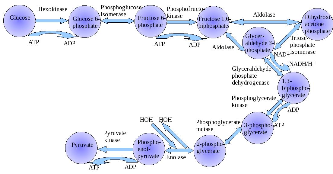
According to JES Roselino, “consider that glycolysis is oscillatory thanks to the kinetic behavior of Phosphofructokinase. Further, by its effect upon Pyruvate kinase through Fructose 1,6 diphosphate oscillatory levels, the inhibition of gluconeogenesis is also oscillatory. When the carbon flow through glycolysis is led to a maximal level gluconeogenesis will be almost completely blocked. The reversal of the Pyruvate kinase step in liver requires two enzymes (Pyruvate carboxylase (maintenance of oxaloacetic levels) + phosphoenolpyruvate carboxykinase (E.C. 4.1.1.32)) and energy requiring reactions that most likely could not as an ensemble, have a fast enough response against pyruvate kinase short period of inhibition during high frequency oscillatory periods of glycolytic flow. Only when glycolysis oscillates at low frequency the opposite reaction could enable gluconeogenic carbon flow.”

In case it can be shown in a rather convincing way, the same reasoning could be applied to understand how simple replicative signals inducing Go to G1 transition in cells, could easily overcome more complex signals required for cell differentiation and differentiated function.

Perhaps the problem of overextension of the equivalence of the DNA and what happens to the organism is also related to the initial reliance on a single cell model to relieve the complexity (which isn’t fully the case).

For instance, consider this fragment:
“Until only recently it was assumed that all proteins take on a clearly defined three-dimensional structure – i.e. they fold in order to be able to assume these functions.”
Cold Spring Harbour Symp. Quant. Biol. 1973  p 187-193 J.C Seidel and J Gergely – Investigation of conformational changes in Spin-Labeled Myosin Model for muscle contraction:
Huxley, A. F. 1971 Proc. Roy. Soc (London) (B) 178:1
Huxley, A.F and R. M. Simmons,1971. Nature 233:633
J.C Haselgrove X ray Evidence for a conformational Change in the Actin-containing filaments…Cold Spring Harbour Symp Quant Biol.1972 v 37: p 341-352

Only a very small sample indicating otherwise. Proteins were held as interacting macromolecules, changing their conformation in regulatory response to changes in the microenvironment (See Footnote 2). DNA was the opposite, non-interacting macromolecules to be as stable as a library must be.

The dogma held that the property of proteins could be read in DNA alone. Consequenly, the few examples quoted above, must be ignored and all people must believe that DNA alone, without environmental factors roles, controls protein amino acid sequence (OK), conformation (not true), activity (not true) and function (not true).

It appeared naively to be correct from the dogma to conclude from interpreting your genome: You have a 50% increased risk of developing the following disease (deterministic statement).  The correct form must be: You belong to a population that has a 50% increase in the risk of….followed by –  what you must do to avoid increase in your personal risk and the care you should take in case you want to have longer healthy life.  Thus, genetics and non-genetic diseases were treated as the same and medical foundations were reinforced by magical considerations (dogmas) in a very profitable way for those involved besides the patient.

 Footnotes:

  1. There is a link of electricity with ions in biology and the oscillatory behavior of some electrical discharges.  In addition, the oscillatory form of electrical discharged may have allowed Planck to relate high energy content with higher frequencies and conversely, low energy content in low frequency oscillatory events.  One may think of high density as an indication of great amount of matter inside a volume in space.  This helps the understanding of Planck’s idea as a high-density-energy in time for a high frequency phenomenon.
  1. Take into account a protein that may have its conformation restricted by an S-S bridge. This protein also, may move to another more flexible conformation in case it is in HS HS condition when the S-S bridge is broken. Consider also that, it takes some time for a protein to move from one conformation for instance, the restricted conformation (S-S) to other conformations. Also, it takes a few seconds or minutes to return to the S-S conformation (This is the Daniel Koshland´s concept of induced fit and relaxation time used by him in order to explain allosteric behavior of monomeric proteins- Monod, Wyman and Changeux requires tetramer or at least, dimer proteins).
  1. In case you have glycolysis oscillating in a frequency much higher than the relaxation time you could lead to the prevalence of high NADH effect leading to high HS /HS condition and at low glycolytic frequency, you could have predominance of S-S condition affecting protein conformation. In case you have predominance of NAD effect upon protein S-S you would get the opposite results.  The enormous effort to display the effect of citrate and over Phosphofructokinase conformation was made by others. Take into account that ATP action as an inhibitor in this case, is a rather unusual one. It is a substrate of the reaction, and together with its action as activator  F1,6 P (or its equivalent F2,6 P) is also unusual. However, it explains oscillatory behaviour of glycolysis. (Goldhammer , A.R, and Paradies: PFK structure and function, Curr. Top Cell Reg 1979; 15:109-141).
  1. The results presented in our Hepatology work must be viewed in the following way: In case the hepatic (oxygenated) blood flow is preserved, the bile secretory cells of liver receive well-oxygenated blood flow (the arterial branches bath secretory cells while the branches originated from portal vein irrigate the hepatocytes.  During extra hepatic cholestasis the low pressure, portal blood flow is reduced and the hepatocytes do not receive enough oxygen required to produce ATP that gluconeogenesis demands. Hepatic artery do not replace this flow since, its branches only join portal blood fluxes after the previous artery pressure  is reduced to a low pressure venous blood – at the point where the formation of hepatic vein is. Otherwise, the flow in the portal vein would be reversed or, from liver to the intestine. It is of no help to take into account possible valves for this reasoning since minimal arterial pressure is well above maximal venous pressure and this difference would keep this valve in permanent close condition. In low portal blood flow condition, the hepatocyte increases pyruvate kinase activity and with increased pyruvate kinase activity Gluconeogenesis is forbidden (See Walsh & Cooper revision quoted in the Hepatology as ref 23). For the hemodynamic considerations, role of artery and veins in hepatic portal system see references 44 and 45 Rappaport and Schneiderman and Rappapaport.


 Appendix I.

metabolic pathways

metabolic pathways

Signals Upstream and Targets Downstream of Lin28 in the Lin28 Pathway

Signals Upstream and Targets Downstream of Lin28 in the Lin28 Pathway


1.  Functional Proteomics Adds to Our Understanding

Ben Schuler’s research group from the Institute of Biochemistry of the University of Zurich has now established that an increase in temperature leads to folded proteins collapsing and becoming smaller. Other environmental factors can trigger the same effect. The crowded environments inside cells lead to the proteins shrinking. As these proteins interact with other molecules in the body and bring other proteins together, understanding of these processes is essential “as they play a major role in many processes in our body, for instance in the onset of cancer”, comments study coordinator Ben Schuler.

Measurements using the “molecular ruler”

“The fact that unfolded proteins shrink at higher temperatures is an indication that cell water does indeed play an important role as to the spatial organisation eventually adopted by the molecules”, comments Schuler with regard to the impact of temperature on protein structure. For their studies the biophysicists use what is known as single-molecule spectroscopy. Small colour probes in the protein enable the observation of changes with an accuracy of more than one millionth of a millimetre. With this “molecular yardstick” it is possible to measure how molecular forces impact protein structure.

With computer simulations the researchers have mimicked the behaviour of disordered proteins. They want to use them in future for more accurate predictions of their properties and functions.

Correcting test tube results

That’s why it’s important, according to Schuler, to monitor the proteins not only in the test tube but also in the organism. “This takes into account the fact that it is very crowded on the molecular level in our body as enormous numbers of biomolecules are crammed into a very small space in our cells”, says Schuler. The biochemists have mimicked this “molecular crowding” and observed that in this environment disordered proteins shrink, too.

Given these results many experiments may have to be revisited as the spatial organisation of the molecules in the organism could differ considerably from that in the test tube according to the biochemist from the University of Zurich. “We have, therefore, developed a theoretical analytical method to predict the effects of molecular crowding.” In a next step the researchers plan to apply these findings to measurements taken directly in living cells.

Explore further: Designer proteins provide new information about the body’s signal processesMore information: Andrea Soranno, Iwo Koenig, Madeleine B. Borgia, Hagen Hofmann, Franziska Zosel, Daniel Nettels, and Benjamin Schuler. Single-molecule spectroscopy reveals polymer effects of disordered proteins in crowded environments. PNAS, March 2014. DOI: 10.1073/pnas.1322611111


Effects of Hypoxia on Metabolic Flux

  1. Glucose-6-phosphate dehydrogenase regulation in the hepatopancreas of the anoxia-tolerantmarinemollusc, Littorina littorea

JL Lama , RAV Bell and KB Storey

Glucose-6-phosphate dehydrogenase (G6PDH) gates flux through the pentose phosphate pathway and is key to cellular antioxidant defense due to its role in producing NADPH. Good antioxidant defenses are crucial for anoxia-tolerant organisms that experience wide variations in oxygen availability. The marine mollusc, Littorina littorea, is an intertidal snail that experiences daily bouts of anoxia/hypoxia with the tide cycle and shows multiple metabolic and enzymatic adaptations that support anaerobiosis. This study investigated the kinetic, physical and regulatory properties of G6PDH from hepatopancreas of L. littorea to determine if the enzyme is differentially regulated in response to anoxia, thereby providing altered pentose phosphate pathway functionality under oxygen stress conditions.

Several kinetic properties of G6PDH differed significantly between aerobic and 24 h anoxic conditions; compared with the aerobic state, anoxic G6PDH (assayed at pH 8) showed a 38% decrease in K G6P and enhanced inhibition by urea, whereas in pH 6 assays Km NADP and maximal activity changed significantly.

All these data indicated that the aerobic and anoxic forms of G6PDH were the high and low phosphate forms, respectively, and that phosphorylation state was modulated in response to selected endogenous protein kinases (PKA or PKG) and protein phosphatases (PP1 or PP2C). Anoxia-induced changes in the phosphorylation state of G6PDH may facilitate sustained or increased production of NADPH to enhance antioxidant defense during long term anaerobiosis and/or during the transition back to aerobic conditions when the reintroduction of oxygen causes a rapid increase in oxidative stress.

Lama et al.  Peer J 2013.   http://dx.doi.org/10.7717/peerj.21


  1. Structural Basis for Isoform-Selective Inhibition in Nitric Oxide Synthase

    TL. Poulos and H Li

In the cardiovascular system, the important signaling molecule nitric oxide synthase (NOS) converts L-arginine into L-citrulline and releases nitric oxide (NO). NO produced by endothelial NOS (eNOS) relaxes smooth muscle which controls vascular tone and blood pressure. Neuronal NOS (nNOS) produces NO in the brain, where it influences a variety of neural functions such as neural transmitter release. NO can also support the immune system, serving as a cytotoxic agent during infections. Even with all of these important functions, NO is a free radical and, when overproduced, it can cause tissue damage. This mechanism can operate in many neurodegenerative diseases, and as a result the development of drugs targeting nNOS is a desirable therapeutic goal.

However, the active sites of all three human isoforms are very similar, and designing inhibitors specific for nNOS is a challenging problem. It is critically important, for example, not to inhibit eNOS owing to its central role in controlling blood pressure. In this Account, we summarize our efforts in collaboration with Rick Silverman at Northwestern University to develop drug candidates that specifically target NOS using crystallography, computational chemistry, and organic synthesis. As a result, we have developed aminopyridine compounds that are 3800-fold more selective for nNOS than eNOS, some of which show excellent neuroprotective effects in animal models. Our group has solved approximately 130 NOS-inhibitor crystal structures which have provided the structural basis for our design efforts. Initial crystal structures of nNOS and eNOS bound to selective dipeptide inhibitors showed that a single amino acid difference (Asp in nNOS and Asn in eNOS) results in much tighter binding to nNOS. The NOS active site is open and rigid, which produces few large structural changes when inhibitors bind. However, we have found that relatively small changes in the active site and inhibitor chirality can account for large differences in isoform-selectivity. For example, we expected that the aminopyridine group on our inhibitors would form a hydrogen bond with a conserved Glu inside the NOS active site. Instead, in one group of inhibitors, the aminopyridine group extends outside of the active site where it interacts with a heme propionate. For this orientation to occur, a conserved Tyr side chain must swing out of the way. This unanticipated observation taught us about the importance of inhibitor chirality and active site dynamics. We also successfully used computational methods to gain insights into the contribution of the state of protonation of the inhibitors to their selectivity. Employing the lessons learned from the aminopyridine inhibitors, the Silverman lab designed and synthesized symmetric double-headed inhibitors with an aminopyridine at each end, taking advantage of their ability to make contacts both inside and outside of the active site. Crystal structures provided yet another unexpected surprise. Two of the double-headed inhibitor molecules bound to each enzyme subunit, and one molecule participated in the generation of a novel Zn site that required some side chains to adopt alternate conformations. Therefore, in addition to achieving our specific goal, the development of nNOS selective compounds, we have learned how subtle differences in and structure can control proteinligand interactions and often in unexpected ways.


300px-Nitric_Oxide_Synthase

Nitric oxide synthase

arginine-NO-citulline cycle

arginine-NO-citulline cycle

active site of eNOS (PDB_1P6L) and nNOS (PDB_1P6H).

active site of eNOS (PDB_1P6L) and nNOS (PDB_1P6H).


NO - muscle, vasculature, mitochondria

NO – muscle, vasculature, mitochondria


Figure:  (A) Structure of one of the early dipeptide lead compounds, 1, that exhibits excellentisoform selectivity. (B, C) show the crystal structures of the dipeptide inhibitor 1 in the active site of eNOS (PDB: 1P6L) and nNOS (PDB: 1P6H). In nNOS, the inhibitor “curls” which enables the inhibitor R-amino group to interact with both Glu592 and Asp597. In eNOS, Asn368 is the homologue to nNOS Asp597.

Accounts in Chem Res 2013; 46(2): 390-98.

  1. Jamming a Protein Signal

Interfering with a single cancer-promoting protein and its receptor can open this resistance mechanism by initiating autophagy of the affected cells,  according to researchers at The University of Texas MD Anderson Cancer Center  in the journal Cell Reports.  According to Dr. Anil Sood and Yunfei Wen, lead and first authors, blocking  prolactin, a potent growth factor for ovarian cancer, sets off downstream events that result in cell by autophagy, the process  recycles damaged organelles and proteins for new use by the cell through the phagolysozome. This in turn, provides a clinical rationale for blocking prolactin and its receptor to initiate sustained autophagy as an alternative strategy for treating cancers.

Steep reductions in tumor weight

Prolactin (PRL) is a hormone previously implicated in ovarian, endometrial and other cancer development andprogression. When PRL binds to its cell membrane receptor, PRLR, activation of cancer-promoting cell signaling pathways follows.  A variant of normal prolactin called G129R blocks the reaction between prolactin and its receptor. Sood and colleagues treated mice that had two different lines of human ovarian cancer, both expressing the prolactin receptor, with G129R. Tumor weights fell by 50 percent for mice with either type of ovarian cancer after 28 days of treatment with G129R, and adding the taxane-based chemotherapy agent paclitaxel cut tumor weight by 90 percent. They surmise that higher doses of G129R may result in even greater therapeutic benefit.


3D experiments show death by autophagy


[video width=”1280″ height=”720″ mp4=”http://pharmaceuticalintelligence.com/wp-content/uploads/2014/04/1741-7007-11-65-s1-macromolecular-juggling-by-ubiquitylation-enzymes1.mp4″][/video]


Next the team used the prolactin-mimicking peptide to treat cultures of cancer spheroids which sharply reduced their numbers, and blocked the activation of JAK2 and STAT signaling pathways.

Protein analysis of the treated spheroids showed increased presence of autophagy factors and genomic analysis revealed increased expression of a number of genes involved in autophagy progression and cell death.  Then a series of experiments using fluorescence and electron microscopy showed that the cytosol of treated cells had large numbers of cavities caused by autophagy.

The team also connected the G129R-induced autophagy to the activity of PEA-15, a known cancer inhibitor. Analysis of tumor samples from 32 ovarian cancer patients showed that tumors express higher levels of the prolactin receptor and lower levels of phosphorylated PEA-15 than normal ovarian tissue. However, patients with low levels of the prolactin receptor and higher PEA-15 had longer overall survival than those with high PRLR and low PEA-15.

Source: MD Anderson Cancer Center


  1. Chemists’ Work with Small Peptide Chains of Enzymes

Korendovych and his team designed seven simple peptides, each containing seven amino acids. They then allowed the molecules of each peptide to self-assemble, or spontaneously clump together, to form amyloids. (Zinc, a metal with catalytic properties, was introduced to speed up the reaction.) What they found was that four of the seven peptides catalyzed the hydrolysis of molecules known as esters, compounds that react with water to produce water and acids—a feat not uncommon among certain enzymes.

“It was the first time that a peptide this small self-assembled to produce an enzyme-like catalyst,” says Korendovych. “Each enzyme has to be an exact fit for its respective substrate,” he says, referring to the molecule with which an enzyme reacts. “Even after millions of years, nature is still testing all the possible combinations of enzymes to determine which ones can catalyze metabolic reactions. Our results make an argument for the design of self-assembling nanostructured catalysts.”

Source: Syracuse University

Here are three articles emphasizing the value of combinatorial analysis, which can be formed from genomic, clinical, and proteomic data sets.


  1. Comparative analysis of differential network modularity in tissue specific normal and cancer protein interaction networks

    F Islam , M Hoque , RS Banik , S Roy , SS Sumi, et al.

As most biological networks show modular properties, the analysis of differential modularity between normal and cancer protein interaction networks can be a good way to understand cancer more significantly. Two aspects of biological network modularity e.g. detection of molecular complexes (potential modules or clusters) and identification of crucial nodes forming the overlapping modules have been considered in this regard.

The computational analysis of previously published protein interaction networks (PINs) has been conducted to identify the molecular complexes and crucial nodes of the networks. Protein molecules involved in ten major cancer signal transduction pathways were used to construct the networks based on expression data of five tissues e.g. bone, breast, colon, kidney and liver in both normal and cancer conditions.

Cancer PINs show higher level of clustering (formation of molecular complexes) than the normal ones. In contrast, lower level modular overlapping is found in cancer PINs than the normal ones. Thus a proposition can be made regarding the formation of some giant nodes in the cancer networks with very high degree and resulting in reduced overlapping among the network modules though the predicted molecular complex numbers are higher in cancer conditions.

Islam et al. Journal of Clinical Bioinformatics 2013, 3:19-32

  1. A new 12-gene diagnostic biomarker signature of melanoma revealed by integrated microarray analysis

    Wanting Liu , Yonghong Peng and Desmond J. Tobin
    PeerJ 1:e49;        http://dx.doi.org/10.7717/peerj.49

Here we present an integrated microarray analysis framework, based on a genome-wide relative significance (GWRS) and genome-wide global significance (GWGS) model. When applied to five microarray datasets on melanoma published between 2000 and 2011, this method revealed a new signature of 200 genes. When these were linked to so-called ‘melanoma driver’ genes involved in MAPK, Ca2+, and WNT signaling pathways we were able to produce a new 12-gene diagnostic biomarker signature for melanoma (i.e., EGFR, FGFR2, FGFR3, IL8, PTPRF, TNC, CXCL13, COL11A1, CHP2, SHC4, PPP2R2C, andWNT4).We have begun to experimentally validate a subset of these genes involved inMAPK signaling at the protein level, including CXCL13, COL11A1, PTPRF and SHC4 and found these to be overexpressed inmetastatic and primarymelanoma cells in vitro and in situ compared to melanocytes cultured from healthy skin epidermis and normal healthy human skin.


catalytic amyloid forming particle

catalytic amyloid forming particle


        8.    PanelomiX: A threshold-based algorithm to create panels of biomarkers

X Robin , N Turck , A Hainard , N Tiberti, et al.
               Translational Proteomics 2013.    http://dx.doi.org/10.1016/j.trprot.2013.04.003

The PanelomiX toolbox combines biomarkers and evaluates the performance of panels to classify patients better than singlemarkers or other classifiers. The ICBTalgorithm proved to be an efficient classifier, the results of which can easily be interpreted.

Here are two current examples of the immense role played by signaling pathways in carcinogenic mechanisms and in treatment targeting, which is also confounded by acquired resistance.


  1. Triple-Negative Breast Cancer

  1. epidermal growth factor receptor (EGFR or ErbB1) and
  2. high activity of the phosphatidylinositol 3-kinase (PI3K)–Akt pathway

are both targeted in triple-negative breast cancer (TNBC).

  • activation of another EGFR family member [human epidermal growth factor receptor 3 (HER3) (or ErbB3)] may limit the antitumor effects of these drugs.

This study found that TNBC cell lines cultured with the EGFR or HER3 ligand EGF or heregulin, respectively, and treated with either an Akt inhibitor (GDC-0068) or a PI3K inhibitor (GDC-0941) had increased abundance and phosphorylation of HER3.

The phosphorylation of HER3 and EGFR in response to these treatments

  1. was reduced by the addition of a dual EGFR and HER3 inhibitor (MEHD7945A).
  2. MEHD7945A also decreased the phosphorylation (and activation) of EGFR and HER3 and
  3. the phosphorylation of downstream targets that occurred in response to the combination of EGFR ligands and PI3K-Akt pathway inhibitors.

In culture, inhibition of the PI3K-Akt pathway combined with either MEHD7945A or knockdown of HER3

  1. decreased cell proliferation compared with inhibition of the PI3K-Akt pathway alone.
  2. Combining either GDC-0068 or GDC-0941 with MEHD7945A inhibited the growth of xenografts derived from TNBC cell lines or from TNBC patient tumors, and
  3. this combination treatment was also more effective than combining either GDC-0068 or GDC-0941 with cetuximab, an EGFR-targeted antibody.
  4. After therapy with EGFR-targeted antibodies, some patients had residual tumors with increased HER3 abundance and EGFR/HER3 dimerization (an activating interaction).

Thus, we propose that concomitant blockade of EGFR, HER3, and the PI3K-Akt pathway in TNBC should be investigated in the clinical setting.

Reference: Antagonism of EGFR and HER3 Enhances the Response to Inhibitors of the PI3K-Akt Pathway in Triple-Negative Breast Cancer. JJ Tao, P Castel, N Radosevic-Robin, M Elkabets, et al.  Sci. Signal., 25 March 2014;
7(318), p. ra29   http://dx.doi.org/10.1126/scisignal.2005125


                  10.   Metastasis in RAS Mutant or Inhibitor-Resistant Melanoma Cells

The protein kinase BRAF is mutated in about 40% of melanomas, and BRAF inhibitors improve progression-free and overall survival in these patients. However, after a relatively short period of disease control, most patients develop resistance because of reactivation of the RAF–ERK (extracellular signal–regulated kinase) pathway, mediated in many cases by mutations in RAS. We found that BRAF inhibition induces invasion and metastasis in RAS mutant melanoma cells through a mechanism mediated by the reactivation of the MEK (mitogen-activated protein kinase kinase)–ERK pathway.

Reference: BRAF Inhibitors Induce Metastasis in RAS Mutant or Inhibitor-Resistant Melanoma Cells by Reactivating MEK and ERK Signaling. B Sanchez-Laorden, A Viros, MR Girotti, M Pedersen, G Saturno, et al., Sci. Signal., 25 March 2014;  7(318), p. ra30  http://dx.doi.org/10.1126/scisignal.2004815

Appendix II.

The world of physics in the twentieth century saw the end of determinism established by Newton. This is characterized by discrete laws that describe natural observations. These are in gravity and in eletricity. In an early phase of investigation, an era of galvanic or voltaic electricity represented a revolutionary break from the historical focus on frictional electricity. Alessandro Voltadiscovered that chemical reactions could be used to create positively charged anodes and negatively charged cathodes.  In 1790, Prof. Luigi Alyisio Galvani of Bologna, while conducting experiments on “animal electricity“, noticed the twitching of a frog’s legs in the presence of an electric machine. He observed that a frog’s muscle, suspended on an iron balustrade by a copper hook passing through its dorsal column, underwent lively convulsions without any extraneous cause, the electric machine being at this time absent.  Volta communicated a description of his pile to the Royal Society of London and shortly thereafter Nicholson and Cavendish (1780) produced the decomposition of water by means of the electric current, using Volta’s pile as the source of electromotive force.

Siméon Denis Poisson attacked the difficult problem of induced magnetization, and his results provided  a first approximation. His innovation required the application of mathematics to physics.  His memoirs on the theory of electricity and magnetism created a new branch of mathematical physics.  The discovery of electromagnetic induction was made almost simultaneously and independently by Michael Faraday and Joseph Henry. Michael Faraday, the successor of Humphry Davy, began his epoch-making research relating to electric and electromagnetic induction in 1831. In his investigations of the peculiar manner in which iron filings arrange themselves on a cardboard or glass in proximity to the poles of a magnet, Faraday conceived the idea of magnetic “lines of force” extending from pole to pole of the magnet and along which the filings tend to place themselves. On the discovery being made that magnetic effects accompany the passage of an electric current in a wire, it was also assumed that similar magnetic lines of force whirled around the wire. He also posited that iron, nickel, cobalt, manganese, chromium, etc., are paramagnetic (attracted by magnetism), whilst other substances, such as bismuth, phosphorus, antimony, zinc, etc., are repelled by magnetism or are diamagnetic.

Around the mid-19th century, Fleeming Jenkin‘s work on ‘ Electricity and Magnetism ‘ and Clerk Maxwell’s ‘ Treatise on Electricity and Magnetism ‘ were published. About 1850 Kirchhoff published his laws relating to branched or divided circuits. He also showed mathematically that according to the then prevailing electrodynamic theory, electricity would be propagated along a perfectly conducting wire with the velocity of light. Herman Helmholtz investigated the effects of induction on the strength of a current and deduced mathematical equations, which experiment confirmed. In 1853 Sir William Thomson (later Lord Kelvin) predicted as a result of mathematical calculations the oscillatory nature of the electric discharge of a condenser circuit.  Joseph Henry, in 1842 discerned  the oscillatory nature of the Leyden jardischarge.

In 1864 James Clerk Maxwell announced his electromagnetic theory of light, which was perhaps the greatest single step in the world’s knowledge of electricity. Maxwell had studied and commented on the field of electricity and magnetism as early as 1855/6 when On Faraday’s lines of force was read to the Cambridge Philosophical Society. The paper presented a simplified model of Faraday’s work, and how the two phenomena were related. He reduced all of the current knowledge into a linked set of differential equations with 20 equations in 20 variables. This work was later published as On Physical Lines of Force in1861. In order to determine the force which is acting on any part of the machine we must find its momentum, and then calculate the rate at which this momentum is being changed. This rate of change will give us the force. The method of calculation which it is necessary to employ was first given by Lagrange, and afterwards developed, with some modifications, by Hamilton’s equations. Now Maxwell logically showed how these methods of calculation could be applied to the electro-magnetic field. The energy of a dynamical systemis partly kinetic, partly potential. Maxwell supposes that the magnetic energy of the field is kinetic energy, the electric energy potential.  Around 1862, while lecturing at King’s College, Maxwell calculated that the speed of propagation of an electromagnetic field is approximately that of the speed of light.   Maxwell’s electromagnetic theory of light obviously involved the existence of electric waves in free space, and his followers set themselves the task of experimentally demonstrating the truth of the theory. By 1871, he presented the Remarks on the mathematical classification of physical quantities.

A Wave-Particle Dilemma at the Century End

In 1896 J.J. Thomson performed experiments indicating that cathode rays really were particles, found an accurate value for their charge-to-mass ratio e/m, and found that e/m was independent of cathode material. He made good estimates of both the charge e and the mass m, finding that cathode ray particles, which he called “corpuscles”, had perhaps one thousandth of the mass of the least massive ion known (hydrogen). He further showed that the negatively charged particles produced by radioactive materials, by heated materials, and by illuminated materials, were universal.  In the late 19th century, the Michelson–Morley experiment was performed by Albert Michelson and Edward Morley at what is now Case Western Reserve University. It is generally considered to be the evidence against the theory of a luminiferous aether. The experiment has also been referred to as “the kicking-off point for the theoretical aspects of the Second Scientific Revolution.” Primarily for this work, Albert Michelson was awarded theNobel Prize in 1907.

Wave–particle duality is a theory that proposes that all matter exhibits the properties of not only particles, which have mass, but also waves, which transfer energy. A central concept of quantum mechanics, this duality addresses the inability of classical concepts like “particle” and “wave” to fully describe the behavior of quantum-scale objects. Standard interpretations of quantum mechanics explain this paradox as a fundamental property of the universe, while alternative interpretations explain the duality as an emergent, second-order consequence of various limitations of the observer. This treatment focuses on explaining the behavior from the perspective of the widely used Copenhagen interpretation, in which wave–particle duality serves as one aspect of the concept of complementarity, that one can view phenomena in one way or in another, but not both simultaneously.  Through the work of Max Planck, Albert Einstein, Louis de Broglie, Arthur Compton, Niels Bohr, and many others, current scientific theory holds that all particles also have a wave nature (and vice versa).

Beginning in 1670 and progressing over three decades, Isaac Newton argued that the perfectly straight lines of reflection demonstrated light’s particle nature, but Newton’s contemporaries Robert Hooke and Christiaan Huygens—and later Augustin-Jean Fresnel—mathematically refined the wave viewpoint, showing that if light traveled at different speeds in different, refraction could be easily explained. The resulting Huygens–Fresnel principle was supported by Thomas Young‘s discovery of double-slit interference, the beginning of the end for the particle light camp.  The final blow against corpuscular theory came when James Clerk Maxwell discovered that he could combine four simple equations, along with a slight modification to describe self-propagating waves of oscillating electric and magnetic fields. When the propagation speed of these electromagnetic waves was calculated, the speed of light fell out. While the 19th century had seen the success of the wave theory at describing light, it had also witnessed the rise of the atomic theory at describing matter.

Matter and Light

In 1789, Antoine Lavoisier secured chemistry by introducing rigor and precision into his laboratory techniques. By discovering diatomic gases, Avogadro completed the basic atomic theory, allowing the correct molecular formulae of most known compounds—as well as the correct weights of atoms—to be deduced and categorized in a consistent manner. The final stroke in classical atomic theory came when Dimitri Mendeleev saw an order in recurring chemical properties, and created a table presenting the elements in unprecedented order and symmetry.   Chemistry was now an atomic science.

Black-body radiation, the emission of electromagnetic energy due to an object’s heat, could not be explained from classical arguments alone. The equipartition theorem of classical mechanics, the basis of all classical thermodynamic theories, stated that an object’s energy is partitioned equally among the object’s vibrational modes. This worked well when describing thermal objects, whose vibrational modes were defined as the speeds of their constituent atoms, and the speed distribution derived from egalitarian partitioning of these vibrational modes closely matched experimental results. Speeds much higher than the average speed were suppressed by the fact that kinetic energy is quadratic—doubling the speed requires four times the energy—thus the number of atoms occupying high energy modes (high speeds) quickly drops off. Since light was known to be waves of electromagnetism, physicists hoped to describe this emission via classical laws. This became known as the black body problem. The Rayleigh–Jeans law which, while correctly predicting the intensity of long wavelength emissions, predicted infinite total energy as the intensity diverges to infinity for short wavelengths.

The solution arrived in 1900 when Max Planck hypothesized that the frequency of light emitted by the black body depended on the frequency of the oscillator that emitted it, and the energy of these oscillators increased linearly with frequency (according to his constant h, where E = hν). By demanding that high-frequency light must be emitted by an oscillator of equal frequency, and further requiring that this oscillator occupy higher energy than one of a lesser frequency, Planck avoided any catastrophe; giving an equal partition to high-frequency oscillators produced successively fewer oscillators and less emitted light. And as in the Maxwell–Boltzmann distribution, the low-frequency, low-energy oscillators were suppressed by the onslaught of thermal jiggling from higher energy oscillators, which necessarily increased their energy and frequency. Planck had intentionally created an atomic theory of the black body, but had unintentionally generated an atomic theory of light, where the black body never generates quanta of light at a given frequency with energy less than hν.

In 1905 Albert Einstein took Planck’s black body model in itself and saw a wonderful solution to another outstanding problem of the day: the photoelectric effect, the phenomenon where electrons are emitted from atoms when they absorb energy from light.   Only by increasing the frequency of the light, and thus increasing the energy of the photons, can one eject electrons with higher energy. Thus, using Planck’s constant h to determine the energy of the photons based upon their frequency, the energy of ejected electrons should also increase linearly with frequency; the gradient of the line being Planck’s constant. These results were not confirmed until 1915, when Robert Andrews Millikan, produced experimental results in perfect accord with Einstein’s predictions. While  the energy of ejected electrons reflected Planck’s constant, the existence of photons was not explicitly proven until the discovery of the photon antibunching effect  When Einstein received his Nobel Prizein 1921, it was  for the photoelectric effect, the suggestion of quantized light. Einstein’s “light quanta” represented the quintessential example of wave–particle duality. Electromagnetic radiation propagates following  linear wave equations, but can only be emitted or absorbed as discrete elements, thus acting as a wave and a particle simultaneously.

Radioactivity Changes the Scientific Landscape

The turn of the century also features radioactivity, which later came to the forefront of the activities of World War II, the Manhattan Project, the discovery of the chain reaction, and later – Hiroshima and Nagasaki.

Marie Curie

Marie Curie


Marie Skłodowska-Curie was a Polish and naturalized-French physicist and chemist who conducted pioneering research on radioactivity. She was the first woman to win a Nobel Prize, the only woman to win in two fields, and the only person to win in multiple sciences. She was also the first woman to become a professor at the University of Paris, and in 1995 became the first woman to be entombed on her own merits in the Panthéon in Paris. She shared the 1903 Nobel Prize in Physics with her husband Pierre Curie and with physicist Henri Becquerel. She won the 1911 Nobel Prize in Chemistry.  Her achievements included a theory of radioactivity (a term that she coined, techniques for isolating radioactive isotopes, and the discovery of polonium and radium. She named the first chemical element that she discovered – polonium, which she first isolated in 1898 – after her native country. Under her direction, the world’s first studies were conducted into the treatment of neoplasms using radioactive isotopes. She founded the Curie Institutes in Paris and in Warsaw, which remain major centres of medical research today. During World War I, she established the first military field radiological centres.  Curie died in 1934 due to aplastic anemia brought on by exposure to radiation – mainly, it seems, during her World War I service in mobile X-ray units created by her.


Read Full Post »

Heroes in Medical Research: Developing Models for Cancer Research

Author, Curator: Stephen J. Williams, Ph.D.


The current rapid progress in cancer research would have never come about if not for the dedication of past researchers who had developed many of the scientific tools we use today. In this issue of Heroes in Medical Research I would like to give tribute to the researchers who had developed the some of the in-vivo and in-vitro models which are critical for cancer research.


The Animal Modelers in Cancer Research

Helen Dean King, Ph.D. (1869-1955)

Helen Dean King

Helen Dean King, Ph.D. from www.ExplorePAhistory.com; photo Courtesy of the Wistar Institute Archive Collection, Philadelphia, PA


The work of Dr. Helen Dean King on rat inbreeding led to development of strains of laboratory animals. Dr. King taught at Bryn Mawr College, then worked at University of Pennsylvania and the Wistar Institute under famed geneticist Thomas Hunt Morgan, researching if inbreeding would produce harmful genetic traits.   At University of Pennsylvania she examined environmental and genetic factors on gender determination.


Important papers include [1-6]as well as the following contributions:

“Studies in Inbreeding”, “Life Processes in Gray Norway Rats During Fourteen Years in Captivity”, doctoral thesis on embryologic development in toads (1899)


Milestones include:


1909    started albino rat breeding and bred 20 female and male from same litter (King colony) to 25

successive generations (inbreeding did not cause harmful traits)


1919     started to domesticate the wild Norwegian rats that ran thru Philadelphia (six pairs Norway rats

thru 28 generations)

A good reference for definitions of rat inbreeding versus line generation including a history of Dr. King’s work can be found at the site: Munificent Mischief Rattery and a brief history here.[7] In addition, Dr. King had investigated using rat strains as a possible recipient for tumor cells. The work was an important advent to the use of immunodeficient models for cancer research.


As shown below Philadelphia became a hotbed for research into embryology, development, genetics, and animal model development.


Beatrice Mintz, Ph.D.

(Beatrice Minz, Ph.D.; photo credit Fox Chase Cancer Center, www.pubweb.fccc.edu) Mintz

Dr. Mintz, an embryologist and cancer researcher from Fox Chase Cancer Center in Philadelphia, PA, contributed some of the most seminal discoveries leading to our current understanding of genetics, embryo development, cellular differentiation, and oncogenesis, especially melanoma, while pioneering techniques which allowed the development of genetically modified mice.

If you get the privilege of hearing her talk, take advantage of it. Dr. Mintz is one of those brilliant scientists who have the ability to look at a clinical problem from the viewpoint of a basic biological question and, at the same time, has the ability to approach the well-thought out questions with equally well thought out experimental design. For example, Dr. Mintz asked if a cell’s developmental fate was affected by location in the embryo. This led to her work by showing teratocarcinoma tumor cells in the developing embryo could revert to a more normal phenotype, essentially proving two important concepts in development and tumor biology:

  1. The existence of pluripotent stem cells
  2. That tumor cells are affected by their environment (which led to future concepts of the importance of tumor microenvironment on tumor growth

Other seminal discoveries included:

  • Development of the first mouse chimeras using novel cell fusion techniques
  • With Rudolf Jaenisch in 1974, showed integration of viral DNA from SV40, could be integrated into the DNA of developing mice and persist into adulthood somatic cells, the first transgenesis in mice which led ultimately to:
  • Development of the first genetically modified mouse model of human melanoma in 1993

Her current work, seen on the faculty webpage here, is developing mice with predisposition to melanoma to uncover risk factors associated with the early development of melanoma.

In keeping with the Philadelphia tradition another major mouse model which became seminal to cancer drug discovery was co-developed in the same city, same institute and described in the next section.

It is interesting to note that the first cloning of an animal, a frog, had taken place at the Institute for Cancer Research, later becoming Fox Chase Cancer Center, which was performed by Drs. Robert Briggs and Thomas J. King and reported in the 152 PNAS paper Transplantation of Living Nuclei From Blastula Cells into Enucleated Frogs’ Eggs.[8]


 The Immunodeficient Animal as a Model System for Cancer Research – Dr. Mel Bosma, Ph.D.


Bosma

Melvin J. Bosma, Ph.D.; photo credit Fox Chase Cancer Center

In the summer of 1980 at Fox Chase Cancer Center, Dr. Melvin J. Bosma and his co-researcher wife Gayle discovered mice with deficiencies in common circulating antibodies and since, these mice were littermates, realized they had found a genetic defect which rendered the mice immunodeficient (upon further investigation these mice were unable to produce mature B and T cells). These mice were the first scid (severe combined immunodeficiency) colony. The scid phenotype was later found to be a result of a spontaneous mutation in the enzyme Prkdc {protein kinase, DNA activated, catalytic polypeptide} involved in DNA repair, and ultimately led to a defect in V(D)J recombination of immunoglobulins.

The emergence of this scid mouse was not only crucial for AIDS research but was another turning point in cancer research , as researchers now had a robust in-vivo recipient for human tumor cells. The orthotopic xenograft of human tumor cells now allowed for studies on genetic and microenvironmental factors affecting tumorigenicity, as well as providing a model for chemotherapeutic drug development (see Suggitt for review and references)[9]. A discussion of the pros and cons of the xenograft system for cancer drug discovery would be too voluminous for this post and would warrant a full review by itself. But before the advent of such scid mouse systems researchers relied on spontaneous and syngeneic mouse tumor models such as the B16 mouse melanoma and Lewis lung tumor model.

Other scid systems have been developed such as in the dog, horse, and pig. Please see the following post on this site The SCID Pig: How Pigs are becoming a Great Alternate Model for Cancer Research. The athymic (nude) mouse (nu/nu) also is a popular immunodeficient mouse model used for cancer research

Two other in-vivo tumor models: Patient Derived Xenografts (PDX) and Genetically Engineered Mouse models (GEM) deserve their own separate discussion however the success of these new models can be attributed to the hard work of the aforementioned investigators. Therefore I will post separately and curate PDX and GEM models of cancer and highlight some new models which are having great impact on cancer drug development.


References

1.         Loeb L, King HD: Transplantation and Individuality Differential in Strains of Inbred Rats. The American journal of pathology 1927, 3(2):143-167.

2.         Lewis MR, Aptekman PM, King HD: Retarding action of adrenal gland on growth of sarcoma grafts in rats. J Immunol 1949, 61(4):315-319.

3.         Aptekman PM, Lewis MR, King HD: Tumor-immunity induced in rats by subcutaneous injection of tumor extract. J Immunol 1949, 63(4):435-440.

4.         Lewis MR, Aptekman PM, King HD: Inactivation of malignant tissue in tumor-immune rats. J Immunol 1949, 61(4):321-326.

5.         Lewis MR, King HD, et al.: Further studies on oncolysis and tumor immunity in rats. J Immunol 1948, 60(4):517-528.

6.         Aptekman PM, Lewis MR, King HD: A method of producing in inbred albino rats a high percentage of immunity from tumors native in their strain. J Immunol 1946, 52:77-86.

7.         Ogilvie MB: Inbreeding, eugenics, and Helen Dean King (1869-1955). Journal of the history of biology 2007, 40(3):467-507.

8.         Briggs R, King TJ: Transplantation of Living Nuclei From Blastula Cells into Enucleated Frogs’ Eggs. Proceedings of the National Academy of Sciences of the United States of America 1952, 38(5):455-463.

9.         Suggitt M, Bibby MC: 50 years of preclinical anticancer drug screening: empirical to target-driven approaches. Clinical cancer research : an official journal of the American Association for Cancer Research 2005, 11(3):971-981.


Other posts on this site about Cancer, Animal Models of Disease, and other articles in this series include:

The SCID Pig: How Pigs are becoming a Great Alternate Model for Cancer Research

A Synthesis of the Beauty and Complexity of How We View Cancer

Guidelines for the welfare and use of animals in cancer research

Importance of Funding Replication Studies: NIH on Credibility of Basic Biomedical Studies

FDA Guidelines For Developmental and Reproductive Toxicology (DART) Studies for Small Molecules

Report on the Fall Mid-Atlantic Society of Toxicology Meeting “Reproductive Toxicology of Biologics: Challenges and Considerations:

What`s new in pancreatic cancer research and treatment?

Heroes in Medical Research: Dr. Carmine Paul Bianchi Pharmacologist, Leader, and Mentor

Heroes in Medical Research: Dr. Robert Ting, Ph.D. and Retrovirus in AIDS and Cancer

Heroes in Medical Research: Barnett Rosenberg and the Discovery of Cisplatin

Richard Lifton, MD, PhD of Yale University and Howard Hughes Medical Institute: Recipient of 2014 Breakthrough Prizes Awarded in Life Sciences for the Discovery of Genes and Biochemical Mechanisms that cause Hypertension

Reuben Shaw, Ph.D., a geneticist and researcher at the Salk Institute: Metabolism Influences Cancer


Read Full Post »

Epilogue: Envisioning New Insights in Cancer Translational Biology

Author and Curator: Larry H Bernstein, MD, FCAP


The foregoing  summary leads to a beginning as it is a conclusion.  It concludes a body of work in the e-book series,

Series C: e-Books on Cancer & Oncology

Series C Content Consultant: Larry H. Bernstein, MD, FCAP


VOLUME ONE 

Cancer Biology and Genomics for Disease Diagnosis

2014

Stephen J. Williams, PhD, Senior Editor

sjwilliamspa@comcast.net

Tilda Barliya, PhD, Editor

tildabarliya@gmail.com

Ritu Saxena, PhD, Editor

ritu.uab@gmail.com

Leaders in Pharmaceutical Business Intelligence 

that has been presented by the cancer team of professional experts, e-Book concept was conceived by Aviva Lev-Ari, PhD, RN, e-Series Editor-in-Chief and Founder of Leaders in Pharmaceutical Business Intelligence 

and the Open Access Online Scientific Journal

http://pharmaceuticalintelligence.com

Stephen J. Williams, PhD, Senior Editor, and other notable contributors in  various aspects of cancer research in the emerging fields of targeted  pharmacology,  nanotechnology, cancer imaging, molecular pathology, transcriptional and regulatory ‘OMICS’, metabolism, medical and allied health related sciences, synthetic biology, pharmaceutical discovery, and translational medicine.

This  volume and its content have been conceived and organized to capture the organized events that emerge in embryological development, leading to the major organ systems that we recognize anatomically and physiologically as an integrated being.  We capture the dynamic interactions between the systems under stress  that are elicited by cytokine-driven hormonal responses, long thought to be circulatory and multisystem, that affect the major compartments of  fat and lean body mass, and are as much the drivers of metabolic pathway changes that emerge as epigenetics, without disregarding primary genetic diseases.

The greatest difficulty in organizing such a work is in whether it is to be merely a compilation of cancer expression organized by organ systems, or whether it is to capture developing concepts of underlying stem cell expressed changes that were once referred to as “dedifferentiation”.  In proceeding through the stages of neoplastic transformation, there occur adaptive local changes in cellular utilization of anabolic and catabolic pathways, and a retention or partial retention of functional specificities.

This  effectively results in the same cancer types not all fitting into the same “shoe”. There is a sequential loss of identity associated with cell migration, cell-cell interactions with underlying stroma, and metastasis., but cells may still retain identifying “signatures” in microRNA combinatorial patterns.  The story is still incomplete, with gaps in our knowledge that challenge the imagination.

What we have laid out is a map with substructural ordered concepts forming subsets within the structural maps.  There are the traditional energy pathways with terms aerobic and anaerobic glycolysis, gluconeogenesis, triose phosphate branch chains, pentose shunt, and TCA cycle vs the Lynen cycle, the Cori cycle, glycogenolysis, lipid peroxidation, oxidative stress, autosomy and mitosomy, and genetic transcription, cell degradation and repair, muscle contraction, nerve transmission, and their involved anatomic structures (cytoskeleton, cytoplasm, mitochondria, liposomes and phagosomes, contractile apparatus, synapse.

Then there is beneath this macro-domain the order of signaling pathways that regulate these domains and through mechanisms of cellular regulatory control have pleiotropic inhibitory or activation effects, that are driven by extracellular and intracellular energy modulating conditions through three recognized structures: the mitochondrial inner membrane, the intercellular matrix, and the ion-channels.

What remains to be done?

  1. There is still to be elucidated the differences in patterns within cancer types the distinct phenotypic and genotypic features  that mitigate anaplastic behavior. One leg of this problem lies in the density of mitochondria, that varies between organ types, but might vary also within cell type of a common function.  Another leg of this problem has also appeared to lie in the cell death mechanism that relates to the proeosomal activity acting on both the ribosome and mitochondrion in a coordinated manner.  This is an unsolved mystery of molecular biology.


  1. Then there is a need to elucidate the major differences between tumors of endocrine, sexual, and structural organs, which are distinguished by primarily a synthetic or primarily a catabolic function, and organs that are neither primarily one or the other.  For example, tumors of the thyroid and paratnhyroids, islet cells of pancreas, adrenal cortex, and pituitary glands have the longest 5 year survivals.  They and the sexual organs are in the visceral compartment.  The rest of the visceral compartment would be the liver, pancreas, salivary glands, gastrointestinal tract, and lungs (which are embryologically an outpouching of the gastrointestinal tract), kidneys and lower urinary tract.  Cancers of these organs have a much less favorable survival (brain, breast and prostate, lymphatic, blood forming organ, skin).  The case  is intermediate for breast and prostate between the endocrine organs and GI tract, based on natural history, irrespective of the available treatments.  Just consider the dilemma over what we do about screening for prostate cancer in men over the age of 60 years age who have a 70 percent incident silent carcinoma of the prostate that could be associated with unrelated cause of death.  The very rapid turnover of the gastric and colonic GI epithelium, and of the  subepithelial  B cell mucosal lymphocytic structures  is associated  with a greater aggressiveness of the tumor.


  1. However, we  have to reconsider the observation by NO Kaplan than the synthetic and catabolic functions are highlighted by differences in the expressions of the balance of  the two major pyridine nucleotides – DPN (NAD) and TPN (NADP) – which also might be related to the density of mitochondria  which is associated with both NADP and synthetic activity, and  with efficient aerobic function.  These are in an equilibrium through the “transhydrogenase reaction” co-discovered by Kaplan, in Fritz Lipmann’s laboratory. There does  arise a conundrum involving the regulation of mitochondria in these high turnover epithelial tissues  that rely on aerobic energy, and generate ATP through TPN linked activity, when they undergo carcinogenesis. The cells  replicate and they become utilizers of glycolysis, while at the same time, the cell death pathway is quiescent. The result becomes the introduction of peripheral muscle and liver synthesized protein cannabolization (cancer cachexia) to provide glucose  from proteolytic amino acid sources.


  1. There is also the structural compartment of the lean body mass. This is the heart, skeletal  structures (includes smooth muscle of GI tract, uterus, urinary bladder, brain, bone, bone marrow).  The contractile component is associated with sarcomas.  What is most striking is that the heart, skeletal muscle, and inflammatory cells are highly catabolic, not anabolic.  NO Kaplan referred tp them as DPN (NAD) tissues. This compartment requires high oxygen supply, and has a high mechanical function. But again, we return to the original observations of enrgy requirements at rest being different than at high demand.  At work, skeletal muscle generates lactic acid, but the heart can use lactic acid as fuel,.


  1. The liver is supplied by both the portal vein and the hepatic artery, so it is not prone to local ischemic injury (Zahn infarct). It is exceptional in that it carries out synthesis of all the circulating transport proteins, has a major function in lipid synthesis and in glycogenesis and glycogenolysis, with the added role of drug detoxification through the P450 system.  It is not only the largest organ (except for brain), but is highly active both anabolically and catabolically (by ubiquitilation).
  2. The expected cellular turnover rates for these tissues and their balance of catabolic and anabolic function would have to be taken into account to account for the occurrence and the activities of oncogenesis. This is by no means a static picture, but a dynamic organism constantly in flux imposed by internal and external challenges.  It is also important to note the the organs have a concentration of mitochondria, associated with energy synthetic and catabolic requirements provided by oxygen supply and the electron transport mechanism for oxidative phosphorylation.  For example, tissues that are primarily synthetic do not have intermitent states of resting and high demand, as seen in skeletal muscle, or perhaps myocardium (which is syncytial and uses lactic acid generated from skeletal muscle when there is high demand).
  3. The existence of  lncDNA has been discovered only as a result of the human genome project (HGP). This was previously known only as “dark DNA”.  It has become clear that lncDNA has an important role in cellular regulatory activities centered in the chromatin modeling.  Moreover, just as proteins exhibit functionality in their folding, related to tertiary structure and highly influenced by location of –S-S- bridges and amino acid residue distances (allosteric effects), there is a less studied effect as the chromatin becomes more compressed within the nucleus, that should have a bearing on cellular expression.

According to Jose Eduardo de Salles Roselino , when the Na/Glucose transport system (for a review Silvermann, M. in Annu. Rev. Biochem.60: 757-794(1991)) was  found in kidneys as well as in key absorptive cells of digestive tract, it should be stressed its functional relationship with “internal milieu” and real meaning, homeostasis. It is easy to understand how the major topic was presented as how to prevent diarrheal deaths in infants, while detected in early stages. However, from a biochemical point of view, as presented in Schrödinger´s What is life?, (biochemistry offering a molecular view for two legs of biology, physiology and genetics). Why should it be driven to the sole target of understanding genetics? Why the understanding of physiology in molecular terms should be so neglected?

From a biochemical point of view, here in a single protein. It is found the transport of the cation most directly related to water maintenance, the internal solvent that bath our cells and the hydrocarbon whose concentration is kept under homeostatic control on that solvent. Completely at variance with what is presented in microorganisms as previously mentioned in Moyed and Umbarger revision (Ann. Rev42: 444(1962)) that does not regulates the environment where they live and appears to influence it only as an incidental result of their metabolism.

In case any attempt is made in order to explain why the best leg that supports scientific reasoning from biology for medical purposes was led to atrophy, several possibilities can be raised. However, none of them could be placed strictly in scientific terms. Factors that bare little relationship with scientific progress in general terms must also be taken into account.

One simple possibility of explanation can be found in one review (G. Scatchard – Solutions of Electrolytes Ann. Rev. Physical Chemistry 14: 161-176 (1963)).  A simple reading of it and the sophisticated differences among researchers will discourage one hundred per cent of biologists to keep in touch with this line of research. Biochemists may keep on reading.  However, consider that first: Complexity is not amenable to reductionist vision in all cases. Second, as coupling between scalar flows such as chemical reactions and vector flows such as diffusion flows, heat flows, and electrical current can occur only in anisotropic system…let them with their problems of solvents, ions and etc. and let our biochemical reactions on another basket. At the interface, for instance, at membrane level, we will agree that ATP is converted to ADP because it is far from equilibrium and the continuous replenishment of ATP that maintain relatively constant ATP levels inside the cell and this requires some non-stationary flow.

Our major point must be to understand that our biological limits are far clearer present in our limited ability to regulate the information stored in the DNA than in the amount of information we have in the DNA as the master regulator of the cells.

The amazing revelation that Masahiro Chiga   (discovery of liver adenylate kinase  distinct from that of muscle) taught  me (LHB) is – draw 2 circles  that intersect, one of which represents what we know, the other – what we don’t know.  We don’t teach how much we don’t know!  Even today, as much as 40 years ago, there is a lot we need to get on top of this.


The observation is rather similar to the presentations I  (Jose Eduardo de Salles Rosalino) was previously allowed to make of the conformational energy as made by R Marcus in his Nobel lecture revised (J. of  Electroanalytical Chemistry 438:(1997) p251-259. His description of the energetic coordinates of a landscape of a chemical reaction is only a two-dimensional cut of what in fact is a volcano crater (in three dimensions) ( each one varie but the sum of the two is constant. Solvational+vibrational=100% in ordinate) nuclear coordinates in abcissa. In case we could represent it by research methods that allow us to discriminate in one by one degree of different pairs of energy, we would most likely have 360 other similar representations of the same phenomenon. The real representation would take into account all those 360 representation together. In case our methodology was not that fine, for instance it discriminate only differences of minimal 10 degrees in 360 possible, will have 36 partial representations of something that to be perfectly represented will require all 36 being taken together. Can you reconcile it with ATGC? Yet, when complete genome sequences were presented they were described as we will know everything about this living being. The most important problems in biology will be viewed by limited vision always and the awareness of this limited is something we should acknowledge and teach it. Therefore, our knowledge is made up of partial representations.


Even though we may have complete genome data for the most intricate biological problems, they are not so amenable to this level of reductionism. However, from general views of signals and symptoms we could get to the most detailed molecular view and in this case the genome provides an anchor. This is somehow, what Houssay was saying to me and to Leloir when he pointed out that only in very rare occasions biological phenomena could be described in three terms: Pacco, the dog and the anesthetic (previous e-mail). The non-coding region, to me will be important guiding places for protein interactions.

Read Full Post »

Introduction – The Evolution of Cancer Therapy and Cancer Research: How We Got Here?

Introduction – The Evolution of Cancer Therapy and Cancer Research: How We Got Here?

Author and Curator: Larry H Bernstein, MD, FCAP

The evolution of progress we have achieved in cancer research, diagnosis, and therapeutics has  originated from an emergence of scientific disciplines and the focus on cancer has been recent. We can imagine this from a historical perspective with respect to two observations. The first is that the oldest concepts of medicine lie with the anatomic dissection of animals and the repeated recurrence of war, pestilence, and plague throughout the middle ages, and including the renaissance.  In the awakening, architecture, arts, music, math, architecture and science that accompanied the invention of printing blossomed, a unique collaboration of individuals working in disparate disciplines occurred, and those who were privileged received an education, which led to exploration, and with it, colonialism.  This also led to the need to increasingly, if not without reprisal, questioning long-held church doctrines.

It was in Vienna that Rokitansky developed the discipline of pathology, and his student Semelweis identified an association between then unknown infection and childbirth fever. The extraordinary accomplishments of John Hunter in anatomy and surgery came during the twelve years war, and his student, Edward Jenner, observed the association between cowpox and smallpox resistance. The development of a nursing profession is associated with the work of Florence Nightengale during the Crimean War (at the same time as Leo Tolstoy). These events preceded the work of Pasteur, Metchnikoff, and Koch in developing a germ theory, although Semelweis had committed suicide by infecting himself with syphilis. The first decade of the Nobel Prize was dominated by discoveries in infectious disease and public health (Ronald Ross, Walter Reed) and we know that the Civil War in America saw an epidemic of Yellow Fever, and the Armed Services Medical Museum was endowed with a large repository of osteomyelitis specimens. We also recall that the Russian physician and playwriter, Anton Checkov, wrote about the conditions in prison camps.

But the pharmacopeia was about to open with the discoveries of insulin, antibiotics, vitamins, thyroid action (Mayo brothers pioneered thyroid surgery in the thyroid iodine-deficient midwest), and pitutitary and sex hormones (isolatation, crystal structure, and synthesis years later), and Karl Landsteiner’s discovery of red cell antigenic groups (but he also pioneered in discoveries in meningitis and poliomyelitis, and conceived of the term hapten) with the introduction of transfusion therapy that would lead to transplantation medicine.  The next phase would be heralded by the discovery of cancer, which was highlighted by the identification of leukemia by Rudolph Virchow, who cautioned about the limitations of microscopy. This period is highlighted by the classic work – “Microbe Hunters”.

John Hunter

John Hunter

Walter Reed

Walter Reed

Robert Koch

Robert Koch

goldberger 1916 Pellagra

goldberger 1916 Pellagra

Louis Pasteur

Louis Pasteur

A multidisciplinary approach has led us to a unique multidisciplinary or systems view of cancer, with different fields of study offering their unique expertise, contributions, and viewpoints on the etiology of cancer.  Diverse fields in immunology, biology, biochemistry, toxicology, molecular biology, virology, mathematics, social activism and policy, and engineering have made such important contributions to our understanding of cancer, that without cooperation among these diverse fields our knowledge of cancer would never had evolved as it has. In a series of posts “Heroes in Medical Research:” the work of researchers are highlighted as examples of how disparate scientific disciplines converged to produce seminal discoveries which propelled the cancer field, although, at the time, they seemed like serendipitous findings.  In the post Heroes in Medical Research: Barnett Rosenberg and the Discovery of Cisplatin (Translating Basic Research to the Clinic) discusses the seminal yet serendipitous discoveries by bacteriologist Dr. Barnett Rosenberg, which eventually led to the development of cisplatin, a staple of many chemotherapeutic regimens. Molecular biologist Dr. Robert Ting, working with soon-to-be Nobel Laureate virologist Dr. James Gallo on AIDS research and the associated Karposi’s sarcoma identified one of the first retroviral oncogenes, revolutionizing previous held misconceptions of the origins of cancer (described in Heroes in Medical Research: Dr. Robert Ting, Ph.D. and Retrovirus in AIDS and Cancer).   Located here will be a MONTAGE of PHOTOS of PEOPLE who made seminal discoveries and contributions in every field to cancer   Each of these paths of discovery in cancer research have led to the unique strategies of cancer therapeutics and detection for the purpose of reducing the burden of human cancer.  However, we must recall that this work has come at great cost, while it is indeed cause for celebration. The current failure rate of clinical trials at over 70 percent, has been a cause for disappointment, and has led to serious reconsideration of how we can proceed with greater success. The result of the evolution of the cancer field is evident in the many parts and chapters of this ebook.  Volume 4 contains chapters that are in a predetermined order:

  1. The concepts of neoplasm, malignancy, carcinogenesis,  and metastatic potential, which encompass:

(a)     How cancer cells bathed in an oxygen rich environment rely on anaerobic glycolysis for energy, and the secondary consequences of cachexia and sarcopenia associated with progression

invasion

invasion

ARTS protein and cancer

ARTS protein and cancer

Glycolysis

Glycolysis

Krebs cycle

Krebs cycle

Metabolic control analysis of respiration in human cancer tissue

Metabolic control analysis of respiration in human cancer tissue

akip1-expression-modulates-mitochondrial-function

akip1-expression-modulates-mitochondrial-function

(b)     How advances in genetic analysis, molecular and cellular biology, metabolomics have expanded our basic knowledge of the mechanisms which are involved in cellular transformation to the cancerous state.

nucleotides

nucleotides

Methylation of adenine

Methylation of adenine

ampk-and-ampk-related-kinase-ark-family-

ampk-and-ampk-related-kinase-ark-family-

ubiquitylation

ubiquitylation

(c)  How molecular techniques continue to advance our understanding  of how genetics, epigenetics, and alterations in cellular metabolism contribute to cancer and afford new pathways for therapeutic intervention.

 genomic effects

genomic effects

LKB1AMPK pathway

LKB1AMPK pathway

mutation-frequencies-across-12-cancer-types

mutation-frequencies-across-12-cancer-types

AMPK-activating drugs metformin or phenformin might provide protection against cancer

AMPK-activating drugs metformin or phenformin might provide protection against cancer

pim2-phosphorylates-pkm2-and-promotes-glycolysis-in-cancer-cells

pim2-phosphorylates-pkm2-and-promotes-glycolysis-in-cancer-cells

pim2-phosphorylates-pkm2-and-promotes-glycolysis-in-cancer-cells

pim2-phosphorylates-pkm2-and-promotes-glycolysis-in-cancer-cells

2. The distinct features of cancers of specific tissue sites of origin

3.  The diagnosis of cancer by

(a)     Clinical presentation

(b)     Age of onset and stage of life

(c)     Biomarker features

hairy cell leukemia

hairy cell leukemia

lymphoma leukemia

lymphoma leukemia

(d)     Radiological and ultrasound imaging

  1. Treatments
  2. Prognostic differences within and between cancer types

We have introduced the emergence of a disease of great complexity that has been clouded in more questions than answers until the emergence of molecular biology in the mid 20th century, and then had to await further discoveries going into the 21st century.  What gave the research impetus was the revelation of

1     the mechanism of transcription of the DNA into amino acid sequences

Proteins in Disease

Proteins in Disease

2     the identification of stresses imposed on cellular function

NO beneficial effects

NO beneficial effects

3     the elucidation of the substructure of the cell – cell membrane, mitochondria, ribosomes, lysosomes – and their functions, respectively

pone.0080815.g006  AKIP1 Expression Modulates Mitochondrial Function

AKIP1 Expression Modulates Mitochondrial Function

4     the elucidation of oligonucleotide sequences

nucleotides

nucleotides

dna-replication-unwinding

dna-replication-unwinding

dna-replication-ligation

dna-replication-ligation

dna-replication-primer-removal

dna-replication-primer-removal

dna-replication-leading-strand

dna-replication-leading-strand

dna-replication-lagging-strand

dna-replication-lagging-strand

dna-replication-primer-synthesis

dna-replication-primer-synthesis

dna-replication-termination

dna-replication-termination

5     the further elucidation of functionally relevant noncoding lncDNA

lncRNA-s   A summary of the various functions described for lncRNA

6     the technology to synthesis mRNA and siRNA sequences

RNAi_Q4 Primary research objectives

Figure. RNAi and gene silencing

7     the repeated discovery of isoforms of critical enzymes and their pleiotropic properties

8.     the regulatory pathways involved in signaling

signaling pathjways map

Figure. Signaling Pathways Map

This is a brief outline of the modern progression of advances in our understanding of cancer.  Let us go back to the beginning and check out a sequence of  Nobel Prizes awarded and related discoveries that have a historical relationship to what we know.  The first discovery was the finding by Louis Pasteur that fungi that grew in an oxygen poor environment did not put down filaments.  They did not utilize oxygen and they produced used energy by fermentation.  This was the basis for Otto Warburg sixty years later to make the comparison to cancer cells that grew in the presence of oxygen, but relied on anaerobic glycolysis. He used a manometer to measure respiration in tissue one cell layer thick to measure CO2 production in an adiabatic system.

video width=”1280″ height=”720″ caption=”1741-7007-11-65-s1 Macromolecular juggling by ubiquitylation enzymes.” mp4=”http://pharmaceuticalintelligence.com/wp-content/uploads/2014/04/1741-7007-11-65-s1-macromolecular-juggling-by-ubiquitylation-enzymes.mp4“][/video]

An Introduction to the Warburg Apparatus

http://www.youtube.com/watch?v=M-HYbZwN43o

Lavoisier Antoine-Laurent and Laplace Pierre-Simon (1783) Memoir on heat. Mémoirs de l’Académie des sciences. Translated by Guerlac H, Neale Watson Academic Publications, New York, 1982.

Instrumental background 200 years later:   Gnaiger E (1983) The twin-flow microrespirometer and simultaneous calorimetry. In Gnaiger E, Forstner H, eds. Polarographic Oxygen Sensors. Springer, Heidelberg, Berlin, New York: 134-166.

otto_heinrich_warburg

otto_heinrich_warburg

Warburg apparatus

The Warburg apparatus is a manometric respirometer which was used for decades in biochemistry for measuring oxygen consumption of tissue homogenates or tissue slices.

The Warburg apparatus has its name from the German biochemist Otto Heinrich Warburg (1883-1970) who was awarded the Nobel Prize in physiology or medicine in 1931 for his “discovery of the nature and mode of action of the respiratory enzyme” [1].

The aqueous phase is vigorously shaken to equilibrate with a gas phase, from which oxygen is consumed while the evolved carbon dioxide is trapped, such that the pressure in the constant-volume gas phase drops proportional to oxygen consumption. The Warburg apparatus was introduced to study cell respiration, i.e. the uptake of molecular oxygen and the production of carbon dioxide by cells or tissues. Its applications were extended to the study of fermentation, when gas exchange takes place in the absence of oxygen. Thus the Warburg apparatus became established as an instrument for both aerobic and anaerobic biochemical studies [2, 3].

The respiration chamber was a detachable glass flask (F) equipped with one or more sidearms (S) for additions of chemicals and an open connection to a manometer (M; pressure gauge). A constant temperature was provided by immersion of the Warburg chamber in a constant temperature water bath. At thermal mass transfer equilibrium, an initial reading is obtained on the manometer, and the volume of gas produced or absorbed is determined at specific time intervals. A limited number of ‘titrations’ can be performed by adding the liquid contained in a side arm into the main reaction chamber. A Warburg apparatus may be equipped with more than 10 respiration chambers shaking in a common water bath.   Since temperature has to be controlled very precisely in a manometric approach, the early studies on mammalian tissue respiration were generally carried out at a physiological temperature of 37 °C.

The Warburg apparatus has been replaced by polarographic instruments introduced by Britton Chance in the 1950s. Since Chance and Williams (1955) measured respiration of isolated mitochondria simultaneously with the spectrophotometric determination of cytochrome redox states, a water chacket could not be used, and measurements were carried out at room temperature (or 25 °C). Thus most later studies on isolated mitochondria were shifted to the artifical temperature of 25 °C.

Today, the importance of investigating mitochondrial performance at in vivo temperatures is recognized again in mitochondrial physiology.  Incubation times of 1 hour were typical in experiments with the Warburg apparatus, but were reduced to a few or up to 20 min, following Chance and Williams, due to rapid oxygen depletion in closed, aqueous phase oxygraphs with high sample concentrations.  Today, incubation times of 1 hour are typical again in high-resolution respirometry, with low sample concentrations and the option of reoxygenations.

“The Nobel Prize in Physiology or Medicine 1931”. Nobelprize.org. 27 Dec 2011 www.nobelprize.org/nobel_prizes/medicine/laureates/1931/

  1. Oesper P (1964) The history of the Warburg apparatus: Some reminiscences on its use. J Chem Educ 41: 294.
  2. Koppenol WH, Bounds PL, Dang CV (2011) Otto Warburg’s contributions to current concepts of cancer metabolism. Nature Reviews Cancer 11: 325-337.
  3. Gnaiger E, Kemp RB (1990) Anaerobic metabolism in aerobic mammalian cells: information from the ratio of calorimetric heat flux and respirometric oxygen flux. Biochim Biophys Acta 1016: 328-332. – “At high fructose concen­trations, respiration is inhibited while glycolytic end products accumulate, a phenomenon known as the Crabtree effect. It is commonly believed that this effect is restric­ted to microbial and tumour cells with uniquely high glycolytic capaci­ties (Sussman et al, 1980). How­ever, inhibition of respiration and increase of lactate production are observed under aerobic condi­tions in beating rat heart cell cultures (Frelin et al, 1974) and in isolated rat lung cells (Ayuso-Parrilla et al, 1978). Thus, the same general mechanisms respon­sible for the integra­tion of respiration and glycolysis in tumour cells (Sussman et al, 1980) appear to be operating to some extent in several isolated mammalian cells.”

Mitochondria are sometimes described as “cellular power plants” because they generate most of the cell’s supply of adenosine triphosphate (ATP), used as a source of chemical energy.[2] In addition to supplying cellular energy, mitochondria are involved in other tasks such as signaling, cellular differentiation, cell death, as well as the control of the cell cycle and cell growth.[3]   The organelle is composed of compartments that carry out specialized functions. These compartments or regions include the outer membrane, the intermembrane space, the inner membrane, and the cristae and matrix. Mitochondrial proteins vary depending on the tissue and the species. In humans, 615 distinct types of proteins have been identified from cardiac mitochondria,[9   Leonor Michaelis discovered that Janus green can be used as a supravital stain for mitochondria in 1900.  Benjamin F. Kingsbury, in 1912, first related them with cell respiration, but almost exclusively based on morphological observations.[13] In 1913 particles from extracts of guinea-pig liver were linked to respiration by Otto Heinrich Warburg, which he called “grana”. Warburg and Heinrich Otto Wieland, who had also postulated a similar particle mechanism, disagreed on the chemical nature of the respiration. It was not until 1925 when David Keilin discovered cytochromes that the respiratory chain was described.[13]    

The Clark Oxygen Sensor

Dr. Leland Clark – inventor of the “Clark Oxygen Sensor” (1954); the Clark type polarographic oxygen sensor remains the gold standard for measuring dissolved oxygen in biomedical, environmental and industrial applications .   ‘The convenience and simplicity of the polarographic ‘oxygen electrode’ technique for measuring rapid changes in the rate of oxygen utilization by cellular and subcellular systems is now leading to its more general application in many laboratories. The types and design of oxygen electrodes vary, depending on the investigator’s ingenuity and specific requirements of the system under investigation.’Estabrook R (1967) Mitochondrial respiratory control and the polarographic measurement of ADP:O ratios. Methods Enzymol. 10: 41-47.   “one approach that is underutilized in whole-cell bioenergetics, and that is accessible as long as cells can be obtained in suspension, is the oxygen electrode, which can obtain more precise information on the bioenergetic status of the in situ mitochondria than more ‘high-tech’ approaches such as fluorescent monitoring of Δψm.” Nicholls DG, Ferguson S (2002) Bioenergetics 3. Academic Press, London.

Great Figures in Cancer

Dr. Elizabeth Blackburn,

Dr. Elizabeth Blackburn,

j_michael_bishop onogene

j_michael_bishop onogene

Harold Varmus

Harold Varmus

Potts and Habener (PTH mRNA, Harvard MIT)  JCI

Potts and Habener (PTH mRNA, Harvard MIT) JCI

JCI Fuller Albright and hPTH AA sequence

JCI Fuller Albright and hPTH AA sequence

Dr. E. Donnall Thomas  Bone Marrow Transplants

Dr. E. Donnall Thomas Bone Marrow Transplants

Dr Haraldzur Hausen  EBV HPV

Dr Haraldzur Hausen EBV HPV

Dr. Craig Mello

Dr. Craig Mello

Dorothy Hodgkin  protein crystallography

Lee Hartwell - Hutchinson Cancer Res Center

Lee Hartwell – Hutchinson Cancer Res Center

Judah Folkman, MD

Judah Folkman, MD

Gertrude B. Elien (1918-1999)

Gertrude B. Elien (1918-1999)

The Nobel Prize in Physiology or Medicine 1922   

Archibald V. Hill, Otto Meyerhof

AV Hill –

“the production of heat in the muscle” Hill started his research work in 1909. It was due to J.N. Langley, Head of the Department of Physiology at that time that Hill took up the study on the nature of muscular contraction. Langley drew his attention to the important (later to become classic) work carried out by Fletcher and Hopkins on the problem of lactic acid in muscle, particularly in relation to the effect of oxygen upon its removal in recovery. In 1919 he took up again his study of the physiology of muscle, and came into close contact with Meyerhof of Kiel who, approaching the problem differently, arrived at results closely analogous to his study. In 1919 Hill’s friend W. Hartree, mathematician and engineer, joined in the myothermic investigations – a cooperation which had rewarding results.

Otto Meyerhof –

otto-fritz-meyerhof

otto-fritz-meyerhof

lactic acid production in muscle contraction Under the influence of Otto Warburg, then at Heidelberg, Meyerhof became more and more interested in cell physiology.  In 1923 he was offered a Professorship of Biochemistry in the United States, but Germany was unwilling to lose him.  In 1929 he was he was placed in charge of the newly founded Kaiser Wilhelm Institute for Medical Research at Heidelberg.  From 1938 to 1940 he was Director of Research at the Institut de Biologie physico-chimique at Paris, but in 1940 he moved to the United States, where the post of Research Professor of Physiological Chemistry had been created for him by the University of Pennsylvania and the Rockefeller Foundation.  Meyerhof’s own account states that he was occupied chiefly with oxidation mechanisms in cells and with extending methods of gas analysis through the calorimetric measurement of heat production, and especially the respiratory processes of nitrifying bacteria. The physico-chemical analogy between oxygen respiration and alcoholic fermentation caused him to study both these processes in the same subject, namely, yeast extract. By this work he discovered a co-enzyme of respiration, which could be found in all the cells and tissues up till then investigated. At the same time he also found a co-enzyme of alcoholic fermentation. He also discovered the capacity of the SH-group to transfer oxygen; after Hopkins had isolated from cells the SH bodies concerned, Meyerhof showed that the unsaturated fatty acids in the cell are oxidized with the help of the sulfhydryl group. After studying closer the respiration of muscle, Meyerhof investigated the energy changes in muscle. Considerable progress had been achieved by the English scientists Fletcher and Hopkins by their recognition of the fact that lactic acid formation in the muscle is closely connected with the contraction process. These investigations were the first to throw light upon the highly paradoxical fact, already established by the physiologist Hermann, that the muscle can perform a considerable part of its external function in the complete absence of oxygen.

But it was indisputable that in the last resort the energy for muscle activity comes from oxidation, so the connection between activity and combustion must be an indirect one, and observed that in the absence of oxygen in the muscle, lactic acid appears, slowly in the relaxed state and rapidly in the active state, disappearing in the presence of oxygen. Obviously, then, oxygen is involved when muscle is in the relaxed state. http://upload.wikimedia.org/wikipedia/commons/e/e1/Glycolysis.jpg

The Nobel Prize committee had been receiving nominations intermittently for the previous 14 years (for Eijkman, Funk, Goldberger, Grijns, Hopkins and Suzuki but, strangely, not for McCollum in this period). Tthe Committee for the 1929 awards apparently agreed that it was high time to honor the discoverer(s) of vitamins; but who were they? There was a clear case for Grijns, but he had not been re-nominated for that particular year, and it could be said that he was just taking the relatively obvious next steps along the new trail that had been laid down by Eijkman, who was also now an old man in poor health, but there was no doubt that he had taken the first steps in the use of an animal model to investigate the nutritional basis of a clinical disorder affecting millions. Goldberger had been another important contributor, but his recent death put him out of consideration. The clearest evidence for lack of an unknown “something” in a mammalian diet was presented by Gowland Hopkins in 1912. This Cambridge biochemist was already well known for having isolated the amino acid tryptophan from a protein and demonstrated its essential nature. He fed young rats on an experimental diet, half of them receiving a daily milk supplement, and only those receiving milk grew well, Hopkins suggested that this was analogous to human diseases related to diet, as he had suggested already in a lecture published in 1906. Hopkins, the leader of the “dynamic biochemistry” school in Britain and an influential advocate for the importance of vitamins, was awarded the prize jointly with Eijkman. A door was opened. Recognition of work on the fat-soluble vitamins begun by McCollum. The next award related to vitamins was given in 1934 to George Whipple, George Minot and William Murphy “for their discoveries concerning liver therapy in cases of [then incurable pernicious] anemia,” The essential liver factor (cobalamin, or vitamin B12) was isolated in 1948, and Vitamin B12  was absent from plant foods. But William Castle in 1928 had demonstrated that the stomachs of pernicious anemia patients were abnormal in failing to secrete an “intrinsic factor”.

1937   Albert von Szent-Györgyi Nagyrápolt –

” the biological combustion processes, with special reference to vitamin C and the catalysis of fumaric acid”

http://www.biocheminfo.org/klotho/html/fumarate.html

structure of fumarate

Szent-Györgyi was a Hungarian biochemist who had worked with Otto Warburg and had a special interest in oxidation-reduction mechanisms. He was invited to Cambridge in England in 1927 after detecting an antioxidant compound in the adrenal cortex, and there, he isolated a compound that he named hexuronic acid. Charles Glen King of the University of Pittsburgh reported success In isolating the anti-scorbutic factor in 1932, and added that his crystals had all the properties reported by Szent-Györgyi for hexuronic acid. But his work on oxidation reactions was also important. Fumarate is an intermediate in the citric acid cycle used by cells to produce energy in the form of adenosine triphosphate (ATP) from food. It is formed by the oxidation of succinate by the enzyme succinate dehydrogenase. Fumarate is then converted by the enzyme fumarase to malate. An enzyme adds water to the fumarate molecule to form malate. The malate is created by adding one hydrogen atom to a carbon atom and then adding a hydroxyl group to a carbon next to a terminal carbonyl group.

In the same year, Norman Haworth from the University of Birmingham in England received a Nobel prize from the Chemistry Committee for having advanced carbohydrate chemistry and, specifically, for having worked out the structure of Szent-Györgyi’s crystals, and then been able to synthesize the vitamin. This was a considerable achievement. The Nobel Prize in Chemistry was shared with the Swiss organic chemist Paul Karrer, cited for his work on the structures of riboflavin and vitamins A and E as well as other biologically interesting compounds. This was followed in 1938 by a further Chemistry award to the German biochemist Richard Kuhn, who had also worked on carotenoids and B-vitamins, including riboflavin and pyridoxine. But Karrer was not permitted to leave Germany at that time by the Nazi regime. However, the American work with radioisotopes at Lawrence Livermore Laboratory, UC Berkeley, was already ushering in a new era of biochemistry that would enrich our studies of metabolic pathways. The importance of work involving vitamins was acknowledged in at least ten awards in the 20th century.

1.   Carpenter, K.J., Beriberi, White Rice and Vitamin B, University of California Press, Berkeley (2000).

2.  Weatherall, M.W. and Kamminga, H., The making of a biochemist: the construction of Frederick Gowland Hopkins’ reputation. Medical History vol.40, pp. 415-436 (1996).

3.  Becker, S.L., Will milk make them grow? An episode in the discovery of the vitamins. In Chemistry and Modern Society (J. Parascandela, editor) pp. 61-83, American Chemical Society,

Washington, D.C. (1983).

4.  Carpenter, K.J., The History of Scurvy and Vitamin C, Cambridge University Press, New York (1986).

Transport and metabolism of exogenous fumarate and 3-phosphoglycerate in vascular smooth muscle.

D R Finder, C D Hardin

Molecular and Cellular Biochemistry (Impact Factor: 2.33). 05/1999; 195(1-2):113-21.  http://dx.doi.org/10.1023/A:1006976432578

The keto (linear) form of exogenous fructose 1,6-bisphosphate, a highly charged glycolytic intermediate, may utilize a dicarboxylate transporter to cross the cell membrane, support glycolysis, and produce ATP anaerobically. We tested the hypothesis that fumarate, a dicarboxylate, and 3-phosphoglycerate (3-PG), an intermediate structurally similar to a dicarboxylate, can support contraction in vascular smooth muscle during hypoxia. 3-PG improved maintenance of force (p < 0.05) during the 30-80 min period of hypoxia. Fumarate decreased peak isometric force development by 9.5% (p = 0.008) but modestly improved maintenance of force (p < 0.05) throughout the first 80 min of hypoxia. 13C-NMR on tissue extracts and superfusates revealed 1,2,3,4-(13)C-fumarate (5 mM) metabolism to 1,2,3,4-(13)C-malate under oxygenated and hypoxic conditions suggesting uptake and metabolism of fumarate. In conclusion, exogenous fumarate and 3-PG readily enter vascular smooth muscle cells, presumably by a dicarboxylate transporter, and support energetically important pathways.

Comparison of endogenous and exogenous sources of ATP in fueling Ca2+ uptake in smooth muscle plasma membrane vesicles.

C D Hardin, L Raeymaekers, R J Paul

The Journal of General Physiology (Impact Factor: 4.73). 12/1991; 99(1):21-40.   http://dx.doi.org:/10.1085/jgp.99.1.21

A smooth muscle plasma membrane vesicular fraction (PMV) purified for the (Ca2+/Mg2+)-ATPase has endogenous glycolytic enzyme activity. In the presence of glycolytic substrate (fructose 1,6-diphosphate) and cofactors, PMV produced ATP and lactate and supported calcium uptake. The endogenous glycolytic cascade supports calcium uptake independent of bath [ATP]. A 10-fold dilution of PMV, with the resultant 10-fold dilution of glycolytically produced bath [ATP] did not change glycolytically fueled calcium uptake (nanomoles per milligram protein). Furthermore, the calcium uptake fueled by the endogenous glycolytic cascade persisted in the presence of a hexokinase-based ATP trap which eliminated calcium uptake fueled by exogenously added ATP. Thus, it appears that the endogenous glycolytic cascade fuels calcium uptake in PMV via a membrane-associated pool of ATP and not via an exchange of ATP with the bulk solution. To determine whether ATP produced endogenously was utilized preferentially by the calcium pump, the ATP production rates of the endogenous creatine kinase and pyruvate kinase were matched to that of glycolysis and the calcium uptake fueled by the endogenous sources was compared with that fueled by exogenous ATP added at the same rate. The rate of calcium uptake fueled by endogenous sources of ATP was approximately twice that supported by exogenously added ATP, indicating that the calcium pump preferentially utilizes ATP produced by membrane-bound enzymes.

Evidence for succinate production by reduction of fumarate during hypoxia in isolated adult rat heart cells.

C Hohl, R Oestreich, P Rösen, R Wiesner, M Grieshaber

Archives of Biochemistry and Biophysics (Impact Factor: 3.37). 01/1988; 259(2):527-35. http://dx.doi.org:/10.1016/0003-9861(87)90519-4   It has been demonstrated that perfusion of myocardium with glutamic acid or tricarboxylic acid cycle intermediates during hypoxia or ischemia, improves cardiac function, increases ATP levels, and stimulates succinate production. In this study isolated adult rat heart cells were used to investigate the mechanism of anaerobic succinate formation and examine beneficial effects attributed to ATP generated by this pathway. Myocytes incubated for 60 min under hypoxic conditions showed a slight loss of ATP from an initial value of 21 +/- 1 nmol/mg protein, a decline of CP from 42 to 17 nmol/mg protein and a fourfold increase in lactic acid production to 1.8 +/- 0.2 mumol/mg protein/h. These metabolite contents were not altered by the addition of malate and 2-oxoglutarate to the incubation medium nor were differences in cell viability observed; however, succinate release was substantially accelerated to 241 +/- 53 nmol/mg protein. Incubation of cells with [U-14C]malate or [2-U-14C]oxoglutarate indicates that succinate is formed directly from malate but not from 2-oxoglutarate. Moreover, anaerobic succinate formation was rotenone sensitive.

We conclude that malate reduction to succinate occurs via the reverse action of succinate dehydrogenase in a coupled reaction where NADH is oxidized (and FAD reduced) and ADP is phosphorylated. Furthermore, by transaminating with aspartate to produce oxaloacetate, 2-oxoglutarate stimulates cytosolic malic dehydrogenase activity, whereby malate is formed and NADH is oxidized.

In the form of malate, reducing equivalents and substrate are transported into the mitochondria where they are utilized for succinate synthesis.

1953 Hans Adolf Krebs –

 ” discovery of the citric acid cycle” and In the course of the 1920’s and 1930’s great progress was made in the study of the intermediary reactions by which sugar is anaerobically fermented to lactic acid or to ethanol and carbon dioxide. The success was mainly due to the joint efforts of the schools of Meyerhof, Embden, Parnas, von Euler, Warburg and the Coris, who built on the pioneer work of Harden and of Neuberg. This work brought to light the main intermediary steps of anaerobic fermentations.

In contrast, very little was known in the earlier 1930’s about the intermediary stages through which sugar is oxidized in living cells. When, in 1930, I left the laboratory of Otto Warburg (under whose guidance I had worked since 1926 and from whom I have learnt more than from any other single teacher), I was confronted with the question of selecting a major field of study and I felt greatly attracted by the problem of the intermediary pathway of oxidations.

These reactions represent the main energy source in higher organisms, and in view of the importance of energy production to living organisms (whose activities all depend on a continuous supply of energy) the problem seemed well worthwhile studying.   http://www.johnkyrk.com/krebs.html

Interactive Krebs cycle

There are different points where metabolites enter the Krebs’ cycle. Most of the products of protein, carbohydrates and fat metabolism are reduced to the molecule acetyl coenzyme A that enters the Krebs’ cycle. Glucose, the primary fuel in the body, is first metabolized into pyruvic acid and then into acetyl coenzyme A. The breakdown of the glucose molecule forms two molecules of ATP for energy in the Embden Meyerhof pathway process of glycolysis.

On the other hand, amino acids and some chained fatty acids can be metabolized into Krebs intermediates and enter the cycle at several points. When oxygen is unavailable or the Krebs’ cycle is inhibited, the body shifts its energy production from the Krebs’ cycle to the Embden Meyerhof pathway of glycolysis, a very inefficient way of making energy.  

Fritz Albert Lipmann –

 “discovery of co-enzyme A and its importance for intermediary metabolism”.

In my development, the recognition of facts and the rationalization of these facts into a unified picture, have interplayed continuously. After my apprenticeship with Otto Meyerhof, a first interest on my own became the phenomenon we call the Pasteur effect, this peculiar depression of the wasteful fermentation in the respiring cell. By looking for a chemical explanation of this economy measure on the cellular level, I was prompted into a study of the mechanism of pyruvic acid oxidation, since it is at the pyruvic stage where respiration branches off from fermentation.

For this study I chose as a promising system a relatively simple looking pyruvic acid oxidation enzyme in a certain strain of Lactobacillus delbrueckii1.   In 1939, experiments using minced muscle cells demonstrated that one oxygen atom can form two adenosine triphosphate molecules, and, in 1941, the concept of phosphate bonds being a form of energy in cellular metabolism was developed by Fritz Albert Lipmann.

In the following years, the mechanism behind cellular respiration was further elaborated, although its link to the mitochondria was not known.[13]The introduction of tissue fractionation by Albert Claude allowed mitochondria to be isolated from other cell fractions and biochemical analysis to be conducted on them alone. In 1946, he concluded that cytochrome oxidase and other enzymes responsible for the respiratory chain were isolated to the mitchondria. Over time, the fractionation method was tweaked, improving the quality of the mitochondria isolated, and other elements of cell respiration were determined to occur in the mitochondria.[13]

The most important event during this whole period, I now feel, was the accidental observation that in the L. delbrueckii system, pyruvic acid oxidation was completely dependent on the presence of inorganic phosphate. This observation was made in the course of attempts to replace oxygen by methylene blue. To measure the methylene blue reduction manometrically, I had to switch to a bicarbonate buffer instead of the otherwise routinely used phosphate. In bicarbonate, pyruvate oxidation was very slow, but the addition of a little phosphate caused a remarkable increase in rate. The phosphate effect was removed by washing with a phosphate free acetate buffer. Then it appeared that the reaction was really fully dependent on phosphate.

A coupling of this pyruvate oxidation with adenylic acid phosphorylation was attempted. Addition of adenylic acid to the pyruvic oxidation system brought out a net disappearance of inorganic phosphate, accounted for as adenosine triphosphate.   The acetic acid subunit of acetyl CoA is combined with oxaloacetate to form a molecule of citrate. Acetyl coenzyme A acts only as a transporter of acetic acid from one enzyme to another. After Step 1, the coenzyme is released by hydrolysis to combine with another acetic acid molecule and begin the Krebs’ Cycle again.

Hugo Theorell –

“the nature and effects of oxidation enzymes”

From 1933 until 1935 Theorell held a Rockefeller Fellowship and worked with Otto Warburg at Berlin-Dahlem, and here he became interested in oxidation enzymes. At Berlin-Dahlem he produced, for the first time, the oxidation enzyme called «the yellow ferment» and he succeeded in splitting it reversibly into a coenzyme part, which was found to be flavin mononucleotide, and a colourless protein part. On return to Sweden, he was appointed Head of the newly established Biochemical Department of the Nobel Medical Institute, which was opened in 1937.

Succinate is oxidized by a molecule of FAD (Flavin Adenine Dinucleotide). The FAD removes two hydrogen atoms from the succinate and forms a double bond between the two carbon atoms to create fumarate.

1953

double-stranded-dna

double-stranded-dna

crick-watson-with-their-dna-model.

crick-watson-with-their-dna-model.

Watson & Crick double helix model 

A landmark in this journey

They followed the path that became clear from intense collaborative work in California by George Beadle, by Avery and McCarthy, Max Delbruck, TH Morgan, Max Delbruck and by Chargaff that indicated that genetics would be important.

1965

François Jacob, André Lwoff and Jacques Monod  –

” genetic control of enzyme and virus synthesis”.

In 1958 the remarkable analogy revealed by genetic analysis of lysogeny and that of the induced biosynthesis of ß-galactosidase led François Jacob, with Jacques Monod, to study the mechanisms responsible for the transfer of genetic information as well as the regulatory pathways which, in the bacterial cell, adjust the activity and synthesis of macromolecules. Following this analysis, Jacob and Monod proposed a series of new concepts, those of messenger RNA, regulator genes, operons and allosteric proteins.

Francois Jacob

Having determined the constants of growth in the presence of different carbohydrates, it occurred to me that it would be interesting to determine the same constants in paired mixtures of carbohydrates. From the first experiment on, I noticed that, whereas the growth was kinetically normal in the presence of certain mixtures (that is, it exhibited a single exponential phase), two complete growth cycles could be observed in other carbohydrate mixtures, these cycles consisting of two exponential phases separated by a-complete cessation of growth.

Lwoff, after considering this strange result for a moment, said to me, “That could have something to do with enzyme adaptation.”

“Enzyme adaptation? Never heard of it!” I said.

Lwoff’s only reply was to give me a copy of the then recent work of Marjorie Stephenson, in which a chapter summarized with great insight the still few studies concerning this phenomenon, which had been discovered by Duclaux at the end of the last century.  Studied by Dienert and by Went as early as 1901 and then by Euler and Josephson, it was more or less rediscovered by Karström, who should be credited with giving it a name and attracting attention to its existence.

Lwoff’s intuition was correct. The phenomenon of “diauxy” that I had discovered was indeed closely related to enzyme adaptation, as my experiments, included in the second part of my doctoral dissertation, soon convinced me. It was actually a case of the “glucose effect” discovered by Dienert as early as 1900.   That agents that uncouple oxidative phosphorylation, such as 2,4-dinitrophenol, completely inhibit adaptation to lactose or other carbohydrates suggested that “adaptation” implied an expenditure of chemical potential and therefore probably involved the true synthesis of an enzyme.

With Alice Audureau, I sought to discover the still quite obscure relations between this phenomenon and the one Massini, Lewis, and others had discovered: the appearance and selection of “spontaneous” mutants.   We showed that an apparently spontaneous mutation was allowing these originally “lactose-negative” bacteria to become “lactose-positive”. However, we proved that the original strain (Lac-) and the mutant strain (Lac+) did not differ from each other by the presence of a specific enzyme system, but rather by the ability to produce this system in the presence of lactose.  This mutation involved the selective control of an enzyme by a gene, and the conditions necessary for its expression seemed directly linked to the chemical activity of the system.

1974

Albert Claude, Christian de Duve and George E. Palade –

” the structural and functional organization of the cell”.

I returned to Louvain in March 1947 after a period of working with Theorell in Sweden, the Cori’s, and E Southerland in St. Louis, fortunate in the choice of my mentors, all sticklers for technical excellence and intellectual rigor, those prerequisites of good scientific work. Insulin, together with glucagon which I had helped rediscover, was still my main focus of interest, and our first investigations were accordingly directed on certain enzymatic aspects of carbohydrate metabolism in liver, which were expected to throw light on the broader problem of insulin action. But I became distracted by an accidental finding related to acid phosphatase, drawing most of my collaborators along with me. The studies led to the discovery of the lysosome, and later of the peroxisome.

In 1962, I was appointed a professor at the Rockefeller Institute in New York, now the Rockefeller University, the institution where Albert Claude had made his pioneering studies between 1929 and 1949, and where George Palade had been working since 1946.  In New York, I was able to develop a second flourishing group, which follows the same general lines of research as the Belgian group, but with a program of its own.

1968

Robert W. Holley, Har Gobind Khorana and Marshall W. Nirenberg –

“interpretation of the genetic code and its function in protein synthesis”.

1969

Max Delbrück, Alfred D. Hershey and Salvador E. Luria –

” the replication mechanism and the genetic structure of viruses”.

1975 David Baltimore, Renato Dulbecco and Howard Martin Temin –

” the interaction between tumor viruses and the genetic material of the cell”.

1976

Baruch S. Blumberg and D. Carleton Gajdusek –

” new mechanisms for the origin and dissemination of infectious diseases” The editors of the Nobelprize.org website of the Nobel Foundation have asked me to provide a supplement to the autobiography that I wrote in 1976 and to recount the events that happened after the award. Much of what I will have to say relates to the scientific developments since the last essay. These are described in greater detail in a scientific memoir first published in 2002 (Blumberg, B. S., Hepatitis B. The Hunt for a Killer Virus, Princeton University Press, 2002, 2004).

1980

Baruj Benacerraf, Jean Dausset and George D. Snell 

” genetically determined structures on the cell surface that regulate immunological reactions”.

1992

Edmond H. Fischer and Edwin G. Krebs 

“for their discoveries concerning reversible protein phosphorylation as a biological regulatory mechanism”

1994

Alfred G. Gilman and Martin Rodbell –

“G-proteins and the role of these proteins in signal transduction in cells”

2011

Bruce A. Beutler and Jules A. Hoffmann –

” the activation of innate immunity“ and the other half to Ralph M. Steinman – “the dendritic cell and its role in adaptive immunity”.

Renato L. Baserga, M.D.

Kimmel Cancer Center and Keck School of Medicine

Dr. Baserga’s research focuses on the multiple roles of the type 1 insulin-like growth factor receptor (IGF-IR) in the proliferation of mammalian cells. The IGF-IR activated by its ligands is mitogenic, is required for the establishment and the maintenance of the transformed phenotype, and protects tumor cells from apoptosis. It, therefore, serves as an excellent target for therapeutic interventions aimed at inhibiting abnormal growth. In basic investigations, this group is presently studying the effects that the number of IGF-IRs and specific mutations in the receptor itself have on its ability to protect cells from apoptosis.

This investigation is strictly correlated with IGF-IR signaling, and part of this work tries to elucidate the pathways originating from the IGF-IR that are important for its functional effects. Baserga’s group has recently discovered a new signaling pathway used by the IGF-IR to protect cells from apoptosis, a unique pathway that is not used by other growth factor receptors. This pathway depends on the integrity of serines 1280-1283 in the C-terminus of the receptor, which bind 14.3.3 and cause the mitochondrial translocation of Raf-1.

Another recent discovery of this group has been the identification of a mechanism by which the IGF-IR can actually induce differentiation in certain types of cells. When cells have IRS-1 (a major substrate of the IGF-IR), the IGF-IR sends a proliferative signal; in the absence of IRS-1, the receptor induces cell differentiation. The extinction of IRS-1 expression is usually achieved by DNA methylation.

Janardan Reddy, MD

Northwestern University

The central effort of our research has been on a detailed analysis at the cellular and molecular levels of the pleiotropic responses in liver induced by structurally diverse classes of chemicals that include fibrate class of hypolipidemic drugs, and phthalate ester plasticizers, which we designated hepatic peroxisome proliferators. Our work has been central to the establishment of several principles, namely that hepatic peroxisome proliferation is associated with increases in fatty acid oxidation systems in liver, and that peroxisome proliferators, as a class, are novel nongenotoxic hepatocarcinogens.

We introduced the concept that sustained generation of reactive oxygen species leads to oxidative stress and serves as the basis for peroxisome proliferator-induced liver cancer development. Furthermore, based on the tissue/cell specificity of pleiotropic responses and the coordinated transcriptional regulation of fatty acid oxidation system genes, we postulated that peroxisome proliferators exert their action by a receptor-mediated mechanism. This receptor concept laid the foundation for the discovery of

  • a three member peroxisome proliferator-activated receptor (PPARalpha-, ß-, and gamma) subfamily of nuclear receptors.
  •  PPARalpha is responsible for peroxisome proliferator-induced pleiotropic responses, including
    • hepatocarcinogenesis and energy combustion as it serves as a fatty acid sensor and regulates fatty acid oxidation.

Our current work focuses on the molecular mechanisms responsible for PPAR action and generation of fatty acid oxidation deficient mouse knockout models. Transcription of specific genes by nuclear receptors is a complex process involving the participation of multiprotein complexes composed of transcription coactivators.  

Jose Delgado de Salles Roselino, Ph.D.

Leloir Institute, Brazil

Warburg effect, in reality “Pasteur-effect” was the first example of metabolic regulation described. A decrease in the carbon flux originated at the sugar molecule towards the end metabolic products, ethanol and carbon dioxide that was observed when yeast cells were transferred from anaerobic environmental condition to an aerobic one. In Pasteur´s works, sugar metabolism was measured mainly by the decrease of sugar concentration in the yeast growth media observed after a measured period of time. The decrease of the sugar concentration in the media occurs at great speed in yeast grown in anaerobiosis condition and its speed was greatly reduced by the transfer of the yeast culture to an aerobic condition. This finding was very important for the wine industry of France in Pasteur time, since most of the undesirable outcomes in the industrial use of yeast were perceived when yeasts cells took very long time to create a rather selective anaerobic condition. This selective culture media was led by the carbon dioxide higher levels produced by fast growing yeast cells and by a great alcohol content in the yeast culture media. This finding was required to understand Lavoisier’s results indicating that chemical and biological oxidation of sugars produced the same calorimetric results. This observation requires a control mechanism (metabolic regulation) to avoid burning living cells by fast heat released by the sugar biological oxidative processes (metabolism). In addition, Lavoisier´s results were the first indications that both processes happened inside similar thermodynamics limits.

In much resumed form, these observations indicates the major reasons that led Warburg to test failure in control mechanisms in cancer cells in comparison with the ones observed in normal cells. Biology inside classical thermo dynamics poses some challenges to scientists. For instance, all classical thermodynamics must be measured in reversible thermodynamic conditions. In an isolated system, increase in P (pressure) leads to decrease in V (volume) all this in a condition in which infinitesimal changes in one affects in the same way the other, a continuum response. Not even a quantic amount of energy will stand beyond those parameters. In a reversible system, a decrease in V, under same condition, will led to an increase in P.

In biochemistry, reversible usually indicates a reaction that easily goes from A to B or B to A. This observation confirms the important contribution of E Schrodinger in his What´s Life: “This little book arose from a course of public lectures, delivered by a theoretical physicist to an audience of about four hundred which did not substantially dwindle, though warned at the outset that the subject-matter was a difficult one and that the lectures could not be termed popular, even though the physicist’s most dreaded weapon, mathematical deduction, would hardly be utilized. The reason for this was not that the subject was simple enough to be explained without mathematics, but rather that it was much too involved to be fully accessible to mathematics.”

Hans Krebs describes the cyclic nature of the citrate metabolism. Two major research lines search to understand the mechanism of energy transfer that explains how ADP is converted into ATP. One followed the organic chemistry line of reasoning and therefore, searched how the breakdown of carbon-carbon link could have its energy transferred to ATP synthesis. A major leader of this research line was B. Chance who tried to account for two carbon atoms of acetyl released as carbon dioxide in the series of Krebs cycle reactions. The intermediary could store in a phosphorylated amino acid the energy of carbon-carbon bond breakdown. This activated amino acid could transfer its phosphate group to ADP producing ATP. Alternatively, under the possible influence of the excellent results of Hodgkin and Huxley a second line of research appears.

The work of Hodgkin & Huxley indicated the storage of electrical potential energy in transmembrane ionic asymmetries and presented the explanation for the change from resting to action potential in excitable cells. This second line of research, under the leadership of P Mitchell postulated a mechanism for the transfer of oxide/reductive power of organic molecules oxidation through electron transfer as the key for energetic transfer mechanism required for ATP synthesis. Paul Boyer could present how the energy was transduced by a molecular machine that changes in conformation in a series of 3 steps while rotating in one direction in order to produce ATP and in opposite direction in order to produce ADP plus Pi from ATP (reversibility). Nonetheless, a victorious Peter Mitchell obtained the correct result in the conceptual dispute, over the B. Chance point of view, after he used E. Coli mutants to show H gradients in membrane and its use as energy source.

However, this should not detract from the important work of Chance. B. Chance got the simple and rapid polarographic assay method of oxidative phosphorylation and the idea of control of energy metabolism that bring us back to Pasteur. This second result seems to have been neglected in searching for a single molecular mechanism required for the understanding of the buildup of chemical reserve in our body. In respiring mitochondria the rate of electron transport, and thus the rate of ATP production, is determined primarily by the relative concentrations of ADP, ATP and phosphate in the external media (cytosol) and not by the concentration of respiratory substrate as pyruvate. Therefore, when the yield of ATP is high as is in aerobiosis and the cellular use of ATP is not changed, the oxidation of pyruvate and therefore of glycolysis is quickly (without change in gene expression), throttled down to the resting state. The dependence of respiratory rate on ADP concentration is also seen in intact cells. A muscle at rest and using no ATP has very low respiratory rate.

I have had an ongoing discussion with Jose Eduardo de Salles Roselino, inBrazil. He has made important points that need to be noted.

  1. The constancy of composition which animals maintain in the environment surrounding their cells is one of the dominant features of their physiology. Although this phenomenon, homeostasis, has held the interest of biologists over a long period of time, the elucidation of the molecular basis for complex processes such as temperature control and the maintenance of various substances at constant levels in the blood has not yet been achieved. By comparison, metabolic regulation in microorganisms is much better understood, in part because the microbial physiologist has focused his attention on enzyme-catalyzed reactions and their control, as these are among the few activities of microorganisms amenable to quantitative study. Furthermore, bacteria are characterized by their ability to make rapid and efficient adjustments to extensive variations in most parameters of their environment; therefore, they exhibit a surprising lack of rigid requirements for their environment, and appears to influence it only as an incidental result of their metabolism. Animal cells on the other hand have only a limited capacity for adjustment and therefore require a constant milieu. Maintenance of such constancy appears to be a major goal in their physiology (Regulation of Biosynthetic Pathways H.S. Moyed and H EUmbarger Phys Rev,42 444 (1962)).
  2. A living cell consists in a large part of a concentrated mixture of hundreds of different enzymes, each a highly effective catalyst for one or more chemical reactions involving other components of the cell. The paradox of intense and highly diverse chemical activity on the one hand and strongly poised chemical stability (biological homeostasis) on the other is one of the most challenging problems of biology (Biological feedback Control at the molecular Level D.E. Atkinson Science vol. 150: 851, 1965). Almost nothing is known concerning the actual molecular basis for modulation of an enzyme`s kinetic behavior by interaction with a small molecule. (Biological feedback Control at the molecular Level D.E. Atkinson Science vol. 150: 851, 1965). In the same article, since the core of Atkinson´s thinking seems to be strongly linked with Adenylates as regulatory effectors, the previous phrases seems to indicate a first step towards the conversion of homeostasis to an intracellular phenomenon and therefore, one that contrary to Umbarger´s consideration could be also studied in microorganisms.
  3.  Most biochemical studies using bacteria, were made before the end of the third upper part of log growth phase. Therefore, they could be considered as time-independent as S Luria presented biochemistry in Life an Unfinished Experiment. The sole ingredient on the missing side of the events that led us into the molecular biology construction was to consider that proteins, a macromolecule, would never be affected by small molecules translational kinetic energy. This, despite the fact that in a catalytic environment and its biological implications S Grisolia incorporated A K Balls observation indicating that the word proteins could be related to Proteus an old sea god that changed its form whenever he was subjected to inquiry (Phys Rev v 4,657 (1964).
  1. In D.E. Atkinson´s work (Science vol 150 p 851, 1965), changes in protein synthesis acting together with factors that interfere with enzyme activity will lead to “fine-tuned” regulation better than enzymatic activity regulation alone. Comparison of glycemic regulation in granivorous and carnivorous birds indicate that when no important nutritional source of glucose is available, glycemic levels can be kept constant in fasted and fed birds. The same was found in rats and cats fed on high protein diets. Gluconeogenesis is controlled by pyruvate kinase inhibition. Therefore, the fact that it can discriminate between fasting alone and fasting plus exercise (carbachol) requirement of gluconeogenic activity (correspondent level of pyruvate kinase inhibition) the control of enzyme activity can be made fast and efficient without need for changes in genetic expression (20 minute after stimulus) ( Migliorini,R.H. et al Am J. Physiol.257 (Endocrinol. Met. 20): E486, 1989). Regrettably, this was not discussed in the quoted work. So, when the control is not affected by the absorption of nutritional glucose it can be very fast, less energy intensive and very sensitive mechanism of control despite its action being made in the extracellular medium (homeostasis).

Read Full Post »

A Synthesis of the Beauty and Complexity of How We View Cancer

A Synthesis of the Beauty and Complexity of How We View Cancer

Author: Larry H. Bernstein, MD, FCAP

Cancer Volume One – Summary

A Synthesis of the Beauty and Complexity of How We View Cancer


This document has covered a broad spectrum of the research, translational biology, diagnostics (both laboratory and imaging methodologies), and treatments for a variety of cancers, mainly by organs, and selectively by the most common cancers seen in human populations. A number of observations stand out on review of all the material presented. 1. The most common cancers affecting humans is spread worldwide, with some variation by region. 2. Cancers within geographic regions may be expressed differently in relationship to population migrations, the incidence of specific environmental pollutants, occurrence of insect transmitted and sexually transmitted diseases (HIV, HCV, HPV), and possibly according to age, or relationship to ultraviolet or high dose radiation exposure. 3. Cancers are expressed within generally recognized age timelines. For example, acute lymphocytic leukemia and neuroblastoma in children under 10 years age; malignant giant cell tumor and osteosarcoma in the third and fourth decade; prostate cancer and breast cancer over age 40, and are more aggressive at an earlier age, both having a strong sex hormone dependence. 4. There is dispute about the effectiveness of screening for cancer with respect to what age, excessive risk in treatment modality, and the duration of progression free survival. Despite the evidence of several years potential life extension, a long term survival of 10 years is not the expected outcome. However, the quality of life in the remaining years is a valid point in favor of progress. 5. There has been a significant reduction in toxicity of treatment, but attention has been focused on a patient-centric decision process. 6. There has been a dramatic improvement in surgical approaches, post-surgical surveillance, and in diagnosis by invasive and noninvasive methods, especially in the combination of needle biopsy and imaging techniques. 7. There is significant variation within cancer cell types with respect to disease-free survival.

The work presented has several main components: First, there is the biology and mechanisms involved in carcinogenesis related to (1) mutations; (2) carcinogenesis; (3) cell regulatory mechanisms; (4) cell signaling pathways; (5) apoptosis (6) ubitination (7) mitochondrial dysfunction; (8) cell-cell interactions; (9) cell migration; (10) metastasis. Then there are large portions covering (1) imaging; (2) specific targeted therapy; (3) nanotechology-based therapy; (4) specific organ-type cancers; (5) genomics-based testing; (6) circulating cancer cells; (7) miRNAs; (8) siRNAs; (9) cancer immunology and (10) immunotherapy.

Classically, we refer to cancer development in terms of the germ cell layers – ectoderm, mesoderm, and endoderm. These are formative in embryonic development. The most active development occurs during embryonic development, with a high growth rate of cells and also a high utilization of energy. The cells utilize oxidation for energy in this period characterized by movement of cells in differentiation and organogenesis. This was observed to be unlike the cell metabolism in carcinogenesis, which is characterized by impaired mitochondrial function and reliance on lactate production for energy – termed anaerobic glycolysis, as investigated by Meyerhof, Embden, Warburg, Szent-Gyorgy, H. Krebs, Theorell, AV Hill, B Chance, P Mitchell, P Boyer, F Lippman, and others.

In addition, the body economy has been divided into two major metabolic compartments: fat and lean body mass (LBM), which is further denoted as visceral and structural. This denotes the gut, kidneys, liver, lung, pancreas, sexual organs, endocrines, brain and fat cells in one compartment, and skeletal muscle, bone and cardiovascular in another. LBM is calculated as fat free mass. Further, brown fat is distinguished from white fat. But this was a first layer of construction of the human body. One peels away this layer to find a second layer. For example, the gut viscera have an inner (outer) epithelial layer, a muscularis, and a deep epithelium, which has circulation and fat. There is also an interstitium between the gut epithelium and muscularis. The lung has an epithelium exposed to the airspaces, then capillaries, and then epithelium, designed for exchange of O2 and CO2, the source of heat generation. The pancreas has an endocrine portion in the islets that are embedded in an exocrine secretory organ. The sexual organs have a combination of glandular structures embedded in a mesothelium.

The structural compartment is entirely accounted for by the force of contraction. If this is purely anatomical, that is not really the case when one goes into the functioning substructures of these tissues – cytoplasm, endoplasmic reticulum (ribosomal), mitochondria, liposomes, chromatin apparatus, cell membrane and vesicles. Within and between these structures are the working and interacting mechanisms of the cell in its unique role. What ties these together was first thought to be found in the dogma following the discovery of the genetic code in 1953 that begat DNA to RNA to protein.

This led to many other discoveries that made it clear that it was only a first approximation. It did not account for noncoding DNA, which became unmasked with the culmination of the Human Genome Project and concurrent advances in genomics (mtDNA, mtRNA, siRNA, exosomes, proteomics, synthetic biology, predictive analytics, and regulatory pathways directed by signaling molecules. Here is a list of signaling pathways: 1. JAK-STAT 2. GPCR 3. Endocrine 4. Cytochemical 5. RTK 6. P13K 7. NF-KB 8. MAPK 9. Ubiquitin 10. TGF-beta 11. Stem cell These signaling pathways have become the basis for the discovery of inhibitors of signaling pathways (suppressors), as well as activators, as these have been considered as specific targets for selective therapy. (.See Figure below) Of course, extensive examination of these pathways has required that all such findings are validated based on the STRENGTH of their effect on the target and in the impact of suppression.

inhibitors of signaling pathways-1

http://www.SelleckChem.com


Let us continue this discussion elucidating several major points.  While the early observations that drove the interest in biochemical behavior of cancer cells has been displaced, it has not faded from view.

Bioenergetics of Cancer cells

Michael J. Gonzalez (Bioenergetic_Theory_of_Carcinigenesis. http://www.academia.edu/2224071/ Bioenergetic_Theory_of_Carcinigenesis) maintains that the altered energy metabolism of tumor cells provides a viable target for a non-toxic chemotherapeutic approach.  An increased glucose consumption rate  has been observed in malignant cells. Warburg (NobelLaureate in medicine) postulated that the respiratory process of malignant cells was impaired in the malignant transformation. Szent-Györgyi (Nobel in medicine) also viewed cancer as originating from insufficient oxygen utilization. Oxygen inhibits anaerobic  metabolism (fermentation and lactic acid production). Interestingly, during cell differentiation (where cell energy level is high) there is an increased cellular production of oxidation products that appear to provide physiological stimulation for changes in gene expression that may lead to a terminal differentiated state. The failure to maintain high ATP production (high cell energy levels) may be a consequence of inactivation of key enzymes, especially those related to the Krebs cycle and the electron transport system. A distorted mitochondrial function (transmembrane potential) may result.  This  aspect could be suggestive of an important mitochondrial involvement in the carcinogenic process in addition to presenting it as a possible therapeutic target for cancer. Intermediate metabolic correction of the mitochondria is postulated as a possible non-toxic therapeutic approach for cancer.

Fermentation is the anaerobic metabolic breakdown of glucose without net oxidation. Fermentation does not release all the available energy of glucose or need oxygen as part of its biochemical reactions ;  it merely allows glycolysis  (a process that yields two ATP per mole of glucose) to continue by replenishing reduced coenzymes and yields lactate as its final product. The first step in aerobic and anaerobic energy producing pathways, it occurs in the cytoplasm of cells, not in specialized organelles, and is found in all living organisms.  Cancer cells have a fundamentally different energy metabolism compared to normal cells, that  are obligate aerobes (oxygen-requiring cells)  meeting their energy needs with oxidative metabolic processes., while cancer cells do not  require oxygen for their survival. This increase in glycolytic  flux is a metabolic strategy of tumor cells to ensure growth and    survival  in  environments  with  low   oxygen concentrations.

Radoslav Bozov has commented that the process of genomic evolution cannot be fully revealed through comparative genomicsHe states that DNA would be entropic- favorable stable state going towards absolute ZERO temp. Themodynamics measurement in subnano discrete space would go negative towards negativity. DNA is like a cold melting/growing crystal, quite stable as it appears not due to hydrogen bonding , but due to interference of C-N-O. That force is contradicted via proteins onto which we now know large amount of negative quantum redox state carbon attaches. The more locally one attempts to observe, the more hidden variables would emerge as a consequence of discrete energy spaces opposing continuity of matter/time. But stability emerges out of non-stable states, and never reaches absolute stability, for there would be neither feelings nor freedom.

Membrane potential(Vm)

Membrane potential (Vm), the voltage across the plasma membrane, arises because of the presence of differention channels/transporters with specific ion selectivity and permeability. Vm is a key biophysical signal in non-excitable cells, modulating important cellular activities, such as proliferation and differentiation. Therefore, the multiplicities of various ion channels/transporters expressed on different cells are finely tuned in order to regulate the Vm. (M Yang and WJ Brackenbury.

Membrane potential and cancer progression. Frontiers in Physiol.  2013(4); 185: 1.  http://dx.doi.org/10.3389/fphys.2013.00185)

It is well-established that cancer cells possess distinct bioelectrical properties. Notably, electrophysiological analyses in many cancer cell types have revealed a depolarized Vm that favors cell proliferation. Ion channels/transporters control cell volume and migration, and emerging data also suggest that the level of Vm has functional roles in cancer cell migration. In addition, yperpolarization is necessary for stem cell differentiation. For example, both osteogenesis and adipogenesis are hindered in human mesenchymal stem cells (hMSCs) under depolarizing conditions. Therefore, in the context of cancer, membrane depolarization might be important for the emergence and maintenance of cancer stem cells (CSCs), giving rise to sustained tumor growth. This review aims to provide a broad understanding of the Vm as a bioelectrical signal in cancer cells by examining several key types of ion channels that contribute to its regulation. The mechanisms by which Vm regulates cancer cell proliferation, migration, and differentiation will be discussed. In the long term, Vm might be avaluable clinical marker for tumor detection with prognostic value, and could even be artificially modified in order to inhibit tumor growth and metastasis.

Perspective beyond Cancer Genomics: Bioenergetics of Cancer Stem Cells

Hideshi Ishii, Yuichiro Doki, and Masaki Mori
Yonsei Med J 2010; 51(5):617-621.  http://dx.doi.org/10.3349/ymj.2010.51.5.617   pISSN: 0513-5796, eISSN: 1976-2437

Although the notion that cancer is a disease caused by genetic and epigenetic alterations is now widely accepted, perhaps more emphasis has been given to the fact that cancr is a genetic disease. It should be noted that in the post-genome sequencing project period of the 21st century, the underlined phenomenon nevertheless could not be discarded towards the complete control of cancer disaster as the whole strategy, and in depth investigation of the factors associated with tumorigenesis is required for achieving it. Otto Warburg has won a Nobel Prize in 1931 for the discovery of tumor bioenergetics, which is now commonly used as the basis of positron emission tomography (PET), a highly sensitive noninvasive technique used in cancer diagnosis. Furthermore, the importance of the cancer stem cell (CSC) hypothesis in therapy-related resistance and metastasis has been recognized during the past 2 decades. Accumulating evidence suggests that tumor bioenergetics plays a critical role in CSC regulation; this finding has opened up a new era of cancer medicine, which goes beyond cancer genomics.

Efficient execution of cell death in non-glycolytic cells requires the generation of ROS controlled by the activity of mitochondrial H+-ATP synthase.

Gema Santamaría1,#, Marta Martínez-Diez1,#, Isabel Fabregat2 and José M. Cuezva1,*
Carcinogenesis 2006 27(5):925-935      http://dx.doi.org/10.1093/carcin/bgi315

There is a large body of clinical data documenting that most human carcinomas contain reduced levels of the catalytic subunit of the mitochondrial H+-ATP synthase. In colon and lung cancer this alteration correlates with a poor patient prognosis. Furthermore, recent findings in colon cancer cells indicate that down-regulation of the H+-ATP synthase is linked to the resistance of the cells to chemotherapy. However, the mechanism by which the H+-ATP synthase participates in cancer progression is unknown. In this work, we show that inhibitors of the H+-ATP synthase delay

staurosporine-induced cell death in liver cells that are dependent on oxidative phosphorylation for energy provision whereas it has no effect on glycolytic cells. Efficient execution of cell death requires the generation of reactive oxygen species (ROS) controlled by the activity of the H+-ATP synthase in a process that is concurrent with the rapid disorganization of the cellular mitochondrial network. The generation of ROS after staurosporine treatment is highly dependent on the mitochondrial membrane potential and most likely caused by reverse electron flow to Complex I. The generated ROS promote the carbonylation and covalent modification of cellular and mitochondrial proteins. Inhibition of the activity of the H+-ATP synthase blunted ROS production, prevented the oxidation of cellular proteins and the modification of mitochondrial proteins, delaying the release of cyt c and the execution of cell death. The results in this work establish the down-regulation of the H+-ATP synthase, and thus of oxidative phosphorylation, as part of the molecular strategy adapted by cancer cells to avoid reactive oxygen species-mediated cell death. Furthermore, the results provide a mechanistic explanation to understand chemotherapeutic resistance of cancer cells that rely on glycolysis as main energy provision pathway.

see also –

The tumor suppressor function of mitochondria: Translation into the clinics

José M. Cuezva, Álvaro D. Ortega, Imke Willers, et al.  
Biochimica et Biophysica Acta (BBA) – Molecular Basis of Disease  Dec 2009;  1792(12): 1145–1158  http://dx.doi.org/10.1016/j.bbadis.2009.01.006

Recently, the inevitable metabolic reprogramming experienced by cancer cells as a result of the onset of cellular proliferation has been added to the list of hallmarks of the cancer cell phenotype. Proliferation is bound to the synchronous fluctuation of cycles of an increased glycolysis concurrent with a restrained oxidative phosphorylation. Mitochondria are key players in the metabolic cycling experienced during proliferation because of their essential roles in the transduction of biological energy and in defining the life–death fate of the cell. These two activities are molecularly and functionally integrated and are both targets of commonly altered cancer genes. Moreover, energetic metabolism of the cancer cell also affords a target to develop new therapies because the activity of mitochondria has an unquestionable tumor suppressor function. In this review, we summarize most of these findings paying special attention to the opportunity that translation of energetic metabolism into the clinics could afford for the management of cancer patients. More specifically, we emphasize the role that mitochondrial β-F1-ATPase has as a marker for the prognosis of different cancer patients as well as in predicting the tumor response to therapy.

Self-Destructive Behavior in Cells May Hold Key to a Longer Life

Carl Zimmer, MY Times  October 5, 2009

In recent years, scientists have found evidence of autophagy in preventing a much wider range of diseases. Many disorders, like Alzheimer’s disease, are the result of certain kinds of proteins forming clumps. Lysosomes can devour these clumps before they cause damage, slowing the onset of diseases.

Lysosomes may also protect against cancer. As mitochondria get old, they cast off charged molecules that can wreak havoc in a cell and lead to potentially cancerous mutations. By gobbling up defective mitochondria, lysosomes may make cells less likely to damage their DNA. Many scientists suspect it is no coincidence that breast cancer cells are often missing autophagy-related genes. The genes may have been deleted by mistake as a breast cell divided. Unable to clear away defective mitochondria, the cell’s descendants become more vulnerable to mutations.

Unfortunately, as we get older, our cells lose their cannibalistic prowess. The decline of autophagy may be an important factor in the rise of cancer, Alzheimer’s disease and other disorders that become common in old age. Unable to clear away the cellular garbage, our bodies start to fail.

If this hypothesis turns out to be right, then it may be possible to slow the aging process by raising autophagy. It has long been known, for example, that animals that are put on a strict low-calorie diet can live much longer than animals that eat all they can. Recent research has shown that caloric restriction raises autophagy in animals and keeps it high. The animals seem to be responding to their low-calorie diet by feeding on their own cells, as they do during famines. In the process, their cells may also be clearing away more defective molecules, so that the animals age more slowly.

Some scientists are investigating how to manipulate autophagy directly. Dr. Cuervo and her colleagues, for example, have observed that in the livers of old mice, lysosomes produce fewer portals on their surface for taking in defective proteins. So they engineered mice to produce lysosomes with more portals. They found that the altered lysosomes of the old experimental mice could clear away more defective proteins. This change allowed the livers to work better.


Essentiality of pyruvate kinase, oxidation, and phosphorylation

We can move to the next level with greater clarity. Yu et al. reported an important relationship between Pyruvate kinase M2 (PKM2) and the Warburg effect of cancer cells ( M Yu, et al. PIM2 phosphorylates PKM2 and promotes Glycolysis in Cancer Cells. J Biol Chem (PMID: 24142698) http://dx.doi.org10.1074/jbc.M113.508226 ).  They found that PIM2 could directly phosphorylate PKM2 on the Thr454 residue, which resulted in an increase of PKM2 protein levels. PKM2 with a phosphorylation-defective mutation displayed a reduced effect on glycolysis compared to the wild-type, thereby co-activating HIF-1α and β-catenin, and enhanced mitochondria respiration and chemotherapeutic sensitivity of cancer cells. This indicated that PIM2-dependent phosphorylation of PKM2 is critical for regulating the Warburg effect in cancer, highlighting PIM2 as a potential therapeutic target.

In another study of the effect of 3 homoplastic mtDNA mutations on oxidative metabolism of osteosarcoma cells, there was a difference proportional to the magnitude of the defect. (Iommarini L, et al. Different mtDNA mutations modify tumor progression in dependence of the degree of respiratory complex I impairment. Hum Mol Genet. 2013 Nov 11. [Epub ahead of print]; PMID: 24163135 ).   Osteosarcoma cells carrying the most marked impairment of the gene encoding mitochondrial complex I  (CI) of oxidative phosphorylation displayed a reduced tumorigenic potential both in vitro and in vivo, when compared with cells with mild CI dysfunction. The severe CI dysfunction was an energetic defect associated with a compensatory increase in glycolytic metabolism and AMP-activated protein kinase activation.  The result suggested that mtDNA mutations may display diverse impact on tumorigenic potential depending on the type and severity of the resulting oxidative phosphorylation dysfunction. The modulation of tumor growth was independent from reactive oxygen species production but correlated with hypoxia-inducible factor 1α stabilization, indicating that structural and functional integrity of CI and oxidative phosphorylation are required for hypoxic adaptation and tumor progression.

An unrelated finding shares some agreement with what has been identified (Systematic isolation of context-dependent vulnerabilities in NSCLC. Cell, 24 Oct 2013; 155 (3): 552-566, http://dx.doi.org/10.1016/ j.cell.2013.09.041). They report  three distinct target/response-indicator pairings that are represented with significant frequencies (6%–16%) in the patient population. These include NLRP3 mutation/inflammasome activation-dependent FLIP addiction, co-occurring KRAS and LKB1 mutation-driven COPI addiction, and selective sensitivity to a synthetic indolotriazine that is specified by a seven-gene expression signature.   This is depicted in the Figure below.  The authors noted a frequency and diversity of somatic lesions detected among lung tumors can confound efforts to identify these targets.

Print


The forging of a cancer-metabolism link and twists in the chain (Biome 19th April 2013)

Ten years ago, Grahame Hardie and Dario Alessi discovered that the elusive upstream kinase required for the activation of AMP-activated protein kinase (AMPK) by metabolic stress that the Hardie lab had been pursuing in their research on the metabolic regulator AMPK was the tumor suppressor, LKB1, that the neighbouring Alessi lab was working on at the time. This finding represented the first clear link between AMPK and cancer.

The resulting paper [1], published in 2003 in what was then Journal of Biology (now BMC Biology), was one [1] of three [2, 3] connecting these two kinases and that helped to swell of a surge of interest in the metabolism of tumor cells that was just beginning at about that time and is still growing. (LKB1 and AMPK and the cancer-metabolism link – ten years after.  D Grahame Hardie, and Dario R Alessi.  BMC Biology 2013, 11:36.   http://dx doi.org.10.1186/1741-7007-11-36.)


In September 2003, both groups published a joint paper [1] in Journal of Biology (now BMC Biology) that identified the long-sought and elusive upstream kinase acting on AMP-activated protein kinase (AMPK) as a complex containing LKB1, a known tumor suppressor. Similar findings were reported at about the same time by David Carling and Marian Carlson [2] and by Reuben Shaw and Lew Cantley [3]; at the time of writing these three papers have received between them a total of over 2,000 citations. These findings provided a direct link between a protein kinase, AMPK, which at the time was mainly associated with regulation of metabolism, and another protein kinase, LKB1, which was known from genetic studies to be a tumor suppressor. While the idea that cancer is in part a metabolic disorder (first suggested by Warburg in the 1920s [4]) is well recognized today [5], this was not the case in 2003, and our paper perhaps contributed towards its renaissance.

The distinctive metabolic feature of tumor cells that enables them to meet the demands of unrestrained growth is the switch from oxidative generation of ATP to aerobic glycolysis – a phenomenon now well known as the Warburg effect. Operating this switch is one of the central functions of the AMP-activated protein kinase (AMPK) that has long been the focus of research in the Hardie lab. AMPK is an energy sensor that is allosterically tuned by competitive binding of ATP, ADP and AMP to sites on its g regulatory subunit (its portrait here, with AMP bound at two sites, was kindly provided by Bing Xiao and Stephen Gamblin). When phosphorylated by LKB1, AMPK responds to depletion of ATP by turning off anabolic reactions required for growth, and turning on catabolic reactions and oxidative phosphorylation – the reverse of the Warburg effect. In this light, it is not surprising that LKB1  is inactivated in some proportion of many different types of tumors.

AMPK as an energy sensor and metabolic switch

AMPK was discovered as a protein kinase activity that phosphorylated and inactivated two key enzymes of fatty acid and sterol biosynthesis: acetyl-CoA carboxylase (ACC) and 3-hydroxy-3-methylglutaryl-CoA reductase (HMGR). The ACC kinase activity was reported to be activated by 5’-AMP, and the HMGR kinase activity by reversible phosphorylation, but for many years the two activities were thought to be due to distinct enzymes. However, in 1987 the DGH laboratory showed that both were functions of a single protein kinase, which we renamed AMPK after its allosteric activator, 5’-AMP. It was subsequently found that AMPK regulated not only lipid biosynthesis, but also many other metabolic pathways, both by direct phosphorylation of metabolic enzymes, and through longer-term effects mediated by phosphorylation of transcription factors and co-activators. In general, AMPK switches off anabolic pathways that consume ATP and NADPH, while switching on catabolic pathways that generate ATP (Figure 1).


target proteins and metabolic pathways regulated by AMPK 1741-7007-11-36-1_1


Summary of a selection of target proteins and metabolic pathways regulated by AMPK. Anabolic pathways switched off by AMPK are shown in the top half of the ‘wheel’ and catabolic pathways switched on by AMPK in the bottom half. Where a protein target for AMPK responsible for the effect is known, it is shown in the inner wheel; a question mark indicates that it is not yet certain that the protein is directly phosphorylated. For original references see [54].

Key to acronyms: ACC1/ACC2, acetyl-CoA carboxylases-1/-2; HMGR, HMG-CoA reductase; SREBP1c, sterol response element binding protein-1c; CHREBP, carbohydrate response element binding protein; TIF-1A, transcription initiation factor-1A; mTORC1, mechanistic target-of-rapamycin complex-1; PFKFB2/3, 6-phosphofructo-2-kinase, cardiac and inducible isoforms; TBC1D1, TBC1 domain protein-1; SIRT1, sirtuin-1; PGC-1α, PPAR-γ coactivator-1α; ULK1, Unc51-like kinase-1.

Regulation of AMPK  1741-7007-11-36-3


Regulation of AMPK. AMPK can be activated by increases in cellular AMP:ATP or ADP:ATP ratio, or Ca2+ concentration. AMPK is activated >100-fold on conversion from a dephosphorylated form (AMPK) to a form phosphorylated at Thr172 (AMPK-P) catalyzed by at least two upstream kinases: LKB1, which appears to be constitutively active, and CaMKKβ, which is only active when intracellular Ca2+ increases. Increases in AMP or ADP activate AMPK by three mechanisms: (1) binding of AMP or ADP to AMPK, causing a conformational change that promotes phosphorylation by upstream kinases (usually this will be LKB1, unless [Ca2+] is elevated); (2) binding of AMP or ADP, causing a conformational change that inhibits dephosphorylation by protein phosphatases; (3) binding of AMP (and not ADP), causing allosteric activation of AMPK-P. All three effects are antagonized by ATP, allowing AMPK to act as an energy sensor.

AMPK and AMPK-related kinase (ARK) family  1741-7007-11-36-4


Members of the AMPK and AMPK-related kinase (ARK) family. All the kinases named in the figure are phosphorylated and activated by LKB1, although what regulates this phosphorylation is known only for AMPK. Alternative names are shown, where applicable.

AMPK-activating drugs metformin or phenformin might provide protection against cancer 1741-7007-11-36-5


Three possible mechanisms to explain how the AMPK-activating drugs metformin or phenformin might provide protection against cancer. (a) Metformin acts on the liver and other insulin target tissues by activating AMPK (and probably via other targets), normalizing blood glucose; this reduces insulin secretion from pancreatic β cells, reducing the growth-promoting effects of insulin (and high glucose) on tumor cells. Since metformin does not reduce glucose levels in normoglycemic individuals, this mechanism would only operate in insulin-resistant subjects. (b) Metformin or phenformin activates AMPK in pre-neoplastic cells, restraining their growth and proliferation and thus delaying the onset of tumorigenesis; this mechanism would only operate in cells where the LKB1-AMPK pathway was intact. (c) Metformin or phenformin inhibits mitochondrial ATP synthesis in tumor cells, promoting cell death. If the LKB1-AMPK pathway was down-regulated in the tumor cells, they would be more sensitive to cell death induced by the biguanides than surrounding normal cells.

Metformin and phenformin are biguanides that inhibit mitochondrial function and so deplete ATP by inhibiting its production . AMPK is activated by any metabolic stress that depletes ATP, either by inhibiting its production (as do hypoxia, glucose deprivation, and treatment with biguanides) or by accelerating its consumption (as does muscle contraction). By switching off anabolism and other ATP-consuming processes and switching on alternative ATP-producing catabolic pathways, AMPK acts to restore cellular energy homeostasis.

Findings that AMPK is activated in skeletal muscle during exercise and that it increases muscle glucose uptake and fatty acid oxidation led to the suggestion that AMPK-activating drugs might be useful for treating type 2 diabetes. Indeed, it turned out that AMPK is activated by metformin, a drug that had at that time been used to treat type 2 diabetes for over 40 years, and by phenformin , a closely related drug that had been withdrawn for treatment of diabetes due to side effects of lactic acidosis.

If only it were so simple. Effects of metformin on cancer in type 2 diabetics could be secondary to reduction in insulin levels, and although there is evidence for direct effects of AMPK activation on the development of tumors in mice, there is also recent evidence that tumors that become established without down-regulating LKB1 survive metformin better than those that have lost it – probably because metformin poisons the mitochondrial respiratory chain, depressing ATP levels, and cells in which AMPK can still be activated in response to the challenge do better than those in which it can’t.

In their review, Hardie and Alessi chart these  twists and turns, and point to the explosion of further possibilities opened up by the discovery, since their 2003 publication, of at least one other class of kinase upstream of AMPK (the CaM kinases), and at least a dozen other downstream targets of LKB1 (AMPK-related kinases, or ARKs) – not to mention the innumerable downstream targets of AMPK; all which make half their schematic illustrations look like hedgehogs.

Analysis of respiration  in human cancer

Bioenergetic profiling of cancer cells is of great potential because it can bring forward new and effective

Therapeutic  strategies along with early diagnosis. Metabolic Control Analysis (MCA) is a methodology that enables quantification of the flux control exerted by different enzymatic steps in a metabolic network thus assessing their contribution to the system‘s function.

(T Kaambre,V Chekulayev, I Shevchuk, et al. Metabolic control analysis of respiration  in human cancer tissue.  Frontiers Physiol 2013 (4); 151:  1. http://dx.doi.org/10.3389/fphys.2013.00151)

Our main goal is to demonstrate the applicability of MCA for in situ studies of energy

Metabolism in human breast and colorectal cancer cells as well as in normal tissues .We seek to determine the metabolic conditions leading to energy flux redirection in cancer cells. A main result obtained is that the adenine nucleotide translocator exhibits the highest control of respiration in human breast cancer thus becoming a prospective therapeutic target. Additionally, we present evidence suggesting the existence of mitochondrial respiratory supercomplexes that may represent a way by which cancer cells avoid apoptosis. The data obtained show that MCA applied in situ can be insightful in cancer cell energetic research.

Metabolic control analysis of respiration in human cancer tissue. fphys-04-00151-g001

Metabolic control analysis of respiration in human cancer tissue.

Representative traces of change in the rate of oxygen consumption by permeabilized human colorectal cancer (HCC) fibers after their titration with increasing concentrations of mersalyl, an inhibitor of inorganic phosphate carrier (panel A). The values of respiration rate obtained were plotted vs. mersalyl concentration (panel B) and from the plot the corresponding flux control coefficient was calculated. Bars are ±SEM.

Oncologic diseases such as breast and colorectal cancers are still one of the main causes of premature death. The low efficiency of contemporary medicine in the treatment of these malignancies is largely mediated by a poor understanding of the processes involved in metastatic dissemination of cancer cells as well as the unique energetic properties of mitochondria from tumors. Current knowledge supports the idea that human breast and colorectal cancer cells exhibit increased rates of glucose consumption displaying Warburg phenotype,i.e.,elevated glycolysis even in the presence of oxygen (Warburg and Dickens, 1930; Warburg, 1956 ;Izuishietal., 2012). Notwithstanding,  there are some evidences that in these malignancies mitochondrial oxidative phosphorylation (OXPHOS) is the main source of ATP rather than glycolysis. Cancer cells have been classified according to their pattern of metabolic remodeling depending of the relative balance between aerobic glycolysis and OXPHOS (Bellanceetal.,2012). The first type of tumor cells is highly glycolytic, the second OXPHOS deficient and the third type of tumors dislay enhanced OXPHOS. Recent studies strongly sug gest  that cancer cells can utilize lactate, free fatty acids, ketone bodies, butyrate and glutamine as key respiratory substrate selic iting metabolic remodeling of normal surrounding cells toward aerobic glycolysis—“reverse Warburg”effect (Whitaker-Menezes et al.,2011;Salem et al.,2012;Sotgia et al.,2012;Witkiewicz et al., 2012).

In normal cells,the OXPHOS system is usually closely linked to phosphotransfer systems, including various creatine kinase(CK) isotypes,which ensure a safe operation of energetics over a broad functional range of cellular activities (Dzejaand Terzic,2003).  However, our current knowledge about the function of CK/creatine (Cr) system in human breast and colorectal cancer is insufficient. In some malignancies, for example sarcomas the CK/Cr system was shown to be strongly downregulated (Beraetal.,2008;Patraetal.,2008).  Our previous studies showed  that the mitochondrial-bound CK (MtCK) activity was significantly decreased in HL-1 tumor cells (Mongeetal.,2009), as compared to normal parent cardiac cells where the OXPHOS is the main ATP source of and the CK system is a main energy carrier. In the present study,we estimated the role of MtCK in maintaining energy homeostasis in human colorectal cancer cells. Understanding the control and regulation of energy metabolism requires analytical tools that take into account  the existing interactions between individual network components and their impact on systemic network function. Metabolic Control Analysis(MCA) is a theoretical framework relating the properties of metabolic systems to the kinetic characteristics of their individual enzymatic components (Fell,2005). An experimental approach of MCA has been already successfully applied to the studies of OXPHOS in isolated mitochondria (Tageretal.,1983; Kunzetal.,1999; Rossignoletal.,2000)  and in skinned muscle fibers (Kuznetsovetal.,1997;Teppetal.,2010).

Metabolic control analysis of respiration in human cancer tissue

Values of basal (Vo) and maximal respiration rate (Vmax, in the presence of 2 mM ADP) and apparent Michaelis Menten constant (Km) for ADP in permeabilized human breast and colorectal cancer samples as well as health tissue. – See more at: http://journal.frontiersin.org/Journal/10.3389/fphys.2013.00151/full#sthash.VBXPdodj.dpuf

Role of Uncoupling Proteins in Cancer

Adamo Valle, Jordi Oliver and Pilar Roca *
Cancers 2010; 2: 567-591;   http://dx.doi.org/10.3390/cancers2020567

Since Otto Warburg discovered that most cancer cells predominantly produce energy by glycolysis rather than by oxidative phosphorylation in mitochondria, much interest has been focused on the alterations of these organelles in cancer cells. Mitochondria have been shown to be key players in numerous cellular events tightly related with the biology of cancer. Although energy production relies on the glycolytic pathway in cancer cells, these organelles also participate in many other processes essential for cell survival and proliferation such as ROS production, apoptotic and necrotic cell death, modulation of oxygen concentration, calcium and iron homeostasis, and certain metabolic and biosynthetic pathways. Many of these mitochondrial-dependent processes are altered in cancer cells, leading to a phenotype characterized, among others, by higher oxidative stress, inhibition of apoptosis, enhanced cell proliferation, chemoresistance, induction of angiogenic genes and aggressive fatty acid oxidation. Uncoupling proteins, a family of inner mitochondrial membrane proteins specialized in energy-dissipation, has aroused enormous interest in cancer due to their relevant impact on such processes and their potential for the development of novel therapeutic strategies.

Uncoupling proteins (UCPs) are a family of inner mitochondrial membrane proteins whose function is to allow the re-entry of protons to the mitochondrial matrix, by dissipating the proton gradient and, subsequently, decreasing membrane potential and production of reactive oxygen species (ROS). Due to their pivotal role in the intersection between energy efficiency and oxidative stress UCPs are being investigated for a potential role in cancer. In this review we compile the latest evidence showing a link between uncoupling and the carcinogenic process, paying special attention to their involvement in cancer initiation, progression and drug chemoresistance.

The Warburg Effect

Uncoupling the Warburg effect from cancer

A Najafov and DR Alessi
Proc Nat Acad Sci                                      www.pnas.org/cgi/doi/10.1073/pnas.1014047107
A remarkable trademark of most tumors is their ability to break down glucose by glycolysis at a vastly higher rate than in normal tissues, even when oxygen is copious. This phenomenon, known as the Warburg effect, enables rapidly dividing tumor cells to generate essential biosynthetic building blocks such as nucleic acids, amino acids, and lipids from glycolytic intermediates to permit growth and duplication of cellular components during  division (1). An assumption dominating research in this area is that the Warburg effect is specific to cancer. Thus, much of the focus has been on uncovering mechanisms by which cancer-causing mutations influence metabolism to stimulate glycolysis.

This has lead to many exciting discoveries. For example, the p53 tumor suppressor can suppress glycolysis through its ability to control expression of key metabolic genes, such as phosphoglycerate mutase (2), synthesis of cytochrome C oxidase-2 (3), and TP53-induced glycolysis and apoptosis regulator (TIGAR) (4). Many cancer-causing mutations lead to activation of the Akt and mammalian target of rapamycin (mTOR) pathway that profoundly influences metabolism and expression of metabolic enzymes to promoteglycolysis (5).

Strikingly, all cancer cells but not nontransformed cells express a specific splice variant of pyruvate kinase, termed M2-PK, that is less active, leading to the build up of phosphoenolpyruvate (6). Recent work has revealed that reduced activity of M2-PK promotes a unique glycolytic pathway in which phosphoenolpyruvate is converted to pyruvate by a histidine-dependent phosphorylation of phosphoglycerate mutase, promoting assimilation of glycolytic products into biomass (7). However, despite these observations, one might imagine that the Warburg effect need not be specific for cancer and that any normal cell would need to stimulate glycolysis to generate sufficient biosynthetic materials to fuel expansion and division.

Recent work by Salvador Moncada’s group published in PNAS (8) and other recent work from the same group (9, 10) provides exciting evidence supporting the idea that the Warburg effect is also required for the proliferation of noncancer cells.

The key discovery was that the anaphase promoting complex/cyclosome-Cdh1(APC/C-Cdh1), a master regulator of the transition of G1 to S phase of the cell cycle, inhibits glycolysis in proliferating noncancer cells by mediating the degradation of two key metabolic enzymes, namely 6-phosphofructo-2-kinase/ fructose-2,6-bisphosphatase isoform3 (PFKFB3) (9, 10) and glutaminase-(Fig. 1) (8).

Fig. 1. Mechanism by which APC_C-Cdh1 inhibits glycolysis and glutaminolysis to suppress cell proliferation


Fig.  Mechanism by which APC/C-Cdh1 inhibits glycolysis and glutaminolysis to suppress cell proliferation.

APC/C-Cdh1 E3 ligase recognizes KEN-box–containing metabolic enzymes, such as PFKFB3 and glutaminase-1 (GLS1), and ubiquitinates and targets them for proteasomal degradation. This inhibits glycolysis and glutaminolysis, leading to decrease in metabolites that can be assimilated into biomass, thereby suppressing proliferation.

PFKFB3 potently stimulates glycolysis by catalyzing the formation of fructose-2,6-bisphosphate, the allosteric activatorof 6-phosphofructo-1-kinase (11). Glutaminase-1 is the first enzyme in glutaminolysis, converting glutamine to lactate, yielding biosyntheticintermediates required for cell proliferation (12).

APC/C is a cell cycle-regulated E3 ubiquitin ligase that promotes ubiquitination of a distinct set of cell cycle proteins containing either a D-box (destruction box) or a KEN-box, named after the essential Lys-Glu-Asn motif required for APC recognition (13). Among its well-known substrates are crucial cell cycle proteins, such as cyclin B1, securin, and Plk1. By ubiquitinating and targeting its substrates to 26S proteasome-mediated degradation, APC/C regulates processes in late mitotic stage, exit  from mitosis, and several events in G1 (14). The Cdh1 subunit is the KENbox binding adaptor of the APC/C ligase and is essential for G1/S transition.

Importantly, APC/C-Cdh1 is inactivated at the initiation of the S-phase of the cell cycle when DNA and cellular organelles are replicated at the time of the greatest need for generation of biosynthetic materials. APC/C-Cdh1 is reactivated later at the mitosis/G1 phase of the cell cycle when there is a lower requirement for biomassgeneration.

Both PFKFB3 (9, 10) and glutaminase-1 (8) possess a KEN-box and are rapidly degraded in nonneoplastic lymphocytes during the cell cycle when APC/C-Cdh1 is active. Consistent with destruction being mediated by APC-C-Cdh1, ablation of the KEN-box prevents degradation of PFKFB3 (9, 10) and glutaminase-1 (8). Inhibiting the proteasomal-dependent degradation with the MG132 inhibitor

markedly increases levels of ubiquitinated PFKFB3 and glutaminase-1 (8). Moreover, overexpression of Cdh1 to activate APC/C-Cdh1 decreases levels of PFKFB3 as well as glutmaninase-1 and concomitantly inhibited glycolysis, as judged by decrease in lactate production. This effect is also observed when cells were treated with a glutaminase-1 inhibitor (6-diazo-5- oxo-L-norleucine) (8). The final evidence supporting the authors’ hypothesis is that proliferation and glycolysis is inhibited after shRNA-mediated silencing of either PFKFB3 or glutaminase-1 (8).

These results are interesting, because unlike most recent work in this area, Colombo et al. (8) link the Warburg effect to the machinery of the cell cycle that is present in all cells rather than to cancer driving mutations. Further work is required to properly define the overall importance of this pathway, which has thus far only been studied in a limited number of cells. It would also be of value to undertake a more detailed analysis of how the rate of glycolysis and other metabolic pathways vary during the cell cycle of normal and cancer cells…(see full 2 page article) at PNAS.


The Warburg Effect Suppresses Oxidative Stress Induced Apoptosis in a Yeast Model for Cancer

C Ruckenstuhl, S Buttner, D Carmona-Gutierre, et al.
PLoS ONE 2009; 4(2): e4592.  http://dx.doi.org/10.1371/journal.pone.0004592

Colonies of Saccharomyces cerevisiae, suitable for manipulation of mitochondrial respiration and shows mitochondria-mediated cell death, were used as a model. Repression of respiration as well as ROS-scavenging via glutathione inhibited apoptosis, conferred a survival advantage during seeding and early development of this fast proliferating solid cell population. In contrast, enhancement of respiration triggered cell death.

Conclusion/Significance: The Warburg effect might directly contribute to the initiation of cancer formation – not only by enhanced glycolysis – but also via decreased respiration in the presence of oxygen, which suppresses apoptosis.


PIM2 phosphorylates PKM2 and promotes Glycolysis in Cancer Cells
Z Yu, L Huang, T Zhang, et al.
J Biol Chem 2013;                               http://dx.doi.org/10.1074/jbc.M113.508226

http://www.jbc.org/cgi/doi/10.1074/jbc.M113.508226

Serine/threonine protein kinase PIM2, a known oncogene is a binding partner of pyruvate kinase M2 (PKM2), a key player in the Warburg effect of cancer cells.   PIM2 interacts with PKM2 and phosphorylates PKM2 on the Thr454 residue.

The phosphorylation of PKM2 increases glycolysis and proliferation in cancer cells.

The PIM2-dependent phosphoirylation of ZPKM2 is critical for regulating the Warburg effect in cancer.


Genome-Scale Metabolic Modeling Elucidates the Role of Proliferative Adaptation in Causing the Warburg Effect

Shlomi T, Benyamini T, Gottlieb E, Sharan R, Ruppin E
PLoS Comput Biol 2011; 7(3): e1002018.    http://dx.doi.org/10.1371/journal.pcbi.1002018
The Warburg effect – a classical hallmark of cancer metabolism – is a counter-intuitive phenomenon in which rapidly proliferating cancer cells resort to inefficient ATP production via glycolysis leading to lactate secretion, instead of relying primarily on more efficient energy production through mitochondrial oxidative phosphorylation, as most normal cells do.

The causes for the Warburg effect have remained a subject of considerable controversy since its discovery over 80 years ago, with several competing hypotheses. Here, utilizing a genome-scale human metabolic network model accounting for stoichiometric and enzyme solvent capacity considerations, we show that the Warburg effect is a direct consequence of the metabolic adaptation of cancer cells to increase biomass production rate. The analysis is shown to accurately capture a three phase metabolic behavior that is observed experimentally during oncogenic progression, as well as a prominent characteristic of cancer cells involving their preference for glutamine uptake over other amino acids.


The metabolic advantage of tumor cells

Maurice Israël and Laurent Schwartz

Additional article information

Abstract

1- Oncogenes express proteins of “Tyrosine kinase receptor pathways”, a receptor family including insulin or IGF-Growth Hormone receptors. Other oncogenes alter the PP2A phosphatase brake over these kinases.

2- Experiments on pancreatectomized animals; treated with pure insulin or total pancreatic extracts, showed that choline in the extract, preserved them from hepatomas.

Since choline is a methyle donor, and since methylation regulates PP2A, the choline protection may result from PP2A methylation, which then attenuates kinases.

3- Moreover, kinases activated by the boosted signaling pathway inactivate pyruvate kinase and pyruvate dehydrogenase. In addition, demethylated PP2A would no longer dephosphorylate these enzymes. A “bottleneck” between glycolysis and the oxidative-citrate cycle interrupts the glycolytic pyruvate supply now provided via proteolysis and alanine transamination. This pyruvate forms lactate (Warburg effect) and NAD+ for glycolysis. Lipolysis and fatty acids provide acetyl CoA; the citrate condensation increases, unusual oxaloacetate sources are available. ATP citrate lyase follows, supporting aberrant transaminations with glutaminolysis and tumor lipogenesis. Truncated urea cycles, increased polyamine synthesis, consume the methyl donor SAM favoring carcinogenesis.

4- The decrease of butyrate, a histone deacetylase inhibitor, elicits epigenic changes (PETEN, P53, IGFBP decrease; hexokinase, fetal-genes-M2, increase)

5- IGFBP stops binding the IGF – IGFR complex, it is perhaps no longer inherited by a single mitotic daughter cell; leading to two daughter cells with a mitotic capability.

6- An excess of IGF induces a decrease of the major histocompatibility complex MHC1, Natural killer lymphocytes should eliminate such cells that start the tumor, unless the fever prostaglandin PGE2 or inflammation, inhibit them…

Introduction

The metabolic network of biochemical pathways forms a system controlled by a few switches, changing the finality of this system. Specific substrates and hormones control such switches. If for example, glycemia is elevated, the pancreas releases insulin, activating anabolism and oxidative glycolysis, energy being required to form new substance or refill stores. If starvation decreases glycemia, glucagon and epinephrine activate gluconeogenesis and ketogenesis to form nutriments, mobilizing body stores. The different finalities of the system are or oriented by switches sensing the NADH/NAD+, the ATP/AMP, the cAMP/AMP ratios or the O2 supply… We will not describe here these metabolic finalities and their controls found in biochemistry books.

Many of the switches depend of the phosphorylation of key enzymes that are active or not. Evidently, there is some coordination closing or opening the different pathways. Take for example gluconeogenesis, the citrate condensation slows down, sparing OAA, which starts the gluconeogenic pathway. In parallel, one also has to close pyruvate kinase (PK); if not, phosphoenolpyruvate would give back pyruvate, interrupting the pathway. Hence, the properties of key enzymes acting like switches on the pathway specify the finality of the system. Our aim is to show that tumor cells invent a new specific finality, with mixed glycolysis and gluconeogenesis features. This very special metabolism gives to tumor cells a selective advantage over normal cells, helping the tumor to develop at the detriment of the rest of the body.

I Abnormal metabolism of tumors, a selective advantage

The initial observation of Warburg 1956 on tumor glycolysis with lactate production is still a crucial observation [1]. Two fundamental findings complete the metabolic picture: the discovery of the M2 pyruvate kinase (PK) typical of tumors [2] and the implication of tyrosine kinase signals and subsequent phosphorylations in the M2 PK blockade [3–5].

A typical feature of tumor cells is a glycolysis associated to an inhibition of apoptosis. Tumors over-express the high affinity hexokinase 2, which strongly interacts with the mitochondrial ANT-VDAC-PTP complex. In this position, close to the ATP/ADP exchanger (ANT), the hexokinase receives efficiently its ATP substrate [6,7]. As long as hexokinase occupies this mitochondria site, glycolysis is efficient. However, this has another consequence, hexokinase pushes away from the mitochondria site the permeability transition pore (PTP), which inhibits the release of cytochrome C, the apoptotic trigger [8]. The site also contains a voltage dependent anion channel (VDAC) and other proteins. The repulsion of PTP by hexokinase would reduce the pore size and the release of cytochrome C. Thus, the apoptosome-caspase proteolytic structure does not assemble in the cytoplasm. The liver hexokinase or glucokinase, is different it has less interaction with the site, has a lower affinity for glucose; because of this difference, glucose goes preferentially to the brain.

Further, phosphofructokinase gives fructose 1-6 bis phosphate; glycolysis is stimulated if an allosteric analogue, fructose 2-6 bis phosphate increases in response to a decrease of cAMP. The activation of insulin receptors in tumors has multiple effects, among them; a decrease of cAMP, which will stimulate glycolysis.

Another control point is glyceraldehyde P dehydrogenase that requires NAD+ in the glycolytic direction. If the oxygen supply is normal, the mitochondria malate/aspartate (MAL/ASP) shuttle forms the required NAD+ in the cytosol and NADH in the mitochondria. In hypoxic conditions, the NAD+ will essentially come via lactate dehydrogenase converting pyruvate into lactate. This reaction is prominent in tumor cells; it is the first discovery of Warburg on cancer.

At the last step of glycolysis, pyruvate kinase (PK) converts phosphoenolpyruvate (PEP) into pyruvate, which enters in the mitochondria as acetyl CoA, starting the citric acid cycle and oxidative metabolism. To explain the PK situation in tumors we must recall that PK only works in the glycolytic direction, from PEP to pyruvate, which implies that gluconeogenesis uses other enzymes for converting pyruvate into PEP. In starvation, when cells need glucose, one switches from glycolysis to gluconeogenesis and ketogenesis; PK and pyruvate dehydrogenase (PDH) are off, in a phosphorylated form, presumably following a cAMP-glucagon-adrenergic signal. In parallel, pyruvate carboxylase (Pcarb) becomes active. Moreover, in starvation, much alanine comes from muscle protein proteolysis, and is transaminated into pyruvate. Pyruvate carboxylase first converts pyruvate to OAA and then, PEP carboxykinase converts OAA to PEP etc…, until glucose. The inhibition of PK is necessary, if not one would go back to pyruvate. Phosphorylation of PK, and alanine, inhibit the enzyme.

Well, tumors have a PK and a PDH inhibited by phosphorylation and alanine, like for gluconeogenesis, in spite of an increased glycolysis! Moreover, in tumors, one finds a particular PK, the M2 embryonic enzyme [2,9,10] the dimeric, phosphorylated form is inactive, leading to a “bottleneck “. The M2 PK has to be activated by fructose 1-6 bis P its allosteric activator, whereas the M1 adult enzyme is a constitutive active form. The M2 PK bottleneck between glycolysis and the citric acid cycle is a typical feature of tumor cell glycolysis.

We also know that starvation mobilizes lipid stores from adipocyte to form ketone bodies, they are like glucose, nutriments for cells. Growth hormone, cAMP, AMP, activate a lipase, which provides fatty acids; their β oxidation cuts them into acetyl CoA in mitochondria and in peroxisomes for very long fatty acids; forming ketone bodies. Normally, citrate synthase slows down, to spare acetyl CoA for the ketogenic route, and OAA for the gluconeogenic pathway. Like for starvation, tumors mobilize lipid stores. But here, citrate synthase activity is elevated, condensing acetyl CoA and OAA [11–13]; citrate increases, ketone bodies decrease. Consequently, ketone bodies will stop stimulating Pcarb. In tumors, the OAA needed for citrate synthase will presumably come from PEP, via reversible PEP carboxykinase or other sources. The quiescent Pcarb will not process the pyruvate produced by alanine transamination after proteolysis, leaving even more pyruvate to lactate dehydrogenase, increasing the lactate released by the tumor, and the NAD+ required for glycolysis.

Above the bottleneck, the massive entry of glucose accumulates PEP, which converts to OAA via mitochondria PEP carboxykinase, an enzyme requiring biotine-CO2-GDP. This source of OAA is abnormal, since Pcarb, another biotin-requiring enzyme, should have provided OAA. Tumors may indeed contain “morule inclusions” of biotin-enzyme [14] suggesting an inhibition of Pcarb, presumably a consequence of the maintained citrate synthase activity, and decrease of ketone bodies that normally stimulate Pcarb. The OAA coming via PEP carboxykinase and OAA coming from aspartate transamination or via malate dehydrogenase condenses with acetyl CoA, feeding the elevated tumoral citric acid condensation starting the Krebs cycle. Thus, tumors have to find large amounts of acetyl CoA for their condensation reaction; it comes essentially from lipolysis and β oxidation of fatty acids, and enters in the mitochondria via the carnitine transporter. This is the major source of acetyl CoA; since PDH that might have provided acetyl CoA remains in tumors, like PK, in the inactive phosphorylated form. The blockade of PDH [15] was recently reversed by inhibiting its kinase [16,17].

The key question is then to find out why NADH, a natural citrate synthase inhibitor did not switch off the enzyme in tumor cells. Probably, the synthesis of NADH by the dehydrogenases of the Krebs cycle and malate/aspartate shuttle, was too low, or the oxidation of NADH via the respiratory electron transport chain and mitochondrial complex1 (NADH dehydrogenase) was abnormally elevated. Another important point concerns PDH and α ketoglutarate dehydrogenase that are homologous enzymes, they might be regulated in a concerted way; when PDH is off, α ketoglutarate dehydrogenase might be also be slowed. Moreover, this could be associated to an upstream inhibition of aconinase by NO, or more probably to a blockade of isocitrate dehydrogenase, which favors in tumor cells, the citrate efflux from mitochondria, and the ATP citrate lyase route.

Normally, an increase of NADH inhibits the citrate condensation, favoring the ketogenic route associated to gluconeogenesis, which turns off glycolysis. Apparently, this regulation does not occur in tumors, since citrate synthase remains active. Moreover, in tumor cells, the α ketoglutarate not processed by
α ketoglutarate dehydrogenase converts to glutamate, via glutamate dehydrogenase, in this direction the reaction forms NAD+, backing up the LDH production. Other sources of glutamate are glutaminolysis, which increases in tumors [2].

The Figure ​Figure11 shows how tumors bypass the PK and PDH bottlenecks and evidently, the increase of glucose influx above the bottleneck, favors the supply of substrates to the pentose shunt, as pentose is needed for synthesizing ribonucleotides, RNA and DNA. The Figure ​Figure11 represents the stop below the citrate condensation. Hence, citrate quits the mitochondria to give via ATP citrate lyase, acetyl CoA and OAA in the cytosol of tumor cells. Acetyl CoA supports the synthesis of fatty acids and the formation of triglycerides. The other product of the ATP citrate lyase reaction, OAA, drives the transaminase cascade (ALAT and GOT transaminases) in a direction that consumes GLU and glutamine and converts in fine alanine into pyruvate and lactate plus NAD+. This consumes protein body stores that provide amino acids and much alanine (like in starvation).

The Figure ​Figure11 indicates that malate dehydrogenase is a source of NAD+ converting OAA into malate, which backs-up LDH. Part of the malate converts to pyruvate (malic enzyme) and processed by LDH. Moreover, malate enters in mitochondria via the shuttle and gives back OAA to feed the citrate condensation. Glutamine will also provide amino groups for the “de novo” synthesis of purine and pyrimidine bases particularly needed by tumor cells. The Figure ​Figure11 indicates that ASP shuttled out of the mitochondrial, joins the ASP formed by cytosolic transaminases, to feed the synthesis of pyrimidine bases via ASP transcarbamylase, a process also enhanced in tumor cells. In tumors, this silences the argininosuccinate synthetase step of the urea cycle [18–20].

This blockade also limits the supply of fumarate to the Krebs cycle. The latter, utilizes the α ketoglutarate provided by the transaminase reaction, since α ketoglutarate coming via aconitase slows down. Indeed, NO and peroxynitrite increase in tumors and probably block aconitase. The Figure ​Figure11 indicates the cleavage of arginine into urea and ornithine. In tumors, the ornithine production increases, following the polyamine pathway. Ornithine is decarboxylated into putrescine by ornithine decarboxylase, then it captures the backbone of S adenosyl methionine (SAM) to form polyamines spermine then spermidine, the enzyme controlling the process is SAM decarboxylase. The other reaction product, 5-methlthioribose is then decomposed into methylthioribose and adenine, providing purine bases to the tumor. We shall analyze below the role of SAM in the carcinogenic mechanism, its destruction aggravates the process.

metabolic pathways 1476-4598-10-70-1
Cancer metabolism. Glycolysis is elevated in tumors, but a pyruvate kinase (PK) “bottleneck” interrupts phosphoenol pyruvate (PEP) to pyruvate conversion. Thus, alanine following muscle proteolysis transaminates to pyruvate, feeding lactate dehydrogenase, …

In summary, it is like if the mechanism switching from gluconeogenesis to glycolysis was jammed in tumors, PK and PDH are at rest, like for gluconeogenesis, but citrate synthase is on. Thus, citric acid condensation pulls the glucose flux in the glycolytic direction, which needs NAD+; it will come from the pyruvate to lactate conversion by lactate dehydrogenase (LDH) no longer in competition with a quiescent Pcarb. Since the citrate condensation consumes acetyl CoA, ketone bodies do not form; while citrate will support the synthesis of triglycerides via ATP citrate lyase and fatty acid synthesis… The cytosolic OAA drives the transaminases in a direction consuming amino acid. The result of these metabolic changes is that tumors burn glucose while consuming muscle protein and lipid stores of the organism. In a normal physiological situation, one mobilizes stores for making glucose or ketone bodies, but not while burning glucose! Tumor cell metabolism gives them a selective advantage over normal cells. However, one may attack some vulnerable points.

Cancer metabolism. Glycolysis is elevated in tumors, but a pyruvate kinase (PK) “bottleneck” interrupts phosphoenol pyruvate (PEP) to pyruvate conversion. Thus, alanine following muscle proteolysis transaminates to pyruvate, feeding lactate dehydrogenase, converting pyruvate to lactate, (Warburg effect) and NAD+ required for glycolysis. Cytosolic malate dehydrogenase also provides NAD+ (in OAA to MAL direction). Malate moves through the shuttle giving back OAA in the mitochondria. Below the PK-bottleneck, pyruvate dehydrogenase (PDH) is phosphorylated (second bottleneck). However, citrate condensation increases: acetyl-CoA, will thus come from fatty acids β-oxydation and lipolysis, while OAA sources are via PEP carboxy kinase, and malate dehydrogenase, (pyruvate carboxylase is inactive). Citrate quits the mitochondria, (note interrupted Krebs cycle). In the cytosol, ATPcitrate lyase cleaves citrate into acetyl CoA and OAA. Acetyl CoA will make fatty acids-triglycerides. Above all, OAA pushes transaminases in a direction usually associated to gluconeogenesis! This consumes protein stores, providing alanine (ALA); like glutamine, it is essential for tumors. The transaminases output is aspartate (ASP) it joins with ASP from the shuttle and feeds ASP transcarbamylase, starting pyrimidine synthesis. ASP in not processed by argininosuccinate synthetase, which is blocked, interrupting the urea cycle. Arginine gives ornithine via arginase, ornithine is decarboxylated into putrescine by ornithine decarboxylase. Putrescine and SAM form polyamines (spermine spermidine) via SAM decarboxylase. The other product 5-methylthioadenosine provides adenine. Arginine deprivation should affect tumors. The SAM destruction impairs methylations, particularly of PP2A, removing the “signaling kinase brake”, PP2A also fails to dephosphorylate PK and PDH, forming the “bottlenecks”. (Black arrows = interrupted pathways).

 II Starters for cancer metabolic anomaly

1. Lessons from oncogenes

Following the discovery of Rous sarcoma virus transmitting cancer [21], we have to wait the work of Stehelin [22] to realize that this retrovirus only transmitted a gene captured from a previous host. When one finds that the transmitted gene encodes the Src tyrosine kinase, we are back again to the tyrosine kinase signals, similar to those activated by insulin or IGF, which control carbohydrate metabolism, anabolism and mitosis.

An up regulation of the gene product, now under viral control causes tumors. However, the captured viral oncogene (v-oncogene) derives from a normal host gene the proto-oncogene. The virus only perturbs the expression of a cellular gene the proto-oncogene. It may modify its expression, or its regulation, or transmit a mutated form of the proto-oncogene. Independently of any viral infection, a similar tumorigenic process takes place, if the proto-oncogene is translocated in another chromosome; and transcribed under the control of stronger promoters. In this case, the proto-oncogene becomes an oncogene of cellular origin (c-oncogene). The third mode for converting a prot-oncogene into an oncogene occurs if a retrovirus simply inserts its strong promoters in front of the proto-oncogene enhancing its expression.

It is impressive to find that retroviral oncogenes and cellular oncogenes disturb this major signaling pathway: the MAP kinases mitogenic pathways. At the ligand level we find tumors such Wilm’s kidney cancer, resulting from an increased expression of insulin like growth factor; we have also the erbB or V-int-2 oncogenes expressing respectively NGF and FGF growth factor receptors. The receptors for these ligands activate tyrosine kinase signals, similarly to insulin receptors. The Rous sarcoma virus transmits the src tyrosine kinase, which activates these signals, leading to a chicken leukemia. Similarly, in murine leukemia, a virus captures and retransmits the tyrosine kinase abl. Moreover, abl is also stimulated if translocated and expressed with the bcr gene of chromosome 22, as a fusion protein (Philadelphia chromosome). Further, ahead Ras exchanging protein for GTP/GDP, and then the Raf serine-threonine kinases proto-oncogenes are known targets for oncogenes. Finally, at the level of transcription factors activated by MAP kinases, one finds cjun, cfos or cmyc. An avian leucosis virus stimulates cmyc, by inserting its strong viral promoter. The retroviral attacks boost the mitogenic MAP kinases similarly to inflammatory cytokins, or to insulin signals, that control glucose transport and gycolysis.

In addition to the MAP kinase mitogenic pathway, tyrosine kinase receptors activate PI3 kinase pathways; PTEN phosphatase counteracts this effect, thus acting as a tumor suppressor. Recall that a DNA virus, the Epstein-Barr virus of infectious mononucleose, gives also the Burkitt lymphoma; the effect of the virus is to enhance PI3 kinase. Down stream, we find mTOR (the target of rapamycine, an immune-suppressor) mTOR, inhibits PP2A phosphatase, which is also a target for the simian SV40 and Polyoma viruses. Schematically, one may consider that the different steps of MAP kinase pathways are targets for retroviruses, while the different steps of PI3 kinase pathway are targets for DNA viruses. The viral-driven enhanced function of these pathways mimics the effects of their prolonged activation by their usual triggers, such as insulin or IGF; one then expects to find an associated increase of glycolysis. The insulin or IGF actions boost the cellular influx of glucose and glycolysis. However, if the signaling pathway gets out of control, the tyrosine kinase phosphorylations may lead to a parallel PK blockade [3–5] explaining the tumor bottleneck at the end of glycolysis. Since an activation of enyme kinases may indeed block essential enzymes (PK, PDH and others); in principle, the inactivation of phosphatases may also keep these enzymes in a phosphorylated form and lead to a similar bottleneck and we do know that oncogenes bind and affect PP2A phosphatase. In sum, a perturbed MAP kinase pathway, elicits metabolic features that would give to tumor cells their metabolic advantage.

2. The methylation hypothesis and the role of PP2A phosphatase

In a remarkable comment, Newberne [23] highlights interesting observations on the carcinogenicity of diethanolamine [24] showing that diethanolamine decreased choline derivatives and methyl donors in the liver, like does a choline deficient diet. Such conditions trigger tumors in mice, particularly in the B6C3F1 strain. Again, the historical perspective recalled by Newberne’s comment brings us back to insulin. Indeed, after the discovery of insulin in 1922, Banting and Best were able to keep alive for several months depancreatized dogs, treated with pure insulin. However, these dogs developed a fatty liver and died. Unlike pure insulin, the total pancreatic extract contained a substance that prevented fatty liver: a lipotropic substance identified later as being choline [25]. Like other lipotropes, (methionine, folate, B12) choline supports transmethylation reactions, of a variety of substrates, that would change their cellular fate, or action, after methylation. In the particular case concerned here, the removal of triglycerides from the liver, as very low-density lipoprotein particles (VLDL), requires the synthesis of lecithin, which might decrease if choline and S-adenosyl methionine (SAM) are missing. Hence, a choline deficient diet decreases the removal of triglycerides from the liver; a fatty liver and tumors may then form. In sum, we have seen that pathways exemplified by the insulin-tyrosine kinase signaling pathway, which control anabolic processes, mitosis, growth and cell death, are at each step targets for oncogenes; we now find that insulin may also provoke fatty liver and cancer, when choline is not associated to insulin.

We must now find how the lipotropic methyl donor controls the signaling pathway. We know that after the tyrosine kinase reaction, serine-threonine kinases take over along the signaling route. It is thus highly probable that serine-threonine phosphatases will counteract the kinases and limit the intensity of the insulin or insulin like signals. One of the phosphatases involved is PP2A, itself the target of DNA viral oncogenes (Polyoma or SV40 antigens react with PP2A subunits and cause tumors). We found a possible link between the PP2A phosphatase brake and choline in works on Alzheimer’s disease [26]. Indeed, the catalytic C subunit of PP2A is associated to a structural subunit A. When C receives a methyle, the dimer recruits a regulatory subunit B. The trimer then targets specific proteins that are dephosphorylated [27].

In Alzheimer’s disease, the poor methylation of PP2A is associated to an increase of homocysteine in the blood [26]. The result of the PP2A methylation failure is a hyperphosphorylation of Tau protein and the formation of tangles in the brain. Tau protein is involved in tubulin polymerization, controlling axonal flow but also the mitotic spindle. It is thus possible that choline, via SAM, methylates PP2A, which is targeted toward the serine-threonine kinases that are counteracted along the insulin-signaling pathway. The choline dependent methylation of PP2A is the brake, the “antidote”, which limits “the poison” resulting from an excess of insulin signaling. Moreover, it seems that choline deficiency is involved in the L to M2 transition of PK isoenzymes [28].

3. Cellular distribution of PP2A

In fact, the negative regulation of Ras/MAP kinase signals mediated by PP2A phosphatase seems to be complex. The serine-threonine phosphatase does more than simply counteracting kinases; it binds to the intermediate Shc protein on the signaling cascade, which is inhibited [29]. The targeting of PP2A towards proteins of the signaling pathway depends of the assembly of the different holoenzymes. The carboxyl methylation of C-terminal leucine 309 of the catalytic C unit, permits to a dimeric form made of C and a structural unit A, to recruit one of the many regulatory units B, giving a great diversity of possible enzymes and effects. The different methylated ABC trimers would then find specific targets. It is consequently essential to have more information on methyl transferases and methyl esterases that control the assembly or disassembly of PP2A trimeric forms.

A specific carboxyl methyltransferase for PP2A [30] was purified and shown to be essential for normal progression through mitosis [31]. In addition, a specific methylesterase that demethylates PP2A has been purified [32]. Is seems that the methyl esterase cancels the action of PP2A, on signaling kinases that increase in glioma [33]. Evidently, the cellular localization of the methyl transferase (LCMT-1) and the phosphatase methyl esterase (PME-1) are crucial for controlling PP2A methylation and targeting. Apparently, LCMT-1 mainly localizes to the cytoplasm and not in the nucleus, where PME-1 is present, and the latter harbors a nuclear localization signal [34]. From these observations, one may suggest that PP2A gets its methyles in the cytoplasm and regulates the tyrosine kinase-signaling pathway, attenuating its effects.

A methylation deficit should then decrease the methylation of PP2A and boost the mitotic insulin signals as discussed above for choline deficiency, steatosis and hepatoma. At the nucleus, where PME-1 is present, it will remove the methyl, from PP2A, favoring the formation of dimeric AC species that have different targets, presumably proteins involved in the cell cycle. It is interesting to quote here the structural mechanism associated to the demethylation of PP2A. The crystal structures of PME-1 alone or in complex with PP2A dimeric core was reported [35] PME-1 binds directly to the active site of PP2A and this rearranges the catalytic triad of PME-1 into an active conformation that should demethylate PP2A, but this also seems to evict a manganese required for the phosphatase activity. Hence, demethylation and inactivation would take place in parallel, blocking mitotic actions.

However, another player is here involved, the so-called PTPA protein, which is a PP2A phosphatase activator. Apparently, this activator is a new type of cis/trans of prolyl isomerase, acting on Pro190 of the catalytic C unit isomerized in presence of Mg-ATP [36], which would then cancel the inactivation mediated by PME-1. Following the PTPA action, the demethylated phosphatase would become active again in the nucleus, and stimulate cell cycle proteins [37,38] inducing mitosis. Unfortunately, the ligand of this new prolyl isomerase is still unknown. Moreover, we have to consider that other enzymes such as cytochrome P450 have also demethylation properties.

In spite of deficient methylations and choline dehydrogenase pathway, tumor cells display an enhanced choline kinase activity, associated to a parallel synthesis of lecithin and triglycerides.

The hypothesis to consider is that triglycerides change the fate of methylated PP2A, by targeting it to the nucleus, there a methylesterase demethylates it; the phosphatase attacks new targets such as cell cycle proteins, inducing mitosis. Moreover, the phosphatase action on nuclear membrane proteins may render the nuclear membrane permeable to SAM the general methyl donor; promoters get methylated inducing epigenetic changes.

The relative decrease of methylated PP2A in the cytosol, not only cancels the brake over the signaling kinases, but also favors the inactivation of PK and PDH, which remain phosphorylated, contributing to the metabolic anomaly of tumor cells.

In order to prevent tumors, one should then favor the methylation route rather than the phosphorylation route for choline metabolism. This would decrease triglycerides, promote the methylation of PP2A and keep it in the cytosol, reestablishing the brake over signaling kinases.

Hypoxia is an essential issue to discuss

Many adequate “adult proteins” replace their fetal isoform: muscle proteins utrophine, switches to dystrophine; enzymes such as embryonic M2 PK [39] is replaced by M1. Hypoxic conditions seem to trigger back the expression of the fetal gene packet via HIF1-Von-Hippel signals. The mechanism would depend of a double switch since not all fetal genes become active after hypoxia. First, the histones have to be in an acetylated form, opening the way to transcription factors, this depends either of histone deacetylase (HDAC) inhibition or of histone acetyltransferase (HAT) activation, and represents the main switch. Second, a more specific switch must be open, indicating the adult/fetal gene couple concerned, or more generally the isoform of a given gene that is more adapted to the specific situation. When the adult gene mutates, an unbound ligand may indeed indicate, directly or indirectly, the particular fetal copy gene to reactivate [40]. In anoxia, lactate is more difficult to release against its external gradient, leading to a cytosolic increase of up-stream glycolytic products, 3P glycerate or others. These products may then be a second signal controlling the specific switch for triggering the expression of fetal genes, such as fetal hemoglobin or the embryonic M2 PK; this takes place if histones (main switch) are in an acetylated form.

Growth hormone-IGF actions, the control of asymmetrical mitosis

When IGF – Growth hormone operate, the fatty acid source of acetyl CoA takes over. Indeed, GH stimulates a triglyceride lipase in adipocytes, increasing the release of fatty acids and their β oxidation. In parallel, GH would close the glycolytic source of acetyl CoA, perhaps inhibiting the hexokinase interaction with the mitochondrial ANT site. This effect, which renders apoptosis possible, does not occur in tumor cells. GH mobilizes the fatty acid source of acetyl CoA from adipocytes, which should help the formation of ketone bodies, but since citrate synthase activity is elevated in tumors, ketone bodies do not form.

Compounds for correcting tumor metabolism

The figure ​figure1 indicates interrupted and enhanced metabolic pathways in tumor cells.

In table ​table1,1, the numbered pathways represent possible therapeutic targets; they cover several enzymes. When the activity of the pathway is increased, one may give inhibitors; when the activity of the pathway decreases, we propose possible activators

Table - metabolic  targets

Table 1 Mol Cancer. 2011; 10 70. Published online Jun 7, 2011. doi  10.1186_1476-4598-10-70

The origin of Cancers by means of metabolic selection

The disruption of cells by internal or external compounds, releases substrates stimulating the tyrosine kinase signals for anabolism proliferation and stem cell repair, like for most oncogenes. If such signals are not limited, there is a parallel blockade of key metabolic enzymes by activated kinases or inhibited phosphatases. The result is a metabolism typical of tumor cells, which gives them a selective advantage; stabilized by epigenetic changes. A proliferation process, in which the two daughter cells divide, increases the tumor mass at the detriment of the body. Inevitable mutations follow.

Maurice Israël, et al. Mol Cancer. 2011;10:70-70.
Transcriptomics and Regulatory Processes

What are lncRNAs?

It was traditionally thought that the transcriptome would be mostly comprised of mRNAs, however advances in high-throughput RNA sequencing technologies have revealed the complexity of our genome. Non-coding RNA is now known to make up the majority of transcribed RNAs and in addition to those that carry out well-known housekeeping functions (e.g. tRNA, rRNA etc), many different types of regulatory RNAs have been and continue to be discovered.

Long noncoding RNAs (lncRNAs) are a large and diverse class of transcribed RNA molecules with a length of more than 200 nucleotides that do not encode proteins. Their expression is developmentally regulated and lncRNAs can be tissue- and cell-type specific. A significant proportion of lncRNAs are located exclusively in the nucleus. They are comprised of many types of transcripts that can structurally resemble mRNAs, and are sometimes transcribed as whole or partial antisense transcripts to coding genes. LncRNAs are thought to carry out important regulatory functions, adding yet another layer of complexity to our understanding of genomic regulation.

lncRNA-s   A summary of the various functions described for lncRNA


The evolution of genome-scale models of cancer metabolism
The importance of metabolism in cancer is becoming increasingly apparent with the identification of metabolic enzyme mutations and the growing awareness of the influence of metabolism on signaling, epigenetic markers, and transcription. However, the complexity of these processes has challenged our ability to make sense of the metabolic changes in cancer. Fortunately, constraint-based modeling, a systems biology approach, now enables one to study the entirety of cancer metabolism and simulate basic phenotypes. With the newness of this field, there has been a rapid evolution of both the scope of these models and their applications. (NE Lewis and AM.Abdel-Haleem. frontiers physiol  2013;4(237): 1   http://dx.doi.org/10.3389/fphys.2013.00237)

Here we review the various constraint-based models built for cancer metabolism and how their predictions are shedding new light on basic cancer phenotypes, elucidating pathway differences between tumors, and discovering putative anti-cancer targets. As the field continues to evolve, the scope of these genome-scale cancer models must expand beyond central metabolism to address questions related to the diverse processes contributing to tumor development and metastasis.

“One of the goals of cancer research is to ascertain the mechanisms of cancer.”These words, penned by Dulbecco (1986), began a treatise on how a mechanistic understanding of cancer requires a sequenced human genome. Now with the abundance of sequence data, we are finding diverse genetic changes among different cancers (Vogelstein et al.,2013). While we are cataloging these mutations, the associated mechanisms leading to phenotypic changes are often unclear since mutations occur in the context of complex biological networks. For example, mutations to isocitrate dehydrogenase lead to oncometabolite synthesis, which alters DNA methylation and ultimately changes gene expression and the balance of normal cell processes (Sasakietal.,2012). Furthermore, many different combinations of mutations can lead to cancer. Since the genetic heterogeneity between tumors can be large, the biomolecular mechanisms underlying tumor physiology can vary substantially.

This is apparent in metabolism, where tumors can differ in serine metabolism  dependence (Possematoetal., 2011) or TCA cycle function (Frezzaetal., 2011b). In addition, diverse mutations can alter NADPH synthesis by differentially regulat ing  signaling pathways, such as the AMPK pathway (Cairnsetal., 2011; Jeonetal., 2012). The challenges regarding complexity and heterogeneity in cancer metabolism are beginning to be addressed with the COnstraint-Based Reconstruction and Analysis (COBRA) approach (Hernández Patiñoetal., 2012; Sharma and König,  2013), an emerging field in systems biology.Specifically, it accounts for the complexity of the perturbed biochemical processes by using genome-scale metabolic network reconstructions (Duarteetal., 2007; Maetal., 2007;Thieleetal., 2013).

In a reconstruction, the stoichiometric chemical reactions in a cell are carefully annotated and stitched together into a large network, often containing thousands of reactions. Genes and enzymes associated with each reaction are also delineated. The networks are converted into computational models and analyzed using many algorithms (Lewisetal., 2012). COBRA approaches are also beginning to address heterogeneity in cancer by integrating experimental data with the reconstructions (Blazier and Papin, 2012; Hydukeetal., 2013)  to tailor the models to the unique gene expression profiles of general cancer tissue, and even individual cell lines and tumors. Here we describe the recent conceptual evolution that has occurred for constraint-based cancer modeling.

Targeting of  gene expression

Tumor Suppressor Genes and its Implications in Human Cancer

Gain-of-function mutations in oncogenes and loss-of-function mutations in tumor suppressor genes (TSG) lead to cancer. In most human cancers, these mutations occur in somatic tissues. However, hereditary forms of cancer exist for which individuals are heterozygous for a germline mutation in a TSG locus at birth. The second allele is frequently inactivated by gene deletion, point mutation, or promoter methylation in classical TSGs that meet Knudson’s two-hit hypothesis. Conversely, the second allele remains as wild-type, even in tumors in which the gene is haplo-insufficient for tumor suppression. (K Inoue, EA Fry and Pj Taneja. Recent Progress in Mouse Models for Tumor Suppressor Genes and its Implications in Human Cancer. Clinical Medicine Insights: Oncology2013:7 103–122). This article highlights the importance of PTEN, APC, and other tumor suppressors for counteracting aberrant PI3K, β-catenin, and other oncogenic signaling pathways. We discuss the use of gene-engineered mouse models (GEMM) of human cancer focusing on Pten and Apc knockout mice that recapitulate key genetic events involved in initiation and progression of human neoplasia.

Targeting cancer metabolism – aiming at a tumour’s sweet-spot
Neil P. Jones and Almut Schulze
Drug Discovery Today   January 2012

Targeting cancer metabolism has emerged as a hot topic for drug discovery. Most cancers have a high demand for metabolic inputs (i.e. glucose/glutamine), which aid proliferation and survival. Interest in targeting cancer metabolism has been renewed in recent years with the discovery that many cancer related (e.g. oncogenic and tumor suppressor) pathways have a profound effect on metabolism and that many tumors become dependent on specific metabolic processes. Considering the recent increase in our understanding of cancer metabolism and the increasing knowledge of the enzymes and pathways involved, the question arises: could metabolism be cancer’s Achilles heel?
During recent years, interest into the possible therapeutic benefit of targeting metabolic pathways in cancer has increased dramatically with academic and pharmaceutical groups actively pursuing this aspect of tumor physiology. Therefore, what has fuelled this revived interest in targeting cancer metabolism and what are the major advances and potential challenges faced in the race to develop new therapeutics in this area? This review will attempt to answer these questions and illustrate why we, and others, believe that targeting metabolism in cancer presents such a promising therapeutic rationale.

Oncogenes and cancer metabolism
Glycolysis  TCA cycle  Pentose phosphate pathway

 FIGURE 1

Schematic representation of the regulation of cancer metabolism pathways. Metabolic enzymes are regulated by signaling pathways involving oncogenes and tumor suppressors. Complex regulatory mechanisms, key pathway interactions and enzymes are shown along with key metabolic endpoints (shown in purple) necessary for proliferation and survival (biosynthetic intermediates and NADPH). Key oncogenic pathways are shown in green and key tumor suppressor pathways are shown in red. Mutant IDH (mIDH) pathway is listed but is only functional in cancers containing mIDH.

FIGURE 2

Schematic representation of key components of the pentose phosphate pathway (PPP). Key enzymes are shown in blue boxes and key intermediates in purple text/box outline. DNA damage can activate ATM which in turn activates G6PDH to upregulate nucleotide synthesis for DNA repair and NAPDH to combat reactive oxygen species. PPP is also regulated by the tumour suppressor p53. The PPP can function as two separate branches (oxidative and non-oxidative) or be coupled into a recycling pathway – the pentose phosphate shunt – for maximum NADPH production.

Serine biosynthesis

Another branch diverting from glycolysis recently implicated in cancer is the serine biosynthesis pathway which converts the glycolytic intermediate 3-phosphoglycerate into serine (Fig. 3). Serine is an amino acid and an important neurotransmitter but can also provide fuel for the synthesis of other amino acids and nucleotides. The serine biosynthesis pathway also provides another key metabolic intermediate, a-KG, from glutamate breakdown via the action of phosphoserine aminotransferase (PSAT1). This pathway couples glycolysis (via 3-phosphoglycerate) with glutaminolysis (via glutamate), thereby linking two metabolic pathways known to be activated in many cancers.

FIGURE 3

Schematic representation of the serine biosynthesis pathway. Synthesis of serine involves integration of metabolites from glycolysis and  glutaminolysis pathways  and generates a-ketoglutarate, a key biosynthetic intermediate, and serine. Serine has many essential uses in the cell including amino acid, phospholipid and nucleotide synthesis.


Silencing of tumor suppressor genes by recruiting DNA methyltransferase 1 (DNMT1)

Ubiquitin-like containing PHD and Ring finger 1 (UHRF1) contributes to silencing of tumor suppressorgenes by recruiting DNA methyltransferase 1 (DNMT1) to their hemi-methylated promoters. Conversely,demethylation of these promoters has been ascribed to the natural anti-cancer drug, epigallocatechin-3-gallate (EGCG). The aim of the present study was to investigate whether the UHRF1/DNMT1 pair is an important target of EGCG action.  (Mayada Achour, et al. Epigallocatechin-3-gallate up-regulates tumor suppressor gene expression via a reactive oxygen species-dependent down-regulation of UHRF1.  Biochemical and Biophysical Research Communications 430 (2013) 208–212.    http://dx.doi.org/10.1016/j.bbrc.2012.11.087)

Here, we show that EGCG down-regulates UHRF1 and DNMT1 expression in Jurkat cells, with subsequent up-regulation of p73 and p16INK4A genes. The down-regulation of UHRF1 is dependent upon the generation of reactive oxygen species by EGCG. Up-regulation of p16INK4A  is strongly correlated with decreased promoter binding by UHRF1. UHRF1 over-expression counteracted EGCG-induced G1-arrested cells, apoptosis, and up-regulation of p16INK4A and p73. Mutants of the Set and Ring Associated (SRA) domain of UHRF1 were unable to down-regulate p16INK4A and p73, either in the presence or absence of EGCG. Our results show that down-regulation of UHRF1 is upstream to many cellular events, including G1 cell arrest, up-regulation of tumor suppressor genes and apoptosis.

Tumor Suppressor Activity of a Constitutively-Active ErbB4 Mutant

ErbB4 (HER4) is a member of the ErbB family of receptor tyrosine kinases, which includes the Epidermal Growth Factor Receptor (EGFR/ErbB1), ErbB2 (HER2/Neu), and ErbB3 (HER3). Mounting evidence indicates that ErbB4, unlike EGFR or ErbB2, functions as a tumor suppressor in many human malignancies. Previous analyses of the constitutively-dimerized and –active ErbB4 Q646C mutant indicate that ErbB4 kinase activity and phosphorylation of ErbB4 Tyr1056 are both required for the tumor suppressor activity of this mutant in human breast, prostate, and pancreatic cancer cell lines. However, the cytoplasmic region of ErbB4 possesses additional putative functional motifs, and the contributions of these functional motifs to ErbB4 tumor suppressor activity have been largely underexplored.  (Citation: Richard M. Gallo, et al. (2013) Multiple Functional Motifs Are Required for the Tumor Suppressor Activity of a Constitutively-Active ErbB4 Mutant. J Cancer Res Therap Oncol 1: 1-10)

Here we demonstrate that ErbB4 BH3 and LXXLL motifs, which are thought to mediate interactions with Bcl family proteins and steroid hormone receptors, respectively, are required for the tumor suppressor activity of the ErbB4 Q646C mutant. Furthermore, abrogation of the site of ErbB4 cleavage by gamma-secretase also disrupts the tumor suppressor activity of the ErbB4 Q646C mutant. This last result suggests that ErbB4 cleavage and subcellular trafficking of the ErbB4 cytoplasmic domain may be required for the tumor suppressor activity of the ErbB4 Q646C mutant. Indeed, here we demonstrate that mutants that disrupt ErbB4 kinase activity, ErbB4 phosphorylation at Tyr1056, or ErbB4 cleavage by gamma-secretase also disrupt ErbB4 trafficking away from the plasma membrane and to the cytoplasm. This supports a model for ErbB4 function in which ErbB4 tumor suppressor activity is dependent on ErbB4 trafficking away from the plasma membrane and to the cytoplasm, mitochondria, and/or the nucleus.

EGF Receptor

 Initiation of pancreatic ductal adenocarcinoma (PDA) is definitively linked to activating mutations in the KRAS oncogene. However, PDA mouse models show that mutant Kras expression early in development gives rise to a normal pancreas, with tumors forming only after a long latency or pancreatitis induction.

(CM Ardito,BM Gruner. ,EGF Receptor Is Required for KRAS-Induced Pancreatic Tumorigenesis.  http://dx.doi.org/10.1016/j.ccr.2012.07.024)

Here, we show that oncogenic KRAS upregulates endogenous EGFR expression and activation, the latter being dependent on the EGFR ligand sheddase, ADAM17. Genetic ablation or pharmacological inhibition of EGFR or ADAM17 effectively eliminates KRAS-driven tumorigenesis in vivo. Without EGFR activity, active RAS levels are not sufficient to induce robust MEK/ERK activity, a requirement for epithelial transformation

The almost universal lethality of PDA has led to the intense study of genetic mutations responsible for its formation and progression. The most common oncogenic mutations associated with all PDA stages are found in the KRAS gene, suggesting it as the primary initiator of pancreatic neoplasia. However, mutant Kras expression throughout the mouse pancreatic parenchyma shows that the oncogene remains largely indolent until secondary events, such as pancreatitis, unlock its transforming potential. We find KRAS requires an inside-outside-in signaling axis that involves ligand-dependent EGFR activation to initiate the signal transduction and cell biological changes that link PDA and pancreatitis. (Cancer Cell (2012); 22: 304–317).

HER4 (EGFR/ErbB, HER2/Neu, HER3)

 ErbB4 (HER4) is a member of the ErbB family of receptor tyrosine kinases, which includes the Epidermal Growth Factor Receptor (EGFR/ErbB1), ErbB2 (HER2/Neu), and ErbB3 (HER3). Mounting evidence indicates that ErbB4, unlike EGFR or ErbB2, functions as a tumor suppressor in many human malignancies. Previous analyses of the constitutively-dimerized and –active ErbB4 Q646C mutant indicate that ErbB4 kinase activity and phosphorylation of ErbB4 Tyr1056 are both required for the tumor suppressor activity of this mutant in human breast, prostate, and pancreatic cancer cell lines. However, the cytoplasmic region of ErbB4 possesses additional putative functional motifs, and the contributions of these functional motifs to ErbB4 tumor suppressor activity have been largely underexplored.

ErbB4 Possesses Multiple Functional Motifs and Mutations Have Been Engineered to Target These Motifs.

The organization of ErbB4 is as indicated in this schematic. The extracellular ligand-binding motifs reside in the amino-terminal region upstream of amino acid residue 651. The singlepass transmembrane domain consists of amino acid residues 652-675. The cytoplasmic tyrosine kinase domain consists of amino acid residues 713-989. The majority of cytoplasmic sites of tyrosine phosphorylation reside in amino acid residues 990-1308, most notably Tyr1056. Additional putative functional motifs include a TACE cleavage site, a gamma-secretase cleavage site, two LXXLL (steroid hormone receptor binding) motifs, a BH3 domain, three WW domain binding motifs, and a PDZ domain binding motif. Mutations that disrupt these motifs are noted. Finally, note the two locations of alternative transcriptional splicing, resulting in a total of four different splicing isoforms.


Here we demonstrate that ErbB4 BH3 and LXXLL motifs, which are thought to mediate interactions with Bcl family proteins and steroid hormone receptors, respectively, are required for the tumor suppressor activity of the ErbB4 Q646C mutant. Furthermore, abrogation of the site of ErbB4 cleavageby gamma-secretase also disrupts the tumor suppressor activity of the ErbB4 Q646C mutant. This last result suggests that ErbB4 cleavage and subcellular trafficking of the ErbB4 cytoplasmic domain may be required for the tumor suppressor activity of the ErbB4 Q646C mutant. Indeed, here we demonstrate that mutants that disrupt ErbB4 kinase activity, ErbB4 phosphorylation at Tyr1056, or ErbB4 cleavage by gamma-secretase also disrupt ErbB4 trafficking away from the plasma membrane and to the cytoplasm. This supports a model for ErbB4 function in which ErbB4 tumor suppressor activity is dependent on ErbB4 trafficking away from the plasma membrane and to the cytoplasm, mitochondria, and/or the nucleus.

(Richard M. Gallo, et al. (2013) Multiple Functional Motifs Are Required for the Tumor Suppressor Activity of a Constitutively-Active ErbB4 Mutant. J Cancer Res Therap Oncol 1: 1-10)

Resistance to Receptor Tyrosine Kinase Inhibition

Receptor tyrosine kinases (RTKs) are activated by somatic genetic alterations in a subset of cancers, and such cancers are often sensitive to specific inhibitors of the activated kinase. Two well-established examples of this paradigm include lung cancers with either EGFR mutations or ALK translocations. In these cancers, inhibition of the corresponding RTK leads to suppression of key downstream signaling pathways, such as the PI3K (phosphatidylinositol 3-kinase)/AKT and MEK (mitogen-activated protein kinase kinase)/ERK (extracellular signal–regulated kinase) pathways, resulting in cell growth arrest and death. Despite the initial clinical efficacy of ALK (anaplastic lymphoma kinase) and EGFR (epidermal growth factor receptor) inhibitors in these cancers, resistance invariably develops, typically within 1 to 2 years. (MJ Niederst and JA Engelman. Sci Signal, 24 Sep 2013; 6(294), p. re6 .  http://dx.doi.org/10.1126/scisignal.2004652)

Over the past several years, multiple molecular mechanisms of resistance have been identified, and some common themes have emerged. One is the development of resistance mutations in the drug target that prevent the drug from effectively inhibiting the respective RTK. A second is activation of alternative RTKs that maintain the signaling of key downstream pathways despite sustained inhibition of the original drug target. Indeed, several different RTKs have been implicated in promoting resistance to EGFR and ALK inhibitors in both laboratory studies and patient samples. In this mini-review, we summarize the concepts underlying RTK-mediated resistance, the specific examples known to date, and the challenges of applying this knowledge to develop improved therapeutic strategies to prevent or overcome resistance.

The TGF-β Pathway

Aberrations in the enzymes that modify ubiquitin moieties have been observed to cause a myriad of diseases, including cancer. Therefore a better understanding of these enzymes and their substrates will lead to the identification of prospective druggable targets. Here we discuss the role of ubiquitin modifying enzymes in the canonical TGF-β pathway highlighting the ubiquitin regulating enzymes, which may potentially be targeted by small molecule inhibitors. (Pieter Eichhorn. (DE) -Ubiquitination in The TGF-β Pathway. J Cancer Res Therap Oncol 2013; 1: 1-6).

TGF-β is a multifunctional cytokine that plays a key role in embryogenesis and adult tissue homoeostasis. TGF-β is secreted by a myriad of cell types triggering a varied array of cellular functions including apoptosis, proliferation, migration, endothelial and mesenchymal transition, and extracellular matrix production. Downstream TGFβ responses can also be modulated by other signalling pathways (i.e. PI3K, ERK, WNT, etc.) resulting in a complex web of TGF-β pathway activation or repression depending on the nature of the signal and cellular context. Apart from TGF-β mediated cell autonomous effects TGF-β can further play an important function in regulating tumour microenvironments effecting the interaction between stromal fibroblasts and tumour cells.
Due to the central role of TGF-β in cellular processes it is therefore unsurprising that loss of TGF-β pathway integrity is frequently observed in a variety of human diseases, including cancer. However, the TGF-β pathway plays a complex dual role in cancer. In normal epithelial cells and premalignant cells TGF-β acts a potent tumor suppressor eliciting a cytostatic response inhibiting tumor progression. Supporting this notion, inactivating mutations in members of the TGF-βpathway have been observed in a variety of cancers including pancreatic, colorectal, and head and neck cancer.

In contrast, during tumor progression the TGF-β antiproliferative function is lost, and in certain advanced cancers TGF-β becomes an oncogenic factor inducing cellular proliferation, invasion, angiogenesis, and immune suppression. As a consequence, the TGFβ pathway is currently considered a therapeutic target in advanced cancers and several anti- TGF-β agents in clinical trials have shown promising results. However, due to the complex dichotomous role of TGF-β in oncogenesis a detailed understanding of TGF-β biology is required in order to design successful therapeutic strategies to identify patient populations that will benefit most from these compounds.

G protein receptor

 G protein-coupled receptors (GPCRs) modulate a vast array of cellular processes. The current review gives an overview of the general characteristics of GPCRs and their role in physiological conditions. In addition, it describes the current knowledge of the physiological and pathophysiological functions of GPR55, an orphan GPCR, and how it can be exploited as a therapeutic target to combat various cancers.

(D Leyva-Illades, S DeMorrow . Orphan G protein receptor GPR55 as an emerging target in cancer therapy and management.  Cancer Management and Research 2013:5 147–155)

Signal transduction is essential for maintaining cellular homeostasis and to coordinate the activity of cells in all organisms. Proteins localized in the cell membrane serve as the interface between the outside and inside of the cell. G protein-coupled receptors (GPCRs) are the largest and most diverse group of membrane receptors in eukaryotes and are encoded by at least 800 genes in the human genome. GPCRs are also known as seven-transmembrane domain receptors, 7TM receptors, heptahelical receptors, serpentine receptors, and G protein-linked receptors. GPCRs can detect an expansive array of extracellular signals or ligands that include photons, ions, odors, pheromones, hormones, and neurotransmitters. Nonsensory GPCRs (excluding light, odor, and taste receptors) have been classified into four families: class A rhodopsin-like, class B secretin-like, class C metabotropic glutamate/pheromone, and frizzled receptors. They have a peculiar structure that has been highly conserved over the course of evolution and are made up of an amino acid chain, the N-terminal of which is localized outside of the cellular membrane and the C-terminal in the cytoplasm. The amino acid chain spans the cellular membrane seven times and has three intracellular and three extracellular loops.

GPCRs are called that because they exert their actions by associating with a family of heterotrimeric proteins (made up of α, β, and γ subunits) that are capable of binding and hydrolyzing guanosine triphosphate (GTP).To date, 16 different α subunits, five β subunits, and 11 γ subunits have been described in mammalian tissues. When activated, these receptors undergo conformational changes that are mechanically transduced to the G proteins, which then initiate a cycle of activation and inactivationassociated with the binding and hydrolysis of GTP. Activated G proteins can then positively or negatively modulate ion channels (mainly potassium and calcium) or the second messenger generating enzymes (ie, adenylate cyclase and phospholipase C [PLC]) that allow the signal to be propagated to the interior of the cell to ultimately affect cell function.

 Matrix Metalloproteinases

Degradation of extracellular matrix is crucial for malignant tumour growth, invasion, metastasis and angiogenesis. Matrix metalloproteinases (MMPs) are a family of zinc-dependent neutral endopeptidases collectively capable of degrading essentially all  components of the ECM. Elevated levels of distinct MMPs can be detected in tumour tissue or serumof patients with advanced cancer and their role as prognostic indicators in cancer is studied. In addition, therapeutic intervention of tumour growth and invasion based on inhibition of MMP activity is under intensive investigation and several MMP inhibitors are in clinical trials in cancer. In this review, we discuss the current view on the feasibility of MMPs as prognostic markers and as targets for therapeutic intervention in cancer.

(MATRIX METALLOPROTEINASES IN CANCER: PROGNOSTIC MARKERS AND THERAPEUTIC TARGETS.

Pia Vihinen and Veli-Matti Kahari.  Int. J. Cancer 2002;99: 157–166. http://dx.doi.org/10.1002/ijc.10329

Common properties of the MMPs include the requirement of zinc in their catalytic site for activity and their synthesis as inactive zymogens that generally need to be proteolytically cleaved to be active. Normally the MMPs are expressed only when and where needed for tissue remodeling accompanies various processes such as during embryonic development, wound healing, uterine and mammary involution, cartilage-to-bone transition during ossification, and trophoblast invasion into the endometrial stoma during placenta development. However, aberrant expression of various MMPs has been correlated with pathological conditions, such as periodontitis, rheumatoid arthritis, and tumor cell invasion and metastasis .

There are now over 20 members of the MMP family, and they can be subgrouped based on their structures. The minimal domain structure consists of a signal peptide, prodomain, and catalytic domain. The propeptide domain contains a conserved cysteine residue (the “cysteine switch”) that coordinates to the catalytic zinc to maintain inactivity. MMPs with only the minimal domain are referred to as matrilysins (MMP-7 and -26). The most common structures for secreted MMPs, including collagenases and stromelysins, have an additional hemopexin-like domain connected by a hinge region to the catalytic domain (MMP-1, -3, -8, -10, -12, -13, -19, and -20).

Terms: 1FN, fibronectin; 2M, 2-macroglobulin; 1PI, 1-proteinase inhibitor; COMP, cartilage oligomeric matrix protein; ND, not determined; TACE, TNF-converting enzyme; OP, osteopontin

FIGURE 1 – Structure of human matrix metalloproteinases


FIGURE 1 – Structure of human matrix metalloproteinases. The signal peptide directs the proenzyme for secretion. The propeptide contains a conserved sequence (PRCGxPD), in which the cysteine forms a covalent bond (cysteine switch), with the catalytic zinc (Zn2_) to maintain the latency of proMMPs. Catalytic domain contains the highly conserved zinc binding site (HExGHxxGxxHS) in which Zn2_is coordinated by 3 histidines. The proline-rich hinge region links the catalytic domain to the hemopexin domain, which determines the substrate specificity of specific MMPs. The hemopexin domain is absent in matrilysin (MMP-7) and matrilysin-2 (endometase, MMP-26). Gelatinases  A and B (MMP-2 and MMP-9, respectively) contain 3 repeats of the fibronectin-type II domain inserted in the catalytic domain. MT1-, MT2-, MT3- and MT5-MMP contain a transmembrane domain and MT4- and MT6-MMPs contain a glycosylphosphatidylinositol (GPI) anchor in the C-terminus of the molecule, which attach these MMPs to the cell surface. MT-MMPs, MMP-11, MMP-23 and MMP-28 contain a furin cleavage site (RxKR) between the propeptide and catalytic domain, making these proenzymes susceptible to activation by intracellular furin convertases. MMP-23 contains an N-terminal signal anchor, which anchors proMMP-23 to the Golgi complex and has a different C-terminal domain instead of hemopexin-like domain.

The physiologic expression of MMP-13 in vivo is limited to situations, such as fetal bone development and fetal wound repair, in which rapid remodeling of collagenous ECM is required. MMP-13 is expressed in pathologic conditions, such as arthritis, chronic dermal and intestinal ulcers, chronic periodontal inflammation and atherosclerotic plaques. The expression of MMP-13 is detected in vivo in invasive malignant tumours, breast carcinomas, squamous cell carcinomas (SCCs) of the head and neck and vulva, malignant melanomas, chondrosarcomas and urinary bladder carcinomas.

Table I. Human MMPS, their chromosomal localization, substrates, exogenous activators, and activating capacity1
Enzyme Chromosomal location Substrates Activated by Activator of
  • FN, fibronectin; 2M, 2-macroglobulin; 1PI, 1-proteinase inhibitor; COMP, cartilage oligomeric matrix protein; ND, not determined; TACE, TNF-converting enzyme; OP, osteopontin.

    …………..

Collagenases
 Collagenase-1 (MMP-1) 11q22.2-22.3 Collagen I, II, III, VII, VIII, X, aggregan, serpins, 2M MMP-3, -7, -10, plasmin kallikrein, chymase MMP-2
 Collagenase-2 (MMP-8) 11q22.2-22.3 Collagen I, II, III, aggregan, serpins, 2M MMP-3, -10, plasmin ND
 Collagenase-3 (MMP-13) 11q22.2-22.3 Collagen I, II, III, IV, IX, X, XIV, gelatin, FN, laminin, large tenascin aggrecan, fibrillin, osteonectin, serpins MMP-2, -3, -10, -14, -15, plasmin MMP-2, -9
Stromelysins
 Stromelysin-1 (MMP-3) 11q22.2-22.3 Collagen IV, V, IX, X, FN, elastin, gelatin, laminin, aggrecan, nidoge fibrillin*, osteonectin*, 1PI*, myelin basic protein*, OP, E-cadherin Plasmin, kallikrein, chymas tryptase MMP-1, -8, -9, -13
 Stromelysin-2 (MMP-10) 11q22.2-3 As MMP-3, except * Elastase, cathepsin G MMP-1, -7, -8, -9, -13
Stromelysin-like MMPs
 Stromelysin-3 (MMP-11) 22q11.2 Serine proteinase inhibitors, 1PI Furin ND
 Metalloelastase (MMP-12) 11q22.2-22.3 Collagen IV, gelatin, FN, laminin, vitronectin, elastin, fibrillin, 1-PI, myelin basic protein, apolipoprotein A ND ND
Matrilysins
 Matrilysin (MMP-7) 11q22.2-22.3 Elastin, FN, laminin, nidogen, collagen IV, tenascin, versican, 1PI, O E-cadherin, TNF- MMP-3, plasmin MMP-9
 Matrilysin-2 (MMP-26) 11q22.2 Gelatin, 1PI, synthetic MMP-substrates, TACE-substrate ND ND
Gelatinases
 Gelatinase A (MMP-2) 16q13 Gelatin, collagen I, IV, V, VII, X, FN, tenascin, fibrillin, osteonectin, Monocyte chemoattractant protein 3 MMP-1, -13, -14, -15, -16, -tryptase? MMP-9, -13
 Gelatinase B (MMP-9) 20q12-13 Gelatin, collagen IV, V, VII, XI, XIV, elastin, fibrillin, osteonectin 2 MMP-2, -3, 7, -13, plasmin, trypsin, chymotrypsin, cathepsin G ND
Membrane-type MMPs
 MT1-MMP (MMP-14) 14q12.2 Collagen I, II, III, gelatin, FN, laminin, vitronectin, aggrecan, tenasci nidogen, perlecan, fibrillin, 1PI, 2M, fibrin Plasmin, furin MMP-2, -13
 MT2-MMP (MMP-15) 16q12.2 FN, laminin, aggrecan, tenascin, nidogen, perlecan ND MMP-2, -13


MMP expression and activity are regulated at several levels. In most cases, MMPs are not synthesized until needed. Transcription can be induced by various signals including cytokines, growth factors, and mechanical stress. In certain cases, regulation of mRNA stability and translational efficiencyhave been reported. Because most MMPs are secreted as inactive zymogens, they need to be activated, usually by proteolytic cleavage of their NH2-terminal prodomains. Some MMPs are activated by other serine proteases such as plasmin and furin, whereas some of the MMPs can activate other members of their family. The most well characterized is the activation of pro-MMP-2 by MT1-MMP.

A number of MMPs have been strongly implicated in multiple stages of cancer progression including the acquisition of invasive and metastatic properties. Thus, efforts have been made for the past 20 years to develop MMPIs that can be used to halt the spread of cancer, which is what ultimately kills the person. However, initial clinical trials using first generation MMPIs proved to be disappointing . In the ensuing years, much has been learned about the roles of specific MMPs in the different processes of carcinogenesis and more specific MMPIs are being developed and brought to clinical trials.

However, the dosing and scheduling for optimal efficacy is not the same as required for conventional cytotoxic drugs because the MMPIs do not directly kill cancer cells, but instead target such processes as angiogenesis (the development of new blood vessels), invasion, and metastatic spread. (Matrix Metalloproteinases, Angiogenesis, and Cancer. Joyce E. Rundhaug.  Commentary re: A. C. Lockhart et al., Reduction of Wound Angiogenesis in Patients Treated with BMS-275291, a Broad Spectrum Matrix Metalloproteinase Inhibitor. Clin. Cancer Res., 2003; 9551–554).

 Role of p38 MAP Kinase Signal Transduction in Solid Tumors

HK Koul, M Pal, and S Koul. Genes & Cancer  2013 ; 4(9-10) 342–359.  http://dx.doi.org/10.1177/ 1947601913507951

Mitogen-activated protein kinases (MAPKs) mediate a wide variety of cellular behaviors in response to extracellular stimuli. One of the main subgroups, the p38 MAP kinases, has been implicated in a wide range of complex biologic processes, such as cell proliferation, cell differentiation, cell death, cell migration, and invasion. Dysregulation of p38 MAPK levels in patients are associated with advanced stages and short survival in cancer patients (e.g., prostate, breast, bladder, liver, and lung cancer). p38 MAPK plays a dual role as a regulator of cell death, and it can either mediate cell survival or cell death depending not only on the type of stimulus but also in a cell type specific manner. In addition to modulating cell survival, an essential role of p38 MAPK in modulation of cell migration and invasion offers a distinct opportunity to target this pathway with respect to tumor metastasis. The specific function of p38 MAPK appears to depend not only on the cell type but also on the stimuli and/or the isoform that is activated.

Mitogen-activated protein kinase (MAPK) signal transduction pathways are evolutionarily conserved among eukaryotes and have been implicated to play key roles in a number of biological processes, including cell growth, differentiation, apoptosis, inflammation, and responses to environmental stresses.

They are typically organized in 3-tiered architecture consisting of a MAPK, a MAPK activator (MAPK kinase), and a MAPKK activator (MAPKK kinase). The MAPK pathways can be regulated at multiple levels as well as via multiple mechanisms, of which the regulation of mitogen-activated protein kinase kinase kinase (MAPKKK/MAP3K) has been proved to be the most challenging due to the great diversity and versatility between different modules at this level. The complex array of growth factors and other ligands that can initiate intracellular cell signaling requires a very high level of coordination among the different proteins involved.

GTP cyclohydrolase (GCH1)

GTP cyclohydrolase (GCH1) is the key-enzyme to produce the essential enzyme cofactor, tetrahydrobiopterin. The byproduct, neopterin is increased in advanced human cancer and used as cancer-biomarker, suggesting that pathologically increased GCH1 activity may promote tumor growth.

(G Picker, Hee-Young Lim, et al. Inhibition of GTP cyclohydrolase attenuates tumor growth by reducing angiogenesis and M2-like polarization of tumor associated macrophages. Int. J. Cancer 2003; 132: 591–604 (2013)  http://dx.doi.org/10.1002/ijc.27706 )

We found that inhibition or silencing of GCH1 reduced tumor cell proliferation and survival and the tube formation of human umbilical vein endothelial cells, which upon hypoxia increased GCH1 and

endothelial NOS expression, the latter prevented by inhibition of GCH1. In nude mice xenografted with HT29-Luc colon cancer cells GCH1 inhibition reduced tumor growth and angiogenesis, determined by in vivo luciferase and near-infrared imaging of newly formed blood vessels. The treatment with the GCH1 inhibitor shifted the phenotype of tumor associated macrophages from the proangiogenic M2 towards M1, accompanied with a shift of plasma chemokine profiles towards tumor-attacking chemokines including CXCL10 and RANTES. GCH1 expression was increased in mouse AOM/DSS-induced colon tumors and in high grade human colon and skin cancer and oppositely, the growth of GCH1-deficient HT29-Luc tumor cells in mice was strongly reduced. The data suggest that GCH1 inhibition reduces tumor growth by (i) direct killing of tumor cells, (ii) by inhibiting angiogenesis, and (iii) by enhancing the antitumoral immune response.

The Role of Stroma in Tumour-Host Co-Existence

Molnár et al.,  The Role of Stroma in Tumour-Host Co-Existence: Some Perspectives in Stroma-Targeted Therapy of Cancer   Biochem Pharmacol 2013, 2:1    http://dx.doi.org/10.4172/2167-0501.1000107

 Cancer grows at the expense of the host as a parasite or superparasite following the second law of thermodynamics (conservation of energy). When the cancer cell progresses via replication to the special state called “spheroid”, a new phase begins with its intimate interaction and development of responses from the stroma which together assist in the formation of a full blown cancer. Among the processes involved are the development of blood vessels and lymphatic channels which are essential for maintenance and further growth of the cancer mass. In this way the condition of “parasitism” is completed with simultaneous suppression of the immune response of the host to the histo-incompatability of the tumor mass. Stroma/parenchyma promotes cancer invasion by feeding cancer cells and inducing immune tolerance. The dynamic changes in composition of stroma and biological consequences as feeder of cancer cells and immune tolerance can give a perspective for rational drug design in anti-stromal therapy. There are differences between normal and cancer cells at subcellular level such as compartmentalzation and structure of cytoskeleton and energy distribution (that is low generally, but locally high in normal cells). In cancer cannibalism of normal cells, the growing cancer mass is a factor for progression and invasion.

Cancer cells have been shown to kill normal cells and the products of cell death used for progression of growth of the cancer cell. Serum and growth factors produced by tumor stroma also provide the needed nutrients and conditions for further tumor growth. Cancer cannot feed off other cancer cells and therefore grow poorly. Probably, although not yet proven, the inability of cancer to “parasitise” other cancer cell types is probably due to some kind of competition or interference. The tumor is in charge of its own development due to its induction proteinases, lipid mobilization factors and angiogenetic factors as well as its ability to negate immune responses of the host response to what is in essence a foreign body.

In our review co-existence of normal and cancer cells in tumor with the growth promoting factors, and the immune tolerance mediating factors produced in the stromal and cancer cells/tissues will be discussed with perspective of stroma targeted therapy.

The clinical significance of cell cannibalism is well defined and described in a large number of publications. The direction of process of cancer development is defined as the tumor invades the normal tissue which never occurs in the reverse direction. This suggests that the cancer cell strives to achieve the lowest energy level possible. Therefore the first of the development of a full blown cancer can be considered as the 2nd Thermodynamic principle  that explains, describes and drives the invading cancer into normal surrounding tissue.

From the normal living state, under particular conditions such as hypoxia, where ATP synthesis is decreased resulting in a switch to glycolytic pathways, cancer cells are selected from a fraction of the population [4]. Energetically, in the presence of electron transfer, by using high energy from respiration, the proliferating state is more stable than resting cells where a higher degree of protein stabilization occurs such as that needed for maintainance of the cytoskeleton of the cell. It was proposed that tumor-promotion might be controlled or modulated by small electronic currents originating from reactive oxygen species and transported through the cytoskeletal microfilament network of the cancer cell.

Aerobic glycolysis is the main energy producing process in cancer cells. Among many other aspects, recently the mitochondria have also been regarded as potential targets in the therapy of cancer. Several small molecules have been tested to restore their dysfunctional functions either by direct or indirect effects. Because of poorly functioning mitochondria, the electron transfer component of the respiration cycle is inefficient; therefore, cancer cells have smaller Gibbs energy than healthy cells. This means, that these cancer cells exists in a metastable state and are not able maintain normal cell structure.

Therefore, the cytoskeleton system is collapsed and dielectric bilayers are formed as a lower grade of cellular structure with decreased electron conductivity. Consequently, to halt cancer growth, one has to evaluate the process of cancer cell development in situ, where the primary tumor is growing as well as that of the metastatic cell that is invading surrounding or distal tissues. This affords one to suggest that the stroma is formed first during long term repeated oxidative stress, a process that is initially accompanied with inflammation due to an active immune response to the histoincompatability antigens present on the surface of the cancer cell. If the cancer cell evades the activity of killer T cells (Treg cells) by either secreting agents that reduce the response of the Treg cells or the immune system for whatever reason is ineffective (immunosuppressed states such as HIV/AIDS, pregnancy, transplantation  therapy, etc.), the formed cancer cells have the opportunity to initiate tumor development. Because of the limited capacity of its electron transfer cycle, cancer cells are essentially starving cells that require glycolytically useful substrates. These substrates are obtained from the killing of normal cells by agents secreted by the cancer cell and the products yielded from dead normal cells “eaten” (phagocytosed) by the starving cancer cell which is digested by the cancer cells lysosomal system. This autophagic process of cannibalism keeps the cancer cell alive and thriving and is known as cytophagy, i.e., cannibalism of normal cells. This type of autophagocytosis  results in a parasitic co-existence of tumor cells with normal cells and will determine the main pathway of interaction between the growing cancer tissue (tumor) and normal tissue where the cancer tissue gradually destroys normal tissues. This process obeys the second law of thermodynamics-conservation of energy within a defined system.

Treatments for Cancer

 Bosutinib: a SRC–ABL tyrosine kinase inhibitor for treatment of chronic myeloid leukemia. 

FE Rassi, HJ Khoury. Pharmacogenomics and Personalized Medicine  2013:6 57–62.

Bosutinib is one of five tyrosine kinase inhibitors commercially available in the United States for the treatment of chronic myeloid leukemia. This review of bosutinib summarizes the mode of action, pharmacokinetics, efficacy and safety data, as well as the patient-focused perspective through quality-of-life data. Bosutinib has shown considerable and sustained efficacy in chronic myeloid leukemia, especially in the chronic phase, with resistance or intolerance to prior tyrosine kinase inhibitors. Bosutinib has distinct but manageable adverse events. In the absence of T315I and V299L mutations, there are no absolute contraindications for the use of bosutinib in this patient population

Chronic myeloid leukemia (CML) is a clonal myeloproliferative stem cell disorder characterized by the presence of a signature hybrid oncogene, the BCR–ABL. The Philadelphia chromosome (Ph+) results from a reciprocal translocation between chromosome 9 and chromosome 22 that juxtaposes the two genes BCR and ABL and drives the leukemogenesis in CML. The ABL gene encodes for a nonreceptor tyrosine kinase that becomes deregulated and constitutively active after the juxtaposition of BCR. BCR–ABL is central in controlling downstream pathways involved in cell proliferation, regulation of cellular adhesion, and apoptosis.The understanding of the importance of this kinase activity in the pathophysiology of CML led to the development of tyrosine kinase inhibitors (TKI) that specifically target BCR–ABL. These agents became the mainstay of modern therapy in CML. CML has a triphasic clinical course, and the majority of patients (∼80%) are diagnosed during the early phase or the chronic phase (CP). However, and without effective treatment, CML invariably progresses to the advanced phases of the disease – the accelerated phase (AP) and the blast phase (BP). BP CML is a lethal refractory secondary leukemia with a short predicted survival.

Comprehensive molecular portraits of human breast tumors

 The Cancer Genome Atlas Network

Nature. 2012 October 4; 490(7418): 61–70. http://dx.doi.org/10.1038/nature11412.

We analyzed primary breast cancers by genomic DNA copy number arrays, DNA methylation, exome sequencing, mRNA arrays, microRNA sequencing and reverse phase protein arrays. Our ability to integrate information across platforms provided key insights into previously-defined gene expression subtypes and demonstrated the existence of four main breast cancer classes when combining data from five platforms, each of which shows significant molecular heterogeneity.

Somatic mutations in only three genes (TP53, PIK3CA and GATA3) occurred at  > 10% incidence across all breast cancers; however, there were numerous subtype-associated and novel gene mutations including the enrichment of specific mutations in GATA3, PIK3CA and MAP3K1 with the Luminal A subtype. We identified two novel protein expression-defined subgroups, possibly contributed by stromal/microenvironmental elements, and integrated analyses identified specific signaling pathways dominant in each molecular subtype including a HER2/p-HER2/HER1/p-HER1 signature within the HER2-Enriched expression subtype. Comparison of Basal-like breast tumors with high-grade Serous Ovarian tumors showed many molecular commonalities, suggesting a related etiology and similar therapeutic opportunities. The biologic finding of the four main breast cancer subtypes caused by different subsets of genetic and epigenetic abnormalities raises the hypothesis that much of the clinically observable plasticity and heterogeneity occurs within, and not across, these major biologic subtypes of breast cancer.

Most molecular studies of breast cancer have focused on just one or two high information content platforms, most frequently mRNA expression profiling or DNA copy number analysis, and more recently massively parallel sequencing. Supervised clustering of mRNA expression data has reproducibly established that breast cancers encompass several distinct disease entities, often referred to as the intrinsic subtypes of breast cancer. The recent development of additional high information content assays focused on abnormalities in DNA methylation, microRNA expression and protein expression, provide further opportunities to more completely characterize the molecular architecture of breast cancer.

Synbiology contribution and Nanotechnology

Synthetic RNAs Designed to Fight Cancer

Xiaowei Wang and his colleagues at  Washington University School of Medicine in St. Louis have designed synthetic molecules that combine the advantages of two experimental RNA therapies against cancer.  They have designed synthetic molecules that combine the advantages of two experimental RNA therapies against cancer.  RNA plays an important role in how genes are turned on and off in the body. Both siRNAs and microRNAs are snippets of RNA known to modulate a gene’s signal or shut it down entirely. Separately, siRNA and microRNA treatment strategies are in early clinical trials against cancer, but few groups have attempted to marry the two.

“We are trying to merge two largely separate fields of RNA research and harness the advantages of both,” said Xiaowei Wang, assistant professor of radiation oncology and a research member of the Siteman Cancer Center.  The study appears in the December issue of the journal RNA.

“We designed an artificial RNA that is a combination of siRNA and microRNA,” Wang said “our artificial RNA simultaneously inhibits both cell migration and proliferation.”  For therapeutic purposes, “small interfering” RNAs, or siRNAs, are designed and assembled in a lab and can be made to shut down– or interfere with– a single specific gene that drives cancer.  The siRNA molecules work extremely well at silencing a gene target because the siRNA sequence is made to perfectly complement the target sequence, thereby silencing a gene’s expression.

Though siRNAs are great at turning off the gene target, they also have potentially dangerous side effects: siRNAs inadvertently can shut down other genes that need to be expressed to carry out tasks that keep the body healthy.  The siRNAs interfere with off-target genesthat closely complement their “seed region,” a section of the siRNA  that governs binding to a gene target. “In the past, we tried to block the seed region in an attempt to reduce the side effects. Until now, we never tried to replace the seed region completely.”

Wang and his colleagues asked whether they could replace the siRNA’s seed region with the seed region from microRNA. Unlike siRNA, microRNA is a natural part of the body’s gene expression. And it can also shut down genes. As such, the microRNA seed region (with its natural targets) might reduce the toxic side effects caused by the artificial siRNA seed region. Plus, the microRNA seed region would add a new tool to shut down other genes that also may be driving cancer.

Wang’s group started with a bioinformatics approach, using a computer algorithm to design siRNA sequences against a common driver of cancer, a gene called AKT1 that encourages uncontrolled cell division. The program also selected siRNAs against AKT1 that had a seed region highly similar to the seed region of a microRNA known to inhibit a cell’s ability to move, thus potentially reducing the cancer’s ability to spread.

A Neutralizing RNA Aptamer

 Nucleic acid aptamers have been developed as high-affinity ligands that may act as antagonists of disease-associated proteins. Aptamers are non immunogenic and characterised by high specificity and low toxicity thus representing a valid alternative to antibodies or soluble ligand receptor traps/decoys to target specific cancer cell surface proteins in clinical diagnosis and therapy. The epidermal growth factor receptor (EGFR) has been implicated in the development of a wide range of human cancers including breast, glioma and lung. The observation that its inhibition can interfere with the growth of such tumors has led to the design of new drugs including monoclonal antibodies and tyrosine kinase inhibitors currently used in clinic. However, some of these molecules can result in toxicity and acquired resistance, hence the need to develop novel kinds of EGFR-targeting drugs with high specificity and low toxicity.

(CL Esposito, D Passaro, et al. A Neutralizing RNA Aptamer against EGFR Causes Selective Apoptotic Cell Death. PLoS ONE 6(9): e24071. http://dx.doi.org/10.1371/journal.pone.0024071)

Here we generated, by a cell-Systematic Evolution of  Ligands by EXponential enrichment (SELEX) approach, a nuclease resistant RNA-aptamer that specifically binds to EGFR with a binding constant of 10 nM. When applied to EGFR-expressing cancer cells the aptamer inhibits EGFR-mediated signal pathways causing selective cell death. Furthermore, at low doses it induces apoptosis even of cells that are resistant to the most frequently used EGFR-inhibitors, such as gefitinib and cetuximab, and inhibits tumor growth in a mouse xenograft model of human non-small-cell lung cancer (NSCLC). Interestingly, combined treatment with cetuximab and the aptamer shows clear synergy in inducing apoptosis in vitro and in vivo. In conclusion, we demonstrate that this neutralizing RNA aptamer is a promising bio-molecule that can be developed as a more effective alternative to the repertoire of already existing EGFR-inhibitors.

In-Silico Molecular Docking Analysis of Cancer Biomarkers

Currently, in the research scenario for cancer, the identification of anti-cancer drugs using immuno-modulatory proteins and other molecular agents to initiate apoptosis in cancer cells and to inhibit the signaling pathways of cancer biomarkers as a drug targeted therapy, for cancer cell proliferation assays by the researchers. In-Silico analysis is used to recognize anticancer compounds as a future prospective for In-Vitro and In-Vivo analysis. A large number of herbal remedies (e.g. garlic, mistletoe) are used by cancer patients for treating the cancer and/or reducing the toxicities of chemotherapeutic drugs. Some herbal medicines have shown potentially beneficial effects on cancer progression and may ameliorate chemotherapy-induced toxicities.  (K. Gowri Shankar et al., In-Silico Molecular Docking Analysis of Cancer Biomarkers with Bioactive Compounds of Tribulus terrestris. Intl J NOVEL TRENDS PHARMAL SCI. 2013; 3(4).

Tribulus terrestris is mentioned in ancient Indian Ayurvedic medical texts dating back thousands of years. Tribulus terrestris has been widely used in the Ayurvedic system of medicine for the treatment of sexualdysfunction and various urinary disorders. The aim of the present study is to evaluate the interactions of some bioactive compounds of Tribulus terrestris for In-Silico anticancer analysis with cancer biomarkers as targets. The targeted biomarkers for analysis include NSE-Lung cancer, Follistatin-Prostrate cancer, GGT Hepatocellular carcinoma, Human Prostasin-Ovarian cancer.

GC-MS analysis of Tribulus terrestris whole plant methanol extract revealed the existence of the major compound like 3,7,11,15-tetramethylhexadec-2-en-1-ol, 1,2-Benzenedicarboxylic acid, disooctyl ester, 9,12,15-Octadecatrienoic acid, (z,z,z)-, 9,12-Octadecadienoic acid (z,z)-, Hexadecadienoic acid, ethyl ester, n-Hexadecadienoic acid, Octadecanoic acid, Phytol, α-Amyrin are chosen as ligands. Hence, by analyzing the minimum binding energy of the ligand binding complex with the receptors by dockinganalysis using AutoDock tools will show effective nature of inhibition of these receptors by the unique ligands. Based on the results low minimum binding energy ligands are identified and used as a future studies can be done for specific receptors  docking.

Anti-Cancerous Effect of4,4′-Dihydroxychalcone ((2E,2′E)-3,3′-(1,4-Phenylene) Bis (1-(4-hydroxyphenyl) Prop-2-en-1-one)) on T47D Breast Cancer Cell Line

Narges Mahmoodi, T Besharati-Seidani, N Motamed, and NO Mahmoodi*
Annual Research & Review in Biology 2014; 4(12): 2045-2052
SCIENCEDOMAIN international    www.sciencedomain.org

Aims: The majority of human breast tumors are estrogen receptor α (ERα) positive. However, not all of the ERα+ breast cancers respond to anti-estrogens drugs for those women who do respond, initial positive responses can be of short duration. Thus, more effective drugs are needed to enhance the efficacy of anti-estrogens drugs or to be used separately in a period of time. In view of potential cytotoxicity associated with silybin as polyhydroxy compounds a synthetic 4-hydroxychalcones (bis-phenol) was considered to explore its anti-carcinogenic effects in comparison to silybin on ERα+ breast cancer cell line.

Methodology: We have studied the inhibitory effect of 4,4′-dihydroxychalcone on the T47D breast cancer cell line by MTT test and the IC50s were estimated using Pharm PCS.

Results: The 4,4′-dihydroxychalcone showed significant dose- and time-dependent cell growth inhibitory effects on T47D breast cancer cells. The IC50 of 4,4′-dihydroxychalcone on T47D cells after 24 and 48 hours was 160.88+/–1 μM, 62.20+/–1 μM and for silybin was 373.42+/-1 μM,176.98+/–1 μM respectively.

Conclusion: Our results strongly suggests that this premade synthetic 4,4′-dihydroxychalcone can promote anti carcinogenic actions on T47D cell line. All 4,4′-dihydroxychalcone doses had a much larger inhibitory effect on cell viability than silybin doses in T47D cells. The ratio of the IC50 of 4,4′-dihydroxychalcone to silybin after 24 and 48 hours was 1: 2.3 and 1: 2.8 respectively.

Anticancer and multidrug resistance-reversal effects of solanidine analogs synthetized from pregnadienolone acetate.

István Zupkó, Judit Molnár, Borbála Réthy, Renáta Minorics, Eva Frank, et al.
Molecules (Impact Factor: 2.43). 01/2014; 19(2):2061-76.  http://dx.doi.org/10.3390/molecules19022061
Source: PubMed

ABSTRACT A set of solanidine analogs  with antiproliferative properties were recently synthetized from pregnadienolone acetate, which occurs in Nature. The aim of the present study was an in vitro characterization of their antiproliferative action and an investigation of their multidrug resistance-reversal activity on cancer cells. Six of the compounds elicited the accumulation of a hypodiploid population of HeLa cells, indicating their apoptosis-inducing character, and another one caused cell cycle arrest at the G2/M phase. The most effective agents inhibited the activity of topoisomerase I, as evidenced by plasmid supercoil relaxation assays. One of the most potent analogs down-regulated the expression of cell-cycle related genes at the mRNA level, including tumor necrosis factor alpha and S-phase kinase-associated protein 2, and induced growth arrest and DNA damage protein 45 alpha. Some of the investigated compounds inhibited the ABCB1 transporter and caused rhodamine-123 accumulation in murine lymphoma cells transfected by human MDR1 gene, expressing the efflux pump (L5178). One of the most active agents in this aspect potentiated the antiproliferative action of doxorubicin without substantial intrinsic cytostatic capacity. The current results indicate that the modified solanidine skeleton is a suitable substrate for the rational design and synthesis of further innovative drug candidates with anticancer activities.

Nutrition and Cancer

 Ascorbic Acid and Selenium Interaction: Its Relevance in Carcinogenesis

 Michael J. Gonzalez
Journal of Orthomolecular Medicine 1990; 5(2)

Ascorbic acid and selenium are two nutrients that seem to have a preventive potential in the process of carcinogenesis; because of a possible synergistic action that may produce an enhanced anticarcinogenic effect. Interaction between these nutrients have been reported. Results indicate that the protective effect of the inorganic form of selenium (Na Selenite) was nullified by ascorbic acid, whereas the chemopreventive action of the organic form (seleno-DL-methionine) was not affected.

A possibility exists that Selenite is reduced by ascorbic acid to elemental selenium and is therefore not available for tissue uptake. In experiments using Selenite; plasma and erythrocyte glutathione peroxidase enzyme activity was directly related to the level of ascorbic acid fed.

Complementary RNA and Protein Profiling Identifies Iron as a Key Regulator of Mitochondrial Biogenesis

J W. Rensvold, Shao-En On, A Jeevananthan, et al.
Cell Rep. 2013 January 31; 3(1): .   http://dx.doi.org/10.1016/j.celrep.2012.11.029

Mitochondria are centers of metabolism and signaling whose content and function must adapt to
changing cellular environments. The biological signals that initiate mitochondrial restructuring
and the cellular processes that drive this adaptive response are largely obscure. To better define
these systems, we performed matched quantitative genomic and proteomic analyses of mouse
muscle cells as they performed mitochondrial biogenesis. We find that proteins involved in
cellular iron homeostasis are highly coordinated with this process and that depletion of cellular
iron results in a rapid, dose-dependent decrease of select mitochondrial protein levels and
oxidative capacity. We further show that this process is universal across a broad range of cell
types and fully reversed when iron is reintroduced. Collectively, our work reveals that cellular iron
is a key regulator of mitochondrial biogenesis, and provides quantitative data sets that can be
leveraged to explore posttranscriptional and posttranslational processes that are essential for
mitochondrial adaptation.

Avemar outshines new cancer ‘breakthrough’ drug

by Michael Traub
Townsend Letter / Oct, 2010

Many of us in the cancer research community were happy to hear about progress against metastatic melanoma reported this June at the annual meeting of the American Society of Clinical
Oncology (ASCO). since there has not been an improvement in overall survival from chemotherapy in over three decades.
Data from a phase III clinical trial of the experimental monoclonal antibody ipilimumab (pronounced “ep-eh-lim-uemab”) showed that patients with melanoma survived longer if they were taking ipilimumab than if they were not, regardless of whether they also were taking the other drug in the study, an experimental cancer vaccine. (1)

A Closer Look: How Big an Improvement, at What Cost to Patients?

Overall Survival: the ‘Gold Standard’ for Judging Cancer Therapies

Overall survival (OS) is the length of time that a patient actuallysurvives a cancer after treatment. It can also be measured as the percentage of patients surviving a specific time. It is the gold
standard by which the usefulness of a cancer treatment should be determined. Many things can help a patient, but the most important goal of doctors and patients is for the cancer patient to live longer, with a decent quality of life (QOL).

Among patients taking ipilimumab with or without the experimental vaccine, median overall survival was about 10 months. That is compared with 6.4 months’ overall survival among patients receiving the vaccine by itself. About 45.6% of patients taking ipilimumab survived one year, an improvement of some 7% over the 38% seen in some earlier studies. This very modest improvement in survival comes at quite a price.

Severe Side Effects in More Than One in Four Ipilimumab Patients Ipilimumab has some side effects that can be “both severe and long-lasting,” according to the study report. Among patients taking ipilimumab by itself (without the vaccine), 19.1% had side effects requiring hospitalization or invasive intervention, 3.8% died from the effects of the drug, and another 33.8% had life-threatening or disabling side effects. All totaled, 26.7% of the patients taking ipilimumab by itself– more than 1 in 4-had side effects that were severe, very severe, or fatal. Severe side effects included diarrhea, nausea, constipation, vomiting, abdominal pain, fatigue, cough, and headache. Vernon Sondak, MD, of the H. Lee Moffitt Cancer and Research Institute, said that “using the drug requires the medical team to be on guard to manage toxicity at all times.” But even with its severe side effects, the researchers said that the drug should be welcomed because it can increase median survival from 6.4 months to 10.1 months. That is because any lengthening of lives is welcome in a disease that hasn’t seen a new drug that can do that in many years.

Fermented Wheat Germ (Avemar) Improves Melanoma Survival Without Harsh Side Effects

But what if there already were such a treatment available-not a drug, but a safe, natural substance shown in clinical trials to have a remarkably similar ability to lengthen the lives of melanoma patients, without the severe side effects of the new drug?
What if the other substance had no significant side effects at all?
What if, instead of causing severe and sometimes fatal side effects, that other substance actually helped prevent and reduce serious side effects caused by chemotherapy and radiotherapy?
In fact, there is just such a treatment available. It is known as fermented wheat germ extract (FWGE) and by its trade name Avemar. It has been approved as a medical nutriment for cancer
patients in Europe for years and is available in the US as a dietary supplement. It has been compared to dacarbazine (DTIC), standard melanoma therapy, in a clinical trial with longer
follow-up than the ipilimumab trial. And with better results.

In 2008, data were published in the research journal Cancer Biotherapy and Radiopharmaceuticals from seven years’ follow-up on a trial at the N. N. Blokhin Cancer Center in Moscow,
Russia, involving 52 patients who had taken or not taken Avemar while taking dacarbazine for the year following surgical removal of their stage III melanoma tumors. (2) Patients who got only dacarbazine survived 44.7 months. Those who got Avemar along with their dacarbazine survived 66.2 months. This is an improvement in overall survival time of over 48%. In the Russian study,
just as it has in other studies, Avemar reduced side effects of the chemotherapy. Among those taking only dacarbazine, 11 % experienced severe (grade 3 or grade 4) side effects that required hospitalization or invasive intervention. None of the Avemar patients had grade 3 or 4 side effects. Since it is difficult to compare length of survival between the recent ipilimumab study and the Avemar melanoma study, because the ipilimumab study tested mostly stage 4 melanoma patients and the Avemar study tested mostly stage 3 melanoma patients, it is most instructive to look at
the percentage improvement in overall survival from adding either treatment to the regimen. Ipilimumab and Avemar both produced very similar improvements in OS (56% vs. 48%, respectively),

Avemar Ameliorates Conventional Treatment Side Effects

The improvement of survival and the amelioration of chemotherapy side effects by Avemar seen in the Russian melanoma study is typical of Avemar’s effects when used in treating other cancers, including in combination with chemotherapy or radiotherapy. Among 170 colorectal cancer patients in a 2003 study published in the British journal of Cancer, Avemar improved overall survival
and reduced metastasis and recurrences after surgery, chemotherapy, and radiotherapy. (3) Taking Avemar for six months during and after those conventional treatments resulted in a 61.8% reduction in the death rate among those patients, compared with those who received only the conventional treatment. Those taking Avemar experienced lower rates of recurrences and metastases
as well, even though most patients in the Avemar group came into the study with more advanced disease, had more radiation earlier, and had been diagnosed longer. Side effects of Avemar, as in
other Avemar trials., were rare, mild, and transient, with no serious adverse events occurring.

In a 2004 study published in the journal of Pediatric Hematology and Oncology, childhood cancer patients taking Avemar during and after conventional therapies had a 42.8% reduction in the
low white blood cell counts and high fever known as febrile neutropenia, which can be a life-threatening consequence of chemotherapy and radiation. (4) This and similar results with
Avemar in other cancers are consistent with animal studies showing that Avemar helps the immune system recover a full white blood cell count after chemotherapy and radiation faster
than would otherwise happen. This study also demonstrated the safety of Avemar for children.

Why Avemar Works in Many Different Kinds of Cancer

Extensive studies in cells and animals have shown how Avemar works. Perhaps its most important action is to restrict cancer cells’ use of glucose. (5) Cancer cells use up to 50 times more glucose
than normal cells, a phenomenon known as the Warburg effect. (6) They use those enormous amounts of glucose to make ribose, the backbone sugar of DNA, much faster than normal cells can. To
do this, they must use a different series of biochemical reactions (“pathway”) than normal cells. Avemar makes this very difficult for cancer cells to do, because it inhibits the activity of the key enzyme in that pathway, transketolase (TK). (7) With the TK pathway blocked, cancer cells cannot use large amounts of glucose to make DNA fast enough to support the proliferation that makes them so dangerous.(8-10)

In experiments in the US and abroad, scientists have learned that Avemar has these additional effects. It:

* lowers the levels of a DNA repair enzyme known as poly (ADPribose) polymerase (PARP).” With this effect, cancer cells are forced to self-destruct, preventing them from proliferating and
producing a synergistic cancer-cell killing effect when given with chemotherapy, which also works to damage cancer cells’ DNA;
* reduces the number of molecules on cancer cells that identify them as originating within the body (MHC-1 molecules). (12) With cancer cells stripped of that protection, the immune system,
which recognizes the cancer cells as abnormal, no longer gives them the pass given to cells originating in the body. The cancer cells are attacked by the immune system’s natural killer (NK)
cells and destroyed;
* increases levels of molecules called intercellular adhesion molecule-1 (ICAM-1) on the blood vessels of cancer tumors. (13). The increase helps immune system cells pass through the walls of the blood vessels supplying the tumor blood flow, moving directly into the tumor to attack its cancer cells; increases the activity of the primary anticancer cytokine, tumor necrosis factor alpha (TNF-a), and produces a synergistic effect in interaction with other anticancer cytokines. (14) Cytokines are substances produced by cells to act directly on other cells. TNF-a helps force cancer cells into the programmed death known as apoptosis and inhibits tumorigenesis, the process through which new tumors are formed;
* inhibits the activity of ribonucleotide reductase (RR), a key enzyme that cells must have to make new DNA so that each cancer cell can divide to make two more like it. (15) With DNA
production slowed, increases in cancer cell growth and replication are inhibited.

Antimetastatic and Immune-Boosting Effects Are Key to Survival

Because the biochemical changes listed above have consistently been shown in both animal and human studies to be directly linked to reducing cancer’s ability to metastasize and to
improving the immune system’s ability to fight cancer, scientists count them as among the most likely main causes of improved survival seen in cancer patients when Avemar is used alone or,
more often, as an adjuvant in addition to standard-of-care therapies such as chemotherapy, radiotherapy, or the combination of the two. (16-23)

Extending Life: How Long, Exactly, and At What Cost in Quality of Life?

Any improvement in advanced melanoma survival, no matter how small, is certainly an achievement. But ipilimumab had severe side effects requiring hospitalization or invasive intervention in
over one-quarter of patients treated with it. And it increased median survival only by 3-plus months. On the other hand, Avemar added to dacarbazine improved survival very markedly, with no severe side effects. If actually improving overall survival substantially without significant side effects means that a drug should be considered as the new standard of care for first-line therapy, then there is no need to wait for further results. Avemar has already demonstrated very significant improvement in survival over chemotherapy alone and has a safety profile unmatched by
conventional therapies.

Michael Traub, ND, FABNO, is in private practice and serves as a member of Oncology Association of Naturopathic Physicians board of examiners.
Notes
(1.) Hodi FS, O’Day SJ, McDermott DF, et al. Improved survival with ipilimumab in patients with metastatic melanoma. N Engl J Med. 2010 Jun 14.
(2.) Demidov LV. Manziuk LV, Kharkevitch GY, Pirogova NA,  Artamonova EV. Adjuvant fermented wheat germ extract (Avemar) nutraceutical improves survival of high-risk skin
melanoma patients; a randomized, pilot, phase ll clinical study with a 7-year follow-up. Cancer Biother Radiopharm. 2008 Aug. 23(4):477-482. Erratum in: Cancer Biother Radiopharm. 2008
Oct;2315):669.
(3.) Jakab F, Shoenfeld Y, Balogh A. et al. A medical nutriment has supportive value in the treatment of colorectal cancer. Br J Cancer. 2001 Aug 4;89(3):465-9.
(4.) Garami M, Schuler D, Babosa M, et al. Fermented wheat germ extract reduces chemotherapy-induced febrile neutropenia in pediatric cancer patients, J Pediatr Hematol Oncol. 2004
Oct;26(10):631-635.
(5.) Boros I.G, Lapis K, Szende B, et al. Wheat germ extract decreases glucose uptake and RNA ribose formation but increases fatty acid synthesis in MIA pancreatic adenocarcinoma
cells. Pancreas. 2001 Aug:23(2):141-147.
(6.) Warburg, O. On the origin of cancer cells. Science. 1956 Feb 24; 123(31 91):309-314.
(7.) Boros LG, Lee VVN, Go VL., A metabolic hypothesis of cell growth and death in pancreatic cancer, Pancreas. 2002 Jan;
24:(1):26 33.
(8.) Boros LG, Lapis K, Szende B, et al. Op cit.
(9.) Comin-Anduix B, Boros LG, Marin S, et al. Fermented wheat germ extract inhibits glycolysis/pentose cycle enzymes and induces apoptosis through poly(ADP-ribose) polymerase
activation in Jurkat T-cell leukemia tumor cells. J Biol Chem. 2002 Nov 29;277 (48):46408-46414. Epub 2002 Sep 25.
(23.) Garami M, Schuler D, Babosa M, et al. Fermented wheat germ extract reduces chemotherapy-induced febrile neutropenia in pediatric cancer patients. J Pediatr Hematol Oncol. 2004 Oct;
26(10):631-635.

by Michael Traub, ND, FABNO
COPYRIGHT 2010 The Townsend Letter Group
COPYRIGHT 2010 Gale, Cengage Learning

Nanotechnology in Cancer Drug Delivery and Selective Targeting

Nanoparticles are rapidly being developed and trialed to overcome several limitations of traditional drug delivery systems and are coming up as a distinct therapeutics for cancer treatment. Conventional chemotherapeutics possess some serious side effects including damage of the immune system and other organs with rapidly proliferating cells due to nonspecific targeting, lack of solubility, and inability to enter the core of the tumors resulting in impaired treatment with reduced dose and with low survival rate.

Nanotechnology has provided the opportunity to get direct access of the cancerous cells selectively with increased drug localization and cellular uptake. Nanoparticles can be programmed for recognizing the cancerous cells and giving selective and accurate drug delivery avoiding interaction with the healthy cells. This review focuses on cell recognizing ability of nanoparticles by various strategies having unique identifying properties that distinguish them from previous anticancer therapies. It also discusses specific drug delivery by nanoparticles inside the cells illustrating many successful researches and how nanoparticles remove the side effects of conventional therapies with tailored cancer treatment.

(Kumar Bishwajit Sutradhar and Md. Lutful Amin. Hindawi Publ. Corp.  2014, Article ID 939378, 12 pages

http://dx.doi.org/10.1155/2014/939378)

Cancer, the uncontrolled proliferation of cells where apoptosis is greatly disappeared, requires very complex process of treatment. Because of complexity in genetic and phenotypic levels, it shows clinical diversity and therapeutic resistance. A variety of approaches are being practiced for the treatment of cancer each of which has some significant limitations and side effects. Cancer treatment includes surgical removal, chemotherapy, radiation, and hormone therapy. Chemotherapy, a  very common treatment, delivers anticancer drugs systemically to patients for quenching the uncontrolled proliferation of cancerous cells. Unfortunately, due to nonspecific targeting by anticancer agents, many side effects occur and poor drug delivery of those agents cannot bring out the desired outcome in most of the cases. Cancer drug development involves a very complex procedure which is associated with advanced polymer chemistry and electronic engineering.

The main challenge of cancer therapeutics is to differentiate the cancerous cells and the normal body cells. That is why the main objective becomes engineering the drug in such a way as it can identify the cancer cells to diminish their growth and proliferation. Conventional chemotherapy fails to target the cancerous cells selectively without interacting with the normal body cells. Thus they cause serious side effects including organ damage resulting in impaired  treatment with lower dose and ultimately low survival rates.

Nanotechnology is the science that usually deals with the size range from a few nanometers (nm) to several hundrednm, depending on their intended use. It has been the area of interest over the last decade for developing precise drug delivery systems as it offers numerous benefits to overcome the limitations of conventional formulations . It is very promising both in cancer diagnosis and treatment since it can enter the tissues at molecular level.

Cisplatin-incorporated nanoparticles of poly(acrylic acid-co-methyl methacrylate) copolymer

K Dong Lee, Young-Il Jeong,  DH Kim,  Gyun-Taek Lim,  Ki-Choon Choi.  Intl J Nanomedicine 2013:8 2835–2845.

Although cisplatin is extensively used in the clinical field, its intrinsic toxicity limits its clinical use. We investigated nanoparticle formations of poly(acrylic acid-co-methyl methacrylate) (PAA-MMA) incorporating cisplatin and their antitumor activity in vitro and in vivo.

Methods: Cisplatin-incorporated nanoparticles were prepared through the ion-complex for­mation between acrylic acid and cisplatin. The anticancer activity of cisplatin-incorporated nanoparticles was assessed with CT26 colorectal carcinoma cells.

Results: Cisplatin-incorporated nanoparticles have small particle sizes of less than 200 nm with spherical shapes. Drug content was increased according to the increase of the feeding amount of cisplatin and acrylic acid content in the copolymer. The higher acrylic acid content in the copolymer induced increase of particle size and decrease of zeta potential. Cisplatin-incorporated nanoparticles showed a similar growth-inhibitory effect against CT26 tumor cells in vitro. However, cisplatin-incorporated nanoparticles showed improved antitumor activity against an animal tumor xenograft model.

Conclusion: We suggest that PAA-MMA nanoparticles incorporating cisplatin are promising carriers for an antitumor drug-delivery system.

Researchers Say Molecule May Help Overcome Cancer Drug Resistance
By Estel Grace Masangkay

A group of researchers from the University of Delaware has discovered that a deubiquitinase (DUB) complex, USP1-UAF1, may present a key target in helping fight resistance to platinum-based anticancer drugs. The research team’s findings were published online in Nature Chemical Biology.

Zhihao Zhuang, associate professor in the Department of Chemistry and Biochemistry at UD, and his team studied a DNA damage tolerance mechanism called translesion synthesis (TLS). Enzymes known as TLS polymerases synthesize DNA over damaged nucleotide bases, followed by replication after lesion. The enzymes have been linked with building cancer cell resistance to certain cancer drugs including cisplatin. Cisplatin is used in treatment of ovarian, bladder, and testicular cancers which have spread.

“Cancer drugs like cisplatin work by damaging DNA and thereby preventing cancer cells from replicating the genomic DNA and dividing. However, cancer cells quickly develop resistance to cisplatin, and we and other researchers suspect that a polymerase known as Pol η is involved in overcoming cisplatin-induced lesions,” Professor Zhuang said.

The team found that USP1-UAF1 may play a crucial role in regulating DNA damage response. A new molecule ML323 can be used to inhibit processes such as translesion synthesis. Zhuang said, “Using ML323, we studied the cellular response to DNA damage and revealed new insights into the role of deubiquitination in both the TLS pathway and another one called the Fanconi anemia, or FA, pathway. We’re very encouraged by the fact that a single molecule is effective at inhibiting the USP1-UAF1 DUB complex and disrupting two essential DNA damage tolerance pathways.”

A novel small peptide as an epidermal growth factor receptor targeting ligand for nanodelivery in vitro

Cui-yan Han,  Li-ling Yue, Ling-yu Tai,  Li Zhou  et al.  Intl J Nanomedicine 2013:8 1541–1549

The discovery of suitable ligands that bind to cancer cells is important for drug delivery specifically targeted to tumors. Monoclonal antibodies and fragments that serve as ligands have specific targets. Natural ligands have strong mitogenic and neoangiogenic activities. Currently, small pep­tides are pursued as targeting moieties because of their small size, low immunogenicity, and their ability to be incorporated into certain delivery vectors.

The epidermal growth factor receptor (EGFR) serves an important function in the proliferation of tumors in humans and is an effective target for the treatment of cancer. The epidermal growth factor receptor (EGFR) is a transmembrane protein on the cell surface that is overexpressed in a wide variety of human cancers. EGFR is an effective tumor-specific target because of its significant functions in tumor cell growth, differentiation, and migration. EGFR-targeted small molecule peptides such as YHWYGYTPQNVI have been successfully identified using phage display library screening; by contrast, the peptide LARLLT has been generated using computer-assisted design (CAD).

These peptides can be conjugated to the surfaces of liposomes that are then delivered selectively to tumors by the specific and efficient binding of these peptides to cancer cells that express high levels of EGFR.

In this paper, we studied the targeting characteristics of small peptides (AEYLR, EYINQ, and PDYQQD) These small peptides were labeled with fluorescein isothiocyanate (FITC) and used the peptide LARLLT as a positive control, which bound to putative EGFR selected from a virtual peptide library by computer-aided design, and the independent peptide RALEL as a negative control.

Analyses with flow cytometry and an internalization assay using NCI-H1299 and K562 with high EGFR and no EGFR expression, respectively, indicated that FITC-AEYLR had high EGFR targeting activity. Biotin-AEYLR that was specifically bound to human EGFR proteins demonstrated a high affinity for human non-small-cell lung tumors.

We found that AEYLR peptide-conjugated, nanostructured lipid carriers enhanced specific cellular uptake in vitro during a process that was apparently mediated by tumor cells with high-expression EGFR. Analysis of the MTT assay indicated that the AEYLR peptide did not significantly stimulate or inhibit the growth activity of the cells. These findings suggest that, when mediated by EGFR, AEYLR may be a potentially safe and efficient delivery ligand for targeted chemotherapy, radiotherapy, and gene therapy.

Arginine-based cationic liposomes for efficient in vitro plasmid DNA delivery with low cytotoxicity

SR Sarker  Y Aoshima,   R Hokama  T Inoue  et al. Intl J Nanomedicine 2013:8 1361–1375.

Currently available gene delivery vehicles have many limitations such as low gene delivery efficiency and high cytotoxicity. To overcome these drawbacks, we designed and synthesized two cationic lipids comprised of n-tetradecyl alcohol as the hydrophobic moiety, 3-hydrocarbon chain as the spacer, and different counterions (eg, hydrogen chloride [HCl] salt or trifluoroacetic acid [TFA] salt) in the arginine head group.

 Cationic lipids were hydrated in 4-(2-hydroxyethyl)-1-piperazineethanesulfonic acid (HEPES) buffer to prepare cationic liposomes and characterized in terms of their size, zeta potential, phase transition temperature, and morphology. Lipoplexes were then prepared and characterized in terms of their size and zeta potential in the absence or presence of serum. The morphology of the lipoplexes was determined using transmission electron microscopy and atomic force microscopy. The gene delivery efficiency was evaluated in neuronal cells and HeLa cells and compared with that of lysine-based cationic assemblies and Lipofectamine™ 2000. The cytotoxicity level of the cationic lipids was investigated and compared with that of Lipofectamine™ 2000.

 We synthesized arginine-based cationic lipids having different counterions (ie, HCl-salt or TFA-salt) that formed cationic liposomes of around 100 nm in size. In the absence of serum, lipoplexes prepared from the arginine-based cationic liposomes and plasmid (p) DNA formed large aggregates and attained a positive zeta potential. However, in the presence of serum, the lipoplexes were smaller in size and negative in zeta potential. The morphology of the lipoplexes was vesicular.

Arginine-based cationic liposomes with HCl-salt showed the highest transfection efficiency in PC-12 cells. However, arginine-based cationic liposomes with TFA salt showed the highest transfection efficiency in HeLa cells, regardless of the presence of serum, with very low associated cytotoxicity.

The gene delivery efficiency of amino acid-based cationic assemblies is influ­enced by the amino acids (ie, arginine or lysine) present as the hydrophilic head group and their associated counterions.

Molecularly targeted approaches herald a new era of non-small-cell lung cancer treatment

H Kaneda, T Yoshida,  I Okamoto.   Cancer Management and Research 2013:5 91–101.

The discovery of activating mutations in the epidermal growth-factor receptor (EGFR) gene in 2004 opened a new era of personalized treatment for non-small-cell lung cancer (NSCLC). EGFR mutations are associated with a high sensitivity to EGFR tyrosine kinase inhibitors, such as gefitinib and erlotinib. Treatment with these agents in EGFR-mutant NSCLC patients results in dramatically high response rates and prolonged progression-free survival compared with conventional standard chemotherapy. Subsequently, echinoderm microtubule-associated protein-like 4 (EML4)–anaplastic lymphoma kinase (ALK), a novel driver oncogene, has been found in 2007. Crizotinib, the first clinically available ALK tyrosine kinase inhibitor, appeared more effective compared with standard chemotherapy in NSCLC patients harboring EML4-ALK. The identification of EGFR mutations and ALK rearrangement in NSCLC has further accelerated the shift to personalized treatmentbased on the appropriate patient selection according to detailed molecular genetic characterization. This review summarizes these genetic biomarker-based approaches to NSCLC, which allow the instigation of individualized therapy to provide the desired clinical outcome.

Non-small-cell lung cancer (NSCLC) has a poor prognosis and remains the leading cause of death related to cancer worldwide. For most individuals with advanced, metastatic NSCLC, cytotoxic chemotherapy is the mainstay of treatment on the basis of the associated moderate improvement in survival and quality of life. However, the outcome of chemotherapy in such patients has reached a plateau in terms of overall response rate (25%–35%) and overall survival (OS; 8–10 months). This poor outcome, even for patients with advanced NSCLC who respond to such chemotherapy, has motivated a search for new therapeutic approaches.

Recent years have seen rapid progress in the development of new treatment strat­egies for advanced NSCLC, in particular the introduction of molecularly targeted therapiesand appropriate patient selection. First, the most important change has been customization of treatment according to patient selection based on the genetic profile of the tumor. Small-molecule tyrosine kinase inhibitors (TKIs) that target the epidermal growth-factor receptor (EGFR), such as gefitinib and erlotinib, are especially effective in the treatment of NSCLC patients who harbor activating EGFR mutations.

Surgical Nanorobotics using nanorobots made from advanced DNA origami and Synthetic Biology

Ido Bachelet’s moonshot to use nanorobotics for surgery has the potential to change lives globally. But who is the man behind the moonshot?

Ido graduated from the Hebrew University of Jerusalem with a PhD in pharmacology and experimental therapeutics. Afterwards he did two postdocs; one in engineering at MIT and one in synthetic biology in the lab of George Church at the Wyss Institute at Harvard.

Now, his group at Bar-Ilan University designs and studies diverse technologies inspired by nature.

They will deliver enzymes that break down cells via programmable nanoparticles.

Delivering insulin to tell cells to grow and regenerate tissue at the desired location.

Surgery would be performed by putting the programmable nanoparticles into saline and injecting them into the body to seek out remove bad cells and grow new cells and perform other medical work.


http://2.bp.blogspot.com/-bnAE6hL2RIE/Uy0wFB8pYPI/AAAAAAAAubM/BeSpFC4vLu0/s1600/screenshot-by-nimbus+(3).png


Robots killing and suppressing cancer cells


http://1.bp.blogspot.com/-LGsE1msGIrw/Uy0vKGoaQ3I/AAAAAAAAubE/2E1_lcAspao/s1600/screenshot-by-nimbus+(2).png


Robots delivering payload

http://www.youtube.com/watch?feature=player_embedded&v=aA-H0L3eEo0

http://4.bp.blogspot.com/-kkfXlMyPRCI/Uy0wkYPMvBI/AAAAAAAAubU/0AQPpJpM5E4/s1600/screenshot-by-nimbus+(4).png

Molecular building blocks


http://www.youtube.com/watch?feature=player_embedded&v=aA-H0L3eEo0#t=236

http://www.youtube.com/watch?feature=player_embedded&v=aA-H0L3eEo0#t=283

http://www.youtube.com/watch?feature=player_embedded&v=aA-H0L3eEo0#t=287

http://www.youtube.com/watch?feature=player_embedded&v=aA-H0L3eEo0#t=292

http://www.youtube.com/watch?feature=player_embedded&v=aA-H0L3eEo0#t=333

http://www.youtube.com/watch?feature=player_embedded&v=aA-H0L3eEo0#t=397

http://2.bp.blogspot.com/-gCHiyZ2MBHg/Uy0ySRKw_II/AAAAAAAAubg/BeneEQ5bY-U/s1600/screenshot-by-nimbus+(5).png


Robot blocks neuron

http://4.bp.blogspot.com/-cbYNJnN_w7U/Uy0yrqyqebI/AAAAAAAAubo/b42r4WRMr8k/s1600/screenshot-by-nimbus+(6).png


automation of robotic surgery


http://www.youtube.com/watch?feature=player_embedded&v=aA-H0L3eEo0#t=470

Nanoparticles with computational logic has already been done

http://www.youtube.com/watch?feature=player_embedded&v=aA-H0L3eEo0#t=501

http://www.youtube.com/watch?feature=player_embedded&v=aA-H0L3eEo0#t=521

http://1.bp.blogspot.com/-rSyRzo7p50w/Uy0y5teQkDI/AAAAAAAAubw/8cxZ4t0WNHw/s1600/screenshot-by-nimbus+(7).png


 robotic algorithm


Load an ensemble of drugs into many particles for programmed release based on situation that is found in the body

http://1.bp.blogspot.com/-kc99CbOQYLs/Uy0zgUG13KI/AAAAAAAAub4/j6nM7hAVxUg/s1600/screenshot-by-nimbus+(8).png

http://www.youtube.com/watch?feature=player_embedded&v=aA-H0L3eEo0#t=572

http://www.youtube.com/watch?feature=player_embedded&v=aA-H0L3eEo0#t=577


robotic lung cancer Rx


chemotherapy regimen


Chemoprevention in Model Experiments

Effects of Two Disiloxanes ALIS-409 and ALIS-421 on Chemoprevention in Model Experiments

H TOKUDA,…. L AMARAL and J MOLNAR..  ANTICANCER RESEARCH 33: 2021-2028 (2013).

ALIS


Figure 1. Chemical structures of ALIS-409 and ALIS-421.

Morpholino-disiloxane (ALIS-409) and piperazinodisiloxane (ALIS-421) compounds were developed as inhibitors of multidrug resistance of various types of cancer cells. In the present study, the effects of ALIS-409 and ALIS-421 compounds were investigated on cancer promotion and on co-existence of

tumor and normal cells. The two compounds were evaluated for their inhibitory effects on Epstein-Barr virus immediate early antigen (EBV-EA) expression induced by tetradecanoylphorbolacetate (TPA) in Raji cell cultures. The method is known as a primary screening test for antitumor effect, below the (IC50) concentration. ALIS-409 was more effective in inhibiting EBV-EA (100 μg/ml) and tumor promotion, than

ALIS-421, in the concentration range up to 1000 μg/ml. However, neither of the compounds were able to reduce tumor promotion significantly, expressed as inhibition of TPA-induced tumor antigen activation. Based on the in vitro results, the two disiloxanes were investigated in vivo for their effects on mouse skin tumors in a two-stage mouse skin carcinogenesis study.


Read Full Post »

Evaluate your Cas9 gene editing vectors: CRISPR/Cas Mediated Genome Engineering – Is your CRISPR gRNA optimized for your cell lines?

Reporter: Aviva Lev-Ari, PhD, RN

Theory Development:

Ribozymes and RNA Machines – Work of Jennifer A. Doudna

http://pharmaceuticalintelligence.com/2013/04/15/ribozymes-and-rna-machines-work-of-jennifer-a-doudna/


From Lab to Translational Medical Research to Merchandising Genomic Lab Capabilities

Is your CRISPR gRNA optimized for your cell lines?

ASC is experienced in providing guide RNA functional evaluation. ASC knows how CRISPR works – we can provide solutions.

  • Perhaps you have multiple CRISPR plasmids, but don’t know which one has the best targeting efficiency.
  • Do you want to optimize CRISPR guide RNA efficiency before you start making multiple lines with mutations?
  • Do you want to know if your guide RNA design really works (or why it did not work)?
  • Maybe your purchased CRISPR plasmids did not work to in your cells or animal models.

Our ASC CRISPR expert team can evaluate your CRISPR vectors (constructed or purchased) on your cell lines for you.

  1. Send us your vectors and cells
  2. ASC can perform evaluation testing including:

-guide RNA design consulting
-guide RNA functional test

Ask us for details (info@appliedstemcell.com).

Seminar of this week:

“CRISPR/Cas Mediated Genome Engineering in Mice” by Jax
To access the event, please go to the following link ~ 10 minutes prior to the start time and enter your username (email address). You will receive a reminder message one day prior to the event.

Thursday 27 March 2014, 01:00 PM – 02:00 PM
(GMT-05:00) Eastern Time (US and Canada)
<a href="https://emeeting.jax.org/e85utci9enl/event/login.html%20″>https://emeeting.jax.org/e85utci9enl/event/login.html <http://cts.vresp.com/c/?AppliedStemCellInc./2a297abcbb/b05fbb2164/1f925c1efd&gt;

<a href="https://emeeting.jax.org/e85utci9enl/event/login.html%20″&gt; 

Read Full Post »

Natural Drug Target Discovery and Translational Medicine in Human Microbiome

Author and Curator: Demet Sag, PhD


Remember Ecology 101, simple description of ecosystem includes both living, biotic, and non-living, abiotic, that response to differentiation based on external and internal factors.  Hence, biodiversity changes since living systems are open systems and always try to reach stability. Both soil and human body are rich in microbial life against ever changing conditions. Previously, discovery of marine microorganisms for treatment of complex diseases especially cancer and drug discovery for pharmaceutical applications was discussed. (http://pharmaceuticalintelligence.com/2014/03/20/without-the-past-no-future-but-learn-and-move-genomics-of-microorganisms-to-translational-medicine/)

Here, the focus will be given to clinical drug discovery based on how lactose intolerance and human microbiome related to treat cancer patients or other diseases. In sum, creating clinical relevance with human microbiome require knowledge of both of the worlds to make best of it to solve complex diseases naturally.

The huge undertake as a roadmap to biomedical research originated by NIH under The Human Microbiome Project (HMP) (http://nihroadmap.nih.gov) with 250 healthy individuals as a starting point.  Recent developments opened the doors to pursue us to understand how human microbiome reflects on metabolism, drug interactions and numerous diseases.  Finally, association between clinical states and microbiome are improving with advanced algorithms, bioinformatics and genomics. In classical reading tests questions finding the simile between two groups of words can well relate how microbiome- human and soil-earth relates.  Both are rich in microbial life with quite changing characters to survive through commensal living.

Thus, it is also good to talk about how we can synthesize existing info on interactions between soil microorganisms and decomposers for human diseases and human microbiome. Epidemiology of living organisms is diverse but they all share common interest. In soil, for example, radioactively contaminated soil can’t support plant growth well so Nitrosomonas may support to bring the life to soil through supplying nitrogen. And others can be added to bring a favorable enriched soil.

In human microbiome nutrition-diseases interacts in such a harmony with genetic make up (the information received at time of birth germline- or acquired later in life due to mutations by various reasons). For example, the simplest example is lactose intolerance and the other is development of diabetes.  Generally, it is described as If person is missing a gene to metabolize lactose (sugar) this person become Lactose intolerant yet this can be gained before birth or after. The fix is easy since avoiding certain food groups i.e. milk products.

Yet, this is not that simple!

In human microbiome, the rich gastrointestinal (GI) tract contains many organisms and one of the most important ones is Enterococci that are often simply described as lactic-acid–producing bacteria—by under- appreciation of their power of microbial physiology and outcomes as well as their ubiquitous nature of enterococci.  Schleifer & Kilpper-Bälz, 1984 also reported that the Group D streptococci, such as Streptococcus faecalis and Streptococcus faecium, were included in the new genus called Enterococcus.

The importance of this genius, consists of 37 species, coming from their spectrum of  habitats that include the gastrointestinal microbiota of nearly every animal phylum and flexibility with ability to widely colonize, intrinsic resistance to many inhabitable conditions even though they don’t have spores but they can survive against desiccation and can persist for months on dried surfaces.  Furthermore, they can tolerate extreme conditions such as pH changes, ionizing radiation, osmotic and oxidative stresses, high heavy metal concentrations, and antibiotics.

There is a double sword application as these organisms used as probiotics to improve immune system of the host.  If it is human to prevent contaminated food related diseases or in animals prevent transmitting them to the consumers. Thus, E. faecium and E. faecalis strains are used as probiotics and are ingested in high numbers, generally in the form of pharmaceutical preparations to treat diarrhea, antibiotic-associated diarrhea or irritable bowel syndrome, to lower cholesterol levels or to improve host immunity.

When it comes to human body within each system specific organs may create distinct values.  For example the pH values of GI tract vary and during diseases since pH levels are not at at correct levels.  As a result, due to mal-absorption of nutrients and elements such as food, vitamins and minerals body can’t heal itself. This changing microbial genomics on the surface of GI reflects on general health.  Entrococcus family among the other GI’s natural flora has the microbial physiology adopt these various pH conditions well. 


Our body has its own standards to function, such as  pH, temperature, oxygen etc these are basics so that enzymatic reactions may happen to metabolize,synthesizing (making) or catalyzing (breaking) what we eat.  The pH is the measure of hydrogen-ion concentration  in solution.  For example, human blood has a narrow pH (7.35 – 7.45 ) and below or above this range means symptoms and disease yet if blood pH moves to much below 6.8 or above 7.8, cells stop functioning and the patient dies since the ideal pH for blood is 7.4.  This value is unified.  On the other hand, the pH in the human digestive tract or GI changes tremendously to adopt and carry on its function, the pH of saliva (6.5 – 7.5), upper portion of the stomach (4.0 – 6.5) where “predigestion” occurs, the lower portion of the stomach is secreting hydrochloric acid (HCI) and pepsin until it reaches a pH between 1.5 – 4.0; duodenum, small intestine, (7.0 – 8.5) where 90% of the absorption of nutrients is taken in by the body while the waste products are passed out through the colon (pH 4.0 – 7.0).


Why is pH important and how related to anything?

Development and presence of cancer always require an acid pH and lack of oxygen.  Thus, prevention of these two factors may be the key for treatment of cancer as it progress the acidity increases such that the level raises even up to 1000 more than normal levels.

Mainly, due to Warburg effect body opt to get its energy from fermentation of glucose and produce lactic acid that decreases the body pH from 7.3 down to 7 then to 6.5 in advanced stages of cancer.  Furthermore, during metastases this level even reaches to 6.0 and even 5.7 where body can’t fight back with the disease. (Warburg effect is well explained previously by Dr. Larry Berstein (www.linkedin.com/pub/larry-bernstein/38/94b/3aa).

How to bypass the lack of oxygen naturally?

One of the many solution can be a natural solution. The nature made the hemoglobin carrying bacteria, Vitreoscilla hemoglobin (VHb), which is first described by Dale Webster in 1966. The gram negative and obligate aerobic bacterium, Vitreoscilla synthesizes elevated quantities of a homodimeric hemoglobin (VHb) under hypoxic growth conditions.   The main role is likely the binding of oxygen at low concentrations and its direct delivery to the terminal respiratory oxidase(s) such as cytochrome o.  Then, after 1986 with detailed description of the molecule other hemoglobins and flavohemoglobins were identified in a variety of microbes, indicating the widespread occurrence of Hb-like proteins.   Currently, it is the most studied bacterial hemoglobin with application potentials in biotechnology.

It is a plausible solution to integrate Vitroscilla and Enterobacter powers for cancer detection and treatment naturally with body’s own microbiome.

However, there are many microbial organisms and differ person to person based on gender, age, background, genetic make-up, food intake, habits, location etc.  The huge undertake as a roadmap to biomedical research originated by NIH under The Human Microbiome Project (HMP) (http://nihroadmap.nih.gov) with 250 healthy individuals as a starting point.

There were three goals in the agenda of The Human Microbiome Project (HMP) simply:

 1. Utilize advanced high throughput technology,

2. Identify any association between microbiome and disease/health stages,

3. Initiate scientific studies to collect more data.

In sum, creating clinical relevance with human microbiome require knowledge of both of the worlds to make best of it to solve complex diseases naturally.

Previously  Discussed:

AMPK Is a Negative Regulator of the Warburg Effect and Suppresses Tumor Growth In Vivo
Reporter-Curator: Stephen J. Williams, Ph.D.
http://pharmaceuticalintelligence.com/2013/03/12/ampk-is-a-negative-regulator-of-the-warburg-effect-and-suppresses-tumor-growth-in-vivo/

Is the Warburg Effect the Cause or the Effect of Cancer: A 21st Century View?
Author: Larry H. Bernstein, MD, FCAP
http://pharmaceuticalintelligence.com/2012/10/17/is-the-warburg-effect-the-cause-or-the-effect-of-cancer-a-21st-century-view/

Otto Warburg, A Giant of Modern Cellular Biology
Reporter: Larry H Bernstein, MD, FCAP
http://pharmaceuticalintelligence.com/2012/11/02/otto-warburg-a-giant-of-modern-cellular-biology/

Targeting Mitochondrial-bound Hexokinase for Cancer Therapy
Author: Ziv Raviv, PhD
http://pharmaceuticalintelligence.com/2013/04/06/targeting-mito…cancer-therapy

Nitric Oxide has a ubiquitous role in the regulation of glycolysis -with a concomitant influence on mitochondrial function
Curator, Larry H. Bernstein, MD, FCAP
http://pharmaceuticalintelligence.com/2012/09/16/nitric-oxide-has-a-ubiquitous-role-in-the-regulation-of-glycolysis-with-a-concomitant-influence-on-mitochondrial-function/

Potential Drug Target: Glucolysis Regulation – Oxidative stress-responsive microRNA-320
Reporter: Aviva Lev-Ari, PhD, RN
http://pharmaceuticalintelligence.com/2012/07/25/potential-drug-target-glucolysis-regulation-oxidative-stress-responsive-microrna-320/

Differentiation Therapy – Epigenetics Tackles Solid Tumors
Author-Writer: Stephen J. Williams, Ph.D.
http://pharmaceuticalintelligence.com/2013/01/03/differentiation-therapy-epigenetics-tackles-solid-tumors/

Prostate Cancer Cells: Histone Deacetylase Inhibitors Induce Epithelial-to-Mesenchymal Transition
Reporter-Curator: Stephen J. Williams, Ph.D.
http://pharmaceuticalintelligence.com/2012/11/30/histone-deacetylase-inhibitors-induce-epithelial-to-mesenchymal-transition-in-prostate-cancer-cells/

Mitochondrial Damage and Repair under Oxidative Stress
Curator: Larry H Bernstein, MD, FCAP
http://pharmaceuticalintelligence.com/2012/10/28/mitochondrial-damage-and-repair-under-oxidative-stress/

Mitochondria: Origin from oxygen free environment, role in aerobic glycolysis, metabolic adaptation
Curator: Larry H Bernsatein, MD, FCAP
http://pharmaceuticalintelligence.com/2012/09/26/mitochondria-origin-from-oxygen-free-environment-role-in-aerobic-glycolysis-metabolic-adaptation/

Expanding the Genetic Alphabet and Linking the Genome to the Metabolome
Reporter& Curator: Larry Bernstein, MD, FCAP
http://pharmaceuticalintelligence.com/2012/09/24/expanding-the-genetic-alphabet-and-linking-the-genome-to-the-metabolome/

What can we expect of tumor therapeutic response?
Author: Larry H. Bernstein, MD, FCAP
http://pharmaceuticalintelligence.com/2012/12/05/what-can-we-expect-of-tumor-therapeutic-response/

A Second Look at the Transthyretin Nutrition Inflammatory Conundrum
Larry H. Bernstein, MD, FACP
http://pharmaceuticalintelligence.com/2012/12/03/a-second-look-at-the-transthyretin-nutrition-inflammatory-conundrum/


Further  Readings and References:

Palmer KL, van Schaik W, Willems RJL, Gilmore MS. “Enterococcal Genomics Enterococci: From Commensals to Leading Causes of Drug Resistant Infection.” 2014-.2014 Feb 8

Franz CM, Holzapfel WH, Stiles ME. “ Enterococci at the crossroads of food safety?

Int J Food Microbiol.” 1999 Mar 1; 47(1-2):1-24.

Franz CM, Huch M, Abriouel H, Holzapfel W, Gálvez A.Int J Food Microbiol. “Enterococci as probiotics and their implications in food safety.” 2011 Dec 2; 151(2):125-40. Epub 2011 Sep 8.

Kayser FH.”Safety aspects of enterococci from the medical point of view.” Int J Food Microbiol. 2003 Dec 1; 88(2-3):255-62.

Webster DA, Hackett DP (1966). “The purification and properties of cytochrome o fromVitreoscilla“. J Biol Chem 241 (14): 3308–3315

Stark BC, Dikshit KL, Pagilla KR (2011). “Recent advances in understanding the structure, function, and biotechnological usefulness of the hemoglobin from the bacterium Vitreoscilla“. Biotechnol Lett 33 (9): 1705–1714

Stark BC, Dikshit KL, Pagilla KR (2012). “The Biochemistry  of Vitreoscillahemoglobin“. Computational and Structural Biotechnology Journal 3 (4): e201210002.

Brenner K, You L, Arnold F. (2008). “Engineering microbial consortia: A new frontier in synthetic biology.” Trends in Biotechnology 26: 483–489.

Dunbar J, White S, Forney L. (1997). “Genetic diversity through the looking glass: Effect of enrichment bias.” Applied and Environmental Microbiology 63: 1326–1331.

Foster J. (2001). “Evolutionary computation” Nature Reviews Genetics 2: 428–436.

Dinsdale EA, et al. 2008. “Functional metagenomic profiling of nine biomes.” Nature452: 629–632.

Gudelj I, Beardmore RE, Arkin SS, MacLean RC. (2007). “Constraints on microbial metabolism drive evolutionary diversification in homogeneous environments.” Journal of Evolutionary Biology 20: 1882–1889.

Haack SK, Garchow H, Klug MJ, Forney L. (1995). “Analysis of factors affecting the accuracy, reproducibility, and interpretation of microbial community carbon source utilization patterns.” Applied and Environmental Microbiology 61: 1458–1468.

Lozupone C, Knight R. (2007). “Global patterns in bacterial diversity.” Proceedings of the National Academy of Sciences 104: 11436–11440.

Thurnheer T, Gmr R, Guggenheim B,  (2004). “Multiplex FISH analysis of a six-species bacterial biofilm. “Journal of Microbiological Methods 56: 37–47.

VijayKumar M, Aitken JD, Carvalho FA, Cullender TC, Mwangi S, Srinivasan S,Sitaraman S, Knight R, Ley RE, Gewirtz AT. (2010). “Metabolic syndrome and altered gut microbiota in mice lacking Toll-like receptor 5.” Science 328: 228–231

Williams HTP, Lenton TM. (2007). “Artificial selection of simulated microbial ecosystems.” Proceedings of the National Academy of Sciences 104: 8918–8923.


Read Full Post »

 What is the key method to harness Inflammation to close the doors for many complex diseases?


Author and Curator: Larry H Bernstein, MD, FCAP


The main goal is to  have a quality of a healthy life.

When we look at the picture 90% of main fluid of life, blood, carried by cardiovascular system with two main pumping mechanisms, lung with gas exchange and systemic with complex scavenger actions, collection of waste, distribution of nutrition and clean gases etc.  Yet without lymphatic system body can’t make up the 100% fluid.  Therefore, 10% balance is completed by lymphatic system as a counter clockwise direction so that not only the fluid balance but also mass balance is  maintained. Finally, the immune system patches the  remaining mechanism by providing cellular support to protect the body because it contains 99% of white cells to fight against any kinds of invasion, attack, trauma.

These three musketeers, ccardiovascular, lyphatic and immune systems, create the core mechanism of survival during human life.

However, there is a cellular balance between immune and cardiovascular system since blood that made up off 99% red cells and 1% white blood cells that are used to scavenger hunt circulating foreign materials.   These three systems are acting with a harmony not only defend the body but provide basic needs of life.  Thus, controlling angiogenesis and working mechanisms in blood not only helps to develop new diagnostic tools but more importantly establishes long lasting treatments that can harness Immunomodulation.

The word inflammation comes from the Latin “inflammo”, meaning “I set alight, I ignite”.

Medical Dictionary description is:

“A fundamental pathologic process consisting of a dynamic complex of histologically apparent cytologic changes, cellular infiltration, and mediator release that occurs in the affected blood vessels and adjacent tissues in response to an injury or abnormal stimulation caused by a physical, chemical, or biologic agent, including the local reactions and resulting morphologic changes; the destruction or removal of the injurious material; and the responses that lead to repair and healing.”

The five elements makes up the signature of  inflammation:  rubor, redness; calor, heat (or warmth); tumor swelling; and dolor, pain; a fifth sign, functio laesa, inhibited or lost function.   However, these indications may not be present at once.

Please click on to the following link for genetic association of autoimmune diseases (Cho Et al selected major association signals in autoimmune diseases) from Cho JH, Gregersen PK. N Engl J Med 2011;365:1612-1623.

Inflammatory diseases grouped under two classification: the immune system related due to  inflammatory disorders, such as both allergic reactions  and some myopathies, with many immune system disorders.  The examples of inflammatory disorders  include Acne vulgaris, asthma, autoimmune disorders, celiac disease, chronic prostatitis, glomerulonepritis, hypersensitivities, inflammatory bowel diseases, pelvic inflammatory diseases, reperfusion diseases, rheumatoid arthritis, sarcoidosis, transplant rejection, vasculitis, interstitial cyctitis, The second kind of inflammation are related to  non-immune diseases such as cancer, atherosclerosis, and ischaemic heart disease.

This seems simple yet at molecular physiology and gene activation levels this is a complex response as an innate immune response from body.  There can be acute lasting few days after exposure to bacterial pathogens, injured tissues or chronic inflammation continuing few months to years after unresolved acute responses such as non-degradable pathogens, viral infection, antigens or any  foreignmaterials, or autoimmune responses.

As the system responses arise from plasma fluid, blood vessels, blood plasma through vasciular changes, differentiation in plasma cascade systems like coagulation system, fibrinolysis, complement system and kinin system.  Some of the various mediators include bradykinin produced by kinin system, C3, C5, membrane attack system (endothelial cell activation or endothelial coagulation activation mechanism) created by the complement system; factor XII that can activate kinin, fibrinolysys and coagulation systems at the same time produced in liver; plasmin from fibrinolysis system to inactivate factor Xii and C3 formation, and thrombin of coagulation system with a reaction through protein activated receptor 1 (PAR1), which is a seven spanning membrane protein-GPCR.   This system is quite fragile and well regulated.  For example activation of inactive Factor XII by collagen, platelets, trauma such as cut, wound, surgery that results in basement membrane changes since it usually circulate in inactive form in plasma automatically initiates and alerts kinin, fibrinolysis and coagulation systems.

Furthermore, the changes reflected through receptors and create gene activation by cellular mediators to establish system wide unified mechanisms. These factors (such as IFN-gamma, IL-1, IL-8, prostaglandins, leukotrene B4,  nitric oxide, histamines,TNFa) target immune cells and redesign their responses, mast cells, macrophages, granulocytes, leukocytes, B cells, T cells) platelets, some neuron cells and endothelial cells.  Therefore, immune system can react with non-specific or specific mechanisms either for a short or a long term.

As a result, controlling of mechanisms in blood and prevention of angiogenesis answer to cure/treat many diseases  Description of angiogenesis is simply formation of new blood vessels without using or changing pre-existing capillaries.  This involves serial numbers of events play a central role during physiologic and pathologic processes such as normal tissue growth, such as in embryonic development, wound healing, and the menstrual cycle.  However this system requires three main elements:  oxygen, nutrients and getting rid of waste or end products.

Genome Wide Gene Association Studies, Genomics and Metabolomics, on the other hand, development of new technologies for diagnostics and non-invasive technologies provided better targeting systems.

In this token recent genomewide association studies showed a clear view on a disease mechanism, or that suggest a new diagnostic or therapeutic approach particularly these disorders are related to  genes within the major histocompatibility complex (MHC) that predisposes the most significant genetic effect.  Presumably, these genes are reflecting the immunoregulatory effects of the HLA molecules themselves. As a result, the working mechanism of pathological conditions are revisited or created new assumptions to develop new targets for diagnosis and treatments.

Even though B and T cells are reactive to initiate responses there are several level of mechanisms control the cell differentiation for designing rules during health or diseases. These regulators are in check for both T and B cells.  For example, during Type 1 diabetes there are presence of more limited defects in selection against reactivity with self-antigens like insulin, thus, T cell differentiation is in jeopardy.  In addition, B cells have many active checkpoints to modulate the immune responses like  pre-B cells in the bone marrow are highly autoreactive yet they prefer to stay  in naïve-B cell forms in the periphery through tyrosine phosphatase nonreceptor type 22 (PTPN22) along with many genes play a role in autoimmunity.  In a nut shell this is just peeling the first layer of the onion at the level of Mendelian Genetics.

There is a great work to be done but if one can harness the blood and immune responses many complex diseases patients may have a big relief and have a quality of life.  When we look at the picture 90% of main fluid of life, blood, carried by cardiovascular system with two main pumping mechanisms, lung with gas exchange and systemic with complex scavenger actions, collection of waste, distribution of nutrition and clean gases.  Yet, without lymphatic system body can’t make up the 100% fluid.  Therefore, 10% balance is completed by lymphatic system as a counter clockwise direction so that not only the fluid balance but also mass balance is  maintained. Finally, the immune system patches the  remaining mechanism by providing cellular support to protect the body because it contains 99% of white cells to fight against any kinds of invasion, attack, trauma.

FURTHER READINGS AND REFERENCES:

Arap W, Pasqualini R, Ruoslahti E (1998) Cancer treatment by targeted drug delivery to tumor vasculature in a mouse model. Science (Wash DC)279:377–380.

 Brouty BD, Zetter BR (1980) Inhibition of cell motility by interferon.Science (Wash DC) 208:516–518.

Ferrara N, Alitalo K (1999) Clinical Applications of angiogenic growth factors and their inhibitors. Nat Med 5:1359–1364.


Ferrara N (1999) Role of vascular endothelial growth factor in the regulation of angiogenesis. Kidney Int 56:794–814.


Ferrara N (1995) Leukocyte adhesion: Missing link in angiogenesis. Nature (Lond) 376:467.


Kohn EC, Alessandro R, Spoonster J, Wersto RP, Liotta LA (1995) Angiogenesis: Role of calcium-mediated signal transduction. Proc Natl Acad Sci U S A 92:1307–1311

Meijer DKF, Molema G (1995) Targeting of drugs to the liver. Semin Liver Dis 15:202–256.

Sidky YA, Borden EC (1987) Inhibition of angiogenesis by interferons: Effects on tumor- and lymphocyte-induced vascular responses. Cancer Res47:5155–5161.

Anonymous (1999a) Genentech takes VEGF back to lab. SCRIP 2493:24.

Ziche M, Morbidelli L, Choudhuri R, Zhang HT, Donnini S, Granger HJ,Bicknell R (1997) Nitric oxide synthase lies downstream from vascular endothelial growth factor-induced but not basic fibroblast growth factor-induced angiogenesis. J Clin Invest 99:2625–2634.


Yoshida S, Ono M, Shono T, Izumi H, Ishibashi T, Suzuki H, Kuwano M(1997) Involvement of interleukin-8, vascular endothelial growth factor, and basic fibroblast growth factor in tumor necrosis factor α-dependent angiogenesis. Mol Cell Biol 17:4015–4023.


Vittet D, Prandini MH, Berthier R, Schweitzer A, Martin SH, Uzan G,Dejana E (1996) Embryonic stem cells differentiate in vitro to endothelial cells through successive maturation steps. Blood 88:3424–3431.


Ruegg C, Yilmaz A, Bieler G, Bamat J, Chaubert P, Lejeune FJ (1998) Evidence for the involvement of endothelial cell integrin αvβ3 in the disruption of the tumor vasculature induced by TNF and IFN. Nat Med4:408–414

Patey N, Vazeux R, Canioni D, Potter T, Gallatin WM, Brousse N (1996) Intercellular adhesion molecule-3 on endothelial cells. Expression in tumors but not in inflammatory responses. Am J Pathol 148:465–472.

Oliver SJ, Banquerigo ML, Brahn E (1994) Supression of collagen-induced arthritis using an angiogenesis inhibitor AGM-1470 and microtubule stabilizer taxol. Cell Immunol 157:291–299

Molema G, Griffioen AW (1998) Rocking the foundations of solid tumor growth by attacking the tumor’s blood supply. Immunol Today 19:392–394.


Losordo DW, Vale PR, Symes JF, Dunnington CH, Esakof DD, Maysky M,Ashare AB, Lathi K, Isner JM (1998) Gene therapy for myocardial angiogenesis: Initial clinical results with direct myocardial injection of PhVEGF165 as sole therapy for myocardial ischemia. Circulation98:2800–2804.

Jain RK, Schlenger K, Hockel M, Yuan F  (1997) Quantitative angiogenesis assays: Progress and problems. Nat Med 3:1203–1208.

Jain RK (1996) 1995 Whitaker Lecture: Delivery of molecules, particles and cells to solid tumors. Ann Biomed Eng 24:457–473.


Giraudo E, Primo L, Audero E, Gerber H, Koolwijk P, Soker S,Klagsbrun M, Ferrara N, Bussolino F (1998) Tumor necrosis factor-alpha regulates expression of vascular endothelial growth factor receptor-2 and of its co-receptor neuropilin-1 in human vascular endothelial cells. J Biol Chem273:22128–22135.

Inflammation Genomics

Kocarnik JM, Pendergrass SA, Carty CL, Pankow JS, Schumacher FR, Cheng I, Durda P, Ambite JL, Deelman E, Cook NR, Liu S, Wactawski-Wende J, Hutter C, Brown-Gentry K, Wilson S, Best LG, Pankratz N, Hong CP, Cole SA, Voruganti VS, Bůžkova P, Jorgensen NW, Jenny NS, Wilkens LR, Haiman CA, Kolonel LN, Lacroix A, North K, Jackson R, Le Marchand L, Hindorff LA, Crawford DC, Gross M, Peters U. Multi-Ancestral Analysis of Inflammation-Related Genetic Variants and C-Reactive Protein in the Population Architecture using Genomics and Epidemiology (PAGE) Study. Circ Cardiovasc Genet. 2014 Mar 12

Ellis J, Lange EM, Li J, Dupuis J, Baumert J, Walston JD, Keating BJ, Durda P, Fox ER, Palmer CD, Meng YA, Young T, Farlow DN, Schnabel RB, Marzi CS, Larkin E, Martin LW, Bis JC, Auer P, Ramachandran VS, Gabriel SB, Willis MS, Pankow JS, Papanicolaou GJ, Rotter JI, Ballantyne CM, Gross MD, Lettre G, Wilson JG, Peters U, Koenig W, Tracy RP, Redline S, Reiner AP, Benjamin EJ, Lange LA. Large multiethnic Candidate Gene Study for C-reactive protein levels: identification of a novelassociation at CD36 in African Americans. Hum Genet. 2014 Mar 19.

Ricaño-Ponce I, Wijmenga C. Mapping of immune-mediated disease genes. Annu Rev Genomics Hum Genet. 2013;14:325-53. doi: 10.1146/annurev-genom-091212-153450. Epub 2013 Jul 3. Review.

McKillop AM, Flatt PR. Emerging applications of metabolomic and genomic profiling in diabetic clinical medicine. Diabetes Care. 2011 Dec;34(12):2624-30. doi: 10.2337/dc11-0837. Review.

Ricaño-Ponce I, Wijmenga C. Mapping of immune-mediated disease genes. Annu Rev Genomics Hum Genet. 2013;14:325-53. doi: 10.1146/annurev-genom-091212-153450. Epub 2013 Jul 3.Review.

Chen YB, Cutler CS. Biomarkers for acute GVHD: can we predict the unpredictable? Bone Marrow Transplant. 2013 Jun;48(6):755-60. doi: 10.1038/bmt.2012.143. Epub 2012 Aug 6. Review.

Cho JH, Gregersen PK. Genomics and the multifactorial nature of human autoimmune disease. N Engl J Med. 2011 Oct 27;365(17):1612-23. doi: 10.1056/NEJMra1100030. Review.

Shikama N, Nusspaumer G, Hollander GA. Clearing the AIRE: on the pathophysiological basis of the autoimmune polyendocrinopathy syndrome type-1. Endocrinol Metab Clin North Am2009;38:273-288

Concannon P, Rich SS, Nepom GT. Genetics of type 1A diabetes. N Engl J Med 2009;360:1646-1654

Read Full Post »

Human Longevity Inc (HLI) – $70M in Financing of Venter’s New Integrative Omics and Clinical Bioinformatics

Reporter: Aviva Lev-Ari, PhD, RN

Article ID #121: Human Longevity Inc (HLI) – $70M in Financing of Venter’s New Integrative Omics and Clinical Bioinformatics. Published on 3/5/14

WordCloud Image Produced by Adam Tubman

Venter’s New Integrative Omics and Clinical Data Analysis Firm Lands $70M in Financing

March 04, 2014

NEW YORK (GenomeWeb News) – J. Craig Venter today unveiled a new company called Human Longevity Inc. that will combine human genome, microbiome, and metabolome data coupled with clinical information to fuel development of new diagnostics, therapeutics, and stem cell treatments for diseases related to aging.

In a media briefing today, Venter said the company will “change the way medicine is practiced,” and will spearhead “a shift to a more preventive, genomic-based medicine model” that can lead to longer, healthier lives and lower healthcare costs.

Using $70 million in Series A financing, HLI initially plans to conduct genome, microbiome, and tumor sequencing on patients from the University of California, San Diego Moores Cancer Center and use their clinical phenotype and metabolomics data to create a massive database, Venter explained in a media briefing. HLI said the financing came from a small group of private investors. Though it didn’t disclose the names of those investors, The New York Times reported today that Illumina was among the backers.

Venter said the initial financing should keep the company going for about 18 months. HLI is building a long-term facility in San Diego that will be completed in about a year, Venter said, and it is currently in temporary facilities.

The firm plans to license data and knowledge to pharmaceutical and biotechnology firms and universities for their own research programs, while developing new therapeutics and diagnostics and providing sequencing services.

The company has already bought two Illumina HiSeq X Ten Sequencing Systems, and has inked an option to buy three more. It plans to sequence up to 40,000 human genomes per year initially and ramp up to 100,000 per year. HLI said it will conduct the first clinical project to include germ line, human genome, and tumor genome sequencing, along with a range of other types of information from each patient.

As part of its efforts, HLI has struck an agreement with Metabolon, under which the NC-based firm will provide biochemical profiling of the genomic samples that HLI collects.

Venter is co-founder, executive chairman and CEO of HLI, which also has agreed to a research services collaboration with the J. Craig Venter Institute, of which he is founder and CEO. That alliance will cover proteomics, infectious disease diagnostics, and the human microbiome.

The company said that it will tackle cancer first. Every patient at the UCSD Moores Cancer Center will have the opportunity to have their genome, microbiome, and tumors sequenced and analyzed as part of their treatment, said Venter. Other diseases of interest include diabetes, obesity, heart and liver diseases, and dementia.

Venter noted that 13 years ago it cost around $100 million and took nine months to sequence his genome, but now that cost has dropped to around $1,000 per genome.

“We are scaling up to do tens of thousands of genomes in the same time frame that it took to do one,” he said.

Through its agreement with HLI, Metabolon will characterize 2,400 chemicals in the bloodstream of 10,000 of the initial patients.

Venter said HLI plans to try to layer “the chemical data with the microbiome data, the human genome data, and most importantly the human phenotype data. We will be importing clinical records of every individual we are sequencing, so this will be one of the largest data studies in the history of science and medicine.”

“Hopefully,” Venter said, within 10 years HLI will “have data from half a million to a million human genomes, and the phenotype data, clinical data, and outcome data associated with that.”

“I view this as just the beginning, a starting point of this new field that some of us have been waiting for for a very long time, following on the first human genome 13 years ago,” he said.

Among Venter’s ventures is Synthetic Genomics, a genomics and synthetic biology firm of which he is a co-founder, chairman, CEO, and co-CSO. Though HLI didn’t say specifically that it would collaborate with Synthetic Genomics, according to a FAQ sheet on its website, it plans to use “synthetic biology advances to repair and repopulate a patient’s depleted and degraded stem cell population, returning those cells to a more healthy and youthful state.”

In addition to Venter, HLI’s two other co-founders are Peter Diamandis, chairman and CEO of the X Prize Foundation and co-founder and executive chairman of Singularity University, and stem cell biology researcher and entrepreneur Robert Hariri, who also will serve as company vice chairman.

J Craig Venter wants to digitize DNA and transmit the signal to teleport organisms

Aviva Lev-Ari, PhD, RN

http://pharmaceuticalintelligence.com/2013/11/01/j-craig-venter-wants-to-digitize-dna-and-transmit-the-signal-to-teleport-organisms/

Life Sciences Circle Event: Next omics – Personalized Medicine beyond Genomics, December 11, 2013 5:30-8:30PM, The Broad Institute, Cambridge

Aviva Lev-Ari, PhD, RN

http://pharmaceuticalintelligence.com/2013/11/18/life-sciences-circle-event-next-omics-personalized-medicine-beyond-genomics-december-11-2013-530-830pm-the-broad-institute-cambridge/

2013 Genomics: The Era Beyond the Sequencing of the Human Genome: Francis Collins, Craig Venter, Eric Lander, et al.

Aviva Lev-Ari, PhD, RN

http://pharmaceuticalintelligence.com/2013/02/11/2013-genomics-the-era-beyond-the-sequencing-human-genome-francis-collins-craig-venter-eric-lander-et-al/

Synthetic Biology: On Advanced Genome Interpretation for Gene Variants and Pathways: What is the Genetic Base of Atherosclerosis and Loss of Arterial Elasticity with Aging

Aviva Lev-Ari, PhD, RN

http://pharmaceuticalintelligence.com/2013/05/17/synthetic-biology-on-advanced-genome-interpretation-for-gene-variants-and-pathways-what-is-the-genetic-base-of-atherosclerosis-and-loss-of-arterial-elasticity-with-aging/

Scientific Innovation: as Influenced by Academia, Publishing Requirements and the Academic Publishing Industry

Aviva Lev-Ari, PhD, RN

http://pharmaceuticalintelligence.com/2014/03/05/scientific-innovation-as-influenced-by-academia-publishing-requirements-and-the-academic-publishing-industry/

Fourth Annual QPrize Competition to Fund the World’s Next Groundbreaking Startups by Qualcomm Ventures

Aviva Lev-Ari, PhD, RN

http://pharmaceuticalintelligence.com/2014/02/09/fourth-annual-qprize-competition-to-fund-the-worlds-next-groundbreaking-startups-by-qualcomm-ventures/

Cancer Genomics – Leading the Way by Cancer Genomics Program at UC Santa Cruz

Aviva Lev-Ari, PhD, RN

http://pharmaceuticalintelligence.com/2012/10/29/cancer-genomics-leading-the-way-by-cancer-genomics-program-at-uc-santa-cruz/

Research Paradigm Shift in Human Genomics – Predictive Biomarkers and Personalized Medicine

Aviva Lev-Ari, PhD, RN

http://pharmaceuticalintelligence.com/2013/01/13/paradigm-shift-in-human-genomics-predictive-biomarkers-and-personalized-medicine-part-1/

LEADERS in the Competitive Space of Genome Sequencing of Genetic Mutations for Therapeutic Drug Selection in Cancer Personalized Treatment

Aviva Lev-Ari, PhD, RN

http://pharmaceuticalintelligence.com/2013/01/13/leaders-in-genome-sequencing-of-genetic-mutations-for-therapeutic-drug-selection-in-cancer-personalized-treatment-part-2/

Personalized Medicine: An Institute Profile – Coriell Institute for Medical Research

Aviva Lev-Ari, PhD, RN

http://pharmaceuticalintelligence.com/2013/01/13/personalized-medicine-an-institute-profile-coriell-institute-for-medical-research-part-3/

The Consumer Market for Personal DNA Sequencing

Aviva Lev-Ari, PhD, RN

http://pharmaceuticalintelligence.com/2013/01/13/consumer-market-for-personal-dna-sequencing-part-4/

Read Full Post »

« Newer Posts - Older Posts »