Feeds:
Posts
Comments

Archive for the ‘Cancer Informatics’ Category

New CRISPR-non Cas9 proteins, Volume 2 (Volume Two: Latest in Genomics Methodologies for Therapeutics: Gene Editing, NGS and BioInformatics, Simulations and the Genome Ontology), Part 2: CRISPR for Gene Editing and DNA Repair

New CRISPR-non Cas9 proteins

Larry H. Bernstein, MD, FCAP, Curator

LPBI


More CRISPR Proteins Discovered

Researchers identify three new proteins that may serve as alternatives to Cas9.

By Jef Akst | October 23, 2015

http://www.the-scientist.com/?articles.view/articleNo/44323/title/More-CRISPR-Proteins-Discovered/

http://www.the-scientist.com/images/Nutshell/Sept2015/Cas9.jpg

Crystal structure of a Cas9 in complex with an RNA guide and a stretch of target DNAWIKIMEDIA, H. NISHIMASU ET AL.

Scouring genomic databases for sequences with similarity to the components of the CRISPR/Cas9 system and the recently identified CRISPR/Cpf1 system, researchers from the National Center for Biotechnology Information (NCBI), MIT, and Rutgers University have discovered three novel CRISPR systems that could one day provide new gene-editing tools to supplement the currently used CRISPR/Cas9 system. The newly discovered CRISPR systems contain three new proteins, C2c1, C2c2, and C2c3 (named for “Class 2 candidate x”), one of which may cleave RNA.

“This work shows a path to discovery of novel CRISPR/Cas systems with diverse properties, which are demonstrated here in direct experiments,” coauthor Eugene Koonin of NCBI told GenomeWeb. “The most remarkable aspect of the story is how evolution has achieved a broad repertoire of biological activities, a feat we can take advantage of for new genome manipulation tools.” The group published its results yesterday (October 22) in Molecular Cell.

Using such sequence-based techniques, the researchers predict that there are even more CRISPR systems to be discovered, added study coauthor Konstantin Severinov of Rutgers. “There are multiple ways to modify the search algorithm. So more exciting and distinct CRISPR/Cas mechanisms should be expected soon.”


Using CRISPR as a High-Throughput Cancer Screening and Modeling Tool

http://www.genengnews.com/gen-news-highlights/using-crispr-as-a-high-throughput-cancer-screening-and-modeling-tool/81251901/

http://www.genengnews.com/Media/images/GENHighlight/thumb_97219_large9423718917.jpg


Using CRISPR/Cas9, scientists created a new high-throughput screening tool for studying the development and progression of liver cancer in mice. [Ernesto del Aguila III, NHGRI]
  • A contingent of researchers from the UK, Germany, and Spain have recently developed a novel CRISPR/Cas9 system that they believe can be utilized as a multiplexed screening approach to study and model cancer development in mice. In the current study, the investigators directly mutated genes within adult mouse livers to elucidate their role in cancer development and progression—simultaneously uncovering the gene combinations that coordinate to cause liver cancer.
  • “We reasoned that, by targeting mutations directly to adult liver cells using CRISPR/Cas9, we could better study and understand the biology of this important cancer,” explained co-author Mathias Friedrich, Ph.D., research scientist at the Wellcome Trust Sanger Institute. “Other approaches to engineer mutations in mice, such as stem cell manipulation, are limited by the laborious process, the long time frames and large numbers of animals needed. And, our method better mimics important aspects of human cancer biology than many “classic” mouse models: as in most human cancers, the mutations occur in the adult and only affect a few cells”.
  • The findings from this study were published online recently in PNAS through an article entitled “CRISPR/Cas9 somatic multiplex-mutagenesis for high-throughput functional cancer genomics in mice.”
  • This new approach is rapid, scalable, and extremely efficient, allowing the researchers to examine an array of genes or large regions of the genome concurrently. Moreover, this methodology affords scientists the ability to distinguish between cancer driver mutations and passenger mutations—those that occur as side-effects of cancer development.
  • The research team developed a list of up to eighteen genes with known or unknown evidence for their importance in two forms of liver cancer. They then introduced the CRISPR/Cas9 molecules, targeting various combinations of these genes into mice, which subsequently developed liver or bile duct cancer within a few months.
  • “Our approach enables us to simultaneously target multiple putative genes in individual cells,” noted co-author Roland Rad, Ph.D., project leader at the Technical University of Munich and the German Cancer Research Center Heidelberg. “We can now rapidly and efficiently screen which genes are cancer-causing and which ones are not. And, we can study how genes work together to cause cancers—a crucial piece of the puzzle we must solve to understand and tackle the disease.”
  • The investigators were able to confirm that a set of DNA-binding proteins called ARID (AT-rich interactive domain), influence the organization of chromosomes and are important for liver cancer development. Furthermore, mutations in a second protein, TET2, were found to be causative in bile duct cancer: although TET2 has not been found to be mutated in human biliary cancers, the proteins that it interacts with have been, showing that the CRISPR/Cas9 method can identify human cancer genes that are not mutated, but whose function is disturbed by other events.
  • “The new tools of targeting genes in combination and inducing insertions or deletions in chromosomes change our ability to identify new cancer-causing genes and to understand their role in cancer,” stated senior group leader and co-author Allan Bradley, Ph.D., director emeritus from the Sanger Institute. “Our results show that this approach is feasible and productive in liver cancer; we will now continue to study our new findings and try to extend the approach to other cancer types.”
  • This CRISPR/Cas9 approach may also be favorable for an in-depth examination of genomic deserts —regions within the human genome that appear to be devoid of genes. Yet, recent data from the ENCODE Project suggests that deserts can be populated, if not by genes, then by DNA regulatory regions that influence the activity of genes.
  • “Liver cancer has many DNA alterations in regions lacking genes: we don’t know which of these might be important for the disease,” said Dr. Rad. “However, we could show that it is now possible to delete such regions to systematically determine their role in liver cancer development.”


Additional CRISPR Enzymes Found Bioinformatically

http://www.genengnews.com/gen-news-highlights/additional-crispr-enzymes-found-bioinformatically/81251889/

  • Three newly discovered enzymes—provisionally named C2c1, C2c3, and C2c3—promise to expand the CRISPR genome-editing toolbox beyond the well-known Cas9. They had been hiding inside NIH genomic databases, but they were eventually found out, thanks to the application of computational approaches developed by two groups of researchers. These researchers also initiated experimental work to explore the function of the bioinformatically identified enzymes.

One of the groups was led by Eugene Koonin, Ph.D., senior investigator at the National Center for Biotechnology Information (NCBI), National Library of Medicine (NLM), part of the NIH. The other group was led by Feng Zhang, Ph.D., of the MIT-Harvard Broad Institute.

According to the researchers, the three newly discovered enzymes are all naturally occurring, and all share some features with Cas9. In addition, these three enzymes have unique properties that could be exploited for novel genome-editing applications.

“This work shows a path to discovery of novel CRISPR-Cas systems with diverse properties, which are demonstrated here in direct experiments,” said Dr. Koonin. “The most remarkable aspect of the story is how evolution has achieved a broad repertoire of biological activities, a feat we can take advantage of for new genome-manipulation tools.”

This comment highlights how the researchers’ work, which appeared October 22 in the journal Molecular Cell, included information about potential evolutionary pathways. The researchers also emphasized that their work might lead to additional enzyme discoveries.

“There are multiple ways to modify the search algorithm, so more exciting and distinct CRISPR-Cas mechanisms should be expected soon,” said Konstantin Severinov, Ph.D., one of the researchers. He is affiliated with Rutgers and the Skolkovo Institute of Science and Technology. “These new mechanisms will undoubtedly attract the attention of basic and applied scientists alike.”

The Koonin and Zhang groups also recently collaborated on a project that resulted in the characterization of Cpf1, a class II CRISPR endonuclease, like Cas9. This work was described last month in an article, published in Cell, suggesting that the newly found enzyme’s distinct features pointed to unique genome-editing possibilities.

In his comments about this earlier work, Dr. Zhang made a point that presaged the current work: “Our goal is to develop tools that can accelerate research and eventually lead to new therapeutic applications. We see much more to come, even beyond Cpf1 and Cas9, with other enzymes that may be repurposed for further genome-editing advances.”


Read Full Post »

Cancer Immunotherapy

Curator: Larry H. Bernstein, MD, FCAP, Curator


Research explains limits of cancer immunotherapy drugs

Finding offers possibility to improve response to therapies

http://www.uofmhealth.org/news/archive/201510/research-explains-limits-cancer-immunotherapy-drugs


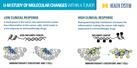
A new study from the University of Michigan Comprehensive Cancer Center reveals molecular changes within the tumor that are preventing the immunotherapy drugs from killing off the cancer.

Clinical trials with PD-L1 and PD-1 blockade suggested that tumors with a high number of inflammation-causing T cells were more responsive to the immunotherapy-based PD-L1 and PD-1 inhibitors. Tumors with low inflammation, or low T cells, were less responsive. But what controls T cells in the tumor microenvironment is poorly understood.

graphical image that shows how turning off a mechanism leads to more inflammation which means better drug response

http://www.uofmhealth.org/sites/default/files/styles/large/public/Immunotherapy.png?itok=_oihl0jj


“We defined a molecular mechanism to explain why some tumors are inflamed and others are not – and consequently why some patients will be responsive to therapy and others not,” says senior author Weiping Zou, M.D., Ph.D.

“If we can reprogram this epigenetic mechanism, then the therapy might work for more patients,” says Zou, Charles B. de Nancrede Professor of Surgery, Immunology and Biology at the University of Michigan Medical School.

Zou’s group was the first to show PD-L1 expression, regulation and functional blockade in dendritic cells in the human cancer microenvironment.

In this study, published in Nature, researchers studied human and mouse models of ovarian cancer cells. They applied epigenetic drugs and found that the numbers of T cells in the tumor increased. They also saw that the epigenetic drugs synergized the anti-tumor effect of PD-L1 blockade in their models.

“We hope this could be developed into a clinical trial testing a combination of PD-L1 and PD-1 blockade with epigenetic therapy. We want to see if we can make the responders more responsive and turn the non-responders into responders,” Zou says.

Additional authors: From U-M: Dongjun Pen, Ilona Kryczek, Nisha Nagarsheth, Lili Zhao, Shuang Wei, Weimin Wang, Yuqing Sun, Ende Zhao, Linda Vatan, Wojciech Szeliga, Yali Dou, Kathleen Cho, Rebecca Liu; from Medical University in Lublin, Poland: Jan Kotarski, Rafal Tarkowski; from Henry Ford Health System: Sharon Hensley-Alford, Adnan Munkarah

Funding: National Institutes of Health grants CA190176, CA123088, CA099985, CA193136, CA152470, CA171306, 5P30 CA46592; Rivkin Ovarian Cancer Center, Ovarian Cancer Research Fund, Barbara and Don Leclair

Cancer immunotherapy research is evolving to more targeted strategies

Discoveries in immune pathway research have helped refine cancer immunotherapy strategies to become more targeted.1,2

Image of timeline showing cancer immunotherapy research has evolved from general to targeted approaches over the years


http://www.researchcancerimmunotherapy.com/overview/targeted-cancer-research

  • CAR=chimeric antigen receptor;
  • CSF-1R=colony-stimulating factor 1; CTLA4=cytotoxic
  • T-lymphocyte antigen-4; IDO=indoleamine
  • 2,3-dioxygenase; PD-1=programmed death-1; PD-L1=programmed death-ligand 1.

http://www.researchcancerimmunotherapy.com/images/overview/evolution-research/history-of-immunotherapy.png


Engaging the immune response: a unique approach to cancer management

Cancer immunotherapy strategies are designed to engage the immune system against tumors. This approach is unique in the oncology setting and introduces new considerations for cancer management.1,2

  • TARGETED

    to tumor-specific antigens

  • RAPID

    activation of the immune response

  • ADAPTABLE

    as the tumor mutates and evolves

  • SELF-PROPAGATING

    with each revolution of the cancer immunity cycle

  • DURABLE

    response over time


CONSIDERATIONS FOR CANCER IMMUNOLOGY

Duration of response

The immune response has the ability to adapt with cancer as it evolves, and can become self-propagating once the cancer immunity cycle is initiated. Immune-directed strategies aim to leverage these attributes, with the goal of inducing a durable antitumor effect.3-5

Pseudo-progression

T-cell infiltration to the tumor site may cause an apparent increase in tumor size or the appearance of new lesions. This inflammatory effect can be misinterpreted as progressive disease, as it can be difficult to differentiate the different cell types in radiographic imaging. New criteria have been developed to better capture immune-related response patterns, and may guide evaluation of immunotherapies in clinical trials, and potentially in clinical care.1,2,6

Image showing T-cell infiltration into the tumor site can cause pseudoprogression]

REFERENCES

  1. Chen DS, Mellman I. Oncology meets immunology: the cancer-immunity cycle. Immunity. 2013;39:1-10. PMID: 23890059
  2. Mellman I, Coukos G, Dranoff G. Cancer immunotherapy comes of age. Nature. 2011;480:480-489. PMID: 22193102
  3. Hanahan D, Weinberg RA. Hallmarks of cancer: the next generation. Cell. 2011;144:646-674. PMID: 21376230

Immune-related adverse events

While the goal of cancer immunotherapy research is to understand how to activate specific components of the immune response, the potential for off-target effects exists. Adverse event profiles may vary among different immune-directed strategies. As strategies grow more targeted, the recognition and management of immune-related adverse events will evolve.1,3

REFERENCES

  1. Mellman I, Coukos G, Dranoff G. Cancer immunotherapy comes of age. Nature. 2011;480:480-489. PMID: 22193102
  2. Hoos A, Eggermont AM, Janetzki S, et al. Improved endpoints for cancer immunotherapy trials. J Natl Cancer Inst. 2010;102:1388-1397.PMID: 20826737
  3. Chen DS, Mellman I. Oncology meets immunology: the cancer-immunity cycle. Immunity. 2013;39:1-10. PMID: 23890059
  4. Topalian SL, Weiner GJ, Pardoll DM. Cancer immunotherapy comes of age. J Clin Oncol. 2011;29:4828-4836. PMID: 22042955
  5. Chen DS, Irving BA, Hodi FS. Molecular pathways: next-generation immunotherapy—inhibiting programmed death-ligand 1 and programmed death-1. Clin Cancer Res. 2012;18:6580-6587. PMID: 23087408
  6. Wolchok JD, Hoos A, O’Day S, et al. Guidelines for the evaluation of immune therapy activity in solid tumors: immune-related response criteria. Clin Cancer Res. 2009;15:7412-7420. PMID: 19934295


Tumors can evade immune destruction

By disrupting the processes of the cancer immunity cycle throughout the body, tumors can avoid detection by the immune system and limit the extent of immune destruction.1-3

Image of dendritic cell capturing tumor antigens, step 2 of cancer immunity cycle

Tumor microenvironment

Disrupting antigen detection

http://www.researchcancerimmunotherapy.com/images/overview/evading-immune-destruction/tumor-microenv.png


Lymph node

Inhibiting T-cell activation by dendritic cells

http://www.researchcancerimmunotherapy.com/images/overview/evading-immune-destruction/lymph-node.png

Image of dendritic cell activating T cell, step 3 of cancer immunity cycle

Blood vessel

Blocking T-cell infiltration into tumor

http://www.researchcancerimmunotherapy.com/images/overview/evading-immune-destruction/blood-vessel.png

Image of T cell infiltrating tumor, step 5 of cancer immunity cycle

Tumor microenvironment

Suppressing cytotoxic T-cell activity

http://www.researchcancerimmunotherapy.com/images/overview/evading-immune-destruction/suppressing-tcell.png


EVADING IMMUNE DESTRUCTION IS AN EMERGING HALLMARK OF CANCER

Alt text: Image of hallmarks of cancer, which includes evading immune destruction

http://www.researchcancerimmunotherapy.com/images/overview/evading-immune-destruction/hallmark-cancer.png

The growing body of research into the mechanisms of immune evasion has led to its addition as an emerging hallmark of cancer, a distinct biological capability that enables tumors to grow and metastasize.3 


MUTATION RATE BY CANCER TYPE4

Image of mutation chart; melanoma, lung, and bladder cancer have high mutation rates

http://www.researchcancerimmunotherapy.com/images/tumor-types/immunogenic-cancer/immunogenic-cancer.png

REFERENCES

  1. Chen DS, Mellman I. Oncology meets immunology: the cancer-immunity cycle. Immunity. 2013;39:1-10. PMID: 23890059
  2. Chen DS, Irving BA, Hodi FS. Molecular pathways: next-generation immunotherapy—inhibiting programmed death-ligand 1 and programmed death-1. Clin Cancer Res. 2012;18:6580-6587. PMID: 23087408
  3. Rajasagi M, Shokla SA, Fritsch EF, et al. Systematic identification of personal tumor-specific neoantigens in chronic lymphocytic leukemia.Blood. 2014;124:453-462. PMID: 24891321
  4. Lawrence MS, Stojanov P, Polak P, et al. Mutational heterogeneity in cancer and the search for new cancer-associated genes. Nature.2013;499:214-218. PMID: 23770567


Atezolizumab (Anti-PDL1) clinical trials   

Atezolizumab clinical trials are being conducted for the following types of cancers: lung, bladder, kidney, hematological malignancies, and breast cancer. Please check back for updates or search clinicaltrials.gov with the search term “Atezolizumab.”


EXPLORING A MORE PERSONALIZED APPROACH TO CANCER IMMUNOTHERAPY RESEARCH

With the evolution to more targeted strategies, research is focusing on identifying predictors of individual immune response through specific tumor characteristics and factors in the tumor microenvironment, such as

  • The presence of tumor-infiltrating immune cells8
    • The ability of immune cells to infiltrate the tumor microenvironment may be a key criterion for a variety of immune-directed strategies, and could indicate which tumors are more likely to respond
  • Gene expression patterns in tumors, particularly the genes involved in immune response9
  • Cell surface protein expression
    • PD-L1 expression on tumor cells and tumor-infiltrating immune cells10,11
    • MUC1 expression on tumor cells12     

REFERENCES

  1. Chen DS, Mellman I. Oncology meets immunology: the cancer-immunity cycle. Immunity. 2013;39:1-10. PMID: 23890059
  2. Mellman I, Coukos G, Dranoff G. Cancer immunotherapy comes of age. Nature. 2011;480:480-489. PMID: 22193102
  3. Lesterhuis WJ, Haanen JB, Punt CJ. Cancer immunotherapy—revisited. Nat Rev Drug Discov. 2011;10:591-600. PMID: 21804596
  4. National Institutes of Health ClinicalTrials.gov. https://clinicaltrials.gov/ct2/show/NCT01494688. Accessed March 4, 2015.
  5. National Institutes of Health ClinicalTrials.gov. https://clinicaltrials.gov/ct2/show/NCT00739609. Accessed March 4, 2015.
  6. Glienke W, Esser R, Priesner C, et al. Advantages and applications of CAR-expressing natural killer cells. Front Pharmacol. 2015;6:21. doi: 10.3389/fphar.2015.00021. PMID: 25729364
  7. National Institutes of Health ClinicalTrials.gov. https://clinicaltrials.gov/ct2/show/NCT01303705. Accessed March 4, 2015.
  8. Gajewski TF, Schreiber H, Fu YX. Innate and adaptive immune cells in the tumor microenvironment. Nat Immunol. 2013;14:1014-1022.PMID: 24048123
  9. Ji RR, Chasalow SD, Wang L, et al. An immune-active tumor microenvironment favors clinical response to ipilimumab. Cancer Immunol Immunother. 2012;61:1019-1031. PMID: 22146893
  10. Taube JM, Anders RA, Young GD, et al. Colocalization of inflammatory response with B7-h1 expression in human melanocytic lesions supports an adaptive resistance mechanism of immune escape. Sci Transl Med. 2012;4:127ra37. PMID: 22461641
  11. Chen DS, Irving BA, Hodi FS. Molecular pathways: next-generation immunotherapy—inhibiting programmed death-ligand 1 and programmed death-1. Clin Cancer Res. 2012;18:6580-6587. PMID: 23087408
  12. Stojnev S, Ristic-Petrovic A, Velickovic LJ, et al. Prognostic significance of mucin expression in urothelial bladder cancer. Int J Clin Exp Pathol. 2014;7:4945-4958. PMID: 25197366
  13. Pardoll DM. The blockade of immune checkpoints in cancer immunotherapy. Nat Rev Cancer. 2012;12:252-264. PMID: 22437870
  14. Taylor RC, Patel A, Panageas KS, Busam KJ, Brady MS. Tumor-infiltrating lymphocytes predict sentinel lymph node positivity in patients with cutaneous melanoma. J Clin Oncol. 2007;25:869-875. PMID: 17327608


Read Full Post »

Early Diagnosis

Reporter: Stephen J. Williams, Ph.D.

This post contains a curation of all Early Diagnosis posts on this site as well as a curation of the Early Detection Research Network.

Early Research Detection Network (EDRN)

Welcome to EDRN

The Early Detection Research Network (EDRN), an initiative of the National Cancer Institute (NCI), brings together dozens of institutions to help accelerate the translation of biomarker information into clinical applications and to evaluate new ways of testing cancer in its earliest stages and for cancer risk.

Getting Started…

Check out the EDRN Highlights — a listing of our accomplishments and milestones.


► Scientific Components ► For Public, Patients, Advocates
► Collaborative Opportunities (how to join EDRN) ► For Researchers

Highlights

Highlights of the accomplishments of the Early Detection Research Network.

A brief list of major EDRN-developed assays that have been adapted for clinical use is described in the table below:

Detection/Biomarker Assay Discovery Refine/Adapt for Clin Use Clinical Validation Clinical Translation
Blood proPSA ✓ ✓ FDA approved
Urine PCA3 ✓ ✓ FDA approved
OVA1™ for Ovarian Cancer ✓ ✓ FDA approved
ROMA Algorithm for CA125 and HE4 Tests for Pelvic Mass Malignancies ✓ ✓ FDA approved
Blood/DCP and AFP-L3 for Hepatocellular Carcinoma ✓ ✓ FDA approved
Blood GP73 ✓ ✓ ✓ Together with AFP-L3 used  for monitoring cirrhotic patients for HCC in China
MiPS (Mi Prostate Score Urine test), Multiplex analysis of T2-ERG gene fusion, PCA3 and serum PSA ✓ ✓ ✓ In CLIA Lab
FISH to detect T2S:Erg fusion for Prostate Cancer ✓ ✓ ✓ In CLIA Lab
GSTP1 methylation for repeat biopsies in prostate cancer ✓ ✓ In CLIA Lab
Mitochondrial deletion for detection of prostate cancer In CLIA Lab
Somalogic 12-marker panel for Lung Cancer ✓ ✓ In CLIA Lab
80-gene panel for Lung Cancer ✓ ✓ In CLIA Lab
Vimentin Methylation Marker for Colon Cancer ✓ ✓ In CLIA Lab
Galectin-3 ligand for detection of adenomas and colon cancer In CLIA Lab
8-gene panel for Barrett’s Esophagus ✓ ✓ In CLIA Lab
SOPs for Blood (Serum, Plasma), Urine, Stool ✓ ✓ Frequently used by biomarker research community
EDRN Pre/Validation Specimen Reference Sets (specimens from well characterized and matched cases and controls from specific disease spectra) ✓ ✓ Frequently used by biomarker research community

Since its inception in 1999 EDRN has achieved several key milestones, summarized below:

1998 through 2000: Inception and Inauguration of EDRN

2001 to 2003: Meeting the Challenges to Harness and Share Emerging Scientific Knowledge

  • EDRN Second Report, Translational Research to Identify Early Cancer and Cancer Risk, October 2002, http://edrn.nci.nih.gov/docs.) published.
  • EDRN joined the Gordon Research Conferences to co-host the New Frontiers in Cancer detection and Diagnosis in 2002.


  • Guidelines Set for Studies Measuring Biomarker Predictive Power Journal of National Cancer Institute (Vol. 93, No. 14, July 18, 2001).
  • EDRN Associate Membership Program Initiated: This novel approach to make EDRN inclusive has been extremely successful. EDRN has now more than 120 Associate Members who are significantly contributing to EDRN efforts in biomarker discovery, development and validation.

2003 to 2004: Network Surges Ahead in Real-time

  • Collaborative Discovery and Validation Projects:  More than 100 collaborative projects spanned the various organ sites. These projects are monitored through the EDRN’s electronic System Information System (eSIS).
  • EDRN Virtual Specimen Bank and Validation Management System Launched: The EDRN Virtual Specimen Bank, also known as ERNE knowledge system, was deployed to 10 institutions in early 2003, allowing a common web-based query to search for available specimens across the EDRN Clinical Epidemiology and Validation Centers https://ginger.fhcrc.org/edrn/imp/GateServlet?pwd. VSIMS was created to allow multiple studies to be administered efficiently by minimizing development time with standardization of information and data management across multiple activities and research sites. This system encompasses all the security features of Food and Drug Administration (FDA)-required auditing systems.
  • Partnership on the Plasma Proteome Project (PPP) Initiative of the Human Proteome Organization (HUPO): PPP project was initiated to evaluate multiple technology platforms, develop bioinformatic tools and standards for protein identification, and create a database of the plasma proteome. The entire study was published in the August issue of the journal Proteomics August 2005, Volume 4 (4), pp 1045-1450.

2005 to 2008: An Investment in Prevention

  • In late 2006, EDRN’s Program for Rapid, Independent Diagnostic Evaluation (PRIDE), was established (http://grants.nih.gov/grants/guide/notice-files/NOT-CA-07-003.html ) as an administrative means to assist extramural investigators in successfully conducting cross-laboratory validation of biomarkers. Ten applications have been reviewed and five are being supported.
  • EDRN underwent external reviews in 2007 and 2008.
  • The Canary Foundation, Palo Alto, CA signed a Memorandum of Understanding with EDRN, NCI on supporting prostate cancer surveillance network of investigators from seven institutions. The tissue and serum will be collected during a three-year period and will be made available to extramural scientists for discovery and validation research.
  • The Lustgarten Foundation, N.Y., funded 6 institutions to generate monoclonal antibodies and associated hybridoma cell lines for pancreatic cancer antigens (biomarkers) identified by EDRN and non-EDRN investigators. These resources will be stored at the NCI-Frederick Facility for distribution to extramural investigators.

2009 to 2011: Realizing Investment for Clinical Use

  • Two biomarker tests approved by FDA and two IVDs pending FDA review.
  • Six biomarker tests offered by CLIA labs.
  • One biomarker test approved for clinical use outside the USA

A Curation of Posts on Early Detection of Cancer and Other Early Detection Networks is Included Below


BRCA 1 and 2 and Early Detection of Cancer

Early Detection of Prostate Cancer: American Urological Association (AUA) Guideline

Mechanism involved in Breast Cancer Cell Growth: Function in Early Detection & Treatment

Warning signs may lead to better early detection of ovarian cancer

Cancer Detection

Biomarker tool development for Early Diagnosis of Pancreatic Cancer: Van Andel Institute and Emory University

China, India, and Russia account for 46% of all new cancer cases globally, as well as 52% of cancer-related mortality per 4/2014 Lancet Oncology article


Read Full Post »

Better Cancer Medication

Larry H. Bernstein, MD, FCAP, Curator

LPBI

A Better Class of Cancer Drugs
http://www.technologynetworks.com/medchem/news.aspx?ID=183124
An SDSU chemist has developed a technique to identify potential cancer drugs that are less likely to produce side effects.
A class of therapeutic drugs known as protein kinase inhibitors has in the past decade become a powerful weapon in the fight against various life-threatening diseases, including certain types of leukemia, lung cancer, kidney cancer and squamous cell cancer of the head and neck. One problem with these drugs, however, is that they often inhibit many different targets, which can lead to side effects and complications in therapeutic use. A recent study by San Diego State University chemist Jeffrey Gustafson has identified a new technique for improving the selectivity of these drugs and possibly decreasing unwanted side effects in the future.

Why are protein kinase–inhibiting drugs so unpredictable? The answer lies in their molecular makeup.

Many of these drug candidates possess examples of a phenomenon known as atropisomerism. To understand what this is, it’s helpful to understand a bit of the chemistry at work. Molecules can come in different forms that have exactly the same chemical formula and even the same bonds, just arranged differently. The different arrangements are mirror images of each other, with a left-handed and a right-handed arrangement. The molecules’ “handedness” is referred to as chirality. Atropisomerism is a form of chirality that arises when the spatial arrangement has a rotatable bond called an axis of chirality. Picture two non-identical paper snowflakes tethered together by a rigid stick.

Some axes of chirality are rigid, while others can freely spin about their axis. In the latter case, this means that at any given time, you could have one of two different “versions” of the same molecule.

Watershed treatment

As the name suggests, kinase inhibitors interrupt the function of kinases—a particular type of enzyme—and effectively shut down the activity of proteins that contribute to cancer.

“Kinase inhibition has been a watershed for cancer treatment,” said Gustafson, who attended SDSU as an undergraduate before earning his Ph.D. in organic chemistry from Yale University, then working there as a National Institutes of Health poctdoctoral fellow in chemical biology.

“However, it’s really hard to inhibit a single kinase,” he explained. “The majority of compounds identified inhibit not just one but many kinases, and that can lead to a number of side effects.”

Many kinase inhibitors possess axes of chirality that are freely spinning. The problem is that because you can’t control which “arrangement” of the molecule is present at a given time, the unwanted version could have unintended consequences.

In practice, this means that when medicinal chemists discover a promising kinase inhibitor that exists as two interchanging arrangements, they actually have two different inhibitors. Each one can have quite different biological effects, and it’s difficult to know which version of the molecule actually targets the right protein.

“I think this has really been under-recognized in the field,” Gustafson said. “The field needs strategies to weed out these side effects.”

Applying the brakes

So that’s what Gustafson did in a recently published study. He and his colleagues synthesized atropisomeric compounds known to target a particular family of kinases known as tyrosine kinases. To some of these compounds, the researchers added a single chlorine atom which effectively served as a brake to keep the atropisomer from spinning around, locking the molecule into either a right-handed or a left-handed version.

When the researchers screened both the modified and unmodified versions against their target kinases, they found major differences in which kinases the different versions inhibited. The unmodified compound was like a shotgun blast, inhibiting a broad range of kinases. But the locked-in right-handed and left-handed versions were choosier.

“Just by locking them into one or another atropisomeric configuration, not only were they more selective, but they  inhibited different kinases,” Gustafson explained.

If drug makers incorporated this technique into their early drug discovery process, he said, it would help identify which version of an atropisomeric compound actually targets the kinase they want to target, cutting the potential for side effects and helping to usher drugs past strict regulatory hurdles and into the hands of waiting patients.

Inroads Against Leukaemia
http://www.technologynetworks.com/medchem/news.aspx?ID=183594
Potential for halting disease in molecule isolated from sea sponges.
A molecule isolated from sea sponges and later synthesized in the lab can halt the growth of cancerous cells and could open the door to a new treatment for leukemia, according to a team of Harvard researchers and other collaborators led by Matthew Shair, a professor of chemistry and chemical biology.

“Once we learned this molecule, named cortistatin A, was very potent and selective in terms of inhibiting the growth of AML [acute myeloid leukemia] cells, we tested it in mouse models of AML and found that it was as efficacious as any other molecule we had seen, without having deleterious effects,” Shair said. “This suggests we have identified a promising new therapeutic approach.”

It’s one that could be available to test in patients relatively soon.

“We synthesized cortistatin A and we are working to develop novel therapeutics based on it by optimizing its drug-like properties,” Shair said. “Given the dearth of effective treatments for AML, we recognize the importance of advancing it toward clinical trials as quickly as possible.”

The drug-development process generally takes years, but Shair’s lab is very close to having what is known as a development candidate that could be taken into late-stage preclinical development and then clinical trials. An industrial partner will be needed to push the technology along that path and toward regulatory approval. Harvard’s Office of Technology Development (OTD) is engaged in advanced discussions to that end.

The molecule works, Shair explained, by inhibiting a pair of nearly identical kinases, called CDK8 and CDK19, that his research indicates play a key role in the growth of AML cells.

The kinases operate as part of a poorly understood, massive structure in the nucleus of cells called the mediator complex, which acts as a bridge between transcription factors and transcriptional machinery. Inhibiting these two specific kinases, Shair and colleagues found, doesn’t shut down all transcription, but instead has gene-specific effects.

“We treated AML cells with cortistatin A and measured the effects on gene expression,” Shair said. “One of the first surprises was that it’s affecting a very small number of genes — we thought it might be in the thousands, but it’s in the low hundreds.”

When Shair, Henry Pelish, a senior research associate in chemistry and chemical biology, and then-Ph.D. student Brian Liau looked closely at which genes were affected, they discovered many were associated with DNA regulatory elements known as “super-enhancers.”

“Humans have about 220 different types of cells in their body — they all have the same genome, but they have to form things like skin and bone and liver cells,” Shair explained. “In all cells, there are a relatively small number of DNA regulatory elements, called super-enhancers. These super-enhancers drive high expression of genes, many of which dictate cellular identity. A big part of cancer is a situation where that identity is lost, and the cells become poorly differentiated and are stuck in an almost stem-cell-like state.”

While a few potential cancer treatments have attacked the disease by down-regulating such cellular identity genes, Shair and colleagues were surprised to find that their molecule actually turned up the activity of those genes in AML cells.

“Before this paper, the thought was that cancer is ramping these genes up, keeping the cells in a hyper-proliferative state and affecting cell growth in that way,” Shair said. “But our molecule is saying that’s one part of the story, and in addition cancer is keeping the dosage of these genes in a narrow range. If it’s too low, the cells die. If they are pushed too high, as with cortistatin A, they return to their normal identity and stop growing.”

Shair’s lab became interested in the molecule several years ago, shortly after it was first isolated and described by other researchers. Early studies suggested it appeared to inhibit just a handful of kinases.

“We tested approximately 400 kinases, and found that it inhibits only CDK8 and CDK19 in cells, which makes it among the most selective kinase inhibitors identified to date,” Shair said. “Having compounds that precisely hit a specific target, like cortistatin A, can help reduce side effects and increase efficacy. In a way, it shatters a dogma because we thought it wasn’t possible for a molecule to be this selective and bind in a site common to all 500 human kinases, but this molecule does it, and it does it because of its 3-D structure. What’s interesting is that most kinase-inhibitor drugs do not have this type of 3-D structure. Nature is telling us that one way to achieve this level of specificity is to make molecules more like cortistatin A.”

Shair’s team successfully synthesized the molecule, which helped them study how it worked and why it affected the growth of a very specific type of cell. Later on, with funding and drug-development expertise provided by Harvard’s Blavatnik Biomedical Accelerator, Shair’s lab created a range of new molecules that may be better suited to clinical application.

“It’s a complex process to make [cortistatin A] — 32 chemical steps,” said Shair. “But we have been able to find less complex structures that act just like the natural compound, with better drug-like properties, and they can be made on a large scale and in about half as many steps.”

“Over the course of several years, we have watched this research progress from an intriguing discovery to a highly promising development candidate,” said Isaac Kohlberg, senior associate provost and chief technology development officer. “The latest results are a real testament to Matt’s ingenuity and dedication to addressing a very tough disease.”

While there is still much work to be done — in particular, to better understand how CDK8 and CDK19 regulate gene expression — the early results have been dramatic.

“This is the kind of thing you do science for,” Shair said, “the idea that once every 10 or 20 years you might find something this interesting, that sheds new light on important, difficult problems. This gives us an opportunity to generate a new understanding of cancer and also develop new therapeutics to treat it. We’re very excited and curious to see where it goes.”

Read Full Post »

New NIH breast cancer research to focus on prevention

Reporter: Stephen J. Williams, PhD

Print this page

A new phase of the Breast Cancer and the Environment Research Program (BCERP), focused on prevention, is being launched at the National Institutes of Health. Grant-funded researchers will now work across scientific disciplines, involve new racially and ethnically diverse communities, and expand the study of risk factors that precede breast cancer, such as breast density.

These new directions reflect recommendations made by the Interagency Breast Cancer and Environmental Research Coordinating Committee (IBCERCC) in 2013. IBCERCC was congressionally mandated to review the state of the science around breast cancer and environmental influences by the Breast Cancer and Environmental Research Act. Recommendations included prioritizing prevention, involving transdisciplinary research teams, engaging public stakeholders, collaborating across federal agencies, and communicating the science to the public.

This broadened research focus will add to the growing knowledge of environmental and genetic factors that may influence breast cancer risk across the lifespan. The six new BCERP projects, plus a new coordinating center promoting cross-project collaboration, are jointly funded by the National Institute of Environmental Health Sciences (NIEHS) and the National Cancer Institute. All projects involve strong partnerships between researchers and organizations focused on breast cancer prevention or environmental health.

The new research will be conducted at the following institutions

  • Brigham and Women’s Hospital, Boston
  • City of Hope/Beckman Research Institute, Duarte, California
  • Columbia University, New York City
  • Georgetown Lombardi Comprehensive Cancer Center, Washington, D.C.
  • Michigan State University, Lansing
  • University of Massachusetts, Amherst
  • University of Wisconsin – Madison (Coordinating Center)

“The beauty of this research is that scientific discoveries and community observations inform each other, in order to dive deeper into the complex causes of breast cancer,” said Gwen Collman, Ph.D., director of NIEHS Division of Extramural Research and Training.

The focus on minority and socio-economically disadvantaged women is an important step in addressing disparities in breast cancer outcomes. Although African-American women are diagnosed with breast cancer less often than white women, more aggressive cancers and breast cancer deaths are more common among African-American women.

Another new direction for BCERP is research on the role of breast density as a possible intermediate risk factor for breast cancer. Dense breast tissue is one of the most common risk factors for breast cancer. Identifying links between environmental exposures and high breast density may provide new insights into prevention.

“These priorities reflect our continued commitment to breast cancer prevention,” noted Caroline Dilworth, Ph.D., BCERP program lead at NIEHS. “Our goal is to build on the high quality science we’ve been funding for more than a decade, while also being responsive to the expert recommendations of the IBCERCC report.”

Grant Numbers: U01ES026130, U01ES026137, U01ES026122, U01ES026132, U01ES026119, U01ES026140, U01ES026127

NIEHS supports research to understand the effects of the environment on human health and is part of NIH. For more information on environmental health topics, visit www.niehs.nih.gov. Subscribe to one or more of the NIEHS news lists to stay current on NIEHS news, press releases, grant opportunities, training, events, and publications.

The National Cancer Institute leads the National Cancer Program and the NIH’s efforts to dramatically reduce the prevalence of cancer and improve the lives of cancer patients and their families, through research into prevention and cancer biology, the development of new interventions, and the training and mentoring of new researchers. For more information about cancer, please visit the NCI website at http://www.cancer.gov or call NCI’s Cancer Information Service at 1-800-4-CANCER.

About the National Institutes of Health (NIH): NIH, the nation’s medical research agency, includes 27 Institutes and Centers and is a component of the U.S. Department of Health and Human Services. NIH is the primary federal agency conducting and supporting basic, clinical, and translational medical research, and is investigating the causes, treatments, and cures for both common and rare diseases. For more information about NIH and its programs, visit www.nih.gov.

Other posts on this site on  Cancer and Early Detection  include

Early Detection of Prostate Cancer: American Urological Association (AUA) Guideline

Mechanism involved in Breast Cancer Cell Growth: Function in Early Detection & Treatment

Warning signs may lead to better early detection of ovarian cancer

‘Mosaicism’ is Associated with Aging and Chronic Diseases like Cancer: detection of genetic mosaicism could be an early marker for detecting cancer.

CDC Findings: Due to Aging Population, Actual Number of Cancer Deaths is Rising while Risk of Dying From Cancer is Falling in the US

Read Full Post »

Development of Chemoresistance to Targeted Therapies: Alterations of Cell Signaling, & the Kinome [11.4.1.2]


Curator: Stephen J. Williams, Ph.D.

UPDATED 5/24/2026

Curated articles on mechanisms of resistance to tyrosine kinase inhibitors


Oncogenic receptor tyrosine kinase signaling is driven by the Golgi protein GOLPH3 and its interaction with MYO18A

19 May 2026
Vol 19, Issue 938
Editor’s summary

Many cancers are driven by signaling mediated by receptor tyrosine kinases (RTKs) upon activation by growth factors. Starost et al. found that RTK signaling was enhanced by the Golgi-localized oncoprotein GOLPH3 and its binding partner MYO18A. Together, these proteins were required for the delivery of EGFR (which is frequently mutated in lung cancer), insulin receptor, and other RTKs to the plasma membrane and for the responsiveness of different cell types to growth factors. These findings identify growth factor signaling as a potential mechanism underlying the oncogenicity of GOLPH3. Moreover, they provide a possible alternate targeting strategy for limiting the growth of RTK-driven cancers that have developed resistance to RTK inhibitors. —Wei Wong
Abstract

Receptor tyrosine kinase (RTK) signaling drives cancer and is a validated therapeutic target. Modulators of RTK signaling can reveal mechanisms of oncogenesis and offer new therapeutic targets. Golgi phosphoprotein 3 (GOLPH3) is a Golgi-localized oncoprotein that promotes signaling downstream of mTOR. Here, examination of RTK signaling indicated that GOLPH3 acted at the level of the RTK and increased all downstream signaling. We found that GOLPH3 enhanced the delivery of RTKs to the plasma membrane. This role was shared with its binding partner myosin 18A (MYO18A) and depended on the interaction of GOLPH3 with MYO18A. The GOLPH3-MYO18A complex at the Golgi apparatus was required and rate-limiting for RTK signaling across the cell types and receptors assessed. Our findings provide insight into the relationship between the function of GOLPH3 at the Golgi and its role as a cancer driver, highlighting its potential as a therapeutic target in cancer.

The advent of molecular targeted therapies like Imatinib (Gleevec), and other tyrosine kinase inhibitors (TKI) has been transformative to cancer therapy. However, as with all chemotherapeutics, including radiation therapy, the development of chemo-resistance toward personalized, molecular therapies has been disastrous to the successful treatment of cancer. The fact that chemo-resistance develops to personalized therapies was a serious disappointment to clinicians (although most expected this to be the case) but more surprisingly it was the rapidity of onset and speed of early reported cases which may have been the biggest shocker.

A post on resistance to other TKIs (to EGFR and ALK) can be seen here: http://pharmaceuticalintelligence.com/2013/11/01/resistance-to-receptor-of-tyrosine-kinase/

Original article

History of Development of Resistance to Imatinib (Gleevec)

The Melo group published a paper in Blood showing that short exposure to STI571 (imatinib; trade name Gleevec®) could result in drug resistant clones

Selection and characterization of BCR-ABL positive cell lines with differential sensitivity to the tyrosine kinase inhibitor STI571: diverse mechanisms of resistance. Blood. 2000 Aug 1;96(3):1070-9.

Mahon FX1, Deininger MW, Schultheis B, Chabrol J, Reiffers J, Goldman JM, Melo JV.

Abstract

Targeting the tyrosine kinase activity of Bcr-Abl with STI571 is an attractive therapeutic strategy in chronic myelogenous leukemia (CML). A few CML cell lines and primary progenitors are, however, resistant to this compound. We investigated the mechanism of this resistance in clones of the murine BaF/3 cells transfected with BCR-ABL and in 4 human cell lines from which sensitive (s) and resistant (r) clones were generated by various methods. Although the resistant cells were able to survive in the presence of STI571, their proliferation was approximately 30% lower than that of their sensitive counterparts in the absence of the compound. The concentration of STI571 needed for a 50% reduction in viable cells after a 3-day exposure was on average 10 times higher in the resistant (2-3 micromol/L) than in the sensitive (0.2-0.25 micromol/L) clones. The mechanism of resistance to STI571 varied among the cell lines. Thus, in Baf/BCR-ABL-r, LAMA84-r, and AR230-r, there was up-regulation of the Bcr-Abl protein associated with amplification of the BCR-ABL gene. In K562-r, there was no Bcr-Abl overexpression, but the IC(50) for the inhibition of Bcr-Abl autophosphorylation was increased in the resistant clones. Sequencing of the Abl kinase domain revealed no mutations. The multidrug resistance P-glycoprotein (Pgp) was overexpressed in LAMA84-r, indicating that at least 2 mechanisms of resistance operate in this cell line. KCL22-r showed neither Bcr-Abl up-regulation nor a higher threshold for tyrosine kinase inhibition by STI571. We conclude that BCR-ABL-positive cells can evade the inhibitory effect of STI571 by different mechanisms, such as Bcr-Abl overexpression, reduced intake mediated by Pgp, and, possibly, acquisition of compensatory mutations in genes other than BCR-ABL.

mellobcrablresistamplification

FISH analysis of AR230 and LAMA84 sensitive and resistant clones, with probes for the ABL (red signal) and theBCR (green signal) genes. BCR-ABL is identified as a red–green or yellow fused signal. Adapted from Mahon et al., Blood 2000; 96(3):1070-9.

This rapid onset of imatinib resistance also see in the clinic and more prominent in advance disease

From NCCN 2nd Annual Congress: Hematologic Malignancies – Update on Primary Therapy, Second-Line Therapy, and New Agents for Chronic Myelogenous Leukemia (Slides with Transcript)

http://www.medscape.org/viewarticle/564097

There is some evidence that even looking earlier makes some sense in determining what the prognosis is. This is from Timothy Hughes’ group in Adelaide, and he is looking at an earlier molecular time point, 3 months after initiation of therapy. And what you have done here is you have taken the 3-month mark and you have said, “Well, based on your response at 3 months, what is your likelihood that in the future you will either get a major molecular response or become resistant?”

3monthimitanibresist

If you look at the accumulation of imatinib resistance to find if it is either initially not responding or becoming resistant after a good response, it goes up with type of disease and phase of disease. So if you look at patients who have early chronic phase disease — that is, they start getting imatinib less than a year from the diagnosis — their chance of failure is pretty low. With later disease — they are in a chronic phase but they have had disease more than a year before they get imatinib — it is higher. If you see patients with accelerated phase or blast crisis, the chances are that they will fail sometime in the future.

speed of imitinib resistance

Therefore, because not all resistant samples show gene amplification of Bcr/Abl and the rapidity of onset of resistance, many feel that there are other mechanisms of resistance at play, like kinome plasticity.

Kinome Plasticity Contributes to TKI resistance

Beyond gene amplification, other mechanisms of imitanib and other tyrosine kinase inhibitors (TKI) include alterations in compensatory signaling pathways. This can be referred to as kinome plasticity and is explained in the following abstracts from the AACR 2015 meeting.

Systems-pharmacology dissection of a drug synergy in imatinib-resistant CML

Georg E Winter, Uwe Rix, Scott M Carlson, Karoline V Gleixner, Florian Grebien, Manuela Gridling, André C Müller, Florian P Breitwieser, Martin Bilban, Jacques Colinge, Peter Valent, Keiryn L Bennett, Forest M White & Giulio Superti-Furga. Nature Chemical Biology 8,905–912(2012)

Occurrence of the BCR-ABLT315I gatekeeper mutation is among the most pressing challenges in the therapy of chronic myeloid leukemia (CML). Several BCR-ABL inhibitors have multiple targets and pleiotropic effects that could be exploited for their synergistic potential. Testing combinations of such kinase inhibitors identified a strong synergy between danusertib and bosutinib that exclusively affected CML cells harboring BCR-ABLT315I. To elucidate the underlying mechanisms, we applied a systems-level approach comprising phosphoproteomics, transcriptomics and chemical proteomics. Data integration revealed that both compounds targeted Mapk pathways downstream of BCR-ABL, resulting in impaired activity of c-Myc. Using pharmacological validation, we assessed that the relative contributions of danusertib and bosutinib could be mimicked individually by Mapk inhibitors and collectively by downregulation of c-Myc through Brd4 inhibition. Thus, integration of genome- and proteome-wide technologies enabled the elucidation of the mechanism by which a new drug synergy targets the dependency of BCR-ABLT315I CML cells on c-Myc through nonobvious off targets.

nchembio.1085-F2kinomegleevecresistance

Please see VIDEO and SLIDESHARE of a roundtable Expert Discussion on CML

Curated Content From the 2015 AACR National Meeting on Drug Resistance Mechanisms and tyrosine kinase inhibitors

Session Title: Mechanisms of Resistance: From Signaling Pathways to Stem Cells
Session Type: Major Symposium
Session Start/End Time: Tuesday, Apr 21, 2015, 10:30 AM -12:30 PM
Location: Terrace Ballroom II-III (400 Level), Pennsylvania Convention Center
CME: CME-Designated
CME/CE Hours: 2
Session Description: Even the most effective cancer therapies are limited due to the development of one or more resistance mechanisms. Acquired resistance to targeted therapies can, in some cases, be attributed to the selective propagation of a small population of intrinsically resistant cells. However, there is also evidence that cancer drugs themselves can drive resistance by triggering the biochemical- or genetic-reprogramming of cells within the tumor or its microenvironment. Therefore, understanding drug resistance at the molecular and biological levels may enable the selection of specific drug combinations to counteract these adaptive responses. This symposium will explore some of the recent advances addressing the molecular basis of cancer cell drug resistance. We will address how tumor cell signaling pathways become rewired to facilitate tumor cell survival in the face of some of our most promising cancer drugs. Another topic to be discussed involves how drugs select for or induce the reprogramming of tumor cells toward a stem-like, drug resistant fate. By targeting the molecular driver(s) of rewired signaling pathways and/or cancer stemness it may be possible to select drug combinations that prevent the reprogramming of tumors and thereby delay or eliminate the onset of drug resistance.
Presentations:
Chairperson
Tuesday, Apr 21, 2015, 10:30 AM -12:30 PM
David A. Cheresh. UCSD Moores Cancer Center, La Jolla, CA
Introduction
Tuesday, Apr 21, 2015, 10:30 AM -10:40 AM
Resistance to tyrosine kinase inhibitors: Heterogeneity and therapeutic strategies.
Tuesday, Apr 21, 2015, 10:40 AM -10:55 AM
Jeffrey A. Engelman. Massachusetts General Hospital, Boston, MA
Discussion
Tuesday, Apr 21, 2015, 10:55 AM -11:00 AM
NG04: Clinical acquired resistance to RAF inhibitor combinations in BRAF mutant colorectal cancer through MAPK pathway alterations
Tuesday, Apr 21, 2015, 11:00 AM -11:15 AM
Ryan B. Corcoran, Leanne G. Ahronian, Eliezer Van Allen, Erin M. Coffee, Nikhil Wagle, Eunice L. Kwak, Jason E. Faris, A. John Iafrate, Levi A. Garraway, Jeffrey A. Engelman. Massachusetts General Hospital Cancer Center, Boston, MA, Dana-Farber Cancer Institute, Boston, MA
Discussion
Tuesday, Apr 21, 2015, 11:15 AM -11:20 AM
SY27-02: Tumour heterogeneity and therapy resistance in melanoma
Tuesday, Apr 21, 2015, 11:20 AM -11:35 AM
Claudia Wellbrock. Univ. of Manchester, Manchester, United Kingdom
Discussion
Tuesday, Apr 21, 2015, 11:35 AM -11:40 AM
SY27-03: Breast cancer stem cell state transitions mediate therapeutic resistance
Tuesday, Apr 21, 2015, 11:40 AM -11:55 AM
Max S. Wicha. University of Michigan, Comprehensive Cancer Center, Ann Arbor, MI
Discussion
Tuesday, Apr 21, 2015, 11:55 AM -12:00 PM
SY27-04: Induction of cancer stemness and drug resistance by EGFR blockade
Tuesday, Apr 21, 2015, 12:00 PM -12:15 PM
David A. Cheresh. UCSD Moores Cancer Center, La Jolla, CA
Discussion
Tuesday, Apr 21, 2015, 12:15 PM -12:20 PM
General Discussion
Tuesday, Apr 21, 2015, 12:20 PM -12:30 PM

Targeting Macromolecular Signaling Complexes 
Room 115, Pennsylvania Convention Center

Drug Resistance 
Hall A (200 Level), Pennsylvania Convention Center
Resistance to Pathway-Targeted Therapeutics 1 
Section 33

Molecular Mechanisms of Sensitivity or Resistance to Pathway-Targeted Agents 
Room 118, Pennsylvania Convention Center

Targeting Signaling Pathways in Cancer 
Room 204, Pennsylvania Convention Center
Exploiting the MAPK Pathway in Cancer 
Room 115, Pennsylvania Convention Center

PLEASE see the attached WORD file which includes ALL abstracts, posters, and talks on this subject from the AACR 2015 national meeting BELOW

 AACR2015resistancekinome

Other posts related to, Cancer, Chemotherapy, Gleevec and Resistance on this Open Access Journal Include

Imatinib (Gleevec) May Help Treat Aggressive Lymphoma: Chronic Lymphocytic Leukemia (CLL)

Treatments for Acute Leukemias [2.4.4A]

Therapeutic Implications for Targeted Therapy from the Resurgence of Warburg ‘Hypothesis’

Hematologic Malignancies [6.2]

Overview of Posttranslational Modification (PTM)

Novel Modeling Methods for Genomic Data Analysis & Evolutionary Systems Biology to Design Dosing Regimens to Minimize Resistance

Mechanisms of Drug Resistance

Using RNA-seq and targeted nucleases to identify mechanisms of drug resistance in acute myeloid leukemia

An alternative approach to overcoming the apoptotic resistance of pancreatic cancer

Resistance to Receptor of Tyrosine Kinase

Read Full Post »

Metabolic Genomics and Pharmaceutics, Vol. 1 of BioMed Series D available on Amazon Kindle

Metabolic Genomics and Pharmaceutics, Vol. 1 of BioMed Series D available on Amazon Kindle

Reporter: Stephen S Williams, PhD

Article ID #180: Metabolic Genomics and Pharmaceutics, Vol. 1 of BioMed Series D available on Amazon Kindle. Published on 8/15/2015

WordCloud Image Produced by Adam Tubman

Leaders in Pharmaceutical Business Intelligence would like to announce the First volume of their BioMedical E-Book Series D:

Metabolic Genomics & Pharmaceutics, Vol. I

SACHS FLYER 2014 Metabolomics SeriesDindividualred-page2

which is now available on Amazon Kindle at

http://www.amazon.com/dp/B012BB0ZF0.

This e-Book is a comprehensive review of recent Original Research on  METABOLOMICS and related opportunities for Targeted Therapy written by Experts, Authors, Writers. This is the first volume of the Series D: e-Books on BioMedicine – Metabolomics, Immunology, Infectious Diseases.  It is written for comprehension at the third year medical student level, or as a reference for licensing board exams, but it is also written for the education of a first time baccalaureate degree reader in the biological sciences.  Hopefully, it can be read with great interest by the undergraduate student who is undecided in the choice of a career. The results of Original Research are gaining value added for the e-Reader by the Methodology of Curation. The e-Book’s articles have been published on the Open Access Online Scientific Journal, since April 2012.  All new articles on this subject, will continue to be incorporated, as published with periodical updates.

We invite e-Readers to write an Article Reviews on Amazon for this e-Book on Amazon.

All forthcoming BioMed e-Book Titles can be viewed at:

http://pharmaceuticalintelligence.com/biomed-e-books/

Leaders in Pharmaceutical Business Intelligence, launched in April 2012 an Open Access Online Scientific Journal is a scientific, medical and business multi expert authoring environment in several domains of  life sciences, pharmaceutical, healthcare & medicine industries. The venture operates as an online scientific intellectual exchange at their website http://pharmaceuticalintelligence.com and for curation and reporting on frontiers in biomedical, biological sciences, healthcare economics, pharmacology, pharmaceuticals & medicine. In addition the venture publishes a Medical E-book Series available on Amazon’s Kindle platform.

Analyzing and sharing the vast and rapidly expanding volume of scientific knowledge has never been so crucial to innovation in the medical field. WE are addressing need of overcoming this scientific information overload by:

  • delivering curation and summary interpretations of latest findings and innovations on an open-access, Web 2.0 platform with future goals of providing primarily concept-driven search in the near future
  • providing a social platform for scientists and clinicians to enter into discussion using social media
  • compiling recent discoveries and issues in yearly-updated Medical E-book Series on Amazon’s mobile Kindle platform

This curation offers better organization and visibility to the critical information useful for the next innovations in academic, clinical, and industrial research by providing these hybrid networks.

Table of Contents for Metabolic Genomics & Pharmaceutics, Vol. I

Chapter 1: Metabolic Pathways

Chapter 2: Lipid Metabolism

Chapter 3: Cell Signaling

Chapter 4: Protein Synthesis and Degradation

Chapter 5: Sub-cellular Structure

Chapter 6: Proteomics

Chapter 7: Metabolomics

Chapter 8:  Impairments in Pathological States: Endocrine Disorders; Stress

                   Hypermetabolism and Cancer

Chapter 9: Genomic Expression in Health and Disease 


Summary 

Epilogue


Read Full Post »

Cancer Biology and Genomics for Disease Diagnosis (Vol. I) Now Available for Amazon Kindle

Cancer Biology and Genomics for Disease Diagnosis (Vol. I) Now Available for Amazon Kindle

Reporter: Stephen J Williams, PhD

Article ID #179: Cancer Biology and Genomics for Disease Diagnosis (Vol. I) Now Available for Amazon Kindle. Published on 8/14/2015

WordCloud Image Produced by Adam Tubman

Leaders in Pharmaceutical Business Intelligence would like to announce the First volume of their BioMedical E-Book Series C: e-Books on Cancer & Oncology

Volume One: Cancer Biology and Genomics for Disease Diagnosis

CancerandOncologyseriesCcoverwhich is now available on Amazon Kindle at                          http://www.amazon.com/dp/B013RVYR2K.

This e-Book is a comprehensive review of recent Original Research on Cancer & Genomics including related opportunities for Targeted Therapy written by Experts, Authors, Writers. This ebook highlights some of the recent trends and discoveries in cancer research and cancer treatment, with particular attention how new technological and informatics advancements have ushered in paradigm shifts in how we think about, diagnose, and treat cancer. The results of Original Research are gaining value added for the e-Reader by the Methodology of Curation. The e-Book’s articles have been published on the Open Access Online Scientific Journal, since April 2012.  All new articles on this subject, will continue to be incorporated, as published with periodical updates.

We invite e-Readers to write an Article Reviews on Amazon for this e-Book on Amazon. All forthcoming BioMed e-Book Titles can be viewed at:

http://pharmaceuticalintelligence.com/biomed-e-books/

Leaders in Pharmaceutical Business Intelligence, launched in April 2012 an Open Access Online Scientific Journal is a scientific, medical and business multi expert authoring environment in several domains of  life sciences, pharmaceutical, healthcare & medicine industries. The venture operates as an online scientific intellectual exchange at their website http://pharmaceuticalintelligence.com and for curation and reporting on frontiers in biomedical, biological sciences, healthcare economics, pharmacology, pharmaceuticals & medicine. In addition the venture publishes a Medical E-book Series available on Amazon’s Kindle platform.

Analyzing and sharing the vast and rapidly expanding volume of scientific knowledge has never been so crucial to innovation in the medical field. WE are addressing need of overcoming this scientific information overload by:

  • delivering curation and summary interpretations of latest findings and innovations
  • on an open-access, Web 2.0 platform with future goals of providing primarily concept-driven search in the near future
  • providing a social platform for scientists and clinicians to enter into discussion using social media
  • compiling recent discoveries and issues in yearly-updated Medical E-book Series on Amazon’s mobile Kindle platform

This curation offers better organization and visibility to the critical information useful for the next innovations in academic, clinical, and industrial research by providing these hybrid networks.

Table of Contents for Cancer Biology and Genomics for Disease Diagnosis

Preface

Introduction  The evolution of cancer therapy and cancer research: How we got here?

Part I. Historical Perspective of Cancer Demographics, Etiology, and Progress in Research

Chapter 1:  The Occurrence of Cancer in World Populations

Chapter 2.  Rapid Scientific Advances Changes Our View on How Cancer Forms

Chapter 3:  A Genetic Basis and Genetic Complexity of Cancer Emerge

Chapter 4: How Epigenetic and Metabolic Factors Affect Tumor Growth

Chapter 5: Advances in Breast and Gastrointestinal Cancer Research Supports Hope for Cure

Part II. Advent of Translational Medicine, “omics”, and Personalized Medicine Ushers in New Paradigms in Cancer Treatment and Advances in Drug Development

Chapter 6:  Treatment Strategies

Chapter 7:  Personalized Medicine and Targeted Therapy

Part III.Translational Medicine, Genomics, and New Technologies Converge to Improve Early Detection

Chapter 8:  Diagnosis                                     

Chapter 9:  Detection

Chapter 10:  Biomarkers

Chapter 11:  Imaging In Cancer

Chapter 12: Nanotechnology Imparts New Advances in Cancer Treatment, Detection, &  Imaging                                 

Epilogue by Larry H. Bernstein, MD, FACP: Envisioning New Insights in Cancer Translational Biology


Read Full Post »

Treatment of Lymphomas [2.4.4C]

Larry H. Bernstein, MD, FCAP, Author, Curator, Editor

http://pharmaceuticalinnovation.com/2015/8/11/larryhbern/Treatment-of-Lymphomas-[2.4.4C]


Lymphoma treatment

Overview

http://www.emedicinehealth.com/lymphoma/page8_em.htm#lymphoma_treatment

The most widely used therapies are combinations of chemotherapyand radiation therapy.

  • Biological therapy, which targets key features of the lymphoma cells, is used in many cases nowadays.

The goal of medical therapy in lymphoma is complete remission. This means that all signs of the disease have disappeared after treatment. Remission is not the same as cure. In remission, one may still have lymphoma cells in the body, but they are undetectable and cause no symptoms.

  • When in remission, the lymphoma may come back. This is called recurrence.
  • The duration of remission depends on the type, stage, and grade of the lymphoma. A remission may last a few months, a few years, or may continue throughout one’s life.
  • Remission that lasts a long time is called durable remission, and this is the goal of therapy.
  • The duration of remission is a good indicator of the aggressiveness of the lymphoma and of the prognosis. A longer remission generally indicates a better prognosis.

Remission can also be partial. This means that the tumor shrinks after treatment to less than half its size before treatment.

The following terms are used to describe the lymphoma’s response to treatment:

  • Improvement: The lymphoma shrinks but is still greater than half its original size.
  • Stable disease: The lymphoma stays the same.
  • Progression: The lymphoma worsens during treatment.
  • Refractory disease: The lymphoma is resistant to treatment.

The following terms to refer to therapy:

  • Induction therapy is designed to induce a remission.
  • If this treatment does not induce a complete remission, new or different therapy will be initiated. This is usually referred to as salvage therapy.
  • Once in remission, one may be given yet another treatment to prevent recurrence. This is called maintenance therapy.

Chemotherapy

Many different types of chemotherapy may be used for Hodgkin lymphoma. The most commonly used combination of drugs in the United States is called ABVD. Another combination of drugs, known as BEACOPP, is now widely used in Europe and is being used more often in the United States. There are other combinations that are less commonly used and not listed here. The drugs that make up these two more common combinations of chemotherapy are listed below.

ABVD: Doxorubicin (Adriamycin), bleomycin (Blenoxane), vinblastine (Velban, Velsar), and dacarbazine (DTIC-Dome). ABVD chemotherapy is usually given every two weeks for two to eight months.

BEACOPP: Bleomycin, etoposide (Toposar, VePesid), doxorubicin, cyclophosphamide (Cytoxan, Neosar), vincristine (Vincasar PFS, Oncovin), procarbazine (Matulane), and prednisone (multiple brand names). There are several different treatment schedules, but different drugs are usually given every two weeks.

The type of chemotherapy, number of cycles of chemotherapy, and the additional use of radiation therapy are based on the stage of the Hodgkin lymphoma and the type and number of prognostic factors.

Adult Non-Hodgkin Lymphoma Treatment (PDQ®)

http://www.cancer.gov/cancertopics/pdq/treatment/adult-non-hodgkins/Patient/page1

Key Points for This Section

Adult non-Hodgkin lymphoma is a disease in which malignant (cancer) cells form in the lymph system.

Because lymph tissue is found throughout the body, adult non-Hodgkin lymphoma can begin in almost any part of the body. Cancer can spread to the liver and many other organs and tissues.

Non-Hodgkin lymphoma in pregnant women is the same as the disease in nonpregnant women of childbearing age. However, treatment is different for pregnant women. This summary includes information on the treatment of non-Hodgkin lymphoma during pregnancy

Non-Hodgkin lymphoma can occur in both adults and children. Treatment for children, however, is different than treatment for adults. (See the PDQ summary on Childhood Non-Hodgkin Lymphoma Treatment for more information.)

There are many different types of lymphoma.

Lymphomas are divided into two general types: Hodgkin lymphoma and non-Hodgkin lymphoma. This summary is about the treatment of adult non-Hodgkin lymphoma. For information about other types of lymphoma, see the following PDQ summaries:

Age, gender, and a weakened immune system can affect the risk of adult non-Hodgkin lymphoma.

If cancer is found, the following tests may be done to study the cancer cells:

  • Immunohistochemistry : A test that uses antibodies to check for certain antigens in a sample of tissue. The antibody is usually linked to a radioactive substance or a dye that causes the tissue to light up under a microscope. This type of test may be used to tell the difference between different types of cancer.
  • Cytogenetic analysis : A laboratory test in which cells in a sample of tissue are viewed under a microscope to look for certain changes in the chromosomes.
  • Immunophenotyping : A process used to identify cells, based on the types of antigens ormarkers on the surface of the cell. This process is used to diagnose specific types of leukemia and lymphoma by comparing the cancer cells to normal cells of the immune system.

Certain factors affect prognosis (chance of recovery) and treatment options.

The prognosis (chance of recovery) and treatment options depend on the following:

  • The stage of the cancer.
  • The type of non-Hodgkin lymphoma.
  • The amount of lactate dehydrogenase (LDH) in the blood.
  • The amount of beta-2-microglobulin in the blood (for Waldenström macroglobulinemia).
  • The patient’s age and general health.
  • Whether the lymphoma has just been diagnosed or has recurred (come back).

Stages of adult non-Hodgkin lymphoma may include E and S.

Adult non-Hodgkin lymphoma may be described as follows:

E: “E” stands for extranodal and means the cancer is found in an area or organ other than the lymph nodes or has spread to tissues beyond, but near, the major lymphatic areas.

S: “S” stands for spleen and means the cancer is found in the spleen.

Stage I adult non-Hodgkin lymphoma is divided into stage I and stage IE.

  • Stage I: Cancer is found in one lymphatic area (lymph node group, tonsils and nearby tissue, thymus, or spleen).
  • Stage IE: Cancer is found in one organ or area outside the lymph nodes.

Stage II adult non-Hodgkin lymphoma is divided into stage II and stage IIE.

  • Stage II: Cancer is found in two or more lymph node groups either above or below the diaphragm (the thin muscle below the lungs that helps breathing and separates the chest from the abdomen).
  • Stage IIE: Cancer is found in one or more lymph node groups either above or below the diaphragm. Cancer is also found outside the lymph nodes in one organ or area on the same side of the diaphragm as the affected lymph nodes.

Stage III adult non-Hodgkin lymphoma is divided into stage III, stage IIIE, stage IIIS, and stage IIIE+S.

  • Stage III: Cancer is found in lymph node groups above and below the diaphragm (the thin muscle below the lungs that helps breathing and separates the chest from the abdomen).
  • Stage IIIE: Cancer is found in lymph node groups above and below the diaphragm and outside the lymph nodes in a nearby organ or area.
  • Stage IIIS: Cancer is found in lymph node groups above and below the diaphragm, and in the spleen.
  • Stage IIIE+S: Cancer is found in lymph node groups above and below the diaphragm, outside the lymph nodes in a nearby organ or area, and in the spleen.

In stage IV adult non-Hodgkin lymphoma, the cancer:

  • is found throughout one or more organs that are not part of a lymphatic area (lymph node group, tonsils and nearby tissue, thymus, or spleen), and may be in lymph nodes near those organs; or
  • is found in one organ that is not part of a lymphatic area and has spread to organs or lymph nodes far away from that organ; or
  • is found in the liver, bone marrow, cerebrospinal fluid (CSF), or lungs (other than cancer that has spread to the lungs from nearby areas).

Adult non-Hodgkin lymphomas are also described based on how fast they grow and where the affected lymph nodes are in the body.  Indolent & aggressive.

The treatment plan depends mainly on the following:

  • The type of non-Hodgkin’s lymphoma
  • Its stage (where the lymphoma is found)
  • How quickly the cancer is growing
  • The patient’s age
  • Whether the patient has other health problems
  • If there are symptoms present such as fever and night sweats (see above)

Read Full Post »

Treatment for Chronic Leukemias [2.4.4B]

Larry H. Bernstein, MD, FCAP, Author, Curator, Editor

http://pharmaceuticalintelligence.com/2015/8/11/larryhbern/Treatment-for-Chronic-Leukemias-[2.4.4B]

2.4.4B1 Treatment for CML

Chronic Myelogenous Leukemia Treatment (PDQ®)

http://www.cancer.gov/cancertopics/pdq/treatment/CML/Patient/page4

Treatment Option Overview

Key Points for This Section

There are different types of treatment for patients with chronic myelogenous leukemia.

Six types of standard treatment are used:

  1. Targeted therapy
  2. Chemotherapy
  3. Biologic therapy
  4. High-dose chemotherapy with stem cell transplant
  5. Donor lymphocyte infusion (DLI)
  6. Surgery

New types of treatment are being tested in clinical trials.

Patients may want to think about taking part in a clinical trial.

Patients can enter clinical trials before, during, or after starting their cancer treatment.

Follow-up tests may be needed.

There are different types of treatment for patients with chronic myelogenous leukemia.

Different types of treatment are available for patients with chronic myelogenous leukemia (CML). Some treatments are standard (the currently used treatment), and some are being tested in clinical trials. A treatment clinical trial is a research study meant to help improve current treatments or obtain information about new treatments for patients with cancer. When clinical trials show that a new treatment is better than the standard treatment, the new treatment may become the standard treatment. Patients may want to think about taking part in a clinical trial. Some clinical trials are open only to patients who have not started treatment.

Six types of standard treatment are used:

Targeted therapy

Targeted therapy is a type of treatment that uses drugs or other substances to identify and attack specific cancer cells without harming normal cells. Tyrosine kinase inhibitors are targeted therapy drugs used to treat chronic myelogenous leukemia.

Imatinib mesylate, nilotinib, dasatinib, and ponatinib are tyrosine kinase inhibitors that are used to treat CML.

See Drugs Approved for Chronic Myelogenous Leukemia for more information.

Chemotherapy

Chemotherapy is a cancer treatment that uses drugs to stop the growth of cancer cells, either by killing the cells or by stopping them from dividing. When chemotherapy is taken by mouth or injected into a vein or muscle, the drugs enter the bloodstream and can reach cancer cells throughout the body (systemic chemotherapy). When chemotherapy is placed directly into the cerebrospinal fluid, an organ, or a body cavity such as the abdomen, the drugs mainly affect cancer cells in those areas (regional chemotherapy). The way the chemotherapy is given depends on the type and stage of the cancer being treated.

See Drugs Approved for Chronic Myelogenous Leukemia for more information.

Biologic therapy

Biologic therapy is a treatment that uses the patient’s immune system to fight cancer. Substances made by the body or made in a laboratory are used to boost, direct, or restore the body’s natural defenses against cancer. This type of cancer treatment is also called biotherapy or immunotherapy.

See Drugs Approved for Chronic Myelogenous Leukemia for more information.

High-dose chemotherapy with stem cell transplant

High-dose chemotherapy with stem cell transplant is a method of giving high doses of chemotherapy and replacing blood-forming cells destroyed by the cancer treatment. Stem cells (immature blood cells) are removed from the blood or bone marrow of the patient or a donor and are frozen and stored. After the chemotherapy is completed, the stored stem cells are thawed and given back to the patient through an infusion. These reinfused stem cells grow into (and restore) the body’s blood cells.

See Drugs Approved for Chronic Myelogenous Leukemia for more information.

Donor lymphocyte infusion (DLI)

Donor lymphocyte infusion (DLI) is a cancer treatment that may be used after stem cell transplant.Lymphocytes (a type of white blood cell) from the stem cell transplant donor are removed from the donor’s blood and may be frozen for storage. The donor’s lymphocytes are thawed if they were frozen and then given to the patient through one or more infusions. The lymphocytes see the patient’s cancer cells as not belonging to the body and attack them.

Surgery

Splenectomy

What`s new in chronic myeloid leukemia research and treatment?

http://www.cancer.org/cancer/leukemia-chronicmyeloidcml/detailedguide/leukemia-chronic-myeloid-myelogenous-new-research

Combining the targeted drugs with other treatments

Imatinib and other drugs that target the BCR-ABL protein have proven to be very effective, but by themselves these drugs don’t help everyone. Studies are now in progress to see if combining these drugs with other treatments, such as chemotherapy, interferon, or cancer vaccines (see below) might be better than either one alone. One study showed that giving interferon with imatinib worked better than giving imatinib alone. The 2 drugs together had more side effects, though. It is also not clear if this combination is better than treatment with other tyrosine kinase inhibitors (TKIs), such as dasatinib and nilotinib. A study going on now is looking at combing interferon with nilotinib.

Other studies are looking at combining other drugs, such as cyclosporine or hydroxychloroquine, with a TKI.

New drugs for CML

Because researchers now know the main cause of CML (the BCR-ABL gene and its protein), they have been able to develop many new drugs that might work against it.

In some cases, CML cells develop a change in the BCR-ABL oncogene known as a T315I mutation, which makes them resistant to many of the current targeted therapies (imatinib, dasatinib, and nilotinib). Ponatinib is the only TKI that can work against T315I mutant cells. More drugs aimed at this mutation are now being tested.

Other drugs called farnesyl transferase inhibitors, such as lonafarnib and tipifarnib, seem to have some activity against CML and patients may respond when these drugs are combined with imatinib. These drugs are being studied further.

Other drugs being studied in CML include the histone deacetylase inhibitor panobinostat and the proteasome inhibitor bortezomib (Velcade).

Several vaccines are now being studied for use against CML.

2.4.4.B2 Chronic Lymphocytic Leukemia

Chronic Lymphocytic Leukemia Treatment (PDQ®)

General Information About Chronic Lymphocytic Leukemia

Key Points for This Section

  1. Chronic lymphocytic leukemia is a type of cancer in which the bone marrow makes too many lymphocytes (a type of white blood cell).
  2. Leukemia may affect red blood cells, white blood cells, and platelets.
  3. Older age can affect the risk of developing chronic lymphocytic leukemia.
  4. Signs and symptoms of chronic lymphocytic leukemia include swollen lymph nodes and tiredness.
  5. Tests that examine the blood, bone marrow, and lymph nodes are used to detect (find) and diagnose chronic lymphocytic leukemia.
  6. Certain factors affect treatment options and prognosis (chance of recovery).
  7. Chronic lymphocytic leukemia is a type of cancer in which the bone marrow makes too many lymphocytes (a type of white blood cell).

Chronic lymphocytic leukemia (also called CLL) is a blood and bone marrow disease that usually gets worse slowly. CLL is one of the most common types of leukemia in adults. It often occurs during or after middle age; it rarely occurs in children.

http://www.cancer.gov/images/cdr/live/CDR755927-750.jpg

Anatomy of the bone; drawing shows spongy bone, red marrow, and yellow marrow. A cross section of the bone shows compact bone and blood vessels in the bone marrow. Also shown are red blood cells, white blood cells, platelets, and a blood stem cell.

Anatomy of the bone. The bone is made up of compact bone, spongy bone, and bone marrow. Compact bone makes up the outer layer of the bone. Spongy bone is found mostly at the ends of bones and contains red marrow. Bone marrow is found in the center of most bones and has many blood vessels. There are two types of bone marrow: red and yellow. Red marrow contains blood stem cells that can become red blood cells, white blood cells, or platelets. Yellow marrow is made mostly of fat.

Leukemia may affect red blood cells, white blood cells, and platelets.

Normally, the body makes blood stem cells (immature cells) that become mature blood cells over time. A blood stem cell may become a myeloid stem cell or a lymphoid stem cell.

A myeloid stem cell becomes one of three types of mature blood cells:

  1. Red blood cells that carry oxygen and other substances to all tissues of the body.
  2. White blood cells that fight infection and disease.
  3. Platelets that form blood clots to stop bleeding.

A lymphoid stem cell becomes a lymphoblast cell and then one of three types of lymphocytes (white blood cells):

  1. B lymphocytes that make antibodies to help fight infection.
  2. T lymphocytes that help B lymphocytes make antibodies to fight infection.
  3. Natural killer cells that attack cancer cells and viruses.

Blood cell development. CDR526538-750

Blood cell development. CDR526538-750

http://www.cancer.gov/images/cdr/live/CDR526538-750.jpg

Blood cell development; drawing shows the steps a blood stem cell goes through to become a red blood cell, platelet, or white blood cell. A myeloid stem cell becomes a red blood cell, a platelet, or a myeloblast, which then becomes a granulocyte (the types of granulocytes are eosinophils, basophils, and neutrophils). A lymphoid stem cell becomes a lymphoblast and then becomes a B-lymphocyte, T-lymphocyte, or natural killer cell.

Blood cell development. A blood stem cell goes through several steps to become a red blood cell, platelet, or white blood cell.

In CLL, too many blood stem cells become abnormal lymphocytes and do not become healthy white blood cells. The abnormal lymphocytes may also be called leukemia cells. The lymphocytes are not able to fight infection very well. Also, as the number of lymphocytes increases in the blood and bone marrow, there is less room for healthy white blood cells, red blood cells, and platelets. This may cause infection, anemia, and easy bleeding.

This summary is about chronic lymphocytic leukemia. See the following PDQ summaries for more information about leukemia:

  • Adult Acute Lymphoblastic Leukemia Treatment.
  • Childhood Acute Lymphoblastic Leukemia Treatment.
  • Adult Acute Myeloid Leukemia Treatment.
  • Childhood Acute Myeloid Leukemia/Other Myeloid Malignancies Treatment.
  • Chronic Myelogenous Leukemia Treatment.
  • Hairy Cell Leukemia Treatment

Older age can affect the risk of developing chronic lymphocytic leukemia.

Anything that increases your risk of getting a disease is called a risk factor. Having a risk factor does not mean that you will get cancer; not having risk factors doesn’t mean that you will not get cancer. Talk with your doctor if you think you may be at risk. Risk factors for CLL include the following:

  • Being middle-aged or older, male, or white.
  • A family history of CLL or cancer of the lymph system.
  • Having relatives who are Russian Jews or Eastern European Jews.

Signs and symptoms of chronic lymphocytic leukemia include swollen lymph nodes and tiredness.

Usually CLL does not cause any signs or symptoms and is found during a routine blood test. Signs and symptoms may be caused by CLL or by other conditions. Check with your doctor if you have any of the following:

  • Painless swelling of the lymph nodes in the neck, underarm, stomach, or groin.
  • Feeling very tired.
  • Pain or fullness below the ribs.
  • Fever and infection.
  • Weight loss for no known reason.

Tests that examine the blood, bone marrow, and lymph nodes are used to detect (find) and diagnose chronic lymphocytic leukemia.

The following tests and procedures may be used:

Physical exam and history : An exam of the body to check general signs of health, including checking for signs of disease, such as lumps or anything else that seems unusual. A history of the patient’s health habits and past illnesses and treatments will also be taken.

Complete blood count (CBC) with differential : A procedure in which a sample of blood is drawn and checked for the following:

The number of red blood cells and platelets.

The number and type of white blood cells.

The amount of hemoglobin (the protein that carries oxygen) in the red blood cells.

The portion of the blood sample made up of red blood cells.

Results from the Phase 3 Resonate™ Trial

Significantly improved progression free survival (PFS) vs ofatumumab in patients with previously treated CLL

  • Patients taking IMBRUVICA® had a 78% statistically significant reduction in the risk of disease progression or death compared with patients who received ofatumumab1
  • In patients with previously treated del 17p CLL, median PFS was not yet reached with IMBRUVICA® vs 5.8 months with ofatumumab (HR 0.25; 95% CI: 0.14, 0.45)1

Significantly prolonged overall survival (OS) with IMBRUVICA® vs ofatumumab in patients with previously treated CLL

  • In patients with previously treated CLL, those taking IMBRUVICA® had a 57% statistically significant reduction in the risk of death compared with those who received ofatumumab (HR 0.43; 95% CI: 0.24, 0.79; P<0.05)1

Typical treatment of chronic lymphocytic leukemia

http://www.cancer.org/cancer/leukemia-chroniclymphocyticcll/detailedguide/leukemia-chronic-lymphocytic-treating-treatment-by-risk-group

Treatment options for chronic lymphocytic leukemia (CLL) vary greatly, depending on the person’s age, the disease risk group, and the reason for treating (for example, which symptoms it is causing). Many people live a long time with CLL, but in general it is very difficult to cure, and early treatment hasn’t been shown to help people live longer. Because of this and because treatment can cause side effects, doctors often advise waiting until the disease is progressing or bothersome symptoms appear, before starting treatment.

If treatment is needed, factors that should be taken into account include the patient’s age, general health, and prognostic factors such as the presence of chromosome 17 or chromosome 11 deletions or high levels of ZAP-70 and CD38.

Initial treatment

Patients who might not be able to tolerate the side effects of strong chemotherapy (chemo), are often treated with chlorambucil alone or with a monoclonal antibody targeting CD20 like rituximab (Rituxan) or obinutuzumab (Gazyva). Other options include rituximab alone or a corticosteroid like prednisione.

In stronger and healthier patients, there are many options for treatment. Commonly used treatments include:

  • FCR: fludarabine (Fludara), cyclophosphamide (Cytoxan), and rituximab
  • Bendamustine (sometimes with rituximab)
  • FR: fludarabine and rituximab
  • CVP: cyclophosphamide, vincristine, and prednisone (sometimes with rituximab)
  • CHOP: cyclophosphamide, doxorubicin, vincristine (Oncovin), and prednisone
  • Chlorambucil combined with prednisone, rituximab, obinutuzumab, or ofatumumab
  • PCR: pentostatin (Nipent), cyclophosphamide, and rituximab
  • Alemtuzumab (Campath)
  • Fludarabine (alone)

Other drugs or combinations of drugs may also be also used.

If the only problem is an enlarged spleen or swollen lymph nodes in one region of the body, localized treatment with low-dose radiation therapy may be used. Splenectomy (surgery to remove the spleen) is another option if the enlarged spleen is causing symptoms.

Sometimes very high numbers of leukemia cells in the blood cause problems with normal circulation. This is calledleukostasis. Chemo may not lower the number of cells until a few days after the first dose, so before the chemo is given, some of the cells may be removed from the blood with a procedure called leukapheresis. This treatment lowers blood counts right away. The effect lasts only for a short time, but it may help until the chemo has a chance to work. Leukapheresis is also sometimes used before chemo if there are very high numbers of leukemia cells (even when they aren’t causing problems) to prevent tumor lysis syndrome (this was discussed in the chemotherapy section).

Some people who have very high-risk disease (based on prognostic factors) may be referred for possible stem cell transplant (SCT) early in treatment.

Second-line treatment of CLL

If the initial treatment is no longer working or the disease comes back, another type of treatment may help. If the initial response to the treatment lasted a long time (usually at least a few years), the same treatment can often be used again. If the initial response wasn’t long-lasting, using the same treatment again isn’t as likely to be helpful. The options will depend on what the first-line treatment was and how well it worked, as well as the person’s health.

Many of the drugs and combinations listed above may be options as second-line treatments. For many people who have already had fludarabine, alemtuzumab seems to be helpful as second-line treatment, but it carries an increased risk of infections. Other purine analog drugs, such as pentostatin or cladribine (2-CdA), may also be tried. Newer drugs such as ofatumumab, ibrutinib (Imbruvica), and idelalisib (Zydelig) may be other options.

If the leukemia responds, stem cell transplant may be an option for some patients.

Some people may have a good response to first-line treatment (such as fludarabine) but may still have some evidence of a small number of leukemia cells in the blood, bone marrow, or lymph nodes. This is known as minimal residual disease. CLL can’t be cured, so doctors aren’t sure if further treatment right away will be helpful. Some small studies have shown that alemtuzumab can sometimes help get rid of these remaining cells, but it’s not yet clear if this improves survival.

Treating complications of CLL

One of the most serious complications of CLL is a change (transformation) of the leukemia to a high-grade or aggressive type of non-Hodgkin lymphoma called diffuse large cell lymphoma. This happens in about 5% of CLL cases, and is known as Richter syndrome. Treatment is often the same as it would be for lymphoma (see our document called Non-Hodgkin Lymphoma for more information), and may include stem cell transplant, as these cases are often hard to treat.

Less often, CLL may transform to prolymphocytic leukemia. As with Richter syndrome, these cases can be hard to treat. Some studies have suggested that certain drugs such as cladribine (2-CdA) and alemtuzumab may be helpful.

In rare cases, patients with CLL may have their leukemia transform into acute lymphocytic leukemia (ALL). If this happens, treatment is likely to be similar to that used for patients with ALL (see our document called Leukemia: Acute Lymphocytic).

Acute myeloid leukemia (AML) is another rare complication in patients who have been treated for CLL. Drugs such as chlorambucil and cyclophosphamide can damage the DNA of blood-forming cells. These damaged cells may go on to become cancerous, leading to AML, which is very aggressive and often hard to treat (see our document calledLeukemia: Acute Myeloid).

CLL can cause problems with low blood counts and infections. Treatment of these problems were discussed in the section “Supportive care in chronic lymphocytic leukemia.”

Read Full Post »
« Newer Posts - Older Posts »