Feeds:
Posts
Comments

Archive for the ‘International Global Work in Pharmaceutical’ Category

Transparency in Clinical Trials, Volume 2 (Volume Two: Latest in Genomics Methodologies for Therapeutics: Gene Editing, NGS and BioInformatics, Simulations and the Genome Ontology), Part 1: Next Generation Sequencing (NGS)

Transparency in Clinical Trials

Curator: Larry H. Bernstein, MD, FCAP


Does Pharma Really Want Transparency In Clinical Trials?

Ed Miseta, Chief Editor, Clinical Leader
Follow Me On Twitter @outsourcedpharm
http://www.clinicalleader.com/doc/does-pharma-really-want-transparency-in-clinical-trials-0001


My recent article on transparency in clinical trials, featuring Dr. Brad Thompson, CEO of Oncolytics, solicited a good number of comments and emails from readers. While most readers agree that more transparency would be good for patients and the industry, there seems to be a lot of disagreement over how it can be achieved, and if it can actually be achieved at all.

To recap, Thompson believes we still have a long way to go, and questioned whether true transparency would ever be achieved. His primary argument noted researchers who want to be published will not put much focus on neutral or negative trials, and even the press releases put out by sponsors include a limited amount of information.

One reader that works for a CRO made the following comment: “Dr. Thompson from Oncolytics made some very interesting comments about investigators holding back information. An investigator will enter patient data into an EDC system that is then verified by monitors. And that is just one of a number of sites. These investigators will generally know when a drug is working and when it is not. As a CRO, I often saw statistical outputs on blinded studies where you could see where the data was trending. Even data guys can tell if a drug is working by the data results and improvements in patients.”

If physicians and researchers are able to see clear results even when the data is still in the process of being collected, then what is the problem with being more transparent? One possible explanation is that once physicians know a drug will not work, they will no longer continue to place patients at risk by having them participate in the trial. This could be done for purely ethical reasons.

But there may also be reluctance to greater transparency on the part of the pharma company. “No drug company truly wants transparency because it leaves the results and outcomes more open to interpretation, mainly by Kaiser, Blue Cross and other groups,” noted another reader.  “Pharma companies could cost themselves a lot of money in sales if they do not have the time to target and position.”

Investors Are A Consideration

There is still another consideration at play. If a study is not going well, would it be to the benefit of the executives of a company to share those results? Let’s play devil’s advocate for a moment and assume you are the CEO of a biotech company. You’re making $500,000 a year with good benefits. You have several investors who have dumped millions of dollars into your company and your product. The study is targeted for four years, but within the first year you see results that indicate your drug is not going to produce the intended results.

“In that scenario, how prone would you be to ending the trial, saving the investors their remaining money, and losing your job?” notes one reader. “Are companies prone or pressured to locate new targets for therapy or identifying reasons to extend the trials, sometimes for a few years or longer?”

All of us have heard discussions about the possibility of electronic medical records (EMRs) someday replacing electronic data capture (EDC). According to one email I received, this will never happen because of the physician issue mentioned above. After all, if patient results were posted in the EMR and every doctor on the network has access to the information, everyone would know if I drug was not having the desired effect. As soon as that happens, promises of riches being delivered to investors will fall by the wayside, and executives will be out of jobs.

“Within big pharma, this is called job preservation,” noted another reader. “If funding for the trial is cut, I am out of a job. At the same time, trial results are not getting any better for patients. Years ago about one in three trials resulted in a successful outcome. Then it went to one in four. Today that success rate is around 15 percent with R&D commitment at about 12% (down from approximately 28%). It appears that the industry is run by money and managed by guys who know how to play the system.  If patients are the primary concern, the industry would target physicians who have the right patients, get enrollment done faster, and quickly identify if the product works as advertised.”

Limit Procedures And Additional Fields

Going back to Thompson’s comments, the problem is not always investigators wanting to get published. One reader noted oftentimes it is the in-house pharma and biotech doctors as well as researchers in academia who are anxious to get their names into publications. “Unfortunately, these are often the same people who include numerous unnecessary procedures in protocols. They will also ask for additional data fields to be included in EDC systems after study launch, which can delay database activity for two months. The reason is they see a hint of something and decide they want to dig deeper, even if the activity has nothing to do with the study results and the overall goal of the trial.”

The obvious fix to this would be executive leadership and study teams standing up and challenging the reason for inclusion of the additional data fields, which cost the industry both time and money. A large number of procedures should be challenged as well, especially if they are not standard of care.

“If a researcher sees a hint of something that seems to be interesting but has nothing to do with the study, they should engage one of the thought leaders to conduct an IIR program to see if the hypothesis is valid,” notes the reader. “They can do this while keeping the clinical program on track to closure without delay, and still appease their interests.”

Clearly, there are no easy solutions. Many pharma companies are certainly making a concerted effort to put the patient first, and I believe those efforts are sincere. But there is no question they must also be focused on funding and trial results – the industry has gone from one focused on a patient to one driven by investors, and that trend is unfortunate. Physicians and researchers will always have their own goals and aspirations, and placing additional burdens upon them could have the unintended consequence of driving them away from trial participation – poor sponsor/CRO pay practices and poorly written/detailed protocols have already moved many physician practices away from clinical trial participation. Coming up with a solution will likely involve bringing together all stakeholders for a more in-depth discussion on the topic, which unfortunately I don’t see happening anytime soon.


Transparency In Clinical Trials: Will It Ever Be Achieved?
Ed Miseta, Chief Editor, Clinical Leader
Follow Me On Twitter @outsourcedpharm


A lot has been made recently about transparency in clinical trials. In the EU a new regulation is about to address the issue, and CISCRPrecently sent a petition letter to the FDA asking it to pass a similar regulation in this country. The petition, signed by hundreds of patients, hopes to make trials results more accessible to patients.

It’s also not hard to understand why a patient participating in a trial would want to know the results of the study, and whether or not they received the active drug or a placebo. But while changes might help companies with patient recruitment and retention issues, will true trial transparency ever be possible?

Dr. Brad Thompson, CEO of biotech firm Oncolytics, believes we still have a very long way to go, and that perhaps pharma companies are not the ones that should be blamed. “I think a lot of people, patients especially, believe that companies are the roadblock in keeping the results of clinical studies from becoming public,” he says. “But personally, I believe it is a much wider issue than that, especially when it comes to finding out the results of unsuccessful trials.”

For example, Thompson looks at clinical investigators. He notes many of these individuals would like for their academic careers to progress. For these folks, the reporting of trial results, especially those that are negative or neutral, does nothing to advance their goals. It is not a deliberate action to conceal information, but the lack of an incentive to do so can often result in delays, provided the results are reported at all.

“If you are conducting a trial at 50 or 60 locations, it doesn’t take too many of them not reporting information to significantly slow down the ability of a sponsor to report on what is going on with the study,” notes Thompson. “And the more time that goes by, the more people will lose interest in doing so. Add to that the fact that there are no journals or annual meetings that are focused on reporting negative results. This is due to space and time limitations. If there are 100 speaking opportunities at the ASCO show in June, those spots will be given to people reporting exciting new results in cancer therapies. There is no time for, nor interest in, anyone reporting on therapies that didn’t work.”

From the standpoint of a public sponsor company, they will typically report negative trial results, but that will generally be via a press release, where there is very little detail. It’s also unlikely that a patient participating in a trial will be on the company’s PR distribution list. As a result, there is an entire system set up with no positive incentives to go into more detail about trials that did not go as planned.  That in itself is unfortunate, since we often learn as much from things that don’t work as we do from things that do.

“In many ways, knowing what didn’t work, or what caused a safety problem, can be more important than knowing what did work and knowing there were no safety problems,” adds Thompson. “Knowing of negative results will allow you to improve your own trials and continue to work to try and find something that does work. I think this is a bigger issue than people realize and it is not something that will be easy to address.”

All Requirements Fall On Sponsors

Of course in this entire daisy-chain of events, there is only one party involved that has a legal obligation to disclose positive or negative information on the trial. That is the sponsor company, which by law is required to disclose information about the trial. Failure to report something could result in a criminal offense. If an investigator doesn’t disclose something, they do not face the same negative repercussions.

“If you talk to an attorney from any sponsor company, they will tell you how important it is to disclose, disclose, disclose,” says Thompson. “They fully understand the importance of doing that. The situation might be slightly different in privately-held companies, because public companies have an obligation to their investors. But even then they have a duty of disclosure under the investment terms. More often than not the investors are sitting on your board of directors and would be privy to the information anyway.”

On a positive note, Thompson is quick to note that most companies, investigators, and researchers he knows want to disclose as much as they possibly can.  There are just a number of soft reasons that might end up keeping them from getting into more detail than they do. For example, there is generally the same amount of content going into a press release regardless of whether or not the trial was successful. He notes no one on the planet is going to put out a 30-page press release covering the detail of a clinical study, whether it was good or bad.

For that reason, most of the press releases that go out are seldom more than two pages, with just a few sentences on the results and the safety aspects. While that will meet the disclosure standards, it certainly does not disclose much detail to the investigators or others who wish to know the details.

“When you look closely at this situation, what you see is a system that is almost accidentally set up to inhibit full disclosure,” states Thompson. “The industry might feel it is good to publish negative results, but where would we publish them? Who is going to pay for it? Who is going to read it? It’s a difficult issue. You can try to induce people to do things, but if an investigator has a failed study, his academic career will not be helped by spending the weeks it would take to write a paper to be published. Especially if they can spend that time writing a paper on a study that did work. There is not a conspiracy of silence. It is just natural for people to want to focus on things that will help them out with their careers.”

More Information Benefits Patients

There are other reasons for reporting as much information as possible. Patients appreciate the information, but from the sponsor perspective, more information might mean coming up with better versions of existing medicines. Thompson likes to use bone marrow studies as an example of how more information can be helpful to patients. When physicians first started using radiation to kill off bone marrow for certain types of leukemia patients, that marrow had to be replaced. It was discovered that bone marrow transferred from people who did not match the patient’s tissue type caused them to perform better…but only for a period of time. After that, the patients began to die quicker. Still, researches published the complete findings.

“They could have reported that non-matching bone marrow works really well for six months and left it at that,” says Thompson. “But they opted to include the downside of the study as well. That led physicians to decide it would be used for emergency use only until a better match could be found. That knowledge ended up making these transplants better for the patients and better for the industry. I think in that case we were lucky that there was a positive effect to report along with the negative. If there was only the negative effect, I don’t know that it would have ever been published.”

Is There A Fix?

I wish I could report that there is an easy fix to this transparency issue. Unfortunately, there is not. According to Thompson, there are not a couple of adjustments that can be made to correct the problem.  After all, you cannot force a researcher to publish an article on a failed study if they have more important needs to attend to. You can’t force a company to produce or publish a 30-page press release or, if they did, force anyone to read it. Unfortunately, that is a reality of the industry.

“We need to come up with a mechanism where the end result is of benefit to the industry, such as people having access to needed information and disseminating it without the process being burdensome,” notes Thompson. “I honestly don’t know how you do that.”

There are so many pieces to this problem…the sponsor companies, the FDA, the investigators, the research sites. It is difficult to fix a problem when the players involved in it are so varied. Still, if this is an issue that is too complicated to tackle with all players at once, perhaps the best approach would be to take it one step at a time. If we put sponsors, patients, and investigators in a room together, all would likely be clamoring for the same end result.

“We would not see pockets of stakeholders fighting this,” adds Thompson. “A solution to this transparency problem would make everyone better off. It’s frustrating because everyone knows this is an issue, and that we have to do better. People who are a lot smarter than I am have spent time on this and were not able to come up with an answer.  But the fact that this is a complicated issue doesn’t mean we should throw our hands in the air and give up. Eventually we will have to produce a solution.”


Taking The “Risk” Out Of Risk-Based Monitoring

http://www.clinicalleader.com/doc/taking-the-risk-out-of-risk-based-monitoring-0002

The clinical trial landscape is continually evolving and with it, efforts in the improvement of participant safety and data integrity. CROs are beginning to transition from on-site monitoring, with 100% point-to-point source data verification, toward a risk-based monitoring (RBM) approach that utilizes source data review and more centralized monitoring techniques better adapted for mitigating risk.

While RBM has gained considerable attention in recent years, reluctance still remains around the approach—from uncertainty arising from the use of “risk” employed in its name to sponsors being wary of potential implications on data quality and regulatory inspection outcomes.

Despite these concerns, there is a growing consensus that risk-based approaches to monitoring, focused on risks to the most critical data elements and processes necessary to achieve study objectives, are more likely than routine visits to all clinical sites and 100% source data verification to ensure subject protection, data integrity, and overall study quality.


Improve the Inclusion & Exclusion Criteria for Your Next Clinical Trial

http://www.clinicalleader.com/doc/improve-the-inclusion-exclusion-criteria-for-your-next-clinical-trial-0001


Read Full Post »

Anti-malarial Agent Drug Discovery

Curator: Larry H. Bernstein, MD, FCAP

Supporting malaria elimination with 21st century antimalarial agent drug discovery

Thierry T. Diagana
   Novartis Institute for Tropical Diseases, 10 Biopolis Road, #05-01 Chromos, Singapore 138670, Singapore
Drug Discovery & Innovation  2015; 20(10)   http://www.e-ditionsbyfry.com/olive/ODE/DDT/Default.aspx?href=DDT/2015/10/01
The burden of malaria has been considerably reduced over recent years. However, to achieve disease elimination, drug discovery for the next generation needs to focus on blocking disease transmission and on targeting the liver-stage forms of the parasite. Properties of the ‘ideal’ new antimalarial drug and the key scientific and technological advances that have led to recent progress in antimalarial drug discovery are reviewed. Using these advances, Novartis has built a robust pipeline of next-generation antimalarials. The preclinical and clinical development of two candidate drugs: KAE609 and KAF156, provide a framework for the path to breakthrough treatments that could be taking us a step closer to the vision of malaria elimination.


Malaria is a devastating and often fatal disease caused by protozoan parasites from the Plasmodium genus. Owing to increased prevention and control measures, such as the introduction of insecticide-treated bed nets and use of artemisinin-based combination therapy, the burden of malaria has been reduced such that between 2000 and 2013 global malaria mortality rates decreased by 47% [1]. Nevertheless, in 2013 an estimated 584 000 individuals (90% from sub-Saharan Africa and 78% children <5 years of age) died from malaria [1]. The current challenge is whether malaria can be completely eliminated on a global scale. Several countries have now developed and started to implement malaria elimination strategies [2], but it is widely accepted that currently available drugs are not ideally suited for disease elimination campaigns [3]. Specifically, there is a need for safe single-dose therapies that are also suitable for mass drug administration to asymptomatic carriers and capable of blocking malaria transmission through the anopheles vector. In addition, chemoprophylaxis prevention requires drugs that are able to eliminate the liver-stage forms of the parasite (especially for Plasmodium vivax).  

The looming threat of artemisinin drug resistance [4–6], coupled with intense lobbying and the financial support of philanthropic organisations such as the Wellcome Trust and the Bill & Melinda Gates Foundation, as well as the creation of publicprivate partnerships such as the Medicines for Malaria Venture [7], triggered a recent surge in antimalarial drug discovery and development activities [8,9]. These research efforts are focused on identifying drugs with novel mechanisms of action. Ultimately, this should mitigate the risk of cross-resistance with existing antimalarials and, by targeting multiple stages of the malaria parasite lifecycle, facilitate prevention, radical cure and transmission blocking [10].

Over the past 10 years, recognition of the need for antimalarial agents with properties that differ from existing treatments has led to dramatic changes in the way in which new targets are identified and new drugs developed. Until very recently, no antimalarial drug with a novel mechanism of action had entered Phase II clinical trials since the hydroxy-1,4-naphthoquinone atovaquone more than 20 years ago [11]. An important milestone in malaria research, however, was achieved in early 2014 with the publication of the results of a Phase II trial of KAE609 [12], the first member of a novel class of antimalarials: the spiroindolones. KAE609 emerged from a malaria drug discovery effort led by the Novartis Institute for Tropical Diseases in partnership with the Genomics Institute of the Novartis Research Foundation, the Swiss Tropical and Public Health Institute, the Biomedical Primate Research Centre and the Medicines for Malaria Venture, with financial support from the Wellcome Trust. KAE609 is being further developed by Novartis aspart of the company’s efforts to contribute to global malaria elimination. This compound is the front-runner in a growing pipeline of antimalarials emerging from Novartis that also includes a second novel antimalarial, the imidazolopiperazine KAF156 [13,14] currently in clinical trials [15], and a preclinical programme aiming to develop inhibitors of phosphatidylinositol-4-OH kinase [PI(4)K], a novel drug target that operates across all the major lifecycle stages of the parasite in its host [16].  

Here, we review and discuss the latest scientific and technological advances in malaria drug discovery that have enabled the development of a robust Novartis drug portfolio, and offer some personal perspectives on the discovery and development of these novel antimalarial agents. A comprehensive overview of the global antimalarial pipeline has been the subject of another recent publication [8].

Scientific and technological advances in antimalarial   drug discovery   Beyond already well-exploited drug targets, such as the Plasmodium dihydrofolate reductase, dihydropteroate synthetase, cytochrome bc1 complex and the haemoglobin degradation pathway, there remains a dearth of novel validated targets. The sequencing of the Plasmodium falciparum genome [17] and use of modern genetics tools were expected to yield many attractive drug targets, reviewed by Winzeler 2008 [18]. However, few proposed targets have subsequently been chemically validated; most targets validated by genetic knockouts have not been tractable. A notable exception is the dihydroorotate dehydrogenase enzyme, which led to the identification of the clinical candidate DSM265 [19]; DSM265 clinical studies represent the first attempt clinically to validate a genetically identified target.  

A number of scientific and technological advances have recently enabled the rapid identification of clinical candidates and also enabled the discovery and chemical validation of novel malaria drug targets. Undoubtedly, the first important technological advance was the miniaturisation of Plasmodium growth assays [20,21]. This development made automated HTS possible and enabled Novartis and many other companies and academic groups to screen large compound libraries for chemical ‘starting points’ with promising antimalarial activity [21–23]. An open-access repository of screening data is available online at the ChEMBL – Neglected Tropical Disease archive ( https://www.ebi.ac.uk/ chemblntd ) [24]. The second advance has been the development of a wide array of genetic and biochemical techniques that have enabled rapid identification of the molecular targets of different screening hits [10,16,25–31].

An important barrier with respect to the elimination of malaria is the liver stage of the parasite lifecycle, because P. vivax can remain dormant in the liver as hypnozoites for many months or years [32]. Currently, the only treatment targeting the liver stage of vivax malaria is primaquine, which cannot be used in individuals with glucose-6-phosphate dehydrogenase deficiency, a common genetic abnormality in malaria endemic areas. However, there is also a new hypnozoiticidal compound in Phase III development: tafenoquine, which might offer another liver-stage treatment in the future [33,34]. The discovery of new liver-stage drugs has been hampered by the dearth of models to screen for activity against hypnozoites; until recently, only the in vivo Plasmodium-cynomolgi–simian malaria model was available. An important step towards the screening of novel hypnozoiticidal compounds was recently made with establishment of an in vitro assay that discriminates the activity of primaquine and atovaquone against the growing schizont and dormant hypnozoite [35–37]. It was this assay that eventually suggested the P. vivax radical cure potential of a novel class of blood-stage antimalarial drugs: the imidazolopyrazines, which target Plasmodium PI(4)K [16].  

The availability of new drugs that prevent the transmission of sexual-stage parasites (gametocytes) to mosquito vectors has also been recognised as a crucial component of future malaria elimination strategies [38]. This task has been aided by the recent development of a novel assay that utilises P. falciparum cell lines to measure the effects of tested compounds on gametocyte maturation and transmission [38]. A variety of in vitro assays have also been employed to show that early- and late-stage gametocytes are susceptible to KAE609 in a dose-dependent manner and that this agent reduces transmission to the vector, providing further evidence that blockage of transmission might be possible with new-generation antimalarials [39].

Another important advance in malaria research was the development and refinement of small animal models, reviewed by Vaughan et al. [40] and Kaushansky et al. [41]. Humanised mouse models, such as the severe combined immunodeficiency model, enable the study of blood- and liver-stage P. falciparum and have been used to establish the antimalarial potential of new drugs.  

As a result of these advances, the emerging global pipeline for new antimalarials [8,9] was identified using different discovery strategies (e.g. target- vs cell-based hits). To have maximum public health impact, novel antimalarial drugs must ideally fulfil a number of pharmacological and safety requirements throughout development. We describe the various approaches employed by Novartis and collaborators to ensure that drug candidates meet these key pharmacological requirements.

Preclinical development strategy   In the preclinical profiling stages of drug development, the overall goal is to ensure that the profile of a new drug candidate is compatible with the probable use of the compound in the clinic. To mitigate the risk of failure owing to a possible difference in activity between laboratory-adapted strains and clinical isolates, KAE609 was shown early to be as effective as artesunate in an ex vivo assay against field isolates of P. falciparum and P. vivax collected from areas of drug resistance [42,43]. As noted, there are several in vivo malaria models available for drug testing. The lethal Plasmodium berghei malaria mouse is inexpensive, rapid and enables the evaluation of cure (i.e. prevention of recrudescence) but species differences in drug sensitivity might warrant the use of the more expensive P. falciparum SCID model [19]. Potent antimalarial activity across Plasmodium species is generally desirable because it is suggestive of strong evolutionary conservation of the compound-binding site on its molecular target, which is a harbinger for a reduced risk of drug resistance development.  

In preclinical species, KAE609 displayed pharmacokinetic profiles upon oral dosing consistent with once-daily oral dosing in humans. In the P. berghei malaria mouse model, KAE609 demonstrated a fast onset of action and potently reduced parasitaemia. Importantly, KAE609 was the only one of the tested drugs (which included artesunate, artemether, chloroquine and mefloquine) to display single-dose cure efficacy [42]. P. berghei appears in vitro to be less sensitive to spiroindolones than P. falciparum and close analogues of KAE609 display faster clearance antimalarial properties in the P. falciparum SCID mouse (unpublished data).  

The imidazolopiperazine class of compounds, which includes KAF156, was also discovered and initially optimised primarily for its asexual blood-stage activity, but was later found to show liverstage activity in vitro (Fig. 1) [44]. KAF156 displayed potent parasitaemia reduction at doses lower than those of standard antimalarial drugs in blood-stage models, and also showed potent prophylactic activity in the P. berghei mouse model. Thus, preclinical data suggest that KAF156 could have clinical utility as a therapeutic and prophylactic antimalarial agent [45]. Similar to KAE609, the pharmacokinetic profile of KAF156 upon oral dosing in preclinical species is compatible with once-daily dosing. In terms of preclinical safety, oral doses of KAF156 were shown to be well-tolerated in studies of rodents and dogs, with no toxicities identified that would preclude use in humans.

New antimalarial drugs must also be fully tested in vitro with regard to the potential for drug–drug interactions to minimise the risk of adverse interactions with other anti-infective therapies frequently used in malaria-endemic countries, such as human immunodeficiency virus and tuberculosis drugs. Many existing antimalarial drugs carry some risk of cardiotoxicity through inhibition of hERG-encoded potassium channels [46,47]. Given that any novel antimalarial might be combined with some of these drugs, it is important to minimise the risk for cardiotoxicity through a thorough early evaluation of this risk in hERG in vitro assays and animal toxicology studies. In addition, because the populations most vulnerable to malaria are young children and pregnant women, the drugs need to be tested as early as possible with regard to their potential for reproductive toxicity, mutagenicity and genotoxicity. Because malaria is a tropical disease, it is also important to assess the risk of phototoxicity in in vitro and in vivo models [48]. KAF156 and KAE609 have been evaluated in these safety pharmacology assays and no significant risks have been identified [42,43,45]. Finally, the risk of drug resistance, together with suitability for combination with existing agents to minimise the risk of future resistance development, should also be assessed at an early stage [49]. Drug resistance to KAE609 and KAF156 can be generated in laboratory P. falciparum strains and, taking into account the frequency of mutations, the fitness of the mutants and the fold-shift drug resistance associated with single mutations, the overall risk of resistance is viewed as moderate.  

Clinical development strategy for new antimalarial   drugs   After Phase I healthy volunteer studies establish preliminary safety and the maximum tolerated dose of a novel agent, demonstration of the efficacy of a well-tolerated regimen predicted to deliver therapeutic and efficacious drug levels is required in a small proof-of-concept (PoC) Phase II study. This is usually performed in patients with uncomplicated malarial infection and low-tomoderate parasitaemia (<50 000 parasites/ml). Such PoC studies also offer the opportunity to investigate the pharmacokinetics, pharmacodynamics and tolerability of the drug in the target population. Assuming the success of the PoC study, additional Phase II studies might then be initiated to test whether single doses of the drug are sufficient to cure acute infections and to define the minimum inhibitory concentration (MIC) [50]. The final step before large-scale Phase III trials can be initiated is usually doserange-finding studies evaluating various combination treatments with other antimalarial drugs.

In a Phase I study (single and multiple ascending doses) in healthy adult volunteers [51], KAE609 showed dose-proportional pharmacokinetics (from 1 to 300 mg) with no significant food effects. It has been proposed that for fast-acting antimalarial drugs with a parasite reduction ratio of › 104 cure can theoretically be achieved in uncomplicated malaria patients if drug levels are maintained above the MIC for three-to-four Plasmodium lifecycles (144–192 h) [50]. In the KAE609 Phase I study, a single 200 mg dose yielded drug levels above the in vitro IC99 for more than 144 h, suggesting that a single-dose cure might be feasible. Although this is an enthralling possibility, the in vitro IC99 might not accurately predict the actual patient MIC and further patient trials are underway to determine whether single-dose cure can be achieved with KAE609 (see below).  

Subsequently, KAE609 underwent an open-label PoC trial involving 21 patients with P. falciparum or P. vivax mono-infection [12]. After treatment with 30 mg KAE609 daily for three days, parasites were cleared in a median of 12 h, an effect that is more rapid than that observed with artemisinin-based combination therapies. In addition, in the five patients for whom gametocytaemia was detected at baseline (all with vivax malaria), gametocytaemia was cleared by 8 h post-dose, confirming the potent transmission-blocking potential of this novel class of compounds. All patients recovered uneventfully, with no patients discontinuing treatment as a result of adverse events. Absorption of KAE609 was reliable and the terminal elimination half-life ( › 21 h) was fully consistent with once-daily dosing. KAE609 is currently under evaluation in another single-dose-range-finding Phase II trial that will assess efficacy, safety and pharmacokinetics in uncomplicated.

P. falciparum mono-infection and should provide information on the feasibility of a 28-day single-dose cure [52]. Another Phase II study will aim to identify the MIC of KAE609 in P. falciparum mono-infection [53]. In terms of resistance, in vitro selection resulted in strains with stable mutations, all mapping to the PfATP4 gene [42]. It is therefore important that KAE609 is combined with a second drug to protect it from the emergence of resistance, particularly if the drug is ultimately used as a singledose therapy. Several preclinical and clinical studies are underway or planned to evaluate potential drug partners to enable combination therapy in the clinic. A Phase IIb dose-ranging study and concurrent safety studies with the selected drug partner would follow, with a view to testing the optimal combination candidate in Phase III non-inferiority studies versus the appropriate comparator. The goal would then be to investigate the combination treatment in children and pregnant patients further. Following a strategy similar to KAE609, a Phase I study in healthy adults has been completed for KAF156 [54] and a Phase II PoC study in patients with uncomplicated P. vivax or P. falciparum infection is underway [15]. Further studies could also be conducted utilising a human sporozoite challenge model [55] to investigate the potential prophylactic activity of KAF156.  

Challenges and opportunities  

Thanks to significant investments over the past decade and technological advances in HTS, the current global pipeline of new antimalarial drug candidates is robust. Ironically, these successes are now challenged by the fact that we might have identified most   – if not all – mechanisms accessible through HTS of conventional compound libraries representing the currently accessible chemical space [24]. Indeed, we suspect that most of our compounds active against Plasmodium asexual stages act through a limited number of known mechanisms and molecular targets [e.g. ATP4, PI(4)K, dihydroorotate dehydrogenase, cytochrome bc1 complex and dihydrofolate reductase]. Accessing alternative mechanisms of action will probably require the use of different screening procedures with different culture conditions and assay readouts.  

Although most of the current clinical candidates have been identified for their asexual blood-stage activity, many have additional activity against the sexual stages and the developing liver stages, which could offer prevention and disease-transmissionblocking properties. Unfortunately, activity against the hypnozoite remains extremely rare and further investments to develop liver- and blood-stage culture systems for the P. vivax parasite are desperately needed.  

A number of challenges can also be identified with respect to the clinical stages of drug development. Firstly, for new investigational drugs, access to non-immune patients in a good clinical practice (GCP)-compliant setting is restricted to few countries (mainly in South-East Asia) and, therefore, our knowledge of the efficacy and safety of new drugs in diverse populations is relatively limited. Secondly, current methods of dose finding for new antimalarial drugs are imprecise, such that some agents have been introduced in the field at suboptimal doses. This potentially exacerbates emergence of resistance and results in inefficient treatment of the patient [50]. Recent recognition of the importance of identifying the optimal dose has led to proposals for alternative dosefinding strategies in specific patient populations, discussed by White [50]. In this context, the recent and promising results obtained in a malaria challenge model using healthy volunteers could allow some of these limitations to be overcome [56,57].  

A third key challenge for clinical development lies in the selection of the most appropriate partner drug for combination treatment and in the strategies employed to study these combinations once they are selected. Existing drugs, such as piperaquine or lumefantrine, have well-characterised safety profiles that could enable an accelerated development path towards registration, whereas new chemical entities might require additional preclinical and clinical safety studies. By contrast, combinations that consist of entirely new chemical entities could enable an aggressive global roll-out once the combination has been approved because there are no concerns over pre-existing drug resistance. For each novel drug combination the decision tree could vary slightly depending on the constituent drug profiles because many pharmacological factors can influence the optimal drug combination strategy. Of utmost importance is ensuring all new antimalarial drugs are made available to the most vulnerable populations as early as possible, which necessitates rapid clinical evaluation in children and pregnant women for whom the tolerance to safety risk is lowest.

Concluding remarks   Over the past decade, significant progress has been made in the global fight against malaria. More recently, a series of scientific and technological advances have led to the development of a new generation of antimalarial candidate drug that holds much promise. In particular, KAE609 and KAF156 might possess some of the attributes of an ‘ideal’ antimalarial and, in combination with other drugs, have the potential to become the first of a new generation of malaria therapies to reach the clinic. Although large-scale trials are still required and several research and clinical development issues remain to be addressed, new drugs such as KAE609 and KAF156 will help to ensure there is sustained pressure on the parasite and, thus, take us a step closer to eliminating this devastating disease.

FIGURE 1   Breaking the malaria life-cycle with novel antimalarials. * Potential for prophylactic activity and stage of action is based on in vitro/in vivo preclinical data.

Read Full Post »

MedChemComm articles -3rd Q 2015

Larry H. Bernstein, MD, FCAP, Curator

LPBI


MedChemComm articles in July, August and September 2015.

MedChemComm medchemcomm-rsc@rsc.org


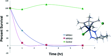
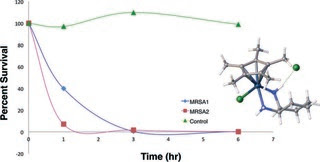
Transition metal diamine complexes with antimicrobial activity against Staphylococcus aureus and methicillin-resistant S. aureus (MRSA)
G. W. Karpin, D. M. Morris, M. T. Ngo, J. S. Merola and J. O. Falkinham III
DOI: 10.1039/C5MD00228A, Concise Article
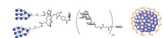
Multivalent glycoconjugates as vaccines and potential drug candidates
Sumati Bhatia, Mathias Dimde and Rainer Haag
DOI: 10.1039/C4MD00143E, Review Article
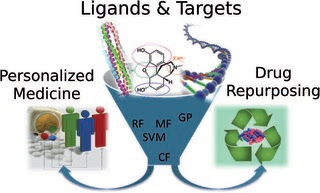
Polypharmacology modelling using proteochemometrics (PCM): recent methodological developments, applications to target families, and future prospects
Isidro Cortés-Ciriano, Qurrat Ul Ain, Vigneshwari Subramanian, Eelke B. Lenselink, Oscar Méndez-Lucio, Adriaan P. IJzerman, Gerd Wohlfahrt, Peteris Prusis, Thérèse E. Malliavin, Gerard J. P. van Westen and Andreas Bender
DOI: 10.1039/C4MD00216D, Review Article
Towards understanding cell penetration by stapled peptides
Qian Chu, Raymond E. Moellering, Gerard J. Hilinski, Young-Woo Kim, Tom N. Grossmann, Johannes T.-H. Yeh and Gregory L. Verdine
DOI: 10.1039/C4MD00131A, Concise Article

Rational design of protein–protein interaction inhibitors
Didier Rognan
DOI: 10.1039/C4MD00328D, Review Article


Transition metal diamine complexes with antimicrobial activity againstStaphylococcus aureus and methicillin-resistant S. aureus (MRSA)

Med. Chem. Commun., 2015,6, 1471-1478     DOI: http://dx.doi.org:/10.1039/C5MD00228A


Multivalent glycoconjugates as vaccines and potential drug candidates

Med. Chem. Commun., 2014,5, 862-878   DOI: http://dx.doi.org:/10.1039/C4MD00143E

Pathogens adhere to the host cells during the first steps of infection through multivalent interactions which involve protein–glycan recognition. Multivalent interactions are also involved at different stages of immune response. Insights into these multivalent interactions generate a way to use suitable carbohydrate ligands that are attached to a basic scaffold consisting of e.g., dendrimer, polymer, nanoparticle, etc., with a suitable linker. Thus a multivalent architecture can be obtained with controllable spatial and topology parameters which can interfere with pathogen adhesion. Multivalent glycoconjugates bearing natural or unnatural carbohydrate antigen epitopes have also been used as carbohydrate based vaccines to stimulate an innate and adaptive immune response. Designing and synthesizing an efficient multivalent architecture with optimal ligand density and a suitable linker is a challenging task. This review presents a concise report on the endeavors to potentially use multi- and polyvalent glycoconjugates as vaccines as well as anti-infectious and anti-inflammatory drug candidates.
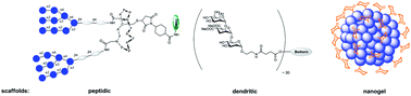
 

Graphical abstract: Multivalent glycoconjugates as vaccines and potential drug candidates


http://pubs.rsc.org/services/images/RSCpubs.ePlatform.Service.FreeContent.ImageService.svc/ImageService/image/GA?id=C4MD00143E


Polypharmacology modelling using proteochemometrics (PCM): recent methodological developments, applications to target families, and future prospects

Proteochemometric (PCM) modelling is a computational method to model the bioactivity of multiple ligands against multiple related protein targets simultaneously. Hence it has been found to be particularly useful when exploring the selectivity and promiscuity of ligands on different proteins. In this review, we will firstly provide a brief introduction to the main concepts of PCM for readers new to the field. The next part focuses on recent technical advances, including the application of support vector machines (SVMs) using different kernel functions, random forests, Gaussian processes and collaborative filtering. The subsequent section will then describe some novel practical applications of PCM in the medicinal chemistry field, including studies on GPCRs, kinases, viral proteins (e.g. from HIV) and epigenetic targets such as histone deacetylases. Finally, we will conclude by summarizing novel developments in PCM, which we expect to gain further importance in the future. These developments include adding three-dimensional protein target information, application of PCM to the prediction of binding energies, and application of the concept in the fields of pharmacogenomics and toxicogenomics. This review is an update to a related publication in 2011 and it mainly focuses on developments in the field since then.
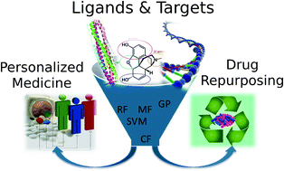
 

Graphical abstract: Polypharmacology modelling using proteochemometrics (PCM): recent methodological developments, applications to target families, and future prospects

http://pubs.rsc.org/services/images/RSCpubs.ePlatform.Service.FreeContent.ImageService.svc/ImageService/image/GA?id=C4MD00216D


Related articles   

Experimental and computational studies of fatty acid distribution networks


Kinetics and non-exponential binding of DNA-coated colloids


Configuration of nonspherical amphiphilic particles at a fluid–fluid interface


Towards understanding cell penetration by stapled peptides

Med. Chem. Commun., 2015,6, 111-119   DOI: http://dx.doi.org:/10.1039/C4MD00131A

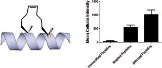
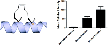
Hydrocarbon-stapled α-helical peptides are a new class of targeting molecules capable of penetrating cells and engaging intracellular targets formerly considered intractable. This technology has been applied to the development of cell-permeable ligands targeting key intracellular protein–protein interactions. However, the properties governing cell penetration of hydrocarbon-stapled peptides have not yet been rigorously investigated. Herein we report our studies to systematically probe cellular uptake of stapled peptides. We developed a high-throughput epifluorescence microscopy assay to quantitatively measure stapled peptide intracellular accumulation and demonstrated that this assay yielded highly reproducible results. Using this assay, we analyzed more than 200 peptides with various sequences, staple positions and types, and found that cell penetration ability is strongly related to staple type and formal charge, whereas other physicochemical parameters do not appear to have a significant effect. We next investigated the mechanism(s) involved in stapled peptide internalization and have demonstrated that stapled peptides penetrate cells through a clathrin- and caveolin-independent endocytosis pathway that involves, in part, sulfated cell surface proteoglycans, but that also seems to exploit a novel, uncharacterized pathway. Taken together, staple type and charge are the key physical properties in determining the cell penetration ability of stapled peptides, and anionic cell surface proteoglycans might serve as receptors to mediate stapled peptide internalization. These findings improve our understanding of stapled peptides as chemical probes and potential targeted therapeutics, and provide useful guidelines for the design of next-generation stapled peptides with enhanced cell permeability.
Graphical abstract: Towards understanding cell penetration by stapled peptides

http://pubs.rsc.org/services/images/RSCpubs.ePlatform.Service.FreeContent.ImageService.svc/ImageService/image/GA?id=C4MD00131A


Introduction Hydrocarbon stapled a-helical peptides are an exciting new class of investigational agents capable of targeting and interfering with intracellular protein–protein interactions.1,2 (For reviews on hydrocarbon stapled peptides, see ref. 3 and 4, and for reviews on synthetic a-helix stabilization in general, see ref. 5 and 6.) These peptides contain a synthetic brace, referred to as a staple, introduced across one face of an a-helix (Fig. 1), that in favorable cases can increase a-helical content and protease resistance, enhance target binding affinity, promote cell membrane penetration, and suppress clearance in vivo. 7–10

Fig. 1 All-hydrocarbon stapled peptide technology. (a) Schematic illustration of peptide stapling. Two alpha-methylated, alkenyl-bearing non-natural amino acids are incorporated at two or more positions in the peptide chain and then cross-linked by ruthenium-catalyzed ringclosing olefin metathesis. (b) Different types of alkenyl-containing non-natural amino acids with distinct stereochemistry at the a-carbon and varied lengths of alkenyl side chains. (c) Three types of stapled peptides used in this study with optimized combinations of nonnatural amino acids.

Stapled peptides are synthesized via incorporation of two amethyl, a-alkenyl amino acids at defined positions in a synthetic peptide, followed by ring-closing olefin metathesis to close the helix-spanning hydrocarbon bridge (Fig. 1a).11,12 The two components of the staple, namely the hydrocarbon bridge and terminal methyl groups, are both important to obtain maximal effectiveness of the conformationally constrained peptide products. This technology has been successfully utilized to target several classes of proteins formerly considered intractable, including multi-component transcription factor complexes and protein–protein interactions having extended interfaces, such as the NOTCH transcription factor complex,13 the b-catenin–TCF interaction in the oncogenic Wnt signaling pathway,14 and the epigenetic modulator PRC2 complex.15 Given the difficulties of developing traditional small molecule drugs that can successfully target intracellular protein–protein interactions, hydrocarbon stapling technology is widely considered to represent a promising avenue of research for the development of chemical probes and potential targeted therapeutics.

Multiple types of hydrocarbon staples have been obtained by varying the relative placement of the cross-linking a,a-disubstituted amino acids, as well as the stereochemistry at the acarbon and the lengths of the alkenyl substituents (Fig. 1b).16,17 These staple types were optimized to provide robust a-helical stabilization and confer the potential for in vitro and in vivo activity. As a result of the combinatorial search process used to identify helix-stabilizing hydrocarbon staples, the diversity of the resulting macrocyclic bridges has revealed stapled peptides with different physicochemical properties. Recently, a new hyperstable version of stapled peptide with tandem crosslinks, referred to as a stitched peptide, was generated by introduction of S5 at the i position, B5 at the i + 4 position, and S8 at the i + 11 position (Fig. 1c) (Y.-W. Kim and G. L. Verdine, to be published).

Of the physicochemical properties demonstrated by peptide bearing hydrocarbon staples, the capacity to promote cellular membrane penetration is perhaps the most signicant and yet remains the most poorly understood. Independent of hydrocarbon-stapled peptides, several classes of cell penetrating peptides (CPPs) have been discovered, including naturally occuring transcription factor domains such as pennetratin18 and HIV-Tat19 and synthetic cationic peptides such as polyArginine peptides.20 Notably, despite extensive exploration during the past two decades, the mechanism(s) by which CPPs enter cells remain unclear.21–23 In contrast to CPPs, in which cell penetration appears to be sequence-dependent, numerous cell permeable stapled peptides have been discovered for peptide scaffolds with little sequence homology. These divergent observations regarding cell penetration is proposed to result from several features of stapled peptides that differentiate them from typical CPPs. For example, the introduction of an allhydrocarbon cross-link results in a constrained a-helical conformation, which embeds the hydrophilic amide backbone in the core of the folded structure. Furthermore, the hydrocarbon brace itself introduces a significantly hydrophobic patch to one face of the peptide. The exposure of the hydrophobic moiety as well as the masking of the hydrophilic peptide backbone may facilitate the interaction of stapled peptides with the hydrophobic interior of the cell membrane and thereby enhance the cellular uptake. As cell penetration is a critical property of stapled peptides, we sought to develop quantitative methods to correlate a battery of stapled peptide properties with the capacity for cellular uptake. A direct comparison with several well-known CPPs has revealed that stapled peptides, including some stapled versions of the CPPs, exhibit more robust cell penetration. Lastly, we have demonstrated that stapled peptides penetrate cells through a clathrin- and caveolin-independent endocytosis pathway that involves, in part, sulfated cell surface proteoglycans. These findings significantly expand our current understanding of cell penetration by stapled peptides and provide useful information for the future rational design of cell penetrating stapled peptides with novel applications.

Results and discussion

Development of a high-throughput assay to quantitatively measure cellular uptake of peptides

Understanding the internalization process of cell penetrating peptides (CPPs), especially stapled peptides, has been a subject of great interest. The majority of previous studies have been performed by either using high-resolution microscopy to show the existence of fluorophore-labeled CPPs inside cells, or by quantitatively measuring intracellular fluorescence by flow cytometry.24,25 Although these two methods can provide important information regarding cell penetration, their respective limitations prompted us to adopt an assay that combines high-resolution imaging with reliable quantitation of intracellular accumulation to better analyze and understand the cell penetration of stapled peptides. In recent years, highthroughput cell-based imaging platforms have become increasingly popular to screen for small molecule modulators of various biological processes.26,27 Taking advantage of one of these platforms, high-content epifluorescence microscopy, we developed a high-throughput quantitative assay to measure stapled peptide intracellular access.

Proof-of-principle experiments were performed to determine whether epifluorescence microscopy could be used to quantitatively compare stapled peptide intracellular access. Human U2OS osteosarcoma cells were seeded in black, clear-bottom 384-well plates and then incubated in serum-containing media supplemented with fluorescein-labeled peptides or DMSO vehicle for 12 hours. After the treatment, cells were washed thoroughly with PBS to remove excess peptide, fixed with 4% formaldehyde, and stained with Hoechst dye to visualize nuclei. Once prepared, the plates were imaged and quantified by epi- fluorescence microscopy according to a protocol developed and discussed in detail in Experimental methods. An initial z-scan was performed using the Hoechst channel to locate the cells, and the microscope parameters were subsequently adjusted to optimize the cell size and fluorescence intensity. The parameters from this acquisition were then applied to the FITC channel, and the microscope scanned and recorded images of the FITC-labeled peptides within the z-plane of the cell. This assay was performed in a high-throughput manner, resulting in a panel of Hoechst/FITC images from individual wells (Fig. S1†). The raw image data was then analyzed using MetaXpress® software (Fig. 2a). Cells were identified based on the Hoechst stain of nuclei, with the requirement that they were a contiguous fluorescent region having a specific intensity above local background as well as having a diameter between defined minimum and maximum to be designated as “positive” cells. The cytoplasm of each cell was then identied according to the spatial location of FITC signal in relation to the nuclei as well as empiric parameters (details in Experimental methods). The FITC intensities in the cytoplasm and nuclei were then quantified separately, and the sum of these two values yielded the FITC signal for the whole cell, which can be considered the relative intracellular peptide intensity. In addition, FITC negative cells were identified on the basis of a positive Hoechst stain, which was accompanied by an absence of appreciable signal in the FITC channel.

Fig. 2 Quantitative measurement of cellular peptide intensity. (a) Hoechst channel (left) showing the location and size of nuclei, FITC channel (middle) showing the fluorescence intensity of the same cells. Information about cell size and fluorescence intensity was integrated to identify the FITC positive (green mask) and negative (red mask) cell (right). For positive cells, additional parameters allowed determination of the fluorescence intensity in the nucleus (inner intense green) and the cytoplasm (outer dim green). (b) The background fluorescence the DMSO vehicle was almost identical among different experiments. (c) Four stapled peptides from different batches of synthesis generated similar intracellular fluorescence intensity in different tests. Error bars represent the S.D. of two measurements.

We found that this system generated highly reproducible and reliable results from assay-to-assay and with different stocks of the same stapled peptides. As shown in Fig. 2b, there were negligible fluorescence differences among experiments for cells treated with DMSO vehicle, which could be used as a fluorescence background for all subsequent experiments. In addition, the same stapled peptides from different batches of synthesis and stocks featured almost identical intracellular fluorescence signals in different tests (Fig. 2c), indicating that the assay developed in this study produces repeatable and reliable results that could be directly combined and compared from a large set of experiments. Furthermore, to determine how this assay performs as a screening tool, we have calculated Z0 factor of 0.54 by using the most penetrant A6 peptide as a positive control and DMSO background as negative control, which also indicates a statistically good assay quality.

Analysis of cell penetration by stapled peptides The development of this quantitative high-throughput assay enabled a broad investigation of the physicochemical properties governing the cell uptake of a diverse set of hydrocarbonstapled peptides synthesized in our laboratory. We postulated that any correlation between cellular uptake and physicochemical properties would illuminate characteristics associated with productive cellular uptake and inform the future design of stapled peptides with improved cell penetration.28 To this end, we screened and analyzed more than 200 discrete FITC-labeled peptides belonging to three different classes: wild-type (unmodified), stapled and stitched peptides. All peptides were converted to two-dimensional structures and analyzed for theoretical physicochemical properties with the publicly available Marvin View software package from ChemAxon. Properties including the molecular weight, theoretical pI, calculated 2D polar surface area (PSA), theoretical log P and formal charge at pH 7.5 were calculated for each peptide (Table S1†). In general, the unmodified, stapled and stitched peptide libraries present in this screen had relatively similar physicochemical characteristics (Fig. S2†). The mean molecular weight and calculated PSA values were nearly identical among the three peptide classes. A notable difference was observed among theoretical log P values, which were significantly higher for the stapled and stitched peptides relative to the unmodified peptides, which is not surprising as these modified peptides contain a solvent exposed hydrocarbon crosslink. Additionally, the stapled peptide class had a mean formal charge of approximately zero while the stitched and unmodified peptide classes exhibited a positive mean charge. Overall, the calculated physicochemical properties indicated that the peptide classes were quite similar in terms of their mean properties, which is useful when making comparisons among their cell penetration properties.

We next performed an intracellular access screen by treating U2OS cells with 1 mM of FITC-labeled peptide for 12 hours in duplicate. All assays contained control DMSO wells and positive control peptides, which were compared among assays to ensure plate-to-plate reproducibility (Fig. 2b and c). The primary readout of the screen was mean cellular fluorescence intensity. As the DMSO background was highly consistent between wells and experiments, a mean background value was subtracted from all data. The results of the screen were used to generate plots comparing cell penetration with peptide physicochemical parameters. Interestingly, as a class, stapled and stitched peptides exhibited significantly higher cell penetration compared with wild-type unmodified peptides, which contained several established cell penetrating peptides (CPPs; Fig. 3a). Given that all three peptide classes have similar physicochemical properties in general, the benefit in cell penetration can be largely attributed to the synthetic stabilization of the a-helical peptides with all-hydrocarbon peptide stapling technology. Furthermore, we found that peptide charge near physiologic pH exhibited a strong correlation with intracellular access and could be fitted into a Gaussian distribution with a population centroid at a formal charge of +4 (Fig. 3b). In particular, peptides exhibiting a net negative charge (7 to 1) exhibited little cellular uptake, whereas peptides of approximately neutral charge (1 to +1) displayed moderate cell penetration above background. Interestingly, peptides with a net positive charge (+1 to +7) showed significantly higher cell penetration as a group. Cellular uptake did not appear to increase linearly with charge, as the cell penetration decreases dramatically for the peptides in this study with charge greater than +7. The same trend between formal charge and cellular uptake were observed for individual stapled and stitched peptide classes as well (Fig. S3†). This observation is not consistent with previously reported models that indicate that peptides/mini-proteins with more positive charge have better penetration properties due to tighter electrostatic interactions with the negatively charged phospholipid membrane.29,30 The lower penetration for highly charged peptides in this study could result from any one of many factors including, for example, peptide aggregation in solution, the disruption of peptide packing during internalization or difficulty in dissociation from cell membrane. Additional tests with a larger number of peptides could further our understanding of this phenomenon. In addition, there was no discernible correlation between cell penetration and peptide molecular weight, log P, pI value or PSA (Fig. S4†). Taken together, these data demonstrated that the staple type and peptide charge are key physical properties correlated with peptide cell penetration ability, whereas the other parameters do not appear to be significantly associated.

In order to further investigate the cell penetration properties for stapled peptides and to systematically analyze the similarities and differences in cellular uptake between stapled peptides and other wild-type cell penetrating peptides, we compared cell penetration of several stapled peptides to that of three well known wild-type CPPs: Tat (48–60), penetratin (Antennapedia 43–58) and poly-Arg8 (Table S2†). First, we investigated the cellular uptake at varied peptide concentrations. As shown in Fig. 4a and b, both wild-type CPPs and stapled peptides showed dose-dependent increases in cell penetration. Strong intracellular fluorescence was detected in the low micromolar range, and although the levels of accumulation were different for distinct peptides, stapled peptides featured more robust dosedependent cell penetration at lower concentrations relative to wild-type CPPs, in general. It is also interesting to note that while significant increases in intracellular fluorescence were mostly evident in the 1–10 mM range for stapled peptides, distinct profiles were observed for specific peptides. For example, TNG147 showed little cell penetration at 1 mM but showed a dramatic increase at 5 mM, which might suggest that concentration-dependent peptide packing or a receptor-mediated mechanism may facilitate the cell penetration process, and these processes may be triggered at different concentrations for distinct peptides. Furthermore, it is worth noting that the stapled peptides studied here were more cell permeable than wild-type CPPs at most concentrations tested, exhibiting nearly an order of magnitude higher intracellular fluorescence at the same treatment concentrations.

Fig. 4 Effects of peptide concentration and incubation time on cellular uptake of stapled and wild-type peptides. (a) Wild-type and (b) stapled peptides showed a dose-dependent increase in cell internalization. Cellular uptake for (c) penetratin and (d) SAHM1 peptides over time at concentrations of 5 and 10 mM. (e) A pulse-chase penetration assay for SAHM1 peptide in which fresh medium containing either a new batch of peptide or DMSO vehicle were exchanged at 12 hours after initial treatment. Error bars represent the S.D. of triplicate samples.

We next performed a time-course penetration assay to better understand the kinetics of peptide internalization using a representative CPP and stapled peptide. Penetratin and SAHM1 showed distinct kinetics of uptake and stabilization throughout a 24 hour time course. 5 mM and 10 mM penetratin peptide exhibited similar intracellular cellular fluorescence after 2 hours, which then decreased until approximately 8 hours and finally stabilized at different intracellular levels until 24 hours (Fig. 4c). On the other hand, the stapled peptide SAHM1 showed time- and dose-dependent cellular uptake, which stabilized after approximately 8 hours (Fig. 4d). Compared to the wild-type penetratin, the SAHM1 profile was unique in that dose-dependent accumulation was evident at all time points and no loss of signal was observed. One explanation for the loss of signal observed with penetratin could be attributed to an equilibrium between cell penetration and subsequent intracellular proteolysis followed by export of the fluorophore. The presence of the all-hydrocarbon crosslink in its peptide sequence and lower net charge of SAHM1 relative to penetratin, could contribute to enhanced cellular uptake and reduced intracellular proteolysis, leading to continuous accumulation in cells. To further explore the equilibrium observed for stapled peptides, we performed a pulse-chase experiment using SAHM1. After 12 hours of incubation with SAHM1, cell culture medium was aspirated and the cells were extensively washed with PBS to completely remove excess peptide. Then fresh medium containing either a new batch of 1 mM peptide or DMSO vehicle was added to cells and incubated for the indicated time points (Fig. 4e). As expected, the cellular uptake increased for the first 12 hours incubation. After medium exchange, cells incubated with fresh medium containing DMSO vehicle retained the intracellular fluorescence intensity. Interestingly, the signal for cells treated with a new batch of staple peptide continued to increase up to 24 hours (Fig. 4e). This observation indicates that despite incubation over a time course previously shown to reach equilibrium, the mechanism(s) responsible for cellular uptake are not saturated, as evidenced by further uptake upon replacement with fresh stapled peptide. Taken together, these data indicate that the mechanism(s) underlying cellular uptake by both CPPs and stapled peptides exhibit time- and dose-dependency that is not saturable at early time points or low micromolar doses and, importantly, appears to be more robustly utilized by stapled peptides.15,31

Given that stapled peptides exhibit better cell penetration properties in general than parent unmodified peptides, we wondered whether the peptide stapling strategy could be applied generally to improve cellular uptake of parent unmodified peptides. To test this hypothesis, we designed a panel of stapled peptides based on Tat (48–60), penetratin and poly-Arg8 (Fig. 5a). These stapled peptides and their parent unmodified peptides were incubated in U2OS cells for 12 hours with a concentration range from 10 nM to 20 mM, mirroring the dosedependent uptake studies shown in Fig. 4. As expected, all peptides showed dose-dependent cell penetration (Fig. 5b–d). Interestingly, stapled peptides derived from penetratin and poly-Arg8 showed improved cell permeability at concentrations starting from 1 mM for stapled penetratin and 5 mM for stapled poly-Arg8. It is noteworthy that the staple position also affected the cellular uptake as the two stapled penetratin peptides with different crosslink positions exhibited varied cell penetration, though both were superior to wild-type penetratin. In contrast, reduced cellular uptake was observed for both stapled peptide variants derived from the Tat sequence (Fig. 5b). This could result from several possible effects, including disruption of peptide secondary structure, masking of residues essential for surface recognition or altering peptide packing interactions involved in cell penetration. Further focused study of these variants is warranted to elucidate the source of altered cellular uptake, however these data clearly demonstrate that peptide stapling may be a general method to further improve the cell permeability of CPPs, which could serve as more efficient transduction domains for molecular cargoes. In addition, while increasing the helical content of stabilized peptides has been stated to be a guiding principle in the successful design of biologically active stapled peptides, it has not been shown to be generally correlated with cell penetration. To specifically address whether increasing the helical content of a peptide is correlated with augmented cell penetration, we have measured the relative helicity of hydrocarbon stapled variants of Tat, penetratin and poly-Arg8 (Fig. S5†). Notably, we did not observe a general correlation between increased helical character and cell penetration of these peptides. Peptide stapling increased the helical content of both Tat and poly-Arg8 peptide sequences, which were largely unstructured when unmodified. In contrast, the unmodified penetratin peptide had signicant helical content (>50%), and the hydrocarbon stapled variants of this sequence largely retained their helicity, albeit lower overall helicity. Intriguingly, these species demonstrated the differing effect of hydrocarbon stapling and increased helical content on cell penetration since introduction of the hydrocarbon staple increased the cellular uptake of both penetratin and poly-Arg8 sequences, while it decreased uptake for Tat peptides. Therefore, we cannot conclusively state, a priori, that the incorporation of a hydrocarbon staple or increased a-helicity will lead to more productive cellular penetration, although in general stapling can increase the uptake of specific sequences (Fig. 5) and as a class stapled and stitched peptides are more cell penetrant (Fig. 3a). A more comprehensive follow-up study with CD analyses on a larger peptide library is needed to better address this question.

Fig. 5 Effects of all-hydrocarbon staples on cell penetration by wild-type cell penetrating peptides. (a) List of wild-type cell penetrating peptides and their stapled derivatives investigated in this study. (b–d) Dose-dependent cell penetration assays showed that stapling strategy greatly improves the cellular uptake of penetratin and poly-Arg8 peptides. Experiments were performed in triplicate, and error bars represent S.D. of three measurements.

Mechanistic studies of cell penetration by stapled peptides The aforementioned studies indicate that stapled peptides exhibit better cellular uptake properties than wild-type peptides in general, and that internalization correlated primarily with hydrocarbon staple type and formal peptide charge. However, the mechanism(s) utilized by peptides to translocate across the cell membrane are still unclear. Therefore, we sought to investigate the uptake mechanism(s) for stapled peptides. The uptake mechanism(s) of wild-type CPPs have been extensively studied. Some evidence indicates that they enter cells via energy-dependent endocytosis, which is an active transport process, however data suggesting passive diffusion for CPPs have also been reported; hence, the mechanism(s) of cell uptake by CPPs remains ambiguous.32–34 We first sought to determine whether cell penetration by stapled peptides and wild-type CPPs occurs via ATP-dependent endocytosis.2 Cells were pre-treated with NaN3 and 2-deoxyglucose (2-DG) to reduce cellular ATP levels, and then incubated with FITC-labeled peptides (wildtype and stapled) for 4 hours and compared to normal cells for intracellular fluorescence. Cellular ATP levels were confirmed to be decreased by approximately 90% after NaN3 and 2-DG treatment (Fig. S6†), but Tat and poly-Arg8 exhibited almost identical cellular uptake in ATP-depleted and normal cells, supporting the model that they utilize passive diffusion to translocate across the cell membrane. However, penetratin and all stapled peptides showed 20–50% lower accumulation in ATP-depleted cells, indicating an active trans-membrane process requiring cellular ATP (Fig. 6a). These data indicate that there may be more than one uptake mechanism for CPPs and stapled peptides, but that for the most robust cell penetrating peptides (penetratin and stapled peptides studied here), the internalization mechanism(s) involves ATP-dependent endocytosis.

Fig. 6 Mechanistic study of cell penetration by stapled peptides and wild-type cell penetrating peptides. (a) Cellular uptake in normal and ATP-depleted cells indicated that stapled peptides penetrate cells via an ATP-dependent endocytosis. (b) Impaired uptake was observed in NaClO3 treated cells, which inhibit proteoglycan biosynthesis. (c) Cell penetration of wild-type and stapled peptides in wild-type CHO and proteoglycan-deficient CHO cells. Experiments were performed in triplicate, and error bars represent S.D. of three measurements.*P < 0.05, **P < 0.01, ***P < 0.001.

Next, we sought to identify the specific pathway(s) utilized for cellular uptake, since energy-dependent endocytosis can be accomplished by several different pathways including caveolinand clathrin-mediated endocytosis. We repeated the cell penetration experiments under a variety of conditions that each blocked a different endocytosis pathway (Table S3†).35–37 We found that uptake was partially blocked in cells treated with sodium chlorate (Fig. 6b), which aborts the decoration of cells with sulfated proteoglycans, but was unaffected by inhibitors of other endocytic pathways (Fig. S7†). It thus appears that interaction with sulfated proteoglycans is responsible for some, but not all, endocytic uptake of stapled peptides and wild-type CPPs. It is reasonable to connect this result with the previous discovery that peptide charge is a key factor determining cell penetration. Proteoglycans are negatively charged under physiologic conditions due to the occurrence of sulfate groups, and these might form electrostatic pairs with positively charged peptides to facilitate anchoring on the cell membrane.38–40 To further confirm that sulfated proteoglycans are important to mediate cellular uptake for peptides, we performed a secondary assay using wild-type CHO cells (CHO-K1) and proteoglycan deficient CHO cells (pgsA-745) which harbor a defect in xylosyltransferase, thereby preventing glycosaminoglycan biosynthesis. All peptides showed similar penetration properties in wild-type CHO cells, but uptake was decreased by approximately 50% in proteoglycan-deficient CHO cells, consistent with the experiment using a small molecule inhibitor (Fig. 6c). Taken together, our data suggest that CPPs and stapled peptides penetrate cells through a clathrin- and caveolin-independent endocytosis pathway that is in part mediated by interaction with anionic cell surface proteoglycans. This result is very similar to the previous reports on the mechanism of cellular uptake for supercharged GFP (scGFP), which likewise does not utilize clathrin- or caveolin-mediated endocytosis.41 Notably, scGFP internalization requires actin polymerization, which may not be required for peptide penetration (Fig. S7c†) types, and distinct physicochemical properties. As a result, we found that stapled peptides penetrate cells more efficiently than unmodified peptides, including well-characterized cell penetrating peptides. For the panel of peptides used in this study, only staple type and formal charge were significantly correlated with cell penetration potential, whereas the other physical parameters did not appear to have a signicant effect. We further studied the relationships between cellular uptake and

In conclusion, we sought to investigate the cell penetration properties of stapled peptides, which is one of the most significant yet poorly understood aspects of peptide stapling technology and cellular transduction technologies in general. In order to address this problem, we developed a high-throughput assay to quantitatively measure stapled peptide intracellular accumulation. Using this assay, we analyzed more than 200 discrete peptides with various sequences, staple positions and peptide concentration or incubation time, revealing that stapled peptides accumulate in cells in a dose-dependent fashion and reach steady intracellular levels over a course of a few hours. These studies revealed similar time- and dosedependent behavior for CPPs and stapled peptides, but stapled peptides, including stapled versions of CPPs, were shown to be 10- to 20-fold more penetrant, measured by intracellular fluorescence level at a given dose, than the most potent CPP. We also propose that the specific intracellular accumulation and stabilization kinetics of stapled peptides or unmodified CPPs may be a consequence of equilibria between peptide penetration, cellular proteolysis and/or retrograde transport of the species. Finally, we investigated the mechanism(s) involved in the internalization of stapled peptide and unmodified CPPs and demonstrated that cell penetration occurs through a clathrinand caveolin-independent, energy-dependent endocytosis pathway that utilizes, in part, sulfated cell surface proteoglycans. This dataset provides significant insight into the physicochemical properties correlated with productive cellular penetration as well as a more detailed understanding of the mechanism(s) utilized by stapled peptides to access intracellular compartments, which together should aid in the design of and characterization of novel stapled peptides in the future.


Rational design of protein–protein interaction inhibitors

Med. Chem. Commun., 2015,6, 51-60    DOI: http://dx.doi.org:/10.1039/C4MD00328D
Protein–protein interactions are at the heart of most physiopathological processes. As such, they have attracted considerable attention for designing drugs of the future. Although initially considered as high-value but difficult to identify, low molecular weight compounds able to selectively and potently modulate protein–protein interactions have recently reached clinical trials. Along with high-throughput screening of compound libraries, combining structural and computational approaches has boosted this formerly minor area of research into a currently tremendously active field. This review highlights the very recent developments in the rational design of protein–protein interaction inhibitors.
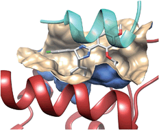
Graphical abstract: Rational design of protein–protein interaction inhibitors

Didier Rognan heads the Laboratory of Structural Chemogenomics at the Faculty of Pharmacy of Strasbourg (France). He studied Pharmacy at the University of Rennes (France) and did a Ph.D. in Medicinal Chemistry in Strasbourg (France) under the supervision of Prof. C.G. Wermuth. Aer a postdoctoral fellowship at the University of Tubingen (Ger- ¨ many), he moved as an Assistant Professor to the Swiss Federal Institute of Technology (ETH) until October 2000. He was then appointed Research Director at the CNRS to build a new group in Strasbourg. He is mainly interested in all aspects (method development and applications) of structurebased drug design, notably on G protein-coupled receptor ligands and protein–protein interaction inhibitors.

Introduction Drug discovery is a long, costly, multi-step endeavour which aims at reducing all possible risks to deliver a novel therapeutic solution to previously unmet clinical needs. To reduce chemical risks, empirical rules are used to filter the chemical space and retain drug-like low molecular weight compounds. Reduction of the biological risk is addressed by considering privileged target families (e.g., G protein-coupled receptors and kinases) whose activation/inhibition by drug-like compounds is likely to correct or reverse pathological states. Until recently, mostly single macromolecules (proteins and nucleic acids) have been considered as potential drug targets. Out of 68 000 proteins currently annotated in UniProt for the human proteome,1 only about 300 targets2 have been addressed by current drugs, and the large majority of single targets is still awaiting first-in class drugs.

Besides single targets, large scale genomics and proteomics3 have identified complex networks of targets and pathways regulating physiopathological processes in a coordinated manner. The current human protein–protein interactome has been estimated between 130 000 (ref. 4) and 650 000 (ref. 5) complexes, out of which only a tiny amount is known, and only a very few6–8 have been the object of a drug discovery initiative. Protein–protein interactions (PPIs) therefore describe a totally new biological space that attracts more and more attention, with 26PPI inhibitors9,10 already under clinical development, notably in the oncology field.11 Despite PPIs may adopt quite different sizes, shapes and electrostatics,12 identifying highaffinity PPI inhibitors is a considerable challenge for many reasons: (i) in contrast to conventional targets, a medicinal chemist cannot start inhibitor design from the structure of endogenous ligands, (i) PPIs often involve flat surfaces delocalized over multiple epitopes, usually lack well-defined buried cavities13 typical of conventional targets, and are significantly larger (ca. 1000–3000 A˚2 ) than enzyme/receptor pockets (300– 1000 A˚2 ), (iii) high-throughput screening of traditional compound libraries often returns no viable hits14 for the main reason that PPI inhibitor chemical space is quite different from that described by traditional drug-like compounds.10 Nonetheless, thanks to bioinformatics and proteomics-guided prioritization of therapeutically relevant protein–protein complexes, more and more PPI inhibitors are currently reported. Several excellent reviews6,7,9,11,15–18 have already been published on experimental methods (high throughput screening, biochemical and cellular assays, and fragment-based approaches) suitable to discover PPI inhibitors. The present report will only cover computer-aided approaches, with a major emphasis on structure-based methods and recent discoveries (2012–2014).

Databases Preliminary access to experimentally validated data is key to launch a drug discovery program on PPI modulators. A multitude of databases storing genomics, proteomics and structural data are currently available to help the medicinal chemist. We will here briefly review these archives, focusing mostly on easily interpretable structural data.

PPI databases Many experimental methods with different throughputs (from low to high) have been developed to characterize binary interactomes in various species, among which the most prominent has been the yeast two-hybrid (Y2H) assays, and mass-spectrometry (MS) coupled with co-immunoprecipitation or coaffinity purification.19 These experimental data are stored in many primary databases (Table 1) that are difficult to mine due to their large heterogeneity. Metadatabases have been derived thereof to facilitate their analysis, among which the most popular are APID and PRIMOS (Table 1). These metadatabases cover a wide range of organisms and notably offer the possibility to mine experimental PPI data according to disease relevance or inter-organism crosstalk, and provide graphic tools to visualize complex networks of interacting proteins and identifying important protein nodes (hubs). It is however very difficult, from this large amount of data, to clearly prioritize PPIs for a drug discovery program. Attempts to classify the PPIs by structural druggability25 (although ligandability26 is probably a better term) are worth mentioning but should be taken with care due to the still insufficient number of existing PPI three-dimensional (3D) structures.

Table 1 Protein–protein interaction databases

Database                 Interactions                         Website                                                             References

BIND                             32 211           http://bond.unleashedinformatics.com                       20

DIP                                 78 191           http://dip.doe-mbi.ucla.edu/dip/Main.cgi                  21

HPRD                           41 327            http://www.hprd.org/                                                           22

IntAct                      448 986             http://www.ebi.ac.uk/intact/                                            17

MIPS                              9 835             http://mips.helmholtz-muenchen.de/proj/ppi/      23

APID                         196 700             http://bioinfow.dep.usal.es/apid/index.htm             24

PRIMOS                  384 127             http://primos.fh-hagenberg.at/                                        19


Table 2 Database of low molecular-weight PPI inhibitors
Database                 Ligands                             Website                                                                  References

2P2I                                 71                   http://2p2idb.cnrs-mrs.fr/                                                12

iPPI-DB                      1650               http://www.ippidb.cdithem.fr/                                        10

TIMBAL                      6896              http://mordred.bioc.cam.ac.uk/ timbal                        29

Ligand databases Initially limited to a limited subset of inhibitors able to disrupt few PPIs (e.g. p53/MDM2, Bcl-Xl/Bak, and IL-2/IL-2Ra),7,27 the repertoire of PPI inhibitors rises constantly thanks to exciting developments in biophysical fragment screening.15,28 Three publicly available databases storing information on PPIs and their inhibitors (Table 2) may be used to better describe the structural properties of druggable PPIs and the chemical space associated with their disruptors.

The 2P2Idb database12 is a hand-curated repository of protein–protein complexes of known X-ray structures (X-ray diffraction and nuclear magnetic resonance spectroscopy) for which at least one low molecular weight orthosteric inhibitor has been co-crystallized with one of the two protein partners. It currently describes 71 inhibitors for 14 PPIs, clustered in two groups (Fig. 1) with respect to the nature of the interface (protein–peptide and protein–protein). Companion tools (2P2I inspector,30 2P2I score,30 and 2P2I hunter31) are provided to analyse PPIs at a structural level, predict their structural druggability and design PPI focussed libraries, respectively.

Fig. 1 Prototypical examples of class I (left panel) and class II PPIs (right panel), exemplified by the Bcl-Xl/Bak (PDB id 1BXL) and integrase/LEDGF (PDB id 2B4J) complexes, respectively. Class I PPIs involve the interaction of a globular protein with a peptide or a single secondary structure (a-helix and b-strand) of a second protein partner. Class II PPIs are characterized by the interaction of two globular proteins.

The iPPI-DB10 is another manually curated database from world patents and the medicinal chemistry literature, focussing on low molecular weight orthosteric inhibitors, disease-related protein–protein interfaces and a clear biochemical readout (e.g. fluorescence polarisation and enzyme-linked immunosorbent assay). The database archives 1650 PPI inhibitors targeting 13 families of homologous PPI targets mainly involved in cancer, immune disorders and infectious diseases.

Finally, the TIMBAL database29 reports ca. 7000 inhibitors for 50 known PPIs. In contrast to the two other databases, TIMBAL is maintained through a predefined list of PPIs and automated searches in ChEMBL32 and the Protein Data Bank.33 In contrast to the other databases, TIMBAL also registers short peptides with an upper molecular weight limit of 1200 Da. It should be pointed that most of the 15 000 uncurated biological data present in TIMBAL arise from a single target family (integrins) and should be considered with care.

Analysing the content of these databases enables a first comparison of PPI inhibitors versus drugs, as well as PPIs amenable to disruption versus standard heterodimers. PPI surfaces disrupted by inhibitors tend to be smaller, more hydrophobic and accessible than standard heterodimers.12 As a consequence, low molecular weight PPI inhibitors tend to be larger, more hydrophobic and more aromatic-rich than standard drugs. Interestingly, many of them (ca. 60%) still comply with Lipinski’s rule-of-five, 10 revealing some hopes in the developability of such compounds.

However, it should be stated that the set of empirical rules designed to discriminate druggable from non-druggable PPIs, as well as to distinguish PPI inhibitors from conventional druglike compounds still rely on a very limited set of highly homologous data (PPIs, inhibitors), and should therefore be regarded with caution. Increasing coverage of the PPI repertoire by future experimental screens will undoubtedly lead to a better denition of PPI biological and chemical spaces. We therefore expect in the future the above-mentioned rules to be rened and be more descriptive of the true world of PPI inhibitors, notably with respect to rational design of PPI focussed libraries.


Rational design of PPI modulators

Sequence-based approaches Whatever the nature of the PPI (type I or type II, see the definition above), PPI interfaces are often characterized by the presence of hotspots,34 in other words anchor residues that contribute the most to the binding free energy of the protein– protein complex. The interaction of a single modified amino acid with a single anchor residue might be sufficient to disrupt a PPI as elegantly demonstrated by Lin et al. in a recent study.35 Capitalizing on the presence of a reactive cysteine (C246) at the interface of the complex between caspase-7 (CASP7) and the Xlinked inhibitor of apoptosis protein (XIAP), they designed the N-iodoacetyl-lysine amino acid derivative 1 (Fig. 2) that covalently traps C246 and further disrupts the XIAP–CASP7 complex, therefore triggering CASP7-dependent apoptosis and killing MCF-7 breast cancer cells (EC50 ¼ 0.64 mM) previously resistant to chemotherapy.

The easiest way to inhibit a PPI is to start with the amino acid sequence of one interacting epitope, notably if the latter is part of regular secondary structures (a-helix, b-strand, and b-turn). For example, a-helical peptides mimicking the sequence of protein transmembrane domains may disrupt PPIs quite efficiently.36,37

Fig. 2 Peptidomimetics as PPI disruptors


Due to poor pharmacokinetic profiles, linear peptides are good in vitro tools but usually not efficient clinical candidates. Chemical modifications are required to stabilize their secondary structures in physiological media and prevent early degradation. Among the most exciting developments in this area38,39 is the design of stapled peptides.40,41 Stapled peptides are synthetic analogues of a-helical protein epitopes involved in a PPI, and in which a covalent hydrocarbon linkage connects adjacent turns of the helix. Stapling is known to significantly increase the in vivo half-life of the natural peptide (increasing proteolytic stability), decrease the entropic cost of binding, and even enable cellular uptake.42 Many stapled peptides with potent in vivo activities have already been reported.39 One of these stapled peptides (ATSP-7041, compound 2, Fig. 2) just entered clinical development as a dual nM MDM2/MDMX inhibitor for p53-dependent cancer therapy.43

Heterocyclic scaffolds mimicking secondary structures can also be obtained by solution-phase synthesis to afford peptidomimetic libraries amenable to PPI inhibition. Whitby et al. notably reported the design of 8000 member 4-acetamido-3- alkoxy-benzamide focused library featuring weak p53/MDM2 inhibitors and potent HIV-1/gp41 inhibition (compound 3, Fig. 2).44 When the peptide epitope is not structured, developing macrocyclic analogues is more difficult but still feasible as recently demonstrated by Glas et al.38 who successfully improved 14-3-3 binding of a 11-mer peptide from a bacterial ExoS virulence factor by cross-linking binding amino acids with polymethylene linkers, up to an in vitro 40 nM disruptor of the ExoS/14-3-3 interaction (compound 4, Fig. 2). Interestingly, the cross-linker was not only chosen to rigidify the natural ExoS peptide structure but also to directly provide additional hydrophobic interactions to the 14-3-3 binding site.38 Only in exceptional cases the natural unmodified peptide is directly usable as a PPI inhibitor. One recent example is the 28 amino acid cell-penetrating peptide (p28) from a bacterial azurin redox protein, that binds to the DNA-binding domain of the p53 tumor suppressor and inhibits p53 degradation by interfering with the Cop1-mediated ubiquitination,45 thereby enhancing p53 levels in cancer cells and exhibiting antitumoral efficacy in patients with advanced solid tumors.46

Pharmacophore-based approaches As defined by the IUPAC,47 a pharmacophore is “an ensemble of steric and electronic features that are necessary to ensure the optimal supramolecular interactions with a specific biological target and to trigger (or block) its biological response.” Although pharmacophores are mainly used to align and compare ligands sharing the same target,48 the same concept can be easily transferred to PPIs in which one partner is the “receptor” and the second one the “ligand”. Pharmacophore features (hydrophobic, aromatic, H-bond donor and H-bond acceptor, positively and negatively ionisable) can therefore be manually or automatically mapped to atoms of the ligand in direct interactions with the receptor. The resulting pharmacophore can then be used to identify a compound library for hits fulfilling the defined query. Several tools (e.g. LigandScout,49 Discovery Studio,50 and Pocket Query51) can be directly used to map PPI pharmacophores onto protein–protein X-ray structures (Fig. 3).

Fig. 3 Example of a PPI pharmacophore mapped onto interacting atoms of human LEDGF (yellow ribbons) bound to HIV-1 integrase (red ribbons, PDB ID 2B4J). The PPI pharmacophore is composed of 2 Hbond donors (magenta balls), two H-bond acceptors (green balls), one hydrophobic feature (cyan ball) and 6 exclusion volumes (gray balls).

Using a manual PPI pharmacophore defined from the X-ray structure of the Annexin A2/S100A10 complex, a pro-angiogenic complex, Reddy et al.52 derived a simple pharmacophore (2 hydrophobes, 2 H-bond donors, and 2 H-bond acceptors) using the Unity program,53 and screened a library of 700 000 compounds to select 586 hits which were further docked to the Annexin A2 binding site to retain only 190 candidates with both a good docking and pharmacophore fitness score (Table 3). Out of 190 tested compounds, 7 hits blocked the interaction between S100A10 and the Annexin A2 N-terminus in a competitive fluorescent binding assay, with the most potent PPI inhibitor (compound 5, Fig. 4) exhibiting an IC50 of 24 mM.52 Geppert et al.54 reported the rational discovery of a low molecular weight inhibitor of the complex between interferon-a (IFN-a) and its receptor (IFNAR2). Fortunately, the PPI interface was small enough (ca. 800 A˚2 ) to be targeted by a small heterocyclic compound. After identifying major hotspots at the IFN-a surface, a fuzzy receptor-based pharmacophore was determined using the VirtualLigand approach,55 which assigns pharmacophoric features to Gaussian densities. Screening a collection of 556 000 commercially available compounds retained six virtual hits, out of which two were weak IFN-a inhibitors, but one (compound 6, Fig. 4) was confirmed by NMR and surface plasmon resonance (SPR) to bind to IFN-a with a dissociation constant (Kd) of 4 mM and to inhibit IFN-a responses in various cell assays. The novel inhibitor may be useful to reduce IFN-a titers in autoimmune disorders.


Table 3 Protein–protein pharmacophore searches to identify PPI inhibitors

Target                                     Library size                          Tested                        Hits                      Ref.

Annexin A2/S100A        10 700 000                              190                               7                          52

INFAR2/IFN-a                       556 000                                   6                               3                          54

p53/MDM2                                  21 287                                  15                               6                          56

Nrf2/Keap1                                   21 199                                  17                                1                          57

PKC3/RACK2                          330 000                                 19                                1                          58


Due to the inherent complexity of PPI pharmacophores (many features covering a large surface), combining several pharmacophores into a consensus model may help to retrieve essential features and simplify pharmacophore queries. Xue et al. applied this approach to the identification of p53–MDM2 inhibitors.56 The p53–MDM2 complex has become a prototypical PPI for its biological background (this interaction plays an important role in regulating the transcriptional activity of tumour cells) and many high affinity low molecular-weight inhibitors of this PPI identified by various screening approaches.59 Starting from a set of 15 MDM2-peptide X-ray structures, a common feature structure-based pharmacophore (2 H-bond donors, one H-bond acceptor, 2 aromatic rings, and one hydrophobe) was first identified. In addition, a receptorligand pharmacophore (five hydrophobes, one aromatic, and one H-bond donor) was generated from a separate set of 10 MDM2-non peptide complexes. Merging both pharmacophores and retaining the most common features led to an ensemble pharmacophore definition (two aromatic rings, two hydrophobes, and one H-bond donor) taking into account both peptide and non-peptide binding. This pharmacophore was used to screen a collection of 21 287 commercially available compounds, and led to a hit list of 15 compounds out of which 6 were confirmed as p53–MDM2 inhibitors using an in vitro uorescence polarization assay.56 The most potent inhibitor (compound 7, Fig. 4) is a 180 nM MDM2 inhibitor. Despite a good selectivity in a MTT tumour cell proliferation assay (p53+/+ vs. p53/ cells), compound 7 was a weak inhibitor (IC50 ¼ 85 mM) of tumour cell growth, because of poor pharmacokinetic properties.

Fig. 4 PPI inhibitors identified by pharmacophore-based virtual screening.

Along the same lines, two X-ray structures were used to derive inhibitors of the PPI between Keap1 and Nrf2, a complex involved in the response to oxidative stress.57 The two PPI pharmacophores were merged into a single query consisting of one H-bond donor, two H-bond acceptors and three negative ionisable centers. To afford some fuzziness in the search, up to two features were allowed to be missed by virtual hits. Since the Keap1-binding epitope of Nrf2 is composed of several acidic residues, only compounds bearing a negative charge were searched among a full commercial library of 251 774 compounds. The remaining 21 199 hit list was matched to the pharmacophore, and led after confirmation with docking and MM-PBSA scoring, to a list of 17 potential hits which were tested for Keap1–Nrf2 inhibition using an in vitro fluorescence polarization assay. A single compound (compound 9, Fig. 3) was confirmed in vitro as a moderately potent Keap1–Nrf2 inhibitor with an EC50 of 9.8 mM.57 Interestingly, the inhibitor activated the Nrf2 transcriptional activity .

When both protein partners involved in the PPI have not been co-crystallized, it is still possible to rationally discover PPI inhibitors, starting from the sole X-ray structure of one of the two proteins. This approach was followed by Rechfeld et al. in the discovery of PKC3–RACK2 inhibitors.58 Starting from the Xray structure of the PKC3 octameric epitope binding to RACK2 (a receptor for activated protein kinase C), a simple peptide-based pharmacophore model (3 H-bond donor/acceptor, one hydrophobe) was defined and used to screen a collection of 330 000 compounds. Out of 19 virtual hits, a thienoquinoline was found to disrupt the PPI in vitro and served as a query for a secondary screen for chemically similar analogues, which led to compound 8 (Fig. 4) as a micromolar potent PKC3-RACK2 inhibitor (IC50 ¼ 5.9 mM) which also inhibited PKC3 downstream signalling, HeLa cancer cell migration and invasion.58

Finally, pharmacophore searches may be used to prioritize privileged scaffolds for synthesizing PPI-focused libraries. For example, Fry et al. reported a rational approach to PPI library design targeting a-helical binding epitopes.60 Starting from the known X-ray structure of an a-helical p53 epitope binding to MDM2, a three point pharmacophore, featuring the three important hydrophobic side chains (Phe19, Trp23, and Leu26) of the p53 peptide, was designed and used to find heterocyclic scaffolds among the CSD database61 of small molecule X-ray structures. Several small-sized libraries (ca. 100 members) were synthesized from each hit and tested for general inhibition of PPIs involving an a-helical epitope (e.g. MDM2, BCL2, BCL-XL, and MCL1). Although no potent hit could be discovered, the average hit rate was far superior (4%) to what should be expected from a random screen. Moreover, many starting hits exhibited good ligand efficiencies,60 and are therefore interesting starting points for hit leading optimization.

Despite its apparent simplicity, PPI-based pharmacophore search is a fast, cost-effective and simple in silico approach to discover the very first inhibitors of a particular PPI. Of course, all successful examples mentioned above imply that the PPI is of manageable size and does not involve a too large and complex binding epitope. Beside the existence of a X-ray or NMR structure of the protein–protein (peptide) complex, it is therefore equally important to properly select PPIs amenable to pharmacophore-based searches, notably with respect to the complexity of the query (5–6 features) and its hydrophobic/ hydrophilic balance.

Docking-based approaches At the first sight, protein–ligand docking should be considered as the most intuitive and logical computational tool to predict likely ligands of any target of known 3D structures.62 Unfortunately, severe drawbacks associated with the scoring of protein– ligand interactions render that tool usually suitable for positioning a ligand into a binding site, but rarely to predict binding free energies or to precisely rank ligands by decreasing affinity.63 Moreover, the ability of docking algorithms to anchor ligands to flat PPI surfaces has long remained elusive. In a benchmark study, Kruger ¨ et al. used two popular docking tools (AutoDock and Glide) to reproduce the known X-ray structure of PPI inhibitors to their target.64 Surprisingly, the performance of these standard docking programs with respect to the positioning of the ligand (rmsd to the X-ray structure) was only moderately affected by switching from conventional targets to PPIs. Although PPI inhibitors with more than 10 rotatable bonds were found more difficult to properly dock, a good pose was generated in ca. 54% of the 80 PPI inhibitors considered. Docking to PPIs providing at least one charge residue was favoured over those purely hydrophobic.64 There are therefore no particular reasons to discard docking-based approaches from rational PPI inhibitor discovery scenarios. Many of the following success stories support this assumption.

We will not here review the many recent reports describing docking as a mean to predict the binding mode of a PPI inhibitor discovered by an experimental screening method.59,65–68 The next section will only focus on inhibitors discovered by a docking-based virtual screening campaign (Table 4).


Table 4 Protein–protein inhibitors discovered by docking-based screening

Target                   Library size                                     Tested                             Hits                   Ref.

TLR4/MD-           2 50 000                                           14                                      3                      69

uPA–uPAR      5 000 000                                            50                                      3                     70

IL-6/gp130                          9                                                2                                     2                     71

Keap1–Nrf2            153 611                                              65                                     9                     72

CRYAB/VEGF       139 735                                             40                                     4                    73

NRP-1/VEGF-        429 623                                        1317                                   56                   74

PPxY/Nedd4       4 800 000                                          20                                       1                  75

p53/MDM2                  87 430                                        295                                       1                 76


Despite an apparent unsuitable large and concave cavity, the MD-2-binding site at the surface of the toll-like receptor 4 (TLR4) was selected for pharmacophore-constrained FlexX77 docking of a library of 49 600 compounds pre-filtered for 3D shape similarity to an existing TLR4 antagonist.69 40 virtual hits were selected for in vitro TLR4 binding and functional antagonism, and 3 of them could be confirmed experimentally. The most potent antagonist (compound 10, Fig. 5) blocked TLR4 in a gene receptor assay with an IC50 of 16.6 mM and inhibited proinflammatory cytokine release (e.g. TNF-a) from human peripheral blood mononuclear cells upon LPS activation. Due to unfavourable aqueous solubility, the compound could not be tested in vivo but represent a good starting hit for developing small molecule TLR4 antagonists for the treatment of neuropathic pain and sepsis.

Fig. 5 PPI inhibitors identified by docking-based virtual screening

To account for the conformational flexibility of proteins, Khanna et al. reported a cascade docking-based virtual screening for discovering inhibitors of the interaction between the urokinase-type plasminogen activator (uPA) and the urokinase receptor (uPAR).70 Two X-ray structures of the uPAR were first used for docking a collection of 5 million commercially available compounds using AutoDock4.78 10 000 top-ranked virtual hits were further docked, still with AutoDock, to 50 molecular dynamics snapshots of the uPAR structure, leading to 500 top-ranked compounds which, in a third step, were docked using a different program (Glide) on the 50 receptor conformers. After clustering the top 250 compounds by chemical similarity, the highest scoring compounds from each of the top 50 clusters were finally selected, purchased and evaluated in vitro in a fluorescence polarization assay. Among the three validated hits, the most potent inhibitor (compound 11, Fig. 5) binds to uPRA with a submicromolar affinity (Kd ¼ 310 nM) and inhibits the uPA–uPAR interaction with an IC50 of 10 mM.70 The hit blocked invasion of breast cancer cells but not their migration or adhesion. A close analogue of compound 11 was recently shown to be efficient in an in vivo breast cancer metastasis assay.79

Docking is not limited to the study of single protein–ligand interactions. In an elegant study, Li et al. reports a computational method enabling the simultaneous docking of multiple fragments to a single binding site, by analogy to experimental fragment screening.71 When applied to the PPI between IL-6 and gp130, simultaneous docking of two fragment pools (6 and 3 fragments, respectively) targeting two different hotspots at the PPI, two theoretical ligands could be reconstructed after tethering the best fragments at each hotspot. Searching for known drugs80 which are chemically similar to the two virtual hits suggested than two estrogen receptor modulators (raloxifene and bazedoxifene) may bind to the gp130/IL-6 PPI. Effective binding of both drugs to gp130 was confirmed experimentally, as well as inhibition of IL-6 induced STAT3 phosphorylation in various cancer cell lines defective in estrogen receptor expression. Bazedoxifene (compound 12, Fig. 5) was the most efficient (IC50 ¼ 25 mM) in inhibiting the ER-independent IL6-induced breast cancer cell proliferation, thereby offering some repositioning potential in the treatment of IL-6/gp130/STAT3 dependent tumours.71

The Nrf2–Keap1 complex, previously investigated using a pharmacophore-based approach (see the previous section), was also used for docking 300 000 commercially available compounds with the program Glide. Among the chemically diverse 65 top-ranking hits, 9 compounds were confirmed to be PPI inhibitors, the most potent disruptor (compound 13, Fig. 5) exhibiting a Kd of 2.9 mM in a fluorescence anisotropy-based assay.

A major hurdle in PPI inhibitor development is the frequently objected high molecular weight and unfavourable pharmacokinetic properties. Chen et al. strikingly contradicted this dogma by reporting a very low molecular weight inhibitor of the aB-crystallin (CRYAB)/VEGF-A interaction.73 CRYAB is a protein overexpressed in triple-negative breast cancer cells that acts as a chaperone to several proteins including the proangiogenic vascular endothelial growth factor (VEGF). Disrupting the interaction between CRYAB and VEGF-A is therefore a potential approach to cancer cell proliferation and invasion. The VEGF-binding site on the surface of the CRYAB X-ray structure was therefore targeted by docking 140 000 compounds from the NCI database using the Dock6.5 program (UCSF). Despite a very modest molecular weight (161.16 Da), one compound (compound 14, Fig. 5) was identified as an in vitro disruptor of the CRYAB/VEGF-A interface with an IC50 of ca. 20 mM. Intraperitoneal injection of compound 14 (200 mg kg1 ) remarkably suppresses tumour growth in vivo in human breast cancer xenograph models. VEGF-A is an important angiogenic factor that interacts with many other partners, notably the family of neuropilin receptors (NRP-1, NRP-2) whose inhibition leads to cancer cell apoptosis. The PPI between the C-terminal end of VEGF-A165 and the tandem b1 and b2 domains of NRP-1 was targeted for docking 430 000 molecules with a consensus docking approach relying on two docking programs (SurflexDock81 and ICM82). A consensus list of 1317 top-scoring compounds was retained for their in vitro anti-proliferative activity and binding to NRP-1 using a chemiluminescent assay.74 56 molecules (hit rate of 4.2%) antagonized the NRP-1/ VEGF-A interaction by at least 30% at the concentration of 10 mM. The best hit (compound 15, Fig. 5) is the first non-peptide NRP-1/VEGF-A antagonist (IC50 ¼ 34 mM) and displays remarkable anti-proliferative effects (IC50 ¼ 0.2 mM) on breast cancer cells. Administered at the dose of 50 mg kg1 in NOG xenographed mice, compound 15 strongly inhibits tumour growth inhibition by inducing cell apoptosis, without any effect on pro-angiogenic kinases.

Although most of the above reported therapeutical indications remain in the oncology field, PPI inhibitors have clear potential in other areas, notably infectious diseases as recently demonstrated by Han et al.75 who reported the structure-based discovery of antiviral compounds inhibiting viral–host interactions. The PPI target is the complex between the conserved Ldomain PPxY sequence of several viral matrix proteins (e.g. Ebola, Marburg, Lassa fever, and VSV) and the ubiquitin ligase Nedd4 protein. Docking ca. 5 million compounds (ZINC database)83 on the Nedd4 X-ray structure with the AutoDock4 program, yielded to the evaluation of 20 compounds, out of which one molecule was confirmed as a true inhibitor of the PPI in a cellular assay. Acquiring close analogs of the initial hit led to two more potent inhibitors (compounds 16 and 17, Fig. 5) as submicromolar inhibitors of the PPxY–Nedd4 interaction in vitro. 75 Both compounds exhibit antibudding activity against Ebola, Lassa fever, Marburg and VSV viruses, thereby decreasing viral titers, without apparent cytotoxicity on HEK293T cells.

Natural compounds are also a major source of potentially interesting PPI inhibitors. By docking a library of commercially available compounds to the p53 binding site, Vogel et al. recently reported lithocholic acid (compound 18, Fig. 5), a secondary bile acid, as a weak binder (Kd of 15 mM) to MDM4 and MDM2 proteins with a slight preference for MDM4.76 The natural compound was further shown to inhibit p53–MDM4 interactions and promote apoptosis in a p53-dependent manner by inducting caspase3/7.

Conclusions

We should acknowledge that peptides usually remain a good starting point to derive PPI inhibitors. Given the increasing number of high resolution X-ray structures of biologically relevant protein–protein complexes, the number of potentially increasing PPIs is likely to significantly rise in the next years. Provided that molecular rules exist to prioritize the most interesting anchoring residues at the interface, continuous protein epitopes can be easily converted into linear peptides for quick experimental validation. Recent progress in peptide stabilisation by chemical stapling next opens an immense eld for deriving either pharmacological tools or drug candidates. Numerous successes in identifying non-peptide PPI inhibitors also exist. The present review has only considered inhibitors mostly discovered by a rational structure-based virtual screening approach. Despite the few cases described herein (15 in total), examples are pretty much indicative of results than can be reasonably achieved. Comparing the properties of PPIs (Fig. 6A and B) and their inhibitors (Fig. 6C) with previously reported larger PPI data,64 some trends could be verified. Considering success as the availability of low micromolar nonpeptide inhibitors, successfully targeted PPIs present a higher proportion of charged residues with respect to conventional targets (sc-PDB data).84 Unsurprisingly, PPI inhibitors bind to smaller cavities (200–350 A˚3 ) than that presented by conventional targets (450–800 A˚3 range). Consequently, PPI inhibitors present a high proportion of aromatic rings, amide moieties and charged groups (Fig. 4 and 5) that hamper their druggability potential, as estimated here by the QED metric85 (Fig. 6C). We notice a significant proportion of negatively charged compounds, suggesting that a strong electrostatic interaction with the target is often mandatory to reach detectable affinity to PPI-participating cavities.

Fig. 6 Properties of PPIs and their inhibitors: (A) cavity properties expressed in percentage according to the cavity detection VolSite program86 (Hydro, hydrophobic; Aro, aromatic; H-bond, H-bond accepting/donating properties; Neg: negatively charged; Pos, positively charged, Du: fully accessible); (B) cavity volumes targeted by PPI inhibitors (this review) and conventional ligands (sc-PDB data84). The box delimits the 25th and 75th percentiles, and the whiskers delimit the 5th and 95th percentiles. The median and mean values are indicated by a horizontal line and an empty square in the box; (C) quantitative estimate of druggability (QED)85 of the inhibitors. QED values for true drug-like compounds should be over 0.5 (red broken line).

However, the current survey also indicates that there is no absolute dogma with respect to PPI inhibitor identification. Very low molecular weight compounds (compounds 1, 6 and 14) have been successfully identified as PPI disruptors.

Beside interfacial inhibitors, there exist promising alternative ways of inhibiting PPIs. For example, PPI stabilizers87,88 (e.g. paclitaxel, rapmycine, and forskolin) bind to rim exposed pockets at or very close to the interface, and also lead to the functional inactivation of the protein–protein complex. Such stabilizers are frequent in the nature, and this area still has not been fully exploited until now. Likewise, the allosteric inhibition of PPIs, at pockets remote from the interface, clearly deserves some consideration. Such pockets have been shown to be frequent at the close vicinity of two protein chains in close interaction,89 and represent, at least for some of them, more ligandable pockets than those presented by PPIs.

Although dominated by a continent of flat and featureless interfaces, the PPI world is also populated by very different islands in terms of shape and electrostatics that should not been discarded. Many factors are likely to increase our knowledge of PPIs and their inhibitors among which: (i) the increasing number of biologically relevant and crystallized protein–protein complexes, (ii) the development of label-free experimental screening techniques, and (iii) the significant contribution of molecular simulations to detect transient interfaces. Medicinal chemistry will be a key factor to transform moderately potent PPI inhibitor hits into clinical candidates with desired pharmacokinetic properties.

References

1 http://www.uniprot.org/uniprot/
?query¼organism% 3A9606+AND+keyword:%22Complete+proteome+[KW- 0181]%22, (accessed 17/07/2014).

2 J. P. Overington, B. Al-Lazikani and A. L. Hopkins, Nat. Rev. Drug Discovery, 2006, 5, 993–996.

3 P. Legrain and J. C. Rain, J. Proteomics, 2014, 107, 93–97.

4 K. Venkatesan, J. F. Rual, A. Vazquez, U. Stelzl, I. Lemmens, T. Hirozane-Kishikawa, T. Hao, M. Zenkner, X. Xin, K. I. Goh, M. A. Yildirim, N. Simonis, K. Heinzmann, ….A. L. Barabasi and M. Vidal, Nat. Methods, 2009, 6, 83–90.

5 M. P. Stumpf, T. Thorne, E. de Silva, R. Stewart, H. J. An, M. Lappe and C. Wiuf, Proc. Natl. Acad. Sci. U. S. A., 2008, 105, 6959–6964


Read Full Post »
Top women in biopharma 2015

Larry H. Bernstein, MD, FCAP, Curator

LPBI

Article ID #191: Top women in biopharma 2015. Published on 11/1/2015

WordCloud Image Produced by Adam Tubman


Top women in biopharma 2015
http://www.fiercebiotech.com/special-reports/top-women-biotech-2015


This year’s fiercest women in biopharma are seemingly doing it all.

Pioneering a fully integrated biotech in China? Check. Leading aging research at Google’s mysterious new life sciences upstart? Check. Changing the game for biotech innovators? Running the world’s largest consumer health player? Blazing the off-P&L financing trail? Check, check, check.

But while at times our 12 honorees have made it look easy, it hasn’t necessarily been that way. Biopharma is still a world dominated by men, which, according to Kleiner Perkins‘ Beth Seidenberg, means women “need to be the champions.”

“It’s a 50-50 world, but we see that 15% of founders or CEOs are women. Only 15% in the 50% that are women have good ideas? Come on,” she says.

Sometimes, though–as several of the women on this list know–being the champions means more than just leading by example, and they’re doing their parts to help others to the same levels of success through recruiting, volunteering and mentoring.

“Leadership is about creating futures that are not a direct path,” says the Broad Institute‘s Samantha Singer. “It’s about being courageous and being willing to speak about and talk about futures that aren’t obvious to other people. Take a stand for a future you can see, one you know can happen even when you can’t see how.”

As for the future we can see? It’s one that includes putting those they’ve inspired on this list sometime soon. — Carly Helfand (email | Twitter)

Revisit our previous Women in Biopharma features: 2014, 2013, 2012, 2011, and 2010.

Samantha Du
China biotech pioneer

Company: ZAI Lab
Title: Chairman and CEO

A lot of people want some of Samantha Du’s time as they pass through China, banking on the depth of her contacts and experience in the pharmaceutical industry’s commercial and regulatory landscape for a clearer path.

For Du, the mother of two sons in addition to her very full-time job as chairman, CEO and adviser at ZAI Lab, her own path to this spot started with a willingness to ask questions during a crucial management stint in licensing at Pfizer ($PFE).

“That was an important job for me professionally and personally,” Du said in a phone interview from Shanghai with FiercePharmaAsia. “I learned to ask questions to establish my own credibility.”

She did indeed manage to establish credibility in the development of multiple early- and late-stage products, two of which were approved and launched globally by Pfizer.

The experience opened up a new direction as managing director for Sequoia Capital China looking at healthcare investments. “That was a big step, making decisions on investments, but I had that other experience as well.”

That would have been as co-founder and effective chief scientific officer atHutchison China MediTech, which she helped lead to a 2006 IPO in London.

“I look back and the reward was always about building an enterprise for China biotech,” she said. “So ZAI Lab is the next stage and the aim is to have a fully-integrated biotech in China. We are not alone. It is so exciting now, so many biotech companies are now in China–and many will not make it. But the drive and building are real.”

That drive sees her sit on the boards of China-focused BGI Tech, JHL Biotech and Beta Pharma–as she easily walks between multinationals and growing companies.

Du holds a doctorate in biochemistry from the University of Cincinnati. She serves as an adjunct professor at Fudan University as well as an adviser to China on broad healthcare issues.

In September, ZAI Lab in-licensed global rights to a first-in-class monoclonal antibody aimed at treating autoimmune and other inflammatory diseases from Belgium’s UCB, following an in-licensed novel multikinase inhibitor aimed at a non-small cell lung cancer target from Sanofi ($SNY) in August.

ZAI Lab also in-licensed China rights to the Phase III liver cancer drug brivanib, an oral kinase inhibitor for oncology indications including hepatocellular carcinoma, from Bristol-Myers Squibb ($BMY) in March.

Overall, the biotech has successfully taken 5 novel drug candidates into clinical trials in China, conducted multiple IND trials in the U.S. and brought the first China-discovered drug into global Phase III trials. Not bad for a company that got its start in 2013.


Belén Garijo
From bedside to boardroom

Company: Merck KGaA
Title: CEO of Merck Healthcare

A doctor who entered the Spanish workforce among an oversupply of medical graduates, Belén Garijo practiced medicine for 6 years before joining the pharma industry. There was a “very high output of physicians that couldn’t get jobs easily,” she toldThe Wall Street Journal in a 2014 interview. So, she looked for other avenues to continue helping patients.

“I saw [pharmaceuticals] as an opportunity to continue to develop myself and serve the patients from a different place,” she told the newspaper.

But while the capacity in which she serves patients has changed, her fundamental approach has not. Her experience as a physician has influenced her management style, as CEO first of Merck Serono, and beginning in January this year, CEO of Merck Healthcare.

“I look at the business as I used to look at my patients. I recognize the symptoms. I go to the root cause to treat my business,” Garijo said.

She started out as medical director at Abbott’s ($ABT) Spanish affiliate and then moved to Abbott headquarters in Illinois as director of international medical affairs. In 1996, she joined Rhône-Poulenc Rorer in Spain as director of the oncology business unit. Following Rhône-Poulenc’s merger with Hoechst AG to form Aventis, Garijo then served as global vice president of oncology for the new company in New Jersey.

2003 saw her return to Spain, where she became the general manager of Sanofi-Aventis and led the merger in 2004. From there, she moved on to Paris, trading in Spain for all of Europe as she became Sanofi’s ($SNY) senior vice president of global operations Europe. Garijo oversaw Sanofi’s acquisition of Genzyme as its global integration leader.

In 2011, Garijo joined Merck KGaA as chief operating officer, before rising to president and CEO in less than three years. And barely a year later, she was named CEO of Merck Healthcare, which encompasses its biopharma, consumer health, allergopharma and biosimilars divisions.

When it comes to developing managerial expertise, Garijo brushes aside the idea of a “magic recipe.”

“I learned by making mistakes. I learned by taking risks. I learned by consulting with others. I don’t think it’s a magic recipe, but you know, just being aware of what do you do well and what can you do better?” she told the WSJ. “I think self-awareness is actually something that’s super important. You have to be very self-confident because you have to give this confidence to others every day.


Kristen Hege
Complacency is not an option

Company: Celgene
Title: VP of Translational Development, Hematology/Oncology

For Dr. Kristen Hege, Celgene’s ($CELG) San Francisco site lead, the decision to enter the field of biotech and medicine was born of her familial history and a passion for drug discovery. Throughout her career, that passion–in addition to her optimism–has allowed her to navigate the field successfully in spite of challenges along the way.

Hege lost her parents–both physicians–by the age of 20, and her brother in the 1990s AIDS epidemic. The loss of her parents–she thinks of her mother as a “pioneer” in her field–taught Hege to be pragmatic and have a “deep appreciation for good health.” Later in life, she served as her brother’s primary caregiver until his death just one year before effective AIDS meds hit the market.

“That had significant impact on me and how much time matters when you are talking about new drug development,” she told FierceBiotech. “The difference of a year or two in delivery of these significant new drugs to the patient really does matter. Complacency is not an option in this world when there are these real lives that are cut short because they happen to not have a drug approved at that moment in time.”

Hege completed her MD at the University of California, San Francisco, and her residency in internal medicine at Brigham & Women’s Hospital, afterward returning to the Bay Area for her fellowship in hematology and oncology at UCSF. It was during the ’90s, though, that she began to notice a lot of innovation in the biotech sector.

After talking about her interests with her program’s visiting doctor, Stephen Sherwin, the two agreed “basically on a handshake” that she could complete her two years of fellowship research at Sherwin’s biotech, Cell Genesys, Hege explained, a move that would end up sparking her fascination with the industry side of things. Six months after returning to UCSF for the academic career she’d always imagined, she had an epiphany, realizing she was more excited about the company’s technology. It wasn’t long before she ended up at the company where she’d spend 14 years, though she never gave up her UCSF appointment.


Cynthia Kenyon
Continuing her life’s work on aging with the power of Google

Company: Calico (Google)
Title: Vice President of Aging Research

Google’s ($GOOG) Calico has kept very quiet in most respects, shrouding its research and business prospects in a blanket of generalities about aging and longevity. What has generated the most excitement, however, is the crack team Calico has assembled over the past couple of years, including one of the foremost experts on aging from the University of California in San Francisco: Cynthia Kenyon.

Kenyon, a renowned geneticist, left UCSF last year for Calico after advising the startup in its first few months, though she has also remained an emeritus professor at the school. She serves as the vice president of aging research at the Google-owned company, joining CEO Art Levinson and R&D chief Hal Barron, in a who’s who of biotech insiders.

At UCSF, Kenyon gained notoriety for decades of work on aging in roundworms, for which her genetic modifications could effectively double the lifespan. Her first foray into clinical therapies was the co-founding of Elixir Pharmaceuticals in 1999, which closed a few years ago after a Novartis ($NVS) buyout fizzled due to cash concerns during the economic recession. And that’s where Google’s deep pockets will likely allow Kenyon and the rest of the team at Calico to reverse course and take their work to the next level.


Samantha Singer
Forging a nontraditional path

Company: The Broad Institute
Title: Chief Operating Officer

Samantha Singer’s journey toward becoming chief operating officer at the Broad Institute has followed a unique trajectory. Singer joined the institute in April 2014 after a long tenure at Biogen ($BIIB) and worked in healthcare and biomedical consulting at different points in her career.

“I wanted to follow my interests and passions,” Singer told FierceBiotech. “Because I haven’t had a path that is based on specific expertise, I haven’t had a series of opportunities that someone can map out. It’s not an obstacle or a challenge, but it’s something that has made it more challenging than for someone with a functional expertise.”

Before jumping into the biotech industry, Singer started a PhD program in molecular biology at Rockefeller University. But “working at the lab bench wasn’t for me,” she said, and she decided to complete a master’s degree instead.

From there, Singer started working in management consulting. She concentrated mostly on the research side of things at the global management consulting firm Boston Consulting Group, but found herself acting as a translator between the research and business departments. The experience prompted Singer to get an MBA from Harvard University. “I wanted to think through what strategy and business looked like,” she said.

Singer then decided to pursue an entrepreneurial partnership, which led to her first encounter with her now-employer, she said. Along with her business partner at the time, Singer wrote the Broad’s initial business plan, setting the wheels in motion for not only the institute but also her next career move. “I got really interested less in strategy but in how you execute the strategy. How do you help people execute against a vision you have?” she said.

Soon thereafter, Singer started working for Biogen in the company’s Organization Effectiveness group. During her 7 years at Biogen she led the biotech’s Supply Chain operations for new product launches and also served as chief of staff for CEO George Scangos.

Since joining the Broad a year and a half ago as COO, Singer has worked with different teams to refine the organization’s vision and to make it more effective, she said. In the past year alone, the Broad has inked deals with key biopharma and med tech players, lending its expertise to companies’ R&D initiatives. In March, Bayer said it would use the Broad’s genomic analysis expertise to find new therapies for cardiovascular diseases. In June, the institute announced it would partner with tech titan Google ($GOOG) on a Big Data initiative.

“We have these technological platforms, leading edge organizations that do technological development and we work with scientists across the institute to execute broad ambitions they have. We’re looking ahead over the next 10 years, asking, how do we do that?” Singer said.

Read Full Post »

GEN Tech Focus: Rethinking Gene Expression Analysis, Volume 2 (Volume Two: Latest in Genomics Methodologies for Therapeutics: Gene Editing, NGS and BioInformatics, Simulations and the Genome Ontology), Part 1: Next Generation Sequencing (NGS)

GEN Tech Focus: Rethinking Gene Expression Analysis

Larry H. Bernstein, MD, FCAP, Curator

LPBI


Quantitating gene expression is essential for researchers to answer important biological questions about basic cellular functions, as well as disease states. In the following articles you will discover the multitude of advances investigators have made to accurately measure and quantitate genetic transcripts within the cell.

Diverse Pathways to Drug Targets

A great deal of research on pathway analysis is currently focusing on RNA rather than proteins, and the complex RNA networks that regulate gene expression. With the realization that more than 90% of the genome that is transcribed into RNA is not translated into protein, and the growing numbers of naturally occurring microRNAs (miRNAs) and long noncoding RNAs (lncRNAs) being identified and characterized, the important role these RNAs play in normal biological processes and across human diseases is becoming increasingly clear.


The Gene-Expression Undergrowth Have Been Well Trodden, but RNA Paths Want Wear, Too

  • Hepatitis C virus depends on a functional interaction between its genome and miR-122 for viral stability and replication. Researchers recently used an antisense oligonucleotide that targets the liver-specific microRNA miR-122, blocking its function. [Bluebay2014/Fotolia]

    A great deal of research on pathway analysis is currently focusing on RNA rather than proteins, and the complex RNA networks that regulate gene expression.

    With the realization that more than 90% of the genome that is transcribed into RNA is not translated into protein, and the growing numbers of naturally occurring microRNAs (miRNAs) and long noncoding RNAs (lncRNAs) being identified and characterized, the important role these RNAs play in normal biological processes and across human diseases is becoming increasingly clear.

    This knowledge—combined with the available technology and strategies to decipher RNA pathways and link alterations in the levels or activity of miRNAs or lncRNAs to gene expression, epigenetic mechanisms, and protein activity in normal and disease phenotypes—is driving the development and clinical testing of novel drug targets and therapeutics that target regulatory RNAs.

    For example, a microRNA was targeted in a Phase II clinical study that assessed the effect of miravirsen, an antisense oligonucleotide, in patients with hepatitis C. The study, which was described in 2013 in the New England Journal of Medicine, indicated that miravirsen sequesters the liver-specific microRNA miR-122 in a highly stable heteroduplex, thereby inhibiting its function.

    Hepatitis C virus (HCV) depends on a functional interaction between its genome and miR-122 for viral stability and replication. According to the study, inhibition of miR-122 in HCV-infected patients was associated with decreased levels of HCV RNA that continued beyond the treatment period, without evidence of viral resistance.

    The therapeutic potential of regulatory RNAs is also being assessed in other conditions such as cancer. Specifically, miRNAs and other ncRNAs in cancer initiation, progression, and metastasis are being studied by George Calin, M.D., Ph.D., a professor of experimental therapeutics, MD Anderson Cancer Center, University of Texas. Dr. Calin’s group is scouring the “microRNAome” to identify miRNAs of about 21–22 nucleotides that can serve as reliable biomarkers for cancer diagnosis and to guide decision-making in patient management, including as predictors of survival and response to drug therapy.

    miRNAs are involved in every aspect of tumorigenesis, cancer progression, and dissemination. Not only are they expressed in tumor cells, they are also stably expressed in exosomes and are present in various bodily fluids, where they can act like hormones and signaling molecules. Comparative profiling of these fluids for differences in miRNA levels between patients with and without cancer could identify relevant biomarkers.


    Analyzing RNA Pathways

  • Using Qiagen’s Ingenuity Pathway Analysis, researchers can analyze relationships between molecules and diseases of interest by modeling how gene expression patterns affect functional outcomes or disease processes.


    Dr. Calin and colleagues have described the significance of miRNA signatures obtained in recent studies involving miRNA profiling of human tumors. An overview appeared 2014 in CA: A Cancer Journal for Clinicians (“MicroRNAome genome: a treasure for cancer diagnosis and therapy”). Also, last February, Dr. Calin gave an account of his group’s work at the Molecular Med Tri Conference in San Francisco.

    Technology is not holding back advances in the field of RNA pathway analysis according to Dr. Calin. The main bottleneck at present is in the design of prospective studies needed to confirm the predictive value of miRNA-based biomarkers.

    Dr. Calin points to two other key challenges that scientists currently face in translating research findings into diagnostic, prognostic, and therapeutic tools. One is the difficulty in selecting an miRNA target, mainly because an individual miRNA could have a role in regulating tens, hundreds, or even thousands of protein-coding genes. For drug discovery, the aim is to identify miRNAs that affect a single pathway of interest to help limit off-target effects. The need for novel delivery systems for RNA-targeted drugs is another key challenge.

    At the Molecular Med Tri Conference, Jean-Noel Billaud, Ph.D., principal scientist at Qiagen Bioinformatics, presented a case study demonstrating how the company’s Ingenuity Pathway Analysis technology can be used to conduct a systems biology analysis to identify the pathways, potential upstream regulators, and downstream outcomes involved in the host response to West Nile Virus (WNV) infection. Dr. Billaud also discussed how to interpret the results from a biological perspective.

    In his presentation, Dr. Billaud described the first step in this analytical process as the acquisition of RNA sequence data using next-generation sequencing techniques for the purpose of characterizing and quantifying differential gene expression between an infected and uninfected cell. The CLC Cancer Research Workbench tool is used to process the sequence data, and the results are imported directly into the IPA system.

    Analysis of differential gene expression aims to answer a series of key questions, including the following: What metabolic and/or signaling pathway(s) is activated or inhibited? Is there an overlap of the genes or pathways that are activated or inhibited? What are the potential upstream, downstream, functional, and phenotypic implications of this pathway activation or inhibition?

    Dr. Billaud described other questions researchers might attempt to answer through the use of IPA: What are the identifying the underlying transcriptional programs? Which biological processes are involved and in what way? Are there splice variants of interest? What type of regulation is involved?

    In the WNV case study, IPA predicted activation of the interferon signaling pathway and added statistically and functionally relevant biological processes to the WNV-related biochemical network the system developed. IPA is able to simulate the effects of interferon pathway activation on neighboring molecules and processes, which enables broader modeling of antiviral responses, prediction of the effects on viral replication, and identification of upstream transcriptional regulators of antiviral and related anti-inflammatory processes, for example.

    These data and analytical capabilities may allow researchers to propose new hypotheses that connect molecules in regulatory networks to disease-related pathways in a predictive way, leading to the identification of a “master regulator” that could serve as a disease-specific drug target, according to Dr. Billaud.

    In the WNV example, he described the use of the Molecule Activity Predictor (MAP) function in IPA to test the hypothesis that CLEC7A is a host susceptibility factor required by WNV to stimulate an immune response in the brains of infected patients, contributing to the development of life-threatening encephalitis. The MAP function simulates the inhibition or downregulation of CLEC7A, showing how it would likely reduce the risk of WNV-associated encephalitis. These types of hypotheses would then need to be tested and validated.

    Pathways Driving B-Cell Differentiation

    • Robert C. Rickert, Ph.D., professor and director of the Tumor Microenvironment and Metastasis Program at Sanford-Burnham Medical Research Institute, is using conditional gene targeting to identify the genes and biochemical pathways that play a role at specific stages of B-cell differentiation. With this approach, it is possible to knock out targeted genes in a mouse at different stages of B-cell development, and to do so in an inducible fashion, allowing you “to look at how it affects different signal transduction pathways in a context-specific manner,” says Dr. Rickert.

      When applied to a relevant mouse model of disease—such as a B-cell lymphoma—this inducible genetic system should yield effects similar to those that could be obtained with a drug capable of blocking the activity of the targeted gene product. Dr. Rickert and colleagues are exploring the similarity between the effects achieved with conditional gene targeting and those of recently approved drugs to treat chronic lymphocytic leukemia (CLL) and some forms of lymphoma such as idelalisib and ibrutinib, which are both inhibitors of the B-cell receptor pathway via blocking of PI3K or Bruton’s tyrosine kinase (BTK), respectively.

      Dr. Rickert presented his group’s latest research at a Keystone Symposium Conference, PI 3-Kinase Signaling Pathways in Disease, which took place last January in Vancouver. In his talk, Dr. Rickert emphasized that the phosphatidyl inositol-3 kinase (PI3K) pathway is a major regulator B lymphocyte differentiation and function.

      Dr. Rickert has also applied conditional gene targeting to compare the roles of the NFκB and PI3K pathways in B-cell maturation. He has shown that while both pathways are essential at some stages of B-cell differentiation, only one pathway may be necessary for B-cell maintenance and survival.

      “Ultimately we want to gain more insight at the biochemical level into single cells and the heterogeneity of the cell populations we’re interested in,” says Dr. Rickert. Tumors and cancer cell populations are quite heterogeneic, and better biochemical tools are needed to be able to sort through these populations of cells and “look at some of the more interesting, rogue cells, such as cancer stem cells,” he adds.

    An Evolutionary Approach

    In his laboratory at Hebrew University of Jerusalem, researcher Yuval Tabach, Ph.D., is using computational tools to analyze and compare the genomes and proteins of hundreds of species to identify evolutionary patterns of conservation and loss that point to connections between molecular pathways and disease.

    “The main power of this phylogenetic profiling approach is that if you look at proteins across evolution, some are lost at certain points in certain species,” says Dr. Tabach. For example, proteins involved in the tricarboxylic acid (TCA) cycle have been highly conserved across some species, but have disappeared in others because those species have lost their mitochondria.

    Dr. Tabach and colleagues have shown that sets of genes associated with particular diseases have similar phylogenetic profiles. They are also using this approach to identify genes associated with longevity, cancer resistance, and various extreme environmental conditions.

    Phylogenetic profiling to connect patterns of conservation and loss across millions of years of evolution can be applied to entire proteins, protein domains, and RNA molecules such as microRNAs. The potential applicability of this approach to drug discovery and development is multifaceted.

    For example, given a gene known to be related to a certain disease, the ability to identify other genes with a similar phylogenetic profile might reveal genetic factors that could explain incomplete penetrance or the variability of disease severity in different affected individuals. Alternatively, identification of a candidate gene in one patient could serve as the basis for identifying other key factors in other patients with the same disease using the phylogenetic profile.

    Compared to strategies such as gene expression analysis or protein-protein interaction mapping for identifying disease-related genes, phylogenetic profiling “is much faster” and will become an increasingly powerful tool as the genome sequences of more species become available, explains Dr. Tabach.

    The Israeli start-up company ReThink Pharmaceuticals is using the molecular networks generated through this phylogenetic profiling work for the purpose of drug repositioning. “If you know that a certain drug targets a gene, we can build a network to find other genes/proteins that interact with the drug target,” asserts Dr. Tabach, citing preliminary results that demonstrate the ability to predict additional effects of a drug candidate.


Measuring siRNA-mediated Knockdown of the IL-8 gene Using the QuantiGene Singleplex Assay

A critical component of RNA interference (RNAi) studies is the validation of gene expression inhibition. RNAi experiments have many sources of variation that make accurate quantitation of target mRNA difficult when qPCR is used. Variation in the potency and stability of short interfering RNA (siRNA), coupled with differences in transfection efficiency and protein turnover, results in varying gene knockdown efficiency.


The RNA World Expands

Over the past 10 years, scientists say new methods, including deep sequencing and DNA tiling arrays, have enabled the identification and characterization of the human transcriptome. These techniques completely changed our understanding of genome organization and content and revealed that a much larger part of the human genome is transcribed into RNA than was previously assumed—about 70%.

The RNA World Expands  

Long noncoding RNAs mean more than HOTAIR.

The RNA World Expands

Long noncoding RNA (lncRNAs) can regulate gene expression at epigenetic, transcriptional, and post-transcriptional levels. [© Alila Medicinal Media – Fotolia.com]

  • Over the past 10 years, scientists say new methods, including deep sequencing and DNA tiling arrays, have enabled the identification and characterization of the human transcriptome. These techniques completely changed our understanding of genome organization and content and revealed that a much larger part of the human genome is transcribed into RNA than was previously assumed—about 70%.

    Last year researchers, including Tim Mercer, Ph.D., at the Institute for Molecular Bioscience-University of Queensland, Roche Nimblegen, and John Rinn, Ph.D., and his team in the department of stem cell and regenerative biology at Harvard, reported that “transcriptomic analyses have revealed an ‘unexpected complexity’ to the human transcriptome, the depth and breadth of which exceeds current RNA sequencing capability.”

    These scientists used these techniques to identify and characterize unannotated transcripts whose rare or transient expression is below the detection limits of conventional sequencing approaches. The data also show that intermittent sequenced reads observed in conventional RNA sequencing datasets, previously dismissed as noise, are indicative of unassembled rare transcripts. Collectively, they say these results reveal the range, depth, and complexity of a human transcriptome that is far from fully characterized.

    Noncoding transcripts are RNA molecules that include classical “housekeeping” RNAs such as transfer RNAs (tRNAs), ribosomal RNAs (rRNAs), small nuclear RNAs (snRNAs), and small nucleolar RNAs (snoRNAs), which are constitutively expressed and play critical roles in protein biosynthesis.

    Among these noncoding RNAs are numerous long noncoding RNAs (lncRNAs), which are defined as endogenous cellular RNAs of more than 200 nucleotides in length that lack an open reading frame of significant length (less than 100 amino acids). The RNA molecules constitute a heterogeneous group, allowing them, scientists point out, to cover a broad spectrum of molecular and cellular functions by implementing different modes of action. lncRNAs are roughly classified based on their position relative to protein-coding genes as intergenic (between genes), intragenic/intronic (within genes), and antisense. Initial efforts to characterize these molecules demonstrated that they function in cis, regulating their immediate genomic neighbors.

    Regulatory Levels

  • lncRNAs can regulate gene expression at epigenetic, transcriptional, and post-transcriptional levels and take part in various physiological and pathological processes, such as cell development, immunity, oncogenesis, clinical disease processes, and more. A classic lncRNA, HOTAIR, was originally identified through work done by Howard Chang, M.D., Ph.D., at Stanford, and Dr. Rinn. Their research eventually led to the discovery of this 2.2 kilobase spliced RNA transcript that interacts with Polycomb group proteins to modify chromatin and repress transcription of the human HOX genes, which regulate development. It remains unclear as to exactly this is accomplished.

    HOTAIR, it was found, originates from the HOXC locus and represses transcription across 40 kb of that locus by altering the chromatin trimethylation state. Hox genes, a highly conserved subgroup of the homeobox superfamily, regulate numerous processes including apoptosis, receptor signaling, differentiation, motility, and angiogenesis. Aberrations in Hox gene expression have been reported in abnormal development and malignancy.

    HOTAIR works to repress Hox gene expression by directing the action of Polycomb chromatin remodeling complexes in trans to govern the cells’ epigenetic state and subsequent gene expression.HOTAIR expression is increased in primary breast tumors and metastases and its expression level in primary tumors can predict eventual metastasis and death. The recent discovery that lncRNA HOTAIRcan link chromatin changes to cancer metastasis furthers the relevance of lncRNAs to human disease.

    Dr. Chang and his colleagues say that the finding that several lncRNAs can control transcriptional alteration implies that the difference in lncRNA profiling between normal and cancer cells is not merely the secondary effect of cancer transformation, and that lncRNAs are strongly associated with cancer progression. The researchers showed that lncRNAs in the HOX loci become systematically dysregulated during breast cancer progression.

    They further demonstrated that enforced expression of HOTAIR in epithelial cancer cells induced genome-wide retargeting of polycomb repressive complex 2 (PRC2) to an occupancy pattern more resembling embryonic fibroblasts, leading to altered histone H3 lysine 27 methylation, gene expression, and increased cancer invasiveness and metastasis in a manner dependent on PRC2.

    On the other hand they noted loss of HOTAIR can inhibit cancer invasiveness, particularly in cells that possess excessive PRC2 activity. These findings indicate that lncRNAs have active roles in modulating the cancer epigenome and may be important targets for cancer diagnosis and therapy. Thus, the investigators say, differential expression of lncRNAs may be profiled to aid in cancer diagnosis and prognosis and in the selection of potential therapeutics.

    Two years ago the GENCODE consortium, within the framework of the ENCODE project, presented, and analyzed the most complete human lncRNA annotation to date. The data comprise 9,277 manually annotated genes producing 14,880 transcripts. The identification and annotation of this wealth of lncRNAs leaves scientists with a lot of research to do to fully characterize the varied functions of these unusual RNAs. Their identification also challenges technology developers to produce the tools to necessary for these analyses.


Transcript Regulation of 18 ADME Genes by Prototypical Inducers in Human Hepatocytes

Drug-drug interactions (DDIs) are of particular concern for regulatory agencies and the pharmaceutical industry for drug safety. Induction of drug metabolizing enzymes by pharmaceuticals, nutraceuticals, and lifestyle influences is one type of DDI in which the influence of a perpetrator molecule increases the enzyme capacity that can metabolize a victim molecule, rendering it ineffective as a therapy. To evaluate this potential, screening assays have been developed, such as the use…


Biomarkers Reshape Drug Development

Biomarkers defining specific phenotypes are becoming increasingly important for developing new drugs for specific patient subpopulations. The value of a new biomarker is measured by its ability to reduce risk. Ideally, the biomarker should be developed in parallel with the new drug, as nearly 50% of the projected development costs can be saved by…

Biomarkers Reshape Drug Development

  • Imanova takes a structured approach to the development of imaging biomarkers, or i-biomarkers.

    Biomarkers defining specific phenotypes are becoming increasingly important for developing new drugs for specific patient subpopulations. The value of a new biomarker is measured by its ability to reduce risk.

    Ideally, the biomarker should be developed in parallel with the new drug, as nearly 50% of the projected development costs can be saved by shutting down a development program before it enters Phase II. A meaningful risk-benefit analysis of a biomarker requires estimates of its cost and accuracy, as well as the consequences of decisions that it will enable.
    For the biomarker to be of value, the cost of its development has to be less than the projected costs of development from Phase II onwards, discounted to present time. While multiple competing business considerations affect a pharmaceutical company’s decision to proceed with a biomarker program, the skyrocketing market for biomarker discovery underscores the pharmaceutical industry’s hope that biomarkers will bolster the success rates of pipeline products.
    “Imaging biomarkers have been Ideally, the biomarker should be developed in parallel with the new drug, as nearly 50% of the projected development costs can be saved by shutting down a development program before it enters Phase II. A meaningful risk-benefit analysis of a biomarker requires estimates of its cost and accuracy, as well as the consequences of decisions that it will enable.

    Ideally, the biomarker should be developed in parallel with the new drug, as nearly 50% of the projected development costs can be saved by shutting down a development program before it enters Phase II. A meaningful risk-benefit analysis of a biomarker requires estimates of its cost and accuracy, as well as the consequences of decisions that it will enable.

    For the biomarker to be of value, the cost of its development has to be less than the projected costs of development from Phase II onwards, discounted to present time. While multiple competing business considerations affect a pharmaceutical company’s decision to proceed with a biomarker program, the skyrocketing market for biomarker discovery underscores the pharmaceutical industry’s hope that biomarkers will bolster the success rates of pipeline products.

    “Imaging biomarkers have been largely underutilized in drug development,” says Kevin Cox, Ph.D., CEO of London-based Imanova. “But we believe that molecular imaging has the power to assist in successful translation of molecules by reducing the risk of several specific causes of failure in Phase II clinical studies. Imaging biomarkers, or i-biomarkers, are especially valuable in giving confidence of tissue delivery, determination of target engagement, and the evaluation of a drug’s pharmacodynamic effects.”

    While imaging is routinely used in clinical diagnostics for cancer, its acceptance in drug development has been slow. “This is a highly specialized area of knowledge,” Dr. Cox observes. “Designing imaging experiments to answer the right questions is not trivial. Combined with the perceived high costs and dearth of well-equipped facilities, this has slowed down the adoption of imaging as an integral step in drug development.”

    Imanova presents an innovative and highly integrated solution in reducing the barriers for use of molecular imaging. Located in the former GlaxoSmithKline imaging center, Imanova’s staff applies the knowledge needed for translational application of imaging science.

    “Another historical barrier for use of molecular imaging has been the lack of versatile PET tracers for key therapeutic targets,” remarks Dr. Cox. Together with its pharmaceutical clients, Imanova develops proprietary tracers that can answer critical questions about target engagement directly after drug administration. A structured approach for i-biomarker development takes the novel tracer from the candidate pool to clinical validation.

    Uniquely, Imanova utilizes in silico biomathematical modeling to predict a candidate with ideal physicohemical characteristics. “The i-biomarker development pipeline adheres to a strict quality system,” continues Dr. Cox. “We not only provide candidate selection and labeling, but also rigorous preclinical evaluation in several species, combined with blood chemistry or other physiological measurements.”

    The resulting biomarker provides quantitative information to make informed go/no-go decisions. Imanova hopes to develop an open innovation approach to i-biomarker research, and to encourage pharmaceutical companies to collaborate on tracer development.

    “By collaborating in this pre-competitive space, a pharma-academic consortium can de-risk i-biomarker development programs and generate new tools to eliminate costs associated with futile activities downstream,” concludes Dr. Cox. “Most tracers need to be utilized early in the drug development process. Used at the right time, imaging biomarkers are able to inform the design of Phase II studies, including dose ranging and possibly patient selection, saving many months in development and millions of dollars in costs.”

    Answers from Big Data

  • “Clinical bioinformatics is the application of a data-driven, high-tech approach in clinical setting,” says Jerome Wojcik, Ph.D., CEO of Quartz Bio, a clinical bioinformatics service provider located in Plan-Les-Ouates, Switzerland. “We use clinical bioinformatics to adapt treatment to patients, that is, to identify cohorts that respond to the drug in a predictable manner,” says Dr. Wojcik.

    Pharmaceutical partners supply Quartz Bio with data collected in a course of clinical trials. The data (which may include information from protein and RNA expression, genotyping, molecular diagnostics, and flow cytometry studies) often exists in silos within a pharma company. To make sense of the data, Quartz Bio integrates heterogeneously formatted data, analyzes it for consistency, and identifies gaps and outliers.

    Dr. Wojcik’s team dedicates over 40% of the overall analysis time to the biomarker data management. This key step is crucial for the quality of the overall analysis. According to Quartz Bio, all the data-management processes are documented, auditable, and reproducible.

    Once the “Big Data” horde is adequately cleaned up, the team applies adaptive statistical methods to generate multiple hypotheses linking the drug action with subpopulations of patients. “Our challenge is to generate reliable hypotheses on a fairly small statistical patient sample, for example, a thousand patients, but using millions of biomarker datapoints,” continues Dr. Wojcik. “We do not rely on statistics alone. Graphical visualization adapted to the objectives of the study is necessary for interpretation of results.”

    In a recent project, Quartz Bio analyzed multiple oncology biomarkers, such as gene expression, circulating tumor cells, and immunohistochemistry, to identify patient cohorts that would most likely benefit from a novel treatment. Biomarker analysis revealed a subpopulation whose survival rate increased significantly over the population average, bringing a potential application of personalized medicine closer to reality.


Read Full Post »

assembling biomolecules

Larry H. Bernstein, MD, FCAP, Curator

LPBI


A powerful new ‘tool’ for assembling biomolecules

Replaces the existing expensive and complex process needed when synthesizing new chemicals — could revolutionize pharmaceutical and biomaterials manufacturing
October 21, 2015

http://www.kurzweilai.net/a-powerful-new-tool-for-assembling-biomolecules?utm_source=KurzweilAI+Weekly+Newsletter_147a5a48c1-9a20162408-282099089


Proposed new simplified chemical reaction for assembling biomolecules in a single chemical reaction (credit: Tiffany Piou & Tomislav Rovis/Nature)


Colorado State University chemists have invented a single chemical reaction that couples two constituent chemicals into a carbon-carbon bond, while simultaneously introducing a nitrogen component. The process promises to replace a multi-step, expensive, and complex process needed when synthesizing new chemicals — for drug creation and testing, for example.

The researchers were able to control this reaction to make the nitrogen atoms go exactly where they want them to, making for precision chemistry that they believe could revolutionize pharmaceutical and biomaterials manufacturing.

The achievement is detailed in the journal Nature, published today (Oct. 21).

Achieving a critical carbon-nitrogen bond

The researchers explain in a statement that “almost every significant carbon-based biomolecule contains a nitrogen compound, or amine. Achieving this carbon-nitrogen bond in the lab, though, is tricky business. Drug companies know it well…. They must first create the carbon-carbon bonds, and then introduce the nitrogen to make a molecule that will do something useful.”

The chemists’ starting materials were simply oil refinery byproducts called olefins, or alkenes. They mixed in a specially engineered reagant, then used a complex based on the precious metal rhodium to reliably and specifically trigger the elusive carbon-nitrogen bonds.

The innovation also controls molecular isomers (an isomer is a molecule with the same chemical formula as another molecule, but with a different chemical structure). Some isomers are mirror images, like right and left gloves, and although they’re chemically identical, their functionalities are strikingly different. Being able to select for a single isomer is critical to safety and efficacy — so much so that the FDA mandates that only single-isomer drugs be marketed for human use.

Take thalidomide, infamous for causing severe birth defects when taken by pregnant women in the 1950s. Chemically, thalidomide comes in two mirror-image isomeric forms. One caused the defects, one didn’t.

“For this reason, spatial display of groups in molecules is incredibly important,” said organic chemist Tomislav Rovis, professor of chemistry in the College of Natural Sciences at CSU. Rovis led the research with postdoctoral researcher Tiffany Piou, who designed all the chemical building blocks and ran the experiments.

“Tiffany’s finding gives us a leg up to do this in a carboamination reaction, by making the carbon carbon bond, and delivering the nitrogen selectively,” Rovis said.

The researchers hope their approach, which they liken to a tool in a toolbox, can be polished, perfected and used widely to make organic chemistry easier, and applied to many different fields.


Abstract of Rhodium-catalysed syn-carboamination of alkenes via a transient directing group

Alkenes are the most ubiquitous prochiral functional groups—those that can be converted from achiral to chiral in a single step—that are accessible to synthetic chemists. For this reason, difunctionalization reactions of alkenes (whereby two functional groups are added to the same double bond) are particularly important, as they can be used to produce highly complex molecular architectures12. Stereoselective oxidation reactions, including dihydroxylation, aminohydroxylation and halogenation3456, are well established methods for functionalizing alkenes. However, the intermolecular incorporation of both carbon- and nitrogen-based functionalities stereoselectively across an alkene has not been reported. Here we describe the rhodium-catalysed carboamination of alkenes at the same (syn) face of a double bond, initiated by a carbon–hydrogen activation event that uses enoxyphthalimides as the source of both the carbon and the nitrogen functionalities. The reaction methodology allows for the intermolecular, stereospecific formation of one carbon–carbon and one carbon–nitrogen bond across an alkene, which is, to our knowledge, unprecedented. The reaction design involves the in situ generation of a bidentate directing group and the use of a new cyclopentadienyl ligand to control the reactivity of rhodium. The results provide a new way of synthesizing functionalized alkenes, and should lead to the convergent and stereoselective assembly of amine-containing acyclic molecules.

Read Full Post »

Proteins that control neurotransmitter release

Author & Curator: Larry H. Bernstein, MD, FCAP

Richard H. Scheller, PhD

The sec6/8 Complex Is Located at Neurite Outgrowth and Axonal Synapse-Assembly Domains

Christopher D. Hazuka, Davide L. Foletti, Shu-Chan Hsu, Yun Kee, F. Woodward Hopf, and Richard H. Scheller
Department of Molecular and Cellular Physiology, Howard Hughes Medical Institute, Stanford University School of Medicine, Stanford, California 94305-5428

The Journal of Neuroscience, February 15, 1999, 19(4):1324–1334   http://www.jneurosci.org/content/19/4/1324.full.pdf

The molecules that specify domains on the neuronal plasma membrane for the delivery and accumulation of vesicles during neurite outgrowth and synapse formation are unknown. We investigated the role of the sec6/8 complex, a set of proteins that specifies vesicle targeting sites in yeast and epithelial cells, in neuronal membrane trafficking. This complex was found in layers of developing rat brain undergoing synaptogenesis. In cultured hippocampal neurons, the sec6/8 complex was present in regions of ongoing membrane addition: the tips of growing neurites, filopodia, and growth cones. In young axons, the sec6/8 complex was also confined to periodic domains of the plasma membrane. The distribution of synaptotagmin, synapsin1, sec6, and FM1–43 labeling in cultured neurons suggested that the plasma membrane localization of the sec6/8 complex preceded the arrival of synaptic markers and was downregulated in mature synapses. We propose that the sec6/8 complex specifies sites for targeting vesicles at domains of neurite outgrowth and potential active zones during synaptogenesis. Key words: synaptogenesis; neurotransmission; secretion; exocytosis; synaptic vesicle; vesicle targeting

Targeting of vesicles to synaptic sites during development may use similar mechanisms as those involved in vesicle fusion underlying membrane outgrowth. Before contact with a postsynaptic target, axons possess mobile vesicle clusters bearing synaptotagmin, which fuse with the plasma membrane after stimulation (Matteoli et al., 1992; Kraszewski et al., 1995; Dai and Peng, 1996). Thus, growing axons must contain the molecular machinery required for constitutive exocytosis, endocytosis, and activitydependent vesicle release. However, it is unclear how vesicles become clustered at synapses. Although vesicle fusion in axons might occur anywhere along the plasma membrane, there must be membrane targets that signal the clustering of vesicles for synapse formation. Furthermore, it is unclear how sites of vesicle exocytosis are modified as the neuron forms stable contacts with postsynaptic partners.

Identification of a Novel Rab11/25 Binding Domain Present in Eferin and Rip Proteins

Rytis Prekeris*, Jason M. Davies*, and Richard H. Scheller#
JBC Papers in Press. Published on July 31, 2001 as Manuscript M106133200
http://www.jbc.org/content/early/2001/07/31/jbc.M106133200.full.pdf

Rab11, a low molecular weight GTP binding protein, has been shown to play a key role in a variety of cellular processes, including endosomal recycling, phagocytosis, and transport of secretory proteins from the TGN. In this study we describe a novel Rab11 effector, EF hands containing Rab11 interacting protein (eferin). In addition, we identify a 20 amino acid domain that is present at the C-terminus of eferin and other Rab11/25 interacting proteins, such as Rip11 and nRip11. Using biochemical techniques we demonstrate that this domain is necessary and sufficient for Rab11 binding in vitro and that it is required for localization of Rab11 effector proteins in vivo. The data suggest that various Rab effectors compete with each other for the binding to Rab11/25 possibly accounting for the diversity of Rab11 functions.

Members of the Rab/Ypt GTPase family have emerged as important regulators of vesicular trafficking (1). Rab proteins have been proposed to mediate a variety of functions, including vesicle translocation and docking at a specific fusion sites. Like all small GTPases, Rabs cycle between active (GTP bound) and inactive (GDP bound) conformations (2). In the GTP bound state, Rab proteins can bind a variety of downstream effector proteins, while GTP hydrolysis leads to a conformational change in the “switch” region that renders the Rab GTPase unrecognizable to its effector proteins (3,4). A key question in understanding the interactions between Rabs and their effectors concerns the mechanisms by which Rab GTPases specifically bind a diverse spectrum of effectors and how this is regulated by the common structural motif used as a GTP switch. Biochemical and genetic studies have identified several hypervariable regions that might be involved in determining Rab specificity, including N- and C-termini, as well as α3/β5 by guest on September 6, 2015 http://www.jbc.org/ Downloaded from loop (5,6). Indeed, the recently reported structure of Rab3a bound to a putative effector, rabphillin-3a, revealed that Rab3a/rabphillin-3a complex interacts through two main regions (7). The first consists of conformationally sensitive “switch” regions of Rab3a bound to the a1 helix and the C-terminal part of rabphillin-3a. The second involves the SGAWFF domain of rabphillin-3a which fits into a pocket formed by the three hypervariable complementary determining regions (CDRs) of Rab3a, corresponding to the N- and C-termini and α3/β5 loop. Thus, it appears that the hypervariable RabCDR are involved in determining the specificity of effector binding, while the conserved “switch” regions impart GTP dependency and binding. It remains to be determined, however, whether this paradigm also applies to other Rab/effector complexes. Rab11a, -11b, and -25 are closely related members of Rab GTPase family that have been implicated in regulating a variety of different post-Golgi trafficking pathways, such as protein recycling (8), phagocytosis (9), insulin-stimulated Glut4 insertion in the plasma membrane (10), and membrane trafficking from early endosomes to the transGolgi network (11). During the last few years several Rab11/25 interacting proteins have been identified, including Rab11BP/Rabphilin-11, Rip11, nRip11, and myosin Vb (12- 15). However, the mechanisms of their function, as well as molecular aspects of their interactions with Rab11, remain to be fully understood. In the present study, we report the identification of EF-hands containing Rab11/25 interacting protein (eferin). Furthermore, we characterized a Rab binding domain (RBD11) which is present at the Cterminus of eferin as well as other Rab11/25 binding proteins, such as Rip11 and nRip11. Using biochemical techniques we demonstrated that RBD11 is the region which encodes the specificity for Rab11/25, but is distinct from the region interacting with Rab “switch” domain, since its interactions with the Rab11/25 are not GTP-dependent.

The functional significance of the differences in Rip and eferin interactions with Rab11/25 remains to be determined. One possibility is that additional cellular factors can regulate the affinity of Rab11/25 binding to its effectors. Indeed, the recombinant full length Rip11 binds poorly to Rab11a in pull down and yeast-two hybrid assays as compared to full length endogenous Rip11 from cellular TX-100 extracts (data not shown). Furthermore, it has been previously shown that Rip11 can also interact with γSNAP and cytoskeleton (13,24). Thus, the interactions of Rips and eferin with different factors could be used as a means of differentially regulating Rab11/25 binding. Alternatively, the Rab11/25 binding motif in eferin and Rip11 might be conformationlly hidden and require activation before binding to Rab11/25. We have previously demonstrated that phosphorylation of Rip11 plays an important role in its trafficking (13). Thus, differential phosphorylation on Rab11/25 binding motifs could also play a role in regulating the binding of Rip11 and eferin to Rab GTPases. Despite to recent progress in understanding the roles of Rabs and their effectors in regulating membrane trafficking, we are only beginning to unravel the structural determinants of their function. Identification and characterization of the Rab11/25 binding regions in Rip and Eferin proteins will be of a crucial importance in understanding the molecular mechanisms involved in differential regulation of the variety of Rab11-dependent trafficking pathways.

J. Immunol. Methods
J Immunol Methods 2008 Mar 14;332(1-2):41-52. Epub 2008 Jan 14.
Genentech Inc., 1DNA Way, South San Francisco, California, 94080, United States. jagath@gene.com
Cysteines with reactive thiol groups are attractive tools for site-specific labeling of proteins. Engineering a reactive cysteine residue into proteins with multiple disulfide bonds is often a challenging task as it may interfere with structural and functional properties of the protein. Here we developed a phage display-based biochemical assay, PHESELECTOR (Phage ELISA for Selection of Reactive Thiols) to rapidly screen reactive thiol groups on antibody fragments without interfering with their antigen binding, using trastuzumab-Fab (hu4D5Fab) as a model system

Antibody-drug conjugates enhance the antitumor effects of antibodies and reduce adverse systemic effects of potent cytotoxic drugs. However, conventional drug conjugation strategies yield heterogenous conjugates with relatively narrow therapeutic index (maximum tolerated dose/curative dose). Using leads from our previously described phage display-based method to predict suitable conjugation sites, we engineered cysteine substitutions at positions on light and heavy chains that provide reactive thiol groups and do not perturb immunoglobulin folding and assembly, or alter antigen binding.

Neuron
Neuron 2008 Nov;60(3):400-1

Antibody drug conjugates (ADCs) combine the ideal properties of both antibodies and cytotoxic drugs by targeting potent drugs to the antigen-expressing tumor cells, thereby enhancing their antitumor activity. Successful ADC development for a given target antigen depends on optimization of antibody selection, linker stability, cytotoxic drug potency, and mode of linker-drug conjugation to the antibody. Here, we systematically examined the in vitro potency as well as in vivo preclinical efficacy and safety profiles of a heterogeneous preparation of conventional trastuzumab-mcc-DM1 (TMAb-mcc-DM1) ADC with that of a homogeneous engineered thio-trastuzumab-mpeo-DM1 (thioTMAb-mpeo-DM1) conjugate.

Sensory and signaling pathways are exquisitely organized in primary cilia. Bardet-Biedl syndrome (BBS) patients have compromised cilia and signaling. BBS proteins form the BBSome, which binds Rabin8, a guanine nucleotide exchange factor (GEF) activating the Rab8 GTPase, required for ciliary assembly.

The reactive thiol in cysteine is used for coupling maleimide linkers in the generation of antibody conjugates. To assess the impact of the conjugation site, we engineered cysteines into a therapeutic HER2/neu antibody at three sites differing in solvent accessibility and local charge. The highly solvent-accessible site rapidly lost conjugated thiol-reactive linkers in plasma owing to maleimide exchange with reactive thiols in albumin, free cysteine or glutathione.

The intracellular pathogenic bacterium Salmonella enterica serovar typhimurium (Salmonella) relies on acidification of the Salmonella-containing vacuole (SCV) for survival inside host cells. The transport and fusion of membrane-bound compartments in a cell is regulated by small GTPases, including Rac and members of the Rab GTPase family, and their effector proteins. However, the role of these components in survival of intracellular pathogens is not completely understood.

Nat. Med.
Nat Med 2013 Oct;19(10):1232-5
Genentech Research and Early Development, 1 DNA Way, San Francisco, California, USA.
MAbs
MAbs 2014 Jan-Feb;6(1):95-107
Multi-transmembrane proteins are especially difficult targets for antibody generation largely due to the challenge of producing a protein that maintains its native conformation in the absence of a stabilizing membrane. Here, we describe an immunization strategy that successfully resulted in the identification of monoclonal antibodies that bind specifically to extracellular epitopes of a 12 transmembrane protein, multi-drug resistant protein 4 (MRP4). These monoclonal antibodies were developed following hydrodynamic tail vein immunization with a cytomegalovirus (CMV) promoter-based plasmid expressing MRP4 cDNA and were characterized by flow cytometry.

Antibody-drug conjugates (ADCs) have a significant impact toward the treatment of cancer, as evidenced by the clinical activity of the recently approved ADCs, brentuximab vedotin for Hodgkin lymphoma and ado-trastuzumab emtansine (trastuzumab-MCC-DM1) for metastatic HER2+ breast cancer. DM1 is an analog of the natural product maytansine, a microtubule inhibitor that by itself has limited clinical activity and high systemic toxicity. However, by conjugation of DM1 to trastuzumab, the safety was improved and clinical activity was demonstrated.

Richard H Scheller, PhD

Published on 16 Sep 2014

The Keck School of Medicine of USC is the first medical school in the nation to host the Lasker Lectures, featuring recipients of the prestigious 2013 Albert Lasker Basic Medical Research Award. In this installment, Richard H. Scheller, PhD, executive vice president of Genentech research and early development, discusses breakthoughs in drug development that are turning the tide in the war against cancer.

https://www.youtube.com/watch?v=Fx54EVJMcxM

Kavli Prize 2015

Xenon Pharmaceuticals Appoints Dr. Richard H. Scheller to Its Board of Directors

Biopharmaceutical company Xenon Pharmaceuticals (NasdaqGM:XENE) reported on Monday the addition of Richard H. Scheller, PhD to its board of directors.

Most recently, Dr Scheller has served as chief science officer and head of Therapeutics at 23andMe.

Previously Dr Scheller was the executive vice president at Genentech Research and Early Development & a member of the Roche Corporate Executive Committee; chief scientific officer, executive vice president of Research and senior vice president of Research at Genentech; as well as a professor of Molecular and Cellular Physiology and of Biological Sciences at Stanford University Medical Center and an investigator of the Howard Hughes Medical Institute.

Dr Scheller is currently an adjunct professor in the Department of Biochemistry and Biophysics, School of Medicine at the University of California, San Francisco.

He has been a Director at Xenon Pharmaceuticals Inc. since March 16, 2015 and Medrio, Inc. since November 2012. He serves as a Member of the Medical and Scientific Review Board of Evotec (US), Inc. (Renovis Inc.). In 2014, he was named a trustee of Caltech. He served as a Member of Scientific Advisory Board of Intra-Cellular Therapies, Inc. and Rinat Neuroscience Corporation.

He served on numerous advisory boards including the National Advisory Mental Health Council of the National Institutes of Health. Dr. Scheller served as chairman of the Genentech Foundation’s board of directors. He is a globally recognized leader in biomedical research.

He has published over 200 papers in scientific journals, and worked in cell biology. He has received several additional awards for his work elucidating the molecular mechanisms governing neurotransmitter release, including the 2013 Albert Lasker Basic Medical Research Award, the 2014 California Institute of Technology’s Caltech Distinguished Alumni Award, the 2010 Kavli Prize in Neuroscience, and the 1997 U.S. National Academy of Sciences Award in Molecular Biology. He is a Fellow of the American Academy of Arts and Sciences. Dr. Scheller holds a Doctorate in Chemistry from the California Institute of Technology in 1980, where he was also a Postdoctoral Fellow, Division of Biology. He was also a Postdoctoral Fellow at Columbia University, College of Physicians & Surgeons. He has Bachelor’s Degree in Biochemistry in 1975 at the University of Wisconsin, Madison.

Education: 1971-1975 University of Wisconsin-Madison B.S. – Biochemistry with Honors 1975-1980 California Institute of Technology Ph.D. – Chemistry – Advisor: Eric H. Davidson 1980-1981 California Institute of Technology Postdoctoral Fellow-Division of Biology Advisor: Eric H. Davidson 1981-1982 Columbia University-College of Physicians & Surgeons Postdoctoral Fellow-Molecular Neurobiology Advisors: Richard Axel and Eric R. Kandel Industry Positions: 2001-2003 Senior Vice President – Research Genentech, Inc. 2003-2009 Executive Vice President – Research Genentech, Inc. 2008-2009 Chief Scientific Officer and Executive Vice President – Research Genentech, Inc. 2009- Executive Vice President – Genentech Research and Early Development (gRED) and Member of the Enlarged Roche Corporate Executive Committee Academic Appointments: 1982-1987 Assistant Professor, Department of Biological Sciences, Stanford University 1987-1990 Associate Professor, Department of Biological Sciences, Stanford University 1990-1993 Associate Professor, Department of Molecular and Cellular Physiology, Stanford University Associate Professor (by courtesy), Department of Biological Sciences, Stanford University

Read Full Post »

Cancer Biology and Genomics for Disease Diagnosis (Vol. I) Now Available for Amazon Kindle

Cancer Biology and Genomics for Disease Diagnosis (Vol. I) Now Available for Amazon Kindle

Reporter: Stephen J Williams, PhD

Article ID #179: Cancer Biology and Genomics for Disease Diagnosis (Vol. I) Now Available for Amazon Kindle. Published on 8/14/2015

WordCloud Image Produced by Adam Tubman

Leaders in Pharmaceutical Business Intelligence would like to announce the First volume of their BioMedical E-Book Series C: e-Books on Cancer & Oncology

Volume One: Cancer Biology and Genomics for Disease Diagnosis

CancerandOncologyseriesCcoverwhich is now available on Amazon Kindle at                          http://www.amazon.com/dp/B013RVYR2K.

This e-Book is a comprehensive review of recent Original Research on Cancer & Genomics including related opportunities for Targeted Therapy written by Experts, Authors, Writers. This ebook highlights some of the recent trends and discoveries in cancer research and cancer treatment, with particular attention how new technological and informatics advancements have ushered in paradigm shifts in how we think about, diagnose, and treat cancer. The results of Original Research are gaining value added for the e-Reader by the Methodology of Curation. The e-Book’s articles have been published on the Open Access Online Scientific Journal, since April 2012.  All new articles on this subject, will continue to be incorporated, as published with periodical updates.

We invite e-Readers to write an Article Reviews on Amazon for this e-Book on Amazon. All forthcoming BioMed e-Book Titles can be viewed at:

http://pharmaceuticalintelligence.com/biomed-e-books/

Leaders in Pharmaceutical Business Intelligence, launched in April 2012 an Open Access Online Scientific Journal is a scientific, medical and business multi expert authoring environment in several domains of  life sciences, pharmaceutical, healthcare & medicine industries. The venture operates as an online scientific intellectual exchange at their website http://pharmaceuticalintelligence.com and for curation and reporting on frontiers in biomedical, biological sciences, healthcare economics, pharmacology, pharmaceuticals & medicine. In addition the venture publishes a Medical E-book Series available on Amazon’s Kindle platform.

Analyzing and sharing the vast and rapidly expanding volume of scientific knowledge has never been so crucial to innovation in the medical field. WE are addressing need of overcoming this scientific information overload by:

  • delivering curation and summary interpretations of latest findings and innovations
  • on an open-access, Web 2.0 platform with future goals of providing primarily concept-driven search in the near future
  • providing a social platform for scientists and clinicians to enter into discussion using social media
  • compiling recent discoveries and issues in yearly-updated Medical E-book Series on Amazon’s mobile Kindle platform

This curation offers better organization and visibility to the critical information useful for the next innovations in academic, clinical, and industrial research by providing these hybrid networks.

Table of Contents for Cancer Biology and Genomics for Disease Diagnosis

Preface

Introduction  The evolution of cancer therapy and cancer research: How we got here?

Part I. Historical Perspective of Cancer Demographics, Etiology, and Progress in Research

Chapter 1:  The Occurrence of Cancer in World Populations

Chapter 2.  Rapid Scientific Advances Changes Our View on How Cancer Forms

Chapter 3:  A Genetic Basis and Genetic Complexity of Cancer Emerge

Chapter 4: How Epigenetic and Metabolic Factors Affect Tumor Growth

Chapter 5: Advances in Breast and Gastrointestinal Cancer Research Supports Hope for Cure

Part II. Advent of Translational Medicine, “omics”, and Personalized Medicine Ushers in New Paradigms in Cancer Treatment and Advances in Drug Development

Chapter 6:  Treatment Strategies

Chapter 7:  Personalized Medicine and Targeted Therapy

Part III.Translational Medicine, Genomics, and New Technologies Converge to Improve Early Detection

Chapter 8:  Diagnosis                                     

Chapter 9:  Detection

Chapter 10:  Biomarkers

Chapter 11:  Imaging In Cancer

Chapter 12: Nanotechnology Imparts New Advances in Cancer Treatment, Detection, &  Imaging                                 

Epilogue by Larry H. Bernstein, MD, FACP: Envisioning New Insights in Cancer Translational Biology


Read Full Post »

EU regulatory changes adapted from the Regulatory blog of Dr. Melvin Crasto

New EU GMP Annex 15 Revision published – Valid as of 1 October 2015.

Reporter Stephen J. Williams, Ph.D.

EUFDA2014Draft

courtesy of Dr. Melvin Crasto’s blog

EU and FDA offer new guidelines in recently published drafts on GMP (Good Manufacturing Procedures) and validation processes incorporating new manufacturing technologies throughout the drug developing and manufacturing life cycle.

The clear focus on user requirements in the area of qualification will also have an impact on equipment suppliers. Process validation will become a difficult task in the future. With 3 different approaches there are clear differences to the US. However, the ongoing process verification means additional effort and is now comparable to the US requirements.

This enhanced “across-the-sea” cooperation between FDA and EU is seeming to become the norm in the drug development world as once seemingly distinct lines separating US and EU companies have become blurred.

Read Full Post »

Tweeting by @pharma_BI and by @AVIVA1950 for #AM2015 and @MassBio LIVE from MassBio Annual Meeting 2015, Cambridge, MA, Sonesta Hotel, March 26 – March 27, 2015

Reporter: Aviva Lev-Ari, PhD, RN

Article ID #172: Tweeting by @pharma_BI and by @AVIVA1950 for #AM2015 and @MassBio LIVE from MassBio Annual Meeting 2015, Cambridge, MA, Sonesta Hotel, March 26 – March 27, 2015. Published on 3/28/2015

WordCloud Image Produced by Adam Tubman

by  @pharma_BI

Presentations Content for All Business Track Panels and the Scientific Panel on Immunotherapy @ MassBio Annual…

12:45 pm – 1:30 pm 3/27/2015 LIVE Keynote: Andrew Lo, Director of the MIT Laboratory for…

12:45 pm – 1:30 pm 3/27/2015 LIVE Keynote: Andrew Lo, Director of the MIT Laboratory for Financial Engineering @…

11:00 am – 12:00 pm, 3/27/2015 LIVE Defining Value @ MassBio Annual Meeting 2015, Cambridge,…

11:00 am – 12:00 pm, 3/27/2015 LIVE Defining Value @ MassBio Annual Meeting 2015, Cambridge, MA, Sonesta Hotel,…

9:40 am – 10:30 am 3/27/2015 LIVE Better Business Track: The Evolving Reimbursement Landscape…

9:40 am – 10:30 am 3/27/2015 LIVE Better Business Track: The Evolving Reimbursement Landscape @ MassBio Annual…

8:40 am – 9:30 am 3/27/2015 LIVE Better Business Track: Innovative Ways to Fund Your…

8:40 am – 9:30 am 3/27/2015 LIVE Better Business Track: Innovative Ways to Fund Your Early-Stage Company @ MassBio…

9:00 am – 9:30 am 3/26/2015 LIVE – Welcome Remarks & MassBio Board Elections @ MassBio Annual…

3:30 pm – 4:20 pm 3/26/2015 Trends in Science Track: Immunotherapy – Oncology and Beyond @…

3:30 pm – 4:20 pm 3/26/2015 Trends in Science Track: Immunotherapy – Oncology and Beyond @ MassBio Annual Meeting…

2:25 pm – 3:15 pm 3/26/2015 Better Business Track: Externalizing Pharma R&D @ MassBio Annual…

2:25 pm – 3:15 pm 3/26/2015 Better Business Track: Externalizing Pharma R&D @ MassBio Annual Meeting 2015,…

1:30 pm – 2:20 pm 3/26/2015, LIVE Precision Medicine: Who’s Paying? @ MassBio Annual Meeting…

1:30 pm – 2:20 pm 3/26/2015, LIVE Precision Medicine: Who’s Paying? @ MassBio Annual Meeting 2015, Cambridge, MA,…

11:45 am – 1:30 pm 3/26/2015, The MassBio Annual Awards Luncheon @ MassBio Annual Meeting 2015…

11:45 am – 1:30 pm 3/26/2015, The MassBio Annual Awards Luncheon @ MassBio Annual Meeting 2015, Cambridge, MA,…

10:30 am – 11:30 am, 3/26/2015 – Better Business Track: It’s Not Your Grandfather’s…

10:30 am – 11:30 am, 3/26/2015 – Better Business Track: It’s Not Your Grandfather’s…

10:30 am – 11:30 am, 3/26/2015 – Better Business Track: It’s Not Your Grandfather’s Manufacturing @ MassBio Annual…

9:30 am – 10:15 am 3/26/2015, LIVE — Keynote: Kathy Guisti, Founder & Executive Chairman of…

9:30 am – 10:15 am 3/26/2015, LIVE — Keynote: Kathy Guisti, Founder & Executive Chairman of the Multiple Myeloma…

9:00 am – 9:30 am 3/26/2015 LIVE – Welcome Remarks & MassBio Board Elections @ MassBio Annual…

9:00 am – 9:30 am 3/26/2015 LIVE – Welcome Remarks & MassBio Board Elections @ MassBio Annual Meeting 2015,…

by @AVIVA1950

LIVE Keynote: Andrew Lo, Director of the MIT Laboratory for Financia… via , , ,

LIVE Defining Value @ MassBio Annual Meeting 2015, Cambridge, MA, So… via , , ,

,, LIVE Better Business Track: The Evolving Reimbursement Landscape @ Mas… via

LIVE Better Business Track: Innovative Ways to Fund Your Early-Stage … via ,,,

Trends in Science Track: Immunotherapy – Oncology and Beyond @ MassBio … via , ,,

Better Business Track: Externalizing Pharma R&D @ MassBio Annual Meetin… via

LIVE Precision Medicine: Who’s Paying? @ MassBio Annual Meeting 2015, … via

LIVE The MassBio Annual Awards Luncheon @ MassBio Annual Meeting 2015, Cam… via @massbio

LIVE – Welcome Remarks & MassBio Board Elections @ MassBio Annual Meet… via

3/26/2015 – Better Business Track: It’s Not Your Grandfather’s Manufacturing … via , ,

Kathy Guisti, Founder & Executive Chairman of the Mu… via , ,

LIVE — Keynote: Kathy Guisti, Founder & Executive Chairman of the Mu… via ,

3/26/2015, LIVE — Keynote: Kathy Guisti, Founder & Executive Chairman of the Mu… via

LIVE – Welcome Remarks & MassBio Board Elections @ MassBio Annual Meet… via

Read Full Post »

« Newer Posts - Older Posts »