Feeds:
Posts
Comments

Posts Tagged ‘Cell Biology’

Prologue to Cancer – e-book Volume One – Where are we in this journey?

Prologue to Cancer – e-book Volume One – Where are we in this journey?

Author and Curator: Larry H. Bernstein, MD, FCAP

Article ID #128: Prologue to Cancer – e-book Volume One – Where are we in this journey? Published on 4/13/2014

WordCloud Image Produced by Adam Tubman

Consulting Reviewer and Contributor:  Jose Eduardo de Salles Roselino, MD


LH Bernstein

LH Bernstein

Jose Eduardo de Salles Roselino

LES Roselino


This is a preface to the fourth in the ebook series of Leaders in Pharmaceutical Intelligence, a collaboration of experienced doctorate medical and pharmaceutical professionals.  The topic is of great current interest, and it entails a significant part of current medical expenditure by a group of neoplastic diseases that may develop at different periods in life, and have come to supercede infections or even eventuate in infectious disease as an end of life event.  The articles presented are a collection of the most up-to-date accounts of the state of a now rapidly emerging field of medical research that has benefitted enormously by progress in immunodiagnostics,  radiodiagnostics, imaging, predictive analytics, genomic and proteomic discovery subsequent to the completion of the Human Genome Project, advances in analytic methods in qPCR, gene sequencing, genome mapping, signaling pathways, exome identification, identification of therapeutic targets in inhibitors, activators, initiators in the progression of cell metabolism, carcinogenesis, cell movement, and metastatic potential.  This story is very complicated because we are engaged in trying to evoke from what we would like to be similar clinical events, dissimilar events in their expression and classification, whether they are within the same or different anatomic class.  Thus, we are faced with constructing an objective evidence-based understanding requiring integration of several disciplinary approaches to see a clear picture.  The failure to do so creates a high risk of failure in biopharmaceutical development.

The chapters that follow cover novel and important research and development in cancer related research, development, diagnostics and treatment, and in balance, present a substantial part of the tumor landscape, with some exceptions.  Will there ever be a unifying concept, as might be hoped for? I certainly can’t see any such prediction on the horizon.  Part of the problem is that disease classification is a human construct to guide us, and so are treatments that have existed and are reexamined for over 2,000 years.  In that time, we have changed, our afflictions have been modified, and our environment has changed with respect to the microorganisms within and around us, viruses, the soil, and radiation exposure, and the impacts of war and starvation, and access to food.  The outline has been given.  Organic and inorganic chemistry combined with physics has given us a new enterprise in biosynthetics that is and will change our world.  But let us keep in mind that this is a human construct, just as drug target development is such a construct, workable with limitations.

What Molecular Biology Gained from Physics

We need greater clarity and completeness in defining the carcinogenetic process.  It is the beginning, but not the end.  But we must first examine the evolution of the scientific structure that leads to our present understanding. This was preceded by the studies of anatomy, physiology, and embryology that had to occur as a first step, which was followed by the researches into bacteriology, fungi, sea urchins and the evolutionary creatures that could be studied having more primary development in scale.  They are still major objects of study, with the expectation that we can derive lessons about comparative mechanisms that have been passed on through the ages and have common features with man.  This became the serious intent of molecular biology, the discipline that turned to find an explanation for genetics, and to carry out controlled experiments modelled on the discipline that already had enormous success in physics, mathematics, and chemistry. In 1900, when Max Planck hypothesized that the frequency of light emitted by the black body depended on the frequency of the oscillator that emitted it, it had important ramifications for chemistry and biology (See Appendix II and Footnote 1, Planck equation, energy and oscillation).  The leading idea is to search below the large-scale observations of classical biology.

The central dogma of molecular biology where genetic material is transcribed into RNA and then translated into protein, provides a starting point, but the construct is undergoing revision in light of emerging novel roles for RNA and signaling pathways.   The term, coined by Warren Weaver (director of Natural Sciences for the Rockefeller Foundation), who observed an emergence of significant change given recent advances in fields such as X-ray crystallography. Molecular biology also plays important role in understanding formations, actions, regulations of various parts of cellswhich can be used efficiently for targeting new drugs, diagnosis of disease, physiology of the Cell. The Nobel Prize in Physiology or Medicine in 1969 was shared by Max Delbrück, Alfred D. Hershey, Salvador E. Luria, whose work with viral replication gave impetus to the field.  Delbruck was a physicist who trained in Copenhagen under Bohr, and specifically committed himself to a rigor in biology, as was in physics.

Dorothy Hodgkin protein crystallography

Dorothy Hodgkin protein crystallography

Rosalind Franlin crystallographer double helix

Rosalind Franlin
crystallographer
double helix

 Max Delbruck         molecular biology

Max Delbruck        
molecular biology

Max Planck

Max Planck Quantum Physics


We then stepped back from classical (descriptive) physiology, with the endless complexity, to molecular biology.  This led us to the genetic code, with a double helix model.  It has recently been found insufficiently explanatory, with the recent construction of triplex and quadruplex models. They have a potential to account for unaccounted for building blocks, such as inosine, and we don’t know whether more than one model holds validity under different conditions .  The other major field of development has been simply unaccounted for in the study of proteomics, especially in protein-protein interactions, and in the energetics of protein conformation, first called to our attention by the work of Jacob, Monod, and Changeux (See Footnote 2).  Proteins are not just rigid structures stamped out by the monotonously simple DNA to RNA to protein concept.  Nothing is ever quite so simple. Just as there are epigenetic events, there are posttranslational events, and yet more.

JPChangeux-150x170

JP Changeux


The Emergence of Molecular Biology

I now return the discussion to the topic of medicine, the emergence of molecular biology and the need for convergence with biochemistry in the mid-20th century. Jose Eduardo de Salles Roselino recalls “I was previously allowed to make of the conformational energy as made by R Marcus in his Nobel lecture revised (J. of Electroanalytical  Chemistry 438:(1997) p251-259. (See Footnote 1) His description of the energetic coordinates of a landscape of a chemical reaction is only a two-dimensional cut of what in fact is a volcano crater (in three dimensions) (each one varies but the sum of the two is constant. Solvational+vibrational=100% in ordinate) nuclear coordinates in abcissa. In case we could represent it by research methods that allow us to discriminate in one by one degree of different pairs of energy, we would most likely have 360 other similar representations of the same phenomenon. The real representation would take into account all those 360 representations together. In case our methodology was not that fine, for instance it discriminates only differences of minimal 10 degrees in 360 possible, will have 36 partial representations of something that to be perfectly represented will require all 36 being taken together. Can you reconcile it with ATGC?  Yet, when complete genome sequences were presented they were described as though we will know everything about this living being. The most important problems in biology will be viewed by limited vision always and the awareness of this limited is something we should acknowledge and teach it. Therefore, our knowledge is made up of partial representations. If we had the entire genome data for the most intricate biological problems, they are still not amenable to this level of reductionism. But going from general views of signals andsymptoms we could get to the most detailed molecular view and in this case genome provides an anchor.”

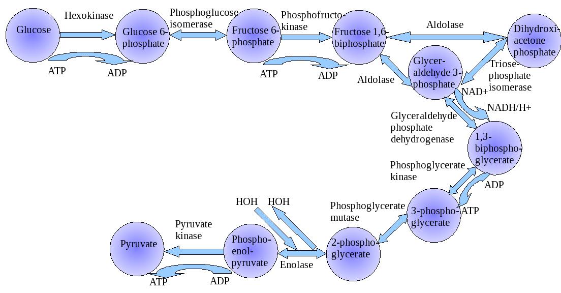
“Warburg Effect” describes the preference of glycolysis and lactic acid fermentation rather than oxidative phosphorylation for energy production in cancer cells. Mitochondrial metabolism is an important and necessary component in the functioning and maintenance of the cell, and accumulating evidence suggests that dysfunction of mitochondrial metabolism plays a role in cancer. Progress has demonstrated the mechanisms of the mitochondrial metabolism-to-glycolysis switch in cancer development and how to target this metabolic switch.


Glycolysis

glycolysis


Otto Heinrich Warburg (1883- )

Otto Warburg

435px-Louis_Pasteur,_foto_av_Félix_Nadar_Crisco_edit

Louis Pasteur


The expression “Pasteur effect” was coined by Warburg when inspired by Pasteur’s findings in yeast cells, when he investigated this metabolic observation (Pasteur effect) in cancer cells. In yeast cells, Pasteur had found that the velocity of sugar used was greatly reduced in presence of oxygen. Not to be confused, in the “Crabtree effect”, the velocity of sugar metabolism was greatly increased, a reversal, when yeast cells were transferred from the aerobic to an anaerobic condition. Thus, the velocity of sugar metabolism of yeast cells was shown to be under metabolic regulatory control in response to change in environmental oxygen conditions in growth. Warburg had to verify whether cancer cells and tissue related normal mammalian cells also have a similar control mechanism. He found that this control was also found in normal cells studied, but was absent in cancer cells. Strikingly, cancer cells continue to have higher anaerobic gycolysis despite the presence of oxygen in their culture media (See Footnote 3).

Taking this a step further, food is digested and supplied to cells In vertebrates mainly in the form of glucose, which is metabolized producing Adenosine Triphosphate (ATP) by two pathways. Glycolysis, occurs via anaerobic metabolism in the cytoplasm, and is of major significance for making ATP quickly, but in a minuscule amount (2 molecules).  In the presence of oxygen, the breakdown process continues in the mitochondria via the Krebs’s cycle coupled with oxidative phosphorylation, which is more efficient for ATP production (36 molecules). Cancer cells seem to depend on glycolysis. In the 1920s, Otto Warburg first proposed that cancer cells show increased levels of glucose consumption and lactate fermentation even in the presence of ample oxygen (known as “Warburg Effect”). Based on this theory, oxidative phosphorylation switches to glycolysis which promotes the proliferation of cancer cells. Many studies have demonstrated glycolysis as the main metabolic pathway in cancer cells.

Albert Szent Gyogy (Warburg’s student) and Otto Meyerhof both studied striated skeletal muscle metabolism invertebrates, and they found those changes observed in yeast by Pasteur. The description of the anaerobic pathway was largely credited to Emden and Meyerhof. Whenever there is increase in muscle work, energy need is above what can be provided by blood supply, the cell metabolism changes from aerobic (where  Acetyl CoA  provides the chemical energy for aerobic production of ATP) to anaerobic metabolism of glucose. In this condition, glucose is obtained directly from its muscle glycogen stores (not from hepatic glycogenolysis).  This is the sole source of chemical energy that is independent of oxygen supplied to the cell. It is a physiological change on muscle metabolism that favors autonomy. It does not depend upon the blood oxygen for aerobic metabolim or blood sources of carbon metabolites borne out from adipose tissue (free fatty acids) or muscle proteins (branched chain amino acids), or vascular delivery of glucose. On that condition, the muscle can perform contraction by its internal source of ATP and uses conversion of pyruvate to lactate in order to regenerate much-needed NAD (by hydride transfer from pyruvate) as a replacement for this mitochondrial function. This regulatory change, keeps glycolysis going at fast rate in order to meet ATP needs of the cell under low yield condition (only two or three ATP for each glucose converted into two lactate molecules). Therefore, it cannot last for long periods of time. This regulatory metabolic change is made in seconds, minutes and therefore happens with the proteins that are already presented in the cell. It does not requires the effect of transcription factors and/or changes in gene expression (See Footnote 1, 2).

In other types mammalian cells, like those from the lens of the eye (86% gycolysis + pentose shunt),  and red blood cells (RBC)[both lacking mitochondria], and also in the deep medullary layer of the kidneys, for lack of mitochondria in the first two cases and normally reduced blood perfusion in the third – A condition required for the counter current mechanism and our ability to concentrate urine also have, permanent higher anaerobic metabolism. In the case of RBC, it includes the ability to produce in a shunt of glycolytic pathway 2,3 diphospho- glycerate that is required to place the hemogloblin macromolecule in an unstable equilibrium between its two forms (R and T – Here presented as simplified accordingly to the model of Monod, Wyman and Changeux. The final model would be even much complex (see for instance, H-W and K review Nature 2007 vol 450: p 964-972 )

Any tissue under a condition of ischemia that is required for some medical procedures (open heart surgery, organ transplants, etc) displays this fast regulatory mechanism (See Footnote 1, 2). A display of these regulatory metabolic changes can be seen in: Cardioplegia: the protection of the myocardium during open heart surgery: a review. D. J. Hearse J. Physiol., Paris, 1980, 76, 751-756 (Fig 1).  The following points are made:

1-       It is a fast regulatory response. Therefore, no genetic mechanism can be taken into account.

2-       It moves from a reversible to an irreversible condition, while the cells are still alive. Death can be seen at the bottom end of the arrow. Therefore, it cannot be reconciled with some of the molecular biology assumptions:

A-       The gene and genes reside inside the heart muscle cells but, in order to preserve intact, the source of coded genetic information that the cell reads and transcribes, DNA must be kept to a minimal of chemical reactivity.

B-       In case sequence determines conformation, activity and function , elevated potassium blood levels could not cause cardiac arrest.

In comparison with those conditions here presented, cancer cells keep the two metabolic options for glucose metabolism at the same time. These cells can use glucose that our body provides to them or adopt temporarily, an independent metabolic form without the usual normal requirement of oxygen (one or another form for ATP generation).  ATP generation is here, an over-simplification of the metabolic status since the carbon flow for building blocks must also be considered and in this case oxidative metabolism of glucose in cancer cells may be viewed as a rich source of organic molecules or building blocks that dividing cells always need.

JES Roselino has conjectured that “most of the Krebs cycle reaction works as ideal reversible thermodynamic systems that can supply any organic molecule that by its absence could prevent cell duplication.” In the vision of Warburg, cancer cells have a defect in Pasteur-effect metabolic control. In case it was functioning normally, it will indicate which metabolic form of glucose metabolism is adequate for each condition. What more? Cancer cells lack differentiated cell function. Any role for transcription factors must be considered as the role of factors that led to the stable phenotypic change of cancer cells. The failure of Pasteur effect must be searched for among the fast regulatory mechanisms that aren’t dependent on gene expression (See Footnote 3).

Extending the thoughts of JES Roselino (Hepatology 1992;16: 1055-1060), reduced blood flow caused by increased hydrostatic pressure in extrahepatic cholestasis decreases mitochondrial function (quoted in Hepatology) and as part of Pasteur effect normal response, increased glycolysis in partial and/or functional anaerobiosis and therefore blocks the gluconeogenic activity of hepatocytes that requires inhibited glycolysis. In this case, a clear energetic link can be perceived between the reduced energetic supply and the ability to perform differentiated hepatic function (gluconeogenesis). In cancer cells, the action of transcription factors that can be viewed as different ensembles of kaleidoscopic pieces (with changing activities as cell conditions change) are clearly linked to the new stable phenotype. In relation to extrahepatic cholestasis mentioned above it must be reckoned that in case a persistent chronic condition is studied a secondary cirrhosis is installed as an example of persistent stable condition, difficult to be reversed and without the requirement for a genetic mutation. (See Footnote 4).

 The Rejection of Complexity

Most of our reasoning about genes was derived from scientific work in microorganisms. These works have provided great advances in biochemistry.

250px-DNA_labeled DNA diagram showing base pairing

double helix


hgp_hubris_220x288_72 genome cartoon

Dna triplex pic

Triple helix


formation of a triplex DNA structure

formation of triple helix


1-      The “Gelehrter idea”: No matter what you are doing you will always be better off, in case you have a gene (In chapter 7 Principles of Medical Genetics Gelehrter and Collins Williams & Wilkins 1990).

2-      The idea that everything could be found following one gene one enzyme relationship that works fine for our understanding of the metabolism, in all biological problems.

3-      The idea that everything that explains biochemistry in microorganisms explains also for every living being (J Nirenberg).

4-      The idea that biochemistry may not require that time should be also taken into account. Time must be considered only for genetic and biological evolution studies (S Luria. In Life- The unfinished experiment 1977 C Scribner´s sons NY).

5-      Finally, the idea that everything in biology, could be found in the genome. Since all information in biology goes from DNA through RNA to proteins. Alternatively, are in the DNA, in case the strict line that includes RNA is not included.

This last point can be accepted in case it is considered that ALL GENETIC information is in our DNA. Genetics as part of life and not as its total expression.

For example, when our body is informed that the ambient temperature is too low or alternatively is too high, our body is receiving an information that arrives from our environment. This external information will affect our proteins and eventually, in case of longer periods in a new condition will cause adaptive response that may include conformational changes in transcription factors (proteins) that will also, produce new readings on the DNA. However, it is an information that moves from outside, to proteins and not from DNA to proteins. The last pathway, when transcription factors change its conformation and change DNA reading will follow the dogmatic view as an adaptive response (See Footnotes 1-3).

However, in case, time is taken into account, the first reactions against cold or warmer temperatures will be the ones that happen through change in protein conformation, activities and function before any change in gene expression can be noticed at protein level. These fast changes, in seconds, minutes cannot be explained by changes in gene expression and are strongly linked to what is needed for the maintenance of life.

“It is possible”, says Roselino, “desirable, to explain all these fast biochemical responses to changes in a living being condition as the sound foundation of medical practices without a single mention to DNA. In case a failure in any mechanism necessary to life is found to be genetic in its origin, the genome in context with with this huge set of transcription factors must be taken into account. This is the biochemical line of reasoning that I have learned with Houssay and Leloir. It would be an honor to see it restored in modern terms.”

More on the Mechanism of Metabolic Control

It was important that genomics would play such a large role in medical research for the last 70 years. There is also good reason to rethink the objections of the Nobelists James Watson and Randy Schekman in the past year, whatever discomfort it brings.  Molecular biology has become a tautology, and as a result deranged scientific rigor inside biology.

Crick & Watson with their DNA model, 1953

Eatson and Crick

Randy-Schekman Berkeley

Randy-Schekman Berkeley


According to JES Roselino, “consider that glycolysis is oscillatory thanks to the kinetic behavior of Phosphofructokinase. Further, by its effect upon Pyruvate kinase through Fructose 1,6 diphosphate oscillatory levels, the inhibition of gluconeogenesis is also oscillatory. When the carbon flow through glycolysis is led to a maximal level gluconeogenesis will be almost completely blocked. The reversal of the Pyruvate kinase step in liver requires two enzymes (Pyruvate carboxylase (maintenance of oxaloacetic levels) + phosphoenolpyruvate carboxykinase (E.C. 4.1.1.32)) and energy requiring reactions that most likely could not as an ensemble, have a fast enough response against pyruvate kinase short period of inhibition during high frequency oscillatory periods of glycolytic flow. Only when glycolysis oscillates at low frequency the opposite reaction could enable gluconeogenic carbon flow.”

In case it can be shown in a rather convincing way, the same reasoning could be applied to understand how simple replicative signals inducing Go to G1 transition in cells, could easily overcome more complex signals required for cell differentiation and differentiated function.

Perhaps the problem of overextension of the equivalence of the DNA and what happens to the organism is also related to the initial reliance on a single cell model to relieve the complexity (which isn’t fully the case).

For instance, consider this fragment:
“Until only recently it was assumed that all proteins take on a clearly defined three-dimensional structure – i.e. they fold in order to be able to assume these functions.”
Cold Spring Harbour Symp. Quant. Biol. 1973  p 187-193 J.C Seidel and J Gergely – Investigation of conformational changes in Spin-Labeled Myosin Model for muscle contraction:
Huxley, A. F. 1971 Proc. Roy. Soc (London) (B) 178:1
Huxley, A.F and R. M. Simmons,1971. Nature 233:633
J.C Haselgrove X ray Evidence for a conformational Change in the Actin-containing filaments…Cold Spring Harbour Symp Quant Biol.1972 v 37: p 341-352

Only a very small sample indicating otherwise. Proteins were held as interacting macromolecules, changing their conformation in regulatory response to changes in the microenvironment (See Footnote 2). DNA was the opposite, non-interacting macromolecules to be as stable as a library must be.

The dogma held that the property of proteins could be read in DNA alone. Consequenly, the few examples quoted above, must be ignored and all people must believe that DNA alone, without environmental factors roles, controls protein amino acid sequence (OK), conformation (not true), activity (not true) and function (not true).

It appeared naively to be correct from the dogma to conclude from interpreting your genome: You have a 50% increased risk of developing the following disease (deterministic statement).  The correct form must be: You belong to a population that has a 50% increase in the risk of….followed by –  what you must do to avoid increase in your personal risk and the care you should take in case you want to have longer healthy life.  Thus, genetics and non-genetic diseases were treated as the same and medical foundations were reinforced by magical considerations (dogmas) in a very profitable way for those involved besides the patient.

 Footnotes:

  1. There is a link of electricity with ions in biology and the oscillatory behavior of some electrical discharges.  In addition, the oscillatory form of electrical discharged may have allowed Planck to relate high energy content with higher frequencies and conversely, low energy content in low frequency oscillatory events.  One may think of high density as an indication of great amount of matter inside a volume in space.  This helps the understanding of Planck’s idea as a high-density-energy in time for a high frequency phenomenon.
  1. Take into account a protein that may have its conformation restricted by an S-S bridge. This protein also, may move to another more flexible conformation in case it is in HS HS condition when the S-S bridge is broken. Consider also that, it takes some time for a protein to move from one conformation for instance, the restricted conformation (S-S) to other conformations. Also, it takes a few seconds or minutes to return to the S-S conformation (This is the Daniel Koshland´s concept of induced fit and relaxation time used by him in order to explain allosteric behavior of monomeric proteins- Monod, Wyman and Changeux requires tetramer or at least, dimer proteins).
  1. In case you have glycolysis oscillating in a frequency much higher than the relaxation time you could lead to the prevalence of high NADH effect leading to high HS /HS condition and at low glycolytic frequency, you could have predominance of S-S condition affecting protein conformation. In case you have predominance of NAD effect upon protein S-S you would get the opposite results.  The enormous effort to display the effect of citrate and over Phosphofructokinase conformation was made by others. Take into account that ATP action as an inhibitor in this case, is a rather unusual one. It is a substrate of the reaction, and together with its action as activator  F1,6 P (or its equivalent F2,6 P) is also unusual. However, it explains oscillatory behaviour of glycolysis. (Goldhammer , A.R, and Paradies: PFK structure and function, Curr. Top Cell Reg 1979; 15:109-141).
  1. The results presented in our Hepatology work must be viewed in the following way: In case the hepatic (oxygenated) blood flow is preserved, the bile secretory cells of liver receive well-oxygenated blood flow (the arterial branches bath secretory cells while the branches originated from portal vein irrigate the hepatocytes.  During extra hepatic cholestasis the low pressure, portal blood flow is reduced and the hepatocytes do not receive enough oxygen required to produce ATP that gluconeogenesis demands. Hepatic artery do not replace this flow since, its branches only join portal blood fluxes after the previous artery pressure  is reduced to a low pressure venous blood – at the point where the formation of hepatic vein is. Otherwise, the flow in the portal vein would be reversed or, from liver to the intestine. It is of no help to take into account possible valves for this reasoning since minimal arterial pressure is well above maximal venous pressure and this difference would keep this valve in permanent close condition. In low portal blood flow condition, the hepatocyte increases pyruvate kinase activity and with increased pyruvate kinase activity Gluconeogenesis is forbidden (See Walsh & Cooper revision quoted in the Hepatology as ref 23). For the hemodynamic considerations, role of artery and veins in hepatic portal system see references 44 and 45 Rappaport and Schneiderman and Rappapaport.


 Appendix I.

metabolic pathways

metabolic pathways

Signals Upstream and Targets Downstream of Lin28 in the Lin28 Pathway

Signals Upstream and Targets Downstream of Lin28 in the Lin28 Pathway


1.  Functional Proteomics Adds to Our Understanding

Ben Schuler’s research group from the Institute of Biochemistry of the University of Zurich has now established that an increase in temperature leads to folded proteins collapsing and becoming smaller. Other environmental factors can trigger the same effect. The crowded environments inside cells lead to the proteins shrinking. As these proteins interact with other molecules in the body and bring other proteins together, understanding of these processes is essential “as they play a major role in many processes in our body, for instance in the onset of cancer”, comments study coordinator Ben Schuler.

Measurements using the “molecular ruler”

“The fact that unfolded proteins shrink at higher temperatures is an indication that cell water does indeed play an important role as to the spatial organisation eventually adopted by the molecules”, comments Schuler with regard to the impact of temperature on protein structure. For their studies the biophysicists use what is known as single-molecule spectroscopy. Small colour probes in the protein enable the observation of changes with an accuracy of more than one millionth of a millimetre. With this “molecular yardstick” it is possible to measure how molecular forces impact protein structure.

With computer simulations the researchers have mimicked the behaviour of disordered proteins. They want to use them in future for more accurate predictions of their properties and functions.

Correcting test tube results

That’s why it’s important, according to Schuler, to monitor the proteins not only in the test tube but also in the organism. “This takes into account the fact that it is very crowded on the molecular level in our body as enormous numbers of biomolecules are crammed into a very small space in our cells”, says Schuler. The biochemists have mimicked this “molecular crowding” and observed that in this environment disordered proteins shrink, too.

Given these results many experiments may have to be revisited as the spatial organisation of the molecules in the organism could differ considerably from that in the test tube according to the biochemist from the University of Zurich. “We have, therefore, developed a theoretical analytical method to predict the effects of molecular crowding.” In a next step the researchers plan to apply these findings to measurements taken directly in living cells.

Explore further: Designer proteins provide new information about the body’s signal processesMore information: Andrea Soranno, Iwo Koenig, Madeleine B. Borgia, Hagen Hofmann, Franziska Zosel, Daniel Nettels, and Benjamin Schuler. Single-molecule spectroscopy reveals polymer effects of disordered proteins in crowded environments. PNAS, March 2014. DOI: 10.1073/pnas.1322611111


Effects of Hypoxia on Metabolic Flux

  1. Glucose-6-phosphate dehydrogenase regulation in the hepatopancreas of the anoxia-tolerantmarinemollusc, Littorina littorea

JL Lama , RAV Bell and KB Storey

Glucose-6-phosphate dehydrogenase (G6PDH) gates flux through the pentose phosphate pathway and is key to cellular antioxidant defense due to its role in producing NADPH. Good antioxidant defenses are crucial for anoxia-tolerant organisms that experience wide variations in oxygen availability. The marine mollusc, Littorina littorea, is an intertidal snail that experiences daily bouts of anoxia/hypoxia with the tide cycle and shows multiple metabolic and enzymatic adaptations that support anaerobiosis. This study investigated the kinetic, physical and regulatory properties of G6PDH from hepatopancreas of L. littorea to determine if the enzyme is differentially regulated in response to anoxia, thereby providing altered pentose phosphate pathway functionality under oxygen stress conditions.

Several kinetic properties of G6PDH differed significantly between aerobic and 24 h anoxic conditions; compared with the aerobic state, anoxic G6PDH (assayed at pH 8) showed a 38% decrease in K G6P and enhanced inhibition by urea, whereas in pH 6 assays Km NADP and maximal activity changed significantly.

All these data indicated that the aerobic and anoxic forms of G6PDH were the high and low phosphate forms, respectively, and that phosphorylation state was modulated in response to selected endogenous protein kinases (PKA or PKG) and protein phosphatases (PP1 or PP2C). Anoxia-induced changes in the phosphorylation state of G6PDH may facilitate sustained or increased production of NADPH to enhance antioxidant defense during long term anaerobiosis and/or during the transition back to aerobic conditions when the reintroduction of oxygen causes a rapid increase in oxidative stress.

Lama et al.  Peer J 2013.   http://dx.doi.org/10.7717/peerj.21


  1. Structural Basis for Isoform-Selective Inhibition in Nitric Oxide Synthase

    TL. Poulos and H Li

In the cardiovascular system, the important signaling molecule nitric oxide synthase (NOS) converts L-arginine into L-citrulline and releases nitric oxide (NO). NO produced by endothelial NOS (eNOS) relaxes smooth muscle which controls vascular tone and blood pressure. Neuronal NOS (nNOS) produces NO in the brain, where it influences a variety of neural functions such as neural transmitter release. NO can also support the immune system, serving as a cytotoxic agent during infections. Even with all of these important functions, NO is a free radical and, when overproduced, it can cause tissue damage. This mechanism can operate in many neurodegenerative diseases, and as a result the development of drugs targeting nNOS is a desirable therapeutic goal.

However, the active sites of all three human isoforms are very similar, and designing inhibitors specific for nNOS is a challenging problem. It is critically important, for example, not to inhibit eNOS owing to its central role in controlling blood pressure. In this Account, we summarize our efforts in collaboration with Rick Silverman at Northwestern University to develop drug candidates that specifically target NOS using crystallography, computational chemistry, and organic synthesis. As a result, we have developed aminopyridine compounds that are 3800-fold more selective for nNOS than eNOS, some of which show excellent neuroprotective effects in animal models. Our group has solved approximately 130 NOS-inhibitor crystal structures which have provided the structural basis for our design efforts. Initial crystal structures of nNOS and eNOS bound to selective dipeptide inhibitors showed that a single amino acid difference (Asp in nNOS and Asn in eNOS) results in much tighter binding to nNOS. The NOS active site is open and rigid, which produces few large structural changes when inhibitors bind. However, we have found that relatively small changes in the active site and inhibitor chirality can account for large differences in isoform-selectivity. For example, we expected that the aminopyridine group on our inhibitors would form a hydrogen bond with a conserved Glu inside the NOS active site. Instead, in one group of inhibitors, the aminopyridine group extends outside of the active site where it interacts with a heme propionate. For this orientation to occur, a conserved Tyr side chain must swing out of the way. This unanticipated observation taught us about the importance of inhibitor chirality and active site dynamics. We also successfully used computational methods to gain insights into the contribution of the state of protonation of the inhibitors to their selectivity. Employing the lessons learned from the aminopyridine inhibitors, the Silverman lab designed and synthesized symmetric double-headed inhibitors with an aminopyridine at each end, taking advantage of their ability to make contacts both inside and outside of the active site. Crystal structures provided yet another unexpected surprise. Two of the double-headed inhibitor molecules bound to each enzyme subunit, and one molecule participated in the generation of a novel Zn site that required some side chains to adopt alternate conformations. Therefore, in addition to achieving our specific goal, the development of nNOS selective compounds, we have learned how subtle differences in and structure can control proteinligand interactions and often in unexpected ways.


300px-Nitric_Oxide_Synthase

Nitric oxide synthase

arginine-NO-citulline cycle

arginine-NO-citulline cycle

active site of eNOS (PDB_1P6L) and nNOS (PDB_1P6H).

active site of eNOS (PDB_1P6L) and nNOS (PDB_1P6H).


NO - muscle, vasculature, mitochondria

NO – muscle, vasculature, mitochondria


Figure:  (A) Structure of one of the early dipeptide lead compounds, 1, that exhibits excellentisoform selectivity. (B, C) show the crystal structures of the dipeptide inhibitor 1 in the active site of eNOS (PDB: 1P6L) and nNOS (PDB: 1P6H). In nNOS, the inhibitor “curls” which enables the inhibitor R-amino group to interact with both Glu592 and Asp597. In eNOS, Asn368 is the homologue to nNOS Asp597.

Accounts in Chem Res 2013; 46(2): 390-98.

  1. Jamming a Protein Signal

Interfering with a single cancer-promoting protein and its receptor can open this resistance mechanism by initiating autophagy of the affected cells,  according to researchers at The University of Texas MD Anderson Cancer Center  in the journal Cell Reports.  According to Dr. Anil Sood and Yunfei Wen, lead and first authors, blocking  prolactin, a potent growth factor for ovarian cancer, sets off downstream events that result in cell by autophagy, the process  recycles damaged organelles and proteins for new use by the cell through the phagolysozome. This in turn, provides a clinical rationale for blocking prolactin and its receptor to initiate sustained autophagy as an alternative strategy for treating cancers.

Steep reductions in tumor weight

Prolactin (PRL) is a hormone previously implicated in ovarian, endometrial and other cancer development andprogression. When PRL binds to its cell membrane receptor, PRLR, activation of cancer-promoting cell signaling pathways follows.  A variant of normal prolactin called G129R blocks the reaction between prolactin and its receptor. Sood and colleagues treated mice that had two different lines of human ovarian cancer, both expressing the prolactin receptor, with G129R. Tumor weights fell by 50 percent for mice with either type of ovarian cancer after 28 days of treatment with G129R, and adding the taxane-based chemotherapy agent paclitaxel cut tumor weight by 90 percent. They surmise that higher doses of G129R may result in even greater therapeutic benefit.


3D experiments show death by autophagy


[video width=”1280″ height=”720″ mp4=”http://pharmaceuticalintelligence.com/wp-content/uploads/2014/04/1741-7007-11-65-s1-macromolecular-juggling-by-ubiquitylation-enzymes1.mp4″][/video]


Next the team used the prolactin-mimicking peptide to treat cultures of cancer spheroids which sharply reduced their numbers, and blocked the activation of JAK2 and STAT signaling pathways.

Protein analysis of the treated spheroids showed increased presence of autophagy factors and genomic analysis revealed increased expression of a number of genes involved in autophagy progression and cell death.  Then a series of experiments using fluorescence and electron microscopy showed that the cytosol of treated cells had large numbers of cavities caused by autophagy.

The team also connected the G129R-induced autophagy to the activity of PEA-15, a known cancer inhibitor. Analysis of tumor samples from 32 ovarian cancer patients showed that tumors express higher levels of the prolactin receptor and lower levels of phosphorylated PEA-15 than normal ovarian tissue. However, patients with low levels of the prolactin receptor and higher PEA-15 had longer overall survival than those with high PRLR and low PEA-15.

Source: MD Anderson Cancer Center


  1. Chemists’ Work with Small Peptide Chains of Enzymes

Korendovych and his team designed seven simple peptides, each containing seven amino acids. They then allowed the molecules of each peptide to self-assemble, or spontaneously clump together, to form amyloids. (Zinc, a metal with catalytic properties, was introduced to speed up the reaction.) What they found was that four of the seven peptides catalyzed the hydrolysis of molecules known as esters, compounds that react with water to produce water and acids—a feat not uncommon among certain enzymes.

“It was the first time that a peptide this small self-assembled to produce an enzyme-like catalyst,” says Korendovych. “Each enzyme has to be an exact fit for its respective substrate,” he says, referring to the molecule with which an enzyme reacts. “Even after millions of years, nature is still testing all the possible combinations of enzymes to determine which ones can catalyze metabolic reactions. Our results make an argument for the design of self-assembling nanostructured catalysts.”

Source: Syracuse University

Here are three articles emphasizing the value of combinatorial analysis, which can be formed from genomic, clinical, and proteomic data sets.


  1. Comparative analysis of differential network modularity in tissue specific normal and cancer protein interaction networks

    F Islam , M Hoque , RS Banik , S Roy , SS Sumi, et al.

As most biological networks show modular properties, the analysis of differential modularity between normal and cancer protein interaction networks can be a good way to understand cancer more significantly. Two aspects of biological network modularity e.g. detection of molecular complexes (potential modules or clusters) and identification of crucial nodes forming the overlapping modules have been considered in this regard.

The computational analysis of previously published protein interaction networks (PINs) has been conducted to identify the molecular complexes and crucial nodes of the networks. Protein molecules involved in ten major cancer signal transduction pathways were used to construct the networks based on expression data of five tissues e.g. bone, breast, colon, kidney and liver in both normal and cancer conditions.

Cancer PINs show higher level of clustering (formation of molecular complexes) than the normal ones. In contrast, lower level modular overlapping is found in cancer PINs than the normal ones. Thus a proposition can be made regarding the formation of some giant nodes in the cancer networks with very high degree and resulting in reduced overlapping among the network modules though the predicted molecular complex numbers are higher in cancer conditions.

Islam et al. Journal of Clinical Bioinformatics 2013, 3:19-32

  1. A new 12-gene diagnostic biomarker signature of melanoma revealed by integrated microarray analysis

    Wanting Liu , Yonghong Peng and Desmond J. Tobin
    PeerJ 1:e49;        http://dx.doi.org/10.7717/peerj.49

Here we present an integrated microarray analysis framework, based on a genome-wide relative significance (GWRS) and genome-wide global significance (GWGS) model. When applied to five microarray datasets on melanoma published between 2000 and 2011, this method revealed a new signature of 200 genes. When these were linked to so-called ‘melanoma driver’ genes involved in MAPK, Ca2+, and WNT signaling pathways we were able to produce a new 12-gene diagnostic biomarker signature for melanoma (i.e., EGFR, FGFR2, FGFR3, IL8, PTPRF, TNC, CXCL13, COL11A1, CHP2, SHC4, PPP2R2C, andWNT4).We have begun to experimentally validate a subset of these genes involved inMAPK signaling at the protein level, including CXCL13, COL11A1, PTPRF and SHC4 and found these to be overexpressed inmetastatic and primarymelanoma cells in vitro and in situ compared to melanocytes cultured from healthy skin epidermis and normal healthy human skin.


catalytic amyloid forming particle

catalytic amyloid forming particle


        8.    PanelomiX: A threshold-based algorithm to create panels of biomarkers

X Robin , N Turck , A Hainard , N Tiberti, et al.
               Translational Proteomics 2013.    http://dx.doi.org/10.1016/j.trprot.2013.04.003

The PanelomiX toolbox combines biomarkers and evaluates the performance of panels to classify patients better than singlemarkers or other classifiers. The ICBTalgorithm proved to be an efficient classifier, the results of which can easily be interpreted.

Here are two current examples of the immense role played by signaling pathways in carcinogenic mechanisms and in treatment targeting, which is also confounded by acquired resistance.


  1. Triple-Negative Breast Cancer

  1. epidermal growth factor receptor (EGFR or ErbB1) and
  2. high activity of the phosphatidylinositol 3-kinase (PI3K)–Akt pathway

are both targeted in triple-negative breast cancer (TNBC).

  • activation of another EGFR family member [human epidermal growth factor receptor 3 (HER3) (or ErbB3)] may limit the antitumor effects of these drugs.

This study found that TNBC cell lines cultured with the EGFR or HER3 ligand EGF or heregulin, respectively, and treated with either an Akt inhibitor (GDC-0068) or a PI3K inhibitor (GDC-0941) had increased abundance and phosphorylation of HER3.

The phosphorylation of HER3 and EGFR in response to these treatments

  1. was reduced by the addition of a dual EGFR and HER3 inhibitor (MEHD7945A).
  2. MEHD7945A also decreased the phosphorylation (and activation) of EGFR and HER3 and
  3. the phosphorylation of downstream targets that occurred in response to the combination of EGFR ligands and PI3K-Akt pathway inhibitors.

In culture, inhibition of the PI3K-Akt pathway combined with either MEHD7945A or knockdown of HER3

  1. decreased cell proliferation compared with inhibition of the PI3K-Akt pathway alone.
  2. Combining either GDC-0068 or GDC-0941 with MEHD7945A inhibited the growth of xenografts derived from TNBC cell lines or from TNBC patient tumors, and
  3. this combination treatment was also more effective than combining either GDC-0068 or GDC-0941 with cetuximab, an EGFR-targeted antibody.
  4. After therapy with EGFR-targeted antibodies, some patients had residual tumors with increased HER3 abundance and EGFR/HER3 dimerization (an activating interaction).

Thus, we propose that concomitant blockade of EGFR, HER3, and the PI3K-Akt pathway in TNBC should be investigated in the clinical setting.

Reference: Antagonism of EGFR and HER3 Enhances the Response to Inhibitors of the PI3K-Akt Pathway in Triple-Negative Breast Cancer. JJ Tao, P Castel, N Radosevic-Robin, M Elkabets, et al.  Sci. Signal., 25 March 2014;
7(318), p. ra29   http://dx.doi.org/10.1126/scisignal.2005125


                  10.   Metastasis in RAS Mutant or Inhibitor-Resistant Melanoma Cells

The protein kinase BRAF is mutated in about 40% of melanomas, and BRAF inhibitors improve progression-free and overall survival in these patients. However, after a relatively short period of disease control, most patients develop resistance because of reactivation of the RAF–ERK (extracellular signal–regulated kinase) pathway, mediated in many cases by mutations in RAS. We found that BRAF inhibition induces invasion and metastasis in RAS mutant melanoma cells through a mechanism mediated by the reactivation of the MEK (mitogen-activated protein kinase kinase)–ERK pathway.

Reference: BRAF Inhibitors Induce Metastasis in RAS Mutant or Inhibitor-Resistant Melanoma Cells by Reactivating MEK and ERK Signaling. B Sanchez-Laorden, A Viros, MR Girotti, M Pedersen, G Saturno, et al., Sci. Signal., 25 March 2014;  7(318), p. ra30  http://dx.doi.org/10.1126/scisignal.2004815

Appendix II.

The world of physics in the twentieth century saw the end of determinism established by Newton. This is characterized by discrete laws that describe natural observations. These are in gravity and in eletricity. In an early phase of investigation, an era of galvanic or voltaic electricity represented a revolutionary break from the historical focus on frictional electricity. Alessandro Voltadiscovered that chemical reactions could be used to create positively charged anodes and negatively charged cathodes.  In 1790, Prof. Luigi Alyisio Galvani of Bologna, while conducting experiments on “animal electricity“, noticed the twitching of a frog’s legs in the presence of an electric machine. He observed that a frog’s muscle, suspended on an iron balustrade by a copper hook passing through its dorsal column, underwent lively convulsions without any extraneous cause, the electric machine being at this time absent.  Volta communicated a description of his pile to the Royal Society of London and shortly thereafter Nicholson and Cavendish (1780) produced the decomposition of water by means of the electric current, using Volta’s pile as the source of electromotive force.

Siméon Denis Poisson attacked the difficult problem of induced magnetization, and his results provided  a first approximation. His innovation required the application of mathematics to physics.  His memoirs on the theory of electricity and magnetism created a new branch of mathematical physics.  The discovery of electromagnetic induction was made almost simultaneously and independently by Michael Faraday and Joseph Henry. Michael Faraday, the successor of Humphry Davy, began his epoch-making research relating to electric and electromagnetic induction in 1831. In his investigations of the peculiar manner in which iron filings arrange themselves on a cardboard or glass in proximity to the poles of a magnet, Faraday conceived the idea of magnetic “lines of force” extending from pole to pole of the magnet and along which the filings tend to place themselves. On the discovery being made that magnetic effects accompany the passage of an electric current in a wire, it was also assumed that similar magnetic lines of force whirled around the wire. He also posited that iron, nickel, cobalt, manganese, chromium, etc., are paramagnetic (attracted by magnetism), whilst other substances, such as bismuth, phosphorus, antimony, zinc, etc., are repelled by magnetism or are diamagnetic.

Around the mid-19th century, Fleeming Jenkin‘s work on ‘ Electricity and Magnetism ‘ and Clerk Maxwell’s ‘ Treatise on Electricity and Magnetism ‘ were published. About 1850 Kirchhoff published his laws relating to branched or divided circuits. He also showed mathematically that according to the then prevailing electrodynamic theory, electricity would be propagated along a perfectly conducting wire with the velocity of light. Herman Helmholtz investigated the effects of induction on the strength of a current and deduced mathematical equations, which experiment confirmed. In 1853 Sir William Thomson (later Lord Kelvin) predicted as a result of mathematical calculations the oscillatory nature of the electric discharge of a condenser circuit.  Joseph Henry, in 1842 discerned  the oscillatory nature of the Leyden jardischarge.

In 1864 James Clerk Maxwell announced his electromagnetic theory of light, which was perhaps the greatest single step in the world’s knowledge of electricity. Maxwell had studied and commented on the field of electricity and magnetism as early as 1855/6 when On Faraday’s lines of force was read to the Cambridge Philosophical Society. The paper presented a simplified model of Faraday’s work, and how the two phenomena were related. He reduced all of the current knowledge into a linked set of differential equations with 20 equations in 20 variables. This work was later published as On Physical Lines of Force in1861. In order to determine the force which is acting on any part of the machine we must find its momentum, and then calculate the rate at which this momentum is being changed. This rate of change will give us the force. The method of calculation which it is necessary to employ was first given by Lagrange, and afterwards developed, with some modifications, by Hamilton’s equations. Now Maxwell logically showed how these methods of calculation could be applied to the electro-magnetic field. The energy of a dynamical systemis partly kinetic, partly potential. Maxwell supposes that the magnetic energy of the field is kinetic energy, the electric energy potential.  Around 1862, while lecturing at King’s College, Maxwell calculated that the speed of propagation of an electromagnetic field is approximately that of the speed of light.   Maxwell’s electromagnetic theory of light obviously involved the existence of electric waves in free space, and his followers set themselves the task of experimentally demonstrating the truth of the theory. By 1871, he presented the Remarks on the mathematical classification of physical quantities.

A Wave-Particle Dilemma at the Century End

In 1896 J.J. Thomson performed experiments indicating that cathode rays really were particles, found an accurate value for their charge-to-mass ratio e/m, and found that e/m was independent of cathode material. He made good estimates of both the charge e and the mass m, finding that cathode ray particles, which he called “corpuscles”, had perhaps one thousandth of the mass of the least massive ion known (hydrogen). He further showed that the negatively charged particles produced by radioactive materials, by heated materials, and by illuminated materials, were universal.  In the late 19th century, the Michelson–Morley experiment was performed by Albert Michelson and Edward Morley at what is now Case Western Reserve University. It is generally considered to be the evidence against the theory of a luminiferous aether. The experiment has also been referred to as “the kicking-off point for the theoretical aspects of the Second Scientific Revolution.” Primarily for this work, Albert Michelson was awarded theNobel Prize in 1907.

Wave–particle duality is a theory that proposes that all matter exhibits the properties of not only particles, which have mass, but also waves, which transfer energy. A central concept of quantum mechanics, this duality addresses the inability of classical concepts like “particle” and “wave” to fully describe the behavior of quantum-scale objects. Standard interpretations of quantum mechanics explain this paradox as a fundamental property of the universe, while alternative interpretations explain the duality as an emergent, second-order consequence of various limitations of the observer. This treatment focuses on explaining the behavior from the perspective of the widely used Copenhagen interpretation, in which wave–particle duality serves as one aspect of the concept of complementarity, that one can view phenomena in one way or in another, but not both simultaneously.  Through the work of Max PlanckAlbert EinsteinLouis de BroglieArthur Compton, Niels Bohr, and many others, current scientific theory holds that all particles also have a wave nature (and vice versa).

Beginning in 1670 and progressing over three decades, Isaac Newton argued that the perfectly straight lines of reflection demonstrated light’s particle nature, but Newton’s contemporaries Robert Hooke and Christiaan Huygens—and later Augustin-Jean Fresnel—mathematically refined the wave viewpoint, showing that if light traveled at different speeds in different, refraction could be easily explained. The resulting Huygens–Fresnel principle was supported by Thomas Young‘s discovery of double-slit interference, the beginning of the end for the particle light camp.  The final blow against corpuscular theory came when James Clerk Maxwell discovered that he could combine four simple equations, along with a slight modification to describe self-propagating waves of oscillating electric and magnetic fields. When the propagation speed of these electromagnetic waves was calculated, the speed of light fell out. While the 19th century had seen the success of the wave theory at describing light, it had also witnessed the rise of the atomic theory at describing matter.

Matter and Light

In 1789, Antoine Lavoisier secured chemistry by introducing rigor and precision into his laboratory techniques. By discovering diatomic gases, Avogadro completed the basic atomic theory, allowing the correct molecular formulae of most known compounds—as well as the correct weights of atoms—to be deduced and categorized in a consistent manner. The final stroke in classical atomic theory came when Dimitri Mendeleev saw an order in recurring chemical properties, and created a table presenting the elements in unprecedented order and symmetry.   Chemistry was now an atomic science.

Black-body radiation, the emission of electromagnetic energy due to an object’s heat, could not be explained from classical arguments alone. The equipartition theorem of classical mechanics, the basis of all classical thermodynamic theories, stated that an object’s energy is partitioned equally among the object’s vibrational modes. This worked well when describing thermal objects, whose vibrational modes were defined as the speeds of their constituent atoms, and the speed distribution derived from egalitarian partitioning of these vibrational modes closely matched experimental results. Speeds much higher than the average speed were suppressed by the fact that kinetic energy is quadratic—doubling the speed requires four times the energy—thus the number of atoms occupying high energy modes (high speeds) quickly drops off. Since light was known to be waves of electromagnetism, physicists hoped to describe this emission via classical laws. This became known as the black body problem. The Rayleigh–Jeans law which, while correctly predicting the intensity of long wavelength emissions, predicted infinite total energy as the intensity diverges to infinity for short wavelengths.

The solution arrived in 1900 when Max Planck hypothesized that the frequency of light emitted by the black body depended on the frequency of the oscillator that emitted it, and the energy of these oscillators increased linearly with frequency (according to his constant h, where E = hν). By demanding that high-frequency light must be emitted by an oscillator of equal frequency, and further requiring that this oscillator occupy higher energy than one of a lesser frequency, Planck avoided any catastrophe; giving an equal partition to high-frequency oscillators produced successively fewer oscillators and less emitted light. And as in the Maxwell–Boltzmann distribution, the low-frequency, low-energy oscillators were suppressed by the onslaught of thermal jiggling from higher energy oscillators, which necessarily increased their energy and frequency. Planck had intentionally created an atomic theory of the black body, but had unintentionally generated an atomic theory of light, where the black body never generates quanta of light at a given frequency with energy less than .

In 1905 Albert Einstein took Planck’s black body model in itself and saw a wonderful solution to another outstanding problem of the day: the photoelectric effect, the phenomenon where electrons are emitted from atoms when they absorb energy from light.   Only by increasing the frequency of the light, and thus increasing the energy of the photons, can one eject electrons with higher energy. Thus, using Planck’s constant h to determine the energy of the photons based upon their frequency, the energy of ejected electrons should also increase linearly with frequency; the gradient of the line being Planck’s constant. These results were not confirmed until 1915, when Robert Andrews Millikan, produced experimental results in perfect accord with Einstein’s predictions. While  the energy of ejected electrons reflected Planck’s constant, the existence of photons was not explicitly proven until the discovery of the photon antibunching effect  When Einstein received his Nobel Prizein 1921, it was  for the photoelectric effect, the suggestion of quantized light. Einstein’s “light quanta” represented the quintessential example of wave–particle duality. Electromagnetic radiation propagates following  linear wave equations, but can only be emitted or absorbed as discrete elements, thus acting as a wave and a particle simultaneously.

Radioactivity Changes the Scientific Landscape

The turn of the century also features radioactivity, which later came to the forefront of the activities of World War II, the Manhattan Project, the discovery of the chain reaction, and later – Hiroshima and Nagasaki.

Marie Curie

Marie Curie


Marie Skłodowska-Curie was a Polish and naturalized-French physicist and chemist who conducted pioneering research on radioactivity. She was the first woman to win a Nobel Prize, the only woman to win in two fields, and the only person to win in multiple sciences. She was also the first woman to become a professor at the University of Paris, and in 1995 became the first woman to be entombed on her own merits in the Panthéon in Paris. She shared the 1903 Nobel Prize in Physics with her husband Pierre Curie and with physicist Henri Becquerel. She won the 1911 Nobel Prize in Chemistry.  Her achievements included a theory of radioactivity (a term that she coined, techniques for isolating radioactive isotopes, and the discovery of polonium and radium. She named the first chemical element that she discovered – polonium, which she first isolated in 1898 – after her native country. Under her direction, the world’s first studies were conducted into the treatment of neoplasms using radioactive isotopes. She founded the Curie Institutes in Paris and in Warsaw, which remain major centres of medical research today. During World War I, she established the first military field radiological centres.  Curie died in 1934 due to aplastic anemia brought on by exposure to radiation – mainly, it seems, during her World War I service in mobile X-ray units created by her.


Read Full Post »

Heroes in Medical Research: Developing Models for Cancer Research

Author, Curator: Stephen J. Williams, Ph.D.


The current rapid progress in cancer research would have never come about if not for the dedication of past researchers who had developed many of the scientific tools we use today. In this issue of Heroes in Medical Research I would like to give tribute to the researchers who had developed the some of the in-vivo and in-vitro models which are critical for cancer research.


The Animal Modelers in Cancer Research

Helen Dean King, Ph.D. (1869-1955)

Helen Dean King

Helen Dean King, Ph.D. from www.ExplorePAhistory.com; photo Courtesy of the Wistar Institute Archive Collection, Philadelphia, PA


The work of Dr. Helen Dean King on rat inbreeding led to development of strains of laboratory animals. Dr. King taught at Bryn Mawr College, then worked at University of Pennsylvania and the Wistar Institute under famed geneticist Thomas Hunt Morgan, researching if inbreeding would produce harmful genetic traits.   At University of Pennsylvania she examined environmental and genetic factors on gender determination.


Important papers include [1-6]as well as the following contributions:

“Studies in Inbreeding”, “Life Processes in Gray Norway Rats During Fourteen Years in Captivity”, doctoral thesis on embryologic development in toads (1899)


Milestones include:


1909    started albino rat breeding and bred 20 female and male from same litter (King colony) to 25

successive generations (inbreeding did not cause harmful traits)


1919     started to domesticate the wild Norwegian rats that ran thru Philadelphia (six pairs Norway rats

thru 28 generations)

A good reference for definitions of rat inbreeding versus line generation including a history of Dr. King’s work can be found at the site: Munificent Mischief Rattery and a brief history here.[7] In addition, Dr. King had investigated using rat strains as a possible recipient for tumor cells. The work was an important advent to the use of immunodeficient models for cancer research.


As shown below Philadelphia became a hotbed for research into embryology, development, genetics, and animal model development.


Beatrice Mintz, Ph.D.

(Beatrice Minz, Ph.D.; photo credit Fox Chase Cancer Center, www.pubweb.fccc.edu) Mintz

Dr. Mintz, an embryologist and cancer researcher from Fox Chase Cancer Center in Philadelphia, PA, contributed some of the most seminal discoveries leading to our current understanding of genetics, embryo development, cellular differentiation, and oncogenesis, especially melanoma, while pioneering techniques which allowed the development of genetically modified mice.

If you get the privilege of hearing her talk, take advantage of it. Dr. Mintz is one of those brilliant scientists who have the ability to look at a clinical problem from the viewpoint of a basic biological question and, at the same time, has the ability to approach the well-thought out questions with equally well thought out experimental design. For example, Dr. Mintz asked if a cell’s developmental fate was affected by location in the embryo. This led to her work by showing teratocarcinoma tumor cells in the developing embryo could revert to a more normal phenotype, essentially proving two important concepts in development and tumor biology:

  1. The existence of pluripotent stem cells
  2. That tumor cells are affected by their environment (which led to future concepts of the importance of tumor microenvironment on tumor growth

Other seminal discoveries included:

  • Development of the first mouse chimeras using novel cell fusion techniques
  • With Rudolf Jaenisch in 1974, showed integration of viral DNA from SV40, could be integrated into the DNA of developing mice and persist into adulthood somatic cells, the first transgenesis in mice which led ultimately to:
  • Development of the first genetically modified mouse model of human melanoma in 1993

Her current work, seen on the faculty webpage here, is developing mice with predisposition to melanoma to uncover risk factors associated with the early development of melanoma.

In keeping with the Philadelphia tradition another major mouse model which became seminal to cancer drug discovery was co-developed in the same city, same institute and described in the next section.

It is interesting to note that the first cloning of an animal, a frog, had taken place at the Institute for Cancer Research, later becoming Fox Chase Cancer Center, which was performed by Drs. Robert Briggs and Thomas J. King and reported in the 152 PNAS paper Transplantation of Living Nuclei From Blastula Cells into Enucleated Frogs’ Eggs.[8]


 The Immunodeficient Animal as a Model System for Cancer Research – Dr. Mel Bosma, Ph.D.


Bosma

Melvin J. Bosma, Ph.D.; photo credit Fox Chase Cancer Center

In the summer of 1980 at Fox Chase Cancer Center, Dr. Melvin J. Bosma and his co-researcher wife Gayle discovered mice with deficiencies in common circulating antibodies and since, these mice were littermates, realized they had found a genetic defect which rendered the mice immunodeficient (upon further investigation these mice were unable to produce mature B and T cells). These mice were the first scid (severe combined immunodeficiency) colony. The scid phenotype was later found to be a result of a spontaneous mutation in the enzyme Prkdc {protein kinase, DNA activated, catalytic polypeptide} involved in DNA repair, and ultimately led to a defect in V(D)J recombination of immunoglobulins.

The emergence of this scid mouse was not only crucial for AIDS research but was another turning point in cancer research , as researchers now had a robust in-vivo recipient for human tumor cells. The orthotopic xenograft of human tumor cells now allowed for studies on genetic and microenvironmental factors affecting tumorigenicity, as well as providing a model for chemotherapeutic drug development (see Suggitt for review and references)[9]. A discussion of the pros and cons of the xenograft system for cancer drug discovery would be too voluminous for this post and would warrant a full review by itself. But before the advent of such scid mouse systems researchers relied on spontaneous and syngeneic mouse tumor models such as the B16 mouse melanoma and Lewis lung tumor model.

Other scid systems have been developed such as in the dog, horse, and pig. Please see the following post on this site The SCID Pig: How Pigs are becoming a Great Alternate Model for Cancer Research. The athymic (nude) mouse (nu/nu) also is a popular immunodeficient mouse model used for cancer research

Two other in-vivo tumor models: Patient Derived Xenografts (PDX) and Genetically Engineered Mouse models (GEM) deserve their own separate discussion however the success of these new models can be attributed to the hard work of the aforementioned investigators. Therefore I will post separately and curate PDX and GEM models of cancer and highlight some new models which are having great impact on cancer drug development.


References

1.         Loeb L, King HD: Transplantation and Individuality Differential in Strains of Inbred Rats. The American journal of pathology 1927, 3(2):143-167.

2.         Lewis MR, Aptekman PM, King HD: Retarding action of adrenal gland on growth of sarcoma grafts in rats. J Immunol 1949, 61(4):315-319.

3.         Aptekman PM, Lewis MR, King HD: Tumor-immunity induced in rats by subcutaneous injection of tumor extract. J Immunol 1949, 63(4):435-440.

4.         Lewis MR, Aptekman PM, King HD: Inactivation of malignant tissue in tumor-immune rats. J Immunol 1949, 61(4):321-326.

5.         Lewis MR, King HD, et al.: Further studies on oncolysis and tumor immunity in rats. J Immunol 1948, 60(4):517-528.

6.         Aptekman PM, Lewis MR, King HD: A method of producing in inbred albino rats a high percentage of immunity from tumors native in their strain. J Immunol 1946, 52:77-86.

7.         Ogilvie MB: Inbreeding, eugenics, and Helen Dean King (1869-1955). Journal of the history of biology 2007, 40(3):467-507.

8.         Briggs R, King TJ: Transplantation of Living Nuclei From Blastula Cells into Enucleated Frogs’ Eggs. Proceedings of the National Academy of Sciences of the United States of America 1952, 38(5):455-463.

9.         Suggitt M, Bibby MC: 50 years of preclinical anticancer drug screening: empirical to target-driven approaches. Clinical cancer research : an official journal of the American Association for Cancer Research 2005, 11(3):971-981.


Other posts on this site about Cancer, Animal Models of Disease, and other articles in this series include:

The SCID Pig: How Pigs are becoming a Great Alternate Model for Cancer Research

A Synthesis of the Beauty and Complexity of How We View Cancer

Guidelines for the welfare and use of animals in cancer research

Importance of Funding Replication Studies: NIH on Credibility of Basic Biomedical Studies

FDA Guidelines For Developmental and Reproductive Toxicology (DART) Studies for Small Molecules

Report on the Fall Mid-Atlantic Society of Toxicology Meeting “Reproductive Toxicology of Biologics: Challenges and Considerations:

What`s new in pancreatic cancer research and treatment?

Heroes in Medical Research: Dr. Carmine Paul Bianchi Pharmacologist, Leader, and Mentor

Heroes in Medical Research: Dr. Robert Ting, Ph.D. and Retrovirus in AIDS and Cancer

Heroes in Medical Research: Barnett Rosenberg and the Discovery of Cisplatin

Richard Lifton, MD, PhD of Yale University and Howard Hughes Medical Institute: Recipient of 2014 Breakthrough Prizes Awarded in Life Sciences for the Discovery of Genes and Biochemical Mechanisms that cause Hypertension

Reuben Shaw, Ph.D., a geneticist and researcher at the Salk Institute: Metabolism Influences Cancer


Read Full Post »

Introduction – The Evolution of Cancer Therapy and Cancer Research: How We Got Here?

Introduction – The Evolution of Cancer Therapy and Cancer Research: How We Got Here?

Author and Curator: Larry H Bernstein, MD, FCAP

The evolution of progress we have achieved in cancer research, diagnosis, and therapeutics has  originated from an emergence of scientific disciplines and the focus on cancer has been recent. We can imagine this from a historical perspective with respect to two observations. The first is that the oldest concepts of medicine lie with the anatomic dissection of animals and the repeated recurrence of war, pestilence, and plague throughout the middle ages, and including the renaissance.  In the awakening, architecture, arts, music, math, architecture and science that accompanied the invention of printing blossomed, a unique collaboration of individuals working in disparate disciplines occurred, and those who were privileged received an education, which led to exploration, and with it, colonialism.  This also led to the need to increasingly, if not without reprisal, questioning long-held church doctrines.

It was in Vienna that Rokitansky developed the discipline of pathology, and his student Semelweis identified an association between then unknown infection and childbirth fever. The extraordinary accomplishments of John Hunter in anatomy and surgery came during the twelve years war, and his student, Edward Jenner, observed the association between cowpox and smallpox resistance. The development of a nursing profession is associated with the work of Florence Nightengale during the Crimean War (at the same time as Leo Tolstoy). These events preceded the work of Pasteur, Metchnikoff, and Koch in developing a germ theory, although Semelweis had committed suicide by infecting himself with syphilis. The first decade of the Nobel Prize was dominated by discoveries in infectious disease and public health (Ronald Ross, Walter Reed) and we know that the Civil War in America saw an epidemic of Yellow Fever, and the Armed Services Medical Museum was endowed with a large repository of osteomyelitis specimens. We also recall that the Russian physician and playwriter, Anton Checkov, wrote about the conditions in prison camps.

But the pharmacopeia was about to open with the discoveries of insulin, antibiotics, vitamins, thyroid action (Mayo brothers pioneered thyroid surgery in the thyroid iodine-deficient midwest), and pitutitary and sex hormones (isolatation, crystal structure, and synthesis years later), and Karl Landsteiner’s discovery of red cell antigenic groups (but he also pioneered in discoveries in meningitis and poliomyelitis, and conceived of the term hapten) with the introduction of transfusion therapy that would lead to transplantation medicine.  The next phase would be heralded by the discovery of cancer, which was highlighted by the identification of leukemia by Rudolph Virchow, who cautioned about the limitations of microscopy. This period is highlighted by the classic work – “Microbe Hunters”.

John Hunter

John Hunter

Walter Reed

Walter Reed

Robert Koch

Robert Koch

goldberger 1916 Pellagra

goldberger 1916 Pellagra

Louis Pasteur

Louis Pasteur

A multidisciplinary approach has led us to a unique multidisciplinary or systems view of cancer, with different fields of study offering their unique expertise, contributions, and viewpoints on the etiology of cancer.  Diverse fields in immunology, biology, biochemistry, toxicology, molecular biology, virology, mathematics, social activism and policy, and engineering have made such important contributions to our understanding of cancer, that without cooperation among these diverse fields our knowledge of cancer would never had evolved as it has. In a series of posts “Heroes in Medical Research:” the work of researchers are highlighted as examples of how disparate scientific disciplines converged to produce seminal discoveries which propelled the cancer field, although, at the time, they seemed like serendipitous findings.  In the post Heroes in Medical Research: Barnett Rosenberg and the Discovery of Cisplatin (Translating Basic Research to the Clinic) discusses the seminal yet serendipitous discoveries by bacteriologist Dr. Barnett Rosenberg, which eventually led to the development of cisplatin, a staple of many chemotherapeutic regimens. Molecular biologist Dr. Robert Ting, working with soon-to-be Nobel Laureate virologist Dr. James Gallo on AIDS research and the associated Karposi’s sarcoma identified one of the first retroviral oncogenes, revolutionizing previous held misconceptions of the origins of cancer (described in Heroes in Medical Research: Dr. Robert Ting, Ph.D. and Retrovirus in AIDS and Cancer).   Located here will be a MONTAGE of PHOTOS of PEOPLE who made seminal discoveries and contributions in every field to cancer   Each of these paths of discovery in cancer research have led to the unique strategies of cancer therapeutics and detection for the purpose of reducing the burden of human cancer.  However, we must recall that this work has come at great cost, while it is indeed cause for celebration. The current failure rate of clinical trials at over 70 percent, has been a cause for disappointment, and has led to serious reconsideration of how we can proceed with greater success. The result of the evolution of the cancer field is evident in the many parts and chapters of this ebook.  Volume 4 contains chapters that are in a predetermined order:

  1. The concepts of neoplasm, malignancy, carcinogenesis,  and metastatic potential, which encompass:

(a)     How cancer cells bathed in an oxygen rich environment rely on anaerobic glycolysis for energy, and the secondary consequences of cachexia and sarcopenia associated with progression

invasion

invasion

ARTS protein and cancer

ARTS protein and cancer

Glycolysis

Glycolysis

Krebs cycle

Krebs cycle

Metabolic control analysis of respiration in human cancer tissue

Metabolic control analysis of respiration in human cancer tissue

akip1-expression-modulates-mitochondrial-function

akip1-expression-modulates-mitochondrial-function

(b)     How advances in genetic analysis, molecular and cellular biology, metabolomics have expanded our basic knowledge of the mechanisms which are involved in cellular transformation to the cancerous state.

nucleotides

nucleotides

Methylation of adenine

Methylation of adenine

ampk-and-ampk-related-kinase-ark-family-

ampk-and-ampk-related-kinase-ark-family-

ubiquitylation

ubiquitylation

(c)  How molecular techniques continue to advance our understanding  of how genetics, epigenetics, and alterations in cellular metabolism contribute to cancer and afford new pathways for therapeutic intervention.

 genomic effects

genomic effects

LKB1AMPK pathway

LKB1AMPK pathway

mutation-frequencies-across-12-cancer-types

mutation-frequencies-across-12-cancer-types

AMPK-activating drugs metformin or phenformin might provide protection against cancer

AMPK-activating drugs metformin or phenformin might provide protection against cancer

pim2-phosphorylates-pkm2-and-promotes-glycolysis-in-cancer-cells

pim2-phosphorylates-pkm2-and-promotes-glycolysis-in-cancer-cells

pim2-phosphorylates-pkm2-and-promotes-glycolysis-in-cancer-cells

pim2-phosphorylates-pkm2-and-promotes-glycolysis-in-cancer-cells

2. The distinct features of cancers of specific tissue sites of origin

3.  The diagnosis of cancer by

(a)     Clinical presentation

(b)     Age of onset and stage of life

(c)     Biomarker features

hairy cell leukemia

hairy cell leukemia

lymphoma leukemia

lymphoma leukemia

(d)     Radiological and ultrasound imaging

  1. Treatments
  2. Prognostic differences within and between cancer types

We have introduced the emergence of a disease of great complexity that has been clouded in more questions than answers until the emergence of molecular biology in the mid 20th century, and then had to await further discoveries going into the 21st century.  What gave the research impetus was the revelation of

1     the mechanism of transcription of the DNA into amino acid sequences

Proteins in Disease

Proteins in Disease

2     the identification of stresses imposed on cellular function

NO beneficial effects

NO beneficial effects

3     the elucidation of the substructure of the cell – cell membrane, mitochondria, ribosomes, lysosomes – and their functions, respectively

pone.0080815.g006  AKIP1 Expression Modulates Mitochondrial Function

AKIP1 Expression Modulates Mitochondrial Function

4     the elucidation of oligonucleotide sequences

nucleotides

nucleotides

dna-replication-unwinding

dna-replication-unwinding

dna-replication-ligation

dna-replication-ligation

dna-replication-primer-removal

dna-replication-primer-removal

dna-replication-leading-strand

dna-replication-leading-strand

dna-replication-lagging-strand

dna-replication-lagging-strand

dna-replication-primer-synthesis

dna-replication-primer-synthesis

dna-replication-termination

dna-replication-termination

5     the further elucidation of functionally relevant noncoding lncDNA

lncRNA-s   A summary of the various functions described for lncRNA

6     the technology to synthesis mRNA and siRNA sequences

RNAi_Q4 Primary research objectives

Figure. RNAi and gene silencing

7     the repeated discovery of isoforms of critical enzymes and their pleiotropic properties

8.     the regulatory pathways involved in signaling

signaling pathjways map

Figure. Signaling Pathways Map

This is a brief outline of the modern progression of advances in our understanding of cancer.  Let us go back to the beginning and check out a sequence of  Nobel Prizes awarded and related discoveries that have a historical relationship to what we know.  The first discovery was the finding by Louis Pasteur that fungi that grew in an oxygen poor environment did not put down filaments.  They did not utilize oxygen and they produced used energy by fermentation.  This was the basis for Otto Warburg sixty years later to make the comparison to cancer cells that grew in the presence of oxygen, but relied on anaerobic glycolysis. He used a manometer to measure respiration in tissue one cell layer thick to measure CO2 production in an adiabatic system.

video width=”1280″ height=”720″ caption=”1741-7007-11-65-s1 Macromolecular juggling by ubiquitylation enzymes.” mp4=”http://pharmaceuticalintelligence.com/wp-content/uploads/2014/04/1741-7007-11-65-s1-macromolecular-juggling-by-ubiquitylation-enzymes.mp4“][/video]

An Introduction to the Warburg Apparatus

http://www.youtube.com/watch?v=M-HYbZwN43o

Lavoisier Antoine-Laurent and Laplace Pierre-Simon (1783) Memoir on heat. Mémoirs de l’Académie des sciences. Translated by Guerlac H, Neale Watson Academic Publications, New York, 1982.

Instrumental background 200 years later:   Gnaiger E (1983) The twin-flow microrespirometer and simultaneous calorimetry. In Gnaiger E, Forstner H, eds. Polarographic Oxygen Sensors. Springer, Heidelberg, Berlin, New York: 134-166.

otto_heinrich_warburg

otto_heinrich_warburg

Warburg apparatus

The Warburg apparatus is a manometric respirometer which was used for decades in biochemistry for measuring oxygen consumption of tissue homogenates or tissue slices.

The Warburg apparatus has its name from the German biochemist Otto Heinrich Warburg (1883-1970) who was awarded the Nobel Prize in physiology or medicine in 1931 for his “discovery of the nature and mode of action of the respiratory enzyme” [1].

The aqueous phase is vigorously shaken to equilibrate with a gas phase, from which oxygen is consumed while the evolved carbon dioxide is trapped, such that the pressure in the constant-volume gas phase drops proportional to oxygen consumption. The Warburg apparatus was introduced to study cell respiration, i.e. the uptake of molecular oxygen and the production of carbon dioxide by cells or tissues. Its applications were extended to the study of fermentation, when gas exchange takes place in the absence of oxygen. Thus the Warburg apparatus became established as an instrument for both aerobic and anaerobic biochemical studies [2, 3].

The respiration chamber was a detachable glass flask (F) equipped with one or more sidearms (S) for additions of chemicals and an open connection to a manometer (M; pressure gauge). A constant temperature was provided by immersion of the Warburg chamber in a constant temperature water bath. At thermal mass transfer equilibrium, an initial reading is obtained on the manometer, and the volume of gas produced or absorbed is determined at specific time intervals. A limited number of ‘titrations’ can be performed by adding the liquid contained in a side arm into the main reaction chamber. A Warburg apparatus may be equipped with more than 10 respiration chambers shaking in a common water bath.   Since temperature has to be controlled very precisely in a manometric approach, the early studies on mammalian tissue respiration were generally carried out at a physiological temperature of 37 °C.

The Warburg apparatus has been replaced by polarographic instruments introduced by Britton Chance in the 1950s. Since Chance and Williams (1955) measured respiration of isolated mitochondria simultaneously with the spectrophotometric determination of cytochrome redox states, a water chacket could not be used, and measurements were carried out at room temperature (or 25 °C). Thus most later studies on isolated mitochondria were shifted to the artifical temperature of 25 °C.

Today, the importance of investigating mitochondrial performance at in vivo temperatures is recognized again in mitochondrial physiology.  Incubation times of 1 hour were typical in experiments with the Warburg apparatus, but were reduced to a few or up to 20 min, following Chance and Williams, due to rapid oxygen depletion in closed, aqueous phase oxygraphs with high sample concentrations.  Today, incubation times of 1 hour are typical again in high-resolution respirometry, with low sample concentrations and the option of reoxygenations.

“The Nobel Prize in Physiology or Medicine 1931”. Nobelprize.org. 27 Dec 2011 www.nobelprize.org/nobel_prizes/medicine/laureates/1931/

  1. Oesper P (1964) The history of the Warburg apparatus: Some reminiscences on its use. J Chem Educ 41: 294.
  2. Koppenol WH, Bounds PL, Dang CV (2011) Otto Warburg’s contributions to current concepts of cancer metabolism. Nature Reviews Cancer 11: 325-337.
  3. Gnaiger E, Kemp RB (1990) Anaerobic metabolism in aerobic mammalian cells: information from the ratio of calorimetric heat flux and respirometric oxygen flux. Biochim Biophys Acta 1016: 328-332. – “At high fructose concen­trations, respiration is inhibited while glycolytic end products accumulate, a phenomenon known as the Crabtree effect. It is commonly believed that this effect is restric­ted to microbial and tumour cells with uniquely high glycolytic capaci­ties (Sussman et al, 1980). How­ever, inhibition of respiration and increase of lactate production are observed under aerobic condi­tions in beating rat heart cell cultures (Frelin et al, 1974) and in isolated rat lung cells (Ayuso-Parrilla et al, 1978). Thus, the same general mechanisms respon­sible for the integra­tion of respiration and glycolysis in tumour cells (Sussman et al, 1980) appear to be operating to some extent in several isolated mammalian cells.”

Mitochondria are sometimes described as “cellular power plants” because they generate most of the cell’s supply of adenosine triphosphate (ATP), used as a source of chemical energy.[2] In addition to supplying cellular energy, mitochondria are involved in other tasks such as signalingcellular differentiationcell death, as well as the control of the cell cycle and cell growth.[3]   The organelle is composed of compartments that carry out specialized functions. These compartments or regions include the outer membrane, the intermembrane space, the inner membrane, and the cristae and matrix. Mitochondrial proteins vary depending on the tissue and the species. In humans, 615 distinct types of proteins have been identified from cardiac mitochondria,[9   Leonor Michaelis discovered that Janus green can be used as a supravital stain for mitochondria in 1900.  Benjamin F. Kingsbury, in 1912, first related them with cell respiration, but almost exclusively based on morphological observations.[13] In 1913 particles from extracts of guinea-pig liver were linked to respiration by Otto Heinrich Warburg, which he called “grana”. Warburg and Heinrich Otto Wieland, who had also postulated a similar particle mechanism, disagreed on the chemical nature of the respiration. It was not until 1925 when David Keilin discovered cytochromes that the respiratory chain was described.[13]    

The Clark Oxygen Sensor

Dr. Leland Clark – inventor of the “Clark Oxygen Sensor” (1954); the Clark type polarographic oxygen sensor remains the gold standard for measuring dissolved oxygen in biomedical, environmental and industrial applications .   ‘The convenience and simplicity of the polarographic ‘oxygen electrode’ technique for measuring rapid changes in the rate of oxygen utilization by cellular and subcellular systems is now leading to its more general application in many laboratories. The types and design of oxygen electrodes vary, depending on the investigator’s ingenuity and specific requirements of the system under investigation.’Estabrook R (1967) Mitochondrial respiratory control and the polarographic measurement of ADP:O ratios. Methods Enzymol. 10: 41-47.   “one approach that is underutilized in whole-cell bioenergetics, and that is accessible as long as cells can be obtained in suspension, is the oxygen electrode, which can obtain more precise information on the bioenergetic status of the in situ mitochondria than more ‘high-tech’ approaches such as fluorescent monitoring of Δψm.” Nicholls DG, Ferguson S (2002) Bioenergetics 3. Academic Press, London.

Great Figures in Cancer

Dr. Elizabeth Blackburn,

Dr. Elizabeth Blackburn,

j_michael_bishop onogene

j_michael_bishop onogene

Harold Varmus

Harold Varmus

Potts and Habener (PTH mRNA, Harvard MIT)  JCI

Potts and Habener (PTH mRNA, Harvard MIT) JCI

JCI Fuller Albright and hPTH AA sequence

JCI Fuller Albright and hPTH AA sequence

Dr. E. Donnall Thomas  Bone Marrow Transplants

Dr. E. Donnall Thomas Bone Marrow Transplants

Dr Haraldzur Hausen  EBV HPV

Dr Haraldzur Hausen EBV HPV

Dr. Craig Mello

Dr. Craig Mello

Dorothy Hodgkin  protein crystallography

Lee Hartwell - Hutchinson Cancer Res Center

Lee Hartwell – Hutchinson Cancer Res Center

Judah Folkman, MD

Judah Folkman, MD

Gertrude B. Elien (1918-1999)

Gertrude B. Elien (1918-1999)

The Nobel Prize in Physiology or Medicine 1922   

Archibald V. Hill, Otto Meyerhof

AV Hill –

“the production of heat in the muscle” Hill started his research work in 1909. It was due to J.N. Langley, Head of the Department of Physiology at that time that Hill took up the study on the nature of muscular contraction. Langley drew his attention to the important (later to become classic) work carried out by Fletcher and Hopkins on the problem of lactic acid in muscle, particularly in relation to the effect of oxygen upon its removal in recovery. In 1919 he took up again his study of the physiology of muscle, and came into close contact with Meyerhof of Kiel who, approaching the problem differently, arrived at results closely analogous to his study. In 1919 Hill’s friend W. Hartree, mathematician and engineer, joined in the myothermic investigations – a cooperation which had rewarding results.

Otto Meyerhof

otto-fritz-meyerhof

otto-fritz-meyerhof

lactic acid production in muscle contraction Under the influence of Otto Warburg, then at Heidelberg, Meyerhof became more and more interested in cell physiology.  In 1923 he was offered a Professorship of Biochemistry in the United States, but Germany was unwilling to lose him.  In 1929 he was he was placed in charge of the newly founded Kaiser Wilhelm Institute for Medical Research at Heidelberg.  From 1938 to 1940 he was Director of Research at the Institut de Biologie physico-chimique at Paris, but in 1940 he moved to the United States, where the post of Research Professor of Physiological Chemistry had been created for him by the University of Pennsylvania and the Rockefeller Foundation.  Meyerhof’s own account states that he was occupied chiefly with oxidation mechanisms in cells and with extending methods of gas analysis through the calorimetric measurement of heat production, and especially the respiratory processes of nitrifying bacteria. The physico-chemical analogy between oxygen respiration and alcoholic fermentation caused him to study both these processes in the same subject, namely, yeast extract. By this work he discovered a co-enzyme of respiration, which could be found in all the cells and tissues up till then investigated. At the same time he also found a co-enzyme of alcoholic fermentation. He also discovered the capacity of the SH-group to transfer oxygen; after Hopkins had isolated from cells the SH bodies concerned, Meyerhof showed that the unsaturated fatty acids in the cell are oxidized with the help of the sulfhydryl group. After studying closer the respiration of muscle, Meyerhof investigated the energy changes in muscle. Considerable progress had been achieved by the English scientists Fletcher and Hopkins by their recognition of the fact that lactic acid formation in the muscle is closely connected with the contraction process. These investigations were the first to throw light upon the highly paradoxical fact, already established by the physiologist Hermann, that the muscle can perform a considerable part of its external function in the complete absence of oxygen.

But it was indisputable that in the last resort the energy for muscle activity comes from oxidation, so the connection between activity and combustion must be an indirect one, and observed that in the absence of oxygen in the muscle, lactic acid appears, slowly in the relaxed state and rapidly in the active state, disappearing in the presence of oxygen. Obviously, then, oxygen is involved when muscle is in the relaxed state. http://upload.wikimedia.org/wikipedia/commons/e/e1/Glycolysis.jpg

The Nobel Prize committee had been receiving nominations intermittently for the previous 14 years (for Eijkman, Funk, Goldberger, Grijns, Hopkins and Suzuki but, strangely, not for McCollum in this period). Tthe Committee for the 1929 awards apparently agreed that it was high time to honor the discoverer(s) of vitamins; but who were they? There was a clear case for Grijns, but he had not been re-nominated for that particular year, and it could be said that he was just taking the relatively obvious next steps along the new trail that had been laid down by Eijkman, who was also now an old man in poor health, but there was no doubt that he had taken the first steps in the use of an animal model to investigate the nutritional basis of a clinical disorder affecting millions. Goldberger had been another important contributor, but his recent death put him out of consideration. The clearest evidence for lack of an unknown “something” in a mammalian diet was presented by Gowland Hopkins in 1912. This Cambridge biochemist was already well known for having isolated the amino acid tryptophan from a protein and demonstrated its essential nature. He fed young rats on an experimental diet, half of them receiving a daily milk supplement, and only those receiving milk grew well, Hopkins suggested that this was analogous to human diseases related to diet, as he had suggested already in a lecture published in 1906. Hopkins, the leader of the “dynamic biochemistry” school in Britain and an influential advocate for the importance of vitamins, was awarded the prize jointly with Eijkman. A door was opened. Recognition of work on the fat-soluble vitamins begun by McCollum. The next award related to vitamins was given in 1934 to George WhippleGeorge Minot and William Murphy “for their discoveries concerning liver therapy in cases of [then incurable pernicious] anemia,” The essential liver factor (cobalamin, or vitamin B12) was isolated in 1948, and Vitamin B12  was absent from plant foods. But William Castle in 1928 had demonstrated that the stomachs of pernicious anemia patients were abnormal in failing to secrete an “intrinsic factor”.

1937   Albert von Szent-Györgyi Nagyrápolt

” the biological combustion processes, with special reference to vitamin C and the catalysis of fumaric acid”

http://www.biocheminfo.org/klotho/html/fumarate.html

structure of fumarate

Szent-Györgyi was a Hungarian biochemist who had worked with Otto Warburg and had a special interest in oxidation-reduction mechanisms. He was invited to Cambridge in England in 1927 after detecting an antioxidant compound in the adrenal cortex, and there, he isolated a compound that he named hexuronic acid. Charles Glen King of the University of Pittsburgh reported success In isolating the anti-scorbutic factor in 1932, and added that his crystals had all the properties reported by Szent-Györgyi for hexuronic acid. But his work on oxidation reactions was also important. Fumarate is an intermediate in the citric acid cycle used by cells to produce energy in the form of adenosine triphosphate (ATP) from food. It is formed by the oxidation of succinate by the enzyme succinate dehydrogenase. Fumarate is then converted by the enzyme fumarase to malate. An enzyme adds water to the fumarate molecule to form malate. The malate is created by adding one hydrogen atom to a carbon atom and then adding a hydroxyl group to a carbon next to a terminal carbonyl group.

In the same year, Norman Haworth from the University of Birmingham in England received a Nobel prize from the Chemistry Committee for having advanced carbohydrate chemistry and, specifically, for having worked out the structure of Szent-Györgyi’s crystals, and then been able to synthesize the vitamin. This was a considerable achievement. The Nobel Prize in Chemistry was shared with the Swiss organic chemist Paul Karrer, cited for his work on the structures of riboflavin and vitamins A and E as well as other biologically interesting compounds. This was followed in 1938 by a further Chemistry award to the German biochemist Richard Kuhn, who had also worked on carotenoids and B-vitamins, including riboflavin and pyridoxine. But Karrer was not permitted to leave Germany at that time by the Nazi regime. However, the American work with radioisotopes at Lawrence Livermore Laboratory, UC Berkeley, was already ushering in a new era of biochemistry that would enrich our studies of metabolic pathways. The importance of work involving vitamins was acknowledged in at least ten awards in the 20th century.

1.   Carpenter, K.J., Beriberi, White Rice and Vitamin B, University of California Press, Berkeley (2000).

2.  Weatherall, M.W. and Kamminga, H., The making of a biochemist: the construction of Frederick Gowland Hopkins’ reputation. Medical History vol.40, pp. 415-436 (1996).

3.  Becker, S.L., Will milk make them grow? An episode in the discovery of the vitamins. In Chemistry and Modern Society (J. Parascandela, editor) pp. 61-83, American Chemical Society,

Washington, D.C. (1983).

4.  Carpenter, K.J., The History of Scurvy and Vitamin C, Cambridge University Press, New York (1986).

Transport and metabolism of exogenous fumarate and 3-phosphoglycerate in vascular smooth muscle.

D R FinderC D Hardin

Molecular and Cellular Biochemistry (Impact Factor: 2.33). 05/1999; 195(1-2):113-21.  http://dx.doi.org/10.1023/A:1006976432578

The keto (linear) form of exogenous fructose 1,6-bisphosphate, a highly charged glycolytic intermediate, may utilize a dicarboxylate transporter to cross the cell membrane, support glycolysis, and produce ATP anaerobically. We tested the hypothesis that fumarate, a dicarboxylate, and 3-phosphoglycerate (3-PG), an intermediate structurally similar to a dicarboxylate, can support contraction in vascular smooth muscle during hypoxia. 3-PG improved maintenance of force (p < 0.05) during the 30-80 min period of hypoxia. Fumarate decreased peak isometric force development by 9.5% (p = 0.008) but modestly improved maintenance of force (p < 0.05) throughout the first 80 min of hypoxia. 13C-NMR on tissue extracts and superfusates revealed 1,2,3,4-(13)C-fumarate (5 mM) metabolism to 1,2,3,4-(13)C-malate under oxygenated and hypoxic conditions suggesting uptake and metabolism of fumarate. In conclusion, exogenous fumarate and 3-PG readily enter vascular smooth muscle cells, presumably by a dicarboxylate transporter, and support energetically important pathways.

Comparison of endogenous and exogenous sources of ATP in fueling Ca2+ uptake in smooth muscle plasma membrane vesicles.

C D HardinL RaeymaekersR J Paul

The Journal of General Physiology (Impact Factor: 4.73). 12/1991; 99(1):21-40.   http://dx.doi.org:/10.1085/jgp.99.1.21

A smooth muscle plasma membrane vesicular fraction (PMV) purified for the (Ca2+/Mg2+)-ATPase has endogenous glycolytic enzyme activity. In the presence of glycolytic substrate (fructose 1,6-diphosphate) and cofactors, PMV produced ATP and lactate and supported calcium uptake. The endogenous glycolytic cascade supports calcium uptake independent of bath [ATP]. A 10-fold dilution of PMV, with the resultant 10-fold dilution of glycolytically produced bath [ATP] did not change glycolytically fueled calcium uptake (nanomoles per milligram protein). Furthermore, the calcium uptake fueled by the endogenous glycolytic cascade persisted in the presence of a hexokinase-based ATP trap which eliminated calcium uptake fueled by exogenously added ATP. Thus, it appears that the endogenous glycolytic cascade fuels calcium uptake in PMV via a membrane-associated pool of ATP and not via an exchange of ATP with the bulk solution. To determine whether ATP produced endogenously was utilized preferentially by the calcium pump, the ATP production rates of the endogenous creatine kinase and pyruvate kinase were matched to that of glycolysis and the calcium uptake fueled by the endogenous sources was compared with that fueled by exogenous ATP added at the same rate. The rate of calcium uptake fueled by endogenous sources of ATP was approximately twice that supported by exogenously added ATP, indicating that the calcium pump preferentially utilizes ATP produced by membrane-bound enzymes.

Evidence for succinate production by reduction of fumarate during hypoxia in isolated adult rat heart cells.

C HohlR OestreichP RösenR WiesnerM Grieshaber

Archives of Biochemistry and Biophysics (Impact Factor: 3.37). 01/1988; 259(2):527-35. http://dx.doi.org:/10.1016/0003-9861(87)90519-4   It has been demonstrated that perfusion of myocardium with glutamic acid or tricarboxylic acid cycle intermediates during hypoxia or ischemia, improves cardiac function, increases ATP levels, and stimulates succinate production. In this study isolated adult rat heart cells were used to investigate the mechanism of anaerobic succinate formation and examine beneficial effects attributed to ATP generated by this pathway. Myocytes incubated for 60 min under hypoxic conditions showed a slight loss of ATP from an initial value of 21 +/- 1 nmol/mg protein, a decline of CP from 42 to 17 nmol/mg protein and a fourfold increase in lactic acid production to 1.8 +/- 0.2 mumol/mg protein/h. These metabolite contents were not altered by the addition of malate and 2-oxoglutarate to the incubation medium nor were differences in cell viability observed; however, succinate release was substantially accelerated to 241 +/- 53 nmol/mg protein. Incubation of cells with [U-14C]malate or [2-U-14C]oxoglutarate indicates that succinate is formed directly from malate but not from 2-oxoglutarate. Moreover, anaerobic succinate formation was rotenone sensitive.

We conclude that malate reduction to succinate occurs via the reverse action of succinate dehydrogenase in a coupled reaction where NADH is oxidized (and FAD reduced) and ADP is phosphorylated. Furthermore, by transaminating with aspartate to produce oxaloacetate, 2-oxoglutarate stimulates cytosolic malic dehydrogenase activity, whereby malate is formed and NADH is oxidized.

In the form of malate, reducing equivalents and substrate are transported into the mitochondria where they are utilized for succinate synthesis.

1953 Hans Adolf Krebs –

 ” discovery of the citric acid cycle” and In the course of the 1920’s and 1930’s great progress was made in the study of the intermediary reactions by which sugar is anaerobically fermented to lactic acid or to ethanol and carbon dioxide. The success was mainly due to the joint efforts of the schools of Meyerhof, Embden, Parnas, von Euler, Warburg and the Coris, who built on the pioneer work of Harden and of Neuberg. This work brought to light the main intermediary steps of anaerobic fermentations.

In contrast, very little was known in the earlier 1930’s about the intermediary stages through which sugar is oxidized in living cells. When, in 1930, I left the laboratory of Otto Warburg (under whose guidance I had worked since 1926 and from whom I have learnt more than from any other single teacher), I was confronted with the question of selecting a major field of study and I felt greatly attracted by the problem of the intermediary pathway of oxidations.

These reactions represent the main energy source in higher organisms, and in view of the importance of energy production to living organisms (whose activities all depend on a continuous supply of energy) the problem seemed well worthwhile studying.   http://www.johnkyrk.com/krebs.html

Interactive Krebs cycle

There are different points where metabolites enter the Krebs’ cycle. Most of the products of protein, carbohydrates and fat metabolism are reduced to the molecule acetyl coenzyme A that enters the Krebs’ cycle. Glucose, the primary fuel in the body, is first metabolized into pyruvic acid and then into acetyl coenzyme A. The breakdown of the glucose molecule forms two molecules of ATP for energy in the Embden Meyerhof pathway process of glycolysis.

On the other hand, amino acids and some chained fatty acids can be metabolized into Krebs intermediates and enter the cycle at several points. When oxygen is unavailable or the Krebs’ cycle is inhibited, the body shifts its energy production from the Krebs’ cycle to the Embden Meyerhof pathway of glycolysis, a very inefficient way of making energy.  

Fritz Albert Lipmann –

 “discovery of co-enzyme A and its importance for intermediary metabolism”.

In my development, the recognition of facts and the rationalization of these facts into a unified picture, have interplayed continuously. After my apprenticeship with Otto Meyerhof, a first interest on my own became the phenomenon we call the Pasteur effect, this peculiar depression of the wasteful fermentation in the respiring cell. By looking for a chemical explanation of this economy measure on the cellular level, I was prompted into a study of the mechanism of pyruvic acid oxidation, since it is at the pyruvic stage where respiration branches off from fermentation.

For this study I chose as a promising system a relatively simple looking pyruvic acid oxidation enzyme in a certain strain of Lactobacillus delbrueckii1.   In 1939, experiments using minced muscle cells demonstrated that one oxygen atom can form two adenosine triphosphate molecules, and, in 1941, the concept of phosphate bonds being a form of energy in cellular metabolism was developed by Fritz Albert Lipmann.

In the following years, the mechanism behind cellular respiration was further elaborated, although its link to the mitochondria was not known.[13]The introduction of tissue fractionation by Albert Claude allowed mitochondria to be isolated from other cell fractions and biochemical analysis to be conducted on them alone. In 1946, he concluded that cytochrome oxidase and other enzymes responsible for the respiratory chain were isolated to the mitchondria. Over time, the fractionation method was tweaked, improving the quality of the mitochondria isolated, and other elements of cell respiration were determined to occur in the mitochondria.[13]

The most important event during this whole period, I now feel, was the accidental observation that in the L. delbrueckii system, pyruvic acid oxidation was completely dependent on the presence of inorganic phosphate. This observation was made in the course of attempts to replace oxygen by methylene blue. To measure the methylene blue reduction manometrically, I had to switch to a bicarbonate buffer instead of the otherwise routinely used phosphate. In bicarbonate, pyruvate oxidation was very slow, but the addition of a little phosphate caused a remarkable increase in rate. The phosphate effect was removed by washing with a phosphate free acetate buffer. Then it appeared that the reaction was really fully dependent on phosphate.

A coupling of this pyruvate oxidation with adenylic acid phosphorylation was attempted. Addition of adenylic acid to the pyruvic oxidation system brought out a net disappearance of inorganic phosphate, accounted for as adenosine triphosphate.   The acetic acid subunit of acetyl CoA is combined with oxaloacetate to form a molecule of citrate. Acetyl coenzyme A acts only as a transporter of acetic acid from one enzyme to another. After Step 1, the coenzyme is released by hydrolysis to combine with another acetic acid molecule and begin the Krebs’ Cycle again.

Hugo Theorell

the nature and effects of oxidation enzymes”

From 1933 until 1935 Theorell held a Rockefeller Fellowship and worked with Otto Warburg at Berlin-Dahlem, and here he became interested in oxidation enzymes. At Berlin-Dahlem he produced, for the first time, the oxidation enzyme called «the yellow ferment» and he succeeded in splitting it reversibly into a coenzyme part, which was found to be flavin mononucleotide, and a colourless protein part. On return to Sweden, he was appointed Head of the newly established Biochemical Department of the Nobel Medical Institute, which was opened in 1937.

Succinate is oxidized by a molecule of FAD (Flavin Adenine Dinucleotide). The FAD removes two hydrogen atoms from the succinate and forms a double bond between the two carbon atoms to create fumarate.

1953

double-stranded-dna

double-stranded-dna

crick-watson-with-their-dna-model.

crick-watson-with-their-dna-model.

Watson & Crick double helix model 

A landmark in this journey

They followed the path that became clear from intense collaborative work in California by George Beadle, by Avery and McCarthy, Max Delbruck, TH Morgan, Max Delbruck and by Chargaff that indicated that genetics would be important.

1965

François Jacob, André Lwoff and Jacques Monod  –

” genetic control of enzyme and virus synthesis”.

In 1958 the remarkable analogy revealed by genetic analysis of lysogeny and that of the induced biosynthesis of ß-galactosidase led François Jacob, with Jacques Monod, to study the mechanisms responsible for the transfer of genetic information as well as the regulatory pathways which, in the bacterial cell, adjust the activity and synthesis of macromolecules. Following this analysis, Jacob and Monod proposed a series of new concepts, those of messenger RNA, regulator genes, operons and allosteric proteins.

Francois Jacob

Having determined the constants of growth in the presence of different carbohydrates, it occurred to me that it would be interesting to determine the same constants in paired mixtures of carbohydrates. From the first experiment on, I noticed that, whereas the growth was kinetically normal in the presence of certain mixtures (that is, it exhibited a single exponential phase), two complete growth cycles could be observed in other carbohydrate mixtures, these cycles consisting of two exponential phases separated by a-complete cessation of growth.

Lwoff, after considering this strange result for a moment, said to me, “That could have something to do with enzyme adaptation.”

“Enzyme adaptation? Never heard of it!” I said.

Lwoff’s only reply was to give me a copy of the then recent work of Marjorie Stephenson, in which a chapter summarized with great insight the still few studies concerning this phenomenon, which had been discovered by Duclaux at the end of the last century.  Studied by Dienert and by Went as early as 1901 and then by Euler and Josephson, it was more or less rediscovered by Karström, who should be credited with giving it a name and attracting attention to its existence.

Lwoff’s intuition was correct. The phenomenon of “diauxy” that I had discovered was indeed closely related to enzyme adaptation, as my experiments, included in the second part of my doctoral dissertation, soon convinced me. It was actually a case of the “glucose effect” discovered by Dienert as early as 1900.   That agents that uncouple oxidative phosphorylation, such as 2,4-dinitrophenol, completely inhibit adaptation to lactose or other carbohydrates suggested that “adaptation” implied an expenditure of chemical potential and therefore probably involved the true synthesis of an enzyme.

With Alice Audureau, I sought to discover the still quite obscure relations between this phenomenon and the one Massini, Lewis, and others had discovered: the appearance and selection of “spontaneous” mutants.   We showed that an apparently spontaneous mutation was allowing these originally “lactose-negative” bacteria to become “lactose-positive”. However, we proved that the original strain (Lac-) and the mutant strain (Lac+) did not differ from each other by the presence of a specific enzyme system, but rather by the ability to produce this system in the presence of lactose.  This mutation involved the selective control of an enzyme by a gene, and the conditions necessary for its expression seemed directly linked to the chemical activity of the system.

1974

Albert Claude, Christian de Duve and George E. Palade –

” the structural and functional organization of the cell”.

I returned to Louvain in March 1947 after a period of working with Theorell in Sweden, the Cori’s, and E Southerland in St. Louis, fortunate in the choice of my mentors, all sticklers for technical excellence and intellectual rigor, those prerequisites of good scientific work. Insulin, together with glucagon which I had helped rediscover, was still my main focus of interest, and our first investigations were accordingly directed on certain enzymatic aspects of carbohydrate metabolism in liver, which were expected to throw light on the broader problem of insulin action. But I became distracted by an accidental finding related to acid phosphatase, drawing most of my collaborators along with me. The studies led to the discovery of the lysosome, and later of the peroxisome.

In 1962, I was appointed a professor at the Rockefeller Institute in New York, now the Rockefeller University, the institution where Albert Claude had made his pioneering studies between 1929 and 1949, and where George Palade had been working since 1946.  In New York, I was able to develop a second flourishing group, which follows the same general lines of research as the Belgian group, but with a program of its own.

1968

Robert W. Holley, Har Gobind Khorana and Marshall W. Nirenberg –

“interpretation of the genetic code and its function in protein synthesis”.

1969

Max Delbrück, Alfred D. Hershey and Salvador E. Luria –

” the replication mechanism and the genetic structure of viruses”.

1975 David Baltimore, Renato Dulbecco and Howard Martin Temin –

” the interaction between tumor viruses and the genetic material of the cell”.

1976

Baruch S. Blumberg and D. Carleton Gajdusek –

” new mechanisms for the origin and dissemination of infectious diseases” The editors of the Nobelprize.org website of the Nobel Foundation have asked me to provide a supplement to the autobiography that I wrote in 1976 and to recount the events that happened after the award. Much of what I will have to say relates to the scientific developments since the last essay. These are described in greater detail in a scientific memoir first published in 2002 (Blumberg, B. S., Hepatitis B. The Hunt for a Killer Virus, Princeton University Press, 2002, 2004).

1980

Baruj Benacerraf, Jean Dausset and George D. Snell 

” genetically determined structures on the cell surface that regulate immunological reactions”.

1992

Edmond H. Fischer and Edwin G. Krebs 

“for their discoveries concerning reversible protein phosphorylation as a biological regulatory mechanism”

1994

Alfred G. Gilman and Martin Rodbell –

“G-proteins and the role of these proteins in signal transduction in cells”

2011

Bruce A. Beutler and Jules A. Hoffmann –

the activation of innate immunity and the other half to Ralph M. Steinman – “the dendritic cell and its role in adaptive immunity”.

Renato L. Baserga, M.D.

Kimmel Cancer Center and Keck School of Medicine

Dr. Baserga’s research focuses on the multiple roles of the type 1 insulin-like growth factor receptor (IGF-IR) in the proliferation of mammalian cells. The IGF-IR activated by its ligands is mitogenic, is required for the establishment and the maintenance of the transformed phenotype, and protects tumor cells from apoptosis. It, therefore, serves as an excellent target for therapeutic interventions aimed at inhibiting abnormal growth. In basic investigations, this group is presently studying the effects that the number of IGF-IRs and specific mutations in the receptor itself have on its ability to protect cells from apoptosis.

This investigation is strictly correlated with IGF-IR signaling, and part of this work tries to elucidate the pathways originating from the IGF-IR that are important for its functional effects. Baserga’s group has recently discovered a new signaling pathway used by the IGF-IR to protect cells from apoptosis, a unique pathway that is not used by other growth factor receptors. This pathway depends on the integrity of serines 1280-1283 in the C-terminus of the receptor, which bind 14.3.3 and cause the mitochondrial translocation of Raf-1.

Another recent discovery of this group has been the identification of a mechanism by which the IGF-IR can actually induce differentiation in certain types of cells. When cells have IRS-1 (a major substrate of the IGF-IR), the IGF-IR sends a proliferative signal; in the absence of IRS-1, the receptor induces cell differentiation. The extinction of IRS-1 expression is usually achieved by DNA methylation.

Janardan Reddy, MD

Northwestern University

The central effort of our research has been on a detailed analysis at the cellular and molecular levels of the pleiotropic responses in liver induced by structurally diverse classes of chemicals that include fibrate class of hypolipidemic drugs, and phthalate ester plasticizers, which we designated hepatic peroxisome proliferators. Our work has been central to the establishment of several principles, namely that hepatic peroxisome proliferation is associated with increases in fatty acid oxidation systems in liver, and that peroxisome proliferators, as a class, are novel nongenotoxic hepatocarcinogens.

We introduced the concept that sustained generation of reactive oxygen species leads to oxidative stress and serves as the basis for peroxisome proliferator-induced liver cancer development. Furthermore, based on the tissue/cell specificity of pleiotropic responses and the coordinated transcriptional regulation of fatty acid oxidation system genes, we postulated that peroxisome proliferators exert their action by a receptor-mediated mechanism. This receptor concept laid the foundation for the discovery of

  • a three member peroxisome proliferator-activated receptor (PPARalpha-, ß-, and gamma) subfamily of nuclear receptors.
  •  PPARalpha is responsible for peroxisome proliferator-induced pleiotropic responses, including
    • hepatocarcinogenesis and energy combustion as it serves as a fatty acid sensor and regulates fatty acid oxidation.

Our current work focuses on the molecular mechanisms responsible for PPAR action and generation of fatty acid oxidation deficient mouse knockout models. Transcription of specific genes by nuclear receptors is a complex process involving the participation of multiprotein complexes composed of transcription coactivators.  

Jose Delgado de Salles Roselino, Ph.D.

Leloir Institute, Brazil

Warburg effect, in reality “Pasteur-effect” was the first example of metabolic regulation described. A decrease in the carbon flux originated at the sugar molecule towards the end metabolic products, ethanol and carbon dioxide that was observed when yeast cells were transferred from anaerobic environmental condition to an aerobic one. In Pasteur´s works, sugar metabolism was measured mainly by the decrease of sugar concentration in the yeast growth media observed after a measured period of time. The decrease of the sugar concentration in the media occurs at great speed in yeast grown in anaerobiosis condition and its speed was greatly reduced by the transfer of the yeast culture to an aerobic condition. This finding was very important for the wine industry of France in Pasteur time, since most of the undesirable outcomes in the industrial use of yeast were perceived when yeasts cells took very long time to create a rather selective anaerobic condition. This selective culture media was led by the carbon dioxide higher levels produced by fast growing yeast cells and by a great alcohol content in the yeast culture media. This finding was required to understand Lavoisier’s results indicating that chemical and biological oxidation of sugars produced the same calorimetric results. This observation requires a control mechanism (metabolic regulation) to avoid burning living cells by fast heat released by the sugar biological oxidative processes (metabolism). In addition, Lavoisier´s results were the first indications that both processes happened inside similar thermodynamics limits.

In much resumed form, these observations indicates the major reasons that led Warburg to test failure in control mechanisms in cancer cells in comparison with the ones observed in normal cells. Biology inside classical thermo dynamics poses some challenges to scientists. For instance, all classical thermodynamics must be measured in reversible thermodynamic conditions. In an isolated system, increase in P (pressure) leads to decrease in V (volume) all this in a condition in which infinitesimal changes in one affects in the same way the other, a continuum response. Not even a quantic amount of energy will stand beyond those parameters. In a reversible system, a decrease in V, under same condition, will led to an increase in P.

In biochemistry, reversible usually indicates a reaction that easily goes from A to B or B to A. This observation confirms the important contribution of E Schrodinger in his What´s Life: “This little book arose from a course of public lectures, delivered by a theoretical physicist to an audience of about four hundred which did not substantially dwindle, though warned at the outset that the subject-matter was a difficult one and that the lectures could not be termed popular, even though the physicist’s most dreaded weapon, mathematical deduction, would hardly be utilized. The reason for this was not that the subject was simple enough to be explained without mathematics, but rather that it was much too involved to be fully accessible to mathematics.”

Hans Krebs describes the cyclic nature of the citrate metabolism. Two major research lines search to understand the mechanism of energy transfer that explains how ADP is converted into ATP. One followed the organic chemistry line of reasoning and therefore, searched how the breakdown of carbon-carbon link could have its energy transferred to ATP synthesis. A major leader of this research line was B. Chance who tried to account for two carbon atoms of acetyl released as carbon dioxide in the series of Krebs cycle reactions. The intermediary could store in a phosphorylated amino acid the energy of carbon-carbon bond breakdown. This activated amino acid could transfer its phosphate group to ADP producing ATP. Alternatively, under the possible influence of the excellent results of Hodgkin and Huxley a second line of research appears.

The work of Hodgkin & Huxley indicated the storage of electrical potential energy in transmembrane ionic asymmetries and presented the explanation for the change from resting to action potential in excitable cells. This second line of research, under the leadership of P Mitchell postulated a mechanism for the transfer of oxide/reductive power of organic molecules oxidation through electron transfer as the key for energetic transfer mechanism required for ATP synthesis. Paul Boyer could present how the energy was transduced by a molecular machine that changes in conformation in a series of 3 steps while rotating in one direction in order to produce ATP and in opposite direction in order to produce ADP plus Pi from ATP (reversibility). Nonetheless, a victorious Peter Mitchell obtained the correct result in the conceptual dispute, over the B. Chance point of view, after he used E. Coli mutants to show H gradients in membrane and its use as energy source.

However, this should not detract from the important work of Chance. B. Chance got the simple and rapid polarographic assay method of oxidative phosphorylation and the idea of control of energy metabolism that bring us back to Pasteur. This second result seems to have been neglected in searching for a single molecular mechanism required for the understanding of the buildup of chemical reserve in our body. In respiring mitochondria the rate of electron transport, and thus the rate of ATP production, is determined primarily by the relative concentrations of ADP, ATP and phosphate in the external media (cytosol) and not by the concentration of respiratory substrate as pyruvate. Therefore, when the yield of ATP is high as is in aerobiosis and the cellular use of ATP is not changed, the oxidation of pyruvate and therefore of glycolysis is quickly (without change in gene expression), throttled down to the resting state. The dependence of respiratory rate on ADP concentration is also seen in intact cells. A muscle at rest and using no ATP has very low respiratory rate.

I have had an ongoing discussion with Jose Eduardo de Salles Roselino, inBrazil. He has made important points that need to be noted.

  1. The constancy of composition which animals maintain in the environment surrounding their cells is one of the dominant features of their physiology. Although this phenomenon, homeostasis, has held the interest of biologists over a long period of time, the elucidation of the molecular basis for complex processes such as temperature control and the maintenance of various substances at constant levels in the blood has not yet been achieved. By comparison, metabolic regulation in microorganisms is much better understood, in part because the microbial physiologist has focused his attention on enzyme-catalyzed reactions and their control, as these are among the few activities of microorganisms amenable to quantitative study. Furthermore, bacteria are characterized by their ability to make rapid and efficient adjustments to extensive variations in most parameters of their environment; therefore, they exhibit a surprising lack of rigid requirements for their environment, and appears to influence it only as an incidental result of their metabolism. Animal cells on the other hand have only a limited capacity for adjustment and therefore require a constant milieu. Maintenance of such constancy appears to be a major goal in their physiology (Regulation of Biosynthetic Pathways H.S. Moyed and H EUmbarger Phys Rev,42 444 (1962)).
  2. A living cell consists in a large part of a concentrated mixture of hundreds of different enzymes, each a highly effective catalyst for one or more chemical reactions involving other components of the cell. The paradox of intense and highly diverse chemical activity on the one hand and strongly poised chemical stability (biological homeostasis) on the other is one of the most challenging problems of biology (Biological feedback Control at the molecular Level D.E. Atkinson Science vol. 150: 851, 1965). Almost nothing is known concerning the actual molecular basis for modulation of an enzyme`s kinetic behavior by interaction with a small molecule. (Biological feedback Control at the molecular Level D.E. Atkinson Science vol. 150: 851, 1965). In the same article, since the core of Atkinson´s thinking seems to be strongly linked with Adenylates as regulatory effectors, the previous phrases seems to indicate a first step towards the conversion of homeostasis to an intracellular phenomenon and therefore, one that contrary to Umbarger´s consideration could be also studied in microorganisms.
  3.  Most biochemical studies using bacteria, were made before the end of the third upper part of log growth phase. Therefore, they could be considered as time-independent as S Luria presented biochemistry in Life an Unfinished Experiment. The sole ingredient on the missing side of the events that led us into the molecular biology construction was to consider that proteins, a macromolecule, would never be affected by small molecules translational kinetic energy. This, despite the fact that in a catalytic environment and its biological implications S Grisolia incorporated A K Balls observation indicating that the word proteins could be related to Proteus an old sea god that changed its form whenever he was subjected to inquiry (Phys Rev v 4,657 (1964).
  1. In D.E. Atkinson´s work (Science vol 150 p 851, 1965), changes in protein synthesis acting together with factors that interfere with enzyme activity will lead to “fine-tuned” regulation better than enzymatic activity regulation alone. Comparison of glycemic regulation in granivorous and carnivorous birds indicate that when no important nutritional source of glucose is available, glycemic levels can be kept constant in fasted and fed birds. The same was found in rats and cats fed on high protein diets. Gluconeogenesis is controlled by pyruvate kinase inhibition. Therefore, the fact that it can discriminate between fasting alone and fasting plus exercise (carbachol) requirement of gluconeogenic activity (correspondent level of pyruvate kinase inhibition) the control of enzyme activity can be made fast and efficient without need for changes in genetic expression (20 minute after stimulus) ( Migliorini,R.H. et al Am J. Physiol.257 (Endocrinol. Met. 20): E486, 1989). Regrettably, this was not discussed in the quoted work. So, when the control is not affected by the absorption of nutritional glucose it can be very fast, less energy intensive and very sensitive mechanism of control despite its action being made in the extracellular medium (homeostasis).

Read Full Post »

Loss of Gene Islands May Promote a Cancer Cell’s Survival, Proliferation and Evolution: A new Hypothesis (and second paper validating model) on Oncogenesis from the Elledge Laboratory

Writer, Curator: Stephen J. Williams, Ph.D.

It is well established that a critical event in the transformation of a cell to the malignant state involves the mutation of hosts of oncogenes and tumor suppressor genes, which in turn, confer on a cell the inability to properly control its proliferation.    On a genomic scale, these mutations can result in gene amplifications, loss of heterozygosity (LOH), and epigenetic changes resulting in tumorigenesis.  The “two hit hypothesis”, proposed by Dr. Al Knudson of Fox Chase Cancer Center[1], proposes that two mutations in the same gene are required for tumorigenesis, initially proposed to explain the progression of retinoblastoma in children, indicating a recessive disease.

(Excerpts from a great article explaining the two-hit-hypothesis is given at the end of this post).

And, although many tumor genomes display haploinsufficeint tumor suppressor genes, and fit the two hit model quite nicely, recent data show that most tumors display hemizygous recurrent deletions within their genomes.  Tumors display numerous recurrent hemizygous focal deletions that seem to contain no known tumor suppressor genes. For instance a recent analysis of over three thousand tumors including breast, bladder, pancreatic, ovarian and gastric cancers averaged greater than 10 deletions/tumor and 82 regions of recurrent focal deletions,

It has been proposed these great number of hemizygous deletions may be a result of:

  • a recessive tumor suppressor gene requiring mutation or silencing of second allele
  • the mutation may recur as they are located in fragile sites (unstable genomic regions)
  • single-copy loss may provide selective advantage regardless of the other allele

Note: some definitions of hemizygosity are given below.  In general at any locus, each parental chromosome can have 3 deletion states:

  1. wild type
  2. large deletion
  3. small deletion

Hemizygous deletions only involve one allele, not both alleles which is unlike the classic tumor suppressor like TP53

To see if it is possible that only one mutated allele of a tumor suppressor gene may be a casual event for tumorigenesis, Dr. Nicole Solimini and colleagues, from Dr. Stephen Elledge’s lab at Harvard, proposed a hypothesis they termed the cancer gene island model, after analyzing the regions of these hemizygous deletions for cancer related genes[2].  Dr. Soliin and colleagues analyzed whole-genome sequence data for 526 tumors in the COSMIC database comparing to a list generated from the Cancer Gene Census for homozygous loss-of-function mutations (mutations which result in a termination codon or frame-shift mutation: {this produces a premature stop in the protein or an altered sequence leading to a nonfunctional protein}.

Results of this analysis revealed:

  1. although tumors have a wide range of deletions per tumor (most epithelial high grade like ovarian, bladder, pancreatic, and esophageal adenocarcinomas had 10-14 deletions per tumor
  2. and although tumors exhibited a wide range (2- 16 ) loss of function mutations
  3. ONLY 14 of 82 recurrent deletions contained a known tumor suppressor gene and was a low frequency event
  4. Most recurrent cancer deletions do not contain putative tumor suppressor genes.

Therefore, as the authors suggest, an alternate method to the two-hit hypothesis may account for a selective growth advantage for these types of deletions, defining these low frequency hemizygous mutations in two general classes

  1. STOP genes: suppressors of tumor growth and proliferation
  2. GO genes: growth enhancers and oncogenes

Identifying potential STOP genes

To identify the STOP and GO genes the authors performed a primary screen of an shRNA library in telomerase (hTERT) immortalized human mammary epithelial cells using increased PROLIFERATION as a screening endpoint to determine STOP genes and decreased proliferation and lethality (essential genes) to determine possible GO genes. An initial screen identified 3582 possible STOP genes.  Using further screens and higher stringency criteria which focused on:

  • Only genes which increased proliferation in independent triplicate screens
  • Validated by competition assays
  • Were enriched more than four fold in three independent shRNA screens

the authors were able to focus on and validate 878 genes to determine the molecular pathways involved in proliferation.

These genes were involved in cell cycle regulation, apoptosis, and autophagy (which will be discussed in further posts).

To further validate that these putative STOP genes are relevant in human cancer, the list of validated STOP genes found in the screen was compared to the list of loss-of-function mutations in the 526 tumors in the COSMIC databaseSurprisingly, the validated STOP gene list were significantly enriched for known and possibly NOVEL tumor suppressor genes and especially loss of function and deletion mutations but also clustered in gene deletions in cancer.  This not only validated the authors’ model system and method but suggests that hemizygous deletions in multiple STOP genes may contribute to tumorigenesis

as the function of the majority of STOP genes is to restrain tumorigenesis

A few key conclusions from this study offer strength to an alternative view of oncogenesis NAMELY:

  • Loss of multiple STOP genes per deletion optimize a cancer cell’s proliferative capacity
  • Cancer cells display an insignificant loss of GO genes, minimizing negative impacts on cellular fitness
  • Haploinsufficiency in multiple STOP genes can result in similar alteration of function similar to complete loss of both alleles of
  • Cancer evolution may result from selection of hemizygous loss of high number of STOP and low number of GO genes
  • Leads to a CANCER GENE ISLAND model where there is a clonal evolution of transformed cells due to selective pressures

A link to the supplemental data containing STOP and GO genes found in validation screens and KEGG analysis can be found at the following link:

http://www.sciencemag.org/content/337/6090/104/suppl/DC1#

A link to an interview with the authors, originally posted on Harvard’s site can be found here.

Cumulative Haploinsufficiency and Triplosensitivity Drive Aneuploidy Patterns and Shape the Cancer Genome; a new paper from the Elledge group in the journal Cell

http://www.cell.com/retrieve/pii/S0092867413012877

A concern of the authors was the extent to which gene silencing could have on their model in tumors.  The validation of the model was performed in cancer cell lines and compared to tumor genome sequence in publicly available databases however a followup paper by the same group shows that haploinsufficiency contributes a greater impact on the cancer genome than these studies have suggested.

In a follow-up paper by the Elledge group in the journal Cell[3], Theresa Davoli and colleagues, after analyzing 8,200 tumor-normal pairs, show there are many more cancer driver genes than once had been predicted.  In addition, the distribution and potency of STOP genes, oncogenes, and essential genes (GO) contribute to the complex picture of aneuploidy seen in many sporadic tumors.  The authors proposed that, together with these and their previous findings, that haploinsufficiency plays a crucial role in shaping the cancer genome.

Hemizygosity and Haploinsufficiency

Below are a few definitions from Wikipedia:

Zygosity is the degree of similarity of the alleles for a trait in an organism.

Most eukaryotes have two matching sets of chromosomes; that is, they are diploid. Diploid organisms have the same loci on each of their two sets of homologous chromosomes, except that the sequences at these loci may differ between the two chromosomes in a matching pair and that a few chromosomes may be mismatched as part of a chromosomal sex-determination system. If both alleles of a diploid organism are the same, the organism is homozygous at that locus. If they are different, the organism is heterozygous at that locus. If one allele is missing, it is hemizygous, and, if both alleles are missing, it is nullizygous.

Haploinsufficiency occurs when a diploid organism has only a single functional copy of a gene (with the other copy inactivated by mutation) and the single functional copy does not produce enough of a gene product (typically a protein) to bring about a wild-type condition, leading to an abnormal or diseased state. It is responsible for some but not all autosomal dominant disorders.

Al Knudsen and The “Two-Hit Hypothesis” of Cancer

Excerpt from a Scientist article by Eugene Russo about Dr. Knudson’s Two hit Hypothesis;

for full article please follow the link http://www.the-scientist.com/?articles.view/articleNo/19649/title/-Two-Hit–Hypothesis/

The “two-hit” hypothesis was, according to many, among the more significant milestones in that rapid evolution of biomedical science. The theory explains the relationship between the hereditary and nonhereditary, or sporadic, forms of retinoblastoma, a rare cancer affecting one in 20,000 children. Years prior to the age of gene cloning, Knudson’s 1971 paper proposed that individuals will develop cancer of the retina if they either inherit one mutated retinoblastoma (Rb) gene and incur a second mutation (possibly environmentally induced) after conception, or if they incur two mutations or hits after conception.3 If only one Rb gene functions normally, the cancer is suppressed. Knudson dubbed these preventive genes anti-oncogenes; other scientists renamed them tumor suppressors.

When first introduced, the “two-hit” hypothesis garnered more interest from geneticists than from cancer researchers. Cancer researchers thought “even if it’s right, it may not have much significance for the world of cancer,” Knudson recalls. “But I had been taught from the early days that very often we learn fundamental things from unusual cases.” Knudson’s initial motivation for the model: a desire to understand the relationship between nonhereditary forms of cancer and the much rarer hereditary forms. He also hoped to elucidate the mechanism by which common cancers, such as those of the breast, stomach, and colon, become more prevalent with age.

According to the then-accepted somatic mutation theory, the more mutations, the greater the risk of cancer. But this didn’t jibe with Knudson’s own studies on childhood cancers, which suggested that, in the case of cancers such as retinoblastoma, disease onset peaks in early childhood. Knudson set out to determine the smallest number of cancer-inducing events necessary to cause cancer and the role of these events in hereditary vs. nonhereditary cancers. Based on existing data on cancer cases and some mathematical deduction, Knudson came up with the “two-hit” hypothesis.

Not until 1986, when researchers at the Whitehead Institute for Biomedical Research in Cambridge, Mass., cloned the Rb gene, would there be solid evidence to back up Knudson’s pathogenesis paradigm.4 “Even with the cloning of the gene, it wasn’t clear how general it would be,” says Knudson. There are, it turns out, several two-hit lesions, including polyposis, neurofibromitosis, and basal cell carcinoma syndrome. Other cancers show only some correspondence with the two-hit model. In the case of Wilm’s tumor, for example, the model accounts for about 15 percent of the cancer incidence; the remaining cases seem to be more complicated.

knudsonTwoHit1600

His seminal paper on the two-hit hypothesis[1]

A.G. Knudson, “Mutation and cancer: statistical study of retinoblastoma,” Proceedings of the National Academy of Sciences, 68:820-3, 1971.

The two hit hypothesis proposed by A.G. Knudson.  A description with video of Dr. Knudson talk at AACR can be found at the following link (photo creditied to A.G. Knudson and Fox Chase Cancer Center at the following link:http://www.fccc.edu/research/research-awards/knudson/index.html

Sources

1.            Knudson AG, Jr.: Mutation and cancer: statistical study of retinoblastoma. Proceedings of the National Academy of Sciences of the United States of America 1971, 68(4):820-823.

2.            Solimini NL, Xu Q, Mermel CH, Liang AC, Schlabach MR, Luo J, Burrows AE, Anselmo AN, Bredemeyer AL, Li MZ et al: Recurrent hemizygous deletions in cancers may optimize proliferative potential. Science 2012, 337(6090):104-109.

3.            Davoli T, Xu Andrew W, Mengwasser Kristen E, Sack Laura M, Yoon John C, Park Peter J, Elledge Stephen J: Cumulative Haploinsufficiency and Triplosensitivity Drive Aneuploidy Patterns and Shape the Cancer Genome. Cell 2013, 155(4):948-962.

Other papers on this site on CANCER and MUTATION include:

Cancer Mutations Across the Landscape

Salivary Gland Cancer – Adenoid Cystic Carcinoma: Mutation Patterns: Exome- and Genome-Sequencing @ Memorial Sloan-Kettering Cancer Center

Whole exome somatic mutations analysis of malignant melanoma contributes to the development of personalized cancer therapy for this disease

Breast Cancer and Mitochondrial Mutations

Winning Over Cancer Progression: New Oncology Drugs to Suppress Passengers Mutations vs. Driver Mutations

Hold on. Mutations in Cancer do good.

Rewriting the Mathematics of Tumor Growth; Teams Use Math Models to Sort Drivers from Passengers

How mobile elements in “Junk” DNA promote cancer. Part 1: Transposon-mediated tumorigenesis.

Read Full Post »

Regeneration: Cardiac System (cardiomyogenesis) and Vasculature (angiogenesis)

Author and Curator: Aviva Lev-Ari, PhD, RN


UPDATED on 4/8/2014

Stem-Cell Therapy for Ischemic Heart Failure: Clinical Trial MSC Demonstrates Efficacy

http://pharmaceuticalintelligence.com/2014/04/08/stem-cell-therapy-for-ischemic-heart-failure-clinical-trial-msc-demonstrates-efficacy/ 

This article represents the FRONTIER on Cardiac Regeneration as developed by Anthony Rosenzweig in Science 338, 1549 (2012).

Point #1: Current Pharmacotherapy for Cardiovascular Diseases and Heart Failure

Point #2: Dynamic model for the Adult heart capacity for cardiomyogenesis to compensate for losses occurring in heart failure: recognition of even limited regenerative capacity in the heart 

Point #3: Results of Multiple Cell Therapy Clinical Trials

Point #4:  The Endogenous Regeneration Potential

Point #5: On pathways regulating cardiomyocyte regeneration in animal models

Point #6: Prof. A. Rosenzweig’s Summary and His Future Outlook of Cardiac Regeneration

This article represents a continuation of the following articles on this topic that were published in this Open Access Online Scientific Journal:

Bernstein HL and A. Lev-Ari 1/14/2014 Circulating Endothelial Progenitors Cells (cEPCs) as Biomarkers

http://pharmaceuticalintelligence.com/2014/01/14/circulating-endothelial-progenitors-cells-as-biomarkers/

Lev-Ari, A. 2/28/2013 The Heart: Vasculature Protection – A Concept-based Pharmacological Therapy including THYMOSIN

http://pharmaceuticalintelligence.com/2013/02/28/the-heart-vasculature-protection-a-concept-based-pharmacological-therapy-including-thymosin/

Lev-Ari, A. 2/27/2013 Arteriogenesis and Cardiac Repair: Two Biomaterials – Injectable Thymosin beta4 and Myocardial Matrix Hydrogel

http://pharmaceuticalintelligence.com/2013/02/27/arteriogenesis-and-cardiac-repair-two-biomaterials-injectable-thymosin-beta4-and-myocardial-matrix-hydrogel/

Lev-Ari, A. 11/13/2012 Peroxisome proliferator-activated receptor (PPAR-gamma) Receptors Activation: PPARγ transrepression for Angiogenesis in Cardiovascular Disease and PPARγ transactivation for Treatment of Diabetes

http://pharmaceuticalintelligence.com/2012/11/13/peroxisome-proliferator-activated-receptor-ppar-gamma-receptors-activation-pparγ-transrepression-for-angiogenesis-in-cardiovascular-disease-and-pparγ-transactivation-for-treatment-of-dia/

Lev-Ari, A. 8/29/2012 Positioning a Therapeutic Concept for Endogenous Augmentation of cEPCs — Therapeutic Indications for Macrovascular Disease: Coronary, Cerebrovascular and Peripheral

http://pharmaceuticalintelligence.com/2012/08/29/positioning-a-therapeutic-concept-for-endogenous-augmentation-of-cepcs-therapeutic-indications-for-macrovascular-disease-coronary-cerebrovascular-and-peripheral/

Lev-Ari, A. 8/28/2012 Cardiovascular Outcomes: Function of circulating Endothelial Progenitor Cells (cEPCs): Exploring Pharmaco-therapy targeted at Endogenous Augmentation of cEPCs

http://pharmaceuticalintelligence.com/2012/08/28/cardiovascular-outcomes-function-of-circulating-endothelial-progenitor-cells-cepcs-exploring-pharmaco-therapy-targeted-at-endogenous-augmentation-of-cepcs/

Lev-Ari, A. 8/27/2012 Endothelial Dysfunction, Diminished Availability of cEPCs, Increasing CVD Risk for Macrovascular Disease – Therapeutic Potential of cEPCs

http://pharmaceuticalintelligence.com/2012/08/27/endothelial-dysfunction-diminished-availability-of-cepcs-increasing-cvd-risk-for-macrovascular-disease-therapeutic-potential-of-cepcs/

Lev-Ari, A. 8/24/2012 Vascular Medicine and Biology: CLASSIFICATION OF FAST ACTING THERAPY FOR PATIENTS AT HIGH RISK FOR MACROVASCULAR EVENTS Macrovascular Disease – Therapeutic Potential of cEPCs

http://pharmaceuticalintelligence.com/2012/08/24/vascular-medicine-and-biology-classification-of-fast-acting-therapy-for-patients-at-high-risk-for-macrovascular-events-macrovascular-disease-therapeutic-potential-of-cepcs/

Lev-Ari, A. 7/19/2012 Cardiovascular Disease (CVD) and the Role of agent alternatives in endothelial Nitric Oxide Synthase (eNOS) Activation and Nitric Oxide Production

http://pharmaceuticalintelligence.com/2012/07/19/cardiovascular-disease-cvd-and-the-role-of-agent-alternatives-in-endothelial-nitric-oxide-synthase-enos-activation-and-nitric-oxide-production/

Lev-Ari, A. 4/30/2012 Resident-cell-based Therapy in Human Ischaemic Heart Disease: Evolution in the PROMISE of Thymosin beta4 for Cardiac Repair

http://pharmaceuticalintelligence.com/2012/04/30/93/

Lev-Ari, A. 7/2/2012 Macrovascular Disease – Therapeutic Potential of cEPCs: Reduction Methods for CV Risk

http://pharmaceuticalintelligence.com/2012/07/02/macrovascular-disease-therapeutic-potential-of-cepcs-reduction-methods-for-cv-risk/

This article represent the FRONTIER on Cardiac Regeneration as developed by Anthony Rosenzweig in Science 338, 1549 (2012).

Prof. A. Rosenzweig is with the Cardiovascular Division at Beth Israel Deaconess Medical Center, Harvard Medical School, and the Harvard Stem Cell Institute, Boston, MA 02215, USA. E-mail: arosenzw@bidmc.harvard.edu

In the United States, heart failure afflicts about 6 million people (1), costs $34.4 billion each year (2), and is now the single most common discharge diagnosis in those over 65 (3). Although enormous progress has been made in managing acute cardiovascular illnesses such as heart attacks, many patients go on to develop late sequelae of their disease, including heart failure and arrhythmia. Thus, the growing number of these patients in some ways represents a burden of our success. It also reflects the incomplete success of most current therapies, which mitigate and manage but do not cure the disease.

Point #1: Current Pharmacotherapy for Cardiovascular Diseases and Heart Failure include:

  • Beta-blockers
  • Angiotensin-converting enzyme inhibitors, and
  • Mineralocorticoid antagonists – in congestive heart failure, they are used in addition to other drugs for additive diuretic effect, which reduces edema and the cardiac workload, and Potassium-sparing diuretics are diuretic drugs that do not promote the secretion of potassium into the urine

These medicines block pathways that are likely compensatory initially but become progressively more maladaptive, thus, prognosis and quality of life remain poor for many heart failure patients.

Point #2: Dynamic model for the Adult heart capacity for cardiomyogenesis to compensate for losses occurring in heart failure: recognition of even limited regenerative capacity in the heart

  • The heart has some endogenous regenerative potential
  • New cardiomyocytes may arise from existing cardiomyocytes  and from
  • Progenitor or stem cells

Point #3: Results of Multiple Cell Therapy Clinical Trials

  • the largest randomized trial thus far— the REPAIR-AMI trial which delivered unfractionated bone marrow cells (BMCs) to patients after a heart attack—as well as
  • a recent meta-analysis of 50 similar trials enrolling 2625 patients (16) suggest that adverse clinical events may actually be less common in BMC-treated patients
  • Autologous BMCs are by far the most common cells used to date but have yielded mixed results. Two recent trials report results with heart-derived donor cells are summariezed, below.  Although both of these studies break new conceptual ground, it is still too early to know how these approaches will hold up in larger studies or impact clinical outcomes, and whether heart-derived cells will have demonstrable advantages over other cell types.

1. The SCIPIO trial targeted patients with cardiac dysfunction undergoing bypass surgery for subsequent delivery of c-kit–positive cells derived from heart tissue harvested at surgery. In interim analyses, cardiac function was substantially better at 4 months in the 14 cell-treated patients available for comparison to seven control patients.

2. In CADUCEUS, autologous cells derived from cardiospheres grown from cardiac biopsies (CDCs) were delivered to patients randomized after myocardial infarction to receive CDCs or usual care. In this trial, although overall heart function was not significantly improved by cell treatment, scar (determined by magnetic resonance imaging) was reduced at 6 and 12 months in the 17 CDC-treated patients but unchanged in the eight control patients.

Point #4:  The Endogenous Regeneration Potential

  • Donor cells have often been selected for their apparent ability to form new cardiomyocytes, the limited clinical data available suggest that relatively few of the donor cells may remain in the heart (20).
  • Other benefits of the cells or molecules delivered with them could include enhanced angiogenesis, cardiomyocyte survival, or endogenous regeneration.
  • The success or failure of cardiovascular cell therapy will ultimately depend on its ability to improve clinical outcomes whatever the mechanisms, and advocates argue that
  • the donor cells may provide a particularly potent mixture of salutary effects. However,
  • the complex and sometimes heterogeneous cell preparations being infused make standardization and reconciling discrepant results particularly challenging. It seems likely that
  • identification and purification of the essential cellular and molecular components mediating any observed benefits will ultimately provide the most effective, safe, and consistent approach.

Point #5: On pathways regulating cardiomyocyte regeneration in animal models

  • Recent work has begun to elucidate the settings and pathways regulating cardiomyocyte regeneration in animal models. Porrello et al. demonstrated a remarkable though transient regenerative capacity of the neonatal murine heart (14), and
  • related studies have begun to define the signaling mechanisms leading to withdrawal of cardiomyocytes from the cell cycle (21).
  • The Hippo pathway is a potent negative regulator of Wnt signaling and cardiomyocyte proliferation (22), which also intersects via Yap with insulin growth factor I (IGF-I) signaling (23).
  • How effectively these pathways can be coopted to promote regeneration after injury is of great interest.
  • Individual pathways may also have multiple effects.
  • Huang et al. ( 24) demonstrate that C/EBP inhibition, previously implicated in exercise-induced cardiac growth and possible cardiomyogenesis (25), also reduces ischemic injury by mitigating inflammation. In addition to
  • Endogenous pathways, reprogramming resident nonprogenitor cells such as fibroblasts through gene delivery has generated contractile cardiomyocyte-like cells (26, 27) that mitigate scar formation and improve function after heart attacks in mice (28).
  • These promising developments have yet to be translated clinically but could provide a path to cardiac repair that obviates the need for exogenous cells.

Point #6: Prof. A. Rosenzweig’s Summary and His Future Outlook of Cardiac Regeneration

  • We are still relatively early in the development of new approaches to cardiovascular disease. It will be some time before we know the conclusion of what will likely be a long and challenging road ahead.
  • Almost as challenging is conveying to patients and policymakers an appropriate perspective that balances unmitigated enthusiasm for the scientific discoveries, cautious optimism for the broader implications, and humble acknowledgment that though even the most appealing ideas may fail, there is only one way to find out.

REFERENCES and NOTES in  Science 338, 1549 (2012).

1. V. L. Roger et al., Circulation 125, e2 (2012).

2. CDC (2012), http://www.cdc.gov/dhdsp/data_statistics/fact_

sheets/docs/fs_heart_failure.pdf

3. C. J. DeFrances, M. N. Podgornik, Adv. Data 371, 1

(2006).

4. T. E. Owan et al., N. Engl. J. Med. 355, 251 (2006).

5. R. S. Bhatia et al., N. Engl. J. Med. 355, 260 (2006).

6. J. Narula et al., N. Engl. J. Med. 335, 1182 (1996).

7. G. Olivetti et al., N. Engl. J. Med. 336, 1131 (1997).

8. A. P. Beltrami et al., Cell 114, 763 (2003).

9. K. Bersell, S. Arab, B. Haring, B. Kühn, Cell 138, 257

(2009).

10. P. C. H. Hsieh et al., Nat. Med. 13, 970 (2007).

11. O. Bergmann et al., Science 324, 98 (2009).

12. J. Kajstura et al., Circ. Res. 107, 305 (2010).

13. K. Kikuchi et al., Nature 464, 601 (2010).

14. E. R. Porrello et al., Science 331, 1078 (2011).

15. V. Schächinger et al., N. Engl. J. Med. 355, 1210 (2006).

16. V. Jeevanantham et al., Circulation 126, 551 (2012).

17. A. Rosenzweig, N. Engl. J. Med. 355, 1274 (2006).

18. R. Bolli et al., Lancet 378, 1847 (2011).

19. R. R. Makkar et al., Lancet 379, 895 (2012).

20. M. Hofmann et al., Circulation 111, 2198 (2005).

21. E. R. Porrello et al., Circ. Res. 109, 670 (2011).

22. T. Heallen et al., Science 332, 458 (2011).

23. M. Xin et al., Sci. Signal. 4, ra70 (2011).

24. G. N. Huang et al., Science 338, 1599 (2012);

10.1126/science.1229765.

25. P. Boström et al., Cell 143, 1072 (2010).

26. M. Ieda et al., Cell 142, 375 (2010).

27. L. Qian et al., Nature 485, 593 (2012).

28. K. Song et al., Nature 485, 599 (2012).


Read Full Post »

Role of Calcium, the Actin Skeleton, and Lipid Structures in Signaling and Cell Motility

Author: Larry H. Bernstein, MD

Author: Stephen Williams, PhD

and

Curator: Aviva Lev-Ari, PhD, RN

Article II Role of Calcium, the Actin Skeleton, and Lipid Structures in Signaling and Cell Motility

Image generated by Adina Hazan, 06/30/2021

This article is Part II in a series of articles on Calcium and its role in Cell motility

The Series consists of the following articles:

Part I: Identification of Biomarkers that are Related to the Actin Cytoskeleton

Larry H Bernstein, MD, FCAP

http://pharmaceuticalintelligence.com/2012/12/10/identification-of-biomarkers-that-are-related-to-the-actin-cytoskeleton/

Part II: Role of Calcium, the Actin Skeleton, and Lipid Structures in Signaling and Cell Motility

Larry H. Bernstein, MD, FCAP, Stephen Williams, PhD and Aviva Lev-Ari, PhD, RN

http://pharmaceuticalintelligence.com/2013/08/26/role-of-calcium-the-actin-skeleton-and-lipid-structures-in-signaling-and-cell-motility/

Part III: Renal Distal Tubular Ca2+ Exchange Mechanism in Health and Disease

Larry H. Bernstein, MD, FCAP, Stephen J. Williams, PhD
 and Aviva Lev-Ari, PhD, RN

http://pharmaceuticalintelligence.com/2013/09/02/renal-distal-tubular-ca2-exchange-mechanism-in-health-and-disease/

Part IV: The Centrality of Ca(2+) Signaling and Cytoskeleton Involving Calmodulin Kinases and Ryanodine Receptors in Cardiac Failure, Arterial Smooth Muscle, Post-ischemic Arrhythmia, Similarities and Differences, and Pharmaceutical Targets

Larry H Bernstein, MD, FCAP, Justin Pearlman, MD, PhD, FACC and Aviva Lev-Ari, PhD, RN

http://pharmaceuticalintelligence.com/2013/09/08/the-centrality-of-ca2-signaling-and-cytoskeleton-involving-calmodulin-kinases-and-ryanodine-receptors-in-cardiac-failure-arterial-smooth-muscle-post-ischemic-arrhythmia-similarities-and-differen/

Part V: Ca2+-Stimulated Exocytosis:  The Role of Calmodulin and Protein Kinase C in Ca2+ Regulation of Hormone and Neurotransmitter

Larry H Bernstein, MD, FCAP
and
Aviva Lev-Ari, PhD, RN

http://pharmaceuticalintelligence.com/2013/12/23/calmodulin-and-protein-kinase-c-drive-the-ca2-regulation-of-hormone-and-neurotransmitter-release-that-triggers-ca2-stimulated-exocytosis/

Part VI: Calcium Cycling (ATPase Pump) in Cardiac Gene Therapy: Inhalable Gene Therapy for Pulmonary Arterial Hypertension and Percutaneous Intra-coronary Artery Infusion for Heart Failure: Contributions by Roger J. Hajjar, MD

Aviva Lev-Ari, PhD, RN

http://pharmaceuticalintelligence.com/2013/08/01/calcium-molecule-in-cardiac-gene-therapy-inhalable-gene-therapy-for-pulmonary-arterial-hypertension-and-percutaneous-intra-coronary-artery-infusion-for-heart-failure-contributions-by-roger-j-hajjar/

Part VII: Cardiac Contractility & Myocardium Performance: Ventricular Arrhythmias and Non-ischemic Heart Failure – Therapeutic Implications for Cardiomyocyte Ryanopathy (Calcium Release-related Contractile Dysfunction) and Catecholamine Responses

Justin Pearlman, MD, PhD, FACC, Larry H Bernstein, MD, FCAP and Aviva Lev-Ari, PhD, RN

http://pharmaceuticalintelligence.com/2013/08/28/cardiac-contractility-myocardium-performance-ventricular-arrhythmias-and-non-ischemic-heart-failure-therapeutic-implications-for-cardiomyocyte-ryanopathy-calcium-release-related-contractile/

Part VIII: Disruption of Calcium Homeostasis: Cardiomyocytes and Vascular Smooth Muscle Cells: The Cardiac and Cardiovascular Calcium Signaling Mechanism

Justin Pearlman, MD, PhD, FACC, Larry H Bernstein, MD, FCAP and Aviva Lev-Ari, PhD, RN

http://pharmaceuticalintelligence.com/2013/09/12/disruption-of-calcium-homeostasis-cardiomyocytes-and-vascular-smooth-muscle-cells-the-cardiac-and-cardiovascular-calcium-signaling-mechanism/

Part IX: Calcium-Channel Blockers, Calcium Release-related Contractile Dysfunction (Ryanopathy) and Calcium as Neurotransmitter Sensor

Justin Pearlman, MD, PhD, FACC, Larry H Bernstein, MD, FCAP and Aviva Lev-Ari, PhD, RN

Part X: Synaptotagmin functions as a Calcium Sensor: How Calcium Ions Regulate the fusion of vesicles with cell membranes during Neurotransmission

Larry H Bernstein, MD, FCAP and Aviva Lev-Ari, PhD, RN

http://pharmaceuticalintelligence.com/2013/09/10/synaptotagmin-functions-as-a-calcium-sensor-how-calcium-ions-regulate-the-fusion-of-vesicles-with-cell-membranes-during-neurotransmission/

Part XI: Sensors and Signaling in Oxidative Stress

Larry H. Bernstein, MD, FCAP

http://pharmaceuticalintelligence.com/2013/11/01/sensors-and-signaling-in-oxidative-stress/

Part XII: Atherosclerosis Independence: Genetic Polymorphisms of Ion Channels Role in the Pathogenesis of Coronary Microvascular Dysfunction and Myocardial Ischemia (Coronary Artery Disease (CAD))

Larry H Bernstein, MD, FCAP and Aviva Lev-Ari, PhD, RN

http://pharmaceuticalintelligence.com/2013/12/21/genetic-polymorphisms-of-ion-channels-have-a-role-in-the-pathogenesis-of-coronary-microvascular-dysfunction-and-ischemic-heart-disease/

This article, constitute, Part II, it is a broad, but not complete review of the emerging discoveries of the critical role of calcium signaling on cell motility and by extension, embryonic development, cancer metastasis, changes in vascular compliance at the junction between the endothelium and the underlying interstitial layer.  The effect of calcium signaling on the heart in arrhtmogenesis and heart failure will be a third in this series, while the binding of calcium to troponin C in the synchronous contraction of the myocardium had been discussed by Dr. Lev-Ari in Part I.

Universal MOTIFs essential to skeletal muscle, smooth muscle, cardiac syncytial muscle, endothelium, neovascularization, atherosclerosis and hypertension, cell division, embryogenesis, and cancer metastasis. The discussion will be presented in several parts:
1.  Biochemical and signaling cascades in cell motility
2.  Extracellular matrix and cell-ECM adhesions
3.  Actin dynamics in cell-cell adhesion
4.  Effect of intracellular Ca++ action on cell motility
5.  Regulation of the cytoskeleton
6.  Role of thymosin in actin-sequestration
7.  T-lymphocyte signaling and the actin cytoskeleton

Part 1.  Biochemical and Signaling Cascades in Cell Motility

BIOCHEMISTRY AND BIOMECHANICS OF CELL MOTILITY

Song Li, Jun-Lin Guan, and Shu Chien
Annu. Rev. Biomed. Eng. 2005. 7:105–50   [doi:10.1146/annurev.bioeng.7.060804.100340]
Cell motility or migration is an essential cellular process for a variety of biological events. In embryonic development, cells migrate to appropriate locations for the morphogenesis of tissues and organs. Cells need to migrate to heal the wound in repairing damaged tissue. Vascular endothelial cells (ECs) migrate to form new capillaries during angiogenesis. White blood cells migrate to the sites of inflammation to kill bacteria. Cancer cell metastasis involves their migration through the blood vessel wall to invade surrounding tissues.

Variety of important roles for cell migration:

1. Embryogenesis
2. Wound healing (secondary extension)
3. Inflammatory infiltrate (chemotaxis)
4. Angiogenesis
5. Cancer metastasis
6. Arterial compliance
7. Myocardial and skeletal muscle contraction
8. Cell division

Portrait of Cell in Migration:

1. protrusion of leading edge
2. Formation of new adhesions at front
3. Cell contraction
4. Release of adhesions at rear
Microenvironmental factor:
1. Concentration gradient of chemoattractants
2. Gradient of immobilized ECM proteins
3. Gradient of matrix rigidity
4. Mechanotaxis
Extracellular signals are sensed by receptors or mechanosensors on cell surface or in cell interior to initiate migration. Actin polymerization is the key event leading to protrusion at the leading edge and new focal adhesions anchor the actin filaments and the cell to the underlying surface.  This is followed by contraction of the actin filaments.  The contraction of actomyosin filaments pulls the elongate body forward and at the same time the tail retracts.

Part 2.  Cell-ECM Adhesions

Cytoskeleton and cell-ECM adhesions are two major molecular machineries involved in mechano-chemical signal transduction during cell migration. Although all three types of cytoskeleton (actin microfilaments, microtubules, and intermediate filaments) contribute to cell motility, actin cytoskeleton plays the central role. The polymerization of actin filaments provides the driving force for the protrusion of the leading edge as lamellipodia (sheet-like protrusions) or filopodia (spike-like protrusions), and actomyosin contraction generates the traction force at (focal adhesions) FAs and induces the retraction at the rear. It is generally accepted that actin filaments interact with the double-headed myosin to generate the force for cell motility and that actomyosin contraction/relaxation involves the modulation of myosin light chain (MLC) phosphorylation.  Rho family GTPases, including Cdc42, Rac, and Rho, are the key regulators of actin polymerization, actomyosin contraction, and cell motility.  Cdc42 activation induces the formation of filopodia; Rac activation induces lamellipodia; and Rho activation increases actin polymerization, stress fiber formation, and actomyosin contractility. All three types of Rho GTPases stimulate new FA formation.
Integrins are the major receptors for ECM proteins. The integrin family includes more than 20  transmembrane heterodimers composed of α and β subunits with noncovalent association. The extracellular domain of integrin binds to specific ligands, e.g., ECM proteins such as fibronectin (FN), vitronectin, collagen, and laminin. The cytoplasmic domain interacts with cytoskeletal proteins (e.g., paxillin, talin, vinculin, and actin) and signaling molecules in the focal adhesion (FA) sites. The unique structural features of integrins enable them to mediate outside-in signaling, in which extracellular stimuli induce the intracellular signaling cascade via integrin activation, and inside-out signaling, in which intracellular signals modulate integrin activation and force generation through FAs.

Part 3. Actin Dynamics in Cell-cell Adhesion

Actin filaments are linked to the focal adhesions (Fas) between cell and ECM through a protein complex that includes talin, vinculin, α-actinin, and filamin. Such a complex couples the actomyosin contractile apparatus to FAs, and plays an important role in the force transmission between ECM and the cell.

3a. Actin dynamics and cell–cell adhesion in epithelia

Valeri Vasioukhin and Elaine Fuchs
Howard Hughes Medical Institute, Department of Molecular Genetics and Cell Biology, The University of Chicago, Chicago, IL
Current Opinion in Cell Biology 2001, 13:76–84
Recent advances in the field of intercellular adhesion highlight the importance of adherens junction association with the underlying actin cytoskeleton. In skin epithelial cells a dynamic feature of adherens junction formation involves filopodia, which physically project into the membrane of adjacent cells, catalyzing the clustering of adherens junction protein complexes at their tips. In turn, actin polymerization is stimulated at the cytoplasmic interface of these complexes. Although the mechanism remains unclear, the VASP/Mena family of proteins seems to be involved in organizing actin polymerization at these sites. In vivo, adherens junction formation appears to rely upon filopodia in processes where epithelial sheets must be physically moved closer to form stable intercellular connections, for example, in ventral closure in embryonic development or wound healing in the postnatal animal.
Located at cell–cell borders, adherens junctions are electron dense transmembrane structures that associate with the actin cytoskeleton. In their absence, the formation of other cell–cell adhesion structures is dramatically reduced. The transmembrane core of adherens junctions consists of cadherins, of which E-cadherin is the epithelial prototype. Its extracellular domain is responsible for homotypic, calcium-dependent, adhesive interactions with E-cadherins on the surface of opposing cells. Its cytoplasmic domain is important for associations with other intracellular proteins involved in the clustering of surface cadherins to form a junctional structure.
The extracellular domain of the transmembrane E-cadherin dimerizes and interacts in a calcium-dependent manner with similar molecules on neighboring cells. The intracellular juxtamembrane part of E-cadherin binds to p120ctn, an armadillo repeat protein capable of modulating E-cadherin clustering. The distal segment of E-cadherin’s cytoplasmic domain can interact with β-catenin or plakoglobin, armadillo repeat proteins which in turn bind to α-catenin. The carboxyl end of α-catenin binds directly to f-actin, and, through a direct mechanism, α-catenin can link the membrane-bound cadherin–catenin complex to the actin cytoskeleton. Additionally, α-catenin can bind to either vinculin or ZO1, and it is required for junctional localization of zyxin. Vinculin and zyxin can recruit VASP (and related family members), which in turn can associate with the actin cytoskeleton, providing the indirect mechanism to link the actin cytoskeleton to adherens junctions. ZO1 is also a member of tight junctions family, providing a means to link these junctions with adherens junctions.
Through a site near its transmembrane domain, cadherins bind directly to the catenin p120ctn, and through a more central site within the cytoplasmic domain, cadherins bind preferentially to β-catenin. Cell migration appears to be promoted by p120ctn through recruiting and activating small GTPases. β-catenin is normally involved in adherens junction formation through its ability to bind to β-catenin and link cadherins to the actin cytoskeleton. However, β-catenin leads a dual life in that it can also act as a transcriptional cofactor when stimulated by the Wnt signal transduction pathway

α-Catenin: More than just a Bridge between Adherens Junctions and the Actin Cytoskeleton

α-catenin was initially discovered as a member of the E-cadherin–catenin complex.  It is related to vinculin, an actin-binding protein that is found at integrin-based focal contacts. The amino-terminal domain of α-catenin is involved in α-catenin/plakoglobin binding and is also important for dimerization. Its central segment can bind to α-actinin and to vinculin, and it partially encompasses the region of the protein necessary for cell adhesion (which is the adhesion-modulation domain; amino acids 509–643). The carboxy-terminal domain of both vinculin and α-catenin is involved in filamentous actin (f-actin) binding, and for α-catenin, this domain is also involved in binding to ZO1.  VH1, VH2 and VH3 are three regions sharing homology to vinculin. The percentage amino acid identity and the numbers correspond to the amino acid residues of the α-catenin polypeptide.
α-catenin is the only catenin that can directly bind to actin filaments , and E-cadherin–catenin complexes do not associate with the actin cytoskeleton after α-catenin is removed by extraction with detergent. Cancer cell lines lacking α-catenin still express E-cadherin and β-catenin, but do not show proper cell–cell adhesion unless the wild-type gene is reintroduced into the cancer cell. This provides strong evidence that clustering of the E-cadherin–catenin complex and cell–cell adhesion requires the presence of α-catenin.
Although intercellular adhesion is dependent upon association of the E-cadherin–β-catenin protein complex with α-catenin and the actin cytoskeleton, it is unclear whether α-catenin’s role goes beyond linking the two structures. Fusion of a nonfunctional tailless E-cadherin (E C71) with α-catenin resulted in a chimeric protein able to confer cell–cell adhesion on mouse fibroblasts in vitro, and generation of additional chimeric proteins enabled delineation of the region of α-catenin that is important for cell aggregation. Not surprisingly, the essential domain of α-catenin was its carboxy-terminal domain (~amino acids 510–906), containing the actin-binding site, which encompasses residues 630–906 of this domain.
The binding of α-catenin to the actin cytoskeleton is required for cell–cell adhesion,  but α-catenin appears to have additional function(s) beyond its ability to link E-cadherin–β-catenin complexes to actin filaments.  The domain encompassing residues 509–643 of α-catenin has been referred to as an adhesion-modulation domain to reflect this added, and as yet unidentified, function.  Besides its association with β-catenin and f-actin, α-catenin binds to a number of additional proteins, some of which are actin binding proteins themselves.  Additionally, the localization of vinculin to cell–cell borders is dependent upon the presence of α-catenin. α-catenin can also bind to the MAGUK (membrane-associated guanylate kinase) family members ZO1 and ZO2.  Thus, the role for α-catenin might not simply be to link E-cadherin–catenin complexes to the actin cytoskeleton but rather to organize a multiprotein complex with multiple actin-binding, bundling and polymerization activities.
The decisive requirement for α-catenin’s actin-binding domain in adherens junction formation underscores the importance of the actin cytoskeleton in intercellular adhesion. Thus, it is perhaps not surprising that the majority of f-actin in epithelial cells localizes to cell–cell junctions.  When epidermal cells are incubated in vitro in culture media with calcium concentrations below 0.08 mM they are unable to form adherens junctions. However, when the calcium concentrations are raised to the levels naturally occurring in skin (1.5–1.8 mM), intercellular adhesion is initiated.
This switch in part promotes a calcium-dependent conformational change in the extracellular domain of E-cadherin that is necessary for homotypic interactions to take place.  It appears that the actin cytoskeleton has a role in facilitating the process that brings opposing membranes together and stabilizing them once junction formation has been initiated. In this regard, the formation of cell–cell adhesion can be divided into two categories:
  • active adhesion, a process that utilizes the actin cytoskeleton to generate the force necessary to bring opposing membranes together, and
  • passive adhesion, a process which may not require actin if the membranes are already closely juxtaposed and stabilized by the deposition of cadherin–catenin complexes.
Upon a switch from low to high calcium, cadherin-mediated intercellular adhesion is activated. Passive adhesion: in cells whose actin cytoskeleton has been largely disrupted by cytochalasin D, cadherin–catenin complexes occur at sites where membranes of neighboring cells directly contact each other. Active adhesion: neighboring cells with functional actin cytoskeletons can draw their membranes together, forming a continuous epithelial sheet.  Upon initial membrane contact, E-cadherin forms punctate aggregates or puncta along regions where opposing membranes are in contact with one another. Each of these puncta is contacted by a bundle of actin filaments that branch off from the cortical belt of actin filaments underlying the cell membrane. At later stages in the process, those segments of the circumferential actin cables that reside along the zone of cell–cell contacts disappear, and the resulting semi-circles of cortical actin align to form a seemingly single circumferential cable around the perimeter of the two cells. At the edges of the zone of cell–cell contact, plaques of E-cadherin–catenin complexes connect the cortical belt of actin to the line of adhesion. At the center of the developing zone of adhesion, E-cadherin puncta associate with small bundles of actin filaments oriented perpendicular to the zone.
Multiple E-cadherin-containing puncta that form along the developing contact rapidly associate with small bundles of actin filaments. As the contact between cells lengthens, puncta continue to develop at a constant average density, with new puncta at the edges of the contact. The segment of the circumferential actin cable that underlies the developing contact gradually ‘dissolves’, and merges into a large cable, encompassing both cells. This is made possible through cable-mediated connections to the E-cadherin plaques at the edges of the contact. As contact propagates, E-cadherin is deposited along the junction as a continuous line. The actin cytoskeleton reorganizes and is now oriented along the cell–cell contact. In primary keratinocytes, two neighboring cells send out filopodia, which, upon contact, slide along each other and project into the opposing cell’s membrane. Filopodia are rich in f-actin. Embedded tips of filopodia are stabilized by puncta, which are transmembrane clusters of adherens junction proteins.
This process draws regions of the two cell surfaces together, which are then clamped by desmosomes. Radial actin fibers reorganize at filopodia tips in a zyxin-, vinculin-, VASP-, and Mena-dependent fashion.  Actin polymerization is initiated at stabilized puncta, creating the directed reverse force needed to push and merge puncta into a single line as new puncta form at the edges. The actin-based movement physically brings remaining regions of opposing membranes together and seals them into epithelial sheets. As filopodia contain actin rather than keratin intermediate filaments, they become natural zones of adherens junctions, whereas the cell surface flanking filopodia becomes fertile ground for desmosome formation, alternating adherens junctions and desmosomes.

Possible Roles of Myosin in Cell–cell Adhesion.

[a] A hypothetical ‘purse string’ model for myosin-driven epithelial sheet closure at a large circular wound site in the cornea of an adult mouse. At the edge of wound site epithelial cables of actin appear to extend from cell to cell, forming a ring around the wound circumference. Contraction of actin cables  driven by myosin can lead to wound closure.
[b] Inside out ‘purse string’ model for contact propagation (compaction) in MDCK cells. During contact formation in MDCK cells, circumferential actin cables contact cadherin–catenin plaques at the edges of the contact. Contraction of actin cables driven by myosin can lead to the contact expansion.

What Regulates the Actin Dynamics that are Important for Cell–cell Adhesion?

The answer to this remains uncertain, but the small GTPases of the Rho family seem to be likely candidates, given that Rho, Rac1 and Cdc42 promote stress fiber, lamellipodia and filopodia formation, respectively.
In vivo mutagenesis studies in Drosophila reveal a role for Rac1 and Rho in dorsal closure and/or in head involution, processes that involve complex and well orchestrated rearrangements of cells. In contrast, Cdc42 appears to be involved in regulating polarized cell shape changes. In vitro, keratinocytes microinjected with dominant negative Rac1 or with C3 toxin, a specific inhibitor of Rho, are unable to form cadherin-based cell–cell contacts.  Similarly, overexpression of a constitutively active form of Rac1 or Cdc42 in MDCK cells increases junctional localization of E-cadherin–catenin complexes, whereas the dominant negative forms of Rac1 and Cdc42, or C3 microinjection, have the opposite effect. The finding that Tiam1, a guanine nucleotide exchange factor for Rac1, increases E-cadherin mediated cell–cell adhesion, inhibits hepatocyte growth-factor-induced cell scattering and reverses the loss of adhesion in Ras-transformed cells is consistent with the above.  Together, these findings provide compelling evidence that activation of the Rho family of small GTPases plays a key role in the actin dynamics that are necessary for adherens junction formation.
We found that E-cadherin–catenin-enriched puncta, which assemble during the first stages of epithelial sheet formation, are sites of de novo actin polymerization. This led us to postulate that actin polymerization might provide the force that is subsequently necessary to merge the double role of puncta into a single row and ultimately into an epithelial sheet. Knowledge of how actin polymerization might generate movement comes largely from studies of the mechanism by which the pathogen Listeria monocytogenes pirates actin polymerization and utilizes it for intracellular propulsion. For this endeavor, these bacteria recruit two types of cellular components, the VASP family of proteins and the Arp2/3 complex. The Arp2/3 protein complex is required for de novo nucleation of actin filament polymerization, whereas VASP appears to accelerate bacterial movement by about 10 fold.
Although most studies have revealed positive roles for VASP and its cousins in actin reorganization/ polymerization, recent experiments have shown that in certain instances these proteins act negatively in directing cell movement. A further complication is the finding that VASP family proteins can be phosphorylated, thereby inhibiting their actin nucleation and f-actin binding ability. A  role for VASP may be in the actin polymerization necessary for filopodia  extensions. In this regard, VASP family proteins localize to the tips of filopodia during neural growth and in calcium-stimulated keratinocytes. VASP family proteins in this process might provide directionality to the process of actin polymerization, reshaping f-actin into parallel bundles to produce and extend filopodia-like structures from branched lamellipodial networks.

The Might of Myosins

Although actin polymerization seems to be important in generating the cellular movement necessary for intercellular adhesion, this does not rule out the possibility that the myosin family of actin motor proteins may also play a role.  It is known, for instance, that cells can use myosin–actin contractile forces to alter cell shape, and myosin II is a ubiquitously expressed protein involved in such diverse processes as cell spreading, cytokinesis, cell migration, generation of tension within actin stress fiber networks and retrograde flow of actin filaments at the leading edge of moving cells. Interestingly, mouse corneal cells at a wound edge assemble cables of actin filaments anchored to E-cadherin–catenin complexes. The cells surrounding the wound site display myosin-II-associated actin filaments that are aligned in a structure resembling a purse string. It has been postulated that closure of the wound may be achieved through myosin-directed contraction of the actin filaments, in a mechanism similar to that of pulling on a purse string.
Overall, through guilt by association, myosins have been implicated in cell–cell adhesion and in adherens junction formation and although the models proposed are attractive, direct experimental evidence is still lacking. BDM (2,3-butanedione monoxime), a general inhibitor of myosin function, had no obvious effect on intercellular junction formation in our keratinocyte adhesion assays (V Vasioukhin, E Fuchs, unpublished data). However, the role of myosins clearly deserves a more detailed investigation, and this awaits the development of new and improved inhibitors and activators of myosin action.

 Key references:

1. Imamura Y, Itoh M, Maeno Y, Tsukita S, Nagafuchi A: Functional  domains of α-catenin required for the strong state of cadherin based cell adhesion. J Cell Biol 1999, 144:1311-1322.
Three distinct functional domains for α-catenin were identified: a vinculin binding domain, a ZO-1-binding domain and an adhesion modulation domain. Both ZO1-binding (also actin binding) and adhesion modulation domains are necessary for strong adhesion.
2. Vasioukhin V, Bauer C, Yin M, Fuchs E: Directed actin polymerization is the driving force for epithelial cell–cell adhesion. Cell 2000, 100:209-219.
A dynamic filopodia-driven process of cell–cell adhesion is described in primary mouse keratinocyte cultures. Newly forming adherens junctions were identified as sites of actin polymerization and/or reorganization, involving VASP/Mena family members.
3. Raich WB, Agbunag C, Hardin J: Rapid epithelial-sheet sealing in the Caenorhabditis elegans embryo requires cadherin-dependent filopodial priming. Curr Biol 1999, 9:1139-1146.
An elegant in vivo analysis of filopodia-based cell–cell junction formation during epithelial-sheet closure in embryonic development of C. elegans.
4. Loisel TP, Boujemaa R, Pantaloni D, Carlier MF: Reconstitution of actin-based motility of Listeria and Shigella using pure proteins.  Nature 1999, 401:613-616.
Using an in vitro reconstitution approach, the authors show that Arp2/3, actin, cofilin and capping proteins are required for motility of Listeria, in contrast VASP seems to act by increasing the speed of movement by about 10 fold.

3b.  Role for Gelsolin in Actuating Epidermal Growth Factor Receptor-mediated Cell Motility

Philip Chen,  Joanne E. Murphy-Ullrich, and Alan Wells
Department of Pathology, University of Alabama at Birmingham, AL
J Cell Biology Aug 1996; 134(3): 689-698
Phospholipase C-~/(PLC~/) is required for EGF-induced motility (Chen, P., H. Xie, M.C. Sekar, K.B. Gupta, and A. Wells. J. Cell Biol. 1994. 127:847-857); however, the molecular basis of how PLC~/modulates the actin filament network underlying cell motility remains undetermined. One connection to the actin cytoskeleton may be direct hydrolysis of PIP 2 with subsequent mobilization of membrane-associated actin modifying proteins. We used signaling restricted EGFR mutants expressed in receptor-devoid NR6 fibroblast cells to investigate whether EGFR activation of PLC causes gelsolin mobilization from the cell membrane in vivo and whether this translocation facilitates cell movement. Gelsolin anti-sense  oligonucleotide (20 p,M) treatment of NR6 ceils expressing the motogenic full-length (WT) and  truncated c’ 1000 EGFR decreased endogenous gelsolin by 30–60%; this resulted in preferential reduction of EGF (25 nM)-induced cell movement by >50% with little effect on the basal motility. As 14 h of EGF stimulation of cells did not increase total cell gelsolin content, we determined whether EGF induced redistribution of gelsolin from the membrane fraction. EGF treatment decreased the gelsolin mass associated with the membrane fraction in motogenic WT and c’1000 EGFR NR6 cells but not in cells expressing the fully mitogenic, but nonmotogenic c’973 EGFR. Blocking PLC activity with the pharmacologic agent U73122 (1 ~M) diminished both this mobilization of gelsolin and EGF-induced motility, suggesting that gelsolin mobilization is downstream of PLC. Concomitantly observed was reorganization of submembranous actin filaments correlating directly with PLC activation and gelsolin mobilization. In vivo expression of a peptide that is reported to compete in vitro with gelsolin in binding to PIP2 dramatically increased basal cell motility in NR6 cells expressing either motogenic (WT and c’1000) or nonmotogenic (c’973) EGFR; EGF did not further augment cell motility and gelsolin mobilization. Cells expressing this peptide demonstrated actin reorganization similar to that observed in EGF-treated control cells; the peptide-induced changes were unaffected by U73122. These data suggest that much of the EGF induced motility and cytoskeletal alterations can be reproduced by displacement of select actin-modifying proteins from a PIP2-bound state. This provides a signaling mechanism for translating cell surface receptor mediated biochemical reactions to the cell movement machinery.

3c.  Actomyosin Contraction at the Cell Rear Drives Nuclear Translocation in Migrating Cortical Interneurons

Francisco J. Martini and Miguel Valdeolmillos
Instituto de Neurociencias de Alicante, Universidad Miguel Hernandez, Alacant, Spain
Journal of Neuroscience 2010 • 30(25):8660–8670
Neuronal migration is a complex process requiring the coordinated interaction of cytoskeletal components and regulated by calcium signaling among other factors. Migratory neurons are polarized cells in which the largest intracellular organelle, the nucleus, has to move repeatedly. Current views support a central role for pulling forces that drive nuclear movement. By analyzing interneurons migrating in cortical slices of mouse brains, we have found that nucleokinesis is associated with a precise pattern of actin dynamics characterized by the initial formation of a cup-like actin structure at the rear nuclear pole. Time-lapse experiments show that progressive actomyosin contraction drives the nucleus forward. Nucleokinesis concludes with the complete contraction of the cup-like structure, resulting in an actin spot at the base of the retracting trailing process. Our results demonstrate that this actin remodeling requires a threshold calcium level provided by low-frequency spontaneous fast intracellular calcium transients. Microtubule stabilization with taxol treatment prevents actin remodeling and nucleokinesis, whereas cells with a collapsed microtubule cytoskeleton induced by nocodazole treatment, display nearly normal actin dynamics and nucleokinesis. In summary, the results presented here demonstrate that actomyosin forces acting at the rear side of the nucleus drives nucleokinesis in tangentially migrating interneurons in a process that requires calcium and a dynamic cytoskeleton of microtubules.

3d. Migration of Zebrafish Primordial Germ Cells: A Role for Myosin Contraction and Cytoplasmic Flow

H Blaser, M Reichman-Fried, I Castanon, K Dumstrei, F L Marlow, et al.
Max Planck Institute, Gottingen & Dresden, Germany;  Vanderbilt University, Nashville, Tenn; National Institute of Genetics, Shizuoka, Japan
Developmental Cell 2006; 11: 613–627 [DOI 10.1016/j.devcel.2006.09.023]
The molecular and cellular mechanisms governing cell motility and directed migration in response to the chemokine SDF-1 are largely unknown. Here, we demonstrate that zebrafish primordial germ cells whose migration is guided by SDF-1 generate bleb-like protrusions that are powered by cytoplasmic flow. Protrusions are formed at sites of higher levels of free calcium where activation of myosin contraction occurs. Separation of the acto-myosin cortex from the plasma membrane at these sites is followed by a flow of cytoplasm into the forming bleb. We propose that polarized activation of the receptor CXCR4 leads to a rise in free calcium that in turn activates myosin contraction in the part of the cell responding to higher levels of the ligand SDF-1. The biased formation of new protrusions in a particular region of the cell in response to SDF-1 defines the leading edge and the direction of cell migration.

Part 4.  Calcium Signaling

4a. Indirect Association of Ezrin with F-Actin: Isoform Specificity and Calcium Sensitivity

Charles B. Shuster and Ira M. Herman
Tufts University Health Science Schools, Boston, MA
J Cell Biology Mar 1995; 128(5): 837-848
Muscle and nonmuscle isoactins are segregated into distinct cytoplasmic domains,  but the mechanism regulating subcellular sorting is unknown (Herman, 1993a). To reveal whether isoform-specific actin-binding proteins function to coordinate these events, cell extracts derived from motile (Era) versus stationary (Es) cytoplasm were selectively and sequentially fractionated over filamentous isoactin affinity columns prior to elution with a KC1 step gradient.  A polypeptide of interest, which binds specifically to/3-actin filament columns, but not to muscle actin columns has been conclusively identified as the ERM family member, ezrin. We studied ezrin-/3 interactions in vitro by passing extracts (Era) over isoactin affinity matrices in the presence of Ca2+-containing versus Ca2+-free buffers, with or without cytochalasin D. Ezrin binds and can be released from/3-actin Sepharose-4B in the presence of Mg2+/EGTA and 100 mM NaC1 (at 4°C and room temperature), but not when affinity fractionation of Em is carried out in the presence of 0.2 mM CaC12 or 2/~M cytochalasin D. N-acetyl-(leucyl)2-norleucinal and E64, two specific inhibitors of the calcium-activated protease, calpain I, protect ezrin binding to β-actin in the presence of calcium. Biochemical analysis of endothelial lysates reveals that a calpain I cleavage product of ezrin emerges when cell locomotion is stimulated in response to monolayer injury. Immunofluorescence analysis shows that anti-ezrin and anti-β-actin IgGs can be simultaneously co-localized, extending the results of isoactin affinity fractionation of Em-derived extracts and suggesting that ezrin and β-actin interact in vivo. To test the hypothesis that ezrin binds directly to β-actin, we performed three sets of studies under a wide range of physiological conditions (pH 7.0-8.5) using purified pericyte ezrin and either α- or β-actin. Results of these experiments reveal that purified ezrin does not directly bind to β-actin filaments. We mapped cellular free calcium in endothelial monolayers crawling in response to injury. Confocal imaging of fluo-3 fluorescence followed by simultaneous double antibody staining reveals a transient rise of free calcium within ezrin-/3-actin-enriched domains in the majority of motile cells bordering the wound edge. These results support the notion that calcium and calpain I modulate ezrin and β-actin interactions during forward protrusion formation.

4b.  Calcium channel and glutamate receptor activities regulate actin organization in salamander retinal neurons

Massimiliano Cristofanilli and Abram Akopian
New York University School of Medicine, New York, NY
J Physiol 575.2 (2006) pp 543–554
Intracellular Ca2+ regulates a variety of neuronal functions, including neurotransmitter release, protein phosphorylation, gene expression and synaptic plasticity. In a variety of cell types, including neurons, Ca2+ is involved in actin reorganization, resulting in either actin polymerization or depolymerization. Very little, however, is known about the relationship between Ca2+ and the actin cytoskeleton organization in retinal neurons. We studied the effect of high-K+-induced depolarization on F-actin organization in salamander retina and found that Ca2+ influx through voltage-gated L-type channels causes F-actin disruption, as assessed by 53±5% (n=23, P <0.001) reduction in the intensity of staining with Alexa-Fluor488-phalloidin, a compound that permits visualization and quantification of polymerized actin. Calcium-induced F-actin depolymerization was attenuated in the presence of protein kinase C antagonists, chelerythrine or bis-indolylmaleimide hydrochloride (GF 109203X). In addition, phorbol 12-myristate 13-acetate (PMA), but not 4α-PMA, mimicked the effect of Ca2+ influx on F-actin. Activation of ionotropic AMPA and NMDA glutamate receptors also caused a reduction in F-actin. No effect on F-actin was exerted by caffeine or thapsigargin, agents that stimulate Ca2+ release from internal stores. In whole-cell recording from a slice preparation, light-evoked ‘off’ but not ‘on’ EPSCs in ‘on–off’ ganglion cells were reduced by 60±8% (n=8, P <0.01) by cytochalasin D. These data suggest that elevation of intracellular Ca2+ during excitatory synaptic activity initiates a cascade for activity-dependent  actin remodelling, which in turn may serve as a feedback mechanism to attenuate excite-toxic Ca2+ accumulation induced by synaptic depolarization.

4c.  Electric Field-directed Cell Shape Changes, Displacement, and Cytoskeletal Reorganization Are Calcium Dependent

Edward K. Onuma and Sek-Wen Hui
Roswell Park Memorial Institute, Buffalo, New York
J Cell Biology 1988; 106: 2067-2075

C3H/10T1/2 mouse embryo fibroblasts were stimulated by a steady electric field ranging up to 10 V/cm. Some cells elongated and aligned perpendicular to the field direction. A preferential positional shift toward the cathode was observed which was inhibited by the calcium channel blocker D-600 and the calmodulin antagonist trifluoperazine. Rhodaminephalloidin labeling of actin filaments revealed a field induced disorganization of the stress fiber pattern, which was reduced when stimulation was conducted in calcium-depleted buffer or in buffer containing calcium antagonist CoC12, calcium channel blocker D-600, or calmodulin antagonist trifluoperazine. Treatment with calcium ionophore A23187 had similar effects, except that the presence of D-600 did not reduce the stress fiber disruption. The calcium-sensitive photoprotein aequorin was used to monitor changes in intracellular-free calcium. Electric stimulation caused an increase of calcium to the micromolar range. This increase was inhibited by calcium-depleted buffer or by CoC12, and was reduced by D-600. A calcium-dependent mechanism is proposed to explain the observed field-directed cell shape changes, preferential orientation, and displacement.

4d. Local Calcium Elevation and Cell Elongation Initiate Guided Motility in Electrically Stimulated osteoblast-Like Cells

N Ozkucur, TK Monsees, S Perike, H Quynh Do, RHW Funk.
Carl Gustav Carus, TU-Dresden, Dresden, Germany; University of the Western Cape, SAfrica.
Plos ONE 2009; 4 (7): e6131

Investigation of the mechanisms of guided cell migration can contribute to our understanding of many crucial biological processes, such as development and regeneration. Endogenous and exogenous direct current electric fields (dcEF) are known to induce directional cell migration, however the initial cellular responses to electrical stimulation are poorly understood. Ion fluxes, besides regulating intracellular homeostasis, have been implicated in many biological events, including regeneration. Therefore understanding intracellular ion kinetics during EF-directed cell migration can provide useful information for development and regeneration.
We analyzed the initial events during migration of two osteogenic cell types, rat calvarial and human SaOS-2 cells, exposed to strong (10–15 V/cm) and weak (#5 V/cm) dcEFs. Cell elongation and perpendicular orientation to the EF vector occurred in a time- and voltage-dependent manner. Calvarial osteoblasts migrated to the cathode as they formed new filopodia or lamellipodia and reorganized their cytoskeleton on the cathodal side. SaOS-2 cells showed similar responses except towards the anode. Strong dcEFs triggered a rapid increase in intracellular calcium levels, whereas a steady state level of intracellular calcium was observed in weaker fields. Interestingly, we found that dcEF induced intracellular calcium elevation was initiated with a local rise on opposite sides in calvarial and SaOS-2 cells, which may explain their preferred directionality. In calcium-free conditions, dcEFs induced neither intracellular calcium elevation nor directed migration, indicating an important role for calcium ions. Blocking studies using cadmium chloride revealed that voltage-gated calcium channels (VGCCs) are involved in dcEF-induced intracellular calcium elevation. Taken together, these data form a time scale of the morphological and physiological rearrangements underlying EF-guided migration of osteoblast-like cell types and reveal a requirement for calcium in these reactions. We show for the first time here that dcEFs trigger different patterns of intracellular calcium elevation and positional shifting in osteogenic cell types that migrate in opposite directions.

4e. TRPM4 Regulates Migration of Mast Cells in Mice

T Shimizua, G Owsianik, M Freichelb, V Flockerzi, et al.
Laboratory of Ion Channel Research, KU Leuven, Leuven, Belgium; Universität des Saarlandes, Homburg, Germany; National Institute for Physiological Sciences,Okazaki, Japan
Cell Calcium 2008; xxx–xxx

We demonstrate here that the transient receptor potential melastatin subfamily channel, TRPM4, controls migration of bone marrow-derived mast cells (BMMCs), triggered by dinitrophenylated human serum albumin (DNP-HSA) or stem cell factor (SCF). Wild-type BMMCs migrate after stimulation with DNPHSA or SCF whereas both stimuli do not induce migration in BMMCs derived from TRPM4 knockout mice (trpm4−/−). Mast cell migration is a Ca2+-dependent process, and TRPM4 likely controls this process by setting the intracellular Ca2+ level upon cell stimulation. Cell migration depends on filamentous actin (F-actin) rearrangement, since pretreatment with cytochalasin B, an inhibitor of F-actin formation, prevented both DNP-HSA- and SCF-induced migration in wild-type BMMC. Immunocytochemical experiments using fluorescence-conjugated phalloidin demonstrate a reduced level of F-actin formation in DNP-HSA-stimulated BMMCs from trpm4−/− mice. Thus, our results suggest that TRPM4 is critically involved in migration of BMMCs by regulation of Ca2+-dependent actin cytoskeleton rearrangements.
4f. Nuclear and cytoplasmic free calcium level changes induced by elastin peptides in human endothelial cells
G FAURY, Y USSON, M ROBERT-NICOUD, L ROBERT, AND J VERDETTI.
Institut Albert Bonniot, Universite´ J. Fourier, Grenoble, Fr; and Universite´ Paris, Paris, Fr
PNAS: Cell Biology 1998; 95: pp. 2967–2972.

The extracellular matrix protein ‘‘elastin’’ is the major component of elastic fibers present in the arterial wall. Physiological degradation of elastic fibers, enhanced in vascular pathologies, leads to the presence of circulating elastin peptides (EP). EP have been demonstrated to influence cell migration and proliferation. EP also induce, at circulating pathophysiological concentrations (and not below), an endothelium-and NO- dependent vasorelaxation mediated by the 67-kDa subunit of the elastin-laminin receptor. Here, by using the techniques of patch-clamp, spectrofluorimetry and confocal microscopy, we demonstrate that circulating concentrations of EP activate low specificity calcium channels on human umbilical venous endothelial cells, resulting in increase in cytoplasmic and nuclear free calcium concentrations. This action is independent of phosphoinositide metabolism. Furthermore, these effects are inhibited by lactose, an antagonist of the elastin-laminin receptor, and by cytochalasin D, an actin microfilament depolymerizer. These observations suggest that EP-induced signal transduction is mediated by the elastin-laminin receptor via coupling of cytoskeletal actin microfilaments to membrane channels and to the nucleus. Because vascular remodeling and carcinogenesis are accompanied by extracellular matrix modifications involving elastin, the processes here described could play a role in the elastin-laminin receptor-mediated cellular migration, differentiation, proliferation, as in atherogenesis, and metastasis formation.

Part 5. Regulation of the Cytoskeleton

5a Regulation of the Actin Cytoskeleton by PIP2 in Cytokinesis

MR Logan and CA Mandato
McGill University, Montreal, Ca
Biol. Cell (2006) 98, 377–388 [doi:10.1042/BC20050081]

Cytokinesis is a sequential process that occurs in three phases:

  • assembly of the cytokinetic apparatus, 
  • furrow progression and 
  • fission (abscission) of the newly formed daughter cells.

The ingression of the cleavage furrow is dependent on the constriction of an equatorial actomyosin ring in many cell types. Recent studies have demonstrated that this structure is highly dynamic and undergoes active polymerization and depolymerization throughout the furrowing process. Despite much progress in the identification of contractile ring components, little is known regarding the mechanism of its assembly and structural rearrangements. PIP2 (phosphatidylinositol 4,5-bisphosphate) is a critical regulator of actin dynamics and plays an essential role in cell motility and adhesion. Recent studies have indicated that an elevation of PIP2 at the cleavage furrow is a critical event for furrow stability. We discuss the role of PIP2-mediated signaling in the structural maintenance of the contractile ring and furrow progression. In addition, we address the role of other phosphoinositides, PI(4)P (phosphatidylinositol-4-phosphate) and PIP3 (phosphatidylinositol 3,4,5-triphosphate) in these processes.

Regulation of the actin cytoskeleton by PIPKs (phosphatidylinositol phosphate kinases) and PIP2 (phosphatidylinositol 4,5-bisphosphate)

PIP2 is generated by the activity of type I (PIPKIs) or type II (PIPKII) kinase isoforms (α, β, γ) which utilize PI(4)P (phosphatidylinositol 4-phosphate) and PI(5)P (phosphatidylinositol 5-phosphate) as substrates respectively. PIPKIs are localized to the plasma membrane and are thought to account for the majority of PIP2 synthesis, whereas PIPKIIs are predominantly localized to intracellular sites. PIP2 plays a key role in re-structuring the actin cytoskeleton in several ways. In general, high levels of PIP2 are associated with actin polymerization, whereas low levels block assembly or promote actin severing activity. PIP2 facilitates actin polymerization in multiple ways such as:

(i) activating N-WASp (neuronal Wiskott–Aldrich syndrome protein)- and Arp2/3 (actin-related protein 2/3)-mediated actin branching, 
(ii) binding and impairing the activity of actin-severing proteins, such as gelsolin and cofilin/ADF (actin depolymerizing factor); and
(iii) uncapping actin filaments for the addition on new actin monomers

This polymerization signal is counteracted by the generation of IP3 (inositol 1,4,5-triphosphate) and DAG (diacylglycerol), following PLC (phospholipase C)-mediated hydrolysis of PIP2. IP3-mediated activation of Ca2+/CaM (calmodulin) promotes the activation of severing proteins such as gelsolins and cofilin, which lead to solubilization of the actin network (Figure 1). In addition to influencing actin polymerization, PIP2 modulates the function of several actin cross-linking and regulatory proteins which are critical for the assembly of stress fibres, gel meshworks and membrane attachment. For example, PIP2 negatively regulates cross-linking mediated by filamin and the actin-bundling activity of α-actinin. In contrast, PIP2 induces conformational changes in vinculin, talin and ERM (ezrin/radixin/moesin) family proteins to promote anchoring of the actin cytoskeleton to the plasma membrane. PLC-mediated hydrolysis of PIP2 and the downstream activation of Ca2+/CaM and PKC (protein kinase C) also influences actin-myosin based contractility. Ca2+/CaM activates MLCK (myosin regulatory light chain kinase), leading to phosphorylation of the MLC (myosin regulatory light chain). Similarly, PKC has been shown to phosphorylate and activate MLC (Figure 1).

Figure 1 Summary of PIP2-mediated regulation of the actin cytoskeleton

Role of PIP2-mediated signaling in cell division

Prior to cell division cells undergo a global cell rounding which is a prerequisite step for the initiation of the cleavage furrow. In frog, sea urchin and newt eggs these shape changes correlate with an increase in cortical tension that precedes or occurs near the onset of the cleavage furrow.  Precise mapping of the changes in cortical tension have shown that peaks of tension are propagated in waves that occur in front of and at the same time as furrow initiation. These tension waves are generated by actomyosin-based contractility and subside after the furrow has passed. Experiments in Xenopus eggs, zebrafish and  Xenopus embryos indicated that site-specific Ca2+ waves were generated within the cleavage furrow that would be predicted to coincide with peaks of cortical tension. The injection of heparin, a competitive inhibitor of IP3 receptors, or Ca2+ chelators were both demonstrated to significantly delay or arrest furrowing , and a similar inhibitory effect was observed of microinjected PIP2 antibodies that caused a depletion of the intracellular pool of DAG and Ca2+ in Xenopus blastomeres. In addition, the increase in cortical contractility of Xenopus oocytes has been shown to occur via a PKC-dependent pathway. Together, these studies demonstrate a role for PIP2-mediated signaling at the early stages of cytokinesis.
Recent studies have supported that PIP2-mediated signaling also plays a critical role in ingression of the cleavage furrow, although significant differences have been shown in the localization of PIP2 and the role of PLC. Lithium and the PLC inhibitor, U73122, caused a rapid (within minutes) regression of cleavage furrows in crane fly spermatocytes, but did not block their initial formation. PIP2 may become concentrated within the cleavage furrow and could facilitate anchoring of the plasma membrane to structural components of the actomyosin ring. A PIPKI homologue, its3, and PIP2 were reported at the septum of dividing fission yeast, Schizosaccharomyces pombe. A temperature sensitive mutant of its3 exhibited disrupted actin patches, following a shift to the restrictive temperature, and also impaired cytokinesis. Although a contractile ring was still evident in these cells, abnormalities, such as an extra ring, were found. Two recent studies demonstrated an increase in PIP2-specific GFP-labeled PH domains within the cleavage furrow of mammalian cells. Both of these reports suggested de novo synthesis of PIP2 occurs within the furrow. Another study found that endogenous and over-expressed PIPKIβ, but not PIPKIγ, concentrated in the cleavage furrow of CHO (Chinese hamster ovary) cells. The expression of a kinase-dead mutant of this isoform and microinjection of PIP2-specific antibodies both caused a significant increase in the number of multinucleated cells. A multinucleated phenotype was, similarly, observed in multiple cell lines (CHO, HeLa, NIH 3T3 and 293T) transfected with high levels of PIP2-specific PH domains, synaptojanin [which dephosphorylates PIP2 to PI(4)P], or a kinase-dead mutant of PIPKIα. In addition, a small percentage of CHO and HeLa cells expressing high levels of PIP2-specific PH domains or synaptojanin showed signs of F-actin dissociation from the plasma membrane.  CHO cells transfected with PIP2 PH domains, but not PH domains specific for PI(3,4)P2 (phosphatidylinositol 3,4-bisphosphate) and PIP3, also exhibited impaired furrow expansion induced by the application of hypotonic buffer. This suggests one of the primary roles of PIP2 is to promote cytoskeleton–membrane anchoring at the furrow.
Role of PI3Ks (phosphoinositide 3-kinases) and PI4Ks (phosphoinositide 4-kinases) in cytokinesis PI4Ks generate the PIPKI substrate, PI(4)P, and play a critical role in PIP2 generation.  Studies in lower organisms support  the requirement of PI4Ks for cytokinesis. In Saccharomyces cerevisiae two PI4Ks, STT4 and PIK1, have non-overlapping functions in Golgi-tomembrane trafficking and cell-wall integrity respectively.  Both genes are also required for cell division. Conditional mutants of Pik1p exhibited a cytokinesis defect: cells arrest with large buds and fully divided nuclei. In addition, STT4 was identified as a gene implicated in reorientation of the mitotic spindle prior to cytokinesis.  Spermatocytes derived from fwd mutant males had unstable furrows that failed to ingress and abnormal contractile rings with dissociated myosin II and F-actin, fwd has homology with yeast PIK1 and human PI4KIIIβ. Although PIK1 is an essential gene in yeast, the deletion of fwd was not lethal and female flies were fertile.  A study in fission yeast suggests that PI4Ks may be recruited to the furrow, as reported for PIPKs. Desautels et al. (2001) identified a PI4K as a binding partner of Cdc4p, a contractile ring protein with homology to the myosin essential light chain. A Cdc4p mutant, G107S, abolished the interaction with PI4K and induced the formation of multinucleated cells with defects in septum formation. This finding suggests that, at least for fission yeast, anchoring of PI4K to the contractile ring may concentrate PI(4)P substrate within the furrow for subsequent PIP2 generation.
An increased synthesis of PIP2 by PIPKIs at the cleavage furrow is anticipated to promote both actin polymerization and structural support to the contractile ring. Structural proteins of the contractile ring regulated by PIP2 include anillin, septin and ERM proteins. The concentration of PIP2 at the cleavage furrow is postulated to be a critical molecule in the recruitment of these proteins and their integration with the actomyosin ring. Anillin exhibits actin-bundling activity and is required at the terminal stages of cytokinesis in Drosophila and human cells.  The depletion of anillin in Drosophila and human cells causes cytokinesis failure, which is correlated with uncoordinated actomyosin contraction of the medial ring. Anillin also functions as a cofactor to promote the recruitment of septins to actin bundles. Mutations within the PH domain of anillin were recently demonstrated to impair septin localization to both the furrow canal and the contractile ring in Drosophila cells, blocking cellularization and furrow progression. Septins have also been shown to bind to phosphoinositides and this interaction regulates their subcellular localization. The mammalian septin, H5, bound PIP2 and PIP3 liposomes at its N-terminal basic region, which is conserved in most septin proteins. The over-expression of synaptojanin and treatment with neomycin (which depletes cellular PIP2) both caused disruption of actin stress fibres and dissociation of H5 from filamentous structures in Swiss 3T3 cells. Septins are co-localized with actin at the cleavage furrow and form ring structures that are postulated to structurally support  the contractile ring.
Studies suggest that PLC-mediated hydrolysis of PIP2 and the subsequent release of intracellular Ca2+ stores is a necessary event for furrow stability and ingression.  A role for Ca2+ is similarly supported by previous findings that Ca2+ waves were localized to the cleavage furrow in frog embryos, eggs and fish. PLC second messengers have also been implicated in cytokinesis. For example, CaM was localized to mitotic spindles of HeLa cells and the inhibition of its activity was reported to cause cytokinesis defects. A recent RNAi (RNA interference) screen also identified PI4Ks and PIPKs, but not PLC genes, as critical proteins for cytokinesis in Drosophila.  This may indicate PLC is required for completion of furrowing, rather than its initiation.
It is hypothesized that PLC activity may promote actin filament severing through the activation of Ca2+-dependent actin-severing proteins, such as gelsolin and cofilin. Depending on the localization of PLC, this could either drive disassembly of actin filaments of the medial ring or the cortical actin network. Furthermore, the activation of PKC and CaM would activate actomyosin contraction via the phosphorylation of MLCK. At the furrow, PKC and CaM could act in concert with the Rho effectors ROCK and Citron kinase, which also phosphorylate and activate MLC.
The activation of CaM and/or PKC may also provide positive feedback for the recruitment of PIP2 effectors and regulate GTPase-mediated actin polymerization. Both PKC and CaM have been shown to promote the dissociation of MARCKS (myristoylated alanine-rich C kinase substrates) family proteins from PIP2. MARCKS are postulated to play a major regulatory role in phosphoinositide signalling by sequestering PIP2 at the membrane. Thus the activation of PKC and CaM promotes PIP2 availability for the recruitment of PH-domain-containing effector proteins. Studies in yeast and mammalian cells have supported that CaM and PKC can mediate positive feedback for PIP2 synthesis by activating PIPKs.

Signaling Crosstalk: Role of GTPases and Phosphoinositides

On the basis of the present available data, PIP2 has been shown to be a critical molecule for structural integrity of the contractile ring and furrow stability. However, the observation that furrows are initiated in cells treated with agents that either sequester PIP2 or prevent its hydrolysis suggests PIP2 does not provide the originating signal for furrow formation. Recent studies suggest that the recruitment and activation of RhoA may provide this early signal.

Figure 2 Proposed model of PIP2 and GTPase signaling at the cleavage furrow

Ect2, is recruited to the cleavage furrow via its interaction withMgcRacGAP at the central spindle. Ect2 and MgcRacGAP regulate the activities of Rho GTPases (RhoA, Cdc42 and Rac) and are functionally implicated in the assembly of the contractile ring. Active RhoA and Cdc42 are increased at the furrow, whereas Rac is suppressed (grey). Furrow-recruited GTPases (RhoA, ARF6 and Cdc42) are predicted to activate PIPKI, leading to the generation of PIP2. PI3K activity is suppressed at the furrow (grey), which may be due to MgcRacGAP-mediated inhibition of Rac and/or the activity of the PIP3 phosphatase, PTEN. Cycles of PIP2 synthesis and hydrolysis by PLC are thought to play a critical role in re-structuring the contractile ring throughout the duration of furrowing. PIP2-mediated activation of anillin, septins and ERM proteins promotes cross-linking and membrane anchoring of the contractile ring. PLC-mediated activation of PKC and CaM can facilitate the contraction of the actomyosin ring, similar to RhoA effectors, ROCK and Citron kinase. CaM may also regulate IQGAP–Cdc42 interactions, and thereby modulate actin organization. It is hypothesized that Cdc42-mediated actin polymerization via effectors, such as N-WASp (neuronalWiskott–Aldrich syndrome protein) and Arp2/3 (actin-related protein 2/3), may reduce membrane tension outside the inner region of RhoA-mediated contractility.
Actin core bundle fimbrin

Actin core bundle fimbrin (Photo credit: Wikipedia)

English: Diagram showing Actin-Myosin filament...

English: Diagram showing Actin-Myosin filaments in Smooth muscle. The actin fibers attach to the cell wall and to dense bodies in the cytoplasm. When activated the slide over the myosin bundles causing shortening of the cell walls (Photo credit: Wikipedia)

English: Figure 2: The matrix can play into ot...

English: Figure 2: The matrix can play into other pathways inside the cell even through just its physical state. Matrix immobilization inhibits the formation of fibrillar adhesions and matrix reorganization. Likewise, players of other signaling pathways inside the cell can affect the structure of the cytoskeleton and thereby the cell’s interaction with the ECM. (Photo credit: Wikipedia)

Read Full Post »

Nitric Oxide Mitigates Sensitivity of Melanoma Cells to Cisplatin

Reporter/Curator: Stephen J. Williams, Ph.D.

Article ID #75: Nitric Oxide Mitigates Sensitivity of Melanoma Cells to Cisplatin. Published on 8/7/2013

WordCloud Image Produced by Adam Tubman

Screen Shot 2021-07-19 at 7.12.54 PM

Word Cloud By Danielle Smolyar

Picture of a human melanoma cell line growing in tissue culture

Cultured human melanocytes .

Nitric oxide (NO), a gas with many biological functions in healthy cells, has also been implicated in the development of pathologies such as cancer.  Nitric oxide may also play a role in chemotherapeutic reisitance. For example it had been known (in the 1996 Melanoma study by Joshi et al. curated below) that nitric oxide synthase activity (the enzyme system which produces NO) was significantly elevated in cultured melanoma cell lines versus normal melanocytes.   Although it is known that many protein and enzymes systems could be directly covalently-modified by nitric oxide, either by S-nitrosylation or NO-NAD+ modifications (one of my earlier postings described one such protein modified by nitric oxide, GAPDH, and the effect these NO-modifications of GAPDH has on the etiology of various pathologies.), the molecular mechanisms by which these modifications affect cellular processes, lead to disease etiology, the proteins which are affected, and mechanisms related to chemotherapeutic sensitivity need to be further characterized. A new study from MIT reveals how NO-induced modifications may reduce cisplatin sensitivity in melanoma cells.  This study focuses on how decreasing nitric oxide levels in melanoma cells increases their cisplatin sensitivity.  The study also describes a possible mechanism for this effect: NO-induced modifications of the proapoptotic enzyme caspace-3 and prolyl-hdroxylase-2 (responsible for targeting prosurvival HIF-1α for proteosomal degradation).  Also, for a description of other cancer-related targets of nitric oxide please see the posting by Dr. Saxena at Crucial role of Nitric Oxide in Cancer on this site.

To read more background on nitric oxide and its role in disease etiology please see our e-book Perspectives on Nitric Oxide in Disease Mechanisms (Biomed e-Books) available on Amazon at:

http://www.amazon.com/Perspectives-Nitric-Disease-Mechanisms-ebook/dp/B00DINFFYC

      It is important, however, to note that most of these relationships between NO-induced protein modification and its relationship to disease mechanisms are causal, meaning that, in general, one notices a nitric-induced modification of a protein/enzyme with concomitant alteration of protein/enzyme function occurring in a disease/phenotype.  However, unlike reversible modifications, which have a cadre of pharmacologic inhibitors, nitric oxide induced modifications are covalent and nonenzymatic, therefore hindering easy cause/effect relationships.

With that said, the following was adapted from the MIT site at http://web.mit.edu/newsoffice/2013/how-melanoma-evades-chemotherapy-0407.html.


The findings from Dr. Luiz Godoy’s PNAS paper ENDOGENOUSLY PRODUCED NITRIC OXIDE MITIGATES SENSITIVITY OF MELANOMA CELLS TO CISPLATIN,  were presented at the 2013 annual meeting of the American Association for Cancer Research. The prognosis is generally worse for patients whose tumors have high levels of NO, said Luiz Godoy, an MIT research associate and lead author of the study.

Godoy and his colleagues have unraveled the mechanism behind melanoma’s resistance to cisplatin, a commonly used chemotherapy drug, and, in ongoing studies, have found that cisplatin treatment also increases NO levels in breast and colon cancers.

“This could be a mechanism that is widely shared in different cancers, and if you use the drugs that are already used to treat cancer, along with other drugs that could scavenge or decrease the production of NO, you may have a synergistic effect,” said Godoy, who works in the lab of Gerald Wogan, an MIT professor emeritus of biological engineering and senior author of the study.

NO has many roles within living cells. At low concentrations, it helps regulate processes such as cell death and muscle contraction. NO, which is a free radical, is also important for immune-system function. Immune cells, such as macrophages, produce large amounts of NO during infection, helping to kill invading microbes by damaging their DNA or other cell components.

“It’s really a molecule that has a dual effect,” Godoy said. “At low concentrations it can act as a signaling molecule, while high concentrations will be toxic.”

Knocking out NO

In the new study, the researchers treated melanoma cells grown in the lab with drugs that capture NO before it can act. They then treated the cells with cisplatin and tracked cell-death rates. The NO-depleted cells became much more sensitive to the drug, confirming earlier findings.

The MIT team then went a step further, investigating how NO confers its survival benefits. It was already known that NO can alter protein function through a process known as S-nitrosation, which involves attaching NO to the target protein. S-nitrosation can affect many proteins, but in this study the researchers focused on two that are strongly linked with cell death and survival, known as caspase-3 and PHD2.

The role of caspase-3 is to stimulate cell suicide, under the appropriate conditions, but adding NO to the protein deactivates it. This prevents the cell from dying even when treated with cisplatin, a drug that produces massive DNA damage.

PHD2 is also involved in cell death; its role is to help break down another protein called HIF-1 alpha, which is a pro-survival protein. When NO inactivates PHD2, HIF-1 alpha stays intact and keeps the cell alive.

“Now we have a mechanistic link between nitric oxide and the increased aggressiveness of melanoma,” said Douglas Thomas, an assistant professor of medicinal chemistry and pharmacognosy at the University of Illinois at Chicago, who was not part of the research team. “It certainly would be worth exploring whether this mechanism is also present in different tumor types as well.”

The MIT researchers also found in some cancer cells, NO levels were five times higher than normal following cisplatin treatment. Godoy is now investigating how cisplatin stimulates that NO boost, and is also looking for other proteins that NO may be targeting.

Source: http://web.mit.edu/newsoffice/2013/how-melanoma-evades-chemotherapy-0407.html

Melanoma Res. 1996 Apr;6(2):121-6.

Nitric oxide synthase activity is up-regulated in melanoma cell lines: a potential mechanism for metastases formation.

Joshi M, Strandhoy J, White WL.

Source

Department of Dermatology, Bowman Gray School of Medicine, Wake Forest University, Winston-Salem, NC 27157, USA.

Abstract

Nitric oxide (NO) may be an important mediator of tumour angiogenesis and metastasis formation. Tumour cell derived NO may be important in the regulation of angiogenesis and vasodilatation of the blood vessels surrounding a tumour. The aims of the present study were, firstly, to determine whether malignant melanoma cells and normal melanocytes had nitric oxide synthase (NOS) activity (measured by the conversion of L-arginine to L-citrulline) and, secondly, to determine whether there was a difference in NOS activity between malignant and normal cell types. This paper assays NOS activity directly in lysates from normal human melanocyte and malignant melanoma cell lines. The enzyme activity was not inducible with bacterial lipopolysaccharide and could be heat denatured. The activity of NOS was demonstrated to be both NADPH- and calcium-dependent and it was inhibitable in a dose-dependent manner by the NOS inhibitor Nw-nitro-L-arginine methyl ester. We conclude that melanoma and melanocyte cells express a constitutive form of NOS. Finally, nitric oxide synthase activity in melanoma cell lines was found to be significantly greater than in normal melanocytes. These findings suggest that NO synthesis is elevated in malignant melanoma. An elevated NO concentration in melanoma is expected to promote metastases by maintaining a vasodilator tone in the blood vessels in and around the melanoma.

Proc Natl Acad Sci U S A. 2012 Dec 11;109(50):20373-8. doi: 10.1073/pnas.1218938109. Epub 2012 Nov 26.

Endogenously produced nitric oxide mitigates sensitivity of melanoma cells to cisplatin.

Godoy LC, Anderson CT, Chowdhury R, Trudel LJ, Wogan GN.

Source

Department of Biological Engineering, Massachusetts Institute of Technology, Cambridge, MA 02139, USA.

Abstract

Melanoma patients experience inferior survival after biochemotherapy when their tumors contain numerous cells expressing the inducible isoform of NO synthase (iNOS) and elevated levels of nitrotyrosine, a product derived from NO. Although several lines of evidence suggest that NO promotes tumor growth and increases resistance to chemotherapy, it is unclear how it shapes these outcomes. Here we demonstrate that modulation of NO-mediated S-nitrosation of cellular proteins is strongly associated with the pattern of response to the anticancer agent cisplatin in human melanoma cells in vitro. Cells were shown to express iNOS constitutively, and to generate sustained nanomolar levels of NO intracellularly. Inhibition of NO synthesis or scavenging of NO enhanced cisplatin-induced apoptotic cell death. Additionally, pharmacologic agents disrupting S-nitrosation markedly increased cisplatin toxicity, whereas treatments favoring stabilization of S-nitrosothiols (SNOs) decreased its cytotoxic potency. Activity of the proapoptotic enzyme caspase-3 was higher in cells treated with a combination of cisplatin and chemicals that decreased NO/SNOs, whereas lower activity resulted from cisplatin combined with stabilization of SNOs. Constitutive protein S-nitrosation in cells was detected by analysis with biotin switch and reduction/chemiluminescence techniques. Moreover, intracellular NO concentration increased significantly in cells that survived cisplatin treatment, resulting in augmented S-nitrosation of caspase-3 and prolyl-hydroxylase-2, the enzyme responsible for targeting the prosurvival transcription factor hypoxia-inducible factor-1α for proteasomal degradation. Because activities of these enzymes are inhibited by S-nitrosation, our data thus indicate that modulation of intrinsic intracellular NO levels substantially affects cisplatin toxicity in melanoma cells. The underlying mechanisms may thus represent potential targets for adjuvant strategies to improve the efficacy of chemotherapy.

Other posts on this site regarding Nitric Oxide and Cancer include:

Crucial role of Nitric Oxide in Cancer

Nitric Oxide Covalent Modifications: A Putative Therapeutic Target?

Nitric Oxide has a ubiquitous role in the regulation of glycolysis -with a concomitant influence on mitochondrial function

Nitric Oxide Signalling Pathways

In focus: Melanoma therapeutics

Combined anti-CTLA4 and anti-PD1 immunotherapy shows promising results against advanced melanoma

Whole exome somatic mutations analysis of malignant melanoma contributes to the development of personalized cancer therapy for this disease

In focus: Melanoma therapeutics

In focus: Melanoma Genetics

Read Full Post »

The Effects of Bovine Thrombin on HUVEC and AoSMC

Curators: Demet Sağ, 1,* and Jeffrey Harold Lawson 1,2

From the Department of Surgery1 and PathologyDuke University Medical Center Durham, NC-USA

Running Foot:

Thrombin induces vascular cell proliferation


crystal structure of thrombin.

crystal structure of thrombin. (Photo credit: Wikipedia)

Review Profs and correspondence should be addressed to:

Dr. Jeffrey Lawson

Duke University Medical Center

Room 481 MSRB/ Boxes 2622

Research Drive

Durham, NC 27710

Phone (919) 681-6432

Fax      (919) 681-1094

Email: lawso717@duke.edu, demet.sag@gmail.com

*Current Address:  TransGenomics Consulting, Principal, 3830 Valley Center Drive, Suite 705-223 San Diego, CA 92130


Abstract: 

Thrombin is a serine protease with multiple cellular functions that acts through protease activated receptor kinases (PARs) and responds to trauma at the endothelial cells of vein resulting in coagulation.  In this study, we had analyzed the activity of thrombin on the vein by using human umbilical vein endothelial (HUVEC) and human aorta smooth muscle (AoSMC) cells.  Ectopic thrombin increases the expression of PARs, cAMP concentration, and Gi signaling as a result the proliferation events in the smooth muscle cells achieved by the elevation of activated ERK leading to gene activation through c-AMP binding elements responsive transcription factors such as CREB, NFkB50, c-fos, ATF-2.  We had observed activation of p38 as well as JNK but they were related to stress and inflammation. In the nucleus, ATF-2 activity is the start point of IL-2 proliferation through T cell activation creating APC and B-cell memory leading to autoimmune reaction as a result of ectopic thrombin.  These changes in the gene activation increased connective tissue growth factor as well as cysteine rich protein expression at the mRNA level, which proven to involve in vascularization and angiogenesis in several studies.  Consequently, when ectopic thrombin used during the graft transplant surgeries, it causes occlusion of the veins so that transplant needs to be replaced within six months due to thrombin’s proliferative function as mitogen in the smooth muscle cells.

WORD COUNT OF ABSTRACT: 221


The Effect of Thrombin(s) on Smooth Muscle and Endothelial Cells

Thrombin is a multifunctional serine protease that plays a major role in the highly regulated series of biochemical reactions leading to the formation of fibrin (1, 2).  Thrombin has been shown to affect a vast number of cell types, including platelets, endothelial cells, smooth muscle cells, cardiomyocytes, fibroblasts, mast cells, neurons, keratinocytes, monocytes, macrophages and a variety of lymphocytes, including B-cells and T-cells, and stimulate smooth muscle and endothelial cell proliferation (3-13).

Induction of thrombin results in cells response as immune response and proliferation by affecting transcriptional control of gene expression through series of signaling mechanisms (14).  First, protease activated receptor kinases (PAR), which are seven membrane spanning receptors called G protein coupled receptors (GPCR) are initiate the line of mechanism by thrombin resulting in variety of cellular responses. These receptorsare activated by a unique mechanism in which the protease createsa new extracellular amino-terminus functioning as a tetheredligand, results in intermolecular activation.  PARs are ‘single-use’ receptors: activation is irreversible and the cleaved receptors are degraded in lysosomes, as they play important roles in ’emergency situations’, such as trauma and inflammation.  Protease activated receptor 1 (PAR1) is the prototype of this family and is activated when thrombin cleaves its amino-terminal extracellular domain.  PAR1, PAR3, and PAR4 are activated by thrombin. Whereas PAR2 is activated by trypsin, factor VIIa, tissue factor, factor Xa, thrombin cleaved PAR1.

Second, the activated PAR by the thrombin stimulates downstream signaling events by G protein dependent or independent pathways.  Although each of the PAR respond to thrombin undoubtedly mediates different thrombin responses, most of what is known about thrombin signaling downstream of the receptors themselves has derived from studies of PAR1.  PAR couples with at least three G protein families Gq, Gi, and G12/13.  With G protein activation: Gi/q leads InsP3 induced Ca release and/or Rac induced membrane ruffling.  Gi dependent signaling activates Ras, p42/44, Src/Fak, p42.  Rho related proteins and phospholipase C results in mitogenesis and actin cytoskeletal rearrangements. G protein independent activation happens either through tyrosine kinase trans-activation results in mitogenesis and stress-fibre formation, neurite retraction by Rho path, or activation of choline for Rap association with newly systhesized actin.  These events are tightly regulated to support diverse cellular responses of thrombin. (15-17).

Treatment of veins with topical bovine thrombin showed early occlusion of the veins result in proliferation of smooth muscle cells (18-24) due to change of gene expression transcription.  The change of Ca++ and cAMP concentrations influence cAMP response element binding protein (25-30) carrying transcription factors such as CREB, ATF-2, c-jun, c-fos, c-Rel.  Activation of angiogenesis and vascularization affects cysteine rich gene family (CCN) genes such as connective tissue factor (CTGF) and cysteine rich gene (Cyr61) according to performed studies and microarray analysis by (31-36).   Currently the most common topical products approved by FDA are bovine originated.   Although bovine thrombin is very similar to human (37, 38), it has a species specific activity, shown to cause autoimmune-response (39-42), which results in repeated surgeries (40, 43, 44), and renal failures that cost to health of individuals as well as to the economy.

In this report we had evaluated the effect of topically applied bovine thrombin to human umbilical endothelial cells (HUVECs) and human aorta smooth muscle cells (AoSMCs).  We had showed that use of bovine thrombin cause adverse affects on the cellular physiology of human vein towards proliferation of smooth muscle tissue.   Collectively, thrombin usage should be assessed before and after surgery because it is a very potent substance.

MATERIALS AND METHODS:

Thrombins:  Bovine thrombin and human thrombin ((Haematologic Technologies Inc, VT); topical bovine thrombin (JMI, King’s Pharmaceutical, KS); topical human thrombin (Baxter, NC human thrombin sealant).

Cell Culture:  The pooled cells were received from Clonetics. Human endothelial cells  (HUVEC) were grown in EGM-2MV bullet kit (refinements to basal medium CCMD130 and the growth factors, 5% FBS, 0.04% hydrocortisone, 2.5% hFGF, 0.1% of each VEGF, IGF-1, Ascorbic acid, hEGF, GA-1000) and human aorta smooth muscle cells (AoSMC) were grown in SmGM-2 medium (5% FBS, 0.1% Insulin, 1.25% hFGF, 0.1% GA-1000, and 0.1% hEGF).     The cells were grown to confluence (2-3 days for HUVEC and 4-5 days for HOSMC) before splitted, and only used from passage 3 to 5.  Before stimulating the confluent cells, they had been starved with starvation media containing 0.1% bovine serum albumin (BSA) EGM-2 or SmBM basal media.

RNA isolation and RT-PCR:  The total RNA was isolated by RNeasy mini kit (Qiagen, Cat#74104) fibrous animal tissue protocol.  The two-step protocol had been applied to amplify cDNA by Prostar Ultra HF RT PCR kit (Stratagene Cat# 600166).  At first step, cDNA from the total RNA had been synthesized. After denaturing the RNA at 65 oC for 5 min, the Pfu Turbo added at room temperature to the reaction with random primers, then incubated at 42oC for 15min for cDNA amplification.   At the second step, hot start PCR reaction had been designed by use of gene specific primers for PAR1, PAR2, PAR3, and PAR4 to amplify DNA with robotic arm PCR. The reaction conditions were one cycle at 95oC for 1 min, 40 cycles for denatured at 95oC for 1 min, annealed at 50 oC 1min, amplified at 68 oC for 3min, finally one cycle of extension at 68 oC for 10 min.  The cDNA products were then usedas PCR templates for the amplification of a 614 bp PAR-1 fragment(PAR-1 sense: 5′-CTGACGCTCTTCATCCCCTCCGTG, PAR-1 antisense:5′-GACAGGAACAAAGCCCGCGACTTC), a 599 bp PAR-2 fragment (PAR-2sense: 5′-GGTCTTTCTTCCGGTCGTCTACAT, PAR-2 antisense: 5′-GCAGTTATGCAGTCAGGC),a 601 bp PAR-3 fragment (PAR-3 sense: 5′-GAGTCCCTGCCCACACAGTC,PAR-3 antisense: 5′-TCGCCAAATACCCAGTTGTT), a 492 bp PAR-4 fragment(PAR-4 sense: 5′-GAGCCGAAGTCCTCAGACAA, PAR-4 antisense: 5′-AGGCCACCAAACAGAGTCCA). The PCR consistedof 25 to 40 cycles between 95°C (15 seconds) and 55°C(45 seconds). Controls included reactions without template,without reverse transcriptase, and water alone. Primers forglyceraldehydes phosphate dehydrogenase (GAPDH; sense: 5′-GACCCCTTCATTGACCTCAAC,antisense: 5′-CTTCTCCATGGTGGTGAAGA) were used as controls. Reactionproducts were resolved on a 1.2% agarose gel and visualizedusing ethidium bromide.

The primers CTGF-(forward) 5′- GGAGCGAGACACCAACC -3′ and CTGF-(reverse) CCAGTCATAATCAAAGAAGCAGC ; Cyr61- (forward)  GGAAGCCTTGCT CATTCTTGA  and Cyr61- (reverse) TCC AAT CGT GGC TGC ATT AGT were used for RT-PCR.  The conditions were hot start at 95C for 1 min, fourty cycles of denaturing for 45 sec at 95C, annealing for 45 sec at 55C and amplifying for 2min at 68C, followed by 10 minutes at 68C extension.


Cell Proliferation Assay with WST-1—Cell proliferation assays were performed using the cell proliferation reagent 3-(4,5 dimethylthiazaol-2-y1)-2,5-dimethyltetrazolium bromide (WST-1, Roche Cat# 1-644-807) via indirect mechanism.   This non-radioactive colorimetric assay is based on the cleavage of the tetrazolium salt WST-1 by mitocondrial dehydrogenases in viable cells forming colored reaction product.   HUVECs were grown in 96 well plates (starting from 250, 500, and 1000 cells/well) for 1 day and then incubated the medium without FBS and growth factors for 24 h.  The cells were then treated with WST-1 and four types of thrombins, 100 units of each BIIa, HIIa, TBIIa, and THIIa.  The reaction was stopped by H2SO4 and absorbance (450 nm) of the formazan product was measured as an index of cell proliferation. The standard error of mean had been calculated.

BrDu incorporation:  This method being chosen to determine the cellular proliferation with a direct non-radioactive measurement of DNA synthesis based on the incorporation of the pyridine analogous 5 bromo-2’-deoxyuridine (BrDu) instead of thymidine into the DNA of proliferating cells. The antibody conjugate reacts with BrDu and with BrDu incorporated into DNA.  The antibody does not cross-react with endogenous cellular components such as thymidine, uridine, or DNA.  The cells were seeded, next day starved for 24h, and were stimulated at time intervals 3h, 24h, and 72h with 100 units of each BIIa, HIIa, TBIIa, and THIIa, and BrDu (Roche).  Cells were fixed for 15 min with fixation-denature solution and incubated with primary antibody (anti-BrDu) prior to incubation with the secondary antibody.  The cells were then fixed in 3.7% formaldehyde for 10 min at room temperature, rinsed in PBS and the chromatin was rendered accessible by a 10 min treatment with HCI (2 M), then measured the activity at A450nm.

Nuclear Extract Preparation:  The nuclear extracts were prepared by the protocol suggested in the ELISA inflammation kit (BD).   For each treatment one 100mm plate were used per cell line.

EMSA:  The 96 well-plates were blocked at room temperature before incubating with the 50 ul of prepared nuclear extracts from each treated cell line were placed for one hour at 25C.  The washed plates were incubated with primary antibodies of each transcription factors for another hour at 25C and repeat the wash step with transfactor/blocking buffer prior to secondary antibody addition for 30 min at 25C, wash again with transfactor buffer, which was followed by development of the blue color for ten minutes and the reaction was stopped with 1M sulfuric acid, and the absorbance readings were taking at 450nm by multiple well plate reader.

Immunoblotting:  The activated level of pERK, Gi, Gq, and PAR1 had been immunoblotted to observe the mitogenic effect of bovine thrombin on both HUVEC and AoSMCs.   The cells were lysed in sample buffer (0.25M Tris-HCl, pH 6.8, 10% glycerol, 5%SDS, 5% b-mercaptoethanol, 0.02%bromophenol blue).  The samples were run on the 16% SDS-PAGE for 1 hour at 30mA per gel. Following the completion of transfer onto 0.45micro molar nitrocellulose membrane for 1 hour at 250mA, the membranes were blocked in 5% skim milk phosphate buffered saline at 4C for 4 hours. The membranes were washed three times for 10 minutes each in 0.1% Tween-20 in PBS after both primary and secondary antibody incubations.  The pERK (42/44 kD), Gi (40kDa), Gq (40kDa) and PAR1 (55kDa) visualized with the polyclonal antibody raised against each in rabbit (1:5000 dilution from g/ml, Cell Signaling) and chemiluminescent detection of anti-rabbit IgG 1/200 conjugated with horseradish peroxidase (ECL, Amersham Corp).

RESULTS:

The expression of PARs differs for the types  of  vascular cells. 

Figure 1 shows PAR 1 and PAR3 expression on HUVECs and AoSMCs. The expression was evaluated consisted with prior work PAR1 and PAR3 express on AoSMC but PAR2 and PAR4 are not.  The level of PAR1 expression is significantly greater on AoSMC (3:1) then HUVECs.  We determine the PAR2 in vitro in HUVECs or AoSMCs, PAR2, does not respond to thrombin however according to reports, has function in inflammation. PAR4 is not detected in either cell types. However, PAR3 responding to thrombin at low concentration showed minute amount in AoSMC compare to weak presence in HUVECs. The origin of the thrombin may influence the difference in expression of PAR4 in HUVECs, since BIIa caused higher PAR4 expression than HIIA, but THIIa had almost none (not shown).

The expression of the PARs, G proteins, and pERK use different signaling dynamics. The application of thrombin triggers the extracellular signaling mechanism through the PARs on the membrane; next, the signal travels through cytoplasm by Gi and Gq to MAPKs. Gi was activated   more on AoSMC than HUVECs (Figure 2 and Figure 3).

In Figure 2 demonstrates the expression of Gi on HUVEC starts at 20minutes and continues to be expressed until 5.5h time interval, but Gq/11 expression is almost same between non-stimulated and stimulated samples from 20min to 5.5 h period.  The difference of expression between the two kinds of G proteins is subtle, Gi is at least five fold more than Gi expression on AoSMC. 

In Figure 3, there is a difference between Gi and Gq/11 expression on HUVEC. The linear  increase from 0 to 30 minutes was detected, at 1hour the expression decreased by 50%, then the expression became un-detectable.   Both Gi and Gq/11 showed the same pattern of expression but only Gi had again showed five times stronger signal than Gq/11.  This brings the possibility that Gi had been activated due to thrombin and this signal pass onto AoSMC and remain there long period of time.

Next, the proliferation through MAPK signaling had been tested by ERK activation.  Figure 4 represents this activation data that both HUVECs and AoSMCs express activated ERK, but the activity dynamics is different as expected from G protein signaling pattern.   Both AoSMC and HUVECs starts to express the activated ERK around 20min time and reach to the plato at 3.5hr.  AoSMCs get phosphorylated at least 5 times more than HUVECs.   This might be related to dynamics of each PARs as it had been suggested previously (by Coughlin group PAR1 vs. PAR4).

Activation of DNA synthesis in AoSMCs.  As it had been shown the serine proteases, thrombin and trypsin are among many factors that malignant cells secrete into the extracellular space to mediate metastatic processes such as cellular invasion, extracellular matrix degradation, angiogenesis, and tissue remodeling. We want to examine whether the types of thrombin had any specificity on proliferation on either cell types. Moreover, if there was a correlation between the number of cells and origin of thrombin, it can be use as reference to predict the response from the patient that may be valuable in patient’s recovery. As a result, we had investigated the proliferation of HUVECs and AoSMCs by WST-1 and BrDu.

DNA synthesis experiments for HUVECs with WST-1and BrDu showed no mitogenic response to thrombins we used with WST-1 or BrDu.   All together, in our data showed that there is no significant proliferation in HUVECs due to thrombins we used (data not shown).

DNA synthesis for AoSMCs With WST-1: After the starvation of the cells hours by depleting the cells were treated with WST-1 and readings were collected at time intervals of 0, 3.5, 25, and 45hours.  The measured WST-1 reaction increased 20% between each time points from 0 to 25 h and stop at 45 h except THIIa continue 20% increase (not shown). 

DNA synthesis at AoSMCs With BrDu: We had observed 2.5 fold increase of DNA synthesis of AoSMC after 72 hr in response to thrombin treatments, that resulted in cell proliferation according to Figure 5.  The plates were seeded with 500 cells and the proliferation was measured at time intervals 3h, 24h, and 72h.  At 3h time interval no difference between non-stimulated and  stimulated by topical bovine thrombin AoSMC.  At 24h the cells proliferate 20% by favor of treated cells, finally at 72h the ectopical bovine thrombin cause 253% more cell proliferationthan baseline. On the same token, TBIIa had 100% more mitogenic than THIIa but there was almost no difference between the HIIa and BIIa on proliferation (not shown).  This predicts that as well as the origin of the product the purity of the preparation is important.

Effects of thrombin and TRAPS (thrombin receptor activated peptides) on the HUVECs

Figure 6A (Figure 6) presents how TRAP stimulated cells change their transcription factor expression.  PAR1 effects CREB and c-Rel, but PAR3 affects ATF-2 and c-Rel. The proliferation signals eventually affect the gene expression and activation of downstream genes.  HUVECs were treated all four known TRAPs directly, before treating them with types of ectopical thrombins.  As a result, it is important to find how direct application of specific peptides for each PAR receptor will change the gene expression in the nucleus of ECs as well as their phenotype to activate SMCs.  PAR1 caused 175% increase on 200% on c-rel, 175% CREB, 90% on ATF2, 80% on c-fos, 70% on NfkB 50 and 60% on NFkB65. On the other hand, PAR3 affected the ATF2 by 200%.  PAR3 increased the c-Rel by 160%, and NfkB50, NFkB65, and c-fos by 60%.  These factors have CREs (cAMP response elements) in their transcriptional sequence and they bind to p300/CREB either creating homodimers or heterodimers to trigger transcriptional control mechanism of a cell, e.g. T cell activation by IL2 proliferation activated by ATF dimers or choosing between controlled versus un-controlled cellular proliferation. These decisions determine what downstream genes are going to be on and when.  This data confirms the increased of activated ERK, p38 and JNK protein expression in vivo study (Sag et al., 2013)

The effects of thrombins on the transcription factors.  Figure 7 demonstrates the comparison between HUVECs and AoSMC after topical bovine thrombin (JMI) stimulation to detect a difference on transcription activation. First, Figure 7A shows in HUVECs  topical bovine thrombin causes elevation of ATF2 activation by  50% and c-Rel by 30%.  Figure 7B represents in AoSMC thrombin affects CREB specifically since no change on HUVECs.  As a result, the transcription factors are activated differently, therefore, CREB 40%, ATF2 80%, and c-Rel 10% elevated by TBII treatment compare to baseline.

Gene Interaction changes after the thrombin treatment both in vivo and in vitro:  Figure 8 shows RT-PCR for two of the cysteine rich family proteins in vitro (this study) as well as in vivo (Sag et al manuscript 2006).  These genes have a  predicted function in angiogenesis, connective tissue growth factor (CTGF) and cystein rich protein 61 (Cyr61).  In our in vivo study, CTGF was only expressed if the veins are treated with thrombin and Cys61 expression is also elevated but both controls and bovine thrombin treated veins showed expression.  The total RNA from the cells was purified and testes against controls, the negative controls by water or by no reverse transcriptase and positive controls by internal gene, expression of beta actin.  The expression of beta actin is  at least two-three times abundant in HUVECs than that of AoSMC.  The CTGF is higher in AoSMCs  than HUVEC.  Simply the fact that the concentration of RNA is lower along with low internal expression positive control gene, but the CTGF expression was even 1 fold higher than HUVEC.  In perfect picture this theoretically adds up to 4 times difference between the cell types in favor of AoSMCs.  However, the Cyr61 expression adds up to the equal level of cDNA expression.

Consequently, the overall use of topical thrombins changed the fate of the cells plus when they were in their very fragile state under the surgical trauma and inflammation caused by the operation.  As a result, the cells may not be able make cohesive decision to avoid these extra signals, depending on the age and types of operations but eventually they lead to complications.

DISCUSSION:

In this study, we had shown the molecular pathway(s) affected by using ectopic thrombin during/after surgery on pig animal model that causing differentiation in the gene interactions for proliferation. In our study the mechanism for ectopic thrombins to investigate whether there was a difference in cell stimulation and gene interactions. Starting from the cell surface to the nucleus we had tested the mechanisms for thrombin affect on cells.  We had found that there were differences between endothelial cells and smooth muscle cell responses depending on the type of thrombin origin.  For example, PAR1 expressed heavily on HUVECs, but PAR1 and PAR3 on the AoSMCs.   Activated PARs couples to signaling cascades affect cell shape, secretion, integrin activation, metabolic responses, transcriptional responses and cell motility. Moreover, according to the literature these diverse functions differ depending on the cell type and time that adds another dimension.

Presence of PARs on different cell types have been studied by many groups for different reasons development, coagulation, inflammation and immune response. For example, PAR1 is the predominant thrombin receptor expressed in HUVECs and cleavage of PAR1 is required for EC responses to thrombin.  As a result, PAR2 may activate PAR1 for action in addition to transactivation between PAR3 and PAR4 observed. PAR4 is not expressed on HUVEC; and transactivation of PAR2 by cleaved PAR1 can contribute to endothelial cell responses to thrombin, particularly when signaling through PAR1 is blocked.

Next, the measurement of G protein expression shows that Gi and Gq have function at both cell types in terms of ectopical response to cAMP; therefore, Gi was heavily expressed. However Gi was stated to be function in development and growth therefore activates MAPKs most.  As it was expected from previous studies and our hands in vivo, observation of elevated ERK phosphorylation in vitro at time intervals relay us to determine simply what molecular genetics and development players cause the thickening in the vessel.  Analysis between the cell types resulted in proliferation of AoSMC, which was enough to occlude a vessel.

The ability of the immune system to distinguish between benignand harmful antigens is central to maintaining the overall healthof an organism. Fields and Shoenecker (2003) from our lab showed that proteases, namely those that can activate the PAR-2 transmembraneprotein, can up-regulate costimulatory molecules on DC and initiatean immune response (45).  Once activated, PAR-2 initiates a numberof intracellular events, including G and Gß signaling. Here, we show the PAR protein expression for PAR1 and PAR3 but not for PAR2.  Yet we had seen mRNA expression of PAR2 in vitro. We had also detected Gi and Gq but no expression of Ga or Gbg.   However, we did detect the difference of transcription factor activation by EMSA that correlates well with danger signal creation by thrombin.  In this report with the highlights of our data it seems that it is possibly an indirect response.

The bovine thrombin also affected the gene activation, measured by EMSA ELISA by direct treatment of the cells with thrombin response activation peptides (TRAPs) for PAR1, PAR2, PAR3, PAR4 on HUVECs since the endothelial cells directly exposed to ectopical thrombin treatment on vascular system and smooth muscle cells are inside of the vein.  Therefore, plausibly ECs transfer the signals received from their surface to the smooth muscle cells.  Second, we applied ectopical thrombins on AoSMCs as well as HUVECs by the same technique for the analysis of change same transcription factors previously with HUVEC for response to TRAPs.  These factors were ATF-2, CREB, c-rel, NFkB p50, NFkB p65, and c-fos.   In HUVECs, NFkB 50 increased the most by PAR2 oligo and PAR4 oligo, CREB as inflammatory response by PAR1 oligo, and ATF2 for PAR3 and PAR4 oligos, and c-fos with PAR4 oligo  The cellular response for thrombin in AoSMC differs from HUVEC since the at AoSMC not only proliferation by CREB  but also T cell activation by ATF-2 observed.

CREB (CRE-binding protein, Cyclic AMP Responsive DNA Binding Protein) protein has been shown to function as calcium regulated transcription factor as well as a substrate for depolarization-activated calcium calmodulin-dependent protein kinases II and I.   Some growth control genes, such as FOS have CRE, in their transcriptional regulatory region and their expression is induced by increase in the intracellular cAMP levels. This data goes very well with our finding of highly elevated Gi expression compare to Gq/11.  The CREB, or ATF (activating transcription factor, CRBP1, cAMP response element-binding protein 2, formerly; (CREB2) are also interacting with p300/CBP.  Transcriptional activation of CREB is controlled through phosphorylation at Ser133 by p90Rsk and the p44/42 MAP kinase (pERK, phosphorylated ERK). The transcriptional activity of the proto-oncogene c-Fos has been implicated in cell growth, differentiation, and development. Like CREB, c-Fos is regulated by p90Rsk.   NFKB has been detected in numerous cell types that express cytokines, chemokines, growth factors, cell adhesion molecules, and some acute phase proteins in health and in various disease states. In sum, our data is coherent from cellular membrane to nucleus as well as from nucleus to cellular membrane.

The origin of the thrombin is proven to be important, and required to be used very defined and clear concentrations.  It is not an old dog trick since ectopical thrombins have been used to control bleeding very widely without much required regulations not only in the surgeries but also in many other common applications.

In our experiments we observe MAPKs activities showed that pERK is active in AoSMCs more than HUVECs. The underlying mechanism how MAPKs connects to the cell cycle agree with our data that the mitogen-dependent induction of cyclin D1 expression, one of the earliest cell cycle-related events to occur during the G0/G1 to S-phase transition, is a potential target of MAPK regulation.  Activation of this signaling pathway by thrombin cause similar affects as expression of a constitutively active MKK1 mutant (46) does which results in dramatically increased cyclin D1 promoter activity and cyclin D1 protein expression.  In marked contrast, the p38 (MAPK) cascade showed an opposite effect on the regulation of cyclin D1 expression, which means that using unconcerned use of ectopic bovine thrombin will lead to more catastrophic affects then it was thought.  Since the p38 also is responsible for immune response mechanism, the system will be alarmed by the danger signal created by bovine thrombin.  The minute amount of well balanced mechanism will start against itself as it was observed previously (39-43, 47).

Finally, according to the lead from the literature tested the cysteine rich gene expression of CTGF and Cyr61 showing elevation of CTGF in AoSMCs also  make our argument stronger that the use of bovine thrombin does affect the cells beyond the proliferation but as system.

All together, both in vivo and in vitro studies confirms that choosing the right kind of ectopic product for the proper “hemostasis” to be resumed at an unexpected situation in the operation room is critical, therefore, this decision should require careful considiration to avoid long term health problems.

References:

1.         Kalafatis, M., Egan, J. O., van ‘t Veer, C., Cawthern, K. M., and Mann, K. G. The regulation of clotting factors. Crit Rev Eukaryot Gene Expr. 7: 241-280, 1997.

2.         Mann, K. G., Brummel-Ziedins, K., Orfeo, T., and Butenas, S. Models of blood coagulation. Blood Cells Mol Dis, 2006.

3.         Mann, K. G., Butenas, S., and Brummel, K. The dynamics of thrombin formation. Arterioscler Thromb Vasc Biol. 23: 17-25, 2003.

4.         Brummel, K. E., Butenas, S., and Mann, K. G. An integrated study of fibrinogen during blood coagulation. J Biol Chem. 274: 22862-22870, 1999.

5.         Kalafatis, M., Swords, N. A., Rand, M. D., and Mann, K. G. Membrane-dependent reactions in blood coagulation: role of the vitamin K-dependent enzyme complexes. Biochim Biophys Acta. 1227: 113-129, 1994.

6.         Lawson, J. H., and Mann, K. G. Cooperative activation of human factor IX by the human extrinsic pathway of blood coagulation. J Biol Chem. 266: 11317-11327, 1991.

7.         Mann, K. G., Nesheim, M. E., Church, W. R., Haley, P., and Krishnaswamy, S. Surface-dependent reactions of the vitamin K-dependent enzyme complexes. Blood. 76: 1-16, 1990.

8.         Hanisch, U. K., van Rossum, D., Xie, Y., Gast, K., Misselwitz, R., Auriola, S., Goldsteins, G., Koistinaho, J., Kettenmann, H., and Moller, T. The microglia-activating potential of thrombin: the protease is not involved in the induction of proinflammatory cytokines and chemokines. J Biol Chem. 279: 51880-51887, 2004.

9.         Zakharov, S. I., Smani, T., Dobrydneva, Y., Monje, F., Fichandler, C., Blackmore, P. F., and Bolotina, V. M. Diethylstilbestrol is a potent inhibitor of store-operated channels and capacitative Ca(2+) influx. Mol Pharmacol. 66: 702-707, 2004.

10.       Park, S. M., Jung, H. Y., Kim, H. O., Rhim, H., Paik, S. R., Chung, K. C., Park, J. H., and Kim, J. Evidence that alpha-synuclein functions as a negative regulator of Ca(++)-dependent alpha-granule release from human platelets. Blood. 100: 2506-2514, 2002.

11.       Naldini, A., Carney, D. H., Pucci, A., and Carraro, F. Human alpha-thrombin stimulates proliferation of interferon-gamma differentiated, growth-arrested U937 cells, overcoming differentiation-related changes in expression of p21CIP1/WAF1 and cyclin D1. J Cell Physiol. 191: 290-297, 2002.

12.       Xi, G., Keep, R. F., Hua, Y., and Hoff, J. T. Thrombin preconditioning, heat shock proteins and thrombin-induced brain edema. Acta Neurochir Suppl. 76: 511-515, 2000.

13.       Ballermann, B. J., and Marsden, P. A. Endothelium-derived vasoactive mediators and renal glomerular function. Clin Invest Med. 14: 508-517, 1991.

14.       Coughlin, S. R. Protease-activated receptors in hemostasis, thrombosis and vascular biology. J Thromb Haemost. 3: 1800-1814, 2005.

15.       Blaukat, A., Barac, A., Cross, M. J., Offermanns, S., and Dikic, I. G protein-coupled receptor-mediated mitogen-activated protein kinase activation through cooperation of Galpha(q) and Galpha(i) signals. Mol Cell Biol. 20: 6837-6848, 2000.

16.       Grand, R. J., Turnell, A. S., and Grabham, P. W. Cellular consequences of thrombin-receptor activation. Biochem J. 313 ( Pt 2): 353-368, 1996.

17.       Hagemann, C., and Blank, J. L. The ups and downs of MEK kinase interactions. Cell Signal. 13: 863-875, 2001.

18.       Fager, G. Thrombin and proliferation of vascular smooth muscle cells. Circ Res. 77: 645-650, 1995.

19.       Gospodarowicz, D., Brown, K. D., Birdwell, C. R., and Zetter, B. R. Control of proliferation of human vascular endothelial cells. Characterization of the response of human umbilical vein endothelial cells to fibroblast growth factor, epidermal growth factor, and thrombin. J Cell Biol. 77: 774-788, 1978.

20.       Kanthou, C., Kanse, S. M., Kakkar, V. V., and Benzakour, O. Involvement of pertussis toxin-sensitive and -insensitive G proteins in alpha-thrombin signalling on cultured human vascular smooth muscle cells. Cell Signal. 8: 59-66, 1996.

21.       Kanthou, C., Kanse, S. M., Newman, P., Kakkar, V. V., and Benzakour, O. Variability in the proliferative responsiveness of cultured human vascular smooth muscle cells to alpha-thrombin. Blood Coagul Fibrinolysis. 6: 753-760, 1995.

22.       Maragoudakis, M. E., Tsopanoglou, N. E., and Andriopoulou, P. Mechanism of thrombin-induced angiogenesis. Biochem Soc Trans. 30: 173-177, 2002.

23.       McNamara, C. A., Sarembock, I. J., Bachhuber, B. G., Stouffer, G. A., Ragosta, M., Barry, W., Gimple, L. W., Powers, E. R., and Owens, G. K. Thrombin and vascular smooth muscle cell proliferation: implications for atherosclerosis and restenosis. Semin Thromb Hemost. 22: 139-144, 1996.

24.       Tsopanoglou, N. E., and Maragoudakis, M. E. Role of thrombin in angiogenesis and tumor progression. Semin Thromb Hemost. 30: 63-69, 2004.

25.       Lau, L. F., and Lam, S. C. The CCN family of angiogenic regulators: the integrin connection. Exp Cell Res. 248: 44-57, 1999.

26.       Fimia, G. M., De Cesare, D., and Sassone-Corsi, P. Mechanisms of activation by CREB and CREM: phosphorylation, CBP, and a novel coactivator, ACT. Cold Spring Harb Symp Quant Biol. 63: 631-642, 1998.

27.       De Cesare, D., and Sassone-Corsi, P. Transcriptional regulation by cyclic AMP-responsive factors. Prog Nucleic Acid Res Mol Biol. 64: 343-369, 2000.

28.       De Cesare, D., Fimia, G. M., and Sassone-Corsi, P. CREM, a master-switch of the transcriptional cascade in male germ cells. J Endocrinol Invest. 23: 592-596, 2000.

29.       De Cesare, D., Fimia, G. M., and Sassone-Corsi, P. Signaling routes to CREM and CREB: plasticity in transcriptional activation. Trends Biochem Sci. 24: 281-285, 1999.

30.       Bonovich, M., Olive, M., Reed, E., O’Connell, B., and Vinson, C. Adenoviral delivery of A-FOS, an AP-1 dominant negative, selectively inhibits drug resistance in two human cancer cell lines. Cancer Gene Ther. 9: 62-70, 2002.

31.       Mo, F. E., Muntean, A. G., Chen, C. C., Stolz, D. B., Watkins, S. C., and Lau, L. F. CYR61 (CCN1) is essential for placental development and vascular integrity. Mol Cell Biol. 22: 8709-8720, 2002.

32.       O’Brien, T. P., Yang, G. P., Sanders, L., and Lau, L. F. Expression of cyr61, a growth factor-inducible immediate-early gene. Mol Cell Biol. 10: 3569-3577, 1990.

33.       Sampath, D., Winneker, R. C., and Zhang, Z. Cyr61, a member of the CCN family, is required for MCF-7 cell proliferation: regulation by 17beta-estradiol and overexpression in human breast cancer. Endocrinology. 142: 2540-2548, 2001.

34.       Pendurthi, U. R., Allen, K. E., Ezban, M., and Rao, L. V. Factor VIIa and thrombin induce the expression of Cyr61 and connective tissue growth factor, extracellular matrix signaling proteins that could act as possible downstream mediators in factor VIIa x tissue factor-induced signal transduction. J Biol Chem. 275: 14632-14641, 2000.

35.       Chen, C. C., Chen, N., and Lau, L. F. The angiogenic factors Cyr61 and connective tissue growth factor induce adhesive signaling in primary human skin fibroblasts. J Biol Chem. 276: 10443-10452, 2001.

36.       Liu, B., Yu, J., Taylor, L., Zhou, X., and Polgar, P. Microarray and phosphokinase screenings leading to studies on ERK and JNK regulation of connective tissue growth factor expression by angiotensin II 1a and bradykinin B2 receptors in Rat1 fibroblasts. J Cell Biochem. 97: 1104-1120, 2006.

37.       Bode, W., Turk, D., and Karshikov, A. The refined 1.9-A X-ray crystal structure of D-Phe-Pro-Arg chloromethylketone-inhibited human alpha-thrombin: structure analysis, overall structure, electrostatic properties, detailed active-site geometry, and structure-function relationships. Protein Sci. 1: 426-471, 1992.

38.       Bode, W., Turk, D., and Sturzebecher, J. Geometry of binding of the benzamidine- and arginine-based inhibitors N alpha-(2-naphthyl-sulphonyl-glycyl)-DL-p-amidinophenylalanyl-pipe ridine (NAPAP) and (2R,4R)-4-methyl-1-[N alpha-(3-methyl-1,2,3,4-tetrahydro-8- quinolinesulphonyl)-L-arginyl]-2-piperidine carboxylic acid (MQPA) to human alpha-thrombin. X-ray crystallographic determination of the NAPAP-trypsin complex and modeling of NAPAP-thrombin and MQPA-thrombin. Eur J Biochem. 193: 175-182, 1990.

39.       Lawson, J. H., Lynn, K. A., Vanmatre, R. M., Domzalski, T., Klemp, K. F., Ortel, T. L., Niklason, L. E., and Parker, W. Antihuman factor V antibodies after use of relatively pure bovine thrombin. Ann Thorac Surg. 79: 1037-1038, 2005.

40.       Lawson, J. H., and Murphy, M. P. Challenges for providing effective hemostasis in surgery and trauma. Semin Hematol. 41: 55-64, 2004.

41.       Schoenecker, J. G., Johnson, R. K., Lesher, A. P., Day, J. D., Love, S. D., Hoffman, M. R., Ortel, T. L., Parker, W., and Lawson, J. H. Exposure of mice to topical bovine thrombin induces systemic autoimmunity. Am J Pathol. 159: 1957-1969, 2001.

42.       Su, Z., Izumi, T., Thames, E. H., Lawson, J. H., and Ortel, T. L. Antiphospholipid antibodies after surgical exposure to topical bovine thrombin. J Lab Clin Med. 139: 349-356, 2002.

43.       Lawson, J. H., Pennell, B. J., Olson, J. D., and Mann, K. G. Isolation and characterization of an acquired antithrombin antibody. Blood. 76: 2249-2257, 1990.

44.       Lundblad, R. L., Bradshaw, R. A., Gabriel, D., Ortel, T. L., Lawson, J., and Mann, K. G. A review of the therapeutic uses of thrombin. Thromb Haemost. 91: 851-860, 2004.

45.       Fields, R. C., Schoenecker, J. G., Hart, J. P., Hoffman, M. R., Pizzo, S. V., and Lawson, J. H. Protease-activated receptor-2 signaling triggers dendritic cell development. Am J Pathol. 162: 1817-1822, 2003.

46.       Lavoie, L., Roy, D., Ramlal, T., Dombrowski, L., Martin-Vasallo, P., Marette, A., Carpentier, J. L., and Klip, A. Insulin-induced translocation of Na+-K+-ATPase subunits to the plasma membrane is muscle fiber type specific. Am J Physiol. 270: C1421-1429, 1996.

47.       O’Shea S, I., Lawson, J. H., Reddan, D., Murphy, M., and Ortel, T. L. Hypercoagulable states and antithrombotic strategies in recurrent vascular access site thrombosis. J Vasc Surg. 38: 541-548, 2003.

Figure Legends:

Figure 1: PAR signaling in HUVEC AND AoSMC by western blotting. Figure 1

Figure 2: The Effects of TBIIa on G Protein signaling of AoSMCs. (a) Gi (B) Gq/11 Figure 2

Figure 3:  The Effects of TBIIa on G Protein signaling of HUVECs (a) Gi (B) Gq/11  Figure 3

Figure 4:  The effects of TBIIa on AoSMC and HUVEC ERK activation. Figure 4

Figure 5:  AoSMC proliferation after BrDu treatment. Figure 5

Figure 6:  Affects of TRAPs, thrombin responsive activation peptides, for the transcription factors on HUVEC Figure 6

Figure 7:  The ectopical thrombin effects the transcription factors differently on HUVECs and AoSMCs.  Figure 7

Figure 8:  Gene interactions differ after ectopic IIa. (A) in the AoSMC,  (B) In the HUVEC. Figure 8


Read Full Post »

Imaging of Non-tumorous and Tumorous Human Brain Tissues

Reporter and Curator: Dror Nir, PhD

The point of interest in the article I feature below is that it represents a potential building block in a future system that will use full-field optical coherence tomography during brain surgery to improve the accuracy of cancer lesions resection. The article is featuring promising results for differentiating tumor from normal brain tissue in large samples (order of 1–3 cm2) by offering images with spatial resolution comparable to histological analysis, sufficient to distinguish microstructures of the human brain parenchyma.  Easy to say, and hard to make…:) –> Intraoperative apparatus to guide the surgeon in real time during resection of brain tumors.


Imaging of non-tumorous and tumorous human brain tissues with full-field optical coherence tomography 

Open Access Article

Osnath Assayaga1Kate Grievea1Bertrand DevauxbcFabrice HarmsaJohan Palludbc,Fabrice ChretienbcClaude BoccaraaPascale Varletbc;  a Inserm U979 “Wave Physics For Medicine” ESPCI -ParisTech – Institut Langevin, 1 rue Jussieu, 75005, b France, Centre Hospitalier Sainte-Anne, 1 rue Cabanis 75014 Paris, France

c University Paris Descartes, France.

Abstract

A prospective study was performed on neurosurgical samples from 18 patients to evaluate the use of full-field optical coherence tomography (FF-OCT) in brain tumor diagnosis.

FF-OCT captures en face slices of tissue samples at 1 μm resolution in 3D to a penetration depth of around 200 μm. A 1 cm2 specimen is scanned at a single depth and processed in about 5 min. This rapid imaging process is non-invasive and requires neither contrast agent injection nor tissue preparation, which makes it particularly well suited to medical imaging applications.

Temporal chronic epileptic parenchyma and brain tumors such as meningiomas, low-grade and high-grade gliomas, and choroid plexus papilloma were imaged. A subpopulation of neurons, myelin fibers and CNS vasculature were clearly identified. Cortex could be discriminated from white matter, but individual glial cells such as astrocytes (normal or reactive) or oligodendrocytes were not observable.

This study reports for the first time on the feasibility of using FF-OCT in a real-time manner as a label-free non-invasive imaging technique in an intraoperative neurosurgical clinical setting to assess tumorous glial and epileptic margins.

Abbreviations

  • FF-OCT, full field optical coherence tomography;
  • OCT, optical coherence tomography

Keywords

Optical imaging; Digital pathology; Brain imaging; Brain tumor; Glioma

1. Introduction

1.1. Primary CNS tumors

Primary central nervous system (CNS) tumors represent a heterogeneous group of tumors with benign, malignant and slow-growing evolution. In France, 5000 new cases of primary CNS tumors are detected annually (Rigau et al., 2011). Despite considerable progress in diagnosis and treatment, the survival rate following a malignant brain tumor remains low and 3000 deaths are reported annually from CNS tumors in France (INCa, 2011). Overall survival from brain tumors depends on the complete resection of the tumor mass, as identified through postoperative imaging, associated with updated adjuvant radiation therapy and chemotherapy regimen for malignant tumors (Soffietti et al., 2010). Therefore, there is a need to evaluate the completeness of the tumor resection at the end of the surgical procedure, as well as to identify the different components of the tumor interoperatively, i.e. tumor tissue, necrosis, infiltrated parenchyma (Kelly et al., 1987). In particular, the persistence of non-visible tumorous tissue or isolated tumor cells infiltrating brain parenchyma may lead to additional resection.

For low-grade tumors located close to eloquent brain areas, a maximally safe resection that spares functional tissue warrants the current use of intraoperative techniques that guide a more complete tumor resection. During awake surgery, speech or fine motor skills are monitored, while cortical and subcortical stimulations are performed to identify functional areas (Sanai et al., 2008). Intraoperative MRI provides images of the surgical site as well as tomographic images of the whole brain that are sufficient for an approximate evaluation of the abnormal excised tissue, but offers low resolution (typically 1 to 1.5 mm) and produces artifacts at the air-tissue boundary of the surgical site.

Histological and immunohistochemical analyses of neurosurgical samples remain the current gold standard method used to analyze tumorous tissue due to advantages of sub-cellular level resolution and high contrast. However, these methods require lengthy (12 to 72 h), complex multiple steps, and use of carcinogenic chemical products that would not be technically possible intra-operatively. In addition, the number of histological slides that can be reviewed and analyzed by a pathologist is limited, and it defines the number and size of sampled locations on the tumor, or the surrounding tissue.

To obtain histology-like information in a short time period, intraoperative cytological smear tests are performed. However tissue architecture information is thereby lost and the analysis is carried out on only a limited area of the sample (1 mm × 1 mm).

Intraoperative optical imaging techniques are recently developed high resolution imaging modalities that may help the surgeon to identify the persistence of tumor tissue at the resection boundaries. Using a conventional operating microscope with Xenon lamp illumination gives an overall view of the surgical site, but performance is limited by the poor discriminative capacity of the white light illumination at the surgical site interface. Better discrimination between normal and tumorous tissues has been obtained using fluorescence properties of tumor cells labeled with preoperatively administered 5-ALA. Tumor tissue shows a strong ALA-induced PPIX fluorescence at 635 nm and 704 nm when the operative field is illuminated with a 440 nm-filtered lamp. More complete resections of high-grade gliomas have been demonstrated using 5-ALA fluorescence guidance (Stummer et al., 2000), however brain parenchyma infiltrated by isolated tumor cells is not fluorescent, reducing the interest of this technique when resecting low-grade gliomas.

Refinement of this induced fluorescence technique has been achieved using a confocal microscope and intraoperative injection of sodium fluorescein. A 488 nm laser illuminates the operative field and tissue contact analysis is performed using a handheld surgical probe (field of view less than 0.5 × 0.5 mm) which scans the fluorescence of the surgical interface at the 505–585 nm band. Fluorescent isolated tumor cells are clearly identified at depths from 0 to 500 μm from the resection border (Sanai et al., 2011), demonstrating the potential of this technique in low-grade glioma resection.

Reviewing the state-of-the-art, a need is identified for a quick and reliable method of providing the neurosurgeon with architectural and cellular information without the need for injection or oral intake of exogenous markers in order to guide the neurosurgeon and optimize surgical resections.

1.2. Full-field optical coherence tomography

Introduced in the early 1990s (Huang et al., 1991), optical coherence tomography (OCT) uses interference to precisely locate light deep inside tissue. The photons coming from the small volume of interest are distinguished from light scattered by the other parts of the sample by the use of an interferometer and a light source with short coherence length. Only the portion of light with the same path length as the reference arm of the interferometer, to within the coherence length of the source (typically a few μm), will produce interference. A two-dimensional B-scan image is captured by scanning. Recently, the technique has been improved, mainly in terms of speed and sensitivity, through spectral encoding (De Boer et al., 2003Leitgeb et al., 2003 and Wojtkowski et al., 2002).

A recent OCT technique called full-field optical coherence tomography (FF-OCT) enables both a large field of view and high resolution over the full field of observation (Dubois et al., 2002 and Dubois et al., 2004). This allows navigation across the wide field image to follow the morphology at different scales and different positions. FF-OCT uses a simple halogen or light-emitting diode (LED) light source for full field illumination, rather than lasers and point-by-point scanning components required for conventional OCT. The illumination level is low enough to maintain the sample integrity: the power incident on the sample is less than 1 mW/mm2 using deep red and near infrared light. FF-OCT provides the highest OCT 3D resolution of 1.5 × 1.5 × 1 μm3 (X × Y × Z) on unprepared label-free tissue samples down to depths of approximately 200 μm–300 μm (tissue-dependent) over a wide field of view that allows digital zooming down to the cellular level. Interestingly, it produces en face images in the native field view (rather than the cross-sectional images of conventional OCT), which mimic the histology process, thereby facilitating the reading of images by pathologists. Moreover, as for conventional OCT, it does not require tissue slicing or modification of any kind (i.e. no tissue fixation, coloration, freezing or paraffin embedding). FF-OCT image acquisition and processing time is less than 5 min for a typical 1 cm2 sample (Assayag et al., in press) and the imaging performance has been shown to be equivalent in fresh or fixed tissue (Assayag et al., in press and Dalimier and Salomon, 2012). In addition, FF-OCT intrinsically provides digital images suitable for telemedicine.

Numerous studies have been published over the past two decades demonstrating the suitability of OCT for in vivo or ex vivo diagnosis. OCT imaging has been previously applied in a variety of tissues such as the eye (Grieve et al., 2004 and Swanson et al., 1993), upper aerodigestive tract (Betz et al., 2008Chen et al., 2007 and Ozawa et al., 2009), gastrointestinal tract (Tearney et al., 1998), and breast tissue and lymph nodes (Adie and Boppart, 2009Boppart et al., 2004Hsiung et al., 2007Luo et al., 2005Nguyen et al., 2009Zhou et al., 2010 and Zysk and Boppart, 2006).

In the CNS, published studies that evaluate OCT (Bizheva et al., 2005Böhringer et al., 2006Böhringer et al., 2009Boppart, 2003 and Boppart et al., 1998) using time-domain (TD) or spectral domain (SD) OCT systems had insufficient resolution (10 to 15 μm axial) for visualization of fine morphological details. A study of 9 patients with gliomas carried out using a TD-OCT system led to classification of the samples as malignant versus benign (Böhringer et al., 2009). However, the differentiation of tissues was achieved by considering the relative attenuation of the signal returning from the tumorous zones in relation to that returning from healthy zones. The classification was not possible by real recognition of CNS microscopic structures. Another study showed images of brain microstructures obtained with an OCT system equipped with an ultra-fast laser that offered axial and lateral resolution of 1.3 μm and 3 μm respectively (Bizheva et al., 2005). In this way, it was possible to differentiate malignant from healthy tissue by the presence of blood vessels, microcalcifications and cysts in the tumorous tissue. However the images obtained were small (2 mm × 1 mm), captured on fixed tissue only and required use of an expensive large laser thereby limiting the possibility for clinical implementation.

Other studies have focused on animal brain. In rat brain in vivo, it has been shown that optical coherence microscopy (OCM) can reveal neuronal cell bodies and myelin fibers (Srinivasan et al., 2012), while FF-OCT can also reveal myelin fibers (Ben Arous et al., 2011), and movement of red blood cells in vessels (Binding et al., 2011).

En face images captured with confocal reflectance microscopy can closely resemble FF-OCT images. For example, a prototype system used by Wirth et al. (2012) achieves lateral and axial resolution of 0.9 μm and 3 μm respectively. However small field size prevents viewing of wide-field architecture and slow acquisition speed prohibits the implementation of mosaicking. In addition, the poorer axial resolution and lower penetration depth of confocal imaging in comparison to FF-OCT limit the ability to reconstruct cross-sections from the confocal image stack.

This study is the first to analyze non-tumorous and tumorous human brain tissue samples using FF-OCT.

2. Materials and methods

2.1. Instrument

The experimental arrangement of FF-OCT (Fig. 1A) is based on a configuration that is referred to as a Linnik interferometer (Dubois et al., 2002). A halogen lamp is used as a spatially incoherent source to illuminate the full field of an immersion microscope objective at a central wavelength of 700 nm, with spectral width of 125 nm. The signal is extracted from the background of incoherent backscattered light using a phase-shifting method implemented in custom-designed software. This study was performed on a commercial FF-OCT system (LightCT, LLTech, France).


Fig 1

Capturing “en face” images allows easy comparison with histological sections. The resolution, pixel number and sampling requirements result in a native field of view that is limited to about 1 mm2. The sample is moved on a high precision mechanical platform and a number of fields are stitched together (Beck et al., 2000) to display a significant field of view. The FF-OCT microscope is housed in a compact setup (Fig. 1B) that is about the size of a standard optical microscope (310 × 310 × 800 mm L × W × H).

2.2. Imaging protocol

All images presented in this study were captured on fresh brain tissue samples from patients operated on at the Neurosurgery Department of Sainte-Anne Hospital, Paris. Informed and written consent was obtained in all cases following the standard procedure at Sainte-Anne Hospital from patients who were undergoing surgical intervention. Fresh samples were collected from the operating theater immediately after resection and sent to the pathology department. A pathologist dissected each sample to obtain a 1–2 cm2 piece and made a macroscopic observation to orientate the specimen in order to decide which side to image. The sample was immersed in physiological serum, placed in a cassette, numbered, and brought to the FF-OCT imaging facility in a nearby laboratory (15 min distant) where the FF-OCT images were captured. The sample was placed in a custom holder with a coverslip on top (Fig. 1C, D). The sample was raised on a piston to rest gently against the coverslip in order to flatten the surface and so optimize the image capture. The sample is automatically scanned under a 10 × 0.3 numerical aperture (NA) immersion microscope objective. The immersion medium is a silicone oil of refractive index close to that of water, chosen to optimize index matching and slow evaporation. The entire area of each sample was imaged at a depth of 20 μm beneath the sample surface. This depth has been reported to be optimal for comparison of FF-OCT images to histology images in a previous study on breast tissue (Assayag et al., in press). There are several reasons for the choice of imaging depth: firstly, histology was also performed at approximately 20 μm from the edge of the block, i.e. the depth at which typically the whole tissue surface begins to be revealed. Secondly, FF-OCT signal is attenuated with depth due to multiple scattering in the tissue, and resolution is degraded with depth due to aberrations. The best FF-OCT images are therefore captured close to the surface, and the best matching is achieved by attempting to image at a similar depth as the slice in the paraffin block. It was also possible to capture image stacks down to several hundred μm in depth (where penetration depth is dependent on tissue type), for the purpose of reconstructing a 3D volume and imaging layers of neurons and myelin fibers. An example of such a stack in the cerebellum is shown as a video (Video 2) in supplementary material. Once FF-OCT imaging was done, each sample was immediately fixed in formaldehyde and returned to the pathology department where it underwent standard processing in order to compare the FF-OCT images to histology slides.

2.3. Matching FF-OCT to histology

The intention in all cases was to match as closely as possible to histology. FF-OCT images were captured 20 μm below the surface. Histology slices were captured 20 μm from the edge of the block. However the angle of the inclusion is hard to control and so some difference in the angle of the plane always exists when attempting matching. Various other factors that can cause differences stem from the histology process — fixing, dehydrating, paraffin inclusion etc. all alter the tissue and so precise correspondence can be challenging. Such difficulties are common in attempting to match histology to other imaging modalities (e.g. FF-OCT Assayag et al., in press; OCT Bizheva et al., 2005; confocal microscopy Wirth et al., 2012).

An additional parameter in the matching process is the slice thickness. Histology slides were 4 μm in thickness while FF-OCT optical slices have a 1 μm thickness. The finer slice of the FF-OCT image meant that lower cell densities were perceived on the FF-OCT images (in those cases where individual cells were seen, e.g. neurons in the cortex). This difference in slice thickness affects the accuracy of the FF-OCT to histology match. In order to improve matching, it would have been possible to capture four FF-OCT slices in 1 μm steps and sum the images to mimic the histology thickness. However, this would effectively degrade the resolution, which was deemed undesirable in evaluating the capacities of the FF-OCT method.

3. Results

18 samples from 18 adult patients (4 males, 14 females) of age range 19–81 years have been included in the study: 1 mesial temporal lobe epilepsy and 1 cerebellum adjacent to a pulmonary adenocarcinoma metastasis (serving as the non-tumor brain samples), 7 diffuse supratentorial gliomas (4 WHO grade II, 3 WHO grade III), 5 meningiomas, 1 hemangiopericytoma, and 1 choroid plexus papilloma. Patient characteristics are detailed in Table 1.


Table 1

3.1. FF-OCT imaging identifies myelinated axon fibers, neuronal cell bodies and vasculature in the human epileptic brain and cerebellum

The cortex and the white matter are clearly distinguished from one another (Fig. 2). Indeed, a subpopulation of neuronal cell bodies (Fig. 2B, C) as well as myelinated axon bundles leading to the white matter could be recognized (Fig. 2D, E). Neuronal cell bodies appear as dark triangles (Fig. 2C) in relation to the bright surrounding myelinated environment. The FF-OCT signal is produced by backscattered photons from tissues of differing refractive indices. The number of photons backscattered from the nuclei in neurons appears to be too few to produce a signal that allows their differentiation from the cytoplasm, and therefore the whole of the cell body (nucleus plus cytoplasm) appears dark.

Fig 2


Myelinated axons are numerous, well discernible as small fascicles and appear as bright white lines (Fig. 2E). As the cortex does not contain many myelinated axons, it appears dark gray. Brain vasculature is visible (Fig. 2F and G), and small vessels are distinguished by a thin collagen membrane that appears light gray. Video 1 in supplementary material shows a movie composed of a series of en face 1 μm thick optical slices captured over 100 μm into the depth of the cortex tissue. The myelin fibers and neuronal cell bodies are seen in successive layers.

The different regions of the human hippocampal formation are easily recognizable (Fig. 3). Indeed, CA1 field and its stratum radiatum, CA4 field, the hippocampal fissure, the dentate gyrus, and the alveus are easily distinguishable. Other structures become visible by zooming in digitally on the FF-OCT image. The large pyramidal neurons of the CA4 field (Fig. 3B) and the granule cells that constitute the stratum granulosum of the dentate gyrus are visible, as black triangles and as small round dots, respectively (Fig. 3D).


Fig 3

In the normal cerebellum, the lamellar or foliar pattern of alternating cortex and central white matter is easily observed (Fig. 4A). By digital zooming, Purkinje and granular neurons also appear as black triangles or dots, respectively (Fig. 4C), and myelinated axons are visible as bright white lines (Fig. 4E). Video 2 in supplementary material shows a fly-through movie in the reconstructed axial slice orientation of a cortex region in cerebellum. The Purkinje and granular neurons are visible down to depths of 200 μm in the tissue.


Fig 4

3.2. FF-OCT images distinguish meningiomas from hemangiopericytoma in meningeal tumors

The classic morphological features of a meningioma are visible on the FF-OCT image: large lobules of tumorous cells appear in light gray (Fig. 5A), demarcated by collagen-rich bundles (Fig. 5B) which are highly scattering and appear a brilliant white in the FF-OCT images. The classic concentric tumorous cell clusters (whorls) are very clearly distinguished on the FF-OCT image (Fig. 5D). In addition the presence of numerous cell whorls with central calcifications (psammoma bodies) is revealed (Fig. 5F). Collagen balls appear bright white on the FF-OCT image (Fig. 5H). As the collagen balls progressively calcify, they are consumed by the black of the calcified area, generating a target-like image (Fig. 5H). Calcifications appear black in FF-OCT as they are crystalline and so allow no penetration of photons to their interior.

Fig 5

Mesenchymal non-meningothelial tumors such as hemangiopericytomas represent a classic differential diagnosis of meningiomas. In FF-OCT, the hemangiopericytoma is more monotonous in appearance than the meningiomas, with a highly vascular branching component with staghorn-type vessels (Fig. 6A, C).

Fig 6

3.3. FF-OCT images identify choroid plexus papilloma

The choroid plexus papilloma appears as an irregular coalescence of multiple papillas composed of elongated fibrovascular axes covered by a single layer of choroid glial cells (Fig. 7). By zooming in on an edematous papilla, the axis appears as a black structure covered by a regular light gray line (Fig. 7B). If the papilla central axis is hemorrhagic, the fine regular single layer is not distinguishable (Fig. 7C). Additional digital zooming in on the image reveals cellular level information, and some nuclei of plexus choroid cells can be recognized. However, cellular atypia and mitosis are not visible. These represent key diagnosis criteria used to differentiate choroid plexus papilloma (grade I) from atypical plexus papilloma (grade II).

Fig 7

3.4. FF-OCT images detect the brain tissue architecture modifications generated by diffusely infiltrative gliomas

Contrary to the choroid plexus papillomas which have a very distinctive architecture in histology (cauliflower-like aspect), very easily recognized in the FF-OCT images (Fig. 7A to G), diffusely infiltrating glioma does not present a specific tumor architecture (Fig. 8) as they diffusely permeate the normal brain architecture. Hence, the tumorous glial cells are largely dispersed through a nearly normal brain parenchyma (Fig. 8E). The presence of infiltrating tumorous glial cells attested by high magnification histological observation (irregular atypical cell nuclei compared to normal oligodendrocytes) is not detectable with the current generation of FF-OCT devices, as FF-OCT cannot reliably distinguish the individual cell nuclei due to lack of contrast (as opposed to lack of resolution). In our experience, diffuse low-grade gliomas (less than 20% of tumor cell density) are mistaken for normal brain tissue on FF-OCT images. However, in high-grade gliomas (Fig. 8G–K), the infiltration of the tumor has occurred to such an extent that the normal parenchyma architecture is lost. This architectural change is easily observed in FF-OCT and is successfully identified as high-grade glioma, even though the individual glial cell nuclei are not distinguished.

Fig 8

4. Discussion

We present here the first large size images (i.e. on the order of 1–3 cm2) acquired using an OCT system that offer spatial resolution comparable to histological analysis, sufficient to distinguish microstructures of the human brain parenchyma.

Firstly, the FF-OCT technique and the images presented here combine several practical advantages. The imaging system is compact, it can be placed in the operating room, the tissue sample does not require preparation and image acquisition is rapid. This technique thus appears promising as an intraoperative tool to help neurosurgeons and pathologists.

Secondly, resolution is sufficient (on the order of 1 μm axial and lateral) to distinguish brain tissue microstructures. Indeed, it was possible to distinguish neuron cell bodies in the cortex and axon bundles going towards white matter. Individual myelin fibers of 1 μm in diameter are visible on the FF-OCT images. Thus FF-OCT may serve as a real-time anatomical locator.

Histological architectural characteristics of meningothelial, fibrous, transitional and psammomatous meningiomas were easily recognizable on the FF-OCT images (lobules and whorl formation, collagenous-septae, calcified psammoma bodies, thick vessels). Psammomatous and transitional meningiomas presented distinct architectural characteristics in FF-OCT images in comparison to those observed in hemangiopericytoma. Thus, FF-OCT may serve as an intraoperative tool, in addition to extemporaneous examination, to refine differential diagnosis between pathological entities with different prognoses and surgical managements.

Diffuse glioma was essentially recognized by the loss of normal parenchyma architecture. However, glioma could be detected on FF-OCT images only if the glial cell density is greater than around 20% (i.e. the point at which the effect on the architecture becomes noticeable). The FF-OCT technique is therefore not currently suitable for the evaluation of low tumorous infiltration or tumorous margins. Evaluation at the individual tumor cell level is only possible by IDH1R132 immunostaining in IDH1 mutated gliomas in adults (Preusser et al., 2011). One of the current limitations of the FF-OCT technique for use in diagnosis is the difficulty in estimating the nuclear/cytoplasmic boundaries and the size and form of nuclei as well as the nuclear-cytoplasmic ratio of cells. This prevents precise classification into tumor subtypes and grades.

To increase the accuracy of diagnosis of tumors where cell density measurement is necessary for grading, perspectives for the technique include development of a multimodal system (Harms et al., 2012) to allow simultaneous co-localized acquisition of FF-OCT and fluorescence images. The fluorescence channel images in this multimodal system show cell nuclei, which increase the possibility of diagnosis and tumor grading direct from optical images. However, the use of contrast agents for the fluorescence channel means that the multimodal imaging technique is no longer non-invasive, and this may be undesirable if the tissue is to progress to histology following optical imaging. This is a similar concern in confocal microscopy where use of dyes is necessary for fluorescence detection (Wirth et al., 2012).

In its current form therefore, FF-OCT is not intended to serve as a diagnostic tool, but should rather be considered as an additional intraoperative aid in order to determine in a short time whether or not there is suspicious tissue present in a sample. It does not aim to replace histological analyses but rather to complement them, by offering a tool at the intermediary stage of intraoperative tissue selection. In a few minutes, an image is produced that allows the surgeon or the pathologist to assess the content of the tissue sample. The selected tissue, once imaged with FF-OCT, may then proceed to conventional histology processing in order to obtain the full diagnosis (Assayag et al., in press and Dalimier and Salomon, 2012).

Development of FF-OCT to allow in vivo imaging is underway, and first steps include increasing camera acquisition speed. First results of in vivo rat brain imaging have been achieved with an FF-OCT prototype setup, and show real-time visualization of myelin fibers (Ben Arous et al., 2011) and movement of red blood cells in vessels (Binding et al., 2011). To respond more precisely to surgical needs, it would be preferable to integrate the FF-OCT system into a surgical probe. Work in this direction is currently underway and preliminary images of skin and breast tissue have been captured with a rigid probe FF-OCT prototype (Latrive and Boccara, 2011).

In conclusion, we have demonstrated the capacity of FF-OCT for imaging of human brain samples. This technique has potential as an intraoperative tool for determining tissue architecture and content in a few minutes. The 1 μm3 resolution and wide-field down to cellular-level views offered by the technique allowed identification of features of non-tumorous and tumorous tissues such as myelin fibers, neurons, microcalcifications, tumor cells, microcysts, and blood vessels. Correspondence with histological slides was good, indicating suitability of the technique for use in clinical practice for tissue selection for biobanking for example. Future work to extend the technique to in vivo imaging by rigid probe endoscopy is underway.

The following are the supplementary data related to this article.

Video 1.  Shows a movie composed of a series of en face 1 μm thick optical slices captured over 100 μm into the depth of the cortex tissue. The myelin fibers and neuronal cell bodies are seen in successive layers. Field size is 800 μm × 800 μm.

Video 2.  Shows a fly-through movie in the reconstructed cross-sectional orientation showing 1 μm steps through a 3D stack down to 200 μm depth in cerebellum cortical tissue. Purkinje and granular neurons are visible as dark spaces. Field size is 800 μm × 200 μm.

Acknowledgments

The authors wish to thank LLTech SAS for use of the LightCT Scanner.

References


Adie and Boppart, 2009

Adie, Boppart

Optical Coherence Tomography for Cancer Detection

SpringerLink (2009), pp. 209–250

Assayag et al., in press

Assayag et al.

Large field, high resolution full field optical coherence tomography: a pre-clinical study of human breast tissue and cancer assessment

Technology in Cancer Research & Treatment TCRT Express, 1 (1) (2013), p. e600254http://dx.doi.org/10.7785/tcrtexpress.2013.600254

Beck et al., 2000

Beck et al.

Computer-assisted visualizations of neural networks: expanding the field of view using seamless confocal montaging

Journal of Neuroscience Methods, 98 (2) (2000), pp. 155–163

Ben Arous et al., 2011

Ben Arous et al.

Single myelin fiber imaging in living rodents without labeling by deep optical coherence microscopy

Journal of Biomedical Optics, 16 (11) (2011), p. 116012

Full Text via CrossRef

Betz et al., 2008

C.S. Betz et al.

A set of optical techniques for improving the diagnosis of early upper aerodigestive tract cancer

Medical Laser Application, 23 (2008), pp. 175–185

Binding et al., 2011

Binding et al.

Brain refractive index measured in vivo with high-NA defocus-corrected full-field OCT and consequences for two-photon microscopy

Optics Express, 19 (6) (2011), pp. 4833–4847

Bizheva et al., 2005

Bizheva et al.

Imaging ex vivo healthy and pathological human brain tissue with ultra-high-resolution optical coherence tomography

Journal of Biomedical Optics, 10 (2005), p. 011006 http://dx.doi.org/10.1117/1.1851513

Böhringer et al., 2006

Böhringer et al.

Time domain and spectral domain optical coherence tomography in the analysis of brain tumor tissue

Lasers in Surgery and Medicine, 38 (2006), pp. 588–597 http://dx.doi.org/10.1002/lsm.20353

Böhringer et al., 2009

Böhringer et al.

Imaging of human brain tumor tissue by near-infrared laser coherence tomography

Acta Neurochirurgica, 151 (2009), pp. 507–517 http://dx.doi.org/10.1007/s00701-009-0248-y

Boppart, 2003

Boppart

Optical coherence tomography: technology and applications for neuroimaging

Psychophysiology, 40 (2003), pp. 529–541 http://dx.doi.org/10.1111/1469-8986.00055

Boppart et al., 1998

Boppart et al.

Optical coherence tomography for neurosurgical imaging of human intracortical melanoma

Neurosurgery, 43 (1998), pp. 834–841 http://dx.doi.org/10.1097/00006123-199810000-00068

Boppart et al., 2004

Boppart et al.

Optical coherence tomography: feasibility for basic research and image-guided surgery of breast cancer

Breast Cancer Research and Treatment, 84 (2004), pp. 85–97

Chen et al., 2007

Chen et al.

Ultrahigh resolution optical coherence tomography of Barrett’s esophagus: preliminary descriptive clinical study correlating images with histology

Endoscopy, 39 (2007), pp. 599–605

Dalimier and Salomon, 2012

Dalimier, Salomon

Full-field optical coherence tomography: a new technology for 3D high-resolution skin imaging

Dermatology, 224 (2012), pp. 84–92 http://dx.doi.org/10.1159/000337423

De Boer et al., 2003

De Boer et al.

Improved signal-to-noise ratio in spectral-domain compared with time-domain optical coherence tomography

Optics Letters, 28 (2003), pp. 2067–2069

Dubois et al., 2002

Dubois et al.

High-resolution full-field optical coherence tomography with a Linnik microscope

Applied Optics, 41 (4) (2002), p. 805

Dubois et al., 2004

Dubois et al.

Ultrahigh-resolution full-field optical coherence tomography

Applied Optics, 43 (14) (2004), p. 2874

Grieve et al., 2004

Grieve et al.

Ocular tissue imaging using ultrahigh-resolution, full-field optical coherence tomography

Investigative Ophthalmology & Visual Science, 45 (2004), pp. 4126-3–4131

Harms et al., 2012

Harms et al.

Multimodal full-field optical coherence tomography on biological tissue: toward all optical digital pathology

Proc. SPIE 2011, Multimodal Biomedical Imaging VII, 8216 (2012)

Hsiung et al., 2007

Hsiung et al.

Benign and malignant lesions in the human breast depicted with ultrahigh resolution and three-dimensional optical coherence tomography

Radiology, 244 (2007), pp. 865–874

Read Full Post »

Author: Ziv Raviv PhD

Screen Shot 2021-07-19 at 7.46.39 PM

Word Cloud By Danielle Smolyar

Part A: Introduction to the PI3K/Akt pathway

Background

Akt/Protein kinase B (PKB) is a cytosolic serine/threonine kinase that promotes cell survival by inactivation of targets of the apoptotic pathways [1], and is implicated in the execution of many other cellular processes including:  cell proliferation, angiogenesis, glucose metabolism [2], protein translation, and gene transcription, all are mediated by extracellular and intracellular signals. In many cancers Akt is overexpressed and has central role in cancer progression and cancer cell survival [3,4], what makes it an attractive target for cancer therapy.

The Akt signaling pathway

Upstream signaling:

The Akt signaling pathway is initiated by growth factors leading to the recruiting and activation of phosphoinositol-3-kinase (PI3K) on receptor tyrosine kinases (RTKs). PI3K is then translocated to the cell membrane where it phosphorylates inositol ring at the D3 position of phosphatidylinositol  to form phosphatidylinositol (3,4,5)-triphosphate (PIP3). PIP3 serves to anchor Akt to the plasma membrane where it is phosphorylated at Thr308 by PDK1 and is further completely activated by mTOR by phosphorylation of Ser473. In certain circumstances activated Ras can also activate PI3K.

Downstream signaling:

Upon activation Akt is transducing its signals to downstream substrates to induce various intracellular processes, among them are: Activation of mTOR and its downstream effector S6K – to facilitate activation of translation; Phosphorylation of Bad – that inhibits apoptosis ; Phosphorylation of the tumor suppressor gene FOXO1 – inducing its ubiquitination and subsequent degradation by the proteasome;  Inhibition by phosphorylation of glycogen synthase kinase 3 (GSK-3) – which results in increase of glycogen synthesis.   Regulation of cell growth and survival is executed also by blocking apoptosis by Akt-associated survivin (BRC5) upregulation and via the NF-κB pathway by activation of IκB kinase (IKK).

  • Watch a Video on Akt Signaling Pathway

Figure 1: The Akt signaling pathway

AKT_cClick on image to enlarge

Taken from: Targeting the PI3K-AKT-mTOR pathway: progress, pitfalls, and promises. Workman P et al. Curr Opin Pharmacol. 2008 Aug;8(4):393-412

Negative regulation:

PI3K-dependent Akt activation is negatively regulated by the tumor suppressor protein PTEN, which works essentially opposite to PI3K, namely,  PTEN acts as a phosphatase and dephosphorylates PIP3 back to PIP2. This step removes Akt from its membrane anchoring through PIP3 resulting in substantial decreased rate of Akt activation and consequently inactivation of Akt-depended downstream pathways. In addition, PIP3 can also be dephosphorylated by the SHIP family of inositol phosphatases form PIP2.

Involvement of Akt  in cancer

The PI3K/Akt pathway is frequently altered and deregulated in many human malignancies. Hyper-activation of AKT kinases is one of the most common molecular findings in human malignancies and account for malignant transformation. Mechanisms for Akt pathway activation include loss of tumor suppressor PTEN function, amplification or mutation of PI3K, amplification or mutation of Akt, activation of growth factor receptors, inactivation of the translation repressor protein 4E-BP1 [5], and exposure to carcinogens [3 ,4]. For instance, heterozygous deletion of PTEN in mice elicits spontaneous tumors attributed mainly to activation of Akt. In addition, the production PIP3 by PI3K is over-activated in a wide range of tumor types. On the other hand, Akt knockout mice demonstrate that Akt is required for both cancer cell survival and oncogenic transformation. That activation of Akt is oncogenic, could be explained by preventing normal apoptosis of cells, thereby enabling accumulation of more oncogenic mutations in these cells. In addition, activation of Akt can also abrogate cell cycle checkpoints and can overcome G2/M cell-cycle arrest mediated by DNA mismatch repair. Thus, cells in which Akt is activated can accumulate mutations because the G2 cell-cycle point is abrogated and survive and continue to divide because of the anti-apoptotic activity of Akt. It is, therefore, proposed that this dual activity of Akt activation may explain the frequent activation of Akt in human malignancies [6].

Taken together, Akt activation has an effective role in cancer and through its downstream substrates Akt controls many cancer related cellular processes such as cell metabolism, growth and survival, proliferation, and motility, all of which contribute to tumor initiation and progression. Therefore, this pathway is an attractive therapeutic target for cancer treatment because it serves as a convergence point for many growth stimuli. Moreover, activation of the PI3/Akt pathway confers resistance to many chemotherapeutic drags [6], and is a poor prognostic factor for many types of cancers. Therefore, small molecule agents that block PI3K/Akt signaling might block many aspects of the tumor-cell phenotype [7,8]. Indeed, the Akt pathway is a major target for anticancer drug development by pharmaceutical companies.

  • The below Part B review the efforts to develop targeted Akt therapies for cancer.

Part B: Clinically available/in clinical development PI3K/Akt/mTOR inhibitors 

As described in Part Athe PI3/Akt cascade is a major intracellular signaling route conferring pro-survival signals to the cell. In cancer, there are many conditions where the PI3K/Akt pathway is deregulated, an attribute that is contributing to cancer formation and propagation. Given that Akt servers as convergence point to many pro-survival signals together with it being deregulated frequently in cancers, make Akt as a valuable target for developing anti-cancer therapy.

In addition, Akt shortens patient survival by allowing cancer cells to escape the cytotoxic effects of standard chemotherapy drugs. The importance of the Akt pathway in cancer thus is also evident from its significant role in the resistance of tumors to chemotherapies. A considerable route in developing anti- Akt based therapies is thus combining Akt inhibitors with standard chemotherapy rather than the using of Akt inhibitors as single agents.

Even in targeted therapies for cancer, such those that target receptor tyrosine kinases (RTKs) and other signaling pathways, it has been demonstrated that when applying a targeted agent such as trastuzumab  (Herceptin) a compensation reaction of increasing of downstream and parallel signaling pathways components, among them Akt, occurs in response, which enables cancer cells to be spared the effects of these targeted drugs. Therefore a multi-targeting approach with selective inhibitors would be useful, and inhibiting Akt directly will restore sensitivity to agents such as trastuzumab.

(i) Inhibitors that are in clinical use

Temsirolimus (CCI-779; marked as Torisel by Pfizer), an analog of sirolimus (rapamycin), is an immunophilin-binding antibiotic that blocks the initiation of the translation of mRNA by inhibiting mammalian target of rapamycin (mTOR) in a highly specific manner. Rapamycin itself is toxic and found in the clinic however as an immunosuppressant to prevent rejection in organ transplantation. Temsirolimus acts by interacting with mTOR, preventing the phosphorylation of eIF4E-BP1 and p70S6K, and thereby inhibiting the initiation of the translation of mRNA. The main mechanism of temsirolimus is inhibition of proliferation by G1 phase arrest induction, yet without inducing apoptosis. Temsirolimus was introduced only recently to treat renal cell carcinoma (RCC). In this cancer type HIF-1a levels are accumulated since its degradation is reduced significantly due to mutations of von Hippel Lindau tumor-suppressor gene and the activation of mTOR only worsen that accumulation of HIF1-a, which is its downstream effector. Therefore by blocking mTOR function temsirolimus is reducing the accumulation of HIF-1a. Temsirolimus has been generally well tolerated by advanced RCC patients that could be attributed to its high specificity toward mTOR. However, temsirolimus is associated with a small, but significant increased risk of developing a fatal adverse event. Nevertheless, temsirolimus benefit the overall patient population with the approved indications, including RCC. In the pivotal phase III study, temsirolimus demonstrated median overall survival (OS) in previously untreated patients of 10.9 months in patients with advanced RCC with poor prognostic risk, compared with 7.3 months for interferon-alpha. Temsirolimus remains the only treatment that shows a significant improvement in OSin treatment-naive, poor-risk patients with advanced RCC. Temsirolimus approved cancer indications are RCC and mantle cell lymphoma (MCL), and many other cancer conditions are found in advanced clinical development processes, including various solid tumors, diffused tumors (leukemias and lymphomas), and even in soft tissue sarcomas (STS).

Everolimus (RAD001; marketed by Novartis  as Afinitor) is an ester derivative of rapamycin and is also an inhibitor mTOR.  The drug inhibits oncogenic signaling in tumor cells and angiogenic signaling in vascular endothelial cells. Key features of everolimus include good tolerability, unique mechanism of action, G1 arrest, and induction of apoptosis. In vitro studies have demonstrated a cooperative effect between everolimus and gefitinib in various cancer cell lines. Treatment of human cancer cell lines with everolimus results in a decrease in p-4E-BP1, p-p70S6K, and p-S6 levels while increasing p-AKT levels. The rise of p-AKT is accompanied with a parallel increase in downstream p-GSK-3a/ß, suggesting feedback activation of the AKT pathway. Thus AKT activation could revert the antitumor activity of everolimus. Gefitinib completely prevents everolimus-induced p-AKT increase and markedly enhances the everolimus mediated decrease in p-4E-BP1 and p-p70S6K.

Everolimus is approved for the treatment of RCC, progressive pancreatic neuroendocrine tumors, breast cancer in post-menopausal women with advanced hormone receptor (HR)-positive/HER2-negative. In addition the drug is used as a preventive drug of organ rejection after renal transplantation. As with the case of temsirolimus, everolimus has also a slight increase of mortality risk over other drugs.

Cancer indications that are now in clinical development for treatment by everolimus, some of which are in advanced clinical studies, include various forms of leukemias and lymphomas such as AML, ALL CML, T-cell leukemia, diffuse large B-cell lymphoma (DLBCL), non-Hodgkin’s lymphoma (NHL), and MCL. Everolimus is particularly applicable to the treatment of leukemia because mTOR-related messengers, particularly PI3K, AKT, p70S6K kinase and 4E-BP1, are known to be both constitutively activated in hematologic malignancies and interfere with the activity of current anti-leukemia therapy. Solid tumors such as lung, breast, prostate, and colorectal at various stages, as well as brain cancers and STS are also in developmental stages for everolimus treatment.

(ii) Inhibitors that are in advanced clinical development (phase II/III)

Perifosine (KRX-0401) by AEterna Zentaris – among Akt inhibitors under development for cancer therapy, perifosine is found in advanced stages of clinical development and is moving toward phase III clinical trials. It belongs to alkylphosphocholines (ALP) – phospholipid-like molecules – which disrupt lipid-mediated signal transduction pathways that are necessary for tumor cell growth and survival. ALP induce apoptotic cell death in a variety of tumor cell lines. Perifosine primarily acts on the cell membrane where it inhibits signaling that could explain its capability to inhibit Akt, as Akt interaction with PIP3 in the cytosolic face of the plasma cell membrane is essential to its activation. In addition to Akt, perifosine inhibits also JNK and NF-kB, both are also associated with apoptosis, cell growth, differentiation, and survival. In addition to its potential efficacy as a single agent, perifosine may provide synergistic effects when combined with established cancer treatments such as radiotherapy, chemotherapy, tyrosine kinase inhibitors such as commercially available EGFR inhibitors, and endocrine therapies.

Many clinical trials were/are conducted with perifosine in various cancer conditions and settings. Especially successive phase II studies engaged perifosine were with colorectal cancer (CRC), where patients with metastatic disease treated with the combination of capecitabine and perifosine had more than doubled the median time to progression (TTP) of the disease, which led to an ongoing phase III study. Other solid cancer indications phase II studies employing perifosine that had encouraging results include metastatic RCC (mRCC) and non-small lung cancer (NSLC). Perifosine is also exmined in clinical trials with hematological cancers. Advanced stages clinical studies were conducted in multiple myeloma (MM), where patients treated with the combination of perifosine + bortezomib (proteasome inhibitor) and dexamethasone, in which after, a phase III study was conducted on that basis. However, that phase III study was terminated in March 2013 upon recommendation by data safety monitoring board to discontinue the experiment since it was highly unlikely that the trial would achieve a significant difference in progression-free survival (PFS).  Another potential benefit for perifosine has been documented in Waldenström’s macroglobulinemia (WM).  In addition, perifosine is examined in other hematologic cancers such as in AML, CLL and lymphomas.

MK-2206 – MK-2206 by Merck is an allosteric inhibitor of Akt that is currently widely examined in tens of clinical experimentation where some of them are in phase II status.  In preclinical experiments, MK-2206, demonstrated synergistic activity when combined with other targeted therapies, such as erlotinib in NSCLC cell lines, and lapatinib in breast cancer cell lines and in xenograft mice bearing ovarian cancer, MK-2206 treatment led to substantial growth inhibition and sustained inhibition of Akt.

Several phase II research studies employing MK-2206 are in progress, among them found a multicenter study with advanced ovarian cancer resistant to platinum therapy, and another multicenter study with breast cancer patients. Phase I/II study is conducted also for CLL patients. Many others phase I studies are in progress, among them trails testing the combinations of MK-2206 with other targeted drugs as well as chemotherapy. For instance an ongoing phase I study is evaluating the addition of MK-2206 to trastuzumab in patients with solid tumors HER2 positive, or another study is conducted to evaluate MK-2206 in combination with trastuzumab and lapatinib for the treatment of HER2 positive, advanced solid tumors. MK-2206 is testing also in advanced NSCLC with the combination of gefitinib in one study and with erlotinib in another. In another relatively large phase I study, patients with advanced solid tumors were randomized to MK-2206 either given with carboplatin and paclitaxel, docetaxel, or erlotinib. Another study with patients bearing locally advanced or metastatic solid tumors or metastatic breast cancer examined MK-2206 given with and paclitaxel (Taxol). Finally MK-2206 and selumetinib administration was tested in phase I studies in patients with advanced CRC. Other cancer indications that are investigated MK-2206 as single agent or in combination with chemotherapy in phase I studies include prostate cancer,  head and neck cancer, large B cell lymphoma, leukemias such as AML, and melanoma.

Ridaforolimus (AP23573/MK-8669,; Taltorvic by Merck) – Ridaforolimus is an oral mTOR inhibitor found in several clinical trials. A compressive phase III experiment was conducted with ridaforolimus in metastatic STS and metastatic bone sarcomas (SUCCEED – Sarcoma Multi-Center Clinical Evaluation of the Efficacy of Ridaforolimus) by Merck and Ariad Pharmaceuticals that had presented positive data at the beginning showing that patients that have received ridaforolimus had a median progression-free survival (PFC) – the primary endpoint of the study – of 17.7 weeks compared with 14.6 weeks for those received placebo. However, FDA’s oncologic drugs advisory committee (ODAC) panel (March 2012) did not approved the use of ridaforolimus as maintenance therapy for patients with metastatic soft-tissue sarcoma or bone sarcoma. The committee did not think that a significant difference was observed between the groups in terms of OS and although patients did experience a longer disease-free period before their cancer returned when receiving ridaforolimus, the delay was not significant. There was also a concern regarding side effects. In a complete response letter, (June 2012) the FDA did not approve the SUCCEED application in its present form, therefore, Merck formally withdrawn the marketing authorization application for ridaforolimus for sarcoma. However, Merck still continue experimenting ridaforolimus in other cancer indications. A phase II study is conducted in breast cancer patients examining ridaforolimus alone, ridaforolimus + dalotuzumab, or ridaforolimus + Exemestane. Another phase II study is conducted in female adult patients harboring recurrent or persistent endometrial cancer. A third Phase II study is examining ridaforolimus in patients with taxane-resistant androgen-independent prostate cancer. Many phase I experiments are conducted with ridaforolimus among them: experiment in pediatric patients with solid tumors treated with dalotuzumab given alone or in combination with ridaforolimus; Bicalutamide and ridaforolimus in men with prostate cancer; Combinations of carboplatin/paclitaxel/ridaforolimus in endometrial and ovarian tumors; Safety study examining ridaforolimus  in patients with progressive or recurrent glioma, and others. Given the consequences as with the SUCCEED experiment; it remains to see whether ridaforolimus alone or in combinations would be approved and be valid in the clinical arena.

RX-0201 (Archexin) by Rexahn Pharmaceuticals is an antisense oligonucleotide directed toward Akt1 mRNA. RX-0201 was demonstrated to significantly downregulated the expression of AKT1 at both the mRNA and protein levels. In addition combined treatment of RX-0201with several cytotoxic drugs resulted in an additive growth inhibition of Caki-1 clear cell carcinoma cells. In addition, preclinical experiments demonstrated that RX-0201 given at nano-molars as a single agent induced substantial growth inhibition in various types of human cancer cells. Furthermore, in vivo studies using nude mice xenografts have resulted in significant inhibition of tumor growth and tumor formation treated with RX-0201. Therefore RX-0201 was further tested in phase I studies in patients with solid tumors. The only dose limiting toxicity (DLT) observed was Grade 3 fatigue. Phase II studies of RX-0201 were approved thus in advanced RCC. Furthermore, another phase II study was completed last year with encouraging results.  This phase II trial was conducted in metastatic pancreatic cancer, assessing the combination of RX-0201 and gemcitabine. The study enrolled 31 patients and the primary endpoint was overall survival following 4 cycles of therapy with a 6-month follow-up. The study demonstrated that treatment with RX-0201 in combination with gemcitabine resulted in a median survival of 9.1 months compared to the published survival data of 5.65 months for gemcitabine given alone. The most frequently side effects were constipation, nausea, abdominal pain, and pyrexia, regardless of relatedness.

BKM120 – by Novartis is an oral selective class-I PI3K inhibitor, induces its inhibition in an ATP-competitive manner, thereby inhibiting the production of the secondary messenger PIP3 and activation of downstream signaling pathway. BKM120 was shown to induce pro-apoptotic effects in vitro and anti-tumor activity in vivo. BKM120 is enrolled in many clinical trials at all levels for several cancer indications. Phase I experiments are performed with the following cancers: CRC in combination with panitumumab; RCC; breast cancer (HR+/HER2+); breast cancer (triple negative, recurrent); ovarian cancer; and leukemias.  Phase II trials include: endometrial cancer; metastatic NSCLC; malignant melanoma (Braf V600 mutated); prostate; and glioblastoma multiforme (GBM).

A phase III study is currently enrolled with postmenopausal breast cancer patients with HR+/HER2- (local, advanced or metastatic), examining BKM120 in combination with fulvestrant. In preliminary clinical experiments activity was observed with BKM120 in patients with breast cancer, as a single agent or in combination with letrozole, or trastuzumab. In this phase III study, postmenopausal women with HR+/HER2- breast cancer whom were treated with aromatase inhibitor (AI), and are refractory to endocrine and mTOR inhibition (mTORi) combination therapy, are randomized to receive continuous BKM120 or placebo daily, with fulvestrant. The rational for this experiment is that the use of PI3K inhibition may overcome resistance to mTORi in breast cancer by targeting the PI3K pathway upstream.  The primary endpoint of the trail is PFS and the secondary endpoint is OS. Other secondary endpoints are overall response rate and clinical benefit rate, safety, pharmacokinetics of BKM120, and patient-reported quality of life.

CAL-101 (Idelalisib) – by Gilead Sciences is an orally bio-available, small molecule inhibitor of PI3K delta proposed for the treatment hematologic malignancies. In preclinical efficacy studies, CAL-101 inhibited the PI3K pathway and decreased cellular proliferation in primary CLL and AML cells, and in a range of NHL cell lines. The delta form of PI3K is expressed primarily in blood-cell lineages, including cells that cause or mediate hematologic malignancies, inflammation, autoimmune diseases and allergies. Therefore, CAL-101 as specific inhibitor of the PI3K-delta is expected to have therapeutic effects in these diseases without inhibiting PI3K signaling that is critical to the normal function of healthy cells. A variety of studies have shown that inhibition of other PI3K forms can cause significant toxicities, particularly with respect to glucose metabolism, which is essential for normal cell activity. CAL-101 was shown to block constitutive PI3K signaling, resulting in decreased phosphorylation of Akt and other downstream effectors, an increase in PARP and caspase cleavage, and an induction of apoptosis across a broad range of immature and mature B-cell malignancies. Importantly, CAL-101 does not promote apoptosis in normal T cells or NK cells, nor does it diminish antibody-dependent cellular cytotoxicity (ADCC) but decreased activated T-cell production of various inflammatory and anti-apoptotic cytokines. These findings provide rationale for the clinical development of CAL-101 as a first-in-class targeted therapy for CLL and related B-cell proliferative disorders. Indeed several clinical trials are currently enrolled for Hodgkin’s lymphoma, NHL, and CLL. Phase III clinical trials for CLL are now recruiting patients aimed to examine CAL-101 in combination with Bendamustine and Rituximab in one study;  CAL-101 + Rituximab;  and the combinations of CAL-101 with Ofatumumab in third phase III study. Both Rituximab and Ofatumumab are monoclonal Abs for CD20, which is primarily found on the surface of B cells. In addition, another phase III study of CAL-101 in combination with Bendamustine and Rituximab for indolent NHLs is also now recruiting patients.

(iii) Other Akt pathway inhibitors in clinical development.

There are dozens of agents targeting Akt pathway that are found at preclinical and clinical development. The various inhibitors are targeting various elements of the Akt pathway including: Akt itself, PI3K, mTOR, and PDK1. Most of these agents are small molecules inhibitors, some are extracts while others are synthetic, but also include an antisense oligonucleotide (RX-0201 to Akt).

The list below describes shortly agents which currently reached phase II stage and their relevant indications:

XL-147 – sponsored by Sanofi, small molecule-pan PI3K inhibitor for breast cancer and endometrial cancer.

XL-765 – also of Sanofi, inhibitor of the activity of PI3K and mTOR, for HR+/HER2- breast cancer patients.

BN108 – by Bionovo, an aqueous extract of Anemarrhena asphodeloides, is an orally available dual inhibitor, that induces apoptotic cancer cell death by rapid inactivation of both Akt and mTOR pathways, for breast cancer.

GDC-0068 – by Genentech, is an orally available small molecule pan-Akt inhibitor, for prostate cancer.

BEZ235 – by Novartis is a dual ATP-competitive PI3K and mTOR inhibitor, prevents PI3K signaling and inhibits growth of cancer cells with activating PI3K mutations. Phase II study is recruiting patients with metastatic or unresectable malignant PEComa (perivascular epithelioid cell tumors), other phase II include endometrial cancer indications and metastatic HR+/HER2-breast cancer patients.

BAY 80-6946 – is a pan class I PI3K inhibitor by BayerPhase II for NHL, currently recruiting.

Nelfinavir  – by ViiV Healthcare is an HIV protease inhibitor found to downregulate Akt phosphorylation by inhibiting proteasomal activity and inducing the unfolded protein response (UPR). HIV-1 protease inhibitor was found induces growth arrest and apoptosis of human prostate cancer cells in vitro and in vivo in conjunction with blockade of androgen receptor, STAT3 and AKT signaling. A phase I/II trial is enrolled for patients with locally advanced CRC to test Nelfinavir in combination with chemo/radiotherapy.

Triciribine  Triciribine phosphate monohydrate (TCN-PM) is a specific AKT inhibitor used also in the basic research arena but undergo also several clinical studies. Currently a phase II sponsored by Cahaba Pharmaceuticals is recruiting, to examine triciribine with paclitaxel in patients with locally advanced breast cancer. And a phase I/II experiment of combination with carboplatin in ovarian patients is planned.

GSK2110183 – by GlaxoSmithKline  is an oral panAkt inhibitor. Phase II is recruiting subjects with solid tumors and hematologic malignancies.

(iv) Conclusive remarks

Given the broaden arsenal of agents targeting Akt that are in pre-clinical and clinical development, it is extremely important to figure out how to use them optimally and to elucidate carefully which of them have the greatest potential to proceed into advanced stages of clinical trials and to clinical approval.  One of the various considerations in developing valid Akt inhibitors for the clinic use should be choosing a relevant cancer in which Akt has a central role in its development/propagation (e.g. mRCC). Since there is cross-talk between the Akt pathway to other pathways especially by involvement of RTKs (e.g. VEGFR), there is a rational to apply Akt inhibitions in cancer indications that had good results with inhibition of RTKs where combinations of Akt with agents such as sunitinib, could results in a synergistic anti-cancer effect. The combinations of Akt inhibitors with RTKs inhibitors could also overcome the compensate reaction to agents such as Herceptin that confer resistance. It is important to introduce efficient Akt inhibitor on the background of existing anti-cancer chemotherapies where Akt inhibitors can complement these therapies by circumvent frequent resistance to these drugs. Finally, the developing of biomarkers for a validation of the efficacy of candidate Akt inhibitor to be developed in further advance clinical studies for specific cancer indications is essentially needed, to ensure that accurate efforts would be invested at the most validate Akt inhibitors. Such biomarkers could be levels of phosphorylated Akt in blood or mRNA levels to be monitored upon treatment with Akt inhibitors and the correlation to the efficacy of these inhibitors, and that is besides of their prognostic value. The status of mutations of PI3K and PTEN could also serve as a marker for the efficiency of Akt inhibitors and how to use them optimally.

References

1. Song G, Ouyang G, Bao S (2005) The activation of Akt/PKB signaling pathway and cell survival. J Cell Mol Med 9 (1):59-71

2. Gonzalez E, McGraw TE (2009) The Akt kinases: isoform specificity in metabolism and cancer. Cell Cycle 8 (16):2502-2508

3. Vivanco I, Sawyers CL (2002) The phosphatidylinositol 3-Kinase AKT pathway in human cancer. Nat Rev Cancer 2 (7):489-501

4. Altomare DA, Testa JR (2005) Perturbations of the AKT signaling pathway in human cancer. Oncogene 24 (50):7455-7464

5. She QB, Halilovic E, Ye Q, Zhen W, Shirasawa S, Sasazuki T, Solit DB, Rosen N (2010) 4E-BP1 is a key effector of the oncogenic activation of the AKT and ERK signaling pathways that integrates their function in tumors. Cancer Cell 18 (1):39-51

6. Kim D, Dan HC, Park S, Yang L, Liu Q, Kaneko S, Ning J, He L, Yang H, Sun M, Nicosia SV, Cheng JQ (2005) AKT/PKB signaling mechanisms in cancer and chemoresistance. Front Biosci 10:975-987

7. Pal SK, Reckamp K, Yu H, Figlin RA (2010) Akt inhibitors in clinical development for the treatment of cancer. Expert Opin Investig Drugs 19 (11):1355-1366

8. Hsieh AC, Truitt ML, Ruggero D (2011) Oncogenic AKTivation of translation as a therapeutic target. Br J Cancer 105 (3):329-336

9. Alexander W (2011) Inhibiting the Akt pathway in cancer treatment. P T.  April; 36(4): 225–227

10. LoPiccolo J, Blumenthal GM, Bernstein WB, Dennis PA.(2008) Targeting the PI3K/Akt/mTOR pathway: effective combinations and clinical considerations. Drug Resist Updat.  Feb-Apr;11(1-2):32-50

11. Weigelt B and Downward J (2012) Genomic Determinants of PI3K Pathway Inhibitor Response in Cancer. Front Oncol. 2012;2:109

12. Janna Elizabeth Hutz. Genetic analysis of the PI3k/AKT/mTOR signaling pathway. udini.proquest.com

Resources

New medicine Oncology KnowledgeBASE (nmOK)

ClinicalTrials.gov

Related articles on this Open Access Online Scientific Journal

AKT signaling variable effects. Reporter: Larry H Bernstein, MD

Read Full Post »

« Newer Posts - Older Posts »