Feeds:
Posts
Comments

Posts Tagged ‘Cell Biology’

Reporter: Aviva Lev-Ari, PhD, RN

Follicular T-helper cell recruitment governed by bystander B cells and ICOS-driven motility

Nature 496, 523–527 (25 April 2013)


24 April 2013

Germinal centres support antibody affinity maturation and memory formation1. Follicular T-helper cells promote proliferation and differentiation of antigen-specific B cells inside the follicle23. A genetic deficiency in the inducible co-stimulator (ICOS), a classic CD28 family co-stimulatory molecule highly expressed by follicular T-helper cells, causes profound germinal centre defects45, leading to the view that ICOS specifically co-stimulates the follicular T-helper cell differentiation program267. Here we show that ICOS directly controls follicular recruitment of activated T-helper cells in mice. This effect is independent from ICOS ligand (ICOSL)-mediated co-stimulation provided by antigen-presenting dendritic cells or cognate B cells, and does not rely on Bcl6-mediated programming as an intermediate step. Instead, it requires ICOSL expression by follicular bystander B cells, which do not present cognate antigen to T-helper cells but collectively form an ICOS-engaging field. Dynamic imaging reveals ICOS engagement drives coordinated pseudopod formation and promotes persistent T-cell migration at the border between the T-cell zone and the B-cell follicle in vivo. When follicular bystander B cells cannot express ICOSL, otherwise competent T-helper cells fail to develop into follicular T-helper cells normally, and fail to promote optimal germinal centre responses. These results demonstrate a co-stimulation-independent function of ICOS, uncover a key role for bystander B cells in promoting the development of follicular T-helper cells, and reveal unsuspected sophistication in dynamic T-cell positioning in vivo.

Read Full Post »

Heroes in Medical Research: Dr. Robert Ting, Ph.D. and Retrovirus in AIDS and Cancer

Curator and Reporter: Stephen J. Williams, PhD

This image has an empty alt attribute; its file name is ArticleID-42.png

WordCloud Image Produced by Adam Tubman

This is the second posting in this series in which I highlight the basic research which led to seminal breakthroughs in the medical field, brought on by the result of basic inquiry, thorough and detailed investigation, meticulously following the scientific method, and eventually leading to development of important medical therapies.

In his autobiography, Virus Hunting: AIDS, Cancer & the Human Retrovirus: A Story of Scientific Discovery, Dr. Robert Gallo, M.D. describes a wonderful story of the history behind, scientific biographies, and chronology of the discoveries which led he and his colleagues (including co-discoverer Dr. Luke Montagnier) to recognize retroviruses (in particular HIV) as the leading culprit for the cause of AIDS and in the etiology of Kaposi’s sarcoma.   For anyone who appreciates the history behind scientific discoveries and appreciates learning about the multitude of individual efforts which are the crux of seminal research, this book is a must read.

Recommendations from the back cover include:

Virus Hunting will be read and reread, for years to come.” —New York Newsday

“Provides a human, revealing look into the arcane, usually secret confines of laboratory science.”

Martin Delany, Project Inform

..as well as others.

While a fascinating aspect of this book is the description, like fitting pieces of a puzzle, of the important discoveries throughout history which are the necessary foundations for further investigations and discoveries, more important is a telling, personal narrative of the people involved in those initial and subsequent discoveries.  In fact, the book has over 396 colleagues, mentors, technicians, students, and even critiques who are given credit, in one form or another, for the ultimate discovery of HIV as a causative agent for the development of AIDS. The book is a literal Who’s Who in Science and shows how important personal collaboration and friendships are in the process of scientific discovery.

In 1972, Dr. Seymour Perry had appointed the young Dr. Robert Gallo as head of a new department, the Human Tumor Cell Biology Branch, renamed the Laboratory of Tumor Cell Biology.  The lab was carrying on the work on tRNA that Dr. Gallo had performed in Dr. Sid Perska’s group at NIH.  However, with the help of new lab members Dr. David Gillespie, Dr. Flossie Wong-Staal, and Dr. Marjorie Robert-Guroff the lab focused on the search for disease-causing retroviruses, especially in human leukemias.  This was, in part, due to conversations with Dr. Robert Huebner and Todaro, who insisted that

“within the genetic makeup of this endogenous retroviral material was, they suggested, a special gene, the oncogene, that was the parent of the cancer-causing protein”

which may explain some of the early work by Rous concerning the Rous sarcoma virus.

Enter in Gallo’s good friend Dr. Bob Ting.  Dr. Gallo had known Dr. Ting socially since 1966, shortly after Gallo had arrived at NIH.  Dr. Bob Ting was a well-established NCI investigator, who was doing work on DNA and RNA oncogenic viruses of animals.  Originally from a large and wealthy family in Hong Kong, Dr. Ting had worked with Nobel Prize winners Salvatore Luria (who worked on phages) and Renato Dulbecco, who, along with his well-known cell culture media, had made the seminal discoveries that led to our knowledge how some DNA viruses can transform normal animal cells into neoplastic-like cells in culture.

Bob Ting gave a talk on these oncogenic viruses and Gallo was very interested in his observations that oncogenic viruses like Rous and Maloney, could transform cells in vitro in a matter of days.

A friendship developed between the two over tennis matches and Chinese food.  During this time, Dr. Ting made the important suggestion that they both collaborate and use the viral systems developed by Dulbecco.  Ting also introduced him to RNA viruses, Dr. Robert Huebner, and Dr. Howard Temin.  It was, in part, due to these associations that Gallo started looking, in earnest, at the possibility of RNA retroviruses in leukemias. Thus, just like the internet today, connections and networking provided new insights into current research, and helped lead the advent of new discoveries, therapies, and scientific disciplines.

Therefore, “after some late-night discussion with Bob Ting, I decided to enter the fray. My own laboratory, … would immediately be set up to compare the properties of reverse transcriptase enzymes from many different animal retroviruses”.

Although the rest is more history, this early friendship, collaboration, and mentoring by Bob Ting had “transformed” Gallo’s research efforts to set him up to make some of the important discoveries eventually leading to the discovery of the role of HIV in AIDS.

A video interviewing Dr. Gallo can be found here:

VIEW VIDEO

https://www.youtube.com/watch?v=ELRlXLGWu4I

A very nice writeup/obituary for Dr. Ting was written by Patricia Sullivan of the Washington Post and is included below.

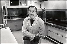
Robert Ting, 77; Biotech Pioneer

ME/Ting-ob

Dr. Robert Ting’s biotech company in Rockville developed the first FDA-approved diagnostic test kits to test for HIV antibodies. (By Gerald Martineau — The Washington Post)

By Patricia Sullivan

Washington Post Staff Writer
Friday, September 22, 2006

Robert C.Y. Ting, 77, a research scientist who started one of the early biotechnology companies in the Washington area, died Sept. 11 of complications after cardiac surgery at the Cleveland Clinic in Cleveland.

Dr. Ting founded Biotech Research Laboratories Inc. in Rockville in 1973, producing cells for government scientists to use in research. Eleven years later, his firm obtained a federal license to develop and produce the first FDA-approved diagnostic test kits for HIV antibody confirmation.

Robert C. Gallo, who co-discovered the HIV virus as the cause of AIDS, called Dr. Ting a pioneer in the field who popularized the term “biotechnology” when he moved from research to entrepreneurship.

“He introduced me to virology, and he did it twice,” said Gallo, director of the Institute of Human Virology in Baltimore. The men had known each other since the 1960s, and while playing tennis one day, Dr. Ting advised the cancer researcher to look at new research in viruses. Later, when Gallo was studying leukemia, Dr. Ting directed him to animal research in leukemia. “First he showed me how viruses change cells. Then he introduced me to retrovirology. . . . I went into retrovirology solely because of those discussions with Bob Ting on tennis courts,” Gallo said.

Dr. Ting, whom Gallo described as a quiet, modest man, was born in Shanghai, the son of a physician to Gen. Chiang Kai-Shek. His family fled the country during the Japanese invasion of China during World War II and moved to Hong Kong. Soon after, he moved to the United States, where he received a bachelor’s degree and in 1956 a master’s degree in genetics from Amherst College.

He received a doctoral degree in microbiology and biochemistry from the University of Illinois in 1960 under Salvador E. Luria, who later won the 1969 Nobel Prize in Medicine and Physiology. Dr. Ting spent the next two years on a postdoctoral fellowship at the California Institute of Technology, working with Renato Dulbecco, who later won the 1975 Nobel Prize in Medicine and Physiology. Their work focused on how viruses cause tumors.

“A lot of molecular biology developed from this,” Dr. Ting told The Washington Post in 1984 from his Rockville office, cluttered with scientific journals, awards and a large blackboard. “There was so much evidence in animal systems [that viruses cause tumors], that the next question was obvious — can you find the equivalent in humans.”

Dr. Ting joined the National Institutes of Health in 1962 as a visiting fellow and then a senior research scientist at the National Cancer Institute. From 1966 to 1968, he was an associate editor for the Journal of the National Cancer Institute.

In 1969, he joined Litton Bionetics Inc. in Rockville as director of experimental oncology, leading a project funded by the institute to search for viruses in human leukemia patients. He became scientific director of the cancer research branch the next year.

With academic, government and private business experience under his belt, Dr. Ting decided to go into business on his own and in 1973 started Biotech Research Laboratories in Rockville. It was a profitable supplier of research services and supplies until 1981, when it went public and produced the HIV diagnostic test kits. It became one of the most successful public biotech companies in the area in the mid-1980s.

The Economic Development Board of Singapore invited him to return to Asia to start a biotech company, which he did in 1985, forming Diagnostic Biotechnology Ltd. He also joined the Institute of Molecular and Cell Biology at the National University of Singapore, which Gallo called “the most prominent Asian academic biotechnology center.”

He returned to the United States in 1998 to join the board of Cell Works Inc. in Baltimore, and became chair and chief executive of a joint venture, Cell Works Asia Limited, in 2000.

Most recently, Dr. Ting was the founding president and chief executive of Profectus Biosciences Inc. of Baltimore, previously known as Maryland BioTherapeutics Inc.

Dr. Ting was past chairman of the F.F. Fraternity, one of the oldest Chinese fraternities in the United States. He was also a member of the Organization of Chinese Americans in the D.C. area since its inception in the early 1970s. He enjoyed tennis, golf, ballroom dancing and international travel. He also was a wine connoisseur.

Survivors include his wife of 44 years, Sylvia Han Ting of Potomac; three children, Anthony Ting of Shaker Heights, Ohio, Andrew Ting of Beverly, Mass., and Jennifer Chow of Potomac; seven sisters; and seven grandchildren.

An obituary written from his son Anthony can be found here:

https://www.amherst.edu/aboutamherst/magazine/in_memory/1953/robertting

Sources:

http://www.amazon.com/Virus-Hunting-Retrovirus-Scientific-Discovery/dp/0465098150

http://www.washingtonpost.com/wp-dyn/content/article/2006/09/21/AR2006092101936.html

Other articles/postings related to this topic and HIV on this site includes:

Heroes in Medical Research: Barnett Rosenberg and the Discovery of Cisplatin

History of medicine, science, and society: 200 Years of the New England Journal of Medicine

Why did Pauling Lose the “Race” to James Watson and Francis Crick? How Crick Describes his Discovery in a Letter to his Son

John Randall’s MRC Research Unit and Rosalind Franklin’s role at Kings College

Interview with the co-discoverer of the structure of DNA: Watson on The Double Helix and his changing view of Rosalind Franklin

Otto Warburg, A Giant of Modern Cellular Biology

Inspiration From Dr. Maureen Cronin’s Achievements in Applying Genomic Sequencing to Cancer Diagnostics

Nanotechnology and HIV/AIDS treatment

HIV vaccine: Caltech puts us One step further

Getting Better: Documentary Videos on Medical Progress — in Surgery, Leukemia, and HIV/AIDS.

Read Full Post »

Modulating Stem Cells with Unread Genome: microRNAs

Author, Demet Sag, PhD

Life is simple but complicated. Both simple specific sequences and the big picture approach as a system are necessary in applications for a coherent outcome. Thus, providing an engineered whole cell as a system of correction for “Stem Cell Therapy” may resolve unmet health problems.  Only 1% of the genome is read and the remaining 99% is not a junk but useful. The energy is never getting lost and there is a tight conservation economy in living organisms.  As an example microRNAs that are one of the families of untranslated sequences can be utilized for a stem cell therapy for cancer.  Their power lies at transcription control that may direct the cell expression at exact time, and place for diagnosing, imaging and treatment.  The development of cell biology and understanding of genetic data from model organisms will assist to design a well-working mechanism.

In 1964, after their elegant experiment Till et. al demonstrated that special stimulating factors caused the differentiation and made new colonies. They suggested that “…since stem cells are responsible for continued cell production, it would appear probable that such stem cells are the sites of action for control mechanisms.”  They also pointed out simply that some cells do continue to be stem cells and some do loose the plasticity as they differentiate. Regardless of the two major unescapable events, “the birth” and “the death”, even though can be less predictable than the other, life must go on.  This nature brought an attention to regenerate the cells for our need.

One of the main issues in stem cell biology is figuring out how to re-activate once upon a time fast dividing cells, while the rest of the cells were not even active. The short answer is escaping the control gates with the precise keys without creating any immune responses or toxicity. The easiest and safest method is to re-write instructions of the cells for making a function based on comparative system biology and development. These retrained, resensitized and reprogrammed cells make possible changes to produce right amount of protein(s) on time and its place.

Functional genomics approach to a system within conserved life mechanisms of organisms (C elegans, D. melanogaster, A. nidulans, S. cerevisiae and M. musculus) is necessary for sound principles development.

The first resolution comes from the worm, C. elegans.  The early founding fathers of these special 20-22 bp untranslated specific sequences that control time in development and possible mRNA regulation are called microRNAs. This significant signature sequences and biomarkers control gene regulation for a proper protein expression even though these whistles and bells are not even expressed. Since they are included in 99% of the genome, they must have a voice in the system.  These miRNAs are shown first time in C.elegans were lin4 and let7.  When they were mutated, the cells went onto extra cell proliferation like it would in cancer. Later, in many metazoans it was discovered and shown that these special RNAs negatively regulate specific gene expression during important developmental stages of life such as cell proliferation, apoptosis and stress response.  For example the famous Drosha and Dicer, members of the RNA H III family, is acting sequentially in Drosophila bind to un-translated region of mRNA that either preventing the expression of the protein or causing to be degraded by RISC (He and Hannon 2004).

Dicer is important in biogenesis of miRNA pathway and Drosophila ovary is a great tool to study embryonic stem cells.   Analysis of Dicer-1 (dcr-1) germline mutants showed that these mutants have fewer cysts because at G1/S checkpoint the activity of Decapo, a cyclin kinase inhibitor, depends on Dicer-1.  As a result, cell division mechanisms require functional miRNA. In addition, these miRNAs also make the cells “insensitive” to the environmental influences. The new epigenetic studies  include their function for oncology RD to increase efficacy and survival rate of the treatment along with personalized genomic data.

The new technologies screening of the genome or doing chromosome walk became less labor intense and more informative like miccroarray technology, faster sequencing. Lu’s group designed a microarray analysis on comparative differential expression of miRNAs between healthy and tumor in human.  Their data show that there is a difference between these populations besides having specific loci for miRNAs in the genome (Lu et al. 2005).  The study by O’Dennel’s group reaffirmed their finding. Microarray screening showed several miRNAs are residing at the chromosome 13 region.  These miRNAs are also interacting specifically with MYC to modulate the cell genesis during cancer development (O’Dennel et al. 2005).

Yet, recent evidences show that miRNAs also manipulate regulation of transcription and epigenetics (Wang et. al 2013).  As a result, nanomolecules without affecting the cellular life with specific miRNAs help us to imagine of this complexity and to receive the snapshot of the condition (Conde et al. 2013).

Furthermore, there is a complexity to be included in the design of molecules.  The system mechanism may bring solutions for human health.  Thus, modulated stem cells with engineered special future based on not only one gene-one enzyme theory but also many/one gene, one/many enzyme. For example, Schwartz group showed that polycomb group of genes made up of several hundred genes manipulate a complete function in the system of organism (Schwartz et al. 2007). First polycombs were found in fruit flies (Drosophila), but they are recognized that they function to regulate homeotic genes both in mammals and insects. Now, it is known that these polycomb complexes play a huge global role in organizing epigenetics by enforcing repressed states, but balanced by Trithorax.  Interestingly, even same genes function in both germline and somatic sex determination pathway, there are different cell-cell communications, signal transductions and players in regulation mechanisms of Drosophila (Salz 2013; Ng et al. 2013).

Therefore, the studies modulating cells by engineering oligos may fix a health problem. Immunomodulation of immune cells APC (antigen presenting cells) / DC (dentritic cells) / T (T/B), reprogramming stem cells and restructuring of the membrane receptors for increased sensitivity to protect/locate/activate are few examples of possible platforms to develop products.

Life is simple but complex, also there is a simple solution, since human is the most resilient living who will answer how to cure what is broken to survive.

References:

  1. A Stochastic model of stem cell proliferation,based on th egrowth of spleen xcolony-forming cells. J. E. Till, E. A. McCulloch, L. Siminovitch Proc Natl Acad Sci U S A. 1964 January; 51(1): 29–36.  PMCID: PMC300599. (http://www.ncbi.nlm.nih.gov/pmc/articles/PMC300599/)
  2. MicroRNAs: Small RNAs with a big role in gene regulation L. He, G.J. Hannon Nat. Rev. Genet., 5 (2004), pp. 522–531 (http://www.nature.com/nrg/journal/v5/n7/full/nrg1379.html)
  3. Stem cell division is regulated by the microRNA pathway.  S.D. Hatfield, H.R. Shcherbata, K.A. Fischer, K. Nakahara, R.W. Carthew, H. Ruohola-Baker Nature, 435 (2005), pp. 974–978 (http://www.nature.com/nature/journal/v435/n7044/full/nature03816.html)
  4. MicroRNA expression profiles classify human cancers. J. Lu, G. Getz, E.A. Miska, E. Alvarez-Saavedra, J. Lamb, D. Peck, A. Sweet-Cordero, B.L. Ebert, R.H. Mak, A.A. Ferrando et al. Nature, 435 (2005), pp. 834–838 (http://www.nature.com/nature/journal/v435/n7043/full/nature03702.html)
  5. c-Myc-regulated microRNAs modulate E2F1 expression. K.A. O’Donnell, E.A. Wentzel, K.I. Zeller, C.V. Dang, J.T. Mendell Nature, 435 (2005), pp. 839–843 (http://www.nature.com/nature/journal/v435/n7043/full/nature03677.html)
  6. Gold-nanobeacons for simultaneous gene specific silencing and intracellular tracking of the silencing events. J. Conde, J, Rosa, J. M. la Fuente, P. V. Baptista.  Biomaterials, Vol. 34, issue 10, March 2013, pp. 2516-2523 (http://www.sciencedirect.com/science/article/pii/S0142961212013956)
  7. Transcriptional and epigenetic regulation of human microRNAs.
  8. Zifeng Wang,Hong Yao, Sheng Lin, Xiao Zhu, Zan Shen, Gang Lu, Wai Sang Poon, Dan Xie, Marie Chia-mi Lin, Hsiang-fu KungCancer Letters Volume 331, Issue 1 , Pages 1-10, 30 April 2013. (http://www.cancerletters.info/article/S0304-3835(12)00723-9/abstract)
  9. The MSC: An Injury Drugstore. A. I. Caplan and D. Correa Cell Stem Cell. 2011 July 8; 9(1): 11–15. doi:10.1016/j.stem.2011.06.008.
  10. Polycomb silencing mechanisms and the management of genomic programmes. Schwartz YB, Pirrotta V (January 2007). Nat. Rev. Genet. 8 (1): 9–22. doi:10.1038/nrg1981. PMID 17173055. (http://www.ncbi.nlm.nih.gov/pubmed/17173055)
  11. Sex, stem cells and tumors in the Drosophila ovary. HK Salz, Fly, 2013 (http://www.landesbioscience.com/journals/fly/article/22687/)
  12. In Vivo Epigenomic Profiling of Germ Cells Reveals Germ Cell Molecular Signatures. J. Ng, V. Kumar, M. Muratani, P. Kraus, JC. Yeo, L-P. Yaw, K. XUe, T. Lufkin, S. Prabhakar, H-H, Ng. Developmental Cell, Vol. 24, Issue 3, 11 February 2013, Pages 324–333. (http://www.sciencedirect.com/science/article/pii/S1534580712005850)

Other related article appeared on this Open Access Online Scientific Journal, including:


When Clinical Application of miRNAs?

Larry H Bernstein, MD, FACP, 3/3/2013


Read Full Post »

Genomic Model of Organogenesis: Computer Modeling of the Gene Regulatory Networks

Curator: Larry H Bernstein, MD, FACP


Caltech biologists created the first predictive computational model of gene networks that control the development of sea-urchin embryos. This model outlines the paths cells take in forming different body parts—muscles, bones, heart. In the process the organ development follows a genetic blueprint, which consists of complex webs of interacting genes called gene regulatory networks.

This model, the scientists say, does a remarkably good job of calculating what these networks do to control the fates of different cells in the early stages of sea-urchin development—confirming that the interactions among a few dozen genes suffice to tell an embryo how to start the development of different body parts in their respective spatial locations. The model is also a powerful tool for understanding gene regulatory networks in a way not previously possible, allowing scientists to better study the genetic bases of both development and evolution.

“We have never had the opportunity to explore the significance of these networks before,” says Eric Davidson, the Norman Chandler Professor of Cell Biology at Caltech. “The results are amazing to us.”

The researchers described their computer model in a paper in the Proceedings of the National Academy of Sciences that appeared as an advance online publication on August 27.

The model encompasses the gene regulatory network that controls the first 30 hours of the development of endomesoderm cells, which eventually form the embryo’s gut, skeleton, muscles, and immune system. This network—so far the most extensively analyzed developmental gene regulatory network of any animal organism—consists of about 50 regulatory genes that turn one another on and off.

To create the model, the researchers distilled everything they knew about the network into a series of logical statements that a computer could understand. “We translated all of our biological knowledge into very simple Boolean statements,” explains Isabelle Peter, a senior research fellow and the first author of the paper. In other words, the researchers represented the network as a series of if-then statements that determine whether certain genes in different cells are on or off (i.e., if gene A is on, then genes B and C will turn off).

By computing the results of each sequence hour by hour, the model determines when and where in the embryo each gene is on and off. Comparing the computed results with experiments, the researchers found that the model reproduced the data almost exactly. “It works surprisingly well,” Peter says.

Some details about the network may still be uncovered, the researchers say, but the fact that the model mirrors a real embryo so well shows that biologists have indeed identified almost all of the genes that are necessary to control these particular developmental processes. The model is accurate enough that the researchers can tweak specific parts—for example, suppress a particular gene—and get computed results that match those of previous experiments.

Allowing biologists to do these kinds of virtual experiments is precisely how computer models can be powerful tools, Peter says. Gene regulatory networks are so complex that it is almost impossible for a person to fully understand the role of each gene without the help of a computational model, which can reveal how the networks function in unprecedented detail.

Studying gene regulatory networks with models may also offer new insights into the evolutionary origins of species. By comparing the gene regulatory networks of different species, biologists can probe how they branched off from common ancestors at the genetic level.

So far, the researchers have only modeled one gene regulatory network, but their goal is to model the networks responsible for every part of a sea-urchin embryo, to build a model that covers not just the first 30 hours of a sea urchin’s life but its entire embryonic development. Now that this modeling approach has been proven effective, Davidson says, creating a complete model is just a matter of time, effort, and resources. 

The title of the PNAS paper is “Predictive computation of genomic logic processing functions in embryonic development.”

http://www.pnas.org/content/109/41/16434.abstract

In addition to Peter and Davidson, the other author on the PNAS paper is Emmanuel Faure, a former Caltech postdoctoral scholar who is now at the École Polytechnique in France. This work was supported by the National Institute of Child Health and Human Development and the National Institute of General Medical Sciences.

A small part of the network is shown here. Image: Caltech/Davidson Lab
After a decade detailing how these gene networks control development in sea-urchin embryos, they   constructed a computational model of sea-urchin embryonic development.
VIEW VIDEO Courtesy of genenetwork

Introduction to Gene Network

GeneNetwork is a group of linked data sets and tools used to study complex networks of genes, molecules, and higher order gene function and phenotypes. GeneNetwork combines more than 25 years of legacy data generated by hundreds of scientists together with sequence data (SNPs) and massive transcriptome data sets (expression genetic or eQTL data sets). The quantitative trait locus (QTL) mapping module that is built into GN is optimized for fast on-line analysis of traits that are controlled by combinations of gene variants and environmental factors.

Historic Highlights of Organ Development

Otto Warburg. Improved manometric techniques of Van Slyke and Haldane in 1920’s and used tissue slices of 100-150 layers of cells, allowing measurement of energy reactions using oxygen without damaging cells.  He demonstrated the rate of oxygen utilization and the respiration of sea urchin egg can increase up to sixfold after fertilization.

Otto Warburg. Hans Krebs.  Clarendon Press. 1981.
Thomas Hunt Morgan.  Explored the mechanism of heredity in accounting for the transmission of variations from 1910 -1928, and claimed that while Mendelian theory could predict breeding results, it could not describe the true processes of heredity.
N William Ingalls (1918)
Carnegie Institution No. 23 – Contributions to Embryology
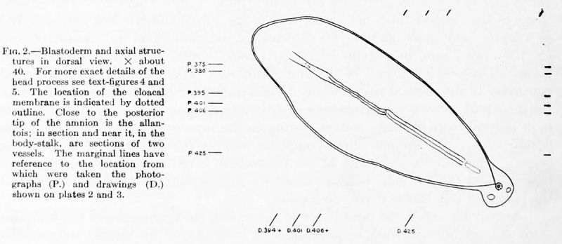
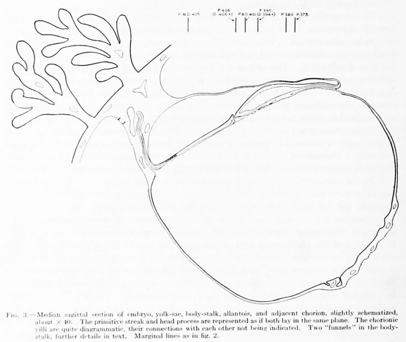
The conditions found here in the cloacal membrane are such as would be expected from the gradual and not entirely regular transformation of the streak into the membrane. All that is required is an arrest of mesoderm formation and the subsequent separation of the upper and middle germ-layers. The entoderm below is a perfectly distinct layer the cells of which have nuclei larger and paler than those of the other layers.
The embryo which we have just described represents an extremely interesting and instructive stage in the ontogenesis of man. In it are found as many important features of early development as could well be expected in one and the same specimen.  Any discussion of the findings in this embryo naturally revolves around the question of gastrulation and the formation of the germ-layers. One should not conclude too much from a single stage, either as to antecedent or later conditions; but every stage must be in harmony with those which precede or follow.
 Hans Spemann (1869 – 1941). Awarded a Nobel Prize in Physiology or Medicine in 1935 for his discovery of the effect now known as embryonic induction. Spemann found that one half of two blastomeres could form a whole embryo, but observed that the plane of division was crucial. This gave support to the concept of a morphogenetic field, a concept of which Spemann learned from Paul Alfred Weiss.  He and colleagues described an area in the embryo, the portions of which, upon transplantation into a second embryo, organized or “induced” secondary embryonic primordia regardless of location.
NOBEL PRIZE FOR GENETICS OF DEVELOPMENT  By Sean Henahan, Access Excellence
Three biologists have been awarded the 1995 Nobel Prize in Medicine for their pioneering work on the genetic control of embryonic development. The researchers work with the Drosophila melanogaster fruit fly provided key information on factors influencing human embryology and birth defects. The recipients of this year’s prize are Drs. Edward Lewis, of the California Institute of Technology; Christiane Nuesslein-Volhard, of Germany’s Max-Planck Institute; and Eric Wieschaus, at Princeton. Each of the three were involved in the early research to find the genes controlling development.
The genes were arranged in the same order on the chromosomes as the body segments they controlled. The first genes in a complex of developmental genes controlled the head region, genes in the middle controlled abdominal segments while the last genes controlled the posterior (“tail”) region.  The fertilized egg is spherical. It divides rapidly to form 2, 4 , 8 cells and so on. Up until the 16-cell stage the early embryo is symmetrical and all cells are equal. Beyond this point, cells begin to specialize and the embryo becomes asymmetrical. Within a week it becomes clear what will form the head and tail regions and what will become the ventral and dorsal sides of the embryo. Somewhat later in development the body of the embryo forms segments and the position of the vertebral column is fixed.
The results of Nuesslein-Volhard and Wieschaus, first published in the English scientific journal Nature during the fall of 1980, established that genes controlling development could be systematically identified. The number of genes involved was limited and they could be classified into specific functional groups.
In 1978 Lewis summarized his results in a review article and formulated theories about how homeotic genes interact, how the gene order corresponded to the segment order along the body axis, and how the individual genes were expressed. This induced other scientists to examine families of analogous genes in higher organisms. In mammalians, the gene clusters first found in Drosophila have been duplicated into four complexes known as the HOX genes. Human genes in these complexes are sufficiently similar to their Drosophila analogues they can restore some of the normal functions of mutant Drosophila genes.
The individual genes within the four HOX gene families in vertebrates occur in the same order as they do in Drosophila, and they exert their influence along the body axis in agreement with the colinearity principle first discovered by Lewis in Drosophila. It is likely that mutations in such important genes are responsible for some of the early, spontaneous abortions that occur in man, and for some of the about 40% of the congenital malformations that develop due to unknown reasons.
Lewis, E.B. (1978) A Gene Complex Controlling Segmentation in Drosophila. Nature 276, 565-570 Nuesslein-Volhard, C., Wieschaus, E. (1980). Mutations Affecting Segment Number and Polarity in Drosophila. Nature 287, 795-801
 Sir John B Gurdon and Shinya Yamanaka. 2012 Nobel Prize for Physiology and Medicine.
the specialisation of cells is reversible,and mature, specialized cells can be reprogrammed.

Work by the Stanford Group on Gene Networks Computational Model

Protein-folding Simulation: Stanford’s Framework for Testing and Predicting Evolutionary Outcomes in Living Organisms – Work by Marcus Feldman

Other Notable and Related Research.
LASAGNA-Search: An integrated web tool for transcription factor binding site search and visualization.  C Lee and Chun-Hsi Huang.
Length-Aware Site Alignment Guided by Nucleotide Association (LASAGNA)
1. unaligned variable-length TF binding sites
2. used for high throughput techniques, such as ChIP-seq
3. a collection of 1726 models
4. automatic promoter sequence retrieval
5. visualization
Biotechniques Rapid Dispatches   http://dx.doi.org/10.2144/000113999

Gene Splicing by Overlap Extension: Tailor-Made Genes Using PCR

RM Horton, Z Cai, SN Ho, LR Pease.  Biotechniques Nov 1990; 8(5):528-535.
Gene splicing by Overlap Extension or “geneSOEing” is a PCR-based recombining DNA sequences without reliance on restriction sites and of directly generated DNA fragments in vitro.  Method relies on modifying the sequences incorporated into the 5′-ends of the primers. Strands from two different fragments can hybridize together forming and overlap.

Democritixing Flow Cytometry.

Reported by M O’Neill. Geneg News Mar 15, 2013; 33(6): p12
Due to advances on many fronts
1. microfluidics
2. software
Flow cytometry is being simplified so that it can be used by a broader range of scientist and clinicians.
Eight Hox genes of D. melanogaster (fruitfly).

Eight Hox genes of D. melanogaster (fruitfly). (Photo credit: Wikipedia)

Read Full Post »

AMPK Is a Negative Regulator of the Warburg Effect and Suppresses Tumor Growth In Vivo

Reporter-Curator: Stephen J. Williams, Ph.D.

AMPK Is a Negative Regulator of the Warburg Effect and Suppresses Tumor Growth In Vivo

Word Cloud by Daniel Menzin

There has been a causal link between alterations in cellular metabolism and the cancer phenotype.  Reorganization of cellular metabolism, marked by a shift from oxidative phosphorylation to aerobic glycolysis for cellular energy requirements (Warburg effect), is considered a hallmark of the transformed cell.  In addition, if tumors are to survive and grow, cancer cells need to adapt to environments high in metabolic stress and to avoid programmed cell death (apoptosis). Recently, a link between cancer growth and metabolism has been supported by the discovery that the LKB1/AMPK signaling pathway as a tumor suppressor axis[1].

LKB1/AMPK/mTOR Signaling Pathway

The Liver Kinase B1 (LKB1)/AMPK  AMP-activated protein kinase/mammalian Target of Rapamycin Complex 1 (mTORC1) signaling pathway links cellular metabolism and energy status to pathways involved in cell growth, proliferation, adaption to energy stress, and autophagy.  LKB1 is a master control for 14 other kinases including AMPK, a serine-threonine kinase which senses cellular AMP/ATP ratios.  In response to cellular starvation, AMPK is allosterically activated by AMP, leading to activation of ATP-generating pathways like fatty acid oxidation and blocking anabolic pathways, like lipid and cholesterol synthesis (which consume ATP).  In addition, AMPK regulates cell growth, proliferation, and autophagy by regulating the mTOR pathway.  AMPK activates the tuberous sclerosis complex 1/2, which ultimately inhibits mTORC1 activity and inhibits protein translation.  This mTOR activity is dis-regulated in many cancers.

LKB1AMPK pathway

LKB1/AMPK in Cancer

  • Somatic mutations of the STK11 gene encoding LKB1 are detected in lung and cervical cancers
  • Therefore LKB1 may be a strong tumor suppressor
  • Pharmacologic activation of LKB1/AMPK with metformin can suppress cancer cell growth

In a recent Cell Metabolism paper[2], Brandon Faubert and colleagues describe how AMPK activity reduces aerobic glycolysis and tumor proliferation while loss of AMPK activity promotes tumor proliferation by shifting cells to aerobic glycolysis and increasing anabolic pathways in a HIF1-dependent manner.

The paper’s major findings were as follows:

  • Loss of AMPKα1 cooperates with the Myc oncogene to accelerate lymphomagenesis
  • AMPKα dysfunction enhances aerobic glycolysis (Warburg effect)
  • Inhibiting HIF-1α reverses the metabolic effects of AMPKα loss
  • HIF-1α mediates the growth advantage of tumors with reduced AMPK signaling

Summary

AMPK is a metabolic sensor that helps maintain cellular energy homeostasis. Despite evidence linking AMPK with tumor suppressor functions, the role of AMPK in tumorigenesis and tumor metabolism is unknown. Here we show that AMPK negatively regulates aerobic glycolysis (the Warburg effect) in cancer cells and suppresses tumor growth in vivo. Genetic ablation of the α1 catalytic subunit of AMPK accelerates Myc-induced lymphomagenesis. Inactivation of AMPKα in both transformed and nontransformed cells promotes a metabolic shift to aerobic glycolysis, increased allocation of glucose carbon into lipids, and biomass accumulation. These metabolic effects require normoxic stabilization of the hypoxia-inducible factor-1α (HIF-1α), as silencing HIF-1α reverses the shift to aerobic glycolysis and the biosynthetic and proliferative advantages conferred by reduced AMPKα signaling. Together our findings suggest that AMPK activity opposes tumor development and that its loss fosters tumor progression in part by regulating cellular metabolic pathways that support cell growth and proliferation.

Below is the graphical abstract of this paper.

Graphical Abstract FINAL.pptx

(Photo credit reference(2; Faubert et. al) permission from Elsevier)

However, this regulation of tumor promotion by AMPK may be more complicated and dependent on the cellular environment.

Nissam Hay from the University of Illinois College of Medicine, Chicago, Illinois, USA and his co-workers Sang-Min Jeon and Navdeep Chandel were investigating the mechanism through which LKB1/AMPK regulate the balance between cancer cell growth and apoptosis under energy stress[3]. In their system, the loss of function of either of these proteins makes cells more sensitive to apoptosis in low glucose environments, and cells deficient in either AMPK or LKB1 were shown to be resistant to oncogenic transformation.  Whereas previous studies showed (as above) AMPK opposes tumor proliferation in a HIF1-dependent manner, their results showed AMPK could promote tumor cell survival during periods of low glucose or altered redox status.

The researchers incubated LKB1-deficient cancer cells in the presence of either glucose or one of the non-metabolizable glucose analogues 2-deoxyglucose (2DG) and 5-thioglucose (5TG), and found that 2DG, but not 5TG, induced the activation of AMPK and protected the cells from apoptosis, even in cells that were deficient in LKB1.

The authors demonstrated that glucose deprivation depleted NADPH levels, increased H2O2 levels and increased cell death, and that this was accelerated in cells deficient in the enzyme glucose-6-phosphate dehydrogenase. Anti-oxidants were also found to inhibit cell death in cells deficient in either AMPK or LKB1.

Knockdown or knockout of either LKB1 or AMPK in cancer cells significantly increased levels of H2O2 but not of peroxide (O2) during glucose depletion. The glucose analogue 2DG was able to activate AMPK and maintain high levels of NADPH and low levels of H2O2 in these cells.

The nucleotide coenzyme NADPH is generated in the pentose phosphate pathway and mitochondrial metabolism, and consumed in H2O2 elimination and fatty acid synthesis. If glucose is limited mitochondrial metabolism becomes the major source of NADPH, supported by fatty acid oxidation. AMPK is known to be a regulator of fatty acid metabolism through inhibition of two acetyl-CoA carboxylases, ACC1 and ACC2.

Short interfering RNAs (siRNAs) to knock down levels of both ACC1 and ACC2 in A549 cancer cells and found that only ACC2 knockdown significantly increased peroxide accumulation and apoptosis, while over-expression of mutant ACC1 and ACC2 in LKB1-proficient cells increased H2O2 and apoptosis.

Therefore, it was concluded AMPK acts to promote early tumor growth and prevent apoptosis in conditions of energy stress through inhibiting acetyl-CoA carboxylase activity, thus maintaining NADPH levels and preventing the build-up of peroxide in glucose-deficient conditions.

This may appear to be conflicting with the previous report in this post however, it is possible that these reports reflect differences in the way cells respond to various cellular stresses, be it hypoxia, glucose deprivation, or changes in redox status.  Therefore a complex situation may arise:

  • AMPK promotes tumor progression under glucose starvation
  • AMPK can oppose tumor proliferation under a normoxic, HIF1-dependent manner
  • Could AMPK regulation be different in cancer stem cells vs. non-stem cell?

References:

1.            Green AS, Chapuis N, Lacombe C, Mayeux P, Bouscary D, Tamburini J: LKB1/AMPK/mTOR signaling pathway in hematological malignancies: from metabolism to cancer cell biology. Cell Cycle 2011, 10(13):2115-2120.

2.            Faubert B, Boily G, Izreig S, Griss T, Samborska B, Dong Z, Dupuy F, Chambers C, Fuerth BJ, Viollet B et al: AMPK is a negative regulator of the Warburg effect and suppresses tumor growth in vivo. Cell metabolism 2013, 17(1):113-124.

3.            Jeon SM, Chandel NS, Hay N: AMPK regulates NADPH homeostasis to promote tumour cell survival during energy stress. Nature 2012, 485(7400):661-665.

 Other posts on this site related to Warburg Effect and Cancer include:

Read Full Post »

Phosphatidyl-5-Inositol Signaling by Pin1


Reporter: Larry H Bernstein, MD, FCAP


Regulation of Phosphatidylinositol-5-Phosphate Signaling by Pin1 Determines Sensitivity to Oxidative Stress

Willem-Jan Keune et al.
Increasing the abundance of the phospholipid PtdIns5P protects cells from oxidative stress.
Science Signaling   27 nov 2012; 5:252.
  1. T cell receptor (TCR) and costimulatory molecule mediated signaling
  2. culminate in maximal cytokine mRNA production and stability.
The transcriptional responses to co-stimulatory T cell signaling involve calcineurin and NF-AT, which
    • can be antagonized by interference with the cis-trans peptidyl-prolyl isomerases (PPIase), cyclophilin A and FKBP.
Signaling molecules downstream of CD28
    • which are essential for the stabilization of cytokine mRNAs are largely unknown.

Pin1, a third member of the PPIase family

    • mediates the post-transcriptional regulation of Th1 cytokines by activated T cells.

Blockade of Pin1 by pharmacologic or genetic means

  • greatly attenuated IFN-γ, IL-2 and CXCL-10 mRNA
    • stability,
    • accumulation and
    • protein expression after cell activation.

In vivo, Pin1 blockade prevented

  • both the acute and chronic rejection of MHC mismatched, orthotopic rat lung transplants by
  • reducing the expression of IFN-γ and CXCL-10.

Combined transcriptional and post-transcriptional blockade with

    • cyclosporine A and the Pin1 inhibitor, juglone, was synergistic.

These data suggest Pin1 inhibitors should be explored for use as immunosuppressants and employed with available calcineurin inhibitors to reduce toxicity and enhance effectiveness.
Esnault S, Braun RK, Shen Z-J, Xiang Z, Heninger E, et al. (2007)
Pin1 Modulates the Type 1 Immune Response. PLoS ONE 2(2): e226.  http://dx. doi.org/10.1371/journal.pone.0000226

Mixed-lineage kinase 3 phosphorylates prolyl-isomerase Pin1 to regulate its nuclear translocation and cellular function
Velusamy Rangasamya,1, Rajakishore Mishraa,1, Gautam Sondarvaa, Subhasis Dasa, et al.
Loyola University Chicago, Maywood, IL 60153;  Beth Israel Deaconess Medical Center, Boston, MA 02115; University of Mississippi Medical Center, Jackson, MS 39216;
University of Wisconsin, Madison, WI 53705; Hines Veterans Affairs Medical Center, Hines, IL 60141; and College of Veterinary Medicine, Iowa State University, Ames, IA 50011
Edited* by Michael Karin, University of California, San Diego School of Medicine, La Jolla, CA, and approved April 11, 2012
Nuclear protein peptidyl-prolyl isomerase Pin1-mediated prolyl isomerization is

  • an essential and novel regulatory mechanism for protein phosphorylation.

Therefore, tight regulation of Pin1 localization and catalytic activity is

  • crucial for its normal nuclear functions.

Pin1 is commonly dysregulated during oncogenesis and likely contributes to these pathologies; The mechanism by which Pin1 catalytic activity and nuclear localization are increased is unknown.
Here we demonstrate that

  1. mixed-lineage kinase 3 (MLK3), a MAP3K family member,
  2. phosphorylates Pin1 on a Ser138 site
  3. to increase its catalytic activity and nuclear translocation.
This phosphorylation event

  1. drives the cell cycle and
  2. promotes cyclin D1 stability and centrosome amplification.

Pin1 pSer138 is significantly

  • up-regulated in breast tumors and
  • is localized in the nucleus.

These findings collectively suggest that the MLK3-Pin1 signaling cascade plays a critical role

  1. in regulating the cell cycle,
  2. centrosome numbers, and
  3. oncogenesis. breast cancer

JNK Peptidyl-prolyl isomerase Pin1 plays a critical role in

  • regulating cellular homeostasis by
  • isomerizing the prolyl bond preceded by
  • a phosphorylated Ser or Thr residue (pSer/Thr-Pro) (1).

This isomerization by Pin1 regulates the biological function of several target proteins, including

  • cell-cycle regulators,
  • protooncogenes,
  • tumor suppressors, and
  • transcription factors (2).
Due to its role in controlling the cell cycle, apoptosis, growth, and stress responses, Pin1 has been linked to the pathogenesis of human diseases, including
  • cancer (3, 4),
  • asthma (5),
  • Alzheimer’s disease (AD) (6), and
  • Parkinson disease (PD) (7).

It is thus quite likely that tight regulation of Pin1 catalytic activity or expression is important for normal physiology. It is reported that Pin1 is

  • overexpressed in most types of cancer (8), whereas
  • its expression is diminished in AD brains (2).

Accumulating evidence suggests that Pin1 isomerase activity

  • and thus function are regulated by posttranslational modifications (2).

Pin1 function is also dependent on its

  • predominant nuclear localization (2),
    • consistent with its substrates being involved in transcription and cell-cycle progression.

It was recently reported that Pin1 nuclear import is regulated by a novel nuclear localization sequence in the PPIase domain, composed of basic amino acids (9). Nonetheless, the detailed mechanism that regulates Pin1 nuclear translocation is still not known. It also remains unknown whether any posttranslational modification of Pin1 can regulate its nuclear translocation or catalytic activity, and therefore directly affect its function.

Stereospecific gating of functional motions in Pin1
Andrew T. Namanjaa, Xiaodong J. Wangb, Bailing Xub, et al.
University of Notre Dame, Notre Dame, IN 46556; Virginia Tech, Blacksburg, VA 24061
Edited by Peter E. Wright, The Scripps Research Institute, La Jolla, CA, and approved June 2, 2011
Pin1 is a modular enzyme that

  • accelerates the cis-trans isomerization of phosphorylated-Ser/Thr-Pro (pS/T-P) motifs
  • found in numerous signaling proteins regulating cell growth and neuronal survival.

We have used NMR to investigate the interaction of Pin1 with three related ligands that include

  1. a pS-P substrate peptide, and
  2. two pS-P substrate analogue inhibitors
    • locked in the cis and trans conformations.

We compared the

  • ligand binding modes and
  • binding-induced changes
    • in Pin1 side-chain flexibility.

The cis and trans binding modes differ, and

  • produce different mobility in Pin1.

The cis-locked inhibitor and substrate produced a

  • loss of side-chain flexibility
    • along an internal conduit of conserved hydrophobic residues,
    • connecting the domain interface with the isomerase active site.

The trans-locked inhibitor

  • produces a weaker conduit response.

Thus, the conduit response is stereoselective. We further show

  • interactions between the peptidyl-prolyl isomerase and
  • Trp-Trp (WW) domains
    • amplify the conduit response, and
    • alter binding properties at the remote peptidyl-prolyl isomerase active site.

These results suggest that

  • specific input conformations can gate dynamic changes that support intraprotein communication.

Such gating may help control the propagation of chemical signals by Pin1, and other modular signaling proteins.

allostery ∣ protein dynamics ∣ ligand dynamics ∣ protein evolution
Phospho-serine/threonine-proline (pS/T-P) motifs are
signaling motifs within
intrinsically disordered loops of cell cycle proteins (1).
The imide bond between the pS/T and P residues can adopt

  • either the cis or trans conformation.

These conformations differ

  • in their susceptibility to kinases and phosphatases

that propagate the chemical signals governing the cell cycle.
Accordingly, the cell must regulate the cis/trans populations of these pS/T-P motifs

  • to ensure proper signal routing.

In this context, the peptidyl-prolyl isomerase Pin1 has emerged as a critical regulator (2, 3). Pin1 is a reversible enzyme that

  • catalyzes the cis-trans isomerization of the pS/T-P imide linkages (2, 3) of other signaling proteins, such as
  1. CDC25C,
  2. p53,
  3. c-Myc,
  4. NF-kB,
  5. cyclin D1, and
  6. tau (3).

Pin1 engages when external events, such as

  • S/T (de)-phosphorylation, change the cis-trans equilibrium.

Pin1 then

  1. catalyzes the cis-trans isomerization, thereby
  2. accelerating the approach to the new equilibrium (1).

Pin1 is a modular protein of 163 residues consisting of a

  • WW domain (1–39) and a larger
  • peptidyl-prolyl isomerase (PPIase) domain (50–163) (Fig. 1).

A flexible linker connects the two domains.

  1. Both domains are specific for pS/T-P motifs (1).
  2. The WW domain serves as a docking module, whereas
  3. catalysis is the sole province of the PPIase domain.

Earlier structural studies of Pin1 revealed

  1. conformational changes upon substrate interaction, thus
  2. motivating flexibility-function studies of Pin1 (4–6).
Peptidyl-prolyl Isomerase Pin1 Controls Down-regulation of Conventional Protein Kinase C Isozymes
JBC Papers in Press, Feb 8, 2012.       http://dx.doi.org/10.1074/jbc.M112.349753

H Abrahamsen, AK O’Neill, N Kannan, N Kruse¶, et al.
From the University of California, San Diego, La Jolla, California 92093
Background: Conventional PKC isozymes have a putative Pin1

  • isomerization sequence at their turn motif phosphorylation site.
Results: Pin1 binds conventional PKCs and

    • promotes their activation-induced down-regulation.
Conclusion: Pin1 isomerizes the phosphorylated turn motif of conventional PKC isozymes,

    • priming them for subsequent down-regulation.
Significance: Pin1 provides a switch regulating the lifetime of conventional PKCs. The down-regulation or cellular depletion of protein kinase C (PKC)
  • attendant to prolonged activation by phorbol esters is a
  • widely described property of this key family of signaling enzymes.

However, neither the mechanism of down-regulation nor whether this mechanism occurs following stimulation by physiological agonists is known.
**the peptidylprolyl isomerase Pin1 provides a timer for the lifetime of conventional PKC isozymes,

  • converting the enzymes into a species that can be dephosphorylated and ubiquitinated
  • following activation induced by either phorbol esters or natural agonists.

The regulation by Pin1 requires both the catalytic activity of the isomerase and the presence of a Pro immediately following the phosphorylated Thr of
the turn motif phosphorylation site,

  • one of two C-terminal sites that is phosphorylated during the maturation of PKC isozymes.
  • the second C-terminal phosphorylation site, the hydrophobic motif, docks
    • Pin1 to PKC.

Our data are consistent with a model in which Pin1

  • binds the hydrophobic motif of conventional PKC isozymes to catalyze the isomerization of the phospho-Thr-Pro peptide bond at the turn motif, thus
  • converting these PKC  isozymes into species that can be efficiently down-regulated following activation.

The peptidyl-prolyl cis-trans isomerase Pin1 is emerging as an important regulator of signal transduction pathways (1).

Pin1-catalyzed isomerization plays a key role in the control of normal cellular functions, most notably proliferation where

    • Pin1 is essential for cell cycle progression (2).

Pin1 belongs to the Parvulin family of peptidyl-prolyl cis-trans isomerases and is the only member that

  • specifically isomerizes phospho-(Ser/Thr)-Pro ((Ser(P)/Thr(P))-Pro) motifs (3):
  1. the enzyme displays an 1000-fold selectivity for peptides phosphorylated on the
  2. Ser/Thr preceding the Pro compared with unphosphorylated peptides (3).
Pin1induced conformational changes in target proteins

  • affect a variety of protein properties from
    • folding to
    • regulation of activity and stability.

As a consequence, deregulation of phosphorylation steps and their attendant conformational changes often lead to disease (4). For example, Pin1 is
downregulated in degenerating neurons from Alzheimer disease patients, correlating with age-dependent neurodegeneration (5).
Pin1 has also been implicated in cancer progression:
levels of this protein are increased in many cancers, including those of the

    • breast,
    • prostate,
    • brain,
    • lung, and
    • colon (6–9).

Thus, Pin1 has been proposed to function as a catalyst for oncogenic pathways (10). The molecular mechanisms that lead to disease progression

  • most likely involve postphosphorylation conformational changes
    • catalyzed by Pin1
    • that are required for downstream effects.
Related articles
The human immunophilin protein FKBP12 colored ...

The human immunophilin protein FKBP12 colored by hydrophobicity (white = hydrophobic) with bound FK506, an immunosuppressant used in treating organ transplant patients to prevent rejection. FKBP also has unrelated prolyl isomerase activity. (Photo credit: Wikipedia)

The human immunophilin protein FKBP12 colored ...

The human immunophilin protein FKBP12 colored by secondary structure with bound FK506, an immunosuppressant used in treating organ transplant patients to prevent rejection. FKBP also has unrelated prolyl isomerase activity. (Photo credit: Wikipedia)


Read Full Post »

Lipoxin A4 Regulates Natural Killer Cell in Asthma

Reporter: Larry H Bernstein, MD, FCAP

Lipoxin A4 Regulates Natural Killer Cell and Type 2 Innate Lymphoid Cell Activation in Asthma
 C Barnig, M Cernadas, S Dutile,…BR Levy.
Sci Transl Med  27 Feb 2013. ; 5(174): p. 174ra26  SciTranslMed.             http://dx.doi.org/10.1126/scitranslmed.3004812
Asthma is a prevalent disease of chronic inflammation in which
  • endogenous counterregulatory signaling pathways are dysregulated.
Recent evidence suggests that innate lymphoid cells (ILCs), including
  • natural killer (NK) cells and
  • type 2 ILCs (ILC2s),
    • can participate in the regulation of allergic airway responses,
    • in particular airway mucosal inflammation.
Sci Transl Med 27 February 2013:  5(174) 174ra26        http://dx.doi.org/10.1126/scitranslmed.3004812
Both NK cells and ILC2s expressed
  • the pro-resolving ALX/FPR2 receptors.
Lipoxin A4, a natural pro-resolving ligand for ALX/FPR2 receptors, significantly
  • increased NK cell–mediated eosinophil apoptosis and
  • decreased IL-13 release by ILC2s.
Together, these findings indicate that ILCs are targets for lipoxin A4
  • to decrease airway inflammation and mediate the catabasis of eosinophilic inflammation

Molecular biology for formyl peptide receptors in human diseases
Yongsheng Li , 

Leukocytes accumulate at sites of inflammation and immunological reaction in response to locally existing chemotactic mediators. The first chemotactic factors structurally defined were N-formyl peptides. Subsequently, numerous ligands were identified

FPRs interact with this menagerie of structurally diverse pro- and anti-inflammatory ligands to possess important regulatory effects in multiple diseases, including

  1. inflammation,
  2. amyloidosis,
  3. Alzheimer’s disease,
  4. prion disease,
  5. acquired immunodeficiency syndrome,
  6. obesity,
  7. diabetes, and
  8. cancer.

How these receptors recognize diverse ligands and how they contribute to disease pathogenesis and host defense are basic questions currently under investigation that

    • would open up new avenues for the future management of inflammation-related diseases.

FPR2/ALX receptor expression and internalization are critical for lipoxin A4 and annexin-derived peptide-stimulated phagocytosis 
PMaderna, DC Cottell, T Toivonen, N Dufton, J Dalli, M Perretti and C Godson
The FASEB Journal Nov 2010; 24 (11): 4240-4249      Published online June 22, 2010, http://dx.doi.org/10.1096/fj.10-159913

Lipoxins (LXs) are endogenously produced eicosanoids with well-described anti-inflammatory and proresolution activities,

  • stimulating nonphlogistic phagocytosis of apoptotic cells by macrophages.

LXA4 and the glucocorticoid-derived annexin A1 peptide (Ac2–26) bind to a common G-protein-coupled receptor, termed FPR2/ALX. However, direct evidence of the involvement of FPR2/ALX in the anti-inflammatory and proresolution activity of LXA4 is still to be investigated. Here we describe FPR2/ALX trafficking in response to LXA4 and Ac2–26 stimulation. We have transfected cells with HA-tagged FPR2/ALX and studied receptor trafficking in unstimulated, LXA4 (1–10 nM)- and Ac2–26 (30 μM)-treated cells using multiple approaches that include immunofluorescent confocal microscopy, immunogold labeling of cryosections, and ELISA and investigated receptor trafficking in agonist-stimulated phagocytosis. We conclude that PKC-dependent internalization of FPR2/ALX is required for phagocytosis. Using bone marrow-derived macrophages (BMDMs) from mice in which the FPR2/ALX ortholog Fpr2 had been deleted, we observed

  • the nonredundant function for this receptor in LXA4 and Ac2–26 stimulated phagocytosis of apoptotic neutrophils.
  1. LXA4 stimulated phagocytosis 1.7-fold above basal (P<0.001) by BMDMs from wild-type mice, whereas no effect was found on BMDMs from Fpr2−/− mice.
  2. Ac2–26 stimulates phagocytosis by BMDMs from wild-type mice 1.5-fold above basal (P<0.05), but  Ac2–26 failed to stimulate phagocytosis by BMDMs isolated from Fpr2−/− mice.

These data reveal novel and complex mechanisms of the FPR2/ALX receptor trafficking and functionality in the resolution of inflammation.—
Maderna, P., Cottell, D. C., Toivonen, T., Dufton, N., Dalli, J., Perretti, M., Godson, C.
http://www.FASEB.j.org/FPR2/ALX receptor expression and internalization are critical for lipoxin A4 and annexin-derived peptide-stimulated phagocytosis.
We have transfected cells with HA-tagged FPR2/ALX and studied receptor trafficking in unstimulated, LXA4 (1–10 nM)- and Ac2–26 (30 μM)-treated cells using multiple approaches and conclude that PKC-dependent internalization of FPR2/ALX is required for phagocytosis. Using bone marrow-derived macrophages (BMDMs) from mice in which the FPR2/ALX ortholog Fpr2 had been deleted,

  • we observed the nonredundant function for this receptor in LXA4 and Ac2–26 stimulated phagocytosis of apoptotic neutrophils.

LXA4 stimulated phagocytosis 1.7-fold above basal (P<0.001) by BMDMs from wild-type mice,

  • whereas no effect was found on BMDMs from Fpr2−/− mice.

Ac2–26 stimulates phagocytosis by BMDMs from wild-type mice 1.5-fold above basal (P<0.05)

  •  Ac2–26 failed to stimulate phagocytosis by BMDMs isolated from Fpr2−/− mice relative to vehicle.

These data reveal novel and complex mechanisms of the FPR2/ALX receptor trafficking and functionality in the resolution of inflammation.
The lipoxin receptor ALX: potent ligand-specific and stereoselective actions in vivo.
Chiang, N., Serhan, CN, Dahlen, SE, Drazen, JM, Hay, DW, Rovati, GE, et al.
Pharmacol. Rev. 2006; 58, 463–487.      http://www.PharmacolRev.com/The_lipoxin_receptor_ALX:_potent_ligand_specific_and_stereoselective_actions_in_vivo/

Asthma Obstruction of the lumen of the bronchi...

Asthma Obstruction of the lumen of the bronchiole by mucoid exudate, goblet cell metaplasia, epithelial basement membrane thickening and severe inflammation of bronchiole. (Photo credit: Wikipedia)

Schematic diagram indicating the complementary...

Schematic diagram indicating the complementary activities of cytotoxic T-cells and NK cells. (Photo credit: Wikipedia)

Related articles


Read Full Post »

Transcript Dynamics of Proinflammatory Genes

Author: Larry H Bernstein, MD, FCAP

Transcript Dynamics of Proinflammatory Genes Revealed by Sequence Analysis of Subcellular RNA Fractions

DM Bhatt, A Pandya-Jones, Ann-Jay Tong, I Barozzi, MM Lissner, et al.
Cell 2012;150: 279–290

In addition to documenting the subcellular locations of coding and noncoding transcripts, the results provide a high-resolution view of the relationship between
  • defined promoter and chromatin properties and
    • the temporal regulation of diverse classes of coexpressed genes.
The data also reveal a striking accumulation of full-length yet incompletely spliced transcripts in the chromatin fraction, suggesting that
  • splicing often occurs after transcription has been completed,
  • with transcripts retained on the chromatin until fully spliced.
Summary
Macrophages respond to inflammatory stimuli by modulating the expression of hundreds of genes in
  • a defined temporal cascade,
  • with diverse transcriptional and posttranscriptional mechanisms contributing to the regulatory network.
We examined proinflammatory gene regulation in activated macrophages by
  • performing RNA-seq with fractionated chromatin-associated, nucleoplasmic, and cytoplasmic transcripts.
This methodological approach allowed us
  • to separate the synthesis of nascent transcripts from transcript processing and
  • the accumulation of mature mRNAs.
In addition to documenting the subcellular locations of coding and noncoding transcripts,
the results provide a high-resolution view of the relationship between
  • defined promoter and chromatin properties and
  • the temporal regulation of diverse classes of coexpressed genes.
The data also reveal a striking accumulation of full-length yet incompletely spliced transcripts in the chromatin fraction, suggesting that
  • splicing often occurs after transcription has been completed, with transcripts retained on the chromatin until fully spliced.

Two independent experiments were performed with lipid A-stimulated bone marrow-derived macrophages. The two experiments made use of different macrophages prepared from different mice, several months apart.(A) Pearson pair-wise correlation values (R) derived from an analysis of greater than 500 lipid A-induced genes (>5-fold induced) are shown. Each time point from the first experiment, A, was compared to every other time point from the same experiment and from the second experiment, B.(B) Hierarchical clustering of the R-values from panel A was performed. This analysis reveals that, when only induced genes are considered, each time point from each experiment correlates more closely with the corresponding time point from the other experiment than with any of the other time points from either experiment.(C)

This analysis reveals that, when the transcript levels of expressed genes are compared,
  • each time point from a given experiment correlates with the same time point from the independent experiment.
The results reveal close correlations between all time-points from both experiments, presumably because genes that are consistently unexpressed (i.e., not counted in B) are contributing to the high degree of correlation. Nevertheless, the time points of each independent experiment still have the highest degree of correlation with each other.
Hierarchical clustering of the R values from panel D was performed. As with other clusterings, each sample clusters with its cognate time point in the independent experiment
Highlights
► Coding and noncoding transcripts exhibit characteristic subcellular distributions
► The most potently induced genes favor promoters with low CpG content
► Full-length, incompletely spliced transcripts accumulate on the chromatin
► Delayed transcript release may reflect a requirement for the completion of splicing
Eukaryotic transcription overview

Eukaryotic transcription overview (Photo credit: Allen Gathman)

English: Nucleosome structure.

English: Nucleosome structure. (Photo credit: Wikipedia)

Read Full Post »

AKT Signaling Variable Effects


Reporter: Larry H Bernstein, MD, FCAP


Heterogeneous kinetics of AKT signaling in individual cells are accounted for by variable protein concentration

Meyer R, D’Alessandro LA, Kar S, Kramer B, She B, Kaschek D, et al.
Front. Physio. 2012; 3:451.     http://dx.doi.org/10.3389/fphys.2012.00451

In most solid cancers, cells harboring oncogenic mutations represent only a sub-fraction of the entire population. Within this sub-fraction the expression level of mutated proteins can vary significantly due to

  • cellular variability limiting the efficiency of targeted therapy.

To address the causes of the heterogeneity, we performed a systematic analysis of one of the most frequently mutated pathways in cancer cells, the phosphatidylinositol 3 kinase (PI3K) signaling pathway. Among others PI3K signaling is activated by the hepatocyte growth factor (HGF) that regulates

  • proliferation of hepatocytes during liver regeneration but
  • also fosters tumor cell proliferation.

HGF-mediated responses of PI3K signaling were monitored both at the single cell and cell population level in primary mouse hepatocytes and in the hepatoma cell line Hepa1_6. Interestingly, we observed that the HGF-mediated AKT responses at the level of individual cells is rather heterogeneous. However, the overall average behavior of the single cells strongly resembled the dynamics of AKT activation

  • determined at the cell population level.

To gain insights into the molecular cause for the observed heterogeneous behavior of individual cells, we employed

  • dynamic mathematical modeling in a stochastic framework.

Our analysis demonstrated that intrinsic noise was not sufficient to explain the observed kinetic behavior, but rather

  • the importance of extrinsic noise has to be considered.

Thus, distinct from gene expression in the examined signaling pathway fluctuations of the reaction rates has only a minor impact whereas

  • variability in the concentration of the various signaling components even in a clonal cell population is a key determinant for the kinetic behavior.
English: Structure of the HGF protein. Based o...

English: Structure of the HGF protein. Based on PyMOL rendering of PDB 1bht. (Photo credit: Wikipedia)

Read Full Post »

microRNA Biomarker

Reporter: Larry H Bernstein, MD, FCAP

MicroRNA Molecule May Serve as Biomarker

miRNA molecule called miR-7 decreased in highly metastatic cancer stem-like cells.
February 18, 2013
Researchers have identified two molecules that could potentially serve as biomarkers in

MicroRNAs are involved in

  • tumor initiation and
  • progression, and
  • may play a role in metastasis, particularly in relation to
  • cancer stem-like cells.
miR-7 is a metastasis

  • suppressor in cancer stem-like cells, and when they
  • increased expression of miR-7 in cancer stem-like cells from
    • it suppressed their metastatic properties.

miR-7 suppressed ………….expression of KLF4.
However, miR-7 significantly suppressed the ability of cancer stem-like cells to metastasize to the brain but not the bone.

A gram illustrating the disctinction between c...

A gram illustrating the disctinction between cancer stem cell targeted (above) and conventional (below) cancer therapies (Photo credit: Wikipedia)

Related articles


Read Full Post »

« Newer Posts - Older Posts »