Feeds:
Posts
Comments

Posts Tagged ‘Drosophila’

English: This diagram shows the chromosomes of...

This diagram shows the chromosomes of Drosophila melanogaster approximately to scale. Chromosome sizes were based on basepair lengths given on the NCBI map viewer, and A. B. Carvalho, 2002. Curr. Op. Genet. & Devel. 12:664-668. Centimorgan distances were derived from selected loci listed in the NCBI website. (credit  Wikipedia)

Introduction

Generally speaking sexually reproducing species are composed of individuals of two complementary mating types or sexes.  An essential aspect of the developmental history of each individual is thus sex determination and differentiation. There exist two sex determination mechanisms, somatic and germline, that based on the chromosomal mechanism in the Drosophila melanogaster.  In the somatic sex determination mechanism, each individual assesses the ratio of X-chromosomes to autosomal chromosome sets), the X:A ratio provides the primary sex-determining signal   (reviewed by Cline and Meyer, 1996).  When X:A=1, female differentiation ensues (Bridges, 1925), along with the male-mode of X-chromosome dosage compensation.  The X:A ratio is calculated within each cell of the developing embryo, 2 hrs after fertilization. The X:A ratio determines the sex in Drosophila (Bridges, 1916, 1921, 1925) in a somatic-cell-autonomous manner that occurs early in embryonic development (Baker and Belote, 1983; Baker, 1989). Females possess two X-chromosomes, and males possess one X-chromosome and one Y-chromosome.   The Y-chromosome is required only for spermatogenesis (Lindsley and Tokuyasu 1980; Bridges 1986), and will not be considered further.  The number of X-chromosomes is counted through a mechanism involving positive-acting X-chromosome-encoded transcription factors, termed X-numerator elements (Cline, 1988), negative-acting autosome-encoded transcription factors or denominators, and signal transduction factors provided maternally.  Among the X-numerators are sisterless-a, sisterless-b (sis-b), sisterless-c, and runt (Schurpbach, 1985; Cline, 1986, 1988; Steinmann-Zwicky et al., 1989; Parkhurst et al., 1990; Ericson and Cline, 1991, 1993; Estes, 1995; Hoshijima et al., 1995; reviewed by Cline, 1993).

The best candidate for a denominator gene is the deadpan (dpn) locus.  Both daughterless (da) and extramacrochaete (emc) fulfill the role of maternally contributed transduction loci (Cline, 1976; Cronmiller et al., 1988).  Both in vitro biochemical evidence and in vivo genetic evidence support the idea that transcription factors of the basic-helix-loop-helix (bHLH) family are able to form homo- and hetero-dimers; thus the X:A ratio counting mechanism seems to involve the relative affinities and chromosome-dependent stoiciometries of the bHLH proteins SIS-B, DA, EMC, and DPN.  When X:A=1, sufficient SIS-B protein is synthesized so that it can effectively compete with the EMC and DPN proteins for binding to DA protein.  DA:SIS:B heterodimers then bind to so-called establishment promoter (Pe) elements of the SXL gene and activates its transcription, resulting in an early burst of SXL protein that sets splicing and dosage compensation in to female-specific modes.  When X:A=0.5, too little SIS-B is produced, and DA protein remains sequestered with EMC and DPN.  The Sxl Pe remains inactive, and splicing and dosage compensation enters male-specific modes. In response to X:A ratio=1, an embryo specific promoter of the gene called Sex-lethal (Sxl) is activated (Keyes et al., 1932).

Sxl protein that acts as a master gene for the somatic germline sex determination, has three somatic functions. First, Sxl protein carries out autoregulation at the level of pre-mRNA splicing.  Second, Sxl controls female-specific differentiation at the level of pre-RNA splicing and polyadenylation at least two genes that code for transcription factors that effect terminal differentiation. Third, Sxl protein negatively regulates X-chromosome dosage compensation.  It does so in two ways, by alternative RNA splicing of a normally male-specific gene, and by translation-level regulation of many X-chromosomal transcripts during embryogenesis. In the male, with Sxl in the off state, male differentiation occurs because tra is in the off state and therefore the differentiation-effector transcription factors are produced in alternative male-specific modes.  Dosage compensation is active, and the male X-chromosome is decorated by a minimum of four proteins and two RNA molecules that form a complex along the entire chromosome (reviewed by Cline and Meyer, 1996).  Transcription of the male X-chromosome is elevated two-fold, and it produces the same amount of RNA per template as found in females.

Germline pathway for sex determination and dosage compensation is different than the somatic sex determination mechanism.  (Figure 1) Figure 1: Sex determination of D. melanogaster (1998)The vast majority of somatic sex determination loci have no function in germline cells.  For example, none of the X-chromosome numerators is required for proper oogenesis (Granadino et al., 1989, 1992; Steinmann-Zwicky 1991), despite the fact that proper oogenesis requires that X:A =1 in the germline (Schupbach, 1982, 1985) nor are tra, tra-2, and dsxF required for oogenesis.  Sxl and snf have germline functions but the former is not a binary switch gene between oogenesis and spermatogenesis (Despande et al., 1996; Bopp et al., 1993, 1995; Hager et al., 1997). Systematic screens for female-sterile mutations have identified a large number of genes required for normal oogenesis (e.g. Gans et al., 1975; Mohler, 1977; Perrimon et al., 1986; Schupbach and Wieschaus, 19889, 1991).  Female-sterility can arise in diverse ways, but one interesting class of mutations is germline-dependent and causes an “ovarian tumor” phenotype.  “Ovarian tumor” mutations cause under-developed ovaries, in which egg chambers and ovarioles are filled with an excess of undifferentiated germ cells that have adopted male-like characteristics that include a prominent spherical nucleus, assembly of mitocondria around the nucleus, and mis-expression of male-specific marker genes (Oliver et al., 1988, 1990, 1993; Steinmann-Zwicky, 1988, 1992; Bopp et al., 1993; Pauli et al., Wei et al., 1994).  Among the “ovarian tumor” class of genes are ovo, ovarian tumor (otu), fused, and two genes with somatic phenotypes, namely snf and Sxl. Strong mutations at the ovo and otu loci result in ovaries totally devoid of germ cells (King and Killey, 1982; Busson et al., 1983; Oliver et al., 1987; Mevel-Ninio et al., 1989; Rodesh et al., 1995), Weaker mutations at both loci result in viable germline cells that have abnormal male-like splicing at the Sxl gene (Oliver et al, 1993). The overall conclusion is that oogenesis requires a chromosomally female germline is wild type for ovo, otu, Sxl, and snf.  If one of these genes is defective, either the germline will die or male-like differentiation and tumor formation ensure.

However, there are soma-germline interactions for a normal sex determination. (Figure 2) Figure 2: Somatic-Germline Interactions. (1998)Unlike the somatic regulatory hierarchy, which genetic mosaic experiments clearly showed functions in cell-autonomous fashion, sexual differentiation of the germline requires inductive signaling from somatic cells.  This was shown by use of pole cell transplantation, the method of making mosaics in which germline cells surgically transferred from donor embryos  (Schubach. 1985; Steinmann-Zwicky et al., 1989).  These experiments show that proper germline differentiation requires a combination of germline-autonomous chromosomal cues and proper signaling from the soma.  Evidence with tra and dsx mutant somatic hosts indicates these soma-germline interactions have detectable effects by larval stages (Steinmann-Zwicky., 1996).

The ovo gene is genetically complex.  At least three transcripts are produced from the ovo region (Mevel-Ninio et al, 1991, 1995, 1996; Garfinkel et al., 1992, 1994).  Two of these are germline-specific and correspond to the ovo function, while the third corresponds to the somatic-epidermal, non-sex-specific shavenbaby (svb) function.  (For a schematic of the gene map please refer to Figure3) 

 The ovo function is transcribed from two closely spaced germline-specific promoters, ovo a and ovob, give rise to 5-kb mRNAs (Mevel-Ninio et al., 1991, 1995; Garfinkel et al., 1992, 1994).   First identified  promoter was ovob  Garfinkel et al., (1994)  and the leader exon it forms is called Exon 1b, 1028-codon-long open reading frame that contains four Cys2-His2 fingers at the carboxy terminus; protein MW of 110.6 kD.  A second germline promoter, ovoa, was identified by Mevel-Ninio et al (1995), 1400 codons long, and predicts a 150.8-kD protein.  This Exon 1a contains an in-frame AUG upstream of the translation start in Exon 2 utilized by the OvoB open reading frame.  The OvoB mRNA isoforms is predominant during adult life, with the OvoA isoforms only appearing during Stage 14 of oogenesis (Mevel-Ninio et al., 1991, 1996; Garfinkel., 1994).  The ovo zinc finger domain binds to its own germline promoter regions, to the otu promoter region (Garfinkel et al., 1997; Lee, 1998; Lee and Garfinkel 1998).  This is consistent with ovo playing an important role in a sex determination hierarchy operating in germline cells that involves these other genes. The svb function is transcribed from an incompletely characterized somatic promoter that forms a 7.1 kb poly(A)+ mRNA (Garfinkel et al., 1994).  This transcript accumulates 9-12-hr post-fertilization, in the somatic tissues that later in embryogenesis form the cuticular structures affected by svb mutations.  Wieschaus et al. (1984) observed that ventral denticle belts and dorsal hairs are defective in svb mutations; hence the name, and svb mutations are polyphasic larval lethals. Exons and exon segments that are found in all mRNA forms coded by the region correspond to genomic DNA where so-called svb-ovo- mutations map (Mevel-Ninio et al., 1989; Garfinkel 1992).  Finally, somatic-specific exons, exon segments, and transcriptional regions correspond to region mutable to the svb- ovo- phenotype.  Since al known mRNA forms utilize the same splice junctions to join Exon3 to Exon4, all protein forms coded by the locus are believed to contain the same four zinc fingers at the carboxy terminus.   A wide variety of evidence points to ovo playing a critical role in germline sex determination.  High-level of ovo transcription in germline cells, as detected with Xgal staining of ovo promoter-lacZ constructs requires that they have a female karyotype (Oliver et al., 1994).  Chromosomally male germline cells have low levels of ovo transcription even if the soma is transformed towards female through the use of hs-traF cDNA minigenes.  Likewise, chromosomally female germline cells have high levels of ovo transcription even if the soma is anatomically male through the action of tra loss-of-function mutations.  This argues that high-level of ovo transcription is a germline X: A ratio-autonomous property, and stands in contrast to related experiments with otu.  In the case of otu, there is evidence that chromosomally male germline cells, which normally have no need of otu+ function at all, require otu- for proliferation when they are in a female host (Nagoshi et al., 1995). The D. melanogaster ovo gene is required for cell viability and differentiation of female germ cells, apparently playing a role in germline sex determination.  While female X: A ratio in germline cells is required for high levels of ovo germline promoters.  Therefore we undertook to identify trans-acting regulatory regions of the X-chromosome, with a particular interest in identifying candidate germline X-chromosome numerator elements. In this study, I screened  X-chromosome using 45 deficiency strains, I found that these trans-regulating regions were grouped into 12 loci based on overlapping cytology.  Five regions were trans-regulating activators, and seven were trans-regulating repressors; extrapolating to the entire genome, this result predicts nearly 85 loci.  A subset of the dozen X-chromosomal regions correlated with previously identified E(ovoD) and Su(ovoD) loci (Pauli et al., 1995).  

Materials and Methods


Fly Strains and Growth Flies were maintained on standard yeast/cornmeal medium and kept at 25oC and 18oC unless otherwise indicated.  Mutants are described in Lindsley and Zimm (1992).  The ovo3U21 and ovo4B8 were obtained from Brian Oliver of NIH;  OvoD1rS1 FM3 is from the Garfinkel lab collection.  The remaining stocks were obtained from the Bloomington Stock Center (see Table 2.1 for the list of stocks that had been used and Figure 2.1 for their location on the X Chromosome). 

Outcrosses Outcrosses were designed to create transgenic flies so that screening of the X chromosome for trans-regulators of ovo in the germline can be done.   Virgin female flies were collected 14 hour long windows at 18oC or 8 hour long windows at 25oC, during which newly emerged males remained immature.  Collected females were kept 3-5 days to make sure they are virgin before outcrossing them.  Heterozygous virgin females (5-7), carrying deficiency X-chromosomes balanced over first chromosome balancers were mated with males homozygous for either of two P-element transformation constructs of a lacZ reporter gene fused to the ovo promoter.  Both events were inserted on third chromosome.  They were grown at 25oC unless otherwise noted. The control class of F1 progeny has a complete X-chromosome pair, whereas the experimental class has one complete and one deficient X chromosome in its genome.  The [ovo::lacZ constructs] were designed by Oliver et al., (1994).  In this study two of their strains, ovo4B8 (pCOW+1.9) and ovo3U21 (pCOW-2.1) respectively, were used to determine the ovo promoter activity.

Outcrosses to Remove Duplications Several X-chromosome deficiencies in the Bloomington collection are carried in males, with compensatory duplications of X material on an autosome.  These had to be crossed to eliminate the duplications (Fig 2.4).  This was done as follows:  FM3/FM7a virgin flies were mated to Df/Y; Dp males.  Among the F1 progeny, half of the Df/(FM3 or FM7a) daughters will carry the unwanted duplication, and half will be free of the duplication.  In some cases, presence of the duplication could be determined from the females’ phenotypes.  In other cases, up to twenty individuals virgin Df(FM3 or FM7) F1 progeny were backcrossed to FM7a/Y males to establish stocks.  In the F2, absence of the duplication could be established by examining sons; in all cases, the Df is male-lethal unless “rescued” by the duplication.  Also FM3 is itself male lethal.  Thus, single-female stocks that produce only FM7a sons had the desired genotypes and were kept for experiments.

X-Gal Staining In this assay ovaries from two-day-old adults were dissected in Drosophila Ringer’s solution (182 mM KCl, 46 mM NaCl, 3 mM CaCl2, 10mM TrisHCl, pH 6.8).  Then, these tissues were transferred to a microtiter plate and fixed in 1% gluteraldehyde, 50mM Na-cacodylyte acid solution for 15 minutes. After rinsing the tissues, three times for 5 minutes each staining buffer (7.2 mM Na2HPO4, 2.8 mM NaH2PO4, 1.0 mM MgCl2, 0.15 mM NaCl), they were transferred to incubation buffer (staining buffer, 5 mM Fe2 (CN)3, 5 mM Fe3 (CN)2, 0.2% X-Gal) for an hour at 37oC.  Next, tissues were washed three times 5 minutes each in washing buffer, which is a 1 mM EDTA, added PBS (130 mM NaCl, 7 mM Na2HPO4*2H2O, 3 mM NaH2PO4*2H2O, pH 7.0) solution.  Finally, the tissues were dehydrated in ethanol solutions of increasing concentrations (50%, 75%, 95%) and mounted on a slide in Permount.  Preparate concentrations were examined under a compound microscope to make correlations between staining and gene activity. Although it was easy to determine positive and negative controls, but this assay wasn’t sensitive enough to see subtle differences due to effects of deleted regions on ovo promoters driving LacZ.

Histochemical Assay of LacZ Activity This method allowed us to make quantitative measurements of lacZ activity due to ovo promoter function in animals heterozygous for X-chromosome deletions.  Emerging F1 flies were collected and aged for two days before dissecting ovaries under a dissecting microscope.  For each soluble assay, 10 flies were dissected.  This is repeated at least seven assays (N, sample number) completed per stock for each construct.  Ovaries from ten dissected outcrossed flies were out into eppendorf tubes containing 100ml of Assay Buffer (50 mM K-phosphate, 1 mM MgCl2 at pH 7.8) and homogenized about 20 strokes.  For each dissected pair of ovaries 100 ml  of assay buffer was used and the volume was completed to appropriate amount.  After centrifuging for one minute, 20 ml of the supernatant was transferred into 980 ml of assay buffer (Simon and Lis, 1987; Ashburner, 1989) to make 2mM chlorophenol red-beta-D-galactopyranoside (CPRG).  Absorbance at 574 nm was measured at half hour time intervals starting from zero to two hours hydrolysis of CPRG by chlorophenol (red CPRG).  CPR has a molar extinction coefficient of 75,000 M-1 cm-1 (Boehringer-Manheim data sheet) and this is a very easily detected product of b-galactoside enzyme activity. Range finding experiments showed that 2mM of CPRG gives linear data for 2-3 hours often, color changes could be seen with the unaided eye. Two controls are shown in Figure 2.8 that validates CPRG for this work.  Ovaries from a non-transformed strain (y w RD) were used to prepare soluble extracts.  A near zero-absorbance at 574 nm was observed that did not appreciably change over several hours.  In contrast, ovarian extracts from the ovo promoter-lacZ transformant strain ovo3U21 and ovo4B8 (Oliver et al, 1994) showed a steep linear increase in A 574 during the same period.  The slopes of these lines were proportional to the amount of ovo3U21 and ovo4B8 extract added.

Bradford (1976) Assay For Protein This protein determination method is based on the binding of Coomasie Brilliant Blue G-250 to the protein.  Preparation of protein reagent was done according to Bradford (1976).  After 100 mg of Coomasie Brilliant Blue G-250 was dissolved in 50 ml 95% ethanol, and then 100 ml 85% (w/v) phosphoric acid was added.  The resulting solution was diluted to a final volume of 1 liter [final concentrations in the reagent were 0.01% (w/v) Coomasie Brilliant Blue G-250, 4.7% (w/v) ethanol, and 8.5% (w/v) phosphoric acid].  20ml of prepared soluble extract from the dissected tissues were used.  This volume is diluted to 0.1ml with ddH2O, then 5ml of protein reagent was added to the test tube and contents were mixed.  The absorbance at 595nm was measured after 2 min and before 1 hr in 3 ml cuvettes against a reagent blank prepared from 0.1 ml of the appropriate buffer and 5 ml of protein reagent.  A standard curve using known quantities of bovine serum albumin (BSA) was constructed.  Soluble extract absorbances were plotted on the standard curve and protein amount interpolated.

Statistical Analysis Average specific activity is calculated as nanomoles of substrate used per hour per nanogram protein expressed (nmole CPRG liberated /ng / hr).  Sample number (N) always exceeded seven.  Mean specific activity and standard error of the mean (SEM) were calculated for each experimental and control class.  The F test was used to determine whether variances were equal, and therefore,, which type of student’s t-test calculation was appropriate.  A significant difference between experimental and control values was identified by a P < 0.05 for the t-test score.

RESULTS

In this study and ovo mechanism study, the X-chromosome was screened, using 56 different deficiency strains    Table 1: List of Stocks for X-chromosome Screening (1998)Table 2: Stocks Made in This Study for X-Chromosome Screening Table 1: Stocks for Negative Autoregulation of ovo (1998)  to identify transregulation of ovo Table 3: LacZ Specific Activities Obtained by Screening X-Chromosome with ovo3U21Table 4: LacZ Specific Activities Obtained by Screening X-Chromosome with ovo4B8 (Results)

The results are given in three sections: X chromosome deficiency screening, negative autoregulation of ovo exhibited by deficiencies removing ovo, and gene dose analysis using P element transformants carrying extra copies of ovo.

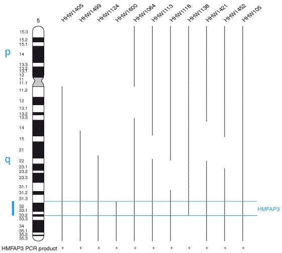
X Chromosome Screening The presence of polytene chromosomes in the salivary glands, which have distinctive, banding patterns allows the map positions of genes to be correlated with physical features of the chromosomes.  Breakpoint locations rearrangements, and the locations of cloned sequences can be easily established.  Each of the major chromosome arms is divided into 20 numbered segments, except chromosome 4, which is divided into 4 regions.  Each numbered region is then divided into six consecutive lettered regions, and each lettered region into numbered bands, for example 4E1. The precise relationship between physical length and the numbering scheme depends on local topography (Lefevre, 1976).  In the summary tables, each deficiency listed according to cytological positions. The map of the X chromosome, including the deficiencies used in this study is given in Materials and Methods (Fig 1). Figure 1: Sex determination of D. melanogaster (1998) In Drosophila melanogaster germ cells, ovo has a primary role in female sex specific cell viability, proliferation and differentiation.  Ovo responds to the number of X-chromosomes as assessed by high level expression (Oliver et al., 1994).  Thus, the ovo promoter may be dependent upon X germline numerator elements.  To identify possible trans-regulators of the ovo germline promoter (and, I hope, to identify germline numerators) I undertook deficiency screen for quantitative effects on ovo::lacZ reporter constructs.  Determination of trans-regulation effect by any of the deletion mutant, was based on two general rules.  If the excised part of the X chromosomes has any genes with the positive regulatory effects on ovo gene activity, then the levels of LacZ reporter gene function will be reduced in experimentals compared to control siblings.  If the experimental class results in the elevation of the LacZ activity by producing high levels of enzyme compared to controls, the elevated region having removed a repression locus. Significant effects were determined by statistical analysis, which using a student’s t-test P value is less than or equal to 0.05.  X-chromosome screening results are presented in Table 3.1 and 3.2.  The entire X-chromosome deficiency set was tested twice: once with a 3.3kb ovo promoter fragment driving LacZ (strain ovo3u21), and separately with a 3.1kb ovo promoter (ovo4B8).  Of  45 deficiencies that represent about 70% of the X-chromosome 17 deficiencies had significant effects in both ovo3U21 and ovo4B8 reporter activity, 1 deficiency had significant effects on only ovo3U21 and only 1 deficiency effect on ovo4B8.  Some of these deficiencies partly overlap, allowing the identification of 11 regions that apparently contain trans-acting modifiers of ovo promoter activity six are positive regulators and five are negative.

Region 1-4.  This region covers the eight overlapping deficiency lines, Df(1) BA1, Df(1)sc14, Df(1)64c18, Df(1)JC19, Df(1)dm75e19, Df(1)N8, Df(1)A113, DF(1)JC70.  For three of them, Df(1)A113, Df(1)JC70, and Df(1)BA1, the student’s t-test probabilities show a significant difference between control and experimental siblings.  The remaining strain has no significant trans-regulation effect on ovo gene activity.  Df(1)BA1 enhanced the ovo gene expression activity about 20% when either ovo3U21 or ovo4B8 is used.  It was suggested that a suppressor of ovoD (1F-2B+ locus) maps within 1E3-4 to 2B3-4 because of the dramatic gene dose effect of this region on the development of ovoD2/+ ovaries (Pauli et al, 1995).  In contrast, it was found that Df(1)A113 and Df(1)JC70 have repressing effects on ovo expression.  Df(1)A113 (3D6-E1; 4F5) removes several genes beside ovo, showed a very significant repression effect in outcrosses, about 82% and 47% (e/C), in ovo3U21 and ovo4B8 respectively.  That data obtained in Df/+ females has a particular quantitative significance, which implies that the missing loci have the complementary effect. It was shown that this region is contains a gene or genes resulting in genetic unbalance (Cline et al., 1987).  Also, Oliver et al., (1988) show that in deficiency lines, which they have used, strains removing both ovo and snf together are reducing viability of the progeny, that is, there is a synergistic interaction between ovo and snf.  

Region 5-8.  Twelve overlapping deletions have been tested in this region.  Two deletions Df(1)N73 (5C3-5;5E-8) and Df(1)Lz90b24 (8B-D) caused very significant repressing effects, implying the presence of trans-activating loci, one deletion Df(1)RA2 (7D10;8A4-5) resulted in heterozygous experimentals with significant elevation in LacZ compared to siblings, implying a trans-repressor locus.  It has been reposted that Df(1)RA2 strongly enhances ovoD  phenotypes due to the function of otu+ in germline sex determination (Pauli et al., 1993).  However, since out protein is cytoplasmic, it is unlikely that the Df(1)RA2 effect on ovo::lacZ promoter activity is due to changing dosage of otu.  It is also suggested that there is a synergistic interaction between ovo and lozenge, eye phenotype, which is deleted by Df(1)Lz90b24, and here the data showed a trans-activating effect due to this deletion.  The other deletions do not cause any significant effect on gene activity.

Region 9-10.  In this cytological position nine deficiency lines had been tested.  Since this region was very dense for putative trans-regulation repressors, it was group in a small region.  Among nine of the deficiencies were used six of them showed a repressor effect.  These effective regions were: Df91)vL15, Df(1)N110, Df(1)HC133, Df(1)vL11, Df(1)KA7, and Df(1)N71.  This region seems to have a very important effect on ovo, since in the 9Bto 10F interval there are various levels of repressor effect.  Two common overlapping regions were found; one was from 9C4 to 9D1-2, and the other was from 10A to10F6.  Other repressor effects from strongest to weakest was Df(1)vL11 (9C4;10A1-2), Df(1)HC133 (9B9-10;9E-F), Df(1)N110 (9B3-4;9D1-2), and Df(1)v-L15 (9B1-2;10A1-2), Df(1)KA7 (10A9;10F6-7) breakpoint was outside the first loci in the examined region.  Df(1)Ka7 and Df(1)vL15 show about 20% increase in the heterozygous siblings, the longest and the shortest breakpoints, respectively.  Three out of five repressing effect intervals, Df(1)v-L11 (9C4; 10A1-2), Df (1)HC133 (9B9-10; 9E-F), Df(1) N110 (9C4; 10A1-2) is the strongest of all in Df/+ and bearing the common region among the five strains, which is 9C4; 10A1-2.  

Region 11-13.     Eight deficiency lines were in this region, Df(1)JA26, Df(1)HF368, Df(1)N12, Df(1)C246, Df(1)g, Df(1) RK2, Df(1)RK4, and Df(1) sd 72b   .  It has been found that this region involves five overlapping deletions that gave rise to repressing effect on ovo gene expression.  According to common regions of the cytological positions, these overlapping deletions were grouped into three loci.  These three common regions, which are responsible from trans-regulation activity of ovo, reside on 11D0F; 12B-D, and 13F-B regions of the X-chromosome.  Df(1)N12 (11D12;11F1-2) and Df(1)C246 (11D-E; 12A1-2) were in the 11D-F loci, Df(1)g (12B;12E8) and Df(1)RK2 (12D2-E1; 13A2-5) were in the 12B0D region, and Df(1)sd72B (13F1-14B1) in the 13B-14B loci, all of which in this examined region showed a repressor activity. The strongest effect among the X-chromosome screening was located in 11D1-11F1-2 excised region of X-chromosome, this deletion corresponds to Df(1)N12 strain, which shows a significant effect as well as high gene activity repression, Around 140% to 240% E/C in Df/+ flies for both ovo::LacZ constructs.  In addition, it has been reported that reduced dose of the 11D-F region results in synergistic mutant phenotypes with a number of somatic sex determination genes (Belote et., 1985).  Furthermore, Flybase reports that this region seems to include locus involved in early sex determination examined by Scott and baker (1986). However, ambiguities in deficiency breakpoint assignments complicate interpretation.  For example, first loci, which includes Df(1)N12 and Df(1)C246 due to uncertainty at the distal end breakpoints of Df(1)C246 (12D-e; 12A1-2); the trans-acting repressor of ovo maybe located in 11E-F rather than 11D-F. Similarly, for the second loci in this region ambiguity at the distal breakpoint of Df(1)RK2 also cause a dilemma about the location of the trans-acting repressor, since the question was the common region between Df(1)g and Df(1)RK2 was whether in the 12D-E or in the 2E1-2E8 of X-chromosome. On the other hand, the last loci were determined by the only one deficiency strain.  In this case, the problem was whether determination of the loci was accurate enough, or whether another locus is involved in repressing of ovo reporter activity which Df(1)sd72b (13F114B1) may have a common region with.  This deficiency removes several lethal mutations, Myb, sd (scalloped), shi (shibiri), and exd (extradenticle).  Two genes previously cloned in the 13F cytological region are the Drosophila c-myb oncogene homolog (Katzen et al, 1985) and a G protein b-subunit (Yarfitz et al 1988).  It has been suggested that the sd+ gene might be associated with more than one product (perhaps a differential processing) or it might reflect differential tissue and/or temporal regulation (Campbell et al., 1991).

Region 14-20.   In this region eight deficiency strains, Df(1)4b18, Df(1)rD1, Df(1)B, Df(1)N19, Df(1)JA27, Df(1)HF396, DF(1)DCB1, and Df(1) A-209, were tested.  According to measured specific activities Df(1)4b18 (14B8; 14C1) and DF(1) B (15F9=16A6-7) showed significant activating effect on ovo promoter, activity of the former was weaker than that of latter.  Since there is no common region between these two putative trans-acting activators, interpretations of the results gave rise to two loci, 14B8-14C1 and 15F-16A1; 16A6-9. In addition, the Flybase report for Df(1) shows that 70 deletion that breaks within the second exon of the non A (no on or transient A) gene from Stanewsky et al (1993). As a result of X-chromosome screening, 45 deficiency strains were tested and found 17 regions were trans-regulating ovo promoter.  These regions were classified into 12 loci according to their overlapping common regions.  Among these, six, of which were showing trans-acting activator effect, and seven, of which were responsible for trans-acting repressor effect on ovo promoter.   Furthermore, one deficiency strain, Df(1)sc14, showed a significant trans-acting repressor effect in only ovo4B8 strain but not in ovo3U21 strain.  This maybe explained by position effect of P[ovo::LacZ] construct due to landing on P element transposase onto insertion site or by difference between the size of the ovo::LacZ constructs, e.g. ovo3U21 carries 200 bp longer than ovo4B8 at the N-terminal end that may cause a better translation product.  Consequently, among the X-chromosome screening data, it was found that two of the deficiency lines. Df(1)A113 and Df(1)JC70, which are removing ovo and snf along with the several genes due to deletions, and correspond to one loci acting as an repressor, were taking into more detailed investigations.  These results suggested a negative autoregulation mechanism in the ovo promoter.  Therefore, negative autoregulation of ovo was examined with three approaches: ovo point mutations, more defined deficiency strain, and downstream genes.

DISCUSSION

  The sex determination involves complex set of mechanisms.  The fly is chosen to be studied since Drosophila is inexpensive to rear, generates large numbers of progeny, and has nearly a century of accumulated data upon which to design experiments.  Mutational analysis of cell biological and developmental process is relatively simple, even if the resulting mutations are organism-lethal when homozygous.  This is decided advantage over mammalian genetics, in which lethal mutations often die in utero, which complicates the ability to examine and interpret mutant phenotypes. The Drosophila genome is one-twentieth the size of the mammalian genome, making insertional mutagenesis and positional cloning much less difficult.  Additionally, mammalian genetics lacks genetic tools such as balancers that make the maintenance of sterile and lethal-mutations nearly trouble free in Drosophila.  Nematodes have many of the same conveniences as Drosophila, with the added advantage of a highly stereotyped pattern of embryonic (and post-hatching) cell lineages.  The more-regulative character of Drosophila development induces complications lacking from worm genetics, with respect to cellular level analysis of mutant phenotypes.  Perhaps, the most compelling reason to take advantage of the specialized properties of Drosophila, is the extent to which prior studies have shown that genes, proteins, and developmental pathways and processes are conserved among metazoan groups.  We can, with high confidence, study sex determination in Drosophila with a reasonable confidence that what we learn can be extrapolated to other species, including man and his clinical diseases.

  The deletion mapping technique was used to identify the locations of genes that are required for ovo trans-regulation.  Each deficiency line removes several to many genes from the genome.  A sufficiently complete set of overlapping deletions can allow, potentially, every individual trans-acting gene to be localized. Seventeen deficiencies that have effects on the ovo germline promoters are shown in Table 4.1.  Twelve deficiencies showed repressor effects, and five deficiencies showed activator effects.  Deleted regions may affect any of several processes, such as numerator elements, cell viability and differentiation, dosage compensation, and response to inductive signals from soma.  Determination of which gene within a specific region is responsible for the effect on ovo requires more defined deletions or having null alleles for each gene. Estimation of the Number of Trans-Regulators.  Among the seventeen deficiencies in Table 4.1, overlapping common regions identify seven that function as trans-acting repressor loci, and five that function as trans-acting activator loci.  Thus, the entire euchromatic X-chromosome may have as many as ≈10 repressor genes and ≈7 activator genes for the ovo germline promoters.  If these results were extrapolated to the entire fly genome, ≈50 repressors and ≈35 activators of ovo transcription are predicted.  These are underestimates from the data, since any given deleted common region need not remove exactly one relevant gene. Is it reasonable for nearly 85 genes to be involved in regulating the ovo germline promoters?  Precedents from other developmental control systems suggest this is not an implausibly high number.

Regulation of the master sex determination gene Sxl is complex.  To establish somatic sex determination in the early embryo, nine genes are required to activate the Sxl early promoter.  These are sis-a, sis-b, sis-c, run, da, emc, gro, dpn, and her.  In biochemical terms, most are DNA-binding proteins.  In genetic terms, some are positive and are others are negative regulators.  Maintenance of Sxl expression involves positive autoregulation at the level of pre-mRNA alternative splicing.  At least five genes are known to play specific roles in this process: Sxl itself, snf, vir, her, and fl(2)d.  Function of Sxl in the germline is regulated in several ways.  Germline-specific transcriptional control of Sxl is still conjectural, but it is clear that the somatic functioning numerator elements play no role in the germline.  It is possible that ovo may play an important role in germline transcriptional control of Sxl (e.g., Lee. 1998); certainly it has an indirect role (e.g., Oliver et al., 1993).  Splicing-level autoregulation of Sxl is active in the female germline, and it involves the same genes that function in this process in somatic cells.  Once Sxl protein is produced in female germline cells, the otu protein plays an important role in this relocalization into the nucleus.  Thus, a minimum of sixteen genes is required for proper regulation of Sxl.

Establishment of the body plan in Drosophila is also under complex transcriptional control.  Maternally localized RNA and protein molecules establish the gross body axes: anterior-posterior and dorsal-ventral.  Hierarchically organized sets of zygotically activated genes are transcribed, and their protein products serve to refine the body axes into progressively finer-grained structures.  The metameric anterior-posterior body axis is specified by so-called gap genes, pair rule genes, and segment polarity genes, which create the segment-sized repeating units of the body.  Homeotic genes encoded by the Antennapedia Complex (ANT-C) and bithorax Complex (BX-C) then confer position-specific identities upon each segment. During the cellular blastoderm stage, gap genes and maternal coordinate genes regulated the activation of primary pair rule genes such as even-skipped (eve).  These are expressed in seven one-segment-wide stripes that alternate with on-segment-wide regions of non-expressing cells.  For example, the second stripe of eve expression is positively regulated by hunchback and bicoid, and negatively regulated by giant and Kruppel.  All four proteins directly bind to a 500-bp-long “eve-stripe 2 enhancer.”  Binding have giant and Kruppel is competitive with binding of hunchback  and bicoid, and vice versa.  Thus, spatially controlled concentrations of giant, Kruppel, bicoid, and hunchback proteins result in spatially restricted activation or repression of the eve stripe 2 enhancer.  The remaining six stripes of eve expression are similarly controlled by other DNA-binding proteins, which are acting another discrete stripe-specific enhancers. Ectopic expression of homeotic genes can have disastrous effects on development.  Thus, a special heterochromatin-like mechanism functions to ensure that ANT-C and BX-C genes are inactive in cells and tissues that do not require their expression.  Stable repression is mediated by the Polycomb class of proteins, which number over forty. Each of these examples illustrates that developmental control of individual gene transcription is mediated by both positive and negative effectors, and that sometimes the number of such upstream regulators numbers between one and several dozen.  Thus, our estimate of 85 regulators of the ovo germline promoters is not out of line with other developmentally regulated systems.

Evaluation of Candidate Loci Within Common Regions.   Based overlapping cytology, seventeen deficiencies that affected the ovo germline promoter fell into twelve common regions.  Each of these will be discussed in turn below. Of particular interest was the relationship each of our trans-acting may have with Su(ovoD) and E(ovoD) loci identified in a generic screen by Pauli et al. (1995).  In general, it is not straightforward to suggest identities between Su(ovoD) or E(ovoD) loci and our trans-acting repressor or activator loci because of the dissimilar means of assaying these gene-dose-sensitive interactions.  We use quantitative measures of LacZ reporter activity as a proxy for ovo transcription, while Pauli et al. (1995) use semi-quantitative measures of vitellogenesis.

Region 1 (polytene bands 1A1; 2A1-4):  The distal region of the X-chromosome showed a trans-regulating activator effect on the ovo promoters.  This region includes the acheate-scute complex (AS-C), home of the X-chromosome numerator element sis-b (Cline, 1988; Parkhurst and Ish-Horowicz, 1990), also known as scute-T4.  This numerator has no function in the female germline (Granadino et al., 1989).  Pauli et al., (1995), using other deficiency strains affecting this section of the X-chromosome, identified a strong Su(ovoD) locus in the polytene region 1E3-4; 2B3-4 that may correspond with our trans-activator.  Flybase indicates that this region contains over 100 genes, among them 23 unassigned open reading frames, 33 genes defined by apparent visible mutations, 53 lethal genes,, and two female sterile loci.

Region 2 (polytene bands 4C15-16; 4F15):  This region includes the ovo and snf loci, and was identified by Pauli et al., (1995) as a strong E(ovoD) due to the effects of these loci.  Further discussion is deferred to mechanism of ovo autoregulation, which deal with ovo negative regulation. Region 3 (polytene bands 5C3-5; 5E8):  This region has a trans-regulatory activation effect on the ovo germline promoters.  Deficiency for this region showed no interaction with ovoD in the vitellogenesis assay (Pauli et al., 1995).  Examination of Flybase records for this region reveals over twenty genes, and no strong candidates that may account for the interaction with the ovo promoters.

Region 4 (polytene bands 7D10; 8A4-5):  Results  showed that this region contains a transacting-repressor of ovo germline promoter activity.  This region reported by Pauli et al. (1995) to contain a strong E(ovoD) locus, which was identified as the ovarian tumor gene (Pauli et al., 1993, 1995).  It is virtually certain that the repressor-of-ovo is distinct from otu.  First, the otu protein is cytoplasmic and plays a role in egg chamber cytoskeletal function (Nagoshi et al., 1997).  Second, the ovo protein binds to the otu promoter in vitro (Garfinkel et al., 1997; Lee, 1998, Lee and Garfinkel 1998; Lu et al., 1998).  Third, under certain conditions, in vivo activity of the otu promoter is dependent upon ovo protein production (Hager and Cline, 1997; Lu et al., 1998).  Examination of Flybase reveals that this region contains fifty genes mutable to lethal, visible, or female-sterile phenotypes, but none appear to be a strong candidate for the repressor-of-ovo locus.

Region 5 (polytene bands 8B5-8; 8DE):  This region also has an apparent repressor of ovo germline promoter activity.  Deficiency for this region showed no interaction with ovoD mutations in the Pauli et al. (1995) vitellogenesis assay.  Examination of Flybase reveals that this region contains thirty genes mutable to lethal, visible, or female sterile phenotypes.  One gene stands out as a candidate for the repressor, namely, lozenge.  This is a complex locus that is mutable to female sterility (Green and Green, 1949, 1956), and it is named for a reduced-eye, smoothened-eye, mutant phenotypes.  Interestingly, certain ovo-mutant alleles are called “lozenge-like” in recognition of a similar eye defect (Oliver et al., 1987; Mevel-Ninio et al., 1989; Garfinkel et al., 1992).  The lz gene codes for a transcription factor (Dag et al., 1996). Region 6 (polytene bands 9C4; 9D1-2):  The cytological assignment of this region is based on the overlap of three deficiencies:  Df(1)N110, Df(1)H133, and Df(1)v L11.  Together, they mark a trans-acting repressor of ovo promoter activity.  According to  Pauli et al. (1995), only two of these three deficiencies behaved as if they exposed an E(ovoD) locus, while the third had no effect.  In combination with positive results from other deficiencies, Pauli et al. positioned the E(ovoD) locus at cytological region 9E-F.  Thus, it is again possible that the repressor-of-ovo we identified is distinct from a nearby E(ovoD) locus, and is among the half-dozen loci identified by Flybase as mapping into this interval.

Region 7 (polytene bands 10A6; 10F6-7):  This region contains a trans-acting repressor of ovo promoter activity.  According to Pauli et al. (1995), the defining deficiency had no significant interaction with ovoD alleles.  Examination of Flybase reveals that this region includes the somatic X-chromosome numerator element sis-a, which also has no function in germline development (Granadino et al., 1989, 1990, 1997).  Given the extent of this region, it is not  surprising that Flybase identifies 65 genes with diverse phenotypes and biochemical roles; however no strong candidate locus that may count for the repressor-of-ovo locus is apparent.

Region 8 (polytene bands11D1-2; 11F1-2):   This region contains perhaps the strongest trans-acting repressor of ovo promoter activity in the survey: deficiency heterozygous experimentals had 2-2.5 fold more lacZ specific activity in their ovaries that the balancer carrying controls.  According to Pauli et al (1995), one of the two deficiencies defining this common region showed a statistically weak enhancement of ovoDalleles, while the other had a significant Su(ovoD) phenotype.  Likewise, Belote et al. (1985) and Scott and Baker (1986) reported that the same deficiency later shown to have Su(ovoD) activity also interacted with loci in the somatic sex determination pathway.  It is an open question how these three results relate to one another.  Among sixteen genes that map into this region are two signal transduction loci: the Mek3 gene, a serine-threonine-specific protein kinase in the MAP kinase pathway, and a beta subunit of the heterotrimeric GTP-binding protein. A solitary female-sterile, fs(1) K4, also maps roughly into this region; it is germline-dependent, and yields fragile eggs, a phenotype occasionally seen in the eggs laid by ovoD3/+ females.

Region 9 (polytene bands 12D2-12E1; 12E8):  This region contains a trans-acting repressor of ovo promoter activity.  According to Pauli et al. (1995), neither deficiency defining this common region interacted with ovoDalleles.  This region contains the yolkless gene (DiMario et al., 1987), which has been cloned and codes for a member of 35 known genes, including a cluster of tRNA genes, the male-germline-specific Stellate genes, and several lethal and female-sterile genes.

Region 10 (polytene bands 13F1; 14B1):  This region contains a trans-acting repressor of ovo promoter activity.  Again, no significant interaction with ovoD allel4es was observed by Pauli et al. (1995).  Podry, Katzen and others have extensively mutagenized this region due to its containing shibiri (the Drosophila homolog of dynamin), c-myb, another Gb subunit, and the homeodomain protein extradenticle.  Their work revealed a total of twenty lethal genes, ten apparent visibles, and over a half-dozen unassigned open reading frames.

Region 11 (polytene bands 14B8; 14C1):  This region contains a trans-acting activator of ovo promoter activity.  According to Pauli et al., (1995), the defining deficiency had no significant interaction with ovoD alleles.  This region is surprisingly dense genetically, as it apparently contains over forty genes.  Several behavioral genes coding for neuronal functions map here, including nonA, paralytic, and easily shocked.  The nonA gene codes for an RNA-binding protein, and is mutable to a variety of phenotypes including recessive lethality, male-courtship-strong abnormalities, and defective vision.  The location of para (a sodium channel) is particularly intriguing since parats mutations fail to complement certain napts alleles, and nap genetically overlaps the dosage compensation function maleless.  Mutations in maleless are unique among the known dosage compensation loci in having a mutant phenotype in germline clones, and they are said to suppress the female-germline-lethality of ovo null mutations.  The easily shocked locus codes for ethanolmine kinase, and mutations at this locus also interact with mle.

Region 12 (polytene bands 15F9-16A1; 16A7):  This region contains a trans-acting activator of ovo promoter activity.  According to Pauli et al. (1995), the defining deficiency had no significant interaction with ovoDalleles.  Examination of Flybase reveals that this region contains at least a dozen female-sterile loci, a dozen lethal loci (including the Bar homeodomain protein gene). There is an ambiguity in compared mean of activities.  According to the negative autoregulation mechanism, there suppose to be a linear decrease pattern correlated to increase in copy of ovo.  However, the pattern of the gene dose was reaching plato, when three copies of ovo were present in the genome. Yet, this also shows that there is a protection mechanism that counts the number of ovo versus number of X chromosome exists.  Therefore, the sex determination mechanism turns off the extra ovo in the system immediately. 

Consequently, the system prohibits more wrong information to be processed according to its default setting where if the X:A ratio equals to one the outcome is going to be prepared as female, if not turn off the mechanism towards male-like, sterile mode, or death at the embryonic stage.  This discontinuity in the linear correlation may be due to position effect of P[w+ ovo+].  Future Directions and Concluding Remarks The results of this study suggest that the ovo germline promoters are regulated by a large set of upstream factors.  Nearly a dozen of these maps to the X-chromosome, some to region that are well characterized genetically.  Further deficiency mapping experiments, and assessment of the phenotypes of single-P insertion lines with female-sterile or perhaps lethal phenotypes, would be required to identify the relevant genes.  Some regions contain candidate loci that have been cloned (e.g. lozenge); in this example, either in vitro DNA-binding experiments using Lz protein and the ovo promoter region, or computational assessment of the likelihood that the ovo promoter contains binding sites for Lz can be done. Another potential upstream factor not assessed in these experiments is the ecdysone regulatory hierarchy.  The steroid ecdysone is the endocrine hormone that controls molting and metamorphosis in arthropods.  It is an allosteric effector for a heterodimeric receptor of the steroid-receptor superfamily.  The ovaries of adult females manufacture their own ecdysone, and the gene for the rate-limiting steroidogenic enzyme transcribed beginning in Stage 7-8 egg chambers.  This stage immediately precedes the onset of the highest level of ovo transcription (Mevel-Ninio et al., 1991; Garfinkel et al., 1994).  Mutations in the E74 and E75 genes, when made homozygous in germline clones, cause arrest of oogenesis at Stage 7-8, as if egg chambers are unable to respond to endogenous ecdysone and continue differentiation.  Both E74 and E75 code for transcription factors that are induced as immediate-early primary responses to added ecdysone both in-vivo and in tissue culture assays.  Thus, it is reasonable to suggest that one or both of these proteins will bind to the ovo germline promoter in an in vivo effect on expression of the ovo::lacZ reporter using the methods established in this dissertation.  

Acknowledgement:  This work had been comppleted in the laboratory of Dr. Mark Garfinkel at Illinois Institute of Technology.   Dr. Demet Sag initiated the project with her own  ideas, was fully supported by Turkish National Merit Fellowship, and  earned NATO Advanced Science institute  Grant on Genome Structure and Functional Genomics, Elba Island, Italy, accepted to work with Dr. Mevel Ninio, based on the proposal submitted by Demet Sag on Molecular Mechanism of  ovo, through EMBO long term scholarship in France.

BIBLIOGRAPHY
  1. Ashburner, M., Drosophila Laboratory Manual, Cold Spring Harbor Laboratory Press, pp. 317-318, 1989
  2. Baker, B.S., “Sex in flies: the splice of life,” Nature 340, pp.521-524, 1989.
  3.  Baker, B.S. and Belote, J.M., “Sex determination an dosage compensation in Drosophila melanogaster” Ann. Rev of Genetics 17, pp345-393, 1983.
  4. Baker B.S. and Ridge, K.A., “Sex and the single cell I. On the action of major loci affecting sex determination in Drosophila melanogaster,” Genetics 94, pp.383-423, 1980.
  5.  Bell, L.R. Horabin, J.I., Schedl, P., and Cline, T.W., “Sex-lethal, a Drosophila sex determination switch gene, exhibits sex specific RNA spilicing and sequence similarity to RNA binding proteins,” Cell 55, pp.1037-1046, 1988.
  6. Bell, L.R. Horabin, J.I., Schedl, P., and Cline, T.W., Positive autoregulation of Sex-lethal by alternative spilicing maintains the female determined state in Drosophila, Cell Vol 65, pp.229-239, 1991.
  7.  Belote, J.M., Handler, A.M., Wolfner, M.F., Livak, K.J., and Baker, B.S., Sex-specific regulation of yolk protein expression in Drosophila, Cell 40, pp.339-348, 1985.
  8.  Bohringer- Manheim Product Support, Chloroform Red-beta-D_Galactopyranoside sodium salt (CPRG) provided by ID#0223p, June 1986 dated information, fax received on 2/26/1996.
  9.  Bopp, D., Bell, L.R., Cline, T.W., Schedl, P. Developmental distribution of female specific Sex-Lethal proteins in Drosophila melanogaster, Genes and Development 5, pp.403-415,, 1991.
  10.  Bopp, D., Sex-specific control of Sex-lethal is a conserved mechanism for sex determination in the genus Drosophila, Development 122, pp.971-982, 1996.
  11.  Bopp, D., Horabin, J.I., Lersch, R.A., Cline, T.W., Schedl, P., Expression of the Sex-lethal gene is controlled at multiple levels during Drosophila oogenesis, Development 118, pp.797-812, 1993.
  12.  Bradford, M. M., A Rapid and sensitive method for the Qantitation of Microgram Quantities of Protein Utilizing the Principle of Protein-Dye Binding, Analytical Biochemistry 72, pp.248-254, 1976.
  13.  Bridges, C.B., Sex in relation to chromosomes, Am. Nat.59, pp.127-137, 1925.
  14.  Bridges, C.B., Triploid intersexes in Drosophila melanogaster, Science 54, pp.252-254, 1921.
  15.  Bridges, C.B., Non-disjunction as proof of the chromosome theory of heredity, Genetics 1, pp.1-52, 1916.
  16.  Burtis, K.C. and Baker, B.S., Drosophila doublesex gene controls somatic sexual differentiation by producing alternatively spliced mRNAs encoded related sex-specific yolk protein gene enhancer, The EMBO J., Vol. 10, No. 9, pp.2557-2582, 1991.
  17.  Busson, D., Gans, M., Komitopoulou, and Masson, M., Genetic Analysis of three dominant female-sterile mutations located on the X-chromosome of Drosophila melanogaster, Genetics 105, pp.309-325, 1983.
  18.  Campbell, S.D., Duttoroy, A., Katzen, A.L., and Chovnick, A., Cloning and characterization of the scalloped region of Drosophila melanogaster, Genetics 127, pp.367-380, 1991.
  19.  Cline, T.W., The Drosophila sex determination signals: how do flies count two?, Ann. Rev. Genet 30, pp637-702, 1996.
  20.  Cline, T.W., Evidence that sisterless-a and sisterless-b are two of several discrete numerator elements of the X/A ratio sex-determination signal in Drosophila that switch Sxl between two alternative stable expression states, Genetics 119, pp.829-862, 1988.
  21.  Cline, T.W., Re-evaluation of the functional relationship in Drosophila between a small region on the X-chromosome (3E8-4F11) and the sex determination gene, Sex-lethal, Genetics 116, s:12, 1987.
  22.  Cline, T.W., A female specific lethal lesion in an X-linked positive regulator of the Drosophila sex determination gene, Sex-lethal, Genetics 113, pp.641-663, 1986.
  23.  Cline, T.W., Autoregulatory functioning of a Drosophila gene product that establishes and maintains the sexuality determined state, Genetics 107, pp.231-277, 1984.
  24.  Cline, T.W., Functioning of the gene daughterless and Sex-lethal in Drosophila germ cells, Genetics 107, s16-17, 1983.
  25.  Cline, T.W., A sex specific temperature-sensitive maternal effect of the daughterless mutation of Drosophila melanogaster, Genetics 84, pp.723-742, 1976.
  26.  Cronmiller, C., Schedl, P., Cline, T.W., Molecular characterization of daughterless, a Drosophila sex determination gene with multiple roles in development, Genes Dev. 2: 155-167, 1988.
  27.  Despande, G., Samuels., M.E., and Schedl, P.D. “Sex-lethal interacts with splicing in vitro and in vivo, Molecular and Cellular Biology, Vol. 16, No 8, pp. 5036-5047, 1996.
  28.  Despande, Stukey, J., and Schedl, P., scute (sis-b) function in Drosophila sex determination, Moll. Cell Biology 15, pp. 4430-4440, 1995.
  29.  DiMario, P.J., and Mahowald, A.P., Female sterile (1) yolkless: A recessive female sterile mutation in Drosophila melanogaster with depressed numbers of coated pits and coated vesicles within the developing oocytes, J. Cell Biology 105: 199-206, 1987.
  30.  Erickson, J.W. and Cline, T.W., A bZIP protein, sisterless-a, collaborates with bHLH transcription factors early in Drosophila development to determine sex, Genes and Dev. 7: 1688-1702, 1993.
  31.  Erickson, J.W. and Cline, T.W., Molecular nature of the Drosophila sex determination signal and its link to neuorogenesis, Science 251, pp. 1071-1074, 1991.
  32.  Estes, P.A., Keyes, L.N., and Schedl, P., Multiple response elements in the sex-lethal early promoter ensure its female-specific expression pattern, Mol Cell Biol 15, pp. 904-917, 1995.
  33.  Flickinger, T.W. and Salz, H.K., The Drosophila sex determination gene snf encodes a nuclear protein with sequence and functional similarity to the mammalian U1A snRNP, Gene and Development 8, pp. 914-925, 1994.
  34.  Flybase, The Drosophila genetic database, http://www.flybase.bio.indiana.edu, 1998.
  35.  Gans, M.C., Audit and Massson, M., Isolation and characterization of sex-linked female-sterile mutations in Drosophila melanogaster, Genetics 81, pp. 683-704, 1975.
  36.  Garfinkel, M.D., Lee, S., and Sigar, I., DNA-binding targets of the Drosophila melanogaster OVO protein, 38th Annual Drosophila Research Conference, Chicago, IL, 1997.
  37.  Garfinkel, M.D., Wang, J., Liang, Y., and Mahowald, A. P., Multiple products from the shavenbaby-ovo gene region of Drosophila melanogaster: relationship to genetic complexity, Molecular and Cell Biology, Vol. 14, No., 10, pp. 6809-6818, 1994.
  38.  Garfinkel, M.D., Lohe, A.H., and Mahowald, A.P., Molecular genetics of the Drosophila melanogaster ovo locus, a gene required for sex determination of germline cells, Genetics 130, pp. 791-803, 1992.
  39.  Gollin, S. M. and King, R. C., Studies on fs(1)1621, a mutation producing ovarian tumors in Drosophila melanogaster, Developmental Genetics 2, pp. 203-218, 1981.
  40.  Granadino, B., Compuzano, S., and Sanchez, L., The Drosophila melanogaster fl(2)d, a gene needed for Sex-lethal expression in Drosophila melanogaster, Genetics 130, pp. 597-612, 1990.
  41.  Granadino, B., Santamaria, P., and Sanchez, L., Sex-determination in the germ line of Drosophila melanogaster: activation of the gene Sex-lethal, Development 118, pp. 813-816, 1993.
  42.  Granadino, B., Juan, A. B. S. B, Santamaria, P., Sanchez, L., Distinct mechanisms of splicing regulation in vivo by the Drosoophila protein Sex-lethal, PNAS USA, 94, pp. 7343-7348, 1997.
  43.  Hager, J.H. and Cline, T.W., Induction of female Sex-lethal RNA splicing in male germ cells: implications for Drosophila germline sex-determination, Development 124, pp. 5033-5048, 1997.
  44.  Hilfiker, A., Amrein, H., H., Dobendorfer, A., Schneiter, R, and Nuthiger, R., The gene virilizer is required for female-specific splicing controlled by Sxl, master gene for development in Drosophila, Development 121, pp. 4017-4026, 1995.
  45.  Horabin, J.I., Bopp, D., Waterburry, J., and Schedl, P., Selection and maintenance of sexual identity in the Drosophila melanogaster, Genetics 141, pp. 1521-1565, 1995.
  46.  Horabin, J. I. And Schedl, P., Regulated spilicing of the Drosophila Sex-lethal male exon involves a blockage mechanism, Moll. Cell. Biol. 13, pp. 1408-1414, 1993.
  47.  Horabin, J.L., and Schedl, P., Sex-lethal autoregulation requires multiple cis-acting elements upstream and downstream of the male exon and appears to depend largely on controlling the use of the male exon 5’ splice site, Moll. Cell. Biol. 13: pp. 7734-7746, 1993.
  48.  Hoshijima, K., Kohyama, A., Watakabe, I., Inonue, K., Sakamato, H., and Shimura, Y., Transcriptiuonal regulation of the Sex-lethal gene by helix-loop-helix proteins, Nucleic Acids Res. 23, pp. 3441-3448, 1995.
  49.  Inonue, K., Hojhijima, K., Sakamato,, H., and Shimura, Y., Binding of the Drosophila Sex-lethal gene product to the alternative splice site of transformer primary transcript, Nature 344, pp. 461-463, 1990.
  50.  Keyes, L.N., Cline, T.W., and Schedl, P., The primary sex-determination signal of Drosophila acts at the level of transcription, Cell, Vol. 68, pp. 933-943, 1992.
  51.  Komitopoulou, K., Gans, M., Margaritis, L.H., Kafatos, F.C., and Masson, M., Isolation and characterization of sex-linked female-sterile mutants in Drosophila melanogaster with special attention to eggshell mutants, Genetics 105: 897-921, 1983.
  52.  Lee, S., DNA binding targets of the Drosophila melanogaster OVO protein, PhD Dissertation, Illinois Institute of Technology, Chicago, IL, USA, 1998.
  53.  Lee, S. and Garfinkel, M.D., DNA-binding targets of the Drosophila melanogaster OVO protein, Nucleic Acid. Res.
  54.  Linsley, D.L., and Zimm, G., The genome of the Drosophila melanogaster, Academic Press, San Diego, New York, 1980.
  55.  Lu, J., Andrews, J., Pauli, D., and Oliver, B., Drosophila OVO zinc finger protein regulates ovo and ovarian tumor target promoters, Dev. Genes. Evol., pp. 1-10, 1998.
  56.  Luccesi, J.C. and Manning, E., Gene dosage compaensation in Drosophila melanogaster, Adv. Genetics 24, pp. 371-429, 1987.
  57.  Madl, J.E., and Herman, R.K., Polyploids and sex determination in Caenornabtidis elegans, Genetics 93, pp. 393-402, 1979.
  58.  Mevel-Ninio, M., Mariol, M.C. and Gans, M., Mobilization of the gypsy and copia retrotransposans in Drosophila melanogaster induces reversion of the ovoD dominant female-sterile-mutations: molecular analysis of revertant alleles, EMBO J. 8, pp. 1549-1558, 1989.
  59.  Mevel-Ninio, M., Terracol, R., and Kafatos, F.C., The ovo gene of Drosophila encodes a zinc finger protein required for female germ line development, EMBO J. 10, pp.2259-2266, 1991.
  60.  Mevel-Ninio, M., Guenal, I., and Limburg-Bouchen, B., Production of dominant female sterility in Drosophila melanogaster by insertion of the ovoD1 allele autosomes: use of transformed starins to generate germline mosaic, Mechanism of development 45, pp. 155-162, 1994.
  61.  Mevel-Ninio, M., Terracol, R., Salles, C., Vincent, A., and Payre, F., ovo, a Drosophila gene required for ovarian development, is specially expressed in the germline and shares most of its coding sequences with shavenbaby, a gene involved in embryo patterning, Mecahnism of Development 49, pp. 83-95, 1995.
  62.  Mevel-Ninio, M., Fouilloux, E., Genal, I.  and Vincent, A., The three point dominant female-staerile mutations of Drosophila ovo gene are point mutations that create new translation-initiatorAUG codons, Development 122, pp. 4131-4138, 1996.
  63.  Mohler, J.D., Developmental genetics of the Drosophila egg. I.: Identification of 50 sex-linked cistrons with maternal effects on embryonic development, genetics 85, pp. 259-272, 1977.
  64.  Nagai, K., Oubridge, C., Jessen, T. H., Li, J., and Evans, P.R., Crystal structure of the RNA-binding domain of the U1 small nuclear ribonucleoprotein A, Nature 348, pp. 515-520, 1990.
  65.  Nagoshi, R.N., McKeown, M., Burtis, K.C., Belote, J.M., and Baker, B., The control of alternative spilicing at genes regulating differentiation in D. melanogaster, Cell, Vol. 53, pp.229-236, 1988.
  66.  Nagoshi, R.N., and Baker, B., Regulation of sex-specific RNA splicing at the Drosophila doublesex gene: cis-acting mutations in exon sequences alter sex specific RNA splicing patterns, Genes and Development 4, pp. 89-97, 1990.
  67.  Nagoshi, R.N., Patton, J.S., Bae, E., and Geyer, P., The somatic sex determines the requirement for ovarian tumor gene activity in the proliferation of the Drosophila germline, development 121, pp.579-587, 1995.
  68.  Nothiger, R., and Steinmann-Zwicky, M., Meier-Gerschwiller, P., and Weber, T., Sex determination in the germline of Drosophila depends on genetic signals and inductive somatic factors, development 107, pp.505-518, 1989.
  69.  Oliver, B., Singer, J., Laget, V., Pennetta, G. and Pauli, D., Function of Drosophila melanogaster ovo– in germ line sex determination depend on X-Chromosome number, Development 120, pp.1-11, 1994.
  70.  Oliver, B., Kim, Y. and Baker, B., Sex-lethal, master and slave: a hierarchy of germ line sex determination in Drosophila, Development 119, pp. 897-908, 1993.
  71.  Oliver, B., Pauli, D., and Mahowald, A.P., Genetic evidence that the ovo locus is involved in Drosophila germ line sex determination, Genetics 125, pp. 535-550, 1990.
  72.  Oliver, B., Perrimon, N, an Mahowald, A.P., The ovo locus is required for sex-specific germ line maintenance in Drosophila, Genes and Development 1, pp. 913-923, 1987.
  73.   Oubridge, C., Ito, N., Evans, P.R., Teo, C.H., and Nagai, K., Crystal structure at the 1.92A resolution of the RNA binding domain of the U1A splicesomal protein completed with an RNA hairpin, Nature 372, pp.432-438, 1994.
  74.  Pauli, D., Oliver, B., and Mahowald, A.P., Identifications of regions interacting with ovo D mutations: potential new genes involved in germline sex determination in Drosophila melanogaster, Genetics 139, pp.713-732, 1995.
  75.  Pauli, D., Oliver, B., and Mahowal, A.P., The role of the ovarian tumor locus in Drosophila melanogaster germ line sex determination, Development 119, pp.123-134, 1993.
  76.  Pauli, D. and Mahowald, A.P., Germline sex determination in Drosophila melanogaster, Trends in Genetics, Vol. 6, No. 8, pp.259-264, 1990.
  77.  Parkhurst, S.M., Bopp, D., and Ish-Horowicz,  X:A ratio, the primary sex determining signal in Drosophila, is transduced by helix-loop-helix proteins, Cell, Vol. 63, pp.1179-1191, 1990.
  78.  Perrimon, N., Mohler, D., Engsttrom, L., and Mahowald, A.P., X-linked female-sterile loci in Drosophila melanogaster, Genetics 113, pp.695-712, 1986.
  79.  Perrimon, N., Engstrom, L., and Mahowald, A.P., The effects of zygotic lethal mutations on female-germ-line functions in Drosophila, Developmental Biology 105, pp. 404-414, 1984.
  80.  Perrimon, N., Clonal analysis of dominant female-sterile, germline-dependent mutations in Drosophila melanogaster, Genetics 108, pp.927-939, 1984.
  81.  Perrimon, N. and Gans, M., Clonalo analysis of the tissue specificity of recessive female-sterile mutations of Drosophila melanogaster using a dominant female sterile mutation Fs(1)K1237, Developmental Biology 100, pp. 365-373, 1983.
  82.  Rodesch, C., Geyer, P.K., Patton, J.S., Bae, E., and Nagoshi, R.N., Developmental analysis of the ovarian tumor gene during Drosophila oogenesis, Genetics 141, pp.191-202, 1995.
  83.  Sag-Ozkol, D., Tekin, S., Garfinkel, M.D., Gene-dose sensitive trans-acting regulators of the Drosophila melanogaster germline promoter, 38th Annual Drosophila Research Conference, Chicago, IL, USA, 1997.
  84.  Sag-Ozkol, D., and Garfinkel, M.D., Negative autoregulation of Drosophila melanogaster female germline specific gene, ovo (in preparation).
  85.  Sag-Ozkol, D., and Garfinkel, M.D., X-chromosome screening of Drosophila melanogaster to find numerator elements of germline sex determination (in preparation).
  86.  Salz, H.K. and Flickinger, T.W., Both loss of function and gain-of-function mutations in snf define a role for snRNP proteins in regulating Sex-lethal pre-mRNA splicing in Drosophila development, Genetics 144, pp.95-108, 1996.
  87.  Salz, H.K., Maine, E.M., Keyes, L.N., Samuels, M.E., Cline, T.W., and Schedl, P., The Drosophila female-specific sex-determination gene, Sex-lethal has stage-, tissue-, and sex-specific RNAs suggesting multiple models of regulation, Genes and Development 3, pp.708-709, 1989.
  88.  Salz, H.K., Cline, T.W., and Schedl, P., Functional changes associated with structural alterations induced by mobilization of a p element inserted in the Sex-lethal gene of Drosophila, Genetics 117, pp.221-231, 1987.
  89.  Sanchez, L., Granadino, B., and Torres, M., Sex determination in Drosophila melanogaster, X-linked genes involved in the initial step of Sex-lethal activation, Developmental Genetics 15: 251-264, 1994.
  90.  Sass, G., Mohler, J.D., Walsh, R.C., Kalfayan, L.J. and Searles, L.L., Structure an the expression of hybrid dysgenesis-induced alleles of the ovarian-tumor (otu) gene in Drosophila melanogaster, Genetics 133, pp.253-263, 1993.
  91.  Sass, G., Comer, A.R. and Searles, L.L., The ovarian tumor protein isoforms of Drosophila melanogaster exhibit differences in function, expression, and localization, developmental Biology 167, pp.201-212, 1995.
  92.  Schedl, A, Ross, A., Lee, M., Engelkamp, D., Rashbass, van Heyningen, V., and Hastie, N., Influence of PAX6 gene dosage on development: over-expression causes sever eye abnormalities, Cell 86, pp.71-82, 1992.
  93.  Schupbach, T., and Wieschhaus, E., Female sterile mutations on the second chromosome of Drosophila melanogaster II mutations blocking oogenesis an altering egg morphology, Genetics 129, pp.1119-1136, 1991.
  94.  Shupbach, T., an Wieschaus, E., Female sterile mutations on the second chromosome of Drosophila melanogaster I. Maternal effect mutations, Genetics 121, pp.101-17, 1989.
  95.  Schupbach, T., Normal female germ cell differentiation requires the female X-chromosome to autosome ratio and expression of Sex-lethal in Drosophila melanogaster, Genetics 109, pp.529-548, 1985.
  96.  Simon, J.A. and Lis, J.T., A germline transformation analysis reveals flexibility in the organization of the heat-shock consensus elements, Nucleic Acids Research, Vol 15, No.7, 1987.
  97.  Staab, H., Heller, A., Steinmann-Zwicky, M., Somatic sex determining signals act on XX germ cells in Drosophila embryos, Development 122, pp.4065-4071, 1996.
  98.  Staab, H., and Steinmann-Zwicky, M., Female germ cells of Drosophila require zygotic ovo and out product for survival in larvae and pupae, Mech. Dev. 54, pp.205-210, 1995.
  99.  Stanewsky, R., Rendahl, K.G., Dill, M., and Saumweber, H., Genetic and molecular analysis of the X-chromosomal region 14B17-14C4 in Drosophila melanogaster: Loss of function in NONA, a nuclear protein common to many cell types, results in specific physiological and behavioral defects, Genetics 135, pp.419-442, 1993.
  100.   Steinman-Zwicky, M., Sex determination of the Drosophila germ line: tra and dsx control somatic inductive signals, Development 120, pp. 707-716, 1994.
  101.  Steinman-Zwicky, M., Sxl in the germline of Drosophila: A target for somatic late induction, Developmental Genetics 15, pp.265-274, 1994.
  102.  Steinman-Zwicky, M., Sex determination in Drosophila: sis-b, a major numerator element of the X:A ratio in the soma, does not contribute to the X:A ratio in germ line, Development 117, pp. 763-767, 1993.
  103.  Steinman-Zwicky, M., How do the germ cells choose their sex? Drosophila as a paradigm, Bioassays 14 (8), pp.513-518, 1992.
  104.  Steinman-Zwicky, M.,  Anrein, H. and Nothiger, R., Genetic control of sex determination in Drosophila, Advanced Genetics 27, pp.189-237, 1990.
  105.  Steinman-Zwicky, M.,  Schmid, H. and Nothiger, R., Cell-autonomous an inductive signals can determine the sex of the germ line of Drosophila by regulating the gene Sxl, Cell, Vol. 57, pp.157-166, 1989.
  106.  Steinman-Zwicky, M., Sex determination in Drosophila. The X-chromosomal gene liz is required for Sxl activity, The EMBO Journal 7, pp.3889-3898, 1988.
  107.  Steinman-Zwicky, M. and Nothiger, R., The small region on the X chromosome of Drosophila regulates a key gene that controls sex determination and dosage compensation, Cell, Vol. 42, pp.877-887, 1985.
  108.   Sosnowski, B. A., Belote, J. M. and McKeown, M., Sex specific alternative spilicing of RNA gene results from sequence-dependent splice site blockage, Cell, Vol. 3, pp.449-459, 1989.
  109.   Yarfitz, S., Provost, N. M., and Hurley, J. B., Cloning of Drosophila melanogaster guanine nucleotide regulatory protein subunit gene and characterization of its expression during development, PNAS USA 85, pp.7134-7138, 1988.
  110. Wieschaus, E., Audit, C., and Masson, M., A clonal analysis of the rules of somatic cells and germline during oogenesis in Drosophila, Developmental Biology 88, pp.92-103, 1981.
  111.  Wieschaus, E., Nusslein-Volhard, C., an Jurgen, G., Mutations affecting the pattern of the larval cuticle in Drosophila melanogaster. Part III. Zygotic loci on the X-chromosome and fourth chromosome, Roux. Arch. Dev. Biol., 193, pp.296-307, 1984.

FIGURES and TABLES:

Figure 1: Sex determination of D. melanogaster (1998)

Figure 2: Somatic-Germline Interactions. (1998)

Figure 3: Molecular Structure of the ovo locus

Figure 4: In vivo Biochemical_genetic Assay for Regulators

Figure 5: ovo-LacZ Reporter Construction. (1998)

Figure 6 : Establishing Stocks From Duplication Carrying Lines.

Figure 7: Control Assay for b-galactosidase Assay. (1998).

Table 1: List of Stocks for X-chromosome Screening (1998)

Table 2: Stocks Made in This Study for X-Chromosome Screening

Table 3: LacZ Specific Activities Obtained by Screening X-Chromosome with ovo3U21

Table 4: LacZ Specific Activities Obtained by Screening X-Chromosome with ovo4B8 (Results)

Table 5: Deficiency Lines Affecting the ovo Gene Activity (X-chromosome screening result)


Previously Posted:  

ovo Female Germline Specific Drosophila melanogaster Gene has two auto-regulation mechanism: negative and positive

Blavatar


Read Full Post »

Understanding of OVO-like proteins (OVOL), which are members of the zinc finger protein family, serve as transcription factors to regulate gene expression in various differentiation processes and are involved in epithelial development and differentiation in a wide variety of organisms. Thus, comparative genomic analysis among three different OVOL genes (OVOL1-3) in vertebrates may shed a light onto this crucial gene for development of molecular diagnostics and targeted therapies.

The Figure is from:

Genomics. Author manuscript; available in PMC 2010 June 29.

Published in final edited form as: Genomics. 2002 September; 80(3): 319–325.

Analysis of mouse and human OVOL2 gene products. (A) The 5′ end sequences of the mouse Ovol2B cDNA and the deduced OVOL2B protein. The “#” symbol indicates the position of an internal methionine previously mistaken as the initiation codon [14]. (B) Deduced amino acid sequences of OVOL2A proteins in mouse (mOvol2A) and human (hOvol2A). The “*” symbol indicates amino acid identity. The four C2H2 zinc fingers are underlined. The predicted NLS sequences are boxed. Sequences common to mouse OVOL2A and OVOL2B start at the brackets in (A) and (B). Human OVOL2B starts at the internal methionine (bold). Shown in bold and italics are positions where our predicted sequence differs from the previously reported sequence [14]. (C) Phylogenetic analysis of OVO proteins. cOvo, C. elegans OVO (GenBank acc. no. AF134806); dOvo, Drosophila OVO (GenBank acc. no. X59772); mOvol1, mouse OVOL1 (GenBank acc. no. AF134804); hOvol1, human OVOL1 (GenBank acc. no. AF016045); mOvol2, mouse OVOL2 (GenBank acc. no.AY090537); hOvol2, human OVOL2 (GenBank acc. no. AK022284); mOvol3, mouse OVOL3 (GenBank acc. no. BF714064); hOvol3, human OVOL3 (GenBank acc. no. AD001527).

The Ovo gene family encodes evolutionarily conserved proteins contain four DNA-binding C2H2 zinc fingers at the C termini and possess transcriptional regulatory activities in diverse array of organisms from Caenorhabditis elegansDrosophilaZebrafish, chick, and mammals.  Drosophila ovo, the founding member of the family, acts genetically downstream of Wg (fly Wnt homolog) and DER (fly epidermal growth factor receptor homolog) signaling pathways and is required for epidermal denticle formation and oogenesis.

OVOL proteins are characterized by the presence of hypervariable ID regions.

A. Mouse OVOL1 has ID residues in the first 100 amino acids. B. Mouse OVOL2 possesses ID residues in the first 50 amino acids with a glycine-rich and serine rich region as marked in red color. C. Mouse OVOL3 has ID segments within the N-terminal 100 residues. DDrosophila OVO is intrinsically disordered with large patches of residue biasness as indicated by the red color. We used DISOPRED2 software [47] for the prediction of ID regions. The horizontal line indicates the ordered/disordered threshold for the default false positive rate of 5%. The ‘filter’ curve represents the outputs from DISOPRED2 and the ‘output’ curve represents the outputs from a linear support vector machine (SVM) classifier (DISOPREDsvm). The outputs from DISOPREDsvm are included to indicate shorter as low confidence predictions of disorder.

doi:10.1371/journal.pone.0039399.g001, http://www.plosone.org/article/info%3Adoi%2F10.1371%2Fjournal.pone.0039399)

This gene family is identified as mammalian Ovol (Ovo-like) genes, including Ovol1(movo1), Ovol2 (movo2), and Ovol3 (movo3) in mice and OVOL1OVOL2, andOVOL3 in humans. Ovol1 is the most studied compared to Ovol2 and Ovol3.

Kumar A, Bhandari A, Sinha R, Sardar P, et al. (2012) Molecular Phylogeny of OVOL Genes Illustrates a Conserved C2H2 Zinc Finger Domain Coupled by Hypervariable Unstructured Regions. PLoS ONE 7(6): e39399. doi:10.1371/journal.pone.0039399

http://www.plosone.org/article/info:doi/10.1371/journal.pone.0039399

Phylogenetic history of OVOL proteins using the Bayesian method. A. Full-length OVOL proteins. B. Selected region of OVOL proteins.

Posterior probabilities scores are depicted by various color balls. The placozoan OVOL protein (e_gw1.4.509.1) was used as the outgroup in this phylogenetic tree. Red x indicates sequence position, which did not accord with species phylogeny. BFL: B. floridae (lancelet), SPU: S. purpuratus (sea urchin), NVE: N. vectensis (sea anemone), HRO: H. robusta (annelids), LGI: L. gigantean (molluscs) and TAD: T. adhaerens(placozoan). Trees in figures 7A and 7B are generated using the MrBayes 3.2 [53] from alignments supplied in supplementary Files S1 and S2, respectively. (Figure 7 of doi:10.1371/journal.pone.0039399.g007, http://www.plosone.org/article/info%3Adoi%2F10.1371%2Fjournal.pone.0039399)

Fine scale analysis of gene expression in Drosophila melanogaster gonads reveals Programmed cell death 4 promotes the differentiation of female germline stem cells” Amy C Cash and Justen Andrews* BMC Developmental Biology 2012, 12:4 doi:10.1186/1471-213X-12-4 

”Regulatory and functional interactions between ovarian tumor and ovo during Drosophila oogenesis. “ Shannon Hinson, Janette Pettus, Rod N Nagoshi Mechanisms of Development Volume 88, Issue 1, 1 October 1999, Pages 3–14  http://www.sciencedirect.com/science/article/pii/S0925477399001677

Molecular phylogeny of OVOL genes illustrates a conserved C2H2 zinc finger domain coupled by hypervariable unstructured regions.” Kumar ABhandari ASinha RSardar PSushma MGoyal PGoswami CGrapputo A. PLoS One. 2012;7(6):e39399. doi: 10.1371/journal.pone.0039399. Epub 2012 Jun 21.

Gapped BLAST and PSI-BLAST: a new generation of protein database search programs.” Altschul SF, Madden TL, Schäffer AA, Zhang J, Zhang Z, Miller W, Lipman DJ.

Nucleic Acids Res. 1997 Sep 1; 25(17):3389-402.

Ovol1:


Chromosomal localization of OVOL1 gene from selected vertebrates, flanked by a set of conserved marker genes.   SIPA1: signal-induced proliferation-associated 1; RELA: v-rel reticuloendotheliosis viral oncogene homolog A (avian); KAT5: K (lysine) acetyltransferase; SNX32: sorting nexin 32; MUS81: MUS81 endonuclease homolog (S. cerevisiae); BANF1: barrier to autointegration factor 1; EXOC6B: exocyst complex component 6B; DYSF: dysferlin, limb girdle muscular dystrophy 2B; COL4A5: collagen, type IV, alpha 5; DAK: dihydroxyacetone kinase 2 S. cerevisiae homolog.   doi:10.1371/journal.pone.0039399.g003, http://www.plosone.org/article/info%3Adoi%2F10.1371%2Fjournal.pone.0039399)


Drosophila ovo/svb (dovo) is required for epidermal cuticle/denticle differentiation and is genetically downstream of the wg signaling pathway, so does a mouse homolog of dovomovo1.  Also, Li group showed that movo1 promoter is activated by the lymphoid enhancer factor 1 (LEF1)/β-catenin complex, a transducer of wnt signaling. Simply these data showed movo1 is a developmental target of wnt signaling during hair morphogenesis in mice, and there is a conserved regulatory pathway at wg/wnt-ovolink in epidermal appendage. human OVOL1 has been identified as a gene that is responsive to TGF-β1/BMP7 treatment via a Smad4-dependent pathway (Kowanetz et al., 2004).

Ovo1 li“Characterization of a human homolog (OVOL1) of the Drosophila ovo gene, which maps to chromosome 11q13.” Chidambaram A, Allikmets R, Chandrasekarappa S, Guru SC, Modi W, Gerrard B, Dean M. Mamm Genome. 1997 Dec;8(12):950-1.nks Wnt signaling with N-cadherin localization during neural crest migrationDevelopment 2010 137 (12) 1981-1990.

“Id2 and Id3 define the potency of cell proliferation and differentiation responses to transforming growth factor beta and bone morphogenetic protein.” Marcin Kowanetz

Ulrich ValcourtRosita Bergström,Carl-Henrik Heldin and Aristidis Moustakas*

. Mol. Cell. Biol., 24 (2004), pp. 4241–4254 http://mcb.asm.org/content/24/10/4241

“The LEF1/β-catenin complex activates movo1, a mouse homolog of Drosophila ovo required for epidermal appendage differentiation” Baoan LiDouglas R. MackayQian DaiTony W. H. LiMahalakshmi NairMagid Fallahi, Christopher P. SchonbaumJudith FantesAnthony P. MahowaldMarian L. Waterman,Elaine Fuchs, and Xing DaiPNAS  vol. 99 no. 9  Baoan Li,  6064–6069. http://www.pnas.org/content/99/9/6064.abstract?ijkey=a7d2985635ef09ca63982ae4397ef325aa46252b&keytype2=tf_ipsecsha


“Expression of murine novel zinc finger proteins highly homologous to Drosophila ovo gene product in testis.” Masu Y, Ikeda S, Okuda-Ashitaka E, Sato E, Ito S. FEBS Lett. 1998 Jan 16;421(3):224-8.

The ovo gene required for cuticle formation and oogenesis in flies is involved in hair formation and spermatogenesis in mice” Xing Dai, Christopher Schonbaum, Linda Degenstein, Wenyu Bai,Anthony Mahowald, and Elaine Fuchs. (1998) Genes Dev. 12, 3452–3463 http://www.ncbi.nlm.nih.gov/pubmed/9808631


OvoL2 at the Junction of Decisions:


Brain development is fascinating and complex since cranial neurulation is an integral component of brain morphogenesis  and there are factors present outside of the neuroepithelium can also affect the morphogenesis of the cranial neural tube.  Previous studies revealed Ovol2 expression in brain, testis, and epithelial tissues such as skin and intestine of adult mice (Li et al., 2002a).


OVOL2 orthologs identified in vertebrates by comparing chromosomal localization.

RRBP1: ribosome binding protein 1 homolog; BANF2: barrier to autointegration factor 2; SNX5: sorting nexin 5; CSRP2BP: CSRP2 binding protein; SEC23B: protein transport protein Sec23B; POLR3F: polymerase (RNA) III (DNA directed) polypeptide F; RBBP9: Retinoblastoma-binding  protein 9; DTD1: D-tyrosyl-tRNA deacylase 1. doi:10.1371/journal.pone.0039399.g004, http://www.plosone.org/article/info%3Adoi%2F10.1371%2Fjournal.pone.0039399)


Neural/non-neural cell fate decisions is carried by bone morphogenetic protein (BMP) signaling, which inhibits precocious neural differentiation and allows for proper differentiation of mesoderm, endoderm, and epidermis, during early embryonic development. There are many unknown in this mechanism yet the expression of Ovol2, which encodes an evolutionarily conserved zinc finger transcription factor, is down-regulated during neural differentiation of mouse embryonic stem cells since null Ovol2 in embryonic stem cells facilitates neural conversion and inhibits mesendodermal differentiation, whereas Ovol2 overexpression gives rise to the opposite phenotype. Furthermore, the studies also prove BMP4 and ovo2 interacted to rescue these changes. If BMP4 is provided,  Ovol2 knockdown partially rescues the neural inhibition.  Mechanism studies show the regulation pattern between these BMP and Ovol2.  BMP4 directly regulates Ovol2 expression through the binding of Smad1/5/8 to the second intron of the Ovol2 gene. Thus, Ovol2 acts downstream of BMP pathway.  In addition, in vivo chick studies presented that when Ovol2 is ectopically expressed the prospective neural plate represses the expression of the definitive neural plate marker cSox2In the chick embryo.  Also, lack of Ovol2 prevented increase BMP4 expression.  During early germ layer development there is an important comment between neuroectoderm and mesendoderm provided by Ovol2.

In addition, Ovol2 acts in downstream of key developmental signaling pathways including Wg/Wnt and BMP/TGF-β.  Based on findings from chromatin immunoprecipitation, luciferase reporter, and functional rescue assays, Wells group demonstrated that Ovol2 directly represses two critical downstream targets, c-Mycand Notch1.  Hence, this action suppresses keratinocyte transient proliferation and terminal differentiation.  Like a twilight zone to choose when to proliferate and when to resist differentiation.

Ovol

Ovol2, a Mammalian Homolog of Drosophila ovo: Gene Structure, Chromosomal Mapping, and Aberrant Expression in Blind-Sterile Mice.” Baoan Li, Qian Dai, Ling Li, Mahalakshmi Nair, Douglas R. Mackay, Xing DaiGenomics Volume 80, Issue 3, September 2002, Pages 319–325.

Ovol2 directly represses two critical downstream targets, c-Myc and Notch1, thereby suppressing keratinocyte transient proliferation and terminal differentiation, respectively

Wells JLee BCai AQKarapetyan ALee WJRugg ESinha SNie QDai X.

J Biol Chem. 2009 Oct 16;284(42):29125-35. doi: 10.1074/jbc.M109.008847. Epub 2009 Aug 21.

The zinc finger transcription factor Ovol2 acts downstream of the bone morphogenetic protein pathway to regulate the cell fate decision between neuroectoderm and mesendoderm.” Zhang T, Zhu Q, Xie Z, Chen Y, Qiao Y, Li L, Jing N. J Biol Chem. 2013 Mar 1;288(9):6166-77. doi: 10.1074/jbc.M112.418376. Epub 2013 Jan 14.

The mouse Ovol2 gene is required for cranial neural tube development”  Douglas R. MackayaMing Hua,   Baoan LiaCatherine RhéaumeaXing Dai.l Developmental Biology Volume 291, Issue 1, 1 March 2006, Pages 38–52. 

Ovol3

While tracing the OVOL genes, we identified a third OVOL gene, OVOL3, in a wide array of mammals including humans (chromosome 19), chimpanzees (chromosome 19), mice (chromosome 7), rats (chromosome 1), cows (chromosome 18), pigs (chromosome 6), and opossums (chromosome 4) with a conserved synteny. The conserved synteny comprises an octet of genes, LIN37-PRODH2-KIRREL2-APLP11-NKF3ID-LPFN3​-SDHAF1-CLIF3,on one side and POLR2L-CAPSN1-COX7A1 on the other side of OVOL3 in a region of about 400 kb (Figure 5 of http://www.plosone.org/article/info%3Adoi%2F10.1371%2Fjournal.pone.0039399).

Synteny analysis of OVOL3 genes illustrates the loss of OVOL3a after duplication event and maintenance of paralogous OVOL3b in fishes.

LIN37: lin-37 homolog (C. elegans); PRODH2: proline dehydrogenase (oxidase) 2; KIRREL2: kin of IRRE like 2 (Drosophila); APLP1: amyloid beta (A4) precursor-like protein 1; NFKBID: nuclear factor of kappa light polypeptide gene enhancer in B-cells inhibitor, delta; LRFN3: leucine rich repeat and fibronectin type III domain containing 3; SDHAF1: succinate dehydrogenase complex assembly factor 1; CLIP3: CAP-GLY domain containing linker protein 3; POLR2I: polymerase (RNA) II (DNA directed) polypeptide I, 14.5 kDa; CAPNS1: calpain, small subunit 1; COX7A1: cytochrome c oxidase subunit VIIa polypeptide 1 (muscle); DMPK: dystrophia myotonica-protein kinase; HLCS: holocarboxylase synthetase; AMOT: angiomotin; REXO2: REX2 RNA exonuclease 2 homolog (S. cerevisiae). (Reference from doi:10.1371/journal.pone.0039399.g005 , http://www.plosone.org/article/info%3Adoi%2F10.1371%2Fjournal.pone.0039399)

There are not many studies on Ovol 3but my interest in this region lies on Chromosome 19 since “it has the highest gene density of all human chromosomes and the large clustered gene families, corresponding high G + C content, CpG islands and density of repetitive DNA indicate a chromosome rich in biological and evolutionary significance.”

The DNA sequence and biology of human chromosome 19.” Grimwood J, et al.  Nature. 2004 Apr 1;428(6982):529-35.

Read Full Post »

Brief background:

Germline stem cells (GSCs) are essential for fertility and fecundity so the molecular characterization of factors involved in initiation, maintenance and differentiation is an important goal not only for in Drosophila but also in stem cell research.  In addition to genetic studies genomics studies carry a weight to explain the function of these factors, genes and structures. For example ovo, female germline specific gene, is an complex transcription gene producing three proteins from a one transcript in Drosophila yet in human ovo is expressed from three different chromosomes to produce three proteins with various roles.  There can be a relevance as a biomarker for fertility and ovarian cancer in human. Genomics increase our knowledge of these pathways in cancer development. Thus, in first section I will present the classical genetic work.  Then,  in the second section, I  will include modern genomics studies like http://www.biomedcentral.com/1471-213X/12/4  in both model organisms and human to correlate ovo and its importance in female cancers.

1.1  Germline Stem Cells and Sex determination

In chromosomally based sex determination, the two sexes differ in karyotype, and the task embryos face is to “count chromosomes” and to set and maintain sex-dimorphic regulatory mechanisms to the appropriate state.  Often associated with sex determination is the problem of adjusting transcription rates of the sex chromosomes so that individuals with dissimilar chromosome and gene doses become equalized for gene product dose, a process called “dosage compensation”.  One example of chromosomal based sex determination is Drosophila melanogaster, the common laboratory fruit fly.  The X:A ratio determines the sex in Drosophila (Bridges, 1916, 1921, 1925) in a somatic-cell-autonomous manner that occurs early in embryonic development (Baker and Belote, 1983; Baker, 1989).  Females possess two X-chromosomes, and males possess one X-chromosome and one Y-chromosome.   As it turns out the only obligatory function of the Y-chromosome in Drosophila is to provide genes required for the completion of spermatogenesis; the Y-chromosome has no primary role in sex determination (Lindsley and Tokuyasu 1980; Bridges 1986).

The number of X-chromosomes is counted through a mechanism involving positive-acting X-chromosome-encoded transcription factors, termed X-numerator elements (Cline, 1988), negative-acting autosome-encoded transcription factors or denominators, and signal transduction factors provided maternally.  Among the X-numerators are sisterless-a, sisterless-b (sis-b), sisterless-c, and runt (Schurpbach, 1985; Cline, 1986, 1988; Steinmann-Zwicky et al., 1989; Parkhurst et al., 1990; Ericson and Cline, 1991, 1993; Estes, 1995; Hoshijima et al., 1995; reviewed by Cline, 1993).   The best candidate for a denominator gene is the deadpan (dpn) locus.  Both daughterless (da) and extramacrochaete (emc) fulfill the role of maternally contributed transduction loci (Cline, 1976; Cronmiller et al., 1988).  Both in vitro biochemical evidence and in vivo genetic evidence support the idea that transcription factors of the basic-helix-loop-helix (bHLH) family are able to form homo- and hetero-dimers; thus the X:A ratio counting mechanism seems to involve the relative affinities and chromosome-dependent stoiciometries of the bHLH proteins SIS-B, DA, EMC, and DPN.  When X:A=1, sufficient SIS-B protein is synthesized so that it can effectively compete with the EMC and DPN proteins for binding to DA protein.  DA:SIS:B heterodimers then bind to so-called establishment promoter (Pe) elements of the SXL gene and activates its transcription, resulting in an early burst of SXL protein that sets splicing and dosage compensation in to female-specific modes.  When X:A=0.5, too little SIS-B is produced, and DA protein remains sequestered with EMC and DPN.  The Sxl Pe remains inactive, and splicing and dosage compensation enters male-specific modes.

Germline Pathway for Sex Determination and Dosage Compensation

The vast majority of somatic sex determination loci have no function in germline cells.  For example, none of the X-chromosome numerators is required for proper oogenesis (Granadino et al., 1989, 1992; Steinmann-Zwicky 1991), despite the fact that proper oogenesis requires that X:A =1 in the germline (Schupbach, 1982, 1985).  Nor are tra, tra-2, and dsxF required for oogenesis.  Sxl and snf have germline functions but the former is not a binary switch gene between oogenesis and spermatogenesis (Despande et al., 1996; Bopp et al., 1993, 1995; Hager et al., 1997).

Systematic screens for female-sterile mutations have identified a large number of genes required for normal oogenesis (e.g. Gans et al., 1975; Mohler, 1977; Perrimon et al., 1986; Schupbach and Wieschaus, 19889, 1991).  Female-sterility can arise in diverse ways, but one interesting class of mutations is germline-dependent and causes an “ovarian tumor” phenotype.  “Ovarian tumor” mutations cause under-developed ovaries, in which egg chambers and ovarioles are filled with an excess of undifferentiated germ cells that have adopted male-like characteristics that include a prominent spherical nucleus, assembly of mitocondria around the nucleus, and mis-expression of male-specific marker genes (Oliver et al., 1988, 1990, 1993; Steinmann-Zwicky, 1988, 1992; Bopp et al., 1993; Pauli et al., Wei et al., 1994).  Among the “ovarian tumor” class of genes are ovo, ovarian tumor (otu), fused, and two genes with somatic phenotypes, namely snf and Sxl.

Strong mutations at the ovo and otu loci result in ovaries totally devoid of germ cells (King and Killey, 1982; Busson et al., 1983; Oliver et al., 1987; Mevel-Ninio et al., 1989; Rodesh et al., 1995), Weaker mutations at both loci result in viable germline cells that have abnormal male-like splicing at the Sxl gene (Oliver et al, 1993).

The overall conclusion is that oogenesis requires a chromosomally female germline is wild type for ovo, otu, Sxl, and snf.  If one of these genes is defective, either the germline will die or male-like differentiation and tumor formation ensure.


The ovo locus

A wide variety of evidence points to this gene playing a critical role in germline sex determination.

Mutations:  ovo mutations are female-sterile, with no discernible effect in male germline or in somatic tissues.  The latter conclusion is based on clonally analysis, which showed that the ovo mutant phenotype is germline-dependent (Perrimon and Gans 1983; Perrimon, 1984).  Homozygous null ovo mutations are female-sterile because germline cell death begins during gastrulation (Oliver et al., 1987; but see Rodesh et al., 1995 and Staab and Steinman-Zwicky et al., 1995), resulting in females whose ovaries lack germ cells altogether.  A second type of ovo mutation results in viable germ cells that adopt a morphology resembling male germ cells (Oliver et al., 1990).  A third type of ovo mutation results in defective oogenesis, but has no apparent germline sex transformation (Busson et al., 1983; Oliver et al, 1987).


Expression:  High-level of ovo transcription in germline cells, as detected with Xgal staining of ovo promoter-lacZ constructs requires that they have a female karyotype (Oliver et al., 1994).  Chromosomally male germline cells have low levels of ovo transcription even if the soma is transformed towards female through the use of hs-traF cDNA minigenes.  Likewise, chromosomally female germline cells have high levels of ovo transcription even if the soma is anatomically male through the action of tra loss-of-function mutations.  This argues that high-level of ovo transcription is a germline X: A ratio-autonomous property, and stands in contrast to related experiments with otu.  In the case of otu, there is evidence that chromosomally male germline cells, which normally have no need of otu+ function at all, require otu- for proliferation when they are in a female host (Nagoshi et al., 1995).

Genetic complexity of ovo: At least three transcripts are produced from the ovo region (Mevel-Ninio et al, 1991, 1995, 1996; Garfinkel et al., 1992, 1994).  Two of these are germline-specific and correspond to the ovo function, while the third corresponds to the somatic-epidermal, non-sex-specific shavenbaby (svb) function.  For a schematic of the gene map please refer to Figure 1.3. Molecular Structure of the ovo locus

The svb function is transcribed from an incompletely characterized somatic promoter that forms a 7.1 kb poly(A)+ mRNA (Garfinkel et al., 1994).  This transcript accumulates 9-12-hr post-fertilization, in the somatic tissues that later in embryogenesis form the cuticular structures affected by svb mutations.  Wieschaus et al. (1984) observed that ventral denticle belts and dorsal hairs are defective in svb mutations; hence the name, and svb mutations are polyphasic larval lethals.

The ovo function is transcribed from two closely spaced germline-specific promoters, and gives rise to 5-kb mRNAs (Mevel-Ninio et al., 1991, 1995; Garfinkel et al., 1992, 1994).  The promoter identified by Garfinkel et al., (1994) codes for an mRNA with a 1028-codon-long open reading frame that contains four Cys2-His2 fingers at the carboxy terminus; the predicted protein has a molecular weight of 110.6 kD.  This promoter now called ovob, and the leader exon it forms is called Exon 1b.  The open reading frame is called OvoB.  OvoB mRNA appears in germline cells during embryogenesis and is present the throughout the life cycle.  It is relatively love abundance in germaria and early egg chambers, but accumulates dramatically beginning in Stage 8 of oogenesis.  Substantial quantities are deposited into the egg as a maternal RNA.  A second germline promoter, ovoa, was identified by Mevel-Ninio et al (1995).  This Exon 1a contains an in-frame AUG upstream of the translation start in Exon 2 utilized by the OvoB open reading frame.  As a result, the OvoA open reading frame is 1400 codons long, and predicts a 150.8-kD protein.  Both proteins are collinear, with the OvoA protein possessing an N-terminal extension relative to OvoB.  The OvoB mRNA isoforms is predominant during adult life, with the OvoA isoforms only appearing during Stage 14 of oogenesis (Mevel-Ninio et al., 1991, 1996; Garfinkel., 1994).

Exons and exon segments that are found in all mRNA forms coded by the region correspond to genomic DNA where so-called svb-ovo- mutations map (Mevel-Ninio et al., 1989; Garfinkel 1992).  Finally, somatic-specific exons, exon segments, and transcriptional regions correspond to region mutable to the svb- ovo- phenotype.  Since al known mRNA forms utilize the same splice junctions to join Exon3 to Exon4, all protein forms coded by the locus are believed to contain the same four zinc fingers at the carboxy terminus.

That the svb-ovo protein isoforms code for putative transcription factors is supported by the primary sequence of the predicted products, and by in vitro biochemical data showing the zinc finger domain binds to DNA with sequence specificity.  The ovo zinc finger domain binds to its own germline promoter regions, to the otu promoter region (Garfinkel et al., 1997; Lee, 1998; Lee and Garfinkel 1998).  This is consistent with ovo playing an important role in a sex determination hierarchy operating in germline cells that involves these other genes.

The Goals of This Study

The D. melanogaster ovo gene is required for cell viability and differentiation of female germ cells, apparently playing a role in germline sex determination.  While female X: A ratio in germline cells is required for high levels of ovo germline promoters.  Therefore we undertook to identify trans-acting regulatory regions of the X-chromosome, with a particular interest in identifying candidate germline X-chromosome numerator elements.  In that study, it had been found that certain regions affecting negative auto-regulation of ovo. Then it had been asked how having disturb expression of ovo by deletions within the gene, upstream or downstream of the gene, and increase of number of gene as well as origin of the copy could affect the germline sex determination mechanism.

Interestingly, deficiencies that removed ovo were scored as trans-acting repressors.  This implied that one of the functions of ovo+ is to down-regulate its own expression, which we called negative autoregulation.  Several point mutations in ovo had the same effect; P [ovo+] transgenes were predicted to have the opposite effect, but this was not observed.  Other gene regions containing candidate downstream targets of ovo, such as Sxl, had no effect on the ovo promoter, as expected.


Materials and Methods


2.1 Fly Strains and Growth

Flies were maintained on standard yeast/cornmeal medium and kept at 25oC and 18oC unless otherwise indicated.  Mutants are described in Lindsley and Zimm (1992).  The ovo3U21 and ovo4B8 were obtained from Brian Oliver of NIH; the snfe8H:snfe8H, snf1621 FM7c, Sxl7B0/FM7c from Helen K. Salz of Case Western Reserve University; ALK coded deficiency stocks from Alisa Katzen of the University of Illinois at Chicago; w1118 Sco/Cyo, w1118 TM3, Sb/TM6 Ubx from Chris Schonbaum of the University of Chicago; y w ovoD1rS1/ y+ Y FM6; TM3, Sb/TM6, Ubx from Rod Nagoshi of University of Iowa.  OvoD1rS1 FM3 is from the Garfinkel lab collection.  The remaining stocks were obtained from the Bloomington Stock Center (see Table 2.1 for the list of stocks that had been used and Figure 2.1 for their location on the X Chromosome).

2.2 Outcrosses


Outcrosses were designed to create transgenic flies so that screening of the X chromosome for trans-regulators of ovo in the germline can be done (Fig 2.2).  Virgin female flies were collected 14 hour long windows at 18oC or 8 hour long windows at 25oC, during which newly emerged males remained immature.  Collected females were kept 3-5 days to make sure they are virgin before outcrossing them.  Heterozygous virgin females (5-7), carrying deficiency X-chromosomes balanced over first chromosome balancers were mated with males homozygous for either of two P-element transformation constructs of a lacZ reporter gene fused to the ovo promoter.  Both events were inserted on third chromosome.  They were grown at 25oC unless otherwise noted.

The control class of F1 progeny has a complete X-chromosome pair, whereas the experimental class has one complete and one deficient X chromosome in its genome.  The [ovo::lacZ constructs] were designed by Oliver et al., (1994).  In this study two of their strains, ovo4B8 (pCOW+1.9) and ovo3U21 (pCOW-2.1) respectively, were used to determine the ovo promoter activity.

2.2.2. Outcrosses to Remove Duplications.  Several X-chromosome deficiencies in the Bloomington collection are carried in males, with compensatory duplications of X material on an autosome.  These had to be crossed to eliminate the duplications (Fig 2.4).  This was done as follows:  FM3/FM7a virgin flies were mated to Df/Y; Dp males.  Among the F1 progeny, half of the Df/(FM3 or FM7a) daughters will carry the unwanted duplication, and half will be free of the duplication.  In some cases, presence of the duplication could be determined from the females’ phenotypes.  In other cases, up to twenty individuals virgin Df(FM3 or FM7) F1 progeny were backcrossed to FM7a/Y males to establish stocks.  In the F2, absence of the duplication could be established by examining sons; in all cases, the Df is male-lethal unless “rescued” by the duplication.  Also FM3 is itself male lethal.  Thus, single-female stocks that produce only FM7a sons had the desired genotypes and were kept for experiments.

2.2.3. Outcrosses for Negative Autoregulation.  In these experiments to prove the negative autoregulation of ovo three types of mutation groups were examined (Table 2.3).  The first group was ovo locus point mutations that were tested to determined whether or not any region of the locus was involving in negative autoregulation.  Four strains of mutant ovo, ovoD1rS1/FM3, y w ovoD1rS1 v24/FM6/y+ Y, which are spontaneous mutations, ovoD1rG2/FM7c, ovoD1rG3/FM7c, which are gamma irradiations from spontaneous mutations, and one svb mutant (svb- ovo +) svb YP17B/FM7c, null of svb, were used.  (Table 2.3).  In the second group deficiency lines due to deletions Df (1) JC70 and Df(1)RC-40, which are null for ovo and snf, and DF(1)A113, which takes out several genes including ovo, were used.  The third group strains were chosen to test downstream genes, such as Sxl and snf, via point null point mutations of these genes.  Thus, snfe8H/FM7a, snf1621/FM7c, and w cm Sxl 7B0) / FM7cwere examined.  All of the tree groups were outcrossed as it is described in basic experimental design for regular stocks outcrosses (Figure 2.2).  Except for snfe8H which is kept as a homozygous (snfe8H / snf e8H) in the stock one extra outcross is carried to produce snfe8H / FM7a stock for my study purposes.  Thus to create balancer carrying stock virgin females of homozygous snfe8H had mated with FM7a males, as a result snfe8H / FM7a stock established.

2.2.4. Outcrosses for ovo Gene Dosage Analysis.    The X:A ratio primary sex determination signal is required in both soma and germline sex determinations.  In germline sex determination ovo receives the X:A signal and responses to the number of X chromosome in the genome for proper female germline sex determination and differentiation.  Moreover, ovo plays a role in transcription by producing zinc finger protein.  Then, the question is how increasing copy number of ovo either in X chromosome or in the autosome would interfere with the ovo’s function and negative regulation character.

Therefore sets of outcrosses were prepared to test these effects by use of Tf(1)OW-10kb-17B (Tf(1) in short) and Tf(A)OW-10kb-13B (Tf(A) in short) stocks (Garfinkel et al., 1992).  Both contain P element mediated transformed of 10kb ovo- with white minigenes as a cell marker, but the difference is the location of the insertion.


ovo Dose Effects in X-Chromosome.  Since there is the negative regulation of ovo in the genome of ovo (Sag-Ozkol et al., 1997), what is the effect of having extra dose of ovo in the genome has decided to be examined Oliver et al., 1994.  Also, considering the fact that ovo is counting the number of X chromosomes, presumably, gene functioning as numerator elements, and itself, increasing the number of ovo more than two copies in the genome would yield similar negative autoregulation effect, too.

Therefore, appropriate outcrosses were designed to ascertain the regulation of ovo.  First, two copies versus three copies of ovo carrying lines were established (Fig 2.5) in two generation outcrosses.  In G0, generation zero, homozygous Tf(1)OW10.0-17B virgin females were mated to FM7a males to get heterozygous females bearing Tf(1)OW10.0-17B Oliver et al, 1994 balanced with FM7a in the F1.  These F1 females then outcrosses to males homozygous for ovo::LacZ reporter constructs, ovo3U21 and ovo4B8.  Control group, carrying two copies of ovo, (FM7a w; ovolacZ /+) gene expression activity was compared to of experimental group, carrying three copies of ovo, (Tf(1) / w; ovo:lacZ /+).

The Tf(1)OW10.0-17B second set of outcrosses is designed to examine the loss of ovo with complete ovo insertion (Fig 2.6).  Therefore, first Tf(1)/w; P[ovo::LacZ];+ (from first test, above) and FM7a/w; P[ovo::LacZ] (F1 of G0) stocks were created.  Then outcross between these stocks were made to generate Tf(1)/FM7a; P[ovo::LacZ]/P[ovo::LacZ] (f1 of G1) line.  Finally, selected F1 to G1 virgin females were mated with ovoD1rS1 males to establish one copy versus two copies of ovo in the F1 of G2.  Then these flies were tested for their gene activity.  In this case, control group has one copy number of ovo, and experimental group has two copies of ovo (figure 2.6).  In third type of outcrosses genes, snf and Sxl, place downstream of ovo in the germline sex determination hierarchy were evaluated (Fig 2.6).  The same methods of basic regular outcross design (Fig 2.3) were applied.

ovo Dose Effect in Autosome.  The effect of increased ovo+ copy number was also analyzed using an autosomal source ovo+ gene insertion.  P element transformed line, Tf(A)OW10.0-17B (Garfinkel et al., 1992).  At the start of this work the insertion was localized.  Therefore, two rounds of outcrosses were designed to identify the chromosome carrying insertion whether in second or fourth chromosome (Fig2.7).  Standard second chromosome and third chromosome balancer stocks were used; X-chromosome location had been ruled out by early segregation data (e.g. Garfinkel, 1991, unpublished).  In these crosses, an assigned location to chromosome four can be inferred.  Chromosomes were chosen appropriate to insertion place for selection of control and experimental class (Fig 2.7).  After collecting yellow eyed balancer marker, like curly, showing phenotypic class (y w-; Bal.  Tf(1);lacZ-) of female flies, they had mated to males homozygous for ovo::LacZ insertion.  These flies were analyzed for their effect of gene activity.

X-Gal Staining


In this assay ovaries from two-day-old adults were dissected in Drosophila Ringer’s solution (182 mM KCl, 46 mM NaCl, 3 mM CaCl2, 10mM TrisHCl, pH 6.8).  Then, these tissues were transferred to a microtiter plate and fixed in 1% gluteraldehyde, 50mM Na-cacodylyte acid solution for 15 minutes. After rinsing the tissues, three times for 5 minutes each staining buffer (7.2 mM Na2HPO4, 2.8 mM NaH2PO4, 1.0 mM MgCl2, 0.15 mM NaCl), they were transferred to incubation buffer (staining buffer, 5 mM Fe2 (CN)3, 5 mM Fe3 (CN)2, 0.2% X-Gal) for an hour at 37oC.  Next, tissues were washed three times 5 minutes each in washing buffer, which is a 1 mM EDTA, added PBS (130 mM NaCl, 7 mM Na2HPO4*2H2O, 3 mM NaH2PO4*2H2O, pH 7.0) solution.  Finally, the tissues were dehydrated in ethanol solutions of increasing concentrations (50%, 75%, 95%) and mounted on a slide in Permount.  Preparate concentrations were examined under a compound microscope to make correlations between staining and gene activity.

Although it was easy to determine positive and negative controls, but this assay wasn’t sensitive enough to see subtle differences due to effects of deleted regions on ovo promoters driving LacZ. 


Histochemical Assay of LacZ Activity

This method allowed us to make quantitative measurements of lacZ activity due to ovo promoter function in animals heterozygous for X-chromosome deletions.  Emerging F1 flies were collected and aged for two days before dissecting ovaries under a dissecting microscope.  For each soluble assay, 10 flies were dissected.  This is repeated at least seven assays (N, sample number) completed per stock for each construct.  Ovaries from ten dissected outcrossed flies were out into eppendorf tubes containing 100ml of Assay Buffer (50 mM K-phosphate, 1 mM MgCl2 at pH 7.8) and homogenized about 20 strokes.  For each dissected pair of ovaries 100 ml  of assay buffer was used and the volume was completed to appropriate amount.  After centrifuging for one minute, 20 ml of the supernatant was transferred into 980 ml of assay buffer (Simon and Lis, 1987; Ashburner, 1989) to make 2mM chlorophenol red-beta-D-galactopyranoside (CPRG).  Absorbance at 574 nm was measured at half hour time intervals starting from zero to two hours hydrolysis of CPRG by chlorophenol (red CPRG).  CPR has a molar extinction coefficient of 75,000 M-1 cm-1 (Boehringer-Manheim data sheet) and this is a very easily detected product of b-galactoside enzyme activity. Range finding experiments showed that 2mM of CPRG gives linear data for 2-3 hours often, color changes could be seen with the unaided eye.

Two controls are shown in Figure 2.8 that validates CPRG for this work.  Ovaries from a non-transformed strain (y w RD) were used to prepare soluble extracts.  A near zero-absorbance at 574 nm was observed that did not appreciably change over several hours.  In contrast, ovarian extracts from the ovo promoter-lacZ transformant strain ovo3U21 and ovo4B8 (Oliver et al, 1994) showed a steep linear increase in A 574 during the same period.  The slopes of these lines were proportional to the amount of ovo3U21 and ovo4B8 extract added.

Bradford (1976) Assay For Protein


This protein determination method is based on the binding of Coomasie Brilliant Blue G-250 to the protein.  Preparation of protein reagent was done according to Bradford (1976).  After 100 mg of Coomasie Brilliant Blue G-250 was dissolved in 50 ml 95% ethanol, and then 100 ml 85% (w/v) phosphoric acid was added.  The resulting solution was diluted to a final volume of 1 liter [final concentrations in the reagent were 0.01% (w/v) Coomasie Brilliant Blue G-250, 4.7% (w/v) ethanol, and 8.5% (w/v) phosphoric acid].  20ml of prepared soluble extract from the dissected tissues were used.  This volume is diluted to 0.1ml with ddH2O, then 5ml of protein reagent was added to the test tube and contents were mixed.  The absorbance at 595nm was measured after 2 min and before 1 hr in 3 ml cuvettes against a reagent blank prepared from 0.1 ml of the appropriate buffer and 5 ml of protein reagent.  A standard curve using known quantities of bovine serum albumin (BSA) was constructed.  Soluble extract absorbances were plotted on the standard curve and protein amount interpolated. 

Statistical Analysis

Average specific activity is calculated as nanomoles of substrate used per hour per nanogram protein expressed (nmole CPRG liberated /ng / hr).  Sample number (N) always exceeded seven.  Mean specific activity and standard error of the mean (SEM) were calculated for each experimental and control class.  The F test was used to determine whether variances were equal, and therefore,, which type of student’s t-test calculation was appropriate.  A significant difference between experimental and control values was identified by a P < 0.05 for the t-test score.

RESULTS

In this study and previous study (X-chromosome Screening), about 70% of the euchromatic X-chromosome was screened, using 56 different deficiency strains, to identify transregulation of ovo. 


The results are given in three sections: X chromosome deficiency screening, negative autoregulation of ovo exhibited by deficiencies removing ovo, and gene dose analysis using P element transformants carrying extra copies of ovo.

Consequently, among the X-chromosome screening data, it was found that two of the deficiency lines. Df(1)A113 and Df(1)JC70, which are removing ovo and snf along with the several genes due to deletions, and correspond to one loci acting as an repressor, were taking into more detailed investigations.  These results suggested a negative autoregulation mechanism in the ovo promoter.  Therefore, negative autoregulation of ovo was examined with three approaches: 1. ovo point mutations,  more defined deficiency strain, and 2. downstream genes, 3. gene dose and origin.

Negative Autoregulation

Table: Stocks for Negative Autoregulation of ovo (1998),

1. ovo point mutations

Deficiency screen identified the ovo region itself as a having negative effects on ovo promoter activity.  Ovo showed significant depressant effect, negative autoregulation, according to preliminary data results (Sag-Ozkol, et al. 1997), along with, in our lab it was shown that ovo protein binds ovo promoter in vitro (Garfinkel and Lee, 1997).  Furthermore, other data are also mimic the ovo autoregulation includes females containing two copies of the ovoD1 transgene, or those containing one recessive allele at the ovo locus, were as sterile as ovo D1 females, which (Mevel-Ninio et al., 1994).  In addition, it is suggested that two transcripts of ovo, a, and b, are regulating itself for their expression at different times of the development (Mevel-Ninio et., 1996).  These data imply that transcription factor function of ovo is also tittering its gene activity in the germline sex determination of D. melanogaster.  Therefore, ovo locus was tested for the negative regulation as well as the presence of genes where trans-acting repressor effect which are found to be downstream of ovo to ascertain that ovo is overting itself and genes reside downstream of ovo to ascertain that ovo is overting itself and genes reside downstream of ovo.


Deficiencies in the ovo locus.  In addition to the previously described experiments with Df(1)JC70, and Df(1)A113 a third deficiency, Df(1)RC40, was also used [Tables 3.3. and 3.4].  The new deficiency is smaller, as it has breakpoints at ovo and snf, thus, it was used to better localize the negative autoregulation Df(1)RC40 effect.  Ovo deficient lines due to deletions that remove ovo along with the other sets of genes have been examined.  According to the gene of interest in this study, Df(1)JC70, and Df(1)RC40 (4D-F) remove both ovo and snf, but Df(1)A113 only removes ovo, but these deletions also take out several other genes in that deleted region.  All three of deficiency lines in the heterozygous Df / + (experimental) are significantly different from the controls (+/Balancer), that is, negative autoregulation of ovo was supported.Table: Stocks for Negative Autoregulation of ovo (1998)


ovo region point mutations.  In these preliminary experiments ovo D1rS1 had been used.  This strain has an 5.8 kb insertion at +4.2 kb of ovo region and produces svb+ ovo- putative null mutant strain, (LOF) mutation, that homozygous mutants cannot produce germ cells and gives sterile females.  ovoD1rS1/FM3 strain is outcrossed and tissues from F1 progeny were examined with b-gal assay (Fig. 3.1).  This graph shows the results of ovo mutant dose on ovo::LacZ reporter activity.  In both reporter constructs enzyme activity of controls showed about two-times higher than that of experimentals.  Differences between LacZ activities of the constructs may depend on either position effect of the P[ovo::LacZ] insertion onto chromosome or better translation product due to 200 bp longer N-terminal of ovo3U21 construct.Table: Stocks for Negative Autoregulation of ovo (1998)


2. Point Mutations for genes Downstream of ovoCandidate downstream genes were tested via point mutations,snfeH8, snf1621, and w cm Sxl 7B0.

It was important to test snf point mutations since Df(1)JC70 and Df(1)RC40 takes out ovo and svb as well as several additional genes including snf.  Two, gain of function snf point mutations were used: snf1621, is an arginine-histidine missense mutation at codon 49 in RNA reading motif (R49H in RRM1), and snfe8H, is an threonine-proline missense mutation at codon 97 in RNA reading frame motif (T97P) (Salz and Flicker, 1996).  As is seen in Tables 3.3 and 3.4, these two mutations have surprisingly different effects, one of which gave evidence of being a trans-acting repressor, contrast to predictions.  snfe8H shows 20% and 30% decrease in gene activity, with ovo3U21 and ovo4B8, respectively.  However, snf1621 had no significant effect on ovo::LacZ activity with either construct.  These results do not correlate simply with Salz and Flicker, 1994.  Flicker and Salz 1996 that snf e8H is a weaker allele than snf1621 (example: snfe8H can be kept as a homozygous stock, but snf1621 must be kept heterozygous over a balancer chromosome. Table: Stocks for Negative Autoregulation of ovo (1998)

Sxl7B0  ,which is a molecular-null for all of Sxl (Salz et al., 1987) had no transregulation effect on ovo expression when assayed with either construct strains.  This is consisted with other genetic data (Oliver et al., 1993) that showed ovo to be upstream of Sxl in a germline regulatory hierarchy.


3. Gene dose and origin: Effects of Increasing ovo+ Gene Dose


Negative autoregulation of ovo as seen with deficiency and point mutations that reduce ovo+ function, predicts that extra doses of ovo+ (introduced by P[w+ ovo+] ) would have an overall repressing effect on ovo repressor activity.  Crosses and soluble extract assays to determine this were performed, and described below.Table: Stocks for Negative Autoregulation of ovo (1998),


 insertion of ovo+ Gene onto X-Chromosome with Tf(1)OW-10.0-17B.  Additional ovo into the genome is decreasing the viability of organism as it is seen in this study, therefore keeping the stock for these strains were not easy at 25oC.  Also, as it is seen in summary tables (Table 4 and Table 5) of gene dose assay increasing the number of ovo is causing high ovo protein production in the experimental strains, hence a relatively large amount of enzyme is processed per time.  At the same token, decreasing number of ovo in the genome has caused an increase in the gene activity in the experimentals.

In this study, it was found that wild type ovo expression is counting the number of ovo in the germline and if the number is low or higher than two repressing its gene expression.  In Table 3.5 and 3.5, transformed ovo insertion showed negative autoregulation in all the outcrosses.  First positive internal control outcrosses were made and they showed about the same level of enzyme activity in both transformed insertion lines, Tf(1), 0.043± 0.006, and Tf(A), 0.038± 0.003.

Two types of outcrosses were designed to identify relation between number of ovo copies and ovo promoter expression.  In first set, two copies of ovo was compared to thee copies of ovo and found that there is about 24% and 20% (E/C) increase in the significant enzyme activity.  In the second set, genotypically recessive ovo mutation carrying males were crossed to heterozygous female genotype transformed ovo insertion with balancer.  Collected transgenic female progeny was tested for the ovo gene dose effect.  One dose of ovo in ovoD1rS1 /FM3, controls, causes about two fold low enzyme activity than that of two doses of ovo in the experimental genotype Tf(1)/ovoD1rS1.  Furthermore comparison of E/C ratios between 1 vs.2, and 3 vs. 2 doses of ovo showed bout 2.2 fold, (270%/124% of E/C) in ovo3U21, and 1.5, fold, (176% /120% E/C) in ovo4B8, increase in repressing activity of ovo.  In the ovo4B8 construct controls of the one dose versus two dose heterozygous females have high standard error of mean that may effect the level of repression.  The lower dose becomes the higher the negative autoregulation activity results.


 Parental Origin Effect of Tf(1) OW-13B.


  In addition, whether it is from maternal or paternal origin of ovo insertion has many affect on gene activity of ovo was tested (Table 3.5 and Table 3.6),


since ovo gene expression is female germline sex specific in germline sex determination of Drosophila melanogaster. Although in both outcrosses there is repressing effect, the paternal origin of ovo insertion of ovo cause 1.2 fold higher repression gene activity. 


Insertion of ovo Gene onto Autosomal Chromosome with Tf(A)OW10.0-13B.  In this study, the effect of ovo insertion onto autosome was also measured to ascertain the information gathered from X-chromosome insertion of ovo.  The results of these experiments showed that elevation of ovo copies in the genome is also tittering itself and causing high ovo protein is produced in the experimental lines.  Therefore, it is also concluded from the insertion data that ovo expression is independent from the location of insertion but dependent on number of ovo in the genome.  Autosomal insertion also showed similar repressing effect, 54%, and 26%, as insertion of ovo in X-chromosome in heterozygous deficiency female progenies.

There is an ambiguity in compared mean of activities.  According to the negative autoregulation mechanism, there suppose to be a linear decrease pattern correlated to increase in copy of ovo.  However, the pattern of the gene dose was reaching plato, when three copies of ovo were present in the genome.  This discontinuity in the linear correlation may be due to position effect of P[w+ ovo+]. 

DISCUSSION

The goal of this study was to find transcriptional trans-regulation of the ovo germline promoters and to identify candidate germline X-chromosome numerator elements.  We surveyed approximately 70% of the X-chromosome using 45 deficiency strains, assessed the negative autoregulation of ovo with ten deficiency or recessive-mutant strains; and performed a complementary gene dose analysis of ovo+ with two P element insertion strains.  In all cases our assay for ovo germline promoter activity relied on measuring ovo::lacZ reported expression in dissected ovaries.

ovo Negative Autoregulation

Table 3.3. Negative Autoregulation of ovo Results Obtained w/ovo3U21

Table: Negative Autoregulation of ovo Results Obtained (result) with/ovo4B8

Table: Gene Dose Assay with ovo3U21 Construct (1998) (result)

Table: Gene Dose Assay with ovo4B8 Construct (1998). (result)


One of the key findings of the X-chromosome screen was the observation that deficiencies that removed the chromosomal interval harboring ovo+ results in a marked increase in the b-galactosidase specific activity of experimental females carrying the ovo: lacZ reporter constructs.  This suggested the possibility that one of the functions of the ovo+ gene is to down-regulate its own expression.  The ovo genetic function is derived from two germline-specific promoters that generate 5-kb mRNAs translatable into a nested pair of proteins, the 1028-aa-long OvoB isoforms and the 1400-aa-long OvoA isoforms (Garfinkel et al., 1994; Mevel-Ninio et al., 1995, 1996).  Both the OvoA and OvoB proteins contain four zinc finger motifs at the carboxy terminus (Garfinkel et al., 1994; Mevel-Ninio et al., 1991, 1995, 1996).  Their amino-terminal domains contain homopolymeric runs of alanine, histidine, aspargine, typical of many transcription factors.  Acidic patches of the sort that the play a role in protein-protein interactions and transcriptional activation are present; OvoB has three acidic regions (Garfinkel et al., 1994) and OvoA has four (Mevel-Ninio et al., 1995).  Bacterially expressed Ovo zinc finger domain is capable of binding a variety of germline-specific promoters in vitro, including the ovo germline promoter region (Garfinkel et al., 1997; Lee, 1998; Lee and Garfinkel, 1998; Lu et al., 1998).  In vivo experiments suggest that the OvoA protein isoform may down-regulate the promoter that generates OvoB protein during the late stages of oogenesis (Mevel-Ninio et al., 1996), and that mutations causing heterochronic production of OvoA-like proteins may result in a heterochronic reduction in OvoB protein synthesis (Mevel-Ninio et al., 1996).  Furthermore, increasing ovo+ gene dose seems to activate ovo::lacZ reporter genes in testes (Oliver et al., 194; Lu et al., 1998).  Thus, both in vitro biochemical data and in vivo genetic data support the idea that ovo regulates its own expression.

To corroborate our findings obtained with deficiencies, we repeated the outcrosses using ovo::lacZ reporter constructs and strains carrying each of several point mutations affecting the svb-ovo gene region.  Every mutation we tested-two gamma-ray-induced svb-ovo- mutations, one spontaneously arisen svb+ovo- mutation in two different chromosomal backgrounds, and one EMS-induced svb-ovo+ mutation-recapitulated the effects seen with Df(1)A113, DF(1)RC-40, and Df(1)JC70.  The results with the gamma-ray-induced svb-ovo- mutations and the svb+ovo- mutation were expected, since all three represent putative null alleles for the germline-specific ovo function.  Unexpected was the finding that the EMS-induced svb- ovo+ mutation also affected the ovo::LacZ reporter constructs.  This was surprising, because svb mutations have no phenotype in germline clones, and thus they were believed to have no effect on oogenesis.

If deficiencies and point mutations that eliminate ovo function cause a depression of ovo::lacZ reporter activity, one would predict that P element transgenes carrying extra copies of ovo+ would have the opposite effect, namely the reduction of ovo::lacZ reporter activity.  Using of different P element insertions of a single w+ minigenes / ovo+ construct (Garfinkel et al, 1992), we performed crosses that generated progeny flies carrying one copy of the ovo::lacZ reporter in combination with one or two ovo+ copies, and in combination with two or three copies.  We anticipated that the three-dose progeny would have less b-galactosidase activity than the two-dose sibling progeny, and that other two-dose progeny would have less than their one-dose siblings.  This was not the case.  One possibility is that the two independent insertions of the ovo+ transgene are subject to different chromosomal position effects and that neither produces precisely one “unit” of ovo function.  Another possible cause of non-linear gene-dose-response is the summation of both positive and negative autoregulatory effects.

Effects of Downstream Genes on Expression of ovo::lacZ Reporters

Table: Deficiency Lines Affecting the ovo Gene Activity (X-chromosome screening result)


The previously proposed germline sex determination hierarchy (Pauli et al., 1993; Oliver et al., 1993) laced Sxl and snf downstream of both ovo and otu.  This was based on weakening of the Sxl autoregulatory loop in female germline cells carrying mutations in ovo or otu, and the restoration of proper Sxl autoregulation by dominant Sxl M#1 mutation.  In contrast to its role in somatic sex determination, Sxl does not act as a binary switch gene for sexual identity in the germline, rather it is thought to control a variety of female-specific germline differentiation and cell proliferation activities (Bopp et al., 1993, 1995; Despande et al., 1996; Hager and Cline a997; Horabin et al., 1995, 1997).  Placement of Sxl downstream of ovo was corroborated by our results that showed no significant effect of cytologically visible deficiencies that remove the Sxl region on the expression of the ovo::lacZ transgenes.  Likewise, a molecular-null for Sxl also had no effect.

The snf gene, despite its original detection as a female-sterile mutation that had dominant-lethal and dominant-sterile synergistic interactions with Sxl (Steinmann-Zwicky and Nothiger, 1985; Steinmann-Zwicky 1988; Oliver et al., 1988, 1990), is not strictly speaking a “sex determination” gene.  While the mutant phenotype of certain missense snf alleles is a breakdown in the efficiency of Sxl autoregulation (e.g., Salz, 1992), both the null phenotype (lethality to both male and female embryos) and the protein contains two tandem copies of the so-called RRM (RNA recognition motif), and is the functional equivalent of mammalian U1A snRNP.  The two characterized snf point mutations are both amino acid substitutions: snf1621replaces arginne-49 with histidine in the amino-terminal RRM, and snfe8H replaces threonine-97 with proline (Flickinger and Salz, 1994).  The snf1621 mutation is predicted from structural studies of the mammalian U1A protein (Nagai et al., 1990; Jessen et al., Howe et al., 1994; Oubridge et al., 1994) to destabilize the RRM and to weaken its binding to U1A snRNA.  The snfe8H mutation, in contrast, maps to a loop structure that is less highly conserved.  Consistent with the predicted structural properties of the mutant alleles, snf1621 is the stronger of the two mutations in the Sxl lethal/synergy assay (Oliver et al., 1988, 1990; Salz, 1992; Salz and Flickinger, 1996).  Paradoxically, the snf1621 mutation had no effect on the ovo::lacZ reporter constructs while the snfe8H mutation did.  One possible explanation is that the two snf mutations differentially affect the protein’s ability to stimulate splicing of generic intervening sequences, the unregulated type such as appears in the ovo pre-mRNA fragment fused to the LacZ reporter gene.

Future Directions and Concluding Remarks

The results of this study suggest that the ovo germline promoters are regulated by a large set of upstream factors.  Nearly a dozen of these maps to the X-chromosome, some to region that are well characterized genetically.  Further deficiency mapping experiments, and assessment of the phenotypes of single-P insertion lines with female-sterile or perhaps lethal phenotypes, would be required to identify the relevant genes.  Some regions contain candidate loci that have been cloned (e.g. lozenge); in this example, either in vitro DNA-binding experiments using Lz protein and the ovo promoter region, or computational assessment of the likelihood that the ovo promoter contains binding sites for Lz can be done.

Another potential upstream factor not assessed in these experiments is the ecdysone regulatory hierarchy.  The steroid ecdysone is the endocrine hormone that controls molting and metamorphosis in arthropods.  It is an allosteric effector for a heterodimeric receptor of the steroid-receptor superfamily.  The ovaries of adult females manufacture their own ecdysone, and the gene for the rate-limiting steroidogenic enzyme transcribed beginning in Stage 7-8 egg chambers.  This stage immediately precedes the onset of the highest level of ovo transcription (Mevel-Ninio et al., 1991; Garfinkel et al., 1994).  Mutations in the E74 and E75 genes, when made homozygous in germline clones, cause arrest of oogenesis at Stage 7-8, as if egg chambers are unable to respond to endogenous ecdysone and continue differentiation.  Both E74 and E75 code for transcription factors that are induced as immediate-early primary responses to added ecdysone both in-vivo and in tissue culture assays.  Thus it is reasonable to suggest that one or both of these proteins will bind to the ovo germline promoter in an in vivo effect on expression of the ovo::lacZ reporter using the methods established in this dissertation.

Acknowledgement:  The experiments were completed in the laboratory of Dr. Mark D. Garfinkel Department of Biological Chemical and Physical Sciences of  Illinois Institute of Technology at Chicago.  Dr. Demet Sag was supported by the Turkish National Merit Fellowship.

BIBLIOGRAPHY
  1. Ashburner, M., Drosophila Laboratory Manual, Cold Spring Harbor Laboratory Press, pp. 317-318, 1989.
  2. Baker, B.S., “Sex in flies: the splice of life,” Nature 340, pp.521-524, 1989.
  3. Baker, B.S. and Belote, J.M., “Sex determination an dosage compensation in Drosophila melanogaster” Ann. Rev of Genetics 17, pp345-393, 1983.
  4. Baker B.S. and Ridge, K.A., “Sex and the single cell I. On the action of major loci affecting sex determination in Drosophila melanogaster,” Genetics 94, pp.383-423, 1980.
  5. Bell, L.R. Horabin, J.I., Schedl, P., and Cline, T.W., “Sex-lethal, a Drosophila sex determination switch gene, exhibits sex specific RNA spilicing and sequence similarity to RNA binding proteins,” Cell 55, pp.1037-1046, 1988.
  6. Bell, L.R. Horabin, J.I., Schedl, P., and Cline, T.W., Positive autoregulation of Sex-lethal by alternative spilicing maintains the female determined state in Drosophila, Cell Vol 65, pp.229-239, 1991.
  7. Belote, J.M., Handler, A.M., Wolfner, M.F., Livak, K.J., and Baker, B.S., Sex-specific regulation of yolk protein expression in Drosophila, Cell 40, pp.339-348, 1985.
  8. Bohringer- Manheim Product Support, Chloroform Red-beta-D_Galactopyranoside sodium salt (CPRG) provided by ID#0223p, June 1986 dated information, fax received on 2/26/1996.
  9. Bopp, D., Bell, L.R., Cline, T.W., Schedl, P. Developmental distribution of female specific Sex-Lethal proteins in Drosophila melanogaster, Genes and Development 5, pp.403-415,, 1991.
  10. Bopp, D., Sex-specific control of Sex-lethal is a conserved mechanism for sex determination in the genus Drosophila, Development 122, pp.971-982, 1996.
  11. Bopp, D., Horabin, J.I., Lersch, R.A., Cline, T.W., Schedl, P., Expression of the Sex-lethal gene is controlled at multiple levels during Drosophila oogenesis, Development 118, pp.797-812, 1993.
  12. Bradford, M. M., A Rapid and sensitive method for the Qantitation of Microgram Quantities of Protein Utilizing the Principle of Protein-Dye Binding, Analytical Biochemistry 72, pp.248-254, 1976.
  13. Bridges, C.B., Sex in relation to chromosomes, Am. Nat.59, pp.127-137, 1925.
  14. Bridges, C.B., Triploid intersexes in Drosophila melanogaster, Science 54, pp.252-254, 1921.
  15. Bridges, C.B., Non-disjunction as proof of the chromosome theory of heredity, Genetics 1, pp.1-52, 1916.
  16. Burtis, K.C. and Baker, B.S., Drosophila doublesex gene controls somatic sexual differentiation by producing alternatively spliced mRNAs encoded related sex-specific yolk protein gene enhancer, The EMBO J., Vol. 10, No. 9, pp.2557-2582, 1991.
  17. Busson, D., Gans, M., Komitopoulou, and Masson, M., Genetic Analysis of three dominant female-sterile mutations located on the X-chromosome of Drosophila melanogaster, Genetics 105, pp.309-325, 1983.
  18. Campbell, S.D., Duttoroy, A., Katzen, A.L., and Chovnick, A., Cloning and characterization of the scalloped region of Drosophila melanogaster, Genetics 127, pp.367-380, 1991.
  19. Cline, T.W., The Drosophila sex determination signals: how do flies count two?, Ann. Rev. Genet 30, pp637-702, 1996.
  20. Cline, T.W., Evidence that sisterless-a and sisterless-b are two of several discrete numerator elements of the X/A ratio sex-determination signal in Drosophila that switch Sxl between two alternative stable expression states, Genetics 119, pp.829-862, 1988.
  21. Cline, T.W., Re-evaluation of the functional relationship in Drosophila between a small region on the X-chromosome (3E8-4F11) and the sex determination gene, Sex-lethal, Genetics 116, s:12, 1987.
  22. Cline, T.W., A female specific lethal lesion in an X-linked positive regulator of the Drosophila sex determination gene, Sex-lethal, Genetics 113, pp.641-663, 1986.
  23. Cline, T.W., Autoregulatory functioning of a Drosophila gene product that establishes and maintains the sexuality determined state, Genetics 107, pp.231-277, 1984.
  24. Cline, T.W., Functioning of the gene daughterless and Sex-lethal in Drosophila germ cells, Genetics 107, s16-17, 1983.
  25. Cline, T.W., A sex specific temperature-sensitive maternal effect of the daughterless mutation of Drosophila melanogaster, Genetics 84, pp.723-742, 1976.
  26. Cronmiller, C., Schedl, P., Cline, T.W., Molecular characterization of daughterless, a Drosophila sex determination gene with multiple roles in development, Genes Dev. 2: 155-167, 1988.
  27. Despande, G., Samuels., M.E., and Schedl, P.D. “Sex-lethal interacts with splicing in vitro and in vivo, Molecular and Cellular Biology, Vol. 16, No 8, pp. 5036-5047, 1996.
  28. Despande, Stukey, J., and Schedl, P., scute (sis-b) function in Drosophila sex determination, Moll. Cell Biology 15, pp. 4430-4440, 1995.
  29. DiMario, P.J., and Mahowald, A.P., Female sterile (1) yolkless: A recessive female sterile mutation in Drosophila melanogaster with depressed numbers of coated pits and coated vesicles within the developing oocytes, J. Cell Biology 105: 199-206, 1987.
  30. Erickson, J.W. and Cline, T.W., A bZIP protein, sisterless-a, collaborates with bHLH transcription factors early in Drosophila development to determine sex, Genes and Dev. 7: 1688-1702, 1993.
  31. Erickson, J.W. and Cline, T.W., Molecular nature of the Drosophila sex determination signal and its link to neuorogenesis, Science 251, pp. 1071-1074, 1991.
  32. Estes, P.A., Keyes, L.N., and Schedl, P., Multiple response elements in the sex-lethal early promoter ensure its female-specific expression pattern, Mol Cell Biol 15, pp. 904-917, 1995.
  33. Flickinger, T.W. and Salz, H.K., The Drosophila sex determination gene snf encodes a nuclear protein with sequence and functional similarity to the mammalian U1A snRNP, Gene and Development 8, pp. 914-925, 1994.
  34. Flybase, The Drosophila genetic database, http://www.flybase.bio.indiana.edu, 1998.
  35. Gans, M.C., Audit and Massson, M., Isolation and characterization of sex-linked female-sterile mutations in Drosophila melanogaster, Genetics 81, pp. 683-704, 1975.
  36. Garfinkel, M.D., Lee, S., and Sigar, I., DNA-binding targets of the Drosophila melanogaster OVO protein, 38th Annual Drosophila Research Conference, Chicago, IL, 1997.
  37. Garfinkel, M.D., Wang, J., Liang, Y., and Mahowald, A. P., Multiple products from the shavenbaby-ovo gene region of Drosophila melanogaster: relationship to genetic complexity, Molecular and Cell Biology, Vol. 14, No., 10, pp. 6809-6818, 1994.
  38. Garfinkel, M.D., Lohe, A.H., and Mahowald, A.P., Molecular genetics of the Drosophila melanogaster ovo locus, a gene required for sex determination of germline cells, Genetics 130, pp. 791-803, 1992.
  39. Gollin, S. M. and King, R. C., Studies on fs(1)1621, a mutation producing ovarian tumors in Drosophila melanogaster, Developmental Genetics 2, pp. 203-218, 1981.
  40. Granadino, B., Compuzano, S., and Sanchez, L., The Drosophila melanogaster fl(2)d, a gene needed for Sex-lethal expression in Drosophila melanogaster, Genetics 130, pp. 597-612, 1990.
  41. Granadino, B., Santamaria, P., and Sanchez, L., Sex-determination in the germ line of Drosophila melanogaster: activation of the gene Sex-lethal, Development 118, pp. 813-816, 1993.
  42. Granadino, B., Juan, A. B. S. B, Santamaria, P., Sanchez, L., Distinct mechanisms of splicing regulation in vivo by the Drosoophila protein Sex-lethal, PNAS USA, 94, pp. 7343-7348, 1997.
  43. Hager, J.H. and Cline, T.W., Induction of female Sex-lethal RNA splicing in male germ cells: implications for Drosophila germline sex-determination, Development 124, pp. 5033-5048, 1997.
  44. Hilfiker, A., Amrein, H., H., Dobendorfer, A., Schneiter, R, and Nuthiger, R., The gene virilizer is required for female-specific splicing controlled by Sxl, master gene for development in Drosophila, Development 121, pp. 4017-4026, 1995.
  45. Horabin, J.I., Bopp, D., Waterburry, J., and Schedl, P., Selection and maintenance of sexual identity in the Drosophila melanogaster, Genetics 141, pp. 1521-1565, 1995.
  46. Horabin, J. I. And Schedl, P., Regulated spilicing of the Drosophila Sex-lethal male exon involves a blockage mechanism, Moll. Cell. Biol. 13, pp. 1408-1414, 1993.
  47. Horabin, J.L., and Schedl, P., Sex-lethal autoregulation requires multiple cis-acting elements upstream and downstream of the male exon and appears to depend largely on controlling the use of the male exon 5’ splice site, Moll. Cell. Biol. 13: pp. 7734-7746, 1993.
  48. Hoshijima, K., Kohyama, A., Watakabe, I., Inonue, K., Sakamato, H., and Shimura, Y., Transcriptiuonal regulation of the Sex-lethal gene by helix-loop-helix proteins, Nucleic Acids Res. 23, pp. 3441-3448, 1995.
  49. Inonue, K., Hojhijima, K., Sakamato,, H., and Shimura, Y., Binding of the Drosophila Sex-lethal gene product to the alternative splice site of transformer primary transcript, Nature 344, pp. 461-463, 1990.
  50. Keyes, L.N., Cline, T.W., and Schedl, P., The primary sex-determination signal of Drosophila acts at the level of transcription, Cell, Vol. 68, pp. 933-943, 1992.
  51. Komitopoulou, K., Gans, M., Margaritis, L.H., Kafatos, F.C., and Masson, M., Isolation and characterization of sex-linked female-sterile mutants in Drosophila melanogaster with special attention to eggshell mutants, Genetics 105: 897-921, 1983.
  52. Lee, S., DNA binding targets of the Drosophila melanogaster OVO protein, PhD Dissertation, Illinois Institute of Technology, Chicago, IL, USA, 1998.
  53. Lee, S. and Garfinkel, M.D., DNA-binding targets of the Drosophila melanogaster OVO protein, Nucleic Acid. Res.
  54. Linsley, D.L., and Zimm, G., The genome of the Drosophila melanogaster, Academic Press, San Diego, New York, 1980.
  55. Lu, J., Andrews, J., Pauli, D., and Oliver, B., Drosophila OVO zinc finger protein regulates ovo and ovarian tumor target promoters, Dev. Genes. Evol., pp. 1-10, 1998.
  56. Luccesi, J.C. and Manning, E., Gene dosage compaensation in Drosophila melanogaster, Adv. Genetics 24, pp. 371-429, 1987.
  57. Madl, J.E., and Herman, R.K., Polyploids and sex determination in Caenornabtidis elegans, Genetics 93, pp. 393-402, 1979.
  58. Mevel-Ninio, M., Mariol, M.C. and Gans, M., Mobilization of the gypsy and copia retrotransposans in Drosophila melanogaster induces reversion of the ovoD dominant female-sterile-mutations: molecular analysis of revertant alleles, EMBO J. 8, pp. 1549-1558, 1989.
  59. Mevel-Ninio, M., Terracol, R., and Kafatos, F.C., The ovo gene of Drosophila encodes a zinc finger protein required for female germ line development, EMBO J. 10, pp.2259-2266, 1991.
  60. Mevel-Ninio, M., Guenal, I., and Limburg-Bouchen, B., Production of dominant female sterility in Drosophila melanogaster by insertion of the ovoD1 allele autosomes: use of transformed starins to generate germline mosaic, Mechanism of development 45, pp. 155-162, 1994.
  61. Mevel-Ninio, M., Terracol, R., Salles, C., Vincent, A., and Payre, F., ovo, a Drosophila gene required for ovarian development, is specially expressed in the germline and shares most of its coding sequences with shavenbaby, a gene involved in embryo patterning, Mecahnism of Development 49, pp. 83-95, 1995.
  62. Mevel-Ninio, M., Fouilloux, E., Genal, I.  and Vincent, A., The three point dominant female-staerile mutations of Drosophila ovo gene are point mutations that create new translation-initiatorAUG codons, Development 122, pp. 4131-4138, 1996.
  63. Mohler, J.D., Developmental genetics of the Drosophila egg. I.: Identification of 50 sex-linked cistrons with maternal effects on embryonic development, genetics 85, pp. 259-272, 1977.
  64. Nagai, K., Oubridge, C., Jessen, T. H., Li, J., and Evans, P.R., Crystal structure of the RNA-binding domain of the U1 small nuclear ribonucleoprotein A, Nature 348, pp. 515-520, 1990.
  65. Nagoshi, R.N., McKeown, M., Burtis, K.C., Belote, J.M., and Baker, B., The control of alternative spilicing at genes regulating differentiation in D. melanogaster, Cell, Vol. 53, pp.229-236, 1988.
  66. Nagoshi, R.N., and Baker, B., Regulation of sex-specific RNA splicing at the Drosophila doublesex gene: cis-acting mutations in exon sequences alter sex specific RNA splicing patterns, Genes and Development 4, pp. 89-97, 1990.
  67. Nagoshi, R.N., Patton, J.S., Bae, E., and Geyer, P., The somatic sex determines the requirement for ovarian tumor gene activity in the proliferation of the Drosophila germline, development 121, pp.579-587, 1995.
  68. Nothiger, R., and Steinmann-Zwicky, M., Meier-Gerschwiller, P., and Weber, T., Sex determination in the germline of Drosophila depends on genetic signals and inductive somatic factors, development 107, pp.505-518, 1989.
  69. Oliver, B., Singer, J., Laget, V., Pennetta, G. and Pauli, D., Function of Drosophila melanogaster ovo– in germ line sex determination depend on X-Chromosome number, Development 120, pp.1-11, 1994.
  70. Oliver, B., Kim, Y. and Baker, B., Sex-lethal, master and slave: a hierarchy of germ line sex determination in Drosophila, Development 119, pp. 897-908, 1993.
  71. Oliver, B., Pauli, D., and Mahowald, A.P., Genetic evidence that the ovo locus is involved in Drosophila germ line sex determination, Genetics 125, pp. 535-550, 1990.
  72. Oliver, B., Perrimon, N, an Mahowald, A.P., The ovo locus is required for sex-specific germ line maintenance in Drosophila, Genes and Development 1, pp. 913-923, 1987.
  73.  Oubridge, C., Ito, N., Evans, P.R., Teo, C.H., and Nagai, K., Crystal structure at the 1.92A resolution of the RNA binding domain of the U1A splicesomal protein completed with an RNA hairpin, Nature 372, pp.432-438, 1994.
  74. Pauli, D., Oliver, B., and Mahowald, A.P., Identifications of regions interacting with ovo D mutations: potential new genes involved in germline sex determination in Drosophila melanogaster, Genetics 139, pp.713-732, 1995.
  75. Pauli, D., Oliver, B., and Mahowal, A.P., The role of the ovarian tumor locus in Drosophila melanogaster germ line sex determination, Development 119, pp.123-134, 1993.
  76. Pauli, D. and Mahowald, A.P., Germline sex determination in Drosophila melanogaster, Trends in Genetics, Vol. 6, No. 8, pp.259-264, 1990.
  77. Parkhurst, S.M., Bopp, D., and Ish-Horowicz,  X:A ratio, the primary sex determining signal in Drosophila, is transduced by helix-loop-helix proteins, Cell, Vol. 63, pp.1179-1191, 1990.
  78. Perrimon, N., Mohler, D., Engsttrom, L., and Mahowald, A.P., X-linked female-sterile loci in Drosophila melanogaster, Genetics 113, pp.695-712, 1986.
  79. Perrimon, N., Engstrom, L., and Mahowald, A.P., The effects of zygotic lethal mutations on female-germ-line functions in Drosophila, Developmental Biology 105, pp. 404-414, 1984.
  80. Perrimon, N., Clonal analysis of dominant female-sterile, germline-dependent mutations in Drosophila melanogaster, Genetics 108, pp.927-939, 1984.
  81. Perrimon, N. and Gans, M., Clonalo analysis of the tissue specificity of recessive female-sterile mutations of Drosophila melanogaster using a dominant female sterile mutation Fs(1)K1237, Developmental Biology 100, pp. 365-373, 1983.
  82. Rodesch, C., Geyer, P.K., Patton, J.S., Bae, E., and Nagoshi, R.N., Developmental analysis of the ovarian tumor gene during Drosophila oogenesis, Genetics 141, pp.191-202, 1995.
  83. Sag-Ozkol, D., Tekin, S., Garfinkel, M.D., Gene-dose sensitive trans-acting regulators of the Drosophila melanogaster germline promoter, 38th Annual Drosophila Research Conference, Chicago, IL, USA, 1997.
  84. Sag-Ozkol, D., and Garfinkel, M.D., Negative autoregulation of Drosophila melanogaster female germline specific gene, ovo (in preparation).
  85. Sag-Ozkol, D., and Garfinkel, M.D., X-chromosome screening of Drosophila melanogaster to find numerator elements of germline sex determination (in preparation).
  86. Salz, H.K. and Flickinger, T.W., Both loss of function and gain-of-function mutations in snf define a role for snRNP proteins in regulating Sex-lethal pre-mRNA splicing in Drosophila development, Genetics 144, pp.95-108, 1996.
  87. Salz, H.K., Maine, E.M., Keyes, L.N., Samuels, M.E., Cline, T.W., and Schedl, P., The Drosophila female-specific sex-determination gene, Sex-lethal has stage-, tissue-, and sex-specific RNAs suggesting multiple models of regulation, Genes and Development 3, pp.708-709, 1989.
  88. Salz, H.K., Cline, T.W., and Schedl, P., Functional changes associated with structural alterations induced by mobilization of a p element inserted in the Sex-lethal gene of Drosophila, Genetics 117, pp.221-231, 1987.
  89. Sanchez, L., Granadino, B., and Torres, M., Sex determination in Drosophila melanogaster, X-linked genes involved in the initial step of Sex-lethal activation, Developmental Genetics 15: 251-264, 1994.
  90. Sass, G., Mohler, J.D., Walsh, R.C., Kalfayan, L.J. and Searles, L.L., Structure an the expression of hybrid dysgenesis-induced alleles of the ovarian-tumor (otu) gene in Drosophila melanogaster, Genetics 133, pp.253-263, 1993.
  91. Sass, G., Comer, A.R. and Searles, L.L., The ovarian tumor protein isoforms of Drosophila melanogaster exhibit differences in function, expression, and localization, developmental Biology 167, pp.201-212, 1995.
  92. Schedl, A, Ross, A., Lee, M., Engelkamp, D., Rashbass, van Heyningen, V., and Hastie, N., Influence of PAX6 gene dosage on development: over-expression causes sever eye abnormalities, Cell 86, pp.71-82, 1992.
  93. Schupbach, T., and Wieschhaus, E., Female sterile mutations on the second chromosome of Drosophila melanogaster II mutations blocking oogenesis an altering egg morphology, Genetics 129, pp.1119-1136, 1991.
  94. Shupbach, T., an Wieschaus, E., Female sterile mutations on the second chromosome of Drosophila melanogaster I. Maternal effect mutations, Genetics 121, pp.101-17, 1989.
  95. Schupbach, T., Normal female germ cell differentiation requires the female X-chromosome to autosome ratio and expression of Sex-lethal in Drosophila melanogaster, Genetics 109, pp.529-548, 1985.
  96. Simon, J.A. and Lis, J.T., A germline transformation analysis reveals flexibility in the organization of the heat-shock consensus elements, Nucleic Acids Research, Vol 15, No.7, 1987.
  97. Staab, H., Heller, A., Steinmann-Zwicky, M., Somatic sex determining signals act on XX germ cells in Drosophila embryos, Development 122, pp.4065-4071, 1996.
  98. Staab, H., and Steinmann-Zwicky, M., Female germ cells of Drosophila require zygotic ovo and out product for survival in larvae and pupae, Mech. Dev. 54, pp.205-210, 1995.
  99. Stanewsky, R., Rendahl, K.G., Dill, M., and Saumweber, H., Genetic and molecular analysis of the X-chromosomal region 14B17-14C4 in Drosophila melanogaster: Loss of function in NONA, a nuclear protein common to many cell types, results in specific physiological and behavioral defects, Genetics 135, pp.419-442, 1993.
  100.  Steinman-Zwicky, M., Sex determination of the Drosophila germ line: tra and dsx control somatic inductive signals, Development 120, pp. 707-716, 1994.
  101. Steinman-Zwicky, M., Sxl in the germline of Drosophila: A target for somatic late induction, Developmental Genetics 15, pp.265-274, 1994.
  102. Steinman-Zwicky, M., Sex determination in Drosophila: sis-b, a major numerator element of the X:A ratio in the soma, does not contribute to the X:A ratio in germ line, Development 117, pp. 763-767, 1993.
  103. Steinman-Zwicky, M., How do the germ cells choose their sex? Drosophila as a paradigm, Bioassays 14 (8), pp.513-518, 1992.
  104. Steinman-Zwicky, M.,  Anrein, H. and Nothiger, R., Genetic control of sex determination in Drosophila, Advanced Genetics 27, pp.189-237, 1990.
  105. Steinman-Zwicky, M.,  Schmid, H. and Nothiger, R., Cell-autonomous an inductive signals can determine the sex of the germ line of Drosophila by regulating the gene Sxl, Cell, Vol. 57, pp.157-166, 1989.
  106. Steinman-Zwicky, M., Sex determination in Drosophila. The X-chromosomal gene liz is required for Sxl activity, The EMBO Journal 7, pp.3889-3898, 1988.
  107. Steinman-Zwicky, M. and Nothiger, R., The small region on the X chromosome of Drosophila regulates a key gene that controls sex determination and dosage compensation, Cell, Vol. 42, pp.877-887, 1985.
  108.  Sosnowski, B. A., Belote, J. M. and McKeown, M., Sex specific alternative spilicing of RNA gene results from sequence-dependent splice site blockage, Cell, Vol. 3, pp.449-459, 1989.
  109.  Yarfitz, S., Provost, N. M., and Hurley, J. B., Cloning of Drosophila melanogaster guanine nucleotide regulatory protein subunit gene and characterization of its expression during development, PNAS USA 85, pp.7134-7138, 1988.
  110. Wieschaus, E., Audit, C., and Masson, M., A clonal analysis of the rules of somatic cells and germline during oogenesis in Drosophila, Developmental Biology 88, pp.92-103, 1981.
  111. Wieschaus, E., Nusslein-Volhard, C., an Jurgen, G., Mutations affecting the pattern of the larval cuticle in Drosophila melanogaster. Part III. Zygotic loci on the X-chromosome and fourth chromosome, Roux. Arch. Dev. Biol., 193, pp.296-307, 1984

Figures and Tables:

Figure: Sex determination of D. melanogaster (1998)

Figure: Somatic-Germline Interactions. (1998)

Figure: Molecular Structure of the ovo locus

Figure: In vivo Biochemical_genetic Assay for Regulators

Figure: ovo-LacZ Reporter Construction. (1998)

Figure: Establishing Stocks From Duplication Carrying Lines.

Figure: Two versus three copies of ovo in the genome.

Figure: Locating the Autosomal Insertion of ovo. (1998)

Figure: Control Assay for b-galactosidase Assay. (1998).

Table: Stocks for Negative Autoregulation of ovo (1998)

Results:


Table1: Negative Autoregulation of ovo Results Obtained with ovo3U21

Table 2: Negative Autoregulation of ovo Results Obtained with 4B28 (result)

Table 3: Gene Dose Assay with ovo3U21 Construct (1998) (results)

Table 4: Gene Dose Assay with ovo4B8 Construct (1998). (results)

Read Full Post »

Modulating Stem Cells with Unread Genome: microRNAs

Author, Demet Sag, PhD

Life is simple but complicated. Both simple specific sequences and the big picture approach as a system are necessary in applications for a coherent outcome. Thus, providing an engineered whole cell as a system of correction for “Stem Cell Therapy” may resolve unmet health problems.  Only 1% of the genome is read and the remaining 99% is not a junk but useful. The energy is never getting lost and there is a tight conservation economy in living organisms.  As an example microRNAs that are one of the families of untranslated sequences can be utilized for a stem cell therapy for cancer.  Their power lies at transcription control that may direct the cell expression at exact time, and place for diagnosing, imaging and treatment.  The development of cell biology and understanding of genetic data from model organisms will assist to design a well-working mechanism.

In 1964, after their elegant experiment Till et. al demonstrated that special stimulating factors caused the differentiation and made new colonies. They suggested that “…since stem cells are responsible for continued cell production, it would appear probable that such stem cells are the sites of action for control mechanisms.”  They also pointed out simply that some cells do continue to be stem cells and some do loose the plasticity as they differentiate. Regardless of the two major unescapable events, “the birth” and “the death”, even though can be less predictable than the other, life must go on.  This nature brought an attention to regenerate the cells for our need.

One of the main issues in stem cell biology is figuring out how to re-activate once upon a time fast dividing cells, while the rest of the cells were not even active. The short answer is escaping the control gates with the precise keys without creating any immune responses or toxicity. The easiest and safest method is to re-write instructions of the cells for making a function based on comparative system biology and development. These retrained, resensitized and reprogrammed cells make possible changes to produce right amount of protein(s) on time and its place.

Functional genomics approach to a system within conserved life mechanisms of organisms (C elegans, D. melanogaster, A. nidulans, S. cerevisiae and M. musculus) is necessary for sound principles development.

The first resolution comes from the worm, C. elegans.  The early founding fathers of these special 20-22 bp untranslated specific sequences that control time in development and possible mRNA regulation are called microRNAs. This significant signature sequences and biomarkers control gene regulation for a proper protein expression even though these whistles and bells are not even expressed. Since they are included in 99% of the genome, they must have a voice in the system.  These miRNAs are shown first time in C.elegans were lin4 and let7.  When they were mutated, the cells went onto extra cell proliferation like it would in cancer. Later, in many metazoans it was discovered and shown that these special RNAs negatively regulate specific gene expression during important developmental stages of life such as cell proliferation, apoptosis and stress response.  For example the famous Drosha and Dicer, members of the RNA H III family, is acting sequentially in Drosophila bind to un-translated region of mRNA that either preventing the expression of the protein or causing to be degraded by RISC (He and Hannon 2004).

Dicer is important in biogenesis of miRNA pathway and Drosophila ovary is a great tool to study embryonic stem cells.   Analysis of Dicer-1 (dcr-1) germline mutants showed that these mutants have fewer cysts because at G1/S checkpoint the activity of Decapo, a cyclin kinase inhibitor, depends on Dicer-1.  As a result, cell division mechanisms require functional miRNA. In addition, these miRNAs also make the cells “insensitive” to the environmental influences. The new epigenetic studies  include their function for oncology RD to increase efficacy and survival rate of the treatment along with personalized genomic data.

The new technologies screening of the genome or doing chromosome walk became less labor intense and more informative like miccroarray technology, faster sequencing. Lu’s group designed a microarray analysis on comparative differential expression of miRNAs between healthy and tumor in human.  Their data show that there is a difference between these populations besides having specific loci for miRNAs in the genome (Lu et al. 2005).  The study by O’Dennel’s group reaffirmed their finding. Microarray screening showed several miRNAs are residing at the chromosome 13 region.  These miRNAs are also interacting specifically with MYC to modulate the cell genesis during cancer development (O’Dennel et al. 2005).

Yet, recent evidences show that miRNAs also manipulate regulation of transcription and epigenetics (Wang et. al 2013).  As a result, nanomolecules without affecting the cellular life with specific miRNAs help us to imagine of this complexity and to receive the snapshot of the condition (Conde et al. 2013).

Furthermore, there is a complexity to be included in the design of molecules.  The system mechanism may bring solutions for human health.  Thus, modulated stem cells with engineered special future based on not only one gene-one enzyme theory but also many/one gene, one/many enzyme. For example, Schwartz group showed that polycomb group of genes made up of several hundred genes manipulate a complete function in the system of organism (Schwartz et al. 2007). First polycombs were found in fruit flies (Drosophila), but they are recognized that they function to regulate homeotic genes both in mammals and insects. Now, it is known that these polycomb complexes play a huge global role in organizing epigenetics by enforcing repressed states, but balanced by Trithorax.  Interestingly, even same genes function in both germline and somatic sex determination pathway, there are different cell-cell communications, signal transductions and players in regulation mechanisms of Drosophila (Salz 2013; Ng et al. 2013).

Therefore, the studies modulating cells by engineering oligos may fix a health problem. Immunomodulation of immune cells APC (antigen presenting cells) / DC (dentritic cells) / T (T/B), reprogramming stem cells and restructuring of the membrane receptors for increased sensitivity to protect/locate/activate are few examples of possible platforms to develop products.

Life is simple but complex, also there is a simple solution, since human is the most resilient living who will answer how to cure what is broken to survive.

References:

  1. A Stochastic model of stem cell proliferation,based on th egrowth of spleen xcolony-forming cells. J. E. Till, E. A. McCulloch, L. Siminovitch Proc Natl Acad Sci U S A. 1964 January; 51(1): 29–36.  PMCID: PMC300599. (http://www.ncbi.nlm.nih.gov/pmc/articles/PMC300599/)
  2. MicroRNAs: Small RNAs with a big role in gene regulation L. He, G.J. Hannon Nat. Rev. Genet., 5 (2004), pp. 522–531 (http://www.nature.com/nrg/journal/v5/n7/full/nrg1379.html)
  3. Stem cell division is regulated by the microRNA pathway.  S.D. Hatfield, H.R. Shcherbata, K.A. Fischer, K. Nakahara, R.W. Carthew, H. Ruohola-Baker Nature, 435 (2005), pp. 974–978 (http://www.nature.com/nature/journal/v435/n7044/full/nature03816.html)
  4. MicroRNA expression profiles classify human cancers. J. Lu, G. Getz, E.A. Miska, E. Alvarez-Saavedra, J. Lamb, D. Peck, A. Sweet-Cordero, B.L. Ebert, R.H. Mak, A.A. Ferrando et al. Nature, 435 (2005), pp. 834–838 (http://www.nature.com/nature/journal/v435/n7043/full/nature03702.html)
  5. c-Myc-regulated microRNAs modulate E2F1 expression. K.A. O’Donnell, E.A. Wentzel, K.I. Zeller, C.V. Dang, J.T. Mendell Nature, 435 (2005), pp. 839–843 (http://www.nature.com/nature/journal/v435/n7043/full/nature03677.html)
  6. Gold-nanobeacons for simultaneous gene specific silencing and intracellular tracking of the silencing events. J. Conde, J, Rosa, J. M. la Fuente, P. V. Baptista.  Biomaterials, Vol. 34, issue 10, March 2013, pp. 2516-2523 (http://www.sciencedirect.com/science/article/pii/S0142961212013956)
  7. Transcriptional and epigenetic regulation of human microRNAs.
  8. Zifeng Wang,Hong Yao, Sheng Lin, Xiao Zhu, Zan Shen, Gang Lu, Wai Sang Poon, Dan Xie, Marie Chia-mi Lin, Hsiang-fu KungCancer Letters Volume 331, Issue 1 , Pages 1-10, 30 April 2013. (http://www.cancerletters.info/article/S0304-3835(12)00723-9/abstract)
  9. The MSC: An Injury Drugstore. A. I. Caplan and D. Correa Cell Stem Cell. 2011 July 8; 9(1): 11–15. doi:10.1016/j.stem.2011.06.008.
  10. Polycomb silencing mechanisms and the management of genomic programmes. Schwartz YB, Pirrotta V (January 2007). Nat. Rev. Genet. 8 (1): 9–22. doi:10.1038/nrg1981. PMID 17173055. (http://www.ncbi.nlm.nih.gov/pubmed/17173055)
  11. Sex, stem cells and tumors in the Drosophila ovary. HK Salz, Fly, 2013 (http://www.landesbioscience.com/journals/fly/article/22687/)
  12. In Vivo Epigenomic Profiling of Germ Cells Reveals Germ Cell Molecular Signatures. J. Ng, V. Kumar, M. Muratani, P. Kraus, JC. Yeo, L-P. Yaw, K. XUe, T. Lufkin, S. Prabhakar, H-H, Ng. Developmental Cell, Vol. 24, Issue 3, 11 February 2013, Pages 324–333. (http://www.sciencedirect.com/science/article/pii/S1534580712005850)

Other related article appeared on this Open Access Online Scientific Journal, including:


When Clinical Application of miRNAs?

Larry H Bernstein, MD, FACP, 3/3/2013


Read Full Post »

CRACKING THE CODE OF HUMAN LIFE: The Birth of BioInformatics & Computational Genomics – Part IIB

Curator: Larry H Bernstein, MD, FCAP

Part I: The Initiation and Growth of Molecular Biology and Genomics – Part I From Molecular Biology to Translational Medicine: How Far Have We Come, and Where Does It Lead Us?

http://pharmaceuticalintelligence.com/wp-admin/post.php?post=8634&action=edit&message=1

Part II: CRACKING THE CODE OF HUMAN LIFE is divided into a three part series.

Part IIA. “CRACKING THE CODE OF HUMAN LIFE: Milestones along the Way” reviews the Human Genome Project and the decade beyond.

http://pharmaceuticalintelligence.com/2013/02/12/cracking-the-code-of-human-life-milestones-along-the-way/

Part IIB. “CRACKING THE CODE OF HUMAN LIFE: The Birth of BioInformatics & Computational Genomics” lays the manifold multivariate systems analytical tools that has moved the science forward to a groung that ensures clinical application.

http://pharmaceuticalintelligence.com/2013/02/13/cracking-the-code-of-human-life-the-birth-of-bioinformatics-and-computational-genomics/

Part IIC. “CRACKING THE CODE OF HUMAN LIFE: Recent Advances in Genomic Analysis and Disease “ will extend the discussion to advances in the management of patients as well as providing a roadmap for pharmaceutical drug targeting.

http://pharmaceuticalintelligence.com/2013/02/14/cracking-the-code-of-human-life-recent-advances-in-genomic-analysis-and-disease/

To be followed by:
Part III will conclude with Ubiquitin, it’s role in Signaling and Regulatory Control.

Part IIB. “CRACKING THE CODE OF HUMAN LIFE: The Birth of BioInformatics & Computational Genomics” is a continuation of a previous discussion on the role of genomics in discovery of therapeutic targets titled, Directions for Genomics in Personalized Medicinewhich focused on:

  • key drivers of cellular proliferation,
  • stepwise mutational changes coinciding with cancer progression, and
  • potential therapeutic targets for reversal of the process.

It is a direct extension of The Initiation and Growth of Molecular Biology and Genomics – Part I 

These articles review a web-like connectivity between inter-connected scientific discoveries, as significant findings have led to novel hypotheses and many expectations over the last 75 years. This largely post WWII revolution has driven our understanding of biological and medical processes at an exponential pace owing to successive discoveries of
  • chemical structure,
  • the basic building blocks of DNA  and proteins, of
  • nucleotide and protein-protein interactions,
  • protein folding,
  • allostericity,
  • genomic structure,
  • DNA replication,
  • nuclear polyribosome interaction, and
  • metabolic control.

Nucleotides_1.svg

In addition, the emergence of methods for

  • copying,
  • removal
  • insertion, and
  • improvements in structural analysis
  • developments in applied mathematics have transformed the research framework.

This last point,

  • developments in applied mathematics have transformed the research framework, is been developed in this very article

CRACKING THE CODE OF HUMAN LIFE: The Birth of BioInformatics & Computational Genomics – Part IIB

Computational Genomics

1. Three-Dimensional Folding and Functional Organization Principles of The Drosophila Genome

Sexton T, Yaffe E, Kenigeberg E, Bantignies F,…Cavalli G. Institute de Genetique Humaine, Montpelliere GenomiX, and Weissman Institute, France and Israel. Cell 2012; 148(3): 458-472.
http://dx.doi.org/10.1016/j.cell.2012.01.010/
http://www.cell.com/retrieve/pii/S0092867412000165
http://www.ncbi.nlm.nih.gov/pubmed/22265598

Chromosomes are the physical realization of genetic information and thus form the basis for its readout and propagation.

250px-DNA_labeled  DNA diagram showing base pairing      circular genome map

Here we present a high-resolution chromosomal contact map derived from

  • a modified genome-wide chromosome conformation capture approach applied to Drosophila embryonic nuclei.
  • the entire genome is linearly partitioned into well-demarcated physical domains that overlap extensively with active and repressive epigenetic marks.
  • Chromosomal contacts are hierarchically organized between domains.
  • Global modeling of contact density and clustering of domains show that inactive
  • domains are condensed and confined to their chromosomal territories, whereas
  • active domains reach out of the territory to form remote intra- and interchromosomal contacts.

Moreover, we systematically identify

  • specific long-range intrachromosomal contacts between Polycomb-repressed domains.

Together, these observations

  • allow for quantitative prediction of the Drosophila chromosomal contact map,
  • laying the foundation for detailed studies of chromosome structure and function in a genetically tractable system.

fractal-globule

2A. Architecture Reveals Genome’s Secrets

Three-dimensional genome maps – Human chromosome

Genome sequencing projects have provided rich troves of information about

  • stretches of DNA that regulate gene expression, as well as
  • how different genetic sequences contribute to health and disease.

But these studies miss a key element of the genome—its spatial organization—which has long been recognized as an important regulator of gene expression.

  • Regulatory elements often lie thousands of base pairs away from their target genes, and recent technological advances are allowing scientists to begin examining
  • how distant chromosome locations interact inside a nucleus.
  • The creation and function of 3-D genome organization, some say, is the next frontier of genetics.

Mapping and sequencing may be completely separate processes. For example, it’s possible to determine the location of a gene—to “map” the gene—without sequencing it. Thus, a map may tell you nothing about the sequence of the genome, and a sequence may tell you nothing about the map.  But the landmarks on a map are DNA sequences, and mapping is the cousin of sequencing. A map of a sequence might look like this:
On this map, GCC is one landmark; CCCC is another. Here we find, the sequence is a landmark on a map. In general, particularly for humans and other species with large genomes,

  • creating a reasonably comprehensive genome map is quicker and cheaper than sequencing the entire genome.
  • mapping involves less information to collect and organize than sequencing does.

Completed in 2003, the Human Genome Project (HGP) was a 13-year project. The goals were:

  • identify all the approximately 20,000-25,000 genes in human DNA,
  • determine the sequences of the 3 billion chemical base pairs that make up human DNA,
  • store this information in databases,
  • improve tools for data analysis,
  • transfer related technologies to the private sector, and
  • address the ethical, legal, and social issues (ELSI) that may arise from the project.

Though the HGP is finished, analyses of the data will continue for many years. By licensing technologies to private companies and awarding grants for innovative research, the project catalyzed the multibillion-dollar U.S. biotechnology industry and fostered the development of new medical applications. When genes are expressed, their sequences are first converted into messenger RNA transcripts, which can be isolated in the form of complementary DNAs (cDNAs). A small portion of each cDNA sequence is all that is needed to develop unique gene markers, known as sequence tagged sites or STSs, which can be detected using the polymerase chain reaction (PCR). To construct a transcript map, cDNA sequences from a master catalog of human genes were distributed to mapping laboratories in North America, Europe, and Japan. These cDNAs were converted to STSs and their physical locations on chromosomes determined on one of two radiation hybrid (RH) panels or a yeast artificial chromosome (YAC) library containing human genomic DNA. This mapping data was integrated relative to the human genetic map and then cross-referenced to cytogenetic band maps of the chromosomes. (Further details are available in the accompanying article in the 25 October issue of SCIENCE).

Tremendous progress has been made in the mapping of human genes, a major milestone in the Human Genome Project. Apart from its utility in advancing our understanding of the genetic basis of disease, it  provides a framework and focus for accelerated sequencing efforts by highlighting key landmarks (gene-rich regions) of the chromosomes. The construction of this map has been possible through the cooperative efforts of an international consortium of scientists who provide equal, full and unrestricted access to the data for the advancement of biology and human health.

There are two types of maps: genetic linkage map and physical map. The genetic linkage map shows the arrangement of genes and genetic markers along the chromosomes as calculated by the frequency with which they are inherited together. The physical map is representation of the chromosomes, providing the physical distance between landmarks on the chromosome, ideally measured in nucleotide bases. Physical maps can be divided into three general types: chromosomal or cytogenetic maps, radiation hybrid (RH) maps, and sequence maps.
 ch10f3  radiation hybrid maps   ch10f2  subchromosomal mapping

2B. Genome-nuclear lamina interactions and gene regulation.

Kind J, van Steensel B. Division of Gene Regulation, Netherlands Cancer Institute, Amsterdam, The Netherlands.
The nuclear lamina, a filamentous protein network that coats the inner nuclear membrane, has long been thought to interact with specific genomic loci and regulate their expression. Molecular mapping studies have now identified
  • large genomic domains that are in contact with the lamina.
Genes in these domains are typically repressed, and artificial tethering experiments indicate that
  • the lamina can actively contribute to this repression.
Furthermore, the lamina indirectly controls gene expression in the nuclear interior by sequestration of certain transcription factors.
Mol Cell. 2010; 38(4):603-13.          http://dx.doi.org/10.1016/j.molcel.2010.03.016
Peric-Hupkes D, Meuleman W, Pagie L, Bruggeman SW, Solovei I,  …., van Steensel B.  Division of Gene Regulation, Netherlands Cancer Institute, Amsterdam, The Netherlands.
To visualize three-dimensional organization of chromosomes within the nucleus, we generated high-resolution maps of genome-nuclear lamina interactions during subsequent differentiation of mouse embryonic stem cells via lineage-committed neural precursor cells into terminally differentiated astrocytes.  A basal chromosome architecture present in embryonic stem cells is cumulatively altered at hundreds of sites during lineage commitment and subsequent terminal differentiation. This remodeling involves both
  • individual transcription units and multigene regions and
  • affects many genes that determine cellular identity.
  •  genes that move away from the lamina are concomitantly activated;
  • others, remain inactive yet become unlocked for activation in a next differentiation step.

lamina-genome interactions are widely involved in the control of gene expression programs during lineage commitment and terminal differentiation.

 view the full text on ScienceDirect.
Graphical Summary
PDF 1.54 MB
Referred to by: The Silence of the LADs: Dynamic Genome-…
Authors:  Daan Peric-Hupkes, Wouter Meuleman, Ludo Pagie, Sophia W.M. Bruggeman, et al.
Highlights
  • Various cell types share a core architecture of genome-nuclear lamina interactions
  • During differentiation, hundreds of genes change their lamina interactions
  • Changes in lamina interactions reflect cell identity
  • Release from the lamina may unlock some genes for activation

Fractal “globule”

About 10 years ago—just as the human genome project was completing its first draft sequence—Dekker pioneered a new technique, called chromosome conformation capture (C3) that allowed researchers to get a glimpse of how chromosomes are arranged relative to each other in the nucleus. The technique relies on the physical cross-linking of chromosomal regions that lie in close proximity to one another. The regions are then sequenced to identify which regions have been cross-linked. In 2009, using a high throughput version of this basic method, called Hi-C, Dekker and his collaborators discovered that the human genome appears to adopt a “fractal globule” conformation—

  • a manner of crumpling without knotting.

gabst_EK.pptx

In the last 3 years, Jobe Dekker and others have advanced technology even further, allowing them to paint a more refined picture of how the genome folds—and how this influences gene expression and disease states.  Dekker’s 2009 findings were a breakthrough in modeling genome folding, but the resolution—about 1 million base pairs— was too crude to allow scientists to really understand how genes interacted with specific regulatory elements. The researchers report two striking findings.

First, the human genome is organized into two separate compartments, keeping

  • active genes separate and accessible
  • while sequestering unused DNA in a denser storage compartment.
  • Chromosomes snake in and out of the two compartments repeatedly
  • as their DNA alternates between active, gene-rich and inactive, gene-poor stretches.

Second, at a finer scale, the genome adopts an unusual organization known in mathematics as a “fractal.” The specific architecture the scientists found, called

  • a “fractal globule,” enables the cell to pack DNA incredibly tightly —

the information density in the nucleus is trillions of times higher than on a computer chip — while avoiding the knots and tangles that might interfere with the cell’s ability to read its own genome. Moreover, the DNA can easily Unfold and Refold during

  • gene activation,
  • gene repression, and
  • cell replication.

Dekker and his colleagues discovered, for example, that chromosomes can be divided into folding domains—megabase-long segments within which

  • genes and regulatory elements associate more often with one another than with other chromosome sections.

The DNA forms loops within the domains that bring a gene into close proximity with a specific regulatory element at a distant location along the chromosome. Another group, that of molecular biologist Bing Ren at the University of California, San Diego, published a similar finding in the same issue of Nature.  Dekker thinks the discovery of [folding] domains will be one of the most fundamental [genetics] discoveries of the last 10 years. The big questions now are

  • how these domains are formed, and
  • what determines which elements are looped into proximity.

“By breaking the genome into millions of pieces, we created a spatial map showing how close different parts are to one another,” says co-first author Nynke van Berkum, a postdoctoral researcher at UMass Medical School in Dekker‘s laboratory. “We made a fantastic three-dimensional jigsaw puzzle and then, with a computer, solved the puzzle.”

Lieberman-Aiden, van Berkum, Lander, and Dekker’s co-authors are Bryan R. Lajoie of UMMS; Louise Williams, Ido Amit, and Andreas Gnirke of the Broad Institute; Maxim Imakaev and Leonid A. Mirny of MIT; Tobias Ragoczy, Agnes Telling, and Mark Groudine of the Fred Hutchison, Cancer Research Center and the University of Washington; Peter J. Sabo, Michael O. Dorschner, Richard Sandstrom, M.A. Bender, and John Stamatoyannopoulos of the University of Washington; and Bradley Bernstein of the Broad Institute and Harvard Medical School.

2C. three-dimensional structure of the human genome

Lieberman-Aiden et al. Comprehensive mapping of long-range interactions reveals folding principles of the human genome. Science, 2009; DOI: 10.1126/science.1181369.
Harvard University (2009, October 11). 3-D Structure Of Human Genome: Fractal Globule Architecture Packs Two Meters Of DNA Into Each Cell. ScienceDaily.   Retrieved February 2, 2013, from        http://www.sciencedaily.com/releases/2009/10/091008142957

Using a new technology called Hi-C and applying it to answer the thorny question of how each of our cells stows some three billion base pairs of DNA while maintaining access to functionally crucial segments. The paper comes from a team led by scientists at Harvard University, the Broad Institute of Harvard and MIT, University of Massachusetts Medical School, and the Massachusetts Institute of Technology. “We’ve long known that on a small scale, DNA is a double helix,” says co-first author Erez Lieberman-Aiden, a graduate student in the Harvard-MIT Division of Health Science and Technology and a researcher at Harvard’s School of Engineering and Applied Sciences and in the laboratory of Eric Lander at the Broad Institute. “But if the double helix didn’t fold further, the genome in each cell would be two meters long. Scientists have not really understood how the double helix folds to fit into the nucleus of a human cell, which is only about a hundredth of a millimeter in diameter. This new approach enabled us to probe exactly that question.”

The mapping technique that Aiden and his colleagues have come up with bridges a crucial gap in knowledge—between what goes on at the smallest levels of genetics (the double helix of DNA and the base pairs) and the largest levels (the way DNA is gathered up into the 23 chromosomes that contain much of the human genome). The intermediate level, on the order of thousands or millions of base pairs, has remained murky.  As the genome is so closely wound, base pairs in one end can be close to others at another end in ways that are not obvious merely by knowing the sequence of base pairs. Borrowing from work that was started in the 1990s, Aiden and others have been able to figure out which base pairs have wound up next  to one another. From there, they can begin to reconstruct the genome—in three dimensions.

4C profiles validate the Hi-C Genome wide map

Even as the multi-dimensional mapping techniques remain in their early stages, their importance in basic biological research is becoming ever more apparent. “The three-dimensional genome is a powerful thing to know,” Aiden says. “A central mystery of biology is the question of how different cells perform different functions—despite the fact that they share the same genome.” How does a liver cell, for example, “know” to perform its liver duties when it contains the same genome as a cell in the eye? As Aiden and others reconstruct the trail of letters into a three-dimensional entity, they have begun to see that “the way the genome is folded determines which genes were

2D. “Mr. President; The Genome is Fractal !”

Eric Lander (Science Adviser to the President and Director of Broad Institute) et al. delivered the message on Science Magazine cover (Oct. 9, 2009) and generated interest in this by the International HoloGenomics Society at a Sept meeting.

First, it may seem to be trivial to rectify the statement in “About cover” of Science Magazine by AAAS.

  • The statement “the Hilbert curve is a one-dimensional fractal trajectory” needs mathematical clarification.

The mathematical concept of a Hilbert space, named after David Hilbert, generalizes the notion of Euclidean space. It extends the methods of vector algebra and calculus from the two-dimensional Euclidean plane and three-dimensional space to spaces with any finite or infinite number of dimensions. A Hilbert space is an abstract vector space possessing the structure of an inner product that allows length and angle to be measured. Furthermore, Hilbert spaces must be complete, a property that stipulates the existence of enough limits in the space to allow the techniques of calculus to be used. A Hilbert curve (also known as a Hilbert space-filling curve) is a continuous fractal space-filling curve first described by the German mathematician David Hilbert in 1891,[1] as a variant of the space-filling curves discovered by Giuseppe Peano in 1890.[2] For multidimensional databases, Hilbert order has been proposed to be used instead of Z order because it has better locality-preserving behavior.

Representation as Lindenmayer system
The Hilbert Curve can be expressed by a rewrite system (L-system).

Alphabet : A, B

Constants : F + –                                                                                                                                      119px-Hilbert3d-step3                             120px-Hilbert512

Axiom : A

Production rules:

A → – B F + A F A + F B –

B → + A F – B F B – F A +

Here, F means “draw forward”, – means “turn left 90°”, and + means “turn right 90°” (see turtle graphics).

620px-Harmonic_partials_on_strings.svg

While the paper itself does not make this statement, the new Editorship of the AAAS Magazine might be even more advanced if the previous Editorship did not reject (without review) a Manuscript by 20+ Founders of (formerly) International PostGenetics Society in December, 2006.

Second, it may not be sufficiently clear for the reader that the reasonable requirement for the DNA polymerase to crawl along a “knot-free” (or “low knot”) structure does not need fractals. A “knot-free” structure could be spooled by an ordinary “knitting globule” (such that the DNA polymerase does not bump into a “knot” when duplicating the strand; just like someone knitting can go through the entire thread without encountering an annoying knot): Just to be “knot-free” you don’t need fractals. Note, however, that

  • the “strand” can be accessed only at its beginning – it is impossible to e.g. to pluck a segment from deep inside the “globulus”.

This is where certain fractals provide a major advantage – that could be the “Eureka” moment for many readers. For instance,

  • the mentioned Hilbert-curve is not only “knot free” –
  • but provides an easy access to “linearly remote” segments of the strand.

If the Hilbert curve starts from the lower right corner and ends at the lower left corner, for instance

  • the path shows the very easy access of what would be the mid-point
  • if the Hilbert-curve is measured by the Euclidean distance along the zig-zagged path.

Likewise, even the path from the beginning of the Hilbert-curve is about equally easy to access – easier than to reach from the origin a point that is about 2/3 down the path. The Hilbert-curve provides an easy access between two points within the “spooled thread”; from a point that is about 1/5 of the overall length to about 3/5 is also in a “close neighborhood”.

This may be the “Eureka-moment” for some readers, to realize that

  • the strand of “the Double Helix” requires quite a finess to fold into the densest possible globuli (the chromosomes) in a clever way
  • that various segments can be easily accessed. Moreover, in a way that distances between various segments are minimized.

This marvellous fractal structure is illustrated by the 3D rendering of the Hilbert-curve. Once you observe such fractal structure, you’ll never again think of a chromosome as a “brillo mess”, would you? It will dawn on you that the genome is orders of magnitudes more finessed than we ever thought so.

Those embarking at a somewhat complex review of some historical aspects of the power of fractals may wish to consult the ouvre of Mandelbrot (also, to celebrate his 85th birthday). For the more sophisticated readers, even the fairly simple Hilbert-curve (a representative of the Peano-class) becomes even more stunningly brilliant than just some “see through density”. Those who are familiar with the classic “Traveling Salesman Problem” know that “the shortest path along which every given n locations can be visited once, and only once” requires fairly sophisticated algorithms (and tremendous amount of computation if n>10 (or much more). Some readers will be amazed, therefore, that for n=9 the underlying Hilbert-curve helps to provide an empirical solution.

refer to pellionisz@junkdna.com

Briefly, the significance of the above realization, that the (recursive) Fractal Hilbert Curve is intimately connected to the (recursive) solution of TravelingSalesman Problem, a core-concept of Artificial Neural Networks can be summarized as below.

Accomplished physicist John Hopfield (already a member of the National Academy of Science) aroused great excitement in 1982 with his (recursive) design of artificial neural networks and learning algorithms which were able to find reasonable solutions to combinatorial problems such as the Traveling SalesmanProblem. (Book review Clark Jeffries, 1991, see also 2. J. Anderson, R. Rosenfeld, and A. Pellionisz (eds.), Neurocomputing 2: Directions for research, MIT Press, Cambridge, MA, 1990):

“Perceptions were modeled chiefly with neural connections in a “forward” direction: A -> B -* C — D. The analysis of networks with strong backward coupling proved intractable. All our interesting results arise as consequences of the strong back-coupling” (Hopfield, 1982).

The Principle of Recursive Genome Function surpassed obsolete axioms that blocked, for half a Century, entry of recursive algorithms to interpretation of the structure-and function of (Holo)Genome.  This breakthrough, by uniting the two largely separate fields of Neural Networks and Genome Informatics, is particularly important for

  • those who focused on Biological (actually occurring) Neural Networks (rather than abstract algorithms that may not, or because of their core-axioms, simply could not
  • represent neural networks under the governance of DNA information).

DNA base triplets

3A. The FractoGene Decade

from Inception in 2002 to Proofs of Concept and Impending Clinical Applications by 2012

  1. Junk DNA Revisited (SF Gate, 2002)
  2. The Future of Life, 50th Anniversary of DNA (Monterey, 2003)
  3. Mandelbrot and Pellionisz (Stanford, 2004)
  4. Morphogenesis, Physiology and Biophysics (Simons, Pellionisz 2005)
  5. PostGenetics; Genetics beyond Genes (Budapest, 2006)
  6. ENCODE-conclusion (Collins, 2007)

The Principle of Recursive Genome Function (paper, YouTube, 2008)

  1. Cold Spring Harbor presentation of FractoGene (Cold Spring Harbor, 2009)
  2. Mr. President, the Genome is Fractal! (2009)
  3. HolGenTech, Inc. Founded (2010)
  4. Pellionisz on the Board of Advisers in the USA and India (2011)
  5. ENCODE – final admission (2012)
  6. Recursive Genome Function is Clogged by Fractal Defects in Hilbert-Curve (2012)
  7. Geometric Unification of Neuroscience and Genomics (2012)
  8. US Patent Office issues FractoGene 8,280,641 to Pellionisz (2012)

http://www.junkdna.com/the_fractogene_decade.pdf
http://www.scribd.com/doc/116159052/The-Decade-of-FractoGene-From-Discovery-to-Utility-Proofs-of-Concept-Open-Genome-Based-Clinical-Applications
http://fractogene.com/full_genome/morphogenesis.html

When the human genome was first sequenced in June 2000, there were two pretty big surprises. The first was thathumans have only about 30,000-40,000 identifiable genes, not the 100,000 or more many researchers were expecting. The lower –and more humbling — number

  • means humans have just one-third more genes than a common species of worm.

The second stunner was

  • how much human genetic material — more than 90 percent — is made up of what scientists were calling “junk DNA.”

The term was coined to describe similar but not completely identical repetitive sequences of amino acids (the same substances that make genes), which appeared to have no function or purpose. The main theory at the time was that these apparently non-working sections of DNA were just evolutionary leftovers, much like our earlobes.

If biophysicist Andras Pellionisz is correct, genetic science may be on the verge of yielding its third — and by far biggest — surprise.

With a doctorate in physics, Pellionisz is the holder of Ph.D.’s in computer sciences and experimental biology from the prestigious Budapest Technical University and the Hungarian National Academy of Sciences. A biophysicist by training, the 59-year-old is a former research associate professor of physiology and biophysics at New York University, author of numerous papers in respected scientific journals and textbooks, a past winner of the prestigious Humboldt Prize for scientific research, a former consultant to NASA and holder of a patent on the world’s first artificial cerebellum, a technology that has already been integrated into research on advanced avionics systems. Because of his background, the Hungarian-born brain researcher might also become one of the first people to successfully launch a new company by using the Internet to gather momentum for a novel scientific idea.

The genes we know about today, Pellionisz says, can be thought of as something similar to machines that make bricks (proteins, in the case of genes), with certain junk-DNA sections providing a blueprint for the different ways those proteins are assembled. The notion that at least certain parts of junk DNA might have a purpose for example, many researchers now refer to with a far less derogatory term: introns.

In a provisional patent application filed July 31, Pellionisz claims to have unlocked a key to the hidden role junk DNA plays in growth — and in life itself. His patent application covers all attempts to count, measure and compare the fractal properties of introns for diagnostic and therapeutic purposes.

3B. The Hidden Fractal Language of Intron DNA

To fully understand Pellionisz’ idea, one must first know what a fractal is.

Fractals are a way that nature organizes matter. Fractal patterns can be found in anything that has a nonsmooth surface (unlike a billiard ball), such as coastal seashores, the branches of a tree or the contours of a neuron (a nerve cell in the brain). Some, but not all, fractals are self-similar and stop repeating their patterns at some stage; the branches of a tree, for example, can get only so small. Because they are geometric, meaning they have a shape, fractals can be described in mathematical terms. It’s similar to the way a circle can be described by using a number to represent its radius (the distance from its center to its outer edge). When that number is known, it’s possible to draw the circle it represents without ever having seen it before.

Although the math is much more complicated, the same is true of fractals. If one has the formula for a given fractal, it’s possible to use that formula

  • to construct, or reconstruct,
  • an image of whatever structure it represents,
  • no matter how complicated.

The mysteriously repetitive but not identical strands of genetic material are in reality building instructions organized in a special type

  • of pattern known as a fractal.  It’s this pattern of fractal instructions, he says, that
  • tells genes what they must do in order to form living tissue,
  • everything from the wings of a fly to the entire body of a full-grown human.

In a move sure to alienate some scientists, Pellionisz has chosen the unorthodox route of making his initial disclosures online on his own Web site. He picked that strategy, he says, because it is the fastest way he can document his claims and find scientific collaborators and investors. Most mainstream scientists usually blanch at such approaches, preferring more traditionally credible methods, such as publishing articles in peer-reviewed journals.

Basically, Pellionisz’ idea is that a fractal set of building instructions in the DNA plays a similar role in organizing life itself. Decode the way that language works, he says, and in theory it could be reverse engineered. Just as knowing the radius of a circle lets one create that circle, the more complicated fractal-based formula would allow us to understand how nature creates a heart or simpler structures, such as disease-fighting antibodies. At a minimum, we’d get a far better understanding of how nature gets that job done.

The complicated quality of the idea is helping encourage new collaborations across the boundaries that sometimes separate the increasingly intertwined disciplines of biology, mathematics and computer sciences.

Hal Plotkin, Special to SF Gate. Thursday, November 21, 2002.                          http://www.junkdna.com/Special to SF Gate/plotkin.htm (1 of 10)2012.12.13. 12:11:58/

fractogene_2002

3C. multifractal analysis

The human genome: a multifractal analysis. Moreno PA, Vélez PE, Martínez E, et al.

BMC Genomics 2011, 12:506. http://www.biomedcentral.com/1471-2164/12/506

Background: Several studies have shown that genomes can be studied via a multifractal formalism. Recently, we used a multifractal approach to study the genetic information content of the Caenorhabditis elegans genome. Here we investigate the possibility that the human genome shows a similar behavior to that observed in the nematode.
Results: We report here multifractality in the human genome sequence. This behavior correlates strongly on the

  • presence of Alu elements and
  • to a lesser extent on CpG islands and (G+C) content.

In contrast, no or low relationship was found for LINE, MIR, MER, LTRs elements and DNA regions poor in genetic information.

  • Gene function,
  • cluster of orthologous genes,
  • metabolic pathways, and
  • exons tended to increase their frequencies with ranges of multifractality and
  • large gene families were located in genomic regions with varied multifractality.

Additionally, a multifractal map and classification for human chromosomes are proposed.

Conclusions

we propose a descriptive non-linear model for the structure of the human genome,

This model reveals

  • a multifractal regionalization where many regions coexist that are far from equilibrium and
  • this non-linear organization has significant molecular and medical genetic implications for understanding the role of
  • Alu elements in genome stability and structure of the human genome.

Given the role of Alu sequences in

  • gene regulation,
  • genetic diseases,
  • human genetic diversity,
  • adaptation
  • and phylogenetic analyses,

these quantifications are especially useful.

MiIP: The Monomer Identification and Isolation Program

Bun C, Ziccardi W, Doering J and Putonti C.Evolutionary Bioinformatics 2012:8 293-300.    http://dx.goi.org/10.4137/EBO.S9248

Repetitive elements within genomic DNA are both functionally and evolutionarilly informative. Discovering these sequences ab initio is

  • computationally challenging, compounded by the fact that
  • sequence identity between repetitive elements can vary significantly.

Here we present a new application, the Monomer Identification and Isolation Program (MiIP), which provides functionality to both

  • search for a particular repeat as well as
  • discover repetitive elements within a larger genomic sequence.

To compare MiIP’s performance with other repeat detection tools, analysis was conducted for

  • synthetic sequences as well as
  • several a21-II clones and
  • HC21 BAC sequences.

The primary benefit of MiIP is the fact that it is a single tool capable of searching for both

  • known monomeric sequences as well as
  • discovering the occurrence of repeats ab initio, per the user’s required sensitivity of the search.

Methods for Examining Genomic and Proteomic Interactions

1. An Integrated Statistical Approach to Compare Transcriptomics Data Across Experiments: A Case Study on the Identification of Candidate Target Genes of the Transcription Factor PPARα

Ullah MO, Müller M and Hooiveld GJEJ. Bioinformatics and Biology Insights 2012:6 145–154.       http://dx.doi.org/10.4137/BBI.S9529

http://www.la- press.com/
http://bionformaticsandBiologyInsights.com/An_Integrated_Statistical_Approach_to_Compare_ transcriptomic_Data_Across_Experiments-A-Case_Study_on_the_Identification_ of_Candidate_Target_Genes_of_the Transcription_Factor_PPARα/
Corresponding author email: guido.hooiveld@wur.nl

An effective strategy to elucidate the signal transduction cascades activated by a transcription factor is to compare the transcriptional profiles of wild type and transcription factor knockout models. Many statistical tests have been proposed for analyzing gene expression data, but most

  • tests are based on pair-wise comparisons. Since the analysis of microarrays involves the testing of multiple hypotheses within one study, it is
  • generally accepted that one should control for false positives by the false discovery rate (FDR). However, it has been reported that
  • this may be an inappropriate metric for comparing data across different experiments.

Here we propose an approach that addresses the above mentioned problem by the simultaneous testing and integration of the three hypotheses (contrasts) using the cell means ANOVA model.

These three contrasts test for the effect of

  • a treatment in wild type,
  • gene knockout, and
  • globally over all experimental groups.

We illustrate our approach on microarray experiments that focused on the identification of candidate target genes and biological processes governed by the fatty acid sensing transcription factor PPARα in liver. Compared to the often applied FDR based across experiment comparison, our approach identified a conservative but less noisy set of candidate genes with same sensitivity and specificity. However, our method had the advantage of

  • properly adjusting for multiple testing while
  • integrating data from two experiments, and
  • was driven by biological inference.

We present a simple, yet efficient strategy to compare

  • differential expression of genes across experiments
  • while controlling for multiple hypothesis testing.

2. Managing biological complexity across orthologs with a visual knowledgebase of documented biomolecular interactions

Vincent VanBuren & Hailin Chen.   Scientific Reports 2, Article number: 1011  Received 02 October 2012 Accepted 04 December 2012 Published 20 December 2012
http://dx.doi.org/10.1038/srep01011

The complexity of biomolecular interactions and influences is a major obstacle to their comprehension and elucidation. Visualizing knowledge of biomolecular interactions increases comprehension and facilitates the development of new hypotheses. The rapidly changing landscape of high-content experimental results also presents a challenge for the maintenance of comprehensive knowledgebases. Distributing the responsibility for maintenance of a knowledgebase to a community of subject matter experts is an effective strategy for large, complex and rapidly changing knowledgebases.
Cognoscente serves these needs by

  • building visualizations for queries of biomolecular interactions on demand,
  • by managing the complexity of those visualizations, and
  • by crowdsourcing to promote the incorporation of current knowledge from the literature.

Imputing functional associations between biomolecules and imputing directionality of regulation for those predictions each

  • require a corpus of existing knowledge as a framework to build upon. Comprehension of the complexity of this corpus of knowledge
  • will be facilitated by effective visualizations of the corresponding biomolecular interaction networks.

Cognoscente

http://vanburenlab.medicine.tamhsc.edu/cognoscente.html
was designed and implemented to serve these roles as

  • a knowledgebase and
  • as an effective visualization tool for systems biology research and education.

Cognoscente currently contains over 413,000 documented interactions, with coverage across multiple species.  Perl, HTML, GraphViz1, and a MySQL database were used in the development of Cognoscente. Cognoscente was motivated by the need to

  • update the knowledgebase of biomolecular interactions at the user level, and
  • flexibly visualize multi-molecule query results for heterogeneous interaction types across different orthologs.

Satisfying these needs provides a strong foundation for developing new hypotheses about regulatory and metabolic pathway topologies.  Several existing tools provide functions that are similar to Cognoscente, so we selected several popular alternatives to

  • assess how their feature sets compare with Cognoscente ( Table 1 ). All databases assessed had
  • easily traceable documentation for each interaction, and
  • included protein-protein interactions in the database.

Most databases, with the exception of BIND,

  • provide an open-access database that can be downloaded as a whole.

Most databases, with the exceptions of EcoCyc and HPRD, provide

  • support for multiple organisms.

Most databases support web services for interacting with the database contents programatically, whereas this is a planned feature for Cognoscente.

  • INT, STRING, IntAct, EcoCyc, DIP and Cognoscente provide built-in visualizations of query results,
  • which we consider among the most important features for facilitating comprehension of query results.
  • BIND supports visualizations via Cytoscape. Cognoscente is among a few other tools that support multiple organisms in the same query,
  • protein->DNA interactions, and
  • multi-molecule queries.

Cognoscente has planned support for small molecule interactants (i.e. pharmacological agents).  MINT, STRING, and IntAct provide a prediction (i.e. score) of functional associations, whereas
Cognoscente does not currently support this. Cognoscente provides support for multiple edge encodings to visualize different types of interactions in the same display,

  • a crowdsourcing web portal that allows users to submit interactions
  • that are then automatically incorporated in the knowledgebase, and displays orthologs as compound nodes to provide clues about potential
  • orthologous interactions.

The main strengths of Cognoscente are that

  1. it provides a combined feature set that is superior to any existing database,
  2. it provides a unique visualization feature for orthologous molecules, and relatively unique support for
  3. multiple edge encodings,
  4. crowdsourcing, and
  5. connectivity parameterization.

The current weaknesses of Cognoscente relative to these other tools are

  • that it does not fully support web service interactions with the database,
  • it does not fully support small molecule interactants, and
  • it does not score interactions to predict functional associations.

Web services and support for small molecule interactants are currently under development.

Other related articles on thie Open Access Online Sceintific Journal, include the following:

Big Data in Genomic Medicine                    lhb                          http://pharmaceuticalintelligence.com/2012/12/17/big-data-in-genomic-medicine/

BRCA1 a tumour suppressor in breast and ovarian cancer – functions in transcription, ubiquitination and DNA repair S Saha                                                                                   http://pharmaceuticalintelligence.com/2012/12/04/brca1-a-tumour-suppressor-in-breast-and-ovarian-cancer-functions-in-transcription-ubiquitination-and-dna-repair/

Computational Genomics Center: New Unification of Computational Technologies at Stanford A Lev-Ari    http://pharmaceuticalintelligence.com/2012/12/03/computational-genomics-center-new-unification-of-computational-technologies-at-stanford/

Paradigm Shift in Human Genomics – Predictive Biomarkers and Personalized Medicine – Part 1 (pharmaceuticalintelligence.com) A Lev-Ari http://pharmaceuticalintelligence.com/2013/01/13/paradigm-shift-in-human-genomics-predictive-biomarkers-and-personalized-medicine-part-1/

LEADERS in Genome Sequencing of Genetic Mutations for Therapeutic Drug Selection in Cancer Personalized Treatment: Part 2 A Lev-Ari
http://pharmaceuticalintelligence.com/2013/01/13/leaders-in-genome-sequencing-of-genetic-mutations-for-therapeutic-drug-selection-in-cancer-personalized-treatment-part-2/

Personalized Medicine: An Institute Profile – Coriell Institute for Medical Research: Part 3 A Lev-Ari http://pharmaceuticalintelligence.com/2013/01/13/personalized-medicine-an-institute-profile-coriell-institute-for-medical-research-part-3/

GSK for Personalized Medicine using Cancer Drugs needs Alacris systems biology model to determine the in silico effect of the inhibitor in its “virtual clinical trial” A Lev-Ari    http://pharmaceuticalintelligence.com/2012/11/14/gsk-for-personalized-medicine-using-cancer-drugs-needs-alacris-systems-biology-model-to-determine-the-in-silico-effect-of-the-inhibitor-in-its-virtual-clinical-trial/

Recurrent somatic mutations in chromatin-remodeling and ubiquitin ligase complex genes in serous endometrial tumors S Saha
http://pharmaceuticalintelligence.com/2012/11/19/recurrent-somatic-mutations-in-chromatin-remodeling-and-ubiquitin-ligase-complex-genes-in-serous-endometrial-tumors/

Human Variome Project: encyclopedic catalog of sequence variants indexed to the human genome sequence A Lev-Ari

http://pharmaceuticalintelligence.com/2012/11/24/human-variome-project-encyclopedic-catalog-of-sequence-variants-indexed-to-the-human-genome-sequence/

Prostate Cancer Cells: Histone Deacetylase Inhibitors Induce Epithelial-to-Mesenchymal Transition sjwilliams
http://pharmaceuticalintelligence.com/2012/11/30/histone-deacetylase-inhibitors-induce-epithelial-to-mesenchymal-transition-in-prostate-cancer-cells/

http://pharmaceuticalintelligence.com/2013/01/09/the-cancer-establishments-examined-by-james-watson-co-discover-of-dna-wcrick-41953/

Directions for genomics in personalized medicine lhb http://pharmaceuticalintelligence.com/2013/01/27/directions-for-genomics-in-personalized-medicine/

How mobile elements in “Junk” DNA promote cancer. Part 1: Transposon-mediated tumorigenesis. Sjwilliams
http://pharmaceuticalintelligence.com/2012/10/31/how-mobile-elements-in-junk-dna-prote-cancer-part1-transposon-mediated-tumorigenesis/

Mitochondrial fission and fusion: potential therapeutic targets? Ritu saxena    http://pharmaceuticalintelligence.com/2012/10/31/mitochondrial-fission-and-fusion-potential-therapeutic-target/

Mitochondrial mutation analysis might be “1-step” away ritu saxena  http://pharmaceuticalintelligence.com/2012/08/14/mitochondrial-mutation-analysis-might-be-1-step-away/

mRNA interference with cancer expression lhb http://pharmaceuticalintelligence.com/2012/10/26/mrna-interference-with-cancer-expression/

Expanding the Genetic Alphabet and linking the genome to the metabolome http://pharmaceuticalintelligence.com/2012/09/24/expanding-the-genetic-alphabet-and-linking-the-genome-to-the-metabolome/

Breast Cancer: Genomic profiling to predict Survival: Combination of Histopathology and Gene Expression Analysis A Lev-Ari

http://pharmaceuticalintelligence.com/2012/12/24/breast-cancer-genomic-profiling-to-predict-survival-combination-of-histopathology-and-gene-expression-analysis/

Ubiquinin-Proteosome pathway, autophagy, the mitochondrion, proteolysis and cell apoptosis lhb http://pharmaceuticalintelligence.com/2012/10/30/ubiquinin-proteosome-pathway-autophagy-the-mitochondrion-proteolysis-and-cell-apoptosis/

Genomic Analysis: FLUIDIGM Technology in the Life Science and Agricultural Biotechnology A Lev-Ari http://pharmaceuticalintelligence.com/2012/08/22/genomic-analysis-fluidigm-technology-in-the-life-science-and-agricultural-biotechnology/

2013 Genomics: The Era Beyond the Sequencing Human Genome: Francis Collins, Craig Venter, Eric Lander, et al.  http://pharmaceuticalintelligence.com/2013_Genomics

Paradigm Shift in Human Genomics – Predictive Biomarkers and Personalized Medicine – Part 1 http://pharmaceuticalintelligence.com/Paradigm Shift in Human Genomics_/

English: DNA replication or DNA synthesis is t...

English: DNA replication or DNA synthesis is the process of copying a double-stranded DNA molecule. This process is paramount to all life as we know it. (Photo credit: Wikipedia)

Français : Deletion chromosomique

Français : Deletion chromosomique (Photo credit: Wikipedia)

A slight mutation in the matched nucleotides c...

A slight mutation in the matched nucleotides can lead to chromosomal aberrations and unintentional genetic rearrangement. (Photo credit: Wikipedia)

Read Full Post »

%d