Feeds:
Posts
Comments

Posts Tagged ‘Cell Biology’

Unraveling Retrograde Signaling Pathways

Reporter: Larry H. Bernstein, MD, FCAP

Unraveling Retrograde Signaling

Image Source: Created by Noam Steiner Tomer 8/10/2020

Unraveling retrograde signaling pathways: finding candidate signaling molecules via metabolomics and systems biology driven approaches
C Caldana, AR Fernie, L Willmitzer and D Steinhauser
Front. Plant Sci. 2012; 3:267.                    http://dx.doi.org/10.3389/fpls.2012.00267

http://fpls.com/Unraveling retrograde signaling pathways: finding candidate signaling molecules via
metabolomics and systems biology driven approaches

signals can be generated within organelles, such as chloroplasts and mitochondria,

  • modulating the nuclear gene expression in a process called
    • retrograde signaling.

Recently, integrative genomics approaches, in which correlation analysis has been applied on transcript and metabolite profiling data
of Arabidopsis thaliana, revealed the identification of metabolites which are

  • putatively acting as mediators of nuclear gene expression.

http://fpls.com/unraveling_retrograde_signaling_pathways:_finding_candidate_signaling_molecules_
via_metabolomics_and_systems_biology_driven_approaches

English: Plant Pathology in Arabidopsis thaliana

English: Plant Pathology in Arabidopsis thaliana (Photo credit: Wikipedia)

B0004313 Gene expression in normal and cancer ...

B0004313 Gene expression in normal and cancer cells (Photo credit: wellcome images)

Related articles

Read Full Post »

Reporter: Aviral Vatsa PhD, MBBS

Osteocytes are the professional mechanosensors of bone. They modulate bone remodelling in accordance with external mechanical loads by orchestrating the activity of one forming osteoblasts and bone resorbing osteoclasts. Osteocytes are at the heart of bone metabolism. They constitute >95% of bone cells. They are terminally differentiated cells and reside in the hard mineralised matrix of bone, thus making it difficult to study them in situ. However, recent developments in imaging and tissue processing have made it possible to study osteocytes in their natural milieu. Moreover, increasing number of studies have highlighted the fact that a multifaceted approach from various domains of science such as biomechanics, cell biology, bioengineering, biophysics, biomaterials, computational modelling, endocrinology, and orthopaedics is essential to further our understanding of the intricate processes involved in bone remodelling and the central role of osteocytes in maintaining bone mass and architecture.

In this post a variety of reviews from an upcoming special issue on osteocytes in the journal Bone are highlighted that help us add few more pieces of knowledge to the ever growing eclaircissements on the subject.

1. Measurement and estimation of osteocyte mechanical strain

Review Article
Amber Rath Stern, Daniel P. Nicolella

Abstract

Osteocytes are the most abundant cell type in bone and are responsible for sensing mechanical strain and signaling bone (re)modeling, making them the primary mechanosensors within the bone. Under aging and osteoporotic conditions, bone is known to be less responsive to loading (exercise), but it is unclear why. Perhaps, the levels of mechanical strain required to initiate these biological events are not perceived by the osteocytes embedded within the bone tissue. In this review we examine the methods used to measure and estimate the strains experienced by osteocytes in vivo as well as the results of related published experiments. Although the physiological levels of strain experienced by osteocytes in vivo are still under investigation, through computational modeling and laboratory experiments, it has been shown that there is significant amplification of average bone strain at the level of the osteocyte lacunae. It has also been proposed that the material properties of the perilacunar region surrounding the osteocyte can have significant effects of the strain perceived by the embedded osteocyte. These facts have profound implications for studies involving osteoporotic bone where the material properties are known to become stiffer.

2. Glucocorticoids and Osteocyte Autophagy

Review Article
Wei Yao, Weiwei Dai, Jean X. Jiang, Nancy E. Lane

Abstract

Glucocorticoids are used for the treatment of inflammatory and autoimmune diseases. While they are effective therapy, bone loss and incident fracture risk is high. While previous studies have found GC effects on both osteoclasts and oteoblasts, our work has focused on the effects of GCs on osteocytes. Osteocytes exposed to low dose GCs undergo autophagy while osteocytes exposed to high doses of GCs or for a prolonged period of time undergo apoptosis. This paper will review the data to support the role of GCs in osteocyte autophagy.

3. Osteocytes remove and replace perilacunar mineral during reproductive cycles

Review Article
John J. Wysolmerski

Abstract

Lactation is associated with an increased demand for calcium and is accompanied by a remarkable cycle of bone loss and recovery that helps to supply calcium and phosphorus for milk production. Bone loss is the result of increased bone resorption that is due, in part, to increased levels of PTHrP and decreased levels of estrogen. However, the regulation of bone turnover during this time is not fully understood. In the 1960s and 1970s many observations were made to suggest that osteocytes could resorb bone and increase the size of their lacunae. This concept became known as osteocytic osteolysis and studies suggested that it occurred in response to parathyroid hormone and/or an increased systemic demand for calcium. However, this concept fell out of favor in the late 1970s when it was established that osteoclasts were the principal bone-resorbing cells. Given that lactation is associated with increased PTHrP levels and negative calcium balance, we recently examined whether osteocytes contribute to bone loss during this time. Our findings suggest that osteocytes can remodel their perilacunar and pericanalicular matrix and that they participate in the liberation of skeletal calcium stores during reproductive cycles. These findings raise new questions about the role of osteocytes in coordinating bone and mineral metabolism during lactation as well as the recovery of bone mass after weaning. It is also interesting to consider whether osteocyte lacunar and canalicular remodeling contribute more broadly to the maintenance of skeletal and mineral homeostasis.

4. Studying osteocytes within their environment

Review Article
Duncan J. Webster, Philipp Schneider, Sarah L. Dallas, Ralph Müller

Abstract

It is widely hypothesized that osteocytes are the mechano-sensors residing in the bone’s mineralized matrix which control load induced bone adaptation. Owing to their inaccessibility it has proved challenging to generate quantitative in vivo experimental data which supports this hypothesis. Recent advances in in situ imaging, both in non-living and living specimens, have provided new insights into the role of osteocytes in the skeleton. Combined with the retrieval of biochemical information from mechanically stimulated osteocytes using in vivo models, quantitative experimental data is now becoming available which is leading to a more accurate understanding of osteocyte function. With this in mind, here we review i) state of the art ex vivo imaging modalities which are able to precisely capture osteocyte structure in 3D, ii) live cell imaging techniques which are able to track structural morphology and cellular differentiation in both space and time, and iii) in vivo models which when combined with the latest biochemical assays and microfluidic imaging techniques can provide further insight on the biological function of osteocytes.

5. Osteocyte apoptosis

Review Article
Robert L. Jilka, Brendon Noble, Robert S. Weinstein

Abstract

Apoptotic death of osteocytes was recognized over 15 years ago, but its significance for bone homeostasis has remained elusive. A new paradigm has emerged that invokes osteocyte apoptosis as a critical event in the recruitment of osteoclasts to a specific site in response to skeletal unloading, fatigue damage, estrogen deficiency and perhaps in other states where bone must be removed. This is accomplished by yet to be defined signals emanating from dying osteocytes, which stimulate neighboring viable osteocytes to produce osteoclastogenic cytokines. The osteocyte apoptosis caused by chronic glucocorticoid administration does not increase osteoclasts; however, it does negatively impact maintenance of bone hydration, vascularity, and strength.

6. Emerging role of primary cilia as mechanosensors in osteocytes

Review Article
An M. Nguyen, Christopher R. Jacobs

Abstract

The primary cilium is a solitary, immotile microtubule-based extension present on nearly every mammalian cell. This organelle has established mechanosensory roles in several contexts including kidney, liver, and the embryonic node. Mechanical load deflects the cilium, triggering biochemical responses. Defects in cilium function have been associated with numerous human diseases. Recent research has implicated the primary cilium as a mechanosensor in bone. In this review, we discuss the cilium, the growing evidence for its mechanosensory role in bone, and areas of future study.

7. Mechanosensation and transduction in osteocytes

Review Article
Jenneke Klein-Nulend, Astrid D. Bakker, Rommel G. Bacabac, Aviral Vatsa, Sheldon Weinbaum

Abstract

The human skeleton is a miracle of engineering, combining both toughness and light weight. It does so because bones possess cellular mechanisms wherein external mechanical loads are sensed. These mechanical loads are transformed into biological signals, which ultimately direct bone formation and/or bone resorption. Osteocytes, since they are ubiquitous in the mineralized matrix, are the cells that sense mechanical loads and transduce the mechanical signals into a chemical response. The osteocytes then release signaling molecules, which orchestrate the recruitment and activity of osteoblasts or osteoclasts, resulting in the adaptation of bone mass and structure. In this review, we highlight current insights in bone adaptation to external mechanical loading, with an emphasis on how a mechanical load placed on whole bones is translated and amplified into a mechanical signal that is subsequently sensed by the osteocytes.

8. The osteocyte in CKD: New concepts regarding the role of FGF23 in mineral metabolism and systemic complications

Review Article
Katherine Wesseling-Perry, Harald Jüppner

Abstract

The identification of elevated circulating levels of the osteocytic protein fibroblast growth factor 23 (FGF23) in patients with chronic kidney disease (CKD), along with recent data linking these values to the pathogenesis of secondary hyperparathyroidism and to systemic complications, has changed the approach to the pathophysiology and treatment of disordered bone and mineral metabolism in renal failure. It now appears that osteocyte biology is altered very early in the course of CKD and these changes have implications for bone biology, as well as for progressive cardiovascular and renal disease. Since circulating FGF23 values are influenced by therapies used to treat secondary hyperparathyroidism, the effects of different therapeutic paradigms on FGF23 have important implications for mineral metabolism as well as for morbidity and mortality. Further studies are critically needed to identify the initial trigger for abnormalities of skeletal mineralization and turnover as well as the potential effects that current therapeutic options may have on osteocyte biology.

9. Vitamin D signaling in osteocytes: Effects on bone and mineral homeostasis

Review Article
Liesbet Lieben, Geert Carmeliet

Abstract

The active form of vitamin D [1,25(OH)2D] is an important regulator of calcium and bone homeostasis, as evidenced by the consequences of 1,25(OH)2D inactivity in man and mice, which include hypocalcemia, hypophosphatemia, secondary hyperparathyroidism and bone abnormalities. The recent generation of tissue-specific (intestine, osteoblast/osteocyte, chondrocyte) vitamin D receptor (Vdr) null mice has provided mechanistic insight in the cell-specific actions of 1,25(OH)2D and their contribution to the integrative physiology of VDR signaling that controls bone and mineral metabolism. These studies have demonstrated that even with normal dietary calcium intake, 1,25(OH)2D is crucial to maintain normal calcium and bone homeostasis and accomplishes this through this primarily through stimulation of intestinal calcium transport. When, moreover, insufficient calcium is acquired from the diet (severe dietary calcium restriction, lack of intestinal VDR activity), 1,25(OH)2D levels will increase and will directly act on osteoblasts and osteocytes to enhance bone resorption and to suppress bone matrix mineralization. Although this system is essential to maintain normal calcium levels in blood during a negative calcium balance, the consequences for bone are disastrous and generate an increased fracture risk. These findings evidently demonstrate that preservation of serum calcium levels has priority over skeletal integrity. Since vitamin D supplementation is an essential part of anti-osteoporotic therapy, mechanistic insight in vitamin D actions is required to define the optimal therapeutic regimen, taking into account the amount of dietary calcium supply, in order to maximize the targeted outcome and to avoid side-effects. We will review the current understanding concerning the functions of osteoblastic/osteocytic VDR signaling which not only include the regulation of bone metabolism, but also comprise the control of calcium and phosphate homeostasis via fibroblast growth factor (FGF) 23 secretion and the maintenance of the hematopoeitic stem cell (HSC) niche, with special focus on the experimental data obtained from systemic and osteoblast/osteocyte-specific Vdr null mice.

10. In vitro and in vivo approaches to study osteocyte biology

Review Article
Ivo Kalajzic, Brya G. Matthews, Elena Torreggiani, Marie A. Harris, Paola Divieti Pajevic, Stephen E. Harris

Abstract

Osteocytes, the most abundant cell population of the bone lineage, have been a major focus in the bone research field in recent years. This population of cells that resides within mineralized matrix is now thought to be the mechanosensory cell in bone and plays major roles in the regulation of bone formation and resorption. Studies of osteocytes had been impaired by their location, resulting in numerous attempts to isolate primary osteocytes and to generate cell lines representative of the osteocytic phenotype. Progress has been achieved in recent years by utilizing in vivo genetic technology and generation of osteocyte directed transgenic and gene deficiency mouse models.

We will provide an overview of the current in vitro and in vivo models utilized to study osteocyte biology. We discuss generation of osteocyte-like cell lines and isolation of primary osteocytes and summarize studies that have utilized these cellular models to understand the functional role of osteocytes. Approaches that attempt to selectively identify and isolate osteocytes using fluorescent protein reporters driven by regulatory elements of genes that are highly expressed in osteocytes will be discussed.

In addition, recent in vivo studies utilizing overexpression or conditional deletion of various genes using dentin matrix protein (Dmp1) directed Cre recombinase are outlined. In conclusion, evaluation of the benefits and deficiencies of currently used cell lines/genetic models in understanding osteocyte biology underlines the current progress in this field. The future efforts will be directed towards developing novel in vitro and in vivo models that would additionally facilitate in understanding the multiple roles of osteocytes.

11. Gap junction and hemichannel functions in osteocytes

Review Article
Alayna E. Loiselle, Jean X. Jiang, Henry J. Donahue

Abstract

Cell-to-cell and cell-to-matrix communication in bone cells mediated by gap junctions and hemichannels, respectively, maintains bone homeostasis. Gap junctional communication between cells permits the passage of small molecules including calcium and cyclic AMP. This cell-to-cell communication occurs between bone cells including osteoblasts, osteoclasts and osteocytes, and is important in both bone formation and bone resorption. Connexin (Cx) 43 is the predominant gap junction protein in bone cells, and facilitates the communication of cellular signals either through docking of gap junctions between two cells, or through the formation of un-paired hemichannels. Systemic deletion of Cx43 results in perinatal lethality, so conditional deletion models are necessary to study the postnatal role of gap junctions in bone. These models provide the opportunity to determine the role of gap junctions in specific bone cells, notably the osteocyte. In this review, we summarize the key roles that gap junctions and hemichannels in osteocytes play in bone cell response to many stimuli including mechanical loading, intracellular and extracellular stimuli, such as parathyroid hormone, PGE2, plasma calcium levels and pH, as well as in maintaining osteocyte survival.

12. Effects of PTH on osteocyte function

Review Article
Teresita Bellido, Vaibhav Saini, Paola Divieti Pajevic

Abstract

Osteocytes are ideally positioned to detect and respond to mechanical and hormonal stimuli and to coordinate the function of osteoblasts and osteoclasts. However, evidence supporting the involvement of osteocytes in specific aspects of skeletal biology has been limited mainly due to the lack of suitable experimental approaches. Few crucial advances in the field in the past several years have markedly increased our understanding of the function of osteocytes. The development of osteocytic cell lines initiated a plethora of in vitro studies that have provided insights into the unique biology of osteocytes and continue to generate novel hypotheses. Genetic approaches using promoter fragments that direct gene expression to osteocytes allowed the generation of mice with gain or loss of function of particular genes revealing their role in osteocyte function. Furthermore, evidence that Sost/sclerostin is expressed primarily in osteocytes and inhibits bone formation by osteoblasts, fueled research attempting to identify regulators of this gene as well as other osteocyte products that impact the function of osteoblasts and osteoclasts. The discovery that parathyroid hormone (PTH), a central regulator of bone homeostasis, inhibits sclerostin expression generated a cascade of studies that revealed that osteocytes are crucial target cells of the actions of PTH. This review highlights these investigations and discusses their significance for advancing our understanding of the mechanisms by which osteocytes regulate bone homeostasis and for developing therapies for bone diseases targeting osteocytes.

13. For whom the bell tolls: Distress signals from long-lived osteocytes and the pathogenesis of metabolic bone diseases

Review Article
Stavros C. Manolagas, A. Michael Parfitt

Abstract

Osteocytes are long-lived and far more numerous than the short-lived osteoblasts and osteoclasts. Immured within the lacunar–canalicular system and mineralized matrix, osteocytes are ideally located throughout the bone to detect the need for, and accordingly choreograph, the bone regeneration process by independently controlling rate limiting steps of bone resorption and formation. Consistent with this role, emerging evidence indicates that signals arising from apoptotic and old/or dysfunctional osteocytes are seminal culprits in the pathogenesis of involutional, post-menopausal, steroid-, and immobilization-induced osteoporosis. Osteocyte-originated signals may also contribute to the increased bone fragility associated with bone matrix disorders like osteogenesis imperfecta, and perhaps the rapid reversal of bone turnover above baseline following discontinuation of anti-resorptive treatments, like denosumab.

14. Osteocyte control of osteoclastogenesis

Review Article
Charles A. O’Brien, Tomoki Nakashima, Hiroshi Takayanagi

Abstract

Multiple lines of evidence support the idea that osteocytes act as mechanosensors in bone and that they control bone formation, in part, by expressing the Wnt antagonist sclerostin. However, the role of osteocytes in the control of bone resorption has been less clear. Recent studies have demonstrated that osteocytes are the major source of the cytokine RANKL involved in osteoclast formation in cancellous bone. The goal of this review is to discuss these and other studies that reveal mechanisms whereby osteocytes control osteoclast formation and thus bone resorption.

References

  1. A. R. Stern and D. P. Nicolella, “Measurement and estimation of osteocyte mechanical strain,” Bone.
  2. W. Yao, W. Dai, J. X. Jiang, and N. E. Lane, “Glucocorticoids and Osteocyte Autophagy,” Bone.
  3. J. J. Wysolmerski, “Osteocytes remove and replace perilacunar mineral during reproductive cycles,” Bone.
  4. D. J. Webster, P. Schneider, S. L. Dallas, and R. Müller, “Studying osteocytes within their environment,” Bone.
  5. R. L. Jilka, B. Noble, and R. S. Weinstein, “Osteocyte apoptosis,” Bone.
  6. A. M. Nguyen and C. R. Jacobs, “Emerging role of primary cilia as mechanosensors in osteocytes,” Bone.
  7. J. Klein-Nulend, A. D. Bakker, R. G. Bacabac, A. Vatsa, and S. Weinbaum, “Mechanosensation and transduction in osteocytes,” Bone.
  8. K. Wesseling-Perry and H. Jüppner, “The osteocyte in CKD: New concepts regarding the role of FGF23 in mineral metabolism and systemic complications,” Bone.
  9. L. Lieben and G. Carmeliet, “Vitamin D signaling in osteocytes: Effects on bone and mineral homeostasis,” Bone.
  10. I. Kalajzic, B. G. Matthews, E. Torreggiani, M. A. Harris, P. Divieti Pajevic, and S. E. Harris, “In vitro and in vivo approaches to study osteocyte biology,” Bone.
  11. A. E. Loiselle, J. X. Jiang, and H. J. Donahue, “Gap junction and hemichannel functions in osteocytes,” Bone.
  12. T. Bellido, V. Saini, and P. D. Pajevic, “Effects of PTH on osteocyte function,” Bone.
  13. S. C. Manolagas and A. M. Parfitt, “For whom the bell tolls: Distress signals from long-lived osteocytes and the pathogenesis of metabolic bone diseases,” Bone
  14. C. A. O’Brien, T. Nakashima, and H. Takayanagi, “Osteocyte control of osteoclastogenesis,” Bone.
  15. Bone remodelling in a nutshel June 22, 2012 by aviralvatsa
  16. Isolation of primary osteocytes from skeletally mature mice bones: Reoprt on “Isolation and culture of primary osteocytes from the long bones of skeletally mature and aged mice” (BioTechniques 52:361-373 ( June 2012) doi 10.2144/0000113876 )
  17. Nitric Oxide in bone metabolism July 16, 2012 by aviralvatsa

Read Full Post »

Genome-Wide Detection of Single-Nucleotide and Copy-Number Variation of a Single Human Cell(1)

Reporter, Writer: Stephen J. Williams, Ph.D.


Most tumors exhibit a level of diversity, at the cellular, histologic, and even genetic level (2).  This genetic heterogeneity within a tumor has been a focus of recent research efforts to analyze the characteristics, expression patterns, and genetic differences between individual tumor cells.  This genetic diversity is usually manifested as single nucleotide variations (SNV) and copy number variations (CNV), both of which provide selection pressures in both cancer and evolution.

As cancer research and personalized medicine is focused on analyzing this tumor heterogeneity it has become pertinent view the tumor as a heterogeneous population of cells instead of as a homogenous mass.  In, fact, studies have suggested that cancer cell lines growing on plastic in culture, even though thought of as clonogenic, can actually display a varied degree of expression differences between neighboring cells growing on the same dish.  Indeed, cancer stem cells show an asynchronous cell division, for example a parent CD133-positive cell will divide into a CD133-positive and a CD133-negative cell(3). In addition, the discovery that circulating tumor cells (a rare population of circulating cells in the blood) can be prognostic of outcome in cancer such as inflammatory breast cancer(4), it is ever more important to develop methods to analyze single cell populations.

Harvard University researchers, Dr. Chenghang Zong, Sijia Lu, Alec Chapman and Sunney Xie developed a new amplification method utilizing multiple annealing and looping-based amplification cycles (MALBAC)(1).   A quasilinear preamplification process is used on pictograms of DNA genomic fragments (form 10 to 100 kb) isolated from a single cell.   This is performed to reduce the bias associated with nonlinear DNA amplification.  A series of random primers (which the authors termed MALBAC primers, constructed with a common sequence tags) are annealed at low temperature (0 °C). PCR rounds produce semiamplicons.  Further rounds of amplification, after a step of looping the amplicons, result in full amplicons with complementary ends.  When the two ends hybridize to form the looped DNA, this prevents use of this loop structure as a template, therefore leading to a close-to–linear amplification.    The process allows for a higher fidelity of DNA replication and the ability to amplify a whole genome.  The amplicons are then sequenced either by whole-genome sequencing methods using Sanger-sequencing to verify any single nucleotide polymorphisms.  This procedure of MALBAC-amplification resulted in coverage of 85-93% of the genome of a single cell.

As proof of principle, the authors used MALBAC to amplify the DNA of single SW480 cancer cells (picked from a clonally expanded population of a heterogeneous population (the bulk DNA).  Comparison of the MALBAC method versus the MDA method revealed copy number variations (CNV) between three individual cells, which had been picked from the clonally expanded pool. Their results were in agreement with karyotyping studies on the SW480 cell line.  Meticulous quality controls were performed to limit contamination, high false positive rates of SNV detection due to amplification bias, and false positives due to amplification or sequencing errors.

Interestingly, the authors found 35 unique single nucleotide variations which h had occurred from 20 cell divisions from a single SW480 cancer cell.  This resulted in an estimated 49 mutations which occurred in 20 generations, yielding a mutation rate of 2.5 nucleotides per generation.  In addition, the authors were able to map some of these mutations on various chromosomes and perform next-gen sequencing (deep sequencing) to verify the nucleotide mutations and found an unusually high purine-pyrimidine exchange rate.

In a subsequent paper, investigators from the same group at Harvard used this technology to sequence 99 sperm cells from a single individual to study genetic diversity created during meiotic recombination, a mechanism involved in evolution and development(5).

Reference:

1.            Zong, C., Lu, S., Chapman, A. R., and Xie, X. S. (2012) Science 338, 1622-1626

2.            Cooke, S. L., Temple, J., Macarthur, S., Zahra, M. A., Tan, L. T., Crawford, R. A., Ng, C. K., Jimenez-Linan, M., Sala, E., and Brenton, J. D. (2011) British journal of cancer 104, 361-368

3.            Guo, R., Wu, Q., Liu, F., and Wang, Y. (2011) Oncology reports 25, 141-146

4.            Giuliano, M., Giordano, A., Jackson, S., Hess, K. R., De Giorgi, U., Mego, M., Handy, B. C., Ueno, N. T., Alvarez, R. H., De Laurentiis, M., De Placido, S., Valero, V., Hortobagyi, G. N., Reuben, J. M., and Cristofanilli, M. (2011) Breast cancer research : BCR 13, R67

5.            Lu, S., Zong, C., Fan, W., Yang, M., Li, J., Chapman, A. R., Zhu, P., Hu, X., Xu, L., Yan, L., Bai, F., Qiao, J., Tang, F., Li, R., and Xie, X. S. (2012) Science 338, 1627-1630

Other related posts on this website regarding Cancer and Genomics include:


Cancer Genomics – Leading the Way by Cancer Genomics Program at UC Santa Cruz


Identifying Aggressive Breast Cancers by Interpreting the Mathematical Patterns in the Cancer Genome

Read Full Post »

Reporter: Aviva Lev-Ari, PhD, RN


Press Release

13 January, 2013

Hebrew University study finds key mechanism in calcium regulation.  The finding is important element in road towards development of new drugs for neurodegenerative diseases

LAB
LAB

All living cells keep their cellular calcium concentration at a very low level. Since a small increase in calcium can affect many critical cellular functions (an elevated calcium concentration over an extended period can induce cell death), powerful cellular mechanisms ensure that calcium concentration quickly returns to its low level.

It is known that impairments of cellular calcium regulation underlie almost all neurodegenerative diseases. For example, age-related loss of calcium regulation was shown to promote cell vulnerability in Alzheimer’s disease.

In a study recently published in the Journal of Neuroscience, Hebrew University of Jerusalem researchers, along with others from Israel and the US, presented their findings of a previously undescribed cellular mechanism which is essential for keeping cellular calcium concentration low. 
This mechanism operates together with other already characterized mechanisms.

Dr. Shirley Weiss and Prof. Baruch Minke of the Hebrew University’s Institute of Medical Research Israel-Canada (IMRIC) and the Edmond and Lily Safra Center for Brain Sciences (ELSC) characterized this mechanism using photoreceptor cells of the fruit fly, which is a powerful model for studying basic biological processes. 

They found that a protein-designated calphotin (a calcium buffer) operates by sequestering elevated calcium concentration. Genetic elimination of calphotin led to a light-induced rise in cellular calcium for an abnormally extended time, leading to retinal photoreceptor degeneration in the fruit flies.

The researchers stress that this kind of research, leading to a better understanding of the fundamental mechanisms underlying cellular calcium regulation, is critical for the development of new drugs and treatments for neurodegenerative diseases. 

SOURCE:

http://www.huji.ac.il/cgi-bin/dovrut/dovrut_search_eng.pl?mesge135806850705872560


Read Full Post »

Introducing Dr. Tim Wu – Interventional Cardiologist, Inventor and Entrepreneur


Author: Ed Kislauskis, PhD

Article ID #18: Introducing Dr. Tim Wu – Interventional Cardiologist, Inventor and Entrepreneur. Published on 1/14/2013

WordCloud Image Produced by Adam Tubman


Welcome readers to the first in a series of interviews with future scientific leaders in biotechnology and medicine.  In this post I interview a close colleague and clinical scientist who appears to be on a fast-track to achieving his vision for the future of interventional cardiology – at the very vanguard of applied nanotechnology.

Tim (Tiangen) Wu, M.D has graciously accepted my invitation to answer a few questions about how his career path and primary goal to develop and commercialize his first product, a fully-biodegradable drug-eluting stent he calls the PowerStent® Absorb (see insert).  This technology combines three especially innovations:  a unique balloon-expandable stent design (PowerStent®), a bioabsorbable nanoparticle composition (BioDe®), and a formulation of two commercially-available anti-restenosis drugs (Combo®).

Stent

About the Subject

Dr. Wu received his clinical education in China and research training in the USA. In 1988, he graduated with an MD from the prestigious Linyli Medical School and completed a fellowship in clinical cardiology at the Tonji Medical University.  In 1993, presented with an opportunity to travel to the US, he uprooted to accept a position as visiting scholar, and ultimately post-doctoral fellow,in Jeffrey Isner’s lab at St. Elizabeth Hospital (Tufts University) and the Beth Israel Medical Center (Harvard Medical).  There he investigated the biology of stenosis, and directed sponsored research projects to evaluate the safety and efficacy of the latest commercially-developed drug-coated stents (DES) in animals.

After  a decade in academia, Dr. Wu made the successful transition to industry and joined Nitromed Inc. as a Research Scientist.  His next stop was as a Research Director at Biomedical Research Models, Inc (2000-2006) where we met and collaborated on developing and characterizing macrovascular disease in an inbred, type 2 diabetic rat model.  After a 20 year career, and upon gaining additional qualification in Mechanical Engineering (Wentworth Institute), Business Administration (MIT), Clinical Research Affairs (Mass. Biotech Council), and Medical Device Regulatory Affairs (North Eastern Univ.), he was ready to take the entrepreneurial leap.  His first company, VasoTech would aim to re-engineer the clinical standards of stent design and drug delivery.

In 2007, Dr. Wu founded VasoTech, Inc. from inside his home garage. Less than a year later, VasoTech received a $1.5M SBIR fast-track grant award from the NIH.  With funding, VasoTech joined the newly announced M2D2 facility on the University of Massachusetts Lowell campus, and expanded operations in China.  With the support of one of his closest advisors, Dr. Stephen McCarthy and other research faculty, Dr. Wu was appointed as an adjunct faculty in the Dept. of BioMedical Engineering at the UMass/Lowell where he mentored a number of talented graduate students.  Dr. Wu is recognized as a senior reviewer on the NIH Bioengineering, Surgical Science and Technology Study Section, and Biomaterials, Delivery Systems and Nanotechnology Special Emphasis Panels servicing the  Small Business Innovation Research (SBIR) grant program.

Dr. Wu’s work at Vasotech is devoted to developing a 3rd generation of fully biodegradable DES coronary stents to solve two major complications associated with stenting, restenosis and late-stage thrombosis. Thusfar, his ideas have attracted well over $1.5 Million (USD) in Small Business Innovation Research (SBIR) grant awards from the National Institute of Diabetes and Digestive and Kidney Diseases, and $1million (USD) from China Innovative Talent Leadership Program.  Through his efforts VasoTech is well positioned to attract the strategic partnerships and venture capital investments necessary to translate his research through clinical stages of development both in China and the US.

The Interview

Kislauskis:  Please help our readers understand the current clinical approach to CAD.

Wu:  Most patients with advanced atherosclerosis diseases are at risk for occlusive coronary arterial disease and stroke. Consequently, it is recommended they undergo a percutaneous intervention (PCI); essentially, balloon angioplasty followed by instillation of one or more expandable metal stents. A properly expanded stent will dilate the vessel and increase blood flow to cardiac muscle tissue. Current 2nd generation drug-eluting-stents (DES) release drugs to inhibit the process of vascular remodeling leading to restenosis. Because the DES approach is remarkably successful and lowers the rate of restenosis to < 10%, DESs is now performed in 85% of the 2 million percutaneous coronary interventions (PCI) procedures annually in the U.S.

Kislauskis:  What is your impression of the recent 5 yr update of the FREEDOM trial comparing effectiveness of coronary artery bypass grafting (CABG) to PCI among diabetics? 1

Wu:  It makes perfect sense. There are other reports evaluating PCI in patients within high risk categories, including those with small diameter vessels, diabetes, and extensive, systemic vascular disease, showing unacceptably high rates of restenosis with bare metal stents (30%-60%) and DESs (6%-18%) 2-4.  We also know first-hand using an inbred rat strain that develops macrovascular disease 4 months after onset of spontaneous diabetes.  In our experiment model, just 4weeks following balloon-induced injury to the coratid artery (PTCA),  we observed 2x greater restenosis in female obese rats, and 4x greater stenosis in obese, diabetic rats  littermates (syndrome X) relative to the non-obese, non-diabetic littermates.  These results predicted that obesity (dyslipidemia) and diabetes (severe hyperglycemia) were major risk factors promoting the complication of restenosis (Wu and Kislauskis, unpublished).

Kislauskis: Can you tell our readers a bit more about the significance of restenosis and thrombosis and the concept behind your approach.

Wu: Two significant drawbacks to conventional PCI are the need for costly, long-term anti-platelet therapy; and having a metal artifact within the coronary vessel. In fact, once installed, the purpose of DES is to maintain patency and provide a scaffold until remodeling is complete, maybe 6 months.  The period of drug elution is typically shorter in duration.  In the event of restenosis, a second DES procedure is recommended and performed with satisfactory results.  However, leaving another metal artifact is problematic.

Most concerning to PCI patients, however, should be an increased risk of sudden death from heart attack from a clot (thrombosis) and tissue ischemia (myocardial infarction).  No available DES technology (eg. Cypher®or Taxus® DES) demonstrates any advantage over bare metal stents in this regard 5-7.  So the thinking is a metal artifact create an irregular vessel surface and micro-eddys in blood flow which ultimately result in late-stage thrombosis, particularly in patients who go off anti-their platelet therapy too soon 8.  Therefore and conceptually, by combining potent DES technology with a fully-biodegradable scaffold, designed to be absorbed fully into the tissue, likely will reduce the rate in-stent stenosis and prevents late-stage thrombosis.

Kislauskis: How did you come up with your unique polymer formulation?

Wu: It turns out that through a process of trial and error in the lab I was able to identify a biodegradable formulation which reduces the local inflammatory response common to all DES formulations while improving the stent’s radial strength.  With a stable drug delivery platform (BioDe®), the process of remodeling will contribute far less to restenosis.  Furthermore, and unlike all prior art, my BioDe® formulation can neutralize acidic intermediates generated during stent degradation that induce inflammation.  The combination of anti-restenosis drugs (Combo®) also is effective at inhibiting signaling pathways that contribute to restenosis.

Kislauskis:  How did you come to design the PowerStent®?

Wu: Again, a long process of trial and error, initially using computer applied design (CAD) principals I learned while earning attending a mechanical engineering certificate program at Wentworth Institute of Technology in Boston. Elements behind my concept for BioDe® came to me while I was involved in a home renovation project, working with grout.  Although the formulation is simple and may be duplicated, the process of manufacturing is complicated.

Kislauskis: So it’s your trade secret.

Wu: Absolutely.

Kislauskis: Can you summary its other advantages and your plans to commercialize the PowerStent®?

Wu: Preclinical, short duration (30 day) studies in porcine models with the PowerStent® Absorb deployed indicate that it will be non-inferior to the current metal DES and competing biodegradable stent technologies. Important functional attributes of the BioDe® polymer include better biocompatibility (less inflammatory), excellent radial strength, potent anti-restenosis activity, and a unique microporous surface that promotes integration into neointimal layer of stented vessel.  Ongoing and much longer duration studies may also support our contention that this design can reduce risks of late-stage in-stent thrombosis.

Kislauskis: What path and difficulties to you foresee in obtaining a regulatory approval to conduct clinical trials with the PowerStent® Absorb?

Wu:  FDA Guidance to commercialize conventional DES technology is available. Unfortunately, no guidance is published for a fully-biodegradable stent.  Therefore, I anticipate seeking advice from the regulatory bodies prior to petitioning for approval to perform clinical trials.  It will no doubt be a complicated process as this technology involves a novel drug combination (albeit FDA-approved drugs), and a novel formulation (albeit FDA-approved components), and a novel indwelling and bioabsorbable medical device (stent).  We are presently completing several required engineering studies for the final phase of pre-clinical safety and efficacy testing, in China. The goals are to obtain FDA pre-market and NDA approvals, and to receive a CE mark from major international markets including Europe and the BRICK nations.

Kislauskis: How will you commercialize this 3rd generation, fully-biodegradable stent?

Wu: There are likely 3 scenarios to complete development and commercialization.  One involves securing bridge funding from the NIH SBIR program, supplemented with angel financing to complete preclinical program. I project that a minimum of $6 Million (USD) will be required to complete regulatory approval and pivotal clinical trials.  Therefore, it is conceivable that a Series A round of equity financing from venture capitalists, in either US or China, will be required. A third scenario is to partner or sell the technology to a major player in this space to complete clinical testing and commercialization. Potential partners include Boston Scientific Company, J&J, etc. Any of these partners could facilitate the processes of regulatory approval, manufacturing, global distribution and marketing.  Discussions are underway with one such prospective partner and with several VC groups.

Kislauskis: What is its likely impact of this product on patient care and the field of interventional cardiology?

Wu: According to US statistics, approximately 14 million Americans suffer from CAD, and 500,000 people die from acute myocardial infarction. One million more survive but with a 1.5 to 15 times greater risk of mortality or morbidity than the rest of the population each year.  In the U.S., the annual health care costs of CAD are estimated to be in excess of $112 billion, and the estimated annual total direct cost associated with PCI with stents is over $2 billion.  I anticipate that our PowerStent® Absorb stent will be competitive in a marketplace estimated to be over $5 billion in 2010. Although CAD patients are the primary market, other related applications for our PowerStent Absorb technology include peripheral arteries, intracerebral vascular and small vessels which are also significant.

Kislauskis:  Thank you for your contribution to this site.  For more information about MMG, LLC and Dr. Wu’s technology please refer to his publications 9-13 or contact him directly at tiangenwu@yahoo.com.

REFERENCES

1.   Mark A. Hlatky, M.D. Compelling Evidence for Coronary-Bypass Surgery in Patients with Diabetes.   N Engl J Med 2012; 367:2437-2438.

2.  Stamler, J. (1989) Epidemiology.  Established major risk factors, and the primary prevention of coronary heart disease. In: Chatterjee K, Karliner J, Rapaport E, Cheitlin MD, Parmlee WW, Sheinman, M eds. Cardiology, Philadelphia Penn: JB Lippincott, 1991, 7.2-7.35. (volume 2).

3. Tanabe, K, Regar, E et al.  Sirolimus-eluting stent for treatment of in-stentrestenosis: One-year angiographic and intravascular ultrasound follow-up. J. Am Col.Cardi.   (2003) 41: 12A.

4. Grube, Eberhard;  Silber, Sigmund.  Six- and twelve-month results from a randomized, double-blind trial on a slow-release paclitaxel-eluting stent for de novo coronary lesions. Circulation 2003: 107, 38-42.

5.  Iakovou I, Schmidt T, Bonizzoni E, et al. Incidence, predictors, and outcome of thrombosis after successful implantation of drug-eluting stents. JAMA 2005;293:2126–2130.

6.  Ong AT, McFadden EP, Regar E, et al. Late angiographic stent thrombosis (LAST) events with drug-eluting stents. J Am Coll Cardiol 2005;45:2088–2092.

7. Wang F, Stouffer GA, Waxman S, et al. Late coronary stent thrombosis: Early vs late stent thrombosis in the stent era. Catheter Cardiovasc Interven 2002;55:142–147.

8. McFadden EP, Stabile E, Regar E, et al. Late thrombosis in drug-eluting coronary stents after discontinuation of antiplatelet therapy. Lancet 2004;364:1519–1521.

9. Ma X, Oyamada S, Wu T, Robich MP, Wu H, Wang X, Buchholz B, McCarthy S, Bianchi CF, Sellke FW, Laham R. In vitro and in vivo degradation of poly(D, L-lactide-co-glycolide)/amorphous calcium phosphate copolymer coated on metal stents. J Biomed Mater Res A. 2011 Mar 15;96(4):632-8. doi: 10.1002/jbm.a.33016. Epub 2011 Jan 25.

10. Oyamada S, Ma X, Wu T, Robich MP, Wu H, Wang X, Buchholz B, McCarthy S, Bianchi CF, Sellke FW, Laham R. Trans-iliac rat aorta stenting: a novel high throughput preclinical stent model for restenosis and thrombosis. J Surg Res. 2011 Mar;166(1):e91-5. Erratum in: J Surg Res. 2012 May 1;174(1):184.

11. Ma X, Oyamada S, Gao F, Wu T, Robich MP, Wu H, Wang X, Buchholz B, McCarthy S, Gu Z, Bianchi CF, Sellke FW, Laham R Paclitaxel/sirolimus combination coated drug-eluting stent: in vitro and in vivo drug release studies. J Pharm Biomed Anal. 2011 Mar 25;54(4):807-11. Erratum in: J Pharm Biomed Anal. 2012 Feb 5;59:217.

12. Ma X, Wu T, Robich MP, Wang X, Wu H, Buchholz B, McCarthy S. Drug-eluting stents. Int J Clin Exp Med. 2010 Jul 15;3(3):192-201.

Other articles related to this subject were published in this Open Access OnlIne Scientific Journal:

Lev-Ari, A. (2012aa). Renal Sympathetic Denervation: Updates on the State of Medicine

http://pharmaceuticalintelligence.com/2012/12/31/renal-sympathetic-denervation-updates-on-the-state-of-medicine/


Lev-Ari, A. (2012U). Imbalance of Autonomic Tone: The Promise of Intravascular Stimulation of Autonomics

http://pharmaceuticalintelligence.com/2012/09/02/imbalance-of-autonomic-tone-the-promise-of-intravascular-stimulation-of-autonomics/

Lev-Ari, A. (2012R). Coronary Artery Disease – Medical Devices Solutions: From First-In-Man Stent Implantation, via Medical Ethical Dilemmas to Drug Eluting Stents http://pharmaceuticalintelligence.com/2012/08/13/coronary-artery-disease-medical-devices-solutions-from-first-in-man-stent-implantation-via-medical-ethical-dilemmas-to-drug-eluting-stents/


Lev-Ari, A. (2012K). Percutaneous Endocardial Ablation of Scar-Related Ventricular Tachycardia

http://pharmaceuticalintelligence.com/2012/07/18/percutaneous-endocardial-ablation-of-scar-related-ventricular-tachycardia/


Lev-Ari, A. (2012C). Treatment of Refractory Hypertension via Percutaneous Renal Denervation

http://pharmaceuticalintelligence.com/2012/06/13/treatment-of-refractory-hypertension-via-percutaneous-renal-denervation/

Lev-Ari, A. (2012D). Competition in the Ecosystem of Medical Devices in Cardiac and Vascular Repair: Heart Valves, Stents, Catheterization Tools and Kits for Open Heart and Minimally Invasive Surgery (MIS)

http://pharmaceuticalintelligence.com/2012/06/22/competition-in-the-ecosystem-of-medical-devices-in-cardiac-and-vascular-repair-heart-valves-stents-catheterization-tools-and-kits-for-open-heart-and-minimally-invasive-surgery-mis/

Lev-Ari, A. (2012E). Executive Compensation and Comparator Group Definition in the Cardiac and Vascular Medical Devices Sector: A Bright Future for Edwards Lifesciences Corporation in the Transcatheter Heart Valve Replacement Market

http://pharmaceuticalintelligence.com/2012/06/19/executive-compensation-and-comparator-group-definition-in-the-cardiac-and-vascular-medical-devices-sector-a-bright-future-for-edwards-lifesciences-corporation-in-the-transcatheter-heart-valve-replace/


Lev-Ari, A. (2012F). Global Supplier Strategy for Market Penetration & Partnership Options (Niche Suppliers vs. National Leaders) in the Massachusetts Cardiology & Vascular Surgery Tools and Devices Market for Cardiac Operating Rooms and Angioplasty Suites

http://pharmaceuticalintelligence.com/2012/06/22/global-supplier-strategy-for-market-penetration-partnership-options-niche-suppliers-vs-national-leaders-in-the-massachusetts-cardiology-vascular-surgery-tools-and-devices-market-for-car/


Lev-Ari, A. (2012G).  Heart Remodeling by Design: Implantable Synchronized Cardiac Assist Device: Abiomed’s Symphony

http://pharmaceuticalintelligence.com/2012/07/23/heart-remodeling-by-design-implantable-synchronized-cardiac-assist-device-abiomeds-symphony/


Read Full Post »

Reporter: Aviva Lev-Ari, PhD, RN


Nature. 2012 Dec 5. doi: 10.1038/nature11682. [Epub ahead of print]

Mammalian heart renewal by pre-existing cardiomyocytes.

Senyo SESteinhauser MLPizzimenti CLYang VKCai LWang MWu TDGuerquin-Kern JLLechene CPLee RT.

Source

Cardiovascular Division, Department of Medicine, Brigham and Women’s Hospital and Harvard Medical School, Cambridge, Massachusetts 02139, USA.

Abstract

Although recent studies have revealed that heart cells are generated in adult mammals, the frequency of generation and the source of new heart cells are not yet known. Some studies suggest a high rate of stem cell activity with differentiation of progenitors to cardiomyocytes. Other studies suggest that new cardiomyocytes are born at a very low rate, and that they may be derived from the division of pre-existing cardiomyocytes. Here we show, by combining two different pulse-chase approaches-genetic fate-mapping with stable isotope labelling, and multi-isotope imaging mass spectrometry-that the genesis of cardiomyocytes occurs at a low rate by the division of pre-existing cardiomyocytes during normal ageing, a process that increases adjacent to areas of myocardial injury. We found that cell cycle activity during normal ageing and after injury led to polyploidy and multinucleation, but also to new diploid, mononucleate cardiomyocytes. These data reveal pre-existing cardiomyocytes as the dominant source of cardiomyocyte replacement in normal mammalian myocardial homeostasis as well as after myocardial injury.

PMID: 23222518


http://www.ncbi.nlm.nih.gov/pubmed/23222518
December 17, 2012

Source of New Heart Cell Growth Discovered

A study in mice suggests that new heart cells arise from pre-existing heart cells and that the renewal process slows with age. The findings may lead to improved regenerative therapy for people with heart damage.

Image of mouse heart cells with brightly colored nuclei.

Dividing heart cells in newborn mice incorporate a tracer that can be seen in the cells’ nuclei. The color scale at the bottom shows the intensity of the tracer signal, with higher intensity toward the right side. Image by Senyo et al., courtesy of Nature.

The heart’s muscle cells, called cardiomyocytes, don’t readily replenish themselves. So an injured heart isn’t easy to mend. After a heart attack, a significant number of cardiomyocytes die. This jeopardizes heart function and can lead to chronic heart failure and possibly death. To help heal damaged hearts, scientists have been searching for a group of cells in the heart that can replenish damaged tissue.

Recent research has shown that the human heart generates new cardiomyocytes throughout its lifespan, but how frequently the cells are generated and where they come from is still debated. Studying heart tissue and cell turnover rate is technically very challenging. Some research has hinted that new cells can arise from progenitor cells at a fairly high rate. Other work has suggested that pre-existing cardiomyocytes divide at a fairly low rate to give rise to new cells.

A team led by Dr. Richard T. Lee of Brigham and Women’s Hospital and Harvard Medical School applied novel technology to investigate heart cell regeneration in mice. They used a technique called multi-isotope imaging mass spectrometry (MIMS). MIMS can detect nonradioactive stable isotope tracers. In contrast to most other tracers, these don’t alter biochemical reactions and aren’t harmful to the organism.

The scientists incorporated a rare stable isotope of nitrogen, nitrogen-15 (15N), into thymidine—one of the building blocks of DNA. When cells divide, the [15N] thymidine is taken up and added to new DNA. It can then be seen in the cells’ nuclei using MIMS. The work was supported in part by several NIH institutes, including the National Institute on Aging (NIA) and National Heart, Lung and Blood Institute (NHLBI). The study appeared online on December 5, 2012, in Nature.

To study cell turnover at different ages, the scientists gave 3 groups of mice [15N] thymidine for 8 weeks starting at day 4 (newborn), 10 weeks (young adult) or 22 months (old adult). To distinguish which types of cells created new cardiomyocytes, they performed similar experiments in mice genetically engineered with fluorescent tags to mark cardiomyocytes.

The scientists found that new heart cells were generated from pre-existing cardiomyocytes rather than progenitor cells. They estimated a yearly renewal rate of less than 1% during normal, healthy conditions. The rate of cell regeneration, they found, declined with age.

The team next used MIMS to study cell turnover following a heart attack. In the 8 weeks after the damage, roughly 3% of heart cells regenerated in the area next to the injured site. However, the researchers also noted that many cells had taken up 15N but not completed cell division.

“Our data show that adult cardiomyocytes are primarily responsible for the generation of new cardiomyocytes and that as we age, we lose some capacity to form new heart cells,” Lee says. “This means that we are losing our potential to rebuild the heart in the latter half of life, just when most heart disease hits us. If we can unravel why this occurs, we may be able to unleash some heart regeneration potential.”

—by Miranda Hanson, Ph.D.

RELATED LINKS:

Reference: Nature. 2012 Dec 5. doi: 10.1038/nature11682. [Epub ahead of print]. PMID: 23222518.


Read Full Post »

Telling NO to Cardiac Risk

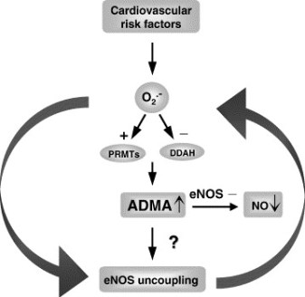
DDAH Says NO to ADMA(1); The DDAH/ADMA/NOS Pathway(2)

Author-Writer-Reporter:  Stephen J. Williams, PhD

Endothelium-derived nitric oxide (NO) has been shown to be vasoprotective.  Nitric oxide enhances endothelial cell survival, inhibits excessive proliferation of vascular smooth muscle cells, regulates vascular smooth muscle tone, and prevents platelets from sticking to the endothelial wall.  Together with evidence from preclinical and human studies, it is clear that impairment of the NOS pathway increases risk of cardiovascular disease (3-5).

This post contains two articles on the physiological regulation of nitric oxide (NO) by an endogenous NO synthase inhibitor asymmetrical dimethylarginine (ADMA) and ADMA metabolism by the enzyme DDAH(1,2).  Previous posts on nitric oxide, referenced at the bottom of the page, provides excellent background and further insight for this posting. In summary plasma ADMA levels are elevated in patients with cardiovascular disease and several large studies have shown that plasma ADMA is an independent biomarker for cardiovascular-related morbidity and mortality(6-8).

admacardiacrisk

admaeffects

Figure 1 A. Cardiac risks of ADMA B. Effects of ADMA (Photo credit: Wikipedia)

ADMA Production and Metabolism

Nuclear proteins such as histones can be methylated on arginine residues by protein-arginine methyltransferases, enzymes which use S-adenosylmethionine as methyl groups.  This methylation event is thought to regulate protein function, much in the way of protein acetylation and phosphorylation (9).  And much like phosphorylation, these modifications are reversible through methylesterases.   The proteolysis of these arginine-methyl modifications lead to the liberation of free guanidine-methylated arginine residues such as L-NMMA, asymmetric dimethylarginine (ADMA) and symmetrical methylarginine (SDMA).

The first two, L-NMMA and ADMA, have been shown to inhibit the activity of the endothelial NOS.  This protein turnover is substantial: for instance the authors note that each day 40% of constitutive protein in adult liver is newly synthesized protein. And in several diseases, such as muscular dystrophy, ischemic heart disease, and diabetes, it has been known since the 1970’s that protein catabolism rates are very high, with corresponding increased urinary excretion of ADMA(10-13).  Methylarginines are excreted in the urine by cationic transport.  However, the majority of ADMA and L-NMMA are degraded within the cell by dimethylaminohydrolase (DDAH), first cloned and purified in rat(14).

endogenous NO inhibitors from pubchem

Figure 2.  Endogenous inhibitors of NO synthase.  Chemical structures generated from PubChem.

DDAH

DDAH specifically hydrolyzes ADMA and L-NMMA to yield citruline and demethylamine and usually shows co-localization with NOS. Pharmacologic inhibition of DDAH activity causes accumulation of ADMA and can reverse the NO-mediated bradykinin-induced relaxation of human saphenous vein.

Two isoforms have been found in human:

  • DDAH1 (found in brain and kidney and associated with nNOS) and
  • DDAH2 (highly expressed in heart, placenta, and kidney and associated with eNOS).

DDAH2 can be upregulated by all-trans retinoic acid (atRA can increase NO production).  Increased reactive oxygen species and possibly homocysteine, a risk factor for cardiovascular disease, can decrease DDAH activity(15,16).

  • The importance of DDAH activity can also be seen in transgenic mice which overexpress DDAH, exhibiting increased NO production, increased insulin sensitivity, and reduced vascular resistance  (17).  Likewise,
  • Transgenic mice, null for the DDAH1, showed increase in blood pressure, decreased NO production, and significant increase in tissue and plasma ADMA and L-NMMA.

amdanosfigure

Figure 3.  The DDAH/ADMA/NOS cycle. Figure adapted from Cooke and Ghebremarian (1).

As mentioned in the article by Cooke and Ghebremariam, the authors state: the weight of the evidence indicates that DDAH is a worthy therapeutic target. Agents that increase DDAH expression are known, and 1 of these, a farnesoid X receptor agonist, is in clinical trials

http://www.interceptpharma.com

An alternate approach is to

  • develop an allosteric activator of the enzyme.  Although
  • development of an allosteric activator is not a typical pharmaceutical approach, recent studies indicate that this may be achievable aim(18).

References:

1.            Cooke, J. P., and Ghebremariam, Y. T. : DDAH says NO to ADMA.(2011) Arteriosclerosis, thrombosis, and vascular biology 31, 1462-1464

2.            Tran, C. T., Leiper, J. M., and Vallance, P. : The DDAH/ADMA/NOS pathway.(2003) Atherosclerosis. Supplements 4, 33-40

3.            Niebauer, J., Maxwell, A. J., Lin, P. S., Wang, D., Tsao, P. S., and Cooke, J. P.: NOS inhibition accelerates atherogenesis: reversal by exercise. (2003) American journal of physiology. Heart and circulatory physiology 285, H535-540

4.            Miyazaki, H., Matsuoka, H., Cooke, J. P., Usui, M., Ueda, S., Okuda, S., and Imaizumi, T. : Endogenous nitric oxide synthase inhibitor: a novel marker of atherosclerosis.(1999) Circulation 99, 1141-1146

5.            Wilson, A. M., Shin, D. S., Weatherby, C., Harada, R. K., Ng, M. K., Nair, N., Kielstein, J., and Cooke, J. P. (2010): Asymmetric dimethylarginine correlates with measures of disease severity, major adverse cardiovascular events and all-cause mortality in patients with peripheral arterial disease. Vasc Med 15, 267-274

6.            Kielstein, J. T., Impraim, B., Simmel, S., Bode-Boger, S. M., Tsikas, D., Frolich, J. C., Hoeper, M. M., Haller, H., and Fliser, D. : Cardiovascular effects of systemic nitric oxide synthase inhibition with asymmetrical dimethylarginine in humans.(2004) Circulation 109, 172-177

7.            Kielstein, J. T., Donnerstag, F., Gasper, S., Menne, J., Kielstein, A., Martens-Lobenhoffer, J., Scalera, F., Cooke, J. P., Fliser, D., and Bode-Boger, S. M. : ADMA increases arterial stiffness and decreases cerebral blood flow in humans.(2006) Stroke; a journal of cerebral circulation 37, 2024-2029

8.            Mittermayer, F., Krzyzanowska, K., Exner, M., Mlekusch, W., Amighi, J., Sabeti, S., Minar, E., Muller, M., Wolzt, M., and Schillinger, M. : Asymmetric dimethylarginine predicts major adverse cardiovascular events in patients with advanced peripheral artery disease.(2006) Arteriosclerosis, thrombosis, and vascular biology 26, 2536-2540

9.            Kakimoto, Y., and Akazawa, S.: Isolation and identification of N-G,N-G- and N-G,N’-G-dimethyl-arginine, N-epsilon-mono-, di-, and trimethyllysine, and glucosylgalactosyl- and galactosyl-delta-hydroxylysine from human urine. (1970) The Journal of biological chemistry 245, 5751-5758

10.          Inoue, R., Miyake, M., Kanazawa, A., Sato, M., and Kakimoto, Y.: Decrease of 3-methylhistidine and increase of NG,NG-dimethylarginine in the urine of patients with muscular dystrophy. (1979) Metabolism: clinical and experimental 28, 801-804

11.          Millward, D. J.: Protein turnover in skeletal muscle. II. The effect of starvation and a protein-free diet on the synthesis and catabolism of skeletal muscle proteins in comparison to liver. (1970) Clinical science 39, 591-603

12.          Goldberg, A. L., and St John, A. C.: Intracellular protein degradation in mammalian and bacterial cells: Part 2. (1976) Annual review of biochemistry 45, 747-803

13.          Dice, J. F., and Walker, C. D.: Protein degradation in metabolic and nutritional disorders. (1979) Ciba Foundation symposium, 331-350

14.          Ogawa, T., Kimoto, M., and Sasaoka, K.: Purification and properties of a new enzyme, NG,NG-dimethylarginine dimethylaminohydrolase, from rat kidney. (1989) The Journal of biological chemistry 264, 10205-10209

15.          Ito, A., Tsao, P. S., Adimoolam, S., Kimoto, M., Ogawa, T., and Cooke, J. P.: Novel mechanism for endothelial dysfunction: dysregulation of dimethylarginine dimethylaminohydrolase. (1999) Circulation 99, 3092-3095

16.          Stuhlinger, M. C., Tsao, P. S., Her, J. H., Kimoto, M., Balint, R. F., and Cooke, J. P. : Homocysteine impairs the nitric oxide synthase pathway: role of asymmetric dimethylarginine.(2001) Circulation 104, 2569-2575

17.          Sydow, K., Mondon, C. E., Schrader, J., Konishi, H., and Cooke, J. P.: Dimethylarginine dimethylaminohydrolase overexpression enhances insulin sensitivity. (2008) Arteriosclerosis, thrombosis, and vascular biology 28, 692-697

18.          Zorn, J. A., and Wells, J. A.: Turning enzymes ON with small molecules. (2010) Nature chemical biology 6, 179-188

Other research papers on Nitric Oxide and Cardiac Risk  were published on this Scientific Web site as follows:

The Nitric Oxide and Renal is presented in FOUR parts:

Part I: The Amazing Structure and Adaptive Functioning of the Kidneys: Nitric Oxide

Part II: Nitric Oxide and iNOS have Key Roles in Kidney Diseases

Part III: The Molecular Biology of Renal Disorders: Nitric Oxide

Part IV: New Insights on Nitric Oxide donors

Cardiac Arrhythmias: A Risk for Extreme Performance Athletes

What is the role of plasma viscosity in hemostasis and vascular disease risk?

Cardiovascular Risk Inflammatory Marker: Risk Assessment for Coronary Heart Disease and Ischemic Stroke – Atherosclerosis.

Endothelial Dysfunction, Diminished Availability of cEPCs, Increasing CVD Risk for Macrovascular Disease – Therapeutic Potential of cEPCs

Biochemistry of the Coagulation Cascade and Platelet Aggregation – Part I

Nitric Oxide Function in Coagulation

Read Full Post »

Histone Deacetylase Inhibitors Induce Epithelial-to-Mesenchymal Transition in Prostate Cancer Cells(1)

Screen Shot 2021-07-19 at 7.44.44 PM

Word Cloud By Danielle Smolyar

Authors: Dejuan Kong, Aamir Ahmad, Bin Bao, Yiwei Li, Sanjeev Banarjee, Fazlul H. Sarkar, Wayne State University School of Medicine

Reporter-Curator: Stephen J. Williams, Ph.D.

Clinically, there has not been much success in treating solid tumors with histone deacetylase inhibitors (HDACi). Histone acetylation and deacetylation play an important role in transcriptional regulation of genes and increased activity is associated with many cancers, therefore it was thought that HDAC inhibition might be fruitful as a therapy.  There have been several phase I and II clinical trials using HDACi for treatment of various malignancies, including hematological and solid malignancies(2), with most success seen in hematologic malignancies such as cutaneous T-cell lymphoma and peripheral T-cell lymphoma and little or no positive outcome with solid tumors.  Many mechanisms of resistance to HDACi in solid tumors have been described, most of which are seen with other chemotherapeutics such as increased multidrug resistance gene MDR1, increased anti-apoptotic proteins and activation of cell survival pathways(3).

A report in PLOS One by Dr. Dejuan Kong, Dr. Fazlul Sarkar, and colleagues from Wayne State University School of Medicine, demonstrate another possible mechanism of resistance to HDACi in prostate cancer, by induction of the epithelial-to-mesenchymal transition (EMT), which has been associated with the development of resistance to chemotherapies in other malignancies of epithelial origin(4,5).

EMT is an important differentiation process in embryogenesis and felt to be important in progression of cancer.  Epithelial cells will acquire a mesenchymal morphology (on plastic this looks like a cuboidal epithelial cell gaining a more flattened, elongated, tri-corner morphology; see paper Figure 1) and down-regulate epithelial markers such as cytokeratin, up-regulation of mesenchymal markers, increased migration and invasiveness in standard assays, and increased resistance to chemotherapeutics, and similarity to cancer stem cells(6-10).

ImageFigure 1. HDACis led to the induction of EMT phemotype. (A and B) PC3 cells treated with TSA and SAHA for 24 h at indicated doses.  The photomicrographs of PC3 cells treated with TSA and SAHA exhibited a fibroblastic-type phenotype, while cells treated with DMAO control displayed rounded epithelial cell morphology (original magnification, x 100). (C) Treated PC3 cells show increased mesenchymal markers vimentin and ZEB1 and F-actin reorganization.  Figure taken from Kong, D., Ahmad, A., Bao, B., Li, Y., Banerjee, S., and Sarkar, F. H. (2012) PloS one 7, e45045

In this study the authors found that treatment of prostate carcinoma cells with two different HDACis (trichostatin A (TSA) and suberoylanilide hydroxamic acid (SAHA)) induced EMT phenotype mediated through up-regulation of transcription factors ZEB1, ZEB2 and Slug, increased expression of mesenchymal markers vimentin, N-cadherin and fibronectin by promoting histone 3 acetylation on gene promoters.  In addition TSA increased the stem cell markers Sox2 and Nanog with concomitant EMT morphology and increased cell motility.

Below is the abstract of this paper(1):

ABSTRACT

Clinical experience of histone deacetylase inhibitors (HDACIs) in patients with solid tumors has been disappointing; however, the molecular mechanism of treatment failure is not known. Therefore, we sought to investigate the molecular mechanism of treatment failure of HDACIs in the present study. We found that HDACIs Trichostatin A (TSA) and Suberoylanilide hydroxamic acid (SAHA) could induce epithelial-to-mesenchymal transition (EMT) phenotype in prostate cancer (PCa) cells, which was associated with changes in cellular morphology consistent with increased expression of transcription factors ZEB1, ZEB2 and Slug, and mesenchymal markers such as vimentin, N-cadherin and Fibronectin. CHIP assay showed acetylation of histone 3 on proximal promoters of selected genes, which was in part responsible for increased expression of EMT markers. Moreover, TSA treatment led to further increase in the expression of Sox2 and Nanog in PCa cells with EMT phenotype, which was associated with cancer stem-like cell (CSLC) characteristics consistent with increased cell motility. Our results suggest that HDACIs alone would lead to tumor aggressiveness, and thus strategies for reverting EMT-phenotype to mesenchymal-to-epithelial transition (MET) phenotype or the reversal of CSLC characteristics prior to the use of HDACIs would be beneficial to realize the value of HDACIs for the treatment of solid tumors especially PCa.

Highlights of the research include:

  • TSA and SAHA induce morphologic changes  in prostate carcinoma LNCaP and PC3 cells related to EMT by microscopy as well as accumulation of mesenchymal markers ZEB1, vimentin, and F-actin reorganization shown by immunofluorescence microscopy and increased expression of these markers shown by real-time PCR
  • Western blotting showed TSA treatment resulted in hyperacetyulation of histone 3 whi8le CHIP analysis revealed increased histone 3 acetylation on the promoters of vimentin, ZEB2, Slug, and MMP2
  • Western analysis revealed that HDACi not only induced EMT but increased the expression of cancer stem cell markers associated with increased motility such as Sox2 and Nanog.  Increased cell migration was measured by Transwell migration assays and increased cell motility was measured via cell detachment assays

1.            Kong, D., Ahmad, A., Bao, B., Li, Y., Banerjee, S., and Sarkar, F. H. (2012) PloS one 7, e45045

2.            Bertino, E. M., and Otterson, G. A. (2011) Expert opinion on investigational drugs 20, 1151-1158

3.            Robey, R. W., Chakraborty, A. R., Basseville, A., Luchenko, V., Bahr, J., Zhan, Z., and Bates, S. E. (2011) Molecular pharmaceutics 8, 2021-2031

4.            Wang, Z., Li, Y., Kong, D., Banerjee, S., Ahmad, A., Azmi, A. S., Ali, S., Abbruzzese, J. L., Gallick, G. E., and Sarkar, F. H. (2009) Cancer research 69, 2400-2407

5.            Wang, Z., Li, Y., Ahmad, A., Azmi, A. S., Kong, D., Banerjee, S., and Sarkar, F. H. (2010) Drug resistance updates : reviews and commentaries in antimicrobial and anticancer chemotherapy 13, 109-118

6.            Hugo, H., Ackland, M. L., Blick, T., Lawrence, M. G., Clements, J. A., Williams, E. D., and Thompson, E. W. (2007) Journal of cellular physiology 213, 374-383

7.            Thiery, J. P. (2002) Nature reviews. Cancer 2, 442-454

8.            Kong, D., Banerjee, S., Ahmad, A., Li, Y., Wang, Z., Sethi, S., and Sarkar, F. H. (2010) PloS one 5, e12445

9.            Kong, D., Li, Y., Wang, Z., and Sarkar, F. H. (2011) Cancers 3, 716-729

10.          Bao, B., Wang, Z., Ali, S., Kong, D., Li, Y., Ahmad, A., Banerjee, S., Azmi, A. S., Miele, L., and Sarkar, F. H. (2011) Cancer letters 307, 26-36

Other research papers on Cancer and Cancer Therapeutics were published on this Scientific Web site as follows:

PIK3CA mutation in Colorectal Cancer may serve as a Predictive Molecular Biomarker for adjuvant Aspirin therapy

Nanotechnology Tackles Brain Cancer

Response to Multiple Cancer Drugs through Regulation of TGF-β Receptor Signaling: a MED12 Control

Personalized medicine-based cure for cancer might not be far away

GSK for Personalized Medicine using Cancer Drugs needs Alacris systems biology model to determine the in silico effect of the inhibitor in its “virtual clinical trial”

Lung Cancer (NSCLC), drug administration and nanotechnology

Non-small Cell Lung Cancer drugs – where does the Future lie?

Cancer Innovations from across the Web

arrayMap: Genomic Feature Mining of Cancer Entities of Copy Number Abnormalities (CNAs) Data

How mobile elements in “Junk” DNA promote cancer. Part 1: Transposon-mediated tumorigenesis.

Cancer Genomics – Leading the Way by Cancer Genomics Program at UC Santa Cruz

Closing the gap towards real-time, imaging-guided treatment of cancer patients.

Closing the gap towards real-time, imaging-guided treatment of cancer patients.

mRNA interference with cancer expression

Search Results for ‘cancer’ on this web site

Cancer Genomics – Leading the Way by Cancer Genomics Program at UC Santa Cruz

Closing the gap towards real-time, imaging-guided treatment of cancer patients.

Lipid Profile, Saturated Fats, Raman Spectrosopy, Cancer Cytology

mRNA interference with cancer expression

Pancreatic cancer genomes: Axon guidance pathway genes – aberrations revealed

Biomarker tool development for Early Diagnosis of Pancreatic Cancer: Van Andel Institute and Emory University

Is the Warburg Effect the cause or the effect of cancer: A 21st Century View?

Crucial role of Nitric Oxide in Cancer

Targeting Glucose Deprived Network Along with Targeted Cancer Therapy Can be a Possible Method of Treatment

Read Full Post »

Subtitle: Nitric Oxide, Peroxinitrite, and NO donors in Renal Function Loss

Curator and Author: Larry H. Bernstein, MD, FCAP

The Nitric Oxide and Renal is presented in FOUR parts:

Part I: The Amazing Structure and Adaptive Functioning of the Kidneys: Nitric Oxide

Part II: Nitric Oxide and iNOS have Key Roles in Kidney Diseases

Part III: The Molecular Biology of Renal Disorders: Nitric Oxide

Part IV: New Insights on Nitric Oxide donors 

Conclusion to this series is presented in

The Essential Role of Nitric Oxide and Therapeutic NO Donor Targets in Renal Pharmacotherapy

The criticality of renal function is easily overlooked until significant loss of nephron mass is overtly seen.  The kidneys become acutely and/or chronically dysfunctional in metabolic,  systemic inflammatory and immunological diseases of man.  We have described how the key role that nitric oxide and the NO synthases (eNOS and iNOS) play competing roles in reduction or in the genesis of reactive oxygen  species.  There is  a balance to be struck between pro- and anti-oxidative as well as inflammatory elements for avoidance of diseases, specifically involving the circulation, but effectively not limited to any organ system.  In this discussion we shall look at kidney function, NO and NO donors.  This is an extension of a series of posts on NO and NO related disorders.

Part IV. New Insights on NO donors


This study investigated the involvement of nitric oxide (NO) into the irradiation-induced increase of cell attachment. These experiments explored the cellular mechanisms of low-power laser therapy.

HeLa cells were irradiated with a monochromatic visible-tonear infrared radiation (600–860 nm, 52 J/m2) or with a diode laser (820 nm, 8–120 J/m2) and the number of cells attached to a glass matrix was counted after 30 minute incubation at 37oC. The NO donors

  • sodium nitroprusside (SNP),
  • glyceryl trinitrate (GTN), or
  • sodium nitrite (NaNO2)

were added to the cellular suspension before or after irradiation. The action spectra and the concentration and fluence dependencies obtained were compared and analyzed. The well-structured action spectrum for the increase of the adhesion of the cells, with maxima at 619, 657, 675, 740, 760, and 820 nm, points to the existence of a photoacceptor responsible for the enhancement of this property (supposedly cytochrome c oxidase, the terminal respiratory chain enzyme), as well as signaling pathways between the cell mitochondria, plasma membrane, and nucleus. Treating the cellular suspension with SNP before irradiation significantly modifies the action spectrum for the enhancement of the cell attachment property (band maxima at 642, 685, 700, 742, 842, and 856 nm).

The action of SNP, GTN, andNaNO2 added before or after irradiation depends on their concentration and radiation fluence.

The NO donors added to the cellular suspension before irradiation eliminate the radiation induced increase in the number of cells attached to the glass matrix, supposedly by way of binding NO to cytochrome c oxidase. NO added to the suspension after irradiation can also inhibit the light-induced signal downstream. Both effects of NO depend on the concentration of the NO donors added. The results indicate that NO can control the irradiation-activated reactions that increase the attachment of cells.

Karu TI, Pyatibrat LV, and Afanasyeva NI. Cellular Effects of Low Power Laser Therapy Can be Mediated by Nitric Oxide. Lasers Surg. Med 2005; 36:307–314.

Interferon a-2b (IFN-a) effect on barrier function of renal tubular epithelium


IFNa treatment can be accompanied by impaired renal function and capillary leak.  This study shows IFNa produced dose-dependent and time-dependent decrease in transepithelial resistance (TER) ameliorated by tyrphostin, an inhibitor of phosphotyrosine kinase with increased expression of occludin and E-cadherin In conclusion, IFNa can directly affect barrier function in renal epithelial cells via overexpression or missorting of the junctional proteins occludin and E-cadherin.

Lechner J, Krall M, Netzer A, Radmayr C, et al.  Effects of interferon a-2b on barrier function and junctional complexes of renal proximal tubular LLC-pK1 cells. Kidney Int 1999; 55:2178-2191.

Ischemia-reperfusion injury


The pathophysiology of acute renal failure (ARF) is complex and not well understood. Numerous models of ARF suggest that oxygen-derived reactive species are important in renal ischemia-reperfusion (I-R) injury, but the nature of the mediators is still controversial. Treatment with

  • oxygen radical scavengers,
  • antioxidants, and
  • iron chelators such as
  1.                   superoxide dismutase,
  2.                   dimethylthiourea,
  3.                   allopurinol, and
  4.                    deferoxamine

are protective in some models, and suggest a role for the hydroxyl radical formation. However, these compounds are not protective in all models of I-R injury, and direct evidence for the generation of hydroxyl radical is absent. Furthermore, these inhibitors have another property in common. They all directly scavenge or inhibit the formation of peroxynitrite (ONOO−), a highly toxic species derived from nitric oxide (NO) and superoxide. Thus, the protective effects seen with these inhibitors may be due in part to their ability to inhibit ONOO− formation.

Even though reactive oxygen species are thought to participate in ischemia-reperfusion (I-R) injury,  induction of inducible nitric oxide synthase (iNOS) and production of high levels of nitric oxide (NO) also contribute to this injury. NO can combine with superoxide to form the potent oxidant peroxynitrite (ONOO−). NO and ONOO− were investigated in a rat model of renal I-R injury using the selective iNOS inhibitor L-N6-(1-iminoethyl)lysine (L-NIL).

I-R surgery significantly increased plasma creatinine levels to 1.9 ± 0.3 mg/dl (P < .05) and caused renal cortical necrosis. L-NIL administration (3 mg/kg) in animals subjected to I-R significantly decreased plasma creatinine levels to 1.2 ± 0.10 mg/dl (P < .05 compared with I-R) and reduced tubular damage.

ONOO− formation was evaluated by detecting 3-nitrotyrosine-protein adducts, a stable biomarker of ONOO− formation.

  1. The kidneys from I-R animals had increased levels of 3-nitrotyrosine-protein adducts compared with control animals
  2. L-NIL-treated rats (3 mg/kg) subjected to I-R showed decreased levels of 3-nitrotyrosine-protein adducts.

These results support the hypothesis that iNOS-generated NO mediates damage in I-R injury possibly through ONOO− formation.

In summary, 3-nitrotyrosine-protein adducts were detected in renal tubules after I-R injury. Selective inhibition of iNOS by L-NIL

  • decreased injury,
  • improved renal function, and
  • decreased apparent ONOO− formation.

Reactive nitrogen species should be considered potential therapeutic targets in the prevention and treatment of renal I-R injury.

Walker LM, Walker PD, Imam SZ, et al. Evidence for Peroxynitrite Formation in Renal Ischemia-Reperfusion Injury: Studies with the Inducible Nitric Oxide Synthase InhibitorL-N6-(1-Iminoethyl)-lysine1.  2000.

Role of TNFa independent of iNOS

Renal failure is a frequent complication of sepsis, mediated by renal vasoconstrictors and vasodilators. Endotoxin induces several proinflammatory cytokines, among which tumor necrosis factor (TNF) is thought to be of major importance. Tumor necrosis factor (TNF) has been suggested to be a factor in the acute renal failure in sepsis or endotoxemia. Passive immunization by anti-TNFa prevented development of septic shock in animal experiments. The development of ARF involves excessive intrarenal vasoconstriction.

Recent studies also suggest involvement of nitric oxide (NO), generated by inducible NO synthase (iNOS), in the pathogenesis of endotoxin-induced renal failure. TNF-a leads to a decrease in glomerular filtration rate (GFR). The present study tested the hypothesis that the role of TNF-a in endotoxic shock related ARF is mediated by iNOS-derived NO.


An injection of lipopolysaccharide (LPS) constituent of gram-negative bacteria to wild-type mice resulted in a 70% decrease in glomerular filtration rate (GFR) and in a 40% reduction in renal plasma flow (RPF) 16 hours after the  injection.

The results occurred independent of

  • hypotension,
  • morphological changes,
  • apoptosis, and
  • leukocyte accumulation.

In mice pretreated with TNFsRp55, only a 30% decrease in GFR was observed without a significant change in RPF as compared with controls.


Effect of TNFsRp55 (10 mg/kg IP) on renal function in wild-type mice.

Mice were pretreated with TNFsRp55 for one hour before the administration of 5 mg/kg intraperitoneal endotoxin. GFR (A) and RPF (B) were determined 16 hours thereafter. Data are expressed as mean 6, SEM, N 5 6. *P , 0.05 vs. Control; §P , 0.05 vs. LPS, by ANOVA.


The serum NO concentration was significantly lower in endotoxemic wild-type mice pretreated with TNFsRp55, as compared with untreated endotoxemic wild-type mice. In LPS-injected iNOS knockout mice and wild-type mice treated with a selective iNOS inhibitor, 1400W, the development of renal failure was similar to that in wild-type mice. As in wild-type mice,TNFsRp55 significantly attenuated the decrease in GFR (a 33% decline, as compared with 75% without TNFsRp55) without a significant change in RPF in iNOS knockout mice given LPS.


These results demonstrate a role of TNF in the early renal dysfunction (16 h) in a septic mouse model independent of

  • iNOS,
  • hypotension,
  • apoptosis,
  • leukocyte accumulation,and
  • morphological alterations,

thus suggesting renal hypoperfusion secondary to an imbalance between, as yet to be defined renal vasoconstrictors and vasodilators.

Knotek M, Rogachev B, Wang W,….., Edelstein CL, Dinarello CA, and Schrier RW.  Endotoxemic renal failure in mice: Role of tumor necrosis factor independent of inducible nitric oxide synthase. Kidney International 2001; 59:2243–2249

Ischemic acute renal failure

Inflammation plays a major role in the pathophysiology of acute renal failure resulting from ischemia. In this review, we discuss the contribution of endothelial and epithelial cells and leukocytes to this inflammatory response. The roles of cytokines/chemokines in the injury and recovery phase are reviewed. The ability of the mouse kidney to be protected by prior exposure to ischemia or urinary tract obstruction is discussed as a potential model to emulate as we search for pharmacologic agents that will serve to protect the kidney against injury.

the inflammatory mediators produced by tubular epithelial cells and activated leukocytes in renal ischemia/reperfusion (I/R) injury.

Tubular epithelia produce TNF-a, IL-1, IL-6, IL-8, TGF-b, MCP-1, ENA-78, RANTES, and fractalkines, whereas leukocytes produce TNF-a, IL-1, IL-8, MCP-1, ROS, and eicosanoids. The release of these chemokines and cytokines serve as effectors for a positive feedback pathway enhancing inflammation and cell injury

the cycle of tubular epithelial cell injury and repair following renal ischemia/reperfusion.

Tubular epithelia are typically cuboidal in shape and apically-basally polarized; the Na+/K+-ATPase localizes to basolateral plasma membranes, whereas cell adhesion molecules, such as integrins localize basally. In response to ischemia reperfusion, the Na+/K+-ATPase appears apically, and integrins are detected on lateral and basal plasma membranes.

Some of the injured epithelial cells undergo necrosis and/or apoptosis detaching from the underlying basement membrane into the tubular space where they contribute to tubular occlusion. Viable cells that remain attached,

  • dedifferentiate,
  • spread, and
  • migrate to
  • repopulate the denuded basement membrane.

With cell proliferation, cell-cell and cell-matrix contacts are restored, and the epithelium redifferentiates and repolarizes, forming a functional, normal epithelium

Inflammation is a significant component of renal I/R injury, playing a considerable role in its pathophysiology. Although significant progress has been made in defining the major components of this process, the complex cross-talk between endothelial cells, inflammatory cells, and the injured epithelium with each generating and often responding to cytokines and chemokines is not well understood. In addition, we have not yet taken full advantage of the large body of data on inflammation in other organ systems.

Furthermore, preconditioning the kidney to afford protection to subsequent bouts of ischemia may serve as a useful model challenging us to therapeutically mimic endogenous mechanisms of protection. Understanding the inflammatory response prevalent in ischemic kidney injury will facilitate identification of molecular targets for therapeutic intervention.

Bonventre JV and Zuk A. Ischemic acute renal failure: An inflammatory disease? Forefronts in Nephrology  2002;.. :480-485

Gene expression profiles in renal proximal tubules

In kidney disease renal proximal tubular epithelial cells (RPTEC) actively contribute to the progression of tubulointerstitial fibrosis by mediating both an inflammatory response and via epithelial-to-mesenchymal transition. Using laser capture microdissection we specifically isolated RPTEC from cryosections of the healthy parts of kidneys removed owing to renal cell carcinoma and from kidney biopsies from patients with proteinuric nephropathies. RNA was extracted and hybridized to complementary DNA microarrays after linear RNA amplification. Statistical analysis identified 168 unique genes with known gene ontology association, which separated patients from controls.

Besides distinct alterations in signal-transduction pathways (e.g. Wnt signalling), functional annotation revealed a significant upregulation of genes involved in

  • cell proliferation and cell cycle control (like insulin-like growth factor 1 or cell division cycle 34),
  • cell differentiation (e.g. bone morphogenetic protein 7),
  • immune response,
  • intracellular transport and
  • metabolism

in RPTEC from patients.

On the contrary we found differential expression of a number of genes responsible for cell adhesion (like BH-protocadherin) with a marked downregulation of most of these transcripts. In summary, our results obtained from RPTEC revealed a differential regulation of genes, which are likely to be involved in

  • either pro-fibrotic or
  • tubulo-protective mechanisms

in proteinuric patients at an early stage of kidney disease.

Rudnicki M, Eder S, Perco P, Enrich J, et al. Gene expression profiles of human proximal tubular epithelial cells in proteinuric nephropathies. Kidney International 2006; xx:1-11.

Kidney International advance online publication, 20 December 2006; doi:10.1038/sj.ki.5002043. http://www.kidney-international.org


Oxidative stress involved in diabetic nephropathy

Diabetic Nephropathy (DN) poses a major health problem. There is strong evidence for a potential role of the eNOS gene. The aim of this case control study was to investigate the possible role of genetic variants of the endothelial Nitric Oxide Synthase (eNOS) gene and oxidative stress in the pathogenesis of nephropathy in patients with diabetes mellitus.

The study included 124 diabetic patients;

  • 68 of these patients had no diabetic nephropathy (group 1) while
  • 56 patients exhibited symptoms of diabetic nephropathy (group 2).
  • Sixty two healthy non-diabetic individuals were also included as a control group.

Blood samples from subjects and controls were analyzed to investigate the eNOS genotypes and to estimate the lipid profile and markers of oxidative stress such as malondialdehyde (MDA) and nitric oxide (NO). No significant differences were found in the frequency of eNOS genotypes between diabetic patients (either in group 1 or group 2) and controls (p >0.05).

Also, no significant differences were found in the frequency of eNOS genotypes between group 1 and group 2 (p >0.05).

Both group 1 and group 2 had significantly higher levels of nitrite and MDA when compared with controls (all p = 0.0001). Also group 2 patients had significantly higher levels of nitrite and MDA when compared with group 1 (p = 0.02, p = 0.001 respectively).

The higher serum level of the markers of oxidative stress in diabetic patients particularly those with diabetic nephropathy suggest that oxidative stress and not the eNOS gene polymorphism is involved in the pathogenesis of the diabetic nephropathy in this subset of patients

Badawy A, Elbaz R, Abbas AM, Ahmed Elgendy A, et al. Oxidative stress and not endothelial Nitric Oxide Synthase gene polymorphism involved in diabetic nephropathy. Journal of Diabetes and Endocrinology 2011; 2(3): 29-35.

Metformin in renal ischemia reperfusion


Renal ischemia plays an important role in renal impairment and transplantation.

Metformin is a biguanide used in type 2 diabetes, it inhibits hepatic glucose production and increases peripheral insulin sensitivity.  While the mode of action of metformin is incompletely understood, it appears to have anti-inflammatory and antioxidant effects involved in its beneficial effects on insulin resistance.

Control, Sham, ischemia/reperfusion (I/R) and Metformin treated I /R groups

A renal I/R injury was done by a left renal pedicle occlusion to induce ischemia for 45 min followed by 60 min of reperfusion with contralateral nephrectomy. Metformin pretreated I/R rats in a dose of 200 mg/kg/day for three weeks before ischemia induction.

Nitric oxide (NO), tumor necrosis factor alpha (TNF α) , catalase (CAT) and reduced glutathione (GSH) activities were determined in renal tissue, while creatinine clearance (CrCl) , blood urea nitrogen (BUN) were measured and 5 hour urinary volume and electrolytes were estimated .

BUN and CrCl levels in the I/R group were significantly higher than in control rats (p<0.05) table (1).

Table 1: Creatinine clearance (Cr Cl) and blood urea nitrogen( BUN) levels in control and test groups. Mean ± SD.

Groups CrCl (ml/min) BUN mg/dl
Control group 1.30 ±0.11 14.30±0.25
Sham group+metformin 1.27±0.09 15.70±0.19
I/R group P1 1.85±0.25<0.001*** 28.00±0.62<0.001***
I/R+metformin group P2 P3 1.55±0.220.001**0.028* 18.10±1.00<0.001***<0.001***

P1: Statistical significance between control group and saline treated I/R group.

P2 Statistical significance between control group and Metformin treated I/R group.

P3 Statistical significance between saline treated I/R group and Metformin treated I/R group.

When metformin was administered before I/R, BUN and CrCl levels were still significantly higher than control group but their elevation were significantly lower in comparison to I/R group alone (P<0.05).

TNF α and NO levels were significantly higher in the I/R group than those of the control group (Table 2).

Pre-treatment with metformin significantly lowered their levels in comparison to I/R group (P<0.05).


Table 2: Tumour necrosis factior α (TNF α)and inducible nitric oxide (iNO)levels in control and test groups. (Mean ± SD).

Groups TNF α (pmol/mg tissue) iNO nmol/ mg tissue
Control group 17.60 ±5.98 2.54 ± 0.82
Sham group+ metformin 16.70 ±5.50 2.35 ±0.80
I/R group P1 54. 00±6.02<0.001*** 4.50±0.89<0.001**
I/R+ metformin group P2 P3 39 ± 14.01<0.001***0.006** 3.53±0.950.02*0.03*

P1: Statistical significance between control group and saline treated I/R group.

P2 Statistical significance between control group and Metformin treated I/R group.

P3 Statistical significance between saline treated I/R group and Metformin treated I/R group

These results showed significant increase in

  • NO,
  • TNF α,
  • BUN ,
  • CrCl and

significant decrease in

  • urinary volume ,
  • electrolytes,
  • CAT and
  • GSH activities

in the I/R group than those in the control group.

Metformin

  1. decreased significantly NO, TNF α, BUN and CrCl while
  2. increased urinary volume, electrolytes, CAT and GSH activities.

Lipid peroxidation is related to I/R induced tissue injury. Production of inducible NO synthase (NOS) under lipid peroxidation and inflammatory conditions results in the induction of NO

which react with

  • O2
  • liberating peroxynitrite (OONO).

NO itself inactivates the antioxidant enzyme system CAT and GSH.

Alteration in NO synthesis have been observed in other kidney injuries as nephrotoxicity and acute renal failure induced by endotoxins. Treatment with iNOS inhibitors improved renal function and decreased peroxynitrite radical which is believed to be responsible for the shedding of proximal convoluted tubules in I/R.

Metformin produced anti-inflammatory renoprotective effect on CrCl and diuresis in renal I/R injury.


Malek HA. The possible mechanism of action of metformin in renal ischemia reperfusion in rats.  The Pharma Research Journal 2011; 6(1):42-49.

Possible role of NO donors in ARF


The L-arginine-nitric oxide (NO) pathway has been implicated in many physiological functions in the kidney, including

  • regulation of glomerular hemodynamics,
  • mediation of pressure-natriuresis,
  • maintenance of medullary perfusion,
  • blunting of tubuloglomerular feedback (TGF),
  • inhibition of tubular sodium reabsorption and
  • modulation of renal sympathetic nerve activity.

Its net effect in the kidney is to promote natriuresis and diuresis, contributing to adaptation to variations of dietary salt intake and maintenance of normal blood pressure.

RAS Renal hemodynamics Sodium balance
  Medullary perfusion                                           
  Pressure-natriuresis  
     
Salt intake Tubulo-glomerular feedback Blood pressure
  Tubular sodium reabsorption  
     
Blood pressure Renal sympathetic activity Regulation
Extrarenal factors Intrarenal functions Physiological roles


Role of nitric oxide in renal physiology. RAS, renin-angiotensin system

Nitric oxide has been implicated in many physiologic processes that influence both acute and long-term control of kidney function. Its net effect in the kidney is to promote natriuresis and diuresis, contributing to adaptation to variations of dietary salt intake and maintenance of normal blood pressure. A pretreatment with nitric oxide donors or L-arginine may prevent the ischemic acute renal injury. In chronic kidney diseases, the systolic blood pressure is correlated with the plasma level of asymmetric dimethylarginine, an endogenous inhibitor of nitric oxide synthase.

A reduced production and biological action of nitric oxide is associated with an elevation of arterial pressure, and conversely, an exaggerated activity may represent a compensatory mechanism to mitigate the hypertension.

JongUn Lee. Nitric Oxide in the Kidney : Its Physiological Role and Pathophysiological Implications. Electrolyte & Blood Pressure 2008; 6:27-34.

Renal Hypoxia and Dysoxia following Reperfusion


Acute renal failure (ARF) is a common condition which develops in 5% of hospitalized patients. Of the patients who develop ARF, ~10% eventually require renal replacement therapy. Among critical care patients who have acute renal failure and survive, 2%-10% develop terminal renal failure and require long-term dialysis.

The kidneys are particularly susceptible to ischemic injury in many clinical conditions such as renal transplantation, treatment of suprarenal aneurysms, renal artery reconstructions, contrast-agent induced nephropathy, cardiac arrest, and shock. One reason for renal sensitivity to ischemia is that the kidney microvasculature is highly complex and must meet a high energy demand. Under normal, steady state conditions, the oxygen (O2) supply to the renal tissues is well in excess of oxygen demand.

Under pathological conditions, the delicate balance of oxygen supply versus demand is easily disturbed due to the unique arrangement of the renal microvasculature and its increasing numbers of diffusive shunting pathways.

The renal microvasculature is serially organized, with almost all descending vasa recta emerging from the efferent arterioles of the juxtamedullary glomeruli. Adequate tissue oxygenation is thus partially dependent on the maintenance of medullary perfusion by adequate cortical perfusion. This, combined with the low amount of medullary blood flow (~10% of total renal blood flow) in the U-shaped microvasculature of the medulla allows O2 shunting between the descending and ascending vasa recta and contributes to the high sensitivity of the medulla and cortico-medullary junction to decreased O2 supply.

Whereas past investigations have focused mainly on tubular injury as the main cause of ischemia-related acute renal failure, increasing evidence implicates alterations in the intra-renal microcirculation pathway and in the O2 handling. Indeed, although acute tubular necrosis (ATN) has classically been believed to be the leading cause of ARF, data from biopsies in patients with ATN have shown few or no changes consistent with tubular necrosis. The role played by microvascular dysfunction, however, has generated increasing interest. The complex pathophysiology of ischemic ARF includes the inevitable reperfusion phase associated with oxidative stress, cellular dysfunction and altered signal transduction.

During this process, alterations in oxygen transport pathways can result in cellular hypoxia and/or dysoxia. In this context, the distinction between hypoxia and dysoxia is that cellular hypoxia refers to the condition of decreased availability of oxygen due to inadequate convective delivery from the microcirculation. Cellular dysoxia, in contrast, refers to a pathological condition where the ability of mitochondria to perform oxidative phosphorylation is limited, regardless of the amount of available oxygen. The latter condition is associated with mitochondrial failure and/or activation of alternative pathways for oxygen consumption. Thus, we would expect that an optimal balance between oxygen supply and demand is essential to reducing damage from renal ischemia-reperfusion (I/R) injury (Figure 1).

Complex interactions exist between tubular injury, microvascular injury, and inflammation after renal I/R. On the one hand, insults to the tubule cells promotes the liberation of a number of inflammatory mediators, such as TNF-á, IL-6, TGF-â, and chemotactic cytokines (RANTES, monocyte chemotactic protein-1, ENA-78, Gro-á, and IL-8). On the other hand, chemokine production can promote leukocyte-endothelium interactions and leukocyte activation, resulting in renal blood flow impairment and the expansion of tubular damage.

renal hemodynamics and electrolyte reabsorption

Adequate medullary tissue oxygenation, in terms of balanced oxygen supply and demand, is dependent on the maintenance of medullary perfusion by adequate cortical perfusion and also on the high rate of O2 consumption required for active electrolyte transport. Furthermore, renal blood flow is closely associated with renal sodium transport.

In addition to having a limited O2 supply due to the anatomy of the microcirculation anatomy, the sensitivity of the medulla to hypoxic conditions results from this high O2 consumption. Renal sodium transport is the main O2-consuming function of the kidney and is closely linked to renal blood flow for sodium transport, particularly in the thick ascending limbs of the loop of Henle and the S3 segments of the proximal tubules.

Medullary renal blood flow is also highly dependent on cortical perfusion, with almost all descending vasa recta emerging from the efferent arteriole of juxta medullary glomeruli. A profound reduction in cortical perfusion can disrupt medullary blood flow and lead to an imbalance between O2 supply and O2 consumption. On theother hand, inhibition of tubular reabsorption by diuretics increases medullary pO2 by decreasing the activity of Na+/K+-ATPases and local O2 consumption.


Mitochondrial activity and NO-mediated O2 consumption

The medulla has been found to be the main site of production of NO in the kidney. In addition to the actions described above, NO appears to be a key regulator of renal tubule cell metabolism by inhibiting the activity of the Na+-K+-2Cl cotransporter and reducing Na+/H+ exchange. Since superoxide (O2) is required to inhibit solute transport activity, it was assumed that these effects were mediated by peroxynitrite (OONO). Indeed, mitochondrial nNOS upregulation, together with an increase in NO production, has been shown to increase mitochondrial peroxynitrite generation, which in turn, can induce cytochrome c release and promote apoptosis. NO has also been shown to directly compete with O2 at the mitochondrial level. These findings support the idea that NO acts as an endogenous regulator to match O2 supply to O2 consumption, especially in the renal medulla.

NO reversibly binds to the O2 binding site of cytochrome oxidase, and acts as a potent, rapid, and reversible inhibitor of cytochrome oxidase in competition with molecular O2. This inhibition could be dependent on the O2 level, since the IC50 (the concentration of NO that reduces the specified response by half) decreases with reduction in O2 concentration. The inhibition of electron flux at the cytochrome oxidase level switches the electron transport chain to a reduced state, and consequently leads to depolarization of the mitochondrial membrane potential and electron leakage.


To summarize, while the NO/O2 ratio can act as a regulator of cellular O2 consumption by matching decreases in O2 delivery to decreases in cellular O2 cellular, the inhibitory effect of NO on mitochondrial respiration under hypoxic conditions further impairs cellular aerobic metabolism This leads to a state of “cytopathic hypoxia,” as described in the sepsis literature.

Only cell-secreted NO competes with O2 and to regulate mitochondrial respiration. In addition to the 3 isoforms (eNOS, iNOS, cnNOS), an α-isoform of neuronal NOS, the mitochondrial isoform (mNOS) located in the inner mitochondrial membrane, has also been shown to regulate mitochondrial respiration.

These data support a role for NO in the balanced regulation of renal O2 supply and O2 consumption after renal I/R However, the relationships between the determinants of O2 supply, O2 consumption, and renal function, and their relation to renal damage remain largely unknown.

Sustained endothelial activation

Ischemic renal failure leads to persistent endothelial activation, mainly in the form of endothelium-leukocyte interactions and the activation of adhesion molecules.

This persistent activation can

  • compromise renal blood flow,
  • prevent the recovery of adequate tissue oxygenation, and
  • jeopardize tubular cell survival despite the initial recovery of renal tubular function.

A 30-50% reduction in microvascular density was seen 40 weeks after renal ischemic injury in a rat model. Vascular rarefaction has been proposed to induce chronic hypoxia resulting in tubulointerstitial fibrosis via the molecular activation of fibrogenic factors such as

  • transforming growth factor (TGF)-β,
  • collagen, and
  • fibronectin,

all of which may play an important role in the progression of chronic renal disease.

Adaptation to hypoxia


Over the last decade, the role of hypoxia-inducible factors (HIFs) in O2 supply and adaptation to hypoxic conditions has found increasing support. HIFs are O2-sensitive transcription factors involved in O2-dependent gene regulation that mediate cellular adaptation to O2 deprivation and tissue protection under hypoxic conditions in the kidney.

NO generation can promote HIF-1α accumulation in a cGMP-independent manner. However, Hagen et al. (2003) showed that NO may reduce the activation of HIF in hypoxia via the inhibitory effect of NO on cytochrome oxidase. Therefore, it seems that NO has pleiotropic effects on HIF expression, with various responses related to different pathways.

HIF-1α upregulates a number of factors implicated in cytoprotection, including angiogenic growth factors, such as

  • vascular endothelial growth factors (VEGF),
  • endothelial progenitor cell recruitment via the endothelial expression of SDF-1,
  • heme-oxygenase-1 (HO-1), and
  • erythropoietin (EPO), and
  • vasomotor regulation.

HO-1 produces carbon monoxide (a potent vasodilator) while degrading heme, which may preserve tissue blood flow during reperfusion. Thus, it has been suggested that

  • the induction of HO-1 can protect the kidney from ischemic damage by decreasing oxidative damage and NO generation.
  • in addition to its anti-apoptotic properties, EPO may protect the kidney from ischemic damage by restoring the renal microcirculation

(by stimulating the mobilization and differentiation of progenitor cells toward an endothelial phenotype and by inducing NO release from eNOS).


Pharmacological interventions

Use of pharmacological interventions which act at the microcirculatory level may be a successful strategy to overcome ischemia-induced vascular damage and prevent ARF.

Activated protein C (APC), an endogenous vitamin K-dependent serine protease with multiple biological activities, may meet these criteria. Along with antithrombotic and profibrinolytic properties, APC can reduce the chemotaxis and interactions of leukocytes with activated endothelium. However, renal dysfunction was not improved in the largest study published so far. In addition, APC has been discontinued by Lilly for the use intended in severe sepsis.

  •  neither drugs with renal vasodilatory effects (i.e., dopamine, fenoldopam, endothelin receptors blockers, adenosine antagonists) or
  • agents that decrease renal oxygen consumption (i.e., loop diuretics) have been shown to protect the kidney from ischemic damage.

We have to bear in mind that a magic bullet to treat the highly complex condition of which is renal I/R is not in sight.  We can expect that understanding the balance between O2 delivery and O2 consumption, as well as the function of O2-consuming pathways (i.e., mitochondrial function, reactive oxygen species generation) will be central to this treatment strategy.

Take home point


The deleterious effects of NO are thought to be associated with the NO generated by the induction of iNOS and its contribution to oxidative stress both resulting in vascular dysfunction and tissue damage. Ischemic injury also leads to structural damage to the endothelium and leukocyte infiltration. Consequently, renal tissue hypoxia is proposed to promote the initial tubular damage, leading to acute organ dysfunction.

Comment: I express great appreciation for refeering to this work, which does provide enormous new insights into hypoxia-induced acute renal failure, and ties together the anatomy, physiology, and gene regulation through signaling pathways.

Ince C, Legrand M, Mik E , Johannes T, Payen D. Renal Hypoxia and Dysoxia following Reperfusion of the Ischemic Kidney. Molecular Medicine (Proof) 2008; pp36. www.molmed.org

English: Major cellular sources of ROS in livi...

English: Major cellular sources of ROS in living cells. Novo and Parola Fibrogenesis & Tissue Repair 2008 1:5 doi:10.1186/1755-1536-1-5 (Photo credit: Wikipedia)

Figure 1

Figure 1 (Photo credit: Libertas Academica)

The reaction mechanism of Nitric oxide synthase

The reaction mechanism of Nitric oxide synthase (Photo credit: Wikipedia)

Nitric oxide and non-hemodynamic functions of the kidney


One of the major scientific advances in the past decade in understanding of the renal function and disease is the prolific growth of literature incriminating nitric oxide (NO) in renal physiology and pathophysiology. NO was first shown to be identical with endothelial derived relaxing factor (EDRF) in 1987 and this was followed by a rapid flurry of information defining the significance of NO in not only vascular physiology and hemodynamics but also in neurotransmission, inflammation and immune defense systems.

Although most actions of NO are mediated by cyclic guanosine monophosphate (cGMP) signaling, S-nitrosylation of cysteine residues in target  proteins constitutes another well defined non-cGMP dependent mechanism of NO effects.

Recent years have witnessed a phenomenal scientific interest in the vascular biology, particularly the relevance of nitric oxide (NO) in cardiovascular and renal physiology and pathophysiology. Although hemodynamic actions of NO received initial attention, a variety of non-hemodynamic actions are now known to be mediated by NO in the normal kidney,which include

  • tubular transport of electrolyte and water,
  • maintenance of acid-base homeostasis,
  • modulation of glomerular and interstitial functions,
  • renin-angiotensin activation and
  • regulation of immune defense mechanism in the kidney.

Table 1 : Functions of NO in the kidney

1. Renal macrovascular and microvascular dilatation (afferent > efferent)

2. Regulation of mitochondrial respiration.

3. Modulation renal medullary blood flow

4. Stimulation of fluid, sodium and HCO3 – reabsorption in the proximal tubule

5. Stimulation of renal acidification in proximal tubule by stimulation of NHE activity

6. Inhibition of Na+, Cl- and HCO3 – reabsorption in the mTALH

7. Inhibition of Na+ conductance in the CCD

8. Inhibition of H+-ATPase in CCD

One of the renal regulatory mechanisms related to maintenance of arterial blood pressure involves the phenomenon of pressure-natriuresis in response to elevation of arterial pressure. This effect implies inhibition of tubular sodium reabsorption resulting in natriuresis, in an effort to lower arterial pressure. Experimental evidence indicates that intra-renal NO modulates pressure natriuresis.

Furthermore many studies have confirmed the role of intra renal NO in mediating tubulo-glomerular feedback (TGF).  In vivo micropuncture studies have shown that NO derived from nNOS in macula densa specifically inhibits the TGF responses leading to renal afferent arteriolar vasoconstriction in response to sodium reabsorption in the distal tubule. Other recent studies support the inhibitory role of NO from eNOS and iNOS in mTALH segment on TGF effects.

Recent observations in vascular biology have yielded new information that endothelial dysfunction early in the course might contribute to the pathophysiology of acute renal failure. Structural and functional changes in the vascular endothelium are demonstrable in early ischemic renal failure. Altered NO production and /or decreased bioavailability of NO comprise the endothelial dysfunction in acute renal failure.

Several studies have indicated imbalance of NOS activity with enhanced expression and activity of iNOS and decreased eNOS in ischemic kidneys. The imbalance results from enhanced iNOS activity and attenuated eNOS activity in the kidney.

Many experimental studies support a contributory role for NO in glomerulonephritis (GN). Evidence from recent studies pointed out that NO may be involved in peroxynitrite formation, pro-inflammatory chemokines and signaling pathways in addition to direct glomerular effects that promote albumin permeability in GN.

Although originally macrophages and other leukocytes were first considered as the source renal NO production in GN, it is now clear iNOS derived NO from glomerular mesangial cells are the primary source of NO in GN.

In most pathological states, the role of NO is

  • dependent on the stage of the disease,
  • the nitric oxide synthase (NOS) isoform involved and
  • the presence or absence of other modifying intrarenal factors.

Additionally NO may have a dual role in several disease states of the kidney such as

  • acute renal failure,
  • inflammatory nephritides,
  • diabetic nephropathy and
  • transplant rejection.

A rapidly growing body of evidence supports a critical role for NO in tubulointerstitial nephritis (TIN). In the rat model of autoimmune TIN, Gabbai et al. demonstrated increased iNOS expression in the kidney and NO metabolites in urine and plasma. However the effects of iNOS on renal damage in TIN seem to have a biphasic effect- since iNOS specific inhibitors (eg. L-Nil) are renoprotective in the acute phase while they actually accelerated the renal damage in the chronic phase. Thus chronic NOS inhibition is used to induce chronic tubulointerstitial injury and fibrosis along with mild glomerulosclerosis and hypertension.

Major pathways of L-arginine metabolism.

  • L-arginine may be metabolized by the urea cycle enzyme arginase to L-ornithine and urea
  • by arginine decarboxylase to agmatine and CO2 or
  • by NOS to nitric oxide (NO) and L-citrulline.

Adapted from Klahr S: Can L-arginine manipulation reduce renal disease?  Semin Nephrol 1999; 61:304-309.

It is obvious that kidney is not only a major source of arginine and nitric oxide but NO plays an important role in the water and electrolyte balance and acid-base physiology and many other homeostatic functions in the kidney. Unfortunately we are far from a precise understanding of the significance of NO alterations in various disease states primarily due to conflicting data from the existing literature.

Therapeutic potential for manipulation of L-arginine- nitric oxide axis in renal disease states has been discussed. More studies are required to elucidate the abnormalities in NO

metabolism in renal diseases and to confirm the therapeutic potential of L-arginine.

Sharma SP. Nitric oxide and the kidney. Indian J Nephrol 2004;14: 77-84

Inhibition of Constitutive Nitric Oxide Synthase


Excess NO generation plays a major role in the hypotension and systemic vasodilatation characteristic of sepsis. Yet the kidney response to sepsis is characterized by vasoconstriction resulting in renal dysfunction.

We have examined the roles of inducible nitric oxide synthase (iNOS) and endothelial NOS (eNOS) on the renal effects of lipopolysaccharide administration by comparing the effects of specific iNOS inhibition, L-N6-(1-iminoethyl)lysine (L-NIL), and 2,4-diamino-6-hydroxy-pyrimidine vs. nonspecific NOS inhibitors (nitro-L-arginine-methylester). cGMP responses to carbamylcholine (CCh) (stimulated, basal) and sodium nitroprusside in isolated glomeruli were used as indices of eNOS and guanylate cyclase (GC) activity, respectively.

LPS significantly decreased blood pressure and GFR (P =0.05) and inhibited the cGMP response to CCh. GC activity was reciprocally increased. L-NIL and 2,4-diamino-6-hydroxy-pyrimidine administration prevented the decrease in GFR, restored the normal response to CCh, and GC activity was normalized. In vitro application of L-NIL also restored CCh responses in LPS glomeruli.

Neuronal NOS inhibitors verified that CCh responses reflected eNOS activity. L-NAME, a nonspecific inhibitor, worsened GFR, a reduction that was functional and not related to glomerular thrombosis, and eliminated the CCh response. No differences were observed in eNOS mRNA expression among the experimental groups.

Selective iNOS inhibition prevents reductions in GFR, whereas nonselective inhibition of NOS further decreases GFR. These findings suggest that the decrease in GFR after LPS is due to local inhibition of eNOS by iNOS, possibly via NO autoinhibition.

Schwartz D, Mendonca M, Schwartz I, Xia Y, et al. Inhibition of Constitutive Nitric Oxide Synthase (NOS) by Nitric Oxide Generated by Inducible NOS after Lipopolysaccharide Administration Provokes Renal Dysfunction in Rats. J. Clin. Invest. 1997; 100:439–448.

Salt-Sensitivity and Hypertension

Renin-angiotensin system (RAS) plays a key role in

  • the regulation of renal function,
  • volume of extracellular fluid and
  • blood pressure.

The activation of RAS also induces oxidative stress, particularly superoxide anion (O2-) formation. Although the involvement of O2 – production in the pathology of many diseases has been long known, recent studies also strongly suggest its physiological regulatory function of many organs including the kidney.

However, a marked accumulation of O2- in the kidney alters normal regulation of renal function and may contribute to the development of salt-sensitivity and hypertension. In the kidney, O2- acts as vasoconstrictor and enhances tubular sodium reabsoption.

Nitric oxide (NO), another important radical that exhibits opposite effects than O2 -, is also involved in the regulation of kidney function. O2- rapidly interacts with NO and thus, when O2- production increases, it diminishes the bioavailability of NO leading to the impairment of organ function.

As the activation of RAS, particularly the enhanced production of angiotensin II, can induce both O2- and NO generation, it has been suggested that physiological interactions of RAS, NO and O2- provide a coordinated regulation of kidney function.

The imbalance of these interactions is critically linked to the pathophysiology of salt-sensitivity and hypertension.

Kopkan L, Červenka L. Renal Interactions of Renin-Angiotensin System, Nitric Oxide and Superoxide Anion: Implications in the Pathophysiology of Salt-Sensitivity and Hypertension. Physiol. Res. 2009; 58 (Suppl. 2): S55-S67.


Epicrisis


In this review I attempted to evaulate complex and still incomplete and conflicting conclusions from many studies.  I thus broke the report into three major portions:

1 The kidney and its anatomy, physiology, and ontogeny.

2 The pathological disease variation affecting the kidney

a  a tie in to eNOS and iNos, nitric oxide, cGMP and glutaminase – in acute renal failure, hypertension, chronic renal failure, dialysis

the pathology of acute tubular necrosis, glomerular function, efferent arteriolar and kidney medullary circulatory impairment, and cast formation related to Tamm Horsfall protein

b   The role of NO, eNOS and iNOS in disorders of the lund alveolar cell and subendothelial matrix, and of liver disease also affecting the kidney, and the heart.

c   Additional references

3.  a          Acute renal failure, oxidate stress, ischemia-reperfusion injury, tubulointerstitial chronic inflammation

b          Additional references

4.    Nitric oxide donors – opportunities for therapeutic targeting?

As we see this in as full a context as possible, it is hard to distinguish the cart from the horse.  We know that there is an unquestionable role of NO, and a competing balance to be achieved between eNOS, iNOS, an effect on tubular water and ion-cation reabsorptrion, a role of TNFa, and consequently an impofrtant role in essential/malignant hypertension, with the size of the effect related to the stage of disorder, the amount of interstitial fibrosis, the remaining nephron population, the hypertonicity of the medulla, the vasodilation of the medularry circullation, and the renin-angiotensin-aldosterone system.  Substantial data and multiple patientys with many factors per patient would be need to extract the best model using a supercomputer.

Read Full Post »

Peroxisome proliferator-activated receptor (PPAR-gamma) Receptors Activation: PPARγ transrepression for Angiogenesis in Cardiovascular Disease and PPARγ transactivation for Treatment of Diabetes

Peroxisome proliferator-activated receptor (PPAR-gamma) Receptors Activation: PPARγ transrepression  for Angiogenesis in Cardiovascular Disease and PPARγ transactivation for Treatment of Diabetes


UPDATED on 11/27/2018

A new combination drug therapy for CVD patients with co-morbidity of DM2 is presented in the following article, representing different mechanism of actions, pathways and a novel treatment proposed in 2018:

Cardiovascular (CV) Disease and Diabetes: New ACC Guidelines for use of two major new classes of diabetes drugs — sodium-glucose cotransporter type 2 (SGLT2) inhibitors and glucagon-like peptide 1 receptor agonists (GLP-1RAs) for reduction of adverse outcomes

https://pharmaceuticalintelligence.com/2018/11/27/cardiovascular-cv-disease-and-diabetes-new-acc-guidelines-for-use-of-two-major-new-classes-of-diabetes-drugs-sodium-glucose-cotransporter-type-2-sglt2-inhibitors-and-glucagon-like/

The title of this article

Peroxisome proliferator-activated receptor (PPAR-gamma) Receptors Activation: PPARγ transrepression  for Angiogenesis in Cardiovascular Disease and PPARγ transactivation for Treatment of Diabetes

represents an explanation for pathways and mechanism of actions of combination drug therapy novel in its conceptualization in 2013.


The research is presented in the following three parts. References for each part are at the end.


PART I:             Genetics and Biochemistry of Peroxisome proliferator-activated receptor

Reporter: Aviva Lev-Ari, PhD, RN

PART II:             Peroxisome proliferator-activated receptors as stimulants of angiogenesis in cardiovascular disease and diabetes

Reporter: Aviva Lev-Ari, PhD, RN

PART III:            PPAR-gamma Role in Activation of eNOS: The Cardiovascular Benefit

Author and Curator: Aviva Lev-Ari, PhD, RN


PART I:

Genetics and Biochemistry of Peroxisome proliferator-activated receptor

PPAR -alpha and -gamma pathways
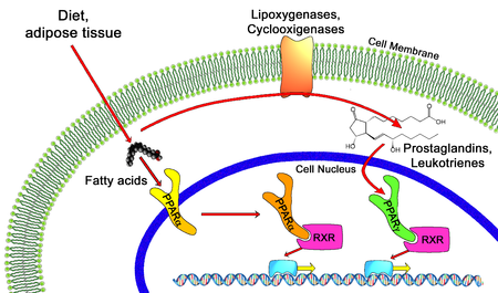
In the field of molecular biology, the peroxisome proliferator-activated receptors (PPARs) are a group of nuclear receptor proteins that function as transcription factors regulating the expression of genes.[1] PPARs play essential roles in the regulation of cellular differentiation, development, and metabolism (carbohydrate, lipid, protein), and tumorigenesis[2] of higher organisms.[3][4]

Three types of PPARs have been identified: alpha, gamma, and delta (beta):[3]

Physiological function

All PPARs heterodimerize with the retinoid X receptor (RXR) and bind to specific regions on the DNA of target genes. These DNA sequences are termed PPREs (peroxisome proliferator hormone response elements). The DNA consensus sequence is AGGTCANAGGTCA, with N being a random nucleotide. In general, this sequence occurs in the promotor region of a gene, and, when the PPAR binds its ligand, transcription of target genes is increased or decreased, depending on the gene. The RXR also forms a heterodimer with a number of other receptors (e.g., vitamin D and thyroid hormone).

The function of PPARs is modified by the precise shape of their ligand-binding domain (see below) induced by ligand binding and by a number of coactivator and corepressor proteins, the presence of which can stimulate or inhibit receptor function, respectively.[9]

Endogenous ligands for the PPARs include free fatty acids and eicosanoids. PPARγ is activated by PGJ2 (a prostaglandin). In contrast, PPARα is activated by leukotriene B4. PPARγ activation by agonist RS5444 may inhibit anaplastic thyroid cancer growth.[10]

Peroxisome proliferator-activated receptor

Peroxisome proliferator-activated receptor (Photo credit: Wikipedia)

Genetics

The three main forms are transcribed from different genes:

  •                PPARα – chromosome 22q12-13.1 (OMIM 170998)
  •                PPARβ/δ – chromosome 6p21.2-21.1 (OMIM 600409)
  •                PPARγ – chromosome 3p25 (OMIM 601487).

Hereditary disorders of all PPARs have been described, generally leading to a loss in function and concomitant lipodystrophy, insulin resistance, and/or acanthosis nigricans.[11] Of PPARγ, a gain-of-function mutation has been described and studied (Pro12Ala) which decreased the risk of insulin resistance; it is quite prevalent (allele frequency 0.03 – 0.12 in some populations).[12] In contrast, pro115gln is associated with obesity. Some other polymorphisms have high incidence in populations with elevated body mass indexes.

SOURCE:

http://en.wikipedia.org/wiki/Peroxisome_proliferator-activated_receptor


Mechanism of action

Thiazolidinediones or TZDs act by activating PPARs (peroxisome proliferator-activated receptors), a group of nuclear receptors with greatest specificity for PPARγ (gamma). The endogenous ligands for these receptors are free fatty acids (FFAs) and eicosanoids. When activated, the receptor binds to DNA in complex with the retinoid X receptor (RXR), another nuclear receptor, increasing transcription of a number of specific genes and decreasing transcription of others.

PPARγ transactivation

Thiazolidinedione ligand dependent transactivation is responsible for the majority of anti-diabetic effects.

The activated PPAR/RXR dimer binds to peroxisome proliferator hormone response elements upstream of target genes in complex with a number of coactivators such as nuclear receptor coactivator 1 and CREB binding protein, this causes upregulation of genes (for a full list see PPARγ:

TZDs also increase the synthesis of certain proteins involved in fat and glucose metabolism, which reduces levels of certain types of lipids, and circulating free fatty acids. TZDs generally decrease triglycerides and increase high-density lipoprotein cholesterol (HDL-C) and low-density lipoprotein cholesterol (LDL-C). Although the increase in LDL-C may be more focused on the larger LDL particles, which may be less atherogenic, the clinical significance of this is currently unknown. Nonetheless, rosiglitazone, a certain glitazone, was suspended from allowed use by medical authorities in Europe, as it has been linked to an increased risk of heart attack and stroke.[3]

PPARγ transrepression

Thiazolidinedione ligand dependent transrepression mediates the majority of anti-inflammatory effects.

Binding of PPARγ to coactivators appears to reduce the levels of coactivators available for binding to pro-inflammatory transcription factors such as NF-κB, this causes a decrease in transcription of a number of pro inflammatory genes, including various interleukins and tumour necrosis factors.

SOURCE:

http://en.wikipedia.org/wiki/Thiazolidinedione

 1. Waki H, Yamauchi T, Kadowaki T (February 2010). “[Regulation of differentiation and hypertrophy of adipocytes and adipokine network by PPARgamma]” (in Japanese). Nippon Rinsho 68 (2): 210–6. PMID 20158086.

2. Panigrahy D, Singer S, Shen LQ, et al. (2002). “PPARγ ligands inhibit primary tumor growth and metastasis by inhibiting angiogenesis”. J. Clin. Invest. 110 (7): 923–32. doi:10.1172/JCI15634. PMC 151148. PMID 12370270.

3. NHS: Avandia diabetes drug suspended, Friday 24th September 2010


Members of the class

The chemical structure of thiazolidinedione

Chemically, the members of this class are derivatives of the parent compound thiazolidinedione, and include:

  •                Rosiglitazone (Avandia), which was put under selling restrictions in the US and withdrawn from the market in            Europe due to an increased risk of cardiovascular events.
  •                Pioglitazone (Actos), France and Germany have suspended the sale of the diabetes drug Actos after a study suggested the drug, also known as pioglitazone, could raise the risk of bladder cancer.[4]
  •                Troglitazone (Rezulin), which was withdrawn from the market due to an increased incidence of drug-induced hepatitis.

Experimental agents include netoglitazone, an antidiabetic agent, rivoglitazone, and the early non-marketed thiazolidinedione ciglitazone.

Replacing one oxygen atom in a thiazolidinedione with an atom of sulfur gives a rhodanine.

SOURCE:

http://en.wikipedia.org/wiki/Thiazolidinedione

PART II:

Peroxisome proliferator-activated receptors as Stimulants of Angiogenesis in Cardiovascular Disease and Diabetes

In 2009 in Diabetes Metab Syndr Obes a seminal paper was published on the topic by  Desouza, Rentschler and Fonseca. (2009). This work constitutes Part II. http://www.ncbi.nlm.nih.gov/pmc/articles/PMC3048019/

Mechanisms by which PPARs may stimulate angiogenesis

PPARs seem to have a protective role in ischemic tissues, including brain, cardiac and skin. A part of this may be by stimulating angiogenesis and improving blood supply. Hypoxia is a trigger for the development of angiogenesis. One of the key mediators in hypoxia-induced angiogenesis is hypoxia inducible factor (HIF-1), which is induced in hypoxic cells and binds to hypoxia response element (HRE). HIF-1 mediates the transcriptional activation of several genes that promote angiogenesis, including VEGF, angiopoeitin (Ang-1, Ang-2), and matrix metalloproteinases (MMP-2, MMP-9).55 15-deoxy-delta(12, 14)-prostaglandin J(2) (15d-PGJ(2)), a PPAR-γ agonist, has been shown to induce HIF-1 expression and thereby angiogenesis (Figure 1).34 However pioglitazone has been shown to suppress the induction of HIF-1.56 Conditions that influence the stimulation or suppression of HIF activation by PPAR-γ are largely unknown.

Several studies suggest that eNOS synthase activation is required for angiogenesis that may be protective under certain conditions.5759 In one study pioglitazone reduced the myocardial infarct size in part via activation of eNOS.60 PPAR-α activation has also been shown to protect the type 2 diabetic rat myocardium against ischemia-reperfusion injury via the activation of the NO pathway (Table 1, Figure 1).61 However, stimulation of the inducible nitric oxide (iNOS) pathway can lead to undesirable angiogenesis that may be contribute to pathological states such as proliferative retinopathy. PPARs in fact have been shown to suppress iNOS expression, thereby suppressing undesirable angiogenesis.62,63 Here again the factors that allow for activation of eNOS and suppression of iNOS is largely unknown.

The most studied pathway by which PPARs may stimulate angiogenesis is the VEGF pathway. VEGF can stimulate angiogenesis via stimulation of the ERK1/2 pathway. PPAR-β/δ activation has been shown to increase VEGF expression and thereby stimulate angiogenesis (Figure 1).26 In some studies PPAR-α and PPAR-γ have also been shown to increase VEGF expression.47,48 However the majority of studies still show that PPAR activation suppresses VEGF expression. The end result of whether PPAR activation suppresses or stimulates VEGF expression seems to lie in the pathological condition in which its actions are observed (Figure 1). It is likely that PPAR activation results in increased VEGF expression in conditions where new blood vessel formation is required, such as ischemic skin flaps, brain, or cardiac tissue ischemia. On the other hand, pathological angiogenesis such as in the eye or within an atherosclerotic plaque is suppressed by PPAR activation via a suppression of VEGF (Figure 1).

An external file that holds a picture, illustration, etc.<br /><br /><br /><br /><br /><br /><br /> Object name is dmso-2-165f1.jpg Object name is dmso-2-165f1.jpg

Mechanisms by which PPARs effect angiogenesis.

Table 1

Effect of PPARs on angiogenesis

Recently some studies indicate that PPARs may increase the expression and activation of the phosphatidylinositol-3-kinase (PI3K/AKT) pathway.61,64 The PI3K/AKT pathway stimulates angiogenesis.59,65 Again the majority of studies show that PPAR activation inhibits PI3K/AKT activation.

It is very likely that a large amount of variation found in different studies is due to the use of agonists and antagonists of the PPAR receptors that exhibit direct PPAR-independent effects. Most study designs do not distinguish between direct effects and indirect effects of various pharmacological agonists/antagonist used. Fibrates and TZDs have both been shown to have direct independent effects on inflammation, proliferation and angiogenesis. Hence it is difficult to conclude that all the pro and antiangiogenic effects seen in various studies are a result of PPAR activation exclusively.

Clinical significance and conclusions

Some compounds such as TZDs and fibrates are routinely used in patients with diabetes, dyslipidemia, and cardiovascular disease. Other compounds such as partial agonists or dual agonists of PPAR-α and PPAR-γ are in development. The effects of these newer compounds, on angiogenesis and cardiovascular disease are yet to be determined. Current evidence from clinical trials suggest a mixed picture. TZD treatment in patients with type 2 diabetes has been shown to be associated with macular edema. On the other hand, the FIELD study using fenofibrate showed a decrease in the need for laser treatments in patients with diabetic retinopathy. The PROACTIVE study showed that pioglitazone trended to decrease certain cardiovascular endpoints. In some studies, rosiglitazone increased the risk of cardiovascular events. In other studies such as ACCORD and VADT, TZD treatment was not associated with increased cardiovascular event risk. Several factors, including the study design, PPAR receptor affinity, and the PPAR-independent actions of these compounds, possibly play a role in the differences in results seen. The duration of the pathological state and the vasculature of the effected organ likely play a role in whether PPARs prove beneficial or harmful. In conclusion it may be prudent to summarize that at this point the evidence suggests that PPARs can either stimulate or inhibit angiogenesis, depending on the biological context and pathological process.

Clinical Trials: Controversial Research Results

Peroxisome proliferator-activated receptors (PPARs) are a group of nuclear hormone receptors that regulate lipid and glucose metabolism. PPAR-α agonists such as fenofibrate and PPAR-γ agonists such as the thiozolidinediones have been used to treat dyslipidemia and insulin resistance in diabetes. Over the past few years research has discovered the role of PPARs in the regulation of inflammation, proliferation, and angiogenesis. Clinical trials looking at the effect of PPAR agonists on cardiovascular outcomes have produced controversial results. Studies looking at angiogenesis and proliferation in various animal models and cell lines have shown a wide variation in results. This may be due to the differential effects of PPARs on proliferation and angiogenesis in various tissues and pathologic states. This review discusses the role of PPARs in stimulating angiogenesis. It also reviews the settings in which stimulation of angiogenesis may be either beneficial or harmful.

affect inflammation, proliferation, immune function and angiogenesis.3 There are three PPAR isotypes, PPAR-α, PPAR-β/δ, and PPAR-γ. They form heterodimers with the retinoid X receptors and bind to specific DNA sequences, called peroxisome proliferator response elements (PPRE), in the promoter regions of their target genes. PPARs exhibit isotype-specific tissue expression patterns. PPAR-α is primarily expressed in organs with significant fatty acid catabolism. PPAR-β/δ is expressed in nearly all cell types and the level of expression seems to depend on the amount of angiogenesis, cell proliferation, and differentiation occurring in that specific tissue.4 PPAR-γ is found in adipose tissue and at lower levels in immune cells vascular tissue and some organs. PPAR-γ exists in two protein isoforms, PPAR-γ1 and PPAR-γ2, with different lengths of the N-terminal. The PPAR-γ2 isoform is predominantly expressed in adipose tissue, whereas PPAR-γ1 is relatively widely expressed.5 Expression of each isoform is driven by a specific promoter that confers the distinct tissue expression patterns. There are also two other mRNA variants of PPAR-γ, proteins identical to PPAR-γ1: PPAR-γ3, which is restricted to macrophages, adipose tissue, and colon, and PPAR-γ4, the tissue distribution of which is unclear at this time.5 Human PPAR-γ plays a critical physiological role as a central transcriptional regulator of both adipogenic and lipogenic programs. Its transcriptional activity is induced by the binding of endogenous and synthetic lipophilic ligands, which has led to the determination of many roles for PPAR-γ in pathological states such as type 2 diabetes, atherosclerosis, inflammation, and cancer.

The role of PPARs has traditionally been recognized as antiproliferative and antiangiogenic in a large number of disease states including cancer and cardiovascular disease.4 These studies have led to clinical trials with PPAR agonists to evaluate their benefits in cancer and cardiovascular disease. The results of some of these trials especially in cardiovascular disease have been mixed and hence controversial.

The results obtained with a PPAR-γ agonist pioglitazone do suggest a better impact on the lipid profile compared to rosiglitazone (the former lowers triglyceride significantly and has less adverse effects on low-density lipoprotein [LDL] cholesterol), and at least a mixed result (the primary composite endpoint was not reduced significantly but myocardial infarction, stroke, and death were reduced by 16%), in an outcome trial – PROspective pioglitAzone Clinical Trial In macroVascular Events (PROACTIVE).6 Rosiglitazone on the other hand was found to increase cardiovascular events in a large restrospective analysis study.7

This has led to a lot of recent research into PPARs that is contrary to the traditional literature in their role as inhibitors of angiogenesis. This review will examine the role and evidence of PPARs as promoters of angiogenesis, the mechanisms involved, and the implications thereof.

SOURCE:

 Desouza, Rentschler and Fonseca. (2009).

Angiogenesis is described as the formation of new capillaries from the existing vasculature. This process involves the breakdown of the extracellular matrix and formation of an endothelial tube. Angiogenesis is an important physiologic process in the female reproductive cycle, wound healing, and bone formation. Angiogenesis is also a crucial step in several disease states including cancer, diabetic retinopathy, rheumatoid arthritis, stroke, and ischemic coronary artery disease.810 Neoangiogenesis has harmful as well as beneficial effects in the setting of type 2 diabetes and cardiovascular disease.10 In the setting of diabetes, there is abnormal regulation and signaling of vascular endothelial growth factor (VEGF) and its receptor Flk-1.11 This may lead to increased levels of circulating VEGF, resulting in increased permeability of vascular structures throughout the body. In the retina, this results in the formation of protein-rich exudates containing VEGF that induces a local inflammatory response resulting in capillary sprouting. A similar process might take place in the arterial wall, thereby promoting capillary sprouting and plaque destabilization.12 At the same time, the lack of Flk-1 activation in endothelial cells and abnormal VEGF-dependent activation of monocytes impair the arteriogenic response that requires monocyte recruitment, and monocyte and endothelial cell migration and proliferation.11 This could lead to a deficient angiogenic response in ischemic tissue. VEGF/Flk-1 signaling may also be required for bone marrow release of circulating endothelial progenitor cells that play a role in endothelial function and arteriogenesis.13 The abnormal release of endothelial progenitors could further reduce arteriogenic response. This has therapeutic implications in terms of vascularization and survival of skin grafts in patients with diabetes as well as vascularization of the ischemic myocardium. An important mechanism by which PPARs seem to regulate angiogenesis is via VEGF.11,12 It would therefore appear that PPARs have a role in regulating both beneficial and harmful effects of angiogenesis thereby leading to controversial results (Figure 1).

The other factor influencing the results of angiogenesis studies is the use of PPAR agonists that have pleotropic effects. PPAR-α agonists such as fibrates stimulate pathways that do not depend on PPAR-α.14 PPAR-γ agonists such as thiozolidinediones (TZDs) have PPARγ independent actions on proliferative and inflammatory pathways.14 Therefore to conclude that the effects of commonly used PPAR agonists on angiogenesis are specifically due to PPAR activation is at best controversial.15

Although the majority of studies point towards the antiproliferative, antiangiogenic properties of PPAR-α, this may be due to the use of fibrates as agonists in these experiments. A lot more research needs to be done using methods such as spontaneous PPAR-α activation, overexpression, silencing and knockout mice, rather than using chemical agonists and antagonists which might have pleotropic effects unrelated to PPAR-α.


PPAR-γ and angiogenesis

PPAR-γ is probably the most studied PPAR, likely due to the use and development of several PPAR-γ agonists such as thiozolidinediones in the treatment of type 2 diabetes. Endogenous ligands for PPAR-γ include long chain polyunsaturated fatty acids and their derivatives, 15-deoxy-Δ12, 14-prostaglandin J2 (15d-PGJ2).4 Other natural ligands include nitrolinoleic acids. 15d-PGJ2 has been found to upregulate the expression of PPAR-γ and also the DNA binding and transcriptional activity.34 Synthetic ligands include TZDs and various nonsteroidal anti-inflammatory drugs.35

Studies supporting antiproliferative properties of PPAR-γ

PPAR-γ has widespread effects involving, inflammation, atherosclerosis, obesity, diabetes, and cancer.36 PPAR-γ agonists directly inhibit tumor cell growth, induce cell differentiation, and apoptosis in various cancer types (Table 1).37 TZDs have been shown to decrease post angioplasty neointimal hyperplasia in both animals and humans (Table 1).38,39 PPAR-γ ligands have been shown to inhibit and stimulate angiogenesis (Table 1). Inhibition by PPAR-γ ligands can occur through direct effects on the endothelium or through indirect effects on the net balance of proangiogenic and antiangiogenic mediators.37 PPAR-γ expressed in choroidal endothelial cells inhibits the differentiation and proliferation of those cells.38,39 Rosiglitazone inhibited endothelial cell proliferation and migration and decreased VEGF-induced tubule formation in human umbilical vein endothelial cells.40,41 In another study PPAR-γ ligands stimulated endothelial cell caspase-mediated apoptosis.42 15d-PGJ2, an endogenous ligand of PPAR-γ, induces growth inhibition, differentiation, and apoptosis of tumor cells.43 PPAR-γ activation interrupts NF-kβ signaling with subsequent blockade of proinflammatory gene expression.43 Pioglitazone and rosiglitazone inhibit the effects of growth factors such as bFGF and VEGF. Endothelial cell migration is also inhibited by both compounds.44 Thus natural and synthetic ligands of PPAR-γ exhibit antiangiogenic properties under certain conditions.

Studies supporting proangiogenic role of PPAR-γ

However, PPAR- ligands have also been shown to stimulate the angiogenic pathway (Table 1). In bovine aortic endothelial cells, prolonged treatment with troglitazone increased VEGF and endothelial nitric oxide (NO) production with no change in endothelial nitric oxide synthase (eNOS) expression.45 In cultured rat myofibroblasts, activation of PPAR-γ by troglitazone and 15-dPGJ2 induced VEGF expression and augmented tubule formation.46 In mice treated with rosiglitazone, angiogenesis was stimulated in adipose tissue with increased expression of VEGF and angiopoeitin-4 (Ang-4). Ang-4 stimulated endothelial cell growth and tubule formation. 47 In rats with focal cerebral ischemia, rosiglitazone treatment enhanced neurologic improvement and reduced the infarct size by reducing caspase-3 activity, increasing the number of endothelial cells, and increasing eNOS expression.48 In the setting of diabetes, PPAR-γ agonists may promote revascularization of ischemic tissue. Diabetic mice with induced unilateral hind limb ischemia, when treated with pioglitazone showed normalization of VEGF, upregulation of eNOS activity, and partial restoration of blood flow recovery.49 In mice treated with pioglitazone, VEGR-receptor-2 positive EPCs were upregulated and migratory capacity was increased. In vivo angiogenesis was increased 2-fold.50 In an endothelial/interstitial cell co-culture assay, treatment with PPAR-γ agonists stimulated production of VEGF. In the same study, corneas treated with the same PPAR-γ agonists increased phosphorylation of eNOS.20

Few studies have evaluated angiogenesis in humans. Pioglitazone treatment has been shown to increase serum VEGF, IL-8, and angiogenin levels in patients with type 2 diabetes.51 In another study thiozolidinedione use in patients with type 2 diabetes was associated with diabetic macular edema.52

PGC-1α and angiogenesis

Peroxisome proliferator-activated receptor (PPAR)-gamma coactivator 1alpha (PGC-1α) is a nuclear transcriptional coactivator that regulates several important metabolic processes, including mitochondrial biogenesis, adaptive thermogenesis, respiration, insulin secretion and gluconeogenesis. 53 PGC-1α also co-activates PPAR-α, PPAR-β/δ, and PPAR-γ which are important transcription factors of genes regulating lipid and glucose metabolism.53 Recently Arany and colleagues have shown that PGC-1α stimulates angiogenesis in ischemic tissues. Using a combination of muscle cell assays and genetically modified mice that over or underexpess PGC-1α, they showed that PGC-1α is a powerful inducer of VEGF expression. PGC-1α did not involve HIF-1 but activated the nuclear receptor, estrogen-related receptor-α (ERR-α).33 PGC-1α−/− mice are viable, suggesting that PGC-1α is not essential in embryonic vascularization but they show a striking failure to reconstitute blood flow in a normal manner to the limb after an ischaemic insult.54 Transgenic expression of PGC-1α in skeletal muscle is protective against ischemic insults. This suggests that PGC-1α plays a more important role in a disease state rather than a physiologically healthy state.


PART III: PPAR-gamma Role in Activation of eNOS: The Cardiovascular Benefit

Author and Curator: Aviva Lev-Ari, PhD, RN


Mechanism of Action (MOA) for ElectEagle‘s component 3

Treatment Regime with PPAR-gamma agonists (TZDs)

For ElectEagle‘s component 1:


Lev-Ari, A., (2012 X). Clinical Trials Results for Endothelin System: Pathophysiological role in Chronic Heart Failure, Acute Coronary Syndromes and MI – Marker of Disease Severity or Genetic Determination?

http://pharmaceuticalintelligence.com/2012/10/19/clinical-trials-results-for-endothelin-system-pathophysiological-role-in-chronic-heart-failure-acute-coronary-syndromes-and-mi-marker-of-disease-severity-or-genetic-determination/


Lev-Ari, A., (2012W). Endothelin Receptors in Cardiovascular Diseases: The Role of eNOS Stimulation

http://pharmaceuticalintelligence.com/2012/10/04/endothelin-receptors-in-cardiovascular-diseases-the-role-of-enos-stimulation/


Lev-Ari, A., (2012V). Inhibition of ET-1, ETA and ETA-ETB, Induction of NO production, stimulation of eNOS and Treatment Regime with PPAR-gamma agonists (TZD): cEPCs Endogenous Augmentation for Cardiovascular Risk Reduction – A Bibliography

http://pharmaceuticalintelligence.com/2012/10/04/inhibition-of-et-1-eta-and-eta-etb-induction-of-no-production-and-stimulation-of-enos-and-treatment-regime-with-ppar-gamma-agonists-tzd-cepcs-endogenous-augmentation-for-cardiovascular-risk-reduc/

For ElectEagle‘s component 2:


Lev-Ari, A. (2012L).. Cardiovascular Disease (CVD) and the Role of agent alternatives in endothelial Nitric Oxide Synthase (eNOS) Activation and Nitric Oxide Production

http://pharmaceuticalintelligence.com/2012/07/19/cardiovascular-disease-cvd-and-the-role-of-agent-alternatives-in-endothelial-nitric-oxide-synthase-enos-activation-and-nitric-oxide-production/

Lev-Ari, A. (2012i). Bystolic’s generic Nebivolol – positive effect on circulating Endothelial Proginetor Cells endogenous augmentation

http://pharmaceuticalintelligence.com/2012/07/16/bystolics-generic-nebivolol-positive-effect-on-circulating-endothilial-progrnetor-cells-endogenous-augmentation/

Three indications for PPAR-gamma agonist (TZD): Experimental agents include netoglitazone, an antidiabetic agent, rivoglitazone, and the early non-marketed thiazolidinedione ciglitazone

  •                Antisclerosis, angiogenic progenitor cell differentiation and endogenous augmentation of cEPCs
  •                Stimulation of eNOS
  •                Decrease insulin resistance

Classic indication: action to decrease insulin resistance. PPAR-gamma receptors are complex and modulate the expression of the genes involved in lipid and glucose metabolism, insulin signal transduction and adipocyte and other tissue differentiation. TZDs have significant effects on vascular endothilium, the immune system, the ovaries, and tumor cells. Some of these responses may be independent of the PPAR-gamma pathway (Nolte and Karam, 2004). TZDs are ligands of PPAR-gamma receptors part of the steroid (estrogen receptor ligands) and thyroid superfamily of nuclear receptors found in muscle, liver and adipocytes. In the gold standard of all pharmacology books, the cardinal indication for TZDs is action to decrease insulin resistance (Nolte and Karam, 2004 in Katzung). However, the recent research has proposed two new indications for Rosiglitazone in addition to the original insulin sensitivity reduction indication.

As implied in Part I of the ElectEagle Project, TDZs were selected for a new indication in the domain of modulation of atherosclerosis (Verma and Szmitko, 2006), (Li et al., 2004)and facilitation of the differentiation of angiogenic progenitor cells, inhibition of vascular smooth muscle, proliferation and migration to improve endothelial function (Wang et al., 2004).

The following three seminal papers on the function of TDZs in modulation of vascular disease served as an inspiration for our extension of their new indication for TDZs in the anti-atherosclerosis domain into the cEPCs endogenous augmentation proposed treatment area.

Verma S, Szmitko, PE, (2006). The vascular biology of peroxisome proliferator-activated receptors: Modulation of atherosclerosis. Can J Cardiol, 22 (Suppl B):12B-17B.

Wang C-H, Ciliberti N, Li S-H, Szmitko PE, Weisel RD, Fedak PWM, Al-Omran M, Cherng W-J, Li R-K, Stanford WL, Verma S., (2004). Rosiglitazone facilitates angiogenic progenitor cell differentiation toward endothelial lineage: a new paradigm in glitazone pleiotropy. Circulation, 109:1392-1400.

Li AC, Binder CJ, Gutierrez A, Brown KK, Plotkin CR, Pattison JW, Valledor AF, Davis RA, Wilson TM, Wizttum JL, Palinski W, Glass CK., (2004). Differential inhibition of macrophage foam-cell formation and atherosclerosis in mice by PPAR alpha, beta/delta, and gamma. J. Clin. Invest., 114:1564-1576.

Namely, in the ElectEagleProject, a finely tuned interpretation is provided. We assume that TZDs may have a potential therapeutic effect on augmentation of cEPCs in a significant way should a combination drug therapy be designed to include Rosiglitazoneand two other drugsonewhich inhibits receptors ETA and ETA-ETB and the other which induces eNOS. TDZs were selected for a new indication related to anti-atherosclerosis, however, we extend and emphasize TZDs function in cell differentiation and cell migration of EPCs following encouraging results by Wang et al., (2004). Thus, we are shifting the indication from atherosclerosis and peripheral vascular disease to cardiovascular and CAD.

In 2005 a new indication for TZDs emerged from new finding about the PPAR-gamma receptors function in cell nitric oxide (NO) release without increasing the expression of endothelial nitric oxide synthase (eNOS) (Polikandriotis et al., 2005). This is an important finding for the drug combination components selected for ElectEagleProject. This subject is covered in the following section, Role of PPAR-gamma in eNOS stimulation.

Mechanism of action (MOA) for ElectEagle‘s components 2 & 3

Role of PPAR-gamma in eNOS stimulation

Polikandriotis et al. (2005), recently reported that the peroxisome proliferator-activated receptor gamma (PPARgamma) ligands 15-deoxy-Delta(12,14)-prostaglandin J2 (15d-PGJ2) and ciglitazone increased cultured endothelial cell nitric oxide (NO) release without increasing the expression of endothelial nitric oxide synthase (eNOS). Their study was designed to characterize further the molecular mechanisms underlying PPARgamma-ligand-stimulated increases in endothelial cell NO production.

Their methods and Results: Treating human umbilical vein endothelial cells (HUVEC) with PPARgamma ligands (10 micromol/L 15d-PGJ2, ciglitazone, or rosiglitazone) for 24 hours increased NOS activity and NO release. In selected studies, HUVEC were treated with PPARgamma ligands and with the PPARgamma antagonist GW9662 (2 micromol/L), which fully inhibited stimulation of a luciferase reporter gene, or with small interfering RNA to PPARgamma, which reduced HUVEC PPARgamma expression. Treatment with either small interfering RNA to PPARgamma or GW9662 inhibited 15d-PGJ2-, ciglitazone-, and rosiglitazone-induced increases in endothelial cell NO release. Rosiglitazone and 15d-PGJ2, but not ciglitazone, increased heat shock protein 90-eNOS interaction and eNOS ser1177 phosphorylation. The heat shock protein 90 inhibitor geldanamycin attenuated 15d-PGJ2- and rosiglitazone-stimulated NOS activity and NO production. Their Conclusion: These findings further clarify mechanisms involved in PPARgamma-stimulated endothelial cell NO release and emphasize that individual ligands exert their effects through distinct PPARgamma-dependent mechanisms

Originally, Rosiglitazone was indicated as an adjunct to diet and exercise to improve glycemic control in patients with type 2 diabetes mellitus who are already treated with combination rosiglitazone and metformin or who are not adequately controlled on metformin alone. As a result of the FDA drug recall of Rosiglitazone, we suggest here several alternatives: Experimental agents include netoglitazone, an antidiabetic agent, rivoglitazone, and the early non-marketed thiazolidinedione ciglitazone

In the ElectEagle project, Rosiglitazone was identified for a new indication – as a PPAR-gamma agonist implicated with efficacy for endogenous augmentation of cEPCs which serves as a biomarker for CVD risk reduction — an extension of the anti-atherosclerosis indication or the confinement to perileral vascular endothelium (Verma & Szmitko), (Wang et al., 2004), (Li et al., 2004).

Polikandriotis et al. (2005) is a very import publication for ElectEagle project for the following critical five reasons:

  •                Polikandriotis et al. (2005) clarify the mechanism of action of PPAR-gamma agonists at the protein level in a set of novel experiments, thus contributes to the understanding of the physiological process of the mechanism of action of PPAReceptor-gamma and its relations to L-arginine: NO pathway and its impact in many areas of research, notably vascular biology.
  •                Polikandriotis et al. (2005) compare two PPAR-gamma agonist agents and confirm Rosiglitazone to be the more potent among the two for the experiments described above
  •                Polikandriotis et al. (2005) identify Rosiglitazone capability to stimulate endothelial cell NO release, which is a third indication for Rosiglitazone.
  •                The combination drug therapy selected in May 2006, for the ElectEagle project involved three drugs. Two of which where a PPAR-gamma agonist, specifically, Rosiglitazone. The other drug was an eNOS agonist to stimulate NO production and reuptake. By identifying Rosiglitazone capability to stimulate endothelial cell NO release, Polikandriotis et al. (2005) offer reassurance for the selection of Rosiglitazone in the first place, and further more we became aware that it will exert synergies with the drug chosen as an eNOS agonist.
  •                In the ElectEagle project, a new experiment is called for following Polikandriotis et al. (2005) findings on Rosiglitazone impact on NO release. It will be needed to measure the incremental induction of NO release resulting from a combination therapy which includes an eNOS agonist and a PPAR-gamma agonist implicated in 2005 with stimulant effects on the release NO.

Moncada & Higgs, (2006) explain that the low concentrations of NO generated by eNOS protect against atherosclerosis by promoting vasodilatation, inhibiting leucocyte and platelet adhesion and/or aggregation and smooth muscle cell proliferation. However, higher concentrations of NO generated by iNOS promote atherosclerosis, either directly or via the formation of NO adducts, such as peroxynitrite. Such a paradox in the action of NO was apparent from their experiments some years ago, in which they found that the acute vascular injury in the ileum and colon following administration of lipopolysaccharide is aggravated by early treatment with a NO synthase inhibitor, whereas delayed administration of such a compound provides protection against the damage to the intestinal vasculature (Laszlo et al., 1994). A prominent example of this comes from experiments in Apo-Emutant mice in which the concomitant knocking out of eNOS leads to an increase in atherosclerosis, while the knocking out of iNOS reduces atherosclerosis (Moncada, 2005).

Research Goals in characterization of ElectEagle Version I


Provided rationale for agent selection for

ElectEagle Version I – Component 3: Treatment Regime with PPAR-gamma agonists (TZD)

agent selection: Rosiglitazone

As a result of the FDA drug recall of Rosiglitazone, we suggest here several alternatives: Experimental agents include netoglitazone, an antidiabetic agentrivoglitazone, and the early non-marketed thiazolidinedione ciglitazone


Retionale:            See discussion on TZDs MOA, above

a-priori postulates presented in Part I for Component 3: PPAR-gamma

  • dose concentration dependence on PPAReceptor-gamma – confirmed by a study for Rosiglitazone and a study for Ciglitazone
PPAReceptor-gamma agonists time concentration dependence manner dose concentration dependencemanner time and dose dose 
Rosiglitazone Polikandriotis et al., (2005) maximum recommended daily dose of 8 mg to 2,000 mg.
Ciglitazone   Polikandriotis et al., (2005)


Proposed integration plan for ElectEagle’s Version I with CVD patients current medication regimen for selective medical diagnoses

Blood Pressure Medicine:

Beta blockers, Verapamil (Calan), Reserpine (Hydropes), Clonidine (Catapres), Methyldopa (Aldomet)

Diuretics:

Thiazides, Spironolactone (Aldactone), Hydralazine

Antidepressants:

Prozac, Lithium, MOA’s, Tricyclics

Stomach Medicine:

Tagamet and Zantac, plus other compounds containing Cimetidine and Ranitidine or associated compounds in Anticholesterol Drugs

Antipsychotics:

Chlorpromazine (Thorazine), Pimozide (Orap), Thiothixine (Navane), Thiordazine (Mellaril), Sulpiride, Haloperidol (haldol), Fluphenazine (Modecate, Prolixin)

Heart Medicine:

Clofibrate (Atromid), Gemfibrozil, Diagoxin

Hormones:

Estrogen, Progesterone, Proscar, Casodex, Eulexin, Corticosteroids Gonadotropin releasing antagonists: Zoladex and Lupron

Cytotoxic agents:

Cyclophosphamide, Methotrexate, Roferon Non-steroidal anti-inflammatories

Others-

Alprazolam, Amoxapine, Chlordiazepoxide, Sertraline, Paroxetine, Clomipramine, Fluvoxamine, Fluoxetine, Imipramine, Doxepine, Desipramine, Clorprothixine, Bethanidine, Naproxen, Nortriptyline, Thioridazine, Tranylcypromine, Venlafaxine, Citalopram.

INTERACTIONS for Nebivolol – Component 2

Calcium Antagonists:

Caution should be exercised when administering beta-blockers with calcium antagonists of the verapamil or diltiazem type because of their negative effect on contractility and atrio-ventricular conduction. Exaggeration of these effects can occur particularly in patients with impaired ventricular function and/or SA or AV conduction abnormalities. Neither medicine should therefore be administered intravenously within 48 hours of discontinuing the other.

Anti-arrhythmics:

Caution should be exercised when administering beta-blockers with Class I anti-arrhythmic drugs and amiodarone as their effect on atrial conduction time and their negative inotropic effect may be potentiated. Such interactions can have life threatening consequences.

Clonidine:

Beta-blockers increase the risk of rebound hypertension after sudden withdrawal of chronic clonidine treatment.

Digitalis:

Digitalis glycosides associated with beta-blockers may increase atrio-ventricular conduction times. Nebivolol does not influence the kinetics of digoxin & clinical trials have not shown any evidence of an interaction.

Special note: Digitalisation of patients receiving long term beta-blocker therapy may be necessary if congestive cardiac failure is likely to develop. The combination can be considered despite the potentiation of the negative chronotropic effect of the two medicines. Careful control of dosages and of individual patient’s response (notably pulse rate) is essential in this situation.

Insulin & Oral Antidiabetic drugs:

Glucose levels are unaffected, however symptoms of hypoglycemia may be masked.

Anaesthetics:

Concomitant use of beta-blockers & anaesthetics e.g. ether, cyclopropane & trichloroethylene may attenuate reflex tachycardia & increase the risk of hypotension

Medical Diagnoses Current medication regiment ET-1, ETA and ETA-ETBinhibition eNOS agonistsproduction stimulation of NO PPAR-gamma agonist (TZD) PPAR-gamma agonist (TZD) as eNOS stimulant
CAD patients Beta blockers, ACEI, ARB, CCB, Diagoxin, Coumadin yes yes yes
Endothelial Dysfunction in DM patients with or without Erectile Dysfunction Insulin yes yes yes yes
Atherosclerosis patients: Arteries and or veins AntihypertensiveCoumadin yes yes yes yes
pre-stenting treatment phase Beta blockers, Verapamil (Calan), Reserpine (Hydropes), Clonidine (Catapres), Methyldopa (Aldomet) yes yes yes
post-stenting treatment phase Antiplatelets yes yes
if stent is a Bare Mesh stent (BMS) CoumadinBeta blockers yes yes
if stent is Drug Eluting stent (DES) antibiotics yes
if stent is EPC antibody coated yes yes
post CABG patients CoumadinBeta blockers, Verapamil(Calan), Reserpine (Hydropes), Clonidine (Catapres), Methyldopa (Aldomet) yes yes
CVD patients on blood thinner Coumadin yes yes yes

Conclusions

  •       Most favorable and unexpected to us was finding in the literature new indications for TDZs as stimulators of eNOS, in addition to the new indication for atherosclerosis besides the classic indication in pharmacology books, being in the reduction of insulin resistance. Reassuring our selection of Rosiglitazone. As a result of the FDA recoll, the drug substitute will be an Experimental agents include netoglitazone, an antidiabetic agentrivoglitazone, and the early non-marketed thiazolidinedione ciglitazone 
  •       Most favorable and unexpected to us was finding in the literature new indications for beta blockers as NO stimulant, nebivolol, a case in point, thus, fulfilling two indications in one drug along the direction of the study to identify eNOS agonists.
  •       The following combination of drugs was selected for ElectEagle Version I

Bosentan (Tracleer), Oral: 62.5 mg tablets

Nebivolol, Oral: 5mg once daily

Experimental agents include netoglitazone, an antidiabetic agent, rivoglitazone, and the early non-marketed thiazolidinedione ciglitazone

  •       We confirmed time and dose concentrations postulating apriori in most cases. Additional literature searches will benefit the project for the three drugs selected
  •       We have identified Inhibition of ET-1, ETA and ETA-ETB as one of the agent in the drug combination. The entire literature on cEPCs does not implicate Endothelin with impact on eEPCs while it is known that mechanical stress increase its secretion, this type of stress is implicated with hypertension. To leave out ET-1 from the cEPCs function in CVD risk equates to leaving out Thrombin from the coagulation cascade. ElectEagle Version I corrects that ommission.

REFERENCES for PART I:

1. Michalik L, Auwerx J, Berger JP, Chatterjee VK, Glass CK, Gonzalez FJ, Grimaldi PA, Kadowaki T, Lazar MA, O’Rahilly S, Palmer CN, Plutzky J, Reddy JK, Spiegelman BM, Staels B, Wahli W (2006). “International Union of Pharmacology. LXI. Peroxisome proliferator-activated receptors”. Pharmacol. Rev. 58 (4): 726–41. doi:10.1124/pr.58.4.5. PMID 17132851.

2. Belfiore A, Genua M, Malaguarnera R (2009). “PPAR-gamma Agonists and Their Effects on IGF-I Receptor Signaling: Implications for Cancer”. PPAR Res 2009: 830501. doi:10.1155/2009/830501. PMC 2709717. PMID 19609453.

3.a b Berger J, Moller DE (2002). “The mechanisms of action of PPARs”. Annu. Rev. Med. 53: 409–35. doi:10.1146/annurev.med.53.082901.104018. PMID 11818483.

4 Feige JN, Gelman L, Michalik L, Desvergne B, Wahli W (2006). “From molecular action to physiological outputs: peroxisome proliferator-activated receptors are nuclear receptors at the crossroads of key cellular functions”. Prog. Lipid Res. 45 (2): 120–59. doi:10.1016/j.plipres.2005.12.002. PMID 16476485.

5 Tyagi S, Gupta P, Saini AS, Kaushal C, Sharma S (October 2011). “The peroxisome proliferator-activated receptor: A family of nuclear receptors role in various diseases”. J Adv Pharm Technol Res 2 (4): 236–40. doi:10.4103/2231-4040.90879. PMC 3255347. PMID 22247890.

6 Dreyer C, Krey G, Keller H, Givel F, Helftenbein G, Wahli W (1992). “Control of the peroxisomal beta-oxidation pathway by a novel family of nuclear hormone receptors”. Cell 68 (5): 879–87. doi:10.1016/0092-8674(92)90031-7. PMID 1312391.

7 Issemann I, Green S (1990). “Activation of a member of the steroid hormone receptor superfamily by peroxisome proliferators”. Nature 347 (6294): 645–50. doi:10.1038/347645a0. PMID 2129546.

8 Schmidt A, Endo N, Rutledge SJ, Vogel R, Shinar D, Rodan GA (1992). “Identification of a new member of the steroid hormone receptor superfamily that is activated by a peroxisome proliferator and fatty acids”. Mol. Endocrinol. 6 (10): 1634–41. doi:10.1210/me.6.10.1634. PMID 1333051.

9 Yu S, Reddy JK (2007). “Transcription coactivators for peroxisome proliferator-activated receptors”. Biochim. Biophys. Acta 1771 (8): 936–51. doi:10.1016/j.bbalip.2007.01.008. PMID 17306620.

10 Marlow LA, Reynolds LA, Cleland AS, Cooper SJ, Gumz ML, Kurakata S, Fujiwara K, Zhang Y, Sebo T, Grant C, McIver B, Wadsworth JT, Radisky DC, Smallridge RC, Copland JA (February 2009). “Reactivation of suppressed RhoB is a critical step for the inhibition of anaplastic thyroid cancer growth”. Cancer Res. 69 (4): 1536–44. doi:10.1158/0008-5472.CAN-08-3718. PMC 2644344. PMID 19208833.

11 Meirhaeghe A, Amouyel P (2004). “Impact of genetic variation of PPARgamma in humans”. Mol. Genet. Metab. 83 (1-2): 93–102. doi:10.1016/j.ymgme.2004.08.014. PMID 15464424.

12 Buzzetti R, Petrone A, Ribaudo MC, Alemanno I, Zavarella S, Mein CA, Maiani F, Tiberti C, Baroni MG, Vecci E, Arca M, Leonetti F, Di Mario U (2004). “The common PPAR-gamma2 Pro12Ala variant is associated with greater insulin sensitivity”. Eur. J. Hum. Genet. 12 (12): 1050–4. doi:10.1038/sj.ejhg.5201283. PMID 15367918.

13 Zoete V, Grosdidier A, Michielin O (2007). “Peroxisome proliferator-activated receptor structures: ligand specificity, molecular switch and interactions with regulators”. Biochim. Biophys. Acta 1771 (8): 915–25. doi:10.1016/j.bbalip.2007.01.007. PMID 17317294.

REFERENCES for PART II:

Part II is based on the following:

Cyrus V Desouza, Lindsey Rentschler, and Vivian Fonseca

Peroxisome proliferator-activated receptors as stimulants of angiogenesis in cardiovascular disease and diabetes Diabetes Metab Syndr Obes. 2009; 2: 165–172. Published online 2009 September 25 PMCID: PMC3048019

http://www.ncbi.nlm.nih.gov/pmc/articles/PMC3048019/

1. Wee CC, Hamel MB, Huang A, Davis RB, Mittleman MA, McCarthy EP. Obesity and undiagnosed diabetes in the US. Diabetes Care. 2008;31:1813–1815. [PMC free article] [PubMed]

2. Westphal SA. Obesity, abdominal obesity, and insulin resistance. Clin Cornerstone. 2008;9:23–31. [PubMed]

3. Calkin AC, Thomas MC. PPAR agonists and cardiovascular disease in diabetes. PPAR Res. 2008;245410

4. Duan SZ, Ivashchenko CY, Usher MG, Mortensen RM. PPAR-gamma in the cardiovascular system. PPAR Res. 2008;745804

5. Knouff C, Auwerx J. Peroxisome proliferator-activated receptor-gamma calls for activation in moderation: lessons from genetics and pharmacology. Endocr Rev. 2004;25:899–918. [PubMed]

6. Erdmann E, Dormandy J, Wilcox R, Massi-Benedetti M, Charbonnel B. PROactive 07: pioglitazone in the treatment of type 2 diabetes: results of the PROactive study. Vasc Health Risk Manag. 2007;3:355–370. [PMC free article] [PubMed]

7. Nissen SE, Wolski K. Effect of rosiglitazone on the risk of myocardial infarction and death from cardiovascular causes. N Engl J Med. 2007;356:2457–2471. [PubMed]

8. Folkman J. Angiogenesis in cancer, vascular, rheumatoid and other disease. Nat Med. 1995;1:27–31. [PubMed]

9. Carmeliet P, Baes M. Metabolism and therapeutic angiogenesis. N Engl J Med. 2008;358:2511–2512. [PubMed]

10. Martin A, Komada MR, Sane DC. Abnormal angiogenesis in diabetes mellitus. Med Res Rev. 2003;23:117–145. [PubMed]

11. Simons M. Angiogenesis, arteriogenesis, and diabetes: paradigm reassessed? J Am Coll Cardiol. 2005;46:835–837. [PubMed]

12. Qaum T, Xu Q, Joussen AM, et al. VEGF-initiated blood-retinal barrier breakdown in early diabetes. Invest Ophthalmol Vis Sci. 2001;42:2408–2413. [PubMed]

13. Pitchford SC, Furze RC, Jones CP, Wengner AM, Rankin SM. Differential mobilization of subsets of progenitor cells from the bone marrow. Cell Stem Cell. 2009;4:62–72. [PubMed]

14. Jandeleit-Dahm KA, Calkin A, Tikellis C, Thomas M. Direct antiatherosclerotic effects of PPAR agonists. Curr Opin Lipidol. 2009;20:24–29. [PubMed]

15. Pozzi A, Ibanez MR, Gatica AE, et al. Peroxisomal proliferator-activated receptor-alpha-dependent inhibition of endothelial cell proliferation and tumorigenesis. J Biol Chem. 2007;282:17685–17695. [PubMed]

16. Grabacka M, Reiss K. Anticancer properties of PPARalpha – effects on cellular metabolism and inflammation. PPAR Res. 2008;930705

17. Scott R, O’Brien R, Fulcher G, et al. Effects of fenofibrate treatment on cardiovascular disease risk in 9,795 individuals with type 2 diabetes and various components of the metabolic syndrome: the Fenofibrate Intervention and Event Lowering in Diabetes (FIELD) study. Diabetes Care. 2009;32:493–498. [PMC free article] [PubMed]

18. Kasai T, Miyauchi K, Yokoyama T, Aihara K, Daida H. Efficacy of peroxisome proliferative activated receptor (PPAR)-alpha ligands, fenofibrate, on intimal hyperplasia and constrictive remodeling after coronary angioplasty in porcine models. Atherosclerosis. 2006;188:274–280. [PubMed]

19. Gizard F, Amant C, Barbier O, et al. PPAR alpha inhibits vascular smooth muscle cell proliferation underlying intimal hyperplasia by inducing the tumor suppressor p16INK4a. J Clin Invest. 2005;115:3228–3238. [PMC free article] [PubMed]

20. Biscetti F, Gaetani E, Flex A, et al. Selective activation of peroxisome proliferator-activated receptor (PPAR)alpha and PPAR gamma induces neoangiogenesis through a vascular endothelial growth factor-dependent mechanism. Diabetes. 2008;57:1394–1404. [PubMed]

21. Biscetti F, Gaetani E, Flex A, et al. Peroxisome proliferator-activated receptor alpha is crucial for iloprost-induced in vivo angiogenesis and vascular endothelial growth factor upregulation. J Vasc Res. 2009;46:103–108. [PubMed]

22. Fauconnet S, Lascombe I, Chabannes E, et al. Differential regulation of vascular endothelial growth factor expression by peroxisome proliferator-activated receptors in bladder cancer cells. J Biol Chem. 2002;277:23534–23543. [PubMed]

23. Wang N. PPAR-delta in Vascular Pathophysiology. PPAR Res. 2008;164163

24. Berry DC, Noy N. All-trans-retinoic acid represses obesity and insulin resistance by activating both PPAR{beta}/{delta} and RAR. Mol Cell Biol. 2009;29:3286–3296. [PMC free article] [PubMed]

25. Stephen RL, Gustafsson MC, Jarvis M, et al. Activation of peroxisome proliferator-activated receptor delta stimulates the proliferation of human breast and prostate cancer cell lines. Cancer Res. 2004;64:3162–3170. [PubMed]

26. Piqueras L, Reynolds AR, Hodivala-Dilke KM, et al. Activation of PPARbeta/delta induces endothelial cell proliferation and angiogenesis. Arterioscler Thromb Vasc Biol. 2007;27:63–69. [PubMed]

27. Gaudel C, Schwartz C, Giordano C, Abumrad NA, Grimaldi PA. Pharmacological activation of PPARbeta promotes rapid and calcineurin-dependent fiber remodeling and angiogenesis in mouse skeletal muscle. Am J Physiol Endocrinol Metab. 2008;295:E297–E304. [PubMed]

28. Yoshinaga M, Kitamura Y, Chaen T, et al. The simultaneous expression of peroxisome proliferator-activated receptor delta and cyclooxygenase-2 may enhance angiogenesis and tumor venous invasion in tissues of colorectal cancers. Dig Dis Sci. 2009;54:1108–1114. [PubMed]

29. He T, Lu T, d’Uscio LV, Lam CF, Lee HC, Katusic ZS. Angiogenic function of prostacyclin biosynthesis in human endothelial progenitor cells. Circ Res. 2008;103:80–88. [PMC free article] [PubMed]

30. Muller-Brusselbach S, Komhoff M, Rieck M, et al. Deregulation of tumor angiogenesis and blockade of tumor growth in PPARbeta-deficient mice. Embo J. 2007;26:3686–3698. [PMC free article] [PubMed]

31. Muller R, Komhoff M, Peters JM, Muller-Brusselbach S. A Role for PPARbeta/delta in Tumor Stroma and Tumorigenesis. PPAR Res. 2008;534294

32. Wang D, Wang H, Guo Y, et al. Crosstalk between peroxisome proliferator-activated receptor delta and VEGF stimulates cancer progression. Proc Natl Acad Sci U S A. 2006;103:19069–19074. [PMC free article] [PubMed]

33. Hollingshead HE, Killins RL, Borland MG, et al. Peroxisome proliferator-activated receptor-beta/delta (PPARbeta/delta) ligands do not potentiate growth of human cancer cell lines. Carcinogenesis. 2007;28:2641–2649. [PubMed]

34. Kim EH, Surh YJ. 15-deoxy-Delta12, 14-prostaglandin J2 as a potential endogenous regulator of redox-sensitive transcription factors. Biochem Pharmacol. 2006;72:1516–1528. [PubMed]

35. Lehmann JM, Lenhard JM, Oliver BB, Ringold GM, Kliewer SA. Peroxisome proliferator-activated receptors alpha and gamma are activated by indomethacin and other non-steroidal anti-inflammatory drugs. J Biol Chem. 1997;272:3406–3410. [PubMed]

36. Pershadsingh HA. Peroxisome proliferator-activated receptor-gamma: therapeutic target for diseases beyond diabetes: quo vadis? Expert Opin Investig Drugs. 2004;13:215–228.

37. Giaginis C, Tsantili-Kakoulidou A, Theocharis S. Peroxisome proliferator-activated receptor-gamma ligands: potential pharmacological agents for targeting the angiogenesis signaling cascade in cancer. PPAR Res. 2008;431763

38. Rosmarakis ES, Falagas ME. Effect of thiazolidinedione therapy on restenosis after coronary stent implantation: a meta-analysis of randomized controlled trials. Am Heart J. 2007;154:144–150. [PubMed]

39. Desouza CV, Gerety M, Hamel FG. Long-term effects of a PPAR-gamma agonist, pioglitazone, on neointimal hyperplasia and endothelial regrowth in insulin resistant rats. Vascul Pharmacol. 2007;46:188–194. [PubMed]

40. Panigrahy D, Singer S, Shen LQ, et al. PPARgamma ligands inhibit primary tumor growth and metastasis by inhibiting angiogenesis. J Clin Invest. 2002;110:923–932. [PMC free article] [PubMed]

41. Sheu WH, Ou HC, Chou FP, Lin TM, Yang CH. Rosiglitazone inhibits endothelial proliferation and angiogenesis. Life Sci. 2006;78:1520–1528. [PubMed]

42. Bishop-Bailey D, Hla T. Endothelial cell apoptosis induced by the peroxisome proliferator-activated receptor (PPAR) ligand 15-deoxy-Delta12, 14-prostaglandin J2. J Biol Chem. 1999;274:17042–17048. [PubMed]

43. Giri S, Rattan R, Singh AK, Singh I. The 15-deoxy-delta12,14-prostaglandin J2 inhibits the inflammatory response in primary rat astrocytes via down-regulating multiple steps in phosphatidylinositol 3-kinase-Akt-NF-kappaB- p300 pathway independent of peroxisome proliferator-activated receptor gamma. J Immunol. 2004;173:5196–5208. [PubMed]

44. Aljada A, O’Connor L, Fu YY, Mousa SA. PPAR gamma ligands, rosiglitazone and pioglitazone, inhibit bFGF- and VEGF-mediated angiogenesis. Angiogenesis. 2008;11:361–367. [PubMed]

45. Cho DH, Choi YJ, Jo SA, Jo I. Nitric oxide production and regulation of endothelial nitric-oxide synthase phosphorylation by prolonged treatment with troglitazone: evidence for involvement of peroxisome proliferator-activated receptor (PPAR) gamma-dependent and PPARgamma-independent signaling pathways. J Biol Chem. 2004;279:2499–2506. [PubMed]

46. Chintalgattu V, Harris GS, Akula SM, Katwa LC. PPAR-gamma agonists induce the expression of VEGF and its receptors in cultured cardiac myofibroblasts. Cardiovasc Res. 2007;74:140–150. [PubMed]

47. Gealekman O, Burkart A, Chouinard M, Nicoloro SM, Straubhaar J, Corvera S. Enhanced angiogenesis in obesity and in response to PPAR-gamma activators through adipocyte VEGF and ANGPTL4 production. Am J Physiol Endocrinol Metab. 2008;295:E1056–E1064. [PMC free article] [PubMed]

48. Chu K, Lee ST, Koo JS, et al. Peroxisome proliferator-activated receptor-gamma-agonist, rosiglitazone, promotes angiogenesis after focal cerebral ischemia. Brain Res. 2006;1093:208–218. [PubMed]

49. Huang PH, Sata M, Nishimatsu H, Sumi M, Hirata Y, Nagai R. Pioglitazone ameliorates endothelial dysfunction and restores ischemia-induced angiogenesis in diabetic mice. Biomed Pharmacother. 2008;62:46–52. [PubMed]

50. Gensch C, Clever YP, Werner C, Hanhoun M, Bohm M, Laufs U. The PPAR-gamma agonist pioglitazone increases neoangiogenesis and prevents apoptosis of endothelial progenitor cells. Atherosclerosis. 2007;192:67–74. [PubMed]

51. Vijay SK, Mishra M, Kumar H, Tripathi K. Effect of pioglitazone and rosiglitazone on mediators of endothelial dysfunction, markers of angiogenesis and inflammatory cytokines in type-2 diabetes. Acta Diabetol. 2009;46:27–33. [PubMed]

52. Fong DS, Contreras R. Glitazone use associated with diabetic macular edema. Am J Ophthalmol. 2009;147:583–586. e1. [PubMed]

53. Finck BN, Kelly DP. Peroxisome proliferator-activated receptor gamma coactivator-1 (PGC-1) regulatory cascade in cardiac physiology and disease. Circulation. 2007;115:2540–2548. [PubMed]

54. Arany Z, Foo SY, Ma Y, et al. HIF-independent regulation of VEGF and angiogenesis by the transcriptional coactivator PGC-1alpha. Nature. 2008;451:1008–1012. [PubMed]

55. Hickey MM, Simon MC. Regulation of angiogenesis by hypoxia and hypoxia-inducible factors. Curr Top Dev Biol. 2006;76:217–257. [PubMed]

56. Lee KS, Kim SR, Park SJ, et al. Peroxisome proliferator activated receptor-gamma modulates reactive oxygen species generation and activation of nuclear factor-kappaB and hypoxia-inducible factor 1alpha in allergic airway disease of mice. J Allergy Clin Immunol. 2006;118:120–127. [PubMed]

57. Chen J, Cui X, Zacharek A, Roberts C, Chopp M. eNOS mediates TO90317 treatment-induced angiogenesis and functional outcome after stroke in mice. Stroke. 2009;40:2532–2538. [PMC free article] [PubMed]

58. Howell K, Costello CM, Sands M, Dooley I, McLoughlin P. L-arginine promotes angiogenesis in the chronically hypoxic lung: a novel mechanism ameliorating pulmonary hypertension. Am J Physiol Lung Cell Mol Physiol. 2009;296:L1042–L1050. [PubMed]

59. Namkoong S, Kim CK, Cho YL, et al. Forskolin increases angiogenesis through the coordinated cross-talk of PKA-dependent VEGF expression and Epac-mediated PI3K/Akt/eNOS signaling. Cell Signal. 2009;21:906–915. [PubMed]

60. Yasuda S, Kobayashi H, Iwasa M, et al. Antidiabetic drug pioglitazone protects the heart via activation of PPAR-{gamma} receptors, PI3-kinase, Akt, and eNOS pathway in a rabbit model of myocardial infarction. Am J Physiol Heart Circ Physiol. 2009;296:H1558–H1565. [PubMed]

61. Bulhak AA, Jung C, Ostenson CG, Lundberg JO, Sjoquist PO, Pernow J. PPAR-alpha activation protects the type 2 diabetic myocardium against ischemia-reperfusion injury: involvement of the PI3-Kinase/Akt and NO pathway. Am J Physiol Heart Circ Physiol. 2009;296:H719–H727. [PubMed]

62. Cuzzocrea S, Pisano B, Dugo L, et al. Rosiglitazone, a ligand of the peroxisome proliferator-activated receptor-gamma, reduces acute inflammation. Eur J Pharmacol. 2004;483:79–93. [PubMed]

63. Tao L, Liu HR, Gao E, et al. Antioxidative, antinitrative, and vasculoprotective effects of a peroxisome proliferator-activated receptor-gamma agonist in hypercholesterolemia. Circulation. 2003;108:2805–2811. [PubMed]

64. Pedchenko TV, Gonzalez AL, Wang D, DuBois RN, Massion PP. Peroxisome proliferator-activated receptor beta/delta expression and activation in lung cancer. Am J Respir Cell Mol Biol. 2008;39:689–696. [PMC free article] [PubMed]

65. Ma J, Sawai H, Ochi N, et al. PTEN regulate angiogenesis through PI3K/Akt/VEGF signaling pathway in human pancreatic cancer cells. Mol Cell Biochem. 2009 May 13; Epub ahead of print.

66. Panigrahy D, Kaipainen A, Huang S, et al. PPARalpha agonist fenofibrate suppresses tumor growth through direct and indirect angiogenesis inhibition. Proc Natl Acad Sci U S A. 2008;105:985–990. [PMC free article] [PubMed]

67. Minutoli L, Antonuccio P, Polito F, et al. Peroxisome proliferator activated receptor beta/delta activation prevents extracellular regulated kinase 1/2 phosphorylation and protects the testis from ischemia and reperfusion injury. J Urol. 2009;181:1913–1921. [PubMed]

68. Lim HJ, Lee S, Park JH, et al. PPAR delta agonist L-165041 inhibits rat vascular smooth muscle cell proliferation and migration via inhibition of cell cycle. Atherosclerosis. 2009;202:446–454. [PubMed]

69. Borland MG, Foreman JE, Girroir EE, et al. Ligand activation of peroxisome proliferator-activated receptor-beta/delta inhibits cell proliferation in human HaCaT keratinocytes. Mol Pharmacol. 2008;74:1429–1442. [PMC free article] [PubMed]

70. Piqueras L, Sanz MJ, Perretti M, et al. Activation of PPAR{beta}/{delta} inhibits leukocyte recruitment, cell adhesion molecule expression, and chemokine release. J Leukoc Biol. 2009;86:115–122. [PubMed]

REFERENCES for PART III:


Inhibition of ET-1, ETA and ETA-ETB, Induction of NO production, stimulation of eNOS and Treatment Regime with PPAR-gamma agonists (TZD): cEPCs Endogenous Augmentation for Cardiovascular Risk Reduction – A Bibliography

http://pharmaceuticalintelligence.com/2012/10/04/inhibition-of-et-1-eta-and-eta-etb-induction-of-no-production-and-stimulation-of-enos-and-treatment-regime-with-ppar-gamma-agonists-tzd-cepcs-endogenous-augmentation-for-cardiovascular-risk-reduc/


Additional References to Studies on PPAR-Gamma


Repository on BioInfoBank Library on Peroxisome proliferator-activated receptor

http://lib.bioinfo.pl/paper:11030710


Repository on Science.gov on Peroxisome proliferator-activated receptor

http://www.science.gov/topicpages/e/exhibits+ppargamma+ligand.html


On this Open Access OnLine Scientific Journal, Dr. Lev-Ari’s research on Pharmaco-Therapy of Cardiovascular Diseases includes the following:


Lev-Ari, A., (2012 X). Clinical Trials Results for Endothelin System: Pathophysiological role in Chronic Heart Failure, Acute Coronary Syndromes and MI – Marker of Disease Severity or Genetic Determination?

http://pharmaceuticalintelligence.com/2012/10/19/clinical-trials-results-for-endothelin-system-pathophysiological-role-in-chronic-heart-failure-acute-coronary-syndromes-and-mi-marker-of-disease-severity-or-genetic-determination/


Lev-Ari, A., (2012W). Endothelin Receptors in Cardiovascular Diseases: The Role of eNOS Stimulation

http://pharmaceuticalintelligence.com/2012/10/04/endothelin-receptors-in-cardiovascular-diseases-the-role-of-enos-stimulation/


Lev-Ari, A., (2012V). Inhibition of ET-1, ETA and ETA-ETB, Induction of NO production, stimulation of eNOS and Treatment Regime with PPAR-gamma agonists (TZD): cEPCs Endogenous Augmentation for Cardiovascular Risk Reduction – A Bibliography

http://pharmaceuticalintelligence.com/2012/10/04/inhibition-of-et-1-eta-and-eta-etb-induction-of-no-production-and-stimulation-of-enos-and-treatment-regime-with-ppar-gamma-agonists-tzd-cepcs-endogenous-augmentation-for-cardiovascular-risk-reduc/


Lev-Ari, A., (2012U). Cardiovascular Outcomes: Function of circulating Endothelial Progenitor Cells (cEPCs): Exploring Pharmaco-therapy targeted at Endogenous Augmentation of cEPCs

http://pharmaceuticalintelligence.com/2012/08/28/cardiovascular-outcomes-function-of-circulating-endothelial-progenitor-cells-cepcs-exploring-pharmaco-therapy-targeted-at-endogenous-augmentation-of-cepcs/

Lev-Ari, A., (2012T). Endothelial Dysfunction, Diminished Availability of cEPCs, Increasing CVD Risk for Macrovascular Disease – Therapeutic Potential of cEPCs

http://pharmaceuticalintelligence.com/2012/08/27/endothelial-dysfunction-diminished-availability-of-cepcs-increasing-cvd-risk-for-macrovascular-disease-therapeutic-potential-of-cepcs/

Lev-Ari, A., (2012S). Vascular Medicine and Biology: CLASSIFICATION OF FAST ACTING THERAPY FOR PATIENTS AT HIGH RISK FOR MACROVASCULAR EVENTS Macrovascular Disease – Therapeutic Potential of cEPCs

http://pharmaceuticalintelligence.com/2012/08/24/vascular-medicine-and-biology-classification-of-fast-acting-therapy-for-patients-at-high-risk-for-macrovascular-events-macrovascular-disease-therapeutic-potential-of-cepcs/

Lev-Ari, A. (2012L).. Cardiovascular Disease (CVD) and the Role of agent alternatives in endothelial Nitric Oxide Synthase (eNOS) Activation and Nitric Oxide Production

http://pharmaceuticalintelligence.com/2012/07/19/cardiovascular-disease-cvd-and-the-role-of-agent-alternatives-in-endothelial-nitric-oxide-synthase-enos-activation-and-nitric-oxide-production/

Lev-Ari, A. (2012a). Resident-cell-based Therapy in Human Ischaemic Heart Disease: Evolution in the PROMISE of Thymosin beta4 for Cardiac Repair

http://pharmaceuticalintelligence.com/2012/04/30/93/

Lev-Ari, A. (2012b). Triple Antihypertensive Combination Therapy Significantly Lowers Blood Pressure in Hard-to-Treat Patients with Hypertension and Diabetes

http://pharmaceuticalintelligence.com/2012/05/29/445/

Lev-Ari, A. (2012h). Macrovascular Disease – Therapeutic Potential of cEPCs: Reduction Methods for CV Risk

http://pharmaceuticalintelligence.com/2012/07/02/macrovascular-disease-therapeutic-potential-of-cepcs-reduction-methods-for-cv-risk/

Lev-Ari, A. (2012j) Mitochondria Dysfunction and Cardiovascular Disease – Mitochondria: More than just the “powerhouse of the cell”

http://pharmaceuticalintelligence.com/2012/07/09/mitochondria-more-than-just-the-powerhouse-of-the-cell/

Lev-Ari, A. (2012i). Bystolic’s generic Nebivolol – positive effect on circulating Endothelial Proginetor Cells endogenous augmentation

http://pharmaceuticalintelligence.com/2012/07/16/bystolics-generic-nebivolol-positive-effect-on-circulating-endothilial-progrnetor-cells-endogenous-augmentation/


Electronic versions NOT available for:

Lev-Ari, A. & Abourjaily, P. (2006a) “An Investigation of the Potential of circulating Endothelial Progenitor Cells (cEPC) as a Therapeutic Target for Pharmacologic Therapy Design for Cardiovascular Risk Reduction.”Part I: Macrovascular Disease – Therapeutic Potential of cEPCs – Reduction methods for CV risk. Part II: (2006b) Therapeutic Strategy for cEPCs Endogenous Augmentation: A Concept-based Treatment Protocol for a Combined Three Drug Regimen. Part III: (2006c) Biomarker for Therapeutic Targets of Cardiovascular Risk Reduction by cEPCs Endogenous Augmentation using New Combination Drug Therapy of Three Drug Classes and Several Drug Indications. Northeastern University, Boston, MA 02115

Lev-Ari, A. (2007) Heart Vasculature Regeneration and Protection of Coronary Artery Endothelium and Smooth Muscle: A Concept-based Pharmacological Therapy of a Combined Three Drug Regimen. Bouve College of Health Sciences, Northeastern University, Boston, MA 02115


Read Full Post »

« Newer Posts - Older Posts »