Feeds:
Posts
Comments

Posts Tagged ‘United States’

Heroes in Medical Research: The Postdoctoral Fellow

Writer: Stephen J. Williams, Ph.D

Thank your Postdoc

The National Postdoctoral Association (NPA) had its Fifth Annual Celebration Of National Postdoc Appreciation Week (NPAW) in September and I wanted to focus a posting on curating stories from postdoctoral fellows as well as private investigators (PIs) and mentors on the impacts that postdoctoral fellows had in research and to recognize the critical and tremendous contributions which postdocs make to science.

During our postdoctoral years, we develop deep friendships which last a lifetime, a close bonding to our kindred scientists different in nature than our bonding with our mentors.  Nothing can replace a great mentor but our fellow postdocs make a huge difference in our complete scientific training.

                                   It’s always the little things that stand out in our fondest memories

Unfortunately I have a plethora of fond, little memories; too many for this posting but just want to ad in a few things:

  • Thank you!    –  To all those postdocs who worked tirelessly to make a memorable PostDoc Day!
  • Thank you!  –  To all my postdoc colleagues who stayed late n the lab with me giving each other moral and scientific support
  • Thank you!  – All my postdoc friends who would give up their time to show me how to make and use a text box correctly in Word
  • Thank you!  =-  for your friendship and understanding in those rough times we had experienced

To enliven the discussion, I ask that postdocs, past, present, and future, as well as PI’s and postdoc mentors comment on their postdoc experience. I also would like PI’s to share the stories how their postdocs made an impact to their labs.

A few interesting links and articles from the web on the importance and struggles of postdocs are included below:

Keith Micoli, from New York University Langone Medical Center states in an Elsevier article on The Academic Executive Brief

Consequently, it’s very difficult to come up with accurate numbers. Current estimates on number of postdocs come between 40,000 and 90,000 — a range that is unacceptable. A solid bet is that there are 60,000 postdocs and that more than half, if not two thirds or higher, are international.

– from US research enterprise powered by international postdocs by Keith Micoli at NYU

Survey Methodology

Since Science started conducting annual surveys seven years ago, alternating between polling postdocs and postdoc advisors, the attributes that survey respondents select as being most important to a successful postdoc have not varied much.This year’s survey was launched on March 15, 2011, with e-mail invitations sent out to about 40,000 current and former postdoc advisors worldwide. Of the 798 completed surveys that were collected, 71 percent came from Europe (39 percent) and North America (32 percent). The remaining respondents were located in Asia/Australia/Pacific Rim (20 percent) or other areas of the world (9 percent). Most were males (72 percent) 40 years of age and older (76 percent) and worked in academic institutions (70 percent) and government organizations (13 percent). The primary area represented was the life sciences (57 percent).

However only a handful of institutions were featured.

An open letter to AAAS journal “Science”: Postdocs need to address the “The Future of Research”

https://thewinnower.com/papers/an-open-letter-to-aaas-journal-science-postdocs-need-to-address-the-the-future-of-research?jm.npa=

This letter, posted on the Winnower.com, was a response to Callier’s article “Ailing academia needs culture change”1 and discussed how postdoctoral fellows have to lead in effecting change if the US research enterprise is to flourish in the future. In addition, the authors have been organizing Boston area postdoctoral associations and are sponsoring a symposium to be held at Boston University October 2-3 2014, focusing on the challenges facing graduate students and postdoctoral fellows: the “Future of Research” symposium (futureofresearch.org, @FORsymp).

  1. V. Callier, N. L. Vanderford. “Ailing academia needs culture change.” Science, 2014: 345; 6199: 885. DOI: 10.1126/science.345.6199.885-b

On the surface, many acknowledge the importance of postdoctoral fellows to the US research effort,

HOWEVER, the QUESTION remains DO POSTDOCS FEEL APPRECIATED FOR THEIR EFFORTS?

Please read Jacquelyn Gil, Ph.D.’s GREAT blog post

Have you hugged your postdoc today?

in The Contemplative Mammoth about her surviving postdoctoral life.

For some postdoc humor go to

http://phdcomics.com/comics.php where Jorge Cham, Ph.D. has been satiring the Ph.D. life since he was a graduate student in the late 90’s.

and see if you could be a star in their movie about Ph.D.’s: The PhD Movie and the sequel.

Don’t Underestimate Your Postdoc

Dr. Thomas C. Sudhof, MD is an example of a postdoctoral fellow making great contributions to a lab. A summary of his work is seen below and obtained from the site thebestschools.org on the “50 Most Influential Scientists”.

http://www.thebestschools.org/features/50-influential-scientists-world-today/#S%C3%BCdhof

Thomas C. Südhof

Thomas C. Südhof is a biochemist and professor in the School of Medicine in the Department of Molecular and Cellular Physiology at Stanford University. He is best known for his work in the area of synaptic transmission, which is the process by which signaling chemicals known as neurotransmitters are released by one neuron and bind to and activate the receptors of another neuron.

Südhof won the 1985 Nobel Prize in Physiology or Medicine, along with Randy Schekman and James Rothman.

Südhof, a native of Germany, obtained his MD from the University of Göttingen and conducted his postdoctoral training in the department of molecular genetics at the University of Texas’s Health Science Center. During his postdoctoral training, he worked on describing the role of the LDL receptor in cholesterol metabolism, for which Michael S. Brown and Joseph L. Goldstein were awarded the Nobel Prize in Physiology or Medicine in 1985.


Another example from the site includes Dr. Craig Mello (Craig C. Mello’s Home Page.) who, along with Dr., Andrew Fire discovered RNAi when both at Carnegie Institute. Both received a Nobel for their work.

So again would love to hear and curate personal stories highlighting how postdocs make a great contribution to US science.

More articles in this “Heroes in Medical Research” series and posts on Scientific Careers from this site include:

Heroes in Medical Research: Green Fluorescent Protein and the Rough Road in Science

Heroes in Medical Research: Developing Models for Cancer Research

Heroes in Medical Research: Dr. Carmine Paul Bianchi Pharmacologist, Leader, and Mentor

Heroes in Medical Research: Dr. Robert Ting, Ph.D. and Retrovirus in AIDS and Cancer

Heroes in Medical Research: Barnett Rosenberg and the Discovery of Cisplatin

Science Budget FY’14: Stakeholders’ Reactions on Selective Budget Drops and Priorities Shift

Careers for Researchers Beyond Academia

BEYOND THE “MALE MODEL”: AN ALTERNATIVE FEMALE MODEL OF SCIENCE, TECHNOLOGY AND INNOVATION

Read Full Post »

Breaching Drug Disclosure on Tresiba been refused U.S. approval – Novo Nordisk A/S (NVO) Reported To Police

Reporter: Aviva Lev-Ari, PhD, RN

SOURCE

http://www.biospace.com/news_story.aspx?NewsEntityId=318189&type=email&source=GP_121013

Novo Nordisk A/S (NVO) Reported To Police For Breaching Drug Disclosure Rules

12/10/2013 7:32:19 AM

Novo Nordisk, the world’s largest insulin maker, is facing a Danish police probe after it was reported by the financial watchdog for not disclosing at once that its big new product hope Tresiba had been refused U.S. approval.

Although the probe is unlikely to have a serious financial impact on the company, the largest by market value in the Nordic region, it may tarnish its reputation and could leave it open to lawsuits from investors in the United States, where its shares also trade.

The Danish Financial Supervisory Authority (FSA) said on Tuesday Novo should have issued a statement about the U.S. decision not to approve Tresiba, its new long-acting insulin, on the evening of Friday, February 8 instead of waiting until Sunday, February 10.


Read Full Post »

aprotinin-sequence.Par.0001.Image.260

aprotinin-sequence.Par.0001.Image.260 (Photo credit: redondoself)

English: Protein folding: amino-acid sequence ...

Protein folding: amino-acid sequence of bovine BPTI (basic pancreatic trypsin inhibitor) in one-letter code, with its folded 3D structure represented by a stick model of the mainchain and sidechains (in gray), and the backbone and secondary structure by a ribbon colored blue to red from N- to C-terminus. 3D structure from PDB file 1BPI, visualized in Mage and rendered in Raster3D. (Photo credit: Wikipedia)

The Effects of Aprotinin on Endothelial Cell Coagulant Biology

Author: Demet Sag, PhD


The Effects of Aprotinin on Endothelial Cell Coagulant Biology

Demet Sag, PhD*†, Kamran Baig, MBBS, MRCS; James Jaggers, MD, Jeffrey H. Lawson, MD, PhD

Departments of Surgery and Pathology (J.H.L.) Duke University Medical Center Durham, NC  27710

Correspondence and Reprints:

                             Jeffrey H. Lawson, M.D., Ph.D.

                              Departments of Surgery & Pathology

                              DUMC Box 2622

                              Durham, NC  27710

                              (919) 681-6432 – voice

                              (919) 681-1094 – fax

                              lawso006@mc.duke.edu

*Current Address: Demet SAG, PhD

                          3830 Valley Centre Drive Suite 705-223, San Diego, CA 92130

Support:

Word Count: 4101 Journal Subject Heads:  CV surgery, endothelial cell activationAprotinin, Protease activated receptors,

Potential Conflict of Interest:         None

Abstract

Introduction:  Cardiopulmonary bypass is associated with a systemic inflammatory response syndrome, which is responsible for excessive bleeding and multisystem dysfunction. Endothelial cell activation is a key pathophysiological process that underlies this response. Aprotinin, a serine protease inhibitor has been shown to be anti-inflammatory and also have significant hemostatic effects in patients undergoing CPB. We sought to investigate the effects of aprotinin at the endothelial cell level in terms of cytokine release (IL-6), tPA release, tissue factor expression, PAR1 + PAR2 expression and calcium mobilization. Methods:  Cultured Human Umbilical Vein Endothelial Cells (HUVECS) were stimulated with TNFa for 24 hours and treated with and without aprotinin (200KIU/ml + 1600KIU/ml). IL-6 and tPA production was measured using ELISA. Cellular expression of Tissue Factor, PAR1 and PAR2 was measured using flow cytometry. Intracellular calcium mobilization following stimulation with PAR specific peptides and agonists (trypsin, thrombin, Human Factor VIIa, factor Xa) was measured using fluorometry with Fluo-3AM. Results: Aprotinin at the high dose (1600kIU/mL), 183.95 ± 13.06mg/mL but not low dose (200kIU/mL) significantly reduced IL-6 production from TNFa stimulated HUVECS (p=0.043). Aprotinin treatment of TNFa activated endothelial cells significantly reduce the amount of tPA released in a dose dependent manner (A200 p=0.0018, A1600 p=0.033). Aprotinin resulted in a significant downregulation of TF expression to baseline levels. At 24 hours, we found that aprotinin treatment of TNFa stimulated cells resulted in a significant downregulation of PAR-1 expression. Aprotinin significantly inhibited the effects of the protease thrombin upon PAR1 mediated calcium release. The effects of PAR2 stimulatory proteases such as human factor Xa, human factor VIIa and trypsin on calcium release was also inhibited by aprotinin. Conclusion:  We have shown that aprotinin has direct anti-inflammatory effects on endothelial cell activation and these effects may be mediated through inhibition of proteolytic activation of PAR1 and PAR2. Abstract word count: 297

INTRODUCTION   Each year it is estimated that 350,000 patients in the United States, and 650,000 worldwide undergo cardiopulmonary bypass (CPB). Despite advances in surgical techniques and perioperative management the morbidity and mortality of cardiac surgery related to the systemic inflammatory response syndrome(SIRS), especially in neonates is devastatingly significant. Cardiopulmonary bypass exerts an extreme challenge upon the haemostatic system as part of the systemic inflammatory syndrome predisposing to excessive bleeding as well as other multisystem dysfunction (1). Over the past decade major strides have been made in the understanding of the pathophysiology of the inflammatory response following CPB and the role of the vascular endothelium has emerged as critical in maintaining cardiovascular homeostasis (2).

CPB results in endothelial cell activation and initiation of coagulation via the Tissue Factor dependent pathway and consumption of important clotting factors. The major stimulus for thrombin generation during CPB has been shown to be through the tissue factor dependent pathway. As well as its effects on the fibrin and platelets thrombin has been found to play a role in a host of inflammatory responses in the vascular endothelium. The recent discovery of the Protease-Activated Receptors (PAR), one of which through which thrombin acts (PAR-1) has stimulated interest that they may provide a vital link between inflammation and coagulation (3).

Aprotinin is a nonspecific serine protease inhibitor that has been used for its ability to reduce blood loss and preserve platelet function during cardiac surgery procedures requiring cardiopulmonary bypass and thus the need for subsequent blood and blood product transfusions. However there have been concerns that aprotinin may be pro-thrombotic, especially in the context of coronary artery bypass grafting, which has limited its clinical use. These reservations are underlined by the fact that the mechanism of action of aprotinin has not been fully understood. Recently aprotinin has been shown to exert anti-thrombotic effects mediated by blocking the PAR-1 (4). Much less is known about its effects on endothelial cell activation, especially in terms of Tissue Factor but it has been proposed that aprotinin may also exert protective effects at the endothelial level via protease-activated receptors (PAR1 and PAR2). In this study we simulated in vitro the effects of endothelial cell activation during CPB by stimulating Human Umbilical Vein Endothelial Cells (HUVECs) with a proinflammatory cytokine released during CPB, Tumor Necrosis Factor (TNF-a) and characterize the effects of aprotinin treatment on TF expression, PAR1 and PAR2 expression, cytokine release IL-6 and tPA secretion.  In order to investigate the mechanism of action of aprotinin we studied its effects on PAR activation by various agonists and ligands.

These experiments provide insight into the effects of aprotinin on endothelial related coagulation mechanisms in terms of Tissue Factor expression and indicate it effects are mediated through Protease-Activated Receptors (PAR), which are seven membrane spanning proteins called G-protein coupled receptors (GPCR), that link coagulant and inflammatory pathways. Therefore, in this study we examine the effects of aprotinin on the human endothelial cell coagulation biology by different-dose aprotinin, 200 and 1600units.  The data demonstrates that aprotinin appears to directly alter endothelial expression of inflammatory cytokines, tPA and PAR receptor expression following treatment with TNF.  The direct mechanism of action is unknown but may act via local protease inhibition directly on endothelial cells.  It is hoped that with improved understanding of the mechanisms of action of aprotinin, especially an antithrombotic effect at the endothelial level the fears of prothrombotic tendency may be lessened and its use will become more routine.  

METHODS Human Umbilical Vein Endothelial Cells (HUVECS) used as our model to study the effects of endothelial cell activation on coagulant biology. In order to simulate the effects of cardiopulmonary bypass at the endothelial cell interface we stimulated the cells with the proinflammatory cytokine TNFa. In the study group the HUVECs were pretreated with low (200kIU/mL) and high (1600kIU/mL) dosages of aprotinin prior to stimulation with TNFa and complement activation fragments. The effects of TNFa stimulation upon endothelial Tissue Factor expression, PAR1 and PAR2 expression, and tPA and IL6 secretion were determined and compared between control and aprotinin treated cells. In order to delineate whether aprotinin blocks PAR activation via its protease inhibition properties we directly activated PAR1 and PAR2 using specific agonist ligands such thrombin (PAR1), trypsin, Factor VIIa, Factor Xa (PAR2) in the absence and presence of aprotinin.

Endothelial Cell Culture HUVECs were supplied from Clonetics. The cells were grown in EBM-2 containing 2MV bullet kit, including 5% FBS, 100-IU/ml penicillin, 0.1mg/mL streptomycin, 2mmol/L L-glutamine, 10 U/ml heparin, 30µg/mL EC growth supplement (ECGS). Before the stimulation cells were starved in 0.1%BSA depleted with FBS and growth factors for 24 hours. Cells were sedimented at 210g for 10 minutes at 4C and then resuspended in culture media. The HUVECs to be used will be between 3 and 5 passages.

Assay of IL-6 and tPA production Levels of IL-6 were measured with an ELISA based kit (RDI, MN) according to the manufacturers instructions. tPA was measured using a similar kit (American Diagnostica).

  Flow Cytometry The expression of transmembrane proteins PAR1, PAR2 and tissue factor were measured by single color assay as FITC labeling agent. Prepared suspension of cells disassociated trypsin free cell disassociation solution (Gibco) to be labeled. First well washed, and resuspended into “labeling buffer”, phosphate buffered saline (PBS) containing 0.5% BSA plus 0.1% NaN3, and 5% fetal bovine serum to block Fc and non-specific Ig binding sites. Followed by addition of 5mcl of antibody to approx. 1 million cells in 100µl labeling buffer and incubate at 4C for 1 hour. After washing the cells with 200µl with wash buffer, PBS + 0.1% BSA + 0.1% NaN3, the cells were pelletted at 1000rpm for 2 mins. Since the PAR1 and PAR2 were directly labeled with FITC these cells were fixed for later analysis by flow cytometry in 500µl PBS containing 1%BSA + 0.1% NaN3, then add equal volume of 4% formalin in PBS. For tissue factor raised in mouse as monoclonal primary antibody, the pellet resuspended and washed twice more as before, and incubated at 4C for 1 hour addition of 5µl donkey anti-mouse conjugated with FITC secondary antibody directly to the cell pellets at appropriate dilution in labeling buffer. After the final wash three times, the cell pellets were resuspended thoroughly in fixing solution. These fixed and labeled cells were then stored in the dark at 4C until there were analyzed. On analysis, scatter gating was used to avoid collecting data from debris and any dead cells. Logarithmic amplifiers for the fluorescence signal were used as this minimizes the effects of different sensitivities between machines for this type of data collection.  

Intracellular Calcium Measurement

Measured the intracellular calcium mobilization by Fluo-3AM. HUVECs were grown in calcium and phenol free EBM basal media containing 2MV bullet kit. Then the cell cultures were starved with the same media by 0.1% BSA without FBS for 24 hour with or without TNFa stimulation presence or absence of aprotinin (200 and 1600KIU/ml). Next the cells were loaded with Fluo-3AM 5µg/ml containing agonists, PAR1 specific peptide SFLLRN-PAR1 inhibitor, PAR2 specific peptide SLIGKV-PAR2 inhibitor, human alpha thrombin, trypsin, factor VIIa, factor Xa for an hour at 37C in the incubation chamber. Finally the media was replaced by Flou-3AM free media and incubated for another 30 minutes in the incubation chamber. The readings were taken at fluoromatic bioplate reader. For comparison purposes readings were taken before and during Fluo-3AM loading as well.  

RESULTS Aprotinin reduces IL-6 production from activated/stimulated HUVECS The effects of aprotinin analyzed on HUVEC for the anti-inflammatory effects of aprotinin at cultured HUVECS with high and low doses.  Figure 1 shows that TNF-a stimulated a considerable increase in IL-6 production, 370.95 ± 109.9 mg/mL.   If the drug is used alone the decrease of IL-6 at the low dose is 50% that is 183.95 ng/ml and with the high dose of 20% that is 338.92 from 370.95ng/ml being compared value.  TNFa-aprotinin results in reduction of the IL-6 expression from 370.95ng/ml to 58.6 (6.4fold) fro A200 and 75.85 (4.9 fold) ng/ml, for A1600.  After the treatment the cells reach to the below baseline limit of IL-6 expression. Aprotinin at the high dose (1600kIU/mL), 183.95 ± 13.06mg/mL but not low dose (200kIU/mL) significantly reduced IL-6 production from TNF-a stimulated HUVECS (p=0.043).  Therefore, the aprotinin prevents inflammation as well as loss of blood.  

Aprotinin reduces tPA production from stimulated HUVECS Whether aprotinin exerted part of its fibrinolytic effects through inhibition of tPA mediated plasmin generation examined by the effects on TNFa stimulated HUVECS. Figure 2 also demonstrates that the amount of tPA released from HUVECS under resting, non-stimulated conditions incubated with aprotinin are significantly different. Figure 2 represents that the resting level of tPA released from non-stimulated cells significantly, by 100%, increase following TNF-a stimulation for 24 hours.  After application of aprotinin alone at two doses the tPA level goes down 25% of TNFa stimulated cells.  However, aprotinin treatment of TNF-a activated endothelial cells significantly lower the amount of tPA release in a dose dependent manner that is low dose decreased 25 but high dose causes 50% decrease of tPA expression (A200 p=0.0018, A1600 p=0.033) This finding suggests that aprotinin exerts a direct inhibitory effect on endothelial cell tPA production.

Aprotinin and receptor expression on activated HUVECS

TF is expressed when the cell in under stress such as TNFa treatments. The stimulated HUVECs with TNF-a tested for the expression of PAR1, PAR2, and tissue factor by single color flow cytometry through FITC labeled detection antibodies at 1, 3, and 24hs.


Tissue Factor expression is reduced:

Figure 3 demonstrates that there is a fluctuation of TF expression from 1 h to 24h that the TF decreases at first hour after aprotinin application 50% and 25%, A1600 and A200 respectively.  Then at 3 h the expression come back up 50% more than the baseline.  Finally, at 24h the expression of TF becomes almost as same as baseline.  Moreover, TNFa stimulated cells remains 45% higher than baseline after at 3h as well as at 24h.

PAR1 decreased:
Figure 4 demonstrates that aprotinin reduces the PAR1 expression 80% at 24h but there is no affect at 1 and 3 h intervals for both doses.

During the treatment with aprotinin only high dose at 1 hour time interval decreases the PAR1 expression on the cells. This data explains that ECCB is affected due to the expression of PAR1 is lowered by the high dose of aprotinin.

PAR2 is decreased by aprotinin:

  Figure 5 shows the high dose of aprotinin reduces the PAR2 expression close to 25% at 1h, 50% at 3h and none at 24h.  This pattern is exact opposite of PAR1 expression.  Figure 5 demonstrates the 50% decrease at 3h interval only.  Does that mean aprotinin affecting the inflammation first and then coagulation?

This suggests that aprotinin may affect the PAR2 expression at early and switched to PAR1 reduction later time intervals.  This fluctuation can be normal because aprotinin is not a specific inhibitor for proteases.  This approach make the aprotinin work better the control bleeding and preventing the inflammation causing cytokine such as IL-6.

Aprotinin inhibits Calcium fluxes induced by PAR1/2 specific agonists

  The specificity of aprotinin’s actions upon PAR studied the effects of the agent on calcium release following proteolytic and non-proteolytic stimulation of PAR1 and PAR2. Figure 6A (Figure 6) shows the stimulation of the cells with the PAR1 specific peptide (SFLLRN) results in release of calcium from the cells. Pretreatment of the cells with aprotinin has no significant effect on PAR1 peptide stimulated calcium release. This suggests that aprotinin has no effect upon the non-proteolytic direct activation of the PAR 1 receptor. Yet, Figure 6B (Figure 6) demonstrates human alpha thrombin does interact with the drug as a result the calcium release drops below base line after high dose (A1600) aprotinin used to zero but low dose does not show significant effect on calcium influx. Figure 7 demonstrates the direct PAR2 and indirect PAR2 stimulation by hFVIIa, hFXa, and trypsin of cells.  Similarly, at Figure 7A aprotinin has no effect upon PAR2 peptide stimulated calcium release, however, at figures 7B, C, and D shows that PAR2 stimulatory proteases Human Factor Xa, Human Factor VIIa and Trypsin decreases calcium release. These findings indicate that aprotinin’s mechanism of action is directed towards inhibiting proteolytic cleavage and hence subsequent activation of the PAR1 and PAR2 receptor complexes.  The binding site of the aprotinin on thrombin possibly is not the peptide sequence interacting with receptors.

Measurement of calcium concentration is essential to understand the mechanism of aprotinin on endothelial cell coagulation and inflammation because these mechanisms are tightly controlled by presence of calcium.  For example, activation of PAR receptors cause activation of G protein q subunit that leads to phosphoinositol to secrete calcium from endoplasmic reticulum into cytoplasm or activation of DAG to affect Phospho Lipase C (PLC). In turn, certain calcium concentration will start the serial formation of chain reaction for coagulation.  Therefore, treatment of the cells with specific factors, thrombin receptor activating peptides (TRAPs), human alpha thrombin, trypsin, human factor VIIa, and human factor Xa, would shed light into the effect of aprotinin on the formation of complexes for pro-coagulant activity.    DISCUSSION   There are two fold of outcomes to be overcome during cardiopulmonary bypass (CPB):  mechanical stress and the contact of blood with artificial surfaces results in the activation of pro- and anticoagulant systems as well as the immune response leading to inflammation and systemic organ failure.  This phenomenon causes the “postperfusion-syndrome”, with leukocytosis, increased capillary permeability, accumulation of interstitial fluid, and organ dysfunction.  CPB is also associated with a significant inflammatory reaction, which has been related to complement activation, and release of various inflammatory mediators and proteolytic enzymes. CPB induces an inflammatory state characterized by tumor necrosis factor-alpha release. Aprotinin, a low molecular-weight peptide inhibitor of trypsin, kallikrein and plasmin has been proposed to influence whole body inflammatory response inhibiting kallikrein formation, complement activation and neutrophil activation (5, 6). But shown that aprotinin has no significant influence on the inflammatory reaction to CPB in men.  Understanding the endothelial cell responses to injury is therefore central to appreciating the role that dysfunction plays in the preoperative, operative, and postoperative course of nearly all cardiovascular surgery patients.  Whether aprotinin increases the risk of thrombotic complications remains controversial.   The anti-inflammatory properties of aprotinin in attenuating the clinical manifestations of the systemic inflammatory response following cardiopulmonary bypass are well known(15) 16)  However its mechanisms and targets of action are not fully understood. In this study we have investigated the actions of aprotinin at the endothelial cell level. Our experiments showed that aprotinin reduced TNF-a induced IL-6 release from cultured HUVECS. Thrombin mediates its effects through PAR-1 receptor and we found that aprotinin reduced the expression of PAR-1 on the surface of HUVECS after 24 hours incubation. We then demonstrated that aprotinin inhibited endothelial cell PAR proteolytic activation by thrombin (PAR-1), trypsin, factor VII and factor X (PAR-2) in terms of less release of Ca preventing the activation of coagulation.  So aprotinin made cells produce less receptor, PAR1, PAR2, and TF as a result there would be less Ca++ release.    Our findings provide evidence for anti-inflammatory as well as anti-coagulant properties of aprotinin at the endothelial cell level, which may be mediated through its inhibitory effects on proteolytic activation of PARs.   IL6   Elevated levels of IL-6 have been shown to correlate with adverse outcomes following cardiac surgery in terms of cardiac dysfunction and impaired lung function(Hennein et al 1992). Cardiopulmonary bypass is associated with the release of the pro-inflammatory cytokines IL-6, IL-8 and TNF-a.  IL-6 is produced by T-cells, endothelial cells as a result monocytes and plasma levels of this cytokine tend to increase during CPB (21, 22). In some studies aprotinin has been shown to reduce levels of IL-6 post CPB(23) Hill(5). Others have failed to demonstrate an inhibitory effect of aprotinin upon pro-inflammatory cytokines following CPB(24) (25).  Our experiments showed that aprotinin significantly reduced the release of IL-6 from TNF-a stimulated endothelial cells, which may represent an important target of its anti-inflammatory properties. Its has been shown recently that activation of HUVEC by PAR-1 and PAR-2 agonists stimulates the production of IL-6(26). Hence it is possible that the effects of aprotinin in reducing IL-6 may be through targeting activation of such receptors.   TPA   Tissue Plasminogen activator is stored, ready made, in endothelial cells and it is released at its highest levels just after commencing CPB and again after protamine administration. The increased fibrinolytic activity associated with the release of tPA can be correlated to the excessive bleeding postoperatively. Thrombin is thought to be the major stimulus for release of t-PA from endothelial cells. Aprotinin’s haemostatic properties are due to direct inhibition of plasmin, thereby reducing fibrinolytic activity as well as inhibiting fibrin degradation.  Aprotinin has not been shown to have any significant effect upon t-PA levels in patients post CPB(27), which would suggest that aprotinin reduced fibrinolytic effects are not the result of inhibition of t-PA mediated plasmin generation. Our study, however demonstrates that aprotinin inhibits the release of t-PA from activated endothelial cells, which may represent a further haemostatic mechanism at the endothelial cell level.   TF   Resting endothelial cells do not normally express tissue factor on their cell surface. Inflammatory mediators released during CPB such as complement (C5a), lipopolysaccharide, IL-6, IL-1, TNF-a, mitogens, adhesion molecules and hypoxia may induce the expression of tissue factor on endothelial cells and monocytes. The expression of TF on activated endothelial cells activates the extrinsic pathway of coagulation, ultimately resulting in the generation of thrombin and fibrin. Aprotinin has been shown to reduce the expression of TF on monocytes in a simulated cardiopulmonary bypass circuit (28).

We found that treatment of activated endothelial cells with aprotinin significantly reduced the expression of TF after 24 hours. This would be expected to result in reduced thrombin generation and represent an additional possible anticoagulant effect of aprotinin. In a previous study from our laboratory we demonstrated that there were two peaks of inducible TF activity on endothelial cells, one immediately post CPB and the second at 24 hours (29). The latter peak is thought to be responsible for a shift from the initial fibrinolytic state into a procoagulant state.  In addition to its established early haemostatic and coagulant effect, aprotinin may also have a delayed anti-coagulant effect through its inhibition of TF mediated coagulation pathway. Hence its effects may counterbalance the haemostatic derangements, i.e. first bleeding then thrombosis caused by CPB. The anti-inflammatory effects of aprotinin may also be related to inhibition of TF and thrombin generation. PARs  

It has been suggested that aprotinin may target PAR on other cells types, especially endothelial cells. We investigated the role of PARs in endothelial cell activation and whether they can be the targets for aprotinin.  In recent study by Day group(30) demonstrated that endothelial cell activation by thrombin and downstream inflammatory responses can be inhibited by aprotinin in vitro through blockade of protease-activated receptor 1. Our results provide a new molecular basis to help explain the anti-inflammatory properties of aprotinin reported clinically.    The finding that PAR-2 can also be activated by the coagulation enzymes factor VII and factor X indicates that PAR may represent the link between inflammation and coagulation.  PAR-2 is believed to play an important role in inflammatory response. PAR-2 are widely expressed in the gastrointestinal tract, pancreas, kidney, liver, airway, prostrate, ovary, eye of endothelial, epithelial, smooth muscle cells, T-cells and neutrophils. Activation of PAR-2 in vivo has been shown to be involved in early inflammatory processes of leucocyte recruitment, rolling, and adherence, possibly through a mechanism involving platelet-activating factor (PAF)   We investigated the effects of TNFa stimulation on PAR-1 and PAR-2 expression on endothelial cells. Through functional analysis of PAR-1 and PAR-2 by measuring intracellular calcium influx we have demonstrated that aprotinin blocks proteolytic cleavage of PAR-1 by thrombin and activation of PAR-2 by the proteases trypsin, factor VII and factor X.  This confirms the previous findings on platelets of an endothelial anti-thrombotic effect through inhibition of proteolysis of PAR-1. In addition, part of aprotinin’s anti-inflammatory effects may be mediated by the inhibition of serine proteases that activate PAR-2. There have been conflicting reports regarding the regulation of PAR-1 expression by inflammatory mediators in cultured human endothelial cells. Poullis et al first showed that thrombin induced platelet aggregation was mediated by via the PAR-1(4) and demonstrated that aprotinin inhibited the serine protease thrombin and trypsin induced platelet aggregation. Aprotinin did not block PAR-1 activation by the non-proteolytic agonist peptide, SFLLRN indicating that the mechanism of action was directed towards inhibiting proteolytic cleavage of the receptor. Nysted et al showed that TNF did not affect mRNA and cell surface protein expression of PAR-1 (35), whereas Yan et al showed downregulation of PAR-1 mRNA levels (36). Once activated PAR1 and PAR2 are rapidly internalized and then transferred to lysosomes for degradation.

Endothelial cells contain large intracellular pools of preformed receptors that can replace the cleaved receptors over a period of approximately 2 hours, thus restoring the capacity of the cells to respond to thrombin. In this study we found that after 1-hour stimulation with TNF there was a significant upregulation in PAR-1 expression. However after 3 hours and 24 hours there was no significant change in PAR-1 expression suggesting that cleaved receptors had been internalized and replenished. Aprotinin was interestingly shown to downregulate PAR-1 expression on endothelial cells at 1 hour and increasingly more so after 24 hours TNF stimulation. These findings may suggest an effect of aprotinin on inhibiting intracellular cycling and synthesis of PAR-1.    

Conclusions   Our study has identified the anti-inflammatory and coagulant effects of aprotinin at the endothelial cell level. All together aprotinin affects the ECCB by reducing the t-PA, IL-6, PAR1, PAR 2, TF expressions. Our data correlates with the previous foundlings in production of tPA (7, (8) 9) 10), and  decreased IL-6 levels (11) during coronary artery bypass graft surgery (12-14). We have importantly demonstrated that aprotinin may target proteolytic activation of endothelial cell associated PAR-1 to exert a possible anti-inflammatory effect. This evidence should lessen the concerns of a possible prothrombotic effect and increased incidence of graft occlusion in coronary artery bypass patients treated with aprotinin. Aprotinin may also inhibit PAR-2 proteolytic activation, which may represent a key mechanism for attenuating the inflammatory response at the critical endothelial cell level. Although aprotinin has always been known as a non-specific protease inhibitor we would suggest that there is growing evidence for a PAR-ticular mechanism of action.  

REFERENCES

1.         Levy, J. H., and Tanaka, K. A. Inflammatory response to cardiopulmonary bypass. Ann Thorac Surg. 75: S715-720, 2003.

2.         Verrier, E. D., and Morgan, E. N. Endothelial response to cardiopulmonary bypass surgery. Ann Thorac Surg. 66: S17-19; discussion S25-18, 1998.

3.         Cirino, G., Napoli, C., Bucci, M., and Cicala, C. Inflammation-coagulation network: are serine protease receptors the knot? Trends Pharmacol Sci. 21: 170-172, 2000. 4.         Poullis, M., Manning, R., Laffan, M., Haskard, D. O., Taylor, K. M., and Landis, R. C. The antithrombotic effect of aprotinin: actions mediated via the proteaseactivated receptor 1. J Thorac Cardiovasc Surg. 120: 370-378, 2000.

5.         Hill, G. E., Alonso, A., Spurzem, J. R., Stammers, A. H., and Robbins, R. A. Aprotinin and methylprednisolone equally blunt cardiopulmonary bypass-induced inflammation in humans. J Thorac Cardiovasc Surg. 110: 1658-1662, 1995.

6.         Hill, G. E., Pohorecki, R., Alonso, A., Rennard, S. I., and Robbins, R. A. Aprotinin reduces interleukin-8 production and lung neutrophil accumulation after cardiopulmonary bypass. Anesth Analg. 83: 696-700, 1996. 7.         Lu, H., Du Buit, C., Soria, J., Touchot, B., Chollet, B., Commin, P. L., Conseiller, C., Echter, E., and Soria, C. Postoperative hemostasis and fibrinolysis in patients undergoing cardiopulmonary bypass with or without aprotinin therapy. Thromb Haemost. 72: 438-443, 1994.

8.         de Haan, J., and van Oeveren, W. Platelets and soluble fibrin promote plasminogen activation causing downregulation of platelet glycoprotein Ib/IX complexes: protection by aprotinin. Thromb Res. 92: 171-179, 1998.

9.         Erhardtsen, E., Bregengaard, C., Hedner, U., Diness, V., Halkjaer, E., and Petersen, L. C. The effect of recombinant aprotinin on t-PA-induced bleeding in rats. Blood Coagul Fibrinolysis. 5: 707-712, 1994.

10.       Orchard, M. A., Goodchild, C. S., Prentice, C. R., Davies, J. A., Benoit, S. E., Creighton-Kemsford, L. J., Gaffney, P. J., and Michelson, A. D. Aprotinin reduces cardiopulmonary bypass-induced blood loss and inhibits fibrinolysis without influencing platelets. Br J Haematol. 85: 533-541, 1993.

11.       Tassani, P., Augustin, N., Barankay, A., Braun, S. L., Zaccaria, F., and Richter, J. A. High-dose aprotinin modulates the balance between proinflammatory and anti-inflammatory responses during coronary artery bypass graft surgery. J Cardiothorac Vasc Anesth.14: 682-686, 2000.

12.       Asehnoune, K., Dehoux, M., Lecon-Malas, V., Toueg, M. L., Gonieaux, M. H., Omnes, L., Desmonts, J. M., Durand, G., and Philip, I. Differential effects of aprotinin and tranexamic acid on endotoxin desensitization of blood cells induced by circulation through an isolated extracorporeal circuit. J Cardiothorac Vasc Anesth. 16: 447-451, 2002.

13.       Dehoux, M. S., Hernot, S., Asehnoune, K., Boutten, A., Paquin, S., Lecon-Malas, V., Toueg, M. L., Desmonts, J. M., Durand, G., and Philip, I. Cardiopulmonary bypass decreases cytokine production in lipopolysaccharide-stimulated whole blood cells: roles of interleukin-10 and the extracorporeal circuit. Crit Care Med. 28: 1721-1727, 2000.

14.       Greilich, P. E., Brouse, C. F., Rinder, C. S., Smith, B. R., Sandoval, B. A., Rinder, H. M., Eberhart, R. C., and Jessen, M. E. Effects of epsilon-aminocaproic acid and aprotinin on leukocyte-platelet adhesion in patients undergoing cardiac surgery. Anesthesiology. 100: 225-233, 2004.

15.       Mojcik, C. F., and Levy, J. H. Aprotinin and the systemic inflammatory response after cardiopulmonary bypass. Ann Thorac Surg. 71: 745-754, 2001.

16.       Landis, R. C., Asimakopoulos, G., Poullis, M., Haskard, D. O., and Taylor, K. M. The antithrombotic and antiinflammatory mechanisms of action of aprotinin. Ann Thorac Surg. 72: 2169-2175, 2001.

17.       Asimakopoulos, G., Kohn, A., Stefanou, D. C., Haskard, D. O., Landis, R. C., and Taylor, K. M. Leukocyte integrin expression in patients undergoing cardiopulmonary bypass. Ann Thorac Surg. 69: 1192-1197, 2000.

18.       Landis, R. C., Asimakopoulos, G., Poullis, M., Thompson, R., Nourshargh, S., Haskard, D. O., and Taylor, K. M. Effect of aprotinin (trasylol) on the inflammatory and thrombotic complications of conventional cardiopulmonary bypass surgery. Heart Surg Forum. 4 Suppl 1: S35-39, 2001.

19.       Asimakopoulos, G., Thompson, R., Nourshargh, S., Lidington, E. A., Mason, J. C., Ratnatunga, C. P., Haskard, D. O., Taylor, K. M., and Landis, R. C. An anti-inflammatory property of aprotinin detected at the level of leukocyte extravasation. J Thorac Cardiovasc Surg. 120: 361-369, 2000.

20.       Asimakopoulos, G., Lidington, E. A., Mason, J., Haskard, D. O., Taylor, K. M., and Landis, R. C. Effect of aprotinin on endothelial cell activation. J Thorac Cardiovasc Surg. 122: 123-128, 2001.

21.       Butler, J., Chong, G. L., Baigrie, R. J., Pillai, R., Westaby, S., and Rocker, G. M. Cytokine responses to cardiopulmonary bypass with membrane and bubble oxygenation. Ann Thorac Surg. 53: 833-838, 1992.

22.       Hennein, H. A., Ebba, H., Rodriguez, J. L., Merrick, S. H., Keith, F. M., Bronstein, M. H., Leung, J. M., Mangano, D. T., Greenfield, L. J., and Rankin, J. S. Relationship of the proinflammatory cytokines to myocardial ischemia and dysfunction after uncomplicated coronary revascularization. J Thorac Cardiovasc Surg. 108: 626-635, 1994.

23.       Diego, R. P., Mihalakakos, P. J., Hexum, T. D., and Hill, G. E. Methylprednisolone and full-dose aprotinin reduce reperfusion injury after cardiopulmonary bypass. J Cardiothorac Vasc Anesth. 11: 29-31, 1997.

24.       Ashraf, S., Tian, Y., Cowan, D., Nair, U., Chatrath, R., Saunders, N. R., Watterson, K. G., and Martin, P. G. “Low-dose” aprotinin modifies hemostasis but not proinflammatory cytokine release. Ann Thorac Surg. 63: 68-73, 1997.

25.       Schmartz, D., Tabardel, Y., Preiser, J. C., Barvais, L., d’Hollander, A., Duchateau, J., and Vincent, J. L. Does aprotinin influence the inflammatory response to cardiopulmonary bypass in patients? J Thorac Cardiovasc Surg. 125: 184-190, 2003.

26.       Chi, L., Li, Y., Stehno-Bittel, L., Gao, J., Morrison, D. C., Stechschulte, D. J., and Dileepan, K. N. Interleukin-6 production by endothelial cells via stimulation of protease-activated receptors is amplified by endotoxin and tumor necrosis factor-alpha. J Interferon Cytokine Res. 21: 231-240, 2001.

27.       Ray, M. J., and Marsh, N. A. Aprotinin reduces blood loss after cardiopulmonary bypass by direct inhibition of plasmin. Thromb Haemost. 78: 1021-1026, 1997.

28.       Khan, M. M., Gikakis, N., Miyamoto, S., Rao, A. K., Cooper, S. L., Edmunds, L. H., Jr., and Colman, R. W. Aprotinin inhibits thrombin formation and monocyte tissue factor in simulated cardiopulmonary bypass. Ann Thorac Surg. 68: 473-478, 1999.

29.       Jaggers, J. J., Neal, M. C., Smith, P. K., Ungerleider, R. M., and Lawson, J. H. Infant cardiopulmonary bypass: a procoagulant state. Ann Thorac Surg. 68: 513-520, 1999.

30.       Day, J. R., Taylor, K. M., Lidington, E. A., Mason, J. C., Haskard, D. O., Randi, A. M., and Landis, R. C. Aprotinin inhibits proinflammatory activation of endothelial cells by thrombin through the protease-activated receptor 1. J Thorac Cardiovasc Surg. 131: 21-27, 2006.

31.       Vergnolle, N. Proteinase-activated receptor-2-activating peptides induce leukocyte rolling, adhesion, and extravasation in vivo. J Immunol. 163: 5064-5069, 1999.

32.       Vergnolle, N., Hollenberg, M. D., Sharkey, K. A., and Wallace, J. L. Characterization of the inflammatory response to proteinase-activated receptor-2 (PAR2)-activating peptides in the rat paw. Br J Pharmacol. 127: 1083-1090, 1999.

33.       McLean, P. G., Aston, D., Sarkar, D., and Ahluwalia, A. Protease-activated receptor-2 activation causes EDHF-like coronary vasodilation: selective preservation in ischemia/reperfusion injury: involvement of lipoxygenase products, VR1 receptors, and C-fibers. Circ Res. 90: 465-472, 2002.

34.       Maree, A., and Fitzgerald, D. PAR2 is partout and now in the heart. Circ Res. 90: 366-368, 2002.

35.       Nystedt, S., Ramakrishnan, V., and Sundelin, J. The proteinase-activated receptor 2 is induced by inflammatory mediators in human endothelial cells. Comparison with the thrombin receptor. J Biol Chem. 271: 14910-14915, 1996.

36.       Yan, W., Tiruppathi, C., Lum, H., Qiao, R., and Malik, A. B. Protein kinase C beta regulates heterologous desensitization of thrombin receptor (PAR-1) in endothelial cells. Am J Physiol. 274: C387-395, 1998.

37.       Shinohara, T., Suzuki, K., Takada, K., Okada, M., and Ohsuzu, F. Regulation of proteinase-activated receptor 1 by inflammatory mediators in human vascular endothelial cells. Cytokine. 19: 66-75, 2002.

FIGURES

Figure 1: IL-6 production following TNF-a stimulation Figure 1

Figure 2:  tPA production following TNF-a stimulation Figure 2

Figure 3:  Tissue Factor Expression on TNF-a stimulated HUVECS Figure 3

Figure 4:  PAR-1 Expression on TNF-a stimulated HUVECS Figure 4

Figure 5:  PAR-2 Expression on TNF-a stimulated HUVECS Figure 5

Figure 6:  Calcium Fluxes following PAR1 Activation Figure 6

Figure 7:  Calcium Fluxes following PAR2 Activation Figure 7


Read Full Post »

Ventricular Assist Device (VAD): A Recommended Approach to the Treatment of Intractable Cardiogenic Shock

Writer: Larry H Bernstein, MD, FCAP

 and

Curator: Aviva Lev-Ari, PhD, RN

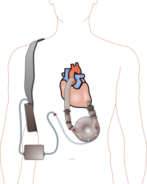
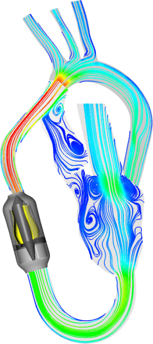
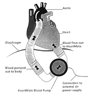
A ventricular assist device (VAD) is an implantable mechanical pump that helps pump blood from the lower chambers of your heart (the ventricles) to the rest of your body. VADs are used in people who have weakened hearts or heart failure. Although VADs can be placed in the left, right or both ventricles of your heart, they are most frequently used in the left ventricle. When placed in the left ventricle they are called left ventricular assist devices (LVADs).

You may have a VAD implanted while you wait for a heart transplant or for your heart to become strong enough to effectively pump blood on its own. Your doctor may also recommend having a VAD implanted as a long-term treatment if you have heart failure and you’re not a good candidate for a heart transplant.

The procedure to implant a VAD requires open-heart surgery and has serious risks. However, a VAD can be lifesaving if you have severe heart failure.

http://www.mayoclinic.com/health/lvad/MY01077

This is an assessment of the development and progression of cardiogenic shock  and review of the use of ventricular assist devices in that setting.  It is another piece of the chapter on cardiothoracic surgical management at Columbia University Medical Center, New York, NY.

A stepwise progression in the treatment of cardiogenic shock.

Pollack AUriel NGeorge IKodali STakayama HNaka YJorde U.

Source

Department of Medicine, New York Presbyterian Hospital/Columbia University Medical Center, New York, New York, USA.

Abstract

Cardiogenic shock remains a deadly complication of acute myocardial infarction (MI). Early revascularization, inotropic support, and intraaortic balloon counterpulsation are the mainstays of treatment, but these are not always sufficient. New mechanical approaches, both percutaneous and surgical, are available in this high-risk population. We present a case of a young woman with a massive anterior wall MI and subsequent cardiogenic shock who was treated with advanced mechanical circulatory support. This case serves as an illustration of the stepwise escalation of mechanical support that can be applied in a patient with an acute MI complicated by refractory cardiogenic shock. We also review the literature with regard to the use of percutaneous left ventricular assist devices in the setting of cardiogenic shock.

Copyright © 2012 Elsevier Inc. All rights reserved.

PMID: 22608034

Care of the Critically Ill:  A Stepwise Progression in the Treatment of Cardiogenic Shock.

Pollack A, Uriel N, George I, Kodali S, Takayama H, Naka Y, Jorde U
J Heart & Lung 2012; 41:500-504.

Initial Presentation

 A 21-year-old woman with a history of migraine headaches was admitted to the hospital with nonradiating substernal chest pain onset that morning. When she presented to another hospital she had a normal electrocardiogram (EKG) and was discharged. When the patient’s chest discomfort became crushing  she presented again to the same hospital where her EKG revealed ST-segment elevations in an anterolateral distribution. Her peak (hs) troponin was 229 ng/mL and peak creatinine kinase was 6900 U/L.  This was an elevation of CK far out of proportion to the troponin increase (suggestive of decreased peripheral circulation with massive release of CK from muscle). There was no family history of early myocardial infarction (MI), sudden cardiac death, clotting disorders, or hypercholesterolemia. She had been taking amitriptyline for migraines and oral contraceptives for 3 years.  The patient developed significant hypotension, after she was given metoprolol and morphine, for which dobutamine and dopamine were administered. Medication was switched to norepinephrine because of excessive tachycardia. Cardiac catheterization was performed emergently approximately 12 hours after the onset of the patient’s chest pain.
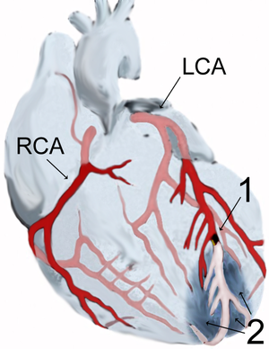
Thrombectomy of an angiographically identified clot in the proximal portion of the left anterior descending artery was performed, followed by placement of a bare metal stent with no residual occlusion. An intraaortic balloon bump (IABP) was placed. The initial transthoracic echocardiogram revealed an ejection fraction of 25% and global hypokinesis with regional wall motion abnormalities, worst in the anterior, apical, and lateral walls. She was intubated and required significant hemodynamic support with norepinephrine. Her antiplatelet regimen consisted of oral aspirin, clopidogrel, and intravenous eptifibatide. The patient was transferred to the New York Presbyterian Hospital/Columbia University Medical Center approximately 12 hours after revascularization.

Transfer to  NY Presbyteran Columbia Hospital

On arrival, the patient was intubated and sedated. Her blood pressure was 80/51mmHg, pulse rate was 140 beats/min, and oral temperature was 101F. On examination, she was tachycardic with warm extremities. The jugular veins were not distended. Her lactate was 7.0 mmol/L. (If she was so severely hypotensive with lactic acidemia, possibly from impaired liver and/or muscle circulation with aerobic glycolysis, then why was the temperature 101 deg F?)  The patient was not tested for procalcitonin (Brahms, BioMerieux), but sepsis is now considered bacterial or abacterial.  Whether there was release of bacterial endotoxin secondary to poor decreased circulation in the superior mesenteric artery is not known, which complicates the situation more.  In a study of acute phase changes in liver proteins by Bernstein and associates [Transthyretin as a marker to predict outcome in critically ill patients. Devakonda A, George L, Raoof S, Esan A, Saleh A, Bernstein LH.   Clin Biochem 2008; 41(14-15):1126-1130. ICID: 939927], and another on  procalcitonin and sepsis [The role of procalcitonin in the diagnosis of sepsis and patient assignment to medical intensive care. Bernstein LH, Devakonda A, Engelman E, Pancer G,  Ferrar J, Rucinski J, Raoof S,  George L, Melniker L.  J Clin Ligand Assay] there was a notable case of negative bacterial culture in a patient with highly elevated procalcitonin, considered a reliable early indicator of sepsis.sepsis classification with PCT and MAP
Procalcitonin (PCT) is a sensitive and specific inflammation marker, which can be used to detect both inflammatory infections and noninflammatory complications in postsurgical monitoring of patients after cardiac surgery using extracorporeal circulation. The optimum cut-off value for PCT levels, as a predictor of postoperative complications, appears to be 1.2 ng/mL with a sensitivity of 80% and a specificity of 90%. PCT may be used to monitor response to therapy because blood concentrations increase in an inflammatory disease relapse. Importance of procalcitonin in post-cardiosurgical patients. Topolcan O, Bartunek L, Holubec Jr L,  Polivkova V, eta al. Journal of Clinical Ligand Assay 2008; 31(1-4): 57-60.]This might be expected to be associated with a CRP increase over 50-70 mg/ml.  In addition, the hemogram would have been of some interest, perhaps raising the question of whether the cardiovascular impairment triggered other events [Validation and Calibration of the Relationship between Granulocyte Maturation and the Septic State. Bernstein LH and Rucinski J.  Clin Chem Lab Med 2011; 49. Walter de Gruyter . http://dx.doi.org/10.1515cclm.2011.688Converting Hematology Based Data into an Inferential Interpretation. Bernstein LH, David G, Rucinski J and Coifman RR.  In Hematology – Science and Practice, 2012. Chapter 22, pp 541-552. InTech Open Access Publ. Croatia]. 
A chest radiograph showed pulmonary edema. Her EKG revealed sinus tachycardia at 121 beats/min with ST-segment elevation of 3 mm in leads V1 to V4 and poor R-wave progression throughout the precordial leads with pathologic Q waves in V1 to V6, I, and aVL. Eptifibatide (Integrilin, Merck & Co., Inc., Whitehouse Station, NJ) was stopped, and norepinephrine was continued at 20 mg/min. Dobutamine 2.5 mg/min and broad-spectrum antibiotics were administered. During the next 4 hours, the patient’s mean arterial pressure fluctuated between 60 and 70 mm Hg with a heart rate between 120 and 140 beats/min on 20 mg/min of norepinephrine, 2.5 mg/min of dobutamine, and the IABP. Rapid escalation of mechanical support with a left ventricular assist device (LVAD) was deemed necessary.  Right-sided heart catheterization after placement of an Impella 2.5 assist device (ABIOMED, Inc.) revealed a cardiac output of 3.3 L/min and a cardiac index (CI) of 2.1 L/min/m2, despite addition of 3 ug/min and 4 U/h of vassopressin.

Day 2

On the second day after transfer she was severely hyponatremic, but her plasma sodium stabilized at 131 to 138 mmol/L after discontinuing the vasopressin. She also developed significant bleeding at the site of the Impella and hemolysis requiring several blood transfusions. Her hemoglobin on transfer was 10.4 g/dL, which trended down to 7.8 g/dL after Impella placement. The patient’s lactate dehydrogenase was 1980 U/L (probably reflecting poor liver perfusion), and total bilirubin was 2.6 mg/dL on day 2 of her hospitalization compared with 1.1 mg/dL on transfer.

Day 3

After the Impella device was removed on day 3 because of persistent bleeding, the patient’s hemoglobin, bilirubin, and platelet count stabilized, but while the patient was able to maintain end-organ perfusion initially as manifested by a normal creatinine, as the day progressed, the patient’s systemic blood pressure trended downward and urine output decreased, and she could not tolerate discontinuation of the vasoactive agents being administered. Pulmonary hypertension developed with a rate-dependent cardiac output as manifested by persistent tachycardia, and had an ejection fraction of 20% with severe hypokinesis of all segments except the basal inferior and inferolateral walls. As a consequence of the enduring cardiogenic shock and the low likelihood for recovery of left ventricular function, it was evident the patient required long-term mechanical support. A continuous flow LVAD (HeartMate II; Thoratec Corporation) was implanted as a rescue therapy, and the patient was emergently listed for transplantation.

Recovery

A comprehensive heart failure regimen was introduced, and the patient was discharged with warfarin 25 days after her transfer. A comprehensive hypercoagulability workup performed while the patient was receiving anticoagulation with negative results. Aside from oral contraceptive use, no other obvious risk factor for an acute arterial thrombosis could be identified, which is not surprising given that up to 40% of all thrombotic events occur in patients without a recognizable risk factor. Early revascularization, inotropic support, and intraaortic balloon counterpulsation are the mainstays of treatment, but these are not always sufficient.  New mechanical approaches, both percutaneous and surgical, are available in this high-risk population. This case serves as an illustration of the stepwise escalation of mechanical support that can be applied in a patient with an acute MI complicated by refractory cardiogenic shock. We also review the literature with regard to the use of percutaneous left ventricular assist devices in the setting of cardiogenic shock.

Recommendation

The authors recommend the following protocol for patients with cardiogenic shock superimposed on acute MI.    Treatment of cardiogenic shock.  PCI, percutaneous coronary intervention; IABP, intraaortic balloon pump; VAD, ventricular assist device; VA-ECMO, venoarterial extracorporeal membrane oxygenation; OHT, orthotopic heart transplantation; pVAD, percutaneous ventricular assist device. It is important to note that it includes immediate revascularization in conjunction with IABP placement. In patients with refractory cardiogenic shock who are unable to be weaned from the IABP, mechanical circulatory support using a percutaneous or surgical device is the next essential measure to be taken. The type of mechanical support to be used depends on many factors, including the reversibility of the shock state, chances of ventricular recovery, and risk of bleeding. Mechanical circulatory support with left ventricular assists devices can improve cardiac performance and reduce myocardial ischemic injury. Principle mechanisms include unloading of the left ventricle, thereby decreasing myocardial oxygen demand and improvement of systemic hypotension, thus increasing coronary perfusion.
Although there were complications related to the use of the device, its deployment resulted in the improvement of the patient’s surgical candidacy by virtue of maintaining her end-organ function.  After the removal of the Impella device, we thought the left ventricle in this patient would not recover, and for this reason, we chose a definitive surgical procedure as opposed to alternative temporary support device.  Clinical studies focusing on the use of VA-ECMO in refractory cardiogenic shock after an acute MI are limited. Observational and retrospective series have thus far demonstrated a high mortality rate in these patients.  However, a recent retrospective study of 33 patients who received ECMO support for advanced refractory cardiogenic shock after an acute MI demonstrated a mortality rate of 46% and 52% at 30 days and 1 year, respectively. In addition to mny complications with VA-ECMO, the procedure also can lead to increased afterload from the retrograde flow of peripheral cannulation., which may to lead to increased left ventricular pressure and wall stress, thereby compromising myocardial recovery and worsening pulmonary edema, both of which were major concerns
in this patient.

Conclusions

This case demonstrates that a sequential approach using percutaneous mechanical support as a bridge to surgical mechanical support is feasible in this high-risk population (Figure ). Advantages of percutaneous mechanical support include its rapid and straightforward placement. Disadvantages include its limited cardiac output and bleeding. Future technology should focus on a device that is capable of providing significant cardiac output and that can be easily placed, like the Impella. Such a device could alter the natural history of intractable cardiogenic shock.

Other related articles published on this Open Access Online Scientific Journal include the following:

Implantable Synchronized Cardiac Assist Device Designed for Heart Remodeling: Abiomed’s Symphony

Aviva Lev-Ari, PhD, RN, 7/11/2012

http://pharmaceuticalintelligence.com/2012/07/11/implantable-synchronized-cardiac-assist-device-designed-for-heart-remodeling-abiomeds-symphony/

Biomaterials Technology: Models of Tissue Engineering for Reperfusion and Implantable Devices for Revascularization

Larry H Bernstein, MD, FCAP and Aviva Lev-Ari, PhD, RN

http://pharmaceuticalintelligence.com/5_04_2013/bernstein_lev-ari/Bioengineering_of_Vascular_and_Tissue_Models

Foreseen changes in Guideline of Treatment of Cardiogenic Shock with Intra-aortic Balloon counterPulsation (IABP)

Evidence for Overturning the Guidelines in Cardiogenic Shock

Clinical Indications for Use of Inhaled Nitric Oxide (iNO) in the Adult Patient Market: Clinical Outcomes after Use, Therapy Demand and Cost of Care

Aviva Lev-Ari, PhD, RN, 6/3/2013

English: Ventricular assist device

English: Ventricular assist device (Photo credit: Wikipedia)

English: Simulation of a wave pump human ventr...

English: Simulation of a wave pump human ventricular assist device (Photo credit: Wikipedia)

myocardial infarction - Myokardinfarkt - scheme

myocardial infarction – Myokardinfarkt – scheme (Photo credit: Wikipedia)

English: Graphic presentation of an LVAD, left...

English: Graphic presentation of an LVAD, left ventricular assist device. (Photo credit: Wikipedia)

Read Full Post »

Hear Barbra Streisand accept her Honorary Doctorate from The Hebrew University of Jerusalem

Reporter: Aviva Lev-Ari, PhD, RN


From: AFHU <AFHU@mail.vresp.com>
Reply-To: AFHU <reply-01febe6994-47cd97e959-aa4c@u.cts.vresp.com>
Date: Mon, 17 Jun 2013 18:37:07 +0000
To: <avivalev-ari@alum.berkeley.edu>
Subject: Hear Barbra Streisand accept her Honorary Doctorate from The Hebrew University of Jerusalem


‘I wish the world were more like the hallways of the Hebrew University,’ says Barbra Streisand 


Legendary singer, actress and philanthropist receives honorary doctorate from the Hebrew University of Jerusalem

Legendary American actress, director, singer, producer, composer, philanthropist and activist Barbra Streisand received an honorary doctor of philosophy degree from the Hebrew University of Jerusalem today. The ceremony took place at 4 p.m. on the Mount Scopus campus, during the 76th Hebrew University International Board of Governors Meeting.

Following welcomes from the Chairman of the Hebrew University’s Board of Governors, Mr. Michael Federmann, and Prof. Menahem Ben-Sasson, the Hebrew University’s president, the honorary doctorate was presented to Ms. Streisand in recognition of her professional achievements, outstanding humanitarianism, leadership in the realm of human and civil rights, and dedication to Israel and the Jewish people.

An audio recording of Ms. Streisand’s comments is available to news organizations at http://bit.ly/hebrewu_streisand. It is not intended for rebroadcast.

In her comments after receiving the award, Ms. Streisand said, “For close to 30 years, I’ve had a deep connection to the Hebrew University. It’s not only home to a diverse population of some of Israel’s best and brightest students, but it also houses the Emanuel Streisand Building for Jewish Studies.”

In 1984 Ms. Streisand established the Emanuel Streisand Building in memory of her beloved father, whom she praised at the time as “a teacher, scholar and religious man who devoted himself to education.”

“I think he would be very proud to know that this esteemed institution is honoring his daughter,” she said today.

Streisand said it made her happy to read in the newspaper that more women than men graduated with a doctorate at the Hebrew University’s Convocation last night.

“One of the things I’ve always admired about this university is the fact that here, women and men, Jews and Arabs, Christians and Muslims, native-born and immigrants, sit together in classes, share the same cafeterias, learn from the same professors, and dream together of a good and meaningful life,” she said.

“I wish the word were more like the hallways of the Hebrew University,” she added.

Streisand condemned manifestations of exclusion of women in Israel, saying, “I realize it’s not easy to fully grasp the dynamics of what happens in a foreign land. Israel and the United States have much in common: Two great and noble countries, each with problems of course, but always striving to shine as a beacon of hope. So it’s distressing to hear about women in Israel being forced to sit at the back of a bus, or when we hear about Women of the Wall having metal chairs hurled at them when they attempt to peacefully pray, or when women are banned from singing in public ceremonies. But I’m also pleased to read that things are changing here. Repairs are being made and that’s very good.”

Streisand also complimented the debut speech of new Member of Knesset Dr. Ruth Calderon and said that Calderon’s speech served as an example of secular-religious dialogue through which people and countries can come together.

She concluded by quoting Albert Einstein, one of the founders of the Hebrew University: “Example isn’t another way to teach, it’s the only way to teach.”

At the conclusion of the ceremony, a member of the audience called out, “We love you, Barbra!” When Hebrew University President Prof. Menahem Ben-Sasson pointed out that with her honorary doctorate she is now “Dr. Streisand,” the audience member shouted back, “We love you, Dr. Streisand!”

After the event, Streisand  toured the Mount Scopus campus and visited the building named for her father. She also met with a number of scholars and students from the university, and among other things discussed the status of women.

Ms. Streisand has been long admired for her civic activism and philanthropic leadership.  Her commitment is reflected in the work of The Streisand Foundation, which is dedicated to fostering women’s equality and health, protecting human and civil rights, advancing the needs of at-risk children in society and preserving the environment. She often donates the proceeds from her performances on behalf of important causes.


Read Full Post »

Dr. Mark Josephson, Chief of Cardiology – CardioVascular Institute at Beth Israel Deaconess Medical Center – Recipient of 2013 American Heart Association‘s Paul Dudley White Award for Contributions to Cardiac Electrophysiology

Reporter: Aviva Lev-Ari, PhD, RN

VIEW VIDEO – Outstanding !!

http://bidmc.org/Centers-and-Departments/Departments/Cardiovascular-Institute/About-the-CVI/CVI-In-the-News/2013/April/Josephson.aspx

Dr. Josephson Recognized for Contributions to Cardiac Electrophysiology

  • Date: 5/1/2013

The American Heart Association presented its prestigious Paul Dudley White Award to Mark E. Josephson, MD, Chief of Cardiovascular Medicine at the CardioVascular Institute at Beth Israel Deaconess Medical Center, at its annual gala in April.

The award is given annually to a Massachusetts medical professional physician who has made a distinguished contribution to the American Heart Association’s mission of building healthier lives, free of cardiovascular disease and stroke.

“The AHA is pleased to honor Dr. Josephson,” said N. A. Mark Estes III, MD, director of the New England Cardiac Arrhythmia Center at Tufts Medical Center, chair of the selection committee. The award “is a fitting tribute for his professional accomplishments, personal attributes, and contributions to the AHA.”

Transformed His Field

Dr. Josephson is credited with transforming the field of cardiac electrophysiology from an intriguing scientific idea to a robust diagnostic and therapeutic tool for the management of arrhythmias, or abnormal heart rhythms. His research into the physiologic basis of these conditions has led to revolutionary achievements in their diagnosis and treatment.

A passionate educator, Dr. Josephson in the late 1970s wrote the definitive textbook on the practice of electrophysiology. It is now in its fourth edition and one of the rare single-author textbooks in any field. Since 1982, he has co-taught a seminal bi-yearly seminar on the interpretation of complex arrhythmias. The course has been attended by nearly 6,000 physicians, including 85 percent of electrophysiologists in the United States, for whom it is considered a rite of passage. He is also one of the busiest clinicians in BIDMC’s Division of Cardiovascular Medicine.

Having trained more physicians in his specialty than anyone else in the world, Dr. Josephson is fond of saying that his greatest legacy is the successes of his “academic children and grandchildren” and the subsequent generations of clinicians and researchers they have gone on to teach.

Dr. Josephson is the author of 444 original articles in peer-reviewed scientific publications, such as the New England Journal of Medicine and Circulation, a journal of the AHA. He is the author of more than 200 chapters, reviews and editorials.

Unfailing Dedication

“The selection of Mark Josephson as the 2013 recipient of the Paul Dudley White Award is a fitting recognition of the impact he has had on cardiology in Boston for the last 20 years,” says cardiologist Peter Zimetbaum, MD, a BIDMC colleague and member of the AHA selection committee. “He, like Paul Dudley White, inspires us through his unparalleled clinical and research insights and his unfailing dedication to the practice of medicine.”

Dr. Josephson shares a number of attributes with Dr. White, who was one of Boston’s most revered cardiologists and a founding father of the AHA. Their shared experiences include an association with Harvard, a sustained tenure at his medical institution, and early military experience that helped launch his career.

Perhaps most significantly, they share an unbridled passion for saving and enhancing the lives of patients with cardiovascular disease.

Pamela Lesser, one of Dr. Josephson’s patients who endorsed his nomination, said, “I don’t know how old he is, but he has an enthusiasm and love for what he does that is like he’s in his 20s, just out of medical school and ready to conquer the world. He’s on the edge of discovery. He has a passion for his work that spreads to the patient and that feeds into that whole feeling of ‘I’m in the best hands possible.’”

Read Full Post »

Dealing with the Use of the High Sensitivity Troponin (hs cTn) Assays: Preparing the United States for High-Sensitivity Cardiac Troponin Assays

Author and Curator: Larry H Bernstein, MD, FCAP
Author and Curator: Aviva Lev-Ari, PhD, RD

In this article we shall address the two following papers:
  1. Acute Chest Pain/ER Admission: Three Emerging Alternatives to Angiography and PCI – Corus CAD, hs cTn, CCTA
  2. Frederick K. Korley, MD, Allan S. Jaffe, MD in Journal of the American College of Cardiology  J Am Coll Cardiol. 2013; 61(17):1753-1758.

In a previous posting I commented on the problem of hs cTn use and the on site ED performance of cardiac treadmill (done in Europe)

  • prior to a decision of CT scan (not done in US).

Acute Chest Pain/ER Admission: Three Emerging Alternatives to Angiography and PCI – Corus CAD, hs cTn, CCTA

We examine the emergence of Alternatives to Angiography and PCI as most common strategy for ER admission with listed cause of Acute Chest Pain. The Goal is to use methods that will improve the process to identify for an Interventional procedure only the patients that a PCI is a must to have.

Alternative #1: Corus®  CAD

Alternative #2: High-Sensitivity Cardiac Troponins in Acute Cardiac Care

Alternative #3: Coronary CT Angiography for Acute Chest Pain
After presenting the the Three alternatives, the Editorial by R.F. Redberg, Division of Cardiology, UCSF, will be analyzed.
  • Alternative #1:  First-Line Test to Help Clinicians Exclude Obstructive CAD as a Cause of the Patient’s Symptoms

Corus®  CAD, a blood-based  gene expression test, demonstrated high accuracy with both a high negative predictive value (96 percent) and high sensitivity (89 percent) for assessing  obstructive coronary artery disease  (CAD) in a population of patients referred for stress testing with myocardial perfusion imaging (MPI).

COMPASS enrolled stable patients with symptoms suggestive of CAD who had been referred for MPI at 19 U.S. sites.  A blood sample was obtained in all 431 patients prior to MPI and Corus CAD gene expression testing was performed with study investigators blinded to Corus CAD test results.Following MPI, patients underwent either invasive coronary angiography orcoronary CT angiography, gold-standard anatomical tests for the diagnosis of coronary artery disease.

A Blood Based Gene Expression Test for Obstructive Coronary Artery Disease Tested in Symptomatic Non-Diabetic Patients Referred for Myocardial Perfusion Imaging: The COMPASS Study

http://pharmaceuticalintelligence.com/2012/08/14/obstructive-coronary-artery-disease-diagnosed-by-rna-levels-of-23-genes-cardiodx-heart-disease-test-wins-medicare-coverage/

  • Alternative #2: High-Sensitivity Cardiac Troponins in Acute Cardiac Care

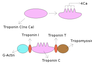
Recommendations for the use of cardiac troponin (cTn) measurement in acute cardiac care have recently been published.[1] Subsequently, a high-sensitivity (hs) cTn T assay was introduced into routine clinical practice.[2] This assay, as others, called highly sensitive, permits measurement of cTn concentrations in significant numbers of apparently illness-free individuals. These assays can measure cTn in the single digit range of nanograms per litre (=picograms per millilitre) and some research assays even allow detection of concentrations <1 ng/L.[2–4] Thus, they provide a more precise calculation of the 99th percentile of cTn concentration in reference subjects (the recommended upper reference limit [URL]). These assays measure the URL with a coefficient of variation (CV) <10%.[2–4]The high precision of hs-cTn assays increases their ability to determine small differences in cTn over time. Many assays currently in use have a CV >10% at the 99th percentile URL limiting that ability.[5–7] However, the less precise cTn assays do not cause clinically relevant false-positive diagnosis of acute myocardial infarction (AMI) and a CV <20% at the 99th percentile URL is still considered acceptable.[8]

We believe that hs-cTn assays, if used appropriately, will improve clinical care. We propose criteria for the clinical interpretation of test results based on the limited evidence available at this time.

References

1. Thygesen K, Mair J, Katus H, Plebani M, Venge P, Collinson P, Lindahl B, Giannitsis E, Hasin Y, Galvani M, Tubaro M, Alpert JS, Biasucci LM, Koenig W, Mueller C, Huber K, Hamm C, Jaffe AS; Study Group on Biomarkers in Cardiology  of the ESC Working Group on Acute Cardiac Care. Recommendations  for the use of cardiac troponin measurement in acute cardiac care. Eur Heart J 2010;31:2197–2204.

2. Saenger AK, Beyrau R, Braun S, Cooray R, Dolci A, Freidank H, Giannitsis E, Gustafson S, Handy B, Katus H, Melanson SE, Panteghini M, Venge P, Zorn M, Jarolim P, Bruton D, Jarausch J, Jaffe AS. Multicenter analytical evaluation of a high sensitivity troponin T assay. Clin Chim Acta 2011;412:748–754.

3. Zaninotto M, Mion MM, Novello E, Moretti M, Delprete E, Rocchi MB, Sisti D, Plebani M. Precision performance at low levels and 99th percentile concentration of the Access AccuTnI assay on two different platforms. Clin Chem Lab Med 2009; 47:367–371.

4. Todd J, Freese B, Lu A, Held D, Morey J, Livingston R, Goix P. Ultrasensitive flow based immunoassays using single-molecule counting. Clin Chem 2007; 53:1990–1995.

5. van de Kerkhof D, Peters B, Scharnhorst V. Performance of Advia Centaur second-generation troponin assay TnI-Ultra compared with the first-generation cTnI assay. Ann Clin Biochem 2008; 45:316–317.

6. Lam Q, Black M, Youdell O, Spilsbury H, Schneider HG. Performance evaluation and subsequent clinical experience with the Abbott automated Architect STAT Troponin-I assay. Clin Chem 2006; 52:298–300.

7. Tate JR, Ferguson W, Bais R, Kostner K, Marwick T, Carter A. The determination of the 99th percentile level for troponin assays in an Australian reference population. Ann Clin Biochem 2008; 45:275–288.

8. Jaffe AS, Apple FS, Morrow DA, Lindahl B, Katus HA. Being rational about (im)-precision: a statement from the Biochemistry Subcommittee of the Joint European Society of Cardiology/American College of Cardiology Foundation/American Heart Association/World Heart Federation Task Force for the definition of myocardial infarction. Clin Chem 2010; 56:921–943.

To the Editor:

Hoffmann et al. (July 26 issue)1 conclude that, among patients with low-to-intermediate-risk acute coronary syndromes, the incorporation of coronary computed tomographic angiography (CCTA) improves the standard evaluation strategy.2 However, it may be difficult to generalize their results, owing to different situations on the two sides of the Atlantic and the availability of high-sensitivity troponin T assays in Europe. In the United States, the Food and Drug Administration has still not approved a high-sensitivity troponin test, and patients in the Rule Out Myocardial Infarction/Ischemia Using Computer Assisted Tomography (ROMICAT-II) trial only underwent testing with the conventional troponin T test. As we found in the biomarker substudy in the ROMICAT-I trial, a single high-sensitivity troponin T test at the time of CCTA accurately ruled out acute myocardial infarction (negative predictive value, 100%) (Table 1TABLE 1Results of High-Sensitivity Troponin T Testing for the Diagnosis of Acute Coronary Syndromes in ROMICAT-I.).3 In addition, patients with acute myocardial infarction can be reliably identified, with up to 100% sensitivity, with the use of two high-sensitivity measurements of troponin T within 3 hours after admission.4,5

It seems plausible to assume that the incorporation of high-sensitivity troponin T assays in this trial would have outperformed CCTA. Therefore, it is important to assess the performance of such testing and compare it with routine CCTA testing in terms of length of stay in the hospital and secondary end points, especially cumulative costs and major adverse coronary events at 28 days.

Mahir Karakas, M.D.
Wolfgang Koenig, M.D.
University of Ulm Medical Center, Ulm, Germany
wolfgang.koenig@uniklinik-ulm.de

References

  1. Hoffmann U, Truong QA, Schoenfeld DA, et al. Coronary CT angiography versus standard evaluation in acute chest pain. N Engl J Med 2012;367:299-308

  2. Redberg RF. Coronary CT angiography for acute chest pain. N Engl J Med 2012;367:375-376

  3. Januzzi JL Jr, Bamberg F, Lee H, et al. High-sensitivity troponin T concentrations in acute chest pain patients evaluated with cardiac computed tomography. Circulation2010;121:1227-1234

  4. Keller T, Zeller T, Ojeda F, et al. Serial changes in highly sensitive troponin I assay and early diagnosis of myocardial infarction. JAMA 2011;306:2684-2693

  5. Thygesen K, Mair J, Giannitsis E, et al. How to use high-sensitivity cardiac troponins in acute cardiac care. Eur Heart J 2012;33:2252-2257

Author/Editor Response

In response to Karakas and Koenig: we agree that high-sensitivity troponin T assays may permit more efficient care of low-risk patients presenting to the emergency department with acute chest pain1 and may also have the potential to identify patients with unstable angina because cardiac troponin T levels are associated with the degree and severity of coronary artery disease.2 Hence, high-sensitivity troponin T assays performed early may constitute an efficient and safe gatekeeper for imaging. CCTA, however, may be useful for ruling out coronary artery disease in patients who have cardiac troponin T levels above the 99th percentile but below levels that are diagnostic for myocardial infarction. The hypothesis that high-sensitivity troponin T testing followed by CCTA, as compared with other strategies, may enable safe and more efficient treatment of patients in the emergency department who are at low-to-moderate risk warrants further assessment. The generalizability of our data to clinical settings outside the United States may also be limited because of differences in the risk profile of emergency-department populations and the use of nuclear stress imaging.3

Udo Hoffmann, M.D., M.P.H.
Massachusetts General Hospital, Boston, MA
uhoffmann@partners.org

W. Frank Peacock, M.D.
Baylor College of Medicine, Houston, TX

James E. Udelson, M.D.
Tufts Medical Center, Boston, MA

Since publication of their article, the authors report no further potential conflict of interest.

References

  1. Than M, Cullen L, Reid CM, et al. A 2-h diagnostic protocol to assess patients with chest pain symptoms in the Asia-Pacific region (ASPECT): a prospective observational validation study. Lancet 2011;377:1077-1084

  2. Januzzi JL Jr, Bamberg F, Lee H, et al. High-sensitivity troponin T concentrations in acute chest pain patients evaluated with cardiac computed tomography. Circulation2010;121:1227-1234

  3. Peacock WF. The value of nothing: the consequence of a negative troponin test. J Am Coll Cardiol 2011;58:1340-1342

  • Alternative #3: Coronary CT Angiography for Acute Chest Pain

The Study concluded:

There was increased diagnostic testing and higher radiation exposure in the CCTA group, with no overall reduction in the cost of care. 

Coronary CT Angiography versus Standard Evaluation in Acute Chest Pain

Udo Hoffmann, M.D., M.P.H., Quynh A. Truong, M.D., M.P.H., David A. Schoenfeld, Ph.D., Eric T. Chou, M.D., Pamela K. Woodard, M.D., John T. Nagurney, M.D., M.P.H., J. Hector Pope, M.D., Thomas H. Hauser, M.D., M.P.H., Charles S. White, M.D., Scott G. Weiner, M.D., M.P.H., Shant Kalanjian, M.D., Michael E. Mullins, M.D., Issam Mikati, M.D., W. Frank Peacock, M.D., Pearl Zakroysky, B.A., Douglas Hayden, Ph.D., Alexander Goehler, M.D., Ph.D., Hang Lee, Ph.D., G. Scott Gazelle, M.D., M.P.H., Ph.D., Stephen D. Wiviott, M.D., Jerome L. Fleg, M.D., and James E. Udelson, M.D. for the ROMICAT-II Investigators

N Engl J Med 2012; 367:299-308 July 26, 2012  http://dx.doi.org/10.1056/NEJMoa1201161

BACKGROUND

It is unclear whether an evaluation incorporating coronary computed tomographic angiography (CCTA) is more effective than standard evaluation in the emergency department in patients with symptoms suggestive of acute coronary syndromes.

METHODS

In this multicenter trial, we randomly assigned patients 40 to 74 years of age with symptoms suggestive of acute coronary syndromes but without ischemic electrocardiographic changes or an initial positive troponin test to early CCTA or to standard evaluation in the emergency department on weekdays during daylight hours between April 2010 and January 2012. The primary end point was length of stay in the hospital. Secondary end points included rates of discharge from the emergency department, major adverse cardiovascular events at 28 days, and cumulative costs. Safety end points were undetected acute coronary syndromes.

RESULTS

The rate of acute coronary syndromes among 1000 patients with a mean (±SD) age of 54±8 years (47% women) was 8%. After early CCTA, as compared with standard evaluation, the mean length of stay in the hospital was reduced by 7.6 hours (P<0.001) and more patients were discharged directly from the emergency department (47% vs. 12%, P<0.001). There were no undetected acute coronary syndromes and no significant differences in major adverse cardiovascular events at 28 days. After CCTA, there was more downstream testing and higher radiation exposure. The cumulative mean cost of care was similar in the CCTA group and the standard-evaluation group ($4,289 and $4,060, respectively; P=0.65).

CONCLUSIONS

In patients in the emergency department with symptoms suggestive of acute coronary syndromes, incorporating CCTA into a triage strategy improved the efficiency of clinical decision making, as compared with a standard evaluation in the emergency department, but it resulted in an increase in downstream testing and radiation exposure with no decrease in the overall costs of care. (Funded by the National Heart, Lung, and Blood Institute; ROMICAT-II ClinicalTrials.gov number, NCT01084239.)

http://www.nejm.org/doi/full/10.1056/NEJMoa1201161#t=abstract

REFERENCES

  1. Roe MT, Harrington RA, Prosper DM, et al. Clinical and therapeutic profile of patients presenting with acute coronary syndromes who do not have significant coronary artery disease. Circulation 2000;102:1101-1106

  2. Miller JM, Rochitte CE, Dewey M, et al. Diagnostic performance of coronary angiography by 64-row CT. N Engl J Med 2008;359:2324-2336

  3. Budoff MJ, Dowe D, Jollis JG, et al. Diagnostic performance of 64-multidetector row coronary computed tomographic angiography for evaluation of coronary artery stenosis in individuals without known coronary artery disease: results from the prospective multicenter ACCURACY (Assessment by Coronary Computed Tomographic Angiography of Individuals Undergoing Invasive Coronary Angiography) trial. J Am Coll Cardiol 2008;52:1724-1732

  4. Marano R, De Cobelli F, Floriani I, et al. Italian multicenter, prospective study to evaluate the negative predictive value of 16- and 64-slice MDCT imaging in patients scheduled for coronary angiography (NIMISCAD-Non Invasive Multicenter Italian Study for Coronary Artery Disease). Eur Radiol 2009;19:1114-1123
  5. Meijboom WB, Meijs MF, Schuijf JD, et al. Diagnostic accuracy of 64-slice computed tomography coronary angiography: a prospective, multicenter, multivendor study. J Am Coll Cardiol 2008;52:2135-2144
  6. Hoffmann U, Bamberg F, Chae CU, et al. Coronary computed tomography angiography for early triage of patients with acute chest pain: the ROMICAT (Rule Out Myocardial Infarction using Computer Assisted Tomography) trial. J Am Coll Cardiol 2009;53:1642-1650

  7. Hollander JE, Chang AM, Shofer FS, et al. One-year outcomes following coronary computerized tomographic angiography for evaluation of emergency department patients with potential acute coronary syndrome. Acad Emerg Med 2009;16:693-698

  8. Rubinshtein R, Halon DA, Gaspar T, et al. Usefulness of 64-slice cardiac computed tomographic angiography for diagnosing acute coronary syndromes and predicting clinical outcome in emergency department patients with chest pain of uncertain origin. Circulation2007;115:1762-1768

  9. Schlett CL, Banerji D, Siegel E, et al. Prognostic value of CT angiography for major adverse cardiac events in patients with acute chest pain from the emergency department: 2-year outcomes of the ROMICAT trial. JACC Cardiovasc Imaging 2011;4:481-491

  10. Goldstein JA, Chinnaiyan KM, Abidov A, et al. The CT-STAT (Coronary Computed Tomographic Angiography for Systematic Triage of Acute Chest Pain Patients to Treatment) trial. J Am Coll Cardiol 2011;58:1414-1422

  11. Litt HI, Gatsonis C, Snyder B, et al. CT angiography for safe discharge of patients with possible acute coronary syndromes. N Engl J Med 2012;366:1393-1403

  12. Shreibati JB, Baker LC, Hlatky MA. Association of coronary CT angiography or stress testing with subsequent utilization and spending among Medicare beneficiaries. JAMA2011;306:2128-2136

  13. Hoffmann U, Truong QA, Fleg JL, et al. Design of the Rule Out Myocardial Ischemia/Infarction Using Computer Assisted Tomography: a multicenter randomized comparative effectiveness trial of cardiac computed tomography versus alternative triage strategies in patients with acute chest pain in the emergency department. Am Heart J2012;163:330-338

  14. Abbara S, Arbab-Zadeh A, Callister TQ, et al. SCCT guidelines for performance of coronary computed tomographic angiography: a report of the Society of Cardiovascular Computed Tomography Guidelines Committee. J Cardiovasc Comput Tomogr 2009;3:190-204

  15. Gerber TC, Carr JJ, Arai AE, et al. Ionizing radiation in cardiac imaging: a science advisory from the American Heart Association Committee on Cardiac Imaging of the Council on Clinical Cardiology and Committee on Cardiovascular Imaging and Intervention of the Council on Cardiovascular Radiology and Intervention. Circulation 2009;119:1056-1065

  16. von Ballmoos MW, Haring B, Juillerat P, Alkadhi H. Meta-analysis: diagnostic performance of low-radiation-dose coronary computed tomography angiography. Ann Intern Med2011;154:413-420[Erratum, Ann Intern Med 2011;154:848.]

  17. Achenbach S, Marwan M, Ropers D, et al. Coronary computed tomography angiography with a consistent dose below 1 mSv using prospectively electrocardiogram-triggered high-pitch spiral acquisition. Eur Heart J 2010;31:340-346

  18. Than M, Cullen L, Reid CM, et al. A 2-h diagnostic protocol to assess patients with chest pain symptoms in the Asia-Pacific region (ASPECT): a prospective observational validation study. Lancet 2011;377:1077-1084

In the EDITORIAL by Redberg RF. Dr. Redberg, Cardiology Division, UCSF made the following points in:

Coronary CT angiography for acute chest pain. N Engl J Med 2012;367:375-376

  • Six million people present to ER annually with Acute Chest Pain, most have other diseases that Heart.
  • Current diagnostic methods lead to admission to the hospital, unnecessary stays and over-treatment – improvement of outcomes is needed.
  • Rule Out Myocardial Infarction Using Computer Assisted Tomography II (ROMICAT-II) 100 patients were randomly assigned to CCTA group or Standard Diagnosis Procedures Group in the ER which involved Stress Test in 74%.

CRITIQUE and Study FLAWS in MGH Study:

  • ROMICAT-II enrolled patients only during “weekday daytime hours, no weekend or nights when the costs are higher.
  • Assumption that a diagnostic test must be done before discharge for low-to-intermediate-risk patients is unproven and probably unwarranted.. No evidence that the tests performed let to improved outcomes.
  • Events rate for patient underwent CCTA, Stress test or no testing at al were less that 1% to have an MI, no one died. Thus, it is impossible to assign a benefit to the CCTA Group. So very low rates were observed in other studies
  • CCTA patients were exposed to substantial dose of Radiation, , contrast die,
  • Patients underwent ECG and Negative Troponin, no evidence that additional testing further reduced the risk.
  • Average age of patients: 54, 47% women.Demographic Characteristics with low incidence of CAD, NEJM, 1979; 300:1350-8
  • Risk of Cancer from radiation in younger population is higher, same in women.
  • Hoffmann’s Study: Radiation burden was clinically significant: Standard Evaluation Group: (4.7+-8.4 mSv), CCTA: (13.9+-10.4 mSv), exposure of 10 mSv have been projected to lead to 1 death from Cancer per 2000 persons, Arch Intern Med 2009; 169:2071-7
  • Middle Age women, increased risk of Breast Cancer from radiation, Arch Intern Med 2012 June 11 (ePub ahead of Print)
  • ROMICAT-II study: discharge diagnosis Acute Coronary Syndrome – less than 10%
  • CCTA Group: more tests, more radiation, more interventions tht the standard-evaluation group.
  • Choose Wisely Campaign – order test only when the benefit will exceed the risks

Dr. Redberd advocates ECG and Troponin, if NORMAL, no further testing.

Epicrisis on Part 1

Redberg’s conclusions are correct for the initial screening. The issue has been whether to do further testing for low or intermediate risk patients.

The most intriguing finding that is not at all surprising is that the CCTA added very little in the suspect group with small or moderate risk. My original studies using a receiver operator characteristic curve were very good, although some patients with CRF or ESRD had extremely high values. The ultra sensitive troponin threw the Area Under the ROC out the window, under the assumption that a perfect assay would exclude AMI, or any injury to the heart. The improved assay does pick up minor elevations of troponin in the absence of MI as a result of plaque rupture. It is possible that 50% of these elevations need medical attention, but then the question is an out of hospital referral or admission and further workup. I have discussed this at some length on several occasions with Dr. Jaffe at Mayo Clinic.

Many of those with minor or intermediate elevation have significant renal insufficiency, but they might also be in CKD Class 3 and not 1 or 2. The coexistence of Type 2 diabetes would go into the standard assessment, but is not mentioned in the study with respect to immediate admission or outcome 28 days after discharge.

The hs troponin I has been in daily use on the Ortho J&J (formerly Kodak) for about 2 years, and the QC standards are very high. I expected the Roche hs-TnT assay to be in use in US as well, but there may have been delays.  Januzzi , Jaffe, and Fred Aplle would be involved in the evaluation in the US, but Paul Collinson in UK, Katus and Mair in Germany, and other Europena centers certainly have been using the Roche Assay.

The biggest problem in these studies is as my mentor called my attention to – the frontrunners aren’t going to support a nose-to-nose up front study. Given that a diagnosis requires more information at minimal cost, especially when diagnosis of the heart that are not MI have to be evaluated as well, it is incomprehensibe to me that such information as

  1. mean arterial blood pressure,
  2. natriuretic peptides,
  3. the calculated EGFR are not used in the evaluation.

It is quite impossible to clear the deck when you have patients who don’t have

  1. ST elevation,
  2. depression, or
  3. T-wave inversion who are seen for vague

(not to mention long QT abnormalities).

  • predordial tightness or shortness of breath
  • pain that resembles gall bladder.

Is this an indication of the obsolescence of the RCT.

A Retrospective Quality and Cost Driven Audit on Effect of hs cTn Assay with On-Site CT Followup. (No treadmill availability)

A retrospective multisite study showed that doing the hs cTn followed by CT on-site was a good choice for US.

I also considered  the selective release of

  • low- moderate-risk patients cardiology followup in a timely manner.

This report is an excellent analysis of my point by Korley and Jaffe in Medscape, and satisfies some several years discussion

I have had with Dr. Jaffe, at Mayo Clinic.  He pointed out the importance of

  • Type 1 and Type 2 AMI

at a discussion with Dr. Fred Apple at a meeting of the Amer Assn for Clinical Chemistry that he fully elaborates on here.
It is really a refinement of other proposals that are being discussed.  It is also timely because hs cTnI is already being used
widely in the US, while there might be a holdup on the hs cTnT.

Highlights

  1. Need for a Universally Accepted Nomenclature
  2. Defining Uniform Criteria for Reference Populations
  3. Discriminating Between Acute and Nonacute Causes of hs-cTn Elevations
  4. Distinguishing Between Type 1 and Type 2 AMI
  5. Analytical Imprecision in Cardiac Troponin Assays
  6. Ruling Out AMI
  7. Investigating the Causes of Positive Troponin Values in Non-AMI Patients
  8. Risk Stratifying Patients With Nonacute Coronary Syndrome Conditions
  9. Conclusions

Abstract

It is only a matter of time before the use of high-sensitivity cardiac
troponin assays (hs-cTn) becomes common throughout the United
States. In preparation  for this inevitability, this article raises a number
of important issues regarding  these assays that deserve consideration.

These include: the need for

  • the adoption  of a universal nomenclature; the importance
  • of defining uniform criteria for reference populations;
  • the challenge of discriminating between acute and nonacute
    causes of hs-cTn elevations, and
  • between type 1 and type 2 acute myocardial infarction (AMI);

factors influencing the analytical precision of hs-cTn;

  • ascertaining the optimal duration  of the rule-out period for AMI;
  • the need for further evaluation to determine the causes
    of a positive hs-cTn in non-AMI patients; and
  • the use of hs-cTn to risk-stratify patients with disease conditions
    other than AMI.

This review elaborates on these critical issues  as a means of
educating clinicians and researchers about them.

Introduction

Recently, clinicians have begun to use the recommended cut-off values
for current generation cardiac troponin (cTn) assays:

  • the 99th percentile upper reference limit (URL).

Previously, there was reluctance to use these cut-off values because

  • of  cTn elevations from non-acute ischemic heart disease conditions.

Thus, there was a tendency to use cut-off values for troponin that equated with the

  • prior gold standard diagnosis developed with less sensitive markers
    • creatinine kinase-MB isoenzyme (CK-MB) or
    • the lowest value at which assay achieved a 10%
      coefficient of variation (CV),

which would reduce false-positive elevations (without plaque rupture).

The use of the 99th percentile URL increases the ability of these assays to detect both

  •   acute myocardial infarction (AMI) and
  •   structural cardiac morbidities.[1]

This change in practice should not be confused with

  •   newer-generation high-sensitivity assays.

Improvements in the analytic performance of cTn assays have resulted in

  •   superior sensitivity and precision.

Improved sensitivity occurs because of

  •   more sensitive antigen binding and detection antibodies,
  •   increases in the concentration of the detection probes on the tag antibodies,
  •   increases in sample volume, and buffer optimization.[2]

Assays now are able to measure

  •   10-fold lower concentrations with high precision

(a CV <10% at the 99th percentile  of the URL).

The high-sensitivity cardiac troponin T (hs-cTnT) assay is already in clinical use
throughout most of the world. It is only a matter of time before high- sensitivity
assays are approved for use in the United States. In preparation for this, as well as

  •   using the 99th percentile URL with contemporary assays,

there are a number of important issues that deserve consideration. Key concepts are included in (Table 1).

Table 1: Key ConceptsThere is a need to develop a universal nomenclature for troponin assays.There is a need for uniform criteria for selecting reference populations.The optimal delta criteria for distinguishing between acute and chronic cardiac injury remain unclear and are likely to be assay-specific.Distinguishing between type 1 and type 2 AMI is challenging, and
more type 2 AMIs will be detected with hsTn assays.Factors affecting the analytical precision of troponin assays (including how we collect samples) will become more important with the use of hs-cTn assays.The optimal duration for ruling out AMI remains unclear;

  • novel approaches to this issue are being developed.

Elevated hs-cTn, regardless of the cause, has important

  • prognostic implications and deserves additional evaluation; 

Many cases of chronic elevations can be evaluated in an outpatient setting.

Hs-cTn can be used to

  • risk-stratify patients with non-ACS cardiovascular comorbidities.

Need for a Universally Accepted Nomenclature

The literature is replete with terms used to refer to cTn assays.
We advocate the use of the term “high-sensitivity cardiac troponin assays”  (hs-cTn) for

  • cTn assays that v   measure cardiac troponin values in
  • in  at least 50% of a reference population.[2,3]

This policy has now been embraced by the journal Clinical Chemistry. High-sensitivity
assays can be further categorized as well (Table 2) with respect to generations of cTn.

Table 2.  Classification of High-Sensitivity Cardiac Troponin Assays 

Category

Description

First Generation                                   Able to measure cTn in
50%–75% of                                       a reference population
Second Generation                              Able to measure cTn in
75%–95% of                                       a reference population
Third Generation                                 Able to measure cTn in
> 95%                                               a reference population
Adapted from Apple and Collinson (3)
  • Ideally, assays should have a CV of <10% at the 99th percentile value.

Assays that do not achieve this level are less sensitive which protects against
false-positive results, and they can be used.[4]

Defining Uniform Criteria for Reference Populations
There is a lack of consistency in the types and numbers of subjects that constitute a reference
population.[2] Often, participants are included after simple screening by check list but without a

  • physical examination,
  • electrocardiogram, or
  • laboratory testing.

At other times, a

  • normal creatinine and/or a normal natriuretic peptide value is required.
  • Imaging to detect structural heart disease is rarely used. 

Because it is known that

  • gender,
  • age,
  • race,
  • renal function,
  • heart failure, and
  • structural heart disease, including
  • increased left ventricular (LV) mass

are associated with increased cTn concentrations,[5,6,7] An assay’s 99th percentile value depends on the composition of the reference group. Thus, the more criteria used, the lower the reference values (Figure 1).[5]

http://img.medscape.com/article/803/159/803159-fig1.jpg

Have no history of

  • vascular disease or diabetes, and
  • not taking cardioactive drugs,
    • based on questionnaire.
Normal defined as those individuals who had
  • no history of vascular or cardiovascular disease,
  • diabetes mellitus,
  • hypertension, or
  • heavy alcohol intake and who were
  • receiving no cardiac medication AND
  • had blood pressure ≤140/90 mmHg;
  • fasting glucose  <110 mg/dL;
  • eGFR >60mL/min;
  • LVEF > 50%; normal lung function; and no significant
  • valvular heart disease,
  • LVH,
  • diastolic HF, or
  • regional wall-motion abnormalities on ECHO.

The appropriate reference value to use clinically also is far from a settled issue.
It might be argued that

  • using a higher 99th percentile value for the elderly
  • allows comparison of the patient to his or her peers, but

in raising the cut-off value, if the increases are caused by comorbidities,

  • those who are particularly healthy will be disadvantaged.[8]

Gender and ethnicity are not comorbidities, and we would urge that those should be taken into account.
Regardless of the assay, there will need to be

  • 99th percentile values for men that are different for women.[2]

The reference population for assay validation studies should ideally be based on  –
demographic characteristics that mirror the U.S. population and include subjects whose

  • blood pressure,
  • serum glucose, and
  • creatinine and
  • natriuretic peptide values are
  • within the normal reference range and
  • who take no cardiac  medications.

These subjects should be

  • free from structural heart disease,
  • documented by echocardiography,
  • cardiac magnetic resonance imaging (MRI) or
  • computed tomography (CT) angiography.

Meeting these criteria will be a major challenge, especially for older individuals.
A conjoint pool of samples collected with manufacturers’ support so that all methods were derived from an

  • identical patient population for their reference ranges would be ideal.

[However, the method of collection and possible freeze-thaw effects is unavoidable].

One large national effort might be advantageous over multiple efforts.

 Discriminating Between Acute and Nonacute Causes of hs-cTn Elevations

With the ability to precisely measure small concentrations of cTn,

  • clinicians will be faced with the challenge of distinguishing patients
    • who have acute problems from those with chronic elevations from other causes.

Using the fourth-generation cTnT assay, approximately 0.7% of patients in
the general population have modest elevations >99th percentile URL.[11]

In the same population, this number was 2% with the hs-cTnT assay.[6]  Only

  • half of them had documentation (even with imaging) of cardiac abnormalities.

If the prevalence of a positive cTnT is 2% in the general population,

  • it will likely be 10% or 20% in the emergency department (ED)
  • and even higher in hospitalized patients, as
  • these patients often have cardiac comorbidities.

Measurement of changes in hs-cTn over time (δ hs-cTn)

  • improves the specificity of hs-cTn for the diagnosis of acute cardiac injury.[12,13]

However, it does so at the cost of sensitivity. With contemporary assays, differences

  • in analytical variation have been used to define an increasing pattern.

At elevated values, CV for most assays is in the range of 5% to 7%, so

  • a change of 20% ensures that a given change is not caused

by analytical variation alone.[10]

At values near the 99th percentile URL, higher change values are necessary.[13]  The situation with hs-cTn assays is much more complex, as follows:

1. Change criteria are unique for each assay.
2. It will be easy to misclassify patients with coronary artery disease who may present with a noncardiac cause of chest pain

  • but have elevated values.

They could be having unstable ischemia or elevations caused by structural cardiac abnormalities and noncardiac discomfort.

If hs-cTn is rising significantly, the issue is easy but

  • if the values are not rising, a diagnosis of AMI still might be made.
  • If so, some patients may be included as having AMI without a changing pattern.
  • This occurred in 14% patients studied by Hammarsten et al.[14]

If patients with elevated hs-cTn without a changing pattern are not called AMI,

  • should they be called patients with “unstable angina and cardiac injury” or patients with structural heart disease and noncardiac chest pain?

Perhaps both exist?

3. The release of biomarkers is flow-dependent.Thus, there may not always be rapid access to the circulation. An area of injury distal to a totally occluded vessel (when collateral channels close) may be different in terms of the dynAMIcs of

  • hs-cTn change than an intermittently occluded coronary artery.
4. Conjoint biological and analytical variation can be measured.

  • They are assay-dependent, and the reference change values range from 35% to 85%.[2]

The use of criteria less than that (which may be what is needed clinically) will thus
likely include individuals with changes caused by

  • conjoint biological and analytical variation alone.

This has been shown to be the case in

  • many patients with nonacute cardiovascular diagnoses.[14,15]
5. Most evaluations have attempted to define the optimal delta, often with receiver operator curve analysis. Such an approach is based on the concept that sensitivity and specificity deserve equivalent weight.[But higher deltas improve specificity more and lower ones improve sensitivity and it is not clear that all physicians want the same tradeoffs in this regard.]ED physicians often prefer high-sensitivity so that their miss rate is low (<1%),[16] whereas hospital clinicians want increased specificity. This tension will need to be addressed in defining the optimal delta.
6. The delta associated with AMI may be different from that associated with other cardiac injury.[14] In addition, women have less marked elevations of cTn in response to coronary artery disease[17] and in earlier studies were less apt to have elevated values.[18] Given their pathology is at times different,

  • it may be that different metrics may be necessary based on gender
7. Some groups have assumed that if a change is of a given magnitude over 6 hours, it can be divided by 6 and the 1-h values can be used.

  • This approach is not data driven, and biomarker release is more likely to be discontinuous rather than continuous.[19]

In addition, the values obtained with this approach are too small to be distinguished from a lack of change with most assays.

These issues pose a major challenge even for defining the ideal delta change value and provide the reasons why

  • the use of this approach will reduce sensitivity[20,21] (Figure 2).

http://img.medscape.com/article/803/159/803159-fig2.jpg

Defining the Optimal Delta: Tension Between Sensitivity and Specificity

There is a reciprocal relationship between sensitivity and specificity. With marked percentage changes,

  • specificity is improved at the expense of sensitivity, and
  • at lower values, the opposite occurs.

In addition, there is controversy in regard to the metrics that should be used with high-sensitivity assays.
The Australian-New Zealand group proposed

  • a 50% change for hs-cTnT for values below 53 ng/l and
  • a 20% change above that value.[22]
  • The 20% change is much less than conjoint biological and analytical variation.

A number of publications have suggested the superiority of

  • absolute δ cTn compared to relative δ cTn in discriminating between AMI and non-AMI causes of elevated cTn.[23,24,25]
  • The utility of the absolute or relative δ cTn appears to depend on the initial cTn concentration, and
  • the major benefit may be at higher values.[23]

A recent publication by Apple et al.[26] calculates deltas in several different ways with a contemporary assay and

  • provides a template for how to do such studies optimally.[26]

If all studies were carried out in a similar fashion, it would help immensely. In the long run, institutions will need to
define the approach they wish to take. We believe this discussion is a critical one and should include

  • laboratory,
  • ED, and
  • cardiology professionals.

Distinguishing Between Type 1 and Type 2 AMI

Although δ cTn is helpful in distinguishing between AMI and nonacute causes of Tn release,

  • it may or may not be useful in discerning type 1 from type 2 AMI.

As assay sensitivity increases, it appears that the frequency of type 2 AMI increases.
Making this distinction is not easy.

Type 1 AMI is caused by a primary coronary event, usually plaque rupture.

  • It is managed acutely with aggressive anticoagulation and
  • revascularization (percutaneous coronary intervention or coronary artery bypass).[10]

Type 2 AMI typically evolves secondary to ischemia from an oxygen demand/supply mismatch

  • severe tachycardia and
  • hypo- or hypertension and the like,
  • with or without a coronary abnormality.

These events usually are treated by addressing the underlying abnormalities.

They are particularly common in patients who are

  • critically ill and those who
  • are postoperative.[27]

However, autopsy studies from patients with postoperative AMI often manifest plaque rupture.[28]
Thus, the more important events, even if less common, may be type 1 AMIs. Type 2 events
seem more common in women,  who tend to have

  • more endothelial dysfunction,
  • more plaque erosion, and
  • less fixed coronary artery disease.[28-30]

Additional studies are needed to determine how best to make this clinical distinction.
For now, clinical judgment is recommended.

Analytical Imprecision in Cardiac Troponin Assays

All analytical problems will be more critical with hs-cTn assays. Cardiac troponin I (cTnI) and cardiac troponin T (cTnT) are measured using enzyme linked immune- sorbent assays.

  •   quantification of hs-cTn can be influenced by interference by reagent antibodies to analyte (cTn), leading to false- positive or negative results.[31]
  •   Autoantibodies to cTnI or cTnT are found in 5% to 20% of individuals and can reduce detection of cTn.[32,33]
  •   Additionally, fetal cTn isoforms can be re-expressed in diseased skeletal muscle and detected by the cTnT assays, resulting in false-positive values.[34]

Several strategies, including the use of

  •   blocking reagents,
  •  assay redesign, and use of
  •  antibody fragments,

have been used to reduce interference.[35–36]

There are differences in measured cTn values based on specimen type (serum versus heparinized plasma versus EDTA plasma).
In addition, hemolysis may affect the accuracy of cTn measurement,[37] and with blood draws from peripheral IV lines, common in ICU.

Ruling Out AMI

Studies evaluating the diagnostic performance of hs-cTn assays for the early diagnosis of AMI usually define AMI on

  • the basis of a rising and/or falling pattern of current generation cTn values.[21,38]

However, defining AMI on the basis of the less sensitive current generation assay results in an underestimation of the true prevalence of AMI and

  • an overestimation of negative predictive value of the experimental assay.
  • shortens the time it takes to rule in all the AMIs and
  • to definitively exclude AMI as it
  • ignores the new AMIs more sensitively detected by the hs-cTn assay.

Thus, in the study by Hammarsten et al.,[14]

  • the time to exclude all AMIs was 8.5 hours when all of the AMIs detected
    with the high-sensitivity assay were included, whereas
  • others that do not include these additional events report this can be done
    in 3 to 4 hours.[21,29,38]

In our view, Hammarsten is correct.

This does not mean that hs-cTn cannot help in excluding AMI. Body et al.[39] reported that patients who present with undetectable values (less than the LOB of the hs-cTnT assay) were unlikely to have adverse events during follow-up. If that group of patients is added to those who present later than 6 hours, then perhaps a significant proportion of patients


  • with possible acute coronary syndrome (ACS) could
  • have that diagnosis excluded with the initial value.[40]
    • studies need to continue to evaluate cTn values for at least 6 h
      to define the frequency of additional AMIs detected in that manner.

Using follow-up evaluations of patients with small event rates

  • who are likely to have additional care during the follow-up period are likely to be underpowered.

It may be that better initial risk stratification may help with this, as recently reported.[16,41]
Low-risk patients who have good follow-up after an ED visit

  • may be a group that can be released as early as 2 h after presentation.[16]

Investigating the Causes of Positive Troponin Values in Non-AMI Patients

Elevated Tn values (including those obtained with high-sensitivity assays) are associated with

  • a 2-fold higher risk for longer-term all-cause mortality and
  • cardiovascular death than a negative troponin values.[6,42-44]

This association is dose-dependent.

  • If values are rising, they are indicative of acute cardiac injury.

Those patients should be admitted because the risk is often short-term. However,

  • if the values are stable, assuming the timing of any acute event would
    allow detection of a changing pattern,
  • the risk, although substantive, in our view, often plays out in the longer term.[44]
  • Many of these individuals, assuming they are doing well clinically, can be
    evaluated outside of the hospital, in our view.
  • However, because such elevations are an indicator of a subclinical
    cardiovascular injury,  such evaluations should be early and aggressive.

Data from several studies suggest that there may well be risk far below the 99th percentile URL value.
Thus, it may evolve that patients in the upper ranges of the normal range also require some degree of cardiovascular evaluation.

Risk Stratifying Patients With Nonacute Coronary Syndrome
Conditions

Patients who have a rising pattern of values have a higher risk of mortality than those with negative values regardless of the cause.
Investigations are ongoing to determine how well results from hs-cTn testing help to risk-stratify patients with

  • pulmonary embolism,[45]
  • congestive heart failure,[46]
  • sepsis,[47]
  • hypertensive emergency,[48] and
  • chronic obstructive pulmonary disease.[49]

Presently, the studies suggest that cTn values classify patients into clinically relevant  risk subgroups. Studies are needed

  • to evaluate the incremental prognostic benefit of hs-cTn.

Conclusions

Routine use of hs-cTn assays in the United States is inevitable. These assays hold
the promise of

  • improving the sensitivity of AMI diagnoses,
  • shortening the duration of AMI evaluation and
  • improving the risk stratification of other noncardiac diagnoses.

However, to be able to fully realize their potential, additional studies are needed to address the

  • knowledge gaps we have identified. In the interim, clinicians need to
    • learn how to use the 99th% URL and
    • the concept of changing values

John Adan, MD, FACC

In 2008 CMS commissioned Yale University to analyze 30 days mortality after myocardial infarction in their hospitals.

The study has been based on review of medical records. Consensus criteria for diagnosis of myocardial infarction include

  • clinical symptoms,
  • EKG,
  • troponins,
  • CK MB,
  • ECHO,
  • cath,
  • histopathology, etc.

How the reviewed hospitals performed diagnostic coding is unknown. In clinical practice we are bombarded by consults

  • for elevated troponins due to causes other than myocardial infarction, like
    • pneumonia,
    • accelerated hypertension,
    • arrhythmias,
    • renal failure, etc.

The metric started out over 19%. Now it is below 15%, on average.

CT Angiography (CCTA) Reduced Medical Resource Utilization compared to Standard Care reported in JACC
Aviva Lev-Ari, PhD, RN
http://pharmaceuticalintelligence.com/2013/05/16/ct-angiography-ccta-reduced-medical-resource-utilization-compared-
to-standard-care-reported-in-jacc/?goback=%2Egde_4346921_member_241569351

typical changes in CK-MB and cardiac troponin ...

typical changes in CK-MB and cardiac troponin in Acute Myocardial Infarction (Photo credit: Wikipedia)

Phosphotungstic acid-haematoxylin staining dem...

Phosphotungstic acid-haematoxylin staining demonstrating contraction band necrosis in an individual that had a myocardial infarction (heart attack). (Photo credit: Wikipedia)

English: Troponin(SVG Version) 日本語: トロポニン(SVG修正版)

English: Troponin(SVG Version) 日本語: トロポニン(SVG修正版) (Photo credit: Wikipedia)

Read Full Post »

Amyloidosis with Cardiomyopathy

Author: Larry H Bernstein, MD, FACP
Introduction
Amyloidosis describes the various clinical syndromes that occur as a result of damage by amyloid deposits in tissues and organs throughout the body.  Systemic amyloidosis is a relatively rare multisystem disease caused by the deposition of misfolded protein in various tissues and organs. The term amyloid describes the deposition in the extracellular space of certain proteins in a highly characteristic, insoluble fibrillar form.  The disease entity is a disorder of misfolded or misassembled proteins.  There is extracellular amyloid fiber laid down as cross β-sheets disrupting organ function, which may affect the pancreas, kidney, autonomic nervous system, the heart, and in one form causes carpal tunnel syndrome.
It may present to almost any specialty, and diagnosis is frequently delayed. Cardiac involvement is a leading cause of morbidity and mortality, especially in primary light chain (AL) amyloidosis and in both wild-type and hereditary transthyretin amyloidosis. The heart is also occasionally involved in acquired serum amyloid A type (AA) amyloidosis and other rare hereditary types. Clinical phenotype varies greatly between different types of amyloidosis, and even the cardiac presentation has a great spectrum. The incidence of amyloidosis is uncertain, but it is thought that the most frequently diagnosed AL amyloidosis has an annual incidence of 6 to 10 cases per million population in the United Kingdom and United States.
The molecular basis for this particular phenomenon came with the extensive work done on multiple myeloma, antibody structure, and light chains.  In 1950, the discovery of a familial amyloid polyneuropathy was described in Portugal, and there were similar diseases in Sweden and Japan.  There were 72 known variants of transthyretin (TTR) in 1995, and now there are 100.  In addition, the occurance of different TTR associated variants with and without (amyloid) is found is Brazil, UK, US, Israel, Spain, France, Germany, Denmark, and Africa.  The table of variants, organ damage, and geographic location is too large to place on this document. If we refer to amyloid cardiomyopathy, it is exclusively a primary amyloidopathy, not secondary to light chain disorders or an inflammatory disease.  If we consider amyloidosis, we also have to consider family history, organ dysfunction, and we have to make a distinction between primary cardiac involvement, autonomic nervous system instability, and the two coexisting.  Familial amyloid polyneuropathy (FAP) is an extremely debilitating and progressive disease that is only treatable by liver transplantation.  Primary amyloid cardiomyopathy has been treated by heart transplant.  The qualifying statement here is, it depends.

Primary and Secondary Amyloidoses

Amyloid was originally described by pathologists based on microscopy. Amyloidoses are a systemic primary or secondary disease. There are distinctions to be made based on location and type.  The clinical significance of amyloid disease varies enormously, ranging from incidental asymptomatic deposits to localized disease through to rapidly fatal systemic forms that can affect multiple vital organs.
Common causes of secondary amyloidosis are – light chain production (AL) as in plasma cell dyscrasia, amyloid A (AA), senile systemic amyloidosis (diagnosed rarely in life).  The systemic amyloidoses are designated by a capital A (for amyloid) followed by the abbreviation for the chemical identity of the fibril protein. Thus, TTR amyloidosis is abbreviated ATTR, and immunoglobulin light chain type amyloidosis is abbreviated AL. Both normal-sequence TTR and variant-sequence TTR form amyloidosis. Normal-sequence TTR forms cardiac amyloidosis in elderly people, termed senile cardiac amyloidosis (SCA). When it was recognized that SCA is often accompanied by microscopic deposits in many other organs, the alternative name senile systemic amyloidosis (SSA) was proposed. Both terms are now used.
Currently available therapy is focused on reducing the supply of the respective amyloid fibril precursor protein and supportive medical care, which together have greatly improved survival. Chemotherapy and anti-inflammatory treatment for the disorders that underlie AL and AA amyloidosis are guided by serial measurements of the respective circulating amyloid precursor proteins, i.e. serial serum free light chains in AL and serum amyloid A protein in AA type.
Quality of life and prognosis of some forms of hereditary systemic amyloidosis can be improved by liver and other organ transplants. Various new therapies, ranging from silencing RNA, protein stabilizers to monoclonal antibodies, aimed at inhibiting fibril precursor supply, fibril formation or the persistence of amyloid deposits, are in development; some are already in clinical phase.
Ann Clin Biochem May 2012; 49(3 ): 229-241   http://acb.2011.011225v1 49/3/229

What is transthyretin (TTR)?

TTR is a  tetramer of 4 127 amino acid subunits synthesized by the liver that circulates as a transporter of thyroxin, and with retinol-binding protein, transports vitamin A.  It was originally defined by the migration in electrophoresis more anodal to albumin, hence, prealbumin.  It is present in cerebrospinal fluid, secreted by the choroid plexus.  The TTR monomer contains 8 antiparallel beta pleated sheet domains. TTR can be found in plasma and in cerebrospinal fluid and is synthesized by the choroid plexus of the brain and, to a lesser degree, by the retina. Its gene is located on the long arm of chromosome 18 and contains 4 exons and 3 introns.
The concentration in serum can be expected to be above 20 mg/dL in a health adult, but the protein decreases by 1 mg/dL/day postoperatively, and it decreases with acute or chronic renal failure, pneumonia or sepsis, rising again with the onset of anabolism.  Patients in the pulmonary intensive care unit have TTR levels that remain low for 7-10 days, but followup data for the remainder of the hospital stay or in relationship to readmission in the six months after release from hospital care was not part of the study.
A decrease in TTR is associated with the systemic inflammatory response, whereby, the liver reprioritizes the synthesis of proteins with an increase in acute phase reactants (APRs), namely, C-reactive protein (CRP) and a-1 acid glycoprotein, and decreased albumin and TTR.  The inflammatory condition maintains a euthyroid status with decreased TTR because of the availability of free thyroxine in equilibrium with the lower binding protein.  This has been referred to sick euthyroid status. The role in thyroxine transport is not insignificant, as chronic protein malnutrition is associated with hypothyroidism, as originally described by Prof. Yves Ingenbleek, Univ. Louis Pateur, Starsbourg, Fr. in Senegalese children with Kwashiorkor.  However, the importance of TTR as a unique biomarker is not to be downgraded because of what is often refered to as “an inverted APR”.
Transthyretin was discovered to be a good reflection of the “lean body mass”, by Vernon Young, MIT, and Ingenbleek, as a result of 3 decades of study. The ratio of S:N being 1:20 in plant proteins and 1:12.5 in animal sources, is closely related to methylation reactions and sustained deficiency of S intake results in elevated homocysteine level.

What is FAP?

Familial amyloid polyneuropathy (FAP), also called transthyretin-related hereditary amyloidosis, transthyretin amyloidosis or Corino de Andrade’s disease, is an autosomal dominant neurodegenerative disease. It is a form of amyloidosis, and was first identified and described by Portuguese neurologist Mário Corino da Costa Andrade, in the 1950s.FAP is distinct from senile systemic amyloidosis (SAS), which is not inherited, and which was determined to be the primary cause of death for 70% of supercentenarians who have been autopsied.
Familial amyloid polyneuropathy (FAP) is an extremely debilitating and progressive disease that is only treatable by liver transplantation.  Primary amyloid cardiomyopathy has been treated by heart transplant.  The qualifying statement here is, it depends.  Those patients with TTR-amyloidopathy have a specific gene substitution in the TTR gene. Consequently, there is circulation TTR, but it is not effectively involved in thyroxine transport.

Characteristics.

Usually manifesting itself between 20 and 40 years of age, it is characterized by pain, paresthesia, muscular weakness and autonomic dysfunction. In its terminal state, the kidneys and the heart are affected. FAP is characterized by the systemic deposition of amyloidogenic variants of the transthyretin protein, especially in the peripheral nervous system, causing a progressive sensory and motor polyneuropathy. The age at symptom onset, pattern of organ involvement, and disease course vary, but most mutations are associated with cardiac and/or nerve involvement. The gastrointestinal tract, vitreous, lungs, and carpal ligament are also frequently affected. When the peripheral nerves are prominently affected, the disease is termed familial amyloidotic polyneuropathy (FAP). When the heart is involved heavily but the nerves are not, the disease is called familial amyloid cardiomyopathy (FAC). Regardless of which organ is primarily targeted, the general term is simply amyloidosis-transthyretin type, abbreviated ATTR.

Genetics.

  1. TTR mutations accelerate the process of TTR amyloid formation and are the most important risk factor for the development of clinically significant ATTR. More than 85 amyloidogenic TTR variants cause systemic familial amyloidosis. The variant TTR is mostly produced by the liver. Amyloidogenic TTR mutations destabilize TTR monomers or tetramers, allowing the molecule to more easily attain an amyloidogenic intermediate conformation. The tetramer has to dissociate into misfolded monomers to aggregate into a variety of structures including amyloid fibrils. Because most patients are heterozygotes, they deposit both mutant and wild type TTR subnits.
  2. Familial amyloid polyneuropathy has an autosomal dominant pattern of inheritance. FAP is caused by a mutation of the TTR gene, located on human chromosome 18q12.1-11.2. A replacement of valine by methionine at position 30 (TTR V30M) is the mutation most commonly found in FAP.
  3. The disease in the TTR V30M kindreds was termed FAP because early symptoms arose from peripheral neuropathy, but these patients actually have systemic amyloidosis, with widespread deposits often involving the heart, gastrointestinal tract, eye, and other organs.
  4. TTR V122I: This variant, carried by 3.9% of African Americans and over 5% of the population in some areas of West Africa, increases the risk of late-onset (after age 60 years) cardiac amyloidosis. It appears to be the most common amyloid-associated TTR variant worldwide. Affected patients usually do not have peripheral neuropathy.
  5. TTR T60A: This variant causes late-onset systemic amyloidosis with cardiac, and sometimes neuropathic, involvement. This variant originated in northwest Ireland and is found in Irish and Irish American patients.
  6. TTR L58H: Typically affecting the carpal ligament and nerves of the upper extremities, this variant originated in Germany. It has spread throughout the United States but is most common in the mid-Atlantic region.
  7. TTR G6S: This is the most common TTR variant, but it appears to be a neutral polymorphism not associated with amyloidosis. It is carried by about 10% of people of white European descent.

Cardiac transthyretin (TTR) amyloidosis

Cardiac amyloidosis of transthyretin fibril protein (ATTR) type is an infiltrative cardiomyopathy characterised by ventricular wall thickening and diastolic heart failure. More than 27 different precursor proteins have the propensity to form amyloid fibrils. The particular precursor protein that misfolds to form amyloid fibrils defines the amyloid type and predicts the patient’s clinical course. Several types of amyloid can infiltrate the heart, resulting in progressive diastolic and systolic dysfunction, congestive heart failure, and death.  Increased access to cardiovascular magnetic resonance imaging has led to a marked increase in referrals to St George’s University of London, London (Dr. Jason Dungu) of Caucasian patients with wild-type ATTR (senile systemic) amyloidosis and Afro-Caribbean patients with the hereditary ATTR V122I type. Both subtypes present predominantly as isolated cardiomyopathy. The differential diagnosis includes cardiac amyloid light-chain (AL) amyloidosis, which has a poorer prognosis and can be amenable to chemotherapy.

Clinical Presentation

Cardiac amyloidosis, irrespective of type, presents as a restrictive cardiomyopathy characterized by progressive diastolic and subsequently systolic biventricular dysfunction and arrhythmia.1 Key “red flags” to possible systemic amyloidosis include nephrotic syndrome, autonomic neuropathy (eg, postural hypotension, diarrhea), soft-tissue infiltrations (eg, macroglossia, carpal tunnel syndrome, respiratory disease), bleeding (eg, cutaneous, such as periorbital, gastrointestinal), malnutrition/cachexia and genetic predisposition (eg, family history, ethnicity). Initial presentations may be cardiac, with progressive exercise intolerance and heart failure. Other organ involvement, particularly in AL amyloidosis, may cloud the cardiac presentation (eg, nephrotic syndrome, autonomic neuropathy, pulmonary or bronchial involvement). Pulmonary edema is not common early in the disease process, but pleural and pericardial effusions and atrial arrhythmias are often seen. Syncope is common and a poor prognostic sign. It is typically exertional or postprandial as part of restrictive cardiomyopathy, sensitivity to intravascular fluid depletion from loop diuretics combined with autonomic neuropathy, or conduction tissue involvement (atrioventricular or sinoatrial nodes) or ventricular arrhythmia. The latter may rarely cause recurrent syncope. Disproportionate septal amyloid accumulation mimicking hypertrophic cardiomyopathy with dynamic left ventricular (LV) outflow tract obstruction is rare but well documented. Myocardial ischemia can result from amyloid deposits within the microvasculature. Atrial thrombus is common, particularly in AL amyloidosis

Diagnosis and Treatment

imaging – Cardiovascular Magnetic Resonance in Cardiac Amyloidosis*.

Cardiac amyloidosis can be diagnostically challenging. Cardiovascular magnetic resonance (CMR) can assess abnormal myocardial interstitium. In cardiac amyloidosis, CMR shows a characteristic pattern of global subendocardial late enhancement coupled with abnormal myocardial and blood-pool gadolinium kinetics. The findings agree with the transmural histological distribution of amyloid protein and the cardiac amyloid load.
 *AM Maceira; J Joshi; SK Prasad; J Charles Moon, et al. Royal Brompton Hospital, London;
The diagnosis of amyloidosis requires histological identification of amyloid deposits. Congo Red staining renders amyloid deposits salmon pink by light microscopy, with a characteristic apple green birefringence under polarized light conditions. Additional immunohistochemical staining for precursor proteins identifies the type of amyloidosis.  Ultimately, immunogold electron microscopy and mass spectrometry confer the greatest sensitivity and specificity for amyloid typing.
Treatment of cardiac amyloidosis is dictated by the amyloid type and degree of involvement. Consequently, early recognition and accurate classification are essential.
Novel diagnostic and surveillance approaches using imaging (echocardiography, cardiovascular magnetic resonance), biomarkers (brain natriuretic peptide [BNP], high-sensitivity troponin), new histological typing techniques, and current and future treatments, including approaches directly targeting the amyloid deposits.

Etiology

Amyloidosis is caused by the extracellular deposition of autologous protein in an abnormal insoluble β-pleated sheet fibrillary conformation—that is, as amyloid fibrils. More than 30 proteins are known to be able to form amyloid fibrils in vivo, which cause disease by progressively damaging the structure and function of affected tissues. Amyloid deposits also contain minor nonfibrillary constituents, including serum amyloid P component (SAP), apolipoprotein E, connective tissue components (glycosaminoglycans, collagen), and basement membrane components (fibronectin, laminin). Amyloid deposits can be massive, and cardiac or other tissues may become substantially replaced. Amyloid fibrils bind Congo red stain, yielding the pathognomonic apple-green birefringence under cross-polarized light microscopy that remains the gold standard for identifying amyloid deposits.

AL Amyloidosis

AL amyloidosis is caused by deposition of fibrils composed of monoclonal immunoglobulin light chains and is associated with clonal plasma cell or other B-cell dyscrasias. The spectrum and pattern of organ involvement is very wide, but cardiac involvement occurs in half of cases and is sometimes the only presenting feature. Cardiac AL amyloidosis may be rapidly progressive. Low QRS voltages, particularly in the limb leads, are common. Thickening of the LV wall is typically mild to moderate and is rarely >18 mm even in advanced disease. Cardiac AL amyloid deposition is accompanied by marked elevation of the biomarkers BNP and cardiac troponin, even at an early stage. Involvement of the heart is the commonest cause of death in AL amyloidosis and is a major determinant of prognosis; without cardiac involvement, patients with AL amyloidosis have a median survival of around 4 years, but the prognosis among affected patients with markedly elevated BNP and cardiac troponin (Mayo stage III disease) is on the order of 8 months.

Hereditary Amyloidoses

Mutations in several genes, such as transthyretin, fibrinogen, apolipoprotein A1, and apolipoprotein A2 can be responsible for hereditary amyloidosis, but by far the most common cause is variant ATTR amyloidosis (variant ATTR) caused by mutations in the transthyretin gene causing neuropathy and, often, cardiac involvement.

TTR gene mutation

 The most common is the Val122Ile mutation. In a large autopsy study that included individuals with cardiac amyloidosis, the TTR Val122Ile allele was present in 3.9% of all African Americans and 23% of African Americans with cardiac amyloidosis. Penetrance of the mutation is not truly known and is associated with a late-onset cardiomyopathy that is indistinguishable from senile cardiac amyloidosis.

Pathology, Presentation, and Management of Amyloidoses

More than 100 genetic variants of TTR are associated with amyloidosis. Most present as the clinical syndrome of progressive peripheral and autonomic neuropathy. Unlike wild-type ATTR or variant ATTR Val122Ile, the features of other variant ATTR include vitreous amyloid deposits or, rarely, deposits in other organs. Cardiac involvement in variant ATTR varies by mutations and can be the presenting or indeed the only clinical feature. For example, cardiac involvement is rare in variant ATTR associated with Val30Met (a common variant in Portugal or Sweden), but it is almost universal and develops early in individuals with variant ATTR due to Thr60Ala mutation (a mutation common in Ireland).

Senile Systemic Amyloidosis (Wild-Type ATTR)

Wild-type TTR amyloid deposits are found at autopsy in about 25% of individuals >80 years of age.  The prevalence of wild-type TTR deposits leading to the clinical syndrome of wild-type ATTR cardiac amyloidosis is unknown. Wild-type ATTR is a predominantly cardiac disease, and the only other significant extracardiac feature is a history of carpal tunnel syndrome, often preceding heart failure by 3 to 5 years. Extracardiac involvement is most unusual.
Both wild-type ATTR and ATTR due to Val122Ile are diseases of the >60-year age group and are often misdiagnosed as hypertensive heart disease. Wild-type ATTR has a strong male predominance, and the natural history remains poorly understood, but studies suggest a median survival of about 7 years from presentation. Recent developments in cardiac magnetic resonance (CMR), which have greatly improved detection of cardiac amyloid during life, suggest that wild-type ATTR is more common than previously thought: It accounted for 0.5% of all patients seen at the UK amyloidosis center until 2001 but now accounts for 7% of 1100 cases with amyloidosis seen since the end of 2009. There appears to be an association between wild-type ATTR and history of myocardial infarctions, G/G (Val/Val) exon 24 polymorphism in the alpha2-macroglobulin (alpha2M), and the H2 haplotype of the tau gene36; the association of tau with Alzheimer’s disease raises interesting questions as both are amyloid-associated diseases of aging.
ECG of a patient with cardiac AL amyloidosis showing small QRS voltages (defined as ≤6 mm height), predominantly in the limb leads and pseudoinfarction pattern in the anterior leads.
Echocardiography is characteristic. Typical findings include concentric ventricular thickening with right ventricular involvement, poor biventricular long-axis function with normal/near-normal ejection fraction and valvular thickening (particularly in wild-type or variant ATTR). Diastolic dysfunction is the earliest echocardiographic abnormality and may occur before cardiac symptoms develop. Biatrial dilatation in presence of biventricular, valvular, and interatrial septal thickening 53 is a useful clue to the diagnosis.
Transthoracic echocardiogram with speckle tracking. The red and yellow lines represent longitudinal motion in the basal segments, whereas the purple and green lines represent apical motion. This shows loss of longitudinal ventricular contraction at the base compared to apex.

Biomarkers.

High-sensitivity troponin is abnormal in >90% of cardiac AL patients, and the combination of BNP/NT-proBNP plus troponin measurements is used to stage and risk-stratify patients with AL amyloidosis at diagnosis. Very interestingly, the concentration of BNP/NT-proBNP in AL amyloidosis may fall dramatically within weeks after chemotherapy that substantially reduces the production of amyloidogenic light chains. The basis for this very rapid phenomenon, which is not mirrored by changes on echocardiography or CMR, remains uncertain, but a substantial fall is associated with improved outcomes.

Cardiac Magnetic Resonance.

CMR provides functional and morphological information on cardiac amyloid in a similar way to echocardiography, though the latter is superior for evaluating and quantifying diastolic abnormalities. An advantage of CMR is in myocardial tissue characterization. Amyloidotic myocardium reveals subtle precontrast abnormalities (T1, T2), but extravascular contrast agents based on chelated gadolinium provide the key information.

CMR with the classic amyloid global, subendocardial late gadolinium enhancement pattern in the left ventricle with blood and mid-/epimyocardium nulling together.
Recently, the technique of equilibrium contrast CMR has demonstrated much higher extracellular myocardial volume in cardiac amyloid than any other measured disease. It is anticipated that accurate measurements of the expanded interstitium in amyloidosis will prove useful in serial quantification of cardiac amyloid burden.
Sequential static images from a CMR TI scout sequence. As the inversion time (TI) increases, myocardium nulls first (arrow in image 3), followed by blood afterwards (arrow in image 6), implying that there is more gadolinium contrast in the myocardium than blood—a degree of interstitial expansion such that the “myocrit” is smaller than the hematocrit.

Tissue biopsy.

To confirm amyloidosis, including familial TTR amyloidosis, the demonstration of amyloid deposition on biopsied tissues is essential. With Congo red staining, amyloid deposits show a characteristic yellow-green birefringence under polarized light. Tissues suitable for biopsy include: subcutaneous fatty tissue of the abdominal wall, skin, gastric or rectal mucosa, sural nerve, and peritendinous fat from specimens obtained at carpal tunnel surgery. Sensitivity of endoscopic biopsy of gastrointestinal mucosa is around 85%; biopsy of the sural nerve is less sensitive. It is ideal to show that these amyloid deposits are specifically immunolabeled by anti-TTR antibodies.

Serum variant TTR protein.

TTR protein normally circulates in serum or plasma as a soluble protein having a tetrameric structure [Kelly 1998, Rochet & Lansbury 2000]. Normal plasma TTR concentration is 20-40 mg/dL (0.20-0.40 mg/mL).  Pathogenic mutations in TTR cause conformational change in the TTR protein molecule, disrupting the stability of the TTR tetramer, which is then more easily dissociated into pro-amyloidogenic monomers.

After immunoprecipitation with anti-TTR antibody, serum variant TTR protein can be detected by mass spectrometry. Approximately 90% of TTR variants so far identified are confirmed by this method. Mass shift associated with each variant TTR protein is indicated.

Molecular genetic testing.

  • TTR is the only gene in which mutations are known to cause familial TTR amyloidosis.
  • Identified in many individuals of different ethnic backgrounds; found in large clusters in Portugal, Sweden, and Japan.
  • The gene has four exons; and all the hitherto-identified mutations are in exons 2, 3, or 4.
GeneReviews designates a molecular genetic test as clinically available only if the test is listed in the GeneTests Laboratory Directory by either a US CLIA-licensed laboratory or a non-US clinical laboratory.
  • Molecular genetic testing of TTR by sequence analysis (may be preceded by targeted mutation analysis)
  • Although deletion/duplication testing is available clinically, no exonic or whole-gene deletions or duplications involving TTR have been reported to cause familial transthyretin amyloidosis.
  • However, with newly available deletion/duplication testing methods, it is theoretically possible that such mutations may be identified in affected individuals in whom prior testing by sequence analysis of the entire coding region was negative.
  • Predictive testing for at-risk asymptomatic adult family members requires prior identification of the disease-causing mutation in the family.
  • Prenatal diagnosis and preimplantation genetic diagnosis (PGD) for at-risk pregnancies require prior identification of the disease-causing mutation in the family.

Genetically Related (Allelic) Disorders

Familial euthyroid hyperthyroxinemia is caused by normal allelic variants in TTR, including Gly6Ser, Ala109Thr, Ala109Val, and Thr119Met (see Table 5) [Nakazato 1998, Benson 2001, Saraiva 2001]. The TTR protein binds approximately 15% of serum thyroxine. These mutations increase total serum thyroxine concentration because of their increased affinity for thyroxine, however, they increase neither free thyroxine nor free triiodothyronine. Therefore, individuals with these sequence variants develop no clinical symptoms (i.e., they are euthyroid).
Senile systemic amyloidosis (SSA; previously called senile cardiac amyloidosis) results from the pathologic deposition of wild-type TTR, predominantly in the heart. Pathologic deposits are also seen in the lungs, blood vessels, and the renal medulla of the kidneys [Westermark et al 2003]. SSA affects mainly the elderly but is rarely diagnosed during life.
Sensorimotor neuropathy and autonomic neuropathy progress over ten to 20 years. Various types of cardiac conduction block frequently appear. Cachexia is a common feature at the late stage of the disease. Affected individuals usually die of cardiac failure, renal failure, or infection.

Cardiac amyloidosis.

Cardiac amyloidosis, mainly characterized by progressive cardiomyopathy, has been reported with more than two thirds of TTR mutations. In some families with specific TTR mutations, such as Asp18Asn, Val20Ile, Pro24Ser, Ala45Thr, Ala45Ser, His56Arg, Gly57Arg, Ile68Leu, Ala81Thr, Ala81Val, His88Arg, Glu92Lys, Arg103Ser, Leu111Met, or Val122Ile, cardiomyopathy without peripheral neuropathy is a main feature of the disease.

Cardiac amyloidosis is usually late onset. Most individuals develop cardiac symptoms after age 50 years; cardiac amyloidosis generally presents with restrictive cardiomyopathy. The typical electrocardiogram shows a pseudoinfarction pattern with prominent Q wave in leads II, III, aVF, and V1-V3, presumably resulting from dense amyloid deposition in the anterobasal or anteroseptal wall of the left ventricle. The echocardiogram reveals left ventricular hypertrophy with preserved systolic function. The thickened walls present “a granular sparkling appearance.”
Among the mutations responsible for cardiac amyloidosis, Val122Ile is notable for its prevalence in African Americans. Approximately 3.0%-3.9% of African Americans are heterozygous for Val122Ile . The high frequency of Val122Ile partly explains the observation that in individuals in the US older than age 60 years, cardiac amyloidosis is four times more common among blacks than whites.

Leptomeningeal (oculoleptomeningeal) amyloidosis.

Amyloid deposition is seen in the pial and arachnoid membrane, as well as in the walls of vessels in the subarachnoid space associated with TTR mutations including Leu12Pro, Asp18Gly, Ala25Thr, Val30Gly, Ala36Pro, Gly53Glu, Gly53Ala, Phe64Ser, Tyr69His, or Tyr114Cys.  Individuals with leptomeningeal amyloidosis show CNS signs and symptoms including: dementia, psychosis, visual impairment, headache, seizures, motor paresis, ataxia, myelopathy, hydrocephalus, or intracranial hemorrhage. When associated with vitreous amyloid deposits, leptomeningeal amyloidosis is known as familial oculolepto-meningeal amyloidosis (FOLMA). In leptomeningeal amyloidosis protein concentration in the cerebrospinal fluid is usually high, and gadolinium-enhanced MRI typically shows extensive enhancement of the surface of the brain, ventricles, and spinal cord.

Genotype-Phenotype Correlations.

In subsets of families with the Val30Met mutation, considerable variation in phenotypic manifestations and age of onset is observed. It is hypothesized that genetic modifiers and non-genetic factors contribute to the pathogenesis and progression of familial TTR amyloidosis. The vast majority of individuals with familial TTR amyloidosis are heterozygous for a TTR mutation. It has been clinically and experimentally demonstrated that the normal allelic variant c.416C>T (Thr119Met) has a protective effect on amyloidogenesis in individuals who have the Val30Met mutation.

Cardiac amyloidosis is caused by Asp18Asn, Val20Ile, Pro24Ser, Ala45Thr, Ala45Ser, His56Arg, Gly57Arg, Ile68Leu, Ala81Thr, Ala81Val, His88Arg, Glu92Lys, Arg103Ser, Leu111Met, or Val122Ile. Peripheral and autonomic neuropathy are absent or less evident in persons with these mutations.
Leptomeningeal amyloidosis is associated with Leu12Pro, Asp18Gly, Ala25Thr, Val30Gly, Ala36Pro, Gly53Glu, Gly53Ala, Phe64Ser, Tyr69His, or Tyr114Cys.

Penetrance.

It is generally accepted that the penetrance is much higher in individuals in endemic foci than outside of endemic foci. In Portugal, cumulative disease risk in individuals with the Val30Met mutation is estimated at 80% by age 50 and 91% by age 70 years, whereas the risk in French heterozygotes is 14% by age 50 and 50% by age 70 years. In Sweden, the penetrance is much lower: 1.7% by age 30, 5% by age 40, 11% by age 50, 22% by age 60, 36% by age 70, 52% by age 80, and 69% by age 90, respectively.

Nomenclature

The neuropathy associated with TTR mutations, now called familial TTR amyloidosis, was formerly referred to as one of the following:
  • Familial amyloid polyneuropathy type I (or the Portuguese-Swedish-Japanese type)
  • Familial amyloid polyneuropathy type II (or the Indiana/Swiss or Maryland/German type)

Prevalence

The Val30Met mutation, found worldwide, is the most widely studied TTR variant and is responsible for the well-known large foci of individuals with TTR amyloid polyneuropathy in Portugal, Sweden, and Japan. Numerous families with various non-Val30Met mutations have also been identified worldwide.

 Small transthyretin (TTR) ligands as possible therapeutic agents in TTR amyloidoses.

Almeida MR, Gales L, Damas AM, Cardoso I, Saraiva MJ. Porto, Portugal.
Curr Drug Targets CNS Neurol Disord. 2005 Oct;4(5):587-96.
In transthyretin (TTR) amyloidosis TTR variants deposit as amyloid fibrils giving origin, in most cases, to peripheral polyneuropathy, cardiomyopathy, carpal tunnel syndrome and/or amyloid deposition in the eye. The amino acid substitutions in the TTR variants destabilize the tetramer, which may dissociate into non native monomeric intermediates that aggregate and polymerize in amyloid fibrils that further elongate. Since this is a multi-step process there is the possibility to impair TTR amyloid fibril formation at different stages of the process namely by tetramer stabilization, inhibition of fibril formation or fibril disruption. Based on the proposed mechanism for TTR amyloid fibril formation we discuss the action of some of the proposed TTR stabilizers such as derivatives of some NSAIDs (diflunisal, diclofenac, flufenamic acid, and derivatives) and the action of amyloid disrupters such as 4′-iodo-4′-deoxydoxorubicin (I-DOX) and tetracyclines. Among all these compounds, TTR stabilizers seem to be the most interesting since they would impair very early the process of amyloid formation and could also have a prophylactic effect.

Clusterin regulates transthyretin amyloidosis.

Lee KW, Lee DH, Son H, Kim YS, Park JY, et al.  Gyeongnam National University, South Korea
Biochem Biophys Res Commun 2009;388(2):256-60.   http://dx.doi.org/10.1016/j.bbrc.2009.07.166.
Clusterin has recently been proposed to play a role as an extracellular molecular chaperone, affecting the fibril formation of amyloidogenic proteins. The ability of clusterin to influence amyloid fibril formation prompted us to investigate whether clusterin is capable of inhibiting TTR amyloidosis. Here, we report that clusterin strongly interacts with wild-type TTR and TTR variants V30M and L55P under acidic conditions, and blocks the amyloid fibril formation of TTR variants. In particular, the amyloid fibril formation of V30M TTR in the presence of clusterin is reduced to level similar to wild-type TTR. We also demonstrated that clusterin is an effective inhibitor of L55P TTR amyloidosis, the most aggressive form of TTR diseases. The mechanism by which clusterin inhibits TTR amyloidosis appears to be through stabilization of TTR tetrameric structure.

Prognosis.

Cardiac amyloidosis in general has a poor prognosis, but this differs according to amyloid type and availability and response to therapy. Treatment may be classified as follows: supportive therapy (ie, modified heart-failure treatment including device therapy); therapies that suppress production of the respective amyloid fibril precursor protein (eg, chemotherapy in AL amyloidosis); and novel strategies to inhibit amyloid fibril formation or to directly target the amyloid deposits or stabilize the precursor protein (especially in ATTR with drugs such as tafamidis or diflunisal). Cardiac transplantation, although rarely feasible, can be very successful in carefully selected patients.

Reducing Amyloid Fibril Precursor Protein Production

Treatment of amyloidosis is currently based on the concept of reducing the supply of the respective amyloid fibril precursor protein. In AL amyloidosis, therapy is directed toward the clonal plasma cells using either cyclical combination chemotherapy or high-dose therapy with autologous stem cell transplantation.
The newer treatment options include bortezomib (a proteosome inhibitor)105 and the newer immunomodulatory drugs lenalidomide and pomalidomide. Bortezomib combinations appear to be especially efficient in amyloidosis with high rates of near-complete clonal responses, which appear to translate into early cardiac responses.106–108 Phase II (bortezomib in combination with cyclophosphamide or doxorubicin) and phase III (bortezomib, melphalan, and dexamethasone compared to melphalan and dexamethasone as front-line treatment) trials are underway.
AA amyloidosis is the only other type of amyloidosis in which production of the fibril precursor protein can be effectively suppressed by currently available therapies. Anti-inflammatory therapies, such as anti-tumor necrosis factor agents in rheumatoid arthritis, can substantially suppress serum amyloid A protein production, but very little experience has been obtained regarding cardiac involvement, which is very rare in this particular type of amyloidosis.
TTR is produced almost exclusively in the liver, and TTR amyloidosis has lately become a focus for novel drug developments aimed at reducing production of TTR through silencing RNA and antisense oligonucleotide therapies. ALN-TTR01, a systemically delivered silencing RNA therapeutic, is already in phase I clinical trial. Liver transplantation has been used as a treatment for variant ATTR for 20 years, to remove genetically variant TTR from the plasma. Although this is a successful approach in ATTR Val30Met, it has had disappointing results in patients with other ATTR variants, which often involve the heart. The procedure commonly results in progressive cardiac amyloidosis through ongoing accumulation of wild-type TTR on the existing template of variant TTR amyloid. The role of liver transplantation in non-Val30Met–associated hereditary TTR amyloidosis thus remains very uncertain.

Inhibition of Amyloid Formation

Amyloid fibril formation involves massive conformational transformation of the respective precursor protein into a completely different form with predominant β-sheet structure. The hypothesis that this conversion might be inhibited by stabilizing the fibril precursor protein through specific binding to a pharmaceutical has lately been explored in TTR amyloidosis. A key step in TTR amyloid fibril formation is the dissociation of the normal TTR tetramer into monomeric species that can autoaggregate in a misfolded form. In vitro studies identified that diflunisal, a now little used nonsteroidal anti-inflammatory analgesic, is bound by TTR in plasma, and that this enhances the stability of the normal soluble structure of the protein. Studies of diflunisal in ATTR are in progress. Tafamidis is a new compound without anti-inflammatory analgesic properties that has a similar mechanism of action. Tafamidis has just been licensed for neuropathic ATTR, but its role in cardiac amyloidosis remains uncertain, and clinical trial results are eagerly awaited. Higher-affinity “superstabilizers” are also in development.

Conclusion

Cardiac amyloidosis remains challenging to diagnose and to treat. Key “red flags” that should raise suspicion include clinical features indicating multisystem disease and concentric LV thickening on echocardiography in the absence of increased voltage on ECG; the pattern of gadolinium enhancement on CMR appears to be very characteristic. Confirmation of amyloid type is now possible in most cases through a combination of immunohistochemistry, DNA analysis, and proteomics. A variety of novel specific therapies are on the near horizon, with potential to both inhibit new amyloid formation and enhance clearance of existing deposits.

Future Prospects

Jeffery W. Kelly, the former Dean of Graduate Studies (2000-2008) and Vice President of Academic Affairs (2000-2006), currently is the Chairman of Molecular and Experimental Medicine and the Lita Annenberg Hazen Professor of Chemistry within the Skaggs Institute of Chemical Biology at The Scripps Research Institute in La Jolla, California.
The work on folding proteins by the Kelly Group focuses on
[1] understanding protein misfolding and aggregation and on developing both chemical
[2] and biological strategies
[3] to ameliorate diseases caused by protein misfolding and/or aggregation.
Besides studying the structural and energetic basis behind protein folding, his laboratory also studies the etiology of neurodegenerative diseases linked to protein aggregation, including Alzheimer’s disease, Parkinson’s Disease, and the familial gelsolin and transthyretin-based amyloidoses–publishing over 250 peer-reviewed papers in this area to date. He has also provided insight into genetic diseases associated with loss of protein function, such as lysosomal storage diseases.
Kelly has cofounded three biotechnology companies, FoldRx Pharmaceuticals (with Susan Lindquist), now owned by Pfizer, Proteostasis Therapeutics, Inc. (with Andrew Dillin and Richard Morimoto) (a private corporation) and Misfolding Diagnostics (with Xin Jiang and Justin Chapman; a private corporation). The Kelly laboratory discovered the first regulatory agency-approved drug that slows the progression of a human amyloid disease using a structure-based design approach. This drug, now called Tafamidis or Vyndaqel, slowed the progression of familial amyloid polyneuropathy in an 18 month placebo controlled trial and in an 18 month extension study sponsored by FoldRx Pharmaceuticals (acquired by Pfizer in 2010). Vyndaqel or Tafamidis  was approved for the treatment of Familial amyloid Polyneuropathy by the European Medicines Agency in late 2011. Kelly also discovered that diflunisal kinetically stabilizes transthyretin, enabling a placebo controlled clinical trial with it to ameliorate familial amyloid polyneuropathy–the results of which will be announced in 2013. Proteostasis Therapeutics, Inc. is developing first-in-class drugs that adapt the proteostasis network to ameliorate both loss-of-function misfolding diseases and gain-of-toxic function diseases linked to protein aggregation.
In addition to discovering the first drug that slows the progression of a human amyloid disease, the Kelly Laboratory is credited with demonstrating that transthyretin conformational changes alone are sufficient for amyloidogenesis, discovering the first example of functional amyloid in mammals, making major contributions toward understanding β-sheet folding, discovering the “enhanced aromatic sequon”–sequences that are more efficiently glycosylated by cells and sequences which stabilize the proteins that they are incorporated into as a consequence of N-glycosylation and was corresponding author on and contributed some of the key experimental data demonstrating that altering cellular proteostasis capacity has the potential to alleviate protein misfolding and aggregation diseases.
Native state kinetic stabilization as a strategy to ameliorate protein misfolding diseases: a focus on the transthyretin amyloidoses. Johnson SM, Wiseman RL, Sekijima Y, Green NS, Adamski-Werner SL, Kelly JW.  http://www.ncbi.nlm.nih.gov/pubmed/16359163
Small molecule-mediated protein stabilization inside or outside of the cell is a promising strategy to treat protein misfolding/misassembly diseases. Herein we focus on the transthyretin (TTR) amyloidoses and demonstrate that preferential ligand binding to and stabilization of the native state over the dissociative transition state raises the kinetic barrier of dissociation (rate-limiting for amyloidogenesis), slowing and in many cases preventing TTR amyloid fibril formation. Since T119M-TTR subunit incorporation into tetramers otherwise composed of disease-associated subunits also imparts kinetic stability on the tetramer and ameliorates amyloidosis in humans, it is likely that small molecule-mediated native state kinetic stabilization will also alleviate TTR amyloidoses.
Energetic characteristics of the new transthyretin variant A25T may explain its atypical central nervous system pathology.
Sekijima Y, Hammarström P, Matsumura M, Shimizu Y, Iwata M, Tokuda T, Ikeda S, Kelly JW.
Lab Invest. 2003 Mar;83(3):409-17.   http://www.ncbi.nlm.nih.gov/pubmed/12649341
Transthyretin (TTR) is a tetrameric protein that must misfold to form amyloid fibrils. Misfolding includes rate-limiting tetramer dissociation, followed by fast tertiary structural changes that enable aggregation. Amyloidogenesis of wild-type (WT) TTR causes a late-onset cardiac disease called senile systemic amyloidosis. The aggregation of one of > 80 TTR variants leads to familial amyloidosis encompassing a collection of disorders characterized by peripheral neuropathy and/or cardiomyopathy. Prominent central nervous system (CNS) impairment is rare in TTR amyloidosis. Herein, we identify a new A25T TTR variant in a Japanese patient who presented with CNS amyloidosis at age 42 and peripheral neuropathy at age 44. The A25T variant is the most destabilized and fastest dissociating TTR tetramer published to date, yet, surprising, disease onset is in the fifth decade. Quantification of A25T TTR in the serum of this heterozygote reveals low levels relative to WT, suggesting that protein concentration influences disease phenotype. Another recently characterized TTR CNS variant (D18G TTR) exhibits strictly analogous characteristics, suggesting that instability coupled with low serum concentrations is the signature of CNS pathology and protects against early-onset systemic amyloidosis. The low A25T serum concentration may be explained either by impaired secretion from the liver or by increased clearance, both scenarios consistent with A25T’s low kinetic and thermodynamic stability. Liver transplantation is the only known treatment for familial amyloid polyneuropathy. This is a form of gene therapy that removes the variant protein from serum preventing systemic amyloidosis. Unfortunately, the choroid plexus would have to be resected to remove A25T from the CSF-the source of the CNS TTR amyloid. Herein we demonstrate that small-molecule tetramer stabilizers represent an attractive therapeutic strategy to inhibit A25T misfolding and CNS amyloidosis. Specifically, 2-[(3,5-dichlorophenyl)amino]benzoic acid is an excellent inhibitor of A25T TTR amyloidosis in vitro.
Prevention of Transthyretin Amyloid Disease by Changing Protein Misfolding Energetics
Per Hammarström*, R. Luke Wiseman*, Evan T. Powers, Jeffery W. Kelly†
Science 31 Jan 2003; 299(5607):713-716    http://dx.doi.org/10.1126/science.1079589
Genetic evidence suggests that inhibition of amyloid fibril formation by small molecules should be effective against amyloid diseases. Known amyloid inhibitors appear to function by shifting the aggregation equilibrium away from the amyloid state. Here, we describe a series of transthyretin amyloidosis inhibitors that functioned by increasing the kinetic barrier associated with misfolding, preventing amyloidogenesis by stabilizing the native state. The trans-suppressor mutation, threonine 119 → methionine 119, which is known to ameliorate familial amyloid disease, also functioned through kinetic stabilization, implying that this small-molecule strategy should be effective in treating amyloid diseases.
R104H may suppress transthyretin amyloidogenesis by thermodynamic stabilization, but not by the kinetic mechanism characterizing T119 interallelic trans-suppression.
Sekijima Y, Dendle MT, Wiseman RL, White JT, D’Haeze W, Kelly JW.
Amyloid. Jun 2006;13(2):57-66.    http://www.ncbi.nlm.nih.gov/pubmed/16911959
The tetrameric protein transthyretin (TTR) forms amyloid fibrils upon dissociation and subsequent monomer misfolding, enabling misassembly. Remarkably, the aggregation of one of over 100 destabilized TTR variants leads to familial amyloid disease. It is known that trans-suppression mediated by the incorporation of T119M subunits into tetramers otherwise composed of the most common familial variant V30M, ameliorates disease by substantially slowing the rate of tetramer dissociation, a mechanism referred to as kinetic stabilization of the native state. R104H TTR has been reported to be non-pathogenic, and recently, this variant has been invoked as a trans-suppressor of amyloid fibril formation. Here, we demonstrate that the trans-suppression mechanism of R104H does not involve kinetic stabilization of the tetrameric structure, instead its modest trans-suppression most likely results from the thermodynamic stabilization of the tetrameric TTR structure. Thermodynamic stabilization increases the fraction of tetramer at the expense of the misfolding competent monomer decreasing the ability of TTR to aggregate into amyloid fibrils. As a consequence of this stabilization mechanism, R104H may be capable of protecting patients with modestly destabilizing mutations against amyloidosis by slightly lowering the overall population of monomeric protein that can misfold and form amyloid.
Amyloidosis, Node, Congo Red. The amyloid depo...

Amyloidosis, Node, Congo Red. The amyloid deposits are strongly congophilic when viewed before white light. (Photo credit: Wikipedia)

Amyloidosis

Amyloidosis (Photo credit: Boonyarit Cheunsuchon)

English: Intermed. mag. (H&E). Image:Cardiac a...

English: Intermed. mag. (H&E). Image:Cardiac amyloidosis high mag he.jpg (Photo credit: Wikipedia)

English: Intermed. mag. (H&E). Image:Cardiac a...

English: Intermed. mag. (H&E). Image:Cardiac amyloidosis high mag he.jpg (Photo credit: Wikipedia)

Read Full Post »

Ca2+ Signaling: Transcriptional Control

Reporter: Larry H. Bernstein, MD, FCAP

Cardiac Physiology (excitation-transcription coupling)(transient receptor potential channels canonical; TRPCs)
The other side of cardiac Ca2+ signaling: transcriptional control
Domínguez-Rodríguez A, Ruiz-Hurtado G, Benitah J-P and Gómez AM
Front. Physio.2012; 3:452.    http://dx.doi.org/10.3389/fphys.2012.00452
 http://www.FrontPhysiol.com/The_other_side_of_cardiac_Ca2+_signaling:_transcriptional_control
http://www.frontiersin.org/Computational_Physiology_and_Medicine/10.3389/fphys.2012.00299/full

Integration of expression data in genome-scale metabolic network reconstructions
Anna S. Blazier and Jason A. Papin*
Department of Biomedical Engineering, University of Virginia, Charlottesville, VA, USA
Front. Physiol., 06 August 2012 |          http://dx.doi.org/10.3389/fphys.2012.00299
http://

The other side of cardiac Ca2+ signaling: transcriptional control
Alejandro Domínguez-Rodríguez1, Gema Ruiz-Hurtado2, Jean-Pierre Benitah1 and Ana M. Gómez1*
Ca2+ is probably the most versatile signal transduction element used by all cell types. In the heart, it is essential to activate cellular contraction in each heartbeat. Nevertheless Ca2+ is not only a key element in excitation-contraction coupling (EC coupling), but it is also

  • a pivotal second messenger in cardiac signal transduction, being able to control processes such as
    • excitability, metabolism, and transcriptional regulation.

Regarding the latter, Ca2+ activates Ca2+-dependent transcription factors by a process called excitation-transcription coupling (ET coupling). ET coupling is an integrated process by which

  • the common signaling pathways that regulate EC coupling
    • activate transcription factors.

In studies on the development of cardiac hypertrophy, two Ca2+-dependent enzymes are key actors:

  1. Ca2+/Calmodulin kinase II (CaMKII) and
  2. phosphatase calcineurin,
    • both of which are activated by the complex Ca2+/Calmodulin.

The question now is how ET coupling occurs in cardiomyocytes, where intracellular Ca2+ is continuously oscillating. We draw attention to location of Ca2+ signaling:

  1. intranuclear ([Ca2+]n) or cytoplasmic ([Ca2+]c), and
  2. the specific ionic channels involved in the activation of cardiac ET coupling.

We highlight the role of the 1,4,5 inositol triphosphate receptors (IP3Rs) in the elevation of [Ca2+]n levels, which are important to

  • locally activate CaMKII, and
  • the role of transient receptor potential channels canonical (TRPCs) in [Ca2+]c,
    • needed to activate calcineurin (Cn).

Keywords: heart, calcium, excitation-transcription coupling, TRPC, nuclear calcium
Citation: Domínguez-Rodríguez A, Ruiz-Hurtado G, Benitah J-P and Gómez AM (2012) The other side of cardiac Ca2+ signaling: transcriptional control.
Front. Physio. 3:452.   http://dx.doi.org/10.3389/fphys.2012.00452       Published online: 28 November 2012.
Edited by:Eric A. Sobie, Mount Sinai School of Medicine, USA; Reviewed by: Jeffrey Varner, Cornell University, USA; Ravi Radhakrishnan, University of Pennsylvania, USA

Integration of expression data in genome-scale metabolic network reconstructions
Anna S. Blazier and Jason A. Papin*
Front. Physiol., 06 August 2012 | doi: 10.3389/fphys.2012.00299

With the advent of high-throughput technologies, the field of systems biology has amassed an abundance of “omics” data,

  • quantifying thousands of cellular components across a variety of scales,
    • ranging from mRNA transcript levels to metabolite quantities.

Methods are needed to not only

  • integrate this omics data but to also
  • use this data to heighten the predictive capabilities of computational models.

Several recent studies have successfully demonstrated how flux balance analysis (FBA), a constraint-based modeling approach, can be used

  • to integrate transcriptomic data into genome-scale metabolic network reconstructions
    • to generate predictive computational models.

We summarize such FBA-based methods for integrating expression data into genome-scale metabolic network reconstructions, highlighting their advantages as well as their limitations.

Introduction
  1. Genomics provides data on a cell’s DNA sequence,
  2. transcriptomics on the mRNA expression of cells,
  3. proteomics on a cell’s protein composition, and
  4. metabolomics on a cell’s metabolite abundance.

Computational methods are needed to reduce this dimensionality across the wide spectrum of omics data to improve understanding of the underlying biological processes (Cakir et al., 2006Pfau et al., 2011).

Metabolic network reconstructions are an advantageous platform for the integration of omics data (Palsson, 2002). Assembled in part from

  • annotated genomes as well as
    • biochemical, genetic, and cell phenotype data,
  • a metabolic network reconstruction is a manually-curated, computational framework that

Numerous studies have demonstrated how such reconstructions of metabolism can guide the development of biological hypotheses and discoveries (Oberhardt et al., 2010Sigurdsson et al., 2010Chang et al., 2011).

Flux balance analysis (FBA), a constraint-based modeling approach, can be used to probe these network reconstructions by

  • predicting physiologically relevant growth rates as a function of the underlying biochemical networks (Gianchandani et al., 2009).

To do so, FBA involves delineating constraints on the network according to

After applying constraints, the solution space of possible phenotypes narrows, allowing for more accurate characterization of the reconstructed metabolic network,

  • Omics data can be used to further constrain the possible solution space and
  • enhance the model’s predictive powers

Given the wealth of transcriptomic data, efforts to integrate mRNA expression data with metabolic network reconstructions, have, in particular, made significant progress when using FBA as an analytical platform (Covert and Palsson, 2002Akesson et al., 2004Covert et al., 2004). However, despite this abundance of data, the integration of expression data faces unique challenges such as

  • experimental and inherent biological noise,
  • variation among experimental platforms,
  • detection bias, and the
  • unclear relationship between gene expression and reaction flux

The past few years have witnessed several advances in the integration of transcriptomic data with genome-scale metabolic network reconstructions. Specifically, numerous FBA-driven algorithms have been introduced that use experimentally derived mRNA transcript levels to modify the network’s reactions either by

  • inactivating them entirely or
  • by constraining their activity levels.

Such algorithms have demonstrated their applicability by, for example,

  1. We give an overview of the formulation of FBA.
  2. We summarize various FBA-driven methods for integrating expression data into genome-scale metabolic network reconstructions.
  3. We survey the limitations of these algorithms as well as look to the future of
    • multi-omics data integration using genome-scale metabolic network reconstructions as the scaffold.

Flux balance analysis

FBA is a constraint-based modeling approach that characterizes and predicts aspects of an organism’s metabolism (Gianchandani et al., 2009) To use FBA, the user supplies a metabolic network reconstruction in the form of a stoichiometric matrix, S, where

  1. the rows in S correspond to the metabolites of the reconstruction and
  2. the columns in S represent reactions in the reconstruction.
  3.  a stoichiometric coefficient sij conveys the molecularity of a certain metabolite in a particular reaction, with
    • sij ≥ 1 indicating that the metabolite is a product of the reaction,
    • sij ≤ −1 a reactant, and
    • sij = 0 signifies that the metabolite is not involved.

A system of linear equations is established by multiplying the matrix by a column vector, v, which contains the unknown fluxes through each of the reactions of the S matrix. Under the assumption that the system operates at steady-state, that is to say there is no net production or consumption of mass within the system, the product of this matrix multiplication must equal zero, S · v = 0 (Gianchandani et al., 2009). Because the resulting system is underdetermined (i.e., too few equations, too many unknowns), linear programming (LP) is used to optimize for a particular flux,Z, the objective function, subject to underlying constraints. The objective function typically takes on the form of:    Z = c ⋅ v
where c is a row vector of weights for each of the fluxes in column vector v, indicating how much each reaction in v contributes to the objective function,Z (Lee et al., 2006; Orth et al., 2010). Examples of objective functions include maximizing biomass, ATP production, and the production of a metabolite of interest (Lewis et al., 2012).

equation M2     (1)

subject to

S ⋅ v = 0
(2)
lb ≤ v ≤ ub                     
(3)

(1) outlines the objective function to be optimized,

(2) the steady state assumption, and

(3) describes the upper and lower bounds, ub and lb, of each of the fluxes in v according to such constraints as

  • enzyme capacities,
  • maximum uptake and secretion rates, and
  • thermodynamic constraints
    • (Price et al., 2003; Jensen and Papin, 2011).

Through this application of constraints, the solution space of physiologically feasible flux distributions for v shrinks. Thus, the task of FBA is to find a solution to v that lies within the bounded solution space and that optimizes the objective function at the same time.

Several recently developed algorithms have demonstrated how expression data can be incorporated into FBA models to further constrain the flux distribution solution space in genome-scale metabolic network reconstructions .
Summary of the algorithms for the integration of expression data.     Table 1 image URL  http://www.ncbi.nlm.nih.gov/pmc/articles/PMC3429070/table/T1/?report=thumb

List of Methods:

GIMME guarantees to both produce a functioning metabolic model based on gene expression levels and quantify the agreement between the model and the data is called the Gene Inactivity Moderated by Metabolism and Expression (GIMME) algorithm (Becker and Palsson, 2008).

iMAT Similar to GIMME, the Integrative Metabolic Analysis Tool (iMAT) results in a functioning model in which the fluxes of reactions correlated with high mRNA levels are maximized and the fluxes of reactions associated with low mRNA levels are minimized (Shlomi et al., 2008; Zur et al., 2010). A key difference is that iMAT does not require a priori knowledge of a defined metabolic functionality. Briefly, this method establishes a tri-valued gene-to-reaction mapping for each reaction in the model according to the level of gene expression in the data. iMAT requires that reactions catalyzed by the products of highly expressed genes are able to carry a minimum flux. By removing this need for user-specified objective functions, iMAT bypasses assumptions about metabolic functionalities of a particular network, which proves advantageous for models where there is no clear objective function, as in models of mammalian cells.

MADE While both GIMME and iMAT rely on user-specified threshold values to determine which reactions are highly expressed and which reactions are lowly expressed, Metabolic Adjustment by Differential Expression (MADE) uses statistically significant changes in gene expression measurements to determine sequences of highly and lowly expressed reactions (Jensen and Papin, 2011). The lack of correlation between mRNA levels and protein levels makes it difficult to accurately determine when genes are “turned on,” and when they are “turned off.” Therefore, in eliminating this need for thresholding, MADE removes significant user-bias from the system.

E-Flux Whereas GIMME, iMAT, and MADE incorporate gene expression data into their models by reducing gene expression levels to binary states, the method E-Flux attempts to more directly incorporate gene expression data into FBA optimization problems by constraining the maximum possible flux through the reactions (Colijn et al., 2009). Rather than setting the upper bounds of a reaction to some large constant or 0, mirroring the implementation of binary-based algorithms, E-Flux constrains the upper bound of a reaction according to its respective gene expression level relative to a particular threshold. In cases where the gene expression data is below a certain threshold, tight constraints are placed on the flux through the corresponding reactions in the reconstruction; conversely, in cases where the gene expression is above a certain threshold, loose constraints are placed on the flux through the corresponding reactions.

PROM In contrast to the other methods discussed, which focused solely on integrating gene expression data into genome-scale metabolic network reconstructions, Probabilistic Regulation of Metabolism (PROM) aims to fuse together metabolic networks and transcription regulatory networks with expression data (Chandrasekaran and Price, 2010). To run PROM, the user supplies a genome-scale metabolic network reconstruction, a regulatory network structure describing transcription factors and their targets, and a range of expression data from various environmental and genetic perturbations. Given this expression data, PROM binarizes the genes with respect to a user-supplied threshold to evaluate the likelihood of the expression of a target gene given the expression of that gene’s transcription factor.

 Challenges facing the integration of expression data

Each of the methods discussed hinges on the assumption that mRNA transcript levels are a strong indicator for the level of protein activity. For instance, GIMME and iMAT assume that mRNA levels below a certain threshold suggest that the corresponding reactions are inactive. MADE follows a similar logic, turning reactions on and off depending on the changes in mRNA transcript levels. E-Flux and PROM assume that transcript levels indicate the degree to which reactions are active, evident in the constraining of the upper bounds in the FBA optimization problems associated with these methods.

Rather than requiring that the reconstruction mirror the expression data exactly, the methods allow for deviations in the FBA flux solution space in order to generate a functioning model that adheres to the specified constraints. In the case of GIMME, highly expressed reactions are prioritized relative to lowly expressed reactions; however, in the event that an optimal, functioning solution cannot be found, the assumption can be violated and lowly expressed reactions can be added back into the reconstruction. Thus, this assumption that mRNA transcript levels correlate to protein levels serves as a cue rather than a mandate.

Conclusion

The above methods have been used to not only integrate expression data from a variety of sources but to also make progress toward overcoming key challenges in the field of systems biology. For instance, iMAT, highlighting its applicability in multi-cellular organisms, was used to curate the human metabolic network reconstruction and predict tissue-specific gene activity levels in ten human tissues (Duarte et al., 2007; Shlomi et al., 2008). Additionally, both E-Flux and PROM have been used to discover novel drug targets in Mycobacterium tuberculosis (Colijn et al., 2009; Chandrasekaran and Price, 2010).

Given the recent success with using genome-scale metabolic network reconstructions as a platform for integrating expression data, efforts should focus on multi-omics data integration. A handful of methods have already been introduced that integrate two or more types of omics data into genome-scale metabolic network reconstructions. For example, despite the current dearth of quantitative metabolomics data, a method has been developed that demonstrates how semi-quantitative metabolomics data can be used with transcriptomic data to curate genome-scale metabolic network reconstructions and identify key reactions involved in the production of certain metabolites (Cakir et al., 2006). Another algorithm, called Integrative Omics-Metabolic Analysis (IOMA), integrates metabolomics data and proteomics data into a genome-scale metabolic network reconstruction by evaluating kinetic rate equations subject to quantitative omics measurements (Yizhak et al., 2010). Furthermore, Mass Action Stoichiometric Simulation (MASS) uses metabolomic, fluxomic, and proteomic data to transform a static stoichiometric reconstruction of an organism into a large-scale dynamic network model (Jamshidi and Palsson, 2010). And finally, building off of iMAT, the Model-Building Algorithm (MBA) utilizes literature-based knowledge, transcriptomic, proteomic, metabolomic, and phenotypic data to curate the human metabolic network reconstruction to derive a more complete picture of tissue-specific metabolism (Jerby et al., 2010). Such algorithms show promise in their ability to easily integrate high-throughput data into genome-scale metabolic network reconstructions to generate phenotypically accurate and predictive computational models.

calcium release calmodulin

calcium release calmodulin

Ca(2+) and contraction

Ca(2+) and contraction

Read Full Post »

Rheumatoid Arthritis Risk

Reporter: Larry H Bernstein, MD, FCAP

Liu Y, Aryee MJ, Padyukov L, et al.
Nat Biotechnol. 2013 Jan 20;31(2):142-7.   http://dx.doi.org/10.1038/nbt.2487. Epub 2013 Jan 20.
The concordance rate for identical twins is only 12%-15%, which tells us that
  • other influences are even more important.
  • the “dark matter” of disease risk might be found in epigenetics,
  • defined as heritable changes in the genome without changes in DNA sequences.

Epigenome-wide association data implicate DNA methylation.
http://www.medscape.com//view-article/778573

Genetics of Rheumatic Disease – Medscape: Medical News, Full …Common variants at CD40 and other loci confer risk of rheumatoid arthritis. …
EF, Lee AT, Padyukov L, Alfredsson L, Coblyn J, et al.: … MM, Klei L, Daly MJ …www.medscape.com/viewarticle/717475  
High impact publications – Ongoing research – Karolinska …
Epigenome-wide association data implicate DNA methylation as an intermediary of genetic risk in rheumatoid arthritis
Liu Y, Aryee MJ, Padyukov L, Fallin MD …
http://www.ki.se/ki/jsp/polopoly.jsp?d=7324&a=61979&l=en
Arthritis Research & Therapy
… Seldin MF, Remmers EF, Lee AT, Padyukov L, Alfredsson L, Coblyn J, et al.: … other loci confer risk of rheumatoid arthritis. Nat …
Liu Y, Helms C , Liao W, Zaba LC …   http://www.arthritis-research.com/content/12/3/r116
CHEST Journal
TRAF1-C5 as a risk locus for rheumatoid arthritis—a genomewide ... Liu G; et al . Whole-genome …
Padyukov L; et al. MHC2TA is associated with http://www. journal.publications.chestnet.org/article.aspx?articleid=1086542
Arthritis Research & Therapy 2010, 12:R116 Published: 16 June 2010    http://dx.doi.org/10.1186/ar3053
The electronic version of this article is the complete one and can be found online at: http://arthritis-research.com/content/12/3/R116
JE Hollis-Moffatt, M Chen-Xu, R Topless, N Dalbeth, … and TR Merriman

Only one independent genetic association with rheumatoid arthritis within the KIAA1109-TENR-IL2-IL21 locus in Caucasian sample sets:

Genetic associations implicate aberrant activation and regulation of autoreactive T-cells as central to RA. In addition to the established human leukocyte antigen locus DRB1, other genes more recently confirmed (either through wide replication or combined analysis at a genome-wide level of significance, P ≤ 10-8) as playing a role in the development of RA are the protein

Aside from HLA-DRB1 and PTPN22, the effects are weak (odds ratio (OR) < 1.3). Most of these loci are also implicated as risk factors in other autoimmune phenotypes [12].
There is extensive linkage disequilibrium across the region,

  • hampering fine-mapping efforts [13],
  • there are two independent autoimmune associated regions within the KIAA1109-TENR-IL2-IL21 gene cluster.
We aimed to consolidate all available data on two SNPs independently associated with autoimmunity within the KIAA1109-TENR-IL2-IL21 gene cluster:
  • rs6822844 (minor allele protective) and rs17388568 (minor allele susceptible),
each into a single meta-analysis of association with RA that included previously published data, new genotype data from Australasia, and
publicly-available data from the Wellcome Trust Case Control Consortium (WTCCC).
 The single nucleotide polymorphism (SNP) rs6822844 within the KIAA1109-TENR-IL2-IL21 gene cluster
  • has been associated with rheumatoid arthritis (RA).

Other variants within this cluster, including

  • rs17388568 that is not in linkage disequilibrium (LD) with rs6822844, and
  • rs907715 that is in moderate LD with rs6822844 and rs17388568, have been associated with a number of autoimmune phenotypes,
    • including type 1 diabetes (T1D).

Here we aimed to:

  1. confirm at a genome-wide level of significance association of rs6822844 with RA
  2. evaluate whether or not there were effects independent of rs6822844 on RA at the KIAA1109-TENR-IL2-IL21 locus.

confirmation of association of rs6822844 with rheumatoid arthritis at a genome-wide level of significance

A total of 842 Australasian RA patients and 1,115 controls of European Caucasian ancestry were

  • genotyped for rs6822844, rs17388568 and rs907715.

Meta-analysis of these data with published and publicly-available data was conducted using STATA.
Imputed RA and control genotypes were obtained for

  • rs6822844, rs17388568 and rs907715 from 100% of the WTCCC dataset (1,856 cases, 2,933 controls) using the publicly available WTCCC data
    • using the program IMPUTE [25] and HapMap (NCBI Build 36 (db126b)) CEU data as reference haplotype set.

Of the Australasian case sample set, 99.1% of subjects for rs6822844, 99.1% of subjects for rs17388568 and 98.9% of subjects for rs9077015 were successfully genotyped and, for the 505 member control sample set, 97.4% of subjects for rs6822844, 99.4% of subjects for rs17388568 and 99.4% of subjects for rs9077015 were successfully genotyped. The remaining New Zealand control genotypes (n = 610) were obtained from the genome-wide data, with 100% successfully genotyped for rs17388568 and 99.6% imputed for rs6822844 and rs907715.
Testing for departures from Hardy-Weinberg equilibrium, for the significance of any difference in minor allele frequencies between patients and controls, calculating odds ratios and conditional association testing was done using the PLINK software package. Logistic regression analysis was applied to the Australasian case-control sample set to stratify data according to gender, RF, CCP and SE status using the STATA 8.0 data analysis and statistics software package (StataCorp, College Station, Texas, USA). Meta-analysis was done using the STATA 8.0 metan software package and cumulative P- values reported. The Mantel-Haenszel test was used to estimate the average conditional common odds ratio between these two independent cohorts and to test for heterogeneity between the groups. P- values from the North American Rheumatoid Arthritis Consortium (NARAC) study, which could not be combined using meta-analysis owing to unavailability of allele counts, were combined using Fisher’s method.

No statistically significant evidence for association was observed in the Australasian sample set for rs6822844 (odds ratio (OR) = 0.95 (0.80 to 1.12), P = 0.54), or rs17388568 (OR = 1.03 (0.90 to 1.19), P = 0.65) or rs907715 (OR = 0.98 (0.86 to 1.12), P = 0.69). When combined in a meta-analysis using data from a total of 9,772 cases and 10,909 controls

  • there was a genome-wide level of significance supporting association of rs6822844 with RA (OR = 0.86 (0.82 to 0.91), P = 8.8 × 10⁻⁸, P = 2.1 × 10⁻⁸ including NARAC data).

Meta-analysis of rs17388568, using a total of 6,585 cases and 7,528 controls, revealed

  • no significant association with RA (OR = 1.03, (0.98 to 1.09); P = 0.22) and
  • meta-analysis of rs907715 using a total of 2,689 cases and 4,045 controls revealed a
  • trend towards association (OR = 0.93 (0.87 to 1.00), P = 0.07).
    • this trend wasnot independent of the association at   rs6822844.

Zhernakova et al. [21] and Coenen et al. [28] both reported association of the KIAA1109-TENR-IL2-IL21 region with RA in overlapping Dutch case-control cohorts. We used data from the former study, as it was the only one to type rs6822844. The meta-analysis provided very strong (genome-wide) support

  • for rs6822844 playing a role in the development of RA (OR = 0.86 (0.82 to 0.91), P = 8.8 × 10⁻⁸).

The NARAC GWAS data (OR rs6822844 = 0.84 (0.74-0.96), P = 0.011) [7] were combined with the meta-analysis result, yielding P = 2.1 × 10⁻⁸.

The KIAA1109-TENR-IL2-IL21 gene cluster, that encodes aninterleukin (IL-21)that plays an important role in Th17 cell biology, is the

  • 20th locus for which there is a genome-wide (P ≤ 5 ×10⁻⁸) level of support for association with RA.

As for most other autoimmune diseases, with the notable exception of T1D, rs6822844 is the dominant association in the locus. The KIAA1109-TENR-IL2-IL21 locus also

    • confers susceptibility to other autoimmune phenotypes with a heterogeneous pattern of association.


Genetic “Tags” Linked with RA Risk
Chemical “tags” that attach to DNA and regulate the activity of genes

  • appear to play a role in the development of rheumatoid arthritis.
    1. These results were published in Nature Biotechnology.
Genes play an important role in rheumatoid arthritis (RA) and many other common chronic diseases, but often do not tell the entire story. Factors that regulate the activity of genes are also thought to be important.

    • These factors include chemical tags that bind to DNA.
If the tagging of certain genes is found to contribute to a disease, it could point to news ways to treat the disease. One of the challenges in studying these tags, however, is

  • determining the sequence of events;
  • some tags may occur prior to disease and influence disease development,
  • while other tags may occur as a result of the disease.
To explore genes and their chemical tags in relation to RA,

  • researchers conducted a study among a group of people with RA and a comparison group of people without RA.
  • The researchers were able to identify DNA sites that were tagged differently in people with RA and that appeared to affect the risk of RA.
  • Most of these sites were in an area of the genome that has been linked with autoimmune disease.
In a prepared statement, the senior author of the study summarized the importance of these findings for patients: “Since RA is a disease in which the body’s immune system turns on itself,

    • current treatments often involve suppressing the entire immune system, which can have serious side effects.

The results of this study may allow clinicians to instead directly target the culpable genes and/or their tags.”

Reference: Liu Y, Aryee MJ, Padyukov L et al. Epigenome-wide association data implicate DNA methylation as an intermediary of genetic risk in rheumatoid arthritis. Nature Biotechnology. Early online publication January 20, 2013;
New Risk Gene for Rheumatoid Arthritis and Lupus Opens Door to More Effective Treatments
gene variant on STAT4 on chromosome 2
http://phys.org/news108298062/
Study identifies genetic risk factor for rheumatoid arthritis, lupus Sept 6, 2007
A genetic variation has been identified that increases the risk of two chronic, autoimmune inflammatory diseases: rheumatoid arthritis (RA) and systemic lupus erythematosus (lupus).
These research findings result from a long-time collaboration between the Intramural Research Program of the National Institute of Arthritis and Musculoskeletal and Skin Diseases and other organizations.
These results appear in the Sept. 6 issue of the New England Journal of Medicine.
“Although both diseases are believed to have a strong genetic component, identifying the relevant genes has been extremely difficult,” says study coauthor Elaine Remmers, Ph.D.  Dr. Remmers and her colleagues
  • tested variants within 13 candidate genes located in a region of chromosome 2,
  • which they had previously linked with RA,
  • for association with disease in large collections of RA and lupus patients and controls.

Among the variants were several disease-associated single nucleotide polymorphisms (SNPs) —

  • small differences in DNA sequence that represent the most common genetic variations between individuals —
  • in a large segment of the STAT4 gene.

The STAT4 gene encodes a protein that plays an important role in the regulation and activation of certain cells of the immune system.

“It may be too early to predict the impact of identifying the STAT4 gene as a susceptibility locus for rheumatoid arthritis — whether the presence of the variant and others will serve as

  • a predictor of disease,
  • disease outcome or
  • response to therapy,”
says coauthor and NARAC principal investigator Peter K. Gregersen, M.D., of The Feinstein Institute for Medical Research,  in Manhasset, N.Y.

  • “It also remains to be found whether the STAT4 pathway plays such a crucial role in RA and lupus that
  • new therapies targeting this pathway would be effective in these and perhaps other autoimmune diseases.”

One variant form of the gene was present at a significantly higher frequency in RA patient samples from the North American Rheumatoid Arthritis Consortium (NARAC) as compared with controls.
The scientists replicated that result in two independent collections of RA cases and controls. The researchers also found that the same variant of the STAT4 gene was

  • even more strongly linked with lupus in three independent collections of patients and controls.

Frequency data on the genetic profiles of the patients and controls suggest that individuals who carry two copies of the disease-risk variant form of the STAT4 gene have a 60 percent increased risk for RA and more than double the risk for lupus compared with people who carry no copies of the variant form. The research also suggests

  • a shared disease pathway for RA and lupus.

“For this complex disease, rheumatoid arthritis, this is the first instance of a genetic linkage study

  1. leading to a chromosomal location, which then,
  2. in a genetic association study, identified a disease susceptibility gene,” says Dr. Gregersen.

The study’s success, according to NIAMS Director Stephen I. Katz, M.D., Ph.D., can be attributed in part to the uncommon and longstanding collaboration between NIAMS intramural researchers and other scientists the Institute supports around the country. “This work required the collection and genotyping of thousands of RA and lupus cases and controls, a task that would have been difficult to accomplish without the strong partnerships we forged,” he says. NARAC was established 10 years ago by Dr. Gregersen, NIAMS Clinical Director and Genetics and Genomics Branch Chief Daniel Kastner, M.D., Ph.D., and investigators at several academic health centers to facilitate the collection and analysis of RA genetic samples. Adds Dr. Remmers,

“Although we do not yet know precisely how the disease-associated variant of the STAT4 gene increases the risk for developing RA or lupus,
  • it is very exciting to know that this gene plays a fundamental role in these important autoimmune diseases.
” Source: National Institute of Arthritis and Musculoskeletal and Skin Diseases
English: A hand affected by rheumatoid arthritis

English: A hand affected by rheumatoid arthritis (Photo credit: Wikipedia)

Rheumatoid arthritis (1)

Rheumatoid arthritis (1) (Photo credit: Wikipedia)

Typisches Röntgenbild einer Rheumatoiden Arthr...

Typisches Röntgenbild einer Rheumatoiden Arthritis. (Photo credit: Wikipedia)

Related articles

1. Monoclonal IgG antibodies generated from joint-derived B cells of RA patients have a strong bias toward citrullinated autoantigen recognition.
Amara K, Steen J, Murray F, Morbach H, Fernandez-Rodriguez BM, Joshua V, Engström M, Snir O, Israelsson L, Catrina AI, Wardemann H, Corti D, Meffre E, Klareskog L, Malmström V.
J Exp Med. 2013 Feb 25. [Epub ahead of print]  PMID: 23440041 [PubMed – as supplied by publisher]

2. Ambient air pollution exposures and risk of rheumatoid arthritis in the Nurses’ Health Study.
Hart JE, Källberg H, Laden F, Costenbader KH, Yanosky JD, Klareskog L, Alfredsson L, Karlson EW.
Arthritis Care Res (Hoboken). 2013 Feb 11.   http://dx. doi.org/10.1002/acr.21975. [Epub ahead of print]     PMID: 23401426 [PubMed – as supplied by publisher]

3. Epigenome-wide association data implicate DNA methylation as an intermediary of genetic risk in rheumatoid arthritis.
Liu Y, Aryee MJ, Padyukov L, Fallin MD, Hesselberg E, Runarsson A, Reinius L, Acevedo N, Taub M, Ronninger M, Shchetynsky K, Scheynius A, Kere J, Alfredsson L, Klareskog L, Ekström TJ, Feinberg AP.
Nat Biotechnol. 2013 Jan 20;31(2):142-7.         http://dx.doi.org/10.1038/nbt.2487.   Epub 2013 Jan 20.   PMID: 23334450 [PubMed – in process]

4. Multiplex analyses of antibodies against citrullinated peptides in individuals prior to development of rheumatoid arthritis.
Brink M, Hansson M, Mathsson L, Jakobsson PJ, Holmdahl R, Hallmans G, Stenlund H, Rönnelid J, Klareskog L, Dahlqvist SR.
Arthritis Rheum. 2013 Jan 10.    http://dx. do.org/10.1002/art.37835. [Epub ahead of print]    PMID: 23310951 [PubMed – as supplied by publisher]

5. Rare, low-frequency, and common variants in the protein-coding sequence of biological candidate genes from GWASs contribute to risk of rheumatoid arthritis.
Diogo D, Kurreeman F, Stahl EA, Liao KP, Gupta N, Greenberg JD, Rivas MA, …Alfredsson L; CRRNA; RACI, Sunyaev S, Martin J,…, Klareskog L, Padyukov L, Raychaudhuri S, Plenge RM.
Am J Hum Genet. 2013 Jan 10;92(1):15-27.   http://dx.doi.org/10.1016/j.ajhg.2012.11.012.    Epub 2012 Dec 20.  PMID: 23261300 [PubMed – in process]

6. Genetic variation in the serotonin receptor gene affects immune responses in rheumatoid arthritis.
Snir O, Hesselberg E, Amoudruz P, Klareskog L, … Padyukov L, Malmström V, Seddighzadeh M.
Genes Immun. 2012 Dec 20.    http://dx.doiorg/10.1038/gene.2012.56. [Epub ahead of print]  PMID: 23254357 [PubMed – as supplied by publisher]

7. Polymorphisms in peptidylarginine deiminase associate with rheumatoid arthritis in diverse Asian populations: evidence from MyEIRA study and meta-analysis.
Too CL, Murad S, …, Alfredsson L, Klareskog L, Padyukov L.
Arthritis Res Ther. 2012 Nov 19;14(6):R250. [Epub ahead of print]   PMID: 23164236

8. High-density genetic mapping identifies new susceptibility loci for rheumatoid arthritis.
Eyre S, Bowes J, Diogo D, Lee A, Barton A, Martin P,…; BRAGGSS; Wellcome Trust Case Control Consortium, … Klareskog L, Gregersen PK, Worthington J.
Nat Genet. 2012 Dec;44(12):1336-40.    http:dx.doi.org/10.1038/ng.2462. Epub 2012 Nov 11.  PMID: 23143596 [PubMed – indexed for MEDLINE]

9. The Swedish twin registry: establishment of a biobank and other recent developments.
Magnusson PK, Almqvist C, Rahman I, Ganna A, …Ingelsson E, Klareskog L, de Faire U, Pedersen NL, Lichtenstein P.
Twin Res Hum Genet. 2013 Feb;16(1):317-29.      http://dx.doi.org/10.1017/thg.2012.104. Epub 2012 Nov 9.         PMID: 23137839 [PubMed – in process]

10. Validation of a multiplex chip-based assay for the detection of autoantibodies against citrullinated peptides.
Hansson M, Mathsson L, Schlederer T, …, Klareskog L, Rönnelid J.
Arthritis Res Ther. 2012 Oct 1;14(5):R201. [Epub ahead of print]    PMID: 23025688 [PubMed – as supplied by publisher] Free PMC Article


Read Full Post »

Older Posts »