Funding, Deals & Partnerships: BIOLOGICS & MEDICAL DEVICES; BioMed e-Series; Medicine and Life Sciences Scientific Journal – http://PharmaceuticalIntelligence.com
ROCHESTER, Minn. — High doses of the herb American ginseng (Panax quinquefolius) over two months reduced cancer-related fatigue in patients more effectively than a placebo, a Mayo Clinic-led study found. Sixty percent of patients studied had breast cancer. The findings are being presented at the American Society of Clinical Oncology’s annual meeting.
Researchers studied 340 patients who had completed cancer treatment or were being treated for cancer at one of 40 community medical centers. Each day, participants received a placebo or 2,000 milligrams of ginseng administered in capsules containing pure, ground American ginseng root.
“Off-the-shelf ginseng is sometimes processed using ethanol, which can give it estrogen-like properties that may be harmful to breast cancer patients,” says researcher Debra Barton, Ph.D., of the Mayo Clinic Cancer Center.
At four weeks, the pure ginseng provided only a slight improvement in fatigue symptoms. However, at eight weeks, ginseng offered cancer patients significant improvement in general exhaustion — feelings of being “pooped,” “worn out,” “fatigued,” “sluggish,” “run-down,” or “tired” — compared to the placebo group.
American ginseng (Photo credit: Wikipedia)
“After eight weeks, we saw a 20-point improvement in fatigue in cancer patients, measured on a 100-point, standardized fatigue scale,” Dr. Barton says. The herb had no apparent side effects, she says.
Ginseng has long been used in traditional Chinese medicine as a natural energy booster. Until this study, its effects had not been tested extensively against the debilitating fatigue that occurs in up to 90 percent of cancer patients. Fatigue in cancer patients has been linked to an increase in the immune system’s inflammatory cytokines as well as poorly regulated levels of the stress-hormone cortisol. Ginseng’s active ingredients, called ginsenosides, have been shown in animal studies to reduce cytokines related to inflammation and help regulate cortisol levels.
Dr. Barton’s next study will look closely at ginseng’s effects on the specific biomarkers for fatigue. “Cancer is a prolonged chronic stress experience and the effects can last 10 years beyond diagnosis and treatment,” she says. “If we can help the body be better modulated throughout treatment with the use of ginseng, we may be able to prevent severe long-term fatigue.”
Cardiovascular Complications: Death from Reoperative Sternotomy after prior CABG, MVR, AVR, or Radiation; Complications of PCI; Sepsis from Cardiovascular Interventions
Author, Introduction and Summary: Justin D Pearlman, MD, PhD, FACC
and
Article Curator: Aviva Lev-Ari, PhD, RN
The Curator recommends the e-Reader to read the following book on Surgical Complications:
Complications
“Essential Reading For Anyone Involved In Medicine”–Amazon.com – 2002
IIb. PAD Endovascular Interventions: Carotid Artery Endarterectomy
III. Incidence of Sepsis (circulation infection with serious consequences)
UPDATED 11/2/2013
As minimally interventional techniques improve, patients are offered a choice of invasive surgical remedies or less invasive procedures (video assisted, robotic, or percutaneous). The decision should not rest on the size of the scar or even the up front risk and discomfort, but rather should weigh all aspects of the risks and benefits. In addition to the risks and benefits for the current problem, one should also consider why the problem occurred and its likelihood of recurrence. Open chest surgery has a clear disadvantage when it comes to recurrences, as the scars from first surgery interfere with second surgery. Opening the chest (sternotomy) for a second or third time poses elevated risks analyzed herein. This article reviews data from major centers addressing the risks from repeat sternotomy and from minimally invasive cardiovascular surgeries. Any invasion of the body elevates risk of infection, which can lead to sepsis and possible death, so that risk is also addressed.
This article addresses specific reports of complications but does not cover numerous other complications that may occur, such as lung collapse, cardiogenic shock, blood loss, local infection, emboli, thrombus, stroke.
Read at the 90th Annual Meeting of The American Association for Thoracic Surgery, Toronto, Ontario, Canada, May 1–5, 2010. Received for publication April 6, 2010; revisions received July 19, 2010; accepted for publication July 30, 2010.
doi:10.1016/j.jtcvs.2010.07.086
Particular attention to protective strategies should be considered during reoperative sternotomy among patients with multiple previous sternotomies, previous mediastinal radiotherapy, and those with patent internal thoracic artery grafts. (J Thorac Cardiovasc Surg 2010;140:1028-35)
Of the 2555 patients,
1537 (60%) had undergone previous coronary artery bypass grafting,
Division of Cardiovascular Surgery, Mayo Clinic, Rochester, MN 55905, USA.
Abstract
OBJECTIVES:
A variety of protective strategies during repeat sternotomy been proposed; however, it remains unclear for which patients they are warranted.
METHODS:
We identified adults undergoing repeat median sternotomy for routine cardiac surgery at our institution between January 1, 1996, and December 31, 2007. The operative notes and perioperative outcomes were reviewed.
RESULTS:
Of the 2555 patients, 1537 (60%) had undergone previous coronary artery bypass grafting, 700 (27%) previous mitral valve surgery, and 643 (25%) previous aortic valve replacement (AVR). Sixty-one patients (2%) had prior mediastinal radiotherapy, and 424 (17%) had more than one previous sternotomy. In 231 patients, 267 injuries (9.0%) occurred. Injury occurred during sternotomy in 87 patients (33%) and during prepump dissection in 135 (51%). The hospital mortality rate was 6.5% among those without injury and 18.5% among those with injury (P < .001); when injury occurred during sternal division, the mortality rate was 25%. Injuries were more common after previous coronary artery bypass grafting (11% with previous coronary artery bypass grafting vs 7% without, P = .0012) but not previous AVR, mitral valve surgery, or aortic surgery. Injury was also more common when the current operation was AVR (10% with AVR vs 8% without, P = .04) or aortic surgery (14% vs 8%, P = .004). On multivariate analysis, previous radiotherapy (odds ratio, 4.9), a greater number of previous sternotomies (odds ratio 1.7), and a patent internal thoracic artery (odds ratio, 1.8) predicted injury. Injury was an independent risk factor of hospital death (odds ratio, 2.6).
CONCLUSIONS:
Particular attention to protective strategies should be considered during reoperative sternotomy among patients with multiple previous sternotomies, previous mediastinal radiotherapy, and those with patent internal thoracic artery grafts.
Previous operation No injury (n 1/4 2324) Injury (n 1/4 231) P value
CABG 1375 (59.2%) 162 (70.1%) .001
Aortic valve surgery 586 (25.2%) 57 (24.7%) .857
Mitral valve surgery 645 (27.8%) 55 (23.8%) .200
Tricuspid valve surgery 64 (2.8%) 9 (3.9%) .320
Aorta surgery 167 (7.2%) 20 (8.7%) .413
Current operation No injury (n 1/4 2324) Injury (n 1/4 231) P value
CABG 897 (38.6%) 104 (45.0%) .056
Aortic valve surgery 1020 (43.9%) 118 (51.1%) .036
Mitral valve surgery 821 (35.3%) 80 (34.6%) .833
Tricuspid valve surgery 414 (17.8%) 52 (22.5%) .078
Aortic surgery 232 (10.0%) 37 (16.0%) .004
DISCUSSION
The results of the present study have confirmed the significant risk of cardiovascular injury during reoperative cardiac surgery. The death rate from such injury can be 10-30%, particularly when occurring during division of the sternum. These risks are greatest among patients with multiple previous sternotomies or prior chest radiotherapy.
Current PROTOCOL at Virginia University, now suggested to be considered for adoption @Mayo Clinic:
The Mayo Clinic’s Authors write: Our findings are more consistent with those reported by Roselli and colleagues.2 The explanation of these institutional differences is unclear, although a number of practice differences are likely present between these institutions in terms of both patient substrate and surgical practice. Compared with the series from the University of Virginia, the Mayo series we have reported represents a greater percentage of total cases performed at the institution (13.5% vs 7.8%), with a somewhat greater percentage of those reoperations being for CABG (41% vs 60%). In the Mayo series, a lower percentage were first-time repeat sternotomies (83% vs 90%) and a greater percentage were the fourth time or more (2.7% vs 1.1%).
The incidence of previous radiotherapy in the University of Virginia series was not reported.
It is also unclear to what degree the differences in surgical practice, including the role of the assistant surgeons in performing the repeat sternotomy, could account for these differences. In the present retrospective study, we were unable to demonstrate an effect of experience or expertise in either the occurrence of injury or the outcome. However, it is clear to all practicing surgeons that, when injury occurs, the judgment and expertise of the operating surgeon is critical to expeditious institution of CPB or other ‘‘rescue’’ maneuvers.
Perhaps of more practical value and broad applicability, however, is the standardized approach to repeat sternotomy advocated by the group at the University of Virginia, including routine preoperative CT scanning if the procedure is the third or fourth sternotomy and insertion of a femoral arterial line by which emergent percutaneous arterial inflow cannulation can be accomplished, if necessary. In their series, emergent institution of CPB using the femoral route was instituted in 1.8% of reoperative patients, constituting 19% of the patients with injury. Most notably, in their series, no deaths occurred among these patients. Serious consideration should be given to adopting such protocols.
Our high mortality rate associated with SVG injury during sternotomy, however, supports the recommendation by others to carefully assess the course of bypass grafts by preoperative angiography. Routine preoperative CT imaging of all patients with more than one previous sternotomy has been advocated by Morishita and colleagues,3 with a demonstrable reduction in operative complications. Roselli and colleagues2 identified a lack of preparative imaging as the most common ‘‘lapse’’ in the preventive strategy among patients with injury. Our data suggest that CT scanning might be particularly helpful in the subset of patients with multiple previous sternotomies or radiotherapy and would support the institution of a policy of routine scanning for these patients.
FIGURE 1. Hospital mortality according to emergent cardiopulmonary bypass (CPB) in The Journal of Thoracic and Cardiovascular Surgery c November 2010, pp. 1032
TABLE 5. Postoperative results
No injury (n 1/4 2324) — Injury (n 1/4 231) — P value
Postoperative transfusion (U)
PRCs 4.5 7.2 6.5 8.9 .046
ICU stay (h) 102.3 228.6 146.3 +/- 346.9 <.001
Reoperation for bleeding 127 (5.5%) 21 (9.1%) .024
The Journal of Thoracic and Cardiovascular Surgery c November 2010, pp. 1032
Independent predictors for injury during repeat median sternotomy
The structures injured and the timing of injury in our study were similar to those reported by Roselli and colleagues.2 Bypass grafts were the most commonly injured and, perhaps in contrast to expectations, most injuries occurred during dissection, not during sternal division. Unlike their study, however, we found injury during sternal division to carry a greater mortality risk. We observed a remarkably high mortality rate associated with injury to the right ventricle, as did Roselli and colleagues.2 This may be particularly true in the presence of pulmonary hypertension, when attempts to repair the injury are hampered by inadequate access, progressive tearing of the ventricle secondary to traction injury, and what can be a relatively thin and friable free wall. The incidence of injury to the Internal thoracic artery (ITA) in our series (4.9%) was comparable to the 4.4%–5.3% reported by other investigators.11-14 Because the ITA was damaged more often during prepump dissection (20.7%) than during re-entry (11.5%), these data support the trend to avoid dissecting and isolating the ITA during AVR after previous CABG.12,13
FOUR CONCLUSIONS
1. On the basis of these data, we would advocate preoperative axial CT imaging to define the proximity of cardiovascular structures to the sternum of patients who have undergone more than one previous sternotomy and those who have undergone radiotherapy because these patients statistically have the greatest risk of injury.
2. We would also advocate considering percutaneous or open access of the femoral vessels, if not the institution of CPB, before sternotomy in these same patients, as well as those with significant pulmonary hypertension.
3. Because injury is common during prepump dissection, we support a philosophy of leaving patent ITA grafts undisturbed by attempts to gain control during AVR after previous CABG.
4. Finally, given the mortality rate associated with graft injury, patients with previous CABG should be considered for graft angiography or high-resolution CT.
Summary
This is a very important study on the Outcomes and the Complications involved in Cardiac Surgery @Mayo Clinic.
Study’s Objectives: A variety of protective strategies during repeat sternotomy been proposed; however, it remains unclear for which patients they are warranted.
Authors @Mayo Clinic reported:
We were unable to definitively assess the effect of any specific protective strategies on the incidence of injury. Because we do not have standardized or uniform prospective institutional policies in this regard, it was not possible to account for the confounding factor of the clinician’s judgment in the decision to use these strategies in particularly highrisk patients.
Our high mortality rate associated with saphenous vein graft (SVG) injury during sternotomy, however, supports the recommendation by others to carefully assess the course of bypass grafts by preoperative angiography.Routine preoperative CT imaging of all patients with more than one previous sternotomy has been advocated by Morishita and colleagues,3 with a demonstrable reduction in operative complications.
The reader is advised to review another article Co-Curated by us on the following related study by Mayo Clinic researches, This article examines 10-year to 15-year survivals from arterial bypass grafts using arterial vs saphenous venous grafts.
In patients undergoing isolated coronary artery bypass graft surgery with LIMA to left anterior descending artery,
arterial grafting of the non-left anterior descending vessels conferred a survival advantage at 15 years compared with Saphenous Venous grafting (SVG).
It is still unproven whether these results apply to higher-risk subgroups of patients.
Impact of Intra-procedural Stent Thrombosis during Percutaneous Coronary Intervention: Insights from the CHAMPION PHOENIX Trial ONLINE FIRST
Philippe Généreux, MD1; Gregg W. Stone, MD1; Robert A. Harrington, MD4; C. Michael Gibson, MD5; Ph. Gabriel Steg, MD6; Sorin J. Brener, MD10; Dominick J. Angiolillo, MD, PhD11; Matthew J. Price, MD12; Jayne Prats, PhD13; Laura LaSalle, MPH2; Tiepu Liu, MD, PhD12; Meredith Todd, B.Sc12; Simona Skerjanec, Pharm.D12; Christian W. Hamm, MD14; Kenneth W. Mahaffey, MD4; Harvey D. White, DSc15; Deepak L. Bhatt, MD, MPH16
Objective We sought to evaluate the clinical impact of intra-procedural stent thrombosis (IPST), a relatively new endpoint.
Background In the prospective, double-blind, active-controlled CHAMPION PHOENIX trial, cangrelor significantly reduced periprocedural and 30-day ischemic events in patients undergoing percutaneous coronary intervention (PCI), including IPST.
Methods An independent core laboratory blinded to treatment assignment performed a frame-by-frame angiographic analysis in 10,939 patients for the development of IPST, defined as new or worsening thrombus related to stent deployment anytime during the procedure. Adverse events were adjudicated by an independent, blinded clinical events committee.
Results IPST developed in 89 patients (0.8%), including 35/5470 (0.6%) and 54/5469 (1.0%) in the cangrelor and clopidogrel arms, respectively (OR [95%CI] = 0.65 [0.42,0.99], p=0.04). Compared to patients without IPST, IPST was associated with a marked increase in composite ischemia (death, myocardial infarction [MI], ischemia-driven revascularization, or new onset out-of-lab stent thrombosis [ARC]) at 48 hours and at 30 days (29.2% vs. 4.5% and 31.5% vs. 5.7%, P<0.0001 for both). After controlling for potential confounders, IPST remained a strong predictor of all adverse ischemic events at both time points.
Conclusion In the large-scale CHAMPION PHOENIX trial, the occurrence of IPST was strongly predictive of subsequent adverse cardiovascular events. The potent intravenous ADP antagonist cangrelor substantially reduced IPST, contributing to its beneficial effects at 48 hours and 30 days.
Bleeding and Vascular Complications at the Femoral Access Site Following Percutaneous Coronary Intervention (PCI): An Evaluation of Hemostasis Strategies
Dale R. Tavris, MD, MPH1, Yongfei Wang, MS2, Samantha Jacobs, BS1, Beverly Gallauresi, MPH, RN1, Jeptha Curtis, MD2, John Messenger, MD3, Frederic S. Resnic, MD, MSc4, Susan Fitzgerald, MS, RN5
Authors Affiliation
From the 1US Food and Drug Administration (FDA), Silver Spring, Maryland, 2Yale University, New Haven, Connecticut, 3University of Colorado, Boulder, Colorado, 4Brigham and Women’s Hospital, Boston, Massachusetts, and 5the American College of Cardiology, Bethesda, Maryland.
Abstract: Background. Previous research found at least one vascular closure device (VCD) to be associated with excess vascular complications, compared to manual compression (MC) controls, following cardiac catheterization. Since that time, several more VCDs have been approved by the Food and Drug Administration (FDA). This research evaluates the safety profiles of current frequently used VCDs and other hemostasis strategies. Methods. Of 1089 sites that submitted data to the CathPCI Registry from 2005 through the second quarter of 2009, a total of 1,819,611 percutaneous coronary intervention (PCI) procedures performed via femoral access site were analyzed. Assessed outcomes included bleeding, femoral artery occlusion, embolization, artery dissection, pseudoaneurysm, and arteriovenous fistula. Seven types of hemostasis strategy were evaluated for rate of “any bleeding or vascular complication” compared to MC controls, using hierarchical multiple logistic regression analysis, controlling for demographic factors, type of hemostasis, several indices of co-morbidity, and other potential confounding variables. Rates for different types of hemostasis strategy were plotted over time, using linear regression analysis.Results. Four of the VCDs and hemostasis patches demonstrated significantly lower bleeding or vascular complication rates than MC controls: Angio-Seal (odds ratio [OR], 0.68; 95% confidence interval [CI], 0.65-0.70); Perclose (OR, 0.54; CI, 0.51-0.57); StarClose (OR, 0.77; CI, 0.72-0.82); Boomerang Closure Wire (OR, 0.63; CI, 0.53-0.75); and hemostasis patches (OR, 0.70; CI, 0.67-0.74). All types of hemostasis strategy, including MC, exhibited reduced complication rates over time. All trends were statistically significant except one. Conclusions. This large, nationally representative observational study demonstrated better safety profiles for most of the frequently used VCDs, compared to MC controls.
The benefits of the transradial approach have clearly been documented in numerous studies in the past ten years.1–9 Access site bleeding complication rates are lower and early ambulation results in a significant reduction in patient morbidity and a lower total procedure cost.3,4 Both patients undergoing the procedure and staff caring for these patients overwhelmingly prefer the transradial approach.10
As a result of these benefits, there has been an increase in the use of the radial artery for interventional procedures worldwide in the past several years. This experience has led to an understanding of the problems and complications that can result from the transradial approach. The purpose of the present manuscript is to review these issues. Radial artery occlusion. Although this complication is a major concern, the consequences of radial artery occlusion are usually benign. The dual blood supply to the hand is an extremely protective mechanism (Figure 1). Hand ischemia with necrosis has occurred following prolonged cannulation of the radial artery for hemodynamic monitoring in critically ill patients; however, this complication has not been reported thus far after transradial coronary procedures.
The absence of ischemic complications is largely the result of the original recommendation by Kiemeneij that the transradial procedure be performed only in patients with a documented patent ulnar artery and palmar arch.1 This has traditionally been evaluated using the Allen’s test, but ultrasound, Doppler, and plethysmography prior to the procedure are more accurate methods.11
Plethysmography is probably the simplest and most effective method. A pulse oximetry test is performed with the probe placed on the patient’s thumb (Figure 2). The persistence of waveform and high oximetry readings after digital occlusion of the radial artery is very strong evidence that the patient will have sufficient collateral flow to prevent hand ischemia if the radial artery should become occluded as a result of the procedure. Barbeau has demonstrated the reappearance of the waveform and a high oximetry reading two minutes after initial negative results.11 This delayed recruitment of collaterals may be an additional explanation for the absence of hand ischemia with radial occlusion.
Several variables influence the incidence of radial artery occlusion. Adequate anticoagulation is extremely important. This is usually not an issue in patients undergoing interventional procedures, but the incidence of radial occlusion was as high as 30% in patients receiving only 1,000 units of heparin during diagnostic catheterization.12 The incidence of radial occlusion is reduced significantly by administering at least 5,000 units of heparin during the procedure.12,13 Due to this risk of radial occlusion, we tend to reserve the use of the radial artery for interventional procedures and “look-see” diagnostic catheterization. Elective diagnostic catheterizations are performed transradially only when there is an increased risk of femoral complications.
Catheter size has been shown to be an important predictor of post-procedure radial artery occlusion. Saito has studied the ratio of the radial artery internal diameter to the external diameter of the arterial sheath.14 The incidence of occlusion was 4% in patients with a ratio of greater than 1, as compared to 13% in those with a ratio of less than 1. Radial procedures have traditionally been performed using 6 Fr catheters, and most patients have an internal radial artery diameter larger than the 2.52 mm external 6 Fr sheath diameter.14 The incidence of radial occlusion following 6 Fr procedures is less than 5%, but the rate increases with larger sheath sizes.4,13 Virtually all interventional procedures can now be performed through large-bore, 6 Fr guide catheters, and larger-sized catheters are rarely necessary. For straightforward procedures, 5 Fr guide catheters may be utilized and are particularly useful in smaller women.
When the radial artery is utilized for hemodynamic monitoring in critically ill patients, the incidence of radial occlusion is significantly higher in patients with cannulation times greater than 24 hours, as compared to those under 20 hours.15 Since catheters are virtually always removed at the conclusion of a catheterization or interventional procedure, the time of cannulation may not be a factor. However, prolonged post-procedure compression times, particularly with high pressure using a mechanical device, may be a factor. We use sufficient pressure only to achieve hemostasis and try to remove the device as quickly as possible. Even in patients with intensive anticoagulation, it is rarely necessary to maintain mechanical compression for longer than one to two hours. A compression dressing using non-occlusive pressures can then be applied.
In summary, post-procedure radial occlusion occurs only in a small percentage of patients and is virtually always asymptomatic because of the dual blood supply to the hand. Patients with generalized vascular disease, diabetes mellitus, and those undergoing repeat procedures are more susceptible. The incidence can be minimized with appropriate anticoagulation, proper sheath selection, and avoiding prolonged high-pressure compression following the procedure. Non-occlusive radial artery injury. Recent studies have demonstrated that permanent radial artery injury without occlusion may occur following transradial intervention in some patients. Mean radial artery internal diameter as measured by ultrasound was smaller in patients undergoing repeat transradial interventional procedures as compared to the initial procedure.16 This smaller diameter was not present on the day following the procedure, but developed during a mean follow up of 4.5 months. Wakeyama et al. demonstrated with intravascular ultrasound that this progressive narrowing is due to intimal hyperplasia, presumably induced by trauma from the cannulation sheath or catheter.17 The studies in our laboratory show that this hyperplasia is usually segmental rather than diffuse and is not present in all patients with a previous transradial procedure (Figure 3). The incidence of subsequent intimal hyperplasia in patients undergoing radial procedures is yet to be determined.
The ramifications of this injury are important not only in patients undergoing repeat interventional procedures, but also in patients in whom the radial artery may be used as a conduit for coronary artery bypass surgery. At our center, this is not an issue as most procedures are performed from the right radial artery and surgeons use the left radial artery for bypass graft purposes. At present, it would seem prudent not to use a radial artery that previously has been used for a catheterization as a bypass graft. Radial artery spasm. Much of the morbidity of the transradial procedure is related to vasospasm induced by the introduction of a sheath or catheter into the radial artery. The vessel has a prominent medial layer that is largely dominated by alpha-1 adenoreceptor function.18 Thus, increased levels of circulating catecholamines are a cause of radial artery spasm. Local anesthesia and adequate sedation to control anxiety during catheter insertion are important preventative measures.
It has been demonstrated in isolated radial artery ring segments that nitroglycerin and verapamil are effective agents in preventing arterial spasm.19 Indeed, a vasodilator cocktail consisting of 3–6 mg of verapamil injected intra-arterially prior to sheath insertion is extremely effective in preventing radial artery spasm. The effect of the drug is immediate and significant arterial dilatation can be seen within minutes of its administration (Figure 4).
Intra-arterial verapamil and nitroglycerin have virtually eliminated vasospasm as a cause of significant morbidity of the procedure. It is now possible to perform transradial procedures using short sheaths and arm discomfort generally occurs only in patients with very small or tortuous radial arteries, particularly if guide catheter manipulation is excessive.
Spasm distal to the access site may be a cause of access failure. Occasionally, guide wire or guide catheter induced focal spasm may occur in a tortuous segment. Angiographic visualization of these areas is important as they generally respond to repeat verapamil administration and can be traversed with an angled hydrophilic coated guide wire. A soft-tipped coronary guide wire may also be used to cross these areas (Figure 5).
Sheath-induced spasm is also minimized by the use of sheaths with hydrophilic coating. Kiemeneij has documented that both patient discomfort and the force required to remove a sheath as measured by an automatic pull-back device was significantly less with hydrophilic coated sheaths as opposed to non-coated sheaths.20 Local access bleeding. The most important benefit of transradial procedures is the elimination of access site bleeding complications.1–4 The radial artery puncture site is located over bone and can easily be compressed with minimal pressure. Thus, bleeding from the radial access site can virtually always be prevented. Although manual pressure from an experienced operator is the ideal method to obtain hemostasis, several compression devices have been developed in an attempt to maximize operator and staff efficiency. Local hematomas may occur as a result of improper device application or device failure. It is important to emphasize that compression of the radial artery both proximally and distally to the puncture site must be performed because of retrograde flow from the palmar arch collaterals. Forearm hematoma. Bleeding may occur from a site in the radial artery remote from the access site. The most common cause is perforation of a small side branch by the guide wire in patients receiving a platelet glycoprotein IIb/IIIa inhibitor (Figure 6). Avulsion of a small radial recurrent artery arising from a radial loop is another important cause of this syndrome.21,22 Hydrophilic guidewires preferentially select this small arterial remnant in patients with a radial loop and forceful advancement of the guide catheter can result in avulsion of the vessel. Radial artery perforation has been described in 1% of patients although in our experience the incidence is substantially lower. A low threshold to perform a radial artery arteriogram when any resistance to guide wire or catheter insertion is encountered will help prevent this complication.
Recognition of this bleeding remote from the access site is important as hemostatic pressure must be applied to an area other than the access site. Hemostasis is usually easily accomplished by the application of an Ace bandage to the forearm. A blood pressure sphygmomanometer may also be utilized. The latter is inflated to systolic pressure and then gradually released over a period of one to two hours. Sealing of a perforation with a long sheath is also an option, but this is rarely necessary.22
Compartment syndrome is the most dreaded complication of radial artery hemorrhage. A large hematoma causes hand ischemia due to pressure-induced occlusion of both the radial and ulnar arteries. Fasciotomy with hematoma evacuation must be performed as an emergency procedure to prevent chronic ischemic injury. This complication is rare, occurring only once in our early experience; it should always be preventable.
Access failure. Failure to cannulate the radial artery using a 20 gauge needle and a 0.025 mm straight Terumo guide wire occurs in less than 5% of patients with an experienced operator. The importance of adequate patient sedation and local anesthesia in the prevention of radial artery spasm has previously been emphasized. In addition, meticulous attention to detail is important as the probability of failure increases as the number of unsuccessful attempts to puncture the artery increases. It should be emphasized that the puncture site is proximal to the styloid process of the radius bone. The radial artery distally usually bifurcates and becomes less superficial and attempting to puncture the vessel too distally is a common cause of access failure (Figure 7).
The radial loop is the most common congenital anomaly of the radial artery and may be a cause of access failure. It occurs in 1–2% of patients and may be unilateral or bilateral.21 Wide loops can occasionally be traversed with hydrophilic guidewires and 5 Fr catheters without excessive patient discomfort.23 However, in most cases, it is preferable to consider an alternative access site.
Radial arteries that are smaller than 2 mm in diameter are difficult to access. These are generally seen in smaller women and patients with previous radial procedures. The use of a 5 Fr guide in this situation may be an option. However, complex or difficult procedures cannot be performed through a 5 Fr guide catheter. Miscellaneous complications. Pseudoaneurysm formation may rarely occur at the radial artery access site. This is usually easily managed with thrombin injection and/or mechanical compression. However, surgery may be required. Radial artery avulsion due to intense spasm has been described but this complication should virtually never occur using contemporary techniques. Sterile abscesses rarely occur with the use of hydrophilic coated sheaths.24 Conclusion. The radial artery is an excellent access site for coronary interventions. Although technically more challenging with a definite learning curve, there are significant advantages to this approach. Complications are infrequent and many are preventable with careful technique.
Vascular complications after percutaneous coronary intervention following hemostasis with the Mynx vascular closure device versus the AngioSeal vascular closure device.
Department Cardiology, New York Medical College, Macy Pavilion, Valhalla, NY 10595, USA.
Abstract
We investigated the prevalence of vascular complications after PCI following hemostasis in 190 patients (67% men and 33% women, mean age 64 years) treated with the AngioSeal vascular closure device (St. Jude Medical, Austin, Texas) versus 238 patients (67% men and 33% women, mean age 64 years) treated with the Mynx vascular closure device (AccessClosure, Mountain View, California).
RESULTS:
Death, myocardial infarction or stroke occurred in none of the 190 patients (0%) treated with the AngioSeal versus none of 238 patients (0%) treated with the Mynx. Major vascular complications occurred in 4 of 190 patients (2.1%) treated with the AngioSeal versus 5 of 238 patients (2.1%) treated with the Mynx (p not significant). Major vascular complications in patients treated with the AngioSeal included removal of a malfunctioning device (1.1%), hemorrhage requiring intervention (0.5%) and hemorrhage with a loss of > 3g Hgb (0.5%). The major vascular complications in patients treated with the Mynx included retroperitoneal bleeding requiring surgical intervention (0.8%), pseudoaneurysm with surgical repair (0.8%) and hemorrhage with a loss of > 3g Hgb (0.4%). These complications were not significantly different between the two vascular closure devices (p = 0.77). Minor complications included hematoma > 5 cm (0.5%, n = 1) within the AngioSeal group, as well as procedure failure requiring > 30 minutes of manual compression after device deployment, which occurred in 7 out of 190 patients (3.7%) treated with the AngioSeal versus 22 of 238 patients with the Mynx (9.2%) (p = 0.033).
CONCLUSIONS:
Major vascular complications after PCI following hemostasis with vascular closure devices occurred in 2.1% of 190 patients treated with the AngioSeal vascular closure device versus 2.1% of 238 patients treated with the Mynx vascular closure device (p not significant). The Mynx vascular closure device appears to have a higher rate of device failure.
Incications and complications of invasive diagnostic procedures and percutaneous coronary interventions in the year 2003. Results of the quality control registry of the Arbeitsgemeinschaft Leitende Kardiologische Krankenhausarzte (ALKK).
The ALKK registry contains about 20% of the invasive and interventional cardiological procedures performed in Germany.
METHODS:
In 2003 a total of 82,282 consecutive diagnostic invasive and 30,689 interventional procedures from 75 hospitals were centrally collected and analyzed.
RESULTS:
The main indication for an invasive diagnostic procedure was coronary artery disease in 92.5% of cases, myocardial disease in 1.6%, impaired left ventricular function in 4.0%, valve disease in 4% and other indications in 1.9%. An acute coronary syndrome was present in 25% of the patients. The rate of severe complications in patients with a lone diagnostic invasive procedure was low (<0.5%). The indication for percutaneous coronary intervention (n=30,689) was stable angina in 44.1%, ST elevation myocardial infarction in 22.3%, non ST elevation myocardial infarction in 14.8%, unstable angina in 10.0%, silent ischemia in 2.2%, prognostic in 5.2% of patients. The majority of interventions were performed directly after the diagnostic procedure (n=23,887=78.6%). The intervention was successful in 94.6% of cases. Stent implantation was performed in 77.2%, with 1 stent in 88.4%, two stents in 7.6% and 3 or more stents in 3.3%. A drug-eluting stent was implanted in 3.6% of the cases. The complication rate after PCI was influenced by the indication for the intervention. The in-hospital mortality in patients with cardiogenic shock was 33%, while in patients with stable angina, silent ischemia and prognostic indication only 0.2% died.
CONCLUSION:
There is an increase of invasive diagnostic and interventional procedures in patients with acute coronary syndromes, with 47% of PCIs performed in these patient. PCIs were performed in 75% of the cases directly after the diagnostic procedure. The rate of stent implantation seems to have reached a plateau at around 80%, while drug-eluting stents were implanted only in a minority of cases. The complication rate is mainly dependent on the clinical presentation of the patients and the indication for PCI.
1Department of Cardiovascular Medicine, 2Department of Pathology, 3Division of Interventional Cardiology, Toho University Faculty of Medicine, Ohta City, Tokyo, Japan
Abstract: Behçet’s disease is a multisystemic vascular inflammatory disease, but concurrent cardiac diseases, such as acute myocardial infarction, are rare. Several complications may arise after coronary intervention for coronary lesions that interfere with treatment, and the incidence of coronary arterial complications due to invasive therapy remains unclear. Further, the long-term outcomes in patients with Behçet’s disease after stenting for acute myocardial infarction have not been described. The present report describes a 35-year-old Japanese man with Behçet’s disease who developed acute myocardial infarction. A coronary aneurysm developed at the stenting site of the left anterior descending coronary artery, along with stenosis in the left anterior descending segment proximal to the site. Although invasive therapy was considered, medication including immunosuppressants was selected because of the high risk of vascular complications after invasive therapy. The coronary artery disease has remained asymptomatic for the 4 years since the patient started medication. This case underscores the importance of considering the incidence of coronary arterial complications and of conservative treatment when possible.
Marso SP, Amin AP, House JA, et al; on behalf of the National Cardiovascular Data Registry. Association between use of bleeding avoidance strategies and risk of periprocedural bleeding among patients undergoing percutaneous coronary intervention. JAMA. 2010;303(21):2156-2164.
Heart Disease and Stroke Statistics — 2011 Update. American Heart Association, 2011.
Tavris DR, Gallauresi BA, Dey S, Brindis R, Mitchel K. Risk of local adverse events by gender following cardiac catheterization. Pharmacoepidemiol Drug Saf. 2007;16(2):125-131.
Tavris DR, Dey S, Gallauresi B, et al. Risk of local adverse events following cardiac catheterization by hemostasis device use — phase II. J Invasive Cardiol. 2005;17(12):644-650.
An initiative of the American College of Cardiology Foundation, the NCDR, National Cardiovascular Data Registry, is a comprehensive, outcomes-based suite of registries focused on quality improvement. www.ncdr.com.
Applegate RJ, Sacrinty MT, Kutcher MA, et al. Propensity score analysis of vascular complications after diagnostic cardiac catheterization and percutaneous coronary intervention 1998-2003. Catheter Cardiovasc Interv. 2006;67(4):556-562.
Applegate RJ, Sacrinty M, Kutcher MA, et al. Vascular complications with newer generations of Angio-Seal vascular closure devices. J Interv Cardiol. 2006;19(1):67-74.
Applegate RJ, Sacrinty MT, Kutcher MA, et al. Propensity score analysis of vascular complications after diagnostic cardiac catheterization and percutaneous coronary intervention using thrombin hemostatic patch-facilitated manual compression. J Invasive Cardiol. 2007;19(4):164-170.
Sulzbach-Hoke LM, Ratcliffe SJ, Kimmel SE, et al. Predictors of complications following sheath removal with percutaneous coronary intervention. J Cardiovasc Nurs. 2010;25(3):E1-E8.
Legrand V, Doneux P, Martinez C, et al. Femoral access management: comparison between two different vascular closure devices after percutaneous coronary intervention. Acta Cardiol. 2005;60(5):482-488.
Hermiller JB, Simonton C, Hinohara T, et al. The StarClose Vascular Closure System: interventional results from the CLIP study. Catheter Cardiovasc Interv. 2006;68(5):677-683.
Martin JL, Pratsos A, Magargee E, et al. A randomized trial comparing compression, Perclose Proglide and Angio-Seal VIP for arterial closure following percutaneous coronary intervention: the CAP trial. Catheter Cardiovasc Interv. 2008;71(1):1-5.
Deuling JH, Vermeulen RP, Anthonio RA, et al. Closure of the femoral artery after cardiac catheterization: a comparison of Angio-Seal, StarClose, and manual compression. Catheter Cardiovasc Interv. 2008;71(4):518-523.
Wong SC, Bachinsky W, Cambier P, et al; ECLIPSE Trial Investigators. A randomized comparison of a novel bioabsorbable vascular closure device versus manual compression in the achievement of hemostasis after percutaneous femoral procedures: the ECLIPSE (Ensure’s Vascular Closure Device Speeds Hemostasis Trial). JACC Cardiovasc Interv. 2009;2(8):785-793.
Arora N, Matheny ME, Sepke C, Resnic FS. A propensity analysis of the risk of vascular complications after cardiac catheterization procedures with the use of vascular closure devices. Am Heart J. 2007;153(4):606-611.
Castillo-Sang M, Tsang AW, Almaroof B, et al. Femoral artery complications after cardiac catheterization: a study of patient profile. Ann Vasc Surg. 2010;24(3):328-335.
Sanborn TA, Ebrahimi R, Manoukian SV, et al. Impact of femoral vascular closure devices and antithrombotic therapy on access site bleeding in acute coronary syndromes: the Acute Catheterization and Urgent Intervention Triage Strategy (ACUITY) trial. Circ Cardiovasc Interv. 2010;3(1):57-62.
Iqtidar AF, Li D, Mather J, McKay RG. Propensity matched analysis of bleeding and vascular complications associated with vascular closure devices vs standard manual compression following percutaneous coronary intervention. Conn Med. 2011;75(1):5-10.
Marso SP, Amin AP, House JA, et al; National Cardiovascular Data Registry. Association between use of bleeding avoidance strategies and risk of periprocedural bleeding among patients undergoing percutaneous coronary intervention. JAMA. 2010;303(21):2156-2164.
Ahmed B, Piper WD, Malenka D, et al. Significantly improved vascular complications among women undergoing percutaneous coronary intervention: a report from the Northern New England Percutaneous Coronary Intervention Registry. Circ Cardiovasc Interv. 2009;2(5):423-429.
Trimarchi S, Smith DE, Share D, et al; BMC2 Registry. Retroperitoneal hematoma after percutaneous coronary intervention: prevalence, risk factors, management, outcomes, and predictors of mortality: a report from the BMC2 (Blue Cross Blue Shield of Michigan Cardiovascular Consortium) registry. JACC Cardiovasc Interv. 2010;3(8):845-850.
Vaitkus PT. A meta-analysis of percutaneous vascular closure devices after diagnostic catheterization and percutaneous coronary intervention. J Invasive Cardiol. 2004;16(5):243-246.
Koreny M, Riedmuller E, Nikfardjam M, et al. Arterial puncture closing devices compared with standard manual compression after cardiac catheterization — systematic review and meta-analysis. JAMA. 2004;291(3):350-357.
Nikolsky E, Mehran R, Halkin A, et al. Vascular complications associated with arteriotomy closure devices in patients undergoing percutaneous coronary procedures: a meta-analysis. J Am Coll Cardiol. 2004;44(6):1200-1209.
Biancari F, D’Andrea V, Di Marco C, et al. Meta-analysis of randomized trials on the efficacy of vascular closure devices after diagnostic angiography and angioplasty. Am Heart J. 2010;159(4):518-531.
Tavris DR, Dey S, Gallauresi B, et al. Risk of local adverse events following cardiac catheterization by hemostasis device use — phase II. J Invasive Cardiol. 2005;17(12): 644-650.
Frequency and Costs of Ischemic and Bleeding Complications After Percutaneous Coronary Interventions: Rationale for New Antithrombotic Therapy
Recent advances in catheter technology and antithrombotic therapy have led to a continuous improvement in outcomes of percutaneous coronary intervention (PCI). These improved outcomes have been associated with broadening of the indications for PCI, with an exponential growth in number of procedures performed, but they have also been paralleled by incremental procedure costs. The estimated costs of PCI currently range from $8,000–$13,000.1 With over 800,000 cases performed each year in the United States (US) alone, this represents over $10 billion annually for the US Healthcare System.2 Roughly half of these costs are incurred by the Center for Medicare and Medicaid Services (CMS, formerly known as the Health Care Financing Administration).3 Total costs of PCI include disposable equipment used during the procedure (balloons, catheters, stents, etc.), cardiac catheterization laboratory overhead and depreciation, nursing and pharmacy costs, laboratory costs and physician services. In addition, factors that have been found to be associated with increased PCI costs include the use of special devices such as atherectomy or vascular closure devices, the use of multiple stents, the use of platelet glycoprotein (GP) IIb/IIIa inhibitors, and the presence of certain patient demographic characteristics including advanced age, gender and other comorbidities.1,4,5 Finally, complications related to the procedure have been identified in several studies as the single most significant contributor to increased costs of PC.5–7
Methods to reduce the cost of PCI include re-use of balloon catheters,8 percutaneous revascularization performed at the same time as diagnostic catheterization,9 reduced anticoagulation, the use of new devices or pharmacological interventions to reduce restenosis and complications, and the use of competitive bidding for cardiac cath lab supplies.10 For example, the evolution of anticoagulation therapy in stented patients from a regime of post-procedural heparin and warfarin to one of thienopyridines and aspirin,11 and the subsequent reduction of length of stay from 4 days in 1995 to 2 days in 2000, have helped keep total procedure costs down.12 In addition, a reduction in complication rates appears to be a key target for cost reduction efforts. In support of this statement, in the economic assessment of the Evaluation of 7E3 for the Prevention of Ischemic Complications (EPIC) trial in high-risk patients, Mark et al. identified bleeding complications, urgent and non-urgent coronary artery bypass graft surgery (CABG), and urgent and non-urgent percutaneous transluminal coronary angioplasty (PTCA) as important correlates of incremental costs.7 Unfortunately, standard aggressive antithrombotic therapy aimed toward a reduction of ischemic complications is often associated with an increase in bleeding complications. In the analysis of the EPIC trial, the benefits of abciximab in decreasing procedure costs through a reduction of ischemic complications were offset by drug acquisition costs and by an increase in bleeding complications.7 Thus, with ischemic complications becoming more rare as a result of improvement in PCI technology and more aggressive antithrombotic therapy, bleeding has become a rather common and costly complication of PCI, with a blood transfusion estimated to add up to $8,000 to the cost of care for the PCI patient.13
Based on these premises, it appears that the next challenge in the care of PCI patients will be to determine how to continue to prevent ischemic complications without increasing the risk of bleeding. This paper examines the frequency of PCI complications in both recent clinical trials and actual practice, discusses the costs of complications, and explores improvements in patient management and particularly changes in anticoagulation therapy that might impact total costs of PCI.
Complication rates in clinical trials
Ischemic complications in clinical trials. Despite advances in PCI technology and adjunctive pharmacotherapy, data from clinical trials indicate that ischemic complications still occur in 5–15% of patients.14–19 Typically, clinical trials define ischemic complications as a combination of death, myocardial infarction (MI; both Q-wave and non-Q wave) and either urgent or any target vessel revascularization (TVR). Different definitions of MI or revascularization can make comparisons across trials difficult. However, comparisons may still be possible through the application of strict meta-analysis methodology. A recent meta-analysis combined data from 6 double-blind PCI trials conducted predominantly in North America between 1993 and 1998.20 A total of 16,546 patients were enrolled in these trials (Table 1). Protocols and case report forms for trials included in the analysis were compared to ensure reasonable consistency of study methods, patient management, data reporting and data structure. Integration of the databases from the trials enabled a direct comparison of key event rates at 7 days, using standard classifications and criteria for severity. The meta-analysis showed that the use of high-dose heparin (175 U/kg) was associated with significantly less frequent clinical ischemic events (8.1%) than lower doses of heparin (100 U/kg; 10.3%). In this same meta-analysis, event rates in patients treated with low-dose heparin (70 U/kg) plus a GP IIb/IIIa inhibitor was 6.5%.20 Although not included in this meta-analysis, it is worth noting that the incidence of death, MI and revascularization in the ESPRIT trial was 9.3% in patients treated with low-dose heparin alone (60 U/kg).21
Bleeding complications in clinical trials. In clinical trials of antiplatelet and anti-thrombotic therapy in PCI, bleeding complications are generally defined using either thrombolysis in myocardial infarction (TIMI)22 or global utilization of streptokinase or tPA outcomes (GUSTO)23 criteria (Table 2). Rates of major bleeding in clinical trials using these criteria are generally less than 2% (Table 3).14–19,21,24,25 However, these restrictive definitions may not capture all clinically significant bleeding. For example, neither the TIMI nor the GUSTO major bleeding definition includes the need for a blood transfusion as part of the criteria. Thus, a broader measure of bleeding using a combination of both major and minor bleeding defined by TIMI or GUSTO criteria appears more likely to be representative of bleeding rates in clinical practice.
In the meta-analysis of contemporary PCI trials, TIMI criteria were used to classify hemorrhagic events, permitting direct comparisons between trials. In the high-dose heparin group, the combination of TIMI major and minor bleeding occurred in 10.5% of patients compared with a rate of 10.7% in the low-dose heparin group, while the bleeding rate was 14.3% in patients receiving a combination of GP IIb/IIIa inhibitors and low-dose heparin.
As shown in Table 3, when both TIMI major and minor bleeding are combined in contemporary PCI trials, bleeding complications average 4–14%, depending on patient characteristics and the drug regime used. In addition, when transfusions are included in the definition, the frequency of bleeding complications increases substantially. For example, in NICE-3, bleeding complications were 10.5% when transfusions were included in the criteria, but only 2% of the patients experienced TIMI major bleeding.26
Notably, the only adjunctive anti-thrombotic agent shown to reduce both ischemic and bleeding complications in PCI is bivalirudin. In the Bivalirudin Angioplasty Trial,27 the risk of bleeding was decreased 62% in the bivalirudin group compared with high-dose heparin. The combined rate of TIMI major and TIMI minor bleeding in bivalirudin patients (n = 2,161) was found to be 4.3% in the meta-analysis of contemporary PCI trials with a corresponding ischemic event rate of 6.6%.20
Complications in practice
Ischemic complications in practice. Rates of ischemic complications in clinical practice are difficult to determine. Although several investigators have published data from multicenter databases, these data tend to be 3–5 years old by the time manuscripts are in print. Since trends in the published literature do show continued reduction in PCI complications over time, the frequency of complications noted in these publications may overestimate the actual rate of complications in clinical practice today. In addition, rates of complications can vary widely across institutions due to differences in practice patterns, definitions, operator skills and resource utilization. For example, in the Society for Cardiac Angiography and Interventions (SCA&I) registry, stent use among laboratories varied from 29–95%.28 Others have found lower complication rates in patients whose procedure was performed by a high-volume operator or in a high-volume institution.29 We identified 6 published reports of PCI complications in clinical practice reporting a variety of ischemic outcomes.1,28–31
Saucedo et al. prospectively collected data on 900 patients undergoing successful elective stent placement in native coronary arteries between January 1994 and December 1995.30 The purpose of this study was to evaluate the incidence and long-term clinical consequences of patients with creatine kinase (CK) myocardial isoenzyme band (CK-MB) elevations after stenting. By design, all patients in this observational study had a successful procedure defined as an increase of > 20% in luminal diameter with final percent diameter stenosis of < 50%, without the occurrence of any major complications (death, Q-wave MI and CABG). Nevertheless, 26.4% of patients had CK-MB elevations 1–5 times the upper limit of normal (ULN) and 8.5% had CK-MB elevations > 5 times ULN. In total, 3.9% of patients required a repeat diagnostic catheterization for recurrent ischemia and 1.2% required urgent target vessel revascularization. In this study, patients requiring the use of GP IIb/IIIa inhibitors were excluded.
The Northern New England group (NNE) collected data on 14,498 patients undergoing PCI between 1994 and 1996.29 In this study, outcomes included the in-hospital occurrence of death; emergency CABG (eCABG) or non-eCABG; or new MI (defined as chest pain, diaphoresis, dyspnea or hypotension associated with the development of new Q-waves or ST-T wave changes and a rise in CK to at least twice normal with a positive CK-MB). Overall, death occurred in 1.2% of patients, CABG in 2.6% (0.8% eCABG and 1.8% non-eCABG), and MI in 2%. Stents were used in 22% of patients enrolled in this registry.
In the National Cardiovascular Network database (NCN), Batchelor et al. reported complications of PCI in 109,708 patients who underwent PCI between 1994 and 1997.31 In this observational study, in-hospital mortality was defined as the occurrence of death after the procedure, MI was defined as the appearance of new Q-waves in 2 contiguous leads on a 12-lead electrocardiogram (ECG) for up to 30 days post-PCI, and repeat revascularization was defined as the need for CABG or additional PCI prior to discharge. In this study, death occurred in 1.3% of patients, Q-wave MI in 1.4% and repeat revascularization in 4.5%. Half of the patients underwent stenting in this study. Notably, this database did not record myocardial enzymes or the use of GP IIb/IIIa inhibitors.
Aronow and colleagues observed outcomes in a cohort of consecutive registry patients undergoing coronary stent placement between 1995 and 1997.32 A total of 373 patients underwent PCI during this time period, with death occurring in 9 patients (2.4%), CABG in 3 (0.8%) and MI in 19 (5.1%, including both QWMI and NQWMI). Repeat diagnostic catheterization was performed in 3.2% of patients and repeat PCI in 0.8%.
The SCA&I registry evaluated outcomes in 16,811 patients undergoing either balloon angioplasty (n = 6,121) or stenting (n = 10,690) between July 1996 and December 1998.28 In this observational analysis, 12.9% of patients received a GP IIb/IIIa inhibitor, 87% of patients enrolled in the database underwent PCI between 1997 and 1998, and 60% of the stent patients were enrolled in 1998. Outcomes reported included in-hospital death (occurring at any time during the hospitalization) and eCABG, defined as CABG occurring immediately after PCI. Death occurred in 0.4% of patients and eCABG in 0.5%.
Finally, Cohen and others recorded in-laboratory complications in 26,421 patients at 70 different centers undergoing PCI in 1998.1 In-laboratory complications were rare, with death occurring in 0.17%, cardiac arrest in 0.32%, stroke in 0.03%, ventricular fibrillation or tachycardia in 0.94%, abrupt closure in 0.71%, and eCABG in 0.53%. Overall, 72% of patients received stents and 20% received GP IIb/IIIa inhibitors.
In addition to published reports of PCI complications, data from unpublished sources can be used to determine outcomes in a more contemporary cohort of patients undergoing PCI.33 The MQ-Profile (MQ-Pro) Database [Cardinal Information Corporation (CIC), Marlborough, Massachusetts] is maintained by CIC, which sells and distributes software to US acute-care hospitals for the collection of detailed clinical and administrative data. Data from 5,373 PCI procedures performed between July 1, 1998 and June 30, 1999 were obtained from the database using International Classification of Diseases 9th Edition (ICD-9) procedure codes for PCI (36.01, 36.02, 36.05). Demographic, clinical and economic data were collected on each patient using a combination of database retrieval and chart review. In this analysis, death was defined as discharge disposition of “deceased”, MI as the presence of ECG changes consistent with MI (new Q-waves or ST-segment changes) or an increase in CK-MB of at least 2 times the testing facility’s ULN. CABG was identified by the presence of ICD-9 procedure code 36.1 and repeat PCI by either code 36.01, 36.02, or 36.05. Failed PCI was defined by the term “failed PTCA” in chart notes (for patients without a previous history of PCI) and recurrent ischemia documented by ECG changes. Death occurred in 2.0% of patients, MI in 3.1%, CABG in 1.3% and repeat PCI in 5.5%. Translated into a combined endpoint similar to those used in clinical trials, the rate of death/MI/revascularization was 11.9%.
Data from these published and unpublished observations of contemporary PCI practice indicate that while in-laboratory ischemic complications are exceedingly rare, in-hospital ischemic complications still occur in a substantial number of patients. Using an approximation of outcomes from these published and unpublished reports, mortality averages 1%, Q-wave MI occurs in 2% of patients, NQWMI in 6%, CABG in 2% and repeat PCI occurs in 3–5% of patients. It is important to underscore that although most deaths following PCI are due to underlying comorbidities (i.e., acute MI, cardiogenic shock, etc.) rather than to the procedure itself, few deaths still occur as a complication of the procedure.34,35 Extrapolated to the estimated PCI population of 800,000 cases per year, then 8,000 people will die and 64,000 will experience an MI. In addition, approximately 16,000 will require CABG and as many as 40,000 will need a repeat PCI before hospital discharge.
From the John P. Robarts Research Institute (M.P., M.E., L.J.K., J.W.F., H.J.M.B) and the Departments of Epidemiology and Biostatistics (M.E.) and Clinical Neurological Sciences (M.E., G.G.F., H.J.M.B.), University of Western Ontario, London, Ontario, Canada.
Correspondence to Dr H.J.M. Barnett, John P. Robarts Research Institute, PO Box 5015, 100 Perth Dr, London, ON N6A 5K8, Canada. E-mail barnett@rri.on.ca
Abstract
Background and Purpose—Carotid endarterectomy (CE) has been shown to be beneficial in patients with symptomatic high-grade (70% to 99%) internal carotid artery stenosis. To achieve this benefit, complications must be kept to a minimum. Complications not associated with the procedure itself, but related to medical conditions, have received little attention.
Methods—Medical complications that occurred within 30 days after CE were recorded in 1415 patients with symptomatic stenosis (30% to 99%) of the internal carotid artery. They were compared with 1433 patients who received medical care alone. All patients were in the North American Symptomatic Carotid Endarterectomy Trial (NASCET).
Results—One hundred fifteen patients (8.1%) had 142 medical complications: 14 (1%) myocardial infarctions, 101 (7.1%) other cardiovascular disorders, 11 (0.8%) respiratory complications, 6 (0.4%) transient confusions, and 10 (0.7%) other complications. Of the 142 complications, 69.7% were of short duration, and only 26.8% prolonged hospitalization. Five patients died: 3 from myocardial infarction and 2 suddenly. Medically treated patients experienced similar complications with one third the frequency. Endarterectomy was ≈1.5 times more likely to trigger medical complications in patients with a history of myocardial infarction, angina, or hypertension (P<0.05).
Conclusions—Perioperative medical complications were observed in slightly fewer than 1 of every 10 patients who underwent CE. The majority of these complications completely resolved. Most complications were cardiovascular and occurred in patients with 1 or more cardiovascular risk factors. In this selected population, the occurrence of perioperative myocardial infarction was uncommon.
The North American Symptomatic Carotid Endarterectomy Trial (NASCET) and the European Carotid Endarterectomy Trial showed unequivocal benefit of carotid endarterectomy (CE) in symptomatic patients with high-grade internal carotid artery (ICA) stenosis (70% to 99%).12 The parallel study dealing with symptomatic patients with moderate-grade stenosis (30% to 69%) showed benefits of CE only in a carefully selected group of patients.3 Currently, CE is the most common elective peripheral vascular procedure, which in 1997 was performed in ≈130 000 patients in the United States.4
Despite benefit in the long term, CE may cause complications either by the operation itself or by concomitant medical conditions. The challenge for the future is to reduce the perioperative risk as much as possible. The incidence and type of complications that are directly related to the surgical procedure have been the subject of many reports,5678910 whereas medical complications that are not directly caused by the procedure have received less attention. The aim of the present study is to describe the incidence and type of medical complications that occurred in patients randomized into NASCET and to determine their association with baseline risk factors.
Subjects and Methods
The methods of the NASCET have been described in detail elsewhere.111 Briefly, NASCET was a randomized clinical trial designed to compare the benefit of best medical therapy alone with best medical therapy plus CE in patients with recent transient or nondisabling neurological deficit caused by cerebral or retinal ischemia in the territory of the ICA. Among the exclusions were patients with recent history (6 months) of myocardial infarction, unstable angina pectoris, atrial fibrillation, recent congestive heart failure, and valvular heart disease. For inclusion, the ICA had to have a 30% to 99% stenosis as assessed by selective carotid angiography and to be technically suitable for CE. Baseline evaluations included a detailed medical history and complete physical and neurological examination.
Surgeons were invited to join NASCET if the center had a documented CE stroke and death rate of ≤6% in a minimum of 50 consecutive cases over a 2-year period. Surgery was completed at the earliest opportunity after randomization, and patients underwent a second complete physical and neurological examination 30 days after surgery. All medical and surgical complications that caused transient or permanent disability within the 30-day period were recorded.
Medical complications consisted of myocardial infarction (based on ECG and cardiac enzyme changes), arrhythmia (requiring antiarrhythmic medication), congestive heart failure, angina pectoris, hypertension (diastolic blood pressure >100 mm Hg requiring intravenous medication), hypotension (systolic pressure <90 mm Hg requiring administration of vasopressor agent), sudden death, respiratory problems (pneumonia, atelectasis, pulmonary edema, or exacerbation of chronic obstructive pulmonary disease), renal failure (doubling of preoperative urea and/or creatinine), depression, and confusion (requiring restraint). Complications were considered mild if they were transient and did not prolong hospital stay, moderate if they were transient but caused delay in hospital discharge, and severe if they were associated with permanent disability or death.
In the present study, patients were excluded from the analyses if they had serious complications that were directly attributable to the surgical procedure, such as those due to anesthesia, thrombosis at the operative site, wound hematomas requiring surgical intervention, or deficits from a vagus nerve injury interfering with swallowing. These surgical complications are described in detail elsewhere.12 For comparative purposes, a list of complications that occurred in the medically treated arm of NASCET was compiled for the 32-day period after randomization (ie, the 30-day period plus the average 2 days that lapsed from randomization to CE in the surgical arm). In both the surgical and medical arms, patients were censored at the time of a stroke, since the subsequent medical complications are commonly the result of the stroke.
Cox proportional hazards regression modeling was used to identify baseline factors that increased the risk of perioperative medical complications. Adjusted hazard rates and adjusted hazard ratios were used to summarize the results. The estimated hazard ratio (or relative hazard) is a measure of association that can be interpreted as a relative risk. Hazard ratios with corresponding probability value of <0.05 were considered statistically significant. Adjusted hazard rates were obtained from the regression model by using the mean value for a factor being adjusted.
The modeling strategy consisted of initially fitting a “full” model, which included all factors. A “final” model was determined by eliminating all factors that were not significantly predictive of the medical complications, using a backward selection approach. The “change-in-estimate” strategy was used to determine whether the remaining factors in the final model were independent risk factors. A factor was considered an independent risk factor if the change in hazard ratios between the full and final models was <10%.
Results
A total of 1436 eligible patients were randomized to the surgical arm and 1449 to the medical arm of the NASCET. In the surgical arm, 21 patients were not operated on for various reasons.12 In the medical arm 16 patients crossed over to surgical therapy within 30 days, leaving 1433 patients for analysis. CE was performed in 1415 patients (328 patients with severe stenosis and 1087 with moderate stenosis). Of the 1415, 59 (4.2%) patients had serious surgical complications that excluded them from further analyses, and 115 (8.1%) had medical complications (Table 1⇓). Of the 142 complications, 69.7% were mild, 26.8% were moderate, and 3.5% were severe. Twenty patients had ≥2 complications. No patient had pulmonary embolus, renal failure, or depression requiring medication. Cardiovascular disorders were >4 times as common as all other conditions combined. All 5 severe complications were fatal and were caused by cardiovascular disorders: 3 patients had fatal myocardial infarction, and 2 patients died suddenly. Of the patients with fatal myocardial infarction, 2 patients had massive myocardial infarctions on the day of surgery. In the other patient, CE was prolonged (7 hours) because of intraoperative occlusion of the ICA. Twenty-four hours after CE, the patient had a myocardial infarction followed by cardiac arrest, leaving the patient in a vegetative state. The patient died 2 months later. Two patients died suddenly on days 3 and 6 after CE, and both had a history of previous myocardial infarction. All patients with fatal medical complications were male, and all had multiple cardiovascular risk factors.
North American Symptomatic Carotid Endarterectomy Trial (NASCET). Beneficial effect of carotid endarterectomy in symptomatic patients with high-grade carotid stenosis. N Engl J Med. 1991;325:445–453.
European Carotid Surgery Trialists’ Collaborative Group. MRC European Carotid Surgery Trial: interim results for symptomatic patients with severe (70–99%) or with mild (0–29%) carotid stenosis. Lancet. 1991;337:1235–1243.
Barnett HJM, Taylor DW, Eliasziw M, Fox AJ, Ferguson GG, Haynes RB, Rankin RN, Clagett GP, Hachinski VC, Sackett DL, Thorpe KE, Meldrum HE, for the North American Symptomatic Carotid Endarterectomy Trial Collaborators. Benefit of carotid endarterectomy in patients with symptomatic moderate or severe stenosis. N Engl J Med. 1998;339:1415–1425.
Kozak LJ, Owings MF. Ambulatory and inpatient procedures in the United States, 1995. National Center for Health Statistics. Vital Health Stat 13. 1998;135:1–116.
Hertzer NR. Early complications of carotid endarterectomy: incidence, diagnosis, and management. In: Moore WS, ed. Surgery for Cerebrovascular Disease. Philadelphia, Pa: WB Saunders Co; 1996:625–649.
Young B, Moore WS, Robertson JT, Toole JF, Ernst CB, Cohen SN, Broderick JP, Dempsey RJ, Hosking JD. An analysis of perioperative surgical mortality and morbidity in the Asymptomatic Carotid Atherosclerosis Study. Stroke.1996;27:2216–2224.
Rothwell PM, Slattery J, Warlow CP. Clinical and angiographic predictors of stroke and death from carotid endarterectomy: systemic review. BMJ.1997;315:1571–1517.
North American Symptomatic Carotid Endarterectomy Trial (NASCET) Steering Committee. North American Symptomatic Carotid Endarterectomy Trial: methods, patient characteristics, and progress. Stroke. 1991;22:711–720.
Ferguson GG, Barnett HJM, Eliasziw M, Finan JW, Clagett GP, Barnes R, Barr H, Wallace C, for the North American Symptomatic Carotid Endarterectomy Trial (NASCET) Collaborators. North American Symptomatic Carotid Endarterectomy Trial (NASCET): surgical results in 1415 patients. Stroke. In press.
Adams HP Jr, Brott TG, Crowell RM, Furlan AJ, Gomez CR, Grotta J, Helgason CM, Marler JR, Woolson RF, Zivin JA, Feinberg W, Mayberg M. Guidelines for the management of patients with acute ischemic stroke: a statement for healthcare professionals from a special writing group of the Stroke Council, American Heart Association. Stroke. 1994;25:1901–1914.
Feinberg WM, Albers GW, Barnett HJ, Biller J, Caplan LR, Carter LP, Hart RG, Hobson RW II, Kronmal RA, Moore WS, Robertson JT, Adams HP, Mayberg M. Guidelines for the management of transient ischemic attacks: from the Ad Hoc Committee on Guidelines for the Management of Transient Ischemic Attacks of the Stroke Council of the American Heart Association. Circulation. 1994;89:2950–2965.
Wong JH, Findlay JM, Suarez-Almazor ME. Regional performance of carotid endarterectomy: appropriateness, outcomes, and risk factors for complications.Stroke. 1997;28:891–898.
Holton P, Wood JB. The effect of bilateral removal of the carotid bodies and denervation of the carotid sinuses in two human subjects. J Physiol (Lond).1965;181:365–378.
Eliasziw M, Spence JD, Barnett HJM. Carotid endarterectomy does not affect long-term blood pressure: observations from the NASCET. Cerebrovasc Dis.1998;8:20–24.
Solomon RA, Loftus CM, Quest DO, Correll JW. Incidence and etiology if intracerebral hemorrhage following carotid endarterectomy. J Neurosurg.1986;64:29–34.
Hafner DH, Smith RB, King OW, Perdue GD, Stewart MT, Rosenthal D, Jordan WD. Massive intracerebral hemorrhage following carotid endarterectomy. Arch Surg.1987;122:305–307.
Section of Vascular and Endovascular Surgery, Boston University Medical Center, Boston, MA, USA. Alik.Farber@bmc.org
Abstract
OBJECTIVE:
Although dextran has been theorized to diminish the risk of stroke associated with carotid endarterectomy (CEA), variation exists in its use. We evaluated outcomes of dextran use in patients undergoing CEA to clarify its utility.
METHODS:
We studied all primary CEAs performed by 89 surgeons within the Vascular Study Group of New England database (2003-2010). Patients were stratified by intraoperative dextran use. Outcomes included perioperative death, stroke, myocardial infarction (MI), and congestive heart failure (CHF). Group and propensity score matching was performed for risk-adjusted comparisons, and multivariable logistic and gamma regressions were used to examine associations between dextran use and outcomes.
RESULTS:
There were 6641 CEAs performed, with dextran used in 334 procedures (5%). Dextran-treated and untreated patients were similar in age (70 years) and symptomatic status (25%). Clinical differences between the cohorts were eliminated by statistical adjustment. In crude, group-matched, and propensity-matched analyses, the stroke/death rate was similar for the two cohorts (1.2%). Dextran-treated patients were more likely to suffer postoperative MI (crude: 2.4% vs 1.0%; P = .03; group-matched: 2.4% vs 0.6%; P = .01; propensity-matched: 2.4% vs 0.5%; P = .003) and CHF (2.1% vs 0.6%; P = .01; 2.1% vs 0.5%; P = .01; 2.1% vs 0.2%; P < .001). In multivariable analysis of the crude sample, dextran was associated with a higher risk of postoperative MI (odds ratio, 3.52; 95% confidence interval, 1.62-7.64) and CHF (odds ratio, 5.71; 95% confidence interval, 2.35-13.89).
CONCLUSIONS:
Dextran use was not associated with lower perioperative stroke but was associated with higher rates of MI and CHF. Taken together, our findings suggest limited clinical utility for routine use of intraoperative dextran during CEA.
Section of Vascular Surgery Dartmouth-Hitchcock Medical Center, Lebanon, NH 03765, USA. philip.goodney@hitchcock.org
Abstract
OBJECTIVE:
This study investigated risk factors for stroke or death after carotid endarterectomy (CEA) among hospitals of varying type and size participating in a regional quality improvement effort.
METHODS:
We reviewed 2714 patients undergoing 3092 primary CEAs (excluding combined procedures or redo CEA) at 11 hospitals in Northern New England from January 2003 through December 2007. Hospitals varied in size (25 to 615 beds) and comprised community and teaching hospitals. Fifty surgeons reported results to the database. Trained research personnel prospectively collected >70 demographic and clinical variables for each patient. Multivariate logistic regression models were used to generate odds ratios (ORs) and prediction models for the 30-day postoperative stroke or death rate.
RESULTS:
Across 3092 CEAs, there were 38 minor strokes, 14 major strokes, and eight deaths (5 stroke-related) < or =30 days of the index procedure (30-day stroke or death rate, 1.8%). In multivariate analyses, emergency CEA (OR, 7.0; 95% confidence interval [CI], 1.8-26.9; P = .004), contralateral internal carotid artery occlusion (OR, 2.8; 95% CI, 1.3-6.2; P = .009), preoperative ipsilateral cortical stroke (OR, 2.4; 95% CI, 1.1-5.1; P = .02), congestive heart failure (OR, 1.6; 95% CI, 1.1-2.4, P = .03), and age >70 (OR, 1.3; 95% CI, 0.8-2.3; P = .315) were associated with postoperative stroke or death. Preoperative antiplatelet therapy was protective (OR, 0.4; 95% CI, 0.2-0.9; P = .02). Risk of stroke or death varied from <1% in patients with no risk factors to nearly 5% with patients with > or =3 risk factors. Our risk prediction model had excellent correlation with observed results (r = 0.96) and reasonable discriminative ability (area under receiver operating characteristic curve, 0.71). Risks varied from <1% in asymptomatic patients with no risk factors to nearly 4% in patients with contralateral internal carotid artery occlusion (OR, 3.2; 95% CI, 1.3-8.1; P = .01) and age >70 (OR, 2.9; 95% CI, 1.0-4.9, P = .05). Two hospitals performed significantly better than expected. These differences were not attributable to surgeon or hospital volume.
CONCLUSION:
Surgeons can “risk-stratify” preoperative patients by considering the variables (emergency procedure, contralateral internal carotid artery occlusion, preoperative ipsilateral cortical stroke, congestive heart failure, and age), reducing risk with antiplatelet agents, and informing patients more precisely about their risk of stroke or death after CEA. Risk prediction models can also be used to compare risk-adjusted outcomes between centers, identify best practices, and hopefully, improve overall results.
The patient with sepsis has severely altered physiology in a number of ways, which can influence cardiac function. Firstly, there is a
Loss of intravascular volume due to excessive third space loss that results in a decrease in preload. Systemic vascular resistance is decreased which results in a fall in afterload. In addition,
end diastolic volumes often increase and
ejection fraction falls. However, many of these changes are overcome by an
increase in heart rate that may result in an increase in cardiac output. However, it should be remembered that even in the presence of high cardiac outputs it is usually always possible to demonstrate
ventricular dysfunction in patients with sepsis. Echocardiographic studies consistently confirm that there is decreased left ventricular systolic function in humans with sepsis.
In addition, there have been many studies in animals and a few in humans which have confirmed the presence of
diastolic dysfunction – particularly in those patients that go on to die from sepsis.
In the presence of adequate fluid resuscitation there is an increase in end diastolic volume and this is probably a normal response to a decrease in contractility. However, in the non-survivors of sepsis there is a normal or low end diastolic volume that is the result of a decrease in ventricular diastolic compliance. Thus, there is a decreased end diastolic volume at the same filling pressure.
During sepsis, a
decrease in contractility results in a shift to the right of the end-systolic pressure / volume curve and if this is not compensated for results in a
a decrease in stroke volume and cardiac output.
When patients with sepsis are appropriately fluid resuscitated there is an
increase in end diastolic pressure that increases stroke volume. In addition, the
decrease in afterload will also increase stroke volume and will prevent a decrease in ejection fraction.
Alas, because there is a decrease in systolic contractility it would be expected that there would also be a decrease in diastolic stiffness which would allow cardiac output to be maintained despite the relatively low filling pressures. However, if this diastolic compliance change does not occur (as in the nonsurvivors of sepsis) then it is apparent that the ability of the ventricle to generate a stroke volume is impaired at both ends of the curve.
The cause of the altered cardiac function in sepsis remains unknown although there are many theoretical explanations. Clearly, one of the most important mechanisms which can be readily corrected is hypovolaemia.
Myocardial oedema may contribute to a decrease in contractility.
Increased circulating catecholamines can result in a decrease in diastolic compliance, particularly important since these agents are often used to improve myocardial contractility.
Increased intrathoracic pressure caused by positive pressure ventilation can also result in decreased diastolic compliance. In addition, many of the
mediators of the inflammatory response, including products of activated endothelial cells and polymorphonuclear leucocytes (e.g. nitric oxide, tumour necrosis factor and interleukins 1 and 2) have all been postulated as negative inotropes and negative lusitropes.
Another, as yet, unidentified agent which is believed to be released from the splanchnic bed –
myocardial depressant factor – is postulated to play a role.
Treatments aimed at correcting the effects of these various inflammatory mediators may be eventually found but until these approaches have been proven to be beneficial the septic patient will continue to be managed according to the physiological principles outlined by Starling.
From the Department of Medicine (M.W.M.), Division of Cardiology, Pulmonary Diseases and Vascular Medicine and the Institute of Molecular Cardiovascular Research (IMCAR) at the University Hospital (C.W.), RWTH Aachen University, Aachen, Germany.
Correspondence to Marc W. Merx, MD, Medizinische Klinik I, Universitätsklinikum der RWTH Aachen, Pauwelstraße 30, 52057 Aachen, Germany (e-mailmmerx@ukaachen.de), or Christian Weber, MD, Institut für Kardiovaskuläre Molekularbiologie, Universitätsklinikum der RWTH Aachen, Pauwelstraße 30, 52057 Aachen, Germany (e-mail cweber@ukaachen.de).
Sepsis is generally viewed as a disease aggravated by an inappropriate immune response encountered in the afflicted individual. As an important organ system frequently compromised by sepsis and always affected by septic shock, the cardiovascular system and its dysfunction during sepsishave been studied in clinical and basic research for more than 5 decades. Although a number of mediators and pathways have been shown to be associated with myocardial depression in sepsis, the precise cause remains unclear to date. There is currently no evidence supporting global ischemia as an underlying cause of myocardial dysfunction in sepsis; however, in septic patients with coexistent and possibly undiagnosed coronary artery disease, regional myocardial ischemia or infarction secondary to coronary artery disease may certainly occur.
A circulating myocardial depressant factor in septic shock has long been proposed, and potential candidates for a myocardial depressant factor include
cytokines,
prostanoids, and
nitric oxide, among others.
Endothelial activation and
induction of the coagulatory system also contribute to the pathophysiology in sepsis.
Prompt and adequate antibiotic therapy accompanied by surgical removal of the infectious focus, if indicated and feasible, is the mainstay and also the only strictly causal line of therapy. In the presence of severe sepsis and septic shock, supportive treatment in addition to causal therapy is mandatory. The purpose of this review is to delineate some characteristics of septic myocardial dysfunction, to assess the most commonly cited and reported underlying mechanisms of cardiac dysfunction in sepsis, and to briefly outline current therapeutic strategies and possible future approaches.
Sepsis, defined by consensus conference as “the systemic inflammatory response syndrome (SIRS) that occurs during infection,”1 is generally viewed as a disease aggravated by the inappropriate immune response encountered in the affected individual (for review, see Hotchkiss and Karl2 and Riedemann et al,3). The Table gives the current criteria for the establishment of the diagnosis of systemic inflammatory response syndrome, sepsis, and septic shock.1,4 Morbidity and mortality are high, resulting in sepsis and septic shock being the 10th most common cause of death in the United States.5 The incidence of sepsis and sepsis-related deaths appears to be increasing by 1.5% per year.6 In a recent study,6 the total national hospital cost invoked by severe sepsis in the United States was estimated at approximately $16.7 billion on the basis of an estimated severe sepsis rate of 751 000 cases per year with 215 000 associated deaths annually. A recent study from Britain documented a 46% in-hospital mortality rate for patients presenting with severe sepsis on admission to the intensive care unit.7
Current Criteria for Establishment of the Diagnosis of SIRS, Sepsis, and Septic Shock1,4
As an important organ system frequently affected by sepsis and always affected by septic shock, the cardiovascular system and its dysfunction during sepsis have been studied in clinical and basic research for more than 5 decades. In 1951, Waisbren was the first to describe cardiovascular dysfunction due to sepsis.8 He recognized a hyperdynamic state with full bounding pulses, flushing, fever, oliguria, and hypotension. In addition, he described a second, smaller patient group who presented clammy, pale, and hypotensive with low volume pulses and who appeared more severely ill. With hindsight, the latter group might well have been volume underresuscitated, and indeed, timely and adequate volume therapy has been demonstrated to be one of the most effective supportive measures in sepsis therapy.9
Under conditions of adequate volume resuscitation, the profoundly reduced systemic vascular resistance typically encountered in sepsis10 leads to a concomitant elevation in cardiac index that obscures the myocardial dysfunction that also occurs. However, as early as the mid-1980s, significant reductions in both stroke volume and ejection fraction in septic patients were observed despite normal total cardiac output.11 Importantly, the presence of cardiovascular dysfunction in sepsis is associated with a significantly increased mortality rate of 70% to 90% compared with 20% in septic patients without cardiovascular impairment.12 Thus, myocardial dysfunction in sepsis has been the focus of intense research activity. Although a number of mediators and pathways have been shown to be associated with myocardial depression in sepsis, the precise cause remains unclear.
The purpose of the present review is to delineate some characteristics of septic myocardial dysfunction, to assess the most commonly cited and reported underlying mechanisms of cardiac dysfunction in sepsis, and to briefly outline current therapeutic strategies and possible future approaches. This review is not intended to be all inclusive.
Characteristics of Myocardial Dysfunction in Sepsis
Using portable radionuclide cineangiography, Calvin et al13 were the first to demonstrate myocardial dysfunction in adequately volume-resuscitated septic patients with decreased ejection fraction and increased end-diastolic volume index. Adding pulmonary artery catheters to serial radionuclide cineangiography, Parker and colleagues11 extended these observations with the 2 major findings that (1) survivors of septic shock were characterized by increased end-diastolic volume index and decreased ejection fraction, whereas nonsurvivors typically maintained normal cardiac volumes, and (2) these acute changes in end-diastolic volume index and ejection fraction, although sustained for several days, were reversible. More recently, echocardiographic studies have demonstrated impaired left ventricular systolic and diastolic function in septic patients.14–16 These human studies, in conjunction with experimental studies ranging from the cellular level17 to isolated heart studies18,19 and to in vivo animal models,20–22 have clearly established decreased contractility and impaired myocardial compliance as major factors that cause myocardial dysfunction in sepsis.
Notwithstanding the functional and structural differences between the left and right ventricle, similar functional alterations, as discussed above, have been observed for the right ventricle, which suggests that right ventricular dysfunction in sepsis closely parallels left ventricular dysfunction.23–26 However, the relative contribution of the right ventricle to septic cardiomyopathy remains unknown.
Myocardial dysfunction in sepsis has also been analyzed with respect to its prognostic value. Parker et al,27 reviewing septic patients on initial presentation and at 24 hours to determine prognostic indicators, found a heart rate of <106 bpm to be the only cardiac parameter on presentation that predicted a favorable outcome. At 24 hours after presentation, a systemic vascular resistance index >1529 dyne · s−1 · cm−5 · m−2, a heart rate <95 bpm or a reduction in heart rate >18 bpm, and a cardiac index >0.5 L · min−1 · m−2 suggested survival.27 In a prospective study, Rhodes et al28 demonstrated the feasibility of a dobutamine stress test for outcome stratification, with nonsurvivors being characterized by an attenuated inotropic response. The well-established biomarkers in myocardial ischemia and heart failure, cardiac troponin I and T, as well as B-type natriuretic peptide, have also been evaluated with regard to sepsis-associated myocardial dysfunction. Although B-type natriuretic peptide studies have delivered conflicting results in septic patients (for review, see Maeder et al29), several small studies have reported a relationship between elevated cardiac troponin T and I and left ventricular dysfunction in sepsis, as assessed by echocardiographic ejection fraction30–33 or pulmonary artery catheter–derived left ventricular stroke work index.34 Cardiac troponin levels also correlated with the duration of hypotension35 and the intensity of vasopressor therapy.34In addition, increased sepsis severity, measured by global scores such as the Simplified Acute Physiology Score II (SAPS II) or the Acute Physiology And Chronic Health Evaluation II score (APACHE II), was associated with increased cardiac troponin levels,31,33 as was poor short-term prognosis.32,33,35,36 Despite the heterogeneity of study populations and type of troponin studied, the mentioned studies were univocal in concluding that elevated troponin levels in septic patients reflect higher disease severity, myocardial dysfunction, and worse prognosis. In a recent meta-analysis of 23 observational studies, Lim et al37 found cardiac troponin levels to be increased in a large percentage of critically ill patients. Furthermore, in a subset of studies that permitted adjusted analysis and comprised 1706 patients, this troponin elevation was associated with an increased risk of death (odds ratio, 2.5; 95% CI, 1.9 to 3.4, P<0.001)37; however, the underlying mechanisms clearly require further research.
Thus, it appears reasonable to recommend inclusion of cardiac troponins in the monitoring of patients with severe sepsis and septic shock to facilitate prognostic stratification and to increase alertness to the presence of cardiac dysfunction in individual patients. However, it remains to be shown whether risk stratification based on cardiac troponins can identify patients in whom aggressive therapeutic regimens might reap the greatest benefit and so translate into a survival benefit.
Mechanisms Underlying Myocardial Dysfunction in Sepsis
Cardiac depression during sepsis is probably multifactorial (Figure). Nevertheless, it is important to identify individual contributing factors and mechanisms to generate worthwhile therapeutic targets. As a consequence, a vast array of mechanisms, pathways, and disruptions in cellular homeostasis have been examined in septic myocardium.
View larger version:
Synopsis of potential underlying mechanisms in septic myocardial dysfunction. MDS indicates myocardial depressant substance.
Global Ischemia
An early theory of myocardial depression in sepsis was based on the hypothesis of global myocardial ischemia; however, septic patients have been shown to have high coronary blood flow and diminished coronary artery–coronary sinus oxygen difference.38 As in the peripheral circulation, these alterations can be attributed to disturbed flow autoregulation or disturbed oxygen utilization.39,40 Coronary sinus blood studies in patients with septic shock have also demonstrated complex metabolic alterations in septic myocardium, including increased lactate extraction, decreased free fatty acid extraction, and decreased glucose uptake.41 Furthermore, several magnetic resonance studies in animal models of sepsis have demonstrated the presence of normal high-energy phosphate levels in the myocardium.42,43 It has also been proposed that myocardial dysfunction in sepsis may reflect hibernating myocardium.44 To reach this conclusion, Levy et al44 studied a murine cecal ligation and double-puncture model and observed diminished cardiac performance, increased myocardial glucose uptake, and deposits of glycogen in a setting of preserved arterial oxygen tension and myocardial perfusion. Although all of the above-mentioned findings reflect important alterations in coronary flow and myocardial metabolism, mirroring effects observed in peripheral circulation during sepsis, there is no evidence supporting global ischemia as an underlying cause of myocardial dysfunction in sepsis. However, in septic patients with coexistent and possibly undiagnosed coronary artery disease (CAD), regional myocardial ischemia or infarction secondary to CAD may certainly occur. The manifestation of myocardial ischemia due to CAD might even be facilitated by the volatile hemodynamics in sepsis, as well as by the generalized microvascular dysfunction so frequently observed in sepsis.45 Additional CAD-aggravating factors encountered in sepsis encompass generalized inflammation and the activated coagulatory system. Furthermore, the endothelium plays a prominent role in sepsis (see below), but little is known of the impact of preexisting, CAD-associated endothelial dysfunction in this context. In a postmortem study of 21 fatal cases of septic shock, previously undiagnosed myocardial ischemia at least contributed to death in 7 of the 21 cases (all 21 patients were males, with a mean age of 60.4 years).46 It certainly appears prudent to remain wary of CAD complications while treating sepsis, especially in patients with identifiable risk factors and in view of the ever-increasing mean age of intensive care unit patients and including septic patients.
Myocardial Depressant Substance
A circulating myocardial depressant factor in septic shock was first proposed more than 50 years ago.47 Parrillo et al48 quantitatively linked the clinical degree of septic myocardial dysfunction with the effect the serum, taken from respective patients, had on rat cardiac myocytes, with clinical severity correlating well with the decrease in extent and velocity of myocyte shortening. These effects were not seen when serum from convalescent patients whose cardiac function had returned to normal was applied or when serum was obtained from other critically ill, nonseptic patients.48 In extension of these findings, ultrafiltrates from patients with severe sepsis and simultaneously reduced left ventricular stroke work index (<30 g · m−1 · m−2) displayed cardiotoxic effects and contained significantly increased concentrations of interleukin (IL)-1, IL-8, and C3a.49Recently, Mink et al50 demonstrated that lysozyme c, a bacteriolytic agent believed to originate mainly from disintegrating neutrophilic granulocytes and monocytes, mediates cardiodepressive effects during Escherichia coli sepsis and, importantly, that competitive inhibition of lysozyme c can prevent myocardial depression in the respective experimental sepsis model. Additional potential candidates for myocardial depressant substance include other cytokines, prostanoids, and nitric oxide (NO). Some of these will be discussed below.
Cytokines
Infusion of lipopolysaccharide (LPS, an obligatory component of Gram-negative bacterial cell walls) into both animals and humans51 partially mimics the hemodynamic effects of septic shock.51,52 However, only a minority of patients with septic shock have detectable LPS levels, and the prolonged time course of septic myocardial dysfunction and the chemical characteristics of LPS are not consistent with LPS representing the sole myocardial depressant substance.48,53 Tumor necrosis factor-α (TNF-α) is an important early mediator of endotoxin-induced shock.54 TNF-α is derived from activated macrophages, but recent studies have shown that TNF-α is also secreted by cardiac myocytes in response to sepsis.55 Although application of anti-TNF-α antibodies improved left ventricular function in patients with septic shock,56 subsequent studies using monoclonal antibodies directed against TNF-α or soluble TNF-α receptors failed to improve survival in septic patients.57–59 IL-1 is synthesized by monocytes, macrophages, and neutrophils in response to TNF-α and plays a crucial role in the systemic immune response. IL-1 depresses cardiac contractility by stimulating NO synthase (NOS).60 Transcription of IL-1 is followed by delayed transcription of IL-1 receptor antagonist (IL-1-ra), which functions as an endogenous inhibitor of IL-1. Recombinant IL-1-ra was evaluated in phase III clinical trials, which showed a tendency toward improved survival61 and increased survival time in a retrospective analysis of the patient subgroup with the most severe sepsis62; however, to date, this initially promising therapy has failed to deliver a statistically significant survival benefit. IL-6, another proinflammatory cytokine, has also been implicated in the pathogenesis of sepsis and is considered a more consistent predictor of sepsis than TNF-α because of its prolonged elevation in the circulation.63 Although cytokines may very well play a key role in the early decrease in contractility, they cannot explain the prolonged duration of myocardial dysfunction in sepsis, unless they result in the induction or release of additional factors that in turn alter myocardial function, such as prostanoids or NO.64,65
Prostanoids
Prostanoids are produced by the cyclooxygenase enzyme from arachidonic acid. The expression of cyclooxygenase enzyme-2 is induced, among other stimuli, by LPS and cytokines (cyclooxygenase enzyme-1 is expressed constitutively).66 Elevated levels of prostanoids such as thromboxane and prostacyclin, which have the potential to alter coronary autoregulation, coronary endothelial function, and intracoronary leukocyte activation, have been demonstrated in septic patients.67 Early animal studies with cyclooxygenase inhibitors such as indomethacin yielded very promising results.68,69Along with other positive results, these led to an important clinical study involving 455 septic patients who were randomized to receive intravenous ibuprofen or placebo.70Unfortunately, that study did not demonstrate improved survival for the treatment arm. Similarly, a more recent, smaller study on the effects of lornoxicam failed to provide evidence for a survival benefit through cyclooxygenase inhibition in sepsis.71 Animal studies aimed at elucidating possible benefits of isotype-selective cyclooxygenase inhibition have so far produced conflicting results.72,73
Endothelin-1
Endothelin-1 (ET-1; for an in-depth review of endothelin in sepsis, see Gupta et al74) upregulation has been demonstrated within 6 hours of LPS-induced septic shock.75Cardiac overexpression of ET-1 triggers an increase in inflammatory cytokines (among others, TNF-α, IL-1, and IL-6), interstitial inflammatory infiltration, and an inflammatory cardiomyopathy that results in heart failure and death.76 The involvement of ET-1 in septic myocardial dysfunction is supported by the observation that tezosentan, a dual endothelin-A and endothelin-B receptor antagonist, improved cardiac index, stroke volume index, and left ventricular stroke work index in endotoxemic shock.77 However, higher doses of tezosentan exhibited cardiotoxic effects and led to increased mortality.77Although ET-1 has been demonstrated to be of pathophysiological importance in a wide array of cardiac diseases through autocrine, endocrine, or paracrine effects, its biosynthesis, receptor-mediated signaling, and functional consequences in septic myocardial dysfunction warrant further investigation to assess the therapeutic potential of ET-1 receptor antagonists.
Nitric Oxide
NO exerts a plethora of biological effects in the cardiovascular system.78 It has been shown to modulate cardiac function under physiological and a multitude of pathophysiological conditions. In healthy volunteers, low-dose NO increases LV function, whereas inhibition of endogenous NO release by intravenous infusion of the NO synthase (NOS) inhibitor NG-monomethyl-L-arginine reduced the stroke volume index.79 Higher doses of NO have been shown to induce contractile dysfunction by depressing myocardial energy generation.80 The absence of the important NO scavenger myoglobin (Mb) in Mb knockout mice results in impaired cardiac function that is partially reversible by NOS inhibition.81 Endogenous NO contributes to hibernation in response to myocardial ischemia by reducing oxygen consumption and preserving calcium sensitivity and contractile function.82 NO also represents a potent modulator of myocardial ischemia/reperfusion injury. However, as in sepsis-related NO research, the reported effects of NO on ischemia/reperfusion injury are inconsistent owing to a multitude of confounding experimental factors.83
Sepsis leads to the expression of inducible NOS (iNOS) in the myocardium,84,85 followed by high-level NO production, which in turn importantly contributes to myocardial dysfunction, in part through the generation of cytotoxic peroxynitrite, a product of NO and superoxide (for an excellent review, see Pacher et al86). In iNOS-deficient mice, cardiac function is preserved after endotoxin challenge.87 Nonspecific NOS inhibition restores cardiac output and stroke volume after LPS injection.88 Strikingly, in septic patients, infusion of methylene blue, a nonspecific NOS inhibitor, improves mean arterial pressure, stroke volume, and left ventricular stroke work and decreases the requirement for inotropic support but, unfortunately, does not alter outcome.89 An interesting study comparing the inhibition of NO superoxide and peroxynitrite in cytokine-induced myocardial contractile failure found peroxynitrite to indeed be the most promising therapeutic target.90 It has also been proposed that the constitutively expressed mitochondrial isoform of NOS (mtNOS), the expression of which can be augmented by induction, controls rates of oxidative phosphorylation by inhibiting various steps of the respiratory chain.91 Although this hypothesis would provide a plausible explanation for the reduced coronary oxygen extraction observed during sepsis (see above), the effects of sepsis on expression of mtNOS and NO generation remain to be explored. Furthermore, the constitutively expressed endothelial NOS (eNOS), previously neglected in the context of sepsis, has been shown to be an important regulator of iNOS expression, resulting in a more stable hemodynamic status in eNOS-deficient mice after endotoxemia.92 Very recently, a functional NOS in red blood cells (rbcNOS) was identified that regulates deformability of erythrocyte membranes and inhibits activation of platelets.93 With both effect targets thus far demonstrated for rbcNOS lying at the core of microvascular dysfunction in sepsis, this discovery opens a whole new window to NO-related sepsis research. Given the existence of different NOS isoforms and their various modulating interactions, dose-dependent NO effects, and the precise balance of NO, superoxide, and thus peroxynitrite generated in subcellular compartments, further advances in our understanding of the complex NO biology and its derived reactive nitrogen species hold the promise of revealing new, more specific and effective therapeutic targets.
Adhesion Molecules
Surface-expression upregulation of intercellular adhesion molecule-1 and vascular cell adhesion molecule-1 has been demonstrated in murine coronary endothelium and cardiomyocytes after LPS and TNF-α stimulation.94 After cecal ligation and double puncture, myocardial intercellular adhesion molecule-1 expression increases in rats.95Vascular cell adhesion molecule-1 blockade with antibodies has been shown to prevent myocardial dysfunction and decrease myocardial neutrophil accumulation,94,96 whereas both knockout and antibody blockade of intercellular adhesion molecule-1 ameliorate myocardial dysfunction in endotoxemia without affecting neutrophil accumulation.94 In addition, neutrophil depletion does not protect against septic cardiomyopathy, which suggests that the cardiotoxic potential of neutrophils infiltrating the myocardium is of lesser importance in this context.94 Other aspects of adhesion molecules are discussed in conjunction with possible statin effects below.
The e-Reader is advised to consider the following expansion on the subject matter carrying the discussion to additional related clinical issues:
Therapeutic Approaches: The Present and the Future
A detailed discussion of therapeutic options in septic patients would clearly be beyond the scope of this review, and readers are kindly referred to the multiple excellent reviews published on the subject (eg, Hotchkiss and Karl,2 Annane et al,4 and Dellinger et al97). Although a number of preventive measures, such as prophylactic antibiotics, maintenance of normoglycemia, selective digestive tract decontamination, vaccines, and intravenous immunoglobulin, have shown benefit in distinct patient populations, preventive strategies with a broader aim remain elusive. Once sepsis is manifest (see the Table for criteria), prompt and adequate antibiotic therapy accompanied by surgical removal of the infectious focus, if indicated and feasible, is the mainstay and also the only strictly causal line of therapy. In the presence of severe sepsis and septic shock, supportive treatment in addition to causal therapy is mandatory. Supportive therapy encompasses early and goal-directed fluid resuscitation,9 vasopressor and inotropic therapy, red blood cell transfusion, mechanical ventilation, and renal support when indicated. It is very likely beneficial to monitor cardiac performance in these patients. A wide array of techniques are available for this purpose, ranging from echocardiography to pulmonary catheters, thermodilution techniques, and pulse pressure analysis.98 Because none of these techniques have demonstrated superiority, physicians should use the method with which they are most familiar. Whichever method is chosen, it should be applied frequently to tailor supportive therapy to the individual patient and to achieve the “gold standard” of early goal-directed therapy. In recent years, several attempts have been made to therapeutically address myocardial dysfunction in sepsis. Although the combination of norepinephrine as vasopressor and dobutamine as inotropic agent is probably the most frequently applied in septic shock, there is currently no evidence to recommend one catecholamine over the other.97 In human endotoxemia, epinephrine has been demonstrated to inhibit proinflammatory pathways and coagulation activation, as well as to augment antiinflammatory pathways,99,100 whereas no immunomodulatory or coagulant effects could be demonstrated for dobutamine in a similar setting.101 Isoproterenol has recently been applied successfully in a small group of patients with septic shock, no known history of CAD, and inappropriate mixed venous oxygen concentration despite correction of hypoxemia and anemia.102 In a cecal ligation and double-puncture model of sepsis, the β-blocker esmolol given continuously after sepsis induction improved myocardial oxygen utilization and attenuated myocardial dysfunction,103 which suggests that therapeutic strategies proven in ischemic heart failure might also hold promise in septic cardiomyopathy. However, the optimal mode of β-receptor stimulation (or indeed inhibition) to limit myocardial dysfunction remains a wide-open field for inspired investigation.
Given the generally accepted view of sepsis as a disease largely propelled by an inappropriate immune response, numerous basic research and clinical trials have been undertaken to curb the lethal toll of sepsis through modulation of this uncontrolled immune response.2,3 To date, activated protein C104 and low-dose hydrocortisone105 have emerged as the only inflammation-modulating substances that have been confirmed to be of benefit in patients with severe sepsis and septic shock. Over the past years, increasing evidence has accumulated that suggests that inhibitors of 3-hydroxy-3-methylglutaryl coenzyme A reductase, or statins, have therapeutic benefits independent of cholesterol lowering, termed “pleiotropic” effects. These have added a wide scope of potential targets for statin therapy that range from decreasing renal function loss106 and lowering mortality in patients with diastolic heart failure107 to prevention and treatment of stroke,108 to name just a few. These pleiotropic effects include antiinflammatory and antioxidative properties, improvement of endothelial function, and increased NO bioavailability and thus might contribute to the benefit observed with statin therapy. Notably, these important immunomodulatory effects of statins have been demonstrated to be independent of lipid lowering109 and appear to be mediated via interference with the synthesis of mevalonate metabolites (nonsteroidal isoprenoid products). Blockade of the mevalonate pathway has been shown to suppress T-cell responses,110 reduce expression of class II major histocompatibility complexes on antigen presenting cells,109 and inhibit chemokine synthesis in peripheral blood mononuclear cells.111 Furthermore, CD11b integrin expression and CD11b-dependent adhesion of monocytes have been found to be attenuated by the initiation of statin treatment in hypercholesterolemic patients.112 In this context, Yoshida et al113 have reported that statins reduce the expression of both monocytic and endothelial adhesion molecules, eg, the integrin leukocyte function-associated antigen-1 (LFA-1), via an inhibition of Rho GTPases, in particular their membrane anchoring by geranylation. In addition, mechanisms for antiinflammatory actions of statins have been revealed that are not related to the isoprenoid metabolism. For instance, Weitz-Schmidt et al114 have identified that some statins act as direct antagonists of LFA-1 owing to their capacity to bind to the regulatory site in the LFA-1 i-domain. In addition to these multifaceted antiinflammatory effects, statins may interfere with activation of the coagulation cascade, as illustrated by the suppression of LPS-induced monocyte tissue factor in vitro.115 Beyond their immunomodulatory functions, statins have been shown to exert direct antichlamydial effects during pulmonary infection with Chlamydia pneumoniae in mice,116 and a recent report suggests the benefit of statins may also extend to viral pathogens.117
Given the strong impact of statins on inflammation, statins might represent a welcome enforcement in the battle against severe infectious diseases such as sepsis. Consequently, several investigators have evaluated the role of statins in the prevention and treatment of sepsis. In a retrospective analysis, Liappis et al118 demonstrated a reduced overall and attributable mortality in patients with bacteremia who were treated concomitantly with statins. Pretreatment with simvastatin has been shown to profoundly improve survival in a polymicrobial murine model of sepsis by preservation of cardiovascular function and inhibition of inflammatory alterations.19 Encouraged by these findings, the same model was used to successfully treat sepsis in a clinically feasible fashion, ie, treatment was initiated several hours after the onset of sepsis. With different statins (atorvastatin, pravastatin, and simvastatin) being effective, the therapeutic potential of statins in sepsis appears to be a class effect.22 Recently, Steiner et al119observed that pretreatment with simvastatin can suppress the inflammatory response induced by LPS in healthy human volunteers. Furthermore, in a prospective observational cohort study in patients with acute bacterial infections performed by Almog et al,120previous treatment with statins was associated with a considerably reduced rate of severe sepsis and intensive care unit admissions. A total of 361 patients were enrolled in that study, and 82 of these patients had been treated with statins for at least 4 weeks before their admission. Severe sepsis developed in 19% of patients in the no-statin group compared with only 2.4% in patients who were taking statins. The intensive care unit admission rates were 12.2% for the no-statin group and 3.7% for the statin group. Because of the number of patients enrolled, the study was not powered to detect differences in mortality, although the large effect on sepsis rate and intensive care unit admission were at least suggestive. As the most recent development in this field, Hackam et al121 have produced an impressive observational study by initial evaluation of 141 487 cardiovascular patients, which resulted in a well-paired and homogenous study cohort of 69 168 patients after propensity-based matching. Drawing from this solid base, Hackam and coauthors were able to support the conclusion that statin therapy is associated with a considerably decreased rate of sepsis (hazard ratio, 0.81; 95% CI, 0.72 to 0.90), severe sepsis (hazard ratio, 0.83; 95% CI, 0.70 to 0.97), and fatal sepsis (hazard ratio, 0.75; 95% CI, 0.61 to 0.93). This protective effect prevailed at both high and low statin doses and for several clinically important subpopulations, such as diabetic and heart failure patients.
As has been suggested previously,122 statins might provide cumulative benefit by reducing mortality from cardiovascular and infectious diseases such as sepsis. However, statins may have detrimental effects in distinct subsets of patients. Therefore, caution should prevail, and the use of statins in patients with sepsis must be accompanied by meticulous monitoring of unexpected side effects and well-designed randomized, controlled clinical trials.
Beyond an apparent rationale for randomized trials on statins in sepsis, it is notable that the results with other immunomodulatory approaches in sepsis have yielded rather limited success. For instance, use of the anti-TNF antibody F(ab′)2 fragment afelimomab led to a significant but rather modest reduction in risk of death and to improved organ-failure scores in patients with severe sepsis and elevated IL-6 levels.123 Moreover, a selective inhibitor of group IIA secretory phospholipase A2 failed to improve clinical outcome for patients with severe sepsis, with a negative trend most pronounced among patients with cardiovascular failure.124 Hence, because none of the available strategies proven to be effective in sepsis are designed specifically to target myocardial dysfunction, one might conclude that strategies that preferentially address cardiac morbidity in sepsis may be a promising area for investigation. For instance, lipoteichoic acid, a major virulence factor in Gram-positive sepsis, causes cardiac depression by activating myocardial TNF-α synthesis via CD14 and induces coronary vascular disturbances by activating thromboxane 2 synthesis. It thus contributes to cardiac depression and may therefore be a worthwhile and cardiac-specific target.125 The implications of intensified efforts in the search for successful novel approaches to the treatment of myocardial dysfunction in sepsis may be considerable with regard to improved patient care that results in reduced mortality. This is of major significance in view of the substantial economic consequences of increasing sepsis morbidity in an aging population.
REFERENCES
in Circulation.2007; 116: 793-802 doi: 10.1161/CIRCULATIONAHA.106.678359
Bone RC, Balk RA, Cerra FB, Dellinger RP, Fein AM, Knaus WA, Schein RMH, Sibbald WJ. Definitions for sepsis and organ failure and guidelines for the use of innovative therapies in sepsis. Chest. 1992; 101: 1644–1655.
Angus DC, Linde-Zwirble WT, Lidicker J, Clermont G, Carcillo J, Pinsky MR. Epidemiology of severe sepsis in the United States: analysis of incidence, outcome, and associated costs of care. Crit Care Med. 2001; 29: 1303–1310.
Padkin A, Goldfrad C, Brady AR, Young D, Black N, Rowan K. Epidemiology of severe sepsis occurring in the first 24 hrs in intensive care units in England, Wales, and Northern Ireland. Crit Care Med. 2003; 31: 2332–2338.
Waisbren BA. Bacteremia due to gram-negative bacilli other than the Salmonella: a clinical and therapeutic study. AMA Arch Intern Med. 1951; 88: 467–488.
Rivers E, Nguyen B, Havstad S, Ressler J, Muzzin A, Knoblich B, Peterson E, Tomlanovich M; Early Goal-Directed Therapy Collaborative Group. Early goal-directed therapy in the treatment of severe sepsis and septic shock. N Engl J Med.2001; 345: 1368–1377.
Parker MM, Shelhamer JH, Bacharach SL, Green MV, Natanson C, Frederick TM, Damske BA, Parrillo JE. Profound but reversible myocardial depression in patients with septic shock. Ann Intern Med. 1984; 100: 483–490.
Parrillo JE, Parker MM, Natanson C, Suffredini AF, Danner RL, Cunnion RE, Ognibene FP. Septic shock in humans: advances in the understanding of pathogenesis, cardiovascular dysfunction, and therapy. Ann Intern Med. 1990; 113:227–242.
Calvin JE, Driedger AA, Sibbald WJ. An assessment of myocardial function in human sepsis utilizing ECG gated cardiac scintigraphy. Chest. 1981; 80: 579–586.
Munt B, Jue J, Gin K, Fenwick J, Tweeddale M. Diastolic filling in human severe sepsis: an echocardiographic study. Crit Care Med. 1998; 26: 1829–1833.
Poelaert J, Declerck C, Vogelaers D, Colardyn F, Visser CA. Left ventricular systolic and diastolic function in septic shock. Intensive Care Med. 1997; 23: 553–560.
Ren J, Ren BH, Sharma AC. Sepsis-induced depressed contractile function of isolated ventricular myocytes is due to altered calcium transient properties. Shock.2002; 18: 285–288.
Natanson C, Fink MP, Ballantyne HK, Mac Vittie TJ, Conklin JJ, Parrillo JE. Gram-negative bacteremia produces both severe systolic and diastolic cardiac dysfunction in a canine model that simulates human septic shock. J Clin Invest.1986; 78: 259–270.
Merx MW, Liehn EA, Graf J, van de Sandt A, Schaltenbrand M, Schrader J, Hanrath P, Weber C. Statin treatment after onset of sepsis in a murine model improves survival. Circulation. 2005; 112: 117–124.
Vincent JL, Thirion M, Brimioulle S, Lejeune P, Kahn RJ. Thermodilution measurement of right ventricular ejection fraction with a modified pulmonary artery catheter. Intensive Care Med. 1986; 12: 33–38.
Dhainaut JF, Brunet F, Monsaillier JF, Villemant D, Devaux JY, Konno M, De Gournay JM, Armaganidis A, Iotti G, Huyghebaert MF. Bedside evaluation of right ventricular performance using a rapid computerized thermodilution method. Crit Care Med. 1987; 15: 148–152.
Parker MM, McCarthy KE, Ognibene FP, Parrillo JE. Right ventricular dysfunction and dilatation, similar to left ventricular changes, characterize the cardiac depression of septic shock in humans. Chest. 1990; 97: 126–131.
Parker MM, Shelhamer JH, Natanson C, Alling DW, Parrillo JE. Serial cardiovascular variables in survivors and nonsurvivors of human septic shock: heart rate as an early predictor of prognosis. Crit Care Med. 1987; 15: 923–929.
Rhodes A, Lamb FJ, Malagon I, Newman PJ, Grounds RM, Bennett ED. A prospective study of the use of a dobutamine stress test to identify outcome in patients with sepsis, severe sepsis, or septic shock. Crit Care Med. 1999; 27: 2361–2366.
Maeder M, Fehr T, Rickli H, Ammann P. Sepsis-associated myocardial dysfunction: diagnostic and prognostic impact of cardiac troponins and natriuretic peptides. Chest. 2006; 129: 1349–1366.
ver Elst KM, Spapen HD, Nguyen DN, Garbar C, Huyghens LP, Gorus FK. Cardiac troponins I and T are biological markers of left ventricular dysfunction in septic shock. Clin Chem. 2000; 46: 650–657.
Mehta NJ, Khan IA, Gupta V, Jani K, Gowda RM, Smith PR. Cardiac troponin I predicts myocardial dysfunction and adverse outcome in septic shock. Int J Cardiol.2004; 95: 13–17.
Arlati S, Brenna S, Prencipe L, Marocchi A, Casella GP, Lanzani M, Gandini C. Myocardial necrosis in ICU patients with acute non-cardiac disease: a prospective study. Intensive Care Med. 2000; 26: 31–37.
Spies C, Haude V, Fitzner R, Schroder K, Overbeck M, Runkel N, Schaffartzik W. Serum cardiac troponin T as a prognostic marker in early sepsis. Chest. 1998;113: 1055–1063.
Herbertson MJ, Werner HA, Russell JA, Iversen K, Walley KR. Myocardial oxygen extraction ratio is decreased during endotoxemia in pigs. J Appl Physiol.1995; 79: 479–486.
Solomon MA, Correa R, Alexander HR, Koev LA, Cobb JP, Kim DK, Roberts WC, Quezado ZM, Scholz TD, Cunnion RE, Hoffman WD, Bacher J, Yatsiv I, Danner RL, Banks SM, Ferrans VJ, Balaban RS, Natanson C. Myocardial energy metabolism and morphology in a canine model of sepsis. Am J Physiol Heart Circ Physiol. 1994; 266: H757–H768.
Van Lambalgen AA, van Kraats AA, Mulder MF, Teerlink T, van den Bos GC. High-energy phosphates in heart, liver, kidney, and skeletal muscle of endotoxemic rats. Am J Physiol Heart Circ Physiol. 1994; 266: H1581–H1587.
Levy RJ, Piel DA, Acton PD, Zhou R, Ferrari VA, Karp JS, Deutschman CS. Evidence of myocardial hibernation in the septic heart. Crit Care Med. 2005; 33:2752–2756.
Parrillo JE, Burch C, Shelhamer JH, Parker MM, Natanson C, Schuette W. A circulating myocardial depressant substance in humans with septic shock: septic shock patients with a reduced ejection fraction have a circulating factor that depresses in vitro myocardial cell performance. J Clin Invest. 1985; 76: 1539–1553.
Hoffmann JN, Werdan K, Hartl WH, Jochum M, Faist E, Inthorn D. Hemofiltrate from patients with severe sepsis and depressed left ventricular contractility contains cardiotoxic compounds. Shock. 1999; 13: 174–180.
Suffredini AF, Fromm RE, Parker MM, Brenner M, Kovacs JA, Wesley RA, Parrillo JE. The cardiovascular response of normal humans to the administration of endotoxin. N Engl J Med. 1989; 321: 280–287.
Reilly JM, Cunnion RE, Burch-Whitman C, Parker MM, Shelhamer JH, Parrillo JE. A circulating myocardial depressant substance is associated with cardiac dysfunction and peripheral hypoperfusion (lactic acidemia) in patients with septic shock. Chest. 1989; 95: 1072–1080.
Vincent JL, Bakker J, Marecaux G, Schandene L, Kahn RJ, Dupont E. Administration of anti-TNF antibody improves left ventricular function in septic shock patients: results of a pilot study. Chest. 1992; 101: 810–815.
Fisher CJ, Agosti JM, Opal SM, Lowry SF, Balk RA, Sadoff JC, Abraham E, Schein RM, Benjamin E; the Soluble TNF Receptor Sepsis Study Group. Treatment of septic shock with the tumor necrosis factor receptor:Fc fusion protein. N Engl J Med. 1996; 334: 1697–1702.
Abraham E, Glauser MP, Butler T, Garbino J, Gelmont D, Laterre PF, Kudsk K, Bruining HA, Otto C, Tobin E, Zwingelstein C, Lesslauer W, Leighton A; Ro 45-2081 Study Group. p55 Tumor necrosis factor receptor fusion protein in the treatment of patients with severe sepsis and septic shock: a randomized controlled multicenter trial. JAMA. 1997; 277: 1531–1538.
Abraham E, Anzueto A, Gutierrez G, Tessler S, San Pedro G, Wunderink R, Dal Nogare A, Nasraway S, Berman S, Cooney R, Levy H, Baughman R, Rumbak M, Light RB, Poole L, Allred R, Constant J, Pennington J, Porter S; NORASEPT II Study Group. Double-blind randomised controlled trial of monoclonal antibody to human tumour necrosis factor in treatment of septic shock. Lancet. 1998; 351: 929–933.
Francis SE, Holden H, Holt CM, Duff GW. Interleukin-1 in myocardium and coronary arteries of patients with dilated cardiomyopathy. J Mol Cell Cardiol. 1998;30: 215–223.
Damas P, Ledoux D, Nys M, Vrindts Y, De Groote D, Franchimont P, Lamy M. Cytokine serum level during severe sepsis in human IL-6 as a marker of severity.Ann Surg. 1992; 215: 356–362.
Schulz R, Nava E, Moncada S. Induction and potential biological relevance of a Ca2+-independent nitric oxide synthase in the myocardium. Br J Pharmacol. 1992;105: 575–580.
Liu SF, Newton R, Evans TW, Barnes PJ. Differential regulation of cyclo-oxygenase-1 and cyclo-oxygenase-2 gene expression by lipopolysaccharide treatment in vivo in the rat. Clin Sci (Lond). 1996; 90: 301–306.
Fletcher JR, Ramwell PW. Modification, by aspirin and indomethacin, of the haemodynamic and prostaglandin releasing effects of E. coli endotoxin in the dog.Br J Pharmacol. 1977; 61: 175–181.
Bernard GR, Wheeler AP, Russel JA, Schein R, Summer WR, Steinberg KP, Fulkerson WJ, Wright PE, Christmann BW, Dupont WD, Higgins SB, Swindell BB; The Ibuprofen in Sepsis Study Group. The effects of ibuprofen on the physiology and survival of patients with sepsis. N Engl J Med. 1997; 336: 912–918.
Tunctan B, Altug S, Uludag O, Demirkay B, Abacioglu N. Effects of cyclooxygenase inhibitors on nitric oxide production and survival in a mice model of sepsis. Pharmacol Res. 2003; 48: 37–48.
Reddy RC, Chen GH, Tateda K, Tsai WC, Phare SM, Mancuso P, Peters-Golden M, Standiford TJ. Selective inhibition of COX-2 improves early survival in murine endotoxemia but not in bacterial peritonitis. Am J Physiol Lung Cell Mol Physiol.2001; 281: L537–L543.
Shindo T, Kurihara H, Kurihara Y, Morita H, Yazaki Y. Upregulation of endothelin-1 and adrenomedullin gene expression in the mouse endotoxin shock model. J Cardiovasc Pharmacol. 1998; 31: S541–S544.
Yang LL, Gros R, Kabir MG, Sadi A, Gotlieb AI, Husain M, Stewart DJ. Conditional cardiac overexpression of endothelin-1 induces inflammation and dilated cardiomyopathy in mice. Circulation. 2004; 109: 255–261.
Konrad D, Oldner A, Rossi P, Wanecek M, Rudehill A, Weitzberg E. Differentiated and dose-related cardiovascular effects of a dual endothelin receptor antagonist in endotoxin shock. Crit Care Med. 2004; 32: 1192–1199.
Schulz R, Rassaf T, Massion PB, Kelm M, Balligand JL. Recent advances in the understanding of the role of nitric oxide in cardiovascular homeostasis. Pharmacol Ther. 2005; 108: 225–256.
Rassaf T, Poll LW, Brouzos P, Lauer T, Totzeck M, Kleinbongard P, Gharini P, Andersen K, Schulz R, Heusch G, Modder U, Kelm M. Positive effects of nitric oxide on left ventricular function in humans. Eur Heart J. 2006; 27: 1699–1705.
Kelm M, Schafer S, Dahmann R, Dolu B, Perings S, Decking UK, Schrader J, Strauer BE. Nitric oxide induced contractile dysfunction is related to a reduction in myocardial energy generation. Cardiovasc Res. 1997; 36: 185–194.
Merx MW, Godecke A, Flogel U, Schrader J. Oxygen supply and nitric oxide scavenging by myoglobin contribute to exercise endurance and cardiac function.FASEB J. 2005; 19: 1015–1017.
Preiser JC, Zhang H, Vray B, Hrabak A, Vincent JL. Time course of inducible nitric oxide synthase activity following endotoxin administration in dogs. Nitric Oxide. 2001; 5: 208–211.
Khadour FH, Panas D, Ferdinandy P, Schulze C, Csont T, Lalu MM, Wildhirt SM, Schulz R. Enhanced NO and superoxide generation in dysfunctional hearts from endotoxemic rats. Am J Physiol Heart Circ Physiol. 2002; 283: H1108–H1115.
Hwang TL, Yeh CC. Hemodynamic and hepatic microcirculational changes in endotoxemic rats treated with different NOS inhibitors. Hepatogastroenterology.2003; 50: 188–191.
Ferdinandy P, Danial H, Ambrus I, Rothery RA, Schulz R. Peroxynitrite is a major contributor to cytokine-induced myocardial contractile failure. Circ Res. 2000;87: 241–247.
Connelly L, Madhani M, Hobbs AJ. Resistance to endotoxic shock in endothelial nitric-oxide synthase (eNOS) knock-out mice: a pro-inflammatory role for eNOS-derived NO in vivo. J Biol Chem. 2005; 280: 10040–10046.
Raeburn CD, Calkins CM, Zimmerman MA, Song Y, Ao L, Banerjee A, Harken AH, Meng X. ICAM-1 and VCAM-1 mediate endotoxemic myocardial dysfunction independent of neutrophil accumulation. Am J Physiol Regul Integr Comp Physiol.2002; 283: R477–R486.
Raeburn CD, Calkins CM, Zimmerman MA, Song Y, Ao L, Banerjee A, Meng X, Harken AH. Vascular cell adhesion molecule-1 expression is obligatory for endotoxin-induced myocardial neutrophil accumulation and contractile dysfunction.Surgery. 2001; 130: 319–325.
Hollenberg SM, Ahrens TS, Annane D, Astiz ME, Chalfin DB, Dasta JF, Heard SO, Martin C, Napolitano LM, Susla GM, Totaro R, Vincent JL, Zanotti-Cavazzoni S. Practice parameters for hemodynamic support of sepsis in adult patients: 2004 update. Crit Care Med. 2004; 32: 1928–1948.
van der Poll T, Coyle SM, Barbosa K, Braxton CC, Lowry SF. Epinephrine inhibits tumor necrosis factor-alpha and potentiates interleukin 10 production during human endotoxemia. J Clin Invest. 1996; 97: 713–719.
van der Poll T, Levi M, Dentener M, Jansen PM, Coyle SM, Braxton CC, Buurman WA, Hack CE, ten Cate JW, Lowry SF. Epinephrine exerts anticoagulant effects during human endotoxemia. J Exp Med. 1997; 185: 1143–1148.
Lemaire L, de Kruif M, Giebelen IA, Levi M, van der Poll T, Heesen M. Dobutamine does not influence inflammatory pathways during human endotoxemia.Crit Care Med. 2006; 34: 1365–1371.
Leone M, Boyadjiev I, Boulos E, Antonini F, Visintini P, Albanese J, Martin C. A reappraisal of isoproterenol in goal-directed therapy of septic shock. Shock. 2006;26: 353–357.
Suzuki T, Morisaki H, Serita R, Yamamoto M, Kotake Y, Ishizaka A, Takeda J. Infusion of the beta-adrenergic blocker esmolol attenuates myocardial dysfunction in septic rats. Crit Care Med. 2005; 33: 2294–2301.
Bernard GR, Vincent JL, Laterre PF, LaRosa SP, Dhainaut JF, Lopez-Rodriguez A, Steingrub JS, Garber GE, Helterbrand JD, Ely EW, Fisher CJ Jr; Recombinant Human Protein C Worldwide Evaluation in Severe Sepsis (PROWESS) Study Group. Efficacy and safety of recombinant human activated protein C for severe sepsis. N Engl J Med. 2001; 344: 699–709.
Tonelli M, Isles C, Craven T, Tonkin A, Pfeffer MA, Shepherd J, Sacks FM, Furberg C, Cobbe SM, Simes J, West M, Packard C, Curhan GC. Effect of pravastatin on rate of kidney function loss in people with or at risk for coronary disease. Circulation. 2005; 112: 171–178.
Fukuta H, Sane DC, Brucks S, Little WC. Statin therapy may be associated with lower mortality in patients with diastolic heart failure: a preliminary report.Circulation. 2005; 112: 357–363.
Zhang L, Zhang ZG, Ding GL, Jiang Q, Liu X, Meng H, Hozeska A, Zhang C, Li L, Morris D, Zhang RL, Lu M, Chopp M. Multitargeted effects of statin-enhanced thrombolytic therapy for stroke with recombinant human tissue-type plasminogen activator in the rat. Circulation. 2005; 112: 3486–3494.
Kurakata S, Kada M, Shimada Y, Komai T, Nomoto K. Effects of different inhibitors of 3-hydroxy-3-methylglutaryl coenzyme A (HMG-CoA) reductase, pravastatin sodium and simvastatin, on sterol synthesis and immunological functions in human lymphocytes in vitro. Immunopharmacology. 1996; 34: 51–61.
Romano M, Diomede L, Sironi M, Massimiliano L, Sottocorno M, Polentarutti N, Guglielmotti A, Albani D, Bruno A, Fruscella P, Salmona M, Vecchi A, Pinza M, Mantovani A. Inhibition of monocyte chemotactic protein-1 synthesis by statins. Lab Invest. 2000; 80: 1095–1100.
Weber C, Erl W, Weber KS, Weber PC. HMG-CoA reductase inhibitors decrease CD11b expression and CD11b-dependent adhesion of monocytes to endothelium and reduce increased adhesiveness of monocytes isolated from patients with hypercholesterolemia. J Am Coll Cardiol. 1997; 30: 1212–1217.
Yoshida M, Sawada T, Ishii H, Gerszten RE, Rosenzweig A, Gimbrone MA Jr, Yasukochi Y, Numano F. HMG-CoA reductase inhibitor modulates monocyte-endothelial cell interaction under physiological flow conditions in vitro: involvement of Rho GTPase-dependent mechanism. Arterioscler Thromb Vasc Biol. 2001; 21:1165–1171.
Colli S, Eligini S, Lalli M, Camera M, Paoletti R, Tremoli E. Vastatins inhibit tissue factor in cultured human macrophages: a novel mechanism of protection against atherothrombosis. Arterioscler Thromb Vasc Biol. 1997; 17: 265–272.
Erkkila L, Jauhiainen M, Laitinen K, Haasio K, Tiirola T, Saikku P, Leinonen M. Effect of simvastatin, an established lipid-lowering drug, on pulmonary Chlamydia pneumoniae infection in mice. Antimicrob Agents Chemother. 2005; 49: 3959–3962.
del Real G, Jimenez-Baranda S, Mira E, Lacalle RA, Lucas P, Gomez-Mouton C, Alegret M, Pena JM, Rodriguez-Zapata M, Alvarez-Mon M, Martinez A, Manes S. Statins inhibit HIV-1 infection by down-regulating Rho activity. J Exp Med. 2004;200: 541–547.
Almog Y, Shefer A, Novack V, Maimon N, Barski L, Eizinger M, Friger M, Zeller L, Danon A. Prior statin therapy is associated with a decreased rate of severe sepsis. Circulation. 2004; 110: 880–885.
Hackam DG, Mamdani M, Li P, Redelmeier DA. Statins and sepsis in patients with cardiovascular disease: a population-based cohort analysis. Lancet. 2006; 367:413–418.
Panacek EA, Marshall JC, Alberson TE, Johnson DH, Johnson S, MacArthur RD, Miller M, Barchuk WT, Fischkoff S, Kaul M, Teoh L, Van Meter L, Daum L, Lemeshow S, Hicklin G, Doig C. Efficacy and safety of the monoclonal anti-tumor necrosis factor antibody F(ab′)2 fragment afelimomab in patients with severe sepsis and elevated interleukin-6 levels. Crit Care Med. 2004; 32: 2173–2182.
Zeiher BG, Steingrub J, Laterre PF, Dmitrienko A, Fukiishi Y, Abraham E; EZZI Study Group. LY315920NA/S-5920, a selective inhibitor of group IIA secretory phospholipase A2, fails to improve clinical outcome for patients with severe sepsis.Crit Care Med. 2005; 33: 1741–1748.
Grandel U, Hopf M, Buerke M, Hattar K, Heep M, Fink L, Bohle RM, Morath S, Hartung T, Pullamsetti S, Schermuly RT, Seeger W, Grimminger F, Sibelius U. Mechanisms of cardiac depression caused by lipoteichoic acids from Staphylococcus aureus in isolated rat hearts. Circulation. 2005; 112: 691–698.
Bernstein, HL, Pearlman, JD and A. Lev-Ari Alternative Designs for the Human Artificial Heart: The Patients in Heart Failure – Outcomes of Transplant (donor)/Implantation (artificial) and Monitoring Technologies for the Transplant/Implant Patient in the Community
Pearlman, JD and A. Lev-Ari 7/17/2013 Emerging Clinical Applications for Cardiac CT: Plaque Characterization, SPECT Functionality, Angiogram’s and Non-Invasive FFR
Lev-Ari, A. 7/14/2013 Vascular Surgery: International, Multispecialty Position Statement on Carotid Stenting, 2013 and Contributions of a Vascular Surgeon at Peak Career – Richard Paul Cambria, MD
Lev-Ari, A. 7/9/2013 Heart Transplant (HT) Indication for Heart Failure (HF): Procedure Outcomes and Research on HF, HT @ Two Nation’s Leading HF & HT Centers
Pearlman, JD and A. Lev-Ari 7/4/2013 Fractional Flow Reserve (FFR) & Instantaneous wave-free ratio (iFR): An Evaluation of Catheterization Lab Tools (Software Validation) for Ischemic Assessment (Diagnostics) – Change in Paradigm: The RIGHT vessel not ALL vessels
Lev-Ari, A. 6/3/2013 Clinical Indications for Use of Inhaled Nitric Oxide (iNO) in the Adult Patient Market: Clinical Outcomes after Use, Therapy Demand and Cost of Care
Pearlman, JD and A. Lev-Ari 5/24/2013 Imaging Biomarker for Arterial Stiffness: Pathways in Pharmacotherapy for Hypertension and Hypercholesterolemia Management
Pearlman, JD and A. Lev-Ari 5/22/2013 Acute and Chronic Myocardial Infarction: Quantification of Myocardial Perfusion Viability – FDG-PET/MRI vs. MRI or PET alone
Lev-Ari, A. 5/17/2013 Synthetic Biology: On Advanced Genome Interpretation for Gene Variants and Pathways: What is the Genetic Base of Atherosclerosis and Loss of Arterial Elasticity with Aging
Justin D Pearlman, HL Bernstein and A. Lev-Ari 5/15/2013 Diagnosis of Cardiovascular Disease, Treatment and Prevention: Current & Predicted Cost of Care and the Promise of Individualized Medicine Using Clinical Decision Support Systems
Pearlman, JD and A. Lev-Ari 5/7/2013 On Devices and On Algorithms: Arrhythmia after Cardiac Surgery Prediction and ECG Prediction of Paroxysmal Atrial Fibrillation Onset
Lev-Ari, A. 4/28/2013 Genetics of Conduction Disease: Atrioventricular (AV) Conduction Disease (block): Gene Mutations – Transcription, Excitability, and Energy Homeostasis
Lev-Ari, A. 4/25/2013 Economic Toll of Heart Failure in the US: Forecasting the Impact of Heart Failure in the United States – A Policy Statement From the American Heart Association
Lev-Ari, A. 4/4/2013 Hypertriglyceridemia concurrent Hyperlipidemia: Vertical Density Gradient Ultracentrifugation a Better Test to Prevent Undertreatment of High-Risk Cardiac Patients
Lev-Ari, A. 4/3/2013 Fight against Atherosclerotic Cardiovascular Disease: A Biologics not a Small Molecule – Recombinant Human lecithin-cholesterol acyltransferase (rhLCAT) attracted AstraZeneca to acquire AlphaCore
Lev-Ari, A. 3/31/2013 High-Density Lipoprotein (HDL): An Independent Predictor of Endothelial Function & Atherosclerosis, A Modulator, An Agonist, A Biomarker for Cardiovascular Risk
Lev-Ari, A. and L H Bernstein 3/7/2013 Genomics & Genetics of Cardiovascular Disease Diagnoses: A Literature Survey of AHA’s Circulation Cardiovascular Genetics, 3/2010 – 3/2013
Lev-Ari, A. 11/13/2012 Peroxisome proliferator-activated receptor (PPAR-gamma) Receptors Activation: PPARγ transrepression for Angiogenesis in Cardiovascular Disease and PPARγ transactivation for Treatment of Diabetes
Lev-Ari, A. 10/19/2012 Clinical Trials Results for Endothelin System: Pathophysiological role in Chronic Heart Failure, Acute Coronary Syndromes and MI – Marker of Disease Severity or Genetic Determination?
Lev-Ari, A. 10/4/2012 Inhibition of ET-1, ETA and ETA-ETB, Induction of NO production, stimulation of eNOS and Treatment Regime with PPAR-gamma agonists (TZD): cEPCs Endogenous Augmentation for Cardiovascular Risk Reduction – A Bibliography
Lev-Ari, A. 8/29/2012 Positioning a Therapeutic Concept for Endogenous Augmentation of cEPCs — Therapeutic Indications for Macrovascular Disease: Coronary, Cerebrovascular and Peripheral
Lev-Ari, A. 8/28/2012 Cardiovascular Outcomes: Function of circulating Endothelial Progenitor Cells (cEPCs): Exploring Pharmaco-therapy targeted at Endogenous Augmentation of cEPCs
Lev-Ari, A. 8/27/2012 Endothelial Dysfunction, Diminished Availability of cEPCs, Increasing CVD Risk for Macrovascular Disease – Therapeutic Potential of cEPCs
Lev-Ari, A. 8/24/2012 Vascular Medicine and Biology: CLASSIFICATION OF FAST ACTING THERAPY FOR PATIENTS AT HIGH RISK FOR MACROVASCULAR EVENTS Macrovascular Disease – Therapeutic Potential of cEPCs
Lev-Ari, A. 7/19/2012 Cardiovascular Disease (CVD) and the Role of agent alternatives in endothelial Nitric Oxide Synthase (eNOS) Activation and Nitric Oxide Production
Lev-Ari, A. 5/29/2012 Triple Antihypertensive Combination Therapy Significantly Lowers Blood Pressure in Hard-to-Treat Patients with Hypertension and Diabetes
Coronary artery bypass surgery (CABG) , is performed to relieve angina and reduce the risk of death from coronary artery disease. Arteries or veins from elsewhere in the patient’s body are grafted to the coronary arteries to bypass atherosclerotic narrowings and improve the blood supply to the coronary circulation supplying the myocardium. This surgery is usually performed with the heart stopped, necessitating the usage of cardiopulmonary bypass; techniques are available to perform CABG on a beating heart, so-called “off-pump” surgery.
Russian cardiac surgeon, Dr. Vasilii Kolesov, performed the first successful internal mammary artery–coronary artery anastomosis in 1964. Using a standard suture technique in 1964, and over the next five years he performed 33 sutured and mechanically stapled anastomoses in St. Petersburg, Russia.
Dr. René Favaloro, an Argentine surgeon, achieved a physiologic approach in the surgical management of coronary artery disease—the bypass grafting procedure—at the Cleveland Clinic in May 1967. His new technique used a saphenous vein autograft to replace a stenotic segment of the right coronary artery, and he later successfully used the saphenous vein as a bypassing channel, which has become the typical bypass graft technique we know today; in the U.S., this vessel is typically harvested endoscopically, using a technique known as endoscopic vessel harvesting (EVH). Soon Dr. Dudley Johnson extended the bypass to include left coronary arterial systems. In 1968, Doctors Charles Bailey, Teruo Hirose and George Green used the internal mammary artery instead of the saphenous vein for the grafting.
A person with a large amount of coronary artery disease (CAD) may receive fewer bypass grafts owing to the lack of suitable “target” vessels. A coronary artery may be unsuitable for bypass grafting if
it is small (< 1 mm or < 1.5 mm depending on surgeon preference),
heavily calcified (meaning the artery does not have a section free of CAD) or
intramyocardial (the coronary artery is located within the heart muscle rather than on the surface of the heart).
Similarly, a person with a single stenosis (“narrowing”) of the left main coronary artery requires only two bypasses (to the LAD and the LCX). However, a left main lesion places a person at the highest risk for death from a cardiac cause.
Both PCI and CABG are more effective than medical management at relieving symptoms, (e.g. angina, dyspnea, fatigue).
CABG is superior to PCI for some patients with multivessel CAD.
The Surgery or Stent (SoS) trial was a randomized controlled trial that compared CABG to PCI with bare-metal stents. The SoS trial demonstrated CABG is superior to PCI in multivessel coronary disease.
The SYNTAX trial was a randomized controlled trial of 1800 patients with multivessel coronary disease, comparing CABG versus PCI using drug-eluting stents (DES). The study found that
rates of major adverse cardiac or cerebrovascular events at 12 months were significantly higher in the DES group (17.8% versus 12.4% for CABG; P=0.002).
This was primarily driven by
higher need for repeat revascularization procedures in the PCI group with no difference in repeat infarctions or survival.
Higher rates of strokes were seen in the CABG group.
S Karthik and BM Fabri
Ann R Coll Surg Engl 2008; 85(4):367-69.
Over the last two decades, many studies have shown better long-term patency rates and survival in patients undergoing coronary artery bypass grafting (CABG) with left internal mammary artery (LIMA) to the left anterior descending artery (LAD).
Although the current focus in the UK is on mortality rates, we believe that it will not be long before this will also include the incidence of major morbidity after CABG such as stroke, myocardial infarction (MI), renal failure and sternal wound problems. We also believe that we should now consider LIMA usage as a marker of quality control in CABG. http://www.ncbi.nlm.nih.gov/pmc/articles/PMC1964611/
This study very clearly demonstrated that:
Approximately 4% of all patients undergoing first-time CABG do not need a graft to the LAD.
Of the rest, about 92% receive LIMA to LAD.
Six sub-groups of patients in whom LIMA usage was significantly less were:
(i) the elderly (> 70 years of age);
(ii) females;
(iii) diabetics;
(iv) patients having emergency CABG;
(v) poor left ventricular (LV) function (ejection fraction [EF] < 30%); and
(vi) respiratory disease.
LIMA usage was also reduced in patients undergoing combined CABG and valve procedures.
Multiple arterial grafts improve late survival of patients undergoing CABG
BACKGROUND: Use of the left internal mammary artery (LIMA) in multivessel coronary artery disease improves survival after coronary artery bypass graft surgery; however, the survival benefit of multiple arterial (MultArt) grafts is debated. (Perhaps not without reason. One problem is the small size of the left circumflex artery, and where does the right coronary artery have a place?)
METHODS : We reviewed 8622 Mayo Clinic patients who had isolated primary coronary artery bypass graft surgery for multivessel coronary artery disease from 1993 to 2009. Patients were stratified by number of arterial grafts into the LIMA plus saphenous veins (LIMA/SV) group (n=7435) or the MultArt group (n=1187). Propensity score analysis matched 1153 patients.
RESULTS: Operative mortality was 0.8% (n=10) in the MultArt and 2.1% (n=154) in the LIMA/SV (P=0.005) group.This result was not statistically different (P=0.996) in multivariate analysis or the propensity-matched analysis (P=0.818). Late survival was greater for MultArt versus LIMA/SV (10- and 15-year survival rates were 84% and 71% versus 61% and 36%, respectively [P<0.001], in unmatched groups and 83% and 70% versus 80% and 60%, respectively [P=0.0025], in matched groups). The large difference between the MultiArt versus the LIMA/SV appears to be the 61% and 36% in unmatched and 80% and 60% in matched, evident at 15-years, favorable for the MultiArt group.
MultArt subgroups with bilateral internal mammary artery/SV (n=589) and
bilateral internal mammary artery only (n=271) had improved 15-year survival (86% and 76%; 82% and 75% at 10 and 15 years [P<0.001]), and
bilateral internal mammary artery/radial artery (n=147) and LIMA/radial artery (n=169) had greater 10-year survival (84% and 78%; P<0.001) versus LIMA/SV.
In multivariate analysis, MultArt grafts remained a strong independent predictor of survival (hazard ratio, 0.79; 95% confidence interval, 0.66-0.94; P=0.007).
CONCLUSIONS:
In patients undergoing isolated coronary artery bypass graft surgery with LIMA to left anterior descending artery,
arterial grafting of the non-left anterior descending vessels conferred a survival advantage at 15 years compared with Saphenous Venous (SV) grafting.
It is still unproven whether these results apply to higher-risk subgroups of patients.
The internal mammary artery and its branches. (Photo credit: Wikipedia)
Coronary artery bypass surgery, the usage of cardiopulmonary bypass Русский: Коронарное шунтирование (Photo credit: Wikipedia)
A coronary angiogram that shows the LMCA, LAD and LCX. (Photo credit: Wikipedia)
Micrograph of an artery that supplies the heart with significant atherosclerosis and marked luminal narrowing. Tissue has been stained using Masson’s trichrome. (Photo credit: Wikipedia)
Coronary Reperfusion Therapies: CABG vs PCI – Mayo Clinic preprocedure Risk Score (MCRS) for Prediction of in-Hospital Mortality after CABG or PCI
Author and Curator: Larry H. Bernstein, MD, FCAP
and
Curator: Aviva Lev-Ari, PhD, RN
Published on Mar 27, 2012
Mayo Clinic cardiologist Charanjit Rihal, M.D. discusses a recent study conducted by Mayo Clinic that focuses on predicting operator outcomes in coronary angioplasty procedures.
“We’ve been interested in prediction of outcomes after coronary angioplasty and stent procedures for some time,” says Dr. Rihal. “Almost ten years ago, we published a paper called ‘The Mayo Clinic Risk Score for Prediction of Adverse Events following Coronary Angioplasty and Stent Procedures’. We’ve since refined into the ‘New Mayo Clinic Risk Score’, which includes seven key variables that predict bad outcomes following PCI procedures.”
The study, which was presented at the 2012 ACC Annual Scientific Session & Expo, presents a novel application of the Mayo Clinic Risk Score to predict operator specific outcomes in coronary angioplasty procedures.
“We looked at the outcomes of over 8000 procedures performed by 21 Mayo Clinic interventional cardiologists as predicted by the Mayo Clinic Risk Score,” says Dr. Rihal. “On an individual basis, we were able to calculate the expected mortality and adverse event rate and compare that to the actual observed mortality and adverse event rate. We were able to show that in our clinical practice of PCI, this risk score was very useful as a performance measure.
In a pleasant surprise, the study also discovered an outlier whose outcomes for instances of adverse event rates were much better than expected. “We don’t know exactly why this operator has such good results,” remarks Dr. Rihal, “But that will be the next phase of this analysis. We can compare procedural, pre-procedural, and post procedural practices of this operator and see if there are things that are translatable to the rest of us.”
VIEW VIDEO
Singh M, Gersh BJ, Li S, Rumsfeld JS, Spertus JA, O’Brien SM, Suri RM, Peterson ED.
BACKGROUND: Current risk models predict in-hospital mortality after either coronary artery bypass graft surgery or percutaneous coronary interventions. The overlap of models suggests that the same variables can define the risks of alternative coronary reperfusion therapies. We sought a preprocedure risk model that can predict in-hospital mortality after either percutaneous coronary intervention or coronary artery bypass graft surgery.
METHODS AND RESULTS: We tested the ability of the recently validated, integer-based Mayo Clinic Risk Score (MCRS) for percutaneous coronary intervention, which is based solely on preprocedure variables:
age,
creatinine,
ejection fraction,
myocardial infarction < or = 24 hours,
shock,
congestive heart failure
peripheral vascular disease
to predict in-hospital mortality among 370,793 patients in the Society of Thoracic Surgeons (STS) database undergoing isolated coronary artery bypass graft surgery from 2004 to 2006. The median age of the STS database patients was 66 years (quartiles 1 to 3, 57 to 74 years), with 37.2% of patients > or = 70 years old. The high prevalence of comorbid conditions included
diabetes mellitus (37.1%)
hypertension (80.5%)
peripheral vascular disease (15.3%)
renal disease (creatinine > or = 1.4 mg/dL; 11.8%).
A strong association existed between the MCRS and the observed mortality in the STS database. The in-hospital mortality ranged between 0.3% (95% confidence interval 0.3% to 0.4%) with a score of 0 on the MCRS and 33.8% (95% confidence interval 27.3% to 40.3%) with an MCRS score of 20 to 24. The discriminatory ability of the MCRS was moderate, as measured by the area under the receiver operating characteristic curve(C-statistic = 0.715 to 0.784 among various subgroups); performance was inferior to the STS model for most categories tested.
CONCLUSIONS: This model is based on the 7 preprocedure risk variables listed above. However, it may be useful for providing patients with individualized, evidence-based estimates of procedural risk as part of the informed consent process before percutaneous or surgical revascularization.
It appears to this reviewer that the model might provide a better AUC if it were reconstructed as follows:
age
estimated creatinine clearance (which has been improved substantially by the Mayo Clinic)
There is another question that This reviewer has about the approach to prediction of post-procedural survival from pre-procedural information.
Age falls into interval classes that would suffice for use as classification variables.
Creatinine is a measurement that is a continuous variable, but I call attention to the fact that eGFR would be preferred, as physicians tend to look at the creatinine roughly in relationship to age, gender, and body size or BMI.
The laboratory contribution as powerful information is underutilized.
On the one hand, CHF is important, but how is the distinction made between
stable CHF and
decompensated CHF, or degrees in between?
This is where the amino-terminal pro b-type natriuretic perptide, or the BNP has been used in isolation, but not in a multivariate model such as described. There is a difference between them, but whether the difference makes a difference is unproved.
The BNP, derived from the propeptide is made by the myocardium as a hormonal mediator of sodium retention. The BNP is degraded by the vascular endothelium, so it’s half time of disappearance would not reflect renal dysfunction, which is not the case for the NT proBNP. This observation has nothing to do with the medical use of BNP.
Three coronary artery bypass grafts, a LIMA to LAD and two saphenous vein grafts – one to the right coronary artery (RCA) system and one to the obtuse marginal (OM) system. (Photo credit: Wikipedia)
Forrester-classification for classification of Congestive heart failure ; Forrester-Klassifikation zur Einteilung einer akuten Herzinsuffizienz (Photo credit: Wikipedia)
The Mayo Clinic is continuing its push into clinical sequencing with several new initiatives. Recently, it announced that it has teamed up with Cancer Genetics to form a commercial entity dedicated to developing products to better diagnose cancer, guide treatment, and predict outcomes. Additionally, the center has now opened a whole-exome sequencing service for patients with unknown diseases or advanced cancer, Gianrico Farrugia, who heads Mayo’s Center for Individualized Medicine, told Clinical Sequencing News.
It has also launched a clinical trial involving next-gen sequencing of patients with castration-resistant prostate cancer, called Prostate Cancer Medically Optimized Genome-Enhanced Therapy, or PROMOTE. The goal is to use sequencing technologies to identify treatment options for prostate cancer patients.
In addition, in April, it launched its first next-generation sequencing panel for hereditary colorectal cancer, and it has around 26 additional panels in the pipeline, Farrugia said.
OncoSpire Genomics
The Mayo/Cancer Genetics entity, dubbed OncoSpire Genomics, will be based in Rochester, Minn. It will focus on cancer biomarker discovery, around which tests can be developed to diagnose cancer, guide treatment, predict drug response and resistance, and predict outcomes.
“We felt that this was an opportunity for us to create a new company that would allow Mayo’s expertise to be partnered with outside resources to accelerate the process of bringing new biomarkers out for our patients,” Farrugia said.
The venture will leverage Mayo Clinic’s clinical expertise and next-generation sequencing resources with Cancer Genetics’ “commercial acumen” and operating capital, Panna Sharma, Cancer Genetics’ CEO, told CSN.
Initially, OncoSpire will focus on hematological and urogenital cancers. A board composed of both Mayo and Cancer Genetics employees will choose the projects, which will be carried out by Mayo staff, Farrugia said. The Mayo has one of the “best clinically annotated biobanks,” he said, and “the ability to use that is key.”
The initial board of governors consists of six members, three Mayo Clinic appointees and three Cancer Genetics appointees. Farrugia is on the board along with Scott Beck, administrator of the Mayo’s Center for Individualized Medicine, and Kathy Bates, director of business development for Mayo’s Medical Laboratories. The three representatives from Cancer Genetics are Sharma, Founder and Chairman of the Board Raju Chaganti, and John Pappajohn, a member of the firm’s board of directors.
Sequencing will initially be done at Mayo, but Farrugia said that the team has not yet decided if that will be its long-term plan.
Sharma added that more details about the products and commercial timeline would be provided at an analyst day conference that will be held in Rochester in the next month or two.
WES Service
Separately, the Mayo has launched a whole-exome sequencing service for patients with unknown diseases and advanced cancer.
For this service, Mayo has been contracting sequencing to Baylor College of Medicine and Foundation Medicine, but plans to do more in-house sequencing by the end of year when its pipeline is CLIA certified. The center is working with Silicon Valley Biosystems to develop that clinical sequencing pipeline (CSN 1/23/2013).
The exome service has been available since September, said Farrugia, but Mayo has only recently begun advertising for it. Around 30 to 35 patients have gone through the pipeline thus far.
The diagnostic rate is about 40 percent for the cancer patients and slightly higher for the diagnostic odyssey patients, said Farrugia, but those “numbers are too small to attach too much significance to them,” he said.
The Mayo Clinic works with patients’ insurance companies to obtain reimbursement for the services, which often will include targeted sequencing as well as whole-exome sequencing, and the average out-of-pocket expense ranges between $7,000 and $11,000, depending on the patient’s condition and what the service entails, said Farrugia.
For instance, the service for cancer patients can include obtaining a new tissue sample, sequencing both normal and tumor samples, and sometimes doing both targeted sequencing for a quicker turnaround and exome sequencing, Farrugia said.
As such, the total price charged for the cancer service can be much higher than what is charged for patients with a rare disease, sometimes approaching $30,000, Farrugia said, although prices vary.
Because of all these variables, Farrugia said there isn’t a list price for the service. “We’re really tailoring it to the patient and what we think they can best benefit from,” he said.
Turnaround time is still too long, he said, about one to two months, which he said will be reduced when the center’s clinical sequencing pipeline becomes CLIA certified and more can be done in-house.
Additionally, every patient that receives clinical sequencing also has the option of participating in research, said Farrugia. If the patient consents to research sequencing, that is done at the Mayo Clinic, while the clinical sequencing is outsourced. However, he said that is a temporary model until Mayo’s clinical exome pipeline is CLIA certified and has New York state approval.
The center also offers patients a choice in terms of which incidental findings to receive from the sequencing. Typically, patients with advanced cancer just want to hear about anything that’s actionable, while the conversation with patients and families with diagnostic odysseys is longer and more complicated, he said.
Like other labs offering clinical sequencing, Mayo has decided to diverge from recommendations recently published by the American College of Medical Genetics and Genomics, which say that providers should always return pathogenic variants from a list of 57 genes related to 24 disorders (CSN 5/8/2013).
The recommendations, which were released in March, have sparked a debate in the field as to how best to deal with incidental findings, and a number of groups have written publications both in support of and disagreeing with the recommendations (CSN 5/22/2013).
Farrugia said that Mayo has also written a formal response to ACMG, which he said would be published in an upcoming journal, detailing where it agrees and where it disagrees with the recommendations.
Monica Heger tracks trends in next-generation sequencing for research and clinical applications for GenomeWeb’s In Sequenceand Clinical Sequencing News. E-mail Monica Heger or follow her GenomeWeb Twitter accounts at @InSequence and@ClinSeqNews.
In a previous posting I commented on the problem of hs cTn use and the on site ED performance of cardiac treadmill (done in Europe)
prior to a decision of CT scan (not done in US).
Acute Chest Pain/ER Admission: Three Emerging Alternatives to Angiography and PCI – Corus CAD, hs cTn, CCTA
We examine the emergence of Alternatives to Angiography and PCI as most common strategy for ER admission with listed cause of Acute Chest Pain. The Goal is to use methods that will improve the process to identify for an Interventional procedure only the patients that a PCI is a must to have.
Alternative #1: Corus® CAD
Alternative #2: High-Sensitivity Cardiac Troponins in Acute Cardiac Care
Alternative #3: Coronary CT Angiography for Acute Chest Pain
After presenting the the Three alternatives, the Editorial by R.F. Redberg, Division of Cardiology, UCSF, will be analyzed.
Alternative #1: First-Line Test to Help Clinicians Exclude Obstructive CAD as a Cause of the Patient’s Symptoms
Corus® CAD, a blood-based gene expression test, demonstrated high accuracy with both a high negative predictive value (96 percent) and high sensitivity (89 percent) for assessing obstructive coronary artery disease (CAD) in a population of patients referred for stress testing with myocardial perfusion imaging (MPI).
COMPASS enrolled stable patients with symptoms suggestive of CAD who had been referred for MPI at 19 U.S. sites. A blood sample was obtained in all 431 patients prior to MPI and Corus CAD gene expression testing was performed with study investigators blinded to Corus CAD test results.Following MPI, patients underwent either invasive coronary angiography orcoronary CT angiography, gold-standard anatomical tests for the diagnosis of coronary artery disease.
Alternative #2: High-Sensitivity Cardiac Troponins in Acute Cardiac Care
Recommendations for the use of cardiac troponin (cTn) measurement in acute cardiac care have recently been published.[1] Subsequently, a high-sensitivity (hs) cTn T assay was introduced into routine clinical practice.[2] This assay, as others, called highly sensitive, permits measurement of cTn concentrations in significant numbers of apparently illness-free individuals. These assays can measure cTn in the single digit range of nanograms per litre (=picograms per millilitre) and some research assays even allow detection of concentrations <1 ng/L.[2–4] Thus, they provide a more precise calculation of the 99th percentile of cTn concentration in reference subjects (the recommended upper reference limit [URL]). These assays measure the URL with a coefficient of variation (CV) <10%.[2–4]The high precision of hs-cTn assays increases their ability to determine small differences in cTn over time. Many assays currently in use have a CV >10% at the 99th percentile URL limiting that ability.[5–7] However, the less precise cTn assays do not cause clinically relevant false-positive diagnosis of acute myocardial infarction (AMI) and a CV <20% at the 99th percentile URL is still considered acceptable.[8]
We believe that hs-cTn assays, if used appropriately, will improve clinical care. We propose criteria for the clinical interpretation of test results based on the limited evidence available at this time.
References
1. Thygesen K, Mair J, Katus H, Plebani M, Venge P, Collinson P, Lindahl B, Giannitsis E, Hasin Y, Galvani M, Tubaro M, Alpert JS, Biasucci LM, Koenig W, Mueller C, Huber K, Hamm C, Jaffe AS; Study Group on Biomarkers in Cardiology of the ESC Working Group on Acute Cardiac Care. Recommendations for the use of cardiac troponin measurement in acute cardiac care. Eur Heart J 2010;31:2197–2204.
2. Saenger AK, Beyrau R, Braun S, Cooray R, Dolci A, Freidank H, Giannitsis E, Gustafson S, Handy B, Katus H, Melanson SE, Panteghini M, Venge P, Zorn M, Jarolim P, Bruton D, Jarausch J, Jaffe AS. Multicenter analytical evaluation of a high sensitivity troponin T assay. Clin Chim Acta 2011;412:748–754.
3. Zaninotto M, Mion MM, Novello E, Moretti M, Delprete E, Rocchi MB, Sisti D, Plebani M. Precision performance at low levels and 99th percentile concentration of the Access AccuTnI assay on two different platforms. Clin Chem Lab Med 2009; 47:367–371.
4. Todd J, Freese B, Lu A, Held D, Morey J, Livingston R, Goix P. Ultrasensitive flow based immunoassays using single-molecule counting. Clin Chem 2007; 53:1990–1995.
5. van de Kerkhof D, Peters B, Scharnhorst V. Performance of Advia Centaur second-generation troponin assay TnI-Ultra compared with the first-generation cTnI assay. Ann Clin Biochem 2008; 45:316–317.
6. Lam Q, Black M, Youdell O, Spilsbury H, Schneider HG. Performance evaluation and subsequent clinical experience with the Abbott automated Architect STAT Troponin-I assay. Clin Chem 2006; 52:298–300.
7. Tate JR, Ferguson W, Bais R, Kostner K, Marwick T, Carter A. The determination of the 99th percentile level for troponin assays in an Australian reference population. Ann Clin Biochem 2008; 45:275–288.
8. Jaffe AS, Apple FS, Morrow DA, Lindahl B, Katus HA. Being rational about (im)-precision: a statement from the Biochemistry Subcommittee of the Joint European Society of Cardiology/American College of Cardiology Foundation/American Heart Association/World Heart Federation Task Force for the definition of myocardial infarction. Clin Chem 2010; 56:921–943.
Hoffmann et al. (July 26 issue)1 conclude that, among patients with low-to-intermediate-risk acute coronary syndromes, the incorporation of coronary computed tomographic angiography (CCTA) improves the standard evaluation strategy.2 However, it may be difficult to generalize their results, owing to different situations on the two sides of the Atlantic and the availability of high-sensitivity troponin T assays in Europe. In the United States, the Food and Drug Administration has still not approved a high-sensitivity troponin test, and patients in the Rule Out Myocardial Infarction/Ischemia Using Computer Assisted Tomography (ROMICAT-II) trial only underwent testing with the conventional troponin T test. As we found in the biomarker substudy in the ROMICAT-I trial, a single high-sensitivity troponin T test at the time of CCTA accurately ruled out acute myocardial infarction (negative predictive value, 100%) (Table 1TABLE 1Results of High-Sensitivity Troponin T Testing for the Diagnosis of Acute Coronary Syndromes in ROMICAT-I.).3 In addition, patients with acute myocardial infarction can be reliably identified, with up to 100% sensitivity, with the use of two high-sensitivity measurements of troponin T within 3 hours after admission.4,5
It seems plausible to assume that the incorporation of high-sensitivity troponin T assays in this trial would have outperformed CCTA. Therefore, it is important to assess the performance of such testing and compare it with routine CCTA testing in terms of length of stay in the hospital and secondary end points, especially cumulative costs and major adverse coronary events at 28 days.
Januzzi JL Jr, Bamberg F, Lee H, et al. High-sensitivity troponin T concentrations in acute chest pain patients evaluated with cardiac computed tomography. Circulation2010;121:1227-1234
Keller T, Zeller T, Ojeda F, et al. Serial changes in highly sensitive troponin I assay and early diagnosis of myocardial infarction. JAMA 2011;306:2684-2693
Thygesen K, Mair J, Giannitsis E, et al. How to use high-sensitivity cardiac troponins in acute cardiac care. Eur Heart J 2012;33:2252-2257
Author/Editor Response
In response to Karakas and Koenig: we agree that high-sensitivity troponin T assays may permit more efficient care of low-risk patients presenting to the emergency department with acute chest pain1 and may also have the potential to identify patients with unstable angina because cardiac troponin T levels are associated with the degree and severity of coronary artery disease.2 Hence, high-sensitivity troponin T assays performed early may constitute an efficient and safe gatekeeper for imaging. CCTA, however, may be useful for ruling out coronary artery disease in patients who have cardiac troponin T levels above the 99th percentile but below levels that are diagnostic for myocardial infarction. The hypothesis that high-sensitivity troponin T testing followed by CCTA, as compared with other strategies, may enable safe and more efficient treatment of patients in the emergency department who are at low-to-moderate risk warrants further assessment. The generalizability of our data to clinical settings outside the United States may also be limited because of differences in the risk profile of emergency-department populations and the use of nuclear stress imaging.3
Udo Hoffmann, M.D., M.P.H.
Massachusetts General Hospital, Boston, MA uhoffmann@partners.org
W. Frank Peacock, M.D.
Baylor College of Medicine, Houston, TX
James E. Udelson, M.D.
Tufts Medical Center, Boston, MA
Since publication of their article, the authors report no further potential conflict of interest.
Than M, Cullen L, Reid CM, et al. A 2-h diagnostic protocol to assess patients with chest pain symptoms in the Asia-Pacific region (ASPECT): a prospective observational validation study. Lancet 2011;377:1077-1084
Januzzi JL Jr, Bamberg F, Lee H, et al. High-sensitivity troponin T concentrations in acute chest pain patients evaluated with cardiac computed tomography. Circulation2010;121:1227-1234
Alternative #3: Coronary CT Angiography for Acute Chest Pain
The Study concluded:
There was increased diagnostic testing and higher radiation exposure in the CCTA group, with no overall reduction in the cost of care.
Coronary CT Angiography versus Standard Evaluation in Acute Chest Pain
Udo Hoffmann, M.D., M.P.H., Quynh A. Truong, M.D., M.P.H., David A. Schoenfeld, Ph.D., Eric T. Chou, M.D., Pamela K. Woodard, M.D., John T. Nagurney, M.D., M.P.H., J. Hector Pope, M.D., Thomas H. Hauser, M.D., M.P.H., Charles S. White, M.D., Scott G. Weiner, M.D., M.P.H., Shant Kalanjian, M.D., Michael E. Mullins, M.D., Issam Mikati, M.D., W. Frank Peacock, M.D., Pearl Zakroysky, B.A., Douglas Hayden, Ph.D., Alexander Goehler, M.D., Ph.D., Hang Lee, Ph.D., G. Scott Gazelle, M.D., M.P.H., Ph.D., Stephen D. Wiviott, M.D., Jerome L. Fleg, M.D., and James E. Udelson, M.D. for the ROMICAT-II Investigators
It is unclear whether an evaluation incorporating coronary computed tomographic angiography (CCTA) is more effective than standard evaluation in the emergency department in patients with symptoms suggestive of acute coronary syndromes.
METHODS
In this multicenter trial, we randomly assigned patients 40 to 74 years of age with symptoms suggestive of acute coronary syndromes but without ischemic electrocardiographic changes or an initial positive troponin test to early CCTA or to standard evaluation in the emergency department on weekdays during daylight hours between April 2010 and January 2012. The primary end point was length of stay in the hospital. Secondary end points included rates of discharge from the emergency department, major adverse cardiovascular events at 28 days, and cumulative costs. Safety end points were undetected acute coronary syndromes.
RESULTS
The rate of acute coronary syndromes among 1000 patients with a mean (±SD) age of 54±8 years (47% women) was 8%. After early CCTA, as compared with standard evaluation, the mean length of stay in the hospital was reduced by 7.6 hours (P<0.001) and more patients were discharged directly from the emergency department (47% vs. 12%, P<0.001). There were no undetected acute coronary syndromes and no significant differences in major adverse cardiovascular events at 28 days. After CCTA, there was more downstream testing and higher radiation exposure. The cumulative mean cost of care was similar in the CCTA group and the standard-evaluation group ($4,289 and $4,060, respectively; P=0.65).
CONCLUSIONS
In patients in the emergency department with symptoms suggestive of acute coronary syndromes, incorporating CCTA into a triage strategy improved the efficiency of clinical decision making, as compared with a standard evaluation in the emergency department, but it resulted in an increase in downstream testing and radiation exposure with no decrease in the overall costs of care. (Funded by the National Heart, Lung, and Blood Institute; ROMICAT-II ClinicalTrials.gov number, NCT01084239.)
Roe MT, Harrington RA, Prosper DM, et al. Clinical and therapeutic profile of patients presenting with acute coronary syndromes who do not have significant coronary artery disease. Circulation 2000;102:1101-1106
Miller JM, Rochitte CE, Dewey M, et al. Diagnostic performance of coronary angiography by 64-row CT. N Engl J Med 2008;359:2324-2336
Budoff MJ, Dowe D, Jollis JG, et al. Diagnostic performance of 64-multidetector row coronary computed tomographic angiography for evaluation of coronary artery stenosis in individuals without known coronary artery disease: results from the prospective multicenter ACCURACY (Assessment by Coronary Computed Tomographic Angiography of Individuals Undergoing Invasive Coronary Angiography) trial. J Am Coll Cardiol 2008;52:1724-1732
Marano R, De Cobelli F, Floriani I, et al. Italian multicenter, prospective study to evaluate the negative predictive value of 16- and 64-slice MDCT imaging in patients scheduled for coronary angiography (NIMISCAD-Non Invasive Multicenter Italian Study for Coronary Artery Disease). Eur Radiol 2009;19:1114-1123
Meijboom WB, Meijs MF, Schuijf JD, et al. Diagnostic accuracy of 64-slice computed tomography coronary angiography: a prospective, multicenter, multivendor study. J Am Coll Cardiol 2008;52:2135-2144
Hoffmann U, Bamberg F, Chae CU, et al. Coronary computed tomography angiography for early triage of patients with acute chest pain: the ROMICAT (Rule Out Myocardial Infarction using Computer Assisted Tomography) trial. J Am Coll Cardiol 2009;53:1642-1650
Hollander JE, Chang AM, Shofer FS, et al. One-year outcomes following coronary computerized tomographic angiography for evaluation of emergency department patients with potential acute coronary syndrome. Acad Emerg Med 2009;16:693-698
Rubinshtein R, Halon DA, Gaspar T, et al. Usefulness of 64-slice cardiac computed tomographic angiography for diagnosing acute coronary syndromes and predicting clinical outcome in emergency department patients with chest pain of uncertain origin. Circulation2007;115:1762-1768
Schlett CL, Banerji D, Siegel E, et al. Prognostic value of CT angiography for major adverse cardiac events in patients with acute chest pain from the emergency department: 2-year outcomes of the ROMICAT trial. JACC Cardiovasc Imaging 2011;4:481-491
Goldstein JA, Chinnaiyan KM, Abidov A, et al. The CT-STAT (Coronary Computed Tomographic Angiography for Systematic Triage of Acute Chest Pain Patients to Treatment) trial. J Am Coll Cardiol 2011;58:1414-1422
Litt HI, Gatsonis C, Snyder B, et al. CT angiography for safe discharge of patients with possible acute coronary syndromes. N Engl J Med 2012;366:1393-1403
Shreibati JB, Baker LC, Hlatky MA. Association of coronary CT angiography or stress testing with subsequent utilization and spending among Medicare beneficiaries. JAMA2011;306:2128-2136
Hoffmann U, Truong QA, Fleg JL, et al. Design of the Rule Out Myocardial Ischemia/Infarction Using Computer Assisted Tomography: a multicenter randomized comparative effectiveness trial of cardiac computed tomography versus alternative triage strategies in patients with acute chest pain in the emergency department. Am Heart J2012;163:330-338
Abbara S, Arbab-Zadeh A, Callister TQ, et al. SCCT guidelines for performance of coronary computed tomographic angiography: a report of the Society of Cardiovascular Computed Tomography Guidelines Committee. J Cardiovasc Comput Tomogr 2009;3:190-204
Gerber TC, Carr JJ, Arai AE, et al. Ionizing radiation in cardiac imaging: a science advisory from the American Heart Association Committee on Cardiac Imaging of the Council on Clinical Cardiology and Committee on Cardiovascular Imaging and Intervention of the Council on Cardiovascular Radiology and Intervention. Circulation 2009;119:1056-1065
von Ballmoos MW, Haring B, Juillerat P, Alkadhi H. Meta-analysis: diagnostic performance of low-radiation-dose coronary computed tomography angiography. Ann Intern Med2011;154:413-420[Erratum, Ann Intern Med 2011;154:848.]
Achenbach S, Marwan M, Ropers D, et al. Coronary computed tomography angiography with a consistent dose below 1 mSv using prospectively electrocardiogram-triggered high-pitch spiral acquisition. Eur Heart J 2010;31:340-346
Than M, Cullen L, Reid CM, et al. A 2-h diagnostic protocol to assess patients with chest pain symptoms in the Asia-Pacific region (ASPECT): a prospective observational validation study. Lancet 2011;377:1077-1084
In the EDITORIAL by Redberg RF. Dr. Redberg, Cardiology Division, UCSF made the following points in:
Six million people present to ER annually with Acute Chest Pain, most have other diseases that Heart.
Current diagnostic methods lead to admission to the hospital, unnecessary stays and over-treatment – improvement of outcomes is needed.
Rule Out Myocardial Infarction Using Computer Assisted Tomography II (ROMICAT-II) 100 patients were randomly assigned to CCTA group or Standard Diagnosis Procedures Group in the ER which involved Stress Test in 74%.
CRITIQUE and Study FLAWS in MGH Study:
ROMICAT-II enrolled patients only during “weekday daytime hours, no weekend or nights when the costs are higher.
Assumption that a diagnostic test must be done before discharge for low-to-intermediate-risk patients is unproven and probably unwarranted.. No evidence that the tests performed let to improved outcomes.
Events rate for patient underwent CCTA, Stress test or no testing at al were less that 1% to have an MI, no one died. Thus, it is impossible to assign a benefit to the CCTA Group. So very low rates were observed in other studies
CCTA patients were exposed to substantial dose of Radiation, , contrast die,
Patients underwent ECG and Negative Troponin, no evidence that additional testing further reduced the risk.
Average age of patients: 54, 47% women.Demographic Characteristics with low incidence of CAD, NEJM, 1979; 300:1350-8
Risk of Cancer from radiation in younger population is higher, same in women.
Hoffmann’s Study: Radiation burden was clinically significant: Standard Evaluation Group: (4.7+-8.4 mSv), CCTA: (13.9+-10.4 mSv), exposure of 10 mSv have been projected to lead to 1 death from Cancer per 2000 persons, Arch Intern Med 2009; 169:2071-7
Middle Age women, increased risk of Breast Cancer from radiation, Arch Intern Med 2012 June 11 (ePub ahead of Print)
ROMICAT-II study: discharge diagnosis Acute Coronary Syndrome – less than 10%
CCTA Group: more tests, more radiation, more interventions tht the standard-evaluation group.
Choose Wisely Campaign – order test only when the benefit will exceed the risks
Dr. Redberd advocates ECG and Troponin, if NORMAL, no further testing.
Epicrisis on Part 1
Redberg’s conclusions are correct for the initial screening. The issue has been whether to do further testing for low or intermediate risk patients.
The most intriguing finding that is not at all surprising is that the CCTA added very little in the suspect group with small or moderate risk. My original studies using a receiver operator characteristic curve were very good, although some patients with CRF or ESRD had extremely high values. The ultra sensitive troponin threw the Area Under the ROC out the window, under the assumption that a perfect assay would exclude AMI, or any injury to the heart. The improved assay does pick up minor elevations of troponin in the absence of MI as a result of plaque rupture. It is possible that 50% of these elevations need medical attention, but then the question is an out of hospital referral or admission and further workup. I have discussed this at some length on several occasions with Dr. Jaffe at Mayo Clinic.
Many of those with minor or intermediate elevation have significant renal insufficiency, but they might also be in CKD Class 3 and not 1 or 2. The coexistence of Type 2 diabetes would go into the standard assessment, but is not mentioned in the study with respect to immediate admission or outcome 28 days after discharge.
The hs troponin I has been in daily use on the Ortho J&J (formerly Kodak) for about 2 years, and the QC standards are very high. I expected the Roche hs-TnT assay to be in use in US as well, but there may have been delays. Januzzi , Jaffe, and Fred Aplle would be involved in the evaluation in the US, but Paul Collinson in UK, Katus and Mair in Germany, and other Europena centers certainly have been using the Roche Assay.
The biggest problem in these studies is as my mentor called my attention to – the frontrunners aren’t going to support a nose-to-nose up front study. Given that a diagnosis requires more information at minimal cost, especially when diagnosis of the heart that are not MI have to be evaluated as well, it is incomprehensibe to me that such information as
mean arterial blood pressure,
natriuretic peptides,
the calculated EGFR are not used in the evaluation.
It is quite impossible to clear the deck when you have patients who don’t have
ST elevation,
depression, or
T-wave inversion who are seen for vague
(not to mention long QT abnormalities).
predordial tightness or shortness of breath
pain that resembles gall bladder.
Is this an indication of the obsolescence of the RCT.
A Retrospective Quality and Cost Driven Audit on Effect of hs cTn Assay with On-Site CT Followup. (No treadmill availability)
A retrospective multisite study showed that doing the hs cTn followed by CT on-site was a good choice for US.
I also considered the selective release of
low- moderate-risk patients cardiology followup in a timely manner.
This report is an excellent analysis of my point by Korley and Jaffe in Medscape, and satisfies some several years discussion
I have had with Dr. Jaffe, at Mayo Clinic. He pointed out the importance of
Type 1 and Type 2 AMI
at a discussion with Dr. Fred Apple at a meeting of the Amer Assn for Clinical Chemistry that he fully elaborates on here.
It is really a refinement of other proposals that are being discussed. It is also timely because hs cTnI is already being used
widely in the US, while there might be a holdup on the hs cTnT.
Highlights
Need for a Universally Accepted Nomenclature
Defining Uniform Criteria for Reference Populations
Discriminating Between Acute and Nonacute Causes of hs-cTn Elevations
Distinguishing Between Type 1 and Type 2 AMI
Analytical Imprecision in Cardiac Troponin Assays
Ruling Out AMI
Investigating the Causes of Positive Troponin Values in Non-AMI Patients
Risk Stratifying Patients With Nonacute Coronary Syndrome Conditions
Conclusions
Abstract
It is only a matter of time before the use of high-sensitivity cardiac
troponin assays (hs-cTn) becomes common throughout the United
States. In preparation for this inevitability, this article raises a number
of important issues regarding these assays that deserve consideration.
These include: the need for
the adoption of a universal nomenclature; the importance
of defining uniform criteria for reference populations;
the challenge of discriminating between acute and nonacute
causes of hs-cTn elevations, and
factors influencing the analytical precision of hs-cTn;
ascertaining the optimal duration of the rule-out period for AMI;
the need for further evaluation to determine the causes
of a positive hs-cTn in non-AMI patients; and
the use of hs-cTn to risk-stratify patients with disease conditions
other than AMI.
This review elaborates on these critical issues as a means of
educating clinicians and researchers about them.
Introduction
Recently, clinicians have begun to use the recommended cut-off values
for current generation cardiac troponin (cTn) assays:
the 99th percentile upper reference limit (URL).
Previously, there was reluctance to use these cut-off values because
of cTn elevations from non-acute ischemic heart disease conditions.
Thus, there was a tendency to use cut-off values for troponin that equated with the
prior gold standard diagnosis developed with less sensitive markers
creatinine kinase-MB isoenzyme (CK-MB) or
the lowest value at which assay achieved a 10%
coefficient of variation (CV),
which would reduce false-positive elevations (without plaque rupture).
The use of the 99th percentile URL increases the ability of these assays to detect both
acute myocardial infarction (AMI) and
structural cardiac morbidities.[1]
This change in practice should not be confused with
newer-generation high-sensitivity assays.
Improvements in the analytic performance of cTn assays have resulted in
superior sensitivity and precision.
Improved sensitivity occurs because of
more sensitive antigen binding and detection antibodies,
increases in the concentration of the detection probes on the tag antibodies,
increases in sample volume, and buffer optimization.[2]
Assays now are able to measure
10-fold lower concentrations with high precision
(a CV <10% at the 99th percentile of the URL).
The high-sensitivity cardiac troponin T (hs-cTnT) assay is already in clinical use
throughout most of the world. It is only a matter of time before high- sensitivity
assays are approved for use in the United States. In preparation for this, as well as
using the 99th percentile URL with contemporary assays,
there are a number of important issues that deserve consideration. Key concepts are included in (Table 1).
Table 1: Key ConceptsThere is a need to develop a universal nomenclature for troponin assays.There is a need for uniform criteria for selecting reference populations.The optimal delta criteria for distinguishing between acute and chronic cardiac injury remain unclear and are likely to be assay-specific.Distinguishing between type 1 and type 2 AMI is challenging, and more type 2 AMIs will be detected with hsTn assays.Factors affecting the analytical precision of troponin assays (including how we collect samples) will become more important with the use of hs-cTn assays.The optimal duration for ruling out AMI remains unclear;
novel approaches to this issue are being developed.
Elevated hs-cTn, regardless of the cause, has important
prognostic implications and deserves additional evaluation;
Many cases of chronic elevations can be evaluated in an outpatient setting.
Hs-cTn can be used to
risk-stratify patients with non-ACS cardiovascular comorbidities.
Need for a Universally Accepted Nomenclature
The literature is replete with terms used to refer to cTn assays.
We advocate the use of the term “high-sensitivity cardiac troponin assays” (hs-cTn) for
cTn assays that v measure cardiac troponin values in
in at least 50% of a reference population.[2,3]
This policy has now been embraced by the journal Clinical Chemistry. High-sensitivity
assays can be further categorized as well (Table 2) with respect to generations of cTn.
Table 2. Classification of High-Sensitivity Cardiac Troponin Assays
Category
Description
First Generation Able to measure cTn in
50%–75% of a reference population
Second Generation Able to measure cTn in
75%–95% of a reference population
Third Generation Able to measure cTn in > 95% a reference population
Adapted from Apple and Collinson (3)
Ideally, assays should have a CV of <10% at the 99th percentile value.
Assays that do not achieve this level are less sensitive which protects against
false-positive results, and they can be used.[4]
Defining Uniform Criteria for Reference Populations There is a lack of consistency in the types and numbers of subjects that constitute a reference
population.[2] Often, participants are included after simple screening by check list but without a
physical examination,
electrocardiogram, or
laboratory testing.
At other times, a
normal creatinine and/or a normal natriuretic peptide value is required.
Imaging to detect structural heart disease is rarely used.
Because it is known that
gender,
age,
race,
renal function,
heart failure, and
structural heart disease, including
increased left ventricular (LV) mass
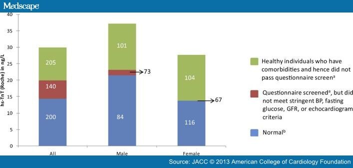
are associated with increased cTn concentrations,[5,6,7] An assay’s 99th percentile value depends on the composition of the reference group. Thus, the more criteria used, the lower the reference values (Figure 1).[5]
LVEF > 50%; normal lung function; and no significant
valvular heart disease,
LVH,
diastolic HF, or
regional wall-motion abnormalities on ECHO.
The appropriate reference value to use clinically also is far from a settled issue.
It might be argued that
using a higher 99th percentile value for the elderly
allows comparison of the patient to his or her peers, but
in raising the cut-off value, if the increases are caused by comorbidities,
those who are particularly healthy will be disadvantaged.[8]
Gender and ethnicity are not comorbidities, and we would urge that those should be taken into account.
Regardless of the assay, there will need to be
99th percentile values for men that are different for women.[2]
The reference population for assay validation studies should ideally be based on –
demographic characteristics that mirror the U.S. population and include subjects whose
blood pressure,
serum glucose, and
creatinine and
natriuretic peptide values are
within the normal reference range and
who take no cardiac medications.
These subjects should be
free from structural heart disease,
documented by echocardiography,
cardiac magnetic resonance imaging (MRI) or
computed tomography (CT) angiography.
Meeting these criteria will be a major challenge, especially for older individuals.
A conjoint pool of samples collected with manufacturers’ support so that all methods were derived from an
identical patient population for their reference ranges would be ideal.
[However, the method of collection and possible freeze-thaw effects is unavoidable].
One large national effort might be advantageous over multiple efforts.
Discriminating Between Acute and Nonacute Causes of hs-cTn Elevations
With the ability to precisely measure small concentrations of cTn,
clinicians will be faced with the challenge of distinguishing patients
who have acute problems from those with chronic elevations from other causes.
Using the fourth-generation cTnT assay, approximately 0.7% of patients in
the general population have modest elevations >99th percentile URL.[11]
In the same population, this number was 2% with the hs-cTnT assay.[6] Only
half of them had documentation (even with imaging) of cardiac abnormalities.
If the prevalence of a positive cTnT is 2% in the general population,
it will likely be 10% or 20% in the emergency department (ED)
and even higher in hospitalized patients, as
these patients often have cardiac comorbidities.
Measurement of changes in hs-cTn over time (δ hs-cTn)
improves the specificity of hs-cTn for the diagnosis of acute cardiac injury.[12,13]
However, it does so at the cost of sensitivity. With contemporary assays, differences
in analytical variation have been used to define an increasing pattern.
At elevated values, CV for most assays is in the range of 5% to 7%, so
a change of 20% ensures that a given change is not caused
by analytical variation alone.[10]
At values near the 99th percentile URL, higher change values are necessary.[13] The situation with hs-cTn assays is much more complex, as follows:
1. Change criteria are unique for each assay.
2. It will be easy to misclassify patients with coronary artery disease who may present with a noncardiac cause of chest pain
but have elevated values.
They could be having unstable ischemia or elevations caused by structural cardiac abnormalities and noncardiac discomfort.
If hs-cTn is rising significantly, the issue is easy but
if the values are not rising, a diagnosis of AMI still might be made.
If so, some patients may be included as having AMI without a changing pattern.
This occurred in 14% patients studied by Hammarsten et al.[14]
If patients with elevated hs-cTn without a changing pattern are not called AMI,
should they be called patients with “unstable angina and cardiac injury” or patients with structural heart disease and noncardiac chest pain?
Perhaps both exist?
3. The release of biomarkers is flow-dependent.Thus, there may not always be rapid access to the circulation. An area of injury distal to a totally occluded vessel (when collateral channels close) may be different in terms of the dynAMIcs of
hs-cTn change than an intermittently occluded coronary artery.
4. Conjoint biological and analytical variation can be measured.
They are assay-dependent, and the reference change values range from 35% to 85%.[2]
The use of criteria less than that (which may be what is needed clinically) will thus
likely include individuals with changes caused by
conjoint biological and analytical variation alone.
This has been shown to be the case in
many patients with nonacute cardiovascular diagnoses.[14,15]
5. Most evaluations have attempted to define the optimal delta, often with receiver operator curve analysis. Such an approach is based on the concept that sensitivity and specificity deserve equivalent weight.[But higher deltas improve specificity more and lower ones improve sensitivity and it is not clear that all physicians want the same tradeoffs in this regard.]ED physicians often prefer high-sensitivity so that their miss rate is low (<1%),[16] whereas hospital clinicians want increased specificity. This tension will need to be addressed in defining the optimal delta.
6. The delta associated with AMI may be different from that associated with other cardiac injury.[14] In addition, women have less marked elevations of cTn in response to coronary artery disease[17] and in earlier studies were less apt to have elevated values.[18] Given their pathology is at times different,
it may be that different metrics may be necessary based on gender
7. Some groups have assumed that if a change is of a given magnitude over 6 hours, it can be divided by 6 and the 1-h values can be used.
This approach is not data driven, and biomarker release is more likely to be discontinuous rather than continuous.[19]
In addition, the values obtained with this approach are too small to be distinguished from a lack of change with most assays.
These issues pose a major challenge even for defining the ideal delta change value and provide the reasons why
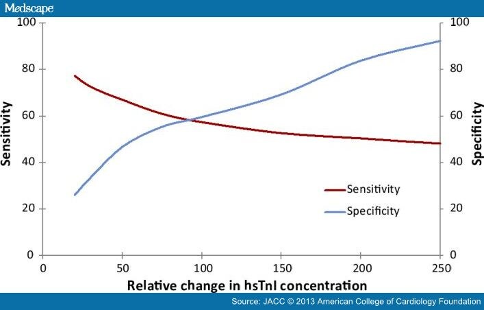
the use of this approach will reduce sensitivity[20,21] (Figure 2).
Defining the Optimal Delta: Tension Between Sensitivity and Specificity
There is a reciprocal relationship between sensitivity and specificity. With marked percentage changes,
specificity is improved at the expense of sensitivity, and
at lower values, the opposite occurs.
In addition, there is controversy in regard to the metrics that should be used with high-sensitivity assays.
The Australian-New Zealand group proposed
a 50% change for hs-cTnT for values below 53 ng/l and
a 20% change above that value.[22]
The 20% change is much less than conjoint biological and analytical variation.
A number of publications have suggested the superiority of
absolute δ cTn compared to relative δ cTn in discriminating between AMI and non-AMI causes of elevated cTn.[23,24,25]
The utility of the absolute or relative δ cTn appears to depend on the initial cTn concentration, and
the major benefit may be at higher values.[23]
A recent publication by Apple et al.[26] calculates deltas in several different ways with a contemporary assay and
provides a template for how to do such studies optimally.[26]
If all studies were carried out in a similar fashion, it would help immensely. In the long run, institutions will need to
define the approach they wish to take. We believe this discussion is a critical one and should include
laboratory,
ED, and
cardiology professionals.
Distinguishing Between Type 1 and Type 2 AMI
Although δ cTn is helpful in distinguishing between AMI and nonacute causes of Tn release,
it may or may not be useful in discerning type 1 from type 2 AMI.
As assay sensitivity increases, it appears that the frequency of type 2 AMI increases.
Making this distinction is not easy.
Type 1 AMI is caused by a primary coronary event, usually plaque rupture.
It is managed acutely with aggressive anticoagulation and
revascularization (percutaneous coronary intervention or coronary artery bypass).[10]
Type 2 AMI typically evolves secondary to ischemia from an oxygen demand/supply mismatch
severe tachycardia and
hypo- or hypertension and the like,
with or without a coronary abnormality.
These events usually are treated by addressing the underlying abnormalities.
They are particularly common in patients who are
critically ill and those who
are postoperative.[27]
However, autopsy studies from patients with postoperative AMI often manifest plaque rupture.[28]
Thus, the more important events, even if less common, may be type 1 AMIs. Type 2 events
seem more common in women, who tend to have
more endothelial dysfunction,
more plaque erosion, and
less fixed coronary artery disease.[28-30]
Additional studies are needed to determine how best to make this clinical distinction.
For now, clinical judgment is recommended.
Analytical Imprecision in Cardiac Troponin Assays
All analytical problems will be more critical with hs-cTn assays. Cardiac troponin I (cTnI) and cardiac troponin T (cTnT) are measured using enzyme linked immune- sorbent assays.
quantification of hs-cTn can be influenced by interference by reagent antibodies to analyte (cTn), leading to false- positive or negative results.[31]
Autoantibodies to cTnI or cTnT are found in 5% to 20% of individuals and can reduce detection of cTn.[32,33]
Additionally, fetal cTn isoforms can be re-expressed in diseased skeletal muscle and detected by the cTnT assays, resulting in false-positive values.[34]
Several strategies, including the use of
blocking reagents,
assay redesign, and use of
antibody fragments,
have been used to reduce interference.[35–36]
There are differences in measured cTn values based on specimen type (serum versus heparinized plasma versus EDTA plasma).
In addition, hemolysis may affect the accuracy of cTn measurement,[37] and with blood draws from peripheral IV lines, common in ICU.
Ruling Out AMI
Studies evaluating the diagnostic performance of hs-cTn assays for the early diagnosis of AMI usually define AMI on
the basis of a rising and/or falling pattern of current generation cTn values.[21,38]
However, defining AMI on the basis of the less sensitive current generation assay results in an underestimation of the true prevalence of AMI and
an overestimation of negative predictive value of the experimental assay.
shortens the time it takes to rule in all the AMIs and
to definitively exclude AMI as it
ignores the new AMIs more sensitively detected by the hs-cTn assay.
Thus, in the study by Hammarsten et al.,[14]
the time to exclude all AMIs was 8.5 hours when all of the AMIs detected
with the high-sensitivity assay were included, whereas
others that do not include these additional events report this can be done
in 3 to 4 hours.[21,29,38]
In our view, Hammarsten is correct.
This does not mean that hs-cTn cannot help in excluding AMI. Body et al.[39] reported that patients who present with undetectable values (less than the LOB of the hs-cTnT assay) were unlikely to have adverse events during follow-up. If that group of patients is added to those who present later than 6 hours, then perhaps a significant proportion of patients
with possible acute coronary syndrome (ACS) could
have that diagnosis excluded with the initial value.[40]
studies need to continue to evaluate cTn values for at least 6 h
to define the frequency of additional AMIs detected in that manner.
Using follow-up evaluations of patients with small event rates
who are likely to have additional care during the follow-up period are likely to be underpowered.
It may be that better initial risk stratification may help with this, as recently reported.[16,41]
Low-risk patients who have good follow-up after an ED visit
may be a group that can be released as early as 2 h after presentation.[16]
Investigating the Causes of Positive Troponin Values in Non-AMI Patients
Elevated Tn values (including those obtained with high-sensitivity assays) are associated with
a 2-fold higher risk for longer-term all-cause mortality and
cardiovascular death than a negative troponin values.[6,42-44]
This association is dose-dependent.
If values are rising, they are indicative of acute cardiac injury.
Those patients should be admitted because the risk is often short-term. However,
if the values are stable, assuming the timing of any acute event would
allow detection of a changing pattern,
the risk, although substantive, in our view, often plays out in the longer term.[44]
Many of these individuals, assuming they are doing well clinically, can be evaluated outside of the hospital, in our view.
However, because such elevations are an indicator of a subclinical
cardiovascular injury, such evaluations should be early and aggressive.
Data from several studies suggest that there may well be risk far below the 99th percentile URL value.
Thus, it may evolve that patients in the upper ranges of the normal range also require some degree of cardiovascular evaluation.
Risk Stratifying Patients With Nonacute Coronary Syndrome Conditions
Patients who have a rising pattern of values have a higher risk of mortality than those with negative values regardless of the cause.
Investigations are ongoing to determine how well results from hs-cTn testing help to risk-stratify patients with
pulmonary embolism,[45]
congestive heart failure,[46]
sepsis,[47]
hypertensive emergency,[48] and
chronic obstructive pulmonary disease.[49]
Presently, the studies suggest that cTn values classify patients into clinically relevant risk subgroups. Studies are needed
to evaluate the incremental prognostic benefit of hs-cTn.
Conclusions
Routine use of hs-cTn assays in the United States is inevitable. These assays hold
the promise of
improving the sensitivity of AMI diagnoses,
shortening the duration of AMI evaluation and
improving the risk stratification of other noncardiac diagnoses.
However, to be able to fully realize their potential, additional studies are needed to address the
knowledge gaps we have identified. In the interim, clinicians need to
learn how to use the 99th% URL and
the concept of changing values
John Adan, MD, FACC
In 2008 CMS commissioned Yale University to analyze 30 days mortality after myocardial infarction in their hospitals.
The study has been based on review of medical records. Consensus criteria for diagnosis of myocardial infarction include
clinical symptoms,
EKG,
troponins,
CK MB,
ECHO,
cath,
histopathology, etc.
How the reviewed hospitals performed diagnostic coding is unknown. In clinical practice we are bombarded by consults
for elevated troponins due to causes other than myocardial infarction, like
pneumonia,
accelerated hypertension,
arrhythmias,
renal failure, etc.
The metric started out over 19%. Now it is below 15%, on average.
typical changes in CK-MB and cardiac troponin in Acute Myocardial Infarction (Photo credit: Wikipedia)
Phosphotungstic acid-haematoxylin staining demonstrating contraction band necrosis in an individual that had a myocardial infarction (heart attack). (Photo credit: Wikipedia)
Pancreatic stellate cell activation in chronic pancreatitis and pancreatic cancer. Pancreatic stellate cells are activated by profibrogenic mediators, such as ethanol metabolites and cytokines/growth factors. Perpetuation of stellate cell activation under persisting pathological conditions results in pancreatic fibrosis. Jaster Molecular Cancer 2004 3:26 doi:10.1186/1476-4598-3-2 (Photo credit: Wikipedia)
Larry H. Bernstein, MD, Reporter
Cancer-Causing Gene Alone Doesn’t Trigger Pancreatic Cancer, Mayo-led Study Finds
More than a cancer-causing gene is needed to trigger pancreatic cancer, a study led by Mayo Clinic, Jacksonville, Fla, has found.
A second factor creates a “perfect storm” that allows tumors to form, the researchers say. The study, published in a recent issue of Cancer Cell, overturns the current belief that a mutation in the KRAS oncogene is enough to initiate pancreatic cancer and unrestrained cell growth.
The findings uncover critical clues on how pancreatic cancer develops and why few patients benefit from current therapies. The findings also provide ideas about how to improve treatment and prevention of pancreatic cancer.
The research team, led by Howard C. Crawford, PhD, a cancer biologist at Mayo Clinic’s campus in Florida, and Jens Siveke, MD, at Technical University in Munich, Germany, found that for pancreatic cancer to form, mutated KRAS must recruit a second player: the epidermal growth factor receptor, or EGFR.A third genetic participant known as Trp53 makes pancreatic tumors very difficult to treat, the study showed.
The scientists also found that EGFR was required in pancreatic cancer initiated by pancreatic inflammation known as pancreatitis.
Imatinib May Help Treat Aggressive Lymphoma
Based on the results of a new study, researchers are developing a clinical trial to test imatinib (Gleevec) in patients with anaplastic large cell lymphoma (ALCL), an aggressive type of non-Hodgkin lymphoma that primarily affects children and young adults.
The researchers found that a protein called PDGFRB is important to the development of a common form of ALCL. PDGFRB, a growth factor receptor protein, is a target of imatinib. Imatinib had anticancer effects in both a mouse model of ALCL and a patient with the disease, Dr. Lukas Kenner of the Medical University of Vienna in Austria and his colleagues reported October 14 in Nature Medicine.
The authors decided to investigate the effect of imatinib after finding a link between PDGFRB and a genetic abnormality that is found in many patients with ALCL. Previous work had shown that this genetic change—a translocation that leads to the production of an abnormal fusion protein called NPM-ALK—stimulates the production of two proteins, transcription factors called JUN and JUNB.
In the new study, experiments in mice revealed that these proteins promote lymphoma development by increasing the levels of PDGFRB.
Because imatinib inhibits PDGFRB, the authors tested the effect of the drug in mice with the NPM-ALK change and found that it improved their survival. They also found that imatinib given together with the ALK inhibitor crizotinib (Xalkori) greatly reduced the growth of NPM-ALK-positive lymphoma cells in mice.
To test the treatment strategy in people, they identified a terminally ill patient with NPM-ALK-positive ALCL who had no other treatment options and agreed to try imatinib. The patient began to improve within 10 days of starting the therapy and has been free of the disease for 22 months, the authors reported.
The observation that inhibiting both ALK and PDGFRB “reduces lymphoma growth and alleviates relapse rates” led the authors to suggest that the findings might be relevant to lymphomas with PDGFRB but without the NPM-ALK protein. “Our findings suggest that imatinib is a potential therapeutic option for patients with crizotinib-resistant lymphomas.”
A planned clinical trial will be based on the expression of PDGFRB in tumors.
Researchers Identify Possible Biomarker for Early-Stage Lung Cancer
A protein that can be detected in blood samples may one day serve as a biomarker for early-stage lung cancer, according to new study results. The findings, published October 16 in the Proceedings of the National Academy of Sciences, suggest that measuring the levels of a variant form of the protein Ciz1 may help detect lung cancer early and noninvasively in high-risk individuals.
“We have struggled to find cancer biomarkers that are disease-specific, and this may be a step in the right direction,” said Dr. Sudhir Srivastava, chief of NCI’s Cancer Biomarkers Research Group. He called the study “promising” but noted that the results will need further validation.
Researchers led by Dr. Dawn Coverley of the University of York in the United Kingdom found that the “b-variant” form of Ciz1 was present in 34 of 35 lung tumors but not in adjacent tissue. Additional experiments showed that an antibody specific for this Ciz1 variant could detect the protein in small samples of blood from individuals with non-small cell lung cancer (NSCLC) and small cell lung cancer (SCLC).
In two independent sets of blood samples—from 170 and 160 patients, respectively—the researchers showed that variant Ciz1 levels above a certain threshold correctly identified 95 to 98 percent of lung cancer patients, with an overall specificity of 71 to 75 percent. Using the second set of samples, they showed that the level of variant Ciz1 could discriminate between patients with stage I NSCLC and age-matched heavy smokers without diagnosed cancer, individuals with benign lung nodules, and patients with inflammatory lung disease.
Although the high rate of false-positive test results seen with variant Ciz1 is a concern, the authors noted that a blood test for the Ciz1 variant might ultimately be shown to be useful when combined with low-dose helical computed tomography, also called spiral CT, for lung cancer screening. In that context, the test could confirm the presence of lung cancer in patients who have suspicious spiral CT results, reducing the need for invasive procedures to confirm a lung cancer diagnosis. And, if used before spiral CT, “the test could reduce the number of people who undergo imaging…[because] the false-negative rate is very low,” Dr. Coverley wrote in an e-mail message.
To assess variant Ciz1 levels, the researchers used a laboratory method known as Western blot analysis. However, this approach could not be routinely applied in a clinical context, the researchers acknowledged, so “a more streamlined method” for testing would need to be developed.
Supported in part by NCI Early Detection Research Network Grant U01CA086137.
NCI Reports
Study Looks at Terminal Cancer Patients’ Expectations of Chemotherapy
A majority of patients who opt to receive chemotherapy to treat newly diagnosed metastatic lung or colorectal cancer believe chemotherapy might cure their cancer, according to a recent survey. The survey results suggest that optimistic assumptions about the benefits of chemotherapy may hamper patients’ abilities to make informed treatment decisions that align with their preferences, said the researchers who led the study. The findings were published October 25 in the New England Journal of Medicine.
Dr. Jane Weeks of the Dana-Farber Cancer Institute and her colleagues interviewed 1,193 patients tracked by the prospective, observational Cancer Care Outcomes Research and Surveillance Consortium (CanCORS) study, 4 to 7 months after diagnosis. All of the patients had been diagnosed with stage IV lung or colorectal cancer and had chosen to receive chemotherapy. A surrogate was interviewed when a patient was too ill to participate. The survey asked patients how likely it was that chemotherapy would cure their disease, extend life, or relieve symptoms. The researchers also collected data on patients’ physical functioning, communication with their physicians, and social and demographic factors.
The majority of patients did not appear to understand that chemotherapy was very unlikely to cure their cancer (81 percent of those with colorectal cancer and 69 percent of those with lung cancer). Black, Hispanic, and Asian/Pacific Islander patients were more likely than white patients to believe that chemotherapy would cure them. Nevertheless, most patients believed that chemotherapy was more likely to extend their life than cure them.
Educational level, functional status, and the patient’s role in treatment decision making were not associated with inaccurate expectations about chemotherapy.
In an accompanying editorial, Drs. Thomas J. Smith of the Johns Hopkins Sidney Kimmel Cancer Center and Dan L. Longo of the National Institute on Aging wrote, “if patients actually have unrealistic expectations of a cure from a therapy that is administered with palliative intent, we have a serious problem of miscommunication that we need to address.”
This research was supported by grants from the National Institutes of Health (U01 CA093344, U01 CA093332, U01CA093324, U01 CA093348, U01 CA093329, U01 CA093339, and U01 CA093326).
Researchers Identify Possible Biomarker for Early-Stage Lung Cancer
A protein that can be detected in blood samples may one day serve as a biomarker for early-stage lung cancer, according to new study results. The findings, published October 16 in the Proceedings of the National Academy of Sciences, suggest that measuring the levels of a variant form of the protein Ciz1 may help detect lung cancer early and noninvasively in high-risk individuals.
“We have struggled to find cancer biomarkers that are disease-specific, and this may be a step in the right direction,” said Dr. Sudhir Srivastava, chief of NCI’s Cancer Biomarkers Research Group. He called the study “promising” but noted that the results will need further validation.
Researchers led by Dr. Dawn Coverley of the University of York in the United Kingdom found that the “b-variant” form of Ciz1 was present in 34 of 35 lung tumors but not in adjacent tissue. Additional experiments showed that an antibody specific for this Ciz1 variant could detect the protein in small samples of blood from individuals with non-small cell lung cancer (NSCLC) and small cell lung cancer (SCLC).
In two independent sets of blood samples—from 170 and 160 patients, respectively—the researchers showed that variant Ciz1 levels above a certain threshold correctly identified 95 to 98 percent of lung cancer patients, with an overall specificity of 71 to 75 percent. Using the second set of samples, they showed that the level of variant Ciz1 could discriminate between patients with stage I NSCLC and age-matched heavy smokers without diagnosed cancer, individuals with benign lung nodules, and patients with inflammatory lung disease.
Although the high rate of false-positive test results seen with variant Ciz1 is a concern, the authors noted that a blood test for the Ciz1 variant might ultimately be shown to be useful when combined with low-dose helical computed tomography, also called spiral CT, for lung cancer screening. In that context, the test could confirm the presence of lung cancer in patients who have suspicious spiral CT results, reducing the need for invasive procedures to confirm a lung cancer diagnosis. And, if used before spiral CT, “the test could reduce the number of people who undergo imaging…[because] the false-negative rate is very low,” Dr. Coverley wrote in an e-mail message.
To assess variant Ciz1 levels, the researchers used a laboratory method known as Western blot analysis. However, this approach could not be routinely applied in a clinical context, the researchers acknowledged, so “a more streamlined method” for testing would need to be developed.
Supported in part by NCI Early Detection Research Network Grant U01CA086137.
Choline C 11 Injection is administered intravenously to produce an image that helps to locate specific body sites for follow-up tissue sampling and testing in men with recurrent prostate cancer.
PET imaging with Choline C 11 Injection is performed in patients whose blood prostate specific antigen (PSA) levels are increasing after earlier treatment for prostate cancer. An elevated PSA result suggests that prostate cancer may have returned, even though conventional imaging tests, such as computerized tomography (CT), have not shown any signs of cancer. PET imaging is not a replacement for tissue sampling and testing.
Choline C 11 Injection must be produced in a specialized facility and administered to patients shortly after its production. While PET imaging with Choline C 11 Injection has been performed at a few facilities over the past several years, none of these facilities were approved by the FDA to manufacture the agent. The Food and Drug Administration Modernization Act directed the agency to establish appropriate approval procedures and current good manufacturing practice requirements for all PET products marketed and used in the United States. The Mayo Clinic is now the first FDA-approved facility to produce Choline C 11 Injection.
“Choline C 11 Injection provides an important imaging method to help detect the location of prostate cancer in patients whose blood tests suggest recurrent cancer when other imaging tests are negative,” said Charles Ganley, M.D., director of the Office of Drug Evaluation IV in FDA’s Center for Drug Evaluation and Research. “The FDA’s approval of Choline C 11 Injection at the Mayo Clinic provides assurance to patients and health care professionals they are using a product that is safe, effective, and produced according to current good manufacturing practices.”
The safety and effectiveness of Choline C 11 Injection were verified by a systematic review of published study reports. Four independent studies examined a total of 98 patients with elevated blood PSA levels but no sign of recurrent prostate cancer on conventional imaging. After PET imaging with Choline C 11, the patients underwent tissue sampling of the abnormalities detected on the PET scans.
In each of the four studies, at least half the patients who had abnormalities detected on PET scans also had recurrent prostate cancer confirmed by tissue sampling of the abnormal areas. PET scan errors also were reported. Depending on the study, falsely positive PET scans were observed in 15 percent to 47 percent of the patients. These findings underscore the need for confirmatory tissue sampling of abnormalities detected with Choline C 11 Injection PET scans.
Aside from an uncommon, mild skin reaction at the injection site, no side effects to Choline C 11 Injection were reported.
Choline C 11 Injection is manufactured and distributed by the Mayo Clinic PET Radiochemistry Facility in Rochester, Minn
In our recent article we mentioned about the amyloidosis, most importantly the most common form of amlyodosis – Primary Amyloidosis (AL).
Primary amyloidosis (AL) is an acquired plasma cell disorder in which a monoclonal immunoglobulin light chain is produced in the bone marrow and usually found in the blood or urine. AL amyloidosis occasionally occurs with multiple myeloma. The amyloid fibrils in this type of amyloidosis are made up of immunoglobulin light chain proteins (kappa or lambda).
Amyloidosis can only be diagnosed by a positive biopsy (i.e., an identification of the amyloid deposits in a piece of tissue). Initial biopsies are most commonly obtained from the abdominal fat.
If amyloid is suspected in other organs, however, a biopsy may be needed from these specific areas. If amyloid is present in a tissue biopsy, further tests can be done to determine the type of the amyloid.
The Amyloid Treatment & Research Program (ATRP) at Boston Medical Center (BMC) is an international referral center that treats amyloidosis with stem cell transplantation.
Last week researchers at Mayo Clinic have used urinary exosomes as a non-invasive diagnostic tool that will offer a snapshot of what is occurring in kidney tissue.
Urinary exosomes are rapidly becoming a powerful tool in the study of renal disease.
Already proteomics studies are looking into ways of using urinary exosome to diagnose genetic diseases and characterize disease biomarkers.
The urinary exosomes are excreted from every renal epithelial cells (from the glomerular podocytes to the urinary epithelial cells lining the urinary drainage system) provides us with an opportunity to study proteins once were either difficult or impossible to reach.
With this understanding the researchers undertook this study to evaluate the possible differences among urinary exosomes from patients with different plasma cells dyscrasias. This study suggests that urinary exosomes may be an excellent non-invasive tool for identifying patients with AL amyloidosis because high molecular weight light chain oligomers were found only in patients with AL.
The oligomeric light chain species captured in the urinary exosomes may represent the initial steps of amyloidogenesis. The potential of urinary exosomes in AL is tremendous and deserves further studies. When combined with mass spectrometry and other proteomics techniques, urinary exosomes represent tremendous potential to increase our understanding of amyloidogenesis.
Authors believe that this is the first report of the use of urinary exosome in the study of patients with plasma cell dyscrasias, specifically patients with AL amyloidosis.