Feeds:
Posts
Comments

Posts Tagged ‘sarcoma’

Author: Ziv Raviv, PhD

Introduction

Sarcoma is a general class of cancers of mesenchymal cells that form connective tissues. Sarcoma can start in any part of the body and can be formed in the bones or in soft tissues. Sarcomas are rare cancers as compared to the more common epithelial cancers (carcinomas). Around 15,000 new cases of sarcomas diagnosed in the United States every year. Both children and adults can develop a sarcoma, however, while in adults it accounts for only about 1% of all cancers, sarcoma represents around 15% of all cancers in children.

There are tens of different types of sarcomas. This fact makes a particular type of sarcoma to be even rarer. Being sarcoma an uncommon cancer, it is strongly recommended for patients diagnosed with sarcoma to get consultant and treatment for the disease in sarcoma centers, or at list be treated by an oncologist physician that had experienced with sarcomas.

As stated, sarcomas are cancers of connective tissues, namely tissues that connect the body, holding it together. These tissues include: bones, cartilage, muscle, nerve, blood and lymph vessels, and fat. Therefore, sarcomas nomenclature is based according to the normal tissue type they most closely resemble (as opposed to carcinomas where the nomenclature is based upon the organ or part of the body where cancer is originated). Few examples: Osteosarcoma (OS) – cancer of bones origin; Chondrosarcoma – cancer of cells that produce cartilage; Fibrosarcoma – cancer derived from fibrous connective tissues cells; Rhabdomyosarcoma (RMS) –  cancer from skeletal muscle progenitors; Liposarcoma – cancer that arises in fat cells, etc.

  • Watch a Dana-Farber Cancer Institute – About Sarcoma Video

Soft tissues sarcoma (STS)

Among sarcomas, the group of soft tissues sarcoma (STS) is the largest one, consists of many different types of cancers that origin in soft connective tissues that support and connect overall body parts. STSs account for less than 1% of all new cancer cases where about 11,000 new cases are diagnosed each year in the US, and about 4,000 people are dying from it each year.  STS can occur almost anywhere in the body: about 60% of STSs occur in an arm or leg, 30% in the trunk (torso) or abdomen, and 10% in the head or neck. Because there are many different types of STS, it is more of a family of related cancer diseases then a single one. The specific types of STS are often named according to the normal tissue cells they most closely resemble (see introduction), however, some STSs do not look like any type of normal tissue and are thought to arise from stem cells.  In addition to their tissue resemblance name, STS are characterized with grades and stages (Table I) where low-grade STSs are often local tumors that grow more slowly and are treated surgically (although radiation therapy or chemotherapy may be used occasionally), and intermediate – and high-grade STSs are tumors that are more likely to metastasize and are treated with a combination of surgery, chemotherapy and/or radiation therapy.

Figure 1. STS of the thigh muscle just above the knee.

soft_tissue_sarcoma_leg

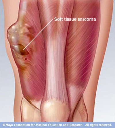
Taken from the Mayo Clinic webpage.

Table I: Sarcoma Staging System according to AJCC

Stage

Grade

Size

Location 

Metastasis

IA

Low

< 5cm

Superficial or Deep

No

IB

Low

≥ 5cm

Superficial

No

IIA

Low

≥ 5cm

Deep

No

IIB

High

< 5cm

Superficial or Deep

No

IIC

High

≥ 5cm

Superficial

No

III

High

≥ 5cm

Deep

No

IV

Any

Any

Any

Yes

Adapted from sarcomahelp.org

Diagnosis

In their early stages, STSs usually do not stimulate any symptoms and can grow unnoticed. This is because STSs are grown within soft connective tissues which are elastic and flexible, thus the tumor can develop quite large before being felt and cause any symptoms. The first noticeable symptom is usually a painless lump or swelling, however, since most lumps are not sarcoma they are often misdiagnosed. Eventually, the tumor interferes with normal body activities and cause pain by pressing against nerves and muscles, or if the sarcoma is located at the abdomen the tumor can induce abdominal pains or constipation. Therefore, when STS is suspected it should be examined for any unusual lumps growing to define whether they are malignant even if symptoms are not present, preferred by a sarcoma specialist. There are no standard screening tests for sarcoma. Usually a biopsy of the suspected tumor is taken to evaluate if indeed it is malignant and to define its type and grade. In addition, molecular testing of the tumor could be performed to identify specific genes unique to the tumor. Finally, imaging tests may be used to find out whether the cancer has metastasized.

Prognosis and current treatment

The five-year survival rate for localized-low grade sarcomas is 83%; 54% for intermediate sarcomas (spread to regional lymph nodes); and 16% for high grade STSs that have spread to distant parts of the body to form metastasis. Survival is depended also on tumor size, location, type, mitotic rate, and whether it is superficial or deep.

Surgery

Treatment options depend on the type and stage of cancer, possible side effects, and the patient’s preferences and overall health. Treatment can be a long and arduous process for many patients. Usually STSs are treated with surgery whenever it is possible. Should the tumor is not removable by surgery it may be possible to control its growth with radiation therapy. For a sarcoma that can be surgically removed, radiation therapy and/or chemotherapy may be given before or after surgery to reduce tumor recurrence. Small STSs can usually be effectively eliminated by surgery alone. However, sarcomas larger than 5 cm are often treated with a combination of surgery and radiation therapy or chemotherapy before surgery – to shrink the tumor and make its removal easier, or during and after surgery – to eradicate any remaining microscopic tumor cells. In addition, radiation and chemotherapy pre-surgical treatment might facilitate less surgery, preserving the limbs if the tumor is located in the arms or legs (limb-sparing surgery). Historically, STSs were treated with amputation; however, nowadays at least 90% of tumors are removed using limb-sparing surgery. In intermediate-high stages, chemotherapy and radiation therapy may also be used to reduce the size of the sarcoma or relieve pain and other symptoms.

Radiotherapy

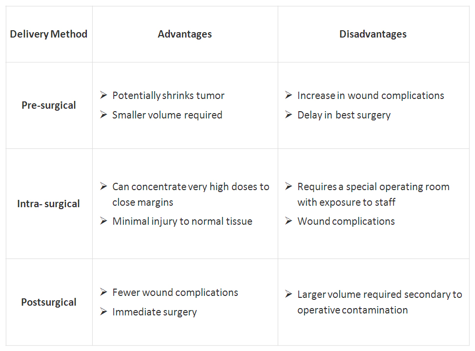
The most commonly used radiation form is external beam radiation. Another mean of post surgically radiation is brachytherapy. This technique allows for high doses of radiation over a short period of time. The decision to use radiation before and/or after surgery is not standardized and may be changed on an individual case basis; Table II describes the choices of using radiation with surgery.

Table II: The advantages and disadvantages of the timing of radiotherapy

T2_aClick on table to enlarge

Adapted from sarcomahelp.org

Proton therapy (also called proton beam therapy), a type of radiation treatment that uses protons rather than x-rays is also being adapted to treat sarcoma. This mode of radiotherapy allows target the radiation much more focused at the tumor site and thus is much protective to surrounding healthy tissue. This procedure however, is currently only available in a few specialized cancer centers in the US. In addition, particle therapy treatment with heavier charged particles such as carbon ions is being used and studied for the treatment of sarcomas in Japan and Germany.

Chemotherapy

Chemotherapy is often used when a sarcoma has already spread and can be given before surgery or, after surgery as adjuvant chemotherapy to destroy any microscopic tumor cells remained after surgery.  In addition, when a tumor is considered non-operable, cycles of chemotherapy could be performed in order to shrink the tumor and make it necrotic to enable its removal by operation.

  • Watch a STS chemo + surgery Video

Different drugs are used to treat different subtypes of sarcoma. The types of chemotherapy that are used alone or in combination for most STSs include doxorubicin and ifosfamide that are the most common chemotherapy drugs employed for STS, as well as other ordinary chemotherapy drugs. The drug trabectedin, approved for use in Europe, is given for patients with advanced STS when conventional chemotherapy fails. Trabectedin has been shown to have high activity levels in the treatment of a specific subtype of liposarcoma (myxoid/round cell liposarcoma). Other chemotherapy drugs that are only used for certain subtypes of STS include: paclitaxel, docetaxel for Angiosarcoma; as well as vincristine, etoposide, actinomycin, and cyclophosphamide for Rhabdomyosarcoma and Ewing sarcoma.

Experimental chemotherapy drugs include Eribulin, a drug approved for treatment of breast cancer that has shown promising results in early clinical trials. In addition, new versions of sarcoma standard chemotherapy that cause fewer side effects are being studied in ongoing clinical trials. For instance, the three new versions of ifosfamide: palifosfamide, glufosfamide, and TH-302.

Targeted therapy

As genetic and molecular cancer research has evolved, targeted treatment to sarcoma became available. Targeted treatment to sarcoma intends to inhibit the growth and spread of cancer cells by hitting specific proteins, mainly by blocking the action of protein kinases.

Imatinib, a tyrosine-kinase inhibitor was approved in 2002 by the FDA for the treatment of gastrointestinal stromal tumor (GIST) in advanced stages and it is now the standard first-line treatment for GIST. In 2006, sunitinib multi-target receptor tyrosine kinase (RTK) inhibitor was also approved for the treatment of GIST when imatinib fails. Imatinib has been approved recently for use for patients with GIST after initial surgery, to try to prevent recurrence of the tumor. Imatinib is approved also for the treatment of advanced stage dermatofibrosarcoma protuberans (DFSP). Pazopanib, another multi-targeted inhibitor of receptor tyrosine kinase, has also been approved for patients with advanced STS as well as for use in sarcomas other than liposarcoma and GIST in conditions where standard chemotherapy is not working. Regorafenib is a new kinase inhibitor with significant activity in patients with advanced GIST who have already been treated with imatinib and suntinib. The FDA is currently reviewing a phase III clinical trial of this drug.

Closing remarks

Research efforts are made in order to elucidate new sarcoma-specific molecular targets. Studying sarcomas unique genetic fingerprints and understanding their value to sarcoma, not only can assist developing new drugs, but also may help better prediction of patients’ prognosis. To find the most effective treatment, tests to identify the genes, proteins, and other sarcoma-associated factors need to be developed and performed to give a better matched treatment for each patient.  However, being sarcoma a highly diverse group of cancers make these efforts a hard task. These issues will be discussed further in future post(s) to be published in Pharmaceutical Intelligence.

Resources

  1. http://www.cancer.net
  2. http://www.sarcomahelp.org
  3. http://www.cancer.gov
  4. http://sarcomaalliance.org
  5. http://www.sarcoma.org.uk
  6. http://www.mayoclinic.com

Additional related references

  1. Soft tissue sarcomas: ESMO Clinical Practice Guidelines for diagnosis, treatment and follow-up. Casali, PG & Blay, JY. Ann Oncol. 2010 May;21 Suppl 5:v198-203.
  2. Chemotherapy in adult soft tissue sarcoma. Jain A, Sajeevan KV, Babu KG, Lakshmaiah KC. Indian J. Cancer. 2009 Oct-Dec;46(4):274-87.
  3. State-of-the-art approach in selective curable tumours: soft tissue sarcoma. Judson I. Ann Oncol. 2008 Sep;19 Suppl 7:vii166-9.
  4. Soft tissue sarcomas of adults: state of the translational science. Borden EC, et al. Clin Cancer Res. 2003 Jun;9(6):1941-56.
  5. Management of soft-tissue sarcomas: an overview and update. Singer S, Demetri GD, Baldini EH, Fletcher CD. Lancet Oncol. 2000 Oct;1:75-85.

Videos

  1. http://www.youtube.com/watch?v=J35GBjTxzIE
  2. http://www.youtube.com/watch?v=f97oWMANXDw

Related articles on this Open Access Online Scientific Journal

  1. Clear Cell Sarcoma – Soft Tissue Melanoma: Patient’s Experience with Disease. Reporter: Aviva Lev-Ari, Ph.D., RN

Read Full Post »

Heroes in Medical Research: Dr. Robert Ting, Ph.D. and Retrovirus in AIDS and Cancer

Curator and Reporter: Stephen J. Williams, PhD

This is the second posting in this series in which I highlight the basic research which led to seminal breakthroughs in the medical field, brought on by the result of basic inquiry, thorough and detailed investigation, meticulously following the scientific method, and eventually leading to development of important medical therapies.

In his autobiography, Virus Hunting: AIDS, Cancer & the Human Retrovirus: A Story of Scientific Discovery, Dr. Robert Gallo, M.D. describes a wonderful story of the history behind, scientific biographies, and chronology of the discoveries which led he and his colleagues (including co-discoverer Dr. Luke Montagnier) to recognize retroviruses (in particular HIV) as the leading culprit for the cause of AIDS and in the etiology of Kaposi’s sarcoma.   For anyone who appreciates the history behind scientific discoveries and appreciates learning about the multitude of individual efforts which are the crux of seminal research, this book is a must read.

Recommendations from the back cover include:

Virus Hunting will be read and reread, for years to come.” —New York Newsday

“Provides a human, revealing look into the arcane, usually secret confines of laboratory science.”

Martin Delany, Project Inform

..as well as others.

While a fascinating aspect of this book is the description, like fitting pieces of a puzzle, of the important discoveries throughout history which are the necessary foundations for further investigations and discoveries, more important is a telling, personal narrative of the people involved in those initial and subsequent discoveries.  In fact, the book has over 396 colleagues, mentors, technicians, students, and even critiques who are given credit, in one form or another, for the ultimate discovery of HIV as a causative agent for the development of AIDS. The book is a literal Who’s Who in Science and shows how important personal collaboration and friendships are in the process of scientific discovery.

In 1972, Dr. Seymour Perry had appointed the young Dr. Robert Gallo as head of a new department, the Human Tumor Cell Biology Branch, renamed the Laboratory of Tumor Cell Biology.  The lab was carrying on the work on tRNA that Dr. Gallo had performed in Dr. Sid Perska’s group at NIH.  However, with the help of new lab members Dr. David Gillespie, Dr. Flossie Wong-Staal, and Dr. Marjorie Robert-Guroff the lab focused on the search for disease-causing retroviruses, especially in human leukemias.  This was, in part, due to conversations with Dr. Robert Huebner and Todaro, who insisted that

“within the genetic makeup of this endogenous retroviral material was, they suggested, a special gene, the oncogene, that was the parent of the cancer-causing protein”

which may explain some of the early work by Rous concerning the Rous sarcoma virus.

Enter in Gallo’s good friend Dr. Bob Ting.  Dr. Gallo had known Dr. Ting socially since 1966, shortly after Gallo had arrived at NIH.  Dr. Bob Ting was a well-established NCI investigator, who was doing work on DNA and RNA oncogenic viruses of animals.  Originally from a large and wealthy family in Hong Kong, Dr. Ting had worked with Nobel Prize winners Salvatore Luria (who worked on phages) and Renato Dulbecco, who, along with his well-known cell culture media, had made the seminal discoveries that led to our knowledge how some DNA viruses can transform normal animal cells into neoplastic-like cells in culture.

Bob Ting gave a talk on these oncogenic viruses and Gallo was very interested in his observations that oncogenic viruses like Rous and Maloney, could transform cells in vitro in a matter of days.

A friendship developed between the two over tennis matches and Chinese food.  During this time, Dr. Ting made the important suggestion that they both collaborate and use the viral systems developed by Dulbecco.  Ting also introduced him to RNA viruses, Dr. Robert Huebner, and Dr. Howard Temin.  It was, in part, due to these associations that Gallo started looking, in earnest, at the possibility of RNA retroviruses in leukemias. Thus, just like the internet today, connections and networking provided new insights into current research, and helped lead the advent of new discoveries, therapies, and scientific disciplines.

Therefore, “after some late-night discussion with Bob Ting, I decided to enter the fray. My own laboratory, … would immediately be set up to compare the properties of reverse transcriptase enzymes from many different animal retroviruses”.

Although the rest is more history, this early friendship, collaboration, and mentoring by Bob Ting had “transformed” Gallo’s research efforts to set him up to make some of the important discoveries eventually leading to the discovery of the role of HIV in AIDS.

A video interviewing Dr. Gallo can be found here:

VIEW VIDEO

https://www.youtube.com/watch?v=ELRlXLGWu4I

A very nice writeup/obituary for Dr. Ting was written by Patricia Sullivan of the Washington Post and is included below.

Robert Ting, 77; Biotech Pioneer

ME/Ting-ob

Dr. Robert Ting’s biotech company in Rockville developed the first FDA-approved diagnostic test kits to test for HIV antibodies. (By Gerald Martineau — The Washington Post)


By Patricia Sullivan

Washington Post Staff Writer
Friday, September 22, 2006

Robert C.Y. Ting, 77, a research scientist who started one of the early biotechnology companies in the Washington area, died Sept. 11 of complications after cardiac surgery at the Cleveland Clinic in Cleveland.

Dr. Ting founded Biotech Research Laboratories Inc. in Rockville in 1973, producing cells for government scientists to use in research. Eleven years later, his firm obtained a federal license to develop and produce the first FDA-approved diagnostic test kits for HIV antibody confirmation.

Robert C. Gallo, who co-discovered the HIV virus as the cause of AIDS, called Dr. Ting a pioneer in the field who popularized the term “biotechnology” when he moved from research to entrepreneurship.

“He introduced me to virology, and he did it twice,” said Gallo, director of the Institute of Human Virology in Baltimore. The men had known each other since the 1960s, and while playing tennis one day, Dr. Ting advised the cancer researcher to look at new research in viruses. Later, when Gallo was studying leukemia, Dr. Ting directed him to animal research in leukemia. “First he showed me how viruses change cells. Then he introduced me to retrovirology. . . . I went into retrovirology solely because of those discussions with Bob Ting on tennis courts,” Gallo said.

Dr. Ting, whom Gallo described as a quiet, modest man, was born in Shanghai, the son of a physician to Gen. Chiang Kai-Shek. His family fled the country during the Japanese invasion of China during World War II and moved to Hong Kong. Soon after, he moved to the United States, where he received a bachelor’s degree and in 1956 a master’s degree in genetics from Amherst College.

He received a doctoral degree in microbiology and biochemistry from the University of Illinois in 1960 under Salvador E. Luria, who later won the 1969 Nobel Prize in Medicine and Physiology. Dr. Ting spent the next two years on a postdoctoral fellowship at the California Institute of Technology, working with Renato Dulbecco, who later won the 1975 Nobel Prize in Medicine and Physiology. Their work focused on how viruses cause tumors.

“A lot of molecular biology developed from this,” Dr. Ting told The Washington Post in 1984 from his Rockville office, cluttered with scientific journals, awards and a large blackboard. “There was so much evidence in animal systems [that viruses cause tumors], that the next question was obvious — can you find the equivalent in humans.”

Dr. Ting joined the National Institutes of Health in 1962 as a visiting fellow and then a senior research scientist at the National Cancer Institute. From 1966 to 1968, he was an associate editor for the Journal of the National Cancer Institute.

In 1969, he joined Litton Bionetics Inc. in Rockville as director of experimental oncology, leading a project funded by the institute to search for viruses in human leukemia patients. He became scientific director of the cancer research branch the next year.

With academic, government and private business experience under his belt, Dr. Ting decided to go into business on his own and in 1973 started Biotech Research Laboratories in Rockville. It was a profitable supplier of research services and supplies until 1981, when it went public and produced the HIV diagnostic test kits. It became one of the most successful public biotech companies in the area in the mid-1980s.

The Economic Development Board of Singapore invited him to return to Asia to start a biotech company, which he did in 1985, forming Diagnostic Biotechnology Ltd. He also joined the Institute of Molecular and Cell Biology at the National University of Singapore, which Gallo called “the most prominent Asian academic biotechnology center.”

He returned to the United States in 1998 to join the board of Cell Works Inc. in Baltimore, and became chair and chief executive of a joint venture, Cell Works Asia Limited, in 2000.

Most recently, Dr. Ting was the founding president and chief executive of Profectus Biosciences Inc. of Baltimore, previously known as Maryland BioTherapeutics Inc.

Dr. Ting was past chairman of the F.F. Fraternity, one of the oldest Chinese fraternities in the United States. He was also a member of the Organization of Chinese Americans in the D.C. area since its inception in the early 1970s. He enjoyed tennis, golf, ballroom dancing and international travel. He also was a wine connoisseur.

Survivors include his wife of 44 years, Sylvia Han Ting of Potomac; three children, Anthony Ting of Shaker Heights, Ohio, Andrew Ting of Beverly, Mass., and Jennifer Chow of Potomac; seven sisters; and seven grandchildren.

An obituary written from his son Anthony can be found here:

https://www.amherst.edu/aboutamherst/magazine/in_memory/1953/robertting

Sources:

http://www.amazon.com/Virus-Hunting-Retrovirus-Scientific-Discovery/dp/0465098150

http://www.washingtonpost.com/wp-dyn/content/article/2006/09/21/AR2006092101936.html

Other articles/postings related to this topic and HIV on this site includes:

Heroes in Medical Research: Barnett Rosenberg and the Discovery of Cisplatin

History of medicine, science, and society: 200 Years of the New England Journal of Medicine

Why did Pauling Lose the “Race” to James Watson and Francis Crick? How Crick Describes his Discovery in a Letter to his Son

John Randall’s MRC Research Unit and Rosalind Franklin’s role at Kings College

Interview with the co-discoverer of the structure of DNA: Watson on The Double Helix and his changing view of Rosalind Franklin

Otto Warburg, A Giant of Modern Cellular Biology

Inspiration From Dr. Maureen Cronin’s Achievements in Applying Genomic Sequencing to Cancer Diagnostics

Nanotechnology and HIV/AIDS treatment

HIV vaccine: Caltech puts us One step further

Getting Better: Documentary Videos on Medical Progress — in Surgery, Leukemia, and HIV/AIDS.

Read Full Post »

Curator: Venkat Karra, Ph.D.

Cancer is a broad group of various diseases involving unregulated cell growth. It is medically known as a malignant neoplasm. In cancer, cells divide and grow uncontrollably and invade nearby parts of the body. The cancer may also spread to more distant parts of the body through the lymphatic system or bloodstream, it is called metastasis. However, not all tumors are cancerous. Some tumors do not grow uncontrollably, do not invade neighboring tissues, and do not spread throughout the body which are called Benign tumors.

There are more than 100 types of Cancers. Follow the link to know more:

http://www.cancer.gov/cancertopics/types/alphalist

Classification of Cancers:

There are five broad groups that are used to classify cancer.

  1. Carcinomas: These are characterized by cells that cover internal and external parts of the body such as lung, breast, and colon cancer.
  2. Sarcomas:These are characterized by cells that are located in bone, cartilage, fat, connective tissue, muscle, and other supportive tissues.
  3. Lymphomas:These are cancers that begin in the lymph nodes and immune system tissues.
  4. Leukemias:These are cancers that begin in the bone marrow and often accumulate in the bloodstream.
  5. Adenomas:These are cancers that arise in the thyroid, the pituitary gland, the adrenal gland, and other glandular tissues.

Causes

  • Hereditary (about 5-10%)
  • Environmental (90-95% of cases) factors e.g.,
  • Tobacco (25-30%) – about 70% of the lung cancers are due to tobacco habit
  • Infections (15-20%)
  • Radiation (both ionizing and non-ionizing, up to 10%)
  • Obesity (30-35%) and
  • Pollutants,Sedentary life, poor diet etc. are likely to cause cancer.

These can directly damage genes or combine with existing genetic faults within cells to cause the disease.

Detection

Presence of certain signs and symptoms, screening tests including medical imaging etc. can be used.

Diagnosis

Cancer can be diagnosed by microscopic examination of a tissue sample called biopsy.

Visit Link for details: http://cancer.stanford.edu/information/cancerDiagnosis/

Treatment

Cancer is usually treated with chemotherapy, radiation therapy and surgery.

Survival

Survival depends greatly by the type and location of the cancer and the extent of disease at the start of treatment. The risk of developing cancer generally increases with age.

Young People with Cancer, visit the following link for details:

http://www.cancer.gov/cancertopics/coping/youngpeople/page6

For Types of Childhood Cancer, visit the following link:

http://www.cancer.gov/cancertopics/coping/youngpeople/page13

For common medical procedures, visit the following link:
http://www.cancer.gov/cancertopics/coping/youngpeople/page6

Signs and Symptoms

Initially there will be no signs and symptoms but only appearing as the mass that continues to grow or ulcerates. The findings that result depends on the type and location of the cancer. For example,

Mass effects from Lung Cancer – can cause blockage of the bronchus resulting in cough (coughing up blood if there is ulceration) or pneumonia.

Oesophageal Cancer – can cause narrowing of the esophagus making it difficult or painful to swallow.

Colorectal Cancer – may lead to changes in bowel habits and bleeding leading to anemia.

General symptoms may include:

  • Unintentional weight loss,
  • Fever,
  • Being excessively tired,
  • Changes to the skin,
  • Hodgkin disease,
  • Leukemias, and
  • Persistent fever due to Cancers of the liver or kidney.

Symptoms of metastasis include:

  • Enlarged lynph nodes which can be felt or sometimes seen under the skin and are typically hard),
  • Enlarged liver or spleen which can be felt in the abdomen,
  • Pain or fracture of affected bones, and
  • Neurological symptoms.

It is nearly impossible to prove what caused a cancer in any individual, because most cancers have multiple possible causes. For example, lung cancer could be due to tobacco habbit or could be a result of air pollution or radiation.

Read Full Post »

%d bloggers like this: