Healthcare analytics, AI solutions for biological big data, providing an AI platform for the biotech, life sciences, medical and pharmaceutical industries, as well as for related technological approaches, i.e., curation and text analysis with machine learning and other activities related to AI applications to these industries.
Live Notes, Real Time Conference Coverage 2020 AACR Virtual Meeting April 27, 2020 Opening Remarks and Clinical Session 11:45am-1:15pm Advances in Cancer Drug Discovery
SESSION VMS.CH01.01 – Advances in Cancer Drug Design and Discovery
April 27, 2020, 11:45 AM – 1:15 PM
Virtual Meeting: All Session Times Are U.S. EDT
DESCRIPTIONAll session times are U.S. Eastern Daylight Time (EDT).
Session Type
Virtual Minisymposium
Track(s)
Cancer Chemistry
14 Presentations
11:45 AM – 11:45 AM
– ChairpersonZoran Rankovic. St. Jude Children’s Research Hospital, Memphis, TN
11:45 AM – 11:45 AM
– ChairpersonChristopher G. Nasveschuk. C4 Therapeutics, Watertown, MA
11:45 AM – 11:50 AM
– IntroductionZoran Rankovic. St. Jude Children’s Research Hospital, Memphis, TN
11:50 AM – 12:00 PM
1036 – Discovery of a highly potent, efficacious and orally active small-molecule inhibitor of embryonic ectoderm development (EED)Changwei Wang, Rohan Kalyan Rej, Jianfeng Lu, Mi Wang, Kaitlin P. Harvey, Chao-Yie Yang, Ester Fernandez-Salas, Jeanne Stuckey, Elyse Petrunak, Caroline Foster, Yunlong Zhou, Rubin Zhou, Guozhi Tang, Jianyong Chen, Shaomeng Wang. Rogel Cancer Center and Departments of Internal Medicine, Pharmacology, and Medicinal Chemistry, Life Sciences Institute, University of Michigan, Ann Arbor, MI, Ascentage Pharma Group, Taizhou, Jiangsu, China
12:00 PM – 12:05 PM
– Discussion
12:05 PM – 12:15 PM
1037 – Orally available small molecule CD73 inhibitor reverses immunosuppression through blocking of adenosine productionXiaohui Du, Brian Blank, Brenda Chan, Xi Chen, Yuping Chen, Frank Duong, Lori Friedman, Tom Huang, Melissa R. Junttila, Wayne Kong, Todd Metzger, Jared Moore, Daqing Sun, Jessica Sun, Dena Sutimantanapi, Natalie Yuen, Tatiana Zavorotinskaya. ORIC Pharmaceuticals, South San Francisco, CA, ORIC Pharmaceuticals, South San Francisco, CA, ORIC Pharmaceuticals, South San Francisco, CA, ORIC Pharmaceuticals, South San Francisco, CA
12:15 PM – 12:20 PM
– Discussion
12:20 PM – 12:30 PM
1038 – A potent and selective PARP14 inhibitor decreases pro-tumor macrophage function and elicits inflammatory responses in tumor explantsLaurie Schenkel, Jennifer Molina, Kerren Swinger, Ryan Abo, Danielle Blackwell, Anne Cheung, William Church, Kristy Kuplast-Barr, Alvin Lu, Elena Minissale, Mario Niepel, Melissa Vasbinder, Tim Wigle, Victoria Richon, Heike Keilhack, Kevin Kuntz. Ribon Therapeutics, Cambridge, MA
12:30 PM – 12:35 PM
– Discussion
12:35 PM – 12:45 PM
1039 – Fragment-based drug discovery to identify small molecule allosteric inhibitors of SHP2. Philip J. Day, Valerio Berdini, Juan Castro, Gianni Chessari, Thomas G. Davies, James E. H. Day, Satoshi Fukaya, Chris Hamlett, Keisha Hearn, Steve Hiscock, Rhian Holvey, Satoru Ito, Yasuo Kodama, Kenichi Matsuo, Yoko Nakatsuru, Nick Palmer, Amanda Price, Tadashi Shimamura, Jeffrey D. St. Denis, Nicola G. Wallis, Glyn Williams, Christopher N. Johnson. Astex Pharmaceuticals, Inc., Cambridge, United Kingdom, Taiho Pharmaceutical Co., Ltd, Tsukuba, Japan
Abstract: The ubiquitously expressed protein tyrosine phosphatase SHP2 is required for signalling downstream of receptor tyrosine kinases (RTKs) and plays a role in regulating many cellular processes. Recent advances have shown that genetic knockdown and pharmacological inhibition of SHP2 suppresses RAS/MAPK signalling and inhibits proliferation of RTK-driven cancer cell lines. SHP2 is now understood to act upstream of RAS and plays a role in KRAS-driven cancers, an area of research which is rapidly growing. Considering that RTK deregulation often leads to a wide range of cancers and the newly appreciated role of SHP2 in KRAS-driven cancers, SHP2 inhibitors are therefore a promising therapeutic approach.
SHP2 contains two N-terminal tandem SH2 domains (N-SH2, C-SH2), a catalytic phosphatase domain and a C-terminal tail. SHP2 switches between “open” active and “closed” inactive forms due to autoinhibitory interactions between the N-SH2 domain and the phosphatase domain. Historically, phosphatases were deemed undruggable as there had been no advancements with active site inhibitors. We hypothesised that fragment screening would be highly applicable and amenable to this target to enable alternative means of inhibition through identification of allosteric binding sites. Here we describe the first reported fragment screen against SHP2.
Using our fragment-based PyramidTM approach, screening was carried out on two constructs of SHP2; a closed autoinhibited C-terminal truncated form (phosphatase and both SH2 domains), as well as the phosphatase-only domain. A combination of screening methods such as X-ray crystallography and NMR were employed to identify fragment hits at multiple sites on SHP2, including the tunnel-like allosteric site reported by Chen et al, 2016. Initial fragment hits had affinities for SHP2 in the range of 1mM as measured by ITC. Binding of these hits was improved using structure-guided design to generate compounds which inhibit SHP2 phosphatase activity and are promising starting points for further optimization.
anti estrogen receptor therapy: ER degraders is one class
AZ9833 enhances degradation of ER alpha
worked in preclinical mouse model (however very specific)
PK parameters were good for orally available in rodents; also in vitro and in vivo correlation correlated in rats but not in dogs so they were not sure if good to go in humans
they were below Km in rats but already at saturated in dogs, dogs were high clearance
predicted human bioavailability at 40%
12:45 PM – 12:50 PM
– Discussion
12:50 PM – 1:00 PM
1042 – Preclinical pharmacokinetic and metabolic characterization of the next generation oral SERD AZD9833Eric T. Gangl, Roshini Markandu, Pradeep Sharma, Andy Sykes, Petar Pop-Damkov, Pablo Morentin Gutierrez, James S. Scott, Dermot F. McGinnity, Adrian J. Fretland, Teresa Klinowska. AstraZeneca, Waltham, MA
1:00 PM – 1:05 PM
– Discussion
1:05 PM – 1:15 PM
– Closing RemarksChristopher G. Nasveschuk. MA
Chapter 1: Evolution of the Foundation for Diagnostics and Pharmaceuticals Industries
1.1 Outline of Medical Discoveries between 1880 and 1980
1.2 The History of Infectious Diseases and Epidemiology in the late 19th and 20th Century
1.3 The Classification of Microbiota
1.4 Selected Contributions to Chemistry from 1880 to 1980
1.5 The Evolution of Clinical Chemistry in the 20th Century
1.6 Milestones in the Evolution of Diagnostics in the US HealthCare System: 1920s to Pre-Genomics
Chapter 2. The search for the evolution of function of proteins, enzymes and metal catalysts in life processes
2.1 The life and work of Allan Wilson
2.2 The evolution of myoglobin and hemoglobin
2.3 More complexity in proteins evolution
2.4 Life on earth is traced to oxygen binding
2.5 The colors of life function
2.6 The colors of respiration and electron transport
2.7 Highlights of a green evolution
Chapter 3. Evolution of New Relationships in Neuroendocrine States
3.1 Pituitary endocrine axis
3.2 Thyroid function
3.3 Sex hormones
3.4 Adrenal Cortex
3.5 Pancreatic Islets
3.6 Parathyroids
3.7 Gastointestinal hormones
3.8 Endocrine action on midbrain
3.9 Neural activity regulating endocrine response
3.10 Genomic Promise for Neurodegenerative Diseases, Dementias, Autism Spectrum, Schizophrenia, and Serious Depression
Chapter 4. Problems of the Circulation, Altitude, and Immunity
4.1 Innervation of Heart and Heart Rate
4.2 Action of hormones on the circulation
4.3 Allogeneic Transfusion Reactions
4.4 Graft-versus Host reaction
4.5 Unique problems of perinatal period
4.6. High altitude sickness
4.7 Deep water adaptation
4.8 Heart-Lung-and Kidney
4.9 Acute Lung Injury
4.10 Reconstruction of Life Processes requires both Genomics and Metabolomics to explain Phenotypes and Phylogenetics
Chapter 5. Problems of Diets and Lifestyle Changes
5.1 Anorexia nervosa
5.2 Voluntary and Involuntary S-insufficiency
5.3 Diarrheas – bacterial and nonbacterial
5.4 Gluten-free diets
5.5 Diet and cholesterol
5.6 Diet and Type 2 diabetes mellitus
5.7 Diet and exercise
5.8 Anxiety and quality of Life
5.9 Nutritional Supplements
Chapter 6. Advances in Genomics, Therapeutics and Pharmacogenomics
6.1 Natural Products Chemistry
6.2 The Challenge of Antimicrobial Resistance
6.3 Viruses, Vaccines and immunotherapy
6.4 Genomics and Metabolomics Advances in Cancer
6.5 Proteomics – Protein Interaction
6.6 Pharmacogenomics
6.7 Biomarker Guided Therapy
6.8 The Emergence of a Pharmaceutical Industry in the 20th Century: Diagnostics Industry and Drug Development in the Genomics Era: Mid 80s to Present
6.09 The Union of Biomarkers and Drug Development
6.10 Proteomics and Biomarker Discovery
6.11 Epigenomics and Companion Diagnostics
Chapter 7
Integration of Physiology, Genomics and Pharmacotherapy
7.1 Richard Lifton, MD, PhD of Yale University and Howard Hughes Medical Institute: Recipient of 2014 Breakthrough Prizes Awarded in Life Sciences for the Discovery of Genes and Biochemical Mechanisms that cause Hypertension
7.2 Calcium Cycling (ATPase Pump) in Cardiac Gene Therapy: Inhalable Gene Therapy for Pulmonary Arterial Hypertension and Percutaneous Intra-coronary Artery Infusion for Heart Failure: Contributions by Roger J. Hajjar, MD
7.3 Diagnostics and Biomarkers: Novel Genomics Industry Trends vs Present Market Conditions and Historical Scientific Leaders Memoirs
7.4 Synthetic Biology: On Advanced Genome Interpretation for Gene Variants and Pathways: What is the Genetic Base of Atherosclerosis and Loss of Arterial Elasticity with Aging
Switching on genes, Volume 2 (Volume Two: Latest in Genomics Methodologies for Therapeutics: Gene Editing, NGS and BioInformatics, Simulations and the Genome Ontology), Part 1: Next Generation Sequencing (NGS)
Switching on genes
Curator: Larry H. Bernstein, MD, FCAP
LPBI
UPDATED 3/17/2020
Gene Expression Controls Revealed
Researchers have modelled every atom in a key part of the process for switching on genes, revealing a whole new area for potential drug targets.
Proteins are essential for processes that sustain life. They are created in cells through a process called gene expression, which uses instructions from stretches of DNA called genes to build proteins. Sometimes genes are faulty and create proteins that contain errors, preventing the cell from functioning properly. These lead to genetic diseases like cystic fibrosis and haemophilia.
Gene expression is controlled by molecules called transcription factors, which bind to the start of a gene sequence at its ‘basal machinery’ and tell it to switch on and start creating certain proteins.
The way transcription factors bind to the basal machinery is a ‘fuzzy’ process, meaning the exact sequence of events is unknown because the steps do not exist for long enough to be captured by traditional imaging techniques.
But now, by creating a computer simulation of all of the tens of thousands of atoms making up the process and modelling their movements in 50 million separate steps, researchers at Imperial College London have been able to determine the sequence of events that lead to genes being switched on.
DISRUPTING DETRIMENTAL GENES
The simulated process revealed ‘pockets’ in the gene basal machinery, which the transcription factors move in and out of during binding. Knowing how these structures fit together could lead to the design of molecules that interfere with or disrupt the process, potentially tackling diseases.
Lead researcher Dr Robert Weinzierl from Imperial’s Department of Life Sciences said: “For the first time, we can fill in the dynamic landscape of interaction between transcription factors and basal machinery. This is a central mechanism for gene expression – the interactions here determine whether a gene gets switched on and creates proteins.”
“Gene regulation is a completely new drug target that has previously been too challenging to explore,” added Dr Weinzierl. “This process influences biology on a really fundamental level, and could allow us to prevent the expression of detrimental genes.”
FASTER DRUG SCREENING
The researchers’ new technique predicts the movements of all the atoms in order to build up a picture of the structures involved changing every couple of femtoseconds – quadrillionths of a second. The results of the first trial of the technique are reported today in PLOS Computational Biology.
Dr Weinzierl has submitted a patent application for his computer-based approach to studying gene expression interactions. Using this, compounds could be screened for possible fit into the basal machinery pockets.
“With computer simulation, it becomes easy to identify candidate compounds that could target these interactions without the need to test them first in real life, cutting down the time required to sift for new drugs,” said Dr Weinzierl.
Steps that lead to genes being switched on revealed in atomic simulation
Researchers have modelled every atom in a key part of the process for switching on genes, revealing a whole new area for potential drug targets.
Proteins are essential for processes that sustain life. They are created in cells through a process called gene expression, which uses instructions from stretches of DNA called genes to build proteins. Sometimes genes are faulty and create proteins that contain errors, preventing the cell from functioning properly. These lead to genetic diseases like cystic fibrosis and haemophilia.
Gene regulation is a completely new drug target that has previously been too challenging to explore.
Gene expression is controlled by molecules called transcription factors, which bind to the start of a gene sequence at its ‘basal machinery’ and tell it to switch on and start creating certain proteins.
The way transcription factors bind to the basal machinery is a ‘fuzzy’ process, meaning the exact sequence of events is unknown because the steps do not exist for long enough to be captured by traditional imaging techniques.
But now, by creating a computer simulation of all of the tens of thousands of atoms making up the process and modelling their movements in 50 million separate steps, researchers at Imperial College London have been able to determine the sequence of events that lead to genes being switched on.
Transcriptional activation domains (ADs) are generally thought to be intrinsically unstructured, but capable of adopting limited secondary structure upon interaction with a coactivator surface. The indeterminate nature of this interface made it hitherto difficult to study structure/function relationships of such contacts. Here we used atomistic accelerated molecular dynamics (aMD) simulations to study the conformational changes of the GCN4 AD and variants thereof, either free in solution, or bound to the GAL11 coactivator surface. We show that the AD-coactivator interactions are highly dynamic while obeying distinct rules. The data provide insights into the constant and variable aspects of orientation of ADs relative to the coactivator, changes in secondary structure and energetic contributions stabilizing the various conformers at different time points. We also demonstrate that a prediction of α-helical propensity correlates directly with the experimentally measured transactivation potential of a large set of mutagenized ADs. The link between α-helical propensity and the stimulatory activity of ADs has fundamental practical and theoretical implications concerning the recruitment of ADs to coactivators.Author Summary
The regulated transcription of eukaryotic genes is governed by gene-specific transcription factors that contain activation domains to stimulate the expression of nearby genes. Activation domains are unable to take up a defined three-dimensional conformation. Nevertheless, as we demonstrate in our study, molecular dynamics simulations reveal that the key docking point of such domains (centered around several large hydrophobic amino acid sidechains) folds into fluctuating α-helical conformations. Analysis of published data shows that this tendency of adopting such local structures correlates directly with stimulation activity. We also investigate the interaction of these structurally unstable domains with a coactivator interaction partner. Computational simulations are ideally suited for analysing the rapidly changing, “fuzzy” interactions occurring between these protein partners. We gained new insights into the competitive nature of the key hydrophobic sidechains in binding to a pocket on the coactivator surface and documented for the first time the rapidly changing movements of an activation domain during these interactions.
The last decade has seen an incredible breakthrough in technologies that allow histones, transcription factors (TFs), and RNA polymerases to be precisely mapped throughout the genome. From this research, it is clear that there is a complex interaction between the chromatin landscape and the general transcriptional machinery and that the dynamic control of this interface is central to gene regulation. However, the chromatin remodeling enzymes and general TFs cannot, on their own, recognize and stably bind to promoter or enhancer regions. Rather, they are recruited to cis regulatory regions through interaction with site-specific DNA binding TFs and/or proteins that recognize epigenetic marks such as methylated cytosines or specifically modified amino acids in histones. These “recruitment” factors are modular in structure, reflecting their ability to interact with the genome via one region of the protein and to simultaneously bind to other regulatory proteins via “effector” domains. In this chapter, we provide examples of common effector domains that can function in transcriptional regulation via their ability to (a) interact with the basal transcriptional machinery and general co-activators, (b) interact with other TFs to allow cooperative binding, and (c) directly or indirectly recruit histone and chromatin modifying enzymes.
Transcriptional activation is a stepwise process that requires (a) creating and maintaining an open chromatin structure, (b) assembly of the preinitiation complex, and (c) transition to productive elongation (Fig. 12.1). Successful completion of each of these steps involves a diverse group of proteins, some of which function in a relatively promoter-specific manner whereas others regulate large sets of genes. Recent advances in molecular and computational biology allow histone and DNA modifications, TFs, and RNA polymerases to be precisely mapped throughout the genome, relative to active or silent promoters (see [1–3] for reviews). From this research, it is becoming clear that there is a complex interaction between the chromatin landscape and the transcriptional machinery and that the dynamic relationship of this interface is central to biological control over gene expression [4]. It is now recognized that regulatory factors can exert their influence on transcriptional activation either via co-localization with other proteins that are bound at or near core promoter regions or they can be recruited to distal enhancer regions and interact with promoter-bound proteins via looping mechanisms. However, generally speaking, the chromatin remodeling enzymes and the general transcription factors involved in initiation and elongation cannot, on their own, recognize and stably bind to the promoter or enhancer regions.
Regulation of transcription. Shown is a schematic representing the three steps needed for productive transcription, including Step 1: the creation of open chromatin, which involves interactions between DNA-bound proteins and histone modifying enzymes …
One way in which chromatin remodeling enzymes and general transcription factors are recruited to cis-regulatory regions is through interaction with site-specific DNA binding TFs (Fig. 12.2a). The three largest classes of site-specific DNA binding proteins in mammals contact the genome via conserved DNA binding domains called zinc fingers, homeodomains, and helix–loop–helix domains [5] (Chapter 3 of this volume provides a catalog of eukaryotic DNA binding domains, and Chapters 4 and 5 specifically review C2H2 zinc fingers and homeodomains). Each of these classes of site-specific DNA binding factors contains many different proteins; for example, in humans there are over 650 zinc finger proteins, ~ 250 homeodomain proteins, and ~80 helix-loop-helix proteins [5]. Within each class, individual TFs can bind to and regulate hundreds to thousands of different genes. Site-specific TFs are modular in their structure reflecting their ability to bind to DNA via their DNA binding domains and simultaneously bind to other transcriptional regulatory proteins via so-called effector domains. The modular nature of site-specific TFs has been repeatedly demonstrated using in vitro and in vivo reporter assays. In these experiments, effector domains are separated from their natural DNA binding domains and then engineered to be part of a fusion protein having a heterologous DNA binding domain. Numerous studies have shown that simply bringing such effector domains to promoter regions can modulate transcription [6–8].
Modular structure of effector domain-containing proteins. Effector domains can be recruited to specific genomic regions via a DNA binding domains that recognize short DNA sequence motifs (TFBS), b recognition of a methylated cytosine (shown as a black…
Another way in which chromatin remodeling enzymes and general transcription factors can be brought to the genome is via effector domains that reside in proteins that can recognize epigenomic marks. Similar to recognition of a short nucleotide motif by a DNA binding protein, other proteins can distinguish distinctively modified DNA and histone protein “motifs”. For example, methylated cytosine in the 5′-CpG-3′ dinucleotide sequence is specifically recognized by members of a family of proteins containing a conserved methyl-CpG binding domain (MBD). MBD-containing proteins, which include MeCP2, MBD1, MBD2 and MBD4, bind specifically to methyl-CpG motifs located throughout the genome [9]; see Fig. 12.2b. MBD-containing proteins function by recruiting various co-regulators to methyl-CpG sites. For example, MeCP2 simultaneously binds promoter regions containing methyl-CpG motifs and the Sin3-containing histone deacetylase complex via a transcriptional repression domain (TRD), resulting in histone deacetylation and transcriptional silencing [10, 11]. Likewise, MBD1 and MBD2 copurify with distinct cellular complexes which link DNA methylation with chromatin modification and transcriptional repression. Similarly, posttranslational modifications of the amino termini of core histones are correlated to transcriptional states and are recognized by relevant chromatin-associated proteins (Fig. 12.2c). Several different histone modifications have been identified, including acetylation, phosphorylation, and methylation, and specific protein domains have evolved to recognize several of these different modifications. For example, different methylation states of histone H3 at lysine 4 can be recognized by tudor, chromo, and plant homeodomains (PHD), by malignant brain tumor (MBT) domains, and by WD40 repeat domains (many of these domains are structurally related and are collectively referred to as the “royal family” [12], reviewed [13, 14]). Other examples of this family include the chromodomain of HP1, which interacts with lower (mono- and di-) methylation states of lysine 9 of histone H3 but preferentially binds to the trimethylated state [15, 16] and the tudor domain of 53BP1, which can discriminate between the diand tri-methyl state of H4K20, preferring the dimethyl form [17, 18]. Acetylated lysine is also recognized by specific protein modules called the bromodomain [19], which is found in many chromatin-associated proteins and in nearly all known nuclear histone acetyltransferases (HATs). Of course, epigenetic marks such as DNA methylation and histone modifications are located at specific genomic regions (which can vary in different cell types), indicating that DNA methylases and histone modifying enzymes must be recruited to the genome by sequence-specific mechanisms such as site-specific TFs or RNAs. For example, KRAB-ZNFs can recruit the KAP1/SETDB1 histone methylating complex and long non-coding RNAs can recruit the PRC2 histone methylation complex [20–23].
The focus of this chapter is on the effector domains that are brought to specific sites of the genome by DNA binding proteins, methyl-CpG binding proteins, or histone binding proteins. (The interaction of TFs with chromatin more generally is discussed in Chapter 11). We provide examples of common effector domains that can function in transcriptional regulation via their ability to influence each of the steps outlined in Fig. 12.1. Specifically, we discuss effector domains that can: (a) interact with the basal transcriptional machinery and general co-activators, (b) interact with other TFs to allow cooperative binding, and (c) directly or indirectly recruit histone and chromatin modifying enzymes.
Eukaryotic transcriptional dynamics: from single molecules to cell populations
Transcriptional regulation is achieved through combinatorial interactions between regulatory elements in the human genome and a vast range of factors that modulate the recruitment and activity of RNA polymerase. Experimental approaches for studying transcription in vivo now extend from single-molecule techniques to genome-wide measurements. Parallel to these developments is the need for testable quantitative and predictive models for understanding gene regulation. These conceptual models must also provide insight into the dynamics of transcription and the variability that is observed at the single-cell level. In this Review, we discuss recent results on transcriptional regulation and also the models those results engender. We show how a non-equilibrium description informs our view of transcription by explicitly considering time-and energy-dependence at the molecular level.
Transcriptional regulation in the nucleus is the culmination of the actions of a diverse range of factors, such as transcription factors, chromatin remodellers, polymerases, helicases, topoisomerases, kinases, chaperones, proteasomes, acetyltransferases, deacetylases and methyltransferases. Determining how these molecules work in concert in the eukaryotic nucleus to regulate genes remains a central challenge in molecular biology. Dynamics lie at the heart of this mystery. Megadalton complexes assemble and disassemble on genes within seconds1,2; nucleosome turnover ranges from minutes to hours3; and gene activity demonstrates complex temporal patterns such as oscillation and transcriptional bursting4,5. Exciting new experimental advances have enabled the study of dynamic transcriptional regulation at the single-molecule6 and genome-wide7levels, thus enhancing our understanding of transcriptional regulation in vivo. These approaches also necessitate new models for describing gene expression. In this Review, we discuss recent in vivo results and the quantitative models that are motivated by those results.
Chromatin immunoprecipitation (ChIP) provides genome-wide occupancy profiles for chromatin-interacting factors at near base-pair resolution in populations of cells8,9. Using this approach on a genome-wide level has generated comprehensive maps of regulation on a gene-by-gene basis7,8,10. This population approach has been complemented by single-cell imaging techniques. Almost all factors that have been studied by live-cell microscopy exhibit dwell times on chromatin on the order of seconds11, and single-cell studies demonstrate a great variability in gene expression among cells in a population, owing in part to the stochastic nature of transcription12. Despite these tremendous advances in understanding the behaviour of individual factors, both methods fall short of capturing the sequence of events that is required to activate or repress a gene in vivo. Ideally, the occupancy of many factors that are coincident on a single stretch of DNA would be measured to obtain a sense of the complexes and intermediates that assemble in vivo. However, this experimental challenge is a daunting one. Current re-ChIP (also known as sequential ChIP) experiments usually look at two factors4,13 but it would be necessary to look at an order of magnitude more factors to begin to capture the combinatorial complexity of transcriptional regulation in metazoans4,14–16.
The gulf between actual mechanisms of transcriptional regulation and experimental capabilities could be bridged by using quantitative models of transcription. Decades of biochemical, structural and genetic data have spawned multiple models of transcriptional regulation, several of which we discuss below (FIG. 1). Even though these views are not mutually exclusive and boundaries between them are not clear, they reflect fundamental differences regarding the mechanisms of the underlying molecular processes. Currently, most quantitative theoretical models describe transcriptional regulation as an equilibrium thermodynamic phenomenon — an assumption that allows model building without explicitly considering the dynamics. Here we explain how this description is fundamentally inconsistent with the canonical view of gene regulation based on a sequential, ordered recruitment of factors, which is an example of a non-equilibrium model. In the context of a non-equilibrium model, the transcriptional dynamics can exhibit a form of molecular memory so that the future behaviour of the system depends on its history. We will outline this gap between the molecular biologist’s canonical view of transcription and the quantitative approaches that are often used to describe it. We argue for a non-equilibrium view of transcriptional regulation that is informed and constrained by single-cell observations. With the ability to observe single transcription factors17 and single transcribing genes18 in living cells, new experimental and modelling possibilities are emerging for understanding transcription dynamics in vivo.
A model of how twist defects, stimulated by the remodeler ATPase, shift DNA around the nucleosome . From the Science article G.D. Bowman and S. Deindl. Remodeling the Genome. Science (2020); 166(6461:p35-36.
In complex organisms such as humans, a single genetic blueprint can give rise to a multitude of different cell types, from nerve to liver to muscle. Such cellular diversity relies on restricting which portions of genomic DNA are accessible and therefore can be read by cellular machinery. Ultimately, access to DNA depends on placement of a repetitive, spool-like structure called the nucleosome, the basic packaging unit of chromosomes. The nucleosome occludes two tight loops of DNA and thus represents a fundamentally repressive element. When and where nucleosomes are positioned can affect complex transcriptional programs, and therefore disruptions in the factors responsible for nucleosome positioning often result in cancers and multisystem developmental diseases. Although the mechanism of shifting nucleosomes along DNA has long proved elusive, a recent flurry of structural, biophysical, and biochemical work has revealed a core mechanistic framework explaining how nucleosomes are actively repositioned throughout the genome.
Nucleosomes are the most ubiquitous protein-DNA complexes in all eukaryotic cells. The core of each nucleosome is a symmetric, disk-like structure made of histone proteins that provides a scaffold around which two loops of the DNA helix are snugly wrapped (1). Histones are often modified through, for example, acetylation, methylation, and phosphorylation, which add an additional layer of information on top of the genetic code. This epigenetic information demarcates functionally distinct regions of the genome—for instance, whether a gene is active or designated to remain silent—for each cell type.
Owing to their extensive protein-DNA interface, nucleosomes are relatively stable structures. Active placement and reorganization of nucleosomes depend on chromatin remodelers. As the gatekeepers of nucleosome packaging, these enzymes participate both in activating and repressing gene expression. Remodelers can assemble, disassemble, and exchange histones within the nucleosome, as well as shift the position of the histone core along DNA. Acting on either face of the nucleosome disk, remodelers can move the histone core back and forth on DNA, changing which parts of DNA are exposed and which are wrapped up in the nucleosome. Increased exposure of DNA occurs when remodelers shift adjacent nucleosomes into each other, resulting in histone ejection (2).
The ability of remodelers to manipulate nucleosome structure stems from a highly conserved adenosine triphosphatase (ATPase) motor that belongs to a larger superfamily of helicase-like ATPases called superfamily 2 (SF2).
Consistent with an inchworm-type translocation mechanism, nucleosomal DNA is shifted by remodelers with an elementary step of a single base pair (4). Given the spiral structure of the duplex, nucleosomal DNA must shift around the histone core in a corkscrew fashion. Remodeler ATPases remain in a fixed location on the histone core during DNA translocation, which means that DNA all around the nucleosome must shift in response to localized action at the ATPase binding site. When poised for hydrolysis, the ATPase in its closed state eliminates this bulge, corkscrewing DNA toward the nucleosome midpoint (dyad) on the other side. This creation and elimination of a DNA bulge is equivalent to altering DNA twist.
These snapshots of remodelers altering DNA twist are complemented by the observation of active movement of nucleosomal DNA by remodelers during three-color single-molecule FRET (Förster resonance energy transfer) experiments, where DNA movements were coupled to ATP hydrolysis (13).
The authors suggest some questions that might direct future research. For example, in addition to DNA geometry and energetics, to what extent is twist diffusion dependent on other characteristics of the nucleosome?
Histone proteins come in a variety of flavors, and an important goal is identifying whether distinct biophysical properties of histone variants and other epigenetic signatures can determine or bias the outcomes of remodeling reactions.
Chemotherapy Benefit in Early Breast Cancer Patients
Larry H Bernstein, MD, FCAP, Curator
LPBI
Agendia’s MammaPrint® First and Only Genomic Assay to Receive Level 1A Clinical Utility Evidence for Chemotherapy Benefit in Early Breast Cancer Patients
Clinical high-risk patients with a low-risk MammaPrint® result, including 48 percent node-positive, had five-year distant metastasis-free survival rate in excess of 94 percent, whether randomized to receive adjuvant chemotherapy or not
MammaPrint could change clinical practice by substantially de-escalating the use of adjuvant chemotherapy and sparing many patients an aggressive treatment they will not benefit from
Forty-six percent overall reduction in chemotherapy prescription among clinically high-risk patients
April 19, 2016 / B3C newswire / —Agendia, Inc., together with the European Organisation for Research and Treatment of Cancer (EORTC) and Breast International Group (BIG), announced results from the initial analysis of the primary objective of the Microarray In Node-negative (and 1 to 3 positive lymph node) Disease may Avoid ChemoTherapy (MINDACT) study at the American Association for Cancer Research Annual Meeting 2016 in New Orleans, LA.
Using the company’s MammaPrint® assay, patients with early-stage breast cancer who were considered at high risk for disease recurrence based on clinical and biological criteria had a distant metastasis-free survival at five years in excess of 94 percent.The MammaPrint test—the first and only genomic assay with FDA 510(k) clearance for use in risk assessment for women of all ages with early stage breast cancer—identified a large group of patients for whom five-year distant metastasis–free survival was equally good whether or not they received adjuvant chemotherapy (chemotherapy given post-surgery).
“The MINDACT trial design is the optimal way to prove clinical utility of a genomic assay,” said Prof. Laura van ’t Veer, CRO at Agendia, Leader, Breast Oncology Program, and Director, Applied Genomics at UCSF Helen Diller Family Comprehensive Cancer Center. “It gives the level 1A clinical evidence (prospective, randomized and controlled) that empowers physicians to clearly and confidently know when chemotherapy is part of optimal early-stage breast cancer therapy. In this trial, MammaPrint (70-gene assay) was compared to the standard of care physicians use today, to decide what is the best treatment option for an early-stage breast cancer patient.”
The MINDACT trial is the first prospective randomized controlled clinical trial of a breast cancer recurrence genomic assay with level 1A clinical evidence and the first prospective translational research study of this magnitude in breast cancer to report the results of its primary objective.
Among the 3,356 patients enrolled in the MINDACT trial, who were categorized as having a high risk of breast cancer recurrence based on common clinical and pathological criteria (C-high), the MammaPrint assay reduced the chemotherapy treatment prescription by 46 percent.Using the 70-gene assay, MammaPrint, 48 percent of lymph-node positive breast cancer patients considered clinically high-risk (Clinical-high) and genomic low-risk (MammaPrint-low) had an excellent distant metastasis-free survival at five years in excess of 94 percent.
“Traditionally, physicians have relied on clinical-pathological factors such as age, tumor size, tumor grade, lymph node involvement, and hormone receptor status to make breast cancer treatment decisions,” said Massimo Cristofanilli, MD, Associate Director of Translational Research and Precision Medicine at the Robert H. Lurie Comprehensive Cancer Center, Northwestern University in Chicago. “These findings provide level 1A clinical utility evidence by demonstrating that the detection of low-risk of distant recurrence reported by the MammaPrint test can be safely used in the management of thousands of women by identifying those who can be spared from a toxic and unnecessary treatment.”
MINDACT is a randomized phase III trial that investigates the clinical utility of MammaPrint, when compared (or – “used in conjunction with”) to the standard clinical pathological criteria, for the selection of patients unlikely to benefit from adjuvant chemotherapy. From 2007 to 2011, 6,693 women who had undergone surgery for early-stage breast cancer enrolled in the trial (111 centers in nine countries). Participants were categorized as low or high risk for tumor recurrence in two ways: first, through analysis of tumor tissue using MammaPrint at a central location in Amsterdam; and second, using Adjuvant! Online, a tool that calculates risk of breast cancer recurrence based on common clinical and biological criteria.
Patients characterized in both clinical and genomic assessments as “low- risk” are spared chemotherapy, while patients characterized as “high- risk” are advised chemotherapy. Those with conflicting results are randomized to use either clinical or genomic risk (MammaPrint) evaluation to decide on chemotherapy treatment.
The MINDACT trial is managed and sponsored by the EORTC as part of an extensive and complex partnership in collaboration with Agendia and BIG, and many other academic and commercial partners, as well as patient advocates.
“These MINDACT trial results are a testament that the science of the MammaPrint test is the most robust in the genomic breast recurrence assay market. Agendia will continue to collaborate with pharmaceutical companies, leading cancer centers and academic groups on additional clinical research and in the pursuit of bringing more effective, individualized treatments within reach of cancer patients,” said Mark Straley, Chief Executive Officer at Agendia. “We value the partnership with the EORTC and BIG and it’s a great honor to share this critical milestone.”
Breast cancer is the most frequently diagnosed cancer in women worldwide(1). In 2012, there were nearly 1.7 million new breast cancer cases among women worldwide, accounting for 25 percent of all new cancer cases in women(2).
CRISPR/Cas9, Familial Amyloid Polyneuropathy (FAP) and Neurodegenerative Disease, Volume 2 (Volume Two: Latest in Genomics Methodologies for Therapeutics: Gene Editing, NGS and BioInformatics, Simulations and the Genome Ontology), Part 2: CRISPR for Gene Editing and DNA Repair
CRISPR/Cas9, Familial Amyloid Polyneuropathy ( FAP) and Neurodegenerative Disease
Curator: Larry H. Bernstein, MD, FCAP
CRISPR/Cas9 and Targeted Genome Editing: A New Era in Molecular Biology
The development of efficient and reliable ways to make precise, targeted changes to the genome of living cells is a long-standing goal for biomedical researchers. Recently, a new tool based on a bacterial CRISPR-associated protein-9 nuclease (Cas9) from Streptococcus pyogenes has generated considerable excitement (1). This follows several attempts over the years to manipulate gene function, including homologous recombination (2) and RNA interference (RNAi) (3). RNAi, in particular, became a laboratory staple enabling inexpensive and high-throughput interrogation of gene function (4, 5), but it is hampered by providing only temporary inhibition of gene function and unpredictable off-target effects (6). Other recent approaches to targeted genome modification – zinc-finger nucleases [ZFNs, (7)] and transcription-activator like effector nucleases [TALENs (8)]– enable researchers to generate permanent mutations by introducing doublestranded breaks to activate repair pathways. These approaches are costly and time-consuming to engineer, limiting their widespread use, particularly for large scale, high-throughput studies.
The Biology of Cas9
The functions of CRISPR (Clustered Regularly Interspaced Short Palindromic Repeats) and CRISPR-associated (Cas) genes are essential in adaptive immunity in select bacteria and archaea, enabling the organisms to respond to and eliminate invading genetic material. These repeats were initially discovered in the 1980s in E. coli (9), but their function wasn’t confirmed until 2007 by Barrangou and colleagues, who demonstrated that S. thermophilus can acquire resistance against a bacteriophage by integrating a genome fragment of an infectious virus into its CRISPR locus (10).
Three types of CRISPR mechanisms have been identified, of which type II is the most studied. In this case, invading DNA from viruses or plasmids is cut into small fragments and incorporated into a CRISPR locus amidst a series of short repeats (around 20 bps). The loci are transcribed, and transcripts are then processed to generate small RNAs (crRNA – CRISPR RNA), which are used to guide effector endonucleases that target invading DNA based on sequence complementarity (Figure 1) (11).
Figure 1. Cas9 in vivo: Bacterial Adaptive Immunity
In the acquisition phase, foreign DNA is incorporated into the bacterial genome at the CRISPR loci. CRISPR loci is then transcribed and processed into crRNA during crRNA biogenesis. During interference, Cas9 endonuclease complexed with a crRNA and separate tracrRNA cleaves foreign DNA containing a 20-nucleotide crRNA complementary sequence adjacent to the PAM sequence. (Figure not drawn to scale.)
One Cas protein, Cas9 (also known as Csn1), has been shown, through knockdown and rescue experiments to be a key player in certain CRISPR mechanisms (specifically type II CRISPR systems). The type II CRISPR mechanism is unique compared to other CRISPR systems, as only one Cas protein (Cas9) is required for gene silencing (12). In type II systems, Cas9 participates in the processing of crRNAs (12), and is responsible for the destruction of the target DNA (11). Cas9’s function in both of these steps relies on the presence of two nuclease domains, a RuvC-like nuclease domain located at the amino terminus and a HNH-like nuclease domain that resides in the mid-region of the protein (13).
To achieve site-specific DNA recognition and cleavage, Cas9 must be complexed with both a crRNA and a separate trans-activating crRNA (tracrRNA or trRNA), that is partially complementary to the crRNA (11). The tracrRNA is required for crRNA maturation from a primary transcript encoding multiple pre-crRNAs. This occurs in the presence of RNase III and Cas9 (12).
During the destruction of target DNA, the HNH and RuvC-like nuclease domains cut both DNA strands, generating double-stranded breaks (DSBs) at sites defined by a 20-nucleotide target sequence within an associated crRNA transcript (11, 14). The HNH domain cleaves the complementary strand, while the RuvC domain cleaves the noncomplementary strand.
The double-stranded endonuclease activity of Cas9 also requires that a short conserved sequence, (2–5 nts) known as protospacer-associated motif (PAM), follows immediately 3´- of the crRNA complementary sequence (15). In fact, even fully complementary sequences are ignored by Cas9-RNA in the absence of a PAM sequence (16).
Cas9 and CRISPR as a New Tool in Molecular Biology
The simplicity of the type II CRISPR nuclease, with only three required components (Cas9 along with the crRNA and trRNA) makes this system amenable to adaptation for genome editing. This potential was realized in 2012 by the Doudna and Charpentier labs (11). Based on the type II CRISPR system described previously, the authors developed a simplified two-component system by combining trRNA and crRNA into a single synthetic single guide RNA (sgRNA). sgRNAprogrammed Cas9 was shown to be as effective as Cas9 programmed with separate trRNA and crRNA in guiding targeted gene alterations (Figure 2A).
To date, three different variants of the Cas9 nuclease have been adopted in genome-editing protocols. The first is wild-type Cas9, which can site-specifically cleave double-stranded DNA, resulting in the activation of the doublestrand break (DSB) repair machinery. DSBs can be repaired by the cellular Non-Homologous End Joining (NHEJ) pathway (17), resulting in insertions and/or deletions (indels) which disrupt the targeted locus. Alternatively, if a donor template with homology to the targeted locus is supplied, the DSB may be repaired by the homology-directed repair (HDR) pathway allowing for precise replacement mutations to be made (Figure 2A) (17, 18).
Cong and colleagues (1) took the Cas9 system a step further towards increased precision by developing a mutant form, known as Cas9D10A, with only nickase activity. This means it cleaves only one DNA strand, and does not activate NHEJ. Instead, when provided with a homologous repair template, DNA repairs are conducted via the high-fidelity HDR pathway only, resulting in reduced indel mutations (1, 11, 19). Cas9D10A is even more appealing in terms of target specificity when loci are targeted by paired Cas9 complexes designed to generate adjacent DNA nicks (20) (see further details about “paired nickases” in Figure 2B).
The third variant is a nuclease-deficient Cas9 (dCas9, Figure 2C) (21). Mutations H840A in the HNH domain and D10A in the RuvC domain inactivate cleavage activity, but do not prevent DNA binding (11, 22). Therefore, this variant can be used to sequence-specifically target any region of the genome without cleavage. Instead, by fusing with various effector domains, dCas9 can be used either as a gene silencing or activation tool (21, 23–26). Furthermore, it can be used as a visualization tool. For instance, Chen and colleagues used dCas9 fused to Enhanced Green Fluorescent Protein (EGFP) to visualize repetitive DNA sequences with a single sgRNA or nonrepetitive loci using multiple sgRNAs (27).
Wild-type Cas9 nuclease site specifically cleaves double-stranded DNA activating double-strand break repair machinery. In the absence of a homologous repair template non-homologous end joining can result in indels disrupting the target sequence. Alternatively, precise mutations and knock-ins can be made by providing a homologous repair template and exploiting the homology directed repair pathway.
B. Mutated Cas9 makes a site specific single-strand nick. Two sgRNA can be used to introduce a staggered double-stranded break which can then undergo homology directed repair.
C. Nuclease-deficient Cas9 can be fused with various effector domains allowing specific localization. For example, transcriptional activators, repressors, and fluorescent proteins.
Targeting Efficiency and Off-target Mutations
Targeting efficiency, or the percentage of desired mutation achieved, is one of the most important parameters by which to assess a genome-editing tool. The targeting efficiency of Cas9 compares favorably with more established methods, such as TALENs or ZFNs (8). For example, in human cells, custom-designed ZFNs and TALENs could only achieve efficiencies ranging from 1% to 50% (29–31). In contrast, the Cas9 system has been reported to have efficiencies up to >70% in zebrafish (32) and plants (33), and ranging from 2–5% in induced pluripotent stem cells (34). In addition, Zhou and colleagues were able to improve genome targeting up to 78% in one-cell mouse embryos, and achieved effective germline transmission through the use of dual sgRNAs to simultaneously target an individual gene (35).
A widely used method to identify mutations is the T7 Endonuclease I mutation detection assay (36, 37) (Figure 3). This assay detects heteroduplex DNA that results from the annealing of a DNA strand, including desired mutations, with a wildtype DNA strand (37).
Figure 3. T7 Endonuclease I Targeting Efficiency Assay
Genomic DNA is amplified with primers bracketing the modified locus. PCR products are then denatured and re-annealed yielding 3 possible structures. Duplexes containing a mismatch are digested by T7 Endonuclease I. The DNA is then electrophoretically separated and fragment analysis is used to calculate targeting efficiency.
Another important parameter is the incidence of off-target mutations. Such mutations are likely to appear in sites that have differences of only a few nucleotides compared to the original sequence, as long as they are adjacent to a PAM sequence. This occurs as Cas9 can tolerate up to 5 base mismatches within the protospacer region (36) or a single base difference in the PAM sequence (38). Off-target mutations are generally more difficult to detect, requiring whole-genome sequencing to rule them out completely.
Recent improvements to the CRISPR system for reducing off-target mutations have been made through the use of truncated gRNA (truncated within the crRNA-derived sequence) or by adding two extra guanine (G) nucleotides to the 5´ end (28, 37). Another way researchers have attempted to minimize off-target effects is with the use of “paired nickases” (20). This strategy uses D10A Cas9 and two sgRNAs complementary to the adjacent area on opposite strands of the target site (Figure 2B). While this induces DSBs in the target DNA, it is expected to create only single nicks in off-target locations and, therefore, result in minimal off-target mutations.
By leveraging computation to reduce off-target mutations, several groups have developed webbased tools to facilitate the identification of potential CRISPR target sites and assess their potential for off-target cleavage. Examples include the CRISPR Design Tool (38) and the ZiFiT Targeter, Version 4.2 (39, 40).
Applications as a Genome-editing and Genome Targeting Tool
Following its initial demonstration in 2012 (9), the CRISPR/Cas9 system has been widely adopted. This has already been successfully used to target important genes in many cell lines and organisms, including human (34), bacteria (41), zebrafish (32), C. elegans (42), plants (34), Xenopus tropicalis (43), yeast (44), Drosophila (45), monkeys (46), rabbits (47), pigs (42), rats (48) and mice (49). Several groups have now taken advantage of this method to introduce single point mutations (deletions or insertions) in a particular target gene, via a single gRNA (14, 21, 29). Using a pair of gRNA-directed Cas9 nucleases instead, it is also possible to induce large deletions or genomic rearrangements, such as inversions or translocations (50). A recent exciting development is the use of the dCas9 version of the CRISPR/Cas9 system to target protein domains for transcriptional regulation (26, 51, 52), epigenetic modification (25), and microscopic visualization of specific genome loci (27).
The CRISPR/Cas9 system requires only the redesign of the crRNA to change target specificity. This contrasts with other genome editing tools, including zinc finger and TALENs, where redesign of the protein-DNA interface is required. Furthermore, CRISPR/Cas9 enables rapid genome-wide interrogation of gene function by generating large gRNA libraries (51, 53) for genomic screening.
The Future of CRISPR/Cas9
The rapid progress in developing Cas9 into a set of tools for cell and molecular biology research has been remarkable, likely due to the simplicity, high efficiency and versatility of the system. Of the designer nuclease systems currently available for precision genome engineering, the CRISPR/Cas system is by far the most user friendly. It is now also clear that Cas9’s potential reaches beyond DNA cleavage, and its usefulness for genome locus-specific recruitment of proteins will likely only be limited by our imagination.
Scientists urge caution in using new CRISPR technology to treat human genetic disease
The bacterial enzyme Cas9 is the engine of RNA-programmed genome engineering in human cells. (Graphic by Jennifer Doudna/UC Berkeley)
A group of 18 scientists and ethicists today warned that a revolutionary new tool to cut and splice DNA should be used cautiously when attempting to fix human genetic disease, and strongly discouraged any attempts at making changes to the human genome that could be passed on to offspring.
Among the authors of this warning is Jennifer Doudna, the co-inventor of the technology, called CRISPR-Cas9, which is driving a new interest in gene therapy, or “genome engineering.” She and colleagues co-authored a perspective piece that appears in the March 20 issue of Science, based on discussions at a meeting that took place in Napa on Jan. 24. The same issue of Science features a collection of recent research papers, commentary and news articles on CRISPR and its implications. …..
A prudent path forward for genomic engineering and germline gene modification
Scientists today are changing DNA sequences to correct genetic defects in animals as well as cultured tissues generated from stem cells, strategies that could eventually be used to treat human disease. The technology can also be used to engineer animals with genetic diseases mimicking human disease, which could lead to new insights into previously enigmatic disorders.
The CRISPR-Cas9 tool is still being refined to ensure that genetic changes are precisely targeted, Doudna said. Nevertheless, the authors met “… to initiate an informed discussion of the uses of genome engineering technology, and to identify proactively those areas where current action is essential to prepare for future developments. We recommend taking immediate steps toward ensuring that the application of genome engineering technology is performed safely and ethically.”
Amyloid CRISPR Plasmids and si/shRNA Gene Silencers
Santa Cruz Biotechnology, Inc. offers a broad range of gene silencers in the form of siRNAs, shRNA Plasmids and shRNA Lentiviral Particles as well as CRISPR/Cas9 Knockout and CRISPR Double Nickase plasmids. Amyloid gene silencers are available as Amyloid siRNA, Amyloid shRNA Plasmid, Amyloid shRNA Lentiviral Particles and Amyloid CRISPR/Cas9 Knockout plasmids. Amyloid CRISPR/dCas9 Activation Plasmids and CRISPR Lenti Activation Systems for gene activation are also available. Gene silencers and activators are useful for gene studies in combination with antibodies used for protein detection. Amyloid CRISPR Knockout, HDR and Nickase Knockout Plasmids
CRISPR-Cas9-Based Knockout of the Prion Protein and Its Effect on the Proteome
The molecular function of the cellular prion protein (PrPC) and the mechanism by which it may contribute to neurotoxicity in prion diseases and Alzheimer’s disease are only partially understood. Mouse neuroblastoma Neuro2a cells and, more recently, C2C12 myocytes and myotubes have emerged as popular models for investigating the cellular biology of PrP. Mouse epithelial NMuMG cells might become attractive models for studying the possible involvement of PrP in a morphogenetic program underlying epithelial-to-mesenchymal transitions. Here we describe the generation of PrP knockout clones from these cell lines using CRISPR-Cas9 knockout technology. More specifically, knockout clones were generated with two separate guide RNAs targeting recognition sites on opposite strands within the first hundred nucleotides of the Prnp coding sequence. Several PrP knockout clones were isolated and genomic insertions and deletions near the CRISPR-target sites were characterized. Subsequently, deep quantitative global proteome analyses that recorded the relative abundance of>3000 proteins (data deposited to ProteomeXchange Consortium) were undertaken to begin to characterize the molecular consequences of PrP deficiency. The levels of ∼120 proteins were shown to reproducibly correlate with the presence or absence of PrP, with most of these proteins belonging to extracellular components, cell junctions or the cytoskeleton.
Recent advances in genome engineering technologies based on the CRISPR-associated RNA-guided endonuclease Cas9 are enabling the systematic interrogation of mammalian genome function. Analogous to the search function in modern word processors, Cas9 can be guided to specific locations within complex genomes by a short RNA search string. Using this system, DNA sequences within the endogenous genome and their functional outputs are now easily edited or modulated in virtually any organism of choice. Cas9-mediated genetic perturbation is simple and scalable, empowering researchers to elucidate the functional organization of the genome at the systems level and establish causal linkages between genetic variations and biological phenotypes. In this Review, we describe the development and applications of Cas9 for a variety of research or translational applications while highlighting challenges as well as future directions. Derived from a remarkable microbial defense system, Cas9 is driving innovative applications from basic biology to biotechnology and medicine.
The development of recombinant DNA technology in the 1970s marked the beginning of a new era for biology. For the first time, molecular biologists gained the ability to manipulate DNA molecules, making it possible to study genes and harness them to develop novel medicine and biotechnology. Recent advances in genome engineering technologies are sparking a new revolution in biological research. Rather than studying DNA taken out of the context of the genome, researchers can now directly edit or modulate the function of DNA sequences in their endogenous context in virtually any organism of choice, enabling them to elucidate the functional organization of the genome at the systems level, as well as identify causal genetic variations.
Broadly speaking, genome engineering refers to the process of making targeted modifications to the genome, its contexts (e.g., epigenetic marks), or its outputs (e.g., transcripts). The ability to do so easily and efficiently in eukaryotic and especially mammalian cells holds immense promise to transform basic science, biotechnology, and medicine (Figure 1).
For life sciences research, technologies that can delete, insert, and modify the DNA sequences of cells or organisms enable dissecting the function of specific genes and regulatory elements. Multiplexed editing could further allow the interrogation of gene or protein networks at a larger scale. Similarly, manipulating transcriptional regulation or chromatin states at particular loci can reveal how genetic material is organized and utilized within a cell, illuminating relationships between the architecture of the genome and its functions. In biotechnology, precise manipulation of genetic building blocks and regulatory machinery also facilitates the reverse engineering or reconstruction of useful biological systems, for example, by enhancing biofuel production pathways in industrially relevant organisms or by creating infection-resistant crops. Additionally, genome engineering is stimulating a new generation of drug development processes and medical therapeutics. Perturbation of multiple genes simultaneously could model the additive effects that underlie complex polygenic disorders, leading to new drug targets, while genome editing could directly correct harmful mutations in the context of human gene therapy (Tebas et al., 2014).
Eukaryotic genomes contain billions of DNA bases and are difficult to manipulate. One of the breakthroughs in genome manipulation has been the development of gene targeting by homologous recombination (HR), which integrates exogenous repair templates that contain sequence homology to the donor site (Figure 2A) (Capecchi, 1989). HR-mediated targeting has facilitated the generation of knockin and knockout animal models via manipulation of germline competent stem cells, dramatically advancing many areas of biological research. However, although HR-mediated gene targeting produces highly precise alterations, the desired recombination events occur extremely infrequently (1 in 106–109 cells) (Capecchi, 1989), presenting enormous challenges for large-scale applications of gene-targeting experiments.
Genome Editing Technologies Exploit Endogenous DNA Repair Machinery
To overcome these challenges, a series of programmable nuclease-based genome editing technologies have been developed in recent years, enabling targeted and efficient modification of a variety of eukaryotic and particularly mammalian species. Of the current generation of genome editing technologies, the most rapidly developing is the class of RNA-guided endonucleases known as Cas9 from the microbial adaptive immune system CRISPR (clustered regularly interspaced short palindromic repeats), which can be easily targeted to virtually any genomic location of choice by a short RNA guide. Here, we review the development and applications of the CRISPR-associated endonuclease Cas9 as a platform technology for achieving targeted perturbation of endogenous genomic elements and also discuss challenges and future avenues for innovation. ……
Figure 4Natural Mechanisms of Microbial CRISPR Systems in Adaptive Immunity
…… A key turning point came in 2005, when systematic analysis of the spacer sequences separating the individual direct repeats suggested their extrachromosomal and phage-associated origins (Mojica et al., 2005; Pourcel et al., 2005; Bolotin et al., 2005). This insight was tremendously exciting, especially given previous studies showing that CRISPR loci are transcribed (Tang et al., 2002) and that viruses are unable to infect archaeal cells carrying spacers corresponding to their own genomes (Mojica et al., 2005). Together, these findings led to the speculation that CRISPR arrays serve as an immune memory and defense mechanism, and individual spacers facilitate defense against bacteriophage infection by exploiting Watson-Crick base-pairing between nucleic acids (Mojica et al., 2005; Pourcel et al., 2005). Despite these compelling realizations that CRISPR loci might be involved in microbial immunity, the specific mechanism of how the spacers act to mediate viral defense remained a challenging puzzle. Several hypotheses were raised, including thoughts that CRISPR spacers act as small RNA guides to degrade viral transcripts in a RNAi-like mechanism (Makarova et al., 2006) or that CRISPR spacers direct Cas enzymes to cleave viral DNA at spacer-matching regions (Bolotin et al., 2005). …..
As the pace of CRISPR research accelerated, researchers quickly unraveled many details of each type of CRISPR system (Figure 4). Building on an earlier speculation that protospacer adjacent motifs (PAMs) may direct the type II Cas9 nuclease to cleave DNA (Bolotin et al., 2005), Moineau and colleagues highlighted the importance of PAM sequences by demonstrating that PAM mutations in phage genomes circumvented CRISPR interference (Deveau et al., 2008). Additionally, for types I and II, the lack of PAM within the direct repeat sequence within the CRISPR array prevents self-targeting by the CRISPR system. In type III systems, however, mismatches between the 5′ end of the crRNA and the DNA target are required for plasmid interference (Marraffini and Sontheimer, 2010). …..
In 2013, a pair of studies simultaneously showed how to successfully engineer type II CRISPR systems from Streptococcus thermophilus (Cong et al., 2013) andStreptococcus pyogenes (Cong et al., 2013; Mali et al., 2013a) to accomplish genome editing in mammalian cells. Heterologous expression of mature crRNA-tracrRNA hybrids (Cong et al., 2013) as well as sgRNAs (Cong et al., 2013; Mali et al., 2013a) directs Cas9 cleavage within the mammalian cellular genome to stimulate NHEJ or HDR-mediated genome editing. Multiple guide RNAs can also be used to target several genes at once. Since these initial studies, Cas9 has been used by thousands of laboratories for genome editing applications in a variety of experimental model systems (Sander and Joung, 2014). ……
The majority of CRISPR-based technology development has focused on the signature Cas9 nuclease from type II CRISPR systems. However, there remains a wide diversity of CRISPR types and functions. Cas RAMP module (Cmr) proteins identified in Pyrococcus furiosus and Sulfolobus solfataricus (Hale et al., 2012) constitute an RNA-targeting CRISPR immune system, forming a complex guided by small CRISPR RNAs that target and cleave complementary RNA instead of DNA. Cmr protein homologs can be found throughout bacteria and archaea, typically relying on a 5′ site tag sequence on the target-matching crRNA for Cmr-directed cleavage.
Unlike RNAi, which is targeted largely by a 6 nt seed region and to a lesser extent 13 other bases, Cmr crRNAs contain 30–40 nt of target complementarity. Cmr-CRISPR technologies for RNA targeting are thus a promising target for orthogonal engineering and minimal off-target modification. Although the modularity of Cmr systems for RNA-targeting in mammalian cells remains to be investigated, Cmr complexes native to P. furiosus have already been engineered to target novel RNA substrates (Hale et al., 2009, 2012). ……
Although Cas9 has already been widely used as a research tool, a particularly exciting future direction is the development of Cas9 as a therapeutic technology for treating genetic disorders. For a monogenic recessive disorder due to loss-of-function mutations (such as cystic fibrosis, sickle-cell anemia, or Duchenne muscular dystrophy), Cas9 may be used to correct the causative mutation. This has many advantages over traditional methods of gene augmentation that deliver functional genetic copies via viral vector-mediated overexpression—particularly that the newly functional gene is expressed in its natural context. For dominant-negative disorders in which the affected gene is haplosufficient (such as transthyretin-related hereditary amyloidosis or dominant forms of retinitis pigmentosum), it may also be possible to use NHEJ to inactivate the mutated allele to achieve therapeutic benefit. For allele-specific targeting, one could design guide RNAs capable of distinguishing between single-nucleotide polymorphism (SNP) variations in the target gene, such as when the SNP falls within the PAM sequence.
CRISPR/Cas9: a powerful genetic engineering tool for establishing large animal models of neurodegenerative diseases
Zhuchi Tu, Weili Yang, Sen Yan, Xiangyu Guo and Xiao-Jiang Li
Animal models are extremely valuable to help us understand the pathogenesis of neurodegenerative disorders and to find treatments for them. Since large animals are more like humans than rodents, they make good models to identify the important pathological events that may be seen in humans but not in small animals; large animals are also very important for validating effective treatments or confirming therapeutic targets. Due to the lack of embryonic stem cell lines from large animals, it has been difficult to use traditional gene targeting technology to establish large animal models of neurodegenerative diseases. Recently, CRISPR/Cas9 was used successfully to genetically modify genomes in various species. Here we discuss the use of CRISPR/Cas9 technology to establish large animal models that can more faithfully mimic human neurodegenerative diseases.
Neurodegenerative diseases — Alzheimer’s disease(AD),Parkinson’s disease(PD), amyotrophic lateral sclerosis (ALS), Huntington’s disease (HD), and frontotemporal dementia (FTD) — are characterized by age-dependent and selective neurodegeneration. As the life expectancy of humans lengthens, there is a greater prevalence of these neurodegenerative diseases; however, the pathogenesis of most of these neurodegenerative diseases remain unclear, and we lack effective treatments for these important brain disorders.
CRISPR/Cas9, Non-human primates, Neurodegenerative diseases, Animal model
There are a number of excellent reviews covering different types of neurodegenerative diseases and their genetic mouse models [8–12]. Investigations of different mouse models of neurodegenerative diseases have revealed a common pathology shared by these diseases. First, the development of neuropathology and neurological symptoms in genetic mouse models of neurodegenerative diseases is age dependent and progressive. Second, all the mouse models show an accumulation of misfolded or aggregated proteins resulting from the expression of mutant genes. Third, despite the widespread expression of mutant proteins throughout the body and brain, neuronal function appears to be selectively or preferentially affected. All these facts indicate that mouse models of neurodegenerative diseases recapitulate important pathologic features also seen in patients with neurodegenerative diseases.
However, it seems that mouse models can not recapitulate the full range of neuropathology seen in patients with neurodegenerative diseases. Overt neurodegeneration, which is the most important pathological feature in patient brains, is absent in genetic rodent models of AD, PD, and HD. Many rodent models that express transgenic mutant proteins under the control of different promoters do not replicate overt neurodegeneration, which is likely due to their short life spans and the different aging processes of small animals. Also important are the remarkable differences in brain development between rodents and primates. For example, the mouse brain takes 21 days to fully develop, whereas the formation of primate brains requires more than 150 days [13]. The rapid development of the brain in rodents may render neuronal cells resistant to misfolded protein-mediated neurodegeneration. Another difficulty in using rodent models is how to analyze cognitive and emotional abnormalities, which are the early symptoms of most neurodegenerative diseases in humans. Differences in neuronal circuitry, anatomy, and physiology between rodent and primate brains may also account for the behavioral differences between rodent and primate models.
Mitochondrial dynamics–fusion, fission, movement, and mitophagy–in neurodegenerative diseases
Neurons are metabolically active cells with high energy demands at locations distant from the cell body. As a result, these cells are particularly dependent on mitochondrial function, as reflected by the observation that diseases of mitochondrial dysfunction often have a neurodegenerative component. Recent discoveries have highlighted that neurons are reliant particularly on the dynamic properties of mitochondria. Mitochondria are dynamic organelles by several criteria. They engage in repeated cycles of fusion and fission, which serve to intermix the lipids and contents of a population of mitochondria. In addition, mitochondria are actively recruited to subcellular sites, such as the axonal and dendritic processes of neurons. Finally, the quality of a mitochondrial population is maintained through mitophagy, a form of autophagy in which defective mitochondria are selectively degraded. We review the general features of mitochondrial dynamics, incorporating recent findings on mitochondrial fusion, fission, transport and mitophagy. Defects in these key features are associated with neurodegenerative disease. Charcot-Marie-Tooth type 2A, a peripheral neuropathy, and dominant optic atrophy, an inherited optic neuropathy, result from a primary deficiency of mitochondrial fusion. Moreover, several major neurodegenerative diseases—including Parkinson’s, Alzheimer’s and Huntington’s disease—involve disruption of mitochondrial dynamics. Remarkably, in several disease models, the manipulation of mitochondrial fusion or fission can partially rescue disease phenotypes. We review how mitochondrial dynamics is altered in these neurodegenerative diseases and discuss the reciprocal interactions between mitochondrial fusion, fission, transport and mitophagy.
Applications of CRISPR–Cas systems in Neuroscience
Genome-editing tools, and in particular those based on CRISPR–Cas (clustered regularly interspaced short palindromic repeat (CRISPR)–CRISPR-associated protein) systems, are accelerating the pace of biological research and enabling targeted genetic interrogation in almost any organism and cell type. These tools have opened the door to the development of new model systems for studying the complexity of the nervous system, including animal models and stem cell-derived in vitro models. Precise and efficient gene editing using CRISPR–Cas systems has the potential to advance both basic and translational neuroscience research.
Cellular neuroscience, DNA recombination, Genetic engineering, Molecular neuroscience
Figure 3: In vitro applications of Cas9 in human iPSCs.close
a | Evaluation of disease candidate genes from large-population genome-wide association studies (GWASs). Human primary cells, such as neurons, are not easily available and are difficult to expand in culture. By contrast, induced pluripo…
The development of the CRISPR/Cas9 system has made gene editing a relatively simple task. While CRISPR and other gene editing technologies stand to revolutionize biomedical research and offers many promising therapeutic avenues (such as in the treatment of HIV), a great deal of debate exists over whether CRISPR should be used to modify human embryos. As I discussed in my previous Insight article, we lack enough fundamental biological knowledge to enhance many traits like height or intelligence, so we are not near a future with genetically-enhanced super babies. However, scientists have identified a few rare genetic variants that protect against disease. One such protective variant is a mutation in the APP gene that protects against Alzheimer’s disease and cognitive decline in old age. If we can perfect gene editing technologies, is this mutation one that we should be regularly introducing into embryos? In this article, I explore the potential for using gene editing as a way to prevent Alzheimer’s disease in future generations. Alzheimer’s Disease: Medicine’s Greatest Challenge in the 21st Century Can gene editing be the missing piece in the battle against Alzheimer’s? (Source: bostonbiotech.org) I chose to assess the benefit of germline gene editing in the context of Alzheimer’s disease because this disease is one of the biggest challenges medicine faces in the 21st century. Alzheimer’s disease is a chronic neurodegenerative disease responsible for the majority of the cases of dementia in the elderly. The disease symptoms begins with short term memory loss and causes more severe symptoms – problems with language, disorientation, mood swings, behavioral issues – as it progresses, eventually leading to the loss of bodily functions and death. Because of the dementia the disease causes, Alzheimer’s patients require a great deal of care, and the world spends ~1% of its total GDP on caring for those with Alzheimer’s and related disorders. Because the prevalence of the disease increases with age, the situation will worsen as life expectancies around the globe increase: worldwide cases of Alzheimer’s are expected to grow from 35 million today to over 115 million by 2050.
Despite much research, the exact causes of Alzheimer’s disease remains poorly understood. The disease seems to be related to the accumulation of plaques made of amyloid-β peptides that form on the outside of neurons, as well as the formation of tangles of the protein tau inside of neurons. Although many efforts have been made to target amyloid-β or the enzymes involved in its formation, we have so far been unsuccessful at finding any treatment that stops the disease or reverses its progress. Some researchers believe that most attempts at treating Alzheimer’s have failed because, by the time a patient shows symptoms, the disease has already progressed past the point of no return.
While research towards a cure continues, researchers have sought effective ways to prevent Alzheimer’s disease. Although some studies show that mental and physical exercise may lower ones risk of Alzheimer’s disease, approximately 60-80% of the risk for Alzheimer’s disease appears to be genetic. Thus, if we’re serious about prevention, we may have to act at the genetic level. And because the brain is difficult to access surgically for gene therapy in adults, this means using gene editing on embryos.
With the latest CRISPR/Cas9 advance, the exhortation “turn on, tune in, drop out” comes to mind. The CRISPR/Cas9 gene-editing system was already a well-known means of “tuning in” (inserting new genes) and “dropping out” (knocking out genes). But when it came to “turning on” genes, CRISPR/Cas9 had little potency. That is, it had demonstrated only limited success as a way to activate specific genes.
A new CRISPR/Cas9 approach, however, appears capable of activating genes more effectively than older approaches. The new approach may allow scientists to more easily determine the function of individual genes, according to Feng Zhang, Ph.D., a researcher at MIT and the Broad Institute. Dr. Zhang and colleagues report that the new approach permits multiplexed gene activation and rapid, large-scale studies of gene function.
The new technique was introduced in the December 10 online edition of Nature, in an article entitled, “Genome-scale transcriptional activation by an engineered CRISPR-Cas9 complex.” The article describes how Dr. Zhang, along with the University of Tokyo’s Osamu Nureki, Ph.D., and Hiroshi Nishimasu, Ph.D., overhauled the CRISPR/Cas9 system. The research team based their work on their analysis (published earlier this year) of the structure formed when Cas9 binds to the guide RNA and its target DNA. Specifically, the team used the structure’s 3D shape to rationally improve the system.
In previous efforts to revamp CRISPR/Cas9 for gene activation purposes, scientists had tried to attach the activation domains to either end of the Cas9 protein, with limited success. From their structural studies, the MIT team realized that two small loops of the RNA guide poke out from the Cas9 complex and could be better points of attachment because they allow the activation domains to have more flexibility in recruiting transcription machinery.
Using their revamped system, the researchers activated about a dozen genes that had proven difficult or impossible to turn on using the previous generation of Cas9 activators. Each gene showed at least a twofold boost in transcription, and for many genes, the researchers found multiple orders of magnitude increase in activation.
After investigating single-guide RNA targeting rules for effective transcriptional activation, demonstrating multiplexed activation of 10 genes simultaneously, and upregulating long intergenic noncoding RNA transcripts, the research team decided to undertake a large-scale screen. This screen was designed to identify genes that confer resistance to a melanoma drug called PLX-4720.
“We … synthesized a library consisting of 70,290 guides targeting all human RefSeq coding isoforms to screen for genes that, upon activation, confer resistance to a BRAF inhibitor,” wrote the authors of the Nature paper. “The top hits included genes previously shown to be able to confer resistance, and novel candidates were validated using individual [single-guide RNA] and complementary DNA overexpression.”
A gene signature based on the top screening hits, the authors added, correlated with a gene expression signature of BRAF inhibitor resistance in cell lines and patient-derived samples. It was also suggested that large-scale screens such as the one demonstrated in the current study could help researchers discover new cancer drugs that prevent tumors from becoming resistant.
Familial amyloid polyneuropathy type I is an autosomal dominant disorder caused by mutations in the transthyretin (TTR ) gene; however, carriers of the same mutation exhibit variability in penetrance and clinical expression. We analyzed alleles of candidate genes encoding non-fibrillar components of TTR amyloid deposits and a molecule metabolically interacting with TTR [retinol-binding protein (RBP)], for possible associations with age of disease onset and/or susceptibility in a Portuguese population sample with the TTR V30M mutation and unrelated controls. We show that the V30M carriers represent a distinct subset of the Portuguese population. Estimates of genetic distance indicated that the controls and the classical onset group were furthest apart, whereas the late-onset group appeared to differ from both. Importantly, the data also indicate that genetic interactions among the multiple loci evaluated, rather than single-locus effects, are more likely to determine differences in the age of disease onset. Multifactor dimensionality reduction indicated that the best genetic model for classical onset group versus controls involved the APCS gene, whereas for late-onset cases, one APCS variant (APCSv1) and two RBP variants (RBPv1 and RBPv2) are involved. Thus, although the TTR V30M mutation is required for the disease in Portuguese patients, different genetic factors may govern the age of onset, as well as the occurrence of anticipation.
Autosomal dominant disorders may vary in expression even within a given kindred. The basis of this variability is uncertain and can be attributed to epigenetic factors, environment or epistasis. We have studied familial amyloid polyneuropathy (FAP), an autosomal dominant disorder characterized by peripheral sensorimotor and autonomic neuropathy. It exhibits variation in cardiac, renal, gastrointestinal and ocular involvement, as well as age of onset. Over 80 missense mutations in the transthyretin gene (TTR ) result in autosomal dominant disease http://www.ibmc.up.pt/~mjsaraiv/ttrmut.html). The presence of deposits consisting entirely of wild-type TTR molecules in the hearts of 10– 25% of individuals over age 80 reveals its inherent in vivo amyloidogenic potential (1).
FAP was initially described in Portuguese (2) where, until recently, the TTR V30M has been the only pathogenic mutation associated with the disease (3,4). Later reports identified the same mutation in Swedish and Japanese families (5,6). The disorder has since been recognized in other European countries and in North American kindreds in association with V30M, as well as other mutations (7).
TTR V30M produces disease in only 5–10% of Swedish carriers of the allele (8), a much lower degree of penetrance than that seen in Portuguese (80%) (9) or in Japanese with the same mutation. The actual penetrance in Japanese carriers has not been formally established, but appears to resemble that seen in Portuguese. Portuguese and Japanese carriers show considerable variation in the age of clinical onset (10,11). In both populations, the first symptoms had originally been described as typically occurring before age 40 (so-called ‘classical’ or early-onset); however, in recent years, more individuals developing symptoms late in life have been identified (11,12). Hence, present data indicate that the distribution of the age of onset in Portuguese is continuous, but asymmetric with a mean around age 35 and a long tail into the older age group (Fig. 1) (9,13). Further, DNA testing in Portugal has identified asymptomatic carriers over age 70 belonging to a subset of very late-onset kindreds in whose descendants genetic anticipation is frequent. The molecular basis of anticipation in FAP, which is not mediated by trinucleotide repeat expansions in the TTR or any other gene (14), remains elusive.
Variation in penetrance, age of onset and clinical features are hallmarks of many autosomal dominant disorders including the human TTR amyloidoses (7). Some of these clearly reflect specific biological effects of a particular mutation or a class of mutants. However, when such phenotypic variability is seen with a single mutation in the gene encoding the same protein, it suggests an effect of modifying genetic loci and/or environmental factors contributing differentially to the course of disease. We have chosen to examine age of onset as an example of a discrete phenotypic variation in the presence of the particular autosomal dominant disease-associated mutation TTR V30M. Although the role of environmental factors cannot be excluded, the existence of modifier genes involved in TTR amyloidogenesis is an attractive hypothesis to explain the phenotypic variability in FAP. ….
ATTR (TTR amyloid), like all amyloid deposits, contains several molecular components, in addition to the quantitatively dominant fibril-forming amyloid protein, including heparan sulfate proteoglycan 2 (HSPG2 or perlecan), SAP, a plasma glycoprotein of the pentraxin family (encoded by the APCS gene) that undergoes specific calcium-dependent binding to all types of amyloid fibrils, and apolipoprotein E (ApoE), also found in all amyloid deposits (15). The ApoE4 isoform is associated with an increased frequency and earlier onset of Alzheimer’s disease (Ab), the most common form of brain amyloid, whereas the ApoE2 isoform appears to be protective (16). ApoE variants could exert a similar modulatory effect in the onset of FAP, although early studies on a limited number of patients suggested this was not the case (17).
In at least one instance of senile systemic amyloidosis, small amounts of AA-related material were found in TTR deposits (18). These could reflect either a passive co-aggregation or a contributory involvement of protein AA, encoded by the serum amyloid A (SAA ) genes and the main component of secondary (reactive) amyloid fibrils, in the formation of ATTR.
Retinol-binding protein (RBP), the serum carrier of vitamin A, circulates in plasma bound to TTR. Vitamin A-loaded RBP and L-thyroxine, the two natural ligands of TTR, can act alone or synergistically to inhibit the rate and extent of TTR fibrillogenesis in vitro, suggesting that RBP may influence the course of FAP pathology in vivo (19). We have analyzed coding and non-coding sequence polymorphisms in the RBP4 (serum RBP, 10q24), HSPG2 (1p36.1), APCS (1q22), APOE (19q13.2), SAA1 and SAA2 (11p15.1) genes with the goal of identifying chromosomes carrying common and functionally significant variants. At the time these studies were performed, the full human genome sequence was not completed and systematic singlenucleotide polymorphism (SNP) analyses were not available for any of the suspected candidate genes. We identified new SNPs in APCS and RBP4 and utilized polymorphisms in SAA, HSPG2 and APOE that had already been characterized and shown to have potential pathophysiologic significance in other disorders (16,20–22). The genotyping data were analyzed for association with the presence of the V30M amyloidogenic allele (FAP patients versus controls) and with the age of onset (classical- versus late-onset patients). Multilocus analyses were also performed to examine the effects of simultaneous contributions of the six loci for determining the onset of the first symptoms. …..
The potential for different underlying models for classical and late onset is supported by the MDR analysis, which produces two distinct models when comparing each class with the controls. One could view the two onset classes as unique diseases. If this is the case, then the failure to detect a single predictive genetic model is consistent with two related, but different, diseases. This is exactly what would be expected in such a case of genetic heterogeneity (28). Using this approach, a major gene effect can be viewed as a necessary, but not sufficient, condition to explain the course of the disease. Analyzing the cases but omitting from the analysis of phenotype the necessary allele, in this case TTR V30M, can then reveal a variety of important modifiers that are distinct between the phenotypes.
The significant comparisons obtained in our study cohort indicate that the combined effects mainly result from two and three-locus interactions involving all loci except SAA1 and SAA2 for susceptibility to disease. A considerable number of four-site combinations modulate the age of onset with SAA1 appearing in a majority of significant combinations in late-onset disease, perhaps indicating a greater role of the SAA variants in the age of onset of FAP.
The correlation between genotype and phenotype in socalled simple Mendelian disorders is often incomplete, as only a subset of all mutations can reliably predict specific phenotypes (34). This is because non-allelic genetic variations and/or environmental influences underlie these disorders whose phenotypes behave as complex traits. A few examples include the identification of the role of homozygozity for the SAA1.1 allele in conferring the genetic susceptibility to renal amyloidosis in FMF (20) and the association of an insertion/deletion polymorphism in the ACE gene with disease severity in familial hypertrophic cardiomyopathy (35). In these disorders, the phenotypes arise from mutations in MEFV and b-MHC, but are modulated by independently inherited genetic variation. In this report, we show that interactions among multiple genes, whose products are confirmed or putative constituents of ATTR deposits, or metabolically interact with TTR, modulate the onset of the first symptoms and predispose individuals to disease in the presence of the V30M mutation in TTR. The exact nature of the effects identified here requires further study with potential application in the development of genetic screening with prognostic value pertaining to the onset of disease in the TTR V30M carriers.
If the effects of additional single or interacting genes dictate the heterogeneity of phenotype, as reflected in variability of onset and clinical expression (with the same TTR mutation), the products encoded by alleles at such loci could contribute to the process of wild-type TTR deposition in elderly individuals without a mutation (senile systemic amyloidosis), a phenomenon not readily recognized as having a genetic basis because of the insensitivity of family history in the elderly.
Safety and Efficacy of RNAi Therapy for Transthyretin Amyloidosis
Transthyretin amyloidosis is caused by the deposition of hepatocyte-derived transthyretin amyloid in peripheral nerves and the heart. A therapeutic approach mediated by RNA interference (RNAi) could reduce the production of transthyretin.
Methods We identified a potent antitransthyretin small interfering RNA, which was encapsulated in two distinct first- and second-generation formulations of lipid nanoparticles, generating ALN-TTR01 and ALN-TTR02, respectively. Each formulation was studied in a single-dose, placebo-controlled phase 1 trial to assess safety and effect on transthyretin levels. We first evaluated ALN-TTR01 (at doses of 0.01 to 1.0 mg per kilogram of body weight) in 32 patients with transthyretin amyloidosis and then evaluated ALN-TTR02 (at doses of 0.01 to 0.5 mg per kilogram) in 17 healthy volunteers.
Results Rapid, dose-dependent, and durable lowering of transthyretin levels was observed in the two trials. At a dose of 1.0 mg per kilogram, ALN-TTR01 suppressed transthyretin, with a mean reduction at day 7 of 38%, as compared with placebo (P=0.01); levels of mutant and nonmutant forms of transthyretin were lowered to a similar extent. For ALN-TTR02, the mean reductions in transthyretin levels at doses of 0.15 to 0.3 mg per kilogram ranged from 82.3 to 86.8%, with reductions of 56.6 to 67.1% at 28 days (P<0.001 for all comparisons). These reductions were shown to be RNAi mediated. Mild-to-moderate infusion-related reactions occurred in 20.8% and 7.7% of participants receiving ALN-TTR01 and ALN-TTR02, respectively.
ALN-TTR01 and ALN-TTR02 suppressed the production of both mutant and nonmutant forms of transthyretin, establishing proof of concept for RNAi therapy targeting messenger RNA transcribed from a disease-causing gene.
Alnylam May Seek Approval for TTR Amyloidosis Rx in 2017 as Other Programs Advance
Officials from Alnylam Pharmaceuticals last week provided updates on the two drug candidates from the company’s flagship transthyretin-mediated amyloidosis program, stating that the intravenously delivered agent patisiran is proceeding toward a possible market approval in three years, while a subcutaneously administered version called ALN-TTRsc is poised to enter Phase III testing before the end of the year.
Meanwhile, Alnylam is set to advance a handful of preclinical therapies into human studies in short order, including ones for complement-mediated diseases, hypercholesterolemia, and porphyria.
The officials made their comments during a conference call held to discuss Alnylam’s second-quarter financial results.
ATTR is caused by a mutation in the TTR gene, which normally produces a protein that acts as a carrier for retinol binding protein and is characterized by the accumulation of amyloid deposits in various tissues. Alnylam’s drugs are designed to silence both the mutant and wild-type forms of TTR.
Patisiran, which is delivered using lipid nanoparticles developed by Tekmira Pharmaceuticals, is currently in a Phase III study in patients with a form of ATTR called familial amyloid polyneuropathy (FAP) affecting the peripheral nervous system. Running at over 20 sites in nine countries, that study is set to enroll up to 200 patients and compare treatment to placebo based on improvements in neuropathy symptoms.
According to Alnylam Chief Medical Officer Akshay Vaishnaw, Alnylam expects to have final data from the study in two to three years, which would put patisiran on track for a new drug application filing in 2017.
Meanwhile, ALN-TTRsc, which is under development for a version of ATTR that affects cardiac tissue called familial amyloidotic cardiomyopathy (FAC) and uses Alnylam’s proprietary GalNAc conjugate delivery technology, is set to enter Phase III by year-end as Alnylam holds “active discussions” with US and European regulators on the design of that study, CEO John Maraganore noted during the call.
In the interim, Alnylam continues to enroll patients in a pilot Phase II study of ALN-TTRsc, which is designed to test the drug’s efficacy for FAC or senile systemic amyloidosis (SSA), a condition caused by the idiopathic accumulation of wild-type TTR protein in the heart.
Based on “encouraging” data thus far, Vaishnaw said that Alnylam has upped the expected enrollment in this study to 25 patients from 15. Available data from the trial is slated for release in November, he noted, stressing that “any clinical endpoint result needs to be considered exploratory given the small sample size and the very limited duration of treatment of only six weeks” in the trial.
Vaishnaw added that an open-label extension (OLE) study for patients in the ALN-TTRsc study will kick off in the coming weeks, allowing the company to gather long-term dosing tolerability and clinical activity data on the drug.
Enrollment in an OLE study of patisiran has been completed with 27 patients, he said, and, “as of today, with up to nine months of therapy … there have been no study drug discontinuations.” Clinical endpoint data from approximately 20 patients in this study will be presented at the American Neurological Association meeting in October.
As part of its ATTR efforts, Alnylam has also been conducting natural history of disease studies in both FAP and FAC patients. Data from the 283-patient FAP study was presented earlier this year and showed a rapid progression in neuropathy impairment scores and a high correlation of this measurement with disease severity.
During last week’s conference call, Vaishnaw said that clinical endpoint and biomarker data on about 400 patients with either FAC or SSA have already been collected in a nature history study on cardiac ATTR. Maraganore said that these findings would likely be released sometime next year.
The first medication for a rare and often fatal protein misfolding disorder has been approved in Europe. On November 16, the E gave a green light to Pfizer’s Vyndaqel (tafamidis) for treating transthyretin amyloidosis in adult patients with stage 1 polyneuropathy symptoms. [Jeffery Kelly, La Jolla]
The most clinically advanced RNA interference (RNAi) therapeutic achieved a milestone in April when Alnylam Pharmaceuticals in Cambridge, Massachusetts, reported positive results for patisiran, a small interfering RNA (siRNA) oligonucleotide targeting transthyretin for treating familial amyloidotic polyneuropathy (FAP). …
FAP is characterized by the systemic deposition of amyloidogenic variants of the transthyretin protein, especially in the peripheral nervous system, causing a progressive sensory and motor polyneuropathy.
FAP is caused by a mutation of the TTR gene, located on human chromosome 18q12.1-11.2.[5] A replacement of valine by methionine at position 30 (TTR V30M) is the mutation most commonly found in FAP.[1] The variant TTR is mostly produced by the liver.[citation needed] The transthyretin protein is a tetramer. ….
The nanoparticles start out relatively large (100 nm) (large blue circle, upper left) to enable smooth transport into the tumor through leaky blood vessels. Then, in acidic conditions found close to tumors, the particles discharge “bomblets” (right, small blue circles) just 5 nm in size. Once inside tumor cells, a second chemical step activates the platinum-based drug cisplatin (bottom) to attack the cancer directly. (credit: Emory Health Sciences)
Scientists have devised a triple-stage stealth “cluster bomb” system for delivering the anti-cancer chemotherapy drug cisplatin, using nanoparticles designed to break up when they reach a tumor:
The nanoparticles start out relatively large — 100 nanometers wide — so that they can move through the bloodstream and smoothly transport into the tumor through leaky blood vessels.
As they detect acidic conditions close to tumors, the nanoparticles discharge “bomblets” just 5 nanometers in size to penetrate tumor cells.
Once inside tumor cells, the bomblets release the platinum-based cisplatin, which kills by crosslinking and damaging DNA.
Doctors have used cisplatin to fight several types of cancer for decades, but toxic side effects — to the kidneys, nerves and inner ear — have limited its effectiveness. But in research with three different mouse tumor models*, the researchers have now shown that their nanoparticles can enhance cisplatin drug accumulation in tumor tissues for several types of cancer.
Details of the research — by teams led by professor Jun Wang, PhD, at the University of Science and Technology of China and by professor Shuming Nie, PhD, in the Wallace H. Coulter Department of Biomedical Engineering at Georgia Tech and Emory — were published this week in the journal PNAS.
* When mice bearing human pancreatic tumors were given the same doses of free cisplatin or cisplatin clothed in pH-sensitive nanoparticles, the level of platinum in tumor tissues was seven times higher with the nanoparticles. This suggests the possibility that nanoparticle delivery of a limited dose of cisplatin could restrain the toxic side effects during cancer treatment.
The researchers also showed that the nanoparticles were effective against a cisplatin-resistant lung cancer model and an invasive metastatic breast cancer model in mice. In the lung cancer model, a dose of free cisplatin yielded just 10 percent growth inhibition, while the same dose clothed in nanoparticles yielded 95 percent growth inhibition, the researchers report. In the metastatic breast cancer model, treating mice with cisplatin clothed in nanoparticles prolonged animal survival by weeks; 50 percent of the mice were surviving at 54 days with nanoparticles compared with 37 days for the same dose of free cisplatin.
Abstract of Stimuli-responsive clustered nanoparticles for improved tumor penetration and therapeutic efficacy
A principal goal of cancer nanomedicine is to deliver therapeutics effectively to cancer cells within solid tumors. However, there are a series of biological barriers that impede nanomedicine from reaching target cells. Here, we report a stimuli-responsive clustered nanoparticle to systematically overcome these multiple barriers by sequentially responding to the endogenous attributes of the tumor microenvironment. The smart polymeric clustered nanoparticle (iCluster) has an initial size of ∼100 nm, which is favorable for long blood circulation and high propensity of extravasation through tumor vascular fenestrations. Once iCluster accumulates at tumor sites, the intrinsic tumor extracellular acidity would trigger the discharge of platinum prodrug-conjugated poly(amidoamine) dendrimers (diameter ∼5 nm). Such a structural alteration greatly facilitates tumor penetration and cell internalization of the therapeutics. The internalized dendrimer prodrugs are further reduced intracellularly to release cisplatin to kill cancer cells. The superior in vivo antitumor activities of iCluster are validated in varying intractable tumor models including poorly permeable pancreatic cancer, drug-resistant cancer, and metastatic cancer, demonstrating its versatility and broad applicability.
The facts suggest that big pharma represents only a few companies in most fields of disease. They spend an enormous amount of money in lobbying congress and doctors to get them to do their bidding.They wouldn’t spend the money if they didn’t need to do so.The profit motive is central with patient well being only being practiced if it pays off.Cancer is a superb example, with new drugs being offered usually at astronomical prices in this country. Like wise the FDA is controlled by them and it is in their best interests to make the cost of developing new drugs outrageously expensive.Only big pharma can afford to get new drugs approved.
After the phase 3 trials are completed usually the documentation to ask for approval to market a drug is at least 100,000 pages long. The legal talent needed to compile such documents ( and this is only one of many documents produced in the process) is extremely expensive. The time taken for approval stretches into many years and then the drugs are often not approved.(only a small percentage are approved).
Antibiotics were one example of a group of drugs that really did cure many diseases. Big pharma found it didn’t pay to develop new antibiotics because the treatment was short and so successful that patients used the drugs only for a short time.
Over time, as Alexander Fleming forsaw, the bacteria would develop resistance, especially if they were extensively used indiscriminantly. Now many dangerous bacteria are resistant to many or all antibiotics and there is no treatment available. Since bacteria can pass this resistance to specific antibiotics to almost any species of bacteria, its only a matter of time before we will be back in the pre-antibiotic era.
SINCE IT DOES NOT PAY FOR BIG PHARMA TO DEVELOP NEW ANTIBIOTICS THEY ARE NOW NOT DOING SO AT ALL.
…..
“In the metastatic breast cancer model, treating mice with cisplatin clothed in nanoparticles prolonged animal survival by weeks; 50 percent of the mice were surviving at 54 days with nanoparticles compared with 37 days for the same dose of free cisplatin.”
I’m not so convinced after all. But this is perfectly in line with big pharma goals. Only an idiot would kill its main source of income.
…….
It is almost impossible to set up a conspiracy against big pharma’s abusive practices.Every avenue their high priced lawyers can think of to stop budding conspiracies has been blocked by law where possible. One possible road might be to do research and development in other countries outside US legal juristiction, however most drugs without FDA approval can and are stopped at the border and confiscated even if as in Canada the same drug produced in the US is being manufactured in Canada.Almost certainly Cisplatin is under patent in the US and the patent holder has the right to refuse the use of the drug for any reason they want, including being used in this cluster bomb drug. The manufacturer is almost certainly making huge profits from selling Cisplatin and I doubt they want to see a cheap drug cure many cancers. I guess the only way to go is to try and turn to a country like India.A number of cancer drugs were being sold by US patent holders at wholesale prices that were to high for most Indians. The government of India refused to allow these companies to patent their medicines in India and forced them to license the drugs and much cheaper prices.Most US patents are not operative in India, they can produce US style insulin pumps at a fraction of our cost as they can in China and Vietnam or Mexico. It would be difficult to send these pumps to buyers in the US from India but by shipping them from another country, say Canada or Mexico most would make it past customs. As for Cancer treatment, India and china have some very fine trained biochemist and doctors, who could easily apply many of the immunological treatments against cancer. All arms of the immune system have been used to produce miracle treatments that have cured some patients that were on their death beds.The treatments can be tested carefully in these countries, and improved by any methods including some I have suggested.By advertising in the US to cancer patients that they can inexpensively have these working treatments cheaply as a medical tourist, it is only a matter of time before they will cure the disease wholesale and break the medical industrial complex down. As far as generics that are not being produced here, by setting up a non profit corporation that produces any and all drugs that come off patent as a goal, at the cheapest price less a reasonable markup for cost of manufacture etc. one by one they will end the abuse of not producing or overpricing generics.
………
Significance
Successively overcoming a series of biological barriers that cancer nanotherapeutics would encounter upon intravenous administration is required for achieving positive treatment outcomes. A hurdle to this goal is the inherently unfavorable tumor penetration of nanoparticles due to their relatively large sizes. We developed a stimuli-responsive clustered nanoparticle (iCluster) and justified that its adaptive alterations of physicochemical properties (e.g. size, zeta potential, and drug release rate) in accordance with the endogenous stimuli of the tumor microenvironment made possible the ultimate overcoming of these barriers, especially the bottleneck of tumor penetration. Results in varying intractable tumor models demonstrated significantly improved antitumor efficacy of iCluster than its control groups, demonstrating that overcoming these delivery barriers can be achieved by innovative nanoparticle design.
Researchers use optogenetic light to block tumor development
Uses light-triggered bioelectric current
Tufts University biologists have demonstrated (using a frog model*) for the first time that it is possible to prevent tumors from forming (and to normalize tumors after they have formed) by using optogenetics (light) to control bioelectrical signalling among cells.
Light/bioelectric control of tumors
Virtually all healthy cells maintain a more negative voltage in the cell interior compared with the cell exterior. But the opening and closing of ion channels in the cell membrane can cause the voltage to become more positive (depolarizing the cell) or more negative (polarizing the cell). That makes it possible to detect tumors by their abnormal bioelectrical signature before they are otherwise apparent.
The study was published online in an open-access paper in Oncotarget on March 16.
The use of light to control ion channels has been a ground-breaking tool in research on the nervous system and brain, but optogenetics had not yet been applied to cancer.
The researchers first injected cells in Xenopus laevis (frog) embryos with RNA that encoded a mutant RAS oncogene known to cause cancer-like growths.
The researchers then used blue light to activate positively charged ion channels,which induced an electric current that caused the cells to go from a cancer-like depolarized state to a normal, more negative polarized state. The did the same with a green light-activated proton pump, Archaerhodopsin (Arch). Activation of both agents significantly lowered the incidence of tumor formation and also increased the frequency with which tumors regressed into normal tissue.
“Discovering new ways to specifically control this bioelectrical signaling could be an important path towards new biomedical approaches to cancer. This provides proof of principle for a novel class of therapies which use light to override the action of oncogenic mutations,” said Levin. “Using light to specifically target tumors would avoid subjecting the whole body to toxic chemotherapy or similar reagents.”
This work was supported by the G. Harold and Leila Y. Mathers Charitable Foundation.
* Frogs are a good model for basic science research into cancer because tumors in frogs and mammals share many of the same characteristics. These include rapid cell division, tissue disorganization, increased vascular growth, invasiveness and cells that have an abnormally positive internal electric voltage.
Abstract of Use of genetically encoded, light-gated ion translocators to control tumorigenesis
It has long been known that the resting potential of tumor cells is depolarized relative to their normal counterparts. More recent work has provided evidence that resting potential is not just a readout of cell state: it regulates cell behavior as well. Thus, the ability to control resting potential in vivo would provide a powerful new tool for the study and treatment of tumors, a tool capable of revealing living-state physiological information impossible to obtain using molecular tools applied to isolated cell components. Here we describe the first use of optogenetics to manipulate ion-flux mediated regulation of membrane potential specifically to prevent and cause regression of oncogene-induced tumors. Injection of mutant-KRAS mRNA induces tumor-like structures with many documented similarities to tumors, in Xenopus tadpoles. We show that expression and activation of either ChR2D156A, a blue-light activated cation channel, or Arch, a green-light activated proton pump, both of which hyperpolarize cells, significantly lowers the incidence of KRAS tumor formation. Excitingly, we also demonstrate that activation of co-expressed light-activated ion translocators after tumor formation significantly increases the frequency with which the tumors regress in a process called normalization. These data demonstrate an optogenetic approach to dissect the biophysics of cancer. Moreover, they provide proof-of-principle for a novel class of interventions, directed at regulating cell state by targeting physiological regulators that can over-ride the presence of mutations.
A schematic representation of the miniaturized gold-aluminum oxide hyperbolic metamaterial (HMM) sensor device with a fluid flow channel, showing a scanning electron microscope (SEM) image [gray inset] of the 2D subwavelength gold diffraction grating on top of the hyperbolic metamaterials layers (scale bar, 2 µm) (credit: Kandammathe Valiyaveedu Sreekanth et al./Nature Materials
An optical sensor that’s 1 million times more sensitive than the current best available has been developed by Case Western Reserve University researchers. Based on nanostructured metamaterials, it can identify a single lightweight molecule in a highly dilute solution.The research goal is to provide oncologists a way to detect a single molecule of an enzyme produced by circulating cancer cells. That could allow doctors to diagnose and monitor patients with certain cancers far earlier than possible today.
“The prognosis of many cancers depends on the stage of the cancer at diagnosis,” said Giuseppe “Pino” Strangi, professor of physics at Case Western Reserve and research leader. “Very early, most circulating tumor cells express proteins of a very low molecular weight, less than 500 Daltons,” Strangi explained. “These proteins are usually too small and in too low a concentration to detect with current test methods, yielding false negative results.
“With this platform, we’ve detected proteins of 244 Daltons, which should enable doctors to detect cancers earlier — we don’t know how much earlier yet,” he said. “This biosensing platform may help to unlock the next era of initial cancer detection.”
The researchers believe the sensing technology will also be useful in diagnosing and monitoring other diseases.
A biological sieve
The nanosensor, which fits in the palm of a hand, acts like a biological sieve, capable of isolating a small protein molecule weighing less than 800 quadrillionths of a nanogram from an extremely dilute solution.
To make the device so sensitive, Strangi’s team faced two long-standing barriers: Light waves cannot detect objects smaller than their own physical dimensions (about 500 nanometers, depending on wavelength). And molecules in dilute solutions float in Brownian (random) motion and are unlikely to land on the sensor’s surface.
The solution was to use a microfluidic channel to restrict the molecules’ ability to float around and a plasmon-based metamaterial made of 16 nanostructured layers of reflective and conductive gold and transparent aluminum oxide, a dielectric, each 10s of atoms thick. Light directed onto and through the layers is concentrated into a very small volume much smaller than the wavelength of light.*
“It’s extremely sensitive,” Strangi said. “When a small molecule lands on the surface, it results in a large local modification, causing the light to shift.” Depending on the size of the molecule, the reflecting light shifts different amounts. The researchers hope to learn to identify specific biomarker and other molecules for different cancers by their light shifts.
To add specificity to the sensor, the team added a layer of trap molecules — molecules that bind specifically with the molecules they hunt. In tests, the researchers used two trap molecules to catch two different biomolecules: bovine serum albumin, with a molecular weight of 66,430 Daltons, and biotin, with a molecular weight of 244 Daltons. Each produced a signature light shift.
Other researchers have reported using plasmon-based biosensors to detect biotin in solutions at concentrations ranging from more than 100 micromoles per liter to 10 micromoles per liter. This device proved 1 million times more sensitive, finding and identifying biotin at a concentration of 10 picomoles per liter.
Testing and clinical use in process
Strangi’s lab is working with other oncologists worldwide to test the device and begin moving the sensor toward clinical use.
In Cleveland, Strangi and Nima Sharifi, MD, co-leader of the Genitourinary Cancer Program for the Case Comprehensive Cancer Center, have begun testing the sensor with proteins related to prostate cancers.
“For some cancers, such as colorectal and pancreatic cancer, early detection is essential,” said Sharifi, who is also the Kendrick Family Chair for Prostate Cancer Research at Cleveland Clinic. “High sensitivity detection of cancer-specific proteins in blood should enable detection of tumors when they are at an earlier disease stage.
“This new sensing technology may help us not only detect cancers, but what subset of cancer, what’s driving its growth and spread, and what it’s sensitive to,” he said. “The sensor, for example, may help us determine markers of aggressive prostate cancers, which require treatments, or indolent forms that don’t.”
The research is published online in the journal Nature Materials.
* The top gold layer is perforated with holes, creating a grating that diffuses light shone on the surface into two dimensions. The incoming light, which is several hundreds of nanometers in wavelength, appears to be confined and concentrated in a few nanometers at the interface between the gold and the dielectric layer. As the light strikes the sensing area, it excites free electrons causing them to oscillate and generate a highly confined propagating surface wave, called a surface plasmon polariton. This propagating surface wave will in turn excite a bulk wave propagating across the sensing platform. The presence of the waves cause deep sharp dips in the spectrum of reflecting light. The combination and the interplay of surface plasmon and bulk plasmon waves are what make the sensor so sensitive. Strangi said. By exciting these waves through the eight bilayers of the metamaterial, they create remarkably sharp resonant modes. Extremely sharp and sensitive resonances can be used to detect smaller objects.
Abstract of Extreme sensitivity biosensing platform based on hyperbolic metamaterials
Optical sensor technology offers significant opportunities in the field of medical research and clinical diagnostics, particularly for the detection of small numbers of molecules in highly diluted solutions. Several methods have been developed for this purpose, including label-free plasmonic biosensors based on metamaterials. However, the detection of lower-molecular-weight (<500 Da) biomolecules in highly diluted solutions is still a challenging issue owing to their lower polarizability. In this context, we have developed a miniaturized plasmonic biosensor platform based on a hyperbolic metamaterial that can support highly confined bulk plasmon guided modes over a broad wavelength range from visible to near infrared. By exciting these modes using a grating-coupling technique, we achieved different extreme sensitivity modes with a maximum of 30,000 nm per refractive index unit (RIU) and a record figure of merit (FOM) of 590. We report the ability of the metamaterial platform to detect ultralow-molecular-weight (244 Da) biomolecules at picomolar concentrations using a standard affinity model streptavidin–biotin.
Scientists build a living cellular organism with a genome smaller than any known in nature.
By Ruth Williams | March 24, 2016
By stripping down the genome of a mycoplasma bacterium to the minimal genes required for life,Craig Venter and colleagues have created a new organism with the smallest genome of any known cellular life form. The work, published in Sciencetoday (March 24), is the closest scientists have come to creating a cell in which every gene and protein is fully understood—but they are not quite there yet.
“In biology, as we’ve been trying to do genetic and biological engineering, we’re frustrated by the fact that . . . evolution has given us a real mess—it’s really just bubble gum and sticks, piecing together whatever works,” said biomedical engineer Chris Voigt of MIT who was not involved in the study. “This [work] is one of the first attempts at a grand scale to go in and try to clean up some of the mess . . . so that we can better understand the genetics.”
The quest to synthesize a minimal genome with only the essential genes for life is one researchers at the J. Craig Venter Institute (JCVI) in San Diego have been doggedly pursuing for the better part of two decades. Clyde Hutchison, an investigator at JCVI and lead author of the new study, explained the motivation: “We want to understand at a mechanistic level how a living cell grows and divides,” he told The Scientist, and yet, “there is no cell that exists where the function of every gene is known.” Possession of such fundamental knowledge, he added, would also put researchers “in a better position to engineer cells to make specific products,” like pharmaceuticals, Hutchinson said.
The team’s starting point was the bacterium Mycoplasma genitalium, which has the smallest known genome of any living cell with just 525 genes. However, it also has a very slow growth rate, making it difficult to work with. To practice synthesizing genomes and building new organisms, the team therefore turned to M. genitalium’s cousins, M. mycoides and M. capricolum, which have bigger genomes and faster growth rates. In 2010, Venter’s team successfully synthesized a version of the M. mycoides genome (JCVI-syn1.0) and placed it into the cell of a M. capricolum that had had its own genome removed. This was the first cell to contain a fully synthetic genome capable of supporting replicative life.
With the genome synthesis and transfer skills mastered, the next step was to make the genome smaller, explained Hutchison. One approach would be to delete the genes one by one and see which the cells could live without. But “we thought we knew enough, that it would be that much faster to design the genome, build it, and install it in a cell,” said Hutchison. The problem was, “we weren’t completely right about that,” he said. “It took quite a bit longer than we thought.”
Using JCVI-syn1.0 as their starting material, the researchers initially designed a minimal genome based on information from the literature and from mutagenesis studies that suggested which genes were likely essential. They divided this genome into eight overlapping segments and tested each one in combination with the complementary seven-eighths of the standard JCVI-syn1.0 genome. All but one of the designed segments failed to sustain viable cells.
Going back to the drawing board, the team decided to perform mutagenesis experiments on JCVI-syn1.0 to determine, categorically, which genes were required for life. Their experiments revealed that the genes fell into three groups: essential, nonessential, and quasiessential—those that aren’t strictly required, but without which growth is severely impaired. The failure to include these quasiessential genes in the initial design explained in large part why it had failed, explained Hutchison. “The concept of a minimal genome seems simple, but when you get into it, it’s a little more complicated,” he said. “There’s a trade-off between genome size and growth rate.”
Equipped with this knowledge, the team redesigned, synthesized, and tested new genome segments retaining the quasiessential genes. Three iterative cycles of testing later, the team had a genome that successfully supported life.
“This is a really pioneering next step in the use of synthetic biology,” said Leroy Hood, president of the Institute for Systems Biology in Seattle who also did not participate in the research.
Ultimately the team removed 428 genes from the JCVI-syn1.0 genome to create JCVI-syn3.0 with 473 genes (438 protein-coding genes and 35 RNA genes)—considerably fewer than the 525 genes of M. genitalium. Interestingly, the functions of around one-third of the genes (149) remain unknown. “I was surprised it was that high,” said Hood, “but I also think we kid ourselves about how much we know about the genomes of organisms. There’s still an enormous amount of dark matter.”
Some of these genes of unknown function appear to be conserved in higher eukaryotes, said Hutchison. “Those, in a way, are the most exciting,” he said, “because they might represent some new undescribed function that has spread through other life forms.”
C.A. Hutchison III et al., “Design and synthesis of a minimal bacterial genome,” Science, 351: 1414, 2016.
Design and synthesis of a minimal bacterial genome
A goal in biology is to understand the molecular and biological function of every gene in a cell. One way to approach this is to build a minimal genome that includes only the genes essential for life. In 2010, a 1079-kb genome based on the genome of Mycoplasma mycoides (JCV-syn1.0) was chemically synthesized and supported cell growth when transplanted into cytoplasm. Hutchison IIIet al. used a design, build, and test cycle to reduce this genome to 531 kb (473 genes). The resulting JCV-syn3.0 retains genes involved in key processes such as transcription and translation, but also contains 149 genes of unknown function.
INTRODUCTION In 1984, the simplest cells capable of autonomous growth, the mycoplasmas, were proposed as models for understanding the basic principles of life. In 1995, we reported the first complete cellular genome sequences (Haemophilus influenza, 1815 genes, and Mycoplasma genitalium, 525 genes). Comparison of these sequences revealed a conserved core of about 250 essential genes, much smaller than either genome. In 1999, we introduced the method of global transposon mutagenesis and experimentally demonstrated that M. genitalium contains many genes that are nonessential for growth in the laboratory, even though it has the smallest genome known for an autonomously replicating cell found in nature. This implied that it should be possible to produce a minimal cell that is simpler than any natural one. Whole genomes can now be built from chemically synthesized oligonucleotides and brought to life by installation into a receptive cellular environment. We have applied whole-genome design and synthesis to the problem of minimizing a cellular genome. RATIONALE Since the first genome sequences, there has been much work in many bacterial models to identify nonessential genes and define core sets of conserved genetic functions, using the methods of comparative genomics. Often, more than one gene product can perform a particular essential function. In such cases, neither gene will be essential, and neither will necessarily be conserved. Consequently, these approaches cannot, by themselves, identify a set of genes that is sufficient to constitute a viable genome. We set out to define a minimal cellular genome experimentally by designing and building one, then testing it for viability. Our goal is a cell so simple that we can determine the molecular and biological function of every gene.
RESULTS Whole-genome design and synthesis were used to minimize the 1079–kilobase pair (kbp) synthetic genome of M. mycoides JCVI-syn1.0. An initial design, based on collective knowledge of molecular biology in combination with limited transposon mutagenesis data, failed to produce a viable cell. Improved transposon mutagenesis methods revealed a class of quasi-essential genes that are needed for robust growth, explaining the failure of our initial design. Three more cycles of design, synthesis, and testing, with retention of quasi-essential genes, produced JCVI-syn3.0 (531 kbp, 473 genes). Its genome is smaller than that of any autonomously replicating cell found in nature. JCVI-syn3.0 has a doubling time of ~180 min, produces colonies that are morphologically similar to those of JCVI-syn1.0, and appears to be polymorphic when examined microscopically. CONCLUSION The minimal cell concept appears simple at first glance but becomes more complex upon close inspection. In addition to essential and nonessential genes, there are many quasi-essential genes, which are not absolutely critical for viability but are nevertheless required for robust growth. Consequently, during the process of genome minimization, there is a trade-off between genome size and growth rate. JCVI-syn3.0 is a working approximation of a minimal cellular genome, a compromise between small genome size and a workable growth rate for an experimental organism. It retains almost all the genes that are involved in the synthesis and processing of macromolecules. Unexpectedly, it also contains 149 genes with unknown biological functions, suggesting the presence of undiscovered functions that are essential for life. JCVI-syn3.0 is a versatile platform for investigating the core functions of life and for exploring whole-genome design.
Four design-build-test cycles produced JCVI-syn3.0.
(A) The cycle for genome design, building by means of synthesis and cloning in yeast, and testing for viability by means of genome transplantation. After each cycle, gene essentiality is reevaluated by global transposon mutagenesis. (B) Comparison of JCVI-syn1.0 (outer blue circle) with JCVI-syn3.0 (inner red circle), showing the division of each into eight segments. The red bars inside the outer circle indicate regions that are retained in JCVI-syn3.0. (C) A cluster of JCVI-syn3.0 cells, showing spherical structures of varying sizes (scale bar, 200 nm).
Abstract
We used whole-genome design and complete chemical synthesis to minimize the 1079–kilobase pair synthetic genome of Mycoplasma mycoides JCVI-syn1.0. An initial design, based on collective knowledge of molecular biology combined with limited transposon mutagenesis data, failed to produce a viable cell. Improved transposon mutagenesis methods revealed a class of quasi-essential genes that are needed for robust growth, explaining the failure of our initial design. Three cycles of design, synthesis, and testing, with retention of quasi-essential genes, produced JCVI-syn3.0 (531 kilobase pairs, 473 genes), which has a genome smaller than that of any autonomously replicating cell found in nature. JCVI-syn3.0 retains almost all genes involved in the synthesis and processing of macromolecules. Unexpectedly, it also contains 149 genes with unknown biological functions. JCVI-syn3.0 is a versatile platform for investigating the core functions of life and for exploring whole-genome design.
The neurovascular unit (NVU) comprises brain endothelial cells, pericytes or vascular smooth muscle cells, glia and neurons. The NVU controls blood–brain barrier (BBB) permeability and cerebral blood flow, and maintains the chemical composition of the neuronal ‘milieu’, which is required for proper functioning of neuronal circuits. Recent evidence indicates that BBB dysfunction is associated with the accumulation of several vasculotoxic and neurotoxic molecules within brain parenchyma, a reduction in cerebral blood flow, and hypoxia. Together, these vascular-derived insults might initiate and/or contribute to neuronal degeneration. This article examines mechanisms of BBB dysfunction in neurodegenerative disorders, notably Alzheimer’s disease, and highlights therapeutic opportunities relating to these neurovascular deficits.
Summary
The neurovascular unit comprises vascular cells (endothelial cells, pericytes and vascular smooth muscle cells (VSMCs)), glial cells (astrocytes, microglia and oliogodendroglia) and neurons.
Neurodegenerative disorders such as Alzheimer’s disease and amyotrophic lateral sclerosis (ALS) are associated with microvascular dysfunction and/or degeneration in the brain, neurovascular disintegration, defective blood–brain barrier (BBB) function and/or vascular factors.
The interactions between endothelial cells and pericytes are crucial for the formation and maintenance of the BBB. Indeed, pericyte deficiency leads to BBB breakdown and extravasation of multiple vasculotoxic and neurotoxic circulating macromolecules, which can contribute to neuronal dysfunction, cognitive decline and neurodegenerative changes.
Alterations in cerebrovascular metabolic functions can also lead to the secretion of multiple neurotoxic and inflammatory factors.
BBB dysfunction and/or breakdown and cerebral blood flow (CBF) reductions and/or dysregulation may occur in sporadic Alzheimer’s disease and experimental models of this disease before cognitive decline, amyloid-β deposition and brain atrophy. In patients with ALS and in some experimental models of ALS, CBF dysregulation, blood–spinal cord barrier breakdown and spinal cord hypoperfusion have been reported prior to motor neuron cell death.
Several studies in animal models of Alzheimer’s disease and, more recently, in patients with this disorder have shown diminished amyloid-β clearance from brain tissue. The recognition of amyloid-β clearance pathways opens exciting new therapeutic opportunities for this disease.
‘Multiple-target, multiple-action’ agents will stand a better chance of controlling the complex disease mechanisms that mediate neurodegeneration in disorders such as Alzheimer’s disease than will agents that have only one target. According to the vasculo-neuronal-inflammatory triad model of neurodegenerative disorders, in addition to neurons, brain endothelium, VSMCs, pericytes, astrocytes and activated microglia all represent important therapeutic targets.
Neurons depend on blood vessels for their oxygen and nutrient supplies, and for the removal of carbon dioxide and other potentially toxic metabolites from the brain’s interstitial fluid (ISF). The importance of the circulatory system to the human brain is highlighted by the fact that although the brain comprises ~2% of total body mass, it receives up to 20% of cardiac output and is responsible for ~20% and ~25% of the body’s oxygen consumption and glucose consumption, respectively1. To underline this point, when cerebral blood flow (CBF) stops, brain functions end within seconds and damage to neurons occurs within minutes2.
Neurodegenerative disorders such as Alzheimer’s disease and amyotrophic lateral sclerosis (ALS) are associated with microvascular dysfunction and/or degeneration in the brain, neurovascular disintegration, defective blood–brain barrier (BBB) function and/or vascular factors1, 3, 4, 5, 6, 7, 8, 9, 10, 11, 12. Microvascular deficits diminish CBF and, consequently, the brain’s supply of oxygen, energy substrates and nutrients. Moreover, such deficits impair the clearance of neurotoxic molecules that accumulate and/or are deposited in the ISF, non-neuronal cells and neurons. Recent evidence suggests that vascular dysfunction leads to neuronal dysfunction and neurodegeneration, and that it might contribute to the development of proteinaceous brain and cerebrovascular ‘storage’ disorders. Such disorders include cerebral β-amyloidosis and cerebral amyloid angiopathy (CAA), which are caused by accumulation of the peptide amyloid-β in the brain and the vessel wall, respectively, and are features of Alzheimer’s disease1.
In this Review, I will discuss neurovascular pathways to neurodegeneration, placing a focus on Alzheimer’s disease because more is known about neurovascular dysfunction in this disease than in other neurodegenerative disorders. The article first examines transport mechanisms for molecules to cross the BBB, before exploring the processes that are involved in BBB breakdown at the molecular and cellular levels, and the consequences of BBB breakdown, hypoperfusion, and hypoxia and endothelial metabolic dysfunction for neuronal function. Next, the article reviews evidence for neurovascular changes during normal ageing and neurovascular BBB dysfunction in various neurodegenerative diseases, including evidence suggesting that vascular defects precede neuronal changes. Finally, the article considers specific mechanisms that are associated with BBB dysfunction in Alzheimer’s disease and ALS, and therapeutic opportunities relating to these neurovascular deficits.
The neurovascular unit
The neurovascular unit (NVU) comprises vascular cells (that is, endothelium, pericytes and vascular smooth muscle cells (VSMCs)), glial cells (that is, astrocytes, microglia and oliogodendroglia) and neurons1,2, 13 (Fig. 1). In the NVU, the endothelial cells together form a highly specialized membrane around blood vessels. This membrane underlies the BBB and limits the entry of plasma components, red blood cells (RBCs) and leukocytes into the brain. The BBB also regulates the delivery into the CNS of circulating energy metabolites and essential nutrients that are required for proper neuronal and synaptic function. Non-neuronal cells and neurons act in concert to control BBB permeability and CBF. Vascular cells and glia are primarily responsible for maintenance of the constant ‘chemical’ composition of the ISF, and the BBB and the blood–spinal cord barrier (BSCB) work together with pericytes to prevent various potentially neurotoxic and vasculotoxic macromolecules in the blood from entering the CNS, and to promote clearance of these substances from the CNS1.
In the brain, pial arteries run through the subarachnoid space (SAS), which contains the cerebrospinal fluid (CSF). These vessels give rise to intracerebral arteries, which penetrate into brain parenchyma. Intracerebral arteries are separated from brain parenchyma by a single, interrupted layer of elongated fibroblast-like cells of the pia and the astrocyte-derived glia limitans membrane that forms the outer wall of the perivascular Virchow–Robin space. These arteries branch into smaller arteries and subsequently arterioles, which lose support from the glia limitans and give rise to pre-capillary arterioles and brain capillaries. In an intracerebral artery, the vascular smooth muscle cell (VSMC) layer occupies most of the vessel wall. At the brain capillary level, vascular endothelial cells and pericytes are attached to the basement membrane. Pericyte processes encase most of the capillary wall, and they communicate with endothelial cells directly through synapse-like contacts containing connexins and N-cadherin. Astrocyte end-foot processes encase the capillary wall, which is composed of endothelium and pericytes. Resting microglia have a ‘ramified’ shape and can sense neuronal injury.
Small lipophilic drugs, oxygen and carbon dioxide diffuse across the blood–brain barrier (BBB), whereas ions require ATP-dependent transporters such as the (Na++K+)ATPase. Transporters for nutrients include the glucose transporter 1 (GLUT1; also known as solute carrier family 2, facilitated glucose transporter member 1 (SLC2A1)), the lactate transporter monocarboxylate transporter 1 (MCT1) and the L1 and y+ transporters for large neutral and cationic essential amino acids, respectively. These four transporters are expressed at both the luminal and albuminal membranes. Non-essential amino acid transporters (the alanine, serine and cysteine preferring system (ASC), and the alanine preferring system (A)) and excitatory amino acid transporter 1 (EAAT1), EAAT2 and EAAT3 are located at the abluminal side. The ATP-binding cassette (ABC) efflux transporters that are found in the endothelial cells include multidrug resistance protein 1 (ABCB1; also known as ATP-binding cassette subfamily B member 1) and solute carrier organic anion transporter family member 1C1 (OATP1C1). Finally, transporters for peptides or proteins include the endothelial protein C receptor (EPCR) for activated protein C (APC); the insulin receptors (IRs) and the transferrin receptors (TFRs), which are associated with caveolin 1 (CAV1); low-density lipoprotein receptor-related protein 1 (LRP1) for amyloid-β, peptide transport system 1 (PTS1) for encephalins; and the PTS2 and PTS4–vasopressin V1a receptor (V1AR) for arginine vasopressin.
Transport across the blood–brain barrier. The endothelial cells that form the BBB are connected by tight and adherens junctions, and it is the tight junctions that confer the low paracellular permeability of the BBB1. Small lipophilic molecules, oxygen and carbon dioxide diffuse freely across the endothelial cells, and hence the BBB, but normal brain endothelium lacks fenestrae and has limited vesicular transport.
The high number of mitochondria in endothelial cells reflects a high energy demand for active ATP-dependent transport, conferred by transporters such as the sodium pump ((Na++K+)ATPase) and the ATP-binding cassette (ABC) efflux transporters. Sodium influx and potassium efflux across the abluminal side of the BBB is controlled by (Na++K+)ATPase (Fig. 2). Changes in sodium and potassium levels in the ISF influence the generation of action potentials in neurons and thus directly affect neuronal and synaptic functions1, 12.
Brain endothelial cells express transporters that facilitate the transport of nutrients down their concentration gradients, as described in detail elsewhere1, 14 (Fig. 2). Glucose transporter 1 (GLUT1; also known as solute carrier family 2, facilitated glucose transporter member 1 (SLC2A1)) — the BBB-specific glucose transporter — is of special importance because glucose is a key energy source for the brain.
Monocarboxylate transporter 1 (MCT1), which transports lactate, and the L1 and y+ amino acid transporters are expressed at the luminal and abluminal membranes12, 14. Sodium-dependent excitatory amino acid transporter 1 (EAAT1), EAAT2 and EAAT3 are expressed at the abluminal side of the BBB15 and enable removal of glutamate, an excitatory neurotransmitter, from the brain (Fig. 2). Glutamate clearance at the BBB is essential for protecting neurons from overstimulation of glutaminergic receptors, which is neurotoxic16.
ABC transporters limit the penetration of many drugs into the brain17. For example, multidrug resistance protein 1 (ABCB1; also known as ATP-binding cassette subfamily B member 1) controls the rapid removal of ingested toxic lipophilic metabolites17 (Fig. 2). Some ABC transporters also mediate the efflux of nutrients from the endothelium into the ISF. For example, solute carrier organic anion transporter family member 1C1 (OATP1C1) transports thyroid hormones into the brain. MCT8 mediates influx of thyroid hormones from blood into the endothelium18 (Fig. 2).
The transport of circulating peptides across the BBB into the brain is restricted or slow compared with the transport of nutrients19. Carrier-mediated transport of neuroactive peptides controls their low levels in the ISF20, 21, 22, 23, 24 (Fig. 2). Some proteins, including transferrin, insulin, insulin-like growth factor 1 (IGF1), leptin25, 26, 27 and activatedprotein C (APC)28, cross the BBB by receptor-mediated transcytosis (Fig. 2).
Circumventricular organs. Several small neuronal structures that surround brain ventricles lack the BBB and sense chemical changes in blood or the cerebrospinal fluid (CSF) directly. These brain areas are known as circumventricular organs (CVOs). CVOs have important roles in multiple endocrine and autonomic functions, including the control of feeding behaviour as well as regulation of water and salt metabolism29. For example, the subfornical organ is one of the CVOs that are capable of sensing extracellular sodium using astrocyte-derived lactate as a signal for local neurons to initiate neural, hormonal and behavioural responses underlying sodium homeostasis30. Excessive sodium accumulation is detrimental, and increases in plasma sodium above a narrow range are incompatible with life, leading to cerebral oedema (swelling), seizures and death29.
Vascular-mediated pathophysiology
The key pathways of vascular dysfunction that are linked to neurodegenerative diseases include BBB breakdown, hypoperfusion–hypoxia and endothelial metabolic dysfunction (Fig. 3). This section examines processes that are involved in BBB breakdown at the molecular and cellular levels, and explores the consequences of all three pathways for neuronal function and viability.
a | Blood–brain barrier (BBB) breakdown that is caused by pericyte detachment leads to leakage of serum proteins and focal microhaemorrhages, with extravasation of red blood cells (RBCs). RBCs release haemoglobin, which is a source of iron. In turn, this metal catalyses the formation of toxic reactive oxygen species (ROS) that mediate neuronal injury. Albumin promotes the development of vasogenic oedema, contributing to hypoperfusion and hypoxia of the nervous tissue, which aggravates neuronal injury. A defective BBB allows several potentially vasculotoxic and neurotoxic proteins (for example, thrombin, fibrin and plasmin) to enter the brain. b | Progressive reductions in cerebral blood flow (CBF) lead to increasing neuronal dysfunction. Mild hypoperfusion, oligaemia, leads to a decrease in protein synthesis, whereas more-severe reductions in CBF, leading to hypoxia, cause an array of detrimental effects.
Blood–brain barrier breakdown. Disruption to tight and adherens junctions, an increase in bulk-flow fluid transcytosis, and/or enzymatic degradation of the capillary basement membrane cause physical breakdown of the BBB.
The levels of many tight junction proteins, their adaptor molecules and adherens junction proteins decrease in Alzheimer’s disease and other diseases that cause dementia1, 9, ALS31, multiple sclerosis32 and various animal models of neurological disease8, 33. These decreases might be partly explained by the fact that vascular-associated matrix metalloproteinase (MMP) activity rises in many neurodegenerative disorders and after ischaemic CNS injury34, 35; tight junction proteins and basement membrane extracellular matrix proteins are substrates for these enzymes34. Lowered expression of messenger RNAs that encode several key tight junction proteins, however, has also been reported in some neurodegenerative disorders, such as ALS31.
Endothelial cell–pericyte interactions are crucial for the formation36, 37and maintenance of the BBB33, 38. Pericyte deficiency can lead to a reduction in expression of certain tight junction proteins, including occludin, claudin 5 and ZO1 (Ref. 33), and to an increase in bulk-flow transcytosis across the BBB, causing BBB breakdown38. Both processes can lead to extravasation of multiple small and large circulating macromolecules (up to 500 kDa) into the brain parenchyma33, 38. Moreover, in mice, an age-dependent progressive loss of pericytes can lead to BBB disruption and microvasular degeneration and, subsequently, neuronal dysfunction, cognitive decline and neurodegenerative changes33. In their lysosomes, pericytes concentrate and degrade multiple circulating exogenous39 and endogenous proteins, including serum immunoglobulins and fibrin33, which amplify BBB breakdown in cases of pericyte deficiency.
BBB breakdown typically leads to an accumulation of various molecules in the brain. The build up of serum proteins such as immunoglobulins and albumin can cause brain oedema and suppression of capillary blood flow8, 33, whereas high concentrations of thrombin lead to neurotoxicity and memory impairment40, and accelerate vascular damage and BBB disruption41. The accumulation of plasmin (derived from circulating plasminogen) can catalyse the degradation of neuronal laminin and, hence, promote neuronal injury42, and high fibrin levels accelerate neurovascular damage6. Finally, an increase in the number of RBCs causes deposition of haemoglobin-derived neurotoxic products including iron, which generates neurotoxic reactive oxygen species (ROS)8, 43(Fig. 3a). In addition to protein-mediated vasogenic oedema, local tissue ischaemia–hypoxia depletes ATP stores, causing (Na++K+)ATPase pumps and Na+-dependent ion channels to stop working and, consequently, the endothelium and astrocytes to swell (known as cytotoxic oedema)44. Upregulation of aquaporin 4 water channels in response to ischaemia facilitates the development of cytotoxic oedema in astrocytes45.
Hypoperfusion and hypoxia. CBF is regulated by local neuronal activity and metabolism, known as neurovascular coupling46. The pial and intracerebral arteries control the local increase in CBF that occurs during brain activation, which is termed ‘functional hyperaemia’. Neurovascular coupling requires intact pial circulation, and for VSMCs and pericytes to respond normally to vasoactive stimuli33, 46, 47. In addition to VSMC-mediated constriction and vasodilation of cerebral arteries, recent studies have shown that pericytes modulate brain capillary diameter through constriction of the vessel wall47, which obstructs capillary flow during ischaemia48. Astrocytes regulate the contractility of intracerebral arteries49, 50.
Progressive CBF reductions have increasingly serious consequences for neurons (Fig. 3b). Briefly, mild hypoperfusion — termed oligaemia — affects protein synthesis, which is required for the synaptic plasticity mediating learning and memory46. Moderate to severe CBF reductions and hypoxia affect ATP synthesis, diminishing (Na++K+)ATPase activity and the ability of neurons to generate action potentials9. In addition, such reductions can lower or increase pH, and alter electrolyte balances and water gradients, leading to the development of oedema and white matter lesions, and the accumulation of glutamate and proteinaceous toxins (for example, amyloid-β and hyperphopshorylated tau) in the brain. A reduction of greater than 80% in CBF results in neuronal death2.
The effect of CBF reductions has been extensively studied at the molecular and cellular levels in relation to Alzheimer’s disease. Reduced CBF and/or CBF dysregulation occurs in elderly individuals at high risk of Alzheimer’s disease before cognitive decline, brain atrophy and amyloid-β accumulation10, 46, 51, 52, 53, 54. In animal models, hypoperfusion can induce or amplify Alzheimer’s disease-like neuronal dysfunction and/or neuropathological changes. For example, bilateral carotid occlusion in rats causes memory impairment, neuronal dysfunction, synaptic changes and amyloid-β oligomerization55, leading to accumulation of neurotoxic amyloid-β oligomers56. In a mouse model of Alzheimer’s disease, oligaemia increases neuronal amyloid-β levels and neuronal tau phosphophorylation at an epitope that is associated with Alzheimer’s disease-type paired helical filaments57. In rodents, ischaemia leads to the accumulation of hyperphosphorylated tau in neurons and the formation of filaments that resemble those present in human neurodegenerative tauopathies and Alzheimer’s disease58. Mice expressing amyloid-β precursor protein (APP) and transforming growth factor β1 (TGFβ1) develop deficient neurovascular coupling, cholinergic denervation, enhanced cerebral and cerebrovascular amyloid-β deposition, and age-dependent cognitive decline59.
Recent studies have shown that ischaemia–hypoxia influences amyloidogenic APP processing through mechanisms that increase the activity of two key enzymes that are necessary for amyloid-β production; that is, β-secretase and γ-secretase60, 61, 62, 63. Hypoxia-inducible factor 1α (HIF1α) mediates transcriptional increase in β-secretase expression61. Hypoxia also promotes phosphorylation of tau through the mitogen-activated protein kinase (MAPK; also known as extracellular signal-regulated kinase (ERK)) pathway64, downregulates neprilysin — an amyloid-β-degrading enzyme65 — and leads to alterations in the expression of vascular-specific genes, including a reduction in the expression of the homeobox protein MOX2 gene mesenchyme homeobox 2 (MEOX2) in brain endothelial cells5 and an increase in the expression of the myocardin gene (MYOCD) in VSMCs66. In patients with Alzheimer’s disease and in models of this disorder, these changes cause vessel regression, hypoperfusion and amyloid-β accumulation resulting from the loss of the key amyloid-β clearance lipoprotein receptor (see below). In addition, hypoxia facilitates alternative splicing of Eaat2 mRNA in Alzheimer’s disease transgenic mice before amyloid-β deposition67 and suppresses glutamate reuptake by astrocytes independently of amyloid formation68, resulting in glutamate-mediated neuronal injury that is independent of amyloid-β.
In response to hypoxia, mitochondria release ROS that mediate oxidative damage to the vascular endothelium and to the selective population of neurons that has high metabolic activity. Such damage has been suggested to occur before neuronal degeneration and amyloid-β deposition in Alzheimer’s disease69, 70. Although the exact triggers of hypoxia-mediated neurodegeneration and the role of HIF1α in neurodegeneration versus preconditioning-mediated neuroprotection remain topics of debate, mitochondria-generated ROS seem to have a primary role in the regulation of the HIF1α-mediated transcriptional switch that can activate an array of responses, ranging from mechanisms that increase cell survival and adaptation to mechanisms inducing cell cycle arrest and death71. Whether inhibition of hypoxia-mediated pathogenic pathways will delay onset and/or control progression in neurodegenerative conditions such as Alzheimer’s disease remains to be determined.
When comparing the contributions of BBB breakdown and hypoperfusion to neuronal injury, it is interesting to consider Meox2+/− mice. Such animals have normal pericyte coverage and an intact BBB but a substantial perfusion deficit5 that is comparable to that found in pericyte-deficient mice that develop BBB breakdown33 Notably, however, Meox2+/− mice show less pronounced neurodegenerative changes than pericyte-deficient mice, indicating that chronic hypoperfusion–hypoxia alone can cause neuronal injury, but not to the same extent as hypoperfusion–hypoxia combined with BBB breakdown.
Endothelial neurotoxic and inflammatory factors. Alterations in cerebrovascular metabolic functions can lead to the secretion of multiple neurotoxic and inflammatory factors72, 73. For example, brain microvessels that have been isolated from individuals with Alzheimer’s disease (but not from neurologically normal age-matched and young individuals) and brain microvessels that have been treated with inflammatory proteins release neurotoxic factors that kill neurons74, 75. These factors include thrombin, the levels of which increase with the onset of Alzheimer’s disease76. Thrombin can injure neurons directly40and indirectly by activating microglia and astrocytes73. Compared with those from age-matched controls, brain microvessels from individuals with Alzheimer’s disease secrete increased levels of multiple inflammatory mediators, such as nitric oxide, cytokines (for example, tumour necrosis factor (TNF), TGFβ1, interleukin-1β (IL-1β) and IL-6), chemokines (for example, CC-chemokine ligand 2 (CCL2; also known as monocyte chemoattractant protein 1 (MCP1)) and IL-8), prostaglandins, MMPs and leukocyte adhesion molecules73. Endothelium-derived neurotoxic and inflammatory factors together provide a molecular link between vascular metabolic dysfunction, neuronal injury and inflammation in Alzheimer’s disease and, possibly, in other neurodegenerative disorders.
Neurovascular changes
This section examines evidence for neurovascular changes during normal ageing and for neurovascular and/or BBB dysfunction in various neurodegenerative diseases, as well as the possibility that vascular defects can precede neuronal changes.
Age-associated neurovascular changes. Normal ageing diminishes brain circulatory functions, including a detectable decay of CBF in the limbic and association cortices that has been suggested to underlie age-related cognitive changes77. Alterations in the cerebral microvasculature, but not changes in neural activity, have been shown to lead to age-dependent reductions in functional hyperaemia in the visual system in cats78 and in the sensorimotor cortex in pericyte-deficient mice33. Importantly, a recent longitudinal CBF study in neurologically normal individuals revealed that people bearing the apolipoprotein E (APOE) ɛ4allele — the major genetic risk factor for late-onset Alzheimer’s disease79, 80, 81 — showed greater regional CBF decline in brain regions that are particularly vulnerable to pathological changes in Alzheimer’s disease than did people without this allele82.
A meta-analysis of BBB permeability in 1,953 individuals showed that neurologically healthy humans had an age-dependent increase in vascular permeability83. Moreover, patients with vascular or Alzheimer’s disease-type dementia and leucoaraiosis — a small-vessel disease of the cerebral white matter — had an even greater age-dependent increase in vascular permeability83. Interestingly, an increase in BBB permeability in brain areas with normal white matter in patients with leukoaraiosis has been suggested to play a causal part in disease and the development of lacunar strokes84. Age-related changes in the permeability of the blood–CSF barrier and the choroid plexus have been reported in sheep85.
Vascular pathology. Patients with Alzheimer’s disease or other dementia-causing diseases frequently show focal changes in brain microcirculation. These changes include the appearance of string vessels (collapsed and acellular membrane tubes), a reduction in capillary density, a rise in endothelial pinocytosis, a decrease in mitochondrial content, accumulation of collagen and perlecans in the basement membrane, loss of tight junctions and/or adherens junctions3, 4, 5, 6, 9,46, 86, and BBB breakdown with leakage of blood-borne molecules4, 6,7, 9. The time course of these vascular alterations and how they relate to dementia and Alzheimer’s disease pathology remain unclear, as no protocol that allows the development of the diverse brain vascular pathology to be scored, and hence to be tracked with ageing, has so far been developed and widely validated87. Interestingly, a recent study involving 500 individuals who died between the ages of 69 and 103 years showed that small-vessel disease, infarcts and the presence of more than one vascular pathological change were associated with Alzheimer’s disease-type pathological lesions and dementia in people aged 75 years of age87. These associations were, however, less pronounced in individuals aged 95 years of age, mainly because of a marked ageing-related reduction in Alzheimer’s disease neuropathology relative to a moderate but insignificant ageing-related reduction in vascular pathology87.
Accumulation of amyloid-β and amyloid deposition in pial and intracerebral arteries results in CAA, which is present in over 80% of Alzheimer’s disease cases88. In patients who have Alzheimer’s disease with established CAA in small arteries and arterioles, the VSMC layer frequently shows atrophy, which causes a rupture of the vessel wall and intracerebral bleeding in about 30% of these patients89, 90. These intracerebral bleedings contribute to, and aggravate, dementia. Patients with hereditary cerebral β-amyloidosis and CAA of the Dutch, Iowa, Arctic, Flemish, Italian or Piedmont L34V type have accelerated VSMC degeneration resulting in haemorrhagic strokes and dementia91. Duplication of the gene encoding APP causes early-onset Alzheimer’s disease dementia with CAA and intracerebral haemorrhage92.
Early studies of serum immunoglobulin leakage reported that patients with ALS had BSCB breakdown and BBB breakdown in the motor cortex93. Microhaemorrhages and BSCB breakdown have been shown in the spinal cord of transgenic mice expressing mutant variants of human superoxide dismutase 1 (SOD1), which in mice cause an ALS-like disease8, 94, 95. In mice with ALS-like disease and in patients with ALS, BSCB breakdown has been shown to occur before motor neuron degeneration or brain atrophy8, 11, 95.
BBB breakdown in the substantia nigra and the striatum has been detected in murine models of Parkinson’s disease that are induced by administration of 1-methyl-4-phenyl-1,2,3,6-tetrahydropyridine (MPTP)96, 97, 98. However, the temporal relationship between BBB breakdown and neurodegeneration in Parkinson’s disease is currently unknown. Notably, the prevalence of CAA and vascular lesions increases in Parkinson’s disease99, 100. Vascular lesions in the striatum and lacunar infarcts can cause vascular parkinsonism syndrome101. A recent study reported BBB breakdown in a rat model of Huntington’s disease that is induced with the toxin 3-nitropropionic acid102.
Several studies have established disruption of BBB with a loss of tight junction proteins during neuroinflammatory conditions such as multiple sclerosis and its murine model, experimental allergic encephalitis. Such disruption facilitates leukocyte infiltration, leading to oliogodendrocyte death, axonal damage, demyelination and lesion development32.
Functional changes in the vasculature. In individuals with Alzheimer’s disease, GLUT1 expression at the BBB decreases103, suggesting a shortage in necessary metabolic substrates. Studies using18F-fluorodeoxyglucose (FDG) positron emission tomography (PET) have identified reductions in glucose uptake in asymptomatic individuals with a high risk of dementia104, 105. Several studies have suggested that reduced glucose uptake across the BBB, as seen by FDG PET, precedes brain atrophy104, 105, 106, 107, 108.
Amyloid-β constricts cerebral arteries109. In a mouse model of Alzheimer’s disease, impairment of endothelium-dependent regulation of neocortical microcirculation110, 111 occurs before amyloid-β accumulation. Recent studies have shown that CD36, a scavenger receptor that binds amyloid-β, is essential for the vascular oxidative stress and diminished functional hyperaemia that occurs in response to amyloid-β exposure112. Neuroimaging studies in patients with Alzheimer’s disease have shown that neurovascular uncoupling occurs before neurodegenerative changes10, 51, 52, 53. Moreover, cognitively normal APOE ɛ4 carriers at risk of Alzheimer’s disease show impaired CBF responses to brain activation in the absence of neurodegenerative changes or amyloid-β accumulation54. Recently, patients with Alzheimer’s disease as well as mouse models of this disease with high cerebrovascular levels of serum response factor (SRF) and MYOCD, the two transcription factors that control VSMC differentiation, have been shown to develop a hypercontractile arterial phenotype resulting in brain hypoperfusion, diminished functional hyperaemia and CAA66, 113. More work is needed to establish the exact role of SRF and MYOCD in the vascular dysfunction that results in the Alzheimer’s disease phenotype and CAA.
PET studies with 11C-verapamil, an ABCB1 substrate, have indicated that the function of ABCB1, which removes multiple drugs and toxins from the brain, decreases with ageing114 and is particularly compromised in the midbrain of patients with Parkinson’s disease, progressive supranuclear palsy or multiple system atrophy115. More work is needed to establish the exact roles of ABC BBB transporters in neurodegeneration and whether their failure precedes the loss of dopaminergic neurons that occurs in Parkinson’s disease.
In mice with ALS-like disease and in patients with ALS, hypoperfusion and/or dysregulated CBF have been shown to occur before motor neuron degeneration or brain atrophy8, 116. Reduced regional CBF in basal ganglia and reduced blood volume have been reported in pre-symptomatic gene-tested individuals at risk for Huntington’s disease117. Patients with Huntington’s disease display a reduction in vasomotor activity in the cerebral anterior artery during motor activation118.
Vascular and neuronal common growth factors. Blood vessels and neurons share common growth factors and molecular pathways that regulate their development and maintenance119, 120. Angioneurins are growth factors that exert both vasculotrophic and neurotrophic activities121. The best studied angioneurin is vascular endothelial growth factor (VEGF). VEGF regulates vessel formation, axonal growth and neuronal survival120. Ephrins, semaphorins, slits and netrins are axon guidance factors that also regulate the development of the vascular system121. During embryonic development of the neural tube, blood vessels and choroid plexus secrete IGF2 into the CSF, which regulates the proliferation of neuronal progenitor cells122. Genetic and pharmacological manipulations of angioneurin activity yielded various vascular and cerebral phenotypes121. Given the dual nature of angioneurin action, these studies have not been able to address whether neuronal dysfunction results from a primary insult to neurons and/or whether it is secondary to vascular dysfunction.
Increased levels of VEGF, a hypoxia-inducible angiogenic factor, were found in the walls of intraparenchymal vessels, perivascular deposits, astrocytes and intrathecal space of patients with Alzheimer’s disease, and were consistent with the chronic cerebral hypoperfusion and hypoxia that were observed in these individuals73. In addition to VEGF, brain microvessels in Alzheimer’s disease release several molecules that can influence angiogenesis, including IL-1β, IL-6, IL-8, TNF, TGFβ, MCP1, thrombin, angiopoietin 2, αVβ3 and αVβ5 integrins, and HIF1α73. However, evidence for increased vascularity in Alzheimer’s disease is lacking. On the contrary, several studies have reported that focal vascular regression and diminished microvascular density occur in Alzheimer’s disease4, 5, 73 and in Alzheimer’s disease transgenic mice123. The reason for this discrepancy is not clear. The anti-angiogenic activity of amyloid-β, which accumulates in the brains of individuals with Alzheimer’s disease and Alzheimer’s disease models, may contribute to hypovascularity123. Conversely, genome-wide transcriptional profiling of brain endothelial cells from patients with Alzheimer’s disease revealed that extremely low expression of vascular-restricted MEOX2 mediates aberrant angiogenic responses to VEGF and hypoxia, leading to capillary death5. This finding raises the interesting question of whether capillary degeneration in Alzheimer’s disease results from unsuccessful vascular repair and/or remodelling. Moreover, mice that lack one Meox2 allele have been shown to develop a primary cerebral endothelial hypoplasia with chronic brain hypoperfusion5, resulting in secondary neurodegenerative changes33.
Does vascular dysfunction cause neuronal dysfunction? In summary, the evidence that is discussed above clearly indicates that vascular dysfunction is tightly linked to neuronal dysfunction. There are many examples to illustrate that primary vascular deficits lead to secondary neurodegeneration, including CADASIL (cerebral autosomal dominant arteriopathy with subcortical infarcts), an hereditary small-vessel brain disease resulting in multiple small ischaemic lesions, neurodegeneration and dementia124; mutations in SLC2A1 that cause dysfunction of the BBB-specific GLUT1 transporter in humans resulting in seizures; cognitive impairment and microcephaly125; microcephaly and epileptiform discharges in mice with genetic deletion of a single Slc2a1allele126; and neurodegeneration mediated by a single Meox2 homebox gene deletion restricted to the vascular system33. Patients with hereditary cerebral β-amyloidosis and CAA of the Dutch, Iowa, Arctic, Flemish, Italian or Piedmont L34V type provide another example showing that primary vascular dysfunction — which in this case is caused by deposition of vasculotropic amyloid-β mutants in the arterial vessel wall — leads to dementia and intracerebral bleeding. Moreover, as reviewed in the previous sections, recent evidence suggests that BBB dysfunction and/or breakdown, and CBF reductions and/or dysregulation may occur in sporadic Alzheimer’s disease and experimental models of this disease before cognitive decline, amyloid-β deposition and brain atrophy. In patients with ALS and in some experimental models of ALS, CBF dysregulation, BSCB breakdown and spinal cord hypoperfusion have been reported to occur before motor neuron cell death. Whether neurological changes follow or precede vascular dysfunction in Parkinson’s disease, Huntington’s disease and multiple sclerosis remains less clear. However, there is little doubt that vascular injury mediates, amplifies and/or lowers the threshold for neuronal dysfunction and loss in several neurological disorders.
Disease-specific considerations
This section examines how amyloid-β levels are kept low in the brain, a process in which the BBB has a central role, and how faulty BBB-mediated clearance mechanisms go awry in Alzheimer’s disease. On the basis of this evidence and the findings discussed elsewhere in the Review, a new hypothesis for the pathogenesis of Alzheimer’s disease that incorporates the vascular evidence is presented. ALS-specific disease mechanisms relating to the BBB are then examined.
Alzheimer’s disease. Amyloid-β clearance from the brain by the BBB is the best studied example of clearance of a proteinaceous toxin from the CNS. Multiple pathways regulate brain amyloid-β levels, including its production and clearance (Fig. 4). Recent studies127, 128, 129 have confirmed earlier findings in multiple rodent and non-human primate models demonstrating that peripheral amyloid-β is an important precursor of brain amyloid-β130, 131, 132, 133, 134, 135, 136. Moreover, peripheral amyloid-β sequestering agents such as soluble LRP1 (ref.137), anti-amyloid-β antibodies138, 139, 140, gelsolin and the ganglioside GM1 (Ref. 141), or systemic expression of neprilysin142, 143have been shown to reduce the amyloid burden in Alzheimer’s disease mice by eliminating contributions of the peripheral amyloid-β pool to the total brain pool of this peptide.
Amyloid-β (Aβ) is produced from the amyloid-β precursor protein (APP), both in the brain and in peripheral tissues. Clearance of amyloid-β from the brain normally maintains its low levels in the brain. This peptide is cleared across the blood–brain barrier (BBB) by the low-density lipoprotein receptor-related protein 1 (LRP1). LRP1 mediates rapid efflux of a free, unbound form of amyloid-β and of amyloid-β bound to apolipoprotein E2 (APOE2), APOE3 or α2-macroglobulin (not shown) from the brain’s interstitial fluid into the blood, and APOE4 inhibits such transport. LRP2 eliminates amyloid-β that is bound to clusterin (CLU; also known as apolipoprotein J (APOJ)) by transport across the BBB, and shows a preference for the 42-amino-acid form of this peptide. ATP-binding cassette subfamily A member 1 (ABCA1; also known as cholesterol efflux regulatory protein) mediates amyloid-β efflux from the brain endothelium to blood across the luminal side of the BBB (not shown). Cerebral endothelial cells, pericytes, vascular smooth muscle cells, astrocytes, microglia and neurons express different amyloid-β-degrading enzymes, including neprilysin (NEP), insulin-degrading enzyme (IDE), tissue plasminogen activator (tPA) and matrix metalloproteinases (MMPs), which contribute to amyloid-β clearance. In the circulation, amyloid-β is bound mainly to soluble LRP1 (sLRP1), which normally prevents its entry into the brain. Systemic clearance of amyloid-β is mediated by its removal by the liver and kidneys. The receptor for advanced glycation end products (RAGE) provides the key mechanism for influx of peripheral amyloid-β into the brain across the BBB either as a free, unbound plasma-derived peptide and/or by amyloid-β-laden monocytes. Faulty vascular clearance of amyloid-β from the brain and/or an increased re-entry of peripheral amyloid-β across the blood vessels into the brain can elevate amyloid-β levels in the brain parenchyma and around cerebral blood vessels. At pathophysiological concentrations, amyloid-β forms neurotoxic oligomers and also self-aggregates, which leads to the development of cerebral β-amyloidosis and cerebral amyloid angiopathy.
The receptor for advanced glycation end products (RAGE) mediates amyloid-β transport in brain and the propagation of its toxicity. RAGE expression in brain endothelium provides a mechanism for influx of amyloid-β144, 145 and amyloid-β-laden monocytes146 across the BBB, as shown in Alzheimer’s disease models (Fig. 4). The amyloid-β-rich environment in Alzheimer’s disease and models of this disorder increases RAGE expression at the BBB and in neurons147, 148, amplifying amyloid-β-mediated pathogenic responses. Blockade of amyloid-β–RAGE signalling in Alzheimer’s disease is a promising strategy to control self-propagation of amyloid-β-mediated injury.
Several studies in animal models of Alzheimer’s disease and, more recently, in patients with this disorder149 have shown that diminished amyloid-β clearance occurs in brain tissue in this disease. LRP1 plays an important part in the three-step serial clearance of this peptide from brain and the rest of the body150 (Fig. 4). In step one, LRP1 in brain endothelium binds brain-derived amyloid-β at the abluminal side of the BBB, initiating its clearance to blood, as shown in many animal models151, 152, 153, 154, 155, 156 and BBB models in vitro151, 157,158. The vasculotropic mutants of amyloid-β that have low binding affinity for LRP1 are poorly cleared from the brain or CSF151, 159, 160. APOE4, but not APOE3 or APOE2, blocks LRP1-mediated amyloid-β clearance from the brain and, hence, promotes its retention161, whereas clusterin (also known as apolipoprotein J (APOJ)) mediates amyloid-β clearance across the BBB via LRP2 (Ref. 153). APOE and clusterin influence amyloid-β aggregation162, 163. Reduced LRP1 levels in brain microvessels, perhaps in addition to altered levels of ABCB1, are associated with amyloid-β cerebrovascular and brain accumulation during ageing in rodents, non-human primates, humans, Alzheimer’s disease mice and patients with Alzheimer’s disease66, 151, 152, 164, 165, 166. Moreover, recent work has shown that brain LRP1 is oxidized in Alzheimer’s disease167, and may contribute to amyloid-β retention in brain because the oxidized form cannot bind and/or transport amyloid-β137. LRP1 also mediates the removal of amyloid-β from the choroid plexus168.
In step two, circulating soluble LRP1 binds more than 70% of plasma amyloid-β in neurologically normal humans137. In patients with Alzheimer’s disease or mild cognitive impairment (MCI), and in Alzheimer’s disease mice, amyloid-β binding to soluble LRP1 is compromised due to oxidative changes137, 169, resulting in elevated plasma levels of free amyloid-β isoforms comprising 40 or 42 amino acids (amyloid-β1–40 and amyloid-β1–42). These peptides can then re-enter the brain, as has been shown in a mouse model of Alzheimer’s disease137. Rapid systemic removal of amyloid-β by the liver is also mediated by LRP1 and comprises step three of the clearance process170.
In brain, amyloid-β is enzymatically degraded by neprilysin171, insulin-degrading enzyme172, tissue plasminogen activator173 and MMPs173,174 in various cell types, including endothelial cells, pericytes, astrocytes, neurons and microglia. Cellular clearance of this peptide by astrocytes and VSMCs is mediated by LRP1 and/or another lipoprotein receptor66, 175. Clearance of amyloid-β aggregates by microglia has an important role in amyloid-β-directed immunotherapy176 and reduction of the amyloid load in brain177. Passive ISF–CSF bulk flow and subsequent clearance through the CSF might contribute to 10–15% of total amyloid-β removal152, 153, 178. In the injured human brain, increasing soluble amyloid-β concentrations in the ISF correlated with improvements in neurological status, suggesting that neuronal activity might regulate extracellular amyloid-β levels179.
The role of BBB dysfunction in amyloid-β accumulation, as discussed above, underlies the contribution of vascular dysfunction to Alzheimer’s disease (see Fig. 5 for a model of vascular damage in Alzheimer’s disease). The amyloid hypothesis for the pathogenesis of Alzheimer’s disease maintains that this peptide initiates a cascade of events leading to neuronal injury and loss and, eventually, dementia180, 181. Here, I present an alternative hypothesis — the two-hit vascular hypothesis of Alzheimer’s disease — that incorporates the vascular contribution to this disease, as discussed in this Review (Box 1). This hypothesis states that primary damage to brain microcirculation (hit one) initiates a non-amyloidogenic pathway of vascular-mediated neuronal dysfunction and injury, which is mediated by BBB dysfunction and is associated with leakage and secretion of multiple neurotoxic molecules and/or diminished brain capillary flow that causes multiple focal ischaemic or hypoxic microinjuries. BBB dysfunction also leads to impairment of amyloid-β clearance, and oligaemia leads to increased amyloid-β generation. Both processes contribute to accumulation of amyloid-β species in the brain (hit two), where these peptides exert vasculotoxic and neurotoxic effects. According to the two-hit vascular hypothesis of Alzheimer’s disease, tau pathology develops secondary to vascular and/or amyloid-β injury.
a | In the early stages of Alzheimer’s disease, small pial and intracerebral arteries develop a hypercontractile phenotype that underlies dysregulated cerebral blood flow (CBF). This phenotype is accompanied by diminished amyloid-β clearance by the vascular smooth muscle cells (VSMCs). In the later phases of Alzheimer’s disease, amyloid deposition in the walls of intracerebral arteries leads to cerebral amyloid angiopathy (CAA), pronounced reductions in CBF, atrophy of the VSMC layer and rupture of the vessels causing microbleeds. b | At the level of capillaries in the early stages of Alzheimer’s disease, blood–brain barrier (BBB) dysfunction leads to a faulty amyloid-β clearance and accumulation of neurotoxic amyloid-β oligomers in the interstitial fluid (ISF), microhaemorrhages and accumulation of toxic blood-derived molecules (that is, thrombin and fibrin), which affect synaptic and neuronal function. Hyperphosphorylated tau (p-tau) accumulates in neurons in response to hypoperfusion and/or rising amyloid-β levels. At this point, microglia begin to sense neuronal injury. In the later stages of the disease in brain capillaries, microvascular degeneration leads to increased deposition of basement membrane proteins and perivascular amyloid. The deposited proteins and amyloid obstruct capillary blood flow, resulting in failure of the efflux pumps, accumulation of metabolic waste products, changes in pH and electrolyte composition and, subsequently, synaptic and neuronal dysfunction. Neurofibrillary tangles (NFTs) accumulate in response to ischaemic injury and rising amyloid-β levels. Activation of microglia and astrocytes is associated with a pronounced inflammatory response. ROS, reactive oxygen species.
Amyotrophic lateral sclerosis. The cause of sporadic ALS, a fatal adult-onset motor neuron neurodegenerative disease, is not known182. In a relatively small number of patients with inherited SOD1 mutations, the disease is caused by toxic properties of mutant SOD1 (Ref. 183). Mutations in the genes encoding ataxin 2 and TAR DNA-binding protein 43 (TDP43) that cause these proteins to aggregate have been associated with ALS182, 184. Some studies have suggested that abnormal SOD1 species accumulate in sporadic ALS185. Interestingly, studies in ALS transgenic mice expressing a mutant version of human SOD1 in neurons, and in non-neuronal cells neighbouring these neurons, have shown that deletion of this gene from neurons does not influence disease progression186, whereas deletion of this gene from microglia186 or astrocytes187 substantially increases an animal’s lifespan. According to an emerging hypothesis of ALS that is based on studies in SOD1 mutant mice, the toxicity that is derived from non-neuronal neighbouring cells, particularly microglia and astrocytes, contributes to disease progression and motor neuron degeneration186, 187, 188, 189, 190, whereas BBB dysfunction might be critical for disease initiation8, 43, 94, 95. More work is needed to determine whether this concept of disease initiation and progression may also apply to cases of sporadic ALS.
Human data support a role for angiogenic factors and vessels in the pathogenesis of ALS. For example, the presence of VEGF variations (which were identified in large meta-analysis studies) has been linked to ALS191. Angiogenin is another pro-angiogenic gene that is implicated in ALS because heterozygous missense mutations in angiogenin cause familial and sporadic ALS192. Moreover, mice with a mutation that eliminates hypoxia-responsive induction of the Vegf gene (Vegfδ/δ mice) develop late-onset motor neuron degeneration193. Spinal cord ischaemia worsens motor neuron degeneration and functional outcome in Vegfδ/δmice, whereas the absence of hypoxic induction of VEGF in mice that develop motor neuron disease from expression of ALS-linked mutant SOD1G93A results in substantially reduced survival191.
Therapeutic opportunities
Many investigators believe that primary neuronal dysfunction resulting from an intrinsic neuronal disorder is the key underlying event in human neurodegenerative diseases. Thus, most therapeutic efforts for neurodegenerative diseases have so far been directed at the development of so-called ‘single-target, single-action’ agents to target neuronal cells directly and reverse neuronal dysfunction and/or protect neurons from injurious insults. However, most preclinical and clinical studies have shown that such drugs are unable to cure or control human neurological disorders2, 181, 183, 194, 195. For example, although pathological overstimulation of glutaminergic NMDA receptors (NMDARs) has been shown to lead to neuronal injury and death in several disorders, including stroke, Alzheimer’s disease, ALS and Huntington’s disease16, NMDAR antagonists have failed to show a therapeutic benefit in the above-mentioned human neurological disorders.
Recently, my colleagues and I coined the term vasculo-neuronal-inflammatory triad195 to indicate that vascular damage, neuronal injury and/or neurodegeneration, and neuroinflammation comprise a common pathological triad that occurs in multiple neurological disorders. In line with this idea, it is conceivable that ‘multiple-target, multiple-action’ agents (that is, drugs that have more than one target and thus have more than one action) will have a better chance of controlling the complex disease mechanisms that mediate neurodegeneration than agents that have only one target (for example, neurons). According to the vasculo-neuronal-inflammatory triad model, in addition to neurons, brain endothelium, VSMCs, pericytes, astrocytes and activated microglia are all important therapeutic targets.
Here, I will briefly discuss a few therapeutic strategies based on the vasculo-neuronal-inflammatory triad model. VEGF and other angioneurins may have multiple targets, and thus multiple actions, in the CNS120. For example, preclinical studies have shown that treatment of SOD1G93A rats with intracerebroventricular VEGF196 or intramuscular administration of a VEGF-expressing lentiviral vector that is transported retrogradely to motor neurons in SOD1G93A mice197 reduced pathology and extended survival, probably by promoting angiogenesis and increasing the blood flow through the spinal cord as well as through direct neuronal protective effects of VEGF on motor neurons. On the basis of these and other studies, a phase I–II clinical trial has been initiated to evaluate the safety of intracerebroventricular infusion of VEGF in patients with ALS198. Treatment with angiogenin also slowed down disease progression in a mouse model of ALS199.
IGF1 delivery has been shown to promote amyloid-β vascular clearance and to improve learning and memory in a mouse model of Alzheimer’s disease200. Local intracerebral implantation of VEGF-secreting cells in a mouse model of Alzheimer’s disease has been shown to enhance vascular repair, reduce amyloid burden and improve learning and memory201. In contrast to VEGF, which can increase BBB permeability, TGFβ, hepatocyte growth factor and fibroblast growth factor 2 promote BBB integrity by upregulating the expression of endothelial junction proteins121 in a similar way to APC43. However, VEGF and most growth factors do not cross the BBB, so the development of delivery strategies such as Trojan horses is required for their systemic use25.
A recent experimental approach with APC provides an example of a neurovascular medicine that has been shown to favourably regulate multiple pathways in non-neuronal cells and neurons, resulting in vasculoprotection, stabilization of the BBB, neuroprotection and anti-inflammation in several acute and chronic models of the CNS disorders195 (Box 2).
The recognition of amyloid-β clearance pathways (Fig. 4), as discussed above, opens exciting new therapeutic opportunities for Alzheimer’s disease. Amyloid-β clearance pathways are promising therapeutic targets for the future development of neurovascular medicines because it has been shown both in animal models of Alzheimer’s disease1 and in patients with sporadic Alzheimer’s disease149 that faulty clearance from brain and across the BBB primarily determines amyloid-β retention in brain, causing the formation of neurotoxic amyloid-β oligomers56 and the promotion of brain and cerebrovascular amyloidosis3. The targeting of clearance mechanisms might also be beneficial in other diseases; for example, the clearance of extracellular mutant SOD1 in familial ALS, the prion protein in prion disorders and α-synuclein in Parkinson’s disease might all prove beneficial. However, the clearance mechanisms for these proteins in these diseases are not yet understood.
Conclusions and perspectives
Currently, no effective disease-modifying drugs are available to treat the major neurodegenerative disorders202, 203, 204. This fact leads to a question: are we close to solving the mystery of neurodegeneration? The probable answer is yes, because the field has recently begun to recognize that, first, damage to neuronal cells is not the sole contributor to disease initiation and progression, and that, second, correcting disease pathways in vascular and glial cells may offer an array of new approaches to control neuronal degeneration that do not involve targeting neurons directly. These realizations constitute an important shift in paradigm that should bring us closer to a cure for neurodegenerative diseases. Here, I raise some issues concerning the existing models of neurodegeneration and the new neurovascular paradigm.
The discovery of genetic abnormalities and associations by linkage analysis or DNA sequencing has broadened our understanding of neurodegeneration204. However, insufficient effort has been made to link genetic findings with disease biology. Another concern for neurodegenerative research is how we should interpret findings from animal models202. Genetically engineered models of human neurodegenerative disorders in Drosophila melanogaster andCaenorhabditis elegans have been useful for dissecting basic disease mechanisms and screening compounds. However, in addition to having much simpler nervous systems, insects and avascular species do not have cerebrovascular and immune systems that are comparable to humans and, therefore, are unlikely to replicate the complex disease pathology that is found in people.
For most neurodegenerative disorders, early steps in the disease processes remain unclear, and biomarkers for these stages have yet to be identified. Thus, it is difficult to predict whether mammalian models expressing human genes and proteins that we know are implicated in the intermediate or later stages of disease pathophysiology, such as amyloid-β or tau in Alzheimer’s disease7, 181, will help us to discover therapies for the early stages of disease and for disease prevention, because the exact role of these pathological accumulations during disease onset remains uncertain. According to the two-hit vascular hypothesis of Alzheimer’s disease, incorporating vascular factors that are associated with Alzheimer’s disease into current models of this disease may more faithfully replicate dementia events in humans. Alternatively, by focusing on the comorbidities and the initial cellular and molecular mechanisms underlying early neurovascular dysfunction that are associated with Alzheimer’s disease, new models of dementia and neurodegeneration may be developed that do not require the genetic manipulation of amyloid-β or tau expression.
The proposed neurovascular triad model of neurodegenerative diseases challenges the traditional neurocentric view of such disorders. At the same time, this model raises a set of new important issues that require further study. For example, the molecular basis of the neurovascular link with neurodegenerative disorders is poorly understood, in terms of the adhesion molecules that keep the physical association of various cell types together, the molecular crosstalk between different cell types (including endothelial cells, pericytes and astrocytes) and how these cellular interactions influence neuronal activity. Addressing these issues promises to create new opportunities not only to better understand the molecular basis of the neurovascular link with neurodegeneration but also to develop novel neurovascular-based medicines.
The construction of a human BBB molecular atlas will be an important step towards understanding the role of the BBB and neurovascular interactions in health and disease. Achievement of this goal will require identifying new BBB transporters by using genomic and proteomic tools, and by cloning some of the transporters that are already known. Better knowledge of transporters at the human BBB will help us to better understand their potential as therapeutic targets for disease.
Development of higher-resolution imaging methods to evaluate BBB integrity, key transporters’ functions and CBF responses in the microregions of interest (for example, in the entorhinal region of the hippocampus) will help us to understand how BBB dysfunction correlates with cognitive outcomes and neurodegenerative processes in MCI, Alzheimer’s disease and related disorders.
The question persists: are we missing important therapeutic targets by studying the nervous system in isolation from the influence of the vascular system? The probable answer is yes. However, the current exciting and novel research that is based on the neurovascular model has already begun to change the way that we think about neurodegeneration, and will continue to provide further insights in the future, leading to the development of new neurovascular therapies.
References
Zlokovic, B. V. The blood–brain barrier in health and chronic neurodegenerative disorders. Neuron57, 178–201 (2008).
Moskowitz, M. A., Lo, E. H. & Iadecola, C. The science of stroke: mechanisms in search of treatments. Neuron67, 181–198 (2010). A comprehensive review describing mechanisms of ischaemic injury to the neurovascular unit.
Brown, W. R. & Thore, C. R. Review: cerebral microvascular pathology in ageing and neurodegeneration. Neuropathol. Appl. Neurobiol.37, 56–74 (2011).
Wu, Z. et al. Role of the MEOX2 homeobox gene in neurovascular dysfunction in Alzheimer disease. Nature Med.11, 959–965 (2005). A study demonstrating that low expression of MEOX2 in brain endothelium leads to aberrant angiogenesis and vascular regression in Alzheimer’s disease.
Paul, J., Strickland, S. & Melchor, J. P. Fibrin deposition accelerates neurovascular damage and neuroinflammation in mouse models of Alzheimer’s disease. J. Exp. Med.204, 1999–2008 (2007). A study showing BBB breakdown in models of Alzheimer’s disease.
Zipser, B. D. et al. Microvascular injury and blood–brain barrier leakage in Alzheimer’s disease. Neurobiol. Aging28, 977–986 (2007).
Zhong, Z. et al. ALS-causing SOD1 mutants generate vascular changes prior to motor neuron degeneration. Nature Neurosci.11, 420–422 (2008). A study demonstrating that BSCB defects precede motor neuron degeneration in mice that develop an ALS-like disease.
Knopman, D. S. & Roberts, R. Vascular risk factors: imaging and neuropathologic correlates. J. Alzheimers Dis.20, 699–709 (2010).
Miyazaki, K. et al. Disruption of neurovascular unit prior to motor neuron degeneration in amyotrophic lateral sclerosis. J. Neurosci. Res.89, 718–728 (2011).
Guo, S. & Lo, E. H. Dysfunctional cell–cell signaling in the neurovascular unit as a paradigm for central nervous system disease.Stroke40, S4–S7 (2009).
Redzic, Z. Molecular biology of the blood–brain and the blood–cerebrospinal fluid barriers: similarities and differences. Fluids Barriers CNS8, 3 (2011).
O’Kane, R. L., Martinez-Lopez, I., DeJoseph, M. R., Vina, J. R. & Hawkins, R. A. Na+-dependent glutamate transporters (EAAT1, EAAT2, and EAAT3) of the blood–brain barrier. A mechanism for glutamate removal. J. Biol. Chem.274, 31891–31895 (1999).
Department of Physiology and Biophysics, and Center for Neurodegeneration and Regeneration at the Zilkha Neurogenetic Institute, University of Southern California, Keck School of Medicine, 1501 San Pablo Street, Los Angeles, California 90089, USA.
Email: bzlokovi@usc.edu
Retromer in Alzheimer disease, Parkinson disease and other neurological disorders.
Retromer is a protein assembly that has a central role in endosomal trafficking, and retromer dysfunction has been linked to a growing number of neurological disorders. First linked to Alzheimer disease, retromer dysfunction causes a range of pathophysiological consequences that have been shown to contribute to the core pathological features of the disease. Genetic studies have established that retromer dysfunction is also pathogenically linked to Parkinson disease, although the biological mechanisms that mediate this link are only now being elucidated. Most recently, studies have shown that retromer is a tractable target in drug discovery for these and other disorders of the nervous system.
Yeast has proved to be an informative model organism in cell biology and has provided early insight into much of the molecular machinery that mediates the intracellular transport of proteins1,2. Indeed, the term ‘retromer’ was first introduced in a yeast study in 1998 (Ref. 3). In this study, retromer was referred to as a complex of proteins that was dedicated to transporting cargo in a retrograde direction, from the yeast endosome back to the Golgi.
By 2004, a handful of studies had identified the molecular4 and the functional5, 6 homologies of the mammalian retromer, and in 2005 retromer was linked to its first human disorder, Alzheimer disease (AD)7. At the time, the available evidence suggested that the mammalian retromer might match the simplicity of its yeast homologue. Since then, a dramatic and exponential rise in research focusing on retromer has led to more than 300 publications. These studies have revealed the complexity of the mammalian retromer and its functional diversity in endosomal transport, and have implicated retromer in a growing number of neurological disorders.
New evidence indicates that retromer is a ‘master conductor’ of endosomal sorting and trafficking8. Synaptic function heavily depends on endosomal trafficking, as it contributes to the presynaptic release of neurotransmitters and regulates receptor density in the postsynaptic membrane, a process that is crucial for neuronal plasticity9. Therefore, it is not surprising that a growing number of studies are showing that retromer has an important role in synaptic biology10, 11, 12, 13. These observations may account for why the nervous system seems particularly sensitive to genetic and other defects in retromer. In this Progress article, we briefly review the molecular organization and the functional role of retromer, before discussing studies that have linked retromer dysfunction to several neurological diseases — notably, AD and Parkinson disease (PD).
The endosome is considered a hub for intracellular transport. From the endosome, transmembrane proteins can be actively sorted and trafficked to various intracellular sites via distinct transport routes (Fig. 1a). Studies have shown that the mammalian retromer mediates two of the three transport routes out of endosomes. First, retromer is involved in the retrieval of cargos from endosomes and in their delivery, in a retrograde direction, to the trans-Golgi network (TGN)5,6. Retrograde transport has many cellular functions but, as we describe, it is particularly important for the normal delivery of hydrolases and proteases to the endosomal–lysosomal system. The second transport route in which retromer functions is the recycling of cargos from endosomes back to the cell surface14, 15 (Fig. 1a). It is this transport route that is particularly important for neurons, as it mediates the normal delivery of glutamate and other receptors to the plasma membrane during synaptic remodelling and plasticity10, 11, 12, 13.
Figure 1: Retromer’s endosomal transport function and molecular organization.
a| Retromer mediates two transport routes out of endosomes via tubules that extend out of endosomal membranes. The first is the retrograde pathway in which cargo is retrieved from the endosome and trafficked to the trans-Golgi network (TGN). The second is the recycling pathway in which cargo is trafficked back from the endosome to the cell surface. The degradation pathway, which is not mediated by retromer, involves the trafficking of cargo from endosomes to lysosomes for degradation. b| The retromer assembly of proteins can be organized into distinct functional modules, all of which work together as part of retromer’s transport role. The ‘cargo-recognition core’ is the central module of the retromer assembly and comprises a trimer of proteins, in which vacuolar protein sorting-associated protein 26 (VPS26) and VPS29 bind VPS35. The ‘tubulation’ module includes protein complexes that bind the cargo-recognition core and aid in the formation and stabilization of tubules that extend out of endosomes, directing the transport of cargos towards their final destinations. The ‘membrane-recruiting’ proteins recruit the cargo-recognition core to the endosomal membrane. The WAS protein family homologue (WASH) complex of proteins also binds the cargo-recognition core and is involved in endosomal ‘actin remodelling’ to form actin patches, which are important for directing cargos towards retromer’s transport pathways. Retromer cargos includes a range of receptors — which bind the cargo-recognition core — and their ligands. PtdIns3P, phosphatidylinositol-3-phosphate.
As well as extending the endosomal transport routes, recent studies have considerably expanded the number of molecular constituents and what is known about the functional organization of the mammalian retromer. Following this expansion in knowledge of the molecular diversity and organizational complexity, retromer might be best described as a multimodular protein assembly. The protein or group of proteins that make up each module can vary, but each module is defined by its distinct function, and the modules work in unison in support of retromer’s transport role.
Two modules are considered central to the retromer assembly. First and foremost is a trimeric complex that functions as a ‘cargo-recognition core’, which selects and binds to the transmembrane proteins that need to be transported and that reside in endosomal membranes5, 6. This trimeric core comprises vacuolar protein sorting-associated protein 26 (VPS26), VPS29 and VPS35; VPS35 functions as the core’s backbone to which the other two proteins bind16. VPS26 is the only member of the core that has been found to have two paralogues, VPS26a and VPS26b17,18, and studies suggest that VPS26b might be differentially expressed in the brain19, 20. Some studies suggest that VPS26a and VPS26b are functionally redundant21, whereas others suggest that they might form distinct cargo-recognition cores20, 22.
The second central module of the retromer assembly is the ‘tubulation’ module, which is made up of proteins that work together in the formation and the stabilization of tubules that extend out of endosomes and that direct the transport of cargo towards its final destination (Fig. 1b). The proteins in this module, which directly binds the cargo-recognition core, are members of the subgroup of the sorting nexin (SNX) family that are characterized by the inclusion of a carboxy-terminal BIN–amphiphysin–RVS (BAR) domain23. These members include SNX1, SNX2, SNX5 and SNX6 (Refs 24,25). As part of the tubulation module, these SNX-BAR proteins exist in different dimeric combinations, but typically SNX1 interacts with SNX5 or SNX6, and SNX2 interacts with SNX5 or SNX6 (Refs 26,27). The EPS15-homology domain 1 (EHD1) protein can be included in this module, as it is involved in stabilizing the tubules formed by the SNX-BAR proteins28.
A third module of the retromer assembly functions to recruit the cargo-recognition core to endosomal membranes and to stabilize the core once it is there (Fig. 1b). Proteins that are part of this ‘membrane-recruiting’ module include SNX3 (Ref. 29), the RAS-related protein RAB7A30, 31,32 and TBC1 domain family member 5 (TBC1D5), which is a member of the TRE2–BUB2–CDC16 (TBC) family of RAB GTPase-activating proteins (GAPs)28. In addition, the lipid phosphatidylinositol-3-phosphate (PtdIns3P), which is found on endosomal membranes, contributes to recruiting most of the retromer-related SNXs through their phox homology domains33. Interestingly, another SNX with a phox homology domain, SNX27, was recently linked to retromer and its function15, 34. SNX27 functions as an adaptor for binding to PDZ ligand-containing cargos that are destined for transport to the cell surface via the recycling pathway. Thus, according to the functional organization of the retromer assembly, SNX27 belongs to the module that engages in cargo recognition and selection.
Recent studies have identified a fourth module of the retromer assembly. The five proteins in this module — WAS protein family homologue 1 (WASH1), FAM21, strumpellin, coiled-coil domain-containing protein 53 (CCDC53) and KIAA1033 (also known as WASH complex subunit 7) — form the WASH complex and function as an ‘actin-remodelling’ module28, 35, 36 (Fig. 1b). Specifically, the WASH complex functions in the rapid polymerization of actin to create patches of actin filaments on endosomal membranes. The complex is recruited to endosomal membranes by binding VPS35 (Ref. 28), and together they divert cargo towards retromer transport pathways and away from the degradation pathway.
The cargos that are transported by retromer include the receptors that directly bind the cargo-recognition core and the ligands of these receptors that are co-transported with the receptors. The receptors that are transported by retromer that have so far been identified to be the most relevant to neurological diseases are the family of VPS10 domain-containing receptors (including sortilin-related receptor 1 (SORL1; also known as SORLA), sortilin, and SORCS1, SORCS2 and SORCS3)7; the cation-independent mannose-6-phosphate receptor (CIM6PR)6, 5; glutamate receptors10; and phagocytic receptors that mediate the clearing function of microglia37. The most disease-relevant ligand to be identified that is trafficked as retromer cargo is the β-amyloid precursor protein (APP)7, 38, 39, 40, 41, which binds SORL1 and perhaps other VPS10 domain-containing receptors42 at the endosomal membrane.
Guided by retromer’s established function, and on the basis of empirical evidence, there are three well-defined pathophysiological consequences of retromer dysfunction that have proven to be relevant to AD and nervous system disorders. First, retromer dysfunction can cause cargos that typically transit rapidly through the endosome to reside in the endosome for longer than normal durations, such that they can be pathogenically processed into neurotoxic fragments (for example, APP, when stalled in the endosome, is more likely to be processed into amyloid-β, which is implicated in AD43 (Fig. 2a)). Second, by reducing endosomal outflow via impairment of the recycling pathway, retromer dysfunction can lead to a reduction in the number of cell surface receptors that are important for brain health (for example, microglia phagocytic receptors37 (Fig. 2b)).
Figure 2: The pathophysiology of retromer dysfunction.
Retromer dysfunction has three established pathophysiological consequences. In the examples shown, the left graphic represents a cell with normal retromer function and the right graphic represents a cell with a deficit in retromer function. a| Retromer dysfunction causes increased levels of cargo to reside in endosomes. For example, in primary neurons, retromer transports the β-amyloid precursor protein (APP) out of endosomes. Accordingly, retromer dysfunction increases APP levels in endosomes, leading to accelerated APP processing, resulting in an accumulation of neurotoxic fragments of APP (namely, β-carboxy-terminal fragment (βCTF) and amyloid-β) that are pathogenic in Alzheimer disease. b| Retromer dysfunction causes decreased cargo levels at the cell surface. For example, in microglia, retromer mediates the transport of phagocytic receptors to the cell surface and retromer dysfunction results in a decrease in the delivery of these receptors. Studies suggest that this cellular phenotype might have a pathogenic role in Alzheimer disease. c| Retromer dysfunction causes decreased delivery of proteases to the endosome. Retromer is required for the normal retrograde transport of the cation-independent mannose-6-phosphate receptor (CIM6PR) from the endosome back to the trans-Golgi network (TGN). It is in the TGN that this receptor binds cathepsin D and other proteases, and transports them to the endosome, to support the normal function of the endosomal–lysosomal system. By impairing the retrograde transport of the receptor, retromer dysfunction ultimately leads to reduced delivery of cathepsin D to this system. Cathepsin D deficiency has been shown to disrupt the endosomal–lysosomal system and to trigger tau pathology either within endosomes or secondarily in the cytosol.
The third consequence (Fig. 2c) is a result of the established role that retromer has in the retrograde transport of receptors, such as CIM6PR5, 6 or sortilin44, after these receptors transport proteases from the TGN to the endosome. Once at the endosome, the proteases disengage from the receptors, are released into endosomes and migrate to lysosomes. These proteases function in the endosomal–lysosomal system to degrade proteins, protein oligomers and aggregates45. Retromer functions to transfer the ‘naked’ receptor from the endosome back to the TGN via the retrograde pathway5, 6, allowing the receptors to continue in additional rounds of protease delivery. Accordingly, by reducing the normal retrograde transport of these receptors, retromer dysfunction has been shown to reduce the proper delivery of proteases to the endosomal–lysosomal system5,6, which, as discussed below, is a pathophysiological state linked to several brain disorders.
Although requiring further validation, recent studies suggest that retromer dysfunction might be involved in two other mechanisms that have a role in neurological disease. One study suggested that retromer might be involved in trafficking the transmembrane protein autophagy-related protein 9A (ATG9A) to recycling endosomes, from where it can then be trafficked to autophagosome precursors — a trafficking step that is crucial in the formation and the function of autophagosomes46. Autophagy is an important mechanism by which neurons clear neurotoxic aggregates that accumulate in numerous neurodegenerative diseases47. A second study has suggested that retromer dysfunction might enhance the seeding and the cell-to-cell spread of intracellular neurotoxic aggregates48, which have emerged as novel pathophysiological mechanisms that are relevant to AD49, PD50 and other neurodegenerative diseases.
Retromer was first implicated in AD in a molecular profiling study that relied on functional imaging observations in patients and animal models to guide its molecular analysis7. Collectively, neuroimaging studies confirmed that the entorhinal cortex is the region of the hippocampal circuit that is affected first in AD, even in preclinical stages, and suggested that this effect was independent of ageing (as reviewed in Ref. 51). At the same time, neuroimaging studies identified a neighbouring hippocampal region, the dentate gyrus, that is relatively unaffected in AD52. Guided by this information, a study was carried out in which the two regions of the brain were harvested post mortem from patients with AD and from healthy individuals, intentionally covering a broad range of ages. A statistical analysis was applied to the determined molecular profiles of the regions that was designed to address the following question: among the thousands of profiled molecules, which are the ones that are differentially affected in the entorhinal cortex versus the dentate gyrus, in patients versus controls, but that are not affected by age? The final results led to the determination that the brains of patients with AD are deficient in two core retromer proteins — VPS26 and VPS35 (Ref. 7).
Little was known about the receptors of the neuronal retromer, so to understand how retromer deficiency might be mechanistically linked to AD, an analysis was carried out on the molecular data set that looked for transmembrane molecules for which expression levels correlated with VPS35 expression. The top ‘hit’ was the transcript encoding the transmembrane protein SORL1 (Ref. 43). As SORL1 belongs to the family of VPS10-containing receptors and as VPS10 is the main retromer receptor in yeast3, it was postulated that SORL1 and the family of other VPS10-containing proteins (sortillin, SORCS1, SORCS2 and SORCS3) might function as retromer receptors in neurons7. In addition, SORL1 had recently been reported to bind APP53, so if SORL1 was assumed to be a receptor that is trafficked by retromer, then APP might be the cargo that is co-trafficked by retromer. This led to a model in which retromer traffics APP out of endosomes7, which are the organelles in which APP is most likely to be cleaved by βAPP-cleaving enzyme 1 (BACE1; also known as β-secretase 1)43; this is the initial enzymatic step in the pathogenic processing of APP.
Subsequent studies were required to further establish the pathogenic link between retromer and AD, and to test the proposed model. The pathogenic link was further supported by human genetic studies. First, a genetic study investigating the association between AD, the genes encoding the components of the retromer cargo-recognition core and the family of VPS10-containing receptors found that variants of SORL1 increase the risk of developing AD38. This finding was confirmed by numerous studies, including a recent large-scale AD genome-wide association study54. Other genetic studies identified AD-associated variants in genes encoding proteins that are linked to nearly all modules of the retromer assembly55, including genes encoding proteins of the retromer tubulation module (SNX1), genes encoding proteins of the retromer membrane-recruiting module (SNX3 and RAB7A) and genes encoding proteins of the retromer actin-remodelling module (KIAA1033). In addition, nearly all of the genes encoding the family of VPS10-containing retromer receptors have been found to have variants that associate with AD56. Finally, a study found that brain regions that are differentially affected in AD are deficient in PtdIns3P, which is the phospholipid required for recruiting many sorting nexins to endosomal membranes57. Thus, together with the observation that the brains of patients with AD are deficient in VPS26a and VPS35 (Refs 7,37), all modules in the retromer assembly are implicated in AD.
Studies in mice39, 58, 59, flies39 and cells in culture34, 40, 41, 60, 61 have investigated how retromer dysfunction leads to the pathogenic processing of APP. Although rare discrepancies have been observed among these studies62, when viewed in total, the most consistent findings are that retromer dysfunction causes increased pathogenic processing of APP by increasing the time that APP resides in endosomes. Moreover, these studies have confirmed that SORL1 and other VPS10-containing proteins function as APP receptors that mediate APP trafficking out of endosomes.
Retromer has unexpectedly been linked to microglial abnormalities37 — another core feature of AD — which, on the basis of recent genetic findings, seem to have an upstream role in disease pathogenesis54, 63. A recent study found that microglia harvested from the brains of individuals with AD are deficient in VPS35 and provided evidence suggesting that retromer’s recycling pathway regulates the normal delivery of various phagocytic receptors to the cell surface of microglia37, including the phagocytic receptor triggering receptor expressed on myeloid cells 2 (TREM2) (Fig. 2b). Mutations in TREM2 have been linked to AD63, and a recent study indicates that these mutations cause a reduction in its cell surface delivery and accelerate TREM2 degradation, which suggests that the mutations are linked to a recycling defect64. While they are located at the microglial cell surface, these phagocytic receptors function in the clearance of extracellular proteins and other molecules from the extracellular space65. Taken together, these recent studies suggest that defects in the retromer’s recycling pathway can, at least in part, account for the microglial defects observed in the disease.
The microtubule-associated protein tau is the key element of neurofibrillary tangles, which are the other hallmark histological features of AD. Although a firm link between retromer dysfunction and tau toxicity remains to be established, recent insight into tau biology suggests several plausible mechanisms that are worth considering. Tau is a cytosolic protein, but nonetheless, through mechanisms that are still undetermined, it is released into the extracellular space from where it gains access to neuronal endosomes via endocytosis66, 67. In fact, recent studies suggest that the pathogenic processing of tau is triggered after it is endocytosed into neurons and while it resides in endosomes67. Of note, it still remains unknown which specific tau processing step — its phosphorylation, cleavage or aggregation — is an obligate step towards tau-related neurotoxicity. Accordingly, if defects in microglia or in other phagocytic cells reduce their capacity to clear extracellular tau, this would accelerate tau endocytosis in neurons and its pathogenic processing.
A second possibility comes from the established role retromer has in the proper delivery of cathepsin D and other proteases to the endosomal–lysosomal system via CIM6PR or sortilin (Fig. 2c). Studies in sheep, mice and flies68 have shown that cathepsin D deficiency can enhance tau toxicity and that this is mediated by a defective endosomal–lysosomal system68. Whether this mechanism leads to abnormal processing of tau within endosomes or in the cytosol via caspase activation68 remains unclear. As discussed above, retromer dysfunction will lead to a decrease in the normal delivery of cathepsin D to the endosome and will result in endosomal–lysosomal system defects. Retromer dysfunction can therefore be considered as a functional phenocopy of cathepsin D deficiency, which suggests a plausible link between retromer dysfunction and tau toxicity. Nevertheless, although these recent insights establish plausibility and support further investigation into the link between retromer and tau toxicity, whether this link exists and how it may be mediated remain open and outstanding questions.
The pathogenic link between retromer and PD is singular and straightforward: exome sequencing has identified autosomal-dominant mutations in VPS35 that cause late-onset PD69, 70, one of a handful of genetic causes of late-onset disease. However, the precise mechanism by which these mutations cause the disease is less clear.
Among a group of recent studies, all46, 48, 71, 72, 73, 74, 75, 76 but one77 strongly suggest that these mutations cause a loss of retromer function. At the molecular level, the mutations do not seem to disrupt mutant VPS35 from interacting normally with VPS26 and VPS29, and from forming the cargo-recognition core. Rather, two studies suggest that the mutations have a restricted effect on the retromer assembly but reduce the ability of VPS35 to associate with the WASH complex46, 75. Studies disagree about the pathophysiological consequences of the mutations. Four studies suggest that the mutations affect the normal retrograde transport of CIM6PR71, 73, 75, 76 from the endosome back to the TGN (Fig. 2c). In this scenario, the normal delivery of cathepsin D to the endosomal–lysosomal system should be reduced and this has been empirically shown73. Cathepsin D has been shown to be the dominant endosomal–lysosomal protease for the normal processing of α-synuclein76, and mutations could therefore lead to abnormal α-synuclein processing and to the formation of α-synuclein aggregates, which are thought to have a key pathogenic role in PD.
A separate study suggested that the mutation might cause a mistrafficking of ATG9, and thereby, as discussed above, reduce the formation and the function of autophagosomes46. Autophagosomes have also been implicated as an intracellular site in which α-synuclein aggregates are cleared. Thus, although future studies are needed to resolve these discrepant findings (which may in fact not be mutually exclusive), these studies are generally in agreement that retromer defects will probably increase the neurotoxic levels of α-synuclein aggregates48.
Several studies in flies71, 74 and in rat neuronal cultures71 provide strong evidence that increasing retromer function by overexpressing VPS35 rescues the neurotoxic effects of the most common PD-causing mutations in leucine-rich repeat kinase 2 (LRRK2). Moreover, a separate study has shown that increasing retromer levels rescues the neurotoxic effect of α-synuclein aggregates in a mouse model48. These findings have immediate therapeutic implications for drugs that increase VPS35 and retromer function, as discussed in the next section, but they also offer mechanistic insight. LRRK2 mutations were found to phenocopy the transport defects caused either by theVPS35 mutations or by knocking down VPS35 (Ref. 71). Together, this and other studies78suggest that LRRK2 might have a role in retromer-dependent transport, but future studies are required to clarify this role.
Besides AD and PD, in which a convergence of findings has established a strong pathogenic link, retromer is being implicated in an increasing number of other neurological disorders. Below, we briefly review three disorders for which the evidence of the involvement of retromer in their pathophysiology is currently the most compelling.
The first of these disorders is Down syndrome (DS), which is caused by an additional copy of chromosome 21. Given the hundreds of genes that are duplicated in DS, it has been difficult to identify which ones drive the intellectual impairments that characterize this condition. A recent elegant study provides strong evidence that a deficiency in the retromer cargo-selection protein SNX27 might be a primary driver for some of these impairments79. This study found that the brains of individuals with DS were deficient in SNX27 and that this deficiency may be caused by an extra copy of a microRNA (miRNA) encoded by human chromosome 21 (the miRNA is produced at elevated levels and thereby decreases SNX27 expression). Consistent with the known role of SNX27 in retromer function, decreased expression of this protein in mice disrupted glutamate receptor recycling in the hippocampus and led to dendritic dysfunction. Importantly, overexpression of SNX27 rescued cognitive and other defects in animal models79, which not only strengthens the causal link between retromer dysfunction and cognitive impairment in DS but also has important therapeutic implications.
Hereditary spastic paraplegia (HSP) is another disorder linked to retromer. HSP is caused by genetic mutations that affect upper motor neurons and is characterized by progressive lower limb spasticity and weakness. Although there are numerous mutations that cause HSP, most are unified by their effects on intracellular transport80. One HSP-associated gene in particular encodes strumpellin81, which is a member of the WASH complex.
The third disorder linked to retromer is neuronal ceroid lipofuscinosis (NCL). NCL is a young-onset neurodegenerative disorder that is part of a larger family of lysosomal storage diseases and is caused by mutations in one of ten identified genes — nine neuronal ceroid lipofuscinosis (CLN) genes and the gene encoding cathepsin D82. Besides cathepsin D, for which the link to retromer has been discussed above, CLN3 seems to function in the normal trafficking of CIM6PR83. However, the most direct link to retromer has been recently described for CLN5, which seems to function, at least in part, as a retromer membrane-recruiting protein84.
As suggested by the first study implicating retromer in AD7, and in several subsequent studies71,85, increasing the levels of retromer’s cargo-recognition core enhances retromer’s transport function. Motivated by this observation and after a decade-long search86, we identified a novel class of ‘retromer pharmacological chaperones’ that can bind and stabilize retromer’s cargo-recognition core and increase retromer levels in neurons61.
Validating the motivating hypothesis, the chaperones were found to enhance retromer function, as shown by the increased transport of APP out of endosomes and a reduction in the accumulation of APP-derived neurotoxic fragments61. Although there are numerous other pharmacological approaches for enhancing retromer function, this success provides the proof-of-principle that retromer is a tractable therapeutic target.
As retromer functions in all cells, a general concern is whether enhancing its function will have toxic adverse effects. However, studies have found that in stark contrast to even mild retromer deficiencies, increasing retromer levels has no obvious negative consequences in yeast, neuronal cultures, flies or mice40, 48, 61, 71. This might make sense because unlike drugs that, for example, function as inhibitors, simply increasing the normal flow of transport through the endosome might not be cytotoxic.
If retromer drugs are safe and can effectively enhance retromer function in the nervous system — which are still outstanding issues — there are two general indications for considering their clinical application. One rests on the idea that these agents will only be efficacious in patients who have predetermined evidence of retromer dysfunction. The most immediate example is that of individuals with PD that is caused by LRRK2 mutations. As discussed above, several ‘preclinical’ studies in flies and neuronal cultures have already established that increasing retromer levels71, 74can reverse the neurotoxic effects of such mutations and, thus, if this approach is proven to be safe, LRRK2-linked PD might be an appropriate indication for clinical trials.
Alternatively, the pathophysiology of a disease might be such that retromer-enhancing drugs would be efficacious regardless of whether there is documented evidence of retromer dysfunction. AD illustrates this point. As reviewed above, current evidence suggests that retromer-enhancing drugs will, at the very least, decrease pathogenic processing of APP in neurons and enhance microglial function, even if there are no pre-existing defects in retromer.
More generally, histological studies comparing the entorhinal cortex of patients with sporadic AD to age-matched controls have documented that enlarged endosomes are a defining cellular abnormality in AD87, 88. Importantly, enlarged endosomes are uniformly observed in a broad range of patients with sporadic AD, which suggests that enlarged endosomes reflect an intracellular site at which molecular aetiologies converge87. In addition, because they are observed in early stages of the disease in regions of the brain without evidence of amyloid pathology87, enlarged endosomes are thought to be an upstream event. Mechanistically, the most likely cause of enlarged endosomes is either too much cargo flowing into endosomes — as occurs, for example, with apolipoprotein E4 (APOE4), which has been shown to accelerate endocytosis89, 90 — or too little cargo flowing out, as observed in retromer dysfunction40, 61 and related transport defects57. By any mechanism, retromer-enhancing drugs might correct this unifying cellular defect and might be expected to be beneficial regardless of the specific aetiology.
The fact that retromer defects, including those derived from bona fide genetic mutations, seem to differentially target the nervous system suggests that the nervous system is differentially dependent on retromer for its normal function. We think that this reflects the unique cellular properties of neurons and how synaptic biology heavily depends on endosomal transport and trafficking. Although plausible, future studies are required to confirm and to test the details of this hypothesis.
However, currently, it is the clinical rather than the basic neuroscience of retromer that is much better understood, with the established pathophysiological consequences of retromer dysfunction providing a mechanistic link to the disorders in which retromer has been implicated. Nevertheless, many questions remain. The two most interesting questions, which are in fact inversions of each other, relate to regional vulnerability in the nervous system. First, why does retromer dysfunction target specific neuronal populations? Second, how can retromer dysfunction cause diseases that target different regions of the nervous system? Recent evidence hints at answers to both questions, which must somehow be rooted in the functional and molecular diversity of retromer.
The type and the extent of retromer defects linked to different disorders might provide pathophysiological clues as well as reasons for differential vulnerability. As discussed, in AD there seem to be across-the-board defects in retromer, such that each module of the retromer assembly as well as multiple retromer cargos have been pathogenically implicated. By contrast, the profile of retromer defects in PD seems to be more circumscribed, involving selective disruption of the interaction between VPS35 and the WASH complex. These insights might agree with histological87, 88 and large-scale genetic studies54 that suggest that endosomal dysfunction is a unifying focal point in the cellular pathogenesis of AD. In contrast, genetics and other studies91suggest that the cellular pathobiology of PD is more distributed, implicating the endosome but other organelles as well, in particular the mitochondria.
Interestingly, studies suggest that the entorhinal cortex — a region that is differentially vulnerable to AD — has unique dendritic structure and function92, which are highly dependent on endosomal transport. We speculate that it is the unique synaptic biology of the entorhinal cortex that can account for why it might be particularly sensitive to defects in endosomal transport in general and retromer dysfunction in particular, and for why this region is the early site of disease. Future studies are required to investigate this hypothesis, as well as to understand why the substantia nigra or other regions that are differentially vulnerable to PD would be particularly sensitive to the more circumscribed defect in retromer.
Perhaps the most important observation for clinical neuroscience is the now well-established fact that increasing levels of retromer proteins enhances retromer function and has already proved capable of reversing defects associated with AD, PD and DS in either cell culture or in animal models. The relationships between protein levels and function are not always simple, but emerging pharmaceutical technologies that selectively and safely increase protein levels are now a tractable goal in drug discovery93. With the evidence mounting that retromer has a pathogenic role in two of the most common neurodegenerative diseases, we think that targeting retromer to increase its functional activity is an important goal that has strong therapeutic promise.
Seaman, M. N., McCaffery, J. M. & Emr, S. D.A membrane coat complex essential for endosome-to-Golgi retrograde transport in yeast. J. Cell Biol.142, 665–681 (1998).
Haft, C. R.et al. Human orthologs of yeast vacuolar protein sorting proteins Vps26, 29, and 35: assembly into multimeric complexes. Mol. Biol. Cell11, 4105–4116 (2000).
Arighi, C. N., Hartnell, L. M., Aguilar, R. C., Haft, C. R. & Bonifacino, J. S.Role of the mammalian retromer in sorting of the cation-independent mannose 6-phosphate receptor. J. Cell Biol.165, 123–133 (2004).
Carroll, R. C., Beattie, E. C., von Zastrow, M. & Malenka, R. C.Role of AMPA receptor endocytosis in synaptic plasticity. Nature Rev. Neurosci.2, 315–324 (2001).
Hussain, N. K., Diering, G. H., Sole, J., Anggono, V. & Huganir, R. L.Sorting nexin 27 regulates basal and activity-dependent trafficking of AMPARs. Proc. Natl Acad. Sci. USA111, 11840–11845 (2014).
Taub Institute for Research on Alzheimer’s Disease and the Ageing Brain, Departments of Neurology, Radiology, and Psychiatry, Columbia University College of Physicians and Surgeons, New York, New York 10032, USA.
Scott A. Small
Helen and Robert Appel Alzheimer’s Disease Research Institute, Department of Neurology and Feil Family Brain and Mind Research Institute, Weill Cornell Medical College, New York, New York 10065, USA.
Variability of Gene Expression and Drug Resistance, Volume 2 (Volume Two: Latest in Genomics Methodologies for Therapeutics: Gene Editing, NGS and BioInformatics, Simulations and the Genome Ontology), Part 1: Next Generation Sequencing (NGS)
Variability of Gene Expression and Drug Resistance
Larry H. Bernstein, MD, FCAP, Curator
LPBI
New Data Suggest Extreme Genetic Diversity of Tumors May Impart Drug Resistance
NEW YORK (GenomeWeb) – Researchers from the University of Chicago and the Beijing Institute of Genomics have undertaken one of the most extensive analyses of the genome of a single tumor and found far greater genetic diversity than anticipated. Such variation, they said, may enable even small tumors to resist treatment.
“With 100 million mutations, each capable of altering a protein in some way, there is a high probability that a significant minority of tumor cells will survive, even after aggressive treatment,” Chung-I Wu, a University of Chicago researcher and senior author of the study, said in a statement. “In a setting with so much diversity, those cells could multiply to form new tumors, which would be resistant to standard treatments.”
Extremely high genetic diversity in a single tumor points to prevalence of non-Darwinian cell evolution
A tumor comprising many cells can be compared to a natural population with many individuals. The amount of genetic diversity reflects how it has evolved and can influence its future evolution. We evaluated a single tumor by sequencing or genotyping nearly 300 regions from the tumor. When the data were analyzed by modern population genetic theory, we estimated more than 100 million coding region mutations in this unexceptional tumor. The extreme genetic diversity implies evolution under the non-Darwinian mode. In contrast, under the prevailing view of Darwinian selection, the genetic diversity would be orders of magnitude lower. Because genetic diversity accrues rapidly, a high probability of drug resistance should be heeded, even in the treatment of microscopic tumors.
The prevailing view that the evolution of cells in a tumor is driven by Darwinian selection has never been rigorously tested. Because selection greatly affects the level of intratumor genetic diversity, it is important to assess whether intratumor evolution follows the Darwinian or the non-Darwinian mode of evolution. To provide the statistical power, many regions in a single tumor need to be sampled and analyzed much more extensively than has been attempted in previous intratumor studies. Here, from a hepatocellular carcinoma (HCC) tumor, we evaluated multiregional samples from the tumor, using either whole-exome sequencing (WES) (n = 23 samples) or genotyping (n = 286) under both the infinite-site and infinite-allele models of population genetics. In addition to the many single-nucleotide variations (SNVs) present in all samples, there were 35 “polymorphic” SNVs among samples. High genetic diversity was evident as the 23 WES samples defined 20 unique cell clones. With all 286 samples genotyped, clonal diversity agreed well with the non-Darwinian model with no evidence of positive Darwinian selection. Under the non-Darwinian model,MALL (the number of coding region mutations in the entire tumor) was estimated to be greater than 100 million in this tumor. DNA sequences reveal local diversities in small patches of cells and validate the estimation. In contrast, the genetic diversity under a Darwinian model would generally be orders of magnitude smaller. Because the level of genetic diversity will have implications on therapeutic resistance, non-Darwinian evolution should be heeded in cancer treatments even for microscopic tumors.
Data deposition: The sequence data reported in this paper have been deposited in the genome sequence archive of Beijing Institute of Genomics, Chinese Academy of Sciences, gsa.big.ac.cn (accession no. PRJCA000091).
Aziz Belkadi, Alexandre Bolze, Yuval Itan, Aurélie Cobat, Quentin B. Vincent, Alexander Antipenko, Lei Shang, Bertrand Boisson, Jean-Laurent Casanova, and Laurent Abel
Whole-genome sequencing is more powerful than whole-exome sequencing for detecting exome variantsPNAS 2015 112 (17)5473–5478; published ahead of print March 31, 2015,doi:10.1073/pnas.1418631112
Estimating the total genetic diversity of a spatial field population from a sample and implications of its dependence on habitat areaPNAS 2005 102 (28)9826–9829; published ahead of print July 5, 2005, doi:10.1073/pnas.0408471102
The findings, which appeared in the Proceedings of the National Academy of Sciences this week, also call into question the widely held view that evolution at the cellular level is driven by Darwinian selection, revealing a level of rapid and extensive genetic diversity beyond what would be expected under this model.
In the study, the researchers focused on a single hepatocellular carcinoma tumor, roughly the size of a ping pong ball. They sampled 286 regions from a single slice of the tumor, studying each one with either whole-exome sequencing or genotyping under both the infinite-site and infinite-allele models of population genetics.
Based on their analyses, the team estimated more than 100 million coding region mutations in what they called an “unexceptional” tumor — more mutations than would ordinarily be expected by orders of magnitude, according to Wu.
This extreme genetic diversity, the study’s authors wrote, implies evolution under the non-Darwinian mode, which is driven by random mutations largely unaffected by natural selection. It also raises the question of why there is so little apparent Darwinian selection in the tumor.
The scientists speculated that in solid tumors, cells remain together and do not migrate, “so that when an advantageous mutation indeed emerges, cells carrying it are competing mostly with themselves. These mutations may confer advantages in fighting for space or extracting nutrients, but they are stifled by their own advantages,” they wrote.
Beneficial mutations may emerge on occasion, but in solid tumors the cell populations are “so structured that selection may often be blunted,” they stated. “The physiological effect has to be very strong to overcome those constraints.” Cancer drugs could remove those constraints, loosening up a cell population and allowing competition to occur, the investigators added.
Wu and his colleagues see the presence of so many mutations in a tumor as creating problems when it comes to treatment. “It almost guarantees that some cells will be resistant,” study co-author and University of Chicago oncologist Daniel Catenacci said in the statement. “But it also suggests that aggressive treatment could push tumor cells into a more Darwinian mode.”
Overall, the findings highlight the need to consider non-Darwinian evolution and the vast genetic diversity it can confer as factors when developing treatment strategies, even for small tumors, the researchers concluded.


 2012pharmaceutical
 sjwilliamspa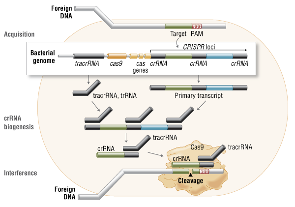
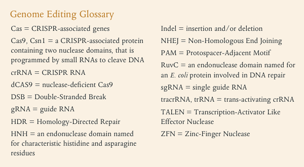
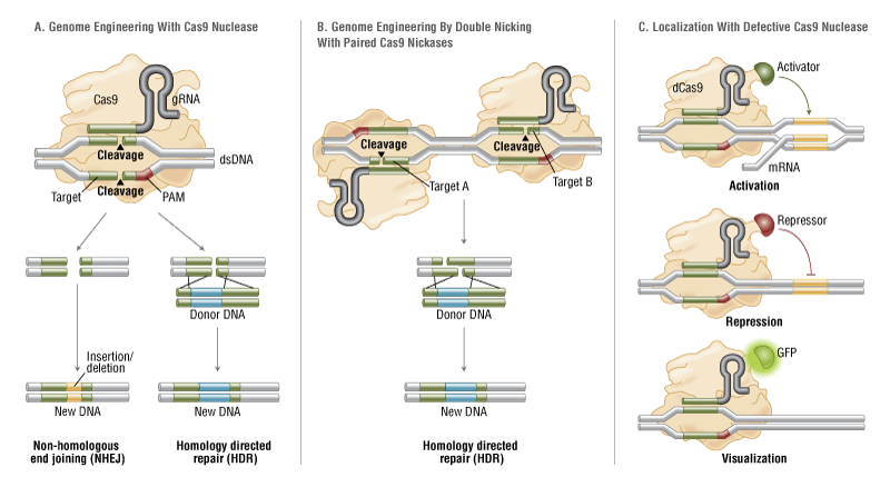
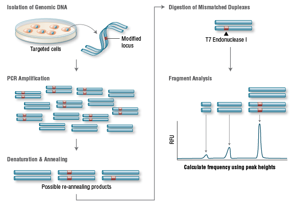


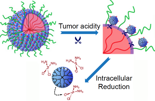
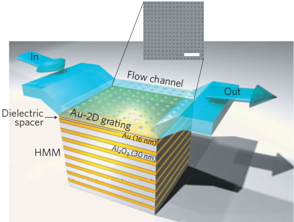

It is almost impossible to set up a conspiracy against big pharma’s abusive practices.Every avenue their high priced lawyers can think of to stop budding conspiracies has been blocked by law where possible. One possible road might be to do research and development in other countries outside US legal juristiction, however most drugs without FDA approval can and are stopped at the border and confiscated even if as in Canada the same drug produced in the US is being manufactured in Canada.Almost certainly Cisplatin is under patent in the US and the patent holder has the right to refuse the use of the drug for any reason they want, including being used in this cluster bomb drug. The manufacturer is almost certainly making huge profits from selling Cisplatin and I doubt they want to see a cheap drug cure many cancers. I guess the only way to go is to try and turn to a country like India.A number of cancer drugs were being sold by US patent holders at wholesale prices that were to high for most Indians. The government of India refused to allow these companies to patent their medicines in India and forced them to license the drugs and much cheaper prices.Most US patents are not operative in India, they can produce US style insulin pumps at a fraction of our cost as they can in China and Vietnam or Mexico. It would be difficult to send these pumps to buyers in the US from India but by shipping them from another country, say Canada or Mexico most would make it past customs. As for Cancer treatment, India and china have some very fine trained biochemist and doctors, who could easily apply many of the immunological treatments against cancer. All arms of the immune system have been used to produce miracle treatments that have cured some patients that were on their death beds.The treatments can be tested carefully in these countries, and improved by any methods including some I have suggested.By advertising in the US to cancer patients that they can inexpensively have these working treatments cheaply as a medical tourist, it is only a matter of time before they will cure the disease wholesale and break the medical industrial complex down. As far as generics that are not being produced here, by setting up a non profit corporation that produces any and all drugs that come off patent as a goal, at the cheapest price less a reasonable markup for cost of manufacture etc. one by one they will end the abuse of not producing or overpricing generics.
………
Significance
Successively overcoming a series of biological barriers that cancer nanotherapeutics would encounter upon intravenous administration is required for achieving positive treatment outcomes. A hurdle to this goal is the inherently unfavorable tumor penetration of nanoparticles due to their relatively large sizes. We developed a stimuli-responsive clustered nanoparticle (iCluster) and justified that its adaptive alterations of physicochemical properties (e.g. size, zeta potential, and drug release rate) in accordance with the endogenous stimuli of the tumor microenvironment made possible the ultimate overcoming of these barriers, especially the bottleneck of tumor penetration. Results in varying intractable tumor models demonstrated significantly improved antitumor efficacy of iCluster than its control groups, demonstrating that overcoming these delivery barriers can be achieved by innovative nanoparticle design.
http://www.pnas.org/content/early/2016/03/23/1522080113.full