Healthcare analytics, AI solutions for biological big data, providing an AI platform for the biotech, life sciences, medical and pharmaceutical industries, as well as for related technological approaches, i.e., curation and text analysis with machine learning and other activities related to AI applications to these industries.
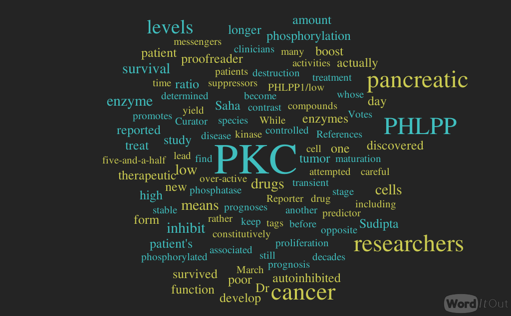
Pancreatic cancer survival is determined by ratio of two enzymes, Volume 2 (Volume Two: Latest in Genomics Methodologies for Therapeutics: Gene Editing, NGS and BioInformatics, Simulations and the Genome Ontology), Part 1: Next Generation Sequencing (NGS)
Reporter and Curator: Dr. Sudipta Saha, Ph.D.
Protein kinase C (PKC) isozymes function as tumor suppressors in increasing contexts. These enzymes are crucial for a number of cellular activities, including cell survival, proliferation and migration — functions that must be carefully controlled if cells get out of control and form a tumor. In contrast to oncogenic kinases, whose function is acutely regulated by transient phosphorylation, PKC is constitutively phosphorylated following biosynthesis to yield a stable, autoinhibited enzyme that is reversibly activated by second messengers. Researchers at University of California San Diego School of Medicine found that another enzyme, called PHLPP1, acts as a “proofreader” to keep careful tabs on PKC.
The researchers discovered that in pancreatic cancer high PHLPP1 levels lead to low PKC levels, which is associated with poor patient survival. They reported that the phosphatase PHLPP1 opposes PKC phosphorylation during maturation, leading to the degradation of aberrantly active species that do not become autoinhibited. They discovered that any time an over-active PKC is inadvertently produced, the PHLPP1 “proofreader” tags it for destruction. That means the amount of PHLPP1 in patient’s cells determines his amount of PKC and it turns out those enzyme levels are especially important in pancreatic cancer.
This team of researchers reversed a 30-year paradigm when they reported evidence that PKC actually suppresses, rather than promotes, tumors. For decades before this revelation, many researchers had attempted to develop drugs that inhibit PKC as a means to treat cancer. Their study implied that anti-cancer drugs would actually need to do the opposite — boost PKC activity. This study sets the stage for clinicians to one day use a pancreatic cancer patient’s PHLPP1/PKC levels as a predictor for prognosis, and for researchers to develop new therapeutic drugs that inhibit PHLPP1 and boost PKC as a means to treat the disease.
The ratio — high PHLPP1/low PKC — correlated with poor prognoses: no pancreatic patient with low PKC in the database survived longer than five-and-a-half years. On the flip side, 50 percent of the patients with low PHLPP1/high PKC survived longer than that. While still in the earliest stages, the researchers hope that this information might one day aid pancreatic diagnostics and treatment. The researchers are next planning to screen chemical compounds to find those that inhibit PHLPP1 and restore PKC levels in low-PKC-pancreatic cancer cells in the lab. These might form the basis of a new therapeutic drug for pancreatic cancer.
Immuno-editing can be a constant defense in the cancer landscape, Volume 2 (Volume Two: Latest in Genomics Methodologies for Therapeutics: Gene Editing, NGS and BioInformatics, Simulations and the Genome Ontology), Part 1: Next Generation Sequencing (NGS)
Reporter and Curator: Dr. Sudipta Saha, Ph.D.
There are many considerations in the cancer immunoediting landscape of defense and regulation in the cancer hallmark biology. The cancer hallmark biology in concert with key controls of the HLA compatibility affinity mechanisms are pivotal in architecting a unique patient-centric therapeutic application. Selection of random immune products including neoantigens, antigens, antibodies and other vital immune elements creates a high level of uncertainty and risk of undesirable immune reactions. Immunoediting is a constant process. The human innate and adaptive forces can either trigger favorable or unfavorable immunoediting features. Cancer is a multi-disease entity. There are multi-factorial initiators in a certain disease process. Namely, environmental exposures, viral and / or microbiome exposure disequilibrium, direct harm to DNA, poor immune adaptability, inherent risk and an individual’s own vibration rhythm in life.
When a human single cell is crippled (Deranged DNA) with mixed up molecular behavior that is the initiator of the problem. A once normal cell now transitioned into full threatening molecular time bomb. In the modeling and creation of a tumor it all begins with the singular molecular crisis and crippling of a normal human cell. At this point it is either chop suey (mixed bit responses) or a productive defensive and regulation response and posture of the immune system. Mixed bits of normal DNA, cancer-laden DNA, circulating tumor DNA, circulating normal cells, circulating tumor cells, circulating immune defense cells, circulating immune inflammatory cells forming a moiety of normal and a moiety of mess. The challenge is to scavenge the mess and amplify the normal.
Immunoediting is a primary push-button feature that is definitely required to be hit when it comes to initiating immune defenses against cancer and an adaptation in favor of regression. As mentioned before that the tumor microenvironment is a “mixed bit” moiety, which includes elements of the immune system that can defend against circulating cancer cells and tumor growth. Personalized (Precision-Based) cancer vaccines must become the primary form of treatment in this case. Current treatment regimens in conventional therapy destroy immune defenses and regulation and create more serious complications observed in tumor progression, metastasis and survival. Commonly resistance to chemotherapeutic agents is observed. These personalized treatments will be developed in concert with cancer hallmark analytics and immunocentrics affinity and selection mapping. This mapping will demonstrate molecular pathway interface and HLA compatibility and adaptation with patientcentricity.
The bacterial makeup of human milk is influenced by the mode of breastfeeding, according to a new study. Although previously considered sterile, breast milk is now known to contain a low abundance of bacteria. While the complexities of how maternal microbiota influence the infant microbiota are still unknown, this complex community of bacteria in breast milk may help to establish the infant gut microbiota. Disruptions in this process could alter the infant microbiota, causing predisposition to chronic diseases such as allergies, asthma, and obesity. While it’s unclear how the breast milk microbiome develops, there are two theories describing its origins. One theory speculates that it originates in the maternal mammary gland, while the other theory suggests that it is due to retrograde inoculation by the infant’s oral microbiome.
To address this gap in knowledge scientists carried out bacterial gene sequencing on milk samples from 393 healthy mothers three to four months after giving birth. They used this information to examine how the milk microbiota composition is affected by maternal factors, early life events, breastfeeding practices, and other milk components. Among the many factors analyzed, the mode of breastfeeding (with or without a pump) was the only consistent factor directly associated with the milk microbiota composition. Specifically, indirect breastfeeding was associated with a higher abundance of potential opportunistic pathogens, such as Stenotrophomonas and Pseudomonadaceae. By contrast, direct breastfeeding without a pump was associated with microbes typically found in the mouth, as well as higher overall bacterial richness and diversity. Taken together, the findings suggest that direct breastfeeding facilitates the acquisition of oral microbiota from infants, whereas indirect breastfeeding leads to enrichment with environmental (pump-associated) bacteria.
The researchers argued that this study supports the theory that the breast milk microbiome is due to retrograde inoculation. Their findings indicate that the act of pumping and contact with the infant oral microbiome influences the milk microbiome, though they noted more research is needed. In future studies, the researchers will further explore the composition and function of the milk microbiota. In addition to bacteria, they will profile fungi in the milk samples. They also plan to investigate how the milk microbiota influences both the gut microbiota of infants and infant development and health. Specifically, their projects will examine the association of milk microbiota with infant growth, asthma, and allergies. This work could have important implications for microbiota-based strategies for early-life prevention of chronic conditions.
Lesson 4 Cell Signaling And Motility: G Proteins, Signal Transduction: Curations and Articles of reference as supplemental information: #TUBiol3373
Curator: Stephen J. Williams, Ph.D.
Updated 7/15/2019
Below please find the link to the Powerpoint presentation for lesson #4 for #TUBiol3373. The lesson first competes the discussion on G Protein Coupled Receptors, including how cells terminate cell signals. Included are mechanisms of receptor desensitization. Please NOTE that desensitization mechanisms like B arrestin decoupling of G proteins and receptor endocytosis occur after REPEATED and HIGH exposures to agonist. Hydrolysis of GTP of the alpha subunit of G proteins, removal of agonist, and the action of phosphodiesterase on the second messenger (cAMP or cGMP) is what results in the downslope of the effect curve, the termination of the signal after agonist-receptor interaction.
Research about marijuana and fertility is limited but some previous studies suggested that it might harm semen quality. Smoking of any type is also known to be a risk factor for male infertility. So, men who have smoked cannabis are expected to have worse measures of fertility but the data from a recent study suggested the opposite. The finding contradicts all conventional knowledge on how weed affects sperm. This may be because previous research typically focused on men with drug abuse history but this present study simply asked men if they had smoked more than two joints in their life.
Analysis of 1,143 semen samples from 662 men collected between 2000 and 2017 at the Fertility Clinic at Massachusetts General Hospital showed that those who had smoked weed at some point in their life had a mean sperm concentration of 62.7 million sperm per milliliter (mL) of ejaculate, while men who avoided marijuana entirely had mean concentrations of 45.4 million/mL. Added to this only 5% of weed smokers had sperm concentrations below the 15 million/mL threshold the World Health Organization has set for a “normal” sperm count, versus 12% of men who never smoked marijuana.
The study has some imperfections such as the participants are not necessarily representative of the general population. They were predominantly college educated men with a mean age of 36, and were all seeking treatment at a fertility center. Further research is needed to support the findings. Two possibilities are put forward by the researchers as the reason behind such data. The first is that low levels of marijuana could have a positive effect on the endocannabinoid system, the neurotransmitters in the nervous system that bind to cannabinoid receptors, and are known to regulate fertility. The second is that may be weed-smokers are just bigger risk takers and men with higher testosterone levels and thus have better sperm count.
But, there’s certainly no medical recommendation to smoke weed as a fertility treatment but this study, at least, suggests that a little marijuana doesn’t hurt and might benefit sperm production in some way. But, the researchers specified that their finding does not necessarily mean that smoking cannabis increases the chances of fatherhood.
Stroke is a leading cause of death worldwide and the most common cause of long-term disability amongst adults, more particularly in patients with diabetes mellitus and arterial hypertension. Increasing evidence suggests that disordered physiological variables following acute ischaemic stroke, especially hyperglycaemia, adversely affect outcomes.
Post-stroke hyperglycaemia is common (up to 50% of patients) and may be rather prolonged, regardless of diabetes status. A substantial body of evidence has demonstrated that hyperglycaemia has a deleterious effect upon clinical and morphological stroke outcomes. Therefore, hyperglycaemia represents an attractive physiological target for acute stroke therapies.
However, whether intensive glycaemic manipulation positively influences the fate of ischaemic tissue remains unknown. One major adverse event of management of hyperglycaemia with insulin (either glucose-potassium-insulin infusions or intensive insulin therapy) is the occurrence of hypoglycaemia, which can also induce cerebral damage.
Doctors all over the world have debated whether intensive glucose management, which requires the use of IV insulin to bring blood sugar levels down to 80-130 mg/dL, or standard glucose control using insulin shots, which aims to get glucose below 180 mg/dL, lead to better outcomes after stroke.
A period of hyperglycemia is common, with elevated blood glucose in the periinfarct period consistently linked with poor outcome in patients with and without diabetes. The mechanisms that underlie this deleterious effect of dysglycemia on ischemic neuronal tissue remain to be established, although in vitro research, functional imaging, and animal work have provided clues.
While prompt correction of hyperglycemia can be achieved, trials of acute insulin administration in stroke and other critical care populations have been equivocal. Diabetes mellitus and hyperglycemia per se are associated with poor cerebrovascular health, both in terms of stroke risk and outcome thereafter.
Interventions to control blood sugar are available but evidence of cerebrovascular efficacy are lacking. In diabetes, glycemic control should be part of a global approach to vascular risk while in acute stroke, theoretical data suggest intervention to lower markedly elevated blood glucose may be of benefit, especially if thrombolysis is administered.
Both hypoglycaemia and hyperglycaemia may lead to further brain injury and clinical deterioration; that is the reason these conditions should be avoided after stroke. Yet, when correcting hyperglycaemia, great care should be taken not to switch the patient into hypoglycaemia, and subsequently aggressive insulin administration treatment should be avoided.
Early identification and prompt management of hyperglycaemia, especially in acute ischaemic stroke, is recommended. Although the appropriate level of blood glucose during acute stroke is still debated, a reasonable approach is to keep the patient in a mildly hyperglycaemic state, rather than risking hypoglycaemia, using continuous glucose monitoring.
The primary results from the Stroke Hyperglycemia Insulin Network Effort (SHINE) study, a large, multisite clinical study showed that intensive glucose management did not improve functional outcomes at 90 days after stroke compared to standard glucose therapy. In addition, intense glucose therapy increased the risk of very low blood glucose (hypoglycemia) and required a higher level of care such as increased supervision from nursing staff, compared to standard treatment.
Today’s lesson 3 explains how extracellular signals are transduced (transmitted) into the cell through receptors to produce an agonist-driven event (effect). This lesson focused on signal transduction from agonist through G proteins (GTPases), and eventually to the effectors of the signal transduction process. Agonists such as small molecules like neurotransmitters, hormones, nitric oxide were discussed however later lectures will discuss more in detail the large growth factor signalings which occur through receptor tyrosine kinases and the Ras family of G proteins as well as mechanosignaling through Rho and Rac family of G proteins.
Transducers: The Heterotrimeric G Proteins (GTPases)
An excellent review of heterotrimeric G Proteins found in the brain is given by
Cyclic AMP is an important second messenger. It forms, as shown, when the membrane enzyme adenylyl cyclase is activated (as indicated, by the alpha subunit of a G protein).
The cyclic AMP then goes on the activate specific proteins. Some ion channels, for example, are gated by cyclic AMP. But an especially important protein activated by cyclic AMP is protein kinase A, which goes on the phosphorylate certain cellular proteins. The scheme below shows how cyclic AMP activates protein kinase A.
Updated 7/15/2019
Additional New Studies on Regulation of the Beta 2 Adrenergic Receptor
We had discussed regulation of the G protein coupled beta 2 adrenergic receptor by the B-AR receptor kinase (BARK)/B arrestin system which uncouples and desensitizes the receptor from its G protein system. In an article by Xiangyu Liu in Science in 2019, the authors describe another type of allosteric modulation (this time a POSITIVE allosteric modulation) in the intracellular loop 2. See below:
Mechanism of β2AR regulation by an intracellular positive allosteric modulator
Xiangyu Liu1,*, Ali Masoudi2,*, Alem W. Kahsai2,*, Li-Yin Huang2, Biswaranjan Pani2, Dean P. Staus2, Paul J. Shim2, Kunio Hirata3,4, Rishabh K. Simhal2, Allison M. Schwalb2, Paula K. Rambarat2, Seungkirl Ahn2, Robert J. Lefkowitz2,5,6,†, Brian Kobilka1
Positive reinforcement in a GPCR
Many drug discovery efforts focus on G protein–coupled receptors (GPCRs), a class of receptors that regulate many physiological processes. An exemplar is the β2-adrenergic receptor (β2AR), which is targeted by both blockers and agonists to treat cardiovascular and respiratory diseases. Most GPCR drugs target the primary (orthosteric) ligand binding site, but binding at allosteric sites can modulate activation. Because such allosteric sites are less conserved, they could possibly be targeted more specifically. Liu et al. report the crystal structure of β2AR bound to both an orthosteric agonist and a positive allosteric modulator that increases receptor activity. The structure suggests why the modulator compound is selective for β2AR over the closely related β1AR. Furthermore, the structure reveals that the modulator acts by enhancing orthosteric agonist binding and stabilizing the active conformation of the receptor.
Abstract
Drugs targeting the orthosteric, primary binding site of G protein–coupled receptors are the most common therapeutics. Allosteric binding sites, elsewhere on the receptors, are less well-defined, and so less exploited clinically. We report the crystal structure of the prototypic β2-adrenergic receptor in complex with an orthosteric agonist and compound-6FA, a positive allosteric modulator of this receptor. It binds on the receptor’s inner surface in a pocket created by intracellular loop 2 and transmembrane segments 3 and 4, stabilizing the loop in an α-helical conformation required to engage the G protein. Structural comparison explains the selectivity of the compound for β2– over the β1-adrenergic receptor. Diversity in location, mechanism, and selectivity of allosteric ligands provides potential to expand the range of receptor drugs.
Recent structures of GPCRs bound to allosteric modulators have revealed that receptor surfaces are decorated with diverse cavities and crevices that may serve as allosteric modulatory sites (1). This substantiates the notion that GPCRs are structurally plastic and can be modulated by a variety of allosteric ligands through distinct mechanisms (2-7). Most of these structures have been solved with negative allosteric modulators (NAMs), which stabilize receptors in their inactive states (1). To date, only a single structure of an active GPCR bound to a small-molecule positive allosteric modulator (PAM) has been reported, namely, the M2 muscarinic acetylcholine receptor with LY2119620 (8). Thus, mechanisms of PAMs and their potential binding sites remain largely unexplored.
Fig 1. Structure of the active state T4L-B2AR in complex with the orthosteric agonist BI-167107, nanobody 689, and compound 6FA. (A) The chemical structure of compound-6FA (Cmpd-6FA). (B) Isoproterenol (ISO) competition binding with 125I-cyanopindolol (CYP) to the β2AR reconstituted in nanodisks in the presence of vehicle (0.32% dimethylsulfoxide; DMSO), Cmpd-6, or Cmpd-6FA at 32 μM. Values were normalized to percentages of the maximal 125I-CYP binding level obtained from a one-site competition binding–log IC50 (median inhibitory concentration) curve fit. Binding curves were generated by GraphPad Prism. Points on curves represent mean ± SEM obtained from five independent experiments performed in duplicate. (C) Analysis of Cmpd-6FA interaction with the BI-167107–bound β2AR by ITC. Representative thermogram (inset) and binding isotherm, of three independent experiments, with the best titration curve fit are shown. Summary of thermodynamic parameters obtained by ITC: binding affinity (KD = 1.2 ± 0.1 μM), stoichiometry (N = 0.9 ± 0.1 sites), enthalpy (ΔH = 5.0 ± 1.2 kcal mol−1), and entropy (ΔS =13 ± 2.0 cal mol−1 deg−1). (D) Side view of T4L-β2AR bound to the orthosteric agonist BI-167107, nanobody 6B9 (Nb6B9), and Cmpd-6FA. The gray box indicates the membrane layer as defined by the OPM database. (E) Close-up view of Cmpd-6FA binding site. Covering Cmpd-6FA is 2Fo– Fc electron density contoured at 1.0 σ (green mesh).From Science 28 Jun 2019:
Vol. 364, Issue 6447, pp. 1283-1287
Fig 3. Fig. 3Mechanism of allosteric activation of the β2AR by Cmpd-6FA.
(A) Superposition of the inactive β2AR bound to the antagonist carazolol (PDB code: 2RH1) and the active β2AR bound to the agonist BI-167107, Cmpd-6FA, and Nb6B9. Close-up view of the Cmpd-6FA binding site is shown. The residues of the inactive (yellow) and active (blue) β2AR are depicted, and the hydrogen bond formed between Asp1303.49and Tyr141ICL2 in the active state is indicated by a black dashed line. (B) Topography of Cmpd-6FA binding surface on the active β2AR (left, blue) and the corresponding surface of the inactive β2AR (right, yellow) with Cmpd-6FA (orange sticks) docked on top. Molecular surfaces are of only those residues involved in interaction with Cmpd-6FA. Steric clash between Cmpd-6FA and the surface of inactive β2AR is represented by a purple asterisk. (C) Overlay of the β2AR bound to BI-167107, Nb6B9, and Cmpd-6FA with the β2AR–Gscomplex (PDB code: 3SN6). The inset shows the position of Phe139ICL2 relative to the α subunit of Gs. (D) Superposition of the active β2AR bound to the agonist BI-167107, Nb6B9, and Cmpd-6FA (blue) with the inactive β2AR bound to carazolol (yellow) (PDB code: 2RH1) as viewed from the cytoplasm. For clarity, Nb6B9 and the orthosteric ligands are omitted. The arrows indicate shifts in the intracellular ends of the TM helices 3, 5, and 6 upon activation and their relative distances.
Allosteric sites may not face the same evolutionary pressure as do orthosteric sites, and thus are more divergent across subtypes within a receptor family (24–26). Therefore, allosteric sites may provide a greater source of specificity for targeting GPCRs.
D. M. Thal, A. Glukhova, P. M. Sexton, A. Christopoulos, Structural insights into G-protein-coupled receptor allostery. Nature 559, 45–53 (2018). doi:10.1038/s41586-018-0259-zpmid:29973731CrossRefPubMedGoogle Scholar
D. Wacker, R. C. Stevens, B. L. Roth, How Ligands Illuminate GPCR Molecular Pharmacology. Cell 170, 414–427 (2017).
D. P. Staus, R. T. Strachan, A. Manglik, B. Pani, A. W. Kahsai, T. H. Kim, L. M. Wingler, S. Ahn, A. Chatterjee, A. Masoudi, A. C. Kruse, E. Pardon, J. Steyaert, W. I. Weis, R. S. Prosser, B. K. Kobilka, T. Costa, R. J. Lefkowitz, Allosteric nanobodies reveal the dynamic range and diverse mechanisms of G-protein-coupled receptor activation. Nature 535, 448–452 (2016). doi:10.1038/nature18636pmid:27409812CrossRefPubMedGoogle Scholar
A. Manglik, T. H. Kim, M. Masureel, C. Altenbach, Z. Yang, D. Hilger, M. T. Lerch, T. S. Kobilka, F. S. Thian, W. L. Hubbell, R. S. Prosser, B. K. Kobilka, Structural Insights into the Dynamic Process of β2-Adrenergic Receptor Signaling. Cell 161, 1101–1111 (2015). doi:10.1016/j.cell.2015.04.043pmid:25981665CrossRefPubMedGoogle Scholar
5, L. Ye, N. Van Eps, M. Zimmer, O. P. Ernst, R. S. Prosser, Activation of the A2A adenosine G-protein-coupled receptor by conformational selection. Nature 533, 265–268 (2016). doi:10.1038/nature17668pmid:27144352CrossRefPubMedGoogle Scholar
N. Van Eps, L. N. Caro, T. Morizumi, A. K. Kusnetzow, M. Szczepek, K. P. Hofmann, T. H. Bayburt, S. G. Sligar, O. P. Ernst, W. L. Hubbell, Conformational equilibria of light-activated rhodopsin in nanodiscs. Proc. Natl. Acad. Sci. U.S.A. 114, E3268–E3275 (2017). doi:10.1073/pnas.1620405114pmid:28373559Abstract/FREE Full TextGoogle Scholar
R. O. Dror, H. F. Green, C. Valant, D. W. Borhani, J. R. Valcourt, A. C. Pan, D. H. Arlow, M. Canals, J. R. Lane, R. Rahmani, J. B. Baell, P. M. Sexton, A. Christopoulos, D. E. Shaw, Structural basis for modulation of a G-protein-coupled receptor by allosteric drugs. Nature 503, 295–299 (2013). doi:10.1038/nature12595pmid:24121438CrossRefPubMedWeb of ScienceGoogle Scholar
A. C. Kruse, A. M. Ring, A. Manglik, J. Hu, K. Hu, K. Eitel, H. Hübner, E. Pardon, C. Valant, P. M. Sexton, A. Christopoulos, C. C. Felder, P. Gmeiner, J. Steyaert, W. I. Weis, K. C. Garcia, J. Wess, B. K. Kobilka, Activation and allosteric modulation of a muscarinic acetylcholine receptor. Nature 504, 101–106 (2013). doi:10.1038/nature12735pmid:24256733
Additional information on Nitric Oxide as a Cellular Signal
Nitric oxide is actually a free radical and can react with other free radicals, resulting in a very short half life (only a few seconds) and so in the body is produced locally to its site of action (i.e. in endothelial cells surrounding the vascular smooth muscle, in nerve cells). In the late 1970s, Dr. Robert Furchgott observed that acetylcholine released a substance that produced vascular relaxation, but only when the endothelium was intact. This observation opened this field of research and eventually led to his receiving a Nobel prize. Initially, Furchgott called this substance endothelium-derived relaxing factor (EDRF), but by the mid-1980s he and others identified this substance as being NO.
Nitric oxide is implicated in many pathologic processes as well. Nitric oxide post translational modifications have been attributed to nitric oxide’s role in pathology however, although the general mechanism by which nitric oxide exerts its physiological effects is by stimulation of soluble guanylate cyclase to produce cGMP, these post translational modifications can act as a cellular signal as well. For more information of NO pathologic effects and how NO induced post translational modifications can act as a cellular signal see the following:
BIO 3096, Cell Structure and Function (Minimum Grade of C- | May not be taken concurrently).
Description:
The communication among cells is essential for the regulation of the development of an organism and for the control of its physiology and homeostasis. Aberrant cellular signaling events are often associated with human pathological conditions, such as cancer, neurological disorders, cardiovascular diseases and so on. The full characterization of cell signaling systems may provide useful insights into the pathogenesis of several human maladies.
Text:
Molecular Biology of the Cell 6th Edition, Alberts et al. Garland Science. This textbook is available at the Temple Bookstore.
Grading:
The final grade will be based on the score of four examinations that include both group and individuals assignment. Each exam accounts for 25% of the final grade. There will be no make-up tests during the course. If you have a documented medical excuse and you contact me as soon as possible after the emergency, I will arrange a make-up exam. Complaints regarding the grading will not be considered later than two weeks after the test is returned.
Blackboard:
Announcements will be readily posted on Blackboard. It is your responsibility to check Blackboard periodically.
Attendance: Lecture attendance is mandatory. In addition, punctuality is expected.
Disabilities: Students with documented disabilities who need particular accommodation should contact me privately as soon as possible.
Honesty and Civility:
Students must follow the Temple’s Code of Conduct (see http://www.temple.edu/assistance/udc/coc.htm). This Code of Conduct prohibits: 1. Academic dishonesty and impropriety, including plagiarism and cheating. 2. Interfering or attempting to interfere with or disrupting the conduct of classes or any other activity of the University.”
This policy sets the parameters for freedom to learn and freedom to teach, which constitute the pillars of academia.
SCHEDULE
This schedule is a general outline, which may be eventually modified. Changes will be announced in advance. Please, always check Blackboard and your email.
Date
Topic
Jan 13
Introduction (course overview and discussion of syllabus). General concepts: Eukaryotic and prokaryotic cell; DNA, RNA and proteins: Protein synthesis
Jan 20
Martin Luther King, Jr. Day (no classes held)
Jan 27
DNA analysis, RNA analysis; Proteins analysis; Microscopy.
Feb 3
Signaling: general concepts; Introduction to G-proteins; signaling via G-proteins (1)
Feb 10
Exam 1: In class presentation (group assignment)
Feb 17
Signaling via G-proteins (2); tyrosine kinase receptors signaling; Ras-MAPK pathway.
Medical consequences of aberrant signaling pathways; production of small molecules for protein kinases In cancer therapy.
Study days
May 4
Exam 4: In class presentation (group assignment)
Below is Powerpoint presentations for Lesson 1 and Lesson 2. Please check for UPDATES on this page for additional supplemental information for these Lessons including articles from this Online Access Journal
The following articles and curations discuss about the new paradigm how we now envision DNA, in particular how we now understand that the important parts of the genome are not just the exons which code for proteins but also the intronic DNA, which contains all the regulatory elements such as promoters, lncDNA, miRNA sequences etc. These are good reads for your presentations.
And on How the Cell Creates Diversity post the Genetic Code by Use of Post Translational Modifications to Bring Diversity to Protein Structure/Function
Once herpes simplex infects a person, the virus goes into hiding inside nerve cells, hibernating there for life, periodically waking up from its sleep to reignite infection, causing cold sores or genital lesions to recur. Research from Harvard Medical School showed that the virus uses a host protein called CTCF, or cellular CCCTC-binding factor, to display this type of behavior. Researchers revealed with experiments on mice that CTCF helps herpes simplex regulate its own sleep-wake cycle, enabling the virus to establish latent infections in the body’s sensory neurons where it remains dormant until reactivated. Preventing that latency-regulating protein from binding to the virus’s DNA, weakened the virus’s ability to come out of hiding.
Herpes simplex virus’s ability to go in and out of hiding is a key survival strategy that ensures its propagation from one host to the next. Such symptom-free latency allows the virus to remain out of the reach of the immune system most of the time, while its periodic reactivation ensures that it can continue to spread from one person to the next. On one hand, so-called latency-associated transcript genes, or LAT genes, turn off the transcription of viral RNA, inducing the virus to go into hibernation, or latency. On the other hand, a protein made by a gene called ICP0 promotes the activity of genes that stimulate viral replication and causes active infection.
Based on these earlier findings, the new study revealed that this balancing act is enabled by the CTCF protein when it binds to the viral DNA. Present during latent or dormant infections, CTCF is lost during active, symptomatic infections. The researchers created an altered version of the virus that lacked two of the CTCF binding sites. The absence of the binding sites made no difference in early-stage or acute infections. Similar results were found in infected cultured human nerve cells (trigeminal ganglia) and infected mice model. The researchers concluded that the mutant virus was found to have significantly weakened reactivation capacity.
Taken together, the experiments showed that deleting the CTCF binding sites weakened the virus’s ability to wake up from its dormant state thereby establishing the evidence that the CTCF protein is a key regulator of sleep-wake cycle in herpes simplex infections.
Live 12:00 – 1:00 P.M Mediterranean Diet and Lifestyle: A Symposium on Diet and Human Health : October 19, 2018
Reporter: Stephen J. Williams, Ph.D.
12.00 The Italian Mediterranean Diet as a Model of Identity of a People with a Universal Good to Safeguard Health?
Prof. Antonino De Lorenzo, MD, PhD.
Director of the School of Specialization in Clinical Nutrition, University of Rome “Tor Vergata”
It is important to determine how our bodies interacts with the environment, such as absorption of nutrients.
Studies shown here show decrease in life expectancy of a high sugar diet, but the quality of the diet, not just the type of diet is important, especially the role of natural probiotics and phenolic compounds found in the Mediterranean diet.
The WHO report in 2005 discusses the unsustainability of nutrition deficiencies and suggest a proactive personalized and preventative/predictive approach of diet and health.
Most of the noncommunicable diseases like CV (46%) cancer 21% and 11% respiratory and 4% diabetes could be prevented and or cured with proper dietary approaches
Italy vs. the US diseases: in Italy most disease due to environmental contamination while US diet plays a major role
The issue we are facing in less than 10% of the Italian population (fruit, fibers, oils) are not getting the proper foods, diet and contributing to as we suggest 46% of the disease
The Food Paradox: 1.5 billion are obese; we notice we are eating less products of quality and most quality produce is going to waste;
growing BMI and junk food: our studies are correlating the junk food (pre-prepared) and global BMI
modern diet and impact of human health (junk food high in additives, salt) has impact on microflora
Western Diet and Addiction: We show a link (using brain scans) showing correlation of junk food, sugar cravings, and other addictive behaviors by affecting the dopamine signaling in the substantia nigra
developed a junk food calculator and a Mediterranean diet calculator
the intersection of culture, food is embedded in the Mediterranean diet; this is supported by dietary studies of two distinct rural Italian populations (one of these in the US) show decrease in diet
Impact of diet: have model in Germany how this diet can increase health and life expectancy
from 1950 to present day 2.7 unit increase in the diet index can increase life expectancy by 26%
so there is an inverse relationship with our index and breast cancer
Environment and metal contamination and glyphosate: contribution to disease and impact of maintaining the healthy diet
huge problem with use of pesticides and increase in celiac disease
Cancer as a disease of the environment. Weinberg’s hallmarks of Cancer reveal how environment and epigenetics can impact any of these hallmarks.
Epigenetic effects
gene gatekeepers (Rb and P53)
DNA repair and damage stabilization
Heavy Metals and Dioxins:( alterations of the immune system as well as epigenetic regulations)
Asbestos and Mesothelioma: they have demonstrated that p53 can be involved in development of mesothelioma as reactivating p53 may be a suitable strategy for therapy
Diet, Tomato and Cancer
looked at tomato extract on p53 function in gastric cancer: tomato extract had a growth reduction effect and altered cell cycle regulation and results in apoptosis
RBL2 levels are increased in extract amount dependent manner so data shows effect of certain tomato extracts of the southern italian tomato ( )
Antonio Giordano: we tested whole extracts of almost 30 different varieties of tomato. The tomato variety with highest activity was near Ravela however black tomatoes have shown high antitumor activity. We have done a followup studies showing that these varieties, if grow elsewhere lose their antitumor activity after two or three generations of breeding, even though there genetics are similar. We are also studying the effects of different styles of cooking of these tomatoes and if it reduces antitumor effect