Feeds:
Posts
Comments

Archive for the ‘Medical Devices R&D and Inventions’ Category

AGENDA – ICI Conference – Innovation in Cardiovascular Interventions – December 14-16, at the David InterContinental Hotel, Tel Aviv, Israel

Reporter: Aviva Lev-Ari, PhD, RN


1. ICI Scientific Program

ICI2014 speakers are some of the leading figures in the field. The preliminary list can be viewed at the ICI website.

ICI2014 will hold for the second time the “Wall to Wall Session – From the Great Wall of China to the Jerusalem Wall”. Click here for a glance at the 2013 program endorsed by Yanping Gao, the Chinese Ambassador in Israel.

Attendees will:

 Be exposed to promising research and new therapies in various phases of development.

 Learn from live case presentations on the impact of emerging technologies on current and future therapies.

 Gain insights from international experts speaking on important clinical topics—with an emphasis on future perspectives.

2. ICI Exhibition

The heart of the ICI Meeting is the strong International collaboration between Medicine and Industry. With an emphasis on technological developments, novel knowledge-rich technologies, and the diligent pursuit of solutions to yet unsolved problems in heart, brain and cardiovascular medicine, the ICI meeting features a State-of-the-Art Exhibition and Innovative Technology Parade.

Since 1995, the ICI exhibition is rapidly growing with more than 90 international exhibitors and sponsors, including the strongest players in the market alongside cutting edge innovative startups. ICI Exhibition is the perfect opportunity to connect and interact with the people that can affect the future of this field.

3. ICI Technology Parade

Focused on innovation, ICI provides an extensive platform for startup companies presenting their latest technologies. The Technology Parade can be a springboard for new companies with bright and creative new ideas. This is the perfect opportunity to help your business move “from idea to reality”. The Technology Parade Sessions enjoy a tremendous success in every meeting, attracting a wide variety of leading clinicians, scientists and corporate representatives. The wide spectrum of investors who will be in attendance will find the ICI Meeting a valuable forum for exposure to the development and advancement of innovative ideas in cardiology.

The ICI meeting is a tremendous opportunity to review the most innovative startups in the field of medical devices and meet in person at the B2B area. This event can be your chance to look into the latest most prominent investments opportunity. 

SOURCE

http://2014.icimeeting.com/

Conference PROGRAM

http://2014.icimeeting.com/ici-2014-program/

Read Full Post »

Surgical Options for Left Atrial Appendage (LAA) Removal for A-Fib Patients without Indication for Anticoagulant Therapy

Reporter: Aviva Lev-Ari, PhD, RN


Sacha Salzberg of the HeartClinic Zurich discusses surgical options for treating the left atrial appendage (LAA), including concomitant and stand-alone LAA therapies. Dr. Salzberg also offers insights into the use of the Atriclip and Tiger-Paw devices for LAA treatment.

SOURCE

VIEW VIDEO
This presentation was originally given during the SCTS Ionescu University program at the 80th Annual Meeting of the Society for Cardiothoracic Surgery in Great Britain and Ireland. This content is published with the permission of SCTS. Please click here for information on the 2015 ACTA/SCTS Annual Meeting & Forum in Manchester, United Kingdom.


Read Full Post »

Tethered–Liquid Perfluorocarbon surface (TLP): Biocoating Prevents Blood from Clotting on Implantables

Reporter: Aviva Lev-Ari, PhD, RN


VIEW VIDEO

http://www.mdtmag.com/news/2014/10/biocoating-prevents-blood-clotting-implantables?et_cid=4205834&et_rid=461755519&type=cta

Wyss Institute researchers discuss how they used FDA-approved materials to develop a slippery coating that can be applied to medical devices to prevent blood clotting and bacteria accumulation. (Credit: Harvard’s Wyss Institute)

See Full Article


Read Full Post »

Development Of Super-Resolved Fluorescence Microscopy


Author and Curator: Larry H. Bernstein, MD, FCAP

CSO, Leaders in Pharmaceutical Business Intelligence

Article ID #153: Development Of Super-Resolved Fluorescence Microscopy. Published on 10/12/2014

WordCloud Image Produced by Adam Tubman

Development Of Super-Resolved Fluorescence Microscopy


Part I. Nobel Prize For Chemistry 2014: Eric Betzig, Stefan W. Hell
and William E. Moerner Honored For Development Of Super-
Resolved Fluorescence Microscopy

The 2014 Nobel Prize in Chemistry was awarded on 10/08/2014 to
Eric Betzig, Stefan W. Hell and William E. Moerner for
“the development of super-resolved fluorescence microscopy.”

The invention of the electron microscope by Max Knoll and Ernst Ruska at the
Berlin Technische Hochschule in 1931 finally overcame the barrier to higher
resolution that had been imposed by the limitations of visible light. Since then
resolution has defined the progress of the technology.

The ultimate goal was atomic resolution – the ability to see atoms – but this would
have to be approached incrementally over the course of decades. The earliest microscopes merely proved the concept: electron beams could, indeed, be tamed
to provide visible images of matter. By the late 1930s electron microscopes with theoretical resolutions of 10 nm were being designed and produced, and by 1944
this was further reduced to 2 nm. (The theoretical resolution of a an optical light microscope is 200 nm.)

Increases in the accelerating voltage of the electron beam accounted for much of
the improvement in resolution. But voltage was not everything. Improvements in electron lens technology minimized aberrations and provided a clearer picture,
which also contributed to improved resolution, as did better vacuum systems and brighter electron guns. So increasing the resolution of electron microscopes was a main driving force throughout the instrument’s development.

With nanoscopy, scientists could observe viruses, proteins and molecules there
are smaller than 0.0000002 metres.

Three researchers won the 2014 Nobel Prize in Chemistry on Wednesday,
October 8, for giving microscopes much sharper vision than was thought possible, letting scientists peer into living cells with unprecedented detail to seek the roots
of disease.  It was awarded to U.S. researchers Eric Betzig and William Moerner
and German scientist Stefan Hell. They found ways to use molecules that glow on demand to overcome what was considered a fundamental limitation for optical microscopes.

Hell, 52, of Germany, is the director at the Max Planck Institute for Biophysical Chemistry and the division head at the German Cancer Research Center in
Heidelberg. He was honored for his work on fluorescence microscopy, a kind
of nano-flashlight where scientists use fluorescent molecules to see parts of a
cell. Later in his career, he developed the STED microscope, which collects light
from “a multitude of small volumes to create a whole.”

Moerner, a 61-year-old professor in chemistry and applied physics at Stanford University in California, is the recipient of the 2008 Wolf Prize in Chemistry, the
2009 Irving Langmuir Award and the 2013 Peter Debye Award. In 1989, he
was the first scientist to be able to measure the light absorption of a single molecule.
This inspired many chemists to begin focusing on single molecules, including Betzig.

Betzig, 54, the group leader at Janelia Farm Research campus at the Howard
Hughes Medical Institute in Virginia, developed new optical imaging tools for
biology. His work involved taking images of the same area multiple times, and illuminating just a few molecules each time. These images were then
superimposed to create a dense super image at the nano level,

The limitation of optical microscopy was thought to have been determined in a calculation published in 1873 that defined the limit of how tiny a detail could be revealed by optical microscopes. Based on experimental evidence and basic principles of physics, Ernst Abbe and Lord Rayleigh defined and formulated
this diffraction-limited resolution in the late 19th century (Abbe, 1873; Rayleigh,
1896
).  However, only cellular structure and objects that were at least 200 to
350 nm apart could be resolved by light microscopy because, the optical resolution
of light microscopy was limited to approximately half of the wavelength of the light used.  Later key innovations—including fluorescence and confocal laser scanning microscopy (CLSM)—made optical microscopy one of the most powerful and
versatile diagnostic tools in modern cell biology. Using highly specific fluorescent labeling techniques such as immunocytochemistry, in situ hybridization, or
fluorescent protein tags, the spatial distribution and dynamics of virtually every subcellular structure, protein, or genomic sequence of interest can be analyzed in chemically fixed or living samples (Conchello and Lichtman, 2005; Giepmans et al., 2006).

The result of their advance is “really a window into the cell which we didn’t have before,” said Catherine Lewis, director of the cell biology and biophysics division
of the National Institute of General Medical Sciences in Bethesda, Maryland.

“You can observe the behavior of individual molecules in living cells in real time.
You can see … molecules moving around inside the cell. You can see them interacting with each other.”

The research of the three men has let scientists study diseases such as
Parkinson’s, Alzheimer’s and Huntington’s at a molecular level, the Royal
Swedish Academy of Sciences said.

Part II. Electron microscopy limitations

Manfred Von Ardenne in Berlin produced the earliest scanning-transmission
electron microscope in 1937. At the University of Toronto in Canada, Cecil Hall, James Hillier, and Albert Prebus, working under the direction of Eli Burton,
produced an advanced 1938 Toronto Model electron microscope that would
later become the basis for Radio Corporation of America’s Model B, the first commercial electron microscope in North America. Ruska at Siemens in
Germany produced the first commercial electron microscope in the world in 938.

Starting in 1939, scientists in Japan gathered to decide on the best way to build
an electron microscope. This group evolved into the Japan Electron Optics Laboratory (JEOL) that would eventually produce more models and varieties
of electron microscopes than any other company. Hitachi and Toshiba in Japan
also played a major role in the early development process.

The 1960s through the 1990s produced many innovative instruments and trends.
The introduction of the first commercial scanning electron microscopes (SEMs)
in 1965 opened up a new world of analysis for materials scientists. Ultrahigh
voltage TEM instruments (up to 3 MeV at CEMES-LOE/CNRS in Toulouse,
France, and at Hitachi in Tokyo, Japan), in the 1960s and 1970s gave electrons higher energy to penetrate more deeply into thick samples. The evolution and incorporation of other detectors (electron microprobes, electron energy loss spectroscopy (EELS), etc.) made the SEM into a true analytical electron
microscope (AEM) beginning in the 1970s. The development of brighter
electron sources, such as the lanthanum hexaboride filament (LAB6) and the
field emission gun in the 1960s, and their commercialization in the 1970s
brought researchers a brighter source of electrons and with it better imaging
and resolution. Tilting specimen stages permitting examination of the specimen
from different angles aided significantly in the determination of crystal structure.
In the late 1980s and throughout the 1990s, the environmental electron
microscopes that allow scientists to examine samples under more natural
conditions of temperature and pressure have dramatically expanded the
types of samples that can be examined.

In medicine, the EM made a unique contribution to diagnostic anatomic
pathology in renal biopsy analysis. However, the small sample had to be
embedded, and in the early days one cut the specimen by breaking glass
for the cutting of the specimen. But even though EM ushered in a new era of molecular pathology, the contribution was limited, despite incremental
improvements.

In the past, the use of microscopes was limited by a physical restriction;
scientists could only see items that were larger than roughly half the
wavelength of light (.2 micrometers)
. However, the groundbreaking work
of the Nobel laureates bypassed the maximum resolution of traditional
microscopes and launched optical microscopy into the nanodimension.

Part III. Super resolution fluorescence microscopy

Bo Huang,1,2 Mark Bates,3 and Xiaowei Zhuang1,2,4
Author information ► Copyright and License information ►
Annu Rev Biochem. 2009; 78: 993–1016.
http://dx.doi.org:/10.1146/annurev.biochem.77.061906.092014
PMCID: PMC2835776  NIHMSID: NIHMS179491

Achieving a spatial resolution that is not limited by the diffraction of
light, recent developments of super-resolution fluorescence microscopy
techniques allow the observation of many biological structures not
resolvable in conventional fluorescence microscopy. New advances
in these techniques now give them the ability to image three-dimensional
(3D) structures, measure interactions by multicolor colocalization, and
record dynamic processes in living cells at the nanometer scale. It is
anticipated that super-resolution fluorescence microscopy will become
a widely used tool for cell and tissue imaging to provide previously
unobserved details of biological structures and processes.

Keywords: Sub-diffraction limit, single-molecule, multicolor imaging,
three-dimensional imaging, live cell imaging, single-particle tracking,
photoswitchable probe

Among the various microscopy techniques, fluorescence microscopy is
one of the most widely used because of its two principal advantages:
Specific cellular components may be observed through molecule-specific
labeling, and light microscopy allows the observation of structures inside
a live sample in real time. Compared to other imaging techniques such
as electron microscopy (EM), however, conventional fluorescence
microscopy is limited by relatively low spatial resolution because of the
diffraction of light. This diffraction limit, about 200–300 nm in the lateral
direction and 500–700 nm in the axial direction, is comparable to or larger
than many subcellular structures, leaving them too small to be observed in
detail. In recent years, a number of “super-resolution” fluorescence microscopy techniques have been invented to overcome the diffraction barrier, including techniques that employ nonlinear effects to sharpen the point-spread function
of the microscope, such as stimulated emission depletion (STED) microscopy
(1, 2), related methods using other reversible saturable optically linear
fluorescence transitions (RESOLFTs) (3), and saturated structured-illumination microscopy (SSIM) (4), as well as techniques that are based on the localization
of individual fluorescent molecules, such as stochastic optical reconstruction microscopy (STORM) (5), photoactivated localization microscopy (PALM) (6),
and fluorescence photoactivation localization microscopy (FPALM) (7). These methods have yielded an order of magnitude improvement in spatial resolution
in all three dimensions over conventional light microscopy.

THE RESOLUTION LIMIT IN OPTICAL MICROSCOPY

Microscopes can be used to visualize fine structures in a sample by providing
a magnified image. However, even an arbitrarily high magnification does not
translate into the ability to see infinitely small details. Instead, the resolution
of light microscopy is limited because light is a wave and is subject to diffraction.

The diffraction limit

An optical microscope can be thought of as a lens system that produces a
magnified image of a small object. In this imaging process, light rays from
each point on the object converge to a single point at the image plane. However,
the diffraction of light prevents exact convergence of the rays, causing a sharp
point on the object to blur into a finite-sized spot in the image. The three-
dimensional (3D) intensity distribution of the image of a point object is called
the point spread function (PSF). The size of the PSF determines the resolution
of the microscope: Two points closer than the full width at half-maximum
(FWHM) of the PSF will be difficult to resolve because their images overlap substantially.

The FWHM of the PSF in the lateral directions (the x–y directions perpendicular
to the optical axis) can be approximated as Δxy ≈ 0.61λ / NA, where λ is the wavelength of the light, and NA is the numerical aperture of the objective
defined as NA = n sinα, with n being the refractive index of the medium and
α being the half-cone angle of the focused light produced by the objective.
The axial width of the PSF is about 2–3 times as large as the lateral width
for ordinary high NA objectives. When imaging with visible light (λ ≈ 550 nm),
the commonly used oil immersion objective with NA = 1.40 yields a PSF with
a lateral size of ~200 nm and an axial size of ~500 nm in a refractive index-
matched medium (Figure 1) (8).

Figure 1

The PSF of a common oil immersion objective with NA = 1.40, showing the
focal spot of 550 nm light in a medium with refractive index n = 1.515. The
intensity distribution in the x-z plane of the focus spot is computed numerically.

PFS of oil immersion microscope

PFS of oil immersion microscope

Because the loss of high-frequency spatial information in optical microscopy
results from the diffraction of light when it propagates through a distance larger
than the wavelength of the light (far field), near-field microscopy is one of the
earliest approaches sought to achieve high spatial resolution. By exciting the fluorophores or detecting the signal through the nonpropagating light near the fluorophore, high-resolution information be retained. Near-field scanning optical microscopy (NSOM) acquires an image by scanning a sharp probe tip across
the sample, typically providing a resolution of 20–50 nm (911). Wide-field
imaging has also been recently demonstrated in the near-field regime using
a super lens with negative refractive index (12, 13). However, the short range
of the near-field region (tens of nanometers) compromises the ability of light microscopy to look into a sample, limiting the application of near-field microscopy
to near-surface features only. This limit highlights the need to develop far-field
high-resolution imaging methods.

Among far-field fluorescence microscopy techniques, confocal and multiphoton microscopy are among the most widely used to moderately enhance the spatial resolution (14, 15). By combining a focused laser for excitation and a pinhole for detection, confocal microscopy can, in principle, have a factor of √2 improvement
in the spatial resolution. In multiphoton microscopy, nonlinear absorption processes reduce the effective size of the excitation PSF. However, this gain in the PSF size
is counteracted by the increased wavelength of the excitation light. Thus, instead
of improving the resolution, the main advantage of confocal and multi-photon microscopy over wide-field microscopy is the reduction of out-of-focus fluorescence background, allowing optical sectioning in 3D imaging.

Two techniques, 4Pi and I5M microscopy, approach this ideal situation by using
two opposing objectives for excitation and/or detection (16, 17). By acquiring
multiple images with illumination patterns of different phases and orientations,
a high-resolution image can be reconstructed. Because the illumination pattern
itself is also limited by the diffraction of light, structured illumination microscopy
(SIM) is only capable of doubling the spatial resolution by combining two diffraction-limited sources of information.  The best achievable result using these methods
would be an isotropic PSF with an additional factor of 2 in resolution improvement. This would correspond to ~100-nm image resolution in all three dimensions, as
has been demonstrated by the I5S technique, which combines I5M and SIM (22). Albeit a significant improvement, this resolution is still fundamentally limited by
the diffraction of light.

SUPER RESOLUTION FLUORESCENCE MICROSCOPY BY SPATIALLY PATTERNED EXCITATION

One approach to attain a resolution far beyond the limit of diffraction, i.e., to
realize super-resolution microscopy, is to introduce sub-diffraction-limit features
in the excitation pattern so that small-length-scale information can be read out.
We refer to this approach, including STED, RESOLFT, and SSIM, as super-
resolution microscopy by spatially patterned excitation or the “patterned excitation” approach.

The concept of STED microscopy was first proposed in 1994 (1) and subsequently demonstrated experimentally (2). Simply speaking, it uses a second laser (STED laser) to suppress the fluorescence emission from the fluorophores located off the center of the excitation. This suppression is achieved through stimulated emission: When an excited-state fluorophores encounters a photon that matches the energy difference between the excited and the ground state, it can be brought back to
the ground state through stimulated emission before spontaneous fluorescence emission occurs. This process effectively depletes excited-state fluorophores
capable of fluorescence emission (Figure 2a,b).

Figure 2

The principle of STED microscopy. (a) The process of stimulated emission. A
ground state (S0) fluorophore can absorb a photon from the excitation light and
jump to the excited state (S1).

STED microsopy

STED microsopy

The pattern of the STED laser is typically generated by inserting a phase mask
into the light path to modulate its phase-spatial distribution (Figure 2b). One such phase mask generates a donut-shaped STED pattern in the xy plane (Figure 2c)
and has provided an xy resolution of ~30 nm (24). STED can also be employed
in 4Pi microscopy (STED-4Pi), resulting in an axial resolution of 30–40 nm (25). STED has been applied to biological samples either immuno-stained with
fluorophore labeled antibodies (26) or genetically tagged with fluorescent
proteins (FPs) (27). Dyes with high photostability under STED conditions and
large stimulated emission cross sections in the visible to near infrared (IR) range
are preferred. Atto 532 and Atto 647N are among the most often used dyes for
STED microscopy.

Stimulated emission is not the only mechanism capable of suppressing
undesired fluorescence emission. A more general scheme using saturable
depletion to achieve super resolution has been formalized with the name
RESOLFT microscopy (3). This scheme employs fluorescent probes that
can be reversibly photoswitched between a fluorescent on state and a dark
off state. The off state can be the ground state of a fluorophores as in the
case of STED, the triplet state as in ground-state-depletion microscopy
(28, 29), or the dark state of a reversibly photoswitchable fluorophore (30).  RESOLFT has been demonstrated using a reversibly photoswitchable
fluorescent protein as FP595 which leads to a resolution better than 100 nm
at a depletion laser intensity of 600 W/cm2(30).

The same concept of employing saturable processes can also be applied
to SIM by introducing sub-diffraction-limit spatial features into the excitation
pattern. SSIM has been demonstrated using the saturation of fluorescence
emission, which occurs when a fluorophore is illuminated by a very high
intensity of excitation light (4). Under this strong excitation, it is immediately
pumped to the excited state each time it returns to the ground state. In SSIM,
where the sample is illuminated with a sinusoidal pattern of strong excitation
light, the peaks of the excitation pattern can be clipped by fluorescence
saturation and become flat, whereas fluorescence emission is still absent
from the zero points in the valleys (Figure 3a). These effects add higher order
spatial frequencies to the excitation pattern. Mixing this excitation pattern with
the high-frequency spatial features in the sample can effectively bring the sub-diffraction-limit spatial features into the detection range of the microscopy
(Figure 3b).

Figure 3

The principle of SSIM. (a) The generation of the illumination pattern. A
diffractive grating in the excitation path splits the light into two beams. Their interference after emerging from the objective and reaching the sample creates
a sinusoidal illumination

SSIM

SSIM

Although the image of a single fluorophore, which resembles the PSF, is a
finite-sized spot, the precision of determining the fluorophores position from
its image can be much higher than the diffraction limit, as long as the image
results from multiple photons emitted from the fluorophore. Fitting an image
consisting of N photons can be viewed as N measurements of the fluorophore position, each with an uncertainty determined by the PSF (8), thus leading to
a localization precision approximated by:

Δloc≈ΔN−−√

where Δloc is the localization precision and Δ is the size of the PSF. This
scaling of the localization precision with the photon number allows super-
resolution microscopy with a resolution not limited by the diffraction of light.

High-precision localization of bright light has reached a precision as high
as ~1 Å (33). Taking advantage of single-molecule detection and imaging
(34, 35), nanometer localization precision has been achieved for single
fluorescent molecules (36).

Using fluorescent probes that can switch between a fluorescent and a dark
state, a recent invention overcomes this barrier by separating in the time
domain the otherwise spatially overlapping fluorescent images. In this approach, molecules within a diffraction limited region can be activated at different time
points so that they can be individually imaged, localized, and subsequently deactivated (Figure 4). Massively parallel localization is achieved through
wide-field imaging, so that the coordinates of many fluorophores can be
mapped and a super-resolution images subsequently reconstructed. This
concept has been independently conceived and implemented by three labs,
and it was given the names STORM (5), PALM (6), and FPALM (7), respectively.

Iterating the activation and imaging process allows the locations of many
fluorophores to be mapped and a super-resolution image to be constructed
from these fluorophore locations. In the following, we refer to this approach
as super-resolution microscopy by single-molecule localization.

Figure 4

The principle of stochastic optical reconstruction microscopy (STORM), photoactivated localization microscopy (PALM), and fluorescence photo-
activation localization microscopy (FPALM). Different fluorescent probes
marking the sample structure are activated.

STORM

STORM

After capturing the images with a digital camera, the point-spread functions
of the individual molecules are localized with high precision based on the
photon output before the probes spontaneously photo-bleach or switch to
a dark state. The positions of localized molecular centers are indicated with
black crosses. The process is repeated in Figures (c) through (e) until all of
the fluorescent probes are exhausted due to photo-bleaching or because the background fluorescence becomes too high. The final super-resolution image
(Figure (f)) is constructed by plotting the measured positions of the fluorescent probes.
http://microscopyu.com/tutorials/flash/superresolution/storm/index.html

The resolution of this technique is limited by the number of photons detected
per photoactivation event, which varies from several hundred for FPs (6) to
several thousand for cyanine dyes such as Cy5 (5, 46). These numbers
theoretically allow more than an order of magnitude improvement in spatial
resolution according to the √N scaling rule. In practice, a lateral resolution
of ~20 nm has been established experimentally using the photoswitchable
cyanine dyes (5, 46). Super-resolution images of biological samples have
been reported with directly labeled DNA structures and immunostained DNA-
protein complexes in vitro (5) as well as with FPtagged or immunostained
cellular structures (6, 44, 46).

Table 1   Photoswitchable fluorophores used in super resolution
fluorescence microscopy

Photoswitchable fluorophores

Photoswitchable fluorophores

Recent advances in super-resolution fluorescence microscopy
(including the capability for 3D, multicolor, live-cell imaging) enable
new applications in biological samples. These technical advances
were made possible through the development of both imaging optics
and fluorescent probes.

  • 3D imaging using the single-molecule localization approach
  • 3D imaging using the patterned excitation approach
  • Multicolor imaging
  • Multicolor imaging using the patterned excitation approach
  • Multicolor imaging using the single-molecule localization approach
  • Live cell imaging

Fluorescence imaging of a live cell has two requirements: specific labeling
of the cell and a time resolution that is high enough to record relevant
dynamics in the cell.  Many fluorescent proteins and organic dyes, including
cyanine dyes (46) and caged dyes, have been shown switchable in live cells.

Because STED has a much smaller PSF than scanning confocal microscopy,
STED would inherently take more time to scan though the same size of image
field. By increasing the scanning speed and limiting the field of view to a few µm, Westphal and coworkers have observed Brownian motion of a dense suspension
of nanoparticles with an impressive rate of 80 frames per second (fps) using
STED microscopy (63). More recently, they have demonstrated video-rate
(28 fps) imaging of live hippocampal neurons and observed the movement of individual synaptic vesicles with 60–80-nm resolution (64).

Sub-diffraction-limit imaging of focal adhesion proteins in live cells has recently
been demonstrated (65). Photoswitchable fluorescent protein, EosFP, was used
to label the focal adhesion protein paxillin. A time resolution of ~25–60 seconds
per frame was obtained, and during this time interval, approximately 103
fluorophores were activated and localized per square micrometer, providing
an effective resolution of 60–70 nm by the Nyquist criterion (65). More recently, super-resolution imaging has also been demonstrated in live bacteria with photoswitchable enhanced yellow fluorescent protein (EYFP), allowing the
MreB structure in the cell to be traced (66).

The optical resolution

Optical resolution is the intrinsic ability of a given method to resolve a structure
and can be defined as the ability to distinguish two point sources in proximity.
For the patterned excitation approaches, such as STED, SSIM, and RESOLFT,
the optical resolution is represented by the size of the effective PSF. For the
single-molecule localization approach, such as STORM/PALM/FPALM, the
precision of determining the positions of individual fluorescent probes is the
principal measure of optical resolution.

By using a spatially patterned excitation profile, this approach achieves super resolution by generating an effective excitation volume with dimensions far
below the diffraction limit. Taking STED as an example, the sharpness of the
PSF results from the saturation of depletion of excited-state fluorophores in
the region neighboring the zero point of the STED laser (which coincide with
the focal point of the excitation laser). With an increasing STED laser power,
the saturated region expands toward the zero point, but fluorophores at the
zero point are not affected by the STED laser if the zero point is strictly kept
at zero intensity. Therefore, a theoretically unlimited gain in spatial resolution
may be achieved if the zero point in the depletion pattern is ideal.

The single-molecule localization approach achieves super resolution through
high precision localization of individual fluorophores. The number of photons
collected from a fluorophore is a principal factor limiting the localization
precision and hence the resolution of the final image.

Several photoswitchable fluorophores have been reported to give thousands
of photons detected per activation event [e.g., 6000 from Cy5 (46)].With the
PSF fitting procedure and the mechanical stability of the system optimized,
the background signal suppressed, and the nonuniformity of camera pixels
corrected, optical resolution of just a few nanometers could potentially be
achieved, reaching the molecular scale. As in the case of the patterned
excitation approach, the optical resolution here is also unlimited, in principle,
given a sufficient number of photons detected from the fluorescent probes.

Part III. A guide to super-resolution fluorescence microscopy

L Schermelleh1R Heintzmann2,3,4, and H Leonhardt1
JCB Jul 19, 2010 // 190(2): 165-175
The Rockefeller University Press,
http://dx.doi.org:/10.1083/jcb.201002018

Based on experimental evidence and basic principles of physics, Ernst Abbe
and Lord Rayleigh defined and formulated this diffraction-limited resolution in
the late 19th century (Abbe, 1873Rayleigh, 1896). Later key innovations—including fluorescence and confocal laser scanning microscopy (CLSM)—made optical microscopy one of the most powerful and versatile diagnostic
tools in modern cell biology.

The optical resolution defines the physical limit of the smallest structure it
can resolve. When imaging a biological sample, the effective resolution is
also affected by several sample-specific factors, including the labeling density,
probe size, and how well the ultrastructures are preserved during sample
preparation.

The diffraction (Abbe) limit of detection

Resolution is often defined as the largest distance at which the image of
two point-like objects seems to amalgamate. Thus, most resolution criteria
(Rayleigh limit,Sparrow limit, full width at half maximum of the PSF) directly
relate to properties of the PSF. These are useful resolution criteria for visible
observation of specimen, but there are several shortcomings of such a definition
of resolution: (1) Knowing that the image is an image of two particles, these
can in fact be discriminated with the help of a computer down to arbitrary
smaller distances. Determining the positions of two adjacent particles thus
becomes a question of experimental precision and most notably photon statistics
rather than being described by the Rayleigh limit. (2) These limits do not
necessarily correspond well to what level of detail can be seen in images or
real world objects; e.g., the Rayleigh limit is defined as the distance from the
center to the first minimum of the point spread function, which can be made
arbitrarily small with the help of ordinary linear optics (e.g., Toraldo-filters),
albeit at the expense of the side lobes becoming much higher than the central
maximum. (3)

Abbe’s formulation of a resolution limit avoids all of the above shortcomings
at the expense of a less direct interpretation. The process of imaging can be
described by a convolution operation. With the help of a Fourier transformation,
every object (whether periodic or not) can uniquely be described as a sum of
sinusoidal curves with different spatial frequencies (where higher frequencies
represent fine object details and lower frequencies represent coarse details).
The rather complex process of convolution can be greatly simplified by looking
at the equivalent operation in Fourier space: The Fourier-transformed object
just needs to be multiplied with the
Fourier-transformed PSF to yield the Fourier-transformed ideal image (without
the noise). Because the Fourier-transformed PSF now describes how well each
spatial frequency of the Fourier-transformed object gets transferred to appear in the
image, this Fourier-transformed PSF is called the optical transfer function, OTF
(right panel). Its strength at each spatial frequency (e.g., measured in oscillations
per meter) conveniently describes the contrast that a sinusoidal object would
achieve in an image.

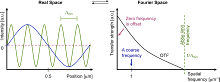
Abbe limit

Abbe limit

Interestingly, the detection OTF of a microscope has a fixed frequency
border (Abbe limit frequency, right panel). The maximum-to-maximum
distance Λmin of the corresponding sine curve is commonly referred to
as Abbe’s limit (left panel). In other words: The Abbe limit is the smallest
periodicity in a structure, which can be discriminated in its image. As a
point object contains all spatial frequencies, this Abbe limit sine curve
needs to also be present in the PSF. A standard wide-field microscope
creates an image of a point object (e.g., an emitting molecule) by capturing
the light from that molecule at various places of the objective lens, and
processing it with further lenses to then interfere at the image plane.
Conveniently due to the reciprocity principle in optics, the Abbe limit Λmin
along an in-plane direction in fluorescence imaging corresponds to the
maximum-to-maximum distance of the intensity structure one would get by
interfering two waves at extreme angles captured by the objective lens:
where λ/n is the wavelength of light in the medium of refractive index n.
The term NA = n sin(α) conveniently combines the half opening angle α
of the objective and the refractive index n of the embedding medium.

Abbe’s famous resolution limit is so attractive because it simply depends
on the maximal relative angle between different waves leaving the
object and being captured by the objective lens to be sent to the image.
It describes the smallest level of detail that can possibly be imaged with
this PSF “brush”. No periodic object detail smaller than this shortest
wavelength can possibly be transferred to the image.

Confocal laser scanning microscopy employs a redesigned optical
path and specialized hardware. A tightly focused spot of laser light is
used to scan the sample and a small aperture (or pinhole) in the
confocal image plane of the light path allows only light originating
from the nominal focus to pass (Cremer and Cremer, 1978Sheppard
and Wilson, 1981
Brakenhoff et al., 1985). The emitted light is
detected by a photomultiplier tube (PMT) or an avalanche photodiode
(APD) and the image is then constructed by mapping the detected
light in dependence of the position of the scanning spot. CLSM can
achieve a better resolution than wide-field fluorescence microscopy
but, to obtain a significant practical advantage, the pinhole needs to
be closed to an extent where most of the light is discarded
(Heintzmann et al., 2003).

Wide-field deconvolution and CLSM have long been the gold standards
in optical bioimaging, but we are now witnessing a revolution in light
microscopy that will fundamentally expand our perception of the cell.
Recently, several new technologies,collectively termed super-resolution
microscopy or nanoscopy, have been developed that break or bypass
the classical diffraction limit and shift the optical resolution down to
macromolecular or even molecular levels (Table I).

Super-resolution light microscopy methods

super resolution microscopy

super resolution microscopy

http://zeiss-campus.magnet.fsu.edu/articles/superresolution/introduction.html

Conceptually, one can discern near-field from far-field methods and
whether the subdiffraction resolution is based on a linear or nonlinear
response of the sample to its locally illuminating (exciting or depleting) irradiance. The required nonlinearity is currently achieved by using reversible saturable optical fluorescence transitions (RESOLFT) between molecular states (Hofmann et al., 2005Hell, 2007).

Besides these saturable optical fluorescence transitions also other
approaches, e.g., Rabi oscillations, could be used to generate the
required nonlinear response.

Note that each of the novel imaging modes has its individual signal-
to-noise consideration depending on various factors.  A full
discussion of this issue is beyond the scope of this review, but as a
general rule, single-point scanning systems, albeit fundamentally limited
in speed by fluorescence saturation effects, can have better signal-
to-noise performance for thicker samples.

With three-dimensional SIM (3D-SIM), an additional twofold increase
in the axial resolution can be achieved by generating an excitation
light modulation along the z-axis using three-beam interference
(Gustafsson et al., 2008Schermelleh et al.,2008) and processing a
z-stack of images accordingly. Thus, with 3D-SIM an approximately eightfold smaller volume can be resolved in comparison to conventional microscopy (Fig. 2). To computationally reconstruct a three-dimensional dataset of a typical mammalian cell of 8-µm height with a
z-spacing of 125 nm, roughly 1,000 raw images (512 × 512 pixels) are
recorded. Because no special photophysics is needed, virtually all modern fluorescent labels can be used provided they are sufficiently photostable
to accommodate the additional exposure cycles.

Resolvable volumes obtained with current commercial super-resolution microscopes.

A schematic 3D representation of focal volumes is shown for the indicated
emission maxima. The approximate lateral (x,y) and axial (z) resolution
and resolvable volumes are listed. Note that STED/CW-STED and 3D-SIM
can reach up to 20 µm into the sample, whereas PALM/STORM is usually
confined to the evanescent wave field near the sample bottom. It should be
noted that deconvolution approaches can further improve STED resolution.
For comparison the “focal volume” for PALM/STORM was estimated based
on the localization precision in combination with the z-range of TIRF.

Resolvable volumes obtained

Resolvable volumes obtained

Super-resolution microscopy of biological samples.

(A) Conventional wide-field image (left) and 3D-SIM image of a mouse
C2C12 prometaphase cell stained with primary antibodies against
lamin B and tubulin, and secondary antibodies conjugated to Alexa 488
(green) and Alexa 594 (red), respectively. Nuclear chromatin was stained
with DAPI (blue). 3D image stacks were acquired with a DeltaVision OMX
prototype system (Applied Precision). The bottom panel shows the
respective orthogonal cross sections. (B) HeLa cell stained with primary
antibodies against the nuclear pore complex protein Nup153 and
secondary antibodies conjugated with ATTO647N. The image was
acquired with a TCS STED confocal microscope (Leica). (C) TdEosFP-
paxillin expressed in a Hep G2 cell to label adhesion complexes at
the lower surface. The image was acquired on an ELYRA P.1
prototype system (Carl Zeiss, Inc.) using TIRF illumination. Single
molecule positional information was projected from 10,000 frames
recorded at 30 frames per second. On the left, signals were summed
up to generate a TIRF image with conventional wide-field lateral
resolution. Bars: 5 µm (insets, 0.5 µm).

biological images

biological images

APPLICATIONS IN BIOLOGICAL SYSTEMS

The cytoskeleton of mammalian cells, especially microtubules
(Figure 5a) (29444652), is the most commonly used benchmark
structure for super-resolution imaging. Other cytoskeletal structures
imaged so far include actin filaments in the lamellipodium (6),
keratin intermediate filaments (59), neurofilaments (2683) and
MreB in Caulobacter (66).

Figure 5

cytoskeleton. f5.

cytoskeleton. f5.

Examples of super-resolution images of biological samples.
(a) Two-color STORM imaging of immunostained microtubule (green)
and clathrin-coated pits (red) (From Reference 46. Reprinted with
permission from AAAS).

Organelles, such as the endoplasmic reticulum (27), lysosome (6),
endocytic and exocytic vesicles (465264), and mitochondria
(65356), have also been imaged. For example, using the single-molecule localization approach, 3D STORM imaging has clearly
resolved the ~150-nm diameter, hemispherical cage shape of clathrin-coated pits (4652), which only appear as diffraction-limited spots
without any feature in conventional fluorescence microscopy (Figure 5a,b).
Two-color 3D STED has resolved the hollow shape of the mitochondrial
outer membrane (marked by the translocase protein Tom20), enclosing
a matrix protein Hsp60 (56), even though the diameter of mitochondria is
only about 300–500 nm (Figure 5c). The outer membrane structure of
mitochondria and their interactions with microtubules have been resolved
by two-color 3D STORM (53). The transport of synaptic vesicles
has been recorded at video rate using 2D STED (Figure 5d ) (64).

Many plasma membrane proteins or membrane associated protein
complexes have also been studied by super-resolution fluorescence
microscopy. For example, synaptotagmin clusters after exocytosis in
primary cultured hippocampal neurons (84), the donut-shaped
clusters of Drosophila protein Bruchpilot at the neuromuscular
synaptic active zone (85), and the size distribution of syntaxin clusters
have all been imaged (8687). Photoactivation has enabled the tracking
of the influenza protein hemagglutinin and the retroviral protein Gag in
live cells, revealing the membrane microdomains (67) and the spatial
heterogeneity of membrane diffusion (68). The morphology and transport
of the focal adhension complex has also been observed using live-cell
PALM (Figure 5e) (65).

Summary points

  1. Super resolution fluorescence microscopy with a spatial resolution not limited by the diffraction of
    light has been implemented using saturated depletion/excitation or single-molecule localization
    of switchable fluorophores.
  2. Three-dimensional imaging with an optical resolution as high as ~20 nm in the lateral direction
    and 40–50 nm in axial dimension has been achieved.
  3. The resolution of these super-resolution fluorescence microscopy techniques can in principle
    reach molecular scale.
  4. In practice, the resolution of the images are not only limited by the intrinsic optical resolution,
    but also by sample specific factors including the labeling density, probe size and sample preservation.
  5. Multicolor super resolution imaging has been implemented, allowing colocalization measurements
    to be performed at nanometer scale resolution and molecular interaction to be more précisely
    identified in cells.
  6. Super-resolution fluorescence imaging allows dynamic processes to be investigated at the tens of
    nanometer resolution in living cells.
  7. Many cellular structures have been imaged at sub-diffraction-limit resolution.

Future issues

  1. Achieving molecular scale resolution (a few nanometers or less).
  2. Fast super resolution imaging of a large view field by multi-point scanning or high-speed single-molecule switching/localization.
  3. Developing new fluorescent probes that are brighter, more photostable and switchable fluorophores
    that have high on-off contrast and fast switching rate.
  4. Developing fluorescent labeling methods that can stain the target with small molecules at high specificity,
    high density and good ultrastructure preservation.
  5. Application of super resolution microscopy to provide novel biological insights

Acronyms

FP

Fluorescent Protein

FPALM

Fluorescence PhotoActivation Localization Microscopy

I5M

Combination of I2M (Illumination Interference Microscopy) and I3M
(Incoherent Imaging Interference Microscopy)

PALM

PhotoActivated Localization Microscopy

PSF

Point Spread Function

RESOLFT

REversible Saturable Optically Linear Fluorescence Transition

SIM

Structured Illumination Microscopy

SSIM

Saturated Structured Illumination Microscopy

STED

STimulated Emission Depletion

STORM

STochastic Optical Reconstruction Microscopy

glossary

Numerical aperture (NA)

The numerical aperture of an objective characterizes the solid angle
of light collected from a point light source at the focus of the objective.

Stimulated emission

The process that an excited state molecule or atom jumps to the
ground state by emitting another photon that is identical to the incoming
photon. It is the basis of laser.

Fluorescence saturation

At high excitation intensity, the fluorescence lifetime instead of the excitation
rate becomes the rate limiting step of fluorescence emission, causing the
fluorescence signal not to increase proportionally with the excitation intensity.

Nyquist criterion

To determine a structure, the sampling interval needs to be no larger than
half of the feature size.

Mitochondria

Organelles in eukaryotic cells for APT generation, consisting of two
membrane (inner and outer) enclosing the inter membrane space and
the matrix inside the inner membrane.

Clathrin-coated pit

Vesicle forming machinery involved in endocytosis and intracellular
vesicle transport, consisting of clathrin coats, adapter proteins, and
other regulatory proteins.

Focal adhesion

The macromolecular complex serving as the mechanical connection
and signaling hub between a cell and the extracellular matrix or other cells.

Selected references with abstract

Near-Field Optics: Microscopy, Spectroscopy, and Surface
Modification Beyond the Diffraction Limit
Eric Betzig,  Jay K. Trautman
AT&T Bell Laboratories, Murray Hill, NJ 07974
Science 10 Jul 1992; 257(5067) pp. 189-195
http://dx.doi.org:/0.1126/science.257.5067.189

 The near-field optical interaction between a sharp probe and a sample
of interest can be exploited to image, spectroscopically probe, or modify
surfaces at a resolution (down to ∼12 nm) inaccessible by traditional far-field
techniques. Many of the attractive features of conventional optics are
retained, including noninvasiveness, reliability, and low cost. In addition, most
optical contrast mechanisms can be extended to the near-field regime,
resulting in a technique of considerable versatility. This versatility
is demonstrated by several examples, such as the imaging of nanometric-scale features in mammalian tissue sections and the creation of ultrasmall,
magneto-optic domains having implications for high density data storage.
Although the technique may find uses in many diverse fields, two of the
most exciting possibilities are localized optical spectroscopy of semiconductors
and the fluorescence imaging of living cells.

Imaging Intracellular Fluorescent Proteins at Nanometer Resolution

 E Betzig1,2,*,†, GH. Patterson3, R Sougrat3, O.W Lindwasser3,
S Olenych4, JS. Bonifacino3, MW. Davidson4, JL Schwartz3, HF. Hess5,*  1 Howard Hughes Medical Institute, Janelia Farm Research Campus,
Ashburn, VA   2 New Millennium Research, LLC, Okemos, MI.   3 Cell Biology and Metabolism Branch, National Institute of Child Health
and Human Development (NICHD), Bethesda, MD.  4 National High
Magnetic Field Laboratory, Florida State University, Tallahassee, FL.
5 NuQuest Research, LLC, La Jolla, CA.
Science 15 Sep 2006; 313(5793): pp. 1642-1645
http://dx.doi.org:/10.1126/science.1127344

We introduce a method for optically imaging intracellular proteins at
nanometer spatial resolution. Numerous sparse subsets of photo-activatable fluorescent protein molecules were activated, localized
(to ∼2 to 25 nanometers), and then bleached. The
aggregate position information from all subsets was then assembled
into a super-resolution image. We used this method—termed photo-
activated localization microscopy to image specific target proteins
in thin sections of lysosomes and mitochondria; in fixed whole cells,
we imaged vinculin at focal adhesions, actin within a lamellipodium,
and the distribution of the retroviral protein Gag at the plasma
membrane.

Toward fluorescence nanoscopy.

Hell SW.   Author information 
Nat Biotechnol. 2003 Nov; 21(11):1347-55.
http://www.ncbi.nlm.nih.gov/pubmed/14595362

For more than a century, the resolution of focusing light microscopy
has been limited by diffraction to 180 nm in the focal plane and to
500 nm along the optic axis. Recently, microscopes have been
reported that provide three- to seven-fold improved axial
resolution in live cells. Moreover, a family of concepts has emerged
that overcomes the diffraction barrier altogether. Its first exponent,
stimulated emission depletion microscopy, has so far displayed a
resolution down to 28 nm. Relying on saturated optical transitions,
these concepts are limited only by the attainable saturation level.
As strong saturation should be feasible at low light intensities,
nanoscale imaging with focused light may be closer than ever.
PMID: 14595362

Far-field optical nanoscopy.

Hell SW.  Author information 
Science. 2007 May 25;316(5828):1153-8.
http://www.ncbi.nlm.nih.gov/pubmed/17525330

In 1873, Ernst Abbe discovered what was to become a well-known
paradigm: the inability of a lens-based optical microscope to
discern details that are closer together than half of the wavelength
for its most popular imaging mode, fluorescence microscopy, the
diffraction barrier is crumbling. Here, I discuss the physical concepts
that have pushed fluorescence microscopy to the nanoscale, once
the prerogative of electron and scanning probe microscopes. Initial
applications indicate that emergent far-field optical nanoscopy will
have a strong impact in the life sciences and in other areas benefiting
from nanoscale visualization.
PMID:  17525330

Imaging intracellular fluorescent proteins at nanometer resolution.

Betzig E1, Patterson GHSougrat RLindwasser OWOlenych S,
Bonifacino JSDavidson MWLippincott-Schwartz JHess HF.
Author information
Science. 2006 Sep 15;313(5793):1642-5. Epub 2006 Aug 10
http://www.ncbi.nlm.nih.gov/pubmed/16902090

We introduce a method for optically imaging intracellular proteins at
nanometer spatial resolution. Numerous sparse subsets of photo-ctivatable fluorescent protein molecules were activated, localized
(to approximately 2 to 25 nanometers), and then bleached. The
aggregate position information from all subsets was then assembled
into a super-resolution image. We used this method–termed photo-activated localization microscopy–to image specific target proteins in
thin sections of lysosomes and mitochondria; in fixed whole cells,
we imaged vinculin at focal adhesions, actin within a lamellipodium,
and the distribution of the retroviral protein Gag at the plasma
membrane.

Comment in

PMID:  16902090  [PubMed – indexed for MEDLINE]

Illuminating single molecules in condensed matter.

Moerner WE1, Orrit M.  Author information 
Science. 1999 Mar 12;283(5408):1670-6.
http://www.ncbi.nlm.nih.gov/pubmed/10073924

Efficient collection and detection of fluorescence coupled with careful
minimization of background from impurities and Raman scattering
now enable routine optical microscopy and study of single molecules
in complex condensed matter environments. This ultimate method
for unraveling ensemble averages leads to the observation of
new effects and to direct measurements of stochastic fluctuations.
Experiments at cryogenic temperatures open new directions in
molecular spectroscopy, quantum optics, and solid-state dynamics.
Room-emperature investigations apply several techniques
(polarization microscopy, single-molecule imaging, emission time
dependence, energy transfer, lifetime studies, and the like) to a
growing array of biophysical problems where new insight may be
gained from direct observations of hidden static and dynamic
inhomogeneity.  PMID: 10073924

Fluorescence microscopy with super-resolved optical sections.

Egner A1, Hell SW.  Author information 
Trends Cell Biol. 2005 Apr;15(4):207-15.
http://www.ncbi.nlm.nih.gov/pubmed/15817377

The fluorescence microscope, especially its confocal variant, has
become a standard tool in cell biology research for delivering
3D-images of intact cells. However, the resolution of any standard
optical microscope is atleast 3 times poorer along the axis of the
lens that in its focal plane. Here, we review principles and applications
of an emerging family of fluorescence microscopes, such as 4Pi
microscopes, which improve axial resolution by a factor of seven by
employing two opposing lenses. Noninvasive axial sections of 80-160 nm
thickness deliver more faithful 3D-images of subcellular features,
providing a new opportunity to significantly enhance our understanding
of cellular structure and function. PMID: 15817377

4Pi-confocal microscopy provides three-dimensional images of the
microtubule network with 100- to 150-nm resolution.

Nagorni M1, Hell SW.  Author information 
J Struct Biol. 1998 Nov;123(3):236-47.

We show the applicability of 4Pi-confocal microscopy to three-dimensional imaging of the microtubule network in a fixed mouse
fibroblast cell.Comparison with two-photon confocal resolution
reveals a fourfold better axial resolution in the 4Pi-confocal case.
By combining 4Pi-confocal microscopy with Richardson-Lucy
image restoration a further resolution increase is achieved.
Featuring a three-dimensional resolution in the range 100-150 nm,
the 4Pi-confocal (restored) images are intrinsically more detailed
than their confocal counterparts. Our images constitute what
to our knowledge are the best-resolved three-dimensional
images of entangled cellular microtubules obtained with light
to date.  PMID: 9878578

Part IV. Super-resolution microscopy

Super-resolution microscopy is a form of light microscopy. Due
to the diffraction of light, the resolution of conventional light
microscopy is limited as stated by Ernst Abbe in 1873.[1]
A good approximation of the resolution attainable is the full
width at half maximum 
 (FWHM) of the point spread function,
and a precise wide-field microscope with high numerical
aperture
 and visible light usually reaches a resolution of ~250 nm.

Super-resolution techniques allow the capture of images with
a higher resolution than the diffraction limit. They fall into
two broad categories,
“true” super-resolution techniques, which capture information
contained in evanescent waves, and “functional” super-
resolution techniques, which use clever experimental
techniques and known limitations on the matter being
imaged to reconstruct a super-resolution image.[2]

True subwavelength imaging techniques include those that
utilize the Pendry Superlens and near field scanning optical
microscopy
, the 4Pi Microscope and structured illumination
microscopy technologies like SIM and SMI. However, the
majority of techniques of importance in biological imaging
fall into the functional category.

Groups of methods for functional super-resolution microscopy:

  1. Deterministic super-resolution: The most commonly used emitters in biological
    microscopy, fluorophores, show a nonlinear response to excitation, and this
    nonlinear response can be exploited to enhance resolution. These
    methods include STEDGSDRESOLFTand SSIM.
  2. Stochastic super-resolution: The chemical complexity of many molecular
    light sources gives them a complex temporal behaviour, which can be used
    to make several close-by fluorophores emit light at separate times and
    thereby become resolvable in time.  These methods include SOFI and all
    single-molecule localization methods (SMLM) such as SPDM,
    SPDMphymodPALM, FPALM, STORM and dSTORM.

Part V. HIV-1

Conformational dynamics of single HIV-1 envelope
trimers on the surface of native virions

James B. Munro1,*,Jason Gorman2Xiaochu Ma1,
Zhou Zhou3James Arthos4,
Dennis R. Burton5,6, et al.
1Department of Microbial Pathogenesis, Yale University
School of Medicine, New Haven, CT. 2Vaccine Research
Center, National Institute of Allergy and Infectious
Diseases, National Institutes of Health, Bethesda, MD .
3Department of Physiology and Biophysics, Weill
Cornell Medical College of Cornell University, New York, NY .
4Laboratory of Immunoregulation, National Institute of Allergy
and Infectious Diseases, National Institutes of Health, Bethesda,
MD . 5Department of Immunology and Microbial Science, and
IAVI Neutralizing Antibody Center, The Scripps Research
Institute, La Jolla, CA . 6Ragon Institute of MGH, MIT, and
Harvard, Cambridge, MA. 7International AIDS Vaccine Initiative
(IAVI), New York, NY . 8Department of
Chemistry, University of Pennsylvania, Philadelphia, PA.

The HIV-1 envelope (Env) mediates viral entry into host cells.
To enable the direct imaging of conformational dynamics
within Env we introduced fluorophores into variable
regions of the gp120 subunit and measured single-molecule
fluorescence resonance energy transfer (smFRET) within
the context of native trimers on the surface of HIV-1 virions.
Our observations revealed unliganded HIV-1 Env to be
intrinsically dynamic, transitioning between three distinct
pre-fusion conformations, whose relative occupancies
were remodeled by receptor CD4 and antibody binding.
The distinct properties of neutralization-sensitive and
neutralization-resistant HIV-1 isolates support a dynamics-based mechanism of immune evasion and ligand recognition.

Read Full Post »

Imaging-guided cancer treatment

Imaging-guided cancer treatment

Writer & reporter: Dror Nir, PhD

It is estimated that the medical imaging market will exceed $30 billion in 2014 (FierceMedicalImaging). To put this amount in perspective; the global pharmaceutical market size for the same year is expected to be ~$1 trillion (IMS) while the global health care spending as a percentage of Gross Domestic Product (GDP) will average 10.5% globally in 2014 (Deloitte); it will reach ~$3 trillion in the USA.

Recent technology-advances, mainly miniaturization and improvement in electronic-processing components is driving increased introduction of innovative medical-imaging devices into critical nodes of major-diseases’ management pathways. Consequently, in contrast to it’s very small contribution to global health costs, medical imaging bears outstanding potential to reduce the future growth in spending on major segments in this market mainly: Drugs development and regulation (e.g. companion diagnostics and imaging surrogate markers); Disease management (e.g. non-invasive diagnosis, guided treatment and non-invasive follow-ups); and Monitoring aging-population (e.g. Imaging-based domestic sensors).

In; The Role of Medical Imaging in Personalized Medicine I discussed in length the role medical imaging assumes in drugs development.  Integrating imaging into drug development processes, specifically at the early stages of drug discovery, as well as for monitoring drug delivery and the response of targeted processes to the therapy is a growing trend. A nice (and short) review highlighting the processes, opportunities, and challenges of medical imaging in new drug development is: Medical imaging in new drug clinical development.

The following is dedicated to the role of imaging in guiding treatment.

Precise treatment is a major pillar of modern medicine. An important aspect to enable accurate administration of treatment is complementing the accurate identification of the organ location that needs to be treated with a system and methods that ensure application of treatment only, or mainly to, that location. Imaging is off-course, a major component in such composite systems. Amongst the available solution, functional-imaging modalities are gaining traction. Specifically, molecular imaging (e.g. PET, MRS) allows the visual representation, characterization, and quantification of biological processes at the cellular and subcellular levels within intact living organisms. In oncology, it can be used to depict the abnormal molecules as well as the aberrant interactions of altered molecules on which cancers depend. Being able to detect such fundamental finger-prints of cancer is key to improved matching between drugs-based treatment and disease. Moreover, imaging-based quantified monitoring of changes in tumor metabolism and its microenvironment could provide real-time non-invasive tool to predict the evolution and progression of primary tumors, as well as the development of tumor metastases.

A recent review-paper: Image-guided interventional therapy for cancer with radiotherapeutic nanoparticles nicely illustrates the role of imaging in treatment guidance through a comprehensive discussion of; Image-guided radiotherapeutic using intravenous nanoparticles for the delivery of localized radiation to solid cancer tumors.

 Graphical abstract

 Abstract

One of the major limitations of current cancer therapy is the inability to deliver tumoricidal agents throughout the entire tumor mass using traditional intravenous administration. Nanoparticles carrying beta-emitting therapeutic radionuclides [DN: radioactive isotops that emits electrons as part of the decay process a list of β-emitting radionuclides used in radiotherapeutic nanoparticle preparation is given in table1 of this paper.) that are delivered using advanced image-guidance have significant potential to improve solid tumor therapy. The use of image-guidance in combination with nanoparticle carriers can improve the delivery of localized radiation to tumors. Nanoparticles labeled with certain beta-emitting radionuclides are intrinsically theranostic agents that can provide information regarding distribution and regional dosimetry within the tumor and the body. Image-guided thermal therapy results in increased uptake of intravenous nanoparticles within tumors, improving therapy. In addition, nanoparticles are ideal carriers for direct intratumoral infusion of beta-emitting radionuclides by convection enhanced delivery, permitting the delivery of localized therapeutic radiation without the requirement of the radionuclide exiting from the nanoparticle. With this approach, very high doses of radiation can be delivered to solid tumors while sparing normal organs. Recent technological developments in image-guidance, convection enhanced delivery and newly developed nanoparticles carrying beta-emitting radionuclides will be reviewed. Examples will be shown describing how this new approach has promise for the treatment of brain, head and neck, and other types of solid tumors.

The challenges this review discusses

  • intravenously administered drugs are inhibited in their intratumoral penetration by high interstitial pressures which prevent diffusion of drugs from the blood circulation into the tumor tissue [1–5].
  • relatively rapid clearance of intravenously administered drugs from the blood circulation by kidneys and liver.
  • drugs that do reach the solid tumor by diffusion are inhomogeneously distributed at the micro-scale – This cannot be overcome by simply administering larger systemic doses as toxicity to normal organs is generally the dose limiting factor.
  • even nanoparticulate drugs have poor penetration from the vascular compartment into the tumor and the nanoparticles that do penetrate are most often heterogeneously distributed

How imaging could mitigate the above mentioned challenges

  • The inclusion of an imaging probe during drug development can aid in determining the clearance kinetics and tissue distribution of the drug non-invasively. Such probe can also be used to determine the likelihood of the drug reaching the tumor and to what extent.

Note: Drugs that have increased accumulation within the targeted site are likely to be more effective as compared with others. In that respect, Nanoparticle-based drugs have an additional advantage over free drugs with their potential to be multifunctional carriers capable of carrying both therapeutic and diagnostic imaging probes (theranostic) in the same nanocarrier. These multifunctional nanoparticles can serve as theranostic agents and facilitate personalized treatment planning.

  • Imaging can also be used for localization of the tumor to improve the placement of a catheter or external device within tumors to cause cell death through thermal ablation or oxidative stress secondary to reactive oxygen species.

See the example of Vintfolide in The Role of Medical Imaging in Personalized Medicine

vinta

Note: Image guided thermal ablation methods include radiofrequency (RF) ablation, microwave ablation or high intensity focused ultrasound (HIFU). Photodynamic therapy methods using external light devices to activate photosensitizing agents can also be used to treat superficial tumors or deeper tumors when used with endoscopic catheters.

  • Quality control during and post treatment

For example: The use of high intensity focused ultrasound (HIFU) combined with nanoparticle therapeutics: HIFU is applied to improve drug delivery and to trigger drug release from nanoparticles. Gas-bubbles are playing the role of the drug’s nano-carrier. These are used both to increase the drug transport into the cell and as ultrasound-imaging contrast material. The ultrasound is also used for processes of drug-release and ablation.

 HIFU

Additional example; Multifunctional nanoparticles for tracking CED (convection enhanced delivery)  distribution within tumors: Nanoparticle that could serve as a carrier not only for the therapeutic radionuclides but simultaneously also for a therapeutic drug and 4 different types of imaging contrast agents including an MRI contrast agent, PET and SPECT nuclear diagnostic imaging agents and optical contrast agents as shown below. The ability to perform multiple types of imaging on the same nanoparticles will allow studies investigating the distribution and retention of nanoparticles initially in vivo using non-invasive imaging and later at the histological level using optical imaging.

 multi

Conclusions

Image-guided radiotherapeutic nanoparticles have significant potential for solid tumor cancer therapy. The current success of this therapy in animals is most likely due to the improved accumulation, retention and dispersion of nanoparticles within solid tumor following image-guided therapies as well as the micro-field of the β-particle which reduces the requirement of perfectly homogeneous tumor coverage. It is also possible that the intratumoral distribution of nanoparticles may benefit from their uptake by intratumoral macrophages although more research is required to determine the importance of this aspect of intratumoral radionuclide nanoparticle therapy. This new approach to cancer therapy is a fertile ground for many new technological developments as well as for new understandings in the basic biology of cancer therapy. The clinical success of this approach will depend on progress in many areas of interdisciplinary research including imaging technology, nanoparticle technology, computer and robot assisted image-guided application of therapies, radiation physics and oncology. Close collaboration of a wide variety of scientists and physicians including chemists, nanotechnologists, drug delivery experts, radiation physicists, robotics and software experts, toxicologists, surgeons, imaging physicians, and oncologists will best facilitate the implementation of this novel approach to the treatment of cancer in the clinical environment. Image-guided nanoparticle therapies including those with β-emission radionuclide nanoparticles have excellent promise to significantly impact clinical cancer therapy and advance the field of drug delivery.

Read Full Post »

Fractional Flow Reserve–Guided PCI vs Drug Therapy for Stable Coronary Artery Disease

Reporter: Aviva Lev-Ari, PhD, RN

Fractional Flow Reserve–Guided PCI for Stable Coronary Artery Disease

Bernard De Bruyne, M.D., Ph.D., William F. Fearon, M.D., Nico H.J. Pijls, M.D., Ph.D., Emanuele Barbato, M.D., Ph.D., Pim Tonino, M.D., Ph.D., Zsolt Piroth, M.D., Nikola Jagic, M.D., Sven Mobius-Winckler, M.D., Gilles Riouffol, M.D., Ph.D., Nils Witt, M.D., Ph.D., Petr Kala, M.D., Philip MacCarthy, M.D., Thomas Engström, M.D., Keith Oldroyd, M.D., Kreton Mavromatis, M.D., Ganesh Manoharan, M.D., Peter Verlee, M.D., Ole Frobert, M.D., Nick Curzen, B.M., Ph.D., Jane B. Johnson, R.N., B.S.N., Andreas Limacher, Ph.D., Eveline Nüesch, Ph.D., and Peter Jüni, M.D. for the FAME 2 Trial Investigators

September 1, 2014DOI: 10.1056/NEJMoa1408758

BACKGROUND

We hypothesized that in patients with stable coronary artery disease and stenosis, percutaneous coronary intervention (PCI) performed on the basis of the fractional flow reserve (FFR) would be superior to medical therapy.

METHODS

In 1220 patients with stable coronary artery disease, we assessed the FFR in all stenoses that were visible on angiography. Patients who had at least one stenosis with an FFR of 0.80 or less were randomly assigned to undergo FFR-guided PCI plus medical therapy or to receive medical therapy alone. Patients in whom all stenoses had an FFR of more than 0.80 received medical therapy alone and were included in a registry. The primary end point was a composite of death from any cause, nonfatal myocardial infarction, or urgent revascularization within 2 years.

RESULTS

The rate of the primary end point was significantly lower in the PCI group than in the medical-therapy group (8.1% vs. 19.5%; hazard ratio, 0.39; 95% confidence interval [CI], 0.26 to 0.57; P<0.001). This reduction was driven by a lower rate of urgent revascularization in the PCI group (4.0% vs. 16.3%; hazard ratio, 0.23; 95% CI, 0.14 to 0.38; P<0.001), with no significant between-group differences in the rates of death and myocardial infarction. Urgent revascularizations that were triggered by myocardial infarction or ischemic changes on electrocardiography were less frequent in the PCI group (3.4% vs. 7.0%, P=0.01). In a landmark analysis, the rate of death or myocardial infection from 8 days to 2 years was lower in the PCI group than in the medical-therapy group (4.6% vs. 8.0%, P=0.04). Among registry patients, the rate of the primary end point was 9.0% at 2 years.

CONCLUSIONS

In patients with stable coronary artery disease, FFR-guided PCI, as compared with medical therapy alone, improved the outcome. Patients without ischemia had a favorable outcome with medical therapy alone. (Funded by St. Jude Medical; FAME 2 ClinicalTrials.gov number, NCT01132495.)

SOURCE

http://www.nejm.org/doi/full/10.1056/NEJMoa1408758?query=TOC


Read Full Post »

Titanium-nitric-oxide-coated bioactive stents in acute coronary syndrome

Reporter: Larry Bernstein, MD, FCAP

SOURCE: http://www.researchgate.net/publication/256607204_Stent-oriented_versus_patient-oriented_outcome_in_patients_undergoing_early_percutaneous_coronary_intervention_for_acute_coronary_syndrome_2-year_report_from_the_BASE-ACS_trial

REBLOG

Titanium-nitric-oxide-coated bioactive stents in acute coronary syndrome: towards a more clear landscape!

 Assisstant Professor of Cardiology, Faculty of Medicine, Ain Shams University, Egypt

With full interest, we read the Editorial “Nitric-oxide Coated Bioactive Titanium Stents: Safer and More Effective Than Second-generation Drug-eluting Stents?” by Sabaté et al 1. I’d like to commend the authors for publishing this interesting analysis of recently published studies – including the BASE ACS randomized controlled trial– comparing titanium-nitric-oxide-coated bioactive stents (NO-BAS) with drug-eluting stents. Yet, since I’m one of the authors of the BASE ACS trial, I’d like to clarify some points. First, the BASE ACS trial was adequately powered to detect a difference of the primary composite endpoint at 12 months (827 patients), with a formal statistical power calculation 2. Second, as the authors reported, the NO-BAS were non-inferior to cobalt-chromium-based everolimus-eluting stents (EES) for the primary composite endpoint of major adverse cardiac events (MACE) that included cardiac death, non-fatal myocardial infarction (MI), and ischemia-driven target lesion revascularization, in patients presenting with the full spectrum of acute coronary syndrome at 12-month follow-up 2. The 12-month rates of the individual secondary endpoints of non-fatal MI and definite stent thrombosis (ST) were lower in patients who received NO-BAS versus those who received EES (2.2% versus 5.9%, and 0.7% versus 2.2%, p= 0.007 and 0.07, respectively) 3. And whereas the definition of MI adopted by the BASE ACS trial (based on CK MB or troponin ≥2 times the upper reference limit) was different from that employed in the EXAMINATION trial (extended definition of the World Health Organization), it cannot be held responsible for the difference in MI rates between NO-BAS and EES in the same BASE ACS trial, since the definition was equally applied to the two trial arms 2,3. Third, the early (within 30 days) divergence of safety endpoints between the 2 stent arms is hard to explain merely in view of peri-procedural bivalirudin monotherapy. In fact, out of 9 cases (2.2%) of definite ST in the EES arm, 3 were acute (within 24 hr); out of these 3, only 2 received peri-procedural bivalirudin as a sole anticoagulant 2. Moreover, peri-procedural bivalirudin use was comparable between the 2 stent arms: 14.1% versus 15.1% in NO-BAS versus EES arms, respectively. Furthermore, angiographic success was achieved in 99.8% in both stent arms; hence, technical issues (distal dissection, stent underexpansion) were probably similar in the 2 stent arms 2. More importantly, the rates of non-fatal MI and definite ST continued to diverge after one year: at 2-year follow-up, they were 2.9% versus 7.1%, and 1.0% versus 2.7%, p= 0.005 and 0.05, for NO-BAS versus EES, respectively 4. Since technical issues generally operate early (within 1 month) after stent implantation; therefore, they cannot fully account for the ‘very late’ events. References

1. Sabaté M, Brugaletta S. Nitric-oxide Coated Bioactive Titanium Stents: Safer and More Effective Than Second-generation Drug-eluting Stents? Rev Esp Cardiol. 2014;67:511-3. 2. Karjalainen PP, Niemela M, Airaksinen KEJ, et al. A prospective randomized comparison of titanium-nitride-oxide-coated bioactive stents with everolimus-eluting stents in acute coronary syndrome: the BASE-ACS trial. EuroIntervention. 2012;8:1769–74. 3. Sabate M, Cequier A, In˜iguez A, et al. Everolimus-eluting stent versus bare-metal stent in ST-segment elevation myocardial infarction (EXAMINATION): 1 year results of a randomised controlled trial. Lancet. 2012;380:1482–90. 4. Romppanen H, Nammas W, Kervinen K, et al. Stent-oriented versus patient-oriented outcome in patients undergoing early percutaneous coronary intervention for acute coronary syndrome: 2-year report from the BASE-ACS trial. Ann Med. 2013;45:488-93.

additional:

 Pooled Analysis of Two Randomized Trials Comparing Titanium-nitride-oxide-coated Stent Versus Drug-eluting Stent in STEMI. Petri O. Tuomainenab, Jussi Siac, Wail Nammasb, Matti Niemeläd, Juhani K.E. Airaksinene, Fausto Biancarif, Pasi P. Karjalainen. Rev Esp Cardiol. 2014;67(7):531-7  http://dx.doi.org:/10.1016/j.rec.2014.01.024

KeywordsBioactive stents. Everolimus-eluting stents. Paclitaxel-eluting stents. ST-segment elevation myocardial infarction. Outcome.

Pooled Analysis of Two Randomized Trials Comparing Titanium-nitride-oxide-coated Stent Versus Drug-eluting Stent in STEMI

Petri O. Tuomainenab, Jussi Siac, Wail Nammasb, Matti Niemeläd, Juhani K.E. Airaksinene, Fausto Biancarif, Pasi P. Karjalainenb, a Department of Internal Medicine and Heart Center, Kuopio University Hospital and University of Eastern Finland, Kuopio, Finland b Department of Cardiology, Satakunta Central Hospital, Pori, Finland c Department of Cardiology, Kokkola Central Hospital, Kokkola, Finland d Department of Internal Medicine, Division of Cardiology, University of Oulu, Oulu, Finland e Department of Medicine, Turku University Hospital, Turku, Finland f Division ofCardiothoracic and Vascular Surgery, Department of Surgery, Oulu, Finland

Refers to

Keywords

Bioactive stents. Everolimus-eluting stents. Paclitaxel-eluting stents. ST-segment elevation myocardial infarction. Outcome.

Abstract

Introduction and objectivesWe performed a pooled analysis based on patient-level data from the TITAX-AMI and BASE-ACS trials to evaluate the outcome of titanium-nitride-oxide-coated bioactive stents vs drug-eluting stents in patients with ST-segment elevation myocardial infarction at 2-year follow-up. MethodsThe TITAX-AMI trial compared bioactive stents with paclitaxel-eluting stents in 425 patients with acute myocardial infarction. The BASE-ACS trial compared bioactive stents with everolimus-eluting stents in 827 patients with acute coronary syndrome. The primary endpoint for the pooled analysis was major adverse cardiac events: a composite of cardiac death, recurrent myocardial infarction, or ischemia-driven target lesion revascularization at 2-year follow-up. ResultsThe pooled analysis included 501 patients; 245 received bioactive stents, and 256 received drug-eluting stents. The pooled bioactive stent group was associated with a risk ratio of 0.85 for major adverse cardiac events (95% confidence interval, 0.53-1.35; P = .49) compared to the pooled drug-eluting stent group. Similarly, the pooled bioactive stent group was associated with a risk ratio of 0.71 for cardiac death (95% confidence interval, 0.26-1.95; P = .51), 0.44 for recurrent myocardial infarction (95% confidence interval, 0.20-0.97; P = .04), and 1.39 for ischemia-driven target lesion revascularization (95% confidence interval, 0.74-2.59; P = .30), compared to the pooled drug-eluting stent group. These results were confirmed by propensity-score adjusted analysis of the combined datasets. ConclusionsIn patients with ST-segment elevation myocardial infarction, bioactive stents were associated with lower rates of recurrent myocardial infarction compared to drug-eluting stents at 2-year follow-up; yet, the rates of cardiac death and ischemia-driven target lesion revascularization were similar.

Read Full Post »

One year Post-Intervention Mortality Rate: TAVR and AVR – Aortic Valve Procedures 6.7% in AVR, 11.0% in AVR with CABG, 20.7 in Transvascular (TV-TAVT) and 28.0% in Transapical (TA-TAVR) Patients

Reporter: Aviva Lev-Ari, PhD, RN


UPDATED on 4/8/2017

Routine Imaging After TAVR Suggested to Catch Thrombosis – German data supports ‘significant’ prevalence warranting routine check by Nicole Lou 

Reporter, MedPage Today/CRTonline.org, April 03, 2017

Clinical transcatheter heart valve thrombosis may be common enough to merit routine imaging after transcatheter aortic valve replacement (TAVR), a German study suggested.

A retrospective analysis of a single-center registry found a 2.8% incidence of clinical valve thrombosis, according to Mohamed Abdel-Wahab, MD, of Germany’s Segeberger Kliniken, and colleagues. No one on oral anticoagulation got bioprosthetic valve thrombosis, however, and no patients died from it.

Thrombosis was more likely with balloon-expandable valves (OR 3.45, 95% CI 1.22-9.81) and with valve-in-valve procedures (OR 5.93, 95% CI 2.01-17.51), the authors reported in the April 10 issue of JACC: Cardiovascular Interventions.

http://www.medpagetoday.com/cardiology/chf/64297


One year Post-Intervention Mortality Rate: TAVR and AVR – Aortic Valve Procedures 6.7% in AVR, 11.0% in AVR with CABG, 20.7 in Transvascular (TV-TAVR) and 28.0% in Transapical (TA-TAVR) Patients

RESULTS

The 1-year mortality rate was

  • 6.7% for conventional AVR patients (n = 6523) and
  • 11.0% for patients who underwent AVR with coronary artery bypass grafting (n = 3464).
  • The 1-year mortality rate was 20.7 and 28.0% in TV- and TA-TAVR patients, respectively (n = 2695 and 1181).

However, if patients were stratified into four risk groups by means of the EuroSCORE and the German AV Score, the

  • highest risk cohorts showed the same mortality at 1 year with either therapy.
  • More than 80% of patients in all groups were in the same or better state of health at 1 year post-intervention and were satisfied with the procedural outcome.


European Journal of Cardio-Thoracic Surgery

Eur J Cardiothorac Surg (2014)doi: 10.1093/ejcts/ezu290

http://ejcts.oxfordjournals.org/content/early/2014/07/24/ejcts.ezu290.full


The German Aortic Valve Registry: 1-year results from 13 680 patients with aortic valve disease


  1. Friedrich W. Mohra,*,
  2. David Holzheya,
  3. Helge Möllmannb,
  4. Andreas Beckmannc,
  5. Christof Veitd,
  6. Hans Reiner Figullae,
  7. Jochen Cremerf,
  8. Karl-Heinz Kuckg,
  9. Rüdiger Langeh,
  10. Ralf Zahni,
  11. Stefan Sackj,
  12. Gerhard Schulera,
  13. Thomas Waltherk,
  14. Friedhelm Beyersdorfl,
  15. Michael Böhmm,
  16. Gerd Heuschn,
  17. Anne-Kathrin Funkata,
  18. Thomas Meinertzo,
  19. Till Neumannp,
  20. Konstantinos Papoutsisq,
  21. Steffen Schneiderr,
  22. Armin Welzs and
  23. Christian W. Hammb
  24. for the GARY Executive Board

+Author Affiliations


  1. aHeart Center Leipzig, Leipzig, Germany

  2. bDepartment of Cardiology, Kerckhoff Heart Center, Bad Nauheim, Germany

  3. cDeutsche Gesellschaft für Thorax-, Herz- und Gefäßchirurgie, Langenbeck-Virchow-Haus, Berlin, Germany

  4. dBQS Institute for Quality and Patient Safety, Düsseldorf, Germany

  5. eDivision of Cardiology, 1st Department of Medicine, University Hospital of Jena, Jena, Germany

  6. fDepartment of Cardiovascular Surgery, University of Schleswig-Holstein, Kiel, Germany

  7. gAsklepios Klinik St. Georg, Hamburg, Germany

  8. hGerman Heart Center Munich, Department of Cardiovascular Surgery, Technische Universität München, Munich, Germany

  9. iDepartment of Cardiology, Medizinische Klinik B, Herzzentrum am Klinikum Ludwigshafen, Ludwigshafen, Germany

  10. jKlinik für Kardiologie, Pneumologie und Internistische Intensivmedizin, Klinikum Schwabing, Munich, Germany

  11. kKerckhoff-Herzzentrum, Abteilung für Herzchirurgie, Bad Nauheim, Germany

  12. lUniversitätsklinikum Freiburg, Chirurgische Klinik Abteilung Herz- und Gefäßchirurgie, Freiburg, Germany

  13. mMedizinische Klinik und Poliklinik, Universitätsklinik des Saarlandes, Innere Medizin III, Homburg/Saar, Germany

  14. nInstitut für Pathophysiologie, Universitätsklinikum Essen, Essen, Germany

  15. oDeutsche Herzstiftung, Frankfurt am Main, Germany

  16. pDepartment of Cardiology, University of Duisburg-Essen Medical School, Essen, Germany

  17. qGerman Cardiac Society, Düsseldorf, Germany

  18. rInstitut für Herzinfarktforschung, Ludwigshafen, Germany

  19. sDepartment of Cardiac Surgery, University of Bonn, Bonn, Germany
  1. *Corresponding author. Heart Center Leipzig, Struempellstrasse 39, 04289 Leipzig, Germany. Tel: +49-341-8651421; fax: +49-341-8651452; e-mail:mohrf@medizin.uni-leipzig.defriedrich.mohr@herzzentrum-leipzig.de (F.W. Mohr).
  1.  An excerpt of the 1-year data was first presented at the ACC in San Francisco in March 2013.

  • Received April 16, 2014.
  • Revision received May 27, 2014.
  • Accepted June 17, 2014.


Abstract

OBJECTIVES The German Aortic Valve Registry (GARY) seeks to provide information on a real-world, all-comers basis for patients undergoing aortic valve interventions. This registry comprises patients undergoing the complete spectrum of transcutaneous and conventional surgical aortic valve interventions. The aim of this study was to use the GARY registry to evaluate conventional and catheter-based aortic valve interventions in several risk groups.

METHODS A total of 13 860 consecutive patients undergoing intervention for aortic valve disease [conventional aortic valve replacement (AVR) or transvascular/transapical TAVR (TV-/TA-TAVR)] were enrolled in 78 German centres in 2011. Baseline, procedural and outcome data, including quality of life, were acquired up to 1 year post-intervention. Vital status at 1 year was known for 98.1% of patients.

RESULTS The 1-year mortality rate was 6.7% for conventional AVR patients (n = 6523) and 11.0% for patients who underwent AVR with coronary artery bypass grafting (n = 3464). The 1-year mortality rate was 20.7 and 28.0% in TV- and TA-TAVR patients, respectively (n = 2695 and 1181). However, if patients were stratified into four risk groups by means of the EuroSCORE and the German AV Score, the highest risk cohorts showed the same mortality at 1 year with either therapy. More than 80% of patients in all groups were in the same or better state of health at 1 year post-intervention and were satisfied with the procedural outcome.

CONCLUSIONS Conventional AVR surgery yields excellent results after 1 year in lower-risk patients. Catheter-based AVR is a good alternative in high-risk and elderly patients.


Key words


SOURCE

http://ejcts.oxfordjournals.org/content/early/2014/07/24/ejcts.ezu290.abstract

FIFTY ARTICLES on TAVR and AVR were published in this Open Access Online Scientific Journal. All articles are included at the following URL:

 http://pharmaceuticalintelligence.com/category/cardiac-and-cardiovascular-surgical-procedures/aortic-valve-tavr-tavi-vs-open-heart-surgery/

Read Full Post »


Cardiovascular Research Foundation (CRF) Events – tctmd – The Source for Interventional Cardiovascular


Reporter: Aviva Lev-Ari, PhD, RN

SOURCE

http://www.tctmd.com/news.aspx

JOURNAL NEWS

VIEW ALL

Monday, July 28, 2014 | Source: EuroIntervention

Proximal May Be Preferred Form of Embolic Protection in Carotid Stenting

By Kim Dalton
A proximal protection device can be successfully and safely used as the first choice for embolic protection during most carotid artery stenting…

Friday, July 25, 2014 | Source: The American Heart Journal

Meta-analysis: Patient Sex Affects Response to Routine Invasive Approach for NSTE-ACS

By Todd Neale
A routine invasive strategy—relative to a more selective approach—appears beneficial over the long term for men but not women with non-ST-segment elevation…

Thursday, July 24, 2014 | Source: American Journal of Cardiology

PCI for Stable CAD Drives Overall Decline in Use of Procedure Since 2009

By Yael L. Maxwell
National use of percutaneous coronary intervention (PCI) has decreased steadily since 2009, mostly driven by a reduction in procedures for patients…

Thursday, July 24, 2014 | Source: European Heart Journal

Pre-AMI Ischemia May Reduce Early Mortality

By Kim Dalton
Patients who report angina symptoms or are diagnosed with ischemia shortly before an acute myocardial infarction (AMI) are less likely to die within…

Wednesday, July 23, 2014 | Source: EuroIntervention

Bioresorbable Scaffold Performs Well, But Thrombosis Raises Concerns

By Todd Neale
Percutaneous coronary intervention (PCI) with an everolimus-eluting bioresorbable vascular scaffold (BVS) results in an “acceptable” rate of target…

Tuesday, July 22, 2014 | Source: Journal of the American College of Cardiology

New CMR-Identified Myocardial Injury Post-TAVR Linked With Decreased LV Function

By Yael L. Maxwell
New ischemic myocardial injury, presumably of embolic origin, is common after transcatheter aortic valve replacement (TAVR) and is associated with…

Tuesday, July 22, 2014 | Source: JAMA Internal Medicine

Catheter-Directed Thrombolysis for DVT Increases Bleeding Compared With Standard Anticoagulation

By L.A. McKeown
While catheter-directed thrombolysis and anticoagulation for deep vein thrombosis (DVT) does not appear to increase mortality over standard anticoagulation…

Monday, July 21, 2014 | Updated With New Commentary | Source: Lancet

HEAT-PPCI Published: Discrepant Finding of Heparin’s Superiority over Bivalirudin in Primary PCI Still Puzzles

By Yael L. Maxwell
The HEAT-PPCI trial, published July 5, 2014, ahead of print in the Lancet , reports better efficacy and comparable safety with bivalirudin than…

Monday, July 21, 2014 | Source: Journal of the American College of Cardiology

One-Time Platelet Testing Appears Insufficient to Guide Clopidogrel Therapy

By Kim Dalton
In many patients with stable coronary artery disease (CAD), platelet reactivity varies markedly over time despite an unchanged dose of clopidogrel,…

Friday, July 18, 2014 | Source: American Heart Journal

Hybrid Revascularization a Promising Option for Diabetic Patients

By L.A. McKeown
A hybrid procedure combining percutaneous revascularization with minimally invasive coronary artery grafting results in similar short-term and…

CONFERENCE NEWS

VIEW ALL

Wednesday, May 28, 2014 | Source: EuroPCR 2014

EuroPCR 2014: Clinical Challenges, Novel Innovations Share the Limelight

By Caitlin E. Cox
PARIS, France—Research presented at EuroPCR 2014, held May 20 23, offered both new ways to improve on current therapies and fresh approaches to treating…

Friday, May 23, 2014 | Source: EuroPCR 2014

Studies Document Initial Steps Toward Percutaneous Mitral Valve Replacement

By Caitlin E. Cox
PARIS, France—Early data on 2 different catheter-based mitral valve therapies were presented in the same Hot Line session at EuroPCR on May 21, 2014.…

Friday, May 23, 2014 | Source: EuroPCR 2014

PERFUSE Registry: Diagnostic Accuracy Achieved with CT-Derived FFR Plus CT Perfusion Imaging

By Yael L. Maxwell
PARIS, France—A strategy combining fractional flow reserve (FFR) derived from computed tomography (CT) with CT perfusion imaging has demonstrated high…

Friday, May 23, 2014 | Source: EuroPCR 2014

Benefit of LAA Closure Becomes Most Evident After 1 Year

By Caitlin E. Cox
PARIS, France—Most of the stroke protection derived from percutaneous left atrial appendage (LAA) closure does not become apparent until 1 year after…

Friday, May 23, 2014 | Source: EuroPCR 2014

Nobori BES Demonstrates Good Long-term Outcomes with No Stent Thrombosis Beyond 3 Years

By Yael L. Maxwell
PARIS, France—A novel biolimus A9-eluting, biodegradable-polymer stent (BES) shows favorable long-term clinical outcomes and very low rates of device-related…

Friday, May 23, 2014 | Source: EuroPCR 2014

New Iteration of Sapien Device Associated with Low Rates of Early Mortality, Stroke

By L.A. McKeown
A new lower-profile valve and delivery system for transcatheter aortic valve replacement (TAVR) appears promising, with low mortality and stroke rates,…

Thursday, May 22, 2014 | Source: EuroPCR 2014

SYMPLICITY HTN-3: Predictors of Response Still Relevant After Trial’s Negative Findings

By Yael L. Maxwell
PARIS, France—Even though 6 month findings of the long awaited SYMPLICITY HTN 3 randomized trial demonstrated little effect of renal denervation on…

Thursday, May 22, 2014 | Source: EuroPCR 2014

UK Registry Finds Long-Term Survival After TAVR Depends on Patient Characteristics

By Caitlin E. Cox
PARIS, France—Nearly half of high-risk patients who undergo transcatheter aortic valve replacement (TAVR) for severe aortic stenosis live at least…

Thursday, May 22, 2014 | Source: EuroPCR 2014

Global SYMPLICITY Substudy Teases out Reasons for Non-response in Real-World Patients

By Yael L. Maxwell
PARIS, France—Renal denervation has spurred much controversy in recent months, with the sham-controlled SYMPLICITY HTN-3 trial demonstrating little…

Thursday, May 22, 2014 | Source: EuroPCR 2014

Early Data Show Novel Catheter-Implanted Device Benefits Patients with Left Heart Failure

By Caitlin E. Cox
PARIS, France—A first-in-man study presented Tuesday, May 20, at EuroPCR 2014 introduced a new percutaneous treatment for heart failure. Josep Rodés-Cabau,…


ACC NEWS

VIEW ALL

Monday, June 24, 2013 | Source: ACC News Releases

Study Shows Heart Failure Survivors at Greater Risk for Cancer Trend toward more cancers and more deaths among heart failure patients

By ACC News Releases
WASHINGTON (June 25, 2013) – Heart failure patients are surviving more often with the heart condition but they are increasingly more likely to be diagnosed…

Tuesday, June 04, 2013 | Source: ACC News Releases

New Program to Help Heart Patients Navigate Care, Reduce Readmissions AstraZeneca sponsorship to help support patient-centered programs in 35 hospitals

By ACC News Releases
WASHINGTON (June 5, 2013) – The American College of Cardiology is developing a program with support from founding sponsor AstraZeneca to provide personalized…

Tuesday, June 04, 2013 | Source: ACC News Releases

ACC/AHA Update Guideline for Management of Heart Failure Update increases emphasis on quality of life, care coordination, palliative care

By ACC News Releases
WASHINGTON (June 5, 2013) – The American College of Cardiology and the American Heart Association today released an expanded clinical practice guideline…

Sunday, March 10, 2013 | Source: ACC News Releases

Study Shows On-Pump Bypass Comparable to Off-Pump at Year Mark

By ACC News Releases
30-day neurocognitive differences disappeared by one-year follow up Read More

Sunday, March 10, 2013 | Source: ACC News Releases

Screenings, Targeted Care Reduce Heart Failure in At-Risk Patients

By ACC News Releases
Study shows simple blood test may help patients with risks for heart disease Read More

Sunday, March 10, 2013 | Source: ACC News Releases

Digoxin Reduces Hospital Admissions in Older Patients with Chronic Heart Failure

By ACC News Releases
If replicated in heart failure patients discharged from hospital, drug may help hospitals avoid readmission penalties Read…

Monday, March 26, 2012 | Source: Clinical Trials

Rule Out Myocardial Ischemia/Infarction Using Computer Assisted Tomography

By Clinical Trials
The goal of the trial was to evaluate a strategy of cardiac computed tomography (CT) angiography compared with standard emergency department (ED)…

Monday, March 26, 2012 | Source: ACC News Releases

STUDY SUGGESTS BETTER SURVIVAL IN PATIENTS UNDERGOING BYPASS SURGERY COMPARED TO CORONARY ANGIOPLASTY

By ACC News Releases
Patients with coronary heart disease and their doctors have long been challenged by the decision of whether to pursue bypass surgery or opt for the…

Monday, March 26, 2012 | Source: ACC News Releases

CARDIAC CT IS FASTER, MORE EFFECTIVE FOR EVALUATING PATIENTS WITH SUSPECTED HEART ATTACK

By ACC News Releases
Cardiac computed tomography angiography scans (CT scans that look at the heart) can provide a virtually instant verdict on whether chest pain is…

Monday, March 26, 2012 | Source: Clinical Trials

EINSTEIN–Pulmonary Embolism (PE) Study

By Clinical Trials
The goal of the trial was to evaluate treatment of the oral direct factor Xa inhibitor, rivaroxaban, compared with standard therapy among patients…


Upcoming Meetings:
 CRF Podium Icon LAA Closure 
A Technique-Oriented Course
July 17-19, 2014
Chicago, IL
 CRF Podium Icon NYC Cineangiogram Case Review Dinner Series for Fellows
July 17, 2014
Opia Restaurant
(inside the Renaissance 57 Hotel)
130 East 57th St
New York, NY
 CRF Podium Icon BRS 
Bioresorbable Vascular Scaffolds: Transformational Technology for PCI
July 25-26, 2014
The Fairmont Copley Plaza
Boston, MA
 TCT Icon Transcatheter Cardiovascular Therapeutics (TCT) 2014
Sept. 13-17, 2014
Walter E. Washington Convention Center
Washington, DC
 CRF Podium Icon NYC Cineangiogram Case Review Dinner Series for Fellows
Sep 25, 2014
Opia Restaurant
(inside the Renaissance 57 Hotel)
130 East 57th St
New York, NY
 CRF Podium Icon The VEINS 
Venous Endovascular Interventional Strategies
Oct 9-11, 2014
The Swissotel Hotel
Chicago, IL
 CRF Podium Icon NYC Cineangiogram Case Review Dinner Series for Fellows
Nov 6, 2014
Opia Restaurant
(inside the Renaissance 57 Hotel)
130 East 57th St
New York, NY
 CRF Podium Icon Transradial Symposium 2014
November 8, 2014
W New York Union Square
New York, NY
 CRF Global Partner Events 
 CRF Podium Icon TCT India 
Advanced CardioVascular Solutions (ACVS) India 2014 with TCT
Aug 1-4, 2014
Hyderabad, India
 CRF Podium Icon TCT 2014 Highlights at GISE 
Oct 14-17, 2014
Genoa, Italy
 CRF Podium Icon ICI 
Innovations in Cardiovascular Interventional Cardiology
Dec 14-16, 2014
Tel-Aviv, Israel

SOURCE

http://www.tctmd.com/show.aspx?id=43158

Read Full Post »

Why did this occur? The matter of Individual Actions Undermining Trust, The Patent Dilemma and The Value of a Clinical Trials

Why did this occur? The matter of Individual Actions Undermining Trust, The Patent Dilemma and The Value of a Clinical Trials

Reporter and Curator: Larry H. Bernstein, MD, FCAP


he large amount of funding tied to continued research and support of postdoctoral fellows leads one to ask how following the money can lead to discredited work in th elite scientific community.

Moreover, the pressure to publish in prestigious journals with high impact factors is a road to academic promotion.  In the last twenty years, it is unusual to find submissions for review with less than 6-8 authors, with the statement that all contributed to the work.  These factors can’t be discounted outright, but it is easy for work to fall through the cracks when a key investigator has over 200 publications and holds tenure in a great research environment.  But that is where we find ourselves today.

There is another issue that comes up, which is also related to the issue of carrying out research, and then protecting the work for commercialization.  It is more complicated in the sense that it is necessary to determine whether there is prior art, and then there is the possibility that after the cost of filing patent and a 6 year delay in obtaining protection, there is as great a cost in bringing the patent to finasl production.

I.  Individual actions undermining trust.

II. The patent dilemma.

III. The value of a clinical trial.

IV. The value contributions of RAP physicians
(radiologists, anesthesiologists, and pathologists – the last for discussion)
Those who maintain and inform the integrity of medical and surgical decisions


I. Top heart lab comes under fire

Kelly Servick

Science 18 July 2014: Vol. 345 no. 6194 p. 254 DOI: 10.1126/science.345.6194.25


In the study of cardiac regeneration, Piero Anversa is among the heavy hitters. His research into the heart’s repair mechanisms helped kick-start the field of cardiac cell therapy (see main story). After more than 4 decades of research and 350 papers, he heads a lab at Harvard Medical School’s Brigham and Women’s Hospital (BWH) in Boston that has more than $6 million in active grant funding from the National Institutes of Health (NIH). He is also an outspoken voice in a field full of disagreement.

So when an ongoing BWH investigation of the lab came to light earlier this year, Anversa’s colleagues were transfixed. “Reactions in the field run the gamut from disbelief to vindication,” says Mark Sussman, a cardiovascular researcher at San Diego State University in California who has collaborated with Anversa. By Sussman’s account, Anversa’s reputation for “pushing the envelope” and “challenging existing dogma” has generated some criticism. Others, however, say that the disputes run deeper—to doubts about a cell therapy his lab has developed and about the group’s scientific integrity. Anversa told Science he was unable to comment during the investigation.

“People are talking about this all the time—at every scientific meeting I go to,” says Charles Murry, a cardiovascular pathologist at the University of Washington, Seattle. “It’s of grave concern to people in the field, but it’s been frustrating,” because no information is available about BWH’s investigation. BWH would not comment for this article, other than to say that it addresses concerns about its researchers confidentially.

In April, however, the journal Circulation agreed to Harvard’s request to retract a 2012 paper on which Anversa is a corresponding author, citing “compromised” data. The Lancet also issued an “Expression of Concern” about a 2011 paper reporting results from a clinical trial, known as SCIPIO, on which Anversa collaborated. According to a notice from the journal, two supplemental figures are at issue.

For some, Anversa’s status has earned him the benefit of the doubt. “Obviously, this is very disconcerting,” says Timothy Kamp, a cardiologist at the University of Wisconsin, Madison, but “I would be surprised if it was an implication of a whole career of research.”

Throughout that career, Anversa has argued that the heart is a prolific, lifelong factory for new muscle cells. Most now accept the view that the adult heart can regenerate muscle, but many have sparred with Anversa over his high estimates for the rate of this turnover, which he maintained in the retracted Circulation paper.

Anversa’s group also pioneered a method of separating cells with potential regenerative abilities from other cardiac tissue based on the presence of a protein called c-kit. After publishing evidence that these cardiac c-kit+cells spur new muscle growth in rodent hearts, the group collaborated in the SCIPIO trial to inject them into patients with heart failure. In The Lancet, the scientists reported that the therapy was safe and showed modest ability to strengthen the heart—evidence that many found intriguing and provocative. Roberto Bolli, the cardiologist whose group at the University of Louisville in Kentucky ran the SCIPIO trial, plans to test c-kit+ cells in further clinical trials as part of the NIH-funded Cardiovascular Cell Therapy Research Network.

But others have been unable to reproduce the dramatic effects Anversa saw in animals, and some have questioned whether these cells really have stem cell–like properties. In May, a group led by Jeffery Molkentin, a molecular biologist at Cincinnati Children’s Hospital Medical Center in Ohio, published a paper in Nature tracing the genetic lineage of c-kit+ cells that reside in the heart. He concluded that although they did make new muscle cells, the number is “astonishingly low” and likely not enough to contribute to the repair of damaged hearts. Still, Molkentin says that he “believe[s] in their therapeutic potential” and that he and Anversa have discussed collaborating.

Now, an anonymous blogger claims that problems in the Anversa lab go beyond controversial findings. In a letter published on the blog Retraction Watch on 30 May, a former research fellow in the Anversa lab described a lab culture focused on protecting the c-kit+ cell hypothesis: “[A]ll data that did not point to the ‘truth’ of the hypothesis were considered wrong,” the person wrote. But another former lab member offers a different perspective. “I had a great experience,” says Federica Limana, a cardiovascular disease researcher at IRCCS San Raffaele Pisana in Rome who spent 2 years of her Ph.D. work with the group in 1999 and 2000, as it was beginning to investigate c-kit+ cells. “In that period, there was no such pressure” to produce any particular result, she says.

Accusations about the lab’s integrity, combined with continued silence from BWH, are deeply troubling for scientists who have staked their research on theories that Anversa helped pioneer. Some have criticized BWH for requesting retractions in the midst of an investigation. “Scientific reputations and careers hang in the balance,” Sussman says, “so everyone should wait until all facts are clearly and fully disclosed.”


II.  Trolling Along: Recent Commotion About Patent Trolls

July 17, 2014

PriceWaterhouseCoopers recently released a study about 2014 Patent Litigation. PwC’s ultimate conclusion was that case volume increased vastly and damages continue a general decline, but what’s making headlines everywhere is that “patent trolls” now account for 67% of all new patent lawsuits (see, e.g., Washington Post and Fast Company).

Surprisingly, looking at PwC’s study, the word “troll” is not to be found. So, with regard to patent trolls, what does this study really mean for companies, patent owners and casual onlookers?

First of all, who are these trolls?

“Patent Troll” is a label applied to patent owners who do not make or manufacture a product, or offer a service. Patent trolls live (and die) by suing others for allegedly practicing an invention that is claimed by their patents.

The politically correct term is Non-practicing Entity (NPE). PwC solely uses the term NPE, which it defines as an entity that does not have the capability to design, manufacture, or distribute products with features protected by the patent.

So, what’s so bad about them?

The common impression of an NPEs is a business venture looking to collect and monetize assets (i.e., patents). In the most basic strategy, an NPE typically buys patents with broad claims that cover a wide variety of technologies and markets, and then sues a large group of alleged patent infringers in the hope to collect a licensing royalty or a settlement. NPEs typically don’t want to spend money on a trial unless they have to, and one tactic uses settlements with smaller businesses to build a “war chest” for potential suits with larger companies.

NPEs initiating a lawsuit can be viewed positively, such as a just defense of the lowly inventor who sold his patent to someone (with deeper pockets) who could fund the litigation to protect the inventor’s hard work against a mega-conglomerate who ripped off his idea.

Or NPE litigation can be seen negatively, such as an attorney’s demand letter on behalf of an anonymous shell corporation to shake down dozens of five-figure settlements from all the local small businesses that have ever used a fax machine.

NPEs can waste a company’s valuable time and resources with lawsuits, yet also bring value to their patent portfolios by energizing a patent sales and licensing market. There are unscrupulous NPEs, but it’s hardly the black and white situation that some media outlets are depicting.

What did PwC say about trolls?

Well, the PwC study looked at the success rates and awards of patent litigation decisions. One conclusion is that damages awards for NPEs averaged more than triple those for practicing entities over the last four years. We’ll come back to this statistic.

Another key observation is that NPEs have been successful 25% of the time overall, versus 35% for practicing entities. This makes sense because of the burden of proof the NPEs carry as a plaintiff at trial and the relative lack of success for NPEs at summary judgment. However, PwC’s report states that both types of entities win about two-thirds of their trials.

But what about this “67% of all patent trials are initiated by trolls” discussion?

The 67% number comes from the RPX Corporation’s litigation report (produced January 2014) that quantified the percentage of NPE cases filed in 2013 as 67%, compared to 64% in 2012, 47% in 2011, 30% in 2010 and 28% in 2009.

PwC refers to the RPX statistics to accentuate that this new study indicates that only 20% ofdecisions in 2013 involved NPE-filed cases, so the general conclusion would be that NPE cases tend to settle or be dismissed prior to a court’s decision. Admittedly, this is indicative of the prevalent “spray and pray” strategy where NPEs prefer to collect many settlement checks from several “targets” and avoid the courtroom.

In this study, who else is an NPE?

If someone were looking to dramatize the role of “trolls,” the name can be thrown around liberally (and hurtfully) to anyone who owns and asserts a patent without offering a product or a service. For instance, colleges and universities fall under the NPE umbrella as their research and development often ends with a series of published papers rather than a marketable product on an assembly line.

In fact, PwC distinguishes universities and non-profits from companies and individuals within their NPE analysis, with only about 5% of the NPE cases from 1995 to 2013 being attributed to universities and non-profits. Almost 50% of the NPE cases are attributed to an “individual,” who could be the listed inventor for the patent or a third-party assignee.

The word “troll” is obviously a derogatory term used to connote greed and hiding (under a bridge), but the term has adopted a newer, meme-like status as trolls are currently depicted as lacking any contribution to society and merely living off of others’ misfortunes and fears. [Three Billy Goats Gruff]. This is not always the truth with NPEs (e.g., universities).

No one wants to be called a troll—especially in front of a jury—so we’ve even recently seen courts bar defendants from referring to NPEs as such colorful terms as a “corporate shell,” “bounty hunter,” “privateer,” or someone “playing the lawsuit lottery.” [Judge Koh Bans Use Of Term ” Patent Troll” In Apple Jury Trial]

Regardless of the portrayal of an NPE, most people in the patent world distinguish the “trolls” by the strength of the patent, merits of the alleged infringement and their behavior upon notification. Often these are expressed as “frivolity” of the case and “gamesmanship” of the attorneys. Courts are able to punish plaintiffs who bring frivolous claims against a party and state bar associations are tasked with monitoring the ethics of attorneys. The USPTO is tasked with working to strengthen the quality of patents.

What’s the take-away from this study regarding NPEs?

The study focuses on patent litigation that produced a decision, therefore the most important and relevant conclusion is that, over the last four years, average damages awards for NPEs are more than triple the damages for practicing entities. Everything else in these articles, such as the initiation of litigation by NPEs, settlement percentages, and the general behavior of patent trolls is pure inference beyond the scope of the study.

This may sound sympathetic to trolls, but keep in mind that the study highlights that NPEs have more than triple the damages on average compared to practicing entities and it is meant to shock the reader a bit. One explanation for this is that NPEs are in the best position to choose the patents they want to assert and choose the targets they wish to sue—especially when the NPE is willing to ride that patent all the way to the end of a long, expensive trial. Sometimes settling is not an option. Chart 2b indicates that the disparity in the damages awarded to NPEs relative to practicing entities has always been big (since 2000), but perhaps going from two-fold from 2000 – 2009 to three times as much in the past 4 years indicates that NPEs are improving at finding patents and/or picking battles to take all the way to a court decision. More than anything, this seems to reflect the growth in the concept of patents as a business asset.

The PwC report is chock full of interesting patterns and trends of litigation results, so it’s a shame that the 67% number makes the headlines—far more interesting are the charts comparing success rates by 4-year periods (Chart 6b) or success rates for NPEs and practicing entities in front of a jury verusin front of a bench (Chart 6c), as well as other tables that reveal statistics for specific districts of the federal courts. Even the stats that look at the success rates of each type of NPE are telling because the reader sees that universities and non-profits have a higher success rate than non-practicing companies or individuals.

What do we do about the trolls?

The White House has recently called for Congress to do something about the trolls as horror stories of scams and shake-downs are shared. A bill was gaining momentum in the Senate, when Senator Leahy took it off the agenda in early July. That bill had miraculously passed 325-91 in the House and President Obama was willing to sign it if the Senate were to pass it. The bill was opposed by trial attorneys, universities, and bio-pharmaceutical businesses who felt as though the law would severely inhibit everyone’s access to the courts in order to hinder just the trolls. Regardless, most people think that the sitting Congressmen merely wanted a “win” prior to the mid-term elections and that patent reform is unlikely to reappear until next term.

In the meantime, the Supreme Court has recently reiterated rules concerning attorney fee-shifting on frivolous patent cases, as well as clarifying the validity of software patents. Time will tell if these changes have any effects on the damages awards that PwC’s study examined or even if they cause a chilling of the number of patent lawsuit filings.

Furthermore, new ways to challenge the validity of asserted patents have been initiated via the America Invents Act. For example, the Inter Partes Review (IPR) has yielded frightening preliminary statistics as to slowing, if not killing, patents that have been asserted in a suit. While these administrative trials are not cheap, many view these new tools at the Patent Trial and Appeals Board as anti-troll measures. It will be interesting to watch how the USPTO implements these procedures in the near future, especially while former Google counsel, Acting Director Michelle K. Lee, oversees the office.

In the private sector, Silicon Valley has recently seen a handful of tech companies come together as the License on Transfer Network, a group hoping to disarm the “Patent Assertion Entities.” Joining the LOT Network comes via an agreement that creates a license for use of a patent by anyone in the LOT network once that patent is sold. The thought is that the NPEs who consider purchasing patents from companies in the LOT Network will have fewer companies to sue since the license to the other active LOT participants will have triggered upon the transfer and, thus, the NPE will not be as inclined to “troll.” For instance, if a member-company such as Google were to sell a patent to a non-member company and an NPE bought that patent, the NPE would not be able to sue any members of the LOT Network with that patent.

Other notes

NPEs are only as evil as the people who run them—that being said, there are plenty of horror stories of small businesses receiving phantom demand letters that threaten a patent infringement suit without identifying themselves or the patent. This is an out-and-out scam and a plague on society that results in wasted time and resource, and inevitably higher prices on the consumer end.

It is a sin and a shame that patent rights can be misused in scams and shake-downs of businesses around us, but there is a reason that U.S. courts are so often used to defend patent rights. The PwC study, at minimum, reflects the high stakes of the patent market and perhaps the fragility. Nevertheless, merely monitoring the courts may not keep the trolls at bay.

I’d love to hear your thoughts.

*This is provided for informational purposes only, and does not constitute legal or financial advice. The information expressed is subject to change at any time and should be checked for completeness, accuracy and current applicability. For advice, consult a suitably licensed attorney or patent agent.


III. Large-scale analysis finds majority of clinical trials don’t provide meaningful evidence

Ineffective TreatmentsMedical Ethics • Tags: Center for Drug Evaluation and ResearchClinical trialCTTIDuke University HospitalFDAFood and Drug AdministrationNational Institutes of HealthUnited States National Library of Medicine

04 May 2012

DURHAM, N.C.— The largest comprehensive analysis of ClinicalTrials.gov finds that clinical trials are falling short of producing high-quality evidence needed to guide medical decision-making. The analysis, published today in JAMA, found the majority of clinical trials is small, and there are significant differences among methodical approaches, including randomizing, blinding and the use of data monitoring committees.

“Our analysis raises questions about the best methods for generating evidence, as well as the capacity of the clinical trials enterprise to supply sufficient amounts of high quality evidence to ensure confidence in guideline recommendations,” said Robert Califf, M.D., first author of the paper, vice chancellor for clinical research at Duke University Medical Center, and director of the Duke Translational Medicine Institute.

The analysis was conducted by the Clinical Trials Transformation Initiative (CTTI), a public private partnership founded by the Food and Drug Administration (FDA) and Duke. It extends the usability of the data in ClinicalTrials.gov for research by placing the data through September 27, 2010 into a database structured to facilitate aggregate analysis. This publically accessible database facilitates the assessment of the clinical trials enterprise in a more comprehensive manner than ever before and enables the identification of trends by study type.


The National Library of Medicine (NLM), a part of the National Institutes of Health, developed and manages ClinicalTrials.gov. This site maintains a registry of past, current, and planned clinical research studies.

“Since 2007, the Food and Drug Administration Amendment Act has required registration of clinical trials, and the expanded scope and rigor of trial registration policies internationally is producing more complete data from around the world,” stated Deborah Zarin, MD, director, ClinicalTrials.gov, and assistant director for clinical research projects, NLM. “We have amassed over 120,000 registered clinical trials. This rich repository of data has a lot to say about the national and international research portfolio.”

This CTTI project was a collaborative effort by informaticians, statisticians and project managers from NLM, FDA and Duke. CTTI comprises more than 60 member organizations with the goal of identifying practices that will improve the quality and efficiency of clinical trials.

“Since the ClinicalTrials.gov registry contains studies sponsored by multiple entities, including government, industry, foundations and universities, CTTI leaders recognized that it might be a valuable source for benchmarking the state of the clinical trials enterprise,” stated Judith Kramer, MD, executive director of CTTI.

The project goal was to produce an easily accessible database incorporating advances in informatics to permit a detailed characterization of the body of clinical research and facilitate analysis of groups of studies by therapeutic areas, by type of sponsor, by number of participants and by many other parameters.

“Analysis of the entire portfolio will enable the many entities in the clinical trials enterprise to examine their practices in comparison with others,” says Califf. “For example, 96% of clinical trials have ≤1000 participants, and 62% have ≤ 100. While there are many excellent small clinical trials, these studies will not be able to inform patients, doctors and consumers about the choices they must make to prevent and treat disease.”

The analysis showed heterogeneity in median trial size, with cardiovascular trials tending to be twice as large as those in oncology and trials in mental health falling in the middle. It also showed major differences in the use of randomization, blinding, and data monitoring committees, critical issues often used to judge the quality of evidence for medical decisions in clinical practice guidelines and systematic overviews.

“These results reinforce the importance of exploration, analysis and inspection of our clinical trials enterprise,” said Rachel Behrman Sherman, MD, associate director for the Office of Medical Policy at the FDA’s Center for Drug Evaluation and Research. “Generation of this evidence will contribute to our understanding of the number of studies in different phases of research, the therapeutic areas, and ways we can improve data collection about clinical trials, eventually improving the quality of clinical trials.”

Related articles


IV.  Lawmakers urge CMS to extend MU hardship exemption for pathologists


Eighty-nine members of Congress have asked the Centers for Medicare & Medicaid Services to give pathologists a break and extend the hardship exemption they currently enjoy for all of Stage 3 of the Meaningful Use program.In the letter–dated July 10 and addressed to CMS Administrator Marilyn Tavenner–the lawmakers point out that CMS had recognized in its 2012 final rule implementing Stage 2 of the program that it was difficult for pathologists to meet the Meaningful Use requirements and granted a one year exception for 2015, the first year that penalties will be imposed. They now are asking that the exception be expanded to include the full five-year maximum allowed under the American Recovery and Reinvestment Act.

“Pathologists have limited direct contact with patients and do not operate in EHRs,” the letter states. “Instead, pathologists use sophisticated computerized laboratory information systems (LISs) to support the work of analyzing patient specimens and generating test results. These LISs exchange laboratory and pathology data with EHRs.”

Interestingly, the lawmakers’ exemption request is only on behalf of pathologists, even though CMS had granted the one-year hardship exception to pathologists, radiologists and anesthesiologists.

Rep. Tom Price (R-Ga.), one of the members spearheading the letter, had also introduced a bill (H.R. 1309) in March 2013 that would exclude pathologists from the incentives and penalties of the Meaningful Use program. The bill, which has 31 cosponsors, is currently sitting in committee. That bill also does not include relief for radiologists or anesthesiologists.

CMS has provided some flexibility about the hardship exceptions in the past, most recently by allowing providers to apply for one due to EHR vendor delays in upgrading to Stage 2 of the program.

However, CMS also noted in the 2012 rule granting the one-year exception that it was granting the exception in large part because of the then-current lack of health information exchange and that “physicians in these three specialties should not expect that this exception will continue indefinitely, nor should they expect that we will grant the exception for the full 5-year period permitted by statute.”

To learn more:
– read the letter (.pdf)

Read Full Post »

« Newer Posts - Older Posts »